JP2011235175A - Biologically active implant - Google Patents
Biologically active implant Download PDFInfo
- Publication number
- JP2011235175A JP2011235175A JP2011171961A JP2011171961A JP2011235175A JP 2011235175 A JP2011235175 A JP 2011235175A JP 2011171961 A JP2011171961 A JP 2011171961A JP 2011171961 A JP2011171961 A JP 2011171961A JP 2011235175 A JP2011235175 A JP 2011235175A
- Authority
- JP
- Japan
- Prior art keywords
- coating
- implant
- group
- coated
- fracture
- Prior art date
- Legal status (The legal status is an assumption and is not a legal conclusion. Google has not performed a legal analysis and makes no representation as to the accuracy of the status listed.)
- Granted
Links
- 239000007943 implant Substances 0.000 title claims abstract description 66
- 238000000576 coating method Methods 0.000 claims abstract description 62
- 239000011248 coating agent Substances 0.000 claims abstract description 55
- 229920002988 biodegradable polymer Polymers 0.000 claims abstract description 6
- 239000004621 biodegradable polymer Substances 0.000 claims abstract description 6
- 210000000988 bone and bone Anatomy 0.000 claims description 8
- 230000006378 damage Effects 0.000 claims description 3
- 230000009477 glass transition Effects 0.000 claims description 2
- 230000000399 orthopedic effect Effects 0.000 claims 2
- 239000003102 growth factor Substances 0.000 abstract description 28
- 208000010392 Bone Fractures Diseases 0.000 abstract description 20
- 239000006185 dispersion Substances 0.000 abstract description 19
- 239000002904 solvent Substances 0.000 abstract description 13
- 229920000747 poly(lactic acid) Polymers 0.000 abstract description 12
- 230000035876 healing Effects 0.000 abstract description 11
- 230000002138 osteoinductive effect Effects 0.000 abstract description 9
- 238000001704 evaporation Methods 0.000 abstract description 6
- 238000004519 manufacturing process Methods 0.000 abstract description 5
- 239000000463 material Substances 0.000 abstract description 4
- 239000003960 organic solvent Substances 0.000 abstract description 3
- 230000003137 locomotive effect Effects 0.000 abstract description 2
- 239000007788 liquid Substances 0.000 abstract 2
- 238000012986 modification Methods 0.000 abstract 1
- 230000004048 modification Effects 0.000 abstract 1
- 230000001575 pathological effect Effects 0.000 abstract 1
- 206010017076 Fracture Diseases 0.000 description 22
- 229920001244 Poly(D,L-lactide) Polymers 0.000 description 22
- 229920000642 polymer Polymers 0.000 description 18
- 210000002303 tibia Anatomy 0.000 description 17
- RTAQQCXQSZGOHL-UHFFFAOYSA-N Titanium Chemical compound [Ti] RTAQQCXQSZGOHL-UHFFFAOYSA-N 0.000 description 14
- 108090000723 Insulin-Like Growth Factor I Proteins 0.000 description 13
- 238000000034 method Methods 0.000 description 13
- 239000000243 solution Substances 0.000 description 13
- 239000010936 titanium Substances 0.000 description 13
- 229910052719 titanium Inorganic materials 0.000 description 13
- 206010020649 Hyperkeratosis Diseases 0.000 description 12
- 102000004218 Insulin-Like Growth Factor I Human genes 0.000 description 11
- 241001465754 Metazoa Species 0.000 description 11
- 238000012360 testing method Methods 0.000 description 9
- 239000000126 substance Substances 0.000 description 8
- 208000004367 Tibial Fractures Diseases 0.000 description 7
- 239000013543 active substance Substances 0.000 description 7
- 238000000338 in vitro Methods 0.000 description 7
- HEDRZPFGACZZDS-UHFFFAOYSA-N Chloroform Chemical compound ClC(Cl)Cl HEDRZPFGACZZDS-UHFFFAOYSA-N 0.000 description 6
- XEKOWRVHYACXOJ-UHFFFAOYSA-N Ethyl acetate Chemical compound CCOC(C)=O XEKOWRVHYACXOJ-UHFFFAOYSA-N 0.000 description 6
- 108090001012 Transforming Growth Factor beta Proteins 0.000 description 6
- 102000004887 Transforming Growth Factor beta Human genes 0.000 description 6
- 238000001727 in vivo Methods 0.000 description 6
- 239000002245 particle Substances 0.000 description 6
- 239000000902 placebo Substances 0.000 description 6
- 229940068196 placebo Drugs 0.000 description 6
- 230000009885 systemic effect Effects 0.000 description 6
- ZRKFYGHZFMAOKI-QMGMOQQFSA-N tgfbeta Chemical compound C([C@H](NC(=O)[C@H](C(C)C)NC(=O)CNC(=O)[C@H](CCC(O)=O)NC(=O)[C@H](CCCNC(N)=N)NC(=O)[C@H](CC(N)=O)NC(=O)[C@H](CC(C)C)NC(=O)[C@H]([C@@H](C)O)NC(=O)[C@H](CCC(O)=O)NC(=O)[C@H]([C@@H](C)O)NC(=O)[C@H](CC(C)C)NC(=O)CNC(=O)[C@H](C)NC(=O)[C@H](CO)NC(=O)[C@H](CCC(N)=O)NC(=O)[C@@H](NC(=O)[C@H](C)NC(=O)[C@H](C)NC(=O)[C@@H](NC(=O)[C@H](CC(C)C)NC(=O)[C@@H](N)CCSC)C(C)C)[C@@H](C)CC)C(=O)N[C@@H]([C@@H](C)O)C(=O)N[C@@H](C(C)C)C(=O)N[C@@H](CC=1C=CC=CC=1)C(=O)N[C@@H](C)C(=O)N1[C@@H](CCC1)C(=O)N[C@@H]([C@@H](C)O)C(=O)N[C@@H](CC(N)=O)C(=O)N[C@@H](CCC(O)=O)C(=O)N[C@@H](C)C(=O)N[C@@H](CC=1C=CC=CC=1)C(=O)N[C@@H](CCCNC(N)=N)C(=O)N[C@@H](C)C(=O)N[C@@H](CC(C)C)C(=O)N1[C@@H](CCC1)C(=O)N1[C@@H](CCC1)C(=O)N[C@@H](CCCNC(N)=N)C(=O)N[C@@H](CCC(O)=O)C(=O)N[C@@H](CCCNC(N)=N)C(=O)N[C@@H](CO)C(=O)N[C@@H](CCCNC(N)=N)C(=O)N[C@@H](CC(C)C)C(=O)N[C@@H](CC(C)C)C(O)=O)C1=CC=C(O)C=C1 ZRKFYGHZFMAOKI-QMGMOQQFSA-N 0.000 description 6
- JJTUDXZGHPGLLC-IMJSIDKUSA-N 4511-42-6 Chemical compound C[C@@H]1OC(=O)[C@H](C)OC1=O JJTUDXZGHPGLLC-IMJSIDKUSA-N 0.000 description 5
- 241000700159 Rattus Species 0.000 description 5
- 102000046299 Transforming Growth Factor beta1 Human genes 0.000 description 5
- 101800002279 Transforming growth factor beta-1 Proteins 0.000 description 5
- 230000015572 biosynthetic process Effects 0.000 description 5
- 230000036760 body temperature Effects 0.000 description 5
- 230000000694 effects Effects 0.000 description 4
- 238000000605 extraction Methods 0.000 description 4
- 239000011888 foil Substances 0.000 description 4
- 231100000915 pathological change Toxicity 0.000 description 4
- 230000036285 pathological change Effects 0.000 description 4
- 210000002966 serum Anatomy 0.000 description 4
- 239000000758 substrate Substances 0.000 description 4
- 229910000831 Steel Inorganic materials 0.000 description 3
- 239000012298 atmosphere Substances 0.000 description 3
- 230000003115 biocidal effect Effects 0.000 description 3
- 230000037396 body weight Effects 0.000 description 3
- 230000008020 evaporation Effects 0.000 description 3
- 210000003041 ligament Anatomy 0.000 description 3
- 210000004789 organ system Anatomy 0.000 description 3
- 239000011148 porous material Substances 0.000 description 3
- 230000008569 process Effects 0.000 description 3
- 239000010959 steel Substances 0.000 description 3
- 238000001356 surgical procedure Methods 0.000 description 3
- 102000007350 Bone Morphogenetic Proteins Human genes 0.000 description 2
- 108010007726 Bone Morphogenetic Proteins Proteins 0.000 description 2
- BVKZGUZCCUSVTD-UHFFFAOYSA-L Carbonate Chemical compound [O-]C([O-])=O BVKZGUZCCUSVTD-UHFFFAOYSA-L 0.000 description 2
- 102000009024 Epidermal Growth Factor Human genes 0.000 description 2
- 101800003838 Epidermal growth factor Proteins 0.000 description 2
- 102000018233 Fibroblast Growth Factor Human genes 0.000 description 2
- 108050007372 Fibroblast Growth Factor Proteins 0.000 description 2
- 102000010780 Platelet-Derived Growth Factor Human genes 0.000 description 2
- 108010038512 Platelet-Derived Growth Factor Proteins 0.000 description 2
- 229920000954 Polyglycolide Polymers 0.000 description 2
- 102000013275 Somatomedins Human genes 0.000 description 2
- 102000009618 Transforming Growth Factors Human genes 0.000 description 2
- 108010009583 Transforming Growth Factors Proteins 0.000 description 2
- 229940061720 alpha hydroxy acid Drugs 0.000 description 2
- 150000001280 alpha hydroxy acids Chemical class 0.000 description 2
- 230000037182 bone density Effects 0.000 description 2
- 229940112869 bone morphogenetic protein Drugs 0.000 description 2
- 150000004649 carbonic acid derivatives Chemical class 0.000 description 2
- 230000015556 catabolic process Effects 0.000 description 2
- 210000001608 connective tissue cell Anatomy 0.000 description 2
- 238000006731 degradation reaction Methods 0.000 description 2
- 238000007922 dissolution test Methods 0.000 description 2
- 229940116977 epidermal growth factor Drugs 0.000 description 2
- 238000002474 experimental method Methods 0.000 description 2
- 229940126864 fibroblast growth factor Drugs 0.000 description 2
- 238000002513 implantation Methods 0.000 description 2
- JJTUDXZGHPGLLC-UHFFFAOYSA-N lactide Chemical compound CC1OC(=O)C(C)OC1=O JJTUDXZGHPGLLC-UHFFFAOYSA-N 0.000 description 2
- 229910052751 metal Inorganic materials 0.000 description 2
- 239000002184 metal Substances 0.000 description 2
- 230000002906 microbiologic effect Effects 0.000 description 2
- 244000005700 microbiome Species 0.000 description 2
- 239000004633 polyglycolic acid Substances 0.000 description 2
- 108010033949 polytyrosine Proteins 0.000 description 2
- 238000011084 recovery Methods 0.000 description 2
- 229920006395 saturated elastomer Polymers 0.000 description 2
- 238000013223 sprague-dawley female rat Methods 0.000 description 2
- 238000012453 sprague-dawley rat model Methods 0.000 description 2
- 238000007910 systemic administration Methods 0.000 description 2
- 210000002435 tendon Anatomy 0.000 description 2
- 230000000699 topical effect Effects 0.000 description 2
- VBEQCZHXXJYVRD-GACYYNSASA-N uroanthelone Chemical compound C([C@@H](C(=O)N[C@H](C(=O)N[C@@H](CS)C(=O)N[C@@H](CC(N)=O)C(=O)N[C@@H](CS)C(=O)N[C@H](C(=O)N[C@@H]([C@@H](C)CC)C(=O)NCC(=O)N[C@@H](CC=1C=CC(O)=CC=1)C(=O)N[C@@H](CO)C(=O)NCC(=O)N[C@@H](CC(O)=O)C(=O)N[C@@H](CCCNC(N)=N)C(=O)N[C@@H](CS)C(=O)N[C@@H](CCC(N)=O)C(=O)N[C@@H]([C@@H](C)O)C(=O)N[C@@H](CCCNC(N)=N)C(=O)N[C@@H](CC(O)=O)C(=O)N[C@@H](CC(C)C)C(=O)N[C@@H](CCCNC(N)=N)C(=O)N[C@@H](CC=1C2=CC=CC=C2NC=1)C(=O)N[C@@H](CC=1C2=CC=CC=C2NC=1)C(=O)N[C@@H](CCC(O)=O)C(=O)N[C@@H](CC(C)C)C(=O)N[C@@H](CCCNC(N)=N)C(O)=O)C(C)C)[C@@H](C)O)NC(=O)[C@H](CO)NC(=O)[C@H](CC(O)=O)NC(=O)[C@H](CC(C)C)NC(=O)[C@H](CO)NC(=O)[C@H](CCC(O)=O)NC(=O)[C@@H](NC(=O)[C@H](CC=1NC=NC=1)NC(=O)[C@H](CCSC)NC(=O)[C@H](CS)NC(=O)[C@@H](NC(=O)CNC(=O)CNC(=O)[C@H](CC(N)=O)NC(=O)[C@H](CC(C)C)NC(=O)[C@H](CS)NC(=O)[C@H](CC=1C=CC(O)=CC=1)NC(=O)CNC(=O)[C@H](CC(O)=O)NC(=O)[C@H](CC=1C=CC(O)=CC=1)NC(=O)[C@H](CO)NC(=O)[C@H](CO)NC(=O)[C@H]1N(CCC1)C(=O)[C@H](CS)NC(=O)CNC(=O)[C@H]1N(CCC1)C(=O)[C@H](CC=1C=CC(O)=CC=1)NC(=O)[C@H](CO)NC(=O)[C@@H](N)CC(N)=O)C(C)C)[C@@H](C)CC)C1=CC=C(O)C=C1 VBEQCZHXXJYVRD-GACYYNSASA-N 0.000 description 2
- KKJUPNGICOCCDW-UHFFFAOYSA-N 7-N,N-Dimethylamino-1,2,3,4,5-pentathiocyclooctane Chemical compound CN(C)C1CSSSSSC1 KKJUPNGICOCCDW-UHFFFAOYSA-N 0.000 description 1
- 206010002091 Anaesthesia Diseases 0.000 description 1
- 208000025978 Athletic injury Diseases 0.000 description 1
- 238000002965 ELISA Methods 0.000 description 1
- LYCAIKOWRPUZTN-UHFFFAOYSA-N Ethylene glycol Chemical compound OCCO LYCAIKOWRPUZTN-UHFFFAOYSA-N 0.000 description 1
- 108010010803 Gelatin Proteins 0.000 description 1
- 102000018997 Growth Hormone Human genes 0.000 description 1
- 108010051696 Growth Hormone Proteins 0.000 description 1
- 206010028980 Neoplasm Diseases 0.000 description 1
- 208000006735 Periostitis Diseases 0.000 description 1
- 229920002538 Polyethylene Glycol 20000 Polymers 0.000 description 1
- 239000002202 Polyethylene glycol Substances 0.000 description 1
- FAPWRFPIFSIZLT-UHFFFAOYSA-M Sodium chloride Chemical compound [Na+].[Cl-] FAPWRFPIFSIZLT-UHFFFAOYSA-M 0.000 description 1
- 206010041738 Sports injury Diseases 0.000 description 1
- 241000295644 Staphylococcaceae Species 0.000 description 1
- 229920002472 Starch Polymers 0.000 description 1
- 239000000654 additive Substances 0.000 description 1
- 239000000853 adhesive Substances 0.000 description 1
- 230000001070 adhesive effect Effects 0.000 description 1
- 239000000956 alloy Substances 0.000 description 1
- 229910045601 alloy Inorganic materials 0.000 description 1
- 230000037005 anaesthesia Effects 0.000 description 1
- 230000002924 anti-infective effect Effects 0.000 description 1
- 230000009286 beneficial effect Effects 0.000 description 1
- 239000003139 biocide Substances 0.000 description 1
- 238000006065 biodegradation reaction Methods 0.000 description 1
- 210000004369 blood Anatomy 0.000 description 1
- 239000008280 blood Substances 0.000 description 1
- 230000001680 brushing effect Effects 0.000 description 1
- 229920002678 cellulose Polymers 0.000 description 1
- 235000010980 cellulose Nutrition 0.000 description 1
- 230000008859 change Effects 0.000 description 1
- 239000003795 chemical substances by application Substances 0.000 description 1
- 239000000084 colloidal system Substances 0.000 description 1
- 238000007428 craniotomy Methods 0.000 description 1
- ZXJXZNDDNMQXFV-UHFFFAOYSA-M crystal violet Chemical compound [Cl-].C1=CC(N(C)C)=CC=C1[C+](C=1C=CC(=CC=1)N(C)C)C1=CC=C(N(C)C)C=C1 ZXJXZNDDNMQXFV-UHFFFAOYSA-M 0.000 description 1
- 239000007857 degradation product Substances 0.000 description 1
- 239000003814 drug Substances 0.000 description 1
- 229940079593 drug Drugs 0.000 description 1
- 239000003937 drug carrier Substances 0.000 description 1
- 238000000635 electron micrograph Methods 0.000 description 1
- 238000010828 elution Methods 0.000 description 1
- 238000011156 evaluation Methods 0.000 description 1
- 230000001815 facial effect Effects 0.000 description 1
- 230000002349 favourable effect Effects 0.000 description 1
- 238000001914 filtration Methods 0.000 description 1
- 230000008014 freezing Effects 0.000 description 1
- 238000007710 freezing Methods 0.000 description 1
- 230000004927 fusion Effects 0.000 description 1
- 229920000159 gelatin Polymers 0.000 description 1
- 235000019322 gelatine Nutrition 0.000 description 1
- 235000011852 gelatine desserts Nutrition 0.000 description 1
- 238000004442 gravimetric analysis Methods 0.000 description 1
- 229940088597 hormone Drugs 0.000 description 1
- 239000005556 hormone Substances 0.000 description 1
- 238000003384 imaging method Methods 0.000 description 1
- 238000012623 in vivo measurement Methods 0.000 description 1
- 238000010348 incorporation Methods 0.000 description 1
- 238000011534 incubation Methods 0.000 description 1
- 208000015181 infectious disease Diseases 0.000 description 1
- 239000004615 ingredient Substances 0.000 description 1
- 239000003983 inhalation anesthetic agent Substances 0.000 description 1
- 238000003780 insertion Methods 0.000 description 1
- 230000037431 insertion Effects 0.000 description 1
- 230000010354 integration Effects 0.000 description 1
- 239000003550 marker Substances 0.000 description 1
- 238000005259 measurement Methods 0.000 description 1
- 239000002207 metabolite Substances 0.000 description 1
- 239000000203 mixture Substances 0.000 description 1
- 231100000956 nontoxicity Toxicity 0.000 description 1
- 210000003460 periosteum Anatomy 0.000 description 1
- 238000005375 photometry Methods 0.000 description 1
- 230000000704 physical effect Effects 0.000 description 1
- 230000004962 physiological condition Effects 0.000 description 1
- 239000002504 physiological saline solution Substances 0.000 description 1
- 239000003495 polar organic solvent Substances 0.000 description 1
- 229920002523 polyethylene Glycol 1000 Polymers 0.000 description 1
- 229920001223 polyethylene glycol Polymers 0.000 description 1
- 229920000151 polyglycol Polymers 0.000 description 1
- 239000010695 polyglycol Substances 0.000 description 1
- 239000004626 polylactic acid Substances 0.000 description 1
- 230000002980 postoperative effect Effects 0.000 description 1
- 230000001737 promoting effect Effects 0.000 description 1
- 238000002601 radiography Methods 0.000 description 1
- 230000002787 reinforcement Effects 0.000 description 1
- 238000007634 remodeling Methods 0.000 description 1
- OARRHUQTFTUEOS-UHFFFAOYSA-N safranin Chemical compound [Cl-].C=12C=C(N)C(C)=CC2=NC2=CC(C)=C(N)C=C2[N+]=1C1=CC=CC=C1 OARRHUQTFTUEOS-UHFFFAOYSA-N 0.000 description 1
- 210000004872 soft tissue Anatomy 0.000 description 1
- 238000000935 solvent evaporation Methods 0.000 description 1
- 238000005507 spraying Methods 0.000 description 1
- 239000003381 stabilizer Substances 0.000 description 1
- 230000000087 stabilizing effect Effects 0.000 description 1
- 239000010935 stainless steel Substances 0.000 description 1
- 229910001220 stainless steel Inorganic materials 0.000 description 1
- 235000019698 starch Nutrition 0.000 description 1
- 210000001519 tissue Anatomy 0.000 description 1
- 231100000419 toxicity Toxicity 0.000 description 1
- 230000001988 toxicity Effects 0.000 description 1
- 239000003053 toxin Substances 0.000 description 1
Images


Classifications
-
- A—HUMAN NECESSITIES
- A61—MEDICAL OR VETERINARY SCIENCE; HYGIENE
- A61L—METHODS OR APPARATUS FOR STERILISING MATERIALS OR OBJECTS IN GENERAL; DISINFECTION, STERILISATION OR DEODORISATION OF AIR; CHEMICAL ASPECTS OF BANDAGES, DRESSINGS, ABSORBENT PADS OR SURGICAL ARTICLES; MATERIALS FOR BANDAGES, DRESSINGS, ABSORBENT PADS OR SURGICAL ARTICLES
- A61L27/00—Materials for grafts or prostheses or for coating grafts or prostheses
- A61L27/28—Materials for coating prostheses
- A61L27/34—Macromolecular materials
-
- A—HUMAN NECESSITIES
- A61—MEDICAL OR VETERINARY SCIENCE; HYGIENE
- A61L—METHODS OR APPARATUS FOR STERILISING MATERIALS OR OBJECTS IN GENERAL; DISINFECTION, STERILISATION OR DEODORISATION OF AIR; CHEMICAL ASPECTS OF BANDAGES, DRESSINGS, ABSORBENT PADS OR SURGICAL ARTICLES; MATERIALS FOR BANDAGES, DRESSINGS, ABSORBENT PADS OR SURGICAL ARTICLES
- A61L31/00—Materials for other surgical articles, e.g. stents, stent-grafts, shunts, surgical drapes, guide wires, materials for adhesion prevention, occluding devices, surgical gloves, tissue fixation devices
- A61L31/14—Materials characterised by their function or physical properties, e.g. injectable or lubricating compositions, shape-memory materials, surface modified materials
- A61L31/16—Biologically active materials, e.g. therapeutic substances
-
- A—HUMAN NECESSITIES
- A61—MEDICAL OR VETERINARY SCIENCE; HYGIENE
- A61L—METHODS OR APPARATUS FOR STERILISING MATERIALS OR OBJECTS IN GENERAL; DISINFECTION, STERILISATION OR DEODORISATION OF AIR; CHEMICAL ASPECTS OF BANDAGES, DRESSINGS, ABSORBENT PADS OR SURGICAL ARTICLES; MATERIALS FOR BANDAGES, DRESSINGS, ABSORBENT PADS OR SURGICAL ARTICLES
- A61L31/00—Materials for other surgical articles, e.g. stents, stent-grafts, shunts, surgical drapes, guide wires, materials for adhesion prevention, occluding devices, surgical gloves, tissue fixation devices
- A61L31/08—Materials for coatings
- A61L31/10—Macromolecular materials
-
- A—HUMAN NECESSITIES
- A61—MEDICAL OR VETERINARY SCIENCE; HYGIENE
- A61P—SPECIFIC THERAPEUTIC ACTIVITY OF CHEMICAL COMPOUNDS OR MEDICINAL PREPARATIONS
- A61P19/00—Drugs for skeletal disorders
- A61P19/08—Drugs for skeletal disorders for bone diseases, e.g. rachitism, Paget's disease
-
- A—HUMAN NECESSITIES
- A61—MEDICAL OR VETERINARY SCIENCE; HYGIENE
- A61L—METHODS OR APPARATUS FOR STERILISING MATERIALS OR OBJECTS IN GENERAL; DISINFECTION, STERILISATION OR DEODORISATION OF AIR; CHEMICAL ASPECTS OF BANDAGES, DRESSINGS, ABSORBENT PADS OR SURGICAL ARTICLES; MATERIALS FOR BANDAGES, DRESSINGS, ABSORBENT PADS OR SURGICAL ARTICLES
- A61L2300/00—Biologically active materials used in bandages, wound dressings, absorbent pads or medical devices
- A61L2300/40—Biologically active materials used in bandages, wound dressings, absorbent pads or medical devices characterised by a specific therapeutic activity or mode of action
- A61L2300/404—Biocides, antimicrobial agents, antiseptic agents
-
- A—HUMAN NECESSITIES
- A61—MEDICAL OR VETERINARY SCIENCE; HYGIENE
- A61L—METHODS OR APPARATUS FOR STERILISING MATERIALS OR OBJECTS IN GENERAL; DISINFECTION, STERILISATION OR DEODORISATION OF AIR; CHEMICAL ASPECTS OF BANDAGES, DRESSINGS, ABSORBENT PADS OR SURGICAL ARTICLES; MATERIALS FOR BANDAGES, DRESSINGS, ABSORBENT PADS OR SURGICAL ARTICLES
- A61L2300/00—Biologically active materials used in bandages, wound dressings, absorbent pads or medical devices
- A61L2300/60—Biologically active materials used in bandages, wound dressings, absorbent pads or medical devices characterised by a special physical form
- A61L2300/606—Coatings
Landscapes
- Health & Medical Sciences (AREA)
- Life Sciences & Earth Sciences (AREA)
- Animal Behavior & Ethology (AREA)
- General Health & Medical Sciences (AREA)
- Public Health (AREA)
- Veterinary Medicine (AREA)
- Epidemiology (AREA)
- Chemical & Material Sciences (AREA)
- Medicinal Chemistry (AREA)
- Heart & Thoracic Surgery (AREA)
- Surgery (AREA)
- Vascular Medicine (AREA)
- Dermatology (AREA)
- Engineering & Computer Science (AREA)
- Transplantation (AREA)
- Oral & Maxillofacial Surgery (AREA)
- Physical Education & Sports Medicine (AREA)
- Organic Chemistry (AREA)
- Chemical Kinetics & Catalysis (AREA)
- Molecular Biology (AREA)
- Biomedical Technology (AREA)
- Pharmacology & Pharmacy (AREA)
- Nuclear Medicine, Radiotherapy & Molecular Imaging (AREA)
- General Chemical & Material Sciences (AREA)
- Bioinformatics & Cheminformatics (AREA)
- Rheumatology (AREA)
- Orthopedic Medicine & Surgery (AREA)
- Materials For Medical Uses (AREA)
- Prostheses (AREA)
- Pharmaceuticals Containing Other Organic And Inorganic Compounds (AREA)
- Polymers & Plastics (AREA)
- Medicines That Contain Protein Lipid Enzymes And Other Medicines (AREA)
- Biological Depolymerization Polymers (AREA)
Abstract
Description
本発明は、背柱および/または運動器官系(locomotor system)における病的変化を補償するように設計されたインプラントに関する。本発明はまた、そのようなインプラントの製造方法を包含する。上記タイプのインプラントは従来技術の一部となっている。それらは、例えば、骨折を機械的に安定化することによって治癒過程を促進すること、また、内部補てつインプラント(endoprosthetic implant)の場合には骨に永久的に接合させることを目的としたものである。 The present invention relates to implants designed to compensate for pathological changes in the dorsal column and / or locomotor system. The present invention also includes a method of manufacturing such an implant. The above types of implants are part of the prior art. They are intended, for example, to promote the healing process by mechanically stabilizing the fracture, and in the case of endoprosthetic implants to be permanently joined to the bone It is.
例えば、WO-A 9819699には、骨接合を促進して骨折の治癒過程を加速するように作用する薬剤またはホルモンの全身的投与が開示されている。適当な手段の例として、IGF-Iのような増殖因子が挙げられている。しかしながら、そのような全身投与は望ましくない副作用をもたらすことがある。 For example, WO-A 9819699 discloses systemic administration of drugs or hormones that act to promote osteosynthesis and accelerate the healing process of fractures. Examples of suitable means include growth factors such as IGF-I. However, such systemic administration can lead to undesirable side effects.
WO-A 9320859には、増殖因子を含有するポリ乳酸/ポリグリコール酸コポリマーからなる薄いフォイルまたはフィルムの作製が開示されている。その意図は、その種のフォイルを例えば骨折固定器具のまわりに巻き付け、その後でそれらを使用することにある。これは骨折部位において局所的に増殖因子を放出するように支持される。しかし、実際には、この方法はうまく機能しない。なぜなら、例えば、この種のフォイルで巻いた釘(nail)は、釘をゆるく覆っているにすぎないフォイルが実際に意図した治癒作用部位に達するようなやり方で、髄質の中に挿入することができないからである。 WO-A 9320859 discloses the production of thin foils or films made of polylactic acid / polyglycolic acid copolymers containing growth factors. The intent is to wrap such foils around, for example, a fracture fixation device and then use them. This is supported to release growth factors locally at the fracture site. In practice, however, this method does not work. This is because, for example, a nail wrapped with this type of foil can be inserted into the medulla in such a way that the foil, which only loosely covers the nail, actually reaches the intended site of healing. It is not possible.
したがって、本発明の目的は、特に骨接合を促進させ、それゆえに骨折の治癒またはインプラントの統合(integration)を加速することによって、背柱および運動器官系における病的変化の治癒過程を促進する、冒頭で述べたタイプのインプラントを提供することである。 The object of the present invention is therefore to promote the healing process of pathological changes in the dorsal column and motor organ system, in particular by promoting osteosynthesis and hence accelerating the healing of fractures or the integration of implants, It is to provide an implant of the type mentioned at the beginning.
本発明によると、この目的は、厚さが100μmまでの、生物分解性ポリマーからなるバーニッシュ様コーティング(varnish-like coating)を施すことにより、このタイプのインプラントにより達成される。 According to the invention, this object is achieved with this type of implant by applying a varnish-like coating consisting of a biodegradable polymer with a thickness of up to 100 μm.
しかし、まず初めに、本発明を説明するうえで使用する用語をいくつか定義しておく必要がある。「インプラント」なる用語は、外科手術の過程で体内に少なくとも部分的に導入される器具を意味する。そのタイプのインプラントは、病的に変化した背柱および/または運動器官系を、特に機械的強化を付与することによって、支持するのに役立つ。上記の病的変化とは、関節と骨の、さらに膨張したまたは引き裂かれた靱帯や腱などの病的変化からなる骨折の形でありうる。新規インプラントの共通した特徴は、それらの適用が、背柱または運動器官系(靱帯や腱など)の他の部分または要素の骨との直接接触、該骨への付着、または該骨への挿入を伴う、ということにある。 However, first of all, it is necessary to define some terms used to describe the present invention. The term “implant” refers to an instrument that is introduced at least partially into the body during a surgical procedure. That type of implant serves to support pathologically altered spine and / or motor organ systems, particularly by imparting mechanical reinforcement. The pathological change may be in the form of a fracture consisting of pathological changes such as joints and bones, and further expanded or torn ligaments and tendons. A common feature of new implants is that their application makes direct contact with, attachment to, or insertion into the bone of other parts or elements of the spine or motor system (such as ligaments and tendons). Is accompanied by.
「骨折固定器具」なる用語は、折れた骨を固定し、修正し、かつ/また機械的に安定化するのに役立つ器具を意味する。その例としては、背柱および運動器官系のための板、ネジ、釘、ピン、ワイヤ、縫合糸またはケージが挙げられる。一般に、このタイプの骨折固定器具は骨折が治った後に取り除かれるが、特定の状況下においては、骨の内部または上部に永久に残っていてもよく、また、生物によって再吸収されてもよい。 The term “fracture fixation device” means a device that serves to fix, modify and / or mechanically stabilize a broken bone. Examples include plates, screws, nails, pins, wires, sutures or cages for the spinal column and motor organ system. Generally, this type of fracture fixation device is removed after the fracture has healed, but under certain circumstances it may remain permanently in or on the bone and may be resorbed by the organism.
内部補てつインプラントは、体内に永久に残存して、通常は関節、骨部分、歯などの生来の身体部分の支持体として機能するように設計される。 Internal prosthetic implants are designed to remain permanently in the body and usually serve as a support for natural body parts such as joints, bone parts, and teeth.
「インプラント」なる用語は、その最も広い意味で理解されるべきである。というのは、この用語が、例えば、延長または縮小骨切除術、開頭術のために、靱帯の治癒および回復のために、腫瘍およびスポーツによる損傷に関連した手術のために、歯科において、さらには口腔、上顎および顔面の脱臼の場合に使用されるインプラントを含むからである。 The term “implant” should be understood in its broadest sense. This is because, for example, for extended or reduced osteotomy, craniotomy, for ligament healing and recovery, for surgery related to tumor and sports injury, in dentistry, and even This is because it includes implants used in the case of oral, maxillary and facial dislocations.
インプラントはバーニッシュ様コーティングとは化学的および/または物理的に異なる基材から作製される。多くの場合に、基材は生物分解性ではないだろう。このことは、それが、用いられる身体位置の周囲の条件下で、かつ体内に保持される時間にわたって、崩壊したり、腐食したり、他のどのような方法でもその物理化学的状態を変えたりしてはならず、たとえ変えるにしても、その所望の効果を無視できる程度にしか低下させないことを意味する。本発明によるインプラントは大抵の場合、金属または合金、例えばステンレス鋼やチタンなどから構成されるだろう。あるいはまた、インプラントはそれ自体が生物分解性または生物再吸収性である基材から構成されてもよいが、本発明によりバーニッシュ様コーティングを施さないかぎり下記の有益な性質を付与しないだろう。 The implant is made from a substrate that is chemically and / or physically different from the varnish-like coating. In many cases, the substrate will not be biodegradable. This means that it can collapse, corrode, or change its physicochemical state in any other way under the conditions surrounding the body location used and for the time it is held in the body. This means that, even if it is changed, the desired effect is reduced only to a negligible level. Implants according to the present invention will most often be composed of a metal or alloy, such as stainless steel or titanium. Alternatively, the implant may itself be composed of a substrate that is biodegradable or bioresorbable, but will not impart the following beneficial properties unless a varnish-like coating is applied according to the present invention.
本発明によれば、インプラントにバーニッシュ様のコーティングが施される。「バーニッシュ様」なる用語は、インプラントを適用するとき、機械的摩擦がコーティングを摩滅したり、または他のやり方で損傷したりしない、あるいは少なくとも、以下で詳細に説明するその物理的効果を危うくする程度には損傷しないほどの十分な接着力でコーティングが基材の表面に結合していることを意味する。例えば、バーニッシュ様コーティングを施した釘は、バーニッシュ様コーティングの有意な摩滅なしに、骨の内部に適切に打ち込むことが可能であるにちがいない。 According to the invention, the implant is provided with a varnish-like coating. The term “burnish-like” means that when applying an implant, mechanical friction does not wear or otherwise damage the coating, or at least compromise its physical effects described in detail below. This means that the coating is bonded to the surface of the substrate with sufficient adhesive strength so as not to damage. For example, a nail with a burnish-like coating should be able to be properly driven into the bone without significant wear of the burnish-like coating.
該コーティングは100μmまでの厚さとする。言い換えると、該コーティングの平均の厚さは100μm以下である。本発明では、コーティング方法における変動(fluctuation)により生じた100μmを超える厚さの斑点(スポット)があっても許容される。 The coating is up to 100 μm thick. In other words, the average thickness of the coating is 100 μm or less. In the present invention, even a spot having a thickness exceeding 100 μm caused by fluctuation in the coating method is allowed.
該コーティングは生物分解性のポリマーからなる。このことは、インプラントが配置される部位の周囲の生理的条件にさらされるために、好ましくは数週間または数カ月間にわたって、それが分子的分解により次第に劣化していくことを意味する。こうした分子分解産物および他のあらゆる代謝産物は、毒性を全く示さないか、最悪の場合でも、無視できるほどの毒性しか示さないことが好ましく、また、それらの全部または大部分が生体によって代謝されるか、排泄されるべきである。毒性の代謝産物をもたらさずかつ完全に生物分解されて排出されるポリマーは、「生物再吸収性」ともいう。本発明で用いるポリマーは生物再吸収性のタイプのものが好適である。 The coating consists of a biodegradable polymer. This means that it gradually deteriorates due to molecular degradation, preferably over weeks or months, in order to be exposed to the physiological conditions surrounding the site where the implant is placed. These molecular degradation products and any other metabolites preferably show no toxicity or, at worst, show negligible toxicity, and all or most of them are metabolized by the body. Or should be excreted. Polymers that do not produce toxic metabolites and are completely biodegraded and excreted are also referred to as “bioresorbable”. The polymer used in the present invention is preferably a bioresorbable type.
本発明は、増殖因子のような他の薬理活性物質を添加しなくとも、本発明によるバーニッシュ様コーティングが、骨接合性の、それゆえに骨折治癒性の、さらには感染を防止する、それゆえに合併症を回避する効果を奏する、という驚くべき発見に基づくものである。 The present invention provides that, without the addition of other pharmacologically active substances such as growth factors, the varnish-like coating according to the present invention prevents osteosynthesis, hence fracture healing, and also prevents infection. It is based on the surprising discovery that it has the effect of avoiding complications.
バーニッシュ様コーティングの厚さは、好ましくは50μm以下、より好ましくは約30μm以下、最も好ましくは約20μm以下とする。多くの場合に、好適な厚さは10〜30μmであり、10〜20μmが最も好ましいものである。 The thickness of the varnish-like coating is preferably 50 μm or less, more preferably about 30 μm or less, and most preferably about 20 μm or less. In many cases, a suitable thickness is 10-30 μm, with 10-20 μm being most preferred.
用いるポリマーは、体内でその所望の強度を保持するように、ガラス転移温度が37℃(98.6°F)以上であるものが好ましい。本発明の範囲内においては、平均分子量が100kDaまたはそれ以下のポリマーが好適である。 The polymer used preferably has a glass transition temperature of 37 ° C. (98.6 ° F.) or higher so as to maintain the desired strength in the body. Within the scope of the present invention, polymers with an average molecular weight of 100 kDa or less are preferred.
ポリマーは好ましくは、ポリα-ヒドロキシ酸、ポリグリコール、ポリチロシンカーボネート、デンプン、ゼラチン、セルロース、ならびにこれらの成分を含有するブレンドおよびインターポリマーからなる群より選択される。ポリα-ヒドロキシ酸の中で特に好ましいものは、ポリラクチド、ポリグリコール酸およびそれらのインターポリマーである。適当なポリラクチドの一例は、R 203の商標名でBoehringer-Ingelheim社から市販されているものである。それはラセミ体のポリ-D,L- ラクチドである。このラセミ化合物はインプラントの表面上に無定形でバーニッシュ様の層を形成する。好ましくは、コーティング中での結晶質ポリマー構造の形成は回避すべきであり、その理由のため通常はエナンチオマーとして純粋なラクチドを使用しない。適当なポリチロシンカーボネートとしては、例えば、p(DTE-co-5% PEG 1000カーボネート)およびp(DTE-co-26% PEG 20000カーボネート)がある。これらは一定量のポリエチレングリコールを含むインターポリマーである。 The polymer is preferably selected from the group consisting of poly alpha-hydroxy acids, polyglycols, polytyrosine carbonates, starches, gelatins, celluloses, and blends and interpolymers containing these components. Particularly preferred among the poly α-hydroxy acids are polylactide, polyglycolic acid and their interpolymers. An example of a suitable polylactide is that marketed by Boehringer-Ingelheim under the trade name R 203. It is racemic poly-D, L-lactide. This racemate forms an amorphous, varnish-like layer on the surface of the implant. Preferably, the formation of crystalline polymer structures in the coating should be avoided, for which reason pure lactide is usually not used as an enantiomer. Suitable polytyrosine carbonates include, for example, p (DTE-co-5% PEG 1000 carbonate) and p (DTE-co-26% PEG 20000 carbonate). These are interpolymers containing a certain amount of polyethylene glycol.
本発明の範囲内において、コーティングは追加の薬理活性物質、例えば、骨誘導物質、殺生物物質または抗感染物質を含むことができる。適当な骨誘導物質としては例えば増殖因子があり、コーティングの全重量に対するその比率は好ましくは0.1〜10重量%、より好ましくは0.5〜8重量%、最も好ましくは1〜5重量%である。この重量%は、どのような製薬上の担体物質も含まない、活性物質の正味の量に関係するものである。 Within the scope of the present invention, the coating may contain additional pharmacologically active substances, such as osteoinductive substances, biocides or anti-infective substances. A suitable osteoinductive material is, for example, a growth factor, and its ratio to the total weight of the coating is preferably 0.1 to 10% by weight, more preferably 0.5 to 8% by weight and most preferably 1 to 5% by weight. This weight percent is related to the net amount of active substance without any pharmaceutical carrier material.
増殖因子は、IGF(インスリン様増殖因子)、TGF(トランスフォーミング増殖因子)、FGF(線維芽細胞増殖因子)、EGF(表皮増殖因子)、BMP(骨形態発生タンパク質)およびPDGF(血小板由来増殖因子)からなる群より選択することができる。これらの増殖因子は当業者に公知であり、市販品として入手可能である。 Growth factors include IGF (insulin-like growth factor), TGF (transforming growth factor), FGF (fibroblast growth factor), EGF (epidermal growth factor), BMP (bone morphogenetic protein) and PDGF (platelet-derived growth factor) ). These growth factors are known to those skilled in the art and are commercially available.
バーニッシュ様コーティングは、IGF-IまたはTGF-β増殖因子を含むことが好ましく、これら2種類の増殖因子の組合せを含むことが特に好適である。 The varnish-like coating preferably comprises IGF-I or TGF-β growth factor, and particularly preferably comprises a combination of these two types of growth factors.
本発明はまた、上記したタイプのインプラントの作製方法に関し、該方法は次のステップ:
− 有機溶媒中の生物分解性ポリマーの分散液を調製すること、
− コーティングすべき表面に該分散液を塗布すること、
− 該溶媒を蒸発させること、
を含んでなる。
The invention also relates to a method of making an implant of the type described above, which method comprises the following steps:
-Preparing a dispersion of the biodegradable polymer in an organic solvent;
-Applying the dispersion to the surface to be coated;
-Evaporating the solvent;
Comprising.
「分散液」なる用語は、有機溶媒中での該ポリマーのあらゆる分布をさす。これは化学的溶液、純粋に物理的な分散体または任意の中間段階(特に、コロイド溶液を含む)であり得る。 The term “dispersion” refers to any distribution of the polymer in an organic solvent. This can be a chemical solution, a purely physical dispersion or any intermediate stage (particularly including colloidal solutions).
分散液を塗布することおよび溶媒を蒸発させることは、好ましくは0〜30℃(32〜86°F)の温度、より好ましくは22℃(72°F)くらいの室温で行なう。このいわゆる常温コーティングにより、ある種の増殖因子のような温度感受性成分を、ポリマーと共にインプラント上に施すことが可能となる。コーティングを施すには、インプラントを分散液中に浸漬することが好ましい。その他のコーティング方法、例えば、はけ塗り、吹き付けなども可能である。もちろん、ポリマーのほかに、該分散液は上記の薬理活性物質、例えば骨誘導物質や殺生物物質を含んでいてもよい。 Applying the dispersion and evaporating the solvent is preferably performed at a temperature of 0 to 30 ° C. (32 to 86 ° F.), more preferably at a room temperature of about 22 ° C. (72 ° F.). This so-called cold coating allows temperature sensitive components such as certain growth factors to be applied to the implant along with the polymer. To apply the coating, it is preferred to immerse the implant in the dispersion. Other coating methods such as brushing and spraying are also possible. Of course, in addition to the polymer, the dispersion may contain the above pharmacologically active substances, such as osteoinductive substances and biocidal substances.
最も好ましくは、該溶媒は実質的に溶媒蒸気で飽和されている気体雰囲気下で蒸発させる。その目的のために、該分散液中に浸漬したインプラントを、溶媒で高度に飽和された雰囲気の密閉された空間で取り扱うことが望ましい。このことによってその溶媒の非常に遅い蒸発がもたらされ、その結果均一で十分に付着したバーニッシュ様のコーティングが得られることとなる。好ましい蒸発時間は1分から1時間の間であり、5分から30分の間がさらに良く、最も望ましいのは約10分である。 Most preferably, the solvent is evaporated under a gaseous atmosphere substantially saturated with solvent vapor. For that purpose, it is desirable to handle the implant immersed in the dispersion in an enclosed space with an atmosphere highly saturated with solvent. This results in a very slow evaporation of the solvent, resulting in a uniform and well-attached varnish-like coating. The preferred evaporation time is between 1 minute and 1 hour, better between 5 minutes and 30 minutes, and most preferred is about 10 minutes.
また、コーティングを漸増的にいくつかの薄層となるように施すことも好ましく、そのような目的のためには、分散液の塗布および溶媒蒸発プロセスを2度またはおそらくは数回繰り返す。 It is also preferred that the coating be applied in several thin layers, for which purpose the dispersion application and solvent evaporation process is repeated twice or possibly several times.
本発明の範囲内で特に好ましいのは、溶媒中でポリマーがコロイド溶液の形になっている分散液の使用である。このコロイド溶液は、好ましくは、1〜1000nmの大きさのコロイドポリマー粒子を含有し、より好ましくはその大きさが400〜500nmより小さい。例えば、このタイプのコロイド溶液はポリマーと溶媒を混合し、次いで1分から24時間の間、好ましくは2時間から24時間、より好ましくは3時間から12時間、さらに好ましくは8時間まで、最も望ましくは約6時間放置することによって調製することができる。最も好ましい期間である約6時間の間に、ポリマーコロイド粒子は約500nmより小さい望ましい大きさの範囲となる。 Particularly preferred within the scope of the present invention is the use of a dispersion in which the polymer is in the form of a colloidal solution in a solvent. This colloidal solution preferably contains colloidal polymer particles with a size of 1-1000 nm, more preferably the size is less than 400-500 nm. For example, this type of colloidal solution mixes polymer and solvent, then between 1 minute and 24 hours, preferably 2 hours to 24 hours, more preferably 3 hours to 12 hours, even more preferably up to 8 hours, most desirably It can be prepared by leaving it for about 6 hours. During the most preferred period of about 6 hours, the colloidal polymer particles are in the desired size range of less than about 500 nm.
残存しているより大きなポリマー粒子を分離するために、そのコロイド溶液をインプラント上に適用する前に、好ましくは孔サイズがコロイド粒子の所望の最大粒子径に対応する微小孔(micropore)フィルターを用いて濾過することができる。微小孔フィルターは孔サイズが例えば0.45または0.2μmの市販のものである。 Prior to applying the colloidal solution onto the implant to separate the remaining larger polymer particles, it is preferable to use a micropore filter whose pore size corresponds to the desired maximum particle size of the colloidal particles. And can be filtered. The micropore filter is a commercially available one having a pore size of, for example, 0.45 or 0.2 μm.
用いる溶媒は好ましくは一般的な非極性もしくはわずかに極性を有する有機溶媒である。本発明の範囲内で特に好ましいのは酢酸エチルまたはクロロホルムである。 The solvent used is preferably a general non-polar or slightly polar organic solvent. Particularly preferred within the scope of the present invention is ethyl acetate or chloroform.
インプラント上に分散液を適用する前には、該分散液は溶媒1mlあたり好ましくは20〜300mg、より好ましくは50〜150mgのポリマー(おそらくは他の成分、例えば骨誘導性もしくは殺生物性物質も含む)を含有している。
下記は本発明を図面を参照しながら実施例によって説明するものである。
Prior to applying the dispersion on the implant, the dispersion is preferably 20-300 mg, more preferably 50-150 mg of polymer (perhaps also containing other ingredients such as osteoinductive or biocidal substances) per ml of solvent. ).
The invention will now be described by way of example with reference to the drawings.
実施例1:本発明に従うインプラントの作製
400mgのPDLLA(ポリ(D,L)ラクチド, Resomer R203, Boehringer-Ingelheim製)を室温で6mLのクロロホルム中に分散する。コーティングに他の骨誘導性もしくは殺生物性の物質を含有させる場合には、それらもこの分散液に添加し、そのような場合にはPDLLAと添加物との合計重量を400mgとする。
その分散液をコロイド溶液が形成されるまで6時間放置させ、次いで0.45μmの孔サイズの無菌ミクロフィルターを通過させて滅菌済容器中に入れる。
次いで、チタンおよび鋼製のキルシュナー(Kirschner)ワイヤ(直径1.6mm, 長さ3.5cm)、ならびにチタン製の骨用釘(bone nail)を濾過済の溶液中に浸漬し、その後溶媒をクロロホルムの雰囲気中で10分間蒸発させる。このプロセス(コーティングと蒸発)を再度繰り返す。
得られたインプラントは、約10〜20μmの厚さの薄いバーニッシュ様のポリマー層でコーティングされている。
Example 1 : Fabrication of an implant according to the present invention
400 mg of PDLLA (poly (D, L) lactide, Resomer R203, manufactured by Boehringer-Ingelheim) is dispersed in 6 mL of chloroform at room temperature. If the coating contains other osteoinductive or biocidal substances, they are also added to this dispersion, in such a case the total weight of PDLLA and additives is 400 mg.
The dispersion is allowed to stand for 6 hours until a colloidal solution is formed, then passed through a sterile microfilter with a pore size of 0.45 μm into a sterilized container.
Then, a titanium and steel Kirschner wire (diameter 1.6 mm, length 3.5 cm) and titanium bone nail are immersed in the filtered solution, after which the solvent is in an atmosphere of chloroform. Evaporate in for 10 minutes. This process (coating and evaporation) is repeated again.
The resulting implant is coated with a thin varnish-like polymer layer about 10-20 μm thick.
実施例2:コーティングの微生物学的性質
それぞれ6週間および12週間のインキュベーション時間の後、本発明によるPDLLA層をコーティングしたチタン製キルシュナーワイヤの微生物学的試験を行ったところ、微生物の増殖は認められなかった。
さらに、本発明によるPDLLAをコーティングした10個のインプラントおよびコーティングしていない10個のインプラントをブドウ球菌(KD 105)で汚染させた。コーティングしたインプラントではこの微生物の付着率は著しく低かった。
Example 2 : Microbiological properties of coating Microbiological examination of titanium Kirschner wires coated with a PDLLA layer according to the present invention after incubation times of 6 weeks and 12 weeks, respectively, showed no growth of microorganisms There wasn't.
In addition, 10 implants coated with PDLLA according to the invention and 10 uncoated implants were contaminated with staphylococci (KD 10 5 ). The coated implants had a significantly lower adherence rate of this microorganism.
実施例3:コーティングの機械的強度
チタン製および鋼製のキルシュナーワイヤ各20個の重量を測定し、次いで実施例1と同様に、カラーマーカーとして1%のメチルバイオレットを含有するPDLLAでコーティングした。
そのワイヤをラットの脛骨に移植した。体外移植後、コーティングの機械的摩滅を重量測定および光度測定分析によって測定した。摩滅率の最大値はチタン製では2.9%、鋼製では4.6%であった。ラスター(Raster)電子顕微鏡写真から、調べたインプラントのうちで金属表面にまでコーティングが摩滅していたものはないことが示された。
Example 3 Mechanical Strength of Coating Twenty each of titanium and steel Kirschner wires were weighed and then coated with PDLLA containing 1% methyl violet as a color marker as in Example 1.
The wire was implanted in the rat tibia. After in vitro implantation, the mechanical wear of the coating was measured by gravimetric and photometric analysis. The maximum wear rate was 2.9% for titanium and 4.6% for steel. Raster electron micrographs showed that none of the implants examined had worn coatings down to the metal surface.
実施例4:
この実施例は、コーティングの機械的強度についてコロイド溶液の利点を示そうとするものである。
酢酸エチル各6mLの2バッチにそれぞれ800mgのPDLLA R203を添加した。その結果得られた分散液を室温でそれぞれ6時間および24時間放置し、次いで実施例1に記載の方法で濾過した。このようにして得られた分散液もしくは溶液を、実施例1の手順で、いわゆるステントをコーティングするために用いた。ここで、ステントは本発明がその範囲に含めようとしているタイプのインプラントではないことを述べておくべきである。これらのステントを用いたのは、それらが伸長試験を行ってバーニッシュ様コーティングの機械的強度を分析するために良く適しているからである。
コーティングの量はコーティングを施す前と後のステントの重量を測定して求めた。
当業者であれば熟知している方法によって、PTCAバルーンを用いてそのステントを8bar(116psi)の圧力で伸長させた。剥がれ落ちた、またはその他の形で失われたコーティング材料の量を測定するために、伸長されたステントの重量を再度測定した。
濾過前に6時間放置させた分散液をコーティングしたステントはそのコーティングが平均0.8%失われたのに対し、他方のステント(24時間放置)では重量で6.0%が失われた。このことは、コーティングの機械的強度については溶媒中で完全な化学的ポリマー溶液が作られない方が良く、むしろ、コロイド粒子径が0.45μm以下のコロイド溶液がよいことを示している。
Example 4 :
This example attempts to show the advantages of a colloidal solution with respect to the mechanical strength of the coating.
800 mg of PDLLA R203 was added to two batches of 6 mL each of ethyl acetate. The resulting dispersion was allowed to stand at room temperature for 6 hours and 24 hours, respectively, and then filtered by the method described in Example 1. The dispersion or solution thus obtained was used in the procedure of Example 1 to coat a so-called stent. It should be mentioned here that the stent is not the type of implant that the present invention is intended to cover. These stents were used because they are well suited for performing elongation tests to analyze the mechanical strength of varnish-like coatings.
The amount of coating was determined by measuring the weight of the stent before and after coating.
The stent was stretched at a pressure of 8 bar (116 psi) using a PTCA balloon in a manner familiar to those skilled in the art. The stretched stent was weighed again to determine the amount of coating material that had peeled off or otherwise lost.
The stent coated with the dispersion that was allowed to stand for 6 hours before filtration lost an average of 0.8% of the coating, while the other stent (24 hours) lost 6.0% by weight. This indicates that it is better not to make a complete chemical polymer solution in the solvent for the mechanical strength of the coating, but rather a colloid solution with a colloidal particle size of 0.45 μm or less.
実施例5:コーティング中に含有させた活性物質の安定性
コーティング中に含ませた増殖因子(WF)の安定性を調べるために、チタン製キルシュナーワイヤを増殖因子であるIGF-I(5重量%)およびTGF-β1(1重量%)を含有するPDLLAで実施例1のとおりコーティングした。増殖因子の安定性(貯蔵寿命)を6週間後、6か月後、および1年後に分析した。6週間後では効力の喪失は3%未満であった。6か月後には、コーティング中に含まれる増殖因子は依然として95.5%より良い効力を示し、1年後には93%より良い値であった。このことは、本発明によって提供されたコーティング中に取り込まれた活性物質は、たとえそのコーティングされたインプラントが使用前に長期間保存されていても、その生物学的安定性および有効性を保持することを立証するものである。
Example 5 : Stability of active substance contained in coating In order to examine the stability of growth factor (WF) contained in the coating, titanium Kirschner wire was used as a growth factor, IGF-I (5% by weight). ) And PDLLA containing TGF-β1 (1 wt%) as in Example 1. Growth factor stability (shelf life) was analyzed after 6 weeks, 6 months, and 1 year. After 6 weeks, the loss of efficacy was less than 3%. After 6 months, the growth factors contained in the coating still showed better efficacy than 95.5% and better than 93% after 1 year. This means that the active substance incorporated in the coating provided by the present invention retains its biological stability and effectiveness even if the coated implant is stored for a long time before use. It proves that.
実施例6:PDLLAコーティングの生物分解性
実施例1の方法によりPDLLAでコーティングされたチタン製キルシュナーワイヤをin vitro 溶出試験(elutriation test)にかけた。in vivoの状況をシミュレートするために、溶出は空気の層流条件下で37℃(98.6°F)で生理食塩液(0.9% NaCl溶液)を通過させて行なった。
9週間以内に約10%のPDLLAコーティングが漸増的に分解した。
PDLLAコーティングのin vivoでの生物分解特性を調べるために、定められたコーティング量のPDLLAでコーティングされたキルシュナーワイヤ10個をSprague Dawleyラットに埋め込んだ。6週間後、そのインプラントを取り出し、PDLLAコーティングのin vivoでの分解を、移植前と移植後の、重量ならびに固有粘度、および完全に分離したコーティングの分子量の差異を測定することによって求め、in vitroのデータと比較した。
結果を図1に示す。9週間以内に、PDLLAコーティングの約10%が生物分解されていた。匹敵するin vivo測定では、その時点でin vitroとin vivoの結果がほぼ同一であった。
Example 6 : Biodegradability of PDLLA coating Titanium Kirschner wire coated with PDLLA by the method of Example 1 was subjected to an in vitro elutriation test. To simulate the in vivo situation, elution was performed by passing physiological saline solution (0.9% NaCl solution) at 37 ° C. (98.6 ° F.) under laminar air flow conditions.
Within 10 weeks, about 10% of the PDLLA coating progressively degraded.
To investigate the in vivo biodegradation properties of PDLLA coatings, 10 Kirschner wires coated with a defined coating amount of PDLLA were implanted in Sprague Dawley rats. After 6 weeks, the implant is removed and the in vivo degradation of the PDLLA coating is determined by measuring the difference in weight and intrinsic viscosity before and after implantation and the molecular weight of the fully separated coating in vitro. Compared with the data.
The results are shown in Figure 1. Within 9 weeks, about 10% of the PDLLA coating was biodegraded. In comparable in vivo measurements, the in vitro and in vivo results were nearly identical at that time.
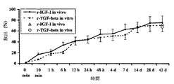
実施例7:コーティング中に取り込ませた活性物質の放出の検討
実施例1に記載の方法で、チタン製キルシュナーワイヤにPDLLAをコーティングし、それにはさらに5重量%のIGF-Iまたは1重量%のTGF-β1または5重量%のIGF-Iと1重量%のTGF-β1の組合せのいずれかを含有させた。
コーティング中に取り込ませた増殖因子の放出パターンをin vitro溶出試験によって分析した。結果を図2に示す。48時間以内にコーティングからの増殖因子の最初の放出が48〜54%の比率で起こった。その後は、放出は漸増的に6週間に至るまで続き、取り込ませた増殖因子の合計71〜78%が放出された。
PDLLAおよび上述の増殖因子でコーティングされたチタン製キルシュナーワイヤ10個を、用いたSprague Dawleyラットの各々の脛骨に移植した。42日後、インプラントを取り出し、取り込ませた増殖因子の残存濃度をELISAを用いて測定した。図2に示すとおり、in vivoでの結果はin vitro溶出試験の結果と一致していた。
Example 7 : Examination of the release of active substance incorporated into the coating In the manner described in Example 1, a titanium Kirschner wire was coated with PDLLA, which was further supplemented with 5% by weight of IGF-I or 1% by weight. Either TGF-β1 or a combination of 5 wt% IGF-I and 1 wt% TGF-β1 was included.
The release pattern of growth factor incorporated into the coating was analyzed by in vitro dissolution test. The result is shown in figure 2. Within 48 hours, the first release of growth factor from the coating occurred at a rate of 48-54%. Thereafter, the release lasted for up to 6 weeks, releasing a total of 71-78% of the incorporated growth factors.
Ten titanium Kirschner wires coated with PDLLA and the growth factors described above were implanted into each tibia of the Sprague Dawley rats used. After 42 days, the implant was removed and the residual concentration of the incorporated growth factor was measured using ELISA. As shown in FIG. 2, the in vivo results were consistent with the in vitro dissolution test results.
実施例8:本発明のインプラントの骨誘導効果
動物実験としては60匹(5月齢の雌のSprague Dawleyラット)の動物で試験を行った。
供試動物は全て右脛骨を標準化された方法で骨折させた。異なるコーティングを施したチタン製ワイヤ(直径1.0mm)を元の位置にもどした脛骨中に髄内の支持体として移植した。
手術後、42日目まで連日、群分けの指定(下記参照)に従って、ラット特異的組換え成長ホルモン(r-rGH)2mg/kgまたはプラシーボをそれぞれ皮下に注射した。0日目、4日目、7日目、14日目、21日目、28日目、35日目、および42日目に、吸入麻酔剤投与後、2平面でX線写真を撮影し、眼球後法(retrobulbar method)(−80℃(−112°F)で急速凍結)によって各動物から1.25ml採血し、各動物の体重と体温を測定した。42日目に骨折した脛骨と骨折していない脛骨を骨膜と共に別々に調製して生体力学的試験(ねじり荷重−ねじり剛性)にかけた。
Example 8 : Osteoinductive effect of the implant of the present invention As an animal experiment, 60 animals (five months old female Sprague Dawley rats) were tested.
All test animals fractured the right tibia in a standardized manner. Titanium wires (diameter 1.0 mm) with different coatings were implanted as intramedullary supports into the tibia returned to its original position.
Following surgery, rats were injected subcutaneously each day with rat-specific recombinant growth hormone (r-rGH) 2 mg / kg or placebo according to grouping designations (see below) until
群分けの指定
第I群 右脛骨骨折−コーティングなしのインプラント−プラシーボの全身適用(対照群)
第II群 右脛骨骨折−ポリ-D,L-ラクチド(登録商標) 203でコーティングしたインプラント−プラシーボの全身適用
第III群 右脛骨骨折−ポリ-D,L-ラクチドでコーティングしたインプラント−(r-rGH)の全身適用
第IV群 右脛骨骨折−ポリ-D,L-ラクチドならびに増殖因子IGF-I(5%)およびTGF-β(1%)でコーティングしたインプラント−プラシーボの全身適用第V群 右脛骨骨折−ポリ-D,L-ラクチドならびに増殖因子IGF-I(5%)およびTGF-β(1%)でコーティングしたインプラント−(r-rGH)の全身適用コーティングされたインプラントは実施例1に示す方法で作製した。
Grouping designation <br/> Group I Right tibial fracture-Uncoated implant-Systemic application of placebo (control group)
Group II right tibial fracture-Implant coated with poly-D, L-lactide (R) 203-Systemic application of placebo Group III right tibial fracture-Implant coated with poly-D, L-lactide- (r- rGH) systemic application group IV right tibial fracture-poly-D, L-lactide and implants coated with growth factors IGF-I (5%) and TGF-β (1%) systemic application of placebo group V right Tibial fractures-Poly-D, L-lactide and implants coated with growth factors IGF-I (5%) and TGF-β (1%)-Systemic application of (r-rGH) Coated implants in Example 1 It was produced by the method shown.
結果:
骨折
用いた骨折モデルでは、軟組織に大きなダメージを与えることなく右脛骨を標準的に横骨折させることができた。60匹中2匹において、脛骨骨折は複雑骨折であった;1匹ではらせん型で、試験途上での中止を必要とした。1匹の動物が手術後観察での麻酔下で死亡した(32日目)。
体重と体温
(r-rGH)での全身治療を受けた動物(第III群および第V群)では、試験期間中にプラシーボを受けた動物(第I、II、およびIV群)と比べて体温の上昇は認められなかったが、体重は13%までの有意の増加を示した(p<0.05)。第I、II、およびIV群(プラシーボ)の群間、または第IIIおよびV群(GH)の群間では大きな相異は認められなかった。
result:
In the fracture model using a fracture, the right tibia could be fractured as a standard fracture without damaging the soft tissue. In 2 of 60 animals, the tibial fracture was a complex fracture; one was helical and required discontinuation during the study. One animal died under postoperative observational anesthesia (day 32).
Weight and body temperature
Increased body temperature in animals that received systemic treatment with (r-rGH) (Group III and Group V) compared to animals that received placebo during the study period (Groups I, II, and IV) Although not observed, body weight showed a significant increase up to 13% (p <0.05). There were no significant differences between groups in Groups I, II, and IV (placebo) or between Groups III and V (GH).
生体力学的試験
得られたデータは絶対値で(ねじり荷重)、および百分率で(ねじり剛性)、骨折させなかった反対側との比較で示した。
Biomechanical tests The data obtained were expressed as absolute values (torsional load) and as percentages (torsional stiffness) compared to the other side without fracture.
この試験の結果から、第III群ならびに第IVおよびV群において、全身適用と比較して最大ねじり荷重の有意な増加が認められた(p<0.05)。増殖因子の局所適用(第IV群)は、対照群と比べて顕著に高い最大ねじり荷重が得られるのみならず、r-rGHの全身適用の結果と比べても平均して同様である(有意差なし)ように見受けられた。r-rGHの投与とIGF-IおよびTGF-βの局所適用とを同時に行っても最大ねじり荷重のそれ以上の増加は見られなかった。ポリ−D,L−ラクチドで処理した群での最大ねじり荷重は対照群と比べて有意に増加した。
ねじり剛性については、反対側の脛骨と比べたとき、同様な所見が得られた。この場合にも、増殖因子の局所適用を行った群では最も好ましい結果が得られた。
The results of this study showed a significant increase in maximum torsional load in group III and groups IV and V compared to systemic application (p <0.05). The topical application of growth factors (Group IV) not only resulted in significantly higher maximum torsional loads compared to the control group, but on average was similar compared to the results of systemic application of r-rGH (significant There was no difference). There was no further increase in maximum torsional load when r-rGH was administered simultaneously with topical application of IGF-I and TGF-β. The maximum torsional load in the group treated with poly-D, L-lactide was significantly increased compared with the control group.
For torsional rigidity, similar findings were obtained when compared to the contralateral tibia. In this case, the most favorable results were obtained in the group to which the growth factor was locally applied.
図9はこれらの結果を要約したものである。 FIG. 9 summarizes these results.
実施例9:
5月齢の雌のSprague Dawleyラット(n=144)の右脛骨を、骨折作成機を用いて標準的に閉鎖骨折させ、チタン製キルシュナーワイヤのコーティングしていないものとコーティングしたものを髄内安定器具として脛骨に移植した。下記の各群について比較した:
第I群: コーティングしていないインプラント(対照群)
第II群: PDLLA(登録商標)203でコーティングしたインプラント
第III群: PDLLA+r-IGF-I(5%)でコーティングしたインプラント
第IV群: PDLLA+r-IGF-I(5%)+TGF-β1(1%)でコーティングしたインプラント
コーティングされたインプラントは実施例1の方法で作製した。
経時的にX線写真を2平面(a.-p.および側面)でとった。0日目、4日目、7日目、14日目、21日目、28日目に、血清を測定し、r-IGF-Iおよびr-TGF-β1の全身濃度ならびに体重および体温も測定した。4週間後、インプラントを取り出し、骨折させた脛骨を非処置の反対側の脛骨と比較して生体力学的に試験した。仮骨の組織形態測定的検討を行ない(O.Safranin/v.Kossa)、分析用イメージングシステム(Zeiss KS 400)で定量した。
Example 9 :
The right tibia of a 5-month-old female Sprague Dawley rat (n = 144) is normally fractured closed using a fracture-making machine, and a titanium Kirschner wire uncoated and coated intramedullary stabilizer As transplanted to the tibia. The following groups were compared:
Group I: Uncoated implant (control group)
Group II: Implants coated with PDLLA® 203 Group III: Implants coated with PDLLA + r-IGF-I (5%) Group IV: PDLLA + r-IGF-I (5%) + TGF-β1 (1% The coated implant was prepared according to the method of Example 1.
Over time, radiographs were taken in two planes (a.-p. and sides). Serum was measured on
X線写真法での評価では、非処置の第I群では依然として骨折部分の分離が認められた。第IIおよびIII群ではコーティングしていない第I群に比べて良好な仮骨形成が認められた。第IV群の動物では、骨折はほぼ完全に癒合していた(図3)。 In radiographic evaluation, fractures were still separated in the untreated group I. In groups II and III, better callus formation was observed compared to uncoated group I. In group IV animals, the fractures were almost completely healed (FIG. 3).
非処置の反対側の脛骨と比べ、また、その他の群全てと比べ、第IV群は生体力学的試験で有意に高い最大ねじり荷重および最大ねじり剛性を示した。r-IGF-Iおよびr-TGF-β1の併用適用では、IGF-I単独での処置を受けた群よりも実質的に高い最大ねじり荷重および最大ねじり剛性が得られた。
ポリラクチドで処置した群は非処置の第I群に比べ有意に高い最大ねじり荷重および最大ねじり剛性を示した(図4)。
Compared to the untreated contralateral tibia and compared to all other groups, Group IV showed significantly higher maximum torsional load and maximum torsional stiffness in biomechanical testing. The combined application of r-IGF-I and r-TGF-β1 resulted in a substantially higher maximum torsional load and maximum torsional stiffness than the group treated with IGF-I alone.
The group treated with polylactide showed significantly higher maximum torsional load and maximum torsional rigidity than the untreated group I (FIG. 4).
組織形態測定的検討結果はX線写真法および生体力学的試験の結果を実証するものである。処置群に比べて実質的に大きい結合組織細胞の領域が第I群で見られた。PDLLAで処置した群では良好な仮骨形成および進行した仮骨再構築のパターンが認められたが、結合組織細胞の比率は最小限であった。第IV群はほぼ完全な骨折の回復が認められ仮骨中の骨密度は最高値であった。ポリラクチドのみで処置した群では対照群と比較して仮骨領域に有意に高い骨密度が認められた(図5および6)。
処置群と非処置群の間には、血清パラメーター、体重、または体温に関して明らかな変化は認められなかった。
The histomorphometric study results demonstrate the results of X-ray photography and biomechanical testing. A region of connective tissue cells that was substantially larger compared to the treatment group was seen in Group I. The group treated with PDLLA showed good callus formation and advanced callus remodeling patterns, but the ratio of connective tissue cells was minimal. In Group IV, almost complete fracture recovery was observed, and the bone density in the callus was the highest. In the group treated with polylactide alone, a significantly higher bone density was found in the callus area compared to the control group (FIGS. 5 and 6).
There were no obvious changes between serum and non-treated groups regarding serum parameters, body weight, or body temperature.
実施例10:
12月齢のユカタンコビトブタ(Yucatan dwarf pig)(n=30)に、標準的な骨切除術(1mmギャップ)を右脛骨に施し、次いでコーティングした、およびコーティングしていないチタン製脛骨髄内釘でそれぞれ髄内安定化させ、静止状態でロックした。下記の群の比較を行った:
第I群: コーティングしていないインプラント(対照群)
第II群: PDLLA(登録商標)203でコーティングしたインプラント
第III群: PDLLA+r-IGF-I(5%)+TGF-β1(1%)でコーティングしたインプラント
コーティングされたインプラントは実施例1の方法で作製した。
Example 10 :
A 12-month-old Yucatan dwarf pig (n = 30) was subjected to standard osteotomy (1 mm gap) in the right tibia and then with coated and uncoated titanium tibia intramedullary nails. Each was stabilized in the medulla and locked in a stationary state. The following groups were compared:
Group I: Uncoated implant (control group)
Group II: Implants coated with PDLLA® 203 Group III: Implants coated with PDLLA + r-IGF-I (5%) + TGF-β1 (1%) were prepared by the method of Example 1 did.
経時的X線写真法の検討と血清の試験を行った。4週間後、2つの脛骨を取り出し、生体力学的に試験した。仮骨の直径を測定し仮骨量をアルキメデスの原理で測定した。 Time-lapse radiography and serum tests were performed. After 4 weeks, the two tibias were removed and tested biomechanically. The diameter of the callus was measured and the amount of callus was measured by Archimedes' principle.
結果:
4週間後、対照群動物は全て骨切除術ギャップの癒合が完全ではないことを示した。ポリラクチドで処置した群では良好な仮骨形成が認められた。第III群では十分に進行した仮骨形成を示した(図7)。
ポリラクチドで処置した第II群、およびさらに増殖因子で処置した第III群では仮骨量と仮骨直径は対照群よりも有意に大きい値であった。
反対側の脛骨と比べ、また対照群と比べてポリラクチドで処置した群ではかなり高い最大ねじり荷重および最大ねじり剛性を示した。
ポリラクチドコーティング中に増殖因子を含ませることによって、最大ねじり荷重および最大ねじり剛性の顕著な増加がもたらされた。
result:
After 4 weeks, all control animals showed that the osteotomy gap fusion was not complete. Good callus formation was observed in the group treated with polylactide. Group III showed sufficiently advanced callus formation (FIG. 7).
In group II treated with polylactide and in group III further treated with growth factors, the amount of callus and the diameter of the callus were significantly larger than in the control group.
The group treated with polylactide compared to the contralateral tibia and the control group showed a much higher maximum torsional load and maximum torsional stiffness.
Inclusion of growth factors in the polylactide coating resulted in a significant increase in maximum torsional load and maximum torsional stiffness.
髄内支持体の強度
電気摘出機(power extractor)を用いたチタン製ワイヤの脛骨からの標準的な摘出では、IGF-IおよびTGF-βでコーティングしたワイヤの摘出には対照群のワイヤーの摘出よりも有意に大きな摘出力が必要であった。
実施例8および10から、本発明に従ってコーティングしたインプラントの使用によって、骨接合を有意に加速し、それゆえに骨折の治癒過程を有意に加速することができることは明かである。この加速した過程は、他の骨誘導剤の添加が無くともポリマーでコーティングしたインプラントで実証されている。増殖因子をコーティング中に取り込ませると骨折治癒過程はさらに加速され、IGF-IおよびTGF-βを組み合わせて適用すると特に効果的である。
これらの実施例は本発明の方法を用いてバーニッシュ様コーティングを作ることができ、その物理的構造および機械的強度により、従来技術から明確に区別しうるものである。
For standard extraction of titanium wire from the tibia using a power extractor of the intramedullary support , extraction of the control group wire for extraction of IGF-I and TGF-β coated wires Significantly larger extraction power was required.
From Examples 8 and 10, it is clear that the use of implants coated in accordance with the present invention can significantly accelerate osteosynthesis and thus significantly accelerate the fracture healing process. This accelerated process has been demonstrated in polymer-coated implants without the addition of other osteoinductive agents. Incorporation of growth factors into the coating further accelerates the fracture healing process and is particularly effective when combined with IGF-I and TGF-β.
These examples can be used to make varnish-like coatings using the method of the present invention and are clearly distinguishable from the prior art by their physical structure and mechanical strength.
Claims (1)
(i)表面、
(ii)本体、及び
(iii)バーニッシュ様の耐摩耗性コーティングであって、
100 kDa以下の平均分子量を有し、ガラス転移温度が37℃より高い
生物分解性ポリマーを備え、
厚さが100μm以下であり、
インプラントが移植されるときに機械的摩擦がコーティングを摩耗又は損傷しないように本体の表面への接着を形成し、且つ
移植されるときに骨と接触するように適合している、前記コーティング、
を備える、前記整形外科用インプラント。 An orthopedic implant, the following:
(i) surface,
(ii) the body, and
(iii) a burnish-like wear-resistant coating,
Comprising a biodegradable polymer having an average molecular weight of 100 kDa or less and a glass transition temperature higher than 37 ° C .;
The thickness is 100 μm or less,
The coating adapted to form an adhesion to the surface of the body such that mechanical friction does not wear or damage the coating when the implant is implanted, and to contact the bone when implanted;
Said orthopedic implant.
Applications Claiming Priority (2)
| Application Number | Priority Date | Filing Date | Title |
|---|---|---|---|
| DE19843251 | 1998-09-11 | ||
| DE19843251.8 | 1998-09-11 |
Related Parent Applications (1)
| Application Number | Title | Priority Date | Filing Date |
|---|---|---|---|
| JP2000569857A Division JP4854114B2 (en) | 1998-09-11 | 1999-09-10 | Biologically active implants |
Publications (2)
| Publication Number | Publication Date |
|---|---|
| JP2011235175A true JP2011235175A (en) | 2011-11-24 |
| JP5726014B2 JP5726014B2 (en) | 2015-05-27 |
Family
ID=7881711
Family Applications (2)
| Application Number | Title | Priority Date | Filing Date |
|---|---|---|---|
| JP2000569857A Expired - Lifetime JP4854114B2 (en) | 1998-09-11 | 1999-09-10 | Biologically active implants |
| JP2011171961A Expired - Lifetime JP5726014B2 (en) | 1998-09-11 | 2011-08-05 | Biologically active implants |
Family Applications Before (1)
| Application Number | Title | Priority Date | Filing Date |
|---|---|---|---|
| JP2000569857A Expired - Lifetime JP4854114B2 (en) | 1998-09-11 | 1999-09-10 | Biologically active implants |
Country Status (13)
| Country | Link |
|---|---|
| US (4) | US6998134B2 (en) |
| EP (1) | EP1112095B1 (en) |
| JP (2) | JP4854114B2 (en) |
| AT (1) | ATE228021T1 (en) |
| AU (1) | AU5862199A (en) |
| CA (1) | CA2350638C (en) |
| DE (1) | DE59903490D1 (en) |
| DK (1) | DK1112095T3 (en) |
| ES (1) | ES2187195T3 (en) |
| PT (1) | PT1112095E (en) |
| SI (1) | SI1112095T1 (en) |
| WO (1) | WO2000015273A1 (en) |
| ZA (1) | ZA200102764B (en) |
Families Citing this family (47)
| Publication number | Priority date | Publication date | Assignee | Title |
|---|---|---|---|---|
| DK1112095T3 (en) * | 1998-09-11 | 2003-03-17 | Michael Dr Raschke | Biologically active implants |
| US7592017B2 (en) | 2000-03-10 | 2009-09-22 | Mast Biosurgery Ag | Resorbable thin membranes |
| CH694935A5 (en) * | 2000-07-26 | 2005-09-30 | Straumann Holding Ag | Oberflaechenmodifizierte implants. |
| DE10059986C2 (en) * | 2000-11-30 | 2003-02-13 | Martin Wiemann | Process for the non-covalent immobilization of heat-resistant biomolecules on implant materials |
| MXPA05001149A (en) | 2002-07-31 | 2005-11-23 | Macropore Biosurgery Inc | Apparatus and method for preventing adhesions between an implant and surrounding tissues. |
| US8048444B2 (en) | 2002-07-31 | 2011-11-01 | Mast Biosurgery Ag | Apparatus and method for preventing adhesions between an implant and surrounding tissues |
| DE10237572A1 (en) * | 2002-08-13 | 2004-02-26 | Biotronik Meß- und Therapiegeräte GmbH & Co. Ingenieurbüro Berlin | Stent with a polymer coating |
| DE10241572B4 (en) * | 2002-09-07 | 2007-02-08 | Werner Scholz | Support or holding part for insertion into a bone part |
| US7704520B1 (en) | 2002-09-10 | 2010-04-27 | Mast Biosurgery Ag | Methods of promoting enhanced healing of tissues after cardiac surgery |
| WO2005114322A2 (en) * | 2004-05-12 | 2005-12-01 | Massachusetts Institute Of Technology | Manufacturing process, such as three-dimensional printing, including solvent vapor filming and the like |
| US7887587B2 (en) * | 2004-06-04 | 2011-02-15 | Synthes Usa, Llc | Soft tissue spacer |
| DE102005002703C5 (en) | 2005-01-19 | 2013-07-04 | Heraeus Kulzer Gmbh | Antibiotic coating of implants and methods for antibiotic coating |
| WO2007130648A2 (en) * | 2006-05-05 | 2007-11-15 | Ceramatec, Inc. | Fully or partially bioresorbable orthopedic implant |
| WO2008003320A2 (en) * | 2006-07-05 | 2008-01-10 | Region Midtjylland | Three-dimensional cell scaffolds |
| WO2008035948A1 (en) * | 2006-09-22 | 2008-03-27 | U & I Corporation | Implants comprising biodegradable metals and method for manufacturing the same |
| EP1913960A1 (en) * | 2006-10-19 | 2008-04-23 | Albert Schömig | Coated implant |
| EP1916006A1 (en) * | 2006-10-19 | 2008-04-30 | Albert Schömig | Implant coated with a wax or a resin |
| DE102006052552A1 (en) | 2006-11-06 | 2008-05-08 | Auriga Medical Products Gmbh | Coated dental implant |
| US8597673B2 (en) * | 2006-12-13 | 2013-12-03 | Advanced Cardiovascular Systems, Inc. | Coating of fast absorption or dissolution |
| US8048857B2 (en) | 2006-12-19 | 2011-11-01 | Warsaw Orthopedic, Inc. | Flowable carrier compositions and methods of use |
| US8133553B2 (en) * | 2007-06-18 | 2012-03-13 | Zimmer, Inc. | Process for forming a ceramic layer |
| US8309521B2 (en) | 2007-06-19 | 2012-11-13 | Zimmer, Inc. | Spacer with a coating thereon for use with an implant device |
| US20110230973A1 (en) * | 2007-10-10 | 2011-09-22 | Zimmer, Inc. | Method for bonding a tantalum structure to a cobalt-alloy substrate |
| US8608049B2 (en) * | 2007-10-10 | 2013-12-17 | Zimmer, Inc. | Method for bonding a tantalum structure to a cobalt-alloy substrate |
| US20090187256A1 (en) * | 2008-01-21 | 2009-07-23 | Zimmer, Inc. | Method for forming an integral porous region in a cast implant |
| AU2008349523B2 (en) * | 2008-01-29 | 2013-10-24 | Zimmer, Inc. | Implant device for use in an implant system |
| US20090198286A1 (en) * | 2008-02-05 | 2009-08-06 | Zimmer, Inc. | Bone fracture fixation system |
| US9616153B2 (en) | 2008-04-17 | 2017-04-11 | Warsaw Orthopedic, Inc. | Rigid bone graft substitute |
| US20090263507A1 (en) * | 2008-04-18 | 2009-10-22 | Warsaw Orthopedic, Inc. | Biological markers and response to treatment for pain, inflammation, neuronal or vascular injury and methods of use |
| US10022164B2 (en) * | 2008-06-11 | 2018-07-17 | Eventions, Llc | Orthopedic fastener device |
| GB0818933D0 (en) | 2008-10-16 | 2008-11-19 | Depuy Int Ltd | An implantable medical device |
| CH700138B1 (en) * | 2008-12-19 | 2012-12-14 | Ind Biomediche Insubri S A | Matrix for bone implant and procedure of preparation of the same. |
| US20100215716A1 (en) * | 2009-02-23 | 2010-08-26 | Biomet Manufacturing Corp. | Compositions and methods for coating orthopedic implants |
| US8506608B2 (en) | 2009-03-24 | 2013-08-13 | Douglas Cerynik | Orthopedic fixation device with bioresorbable layer |
| US20100247600A1 (en) * | 2009-03-24 | 2010-09-30 | Warsaw Orthopedic, Inc. | Therapeutic drug eluting implant cover and method of making the same |
| US20100249783A1 (en) * | 2009-03-24 | 2010-09-30 | Warsaw Orthopedic, Inc. | Drug-eluting implant cover |
| US9414864B2 (en) | 2009-04-15 | 2016-08-16 | Warsaw Orthopedic, Inc. | Anterior spinal plate with preformed drug-eluting device affixed thereto |
| US9078712B2 (en) * | 2009-04-15 | 2015-07-14 | Warsaw Orthopedic, Inc. | Preformed drug-eluting device to be affixed to an anterior spinal plate |
| US8333791B2 (en) * | 2009-04-24 | 2012-12-18 | Warsaw Orthopedic, Inc. | Medical implant with tie configured to deliver a therapeutic substance |
| US20100274295A1 (en) * | 2009-04-24 | 2010-10-28 | Warsaw Orthopedic, Inc. | Medical implant configured to deliver a therapeutic substance |
| DE102009024616A1 (en) * | 2009-06-08 | 2010-12-23 | Telos Gmbh | Sterilizable coating used for complete or partial treatment of surfaces of implantable materials for use as implants or hard tissue replacement materials in medical and dental field, comprises osteogenic protein and ionic polysaccharide |
| MX2012012050A (en) | 2010-04-16 | 2012-11-22 | Novartis Ag | Methods and compositions for improving implant osseointegration. |
| EP3741384A1 (en) | 2011-09-07 | 2020-11-25 | Mount Sinai School Of Medicine | Ceramidase and cell differentiation |
| US9775853B2 (en) | 2013-03-15 | 2017-10-03 | Biomet Manufacturing, Llc. | Hemostatic compositions and methods |
| US9763911B2 (en) * | 2013-12-12 | 2017-09-19 | Mayo Foundation For Medical Education And Research | Prostacyclin compositions for regulation of fracture repair and bone formation |
| CN112996546A (en) * | 2018-10-18 | 2021-06-18 | 南丹麦大区 | Coating composition for medical implants |
| JP7775554B2 (en) | 2020-09-04 | 2025-11-26 | デピュイ・シンセス・プロダクツ・インコーポレイテッド | Temporary antibacterial cement spacers, assemblies, kits, and methods of manufacture |
Citations (3)
| Publication number | Priority date | Publication date | Assignee | Title |
|---|---|---|---|---|
| JPH05505125A (en) * | 1990-12-28 | 1993-08-05 | ユニオン、カーバイド、ケミカルズ、アンド、プラスチックス、テクノロジー、コーポレーション | Biocompatible wear-resistant coated support |
| WO1993020859A1 (en) * | 1992-04-20 | 1993-10-28 | Board Of Regents Of The University Of Washington | Sustained release compositions for delivery of growth factors |
| WO1998007458A1 (en) * | 1996-08-19 | 1998-02-26 | Korea Institute Of Science And Technology | Surface coating method for metal implants |
Family Cites Families (160)
| Publication number | Priority date | Publication date | Assignee | Title |
|---|---|---|---|---|
| US3933217A (en) * | 1973-04-04 | 1976-01-20 | Carl Hurth, Maschinen- Und Zahnradfabrik | Drive gear system for motor vehicles |
| GB1482914A (en) * | 1973-07-25 | 1977-08-17 | Babcock & Wilcox Ltd | Semi-permeable polymeric membranes |
| IE51564B1 (en) * | 1980-03-27 | 1987-01-21 | Nat Res Dev | Antimicrobial surgical implants |
| US4338926A (en) * | 1980-11-21 | 1982-07-13 | Howmedica, Inc. | Bone fracture prosthesis with controlled stiffness |
| DK154260C (en) * | 1981-02-20 | 1989-05-22 | Mundipharma Gmbh | PROCEDURE FOR THE MANUFACTURING OF A BONE IMPLANT OF FURNISHED TRICAL CUMPHOSPHATE, SPECIFICALLY FOR FILLING OF SPACES OR FOR COMPOSITION OF BONE PARTS AFTER FRACTURE. |
| US4563489A (en) * | 1984-02-10 | 1986-01-07 | University Of California | Biodegradable organic polymer delivery system for bone morphogenetic protein |
| SE452110B (en) * | 1984-11-08 | 1987-11-16 | Medinvent Sa | MULTILAYER PROTEST MATERIAL AND PROCEDURE FOR ITS MANUFACTURING |
| IL74617A (en) | 1985-03-15 | 1988-11-15 | Yeda Res & Dev | Compositions comprising a vitamin d derivative and method for the local treatment of bone fractures in animals |
| DE3521684A1 (en) * | 1985-06-18 | 1986-12-18 | Dr. Müller-Lierheim KG, Biologische Laboratorien, 8033 Planegg | METHOD FOR COATING POLYMERS |
| US4962091A (en) * | 1986-05-23 | 1990-10-09 | Syntex (U.S.A.) Inc. | Controlled release of macromolecular polypeptides |
| FR2616318A1 (en) | 1987-06-15 | 1988-12-16 | Centre Nat Rech Scient | ARTIFICIAL SKIN AND PROCESS FOR PREPARING THE SAME |
| FI83729C (en) | 1987-11-26 | 1991-08-26 | Biocon Oy | SURGICAL IMPLANTS. |
| AU2902289A (en) | 1987-12-08 | 1989-07-05 | Mark Chasin | Method of forming bioerodible implants for improved controlled drug release |
| EP0322250B1 (en) * | 1987-12-23 | 1991-10-09 | Sumitomo Chemical Company, Limited | Coating liquor containing hydroxyapatite and method for forming hydroxyapatite coating film using the same |
| US5019096A (en) * | 1988-02-11 | 1991-05-28 | Trustees Of Columbia University In The City Of New York | Infection-resistant compositions, medical devices and surfaces and methods for preparing and using same |
| US5344654A (en) * | 1988-04-08 | 1994-09-06 | Stryker Corporation | Prosthetic devices having enhanced osteogenic properties |
| US4880610A (en) * | 1988-04-20 | 1989-11-14 | Norian Corporation | In situ calcium phosphate minerals--method and composition |
| US5053212A (en) * | 1988-04-20 | 1991-10-01 | Norian Corporation | Intimate mixture of calcium and phosphate sources as precursor to hydroxyapatite |
| US6005162A (en) * | 1988-04-20 | 1999-12-21 | Norian Corporation | Methods of repairing bone |
| US5962028A (en) * | 1988-04-20 | 1999-10-05 | Norian Corporation | Carbonated hydroxyapatite compositions and uses |
| US5047031A (en) * | 1988-04-20 | 1991-09-10 | Norian Corporation | In situ calcium phosphate minerals method |
| US5129905A (en) * | 1988-04-20 | 1992-07-14 | Norian Corporation | Methods for in situ prepared calcium phosphate minerals |
| US5178845A (en) * | 1988-04-20 | 1993-01-12 | Norian Corporation | Intimate mixture of calcium and phosphate sources as precursor to hydroxyapatite |
| US4902515A (en) * | 1988-04-28 | 1990-02-20 | E. I. Dupont De Nemours And Company | Polylactide compositions |
| US4983581A (en) * | 1988-05-20 | 1991-01-08 | Institute Of Molecular Biology, Inc. | Wound healing composition of IGF-I and TGF-β |
| US5502158A (en) * | 1988-08-08 | 1996-03-26 | Ecopol, Llc | Degradable polymer composition |
| DE3831657A1 (en) * | 1988-09-17 | 1990-03-22 | Boehringer Ingelheim Kg | DEVICE FOR THE OSTEOSYNTHESIS AND METHOD FOR THE PRODUCTION THEREOF |
| US5725491A (en) * | 1988-10-03 | 1998-03-10 | Atrix Laboratories, Inc. | Method of forming a biodegradable film dressing on tissue |
| EP0366018B1 (en) | 1988-10-24 | 1993-05-26 | Krysmann, Waldemar, Dr.rer.nat. | Metal sponge-like structure and method for making the same |
| GB8903464D0 (en) * | 1989-02-15 | 1989-04-05 | Joint Replacement Instrumentat | Coated femoral prosthesis |
| US5034059A (en) * | 1989-02-17 | 1991-07-23 | Norian Corporation | Composition comprising octacalcium phosphate crystals and polypeptide |
| AU5654990A (en) | 1989-04-28 | 1990-11-29 | Brigham And Women's Hospital | Novel materials and methods for guided tissue regeneration |
| US4976736A (en) | 1989-04-28 | 1990-12-11 | Interpore International | Coated biomaterials and methods for making same |
| US5258419A (en) * | 1989-06-26 | 1993-11-02 | Minnesota Mining And Manufacturing Company | Methods of preparing radiation resistant heat sealable polymer blends |
| JPH0385179A (en) | 1989-08-30 | 1991-04-10 | Sumitomo Bakelite Co Ltd | Medical balloon catheter |
| DE3933217A1 (en) | 1989-10-05 | 1991-04-11 | Gunther Dr Med Dr Rer Hofmann | DEVICE FOR THE OPERATIONAL SCREWLESS RAILING OF BONE BREAKS MADE OF BIODEGRADABLE PLASTICS |
| US5573401A (en) * | 1989-12-21 | 1996-11-12 | Smith & Nephew Richards, Inc. | Biocompatible, low modulus dental devices |
| US5071655A (en) | 1990-01-12 | 1991-12-10 | Baylink David J | Pharmaceutical combination for treatment of bone-wasting diseases |
| US5556645A (en) * | 1990-01-12 | 1996-09-17 | Bockman; Richard | Methods of enhancing wound healing and tissue repair |
| US5686116A (en) * | 1990-01-12 | 1997-11-11 | New York Society For The Relief Of The Ruptured And Crippled, Maintaining The Hospital For Special Surgery | Methods of enhancing repair, healing and augmentation of bone implants |
| DK27390D0 (en) | 1990-02-02 | 1990-02-02 | Troels Torp Andreassen | METHOD AND APPARATUS FOR ADMINISTRATING BIOLOGICALLY ACTIVE SUBSTANCES |
| US5290494A (en) * | 1990-03-05 | 1994-03-01 | Board Of Regents, The University Of Texas System | Process of making a resorbable implantation device |
| US5492697A (en) * | 1990-03-05 | 1996-02-20 | Board Of Regents, Univ. Of Texas System | Biodegradable implant for fracture nonunions |
| US5188670A (en) * | 1990-04-05 | 1993-02-23 | Norian Corporation | Apparatus for hydroxyapatite coatings of substrates |
| US5164187A (en) * | 1990-04-05 | 1992-11-17 | Norian Corporation | Hydroxyapatite prosthesis coatings |
| US5645591A (en) * | 1990-05-29 | 1997-07-08 | Stryker Corporation | Synthetic bone matrix |
| US5098779A (en) | 1990-06-25 | 1992-03-24 | W. L. Gore & Associates, Inc. | Carvable implant material |
| MY108621A (en) | 1990-08-01 | 1996-10-31 | Novartis Ag | Polylactide preparation and purification |
| US5466609A (en) * | 1990-10-31 | 1995-11-14 | Coulter Corporation | Biodegradable gelatin-aminodextran particle coatings of and processes for making same |
| JPH04221538A (en) | 1990-12-21 | 1992-08-12 | Olympus Optical Co Ltd | Device for forming hole |
| US5782971B1 (en) * | 1991-06-28 | 1999-09-21 | Norian Corp | Calcium phosphate cements comprising amorophous calcium phosphate |
| EP0523926A3 (en) * | 1991-07-15 | 1993-12-01 | Smith & Nephew Richards Inc | Prosthetic implants with bioabsorbable coating |
| US5876452A (en) * | 1992-02-14 | 1999-03-02 | Board Of Regents, University Of Texas System | Biodegradable implant |
| US5352515A (en) * | 1992-03-02 | 1994-10-04 | American Cyanamid Company | Coating for tissue drag reduction |
| DE4209122A1 (en) * | 1992-03-20 | 1993-09-23 | Kessler Sigurd | MARKING NAIL |
| CA2093836A1 (en) | 1992-04-24 | 1993-10-25 | Wayne Gombotz | Biodegradable tgf-.beta. delivery system for bone regeneration |
| US5681575A (en) * | 1992-05-19 | 1997-10-28 | Westaim Technologies Inc. | Anti-microbial coating for medical devices |
| IT1259100B (en) * | 1992-05-20 | 1996-03-11 | Lanfranco Callegaro | USE OF HYDROGELS FOR THE LOCKING OF PROSTHETIC SYSTEMS |
| US5366756A (en) * | 1992-06-15 | 1994-11-22 | United States Surgical Corporation | Method for treating bioabsorbable implant material |
| WO1994003159A1 (en) | 1992-07-31 | 1994-02-17 | Daratech Pty. Ltd. | Controlled release implants |
| US5281419A (en) * | 1992-09-28 | 1994-01-25 | Thomas Jefferson University | Biodegradable drug delivery system for the prevention and treatment of osteomyelitis |
| GB9222197D0 (en) | 1992-10-22 | 1992-12-02 | Material Technology Consultant | Biodegradable polymer impant materials |
| GB2280372B (en) | 1993-07-28 | 1997-12-03 | Johnson & Johnson Medical | Composite surgical material |
| EP0710267A4 (en) * | 1993-07-22 | 1996-07-10 | Norian Corp | Storage stable partially neutralized acid compositions and uses |
| DE59408725D1 (en) * | 1993-10-07 | 1999-10-14 | Axel Stemberger | Coating for biomaterial |
| DE4334272C2 (en) | 1993-10-07 | 1996-07-18 | Stemberger Axel Dr | Coating for biomaterial and its use |
| KR0148704B1 (en) * | 1994-01-10 | 1998-08-17 | 김상응 | Biodegradable Drug Delivery Polymer |
| AU1702395A (en) | 1994-02-17 | 1995-09-04 | Pankaj Modi | Drugs, vaccines and hormones in polylactide coated microspheres |
| US5569250A (en) * | 1994-03-01 | 1996-10-29 | Sarver; David R. | Method and apparatus for securing adjacent bone portions |
| ATE269102T1 (en) * | 1994-03-08 | 2004-07-15 | Osteosa Inc | USE OF FIBROBLAST GROWTH FACTORS TO STIMULATE BONE GROWTH |
| US5656598A (en) * | 1994-03-08 | 1997-08-12 | Rhone-Poulenc Rorer Pharmaceuticals Inc. | Use of fibroblast growth factors to stimulate bone growth |
| US5626861A (en) * | 1994-04-01 | 1997-05-06 | Massachusetts Institute Of Technology | Polymeric-hydroxyapatite bone composite |
| DE4414675C1 (en) * | 1994-04-27 | 1995-09-28 | Kirsch Axel | Covering device for bone defects and method for their production |
| US5947893A (en) * | 1994-04-27 | 1999-09-07 | Board Of Regents, The University Of Texas System | Method of making a porous prothesis with biodegradable coatings |
| KR0141431B1 (en) * | 1994-05-17 | 1998-07-01 | 김상웅 | Biodegradable Hydrogel Polymer |
| JP3125579B2 (en) | 1994-05-24 | 2001-01-22 | 三菱マテリアル株式会社 | Biological cement filling equipment |
| AU2952195A (en) | 1994-06-28 | 1996-01-25 | Board Of Regents, The University Of Texas System | Biodegradable fracture fixation plates and uses thereof |
| TW459044B (en) | 1994-07-01 | 2001-10-11 | Bioph Biotech Entw Pharm Gmbh | Protein MP-121 of the TGF-β-like family |
| JP3250127B2 (en) | 1994-07-14 | 2002-01-28 | 三菱マテリアル株式会社 | Biological cement injection jig |
| US5502074A (en) * | 1994-08-22 | 1996-03-26 | Eli Lilly And Company | Benzothiophenes for bone healing and fracture repair |
| US5496399A (en) * | 1994-08-23 | 1996-03-05 | Norian Corporation | Storage stable calcium phosphate cements |
| US5660873A (en) * | 1994-09-09 | 1997-08-26 | Bioseal, Limited Liability Corporaton | Coating intraluminal stents |
| TW369414B (en) * | 1994-09-30 | 1999-09-11 | Yamanouchi Pharma Co Ltd | Bone formation transplant |
| DE4435998C1 (en) | 1994-10-08 | 1995-11-30 | Forschungszentrum Juelich Gmbh | Bio-material immobilisation on carrier coated with silicon nitride |
| US5569442A (en) * | 1994-11-04 | 1996-10-29 | Norian Corporation | Reactive tricalcium phosphate compositions and uses |
| KR970008989B1 (en) | 1994-12-12 | 1997-06-03 | Song Mi Young | Matrix fertilizer |
| FR2728797B1 (en) | 1994-12-30 | 1997-03-28 | Diami | STENT WITH CALCITE COATING AND PROCESS FOR OBTAINING |
| AUPN174495A0 (en) | 1995-03-15 | 1995-04-06 | Ketharanathan, Vettivetpillai | Surgical prostheses |
| DE19514104C2 (en) | 1995-04-13 | 1997-05-28 | Behringwerke Ag | Coating for biomaterial that can be introduced into the bloodstream or into the tissue of the human body |
| US5837313A (en) | 1995-04-19 | 1998-11-17 | Schneider (Usa) Inc | Drug release stent coating process |
| US5635571A (en) * | 1995-06-30 | 1997-06-03 | Cornell Research Foundation, Inc. | Polymerizable macromonomers and polymers prepared therefrom |
| KR0180334B1 (en) | 1995-09-21 | 1999-03-20 | 김윤 | Drug messenger using el-2l-2 micelle and method for sealing drug to it |
| US5756145A (en) * | 1995-11-08 | 1998-05-26 | Baylor College Of Medicine | Durable, Resilient and effective antimicrobial coating for medical devices and method of coating therefor |
| US6048964A (en) * | 1995-12-12 | 2000-04-11 | Stryker Corporation | Compositions and therapeutic methods using morphogenic proteins and stimulatory factors |
| TW432073B (en) | 1995-12-28 | 2001-05-01 | Pfizer | Pyrazolopyridine compounds |
| JPH09201330A (en) | 1996-01-25 | 1997-08-05 | Nissho Corp | Drainage tube assembly |
| DE19607314A1 (en) * | 1996-02-27 | 1997-08-28 | Orthomed Chirurgische Instr Gm | Body exhibiting antibacterial activity for use as a medical or surgical aid |
| DE19608423A1 (en) * | 1996-03-05 | 1997-09-11 | Merck Patent Gmbh | Phased drug delivery implants |
| IL117426A (en) | 1996-03-10 | 2005-09-25 | Yissum Res Dev Co | Synthetic pseudopeptides having osteogenic activity and pharmaceutical compositions containing the same |
| WO1997034953A1 (en) | 1996-03-19 | 1997-09-25 | The Procter & Gamble Company | Biodegradable polymeric compositions and products thereof |
| FI99268C (en) | 1996-04-04 | 1998-02-25 | Upm Kymmene Oy | layer Material |
| DE19614421C2 (en) | 1996-04-12 | 1999-12-16 | Biovision Gmbh | Process for the production of a biodegradable bone replacement and implant material and biodegradable bone replacement and implant material |
| US5759564A (en) * | 1996-04-16 | 1998-06-02 | Implemed, Inc. | Iontophoretic material |
| US5670161A (en) * | 1996-05-28 | 1997-09-23 | Healy; Kevin E. | Biodegradable stent |
| US5916585A (en) * | 1996-06-03 | 1999-06-29 | Gore Enterprise Holdings, Inc. | Materials and method for the immobilization of bioactive species onto biodegradable polymers |
| US6143037A (en) * | 1996-06-12 | 2000-11-07 | The Regents Of The University Of Michigan | Compositions and methods for coating medical devices |
| CA2591651C (en) | 1996-07-23 | 2010-09-28 | The Governors Of The University Of Alberta | Shadow sculpted thin films |
| US5797887A (en) | 1996-08-27 | 1998-08-25 | Novovasc Llc | Medical device with a surface adapted for exposure to a blood stream which is coated with a polymer containing a nitrosyl-containing organo-metallic compound which releases nitric oxide from the coating to mediate platelet aggregation |
| ATE269720T1 (en) | 1996-10-16 | 2004-07-15 | Etex Corp | METHOD FOR PRODUCING LOW CRYSTALLINE CALCIUM PHOSPHATE AND METHOD FOR USE THEREOF |
| US6530951B1 (en) * | 1996-10-24 | 2003-03-11 | Cook Incorporated | Silver implantable medical device |
| WO1998019699A1 (en) | 1996-11-05 | 1998-05-14 | Novo Nordisk A/S | Use of growth hormone or a growth hormone secretagogue for promoting bone formation |
| AU6012998A (en) * | 1996-12-13 | 1998-07-17 | Norian Corporation | Preparation, storage and administration of cements |
| US5980972A (en) | 1996-12-20 | 1999-11-09 | Schneider (Usa) Inc | Method of applying drug-release coatings |
| US5837133A (en) * | 1997-03-03 | 1998-11-17 | Natale; John M. | Method and apparatus for preventing algae growth in open-topped cooling tower reservoirs |
| WO1998051241A1 (en) | 1997-05-16 | 1998-11-19 | Norian Corporation | Implantable re-bar devices |
| US5967946A (en) * | 1997-08-12 | 1999-10-19 | Beatty, Jr.; Alfred C. | Apparatus for cycling training |
| AU3203599A (en) * | 1998-04-01 | 1999-10-18 | Parallax Medical, Inc. | Pressure applicator for hard tissue implant placement |
| US6652581B1 (en) * | 1998-07-07 | 2003-11-25 | Boston Scientific Scimed, Inc. | Medical device with porous surface for controlled drug release and method of making the same |
| US5968253A (en) * | 1998-07-31 | 1999-10-19 | Norian Corporation | Calcium phosphate cements comprising antimicrobial agents |
| DK1112095T3 (en) | 1998-09-11 | 2003-03-17 | Michael Dr Raschke | Biologically active implants |
| WO2001032100A2 (en) | 1999-10-29 | 2001-05-10 | Drexel University | Associating hydrogels for nucleus pulposus replacement in intervertebral discs |
| ATE274857T1 (en) * | 1999-12-06 | 2004-09-15 | Synthes Ag | ABSORBABLE BONE PLATE |
| AR027685A1 (en) * | 2000-03-22 | 2003-04-09 | Synthes Ag | METHOD AND METHOD FOR CARRYING OUT |
| CA2416334C (en) * | 2000-06-09 | 2008-10-28 | Synthes (U.S.A.) | Osteosynthesis implant from a plastic material |
| JP4611607B2 (en) | 2000-06-30 | 2011-01-12 | ジンテーズ ゲゼルシャフト ミト ベシュレンクテル ハフツング | Bone cement injection equipment |
| MY133943A (en) * | 2000-08-22 | 2007-11-30 | Synthes Gmbh | Bone replacement material |
| MXPA03004216A (en) | 2000-12-08 | 2003-09-22 | Synthes Ag | Device for fixing bones, particularly vertebral bodies, in relation to one another. |
| US6545097B2 (en) * | 2000-12-12 | 2003-04-08 | Scimed Life Systems, Inc. | Drug delivery compositions and medical devices containing block copolymer |
| US6855169B2 (en) * | 2001-02-28 | 2005-02-15 | Synthes (Usa) | Demineralized bone-derived implants |
| US7147641B2 (en) | 2001-05-30 | 2006-12-12 | Chen Michael C | Fixation element insertion device |
| US6793660B2 (en) * | 2001-08-20 | 2004-09-21 | Synthes (U.S.A.) | Threaded syringe for delivery of a bone substitute material |
| US6747121B2 (en) * | 2001-09-05 | 2004-06-08 | Synthes (Usa) | Poly(L-lactide-co-glycolide) copolymers, methods for making and using same, and devices containing same |
| TW200400062A (en) * | 2002-04-03 | 2004-01-01 | Mathys Medizinaltechnik Ag | Kneadable, pliable bone replacement material |
| DE50207579D1 (en) | 2002-04-11 | 2006-08-31 | Synthes Ag | DEVICE FOR MIXING AND / OR INJECTING CEMENTS |
| US6951562B2 (en) | 2002-11-13 | 2005-10-04 | Ralph Fritz Zwirnmann | Adjustable length tap and method for drilling and tapping a bore in bone |
| AU2004266154B2 (en) * | 2003-08-22 | 2010-07-08 | Synthes Gmbh | Dura substitute and a process for producing the same |
| US7494950B2 (en) | 2003-09-05 | 2009-02-24 | Synthes (U.S.A.) | Bone cement compositions having fiber-reinforcement and/or increased flowability |
| US20050220837A1 (en) | 2003-09-30 | 2005-10-06 | Disegi John A | Antimicrobial hyaluronic acid coatings for orthopedic implants |
| US20050149032A1 (en) * | 2003-12-30 | 2005-07-07 | Douglas Vaughen | Resorbable surgical fixation device |
| EP1740190A1 (en) | 2004-03-05 | 2007-01-10 | Synthes GmbH | Use of a mixture for the production of an agent for treating defective or degenerated cartilage in the production of natural cartilage replacement in vitro |
| US20050216008A1 (en) * | 2004-03-24 | 2005-09-29 | Zwirnmann Ralph F | Bone fixation implants |
| US20060062825A1 (en) | 2004-04-19 | 2006-03-23 | Maria Maccecchini | Method of implanting a sterile, active agent-coated material and composition made according to same |
| WO2005105121A1 (en) | 2004-05-05 | 2005-11-10 | Synthes Gmbh | Use of platelets or platelet rich plasma (prp) |
| AU2004320279A1 (en) | 2004-06-03 | 2005-12-15 | Synthes Gmbh | Device for impregnating a porous bone replacement material |
| US8012501B2 (en) | 2004-06-10 | 2011-09-06 | Synthes Usa, Llc | Flexible bone composite |
| CN1972724A (en) | 2004-07-06 | 2007-05-30 | 斯恩蒂斯有限公司 | Interference-generating colored coating for surgical implants and instruments |
| WO2006010278A1 (en) | 2004-07-26 | 2006-02-02 | Synthes Gmbh | Biocompatible, biodegradable polyurethane materials with controlled hydrophobic to hydrophilic ratio |
| US20090216191A1 (en) | 2004-08-30 | 2009-08-27 | Mario Loeffel | Injection Device With Haptic Feedback |
| US7914810B2 (en) | 2005-05-06 | 2011-03-29 | Synthes Usa, Llc | Methods for the in situ treatment of bone cancer |
| WO2006130998A1 (en) | 2005-06-09 | 2006-12-14 | Dr. H. C. Robert Mathys Stiftung | Shaped article |
| JP5036715B2 (en) | 2005-08-10 | 2012-09-26 | シンセス ゲゼルシャフト ミット ベシュレンクテル ハフツング | Porous implant |
| WO2007047420A2 (en) | 2005-10-13 | 2007-04-26 | Synthes (U.S.A.) | Drug-impregnated encasement |
| US8641667B2 (en) | 2005-10-20 | 2014-02-04 | DePuy Synthes Products, LLC | Perfusion device and method |
| US20080003255A1 (en) | 2006-05-10 | 2008-01-03 | Synthes (Usa) | Method for augmenting, reducing, and repairing bone with thermoplastic materials |
| US20100041770A1 (en) | 2006-10-31 | 2010-02-18 | Synthes Usa, Llc | Polymer-ceramic composite and method |
| US8197491B2 (en) | 2006-12-19 | 2012-06-12 | Synthes Usa, Llc | Injectable fastener system and method |
| ES2588073T3 (en) | 2007-09-17 | 2016-10-28 | Synergy Biosurgical Ag | Medical implant |
| CN101952341B (en) | 2008-02-01 | 2016-03-23 | 辛西斯有限责任公司 | Porous biocompatible polymer materials and methods |
| ES2557496T3 (en) | 2008-09-02 | 2016-01-26 | Innovative In Vivo Sensing, Llc | Microelectromechanical system sensor used in the biological field (BIOMEMS) and related devices and procedures |
| CA2741426C (en) | 2008-10-31 | 2020-04-21 | Meredith Hans | Method and device for activating stem cells |
-
1999
- 1999-09-10 DK DK99946158T patent/DK1112095T3/en active
- 1999-09-10 PT PT99946158T patent/PT1112095E/en unknown
- 1999-09-10 EP EP99946158A patent/EP1112095B1/en not_active Expired - Lifetime
- 1999-09-10 AU AU58621/99A patent/AU5862199A/en not_active Abandoned
- 1999-09-10 DE DE59903490T patent/DE59903490D1/en not_active Expired - Lifetime
- 1999-09-10 CA CA002350638A patent/CA2350638C/en not_active Expired - Lifetime
- 1999-09-10 SI SI9930182T patent/SI1112095T1/en unknown
- 1999-09-10 JP JP2000569857A patent/JP4854114B2/en not_active Expired - Lifetime
- 1999-09-10 ES ES99946158T patent/ES2187195T3/en not_active Expired - Lifetime
- 1999-09-10 AT AT99946158T patent/ATE228021T1/en active
- 1999-09-10 WO PCT/EP1999/006708 patent/WO2000015273A1/en not_active Ceased
-
2001
- 2001-03-09 US US09/801,752 patent/US6998134B2/en not_active Expired - Lifetime
- 2001-04-04 ZA ZA200102764A patent/ZA200102764B/en unknown
-
2005
- 2005-10-18 US US11/254,200 patent/US8114427B2/en not_active Expired - Fee Related
-
2009
- 2009-05-29 US US12/474,756 patent/US20090317538A1/en not_active Abandoned
-
2011
- 2011-08-05 JP JP2011171961A patent/JP5726014B2/en not_active Expired - Lifetime
-
2016
- 2016-04-06 US US15/091,812 patent/US10646622B2/en not_active Expired - Lifetime
Patent Citations (3)
| Publication number | Priority date | Publication date | Assignee | Title |
|---|---|---|---|---|
| JPH05505125A (en) * | 1990-12-28 | 1993-08-05 | ユニオン、カーバイド、ケミカルズ、アンド、プラスチックス、テクノロジー、コーポレーション | Biocompatible wear-resistant coated support |
| WO1993020859A1 (en) * | 1992-04-20 | 1993-10-28 | Board Of Regents Of The University Of Washington | Sustained release compositions for delivery of growth factors |
| WO1998007458A1 (en) * | 1996-08-19 | 1998-02-26 | Korea Institute Of Science And Technology | Surface coating method for metal implants |
Non-Patent Citations (1)
| Title |
|---|
| JPN6010019399; Journal of Biomedical Materials Research Vol.36,No.4, 1997, p516-521 * |
Also Published As
| Publication number | Publication date |
|---|---|
| US20160220740A1 (en) | 2016-08-04 |
| ATE228021T1 (en) | 2002-12-15 |
| EP1112095B1 (en) | 2002-11-20 |
| DK1112095T3 (en) | 2003-03-17 |
| CA2350638A1 (en) | 2000-03-23 |
| US6998134B2 (en) | 2006-02-14 |
| WO2000015273A1 (en) | 2000-03-23 |
| DE59903490D1 (en) | 2003-01-02 |
| CA2350638C (en) | 2009-11-24 |
| US20060039947A1 (en) | 2006-02-23 |
| ES2187195T3 (en) | 2003-05-16 |
| ZA200102764B (en) | 2004-02-04 |
| US20090317538A1 (en) | 2009-12-24 |
| PT1112095E (en) | 2003-04-30 |
| US8114427B2 (en) | 2012-02-14 |
| JP5726014B2 (en) | 2015-05-27 |
| US10646622B2 (en) | 2020-05-12 |
| EP1112095A1 (en) | 2001-07-04 |
| US20010031274A1 (en) | 2001-10-18 |
| JP4854114B2 (en) | 2012-01-18 |
| SI1112095T1 (en) | 2003-04-30 |
| AU5862199A (en) | 2000-04-03 |
| JP2002524208A (en) | 2002-08-06 |
Similar Documents
| Publication | Publication Date | Title |
|---|---|---|
| JP4854114B2 (en) | Biologically active implants | |
| Schmidmaier et al. | Biodegradable poly (D, L‐lactide) coating of implants for continuous release of growth factors | |
| JP5684140B2 (en) | Ascorbic acid phosphate sustained release system | |
| Bodde et al. | Closing capacity of segmental radius defects in rabbits | |
| JP7046237B2 (en) | Compositions and Methods for Treatment and Prevention of Surgical Site Infections | |
| KR101724083B1 (en) | Endoprosthesis having an active substance coating | |
| HRP20040486A2 (en) | Controlled release polymeric compositions of bone growth promoting compounds | |
| Lee et al. | Sustained BMP-2 delivery via alginate microbeads and polydopamine-coated 3D-Printed PCL/β-TCP scaffold enhances bone regeneration in long bone segmental defects | |
| JP2000501318A (en) | Metal graft surface coating method | |
| Ekholm et al. | Histological study of tissue reactions to ε-caprolactone–lactide copolymer in paste form | |
| Chou et al. | Combination of a biodegradable three-dimensional (3D)–printed cage for mechanical support and nanofibrous membranes for sustainable release of antimicrobial agents for treating the femoral metaphyseal comminuted fracture | |
| EP2785358B1 (en) | Insulin-mimetics as therapeutic adjuncts for bone regeneration | |
| US9265794B2 (en) | Insulin-mimetics as therapeutic adjuncts for bone regeneration | |
| Jo et al. | Application of modified porcine xenograft by collagen coating in the veterinary field: pre-clinical and clinical evaluations | |
| Giardino et al. | Polylactide bioabsorbable polymers for guided tissue regeneration | |
| US20240416014A1 (en) | BioActive Implant for Reconstruction of Bone Defect, Deformity, and NonUnion | |
| US20190231926A1 (en) | Bone graft composition with osteogenic capacity | |
| WO2005105168A1 (en) | Biologically active implants | |
| EP2433660B1 (en) | Coated implant composed of a biocorrodible magnesium alloy | |
| RU2535067C1 (en) | Biointegrated composite and method for coating formation on medical devices with use of biointegrated composite | |
| Mousavi | Polymers for Surgery Implants | |
| 박민 | SURFACE COATING OF IMPLANTABLE MEDICAL DEVICES ADDING THERAPEUTIC FUNCTIONALITY |
Legal Events
| Date | Code | Title | Description |
|---|---|---|---|
| A621 | Written request for application examination |
Free format text: JAPANESE INTERMEDIATE CODE: A621 Effective date: 20110902 |
|
| A521 | Request for written amendment filed |
Free format text: JAPANESE INTERMEDIATE CODE: A523 Effective date: 20120829 |
|
| A131 | Notification of reasons for refusal |
Free format text: JAPANESE INTERMEDIATE CODE: A131 Effective date: 20130528 |
|
| A601 | Written request for extension of time |
Free format text: JAPANESE INTERMEDIATE CODE: A601 Effective date: 20130822 |
|
| A602 | Written permission of extension of time |
Free format text: JAPANESE INTERMEDIATE CODE: A602 Effective date: 20130827 |
|
| A521 | Request for written amendment filed |
Free format text: JAPANESE INTERMEDIATE CODE: A523 Effective date: 20131127 |
|
| A02 | Decision of refusal |
Free format text: JAPANESE INTERMEDIATE CODE: A02 Effective date: 20140603 |
|
| A521 | Request for written amendment filed |
Free format text: JAPANESE INTERMEDIATE CODE: A523 Effective date: 20141003 |
|
| A911 | Transfer to examiner for re-examination before appeal (zenchi) |
Free format text: JAPANESE INTERMEDIATE CODE: A911 Effective date: 20141203 |
|
| A131 | Notification of reasons for refusal |
Free format text: JAPANESE INTERMEDIATE CODE: A131 Effective date: 20150203 |
|
| A521 | Request for written amendment filed |
Free format text: JAPANESE INTERMEDIATE CODE: A523 Effective date: 20150226 |
|
| TRDD | Decision of grant or rejection written | ||
| A01 | Written decision to grant a patent or to grant a registration (utility model) |
Free format text: JAPANESE INTERMEDIATE CODE: A01 Effective date: 20150317 |
|
| A61 | First payment of annual fees (during grant procedure) |
Free format text: JAPANESE INTERMEDIATE CODE: A61 Effective date: 20150331 |
|
| R150 | Certificate of patent or registration of utility model |
Ref document number: 5726014 Country of ref document: JP Free format text: JAPANESE INTERMEDIATE CODE: R150 |
|
| R250 | Receipt of annual fees |
Free format text: JAPANESE INTERMEDIATE CODE: R250 |
|
| R250 | Receipt of annual fees |
Free format text: JAPANESE INTERMEDIATE CODE: R250 |
|
| EXPY | Cancellation because of completion of term |