JP2009544355A - Devices, systems, and methods for ophthalmic drug delivery - Google Patents
Devices, systems, and methods for ophthalmic drug delivery Download PDFInfo
- Publication number
- JP2009544355A JP2009544355A JP2009520848A JP2009520848A JP2009544355A JP 2009544355 A JP2009544355 A JP 2009544355A JP 2009520848 A JP2009520848 A JP 2009520848A JP 2009520848 A JP2009520848 A JP 2009520848A JP 2009544355 A JP2009544355 A JP 2009544355A
- Authority
- JP
- Japan
- Prior art keywords
- drug
- drugs
- ocular tissue
- pump
- fluid
- Prior art date
- Legal status (The legal status is an assumption and is not a legal conclusion. Google has not performed a legal analysis and makes no representation as to the accuracy of the status listed.)
- Pending
Links
Images


Classifications
-
- A—HUMAN NECESSITIES
- A61—MEDICAL OR VETERINARY SCIENCE; HYGIENE
- A61F—FILTERS IMPLANTABLE INTO BLOOD VESSELS; PROSTHESES; DEVICES PROVIDING PATENCY TO, OR PREVENTING COLLAPSING OF, TUBULAR STRUCTURES OF THE BODY, e.g. STENTS; ORTHOPAEDIC, NURSING OR CONTRACEPTIVE DEVICES; FOMENTATION; TREATMENT OR PROTECTION OF EYES OR EARS; BANDAGES, DRESSINGS OR ABSORBENT PADS; FIRST-AID KITS
- A61F9/00—Methods or devices for treatment of the eyes; Devices for putting in contact-lenses; Devices to correct squinting; Apparatus to guide the blind; Protective devices for the eyes, carried on the body or in the hand
- A61F9/0008—Introducing ophthalmic products into the ocular cavity or retaining products therein
- A61F9/0017—Introducing ophthalmic products into the ocular cavity or retaining products therein implantable in, or in contact with, the eye, e.g. ocular inserts
-
- A—HUMAN NECESSITIES
- A61—MEDICAL OR VETERINARY SCIENCE; HYGIENE
- A61K—PREPARATIONS FOR MEDICAL, DENTAL OR TOILETRY PURPOSES
- A61K9/00—Medicinal preparations characterised by special physical form
- A61K9/0012—Galenical forms characterised by the site of application
- A61K9/0048—Eye, e.g. artificial tears
- A61K9/0051—Ocular inserts, ocular implants
-
- A—HUMAN NECESSITIES
- A61—MEDICAL OR VETERINARY SCIENCE; HYGIENE
- A61M—DEVICES FOR INTRODUCING MEDIA INTO, OR ONTO, THE BODY; DEVICES FOR TRANSDUCING BODY MEDIA OR FOR TAKING MEDIA FROM THE BODY; DEVICES FOR PRODUCING OR ENDING SLEEP OR STUPOR
- A61M25/00—Catheters; Hollow probes
- A61M25/0021—Catheters; Hollow probes characterised by the form of the tubing
- A61M25/0023—Catheters; Hollow probes characterised by the form of the tubing by the form of the lumen, e.g. cross-section, variable diameter
- A61M25/0026—Multi-lumen catheters with stationary elements
- A61M25/0029—Multi-lumen catheters with stationary elements characterized by features relating to least one lumen located at the middle part of the catheter, e.g. slots, flaps, valves, cuffs, apertures, notches, grooves or rapid exchange ports
-
- A—HUMAN NECESSITIES
- A61—MEDICAL OR VETERINARY SCIENCE; HYGIENE
- A61M—DEVICES FOR INTRODUCING MEDIA INTO, OR ONTO, THE BODY; DEVICES FOR TRANSDUCING BODY MEDIA OR FOR TAKING MEDIA FROM THE BODY; DEVICES FOR PRODUCING OR ENDING SLEEP OR STUPOR
- A61M5/00—Devices for bringing media into the body in a subcutaneous, intra-vascular or intramuscular way; Accessories therefor, e.g. filling or cleaning devices, arm-rests
- A61M5/14—Infusion devices, e.g. infusing by gravity; Blood infusion; Accessories therefor
- A61M5/142—Pressure infusion, e.g. using pumps
- A61M5/14244—Pressure infusion, e.g. using pumps adapted to be carried by the patient, e.g. portable on the body
- A61M5/14276—Pressure infusion, e.g. using pumps adapted to be carried by the patient, e.g. portable on the body specially adapted for implantation
-
- A—HUMAN NECESSITIES
- A61—MEDICAL OR VETERINARY SCIENCE; HYGIENE
- A61N—ELECTROTHERAPY; MAGNETOTHERAPY; RADIATION THERAPY; ULTRASOUND THERAPY
- A61N1/00—Electrotherapy; Circuits therefor
- A61N1/18—Applying electric currents by contact electrodes
- A61N1/32—Applying electric currents by contact electrodes alternating or intermittent currents
- A61N1/36—Applying electric currents by contact electrodes alternating or intermittent currents for stimulation
- A61N1/36046—Applying electric currents by contact electrodes alternating or intermittent currents for stimulation of the eye
-
- A—HUMAN NECESSITIES
- A61—MEDICAL OR VETERINARY SCIENCE; HYGIENE
- A61P—SPECIFIC THERAPEUTIC ACTIVITY OF CHEMICAL COMPOUNDS OR MEDICINAL PREPARATIONS
- A61P27/00—Drugs for disorders of the senses
- A61P27/02—Ophthalmic agents
-
- A—HUMAN NECESSITIES
- A61—MEDICAL OR VETERINARY SCIENCE; HYGIENE
- A61M—DEVICES FOR INTRODUCING MEDIA INTO, OR ONTO, THE BODY; DEVICES FOR TRANSDUCING BODY MEDIA OR FOR TAKING MEDIA FROM THE BODY; DEVICES FOR PRODUCING OR ENDING SLEEP OR STUPOR
- A61M5/00—Devices for bringing media into the body in a subcutaneous, intra-vascular or intramuscular way; Accessories therefor, e.g. filling or cleaning devices, arm-rests
- A61M5/14—Infusion devices, e.g. infusing by gravity; Blood infusion; Accessories therefor
- A61M5/142—Pressure infusion, e.g. using pumps
- A61M5/145—Pressure infusion, e.g. using pumps using pressurised reservoirs, e.g. pressurised by means of pistons
- A61M2005/14513—Pressure infusion, e.g. using pumps using pressurised reservoirs, e.g. pressurised by means of pistons with secondary fluid driving or regulating the infusion
-
- A—HUMAN NECESSITIES
- A61—MEDICAL OR VETERINARY SCIENCE; HYGIENE
- A61M—DEVICES FOR INTRODUCING MEDIA INTO, OR ONTO, THE BODY; DEVICES FOR TRANSDUCING BODY MEDIA OR FOR TAKING MEDIA FROM THE BODY; DEVICES FOR PRODUCING OR ENDING SLEEP OR STUPOR
- A61M25/00—Catheters; Hollow probes
- A61M25/0021—Catheters; Hollow probes characterised by the form of the tubing
- A61M25/0023—Catheters; Hollow probes characterised by the form of the tubing by the form of the lumen, e.g. cross-section, variable diameter
- A61M25/0026—Multi-lumen catheters with stationary elements
- A61M2025/0034—Multi-lumen catheters with stationary elements characterized by elements which are assembled, connected or fused, e.g. splittable tubes, outer sheaths creating lumina or separate cores
-
- A—HUMAN NECESSITIES
- A61—MEDICAL OR VETERINARY SCIENCE; HYGIENE
- A61M—DEVICES FOR INTRODUCING MEDIA INTO, OR ONTO, THE BODY; DEVICES FOR TRANSDUCING BODY MEDIA OR FOR TAKING MEDIA FROM THE BODY; DEVICES FOR PRODUCING OR ENDING SLEEP OR STUPOR
- A61M25/00—Catheters; Hollow probes
- A61M25/0021—Catheters; Hollow probes characterised by the form of the tubing
- A61M25/0023—Catheters; Hollow probes characterised by the form of the tubing by the form of the lumen, e.g. cross-section, variable diameter
- A61M25/0026—Multi-lumen catheters with stationary elements
- A61M2025/0037—Multi-lumen catheters with stationary elements characterized by lumina being arranged side-by-side
-
- A—HUMAN NECESSITIES
- A61—MEDICAL OR VETERINARY SCIENCE; HYGIENE
- A61M—DEVICES FOR INTRODUCING MEDIA INTO, OR ONTO, THE BODY; DEVICES FOR TRANSDUCING BODY MEDIA OR FOR TAKING MEDIA FROM THE BODY; DEVICES FOR PRODUCING OR ENDING SLEEP OR STUPOR
- A61M2205/00—General characteristics of the apparatus
- A61M2205/75—General characteristics of the apparatus with filters
- A61M2205/7518—General characteristics of the apparatus with filters bacterial
-
- A—HUMAN NECESSITIES
- A61—MEDICAL OR VETERINARY SCIENCE; HYGIENE
- A61M—DEVICES FOR INTRODUCING MEDIA INTO, OR ONTO, THE BODY; DEVICES FOR TRANSDUCING BODY MEDIA OR FOR TAKING MEDIA FROM THE BODY; DEVICES FOR PRODUCING OR ENDING SLEEP OR STUPOR
- A61M5/00—Devices for bringing media into the body in a subcutaneous, intra-vascular or intramuscular way; Accessories therefor, e.g. filling or cleaning devices, arm-rests
- A61M5/36—Devices for bringing media into the body in a subcutaneous, intra-vascular or intramuscular way; Accessories therefor, e.g. filling or cleaning devices, arm-rests with means for eliminating or preventing injection or infusion of air into body
- A61M5/38—Devices for bringing media into the body in a subcutaneous, intra-vascular or intramuscular way; Accessories therefor, e.g. filling or cleaning devices, arm-rests with means for eliminating or preventing injection or infusion of air into body using hydrophilic or hydrophobic filters
- A61M5/385—Devices for bringing media into the body in a subcutaneous, intra-vascular or intramuscular way; Accessories therefor, e.g. filling or cleaning devices, arm-rests with means for eliminating or preventing injection or infusion of air into body using hydrophilic or hydrophobic filters using hydrophobic filters
Landscapes
- Health & Medical Sciences (AREA)
- Life Sciences & Earth Sciences (AREA)
- Veterinary Medicine (AREA)
- Public Health (AREA)
- General Health & Medical Sciences (AREA)
- Animal Behavior & Ethology (AREA)
- Engineering & Computer Science (AREA)
- Biomedical Technology (AREA)
- Ophthalmology & Optometry (AREA)
- Heart & Thoracic Surgery (AREA)
- Anesthesiology (AREA)
- Hematology (AREA)
- Vascular Medicine (AREA)
- Chemical & Material Sciences (AREA)
- Pharmacology & Pharmacy (AREA)
- Nuclear Medicine, Radiotherapy & Molecular Imaging (AREA)
- Medicinal Chemistry (AREA)
- Biophysics (AREA)
- Radiology & Medical Imaging (AREA)
- Epidemiology (AREA)
- Pulmonology (AREA)
- Chemical Kinetics & Catalysis (AREA)
- Bioinformatics & Cheminformatics (AREA)
- General Chemical & Material Sciences (AREA)
- Organic Chemistry (AREA)
- Medicinal Preparation (AREA)
- Pharmaceuticals Containing Other Organic And Inorganic Compounds (AREA)
- Medicines That Contain Protein Lipid Enzymes And Other Medicines (AREA)
- Materials For Medical Uses (AREA)
Abstract
眼組織に薬物を送達するためのデバイス、システム、および技術を記載する。少なくともいくつかの態様において、末端構成要素(例えば、ニードルまたはカテーテルの開端)を、眼組織に移植し、一つまたは複数の薬物を送達するために使用する。送達される薬物は、同様に移植される供給源に由来してもよく、または外部供給源から(例えば、ポートを介して)導入してもよい。固体および液体薬物製剤の両方を使用してもよい。あるいは、眼インプラントは、眼組織内へ薬物を放出する薄膜コーティングを含むことができる。
Description
関連出願の相互参照
本出願は、参照により本明細書に組み入れられる2006年7月20に出願されかつ「Devices, Systems and Methods for Ophthalmic Drug Delivery」と題される米国特許仮出願第60/807,900号(代理人整理番号006501.00023)の恩典を主張する。
CROSS REFERENCE TO RELATED APPLICATIONS Claim the benefit of (Agent reference number 006501.00023).
発明の背景
薬物は、それらが必要とされる場所に局所的に送達される場合に、ヒトまたは動物の体において最も効率的に働くことは周知である。全身的に送達される場合、すべての組織が大量の薬物に曝露されるので、副作用のはるかに高い可能性がある。しかしながら、罹患エリアが体の内側にある場合、局部的薬物送達は課題を提示する。解剖学的に難しいエリアに位置する組織への局所的送達は、しばしば特殊な注射デバイスを必要とする。これは、とりわけ、目への注射に当てはまる。
BACKGROUND OF THE INVENTION It is well known that drugs work most efficiently in the human or animal body when they are delivered locally where they are needed. When delivered systemically, all tissues are exposed to a large amount of drug, so the side effects can be much higher. However, local drug delivery presents a challenge when the affected area is inside the body. Local delivery to tissue located in anatomically difficult areas often requires specialized injection devices. This is especially true for injections into the eye.
眼疾患の多くの処置は、目の表面への溶液(液滴で)の局所適用に頼る。局所薬物適用の有用性は、角膜上皮によって提供される重要なフラックスバリアならびに排液および涙液ターンオーバーの結果として生じる急速で広域な前角膜損失によって限定される。典型的には、局所に適用される薬物の5%未満が、角膜に浸透すると推定されている。 Many treatments for eye diseases rely on topical application of solutions (in droplets) to the surface of the eye. The usefulness of topical drug application is limited by the critical flux barrier provided by the corneal epithelium and the rapid and widespread anterior corneal loss that occurs as a result of drainage and tear turnover. Typically, it is estimated that less than 5% of topically applied drugs penetrate the cornea.
局所製剤としての高濃度の薬物の送達が効果的であることが分かっているが、後眼部における組織への治療的用量の薬物の送達は、いまだに重要な課題である。加齢性黄斑変性症、糖尿病性網膜症、緑内障、および網膜色素変性症を含む、後部に影響を及ぼす多くの疾患がある。硝子体内注射は、後部の組織へ薬物を送達することに対する、および治療的組織薬物レベルを達成するための、最も直接的なアプローチを提供する。しかしながら、繰り返し注射が、しばしば必要とされる。ほとんどの患者は、そのような注射が非常に不快であることを見出すと考えられる。繰り返し注射は、網膜剥離、出血、眼内炎、および白内障などの副作用を引き起こす可能性もある。繰り返し注射は、感染症に対する潜在性も増加させる。 While delivery of high concentrations of drug as a topical formulation has proven effective, the delivery of therapeutic doses of drug to tissues in the posterior eye is still an important issue. There are many diseases that affect the back, including age-related macular degeneration, diabetic retinopathy, glaucoma, and retinitis pigmentosa. Intravitreal injection provides the most direct approach to delivering drugs to the posterior tissue and to achieve therapeutic tissue drug levels. However, repeated injections are often required. Most patients will find such injections to be very uncomfortable. Repeated injections can also cause side effects such as retinal detachment, bleeding, endophthalmitis, and cataracts. Repeated injections also increase the potential for infection.
本概要は、単純化された形式で、詳細な説明において以下でさらに記載する概念の選択を紹介するために提供する。本概要は、特許請求の対象の鍵となる特長または本質的な特長を同定することを意図しておらず、それは、特許請求の対象の範囲を決定することにおける補助として使用されることも意図していない。 This summary is provided in a simplified form to introduce a selection of concepts that are further described below in the detailed description. This summary is not intended to identify key features or essential features of the claimed subject matter, but is also intended to be used as an aid in determining the scope of the claimed subject matter. Not done.
少なくともいくつかの態様において、目に薬物を送達するためのデバイスは、ポンプ、フィルター、および流体運搬システムなどの構成要素を含む。少なくともいくつかの態様に従うデバイスを、数日などであるがそれに限定されない時間の比較的長い期間にわたって、目に薬物の複数のボーラス用量または継続的な注入を送達するために使用してもよい。 In at least some embodiments, a device for delivering a drug to the eye includes components such as a pump, a filter, and a fluid delivery system. Devices according to at least some embodiments may be used to deliver multiple bolus doses or continuous infusions of drugs to the eye over a relatively long period of time, such as but not limited to several days.
本発明のいくつかの態様は、目への薬物の標的送達のために使用してもよい移植可能な薬物送達システムを含む。そのようなシステムを使用して、短期間または長期間(例えば、数か月または数年)、断続的にまたは継続的に、少容量の薬物を目に送達してもよい。いくつかの態様において、移植される浸透圧ポンプは、固体または液体薬物を含み(または薬物/フィルターカプセルと流体連通しており)、目に移植されたカテーテルおよびニードルまたはその他の末端構成要素を介して薬物を送達する。 Some embodiments of the present invention include an implantable drug delivery system that may be used for targeted delivery of drugs to the eye. Such a system may be used to deliver small volumes of drug to the eye for short or long periods (eg, months or years), intermittently or continuously. In some embodiments, the implanted osmotic pump includes a solid or liquid drug (or is in fluid communication with the drug / filter capsule) and is passed through a catheter and needle or other end component implanted in the eye. To deliver the drug.
固体および液体薬物製剤の両方を使用してもよい。固体薬物を使用する態様において、ポート貯蔵部または薬物保持カプセルに含まれる固体薬物塊の一部を取り込むために、別個の薬物媒体を使用してもよい。媒体の例は、生理食塩水、リンガー溶液、乳酸リンガー液、人工硝子体液、ならびに/または前眼房および/もしくは後眼部またはそうでなければ眼組織への注射と適合性のある任意のその他の媒体を含むが、それらに限定されない。次いで、媒体は、移植されたカテーテルを介して目またはその他の眼組織に送達される。 Both solid and liquid drug formulations may be used. In embodiments using a solid drug, a separate drug vehicle may be used to capture a portion of the solid drug mass contained in the port reservoir or drug holding capsule. Examples of media are saline, Ringer's solution, lactated Ringer's solution, artificial vitreous humor, and / or any other that is compatible with injection into the anterior chamber and / or posterior eye or otherwise into ocular tissue Including, but not limited to: The medium is then delivered to the eye or other ocular tissue via the implanted catheter.
本発明の前述の概要ならびに特定の態様の以下の詳細な説明は、限定ではなく一例として含まれる添付の図面と併せて読まれる場合、より良く理解される。 The foregoing summary, as well as the following detailed description of specific embodiments of the present invention, will be better understood when read in conjunction with the appended drawings, which are included by way of example and not limitation.
詳細な説明
概説
薬物の眼科送達のための、すなわち眼組織への薬物の送達のためのシステム、デバイス、および方法の少なくともいくつかの態様を、本明細書に記載する。本明細書において使用するように、「眼組織」は、強膜内(例えば、網膜)および強膜の外側(例えば、眼窩内の眼筋)の組織を含む目を指す。「眼組織」は、視神経、膝状体核、および視覚野などの目に神経学的に接続される(が、目とは違った)組織も含む。いくつかの態様は、カテーテルに取り付けられる皮下ポンプ(浸透圧ポンプなど)および貯蔵部を含む。末端構成要素は、カテーテルに取り付けられ(またはその一部であり)、そこから薬物が目へ放出される要素である。いくつかの場合、末端構成要素は、強膜における切開部を通って目の内部の特定の領域へ流体を注射する軟組織カテーテル(例えばポリイミド、フルオロポリマー、シリコン、ポリウレタン、またはPVCから作られる、例えば小径弾性ポリマーチューブ)である。いくつかの態様(例えば、急性の病気の短期処置)において、末端構成要素は、ニードルであり得る。末端構成要素の挿入の深さおよび位置は、目またはその他の眼組織においてどの領域が標的にされているかに依存する。カテーテルまたはニードルは、挿入の深さを制御する挿入止めを有し得る。ほとんどの場合、末端構成要素を、目の運動への干渉を最小にするように移植してもよい。末端構成要素を挿入するための切開のための一つの可能性のある位置は、扁平部内である。薬物送達のためのカテーテルの末端となる可能性のある位置は、硝子体または前房内であってよく、これは薬物を目の正確なエリアに制御用量で送達することを可能にする。カテーテルの末端は、目の外表面の近くの組織に、例えば縫合、外科的タック、組織接着剤、またはその組み合わせを介して固定してもよい。取り付けられる場合、カテーテルは、目の運動に影響を及ぼさない、または他の方法で目の運動を制限しない。ポンプは、医師によって開けられた腔に固定してもよい。そのような腔は、乳様突起上の頭皮下または目により近い別の位置に設置してもよい。薬物送達カテーテルは、目の隣の骨に開けられた穴を介して目またはその他の眼組織に通じていてもよい。
Detailed description
Overview At least some aspects of systems, devices, and methods for ophthalmic delivery of drugs, ie, for delivery of drugs to ocular tissue, are described herein. As used herein, “eye tissue” refers to an eye that includes tissue within the sclera (eg, the retina) and outside the sclera (eg, eye muscles within the orbit). “Ocular tissue” also includes tissues that are neurologically connected to the eye (but different from the eye), such as the optic nerve, knee nucleus, and visual cortex. Some embodiments include a subcutaneous pump (such as an osmotic pump) attached to the catheter and a reservoir. The terminal component is the element that is attached to (or is part of) the catheter from which the drug is released to the eye. In some cases, the distal component is made from a soft tissue catheter (e.g., polyimide, fluoropolymer, silicone, polyurethane, or PVC) that injects fluid through an incision in the sclera into a specific area inside the eye, e.g. Small diameter elastic polymer tube). In some embodiments (eg, short-term treatment of acute illness), the terminal component can be a needle. The depth and position of insertion of the end component depends on which region is targeted in the eye or other ocular tissue. The catheter or needle may have an insertion stop that controls the depth of insertion. In most cases, the end component may be implanted to minimize interference with eye movement. One possible location for an incision to insert a terminal component is in the flat. The potential end of the catheter for drug delivery may be in the vitreous or the anterior chamber, which allows the drug to be delivered to the correct area of the eye at a controlled dose. The distal end of the catheter may be secured to tissue near the outer surface of the eye, for example, via sutures, surgical tack, tissue adhesive, or combinations thereof. When attached, the catheter does not affect eye movement or otherwise restrict eye movement in other ways. The pump may be secured in a cavity opened by the physician. Such a cavity may be placed under the scalp over the mastoid process or at another location closer to the eyes. The drug delivery catheter may lead to the eye or other ocular tissue through a hole drilled in the bone next to the eye.
末端構成要素を、眼球内または強膜の外側の位置(例えば、眼球の後ろであるが眼窩内)に移植することができる。いくつかの態様において、注射デバイス(浸透圧ポンプまたはその他のタイプの流体移動デバイスを含む)のほとんどまたはすべてが移植される。様々な態様が、網膜インプラントと併せて注射デバイスのすべての移植を含む可能性があると考えられる。ポンプおよび貯蔵部からの薬物送達カテーテルは、望ましい量の薬物を送達するための眼球の第二の穿刺の必要性を回避するために、網膜インプラントのための電子機器パッケージからのワイヤと一緒に束ねられ得ると考えられる。しかしながら、望ましい場合、末端構成要素は、一つの位置に取り付けられ、網膜インプラントは、同じ目内の異なる位置に取り付けられてもよいと考えられる。 The terminal component can be implanted in the eyeball or at a location outside the sclera (eg, behind the eyeball but in the orbit). In some embodiments, most or all of the injection device (including osmotic pumps or other types of fluid transfer devices) is implanted. It is contemplated that various aspects may include all implantations of injection devices in conjunction with retinal implants. The drug delivery catheter from the pump and reservoir is bundled together with wires from the electronics package for the retinal implant to avoid the need for a second puncture of the eyeball to deliver the desired amount of drug. It can be considered. However, it is contemplated that the distal component may be attached at one location and the retinal implant may be attached at different locations within the same eye if desired.
網膜から視覚野への視神経または神経学的経路の障害を処置する際、末端構成要素を、膝状体核などの網膜と視覚野との間の位置または視覚野そのものに配置してもよい。薬物放出末端構成要素の配置は、処置を最も必要としている組織に依存すると考えられ、患者間で異なる場合がある。目の外側での配置により、疾患もしくは損傷(外科的外傷を含む外傷など)、静脈閉塞もしくは虚血、糖尿病性神経障害、またはその他の原因による神経変性によって影響を受けている視神経または視覚に関与するニューロンに、薬物を送達できると考えられる。 In treating an impairment of the optic nerve or neurological pathway from the retina to the visual cortex, the terminal component may be placed at a position between the retina and the visual cortex, such as the knee nucleus, or at the visual cortex itself. The placement of the drug release terminal component will depend on the tissue most in need of treatment and may vary between patients. Placement outside the eye involves the optic nerve or vision affected by disease or injury (such as trauma including surgical trauma), venous occlusion or ischemia, diabetic neuropathy, or other causes of neurodegeneration It is thought that a drug can be delivered to a neuron.
いくつかの態様において、薬物送達システムは、別のタイプの眼電極、別のタイプの人工網膜視覚などと組み合わせてもよい。網膜インプラントと同様に、薬物送達カテーテルは、電極またはその他のデバイスのための電子機器パッケージからのワイヤと束ねられ、それによって目への外傷を最小にし、共移植されたデバイスの近くでの薬物の送達を可能にするだろう。 In some embodiments, the drug delivery system may be combined with another type of eye electrode, another type of artificial retinal vision, and the like. Similar to retinal implants, drug delivery catheters are bundled with wires from an electronics package for electrodes or other devices, thereby minimizing trauma to the eye and for the drug near the co-implanted device. Will allow delivery.
以下の説明は、概していくつかのパートへまとめられる。パートIは概して、様々な態様に従って処置可能な眼病および使用可能な薬物の例の少なくともいくつかを議論する。パートIIは概して、少なくともいくつかの態様に従う眼組織に薬物を送達するために使用可能なデバイスを議論する。いくつかの実施例が、パートIIに続く。 The following description is generally summarized into several parts. Part I generally discusses at least some examples of eye diseases and drugs that can be treated according to various embodiments. Part II generally discusses devices that can be used to deliver drugs to ocular tissue according to at least some embodiments. Some examples follow Part II.
パートI:眼病および薬物
本明細書に記載するようなデバイスは、目に影響を及ぼす多くの障害を改善するために使用することができる。そのような障害は、眼感染症、炎症性疾患、新生物疾患、および変性障害を含むが、それらに限定されない。本明細書に記載するようなシステム、デバイス、および/または方法を使用して処置可能であると考えられる病気のいくつかを、表1に列記する。
Part I : Eye Disease and Drugs Devices as described herein can be used to ameliorate many disorders affecting the eye. Such disorders include, but are not limited to, eye infections, inflammatory diseases, neoplastic diseases, and degenerative disorders. Some of the illnesses that may be treatable using systems, devices, and / or methods as described herein are listed in Table 1.
少なくともいくつかの態様に従う薬物送達デバイスは、表1に列記した病気の一つもしくは複数の処置および/またはその他の病気の処置のために、特定の標的部位に一つまたは複数の薬物を送達するために使用することができる。薬物は、固体、液体、またはゲル形状であり得る。本明細書において使用するように、「薬物」という用語は、動物またはヒトに投与する場合に、局部または全身の予防的および/または治療的効果を生成することが可能な、任意の天然または合成、有機または無機、生理学的または薬理学的に活性な物質を含む。薬物は、(i)任意の活性薬物、(ii)活性薬物を産生するために動物またはヒト内で代謝され得る任意の薬物前駆体またはプロドラッグ、(iii)薬物の組み合わせ、(iv)薬物前駆体の組み合わせ、(v)薬物前駆体と薬物の組み合わせ、および(vi)薬学的に許容される担体、賦形剤、徐放送達システム、または製剤薬剤との組み合わせにおける前述のいずれかを含む。本明細書において使用するように、「薬物」という用語は、表2に列記した物質の一つまたは複数のいずれかも含むが、それらに限定されない。 A drug delivery device according to at least some embodiments delivers one or more drugs to a particular target site for treatment of one or more of the diseases listed in Table 1 and / or treatment of other diseases Can be used for. The drug can be in solid, liquid, or gel form. As used herein, the term “drug” is any natural or synthetic that is capable of producing a local or systemic prophylactic and / or therapeutic effect when administered to an animal or human. , Organic or inorganic, physiologically or pharmacologically active substances. The drug can be (i) any active drug, (ii) any drug precursor or prodrug that can be metabolized in an animal or human to produce the active drug, (iii) a combination of drugs, (iv) a drug precursor A body combination, (v) a drug precursor and drug combination, and (vi) any of the foregoing in combination with a pharmaceutically acceptable carrier, excipient, slow broadcast delivery system, or pharmaceutical agent. As used herein, the term “drug” includes, but is not limited to, one or more of the substances listed in Table 2.
多くの眼科疾患および障害は、血管形成、炎症、および変性の一つまたは複数に関連する。これらおよびその他の障害を処置するために、少なくともいくつかの態様に従うデバイスは、抗血管形成因子;抗炎症因子;細胞変性を遅らせる、細胞温存を促す、または細胞成長を促す因子;および前述の組み合わせの送達を可能にする。本明細書に記載するデバイスおよび/または提供する情報を使用して、かつ特定の障害の兆候に基づいて、当業者は、望ましい投薬量で本明細書に記載する薬物などの任意の適した薬物(または薬物の組み合わせ)を投与することができる。 Many ophthalmic diseases and disorders are associated with one or more of angiogenesis, inflammation, and degeneration. To treat these and other disorders, a device according to at least some embodiments comprises an anti-angiogenic factor; an anti-inflammatory factor; a factor that delays cell degeneration, promotes cell preservation, or promotes cell growth; and combinations of the foregoing Enabling the delivery of Using the devices described herein and / or the information provided, and based on the symptoms of a particular disorder, one of ordinary skill in the art will recognize any suitable drug, such as the drug described herein at the desired dosage. (Or a combination of drugs) can be administered.
本発明のデバイス、システム、および方法に従って、任意の適した生物学的活性分子(「BAM」)が送達されてもよい。そのような分子は、抗体、サイトカイン、酵素、ホルモン、リンフォカイン、神経保護剤、神経伝達物質、および神経栄養因子、ならびに前述の活性フラグメントおよび誘導体を含むが、それらに限定されない。少なくとも4つのタイプのBAMが、少なくともいくつかの態様に従うデバイスを使用する送達に対して企図される:(1)抗血管形成因子(2)抗炎症因子、(3)細胞変性を遅らせる(抗アポトーシス剤)、細胞温存を促す、または細胞成長を促す因子、および(4)神経保護剤。 Any suitable biologically active molecule (“BAM”) may be delivered in accordance with the devices, systems, and methods of the present invention. Such molecules include, but are not limited to antibodies, cytokines, enzymes, hormones, lymphokines, neuroprotective agents, neurotransmitters, and neurotrophic factors, and the active fragments and derivatives described above. At least four types of BAM are contemplated for delivery using a device according to at least some embodiments: (1) anti-angiogenic factor (2) anti-inflammatory factor, (3) delay cytopathic (anti-apoptotic) Agents), factors that promote cell preservation or cell growth, and (4) neuroprotective agents.
血管形成阻害剤は、哺乳類における新しい血管の形成を低減または阻害する化合物であり、血管新生に関連する特定の眼障害の処置において有用である可能性がある。有用な血管形成阻害剤の例は、表3に列記した物質を含むが、それらに限定されない。 Angiogenesis inhibitors are compounds that reduce or inhibit the formation of new blood vessels in mammals and may be useful in the treatment of certain ocular disorders associated with angiogenesis. Examples of useful angiogenesis inhibitors include, but are not limited to, the substances listed in Table 3.
本明細書において使用するように、「生物活性フラグメント」は、無傷のタンパク質の生物学的活性の少なくとも30%、少なくとも70%、または少なくとも90%を有する無傷のタンパク質の部分を指す。「類似体」は、無傷のタンパク質の生物学的活性の少なくとも30%、少なくとも70%、または少なくとも90%を有する無傷のタンパク質の種および対立遺伝子変異体、またはそのアミノ酸置換、挿入、もしくは欠失を指す。 As used herein, a “bioactive fragment” refers to a portion of an intact protein that has at least 30%, at least 70%, or at least 90% of the biological activity of the intact protein. An “analog” is an intact protein species and allelic variant having at least 30%, at least 70%, or at least 90% of the biological activity of the intact protein, or amino acid substitutions, insertions or deletions thereof Point to.
糖尿病性網膜症は、血管形成によって特徴付けられる。少なくともいくつかの態様は、眼内に、好ましくは硝子体に、または眼周囲に、好ましくはテノン嚢下領域に、一つまたは複数の抗血管形成因子を送達するデバイスを移植することによって糖尿病性網膜症を処置することを企図する。眼内に、眼周囲に、および/または硝子体内に、一つまたは複数の神経栄養因子を共送達することが望ましい場合もある。 Diabetic retinopathy is characterized by angiogenesis. At least some embodiments are diabetic by implanting a device that delivers one or more anti-angiogenic factors in the eye, preferably in the vitreous, or around the eye, preferably in the subtenon region. It is contemplated to treat retinopathy. It may be desirable to co-deliver one or more neurotrophic factors in the eye, around the eye, and / or in the vitreous.
その生物活性フラグメントおよびその類似体を含むいくつかのサイトカインも、抗血管形成活性を有することが報告されており、したがって、一つまたは複数の態様に従うデバイスを使用して送達してもよい。例は、IL-12(IFN-γ-依存性メカニズムを介して働くと報告されている)およびIFN-α(単独でまたはその他の阻害剤との組み合わせにおいて抗血管形成性であることが示されている)を含むが、それらに限定されない。インターフェロンIFN-α、IFN-β、およびIFN-γは、それらの抗ウィルス活性とは無関係である免疫学的効果ならびに抗血管形成特性を有すると報告されている。 Several cytokines, including their bioactive fragments and analogs thereof, have also been reported to have anti-angiogenic activity and may therefore be delivered using a device according to one or more embodiments. Examples have been shown to be anti-angiogenic in IL-12 (reported to work through an IFN-γ-dependent mechanism) and IFN-α (alone or in combination with other inhibitors) Including, but not limited to). Interferons IFN-α, IFN-β, and IFN-γ have been reported to have immunological effects that are independent of their antiviral activity as well as anti-angiogenic properties.
少なくともいくつかの態様における使用に対して企図される抗血管形成因子は、アンジオスタチン、抗インテグリン、bFGF-結合分子、エンドスタチン、ヘパリナーゼ、血小板因子4、血管内皮成長因子阻害剤(VEGF-阻害剤)、およびバスキュロスタチンを含むが、それらに限定されない。VEGF受容体FltおよびFlkの使用も企図される。可溶性形態で送達される場合、これらの分子は、内皮細胞成長を阻害するために、血管内皮細胞上のVEGF受容体と競合する。 Anti-angiogenic factors contemplated for use in at least some embodiments include angiostatin, anti-integrin, bFGF-binding molecule, endostatin, heparinase, platelet factor 4, vascular endothelial growth factor inhibitor (VEGF-inhibitor ), And vasculostatin. The use of the VEGF receptors Flt and Flk is also contemplated. When delivered in soluble form, these molecules compete with VEGF receptors on vascular endothelial cells to inhibit endothelial cell growth.
少なくともいくつかの態様における使用に対して企図されるVEGF阻害剤は、可溶性VEGF受容体などのVEGF-中和キメラタンパク質を含むが、それらに限定されない。特に、例の一つのセットは、VEGF-受容体-IgGキメラタンパク質を含む。少なくともいくつかの態様における使用に対して企図される別のVEGF阻害剤は、アンチセンスホスホロチオエートオリゴデオキシヌクレオチド(PS-ODN)である。 VEGF inhibitors contemplated for use in at least some embodiments include, but are not limited to, VEGF-neutralizing chimeric proteins such as soluble VEGF receptors. In particular, one set of examples includes VEGF-receptor-IgG chimeric proteins. Another VEGF inhibitor contemplated for use in at least some embodiments is an antisense phosphorothioate oligodeoxynucleotide (PS-ODN).
まだ公知ではないにせよ、有用な血管形成阻害剤を、当技術分野において周知でありかつ使用される様々なアッセイを使用して同定してもよいことが企図される。そのようなアッセイは、例えば、ウシ毛細血管内皮細胞増殖アッセイ、ニワトリ絨毛尿膜(CAM)アッセイ、またはマウス角膜アッセイを含む。 Although not yet known, it is contemplated that useful angiogenesis inhibitors may be identified using various assays that are well known and used in the art. Such assays include, for example, bovine capillary endothelial cell proliferation assay, chicken chorioallantoic membrane (CAM) assay, or mouse corneal assay.
ブドウ膜炎は、炎症に関与する。少なくともいくつかの態様は、一つまたは複数の抗炎症因子を放出するデバイスの眼内、硝子体、または前房移植によって、ブドウ膜炎を処置することを企図する。少なくともいくつかの態様における使用に対して企図される抗炎症因子は、アルファ-インターフェロン(IFN-α)、アンチフラミン、ベータ-インターフェロン(IFN-β)、副腎皮質細胞からのグルココルチコイドおよびミネラルコルチコイド、インターロイキン-10(IL-10)、ならびにTGF-βを含むが、それらに限定されない。特定のBAMは、複数の活性を有し得る。例えば、IFN-αおよびIFN-βは、抗炎症分子および抗血管形成分子の両方としての活性を有し得ると考えられる。 Uveitis is involved in inflammation. At least some embodiments contemplate treating uveitis by intraocular, vitreous, or anterior chamber implantation of a device that releases one or more anti-inflammatory factors. Anti-inflammatory factors contemplated for use in at least some embodiments include alpha-interferon (IFN-α), antiframin, beta-interferon (IFN-β), glucocorticoids and mineralocorticoids from adrenocortical cells, Including but not limited to interleukin-10 (IL-10), as well as TGF-β. Certain BAMs may have multiple activities. For example, it is believed that IFN-α and IFN-β may have activity as both anti-inflammatory and anti-angiogenic molecules.
網膜色素変性症は、網膜変性によって特徴付けられる。少なくともいくつかの態様は、一つまたは複数の神経栄養因子を分泌するデバイスの眼内または硝子体配置によって、網膜色素変性症を処置することを企図する。 Retinitis pigmentosa is characterized by retinal degeneration. At least some embodiments contemplate treating retinitis pigmentosa by intraocular or vitreous placement of a device that secretes one or more neurotrophic factors.
加齢性黄斑変性症(湿潤および乾燥)は、血管形成および網膜変性の両方に関与する。少なくともいくつかの態様は、眼内に、好ましくは硝子体に、一つもしくは複数の神経栄養因子、および/または眼内にもしくは眼周囲に、好ましくは眼周囲に、最も好ましくはテノン嚢下領域に、一つもしくは複数の抗血管形成因子を送達するために、本明細書に記載するデバイスの一つまたは複数を使用することによって、この障害を処置することを企図する。 Age-related macular degeneration (wet and dry) is involved in both angiogenesis and retinal degeneration. At least some embodiments are in the eye, preferably in the vitreous, one or more neurotrophic factors, and / or in or around the eye, preferably around the eye, most preferably the subtenon region. In addition, it is contemplated to treat this disorder by using one or more of the devices described herein to deliver one or more anti-angiogenic factors.
細胞変性を遅らせる、細胞温存を促す、または新細胞成長を促すことにおける使用に対して企図される因子は、本明細書において総称して「神経栄養因子」と呼ばれる。少なくともいくつかの態様における使用に対して企図される神経栄養因子は、酸性線維芽細胞成長因子(aFGF)、塩基性線維芽細胞成長因子(bFGF)、骨形態形成タンパク質(BMP-1、BMP-2、BMP-7など)、脳由来神経栄養因子(BDNF)、カルジオトロフィン-1(CT-1)、毛様体神経栄養因子(CNTF)、サイトカイン(IL-6、IL-10、CDF/LIF、およびIFN-βなど)、EGF、形質転換成長因子のファミリー(例えば、TGFβ-1、TGF β-2、およびTGF β-3を含む)、グリア細胞株由来神経栄養因子(GDNF)、ヘッジホッグファミリー(ソニックヘッジホッグ、インディアンヘッジホッグ、およびデザートヘッジホッグなど)、ヘレグリン、インシュリン様成長因子-1(IGF-1)、インターロイキン1-β(IL 1-β)、ニューレグリン、ニューロトロフィン3(NT-3)、ニューロトロフィン4/5(NT-4/5)、ニュールツリン、神経成長因子(NGF)、PDGF、TGF-アルファを含むが、それらに限定されない。好ましい神経栄養因子は、GDNF、BDNF、NT-4/5、ニュールツリン、CNTF、およびCT-1である。 Factors contemplated for use in delaying cell degeneration, promoting cell preservation, or promoting new cell growth are collectively referred to herein as “neurotrophic factors”. Neurotrophic factors contemplated for use in at least some embodiments include acidic fibroblast growth factor (aFGF), basic fibroblast growth factor (bFGF), bone morphogenic protein (BMP-1, BMP- 2, BMP-7, etc., brain-derived neurotrophic factor (BDNF), cardiotrophin-1 (CT-1), ciliary neurotrophic factor (CNTF), cytokines (IL-6, IL-10, CDF) / LIF, and IFN-β), EGF, transforming growth factor family (including TGFβ-1, TGF β-2, and TGF β-3), glial cell line derived neurotrophic factor (GDNF), Hedgehog family (such as Sonic hedgehog, Indian hedgehog, and Desert hedgehog), heregulin, insulin-like growth factor-1 (IGF-1), interleukin 1-β (IL 1-β), neuroregulin, neurotro Fin 3 (NT-3), Neurotrophin 4/5 ( NT-4 / 5), neurturin, nerve growth factor (NGF), PDGF, TGF-alpha. Preferred neurotrophic factors are GDNF, BDNF, NT-4 / 5, neurturin, CNTF, and CT-1.
上述の分子の修飾、切断、および突然変異型の使用も、少なくともいくつかの態様において企図される。さらに、これらの成長因子の活性フラグメント(すなわち、治療的効果を達成するのに十分な生物学的活性を有する成長因子のフラグメント)の使用も企図される。一つまたは複数のポリエチレングリコール(PEG)またはその他の繰り返しポリマー部分の付着によって修飾される成長因子分子の使用も企図される。これらのタンパク質およびそのポリシストロン性バージョンの組み合わせの使用も企図される。 The use of the molecular modifications, truncations, and mutant forms described above are also contemplated in at least some embodiments. Furthermore, the use of active fragments of these growth factors (ie, fragments of growth factors that have sufficient biological activity to achieve a therapeutic effect) is also contemplated. Also contemplated is the use of growth factor molecules that are modified by the attachment of one or more polyethylene glycol (PEG) or other repeating polymer moieties. The use of combinations of these proteins and their polycistronic versions is also contemplated.
緑内障は、眼圧の増加および網膜神経節細胞の損失によって特徴付けられる。少なくともいくつかの態様において企図される緑内障に対する処置は、興奮毒性ダメージから細胞を保護する一つまたは複数の神経保護剤の送達を含む。そのような薬剤は、サイトカイン、N-メチル-D-アスパラギン酸(NMDA)アンタゴニスト、および神経栄養因子を含むが、それらに限定されない。これらの薬剤を、眼内に、好ましくは硝子体内に送達してもよい。ガシクリジン(GK11)は、NMDAアンタゴニストであり、緑内障、および神経保護が助けとなり得るまたは活動亢進ニューロンがあるその他の疾患を処置することにおいて有用であると考えられている。有用な活性を有する付加的な化合物は、D-JNK-キナーゼ阻害剤である。 Glaucoma is characterized by increased intraocular pressure and loss of retinal ganglion cells. Treatment for glaucoma contemplated in at least some embodiments includes delivery of one or more neuroprotective agents that protect the cells from excitotoxic damage. Such agents include, but are not limited to, cytokines, N-methyl-D-aspartate (NMDA) antagonists, and neurotrophic factors. These agents may be delivered intraocularly, preferably intravitreally. Gacyclidine (GK11) is an NMDA antagonist and is thought to be useful in treating glaucoma and other diseases where neuroprotection may be helpful or have hyperactive neurons. Additional compounds with useful activity are D-JNK-kinase inhibitors.
「薬物」という用語は、神経保護剤、すなわち、神経細胞の死を遅らせる、軽減する、または最小にすることが可能な薬剤を含む。神経保護剤は、神経細胞死に関連する様々な障害(例えば、糖尿病性網膜症、緑内障、黄斑変性症(湿潤および乾燥)、網膜色素変性症など)の処置において有用である可能性がある。少なくともいくつかの態様において使用してもよい神経保護剤の例は、アポトーシス阻害剤、cAMP上昇剤、カスパーゼ阻害剤、神経栄養因子、およびNMDAアンタゴニスト(ガシクリジンおよび関連類似体など)を含むが、それらに限定されない。例示的な神経栄養因子は、以下を含むがそれらに限定されない:脳由来成長因子ならびにその生物活性フラグメントおよび類似体;サイトカイン関連神経栄養因子;線維芽細胞成長因子ならびにその生物活性フラグメントおよび類似体;インシュリン様成長因子(IGF)ならびにその生物活性フラグメントおよび類似体(例えば、IGF-IおよびIGF-II);ならびに色素上皮由来成長因子ならびにその生物活性フラグメントおよび類似体。例示的なcAMP上昇剤は、以下を含むがそれらに限定されない:8-(4-クロロフェニルチオ)-アデノシン-3':5'-環状一リン酸(CPT-cAMP)、8-ブロモ-cAMP、ジブチリル-cAMPおよびジオクタノイル-cAMP、コレラ毒素、フォルスコリン、ならびにイソブチルメチルキサンチン。例示的なカスパーゼ阻害剤は、以下を含むが、それらに限定されない:カスパーゼ-1阻害剤(例えば、Ac-N-Me-Tyr-Val-Ala-Asp-アルデヒド;配列番号:1);カスパーゼ-2阻害剤(例えば、Ac-Val-Asp-Val-Ala-Asp-アルデヒド;配列番号:2);カスパーゼ-3阻害剤(例えば、Ac-Asp-Glu-Val-Asp-アルデヒド;配列番号:3);カスパーゼ-4阻害剤(例えば、Ac-Leu-Glu-Val-Asp-アルデヒド;配列番号:4);カスパーゼ-6阻害剤(例えば、Ac-Val-Glu-Ile-Asp-アルデヒド;配列番号:5);カスパーゼ-8阻害剤(例えば、Ac-Asp-Glu-Val-Asp-アルデヒド;配列番号:6);およびカスパーゼ-9阻害剤(例えば、Ac-Asp-Glu-Val-Asp-アルデヒド;配列番号:7)。先述のカスパーゼ阻害剤の各々は、Bachem Bioscience Inc., PAまたはPeptides International, Inc., Louisville, KYから入手できる。 The term “drug” includes neuroprotective agents, ie, agents that can delay, reduce or minimize the death of nerve cells. Neuroprotective agents may be useful in the treatment of various disorders associated with neuronal cell death, such as diabetic retinopathy, glaucoma, macular degeneration (wet and dry), retinitis pigmentosa, and the like. Examples of neuroprotective agents that may be used in at least some embodiments include apoptosis inhibitors, cAMP elevating agents, caspase inhibitors, neurotrophic factors, and NMDA antagonists such as gacyclidine and related analogs, It is not limited to. Exemplary neurotrophic factors include, but are not limited to: brain-derived growth factor and bioactive fragments and analogs thereof; cytokine-related neurotrophic factor; fibroblast growth factor and bioactive fragments and analogs thereof; Insulin-like growth factor (IGF) and biologically active fragments and analogs thereof (eg, IGF-I and IGF-II); and pigment epithelium-derived growth factor and biologically active fragments and analogs thereof. Exemplary cAMP elevating agents include, but are not limited to, 8- (4-chlorophenylthio) -adenosine-3 ′: 5′-cyclic monophosphate (CPT-cAMP), 8-bromo-cAMP, Dibutyryl-cAMP and dioctanoyl-cAMP, cholera toxin, forskolin, and isobutylmethylxanthine. Exemplary caspase inhibitors include, but are not limited to: caspase-1 inhibitors (eg, Ac-N-Me-Tyr-Val-Ala-Asp-aldehyde; SEQ ID NO: 1); caspase- 2 inhibitors (eg, Ac-Val-Asp-Val-Ala-Asp-aldehyde; SEQ ID NO: 2); caspase-3 inhibitors (eg, Ac-Asp-Glu-Val-Asp-aldehyde; SEQ ID NO: 3) Caspase-4 inhibitors (eg Ac-Leu-Glu-Val-Asp-aldehyde; SEQ ID NO: 4); caspase-6 inhibitors (eg Ac-Val-Glu-Ile-Asp-aldehyde; SEQ ID NO: 4) 5); caspase-8 inhibitors (eg, Ac-Asp-Glu-Val-Asp-aldehyde; SEQ ID NO: 6); and caspase-9 inhibitors (eg, Ac-Asp-Glu-Val-Asp-aldehyde) SEQ ID NO: 7). Each of the aforementioned caspase inhibitors can be obtained from Bachem Bioscience Inc., PA or Peptides International, Inc., Louisville, KY.
少なくともいくつかの態様に従うデバイスは、様々なその他の眼障害の処置において有用であり得る。例えば、薬物送達デバイスは、眼感染症の処置のために、抗生物質、抗ウィルス剤、または抗真菌剤などの抗感染症剤を送達してもよい。同様に、デバイスは、目の炎症性疾患の処置のために、例えば、ヒドロコルチゾン、リン酸デキサメタゾンナトリウム、または酢酸メチルプレドニゾロンなどのステロイドを送達してもよい。デバイスは、眼新生物の処置のために、例えば、メトトレキサート、クロラムブシル、シクロスポリン、またはインターフェロンなどの化学療法薬または細胞毒性剤を送達するために使用されてもよい。さらに、デバイスは、特定の変性眼障害の処置のための一つまたは複数の薬物を送達することにおいて有用であり得る。そのような薬物の付加的な例は、表4に列記した物質を含むが、それらに限定されない。 Devices according to at least some embodiments may be useful in the treatment of various other eye disorders. For example, the drug delivery device may deliver an anti-infective agent, such as an antibiotic, antiviral agent, or antifungal agent, for the treatment of an eye infection. Similarly, the device may deliver steroids such as hydrocortisone, dexamethasone sodium phosphate, or methylprednisolone acetate for the treatment of inflammatory diseases of the eye. The device may be used to deliver chemotherapeutic or cytotoxic agents such as methotrexate, chlorambucil, cyclosporine, or interferon for the treatment of ocular neoplasms. In addition, the device may be useful in delivering one or more drugs for the treatment of certain degenerative eye disorders. Additional examples of such drugs include, but are not limited to, the substances listed in Table 4.
本明細書において使用するように、アンタゴニストは、非限定的に、抗体、抗体の抗原結合部分、特定の標的タンパク質(例えば、ICAM-1)に結合する生合成抗体結合部位、または標的タンパク質もしくはそれに関連する調節性要素をコードする核酸にインビボでハイブリダイズするアンチセンス分子を含み得る。アンタゴニストは、標的タンパク質(例えば、ICAM-1)に結合するおよび/もしくはそれを阻害する、または標的タンパク質(例えば、ICAM-1)をコードする核酸に結合するおよび/またはその発現を阻害する、低減する、もしくは他の方法で調節するリボザイム、アプタマー、または小分子も含み得る。 As used herein, an antagonist includes, but is not limited to, an antibody, an antigen-binding portion of an antibody, a biosynthetic antibody binding site that binds to a specific target protein (eg, ICAM-1), or a target protein or a An antisense molecule that hybridizes in vivo to a nucleic acid encoding the relevant regulatory element may be included. An antagonist reduces and binds to and / or inhibits a target protein (eg, ICAM-1) or binds to and / or inhibits expression of a nucleic acid encoding a target protein (eg, ICAM-1) Ribozymes, aptamers, or small molecules that do or otherwise regulate may also be included.
少なくともいくつかの態様は、多くの眼疾患および障害に関連し、かつ重度の視覚損失の大部分の原因となる病気である眼血管新生の処置に対して有用であり得る。例えば、糖尿病および多くのその他の疾患における盲目の主要な原因である網膜虚血関連眼血管新生;患者に角膜移植不全を起こしやすくする角膜血管新生;ならびに糖尿病性網膜症、中心網膜静脈閉塞症、およびおそらく加齢性黄斑変性症に関連する血管新生の処置が企図される。 At least some embodiments may be useful for the treatment of ocular neovascularization, a disease associated with many ocular diseases and disorders and responsible for the majority of severe visual loss. For example, retinal ischemia-related ocular neovascularization, which is the leading cause of blindness in diabetes and many other diseases; corneal neovascularization that predisposes patients to corneal transplant failure; and diabetic retinopathy, central retinal vein occlusion, And possibly the treatment of angiogenesis associated with age-related macular degeneration.
少なくともいくつかの態様は、眼および非眼症状の両方を有する疾患または病気に起因する眼症状を処置するために使用されてもよい。例は、サイトメガロウィルス性網膜炎および硝子体の障害などのAIDS関連障害、網膜における高血圧性変化などの妊娠関連障害、ならびに様々な感染性疾患の眼への作用(例えば、嚢中症、真菌感染症、ライム病、眼ハエウジ病(ophthalmonyiasis)、寄生虫性疾患、梅毒、イヌ回虫症、結核症など)を含むが、それらに限定されない。 At least some embodiments may be used to treat ocular symptoms resulting from a disease or condition having both ocular and non-ocular symptoms. Examples include AIDS-related disorders such as cytomegalovirus retinitis and vitreous disorders, pregnancy-related disorders such as hypertensive changes in the retina, and the effects of various infectious diseases on the eye (eg, capsular diseases, fungi Infectious diseases, Lyme disease, ophthalmonyiasis, parasitic diseases, syphilis, canine roundworm, tuberculosis, and the like).
薬物は、純粋な形態で、または例えば薬学的に許容される担体との組み合わせでもしくは放出システム内に封入される製剤として、目の腔へ(またはその他の眼組織に)導入してもよい。薬物は、放出システム内に均質にまたは不均質に分布してもよい。様々な放出システムが、本発明の実践において有用であり得るが、しかしながら、適当なシステムの選択は、特定の薬物計画によって必要とされる薬物放出の速度に依存すると考えられる。非分解性および分解性放出システムの両方を使用してもよい。適した放出システムは、ポリマーおよびポリマーマトリクス、非ポリマーマトリクス、または無機および有機賦形剤および希釈剤を含む。放出システムは、天然または合成であり得る。しかしながら、合成放出システムは、概して、それらがより信頼性があり、より再現性があり、より明確な放出プロファイルを産生するので、好ましい。異なる分子量を有する薬物が、放出システム材料を通る拡散または放出システム材料の分解によって特定の腔から放出されるように、放出システム材料を選択してもよい。本発明の態様は、生分解性ポリマー、生浸食性ヒドロゲル、およびタンパク質送達システムを使用する拡散または分解を介する薬物放出を含む。 The drug may be introduced into the eye cavity (or into other ocular tissues) in pure form or as a formulation, eg, in combination with a pharmaceutically acceptable carrier or encapsulated within a release system. The drug may be distributed homogeneously or heterogeneously within the release system. A variety of release systems may be useful in the practice of the present invention, however, the selection of an appropriate system will depend on the rate of drug release required by a particular drug regime. Both non-degradable and degradable release systems may be used. Suitable release systems include polymers and polymer matrices, non-polymer matrices, or inorganic and organic excipients and diluents. The release system can be natural or synthetic. However, synthetic release systems are generally preferred because they are more reliable, more reproducible, and produce a clearer release profile. The release system material may be selected such that drugs with different molecular weights are released from a particular cavity by diffusion through the release system material or by degradation of the release system material. Aspects of the invention include drug release via diffusion or degradation using biodegradable polymers, bioerodible hydrogels, and protein delivery systems.
本発明の態様を、固体または液体製剤である薬物を送達するために使用してもよい。高い頻度で固体薬物は、比較的長い時間その安定性を維持する利点を有する。固体薬物は、高い薬物対容量比および小さい表面積も有する。固体薬物を使用する場合、固体薬物の一つまたは複数の塊から(溶解、溶出、浸食、もしくは何らかの他のメカニズム、またはメカニズムの組み合わせのいずれでも)薬物が取り出される速度を制御するために、媒体の特性を使用することができ、それによって眼組織に送達される薬物の濃度を調節することに対する柔軟性を提供する。本明細書(特許請求の範囲を含む)において使用するように、「媒体」は、固体薬物の一つもしくは複数の塊から固体薬物を取り出すために、および/または眼組織に取り出された薬物を送達するために使用される流体媒質である。媒体は、間質液などの体液、人工液、または体液および人工液の組み合わせであり得、取り出されるおよび/または送達される薬物に加えてその他の材料を含んでもよい。媒体は、溶液(例えば、生理食塩水におけるNaCl、水における酸または塩基の溶液など)および/または懸濁液(例えば、ナノ粒子)にそのようなその他の材料を含んでもよい。媒体のさらなる例は、以下に含まれる。 Aspects of the invention may be used to deliver drugs that are solid or liquid formulations. High frequency solid drugs have the advantage of maintaining their stability for a relatively long time. Solid drugs also have a high drug to volume ratio and a small surface area. When using a solid drug, the medium is used to control the rate at which the drug is removed (either by dissolution, elution, erosion, or some other mechanism, or combination of mechanisms) from one or more masses of solid drug Can be used, thereby providing flexibility for adjusting the concentration of drug delivered to the ocular tissue. As used herein (including the claims), a “medium” is used to remove a solid drug from one or more masses of solid drug and / or a drug that has been removed to ocular tissue. A fluid medium used for delivery. The medium can be a bodily fluid such as an interstitial fluid, an artificial fluid, or a combination of a bodily fluid and an artificial fluid and may include other materials in addition to the drug to be removed and / or delivered. The medium may include such other materials in solutions (eg, NaCl in saline, acid or base solutions in water, etc.) and / or suspensions (eg, nanoparticles). Further examples of media are included below.
媒体によって固体薬物塊から取り出され、その媒体に留保される薬物は、時に本明細書において媒体内に(または、媒体によって)取り込まれると見なされる。本明細書(特許請求の範囲を含む)において使用するように、「取り込まれる」薬物は、塊から浸食され媒体に溶解される薬物、塊から浸食され媒体に懸濁される薬物、および塊から浸食されナノ粒子または媒体のその他の成分に吸着/吸収される薬物を含む。固体薬物塊から取り出され、別の化学的形態(例えば、塩基性固体薬物塊を酸性媒体と接触させる場合に生じる塩)で媒体内に残る薬物も、「取り込まれる」薬物という語句の範囲内に含まれる。 Drug that is removed from a solid drug mass by a medium and retained in that medium is sometimes considered herein to be taken into (or by) the medium. As used herein (including the claims), “ingested” drugs are drugs that are eroded from the mass and dissolved in the medium, drugs that are eroded from the mass and suspended in the medium, and eroded from the mass. And drug adsorbed / absorbed by the nanoparticles or other components of the medium. Drugs that are removed from the solid drug mass and remain in the medium in another chemical form (eg, the salt that results when the basic solid drug mass is contacted with an acidic medium) are also within the scope of the phrase “incorporated” drug. included.
本発明の態様は、薬物の酸性または塩基性型が水不溶性または難溶性である薬物の治療的有効濃度を送達するための方法を含む。酸-塩基官能基を有する薬物に対して、低水溶性型は、とりわけ薬物を固体として、例えば結晶質状態で保存する場合、溶液依存性分解プロセスをしにくいことから、より安定である可能性が高い。加えて、結晶質または非晶質固体として、薬物は、最小であろう空間を占めると考えられ、小さな送達デバイスの構築も容易にする。 Embodiments of the invention include a method for delivering a therapeutically effective concentration of a drug in which the acidic or basic form of the drug is water insoluble or sparingly soluble. For drugs with acid-base functional groups, the low water-soluble form may be more stable, especially when storing the drug as a solid, for example in the crystalline state, because it is less likely to undergo a solution-dependent degradation process Is expensive. In addition, as a crystalline or amorphous solid, the drug is thought to occupy minimal space and also facilitates the construction of small delivery devices.
固体薬物の塩基性型が酸性固体型より低可溶性である少なくともいくつかの態様に従って、塩基性型の固体ペレットは、望ましい薬物濃度と実質的に同じである濃度で酸により溶出される。薬物の酸性型が塩基性型より低可溶性である少なくともいくつかの態様において、酸性型の固体ペレットは、望ましい薬物濃度と実質的に同じである濃度で塩基により溶出される。少なくともいくつかの付加的な態様に従って、水不溶性薬物を可溶化し得る両親媒性分子を有する一つまたは複数の成分を含む水性溶液を使用することで、固体薬物ペレットを浸食して治療的有効量の薬物を送達することができる。 In accordance with at least some embodiments in which the basic form of the solid drug is less soluble than the acidic solid form, the solid pellet of the basic form is eluted with an acid at a concentration that is substantially the same as the desired drug concentration. In at least some embodiments where the acidic form of the drug is less soluble than the basic form, the solid pellet of acidic form is eluted with a base at a concentration that is substantially the same as the desired drug concentration. In accordance with at least some additional embodiments, the use of an aqueous solution comprising one or more components having amphiphilic molecules capable of solubilizing water-insoluble drugs erodes solid drug pellets and is therapeutically effective An amount of drug can be delivered.
少なくともいくつかの態様では、移植されたデバイスにおいて固体薬物を使用する有利点は、薬物の予混合(またはその他の液体)型を使用する場合に必要とされるよりも少ない容量を使用して、デバイスに薬物を保存する能力である。いくつかの場合、このより少ない容量は、(適当な媒体供給源と組み合わせられる場合に)実質的に継続的な長期治療を提供するのに十分な薬物を含むデバイスの移植を可能にする。この長期治療は、日、週、または月の期間にわたる場合がある。いくつかの場合、長期治療は、数年以上に延長することもある。いくつかの態様に従う方法における使用に適した塩基性結晶質または固体非晶質薬物の一つの例は、ガシクリジンである。例えば、適当な媒体で浸食される18 mgの固体ガシクリジンは、1時間あたり20マイクロリットル以下の流速で4年以上100μM薬物を送達すると推定されている。ガシクリジンの塩酸塩、その酸性型は、高度に水溶性である。しかしながら、ガシクリジンの酸性型は、体温で不安定でもある。対照的に、ガシクリジンの塩基性型は、水難溶性であり、水の存在下でその酸性型よりはるかに安定である。水中でのガシクリジンの塩基性型の溶解は、塩基性型を水溶性酸性型に変換するために、酸(例えば、塩酸または乳酸)の存在を必要とする。それ故に、溶液中のガシクリジンの濃度は、塩基性型を酸型に変換するために利用できる酸の量に依存すると考えられる。溶解されかつ送達される薬物の量を変える適当な媒体のこの能力は、固体薬物を保持しているデバイスの取り換えを必要とすることなく、かつ液体貯蔵部へ異なる濃度の治療的溶液をロードすることなく、送達される薬物の濃度を変える際の実質的な柔軟性を提供する。 In at least some embodiments, the advantage of using a solid drug in an implanted device is that it uses less volume than is required when using a premixed (or other liquid) form of drug, The ability to store drugs on the device. In some cases, this smaller volume allows implantation of a device containing sufficient drug to provide a substantially continuous long-term treatment (when combined with an appropriate vehicle source). This long-term treatment may span a day, week, or month period. In some cases, long-term treatment may be extended beyond several years. One example of a basic crystalline or solid amorphous drug suitable for use in methods according to some embodiments is gacyclidine. For example, it is estimated that 18 mg of solid gacyclidine eroded with a suitable vehicle delivers 100 μM drug over 4 years at a flow rate of 20 microliters per hour or less. The hydrochloride salt of gacyclidine, its acidic form, is highly water soluble. However, the acidic form of gacyclidine is also unstable at body temperature. In contrast, the basic form of gacyclidine is poorly water soluble and is much more stable than its acidic form in the presence of water. Dissolution of the basic form of gacyclidine in water requires the presence of an acid (eg, hydrochloric acid or lactic acid) to convert the basic form to a water-soluble acidic form. Therefore, the concentration of gacyclidine in solution is believed to depend on the amount of acid available to convert the basic form to the acid form. This ability of a suitable medium to alter the amount of drug that is dissolved and delivered will load different concentrations of the therapeutic solution into the liquid reservoir without requiring replacement of the device holding the solid drug. Without providing substantial flexibility in changing the concentration of drug delivered.
ガシクリジン基剤の滅菌ペレットは、ガシクリジン塩酸塩の滅菌溶液を水酸化ナトリウムの滅菌溶液と混合することによって調製してもよい。0.22μmポリエーテルスルホン、ポリテトラフルオロエチレン、またはポリビニリデンジフルオライド膜フィルターなどであるがそれらに制限されない滅菌フィルターを通過させることによって、塩酸ガシクリジンと水酸化ナトリウムの溶液を滅菌してもよい。ポリエーテルスルホン膜フィルターは、pH 5.5かつ25℃で、室温でガシクリジン溶液に対する低親和性を有する;そのように、これらの膜は、塩酸ガシクリジン溶液の滅菌濾過と適合性がある。混合した後、単一の塊へ薬物基剤の液体型を収集するために溶液を遠心し、長期にわたって固体薬物基剤の単一の塊に凝固または結晶化させる。均一なサイズおよび形状の滅菌ペレットを調製するために、遠心プロセスにおいて、望ましい形状の塊を形成する滅菌チューブを使用してもよい。 A sterile pellet of gacyclidine base may be prepared by mixing a sterile solution of gacyclidine hydrochloride with a sterile solution of sodium hydroxide. The solution of gacyclidine hydrochloride and sodium hydroxide may be sterilized by passing through a sterile filter such as but not limited to 0.22 μm polyethersulfone, polytetrafluoroethylene, or polyvinylidene difluoride membrane filter. Polyethersulfone membrane filters have low affinity for gacyclidine solutions at room temperature at pH 5.5 and 25 ° C .; as such, these membranes are compatible with sterile filtration of gacyclidine hydrochloride solutions. After mixing, the solution is centrifuged to collect the liquid form of the drug base into a single mass and allowed to solidify or crystallize into a single mass of solid drug base over time. To prepare uniform pellets of uniform size and shape, sterile tubes that form lumps of the desired shape may be used in the centrifugation process.
付加的な態様には、酸または塩基形態の一つで水(またはその他の媒体)可溶性であり、かつ酸または塩基形態のもう一方で難溶性であるその他の薬物の送達に適用可能な方法が含まれる。低水溶性薬物型からなる固体は、必要に応じて酸または塩基を含む適合性媒体(例えば、リンガー溶液、乳酸リンガー液、生理食塩水、生理学的生理食塩水、人工硝子体液、ならびに/または前眼房および/もしくは後眼部またはその他の眼組織への注射と適合性がある任意のその他の媒体)で溶出または浸食される。低水溶性薬物型が塩基性型である場合、次いで、媒体は、塩酸、リン酸二水素ナトリウム(例えば、リン酸一ナトリウム)、乳酸、リン酸、クエン酸、クエン酸のナトリウム塩、または乳酸などの薬学的に許容される酸を含み得る。低水溶性薬物型が酸性型である場合、次いで、媒体は、水酸化ナトリウム、重炭酸ナトリウム、または水酸化コリンなどの薬学的に許容される塩基を含み得る。 Additional embodiments include methods applicable to the delivery of other drugs that are soluble in water (or other vehicle) in one of the acid or base forms and are sparingly soluble in the other of the acid or base forms. included. Solids consisting of poorly water-soluble drug forms may contain compatible media containing acids or bases as needed (eg, Ringer's solution, lactated Ringer's solution, saline, physiological saline, artificial vitreous humor, and / or Elution or erosion in the chamber and / or any other medium that is compatible with injection into the posterior segment or other ocular tissues. If the low water-soluble drug form is a basic form, then the medium is hydrochloric acid, sodium dihydrogen phosphate (eg monosodium phosphate), lactic acid, phosphoric acid, citric acid, sodium salt of citric acid, or lactic acid Or a pharmaceutically acceptable acid such as If the low water-soluble drug form is the acidic form, then the vehicle may contain a pharmaceutically acceptable base such as sodium hydroxide, sodium bicarbonate, or choline hydroxide.
いくつかの態様は、固体薬物ペレットを採用し得る。それらのペレットは、結晶質塊または固体非晶質塊であり得る。薬物ペレットを製造する例は、実施例1および3として本明細書に含まれる。固体薬物は、結晶質および非晶質塊の組み合わせを含んでもよいと考えられる。薬物は、任意の望ましい形状へ成形される融液であってよく、または圧を使用して(結合剤を用いてまたは用いずに)ペレットへ押圧されてもよい。結晶質物質は、典型的には比較的安定なので、いくつかの場合、結晶質薬物(利用できる場合)が非晶質固体薬物型よりも望ましい可能性がある。結晶格子エネルギーが、薬物を安定化させるのを助ける可能性もある。しかしながら、本発明は、結晶質薬物型またはその使用に限定されない。 Some embodiments may employ solid drug pellets. The pellets can be a crystalline mass or a solid amorphous mass. Examples of making drug pellets are included herein as Examples 1 and 3. It is believed that a solid drug may contain a combination of crystalline and amorphous masses. The drug may be a melt that is shaped into any desired shape, or may be pressed into the pellet using pressure (with or without a binder). Because crystalline materials are typically relatively stable, in some cases crystalline drugs (when available) may be preferred over amorphous solid drug forms. The crystal lattice energy may also help stabilize the drug. However, the present invention is not limited to crystalline drug forms or uses thereof.
本発明は、同様に、酸-塩基官能性を有する薬物(または薬物を採用する方法もしくはデバイス)に限定されない。態様は、モノパルミトイルグリセロールまたはポリソルベート80(例えば、TWEEN 80(登録商標))などの両親媒性分子を有する一つまたは複数の成分を含む薬学的に許容される媒体で薬物を溶出することによる、水難溶性である任意の薬物の溶解(またはその他のメカニズムによる塊からの放出)も含む。その他の適した両親媒性分子成分は、アシルグリセロール、12-ヒドロキシステアリン酸のポリ-オキシエチレンエステル(例えば、ソルトール(登録商標)HS15)、ベータ-シクロデキストリン(例えば、カプチゾル(登録商標))、タウロコール酸、タウロウルソデオキシコール酸、コール酸、もしくはウルソデオキシコール酸などの胆汁酸、硫酸ガラクトセレブロシドなどの天然陰イオン性界面活性剤、ラクトシルセラミドなどの天然中性界面活性剤、またはスフィンゴミエリン、ホスファチジルコリン、もしくはカルニチンパルミトイルなどの天然両イオン性界面活性剤を含む(がそれらに限定されない)。溶解(またはその他の放出)は、間質液または天然(もしくは疑似)涙液などの生理学的流体媒体の使用によって遂行されてもよい。生理学的流体媒体は、水不溶性薬物の溶解をもたらすことが可能であるタンパク質および脂質などの両親媒性分子を含む。溶解は、両親媒性分子を使用することなく実行されてもよく、薬物の許容される濃度が得られる。 The present invention is likewise not limited to drugs having acid-base functionality (or methods or devices that employ drugs). Embodiments are by eluting the drug with a pharmaceutically acceptable medium comprising one or more components having amphiphilic molecules such as monopalmitoyl glycerol or polysorbate 80 (e.g. TWEEN 80®), Also includes dissolution of any drug that is poorly water soluble (or release from the mass by other mechanisms). Other suitable amphiphilic molecular components include acylglycerols, poly-oxyethylene esters of 12-hydroxystearic acid (eg, Solutol® HS15), beta-cyclodextrins (eg, Captisol®), Taurocholic acid, tauroursodeoxycholic acid, cholic acid, bile acids such as ursodeoxycholic acid, natural anionic surfactants such as galactocerebroside sulfate, natural neutral surfactants such as lactosylceramide, or sphingomyelin Natural amphoteric surfactants such as, but not limited to, phosphatidylcholine, or carnitine palmitoyl. Dissolution (or other release) may be accomplished by use of a physiological fluid medium such as interstitial fluid or natural (or simulated) tear fluid. Physiological fluid media include amphiphilic molecules such as proteins and lipids that can effect dissolution of water-insoluble drugs. Dissolution may be performed without the use of amphiphilic molecules, resulting in an acceptable concentration of drug.
酸-塩基官能性を有しない薬物の一つの例は、トリアムシノロンアセトニドである。トリアムシノロンアセトニドは、非常に低い水溶解度を有する結晶質固体として市販されている。トリアムシノロンアセトニドの固体ペレットが、リンガー溶液などの媒体の連続ストリームに曝露される場合、溶液中の抽出されるトリアムシノロンアセトニドの予期される濃度は、40μM以下であるべきである。より高い濃度のトリアムシノロンアセトニドは、媒体に両親媒性分子を含むことによって可溶化することができる。そのような薬学的に許容される両親媒性分子は、ポリソルベート80(例えば、TWEEN 80(登録商標))であると考えられる。望ましい薬物濃度を支持すると考えられる媒体に必要とされる量の両親媒性分子を添加することによって、可溶化されるトリアムシノロンアセトニドの濃度を、その水溶解度(40μM)以上に増加してもよい。本発明は、トリアムシノロンアセトニド、リンガー溶液、またはポリソルベート80の使用を介して実施する方法に限定されない。任意の難溶性薬物、薬学的に許容される媒体、および薬学的に許容される両親媒性分子が使用されてもよい。 One example of a drug that does not have acid-base functionality is triamcinolone acetonide. Triamcinolone acetonide is commercially available as a crystalline solid with very low water solubility. When solid pellets of triamcinolone acetonide are exposed to a continuous stream of medium such as Ringer's solution, the expected concentration of triamcinolone acetonide extracted in the solution should be 40 μM or less. Higher concentrations of triamcinolone acetonide can be solubilized by including amphiphilic molecules in the medium. Such a pharmaceutically acceptable amphiphilic molecule is believed to be polysorbate 80 (eg, TWEEN 80®). The concentration of solubilized triamcinolone acetonide may be increased above its aqueous solubility (40 μM) by adding the required amount of amphiphilic molecules to the medium that is believed to support the desired drug concentration. . The present invention is not limited to methods practiced through the use of triamcinolone acetonide, Ringer's solution, or polysorbate 80. Any poorly soluble drug, pharmaceutically acceptable medium, and pharmaceutically acceptable amphiphilic molecule may be used.
さらにその他の態様は、ナノ粒子を採用する。ナノ粒子は、抗菌フィルターを通過することが可能な移動相に薬物を維持することができる。いくつかの態様は、両親媒性薬物担体の代わりにまたはそれと組み合わせて、薬物に対する親和性を有し(例えば、薬物を吸着/吸収し)かつ担体として作用すると考えられる粒子(例えば、ナノ粒子)の懸濁液を使用する。さらにその他の態様は、純粋な薬物ナノ粒子の使用を含む。態様は、純粋な薬物ナノ粒子および担体ナノ粒子に吸着/吸収された薬物の両方の組み合わせも含む。少なくともいくつかの態様に従う粒子は、0.22ミクロン以下の抗菌フィルターを通過するのに十分小さいと考えられる。懸濁された担体ナノ粒子を有する媒体を使用する薬物のその塊からの放出は、薬物安定性および送達の両方に有利であると考えられる。薬物ナノ粒子の塊からの固体薬物の放出は、同様の利点を有すると考えられる。 Still other embodiments employ nanoparticles. The nanoparticles can maintain the drug in a mobile phase that can pass through the antimicrobial filter. Some embodiments, instead of or in combination with an amphiphilic drug carrier, have an affinity for the drug (eg, adsorb / absorb the drug) and are believed to act as a carrier (eg, nanoparticles) Use a suspension of Yet another embodiment involves the use of pure drug nanoparticles. Embodiments also include a combination of both pure drug nanoparticles and drugs adsorbed / absorbed on carrier nanoparticles. It is believed that particles according to at least some embodiments are small enough to pass an antimicrobial filter of 0.22 microns or less. Release of the drug from its mass using a medium with suspended carrier nanoparticles is believed to be advantageous for both drug stability and delivery. The release of solid drug from a mass of drug nanoparticles is believed to have similar advantages.
少なくともいくつかの態様において、媒体は、送達されるべき薬物に対する親和性を有する小さな担体粒子(サイズが100 nm〜0.1 mm)または担体ナノ粒子(サイズが10 nm〜100 nm)の懸濁液を含む。担体粒子またはナノ粒子を形成できる材料の例は、ポリ乳酸、ポリグリコール酸、乳酸およびグリコール酸のコポリマー、ポリプロピレン、ポリエチレン、ならびにポリスチレンを含む(がそれらに限定されない)。担体粒子またはナノ粒子を形成できる材料の付加的な例は、磁性金属および対象となる薬物(または複数の薬物)を引き付けるためのコーティングを有する磁性金属を含む。これらの小さな担体粒子またはナノ粒子は、担体粒子(またはナノ粒子)が懸濁されている媒体によって、固体薬物の塊(本明細書に記載するような貯蔵部に保存してもよい)から浸食される薬物を吸着/吸収するまたは他の方法で引き付けると考えられる。 In at least some embodiments, the vehicle comprises a suspension of small carrier particles (size 100 nm to 0.1 mm) or carrier nanoparticles (size 10 nm to 100 nm) that have an affinity for the drug to be delivered. Including. Examples of materials that can form carrier particles or nanoparticles include (but are not limited to) polylactic acid, polyglycolic acid, copolymers of lactic acid and glycolic acid, polypropylene, polyethylene, and polystyrene. Additional examples of materials that can form carrier particles or nanoparticles include magnetic metals and a magnetic metal having a coating to attract the drug (s) of interest. These small carrier particles or nanoparticles erode from the solid drug mass (which may be stored in a reservoir as described herein) by the medium in which the carrier particles (or nanoparticles) are suspended. It is thought to adsorb / absorb or otherwise attract the drug being absorbed.
いくつかの態様において、媒体は、そのような純粋な薬物ナノ粒子からなる固体塊から、純粋な薬物ナノ粒子を浸食するために使用されると考えられる。そのようなナノ粒子の固体塊を、圧縮によっておよび/または結合剤の使用によって形成することができると考えられる。 In some embodiments, the media will be used to erode pure drug nanoparticles from a solid mass of such pure drug nanoparticles. It is believed that a solid mass of such nanoparticles can be formed by compression and / or by using a binder.
いくつかの場合、少量の酸または両親媒性賦形剤(例えば、ソルトール(登録商標)HS15、TWEEN 80(登録商標)、またはカプチゾル(登録商標))が、固体薬物の塊から(または固体薬物ナノ粒子の塊から)の薬物溶出および溶液または移動性ナノ粒子懸濁液への薬物の移送を促進するために採用されてもよい。 In some cases, small amounts of acid or amphiphilic excipients (eg, Saltol® HS15, TWEEN 80®, or Captisol®) are extracted from the solid drug mass (or solid drug It may be employed to facilitate drug elution (from the nanoparticle mass) and transfer of the drug to a solution or mobile nanoparticle suspension.
いくつかの態様において、担体ナノ粒子を加工するために使用されるポリマー材料は、(薬物の最終的な送達を促すことを助けるために)生分解性であり、市販されており、かつヒト使用が認可されている。L-およびD,L-乳酸のポリマーならびに乳酸およびグリコール酸のコポリマー[ポリ(ラクチド-コ-グリコリド)](Birmingham, ALにあるLakeshore Biomaterialsから入手できる)は、担体ナノ粒子に対するポリマーの望ましい特性に合う潜在性を有するポリマー材料の例である。0.22μm抗菌フィルターを通過するのに十分小さなナノ粒子は、溶媒置換法によってポリ(ラクチド-コ-グリコリド)の50:50混合物から加工されている。 In some embodiments, the polymeric material used to process the carrier nanoparticles is biodegradable (to help facilitate final delivery of the drug), is commercially available, and human use Is authorized. Polymers of L- and D, L-lactic acid and copolymers of lactic acid and glycolic acid [poly (lactide-co-glycolide)] (available from Lakeshore Biomaterials in Birmingham, AL) are the desired properties of the polymer for carrier nanoparticles. It is an example of a polymeric material with a matching potential. Nanoparticles small enough to pass through a 0.22 μm antibacterial filter have been processed from a 50:50 mixture of poly (lactide-co-glycolide) by a solvent displacement method.
適したサイズのナノ粒子を加工するために、いくつかの方法が採用されている。これらの方法は、蒸発法(例えば、自由噴流膨張、レーザー蒸発、スパーク浸食、電子爆発、および化学的蒸着)、機械的摩耗(例えば、パールフライス加工)を伴う物理的方法、溶媒置換に続く界面沈着、および超臨界CO2を含む。ナノ粒子を調製するための付加的な方法は、可溶化溶媒およびナノ粒子が可溶でない溶媒の溶媒置換、振動噴霧および霧化状態での乾燥、二つの液体流の超音波処理、マイクロポンプ(薬物のナノおよびマイクロサイズ液滴を送達するインクジェット様システムなど)の使用、ならびに連続流ミキサーを含む。 Several methods have been employed to process appropriately sized nanoparticles. These methods include evaporation methods (eg free jet expansion, laser evaporation, spark erosion, electron explosion, and chemical vapor deposition), physical methods with mechanical wear (eg pearl milling), interfaces following solvent displacement. deposition, and a supercritical CO 2. Additional methods for preparing nanoparticles include solvent replacement of solubilized and non-solubilized solvents, vibration spraying and drying in an atomized state, sonication of two liquid streams, micropumps ( Use of drug-like nano- and micro-sized droplets, etc.), as well as continuous flow mixers.
溶媒置換法によってナノ粒子を調製する場合、通常は500 rpm以上の攪拌速度が採用される。混合の間、溶媒交換速度が遅ければ、より大きな粒子が産生される。圧勾配を変動させることは、完全に発達した乱流における効率的な混合をもたらすのに必須である。超音波処理は、十分な乱流混合を提供し得る一つの方法である。超音波処理を用いるおよび用いない連続流ミキサー(二つ以上の溶媒流)は、スケールが十分に小さい場合に、小さな粒子サイズを確保するのに必要な乱流を提供し得る。溶媒置換法は、研究室または工業的スケールで実施するのに比較的単純であるという有利点を有し、0.22μmフィルターを通過することができるナノ粒子を産生する。溶媒置換法によって生成されるナノ粒子のサイズは、有機溶媒中のポリマーの濃度、混合の速度、およびプロセスにおいて採用される界面活性剤の影響を受ける。単離されると、薬物粒子または薬物含有ポリマー粒子の乾燥または湿潤ペレットは、固体塊へ圧縮することができ、または薬学的に許容される結合剤と混合しかつ塊へ圧縮することができる。 When preparing nanoparticles by a solvent substitution method, a stirring speed of 500 rpm or more is usually employed. If the solvent exchange rate is slow during mixing, larger particles are produced. Varying the pressure gradient is essential to provide efficient mixing in fully developed turbulence. Sonication is one method that can provide sufficient turbulent mixing. A continuous flow mixer (two or more solvent streams) with and without sonication can provide the turbulence necessary to ensure a small particle size when the scale is small enough. The solvent displacement method has the advantage of being relatively simple to perform on a laboratory or industrial scale and produces nanoparticles that can pass through a 0.22 μm filter. The size of the nanoparticles produced by the solvent displacement method is affected by the concentration of the polymer in the organic solvent, the speed of mixing, and the surfactant employed in the process. Once isolated, dry or wet pellets of drug particles or drug-containing polymer particles can be compressed into a solid mass, or mixed with a pharmaceutically acceptable binder and compressed into a mass.
パートII:眼薬物送達デバイス
少なくともいくつかの態様に従う薬物送達システムは、様々な移植可能な構成要素の組み合わせを含む。これらの構成要素は、浸透圧ポンプ、皮下(または経皮)ポート、カテーテル、および末端構成要素を含む。いくつかの場合、浸透圧ポンプ(および/またはポート)およびその他のシステム構成要素は、患者の頭の側面(または頭の他の場所)での皮下移植を可能にするのに十分小さく、目に薬物を送達するために使用することができる。しかしながら、これらの構成要素は、患者の体の他の場所に移植されてもよい。
Part II : An ocular drug delivery device A drug delivery system according to at least some embodiments comprises a combination of various implantable components. These components include osmotic pumps, subcutaneous (or transdermal) ports, catheters, and end components. In some cases, osmotic pumps (and / or ports) and other system components are small enough to allow subcutaneous implantation on the side of the patient's head (or elsewhere on the head) Can be used to deliver drugs. However, these components may be implanted elsewhere in the patient's body.
少なくともいくつかの態様において、媒体による固体薬物塊からの薬物の放出(および媒体による取り込み)に対して採用されるデバイスは、薬物の低水溶性型を保持し、かつ溶解またはその他の除去薬剤(例えば、酸、塩基、両親媒性分子、ナノ粒子の懸濁液)を含む媒体が固体薬物を通って流れることを可能にすることができる任意のチャンバーを含むことができる。チャンバーのサイズ、媒体流の速度、および使用される酸、塩基、両親媒性分子、またはナノ粒子の濃度は、薬物送達デバイスの意図される用途ならびに薬物物質および/または薬物塊の溶解特徴(または浸食もしくはその他の物理的特徴)によって、ならびに任意の必要とされる媒体貯蔵部および/またはポンプシステムによって決定される。そのようなデバイスに対するパラメータの決定は、当業者が本明細書に含まれる情報を得れば、当業者の能力内である。 In at least some embodiments, the device employed for release of the drug from the solid drug mass (and uptake by the medium) by the medium retains the low water soluble form of the drug and dissolves or other removal agent ( For example, any chamber that can allow a medium containing acid, base, amphiphilic molecule, nanoparticle suspension) to flow through the solid drug can be included. The size of the chamber, the speed of the media flow, and the concentration of acid, base, amphiphilic molecule, or nanoparticles used will depend on the intended use of the drug delivery device and the dissolution characteristics of the drug substance and / or drug mass (or Erosion or other physical characteristics) and by any required media storage and / or pump system. Determination of parameters for such devices is within the ability of those skilled in the art given the information contained herein.
薬物溶解(またはその他のメカニズムによる放出)をもたらすための液流は、望ましい用途に合致する液流パラメータを有する任意のポンプによって達成することができる。そのようなポンプは、移植可能なMEMSポンプ、移植可能な浸透圧ポンプ、移植可能なペリスタポンプ、移植可能なピストンポンプ、移植可能な圧電ポンプなどを含むが、それらに限定されない。適当なポンプの選択は、当業者が本明細書に含まれる情報を得れば、同様に当業者の能力内である。いくつかの態様において、ポンプは、ヒト(または動物)の体内に完全に移植されてもよい。その他の態様において、ポンプは、体の外部にあり、皮下ポートまたはその他の接続を介して、固体薬物を保持する貯蔵部に媒体を送達していてもよい。 Fluid flow to effect drug dissolution (or release by other mechanisms) can be achieved by any pump with fluid flow parameters that match the desired application. Such pumps include, but are not limited to, implantable MEMS pumps, implantable osmotic pumps, implantable peristaltic pumps, implantable piston pumps, implantable piezoelectric pumps, and the like. The selection of an appropriate pump is likewise within the ability of those skilled in the art given the information contained herein. In some embodiments, the pump may be fully implanted in a human (or animal) body. In other embodiments, the pump may be external to the body and deliver media to a reservoir that holds the solid drug via a subcutaneous port or other connection.
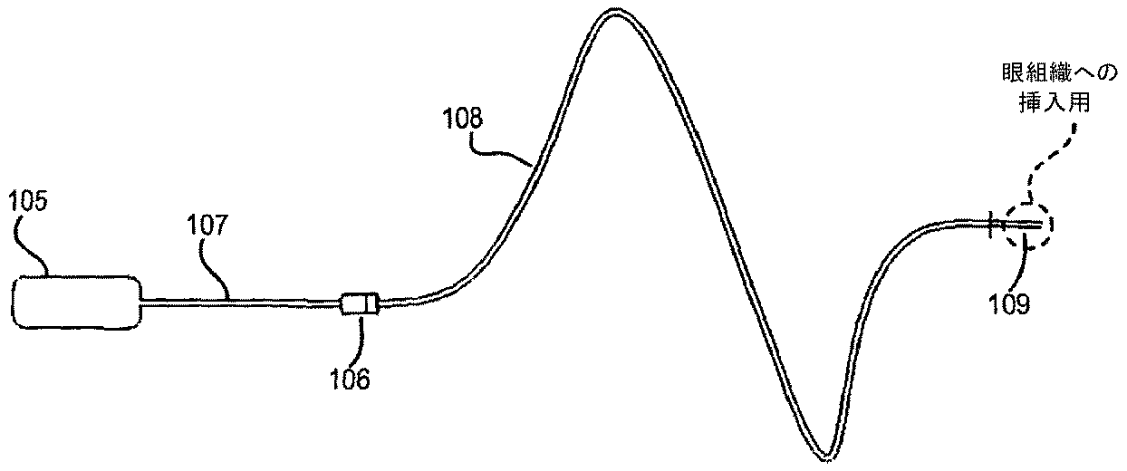
図1は、固体薬物塊から薬物を送達するために使用することができる、少なくともいくつかの態様に従う薬物送達システムの図面である。図1のシステムは、移植可能な浸透圧ポンプ105および薬物/フィルターハウジング106を含む。以下で説明するように、ハウジング106は、内部腔、インレット、およびアウトレットを含む。第一カテーテル107の管腔は、浸透圧ポンプ105のアウトレットおよび薬物/フィルターハウジング106のインレットに接続する。「カテーテル」は、流体が流れ得る一つまたは複数の内部管腔を有するチューブまたはその他の細長い本体である。第二カテーテル108の管腔は、薬物/フィルターハウジング106のアウトレットを末端構成要素109に接続する。理解される通り、流路は、ポンプ105、カテーテル107の管腔、ハウジング106の内部腔、カテーテル108の管腔、および末端構成要素109によって形成される。
FIG. 1 is a drawing of a drug delivery system according to at least some embodiments that can be used to deliver a drug from a solid drug mass. The system of FIG. 1 includes an implantable
浸透圧ポンプ105は、当技術分野において公知のタイプである。そのようなポンプ(例えば、Durect Corp. of Cupertino CAによってデュロス(登録商標)およびCHRONOGESIC(登録商標)という商標名で売られているポンプ)は、その他の用途での使用に関して公知であり、例えば、米国特許第4,034,756号に記載される。概して、移植される浸透圧ポンプは、ポンプ内の膜の水性透過性に関連する所定の流速で薬物を駆動するための浸透圧差を組み入れる。このメカニズムは、典型的には、周囲の組織環境から液体を吸収し区画容量を拡大する高浸透性を有する浸透圧ポリマー、塩、またはその他の材料を使用する。この容量増加は、ピストンを動かす、または弾性貯蔵部を圧縮し、ポンプからの液体の排出を引き起こす。ピストン(または可動シール)は、排出されるべき液体を含む貯蔵部から浸透圧ポリマーを分離する。ポンプハウジングは、水または適当な液体を浸透圧ポリマーに到達させる半透体から構成されてもよい。ポンプの送達の速度は、ポンプの外膜の透過性によって決定される。
The
従来の浸透圧ポンプは、液体貯蔵部に液体製剤薬物を保持する;そのようなポンプは、いくつかの態様において、目またはその他の眼組織にそのような液体薬物製剤を送達するために使用することができる。しかしながら、図1における浸透圧ポンプ105は、薬物媒体を含む。媒体は、薬物/フィルターハウジング106の内側の固体薬物塊からの薬物の取り込みのためにポンプ105から排出される。その他の態様において、ポンプ105は、薬物を含むが薬物/フィルターハウジング106から付加的な薬物を運ぶための媒体としても使用される液体を排出してもよい。
Conventional osmotic pumps hold liquid drug formulations in a liquid reservoir; such pumps are used in some embodiments to deliver such liquid drug formulations to the eye or other ocular tissue. be able to. However, the
浸透圧ミニポンプは、長い時間継続的に少量の液体を送達することができる。しかしながら、従来の浸透圧ポンプの内部流体貯蔵部を再充填するのが難しい場合がある。従って、図1の態様は、手短な外科的手順における浸透圧ポンプ105の従来の除去および置き換えを可能にする接続金具(図1には図示せず)を含む。浸透圧ポンプの流速を制御することも難しい場合がある。図1の態様に対するバリエーションは、低浸透性の環境流体から半透膜(ポンプ内)を単離するポンプに接続される制御可能な弁を含む。これは、流体送達ピストンを駆動するためのポンプ区画への流体の進入を防ぐ。制御弁は、電場がそれを横切って印加される場合に変形する圧電要素であり得る。そのような弁は、内部電子機器パッケージによって、またはRFトランスミッションを介して(例えば、患者が着る体の外側の外部信号システムから)信号を受信する内部制御モジュールによって、接続されかつ制御されてもよい。さらにその他の態様において、小さな磁気的に活性化されたスイッチが、弁のための電子機器へ内蔵される。弁は、制御電子機器が移植されている患者の体の一部上に十分な強度の磁石を配置することによって開かれるまたは閉じられる。同様の磁気的にアクティブにされるスイッチは、ペースメーカーなどの移植されるデバイスおよび移植される心臓除細動器において見出される。しかしながら、そのような制御弁が採用される場合でさえも、浸透圧ポンプは、即時オン/即時オフ様式で機能しない場合がある。例えば、制御弁が閉じられる時間とポンプ送達が徐々に減る時間との間に遅延がある場合がある;この遅延の間に、ポンプは、浸透圧平衡に到達する。さらにその他の態様において、制御弁またはダイバータ弁をポンプアウトレットカテーテル107に配置することによって、これに対処することができる。さらにその他の態様において、即座のポンプ停止を必要とする緊急事態において、浸透圧をなくすために圧放出弁が含まれてもよいと考えられる。
An osmotic minipump can deliver a small amount of liquid continuously for a long time. However, it may be difficult to refill the internal fluid reservoir of a conventional osmotic pump. Accordingly, the embodiment of FIG. 1 includes a fitting (not shown in FIG. 1) that allows conventional removal and replacement of the
図2は、図1からの薬物/フィルターハウジング106の断面図である。ハウジング106は、一つまたは複数の固体薬物および抗菌フィルターを保持するためのカプセルとしての役目を果たす。液体薬物製剤を送達するために移植された浸透圧ポンプを使用するいくつかの態様において、ハウジング106は、抗菌フィルターを含むだけである場合がある。ハウジング106は、チタンまたは生体適合性がありかつ調剤されるべき薬物と適合性があるその他の材料から形成される。ハウジング106の近位(または「上流」)端は、ハウジングに恒久的に取り付けられ得る、または除去可能であり得る多孔性ケージ111を保持する。同様にチタンまたはその他の生体適合性および薬物適合性材料から形成されるケージ111は、一つまたは複数の固体薬物の一つまたは複数の塊を保持する。薬物は、粉末の形態で、ペレットの形態で、または何らかのその他の固体形態で、モノリシックであり得る。ケージ111上の複数の穴は、ポンプ105からの流体が、溶解された(またはその他の取り込まれた)形態でその固体薬物の一部と混合しかつそれを運び出すことを可能にする。ハウジング106の遠位(または「下流」)端は、三次元抗菌フィルター112を含む。以下でより詳細に記載するように、「抗菌フィルター」は、薬物運搬液を通過させるのに十分小さいが、バクテリアまたはその他の望ましくない要素の通過を妨害する細孔サイズを有するフィルターである。ハウジング106は、ツーピースアセンブリ(ピース106aおよび106b)であり、それによって、ケージ111(例えば、薬物を変えるため、または薬物が枯渇した場合に)および/またはフィルター112(例えば、フィルターが詰まった場合に)を置き換えるために、ハウジング106を解体しかつ再度組み立てることを可能にする。ピース106aおよび106bは、ネジ式接続を介してまたはその他のタイプの機械的メカニズム(例えば、連動タブおよびスロット)によって互いに取り付け可能であり得る。カテーテル107は、ピース106aにおけるインレットに取り付けられ;カテーテル108は、ピース106bにおけるアウトレットに取り付けられる。カテーテル107および108は、エポキシまたはその他の接着剤で取り付けられ得る。その他の態様において、かかり付きコネクターが採用されてもよい。ハウジング106にカテーテル107および108を留保するために、クリップおよび/またはその他のロッキングメカニズムが使用されてもよいと考えられる。
FIG. 2 is a cross-sectional view of drug /
少なくともいくつかの態様において、浸透圧ポンプ105および薬物/フィルターハウジング106は、患者の頭蓋骨内の特別に調製されたポケットにおける移植に対してサイズ決定される。カテーテル107および108は、同様に患者の頭蓋骨上で調製された溝内に置かれてもよい。
In at least some embodiments, the
図3Aおよび3Bは、別の態様に従う薬物送達システムを示す。浸透圧ポンプ205は、ポンプ205のアウトレット231がある程度拡大されかつ内部ネジ山232を有すること以外、図1の浸透圧ポンプ105と同様である。薬物/フィルターハウジング206は、図1および2のハウジング106と同様である。しかしながら、ハウジング206は、ポンプ205のアウトレット231上の内部ネジ山232に対応する外部ネジ山233を有する。図3Bに示すように、これは、ポンプ205とハウジング206との間の直接的な取り付けを容易にし、それによって、図1に示すカテーテルの一つ(すなわち、カテーテル107)に対する必要性を回避する。ハウジング206へのインレット(図2におけるカテーテル107に接続されるハウジング106のインレットと同様)は、ポンプ205のアウトレットと流体連通するように配置される。ハウジング206のアウトレットからの流体は、カテーテル208を介して眼組織に流れる。ハウジング206の寸法は、送達される薬物および望ましい濃度の薬物を提供するのに必要とされる表面積に依存すると考えられる。
3A and 3B show a drug delivery system according to another embodiment. The
図3A〜3Bの形態は、ハウジング206内の薬物および/またはフィルターの置き換えのためのポンプ205からのハウジング206の定期的な除去を可能にする。図3A〜3Bの態様に対するバリエーションにおいて、ポンプ205とハウジング206との間のその他のタイプの接続メカニズム(例えば、ロッキングタブおよび溝)が採用される。さらにその他のバリエーションにおいて、ハウジング206は、ポンプ205に恒久的に(例えば、接着剤で)取り付けられる。
The configuration of FIGS. 3A-3B allows for periodic removal of the
眼科薬物送達デバイスの別の態様を、図4に示す。図4の態様において、デバイス310は、カテーテル316および317を介してスリーブ付き薬物貯蔵部314に連結される浸透圧ポンプ312を含む。三次元(3-D)抗菌フィルター319は、カテーテル318を介して薬物貯蔵部314に連結される。別のカテーテル321およびコネクター322は、付加的なカテーテル(図示せず)を介して、3-Dフィルター319を、標的眼組織への薬物含有溶液の送達のために位置付けられる末端構成要素(同様に図示せず)に接続する。末端構成要素は、例えば、ニードルまたはカテーテルの開端であり得る。移植の前に、浸透圧ポンプは、固体薬物を取り込むと考えられる溶液で満たされる。
Another embodiment of an ophthalmic drug delivery device is shown in FIG. In the embodiment of FIG. 4,
固体薬物貯蔵部は、流体が固体薬物の一つまたは複数の塊(例えば、固体薬物ペレット)の周りを流れかつ浸食するための腔を提供するようにデザインされる。図5は、少なくともいくつかの態様に従う薬物貯蔵部のほんの一例である、図4のスリーブ付き薬物貯蔵部314の断面図である。薬物貯蔵部314は、一つまたは複数の固体薬物ペレット325がロードされるチャンバー320を形成する二つの中空金属チューブ328および329(薬物適合性材料から作られる)を含む。液体密封シールを形成するために、スリーブ327(シリコンまたはその他の適当な材料から作られる)が、チューブ328および329に巻かれる。チューブ328および329の先細端は、それぞれ、カテーテル318および317の端に適合する。図5の薬物貯蔵部314は、チャンバー320内に薬物ペレットを含むように、かつ固体ピースがチャンバー320から出ていくのを防ぐように形成される。薬物貯蔵部314は、分離されかつ再び取り付けられてもよく、それによって一つまたは複数の固体薬物ペレットのローディングを可能にする。
The solid drug reservoir is designed to provide a cavity for fluid to flow and erode around one or more masses of solid drug (eg, solid drug pellets). FIG. 5 is a cross-sectional view of the
いくつかの態様において、薬物ペレットの移動をさらに防ぐために、円形スクリーンが、薬物チャンバーの内側に配置される。いくつかの場合、スクリーンの少なくとも一つは、薬物の補充を可能にするために除去可能であってもよい。図6Aおよび6Bは、別の態様に従い、かつそのようなスクリーンを含む、薬物貯蔵部340の断面図である。図6Aおよび6Bにおいて見られるように、薬物貯蔵部340は、流体密封接続を形成するために(ネジ山351および352で)嵌め合うハウジング344および346を含む。チュービング接続インレット350の側に定常メッシュスクリーン343とハウジング344のエッジに除去可能メッシュスクリーン341とを含むハウジング344内のチャンバー342の内側に、固体薬物を配置してもよい。図6Aに見られるように、スクリーン341は、ハウジング346内にある3-D抗菌フィルター345の直前にある。スクリーン341および343は、多孔性であり、チタン、ステンレススチール、もしくはその他の生体適合性、薬物適合性金属(例えば、金、白金)で作られた金網クロスおよび/またはポリマー(例えば、フルオロポリマー)であり得る。その他の態様において、スクリーンは、チタンまたはステンレススチールなどの多孔性金属で作られていてもよい。メッシュスクリーン341および343は、薬物ペレットが、ハウジング346、抗菌フィルター345、またはインレット接続350もしくはアウトレット接続348に接続してもよいチュービング(図示せず)へ移動することを防ぐ。図6Aにおいて、ハウジング半分344および346がネジ式結合した薬物貯蔵部340を示す。図6Bは、分離されたハウジング344および346を示すが、適所に除去可能スクリーン341、定常スクリーン343、および抗菌フィルター345を有する。図6Bに見られるように、除去可能スクリーン341は、ハウジング344の端の外円形表面を覆う。定常スクリーン343は、空間342の内円形表面を覆うだけである。スクリーンは、薬物チャンバーの形状に適合する任意の形状であり得る。しかしながら、特定の態様において、スクリーンは必要とされず、省略されてもよい。
In some embodiments, a circular screen is placed inside the drug chamber to further prevent drug pellet movement. In some cases, at least one of the screens may be removable to allow drug replenishment. 6A and 6B are cross-sectional views of a
抗菌フィルターは、同様に必要とされない。例えば、図6Cは、抗菌フィルター345を伴わない薬物貯蔵部340の断面図である。少なくともいくつかの態様は、システムの充填の間に空気泡を抜き取ることを可能にする機構を含んでもよい。これは、流体送達システム(例えば、浸透圧ポンプまたは皮下ポートを介して接続される外部ポンプ)が、湿潤多孔性フィルター(図6Aおよび6Bの3-Dフィルター345など)の毛細血管様構造内で液体を保持する表面張力に打ち勝つのに十分な圧を生成しない場合に、蒸気閉塞を防ぐのを補助し得る。いくつかの態様において、セットスクリューまたはプラグが、フィルターの上流(すなわち、より高い圧)側の薬物チャンバーハウジングの側面へ組み入れられてもよい。セットスクリューまたはプラグは、プライミングの間に除去され得、すべての空気泡がシステムから抜き取られると、使用のために再び取り付けられ得る。さらにその他の態様において、換気弁は、ガスの換気を可能にする上流半透膜を含んでもよい。さらにその他の態様において、セットスクリューまたはプラグは、除去不可能である場合があるが、脱気を可能にするためにガス透過性であるが液体透過性ではない部分を含む場合がある。
Antibacterial filters are not required as well. For example, FIG. 6C is a cross-sectional view of the
図6Dは、少なくとも一つの態様に従う薬物貯蔵部360を示し、これはガスの換気を可能にする半透膜を有する換気弁361を含む。チュービングコネクターかかり362が、貯蔵部360の上流側にあり、チュービングコネクター363が、下流側にある。図6Eは、薬物貯蔵部360の断面図である。薬物貯蔵部360は、ネジ山371、372で流体密封接続を形成するように接合するハウジング364および365を含む。腔366は、一つまたは複数の固体薬物ペレットまたはその他の塊を保持する。図示しないが、図6Aおよび6Bにおけるスクリーン343および341と同様のスクリーンが、空間366の上流側の面369上および空間366の下流側の面368上に(定常または除去可能形態で)置かれてもよい。図6Eの態様において、3-D抗菌フィルター367は、ハウジング365において形成される空間374に収まる。
FIG. 6D shows a
薬物貯蔵部340のハウジング344および346、薬物貯蔵部360のハウジング364および365、ならびにその他の態様における薬物貯蔵部のハウジングは、チタン、ステンレススチール、白金、金などの薬物適合性、腐食耐性材料、生体適合性コーティング金属、PTFE(ポリテトラフルオロエチレン)、FEP(テトラフルオロエチレン-ヘキサフルオロプロピレンコポリマー)、PFA(ペルフルオロアルコキシエチレン)、その他のフルオロポリマーなどの化学的不活性ポリマー、またはフルオロポリマーコーティング金属で作製することができる。体温で低流速の間に、薬物は、チャンバーの壁に吸着しやすい場合があり、そのため予期される濃度よりも低い濃度の薬物が患者に送達される。フルオロポリマーは、吸着に耐えるための最良の公知の材料である。その他のポリマーは、ECTFE(エチレン-クロロトリフルオロエチレンコポリマー)、ETFE(エチレン-テトラフルオロエチレンコポリマー)、MFA(テトラフルオロエチレンペルフルオロ(メチルビニルエーテル)コポリマー)、PCTFE(ポリクロロトリ-フルオロエチレン)、およびPVDF(ポリビニリデンジフルオライド)を含むが、それらに限定されない。
The
上で示すように、様々な態様における薬物貯蔵部は、固体薬物の補充を可能にするために開かれかつ閉じられる場合がある。貯蔵部構成要素は、(図6A〜6Cおよび6Eに示すように)ネジ式である場合もあり、またはロッキングタブおよび溝からなる場合もある。さらにその他の態様において、外部クランプが使用されてもよい。さらにその他の態様において、貯蔵部ハウジングは、スナップフィットによって接合されてもよい。上でも示すように、貯蔵部314(図5)は、周囲のスリーブ327によって一緒に保持される二つの金属チューブ328および329を含む。周囲のスリーブ327は、シリコンゴムなどの弾性ポリマーで作製することができる。いくつかの態様において、漏れを防ぐために、薬物貯蔵部の嵌め合い部分の間(例えば、図5のチューブ328と329との間、図6A〜6Cのハウジング344と346との間、図6Eのハウジング364と365との間)に、生体適合性ガスケットが置かれてもよい。さらにその他の態様において、薬物貯蔵部ハウジングの外部部分は、より簡単な締め付けを促進するために平面またはその他の領域を含んでもよい。図6Fは、嵌め合いハウジング381および382を有する薬物貯蔵部380の態様を示す。平面383は、ハウジング381の一側面に形成される。第二平面(図示せず)は、ハウジング381の反対側面に形成することができる。同様に、ハウジング382は、一側面に形成される平面384を含み、反対側面に付加的な平面(同様に図示せず)を含んでもよい。
As indicated above, the drug reservoir in various embodiments may be opened and closed to allow replenishment of solid drug. The reservoir component may be threaded (as shown in FIGS. 6A-6C and 6E) or may consist of locking tabs and grooves. In still other aspects, external clamps may be used. In yet other aspects, the reservoir housing may be joined by a snap fit. As also shown above, the reservoir 314 (FIG. 5) includes two
薬物ケージ111(図2)と同様の薬物ケージは、図5〜6Fに示す薬物フィルターハウジングのいずれかとともに、ならびに以下で記載するその他のハウジングとともに使用されてもよい。 A drug cage similar to drug cage 111 (FIG. 2) may be used with any of the drug filter housings shown in FIGS. 5-6F, as well as with other housings described below.
少なくともいくつかの態様において、薬物貯蔵部の上流側のカテーテルチュービング(例えば、図4におけるデバイス310のポンプ側のカテーテル316に対するチュービング)は、シリコン、ポリウレタン、またはPTFE、FEP、およびPFAを含むフルオロポリマーなどの媒体適合性および生体適合性、弾性ポリマーであり、薬物貯蔵部の下流側のカテーテルチュービングは、PTFE、FEP、およびその他のフルオロポリマーなどの生体適合性、薬物適合性、弾性ポリマーである。
In at least some embodiments, the catheter tubing upstream of the drug reservoir (eg, tubing for the
いくつかの態様において、固体薬物貯蔵部および3-D抗菌フィルターは、カテーテル接続を介して流体連通している。これは、概して、上流または下流構成要素に接続するために使用することができる金属チュービングコネクター322および389も示している図4に見られる。その他の態様においてかつ上で記載したように、単一のハウジングは、固体薬物(単独でまたはケージ内で)ならびに三次元抗菌フィルターを含んでもよい。そのようなハウジングは、固体薬物の補充を可能にするために、開かれかつ閉じられてもよい。図7は、別の態様に従う薬物貯蔵部395の断面図である。薬物貯蔵部395は、ネジ山401、402を嵌め合うことによって接合されるハウジング396および397を含む。ハウジング396の内側の腔403が、固体薬物を保持する(図示せず)。図6Aおよび6Bのスクリーン341および343と同様のスクリーンが含まれてもよい。任意で、3-D抗菌フィルター398が、空間399に設置される。図6A〜6Fに示すかかり付き接続金具の代わりに、薬物貯蔵部395は、上流インレット穴405および下流アウトレット穴406を含む。
In some embodiments, the solid drug reservoir and the 3-D antimicrobial filter are in fluid communication via a catheter connection. This is seen generally in FIG. 4 which also shows
少なくともいくつかの態様において、薬物およびフィルターのためのハウジングは、チタン、金、白金、またはステンレススチールから作製され、人体へ移植するのに十分なほど小さい。内径は、3-D抗菌フィルターがハウジングの内側に固着することができるようにサイズ決定される。(様々な態様における)可能性のあるフィルターサイズの例は、0.03〜0.25''の物理的外径を有する0.22ミクロン最大細孔サイズ3-Dフィルターを含むが、それらに限定されない。さらにその他の態様において、物理的外径は、0.1''と0.3''との間である。 In at least some embodiments, the housing for the drug and filter is made from titanium, gold, platinum, or stainless steel and is small enough to be implanted into the human body. The inner diameter is sized so that the 3-D antibacterial filter can be secured inside the housing. Examples of possible filter sizes (in various embodiments) include, but are not limited to, 0.22 micron maximum pore size 3-D filters having a physical outer diameter of 0.03 to 0.25 ″. In yet other embodiments, the physical outer diameter is between 0.1 "and 0.3".
図8は、少なくとも一つの態様に従う薬物貯蔵部425の二つの分離されたハウジング426および427の透視図である。図9は、ハウジング426および427が(ネジ山430および431を介して)接合されている薬物貯蔵部425の断面図である。ハウジング426および427の外端全体は、そこに形成される(それぞれ)かかり428および429を有する。固体薬物および任意の3-D抗菌フィルター433を保持するための空間432も、図9に見られる。
FIG. 8 is a perspective view of two separated
図10は、二重管腔チューブ445がポンプおよび/または固体薬物を含む貯蔵部から伸長する付加的な態様を示す。二重管腔チューブ445は、二つの別個のラインへ分離する。チューブ446は、一つの管腔に取り付けられ、患者から流入する生理学的流体を受ける。チューブ447は、別の管腔に取り付けられ、患者の眼組織に治療的流体を送達する。ライン446内に受けられる間質液は、貯蔵部内の固体薬物ペレットを通って流れ、それらのペレットから薬物をゆっくりと(例えば、溶解によって)取り出す。次いで、得られた薬物および生理学的流体の溶液は、チューブ447を介して標的眼組織に送達される。図11は、チューブ446および447の遠位端448および449の拡大図であり、液流を再循環させるための二つの管腔をさらに図示する。その他の態様において、二つの完璧に別個のチューブ(すなわち、二重管腔チューブから出現しない二つのチューブ)が使用されてもよい。そのような態様は、治療的流体が送達されるべき領域からより遠くにある領域から、生理学的流体が引き出される場合に有用である可能性がある。特定の態様において、チューブ446を介して目から受けた流体のいくらかまたはすべては、再循環されない。これは、例えば緑内障によって引き起こされる過剰な眼内圧を軽減するために行われ得ると考えられる。
FIG. 10 shows an additional embodiment in which a
図12は、ポンプが流れを生成することを必要としないシステムの態様を示す透視図である。半透膜455は、固体薬物を含む貯蔵部456のチャンバーへ間質液媒体を通過させる。チャンバー内の薬物が溶解する(または別の方法で、固体薬物塊から取り出され、間質液媒体に取り込まれる)時、膜を隔てた濃度差により、流体は低い濃度からより高い濃度に流れる。浸透圧により、流体は、膜455を通過し、アウトレットを通って薬物チャンバーへ移動し、カテーテル458(図示の目的のためにクリアなカテーテルとして示す)における任意の3-D抗菌フィルター457を通過して、標的眼送達部位に移動する。半透膜455は、間質液を通すのに十分であるが、取り込まれた固体薬物を拡散させない細孔サイズカットオフを有する。抗菌フィルター457は、バクテリアを留保するが、溶解された(または他の方法で取り込まれた)薬物を通過させるのに十分な細孔を有する。電場が膜455に印加されてもよく、その結果電気浸透による拡散が引き起こされる。図13は、図12の態様の完全な断面図であり、固体薬物を保持するための腔460をより詳細に示す。図14は、腔460に固体薬物ペレット325を含む図12および13の態様を示す。逆流を防ぐために、適当なチェック弁(図示せず)が、腔460または流路内の他の場所内に含まれてもよい。
FIG. 12 is a perspective view showing an embodiment of a system that does not require the pump to generate a flow. The
図15は、流体が、半透性中空ファイバー473を含むループ472を介してポンプ/貯蔵部から(二重管腔チュービング475の一つの管腔を介して)一方向に循環し、チュービング475の第二管腔を介して戻る、システム470の態様を示す。中空ファイバーループ473は、標的眼送達エリアに位置付けることができる末端構成要素である。ポンプは、貯蔵部に設置された固体薬物を通って媒体を循環させ、結果として生じる薬物を保持した媒体は、中空ファイバー473の壁を通って標的眼組織へ拡散する。その他の態様において、図15のものと同様の送達システムは、実際には液体の容量を送達することなく、受動拡散によって外部環境へ薬物を放出する薬物透過中空ファイバーを含む。
FIG. 15 shows that the fluid circulates in one direction (through one lumen of the dual lumen tubing 475) from the pump / reservoir via a
さらにその他の態様は、取り付けられた電池および電力電子機器(電力供給、再充電回路など)ならびに情報を受信しかつ送信するための通信電子機器を有するセンサー(例えば、緑内障のための圧センサーまたは薬物センサー)を含む。これらの態様において、電子機器は、デバイスの貯蔵部区域と束ねられてもよいと考えられ、センサーは、カテーテルの表面に付随するワイヤと組み合わせられるまたは多管腔チュービングの管腔の一つの中に含まれ、かつ標的眼組織内に出ていてもよいと考えられる。 Yet another aspect is a sensor (eg, a pressure sensor or drug for glaucoma) having an attached battery and power electronics (power supply, recharge circuit, etc.) and communication electronics for receiving and transmitting information Sensor). In these embodiments, it is contemplated that the electronics may be bundled with the reservoir area of the device and the sensor is combined with a wire associated with the surface of the catheter or in one of the lumens of the multi-lumen tubing. It is contemplated that it may be included and exit into the target eye tissue.
少なくともいくつかの態様は、帯電薬物イオンまたは薬物のその他の粒子の電気泳動誘導送達を含む。帯電薬物に対して、薬物を含む(または吸着/吸収された薬物を有するナノ粒子を含む)流体に電場を印加することにより、普通の拡散よりも速い薬物の移動を誘起できる。ガシクリジンの場合は、ポンプを必要とせずに目への薬物送達を加速するために、デバイス出口の(例えば、カテーテルの端における)またはデバイス出口のすぐ外側の負電荷を使用することができる。反対極性の同じまたは類似の電荷(例えば、ガシクリジンの場合は正電荷)が、薬物含有区画(例えば、固体薬物が保持されるチャンバー)に同様に印加されてもよいと考えられ、それによって、ポンプを必要とせずにデバイスからの薬物送達を可能にする。電気泳動環境は、標的眼組織内の天然低抵抗アウトレットに電気浸透流を誘起すると考えられる。適当な電子機器パッケージ、電池、再充電アセンブリ、オン/オフスイッチ、通信回路、およびその他の電子機器によって調節される電気量の電界強度およびその他のパラメータによって、カテーテルチップへの薬物の移動の速度(または薬物の濃度)を調節することができると考えられる。反対電荷を有する薬物が使用される場合、次いで、電子回路は、電極上の電荷を逆転させると考えられる。電気泳動誘導薬物送達の態様は、患者の安全性を促すために、および少量の薬物が送達されることから、非常に低い電力デバイスであると考えられる。眼組織内の帯電デバイスは、例えば、盲目患者において、および目における光フラッシュまたは光に対する活動亢進感受性を有する患者を処置するための特別な状況において、ならびに電気刺激からの利点が報告されるその他の患者に対して、視神経の神経変性を抑圧するための付加的な利点を提供する可能性がある。いくつかの態様において(ならびに図23および24に関連して以下で記載するように)、カテーテルは、目への電気刺激(パルスまたはその他)の送達のために使用されるだけである電極を含む。さらにその他の態様において、カテーテルは、目における眼内圧、電位、または何らかのその他の物理的特徴を検知するために、代替的に(または付加的に)使用される電極を含む。そのような刺激および/または検知のための方法および電子機器は、当技術分野において公知である(本明細書に記載する薬物送達デバイスとの組み合わせではないが)。本明細書に記載する薬物送達システムへの適当な刺激および/または検知電子機器の含有は、当業者が本明細書に含まれる情報を得れば、当業者の日常的な技能内であると考えられる。 At least some embodiments include electrophoretic guided delivery of charged drug ions or other particles of drug. By applying an electric field to a fluid containing a drug (or containing nanoparticles with adsorbed / absorbed drug) relative to a charged drug, drug migration can be induced faster than normal diffusion. In the case of gacyclidine, a negative charge at the device outlet (eg, at the end of the catheter) or just outside the device outlet can be used to accelerate drug delivery to the eye without the need for a pump. It is contemplated that the same or similar charge of opposite polarity (eg, positive charge in the case of gacyclidine) may be similarly applied to the drug containing compartment (eg, the chamber in which the solid drug is held), thereby pumping Allows drug delivery from the device without the need for The electrophoretic environment is believed to induce electroosmotic flow to natural low resistance outlets in the target eye tissue. The rate of drug transfer to the catheter tip (by the electric field strength and other parameters adjusted by the appropriate electronics package, battery, recharge assembly, on / off switch, communication circuit, and other electronics) Or the concentration of the drug). If a drug with the opposite charge is used, then the electronic circuit is believed to reverse the charge on the electrode. The electrophoretic guided drug delivery aspect is considered to be a very low power device to promote patient safety and because small amounts of drug are delivered. Charging devices in ocular tissues are, for example, in blind patients and in special situations for treating patients with light flash or hyperactivity sensitivity to light in the eye, and others where benefits from electrical stimulation are reported It may provide an additional benefit for patients to suppress neurodegeneration of the optic nerve. In some embodiments (and as described below in connection with FIGS. 23 and 24), the catheter includes an electrode that is only used for delivery of electrical stimulation (pulse or other) to the eye. . In yet other embodiments, the catheter includes electrodes that are alternatively (or additionally) used to sense intraocular pressure, electrical potential, or some other physical characteristic in the eye. Methods and electronics for such stimulation and / or sensing are known in the art (although not in combination with the drug delivery devices described herein). Inclusion of appropriate stimulation and / or sensing electronics in the drug delivery system described herein is within the routine skill of one of ordinary skill in the art given the information contained herein. Conceivable.
図16は、少なくともいくつかの態様に従う電気泳動誘導薬物送達システム495を示す。チューブ497は、流体送達管腔および電極ワイヤを含み、薬物貯蔵部496から伸長する。図17は、薬物貯蔵部496およびチューブ497の一部の断面図である。貯蔵部496は、半透膜500および固体薬物ペレットを保持するための内部腔501を含む。電子機器パッケージ503および電池505は、貯蔵部496の下側に取り付けられる。電子機器パッケージ503は、電極チップ507に一つの極性の電荷を、電極ワイヤ509のチップ508(図16および19を参照されたい)に反対極性の電荷を誘起する。腔501内のワイヤ509の部分は、チップ507との早期電荷交換を防ぐために、誘電体でコーティングしてもよく、または他の方法で絶縁してもよい。図18は、図17と同様であるが、腔501内の固体薬物ペレット325を示す。図19は、チュービング497の末端(または遠位端)を(図18に対して反転した配向で)示し、電極チップ508および流体アウトレット510を図示する。反対電荷が電極507およびワイヤチップ508に適用される場合、目の中の自然な低い抵抗のアウトレットに電気浸透流が誘起される。間質液は、半透膜500を介して腔501に入る。その他の態様において、薬物貯蔵部から離れている別の体領域から流体を引き出すために、別個のチューブが(膜500の代わりに)使用される。腔501に入る流体は、腔501内の薬物を溶解し、標的眼組織に薬物を送達する。
FIG. 16 illustrates an electrophoretic guided
少なくともいくつかの態様において、ポートは、患者の体の皮下に(または経皮的に)移植され、移植されたカテーテルおよび末端構成要素と流体連通するように配置される。ポートは、液体および/または固体薬物を保持するために使用することができる内部腔を含む。セルフシーリングエラストマー(例えば、シリコン)中隔が、腔を覆う。中隔は、薬物吸着を最小にするための薬物適合性フルオロポリマー積層ライニングを有してもよい。外部供給源から腔へ流体を導入するために、ノンコアリングニードルを中隔を通って挿入してもよい。その流体は、液体製剤薬物であってもよく、または腔内に既に設置されている固体型薬物を溶解し(または他の方法で取り込み)、その取り込まれた薬物を目に送達するための液体媒体であってもよい。いくつかの態様において、液体製剤薬物は、腔に含まれる付加的な固体型薬物を取り込むための媒体として使用される。 In at least some embodiments, the port is implanted subcutaneously (or percutaneously) in the patient's body and is placed in fluid communication with the implanted catheter and the end component. The port includes an internal cavity that can be used to hold a liquid and / or solid drug. A self-sealing elastomer (eg, silicone) septum covers the cavity. The septum may have a drug compatible fluoropolymer laminate lining to minimize drug adsorption. A non-coring needle may be inserted through the septum to introduce fluid from an external source into the cavity. The fluid may be a liquid pharmaceutical drug, or a liquid to dissolve (or otherwise incorporate) a solid drug already placed in the cavity and deliver the incorporated drug to the eye It may be a medium. In some embodiments, the liquid pharmaceutical drug is used as a vehicle for incorporating additional solid form drug contained in the cavity.
ポートの薬物保持腔は、薬物適合性材料(例えば、ステンレススチール、チタン、白金、金、または薬物適合性ポリマー)から構成されてもよい(またはそれでコーティングしてもよい)。この材料は、(組織拒絶を防ぐために)生体適合性であり、薬物の繰り返しの再充填および調剤ならびに薬物含有媒体の潜在的な腐食作用に持ちこたえることができ、かつ薬物を保持し劣化することなくより長期間、移植した状態を保つことが可能である。ポートが固体状態で薬物を保持するために使用される場合、腔形成材料は、薬物が腔壁にくっつかないように、かつ薬物と接触する腔表面が薬物のいずれをも吸着しないように、適合性であってよい。腔壁は、少なくとも特定の態様において、水または生理学的流体に対して透過性であるべきではない。 The port drug retention cavity may be constructed of (or coated with) a drug-compatible material (eg, stainless steel, titanium, platinum, gold, or a drug-compatible polymer). This material is biocompatible (to prevent tissue rejection), can withstand repeated refilling and dispensing of drugs and the potential corrosive action of drug-containing media, and retains and degrades drugs The transplanted state can be maintained for a longer period of time. When the port is used to hold a drug in the solid state, the cavity forming material is adapted so that the drug does not stick to the cavity wall and the cavity surface that contacts the drug does not adsorb any of the drug May be sex. The cavity wall should not be permeable to water or physiological fluids, at least in certain embodiments.
図20は、ポートを含む一つの配置を示す。移植されるポート601(ブロック図形式で示す)は、カテーテル602に接続され、そのカテーテルも、患者の体の内側に移植される。末端構成要素604は、カテーテル602の遠位端に設置される。フランジまたはその他のタイプの止め具(図20には図示せず)は、目への末端構成要素604の過挿入を防ぐ。任意の縫合アンカー603は、適所にカテーテル602を固定する手段を提供する。ポート601は、固体薬物を含み得ると考えられ、次いで、外部ポンプからポートへ導入される滅菌媒体(例えば、生理食塩水、リンガー溶液、乳酸リンガー液、人工硝子体液、および/または目もしくはその他の眼組織への注射と適合性がある任意のその他の媒体)によって溶解されるまたは他の方法で取り込まれる。ポート601は、外部ポンプ(例えば、実施例2に記載するMiniMed 508ポンプ)またはその他の外部供給源からの薬物および/または媒体を受けることができる。
FIG. 20 shows one arrangement that includes ports. Implanted port 601 (shown in block diagram form) is connected to
いくつかの態様において、かつ出願第60/807,900号においてより詳細に記載されるように、皮下移植可能ポートは、二つの腔を含む。それらの腔の一つは、二重管腔カテーテルの第一管腔と流体連通しており、もう一方の腔は、もう一方の管腔と流体連通している。そのような態様は、別の供給源(例えば、外部ポンプ)から流体を受けるためにポートの一つの側を使用して、かつ標的眼組織から流体を引き出すためにポートのもう一方の側を使用して、標的眼組織の洗浄を可能にする。 In some embodiments, and as described in more detail in application 60 / 807,900, the subcutaneously implantable port includes two cavities. One of these lumens is in fluid communication with the first lumen of the dual lumen catheter, and the other lumen is in fluid communication with the other lumen. Such an embodiment uses one side of the port to receive fluid from another source (eg, an external pump) and the other side of the port to draw fluid from the target eye tissue Thus, the target eye tissue can be cleaned.
図21は、デバイス310の浸透圧ポンプ312(図4)が、皮下ポート710で置き換えられている態様を示す。いくつかの態様において、ポート710は、ポートの中隔711を貫通するニードル(ニードルは外部ポンプまたは媒体の何らかのその他の供給源と流体連通している)を介してポートへ導入される媒体によって浸食される固体薬物ペレットを含む。
FIG. 21 illustrates an embodiment where the osmotic pump 312 (FIG. 4) of the
眼組織への薬物の送達は、米国特許出願第11/337,815号(米国特許出願公開第2006/0264897号として刊行された、2006年1月24日に出願されかつ「Apparatus and Method for Delivering Therapeutic and/or Other Agents to the Inner Ear and to Other Tissues」と題される)に記載されるデバイスおよび手順を使用して行われてもよい。 Delivery of drugs to ocular tissue is described in U.S. Patent Application No. 11 / 337,815 (U.S. Patent Application Publication No. 2006/0264897, filed on Jan. 24, 2006 and "Apparatus and Method for Delivering Therapeutic and / or Other Agents to the Inner Ear and to Other Tissues).
いくつかの態様において、薬物貯蔵部に連結される電子機器パッケージ(例えば、図17における電子機器パッケージ503)は、薬物/媒体溶液(または懸濁液)の特性を検知するための構成要素を含む。検知される特性は、圧、光の吸収、電気伝導度、光散乱、薬物、または電解質濃度などの一つまたは複数を含み得ると考えられる。次いで、これらの検知される特性を適当な電子機器を介して使用して、ポンプ(内部または外部)またはその他の要素(例えば、磁気コイルまたは電気泳動電極)の操作を調整することができる。電子機器パッケージは、同様に(または代替的に)光もしくはその他の物理的パラメータ(例えば、組織電気活動)を検出する、および/または離れたセンサーと連通するように設定できるだろう。
In some embodiments, an electronic device package (eg,
少なくともいくつかの付加的な態様において、貯蔵部内の一つまたは複数の固体薬物塊から薬物を取り出すために使用する媒体は、それ自身、薬物(または複数の薬物)を含むナノ粒子の予混合懸濁液であってもよい。さらにその他の態様において、貯蔵部チャンバー内の固体薬物塊を採用することなく薬物(または複数の薬物)を含むナノ粒子の予混合懸濁液を送達するために、様々な態様に従う薬物デバイスが使用されてもよい。どちらの場合でも、ナノ粒子は、薬物ナノ粒子または薬物が吸収/吸着されているもしくは他の方法で取り付けられている担体材料のナノ粒子であり得る。 In at least some additional embodiments, the medium used to remove the drug from the one or more solid drug masses in the reservoir is itself a premixed suspension of nanoparticles comprising the drug (or drugs). It may be a turbid liquid. In still other embodiments, drug devices according to various embodiments are used to deliver a premixed suspension of nanoparticles containing drug (or drugs) without employing a solid drug mass in the reservoir chamber May be. In either case, the nanoparticles can be drug nanoparticles or nanoparticles of a carrier material to which the drug has been absorbed / adsorbed or otherwise attached.
以前に示されたように、本明細書に記載するようなデバイスおよび方法は、薬物の持続性長期送達を提供するために使用することができる。そのようなデバイスおよび方法は、長期的に断続的な薬物送達を提供するために使用されてもよい。例えば、固体薬物塊を保持する貯蔵部は、患者の体に移植することができると考えられる。次いで、その貯蔵部は、媒体の供給源に(例えば、貯蔵部と流体連通している皮下ポートを使用して)定期的に接続することができる。 As previously indicated, devices and methods as described herein can be used to provide sustained long-term delivery of drugs. Such devices and methods may be used to provide long-term intermittent drug delivery. For example, a reservoir that holds a solid drug mass could be implanted into the patient's body. The reservoir can then be periodically connected to a source of media (eg, using a subcutaneous port in fluid communication with the reservoir).
図4に示すシステム310と同様に、図6A〜9に示す貯蔵部は、ヒトまたは動物に移植して、一つの端(例えば、貯蔵部340のインレット350、貯蔵部360のインレットかかり362)でカテーテルを用いて媒体供給源(例えば、移植された浸透圧ポンプ、媒体が外部供給源から導入されるポート)に連結することができる。もう一方の端(例えば、貯蔵部340のアウトレット348、貯蔵部360のかかり363)は、別のカテーテルを介して患者の目に移植された末端構成要素に接続することができる。
Similar to the
上記の態様の一つまたは複数において、眼インプラントは、送達されるべき薬物を含む薄膜コーティングを含むように処置され得、その薄膜コーティングは、標的眼組織への眼インプラントの配置後に薬物をゆっくりと放出する。そのような眼インプラント801の一つの例を、図22に示し、薄膜コーティング802を、破線で示す。インプラント801に取り付けられるロッド803またはその他の部材は、インプラント801を眼組織へ配置する(かつ眼組織からインプラント801を除去する)ために使用することができる。図22の眼インプラント801は、固体ディスクであるが、薄膜コーティングは、その他のタイプの眼組織インプラント(例えば、電気刺激のための電極を有するインプラントおよび/またはセンサーを有するインプラント)とともに使用されてもよい。薄膜を介する送達に適した薬物は、神経保護剤および抗生物質薬剤を含むが、それらに限定されない。薬物含有コーティングを調製するために使用することができる方法および材料は、当業者に周知である。適した方法および材料の例は、米国特許第6,627,246号に記載されるものである。
In one or more of the above embodiments, the ocular implant can be treated to include a thin film coating comprising a drug to be delivered, the thin film coating slowly slowing the drug after placement of the ocular implant in the target ocular tissue. discharge. One example of such an
使用するコーティングは、生体適合性および薬物適合性の両方であるべきである。生体吸収性ポリマーからなる薄膜が、いくつかの態様において使用される;被膜の浸食は、コーティングからの薬物物質の放出を確保することを補助する。適した生体吸収性エラストマーの例は、米国特許第5,468,253号および第6,627,246号に記載される。有用なポリマーは、L-ラクチド、D-ラクチド、エプシロン-カプロラクトン、およびグリコリドの混合物を含む。これらの混合物の相対組成物は、コーティング加水分解および吸着の速度、薬物放出の速度、ならびに被膜の強度を制御するために使用することができる。薬物放出薄膜を調製するために使用することができるその他のポリマー材料は、ポリアミド、シュウ酸ポリアルキレン、ポリ(アミノ酸)、コポリ(エーテル-エステル)、ポリ(イミノカーボネート)、ポリオルトエステル、ポリ(無水物)、およびそのブレンドを含む(が、それらに限定されない)。眼内で分解可能な天然ポリマーは、ヒアルロン酸、キトサンまたはデンプンなどの吸収性生体適合性ポリサッカライド、フィブリン、エラスチン、フィブリノーゲン、コラーゲン、および脂肪酸(およびそのエステル)を含む。薬物含有ポリマーは、コーティングするべき表面に溶解ポリマーおよび薬物を含む溶液を噴霧することによって、またはそれらの溶液にインプラントの一部を浸漬することによって塗布することができる。アセトンなどのコーティングプロセスにおいて残留物または毒性に対する低い潜在性を有する高揮発性溶媒を、そのような噴霧または浸漬において使用することができる。薄膜は、典型的には、被膜内の治療薬が消耗するまで数週間薬物送達を提供する。厚さは、薬物送達を所望する期間と薬物ローディングとに依存すると考えられる。多くは厚さが、5〜30ミクロン以下であるが、その他の厚さも許容される。 The coating used should be both biocompatible and drug compatible. A thin film of bioabsorbable polymer is used in some embodiments; erosion of the film helps to ensure the release of the drug substance from the coating. Examples of suitable bioabsorbable elastomers are described in US Pat. Nos. 5,468,253 and 6,627,246. Useful polymers include mixtures of L-lactide, D-lactide, epsilon-caprolactone, and glycolide. The relative composition of these mixtures can be used to control the rate of coating hydrolysis and adsorption, the rate of drug release, and the strength of the coating. Other polymeric materials that can be used to prepare drug release films include polyamides, polyalkylene oxalates, poly (amino acids), copoly (ether-esters), poly (iminocarbonates), polyorthoesters, poly ( Anhydride), and blends thereof (but not limited to). Natural polymers degradable in the eye include absorbable biocompatible polysaccharides such as hyaluronic acid, chitosan or starch, fibrin, elastin, fibrinogen, collagen, and fatty acids (and esters thereof). The drug-containing polymer can be applied by spraying a solution containing the dissolved polymer and drug on the surface to be coated or by immersing a portion of the implant in those solutions. Highly volatile solvents that have a low potential for residue or toxicity in the coating process, such as acetone, can be used in such spraying or dipping. The thin film typically provides drug delivery for several weeks until the therapeutic agent in the coating is depleted. The thickness is believed to depend on the duration of drug delivery desired and drug loading. Many are 5-30 microns or less in thickness, although other thicknesses are acceptable.
コーティングは、強膜内に配置されるインプラントと強膜の外側に配置されるインプラントの両方で使用してもよい。 The coating may be used on both implants placed within the sclera and implants placed outside the sclera.
様々な態様に関連して上で記載したような3-Dフィルター要素を、様々な方式で形成してもよい。一つの例として、3-Dフィルター要素は、抗菌フィルターとしての使用のために適当に小さな細孔/チャネルサイズ(<0.22ミクロンなど)を有する材料(例えば、生体適合性ポリマー材料または多孔性金属材料)のシートから切断またはパンチされ得、そのシートは、流路に沿って数ミリメートル伸長可能な長さのフィルター要素を生じる厚さを有する。最大細孔サイズは、<10ミクロン、例えば、<2.0ミクロンまたは<0.22ミクロンであり得る。金属3-Dフィルター要素は、焼結によって形成されてもよい。例えば、チタン金属などの微細金属粉末(望ましい結果として生じる細孔サイズに対して選択された粒子直径を有する)が、最終フィルター要素に対する望ましい形状を有するモールドへしっかりと詰めることができる。粉末粒子が融解し近隣の粒子への付着を形成し始めるポイントまで、金属を加熱する。この結果、フィルターのように働き、蛇行性流路を有し、かつ既定のマクロ外部形状を有する複雑な多孔性の固着網目構造となる。フィルター要素は、316型ステンレススチール、多孔性金、多孔性白金、または任意のその他の生体適合性金属から交互に形成することができる。本明細書(特許請求の範囲を含む)全体にわたって使用するように、「金属」は、合金を含む。特定の態様において、合金は、金および銀などの二つの材料から作製することができ、次いで、一つの金属を除去して(例えば、銀脱合金)、微孔性フィルター材料を生成する。
A 3-D filter element as described above in connection with various embodiments may be formed in various ways. As one example, a 3-D filter element is a material (eg, a biocompatible polymer material or a porous metal material) that has a suitably small pore / channel size (such as < 0.22 microns) for use as an antimicrobial filter. The sheet has a thickness that results in a filter element that is long enough to extend several millimeters along the flow path. The maximum pore size can be <10 microns, such as <2.0 microns or < 0.22 microns. The metal 3-D filter element may be formed by sintering. For example, a fine metal powder such as titanium metal (having a particle diameter selected for the desired resulting pore size) can be tightly packed into a mold having the desired shape for the final filter element. The metal is heated to a point where the powder particles begin to melt and form a deposit on neighboring particles. This results in a complex, porous, fixed network that acts like a filter, has a serpentine flow path, and has a predetermined macro external shape. The filter elements can be alternately formed from
さらに別の例として、抗菌フィルターに適した適当な直径のマイクロファイバーを、適当な金属へ組み入れ、次いで、燃焼させる(炭素に基づく)、またはシリカに基づくセラミック(例えば、一過性フィルターファイバー)などのようにフッ化水素酸でエッチングすることができる。そのようなフィルター充填剤構成要素の例は、当技術分野において公知である。付加的な態様は、薄いフィルターに付加的な強度を提供するためにより大きな多孔性材料上に層化または積層された正しい細孔/チャネルサイズの薄いフィルターを含む。ポリマー材料から調製される3-Dフィルターにおいて、フィルター材料を改変するために、およびフィルター材料への細孔のエッチングを可能にし、非常に均一な細孔直径を有するフィルターを生成するために、レーザーまたはガンマ線が使用されてもよい。抗菌フィルターを詰まらせる可能性のある粒子を除去するためのプレフィルターとして作用するために、より大きな細孔サイズを有するフィルターが、抗菌フィルターと一緒に使用されてもよい。 As yet another example, suitable diameter microfibers suitable for antibacterial filters can be incorporated into suitable metals and then fired (carbon based) or silica based ceramics (eg, transient filter fibers), etc. It can etch with hydrofluoric acid like this. Examples of such filter filler components are known in the art. Additional embodiments include thin filters with the correct pore / channel size layered or laminated on a larger porous material to provide additional strength to the thin filter. In 3-D filters prepared from polymer materials, lasers are used to modify the filter material and to enable the etching of pores into the filter material and produce a filter with a very uniform pore diameter Or gamma rays may be used. A filter with a larger pore size may be used with the antimicrobial filter to act as a prefilter to remove particles that may clog the antimicrobial filter.
非限定的に、かつさらなる例として、3-Dフィルター要素は(金属であろうとポリマーであろうと)、約0.010インチ〜0.400インチの範囲(例えば、約0.062インチ)の直径を有し得る。3-Dフィルター要素の長さは、およそ0.010インチ〜0.200インチ(例えば、約0.039インチ)であり得る。細孔サイズは、例えば<0.22ミクロンであり得る。その他の寸法のフィルター要素は、それらが抗菌フィルターとして機能する限り、(望ましい応用およびデバイスに依存して)許容される;効果的な細孔サイズは、概して、全体の寸法よりも重大であるが、より小さな細孔サイズは、背圧を増加させる。 Non-limitingly and by way of further example, the 3-D filter element (whether metal or polymer) can have a diameter in the range of about 0.010 inches to 0.400 inches (eg, about 0.062 inches). The length of the 3-D filter element can be approximately 0.010 inches to 0.200 inches (eg, about 0.039 inches). The pore size can be, for example, < 0.22 microns. Other sized filter elements are allowed (depending on the desired application and device) as long as they function as antibacterial filters; effective pore size is generally more critical than overall dimensions Smaller pore sizes increase back pressure.
特定の状況において、詰まったフィルターの除去および取り換えが、患者に対して外科的に便利である場合、微孔性3-Dフィルターが、抗菌薄膜フィルターと一緒に使用されてもよい。例えば、これは、薬物ポートがアセンブリの一部として密閉抗菌フィルターとともに使用される場合、有用である可能性があると考えられる。薄膜膜フィルターを、液体がフィルターの周りに行くことを防ぐために、支持基礎構造とともに組み立てることができる。これは、膜エッジの周りに液体密封シールを作るための膜フィルターに対する裏打ちおよびオーリングで実施することができる。 In certain situations, microporous 3-D filters may be used with antimicrobial membrane filters where removal and replacement of clogged filters is surgically convenient for the patient. For example, it is believed that this may be useful when the drug port is used with a sealed antimicrobial filter as part of the assembly. A thin film membrane filter can be assembled with a supporting substructure to prevent liquid from going around the filter. This can be done with a backing and O-ring against the membrane filter to create a liquid tight seal around the membrane edge.
3-Dフィルター要素は(どのように形成されても)、様々な方式のいずれかで流体システムへ組み入れることができる。上で記載した組み入れ(例えば、薬物/フィルターハウジングの使用)に加えて、チューブへのフィルター要素の簡単な挿入を可能にするために(溶媒で)膨化されたカテーテルまたはその他のチューブ(例えば、部分的にシリコンゴムなどの弾性生体適合性ポリマーから形成されるカテーテル)の一部へ、3-Dフィルター要素を挿入することができる。溶媒が蒸発する時に、チュービングは、密封シールを作るために、その設計時の直径に戻り、フィルター要素の周りで閉じる。3-Dフィルター要素の外側は、フィルター要素の側面の周りの漏れを防ぐために、チュービングと溶接、糊付け、またはシールされてもよい。 The 3-D filter element (however formed) can be incorporated into the fluid system in any of a variety of ways. In addition to the incorporation described above (eg, use of a drug / filter housing), a catheter or other tube (eg, a portion) that has been swollen (with solvent) to allow easy insertion of the filter element into the tube A 3-D filter element can be inserted into a part of a catheter (typically made of an elastic biocompatible polymer such as silicone rubber). As the solvent evaporates, the tubing returns to its designed diameter and closes around the filter element to create a hermetic seal. The outside of the 3-D filter element may be tubing and welded, glued, or sealed to prevent leakage around the sides of the filter element.
3-D抗菌フィルターが採用される上記の態様のすべて(およびその他の態様)において、それらの態様の改良型は、膜フィルターまたはその他のタイプの抗菌フィルターメカニズムを採用してもよい。 In all of the above embodiments (and other embodiments) where 3-D antibacterial filters are employed, improved versions of those embodiments may employ membrane filters or other types of antibacterial filter mechanisms.
移植可能な浸透圧ポンプを使用する様々な態様を上で記載したが、その他のタイプの移植可能なポンプが使用されてもよい。そのようなその他のタイプのポンプは、適当なマイクロフルイディクスを含むMEMS(微小電気機械システム)ポンプ(例えば、チェック弁を有する圧電ポンプ、ミニペリスタおよびその他の種類のミニチュアポンプ)を含む。 While various embodiments using an implantable osmotic pump have been described above, other types of implantable pumps may be used. Such other types of pumps include MEMS (microelectromechanical system) pumps (eg, piezoelectric pumps with check valves, miniperisters and other types of miniature pumps) that include appropriate microfluidics.
カテーテルおよび/または末端構成要素を固定するための多くの態様において、縫合アンカーを使用することができる。縫合アンカーは、液体シリコンエラストマーまたは別の適した生体適合性ポリマーを使用して、カテーテルに直接的に取り付けることができる。縫合アンカーは、リング状であり得るが、その他の形状(例えば、縫合糸のための穴を有する四角、半リング、薄プレート、または「イヤー」)も採用できる。あるいは、縫合アンカーは、シリコンエラストマー、エポキシ、またはその他の種類の接着剤で作られたチュービングの表面上の隆起であってもよい。 In many embodiments for securing the catheter and / or end component, a suture anchor can be used. The suture anchor can be attached directly to the catheter using a liquid silicone elastomer or another suitable biocompatible polymer. The suture anchors can be ring-shaped, but other shapes (eg, squares with holes for sutures, half rings, thin plates, or “ears”) can be employed. Alternatively, the suture anchor may be a ridge on the surface of the tubing made of silicone elastomer, epoxy, or other type of adhesive.
多くのタイプのカテーテルを、様々な態様で使用することができる。少なくともいくつかの態様では、移植されるカテーテルは、フルオロポリマー(例えば、PTFE、FEP、ETFE、およびPFA)、シリコンゴム、PVC、PEEK、ポリイミド、ポリエチレン、ポリプロピレン、およびポリウレタンなどの薬物適合性および生体適合性材料から形成される。カテーテルのために選択される正確な化合物は、送達されるべき薬物に対する材料-薬物適合性、ならびに特定の応用に必要とされる弾性、管腔サイズ、およびその他の仕様に依存すると考えられる。単管腔および多管腔カテーテルを使用することができる。 Many types of catheters can be used in various ways. In at least some embodiments, the implanted catheter is a drug-compatible and biological material such as fluoropolymers (eg, PTFE, FEP, ETFE, and PFA), silicone rubber, PVC, PEEK, polyimide, polyethylene, polypropylene, and polyurethane. Formed from a compatible material. The exact compound selected for the catheter will depend on the material-drug compatibility for the drug to be delivered, as well as the elasticity, lumen size, and other specifications required for the particular application. Single lumen and multilumen catheters can be used.
上で示すように、移植可能な構成要素を、様々な生体適合性材料から形成してもよい(または含んでもよい)。構成要素の薬物接触表面は、少なくともいくつかの態様において、4〜9の間のpHを有する薬物と適合性がある材料から形成される。 As indicated above, the implantable component may be formed (or may include) from a variety of biocompatible materials. The drug contact surface of the component is, in at least some embodiments, formed from a material that is compatible with a drug having a pH between 4-9.
末端構成要素は、電気眼インプラントを含み、本発明の態様は、網膜またはその他の眼内電気インプラントと併せた、またはその一部としての、移植可能な薬物送達デバイスの使用を含む。図23および24は、そのような態様の一つの例を示す。図23は、網膜インプラント901の上面図である。インプラント901の上面は、内部詳細を明らかにするために部分的に除去されている。図24は、図23に示す位置から取られた断面図である。具体的に、網膜インプラント901は、複数の電極906および流体出口開口908を含む内チャンバー904を含む。各々の電極906には、コンダクター(例えば、ワイヤ)909が取り付けられる。混乱を回避するために、いくつかの電極コンダクターの一部のみを示す。各々の電極906は、一部がインプラント901の下面911上に曝されるように、インプラント901の底面を介して伸長する。この様式で、各々の電極906は、下面911が接触するように配置された網膜の一部に電気刺激を印加できる。開口908により、チャンバー904内の流体を、インプラント901を出て網膜に送達することが可能になる。あるいは(または同様に)、開口は、目の内側の硝子体に薬物を送達するために、電極906と反対の側に含まれてもよいと考えられる。
The terminal component includes an electrical ocular implant and aspects of the invention include the use of an implantable drug delivery device in conjunction with or as part of a retina or other intraocular electrical implant. Figures 23 and 24 show one example of such an embodiment. FIG. 23 is a top view of the
網膜インプラント901は、二重管腔カテーテル902の端に取り付けられる。第一管腔905は、電極906から制御電子機器パッケージ(図示せず)にコンダクター909を送るための導管として使用される。第二管腔903を使用して、網膜への最終的な送達のために、チャンバー904に薬物含有流体(液体薬物製剤、媒体および取り込まれた薬物など)を輸送する。管腔903を、上で記載した移植可能な薬物送達デバイスのいずれかに(直接的にまたは介在接続カテーテルを介して)連結してもよい。インプラント901のための材料は、デュロメーターで測定されるようなショアAスケールで50以下の硬度を有するシリコンまたはポリマーなど、米国特許第7,181,287号に記載されるものを含む。その他の材料が使用されてもよいと考えられる。電極906は、米国特許第7,181,287号に記載されるものなどの材料(例えば、白金またはその合金、イリジウム、酸化イリジウム、窒化チタン)ならびにその他の材料から同様に形成されてもよい。コンダクター909は、白金、その合金、またはその他の材料から形成され得、絶縁のために、および保護のために、および調剤される薬物との相互作用に対して、シリコンまたはフルオロポリマー鞘またはコーティングを含み得ると考えられる。
A
その他のタイプおよび形態の薬物送達インプラントが使用されてもよく、様々な眼組織(例えば、目、視神経、視覚野)において使用されてもよい。薬物送達インプラントは、電気刺激を提供する必要はない。 Other types and forms of drug delivery implants may be used and may be used in various ocular tissues (eg, eyes, optic nerve, visual cortex). The drug delivery implant need not provide electrical stimulation.
図25および26は、強膜951、網膜950、および視神経952を有する眼内の、いくつかの態様に従うデバイスの配置を示す部分的概略図(断面で示す)である。簡単にするために、その他の眼構造(例えば、レンズ、角膜)および組織は省略する。図25は、カテーテル931に接続された、扁平部におけるまたはその近くの末端構成要素930(この場合カテーテル端)の配置を示す。カテーテル931は、以前に記載されたような移植された薬物送達デバイスに順に接続される。図26は、網膜インプラント901の配置を示す。(関連電子機器と一緒の)網膜インプラントの配置のための形態の付加的な例は、米国特許第6,718,209号に示される。
FIGS. 25 and 26 are partial schematics (shown in cross section) showing the placement of a device according to some embodiments within an eye having a
上で特定する目の病気のいずれも、上で記載したデバイスおよび/またはシステム態様の一つまたは複数を使用することによって処置することができる。上で記載した薬物のいずれも、上で記載したデバイスおよび/またはシステム態様の一つまたは複数を使用して送達することができる。上で議論した態様のいずれにおいても、システムは、上で記載したフィルターまたはその他の構成要素を含まない場合もあると考えられる。 Any of the eye diseases identified above can be treated by using one or more of the device and / or system embodiments described above. Any of the drugs described above can be delivered using one or more of the device and / or system embodiments described above. In any of the aspects discussed above, it is contemplated that the system may not include the filters or other components described above.
上記の明細書において引用したすべての特許および特許出願は、参照により明確に組み入れられる。しかしながら、この組み入れられる特許または出願の一つで、用語が上記の明細書において使用する様式とは異なる様式で用語を使用する事象では、上記の明細書における使用法のみが、特許請求の範囲を解釈する場合に(特許請求の範囲以外の任意の言語が考慮される必要がある程度に)考慮されるべきである。 All patents and patent applications cited in the above specification are expressly incorporated by reference. However, in one of the incorporated patents or applications, in the event that the term is used in a manner different from that used in the above specification, only the usage in the above specification will limit the scope of the claims. It should be considered when interpreting (to the extent that any language other than the claims need to be considered).
実施例
以下の特定の実施例は、例証の目的のためだけに提供され、本発明の範囲を限定することを意図していない。
EXAMPLES The following specific examples are provided for purposes of illustration only and are not intended to limit the scope of the invention.
実施例1:ガシクリジン基剤のペレットの加工
水(500 mL)を沸騰させた。次いで、固体ガシクリジン基剤を融解するために、この熱水バスを使用した。35 mgのガシクリジン基剤を小さなガラスバイアルに置いた後、ガシクリジン基剤が融解するまで、バイアルを熱水バス(90〜100℃)でインキュベートした。次いで、融解ガシクリジン基剤の小さなアリコート(2μL)を、ポリプロピレンチューブ(サイズが1.5 mL)に移し、ガシクリジン基剤が凝固するまで室温で放置した。
Example 1: Processing of pellets of gacyclidine base Water (500 mL) was boiled. This hot water bath was then used to melt the solid gacyclidine base. After 35 mg of gacyclidine base was placed in a small glass vial, the vial was incubated in a hot water bath (90-100 ° C.) until the gacyclidine base melted. A small aliquot (2 μL) of molten gacyclidine base was then transferred to a polypropylene tube (size 1.5 mL) and left at room temperature until the gacyclidine base solidified.
融解ガシクリジンの凝固は、典型的には30分以内に完了するが、時折何時間もかかる場合がある。時間の約半分で、単一の焦点からゆっくりと成長する単一の固体塊を獲得する。放置の際に複数のより小さな結晶質/非晶質塊を引き起こすアリコートに対して、そのアリコートを含むチューブを、それが再度融解されるまで、熱水バス(90〜100℃)でインキュベートすることができる。冷却する際に、単一の固体塊の第二収穫を獲得する。このプロセスは、ガシクリジン基剤のすべてのアリコートが単一の固体塊に変換されるまで、必要に応じて、繰り返してもよい。 Clotting of molten gacyclidine is typically complete within 30 minutes, but can sometimes take hours. In about half of the time, you get a single solid mass that grows slowly from a single focus. For aliquots that cause multiple smaller crystalline / amorphous lumps upon standing, incubate the tube containing the aliquot in a hot water bath (90-100 ° C) until it is thawed again Can do. Upon cooling, a second crop of a single solid mass is acquired. This process may be repeated as necessary until all aliquots of gacyclidine base have been converted to a single solid mass.
この方式で獲得される単一の固体塊(薬物ペレット)は、1.5±0.3 mgの平均重量を有し、約1.9 mmの直径を有する半球である。これらの薬物ペレットは、それらが成長する表面から引き離して溶解チャンバーに移すのに十分な機械的安定性を有する。 A single solid mass (drug pellet) obtained in this manner is a hemisphere having an average weight of 1.5 ± 0.3 mg and a diameter of about 1.9 mm. These drug pellets have sufficient mechanical stability to be pulled away from the surface on which they are grown and transferred to the dissolution chamber.
実施例2:連続流反応器におけるガシクリジン基剤の溶解
図5に図示するものと同様の薬物チャンバーに、18 mgの組み合わされた塊を有するガシクリジン基剤の11ペレットをロードした。この薬物ロードチャンバーを、MiniMed 508シリンジポンプ(Medtronics MiniMed of Northridge, Californiaから入手できる)を使用して室温(23±2℃)で20μL/時間の流速で溶出した。シリンジに、0.05〜3 mM塩酸を含む3 mLのリンガー溶液をロードした。溶出容量を、3-D抗菌フィルターの後のポンプ薬物カプセルアセンブリに取り付けられたPTFEチュービングに収集した。この溶液のpHは、塩化第一水銀電極が装備されているpHメーターの使用によって決定された。薬物濃度は、HPLCによって決定された。
Example 2: Dissolution of Gacyclidine Base in a Continuous Flow Reactor A drug chamber similar to that illustrated in Figure 5 was loaded with 11 pellets of gacyclidine base with 18 mg combined mass. The drug load chamber was eluted using a
溶出された薬物溶液の最高pH(5.9)は、0.05 mM塩酸で獲得され、溶出された薬物溶液の最低pH(5.6)は、3 mM塩酸で獲得された。これらのpH値は、塩酸の薬物塩への定量的変換を示し、塩酸塩の溶液に対して予期されるpHと一致している。図27に示すように、連続流反応器からの出力において獲得されたガシクリジンの濃度は、チャンバーを溶出するために使用された塩酸の濃度と線形相関した。これらのデータは、溶出のために使用された塩酸濃度あたりのガシクリジン濃度において0.976±0.049の相関および塩酸のゼロ濃度で0.0014±0.0061 mMガシクリジンの切片を有した。 The highest pH (5.9) of the eluted drug solution was obtained with 0.05 mM hydrochloric acid, and the lowest pH (5.6) of the eluted drug solution was obtained with 3 mM hydrochloric acid. These pH values indicate a quantitative conversion of hydrochloric acid to the drug salt and are consistent with the expected pH for a solution of hydrochloride. As shown in FIG. 27, the concentration of gacyclidine obtained at the output from the continuous flow reactor was linearly correlated with the concentration of hydrochloric acid used to elute the chamber. These data had a correlation of 0.976 ± 0.049 at the concentration of gacyclidine per hydrochloric acid concentration used for elution and an intercept of 0.0014 ± 0.0061 mM gacyclidine at zero concentration of hydrochloric acid.
実施例3:塩酸ガシクリジンの溶液からのガシクリジン基剤ペレットの調製
1.0 M塩酸ガシクリジンの水性ストック溶液(299.9 mg/mL)および1.0 M NaOHを調製した。これらの溶液の等容量を、1.7 mLポリプロピレンバイアルにおいて混合し、次いで5分間Hermle Z229ミニ遠心分離機において30,000倍重力遠心力に供した。ガシクリジン基剤を、油として遠心の間に分離し、遠心チューブの底面で収集した。溶液の混合後7分〜2時間の間、液体ガシクリジン基剤を、単一の塊へ凝固させた。薬物ペレット上の水性上清を、滅菌ニードルおよびシリンジの使用による吸引によって除去した。混合された容量および回収された薬物ペレットの重量を、表5に示す。
Example 3: Preparation of gacyclidine base pellets from a solution of gacyclidine hydrochloride
An aqueous stock solution of 1.0 M gacyclidine hydrochloride (299.9 mg / mL) and 1.0 M NaOH were prepared. Equal volumes of these solutions were mixed in 1.7 mL polypropylene vials and then subjected to a 30,000 times gravity centrifugal force in a Hermle Z229 minicentrifuge for 5 minutes. Gacyclidine base was separated as an oil during centrifugation and collected at the bottom of the centrifuge tube. Between 7 minutes and 2 hours after mixing of the solution, the liquid gacyclidine base was coagulated into a single mass. The aqueous supernatant on the drug pellet was removed by aspiration using a sterile needle and syringe. The mixed volume and the weight of the recovered drug pellet are shown in Table 5.
結論
本発明の多くの特徴、有利点、および態様が、添付の図面を参照して前述の説明において詳細に記載されている。しかしながら、上記の説明および図面は、実例的なだけである。本発明は、例証する態様に限定されず、本発明のすべての態様は、必ずしも、本明細書で特定する有利点もしくは目的のすべてを達成する必要はない、またはすべての特徴を保有する必要はない。様々な変化および改変が、本発明の範囲または精神から逸脱することなく、当業者によってもたらされ得る。例材料および寸法が提供されているが、本発明は、特許請求の範囲の言語によって具体的に必要とされない限り、そのような材料または寸法に限定されない。上記の態様の要素および使用は、本発明の範囲内の任意のおよびすべての並べ替えをともなって、具体的に上で記載した以外の様式で再編成されかつ組み合わせられ得る。本明細書(特許請求の範囲を含む)において使用するように、「流体連通している」は、流体が、一つの構成要素から別の構成要素に流れ得ることを意味する;そのような流れは、一つまたは複数の中間の(かつ具体的に述べられない)その他の構成要素を経由し得;かつ(例えば、弁で)選択的に中断される場合もあり、またはそうでない場合もある。同様に本明細書(特許請求の範囲を含む)において使用するように、「連結される」は、一つまたは複数の中間構成要素によって(動かせるようにまたは固定して)取り付けられる二つの構成要素を含む。
CONCLUSION Many features, advantages, and aspects of the present invention are described in detail in the foregoing description with reference to the accompanying drawings. However, the above description and drawings are illustrative only. The invention is not limited to the illustrated embodiments, and all aspects of the invention need not necessarily achieve all of the advantages or objectives identified herein or possess all features. Absent. Various changes and modifications can be made by those skilled in the art without departing from the scope or spirit of the invention. Although example materials and dimensions are provided, the invention is not limited to such materials or dimensions unless specifically required by the language of the claims. The elements and uses of the above aspects can be rearranged and combined in ways other than those specifically described above, with any and all permutations within the scope of the present invention. As used herein (including the claims), “in fluid communication” means that fluid can flow from one component to another; such a flow. May be routed through one or more intermediate (and not specifically mentioned) other components; and may or may not be selectively interrupted (eg, by a valve) . Similarly, as used herein (including the claims), “coupled” is two components attached (movably or fixed) by one or more intermediate components. including.
Claims (32)
ヒトまたは動物の眼組織へ薬物供給源と流体連通している末端構成要素を移植する段階;および
末端構成要素を通って薬物供給源から眼組織に一つまたは複数の薬物を送達する段階
を含む、眼組織に一つまたは複数の薬物を送達する方法。 Transplanting a drug source into a human or animal;
Implanting a terminal component in fluid communication with a drug source into human or animal eye tissue; and delivering one or more drugs from the drug source to the eye tissue through the terminal component A method of delivering one or more drugs to ocular tissue.
方法が、該塊から一つまたは複数の薬物を取り込むために、媒体供給源由来の媒体が、移植された薬物供給源内の固体薬物塊を通過する段階をさらに含み、
送達段階が、末端構成要素から媒体および取り込まれた一つまたは複数の薬物を送達することを含む、
請求項1記載の方法。 Implanting the drug source comprises implanting a reservoir containing a solid mass of one or more drugs;
The method further comprises passing a medium from the media source through the solid drug mass in the implanted drug source to entrap one or more drugs from the mass;
The delivery step comprises delivering the vehicle and the incorporated drug or drugs from the end component;
The method of claim 1.
ガシクリジン、その類似体の一つ、またはその誘導体の一つ、
NMDA受容体アンタゴニスト、
抗炎症薬、
ステロイド、
抗線維化剤、
インテグリンアンタゴニスト、
RDG(Arg-Gly-Asp)トリペプチド細胞接着モチーフを有する分子、
フィブロネクチン、
抗生物質、
抗分泌分子、
コリン作動剤、
神経保護剤、
抗ウィルス因子
抗血管形成因子、
抗新生物因子、および
神経栄養因子
の少なくとも一つを含む、請求項1記載の方法。 One or more drugs delivered to the ocular tissue
Gacyclidine, one of its analogs, or one of its derivatives,
NMDA receptor antagonist,
Anti-inflammatory drugs,
steroid,
Antifibrotic agents,
Integrin antagonists,
A molecule having an RDG (Arg-Gly-Asp) tripeptide cell adhesion motif,
Fibronectin,
Antibiotics,
Antisecretory molecules,
Cholinergic agent,
Neuroprotective agent,
Antiviral factor antiangiogenic factor,
2. The method of claim 1, comprising at least one of an antineoplastic factor and a neurotrophic factor.
新生物疾患の処置、
緑内障の処置、
眼組織の炎症性疾患の処置、
眼組織の外傷または手術後の眼組織の処置、
感染症または外傷による手術または損傷後の線維症を防ぐための眼組織の処置、
剥離した網膜または細胞接着が必要とされる別の疾患を防ぐための眼組織の処置、
さらなる網膜剥離を阻害するための眼組織の処置、
眼組織の内部感染症を処置すること、
緑内障またはその他の疾患から眼圧を軽減すること、および
神経変性症の処置
の少なくとも一つのための、眼組織への一つまたは複数の薬物の送達を含む、請求項1記載の方法。 The delivery phase is
Treatment of neoplastic diseases,
Glaucoma treatment,
Treatment of inflammatory diseases of the eye tissue,
Ocular tissue trauma or treatment of ocular tissue after surgery,
Treatment of ocular tissue to prevent fibrosis after surgery or injury due to infection or trauma,
Treatment of ocular tissue to prevent a detached retina or another disease that requires cell adhesion,
Treatment of ocular tissue to inhibit further retinal detachment,
Treating internal infections of the eye tissue,
2. The method of claim 1, comprising reducing intraocular pressure from glaucoma or other diseases, and delivering one or more drugs to the ocular tissue for at least one of the treatment of neurodegeneration.
多管腔カテーテルの別の管腔を通して眼内圧を緩和する段階をさらに含む、請求項1記載の方法。 The drug source to be implanted is in fluid communication with the distal component via at least one lumen of the multi-lumen catheter, and the one or more drugs pass through the at least one lumen to ocular tissue Delivered to the
The method of claim 1, further comprising relieving intraocular pressure through another lumen of the multi-lumen catheter.
多管腔カテーテルの別の管腔を通してヒトまたは動物から流体を受ける段階をさらに含む、請求項1記載の方法。 The drug source to be implanted is in fluid communication with the end component via at least one lumen of the multi-lumen catheter, and the one or more drugs pass through the at least one lumen to ocular tissue Delivered to the
2. The method of claim 1, further comprising receiving fluid from a human or animal through another lumen of the multi-lumen catheter.
眼内電気刺激器を介して眼組織に刺激を提供する段階をさらに含む、請求項1記載の方法。 The terminal component comprises an intraocular electrical stimulator;
The method of claim 1, further comprising providing stimulation to ocular tissue via an intraocular electrical stimulator.
送達される一つまたは複数の薬物に対する親和性を有する、サイズが100 nm〜0.1 mmの小さな粒子、および
送達される一つまたは複数の薬物に対する親和性を有する、サイズが10nm〜100 nmのナノ粒子
の少なくとも一つの懸濁液を含む流体中で末端構成要素から送達される、請求項1記載の方法。 One or more drugs
Small particles 100 nm to 0.1 mm in size with affinity for one or more drugs to be delivered, and nano 10 nm to 100 nm in size with affinity for one or more drugs to be delivered The method of claim 1, wherein the method is delivered from a terminal component in a fluid comprising at least one suspension of particles.
中和段階から得られる懸濁液を遠心力に供する段階
を含む、ガシクリジン基剤の固体ペレットを製造する方法。 A solid pellet of gacyclidine base comprising the steps of neutralizing the conjugate acid form of gacyclidine base in solution with a pharmaceutically acceptable base, and subjecting the suspension resulting from the neutralization step to centrifugal force. How to manufacture.
ヒトまたは動物の眼組織へ皮下ポートと流体連通している末端構成要素を移植する段階;
ヒトまたは動物に対して外部に設置されたポンプまたはその他の流体供給源と流体連通するように、移植された皮下ポートを配置する段階;および
移植された末端構成要素を通ってポンプまたはその他の流体供給源から眼組織に一つまたは複数の薬物を送達する段階
を含む、眼組織に一つまたは複数の薬物を送達する方法。 Implanting a subcutaneous port in a human or animal;
Implanting a terminal component in fluid communication with a subcutaneous port into human or animal eye tissue;
Positioning the implanted subcutaneous port in fluid communication with a pump or other fluid source externally installed to a human or animal; and a pump or other fluid through the implanted end component A method of delivering one or more drugs to ocular tissue comprising delivering one or more drugs from a source to ocular tissue.
Applications Claiming Priority (2)
| Application Number | Priority Date | Filing Date | Title |
|---|---|---|---|
| US80790006P | 2006-07-20 | 2006-07-20 | |
| PCT/US2007/016414 WO2008011125A2 (en) | 2006-07-20 | 2007-07-20 | Devices, systems and methods for ophthalmic drug delivery |
Publications (1)
| Publication Number | Publication Date |
|---|---|
| JP2009544355A true JP2009544355A (en) | 2009-12-17 |
Family
ID=38957384
Family Applications (1)
| Application Number | Title | Priority Date | Filing Date |
|---|---|---|---|
| JP2009520848A Pending JP2009544355A (en) | 2006-07-20 | 2007-07-20 | Devices, systems, and methods for ophthalmic drug delivery |
Country Status (5)
| Country | Link |
|---|---|
| US (1) | US20080152694A1 (en) |
| EP (1) | EP2046437A2 (en) |
| JP (1) | JP2009544355A (en) |
| CA (1) | CA2657380A1 (en) |
| WO (1) | WO2008011125A2 (en) |
Cited By (3)
| Publication number | Priority date | Publication date | Assignee | Title |
|---|---|---|---|---|
| JP2019516496A (en) * | 2016-05-20 | 2019-06-20 | ザ リージェンツ オブ ザ ユニバーシティ オブ コロラド,ア ボディー コーポレイトTHE REGENTS OF THE UNIVERSITY OF COLORADO,a body corporate | Lacrimal gland drug delivery device |
| JP2019176911A (en) * | 2018-03-30 | 2019-10-17 | 真一 芦田 | Ophthalmic medicament administration device |
| JP2019176910A (en) * | 2018-03-30 | 2019-10-17 | 真一 芦田 | Ophthalmic medicament administration device |
Families Citing this family (67)
| Publication number | Priority date | Publication date | Assignee | Title |
|---|---|---|---|---|
| US7431710B2 (en) | 2002-04-08 | 2008-10-07 | Glaukos Corporation | Ocular implants with anchors and methods thereof |
| US8012115B2 (en) * | 2003-02-18 | 2011-09-06 | S.K. Pharmaceuticals, Inc. | Optic nerve implants |
| US20060264897A1 (en) * | 2005-01-24 | 2006-11-23 | Neurosystec Corporation | Apparatus and method for delivering therapeutic and/or other agents to the inner ear and to other tissues |
| US7833189B2 (en) | 2005-02-11 | 2010-11-16 | Massachusetts Institute Of Technology | Controlled needle-free transport |
| US20060280655A1 (en) * | 2005-06-08 | 2006-12-14 | California Institute Of Technology | Intravascular diagnostic and therapeutic sampling device |
| US20070202186A1 (en) | 2006-02-22 | 2007-08-30 | Iscience Interventional Corporation | Apparatus and formulations for suprachoroidal drug delivery |
| BRPI0709663A2 (en) * | 2006-03-31 | 2011-07-26 | Qlt Plug Delivery Inc | Drug release methods, structures and compositions for a nasolacrimal system |
| US8267905B2 (en) | 2006-05-01 | 2012-09-18 | Neurosystec Corporation | Apparatus and method for delivery of therapeutic and other types of agents |
| US7803148B2 (en) | 2006-06-09 | 2010-09-28 | Neurosystec Corporation | Flow-induced delivery from a drug mass |
| EP2060286B1 (en) * | 2007-11-13 | 2011-01-12 | Acuros GmbH | Osmotic pump |
| WO2009092067A2 (en) * | 2008-01-18 | 2009-07-23 | Neurosystec Corporation | Valveless impedance pump drug delivery systems |
| DK2320989T3 (en) * | 2008-05-08 | 2015-06-22 | Minipumps Llc | IMPLANT PUMPS AND CANNELS THEREOF |
| US9095404B2 (en) | 2008-05-12 | 2015-08-04 | University Of Utah Research Foundation | Intraocular drug delivery device and associated methods |
| AU2009246520B2 (en) * | 2008-05-12 | 2012-04-19 | University Of Utah Research Foundation | Intraocular drug delivery device and associated methods |
| US10588855B2 (en) | 2008-05-12 | 2020-03-17 | University Of Utah Research Foundation | Intraocular drug delivery device and associated methods |
| US10064819B2 (en) | 2008-05-12 | 2018-09-04 | University Of Utah Research Foundation | Intraocular drug delivery device and associated methods |
| US9877973B2 (en) | 2008-05-12 | 2018-01-30 | University Of Utah Research Foundation | Intraocular drug delivery device and associated methods |
| US8623395B2 (en) | 2010-01-29 | 2014-01-07 | Forsight Vision4, Inc. | Implantable therapeutic device |
| JP5577354B2 (en) | 2009-01-29 | 2014-08-20 | フォーサイト・ビジョン フォー・インコーポレーテッド | Treatment device |
| US20100280531A1 (en) * | 2009-04-30 | 2010-11-04 | Medtronic, Inc. | Snap-on suture ring for implantable medical device |
| WO2012071476A2 (en) * | 2010-11-24 | 2012-05-31 | David Haffner | Drug eluting ocular implant |
| US10206813B2 (en) | 2009-05-18 | 2019-02-19 | Dose Medical Corporation | Implants with controlled drug delivery features and methods of using same |
| CN105726201B (en) | 2009-05-18 | 2020-08-25 | 多斯医学公司 | Administering Eye Implants |
| US12478503B2 (en) | 2009-05-18 | 2025-11-25 | Glaukos Corporation | Implants with controlled drug delivery features and methods of using same |
| EP2470116B1 (en) * | 2009-08-26 | 2014-12-03 | The Bionics Institute of Australia | Apparatus for stimulating and/or monitoring activity in tissue |
| US8758271B2 (en) | 2009-09-01 | 2014-06-24 | Massachusetts Institute Of Technology | Nonlinear system identification techniques and devices for discovering dynamic and static tissue properties |
| US8617097B2 (en) * | 2010-05-24 | 2013-12-31 | Cochlear Limited | Drug-delivery accessory for an implantable medical device |
| US20110288500A1 (en) * | 2010-05-24 | 2011-11-24 | Fysh Dadd | Drug-delivery element for an elongate implantable medical device component |
| SI2600930T1 (en) | 2010-08-05 | 2021-08-31 | Forsight Vision4, Inc. | Injector apparatus for drug delivery |
| US10617557B2 (en) | 2010-08-05 | 2020-04-14 | Forsight Vision4, Inc. | Combined drug delivery methods and apparatus |
| HRP20211909T1 (en) | 2010-08-05 | 2022-03-18 | Forsight Vision4, Inc. | Apparatus to treat an eye |
| CN103179927B (en) * | 2010-08-12 | 2016-09-07 | 南洋理工大学 | Glaucoma Valve, for accommodating glaucoma valve housing and comprise the glaucoma drainage device of this valve and/or this housing |
| US10143796B2 (en) * | 2010-08-25 | 2018-12-04 | Medtronic, Inc. | Fluid delivery device refill access |
| CN103269737B (en) * | 2010-10-07 | 2017-06-06 | 麻省理工学院 | Injection method using servo-controlled needle-free injector |
| CN104758118B (en) | 2010-10-15 | 2018-04-06 | 科尼尔赛德生物医学公司 | For entering the device of eyes |
| AU2011329656B2 (en) | 2010-11-19 | 2017-01-05 | Forsight Vision4, Inc. | Therapeutic agent formulations for implanted devices |
| EP2701682A1 (en) | 2011-04-29 | 2014-03-05 | Allergan, Inc. | Solvent cast film sustained release latanoprost implant |
| US10245178B1 (en) | 2011-06-07 | 2019-04-02 | Glaukos Corporation | Anterior chamber drug-eluting ocular implant |
| WO2013003620A2 (en) | 2011-06-28 | 2013-01-03 | Forsight Vision4, Inc. | Diagnostic methods and apparatus |
| EP2739252A4 (en) | 2011-08-05 | 2015-08-12 | Forsight Vision4 Inc | SMALL MOLECULE ADMINISTRATION USING AN IMPLANTABLE THERAPEUTIC DEVICE |
| ES3049671T3 (en) | 2011-09-16 | 2025-12-17 | Forsight Vision4 Inc | Fluid exchange apparatus |
| US8808256B2 (en) * | 2012-01-16 | 2014-08-19 | Johnson & Johnson Vision Care, Inc. | Eye drug delivery system |
| WO2013116061A1 (en) | 2012-02-03 | 2013-08-08 | Forsight Vision4, Inc. | Insertion and removal methods and apparatus for therapeutic devices |
| US9889041B2 (en) | 2012-10-08 | 2018-02-13 | Universitat Leipzig | Device for a medical treatment of a sclera |
| EP4566672A3 (en) | 2012-11-08 | 2025-08-13 | Clearside Biomedical Inc. | Methods for the treatment of ocular disease in human subjects |
| US9022968B2 (en) * | 2013-01-08 | 2015-05-05 | University Of South Florida | Auto-regulation system for intraocular pressure |
| US9968603B2 (en) | 2013-03-14 | 2018-05-15 | Forsight Vision4, Inc. | Systems for sustained intraocular delivery of low solubility compounds from a port delivery system implant |
| US9084620B2 (en) | 2013-03-14 | 2015-07-21 | DePuy Synthes Products, Inc. | Detection and clearing of occlusions in catheters |
| US10517759B2 (en) | 2013-03-15 | 2019-12-31 | Glaukos Corporation | Glaucoma stent and methods thereof for glaucoma treatment |
| AU2014241163B2 (en) | 2013-03-28 | 2018-09-27 | Forsight Vision4, Inc. | Ophthalmic implant for delivering therapeutic substances |
| US9539139B2 (en) | 2013-05-03 | 2017-01-10 | Clearside Biomedical, Inc. | Apparatus and methods for ocular injection |
| WO2014197317A1 (en) | 2013-06-03 | 2014-12-11 | Clearside Biomedical, Inc. | Apparatus and methods for drug delivery using multiple reservoirs |
| JP6561299B2 (en) * | 2013-12-27 | 2019-08-21 | パナソニックIpマネジメント株式会社 | Percutaneous absorption enhancer and method of operating percutaneous absorption enhancer |
| WO2015184173A1 (en) | 2014-05-29 | 2015-12-03 | Dose Medical Corporation | Implants with controlled drug delivery features and methods of using same |
| ES2992100T3 (en) | 2014-07-15 | 2024-12-09 | Forsight Vision4 Inc | Ocular implant delivery device |
| CA2957548A1 (en) | 2014-08-08 | 2016-02-11 | Forsight Vision4, Inc. | Stable and soluble formulations of receptor tyrosine kinase inhibitors, and methods of preparation thereof |
| SG11201703726XA (en) | 2014-11-10 | 2017-06-29 | Forsight Vision4 Inc | Expandable drug delivery devices and method of use |
| WO2017040853A1 (en) | 2015-09-02 | 2017-03-09 | Glaukos Corporation | Drug delivery implants with bi-directional delivery capacity |
| WO2017053885A1 (en) | 2015-09-25 | 2017-03-30 | Glaukos Corporation | Punctal implants with controlled drug delivery features and methods of using same |
| EP3377009B1 (en) | 2015-11-20 | 2020-10-28 | ForSight Vision4, Inc. | Porous structures for extended release drug delivery devices |
| MX2018012021A (en) | 2016-04-05 | 2019-01-24 | Forsight Vision4 Inc | Implantable ocular drug delivery devices. |
| AU2017252294B2 (en) | 2016-04-20 | 2021-12-02 | Glaukos Corporation | Bioresorbable ocular drug delivery device |
| JP2019514581A (en) * | 2016-05-02 | 2019-06-06 | クリアサイド バイオメディカル,インコーポレイテッド | Systems and methods for ocular drug delivery |
| WO2018031913A1 (en) | 2016-08-12 | 2018-02-15 | Clearside Biomedical, Inc. | Devices and methods for adjusting the insertion depth of a needle for medicament delivery |
| US12090294B2 (en) | 2017-05-02 | 2024-09-17 | Georgia Tech Research Corporation | Targeted drug delivery methods using a microneedle |
| WO2019103906A1 (en) | 2017-11-21 | 2019-05-31 | Forsight Vision4, Inc. | Fluid exchange apparatus for expandable port delivery system and methods of use |
| USD1033637S1 (en) | 2022-01-24 | 2024-07-02 | Forsight Vision4, Inc. | Fluid exchange device |
Family Cites Families (95)
| Publication number | Priority date | Publication date | Assignee | Title |
|---|---|---|---|---|
| US4034756A (en) * | 1971-01-13 | 1977-07-12 | Alza Corporation | Osmotically driven fluid dispenser |
| CH557178A (en) * | 1972-08-10 | 1974-12-31 | Siemens Ag | DEVICE FOR DISPENSING DRUGS. |
| US3850161A (en) * | 1973-04-09 | 1974-11-26 | S Liss | Method and apparatus for monitoring and counteracting excess brain electrical energy to prevent epileptic seizures and the like |
| US3881495A (en) * | 1973-08-08 | 1975-05-06 | Anthony N Pannozzo | Method of nerve therapy using trapezoidal pulses |
| US4232679A (en) * | 1977-01-26 | 1980-11-11 | Pacesetter Systems, Inc. | Programmable human tissue stimulator |
| US4360019A (en) * | 1979-02-28 | 1982-11-23 | Andros Incorporated | Implantable infusion device |
| US4481950A (en) * | 1979-04-27 | 1984-11-13 | Medtronic, Inc. | Acoustic signalling apparatus for implantable devices |
| US4692147A (en) * | 1980-04-02 | 1987-09-08 | Medtronic, Inc. | Drug administration device |
| US4408608A (en) * | 1981-04-09 | 1983-10-11 | Telectronics Pty. Ltd. | Implantable tissue-stimulating prosthesis |
| DE3138320A1 (en) * | 1981-09-25 | 1983-04-14 | Siemens AG, 1000 Berlin und 8000 München | INFUSION DEVICE PROVIDED FOR IMPLANTATION IN A LIVING BODY |
| US4484921A (en) * | 1982-02-01 | 1984-11-27 | Alza Corporation | Theophylline therapy utilizing osmotic delivery |
| US4487603A (en) * | 1982-11-26 | 1984-12-11 | Cordis Corporation | Implantable microinfusion pump system |
| US4702254A (en) * | 1983-09-14 | 1987-10-27 | Jacob Zabara | Neurocybernetic prosthesis |
| US4819647A (en) * | 1984-05-03 | 1989-04-11 | The Regents Of The University Of California | Intracochlear electrode array |
| US4590946A (en) * | 1984-06-14 | 1986-05-27 | Biomed Concepts, Inc. | Surgically implantable electrode for nerve bundles |
| US4649936A (en) * | 1984-10-11 | 1987-03-17 | Case Western Reserve University | Asymmetric single electrode cuff for generation of unidirectionally propagating action potentials for collision blocking |
| US4628942A (en) * | 1984-10-11 | 1986-12-16 | Case Western Reserve University | Asymmetric shielded two electrode cuff |
| US4608985A (en) * | 1984-10-11 | 1986-09-02 | Case Western Reserve University | Antidromic pulse generating wave form for collision blocking |
| US4756710A (en) * | 1985-04-05 | 1988-07-12 | Merck & Co., Inc. | pH-Mediated drug delivery system |
| US4725852A (en) * | 1985-05-09 | 1988-02-16 | Burlington Industries, Inc. | Random artificially perturbed liquid apparatus and method |
| GB8701731D0 (en) * | 1987-01-27 | 1987-03-04 | Patcentre Benelux Nv Sa | Pumps |
| US4929236A (en) * | 1988-05-26 | 1990-05-29 | Shiley Infusaid, Inc. | Snap-lock fitting catheter for an implantable device |
| CA1327838C (en) * | 1988-06-13 | 1994-03-15 | Fred Zacouto | Implantable device to prevent blood clotting disorders |
| JPH02121042A (en) * | 1988-10-31 | 1990-05-08 | Toshiba Corp | Memory system |
| US5314458A (en) * | 1990-06-01 | 1994-05-24 | University Of Michigan | Single channel microstimulator |
| US5188104A (en) * | 1991-02-01 | 1993-02-23 | Cyberonics, Inc. | Treatment of eating disorders by nerve stimulation |
| US5299569A (en) * | 1991-05-03 | 1994-04-05 | Cyberonics, Inc. | Treatment of neuropsychiatric disorders by nerve stimulation |
| US5215086A (en) * | 1991-05-03 | 1993-06-01 | Cyberonics, Inc. | Therapeutic treatment of migraine symptoms by stimulation |
| EP0608306B1 (en) * | 1991-10-16 | 1996-03-13 | URTTI, Arto Olavi | Peroral drug delivery system |
| ATE171374T1 (en) * | 1991-11-15 | 1998-10-15 | Phafag Ag | USE OF 1-(AMINOALKYL)-3-QUINOXALIN-2-ONE DERIVATIVES FOR PRODUCING NEUROPROTECTIVE AGENTS |
| US5358514A (en) * | 1991-12-18 | 1994-10-25 | Alfred E. Mann Foundation For Scientific Research | Implantable microdevice with self-attaching electrodes |
| US5330515A (en) * | 1992-06-17 | 1994-07-19 | Cyberonics, Inc. | Treatment of pain by vagal afferent stimulation |
| DE4235506A1 (en) * | 1992-10-21 | 1994-04-28 | Bavaria Med Tech | Drug injection catheter |
| US5468253A (en) * | 1993-01-21 | 1995-11-21 | Ethicon, Inc. | Elastomeric medical device |
| US5421818A (en) * | 1993-10-18 | 1995-06-06 | Inner Ear Medical Delivery Systems, Inc. | Multi-functional inner ear treatment and diagnostic system |
| DE4402380A1 (en) * | 1994-01-27 | 1995-08-03 | Hans Peter Prof Dr Med Zenner | Implantable dosing system |
| SE9400821D0 (en) * | 1994-03-10 | 1994-03-10 | Siemens Elema Ab | Implantable infusion system with pressure neutral drug container |
| DE4423881C1 (en) * | 1994-07-07 | 1995-10-26 | Karlsruhe Forschzent | Surgical sewing device |
| EP1634597A1 (en) * | 1994-09-22 | 2006-03-15 | Richard Alan Smith | Compositions comprising dextromethorphan and quinidine or quinine for the treatment of emotional lability |
| US5545152A (en) * | 1994-10-28 | 1996-08-13 | Minimed Inc. | Quick-connect coupling for a medication infusion system |
| ATE232089T1 (en) * | 1994-11-10 | 2003-02-15 | Univ Kentucky Res Found | CONTROLLED RELEASE IMPLANTABLE REFILLABLE DEVICE FOR ADMINISTERING DRUGS IMMEDIATELY TO AN INTERNAL PART OF THE BODY |
| US5716318A (en) * | 1995-04-14 | 1998-02-10 | The University Of North Carolina At Chapel Hill | Method of treating cardiac arrest and apparatus for same |
| US6219580B1 (en) * | 1995-04-26 | 2001-04-17 | Advanced Bionics Corporation | Multichannel cochlear prosthesis with flexible control of stimulus waveforms |
| US5643207A (en) * | 1995-04-28 | 1997-07-01 | Medtronic, Inc. | Implantable techniques for infusing a therapeutic agent with endogenous bodily fluid |
| US5843464A (en) * | 1995-06-02 | 1998-12-01 | The Ohio State University | Synthetic chimeric fimbrin peptides |
| DE19528388A1 (en) * | 1995-08-02 | 1997-02-06 | Hans Peter Prof Dr Med Zenner | Use of adamantane derivatives for the treatment of diseases of the inner ear |
| US5945052A (en) * | 1995-09-18 | 1999-08-31 | Specialty Silicone Fabricators | Unitary "Y" tubing and method for manufacturing same |
| US5700282A (en) * | 1995-10-13 | 1997-12-23 | Zabara; Jacob | Heart rhythm stabilization using a neurocybernetic prosthesis |
| US5755750A (en) * | 1995-11-13 | 1998-05-26 | University Of Florida | Method and apparatus for selectively inhibiting activity in nerve fibers |
| US5995868A (en) * | 1996-01-23 | 1999-11-30 | University Of Kansas | System for the prediction, rapid detection, warning, prevention, or control of changes in activity states in the brain of a subject |
| DE69731902T2 (en) * | 1996-02-02 | 2005-12-22 | Alza Corp., Mountain View | Implantable system with delayed release of active ingredient |
| US6006134A (en) * | 1998-04-30 | 1999-12-21 | Medtronic, Inc. | Method and device for electronically controlling the beating of a heart using venous electrical stimulation of nerve fibers |
| US6104956A (en) * | 1996-05-31 | 2000-08-15 | Board Of Trustees Of Southern Illinois University | Methods of treating traumatic brain injury by vagus nerve stimulation |
| US5752979A (en) * | 1996-11-01 | 1998-05-19 | Medtronic, Inc. | Method of controlling epilepsy by brain stimulation |
| US5895416A (en) * | 1997-03-12 | 1999-04-20 | Medtronic, Inc. | Method and apparatus for controlling and steering an electric field |
| US6129753A (en) * | 1998-03-27 | 2000-10-10 | Advanced Bionics Corporation | Cochlear electrode array with electrode contacts on medial side |
| US6045528A (en) * | 1997-06-13 | 2000-04-04 | Intraear, Inc. | Inner ear fluid transfer and diagnostic system |
| US5980928A (en) * | 1997-07-29 | 1999-11-09 | Terry; Paul B. | Implant for preventing conjunctivitis in cattle |
| JP2001511409A (en) * | 1997-08-01 | 2001-08-14 | アルフレッド イー. マン ファウンデイション フォー サイエンティフィック リサーチ | Implantable device with improved battery recharging and powering configuration |
| US5849015A (en) * | 1997-09-11 | 1998-12-15 | Bristol-Myers Squibb Company | Orthopaedic stem inserter with quick release lever and ratchet |
| US5971953A (en) * | 1998-01-09 | 1999-10-26 | Bachynsky; Nicholas | Dual chamber syringe apparatus |
| US6974590B2 (en) * | 1998-03-27 | 2005-12-13 | Cima Labs Inc. | Sublingual buccal effervescent |
| US6181969B1 (en) * | 1998-06-26 | 2001-01-30 | Advanced Bionics Corporation | Programmable current output stimulus stage for implantable device |
| US6440102B1 (en) * | 1998-07-23 | 2002-08-27 | Durect Corporation | Fluid transfer and diagnostic system for treating the inner ear |
| US6309410B1 (en) * | 1998-08-26 | 2001-10-30 | Advanced Bionics Corporation | Cochlear electrode with drug delivery channel and method of making same |
| US6205359B1 (en) * | 1998-10-26 | 2001-03-20 | Birinder Bob Boveja | Apparatus and method for adjunct (add-on) therapy of partial complex epilepsy, generalized epilepsy and involuntary movement disorders utilizing an external stimulator |
| US20040141925A1 (en) * | 1998-11-12 | 2004-07-22 | Elan Pharma International Ltd. | Novel triamcinolone compositions |
| DE19853299C2 (en) * | 1998-11-19 | 2003-04-03 | Thomas Lenarz | Catheter for the application of medication in fluid spaces of the human inner ear |
| US6393325B1 (en) * | 1999-01-07 | 2002-05-21 | Advanced Bionics Corporation | Directional programming for implantable electrode arrays |
| US6112116A (en) * | 1999-02-22 | 2000-08-29 | Cathco, Inc. | Implantable responsive system for sensing and treating acute myocardial infarction |
| US20010041870A1 (en) * | 1999-03-09 | 2001-11-15 | Edward M. Gillis | Implantable device for access to a treatment site |
| US6464687B1 (en) * | 1999-03-09 | 2002-10-15 | Ball Semiconductor, Inc. | Implantable drug delivery system |
| US6178349B1 (en) * | 1999-04-15 | 2001-01-23 | Medtronic, Inc. | Drug delivery neural stimulation device for treatment of cardiovascular disorders |
| IL146020A0 (en) * | 1999-04-26 | 2002-07-25 | Gmp Vision Solutions Inc | Stent device and method for treating glaucoma |
| US6341236B1 (en) * | 1999-04-30 | 2002-01-22 | Ivan Osorio | Vagal nerve stimulation techniques for treatment of epileptic seizures |
| US6368315B1 (en) * | 1999-06-23 | 2002-04-09 | Durect Corporation | Composite drug delivery catheter |
| WO2001020999A1 (en) * | 1999-09-23 | 2001-03-29 | Trimedyne, Inc. | Materials and methods for inducing angiogenesis and the repair of mammalian tissue |
| US6458118B1 (en) * | 2000-02-23 | 2002-10-01 | Medtronic, Inc. | Drug delivery through microencapsulation |
| US7181287B2 (en) * | 2001-02-13 | 2007-02-20 | Second Sight Medical Products, Inc. | Implantable drug delivery device |
| US7678065B2 (en) * | 2001-05-02 | 2010-03-16 | Glaukos Corporation | Implant with intraocular pressure sensor for glaucoma treatment |
| US20040172005A1 (en) * | 2001-06-18 | 2004-09-02 | Arenberg Michael H. | Device and method for delivering microdoses of agent to the ear |
| US6692481B2 (en) * | 2001-12-13 | 2004-02-17 | John M. Guerrero | Method and apparatus for treatment of amblyopia |
| AU2002351626A1 (en) * | 2001-12-21 | 2003-07-15 | Celator Technologies Inc. | Improved polymer-lipid delivery vehicles |
| US20030171738A1 (en) * | 2002-03-06 | 2003-09-11 | Konieczynski David D. | Convection-enhanced drug delivery device and method of use |
| DE60328486D1 (en) * | 2002-05-24 | 2009-09-03 | Angiotech Int Ag | COMPOSITIONS AND METHODS FOR COATING MEDICAL IMPLANTS |
| US7589110B2 (en) * | 2002-09-06 | 2009-09-15 | Durect Corporation | Delivery of modulators of glutamate-mediated neurotransmission to the inner ear |
| WO2004047768A2 (en) * | 2002-11-26 | 2004-06-10 | Seacoast Neuroscience, Inc. | Buoyant polymer particles delivering therapeutic agents |
| US20060041182A1 (en) * | 2003-04-16 | 2006-02-23 | Forbes Zachary G | Magnetically-controllable delivery system for therapeutic agents |
| US7867194B2 (en) * | 2004-01-29 | 2011-01-11 | The Charles Stark Draper Laboratory, Inc. | Drug delivery apparatus |
| WO2005072793A1 (en) * | 2004-01-29 | 2005-08-11 | The Charles Stark Draper Laboratory, Inc. | Implantable drug delivery apparatus |
| US7384550B2 (en) * | 2004-02-24 | 2008-06-10 | Becton, Dickinson And Company | Glaucoma implant having MEMS filter module |
| US7347854B2 (en) * | 2004-03-02 | 2008-03-25 | Infusion Systems, Llc | Method and apparatus for automatically modifying delivery profile of drug delivery system |
| US20060009805A1 (en) * | 2004-04-26 | 2006-01-12 | Ralph Jensen | Neural stimulation device employing renewable chemical stimulation |
| US20060047270A1 (en) * | 2004-08-27 | 2006-03-02 | Shelton Brian M | Drug delivery apparatus and method for automatically reducing drug dosage |
| US20080145439A1 (en) * | 2006-07-31 | 2008-06-19 | Neurosystec Corporation | Nanoparticle drug formulations |
-
2007
- 2007-07-20 JP JP2009520848A patent/JP2009544355A/en active Pending
- 2007-07-20 WO PCT/US2007/016414 patent/WO2008011125A2/en not_active Ceased
- 2007-07-20 CA CA002657380A patent/CA2657380A1/en not_active Abandoned
- 2007-07-20 EP EP07836162A patent/EP2046437A2/en not_active Withdrawn
- 2007-07-20 US US11/780,853 patent/US20080152694A1/en not_active Abandoned
Cited By (5)
| Publication number | Priority date | Publication date | Assignee | Title |
|---|---|---|---|---|
| JP2019516496A (en) * | 2016-05-20 | 2019-06-20 | ザ リージェンツ オブ ザ ユニバーシティ オブ コロラド,ア ボディー コーポレイトTHE REGENTS OF THE UNIVERSITY OF COLORADO,a body corporate | Lacrimal gland drug delivery device |
| JP7054211B2 (en) | 2016-05-20 | 2022-04-13 | ザ リージェンツ オブ ザ ユニバーシティ オブ コロラド,ア ボディー コーポレイト | Lacrimal gland drug feeding device |
| US12011390B2 (en) | 2016-05-20 | 2024-06-18 | The Regents Of The University Of Colorado, A Body Corporate | Lacrimal drug delivery device |
| JP2019176911A (en) * | 2018-03-30 | 2019-10-17 | 真一 芦田 | Ophthalmic medicament administration device |
| JP2019176910A (en) * | 2018-03-30 | 2019-10-17 | 真一 芦田 | Ophthalmic medicament administration device |
Also Published As
| Publication number | Publication date |
|---|---|
| EP2046437A2 (en) | 2009-04-15 |
| WO2008011125A3 (en) | 2008-10-16 |
| CA2657380A1 (en) | 2008-01-24 |
| US20080152694A1 (en) | 2008-06-26 |
| WO2008011125A2 (en) | 2008-01-24 |
Similar Documents
| Publication | Publication Date | Title |
|---|---|---|
| JP2009544355A (en) | Devices, systems, and methods for ophthalmic drug delivery | |
| US20080065002A1 (en) | Catheter for Localized Drug Delivery and/or Electrical Stimulation | |
| JP7033166B2 (en) | Drug-eluting intraocular implant | |
| JP5416048B2 (en) | Reservoir device for intraocular drug delivery | |
| JP5009786B2 (en) | Steroid intraocular implant that provides long-term sustained release for more than 2 months | |
| RU2539556C2 (en) | Intermittent drug feed from lachrymal opening plug | |
| JP6162047B2 (en) | Drug delivery device | |
| US20090162417A1 (en) | Drug eluting ocular conformer | |
| US10064819B2 (en) | Intraocular drug delivery device and associated methods | |
| US12102785B2 (en) | Inner ear drug delivery devices and methods of use | |
| JP2009539485A (en) | Flow-induced delivery from drug mass | |
| EP1365738A2 (en) | Improved process for the production of sustained release drug delivery devices | |
| JP2008537684A (en) | Devices and methods for delivering therapeutic and / or other agents to the inner ear and other tissues | |
| WO2002102278A2 (en) | Device and method for delivering microdoses of agent to the ear | |
| US20240041645A1 (en) | Medicament therapy delivery systems and methods | |
| CN1204919C (en) | Long-acting Cyclosporine Intraocular Delivery System | |
| CN220327790U (en) | Medicine carrying electrode for artificial cochlea | |
| Chamgordani | Implantable Drug Delivery Systems | |
| WO2001008725A1 (en) | An apparatus for pharmaceutical delivery and the use thereof | |
| Bagade et al. | A Facet Upshot on Parenteral Ocular Implants: In Middle of Updated Perspective |