JP2008525005A - New substances and their use - Google Patents
New substances and their use Download PDFInfo
- Publication number
- JP2008525005A JP2008525005A JP2007547622A JP2007547622A JP2008525005A JP 2008525005 A JP2008525005 A JP 2008525005A JP 2007547622 A JP2007547622 A JP 2007547622A JP 2007547622 A JP2007547622 A JP 2007547622A JP 2008525005 A JP2008525005 A JP 2008525005A
- Authority
- JP
- Japan
- Prior art keywords
- substance
- hcap18
- cells
- cell
- cancer cells
- Prior art date
- Legal status (The legal status is an assumption and is not a legal conclusion. Google has not performed a legal analysis and makes no representation as to the accuracy of the status listed.)
- Abandoned
Links
- 239000000126 substance Substances 0.000 title claims abstract description 149
- POIUWJQBRNEFGX-XAMSXPGMSA-N cathelicidin Chemical compound C([C@@H](C(=O)N[C@@H](CCCNC(N)=N)C(=O)N[C@@H](CCCCN)C(=O)N[C@@H](CO)C(=O)N[C@@H](CCCCN)C(=O)N[C@@H](CCC(O)=O)C(=O)N[C@@H](CCCCN)C(=O)N[C@@H]([C@@H](C)CC)C(=O)NCC(=O)N[C@@H](CCCCN)C(=O)N[C@@H](CCC(O)=O)C(=O)N[C@@H](CC=1C=CC=CC=1)C(=O)N[C@@H](CCCCN)C(=O)N[C@@H](CCCNC(N)=N)C(=O)N[C@@H]([C@@H](C)CC)C(=O)N[C@@H](C(C)C)C(=O)N[C@@H](CCC(N)=O)C(=O)N[C@@H](CCCNC(N)=N)C(=O)N[C@@H]([C@@H](C)CC)C(=O)N[C@@H](CCCCN)C(=O)N[C@@H](CC(O)=O)C(=O)N[C@@H](CC=1C=CC=CC=1)C(=O)N[C@@H](CC(C)C)C(=O)N[C@@H](CCCNC(N)=N)C(=O)N[C@@H](CC(N)=O)C(=O)N[C@@H](CC(C)C)C(=O)N[C@@H](C(C)C)C(=O)N1[C@@H](CCC1)C(=O)N[C@@H](CCCNC(N)=N)C(=O)N[C@@H]([C@@H](C)O)C(=O)N[C@@H](CCC(O)=O)C(=O)N[C@@H](CO)C(O)=O)NC(=O)[C@H](CC=1C=CC=CC=1)NC(=O)[C@H](CC(O)=O)NC(=O)CNC(=O)[C@H](CC(C)C)NC(=O)[C@@H](N)CC(C)C)C1=CC=CC=C1 POIUWJQBRNEFGX-XAMSXPGMSA-N 0.000 claims abstract description 227
- 206010028980 Neoplasm Diseases 0.000 claims abstract description 134
- 238000000034 method Methods 0.000 claims abstract description 105
- 201000011510 cancer Diseases 0.000 claims abstract description 84
- 230000027455 binding Effects 0.000 claims abstract description 69
- 108091034117 Oligonucleotide Proteins 0.000 claims abstract description 55
- 108020004459 Small interfering RNA Proteins 0.000 claims abstract description 43
- 239000004055 small Interfering RNA Substances 0.000 claims abstract description 42
- 150000001875 compounds Chemical class 0.000 claims abstract description 40
- 230000004071 biological effect Effects 0.000 claims abstract description 24
- 239000003795 chemical substances by application Substances 0.000 claims abstract description 20
- 238000013518 transcription Methods 0.000 claims abstract description 19
- 230000035897 transcription Effects 0.000 claims abstract description 19
- 230000002401 inhibitory effect Effects 0.000 claims abstract description 16
- 239000000074 antisense oligonucleotide Substances 0.000 claims abstract description 11
- 238000012230 antisense oligonucleotides Methods 0.000 claims abstract description 11
- 230000005907 cancer growth Effects 0.000 claims abstract description 9
- 238000013519 translation Methods 0.000 claims abstract description 6
- 210000004027 cell Anatomy 0.000 claims description 410
- 208000026310 Breast neoplasm Diseases 0.000 claims description 50
- 206010006187 Breast cancer Diseases 0.000 claims description 47
- 239000012634 fragment Substances 0.000 claims description 46
- 108090000765 processed proteins & peptides Proteins 0.000 claims description 40
- 102000004196 processed proteins & peptides Human genes 0.000 claims description 36
- 108020004999 messenger RNA Proteins 0.000 claims description 34
- 210000001519 tissue Anatomy 0.000 claims description 31
- 239000000427 antigen Substances 0.000 claims description 29
- 108091007433 antigens Proteins 0.000 claims description 29
- 102000036639 antigens Human genes 0.000 claims description 29
- 239000000203 mixture Substances 0.000 claims description 28
- 229920001184 polypeptide Polymers 0.000 claims description 28
- 102100021126 N-formyl peptide receptor 2 Human genes 0.000 claims description 26
- 210000000481 breast Anatomy 0.000 claims description 24
- 238000011282 treatment Methods 0.000 claims description 24
- 239000002773 nucleotide Substances 0.000 claims description 21
- 125000003729 nucleotide group Chemical group 0.000 claims description 21
- 239000003112 inhibitor Substances 0.000 claims description 20
- 239000003814 drug Substances 0.000 claims description 18
- 210000002919 epithelial cell Anatomy 0.000 claims description 16
- 238000004519 manufacturing process Methods 0.000 claims description 15
- 102000004190 Enzymes Human genes 0.000 claims description 14
- 108090000790 Enzymes Proteins 0.000 claims description 14
- 210000003491 skin Anatomy 0.000 claims description 14
- 102000005962 receptors Human genes 0.000 claims description 13
- 108020003175 receptors Proteins 0.000 claims description 13
- 229940002612 prodrug Drugs 0.000 claims description 11
- 239000000651 prodrug Substances 0.000 claims description 11
- 108060006698 EGF receptor Proteins 0.000 claims description 10
- 238000009007 Diagnostic Kit Methods 0.000 claims description 9
- 102000001301 EGF receptor Human genes 0.000 claims description 9
- 238000002965 ELISA Methods 0.000 claims description 9
- 239000008194 pharmaceutical composition Substances 0.000 claims description 9
- 210000003734 kidney Anatomy 0.000 claims description 7
- 210000004185 liver Anatomy 0.000 claims description 7
- 210000004072 lung Anatomy 0.000 claims description 7
- 210000000013 bile duct Anatomy 0.000 claims description 6
- 210000004556 brain Anatomy 0.000 claims description 6
- 210000001072 colon Anatomy 0.000 claims description 6
- 239000003085 diluting agent Substances 0.000 claims description 6
- 210000000232 gallbladder Anatomy 0.000 claims description 6
- 210000004392 genitalia Anatomy 0.000 claims description 6
- 210000004907 gland Anatomy 0.000 claims description 6
- 210000002751 lymph Anatomy 0.000 claims description 6
- 210000001989 nasopharynx Anatomy 0.000 claims description 6
- 210000002569 neuron Anatomy 0.000 claims description 6
- 239000000546 pharmaceutical excipient Substances 0.000 claims description 6
- 210000002307 prostate Anatomy 0.000 claims description 6
- 238000003757 reverse transcription PCR Methods 0.000 claims description 6
- 210000002784 stomach Anatomy 0.000 claims description 6
- 239000007850 fluorescent dye Substances 0.000 claims description 5
- 239000003446 ligand Substances 0.000 claims description 5
- 101000818546 Homo sapiens N-formyl peptide receptor 2 Proteins 0.000 claims description 4
- 230000001394 metastastic effect Effects 0.000 claims description 4
- 206010061289 metastatic neoplasm Diseases 0.000 claims description 4
- 239000002464 receptor antagonist Substances 0.000 claims description 4
- 229940044551 receptor antagonist Drugs 0.000 claims description 4
- 206010017993 Gastrointestinal neoplasms Diseases 0.000 claims description 3
- 238000002105 Southern blotting Methods 0.000 claims description 3
- 210000001035 gastrointestinal tract Anatomy 0.000 claims description 3
- 238000012544 monitoring process Methods 0.000 claims description 3
- 238000007911 parenteral administration Methods 0.000 claims description 3
- 210000002345 respiratory system Anatomy 0.000 claims description 3
- 108010067060 Immunoglobulin Variable Region Proteins 0.000 claims description 2
- 102000017727 Immunoglobulin Variable Region Human genes 0.000 claims description 2
- 102000007066 Prostate-Specific Antigen Human genes 0.000 claims description 2
- 108010072866 Prostate-Specific Antigen Proteins 0.000 claims description 2
- 102000013529 alpha-Fetoproteins Human genes 0.000 claims description 2
- 108010026331 alpha-Fetoproteins Proteins 0.000 claims description 2
- 125000004057 biotinyl group Chemical group [H]N1C(=O)N([H])[C@]2([H])[C@@]([H])(SC([H])([H])[C@]12[H])C([H])([H])C([H])([H])C([H])([H])C([H])([H])C(*)=O 0.000 claims description 2
- 230000002250 progressing effect Effects 0.000 claims 1
- 230000014509 gene expression Effects 0.000 description 77
- 108090000623 proteins and genes Proteins 0.000 description 63
- 102000004169 proteins and genes Human genes 0.000 description 53
- 230000000694 effects Effects 0.000 description 50
- 102000053602 DNA Human genes 0.000 description 39
- 108020004414 DNA Proteins 0.000 description 39
- 238000004458 analytical method Methods 0.000 description 31
- 239000000523 sample Substances 0.000 description 27
- 108091003079 Bovine Serum Albumin Proteins 0.000 description 26
- 101710091942 N-formyl peptide receptor 2 Proteins 0.000 description 25
- JLCPHMBAVCMARE-UHFFFAOYSA-N [3-[[3-[[3-[[3-[[3-[[3-[[3-[[3-[[3-[[3-[[3-[[5-(2-amino-6-oxo-1H-purin-9-yl)-3-[[3-[[3-[[3-[[3-[[3-[[5-(2-amino-6-oxo-1H-purin-9-yl)-3-[[5-(2-amino-6-oxo-1H-purin-9-yl)-3-hydroxyoxolan-2-yl]methoxy-hydroxyphosphoryl]oxyoxolan-2-yl]methoxy-hydroxyphosphoryl]oxy-5-(5-methyl-2,4-dioxopyrimidin-1-yl)oxolan-2-yl]methoxy-hydroxyphosphoryl]oxy-5-(6-aminopurin-9-yl)oxolan-2-yl]methoxy-hydroxyphosphoryl]oxy-5-(6-aminopurin-9-yl)oxolan-2-yl]methoxy-hydroxyphosphoryl]oxy-5-(6-aminopurin-9-yl)oxolan-2-yl]methoxy-hydroxyphosphoryl]oxy-5-(6-aminopurin-9-yl)oxolan-2-yl]methoxy-hydroxyphosphoryl]oxyoxolan-2-yl]methoxy-hydroxyphosphoryl]oxy-5-(5-methyl-2,4-dioxopyrimidin-1-yl)oxolan-2-yl]methoxy-hydroxyphosphoryl]oxy-5-(4-amino-2-oxopyrimidin-1-yl)oxolan-2-yl]methoxy-hydroxyphosphoryl]oxy-5-(5-methyl-2,4-dioxopyrimidin-1-yl)oxolan-2-yl]methoxy-hydroxyphosphoryl]oxy-5-(5-methyl-2,4-dioxopyrimidin-1-yl)oxolan-2-yl]methoxy-hydroxyphosphoryl]oxy-5-(6-aminopurin-9-yl)oxolan-2-yl]methoxy-hydroxyphosphoryl]oxy-5-(6-aminopurin-9-yl)oxolan-2-yl]methoxy-hydroxyphosphoryl]oxy-5-(4-amino-2-oxopyrimidin-1-yl)oxolan-2-yl]methoxy-hydroxyphosphoryl]oxy-5-(4-amino-2-oxopyrimidin-1-yl)oxolan-2-yl]methoxy-hydroxyphosphoryl]oxy-5-(4-amino-2-oxopyrimidin-1-yl)oxolan-2-yl]methoxy-hydroxyphosphoryl]oxy-5-(6-aminopurin-9-yl)oxolan-2-yl]methoxy-hydroxyphosphoryl]oxy-5-(4-amino-2-oxopyrimidin-1-yl)oxolan-2-yl]methyl [5-(6-aminopurin-9-yl)-2-(hydroxymethyl)oxolan-3-yl] hydrogen phosphate Polymers Cc1cn(C2CC(OP(O)(=O)OCC3OC(CC3OP(O)(=O)OCC3OC(CC3O)n3cnc4c3nc(N)[nH]c4=O)n3cnc4c3nc(N)[nH]c4=O)C(COP(O)(=O)OC3CC(OC3COP(O)(=O)OC3CC(OC3COP(O)(=O)OC3CC(OC3COP(O)(=O)OC3CC(OC3COP(O)(=O)OC3CC(OC3COP(O)(=O)OC3CC(OC3COP(O)(=O)OC3CC(OC3COP(O)(=O)OC3CC(OC3COP(O)(=O)OC3CC(OC3COP(O)(=O)OC3CC(OC3COP(O)(=O)OC3CC(OC3COP(O)(=O)OC3CC(OC3COP(O)(=O)OC3CC(OC3COP(O)(=O)OC3CC(OC3COP(O)(=O)OC3CC(OC3COP(O)(=O)OC3CC(OC3COP(O)(=O)OC3CC(OC3CO)n3cnc4c(N)ncnc34)n3ccc(N)nc3=O)n3cnc4c(N)ncnc34)n3ccc(N)nc3=O)n3ccc(N)nc3=O)n3ccc(N)nc3=O)n3cnc4c(N)ncnc34)n3cnc4c(N)ncnc34)n3cc(C)c(=O)[nH]c3=O)n3cc(C)c(=O)[nH]c3=O)n3ccc(N)nc3=O)n3cc(C)c(=O)[nH]c3=O)n3cnc4c3nc(N)[nH]c4=O)n3cnc4c(N)ncnc34)n3cnc4c(N)ncnc34)n3cnc4c(N)ncnc34)n3cnc4c(N)ncnc34)O2)c(=O)[nH]c1=O JLCPHMBAVCMARE-UHFFFAOYSA-N 0.000 description 25
- 238000002474 experimental method Methods 0.000 description 25
- 239000012894 fetal calf serum Substances 0.000 description 24
- 239000006144 Dulbecco’s modified Eagle's medium Substances 0.000 description 22
- KLWPJMFMVPTNCC-UHFFFAOYSA-N Camptothecin Natural products CCC1(O)C(=O)OCC2=C1C=C3C4Nc5ccccc5C=C4CN3C2=O KLWPJMFMVPTNCC-UHFFFAOYSA-N 0.000 description 21
- VSJKWCGYPAHWDS-FQEVSTJZSA-N camptothecin Chemical compound C1=CC=C2C=C(CN3C4=CC5=C(C3=O)COC(=O)[C@]5(O)CC)C4=NC2=C1 VSJKWCGYPAHWDS-FQEVSTJZSA-N 0.000 description 21
- 229940127093 camptothecin Drugs 0.000 description 21
- VSJKWCGYPAHWDS-UHFFFAOYSA-N dl-camptothecin Natural products C1=CC=C2C=C(CN3C4=CC5=C(C3=O)COC(=O)C5(O)CC)C4=NC2=C1 VSJKWCGYPAHWDS-UHFFFAOYSA-N 0.000 description 21
- 229920002477 rna polymer Polymers 0.000 description 21
- 108091007065 BIRCs Proteins 0.000 description 20
- 230000006907 apoptotic process Effects 0.000 description 20
- 239000002609 medium Substances 0.000 description 20
- 238000001727 in vivo Methods 0.000 description 18
- 210000004881 tumor cell Anatomy 0.000 description 18
- 102000055031 Inhibitor of Apoptosis Proteins Human genes 0.000 description 17
- 210000002510 keratinocyte Anatomy 0.000 description 17
- 108700022109 ropocamptide Proteins 0.000 description 17
- 239000013598 vector Substances 0.000 description 17
- 238000009472 formulation Methods 0.000 description 16
- 239000002245 particle Substances 0.000 description 16
- LFQSCWFLJHTTHZ-UHFFFAOYSA-N Ethanol Chemical compound CCO LFQSCWFLJHTTHZ-UHFFFAOYSA-N 0.000 description 15
- 229940088598 enzyme Drugs 0.000 description 15
- 230000001965 increasing effect Effects 0.000 description 15
- 230000001640 apoptogenic effect Effects 0.000 description 14
- 102000014509 cathelicidin Human genes 0.000 description 14
- 108060001132 cathelicidin Proteins 0.000 description 14
- 230000012010 growth Effects 0.000 description 14
- XJMOSONTPMZWPB-UHFFFAOYSA-M propidium iodide Chemical compound [I-].[I-].C12=CC(N)=CC=C2C2=CC=C(N)C=C2[N+](CCC[N+](C)(CC)CC)=C1C1=CC=CC=C1 XJMOSONTPMZWPB-UHFFFAOYSA-M 0.000 description 14
- 102000003952 Caspase 3 Human genes 0.000 description 13
- 108090000397 Caspase 3 Proteins 0.000 description 13
- 229940079593 drug Drugs 0.000 description 13
- 238000000338 in vitro Methods 0.000 description 13
- 230000005764 inhibitory process Effects 0.000 description 13
- 238000003753 real-time PCR Methods 0.000 description 13
- 241000699670 Mus sp. Species 0.000 description 12
- 102100038280 Prostaglandin G/H synthase 2 Human genes 0.000 description 12
- 238000000684 flow cytometry Methods 0.000 description 12
- 108050003267 Prostaglandin G/H synthase 2 Proteins 0.000 description 11
- 241000607479 Yersinia pestis Species 0.000 description 11
- 230000000845 anti-microbial effect Effects 0.000 description 11
- 230000015572 biosynthetic process Effects 0.000 description 11
- 210000000981 epithelium Anatomy 0.000 description 11
- 230000009368 gene silencing by RNA Effects 0.000 description 11
- -1 phosphate ester Chemical class 0.000 description 11
- 239000000243 solution Substances 0.000 description 11
- 102100038608 Cathelicidin antimicrobial peptide Human genes 0.000 description 10
- 230000004913 activation Effects 0.000 description 10
- 230000000692 anti-sense effect Effects 0.000 description 10
- 238000013459 approach Methods 0.000 description 10
- 238000005516 engineering process Methods 0.000 description 10
- 108010048367 enhanced green fluorescent protein Proteins 0.000 description 10
- 238000003119 immunoblot Methods 0.000 description 10
- 230000000638 stimulation Effects 0.000 description 10
- 230000000844 anti-bacterial effect Effects 0.000 description 9
- 230000002068 genetic effect Effects 0.000 description 9
- 239000012528 membrane Substances 0.000 description 9
- 230000035755 proliferation Effects 0.000 description 9
- 238000003786 synthesis reaction Methods 0.000 description 9
- 241000701161 unidentified adenovirus Species 0.000 description 9
- 102000044503 Antimicrobial Peptides Human genes 0.000 description 8
- 108700042778 Antimicrobial Peptides Proteins 0.000 description 8
- IQFYYKKMVGJFEH-XLPZGREQSA-N Thymidine Chemical compound O=C1NC(=O)C(C)=CN1[C@@H]1O[C@H](CO)[C@@H](O)C1 IQFYYKKMVGJFEH-XLPZGREQSA-N 0.000 description 8
- 230000004663 cell proliferation Effects 0.000 description 8
- 238000010348 incorporation Methods 0.000 description 8
- 210000004379 membrane Anatomy 0.000 description 8
- 239000013612 plasmid Substances 0.000 description 8
- 239000000758 substrate Substances 0.000 description 8
- 230000008685 targeting Effects 0.000 description 8
- 241001430294 unidentified retrovirus Species 0.000 description 8
- 101000741320 Homo sapiens Cathelicidin antimicrobial peptide Proteins 0.000 description 7
- 108010021625 Immunoglobulin Fragments Proteins 0.000 description 7
- 102000008394 Immunoglobulin Fragments Human genes 0.000 description 7
- 102000019149 MAP kinase activity proteins Human genes 0.000 description 7
- 108040008097 MAP kinase activity proteins Proteins 0.000 description 7
- 108091030071 RNAI Proteins 0.000 description 7
- 230000015556 catabolic process Effects 0.000 description 7
- 238000006731 degradation reaction Methods 0.000 description 7
- 102000015694 estrogen receptors Human genes 0.000 description 7
- 108010038795 estrogen receptors Proteins 0.000 description 7
- 230000006698 induction Effects 0.000 description 7
- 239000002502 liposome Substances 0.000 description 7
- 230000001177 retroviral effect Effects 0.000 description 7
- 238000010186 staining Methods 0.000 description 7
- 238000012360 testing method Methods 0.000 description 7
- 230000009261 transgenic effect Effects 0.000 description 7
- 108020004463 18S ribosomal RNA Proteins 0.000 description 6
- 108010081690 Pertussis Toxin Proteins 0.000 description 6
- DNIAPMSPPWPWGF-UHFFFAOYSA-N Propylene glycol Chemical compound CC(O)CO DNIAPMSPPWPWGF-UHFFFAOYSA-N 0.000 description 6
- 239000005557 antagonist Substances 0.000 description 6
- 239000000872 buffer Substances 0.000 description 6
- 108010007004 cathelin Proteins 0.000 description 6
- 239000003153 chemical reaction reagent Substances 0.000 description 6
- 239000000284 extract Substances 0.000 description 6
- 230000006870 function Effects 0.000 description 6
- 239000001963 growth medium Substances 0.000 description 6
- 239000007788 liquid Substances 0.000 description 6
- 210000000440 neutrophil Anatomy 0.000 description 6
- 102000039446 nucleic acids Human genes 0.000 description 6
- 108020004707 nucleic acids Proteins 0.000 description 6
- 150000007523 nucleic acids Chemical class 0.000 description 6
- 239000003910 polypeptide antibiotic agent Substances 0.000 description 6
- 230000002441 reversible effect Effects 0.000 description 6
- 239000000725 suspension Substances 0.000 description 6
- 230000001225 therapeutic effect Effects 0.000 description 6
- 230000004614 tumor growth Effects 0.000 description 6
- 108010047041 Complementarity Determining Regions Proteins 0.000 description 5
- IAZDPXIOMUYVGZ-UHFFFAOYSA-N Dimethylsulphoxide Chemical compound CS(C)=O IAZDPXIOMUYVGZ-UHFFFAOYSA-N 0.000 description 5
- 241000699666 Mus <mouse, genus> Species 0.000 description 5
- 108700020796 Oncogene Proteins 0.000 description 5
- 229930006000 Sucrose Natural products 0.000 description 5
- CZMRCDWAGMRECN-UGDNZRGBSA-N Sucrose Chemical compound O[C@H]1[C@H](O)[C@@H](CO)O[C@@]1(CO)O[C@@H]1[C@H](O)[C@@H](O)[C@H](O)[C@@H](CO)O1 CZMRCDWAGMRECN-UGDNZRGBSA-N 0.000 description 5
- 208000027418 Wounds and injury Diseases 0.000 description 5
- 230000009471 action Effects 0.000 description 5
- 230000001093 anti-cancer Effects 0.000 description 5
- 229960000074 biopharmaceutical Drugs 0.000 description 5
- 239000008280 blood Substances 0.000 description 5
- 230000000295 complement effect Effects 0.000 description 5
- 239000002299 complementary DNA Substances 0.000 description 5
- 230000001419 dependent effect Effects 0.000 description 5
- 238000013461 design Methods 0.000 description 5
- 238000010790 dilution Methods 0.000 description 5
- 239000012895 dilution Substances 0.000 description 5
- LOKCTEFSRHRXRJ-UHFFFAOYSA-I dipotassium trisodium dihydrogen phosphate hydrogen phosphate dichloride Chemical compound P(=O)(O)(O)[O-].[K+].P(=O)(O)([O-])[O-].[Na+].[Na+].[Cl-].[K+].[Cl-].[Na+] LOKCTEFSRHRXRJ-UHFFFAOYSA-I 0.000 description 5
- 201000010099 disease Diseases 0.000 description 5
- 208000037265 diseases, disorders, signs and symptoms Diseases 0.000 description 5
- 239000013604 expression vector Substances 0.000 description 5
- 239000000499 gel Substances 0.000 description 5
- 238000007901 in situ hybridization Methods 0.000 description 5
- 239000007924 injection Substances 0.000 description 5
- 238000002347 injection Methods 0.000 description 5
- 239000000463 material Substances 0.000 description 5
- 108010082117 matrigel Proteins 0.000 description 5
- 230000004987 nonapoptotic effect Effects 0.000 description 5
- 239000002953 phosphate buffered saline Substances 0.000 description 5
- 102000040430 polynucleotide Human genes 0.000 description 5
- 108091033319 polynucleotide Proteins 0.000 description 5
- 239000002157 polynucleotide Substances 0.000 description 5
- 239000002244 precipitate Substances 0.000 description 5
- 230000008569 process Effects 0.000 description 5
- 238000011002 quantification Methods 0.000 description 5
- 230000002829 reductive effect Effects 0.000 description 5
- 239000005720 sucrose Substances 0.000 description 5
- 238000012250 transgenic expression Methods 0.000 description 5
- 238000001262 western blot Methods 0.000 description 5
- 108091032973 (ribonucleotides)n+m Proteins 0.000 description 4
- 102000040650 (ribonucleotides)n+m Human genes 0.000 description 4
- 102000011727 Caspases Human genes 0.000 description 4
- 108010076667 Caspases Proteins 0.000 description 4
- 108090000994 Catalytic RNA Proteins 0.000 description 4
- 102000053642 Catalytic RNA Human genes 0.000 description 4
- 102000003688 G-Protein-Coupled Receptors Human genes 0.000 description 4
- 108090000045 G-Protein-Coupled Receptors Proteins 0.000 description 4
- PEDCQBHIVMGVHV-UHFFFAOYSA-N Glycerine Chemical compound OCC(O)CO PEDCQBHIVMGVHV-UHFFFAOYSA-N 0.000 description 4
- 101000884714 Homo sapiens Beta-defensin 4A Proteins 0.000 description 4
- GUBGYTABKSRVRQ-QKKXKWKRSA-N Lactose Natural products OC[C@H]1O[C@@H](O[C@H]2[C@H](O)[C@@H](O)C(O)O[C@@H]2CO)[C@H](O)[C@@H](O)[C@H]1O GUBGYTABKSRVRQ-QKKXKWKRSA-N 0.000 description 4
- 206010027476 Metastases Diseases 0.000 description 4
- 101710163270 Nuclease Proteins 0.000 description 4
- JGSARLDLIJGVTE-MBNYWOFBSA-N Penicillin G Chemical compound N([C@H]1[C@H]2SC([C@@H](N2C1=O)C(O)=O)(C)C)C(=O)CC1=CC=CC=C1 JGSARLDLIJGVTE-MBNYWOFBSA-N 0.000 description 4
- 238000012228 RNA interference-mediated gene silencing Methods 0.000 description 4
- 102100030086 Receptor tyrosine-protein kinase erbB-2 Human genes 0.000 description 4
- 208000006265 Renal cell carcinoma Diseases 0.000 description 4
- 102000006382 Ribonucleases Human genes 0.000 description 4
- 108010083644 Ribonucleases Proteins 0.000 description 4
- 229920002472 Starch Polymers 0.000 description 4
- RYYWUUFWQRZTIU-UHFFFAOYSA-N Thiophosphoric acid Chemical class OP(O)(S)=O RYYWUUFWQRZTIU-UHFFFAOYSA-N 0.000 description 4
- 241000700605 Viruses Species 0.000 description 4
- 229930003316 Vitamin D Natural products 0.000 description 4
- QYSXJUFSXHHAJI-XFEUOLMDSA-N Vitamin D3 Natural products C1(/[C@@H]2CC[C@@H]([C@]2(CCC1)C)[C@H](C)CCCC(C)C)=C/C=C1\C[C@@H](O)CCC1=C QYSXJUFSXHHAJI-XFEUOLMDSA-N 0.000 description 4
- 239000004480 active ingredient Substances 0.000 description 4
- 239000000443 aerosol Substances 0.000 description 4
- 230000033115 angiogenesis Effects 0.000 description 4
- 239000003242 anti bacterial agent Substances 0.000 description 4
- 230000000259 anti-tumor effect Effects 0.000 description 4
- 229940088710 antibiotic agent Drugs 0.000 description 4
- 238000000149 argon plasma sintering Methods 0.000 description 4
- 230000008901 benefit Effects 0.000 description 4
- 210000004369 blood Anatomy 0.000 description 4
- 230000008859 change Effects 0.000 description 4
- 238000003776 cleavage reaction Methods 0.000 description 4
- 238000009826 distribution Methods 0.000 description 4
- VHJLVAABSRFDPM-QWWZWVQMSA-N dithiothreitol Chemical compound SC[C@@H](O)[C@H](O)CS VHJLVAABSRFDPM-QWWZWVQMSA-N 0.000 description 4
- 238000012377 drug delivery Methods 0.000 description 4
- 239000000262 estrogen Substances 0.000 description 4
- 108020001507 fusion proteins Proteins 0.000 description 4
- 102000037865 fusion proteins Human genes 0.000 description 4
- 238000001415 gene therapy Methods 0.000 description 4
- BRZYSWJRSDMWLG-CAXSIQPQSA-N geneticin Natural products O1C[C@@](O)(C)[C@H](NC)[C@@H](O)[C@H]1O[C@@H]1[C@@H](O)[C@H](O[C@@H]2[C@@H]([C@@H](O)[C@H](O)[C@@H](C(C)O)O2)N)[C@@H](N)C[C@H]1N BRZYSWJRSDMWLG-CAXSIQPQSA-N 0.000 description 4
- 102000049262 human DEFB4A Human genes 0.000 description 4
- 238000009396 hybridization Methods 0.000 description 4
- 230000003993 interaction Effects 0.000 description 4
- 238000001990 intravenous administration Methods 0.000 description 4
- 239000008101 lactose Substances 0.000 description 4
- 210000001165 lymph node Anatomy 0.000 description 4
- 230000007246 mechanism Effects 0.000 description 4
- 230000001404 mediated effect Effects 0.000 description 4
- 230000001338 necrotic effect Effects 0.000 description 4
- 239000000843 powder Substances 0.000 description 4
- 230000001737 promoting effect Effects 0.000 description 4
- 238000010839 reverse transcription Methods 0.000 description 4
- 108091092562 ribozyme Proteins 0.000 description 4
- 210000003079 salivary gland Anatomy 0.000 description 4
- 230000007017 scission Effects 0.000 description 4
- 241000894007 species Species 0.000 description 4
- 239000008107 starch Substances 0.000 description 4
- 235000019698 starch Nutrition 0.000 description 4
- 229940032147 starch Drugs 0.000 description 4
- UCSJYZPVAKXKNQ-HZYVHMACSA-N streptomycin Chemical compound CN[C@H]1[C@H](O)[C@@H](O)[C@H](CO)O[C@H]1O[C@@H]1[C@](C=O)(O)[C@H](C)O[C@H]1O[C@@H]1[C@@H](NC(N)=N)[C@H](O)[C@@H](NC(N)=N)[C@H](O)[C@H]1O UCSJYZPVAKXKNQ-HZYVHMACSA-N 0.000 description 4
- RYYWUUFWQRZTIU-UHFFFAOYSA-K thiophosphate Chemical compound [O-]P([O-])([O-])=S RYYWUUFWQRZTIU-UHFFFAOYSA-K 0.000 description 4
- 238000001890 transfection Methods 0.000 description 4
- 230000005740 tumor formation Effects 0.000 description 4
- 235000019166 vitamin D Nutrition 0.000 description 4
- 239000011710 vitamin D Substances 0.000 description 4
- 150000003710 vitamin D derivatives Chemical class 0.000 description 4
- 229940046008 vitamin d Drugs 0.000 description 4
- XLYOFNOQVPJJNP-UHFFFAOYSA-N water Substances O XLYOFNOQVPJJNP-UHFFFAOYSA-N 0.000 description 4
- FPIPGXGPPPQFEQ-UHFFFAOYSA-N 13-cis retinol Natural products OCC=C(C)C=CC=C(C)C=CC1=C(C)CCCC1(C)C FPIPGXGPPPQFEQ-UHFFFAOYSA-N 0.000 description 3
- UMCMPZBLKLEWAF-BCTGSCMUSA-N 3-[(3-cholamidopropyl)dimethylammonio]propane-1-sulfonate Chemical compound C([C@H]1C[C@H]2O)[C@H](O)CC[C@]1(C)[C@@H]1[C@@H]2[C@@H]2CC[C@H]([C@@H](CCC(=O)NCCC[N+](C)(C)CCCS([O-])(=O)=O)C)[C@@]2(C)[C@@H](O)C1 UMCMPZBLKLEWAF-BCTGSCMUSA-N 0.000 description 3
- NCEQLLNVRRTCKJ-UHFFFAOYSA-N 4-[2-[5,5-dimethyl-8-(4-methylphenyl)-6h-naphthalen-2-yl]ethynyl]benzoic acid Chemical compound C1=CC(C)=CC=C1C1=CCC(C)(C)C2=CC=C(C#CC=3C=CC(=CC=3)C(O)=O)C=C12 NCEQLLNVRRTCKJ-UHFFFAOYSA-N 0.000 description 3
- FWMNVWWHGCHHJJ-SKKKGAJSSA-N 4-amino-1-[(2r)-6-amino-2-[[(2r)-2-[[(2r)-2-[[(2r)-2-amino-3-phenylpropanoyl]amino]-3-phenylpropanoyl]amino]-4-methylpentanoyl]amino]hexanoyl]piperidine-4-carboxylic acid Chemical compound C([C@H](C(=O)N[C@H](CC(C)C)C(=O)N[C@H](CCCCN)C(=O)N1CCC(N)(CC1)C(O)=O)NC(=O)[C@H](N)CC=1C=CC=CC=1)C1=CC=CC=C1 FWMNVWWHGCHHJJ-SKKKGAJSSA-N 0.000 description 3
- 244000215068 Acacia senegal Species 0.000 description 3
- 229920000936 Agarose Polymers 0.000 description 3
- 102400000669 Antibacterial peptide LL-37 Human genes 0.000 description 3
- 101800000245 Antibacterial peptide LL-37 Proteins 0.000 description 3
- 201000009030 Carcinoma Diseases 0.000 description 3
- 101710140438 Cathelicidin antimicrobial peptide Proteins 0.000 description 3
- 108050004290 Cecropin Proteins 0.000 description 3
- 108010010803 Gelatin Proteins 0.000 description 3
- 229920000084 Gum arabic Polymers 0.000 description 3
- 241000282412 Homo Species 0.000 description 3
- 101000952040 Homo sapiens Beta-defensin 1 Proteins 0.000 description 3
- 101001012157 Homo sapiens Receptor tyrosine-protein kinase erbB-2 Proteins 0.000 description 3
- 241000713772 Human immunodeficiency virus 1 Species 0.000 description 3
- 108060001084 Luciferase Proteins 0.000 description 3
- 239000005089 Luciferase Substances 0.000 description 3
- 108010036176 Melitten Proteins 0.000 description 3
- 102000004338 Transferrin Human genes 0.000 description 3
- 108090000901 Transferrin Proteins 0.000 description 3
- GLNADSQYFUSGOU-GPTZEZBUSA-J Trypan blue Chemical compound [Na+].[Na+].[Na+].[Na+].C1=C(S([O-])(=O)=O)C=C2C=C(S([O-])(=O)=O)C(/N=N/C3=CC=C(C=C3C)C=3C=C(C(=CC=3)\N=N\C=3C(=CC4=CC(=CC(N)=C4C=3O)S([O-])(=O)=O)S([O-])(=O)=O)C)=C(O)C2=C1N GLNADSQYFUSGOU-GPTZEZBUSA-J 0.000 description 3
- 102000004142 Trypsin Human genes 0.000 description 3
- 108090000631 Trypsin Proteins 0.000 description 3
- 108091023045 Untranslated Region Proteins 0.000 description 3
- FPIPGXGPPPQFEQ-BOOMUCAASA-N Vitamin A Natural products OC/C=C(/C)\C=C\C=C(\C)/C=C/C1=C(C)CCCC1(C)C FPIPGXGPPPQFEQ-BOOMUCAASA-N 0.000 description 3
- 235000010489 acacia gum Nutrition 0.000 description 3
- 239000000205 acacia gum Substances 0.000 description 3
- FPIPGXGPPPQFEQ-OVSJKPMPSA-N all-trans-retinol Chemical compound OC\C=C(/C)\C=C\C=C(/C)\C=C\C1=C(C)CCCC1(C)C FPIPGXGPPPQFEQ-OVSJKPMPSA-N 0.000 description 3
- 230000003281 allosteric effect Effects 0.000 description 3
- 102000018568 alpha-Defensin Human genes 0.000 description 3
- 108050007802 alpha-defensin Proteins 0.000 description 3
- 230000003321 amplification Effects 0.000 description 3
- 239000002246 antineoplastic agent Substances 0.000 description 3
- 239000007864 aqueous solution Substances 0.000 description 3
- 210000002469 basement membrane Anatomy 0.000 description 3
- 230000033228 biological regulation Effects 0.000 description 3
- 230000030833 cell death Effects 0.000 description 3
- 230000010261 cell growth Effects 0.000 description 3
- 239000006285 cell suspension Substances 0.000 description 3
- AGVAZMGAQJOSFJ-WZHZPDAFSA-M cobalt(2+);[(2r,3s,4r,5s)-5-(5,6-dimethylbenzimidazol-1-yl)-4-hydroxy-2-(hydroxymethyl)oxolan-3-yl] [(2r)-1-[3-[(1r,2r,3r,4z,7s,9z,12s,13s,14z,17s,18s,19r)-2,13,18-tris(2-amino-2-oxoethyl)-7,12,17-tris(3-amino-3-oxopropyl)-3,5,8,8,13,15,18,19-octamethyl-2 Chemical compound [Co+2].N#[C-].[N-]([C@@H]1[C@H](CC(N)=O)[C@@]2(C)CCC(=O)NC[C@@H](C)OP(O)(=O)O[C@H]3[C@H]([C@H](O[C@@H]3CO)N3C4=CC(C)=C(C)C=C4N=C3)O)\C2=C(C)/C([C@H](C\2(C)C)CCC(N)=O)=N/C/2=C\C([C@H]([C@@]/2(CC(N)=O)C)CCC(N)=O)=N\C\2=C(C)/C2=N[C@]1(C)[C@@](C)(CC(N)=O)[C@@H]2CCC(N)=O AGVAZMGAQJOSFJ-WZHZPDAFSA-M 0.000 description 3
- 229940111134 coxibs Drugs 0.000 description 3
- 239000003255 cyclooxygenase 2 inhibitor Substances 0.000 description 3
- 230000006378 damage Effects 0.000 description 3
- 238000010494 dissociation reaction Methods 0.000 description 3
- 230000005593 dissociations Effects 0.000 description 3
- 239000000975 dye Substances 0.000 description 3
- 238000001378 electrochemiluminescence detection Methods 0.000 description 3
- 238000004520 electroporation Methods 0.000 description 3
- 230000008556 epithelial cell proliferation Effects 0.000 description 3
- 229940011871 estrogen Drugs 0.000 description 3
- 230000002349 favourable effect Effects 0.000 description 3
- 238000013467 fragmentation Methods 0.000 description 3
- 238000006062 fragmentation reaction Methods 0.000 description 3
- 230000004927 fusion Effects 0.000 description 3
- 239000008273 gelatin Substances 0.000 description 3
- 229920000159 gelatin Polymers 0.000 description 3
- 235000019322 gelatine Nutrition 0.000 description 3
- 235000011852 gelatine desserts Nutrition 0.000 description 3
- 230000030279 gene silencing Effects 0.000 description 3
- 239000003102 growth factor Substances 0.000 description 3
- 102000057492 human FPR2 Human genes 0.000 description 3
- 238000003364 immunohistochemistry Methods 0.000 description 3
- 208000014674 injury Diseases 0.000 description 3
- VDXZNPDIRNWWCW-JFTDCZMZSA-N melittin Chemical compound NCC(=O)N[C@@H]([C@@H](C)CC)C(=O)NCC(=O)N[C@@H](C)C(=O)N[C@@H](C(C)C)C(=O)N[C@@H](CC(C)C)C(=O)N[C@@H](CCCCN)C(=O)N[C@@H](C(C)C)C(=O)N[C@@H](CC(C)C)C(=O)N[C@@H]([C@@H](C)O)C(=O)N[C@@H]([C@@H](C)O)C(=O)NCC(=O)N[C@@H](CC(C)C)C(=O)N1CCC[C@H]1C(=O)N[C@@H](C)C(=O)N[C@@H](CC(C)C)C(=O)N[C@@H]([C@@H](C)CC)C(=O)N[C@@H](CO)C(=O)N[C@H](C(=O)N[C@@H]([C@@H](C)CC)C(=O)N[C@@H](CCCCN)C(=O)N[C@@H](CCCNC(N)=N)C(=O)N[C@@H](CCCCN)C(=O)N[C@@H](CCCNC(N)=N)C(=O)N[C@@H](CCC(N)=O)C(=O)N[C@@H](CCC(N)=O)C(N)=O)CC1=CNC2=CC=CC=C12 VDXZNPDIRNWWCW-JFTDCZMZSA-N 0.000 description 3
- YACKEPLHDIMKIO-UHFFFAOYSA-N methylphosphonic acid Chemical compound CP(O)(O)=O YACKEPLHDIMKIO-UHFFFAOYSA-N 0.000 description 3
- 210000001616 monocyte Anatomy 0.000 description 3
- 230000006654 negative regulation of apoptotic process Effects 0.000 description 3
- 238000003199 nucleic acid amplification method Methods 0.000 description 3
- 239000002674 ointment Substances 0.000 description 3
- 230000007170 pathology Effects 0.000 description 3
- 230000037361 pathway Effects 0.000 description 3
- 230000035699 permeability Effects 0.000 description 3
- 102000013415 peroxidase activity proteins Human genes 0.000 description 3
- 108040007629 peroxidase activity proteins Proteins 0.000 description 3
- 235000019271 petrolatum Nutrition 0.000 description 3
- 150000008298 phosphoramidates Chemical class 0.000 description 3
- 229920001223 polyethylene glycol Polymers 0.000 description 3
- 229920000656 polylysine Polymers 0.000 description 3
- 230000001124 posttranscriptional effect Effects 0.000 description 3
- 238000012545 processing Methods 0.000 description 3
- 239000003380 propellant Substances 0.000 description 3
- 230000009467 reduction Effects 0.000 description 3
- 238000011160 research Methods 0.000 description 3
- 239000012723 sample buffer Substances 0.000 description 3
- 238000012216 screening Methods 0.000 description 3
- 210000002966 serum Anatomy 0.000 description 3
- 239000007921 spray Substances 0.000 description 3
- 239000003826 tablet Substances 0.000 description 3
- 238000002560 therapeutic procedure Methods 0.000 description 3
- 239000012588 trypsin Substances 0.000 description 3
- 235000019155 vitamin A Nutrition 0.000 description 3
- 239000011719 vitamin A Substances 0.000 description 3
- 102000009310 vitamin D receptors Human genes 0.000 description 3
- 108050000156 vitamin D receptors Proteins 0.000 description 3
- 229940045997 vitamin a Drugs 0.000 description 3
- 230000029663 wound healing Effects 0.000 description 3
- SAODSJHDCZTVAT-CZADFQNYSA-N (23S)-1-hydroxy-25,27-didehydrovitamin D3 26,23-lactone Chemical compound C([C@@H](C)[C@@H]1[C@]2(CCCC(/[C@@H]2CC1)=C\C=C\1C([C@@H](O)C[C@H](O)C/1)=C)C)[C@H]1CC(=C)C(=O)O1 SAODSJHDCZTVAT-CZADFQNYSA-N 0.000 description 2
- VOXZDWNPVJITMN-ZBRFXRBCSA-N 17β-estradiol Chemical compound OC1=CC=C2[C@H]3CC[C@](C)([C@H](CC4)O)[C@@H]4[C@@H]3CCC2=C1 VOXZDWNPVJITMN-ZBRFXRBCSA-N 0.000 description 2
- JKMHFZQWWAIEOD-UHFFFAOYSA-N 2-[4-(2-hydroxyethyl)piperazin-1-yl]ethanesulfonic acid Chemical compound OCC[NH+]1CCN(CCS([O-])(=O)=O)CC1 JKMHFZQWWAIEOD-UHFFFAOYSA-N 0.000 description 2
- AXAVXPMQTGXXJZ-UHFFFAOYSA-N 2-aminoacetic acid;2-amino-2-(hydroxymethyl)propane-1,3-diol Chemical compound NCC(O)=O.OCC(N)(CO)CO AXAVXPMQTGXXJZ-UHFFFAOYSA-N 0.000 description 2
- 108020000948 Antisense Oligonucleotides Proteins 0.000 description 2
- IJGRMHOSHXDMSA-UHFFFAOYSA-N Atomic nitrogen Chemical compound N#N IJGRMHOSHXDMSA-UHFFFAOYSA-N 0.000 description 2
- VTYYLEPIZMXCLO-UHFFFAOYSA-L Calcium carbonate Chemical compound [Ca+2].[O-]C([O-])=O VTYYLEPIZMXCLO-UHFFFAOYSA-L 0.000 description 2
- CURLTUGMZLYLDI-UHFFFAOYSA-N Carbon dioxide Chemical compound O=C=O CURLTUGMZLYLDI-UHFFFAOYSA-N 0.000 description 2
- 229930186147 Cephalosporin Natural products 0.000 description 2
- 108091026890 Coding region Proteins 0.000 description 2
- 102000004127 Cytokines Human genes 0.000 description 2
- 108090000695 Cytokines Proteins 0.000 description 2
- 108010086821 DEVDase Proteins 0.000 description 2
- 229940123780 DNA topoisomerase I inhibitor Drugs 0.000 description 2
- KCXVZYZYPLLWCC-UHFFFAOYSA-N EDTA Chemical compound OC(=O)CN(CC(O)=O)CCN(CC(O)=O)CC(O)=O KCXVZYZYPLLWCC-UHFFFAOYSA-N 0.000 description 2
- 102000007665 Extracellular Signal-Regulated MAP Kinases Human genes 0.000 description 2
- 108010007457 Extracellular Signal-Regulated MAP Kinases Proteins 0.000 description 2
- 230000010190 G1 phase Effects 0.000 description 2
- 230000010337 G2 phase Effects 0.000 description 2
- 241000287828 Gallus gallus Species 0.000 description 2
- DHMQDGOQFOQNFH-UHFFFAOYSA-N Glycine Chemical compound NCC(O)=O DHMQDGOQFOQNFH-UHFFFAOYSA-N 0.000 description 2
- 239000007995 HEPES buffer Substances 0.000 description 2
- 241000701044 Human gammaherpesvirus 4 Species 0.000 description 2
- 238000012404 In vitro experiment Methods 0.000 description 2
- 206010061218 Inflammation Diseases 0.000 description 2
- 102100024319 Intestinal-type alkaline phosphatase Human genes 0.000 description 2
- XEEYBQQBJWHFJM-UHFFFAOYSA-N Iron Chemical compound [Fe] XEEYBQQBJWHFJM-UHFFFAOYSA-N 0.000 description 2
- KFZMGEQAYNKOFK-UHFFFAOYSA-N Isopropanol Chemical compound CC(C)O KFZMGEQAYNKOFK-UHFFFAOYSA-N 0.000 description 2
- 102000043136 MAP kinase family Human genes 0.000 description 2
- 239000000637 Melanocyte-Stimulating Hormone Substances 0.000 description 2
- 108010007013 Melanocyte-Stimulating Hormones Proteins 0.000 description 2
- 241001465754 Metazoa Species 0.000 description 2
- 108090000744 Mitogen-Activated Protein Kinase Kinases Proteins 0.000 description 2
- 239000000020 Nitrocellulose Substances 0.000 description 2
- 241000283973 Oryctolagus cuniculus Species 0.000 description 2
- 229910019142 PO4 Inorganic materials 0.000 description 2
- 102000016387 Pancreatic elastase Human genes 0.000 description 2
- 108010067372 Pancreatic elastase Proteins 0.000 description 2
- 229930182555 Penicillin Natural products 0.000 description 2
- 239000004264 Petrolatum Substances 0.000 description 2
- 206010035226 Plasma cell myeloma Diseases 0.000 description 2
- 239000004698 Polyethylene Substances 0.000 description 2
- 239000002202 Polyethylene glycol Substances 0.000 description 2
- 239000004365 Protease Substances 0.000 description 2
- 238000011529 RT qPCR Methods 0.000 description 2
- 241000283984 Rodentia Species 0.000 description 2
- 230000018199 S phase Effects 0.000 description 2
- 238000011579 SCID mouse model Methods 0.000 description 2
- 206010072170 Skin wound Diseases 0.000 description 2
- FAPWRFPIFSIZLT-UHFFFAOYSA-M Sodium chloride Chemical compound [Na+].[Cl-] FAPWRFPIFSIZLT-UHFFFAOYSA-M 0.000 description 2
- 238000000692 Student's t-test Methods 0.000 description 2
- 210000001744 T-lymphocyte Anatomy 0.000 description 2
- 239000000365 Topoisomerase I Inhibitor Substances 0.000 description 2
- 108700019146 Transgenes Proteins 0.000 description 2
- 229930003779 Vitamin B12 Natural products 0.000 description 2
- 206010052428 Wound Diseases 0.000 description 2
- 108010058883 acetyl-aspartyl-glutamyl-valyl-aspartyl-amino-4-methylcoumarin Proteins 0.000 description 2
- 239000002253 acid Substances 0.000 description 2
- 239000013566 allergen Substances 0.000 description 2
- 125000000539 amino acid group Chemical group 0.000 description 2
- 150000001413 amino acids Chemical class 0.000 description 2
- 230000000890 antigenic effect Effects 0.000 description 2
- 238000003556 assay Methods 0.000 description 2
- 230000004888 barrier function Effects 0.000 description 2
- 239000007640 basal medium Substances 0.000 description 2
- 102000005936 beta-Galactosidase Human genes 0.000 description 2
- 108010005774 beta-Galactosidase Proteins 0.000 description 2
- 102000012265 beta-defensin Human genes 0.000 description 2
- 108050002883 beta-defensin Proteins 0.000 description 2
- 230000000903 blocking effect Effects 0.000 description 2
- 230000036760 body temperature Effects 0.000 description 2
- 210000001185 bone marrow Anatomy 0.000 description 2
- 210000004899 c-terminal region Anatomy 0.000 description 2
- 239000001506 calcium phosphate Substances 0.000 description 2
- 239000002775 capsule Substances 0.000 description 2
- 230000003197 catalytic effect Effects 0.000 description 2
- 125000002091 cationic group Chemical group 0.000 description 2
- 238000004113 cell culture Methods 0.000 description 2
- 239000013592 cell lysate Substances 0.000 description 2
- 210000000170 cell membrane Anatomy 0.000 description 2
- 230000003833 cell viability Effects 0.000 description 2
- 230000005754 cellular signaling Effects 0.000 description 2
- 229940124587 cephalosporin Drugs 0.000 description 2
- 230000003399 chemotactic effect Effects 0.000 description 2
- 230000035605 chemotaxis Effects 0.000 description 2
- 230000001684 chronic effect Effects 0.000 description 2
- 239000006071 cream Substances 0.000 description 2
- 238000012258 culturing Methods 0.000 description 2
- 230000003013 cytotoxicity Effects 0.000 description 2
- 231100000135 cytotoxicity Toxicity 0.000 description 2
- 230000007547 defect Effects 0.000 description 2
- 230000007123 defense Effects 0.000 description 2
- 230000002950 deficient Effects 0.000 description 2
- 239000007857 degradation product Substances 0.000 description 2
- NAGJZTKCGNOGPW-UHFFFAOYSA-K dioxido-sulfanylidene-sulfido-$l^{5}-phosphane Chemical compound [O-]P([O-])([S-])=S NAGJZTKCGNOGPW-UHFFFAOYSA-K 0.000 description 2
- 230000003828 downregulation Effects 0.000 description 2
- 239000012636 effector Substances 0.000 description 2
- 238000010195 expression analysis Methods 0.000 description 2
- 102000036444 extracellular matrix enzymes Human genes 0.000 description 2
- 108091007167 extracellular matrix enzymes Proteins 0.000 description 2
- 238000001125 extrusion Methods 0.000 description 2
- 239000012091 fetal bovine serum Substances 0.000 description 2
- 239000000796 flavoring agent Substances 0.000 description 2
- 238000012921 fluorescence analysis Methods 0.000 description 2
- 239000003365 glass fiber Substances 0.000 description 2
- 235000011187 glycerol Nutrition 0.000 description 2
- 239000008187 granular material Substances 0.000 description 2
- 239000005090 green fluorescent protein Substances 0.000 description 2
- 206010073071 hepatocellular carcinoma Diseases 0.000 description 2
- 231100000844 hepatocellular carcinoma Toxicity 0.000 description 2
- 238000004128 high performance liquid chromatography Methods 0.000 description 2
- 102000046975 human DEFB1 Human genes 0.000 description 2
- 235000003642 hunger Nutrition 0.000 description 2
- 210000004408 hybridoma Anatomy 0.000 description 2
- JYGXADMDTFJGBT-VWUMJDOOSA-N hydrocortisone Chemical compound O=C1CC[C@]2(C)[C@H]3[C@@H](O)C[C@](C)([C@@](CC4)(O)C(=O)CO)[C@@H]4[C@@H]3CCC2=C1 JYGXADMDTFJGBT-VWUMJDOOSA-N 0.000 description 2
- 239000001863 hydroxypropyl cellulose Substances 0.000 description 2
- 235000010977 hydroxypropyl cellulose Nutrition 0.000 description 2
- 239000001866 hydroxypropyl methyl cellulose Substances 0.000 description 2
- 235000010979 hydroxypropyl methyl cellulose Nutrition 0.000 description 2
- 229920003088 hydroxypropyl methyl cellulose Polymers 0.000 description 2
- UFVKGYZPFZQRLF-UHFFFAOYSA-N hydroxypropyl methyl cellulose Chemical compound OC1C(O)C(OC)OC(CO)C1OC1C(O)C(O)C(OC2C(C(O)C(OC3C(C(O)C(O)C(CO)O3)O)C(CO)O2)O)C(CO)O1 UFVKGYZPFZQRLF-UHFFFAOYSA-N 0.000 description 2
- 229940071676 hydroxypropylcellulose Drugs 0.000 description 2
- 230000028993 immune response Effects 0.000 description 2
- 238000011534 incubation Methods 0.000 description 2
- 239000000411 inducer Substances 0.000 description 2
- 208000015181 infectious disease Diseases 0.000 description 2
- 230000004054 inflammatory process Effects 0.000 description 2
- 238000001802 infusion Methods 0.000 description 2
- 229940029329 intrinsic factor Drugs 0.000 description 2
- 230000000670 limiting effect Effects 0.000 description 2
- 238000012417 linear regression Methods 0.000 description 2
- 239000006210 lotion Substances 0.000 description 2
- 239000000314 lubricant Substances 0.000 description 2
- 229920002521 macromolecule Polymers 0.000 description 2
- HQKMJHAJHXVSDF-UHFFFAOYSA-L magnesium stearate Chemical compound [Mg+2].CCCCCCCCCCCCCCCCCC([O-])=O.CCCCCCCCCCCCCCCCCC([O-])=O HQKMJHAJHXVSDF-UHFFFAOYSA-L 0.000 description 2
- 230000003211 malignant effect Effects 0.000 description 2
- 210000004962 mammalian cell Anatomy 0.000 description 2
- 210000005075 mammary gland Anatomy 0.000 description 2
- 239000003550 marker Substances 0.000 description 2
- 230000009401 metastasis Effects 0.000 description 2
- 239000004005 microsphere Substances 0.000 description 2
- 239000002480 mineral oil Substances 0.000 description 2
- 235000010446 mineral oil Nutrition 0.000 description 2
- 230000004048 modification Effects 0.000 description 2
- 238000012986 modification Methods 0.000 description 2
- 239000003068 molecular probe Substances 0.000 description 2
- 201000000050 myeloid neoplasm Diseases 0.000 description 2
- UPSFMJHZUCSEHU-JYGUBCOQSA-N n-[(2s,3r,4r,5s,6r)-2-[(2r,3s,4r,5r,6s)-5-acetamido-4-hydroxy-2-(hydroxymethyl)-6-(4-methyl-2-oxochromen-7-yl)oxyoxan-3-yl]oxy-4,5-dihydroxy-6-(hydroxymethyl)oxan-3-yl]acetamide Chemical compound CC(=O)N[C@@H]1[C@@H](O)[C@H](O)[C@@H](CO)O[C@H]1O[C@H]1[C@H](O)[C@@H](NC(C)=O)[C@H](OC=2C=C3OC(=O)C=C(C)C3=CC=2)O[C@@H]1CO UPSFMJHZUCSEHU-JYGUBCOQSA-N 0.000 description 2
- 239000006199 nebulizer Substances 0.000 description 2
- 239000013642 negative control Substances 0.000 description 2
- 229920001220 nitrocellulos Polymers 0.000 description 2
- 229940049954 penicillin Drugs 0.000 description 2
- 229940066842 petrolatum Drugs 0.000 description 2
- 239000010452 phosphate Substances 0.000 description 2
- 150000004713 phosphodiesters Chemical class 0.000 description 2
- PTMHPRAIXMAOOB-UHFFFAOYSA-L phosphoramidate Chemical compound NP([O-])([O-])=O PTMHPRAIXMAOOB-UHFFFAOYSA-L 0.000 description 2
- 230000004962 physiological condition Effects 0.000 description 2
- 239000011148 porous material Substances 0.000 description 2
- 239000013641 positive control Substances 0.000 description 2
- 230000035409 positive regulation of cell proliferation Effects 0.000 description 2
- 230000027365 positive regulation of epithelial cell proliferation Effects 0.000 description 2
- 239000002243 precursor Substances 0.000 description 2
- 230000002265 prevention Effects 0.000 description 2
- 230000002062 proliferating effect Effects 0.000 description 2
- 230000017854 proteolysis Effects 0.000 description 2
- 238000004445 quantitative analysis Methods 0.000 description 2
- 239000011535 reaction buffer Substances 0.000 description 2
- 230000001105 regulatory effect Effects 0.000 description 2
- 230000008439 repair process Effects 0.000 description 2
- 230000010076 replication Effects 0.000 description 2
- 230000004044 response Effects 0.000 description 2
- 230000000717 retained effect Effects 0.000 description 2
- 238000012552 review Methods 0.000 description 2
- 210000003296 saliva Anatomy 0.000 description 2
- 150000003839 salts Chemical class 0.000 description 2
- 230000010473 stable expression Effects 0.000 description 2
- 230000037351 starvation Effects 0.000 description 2
- 230000004936 stimulating effect Effects 0.000 description 2
- 229960005322 streptomycin Drugs 0.000 description 2
- 239000006228 supernatant Substances 0.000 description 2
- 239000000375 suspending agent Substances 0.000 description 2
- 238000013268 sustained release Methods 0.000 description 2
- 239000012730 sustained-release form Substances 0.000 description 2
- 230000001360 synchronised effect Effects 0.000 description 2
- 230000002195 synergetic effect Effects 0.000 description 2
- 238000012353 t test Methods 0.000 description 2
- 230000001988 toxicity Effects 0.000 description 2
- 231100000419 toxicity Toxicity 0.000 description 2
- 239000012581 transferrin Substances 0.000 description 2
- 230000005748 tumor development Effects 0.000 description 2
- 238000005199 ultracentrifugation Methods 0.000 description 2
- 239000006216 vaginal suppository Substances 0.000 description 2
- 210000003462 vein Anatomy 0.000 description 2
- 230000003612 virological effect Effects 0.000 description 2
- 235000019163 vitamin B12 Nutrition 0.000 description 2
- 239000011715 vitamin B12 Substances 0.000 description 2
- 238000001086 yeast two-hybrid system Methods 0.000 description 2
- JPOKAKNGULMYHZ-UILVTTEASA-N (2s)-6-amino-2-[[(2s)-2-[[(2s)-2-[[(2s)-2-[[(2s)-6-amino-2-[[(2s)-2-[[(2s)-6-amino-2-[[(2s)-6-amino-2-[[(2s)-2-amino-5-(diaminomethylideneamino)pentanoyl]amino]hexanoyl]amino]hexanoyl]amino]-3-(4-hydroxyphenyl)propanoyl]amino]hexanoyl]amino]-3-(4-hydroxyp Chemical compound C([C@@H](C(=O)N[C@@H](CCCN=C(N)N)C(=O)N[C@@H](CCCN=C(N)N)C(=O)N[C@@H](CCCCN)C(N)=O)NC(=O)[C@H](CCCCN)NC(=O)[C@H](CC=1C=CC(O)=CC=1)NC(=O)[C@H](CCCCN)NC(=O)[C@H](CCCCN)NC(=O)[C@@H](N)CCCN=C(N)N)C1=CC=C(O)C=C1 JPOKAKNGULMYHZ-UILVTTEASA-N 0.000 description 1
- YFMFNYKEUDLDTL-UHFFFAOYSA-N 1,1,1,2,3,3,3-heptafluoropropane Chemical compound FC(F)(F)C(F)C(F)(F)F YFMFNYKEUDLDTL-UHFFFAOYSA-N 0.000 description 1
- LVGUZGTVOIAKKC-UHFFFAOYSA-N 1,1,1,2-tetrafluoroethane Chemical compound FCC(F)(F)F LVGUZGTVOIAKKC-UHFFFAOYSA-N 0.000 description 1
- OKMWKBLSFKFYGZ-UHFFFAOYSA-N 1-behenoylglycerol Chemical compound CCCCCCCCCCCCCCCCCCCCCC(=O)OCC(O)CO OKMWKBLSFKFYGZ-UHFFFAOYSA-N 0.000 description 1
- WEEMDRWIKYCTQM-UHFFFAOYSA-N 2,6-dimethoxybenzenecarbothioamide Chemical compound COC1=CC=CC(OC)=C1C(N)=S WEEMDRWIKYCTQM-UHFFFAOYSA-N 0.000 description 1
- CFBILACNYSPRPM-UHFFFAOYSA-N 2-amino-2-(hydroxymethyl)propane-1,3-diol;2-[[1,3-dihydroxy-2-(hydroxymethyl)propan-2-yl]amino]acetic acid Chemical compound OCC(N)(CO)CO.OCC(CO)(CO)NCC(O)=O CFBILACNYSPRPM-UHFFFAOYSA-N 0.000 description 1
- LEACJMVNYZDSKR-UHFFFAOYSA-N 2-octyldodecan-1-ol Chemical compound CCCCCCCCCCC(CO)CCCCCCCC LEACJMVNYZDSKR-UHFFFAOYSA-N 0.000 description 1
- 238000010600 3H thymidine incorporation assay Methods 0.000 description 1
- 102000007469 Actins Human genes 0.000 description 1
- 108010085238 Actins Proteins 0.000 description 1
- 239000000275 Adrenocorticotropic Hormone Substances 0.000 description 1
- 102000002260 Alkaline Phosphatase Human genes 0.000 description 1
- 108020004774 Alkaline Phosphatase Proteins 0.000 description 1
- GUBGYTABKSRVRQ-XLOQQCSPSA-N Alpha-Lactose Chemical compound O[C@@H]1[C@@H](O)[C@@H](O)[C@@H](CO)O[C@H]1O[C@@H]1[C@@H](CO)O[C@H](O)[C@H](O)[C@H]1O GUBGYTABKSRVRQ-XLOQQCSPSA-N 0.000 description 1
- APKFDSVGJQXUKY-KKGHZKTASA-N Amphotericin-B Natural products O[C@H]1[C@@H](N)[C@H](O)[C@@H](C)O[C@H]1O[C@H]1C=CC=CC=CC=CC=CC=CC=C[C@H](C)[C@@H](O)[C@@H](C)[C@H](C)OC(=O)C[C@H](O)C[C@H](O)CC[C@@H](O)[C@H](O)C[C@H](O)C[C@](O)(C[C@H](O)[C@H]2C(O)=O)O[C@H]2C1 APKFDSVGJQXUKY-KKGHZKTASA-N 0.000 description 1
- 240000003291 Armoracia rusticana Species 0.000 description 1
- 235000011330 Armoracia rusticana Nutrition 0.000 description 1
- 241000416162 Astragalus gummifer Species 0.000 description 1
- 102100037437 Beta-defensin 1 Human genes 0.000 description 1
- 108020004256 Beta-lactamase Proteins 0.000 description 1
- 206010005003 Bladder cancer Diseases 0.000 description 1
- 208000006386 Bone Resorption Diseases 0.000 description 1
- 101001011741 Bos taurus Insulin Proteins 0.000 description 1
- 101000766308 Bos taurus Serotransferrin Proteins 0.000 description 1
- 101100244725 Caenorhabditis elegans pef-1 gene Proteins 0.000 description 1
- OYPRJOBELJOOCE-UHFFFAOYSA-N Calcium Chemical compound [Ca] OYPRJOBELJOOCE-UHFFFAOYSA-N 0.000 description 1
- 241000282472 Canis lupus familiaris Species 0.000 description 1
- 241000283707 Capra Species 0.000 description 1
- 102000014914 Carrier Proteins Human genes 0.000 description 1
- 102000003727 Caveolin 1 Human genes 0.000 description 1
- 108090000026 Caveolin 1 Proteins 0.000 description 1
- 101800003223 Cecropin-A Proteins 0.000 description 1
- 102000000844 Cell Surface Receptors Human genes 0.000 description 1
- 108010001857 Cell Surface Receptors Proteins 0.000 description 1
- 101710091342 Chemotactic peptide Proteins 0.000 description 1
- 108010035532 Collagen Proteins 0.000 description 1
- 102000008186 Collagen Human genes 0.000 description 1
- 102000003712 Complement factor B Human genes 0.000 description 1
- 108090000056 Complement factor B Proteins 0.000 description 1
- 108010026206 Conalbumin Proteins 0.000 description 1
- 108091035707 Consensus sequence Proteins 0.000 description 1
- 102400000739 Corticotropin Human genes 0.000 description 1
- 101800000414 Corticotropin Proteins 0.000 description 1
- 229920000742 Cotton Polymers 0.000 description 1
- 229920002785 Croscarmellose sodium Polymers 0.000 description 1
- 241001559589 Cullen Species 0.000 description 1
- 102000002554 Cyclin A Human genes 0.000 description 1
- 108010068192 Cyclin A Proteins 0.000 description 1
- 108010037462 Cyclooxygenase 2 Proteins 0.000 description 1
- 102000004594 DNA Polymerase I Human genes 0.000 description 1
- 108010017826 DNA Polymerase I Proteins 0.000 description 1
- 102000052510 DNA-Binding Proteins Human genes 0.000 description 1
- 101710096438 DNA-binding protein Proteins 0.000 description 1
- 102000000541 Defensins Human genes 0.000 description 1
- 108010002069 Defensins Proteins 0.000 description 1
- 241000702421 Dependoparvovirus Species 0.000 description 1
- 235000019739 Dicalciumphosphate Nutrition 0.000 description 1
- 239000004338 Dichlorodifluoromethane Substances 0.000 description 1
- 206010061818 Disease progression Diseases 0.000 description 1
- 239000012591 Dulbecco’s Phosphate Buffered Saline Substances 0.000 description 1
- 102000004533 Endonucleases Human genes 0.000 description 1
- 108010042407 Endonucleases Proteins 0.000 description 1
- YQYJSBFKSSDGFO-UHFFFAOYSA-N Epihygromycin Natural products OC1C(O)C(C(=O)C)OC1OC(C(=C1)O)=CC=C1C=C(C)C(=O)NC1C(O)C(O)C2OCOC2C1O YQYJSBFKSSDGFO-UHFFFAOYSA-N 0.000 description 1
- 241000283086 Equidae Species 0.000 description 1
- 108010039659 FALL 39 Proteins 0.000 description 1
- 241000282326 Felis catus Species 0.000 description 1
- 101710145505 Fiber protein Proteins 0.000 description 1
- WQZGKKKJIJFFOK-GASJEMHNSA-N Glucose Natural products OC[C@H]1OC(O)[C@H](O)[C@@H](O)[C@@H]1O WQZGKKKJIJFFOK-GASJEMHNSA-N 0.000 description 1
- 239000004471 Glycine Substances 0.000 description 1
- 108010043121 Green Fluorescent Proteins Proteins 0.000 description 1
- 102000004144 Green Fluorescent Proteins Human genes 0.000 description 1
- 108010034145 Helminth Proteins Proteins 0.000 description 1
- 229920000209 Hexadimethrine bromide Polymers 0.000 description 1
- 241000238631 Hexapoda Species 0.000 description 1
- 239000004705 High-molecular-weight polyethylene Substances 0.000 description 1
- 101500024774 Homo sapiens Antibacterial peptide LL-37 Proteins 0.000 description 1
- 101500025419 Homo sapiens Epidermal growth factor Proteins 0.000 description 1
- 101000766306 Homo sapiens Serotransferrin Proteins 0.000 description 1
- 108010001336 Horseradish Peroxidase Proteins 0.000 description 1
- 102000002265 Human Growth Hormone Human genes 0.000 description 1
- 108010000521 Human Growth Hormone Proteins 0.000 description 1
- 239000000854 Human Growth Hormone Substances 0.000 description 1
- 101000927268 Hyas araneus Arasin 1 Proteins 0.000 description 1
- 206010021143 Hypoxia Diseases 0.000 description 1
- 108060003951 Immunoglobulin Proteins 0.000 description 1
- 108010054477 Immunoglobulin Fab Fragments Proteins 0.000 description 1
- 102000001706 Immunoglobulin Fab Fragments Human genes 0.000 description 1
- 102000014150 Interferons Human genes 0.000 description 1
- 108010050904 Interferons Proteins 0.000 description 1
- 102000004889 Interleukin-6 Human genes 0.000 description 1
- 108090001005 Interleukin-6 Proteins 0.000 description 1
- 241000713321 Intracisternal A-particles Species 0.000 description 1
- ZDXPYRJPNDTMRX-VKHMYHEASA-N L-glutamine Chemical compound OC(=O)[C@@H](N)CCC(N)=O ZDXPYRJPNDTMRX-VKHMYHEASA-N 0.000 description 1
- ROHFNLRQFUQHCH-YFKPBYRVSA-N L-leucine Chemical compound CC(C)C[C@H](N)C(O)=O ROHFNLRQFUQHCH-YFKPBYRVSA-N 0.000 description 1
- 108010085895 Laminin Proteins 0.000 description 1
- 102000007547 Laminin Human genes 0.000 description 1
- 108091026898 Leader sequence (mRNA) Proteins 0.000 description 1
- 229930190887 Leptomycin Natural products 0.000 description 1
- ROHFNLRQFUQHCH-UHFFFAOYSA-N Leucine Natural products CC(C)CC(N)C(O)=O ROHFNLRQFUQHCH-UHFFFAOYSA-N 0.000 description 1
- 108090001030 Lipoproteins Proteins 0.000 description 1
- 102000004895 Lipoproteins Human genes 0.000 description 1
- KDXKERNSBIXSRK-UHFFFAOYSA-N Lysine Natural products NCCCCC(N)C(O)=O KDXKERNSBIXSRK-UHFFFAOYSA-N 0.000 description 1
- 239000004472 Lysine Substances 0.000 description 1
- 108091054455 MAP kinase family Proteins 0.000 description 1
- 241000124008 Mammalia Species 0.000 description 1
- 240000003183 Manihot esculenta Species 0.000 description 1
- 235000016735 Manihot esculenta subsp esculenta Nutrition 0.000 description 1
- 238000000585 Mann–Whitney U test Methods 0.000 description 1
- 241000940612 Medina Species 0.000 description 1
- 102000018697 Membrane Proteins Human genes 0.000 description 1
- 108010052285 Membrane Proteins Proteins 0.000 description 1
- 229920000168 Microcrystalline cellulose Polymers 0.000 description 1
- 102000004232 Mitogen-Activated Protein Kinase Kinases Human genes 0.000 description 1
- 208000003445 Mouth Neoplasms Diseases 0.000 description 1
- 102000015728 Mucins Human genes 0.000 description 1
- 108010063954 Mucins Proteins 0.000 description 1
- 241000699660 Mus musculus Species 0.000 description 1
- 102100038895 Myc proto-oncogene protein Human genes 0.000 description 1
- 101710135898 Myc proto-oncogene protein Proteins 0.000 description 1
- 108091061960 Naked DNA Proteins 0.000 description 1
- 206010061309 Neoplasm progression Diseases 0.000 description 1
- 206010029113 Neovascularisation Diseases 0.000 description 1
- 108091028043 Nucleic acid sequence Proteins 0.000 description 1
- 108700026244 Open Reading Frames Proteins 0.000 description 1
- 208000010191 Osteitis Deformans Diseases 0.000 description 1
- 238000012408 PCR amplification Methods 0.000 description 1
- 208000027868 Paget disease Diseases 0.000 description 1
- 108090000526 Papain Proteins 0.000 description 1
- 101710173835 Penton protein Proteins 0.000 description 1
- 102000057297 Pepsin A Human genes 0.000 description 1
- 108090000284 Pepsin A Proteins 0.000 description 1
- 102000035195 Peptidases Human genes 0.000 description 1
- 108091005804 Peptidases Proteins 0.000 description 1
- 102100026918 Phospholipase A2 Human genes 0.000 description 1
- 108010058864 Phospholipases A2 Proteins 0.000 description 1
- OAICVXFJPJFONN-UHFFFAOYSA-N Phosphorus Chemical compound [P] OAICVXFJPJFONN-UHFFFAOYSA-N 0.000 description 1
- 108010039918 Polylysine Proteins 0.000 description 1
- 229920001214 Polysorbate 60 Polymers 0.000 description 1
- 102100033237 Pro-epidermal growth factor Human genes 0.000 description 1
- 102000007327 Protamines Human genes 0.000 description 1
- 108010007568 Protamines Proteins 0.000 description 1
- 108010067787 Proteoglycans Proteins 0.000 description 1
- 102000016611 Proteoglycans Human genes 0.000 description 1
- 241000125945 Protoparvovirus Species 0.000 description 1
- 241000700159 Rattus Species 0.000 description 1
- 101710100968 Receptor tyrosine-protein kinase erbB-2 Proteins 0.000 description 1
- 102000007056 Recombinant Fusion Proteins Human genes 0.000 description 1
- 108010008281 Recombinant Fusion Proteins Proteins 0.000 description 1
- 108700008625 Reporter Genes Proteins 0.000 description 1
- 108091027981 Response element Proteins 0.000 description 1
- 206010038997 Retroviral infections Diseases 0.000 description 1
- 102000003661 Ribonuclease III Human genes 0.000 description 1
- 108010057163 Ribonuclease III Proteins 0.000 description 1
- 206010039491 Sarcoma Diseases 0.000 description 1
- 229940124639 Selective inhibitor Drugs 0.000 description 1
- 208000028990 Skin injury Diseases 0.000 description 1
- 244000061456 Solanum tuberosum Species 0.000 description 1
- 235000002595 Solanum tuberosum Nutrition 0.000 description 1
- HVUMOYIDDBPOLL-XWVZOOPGSA-N Sorbitan monostearate Chemical compound CCCCCCCCCCCCCCCCCC(=O)OC[C@@H](O)[C@H]1OC[C@H](O)[C@H]1O HVUMOYIDDBPOLL-XWVZOOPGSA-N 0.000 description 1
- 239000004147 Sorbitan trioleate Substances 0.000 description 1
- PRXRUNOAOLTIEF-ADSICKODSA-N Sorbitan trioleate Chemical compound CCCCCCCC\C=C/CCCCCCCC(=O)OC[C@@H](OC(=O)CCCCCCC\C=C/CCCCCCCC)[C@H]1OC[C@H](O)[C@H]1OC(=O)CCCCCCC\C=C/CCCCCCCC PRXRUNOAOLTIEF-ADSICKODSA-N 0.000 description 1
- 241000191967 Staphylococcus aureus Species 0.000 description 1
- 108091081024 Start codon Proteins 0.000 description 1
- 235000021355 Stearic acid Nutrition 0.000 description 1
- 241001505901 Streptococcus sp. 'group A' Species 0.000 description 1
- QAOWNCQODCNURD-UHFFFAOYSA-L Sulfate Chemical compound [O-]S([O-])(=O)=O QAOWNCQODCNURD-UHFFFAOYSA-L 0.000 description 1
- NINIDFKCEFEMDL-UHFFFAOYSA-N Sulfur Chemical compound [S] NINIDFKCEFEMDL-UHFFFAOYSA-N 0.000 description 1
- 101100244894 Sus scrofa PR39 gene Proteins 0.000 description 1
- 229920001615 Tragacanth Polymers 0.000 description 1
- 101710150448 Transcriptional regulator Myc Proteins 0.000 description 1
- 208000025865 Ulcer Diseases 0.000 description 1
- 208000007097 Urinary Bladder Neoplasms Diseases 0.000 description 1
- 241000700618 Vaccinia virus Species 0.000 description 1
- 206010046865 Vaccinia virus infection Diseases 0.000 description 1
- ULHRKLSNHXXJLO-UHFFFAOYSA-L Yo-Pro-1 Chemical compound [I-].[I-].C1=CC=C2C(C=C3N(C4=CC=CC=C4O3)C)=CC=[N+](CCC[N+](C)(C)C)C2=C1 ULHRKLSNHXXJLO-UHFFFAOYSA-L 0.000 description 1
- 240000008042 Zea mays Species 0.000 description 1
- 235000005824 Zea mays ssp. parviglumis Nutrition 0.000 description 1
- 235000002017 Zea mays subsp mays Nutrition 0.000 description 1
- DPXJVFZANSGRMM-UHFFFAOYSA-N acetic acid;2,3,4,5,6-pentahydroxyhexanal;sodium Chemical compound [Na].CC(O)=O.OCC(O)C(O)C(O)C(O)C=O DPXJVFZANSGRMM-UHFFFAOYSA-N 0.000 description 1
- 150000007513 acids Chemical class 0.000 description 1
- 239000012190 activator Substances 0.000 description 1
- 239000011149 active material Substances 0.000 description 1
- 239000013543 active substance Substances 0.000 description 1
- 230000001154 acute effect Effects 0.000 description 1
- 239000000654 additive Substances 0.000 description 1
- 210000000577 adipose tissue Anatomy 0.000 description 1
- 210000001552 airway epithelial cell Anatomy 0.000 description 1
- 230000004075 alteration Effects 0.000 description 1
- APKFDSVGJQXUKY-INPOYWNPSA-N amphotericin B Chemical compound O[C@H]1[C@@H](N)[C@H](O)[C@@H](C)O[C@H]1O[C@H]1/C=C/C=C/C=C/C=C/C=C/C=C/C=C/[C@H](C)[C@@H](O)[C@@H](C)[C@H](C)OC(=O)C[C@H](O)C[C@H](O)CC[C@@H](O)[C@H](O)C[C@H](O)C[C@](O)(C[C@H](O)[C@H]2C(O)=O)O[C@H]2C1 APKFDSVGJQXUKY-INPOYWNPSA-N 0.000 description 1
- 229960003942 amphotericin b Drugs 0.000 description 1
- 239000012491 analyte Substances 0.000 description 1
- 230000003322 aneuploid effect Effects 0.000 description 1
- 208000036878 aneuploidy Diseases 0.000 description 1
- 230000025164 anoikis Effects 0.000 description 1
- 230000002424 anti-apoptotic effect Effects 0.000 description 1
- 239000003963 antioxidant agent Substances 0.000 description 1
- 235000006708 antioxidants Nutrition 0.000 description 1
- 230000005775 apoptotic pathway Effects 0.000 description 1
- 239000007900 aqueous suspension Substances 0.000 description 1
- 238000003491 array Methods 0.000 description 1
- 230000003305 autocrine Effects 0.000 description 1
- 210000003719 b-lymphocyte Anatomy 0.000 description 1
- 230000009286 beneficial effect Effects 0.000 description 1
- 229960000686 benzalkonium chloride Drugs 0.000 description 1
- CADWTSSKOVRVJC-UHFFFAOYSA-N benzyl(dimethyl)azanium;chloride Chemical compound [Cl-].C[NH+](C)CC1=CC=CC=C1 CADWTSSKOVRVJC-UHFFFAOYSA-N 0.000 description 1
- WQZGKKKJIJFFOK-VFUOTHLCSA-N beta-D-glucose Chemical compound OC[C@H]1O[C@@H](O)[C@H](O)[C@@H](O)[C@@H]1O WQZGKKKJIJFFOK-VFUOTHLCSA-N 0.000 description 1
- 102000006635 beta-lactamase Human genes 0.000 description 1
- 239000011230 binding agent Substances 0.000 description 1
- 108091008324 binding proteins Proteins 0.000 description 1
- 229920002988 biodegradable polymer Polymers 0.000 description 1
- 239000004621 biodegradable polymer Substances 0.000 description 1
- 229920001222 biopolymer Polymers 0.000 description 1
- 210000000601 blood cell Anatomy 0.000 description 1
- 230000024279 bone resorption Effects 0.000 description 1
- IXIBAKNTJSCKJM-BUBXBXGNSA-N bovine insulin Chemical compound C([C@@H](C(=O)N[C@@H](CC(C)C)C(=O)N[C@H]1CSSC[C@H]2C(=O)N[C@@H](C)C(=O)N[C@@H](CO)C(=O)N[C@H](C(=O)N[C@H](C(N[C@@H](CO)C(=O)N[C@@H](CC(C)C)C(=O)N[C@@H](CC=3C=CC(O)=CC=3)C(=O)N[C@@H](CCC(N)=O)C(=O)N[C@@H](CC(C)C)C(=O)N[C@@H](CCC(O)=O)C(=O)N[C@@H](CC(N)=O)C(=O)N[C@@H](CC=3C=CC(O)=CC=3)C(=O)N[C@@H](CSSC[C@H](NC(=O)[C@H](C(C)C)NC(=O)[C@H](CC(C)C)NC(=O)[C@H](CC=3C=CC(O)=CC=3)NC(=O)[C@H](CC(C)C)NC(=O)[C@H](C)NC(=O)[C@H](CCC(O)=O)NC(=O)[C@H](C(C)C)NC(=O)[C@H](CC(C)C)NC(=O)[C@H](CC=3NC=NC=3)NC(=O)[C@H](CO)NC(=O)CNC1=O)C(=O)NCC(=O)N[C@@H](CCC(O)=O)C(=O)N[C@@H](CCCNC(N)=N)C(=O)NCC(=O)N[C@@H](CC=1C=CC=CC=1)C(=O)N[C@@H](CC=1C=CC=CC=1)C(=O)N[C@@H](CC=1C=CC(O)=CC=1)C(=O)N[C@@H]([C@@H](C)O)C(=O)N1[C@@H](CCC1)C(=O)N[C@@H](CCCCN)C(=O)N[C@@H](C)C(O)=O)C(=O)N[C@@H](CC(N)=O)C(O)=O)=O)CSSC[C@@H](C(N2)=O)NC(=O)[C@H](CCC(N)=O)NC(=O)[C@H](CCC(O)=O)NC(=O)[C@H](C(C)C)NC(=O)[C@@H](NC(=O)CN)[C@@H](C)CC)C(C)C)NC(=O)[C@H](CCC(N)=O)NC(=O)[C@H](CC(N)=O)NC(=O)[C@@H](NC(=O)[C@@H](N)CC=1C=CC=CC=1)C(C)C)C1=CN=CN1 IXIBAKNTJSCKJM-BUBXBXGNSA-N 0.000 description 1
- 201000008275 breast carcinoma Diseases 0.000 description 1
- 210000000621 bronchi Anatomy 0.000 description 1
- 210000005178 buccal mucosa Anatomy 0.000 description 1
- 125000004063 butyryl group Chemical group O=C([*])C([H])([H])C([H])([H])C([H])([H])[H] 0.000 description 1
- 239000011575 calcium Substances 0.000 description 1
- 229910052791 calcium Inorganic materials 0.000 description 1
- 229910000019 calcium carbonate Inorganic materials 0.000 description 1
- 230000009460 calcium influx Effects 0.000 description 1
- 229910000389 calcium phosphate Inorganic materials 0.000 description 1
- 235000011010 calcium phosphates Nutrition 0.000 description 1
- 239000012830 cancer therapeutic Substances 0.000 description 1
- 239000000298 carbocyanine Substances 0.000 description 1
- 150000001720 carbohydrates Chemical class 0.000 description 1
- 235000014633 carbohydrates Nutrition 0.000 description 1
- 239000001569 carbon dioxide Substances 0.000 description 1
- 229910002092 carbon dioxide Inorganic materials 0.000 description 1
- 229910002091 carbon monoxide Inorganic materials 0.000 description 1
- HCQPHKMLKXOJSR-IRCPFGJUSA-N cecropin-a Chemical compound C([C@@H](C(=O)N[C@@H](CCCCN)C(=O)N[C@@H](CCCCN)C(=O)N[C@H](C(=O)N[C@@H](CCC(O)=O)C(=O)N[C@@H](CCCCN)C(=O)N[C@H](C(=O)NCC(=O)N[C@@H](CCC(N)=O)C(=O)N[C@@H](CC(N)=O)C(=O)N[C@H](C(=O)N[C@@H](CCCNC(N)=N)C(=O)N[C@@H](CC(O)=O)C(=O)NCC(=O)N[C@@H]([C@@H](C)CC)C(=O)N[C@@H]([C@@H](C)CC)C(=O)N[C@@H](CCCCN)C(=O)N[C@@H](C)C(=O)NCC(=O)N1[C@@H](CCC1)C(=O)N[C@@H](C)C(=O)N[C@@H](C(C)C)C(=O)N[C@@H](C)C(=O)N[C@@H](C(C)C)C(=O)N[C@@H](C(C)C)C(=O)NCC(=O)N[C@@H](CCC(N)=O)C(=O)N[C@@H](C)C(=O)N[C@@H]([C@@H](C)O)C(=O)N[C@@H](CCC(N)=O)C(=O)N[C@@H]([C@@H](C)CC)C(=O)N[C@@H](C)C(=O)N[C@@H](CCCCN)C(N)=O)[C@@H](C)CC)C(C)C)[C@@H](C)CC)NC(=O)[C@H](CC(C)C)NC(=O)[C@H](CCCCN)NC(=O)[C@H](CC=1C2=CC=CC=C2NC=1)NC(=O)[C@@H](N)CCCCN)C1=CC=CC=C1 HCQPHKMLKXOJSR-IRCPFGJUSA-N 0.000 description 1
- 239000006143 cell culture medium Substances 0.000 description 1
- 230000012292 cell migration Effects 0.000 description 1
- 230000001413 cellular effect Effects 0.000 description 1
- 235000010980 cellulose Nutrition 0.000 description 1
- 239000001913 cellulose Substances 0.000 description 1
- 229920002678 cellulose Polymers 0.000 description 1
- 125000001271 cephalosporin group Chemical group 0.000 description 1
- 150000001780 cephalosporins Chemical class 0.000 description 1
- 210000003679 cervix uteri Anatomy 0.000 description 1
- 229940081733 cetearyl alcohol Drugs 0.000 description 1
- 238000006243 chemical reaction Methods 0.000 description 1
- 239000002975 chemoattractant Substances 0.000 description 1
- 230000002113 chemopreventative effect Effects 0.000 description 1
- 238000002512 chemotherapy Methods 0.000 description 1
- 210000004978 chinese hamster ovary cell Anatomy 0.000 description 1
- 238000000975 co-precipitation Methods 0.000 description 1
- 229920001436 collagen Polymers 0.000 description 1
- 230000005757 colony formation Effects 0.000 description 1
- 239000003086 colorant Substances 0.000 description 1
- 238000004040 coloring Methods 0.000 description 1
- 230000002301 combined effect Effects 0.000 description 1
- 238000004590 computer program Methods 0.000 description 1
- 239000003636 conditioned culture medium Substances 0.000 description 1
- 230000021615 conjugation Effects 0.000 description 1
- 238000010276 construction Methods 0.000 description 1
- 238000010924 continuous production Methods 0.000 description 1
- 238000007796 conventional method Methods 0.000 description 1
- 235000005822 corn Nutrition 0.000 description 1
- IDLFZVILOHSSID-OVLDLUHVSA-N corticotropin Chemical compound C([C@@H](C(=O)N[C@@H](CO)C(=O)N[C@@H](CCSC)C(=O)N[C@@H](CCC(O)=O)C(=O)N[C@@H](CC=1NC=NC=1)C(=O)N[C@@H](CC=1C=CC=CC=1)C(=O)N[C@@H](CCCNC(N)=N)C(=O)N[C@@H](CC=1C2=CC=CC=C2NC=1)C(=O)NCC(=O)N[C@@H](CCCCN)C(=O)N1[C@@H](CCC1)C(=O)N[C@@H](C(C)C)C(=O)NCC(=O)N[C@@H](CCCCN)C(=O)N[C@@H](CCCCN)C(=O)N[C@@H](CCCNC(N)=N)C(=O)N[C@@H](CCCNC(N)=N)C(=O)N1[C@@H](CCC1)C(=O)N[C@@H](C(C)C)C(=O)N[C@@H](CCCCN)C(=O)N[C@@H](C(C)C)C(=O)N[C@@H](CC=1C=CC(O)=CC=1)C(=O)N1[C@@H](CCC1)C(=O)N[C@@H](CC(N)=O)C(=O)NCC(=O)N[C@@H](C)C(=O)N[C@@H](CCC(O)=O)C(=O)N[C@@H](CC(O)=O)C(=O)N[C@@H](CCC(O)=O)C(=O)N[C@@H](CO)C(=O)N[C@@H](C)C(=O)N[C@@H](CCC(O)=O)C(=O)N[C@@H](C)C(=O)N[C@@H](CC=1C=CC=CC=1)C(=O)N1[C@@H](CCC1)C(=O)N[C@@H](CC(C)C)C(=O)N[C@@H](CCC(O)=O)C(=O)N[C@@H](CC=1C=CC=CC=1)C(O)=O)NC(=O)[C@@H](N)CO)C1=CC=C(O)C=C1 IDLFZVILOHSSID-OVLDLUHVSA-N 0.000 description 1
- 229960000258 corticotropin Drugs 0.000 description 1
- 238000005859 coupling reaction Methods 0.000 description 1
- 229960001681 croscarmellose sodium Drugs 0.000 description 1
- 235000010947 crosslinked sodium carboxy methyl cellulose Nutrition 0.000 description 1
- 239000012228 culture supernatant Substances 0.000 description 1
- 230000000445 cytocidal effect Effects 0.000 description 1
- 210000000805 cytoplasm Anatomy 0.000 description 1
- 231100000433 cytotoxic Toxicity 0.000 description 1
- 229940127089 cytotoxic agent Drugs 0.000 description 1
- 239000002254 cytotoxic agent Substances 0.000 description 1
- 231100000599 cytotoxic agent Toxicity 0.000 description 1
- 230000001472 cytotoxic effect Effects 0.000 description 1
- 238000007405 data analysis Methods 0.000 description 1
- 238000000354 decomposition reaction Methods 0.000 description 1
- 230000003247 decreasing effect Effects 0.000 description 1
- 230000003111 delayed effect Effects 0.000 description 1
- 238000002716 delivery method Methods 0.000 description 1
- 239000007933 dermal patch Substances 0.000 description 1
- 238000001514 detection method Methods 0.000 description 1
- 238000011161 development Methods 0.000 description 1
- 230000018109 developmental process Effects 0.000 description 1
- 238000003745 diagnosis Methods 0.000 description 1
- 239000000032 diagnostic agent Substances 0.000 description 1
- 229940039227 diagnostic agent Drugs 0.000 description 1
- 238000002405 diagnostic procedure Methods 0.000 description 1
- NEFBYIFKOOEVPA-UHFFFAOYSA-K dicalcium phosphate Chemical compound [Ca+2].[Ca+2].[O-]P([O-])([O-])=O NEFBYIFKOOEVPA-UHFFFAOYSA-K 0.000 description 1
- 229910000390 dicalcium phosphate Inorganic materials 0.000 description 1
- 229940038472 dicalcium phosphate Drugs 0.000 description 1
- PXBRQCKWGAHEHS-UHFFFAOYSA-N dichlorodifluoromethane Chemical compound FC(F)(Cl)Cl PXBRQCKWGAHEHS-UHFFFAOYSA-N 0.000 description 1
- 235000019404 dichlorodifluoromethane Nutrition 0.000 description 1
- 229940042935 dichlorodifluoromethane Drugs 0.000 description 1
- 229940087091 dichlorotetrafluoroethane Drugs 0.000 description 1
- 230000029087 digestion Effects 0.000 description 1
- 230000005750 disease progression Effects 0.000 description 1
- 239000007884 disintegrant Substances 0.000 description 1
- 230000002222 downregulating effect Effects 0.000 description 1
- 239000003937 drug carrier Substances 0.000 description 1
- 230000007750 drug combination effect Effects 0.000 description 1
- 229940112141 dry powder inhaler Drugs 0.000 description 1
- 239000000428 dust Substances 0.000 description 1
- 230000005684 electric field Effects 0.000 description 1
- 230000009881 electrostatic interaction Effects 0.000 description 1
- 239000003995 emulsifying agent Substances 0.000 description 1
- 239000008387 emulsifying waxe Substances 0.000 description 1
- 210000001163 endosome Anatomy 0.000 description 1
- 230000002708 enhancing effect Effects 0.000 description 1
- 108700004025 env Genes Proteins 0.000 description 1
- 210000005175 epidermal keratinocyte Anatomy 0.000 description 1
- 210000000918 epididymis Anatomy 0.000 description 1
- 201000010063 epididymitis Diseases 0.000 description 1
- 210000003238 esophagus Anatomy 0.000 description 1
- 229960005309 estradiol Drugs 0.000 description 1
- 210000003527 eukaryotic cell Anatomy 0.000 description 1
- 238000011156 evaluation Methods 0.000 description 1
- 210000003499 exocrine gland Anatomy 0.000 description 1
- 108010036236 extracellular matrix receptor Proteins 0.000 description 1
- 239000000835 fiber Substances 0.000 description 1
- 239000000945 filler Substances 0.000 description 1
- 239000012530 fluid Substances 0.000 description 1
- MHMNJMPURVTYEJ-UHFFFAOYSA-N fluorescein-5-isothiocyanate Chemical compound O1C(=O)C2=CC(N=C=S)=CC=C2C21C1=CC=C(O)C=C1OC1=CC(O)=CC=C21 MHMNJMPURVTYEJ-UHFFFAOYSA-N 0.000 description 1
- 238000001943 fluorescence-activated cell sorting Methods 0.000 description 1
- 235000013355 food flavoring agent Nutrition 0.000 description 1
- 235000003599 food sweetener Nutrition 0.000 description 1
- 210000003953 foreskin Anatomy 0.000 description 1
- 230000005714 functional activity Effects 0.000 description 1
- 239000007789 gas Substances 0.000 description 1
- XGALLCVXEZPNRQ-UHFFFAOYSA-N gefitinib Chemical compound C=12C=C(OCCCN3CCOCC3)C(OC)=CC2=NC=NC=1NC1=CC=C(F)C(Cl)=C1 XGALLCVXEZPNRQ-UHFFFAOYSA-N 0.000 description 1
- 239000007903 gelatin capsule Substances 0.000 description 1
- 238000001879 gelation Methods 0.000 description 1
- 238000012226 gene silencing method Methods 0.000 description 1
- 102000034356 gene-regulatory proteins Human genes 0.000 description 1
- 108091006104 gene-regulatory proteins Proteins 0.000 description 1
- 230000000762 glandular Effects 0.000 description 1
- 239000008103 glucose Substances 0.000 description 1
- ZDXPYRJPNDTMRX-UHFFFAOYSA-N glutamine Natural products OC(=O)C(N)CCC(N)=O ZDXPYRJPNDTMRX-UHFFFAOYSA-N 0.000 description 1
- 229940049654 glyceryl behenate Drugs 0.000 description 1
- 238000005469 granulation Methods 0.000 description 1
- 230000003179 granulation Effects 0.000 description 1
- 210000004565 granule cell Anatomy 0.000 description 1
- 210000003714 granulocyte Anatomy 0.000 description 1
- 244000000013 helminth Species 0.000 description 1
- 108010037536 heparanase Proteins 0.000 description 1
- 229940022353 herceptin Drugs 0.000 description 1
- 210000003630 histaminocyte Anatomy 0.000 description 1
- 210000005260 human cell Anatomy 0.000 description 1
- 229940116978 human epidermal growth factor Drugs 0.000 description 1
- 235000020256 human milk Nutrition 0.000 description 1
- 210000004251 human milk Anatomy 0.000 description 1
- 229960000890 hydrocortisone Drugs 0.000 description 1
- 230000007062 hydrolysis Effects 0.000 description 1
- 238000006460 hydrolysis reaction Methods 0.000 description 1
- 230000003301 hydrolyzing effect Effects 0.000 description 1
- 230000037417 hyperactivation Effects 0.000 description 1
- 230000001146 hypoxic effect Effects 0.000 description 1
- 238000005286 illumination Methods 0.000 description 1
- 239000012729 immediate-release (IR) formulation Substances 0.000 description 1
- 230000008105 immune reaction Effects 0.000 description 1
- 230000036039 immunity Effects 0.000 description 1
- 238000003018 immunoassay Methods 0.000 description 1
- 230000002163 immunogen Effects 0.000 description 1
- 230000005847 immunogenicity Effects 0.000 description 1
- 102000018358 immunoglobulin Human genes 0.000 description 1
- 230000016784 immunoglobulin production Effects 0.000 description 1
- 238000012744 immunostaining Methods 0.000 description 1
- 230000006872 improvement Effects 0.000 description 1
- 238000011065 in-situ storage Methods 0.000 description 1
- 230000002779 inactivation Effects 0.000 description 1
- 230000006882 induction of apoptosis Effects 0.000 description 1
- 230000001939 inductive effect Effects 0.000 description 1
- 230000008595 infiltration Effects 0.000 description 1
- 238000001764 infiltration Methods 0.000 description 1
- 208000027866 inflammatory disease Diseases 0.000 description 1
- 230000000977 initiatory effect Effects 0.000 description 1
- 230000015788 innate immune response Effects 0.000 description 1
- 210000005007 innate immune system Anatomy 0.000 description 1
- 230000010354 integration Effects 0.000 description 1
- 229940079322 interferon Drugs 0.000 description 1
- 229940100601 interleukin-6 Drugs 0.000 description 1
- 230000000968 intestinal effect Effects 0.000 description 1
- 239000007926 intracavernous injection Substances 0.000 description 1
- 230000003834 intracellular effect Effects 0.000 description 1
- 230000006662 intracellular pathway Effects 0.000 description 1
- 238000007917 intracranial administration Methods 0.000 description 1
- 230000009545 invasion Effects 0.000 description 1
- 208000030776 invasive breast carcinoma Diseases 0.000 description 1
- JORABGDXCIBAFL-UHFFFAOYSA-M iodonitrotetrazolium chloride Chemical compound [Cl-].C1=CC([N+](=O)[O-])=CC=C1N1[N+](C=2C=CC(I)=CC=2)=NC(C=2C=CC=CC=2)=N1 JORABGDXCIBAFL-UHFFFAOYSA-M 0.000 description 1
- 238000001038 ionspray mass spectrometry Methods 0.000 description 1
- 229910052742 iron Inorganic materials 0.000 description 1
- 230000003780 keratinization Effects 0.000 description 1
- 101150066555 lacZ gene Proteins 0.000 description 1
- 229910052747 lanthanoid Inorganic materials 0.000 description 1
- 150000002602 lanthanoids Chemical class 0.000 description 1
- YACHGFWEQXFSBS-RJXCBBHPSA-N leptomycin Chemical compound OC(=O)/C=C(C)/C[C@H](C)[C@@H](O)[C@H](C)C(=O)[C@H](C)/C=C(\C)/C=C/C[C@@H](C)\C=C(/CC)\C=C\[C@@H]1OC(=O)C=C[C@@H]1C YACHGFWEQXFSBS-RJXCBBHPSA-N 0.000 description 1
- 208000032839 leukemia Diseases 0.000 description 1
- 210000000265 leukocyte Anatomy 0.000 description 1
- 150000002632 lipids Chemical class 0.000 description 1
- 229940057995 liquid paraffin Drugs 0.000 description 1
- 238000011068 loading method Methods 0.000 description 1
- 238000002865 local sequence alignment Methods 0.000 description 1
- 239000007937 lozenge Substances 0.000 description 1
- 210000004698 lymphocyte Anatomy 0.000 description 1
- 210000003712 lysosome Anatomy 0.000 description 1
- 230000001868 lysosomic effect Effects 0.000 description 1
- 230000002101 lytic effect Effects 0.000 description 1
- 235000019359 magnesium stearate Nutrition 0.000 description 1
- 238000012423 maintenance Methods 0.000 description 1
- 230000014759 maintenance of location Effects 0.000 description 1
- 230000036210 malignancy Effects 0.000 description 1
- 208000027202 mammary Paget disease Diseases 0.000 description 1
- 238000009607 mammography Methods 0.000 description 1
- 230000010816 mast cell chemotaxis Effects 0.000 description 1
- 239000011159 matrix material Substances 0.000 description 1
- 201000001441 melanoma Diseases 0.000 description 1
- 238000002844 melting Methods 0.000 description 1
- 230000008018 melting Effects 0.000 description 1
- 239000002207 metabolite Substances 0.000 description 1
- 229910052751 metal Inorganic materials 0.000 description 1
- 239000002184 metal Substances 0.000 description 1
- 244000005700 microbiome Species 0.000 description 1
- 239000008108 microcrystalline cellulose Substances 0.000 description 1
- 229940016286 microcrystalline cellulose Drugs 0.000 description 1
- 235000019813 microcrystalline cellulose Nutrition 0.000 description 1
- 230000005012 migration Effects 0.000 description 1
- 238000013508 migration Methods 0.000 description 1
- 235000013336 milk Nutrition 0.000 description 1
- 210000004080 milk Anatomy 0.000 description 1
- 239000008267 milk Substances 0.000 description 1
- 210000000258 minor salivary gland Anatomy 0.000 description 1
- 238000007479 molecular analysis Methods 0.000 description 1
- 238000010369 molecular cloning Methods 0.000 description 1
- 230000009456 molecular mechanism Effects 0.000 description 1
- 230000004660 morphological change Effects 0.000 description 1
- 238000010172 mouse model Methods 0.000 description 1
- 229940126619 mouse monoclonal antibody Drugs 0.000 description 1
- 239000002324 mouth wash Substances 0.000 description 1
- GVUGOAYIVIDWIO-UFWWTJHBSA-N nepidermin Chemical compound C([C@@H](C(=O)N[C@@H]([C@@H](C)CC)C(=O)NCC(=O)N[C@@H](CCC(O)=O)C(=O)N[C@@H](CCCNC(N)=N)C(=O)N[C@@H](CS)C(=O)N[C@@H](CCC(N)=O)C(=O)N[C@@H](CC=1C=CC(O)=CC=1)C(=O)N[C@@H](CCCNC(N)=N)C(=O)N[C@@H](CC(O)=O)C(=O)N[C@@H](CC(C)C)C(=O)N[C@@H](CCCCN)C(=O)N[C@@H](CC=1C2=CC=CC=C2NC=1)C(=O)N[C@@H](CC=1C2=CC=CC=C2NC=1)C(=O)N[C@@H](CCC(O)=O)C(=O)N[C@@H](CC(C)C)C(=O)N[C@@H](CCCNC(N)=N)C(O)=O)NC(=O)CNC(=O)[C@@H](NC(=O)[C@@H](NC(=O)[C@H](CS)NC(=O)[C@H](CC(N)=O)NC(=O)[C@H](CS)NC(=O)[C@H](C)NC(=O)[C@H](CC=1C=CC(O)=CC=1)NC(=O)[C@H](CCCCN)NC(=O)[C@H](CC(O)=O)NC(=O)[C@H](CC(C)C)NC(=O)[C@H](C)NC(=O)[C@H](CCC(O)=O)NC(=O)[C@@H](NC(=O)[C@H](CC=1C=CC(O)=CC=1)NC(=O)[C@H](CCSC)NC(=O)[C@H](CS)NC(=O)[C@@H](NC(=O)CNC(=O)[C@H](CC(O)=O)NC(=O)[C@H](CC=1NC=NC=1)NC(=O)[C@H](CC(C)C)NC(=O)[C@H](CS)NC(=O)[C@H](CC=1C=CC(O)=CC=1)NC(=O)CNC(=O)[C@H](CC(O)=O)NC(=O)[C@H](CC=1NC=NC=1)NC(=O)[C@H](CO)NC(=O)[C@H](CC(C)C)NC(=O)[C@H]1N(CCC1)C(=O)[C@H](CS)NC(=O)[C@H](CCC(O)=O)NC(=O)[C@H](CO)NC(=O)[C@H](CC(O)=O)NC(=O)[C@H](CO)NC(=O)[C@@H](N)CC(N)=O)C(C)C)[C@@H](C)CC)C(C)C)C(C)C)C1=CC=C(O)C=C1 GVUGOAYIVIDWIO-UFWWTJHBSA-N 0.000 description 1
- 230000007935 neutral effect Effects 0.000 description 1
- 229910052757 nitrogen Inorganic materials 0.000 description 1
- 239000012299 nitrogen atmosphere Substances 0.000 description 1
- 208000002154 non-small cell lung carcinoma Diseases 0.000 description 1
- 238000010899 nucleation Methods 0.000 description 1
- 230000001293 nucleolytic effect Effects 0.000 description 1
- 238000011580 nude mouse model Methods 0.000 description 1
- GLDOVTGHNKAZLK-UHFFFAOYSA-N octadecan-1-ol Chemical compound CCCCCCCCCCCCCCCCCCO GLDOVTGHNKAZLK-UHFFFAOYSA-N 0.000 description 1
- QIQXTHQIDYTFRH-UHFFFAOYSA-N octadecanoic acid Chemical compound CCCCCCCCCCCCCCCCCC(O)=O QIQXTHQIDYTFRH-UHFFFAOYSA-N 0.000 description 1
- OQCDKBAXFALNLD-UHFFFAOYSA-N octadecanoic acid Natural products CCCCCCCC(C)CCCCCCCCC(O)=O OQCDKBAXFALNLD-UHFFFAOYSA-N 0.000 description 1
- 239000004006 olive oil Substances 0.000 description 1
- 235000008390 olive oil Nutrition 0.000 description 1
- 210000002997 osteoclast Anatomy 0.000 description 1
- 230000002018 overexpression Effects 0.000 description 1
- 238000007254 oxidation reaction Methods 0.000 description 1
- 238000004806 packaging method and process Methods 0.000 description 1
- 238000002559 palpation Methods 0.000 description 1
- 229940055729 papain Drugs 0.000 description 1
- 235000019834 papain Nutrition 0.000 description 1
- 230000003076 paracrine Effects 0.000 description 1
- 239000012188 paraffin wax Substances 0.000 description 1
- 230000036961 partial effect Effects 0.000 description 1
- 244000052769 pathogen Species 0.000 description 1
- 230000001717 pathogenic effect Effects 0.000 description 1
- 239000008188 pellet Substances 0.000 description 1
- 230000035515 penetration Effects 0.000 description 1
- 229940056360 penicillin g Drugs 0.000 description 1
- 229940111202 pepsin Drugs 0.000 description 1
- 239000000816 peptidomimetic Substances 0.000 description 1
- 210000005259 peripheral blood Anatomy 0.000 description 1
- 239000011886 peripheral blood Substances 0.000 description 1
- 238000002823 phage display Methods 0.000 description 1
- 229940124531 pharmaceutical excipient Drugs 0.000 description 1
- 230000000144 pharmacologic effect Effects 0.000 description 1
- WVDDGKGOMKODPV-ZQBYOMGUSA-N phenyl(114C)methanol Chemical compound O[14CH2]C1=CC=CC=C1 WVDDGKGOMKODPV-ZQBYOMGUSA-N 0.000 description 1
- NBIIXXVUZAFLBC-UHFFFAOYSA-K phosphate Chemical compound [O-]P([O-])([O-])=O NBIIXXVUZAFLBC-UHFFFAOYSA-K 0.000 description 1
- 125000002467 phosphate group Chemical group [H]OP(=O)(O[H])O[*] 0.000 description 1
- 150000003904 phospholipids Chemical class 0.000 description 1
- 150000003014 phosphoric acid esters Chemical class 0.000 description 1
- 230000026731 phosphorylation Effects 0.000 description 1
- 238000006366 phosphorylation reaction Methods 0.000 description 1
- 230000000704 physical effect Effects 0.000 description 1
- 238000000053 physical method Methods 0.000 description 1
- 239000000049 pigment Substances 0.000 description 1
- QWYZFXLSWMXLDM-UHFFFAOYSA-M pinacyanol iodide Chemical compound [I-].C1=CC2=CC=CC=C2N(CC)C1=CC=CC1=CC=C(C=CC=C2)C2=[N+]1CC QWYZFXLSWMXLDM-UHFFFAOYSA-M 0.000 description 1
- 238000007747 plating Methods 0.000 description 1
- 239000004417 polycarbonate Substances 0.000 description 1
- 229920000515 polycarbonate Polymers 0.000 description 1
- 229920000642 polymer Polymers 0.000 description 1
- 239000001818 polyoxyethylene sorbitan monostearate Substances 0.000 description 1
- 235000010989 polyoxyethylene sorbitan monostearate Nutrition 0.000 description 1
- 229920002503 polyoxyethylene-polyoxypropylene Polymers 0.000 description 1
- 229940113124 polysorbate 60 Drugs 0.000 description 1
- 239000001267 polyvinylpyrrolidone Substances 0.000 description 1
- 235000013855 polyvinylpyrrolidone Nutrition 0.000 description 1
- 229920000036 polyvinylpyrrolidone Polymers 0.000 description 1
- 230000023603 positive regulation of transcription initiation, DNA-dependent Effects 0.000 description 1
- 239000003755 preservative agent Substances 0.000 description 1
- 230000002335 preservative effect Effects 0.000 description 1
- 238000002203 pretreatment Methods 0.000 description 1
- 125000002924 primary amino group Chemical group [H]N([H])* 0.000 description 1
- KCXFHTAICRTXLI-UHFFFAOYSA-N propane-1-sulfonic acid Chemical compound CCCS(O)(=O)=O KCXFHTAICRTXLI-UHFFFAOYSA-N 0.000 description 1
- 238000011321 prophylaxis Methods 0.000 description 1
- 235000013772 propylene glycol Nutrition 0.000 description 1
- 229940048914 protamine Drugs 0.000 description 1
- 235000019419 proteases Nutrition 0.000 description 1
- 230000004952 protein activity Effects 0.000 description 1
- 238000002331 protein detection Methods 0.000 description 1
- 238000000751 protein extraction Methods 0.000 description 1
- 229940121649 protein inhibitor Drugs 0.000 description 1
- 239000012268 protein inhibitor Substances 0.000 description 1
- 230000002797 proteolythic effect Effects 0.000 description 1
- XNSAINXGIQZQOO-SRVKXCTJSA-N protirelin Chemical compound NC(=O)[C@@H]1CCCN1C(=O)[C@@H](NC(=O)[C@H]1NC(=O)CC1)CC1=CN=CN1 XNSAINXGIQZQOO-SRVKXCTJSA-N 0.000 description 1
- 238000003127 radioimmunoassay Methods 0.000 description 1
- 230000009257 reactivity Effects 0.000 description 1
- 230000030541 receptor transactivation Effects 0.000 description 1
- 230000010837 receptor-mediated endocytosis Effects 0.000 description 1
- 238000011084 recovery Methods 0.000 description 1
- 230000037309 reepithelialization Effects 0.000 description 1
- 230000008844 regulatory mechanism Effects 0.000 description 1
- 102000003702 retinoic acid receptors Human genes 0.000 description 1
- 108090000064 retinoic acid receptors Proteins 0.000 description 1
- 150000004492 retinoid derivatives Chemical class 0.000 description 1
- PYWVYCXTNDRMGF-UHFFFAOYSA-N rhodamine B Chemical compound [Cl-].C=12C=CC(=[N+](CC)CC)C=C2OC2=CC(N(CC)CC)=CC=C2C=1C1=CC=CC=C1C(O)=O PYWVYCXTNDRMGF-UHFFFAOYSA-N 0.000 description 1
- 238000003118 sandwich ELISA Methods 0.000 description 1
- 238000013077 scoring method Methods 0.000 description 1
- 239000012679 serum free medium Substances 0.000 description 1
- 230000019491 signal transduction Effects 0.000 description 1
- 230000011664 signaling Effects 0.000 description 1
- 150000004760 silicates Chemical class 0.000 description 1
- 108010062513 snake venom phosphodiesterase I Proteins 0.000 description 1
- 239000011780 sodium chloride Substances 0.000 description 1
- 239000001509 sodium citrate Substances 0.000 description 1
- NLJMYIDDQXHKNR-UHFFFAOYSA-K sodium citrate Chemical compound O.O.[Na+].[Na+].[Na+].[O-]C(=O)CC(O)(CC([O-])=O)C([O-])=O NLJMYIDDQXHKNR-UHFFFAOYSA-K 0.000 description 1
- 229940080313 sodium starch Drugs 0.000 description 1
- 239000007787 solid Substances 0.000 description 1
- 239000008247 solid mixture Substances 0.000 description 1
- 239000002904 solvent Substances 0.000 description 1
- 239000001587 sorbitan monostearate Substances 0.000 description 1
- 235000011076 sorbitan monostearate Nutrition 0.000 description 1
- 229940035048 sorbitan monostearate Drugs 0.000 description 1
- 235000019337 sorbitan trioleate Nutrition 0.000 description 1
- 229960000391 sorbitan trioleate Drugs 0.000 description 1
- 125000006850 spacer group Chemical group 0.000 description 1
- 230000009870 specific binding Effects 0.000 description 1
- 210000000952 spleen Anatomy 0.000 description 1
- 239000012192 staining solution Substances 0.000 description 1
- 238000007619 statistical method Methods 0.000 description 1
- 239000008117 stearic acid Substances 0.000 description 1
- 210000000434 stratum corneum Anatomy 0.000 description 1
- 229960002385 streptomycin sulfate Drugs 0.000 description 1
- 125000001424 substituent group Chemical group 0.000 description 1
- 150000003457 sulfones Chemical class 0.000 description 1
- 239000011593 sulfur Substances 0.000 description 1
- 229910052717 sulfur Inorganic materials 0.000 description 1
- 239000013589 supplement Substances 0.000 description 1
- 239000000829 suppository Substances 0.000 description 1
- 230000001629 suppression Effects 0.000 description 1
- 238000002198 surface plasmon resonance spectroscopy Methods 0.000 description 1
- 238000001356 surgical procedure Methods 0.000 description 1
- 230000004083 survival effect Effects 0.000 description 1
- 210000004243 sweat Anatomy 0.000 description 1
- 210000000106 sweat gland Anatomy 0.000 description 1
- 239000003765 sweetening agent Substances 0.000 description 1
- 238000001308 synthesis method Methods 0.000 description 1
- 230000002194 synthesizing effect Effects 0.000 description 1
- 239000000454 talc Substances 0.000 description 1
- 229910052623 talc Inorganic materials 0.000 description 1
- 210000001550 testis Anatomy 0.000 description 1
- 239000002562 thickening agent Substances 0.000 description 1
- 229940104230 thymidine Drugs 0.000 description 1
- 230000036962 time dependent Effects 0.000 description 1
- 238000011200 topical administration Methods 0.000 description 1
- 230000000699 topical effect Effects 0.000 description 1
- 231100000816 toxic dose Toxicity 0.000 description 1
- 239000003053 toxin Substances 0.000 description 1
- 231100000765 toxin Toxicity 0.000 description 1
- 235000010487 tragacanth Nutrition 0.000 description 1
- 239000000196 tragacanth Substances 0.000 description 1
- 229940116362 tragacanth Drugs 0.000 description 1
- 238000010361 transduction Methods 0.000 description 1
- 230000026683 transduction Effects 0.000 description 1
- 238000012546 transfer Methods 0.000 description 1
- 230000014621 translational initiation Effects 0.000 description 1
- 230000032258 transport Effects 0.000 description 1
- 230000035903 transrepression Effects 0.000 description 1
- 229960000575 trastuzumab Drugs 0.000 description 1
- QORWJWZARLRLPR-UHFFFAOYSA-H tricalcium bis(phosphate) Chemical compound [Ca+2].[Ca+2].[Ca+2].[O-]P([O-])([O-])=O.[O-]P([O-])([O-])=O QORWJWZARLRLPR-UHFFFAOYSA-H 0.000 description 1
- CYRMSUTZVYGINF-UHFFFAOYSA-N trichlorofluoromethane Chemical compound FC(Cl)(Cl)Cl CYRMSUTZVYGINF-UHFFFAOYSA-N 0.000 description 1
- 229940029284 trichlorofluoromethane Drugs 0.000 description 1
- 210000004926 tubular epithelial cell Anatomy 0.000 description 1
- 230000005751 tumor progression Effects 0.000 description 1
- 208000029729 tumor suppressor gene on chromosome 11 Diseases 0.000 description 1
- 231100000588 tumorigenic Toxicity 0.000 description 1
- 230000000381 tumorigenic effect Effects 0.000 description 1
- 231100000397 ulcer Toxicity 0.000 description 1
- 238000000108 ultra-filtration Methods 0.000 description 1
- 241000712461 unidentified influenza virus Species 0.000 description 1
- 230000003827 upregulation Effects 0.000 description 1
- 238000011144 upstream manufacturing Methods 0.000 description 1
- 201000005112 urinary bladder cancer Diseases 0.000 description 1
- 229960005486 vaccine Drugs 0.000 description 1
- 208000007089 vaccinia Diseases 0.000 description 1
- 210000001215 vagina Anatomy 0.000 description 1
- 230000029812 viral genome replication Effects 0.000 description 1
- 229940088594 vitamin Drugs 0.000 description 1
- 229930003231 vitamin Natural products 0.000 description 1
- 235000013343 vitamin Nutrition 0.000 description 1
- 239000011782 vitamin Substances 0.000 description 1
- 150000003722 vitamin derivatives Chemical class 0.000 description 1
- 238000005406 washing Methods 0.000 description 1
- 239000001993 wax Substances 0.000 description 1
- 230000003442 weekly effect Effects 0.000 description 1
- 239000003871 white petrolatum Substances 0.000 description 1
Images


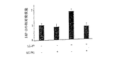
Classifications
-
- C—CHEMISTRY; METALLURGY
- C07—ORGANIC CHEMISTRY
- C07K—PEPTIDES
- C07K16/00—Immunoglobulins [IGs], e.g. monoclonal or polyclonal antibodies
- C07K16/18—Immunoglobulins [IGs], e.g. monoclonal or polyclonal antibodies against material from animals or humans
-
- A—HUMAN NECESSITIES
- A61—MEDICAL OR VETERINARY SCIENCE; HYGIENE
- A61P—SPECIFIC THERAPEUTIC ACTIVITY OF CHEMICAL COMPOUNDS OR MEDICINAL PREPARATIONS
- A61P1/00—Drugs for disorders of the alimentary tract or the digestive system
- A61P1/02—Stomatological preparations, e.g. drugs for caries, aphtae, periodontitis
-
- A—HUMAN NECESSITIES
- A61—MEDICAL OR VETERINARY SCIENCE; HYGIENE
- A61P—SPECIFIC THERAPEUTIC ACTIVITY OF CHEMICAL COMPOUNDS OR MEDICINAL PREPARATIONS
- A61P1/00—Drugs for disorders of the alimentary tract or the digestive system
- A61P1/04—Drugs for disorders of the alimentary tract or the digestive system for ulcers, gastritis or reflux esophagitis, e.g. antacids, inhibitors of acid secretion, mucosal protectants
-
- A—HUMAN NECESSITIES
- A61—MEDICAL OR VETERINARY SCIENCE; HYGIENE
- A61P—SPECIFIC THERAPEUTIC ACTIVITY OF CHEMICAL COMPOUNDS OR MEDICINAL PREPARATIONS
- A61P1/00—Drugs for disorders of the alimentary tract or the digestive system
- A61P1/16—Drugs for disorders of the alimentary tract or the digestive system for liver or gallbladder disorders, e.g. hepatoprotective agents, cholagogues, litholytics
-
- A—HUMAN NECESSITIES
- A61—MEDICAL OR VETERINARY SCIENCE; HYGIENE
- A61P—SPECIFIC THERAPEUTIC ACTIVITY OF CHEMICAL COMPOUNDS OR MEDICINAL PREPARATIONS
- A61P11/00—Drugs for disorders of the respiratory system
-
- A—HUMAN NECESSITIES
- A61—MEDICAL OR VETERINARY SCIENCE; HYGIENE
- A61P—SPECIFIC THERAPEUTIC ACTIVITY OF CHEMICAL COMPOUNDS OR MEDICINAL PREPARATIONS
- A61P13/00—Drugs for disorders of the urinary system
- A61P13/08—Drugs for disorders of the urinary system of the prostate
-
- A—HUMAN NECESSITIES
- A61—MEDICAL OR VETERINARY SCIENCE; HYGIENE
- A61P—SPECIFIC THERAPEUTIC ACTIVITY OF CHEMICAL COMPOUNDS OR MEDICINAL PREPARATIONS
- A61P13/00—Drugs for disorders of the urinary system
- A61P13/12—Drugs for disorders of the urinary system of the kidneys
-
- A—HUMAN NECESSITIES
- A61—MEDICAL OR VETERINARY SCIENCE; HYGIENE
- A61P—SPECIFIC THERAPEUTIC ACTIVITY OF CHEMICAL COMPOUNDS OR MEDICINAL PREPARATIONS
- A61P15/00—Drugs for genital or sexual disorders; Contraceptives
-
- A—HUMAN NECESSITIES
- A61—MEDICAL OR VETERINARY SCIENCE; HYGIENE
- A61P—SPECIFIC THERAPEUTIC ACTIVITY OF CHEMICAL COMPOUNDS OR MEDICINAL PREPARATIONS
- A61P17/00—Drugs for dermatological disorders
-
- A—HUMAN NECESSITIES
- A61—MEDICAL OR VETERINARY SCIENCE; HYGIENE
- A61P—SPECIFIC THERAPEUTIC ACTIVITY OF CHEMICAL COMPOUNDS OR MEDICINAL PREPARATIONS
- A61P25/00—Drugs for disorders of the nervous system
-
- A—HUMAN NECESSITIES
- A61—MEDICAL OR VETERINARY SCIENCE; HYGIENE
- A61P—SPECIFIC THERAPEUTIC ACTIVITY OF CHEMICAL COMPOUNDS OR MEDICINAL PREPARATIONS
- A61P35/00—Antineoplastic agents
-
- A—HUMAN NECESSITIES
- A61—MEDICAL OR VETERINARY SCIENCE; HYGIENE
- A61P—SPECIFIC THERAPEUTIC ACTIVITY OF CHEMICAL COMPOUNDS OR MEDICINAL PREPARATIONS
- A61P35/00—Antineoplastic agents
- A61P35/02—Antineoplastic agents specific for leukemia
-
- A—HUMAN NECESSITIES
- A61—MEDICAL OR VETERINARY SCIENCE; HYGIENE
- A61P—SPECIFIC THERAPEUTIC ACTIVITY OF CHEMICAL COMPOUNDS OR MEDICINAL PREPARATIONS
- A61P35/00—Antineoplastic agents
- A61P35/04—Antineoplastic agents specific for metastasis
-
- A—HUMAN NECESSITIES
- A61—MEDICAL OR VETERINARY SCIENCE; HYGIENE
- A61P—SPECIFIC THERAPEUTIC ACTIVITY OF CHEMICAL COMPOUNDS OR MEDICINAL PREPARATIONS
- A61P43/00—Drugs for specific purposes, not provided for in groups A61P1/00-A61P41/00
-
- A—HUMAN NECESSITIES
- A61—MEDICAL OR VETERINARY SCIENCE; HYGIENE
- A61P—SPECIFIC THERAPEUTIC ACTIVITY OF CHEMICAL COMPOUNDS OR MEDICINAL PREPARATIONS
- A61P7/00—Drugs for disorders of the blood or the extracellular fluid
-
- C—CHEMISTRY; METALLURGY
- C07—ORGANIC CHEMISTRY
- C07K—PEPTIDES
- C07K14/00—Peptides having more than 20 amino acids; Gastrins; Somatostatins; Melanotropins; Derivatives thereof
- C07K14/435—Peptides having more than 20 amino acids; Gastrins; Somatostatins; Melanotropins; Derivatives thereof from animals; from humans
- C07K14/46—Peptides having more than 20 amino acids; Gastrins; Somatostatins; Melanotropins; Derivatives thereof from animals; from humans from vertebrates
- C07K14/47—Peptides having more than 20 amino acids; Gastrins; Somatostatins; Melanotropins; Derivatives thereof from animals; from humans from vertebrates from mammals
- C07K14/4701—Peptides having more than 20 amino acids; Gastrins; Somatostatins; Melanotropins; Derivatives thereof from animals; from humans from vertebrates from mammals not used
- C07K14/4723—Cationic antimicrobial peptides, e.g. defensins
-
- C—CHEMISTRY; METALLURGY
- C07—ORGANIC CHEMISTRY
- C07K—PEPTIDES
- C07K16/00—Immunoglobulins [IGs], e.g. monoclonal or polyclonal antibodies
- C07K16/18—Immunoglobulins [IGs], e.g. monoclonal or polyclonal antibodies against material from animals or humans
- C07K16/28—Immunoglobulins [IGs], e.g. monoclonal or polyclonal antibodies against material from animals or humans against receptors, cell surface antigens or cell surface determinants
- C07K16/30—Immunoglobulins [IGs], e.g. monoclonal or polyclonal antibodies against material from animals or humans against receptors, cell surface antigens or cell surface determinants from tumour cells
- C07K16/3076—Immunoglobulins [IGs], e.g. monoclonal or polyclonal antibodies against material from animals or humans against receptors, cell surface antigens or cell surface determinants from tumour cells against structure-related tumour-associated moieties
- C07K16/3092—Immunoglobulins [IGs], e.g. monoclonal or polyclonal antibodies against material from animals or humans against receptors, cell surface antigens or cell surface determinants from tumour cells against structure-related tumour-associated moieties against tumour-associated mucins
-
- A—HUMAN NECESSITIES
- A61—MEDICAL OR VETERINARY SCIENCE; HYGIENE
- A61K—PREPARATIONS FOR MEDICAL, DENTAL OR TOILETRY PURPOSES
- A61K39/00—Medicinal preparations containing antigens or antibodies
- A61K2039/505—Medicinal preparations containing antigens or antibodies comprising antibodies
-
- C—CHEMISTRY; METALLURGY
- C07—ORGANIC CHEMISTRY
- C07K—PEPTIDES
- C07K2317/00—Immunoglobulins specific features
- C07K2317/30—Immunoglobulins specific features characterized by aspects of specificity or valency
- C07K2317/31—Immunoglobulins specific features characterized by aspects of specificity or valency multispecific
Landscapes
- Health & Medical Sciences (AREA)
- Chemical & Material Sciences (AREA)
- Organic Chemistry (AREA)
- Life Sciences & Earth Sciences (AREA)
- Medicinal Chemistry (AREA)
- General Health & Medical Sciences (AREA)
- General Chemical & Material Sciences (AREA)
- Public Health (AREA)
- Veterinary Medicine (AREA)
- Pharmacology & Pharmacy (AREA)
- Chemical Kinetics & Catalysis (AREA)
- Animal Behavior & Ethology (AREA)
- Nuclear Medicine, Radiotherapy & Molecular Imaging (AREA)
- Bioinformatics & Cheminformatics (AREA)
- Engineering & Computer Science (AREA)
- Immunology (AREA)
- Proteomics, Peptides & Aminoacids (AREA)
- Molecular Biology (AREA)
- Genetics & Genomics (AREA)
- Biophysics (AREA)
- Biochemistry (AREA)
- Gastroenterology & Hepatology (AREA)
- Zoology (AREA)
- Urology & Nephrology (AREA)
- Cell Biology (AREA)
- Oncology (AREA)
- Hematology (AREA)
- Toxicology (AREA)
- Diabetes (AREA)
- Pulmonology (AREA)
- Biomedical Technology (AREA)
- Neurology (AREA)
- Neurosurgery (AREA)
- Dermatology (AREA)
- Endocrinology (AREA)
- Reproductive Health (AREA)
- Medicines That Contain Protein Lipid Enzymes And Other Medicines (AREA)
- Pharmaceuticals Containing Other Organic And Inorganic Compounds (AREA)
- Measuring Or Testing Involving Enzymes Or Micro-Organisms (AREA)
- Medicines Containing Antibodies Or Antigens For Use As Internal Diagnostic Agents (AREA)
Abstract
本発明は、癌細胞の増殖を阻害する物質を提供し、ここにおいて、該物質は、hCAP18/LL−37の生物活性を阻害する。好ましい実施態様において、該物質は、hCAP18/LL−37の転写、翻訳および/または結合特性を変化させる。好ましくは、該物質は、短鎖干渉RNA(siRNA)分子、アンチセンスオリゴヌクレオチド、および、hCAP18/LL−37への結合親和性を有する化合物からなる群より選択される。本発明はさらに、患者における癌細胞の増殖を阻害する方法、加えて癌を診断するための方法およびキットを提供する。
【選択図】図1The present invention provides a substance that inhibits the growth of cancer cells, wherein the substance inhibits the biological activity of hCAP18 / LL-37. In a preferred embodiment, the agent alters the transcription, translation and / or binding properties of hCAP18 / LL-37. Preferably, the substance is selected from the group consisting of a short interfering RNA (siRNA) molecule, an antisense oligonucleotide, and a compound having binding affinity for hCAP18 / LL-37. The present invention further provides methods for inhibiting the growth of cancer cells in a patient, as well as methods and kits for diagnosing cancer.
[Selection] Figure 1
Description
本発明は、癌の治療と診断に使用するための物質に関する。特に本発明は、癌細胞の増殖を阻害することができる物質を提供する。 The present invention relates to substances for use in the treatment and diagnosis of cancer. In particular, the present invention provides a substance that can inhibit the growth of cancer cells.
抗菌性タンパク質は、先天性免疫システムにおける主要なエフェクターである。ヒトカテリシジン(cathelicidin)抗菌性タンパク質であるhCAP18は、ヒトでしか知られていないカテリシジンであるが、これは、保存されたカテリン(cathelin)ドメインと、LL−37と呼ばれる可変性のC末端とからなる(Gudmundsson等,1996,Eur J Biochem 1238:325〜32;Zanetti等,1995,FEBS Lett 374:1〜5)。ホロタンパク質の細胞外のタンパク質分解プロセシングによりLL−37ペプチドが放出されるが、これは、広範な抗菌活性(Gudmundsson等,1995,Proc Natl Acad Sci USA 92:7085〜9;Agerberth等,1995,Proc Natl Acad Sci USA 92:195〜99)を有し、加えて、宿主細胞に対して作用も有しそのうちのいくつかは、Gタンパク質結合受容体、ホルミルペプチド受容体様1(FPRL1)が介在する(Yang等,2000,J Exp Med 192:1069〜74;Koczulla等,2003,J Clin Invest 111:1665〜72)。hCAP18は、白血球に存在し(Cowland等,1995,FEBS Lett 368:173〜76)、皮膚およびその他の上皮で発現され、そこで炎症に付随してアップレギュレートされる(Cowland等,1995,FEBS Lett 368:173〜76;Frohm等,1997,J Biol Chem 272:15258〜63)、および、傷害(Dorschner等,2001,J Invest Dermatol 117:91〜97;Heilborn等,2003,J Invest Dermatol 120:379〜89)は、先天性のバリア保護における役割と一致する。近年、カテリシジンなどの抗菌性タンパク質は、腫瘍に対する非特異的な宿主側防御においても役割を果たすことが提唱されている(Winder等,1998,Biochem Biophys Res Commun 242:608〜12;Ohtake等,1999,Br J Cancer 181:393〜403)。 Antibacterial proteins are major effectors in the innate immune system. HCAP18, a human cathelicidin antibacterial protein, is a cathelicidin known only in humans, which consists of a conserved cathelin domain and a variable C-terminus called LL-37. (Gudmundsson et al., 1996, Eur J Biochem 1238: 325-32; Zanetti et al., 1995, FEBS Lett 374: 1-5). Extracellular proteolytic processing of the holoprotein releases LL-37 peptide, which has a wide range of antibacterial activities (Gudmundsson et al., 1995, Proc Natl Acad Sci USA 92: 7085-9; Agerberth et al., 1995, Proc Natl Acad Sci USA 92: 195-99) and in addition has an effect on host cells, some of which are mediated by G protein-coupled receptors, formyl peptide receptor-like 1 (FPRL1) (Yang et al., 2000, J Exp Med 192: 1069-74; Koczulla et al., 2003, J Clin Invest 111: 1665-72). hCAP18 is present in leukocytes (Cowland et al., 1995, FEBS Lett 368: 173-76) and is expressed in the skin and other epithelia where it is up-regulated with inflammation (Cowland et al., 1995, FEBS Lett. 368: 173-76; Fromhm et al., 1997, J Biol Chem 272: 15258-63) and injury (Dorschner et al., 2001, J Invest Dermatol 117: 91-97; Heilborn et al., 2003, J Invest Dermatol 120: 379 ~ 89) is consistent with a role in innate barrier protection. Recently, antimicrobial proteins such as cathelicidin have been proposed to play a role in non-specific host-side defense against tumors (Winder et al., 1998, Biochem Biophys Res Commun 242: 608-12; Ohtake et al., 1999). , Br J Cancer 181: 393-403).
本発明の第一の形態は、癌細胞の増殖を阻害するための物質を提供し、ここにおいて、該物質は、hCAP18/LL−37の生物活性を阻害する(すなわち、インビボで阻害することができる)。 A first aspect of the invention provides a substance for inhibiting cancer cell growth, wherein the substance inhibits the biological activity of hCAP18 / LL-37 (ie, inhibits in vivo). it can).
ここでは、「物質」には、あらゆる化学物質が含まれ、例えばオリゴヌクレオチド、ポリヌクレオチド、ポリペプチド、ペプチド模倣剤、および、小さい化合物である。 As used herein, “substance” includes any chemical substance, such as oligonucleotides, polynucleotides, polypeptides, peptidomimetics, and small compounds.
従って、本発明は、hCAP18/LL−37の生物活性を、直接的に(例えば、タンパク質の生物活性を減少させることによって)、または、間接的に(例えば、hCAP18/LL−37の発現を減少させることによって)阻害することができる物質を提供する。 Thus, the present invention reduces the biological activity of hCAP18 / LL-37 either directly (eg, by reducing the biological activity of the protein) or indirectly (eg, reducing the expression of hCAP18 / LL-37). A substance that can be inhibited).
好ましい実施態様において、該物質は、hCAP18/LL−37の転写、翻訳および/または結合特性を変化させることによってhCAP18/LL−37の生物活性を阻害する。 In a preferred embodiment, the agent inhibits the biological activity of hCAP18 / LL-37 by altering the transcription, translation and / or binding properties of hCAP18 / LL-37.
このような物質は、当業界周知の方法を用いて同定することができ、例えば:
(a)hCAP18/LL−37のmRNAの発現レベルに対する試験物質の作用を決定すること(例えばサザンブロッティング、または、関連するハイブリダイゼーション技術で);
(b)hCAP18/LL−37のタンパク質レベルに対する試験物質の作用を決定すること(例えば抗hCAP18/LL−37抗体を用いたイムノアッセイで);および、
(c)hCAP18/LL−37の活性の機能的なマーカー(例えばErbB2のリン酸化)に対する試験物質の作用を決定すること、
によって同定することができる。
Such materials can be identified using methods well known in the art, for example:
(A) determining the effect of the test substance on the expression level of hCAP18 / LL-37 mRNA (eg, by Southern blotting or related hybridization techniques);
(B) determining the effect of the test substance on the protein level of hCAP18 / LL-37 (eg, in an immunoassay using an anti-hCAP18 / LL-37 antibody);
(C) determining the effect of the test substance on a functional marker of hCAP18 / LL-37 activity (eg, phosphorylation of ErbB2);
Can be identified.
本発明の好ましい実施態様において、物質は、hCAP18/LL−37の転写阻害剤である。 In a preferred embodiment of the invention, the substance is a hCAP18 / LL-37 transcription inhibitor.
本発明のその代わりの実施態様において、該物質は、hCAP18/LL−37の翻訳阻害剤である。 In an alternative embodiment of the invention, the agent is a translation inhibitor of hCAP18 / LL-37.
本発明のさらなる実施態様において、該物質は、hCAP18/LL−37の結合特性の阻害剤である。例えば、該物質は、hCAP18/LL−37がその受容体に結合できなくなるように、hCAP18/LL−37のコンフォメーションを変化させるものでもよい。 In a further embodiment of the invention the agent is an inhibitor of the binding properties of hCAP18 / LL-37. For example, the substance may change the conformation of hCAP18 / LL-37 such that hCAP18 / LL-37 cannot bind to its receptor.
当業者であれば当然であるが、該物質はまた、hCAP18/LL−37受容体の機能を直接ブロックすることによって、すなわちhCAP18/LL−37受容体アンタゴニストとして作用することによって、hCAP18/LL−37の生物活性を阻害するものでもよい。好ましくは、hCAP18/LL−37受容体は、FPRL1である。 As will be appreciated by those skilled in the art, the substance may also be hCAP18 / LL- by directly blocking the function of the hCAP18 / LL-37 receptor, ie acting as an hCAP18 / LL-37 receptor antagonist. It may be one that inhibits 37 biological activities. Preferably, the hCAP18 / LL-37 receptor is FPRL1.
さらに本発明のその他の実施態様において、該物質は、hCAP18/LL−37またはそのmRNAの安定性を調節すること(例えば減少させること)によって、hCAP18/LL−37の生物活性を阻害する。 In yet another embodiment of the invention, the agent inhibits the biological activity of hCAP18 / LL-37 by modulating (eg, reducing) the stability of hCAP18 / LL-37 or its mRNA.
有利には、該物質は、hCAP18/LL−37の生物活性を選択的に阻害することができる。 Advantageously, the substance can selectively inhibit the biological activity of hCAP18 / LL-37.
「選択的に」とは、ここでは、該物質は、癌細胞中の他のタンパク質の活性を調節するよりも大きい程度にhCAP18/LL−37の生物活性を阻害することを意味する。好ましくは、該物質は、hCAP18/LL−37の生物活性だけを阻害するが、当然のことながら、癌細胞内のその他のタンパク質の発現や活性は、hCAP18/LL−37の選択的な阻害の下流の結果として変化している可能性がある。従って、我々は、遺伝子発現および/または癌細胞の増殖に非特異的な作用を有する物質を排除する。 “Selectively” means here that the substance inhibits the biological activity of hCAP18 / LL-37 to a greater extent than it modulates the activity of other proteins in the cancer cell. Preferably, the substance only inhibits the biological activity of hCAP18 / LL-37, but it should be understood that the expression and activity of other proteins in the cancer cell is not selective for hCAP18 / LL-37. It may have changed as a result of downstream. We therefore exclude substances that have nonspecific effects on gene expression and / or cancer cell growth.
当業者であれば当然であるが、本発明の物質によるhCAP18/LL−37の生物活性の阻害は、全体的であってもよいし、または、部分的であってもよい。例えば、該物質は、hCAP18/LL−37の生物活性を、該物質に晒されていない癌細胞におけるhCAP18/LL−37の生物活性と比較して、少なくとも10%、好ましくは少なくとも20%、30%、40%、50%、60%、70%、80%、または、90%、最も好ましくは100%阻害する可能性がある。好ましい実施態様において、該物質は、hCAP18/LL−37の生物活性を、該物質に晒されていない癌細胞におけるhCAP18/LL−37の生物活性と比較して50%またはそれ以上阻害することができる。 As will be appreciated by those skilled in the art, the inhibition of the biological activity of hCAP18 / LL-37 by the substances of the present invention may be complete or partial. For example, the substance has a biological activity of hCAP18 / LL-37 of at least 10%, preferably at least 20%, 30 compared to the biological activity of hCAP18 / LL-37 in cancer cells not exposed to the substance. %, 40%, 50%, 60%, 70%, 80%, or 90%, most preferably 100%. In a preferred embodiment, the agent inhibits the biological activity of hCAP18 / LL-37 by 50% or more compared to the biological activity of hCAP18 / LL-37 in cancer cells not exposed to the agent. it can.
好ましくは、該物質は、以下のタイプの物質から選択される:
(a)短鎖干渉RNA(siRNA)分子;
(b)アンチセンスオリゴヌクレオチド;および、
(c)hCAP18/LL−37への結合親和性を有する化合物、例えばポリペプチド。
Preferably, the substance is selected from the following types of substances:
(A) a short interfering RNA (siRNA) molecule;
(B) an antisense oligonucleotide; and
(C) a compound having binding affinity for hCAP18 / LL-37, such as a polypeptide.
あるいは、該物質は低分子量の阻害化合物であってもよく、例えば、ビタミンDに対するアンタゴニスト、例えばZK159222(シエーリング社(Schering AG))、および、TEI−9647(帝人医学研究所(Tejin Institute for Medical Research),東京)、ならびに、ビタミンAに対するアンタゴニスト、例えばAGN193109(アレルゲン・ファーマシューティカルズ(Allergen Pharmaceuticals))。 Alternatively, the substance may be a low molecular weight inhibitory compound, such as an antagonist to vitamin D, such as ZK159222 (Schering AG), and TEI-9647 (Tejin Institute for Medical Research). ), Tokyo), as well as antagonists to vitamin A, such as AGN 193109 (Allergen Pharmaceuticals).
本発明の第一の形態の好ましい実施態様において、該物質は、短鎖干渉RNA(siRNA)分子である。 In a preferred embodiment of the first aspect of the invention, the substance is a short interfering RNA (siRNA) molecule.
RNA干渉は、2工程からなるプロセスである。第一の工程(開始工程と名付ける)は、入ってきたdsRNAを21〜23個のヌクレオチド(nt)の短鎖干渉(siRNA)に消化するが、これは恐らく、ATP依存性の様式で、dsRNA(直接導入された、または、導入遺伝子またはウイルスを介して導入された)をプロセシングする(切断する)dsRNAに特異的なリボヌクレアーゼのRNアーゼIIIファミリーの一種であるダイサーの作用によってなされる。連続的な切断現象によって、RNAを、それぞれ2個のヌクレオチドからなる3’オーバーハングを有する19〜21bpの二重鎖(siRNA)にまで分解する(HutvagnerおよびZamore,2002,Curr.Opin.Genetics and Development 12:225〜232;Bernstein,2001,Nature 409:363〜366)。 RNA interference is a two-step process. The first step (named the initiation step) digests the incoming dsRNA into a short interference (siRNA) of 21-23 nucleotides (nt), which is probably in an ATP-dependent manner, dsRNA. This is done by the action of Dicer, a member of the RNase III family of ribonucleases specific for dsRNA that processes (cleaves) (directly introduced or introduced via a transgene or virus). The continuous cleavage phenomenon breaks down the RNA into 19-21 bp duplexes (siRNA) each with a 3 'overhang of 2 nucleotides (Hutvagner and Zamore, 2002, Curr. Opin. Genetics and Development 12: 225-232; Bernstein, 2001, Nature 409: 363-366).
エフェクター工程において、siRNA二重鎖はヌクレアーゼ複合体に結合して、RNA誘導型サイレンシング複合体(RISC)を形成する。siRNA二重鎖のATP依存性の巻き戻しが、RISCの活性化に必要である。次に、活性RISCは、塩基対形成による相互作用で相同的に転写することを標的として、mRNAを、siRNAの3’末端から12個のヌクレオチドからなるフラグメントに切断する(HutvagnerおよびZamore,2002,上記;Hammond等,2001,Nat.Rev.Gen.2:110〜119(2001);Sharp,2001,Genes.Dev.15:485〜90)。切断メカニズムをさらに解明すべきであるが、調査によれば、RISCはそれぞれ、単一のsiRNA、および、RNアーゼを含むことが示されている(HutvagnerおよびZamore,2002,上記)。 In the effector process, siRNA duplexes bind to nuclease complexes to form RNA-induced silencing complexes (RISC). ATP-dependent unwinding of siRNA duplexes is required for RISC activation. Active RISC then cleaves mRNA into 12 nucleotide fragments from the 3 ′ end of siRNA, targeting homologous transcription through base pairing interactions (Hutvagner and Zamore, 2002). Hammond et al., 2001, Nat. Rev. Gen. 2: 110-119 (2001); Sharp, 2001, Genes. Dev. 15: 485-90). The cleavage mechanism should be further elucidated, but studies have shown that each RISC contains a single siRNA and an RNase (Hutvagner and Zamore, 2002, supra).
RNAiの驚くべき効力を考慮して、RNAi経路内の増幅工程が示唆されている。増幅は、入ってきたdsRNAをコピーしてより多くのsiRNAを生成することによって起こる場合もあるし、または、形成されたsiRNAの複製によって起こる場合もある。あるいは、または、それに加えて、増幅は、RISCの転換を複数行うことによって実行されてもよい(Hammond等,2001,上記;HutvagnerおよびZamore,2002,上記)。RNAiに関するさらなる情報は、以下の総論で見出すことができる:Tuschl,2001,Chem.Biochem.2:239〜245,Cullen,2002,Nat.Immunol.3:597〜599およびBrantl,2002,Biochem.Biophys Act.1575:15〜25。 In view of the surprising potency of RNAi, an amplification step within the RNAi pathway has been suggested. Amplification can occur by copying incoming dsRNA to produce more siRNA, or it can occur by replication of the formed siRNA. Alternatively or in addition, amplification may be performed by performing multiple RISC conversions (Hammond et al., 2001, supra; Hutvagner and Zamore, 2002, supra). More information on RNAi can be found in the following general review: Tuschl, 2001, Chem. Biochem. 2: 239-245, Cullen, 2002, Nat. Immunol. 3: 597-599 and Brantl, 2002, Biochem. Biophys Act. 1575: 15-25.
本発明との使用に適したRNAi分子の合成は、以下のように実行することができる。まず、hCAP18/LL−37のmRNA配列を、AAジヌクレオチド配列に関してAUG開始コドンの下流でスキャンする。各AAの出現と、3’に隣接する19個のヌクレオチドの出現を、可能性があるsiRNA標的部位として記録する。好ましくは、調節タンパク質結合部位において非翻訳領域(UTR)がより豊富であるため、siRNA標的部位はオープンリーディングフレームから選択される。UTR結合タンパク質および/または翻訳開始複合体は、siRNAエンドヌクレアーゼ複合体の結合に干渉する可能性がある(Tuschl,ChemBiochem.2:239〜245)。しかし当然ながら、非翻訳領域に向けられたsiRNAも、有効である可能性がある。 Synthesis of RNAi molecules suitable for use with the present invention can be performed as follows. First, the hCAP18 / LL-37 mRNA sequence is scanned downstream of the AUG start codon for AA dinucleotide sequences. The occurrence of each AA and the occurrence of the 19 nucleotides adjacent to 3 'are recorded as potential siRNA target sites. Preferably, the siRNA target site is selected from the open reading frame because it is richer in untranslated regions (UTRs) in the regulatory protein binding site. UTR binding proteins and / or translation initiation complexes can interfere with the binding of siRNA endonuclease complexes (Tuschl, ChemBiochem. 2: 239-245). Of course, however, siRNA directed at untranslated regions may also be effective.
第二に、配列アライメントソフトウェア、例えばBLAST(www.ncbi.nlm.nih.gov/BLAST/)を用いて、可能性がある標的部位を適切なゲノムデータベース(例えばヒト、マウス、ラットなど)と比較する。他のコード配列に対して有意な相同性を示す推定上の標的部位を、フィルターをかけて除外する。 Second, use sequence alignment software such as BLAST (www.ncbi.nlm.nih.gov/BLAST/) to compare potential target sites with the appropriate genomic database (eg human, mouse, rat, etc.) To do. A putative target site that shows significant homology to other coding sequences is filtered out.
条件にあった標的配列を、siRNA合成のためのテンプレートとして選択する。好ましい配列は、低いG/C含量を含む配列であり、なぜならこれらは、遺伝子サイレンシングの媒介において、55%より高いG/C含量を有するものと比較してより有効であることが証明されているためである。好ましくは、評価するために、標的遺伝子の長さに従って数種の標的部位が選択される。選択されたsiRNAをよりよく評価するために、好ましくは、ネガティブコントロールが一緒に用いられる。好ましくは、ネガティブコントロールのsiRNAは、選択されたsiRNAと同じヌクレオチド組成を含むが、ゲノムに対する有意な相同性はない。従って、好ましくは、siRNAのスクランブル化したヌクレオチド配列が用いられる(ただしこれらは、その他のあらゆる遺伝子に対する有意な相同性をまったく示さない)。 A target sequence that meets the conditions is selected as a template for siRNA synthesis. Preferred sequences are those that contain a low G / C content because they have proven to be more effective in mediating gene silencing than those with a G / C content higher than 55%. Because it is. Preferably, several target sites are selected for evaluation according to the length of the target gene. In order to better evaluate the selected siRNA, a negative control is preferably used together. Preferably, the negative control siRNA comprises the same nucleotide composition as the selected siRNA, but there is no significant homology to the genome. Preferably, therefore, scrambled nucleotide sequences of siRNA are used (though they do not show any significant homology to any other gene).
好ましくは、siRNA分子は、配列番号1のヌクレオチド配列のフラグメント、または、このようなフラグメントの変異体を含む。 Preferably, the siRNA molecule comprises a fragment of the nucleotide sequence of SEQ ID NO: 1, or a variant of such a fragment.
hCAP18/LL−37のmRNA(登録番号MN004345)hCAP18 / LL-37 mRNA (registration number MN004345)

あるいは、siRNA分子は、ENSG00000164047(ゲノム配列)から誘導されたヌクレオチド配列のフラグメントを含む。 Alternatively, the siRNA molecule comprises a fragment of a nucleotide sequence derived from ENSG0000014047 (genomic sequence).
「フラグメント」は、ここでは、少なくとも10個のヌクレオチド、例えば少なくとも15、16、17、18、19、20、21、22、23、24または25個のヌクレオチドを意味する。 “Fragment” means here at least 10 nucleotides, for example at least 15, 16, 17, 18, 19, 20, 21, 22, 23, 24 or 25 nucleotides.
「変異体」は、ここでは、配列番号1のフラグメントと少なくとも90%の配列同一性を共有するヌクレオチド配列、例えば少なくとも95%、96%、97%、98%、または、99%の配列同一性を共有するヌクレオチド配列を意味する。 A “variant” as used herein refers to a nucleotide sequence that shares at least 90% sequence identity with a fragment of SEQ ID NO: 1, such as at least 95%, 96%, 97%, 98%, or 99% sequence identity. Means a nucleotide sequence that shares
2種のポリヌクレオチド間の配列同一性(%)は、適切なコンピュータープログラム、例えばウィスコンシン大学のジェネティック・コンピューティング・グループ(University of Wisconsin Genetic Computing Group)のギャップ・プログラム(GAP program)を用いて決定してもよく、当然のことながら、同一性(%)は、配列が最適に並べられたポリヌクレオチドに関して計算される。 Sequence identity (%) between two polynucleotides is determined using an appropriate computer program, such as the Gap program of the University of Wisconsin Genetic Computing Group (University of Wisconsin Genetic Computing Group). Of course, it will be appreciated that the percent identity is calculated for the optimally aligned polynucleotide.
このようなアライメントは、別の方法で行って、クラスタルWプログラム(Clustal W program,Thompson等,1994,Nuc.Acid Res.22:4673〜4680で説明されている通り)を用いてもよい。 Such alignment may be performed by another method, and a clustered W program (as described in Clustal W program, Thompson et al., 1994, Nuc. Acid Res. 22: 4673-4680) may be used.
用いられるパラメーターは、以下のようなものが可能である:
Fastのペアワイズアライメントパラメーター:K−tuple(ワード)サイズ;1、ウィンドウサイズ:5、ギャップペナルティー;3、トップの対角線の数;5。スコア付けの方法:xパーセント。
マルチアライメントのパラメーター:ギャップオープンペナルティー;10、ギャップ伸長ペナルティー;0.05。
スコアリングマトリックス:BLOSUM。
The parameters used can be as follows:
Fast pairwise alignment parameters: K-tuple (word) size; 1, window size: 5, gap penalty; 3, number of top diagonals; Scoring method: x percent.
Multi-alignment parameters: gap open penalty; 10, gap extension penalty; 0.05.
Scoring matrix: BLOSUM.
あるいは、局所的な配列アライメントを決定するためには、BESTFITプログラムを用いてもよい。 Alternatively, the BESTFIT program may be used to determine local sequence alignment.
siRNA分子が、長さが19〜23個のヌクレオチドであることが有利である。 Advantageously, the siRNA molecule is 19-23 nucleotides in length.
本発明の第一の形態の好ましいその代わりの実施態様において、該物質はアンチセンスオリゴヌクレオチドである。 In a preferred alternative embodiment of the first aspect of the invention, the substance is an antisense oligonucleotide.
hCAP18/LL−37のレベル/活性を効率的に減少させるのに用いることができるアンチセンス分子の設計には、アンチセンス法に重要な2つの観点の考察が必要である。第一の観点は、癌細胞の細胞質へのオリゴヌクレオチドの送達であり、一方で、第二の観点は、細胞内で指示されたmRNAの翻訳を阻害するようにして、それらに特異的に結合するオリゴヌクレオチドの設計である。 The design of antisense molecules that can be used to efficiently reduce the level / activity of hCAP18 / LL-37 requires consideration of two aspects that are important to antisense methods. The first aspect is the delivery of oligonucleotides to the cytoplasm of cancer cells, while the second aspect specifically binds to them by inhibiting translation of the directed mRNA in the cell. This is the design of the oligonucleotide.
従来技術では、多種多様の細胞型にオリゴヌクレオチドを効率的に送達するのに用いることができる多数の送達方策を教示している(例えば、Luft,1998,J Mol Med 76:75〜6;Kronenwett等,1998,Blood 91:852〜62;Rajur等,1997,Bioconjug Chem 8:935〜40;Lavigne等,1997,Biochem Biophys Res Commun 237:566〜71;Aoki等,1997,Biochem Biophys Res Commun 231:540〜5を参照)。 The prior art teaches a number of delivery strategies that can be used to efficiently deliver oligonucleotides to a wide variety of cell types (eg, Luft, 1998, J Mol Med 76: 75-6; Kronenwett; 1998, Blood 91: 852-62; Rajur et al., 1997, Bioconjug Chem 8: 935-40; Lavigne et al., 1997, Biochem Biophys Res Commun 237: 566-71; Aoki et al., 1997, Biochem Biophys Res1: 540-5).
加えて、標的mRNAとオリゴヌクレオチドとの両方における構造的な変化のエネルギー論を明らかにする熱力学的なサイクルに基づき、それらの標的mRNAに対して最高の予想された結合親和性を有する配列を同定するためのアルゴリズムが利用可能である(例えば、Walton等,1999,Biotechnol Bioeng 65:1〜9を参照)。 In addition, based on a thermodynamic cycle that reveals the energetics of structural changes in both target mRNAs and oligonucleotides, sequences with the highest expected binding affinity for those target mRNAs Algorithms for identification are available (see, eg, Walton et al., 1999, Biotechnol Bioeng 65: 1-9).
また、インビトロでのシステムを用いて特異的なオリゴヌクレオチドの効率を設計および予想するためのアプローチもいくつか知られている(例えば、Matveeva等,1998,Nature biotechnology 16:1374〜1375を参照)。 Several approaches are also known for designing and predicting the efficiency of specific oligonucleotides using in vitro systems (see, eg, Mateveva et al., 1998, Nature biotechnology 16: 1374-1375).
いくつかの臨床的な追跡によって、アンチセンスオリゴヌクレオチドの安全性、実行可能性および活性が実証されている。例えば、癌の治療に適したアンチセンスオリゴヌクレオチドの使用が成功している(Holmlund等,1999,Curr Opin Mol Ther 1:372〜85;Gerwitz,1999,Curr Opin Mol Ther 1:297〜306)。さらに近年、ヒトヘパラナーゼ遺伝子発現のアンチセンスが介在する抑制は、マウスモデルにおいて、ヒト癌細胞の胸膜の内転移を阻害することが報告されている(Uno等,2001,Cancer Res 61:7855〜60)。 Several clinical follow-ups have demonstrated the safety, feasibility and activity of antisense oligonucleotides. For example, antisense oligonucleotides suitable for the treatment of cancer have been successfully used (Holmlund et al., 1999, Curr Opin Mol Ther 1: 372-85; Gerwitz, 1999, Curr Opin Mol Ther 1: 297-306). More recently, antisense-mediated suppression of human heparanase gene expression has been reported to inhibit pleural internal metastasis of human cancer cells in a mouse model (Uno et al., 2001, Cancer Res 61: 7855-60). ).
従って、当業者であれば、hCAP18/LL−37の発現をダウンレギュレートするのに適したアンチセンス法を容易に設計および実施することができる。 Accordingly, those skilled in the art can easily design and implement an antisense method suitable for down-regulating the expression of hCAP18 / LL-37.
好ましくは、このようなアンチセンスオリゴヌクレオチドは、配列番号1に記載のヌクレオチドのフラグメント、または、このようなフラグメントの変異体を含む。 Preferably, such an antisense oligonucleotide comprises a fragment of the nucleotide set forth in SEQ ID NO: 1 or a variant of such a fragment.
有利には、該アンチセンスオリゴヌクレオチドは、長さが15〜35個の塩基である。例えば、20−merのオリゴヌクレオチドは、上皮増殖因子受容体のmRNAの発現を阻害することが示されており(Witters等,Breast Cancer Res Treat 53:41〜50(1999))、さらに、25−merのオリゴヌクレオチドは、副腎皮質刺激ホルモンの発現を90%を超えて減少させることが示されている(Frankel等,J Neurosurg 91:261〜7(1999))。しかし当然ながら、この範囲外の長さを有するオリゴヌクレオチド、例えば10、11、12、13もしくは14個の塩基、または、36,37,38,39もしくは40個の塩基からなるオリゴヌクレオチドを使用することが望ましい場合がある。 Advantageously, the antisense oligonucleotide is 15 to 35 bases in length. For example, a 20-mer oligonucleotide has been shown to inhibit expression of epidermal growth factor receptor mRNA (Witters et al., Breast Cancer Res Treat 53: 41-50 (1999)), and 25- The mer oligonucleotide has been shown to reduce the expression of adrenocorticotropic hormone by more than 90% (Frankel et al., J Neurosurg 91: 261-7 (1999)). However, of course, oligonucleotides with lengths outside this range are used, eg oligonucleotides consisting of 10, 11, 12, 13 or 14 bases, or 36, 37, 38, 39 or 40 bases. Sometimes it is desirable.
さらに当業者であれば当然ながら、オリゴヌクレオチドは、細胞の内因性ヌクレアーゼによって分解されるか、または、不活性化される。この問題を克服するために、例えば、天然に存在するホスホジエステル結合がその他の結合で置き換えられた改変インターヌクレオチド結合を有する、改変オリゴヌクレオチドを使用することが可能である。例えば、Agrawal等(1988)Proc.Natl.Acad.Sci.USA 85,7079〜7083では、HIV−1の組織培養において、オリゴヌクレオチドホスホルアミデートおよびホスホロチオエートを用いたところ阻害が増加したことが示されている。Sarin等(1988)Proc.Natl.Acad.Sci.USA 85,7448〜7451では、オリゴヌクレオチドメチルホスホナートを用いたところ、HIV−1の阻害が増加したことが実証されている。Agrawal等(1989)Proc.Natl.Acad.Sci.USA 86,7790〜7794では、ヌクレオチド配列に特異的なオリゴヌクレオチドホスホロチオエートを用いたところ、初期に感染させた細胞培養と、慢性的に感染させた細胞培養の両方においてHIV−1複製が阻害されることが示されている。Leither等(1990)Proc.Natl.Acad.Sci.USA 87,3430〜3434は、組織培養において、オリゴヌクレオチドホスホロチオエートによりインフルエンザウイルス複製が阻害されることを報告している。 Furthermore, as will be appreciated by those skilled in the art, oligonucleotides are degraded or inactivated by cellular endogenous nucleases. To overcome this problem, for example, it is possible to use modified oligonucleotides having modified internucleotide linkages in which naturally occurring phosphodiester linkages are replaced with other linkages. See, for example, Agrawal et al. (1988) Proc. Natl. Acad. Sci. USA 85, 7079-7083 show increased inhibition in the tissue culture of HIV-1 using oligonucleotide phosphoramidates and phosphorothioates. Sarin et al. (1988) Proc. Natl. Acad. Sci. USA 85, 7448-7451 demonstrates increased inhibition of HIV-1 using oligonucleotide methylphosphonate. Agrawal et al. (1989) Proc. Natl. Acad. Sci. USA 86, 7790-7794 inhibits HIV-1 replication in both early and chronically infected cell cultures using oligonucleotide phosphorothioate specific for the nucleotide sequence. It has been shown. Leither et al. (1990) Proc. Natl. Acad. Sci. USA 87, 3430-3434 reports that influenza virus replication is inhibited by oligonucleotide phosphorothioate in tissue culture.
人工的な結合を有するオリゴヌクレオチドは、インビボでの分解に耐性であることが示されている。例えば、Shaw等(1991)は、Nucleic Acids Res.19,747〜750で、その他の改変されていないオリゴヌクレオチドが、所定のキャッピング構造によって3’末端でブロックされると、それらはヌクレアーゼに対してインビボでより耐性になること、さらに、キャッピングされていないオリゴヌクレオチドのホスホロチオエートが、インビボで分解されないことを報告している。 Oligonucleotides with artificial linkages have been shown to be resistant to degradation in vivo. For example, Shaw et al. (1991) described Nucleic Acids Res. 19, 747-750, when other unmodified oligonucleotides are blocked at the 3 ′ end by a given capping structure, they become more resistant to nucleases in vivo and are also capped. No oligonucleotides phosphorothioates have been reported to be degraded in vivo.
オリゴヌクレオチドのホスホロチオエートを合成することによるH−ホスホナート法の詳細な説明は、AgrawalおよびTang(1990)Tetrahedron Letters 31,7541〜7544で示されており、そこでの教示は参照により本明細書に加入させる。オリゴヌクレオチドのメチルホスホナート、ホスホロジチオエート、ホスホルアミデート、リン酸エステル、架橋ホスホルアミデート、および、架橋ホスホロチオエートの合成は、当業界既知である。例えば、AgrawalおよびGoodchild(1987)Tetrahedron Letters 28,3539;Nielsen等(1988)Tetrahedron Letters 29,2911;Jager等(1988)Biochemistry 27,7237;Uznanski等(1987)Tetrahedron Letters 28,3401;Bannwarth(1988)Helv.Chim.Acta.71,1517;CrosstickおよびVyle(1989)Tetrahedron Letters 30,4693;Agrawal等(1990)Proc.Natl.Acad.Sci.USA 87,1401〜1405を参照(これらの教示は、この参照により本明細書に含まれる)。また、その他の合成または生産方法も可能である。好ましい実施態様において、上記オリゴヌクレオチドはデオキシリボ核酸(DNA)であるが、リボ核酸(RNA)配列を合成して適用してもよい。 A detailed description of the H-phosphonate method by synthesizing oligonucleotide phosphorothioates is given in Agrawal and Tang (1990) Tetrahedron Letters 31, 7541-7544, the teachings of which are incorporated herein by reference. . The synthesis of oligonucleotides methylphosphonate, phosphorodithioate, phosphoramidate, phosphate ester, bridged phosphoramidate, and bridged phosphorothioate are known in the art. For example, Agrawal and Goodchild (1987) Tetrahedron Letters 28, 3539; Nielsen et al. (1988) Tetrahedron Letters 29, 2911; Jager et al. (1988) Biochemistry 27, 7237; Uznanski et al. (1987) Helv. Chim. Acta. 71, 1517; Crossstick and Vyle (1989) Tetrahedron Letters 30, 4669; Agrawal et al. (1990) Proc. Natl. Acad. Sci. USA 87, 1401-1405 (the teachings of which are incorporated herein by this reference). Other synthesis or production methods are also possible. In a preferred embodiment, the oligonucleotide is deoxyribonucleic acid (DNA), but ribonucleic acid (RNA) sequences may be synthesized and applied.
好ましくは、本発明において有用なオリゴヌクレオチドは、内因性の核酸分解酵素による分解に耐性を有するように設計される。インビボでのオリゴヌクレオチドの分解により、長さが減少したオリゴヌクレオチドの分解産物が生産される。このような分解産物は、非特異的なハイブリダイゼーションに関わる可能性がより高く、さらに、それに相当する全長の対応物に比べて有効である可能性はより低い。従って、体内での分解に対して耐性であり、さらに、標的化した細胞に到達することができるオリゴヌクレオチドを使用することが望ましい。本発明のオリゴヌクレオチドは、天然型ホスホジエステル結合を1個またはそれ以上の内部の人工的なインターヌクレオチド結合で置換することによって、例えば、結合内のリン酸を硫黄で置換することによって、インビボでの分解に対してより耐性にすることができる。使用可能な結合の例としては、ホスホロチオエート、メチルホスホナート、スルホン、硫酸塩、ケチル、ホスホロジチオエート、様々なホスホルアミデート、リン酸エステル、架橋ホスホロチオエート、および、架橋ホスホルアミデートが挙げられる。その他のインターヌクレオチド結合は当業界周知であるため、このような例は制限するものではなく、例証である。これらのホスホジエステルインターヌクレオチド結合が置換された結合を1個またはそれ以上有するオリゴヌクレオチドの合成は当業界周知であり、例えば、混合型のインターヌクレオチド結合を有するオリゴヌクレオチドを製造する合成経路が挙げられる。 Preferably, the oligonucleotides useful in the present invention are designed to be resistant to degradation by endogenous nucleolytic enzymes. Oligonucleotide degradation in vivo produces oligonucleotide degradation products of reduced length. Such degradation products are more likely to be involved in non-specific hybridization, and are less likely to be effective compared to their full-length counterparts. It is therefore desirable to use oligonucleotides that are resistant to degradation in the body and that can reach the targeted cells. The oligonucleotides of the invention can be obtained in vivo by replacing a natural phosphodiester bond with one or more internal artificial internucleotide bonds, for example, by replacing the phosphate in the bond with sulfur. It can be made more resistant to decomposition. Examples of linkages that can be used include phosphorothioate, methylphosphonate, sulfone, sulfate, ketyl, phosphorodithioate, various phosphoramidates, phosphate esters, bridged phosphorothioates, and bridged phosphoramidates. It is done. Such examples are illustrative rather than limiting as other internucleotide linkages are well known in the art. The synthesis of oligonucleotides having one or more bonds in which these phosphodiester internucleotide bonds are substituted is well known in the art and includes, for example, synthetic routes to produce oligonucleotides having mixed internucleotide bonds. .
オリゴヌクレオチドは、「キャッピング」によって、または、5’または3’末端のヌクレオチドに類似の基を取り込むことによって、内因性の酵素による伸長に対して耐性にすることができる。キャッピングのための試薬は、アプライド・バイオシステムズ社(Applied Biosystems Inc,フォスターシティー,カリフォルニア州)製のアミノ−リンクIITM(Amino−Link IITM)として市販されている。キャッピングに関する方法は、例えば、Shaw等(1991)Nucleic Acids Res.19,747〜750およびAgrawal等(1991)Proc.Natl.Acad.Sci.USA 88(17),7595〜7599によって説明されている。 Oligonucleotides can be made resistant to elongation by endogenous enzymes by “capping” or by incorporating groups similar to the 5 ′ or 3 ′ terminal nucleotide. Reagents for capping, Applied Biosystems (Applied Biosystems Inc, Foster City, CA) made of amino - commercially available as a link II TM (Amino-Link II TM ). Methods related to capping are described in, for example, Shaw et al. (1991) Nucleic Acids Res. 19, 747-750 and Agrawal et al. (1991) Proc. Natl. Acad. Sci. USA 88 (17), 7595-7599.
オリゴヌクレオチドをヌクレアーゼの攻撃に対して耐性にするさらなる方法は、それらを「自己安定化させること」であり、例えば、Tang等(1993)Nucl.Acids Res.21,2729〜2735で説明されている通りである。自己安定化したオリゴヌクレオチドは、それらの3’末端でヘアピンループ構造を有し、ヘビ毒ホスホジエステラーゼ、DNAポリメラーゼI、および、ウシ胎児血清による分解に対して高い耐性を示す。このようなオリゴヌクレオチドの自己安定化した領域は、ハイブリダイゼーションの際に相補的な核酸に干渉せず、さらに、マウスにおける薬物動態学的研究および安定性の研究によれば、それらの直鎖状の対応物と比べて、自己安定化したオリゴヌクレオチドはインビボで持久性が増加することが示されている。 A further way to make oligonucleotides resistant to nuclease attack is to “self-stabilize” them, see, eg, Tang et al. (1993) Nucl. Acids Res. 21, 2729-2735. Self-stabilized oligonucleotides have a hairpin loop structure at their 3 'end and are highly resistant to degradation by snake venom phosphodiesterase, DNA polymerase I, and fetal bovine serum. Such self-stabilized regions of oligonucleotides do not interfere with complementary nucleic acids during hybridization, and, according to pharmacokinetic and stability studies in mice, their linear Compared to their counterparts, self-stabilized oligonucleotides have been shown to increase durability in vivo.
本発明の物質のさらなる好ましい実施態様において、該物質は、hCAP18/LL−37への結合親和性を有する化合物であり、例えばタンパク質、および、炭水化物である。 In a further preferred embodiment of the substance according to the invention, the substance is a compound having a binding affinity for hCAP18 / LL-37, such as proteins and carbohydrates.
「結合親和性を有する化合物」とは、ここでは、インビボで、すなわち癌細胞の内部にhCAP18/LL−37が存在するような生理学的条件下で、hCAP18/LL−37に結合することができる化合物を意味する。 “Compound with binding affinity” as used herein is capable of binding to hCAP18 / LL-37 in vivo, ie under physiological conditions such that hCAP18 / LL-37 is present inside cancer cells. Means a compound.
例えば、本化合物は、hCAP18/LL−37の活性部位に実質的に可逆的に結合してもよいし、または、実質的に不可逆的に結合してもよい。さらなる例において、本化合物は、hCAP18/LL−37のリガンドまたは受容体への結合に干渉するように、hCAP18/LL−37の活性部位ではない部分に結合してもよい。さらにその他の実施例において、本化合物は、アロステリック作用によってタンパク質活性を減少させるように、hCAP18/LL−37の一部に結合してもよい。このアロステリック作用は、hCAP18/LL−37の活性の天然の調節であってもよく、例えば「上流の活性化因子」によるhCAP18/LL−37の活性化に関与するアロステリック作用である。 For example, the compound may bind to the active site of hCAP18 / LL-37 substantially reversibly or may bind substantially irreversibly. In a further example, the compound may bind to a non-active site portion of hCAP18 / LL-37 so as to interfere with binding of hCAP18 / LL-37 to a ligand or receptor. In still other examples, the compound may bind to a portion of hCAP18 / LL-37 so as to reduce protein activity by allosteric action. This allosteric effect may be a natural regulation of the activity of hCAP18 / LL-37, for example an allosteric effect involved in the activation of hCAP18 / LL-37 by “upstream activators”.
試験化合物とhCAP18/LL−37との相互作用を検出する方法は当業界周知である。例えば、イオンスプレーマススペクトロスコピー/HPLC法を用いた限外ろ過、またはその他の物理的方法や分析方法を用いてもよい。加えて、蛍光エネルギー共鳴移動(FRET)法を用いてもよく、ここにおいて、2つの蛍光標識された物体の結合は、互いに近接した際の蛍光標識相互の作用を測定することによって測定することもできる。 Methods for detecting the interaction between a test compound and hCAP18 / LL-37 are well known in the art. For example, ultrafiltration using ion spray mass spectroscopy / HPLC methods, or other physical or analytical methods may be used. In addition, a fluorescence energy resonance transfer (FRET) method may be used, where the binding of two fluorescently labeled objects can be measured by measuring the interaction of the fluorescent labels when they are close to each other. it can.
高分子(例えばDNA、RNA、タンパク質およびリン脂質)へのポリペプチドの結合を検出する代替法としては、表面プラズモン共鳴分析が挙げられ、これは例えば、Plant等,1995,Analyt Biochem 226(2),342〜348で説明されている通りである。例えば放射標識または蛍光標識で標識されたポリペプチドを利用する方法も可能である。 An alternative method for detecting polypeptide binding to macromolecules (eg, DNA, RNA, proteins and phospholipids) includes surface plasmon resonance analysis, such as Plant et al., 1995, Analyt Biochem 226 (2). 342-348. For example, a method using a polypeptide labeled with a radiolabel or a fluorescent label is also possible.
このようなポリペプチドに結合することができる化合物を同定するさらなる方法は、ポリペプチドを化合物に晒し、化合物の前記ポリペプチドへのあらゆる結合を検出および/または測定する方法である。化合物のポリペプチドへの結合に関する結合定数を決定してもよい。化合物のポリペプチドへの結合を検出および/または測定する(定量する)のに適切な方法は当業者周知であり、例えば、ハイスループット操作が可能な方法(例えばチップベースの方法)を用いて行ってもよい。VLSIPSTMと呼ばれる新しい技術により、何十万またはそれを超える種類の様々な分子プローブを含む極めて小さいチップの生産が可能になっている。これらの生物学的なチップまたはアレイは、アレイ中に配置されたプローブを有し、それぞれのプローブには特定の位置が割り当てられている。それぞれの位置に例えば10ミクロンの目盛りを有する生物学的なチップが生産されている。このようなチップは、チップ上で標的分子がプローブのいずれかと相互作用するかどうかを決定するのに用いることができる。選択した試験条件下でアレイを標的分子に晒した後、スキャン装置によってアレイ中の各位置を試験し、その位置において標的分子はプローブとの相互作用を有するかどうかを決定することができる。 A further method of identifying a compound that can bind to such a polypeptide is to expose the polypeptide to the compound and detect and / or measure any binding of the compound to said polypeptide. A binding constant for the binding of the compound to the polypeptide may be determined. Suitable methods for detecting and / or measuring (quantifying) binding of a compound to a polypeptide are well known to those skilled in the art and are performed, for example, using methods capable of high-throughput operation (eg, chip-based methods). May be. A new technology called VLSIPS ™ has enabled the production of very small chips containing various molecular probes of hundreds of thousands or more. These biological chips or arrays have probes arranged in the array, and each probe is assigned a specific position. Biological chips with a scale of, for example, 10 microns are produced at each location. Such a chip can be used to determine whether a target molecule interacts with any of the probes on the chip. After exposing the array to the target molecule under selected test conditions, each position in the array can be tested by the scanning device to determine whether the target molecule has interaction with the probe at that position.
hCAP18/LL−37への結合親和性を有する化合物を同定するその他の方法は、酵母2ハイブリッドシステムであり、ここにおいて、本発明のポリペプチドを、hCAP18/LL−37に結合するタンパク質を「捕獲する」ために用いることができる。酵母2ハイブリッドシステムは、FieldsおよびSong,Nature 340:245〜246(1989)で説明されている。 Another method for identifying compounds with binding affinity for hCAP18 / LL-37 is the yeast two-hybrid system, in which the polypeptide of the present invention “captures proteins that bind to hCAP18 / LL-37. Can be used. The yeast two-hybrid system is described in Fields and Song, Nature 340: 245-246 (1989).
この本発明の形態の好ましい実施態様において、該物質は、hCAP18/LL−37へのリガンド結合能力を有する化合物である。 In a preferred embodiment of this aspect of the invention, the substance is a compound that has the ability to bind ligand to hCAP18 / LL-37.
例えば、該物質は、hCAP18/LL−37受容体(例えばFPRL1)の可溶性フラグメントが可能である。あるいは、該物質は、抗体を模擬する高い親和性を有する分子(いわゆる「アフィボディ(affibody)」)が可能である(例えば、US5,831,012、および、www.affibody.seを参照)。これらのリガンドは、小さく単純なタンパク質であり、プロテインA(細菌スタフィロコッカス・アウレウス(Staphylococcus aureus)由来の表面タンパク質)のIgG結合ドメインのうち1つの足場をベースとした3個のヘリックスバンドルで構成される。この足場は、親和性リガンドとして優れた特徴を有し、どのような所定の標的タンパク質にも高い親和性で結合するように設計することができる。 For example, the agent can be a soluble fragment of the hCAP18 / LL-37 receptor (eg, FPRL1). Alternatively, the substance can be a molecule with a high affinity that mimics an antibody (so-called “affibody”) (see, eg, US Pat. No. 5,831,012, and www.affibody.se). These ligands are small and simple proteins, consisting of three helix bundles based on one scaffold of the IgG binding domain of protein A (a surface protein from the bacterium Staphylococcus aureus). Is done. This scaffold has excellent characteristics as an affinity ligand and can be designed to bind to any given target protein with high affinity.
しかしながら、有利には、hCAP18/LL−37への結合親和性を有する化合物は、ポリペプチドであるか、または、ポリペプチドを含む。 Advantageously, however, the compound having binding affinity for hCAP18 / LL-37 is or comprises a polypeptide.
例えば、本化合物は抗体であってもよく、例えばモノクローナル、または、ポリクローナル抗体、または、それらの抗原結合フラグメントである。 For example, the compound may be an antibody, such as a monoclonal or polyclonal antibody, or an antigen-binding fragment thereof.
「抗体」には、ここでは、実質的に無傷の抗体分子、加えて、キメラ抗体、ヒト化抗体、ヒト抗体(ここにおいて、少なくとも1個のアミノ酸が、天然に存在するヒト抗体と比べて突然変異している)、単鎖抗体、二重特異性抗体、抗体重鎖、抗体軽鎖、抗体重鎖および/または軽鎖のホモ二量体およびヘテロ二量体、および、抗原結合フラグメント、ならびに、それらの誘導体が含まれる。 “Antibody” herein includes substantially intact antibody molecules, as well as chimeric antibodies, humanized antibodies, human antibodies (wherein at least one amino acid is abruptly compared to a naturally occurring human antibody). Mutated), single chain antibodies, bispecific antibodies, antibody heavy chains, antibody light chains, antibody heavy and / or light chain homodimers and heterodimers, and antigen-binding fragments, and And their derivatives.
「抗原結合フラグメント」は、ここでは、hCAP18/LL−37に結合することができる抗体の機能的なフラグメントを意味する。 By “antigen-binding fragment” is meant here a functional fragment of an antibody capable of binding to hCAP18 / LL-37.
好ましくは、抗原結合フラグメントは、Fvフラグメント(例えば、単鎖Fv、および、ジスルフィド結合Fv)、Fab様フラグメント(例えば、Fabフラグメント、Fab’フラグメント、および、F(ab)2フラグメント)、単一の可変性ドメイン(例えば、VhおよびVlドメイン)、ならびに、ドメイン抗体(dAbs、単鎖様式と二重鎖様式[すなわちdAb−リンカー−dAb]を含む)からなる群より選択される。 Preferably, antigen binding fragments are Fv fragments (eg, single chain Fv and disulfide bond Fv), Fab-like fragments (eg, Fab fragment, Fab ′ fragment, and F (ab) 2 fragment), single Selected from the group consisting of variable domains (eg, Vh and Vl domains) and domain antibodies (including dAbs, single chain and double chain modes [ie dAb-linker-dAb]).
抗体全体よりも抗体フラグメントを用いる利点は数倍にもなる。フラグメントの大きさがより小さいと、より優れた固形組織への貫入のような薬理学的特性が改善される可能性がある。さらに、抗原結合フラグメント、例えばFab、Fv、ScFvおよびdAb抗体フラグメントは、E.Coliで発現させ、分泌させてもよく、従って、大量の前記フラグメントを容易に生産することができる。 The advantage of using antibody fragments over the whole antibody is several times greater. Smaller fragment sizes may improve pharmacological properties such as better solid tissue penetration. In addition, antigen binding fragments, such as Fab, Fv, ScFv and dAb antibody fragments may be obtained from It can be expressed in Coli and secreted, so large quantities of the fragments can be easily produced.
また、抗体およびそれらの抗原結合フラグメントの改変型も本発明の範囲内でであり、例えば、ポリエチレングリコールまたはその他の適切なポリマーが共有結合したことによって改変された改変型である。 Modified forms of antibodies and their antigen-binding fragments are also within the scope of the present invention, such as modified forms modified by covalent attachment of polyethylene glycol or other suitable polymers.
抗体および抗体フラグメントの製造方法は当業界周知である。例えば、抗体は、インビボでの抗体分子生産の誘導、免疫グロブリンライブラリーのスクリーニング(Orlandi等,1989.Proc.Natl.Acad.Sci.U.S.A.86:3833〜3837;Winter等,1991,Nature 349:293〜299)、または、培養中の細胞系によるモノクローナル抗体分子の生成を用いた数種の方法のいずれか一つによって製造することができる。これらとしては、これらに限定されないが、ハイブリドーマ技術、ヒトB細胞ハイブリドーマ技術、および、エプスタイン−バーウイルス(EBV)−ハイブリドーマ技術が挙げられる(Kohler等,1975.Nature 256:4950497;Kozbor等,1985.J.Immunol.Methods 81:31〜42;Cote等,1983.Proc.Natl.Acad.Sci.USA 80:2026〜2030;Cole等,1984.Mol.Cell.Biol.62:109〜120)。 Methods for producing antibodies and antibody fragments are well known in the art. For example, antibodies can be induced by in vivo production of antibody molecules, screening of immunoglobulin libraries (Orlandi et al., 1989. Proc. Natl. Acad. Sci. USA 86: 3833-3837; Winter et al., 1991. , Nature 349: 293-299), or any one of several methods using production of monoclonal antibody molecules by cell lines in culture. These include, but are not limited to, hybridoma technology, human B cell hybridoma technology, and Epstein-Barr virus (EBV) -hybridoma technology (Kohler et al., 1975. Nature 256: 4950497; Kozbor et al., 1985. J. Immunol. Methods 81: 31-42; Cote et al., 1983. Proc. Natl. Acad. Sci. USA 80: 2026-2030; Cole et al., 1984. Mol. Cell. Biol. 62: 109-120).
選択された抗原に適したモノクローナル抗体は、既知の技術によって製造してもよく、例えば、“Monoclonal Antibodies:A manual of techniques”,H Zola(CRC Press,1988)、および、“Monoclonal Hybridoma Antibodies:Techniques and Applications”,J G R Hurrell(CRC Press,1982)で開示された技術によって製造してもよい。 Monoclonal antibodies suitable for the selected antigen may be produced by known techniques, for example, “Monoclonal Antibodies: A manual of techniques”, H Zola (CRC Press, 1988), and “Monoclonal Hybridos Antibiotics Antibiotics: and Applications ", J GR Hurrell (CRC Press, 1982).
抗体フラグメントは、当業界周知の方法を用いて得ることができる(例えば、HarlowおよびLane,1988,“Antibodies:A Laboratory Manual”,Cold Spring Harbor Laboratory,New Yorkを参照)。例えば、本発明に係る抗体フラグメントは、抗体を加水分解によるタンパク質分解により、または、このフラグメントをコードするDNAをE.coliまたは哺乳動物細胞(例えばチャイニーズハムスター卵巣細胞の培養物、またはその他のタンパク質発現系)中で発現させることによって製造できる。あるいは、抗体フラグメントは、従来の方法による抗体全体のペプシンまたはパパイン消化により得ることができる。 Antibody fragments can be obtained using methods well known in the art (see, eg, Harlow and Lane, 1988, “Antibodies: A Laboratory Manual”, Cold Spring Harbor Laboratory, New York). For example, the antibody fragment according to the present invention may be obtained by proteolysis by hydrolysis of the antibody or by DNA encoding the fragment. and can be produced by expression in mammalian cells (eg, cultures of Chinese hamster ovary cells, or other protein expression systems). Alternatively, antibody fragments can be obtained by pepsin or papain digestion of whole antibodies by conventional methods.
当業者であれば当然であるが、ヒトの治療または診断法には、好ましくはヒト化された抗体が用いられる。非ヒト(例えばマウス)抗体のヒト化された形態は、好ましくは、非ヒト抗体から誘導された部分が最も少ない遺伝操作されたキメラ抗体または抗体フラグメントである。ヒト化抗体としては、ヒト抗体(レシピエント抗体)の相補性決定領域が、望ましい官能性を有するマウス、ラットまたはウサギのようなヒト以外の種(ドナー抗体)の相補性決定領域由来の残基で置き換えられた抗体が挙げられる。場合によっては、ヒト抗体のFvフレームワークの残基は、それに対応する非ヒトの残基で置き換えられる。ヒト化抗体はまた、レシピエント抗体でも、導入された相補決定領域またはフレームワーク配列でも見出されていない残基を含んでいてもよい。一般的に、ヒト化抗体は、少なくとも1個、典型的には2個の可変ドメインの実質的に全てを含むと予想され、ここにおいて、全ての、または、実質的に全ての相補決定領域は、非ヒト抗体の相補決定領域に相当し、全ての、または、実質的に全てのフレームワーク領域は、関連するヒトコンセンサス配列のフレームワーク領域に相当する。また、ヒト化された抗体は、最適には、抗体の定常領域(例えばFc領域)の少なくとも一部も含み、典型的にはヒト抗体から誘導された上記領域の少なくとも一部である(例えば、Jones等,1986.Nature 321:522〜525;Riechmann等,1988,Nature 332:323〜329;Presta,1992,Curr.Op.Struct.Biol.2:593〜596を参照)。 Those skilled in the art will appreciate that humanized antibodies are preferably used for human therapy or diagnostic methods. The humanized form of the non-human (eg mouse) antibody is preferably a genetically engineered chimeric antibody or antibody fragment with the least portion derived from the non-human antibody. For humanized antibodies, the complementarity determining region of a human antibody (recipient antibody) is a residue derived from the complementarity determining region of a non-human species (donor antibody) such as mouse, rat or rabbit having the desired functionality. The antibody replaced by is mentioned. In some cases, residues of the Fv framework of a human antibody are replaced with corresponding non-human residues. Humanized antibodies may also contain residues that are found neither in the recipient antibody nor in the introduced complementary determining regions or framework sequences. In general, a humanized antibody is expected to comprise substantially all of at least one, typically two variable domains, wherein all or substantially all complementary determining regions are , Corresponding to the complementarity-determining regions of non-human antibodies, and all or substantially all framework regions correspond to the framework regions of the relevant human consensus sequence. A humanized antibody optimally also comprises at least a portion of an antibody constant region (eg, an Fc region), typically at least a portion of the region derived from a human antibody (eg, Jones et al., 1986. Nature 321: 522-525; Riechmann et al., 1988, Nature 332: 323-329; Presta, 1992, Curr. Op.Struct.Biol. 2: 593-596).
非ヒト抗体をヒト化する方法は当業界周知である。一般的に、ヒト化抗体は、1またはそれ以上のアミノ酸残基がヒト以外の源からそこに導入されている。これらの非ヒトアミノ酸残基(導入された残基と称されることが多い)は、典型的には、導入された可変ドメインから取り入れられる。ヒト化は、実質的に、説明されている通りにして、ヒト相補決定領域をそれに対応するげっ歯類の相補決定領域で置換することによって行うことができる(例えば、Jones等,1986,Nature 321:522〜525;Reichmann等,1988.Nature 332:323〜327;Verhoeyen等,1988,Science 239:1534〜1536l;US4,816,567を参照)。従って、このようなヒト化抗体はキメラ抗体であり、ここにおいて、実質的に無傷のヒト可変ドメイン以外は、非ヒトの種由来のそれに対応する配列で置換されている。実際には、ヒト化抗体は、典型的にはヒト抗体が可能であり、ここにおいて、いくつかの相補決定領域残基、場合によってはいくつかのフレームワーク残基は、げっ歯類の抗体における類似した部位由来の残基で置換されている。 Methods for humanizing non-human antibodies are well known in the art. Generally, a humanized antibody has one or more amino acid residues introduced into it from a source other than human. These non-human amino acid residues (often referred to as introduced residues) are typically taken from the introduced variable domain. Humanization can be performed in substantially the same manner as described by replacing the human complementarity determining region with the corresponding rodent complementarity determining region (eg, Jones et al., 1986, Nature 321). Reichmann et al., 1988. Nature 332: 323-327; Verhoeyen et al., 1988, Science 239: 1534-1536 l; see US 4,816,567). Accordingly, such a humanized antibody is a chimeric antibody in which, except for a substantially intact human variable domain, has been replaced with the corresponding sequence from a non-human species. In practice, a humanized antibody can typically be a human antibody, in which some complementary determining region residues and possibly some framework residues are present in rodent antibodies. Substituted with a residue from a similar site.
また、ヒト抗体は、当業界既知の様々な技術を用いて同定することができ、このような技術としては、ファージディスプレイライブラリーが挙げられる(例えば、HoogenboomおよびWinter,1991,J.Mol.Biol.227:381;Marks等,1991,J.Mol.Biol.222:581;Cole等,1985,In:Monoclonal antibodies and Cancer Therapy,Alan R.Liss,pp.77;Boerner等,1991.J.Immunol.147:86〜95を参照)。 Human antibodies can also be identified using various techniques known in the art, including phage display libraries (eg, Hoogenboom and Winter, 1991, J. Mol. Biol). 227: 381; Marks et al., 1991, J. Mol.Biol.222: 581; Cole et al., 1985, In: Monoclonal antibodies and Cancer Therapeutic, Alan R.Liss, pp. 77; Bonerner et al., 1991.J. Immunol. 147: 86-95).
適切な抗体が得られたら、それらを活性に関して試験してもよく、例えばELISAで試験される。 Once suitable antibodies are obtained, they may be tested for activity, eg, by ELISA.
本発明の第一の形態の具体的に好ましい実施態様において、該物質は、癌細胞に選択的に送達することができる、または、癌細胞によって選択的に活性化することができる。 In a particularly preferred embodiment of the first aspect of the present invention, the substance can be selectively delivered to cancer cells or can be selectively activated by cancer cells.
「選択的に」は、ここでは、該物質のhCAP18/LL−37の生物活性に対する阻害作用が、癌細胞で、または、癌細胞内で選択的に作用することを意味する(ただし、該物質を癌細胞部位に局所投与することに起因する場合を除く)。 “Selectively” means here that the inhibitory action of the substance on the biological activity of hCAP18 / LL-37 acts selectively in or within cancer cells (provided that the substance Except for local administration to the cancer cell site).
物質を癌細胞のような特定の細胞型に標的化する方法は当業界周知である(例えば、VasirおよびLabhasetwar,2005,Technol Cancer Res Treat.4(4):363〜74;Brannon−PeppasおよびBlanchette,2004,Adv Drug Deliv Rev.56(11):1649〜59およびZhaoおよびLee,2004,Adv Drug Deliv Rev.56(8):1193〜204を参照)。 Methods for targeting agents to specific cell types such as cancer cells are well known in the art (eg, Vasir and Labhasetwar, 2005, Technol Cancer Res Treat. 4 (4): 363-74; Brannon-Peppas and Blanchette). , 2004, Adv Drug Deliv Rev. 56 (11): 1649-59 and Zhao and Lee, 2004, Adv Drug Deliv Rev. 56 (8): 1193-204).
例えば、該物質は、標的細胞に特異的な部分を含んでいてもよい。
「標的細胞に特異的な」部分とは、ここでは、標的癌細胞上の物体を認識して結合する1またはそれ以上の結合部位を含む物質の部分を意味する。標的細胞と接触すると、標的細胞に特異的な部分は、阻害剤部分と共に内在化され得る。
For example, the substance may contain a portion specific to the target cell.
By “target cell specific” portion is meant here the portion of the substance that contains one or more binding sites that recognize and bind to an object on the target cancer cell. Upon contact with the target cell, the portion specific for the target cell can be internalized with the inhibitor moiety.
標的細胞特異的な部分によって認識された物体は、標的癌細胞上で優勢に、好ましくは独占的に発現される。標的細胞に特異的な部分は、同じ標的細胞型で発現された異なる物体に対する1またはそれ以上の結合部位、または、2またはそれ以上の異なる標的細胞型で発現された異なる物体に対する1またはそれ以上の結合部位を含む可能性がある。 Objects recognized by the target cell specific part are expressed predominantly, preferably exclusively, on the target cancer cells. The portion specific for the target cell can be one or more binding sites for different objects expressed in the same target cell type, or one or more for different objects expressed in two or more different target cell types. May contain a binding site.
好ましくは、標的細胞特異的な部分は、高い結合活性を有する標的細胞を認識する。
「高い結合活性」は、ここでは、標的細胞特異的な部分が、少なくともKd=10-6M、好ましくは少なくともKd=10-9M適切にはKd=10-10M、より適切にはKd=10-11M、さらにより適切にはKd=10-12M、より好ましくはKd=10-15Mの結合定数を有する標的細胞を認識することを意味し、または、さらにKd=10-18Mの結合定数を有する標的細胞を認識することも意味する。
Preferably, the target cell specific portion recognizes a target cell having high binding activity.
“High binding activity” means here that the target cell specific part is at least K d = 10 −6 M, preferably at least K d = 10 −9 M, suitably K d = 10 −10 M, more appropriate Means to recognize a target cell with a binding constant of K d = 10 −11 M, even more suitably K d = 10 −12 M, more preferably K d = 10 −15 M, or It also means recognizing target cells with a binding constant of K d = 10 -18 M.
認識される物体は、腫瘍細胞によって発現される適切なあらゆる物体が可能である。このような認識される物体は、抗原であることが多い。 The recognized object can be any suitable object expressed by tumor cells. Such recognized objects are often antigens.
抗原の例としては、表1で列挙したものが挙げられる。
その他の抗原としては、アルファフェトプロテイン、Ca−125、前立腺に特異的な抗原、および、上皮増殖因子受容体ファミリーの種類、すなわちEGFR、erb B3およびerb B4が挙げられる。 Other antigens include alpha fetoprotein, Ca-125, prostate specific antigen, and types of epidermal growth factor receptor family, ie EGFR, erb B3 and erb B4.
有利には、標的細胞に特異的な部分は、抗体(例えばモノクローナル抗体)、または、それらの抗原結合フラグメントである。好ましくは、このような抗体はヒト化抗体である。 Advantageously, the portion specific for the target cell is an antibody (eg a monoclonal antibody) or an antigen-binding fragment thereof. Preferably, such an antibody is a humanized antibody.
都合のよい形態としては、標的細胞特異的な部分は、標的細胞に関する結合部位を2またはそれ以上を含み、ここにおいて、標的細胞に特異的な部分は、抗体またはそれらの2価フラグメントである。前記標的細胞に特異的な部分は、それぞれ互いに同じ物体を認識する「アーム」を有していてもよいし、または、異なる物体を認識する「アーム」を有していてもよい。 Conveniently, the target cell specific portion comprises two or more binding sites for the target cell, wherein the target cell specific portion is an antibody or a bivalent fragment thereof. The portion specific to the target cell may have an “arm” that recognizes the same object, or may have an “arm” that recognizes a different object.
本発明の物質の一実施態様において、標的細胞に特異的な部分は、同じ標的細胞上で異なる分子を認識する2つの「アーム」を有し、ここにおいて、この同じ標的細胞上の分子は、その細胞型に限定されないが、2〜3種のその他の細胞型に存在する可能性がある。例えば、標的細胞特異的な部分の1つの「アーム」は、細胞型I、IIおよびIIIで分子を認識する可能性があるが、それに対して、他方の「アーム」は、細胞型I、IVおよびVで分子を認識する可能性がある。従って、このような標的細胞特異的な部分を含む本発明の物質は、細胞型II、IIIおよびIVと比較して、細胞型Iへの大きい特異性を有すると予想される。この本発明の形態は、完全に標的細胞特異的な分子はほとんど発見されていないため特に有益であり、それに対して、いくつかの細胞型に存在し本発明のこの形態において有用な分子がよく知られている。このような分子は、一般的に、交叉反応性抗体が既知の細胞表面抗原である。 In one embodiment of the substance of the invention, the portion specific for the target cell has two “arms” that recognize different molecules on the same target cell, wherein the molecule on the same target cell is Although not limited to that cell type, it may be present in 2-3 other cell types. For example, one “arm” of a target cell specific portion may recognize a molecule in cell types I, II and III, whereas the other “arm” is a cell type I, IV. And V may recognize molecules. Therefore, the substance of the present invention containing such a target cell specific portion is expected to have a greater specificity for cell type I compared to cell types II, III and IV. This form of the invention is particularly beneficial because few fully target cell specific molecules have been discovered, whereas molecules useful in this form of the invention that are present in several cell types are often Are known. Such molecules are generally cell surface antigens for which cross-reactive antibodies are known.
表1に列挙した抗原の多くに結合すると予想されるモノクローナル抗体はすでに既知であるが、どのような場合においても、モノクローナル抗体テクノロジーに関する現在の技術を用いれば、ほとんどの抗原(上記参照)に対する抗体を製造することができる。 Monoclonal antibodies that are expected to bind to many of the antigens listed in Table 1 are already known, but in any case, antibodies against most antigens (see above) using the current techniques for monoclonal antibody technology Can be manufactured.
認識される物体は抗原性であってもよいし、または、抗原性でなくてもよく、ただし、これらは、その他の何らかの方法で、認識し、選択的に結合してもよい。例えば上記物体は、特徴的な細胞表面受容体が可能であり、例えば、黒色腫細胞で大量に発現されるメラニン細胞刺激ホルモン(MSH)に関する受容体である。あるいは、上記物体は、標的細胞中で誘導される物体であってもよい。続いて、細胞特異的部分は、非免疫的な意味で、例えば細胞表面酵素に関する基質またはそれらの類似体として、または、メッセンジャーとして、上記物体に特異的に結合する化合物またはそれらの一部であり得る。 The recognized objects may be antigenic or non-antigenic, although they may be recognized and selectively bound in some other way. For example, the object can be a characteristic cell surface receptor, for example, a receptor for melanocyte-stimulating hormone (MSH) expressed in large amounts in melanoma cells. Alternatively, the object may be an object that is induced in the target cell. Subsequently, a cell-specific moiety is a compound or part thereof that specifically binds to the object in a non-immune sense, for example as a substrate for cell surface enzymes or analogues thereof, or as a messenger. obtain.
好ましくは、高い結合活性の標的細胞に特異的な部分は、2またはそれ以上の異なる標的細胞に関する結合部位を含む。 Preferably, the portion specific for high binding activity target cells comprises binding sites for two or more different target cells.
標的細胞に関する異なる結合部位は、標的細胞上で発現された異なる物体に向けられた2種またはそれ以上の異なる抗体、または、それらのフラグメントであってもよいし、または、そうでなくてもよい。あるいは、標的細胞に関する異なる結合部位は、その他のいくつかの非免疫的な意味で、その標的細胞を認識し、選択的に結合することであってもよい。 The different binding sites for the target cell may or may not be two or more different antibodies directed to different objects expressed on the target cell, or fragments thereof. . Alternatively, the different binding sites for a target cell may be to recognize and selectively bind the target cell in some other non-immune sense.
当然のことながら、標的化部分は、本発明の阻害剤と、2つの部分の機能的な活性が保持されるようなあらゆる適切な手段で結合させてもよい。例えば、標的化部分と阻害剤部分がいずれもポリペプチドである場合、それらが互いに融合して融合ポリペプチドを形成してもよい。このような融合の例は当業者周知である。 Of course, the targeting moiety may be coupled to the inhibitor of the present invention by any suitable means such that the functional activity of the two moieties is retained. For example, if the targeting moiety and inhibitor moiety are both polypeptides, they may be fused together to form a fusion polypeptide. Examples of such fusions are well known to those skilled in the art.
本発明における選択的な阻害物質のその代わりの実施態様において、該物質は、癌細胞によって選択的に活性化されたプロドラッグである。 In an alternative embodiment of the selective inhibitor in the present invention, the agent is a prodrug selectively activated by cancer cells.
用語「プロドラッグ」は、本願で用いられる場合、癌細胞への細胞毒性が、親となる薬物と比較してより少なく、さらに、酵素による活性化またはより活性な親の形態への変換が可能な、医薬活性物質の前駆体または誘導体の形態を意味する(例えば、D.E.V.Wilman“Prodrugs in Cancer Chemotherapy” Biochemical Society Transactions 14,375〜382(615th Meeting,Belfast 1986)and V.J.Stella等“Prodrugs:A Chemical Approach to Targeted Drug Delivery” Directed Drug Delivery R.Borchardt等(編集)247〜267頁(Humana Press 1985)を参照)。 The term “prodrug” as used herein has less cytotoxicity to cancer cells compared to the parent drug, and can also be enzymatically activated or converted to the more active parent form Such as a precursor or derivative form of a pharmaceutically active substance (e.g., DEV Wilman "Prodrugs in Cancer Chemotherapy" Biochemical Society Transactions 14, 375-382 (615th Meeting, J. Stella et al. “Prodrugs: A Chemical Approach to Targeted Drug Delivery” Directed Drug Delivery R. Borchard et al. (Edit) ) Pp. 247-267 (see Humana Press 1985).
このようなプロドラッグ物質を生産する適切な方法は当業界周知である(例えば、Denny,2004,Cancer Invest.22(4):604〜19;Rooseboom等,2004,Pharmacol Rev.2004 56(1):53〜102;WO03/106491を参照)。 Suitable methods for producing such prodrug substances are well known in the art (eg, Denny, 2004, Cancer Invest. 22 (4): 604-19; Roseboom et al., 2004 Pharmacol Rev. 2004 56 (1). : 53-102; see WO 03/106491).
プロドラッグ活性化のための酵素の選択には、数種の要素を考慮する必要がある。このような要素としては、酵素の分子量および物理特性、生理学的条件下でのそれらの活性および安定性、および、酵素が生成する薬物の性質が挙げられる。 Several factors need to be considered in selecting an enzyme for prodrug activation. Such factors include the molecular weight and physical properties of the enzyme, their activity and stability under physiological conditions, and the nature of the drug that the enzyme produces.
これらの方策の適応性は、多数の機構的に別個の抗癌剤を放出し得る多種多様な酵素の使用に応じる。特に価値があるのは、単一のMab−酵素複合体が、相乗的な活性を有する治療上有効な用量の機構的に別個の抗癌剤を生成できることである。これは、免疫原性の理由で重要であることがわかる。このような観点において、多くのβ−ラクタマーゼは、それらの好ましい速度論と広範な基質の特異性、加えて、セファロスポリン基質の3’位に結合した置換基を除去するそれらの能力のために、多大な可能性を有している(Svensson等(1992)“Mab−β−lactamase conjugates for the activation of a cephalosporin mustard prodrug”Bioconjugate Chem.3,176〜181を参照)。 The adaptability of these strategies depends on the use of a wide variety of enzymes that can release a large number of mechanistically distinct anticancer agents. Of particular value is that a single Mab-enzyme complex can produce a therapeutically effective dose of a mechanistically distinct anticancer agent with synergistic activity. This turns out to be important for immunogenic reasons. In this regard, many β-lactamases are due to their favorable kinetics and broad substrate specificity, as well as their ability to remove substituents attached to the 3 ′ position of cephalosporin substrates. (See Svensson et al. (1992) “Mab-β-lactamase conjugations for the activation of a cephalosporin musprud prodrug” Bioconjugate Chem. 3, 176-18).
多様なプロドラッグの活性化には、哺乳動物由来と非哺乳動物由来の酵素の両方が用いられている(Senter等,1993.Generation of cytotoxic agents by targeted enzymes.Bioconjugate 4.3〜9;Senter等,1991.Activation of prodrugs by antibody−enzyme conjugates.In Immunobiology of Proteins and Peptides VI,ed.M.Z.Atassi.Plenum Press,New York,97〜105頁)。哺乳動物由来の酵素は、減少した免疫原性のために有利である可能性があるが、それらが作用を与えるプロドラッグは、対応する内因性酵素の基質である可能性もある。 For activation of various prodrugs, both mammal-derived and non-mammalian-derived enzymes are used (Senter et al., 1993. Generation of cytotoxic agents by targeted enzymes. Bioconjugate 4.3-9; 1991. Activation of prodrugs by antibody-enzyme conjugates. In Immunology of Proteins and Peptides VI, ed. M. Z. Atssi. While mammalian-derived enzymes may be advantageous due to reduced immunogenicity, the prodrugs they act on may also be substrates for the corresponding endogenous enzyme.
当業者であれば当然であるが、本発明の物質は、異なるタイプの癌細胞の増殖を阻害するのに用いてもよい。しかしながら、好ましい実施態様において、癌細胞(上皮性の癌細胞)は、上皮細胞または扁平細胞である。 As will be appreciated by those skilled in the art, the substances of the present invention may be used to inhibit the growth of different types of cancer cells. However, in a preferred embodiment, the cancer cells (epithelial cancer cells) are epithelial cells or squamous cells.
有利には、癌細胞は、乳房、胆管、脳、結腸、胃、生殖器、肺および気道、皮膚、胆嚢、肝臓、上咽頭、神経細胞、腎臓、前立腺、リンパ腺、ならびに、消化管の癌細胞からなる群より選択される。 Advantageously, the cancer cells are breast, bile duct, brain, colon, stomach, genitals, lungs and airways, skin, gallbladder, liver, nasopharynx, nerve cells, kidney, prostate, lymph gland, and gastrointestinal cancer cells Selected from the group consisting of
好ましくは、癌細胞は乳癌細胞である。より好ましくは、乳癌細胞はエルストン(Elston)のグレードがIIIの細胞である。最も好ましくは、乳癌細胞は転移性である。 Preferably, the cancer cell is a breast cancer cell. More preferably, the breast cancer cells are Elston grade III cells. Most preferably, the breast cancer cells are metastatic.
本発明の第二の形態は、本発明の第一の形態に係る物質、および、製薬的に「許容できる賦形剤、希釈剤またはキャリアーを含む医薬組成物を提供する。従って、本発明はさらに、癌細胞の増殖を阻害するための医薬品を提供する。 The second aspect of the invention provides a pharmaceutical composition comprising a substance according to the first aspect of the invention and a pharmaceutically “acceptable excipient, diluent or carrier. Furthermore, a pharmaceutical for inhibiting the growth of cancer cells is provided.
本明細書で用いられるように、「医薬製剤」は、本発明に係る治療上有効な製剤を意味する。 As used herein, “pharmaceutical formulation ” means a therapeutically effective formulation according to the present invention.
本明細書で用いられる「治療有効量」または「有効量」または「治療上有効な」は、所定の条件および投与の処方計画で治療効果を提供する量を意味する。これは、必要な添加剤および希釈剤、すなわちキャリアーまたは投与用の基剤に関して望ましい治療効果が生じるように計算された活性物質の既定量である。さらに、これは、宿主の活性、機能および応答における臨床的に顕著な欠陥を減少させる、最も好ましくは予防するのに十分な量を意味することを目的とする。あるいは、治療有効量は、宿主の臨床的に顕著な状態に改善をもたらすのに十分な量である。当業者であれば当然であるが、化合物の量は、それらの特定の活性に応じて様々であってよい。適切な用量は、必要な希釈剤に関して望ましい治療効果が生じるように計算された既定量の活性組成物を含んでいてもよい。本発明の組成物の製造方法と使用において、治療有効量の活性成分が提供される。治療有効量は、通常の技術を有する医療または獣医学に関する作業者によって、患者の特徴、例えば年齢、体重、性別、状態、合併症、当業界周知のその他の病気などに基づき決定することができる。 As used herein, “therapeutically effective amount” or “effective amount” or “therapeutically effective” means an amount that provides a therapeutic effect in a given condition and regimen of administration. This is a predetermined amount of active material calculated to produce the desired therapeutic effect with respect to the required additives and diluents, ie, the carrier or base for administration. Furthermore, this is intended to mean an amount sufficient to reduce, most preferably prevent, clinically significant defects in host activity, function and response. Alternatively, a therapeutically effective amount is an amount sufficient to effect an improvement in the clinically significant condition of the host. As will be appreciated by those skilled in the art, the amounts of the compounds may vary depending on their specific activity. An appropriate dosage may contain a predetermined amount of active composition calculated to produce the desired therapeutic effect for the required diluent. In making and using the compositions of the present invention, a therapeutically effective amount of the active ingredient is provided. A therapeutically effective amount can be determined by a medical or veterinary worker having ordinary skill based on patient characteristics such as age, weight, sex, condition, complications, other illnesses known in the art, etc. .
従って、好ましい実施態様において、本発明は、癌細胞中のhCAP18/LL−37の活性を阻害するのに十分な量の本発明の物質、および、製薬上許容できるキャリアーを含む医薬製剤を提供する。 Accordingly, in a preferred embodiment, the present invention provides a pharmaceutical formulation comprising an amount of a substance of the present invention sufficient to inhibit the activity of hCAP18 / LL-37 in cancer cells and a pharmaceutically acceptable carrier. .
当業者であれば当然であるが、このような物質またはそれらの製剤の有効量は、単回大量投与(すなわち急性投与)として、または、より好ましくは長期にわたる連続した投与(すなわち長期にわたる投与)として送達してもよい。 As will be appreciated by those skilled in the art, an effective amount of such substances or their formulations may be administered as a single large dose (ie, acute administration), or more preferably over a long period of continuous administration (ie, chronic administration). May be delivered as
本発明の物質は、使用される化合物の有効性/毒性、および、その使用される化合物の効能に応じて様々な濃度で製剤化することができる。好ましくは、この製剤は、本発明の物質を、0.1μM〜1mM、より好ましくは1μM〜100μM、5μM〜50μM、10μM〜50μM、20μM〜40μM、最も好ましくは約30μMの濃度で含む。インビトロでの用途の場合、製剤は、本発明の化合物を、より低い濃度で、例えば0.0025μM〜1μMで含んでいてもよい。 The substances of the invention can be formulated in various concentrations depending on the effectiveness / toxicity of the compound used and the efficacy of the compound used. Preferably, the formulation comprises a substance of the invention in a concentration of 0.1 μM to 1 mM, more preferably 1 μM to 100 μM, 5 μM to 50 μM, 10 μM to 50 μM, 20 μM to 40 μM, most preferably about 30 μM. For in vitro use, the formulation may contain a compound of the invention at a lower concentration, for example 0.0025 μM to 1 μM.
当業者であれば当然であるが、本発明の物質は、一般的に、目的とする投与経路および標準的な製薬上の実施を考慮して選択された適切な製薬用賦形剤、希釈剤またはキャリアーとの混合物として投与されると予想される(例えば、Remington:The Science and Practice of Pharmacy,第19版,1995,Alfonso Gennaro編,Mack Publishing Company,ペンシルベニア州,米国を参照)。 As will be appreciated by those skilled in the art, the substances of the invention will generally be suitable pharmaceutical excipients, diluents selected in view of the intended route of administration and standard pharmaceutical practice. Or as a mixture with a carrier (see, eg, Remington: The Science and Practice of Pharmacy, 19th Edition, 1995, edited by Alfonso Gennaro, Mack Publishing Company, Pennsylvania, USA).
例えば、本発明の物質は、錠剤、カプセル、膣坐剤、エリキシル、溶液または懸濁液の形態で、経口、口腔または舌下投与することができ、これらはさらに、即時放出、遅延放出または制御放出の用途のための矯味矯臭薬剤または着色剤を含んでいてもよい。また、本発明の物質は、海綿体内注射によって投与してもよい。 For example, the substances of the present invention can be administered orally, buccally or sublingually in the form of tablets, capsules, vaginal suppositories, elixirs, solutions or suspensions, which further can be immediate release, delayed release or controlled. It may contain a flavoring or coloring agent for release applications. The substance of the present invention may also be administered by intracavernous injection.
このような錠剤は、賦形剤、例えば微結晶性セルロース、ラクトース、クエン酸ナトリウム、炭酸カルシウム、第二リン酸カルシウムおよびグリシン、崩壊剤、例えばスターチ(好ましくはトウモロコシ、ジャガイモまたはタピオカスターチ)、ナトリウムスターチグリコレート、クロスカルメロースナトリウム、および、ある種のケイ酸塩複合体、および、顆粒結合剤(granulation binder)、例えばポリビニルピロリドン、ヒドロキシプロピルメチルセルロース(HPMC)、ヒドロキシ−プロピルセルロース(HPC)、スクロース、ゼラチン、および、アラビアゴムを含んでいてもよい。加えて、潤滑剤、例えばステアリン酸マグネシウム、ステアリン酸、ベヘン酸グリセリルおよびタルクが含まれていてもよい。 Such tablets may contain excipients such as microcrystalline cellulose, lactose, sodium citrate, calcium carbonate, dicalcium phosphate and glycine, disintegrants such as starch (preferably corn, potato or tapioca starch), sodium starch glyco Rate, croscarmellose sodium and certain silicate complexes and granulation binders such as polyvinylpyrrolidone, hydroxypropylmethylcellulose (HPMC), hydroxy-propylcellulose (HPC), sucrose, gelatin And gum arabic may be included. In addition, lubricants such as magnesium stearate, stearic acid, glyceryl behenate and talc may be included.
またゼラチンカプセルにおいては、充填剤として類似のタイプの固形組成物を用いてもよい。これに関する好ましい賦形剤としては、ラクトース、スターチ、セルロース、乳糖、または、高分子量ポリエチレングリコールが挙げられる。水性懸濁液および/またはエリキシルの場合、本発明の化合物は、様々な甘味剤または矯味矯臭薬剤、着色物質または色素、乳化剤および/または懸濁化剤、および、希釈剤、例えば水、エタノール、プロピレングリコールおよびグリセリン、ならびに、それらの組み合わせと混合されていてもよい。 In gelatin capsules, a similar type of solid composition may be used as a filler. Preferred excipients in this regard include lactose, starch, cellulose, lactose, or high molecular weight polyethylene glycols. In the case of aqueous suspensions and / or elixirs, the compounds of the invention may contain various sweetening or flavoring agents, coloring substances or pigments, emulsifiers and / or suspending agents, and diluents such as water, ethanol, It may be mixed with propylene glycol and glycerin, and combinations thereof.
また、本発明の物質は、非経口投与することもでき、例えば、静脈内、関節内、動脈内、腹腔内、クモ膜下、心室内、胸骨内、頭蓋内、筋肉内または皮下に投与するか、または、輸液技術によって投与してもよい。これらは滅菌水溶液の形態で用いられることが最も良く、滅菌水溶液は、その他の物質を含んでいてもよく、例えば、溶液を血液と等張にするのに適した量の塩またはグルコースを含んでいてもよい。このような水溶液は、必要に応じて、適切に(好ましくはpH3〜9に)緩衝化されているべきである。滅菌条件下での適切な非経口製剤の製造は、当業者周知の標準的な製薬技術によって容易に達成される。 The substance of the present invention can also be administered parenterally, for example, intravenously, intraarticularly, intraarterially, intraperitoneally, intrathecally, ventricularly, intrasternally, intracranial, intramuscularly or subcutaneously. Or may be administered by infusion techniques. These are best used in the form of a sterile aqueous solution, which may contain other substances, for example, an amount of salt or glucose suitable to make the solution isotonic with blood. May be. Such an aqueous solution should be buffered appropriately (preferably to pH 3-9) as required. The manufacture of suitable parenteral formulations under sterile conditions is readily accomplished by standard pharmaceutical techniques well known to those skilled in the art.
非経口投与に適した製剤としては、水性および非水性滅菌注射液が挙げられ、これは、抗酸化剤、緩衝液、静菌薬、および、製剤を対象のレシピエントの血液と等張にする溶質を含んでいてもよく;さらに、水性および非水性滅菌懸濁液が挙げられ、これは、懸濁化剤および増粘剤を含んでいてもよい。このような製剤は、単位用量または複数回投与のための容器、例えば密封したアンプルおよびバイアルの中に入れてもよく、さらに、凍結乾燥した(freeze−dried)(凍結乾燥した(lyophilised))状態でに保存してもよく、このような製剤は、使用直前に注射用の滅菌液状キャリアー(例えば水)を添加するだけでよい。即時型の注射液および懸濁液は、これまで説明した種類の滅菌粉末、顆粒および錠剤から製造することもできる。 Formulations suitable for parenteral administration include aqueous and non-aqueous sterile injection solutions, which make the formulation isotonic with the blood of the intended recipient, antioxidants, buffers, bacteriostats, and formulations. Solutes may be included; in addition, aqueous and non-aqueous sterile suspensions may be mentioned, which may include suspending and thickening agents. Such formulations may be placed in containers for unit doses or multiple doses, such as sealed ampoules and vials, and are also freeze-dried (lyophilized) Such formulations may be stored in a sterile liquid carrier for injection (eg, water) just prior to use. Extemporaneous injection solutions and suspensions may be prepared from sterile powders, granules and tablets of the kind previously described.
人間の患者への経口および非経口投与のためには、本発明の物質の1日の用量レベルは、通常、成人一人当たり1〜1000mg(すなわち約0.015〜15mg/kg)と予想され、1回の用量で投与してもよいし、または、複数回の用量で投与してもよい。 For oral and parenteral administration to human patients, the daily dose level of the substance of the invention is normally expected to be 1-1000 mg per adult (ie about 0.015-15 mg / kg), It may be administered in a single dose or may be administered in multiple doses.
また、本発明の物質は、鼻腔内投与してもよく、または、吸入法によって投与してもよく、都合のよい形態としては、乾燥粉末の吸入器の形態で送達されるか、または、加圧容器、ポンプ、スプレーまたはネブライザーからのエアロゾルスプレー噴射の形態で適切な噴射剤を使用して送達され、ここにおいて、適切な噴射剤としては、例えばジクロロジフルオロメタン、トリクロロフルオロメタン、ジクロロテトラフルオロエタン、ヒドロフルオロアルカン、例えば1,1,1,2−テトラフルオロエタン(HFA134A3または1,1,1,2,3,3,3−ヘプタフルオロプロパン(HFA227EA3)、二酸化炭素、またはその他の適切なガスが挙げられる。加圧されたエアロゾルの場合、投与単位は、定量を送達するバルブを提供することによって決定してもよい。加圧容器、ポンプ、スプレーまたはネブライザーは、例えばエタノールと噴射剤(溶媒として)との混合物を用いた活性化合物の溶液または懸濁液を含んでいてもよく、この溶液または懸濁液は、加えて、潤滑剤(例えばトリオレイン酸ソルビタン)を含んでいてもよい。吸入器または注入器に使用するためのカプセルおよびカートリッジ(例えばゼラチンで製造された)は、本発明の化合物と、適切な粉末ベース(例えばラクトースまたはスターチ)との粉末混合物が含まれるように製剤化されてもよい。 The substances of the invention may also be administered intranasally or by inhalation methods, conveniently delivered in the form of a dry powder inhaler or added. Delivered using a suitable propellant in the form of an aerosol spray from a pressure vessel, pump, spray or nebulizer, where suitable propellants include, for example, dichlorodifluoromethane, trichlorofluoromethane, dichlorotetrafluoroethane Hydrofluoroalkanes such as 1,1,1,2-tetrafluoroethane (HFA134A3 or 1,1,1,2,3,3,3-heptafluoropropane (HFA227EA3), carbon dioxide, or other suitable gas In the case of a pressurized aerosol, the dosage unit is to provide a valve that delivers a metered amount. A pressurized container, pump, spray or nebulizer may contain a solution or suspension of the active compound, for example using a mixture of ethanol and a propellant (as a solvent). Solutions or suspensions may additionally contain a lubricant (eg sorbitan trioleate) Capsules and cartridges (eg made of gelatin) for use in an inhaler or infuser It may be formulated to include a powder mixture of a compound of the invention and a suitable powder base (eg, lactose or starch).
エアロゾルまたは乾燥粉末製剤は、好ましくは、定量または「一吹き」それぞれが、患者に送達するための本発明の化合物を少なくとも1mg含むように調整される。当然のことながら、エアロゾルを用いた場合の総体的な1日用量は患者に応じて様々であると予想され、単回投与で投与してもよいし、または、より一般的には一日で複数回の用量で投与してもよい。 Aerosol or dry powder formulations are preferably adjusted so that each metered or “puff” each contains at least 1 mg of a compound of the invention for delivery to a patient. Of course, the overall daily dose with aerosols is expected to vary from patient to patient and may be administered in a single dose or, more generally, in a single day Multiple doses may be administered.
あるいは、本発明の物質は、坐剤または膣坐剤の形態で投与することができ、または、ローション、溶液、クリーム、軟膏または散粉剤の形態で局所に塗布してもよい。また、本発明の化合物は、例えば皮膚用パッチ剤の使用によって、経皮投与してもよい。また、これらは眼の経路で投与してもよい。 Alternatively, the substances of the invention can be administered in the form of suppositories or vaginal suppositories, or may be applied topically in the form of a lotion, solution, cream, ointment or dust. The compounds of the present invention may also be administered transdermally, for example by use of a skin patch. They may also be administered by the ocular route.
眼に使用する場合、本発明の物質は、等張のpH調節した滅菌生理食塩水中の微粉化した懸濁液として、または、好ましくは、場合により塩化ベンザルコニウムのような保存剤と組み合わせた、等張のpH調節した滅菌生理食塩水の溶液として、製剤化することができる。あるいは、本発明の物質は、ワセリンのような軟膏に製剤化しもよい。 When used in the eye, the substances of the invention are prepared as a finely divided suspension in isotonic pH-adjusted sterile saline or, preferably, optionally in combination with a preservative such as benzalkonium chloride. It can be formulated as an isotonic pH-adjusted sterile saline solution. Alternatively, the substance of the present invention may be formulated in an ointment such as petrolatum.
皮膚に局所塗布するために、本発明の物質は、例えば以下:ミネラルオイル、液体ワセリン、白色ワセリン、プロピレングリコール、ポリオキシエチレンポリオキシプロピレン化合物、乳化ろう、および、水のうち1種またはそれ以上の混合物に懸濁または溶解させた活性化合物を含む適切な軟膏として製剤化することができる。あるいは、本発明の物質は、例えば以下:ミネラルオイル、ソルビタンモノステアレート、ポリエチレングリコール、流動パラフィン、ポリソルベート60、セチルエステルワックス、セテアリルアルコール、2−オクチルドデカノール、ベンジルアルコール、および、水のうち1種またはそれ以上の混合物に懸濁または溶解させた適切なローションまたはクリームとして製剤化することができる。 For topical application to the skin, the substance of the present invention may be, for example, one or more of the following: mineral oil, liquid petrolatum, white petrolatum, propylene glycol, polyoxyethylene polyoxypropylene compound, emulsifying wax, and water. Can be formulated as a suitable ointment containing the active compound suspended or dissolved in a mixture thereof. Alternatively, the substance of the present invention is, for example, of the following: mineral oil, sorbitan monostearate, polyethylene glycol, liquid paraffin, polysorbate 60, cetyl ester wax, cetearyl alcohol, 2-octyldodecanol, benzyl alcohol, and water It can be formulated as a suitable lotion or cream suspended or dissolved in one or more mixtures.
口への局所投与に適した製剤としては、ロゼンジ(これは、フレーバーを付けた基剤、一般的にはスクロースおよびアラビアゴム、または、トラガカント中に活性成分を含む);香錠(これは、不活性な基剤、例えばゼラチンおよびグリセリン、または、スクロースおよびアラビアゴム中に活性成分を含む);および、うがい薬(これは、適切な液状キャリアー中に活性成分を含む)が挙げられる。 Formulations suitable for topical administration in the mouth include lozenges (which contain a flavored base, typically sucrose and gum arabic, or the active ingredient in tragacanth); Inactive bases such as gelatin and glycerin, or active ingredients in sucrose and gum arabic; and mouthwashes, which contain the active ingredients in a suitable liquid carrier.
該物質がポリペプチドの場合、持続放出性の薬物送達システムを使用することが好ましい場合があり、例えばマイクロスフェアである。これらは特に、注射の頻度が少なくなるように設計される。このようなシステムの例は、ニュートロピン(Nutropin)デポー剤であり、これは、生分解性のマイクロスフェアに組換えヒト成長ホルモン(rhGH)をカプセル封入したものであり、一度注射されると、ある期間持続的にrhGHを徐放する。 Where the substance is a polypeptide, it may be preferable to use a sustained release drug delivery system, such as a microsphere. They are specifically designed to reduce the frequency of injections. An example of such a system is a Neutropin depot, which is a biodegradable microsphere encapsulated with recombinant human growth hormone (rhGH), once injected, Sustained release of rhGH for a certain period of time.
あるいは、本発明のポリペプチド剤は、必要な部位に薬物を直接放出する外科的に埋め込まれた装置によって投与することができる。 Alternatively, a polypeptide agent of the invention can be administered by a surgically implanted device that releases the drug directly to the required site.
また、タンパク質およびポリペプチドを投与するために、エレクトロポレーション治療(EPT)システムを用いることもできる。細胞にパルス電場を送達する装置は、細胞膜の薬物に対する透過性を高め、それにより、細胞内薬物送達がかなり強化される。 Electroporation therapy (EPT) systems can also be used to administer proteins and polypeptides. Devices that deliver a pulsed electric field to cells increase the permeability of the cell membrane to the drug, thereby significantly enhancing intracellular drug delivery.
また、タンパク質およびポリペプチドは、エレクトロインコーポレーション(electroincorporation;EI)によって送達することもできる。皮膚表面上の直径30ミクロン以下の小さい粒子が、エレクトロポレーションで用いられる電気パルスと同一な、または、それに類似した電気パルスを受けると、EIが起こる。EIにおいて、これらの粒子は角質層を通過して、皮膚のより深い層に侵入する。このような粒子は、薬物または遺伝子を充填していてもよいし、もしくはそれらで被覆されていてもよく、または、単に、皮膚に孔を生成する「弾丸」(そこを通過して薬物が入る)として作用してもよい。 Proteins and polypeptides can also be delivered by electroincorporation (EI). EI occurs when small particles on the skin surface of 30 microns or less in diameter receive an electrical pulse that is the same as or similar to the electrical pulse used in electroporation. In EI, these particles pass through the stratum corneum and penetrate deeper layers of the skin. Such particles may be loaded with or coated with a drug or gene, or simply a “bullet” that creates a hole in the skin (through which the drug enters. ).
タンパク質およびポリペプチドを送達する代替法は、注射用の感熱性のReGelである。体温より低温では、ReGelは注射可能な液体であるが、体温では、それは即座にゲルの貯蔵所を形成し、それが既知の安全な生分解性ポリマーにゆっくり浸透し溶解する。バイオポリマーが溶解するにつれて、活性な薬物が長期にわたり送達される。 An alternative method of delivering proteins and polypeptides is thermosensitive ReGel for injection. Below body temperature, ReGel is an injectable liquid, but at body temperature it immediately forms a reservoir of gel that slowly penetrates and dissolves known safe biodegradable polymers. As the biopolymer dissolves, the active drug is delivered over time.
また、タンパク質およびポリペプチド医薬は、経口的に送達することもできる。このようなシステムの一つは、体内の経口によるビタミンB12摂取のための天然のプロセスを用いて、タンパク質およびポリペプチドを共に送達するものである。ビタミンB12摂取システムに便乗することによって、タンパク質またはポリペプチドは腸壁を通過して移動が可能になり、ビタミンB12類似体と薬物との複合体が生産され、この複合体は、複合体のビタミンB12部分における内因子(IF)への有意な親和性と、複合体の薬物部分の有意な生物活性との両方を保持する。 Protein and polypeptide pharmaceuticals can also be delivered orally. One such system is to deliver proteins and polypeptides together using a natural process for oral vitamin B12 intake in the body. By piggybacking on the vitamin B12 intake system, the protein or polypeptide can move through the intestinal wall, producing a complex of vitamin B12 analog and drug, which is the complex's vitamin. It retains both significant affinity for intrinsic factor (IF) in the B12 moiety and significant biological activity of the drug moiety of the complex.
また、本発明のオリゴヌクレオチドまたはポリヌクレオチド剤の投与方法は、当業界でもよく知られている(Dass,2002,J Pharm Pharmacol.54(1):3〜27;Dass,2001,Drug Deliv.8(4):191〜213;Lebedeva等,2000,Eur J Pharm Biopharm.50(1):101〜19;Pierce等,2005,Mini Rev Med Chem.5(1):41〜55;LysikおよびWu−Pong,2003,J Pharm Sci.2003 2(8):1559〜73;Dass,2004,Biotechnol Appl Biochem.40(Pt 2):113〜22;Medina,2004,Curr Pharm Des.10(24):2981〜9を参照)。 In addition, the administration method of the oligonucleotide or polynucleotide agent of the present invention is well known in the art (Dass, 2002, J Pharm Pharmacol. 54 (1): 3-27; Dass, 2001, Drug Deliv. 8). (4): 191-213; Lebedeva et al., 2000, Eur J Pharm Biopharm. 50 (1): 101-19; Pierce et al., 2005, Mini Rev Med Chem. 5 (1): 41-55; Lysik and Wu- Pong, 2003, J Pharm Sci. 2003 2 (8): 1559-73; Dass, 2004, Biotechnol Appl Biochem. 40 (Pt 2): 113-22; Medina, 2004, Curr Pharm De. s.10 (24): 2981-9).
例えば、本発明のコンストラクトは、レトロウイルスに関連する方法によってコンストラクトが細胞のゲノムに挿入されるようにして、細胞に導入することもできる。例えば、Kuriyama等(1991)Cell Struc.and Func.16,503〜510では、精製したレトロウイルスが投与されている。上述のようなポリヌクレオチドを含むレトロウイルスのDNAコンストラクトは、当業界周知の方法を用いて作製することができる。このようなコンストラクトから活性なレトロウイルスを生産するために、通常、10%ウシ胎仔血清(FCS)を含むダルベッコ改変イーグル培地(DMEM)中で増殖させたエコトロピック・サイ2(ecotropic psi2)パッケージング細胞系を使用する。細胞系のトランスフェクションは、都合のよい形態として、リン酸カルシウムの共沈殿によってなされ、安定な形質転換株が、G418を最終濃度1mg/mlまで添加することによって選択される(レトロウイルスのコンストラクトが、neoR遺伝子を含むと仮定する)。独立したコロニーを単離し、拡張し、培養上清を除去し、孔サイズ0.45μmのフィルターでろ過し、−70℃で保存する。腫瘍細胞にレトロウイルスを導入するためには、10μg/mlのポリブレンが添加されたレトロウイルス上清を直接注入することが便利である。直径10mmを超える腫瘍の場合、レトロウイルス上清を0.1ml〜1ml;好ましくは0.5ml注入することが適切である。 For example, the constructs of the present invention can be introduced into cells such that the construct is inserted into the cell's genome by methods associated with retroviruses. See, for example, Kuriyama et al. (1991) Cell Struc. and Func. In 16,503-510, purified retroviruses are administered. A retroviral DNA construct comprising a polynucleotide as described above can be made using methods well known in the art. To produce active retroviruses from such constructs, ecotropic psi2 packaging grown in Dulbecco's Modified Eagle Medium (DMEM), usually containing 10% fetal calf serum (FCS) Use cell lines. Cell line transfection is conveniently performed by calcium phosphate coprecipitation and stable transformants are selected by adding G418 to a final concentration of 1 mg / ml (retroviral constructs are neoR Assume that it contains a gene). Independent colonies are isolated and expanded, the culture supernatant is removed, filtered through a 0.45 μm pore size filter and stored at −70 ° C. In order to introduce a retrovirus into tumor cells, it is convenient to directly inject a retrovirus supernatant to which 10 μg / ml polybrene has been added. For tumors larger than 10 mm in diameter, it is appropriate to inject 0.1 ml to 1 ml of retroviral supernatant; preferably 0.5 ml.
あるいは、Culver等(1992)Science 256,1550〜1552で説明されているように、レトロウイルスを生産する細胞が注射される。このようにして導入されたレトロウイルス生産細胞は、腫瘍塊内でインサイチュでベクターの連続生産が起こるように、レトロウイルスベクター粒子を活発に生産するように加工される。従って、増殖中の細胞は、レトロウイルスベクター生産細胞と混合されると、インビボでうまく形質導入を起こすことができる。 Alternatively, retroviral producing cells are injected, as described in Culver et al. (1992) Science 256, 1550-1552. The retrovirus-producing cells thus introduced are processed to actively produce retroviral vector particles so that continuous production of the vector occurs in situ within the tumor mass. Thus, proliferating cells can successfully transduce in vivo when mixed with retroviral vector-producing cells.
また、標的化されたレトロウイルスも本発明での使用に利用可能である;例えば、特異的な結合親和性を付与する配列を、予め存在するウイルスenv遺伝子に組み入れてもよい(このような標的化ベクターおよびその他の遺伝子治療のための標的化ベクターの総論として、MillerおよびVile(1995)Faseb J.9,190〜199を参照)。 Targeted retroviruses are also available for use in the present invention; for example, sequences that confer specific binding affinity may be incorporated into pre-existing viral env genes (such targets For a general review of targeting vectors and targeting vectors for other gene therapies, see Miller and Vile (1995) Faceb J. 9, 190-199).
その他の方法は、限られた時間、または、ゲノムに組み込んだ後より長い時間、コンストラクトを細胞で発現させるための、コンストラクトの細胞への単純な送達に関する方法である。後者のアプローチの例としては、リポソームが挙げられる(Nassander等(1992)Cancer Res.52,646〜653)。 Another method involves simple delivery of the construct to the cell to allow the construct to be expressed in the cell for a limited time or longer after integration into the genome. Examples of the latter approach include liposomes (Nassander et al. (1992) Cancer Res. 52, 646-653).
イムノリポソームの製造のために、MartinおよびPapahadjopoulos(1982)J.Biol.Chem.257,286〜288の方法に従って、MPB−PE(N−[4−(p−マレイミドフェニル)ブチリル]−ホスファチジルエタノールアミン)が合成される。MPB−PEはリポソームの二重層に取り込まれ、抗体またはそれらのフラグメントを、リポソーム表面に共有結合させることができる。都合のよい形態としては、このようなリポソームは、標的細胞への送達のために本発明の物質(例えば、DNAまたはその他の遺伝学的なコンストラクト)が充填されており、これは例えば、上記物質の溶液中で前記リポソームを形成し、続いて、孔径0.6μmおよび0.2μmを有するポリカーボネートメンブレンフィルターを介して0.8MPa以下の窒素圧下で連続的に押出すことによってなされる。押出し後、捕獲されたDNAコンストラクトは、80000×gで45分間超遠心分離することによって遊離のDNAコンストラクトから分離される。脱酸素した緩衝液中で新たに製造されたMPB−PE−リポソームを、新たに製造された抗体(またはそれらのフラグメント)と混合し、窒素雰囲気中、4℃で一定して転倒回転させて、一晩カップリング反応を行う。イムノリポソームは、80000×gで45分間超遠心分離することによって未結合の抗体から分離される。イムノリポソームは、腹腔内に注射してもよいし、または、腫瘍に直接注射してもよい。 For the production of immunoliposomes, Martin and Papahadjopoulos (1982) J. MoI. Biol. Chem. 257, 286 to 288, MPB-PE (N- [4- (p-maleimidophenyl) butyryl] -phosphatidylethanolamine) is synthesized. MPB-PE is incorporated into the bilayer of the liposome, and the antibody or fragment thereof can be covalently bound to the liposome surface. Conveniently, such liposomes are loaded with a substance of the invention (eg DNA or other genetic construct) for delivery to a target cell, for example The liposomes are formed in the following solution, followed by continuous extrusion under a nitrogen pressure of 0.8 MPa or less through a polycarbonate membrane filter having pore sizes of 0.6 μm and 0.2 μm. After extrusion, the captured DNA construct is separated from the free DNA construct by ultracentrifugation at 80000 × g for 45 minutes. The newly produced MPB-PE-liposomes in deoxygenated buffer are mixed with the newly produced antibody (or fragment thereof) and rotated constantly at 4 ° C. in a nitrogen atmosphere. Perform the coupling reaction overnight. Immunoliposomes are separated from unbound antibody by ultracentrifugation at 80000 × g for 45 minutes. The immunoliposomes may be injected intraperitoneally or directly into the tumor.
その他の送達方法としては、抗体−ポリリジン架橋を介したアデノウイルスを有する外来DNA(Curiel Prog.Med.Virol.40,1〜18を参照)、および、キャリアーとしてのトランスフェリン−ポリカチオン結合体(Wagner等(1990)Proc.Natl.Acad.Sci.USA 87,3410〜3414)が挙げられる。これらの方法はまず、本発明のオリゴヌクレオチド剤を用いてポリカチオン−抗体複合体を形成し(この抗体は、野生型アデノウイルス、または、変異型アデノウイルスのいずれかに特異的である)、この段階で抗体に結合する新しいエピトープが導入されている。このオリゴヌクレオチド剤に、リン酸骨格との静電的相互作用を介してポリカチオン部分が結合する。アデノウイルスは、変化していないファイバーとペントンタンパク質を含むため、細胞の内側に入り、自身と共に本発明のオリゴヌクレオチド剤を細胞に運搬する。ポリカチオンがポリリジンである場合が好ましい。 Other delivery methods include foreign DNA with adenovirus via antibody-polylysine bridge (see Curiel Prog. Med. Virol. 40, 1-18), and transferrin-polycation conjugate (Wagner as carrier). (1990) Proc. Natl. Acad. Sci. USA 87, 3410-3414). These methods first form a polycation-antibody complex using the oligonucleotide agent of the invention (this antibody is specific for either wild-type or mutant adenovirus), At this stage, new epitopes that bind to antibodies have been introduced. A polycation moiety is attached to this oligonucleotide agent through electrostatic interaction with the phosphate backbone. Adenoviruses contain unaltered fibers and penton proteins, so they enter the interior of the cell and carry the oligonucleotide agent of the present invention to the cell along with itself. Preferred is when the polycation is polylysine.
また、オリゴヌクレオチド剤はアデノウイルスによって送達されてもよく、ここにおいて、オリゴヌクレオチド剤は、例えば以下で説明されるようにアデノウイルス粒子内に存在する。 The oligonucleotide agent may also be delivered by an adenovirus, wherein the oligonucleotide agent is present within the adenovirus particle, eg, as described below.
代替法で、受容体介在エンドサイトーシスを利用してDNA高分子を細胞に運搬する高効率の核酸送達システムが用いられる。これは、鉄−輸送タンパク質トランスフェリンを、核酸と結合するポリカチオンに結合させることによって達成される。ヒトトランスフェリン、もしくは、ニワトリ相同体コンアルブミン、またはそれらの組み合わせは、小さいDNA結合タンパク質プロタミンに、または、様々なサイズのポリリジンにジスルフィド結合を介して共有結合する。これらの改変トランスフェリン分子は、それらの同源の受容体と結合し、効率的な細胞への鉄輸送を媒介するそれらの能力を維持している。トランスフェリン−ポリカチオン分子は、核酸サイズとは無関係に(短いオリゴヌクレオチドから、21キロ塩基対のDNAに至る)、本発明のDNAコンストラクトまたはその他の遺伝学的コンストラクトと共に電気泳動で安定な複合体を形成する。トランスフェリン−ポリカチオン複合体と、本発明のDNAコンストラクトまたはその他の遺伝学的コンストラクトとが腫瘍細胞に供給されると、細胞中でコンストラクトからの高レベルの発現が期待される。 An alternative is to use a highly efficient nucleic acid delivery system that utilizes receptor-mediated endocytosis to deliver DNA macromolecules to cells. This is accomplished by binding the iron-transport protein transferrin to a polycation that binds to the nucleic acid. Human transferrin, or chicken homologue conalbumin, or combinations thereof are covalently linked via disulfide bonds to the small DNA binding protein protamine or to polylysines of various sizes. These modified transferrin molecules bind to their cognate receptors and maintain their ability to mediate efficient iron transport into the cell. Transferrin-polycation molecules can form electrophoretic stable complexes with the DNA constructs or other genetic constructs of the present invention, regardless of nucleic acid size (from short oligonucleotides to 21 kilobase pairs of DNA). Form. When a transferrin-polycation complex and a DNA construct or other genetic construct of the present invention is supplied to a tumor cell, high level expression from the construct is expected in the cell.
また、Cotten等(1992)Proc.Natl.Acad.Sci.USA 89,6094〜6098の方法によって生産された欠陥のある、または化学的に不活性化されたアデノウイルス粒子のエンドソームの破壊活性を用いた、本発明のDNAコンストラクトまたはその他の遺伝学的コンストラクトの高効率の受容体介在送達を用いてもよい。このアプローチは、アデノウイルスは、リソソームを経由する通路ではなく、例えば本発明のDNAコンストラクトまたはその他の遺伝学的コンストラクトに結合しているトランスフェリンの存在下でエンドソームからそれらのDNAが放出されるように適合されるという事実に依存するようであり、このコンストラクトは、アデノウイルス粒子と同じ経路で細胞に取り込まれる。 Also, Cotten et al. (1992) Proc. Natl. Acad. Sci. Of DNA constructs or other genetic constructs of the present invention using the endosomal disruption activity of defective or chemically inactivated adenoviral particles produced by the method of USA 89,6094-6098. High efficiency receptor mediated delivery may be used. This approach allows adenoviruses to release their DNA from endosomes in the presence of transferrin bound to, for example, the DNA constructs of the invention or other genetic constructs, rather than a pathway through lysosomes. This construct appears to depend on the fact that it is adapted, and this construct is taken up by the cell by the same route as the adenoviral particle.
このアプローチは、複合型のレトロウイルスコンストラクトを使用するする必要がない;レトロウイルス感染に付随して起こるようなゲノムの永続的な改変が起こらない;および、標的化された発現系は、標的化された送達システムとカップリングされるため、その他の細胞型への毒性を減少させるという利点を有する。 This approach does not require the use of complex retroviral constructs; no permanent genome alteration occurs as is associated with retroviral infection; and the targeted expression system is targeted Have the advantage of reducing toxicity to other cell types.
当然のことながら、「裸のDNA」、および、カチオン性および中性脂質と複合体化したDNAも、本発明のDNAを治療しようとする個体の細胞に導入することにおいて有用であり得る。遺伝子治療への非ウイルスアプローチは、Ledley(1995)Human Gene Therapy 6,1129〜1144で説明されている。 Of course, “naked DNA” and DNA complexed with cationic and neutral lipids may also be useful in introducing the DNA of the invention into the cells of the individual to be treated. A non-viral approach to gene therapy is described in Ledley (1995) Human Gene Therapy 6, 1129-1144.
また、その代わりの標的化送達システムも既知であり、例えばWO94/10323で説明されている改変アデノウイルスシステムであり、ここにおいて、DNAは、典型的にはアデノウイルスまたはアデノウイルス様粒子内に保持されている。Michael等(1995)Gene Therapy 2,660〜668では、ファイバータンパク質に細胞選択的な部分を付加するためのアデノウイルスの改変を説明している。また、Bischoff等(1996)Science 274,373〜376で説明されているのような、p53欠失ヒト腫瘍細胞中で選択的に複製する突然変異アデノウイルスも、細胞に本発明の遺伝学的コンストラクトを運搬するのに有用である。従って、当然のことながら、本発明のさらなる形態は、本発明の遺伝学的コンストラクトを含むウイルスまたはウイルス様粒子を提供する。その他の適切なウイルスまたはウイルス様粒子としては、HSV、AAV、ワクシニア、および、パルボウイルスが挙げられる。 Alternative targeted delivery systems are also known, such as the modified adenoviral system described in WO94 / 10323, where the DNA is typically retained within an adenovirus or adenovirus-like particle. Has been. Michael et al. (1995) Gene Therapy 2,660-668 describe the modification of adenoviruses to add cell-selective moieties to fiber proteins. Mutant adenoviruses that selectively replicate in p53-deficient human tumor cells, such as those described in Bischoff et al. (1996) Science 274, 373-376, are also present in the genetic constructs of the present invention. Useful for transporting. Thus, it will be appreciated that a further aspect of the invention provides a virus or virus-like particle comprising the genetic construct of the invention. Other suitable viruses or virus-like particles include HSV, AAV, vaccinia, and parvovirus.
さらなる実施態様において、hCAP18/LL−37の機能を選択的に予防する物質は、標的化hCAP18/LL−37のmRNAまたはDNAを切断することができるリボザイムである。前記リボザイムを発現する遺伝子は、アンチセンス分子の場合と実質的に同様に、および、アンチセンス分子での賦形剤と実質的に同じ賦形剤を用いて投与してもよい。 In a further embodiment, the substance that selectively prevents hCAP18 / LL-37 function is a ribozyme capable of cleaving mRNA or DNA of targeted hCAP18 / LL-37. The gene that expresses the ribozyme may be administered in substantially the same manner as in the case of the antisense molecule and using substantially the same excipient as that in the antisense molecule.
本明細書で開示されたウイルスまたはウイルス様粒子のゲノムにコード可能なリボザイムは、US5,180,818、US5,168,053、US5,149,796、US5,116,742、US5,093,246、および、US4,987,071で説明されている。 The ribozymes that can be encoded in the genome of the virus or virus-like particle disclosed herein are US 5,180,818, US 5,168,053, US 5,149,796, US 5,116,742, US 5,093,246. And in US Pat. No. 4,987,071.
当然のことながら、細胞特異的プロモーター構成要素から、アンチセンス分子またはリボザイムが発現されることが望ましい場合がある。 Of course, it may be desirable to express an antisense molecule or ribozyme from a cell-specific promoter component.
本発明の製剤の具体的に好ましい実施態様において、該製剤は、本発明の物質の癌細胞への標的化された送達が可能である。 In a particularly preferred embodiment of the formulation of the present invention, the formulation is capable of targeted delivery of the substance of the present invention to cancer cells.
さらに当業者であれば当然ながら、本発明の物質および医薬製剤は、医学分野と獣医学分野の両方において有用性を有する。従って、本発明の物質は、ヒトの治療で用いてもよいし、ヒト以外の動物(例えばウマ、イヌおよびネコ)の治療で用いてもよい。しかしながら、好ましくは患者はヒトである。 Furthermore, it will be appreciated by those skilled in the art that the materials and pharmaceutical formulations of the present invention have utility in both the medical and veterinary fields. Therefore, the substance of the present invention may be used in the treatment of humans or in the treatment of animals other than humans (for example, horses, dogs and cats). However, preferably the patient is a human.
本発明の第三形態は、患者における癌細胞の増殖を阻害する方法を提供し、該方法は、患者に、本発明の第一の形態に係る物質、または、本発明の第二の形態に係る医薬製剤を投与することを含む。
好ましくは、患者はヒトである。
A third aspect of the present invention provides a method of inhibiting the growth of cancer cells in a patient, said method comprising, in a patient, a substance according to the first aspect of the present invention or a second aspect of the present invention Administration of such a pharmaceutical formulation.
Preferably, the patient is a human.
有利には、該物質は、癌細胞に選択的に送達されるか、または、癌細胞によって選択的に活性化される。 Advantageously, the substance is selectively delivered to or activated by cancer cells.
従って、本発明はさらに、医薬に使用するための、本発明の第一の形態に係る物質を提供する。
好ましくは、該物質は、癌の治療に使用するためである。
Accordingly, the present invention further provides a substance according to the first aspect of the present invention for use in medicine.
Preferably, the substance is for use in the treatment of cancer.
「治療」は、ここでは、患者の治療的処置と予防的処置の両方を含める。用語「予防」は、本明細書で説明されているポリペプチドまたは製剤を、患者または被検体において癌を予防する、または、癌の可能性を減少させるために使用することを包含するものとして用いられる。 “Treatment” as used herein includes both therapeutic and prophylactic treatment of patients. The term “prevention” is used to encompass the use of a polypeptide or formulation described herein to prevent or reduce the likelihood of cancer in a patient or subject. It is done.
上記で考察されたように、用語「有効量」は、本明細書において、治療される病気または状態に好都合な変化を起こすために使用可能な、本発明に係る化合物の濃度または量を説明するために用いられ、ここにおいて、このような変化は、治療された病気または状態に応じて、治療された病状または状態の緩和、好都合な生理学的な結果、回復または低減、状態または病状が発生する見込みの予防または減少である。本発明の物質が組み合わせて用いられる場合、それぞれの物質は、有効量で用いてもよく、ここにおいて、有効量は相乗効果を生じる量を含んでいてもよい。 As discussed above, the term “effective amount” describes herein the concentration or amount of a compound according to the present invention that can be used to effect a favorable change in the disease or condition being treated. Where such changes are dependent on the disease or condition being treated, alleviation of the treated disease state or condition, favorable physiological consequences, recovery or reduction, condition or disease state occurs Prevention or reduction of prospects. When the substances of the invention are used in combination, each substance may be used in an effective amount, wherein the effective amount may include an amount that produces a synergistic effect.
本発明のさらにその他の形態は、癌細胞の増殖を阻害するための医薬品の製造における、本発明の第一の形態に係る物質を提供する。
有利には、癌細胞(上皮性の癌細胞)は上皮細胞である。あるいは、癌細胞は扁平細胞であってもよい。
Yet another aspect of the present invention provides a substance according to the first aspect of the present invention in the manufacture of a medicament for inhibiting the growth of cancer cells.
Advantageously, the cancer cell (epithelial cancer cell) is an epithelial cell. Alternatively, the cancer cell may be a flat cell.
都合のよい形態としては、癌細胞は、乳房、胆管、脳、結腸、胃、生殖器、肺および気道、皮膚、胆嚢、肝臓、上咽頭、神経細胞、腎臓、前立腺、リンパ腺、ならびに、消化管の癌細胞からなる群より選択される。 Conveniently, cancer cells are breast, bile duct, brain, colon, stomach, genitals, lungs and respiratory tract, skin, gallbladder, liver, nasopharynx, nerve cells, kidney, prostate, lymph gland, and gastrointestinal tract Selected from the group consisting of cancer cells.
好ましくは、癌細胞は乳癌細胞である。より好ましくは、乳癌細胞はエルストン(Elston)のグレードがIIIの細胞である。最も好ましくは、乳癌細胞は転移性である。 Preferably, the cancer cell is a breast cancer cell. More preferably, the breast cancer cells are Elston grade III cells. Most preferably, the breast cancer cells are metastatic.
本発明の第六の形態は、患者における癌細胞を検出する方法を提供し、該方法は、以下の工程を含む:
(a)試験しようとする患者からの細胞サンプルを提供する工程;
(b)該細胞によって生産されたhCAP18/LL−37の量を(直接的または間接的に)測定する工程;および、
(c)工程(b)で測定されたhCAP18/LL−37の量と、健康な細胞によって生産されたhCAP18/LL−37の量とを比較する工程(ここにおいて、該患者からの細胞サンプルにおけるhCAP18/LL−37の生産のレベルが、健康な細胞におけるレベルと比較して高いことは、該細胞が癌細胞であることを示す)。
従って、本方法は、患者の癌を診断する方法を提供する。
The sixth aspect of the invention provides a method for detecting cancer cells in a patient, the method comprising the following steps:
(A) providing a cell sample from the patient to be tested;
(B) measuring (directly or indirectly) the amount of hCAP18 / LL-37 produced by the cells; and
(C) comparing the amount of hCAP18 / LL-37 measured in step (b) with the amount of hCAP18 / LL-37 produced by healthy cells, wherein in a cell sample from the patient High levels of hCAP18 / LL-37 production compared to levels in healthy cells indicate that the cells are cancer cells).
Thus, the method provides a method for diagnosing a patient's cancer.
好ましい実施態様において、工程(a)における細胞は、乳房、胆管、脳、結腸、胃、生殖器、肺および気道、皮膚、胆嚢、肝臓、上咽頭、神経細胞、腎臓、前立腺、リンパ腺、ならびに、消化管の細胞からなる群より選択される。
例えば、工程(a)における細胞サンプルは、腫瘍由来でもよいし、または、腫瘍の可能性がある組織由来でもよい。
In a preferred embodiment, the cells in step (a) are breast, bile duct, brain, colon, stomach, genitals, lungs and respiratory tract, skin, gallbladder, liver, nasopharynx, nerve cells, kidney, prostate, lymph gland, and Selected from the group consisting of cells of the gastrointestinal tract.
For example, the cell sample in the step (a) may be derived from a tumor or may be derived from a tissue that may be a tumor.
好ましくは、工程(b)は、該細胞サンプルと、hCAP18/LL−37に結合する物質とを接触させること、続いて、それに結合したhCAP18/LL−37の量を検出することを含む。
サンプル中のタンパク質検出に適した方法は、当業界周知であり、例えばラジオイムノアッセイおよびELISAである。
Preferably, step (b) comprises contacting the cell sample with a substance that binds to hCAP18 / LL-37, followed by detecting the amount of hCAP18 / LL-37 bound thereto.
Suitable methods for protein detection in a sample are well known in the art, such as radioimmunoassay and ELISA.
有利には、hCAP18/LL−37に結合する物質は、抗体、または、それらの抗原結合フラグメントである。 Advantageously, the substance that binds to hCAP18 / LL-37 is an antibody or an antigen-binding fragment thereof.
一実施態様において、工程(b)は、(i)該細胞と、hCAP18/LL−37に結合する物質とを接触させること、および、(ii)抗体、または、それらの抗原結合フラグメントを用いて、該物質に結合するhCAP18/LL−37の量を検出することを含む。例えば、工程(b)は、ELISAによって行ってもよい。 In one embodiment, step (b) comprises (i) contacting the cell with a substance that binds to hCAP18 / LL-37, and (ii) using an antibody, or antigen-binding fragment thereof. Detecting the amount of hCAP18 / LL-37 bound to the substance. For example, step (b) may be performed by ELISA.
その代わりの実施態様において、工程(b)は、該細胞中のhCAP18/LL−37のmRNAの量を測定することを含む。適切な方法は当業界周知である(例えば、Molecular Cloning:a Laboratory Manual,3rd edition,SambrookおよびRussell,2001,Cold Spring Harbor Laboratory Pressを参照)。 In an alternative embodiment, step (b) comprises measuring the amount of hCAP18 / LL-37 mRNA in the cell. Appropriate methods are well known in the art (see, eg, Molecular Cloning: a Laboratory Manual, 3rd edition, Sambrook and Russell, 2001, Cold Spring Harbor Laboratory Press).
都合のよい形態としては、工程(b)は、サザンブロット、または、RT−PCRによって行われる。 Conveniently, step (b) is performed by Southern blot or RT-PCR.
本発明の第七の形態は、患者における癌の進行をモニターする方法を提供し、該方法は:
(a)第一の時点で患者から回収された細胞のサンプルを提供すること、および、本発明の第六の形態に記載の方法を用いて、そこに含まれる癌細胞を検出すること;
(b)第二の時点で患者から回収された細胞のサンプルを提供すること’、および、本発明の第六の形態に記載の方法を用いて、そこに含まれる癌細胞を検出すること;および、
(c)工程(a)および(b)で測定された癌細胞の数を比較すること(ここにおいて、工程(b)で測定された癌細胞の数が、工程(a)と比較して高いことは、癌が進行していることの指標である)、
を含む。
A seventh aspect of the invention provides a method of monitoring cancer progression in a patient, the method comprising:
(A) providing a sample of cells collected from a patient at a first time point and detecting cancer cells contained therein using the method according to the sixth aspect of the present invention;
(B) providing a sample of cells recovered from the patient at a second time point 'and detecting cancer cells contained therein using the method according to the sixth aspect of the invention; and,
(C) comparing the number of cancer cells measured in steps (a) and (b) (where the number of cancer cells measured in step (b) is higher than in step (a) Is an indicator of cancer progression)
including.
本発明のさらなる形態は、hCAP18/LL−37、または、hCAP18/LL−37のmRNAに結合する物質を含む、本発明の第六の形態に記載の方法を行うための診断キットを提供する。 A further aspect of the present invention provides a diagnostic kit for performing the method according to the sixth aspect of the present invention, comprising a substance that binds to hCAP18 / LL-37 or hCAP18 / LL-37 mRNA.
好ましい実施態様において、本キットは、hCAP18/LL−37に結合することができる抗体、または、それらの抗原結合フラグメントを含む(上記参照)。本キットは、例えばELISAに使用するための、このような一次抗体に結合することができる二次抗体をさらに含んでいてもよい。 In a preferred embodiment, the kit comprises an antibody capable of binding to hCAP18 / LL-37, or an antigen-binding fragment thereof (see above). The kit may further comprise a secondary antibody capable of binding to such a primary antibody, for example for use in an ELISA.
その代わりの実施態様において、本キットは、hCAP18/LL−37のmRNAに選択的にハイブリダイズすることができるオリゴヌクレオチドを含む。好ましくは、このようなオリゴヌクレオチドは、配列番号1に記載のヌクレオチドの配列フラグメント、または、それらの変異体を含む、または、それらからなる。当然のことながら、プローブは、高いストリンジェンシー条件下でhCAP18/LL−37のmRNAに選択的にハイブリダイズすることができるものと予想される。有利には、本キットは、hCAP18/LL−37のmRNAのPCR増幅に適したプライマー対を含む。 In an alternative embodiment, the kit comprises an oligonucleotide capable of selectively hybridizing to hCAP18 / LL-37 mRNA. Preferably, such an oligonucleotide comprises or consists of a sequence fragment of the nucleotide set forth in SEQ ID NO: 1, or a variant thereof. Of course, the probes are expected to be able to selectively hybridize to hCAP18 / LL-37 mRNA under high stringency conditions. Advantageously, the kit comprises a primer pair suitable for PCR amplification of hCAP18 / LL-37 mRNA.
標的mRNAに選択的にハイブリダイズすることができるPCRプライマーおよびその他のハイブリダイゼーションプローブの設計、および、それらの使用方法は当業界周知であり、例えば、上記のSambrookおよびRussellを参照。 The design of PCR primers and other hybridization probes that can selectively hybridize to the target mRNA and methods for their use are well known in the art, see, for example, Sambrook and Russell, supra.
好ましい実施態様において、hCAP18/LL−37に結合する物質、および/または、hCAP18/LL−37のmRNAに選択的にハイブリダイズすることができる物質は、検出可能な部分を含む。 In a preferred embodiment, the agent that binds to hCAP18 / LL-37 and / or the agent that can selectively hybridize to hCAP18 / LL-37 mRNA comprises a detectable moiety.
「検出可能な」は、ここでは、検出可能な種の生産に直接的または間接的に関与する単一の原子および分子を含める。適切な検出可能な成分は医薬品化学において周知であり、これらの成分のポリペプチドおよびタンパク質への結合も当業界周知である。検出可能な成分の例としては、これらに限定されないが、以下が挙げられる:放射性同位体(例えば、3H、14C、35S、123I、125I、131I、99Tc、111In、90Y、188Re)、放射性核種(例えば、11C、18F、64Cu)、蛍光標識(例えば、FITC、ローダミン、ランタニド・リン(lanthanide phosphor)、カルボシアニン)、酵素標識(例えば、ホースラディッシュペルオキシダーゼ、β−ガラクトシダーゼ、ルシフェラーゼ、アルカリホスファターゼ)、化学発光性のビオチニル基、および、第二のレポーターによって認識された既定のポリペプチドエピトープ(例えば、ロイシンジッパー対の配列、二次抗体のための結合部位、金属結合ドメイン、エピトープタグ)。いくつかの実施態様において、標識は、立体障害が起こる可能性を減少させるために様々な長さのスペーサーアームによって取り付けられる。 “Detectable” as used herein includes single atoms and molecules that are directly or indirectly involved in the production of a detectable species. Suitable detectable components are well known in medicinal chemistry and the binding of these components to polypeptides and proteins is also well known in the art. Examples of detectable components include, but are not limited to: radioisotopes (eg, 3 H, 14 C, 35 S, 123 I, 125 I, 131 I, 99 Tc, 111 In, 90 Y, 188 Re), radionuclide (eg, 11 C, 18 F, 64 Cu), fluorescent label (eg, FITC, rhodamine, lanthanide phosphor, carbocyanine), enzyme label (eg, horseradish) Peroxidase, β-galactosidase, luciferase, alkaline phosphatase), chemiluminescent biotinyl group, and a predetermined polypeptide epitope recognized by the second reporter (eg, leucine zipper pair sequence, binding for secondary antibody) Site, metal binding domain, epitope tag). In some embodiments, the label is attached by spacer arms of various lengths to reduce the likelihood of steric hindrance.
好ましくは、本診断キットは、少なくとも1回の分析に十分な量の診断薬を、別々に包装された試薬として含む。また、包装された試薬の使用説明書も通常含まれる。このような説明書は通常、試薬濃度、および/または、少なくとも1つの分析法パラメーター、例えば、混合する試薬とサンプルの相対量、試薬/サンプル混合物の維持期間、温度、緩衝液条件などを説明する具体的な表現を含む。 Preferably, the diagnostic kit includes an amount of diagnostic agent sufficient for at least one analysis as separately packaged reagents. Also included are instructions for using the packaged reagents. Such instructions typically describe the reagent concentration and / or at least one method parameter, such as the relative amount of reagent and sample to be mixed, the retention period of the reagent / sample mixture, temperature, buffer conditions, etc. Includes specific expressions.
本発明の好ましい形態を、以下の図を参照しながら、以下の非限定的な実施例で説明する:
図1−hCAP18/LL−37は、乳癌で高度に発現される
(A)グレードIIIの乳管内乳癌(患者番号7,表2)の切片であり、島状の間質(st)を取り囲んで腫瘍細胞中のhCAP18タンパク質に関する強い免疫反応性を示す(赤色の沈殿)。(B)インサイチュハイブリダイゼーションは、同じ組織からの切片中のhCAP18のmRNAに適合するシグナルを示す。強いオートラジオグラフィーシグナルは、暗視野照明下では白色の粒子のように見える。(C)癌細胞の高出力の表示は、免疫反応性を欠いている腫瘍細胞に隣接する免疫反応細胞を強く実証している。(D)血管内のhCAP18免疫反応性の乳癌細胞である。(E)カテリン組換えペプチドを用いた免疫吸着法によって、hCAP18の免疫反応性が完全になくなった(図1Aと同じ組織)。(F)免疫吸着法の際のポジティブコントロールとしての、hCAP18に関する一般的な免疫染色である(図1Aと同じ組織)。(G)正常な乳腺上皮は、hCAP18に対して弱い免疫反応性を示す。顕微鏡写真(A,C〜G)は、hCAP18抗体(1:500希釈)を用いて得られた結果を示す。スケールバー(A,B)=100μm;(C,D)=25μm;(E,F,G)=10μm。
Preferred forms of the invention are illustrated in the following non-limiting examples with reference to the following figures:
FIG. 1-hCAP18 / LL-37 is a section of (A) Grade III intraductal breast cancer (
図2−乳癌において、hCAP−18/LL−37は免疫ブロッティングで検出される
患者の臨床データは表2に示す(サンプル1〜10)。組換えカテリン(C)とLL−37ペプチド(L)を大きさの参照として用いた。正常な乳房組織は、レーン1に示される。エルストングレードIの腫瘍は、レーン2、4および5に示される。グレードIIの腫瘍は、レーン3に示され、グレードIIIの腫瘍は、レーン6〜10に示される。全ての組織において、無傷の未加工の18kDaのホロタンパク質に相当する免疫反応を示すバンドが見つかった。5種のグレードIIIの腫瘍(番号7〜10)のうち4種に、加工したLL−37ペプチド(4kD)が見られた。
FIG. 2- Patient clinical data in which hCAP-18 / LL-37 is detected by immunoblotting in breast cancer are shown in Table 2 (Samples 1-10). Recombinant catelin (C) and LL-37 peptide (L) were used as size references. Normal breast tissue is shown in
図3−上皮細胞におけるhCAP18のトランスジェニック発現は、細胞増殖を高める
(A)上のパネル、左のレーン;HEK293抽出物における抗LL37抗血清での免疫ブロッティングである。バイシストロン性のベクターhCAP18+EGFP(hCAP18/E)でトランスフェクションされた細胞は、hCAP18タンパク質の発現を示す。上のパネル、右のレーン;EGFP(E)のみでトランスフェクションされたHEK293細胞である。下のパネル;HEK293細胞(hCAP18/E)は、コントロール細胞(E)と比較してより有意に高い増殖速度を示した(フローサイトメトリーで評価)。ポンソー染色は、ローディング用のコントロールとして示される。(B)上のパネル、左のレーン;(A)で説明されているようなトランスフェクションされたHaCaT細胞である。下のパネル;hCAP18でトランスフェクションされたHaCaT細胞は、コントロール細胞と比較してより有意に高い増殖速度を示す(3H−チミジン取り込みで評価)。
FIG. 3—Transgenic expression of hCAP18 in epithelial cells is (A) top panel, left lane; immunoblotting with anti-LL37 antiserum in HEK293 extract. Cells transfected with the bicistronic vector hCAP18 + EGFP (hCAP18 / E) show expression of hCAP18 protein. Upper panel, right lane; HEK293 cells transfected with EGFP (E) alone. Lower panel; HEK293 cells (hCAP18 / E) showed a significantly higher proliferation rate compared to control cells (E) (assessed by flow cytometry). Ponceau staining is shown as a loading control. (B) Upper panel, left lane; transfected HaCaT cells as described in (A). Lower panel; HaCaT cells transfected with hCAP18 show a significantly higher proliferation rate compared to control cells (assessed by 3 H-thymidine incorporation).
図4−合成LL−37ペプチドを用いた治療により、上皮細胞の細胞増殖が増加する
血清飢餓によって72時間同調させ、次に、10μg/mlの合成の生物学的に活性なLL−37ペプチドで36時間処理した(DMEM+5%FCS+PEST中で)HaCaT細胞は、未処理の(コントロール)HaCaT細胞と比較して有意に増加した細胞増殖を示す。増殖速度を[3H]−チミジンの取り込みを用いて評価した。
FIG. 4—Synthesis with synthetic LL-37 peptide, synchronized with serum starvation to increase cell proliferation of epithelial cells for 72 hours, and then with 10 μg / ml synthetic biologically active LL-37 peptide HaCaT cells treated in 36 hours (in DMEM + 5% FCS + PEST) show significantly increased cell proliferation compared to untreated (control) HaCaT cells. Growth rate was assessed using [ 3 H] -thymidine incorporation.
図5−LL−37受容体FPRL1は、乳癌および正常な乳腺上皮で発現される
(A)腫瘍細胞中のFPRL1受容体に対して顕著な免疫反応性を有する、エルストンのグレード2の乳管内乳癌(患者番号12、表2)の切片(赤色の沈殿)である。(B)管内領域でFPRL1に対する免疫反応性を示す正常な乳腺上皮の切片である(赤色の沈殿)。顕微鏡写真は、1:400で希釈したFPRL1抗血清を用いてで得られた結果を示す。スケールバー(A)=50μm;(B)=10μm。(C)免疫ブロッティングにより、LL−37受容体、FPRL1が、正常な(N)組織と乳癌(T)組織の両方で発現されたことが明らかになった。(D)リアルタイムPCRにより、バイシストロン性のベクターhCAP18+EGFP(hCAP18/E)でトランスフェクションされたHaCaTは、FPRL1受容体mRNAの有意に増加した発現を示す。EGFP(E)のみでトランスフェクションされたHaCaT細胞を、コントロールとして利用した。
FIG. 5-LL-37 receptor FPRL1 is expressed in breast cancer and normal mammary epithelium (A)
図6−エストロゲン受容体(ER)、および、リンパ節(N)陽性の乳房腫瘍における、hCAP18/LL−37の発現の増加(対数目盛で表示)である
RNAを、140種の乳房腫瘍と4種の罹患していない乳房組織サンプルから抽出し、プライマーとしてランダムヘキサマーを用いて逆転写した。hCAP18転写物の発現を、標準的なプロトコールに従ってリアルタイムPCRでcDNA10ngを用いて決定した。このサンプルを18S−RNAの定量化によって標準化した。罹患していないサンプルの平均の発現を、任意に1に設定した。平均および偏差はAnova統計によって評価される。
FIG. 6— RNA that is an increase in expression of hCAP18 / LL-37 (expressed on a logarithmic scale) in estrogen receptor (ER) and lymph node (N) positive breast tumors. Extracted from unaffected breast tissue samples of the species and reverse transcribed using random hexamers as primers. The expression of hCAP18 transcript was determined using 10 ng of cDNA by real-time PCR according to standard protocols. This sample was normalized by quantification of 18S-RNA. The mean expression of unaffected samples was arbitrarily set to 1. Means and deviations are assessed by Anova statistics.
図7−HEK細胞(A)およびHaCat細胞(B)において、LL−37は、カンプトテシンにより誘導されたアポトーシスを阻害する
A.サブコンフルエントな細胞を、様々なLL−37(1/2および4μM)濃度で24時間処理した。培地単独で処理した細胞をコントロール細胞(a)として用いた。次に、この細胞を、6μMカンプトテシンの非存在下(a〜d)または存在下(e〜h)でさらに24時間培養した。回収した細胞を、2色のアポトーシス分析とフローサイトメトリーで加工し、蛍光強度に従い象限解析によって、生存可能(蛍光がない、または、ほんのわずか)、アポトーシス(FL−1YOPRO色素に関して陽性)、および、壊死(FL−1とPI蛍光の両方に関して陽性)と分類した。この図は、それぞれ3連で行われた3つの独立した実験の代表を示す。
B.アポトーシス細胞の定量分析である。
FIG. 7-In HEK cells (A) and HaCat cells (B), LL-37 inhibits camptothecin-induced apoptosis . Subconfluent cells were treated with various LL-37 (1/2 and 4 μM) concentrations for 24 hours. Cells treated with medium alone were used as control cells (a). The cells were then cultured for an additional 24 hours in the absence (ad) or presence (eh) of 6 μM camptothecin. Recovered cells were processed with two-color apoptosis analysis and flow cytometry, viable (no fluorescence or very little), apoptosis (positive for FL-1YOPRO dye), and quadrant analysis according to fluorescence intensity, and Classified as necrotic (positive for both FL-1 and PI fluorescence). This figure shows representatives of three independent experiments, each performed in triplicate.
B. Quantitative analysis of apoptotic cells.
図8−HEKn細胞をLL−37で前処理することによって、カンプトテシンにより誘導されたアポトーシスから保護することを示すHEKn細胞のフローサイトメトリー解析である
1または2μMのLL−37で24時間処理した細胞(b,c,e,f)、および、未処理細胞(a,d)を、24時間カンプトテシンで処理してアポトーシスを受けるように誘導した(d〜f)。次に、非アポトーシス群(2N〜4N)、および、アポトーシス群(sub2N)を検出するために、細胞をフローサイトメトリーで解析した。グラフは、それぞれ三連で行われた3つの実験の代表的な結果である。
FIG. 8-Flow cytometric analysis of HEKn cells showing that HEKn cells are protected from apoptosis induced by camptothecin by pretreatment with LL-37 cells treated with 1 or 2 μM LL-37 for 24 hours (B, c, e, f) and untreated cells (a, d) were induced to undergo apoptosis by treatment with camptothecin for 24 hours (df). Next, cells were analyzed by flow cytometry to detect non-apoptotic group (2N-4N) and apoptotic group (sub2N). The graph is representative of three experiments, each performed in triplicate.
図9−LL−37は、HEKn細胞においてカスパーゼ−3のカンプトテシンの活性化を減少させる
この細胞を、LL−37およびカンプトテシン存在または非存在下で培養し、次に、回収し、インビトロでVDVAD−AMCを加水分解することによってカスパーゼ−3の活性に関して解析した。カンプトテシンと24時間インキュベートすることにより誘導されたカスパーゼ活性化は、この細胞をLL−37で処理することによって減少した。値は、それぞれ三連で行われた3つの異なる実験の平均である。
FIG. 9-LL-37 shows that this cell , which reduces caspase-3 camptothecin activation in HEKn cells, was cultured in the presence or absence of LL-37 and camptothecin, and then harvested and VDVAD − in vitro. The activity of caspase-3 was analyzed by hydrolyzing AMC. Caspase activation induced by incubation with camptothecin for 24 hours was reduced by treating the cells with LL-37. Values are the average of three different experiments each performed in triplicate.
図10−LL−37の処理は、IAP2の発現を高める
HEKn細胞を2μMのLL−37で処理し、刺激後の異なる時点で回収した。これらの細胞からのRNAを逆転写し、IAP−2の発現をリアルタイムPCRによって決定した。各時点でのIAP−2の転写レベルは、18S−RNAに対して、未処理細胞(コントロールであり、各時点における1として設定される)に相対的に標準化して示される。
In the treatment of FIG. 10-LL-37, HEKn cells that enhance IAP2 expression were treated with 2 μM LL-37 and collected at different time points after stimulation. RNA from these cells was reverse transcribed and IAP-2 expression was determined by real-time PCR. Transcription levels of IAP-2 at each time point are shown relative to 18S-RNA relative to untreated cells (control, set as 1 at each time point).
図11−LL−37は、HEK細胞におけるプロスタグランジンエンドペルオキシドシンターゼ−2(COX−2)の発現を高める
HEKn細胞を2μMのLL−37で処理し、刺激の6、12および24時間後に回収した。未処理細胞を、各時点におけるコントロールとして用いた(薄灰色のカラム)。これらの細胞からのRNAを逆転写し、コントロールに対するCOX−2の相対的な発現を、上述したようにqPCRによって決定した。
FIG. 11-LL-37 treats HEKn cells that enhance prostaglandin endoperoxide synthase-2 (COX-2) expression in HEK cells with 2 μM LL-37 and is recovered at 6, 12 and 24 hours after stimulation. did. Untreated cells were used as controls at each time point (light gray column). RNA from these cells was reverse transcribed and the relative expression of COX-2 relative to the control was determined by qPCR as described above.
図12−COX−2阻害は、LL−37によって誘導されたIAP−2の増加に反対に作用する
HEKn細胞を、特異的なCOX−2阻害剤SC−791(25nM)を用いて、または、それを用いないで処理し、さらに6時間、LL−37(2μM)と共に、または、それを用いないでインキュベートした。IAP−2のmRNA遺伝子のレベルをRT−PCRによって解析したところ、未処理コントロールサンプルと関連することが示された。
FIG. 12—COX-2 inhibition indicates that HEKn cells that act against the increase in IAP-2 induced by LL-37 were treated with the specific COX-2 inhibitor SC-791 (25 nM), or It was treated without it and incubated for another 6 hours with or without LL-37 (2 μM). The level of IAP-2 mRNA gene was analyzed by RT-PCR and shown to be associated with untreated control samples.
実施例A−抗菌性タンパク質hCAP18/LL−37は乳癌で高度に発現され、上皮細胞の増殖因子と推測される
序論
この実施例において、一連の乳癌におけるhCAP18/LL−37の発現パターンを調査したところ、腫瘍細胞ではhCAP18のmRNAとタンパク質は著しいアップレギュレーションを示したが、隣接する間質では示さなかった。興味深いことに、最高の組織学的なグレードを有する腫瘍のなかでも、最高のレベルのhCAP18タンパク質が検出されたが、それに対していくつかの低いグレードの腫瘍では、そのhCAP18レベルは正常な乳房組織で検出されたレベルと等しかった。これらの発見は、抗菌性ペプチドに関して提唱されている仮説の抗腫瘍作用とは明らかに対照的であるが、上皮の修復および血管新生におけるhCAP18/LL−37に関する役割を示唆する近年の発見と一致する(5,10)。さらにhCAP18/LL−37が増殖促進因子であると裏付けるために、我々はここにおいて、合成の生物学的に活性なLL−37ペプチドでの処理と、hCAP18のトランスジェニック発現の両方によって上皮細胞の増殖が有意に強化されたことを示す。
Example A-Antibacterial protein hCAP18 / LL-37 is highly expressed in breast cancer and presumed to be a growth factor for epithelial cells
INTRODUCTION In this example, the expression pattern of hCAP18 / LL-37 in a series of breast cancers was examined. In tumor cells, hCAP18 mRNA and protein showed significant upregulation, but not in adjacent stroma. Interestingly, the highest level of hCAP18 protein was detected among the tumors with the highest histological grade, whereas in some lower grade tumors, the hCAP18 level is normal breast tissue It was equal to the level detected in These findings are in sharp contrast to the hypothetical antitumor effects proposed for antimicrobial peptides, but are consistent with recent findings suggesting a role for hCAP18 / LL-37 in epithelial repair and angiogenesis (5, 10). In addition, to support hCAP18 / LL-37 as a growth-promoting factor, we now consider epithelial cell expression by both treatment with a synthetic biologically active LL-37 peptide and transgenic expression of hCAP18. It shows that proliferation was significantly enhanced.
材料および方法
組織
28人の乳癌患者からの凍結させた腫瘍組織を、ダンデリード病院病理学部(Department of Pathology,Danderyd Hospital,ストックホルム,スウェーデン)から得た(表2)。これらの腫瘍を、確立されたガイドライン(13)に従ってエルストン(Elston)およびエリス(Ellis)のI〜IIIに沿ってスコア付けした。サイクリンAを、増殖マーカーとして用いた(ノボカストラ・ラボラトリーズ,ニューキャッスル・アポン・タイン,イギリス)。エストロゲン受容体の状態を、慣例的手順で加工したパラフィン切片で評価した。乳癌に罹った8人の患者、および、乳房の縮小手術を受けた2人の健康な個体からの非浸潤性の乳房組織を、コントロールとして利用した。全てのサンプルを同じ病理学者(B.S.)が試験し、正常と分類した(表2)。記入されたインフォームド・コンセントを全ての患者に提出してもらった。この研究は、倫理地方委員会(Regional Committee of Ethics)によって承認された。
Materials and methods
The tumor tissue was frozen from a breast cancer patient's tissue 28 people, was obtained Danderyd Hospital disease Faculty of Science (Department of Pathology, Danderyd Hospital, Stockholm, Sweden) from (Table 2). These tumors were scored along Elston and Ellis I-III according to established guidelines (13). Cyclin A was used as a proliferation marker (Novokastra Laboratories, Newcastle upon Tyne, UK). Estrogen receptor status was assessed on paraffin sections processed by routine procedures. Non-invasive breast tissue from 8 patients with breast cancer and 2 healthy individuals who underwent breast reduction surgery were used as controls. All samples were tested by the same pathologist (B.S.) and classified as normal (Table 2). All informed consents were submitted to all patients. This study was approved by the Regional Committee of Ethics.
hCAP18に関するインサイチュハイブリダイゼーション
サンプル0〜17については、本質的に8で説明されている通りに、pBluescriptにサブクローニングした435bpのhCAP18全長cDNAを用いて、インビトロで[35S]で標識されたアンチセンスおよびセンスプローブを転写し、インサイチュハイブリダイゼーションを行った(表2)。
For in situ hybridization samples 0-17 relates hCAP18, as described in essentially 8, with hCAP18 full length cDNA of 435bp was subcloned into pBluescript, in vitro [35 S] Antisense and labeled with The sense probe was transcribed and in situ hybridization was performed (Table 2).
免疫組織化学
サンプル0〜17で免疫組織化学を行った(表2)。以前に10で説明されている通りに、間接ペルオキシダーゼ法に従って、ベクタステイン(Vectastain)キット(ベクター・ラボラトリーズ(Vector Laboratories),バーリンゲーム,米国)を用いて、組換えhCAP1814に対するカテリンの親和性で精製したウサギ抗血清を1:500の希釈度で用いた。その染色の特異性を確認するために、以前に10で報告されている通りにして免疫吸着法を行った。FPRL1受容体を検出するために、間接ペルオキシダーゼ法に従って、親和性によって精製したヤギポリクローナル抗体(サンタ・クルーズ・バイオテクノロジー(Santa Cruz Biotechnology),サンタクルーズ,カルフォルニア州)を1:400の希釈度で用いた。
Immunohistochemistry was performed on immunohistochemistry samples 0-17 (Table 2). Purified with the affinity of caterin for recombinant hCAP1814 using the Vector stain kit (Vector Laboratories, Burlingame, USA) according to the indirect peroxidase method as previously described in 10. Rabbit antiserum was used at a dilution of 1: 500. In order to confirm the specificity of the staining, immunoadsorption was performed as previously reported in 10. To detect the FPRL1 receptor, a goat polyclonal antibody (Santa Cruz Biotechnology, Santa Cruz, CA) purified by affinity according to the indirect peroxidase method is used at a dilution of 1: 400 It was.
タンパク質抽出およびウェスタンブロット解析
凍結させた腫瘍組織16〜60mgを、電気ホモジナイザーを用いてリシン緩衝液中でホモジナイズした。標準的なプロトコール(15)に従って、SDS含有サンプル緩衝液中で腫瘍組織および細胞系由来のタンパク質を抽出した。タンパク質濃度を分光光度分析によって決定し、SDS含有サンプル緩衝液で等しいタンパク質濃度になるように調節した(16)。hCAP18/LL−37を検出するために、抽出物を16.5%トリス−トリシンの即席ゲル(Ready gel,バイオ・ラッド・ラボラトリーズ(Bio−Rad Laboratories),ハーキュリーズ,カリフォルニア州)上で分離した。組換えカテリン17と合成LL−37ペプチドを大きさの参照として用いた。ERK1/2およびFPRL1を検出するために、タンパク質を、それぞれ12%および8%のトリス−グリシンゲル上で分離した。各サンプル中でほぼ等量のタンパク質がブロットされたことを確認するために、一次抗体でインキュベートする前に、フィルターを3%ポンソーS(Ponceau S)の3%TCA溶液(シグマ・アルドリッチ,米国)で可逆的に染色した。親和性で精製した抗カテリン抗血清(17)、親和性で精製した抗LL−37抗血清(10)、抗FPRL1抗血清(sc18191,サンタ・クルーズ・バイオテクノロジー,カリフォルニア州)、および、モノクローナル抗ERK1/2抗体(セル・シグナリング・テクノロジー(Cell Signaling Technology),ビバリー,マサチューセッツ州)はいずれも、1:1000の希釈度で用いられた。ニトロセルロースフィルター(シュライヒャー&シュエル(Schleicher&Schuell),ダッセル,ドイツ)上にエレクトロブロッティングし、一次抗体と、IgGに結合したホースラディッシュペルオキシダーゼ(サンタ・クルーズ・バイオテクノロジー,サンタクルーズ,カルフォルニア州)とで連続的にインキュベートした後、強化された化学発光(ECL,アマシャム・バイオサイエンス(Amersham Biosciences)、ピスカタウェイ,ニュージャージー州)からのシグナルを、CCDカメラ(LAS1000,フジフィルム(Fujifilm),東京,日本)で捕捉した。
Protein Extraction and Western Blot Analysis 16-60 mg of frozen tumor tissue was homogenized in lysine buffer using an electric homogenizer. Proteins from tumor tissues and cell lines were extracted in SDS-containing sample buffer according to standard protocol (15). Protein concentration was determined spectrophotometrically and adjusted to equal protein concentration with SDS-containing sample buffer (16). To detect hCAP18 / LL-37, extracts were separated on 16.5% Tris-Tricine instant gel (Ready gel, Bio-Rad Laboratories, Hercules, CA). Recombinant catelin 17 and synthetic LL-37 peptide were used as size references. In order to detect ERK1 / 2 and FPRL1, the proteins were separated on 12% and 8% Tris-Glycine gels, respectively. To ensure that approximately equal amounts of protein were blotted in each sample, the filter was washed with a 3% TCA solution in 3% Ponceau S (Sigma Aldrich, USA) before incubating with the primary antibody. And reversibly stained. Affinity purified anti-caterin antiserum (17), affinity purified anti-LL-37 antiserum (10), anti-FPRL1 antiserum (sc18191, Santa Cruz Biotechnology, CA), and monoclonal antisera All ERK1 / 2 antibodies (Cell Signaling Technology, Beverly, Mass.) Were used at a dilution of 1: 1000. Electroblotting on nitrocellulose filters (Schleicher & Schuell, Dassel, Germany), continuous with primary antibody and horseradish peroxidase conjugated to IgG (Santa Cruz Biotechnology, Santa Cruz, CA) After incubation, signals from enhanced chemiluminescence (ECL, Amersham Biosciences, Piscataway, NJ) were captured with a CCD camera (LAS1000, Fujifilm, Tokyo, Japan). .
ELISA
これまで説明されている(17)サンドイッチELISAを用いて、正常な乳腺および腫瘍組織からのタンパク質抽出物中のhCAP18を定量した。
ELISA
A previously described (17) sandwich ELISA was used to quantify hCAP18 in protein extracts from normal breast and tumor tissues.
リアルタイムPCRによるhCAP18の発現解析
4種の正常なサンプルと4種の腫瘍からのRNAを、キアゲン(Qiagen)のRNイージー(RNeasy)キット(オペロン・バイオテクノロジーズ(Operon Biotechnologies),コローニュ,ドイツ)で抽出し、第一の鎖の合成キット(アマシャム・バイオサイエンス,ノーウォーク,コネチカット州)を用いて逆転写した。RNAを、リアルタイムPCRによって、標準的なプロトコールに従って、ABIプリズム7700(ABI Prism 7700,アプライド・バイオシステムズ)でcDNA(10ng)を用いて定量した。このサンプルを3連で評価した。プライマーの配列は、5’−GTCACCAGAGGATTGTGACTTCAA−3’[配列番号2]、および、5’−TTGAGGGTCACTGTCCCCATA−3’[配列番号3]であり、蛍光原性プローブの配列は、6−FAM−5’−CCGCTTCACCAGCCCGTCCTT−3’−BHQ1[配列番号4]である。このサンプルを18S−RNAの定量化によって標準化した(アッセイ・オン・デマンド(Assay on Demand),アプライド・バイオシステムズ)。正常なサンプルの平均の発現を、任意に1に設定した。
Expression analysis of hCAP18 by real-time PCR RNA from 4 normal samples and 4 tumors was extracted with Qiagen's RNeasy kit (Operon Biotechnologies, Cologne, Germany) And reverse transcription using the first strand synthesis kit (Amersham Bioscience, Norwalk, CT). RNA was quantified by real-time PCR using cDNA (10 ng) with ABI Prism 7700 (ABI Prism 7700, Applied Biosystems) according to standard protocols. This sample was evaluated in triplicate. The sequences of the primers are 5′-GTCACCAGAGGATGTGGATCTCAA-3 ′ [SEQ ID NO: 2] and 5′-TTGAGGGTCACTGTCCCCATA-3 ′ [SEQ ID NO: 3], and the sequence of the fluorogenic probe is 6-FAM-5′- It is CCGCTTCACCAGCCCGTCTCTT-3′-BHQ1 [SEQ ID NO: 4]. The sample was normalized by quantification of 18S-RNA (Assay on Demand, Applied Biosystems). The average expression of normal samples was arbitrarily set to 1.
合成LL−37ペプチド
LL−37ペプチドを合成し、HPLCによって98%の純度まで精製した(ポリペプチド・ラボラトリーズ社(PolyPeptide Laboratories A/S、Hillerod,デンマーク)。ペプチドの生物活性を抗菌分析(18)で確認した。
Synthetic LL-37 peptide The LL-37 peptide was synthesized and purified to 98% purity by HPLC (Polypeptide Laboratories A / S, Hillerod, Denmark). Antimicrobial analysis of the biological activity of peptides (18) Confirmed with.
上皮細胞のLL−37ペプチドの治療
自然に不死化したヒトケラチノサイト細胞系(HaCaT)(19)を、10%FCS(ウシ胎仔血清、ハイ−クローン(Hy−Clone),ブール・ノルディック社(Boule Nordic AB),フッディンゲ,スウェーデン)、および、抗生物質(PEST=ペニシリン50U/l、および、ストレプトマイシン50mg/ml,ギブコ・BRL)が補足されたDMEM(ダルベッコ改変イーグル培地,ギブコ・BRL(Gibco BRL),ライフ・テクノロジーズ(Life Technologies),スコットランド)中で培養した。細胞を70%の密集度で回収し、培地(DMEM+10%FCS、および、PEST)100μl中で2000個の細胞を96ウェルプレートにシーディングした。12時間後に、培地を血清非含有の培地(DMEM+PEST)に交換し、細胞を血清飢餓によってG0/G1に72時間同調させ、次に、合成の生物学的に活性なLL−37ペプチドを10μg/ml含む培地(DMEM+5%FCS+PEST)100μlで処理した。DMEM+5%FCSおよびPESTのみで処理した細胞を、コントロールとして利用した。この実験を四連で行った。細胞増殖を、[3H]−チミジン取り込みによって評価した。細胞を、12時間の間に、1μCi/ウェルの[3H]−チミジン(20.00Ci/mmol,パーキン・エルマー・ライフサイエンス社(Perkin Elmer Life Sciences Inc.),ボストン,マサチューセッツ州)で処理し、グラスファイバーフィルター(ワラック(Wallac)Oy Turku,フィンランド)上に回収した(ハーベスター96(Harvester 96),トムテック(Tomtec),Orage,コネチカット州)。液体シンチレーションカウンター(マイクロベータ・プラス(Microbeta Pluss),Wallac Sveriges AB)を用いて、[3H]−チミジンの取り込みを決定した。この実験を6連で2回繰り返した。
Treatment of epithelial cells with LL-37 peptide
Naturally immortalized human keratinocyte cell line (HaCaT) (19) with 10% FCS (fetal calf serum, Hy-Clone, Boule Nordic AB, Hoodinge, Sweden), and , DMEM (Dulbecco's Modified Eagle Medium, Gibco BRL), Life Technologies, Scotland, supplemented with antibiotics (PEST = penicillin 50 U / l and streptomycin 50 mg / ml, Gibco BRL) ). Cells were harvested at 70% confluence and 2000 cells were seeded into 96 well plates in 100 μl of medium (DMEM + 10% FCS and PEST). After 12 hours, the medium was replaced with serum-free medium (DMEM + PEST), the cells were synchronized to G0 / G1 for 72 hours by serum starvation, and then 10 μg / g of synthetic biologically active LL-37 peptide. Treated with 100 μl of medium (DMEM + 5% FCS + PEST) containing ml. Cells treated with only DMEM + 5% FCS and PEST were used as controls. This experiment was performed in quadruplicate. Cell proliferation was assessed by [ 3 H] -thymidine incorporation. Cells were treated with 1 μCi / well of [ 3 H] -thymidine (20.00 Ci / mmol, Perkin Elmer Life Sciences Inc., Boston, Mass.) For 12 hours. And collected on glass fiber filters (Wallac Oy Turku, Finland) (Harvester 96, Tomtec, Orage, CT). [ 3 H] -thymidine incorporation was determined using a liquid scintillation counter (Microbeta Plus, Wallac Severages AB). This experiment was repeated twice in 6 series.
HEK293、および、HaCaT細胞中のhCAP18のトランスジェニック発現
16bpの5’非翻訳領域を含む全コード配列を含むイメージクローン(Image clone)3057931由来のBfalフラグメント(20)を、バイシストロン性ベクターpIRES2−EGFP(BDバイオサイエンス(BD Biosciences),ベッドフォード,マサチューセッツ州)のSma1部位にサブクローニングした。
標準条件下でフージーン(Fugene,ロシュ・ダイアグノスティックス(Roche Diagnostics),インディアナポリス,インディアナ州)を用いて、HEK293とHaCaT細胞をトランスフェクションし、2週間で、400ng/mlのG418(インビトロジェン(Invitrogen),ペーズリー,イギリス)を用いて選択した。MoFlo(R) 高速セルソーティングフローサイトメーター(ダコ・サイトメーション(DakoCytomation),フォートコリンズ,コロラド州)で、データ解析のためのSummitTMソフトウェアを用いて、細胞をEGFP発現に関してソートし、それらのCAP18発現を免疫ブロッティングによって定量した。同様にして、コントロール細胞系を、EGFPのみを発現するベクターでのトランスフェクションによって確立した。この細胞系は、数ヶ月の連続培養中ずっと、選択をまったくしなくてもCAP18の安定な発現を維持した。この実験を30連で2回繰り返した。
Bfal fragment (20) from HEK293 and the image clone (Image clone) 3057931 containing the entire coding sequence including the 16 ′ 5 ′ untranslated region of transgenic expression of hCAP18 in HaCaT cells was obtained from the bicistronic vector pIRES2-EGFP. (BD Biosciences, Bedford, Mass.) Was subcloned into the Sma1 site.
HEK293 and HaCaT cells were transfected with Fugene (Fugene, Roche Diagnostics, Indianapolis, Ind.) Under standard conditions and at 2 weeks, 400 ng / ml G418 (Invitrogen ( Invitrogen), Paisley, UK). In MoFlo (R) High-speed cell sorting flow cytometer (Dako Cytomation (DakoCytomation), Fort Collins, CO), using Summit TM software for data analysis, sorted for cell EGFP expression, their CAP18 Expression was quantified by immunoblotting. Similarly, a control cell line was established by transfection with a vector expressing only EGFP. This cell line maintained stable expression of CAP18 throughout the months of continuous culture without any selection. This experiment was repeated twice in 30 series.
hCAP18で安定してトランスフェクションされたHEK293およびHaCaT細胞の増殖分析
hCAP18でトランスフェクションされたHEK293細胞を70%の密集度で回収し、24ウェルプレートにシーディングした。24時間後に、培地を交換し、細胞を、5%FCSおよびPESTが補足された培地(Optimem,ギブコ・BRL,ライフ・テクノロジーズ,スコットランド)2ml中で培養した。6日目に細胞を回収し、フローサイトメトリー(ベクトン・ディッキンソン(Becton Dickinson),ベッドフォード,マサチューセッツ州)で計数した。トリパンブルーを用いて細胞生存率を測定した;いずれの条件下でも、トリパンブルー陽性の細胞は5%未満であった。全ての条件は3連で行われた。EGFPのみを発現するベクターでトランスフェクションされたHEK293細胞をコントロールとして利用した。
Proliferation analysis of HEK293 and HaCaT cells stably transfected with hCAP18 HEK293 cells transfected with hCAP18 were harvested at 70% confluence and seeded into 24-well plates. After 24 hours, the medium was changed and the cells were cultured in 2 ml of medium supplemented with 5% FCS and PEST (Optimem, Gibco BRL, Life Technologies, Scotland). Cells were harvested on day 6 and counted by flow cytometry (Becton Dickinson, Bedford, Mass.). Cell viability was measured using trypan blue; under any condition, trypan blue positive cells were less than 5%. All conditions were done in triplicate. HEK293 cells transfected with a vector expressing only EGFP were used as a control.
hCAP18でトランスフェクションされたHaCaT細胞を70%の密集度で回収し、10%FCS+PESTを含むDMEM中で、ウェルあたり2000個の細胞で96ウェルプレートにシーディングした。12時間後に、培地を、5%FCS+PESTが補足されたDMEMに交換した。24時間培養した後に、この細胞を1μCi/ウェルの[3H]−チミジンで12時間処理し、回収し、上述のようにして解析した。EGFPのみを発現するベクターでトランスフェクションされたHaCaT細胞をコントロールとして利用した。 HaCaT cells transfected with hCAP18 were harvested at 70% confluence and seeded in 96-well plates at 2000 cells per well in DMEM containing 10% FCS + PEST. After 12 hours, the medium was changed to DMEM supplemented with 5% FCS + PEST. After culturing for 24 hours, the cells were treated with 1 μCi / well of [ 3 H] -thymidine for 12 hours, collected, and analyzed as described above. HaCaT cells transfected with a vector expressing only EGFP were used as a control.
FPRL1の発現解析
HaCaT細胞からのRNAをRNイージーキット(キアゲン)で抽出し、第一の鎖の合成キット(アマシャム(Amersham)−ファルマシア(Pharmacia))を用いて逆転写した。FPRL1のRNAを、リアルタイムPCRによって定量し、上述のように18S−RNAに対して標準化した。プライマーの配列は、5’−TCTGCTGGCTACACTGTTCTGC−3’[配列番号2]、および、5’−GACCCCGAGGACAAAGGTG−3’[配列番号3]であり、蛍光原性プローブの配列は、6−FAM−5’−CCCAAGCACCACCAATGGGAGGA−3’−BHQ1[配列番号4]である。
Expression analysis of FPRL1 RNA from HaCaT cells was extracted with RN Easy Kit (Qiagen) and reverse transcribed using a first strand synthesis kit (Amersham-Pharmacia). FPRL1 RNA was quantified by real-time PCR and normalized to 18S-RNA as described above. The sequences of the primers are 5′-TCTGCTGCTCACTACTTCTCGC-3 ′ [SEQ ID NO: 2] and 5′-GACCCCGAGGAACAAAGGTG-3 ′ [SEQ ID NO: 3], and the sequence of the fluorogenic probe is 6-FAM-5′- CCCAAGCACCACCAATGGGAGAGA-3′-BHQ1 [SEQ ID NO: 4].
百日咳毒素の分析
hCAP18/LL−37により誘導された上皮細胞増殖の刺激を媒介することにFPRL1が関与するかを評価するために、HaCaT細胞を、Gタンパク質結合受容体阻害剤である百日咳毒素で処理した。LL−37処理の24時間前に、細胞を百日咳毒素(シグマ−アルドリッチ(Sigma−Aldrich)、スイス)でプレインキュベートした(最終的な毒素濃度は20ng/ml)。細胞をシーディングして48時間後に培地を交換し、HaCaT細胞を、合成の生物学的に活性なLL−37ペプチドをそれぞれ5または10μg/ml含む培地(DMEM+5%FCS、および、PEST)100μlで処理した。DMEM+5%FCS、および、PESTのみで処理した細胞を、コントロールとして利用した。
Pertussis toxin analysis To assess whether FPRL1 is involved in mediating stimulation of epithelial cell proliferation induced by hCAP18 / LL-37, HaCaT cells were analyzed with pertussis toxin , a G protein-coupled receptor inhibitor. Processed. Cells were preincubated with pertussis toxin (Sigma-Aldrich, Switzerland) 24 hours prior to LL-37 treatment (
LL−37で処理したHaCaT細胞におけるリン酸化ERK1/2の分析
HaCaT細胞を10%の密集度でシーディングし、0.2%FCSを含むDMEM中で36時間維持した。さらに48時間、細胞を、それぞれ1%または5%FCSを含むDMEM中で、LL−37の存在下(10μg/ml)または非存在下で、培地を毎日交換して培養した。10ng/mlのEGFを、ポジティブコントロールとして利用した。リン酸化ERK1/2の発現を、マウスモノクローナル抗体(セル・シグナリング・テクノロジー(Cell Signaling Technology),ビバリー,マサチューセッツ州)を用いたウェスタンブロット解析によって評価した。
Analysis of phosphorylated ERK1 / 2 in HaCaT cells treated with LL-37 HaCaT cells were seeded at 10% confluency and maintained in DMEM containing 0.2% FCS for 36 hours. Cells were cultured for an additional 48 hours in DMEM containing 1% or 5% FCS, respectively, in the presence (10 μg / ml) or absence of LL-37, with daily changes of medium. 10 ng / ml EGF was used as a positive control. The expression of phosphorylated ERK1 / 2 was assessed by Western blot analysis using mouse monoclonal antibodies (Cell Signaling Technology, Beverly, Mass.).
統計的分析
値は、細胞の平均数、または、カウント毎分(CPM)±SDとして示される。群と群との比較は、両側t検定で解析した。結果は、P値が0.05未満の場合に統計学的に有意とみなした。腫瘍における発現を解析するために、有意性のレベルを0.05未満として、hCAP18タンパク質レベルで片側t検定を行った。
Statistical analysis values are presented as the average number of cells, or counts per minute (CPM) ± SD. Comparison between groups was analyzed by two-tailed t-test. Results were considered statistically significant when the P value was less than 0.05. To analyze expression in tumors, a one-sided t-test was performed at the hCAP18 protein level with a significance level of less than 0.05.
結果
hCAP18/LL−37は乳癌で発現される
患者の詳細は表2に示す。
result
Table 2 shows the details of patients whose hCAP18 / LL-37 is expressed in breast cancer .
インサイチュハイブリダイゼーションによれば、健康なコントロール(図1G)からの乳房組織、および、非浸潤性の乳癌(示さず)において、hCAP18のmRNAに関する低いシグナル(示さず)、および、hCAP18タンパク質に関する弱い免疫反応性がみられた。全ての乳癌組織において、腫瘍細胞ではhCAP18に対する免疫反応性を示し、間質では示さなかった(図1A,C,D)。腫瘍細胞個体群はhCAP18の免疫反応性に関して均一ではなく、強い陽性を示す細胞は、検出可能なhCAP18を欠いている細胞に隣接していることがわかった(図1C)。カテリン組換えタンパク質を用いた免疫吸着法では、hCAP18の免疫反応性は無効になった(図1E,F)。インサイチュハイブリダイゼーションによって、hCAP18のmRNAに関する陽性シグナルが、免疫組織化学で得られた発現パターンとぴったりと適合する同じ領域で検出された(図IB)。シグナル強度は様々であり、高いグレードの腫瘍のなかでも最も顕著であった。コントロール切片は、hCAP18のmRNAに特異的なシグナルを欠失したセンスhCAP18cRNAプローブとハイブリダイズした(示さず)。 According to in situ hybridization, low signal for hCAP18 mRNA (not shown) and weak immunity for hCAP18 protein in breast tissue from healthy controls (FIG. 1G) and non-invasive breast cancer (not shown). Reactivity was observed. In all breast cancer tissues, tumor cells showed immunoreactivity to hCAP18 and not stroma (FIGS. 1A, C, D). The tumor cell population was not uniform with respect to the immunoreactivity of hCAP18, and cells showing strong positives were found to be adjacent to cells lacking detectable hCAP18 (FIG. 1C). In the immunoadsorption method using the cathelin recombinant protein, the immunoreactivity of hCAP18 became ineffective (FIGS. 1E and F). By in situ hybridization, a positive signal for hCAP18 mRNA was detected in the same region that closely matched the expression pattern obtained by immunohistochemistry (FIG. IB). The signal intensity varied and was most prominent among the high grade tumors. Control sections hybridized with a sense hCAP18 cRNA probe lacking a signal specific for hCAP18 mRNA (not shown).
ELISAによる乳癌組織抽出物中のhCAP18タンパク質の定量化によって、エルストンIグレードの腫瘍とIIグレードの腫瘍とで差がないことが解明されたが、最高の悪性腫瘍グレードの腫瘍におけるhCAP18レベルは明らかにそれらより高かった(表2)。エルストンIIIグレードと、それ以外の腫瘍との差は統計学的に有意であった(p<0.01)。13のグレードIIIの腫瘍のうち10におけるhCAP18の濃度が、5ng/mg総タンパク質に達するか、または、それを超過していた。それ以外の18の腫瘍サンプルのうちこのレベルに達したのは、わずか2つだけだった。また我々は、エルストンIまたはIIの腫瘍と類似したレベルであることが解明された健康な乳房組織の4つの試料も分析した。ELISAにより得られた発現パターンを確かめるために、我々は、4つの正常なサンプルと、4つの腫瘍サンプルにリアルタイムPCRを行った。転写を定量した結果は、タンパク質発現に関するデータと一致していた(表2)。 Quantification of hCAP18 protein in breast cancer tissue extracts by ELISA revealed no difference between Elston I grade tumors and II grade tumors, but reveals hCAP18 levels in the highest malignant tumor grade tumors It was higher (Table 2). The difference between Elston III grade and other tumors was statistically significant (p <0.01). The concentration of hCAP18 in 10 of 13 grade III tumors reached or exceeded 5 ng / mg total protein. Only two of the other 18 tumor samples reached this level. We also analyzed four samples of healthy breast tissue that were found to be at a level similar to Elston I or II tumors. To confirm the expression pattern obtained by ELISA, we performed real-time PCR on 4 normal samples and 4 tumor samples. The results of quantifying transcription were consistent with the data on protein expression (Table 2).
免疫ブロッティングによって、調査した全ての腫瘍と正常な乳房組織は、無傷の未加工の18kDaのホロタンパク質に相当する免疫反応を示すバンドを示した(図2)。5つの調査したグレードIIIの腫瘍のうち4つにおいて(表2,サンプル6〜10)、我々はまた、LL−37、加工したhCAP18タンパク質に相当するバンドも検出した(図2)。用いられた抗血清はhCAP18ホロタンパク質に対して上昇し、LL−37はカテリンペプチドに対する親和性で精製されたにもかかわらず、LL−37が高濃度で検出された(10)。 By immunoblotting, all tumors examined and normal breast tissue showed a band showing an immune response corresponding to an intact raw 18 kDa holoprotein (FIG. 2). In 4 out of 5 investigated grade III tumors (Table 2, samples 6-10), we also detected a band corresponding to LL-37, a processed hCAP18 protein (FIG. 2). The antisera used was elevated against hCAP18 holoprotein, and LL-37 was detected at high concentrations, even though LL-37 was purified with affinity for the cathelin peptide (10).
hCAP18/LL−37は、上皮細胞の増殖を高める
hCAP18(hCAP18/E)発現ベクターでトランスフェクションされたHEK293およびHaCaT細胞は、EGFPのみを発現するベクターでトランスフェクションされたコントロール細胞(E)よりも有意に高い増殖速度を示した(図3AおよびB)。トランスフェクションされたHEK293、および、HaCaT細胞からのタンパク質抽出物を免疫ブロッティングすることによって、我々は、これらのhCAP18ベクターを含む細胞は、ホロタンパク質を生産することを確認した(図3AおよびB)、および、この細胞培地中で、LL−37に相当する4kDの免疫反応を示すバンドを検出した(データ示さず)。加えて、5%ウシ胎仔血清で培養し、10μg/mlの合成の生物学的に活性なLL−37ペプチドで処理したHaCaT細胞から、細胞増殖が有意に増加することが示された(図4)。
hCAP18 / LL-37 enhances epithelial cell proliferation HEK293 and HaCaT cells transfected with an hCAP18 (hCAP18 / E) expression vector are better than control cells (E) transfected with a vector expressing only EGFP. A significantly higher growth rate was shown (FIGS. 3A and B). By immunoblotting protein extracts from transfected HEK293 and HaCaT cells, we confirmed that cells containing these hCAP18 vectors produced holoproteins (FIGS. 3A and B), And in this cell culture medium, the band which shows the immune reaction of 4kD corresponding to LL-37 was detected (data not shown). In addition, cell proliferation was significantly increased from HaCaT cells cultured in 5% fetal calf serum and treated with 10 μg / ml synthetic biologically active LL-37 peptide (FIG. 4). ).
LL−37受容体FPRL1は乳癌で発現される
Gタンパク質結合受容体、FPRL1は、真核生物細胞においてLL−37によって誘導された作用に介在することが示されており(4,5)、本発明の設定において、それらの考えられる役割を評価するために、我々は、乳房組織におけるFPRL1タンパク質の発現調査したところ、乳癌細胞と正常な腺上皮の両方でFPRL1に関する強い免疫反応性が見出された(図5A,B)。免疫ブロッティングにより、FPRL1は両方の組織で発現されたことを確認した(図5C)。加えて、HaCaT細胞において、hCAP18のトランスジェニック発現は、FPRL1のmRNAの発現を有意に増加させ(図5D)、これは、hCAP18/LL−37シグナル伝達のFPRL1の関与をさらに裏付ける可能性がある。しかしながら、百日咳毒素でのHaCat細胞の前処理は、これらの細胞の増殖を止めなかったが、約50%抑制し(示さず)、これは、FPRL1は、これらの細胞において、hCAP18/LL−37の増殖刺激作用の媒介に特異的に関与していない可能性を示す。上皮細胞増殖の活性化におけるERK1/2の関与の可能性を試験するために、我々は、HaCaT細胞を合成の生物学的に活性なLL−37で処理したが、ERK1/2の有意な活性化はみられず、これは、EGFRは、HaCaT細胞増殖においてLL−37の刺激作用の媒介に関与しないことを示す。
LL-37 receptor FPRL1 is a G protein-coupled receptor expressed in breast cancer, and FPRL1 has been shown to mediate the action induced by LL-37 in eukaryotic cells (4, 5) To evaluate their possible role in the invention setting, we investigated the expression of FPRL1 protein in breast tissue and found strong immunoreactivity for FPRL1 in both breast cancer cells and normal glandular epithelium. (FIGS. 5A and 5B). Immunoblotting confirmed that FPRL1 was expressed in both tissues (FIG. 5C). In addition, in HaCaT cells, transgenic expression of hCAP18 significantly increased FPRL1 mRNA expression (FIG. 5D), which may further support FPRL1 involvement in hCAP18 / LL-37 signaling . However, pretreatment of HaCat cells with pertussis toxin did not stop the growth of these cells but was suppressed by approximately 50% (not shown), indicating that FPRL1 is hCAP18 / LL-37 in these cells. This indicates the possibility that it is not specifically involved in mediating the growth stimulating action of. To test the possible involvement of ERK1 / 2 in the activation of epithelial cell proliferation, we treated HaCaT cells with synthetic biologically active LL-37, but the significant activity of ERK1 / 2 No oxidization is seen, indicating that EGFR is not involved in mediating the stimulatory action of LL-37 in HaCaT cell proliferation.
考察
本発明の研究において、我々は、hCAP18/LL−37は、正常な乳腺上皮で構成的に生産されることを実証している。これは、ヒトの抗菌バリア保護におけるLL−37の役割と一致しており、さらに、正常な静止状態の上皮におけるLL−37の構成的発現は、傷害や炎症に付随して観察される顕著な発現に対して低いことが見出されたという初期の報告とも一致する(7〜10)。これまでに、抗菌性ペプチドの構成的発現は、様々な外分泌腺で検出されており、例えば、ヒトカテリシジンLL−37が汗腺で、カテリシジンCRAMPがマウス唾液腺で、ベータ−デフェンシンがヒト唾液腺で検出されている(21〜23)。Bals等(1998)によって、乳腺におけるヒトベータ−デフェンシン−2(hBD−2)のmRNAの発現が報告されており、近年他のグループが、非授乳婦の乳腺組織において構成的なhBD−1発現を見出し、加えて授乳中の乳房組織や母乳においても見出した(26)。
Discussion In the study of the present invention, we demonstrate that hCAP18 / LL-37 is constitutively produced in normal mammary epithelium. This is consistent with the role of LL-37 in protecting human antibacterial barriers, and further, constitutive expression of LL-37 in normal quiescent epithelium is markedly observed accompanying injury and inflammation. Consistent with earlier reports that it was found to be low for expression (7-10). So far, constitutive expression of antimicrobial peptides has been detected in various exocrine glands, for example, human cathelicidin LL-37 has been detected in sweat glands, cathelicidin CRAMP has been detected in mouse salivary glands, and beta-defensin has been detected in human salivary glands. (21-23). Bals et al. (1998) reported the expression of human beta-defensin-2 (hBD-2) mRNA in the mammary gland, and recently other groups have expressed constitutive hBD-1 expression in mammary tissue of non-lactating women. In addition to the headline, it was also found in breast tissues and breast milk during breastfeeding (26).
hCAP18/LL−37は、乳癌細胞で発現されるという我々の発見は新規である。興味深いことに、hCAP18の生産は、正常な乳房の上皮、または、低いグレードの腫瘍と比較して、高いグレードの腫瘍の乳房上皮において最も顕著に増加した。しかしながら、hCAP18の発現は普遍的でもなければ一様でもなく、すなわち、全ての癌細胞がhCAP18に関して陽性というわけではなく、明らかに、陽性細胞は検出可能なhCAP18のmRNAとタンパク質を欠いている細胞に隣接して見出され(図1C)、発現の程度は、あらゆる腫瘍タイプの細胞のなかでも相当ばらつきがあった。これは、腎細胞癌においてヒトアルファ−デフェンシンに関して示唆されているように、hCAP18の複雑だが厳密に制御された調節を反映している可能性がある。 Our discovery that hCAP18 / LL-37 is expressed in breast cancer cells is novel. Interestingly, hCAP18 production was most significantly increased in normal breast epithelium or in breast epithelium of high grade tumors compared to low grade tumors. However, hCAP18 expression is neither universal nor uniform, ie, not all cancer cells are positive for hCAP18, and clearly positive cells are cells that lack detectable hCAP18 mRNA and protein. And the degree of expression varied considerably among cells of all tumor types. This may reflect a complex but strictly controlled regulation of hCAP18, as suggested for human alpha-defensins in renal cell carcinoma.
我々の研究において、最高の組織学的なグレードを有する腫瘍のなかでも、最高のhCAP18/LL−37のレベルが検出された。一方では高いグレードの腫瘍と、低いグレードの乳房組織や正常な乳房組織とのhCAP18発現の差は統計学的に有意であるにもかかわらず、厳密な相関はない。全ての群で、hCAP18を発現する腫瘍が健康なサンプルのレベルで観察され、グレードIの腫瘍のうち2つは、別の方法でのみ観察されたグレードIIIの腫瘍のなかでも比較的高い発現を示した。しかしながら、このサンプル番号に限定すると、我々の観察は、悪性腫瘍の程度と、hCAP18/LL−37の発現との相関がある可能性を示唆する。乳癌におけるhCAP18の過剰発現は、hCAP18の調節に影響を及ぼす細胞内経路の欠陥が原因で起こる可能性があること、さらに、hCAP18の発現は、腫瘍に増殖の利益を提供するよりむしろ、これらの変化を反映する、と主張することもできる。しかしながら、本明細書で示したインビトロでの研究を組み合わせて、我々は、これらのデータは、腫瘍増殖の促進においてLL−37が役割を有する可能性を強調していると考える。 In our study, the highest hCAP18 / LL-37 levels were detected among the tumors with the highest histological grade. On the one hand, although the difference in hCAP18 expression between high grade tumors and low grade or normal breast tissue is statistically significant, there is no strict correlation. In all groups, tumors expressing hCAP18 were observed at the level of healthy samples, and two of the grade I tumors had relatively high expression among the grade III tumors observed only by other methods. Indicated. However, limited to this sample number, our observation suggests that there may be a correlation between the extent of malignancy and the expression of hCAP18 / LL-37. Overexpression of hCAP18 in breast cancer may occur due to defects in intracellular pathways that affect the regulation of hCAP18, and furthermore, expression of hCAP18 provides these tumors rather than providing growth benefits. You can argue that it reflects the change. However, in combination with the in vitro studies presented here, we believe that these data highlight the potential role of LL-37 in promoting tumor growth.
癌腫における抗菌性ペプチドの生物学的な役割は不明瞭である。様々な口腔癌で、ヒトアルファ−およびベータ−デフェンシンの両方における高いhBD−2タンパク質濃度と著しい免疫反応性が見出されており、これらの抗菌性ペプチドの高いレベルは、感染および/またはサイトカイン刺激の結果の可能性があると示唆されている(28〜30)。その他の研究では、昆虫から単離された抗菌性ペプチド、例えばメリチンおよびセクロピン関連ペプチドは、哺乳動物の腫瘍細胞に抗腫瘍作用を発揮することが提唱されている(31〜34)。その上、ヒト膀胱癌細胞系におけるセクロピンおよびメリチンに関するコード配列のベクターが介在する送達と発現は、ヌードマウスで腫瘍原性を抑制した(11)。同様に、ブタカテリシジンPR−39のトランスジェニック発現は、ヒト肝細胞癌の浸潤能を減少させた。 The biological role of antimicrobial peptides in carcinomas is unclear. In various oral cancers, high hBD-2 protein levels and significant immunoreactivity in both human alpha- and beta-defensins have been found, and high levels of these antimicrobial peptides are associated with infection and / or cytokine stimulation. (28-30). Other studies have proposed that antibacterial peptides isolated from insects, such as melittin and cecropin-related peptides, exert antitumor effects on mammalian tumor cells (31-34). Moreover, vector-mediated delivery and expression of coding sequences for cecropin and melittin in human bladder cancer cell lines suppressed tumorigenicity in nude mice (11). Similarly, transgenic expression of porcine cathelicidin PR-39 decreased the invasive ability of human hepatocellular carcinoma.
癌における抗菌性ペプチドの機能を解明するためにさらなる研究が必要であるが、これらのペプチドの多機能の役割はますます明らかになりつつある。直接的な膜作用による病原体不活性化に加えて、LL−37は、インビトロで走化性効果を発揮し、ヒト好中球、単球、T細胞の部分集合および肥満細胞の移動を誘導する(4,35,36)。この走化性活性は、LL−37の、FPRL1、百日咳毒素感受性の膜結合型Gタンパク質結合受容体への結合に依存する(4)。hCAP18/LL−37に関してさらに示唆された機能としては、皮膚創傷の再上皮化および新血管形成を促進することによる、上皮修復および血管新生における役割が挙げられる(5,10)。 Further research is needed to elucidate the function of antimicrobial peptides in cancer, but the multifunctional role of these peptides is becoming increasingly clear. In addition to pathogen inactivation by direct membrane action, LL-37 exerts a chemotactic effect in vitro, inducing human neutrophils, monocytes, a subset of T cells and mast cell migration. (4, 35, 36). This chemotactic activity is dependent on the binding of LL-37 to FPRL1, a pertussis toxin sensitive membrane-bound G protein coupled receptor (4). Further suggested functions for hCAP18 / LL-37 include a role in epithelial repair and angiogenesis by promoting skin wound re-epithelialization and neovascularization (5, 10).
従って、本明細書で示された乳癌細胞における著しいhCAP18/LL−37発現は、これらの腫瘍細胞における増殖の利益を反映している可能性がある。この仮説を検証するために、我々は、ヒト上皮細胞系HEK293とHaCaTを、hCAP18発現ベクターでトランスフェクションしたところ、トランスフェクションされた細胞の増殖が有意に増加することを見出した。加えて、合成の生物学的に活性なLL−37ペプチドは、HaCaT細胞の増殖を有意に増加させた。これらの発見は、明らかに、抗菌性ペプチドに関して提唱されている示唆された抗腫瘍作用とは対照的であるが、Mueller等による、ヒトアルファ−デフェンシンは、腎細胞癌(RCC)の進行を調節する可能性があるという近年の発見と一致する。これらのデフェンシンは、RCCの腫瘍細胞、加えて腎臓の正常な尿細管上皮細胞中に見出されており、さらに、生理学的な濃度で腫瘍細胞増殖を刺激した。 Thus, the significant hCAP18 / LL-37 expression in breast cancer cells presented herein may reflect the growth benefits in these tumor cells. To test this hypothesis, we found that when the human epithelial cell lines HEK293 and HaCaT were transfected with the hCAP18 expression vector, the proliferation of the transfected cells was significantly increased. In addition, the synthetic biologically active LL-37 peptide significantly increased the proliferation of HaCaT cells. These findings are clearly in contrast to the suggested antitumor effects proposed for antimicrobial peptides, but human alpha-defensin by Mueller et al. Regulates the progression of renal cell carcinoma (RCC) Consistent with recent discoveries that there is a possibility. These defensins have been found in RCC tumor cells, as well as normal renal tubular epithelial cells of the kidney, and also stimulated tumor cell proliferation at physiological concentrations.
我々のインビトロでの研究によれば、LL−37は、部分的にFPRL1を介して上皮細胞の増殖を刺激することが示唆され、これは、受容体を百日咳毒素でブロックすることによって、外因性のLL−37の増殖作用を約50%減少させるためであり、場合によっては、その他の受容体の関与も示されている。近年の研究において、LL−37は、マイトジェン活性化タンパク質キナーゼ/細胞外シグナル調節性キナーゼ(MAPK/ERKキナーゼ=MEK)を、上皮増殖因子受容体(EGFR)の転写促進を介して活性化することによって気道上皮細胞を活性化することが示唆されている(37)。しかしながら、我々の実験において、我々は、ERK1/2の有意な活性化をまったく検出しなかった。 Our in vitro studies suggest that LL-37 partially stimulates epithelial cell proliferation via FPRL1, which is exogenous by blocking the receptor with pertussis toxin. This is to reduce the proliferative effect of LL-37 by about 50%, and in some cases, the involvement of other receptors has also been shown. In a recent study, LL-37 activates mitogen-activated protein kinase / extracellular signal-regulated kinase (MAPK / ERK kinase = MEK) through enhanced transcription of epidermal growth factor receptor (EGFR). Has been suggested to activate airway epithelial cells (37). However, in our experiments we did not detect any significant activation of ERK1 / 2.
すなわち、本明細書で示された結果から、LL−37は、腫瘍の増殖を促進することが示される。 That is, the results presented herein indicate that LL-37 promotes tumor growth.
参考文献
1. Gudmundsson GH, Agerberth B, Odeberg J, Bergman T, Olsson B, Salcedo R. The human gene FALL39 and processing of the cathelin precursor to the antibacterial peptide LL-37 in granulocytes. Eur J Biochem 1996;238:325-32.
2. Zanetti M, Gennaro R, Romeo D. Cathelicidins: a novel protein family with a common proregion and a variable C-terminal antimicrobial domain. FEBS Lett 1995;374:1-5.
3. Agerberth B, Gunne H, Odeberg J, Kogner P, Boman HG, Gudmundsson GH. FALL-39, a putative human peptide antibiotic, is cysteine-free and expressed in bone marrow and testis. Proc Natl Acad Sci U S A 1995;92:195-99.
4. Yang D, Chen Q, Schmidt AP, Anderson GM, Wang JM, Wooters J, Oppenheim JJ, Chertov O. LL-37, the neutrophil granule- and epithelial cell-derived cathelicidin, utilizes formyl peptide receptor-like 1 (FPRL1) as a receptor to chemoattract human peripheral blood neutrophils, monocytes, and T cells. J Exp Med 2000;192:1069-74.
5. Koczulla R, von Degenfeld G, Kupatt C, Krotz F, Zahler S, Gloe T, Issbrucker K, Unterberger P, Zaiou M, Lebherz C, Karl A, Raake P, et al. An angiogenic role for the human peptide antibiotic LL-37/hCAP-18. J Clin Invest 2003;111:1665-72.
6. Cowland JB, Johnsen AH, Borregaard N. hCAP-18, a cathelin/pro-bactenecin-like protein of human neutrophil specific granules. FEBS Lett 1995;368:173-76.
7. Frohm M, Agerberth B, Ahangari G, Stahle-Backdahl M, Liden S, Wigzell H, Gudmundsson GH. The expression of the gene coding for the antibacterial peptide LL-37 is induced in human keratinocytes during inflammatory disorders. J Biol Chem 1997;272:15258-63.
8. Frohm Nilsson M, Sandstedt B, Sorensen O, Weber G, Borregaard N, Stahle-Backdahl M. The human cationic antimicrobial protein (hCAP18), a peptide antibiotic, is widely expressed in human squamous epithelia and colocalizes with interleukin-6. Infect Immun 1999;67:2561-66.
9. Dorschner RA, Pestonjamasp VK, Tamakuwala S, Ohtake T, Rudisill J, Nizet V, Agerberth B, Gudmundsson GH, Gallo RL. Cutaneous injury induces the release of cathelicidin anti-microbial peptides active against group A Streptococcus. J Invest Dermatol 2001;117:91-97.
10. Heilborn JD, Nilsson MF, Kratz G, Weber G, Sorensen O, Borregaard N, Stahle-Backdahl M. The cathelicidin anti-microbial peptide LL-37 is involved in re-epithelialization of human skin wounds and is lacking in chronic ulcer epithelium. J Invest Dermatol 2003;120:379-89.
11. Winder D, Gunzburg WH, Erfle V, Salmons B. Expression of antimicrobial peptides has an antitumour effect in human cells. Biochem Biophys Res Commun 1998;242:608-12.
12. Ohtake T, Fujimoto Y, Ikuta K, Saito H, Ohhira M, Ono M, Kohgo Y. Proline-rich antimicrobial peptide, PR-39 gene transduction altered invasive activity and actin structure in human hepatocellular carcinoma cells. Br J Cancer 1999;81:393-403.
13. Pathology NCGfBS. Pathology reporting in breast cancer screening. second edition,NHSBSP Publication 1995.
14. Sorensen O, Bratt T, Johnsen AH, Madsen MT, Borregaard N. The human antibacterial cathelicidin, hCAP-18, is bound to lipoproteins in plasma. J Biol Chem 1999;274:22445-51.
15. Ausubel FM, Brent R, R.E. K, Moore DM, Seidman JG, Smith JA, Struhl K. Current Protocols in Molecular Biology: Wiley & Sons, Hoboken, NJ., 2003.
16. Schaffner W, Weissmann C. A rapid, sensitive, and specific method for the determination of protein in dilute solution. Anal Biochem 1973;56:502-14.
17. Sorensen O, Cowland JB, Askaa J, Borregaard N. An ELISA for hCAP-18, the cathelicidin present in human neutrophils and plasma. J Immunol Methods 1997;206:53-59.
18. Frohm M, Gunne H, Bergman AC, Agerberth B, Bergman T, Boman A, Liden S, Jornvall H, Boman HG. Biochemical and antibacterial analysis of human wound and blister fluid. Eur J Biochem 1996;237:86-92.
19. Boukamp P, Petrussevska RT, Breitkreutz D, Hornung J, Markham A, Fusenig NE. Normal keratinization in a spontaneously immortalized aneuploid human keratinocyte cell line. J Cell Biol 1988;106:761-71.
20. Lennon G, Auffray C, Polymeropoulos M, Soares MB. The I.M.A.G.E. Consortium: an integrated molecular analysis of genomes and their expression. Genomics 1996;33:151-2.
21. Murakami M, Ohtake T, Dorschner RA, Gallo RL. Cathelicidin antimicrobial pe
ptides are expressed in salivary glands and saliva. J Dent Res 2002;81:845-50.
22. Murakami M, Ohtake T, Dorschner RA, Schittek B, Garbe C, Gallo RL. Cathelicidin anti-microbial peptide expression in sweat, an innate defense system for the skin. J Invest Dermatol 2002;119:1090-5.
23. Sahasrabudhe KS, Kimball JR, Morton TH, Weinberg A, Dale BA. Expression of the antimicrobial peptide, human beta-defensin 1, in duct cells of minor salivary glands and detection in saliva. J Dent Res 2000;79:1669-74.
24. Bals R, Wang X, Wu Z, Freeman T, Bafna V, Zasloff M, Wilson JM. Human beta-defensin 2 is a salt-sensitive peptide antibiotic expressed in human lung. J Clin Invest 1998;102:874-80.
25. Tunzi CR, Harper PA, Bar-Oz B, Valore EV, Semple JL, Watson-MacDonell J, Ganz T, Ito S. Beta-defensin expression in human mammary gland epithelia. Pediatr Res 2000;48:30-5.
26. Jia HP, Starner T, Ackermann M, Kirby P, Tack BF, McCray PB, Jr. Abundant human beta-defensin-1 expression in milk and mammary gland epithelium. J Pediatr 2001;138:109-12.
27. Mueller CA, Markovic-Lipkovski J, Klatt T, Gamper J, Schwarz G, Beck H, Deeg M, Kalbacher H, Widmann S, Wessels JT, Becker V, Muller GA, et al. Human alpha-defensins HNPs-1, -2, and -3 in renal cell carcinoma: influences on tumour cell proliferation. Am J Pathol 2002;160:1311-24.
28. Mizukawa N, Sawaki K, Yamachika E, Fukunaga J, Ueno T, Takagi S, Sugahara T. Presence of human beta-defensin-2 in oral squamous cell carcinoma. Anticancer Res 2000;20:2005-7.
29. Mizukawa N, Sawaki K, Nagatsuka H, Kamio M, Yamachika E, Fukunaga J, Ueno T, Takagi S, Sugahara T. Human alpha-and beta-defensin immunoreactivity in oral mucoepidermoid carcinomas. Anticancer Res 2001;21:2171-4.
30. Sawaki K, Mizukawa N, Yamaai T, Yoshimoto T, Nakano M, Sugahara T. High concentration of beta-defensin-2 in oral squamous cell carcinoma. Anticancer Res 2002;22:2103-7.
31. Jaynes JM, Julian GR, Jeffers GW, White KL, Enright FM. In vitro cytocidal effect of lytic peptides on several transformed mammalian cell lines. Pept Res 1989;2:157-60.
32. Sharma SV. Melittin-induced hyperactivation of phospholipase A2 activity and calcium influx in ras-transformed cells. Oncogene 1993;8:939-47.
33. Moore AJ, Devine DA, Bibby MC. Preliminary experimental anticancer activity
of cecropins. Pept Res 1994;7:265-9.
34. Hui L, Leung K, Chen HM. The combined effects of antibacterial peptide cecropin A and anti-cancer agents on leukemia cells. Anticancer Res 2002;22:2811-6.
35. Agerberth B, Charo J, Werr J, Olsson B, Idali F, Lindbom L, Kiessling R, Jornvall H, Wigzell H, Gudmundsson GH. The human antimicrobial and chemotactic peptides LL-37 and alpha- defensins are expressed by specific lymphocyte and monocyte populations. Blood 2000;96:3086-93.
36. Niyonsaba F, Iwabuchi K, Someya A, Hirata M, Matsuda H, Ogawa H, Nagaoka I.
A cathelicidin family of human antibacterial peptide LL-37 induces mast cell chemotaxis. Immunology 2002;106:20-6.
37. Tjabringa GS, Aarbiou J, Ninaber DK, Drijfhout JW, Sorensen OE, Borregaard N, Rabe KF, Hiemstra PS. The antimicrobial peptide LL-37 activates innate immunity at the airway epithelial surface by transactivation of the epidermal growth factor receptor. J Immunol 2003;171:6690-6.
References
1.Gudmundsson GH, Agerberth B, Odeberg J, Bergman T, Olsson B, Salcedo R. The human gene FALL39 and processing of the cathelin precursor to the antibacterial peptide LL-37 in granulocytes. Eur J Biochem 1996; 238: 325-32 .
2.Zanetti M, Gennaro R, Romeo D. Cathelicidins: a novel protein family with a common proregion and a variable C-terminal antimicrobial domain.FEBS Lett 1995; 374: 1-5.
3. Agerberth B, Gunne H, Odeberg J, Kogner P, Boman HG, Gudmundsson GH.FALL-39, a putative human peptide antibiotic, is cysteine-free and expressed in bone marrow and testis.Proc Natl Acad Sci USA 1995; 92 : 195-99.
4. Yang D, Chen Q, Schmidt AP, Anderson GM, Wang JM, Wooters J, Oppenheim JJ, Chertov O.LL-37, the neutrophil granule- and epithelial cell-derived cathelicidin, using formyl peptide receptor-like 1 (FPRL1 ) as a receptor to chemoattract human peripheral blood neutrophils, monocytes, and T cells.J Exp Med 2000; 192: 1069-74.
5. Koczulla R, von Degenfeld G, Kupatt C, Krotz F, Zahler S, Gloe T, Issbrucker K, Unterberger P, Zaiou M, Lebherz C, Karl A, Raake P, et al. An angiogenic role for the human peptide antibiotic LL-37 / hCAP-18. J Clin Invest 2003; 111: 1665-72.
6. Cowland JB, Johnsen AH, Borregaard N. hCAP-18, a cathelin / pro-bactenecin-like protein of human neutrophil specific granules. FEBS Lett 1995; 368: 173-76.
7. Frohm M, Agerberth B, Ahangari G, Stahle-Backdahl M, Liden S, Wigzell H, Gudmundsson GH.The expression of the gene coding for the antibacterial peptide LL-37 is induced in human keratinocytes during inflammatory disorders.J Biol Chem 1997; 272: 15258-63.
8. Frohm Nilsson M, Sandstedt B, Sorensen O, Weber G, Borregaard N, Stahle-Backdahl M. The human capable antimicrobial protein (hCAP18), a peptide antibiotic, is widely expressed in human squamous epithelia and colocalizes with interleukin-6. Infect Immun 1999; 67: 2561-66.
9. Dorschner RA, Pestonjamasp VK, Tamakuwala S, Ohtake T, Rudisill J, Nizet V, Agerberth B, Gudmundsson GH, Gallo RL. Cutaneous injury induces the release of cathelicidin anti-microbial peptides active against group A Streptococcus. J Invest Dermatol 2001 ; 117: 91-97.
10. Heilborn JD, Nilsson MF, Kratz G, Weber G, Sorensen O, Borregaard N, Stahle-Backdahl M. The cathelicidin anti-microbial peptide LL-37 is involved in re-epithelialization of human skin wounds and is lacking in chronic ulcer epithelium. J Invest Dermatol 2003; 120: 379-89.
11. Winder D, Gunzburg WH, Erfle V, Salmons B. Expression of antimicrobial peptides has an antitumor effect in human cells.Biochem Biophys Res Commun 1998; 242: 608-12.
12.Ohtake T, Fujimoto Y, Ikuta K, Saito H, Ohhira M, Ono M, Kohgo Y. Proline-rich antimicrobial peptide, PR-39 gene transduction altered invasive activity and actin structure in human hepatocellular carcinoma cells.Br J Cancer 1999 ; 81: 393-403.
13. Pathology NCGfBS. Pathology reporting in breast cancer screening.second edition, NHSBSP Publication 1995.
14. Sorensen O, Bratt T, Johnsen AH, Madsen MT, Borregaard N. The human antibacterial cathelicidin, hCAP-18, is bound to lipoproteins in plasma.J Biol Chem 1999; 274: 22445-51.
15.Ausubel FM, Brent R, RE K, Moore DM, Seidman JG, Smith JA, Struhl K. Current Protocols in Molecular Biology: Wiley & Sons, Hoboken, NJ., 2003.
16. Schaffner W, Weissmann C. A rapid, sensitive, and specific method for the determination of protein in dilute solution.Analy Biochem 1973; 56: 502-14.
17. Sorensen O, Cowland JB, Askaa J, Borregaard N. An ELISA for hCAP-18, the cathelicidin present in human neutrophils and plasma.J Immunol Methods 1997; 206: 53-59.
18. Frohm M, Gunne H, Bergman AC, Agerberth B, Bergman T, Boman A, Liden S, Jornvall H, Boman HG.Biochemical and antibacterial analysis of human wound and blister fluid.Eur J Biochem 1996; 237: 86-92 .
19. Boukamp P, Petrussevska RT, Breitkreutz D, Hornung J, Markham A, Fusenig NE.Normal keratinization in a spontaneously immortalized aneuploid human keratinocyte cell line.J Cell Biol 1988; 106: 761-71.
20. Lennon G, Auffray C, Polymeropoulos M, Soares MB.The IMAGE Consortium: an integrated molecular analysis of genomes and their expression.Genomics 1996; 33: 151-2.
21. Murakami M, Ohtake T, Dorschner RA, Gallo RL. Cathelicidin antimicrobial pe
ptides are expressed in salivary glands and saliva. J Dent Res 2002; 81: 845-50.
22. Murakami M, Ohtake T, Dorschner RA, Schittek B, Garbe C, Gallo RL.Cathelicidin anti-microbial peptide expression in sweat, an innate defense system for the skin.J Invest Dermatol 2002; 119: 1090-5.
23. Sahasrabudhe KS, Kimball JR, Morton TH, Weinberg A, Dale BA.Expression of the antimicrobial peptide, human beta-
24. Bals R, Wang X, Wu Z, Freeman T, Bafna V, Zasloff M, Wilson JM. Human beta-
25. Tunzi CR, Harper PA, Bar-Oz B, Valore EV, Semple JL, Watson-MacDonell J, Ganz T, Ito S. Beta-defensin expression in human mammary gland epithelia.Pediatr Res 2000; 48: 30-5.
26. Jia HP, Starner T, Ackermann M, Kirby P, Tack BF, McCray PB, Jr. Abundant human beta-defensin-1 expression in milk and mammary gland epithelium. J Pediatr 2001; 138: 109-12.
27. Mueller CA, Markovic-Lipkovski J, Klatt T, Gamper J, Schwarz G, Beck H, Deeg M, Kalbacher H, Widmann S, Wessels JT, Becker V, Muller GA, et al. Human alpha-defensins HNPs-1 , -2, and -3 in renal cell carcinoma: influences on tumour cell proliferation.Am J Pathol 2002; 160: 1311-24.
28. Mizukawa N, Sawaki K, Yamachika E, Fukunaga J, Ueno T, Takagi S, Sugahara T. Presence of human beta-defensin-2 in oral squamous cell carcinoma. Anticancer Res 2000; 20: 2005-7.
29. Mizukawa N, Sawaki K, Nagatsuka H, Kamio M, Yamachika E, Fukunaga J, Ueno T, Takagi S, Sugahara T. Human alpha-and beta-defensin immunoreactivity in oral mucoepidermoid carcinomas. Anticancer Res 2001; 21: 2171- Four.
30. Sawaki K, Mizukawa N, Yamaai T, Yoshimoto T, Nakano M, Sugahara T. High concentration of beta-defensin-2 in oral squamous cell carcinoma. Anticancer Res 2002; 22: 2103-7.
31. Jaynes JM, Julian GR, Jeffers GW, White KL, Enright FM.In vitro cytocidal effect of lytic peptides on several transformed mammalian cell lines.Pept Res 1989; 2: 157-60.
32. Sharma SV. Melittin-induced hyperactivation of phospholipase A2 activity and calcium influx in ras-transformed cells. Oncogene 1993; 8: 939-47.
33. Moore AJ, Devine DA, Bibby MC. Preliminary experimental anticancer activity
of cecropins. Pept Res 1994; 7: 265-9.
34. Hui L, Leung K, Chen HM.The combined effects of antibacterial peptide cecropin A and anti-cancer agents on leukemia cells.Anticancer Res 2002; 22: 2811-6.
35. Agerberth B, Charo J, Werr J, Olsson B, Idali F, Lindbom L, Kiessling R, Jornvall H, Wigzell H, Gudmundsson GH. The human antimicrobial and chemotactic peptides LL-37 and alpha- defensins are expressed by specific lymphocyte. and monocyte populations. Blood 2000; 96: 3086-93.
36.Niyonsaba F, Iwabuchi K, Someya A, Hirata M, Matsuda H, Ogawa H, Nagaoka I.
A cathelicidin family of human antibacterial peptide LL-37 induces mast cell chemotaxis. Immunology 2002; 106: 20-6.
37. Tjabringa GS, Aarbiou J, Ninaber DK, Drijfhout JW, Sorensen OE, Borregaard N, Rabe KF, Hiemstra PS.The antimicrobial peptide LL-37 activates innate immunity at the airway epithelial surface by transactivation of the epidermal growth factor receptor. Immunol 2003; 171: 6690-6.
実施例B−エストロゲン受容体(ER)およびリンパ節(N)陽性乳房腫瘍におけるhCAP18/LL−37発現の増加
材料および方法
140種の乳房腫瘍、および、4種の罹患していない乳房組織サンプルからRNAを抽出し、プライマーとしてランダムヘキサマーを用いて逆転写した。hCAP18転写物の発現を、標準的なプロトコールに従って(上述した通り)cDNA10ngを用いてリアルタイムPCRによって決定した。
Example B-Increased hCAP18 / LL-37 expression in estrogen receptor (ER) and lymph node (N) positive breast tumors
Materials and Methods RNA was extracted from 140 breast tumors and 4 unaffected breast tissue samples and reverse transcribed using random hexamers as primers. Expression of hCAP18 transcript was determined by real-time PCR using 10 ng cDNA (as described above) according to standard protocols.
結果および考察
結果を図6に示す。罹患していないサンプルの平均の発現を、任意に1に設定した。平均および偏差をAnova統計によって評価した。
hCAP18の発現は、ER陽性の腫瘍で、リンパ節が発達している場合、リンパ節が無い場合よりも有意に高い(約5倍)。
Results and discussion results are shown in FIG. The mean expression of unaffected samples was arbitrarily set to 1. Means and deviations were assessed by Anova statistics.
The expression of hCAP18 is significantly higher (about 5 times) when the lymph node is developed in an ER positive tumor than when there is no lymph node.
実施例C−hCAP18/LL−37活性を阻害する物質の同定
多数の明確なインビトロおよびインビボでの分析を用いて、hCAP18/LL−37の腫瘍発達への寄与の様々な観点を実証することができ、すなわち、マウスにおける、周知のシグナル伝達経路の誘導、細胞増殖の刺激、および、アポトーシス抑制、コロニー増殖の刺激、および、増殖から独立した定着、基底膜を越える浸潤の刺激、および、最終的に、腫瘍増殖と転移形成の促進を実証することができる。
Example C-Identification of substances that inhibit hCAP18 / LL-37 activity Numerous distinct in vitro and in vivo analyzes can be used to demonstrate various aspects of hCAP18 / LL-37's contribution to tumor development. In other words, induction of well-known signaling pathways, stimulation of cell proliferation, and inhibition of apoptosis, stimulation of colony growth, and colonization independent of proliferation, stimulation of invasion across the basement membrane, and ultimately in mice In addition, the promotion of tumor growth and metastasis formation can be demonstrated.
上述の特徴は、これまで一次ケラチノサイトおよび上皮細胞系においてはhCAP18/LL−37に原因があると考えられているが、これらも乳癌細胞系で実証することができた。適切な標的細胞系はMCF−7であり、これは、hCAP18を発現しない悪性が低いエストロゲン受容体陽性細胞系である。上記の観点を調査するために、組換えプラスミドからhCAP18を発現するMCF−7誘導体を確立した。hCAP18タンパク質とパラ分泌型LL−37の自己分泌活性は異なると予想されるため、インビトロでの分析はまた、この細胞において、実験の性質上LL−37を外的に添加することが内因性の生産の代わりに許容される場合、LL−37を外的に添加することによって行われた。 The above features have been previously attributed to hCAP18 / LL-37 in primary keratinocytes and epithelial cell lines, but these could also be demonstrated in breast cancer cell lines. A suitable target cell line is MCF-7, which is a low malignant estrogen receptor positive cell line that does not express hCAP18. In order to investigate the above aspects, an MCF-7 derivative expressing hCAP18 was established from a recombinant plasmid. Since the autocrine activity of hCAP18 protein and paracrine LL-37 is expected to be different, in vitro analysis also shows that in this cell, the addition of LL-37 externally is intrinsic due to the nature of the experiment. Where allowed instead of production, this was done by adding LL-37 externally.
上記の実験は、hCAP18/LL−37の全体の腫瘍形成性の可能性を評価するために行われるが、これらの作用の抗hCAP18/LL−37活性を有する物質による阻害を実証するためにさらなる実験を行うことができる。例えば、合成された活性タンパク質LL−37は、抗体による阻害の標的としてみなされる可能性がある。その上、このタンパク質の生産は、プロモーターのブロックを介したhCAP18遺伝子の転写を阻害することによって阻害することができ、加えて、RNA干渉による転写の転写後のダウンレギュレーションによっても阻害することができる。 The above experiments are performed to assess the overall tumorigenic potential of hCAP18 / LL-37, but further to demonstrate inhibition of these effects by substances with anti-hCAP18 / LL-37 activity. Experiments can be performed. For example, the synthesized active protein LL-37 may be considered as a target for inhibition by antibodies. Moreover, the production of this protein can be inhibited by inhibiting the transcription of the hCAP18 gene through the block of the promoter, and in addition can be inhibited by post-transcriptional down-regulation of transcription by RNA interference. .
従って、インビトロで作用のメカニズムと標的を実証するための実験と、インビボで、マウスにおける腫瘍において体内での作用を実証するための実験を両方行ってもよい。このようなインビトロでの実験は上述のMCF−7誘導体で行ってもよい。hCAP18遺伝子における調節の実験のために、hCAP18を低いが明らかに検出可能なレベルで発現する乳癌細胞系ZR7S−1が用いられた。 Thus, both experiments to demonstrate the mechanism and target of action in vitro and experiments to demonstrate in vivo effects on tumors in mice in vivo may be performed. Such in vitro experiments may be performed with the MCF-7 derivatives described above. For regulatory experiments in the hCAP18 gene, the breast cancer cell line ZR7S-1, which expresses hCAP18 at low but clearly detectable levels, was used.
以下で説明する実験については、特に他の指定がない限り以下の条件が用いられた:細胞は、10%ウシ胎仔血清を含むダルベッコ改変イーグル培地で増殖させた。トランスジェニック系のための培地は、導入遺伝子を維持するために150μg/mlのハイグロマイシン、および、400μg/mlのゲネチシンを含む。実験を3日間未満継続させる場合、この細胞の使用の24時間前に抗生物質を除去し、ウシ胎仔血清濃度を、それぞれ特定の実験に応じて必要な濃度に調節した。LL−37を2μMの濃度で添加した。 For the experiments described below, the following conditions were used unless otherwise specified: Cells were grown in Dulbecco's modified Eagle's medium containing 10% fetal calf serum. The medium for the transgenic system contains 150 μg / ml hygromycin and 400 μg / ml geneticin to maintain the transgene. If the experiment was continued for less than 3 days, antibiotics were removed 24 hours prior to use of the cells, and fetal calf serum concentration was adjusted to the concentration required for each particular experiment. LL-37 was added at a concentration of 2 μM.
細胞増殖の刺激(Heilborn 2005を参照)
トランスジェニック細胞系および細胞系は70%の密集度で回収し、10%FCSを含むDMEM中で、ウェルあたり2000個で96ウェルプレートにシーディングした。24時間後、培地を、LL−37が添加された、または、添加されていない5%FCSを含む培地に交換した。24時間培養した後、この細胞を1μCi/ウェルの[3H]−チミジン20Ci/mmolで12時間処理し、グラスファイバーフィルター(ワラック,Turku,フィンランド)上に回収した(ハーベスター96;トムテック,Orage,コネチカット州)。[3H]−チミジンの取り込みは、液体シンチレーションカウンター(マイクロベータ・プラス,ワラック)を用いて決定した。実験の統計的な有意性を確認するために、各条件/細胞系あたり24ウェルを用いた。
Stimulation of cell proliferation (see Heilborn 2005)
Transgenic cell lines and cell lines were collected at 70% confluence and seeded in 96 well plates at 2000 cells per well in DMEM containing 10% FCS. After 24 hours, the medium was replaced with medium containing 5% FCS with or without LL-37. After culturing for 24 hours, the cells were treated with 1 μCi / well of [ 3 H] -
細胞増殖分析(Lu 2005,Ludes−Meyers 2002を参照)
この分析は、hCAP18/LL−37により細胞増殖が刺激されるかどうかを決定するために、[3H]−チミジン取り込み分析の補足として行われた。MCF−7誘導体を、各時点において三連で、50細胞/mm2の密度で平板培養した(6ウェルプレート中、約50000細胞/ウェル)。総細胞数を血球計数器で2日毎に定量した。細胞生存率は、トリパンブルーを用いることによって評価した。
Cell proliferation analysis (see Lu 2005, Ludes-Meyers 2002)
This analysis was performed as a supplement to the [ 3 H] -thymidine incorporation analysis to determine whether hCAP18 / LL-37 stimulates cell proliferation. MCF-7 derivatives were plated at a density of 50 cells / mm 2 in triplicate at each time point (approximately 50000 cells / well in a 6-well plate). Total cell number was quantified every 2 days with a hemocytometer. Cell viability was assessed by using trypan blue.
hCAP18/LL−37によるアポトーシスの阻害
アポトーシス誘導のために、5%FCSを含む6ウェルプレートの培地にこの細胞を25000細胞/ウェルでシーディングし、トポイソメラーゼI阻害剤カンプトテシン(CAM)(シグマ・ケミカル社(Sigma Chemical Co.),セントルイス,ミズーリ州)で、6μMで24時間処理した。
hCAP18/LL−37によるアポトーシス抑制を確認するために、2つのアポトーシスのパラメーター、DNA含量およびカスパーゼ−3活性を評価した。統計的な有意性を確認するために、全ての実験を6連で行った。
Inhibition of apoptosis by hCAP18 / LL-37 For apoptosis induction, the cells were seeded at 25000 cells / well in 6-well plate medium containing 5% FCS and the topoisomerase I inhibitor camptothecin (CAM) (Sigma Chemical) (Sigma Chemical Co., St. Louis, Mo.) for 24 hours at 6 μM.
To confirm inhibition of apoptosis by hCAP18 / LL-37, two apoptosis parameters, DNA content and caspase-3 activity were evaluated. All experiments were performed in triplicate to confirm statistical significance.
フローサイトメトリーによるDNA含量の解析(Warburton 2005を参照)
この解析のために、ヨウ化プロピジウム(PI)/RNアーゼ染色緩衝液(ベクトン・ディッキンソン,サンノゼ,カリフォルニア州)を用いた。冷70%(V/V)エタノール中でトリプシン処理した細胞を固定し、それを使用するまで4℃で保存した。PIのDNAへの取り込みによってDNA含量を測定した。蛍光解析は、FACScanフローサイトメーター(ベクトン・アンド・ディッキンソン,マウンテンビュー,カリフォルニア州,米国)を用いて行われた。細胞のサイズと粒度を決定するために、粒子のFSC(前方光散乱)とSSC(側方光散乱)を同時に測定した。PIで染色された核の赤色の蛍光は、FL−4ウィンドウ(600nmのバンドパスフィルター、および、35nmの帯域幅)で検出した。蛍光強度は、細胞のDNA含量に比例する。各ヒストグラムは、2部に分割される:(1)非同調の非アポトーシス性の生きた細胞を示すM1領域(2N〜4Nに等しいDNA含量を有するG1、SおよびG2相における細胞);および、(2)生きた細胞に比べて少量のDNAを含むアポトーシス細胞を示すM2領域。
Analysis of DNA content by flow cytometry (see Warburton 2005)
For this analysis, propidium iodide (PI) / RNase staining buffer (Becton Dickinson, San Jose, Calif.) Was used. Trypsinized cells were fixed in cold 70% (V / V) ethanol and stored at 4 ° C. until use. DNA content was measured by incorporation of PI into DNA. Fluorescence analysis was performed using a FACScan flow cytometer (Becton and Dickinson, Mountain View, CA, USA). To determine cell size and particle size, FSC (forward light scattering) and SSC (side light scattering) of particles were measured simultaneously. Red fluorescence of nuclei stained with PI was detected in the FL-4 window (600 nm bandpass filter and 35 nm bandwidth). The fluorescence intensity is proportional to the DNA content of the cell. Each histogram is divided into two parts: (1) the M1 region (cells in the G1, S and G2 phases with a DNA content equal to 2N-4N) representing unsynchronized non-apoptotic live cells; and (2) M2 region showing apoptotic cells containing a small amount of DNA compared to living cells.
カスパーゼ-3活性分析
蛍光基質VDVAD−AMC(100μM)、および、DEVD−AMC(50μM)を用いて、カスパーゼ−3酵素活性を測定した。細胞溶解産物は、反応緩衝液(100mMのHEPES、10%スクロース、5mMジチオスレイトール(DTT)、10〜6%のNP−40および0.1%CHAPS(pH7.25))と合わせ、マイクロタイタープレートに添加した。蛍光発生基質の切断は、フルオロスキャンII(Fluoroscan II)プレートリーダー(ラボシステムズ(Labsystems),ストックホルム,スウェーデン)でAMCの解離によってモニターした。蛍光単位は、遊離のAMCから作製した標準曲線を用いてAMCのpmolに変換した。データは、線形回帰によって解析した。
Caspase-3 activity analysis Caspase-3 enzyme activity was measured using fluorescent substrates VDVAD-AMC (100 μM) and DEVD-AMC (50 μM). The cell lysate was combined with reaction buffer (100 mM HEPES, 10% sucrose, 5 mM dithiothreitol (DTT), 10-6% NP-40 and 0.1% CHAPS (pH 7.25)) and microtiter. Added to plate. Cleavage of the fluorogenic substrate was monitored by dissociation of AMC on a Fluoroscan II plate reader (Labsystems, Stockholm, Sweden). Fluorescence units were converted to AMC pmol using a standard curve generated from free AMC. Data was analyzed by linear regression.
コロニー形成分析(Ludes−Meyers 2002,Wang 2005を参照)
この分析は、近接する支持細胞がない場合の持続的に増殖する細胞の能力を決定するために用いた。この特性は、この細胞の腫瘍を形成する能力を反映するとみなされる。
Colony analysis (see Ludes-Meyers 2002, Wang 2005)
This analysis was used to determine the ability of cells to grow continuously in the absence of adjacent feeder cells. This property is considered to reflect the ability of this cell to form a tumor.
トランスジェニック細胞を、サブコンフルエントな状態で維持し、最大70%の密集度でトリプシン処理し、続いて5、10および25細胞/ml増殖培地の濃度に希釈した。細胞を3〜7日間(基本的に3〜4倍になるのに十分な日数で)増殖させ、コロニー一つあたりの細胞数を決定した。組換えプラスミドが緑色蛍光タンパク質を発現すると、蛍光顕微鏡で細胞およびコロニーを計数することができる。コロニー形成の結果は、コロニー一つあたりの蛍光性の細胞の分布として表示され、培地中にLL−37が存在する場合と存在しない場合における、様々なトランスジェニック系の間の分布を比較するために、ウィルコクソンの順位和検定が用いた。MCF−7細胞は、β−エストロゲンの存在下でのみ腫瘍を形成することがわかっているため、トランスジェニック細胞の増殖に対する1〜5nMエストロゲンの作用を分析することができる。 Transgenic cells were maintained in subconfluent state, trypsinized at a maximum confluency of 70%, and subsequently diluted to concentrations of 5, 10 and 25 cells / ml growth medium. Cells were grown for 3-7 days (basically enough days to be 3-4 fold) and the number of cells per colony was determined. If the recombinant plasmid expresses green fluorescent protein, cells and colonies can be counted with a fluorescent microscope. The results of colony formation are displayed as the distribution of fluorescent cells per colony to compare the distribution between different transgenic lines with and without LL-37 in the medium. The Wilcoxon rank sum test was used. Since MCF-7 cells are known to form tumors only in the presence of β-estrogens, the effect of 1-5 nM estrogen on the growth of transgenic cells can be analyzed.
増殖から独立した定着の分析(Fiucci 2002を参照)
この分析は、この細胞の転移を形成する能力を反映する。細胞は、300μ/mlトリプシン、20μ/mlエラスターゼ、および、1mMのEDTAの混合物を用いた処理によって製造した。エラスターゼの存在は、単一の細胞懸濁液への細胞の解離を容易にする。細胞を増殖培地に懸濁し、最終濃度500細胞/mlの溶融した0.7%シープラーク(Seaplaque)低融点アガロース、および、0.35%アガロースを含む増殖培地を1:1で混合した。この混合液1mlを、6ウェルプレートで、凝固した培地/0.6%アガロースの2mlの層上で平板培養した。この細胞を、3〜4日毎に、増殖培地100μlを添加して供給した。2週間後、培養の上部層を、0.2%p−ヨードニトロテトラゾリウムバイオレット(シグマ)で染色し、そして、直径が100μmより大きいコロニーを計数した。この分析は3連で行われ、2回繰り返した。
Analysis of colonization independent of growth (see Fiucci 2002)
This analysis reflects the ability of this cell to form metastases. Cells were produced by treatment with a mixture of 300 μ / ml trypsin, 20 μ / ml elastase, and 1 mM EDTA. The presence of elastase facilitates cell dissociation into a single cell suspension. The cells were suspended in growth medium and the growth medium containing molten 0.7% Seaplaque low melting point agarose and 0.35% agarose at a final concentration of 500 cells / ml was mixed 1: 1. 1 ml of this mixture was plated on 6 ml plates on a 2 ml layer of coagulated medium / 0.6% agarose. The cells were fed every 3-4 days with the addition of 100 μl of growth medium. After 2 weeks, the upper layer of the culture was stained with 0.2% p-iodonitrotetrazolium violet (Sigma) and colonies with diameters greater than 100 μm were counted. This analysis was performed in triplicate and repeated twice.
浸潤の誘導(Fiucci 2002を参照)
マトリゲル(Matrigel)は基底膜とみなされ、これはEHS肉腫から生成された。マトリゲルは、基底膜成分(コラーゲン、ラミニンおよびプロテオグリカン)だけでなく、マトリックス分解酵素/それらの阻害剤および増殖因子もを含む。腫瘍細胞のマトリゲルへの浸潤は、腫瘍進行において役割を果たす細胞外マトリックス受容体とマトリックス分解酵素の関与を特徴付けるために用いられた。
Induction induction (see Fiucci 2002)
Matrigel was considered the basement membrane, which was generated from EHS sarcoma. Matrigel contains not only basement membrane components (collagen, laminin and proteoglycans) but also matrix degrading enzymes / its inhibitors and growth factors. Tumor cell infiltration into Matrigel was used to characterize the involvement of extracellular matrix receptors and matrix degrading enzymes that play a role in tumor progression.
マトリゲルを血清非含有の冷増殖培地で2mg/mlに希釈し、100μlを24−ウェルのトランスウェルの上部チャンバーに置き、ゲル化のために37℃で一晩インキュベートした。細胞を回収し、1%FCSを含む培地に106/mlの濃度で再懸濁した。マトリゲルを蠕虫血清非含有の培地で洗浄した後、この細胞懸濁液100μlを上に置いた。下部のチャンバーを化学誘引物質として調整培地を含む増殖培地600μlで充填した。このチャンバーを、細胞インキュベーターで6時間〜24時間インキュベートした。トランスウェルを染色し(ディフ−クイック染色溶液,フィッシャー・サイエンティフィック(Fisher Scientific))、非浸潤性の細胞を綿棒で削り取った後、光学顕微鏡で浸潤された細胞を計数した。 Matrigel was diluted to 2 mg / ml in serum-free cold growth medium and 100 μl was placed in the upper chamber of a 24-well transwell and incubated overnight at 37 ° C. for gelation. Cells were harvested and resuspended at a concentration of 10 6 / ml in medium containing 1% FCS. After washing Matrigel with medium without helminth serum, 100 μl of this cell suspension was placed on top. The lower chamber was filled with 600 μl of growth medium containing conditioned medium as a chemoattractant. The chamber was incubated for 6-24 hours in a cell incubator. Transwells were stained (Diff-Quick staining solution, Fisher Scientific), non-invasive cells were scraped off with a cotton swab, and infiltrated cells were counted with a light microscope.
SCIDマウスにおける腫瘍増殖(Wang 2005 Warburton 2005を参照)
サブコンフルエントに増殖させた細胞を回収し、2.5×10⁷細胞/mlで冷PBSに懸濁した。そのうち200μl分を、4〜6週齢の雌SCIDマウスの脂肪体または尾部の静脈に皮下注射した。通常、MCF−7細胞の腫瘍形成はエストロゲンに依存するため、60日にわたりβ−エストラジオールを1日あたり1.7mg放出されるようにペレットを皮下に埋め込んだ。これらのマウスを1週間あたり2回触診し、触診でわかる最初の腫瘍が見出された日付を記録した。腫瘍の増殖を手動でモニターし、最後に腫瘍の直径が1cmになったらマウスを殺した。腫瘍の数、サイズおよび分布を記録した。マウスを切片化することによって小さい転移が検出される、組換えプラスミドから発現されたGFPから蛍光を誘導するためにスキャンした。加えて、脾臓および肝臓をコラーゲン化して、蛍光性の細胞の数を決定するためにFACSソーターで単一の細胞懸濁液を評価した。
Tumor growth in SCID mice (see Wang 2005 Warburton 2005)
Cells grown to sub-confluence were collected and suspended in cold PBS at 2.5 × 10 7 cells / ml. Of these, 200 μl was injected subcutaneously into the fat pad or tail vein of 4-6 week old female SCID mice. Since tumor formation of MCF-7 cells is usually estrogen dependent, pellets were implanted subcutaneously so that 1.7 mg of β-estradiol was released per day over 60 days. These mice were palpated twice a week and the date on which the first tumor found by palpation was found was recorded. Tumor growth was monitored manually and the mice were killed when the tumor diameter finally reached 1 cm. Tumor number, size and distribution were recorded. Scanning to induce fluorescence from GFP expressed from recombinant plasmids, where small metastases are detected by sectioning mice. In addition, single cell suspensions were evaluated with a FACS sorter to collagenize the spleen and liver and determine the number of fluorescent cells.
hCAP18転写に対するアンタゴニストを用いた阻害の研究(Agarwal 1998,Andela 2004,Toell 2001,Ishizuka 2005,Weber 2005を参照)
現在既知のhCAP18転写の最も強い誘導物質はビタミンDである。ビタミンD受容体は、hCAP18プロモーターにおける反応要素に結合することによってhCAP18転写を直接活性化する。このようにして低分子物質はhCAP18の発現を制御できるため、阻害化合物を系統的にスクリーニングすることは有意義である。初期の研究によれば、エストロゲンとその代謝産物のいくつかは作用を有さないことが示された。ビタミンAは、皮膚ケラチノサイトにおける転写を阻害するが、乳癌細胞系ZR−75−1における転写を刺激する。これらの発見に基づいて、ビタミンDに対するアンタゴニスト、例えばZK159222(シエーリング社(Schering AG))、および、TEI−9647(帝人医学研究所,東京)、および、ビタミンAに対するアンタゴニスト、例えばAGN193109(アレルゲン・ファーマシューティカルズ)は、本発明の方法に使用するための可能性がある物質である。
Inhibition studies with antagonists to hCAP18 transcription (see Agarwal 1998, Andela 2004, Toell 2001, Ishizuka 2005, Weber 2005)
The strongest inducer of hCAP18 transcription currently known is vitamin D. The vitamin D receptor directly activates hCAP18 transcription by binding to a response element in the hCAP18 promoter. Thus, since low molecular weight substances can control the expression of hCAP18, it is meaningful to screen inhibitory compounds systematically. Early studies have shown that estrogen and some of its metabolites have no effect. Vitamin A inhibits transcription in skin keratinocytes, but stimulates transcription in the breast cancer cell line ZR-75-1. Based on these findings, antagonists to vitamin D, such as ZK159222 (Schering AG), and TEI-9647 (Teijin Medical Research Institute, Tokyo), and antagonists to vitamin A, such as AGN 193109 (allergen pharma) Is a potential substance for use in the method of the present invention.
hCAP18転写阻害剤のスクリーニング(Weber 2005を参照)
ZR75−1細胞を25%の密集度で平板培養し、イソプロパノールまたはDMSOに100μMで溶解させた可能性がある阻害剤(最終濃度100nM)で処理した。24時間後、RNAを、キアゲンRNイージーキット(オペロン・バイオテクノロジーズ,コローニュ,ドイツ)で抽出し、第一の鎖の合成キット(アマシャム・バイオサイエンス,ノーウォーク,コネチカット州)を用いて逆転写した。RNAは、リアルタイムPCRによって、標準的なプロトコールに従って、ABIプリズム7700(ABI Prism 7700)(アプライド・バイオシステムズ)でcDNA(5ng)を用いて定量した。このサンプルは3連で評価した。プライマーの配列は、5’−GTCACCAGAGGATTGTGACTTCAA−3’[配列番号2]、および、5’−TTGAGGGTCACTGTCCCCATA−3’[配列番号3]であり、蛍光原性プローブの配列は、6−FAM−5’−CCGCTTCACCAGCCCGTCCTT−3’−BHQ1[配列番号4]である。このサンプルを、18S−RNAの定量化によって標準化する(アッセイ・オン・デマンド,アプライド・バイオシステムズ)。
Screening for hCAP18 transcription inhibitors (see Weber 2005)
ZR75-1 cells were plated at 25% confluency and treated with an inhibitor (final concentration 100 nM) that may be dissolved in isopropanol or DMSO at 100 μM. After 24 hours, RNA was extracted with Qiagen RN Easy Kit (Operon Biotechnology, Cologne, Germany) and reverse transcribed using the first strand synthesis kit (Amersham Biosciences, Norwalk, CT). . RNA was quantified by real-time PCR using cDNA (5 ng) with ABI Prism 7700 (Applied Biosystems) according to standard protocols. This sample was evaluated in triplicate. The sequences of the primers are 5′-GTCACCAGAGGATGTGGATCTCAA-3 ′ [SEQ ID NO: 2] and 5′-TTGAGGGTCACTGTCCCCATA-3 ′ [SEQ ID NO: 3], and the sequence of the fluorogenic probe is 6-FAM-5′- It is CCGCTTCACCAGCCCGTCTCTT-3′-BHQ1 [SEQ ID NO: 4]. This sample is normalized by quantification of 18S-RNA (Assay on Demand, Applied Biosystems).
阻害の調節メカニズムを調査するために、プロモーターの活性は、hCAP18プロモーターがルシフェラーゼレポーター遺伝子を制御する組換えプラスミドの使用によって決定した。ZR75−1細胞を25%の密集度で6ウェルプレート中で平板培養し、jetPEI(qBiogene)6μlと複合体化したレポータープラスミドをウェルあたり3μgで用いてトランスフェクションした。トランスフェクションの14時間後、上述したように阻害剤を添加した。24時間後、この細胞を溶解させ、分析システム(プロメガ(Promega))でルシフェラーゼ活性を測定した。コトランスフェクションされたプラスミド(pEF1/lacZ,インビトロジェン)200ngから発現されたβ−ガラクトシダーゼに対して、活性を標準化した。各実験は、分析ごとに少なくとも2回、3連で行った。 To investigate the regulatory mechanism of inhibition, promoter activity was determined by the use of a recombinant plasmid in which the hCAP18 promoter controls the luciferase reporter gene. ZR75-1 cells were plated in 6-well plates at 25% confluency and transfected with 3 μg per well of reporter plasmid complexed with 6 μl of jetPEI (qBiogene). Inhibitors were added as described above 14 hours after transfection. After 24 hours, the cells were lysed and luciferase activity was measured with an analytical system (Promega). Activity was normalized to β-galactosidase expressed from 200 ng of co-transfected plasmid (pEF1 / lacZ, Invitrogen). Each experiment was performed in triplicate at least twice per analysis.
転写後の阻害研究(Wang 2005,Zhang 2005,Roh 2000を参照)
現在最も有効な転写後の阻害は、RNA干渉であり、これは基本的には、標的mRNAの配列特異的で触媒性の分解を誘導するものである。短鎖干渉RNAがトランスフェクションされる細胞系において、転写における最も有効な標的部位が、最も容易にスクリーニングすることができる。RNA干渉の効率は、定量PCRとウェスタンブロット解析によってモニターした。次に、うまくいく標的部位は、腫瘍部位で標的分子を発現するように設計された組換えベクターで発現することができる。
Post-transcriptional inhibition studies (see Wang 2005, Zhang 2005, Roh 2000)
Currently, the most effective post-transcriptional inhibition is RNA interference, which basically induces sequence-specific and catalytic degradation of the target mRNA. In cell lines transfected with short interfering RNA, the most effective target sites for transcription can be most easily screened. The efficiency of RNA interference was monitored by quantitative PCR and Western blot analysis. A successful target site can then be expressed in a recombinant vector designed to express the target molecule at the tumor site.
短鎖干渉RNAは、hCAP18のコード配列に基づき設計、合成した。このRNAは、二本鎖の19−merと、いずれかの末端に2塩基のdTdT−オーバーハングとからなる。設計のために、商業的なsiRNAデータベースが、最も簡単に利用することができ、ここにおいて、標的部位は予め選択されている(hCAP18の場合、例えば、アンビオン(Ambion)のsiRNA番号14586、14402、146365)。ZR75−1細胞またはトランスジェニックMCF−7細胞は、インターフェロン関連の経路のような非特異的な応答を回避するために最終濃度は最大10nMでsiRNAでトランスフェクションした。コントロールsiRNAは、標的配列に2〜3個のミスマッチを含む。トランスフェクションの24〜72時間後に細胞を回収し、hCAP18の発現を、上述のようなリアルタイムPCRと定量的なウェスタンブロット解析によって決定した。ウェスタンブロット解析のために、SDS含有サンプル緩衝液中でタンパク質を抽出し、15%トリス−グリシンゲルで分離し、上述のようなニトロセルロースフィルターにエレクトロブロッティングした。フィルターを3%ポンソーSで可逆的に染色し、その後、親和性で精製した抗LL−37抗血清と1/1000の希釈度でインキュベートした。強化された化学発光を用いた、HRPに結合した二次IgGからのシグナルを捕捉し、上述のようにして評価した。 Short interfering RNA was designed and synthesized based on the coding sequence of hCAP18. This RNA consists of a double-stranded 19-mer and a 2-base dTdT-overhang at either end. For design, commercial siRNA databases are most easily available, where the target site is preselected (in the case of hCAP18, eg, Ambion siRNA numbers 14586, 14402, 146365). ZR75-1 cells or transgenic MCF-7 cells were transfected with siRNA at a final concentration of up to 10 nM to avoid non-specific responses such as interferon-related pathways. The control siRNA contains 2-3 mismatches in the target sequence. Cells were harvested 24-72 hours after transfection and hCAP18 expression was determined by real-time PCR and quantitative Western blot analysis as described above. For Western blot analysis, proteins were extracted in SDS-containing sample buffer, separated on a 15% Tris-Glycine gel, and electroblotted onto nitrocellulose filters as described above. Filters were reversibly stained with 3% Ponceau S and then incubated with an affinity purified anti-LL-37 antiserum at a dilution of 1/1000. Using enhanced chemiluminescence, the signal from secondary IgG bound to HRP was captured and evaluated as described above.
インビボでの実験は、hCAP18発現の最も有効な転写阻害剤にあわせて組み立てた。12匹のマウスに、トランスジェニックhCAP18を発現するMCF−7細胞を注射した。そのマウスの半分に、毎日、オリーブ油(シグマ)50μlに溶解させた阻害剤30mgを皮下注射した。腫瘍形成の発生、サイズおよび分布は、上述したように分析した。インビボでのアプローチに有効なsiRNAは、hCAP18発現を少なくとも90%ダウンレギュレートすると予想される。 In vivo experiments were assembled to the most effective transcription inhibitors of hCAP18 expression. Twelve mice were injected with MCF-7 cells expressing transgenic hCAP18. Half of the mice were daily injected subcutaneously with 30 mg of inhibitor dissolved in 50 μl of olive oil (Sigma). The occurrence, size and distribution of tumor formation was analyzed as described above. Effective siRNAs for in vivo approaches are expected to down-regulate hCAP18 expression by at least 90%.
マウスにおいて腫瘍形成を妨害するために、hCAP18発現のダウンレギュレーションを数週間維持する必要がある。大量のsiRNAを尾部の静脈に毎日注射することが可能であると証明されたが、治療アプローチとしての現実性はほとんどないようである。その代わりとしては、生物/腫瘍細胞におけるsiRNAの安定な発現が挙げられる。RNAをヘアピンとして発現し、次にこれをsiRNAに細胞内で変換するプラスミドが設計された(例えばpSuper、オリジーン社(OriGene Inc))。我々の最初のアプローチとして、上記実験によって決定された標的siRNA配列をpSuperにクローニングし、このコンストラクトで腫瘍細胞をトランスフェクションし、その後、マウスに埋め込んだ。現在、治療アプローチ、すなわち腫瘍形成後の発現ベクターの送達はまだ開発されていないが、リポソームを用いたプラスミドの送達、および、レトロウイルスの構築、および、この目的のためのアデノウイルス発現ベクターは世界的に研究されている。 Down-regulation of hCAP18 expression needs to be maintained for several weeks to prevent tumor formation in mice. Although it has been demonstrated that large amounts of siRNA can be injected daily into the tail vein, there appears to be little practicality as a therapeutic approach. An alternative is the stable expression of siRNA in the organism / tumor cell. Plasmids have been designed that express RNA as a hairpin and then convert it intracellularly into siRNA (eg, pSuper, OriGene Inc). As our first approach, the target siRNA sequence determined by the above experiment was cloned into pSuper, tumor cells were transfected with this construct, and then implanted into mice. Currently, therapeutic approaches, ie delivery of expression vectors after tumor formation, have not yet been developed, but delivery of plasmids using liposomes and construction of retroviruses and adenoviral expression vectors for this purpose are worldwide Has been studied.
RNAiの代替法は、「古典的な」アンチセンス法である。標的転写物に相補的な、長さが20〜3bpの一本鎖オリゴヌクレオチドは、この細胞培地に直接添加されるか、または、マウスに注射した。このオリゴヌクレオチドの主鎖は、一般的には、キャリアー基質をまったく用いなくても、その分解が阻害され、さらにその摂取が容易になるように改変される。しかしながら、阻害メカニズムは非触媒性であり、主鎖の改変は、細胞毒性や非特異的な副作用を高めるため、本方法では、高い用量を必要とする。マウス実験において、典型的な投与範囲は、100〜500μg/動物/日である。 An alternative to RNAi is the “classical” antisense method. Single-stranded oligonucleotides of 20-3 bp in length, complementary to the target transcript, were added directly to this cell medium or injected into mice. The backbone of this oligonucleotide is generally modified so that its degradation is inhibited and its uptake is facilitated without the use of any carrier substrate. However, the inhibition mechanism is non-catalytic and modification of the main chain increases cytotoxicity and non-specific side effects, so this method requires high doses. In mouse experiments, a typical dosage range is 100-500 μg / animal / day.
抗体を用いたLL−37活性の阻害(Warburton 2005を参照)
ニワトリ抗体を生産し、計画した実験に十分なラージスケールで親和性で精製した。LL−37に対する抗体は、LL−37活性を阻害できることがこれまでに示されている。それゆえにインビトロでの実験は必要ではない。
Inhibition of LL-37 activity using antibodies (see Warburton 2005)
Chicken antibodies were produced and purified with affinity on a large scale sufficient for the planned experiments. It has been shown previously that antibodies to LL-37 can inhibit LL-37 activity. Therefore, in vitro experiments are not necessary.
インビボでの実験のために、上述したようにマウスで腫瘍を誘導した。腫瘍が触診可能である場合、用いて抗体を、最初は1〜100mg/kg抗体で週2回注射した。
次に、腫瘍の発達における抗体の作用を評価した。
Tumors were induced in mice as described above for in vivo experiments. If the tumor was palpable, the antibody used was initially injected twice weekly with 1-100 mg / kg antibody.
Next, the effect of antibodies on tumor development was evaluated.
参考文献
1. Agarwal C et al.: AGN193109 is a highly effective antagonist of retinoid action in human ectocervical epithelial cells. J Biol Chem 271: 12209-12212 (1996).
2. Andela VB, Rosier RN: The proteosome inhibitor MGI32 attenuates retinoic acid receptor trans-activation and enhances trans-repression of nuclear factor B. Potential relevance to chemo-preventive interventions with retinoids. Molecular Cancer 3: 8-19 (2004).
3. Fiucci G, Ravid D, Reich R, Liscovitch M: Caveolin-1 inhibits anchorage-independent growth, anoikis and invasiveness in MCF-7 human breast cancer cells. Oncogene 21: 2365-2375 (2002).
4. Heilborn J et al.:Antimicrobial protein hCAP18/LL-37 is highly expressed in breast cancer and is a putative growth factor for epithelial cells. Int J Cancer
114:713-9 (2005).
5. Ishizuka S et al.: (23S)-25-Dehydro-1{alpha}-hydroxyvitamin D3-26,23-lactone, a vitamin D receptor antagonist that inhibits osteoclast formation and bone resorption in bone marrow cultures from patients with Paget's disease. Endocrinology. 146:2023-2030 (2005).
6. Lu C, Shen Q et al.: cFos is critical for MCF-7 breast cancer cell growth. Oncogene 24: 6516-6524 (2005).
7. Ludes-Meyers JH et al.: AP-1 blockade inhibits the growth of normal and malignant breast cells. Oncogene 20: 2771-2780 (2001).
8. Roh H et al.:HER2/neu antisense targeting of human breast carcinoma. Oncogene. 2000 Dec 11;19(53):6138-43.
9. Toell A et al.: Different molecular mechanisms of vitamin D3 receptor antagonists. Mol Pharmacol 59: 1478-1485 (2001).
10. Wang Y-h et al.: Knockdown of c-Myc expression by RNAi inhibits MCF-7 breast tumour cells growth in vitro and in vivo. Breast Cancer Res 7: R220-R228 (2005).
11. Warburton C et al.: Treatment of HER-2/neu overexpressing breast cancer xenograft models with trastuzumab (Herceptin) and gefitinib (ZD1839): drug combination effects on tumour growth, HER-2/neu and epidermal growth factor receptor expression, and viable hypoxic cell fraction. Clin Cancer Res. 10:2512-24 (2004).
12. Weber G et al.: Vitamin D induces the antimicrobial protein hCAP18 in human
skin. J Invest Dermatol. 124:1080-2 (2005).
13. Zhang M et al.: Silencing the epidermal growth factor receptor gene with RNAi may be developed as a potential therapy for non small cell lung cancer. Genet Vaccines Ther. 3:5-16 (2005).
References
1.Agarwal C et al .: AGN193109 is a highly effective antagonist of retinoid action in human ectocervical epithelial cells.J Biol Chem 271: 12209-12212 (1996).
2.Andela VB, Rosier RN: The proteosome inhibitor MGI32 attenuates retinoic acid receptor trans-activation and enhances trans-repression of nuclear factor B. Potential relevance to chemo-preventive interventions with retinoids. Molecular Cancer 3: 8-19 (2004).
3.Fiucci G, Ravid D, Reich R, Liscovitch M: Caveolin-1 inhibits anchorage-independent growth, anoikis and invasiveness in MCF-7 human breast cancer cells.Oncogene 21: 2365-2375 (2002).
4. Heilborn J et al .: Antimicrobial protein hCAP18 / LL-37 is highly expressed in breast cancer and is a putative growth factor for epithelial cells.Int J Cancer
114: 713-9 (2005).
5. Ishizuka S et al .: (23S) -25-Dehydro-1 {alpha} -hydroxyvitamin D3-26,23-lactone, a vitamin D receptor antagonist that inhibits osteoclast formation and bone resorption in bone marrow cultures from patients with Paget's disease. Endocrinology. 146: 2023-2030 (2005).
6. Lu C, Shen Q et al .: cFos is critical for MCF-7 breast cancer cell growth.Oncogene 24: 6516-6524 (2005).
7. Ludes-Meyers JH et al .: AP-1 blockade inhibits the growth of normal and malignant breast cells.Oncogene 20: 2771-2780 (2001).
8. Roh H et al .: HER2 / neu antisense targeting of human breast carcinoma. Oncogene. 2000 Dec 11; 19 (53): 6138-43.
9. Toell A et al .: Different molecular mechanisms of vitamin D3 receptor antagonists. Mol Pharmacol 59: 1478-1485 (2001).
10. Wang Yh et al .: Knockdown of c-Myc expression by RNAi inhibits MCF-7 breast tumour cells growth in vitro and in vivo. Breast Cancer Res 7: R220-R228 (2005).
11. Warburton C et al .: Treatment of HER-2 / neu overexpressing breast cancer xenograft models with trastuzumab (Herceptin) and gefitinib (ZD1839): drug combination effects on tumour growth, HER-2 / neu and epidermal growth factor receptor expression, and viable hypoxic cell fraction. Clin Cancer Res. 10: 2512-24 (2004).
12. Weber G et al .: Vitamin D induces the antimicrobial protein hCAP18 in human
skin. J Invest Dermatol. 124: 1080-2 (2005).
13. Zhang M et al .: Silencing the epidermal growth factor receptor gene with RNAi may be developed as a potential therapy for non small cell lung cancer. Genet Vaccines Ther. 3: 5-16 (2005).
実施例D−培養ヒトケラチノサイトにおいて、ヒト抗菌性ペプチドLL−37はアポトーシスを阻害し、アポトーシスタンパク質の阻害剤(IAP−2)の発現をアップレギュレートする
序論
カテリシジンは、多くの哺乳動物種に見出される抗菌性ペプチドファミリーである。これらは、高度に保存されたアミノ末端ドメイン、カテリン、および、タンパク質分解によって放出されて抗菌活性を付与する可変性カルボキシ末端ドメインからなる(1,2)。このファミリーのうち、18kDaのヒト陽イオン性抗菌タンパク質であるヒトカテリシジン(hCAP18)だけが、主として、好中球、数種の上皮および粘膜細胞(皮膚、気管支、頬粘膜、食道、子宮頚、膣、精巣上体および唾液腺)によって生産される(3〜8)。C末端の37個のアミノ酸ドメイン、LL−37は、膜安定性を破壊することによって(11,12)広範囲の微生物に対する抗菌活性(9、10)を示す。
Example D-In cultured human keratinocytes, the human antimicrobial peptide LL-37 inhibits apoptosis and up-regulates the expression of an inhibitor of apoptosis protein (IAP-2)
Introduction Cathelicidins are a family of antibacterial peptides found in many mammalian species. These consist of a highly conserved amino-terminal domain, caterin, and a variable carboxy-terminal domain that is released by proteolysis to confer antimicrobial activity (1, 2). Of this family, only human cathelicidin (hCAP18), an 18 kDa human cationic antimicrobial protein, is mainly composed of neutrophils, several epithelial and mucosal cells (skin, bronchi, buccal mucosa, esophagus, cervix, vagina, Produced by the epididymis and salivary glands) (3-8). The C-
抗菌性の機能のほかに、このペプチドは、その他の生物活性にも関与しており、例えば、走化性、および、血液および上皮細胞におけるサイトカイン放出(8,13)、上皮細胞増殖の誘導(14)、および、血管新生(15)に関与している。近年の研究によれば、LL−37は創傷治癒の際に高度に発現され、インビトロでのヒトケラチノサイトの増殖に影響を与え、創傷の再上皮化に関与することが示されている(16,17)。 In addition to antibacterial function, this peptide is also involved in other biological activities such as chemotaxis and cytokine release in blood and epithelial cells (8, 13), induction of epithelial cell proliferation ( 14) and is involved in angiogenesis (15). Recent studies have shown that LL-37 is highly expressed during wound healing, affects human keratinocyte proliferation in vitro, and is involved in wound re-epithelialization (16, 17).
材料および方法
細胞培養
包皮の表皮ケラチノサイトを、カスケード・バイオロジクス(Cascade Biologics)(カスケード・バイオロジクス,ユージーン,オレゴン州)から得て、0,06mMのカルシウム、0.2%v/vBPE、5μg/mlのウシインスリン、0.18μg/mlのヒドロコルチゾン、5μg/mlのウシトランスフェリン、0.2ng/mlのヒト上皮増殖因子、100U/mlのペニシリンG、100μg/ml硫酸ストレプトマイシン、および、0.25μg/mlのアンフォテリシンB(全てカスケード・バイオロジクス)が補足されたエピライフ(EpiLife)基礎培地(カスケード・バイオロジクス)中で、37℃、および、5%CO2で培養した。細胞を、0.025%(w/v)トリプシン、および、0.01%(w/v)EDTA(カスケード・バイオロジクス)を用いて週1回継代させた。
Materials and methods
Cell culture foreskin epidermal keratinocytes were obtained from Cascade Biologics (Cascade Biologics, Eugene, Oreg.), 0.06 mM calcium, 0.2% v / v BPE, 5 μg / ml bovine insulin, 0.18 μg / ml hydrocortisone, 5 μg / ml bovine transferrin, 0.2 ng / ml human epidermal growth factor, 100 U / ml penicillin G, 100 μg / ml streptomycin sulfate, and 0.25 μg / ml amphotericin B ( The cells were cultured in EpiLife basal medium (cascade biologics) supplemented with (all cascade biologics) at 37 ° C. and 5
HaCaTケラチノサイトを、10%ウシ胎仔血清(ハイクローン,ブール・ノルディック社,フッディンゲ,スウェーデン)、2mMグルタミン、ペニシリン(50U/L,ギブコ−BRL)、および、ストレプトマイシン(50mg/ml,ギブコ−BRL)が補足されたDMEM(ダルベッコ改変イーグル培地;ギブコ−BRL(Gibco−BRL)テクノロジー,ペーズリー,イギリス)中で培養した。 HaCaT keratinocytes were prepared with 10% fetal bovine serum (High clone, Bourg Nordic, Hoodinge, Sweden), 2 mM glutamine, penicillin (50 U / L, Gibco-BRL), and streptomycin (50 mg / ml, Gibco-BRL). Cultured in supplemented DMEM (Dulbecco Modified Eagle Medium; Gibco-BRL technology, Paisley, UK).
細胞の処理
25,000細胞/ウェルを6ウェルプレート培養皿で平板培養した。平板培養して48時間後に、細胞を、0、0.23、0.46、0.69、0.9、1.15、2.3、4.6および11.5μMのLL−37で処理した。各処理は三連で行った。これらのペプチドを、培地(HaCaT細胞には5%FCSを含むDMEM、および、HEKn細胞には0.1%FCSが補足されたエピライフ基礎培地)で希釈した。この細胞を合計で24時間にわたり処理した。LL−37の刺激の後、この細胞に、トポイソメラーゼI阻害剤であるカンプトテンシン(CAM)(シグマ・ケミカル社,セントルイス,ミズーリ州)のジメチルスルホキシド溶液を最終濃度6μMまで添加した。解析の前に、細胞をさらに24時間インキュベートした。
Cell Treatment 25,000 cells / well were plated in 6-well plate culture dishes. 48 hours after plating, the cells were treated with 0, 0.23, 0.46, 0.69, 0.9, 1.15, 2.3, 4.6 and 11.5 μM LL-37. did. Each treatment was performed in triplicate. These peptides were diluted in medium (DMEM containing 5% FCS for HaCaT cells and epilife basal medium supplemented with 0.1% FCS for HEKn cells). The cells were treated for a total of 24 hours. After stimulation with LL-37, a dimethyl sulfoxide solution of camptotensin (CAM) (Sigma Chemical Co., St. Louis, MO), a topoisomerase I inhibitor, was added to the cells to a final concentration of 6 μM. Cells were incubated for an additional 24 hours prior to analysis.
フローサイトメトリーによるアポトーシスの評価
2種のアポトーシスのパラメーター、すなわち膜の完全性の喪失とDNAの減少を、フローサイトメトリーによって評価した。
Assessment of apoptosis by flow cytometry Two apoptotic parameters were evaluated by flow cytometry: loss of membrane integrity and DNA loss.
膜の完全性の評価:LL−37での刺激、および、CAMを用いた処理の後に、細胞をトリプシン処理によって回収した。次に、この細胞を冷ダルベッコリン酸緩衝食塩水(PBS)で洗浄し、血球計数器(ホイザー・サイエンティフィック(Hausser Scientific),ホーシャム,ペンシルベニア州)で計数した。細胞濃度をPBS中で1×10⁶細胞/mlに調節した。ヨウ化プロピジウムとYO−PRO−1(モレキュラープローブス(Molecular Probes),ユージーン,オレゴン州)色素で染色することによってアポトーシスを検出した。染色された細胞をFACScan(ベクトン−ディッキンソン,サンノゼ,カリフォルニア州)で解析した。前方光散乱(FSC)および側方光散乱(SSC)特性に基づき解析するために、細胞をゲーティングした。YO−PROおよびPI陽性細胞のパーセンテージを示すセルクエスト(Cell Quest)ソフトウェア(ベクトン・ディッキンソン)を用いて、YO−PROとヨウ化プロピジウム(PI)を示す細胞の解析を行った。YO−PROで緑色に染色された細胞をアポトーシスとみなした。ヨウ化プロピジウムで赤色に染色された細胞を壊死とした。生きている生存可能な細胞は、色素をわずかしか吸収しないか、または、まったく吸収しなかった。 Assessment of membrane integrity: Cells were harvested by trypsin treatment after stimulation with LL-37 and treatment with CAM. The cells were then washed with cold Dulbecco's phosphate buffered saline (PBS) and counted with a hemocytometer (Hauser Scientific, Horsham, PA). The cell concentration was adjusted to 1 × 10 6 cells / ml in PBS. Apoptosis was detected by staining with propidium iodide and YO-PRO-1 (Molecular Probes, Eugene, Oreg.) Dye. Stained cells were analyzed on a FACScan (Becton-Dickinson, San Jose, Calif.). Cells were gated for analysis based on forward light scatter (FSC) and side light scatter (SSC) characteristics. Analysis of cells showing YO-PRO and propidium iodide (PI) was performed using Cell Quest software (Becton Dickinson) showing the percentage of YO-PRO and PI positive cells. Cells stained green with YO-PRO were considered apoptotic. Cells stained red with propidium iodide were considered necrotic. Viable viable cells absorbed little or no dye.
フローサイトメトリーによるDNA含量の解析:この解析のために、PI/RNアーゼ染色緩衝液(ベクトン・ディッキンソン)を用いた。冷70%(v/v)エタノール中でトリプシン処理した細胞を固定し、それらを使用するまで4℃で保存した。ヨウ化プロピジウムのDNAへの取り込みによってDNA含量を測定した。蛍光解析を、FACScanフローサイトメーター(ベクトン・ディッキンソン、マウンテンビュー,カリフォルニア州,米国)を用いて行った。細胞のサイズと粒度を決定するために、粒子のFSC(前方光散乱)およびSSC(側方光散乱)を同時に測定した。PIで染色された核の赤色の蛍光を、FL−4ウィンドウ(600nmバンドパスフィルター、および、35nmの帯域幅)で検出した。蛍光強度は、細胞のDNA含量に比例していた。各ヒストグラムを2部に分割した:(1)非同調の非アポトーシス性の生きた細胞を示すM1領域(2N〜4Nに等しいDNA含量を有するG1、SおよびG2相における細胞);および、(2)生きた細胞に比べて少量のDNAを含むアポトーシス細胞を示すM2領域。 Analysis of DNA content by flow cytometry: PI / RNase staining buffer (Becton Dickinson) was used for this analysis. Trypsinized cells in cold 70% (v / v) ethanol were fixed and stored at 4 ° C. until use. DNA content was determined by incorporation of propidium iodide into DNA. Fluorescence analysis was performed using a FACScan flow cytometer (Becton Dickinson, Mountain View, CA, USA). To determine cell size and particle size, FSC (forward light scattering) and SSC (side light scattering) of particles were measured simultaneously. Red fluorescence of nuclei stained with PI was detected with an FL-4 window (600 nm bandpass filter and 35 nm bandwidth). The fluorescence intensity was proportional to the DNA content of the cells. Each histogram was divided into two parts: (1) the M1 region (cells in the G1, S and G2 phases with a DNA content equal to 2N-4N) showing unsynchronized non-apoptotic live cells; and (2 ) M2 region showing apoptotic cells with a small amount of DNA compared to living cells.
カスパーゼ−3活性分析
上述したようにして蛍光基質VDVAD−AMC(100μM)、および、DEVD−AMC(50μM)を用いてカスパーゼ−3酵素活性を概算した。簡単に言えば、細胞溶解産物を反応緩衝液(100mMのHEPES、10%スクロース、5mMジチオスレイトール(DTT)、10〜6%のNP−40および0.1%CHAPS(pH7.25))と合わせ、マイクロタイタープレートに添加した。蛍光発生基質の切断を、フルオロスキャンIIプレートリーダー(ラボシステムズ,ストックホルム,スウェーデン)でAMCの解離によってモニターした。蛍光単位を、遊離のAMCから作製した標準曲線を用いてAMCのpmolに変換した。データを線形回帰によって解析し、1分あたり放出されたAMCのpmolとして表示した。
Caspase-3 activity analysis Caspase-3 enzyme activity was estimated using the fluorescent substrates VDVAD-AMC (100 μM) and DEVD-AMC (50 μM) as described above. Briefly, cell lysates are combined with reaction buffer (100 mM HEPES, 10% sucrose, 5 mM dithiothreitol (DTT), 10-6% NP-40 and 0.1% CHAPS (pH 7.25)). Combined and added to microtiter plate. Cleavage of the fluorogenic substrate was monitored by AMC dissociation with a Fluoroscan II plate reader (Lab Systems, Stockholm, Sweden). Fluorescence units were converted to AMC pmol using a standard curve generated from free AMC. Data were analyzed by linear regression and expressed as pmol of AMC released per minute.
RT−PCR
様々な処理の後、キアゲンのRNアーゼイ(RNasy)キット(オペロン・バイオテクノロジーズ,コローニュ,ドイツ)を製造元の指示に従って用いることによって細胞からトータルRNAを単離した。第一鎖合成キット(アマシャム・バイオサイエンス)を用いて逆転写を行った。
RT-PCR
After various treatments, total RNA was isolated from cells by using Qiagen's RNasey kit (Operon Biotechnology, Cologne, Germany) according to the manufacturer's instructions. Reverse transcription was performed using a first strand synthesis kit (Amersham Bioscience).
結果
CAMで誘導されたアポトーシスのLL−37による阻害
我々は、アポトーシス誘導物質として広く用いられている薬物であるCAMにより誘導された細胞死およびアポトーシスに対するLL−37の作用を試験した。処理後、膜の完全性とDNA断片化を2つの方法を用いてフローサイトメトリーで解析した。第一の方法は、細胞膜の透過性の増加を区別することが可能な2種の染色システムであり;未処理細胞を、染色強度に関して解析し(図7)、3タイプ:生存可能な染色されていない細胞、アポトーシスのYO−PROで染色された細胞、および、YO−PROとPLの両方で染色された壊死の細胞に分類した。第二の方法では、エタノールで透過させた細胞におけるDNA含量をPIで解析した。アポトーシス細胞は、断片化のために、より少ないDNA量を示した。
result
Inhibition of CAM-induced apoptosis by LL-37 We tested the effect of LL-37 on cell death and apoptosis induced by CAM, a drug widely used as an apoptosis inducer. After treatment, membrane integrity and DNA fragmentation were analyzed by flow cytometry using two methods. The first method is two staining systems that can distinguish between increased permeability of the cell membrane; untreated cells are analyzed for staining intensity (FIG. 7), three types: viable stained The cells were classified into non-apoptotic cells, cells stained with apoptotic YO-PRO, and necrotic cells stained with both YO-PRO and PL. In the second method, DNA content in cells permeated with ethanol was analyzed by PI. Apoptotic cells showed less DNA due to fragmentation.
HaCaTおよびHEKn細胞においてアポトーシスが誘導された用量および時間を選択するために、前もって濃度および時点の実験を行った(データ示さず)。6μMのCAMに24時間晒した場合、HaCaTとケラチノサイトの両方が、アポトーシスに特徴的な形態学的な変化、例えば膜の完全性とDNAのプロファイルにおける変化を示した(図7)。この細胞を、LL−37単独で、または、LL−37およびCAMで同時に24時間処理した場合、アポトーシス細胞の量の変化は観察されなかった(データ示さず)。しかしながら、高濃度のLL−37(4.6および11.5μM)は細胞毒性り、壊死の(ただしアポトーシス細胞ではない)分画が増加した。同時に処理する代わりに、CAMによるアポトーシス誘導前にこの細胞をLL−37に24時間晒した場合は、この結果は異なる。LL−37は、HEKnおよびHaCaT細胞において、0.23μMから2,3μMまでの濃度で、DNA断片化と膜透過性の喪失の両方を阻害した(図7および8)。これらの条件下でも、LL−37は、より高い濃度で(4.6μM、および、11.5μM)細胞死を引き起こした。毒性濃度でさえも、アポトーシス細胞分画は低いままであり、これは、LL−37による細胞死は、非アポトーシスのメカニズムによって生じることを示す。 To select the dose and time at which apoptosis was induced in HaCaT and HEKn cells, prior concentration and time point experiments were performed (data not shown). When exposed to 6 μM CAM for 24 hours, both HaCaT and keratinocytes showed morphological changes characteristic of apoptosis, such as changes in membrane integrity and DNA profile (FIG. 7). When the cells were treated with LL-37 alone or simultaneously with LL-37 and CAM for 24 hours, no change in the amount of apoptotic cells was observed (data not shown). However, high concentrations of LL-37 (4.6 and 11.5 μM) were cytotoxic and increased the necrotic (but not apoptotic) fraction. This result is different if the cells were exposed to LL-37 for 24 hours prior to induction of apoptosis by CAM instead of simultaneous treatment. LL-37 inhibited both DNA fragmentation and loss of membrane permeability in HEKn and HaCaT cells at concentrations from 0.23 μM to 2-3 μM (FIGS. 7 and 8). Even under these conditions, LL-37 caused cell death at higher concentrations (4.6 μM and 11.5 μM). Even at toxic concentrations, the apoptotic cell fraction remains low, indicating that cell death by LL-37 occurs by a non-apoptotic mechanism.
LL−37は、HaCaTおよびHEKn細胞におけるCAMによるカスパーゼ−3の誘導を阻害する
カスパーゼ−3はアポトーシス経路における主要なタンパク質の1つであり、アポトーシスの初期の指標であることから、我々は、LL−37で治療した、または処理していないケラチノサイトにおけるカスパーゼ−3活性を測定することにした。ケラチノサイトにおいて、最初の12時間のCAM処理の間カスパーゼ活性は有意に増加し、24時間でピークに達した。2.3μMのLL−37での前処理によって、カンプトテンシンで誘導されたカスパーゼの活性化が減少した(図9a、および、b)。これらのデータによれば、LL−37は、カスパーゼ活性を減少させることによってヒトケラチノサイトにおけるアポトーシスを阻害することが示唆される。
LL-37 inhibits the induction of caspase-3 by CAM in HaCaT and HEKn cells. Since caspase-3 is one of the major proteins in the apoptotic pathway and is an early indicator of apoptosis, we It was decided to measure caspase-3 activity in keratinocytes treated or not treated with -37. In keratinocytes, caspase activity increased significantly during the first 12 hours of CAM treatment and peaked at 24 hours. Pre-treatment with 2.3 μM LL-37 reduced camptotensin-induced caspase activation (FIGS. 9a and b). These data suggest that LL-37 inhibits apoptosis in human keratinocytes by reducing caspase activity.
LL−37は、ヒトケラチノサイトにおいてIAP−2発現を誘導する
アポトーシスタンパク質の阻害剤(IAP)の種類は、主要なアポトーシスの調節因子と説明されている。このファミリーは、カスパーゼ−3活性を阻害する能力を有する。ブタカテリシジン種のPR39の抗アポトーシス機能は、IAP−2発現の誘導と関連を示している。それゆえに、我々は、RT−PCRによって、異なるLL−37濃度、異なる時点で処理したケラチノサイトにおけるIAP−2のHEKn細胞における発現を解析した。2.3μMのLL−37での刺激により、刺激後6時間から12時間にかけてIAP−2のmRNAが増加した(図10)。2.3μMより低い濃度のLL−37は、c−IAP−2レベルに影響を与えなかった(データ示さず)
LL-37, a class of apoptotic protein inhibitors (IAPs) that induce IAP-2 expression in human keratinocytes, has been described as a major apoptotic regulator. This family has the ability to inhibit caspase-3 activity. The anti-apoptotic function of porcine cathelicidin species PR39 has been linked to induction of IAP-2 expression. Therefore, we analyzed the expression of IAP-2 in HEKn cells at different LL-37 concentrations, keratinocytes treated at different time points by RT-PCR. Stimulation with 2.3 μM LL-37 increased IAP-2 mRNA from 6 to 12 hours after stimulation (FIG. 10). LL-37 at concentrations below 2.3 μM did not affect c-IAP-2 levels (data not shown)
LL−37で刺激されたケラチノサイトは、COX−2をアップレギュレートする
以前の研究では、上皮細胞におけるCOX−2の阻害によりIAP−2の発現が減少するため、COX−2はIAP−2発現のレギュレーターと同定されている。それゆえに、我々は、2.3μMのLL−37で処理したHEKnにおけるCOX−2の発現を解析した。mRNAレベルの時間依存性の増加が見出され(図11)、12時間後に明らかに最高のレベルに達した。LL−37刺激の後に、COX−2がIAP−2発現の増加を媒介するかどうかを分析するために、HEKn細胞を、25nMの特異的なCOX−2阻害剤SC−791で12時間前処理し、次に、2μMのLL−37で刺激した。これらの条件下で、LL−37処理は、これまで観察されたようにIAP−2のmRNAレベルを増加させなかった(図12)。これらのデータによれば、LL−37は、COX−2酵素の発現の増加によってIAP−2レベルを増加させることが示唆される。
Since keratinocytes stimulated with LL-37 have reduced expression of IAP-2 by inhibiting COX-2 in epithelial cells in previous studies that upregulate COX-2, COX-2 is expressed in IAP-2. Has been identified as a regulator. Therefore, we analyzed the expression of COX-2 in HEKn treated with 2.3 μM LL-37. A time-dependent increase in mRNA levels was found (Figure 11), clearly reaching the highest level after 12 hours. To analyze whether COX-2 mediates increased IAP-2 expression following LL-37 stimulation, HEKn cells were pretreated with 25 nM of the specific COX-2 inhibitor SC-791 for 12 hours. And then stimulated with 2 μM LL-37. Under these conditions, LL-37 treatment did not increase IAP-2 mRNA levels as previously observed (FIG. 12). These data suggest that LL-37 increases IAP-2 levels by increasing the expression of the COX-2 enzyme.
考察
皮膚の傷害後に抗菌性ペプチドLL−37発現が増加することが報告されている。このペプチドの発現は、感染の予防だけでなく、走化性、移動、血管新生および再上皮化のような様々な創傷治癒関連プロセスへの活発な関与によって、創傷治癒の促進に関連している。
Discussion It has been reported that antibacterial peptide LL-37 expression increases after skin injury. Expression of this peptide is associated with promoting wound healing not only by preventing infection, but also by active involvement in various wound healing-related processes such as chemotaxis, migration, angiogenesis and reepithelialization .
本発明のデータによれば、LL−37も、アポトーシスへの阻害作用によってケラチノサイトにおける細胞生存の促進に関与することが示される。 The data of the present invention indicate that LL-37 is also involved in promoting cell survival in keratinocytes by inhibiting apoptosis.
略語
hCAP18/LL−37,ヒトカテリシジン抗菌性ペプチド18kDa/LL−37;IAP−2,アポトーシスタンパク質−2阻害剤;COX−2,シクロオキシゲナーゼ−2;VDVAD−AMC,カスパーゼ3基質;HEKn,新生児由来ヒト表皮ケラチノサイト;DMEN,ダルベッコ改変イーグル培地;PBS,リン酸緩衝生理食塩水;DEVDアーゼ(DEVDase),Asp−Glu−Val−Aspプロテアーゼ活性CHAPS;3−[(3−コラミドプロピル)ジメチル−アンモニオ]プロパン−1−スルホン酸,CAM:カンプトテシン。
Abbreviations hCAP18 / LL-37, human cathelicidin
参考文献
1. Zanetti, M., Gennaro, R., and Romeo, D. (1995) FEBS Lett 374(1), 1-5
2. Sorensen, O. E., Follin, P., Johnsen, A. H., Calafat, J., Tjabringa, G. S., Hiemstra, P. S., and Borregaard, N. (2001) Blood 97(12), 3951-3959
3. Sorensen, O., Arnljots, K., Cowland, J. B., Bainton, D. F., and Borregaard, N. (1997) Blood 90(7), 2796-2803
4. Bals, R., Wang, X., Zasloff, M., and Wilson, J. M. (1998) Proc Natl Acad Sci
U S A 95(16), 9541-9546
5. Frohm, M., Agerberth, B., Ahangari, G., Stahle-Backdahl, M., Liden, S., Wigzell, H., and Gudmundsson, G. H. (1997) J Biol Chem 272(24), 15258-15263
6. Agerberth, B., Gunne, H., Odeberg, J., Kogner, P., Boman, H. G., and Gudmundsson, G. H. (1995) Proc Natl Acad Sci U S A 92(1), 195-199
7. Frohm Nilsson, M., Sandstedt, B., Sorensen, O., Weber, G., Borregaard, N., and Stahle-Backdahl, M. (1999) Infect Immun 67(5), 2561-2566
8. De, Y., Chen, Q., Schmidt, A. P., Anderson, G. M., Wang, J. M., Wooters, J.,
Oppenheim, J. J., and Chertov, O. (2000) J Exp Med 192(7), 1069-1074
9. Zasloff, M. (2002) Nature 415(6870), 389-395
10. Dorschner, R. A., Pestonjamasp, V. K., Tamakuwala, S., Ohtake, T., Rudisill
, J., Nizet, V., Agerberth, B., Gudmundsson, G. H., and Gallo, R. L. (2001) J Invest Dermatol 117(1), 91-97
11. Oren, Z., Lerman, J. C., Gudmundsson, G. H., Agerberth, B., and Shai, Y. (1999) Biochem J 341 ( Pt 3), 501-513
12. Ramanathan, B., Davis, E. G., Ross, C. R., and Blecha, F. (2002) Microbes Infect 4(3), 361-372
13. Braff, M. H., Hawkins, M. A., Di Nardo, A., Lopez-Garcia, B., Howell, M. D., Wong, C., Lin, K., Streib, J. E., Dorschner, R., Leung, D. Y., and Gallo, R. L. (2005) J Immunol 174(7), 4271-4278
14. Shaykhiev, R., Beisswenger, C., Kandler, K., Senske, J., Puchner, A., Damm,
T., Behr, J., and Bals, R. (2005) Am J Physiol Lung Cell Mol Physiol 289(5), L842-L848
15. Koczulla, R., von Degenfeld, G., Kupatt, C., Krotz, F., Zahler, S., Gloe, T., Issbrucker, K., Unterberger, P., Zaiou, M., Lebherz, C., Karl, A., Raake, P., Pfosser, A., Boekstegers, P., Welsch, U., Hiemstra, P. S., Vogelmeier, C., Gallo, R. L., Clauss, M., and Bals, R. (2003) J Clin Invest 111(11), 1665-1672
16. Heilborn, J. D., Nilsson, M. F., Jimenez, C. I., Sandstedt, B., Borregaard,
N., Tham, E., Sorensen, O. E., Weber, G., and Stahle, M. (2005) Int J Cancer 114(5), 713-719
17. Heilborn, J. D., Nilsson, M. F., Kratz, G., Weber, G., Sorensen, O., Borregaard, N., and Stahle-Backdahl, M. (2003) J Invest Dermatol 120(3), 379-389
References
1. Zanetti, M., Gennaro, R., and Romeo, D. (1995) FEBS Lett 374 (1), 1-5
2. Sorensen, OE, Follin, P., Johnsen, AH, Calafat, J., Tjabringa, GS, Hiemstra, PS, and Borregaard, N. (2001) Blood 97 (12), 3951-3959
3. Sorensen, O., Arnljots, K., Cowland, JB, Bainton, DF, and Borregaard, N. (1997) Blood 90 (7), 2796-2803
4. Bals, R., Wang, X., Zasloff, M., and Wilson, JM (1998) Proc Natl Acad Sci
USA 95 (16), 9541-9546
5. Frohm, M., Agerberth, B., Ahangari, G., Stahle-Backdahl, M., Liden, S., Wigzell, H., and Gudmundsson, GH (1997) J Biol Chem 272 (24), 15258 -15263
6. Agerberth, B., Gunne, H., Odeberg, J., Kogner, P., Boman, HG, and Gudmundsson, GH (1995) Proc Natl Acad Sci USA 92 (1), 195-199
7. Frohm Nilsson, M., Sandstedt, B., Sorensen, O., Weber, G., Borregaard, N., and Stahle-Backdahl, M. (1999) Infect Immun 67 (5), 2561-2566
8. De, Y., Chen, Q., Schmidt, AP, Anderson, GM, Wang, JM, Wooters, J.,
Oppenheim, JJ, and Chertov, O. (2000) J Exp Med 192 (7), 1069-1074
9. Zasloff, M. (2002) Nature 415 (6870), 389-395
10. Dorschner, RA, Pestonjamasp, VK, Tamakuwala, S., Ohtake, T., Rudisill
, J., Nizet, V., Agerberth, B., Gudmundsson, GH, and Gallo, RL (2001) J Invest Dermatol 117 (1), 91-97
11. Oren, Z., Lerman, JC, Gudmundsson, GH, Agerberth, B., and Shai, Y. (1999) Biochem J 341 (Pt 3), 501-513
12. Ramanathan, B., Davis, EG, Ross, CR, and Blecha, F. (2002) Microbes Infect 4 (3), 361-372
13. Braff, MH, Hawkins, MA, Di Nardo, A., Lopez-Garcia, B., Howell, MD, Wong, C., Lin, K., Streib, JE, Dorschner, R., Leung, DY, and Gallo, RL (2005) J Immunol 174 (7), 4271-4278
14. Shaykhiev, R., Beisswenger, C., Kandler, K., Senske, J., Puchner, A., Damm,
T., Behr, J., and Bals, R. (2005) Am J Physiol Lung Cell Mol Physiol 289 (5), L842-L848
15. Koczulla, R., von Degenfeld, G., Kupatt, C., Krotz, F., Zahler, S., Gloe, T., Issbrucker, K., Unterberger, P., Zaiou, M., Lebherz, C., Karl, A., Raake, P., Pfosser, A., Boekstegers, P., Welsch, U., Hiemstra, PS, Vogelmeier, C., Gallo, RL, Clauss, M., and Bals, R (2003) J Clin Invest 111 (11), 1665-1672
16. Heilborn, JD, Nilsson, MF, Jimenez, CI, Sandstedt, B., Borregaard,
N., Tham, E., Sorensen, OE, Weber, G., and Stahle, M. (2005) Int J Cancer 114 (5), 713-719
17. Heilborn, JD, Nilsson, MF, Kratz, G., Weber, G., Sorensen, O., Borregaard, N., and Stahle-Backdahl, M. (2003) J Invest Dermatol 120 (3), 379- 389
実施例E−本発明の標的化した物質の生産およびインビボでの使用
抗hCAP18/LL−37抗体/抗HMFGl抗体融合体の発現
阻害剤部分(抗hCAP18/LL−37抗体)、および、標的化部分(抗多形性の上皮のムチン抗体、HMFG1)を含む融合タンパク質を、NSO骨髄腫細胞で発現させた。
Example E-Production of Targeted Substances of the Invention and Use In Vivo
A fusion protein comprising an expression inhibitor portion (anti-hCAP18 / LL-37 antibody) of an anti-hCAP18 / LL-37 antibody / anti-HMFG antibody fusion and a targeting portion (anti-polymorphic epithelial mucin antibody, HMFG1) Was expressed in NSO myeloma cells.
簡単に言えば、NSO骨髄腫細胞は、エレクトロポレーションによって、融合タンパク質の軽鎖および重鎖成分をコードする発現ベクターでコトランスフェクションした。次に、トランスフェクトマス(transfectomas)を選択し、ELISA分析で抗体産生に関してスクリーニングした。 Briefly, NSO myeloma cells were cotransfected by electroporation with an expression vector encoding the light and heavy chain components of the fusion protein. Transfectomas were then selected and screened for antibody production by ELISA analysis.
患者への投与
このような融合タンパク質を滅菌注射用水溶液に製剤化した。
次に、このような製剤を、乳癌に罹った患者に静脈内(IV)輸液によって90分間かけて投与した。
用量は、医師によって決定される各患者それぞれの必要条件に従って選択した。しかしながら、典型的には、4mg/kgの初期用量が用いられ、続いて、週1回、2mg/kgの維持量が用いられる。
Administration to patients Such fusion proteins were formulated into sterile injectable aqueous solutions.
Such a formulation was then administered to patients suffering from breast cancer by intravenous (IV) infusion over 90 minutes.
The dose was selected according to each patient's individual requirements as determined by the physician. Typically, however, an initial dose of 4 mg / kg is used, followed by a maintenance dose of 2 mg / kg once a week.
病気の進行のモニター
次に、融合タンパク質を用いた処理の乳癌の進行に与える影響を、従来のマンモグラフィーによってモニターした。
Disease progression monitoring Next, the effect of treatment with the fusion protein on breast cancer progression was monitored by conventional mammography.
Claims (68)
(a)試験しようとする患者からの細胞サンプルを提供する工程;
(b)該細胞によって生産されたhCAP18/LL−37の量を測定する工程;および、
(c)工程(b)で測定されたhCAP18/LL−37の量と、健康な細胞によって生産されたhCAP18/LL−37の量とを比較する工程
を含み、ここにおいて、患者からの細胞サンプルにおけるhCAP18/LL−37の生産のレベルが、健康な細胞におけるレベルと比較して高いことは、該細胞が癌細胞であることを示す、上記方法。 A method of detecting cancer cells in a patient comprising:
(A) providing a cell sample from the patient to be tested;
(B) measuring the amount of hCAP18 / LL-37 produced by the cells; and
(C) comparing the amount of hCAP18 / LL-37 measured in step (b) with the amount of hCAP18 / LL-37 produced by healthy cells, wherein a cell sample from the patient The method of claim 1, wherein the level of hCAP18 / LL-37 production in is high compared to the level in healthy cells, indicating that the cells are cancer cells.
(a)第一の時点で該患者から回収された細胞のサンプルを提供すること、および、請求項52〜60のいずれか一項に記載の方法を用いて、そこに含まれる癌細胞を検出すること;
(b)第二の時点で該患者から回収された細胞のサンプルを提供すること、および、請求項52〜60のいずれか一項に記載の方法を用いて、そこに含まれる癌細胞を検出すること;および、
(c)工程(a)および(b)で測定された癌細胞の数を比較すること
を含み、ここにおいて、工程(b)で測定された癌細胞の数が、工程(a)と比較して高いことは、癌が進行していることの指標である、上記方法。 A method of monitoring cancer progression in a patient comprising:
61. (a) providing a sample of cells recovered from the patient at a first time point and detecting cancer cells contained therein using the method of any one of claims 52-60 To do;
(B) providing a sample of cells recovered from the patient at a second time point, and detecting cancer cells contained therein using the method of any one of claims 52-60. To do; and
(C) comparing the number of cancer cells measured in steps (a) and (b), wherein the number of cancer cells measured in step (b) is compared with step (a). The above method is an indicator that the cancer is progressing.
Applications Claiming Priority (2)
| Application Number | Priority Date | Filing Date | Title |
|---|---|---|---|
| US63775904P | 2004-12-22 | 2004-12-22 | |
| PCT/GB2005/004900 WO2006067402A2 (en) | 2004-12-22 | 2005-12-16 | Agents inibiting the cathelin-like protein cap18/ll-37 |
Publications (2)
| Publication Number | Publication Date |
|---|---|
| JP2008525005A true JP2008525005A (en) | 2008-07-17 |
| JP2008525005A5 JP2008525005A5 (en) | 2009-02-12 |
Family
ID=36283015
Family Applications (1)
| Application Number | Title | Priority Date | Filing Date |
|---|---|---|---|
| JP2007547622A Abandoned JP2008525005A (en) | 2004-12-22 | 2005-12-16 | New substances and their use |
Country Status (6)
| Country | Link |
|---|---|
| US (1) | US7741275B2 (en) |
| EP (1) | EP1846444A2 (en) |
| JP (1) | JP2008525005A (en) |
| CN (1) | CN101142233A (en) |
| CA (1) | CA2593956A1 (en) |
| WO (1) | WO2006067402A2 (en) |
Families Citing this family (7)
| Publication number | Priority date | Publication date | Assignee | Title |
|---|---|---|---|---|
| GB0608797D0 (en) * | 2006-05-04 | 2006-06-14 | Lipopeptide Ab | Novel agents and the use thereof |
| WO2007148078A1 (en) * | 2006-06-20 | 2007-12-27 | Lipopeptide Ab | Use cathelicidin antimicrobial protein (hcap18 ) as anticancer agent |
| US20120315290A1 (en) * | 2006-12-15 | 2012-12-13 | Board Of Regents, The University Of Texas System | Inhibitors of ll-37 mediated immune reactivity to self nucleic acids |
| CN101571549B (en) * | 2008-04-30 | 2013-08-14 | 上海泽润生物科技有限公司 | Vero cell HCP test kit and application thereof |
| EP4089169A1 (en) | 2009-10-12 | 2022-11-16 | Larry J. Smith | Methods and compositions for modulating gene expression using oligonucleotide based drugs administered in vivo or in vitro |
| CN111973805B (en) * | 2019-08-16 | 2022-03-01 | 苏州吉美瑞生医学科技有限公司 | Application of antibacterial peptide hCAP18/LL-37 in anti-infection bioengineering lung |
| CA3190519A1 (en) * | 2020-08-26 | 2022-03-03 | Cila Therapeutics Inc. | Inhalable therapeutic agents |
Family Cites Families (17)
| Publication number | Priority date | Publication date | Assignee | Title |
|---|---|---|---|---|
| US4816567A (en) * | 1983-04-08 | 1989-03-28 | Genentech, Inc. | Recombinant immunoglobin preparations |
| US4987071A (en) * | 1986-12-03 | 1991-01-22 | University Patents, Inc. | RNA ribozyme polymerases, dephosphorylases, restriction endoribonucleases and methods |
| US5116742A (en) * | 1986-12-03 | 1992-05-26 | University Patents, Inc. | RNA ribozyme restriction endoribonucleases and methods |
| US5168053A (en) * | 1989-03-24 | 1992-12-01 | Yale University | Cleavage of targeted RNA by RNAase P |
| US5149796A (en) * | 1989-08-31 | 1992-09-22 | City Of Hope | Chimeric DNA-RNA catalytic sequences |
| US5180818A (en) * | 1990-03-21 | 1993-01-19 | The University Of Colorado Foundation, Inc. | Site specific cleavage of single-stranded dna |
| GB9223084D0 (en) | 1992-11-04 | 1992-12-16 | Imp Cancer Res Tech | Compounds to target cells |
| SE9400088D0 (en) * | 1994-01-14 | 1994-01-14 | Kabi Pharmacia Ab | Bacterial receptor structures |
| CN1098681C (en) | 1994-02-04 | 2003-01-15 | 施科舍里皮技术有限公司 | Oil-in-water emulsions |
| ATE201980T1 (en) | 1994-02-04 | 2001-06-15 | Scotia Lipidteknik Ab | PREPARATIONS WITH LIPOPHILIC CARRIERS |
| DK0744939T3 (en) | 1994-02-04 | 2003-02-03 | Lipocore Holding Ab | Double layer preparations made from galactolipid containing digalactosyldiacylglycerol |
| EP1358888A1 (en) | 2002-02-28 | 2003-11-05 | Robert Bals | The human peptide antibiotic LL-37/hCAP-18 is an inducer of angiogenesis |
| SE0201863D0 (en) | 2002-06-18 | 2002-06-18 | Cepep Ab | Cell penetrating peptides |
| JPWO2004009640A1 (en) * | 2002-07-22 | 2005-11-17 | 生化学工業株式会社 | Antibody to antibacterial peptide and use thereof |
| MXPA05006359A (en) | 2002-12-19 | 2006-02-08 | Yitzchak Hillman | Disease treatment via antimicrobial peptide inhibitors. |
| SE0300207D0 (en) * | 2003-01-29 | 2003-01-29 | Karolinska Innovations Ab | New use and composition |
| WO2004098536A2 (en) * | 2003-03-06 | 2004-11-18 | The Regents Of The University Of California | Anti-viral activity of cathelicidin peptides |
-
2005
- 2005-12-16 WO PCT/GB2005/004900 patent/WO2006067402A2/en active Application Filing
- 2005-12-16 US US11/793,708 patent/US7741275B2/en not_active Expired - Fee Related
- 2005-12-16 CN CNA2005800482770A patent/CN101142233A/en active Pending
- 2005-12-16 CA CA002593956A patent/CA2593956A1/en not_active Abandoned
- 2005-12-16 JP JP2007547622A patent/JP2008525005A/en not_active Abandoned
- 2005-12-16 EP EP05847503A patent/EP1846444A2/en not_active Withdrawn
Also Published As
| Publication number | Publication date |
|---|---|
| CA2593956A1 (en) | 2006-06-29 |
| CN101142233A (en) | 2008-03-12 |
| US20080187530A1 (en) | 2008-08-07 |
| EP1846444A2 (en) | 2007-10-24 |
| WO2006067402A2 (en) | 2006-06-29 |
| US7741275B2 (en) | 2010-06-22 |
| WO2006067402A3 (en) | 2006-09-08 |
Similar Documents
| Publication | Publication Date | Title |
|---|---|---|
| Palazzo et al. | Activation of enteroendocrine cells via TLRs induces hormone, chemokine, and defensin secretion | |
| EP3124035B1 (en) | Novel cancer antigen eef2 | |
| EP2413968B1 (en) | Semaphorin 3c (sema3c) inhibitor therapeutics, methods, and uses | |
| JP2009541287A (en) | Use of cathelicidin antibacterial protein (hCAP18) as anticancer agent | |
| US9518098B2 (en) | Uses of NANOG inhibitors and related methods | |
| US20070298445A1 (en) | Cancer Therapeutic | |
| US7741275B2 (en) | Agents and use thereof | |
| WO2020081528A1 (en) | Method and system for treating cancer utilizing tinagl 1 | |
| US20110217259A1 (en) | IL-16 as a target for diagnosis and therapy of hematological malignancies and solid tumors | |
| JP2009535391A (en) | Inhibitors of hCAP18 / LL-37 for use in the treatment of breast cancer | |
| CN116139275A (en) | Use of CD300LD inhibitors in the preparation of a product for the prevention, diagnosis or treatment of tumors | |
| JP2009535392A (en) | Combination therapy products and their use | |
| US20250205304A1 (en) | Cd300ld inhibitor and use thereof in the preparation of tumor immunotherapy products | |
| US20240247031A1 (en) | Composition for treating cancer containing telomerase-targeting gnrh antagonist-derived peptide hs1002 | |
| WO2006016574A1 (en) | ANTINEOPLASTIC AGENT APPLYING RNAi | |
| JP2025516174A (en) | Compositions and methods for preventing T cell exhaustion | |
| TW202502371A (en) | Combination therapy with braf inhibitors for the treatment of cancer | |
| WO2018222432A1 (en) | A predictive biomarker for let-7 microrna-based therapeutics for the treatment of human cancer | |
| HK1232453B (en) | Novel cancer antigen eef2 | |
| HK1232453A1 (en) | Novel cancer antigen eef2 | |
| KR20190084517A (en) | siRNA for inhibiting CEACAM6 expression and therapeutic agent for adenocarcinoma comprising thereof | |
| KR20190112978A (en) | Use of CRY1 for Treating or Diagnosing NANOG-mediated Diseases | |
| HK1189514A (en) | Eef2-derived peptides for the treatment or prevention of cancer | |
| HK1189514B (en) | Eef2-derived peptides for the treatment or prevention of cancer |
Legal Events
| Date | Code | Title | Description |
|---|---|---|---|
| A521 | Request for written amendment filed |
Free format text: JAPANESE INTERMEDIATE CODE: A523 Effective date: 20081212 |
|
| A621 | Written request for application examination |
Free format text: JAPANESE INTERMEDIATE CODE: A621 Effective date: 20081212 |
|
| A762 | Written abandonment of application |
Free format text: JAPANESE INTERMEDIATE CODE: A762 Effective date: 20090903 |
|
| A521 | Request for written amendment filed |
Free format text: JAPANESE INTERMEDIATE CODE: A821 Effective date: 20090903 |