JP2008509730A - Apparatus and method for treating obesity by extracting food - Google Patents
Apparatus and method for treating obesity by extracting food Download PDFInfo
- Publication number
- JP2008509730A JP2008509730A JP2007525656A JP2007525656A JP2008509730A JP 2008509730 A JP2008509730 A JP 2008509730A JP 2007525656 A JP2007525656 A JP 2007525656A JP 2007525656 A JP2007525656 A JP 2007525656A JP 2008509730 A JP2008509730 A JP 2008509730A
- Authority
- JP
- Japan
- Prior art keywords
- tube
- patient
- food
- digestive system
- upper digestive
- Prior art date
- Legal status (The legal status is an assumption and is not a legal conclusion. Google has not performed a legal analysis and makes no representation as to the accuracy of the status listed.)
- Granted
Links
- 0 *C=NCC=C(C(C1)C1=C)C=CC=CC=C Chemical compound *C=NCC=C(C(C1)C1=C)C=CC=CC=C 0.000 description 2
Images


Classifications
-
- A—HUMAN NECESSITIES
- A61—MEDICAL OR VETERINARY SCIENCE; HYGIENE
- A61F—FILTERS IMPLANTABLE INTO BLOOD VESSELS; PROSTHESES; DEVICES PROVIDING PATENCY TO, OR PREVENTING COLLAPSING OF, TUBULAR STRUCTURES OF THE BODY, e.g. STENTS; ORTHOPAEDIC, NURSING OR CONTRACEPTIVE DEVICES; FOMENTATION; TREATMENT OR PROTECTION OF EYES OR EARS; BANDAGES, DRESSINGS OR ABSORBENT PADS; FIRST-AID KITS
- A61F5/00—Orthopaedic methods or devices for non-surgical treatment of bones or joints; Nursing devices ; Anti-rape devices
- A61F5/0003—Apparatus for the treatment of obesity; Anti-eating devices
- A61F5/0013—Implantable devices or invasive measures
- A61F5/003—Implantable devices or invasive measures inflatable
-
- A—HUMAN NECESSITIES
- A61—MEDICAL OR VETERINARY SCIENCE; HYGIENE
- A61F—FILTERS IMPLANTABLE INTO BLOOD VESSELS; PROSTHESES; DEVICES PROVIDING PATENCY TO, OR PREVENTING COLLAPSING OF, TUBULAR STRUCTURES OF THE BODY, e.g. STENTS; ORTHOPAEDIC, NURSING OR CONTRACEPTIVE DEVICES; FOMENTATION; TREATMENT OR PROTECTION OF EYES OR EARS; BANDAGES, DRESSINGS OR ABSORBENT PADS; FIRST-AID KITS
- A61F5/00—Orthopaedic methods or devices for non-surgical treatment of bones or joints; Nursing devices ; Anti-rape devices
- A61F5/0003—Apparatus for the treatment of obesity; Anti-eating devices
- A61F5/0013—Implantable devices or invasive measures
- A61F5/0036—Intragastrical devices
-
- A—HUMAN NECESSITIES
- A61—MEDICAL OR VETERINARY SCIENCE; HYGIENE
- A61M—DEVICES FOR INTRODUCING MEDIA INTO, OR ONTO, THE BODY; DEVICES FOR TRANSDUCING BODY MEDIA OR FOR TAKING MEDIA FROM THE BODY; DEVICES FOR PRODUCING OR ENDING SLEEP OR STUPOR
- A61M1/00—Suction or pumping devices for medical purposes; Devices for carrying-off, for treatment of, or for carrying-over, body-liquids; Drainage systems
- A61M1/84—Drainage tubes; Aspiration tips
-
- A—HUMAN NECESSITIES
- A61—MEDICAL OR VETERINARY SCIENCE; HYGIENE
- A61M—DEVICES FOR INTRODUCING MEDIA INTO, OR ONTO, THE BODY; DEVICES FOR TRANSDUCING BODY MEDIA OR FOR TAKING MEDIA FROM THE BODY; DEVICES FOR PRODUCING OR ENDING SLEEP OR STUPOR
- A61M1/00—Suction or pumping devices for medical purposes; Devices for carrying-off, for treatment of, or for carrying-over, body-liquids; Drainage systems
- A61M1/71—Suction drainage systems
- A61M1/73—Suction drainage systems comprising sensors or indicators for physical values
-
- A—HUMAN NECESSITIES
- A61—MEDICAL OR VETERINARY SCIENCE; HYGIENE
- A61M—DEVICES FOR INTRODUCING MEDIA INTO, OR ONTO, THE BODY; DEVICES FOR TRANSDUCING BODY MEDIA OR FOR TAKING MEDIA FROM THE BODY; DEVICES FOR PRODUCING OR ENDING SLEEP OR STUPOR
- A61M1/00—Suction or pumping devices for medical purposes; Devices for carrying-off, for treatment of, or for carrying-over, body-liquids; Drainage systems
- A61M1/71—Suction drainage systems
- A61M1/74—Suction control
- A61M1/75—Intermittent or pulsating suction
-
- A—HUMAN NECESSITIES
- A61—MEDICAL OR VETERINARY SCIENCE; HYGIENE
- A61M—DEVICES FOR INTRODUCING MEDIA INTO, OR ONTO, THE BODY; DEVICES FOR TRANSDUCING BODY MEDIA OR FOR TAKING MEDIA FROM THE BODY; DEVICES FOR PRODUCING OR ENDING SLEEP OR STUPOR
- A61M1/00—Suction or pumping devices for medical purposes; Devices for carrying-off, for treatment of, or for carrying-over, body-liquids; Drainage systems
- A61M1/71—Suction drainage systems
- A61M1/77—Suction-irrigation systems
- A61M1/772—Suction-irrigation systems operating alternately
-
- A—HUMAN NECESSITIES
- A61—MEDICAL OR VETERINARY SCIENCE; HYGIENE
- A61M—DEVICES FOR INTRODUCING MEDIA INTO, OR ONTO, THE BODY; DEVICES FOR TRANSDUCING BODY MEDIA OR FOR TAKING MEDIA FROM THE BODY; DEVICES FOR PRODUCING OR ENDING SLEEP OR STUPOR
- A61M2210/00—Anatomical parts of the body
- A61M2210/10—Trunk
- A61M2210/1042—Alimentary tract
- A61M2210/1053—Stomach
Landscapes
- Health & Medical Sciences (AREA)
- Heart & Thoracic Surgery (AREA)
- Animal Behavior & Ethology (AREA)
- General Health & Medical Sciences (AREA)
- Veterinary Medicine (AREA)
- Engineering & Computer Science (AREA)
- Biomedical Technology (AREA)
- Public Health (AREA)
- Vascular Medicine (AREA)
- Life Sciences & Earth Sciences (AREA)
- Obesity (AREA)
- Child & Adolescent Psychology (AREA)
- Nursing (AREA)
- Orthopedic Medicine & Surgery (AREA)
- Surgery (AREA)
- Anesthesiology (AREA)
- Hematology (AREA)
- Surgical Instruments (AREA)
- Media Introduction/Drainage Providing Device (AREA)
- External Artificial Organs (AREA)
- Medicines Containing Plant Substances (AREA)
Abstract
本発明は、肥満症を治療するための装置および方法を対象とする。チューブは、患者の腹壁を通して、患者の上部消化器系に入るように位置する。患者は、食物摂取を含む日常の作業の実行が可能になる。患者が、食物を摂取した後、食物は、チューブを通して上部消化器系の外に送り出されることにより抽出される。本発明は、体重の減少、および有害な副作用を経験することなしに患者が正常で活動的な生活を送ることを可能にするための現在の外科手術手技より低侵襲的である。The present invention is directed to an apparatus and method for treating obesity. The tube is positioned through the patient's abdominal wall and into the patient's upper digestive system. Patients can perform daily tasks, including food intake. After the patient ingests food, the food is extracted by being pumped out of the upper digestive system through a tube. The present invention is less invasive than current surgical procedures to allow a patient to live a normal and active life without experiencing weight loss and adverse side effects.
Description
関連出願
本出願は、2004年8月10日に提出された米国仮特許番号60/600,496および2004年10月12日に提出された米国仮特許番号60/618,346に対して優先権を主張し、参照により、これらの全体が本明細書に組み込まれる。
RELATED APPLICATIONS This application has priority over US Provisional Patent No. 60 / 600,496 filed on August 10, 2004 and US Provisional Patent No. 60 / 618,346 filed on October 12, 2004. And are incorporated herein by reference in their entirety.
発明の背景
肥満症は、米国および他の国での主な健康問題である。米国全国健康栄養調査(1988〜1994)は、約20〜25%のアメリカ人が肥満であると報告しているが、他の研究では、過体重アメリカ人の予測割合は、60%および65%である(非特許文献1)。肥満症は、糖尿病、変性関節疾患、高血圧および心疾患を含む多数の健康問題を引き起こす。体重減少は、運動を通してのカロリー消費の増加および/または食事を通してのカロリー摂取の削減により達成することができる。しかし、多くの場合、体重増加は、しばしば繰り返され、関連する合併症の改善は、維持されないことが多い。
BACKGROUND OF THE INVENTION Obesity is a major health problem in the United States and other countries. The US National Health and Nutrition Survey (1988-1994) reports that about 20-25% of Americans are obese, but in other studies, the predicted proportions of overweight Americans are 60% and 65% (Non-Patent Document 1). Obesity causes a number of health problems including diabetes, degenerative joint disease, hypertension and heart disease. Weight loss can be achieved by increasing calorie consumption through exercise and / or reducing caloric intake through the diet. However, in many cases, weight gain is often repeated and the associated complication improvement is often not maintained.
外科手術手技は、肥満患者にとって、ますます一般的な解決法となっている。外科手術手技は、例えば、ステープル胃形成術、帯胃形成術、胃緊縛術、胃バイパス手術および胆膵路バイパスを含む。しかし、これらの外科手術手技は、実施するには侵襲的で危険であり費用が高く、多くの患者は、減少した体重の実質的部分を回復する。
発明の開示
本発明は、肥満症の治療または体重減少を促進する装置および方法を対象とする。通路は、患者の上部消化器系に差し込まれるため、患者の腹壁を通過する。患者は、食物摂取を含む日常の作業の実行が可能になる。患者が、食物を摂取した後、食物は、通路を通して上部消化器系の外に送り出されることにより抽出される。本アプローチは、上述の手術手技より低侵襲的で、実施が容易、反転が容易、および肥満患者の著しい体重減少において、効果的な結果となる。
DISCLOSURE OF THE INVENTION The present invention is directed to devices and methods for treating obesity or promoting weight loss. The passage passes through the patient's abdominal wall as it is plugged into the patient's upper digestive system. Patients can perform daily tasks, including food intake. After the patient has ingested food, the food is extracted by being sent out of the upper digestive system through the passageway. This approach is less invasive than the surgical procedures described above, is easy to implement, easy to reverse, and has effective results in significant weight loss in obese patients.
好適な実施形態の詳細な説明
本明細書で使用される用語「食物」とは、患者により摂取された個体および液体物質の双方を含み、用語「摂取する」または「摂取される」とは、飲食を含み、用語「上部消化器系」とは、患者の胃3、十二指腸4および近位空腸を含む。
Detailed Description of the Preferred Embodiments As used herein, the term “food” includes both individuals and liquid substances consumed by a patient, and the terms “take” or “taken” Including food and drink, the term “upper digestive system” includes the patient's stomach 3, duodenum 4 and proximal jejunum.
図に示すように、本発明の一番目の実施形態において、経腹チューブ1は、患者の腹壁を通して設置されるため、チューブの遠端部17は、患者の胃3の内部に配置され、チューブ1の近端部16は、患者の皮膚5から外に伸びる。チューブ1は、好ましくは、直径のサイズが20から36フレンチ(1フレンチ=1/3mm)である。より好ましくは、直径が28フレンチより大きく、チューブが抽出されたときに、収縮に耐性である。任意で、チューブ1は、例えば、ナイロンを使用してチューブを編むことにより、硬化され、耐久性および低収縮であってよい。代替的に、チューブは、ワイヤー素材で包まれてよい。チューブ1に適した素材は、ポリウレタン、シリコンおよび他の類似した素材を含む。チューブ1は、不透明であってよい。
As shown in the figure, in the first embodiment of the present invention, the
保持部材は、チューブ1が患者から外れるのを防ぐためにチューブ1に取付けられる。一実施形態において、保持部材は、図1に示す、膨張部2(バルーンアンカー)のように膨張可能である。図1に示すように、膨張部2は、チューブ1が胃3から外れるのを防ぐために、チューブ1の遠端部17に設けられる。図1は、チューブ1が患者の上部消化器系に落下するのを防ぐための、チューブ1の近端部16の非膨張可能保持部材フランジ2’も示す。キャップ13は、近端部16の端に脱着可能に設けられ、取付けられたときに、チューブ1を塞ぐ。キャップ13は、ポンプ8、9(図2および図3にそれぞれ示す)が患者の上部消化器系から食物を除去するためのチューブ1に取付けられるときに、取り外される。
The holding member is attached to the
ここで、チューブ1の挿入に使用することができる方法を参照する。これらの方法は、合併症の低リスクおよび従来の肥満症治療の手術法より低コストをもたらし、これらの治療を受けた患者は、手術と同じ日に一般的に退院できる。このような患者は、彼らの肥満症による手術合併症のリスクが増大するため、これらの方法は、よって、肥満患者の治療の使用に特に有利である。
Reference is now made to a method that can be used for the insertion of the
チューブ1は、例えば、胃瘻栄養法(PEG)による、栄養管の挿入に類似した方法を通して挿入されてよい。PEGを実施する多様な方法は、当該分野において周知であり、これらの方法の一つが、チューブ1挿入に使用されてよい。PEG法は、試みの90%以上の成功で完了している。PEGは、例えば、メペリジンおよびミダゾラムにより誘導された意識鎮静下で実施されてよい。引上法として知られるPEGの一方法によると、内視鏡は、患者の口を通して胃に挿入される。胃は、内視鏡を通して胃に空気を吹き込むことにより吸入される。吸入法は胃を腹壁の横に持ってきて、皮膚から患者の胃に直接アクセスするのを可能にする。
The
挿入部位は、内視鏡で胃の内部を調査することにより位置づけられる。内視鏡は、次に、このような方法で選択された挿入部位の照射に使用されるため、内視鏡の照明は患者の皮膚を通して患者の体の外側から見える。 The insertion site is located by examining the interior of the stomach with an endoscope. The endoscope is then used to illuminate the insertion site selected in this manner, so that the illumination of the endoscope is visible from outside the patient's body through the patient's skin.
切開口は、内視鏡からの照明により示された患者の皮膚の所定の位置と胃の外壁の対応する位置に作られる。カニューレが、次に、切開口を通して挿入され、ガイドワイヤーが、カニューレを通して胃に挿入される。内視鏡の端の把持器具が、胃のガイドワイヤーの遠位部の把持を掴み、内視鏡は、把持器具がガイドワイヤーを保持している間に、患者から取り除く。ガイドワイヤーは、遠位部が内視鏡により胃から患者の口を通して取り除かれた後、線の近位部がカニューレから患者の外に伸びるのに十分な長さである。 An incision is made at a predetermined location on the patient's skin as indicated by illumination from the endoscope and a corresponding location on the outer wall of the stomach. The cannula is then inserted through the incision and the guide wire is inserted through the cannula and into the stomach. A grasping device at the end of the endoscope grasps the grasp of the distal portion of the gastric guidewire, and the endoscope is removed from the patient while the grasping device holds the guidewire. The guidewire is long enough that the proximal portion of the wire extends out of the patient from the cannula after the distal portion has been removed from the stomach through the patient's mouth by the endoscope.
患者の口から外に伸びるガイドワイヤーの端は、ガイドワイヤーの近位端を引っ張ることにより、口および食道を通して患者の胃に引き込まれるチューブ1の近位端に取付けられる。チューブ1は、次に、遠端部17およびチューブ1の膨張部2のみが胃の内部に留まるまで、患者の胃および皮膚の切開口を通して引っ張られる。任意に、チューブ1は、胃の切開口を通してチューブ1の動きを助けるための円錐形の先端であってよい。任意で、円錐形の先端の線は、切開口を通してチューブ1を引っ張るために使用されてよい。ひとたびチューブ1が設置されれば、円錐形の先端は切断されてよい。カニューレは、チューブ1の近位端16が、胃の切開口を通して引っ張られると取り除かれ、チューブ1の近位端16が、患者の皮膚に配置されたら完全に取り除かれる。チューブ1の膨張部2は、次に、膨張管腔26を通して膨張部2に流動物を導入することにより膨張させる。膨張させた膨張部2は、チューブ1を設置場所に維持し、ガイドワイヤーは、チューブ1から取り除かれる。フランジ2’などの非膨張可能保持部材は、患者の皮膚に配置されたチューブ1を保持するために、チューブ1の近端部に設置されてよい。
The end of the guide wire that extends out of the patient's mouth is attached to the proximal end of the
プッシュPEGとして知られるPEGの代替法も、チューブ1の挿入に使用されてよい。チューブ1は、引上法に対して本明細書で上述したように配置されるまで、患者の胃および皮膚の切開口を通して押し込まれる。
An alternative to PEG known as push PEG may also be used for
PEGを介してチューブ1の挿入に使用されてよい三番目の方法は、ラッセル法として知られる。プッシュ法および引上法の双方と同様、挿入部位は、内視鏡を介して位置づけられる。挿入部は、皮膚および胃に作られ、ガイドワイヤーは、カニューレまたは針を介して切開口を通して胃に挿入される。剥離シースを備えた拡張器(または導入器)は、ガイドワイヤーに沿って誘導され、胃に挿入される。拡張器(導入器)およびシースが胃の管腔の内部に入った後、拡張器は、取り除かれ、チューブ1がガイドワイヤーに沿って剥離シースを通して挿入される。シースは、次に、剥離され、チューブ1は設置場所に固定される。
A third method that may be used to insert the
チューブ1は、例えば、経皮的放射線胃瘻造設術(PRG)により栄養管の挿入に類似した手技を通して、内視鏡を使用せずに挿入されてもよい。PRGによると、胃は、胃管を介して吸入される。例えば、結腸などの胃および腹壁の間に介在してよい臓器は、CTスキャンまたは超音波検査により除外される。介在する臓器の除外は、蛍光透視による吸入後達成されてもよい。挿入部位の選択は、蛍光透視または類似した方法により判断される。
The
挿入部位が位置づけられた後、チューブ1は、PEGのラッセル法のように経腹的に挿入されてよい。代替的に、ガイドワイヤーは、内視鏡引上法のように挿入されてよい。線は、次に、胃および食道を通して患者の口から外へ操作され、チューブ1を口、食道および胃を通して挿入部位の外へ誘導するために使用される(例えば、Mustafa N. Zmen ら、「Percutaneous Radiologic Gastrostomy」 European Journal of Radiology 43:186−95を参照)。
After the insertion site is positioned, the
チューブ1は、外科的に挿入されてよい。チューブ1の挿入に使用されてよい適切な外科的手法の一つは、腹腔鏡下法である。本方法において、気腹が作られた後、5mmの套管針は、胃に過剰の張力を与えずチューブの留置に適切である、前側の腹壁の部位を掴むために使用される。腹直筋鞘への皮膚切開口が作られる。套管針は、腹直筋鞘を通して設置され、腹壁は、掴まれ、上方向に引っ張られる。切開口が胃に作られ、チューブ1が挿入される。チューブ1の遠端部17の保持部材を使用して、胃を腹壁に対してぴったり持ってくる。組織は、チューブ1の周りに縫合される(例えば、Andrew Luckら、「Laparoscopic Gastrostomy: Towards the Ideal Technique」 Aust. N.Z. J. Surg. (1998) 68:281−283を参照)。
The
チューブ1は、胃の横の上部消化器系の他の部分に挿入される。例えば、チューブが空腸に経腹的に挿入される直接的な空腸造瘻術は、胃瘻管留置を参照に本明細書で上述した類似する方法により達成されてよい。デバイスの保持部材は、空腸または空腸管腔の障壁の刺激を避けるため、空腸造瘻術手技において、一般的に小さい。
The
図1は、膨張可能保持部材、つまり、チューブ1が患者から外れるのを防ぐためにチューブ1に取付けられる膨張部2を示す。図1、1Aおよび1Bは、膨張部2の代わりにおよび/または追加に使用されてよい、代替的な非膨張可能保持部材を二つ示す。図1および1Aは、フランジ2’を示し、図1Bはドーム2’’を示す。チューブ1の遠端部17に位置するフランジ2’またはドーム2’’は、チューブ1が胃3または上部消化器系の他の部分から外れるのを防ぐのに役立つ。チューブ1の近端部16に位置するフランジ2’またはドーム2’’は、チューブ1が患者の上部消化器系に落下するのを防ぐのに役立つ。
FIG. 1 shows an inflatable retention member, i.e., an inflatable portion 2 attached to the
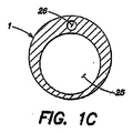
膨張可能保持部材が使用されるときに、チューブ1は、好ましくは、膨張可能管腔26を備えるため膨張可能保持部材は膨張する。図1Cは、チューブ1の軸に垂直なチューブ1の断面図を示す。膨張管腔26は、膨張部2からチューブ1の近端部16に伸び、水または空気などの流動物を患者の外側から膨張部2に導入するための通路である。除去管腔25は、近端部16からチューブ1の遠端部17に伸び、患者の胃3または上部消化器系の他の部分から食物を除去するための通路である。膨張管腔26は、好ましくは、除去管腔25がチューブ1内でできるだけ開くように、最小がゼロである。例示した実施形態において、弁15、27は、図7に示すように、それぞれ管腔25、26に設けられる。図1Aおよび1Bに示す非膨張可能保持部材2’および2’’を用いて、チューブ1の第二管腔26は、削除できる。
When the inflatable retaining member is used, the
膨張可能保持部材は、プッシュ法に類似した手技を用いた使用に適しているが、膨張可能または硬化保持部材は、引上法に類似した手技の使用には適していない。膨張可能保持部材を備えるチューブの一実施例は、Tiefenthalら(米国特許番号6,506,179)で説明されており、参照資料により、その全体が本明細書に記載されているものとみなす。代替的な変形保持部材は、Snowら(米国特許番号6,077,250)で説明されており、参照することにより、その全体が本明細書に組み込まれる。 Inflatable retaining members are suitable for use with procedures similar to the push method, while inflatable or cured retaining members are not suitable for use with procedures similar to the pulling method. One example of a tube with an inflatable retaining member is described in Tiefenthal et al. (US Pat. No. 6,506,179), which is considered to be fully described herein by reference. Alternative deformation retaining members are described in Snow et al. (US Pat. No. 6,077,250), which is hereby incorporated by reference in its entirety.
生体位で変形したかもしれない保持部材は、チューブ1がさらなる内視鏡なしで除去することを可能にする。保持部材は、収縮または変形し、チューブ1は、牽引を使用して引き出される。保持部材が硬化なときは、チューブ1は皮膚近くで切断され、内視鏡的に除去されてよい。
A holding member that may have been deformed in a living position allows the
胃が内腹壁に対して位置するのが好ましい。これは、チューブの留置手技中およびチューブ1が保持部材により設置された後、吸入により達成されてよい。例えば、図1に示すように、チューブ1の近端部16および遠端部17の保持部材は、腹壁に対して胃を固定する。胃も、チューブ留置から起こる合併症を防ぎ、留置手技を容易にするかもしれない胃腹壁固定術により腹壁に固定されてよい。さらに、空腸固定術(jejunopexy)は、チューブ留置手技中に空腸を固定するため、空腸造瘻術手技において重要である(Zmenら、上記参照)。例えば、胃または空腸を腹壁に固定するために、T字型の金属またはナイロンの固定部材は、チューブ挿入部位近くに経胃的または経腸的に挿入されてよい。固定部材は、挿入後、T字型をとり、皮膚近くに連結される。四個の固定部材は、胃または空腸を固定するために、基本的に、チューブ挿入部位の周りに四角形に配置される(例えば、F. J. Thorntonら、「Percutaneous Radiologic Gastrostomy with and without T−Fastener Gastropexy: a Randomized Comparison Study」Cardiovasc Intervent Radiol. 2002 November−December; 25(6):467−71を参照)。
The stomach is preferably located with respect to the inner abdominal wall. This may be achieved by inhalation during the tube indwelling procedure and after the
ここで、チューブ1の近端部16に取付けられる、ポンプの多様な形式について参照する。当該分野に精通した者には容易に理解される構造だが、全ての従来のポンプが使用されてよい。図2および3は、例えば、患者の胃3または上部消化器系から食物を除去するための、チューブ1の近端部16に取付けられるポンプ8および9を示す。30分以内に患者の上部消化器系から750ml以上の食物を抽出するポンプを使用するのに適している。ポンプは、チューブの収縮、チューブの詰まり、または粘膜刺激を防ぐため、断続的に操作されてよい。ポンプは、手動または電池作動であってよい。任意で、充電式電源装置は、ポンプに組み込まれてよく、ポンプは、患者のベルトに装着されるよう構成されてよい。
Reference is now made to the various types of pumps that are attached to the
図2は、チューブ1の近端部16に取付けられ、チューブ1を通して患者の上部消化器系から食物を除去するために操作される、手動の球形ポンプを示す。手動球形ポンプ8は、球形ポンプ8の球状の端部を圧搾することにより球形ポンプ8の中身を排除できるように、好ましくは、シリコンゴムまたは類似した柔軟な素材から成る。テーパ状の端部の周縁は、チューブ1の管腔25の内部周縁と基本的に対応する。手動球形ポンプ8を操作するために、最初に、空気が、球形を圧搾することにより球形ポンプから排除され、次に、テーパ状の端部とチューブ1の間にシールを作り出するために、球形ポンプ8のテーパ状の端部が、チューブ1の近端部16の管腔25に挿入される。次に、球形は、再び膨張するように緩められる。球形ポンプ8の負圧(緩められる時)は、食物を上部消化器系からチューブ1の近端部16に向かい、球形ポンプ8の球形に流れ込ませる。球形ポンプ8は、次に、チューブ1から取り外され、除去された食物は、球形から排除される。サイクルは、所望の量の食物が患者の上部消化器系から取り除かれるまで繰り返されてよい。
FIG. 2 shows a manual spherical pump attached to the
図3は、注入器の形のポンプが、チューブ1の近端部16に取付けられ、チューブ1を通して患者の上部消化器系から食物を除去するために操作される、他のポンプ構成を示す。注入器9は、好ましくは、その遠端部に開口のあるテーパ状の端部を備える。テーパ状の端部9aの周縁は、チューブ1の管腔25の内部周縁に対応する。患者の上部消化器系から食物を除去するための注入器9を操作するために、注入器9の中身(空気または食物)は、プランジャーを押し下げることにより排除される。テーパ状の端部9aとチューブ1の間にシールを作り出すために、注入器9のテーパ状の端部9aがチューブ1の近端部16に挿入される。次に、上部消化器系からチューブを通して注入器9に食物を取り出すための負圧を作り出すために、注入器9のプランジャーを引き出す。注入器9は、次に、チューブ1から取り外され、例えば、そのプランジャーを押し下げることにより排除される。60ccは、注入器9の適切なサイズの例である。サイクルは、所望の量の食物が患者の上部消化器系から取り除かれるまで繰り返されてよい。
FIG. 3 shows another pump configuration in which a pump in the form of an injector is attached to the
手動球形ポンプ8および注入器9は、食後、所定の時間に、患者または医療提供者により実行される。所定の時間は、好ましくは、医師により設定され、例えば、20〜30分などであってよい。医師は、各食事後、患者の上部消化器系から除去される食物の最大量も特定してよい。最大量は、ポンプ8、9が手動で操作されるときに、患者または医療提供者に通知される、サイクルの最大数に基づいて設定されてよい。 The manual spherical pump 8 and the injector 9 are executed by a patient or a health care provider at a predetermined time after a meal. The predetermined time is preferably set by a doctor and may be, for example, 20 to 30 minutes. The physician may also specify the maximum amount of food that is removed from the patient's upper digestive system after each meal. The maximum amount may be set based on the maximum number of cycles that are notified to the patient or health care provider when the pumps 8, 9 are manually operated.
好適な実施形態において、患者の上部消化器系から食物を抽出するために使用されるポンプは、定期的に方向を逆にし、逆操作の期間中、空気および/または水を患者の上部消化器系に送り入れる。空気および/または水は、上部消化器系の食物を可溶化または分解するのを助けるため、容易に吸い出せる。さらに、空気および/または水は、食物がチューブ1から上部消化器から抽出される間、チューブ1が腹壁に対して吸い上げられるのを防ぐのに役立つ。例えば、7秒毎の吸い上げは、次に2秒の逆操作が続く。
In a preferred embodiment, the pump used to extract food from the patient's upper digestive system periodically reverses direction and draws air and / or water during the reverse operation to the patient's upper digestive system. Send it to the system. Air and / or water can be easily sucked out to help solubilize or break down food in the upper digestive system. Furthermore, the air and / or water helps to prevent the
図4は、抽出された食物が、ポンプ6からポンプ6に取付けられた袋12に排除される、本発明の実施形態の変形を示す。図4に示すように、食物が、ポンプ6により患者の上部消化器系の外に吸い上げられた後、食物は、ポンプ6の近端部に取付けられた袋12に貯蔵されてよい。袋12は、不透明、香付き、生分解性でもよく、また患者のベルトまたは他のストラップに着用されてよい。代替的に、図11および16に示すように、食物は、患者の上部消化器系からポンプ6、次に、ポンプ6に取付けられたチューブ28に吸い上げられてよい。ポンプ6に取付けられたチューブ28の中身は、トイレに空けてよい。チューブ28は、不透明、香付き、生分解性であり、またトイレに流すことが可能であってよい。
FIG. 4 shows a variation of the embodiment of the present invention in which the extracted food is removed from the pump 6 to the
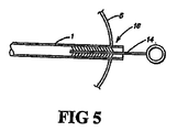
図5は、食物が、チューブ1を通して患者の上部消化器系から抽出された後、チューブ1を洗浄するために使用される洗浄デバイスを示す。図5に示すように、チューブ1は、チューブ1の内部を洗浄するよう適合させたブラシ14を使用して洗浄されてもよい。ポンプ6、手動球形ポンプ8、および注入器9は、使用後、食塩水および/または消毒液で濯ぐことにより洗浄されてもよい。
FIG. 5 shows a cleaning device used to clean the
図6は、バルーンアンカーの膨張により満腹感を患者に与える、本発明の二番目の実施形態を示す。満腹感を生み出すことは、患者の空腹および食物摂取の欲求を抑制することになり、患者の食物摂取の低下および体重減少を可能にする。図6に示すように、患者の胃のチューブ1を保持する保持部材である膨張部2は、膨張したときに、満腹感を生み出すための胃容量減少の機能としても役立つ。膨張部2は、チューブ1の膨張管腔26(図1Cに示す)を通して流動物を添加または除去することにより、可変的に膨張されてよい。
FIG. 6 shows a second embodiment of the present invention that provides a patient with a feeling of fullness by inflation of a balloon anchor. Creating a feeling of fullness will reduce the patient's hunger and desire for food intake, allowing the patient to reduce food intake and lose weight. As shown in FIG. 6, the inflatable portion 2, which is a holding member that holds the patient's
図7は、除去管腔25および膨張管腔26の見える、患者の皮膚5から外に伸びるチューブ1の軸断面図を示す。本発明の多様な実施形態の全てに引用されてよい特徴において、弁15は、チューブ1の近端部16の除去管腔25に設けられる。弁15は、通常、食物がチューブ1から流出するのを防ぐ。ポンプがチューブ1の近端部16に取付けられると、弁15が開く。例えば、手動球形ポンプ8のテーバ状の端部(図2に示す)および注入器9のテーパ状の端部(図3に示す)のそれぞれは、チューブ1の近端部16に挿入されたときに、弁15を押し開く。弁15が、ポンプの端部により開かれるとき、食物は、本明細書の上で説明したように除去される。キャップ13(図1に示す)は、ポンプが取付けられていないとき、好ましくは、チューブ1の近端部16に設置される。キャップ13は、チューブ1の端部に押し付けられるか、チューブ1の端部に螺合されるか、または閉口状態になるように管腔を塞ぐために管腔25、26の端部に摩擦的に挿入される突起を備えてよい。
FIG. 7 shows an axial cross-sectional view of the
図7も、チューブ1の近端部16の膨張管腔26に設けられた弁27を示す。弁27は、膨張部2を膨張するために使用される液体が、膨張管腔26を通って膨張部2から流出するのを防ぐ。つまり、弁27は、膨張部2が収縮するのを防ぐ。患者の上部消化器系からチューブ1を取り除くために膨張部2を収縮する必要がある場合、または膨張部2をさらに膨張する必要がある場合、注入器の針は、弁部材に針を押し通すことにより弁27が開くように、膨張部26に挿入されてよい。膨張部2を膨張するために使用される液体は、次に、取り除かれる、または注入器により添加される。
FIG. 7 also shows a valve 27 provided in the
図8は、患者の上部消化器系内に配置されたチューブの部分に取付けられた二つのバルーンを有するチューブを示す、本発明の三番目の実施形態を示す。バルーンアンカー2は、約10mlに拡張可能で、チューブ1が外れるのを防ぐために腹壁に対して設置される。膨張可能バルーン29は、約100mlから約850mlに拡張可能で、胃容量を制限するために断続的に拡張されてよい。例えば、バルーン29は、満腹感を生み出すために食前に膨張管腔を介して膨張されてよい。食後、バルーン29は、慢性的順応を防ぐために収縮されてよい。電気的または手動で操作されるポンプを、膨張させるのに使用してよい。
FIG. 8 shows a third embodiment of the present invention showing a tube having two balloons attached to the portion of the tube placed in the patient's upper digestive system. The balloon anchor 2 can be expanded to about 10 ml, and is placed against the abdominal wall to prevent the
本実施形態のチューブ1は、長さが10cm以上で、直径のサイズが28フレンチ(9.3mm)より大きい長い内部チューブを備える。チューブ1は、図8および図10および13〜15Bにも示すように、遠端部17の側壁に複数の穴32を有してよい。穴32は、サイズが5×7mmであってよい。穴32は、患者からの非血管性排出口を提供する。好ましくは、穴32は、構造の一体性を失わずに1cmから1.5cm間隔の螺旋状に配列される。より好ましくは、クッションまたはバンパー(示されていない)が、食物がチューブ1を通して上部消化器系から抽出される間、チューブが腹壁に対して吸引されるのを防ぐために、チューブ1上におよび穴32の間に配置される。例えば、チューブ1の表面から3〜4mm高くされたクッションまたはバンパーは、本目的のために使用される。
The
図8に示すように、第二保持部材33は、腹面に固定されたチューブ1を保持するために、チューブ1の近端部16に取付けられてよい。この第二保持部材は、本明細書で上述した保持部材に類似してよく、図1、1A、1Bおよび6に示される。チューブ1の近端部16の第二保持部材33とチューブ1の遠端部17のバルーンアンカー間の距離は、図15Aおよび15Bに示すように、介在する組織40、40’の量の変化に対応するよう調節可能である。例えば、第二保持部材33は、干渉嵌合または摩擦係合を介してチューブ1に取付けられてよい。特に、第二保持部材33は、チューブ1の外径よりわずかに小さい内径を有する場合、チューブ1の近端部16の外面の周りに設置され、チューブ1に固定されてよい。患者の体重が減少すると、チューブ1の近端部16は、患者の腹面からさらに伸長する。医師または患者は、第二保持部材33を腹面方向にずらし、過剰量なチューブ1は切断される。
As shown in FIG. 8, the second holding
図9は、遠端部17が曲線形状で、外壁に多数の穴32を有するチューブ1を用いた本発明の四番目の実施形態を示す。図9に示すように、チューブ1の遠端部17は、患者の上部消化器系に配置されたときに、曲線形状をとるように適合される。特に、チューブ1の遠端部17は、患者への挿入および患者からの除去を容易にするために柔軟である。チューブ1の遠端部17は、患者の上部消化器系に配置されるとき、自然な曲線形状に戻る。自然な曲線形状に戻るというチューブの傾向は、例えば、製造工程中、チューブが完全に硬化または冷却される前に所望の曲線形状にチューブを曲げることにより、または形状記憶材料をチューブに引用することにより、達成される。本明細書で使用されるように、用語「曲線」とは、屈曲、わん曲、円形、アーク形、縁曲、コイル状、螺旋状およびピッグテイルを含む。この曲線形状は、上部消化器系内で収容域が増大するため、好ましい。さらに、図10に示すように、コイル状のチューブ1の遠端部17は、患者の上部消化器系内にチューブ1の位置を維持するのに役立つ。チューブ1の遠端部17は、例えば、上部消化器系から食物の摂取を向上するために、長さが約10cm以上であってよい。上の実施形態で説明した類似した保持部材(示されていない)も、本実施形態で使用されてよい。
FIG. 9 shows a fourth embodiment of the present invention in which the
代替実施形態(示されていない)において、動作メカニズムは、チューブ1の遠端部17を曲線形状に曲げるように構成される。動作メカニズムは、例えば、チューブ1の遠端部17に取付けられる紐であってよく、引き込んだときに、チューブが曲線形状(例えば、約270°から360°の間の弧を持つループ)をとる。上型ループ(Cope Loop)は、本変形のよく知られた例である。
In an alternative embodiment (not shown), the operating mechanism is configured to bend the
図10は、曲線形状で、外壁に複数の穴32および遠端部17のハウジング37内に収容された細切デバイス36を有するチューブ1を示す、本発明の五番目の実施形態を示す。細切デバイスの例は、米国特許番号5,618,296、5,741,287および5,520,634に開示されており、参照により、その全体が本明細書に組み込まれる。図10に示すように、細切デバイス36は、食物がチューブ1に入ると小さい小片に分割および粉砕するために、チューブ1の遠端部17に配置される。細切デバイス36は、よって、チューブ1を詰まらせることなく患者から大きい食物を除去することを可能にする。細切デバイス36は、例えば、チューブ1の遠端部17のハウジング37内に配置された機械的プロペラであってよい。ハウジング37は、細切デバイスから生体組織を保護するために組立てられる。例示された実施形態において、ハウジング37は、患者からチューブ1に食物を進入させる開口部を有し、例えば、チューブ1の遠端部17に細切デバイス36を囲むケージであってよい。ハウジング37は、容易に患者に挿入または除去されるように、両方向に収縮可能であるのが好ましい。ハウジング37は、胃への損傷を防ぐために必要である。
FIG. 10 shows a fifth embodiment of the invention showing a
図11は、チューブ1の近端部16が患者の腹部の外面に実質的に面一に位置する、本発明のあらゆる実施形態に使用されてよい特徴を示す。これは、例えば、内部保持部材の、チューブ1に取付けられたリボンを使用して達成されてよい。リボンは、チューブ1の遠端部17が患者の上部消化器系に配置されたときに、チューブ1をぴんと引っ張るために使用される。リボンが引っ張られている間、チューブ1の近端部16は、腹面に面一に位置するように切断され、フランジの付いた細い中空の注入器は、その位置にチューブ1を留置し腹面と面一を保つために、摩擦によってまたはチューブ1にねじ込むことにより、チューブ1の外面または内面に押し込まれる。代替的な実施形態において、チューブ1の近端部16は、あらゆる所望する長さ(例えば、1〜10インチ)により腹面を超えて伸長してよい。
FIG. 11 illustrates a feature that may be used in any embodiment of the present invention in which the
図12は、ルアーロック34がチューブ1の近端部16に使用される、本発明のあらゆる実施形態に使用されてよい他の特徴を示す。本実施形態において、ポンプ6は、チューブ1に挿入されるよりむしろチューブ1の近端部16の外部の周りのチューブ1にポンプ6をねじ込むことにより、チューブ1に取付けられる。より具体的には、チューブ1の近端部16は、ポンプ6が除去管腔25のサイズを減少するのを防ぐ、ポンプ6を適合するための同心円状の溝または筋道を外側に備える。同様に、ポンプ6はルアーロック34と相互作用または接続が可能な、対応する同心円状の溝または筋道を有する。この方法により、チューブ1の内径は、ポンプ6がチューブ1に挿入されることにより破損または減少しないため、大きな食物もまたチューブ1から抽出できる。代わりに、ポンプ6は、チューブ1の近端部16の外側に結合または螺合される。
FIG. 12 illustrates another feature that may be used in any embodiment of the present invention in which a
図13は、チューブ1が漏斗形の先端35を有する、本発明のあらゆる実施形態に使用されてよい、さらに他の特徴を示す。漏斗形の先端は、患者の消化器系から食物の大きい小片をチューブ1へ抽出するのを容易にするため、有利である。
FIG. 13 shows yet another feature that may be used in any embodiment of the present invention where the
図14は、二つの吸入チューブを有する本発明の六番目の実施形態を示す。本実施形態において、両吸入チューブ38は、曲線形状で、その内部に複数の穴32を有する側壁を備える。各吸入チューブ38は、近端部39および遠端部40を備える。本装置は、近端部および遠端部42を有する排出チューブ41も備える。一つ以上の保持部材(示されていない)は、好ましくは、装置が上部消化器系から外れるのを防ぐために排出チューブ41に取付けられる。複数の吸入チューブ38は患者の上部消化器系に設置されるよう構成され、排出チューブ41は、複数の吸入チューブ38がそのように設置されたときに、患者の腹壁を通過するように構成される。排出チューブ41の遠端部42は、食物は複数の吸入チューブ38のそれぞれの遠端部40を通して患者の消化器系から抽出され、排出チューブ41の近端部を通して外に出るように、複数の吸入チューブ38のそれぞれの遠端部39に操作可能に接続される。 FIG. 14 shows a sixth embodiment of the present invention having two suction tubes. In the present embodiment, both suction tubes 38 are curved and include side walls having a plurality of holes 32 therein. Each suction tube 38 includes a near end 39 and a far end 40. The apparatus also includes a discharge tube 41 having a near end and a far end 42. One or more retaining members (not shown) are preferably attached to the drain tube 41 to prevent the device from detaching from the upper digestive system. The plurality of inhalation tubes 38 are configured to be installed in the patient's upper digestive system, and the drain tube 41 is configured to pass through the patient's abdominal wall when the plurality of inhalation tubes 38 are so installed. The The distal end 42 of the drain tube 41 has a plurality of foods such that food is extracted from the patient's digestive system through the distal end 40 of each of the plurality of inhalation tubes 38 and exits through the proximal end of the drain tube 41. Operatively connected to each distal end 39 of the suction tube 38.
任意で、圧力および/またはフローセンサー(示されていない)がチューブ1の外および/または中に設置されてよい。胃3の内側および外側のチューブ1に設置された圧力センサーは、患者の満腹度を予測するために使用されてよい。代替的または追加で、チューブ1の内側に設置されたフローセンサーは、チューブ1を通して抽出された食物の量を算出するために使用されてよい。
Optionally, pressure and / or flow sensors (not shown) may be installed outside and / or in the
ここで、食物の抽出、食物の吸収制限および肥満患者の治療に体する様々な方法を参照する。 Reference is now made to various methods for the extraction of food, limiting absorption of food and treating obese patients.
上述のあらゆる実施形態の例示は、患者の腹壁を通して患者の上部消化器系への通路を形成する。患者は、食物摂取を含む日常の作業の実行が可能である。患者が食物を摂取した後、食物は完全に消化される前に、通路を通して上部消化器系から吸い出されることにより抽出される。本方法および他の以下に説明する方法は、体重の減少のための代替的な外科手術手技より低侵襲的であり、実行が容易、反転が容易、および肥満患者の著しい体重減少において効果的な結果を有する。 The illustration of any of the embodiments described above creates a passage through the patient's abdominal wall to the patient's upper digestive system. Patients can perform routine tasks, including food intake. After the patient has ingested the food, it is extracted by being aspirated from the upper digestive system through the passageway before being fully digested. This method and other methods described below are less invasive than alternative surgical procedures for weight loss, are easier to perform, are easier to reverse, and are effective in significant weight loss in obese patients Have a result.
一方法において、チューブは、患者の腹壁を通って患者の上部消化器系に入るように位置づけられる。患者は、食物の摂取を含む自身の日常の活動を行うことが可能である。患者が食物を摂取した後、食物は、チューブを通して患者の上部消化器系から抽出される。患者は所望の体重減少が達成されるまで、食事をし、繰り返しチューブを通して自分の上部消化器系から摂取した食物を抽出してよい。抽出された食物は、患者に再度導入されない。チューブは、チューブが設置されている間、食事/抽出が多数(例えば20回以上)繰り返される間、長期間(例えば1ヶ月以上)、患者の上部消化器系に留置されてよい。 In one method, the tube is positioned to enter the patient's upper digestive system through the patient's abdominal wall. Patients can perform their daily activities, including food intake. After the patient has ingested food, the food is extracted from the patient's upper digestive system through a tube. The patient may eat until the desired weight loss is achieved and repeatedly extract ingested food from his upper digestive system through a tube. The extracted food is not reintroduced to the patient. The tube may be left in the patient's upper digestive system for an extended period of time (eg, over a month) while the meal / extraction is repeated many times (eg, over 20 times) while the tube is installed.
第二の方法において、チューブは、肥満患者の腹壁を通って肥満患者の上部消化器系に入るように位置づけられる。肥満患者は、食物の摂取を含む自身の日常の活動を行うことが可能である。肥満患者が食物を摂取した後、食物は、チューブを通して肥満患者の上部消化器系から抽出される。肥満患者は少なくとも40ポンド減少するまで、食事をし、繰り返しチューブを通して自分の上部消化器系から摂取した食物を抽出してよい。抽出された食物は、肥満患者に再度導入されない。 In the second method, the tube is positioned to enter the upper digestive system of the obese patient through the abdominal wall of the obese patient. Obese patients can perform their daily activities, including food intake. After the obese patient has ingested food, the food is extracted from the obese patient's upper digestive system through a tube. Obese patients may dine until they are reduced by at least 40 pounds and repeatedly extract ingested food from their upper digestive system through tubes. The extracted food is not reintroduced into the obese patient.
第三の方法において、チューブは、消化管が非閉塞である患者の腹壁を通って患者の上部消化器系に入るように位置づけられる。本明細書で使用される用語「非閉塞」とは、機械的に閉塞でなく、機能的にも閉塞でない消化管を意味する。患者は、食物の摂取を含む自身の日常の活動を行うことが可能である。患者が食物を摂取した後、食物は、チューブを通して患者の上部消化器系から抽出される。患者は、所望の体重減少が達成されるまで、食事をし、繰り返しチューブを通して自分の上部消化器系から摂取した食物を抽出してよい。チューブは、チューブが設置されている間、食事/抽出が多数(例えば20回以上)繰り返される間、長期間(例えば1ヶ月以上)、患者の上部消化器系に留置されてよい。 In the third method, the tube is positioned to enter the patient's upper digestive system through the abdominal wall of the patient where the gastrointestinal tract is unobstructed. The term “non-occluded” as used herein refers to the gastrointestinal tract that is not mechanically occluded and is not functionally occluded. Patients can perform their daily activities, including food intake. After the patient has ingested food, the food is extracted from the patient's upper digestive system through a tube. The patient may eat until the desired weight loss is achieved and repeatedly extract the ingested food from his upper digestive system through a tube. The tube may be left in the patient's upper digestive system for an extended period of time (eg, over a month) while the meal / extraction is repeated many times (eg, over 20 times) while the tube is installed.
ヒト患者における先行試験は、成功であった。例えば、中年、体重100キロ(約220ポンド)の一人の女性患者は、59週間、胃にチューブを設置し、いかなる重度の副作用を経験せずに、成功にも38.45キロ(約85ポンド)痩せた。59週間、女性患者は、毎日、朝食および昼食後に吸引した。女性は、約30分以上一切の液体なしに、食事を摂取した。食事の終わりに、女性は、約3〜4分で52オンスの水を摂取した。女性は、抽出工程の開始前に水を摂取した後、約20分待った。次に、患者は、チューブのキャップを取り、チューブに60ccの注入器を接続し、胃から食物を2回抽出した。これは、被験者が、開口チューブにバケツの中に空にさせることにより、自由に自分の胃を空にするのを可能にする、サイフォン作用を引き起こした。患者は、推進を進め大きい食物を砕くためにチューブを圧搾した。排出が停止した後、患者は、通常52オンスの水をさらに飲み、抽出工程を繰り返した。女性は、胃が空になったと感じるまで、通常この工程(飲水および抽出)を約2回繰り返した。食物抽出の合計量は、約2〜3リットルで、全工程は、約20分かかった。抽出に対する抵抗が工程中に起こった場合、患者は、30ccの水でチューブを濯いだ。水は、食物を溶解することおよび通路を洗浄することにより、食物の抽出を助けた。患者は、チューブの詰まりを避けるために、自分の食物摂取を変更した。女性は、カリフラワー、ブロッコリー、中華料理、炒め物、さやえんどう、プレッツェル、チップスおよびステーキの摂取を避けた。さらに、女性の食事は、カリウムで補われた。下のチャートは、女性の体重減少を示す。 Prior trials in human patients have been successful. For example, a single female patient weighing 100 kg (about 220 pounds) in middle age successfully installs a tube in the stomach for 59 weeks without experiencing any severe side effects and succeeds in 38.45 kg (about 85 kg). Pound) For 59 weeks, female patients were aspirated daily after breakfast and lunch. The woman took a meal for about 30 minutes or more without any liquid. At the end of the meal, the woman took 52 ounces of water in about 3-4 minutes. The woman waited about 20 minutes after ingesting water before the start of the extraction process. The patient then removed the tube cap, connected a 60 cc syringe to the tube, and extracted food twice from the stomach. This caused a siphoning effect that allowed the subject to freely empty his stomach by emptying the open tube into the bucket. The patient squeezed the tube to push forward and break large food. After draining ceased, the patient usually drank another 52 ounces of water and repeated the extraction process. The woman typically repeated this process (drinking and extraction) about twice until she felt the stomach was empty. The total amount of food extraction was about 2-3 liters and the whole process took about 20 minutes. If resistance to extraction occurred during the process, the patient rinsed the tube with 30 cc of water. The water helped extract the food by dissolving the food and washing the passageways. The patient changed his food intake to avoid clogging the tube. Women avoided eating cauliflower, broccoli, Chinese food, stir-fried, peas, pretzels, chips and steaks. In addition, women's diet was supplemented with potassium. The lower chart shows women's weight loss.

上述の実施形態は、急激で侵襲的な手術を受けずに肥満患者の体重を減少させる。結果として、肥満患者は、このような手術に伴う多くの合併症を回避する。さらに、本発明は、実行が容易、反転が容易および肥満患者がわずかな有害副作用で正常で活動的な生活習慣を送るのを可能にする。 The above-described embodiments reduce the weight of obese patients without undergoing rapid and invasive surgery. As a result, obese patients avoid many of the complications associated with such surgery. Furthermore, the present invention enables easy to perform, easy to reverse and obese patients to live a normal and active lifestyle with few adverse side effects.
さらなる利点および修正は、当該分野に精通した者に容易に生じるであろう。例えば、あらゆる実施形態の特徴は、単一または本発明の他のあらゆる実施形態と組み合わせて使用されてよい。さらに、チューブを設置する挿入技術は、周知の胃瘻造設術に制限されない。よって、様々な修正が、添付の特許請求の範囲およびその等価物により定義されるような、一般的な発明概念の精神および範囲から逸脱することなく行われてよい。 Additional advantages and modifications will readily occur to those skilled in the art. For example, the features of any embodiment may be used singly or in combination with any other embodiment of the invention. Further, the insertion technique for installing the tube is not limited to the known gastrostomy. Accordingly, various modifications may be made without departing from the spirit and scope of the general inventive concept as defined by the appended claims and their equivalents.
Claims (130)
i.内部に複数の穴を有する側壁をもつ遠端部であって、患者の上部消化器系に位置している間は、自動的に曲線形状をとるように適合された遠端部と、
ii.該チューブの遠端部が、該患者の該上部消化器系に配置されたときに、該患者の腹壁を通るように構成された近端部と、
を備えるチューブと、
b.該チューブのズレを防ぐための、該チューブに取付けられた第一保持部材と、
を備える装置。 a. A tube,
i. A distal end with a sidewall having a plurality of holes therein, the distal end adapted to automatically take a curved shape while positioned in the patient's upper digestive system;
ii. A proximal end configured to pass through the abdominal wall of the patient when the distal end of the tube is placed in the upper digestive system of the patient;
A tube comprising:
b. A first holding member attached to the tube for preventing displacement of the tube;
A device comprising:
b.近端部および遠端部のある排出チューブと、
c.前記チューブのズレを防ぐための前記排出チューブに取付けられる保持部材と、
を備え、前記複数の吸入チューブは、患者の上部消化器系に配置されるよう構成され、前記排出チューブは、前記複数の吸入チューブが前記患者の前記上部消化器系に配置されたときに、前記患者の腹壁を通過するように構成され、食物が、前記患者の前記上部消化器系から、前記複数の吸入チューブのそれぞれの遠端部を通して、および前記排出チューブの近端部を通して外に抽出されるように、前記排出チューブの遠端部は、前記複数の吸入チューブのそれぞれの近端部に操作可能に接続される、
装置。 a. A plurality of suction tubes each having a plurality of suction tubes each having a sidewall having a plurality of holes therein, each having a near end and a far end; and
b. A discharge tube with a near end and a far end; and
c. A holding member attached to the discharge tube for preventing displacement of the tube;
The plurality of inhalation tubes are configured to be disposed in a patient's upper digestive system, and the drainage tube is disposed when the plurality of inhalation tubes are disposed in the patient's upper digestive system. Configured to pass through the patient's abdominal wall, food is extracted from the patient's upper digestive system through the distal end of each of the plurality of inhalation tubes and through the proximal end of the drainage tube The distal end of the discharge tube is operably connected to the proximal end of each of the plurality of suction tubes,
apparatus.
i.内部に複数の穴を有する側壁をもつ、長さが少なくとも10cmの遠端部であって、患者の上部消化器系に配置されるように構成された遠端部と、
ii.前記チューブの遠端部が、前記患者の前記上部消化器系に配置されたとき、前記患者の腹壁を通るように構成された近端部と、
を備えるチューブと、
b.前記チューブのズレを防ぐための、前記チューブに取付けられた第一保持部材と、
を備える装置。 a. A tube,
i. A distal end having a sidewall having a plurality of holes therein and having a length of at least 10 cm, the distal end configured to be placed in a patient's upper digestive system;
ii. A proximal end configured to pass through the abdominal wall of the patient when the distal end of the tube is positioned in the upper digestive system of the patient;
A tube comprising:
b. A first holding member attached to the tube for preventing displacement of the tube;
A device comprising:
i.内部に複数の穴を有する側壁をもつ遠端部であって、患者の上部消化器系に配置されるように構成された遠端部と、
ii.該チューブの遠端部が、該患者の該上部消化器系に配置されたときに、該患者の腹壁を通るように構成された近端部と
を備えるチューブと、
b.該チューブのズレを防ぐための、該チューブに取付けられた第一保持部材と、
を備える装置。 a. The diameter of the tube is larger than 28 French,
i. A distal end having a sidewall having a plurality of holes therein, the distal end configured to be placed in a patient's upper digestive system;
ii. A tube comprising a distal end configured to pass through the abdominal wall of the patient when the distal end of the tube is placed in the upper digestive system of the patient;
b. A first holding member attached to the tube for preventing displacement of the tube;
A device comprising:
i.内部に複数の穴を有する側壁をもつ遠端部であって、患者の上部消化器系に配置されるように構成される遠端部と、
ii.該チューブの遠端部が、該患者の該上部消化器系に配置されたときに、該患者の腹壁を通るように構成された近端部と、
を備えるチューブと、
b.該チューブのズレを防ぐための、該チューブに取付けられた第一保持部材と、
を備える装置であって、該第一保持部材は、該チューブに対して、該チューブの遠端部が該患者の該上部消化器系に配置されたときに、該チューブの近端部が該患者の腹面と実質的に面一となるように構成される、
装置。 a. A tube,
i. A distal end having a sidewall with a plurality of holes therein, the distal end configured to be placed in a patient's upper digestive system;
ii. A proximal end configured to pass through the abdominal wall of the patient when the distal end of the tube is placed in the upper digestive system of the patient;
A tube comprising:
b. A first holding member attached to the tube for preventing displacement of the tube;
The first retaining member is positioned relative to the tube when the distal end of the tube is positioned in the upper digestive system of the patient. Configured to be substantially flush with the patient's abdominal surface,
apparatus.
i.内部に複数の穴を有する側壁をもつ遠端部と、
ii.該チューブの遠端部が、該患者の該上部消化器系に配置されたときに、該患者の腹壁を通るように構成された近端部と、
を備えるチューブと、
b.該チューブのズレを防ぐための、該チューブに取付けられた第一保持部材と、
c.患者の上部消化器系に配置されている間、該チューブの遠端部を曲線形状に曲げるように構成される動作メカニズムと、
を備える装置。 a. A tube,
i. A far end having a sidewall having a plurality of holes therein;
ii. A proximal end configured to pass through the abdominal wall of the patient when the distal end of the tube is placed in the upper digestive system of the patient;
A tube comprising:
b. A first holding member attached to the tube for preventing displacement of the tube;
c. An operating mechanism configured to bend the distal end of the tube into a curved shape while placed in the patient's upper digestive system;
A device comprising:
(a)患者の腹壁を通過して該患者の上部消化器系に入るチューブを位置づけるステップと、
(b)該患者に食物を摂取させるステップと、
(c)該患者が該食物を摂取した後、該チューブを通して該患者の該上部消化器系から該食物を抽出するステップと、
(d)所望の体重減少が得られるまで、ステップ(b)および(c)を繰り返すステップであって、ステップ(c)で抽出された該食物は該患者に再度導入されないステップと、
を含む、食物吸収の制限方法。 A method of limiting food absorption,
(A) positioning a tube that passes through the patient's abdominal wall and enters the upper digestive system of the patient;
(B) causing the patient to take food;
(C) extracting the food from the upper digestive system of the patient through the tube after the patient has consumed the food;
(D) repeating steps (b) and (c) until the desired weight loss is obtained, wherein the food extracted in step (c) is not reintroduced to the patient;
A method of limiting food absorption, including:
(f)前記患者の血液を電解質類について定期的にテストするステップと、
(g)必要に応じて、ビタミンおよびミネラルで前記患者の食事を補うステップと、
(h)必要に応じて、胆石形成を防ぐために前記患者に薬物を投与するステップと、
をさらに含む、請求項74に記載の方法。 (E) educating the patient to change caloric intake and lifestyle habits;
(F) periodically testing the patient's blood for electrolytes;
(G) supplementing the patient's diet with vitamins and minerals as needed;
(H) optionally administering a drug to the patient to prevent gallstone formation;
75. The method of claim 74, further comprising:
(a)肥満患者の腹壁を通過して該肥満患者の上部消化器系に入るチューブを位置づけるステップと、
(b)該肥満患者に食物を摂取させるステップと、
(c)該患者が食物を摂取した後、該チューブを通して該肥満患者の該上部消化器系から該食物を抽出するステップと、
(d)該肥満患者が少なくとも40ポンド減るまで、ステップ(b)および(c)を繰り返すステップと、
を含む、肥満患者の処置方法。 A method for treating obese patients,
(A) positioning a tube that passes through the abdominal wall of the obese patient and enters the upper digestive system of the obese patient;
(B) causing the obese patient to take food;
(C) extracting the food from the upper digestive system of the obese patient through the tube after the patient has consumed the food;
(D) repeating steps (b) and (c) until the obese patient loses at least 40 pounds;
A method for treating obese patients.
(f)前記患者の血液を電解質類について定期的にテストするステップと、
(g)必要に応じて、ビタミンおよびミネラルで前記患者の食事を補うステップと、
(h)必要に応じて、胆石形成を防ぐために前記患者に薬物を投与するステップと、
をさらに含む、請求項85に記載の方法。 (E) educating the patient to change caloric intake and lifestyle habits;
(F) periodically testing the patient's blood for electrolytes;
(G) supplementing the patient's diet with vitamins and minerals as needed;
(H) optionally administering a drug to the patient to prevent gallstone formation;
The method of claim 85, further comprising:
(b)該患者に食物を摂取させるステップと、
(c)該患者が該食物を摂取した後、該チューブを通して該患者の該上部消化器系から該食物を抽出するステッップと、
(d)所望の体重減少を達成するまで、ステップ(b)および(c)を繰り返すステップと、
を含む、方法。 (A) positioning a tube that passes through the abdominal wall of a patient whose gastrointestinal tract is unobstructed and enters the patient's upper digestive system;
(B) causing the patient to take food;
(C) extracting the food from the upper digestive system of the patient through the tube after the patient has consumed the food;
(D) repeating steps (b) and (c) until the desired weight loss is achieved;
Including a method.
(f)前記患者の血液を電解質類について定期的にテストするステップと、
(g)必要に応じて、ビタミンおよびミネラルで前記患者の食事を補うステップと、
(h)必要に応じて、胆石形成を防ぐために前記患者に薬物を投与するステップと、
をさらに含む、請求項95に記載の方法。 (E) educating the patient to change caloric intake and lifestyle habits;
(F) periodically testing the patient's blood for electrolytes;
(G) supplementing the patient's diet with vitamins and minerals as needed;
(H) optionally administering a drug to the patient to prevent gallstone formation;
96. The method of claim 95, further comprising:
(a)患者の腹壁を通過して該患者の上部消化器系に入るチューブを位置づけるステップと、
(b)該患者に食物を摂取させるステップと、
(c)該患者が食物を摂取した後、該チューブを通して該患者の該上部消化器系から該食物を抽出するステップと、
(d)該チューブを少なくとも1ヶ月間該患者に留置し、ステップ(b)および(c)を少なくとも20回繰り返すステップと、
を含む、食物吸収の制限方法。 A method of limiting food absorption,
(A) positioning a tube that passes through the patient's abdominal wall and enters the upper digestive system of the patient;
(B) causing the patient to take food;
(C) extracting the food from the upper digestive system of the patient through the tube after the patient has consumed the food;
(D) placing the tube in the patient for at least one month and repeating steps (b) and (c) at least 20 times;
A method of limiting food absorption, including:
(a)通路が該患者の腹壁を通過するように、患者の上部消化器系に該通路を導入するステップと、
(b)該患者に食物を摂取させるステップと、
(c)該患者が食物を摂取した後、該通路を通して該患者の該上部消化器系から該食物を抽出するステップと、
(d)所望の体重減少が達成されるまで、ステップ(b)および(c)を繰り返すステップであって、ステップ(c)において抽出された該食物が該患者に再度導入されないステップと、
を含む、食物吸収の制限方法。 A method of limiting food absorption,
(A) introducing the passageway into the patient's upper digestive system such that the passageway passes through the abdominal wall of the patient;
(B) causing the patient to take food;
(C) extracting the food from the upper digestive system of the patient through the passage after the patient has consumed the food;
(D) repeating steps (b) and (c) until the desired weight loss is achieved, wherein the food extracted in step (c) is not reintroduced to the patient;
A method of limiting food absorption, including:
(a)通路が該肥満患者の腹壁を通過するように、肥満患者の上部消化器系に該通路を導入するステップと、
(b)該肥満患者に食物を摂取させるステップと、
(c)該患者が食物を摂取した後、該通路を通して該肥満患者の該上部消化器系から該食物を抽出するステップと、
(d)該肥満患者が少なくとも40ポンド減少するまで、ステップ(b)および(c)を繰り返すステップと、
を含む、肥満患者の治療方法。 A method for treating obese patients,
(A) introducing the passageway into the upper digestive system of the obese patient such that the passage passes through the abdominal wall of the obese patient;
(B) causing the obese patient to take food;
(C) extracting the food from the upper digestive system of the obese patient through the passage after the patient has consumed the food;
(D) repeating steps (b) and (c) until the obese patient has lost at least 40 pounds;
A method for treating obese patients, comprising:
(b)該患者に食物を摂取させるステップと、
(c)該患者が食物を摂取した後、該通路を通して該患者の該上部消化器系から該食物を抽出するステップと、
(d)所望の体重減少が達成されるまで、ステップ(b)および(c)を繰り返すステップと、
を含む、方法。 (A) introducing a passageway into the upper digestive system of a patient whose gastrointestinal tract is unobstructed, the passageway passing through the patient's abdominal wall;
(B) causing the patient to take food;
(C) extracting the food from the upper digestive system of the patient through the passage after the patient has consumed the food;
(D) repeating steps (b) and (c) until the desired weight loss is achieved;
Including a method.
(a)通路が該患者の腹壁を通過するように、患者の上部消化器系に該通路を導入するステップと、
(b)該患者に食物を摂取させるステップと、
(c)該患者が食物を摂取した後、該通路を通して該患者の該上部消化器系から該食物を抽出するステップと、
(d)少なくとも20回、ステップ(b)および(c)を繰り返すステップと、
を含む、食物吸収の制限方法。 A method of limiting food absorption,
(A) introducing the passageway into the patient's upper digestive system such that the passageway passes through the abdominal wall of the patient;
(B) causing the patient to take food;
(C) extracting the food from the upper digestive system of the patient through the passage after the patient has consumed the food;
(D) repeating steps (b) and (c) at least 20 times;
A method of limiting food absorption, including:
(a)食物を摂取するステップと、
(b)食物が摂取された後、該通路を通して該個人の該上部消化器系から該食物を抽出するステップと、
(c)所望の体重減少が達成されるまで、ステップ(a)および(b)を繰り返すであって、ステップ(b)で抽出された該食物は該個人に再び導入されないステップと、
を含む、方法。 A method for achieving or maintaining weight loss in an individual having a passage through the abdominal wall of the individual to the upper digestive system of the individual,
(A) ingesting food;
(B) extracting the food from the upper digestive system of the individual through the passage after the food has been ingested;
(C) repeating steps (a) and (b) until the desired weight loss is achieved, wherein the food extracted in step (b) is not reintroduced to the individual;
Including a method.
(a)食物を摂取するステップと、
(b)食物が摂取された後、該通路を通して該肥満の個人の該上部消化器系から該食物を抽出するステップと、
(c)該肥満の個人が少なくとも40ポンド減少するまで、ステップ(a)および(b)を繰り返すステップと、
を含む、方法。 A method for achieving or maintaining weight loss in an obese individual having a passage through the abdominal wall of the obese individual to the upper digestive system of the obese individual, comprising:
(A) ingesting food;
(B) extracting the food from the upper digestive system of the obese individual through the passage after food has been ingested;
(C) repeating steps (a) and (b) until the obese individual is reduced by at least 40 pounds;
Including a method.
(a)食物を摂取するステップと、
(b)食物が摂取された後、該通路を通して該個人の該上部消化器系から該食物を抽出するステップと、
(c)ステップ(a)および(b)を少なくとも20回繰り返すステップと、
を含む、方法。 A method for achieving or maintaining weight loss in an individual having a passage through the abdominal wall of the individual to the upper digestive system of the individual,
(A) ingesting food;
(B) extracting the food from the upper digestive system of the individual through the passage after the food has been ingested;
(C) repeating steps (a) and (b) at least 20 times;
Including a method.
Applications Claiming Priority (5)
| Application Number | Priority Date | Filing Date | Title |
|---|---|---|---|
| US60049604P | 2004-08-10 | 2004-08-10 | |
| US60/600,496 | 2004-08-10 | ||
| US61834604P | 2004-10-12 | 2004-10-12 | |
| US60/618,346 | 2004-10-12 | ||
| PCT/US2005/027164 WO2006020441A2 (en) | 2004-08-10 | 2005-07-28 | Apparatus and method for treating obesity by extracting food |
Related Child Applications (1)
| Application Number | Title | Priority Date | Filing Date |
|---|---|---|---|
| JP2011032642A Division JP5360917B2 (en) | 2004-08-10 | 2011-02-17 | Apparatus and method for treating obesity by extracting food |
Publications (3)
| Publication Number | Publication Date |
|---|---|
| JP2008509730A true JP2008509730A (en) | 2008-04-03 |
| JP2008509730A5 JP2008509730A5 (en) | 2011-04-07 |
| JP4865714B2 JP4865714B2 (en) | 2012-02-01 |
Family
ID=35241314
Family Applications (2)
| Application Number | Title | Priority Date | Filing Date |
|---|---|---|---|
| JP2007525656A Expired - Fee Related JP4865714B2 (en) | 2004-08-10 | 2005-07-28 | Apparatus and method for treating obesity by extracting food |
| JP2011032642A Expired - Fee Related JP5360917B2 (en) | 2004-08-10 | 2011-02-17 | Apparatus and method for treating obesity by extracting food |
Family Applications After (1)
| Application Number | Title | Priority Date | Filing Date |
|---|---|---|---|
| JP2011032642A Expired - Fee Related JP5360917B2 (en) | 2004-08-10 | 2011-02-17 | Apparatus and method for treating obesity by extracting food |
Country Status (8)
| Country | Link |
|---|---|
| EP (2) | EP1784233B1 (en) |
| JP (2) | JP4865714B2 (en) |
| AU (1) | AU2005274113A1 (en) |
| CA (2) | CA2867814C (en) |
| DK (1) | DK1784233T3 (en) |
| ES (1) | ES2554336T3 (en) |
| HK (1) | HK1219687A1 (en) |
| WO (1) | WO2006020441A2 (en) |
Cited By (3)
| Publication number | Priority date | Publication date | Assignee | Title |
|---|---|---|---|---|
| JP2009542349A (en) * | 2006-07-05 | 2009-12-03 | アスピレーション・メディカル・テクノロジー・エルエルシー | Short-circuit device to treat obesity by extracting food |
| JP2011529766A (en) * | 2008-08-03 | 2011-12-15 | ランガード、リミテッド | Nose-to-stomach and mouth-to-stomach delivery device, system provided therewith, method and use thereof |
| JP2017185366A (en) * | 2008-10-10 | 2017-10-12 | ミルックス・ホールディング・エスエイ | Implantable device for controlling urology in the body |
Families Citing this family (8)
| Publication number | Priority date | Publication date | Assignee | Title |
|---|---|---|---|---|
| US7815629B2 (en) | 2002-11-04 | 2010-10-19 | Deka Products Limited Partnership | Apparatus for treating obesity by extracting food |
| US9055995B2 (en) | 2002-11-04 | 2015-06-16 | Aspire Bariatrics, Inc. | Method for treating obesity by extracting food |
| US8414561B2 (en) * | 2006-08-03 | 2013-04-09 | Aspire Bariatrics, Llc | Systems and methods for removing ingested material from a stomach |
| ITMO20100345A1 (en) * | 2010-12-06 | 2012-06-07 | Azzolini Graziano | MEDICAL DEVICE FOR THE IMPLEMENTATION OF ILEOSTOMIES AND / OR REJECTIVES |
| CA2846228C (en) | 2011-08-23 | 2020-10-13 | Aspire Bariatrics, Inc. | Improved skin port connector and method of installation |
| US10085866B2 (en) | 2013-02-23 | 2018-10-02 | Aspire Bariatrics, Inc. | Apparatus and method for draining material from a stomach |
| AU2016419833B2 (en) * | 2016-08-17 | 2021-12-16 | Avent, Inc. | Enteral feeding satiation device |
| US10765546B2 (en) | 2017-01-25 | 2020-09-08 | Ethicon, Inc. | Modified apparatus for food extraction and obesity treatment |
Citations (3)
| Publication number | Priority date | Publication date | Assignee | Title |
|---|---|---|---|---|
| US4642092A (en) * | 1984-12-10 | 1987-02-10 | Gerald Moss | Gastrointestinal aspirating device with suction breakers |
| JPH042361A (en) * | 1990-04-19 | 1992-01-07 | Olympus Optical Co Ltd | Catheter for dissolution treatment apparatus |
| JPH08266546A (en) * | 1995-03-30 | 1996-10-15 | Morita Mfg Co Ltd | Medical suction device |
Family Cites Families (21)
| Publication number | Priority date | Publication date | Assignee | Title |
|---|---|---|---|---|
| US3144868A (en) * | 1960-10-21 | 1964-08-18 | Mario E Jascalevich | Drainage and feeding cannulae |
| US4356824A (en) * | 1980-07-30 | 1982-11-02 | Vazquez Richard M | Multiple lumen gastrostomy tube |
| US4834724A (en) * | 1987-04-06 | 1989-05-30 | Geiss Alan C | Device for aspirating fluids from a body cavity or hollow organ |
| US4822338A (en) * | 1987-09-03 | 1989-04-18 | Longmore Wayne D | Method of removing material from the stomach using a collapsible funnel |
| US5071405A (en) * | 1989-06-02 | 1991-12-10 | Abbott Laboratories | Gastrostomy tube |
| US5234454A (en) * | 1991-08-05 | 1993-08-10 | Akron City Hospital | Percutaneous intragastric balloon catheter and method for controlling body weight therewith |
| ATE203920T1 (en) * | 1993-01-07 | 2001-08-15 | Medical Innovations Corp | CATHETER SYSTEM FOR GASTROSTOMY |
| CA2121861A1 (en) | 1993-04-23 | 1994-10-24 | William D. Fox | Mechanical morcellator |
| JPH0796030A (en) * | 1993-09-30 | 1995-04-11 | Nippon Zeon Co Ltd | Gastric tube catheter |
| US5520662A (en) * | 1994-09-09 | 1996-05-28 | Moss; Gerald | Gastrointestinal aspirating and feeding device with removable sleeve |
| JPH08196621A (en) * | 1995-01-30 | 1996-08-06 | Nippon Zeon Co Ltd | Sump tube and fluid discharge method using the same |
| US5618296A (en) | 1995-07-24 | 1997-04-08 | Endomedix Corporation/Box 330 | Tissue morcellator system and method |
| US5727555A (en) * | 1996-06-18 | 1998-03-17 | Cook Incorporated | Indwelling catheter |
| US5741287A (en) | 1996-11-01 | 1998-04-21 | Femrx, Inc. | Surgical tubular cutter having a tapering cutting chamber |
| US6077250A (en) | 1997-10-01 | 2000-06-20 | Boston Scientific Corporation | Apparatus and method for percutaneously placing gastrostomy tubes |
| EP1374930A1 (en) * | 1998-05-08 | 2004-01-02 | Cardeon Corporation | Circulatory support system for isolated segmental perfusion |
| AU5700501A (en) * | 2000-04-10 | 2001-10-23 | Scimed Life Systems Inc | Locking catheter |
| US6447472B1 (en) * | 2000-10-19 | 2002-09-10 | Gerald Moss | Method and pump apparatus for combined gastro-intestinal feeding and aspiration |
| US7066917B2 (en) * | 2001-10-05 | 2006-06-27 | Talamonti Anthony R | Oral gastric lavage kit with matched aspiration stream apertures |
| US6506179B1 (en) | 2001-10-12 | 2003-01-14 | Abbott Laboratories | Tube having a retention member |
| US20030225369A1 (en) * | 2002-05-31 | 2003-12-04 | Kimberly-Clark Worldwide, Inc. | Low profile transpyloric jejunostomy system |
-
2005
- 2005-07-28 WO PCT/US2005/027164 patent/WO2006020441A2/en not_active Ceased
- 2005-07-28 DK DK05778259.1T patent/DK1784233T3/en active
- 2005-07-28 JP JP2007525656A patent/JP4865714B2/en not_active Expired - Fee Related
- 2005-07-28 CA CA2867814A patent/CA2867814C/en not_active Expired - Fee Related
- 2005-07-28 AU AU2005274113A patent/AU2005274113A1/en not_active Abandoned
- 2005-07-28 EP EP05778259.1A patent/EP1784233B1/en not_active Expired - Lifetime
- 2005-07-28 EP EP15179488.0A patent/EP2962708A1/en not_active Withdrawn
- 2005-07-28 CA CA2575162A patent/CA2575162C/en not_active Expired - Fee Related
- 2005-07-28 ES ES05778259.1T patent/ES2554336T3/en not_active Expired - Lifetime
-
2011
- 2011-02-17 JP JP2011032642A patent/JP5360917B2/en not_active Expired - Fee Related
-
2016
- 2016-06-30 HK HK16107631.2A patent/HK1219687A1/en unknown
Patent Citations (3)
| Publication number | Priority date | Publication date | Assignee | Title |
|---|---|---|---|---|
| US4642092A (en) * | 1984-12-10 | 1987-02-10 | Gerald Moss | Gastrointestinal aspirating device with suction breakers |
| JPH042361A (en) * | 1990-04-19 | 1992-01-07 | Olympus Optical Co Ltd | Catheter for dissolution treatment apparatus |
| JPH08266546A (en) * | 1995-03-30 | 1996-10-15 | Morita Mfg Co Ltd | Medical suction device |
Cited By (4)
| Publication number | Priority date | Publication date | Assignee | Title |
|---|---|---|---|---|
| JP2009542349A (en) * | 2006-07-05 | 2009-12-03 | アスピレーション・メディカル・テクノロジー・エルエルシー | Short-circuit device to treat obesity by extracting food |
| JP2011529766A (en) * | 2008-08-03 | 2011-12-15 | ランガード、リミテッド | Nose-to-stomach and mouth-to-stomach delivery device, system provided therewith, method and use thereof |
| US8876762B2 (en) | 2008-08-03 | 2014-11-04 | Lunguard Ltd. | Nasogastric and orogastric feeding devices, system comprising them, methods and uses thereof |
| JP2017185366A (en) * | 2008-10-10 | 2017-10-12 | ミルックス・ホールディング・エスエイ | Implantable device for controlling urology in the body |
Also Published As
| Publication number | Publication date |
|---|---|
| DK1784233T3 (en) | 2015-10-05 |
| CA2575162A1 (en) | 2006-02-23 |
| HK1219687A1 (en) | 2017-04-13 |
| JP5360917B2 (en) | 2013-12-04 |
| CA2867814A1 (en) | 2006-02-23 |
| CA2575162C (en) | 2014-12-09 |
| JP4865714B2 (en) | 2012-02-01 |
| EP1784233A2 (en) | 2007-05-16 |
| WO2006020441A2 (en) | 2006-02-23 |
| AU2005274113A2 (en) | 2006-02-23 |
| EP1784233B1 (en) | 2015-08-26 |
| JP2011098225A (en) | 2011-05-19 |
| CA2867814C (en) | 2017-03-14 |
| ES2554336T3 (en) | 2015-12-18 |
| EP2962708A1 (en) | 2016-01-06 |
| AU2005274113A1 (en) | 2006-02-23 |
| WO2006020441A3 (en) | 2006-06-15 |
Similar Documents
| Publication | Publication Date | Title |
|---|---|---|
| US9039677B2 (en) | Apparatus for treating obesity by extracting food | |
| JP5360917B2 (en) | Apparatus and method for treating obesity by extracting food | |
| US8282623B2 (en) | Method for treating obesity by extracting food | |
| US9055995B2 (en) | Method for treating obesity by extracting food | |
| JP2009542349A (en) | Short-circuit device to treat obesity by extracting food | |
| US20040220516A1 (en) | Food extraction apparatus and method | |
| US20110082442A1 (en) | Externally reinforced percutaneous gastrostomy tube with customizable smooth tube length | |
| US20060025799A1 (en) | Endoscopically placed gastric balloon (EPGB) device and method for treating obesity involving the same | |
| US20080249533A1 (en) | Medical Device and Method For Controlling Obesity | |
| Fishman et al. | A novel endoscopic delivery system for placement of a duodenal-jejunal implant for the treatment of obesity and type 2 diabetes | |
| WO2011031679A1 (en) | Externally reinforced percutaneous gastrostomy tube with customizable smooth tube length | |
| US20110004234A1 (en) | Systems and Methods for Treatment of Obesity and Type 2 Diabetes | |
| AU2011242124B2 (en) | Apparatus and method for treating obesity by extracting food | |
| AU2011242131B2 (en) | Apparatus and method for treating obesity by extracting food | |
| EP2533844B1 (en) | System for treating obesity and type 2 diabetes | |
| WO2006022709A1 (en) | Food extraction apparatus and method | |
| KR20060024692A (en) | Liquid balloon insertion device for stomach reduction |
Legal Events
| Date | Code | Title | Description |
|---|---|---|---|
| A621 | Written request for application examination |
Free format text: JAPANESE INTERMEDIATE CODE: A621 Effective date: 20080109 |
|
| A521 | Request for written amendment filed |
Free format text: JAPANESE INTERMEDIATE CODE: A523 Effective date: 20080903 |
|
| A711 | Notification of change in applicant |
Free format text: JAPANESE INTERMEDIATE CODE: A711 Effective date: 20080903 |
|
| A521 | Request for written amendment filed |
Free format text: JAPANESE INTERMEDIATE CODE: A821 Effective date: 20080903 |
|
| A977 | Report on retrieval |
Free format text: JAPANESE INTERMEDIATE CODE: A971007 Effective date: 20100805 |
|
| A131 | Notification of reasons for refusal |
Free format text: JAPANESE INTERMEDIATE CODE: A131 Effective date: 20100817 |
|
| A601 | Written request for extension of time |
Free format text: JAPANESE INTERMEDIATE CODE: A601 Effective date: 20101112 |
|
| A602 | Written permission of extension of time |
Free format text: JAPANESE INTERMEDIATE CODE: A602 Effective date: 20101119 |
|
| A601 | Written request for extension of time |
Free format text: JAPANESE INTERMEDIATE CODE: A601 Effective date: 20110112 |
|
| A602 | Written permission of extension of time |
Free format text: JAPANESE INTERMEDIATE CODE: A602 Effective date: 20110119 |
|
| A524 | Written submission of copy of amendment under article 19 pct |
Free format text: JAPANESE INTERMEDIATE CODE: A524 Effective date: 20110217 |
|
| A02 | Decision of refusal |
Free format text: JAPANESE INTERMEDIATE CODE: A02 Effective date: 20110404 |
|
| A521 | Request for written amendment filed |
Free format text: JAPANESE INTERMEDIATE CODE: A523 Effective date: 20110803 |
|
| A521 | Request for written amendment filed |
Free format text: JAPANESE INTERMEDIATE CODE: A821 Effective date: 20110803 |
|
| A911 | Transfer to examiner for re-examination before appeal (zenchi) |
Free format text: JAPANESE INTERMEDIATE CODE: A911 Effective date: 20110825 |
|
| TRDD | Decision of grant or rejection written | ||
| A01 | Written decision to grant a patent or to grant a registration (utility model) |
Free format text: JAPANESE INTERMEDIATE CODE: A01 Effective date: 20111012 |
|
| A01 | Written decision to grant a patent or to grant a registration (utility model) |
Free format text: JAPANESE INTERMEDIATE CODE: A01 |
|
| A61 | First payment of annual fees (during grant procedure) |
Free format text: JAPANESE INTERMEDIATE CODE: A61 Effective date: 20111110 |
|
| FPAY | Renewal fee payment (event date is renewal date of database) |
Free format text: PAYMENT UNTIL: 20141118 Year of fee payment: 3 |
|
| R150 | Certificate of patent or registration of utility model |
Ref document number: 4865714 Country of ref document: JP Free format text: JAPANESE INTERMEDIATE CODE: R150 Free format text: JAPANESE INTERMEDIATE CODE: R150 |
|
| FPAY | Renewal fee payment (event date is renewal date of database) |
Free format text: PAYMENT UNTIL: 20141118 Year of fee payment: 3 |
|
| S111 | Request for change of ownership or part of ownership |
Free format text: JAPANESE INTERMEDIATE CODE: R313113 |
|
| FPAY | Renewal fee payment (event date is renewal date of database) |
Free format text: PAYMENT UNTIL: 20141118 Year of fee payment: 3 |
|
| R350 | Written notification of registration of transfer |
Free format text: JAPANESE INTERMEDIATE CODE: R350 |
|
| S111 | Request for change of ownership or part of ownership |
Free format text: JAPANESE INTERMEDIATE CODE: R313113 |
|
| R350 | Written notification of registration of transfer |
Free format text: JAPANESE INTERMEDIATE CODE: R350 |
|
| R250 | Receipt of annual fees |
Free format text: JAPANESE INTERMEDIATE CODE: R250 |
|
| R250 | Receipt of annual fees |
Free format text: JAPANESE INTERMEDIATE CODE: R250 |
|
| R250 | Receipt of annual fees |
Free format text: JAPANESE INTERMEDIATE CODE: R250 |
|
| R250 | Receipt of annual fees |
Free format text: JAPANESE INTERMEDIATE CODE: R250 |
|
| R250 | Receipt of annual fees |
Free format text: JAPANESE INTERMEDIATE CODE: R250 |
|
| R250 | Receipt of annual fees |
Free format text: JAPANESE INTERMEDIATE CODE: R250 |
|
| LAPS | Cancellation because of no payment of annual fees |