JP2006527628A - Stent of polymer material and manufacturing method - Google Patents
Stent of polymer material and manufacturing method Download PDFInfo
- Publication number
- JP2006527628A JP2006527628A JP2006517067A JP2006517067A JP2006527628A JP 2006527628 A JP2006527628 A JP 2006527628A JP 2006517067 A JP2006517067 A JP 2006517067A JP 2006517067 A JP2006517067 A JP 2006517067A JP 2006527628 A JP2006527628 A JP 2006527628A
- Authority
- JP
- Japan
- Prior art keywords
- stent
- polymer
- layer
- shape
- agent
- Prior art date
- Legal status (The legal status is an assumption and is not a legal conclusion. Google has not performed a legal analysis and makes no representation as to the accuracy of the status listed.)
- Withdrawn
Links
- 238000004519 manufacturing process Methods 0.000 title claims abstract description 11
- 239000002861 polymer material Substances 0.000 title 1
- 239000003814 drug Substances 0.000 claims abstract description 112
- 229940124597 therapeutic agent Drugs 0.000 claims abstract description 81
- 238000000034 method Methods 0.000 claims abstract description 78
- 230000007704 transition Effects 0.000 claims abstract description 15
- 230000008859 change Effects 0.000 claims abstract description 6
- 229920000642 polymer Polymers 0.000 claims description 178
- -1 antibiotic Substances 0.000 claims description 33
- 229920001432 poly(L-lactide) Polymers 0.000 claims description 31
- 229940079593 drug Drugs 0.000 claims description 30
- 230000009477 glass transition Effects 0.000 claims description 28
- 229920000139 polyethylene terephthalate Polymers 0.000 claims description 18
- 239000005020 polyethylene terephthalate Substances 0.000 claims description 18
- 229920006254 polymer film Polymers 0.000 claims description 18
- 239000003146 anticoagulant agent Substances 0.000 claims description 17
- 239000003795 chemical substances by application Substances 0.000 claims description 16
- 239000011118 polyvinyl acetate Substances 0.000 claims description 16
- 229920002689 polyvinyl acetate Polymers 0.000 claims description 15
- 239000004014 plasticizer Substances 0.000 claims description 12
- 239000002904 solvent Substances 0.000 claims description 12
- 238000011282 treatment Methods 0.000 claims description 12
- 239000003242 anti bacterial agent Substances 0.000 claims description 10
- 229920002463 poly(p-dioxanone) polymer Polymers 0.000 claims description 10
- 229920001610 polycaprolactone Polymers 0.000 claims description 10
- 239000004632 polycaprolactone Substances 0.000 claims description 10
- 239000000622 polydioxanone Substances 0.000 claims description 10
- 230000001028 anti-proliverative effect Effects 0.000 claims description 9
- 229940127219 anticoagulant drug Drugs 0.000 claims description 9
- 239000002220 antihypertensive agent Substances 0.000 claims description 9
- 238000001125 extrusion Methods 0.000 claims description 9
- 229920002635 polyurethane Polymers 0.000 claims description 9
- 239000004814 polyurethane Substances 0.000 claims description 9
- 108010001857 Cell Surface Receptors Proteins 0.000 claims description 8
- 239000004677 Nylon Substances 0.000 claims description 8
- 239000002260 anti-inflammatory agent Substances 0.000 claims description 8
- 229940121363 anti-inflammatory agent Drugs 0.000 claims description 8
- 229960004676 antithrombotic agent Drugs 0.000 claims description 8
- 238000005266 casting Methods 0.000 claims description 8
- 230000001413 cellular effect Effects 0.000 claims description 8
- 239000005556 hormone Substances 0.000 claims description 8
- 229940088597 hormone Drugs 0.000 claims description 8
- 239000003446 ligand Substances 0.000 claims description 8
- 102000006240 membrane receptors Human genes 0.000 claims description 8
- 102000039446 nucleic acids Human genes 0.000 claims description 8
- 108020004707 nucleic acids Proteins 0.000 claims description 8
- 150000007523 nucleic acids Chemical class 0.000 claims description 8
- 229920001778 nylon Polymers 0.000 claims description 8
- 108090000765 processed proteins & peptides Proteins 0.000 claims description 8
- 238000000807 solvent casting Methods 0.000 claims description 8
- 102000008186 Collagen Human genes 0.000 claims description 7
- 108010035532 Collagen Proteins 0.000 claims description 7
- 229920003171 Poly (ethylene oxide) Polymers 0.000 claims description 7
- 229920002732 Polyanhydride Polymers 0.000 claims description 7
- 239000004698 Polyethylene Substances 0.000 claims description 7
- 229920000331 Polyhydroxybutyrate Polymers 0.000 claims description 7
- 229920000388 Polyphosphate Polymers 0.000 claims description 7
- 239000003443 antiviral agent Substances 0.000 claims description 7
- 230000003115 biocidal effect Effects 0.000 claims description 7
- 239000001913 cellulose Substances 0.000 claims description 7
- 229920002678 cellulose Polymers 0.000 claims description 7
- 229920001436 collagen Polymers 0.000 claims description 7
- 239000005015 poly(hydroxybutyrate) Substances 0.000 claims description 7
- 229920000573 polyethylene Polymers 0.000 claims description 7
- 229920002643 polyglutamic acid Polymers 0.000 claims description 7
- 239000001205 polyphosphate Substances 0.000 claims description 7
- 235000011176 polyphosphates Nutrition 0.000 claims description 7
- 229940030600 antihypertensive agent Drugs 0.000 claims description 6
- 150000002148 esters Chemical class 0.000 claims description 6
- 229920001982 poly(ester urethane) Polymers 0.000 claims description 6
- 229920001691 poly(ether urethane) Polymers 0.000 claims description 6
- 239000000758 substrate Substances 0.000 claims description 6
- 230000002265 prevention Effects 0.000 claims description 5
- 230000002051 biphasic effect Effects 0.000 claims description 4
- 238000009792 diffusion process Methods 0.000 claims description 4
- 238000004528 spin coating Methods 0.000 claims description 4
- 229940127088 antihypertensive drug Drugs 0.000 claims description 3
- 239000002246 antineoplastic agent Substances 0.000 claims description 3
- 229940127089 cytotoxic agent Drugs 0.000 claims description 3
- 229940088710 antibiotic agent Drugs 0.000 claims description 2
- 230000001419 dependent effect Effects 0.000 claims description 2
- 239000004743 Polypropylene Substances 0.000 claims 4
- 150000001413 amino acids Chemical class 0.000 claims 4
- 238000002512 chemotherapy Methods 0.000 claims 4
- 229920001155 polypropylene Polymers 0.000 claims 4
- 229920000915 polyvinyl chloride Polymers 0.000 claims 4
- 239000004800 polyvinyl chloride Substances 0.000 claims 4
- 229920001054 Poly(ethylene‐co‐vinyl acetate) Polymers 0.000 claims 2
- 230000000840 anti-viral effect Effects 0.000 claims 2
- 210000001217 buttock Anatomy 0.000 claims 2
- 229920001200 poly(ethylene-vinyl acetate) Polymers 0.000 claims 2
- 238000004804 winding Methods 0.000 claims 2
- 239000003130 blood coagulation factor inhibitor Substances 0.000 claims 1
- 102000004196 processed proteins & peptides Human genes 0.000 claims 1
- 239000000463 material Substances 0.000 abstract description 15
- 238000011065 in-situ storage Methods 0.000 abstract description 2
- 239000010410 layer Substances 0.000 description 153
- 239000010408 film Substances 0.000 description 24
- 229920006125 amorphous polymer Polymers 0.000 description 12
- YMWUJEATGCHHMB-UHFFFAOYSA-N Dichloromethane Chemical compound ClCCl YMWUJEATGCHHMB-UHFFFAOYSA-N 0.000 description 9
- 230000036760 body temperature Effects 0.000 description 8
- 238000003780 insertion Methods 0.000 description 8
- 230000037431 insertion Effects 0.000 description 8
- 239000000243 solution Substances 0.000 description 8
- 229920000954 Polyglycolide Polymers 0.000 description 7
- 230000015556 catabolic process Effects 0.000 description 7
- 229920001577 copolymer Polymers 0.000 description 7
- 238000006731 degradation reaction Methods 0.000 description 7
- 210000000056 organ Anatomy 0.000 description 7
- PEDCQBHIVMGVHV-UHFFFAOYSA-N Glycerine Chemical compound OCC(O)CO PEDCQBHIVMGVHV-UHFFFAOYSA-N 0.000 description 6
- 238000002513 implantation Methods 0.000 description 6
- 239000002184 metal Substances 0.000 description 5
- 229910052751 metal Inorganic materials 0.000 description 5
- 229920000747 poly(lactic acid) Polymers 0.000 description 5
- 208000037803 restenosis Diseases 0.000 description 5
- 239000007787 solid Substances 0.000 description 5
- 239000000126 substance Substances 0.000 description 5
- CSCPPACGZOOCGX-UHFFFAOYSA-N Acetone Chemical compound CC(C)=O CSCPPACGZOOCGX-UHFFFAOYSA-N 0.000 description 4
- AOJJSUZBOXZQNB-TZSSRYMLSA-N Doxorubicin Chemical compound O([C@H]1C[C@@](O)(CC=2C(O)=C3C(=O)C=4C=CC=C(C=4C(=O)C3=C(O)C=21)OC)C(=O)CO)[C@H]1C[C@H](N)[C@H](O)[C@H](C)O1 AOJJSUZBOXZQNB-TZSSRYMLSA-N 0.000 description 4
- 229930012538 Paclitaxel Natural products 0.000 description 4
- 229920006037 cross link polymer Polymers 0.000 description 4
- 229910000701 elgiloys (Co-Cr-Ni Alloy) Inorganic materials 0.000 description 4
- 230000006870 function Effects 0.000 description 4
- 238000002844 melting Methods 0.000 description 4
- 230000008018 melting Effects 0.000 description 4
- 229960001592 paclitaxel Drugs 0.000 description 4
- RCINICONZNJXQF-MZXODVADSA-N taxol Chemical compound O([C@@H]1[C@@]2(C[C@@H](C(C)=C(C2(C)C)[C@H](C([C@]2(C)[C@@H](O)C[C@H]3OC[C@]3([C@H]21)OC(C)=O)=O)OC(=O)C)OC(=O)[C@H](O)[C@@H](NC(=O)C=1C=CC=CC=1)C=1C=CC=CC=1)O)C(=O)C1=CC=CC=C1 RCINICONZNJXQF-MZXODVADSA-N 0.000 description 4
- QGJOPFRUJISHPQ-UHFFFAOYSA-N Carbon disulfide Chemical compound S=C=S QGJOPFRUJISHPQ-UHFFFAOYSA-N 0.000 description 3
- ZMXDDKWLCZADIW-UHFFFAOYSA-N N,N-Dimethylformamide Chemical compound CN(C)C=O ZMXDDKWLCZADIW-UHFFFAOYSA-N 0.000 description 3
- 229910045601 alloy Inorganic materials 0.000 description 3
- 239000000956 alloy Substances 0.000 description 3
- 239000011521 glass Substances 0.000 description 3
- 238000010438 heat treatment Methods 0.000 description 3
- 229920001308 poly(aminoacid) Polymers 0.000 description 3
- 230000008569 process Effects 0.000 description 3
- 229920006126 semicrystalline polymer Polymers 0.000 description 3
- 238000003860 storage Methods 0.000 description 3
- 239000004094 surface-active agent Substances 0.000 description 3
- SGTNSNPWRIOYBX-UHFFFAOYSA-N 2-(3,4-dimethoxyphenyl)-5-{[2-(3,4-dimethoxyphenyl)ethyl](methyl)amino}-2-(propan-2-yl)pentanenitrile Chemical compound C1=C(OC)C(OC)=CC=C1CCN(C)CCCC(C#N)(C(C)C)C1=CC=C(OC)C(OC)=C1 SGTNSNPWRIOYBX-UHFFFAOYSA-N 0.000 description 2
- HEDRZPFGACZZDS-UHFFFAOYSA-N Chloroform Chemical compound ClC(Cl)Cl HEDRZPFGACZZDS-UHFFFAOYSA-N 0.000 description 2
- GJSURZIOUXUGAL-UHFFFAOYSA-N Clonidine Chemical compound ClC1=CC=CC(Cl)=C1NC1=NCCN1 GJSURZIOUXUGAL-UHFFFAOYSA-N 0.000 description 2
- RTZKZFJDLAIYFH-UHFFFAOYSA-N Diethyl ether Chemical compound CCOCC RTZKZFJDLAIYFH-UHFFFAOYSA-N 0.000 description 2
- HKVAMNSJSFKALM-GKUWKFKPSA-N Everolimus Chemical compound C1C[C@@H](OCCO)[C@H](OC)C[C@@H]1C[C@@H](C)[C@H]1OC(=O)[C@@H]2CCCCN2C(=O)C(=O)[C@](O)(O2)[C@H](C)CC[C@H]2C[C@H](OC)/C(C)=C/C=C/C=C/[C@@H](C)C[C@@H](C)C(=O)[C@H](OC)[C@H](O)/C(C)=C/[C@@H](C)C(=O)C1 HKVAMNSJSFKALM-GKUWKFKPSA-N 0.000 description 2
- AEMRFAOFKBGASW-UHFFFAOYSA-N Glycolic acid Chemical compound OCC(O)=O AEMRFAOFKBGASW-UHFFFAOYSA-N 0.000 description 2
- HTTJABKRGRZYRN-UHFFFAOYSA-N Heparin Chemical compound OC1C(NC(=O)C)C(O)OC(COS(O)(=O)=O)C1OC1C(OS(O)(=O)=O)C(O)C(OC2C(C(OS(O)(=O)=O)C(OC3C(C(O)C(O)C(O3)C(O)=O)OS(O)(=O)=O)C(CO)O2)NS(O)(=O)=O)C(C(O)=O)O1 HTTJABKRGRZYRN-UHFFFAOYSA-N 0.000 description 2
- 206010061876 Obstruction Diseases 0.000 description 2
- 208000031481 Pathologic Constriction Diseases 0.000 description 2
- 239000002202 Polyethylene glycol Substances 0.000 description 2
- 229960003022 amoxicillin Drugs 0.000 description 2
- LSQZJLSUYDQPKJ-NJBDSQKTSA-N amoxicillin Chemical compound C1([C@@H](N)C(=O)N[C@H]2[C@H]3SC([C@@H](N3C2=O)C(O)=O)(C)C)=CC=C(O)C=C1 LSQZJLSUYDQPKJ-NJBDSQKTSA-N 0.000 description 2
- 230000008901 benefit Effects 0.000 description 2
- 210000003445 biliary tract Anatomy 0.000 description 2
- 230000015572 biosynthetic process Effects 0.000 description 2
- 229960002896 clonidine Drugs 0.000 description 2
- 238000007796 conventional method Methods 0.000 description 2
- 238000000354 decomposition reaction Methods 0.000 description 2
- 238000013461 design Methods 0.000 description 2
- 238000010586 diagram Methods 0.000 description 2
- 239000002934 diuretic Substances 0.000 description 2
- 229940030606 diuretics Drugs 0.000 description 2
- 229960004679 doxorubicin Drugs 0.000 description 2
- 230000002526 effect on cardiovascular system Effects 0.000 description 2
- 230000003628 erosive effect Effects 0.000 description 2
- 210000003238 esophagus Anatomy 0.000 description 2
- 229960005167 everolimus Drugs 0.000 description 2
- 229960002963 ganciclovir Drugs 0.000 description 2
- IRSCQMHQWWYFCW-UHFFFAOYSA-N ganciclovir Chemical compound O=C1NC(N)=NC2=C1N=CN2COC(CO)CO IRSCQMHQWWYFCW-UHFFFAOYSA-N 0.000 description 2
- 229960002897 heparin Drugs 0.000 description 2
- 229920000669 heparin Polymers 0.000 description 2
- 210000003734 kidney Anatomy 0.000 description 2
- JVTAAEKCZFNVCJ-UHFFFAOYSA-N lactic acid Chemical compound CC(O)C(O)=O JVTAAEKCZFNVCJ-UHFFFAOYSA-N 0.000 description 2
- 239000007788 liquid Substances 0.000 description 2
- 230000007774 longterm Effects 0.000 description 2
- 239000000203 mixture Substances 0.000 description 2
- 238000012986 modification Methods 0.000 description 2
- 230000004048 modification Effects 0.000 description 2
- 229910001000 nickel titanium Inorganic materials 0.000 description 2
- LSQZJLSUYDQPKJ-UHFFFAOYSA-N p-Hydroxyampicillin Natural products O=C1N2C(C(O)=O)C(C)(C)SC2C1NC(=O)C(N)C1=CC=C(O)C=C1 LSQZJLSUYDQPKJ-UHFFFAOYSA-N 0.000 description 2
- 229920001606 poly(lactic acid-co-glycolic acid) Polymers 0.000 description 2
- 229920001223 polyethylene glycol Polymers 0.000 description 2
- 230000000069 prophylactic effect Effects 0.000 description 2
- ZAHRKKWIAAJSAO-UHFFFAOYSA-N rapamycin Natural products COCC(O)C(=C/C(C)C(=O)CC(OC(=O)C1CCCCN1C(=O)C(=O)C2(O)OC(CC(OC)C(=CC=CC=CC(C)CC(C)C(=O)C)C)CCC2C)C(C)CC3CCC(O)C(C3)OC)C ZAHRKKWIAAJSAO-UHFFFAOYSA-N 0.000 description 2
- 239000002356 single layer Substances 0.000 description 2
- 229960002930 sirolimus Drugs 0.000 description 2
- QFJCIRLUMZQUOT-HPLJOQBZSA-N sirolimus Chemical compound C1C[C@@H](O)[C@H](OC)C[C@@H]1C[C@@H](C)[C@H]1OC(=O)[C@@H]2CCCCN2C(=O)C(=O)[C@](O)(O2)[C@H](C)CC[C@H]2C[C@H](OC)/C(C)=C/C=C/C=C/[C@@H](C)C[C@@H](C)C(=O)[C@H](OC)[C@H](O)/C(C)=C/[C@@H](C)C(=O)C1 QFJCIRLUMZQUOT-HPLJOQBZSA-N 0.000 description 2
- 208000037804 stenosis Diseases 0.000 description 2
- 230000036262 stenosis Effects 0.000 description 2
- 210000001635 urinary tract Anatomy 0.000 description 2
- 230000002792 vascular Effects 0.000 description 2
- 210000005166 vasculature Anatomy 0.000 description 2
- 229960001722 verapamil Drugs 0.000 description 2
- RKDVKSZUMVYZHH-UHFFFAOYSA-N 1,4-dioxane-2,5-dione Chemical compound O=C1COC(=O)CO1 RKDVKSZUMVYZHH-UHFFFAOYSA-N 0.000 description 1
- 229910000619 316 stainless steel Inorganic materials 0.000 description 1
- 206010002329 Aneurysm Diseases 0.000 description 1
- VGGSQFUCUMXWEO-UHFFFAOYSA-N Ethene Chemical compound C=C VGGSQFUCUMXWEO-UHFFFAOYSA-N 0.000 description 1
- 239000005977 Ethylene Substances 0.000 description 1
- 229940121710 HMGCoA reductase inhibitor Drugs 0.000 description 1
- 208000034530 PLAA-associated neurodevelopmental disease Diseases 0.000 description 1
- 229920012485 Plasticized Polyvinyl chloride Polymers 0.000 description 1
- RVGRUAULSDPKGF-UHFFFAOYSA-N Poloxamer Chemical compound C1CO1.CC1CO1 RVGRUAULSDPKGF-UHFFFAOYSA-N 0.000 description 1
- RYMZZMVNJRMUDD-UHFFFAOYSA-N SJ000286063 Natural products C12C(OC(=O)C(C)(C)CC)CC(C)C=C2C=CC(C)C1CCC1CC(O)CC(=O)O1 RYMZZMVNJRMUDD-UHFFFAOYSA-N 0.000 description 1
- 229910001069 Ti alloy Inorganic materials 0.000 description 1
- DOOTYTYQINUNNV-UHFFFAOYSA-N Triethyl citrate Chemical compound CCOC(=O)CC(O)(C(=O)OCC)CC(=O)OCC DOOTYTYQINUNNV-UHFFFAOYSA-N 0.000 description 1
- 206010052428 Wound Diseases 0.000 description 1
- 208000027418 Wounds and injury Diseases 0.000 description 1
- HZEWFHLRYVTOIW-UHFFFAOYSA-N [Ti].[Ni] Chemical compound [Ti].[Ni] HZEWFHLRYVTOIW-UHFFFAOYSA-N 0.000 description 1
- 230000001154 acute effect Effects 0.000 description 1
- 239000000654 additive Substances 0.000 description 1
- 230000000996 additive effect Effects 0.000 description 1
- 239000000853 adhesive Substances 0.000 description 1
- 230000001070 adhesive effect Effects 0.000 description 1
- 230000002009 allergenic effect Effects 0.000 description 1
- 238000002399 angioplasty Methods 0.000 description 1
- 230000000844 anti-bacterial effect Effects 0.000 description 1
- 229940127217 antithrombotic drug Drugs 0.000 description 1
- 238000013459 approach Methods 0.000 description 1
- 210000000013 bile duct Anatomy 0.000 description 1
- 229920002988 biodegradable polymer Polymers 0.000 description 1
- 239000004621 biodegradable polymer Substances 0.000 description 1
- 238000006065 biodegradation reaction Methods 0.000 description 1
- 238000009835 boiling Methods 0.000 description 1
- 239000006227 byproduct Substances 0.000 description 1
- 239000003054 catalyst Substances 0.000 description 1
- 230000004663 cell proliferation Effects 0.000 description 1
- 238000012668 chain scission Methods 0.000 description 1
- 238000001311 chemical methods and process Methods 0.000 description 1
- 238000006243 chemical reaction Methods 0.000 description 1
- 239000013626 chemical specie Substances 0.000 description 1
- 229940044683 chemotherapy drug Drugs 0.000 description 1
- 230000001684 chronic effect Effects 0.000 description 1
- 238000004140 cleaning Methods 0.000 description 1
- 239000002131 composite material Substances 0.000 description 1
- 150000001875 compounds Chemical class 0.000 description 1
- 238000001816 cooling Methods 0.000 description 1
- 238000004132 cross linking Methods 0.000 description 1
- 239000013078 crystal Substances 0.000 description 1
- 239000002178 crystalline material Substances 0.000 description 1
- 238000012377 drug delivery Methods 0.000 description 1
- 238000005538 encapsulation Methods 0.000 description 1
- 150000002170 ethers Chemical class 0.000 description 1
- 239000000835 fiber Substances 0.000 description 1
- 125000000524 functional group Chemical group 0.000 description 1
- 230000002496 gastric effect Effects 0.000 description 1
- 208000019622 heart disease Diseases 0.000 description 1
- 230000007062 hydrolysis Effects 0.000 description 1
- 238000006460 hydrolysis reaction Methods 0.000 description 1
- 239000002471 hydroxymethylglutaryl coenzyme A reductase inhibitor Substances 0.000 description 1
- 238000001727 in vivo Methods 0.000 description 1
- 238000010348 incorporation Methods 0.000 description 1
- 230000000977 initiatory effect Effects 0.000 description 1
- 235000014655 lactic acid Nutrition 0.000 description 1
- 239000004310 lactic acid Substances 0.000 description 1
- JJTUDXZGHPGLLC-UHFFFAOYSA-N lactide Chemical compound CC1OC(=O)C(C)OC1=O JJTUDXZGHPGLLC-UHFFFAOYSA-N 0.000 description 1
- 230000007246 mechanism Effects 0.000 description 1
- 230000001404 mediated effect Effects 0.000 description 1
- 230000003446 memory effect Effects 0.000 description 1
- 229910001092 metal group alloy Inorganic materials 0.000 description 1
- 239000007769 metal material Substances 0.000 description 1
- 239000000178 monomer Substances 0.000 description 1
- HLXZNVUGXRDIFK-UHFFFAOYSA-N nickel titanium Chemical compound [Ti].[Ti].[Ti].[Ti].[Ti].[Ti].[Ti].[Ti].[Ti].[Ti].[Ti].[Ni].[Ni].[Ni].[Ni].[Ni].[Ni].[Ni].[Ni].[Ni].[Ni].[Ni].[Ni].[Ni].[Ni] HLXZNVUGXRDIFK-UHFFFAOYSA-N 0.000 description 1
- 231100000065 noncytotoxic Toxicity 0.000 description 1
- 230000002020 noncytotoxic effect Effects 0.000 description 1
- 231100000956 nontoxicity Toxicity 0.000 description 1
- 230000006911 nucleation Effects 0.000 description 1
- 238000010899 nucleation Methods 0.000 description 1
- 229960000502 poloxamer Drugs 0.000 description 1
- 229920001983 poloxamer Polymers 0.000 description 1
- 229920000728 polyester Polymers 0.000 description 1
- 229920005594 polymer fiber Polymers 0.000 description 1
- 238000003825 pressing Methods 0.000 description 1
- 238000012545 processing Methods 0.000 description 1
- 239000000047 product Substances 0.000 description 1
- 230000002062 proliferating effect Effects 0.000 description 1
- 238000011321 prophylaxis Methods 0.000 description 1
- 230000005855 radiation Effects 0.000 description 1
- 238000007634 remodeling Methods 0.000 description 1
- 238000012552 review Methods 0.000 description 1
- RYMZZMVNJRMUDD-HGQWONQESA-N simvastatin Chemical compound C([C@H]1[C@@H](C)C=CC2=C[C@H](C)C[C@@H]([C@H]12)OC(=O)C(C)(C)CC)C[C@@H]1C[C@@H](O)CC(=O)O1 RYMZZMVNJRMUDD-HGQWONQESA-N 0.000 description 1
- 229960002855 simvastatin Drugs 0.000 description 1
- 238000009987 spinning Methods 0.000 description 1
- 239000012086 standard solution Substances 0.000 description 1
- 230000000638 stimulation Effects 0.000 description 1
- 238000001356 surgical procedure Methods 0.000 description 1
- KKEYFWRCBNTPAC-UHFFFAOYSA-L terephthalate(2-) Chemical compound [O-]C(=O)C1=CC=C(C([O-])=O)C=C1 KKEYFWRCBNTPAC-UHFFFAOYSA-L 0.000 description 1
- 229940126585 therapeutic drug Drugs 0.000 description 1
- 230000001225 therapeutic effect Effects 0.000 description 1
- 238000002560 therapeutic procedure Methods 0.000 description 1
- 239000010409 thin film Substances 0.000 description 1
- 210000000115 thoracic cavity Anatomy 0.000 description 1
- 230000001131 transforming effect Effects 0.000 description 1
- 239000001069 triethyl citrate Substances 0.000 description 1
- VMYFZRTXGLUXMZ-UHFFFAOYSA-N triethyl citrate Natural products CCOC(=O)C(O)(C(=O)OCC)C(=O)OCC VMYFZRTXGLUXMZ-UHFFFAOYSA-N 0.000 description 1
- 235000013769 triethyl citrate Nutrition 0.000 description 1
- 210000000626 ureter Anatomy 0.000 description 1
- 210000003462 vein Anatomy 0.000 description 1
Images


Classifications
-
- A—HUMAN NECESSITIES
- A61—MEDICAL OR VETERINARY SCIENCE; HYGIENE
- A61F—FILTERS IMPLANTABLE INTO BLOOD VESSELS; PROSTHESES; DEVICES PROVIDING PATENCY TO, OR PREVENTING COLLAPSING OF, TUBULAR STRUCTURES OF THE BODY, e.g. STENTS; ORTHOPAEDIC, NURSING OR CONTRACEPTIVE DEVICES; FOMENTATION; TREATMENT OR PROTECTION OF EYES OR EARS; BANDAGES, DRESSINGS OR ABSORBENT PADS; FIRST-AID KITS
- A61F2/00—Filters implantable into blood vessels; Prostheses, i.e. artificial substitutes or replacements for parts of the body; Appliances for connecting them with the body; Devices providing patency to, or preventing collapsing of, tubular structures of the body, e.g. stents
- A61F2/82—Devices providing patency to, or preventing collapsing of, tubular structures of the body, e.g. stents
- A61F2/86—Stents in a form characterised by the wire-like elements; Stents in the form characterised by a net-like or mesh-like structure
- A61F2/88—Stents in a form characterised by the wire-like elements; Stents in the form characterised by a net-like or mesh-like structure the wire-like elements formed as helical or spiral coils
-
- A—HUMAN NECESSITIES
- A61—MEDICAL OR VETERINARY SCIENCE; HYGIENE
- A61F—FILTERS IMPLANTABLE INTO BLOOD VESSELS; PROSTHESES; DEVICES PROVIDING PATENCY TO, OR PREVENTING COLLAPSING OF, TUBULAR STRUCTURES OF THE BODY, e.g. STENTS; ORTHOPAEDIC, NURSING OR CONTRACEPTIVE DEVICES; FOMENTATION; TREATMENT OR PROTECTION OF EYES OR EARS; BANDAGES, DRESSINGS OR ABSORBENT PADS; FIRST-AID KITS
- A61F2/00—Filters implantable into blood vessels; Prostheses, i.e. artificial substitutes or replacements for parts of the body; Appliances for connecting them with the body; Devices providing patency to, or preventing collapsing of, tubular structures of the body, e.g. stents
- A61F2/82—Devices providing patency to, or preventing collapsing of, tubular structures of the body, e.g. stents
-
- A—HUMAN NECESSITIES
- A61—MEDICAL OR VETERINARY SCIENCE; HYGIENE
- A61L—METHODS OR APPARATUS FOR STERILISING MATERIALS OR OBJECTS IN GENERAL; DISINFECTION, STERILISATION OR DEODORISATION OF AIR; CHEMICAL ASPECTS OF BANDAGES, DRESSINGS, ABSORBENT PADS OR SURGICAL ARTICLES; MATERIALS FOR BANDAGES, DRESSINGS, ABSORBENT PADS OR SURGICAL ARTICLES
- A61L27/00—Materials for grafts or prostheses or for coating grafts or prostheses
- A61L27/28—Materials for coating prostheses
- A61L27/34—Macromolecular materials
-
- A—HUMAN NECESSITIES
- A61—MEDICAL OR VETERINARY SCIENCE; HYGIENE
- A61L—METHODS OR APPARATUS FOR STERILISING MATERIALS OR OBJECTS IN GENERAL; DISINFECTION, STERILISATION OR DEODORISATION OF AIR; CHEMICAL ASPECTS OF BANDAGES, DRESSINGS, ABSORBENT PADS OR SURGICAL ARTICLES; MATERIALS FOR BANDAGES, DRESSINGS, ABSORBENT PADS OR SURGICAL ARTICLES
- A61L27/00—Materials for grafts or prostheses or for coating grafts or prostheses
- A61L27/50—Materials characterised by their function or physical properties, e.g. injectable or lubricating compositions, shape-memory materials, surface modified materials
- A61L27/54—Biologically active materials, e.g. therapeutic substances
-
- A—HUMAN NECESSITIES
- A61—MEDICAL OR VETERINARY SCIENCE; HYGIENE
- A61L—METHODS OR APPARATUS FOR STERILISING MATERIALS OR OBJECTS IN GENERAL; DISINFECTION, STERILISATION OR DEODORISATION OF AIR; CHEMICAL ASPECTS OF BANDAGES, DRESSINGS, ABSORBENT PADS OR SURGICAL ARTICLES; MATERIALS FOR BANDAGES, DRESSINGS, ABSORBENT PADS OR SURGICAL ARTICLES
- A61L31/00—Materials for other surgical articles, e.g. stents, stent-grafts, shunts, surgical drapes, guide wires, materials for adhesion prevention, occluding devices, surgical gloves, tissue fixation devices
- A61L31/04—Macromolecular materials
-
- A—HUMAN NECESSITIES
- A61—MEDICAL OR VETERINARY SCIENCE; HYGIENE
- A61L—METHODS OR APPARATUS FOR STERILISING MATERIALS OR OBJECTS IN GENERAL; DISINFECTION, STERILISATION OR DEODORISATION OF AIR; CHEMICAL ASPECTS OF BANDAGES, DRESSINGS, ABSORBENT PADS OR SURGICAL ARTICLES; MATERIALS FOR BANDAGES, DRESSINGS, ABSORBENT PADS OR SURGICAL ARTICLES
- A61L31/00—Materials for other surgical articles, e.g. stents, stent-grafts, shunts, surgical drapes, guide wires, materials for adhesion prevention, occluding devices, surgical gloves, tissue fixation devices
- A61L31/14—Materials characterised by their function or physical properties, e.g. injectable or lubricating compositions, shape-memory materials, surface modified materials
-
- A—HUMAN NECESSITIES
- A61—MEDICAL OR VETERINARY SCIENCE; HYGIENE
- A61F—FILTERS IMPLANTABLE INTO BLOOD VESSELS; PROSTHESES; DEVICES PROVIDING PATENCY TO, OR PREVENTING COLLAPSING OF, TUBULAR STRUCTURES OF THE BODY, e.g. STENTS; ORTHOPAEDIC, NURSING OR CONTRACEPTIVE DEVICES; FOMENTATION; TREATMENT OR PROTECTION OF EYES OR EARS; BANDAGES, DRESSINGS OR ABSORBENT PADS; FIRST-AID KITS
- A61F2210/00—Particular material properties of prostheses classified in groups A61F2/00 - A61F2/26 or A61F2/82 or A61F9/00 or A61F11/00 or subgroups thereof
- A61F2210/0076—Particular material properties of prostheses classified in groups A61F2/00 - A61F2/26 or A61F2/82 or A61F9/00 or A61F11/00 or subgroups thereof multilayered, e.g. laminated structures
-
- Y—GENERAL TAGGING OF NEW TECHNOLOGICAL DEVELOPMENTS; GENERAL TAGGING OF CROSS-SECTIONAL TECHNOLOGIES SPANNING OVER SEVERAL SECTIONS OF THE IPC; TECHNICAL SUBJECTS COVERED BY FORMER USPC CROSS-REFERENCE ART COLLECTIONS [XRACs] AND DIGESTS
- Y10—TECHNICAL SUBJECTS COVERED BY FORMER USPC
- Y10T—TECHNICAL SUBJECTS COVERED BY FORMER US CLASSIFICATION
- Y10T156/00—Adhesive bonding and miscellaneous chemical manufacture
- Y10T156/10—Methods of surface bonding and/or assembly therefor
Landscapes
- Health & Medical Sciences (AREA)
- Life Sciences & Earth Sciences (AREA)
- Engineering & Computer Science (AREA)
- Biomedical Technology (AREA)
- Veterinary Medicine (AREA)
- Public Health (AREA)
- Animal Behavior & Ethology (AREA)
- General Health & Medical Sciences (AREA)
- Heart & Thoracic Surgery (AREA)
- Vascular Medicine (AREA)
- Transplantation (AREA)
- Oral & Maxillofacial Surgery (AREA)
- Epidemiology (AREA)
- Cardiology (AREA)
- Surgery (AREA)
- Chemical & Material Sciences (AREA)
- Medicinal Chemistry (AREA)
- Dermatology (AREA)
- Molecular Biology (AREA)
- Materials For Medical Uses (AREA)
- Prostheses (AREA)
Abstract
管腔の拡張および1種類以上の治療剤のインサイチュでの送達に有用な、ポリマー材料製のステントが開示される。このステントは、多層状とすることが可能であり、それらの層を形成する物質によって左右される状態転移温度において形状を変化させることが可能である。使用方法および製造方法もまた開示される。本発明は、一般に患者に移植する医療デバイスに関し、特に自己膨張が可能であり、治療剤の送達が可能なステントに関する。Disclosed are polymeric material stents useful for lumen expansion and in situ delivery of one or more therapeutic agents. The stent can be multi-layered and can change shape at a state transition temperature that depends on the materials forming the layers. Methods of use and methods of manufacture are also disclosed. The present invention relates generally to medical devices that are implanted in a patient, and more particularly to a stent that is capable of self-expanding and capable of delivering a therapeutic agent.
Description
(関連出願の引用)
本願は、米国仮特許出願番号60/478,887(2003年6月16日出願)から優先権を主張する。この仮特許出願の内容は、本明細書中に参考として援用される。
(Citation of related application)
This application claims priority from US Provisional Patent Application No. 60 / 478,887 (filed Jun. 16, 2003). The contents of this provisional patent application are incorporated herein by reference.
(発明の分野)
本発明は、一般に患者に移植する医療デバイスに関し、特に自己膨張が可能であり、治療剤の送達が可能なステントに関する。
(Field of Invention)
The present invention relates generally to medical devices that are implanted in a patient, and more particularly to a stent that is capable of self-expanding and capable of delivering a therapeutic agent.
(発明の背景)
しばしばステントと称される膨張可能な医療用人工器官が周知であり市販されている。それらは、例えば、米国特許第4,655,771号(Wallsten)、米国特許第5,061,275号(Wallstenら)および米国特許第5,645,559号(Hachtmannら)において一般に開示されている。ステントは、様々な医療用適用として人体の脈管内に用いられる。その例には、狭窄症を治療するための脈管内ステント、尿路、胆道、気管気管支、食道、および腎臓路、ならびに下行静脈を開放状態に維持するためのステントが挙げられる。
(Background of the Invention)
Inflatable medical prostheses, often referred to as stents, are well known and commercially available. They are generally disclosed, for example, in US Pat. No. 4,655,771 (Wallsten), US Pat. No. 5,061,275 (Wallsten et al.) And US Pat. No. 5,645,559 (Hachtmann et al.). Yes. Stents are used in the human vasculature for various medical applications. Examples include intravascular stents to treat stenosis, urinary tract, biliary tract, tracheobronchial, esophagus, and kidney tract, and stents to keep the descending vein open.
代表的に、ステントを体内の脈管を通して治療部位に送達するために、ステントを圧縮した状態に保持する送達デバイスが用いられる。ステントは、比較的小さく曲線状の脈管を通して送達できるように小径であり柔軟性を有するように設計される傾向がある。経皮経管的血管形成では、移植可能な内部人工器官が小さな経皮的穿刺部位、気道、またはポートを通して導入され、様々な体内の脈管を通り抜けて治療部位に達する。ステントが治療部位に配置された後、送達デバイスは、ステントを解放するように作動し、通常膨張可能バルーンの補助によってステントを機械的に膨張させ、それによって体内の脈管内を膨張させる。次いで、この送達デバイスは、ステントから分離されて患者から取り除かれる。このステントは、治療部位における脈管内に移植片として残存する。 Typically, a delivery device that holds the stent in a compressed state is used to deliver the stent through the body vessel to the treatment site. Stents tend to be designed to be small diameter and flexible so that they can be delivered through relatively small and curved vessels. In percutaneous transluminal angioplasty, an implantable endoprosthesis is introduced through a small percutaneous puncture site, airway, or port and passes through various body vessels to reach the treatment site. After the stent is placed at the treatment site, the delivery device operates to release the stent, typically inflating the stent with the aid of an inflatable balloon, thereby expanding within the body vessel. The delivery device is then separated from the stent and removed from the patient. The stent remains as a graft in the vessel at the treatment site.
公知のステントフィラメントに一般的に用いられる物質としては、ElgiloyTMおよびPhynoxTMなどの金属バネ合金が挙げられる。膨張可能なステントフィラメントに用いることができる他の金属材料は、316ステンレス鋼、MP35N合金、および超弾性ニチノールニッケル−チタンである。別の膨張可能なステントは、Mayerによる米国特許第5,630,840号に示されるような放射線不透過性の被覆複合構造体を有する。膨張可能なステントは、チタン合金で製作することもできる。 Materials commonly used for known stent filaments include metal spring alloys such as Elgiloy ™ and Phynox ™ . Other metallic materials that can be used for the expandable stent filament are 316 stainless steel, MP35N alloy, and superelastic nitinol nickel-titanium. Another expandable stent has a radiopaque coated composite structure as shown in US Pat. No. 5,630,840 by Mayer. The expandable stent can also be made of a titanium alloy.
管内ステントの移植においては、その機能を果たす一方で管腔壁にある程度の急性的および慢性的な創傷を生じさせることがある。壁に対して穏やかな半径方向力を加え、管腔の動きについて順応性および柔軟性のあるステントを、病気にかかった、衰弱した、または脆い管腔に用いることが好ましい。ステントは、腫瘤、斑、ならびに管腔の反動および再形成による半径方向の閉鎖的圧力に耐え得ることが好ましい。 In endovascular stent implantation, while performing its function, it can cause some acute and chronic wounds in the lumen wall. Stents that apply a gentle radial force against the wall and are flexible and flexible with respect to lumen movement are preferably used for diseased, debilitated or fragile lumens. The stent is preferably capable of withstanding the radial closing pressure due to masses, plaques, and lumen recoil and remodeling.
一部のステントの設計は、管腔内への挿入の際に自己拡張する傾向にある。例えば、EP1287790(SchimittおよびLentz)には、編組ポリマー繊維の弾性記憶に起因して自己膨張可能である軸方向に柔軟性のある編組ステントが開示されている。この編組繊維は、ポリマーの溶融温度またはそれよりもわずかに低い温度で管状に形成され、その後冷却されながら長手方向に引き伸ばされる。ステントは引き伸ばされた状態で挿入され、そして一旦挿入されると、伸長張力が解放され、挿入時の管状器官の半径方向の膨張が可能となる。 Some stent designs tend to self-expand upon insertion into the lumen. For example, EP 1287790 (Schmitt and Lentz) discloses an axially flexible braided stent that is self-expandable due to the elastic memory of the braided polymer fiber. This braided fiber is formed into a tube at the melting temperature of the polymer or slightly below it and then stretched longitudinally while being cooled. The stent is inserted in the stretched state, and once inserted, the stretching tension is released, allowing the tubular organ to expand radially upon insertion.
しかし、公知の自己膨脹ステントは、代表的に、挿入するためには拘束しなければならない。さらに、それらの除去は不可能ではないが困難であることが多い。 However, known self-expanding stents typically must be constrained for insertion. Moreover, their removal is often difficult if not impossible.
よって、挿入が簡素であり、かつ除去を簡素化し得る、改善された膨張可能な医療用ステントが必要である。 Thus, there is a need for an improved expandable medical stent that is simple to insert and that can simplify removal.
(発明の要旨)
アモルファスまたは少なくとも部分的にアモルファスであるポリマーは、特定の温度を移行する際に、柔軟な弾性状態(より高温において)から脆いガラスのような状態(より低温において)へ転移し、この温度をガラス転移温度(Tg)と称する。所与のポリマーに対する前記ガラス転移温度は、側鎖のサイズおよび柔軟性、ならびに主鎖結合の柔軟性およびポリマー主鎖に組み込まれた官能基の大きさによって変化する。Tg未満では、前記ポリマーはある程度の柔軟性を残すことになり、新しい形状に変形することが可能である。しかし、Tgよりもさらに温度が低くなると、前記ポリマーを変形させる場合にはその形成のためにより大きな力が必要である。
(Summary of the Invention)
Amorphous or at least partially amorphous polymers transition from a soft, elastic state (at higher temperatures) to a brittle glassy state (at lower temperatures) when transitioning to a certain temperature, which is then transferred to glass. It is called the transition temperature (T g ). The glass transition temperature for a given polymer varies with the size and flexibility of the side chains, as well as the flexibility of the backbone bonds and the size of the functional groups incorporated into the polymer backbone. Below T g , the polymer will leave some flexibility and can be transformed into a new shape. However, when the temperature further lower than T g, in the case of deforming the polymer is required greater force for their formation.
さらに、より高温においてある形状に固める場合、アモルファスまたは部分的にアモルファスのポリマーは、冷却し、かつより小さな形状に圧縮した場合に前記ポリマーを状態転移温度以上に加熱することによって元の形状へ膨張するような弾性記憶または形状記憶を有する。ポリマーに関して本願明細書において用いられる用語「形状記憶」、「弾性記憶」、および「記憶効果」は同じ意味で用いられており、ポリマーがTgより高温で第2の形状に予め固められた場合、Tg未満で保持されたある形状からTgより高温に加熱された場合に第2の形状に戻るためのTgを有する前記ポリマーの特性をいう。 In addition, when consolidated into a shape at higher temperatures, an amorphous or partially amorphous polymer expands to its original shape by cooling and compressing it to a smaller shape by heating the polymer above the state transition temperature. Have elastic memory or shape memory. The term "shape memory" as used herein with respect to the polymer, "elastic memory" and "memory effect" is used in the same sense, when the polymer has been solidified in advance in the second configuration at a high temperature the T g refers to the characteristics of the polymer having a T g for returning to the second shape when heated from a certain shape which is held the T g to a high temperature below T g.
アモルファスまたは半結晶ポリマーのこの特性は、本発明の自己膨張ステントにおいて用いられる。従って、本発明は、1つの局面においてステントを提供する。本願明細書において用いられる用語のステントは、一般に、長手方向に伸長するステント、ステント移植片、移植片、フィルタ、閉鎖的デバイス、バルブなどを含む、膨張可能な医療用人工器官をいうことを意図する。前記ステントは、医療用人工器官としての所望の機能を達成するために必要なあらゆる好適な形状とすることが可能である。例えば、前記ステントは、ほぼ管状であってもほぼ螺旋状であってもよい。 This property of amorphous or semi-crystalline polymer is used in the self-expanding stent of the present invention. Accordingly, the present invention provides a stent in one aspect. The term stent, as used herein, is generally intended to refer to an expandable medical prosthesis, including a longitudinally extending stent, stent-graft, graft, filter, closure device, valve, and the like. To do. The stent can be any suitable shape necessary to achieve the desired function as a medical prosthesis. For example, the stent may be substantially tubular or substantially helical.
例示したように、前記ステントは、軸方向に柔軟で、少なくとも1つのポリマー層を備えた半径方向に自己膨張可能な構造体である移植可能な、螺旋状で管状の部材とすることが可能である。前記ステントは、膨張または非膨張状態において実質的に管状を呈する。 As illustrated, the stent can be an implantable, helical, tubular member that is an axially flexible, radially self-expandable structure with at least one polymer layer. is there. The stent is substantially tubular in the expanded or unexpanded state.
このようなステントは、治療剤、より詳しくは、複数の拡散速度を有する複数の治療剤の送達に有用となり得る。前記ステントは、生体安定型または、生体吸収型とすることが可能である。 Such stents can be useful for delivery of therapeutic agents, and more particularly, multiple therapeutic agents having multiple diffusion rates. The stent can be biostable or bioabsorbable.
従って、本発明は、1つの局面において、少なくとも部分的にアモルファスであり、ガラス転移温度Tgを有するポリマーと、前記ポリマー内に含有される治療剤を含む基材を備えたステントを提供する。前記ステントは、より低温T2で第1の形状を有し、より高温T1で第2の形状を有するように形成され、転移温度T3に等しいかそれより高い温度で第1の形状から第2の形状に変化するように構成される。 Accordingly, the present invention in one aspect provides a stent comprising a substrate that is at least partially amorphous and has a glass transition temperature Tg and a therapeutic agent contained within the polymer. The stent, and more have a first shape at a low temperature T 2, are formed so as to have a greater second shape at a high temperature T 1, the first shape at equal or higher temperature than the transition temperature T 3 It is configured to change to the second shape.
例示的なステントは、複数の層を有して形成することが可能である。前記複数の層は、螺旋幅に関連して逐次的に構成することが可能であり、それによって1つの外層および1つ以上の内層を形成する。一実施形態において、複数層のステントは、少なくとも1つの内層を形成するポリマーのガラス転移温度(Tg)よりも低いTgを有するアモルファスポリマーから形成された外層を有する。 Exemplary stents can be formed with multiple layers. The plurality of layers can be constructed sequentially in relation to the helical width, thereby forming one outer layer and one or more inner layers. In one embodiment, the stent of a plurality of layers, having an outer layer formed from an amorphous polymer having a glass transition temperature (T g) T g below the polymer forming at least one inner layer.
従って、本発明は、1つの局面において、少なくとも第1および第2の層を備えたステントを提供する。第1の層は、少なくとも部分的にアモルファスであり、ガラス転移温度Tg1を有する第1のポリマーを備える。第2の層は、少なくとも部分的にアモルファスであり、ガラス転移温度Tg2を有する第2のポリマーを備える。前記ステントは、より低温T2で第1の形状を有し、より高温T1で第2の形状を有するように形成され、転移温度T3に等しいかそれより高い温度で第1の形状から第2の形状に変化するように構成され、このT3は、少なくともTg1およびTg2のうちの少なくとも1つに少なくとも部分的に依存する。 Accordingly, the present invention in one aspect provides a stent comprising at least first and second layers. The first layer comprises a first polymer that is at least partially amorphous and has a glass transition temperature Tg1 . The second layer is at least partially amorphous and comprises a second polymer having a glass transition temperature Tg2 . The stent, and more have a first shape at a low temperature T 2, are formed so as to have a greater second shape at a high temperature T 1, the first shape at equal or higher temperature than the transition temperature T 3 Configured to change to the second shape, this T 3 is at least partially dependent on at least one of T g1 and T g2 .
別の局面において、少なくとも第1の層および第2の層を備えたステントが提供される。第1の層は、第1のポリマーと、第1の治療剤とを備える。第2の層は、第2のポリマーと、第2の治療剤とを備える。前記ステントは、より低温T2で第1の形状を有し、より高温T1で第2の形状を有するように形成される。 In another aspect, a stent is provided that includes at least a first layer and a second layer. The first layer comprises a first polymer and a first therapeutic agent. The second layer comprises a second polymer and a second therapeutic agent. The stent, and more have a first shape at a low temperature T 2, are formed so as to have a greater second shape at a high temperature T 1.
1つ以上のポリマー層を前記ステントに組み込むことによって、以下のような複数の利点を提供することが可能である。適切なポリマーの選択を通じて自己膨張割合を制御することができる。異なる速度で分解するポリマーを用いることによって、同じ薬剤を2種類以上の異なる速度で送達する能力を提供する。例えば、異なる薬剤を異なる層に組み込むことによって、2種類以上の異なる薬剤を送達する能力も提供する。薬剤を組み込む場合には、その薬剤を分解しない製造工程を容易に用いることができる。本発明は、前記ステントの製造方法についても考察する。本発明は、1つの局面において、少なくとも部分的にアモルファスであり、ガラス転移温度Tg1を有するポリマーを備えた第1の層と、少なくとも部分的にアモルファスであり、ガラス転移温度Tg2を有するポリマーを備えた第2の層とを有する細長片のポリマーフィルムを形成するステップと、前記細長片を温度T1で第1の形状に形成するステップであって、T1=Tg1+X℃であり、Xが約−20〜約+120であるステップとを含む、ステントの製造方法を提供する。加えて、本方法は、前記細長片を温度T2で第2の形状に形成するステップであって、T2=T1−Y℃であり、Yが約5〜約80であるステップをさらに含むことが可能である。 Incorporating one or more polymer layers into the stent can provide several advantages, including: Through selection of an appropriate polymer, the self-swell rate can be controlled. By using polymers that degrade at different rates, the ability to deliver the same drug at two or more different rates is provided. For example, the ability to deliver two or more different drugs is also provided by incorporating different drugs in different layers. In the case of incorporating a drug, a manufacturing process that does not decompose the drug can be easily used. The present invention also considers a method for manufacturing the stent. The invention, in one aspect, at least partially amorphous, a first layer comprising a polymer having a glass transition temperature T g1, at least partially amorphous polymers having a glass transition temperature T g2 Forming a strip of polymer film having a second layer comprising: and forming the strip into a first shape at a temperature T 1 , wherein T 1 = T g1 + X ° C. Wherein X is from about -20 to about +120. Additionally, the method comprises the steps of forming the strip into a second shape at a temperature T 2, a T 2 = T 1 -Y ℃, further steps Y is about 5 to about 80 It is possible to include.
本発明は、別の局面において、治療剤を、少なくとも部分的にアモルファスであり、ガラス転移温度を有するポリマーに添加するステップと、ポリマーフィルムの細長片を前記ポリマーから形成するステップと、前記細長片を温度で第1の形状に形成するステップであって、T1=Tg+X℃であり、Tgがこのポリマーのガラス転移温度であり、Xが約−20〜約+120であるステップと、前記細長片を温度T2で第2の形状に形成するステップであって、T2=T1−Y℃であり、Yが約5〜約80であるステップとを含む、ステントの製造方法を提供する。 The invention, in another aspect, includes adding a therapeutic agent to a polymer that is at least partially amorphous and having a glass transition temperature, forming a strip of polymer film from the polymer, and the strip. Forming a first shape at a temperature, wherein T 1 = T g + X ° C., T g is the glass transition temperature of the polymer, and X is about −20 to about +120; Forming the strip into a second shape at a temperature T 2 , wherein T 2 = T 1 −Y ° C., and Y is about 5 to about 80. provide.
このようなステントは、体内管腔、中空の器官、または他の空腔を非収縮または非制限とすることが所望である様々な医療適用において有用であり得る。従って、このようなステントは、とりわけ閉塞または潜在的閉塞の治療ならびに/あるいは脈管、尿路、胆道、気管気管支、食道、および腎臓路の再狭窄の防止に有用である。一実施形態において、ステントの螺旋状形状によってステントの挿入および管腔を開放状態に維持することが容易になる。 Such stents can be useful in a variety of medical applications where it is desirable to make the body lumen, hollow organ, or other cavity non-shrinkable or unrestricted. Accordingly, such stents are particularly useful for the treatment of obstruction or potential obstruction and / or prevention of restenosis of the vasculature, urinary tract, biliary tract, tracheobronchial, esophagus, and renal tract. In one embodiment, the helical shape of the stent facilitates stent insertion and maintaining the lumen open.
従って、本発明は、さらなる局面において、管腔の膨張を必要とする被験体に対して、少なくとも部分的にアモルファスである第1のポリマーおよび第1の治療剤を備えた第1の層を含むステントであって、より低い温度T2で第1の形状を有するように形成され、より高い温度T1で第2の形状を有するように形成されるステントを、前記被験体の膨張させることが望まれる管腔の部位に導入し、それによって第1の治療剤を前記被験体に送達するステップと、前記ステントを第2の形状に変化させるステップとを含む、治療または予防の方法を提供する。 Accordingly, the present invention, in a further aspect, includes a first layer comprising a first polymer and a first therapeutic agent that are at least partially amorphous for a subject in need of lumen expansion. a stent is formed to have a first shape at a lower temperature T 2, the stent is formed to have a second shape at a higher temperature T 1, it is expanded in said subject A method of treatment or prophylaxis comprising the steps of introducing into a desired luminal site thereby delivering a first therapeutic agent to the subject and changing the stent into a second shape. .
本発明は、さらなる局面において、管腔の膨張を必要とする被験体に対して、少なくとも部分的にアモルファスであり、ガラス転移温度Tg1を有する第1のポリマーを備えた第1の層と、少なくとも部分的にアモルファスであり、ガラス転移温度Tg2を有する第2のポリマーを備えた第2の層とを含むステントであって、より低温T2で第1の形状を有し、より高温T1で第2の形状を有するように形成され、形状転移温度T3に等しいかそれより高い温度で第1の形状から第2の形状に変化するように構成されるステントを、被験体の膨張させることが望まれる管腔の部位に導入するステップであって、前記ステントが前記第1の形状であるように、T3より低い温度で導入するステップと、部分的に前記ステントをT3に等しいかそれより高い温度に平衡させることによって前記ステントを第2の形状に変化させるステップとを含む、治療または予防の方法を提供する。 The present invention, in a further aspect, for a subject in need of lumen expansion, a first layer comprising a first polymer that is at least partially amorphous and has a glass transition temperature T g1 ; at least partially amorphous, a stent and a second layer with a second polymer having a glass transition temperature T g2, have a more first shape at a low temperature T 2, a higher temperature T A stent formed to have a second shape at 1 and configured to change from the first shape to the second shape at a temperature equal to or higher than the shape transition temperature T 3. Introducing to a lumen site desired to be introduced, wherein the stent is introduced at a temperature below T 3 such that the stent is in the first shape, and partially introducing the stent into T 3 . equal Transforming the stent into a second shape by equilibrating to or higher temperature. A method of treatment or prevention is provided.
本発明の他の局面および機能は、添付の図面を参照して以下の本発明の具体的な実施形態の説明を精査することによって当業者に明らかになる。 Other aspects and functions of the present invention will become apparent to those skilled in the art upon review of the following description of specific embodiments of the invention with reference to the accompanying drawings.
図面において、例示のみによって、本発明の実施形態が示される。 In the drawings, by way of example only, embodiments of the invention are shown.
(詳細な説明)
図1〜4は、本発明の一実施形態を例示するステント10を示す。図示するように、ステント10は、少なくとも部分的にアモルファスポリマー14より形成される基材12を備える。
(Detailed explanation)
1-4 illustrate a
理解されるように、アモルファスポリマーは、分子レベルでは、少なくとも一部の無秩序状態にあるポリマー鎖を有する。分子は、結晶性の物質においてのように周期的に配列されるのではなくランダムに配列され、長距離秩序を有さない。理解されるように、このようなポリマーとしては、従って、完全にアモルファスであるポリマー、部分的にアモルファスであるポリマー、および半結晶性であるポリマーが挙げられる。アモルファスポリマーは、その温度より高温ではポリマーが互いに関連して動くことができるのでポリマーに柔軟性があり、その温度未満ではポリマーが応力を受けた場合にポリマー鎖が互いに関連して大きく動かない傾向を示すのでポリマーは比較的脆くなる、ガラス転移温度Tgを有する。すなわち、Tgよりも低ければその物質は固体であり、さらに長距離分子秩序を有さず非晶質である。換言すれば、その物質は非晶質固体またはガラスである。ポリマーは脆いが、それでも別の形状に成形することが可能である。ポリマーを成形するために必要とされる力の量は、形成を行う温度がTgより低くなるほど増加する。ガラス転移温度Tgは、各ポリマーごとに異なる。 As will be appreciated, amorphous polymers have polymer chains that are at least partially in a disordered state at the molecular level. Molecules are randomly arranged rather than periodically arranged as in crystalline materials and do not have long range order. As will be appreciated, such polymers thus include polymers that are completely amorphous, polymers that are partially amorphous, and polymers that are semi-crystalline. Amorphous polymers are flexible because the polymers can move relative to each other above that temperature, and below that temperature the polymer chains tend not to move significantly relative to each other when the polymer is stressed it indicates the polymer is relatively brittle, having a glass transition temperature T g. That is, the material is lower than The T g is a solid, amorphous without further have a long range molecular order. In other words, the material is an amorphous solid or glass. Although the polymer is brittle, it can still be molded into another shape. The amount of force required to shape the polymer, temperature at which the formation increases as lower than T g. The glass transition temperature Tg is different for each polymer.
一般に、Tgを有するあらゆるポリマーをステント10の形成に用いることが可能である。ステント10の形成に用いることが可能なポリマーの例としては、ポリ−L−ラクチド(PLLA)、ポリ−D−ラクチド(PDLA)、ポリグリコリド(PGA)、ポリ(ラクチド−co−グリコリド)、ポリジオキサノン、ポリカプロラクトン、ポリグルコネート、ポリ乳酸−ポリエチレンオキシドコポリマー、変性セルロース、コラーゲン、ポリ(ヒドロキシブチレート)、ポリ無水物、ポリリン酸エステル、ポリ(アミノ酸)または関連するコポリマー、物理的に架橋したエーテルまたはエステル−ウレタンを含むポリウレタン、ポリエチレン、ポリ(エチレンテレフタレート)(PET)、あるいは6,6ナイロンが挙げられる。
In general, any polymer having a T g can be used to form the
Tg未満の温度で、ステント10は、図3および4に示される螺旋幅D2を有するほぼ螺旋管形状16である、第1の状態に形成される。Tgより高い第2の温度で、ステント10は、図1および2に示される螺旋幅D1を有する第2のほぼ螺旋状管状の形状18である、第2の状態に形成される。示された実施形態において、形状16は、ほぼ円形の断面を有する。従って、螺旋幅D1およびD2は2つの螺旋形状16および18の螺旋の直径に等しい。さらに、D1/D2>1である。従って、ステント10は、所与の温度でその第1の状態から第2の状態への自己膨張が可能であり、これを状態転移温度と称する。
At temperatures below T g, the
ステント10は、図5に示される方法S500に従って形成することが可能である。図示するように、ステップS502において、基材12は最初にポリマーフィルムの細長片として形成される。
The
ポリマーフィルムは、1種類以上のポリマーより形成することが可能であり、またポリマーの溶媒キャスト法または押出成形を含む、当該分野において公知である従来の方法を用いて形成することが可能である。 The polymer film can be formed from one or more polymers and can be formed using conventional methods known in the art, including polymer solvent casting or extrusion.
例えば、押出成形されるポリマーは、その融点よりも高い温度にされることが可能である。例えば、PLLAは、210℃と230℃との間に加熱することが可能である。次いで、このポリマーは、適切なダイを用いて、毎分約3フィート〜4フィートの速度で、連続的な、ほぼ平坦なフィルム状に高温で押出される。次いで、この連続するフィルムは、例えば、このフィルムを核形成水槽を通過させることによってT1またはそれ以下に冷却することが可能である。そのフィルムは、必要であれば、所望の幅の細長片に切断される。 For example, the polymer being extruded can be brought to a temperature above its melting point. For example, PLLA can be heated between 210 ° C and 230 ° C. The polymer is then extruded at high temperatures into a continuous, substantially flat film at a speed of about 3 to 4 feet per minute using a suitable die. Then, a film for the continuous, for example, it is possible to cool the film to T 1 or less by passing through a nucleation tank. The film is cut into strips of the desired width if necessary.
ステップS504において、フィルムはある温度に至らされ、螺旋直径D1を有する螺旋形状に固められる。代表的に、フィルムの加熱には炉が用いられる。T1には、そのポリマーについてのTgより高いいずれかが選択される(すなわち、T1=Tg+X℃)。Xの値は、約−20〜約+120であり、通常は約0〜約30、または約0〜約20である。PLLAについては、炉の温度は、約60℃と約90℃との間(好ましくは70℃)であり得る。 In step S504, the film is brought to a certain temperature, compacted in a spiral shape having a spiral diameter D 1. Typically, a furnace is used to heat the film. T 1 is selected to be higher than the T g for that polymer (ie, T 1 = T g + X ° C.). The value of X is about -20 to about +120, usually about 0 to about 30, or about 0 to about 20. For PLLA, the furnace temperature can be between about 60 ° C. and about 90 ° C. (preferably 70 ° C.).
このフィルムは、直径D1を有する形状に固めるために必要な一定の時間、温度T1に保持される。D1に固めるために必要な一定の時間は、T1、Tgおよびフィルムの厚さによって異なり、30分と24時間との間であり得る。 This film is held at temperature T 1 for a certain amount of time required to harden into a shape having a diameter D 1 . The fixed time required to harden to D 1 depends on T 1 , T g and film thickness and can be between 30 minutes and 24 hours.
一旦、より高い温度T1で固められると、S506において、このステントは、より低い温度T2、代表的には、Tg未満まで冷却される(すなわち、T2=T1−Y℃)。この温度において、ポリマーはまだ変形することが可能であり、より小さな螺旋幅D2を有する螺旋状に形成され、ここでD2<D1である。この直径の減少は、螺旋状のステントが引き伸ばされるので、通常、長さの増加を伴う。Yの値は、約5〜約80であり、通常は約5〜約50、より好ましくは約5〜30である。代表的に、T2はTgより低いがTgに近く、例えば、Tgより5℃〜20℃低い。通常、T2がTgに近づくにつれて、ポリマーをD2により形成しやすくなる。このより小さな螺旋幅で、ステント10は使用できる状態となる。
Once compacted at a higher temperature T 1, in S506, the stent, a lower temperature T 2, it is typically cooled to below T g (i.e., T 2 = T 1 -Y ℃ ). At this temperature, the polymer can still be deformed and formed into a helix with a smaller helix width D 2 , where D 2 <D 1 . This decrease in diameter is usually accompanied by an increase in length as the helical stent is stretched. The value of Y is about 5 to about 80, usually about 5 to about 50, more preferably about 5 to 30. Typically, T 2 is close to T g lower but T g, for example, 5 ° C. to 20 ° C. lower than T g. Usually, as T 2 approaches T g , it becomes easier to form the polymer with D 2 . With this smaller helical width, the
最終的に、フィルムは所望の長さのスプールに回収される。 Finally, the film is collected on the desired length spool.
従って、このように形成されるステント10は、1つには、直径D2を有する螺旋形状(図3および4)と、他には、直径D1を有する螺旋形状(図1および2)との2種類の状態を有する。また、ステント10は、状態転移温度T3か、またはその付近で第1の状態から第2の状態に転移する。ステントはT3がどの程度Tgに近いかによってこの温度より高温でも低温でも膨張し得るが、T3はステント10が膨張するに好適な温度である。特に、T1<T3<T2である。T3は、ステント10を形成するために用いられるポリマーのガラス転移温度に関連する。T3は、T3=Tg+Zと表すことが可能で、Z=−30〜+30である。図1〜4に示される実施形態において、ステント10は、同一のポリマーで形成された均一なフィルムで形成される。この場合、T3はTgにほぼ等しい。
Thus, the
T3は、選択したポリマーおよび/またはいずれかの添加物によって決まる。その温度は、生物学的に適切な温度であることが好ましい。T3は、例えば、体温かまたはそれ以下であってもよい。あるいは、このポリマーは、Tg<37℃において選択することが可能で、T3はT1に等しくてよい。T3<37℃の場合、使用前に周囲温度未満(または少なくともT2に等しいかそれより低い)での格納か、または圧迫した状態での格納などの特別な格納条件が必要となることがある。 T 3 depends on the selected polymer and / or any additive. The temperature is preferably a biologically relevant temperature. T 3 may be, for example, body temperature or less. Alternatively, the polymer can be selected at T g <37 ° C. and T 3 can be equal to T 1 . If T 3 <37 ° C., special storage conditions may be required, such as storage below ambient temperature (or at least equal to or lower than T 2 ) or storage under pressure before use. is there.
必要に応じて、治療剤が、このように形成されるステントに組み込まれ得る。治療剤は、ポリマーに押出成形前に含めることができる。フィルムの押出成形によって、押出成形温度に耐え得る薬剤または薬品の封入が可能になる。治療剤は、治療効果または予防効果を有するように設計されたあらゆる薬品であってよい。治療剤は、例えば、薬品、抗生物質、抗炎症剤、抗凝固因子、ホルモン、核酸、ペプチド、細胞因子または細胞表面レセプタに対するリガンドであってよい。また、治療剤は、この治療剤が含まれるポリマーの物理的特性または化学的特性を物質的に妨害しないものであるべきである。 If desired, the therapeutic agent can be incorporated into the stent thus formed. The therapeutic agent can be included in the polymer prior to extrusion. Film extrusion allows the encapsulation of a drug or drug that can withstand the extrusion temperature. The therapeutic agent can be any drug designed to have a therapeutic or prophylactic effect. The therapeutic agent can be, for example, a drug, an antibiotic, an anti-inflammatory agent, an anticoagulant, a hormone, a nucleic acid, a peptide, a cellular factor, or a ligand for a cell surface receptor. The therapeutic agent should also not materially interfere with the physical or chemical properties of the polymer in which the therapeutic agent is included.
特定の考察された治療剤には、シロリムスおよびその誘導体(エベロリムスが挙げられる)、ならびにパクリタキセルおよびその誘導体などの抗増殖的薬品;ヘパリンなどの抗血栓性の薬品、アモキシシリンなどの抗菌物質、パクリタキセルまたはドキソルビシンなどの化学療法薬、ガンシクロビルなどの抗ウイルス薬品、利尿剤、ベラパミルまたはクロニジンなどの抗高血圧薬品、ならびにシンバスタチンなどのスタチンなどが挙げられる。 Specific contemplated therapeutic agents include sirolimus and its derivatives (including everolimus), and antiproliferative drugs such as paclitaxel and its derivatives; antithrombotic drugs such as heparin, antibacterial substances such as amoxicillin, paclitaxel or Chemotherapy drugs such as doxorubicin, antiviral drugs such as ganciclovir, diuretics, antihypertensive drugs such as verapamil or clonidine, and statins such as simvastatin.
好ましくは、スピンキャストを含む溶媒キャスト法が、多くの治療剤を分解することがある高温を用いないキャスト法なので、フィルム16の形成に用いられ得る。このようなキャストによって、多数のさらなる治療剤の組み込みを容易にすることが可能である。従って、治療剤を組み込む場合に、大部分の治療剤は押出成形温度で分解することがあるので、溶媒キャスト法は押出成形および同時押出に好まれる。
Preferably, solvent casting methods, including spin casting, can be used to form
必要に応じて、Tgを減じるために、ポリマーをフィルムに形成する前に可塑剤を添加することが可能である。一般に、可塑剤は、使用される割合でポリマーと混合できるあらゆる固体または高沸点の液体であり、その可塑剤がTgを有する場合をTgpと称し、TgpはポリマーのTgよりも低い。許容可能な可塑剤には、例えば、グリセロール、ポリエチレングリコール、二硫化炭素またはクエン酸トリエチルなどの低分子量の液体または固体が挙げられる。 If necessary, in order to reduce the T g, it is possible to add a plasticizer prior to forming the polymer into a film. In general, a plasticizer is any solid or high boiling liquid that can be mixed with the polymer in the proportions used and is referred to as T gp when the plasticizer has T g , where T gp is lower than the T g of the polymer . Acceptable plasticizers include, for example, low molecular weight liquids or solids such as glycerol, polyethylene glycol, carbon disulfide or triethyl citrate.
第2の実施形態において、ステント20は、図6〜9に示すように1つ以上のポリマー層22および24から形成され得る。図示するように、層22および24は、互いの層上に形成することが可能である。
In a second embodiment, the
層22は、内層(螺旋の軸により近い)として配列され、一方で層24は、形成される螺旋の外層である。複数の層を形成するポリマーは、異なるガラス転移温度Tgを有する。外層24は、ガラス転移温度Tg1を有する第1のポリマー28から形成され、内層22は、異なるガラス転移温度Tg2を有する第2のポリマー26から形成される。示された実施形態において、内層のTg2>外層のTg1である。例えば、ステントは、約25℃と60℃との間のTg1を有する最外層と、60℃と100℃との間のTg2を有する内層で形成することが可能である。
外層がそのTg1より高温である場合に、この外層は、Tg2未満であり得る内層を、膨張状態の方へと引っ張り、この内層は、ステントの膨張を弱めるように作用し、T3および膨張速度に影響を与える。 When the outer layer is hotter than its T g1 , the outer layer pulls the inner layer, which may be less than T g2 , toward the expanded state, which acts to weaken the expansion of the stent, and T 3 and Affects the rate of expansion.
また、ステント20の層24および層22の一方または両方を形成することが可能な好適なポリマーとしては、アモルファスポリマー、部分的にアモルファスのポリマー、および半結晶性ポリマーが挙げられる。また、このポリマーは放射線、化学的過程、物理的圧力、または人為操作を通じて生成されるような架橋されたポリマーであってもよい。
Also suitable polymers that can form one or both of
ステント20は、図5に示すようにステント10(図1〜4)と全く同じように形成することが可能である。しかし、単一のポリマーを押出成形してフィルムを形成する代わりに、ステップS502において、複数の層を共押出成形し、それによって多層フィルムを形成することが可能である。層間の接着性を増加させるために界面接着薬品を用いることが可能である。例えば、界面接着性を増加させるためにPoloxamer(登録商標)などの固体の界面活性剤を添加することが可能である。例えば、界面活性剤は、押出成形前に添加することが可能である。従って、結果として生じるフィルムは、互いに重なる2つ以上のポリマー層を有する。
The
あるいは、複数の層のそれぞれを溶媒キャスティングすることが可能である。このようなキャスティングによって、良好な界面接着性が得られる。第2の層は、すでにキャスティングされた層を溶解しない溶剤でキャストされる。例えば、第1の層を形成するために用いられるポリウレタンはジメチルホルムアミドに溶解し得、一方で第2の層を形成するために用いられるPETはクロロホルムに溶解し得る。第2の溶液は、一旦乾燥された第1の層上に広げられ得、そして溶剤が蒸発させられる。また、界面活性剤をキャスト前にポリマー溶液に添加することが可能である。結果として生じる複数の層は、層間に強力な接着を有する。 Alternatively, each of the plurality of layers can be solvent cast. Such casting provides good interfacial adhesion. The second layer is cast with a solvent that does not dissolve the already cast layer. For example, the polyurethane used to form the first layer can be dissolved in dimethylformamide, while the PET used to form the second layer can be dissolved in chloroform. The second solution can be spread on the first layer once dried and the solvent is allowed to evaporate. It is also possible to add a surfactant to the polymer solution before casting. The resulting multiple layers have strong adhesion between the layers.
これらの層は、代替的に、高速紡績機を用いてスピンキャストすることが可能である。このような機械はポリマーの溶液を基材上で回転させて溶媒を蒸発させる。この方法によって形成されるフィルムは、溶媒キャスト法によって形成されるものよりも薄くすることが可能である。この方法は、多層状のポリマーフィルムを形成するために用いることができる。この方法を用いて、例えば、総厚さが0.1mm〜0.2mmの非常に薄いフィルムを形成することができ、このフィルムは、最大で20の異なるポリマー層を含み、隣接する層間に良好な界面接着を有する。 These layers can alternatively be spin cast using a high speed spinning machine. Such machines rotate the polymer solution on the substrate to evaporate the solvent. The film formed by this method can be made thinner than that formed by the solvent casting method. This method can be used to form multilayer polymer films. Using this method, for example, a very thin film with a total thickness of 0.1 mm to 0.2 mm can be formed, which contains up to 20 different polymer layers and is good between adjacent layers Has good interfacial adhesion.
ポリマーフィルムを形成するさらに別の手段では、内層を押出成形またはキャストし、その内層上に架橋可能な層を溶媒キャストまたはスピンキャストする。次いで、架橋を、加熱、加圧、または触媒を用いるかあるいは光重合開始によって行い得る。 In yet another means of forming the polymer film, the inner layer is extruded or cast and a crosslinkable layer is solvent cast or spin cast on the inner layer. Crosslinking can then be carried out by heating, pressing, or using a catalyst or by initiation of photopolymerization.
上述したステント10と同様に、Tgを減じるために、適切な可塑剤を、多層状のステント20を形成する前に1つ以上のポリマーに添加することが可能であり、可塑剤を1つより多くのポリマーに添加する場合は、同一または異なる可塑剤を各ポリマーに添加することが可能である。
Similar to the
好ましい実施形態では、多層状の螺旋状ステントは、PLLAの内層をジクロロメタンなどの溶媒中で溶媒キャストすることによって形成される。PLGAのような外層は、PLLAを溶解しないアセトンなどの溶媒を用いて形成される。次いで、この溶液が、内層ポリマー上にキャストされ、そして乾燥されて、2層状のステントフィルムを生成する。次いで、このフィルムを、上述のように螺旋状に形成する。 In a preferred embodiment, a multi-layered helical stent is formed by solvent casting an inner layer of PLLA in a solvent such as dichloromethane. The outer layer such as PLGA is formed using a solvent such as acetone that does not dissolve PLLA. This solution is then cast onto the inner layer polymer and dried to produce a bilayer stent film. The film is then formed in a spiral as described above.
一旦、多層状のフィルムが形成されると、再びT1まで加熱され、螺旋直径D1を有する螺旋形状に形成される。その後、T2まで冷却して螺旋直径D2を有する螺旋形状に再形成する。多層ステント20に対するT1およびT2の定義は、外側のポリマー層のTg1およびTgに基づく。
Once the multilayer film is formed, it is heated again to T 1 and formed into a spiral shape having a spiral diameter D 1 . Thereafter, it is cooled to T 2 and re-formed into a helical shape having a helical diameter D 2 . The definition of T 1 and T 2 for the
好都合には、形成されたステントをある状態から別の状態に転移させる温度のT3は、複数のポリマーのTgに影響を受ける(2つの層である場合、第1のポリマー28のTg1および第2のポリマー26のTg2)。一般的に、T3はTg1により近い。 Conveniently, the temperature T 3 at which the formed stent is transitioned from one state to another is affected by the T g of the plurality of polymers (in the case of two layers, the T g1 of the first polymer 28). And T g2 of the second polymer 26). Generally, T 3 is closer to the T g1.
同様に、膨張速度(すなわち、ステント20の温度が状態転移温度を超えて上昇した後に自己膨張する速度)は、ポリマーの組み合わせによって決まり得る。例えば、単一のポリマーは一般に膨張速度が遅い。例えば、中程度の分子量のポリ−L−ラクチド(PLLA)は、37℃、300時間でその最終的な螺旋幅(D1)に膨張する(135%の初期膨張が120分で生じる)。しかし、例えば、PLLAおよびポリ−ラクトグリコリド(PLGA)から形成される2つの層を有する医療デバイスは、37℃、9分で完全に膨張する。この膨張速度は、例えば、24〜48時間の膨張速度が好適であり得る泌尿器学的適用などの多くの適用に対しては重要ではないかもしれない。冠状動脈適用などの別の適用では、この膨張速度がより重要となり得る。当業者は、特定のTgを有する層を慎重に選択することによってT3およびデバイスの膨張速度を理解する。
Similarly, the rate of expansion (i.e. the rate at which the
一般に、膨張速度は、T3とTgとの間の差異に関連する。T3がTg1を超えて高くなるにつれて、膨張速度はより高速になる。外層のポリマーは、Tg1以上になり得るので、Tg2>Tg1を有する内層を含むことにより、膨張状態における多層状のステント20の機械的強度に影響を与え、従ってガラス状態の剛性が不足する。従って、Tg2よりも低く、従ってまだガラス状態であり得る内層は、膨張したステントに剛性を与えることが可能である。
In general, the rate of expansion is related to the difference between T 3 and T g. As T 3 is higher than the T g1, inflation speed becomes faster. Since the polymer of the outer layer may be more than T g1, by including an inner layer having a T g2> T g1, affect the mechanical strength of the
また、螺旋状ステント20の1つ以上の層において使用するために好適なポリマーとしては、ポリ−L−ラクチド(PLLA)、ポリ−D−ラクチド(PLDA)、ポリ(ラクチド−co−グリコリド)(PLGA)、ポリグリコリド(PGA)、ポリジオキサノン、ポリカプロラクトン、ポリグルコネート、ポリ乳酸−ポリエチレンオキシドコポリマー類、変性セルロース、コラーゲン、ポリ(ヒドロキシブチレート)、ポリ無水物、ポリリン酸エステル、ポリ(アミノ酸)または関連したコポリマー物質、物理的に架橋したエーテルまたはエステル−ウレタン類を含むポリウレタン、ポリエチレン、ポリ(エチレンテレフタレート)(PET)、あるいは6,6ナイロンが挙げられる。
Also suitable polymers for use in one or more layers of the
一実施形態において、医療デバイスは、少なくとも2つの層を有し、例えば、外層24は、約35℃と約60℃との間のTgを有するアモルファスポリマーか、または−10℃と60℃との間のTgを有する架橋したポリマーのどちらかにより形成することが可能で、第2の内層22は、アモルファスか、または約60℃と約110℃との間のTgを有する半結晶性ポリマーのいずれかにより形成され、半結晶である場合、結晶の融点は、100℃より高い。一実施例において、外層はPLGA 53/47から形成され、内層は、PLA 8.4またはPLGA 80/20から形成される。上述のPLGAコポリマーについて、ポリマー名に与えられた最初の数字はPLAの含有量(53%または80%)を示し、2番目の数字はPGAの含有量(47%または20%)を示す。また、Tg2が40℃〜60℃の間になるように、可塑化されたPLA 8.4(または他のPLA)を外層として用いることも可能である。
In one embodiment, the medical device has at least two layers, for example, the
特に外層24において、架橋されたポリマーを使用することは、架橋されたポリマーのTgが、約−10℃と60℃との間、特に、約0℃と40℃との間といった、体温未満から体温より高温の範囲であり得るので、有用である。
The use of a crosslinked polymer, particularly in the
外層24と内層22の相対的厚さは、異なる実施形態において、デバイスが、組み合わせた層の総厚さは同じであるが、内層22および外層24が異なる厚さを有するように、異ならせることができる。2層ステントについて、外層24に対する内層22の比率を3:1と1:1との間とすることが可能である。
The relative thickness of
代替の実施形態では、ステント20は、さらなるポリマーにより形成されるさらなる層を備えることが可能である。また、これらの層は、互いの層上に形成されることが好ましい。それぞれが異なるガラス転移温度を有するポリマーにより形成された複数の層を含むことによって、T3(デバイスの状態転移温度)およびデバイスがD1に膨張する速度を細かく調整することが可能になる。さらなる層をステント20に備える場合、最内層が最大のTgを有するように、各内側の層のTgはその手前の外側の層のTgよりも高くなる。
In alternative embodiments, the
図10に示されるさらに別の実施形態では、2層状のステント30は、オーバーラップしている層ではなく隣接するポリマー層によって形成することが可能である。図示するように、第1の層32は、2つの層が螺旋の長さに巻回するように、また螺旋の長手方向の軸に対して第1の層32が上で上部層となり、第2の層32が下部層となるように、第2の層34に対して並列に配置されている。また、ステント30は、温度T1で螺旋直径D1を有するほぼ螺旋形状に形成される。その後、温度D1で螺旋直径D2を有するほぼ螺旋形状に再形成される。
In yet another embodiment shown in FIG. 10, a
ステント30は、2つ以上の治療剤の送達、または異なる速度での単一の治療剤の送達に有用である。従って、ステント30は、1つ以上の治療剤を備えることが可能である。例えば、各層に異なる治療剤を含ませること、または各層に同じ治療剤を含ませることが可能であり、治療剤は各層の形成に用いたポリマーおよびそのポリマーの異なるTgに基づいて異なる速度で分散される。層が並列に形成されるので、治療剤は同じ方向に送達される。
ステント30は、共押出成形、溶媒キャスト法、またはスピンキャスト法を用いて上述のように形成される。各層の形成に用いられるポリマーは、ステントが螺旋状になったときに、螺旋の長さを巻回する隣接する層を有するように、共押出成形して各ポリマーの隣接した帯を有するポリマーの細長片を形成することが可能である。あるいは、これらの層は、代表的に、ポリマーの細長片の端部をわずかにオーバーラップさせて、並列にキャストすることが可能である。
The
医療用適用については、ステント10(またはステント20、30)の形成に用いるポリマーは、代表的に、生体適合性、非細胞毒性、かつ非アレルギー性であり、身体の管腔内に挿入された場合でも組織に対して最小限の刺激しか生じない。
For medical applications, the polymer used to form the stent 10 (or
ある種の実施形態では、用いられるポリマーは、生体安定性であるかまたは非生分解性であってよく、体内で分解されない。このようなポリマーは、それらの侵食速度が通常では数ヶ月ではなく数年かかるという意味では実質的に非侵食性であると認められる。生体安定性ポリマーから形成されるステント10(またはステント20、30)は、例えば、冠状動脈適用もしくは泌尿器学的適用において、または頭蓋動脈瘤に対して用いるように、長期にわたる管腔の非制限または非収縮に対して特に有用である。適切な生体安定性ポリマーとしては、ポリウレタン、ポリ(エーテルウレタン)、ポリ(エステルウレタン)、ポリカプロラクトン、可塑化されたPVC、ポリエチレン、ポリエチレンテレフタレート、ポリ酢酸ビニル(PVAc)、ポリエチレン−co−酢酸ビニル(PEVAc)、または6,6ナイロンなどが挙げられる。
In certain embodiments, the polymers used may be biostable or non-biodegradable and are not degraded in the body. Such polymers are recognized as being substantially non-erosive in the sense that their erosion rate usually takes years rather than months. Stent 10 (or
ステント10(またはステント20、30)は、生体吸収性ポリマーで構成される場合、金属ステントなどの公知のデバイスより優れた特定の利益を提供し、この利益としては、一定の期間にわたる、非毒性の化学種への自然な分解作用が挙げられる。生体吸収性デバイスは、脈管においてその耐用年数を経過した後に第2の手順を用いて回収する必要がない。また、生体吸収性ポリマーのステントは、金属ステントの製造に一般に用いられる真空熱処理および化学的洗浄が不要なので、比較的低コストでの製造が可能である。しかし、例えば心血管適用における6ヶ月以上にわたるさらなる安全性に起因して、生体安定性ステントが好ましい選択肢であるという、特定の状況が存在し得る。
Stent 10 (or
ステント10(またはステント20、30)は、良好な破壊強度(金属ステントに匹敵する)、長手方向の柔軟性(挿入の容易さのために)、および脈管または空腔内部で膨張可能なように容易な拡張性を有し、バルーンを収縮させるだけで配置されるように設計される。自己膨張過程は、螺旋状の設計に特有である。ステントの機械的性質および自己膨張は、材料の引張係数に正比例する。本発明は、腔内組織を開いた状態に維持することが可能な、必要とされる機械的性質を有するポリマーのステントを好都合に提供する。
Stent 10 (or
例示的な生体安定性2層状のステント10では、外層24は、物理的に架橋され得るポリウレタン(例えば、ポリ(エーテルウレタン)またはポリ(エステルウレタン))で形成され、内層22は、ポリ(エチレンテレフタレート)または6,6ナイロンで形成される。
In the exemplary
あるいは、1つ以上の層のステント20(またはステント30)は、生体吸収性であってよい。すなわち、種々のポリマーは体内で分解されるが、モノマー類および副生成物を吸収することが可能である。例えば、生体吸収性PLLAおよびPGA材料は、加水分解による鎖の切断を通じて、生体内で乳酸およびグリコール酸にそれぞれ分解され、次にCO2に変換され、その後呼吸によって体内から排出される。 Alternatively, one or more layers of stent 20 (or stent 30) may be bioabsorbable. That is, various polymers are broken down in the body, but can absorb monomers and by-products. For example, bioabsorbable PLLA and PGA material, through chain scission by hydrolysis, is disassembled into lactic acid and glycolic acid in vivo, is then converted into CO 2, it is discharged from the body by the subsequent breath.
半結晶性ポリマーの不均一な分解は、例えば、このような材料がアモルファスの領域および結晶性の領域を有するので、代表的に生じる。分解は、結晶性の領域よりもアモルファス領域で、より迅速に生じる。結果的に製品の強度の減少がその質量の減少よりも速くなる。完全なアモルファスの架橋したポリエステルは、結晶性の領域およびアモルファス領域を有する材料と比較して、一定の時間に、質量に対して強度のより直線的な減少を示す。分解時間は、化学組成およびポリマー鎖構造の変動、ならびに材料処理に影響を受けることがある。 Non-uniform decomposition of semicrystalline polymers typically occurs, for example, because such materials have amorphous and crystalline regions. Degradation occurs more rapidly in the amorphous region than in the crystalline region. As a result, the decrease in strength of the product is faster than the decrease in its mass. Fully amorphous cross-linked polyester shows a more linear decrease in strength with respect to mass at a given time compared to materials with crystalline and amorphous regions. Degradation time can be affected by variations in chemical composition and polymer chain structure, as well as material processing.
適切な生体吸収性ポリマーとしては、ポリ−L−ラクチド(PLLA)、ポリ−D−ラクチド(PDLA)、ポリグリコリド(PGA)、ラクチドとグリコリドとのコポリマー(PLGA)、ポリジオキサノン、ポリグルコネート、ポリ乳酸−ポリエチレンオキシドコポリマー、変性セルロース、コラーゲン、ポリ(ヒドロキシブチレート)、ポリ無水物、ポリリン酸エステル、ポリ(アミノ酸)または関連するコポリマーが挙げられ、それぞれ体内での特徴的な分解速度を有する。例えば、PGAおよびポリジオキサノンは比較的高速な生体吸収性材料(数週間〜数ヶ月)であり、PLLAおよびポリカプロラクトンは比較的低速な生体吸収性材料(数ヶ月〜数年)である。従って、当業者は、所望の適用に適切な分解速度を有する、適切な生体吸収性材料を選択することが可能となる。 Suitable bioabsorbable polymers include poly-L-lactide (PLLA), poly-D-lactide (PDLA), polyglycolide (PGA), a copolymer of lactide and glycolide (PLGA), polydioxanone, polygluconate, poly Examples include lactic acid-polyethylene oxide copolymers, modified cellulose, collagen, poly (hydroxybutyrate), polyanhydrides, polyphosphates, poly (amino acids) or related copolymers, each having a characteristic degradation rate in the body. For example, PGA and polydioxanone are relatively fast bioabsorbable materials (weeks to months), and PLLA and polycaprolactone are relatively slow bioabsorbable materials (months to years). Thus, one of ordinary skill in the art can select an appropriate bioabsorbable material that has an appropriate degradation rate for the desired application.
2層状のステントの崩壊圧は、一般に単層状のステントよりも低く、例えばその半分以下であるということもまた、留意されるべきである。 It should also be noted that the collapse pressure of a bilayer stent is generally lower than, for example, less than half that of a single layer stent.
一般に、ポリマーの機械的性質は、分子量の増加と共に増加する。例えば、PLLAの強度および引張係数は、一般に、分子量の増加と共に増加する。PLLA、PDLA、およびPGAは、約40キロポンド毎平方インチ(ksi)(276Mpa)〜約120ksi(827MPa)の引張強さを有する。80ksi(552MPa)の引張強さが代表的であり、好適な引張強さは、約60ksi(414MPa)〜約120ksi(827MPa)である。ポリジオキサノン、ポリカプロラクトン、およびポリグルコネートは、約15ksi(103MPa)〜約60ksi(414MPa)の引張強さを有する。35ksi(241Mpa)の引張強さが代表的であり、好適な引張強さは、約25ksi(172MPa)〜約45ksi(310Mpa)である。 In general, the mechanical properties of polymers increase with increasing molecular weight. For example, PLLA strength and tensile modulus generally increase with increasing molecular weight. PLLA, PDLA, and PGA have a tensile strength of about 40 kilopounds per square inch (ksi) (276 Mpa) to about 120 ksi (827 MPa). A tensile strength of 80 ksi (552 MPa) is typical, and a suitable tensile strength is about 60 ksi (414 MPa) to about 120 ksi (827 MPa). Polydioxanone, polycaprolactone, and polygluconate have a tensile strength of about 15 ksi (103 MPa) to about 60 ksi (414 MPa). A tensile strength of 35 ksi (241 Mpa) is typical, with a preferred tensile strength being about 25 ksi (172 MPa) to about 45 ksi (310 Mpa).
PLLA、PDLA、およびPGAは、約400,000ポンド毎平方インチ(2,758MPa)〜約2,000,000psi(13,790MPa)の引張係数を有する。900,000psi(62,606MPa)の引張係数が代表的であり、好適な引張係数は、約700,000psi(4,827MPa)〜約1,200,000psi(8,274MPa)である。ポリジオキサノン、ポリカプロラクトン、およびポリグルコネートは、約200,000psi(1,379MPa)〜約700,000psi(4,827MPa)の引張係数を有する。450,000psi(3,103MPa)の引張係数が代表的であり、好適な引張係数は約350,000psi(2,414MPa)〜約55,000psi(3,792MPa)である。 PLLA, PDLA, and PGA have a tensile modulus of about 400,000 pounds per square inch (2,758 MPa) to about 2,000,000 psi (13,790 MPa). A tensile modulus of 900,000 psi (62,606 MPa) is typical, with a preferred tensile modulus being about 700,000 psi (4,827 MPa) to about 1,200,000 psi (8,274 MPa). Polydioxanone, polycaprolactone, and polygluconate have a tensile modulus of about 200,000 psi (1,379 MPa) to about 700,000 psi (4,827 MPa). A tensile modulus of 450,000 psi (3,103 MPa) is typical, and a suitable tensile modulus is from about 350,000 psi (2,414 MPa) to about 55,000 psi (3,792 MPa).
PLLAの細長片は、例えば編組ステントを製造するのに使用され得るELGILOYTM金属合金ワイヤよりもさらに低い引張強さおよび引張係数を有する。PLLAの引張強さは、ELGILOYTMの引張強さの約22%である。PLLAの引張係数はELGILOYTMの引張係数の約3%である。 The strips of PLLA have a lower tensile strength and tensile modulus than ELGILOY ™ metal alloy wires that can be used, for example, to manufacture braided stents. The tensile strength of PLLA is about 22% of that of ELGILOY ™ . The tensile modulus of PLLA is about 3% of that of ELGILOY ™ .
ステント10(またはステント20、30)は一般に放射線半透過性であり、ポリマーの機械的性質は構造合金よりも一般に低い。生体吸収性または生体安定性ステントは、放射線不透過性マーカーを必要とすることがあり、より低い材料性質を補うために送達カテーテル上および体内管腔内においてより大きな形状を有することがある。
Stent 10 (or
例えば、内層は非可塑化することによって高Tgを有することが可能であり、同一または類似のポリマーを許容可能な可塑剤によって予備可塑化することによって、より低いTgを有する外層を形成することが可能である。例えば、PLLAは、グリセロールによって可塑化され、PGA層上にキャストまたは押出成形することが可能である。この場合、可塑化のレベルは、PLLAアモルファスを溶かすほど高いので、許容可能な溶媒により溶けやすくする。 For example, the inner layer can have a high T g by unplasticizing and pre-plasticizing the same or similar polymer with an acceptable plasticizer to form an outer layer having a lower T g It is possible. For example, PLLA can be plasticized with glycerol and cast or extruded onto a PGA layer. In this case, the level of plasticization is so high that the PLLA amorphous is dissolved that it is easily dissolved by an acceptable solvent.
一実施形態において、ステント20は、二相性パターンで治療剤を送達するために用いられる。ステント20は、同じ治療剤を異なる速度で拡散させるために2つ以上の層に溶解または分散させることが可能となるように、それぞれが異なるTgを有する2つ以上の層により形成される。放出される薬剤の総量は、薬剤が包埋されている層の厚さ、Tg、および総面積を調整することによって操作することが可能である。当業者は、慣用的な実験を用いることによって、治療剤の所望の放出速度を達成し、それによって時間とともに特定の用量の治療剤を送達するために、特定の層内に含有させる治療剤の適切な量を決定することが可能である。
In one embodiment, the
好都合には、ステント20の最内層は、ステント20が巻きついている長手方向軸の方向に、その中の治療剤を放出する。同様に、ステント20の最外層は、ステント20が巻きついている長手方向軸から離れるように、一般に、ステント20から離れるように、その中の治療剤を放出する。
Conveniently, the innermost layer of
ステント20(または30)が層から形成される場合、両方の層が生分解性であれば、生分解速度も薬剤放出速度に影響を与える。一実施形態において、外層24は、より低いTgおよびより速い分解速度を有する第1のポリマー28により形成され、内層22は、より高いTgおよびより遅い分解速度を有する第2のポリマー26により形成される。体内の管腔に挿入される場合、外層24は一般により速く分解し、薬剤放出の高速な初速度をもたらす。内層22は、一般により長い半減期を有し、それによって管腔を必要な時間開いた状態に維持する基材として残存し、一方で薬剤を時間とともにゆっくりと放出する。
When the stent 20 (or 30) is formed from layers, the biodegradation rate also affects the drug release rate if both layers are biodegradable. In one embodiment, the
あるいは、本発明の実施形態の例示であるステント20によって、2種類以上の異なる治療剤を制御された様式で送達することが可能である。一実施形態において、多層状のステント20は、他の層に含有された1種類または複数の治療剤とは異なる1種類以上の治療剤を含侵させたポリマーにより形成される各層を有する。各層の分解速度および厚さは、各層の1種類または複数の治療剤が、管腔に挿入されると、特定の速度または特定の時間でステント20から放出されるように設計され得る。
Alternatively, two or more different therapeutic agents can be delivered in a controlled manner by the
例えば、心血管適用の場合、2層状のステント20は、非増殖性薬剤が最初により速い速度で外層24から放出され、次いで後期再狭窄を防止するようにさらにゆっくりと内層22から放出されるように設計される。さらに、内層22は、異なる種類の薬剤(例えば抗凝固剤)を管腔側に送達するために用いることが可能である。本発明のデバイスのための二相性放出プロフィールに対する他の類似した適用が存在し、これらは、当業者によって理解される。
For example, for cardiovascular applications, the
使用中、ステント10(またはステント20、30)は、図11に図示すように、管腔の膨張を必要とする被験体の予防または治療に用いることができる。具体的には、ステップS1102において、ステント10は、膨張が望まれる部位において被験体の管腔に挿入される。この挿入は、一般に、ステント10をT3以下の温度で、螺旋幅D2にある間に挿入することによって行われる。ステント10は、従来のカテーテルを用いて管腔内に容易に配置することが可能である。
In use, the stent 10 (or
理解されるとおり、本願明細書において用いられる「管腔」は、血管の空腔、消化管の管状器官、胆管などの脈管および尿管の空腔、腎臓から膀胱に通じる管状器官を含む管状器官の内部空間または空腔をいう。 As will be understood, the term “lumen” as used herein refers to a tube that includes vascular cavities, gastrointestinal tubular organs, vascular and ureteral cavities such as bile ducts, and tubular organs that lead from the kidney to the bladder. The internal space or cavity of an organ.
S1104において、ステント10は、一旦、所望の位置にくると、膨張する。これは、ステント10の温度をT3に上げることによって行うことが可能である。T3を体温またはそれ以下に選択した場合、このデバイスはその温度が移植部位の温度と平衡したときに自己膨張することが可能である。
In S1104, the
しかし、ステント10は自己膨張するように設計されているが、例えば、半径方向の膨張と高めた温度との組み合わせによる二相性膨張方法などの、さらなる膨張方法を用いることが可能である。物理的膨張を用いた場合、当該分野において公知であるように、このような膨張は、バルーンまたは付勢により媒介される膨張によることが可能である。
However, although the
配置後、および必要に応じて、物理的膨張による膨張後、あらゆる配置補助器具および膨張補助器具は取り除かれる。好都合には、膨張がバルーンによって補助される場合、このバルーンは収縮され、取り除かれる。人工器官デバイスは、このデバイスが接触する組織およびこのデバイス自身の膨張傾向によって、適所に保持される。 After placement, and if necessary, after placement by physical inflation, any placement aids and inflation aids are removed. Conveniently, if inflation is assisted by a balloon, the balloon is deflated and removed. The prosthetic device is held in place by the tissue it contacts and the tendency of the device to expand itself.
ステント10は、バルーンを用いて部分的に膨張させることが可能であり、膨張状態のままでその場所に残しておくことが可能である。ステント10は、規定された最終的な螺旋直径D1まで膨張を継続することが可能であり、T3が37℃かまたはそれ以下に設計されている場合は、自己膨張過程を開始させるために加熱する必要がない。この螺旋状ステントの配置によって、脈管または中空の臓器の反動に起因する合併症を伴わずに、遮断された脈管または中空の臓器の開放と、移植の間、開放状態に維持することを確実にする。
The
一旦、配置されると、ステント10は、一般に、配置前よりも長さ方向において短くなり、螺旋幅において大きくなる。例えば、一実施形態において、このデバイスは長さ約20mm、螺旋幅1.5mmで開始することが可能で、配置後は長さが15%減少し得、螺旋幅が3mmに増加し得る。比較として、膨張可能な金属ステントは、挿入前と配置後において一般にほぼ同じ長手方向の大きさを有する。
Once deployed, the
ここで理解されるように、ステント10は、生体安定性、高速分解または低速分解の生体吸収性デバイスが所望である、長期的および短期的な移植を含む様々な医療用適用に用いることが可能である。必要に応じて、このようなステントは、1つ以上の治療剤を移植部位に放出することが可能である。例えば、ステント10は、再狭窄を防止するための薬剤運搬機能を有するかまたは有さない生体吸収性ポリマーを用いて、心臓病の治療に用いることが可能である。他の適用には、胸部の手術において本ステントを配置して気管支狭窄症を罹患する患者の気道を開放状態に維持すること、または泌尿器科において尿管を開放状態に維持することが挙げられる。
As understood herein, the
従って、S1106において、ステント10(またはステント20、30)は、上述のように、このような治療剤をこのデバイスを形成するために用いられた1つ以上のポリマーに分散させた治療剤を組み込んだデバイスの移植部位へ1種類以上の治療剤を送達する。
Accordingly, at S1106, the stent 10 (or
代表的に、アモルファスまたは部分的にアモルファスのポリマーを介した薬剤の拡散はポリマーのTgに影響され、薬剤の拡散速度は、より低いTgのポリマーにおいてより速くなる。 Typically, the diffusion of the amorphous or partially drug through the polymer amorphous is influenced by the T g of the polymer, the diffusion rate of the drug, the faster the polymer lower T g.
当然、上述のような種々の実施形態におけるステント10、20、または30は、販売のために包装することが可能であり、使用説明書ありまたはなしで販売することが可能である。
Of course, the
本願明細書において記述される実施形態は螺旋状ステントに関するが、当業者は、本発明がそのように限定されないこと、ならびに本願明細書において記述される多層状のポリマーステントおよび自己膨張特性を有する治療剤を含有するステントは、管状を含む螺旋以外の形状に形成することが可能であるということを理解する。 Although the embodiments described herein relate to helical stents, those skilled in the art will recognize that the present invention is not so limited and that the multi-layer polymer stents described herein and treatments with self-expanding properties. It will be appreciated that the stent containing the agent can be formed into shapes other than a helix including a tube.
本発明の実施形態は、以下の限定されない実施例に照らしてさらに理解され得る。 Embodiments of the invention can be further understood in light of the following non-limiting examples.
(実施例1:ステントの製造)
ポリマーフィルムの細長片を、通常の方法(溶媒キャスト法または押出成形)によって形成する。次に、この細長片を螺旋形状に巻き、この形状(螺旋幅D1)に、より高温度(T1)で固める。T1は、ポリマーのTgに基づいて選択する。原則的には、T1がTg〜約Tg+40℃となるようにT1を選択する。一旦、より高い温度(T1)で固めたら、このステントを、通常、より小さな螺旋幅(D2)の螺旋に形成する。D1/D2の比率は、一般的に1よりも大きく、例えば、より低い温度(T2)で6〜2である。また、T2は、T1から約5℃〜80℃低い範囲であり得る。
(Example 1: Production of stent)
A strip of polymer film is formed by conventional methods (solvent casting or extrusion). Next, this elongated strip is wound into a spiral shape, and this shape (helical width D 1 ) is hardened at a higher temperature (T 1 ). T 1 is selected based on the T g of the polymer. In principle, T 1 selects T 1 so that the T g ~ about T g + 40 ℃. Once consolidated at a higher temperature (T 1 ), the stent is typically formed into a helix with a smaller helix width (D 2 ). The ratio of D 1 / D 2 is generally greater than 1, for example 6 to 2 at the lower temperature (T 2 ). T 2 can also be in the range of about 5 ° C. to 80 ° C. below T 1 .
このより小さな螺旋幅において、従来のカテーテルを用いてステントを容易に配置することが可能である。ステントは、体内の脈管または空腔内に挿入した後、圧力および高くした温度の両方によって膨張させることが可能である(この温度は、通常T1とT2との間にあり、T3と称され、すなわち、T1>T3>T2である)。これらの条件下で、ステントは物理的な膨張方法によって最初に高速に膨張し、次いでステントの自己膨張特性によって、T1において固められた螺旋幅までよりゆっくりと膨張する。 With this smaller helical width, it is possible to easily place the stent using a conventional catheter. A stent can be expanded by both pressure and elevated temperature after insertion into a body vessel or cavity (this temperature is typically between T 1 and T 2 and T 3 Ie, T 1 > T 3 > T 2 ). Under these conditions, the stent initially expands at high speed by the physical expansion method, followed by the self-expansion properties of the stent, slowly expanded than before the spiral width compacted in T 1.
初期膨張の後、バルーンを収縮させて回収する。ステントは、このステントが接触する組織およびこのステント自体の膨張傾向によって、適所に残る。 After initial inflation, the balloon is deflated and collected. The stent remains in place due to the tissue it contacts and the tendency of the stent itself to expand.
一般に、このステントは、使用時に、バルーンによって最初に膨張され、次いで体温で自己膨張することが可能となる。体温での膨張速度は、Tgが体温以下である場合、T3において、一般に、より遅い。図1〜4は、螺旋幅D1およびD2を有するステントの図式による表示である。 In general, in use, the stent can be first expanded by a balloon and then self-expanded at body temperature. The rate of expansion at body temperature is generally slower at T 3 when T g is below body temperature. Figure 1-4 is a view according to Scheme stent having a helical width D 1 and D 2.
(実施例2:多層状のステントの生成)
ステントの好適な形状は多層状の螺旋状ステントであり、外層は、Tgが40℃と60℃との間であるアモルファスポリマーで形成され、内層は、より高いTg(60℃〜100℃)を有し、結晶の融点が100℃より高いであるアモルファスまたは結晶性ポリマーで形成される。これにより、高速な膨張性が確実になる。
(Example 2: Production of multilayer stent)
Preferred shapes of the stent is a multi-layered helical stent, outer layer, T g is formed in amorphous polymers is between 40 ° C. and 60 ° C., the inner layer is higher T g (60 ℃ ~100 ℃ ) And the crystalline melting point is higher than 100 ° C. This ensures fast expansibility.
2層状のステントを形成するために、以下の手順を用いる。 The following procedure is used to form a bilayer stent.
内層(例えば、PLAにより形成)を、ジクロロメタンの溶液からポリマー(薬剤ありまたはなし)をキャストすることによって形成する。標準液コーターを、この目的で用いる。次いで、外層のポリマー(代表的に、PLGA)の溶液を、すでにキャストされた内部のポリマーを溶解しない溶媒内で形成する。このような溶媒の一例は、アセトンである。次いで、この溶液を内層のポリマー上へキャストし、乾燥させて2層状のステントフィルムを形成する。次いで、このフィルムを、上述の手順を用いて螺旋状ステントに形成する。 The inner layer (eg, formed by PLA) is formed by casting a polymer (with or without drug) from a solution of dichloromethane. A standard solution coater is used for this purpose. A solution of the outer layer polymer (typically PLGA) is then formed in a solvent that does not dissolve the already cast inner polymer. An example of such a solvent is acetone. The solution is then cast onto the inner layer polymer and dried to form a bilayer stent film. This film is then formed into a helical stent using the procedure described above.
2つの層が生分解性ポリマーから形成される場合、それらは異なる速度で分解し、有効に用いることが可能である。例えば、再狭窄の防止では、迅速な新生内膜の細胞増殖が最初の2週間〜4週間で生じると考えられる。従って、外層は、この期間を通して分解し、すべての薬剤を同じ期間に放出するようにプログラムすることが可能である。次いで、第2の層は、後期再狭窄を防止するようにさらにゆっくりと分解するようにプログラムすることが可能である。また、抗凝固剤などの別の薬剤を送達するために用いることもできる。 When the two layers are formed from a biodegradable polymer, they degrade at different rates and can be used effectively. For example, in preventing restenosis, rapid neointimal cell proliferation may occur in the first 2-4 weeks. Thus, the outer layer can be programmed to degrade throughout this period and release all drug during the same period. The second layer can then be programmed to degrade more slowly to prevent late restenosis. It can also be used to deliver another drug, such as an anticoagulant.
2層状(または複数層)のシステムによって、ポリマーを互いに上部にするか、または並列させることが可能である。外層は、内層よりもより低いTgを有する。この場合、T1の範囲は、通常、外層のTg〜約Tg+40℃である。外側のポリマーのTgが37℃に近い場合、膨張速度は体温において高速である。この場合、T3を37℃とすることができる。このことは、PLGA 53/47またはPLAとPGAとの50/50のコポリマーにおいて事実であり、そのTgは約37℃〜38℃である。 The bilayer (or multi-layer) system allows the polymers to be on top of each other or side by side. The outer layer, having a lower T g than the inner layer. In this case, the range of T 1 is usually from the T g of the outer layer to about Tg + 40 ° C. If the T g of the outer polymer is close to 37 ° C., the expansion rate is faster at body temperature. In this case, the T 3 may be 37 ° C.. This is in fact in a 50/50 copolymer of the PLGA 53/47 or PLA and PGA, that The T g is about 37 ° C. to 38 DEG ° C..
表1は、T1、T2、およびT3の代表値を示す。示された箇所では、ポリエチレングリコールを可塑剤として用いた。 Table 1 shows representative values for T 1 , T 2 , and T 3 . Where indicated, polyethylene glycol was used as the plasticizer.
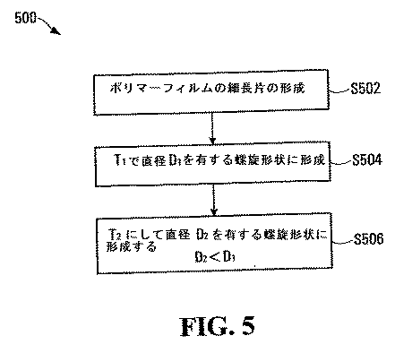
図12は、37℃での単層および2層に対する膨張速度のデータを示すグラフ図である。
FIG. 12 is a graph showing expansion rate data for a single layer and two layers at 37 ° C.
(実施例4:ステントの使用)
図13は、インサイチュで配置されるステントを表したものである。
(Example 4: Use of stent)
FIG. 13 shows a stent deployed in situ.
(実施例5:治療剤の送達)
ステント内の1つ以上のポリマーは、治療剤または薬剤を含浸させることが可能である。このような薬品としては、シロリムスおよびその誘導体(エベロリムスが挙げられる)ならびにパクリタキセルおよびその誘導体などの抗増殖剤、ヘパリンなどの抗血栓剤、アモキシシリンなどの抗生物質、パクリタキセルまたはドキソルビシンなどの化学療法剤、ガンシクロビルなどの抗ウイルス剤、ならびに利尿剤、ベラパミル、またはクロニジンなどの抗高血圧薬などが挙げられる。
Example 5: Delivery of therapeutic agent
One or more polymers in the stent can be impregnated with a therapeutic agent or drug. Such drugs include sirolimus and its derivatives (including everolimus) and antiproliferative agents such as paclitaxel and its derivatives, antithrombotic agents such as heparin, antibiotics such as amoxicillin, chemotherapeutic agents such as paclitaxel or doxorubicin, Antiviral agents such as ganciclovir, as well as antihypertensive agents such as diuretics, verapamil, or clonidine.
本願明細書において記述される螺旋形状が好適であるが、ポリマーのうちのいずれかのTgよりも高い温度でより小さな螺旋幅に伸張することが可能である、完全に管状のステントを提供することが可能である。これは、より高い力を必要とすることがある。螺旋幅は、その後機能ステントを提供するためにT3で膨張させることが可能である。 Although the helical shape described herein is preferred, it provides a fully tubular stent that is capable of stretching to a smaller helical width at temperatures above the Tg of any of the polymers. It is possible. This may require higher power. Helical width may be inflated with T 3 in order to provide subsequent functional stent.
(実施例6:二重層ステント)
生体安定性PET/ポリ酢酸ビニル(PVA)ステントに対して、PVA(外層)のTg=28℃、PET(内層)のTg=+60℃であり、ここでT1=37℃、T2=25℃であり、自己膨張可能なステントは、厚さ=0.18mmのPET層を有し、PVAの厚さ=0.07mm〜0.15mmである。
(Example 6: Double layer stent)
For biostable PET / polyvinyl acetate (PVA) stents, PVA (outer layer) T g = 28 ° C., PET (inner layer) T g = + 60 ° C., where T 1 = 37 ° C., T 2 = 25 ° C, a self-expandable stent has a PET layer with a thickness = 0.18 mm and a PVA thickness = 0.07 mm to 0.15 mm.
押出成形されたPETのシート(厚さ0.18mm)を、内層として用いる。PVAフィルムを、PVAをジクロロメタンに溶かした溶液を用いてこの上にキャストする。PVAのキャスト層の厚さは、約0.10mmである。この二重層フィルムを、37℃、1時間で螺旋幅3mmの螺旋状ステントに固め、25℃でより小さい螺旋幅1mmに固める。このステントはバルーンで膨張させることができ、次いで37℃で自己膨張できる。 An extruded PET sheet (thickness 0.18 mm) is used as the inner layer. A PVA film is cast onto it using a solution of PVA in dichloromethane. The thickness of the cast layer of PVA is about 0.10 mm. The bilayer film is consolidated into a spiral stent with a spiral width of 3 mm at 37 ° C. for 1 hour, and at 25 ° C. with a smaller spiral width of 1 mm. The stent can be expanded with a balloon and then self-expanded at 37 ° C.
ここで理解されるように、上述の実施形態は、多数の変更の影響を受けやすい。例えば、例示的なステントは、非螺旋形状に形成され得る。典型的なステントは、ほぼ円筒形状か、2種類の温度での2つの異なる形状か、またはある温度で不確定な形状を有するように形成することが可能である。同様に、例示的なステントは、第1と第2の層との間に第3、第4、さらにさらなる層を形成することが可能である。複数の層のそれぞれまたはいくつかは、上述のように治療剤を含有することが可能である。 As will be appreciated, the above-described embodiments are susceptible to numerous changes. For example, an exemplary stent can be formed in a non-helical shape. A typical stent can be formed to have a generally cylindrical shape, two different shapes at two temperatures, or an indeterminate shape at one temperature. Similarly, exemplary stents can form third, fourth, and further layers between the first and second layers. Each or some of the plurality of layers can contain a therapeutic agent as described above.
当業者によって理解されるように、本願明細書に記述される例示的な実施形態に対して多くの変更が可能である。本発明は、請求項に記載されるようにその範囲内にそのようなすべての変更を包含することをむしろ目的とする。また、本発明は、本明細書において個々にまたは集合的に参照されるか、あるいは示されるすべてのステップ、特徴、組成、および化合物、ならびに2つ以上のそのステップまたは特徴のいずれかのおよびすべての組み合わせを含む。 As will be appreciated by those skilled in the art, many modifications can be made to the exemplary embodiments described herein. The invention is rather intended to encompass all such modifications within its scope as described in the claims. The invention also includes all steps, features, compositions, and compounds referred to or shown individually or collectively herein, as well as any and all of two or more of the steps or features. Including a combination of
従って、本発明は、さらなる局面において、管腔の膨張を必要とする被験体に対して、少なくとも部分的にアモルファスである第1のポリマーおよび第1の治療剤を備えた第1の層、ならびに少なくとも部分的にアモルファスである第2のポリマーおよび、第2の治療剤を備える第2の層を含むステントであって、より低い温度T2で第1の形状を有するように形成され、より高い温度T1で第2の形状を有するように形成されるステントを、前記被験体の膨張させることが望まれる管腔の部位に導入し、それによって第1の治療剤および第2の治療剤を前記被験体に送達するステップと、前記ステントを第2の形状に変化させるステップとを含む、治療または予防の方法を提供する。 Accordingly, the present invention, in a further aspect, for a subject in need of lumen expansion, a first layer comprising a first polymer and a first therapeutic agent that are at least partially amorphous , and the second polymer and is at least partially amorphous, a stent comprising a second layer comprising a second therapeutic agent, is formed to have a first shape at a lower temperature T 2, greater A stent formed to have a second shape at temperature T 1 is introduced into the lumen site of the subject where it is desired to expand, thereby providing a first therapeutic agent and a second therapeutic agent . A method of treatment or prevention is provided that includes delivering to the subject and changing the stent to a second shape.
Claims (77)
少なくとも部分的にアモルファスであり、ガラス転移温度Tg1を有するポリマーを備えた第1の層と、少なくとも部分的にアモルファスであり、ガラス転移温度Tg2を有するポリマーを備えた第2の層とを有するポリマーフィルムの細長片を形成するステップと、
前記細長片を温度T1で第1の形状に形成するステップと、
を含み、T1=Tg1+X℃であり、Xが約−20〜約+120である方法。 A method for manufacturing a stent, comprising:
At least partially amorphous, a first layer comprising a polymer having a glass transition temperature T g1, at least partially amorphous, and a second layer which includes a polymer having a glass transition temperature T g2 Forming a strip of polymer film having:
Forming the strip into a first shape at a temperature T 1 ;
Wherein T 1 = T g1 + X ° C. and X is about −20 to about +120.
少なくとも部分的にアモルファスである第1のポリマーおよび第1の治療剤を備える第1の層を含むステントであって、より低い温度T2で第1の形状を有し、より高い温度T1で第2の形状を有するように形成されるステントを、前記被験体の膨張させることが望まれる管腔の部位に導入し、それによって第1の治療剤を前記被験体に送達するステップと、
前記ステントを前記第2の形状に変化させるステップと、
を含む方法。 A method of treating or preventing a subject in need of lumen expansion comprising:
A stent comprising a first layer comprising a first polymer and first therapeutic agent is at least partially amorphous, have a first shape at a lower temperature T 2, at higher temperatures T 1 Introducing a stent formed to have a second shape to a site of the lumen where it is desired to expand the subject, thereby delivering a first therapeutic agent to the subject;
Changing the stent to the second shape;
Including methods.
少なくとも部分的にアモルファスであり、ガラス転移温度Tg1を有する第1のポリマーを備えた第1の層と、少なくとも部分的にアモルファスであり、ガラス転移温度Tg2を有する第2のポリマーを備えた第2の層とを含むステントであって、より低温T2で第1の形状を有し、より高温T1で第2の形状を有するように形成され、形状転移温度T3に等しいかそれより高い温度で第1の形状から第2の形状に変化するように構成されるステントを、被験体の膨張させることが望まれる管腔の部位に導入するステップであって、前記ステントが前記第1の形状であるように、T3より低い温度で導入するステップと、
部分的に前記ステントをT3に等しいかそれより高い温度に平衡させることによって前記ステントを第2の形状に変化させるステップと、
を含む方法。 A method for the prevention or treatment of a subject in need of lumen expansion comprising:
A first layer comprising a first polymer that is at least partially amorphous and having a glass transition temperature T g1, and a second polymer that is at least partially amorphous and has a glass transition temperature T g2 . a stent comprising a second layer, more have a first shape at a low temperature T 2, it is formed so as to have a greater second shape at a high temperature T 1, or equal to the shape transition temperature T 3 Introducing a stent configured to change from a first shape to a second shape at a higher temperature at a lumen site where it is desired to expand the subject, wherein the stent comprises the first shape; Introducing at a temperature below T 3 so as to be in the shape of 1;
Changing the stent to a second shape by partially equilibrating the stent to a temperature equal to or higher than T 3 ;
Including methods.
少なくとも部分的にアモルファスであり、ガラス転移温度を有するポリマーに治療剤を添加するステップと、
ポリマーフィルムの細長片を前記ポリマーから形成するステップと、
前記細長片を温度T1で第1の形状に形成するステップであって、T1=Tg+X℃であり、Tgが前記ポリマーのガラス転移温度であり、Xが約−20〜約+120であるステップと、
前記細長片を温度T2で第2の形状に形成するステップであって、T2=T1−Y℃であり、Yが約5〜約80であるステップと、
を含む方法。 A method for manufacturing a stent, comprising:
Adding a therapeutic agent to a polymer that is at least partially amorphous and has a glass transition temperature;
Forming a strip of polymer film from the polymer;
Forming the strip into a first shape at a temperature T 1 , where T 1 = T g + X ° C., T g is the glass transition temperature of the polymer, and X is about −20 to about +120 And a step that is
Forming the strip into a second shape at a temperature T 2 , wherein T 2 = T 1 −Y ° C. and Y is about 5 to about 80;
Including methods.
Applications Claiming Priority (3)
| Application Number | Priority Date | Filing Date | Title |
|---|---|---|---|
| US47888703P | 2003-06-16 | 2003-06-16 | |
| US60/478,887 | 2003-06-16 | ||
| PCT/SG2004/000180 WO2004110315A1 (en) | 2003-06-16 | 2004-06-15 | Polymeric stent and method of manufacture |
Publications (1)
| Publication Number | Publication Date |
|---|---|
| JP2006527628A true JP2006527628A (en) | 2006-12-07 |
Family
ID=33551858
Family Applications (1)
| Application Number | Title | Priority Date | Filing Date |
|---|---|---|---|
| JP2006517067A Withdrawn JP2006527628A (en) | 2003-06-16 | 2004-06-15 | Stent of polymer material and manufacturing method |
Country Status (9)
| Country | Link |
|---|---|
| US (2) | US20050021131A1 (en) |
| EP (1) | EP1633280A4 (en) |
| JP (1) | JP2006527628A (en) |
| KR (1) | KR20060028695A (en) |
| CN (1) | CN100558321C (en) |
| AU (1) | AU2004246998A1 (en) |
| BR (1) | BRPI0411548A (en) |
| CA (1) | CA2529494A1 (en) |
| WO (1) | WO2004110315A1 (en) |
Cited By (2)
| Publication number | Priority date | Publication date | Assignee | Title |
|---|---|---|---|---|
| JP2013188588A (en) * | 2007-01-19 | 2013-09-26 | Elixir Medical Corp | Biodegradable endoprosthesis and method for its fabrication |
| JP2014529518A (en) * | 2011-08-16 | 2014-11-13 | シンセス・ゲーエムベーハーSynthes GmbH | Thermoplastic multilayer article |
Families Citing this family (73)
| Publication number | Priority date | Publication date | Assignee | Title |
|---|---|---|---|---|
| US8435550B2 (en) * | 2002-12-16 | 2013-05-07 | Abbot Cardiovascular Systems Inc. | Anti-proliferative and anti-inflammatory agent combination for treatment of vascular disorders with an implantable medical device |
| US7758881B2 (en) * | 2004-06-30 | 2010-07-20 | Advanced Cardiovascular Systems, Inc. | Anti-proliferative and anti-inflammatory agent combination for treatment of vascular disorders with an implantable medical device |
| EP1633280A4 (en) * | 2003-06-16 | 2011-03-16 | Univ Nanyang Tech | POLYMERIC STENT AND MANUFACTURING PROCESS |
| US8709469B2 (en) | 2004-06-30 | 2014-04-29 | Abbott Cardiovascular Systems Inc. | Anti-proliferative and anti-inflammatory agent combination for treatment of vascular disorders with an implantable medical device |
| US7931671B2 (en) | 2005-03-11 | 2011-04-26 | Radi Medical Systems Ab | Medical sealing device |
| US20070038290A1 (en) * | 2005-08-15 | 2007-02-15 | Bin Huang | Fiber reinforced composite stents |
| GB0517085D0 (en) | 2005-08-19 | 2005-09-28 | Angiomed Ag | Polymer prosthesis |
| DE102005056532A1 (en) * | 2005-11-28 | 2007-05-31 | Mnemoscience Gmbh | Method for removal of tubular tissue supports e.g. stents from human or animal hollow organs, involves heating of tissue support to temperature below transition temperature, till softening |
| US10029034B2 (en) * | 2005-12-15 | 2018-07-24 | CARDINAL HEALTH SWITZERLAND 515 GmbH | Drug-eluting articles with improved drug release profiles |
| US8945598B2 (en) * | 2005-12-29 | 2015-02-03 | Cordis Corporation | Low temperature drying methods for forming drug-containing polymeric compositions |
| US20070156230A1 (en) | 2006-01-04 | 2007-07-05 | Dugan Stephen R | Stents with radiopaque markers |
| US20070162110A1 (en) * | 2006-01-06 | 2007-07-12 | Vipul Bhupendra Dave | Bioabsorbable drug delivery devices |
| US20070160672A1 (en) * | 2006-01-06 | 2007-07-12 | Vipul Bhupendra Dave | Methods of making bioabsorbable drug delivery devices comprised of solvent cast films |
| JPWO2007083797A1 (en) * | 2006-01-23 | 2009-06-18 | テルモ株式会社 | Stent |
| US20070254012A1 (en) * | 2006-04-28 | 2007-11-01 | Ludwig Florian N | Controlled degradation and drug release in stents |
| CA2652871C (en) * | 2006-05-12 | 2016-01-12 | Cordis Corporation | Balloon expandable bioabsorbable drug eluting flexible stent |
| EP2020967B1 (en) * | 2006-05-12 | 2020-09-23 | Cardinal Health Switzerland 515 GmbH | Baloon expandable bioabsorbable drug eluting stent |
| EP2301487B1 (en) * | 2006-05-12 | 2014-09-17 | Covidien LP | Implant and delivery system with multiple marker interlocks |
| US20130325104A1 (en) | 2006-05-26 | 2013-12-05 | Abbott Cardiovascular Systems Inc. | Stents With Radiopaque Markers |
| EP2020956A2 (en) * | 2006-05-26 | 2009-02-11 | Nanyang Technological University | Implantable article, method of forming same and method for reducing thrombogenicity |
| US20070292470A1 (en) * | 2006-06-15 | 2007-12-20 | Medtronic Vascular, Inc. | Implantable Medical Devices and Methods for Making the Same |
| US8535372B1 (en) | 2006-06-16 | 2013-09-17 | Abbott Cardiovascular Systems Inc. | Bioabsorbable stent with prohealing layer |
| US8128688B2 (en) * | 2006-06-27 | 2012-03-06 | Abbott Cardiovascular Systems Inc. | Carbon coating on an implantable device |
| US7823263B2 (en) | 2006-07-11 | 2010-11-02 | Abbott Cardiovascular Systems Inc. | Method of removing stent islands from a stent |
| EP3047860B1 (en) | 2006-07-20 | 2024-08-28 | OrbusNeich Medical Pte. Ltd. | Bioabsorbable polymeric composition for a medical device |
| US8016879B2 (en) * | 2006-08-01 | 2011-09-13 | Abbott Cardiovascular Systems Inc. | Drug delivery after biodegradation of the stent scaffolding |
| US9173733B1 (en) | 2006-08-21 | 2015-11-03 | Abbott Cardiovascular Systems Inc. | Tracheobronchial implantable medical device and methods of use |
| US7959942B2 (en) | 2006-10-20 | 2011-06-14 | Orbusneich Medical, Inc. | Bioabsorbable medical device with coating |
| US8691321B2 (en) | 2006-10-20 | 2014-04-08 | Orbusneich Medical, Inc. | Bioabsorbable polymeric composition and medical device background |
| US8814930B2 (en) | 2007-01-19 | 2014-08-26 | Elixir Medical Corporation | Biodegradable endoprosthesis and methods for their fabrication |
| WO2008121702A2 (en) * | 2007-03-28 | 2008-10-09 | Boston Scientific Scimed, Inc. | Medical devices having bioerodable layers for the release of therapeutic agents |
| EP2152332A2 (en) * | 2007-06-12 | 2010-02-17 | Bioring SA | Biodegradable device for temporary angioplasty and repair of the vessels in general and of the coronary arteries in particular |
| US7901452B2 (en) * | 2007-06-27 | 2011-03-08 | Abbott Cardiovascular Systems Inc. | Method to fabricate a stent having selected morphology to reduce restenosis |
| US7955381B1 (en) | 2007-06-29 | 2011-06-07 | Advanced Cardiovascular Systems, Inc. | Polymer-bioceramic composite implantable medical device with different types of bioceramic particles |
| US20090035350A1 (en) | 2007-08-03 | 2009-02-05 | John Stankus | Polymers for implantable devices exhibiting shape-memory effects |
| US20090306120A1 (en) * | 2007-10-23 | 2009-12-10 | Florencia Lim | Terpolymers containing lactide and glycolide |
| US7972373B2 (en) * | 2007-12-19 | 2011-07-05 | Advanced Technologies And Regenerative Medicine, Llc | Balloon expandable bioabsorbable stent with a single stress concentration region interconnecting adjacent struts |
| US8057876B2 (en) * | 2008-02-25 | 2011-11-15 | Abbott Cardiovascular Systems Inc. | Bioabsorbable stent with layers having different degradation rates |
| US10898620B2 (en) | 2008-06-20 | 2021-01-26 | Razmodics Llc | Composite stent having multi-axial flexibility and method of manufacture thereof |
| US8206636B2 (en) | 2008-06-20 | 2012-06-26 | Amaranth Medical Pte. | Stent fabrication via tubular casting processes |
| US8206635B2 (en) | 2008-06-20 | 2012-06-26 | Amaranth Medical Pte. | Stent fabrication via tubular casting processes |
| US9265633B2 (en) * | 2009-05-20 | 2016-02-23 | 480 Biomedical, Inc. | Drug-eluting medical implants |
| WO2010138212A2 (en) * | 2009-05-29 | 2010-12-02 | Incube Labs, Llc | Biodegradable medical implants, polymer compositions and methods of use |
| EP2440162A1 (en) * | 2009-06-08 | 2012-04-18 | Boston Scientific Scimed, Inc. | Crosslinked bioabsorbable medical devices |
| WO2010145824A1 (en) * | 2009-06-17 | 2010-12-23 | Gilupi Gmbh | Detection device for in vivo and/or in vitro enrichment of sample material |
| US9993441B2 (en) | 2009-12-30 | 2018-06-12 | Surmodics, Inc. | Controlled release matrix barrier structure for subcutaneous medical devices |
| US20110159072A1 (en) * | 2009-12-30 | 2011-06-30 | Surmodics, Inc. | Controlled release matrix |
| US8568471B2 (en) | 2010-01-30 | 2013-10-29 | Abbott Cardiovascular Systems Inc. | Crush recoverable polymer scaffolds |
| US8808353B2 (en) | 2010-01-30 | 2014-08-19 | Abbott Cardiovascular Systems Inc. | Crush recoverable polymer scaffolds having a low crossing profile |
| KR101253284B1 (en) * | 2011-04-14 | 2013-04-10 | 한국과학기술연구원 | Method for modifying surface of stent |
| WO2012170591A2 (en) * | 2011-06-07 | 2012-12-13 | Qing Liu | Hybrid polymer stent fabricated by a non-laser cut fabrication method |
| US8726483B2 (en) | 2011-07-29 | 2014-05-20 | Abbott Cardiovascular Systems Inc. | Methods for uniform crimping and deployment of a polymer scaffold |
| CN105188603A (en) | 2013-05-03 | 2015-12-23 | 阿玛安斯医药私人公司 | Stent delivery and deployment methods and apparatus |
| CN105658181B (en) | 2013-08-16 | 2018-05-18 | 阿玛安斯医药私人公司 | For treating the bioabsorbable skeleton for intersecting lesion |
| US9675478B2 (en) * | 2014-06-11 | 2017-06-13 | Abbott Cardiovascular Systems Inc. | Solvent method for forming a polymer scaffolding |
| US9730819B2 (en) | 2014-08-15 | 2017-08-15 | Elixir Medical Corporation | Biodegradable endoprostheses and methods of their fabrication |
| US9480588B2 (en) | 2014-08-15 | 2016-11-01 | Elixir Medical Corporation | Biodegradable endoprostheses and methods of their fabrication |
| US9259339B1 (en) | 2014-08-15 | 2016-02-16 | Elixir Medical Corporation | Biodegradable endoprostheses and methods of their fabrication |
| US9855156B2 (en) | 2014-08-15 | 2018-01-02 | Elixir Medical Corporation | Biodegradable endoprostheses and methods of their fabrication |
| CN104473706B (en) * | 2014-12-09 | 2017-02-22 | 金华市人民医院 | Biodegradable composite type tubular urethral stent and preparation method |
| US9999527B2 (en) | 2015-02-11 | 2018-06-19 | Abbott Cardiovascular Systems Inc. | Scaffolds having radiopaque markers |
| US9700443B2 (en) | 2015-06-12 | 2017-07-11 | Abbott Cardiovascular Systems Inc. | Methods for attaching a radiopaque marker to a scaffold |
| US11622872B2 (en) | 2016-05-16 | 2023-04-11 | Elixir Medical Corporation | Uncaging stent |
| EP3457985B1 (en) | 2016-05-16 | 2021-02-17 | Elixir Medical Corporation | Uncaging stent |
| EP3528746A4 (en) * | 2016-10-18 | 2020-05-06 | University of Pittsburgh- Of the Commonwealth System of Higher Education | ADHESION ADJUSTMENT AT CONTACT DEVICE INTERFACE LEVEL: GEOMETRIC TOOLS FOR MINIMIZING SURFACE FOULING |
| JP7091335B2 (en) | 2016-12-09 | 2022-06-27 | ゼンフロー, インコーポレイテッド | Systems, devices, and methods for the accurate deployment of implants within the urethral prostate |
| CN110087720B (en) | 2017-03-30 | 2021-03-30 | 泰尔茂株式会社 | Medical device having hydrophilic member and hydrophobic member laminated thereon |
| US12364792B2 (en) * | 2018-01-08 | 2025-07-22 | Foundry Therapeutics, Inc. | Devices, systems, and methods for treating intraluminal cancer via controlled delivery of therapeutic agents |
| CN109172075A (en) * | 2018-08-20 | 2019-01-11 | 南京永明医疗器械有限公司 | A kind of degradable intravascular stent and preparation method thereof containing elastic layer |
| CN110960734B (en) * | 2019-10-30 | 2022-04-22 | 上海七木医疗器械有限公司 | Preparation method of degradable spiral stent |
| WO2021101951A1 (en) | 2019-11-19 | 2021-05-27 | Zenflow, Inc. | Systems, devices, and methods for the accurate deployment and imaging of an implant in the prostatic urethra |
| CN111035809B (en) * | 2019-12-04 | 2021-06-01 | 中山大学 | Double-layer composite nanofiber membrane with three-dimensional deformation structure and preparation method and application thereof |
| CN112807059A (en) * | 2021-02-26 | 2021-05-18 | 上海微创医疗器械(集团)有限公司 | Self-expandable medical implant and medical device |
Family Cites Families (59)
| Publication number | Priority date | Publication date | Assignee | Title |
|---|---|---|---|---|
| SE445884B (en) * | 1982-04-30 | 1986-07-28 | Medinvent Sa | DEVICE FOR IMPLANTATION OF A RODFORM PROTECTION |
| SE453258B (en) * | 1986-04-21 | 1988-01-25 | Medinvent Sa | ELASTIC, SELF-EXPANDING PROTEST AND PROCEDURE FOR ITS MANUFACTURING |
| JP2561853B2 (en) * | 1988-01-28 | 1996-12-11 | 株式会社ジェイ・エム・エス | Shaped memory molded article and method of using the same |
| US5139480A (en) * | 1990-08-22 | 1992-08-18 | Biotech Laboratories, Inc. | Necking stents |
| US6248129B1 (en) * | 1990-09-14 | 2001-06-19 | Quanam Medical Corporation | Expandable polymeric stent with memory and delivery apparatus and method |
| US5258020A (en) * | 1990-09-14 | 1993-11-02 | Michael Froix | Method of using expandable polymeric stent with memory |
| US5163952A (en) * | 1990-09-14 | 1992-11-17 | Michael Froix | Expandable polymeric stent with memory and delivery apparatus and method |
| DE9390115U1 (en) * | 1992-05-08 | 1994-12-22 | Schneider Usa Inc | Esophageal stent and delivery instrument |
| US5443458A (en) * | 1992-12-22 | 1995-08-22 | Advanced Cardiovascular Systems, Inc. | Multilayered biodegradable stent and method of manufacture |
| US5630840A (en) * | 1993-01-19 | 1997-05-20 | Schneider (Usa) Inc | Clad composite stent |
| EP0696185B1 (en) * | 1993-04-28 | 1998-08-12 | Focal, Inc. | Apparatus, product and use related to intraluminal photothermoforming |
| US5716410A (en) * | 1993-04-30 | 1998-02-10 | Scimed Life Systems, Inc. | Temporary stent and method of use |
| US5735892A (en) * | 1993-08-18 | 1998-04-07 | W. L. Gore & Associates, Inc. | Intraluminal stent graft |
| US5954744A (en) * | 1995-06-06 | 1999-09-21 | Quanam Medical Corporation | Intravascular stent |
| US5824332A (en) * | 1995-10-05 | 1998-10-20 | Jannetta; Peter J. | Method and apparatus for treatment of neurogenic diabetes mellitus, and other conditions |
| US6007573A (en) * | 1996-09-18 | 1999-12-28 | Microtherapeutics, Inc. | Intracranial stent and method of use |
| US6117168A (en) * | 1996-12-31 | 2000-09-12 | Scimed Life Systems, Inc. | Multilayer liquid absorption and deformation devices |
| US6245103B1 (en) * | 1997-08-01 | 2001-06-12 | Schneider (Usa) Inc | Bioabsorbable self-expanding stent |
| US5962007A (en) * | 1997-12-19 | 1999-10-05 | Indigo Medical, Inc. | Use of a multi-component coil medical construct |
| BR9908339A (en) * | 1998-02-23 | 2001-10-02 | Mnemoscience Gmbh | Method for the manufacture of an article with format memory, polymer composition with format memory and respective method of forming the field |
| DE69917224T2 (en) * | 1998-02-23 | 2004-09-09 | Massachusetts Institute Of Technology, Cambridge | BIODEGRADABLE POLYMERS WITH SHAPED MEMORY |
| US6500204B1 (en) * | 1998-09-08 | 2002-12-31 | Kabushikikaisha Igaki Iryo Sekkei | Stent for vessels |
| US6547814B2 (en) * | 1998-09-30 | 2003-04-15 | Impra, Inc. | Selective adherence of stent-graft coverings |
| CA2254002A1 (en) * | 1998-11-12 | 2000-05-12 | Takiron Co., Ltd. | Shape-memory, biodegradable and absorbable material |
| US6503270B1 (en) * | 1998-12-03 | 2003-01-07 | Medinol Ltd. | Serpentine coiled ladder stent |
| US6368346B1 (en) * | 1999-06-03 | 2002-04-09 | American Medical Systems, Inc. | Bioresorbable stent |
| US6364904B1 (en) * | 1999-07-02 | 2002-04-02 | Scimed Life Systems, Inc. | Helically formed stent/graft assembly |
| US6790228B2 (en) * | 1999-12-23 | 2004-09-14 | Advanced Cardiovascular Systems, Inc. | Coating for implantable devices and a method of forming the same |
| US6338739B1 (en) * | 1999-12-22 | 2002-01-15 | Ethicon, Inc. | Biodegradable stent |
| ES2367628T3 (en) * | 2000-03-13 | 2011-11-07 | Kabushiki Kaisha Kyoto Iryo Sekkei | THREAD RODS FOR VASCULAR STENTS AND VASCULAR STENTS WITH THE USE OF THE SAME. |
| IL137090A (en) * | 2000-06-29 | 2010-04-15 | Pentech Medical Devices Ltd | Polymeric stent |
| US20020077693A1 (en) * | 2000-12-19 | 2002-06-20 | Barclay Bruce J. | Covered, coiled drug delivery stent and method |
| JP2004535836A (en) * | 2000-11-14 | 2004-12-02 | エヌ.ブイ.アール. ラブズ インコーポレイテッド | Crosslinked hyaluronic acid-laminin gels and their use in cell culture and medical implants |
| DE20119322U1 (en) * | 2000-11-21 | 2002-02-21 | Schering Ag, 13353 Berlin | Tubular vascular grafts (stents) |
| CA2710960A1 (en) * | 2000-11-22 | 2002-05-30 | Bard Peripheral Vascular, Inc. | Method for producing a single layer high density expanded polytetrafluoroethylene tube |
| EP1386310A1 (en) * | 2001-05-11 | 2004-02-04 | Matsushita Electric Industrial Co., Ltd. | Device to encode, decode and broadcast audio signal with reduced size spectral information |
| US20030069629A1 (en) * | 2001-06-01 | 2003-04-10 | Jadhav Balkrishna S. | Bioresorbable medical devices |
| US7008446B1 (en) * | 2001-08-17 | 2006-03-07 | James Peter Amis | Thermally pliable and carbon fiber stents |
| US7572287B2 (en) * | 2001-10-25 | 2009-08-11 | Boston Scientific Scimed, Inc. | Balloon expandable polymer stent with reduced elastic recoil |
| AU2003222660A1 (en) * | 2002-04-18 | 2003-11-03 | Robert S. Langer | Biodegradable shape memory polymeric sutures |
| DE10219014A1 (en) * | 2002-04-27 | 2003-11-13 | Ruesch Willy Gmbh | Self-expanding stent for reinforcing and/or keeping open a hollow organ comprise two elastic tubular layers which bracket and positionally fix at least one helical filament |
| US20030216804A1 (en) * | 2002-05-14 | 2003-11-20 | Debeer Nicholas C. | Shape memory polymer stent |
| US20040034405A1 (en) * | 2002-07-26 | 2004-02-19 | Dickson Andrew M. | Axially expanding polymer stent |
| EP1543860B1 (en) * | 2002-09-25 | 2015-02-18 | Kabushikikaisha Igaki Iryo Sekkei | Thread for vascular stent and vascular stent using the thread |
| US6770729B2 (en) * | 2002-09-30 | 2004-08-03 | Medtronic Minimed, Inc. | Polymer compositions containing bioactive agents and methods for their use |
| CA2501643C (en) * | 2002-10-11 | 2013-12-31 | University Of Connecticut | Shape memory polymers based on semicrystalline thermoplastic polyurethanes bearing nanostructured hard segments |
| US6932930B2 (en) * | 2003-03-10 | 2005-08-23 | Synecor, Llc | Intraluminal prostheses having polymeric material with selectively modified crystallinity and methods of making same |
| US20050010170A1 (en) * | 2004-02-11 | 2005-01-13 | Shanley John F | Implantable medical device with beneficial agent concentration gradient |
| EP1633280A4 (en) * | 2003-06-16 | 2011-03-16 | Univ Nanyang Tech | POLYMERIC STENT AND MANUFACTURING PROCESS |
| US20050100577A1 (en) * | 2003-11-10 | 2005-05-12 | Parker Theodore L. | Expandable medical device with beneficial agent matrix formed by a multi solvent system |
| US8192752B2 (en) * | 2003-11-21 | 2012-06-05 | Advanced Cardiovascular Systems, Inc. | Coatings for implantable devices including biologically erodable polyesters and methods for fabricating the same |
| MY146259A (en) * | 2004-03-24 | 2012-07-31 | Polynovo Biomaterials Pty Ltd | Boidegradable polyurethane and polyurethane ureas |
| US7731740B2 (en) * | 2004-04-02 | 2010-06-08 | Arterial Remodelling Technologies, Inc. | Polymer-based stent assembly |
| US20060025852A1 (en) * | 2004-08-02 | 2006-02-02 | Armstrong Joseph R | Bioabsorbable self-expanding endolumenal devices |
| CA2577018C (en) * | 2004-08-13 | 2012-03-27 | Reva Medical, Inc. | Inherently radiopaque bioresorbable polymers for multiple uses |
| US8119153B2 (en) * | 2004-08-26 | 2012-02-21 | Boston Scientific Scimed, Inc. | Stents with drug eluting coatings |
| JP4149980B2 (en) * | 2004-09-17 | 2008-09-17 | シャープ株式会社 | Manufacturing method of semiconductor manufacturing equipment |
| US7901451B2 (en) * | 2004-09-24 | 2011-03-08 | Biosensors International Group, Ltd. | Drug-delivery endovascular stent and method for treating restenosis |
| US20070060895A1 (en) * | 2005-08-24 | 2007-03-15 | Sibbitt Wilmer L Jr | Vascular closure methods and apparatuses |
-
2004
- 2004-06-15 EP EP04736895A patent/EP1633280A4/en not_active Withdrawn
- 2004-06-15 WO PCT/SG2004/000180 patent/WO2004110315A1/en not_active Ceased
- 2004-06-15 CN CNB2004800169315A patent/CN100558321C/en not_active Expired - Lifetime
- 2004-06-15 CA CA002529494A patent/CA2529494A1/en not_active Abandoned
- 2004-06-15 KR KR1020057024110A patent/KR20060028695A/en not_active Withdrawn
- 2004-06-15 BR BRPI0411548-1A patent/BRPI0411548A/en not_active Application Discontinuation
- 2004-06-15 US US10/867,617 patent/US20050021131A1/en not_active Abandoned
- 2004-06-15 JP JP2006517067A patent/JP2006527628A/en not_active Withdrawn
- 2004-06-15 AU AU2004246998A patent/AU2004246998A1/en not_active Abandoned
-
2008
- 2008-04-24 US US12/109,317 patent/US20080208321A1/en not_active Abandoned
Cited By (2)
| Publication number | Priority date | Publication date | Assignee | Title |
|---|---|---|---|---|
| JP2013188588A (en) * | 2007-01-19 | 2013-09-26 | Elixir Medical Corp | Biodegradable endoprosthesis and method for its fabrication |
| JP2014529518A (en) * | 2011-08-16 | 2014-11-13 | シンセス・ゲーエムベーハーSynthes GmbH | Thermoplastic multilayer article |
Also Published As
| Publication number | Publication date |
|---|---|
| EP1633280A4 (en) | 2011-03-16 |
| US20050021131A1 (en) | 2005-01-27 |
| KR20060028695A (en) | 2006-03-31 |
| WO2004110315A1 (en) | 2004-12-23 |
| CA2529494A1 (en) | 2004-12-23 |
| CN1812754A (en) | 2006-08-02 |
| AU2004246998A1 (en) | 2004-12-23 |
| BRPI0411548A (en) | 2006-08-01 |
| CN100558321C (en) | 2009-11-11 |
| US20080208321A1 (en) | 2008-08-28 |
| EP1633280A1 (en) | 2006-03-15 |
Similar Documents
| Publication | Publication Date | Title |
|---|---|---|
| JP2006527628A (en) | Stent of polymer material and manufacturing method | |
| US8303650B2 (en) | Biodegradable self-expanding drug-eluting prosthesis | |
| US8317857B2 (en) | Biodegradable self-expanding prosthesis | |
| EP3043753B1 (en) | Braided scaffolds | |
| CN101631518B (en) | Temporary intraluminal stent and methods of making and using the same | |
| EP3384878B1 (en) | A method of making a self-expanding implant | |
| AU781199B2 (en) | Bioresorbable stent | |
| US9561611B2 (en) | Multilayer bioabsorbable scaffolds and methods of fabricating | |
| US20130184809A1 (en) | Disintegrating stent and method of making same | |
| EP3765106B1 (en) | Bioabsorbable flow diverting scaffold | |
| WO2009059224A2 (en) | Composite stent comprising bioremovable polymer and bioremovale ceramics | |
| CN106361477A (en) | Endoluminal device | |
| US20080167710A1 (en) | Medical Device Having Regions With Various Agents Dispersed Therein and a Method for Making the Same | |
| US20230414385A1 (en) | Implantable scaffolds having biodegradable components and methods of manufacturing and use thereof |
Legal Events
| Date | Code | Title | Description |
|---|---|---|---|
| A300 | Application deemed to be withdrawn because no request for examination was validly filed |
Free format text: JAPANESE INTERMEDIATE CODE: A300 Effective date: 20070904 |