JP2004522940A - Methods for detecting pathological alterations in APP proteins and uses thereof - Google Patents
Methods for detecting pathological alterations in APP proteins and uses thereof Download PDFInfo
- Publication number
- JP2004522940A JP2004522940A JP2002539821A JP2002539821A JP2004522940A JP 2004522940 A JP2004522940 A JP 2004522940A JP 2002539821 A JP2002539821 A JP 2002539821A JP 2002539821 A JP2002539821 A JP 2002539821A JP 2004522940 A JP2004522940 A JP 2004522940A
- Authority
- JP
- Japan
- Prior art keywords
- app
- fragments
- alzheimer
- kda
- terminal
- Prior art date
- Legal status (The legal status is an assumption and is not a legal conclusion. Google has not performed a legal analysis and makes no representation as to the accuracy of the status listed.)
- Granted
Links
- 238000000034 method Methods 0.000 title claims abstract description 39
- 108090000623 proteins and genes Proteins 0.000 title claims abstract description 39
- 102000004169 proteins and genes Human genes 0.000 title claims abstract description 22
- 230000009543 pathological alteration Effects 0.000 title claims abstract description 5
- 239000012634 fragment Substances 0.000 claims abstract description 126
- 208000024827 Alzheimer disease Diseases 0.000 claims abstract description 46
- 238000004458 analytical method Methods 0.000 claims abstract description 28
- 239000003550 marker Substances 0.000 claims abstract description 13
- 230000000626 neurodegenerative effect Effects 0.000 claims abstract description 10
- 230000007170 pathology Effects 0.000 claims abstract description 8
- 230000002503 metabolic effect Effects 0.000 claims abstract description 5
- 230000001925 catabolic effect Effects 0.000 claims abstract description 4
- 238000003745 diagnosis Methods 0.000 claims abstract description 4
- 230000008506 pathogenesis Effects 0.000 claims abstract description 4
- 210000001519 tissue Anatomy 0.000 claims description 77
- 210000004027 cell Anatomy 0.000 claims description 36
- 230000035772 mutation Effects 0.000 claims description 29
- 230000001575 pathological effect Effects 0.000 claims description 23
- 230000004048 modification Effects 0.000 claims description 14
- 238000012986 modification Methods 0.000 claims description 14
- 230000026731 phosphorylation Effects 0.000 claims description 13
- 238000006366 phosphorylation reaction Methods 0.000 claims description 13
- 238000001514 detection method Methods 0.000 claims description 12
- 241000699670 Mus sp. Species 0.000 claims description 11
- 230000014509 gene expression Effects 0.000 claims description 11
- 230000004064 dysfunction Effects 0.000 claims description 9
- 238000001262 western blot Methods 0.000 claims description 9
- 210000002569 neuron Anatomy 0.000 claims description 8
- 230000004481 post-translational protein modification Effects 0.000 claims description 8
- 230000001537 neural effect Effects 0.000 claims description 7
- 238000001155 isoelectric focusing Methods 0.000 claims description 6
- 238000010171 animal model Methods 0.000 claims description 5
- 230000011987 methylation Effects 0.000 claims description 5
- 238000007069 methylation reaction Methods 0.000 claims description 5
- 230000021736 acetylation Effects 0.000 claims description 4
- 238000006640 acetylation reaction Methods 0.000 claims description 4
- 238000003556 assay Methods 0.000 claims description 4
- 210000004369 blood Anatomy 0.000 claims description 4
- 239000008280 blood Substances 0.000 claims description 4
- 238000012216 screening Methods 0.000 claims description 4
- 230000000984 immunochemical effect Effects 0.000 claims description 3
- 239000000463 material Substances 0.000 claims description 3
- 238000009007 Diagnostic Kit Methods 0.000 claims description 2
- 238000002965 ELISA Methods 0.000 claims description 2
- 230000002159 abnormal effect Effects 0.000 claims description 2
- 210000004899 c-terminal region Anatomy 0.000 claims description 2
- 210000001175 cerebrospinal fluid Anatomy 0.000 claims description 2
- 241000282412 Homo Species 0.000 claims 2
- 102000001708 Protein Isoforms Human genes 0.000 claims 2
- 108010029485 Protein Isoforms Proteins 0.000 claims 2
- 210000001124 body fluid Anatomy 0.000 claims 1
- 239000010839 body fluid Substances 0.000 claims 1
- 239000003153 chemical reaction reagent Substances 0.000 claims 1
- 210000000944 nerve tissue Anatomy 0.000 claims 1
- 230000003412 degenerative effect Effects 0.000 abstract description 3
- 230000002490 cerebral effect Effects 0.000 description 51
- 238000011830 transgenic mouse model Methods 0.000 description 22
- 239000000872 buffer Substances 0.000 description 20
- 241000699660 Mus musculus Species 0.000 description 17
- 238000001962 electrophoresis Methods 0.000 description 17
- 108090000765 processed proteins & peptides Proteins 0.000 description 16
- 239000000523 sample Substances 0.000 description 16
- 108010026424 tau Proteins Proteins 0.000 description 13
- PEDCQBHIVMGVHV-UHFFFAOYSA-N Glycerine Chemical compound OCC(O)CO PEDCQBHIVMGVHV-UHFFFAOYSA-N 0.000 description 12
- 102000002659 Amyloid Precursor Protein Secretases Human genes 0.000 description 11
- 108010043324 Amyloid Precursor Protein Secretases Proteins 0.000 description 11
- 210000000265 leukocyte Anatomy 0.000 description 11
- 102000013498 tau Proteins Human genes 0.000 description 10
- XSQUKJJJFZCRTK-UHFFFAOYSA-N Urea Chemical compound NC(N)=O XSQUKJJJFZCRTK-UHFFFAOYSA-N 0.000 description 9
- 238000003119 immunoblot Methods 0.000 description 9
- 230000004770 neurodegeneration Effects 0.000 description 8
- 239000008188 pellet Substances 0.000 description 8
- 239000000243 solution Substances 0.000 description 8
- QKNYBSVHEMOAJP-UHFFFAOYSA-N 2-amino-2-(hydroxymethyl)propane-1,3-diol;hydron;chloride Chemical compound Cl.OCC(N)(CO)CO QKNYBSVHEMOAJP-UHFFFAOYSA-N 0.000 description 7
- 101100268553 Homo sapiens APP gene Proteins 0.000 description 7
- 230000004075 alteration Effects 0.000 description 7
- 238000005119 centrifugation Methods 0.000 description 7
- 239000012528 membrane Substances 0.000 description 7
- 230000001225 therapeutic effect Effects 0.000 description 7
- FAPWRFPIFSIZLT-UHFFFAOYSA-M Sodium chloride Chemical compound [Na+].[Cl-] FAPWRFPIFSIZLT-UHFFFAOYSA-M 0.000 description 6
- DBMJMQXJHONAFJ-UHFFFAOYSA-M Sodium laurylsulphate Chemical compound [Na+].CCCCCCCCCCCCOS([O-])(=O)=O DBMJMQXJHONAFJ-UHFFFAOYSA-M 0.000 description 6
- 229920004890 Triton X-100 Polymers 0.000 description 6
- 238000003776 cleavage reaction Methods 0.000 description 6
- 230000007850 degeneration Effects 0.000 description 6
- 238000006206 glycosylation reaction Methods 0.000 description 6
- 239000012139 lysis buffer Substances 0.000 description 6
- 238000004519 manufacturing process Methods 0.000 description 6
- 230000008569 process Effects 0.000 description 6
- 230000007017 scission Effects 0.000 description 6
- UMGDCJDMYOKAJW-UHFFFAOYSA-N thiourea Chemical compound NC(N)=S UMGDCJDMYOKAJW-UHFFFAOYSA-N 0.000 description 6
- CFBILACNYSPRPM-UHFFFAOYSA-N 2-amino-2-(hydroxymethyl)propane-1,3-diol;2-[[1,3-dihydroxy-2-(hydroxymethyl)propan-2-yl]amino]acetic acid Chemical compound OCC(N)(CO)CO.OCC(CO)(CO)NCC(O)=O CFBILACNYSPRPM-UHFFFAOYSA-N 0.000 description 5
- 241000699666 Mus <mouse, genus> Species 0.000 description 5
- 230000015556 catabolic process Effects 0.000 description 5
- 210000004978 chinese hamster ovary cell Anatomy 0.000 description 5
- 238000011161 development Methods 0.000 description 5
- 230000018109 developmental process Effects 0.000 description 5
- 201000010099 disease Diseases 0.000 description 5
- 208000037265 diseases, disorders, signs and symptoms Diseases 0.000 description 5
- 239000003814 drug Substances 0.000 description 5
- 230000013595 glycosylation Effects 0.000 description 5
- 238000001114 immunoprecipitation Methods 0.000 description 5
- 230000004060 metabolic process Effects 0.000 description 5
- 235000020183 skimmed milk Nutrition 0.000 description 5
- KCXVZYZYPLLWCC-UHFFFAOYSA-N EDTA Chemical compound OC(=O)CN(CC(O)=O)CCN(CC(O)=O)CC(O)=O KCXVZYZYPLLWCC-UHFFFAOYSA-N 0.000 description 4
- 241000700159 Rattus Species 0.000 description 4
- 239000007983 Tris buffer Substances 0.000 description 4
- 239000013504 Triton X-100 Substances 0.000 description 4
- 230000005856 abnormality Effects 0.000 description 4
- 238000009825 accumulation Methods 0.000 description 4
- UDSAIICHUKSCKT-UHFFFAOYSA-N bromophenol blue Chemical compound C1=C(Br)C(O)=C(Br)C=C1C1(C=2C=C(Br)C(O)=C(Br)C=2)C2=CC=CC=C2S(=O)(=O)O1 UDSAIICHUKSCKT-UHFFFAOYSA-N 0.000 description 4
- 229940079593 drug Drugs 0.000 description 4
- 230000002163 immunogen Effects 0.000 description 4
- 238000011534 incubation Methods 0.000 description 4
- 210000004698 lymphocyte Anatomy 0.000 description 4
- 239000000047 product Substances 0.000 description 4
- 238000000926 separation method Methods 0.000 description 4
- 239000006228 supernatant Substances 0.000 description 4
- LENZDBCJOHFCAS-UHFFFAOYSA-N tris Chemical compound OCC(N)(CO)CO LENZDBCJOHFCAS-UHFFFAOYSA-N 0.000 description 4
- 238000000539 two dimensional gel electrophoresis Methods 0.000 description 4
- HRPVXLWXLXDGHG-UHFFFAOYSA-N Acrylamide Chemical compound NC(=O)C=C HRPVXLWXLXDGHG-UHFFFAOYSA-N 0.000 description 3
- 229920000936 Agarose Polymers 0.000 description 3
- 208000037259 Amyloid Plaque Diseases 0.000 description 3
- 101000823051 Homo sapiens Amyloid-beta precursor protein Proteins 0.000 description 3
- 241001529936 Murinae Species 0.000 description 3
- 206010029260 Neuroblastoma Diseases 0.000 description 3
- 208000037273 Pathologic Processes Diseases 0.000 description 3
- 239000002671 adjuvant Substances 0.000 description 3
- 239000000538 analytical sample Substances 0.000 description 3
- 230000008901 benefit Effects 0.000 description 3
- 238000001574 biopsy Methods 0.000 description 3
- 210000004556 brain Anatomy 0.000 description 3
- 239000004202 carbamide Substances 0.000 description 3
- 230000008859 change Effects 0.000 description 3
- 230000009089 cytolysis Effects 0.000 description 3
- 230000034994 death Effects 0.000 description 3
- 230000008034 disappearance Effects 0.000 description 3
- 230000000694 effects Effects 0.000 description 3
- 238000002474 experimental method Methods 0.000 description 3
- 102000046783 human APP Human genes 0.000 description 3
- 230000003053 immunization Effects 0.000 description 3
- 238000002649 immunization Methods 0.000 description 3
- 238000002347 injection Methods 0.000 description 3
- 239000007924 injection Substances 0.000 description 3
- 239000011159 matrix material Substances 0.000 description 3
- 239000000203 mixture Substances 0.000 description 3
- 230000009054 pathological process Effects 0.000 description 3
- USRGIUJOYOXOQJ-GBXIJSLDSA-N phosphothreonine Chemical compound OP(=O)(O)O[C@H](C)[C@H](N)C(O)=O USRGIUJOYOXOQJ-GBXIJSLDSA-N 0.000 description 3
- 230000009467 reduction Effects 0.000 description 3
- 210000002966 serum Anatomy 0.000 description 3
- 239000011780 sodium chloride Substances 0.000 description 3
- 238000003756 stirring Methods 0.000 description 3
- 238000012360 testing method Methods 0.000 description 3
- 238000012546 transfer Methods 0.000 description 3
- DGVVWUTYPXICAM-UHFFFAOYSA-N β‐Mercaptoethanol Chemical compound OCCS DGVVWUTYPXICAM-UHFFFAOYSA-N 0.000 description 3
- 108010090849 Amyloid beta-Peptides Proteins 0.000 description 2
- 102000013455 Amyloid beta-Peptides Human genes 0.000 description 2
- 206010012289 Dementia Diseases 0.000 description 2
- 102000004190 Enzymes Human genes 0.000 description 2
- 108090000790 Enzymes Proteins 0.000 description 2
- 241001465754 Metazoa Species 0.000 description 2
- 238000011887 Necropsy Methods 0.000 description 2
- 101150089079 PS1 gene Proteins 0.000 description 2
- 108010022181 Phosphopyruvate Hydratase Proteins 0.000 description 2
- 229920001213 Polysorbate 20 Polymers 0.000 description 2
- 206010070834 Sensitisation Diseases 0.000 description 2
- AYFVYJQAPQTCCC-UHFFFAOYSA-N Threonine Natural products CC(O)C(N)C(O)=O AYFVYJQAPQTCCC-UHFFFAOYSA-N 0.000 description 2
- 239000004473 Threonine Substances 0.000 description 2
- 230000032683 aging Effects 0.000 description 2
- DZHSAHHDTRWUTF-SIQRNXPUSA-N amyloid-beta polypeptide 42 Chemical compound C([C@@H](C(=O)N[C@@H](C)C(=O)N[C@@H](CCC(O)=O)C(=O)N[C@@H](CC(O)=O)C(=O)N[C@H](C(=O)NCC(=O)N[C@@H](CO)C(=O)N[C@@H](CC(N)=O)C(=O)N[C@@H](CCCCN)C(=O)NCC(=O)N[C@@H](C)C(=O)N[C@H](C(=O)N[C@@H]([C@@H](C)CC)C(=O)NCC(=O)N[C@@H](CC(C)C)C(=O)N[C@@H](CCSC)C(=O)N[C@@H](C(C)C)C(=O)NCC(=O)NCC(=O)N[C@@H](C(C)C)C(=O)N[C@@H](C(C)C)C(=O)N[C@@H]([C@@H](C)CC)C(=O)N[C@@H](C)C(O)=O)[C@@H](C)CC)C(C)C)NC(=O)[C@H](CC=1C=CC=CC=1)NC(=O)[C@@H](NC(=O)[C@H](CC(C)C)NC(=O)[C@H](CCCCN)NC(=O)[C@H](CCC(N)=O)NC(=O)[C@H](CC=1N=CNC=1)NC(=O)[C@H](CC=1N=CNC=1)NC(=O)[C@@H](NC(=O)[C@H](CCC(O)=O)NC(=O)[C@H](CC=1C=CC(O)=CC=1)NC(=O)CNC(=O)[C@H](CO)NC(=O)[C@H](CC(O)=O)NC(=O)[C@H](CC=1N=CNC=1)NC(=O)[C@H](CCCNC(N)=N)NC(=O)[C@H](CC=1C=CC=CC=1)NC(=O)[C@H](CCC(O)=O)NC(=O)[C@H](C)NC(=O)[C@@H](N)CC(O)=O)C(C)C)C(C)C)C1=CC=CC=C1 DZHSAHHDTRWUTF-SIQRNXPUSA-N 0.000 description 2
- 230000003941 amyloidogenesis Effects 0.000 description 2
- 101150031224 app gene Proteins 0.000 description 2
- 239000011324 bead Substances 0.000 description 2
- 239000012148 binding buffer Substances 0.000 description 2
- 230000033228 biological regulation Effects 0.000 description 2
- 210000000349 chromosome Anatomy 0.000 description 2
- 230000030609 dephosphorylation Effects 0.000 description 2
- 238000006209 dephosphorylation reaction Methods 0.000 description 2
- 239000003599 detergent Substances 0.000 description 2
- 238000010586 diagram Methods 0.000 description 2
- 230000004069 differentiation Effects 0.000 description 2
- 238000010790 dilution Methods 0.000 description 2
- 239000012895 dilution Substances 0.000 description 2
- 238000009826 distribution Methods 0.000 description 2
- VHJLVAABSRFDPM-QWWZWVQMSA-N dithiothreitol Chemical compound SC[C@@H](O)[C@H](O)CS VHJLVAABSRFDPM-QWWZWVQMSA-N 0.000 description 2
- 210000002919 epithelial cell Anatomy 0.000 description 2
- 208000015756 familial Alzheimer disease Diseases 0.000 description 2
- 230000003902 lesion Effects 0.000 description 2
- 238000012544 monitoring process Methods 0.000 description 2
- 208000015122 neurodegenerative disease Diseases 0.000 description 2
- 210000004498 neuroglial cell Anatomy 0.000 description 2
- 230000000144 pharmacologic effect Effects 0.000 description 2
- 239000000256 polyoxyethylene sorbitan monolaurate Substances 0.000 description 2
- 235000010486 polyoxyethylene sorbitan monolaurate Nutrition 0.000 description 2
- 229920000136 polysorbate Polymers 0.000 description 2
- 238000002360 preparation method Methods 0.000 description 2
- 102000004196 processed proteins & peptides Human genes 0.000 description 2
- 238000011002 quantification Methods 0.000 description 2
- 230000008313 sensitization Effects 0.000 description 2
- 230000002123 temporal effect Effects 0.000 description 2
- 238000013518 transcription Methods 0.000 description 2
- 230000035897 transcription Effects 0.000 description 2
- 230000009261 transgenic effect Effects 0.000 description 2
- MTCFGRXMJLQNBG-REOHCLBHSA-N (2S)-2-Amino-3-hydroxypropansäure Chemical compound OC[C@H](N)C(O)=O MTCFGRXMJLQNBG-REOHCLBHSA-N 0.000 description 1
- UMCMPZBLKLEWAF-BCTGSCMUSA-N 3-[(3-cholamidopropyl)dimethylammonio]propane-1-sulfonate Chemical compound C([C@H]1C[C@H]2O)[C@H](O)CC[C@]1(C)[C@@H]1[C@@H]2[C@@H]2CC[C@H]([C@@H](CCC(=O)NCCC[N+](C)(C)CCCS([O-])(=O)=O)C)[C@@]2(C)[C@@H](O)C1 UMCMPZBLKLEWAF-BCTGSCMUSA-N 0.000 description 1
- LHGMHYDJNXEEFG-UHFFFAOYSA-N 4-[4-(dimethylamino)phenyl]iminocyclohexa-2,5-dien-1-one Chemical compound C1=CC(N(C)C)=CC=C1N=C1C=CC(=O)C=C1 LHGMHYDJNXEEFG-UHFFFAOYSA-N 0.000 description 1
- MPVDXIMFBOLMNW-ISLYRVAYSA-N 7-hydroxy-8-[(E)-phenyldiazenyl]naphthalene-1,3-disulfonic acid Chemical compound OC1=CC=C2C=C(S(O)(=O)=O)C=C(S(O)(=O)=O)C2=C1\N=N\C1=CC=CC=C1 MPVDXIMFBOLMNW-ISLYRVAYSA-N 0.000 description 1
- 102000007469 Actins Human genes 0.000 description 1
- 108010085238 Actins Proteins 0.000 description 1
- 102100038910 Alpha-enolase Human genes 0.000 description 1
- 101710137189 Amyloid-beta A4 protein Proteins 0.000 description 1
- 101710151993 Amyloid-beta precursor protein Proteins 0.000 description 1
- 102100022704 Amyloid-beta precursor protein Human genes 0.000 description 1
- 206010059245 Angiopathy Diseases 0.000 description 1
- 241000283707 Capra Species 0.000 description 1
- 108020004705 Codon Proteins 0.000 description 1
- 241000699800 Cricetinae Species 0.000 description 1
- 241000699802 Cricetulus griseus Species 0.000 description 1
- 208000031124 Dementia Alzheimer type Diseases 0.000 description 1
- 206010067889 Dementia with Lewy bodies Diseases 0.000 description 1
- 102100028652 Gamma-enolase Human genes 0.000 description 1
- 108060003951 Immunoglobulin Proteins 0.000 description 1
- 102100024319 Intestinal-type alkaline phosphatase Human genes 0.000 description 1
- 101710184243 Intestinal-type alkaline phosphatase Proteins 0.000 description 1
- AYFVYJQAPQTCCC-GBXIJSLDSA-N L-threonine Chemical compound C[C@@H](O)[C@H](N)C(O)=O AYFVYJQAPQTCCC-GBXIJSLDSA-N 0.000 description 1
- OUYCCCASQSFEME-QMMMGPOBSA-N L-tyrosine Chemical compound OC(=O)[C@@H](N)CC1=CC=C(O)C=C1 OUYCCCASQSFEME-QMMMGPOBSA-N 0.000 description 1
- 201000002832 Lewy body dementia Diseases 0.000 description 1
- 101710138657 Neurotoxin Proteins 0.000 description 1
- 239000000020 Nitrocellulose Substances 0.000 description 1
- 241000283977 Oryctolagus Species 0.000 description 1
- 108010058846 Ovalbumin Proteins 0.000 description 1
- 102000003992 Peroxidases Human genes 0.000 description 1
- OAICVXFJPJFONN-UHFFFAOYSA-N Phosphorus Chemical compound [P] OAICVXFJPJFONN-UHFFFAOYSA-N 0.000 description 1
- 102000012412 Presenilin-1 Human genes 0.000 description 1
- 108010036933 Presenilin-1 Proteins 0.000 description 1
- 102100022033 Presenilin-1 Human genes 0.000 description 1
- 102000015499 Presenilins Human genes 0.000 description 1
- 108010050254 Presenilins Proteins 0.000 description 1
- 241000283984 Rodentia Species 0.000 description 1
- 229920002684 Sepharose Polymers 0.000 description 1
- MTCFGRXMJLQNBG-UHFFFAOYSA-N Serine Natural products OCC(N)C(O)=O MTCFGRXMJLQNBG-UHFFFAOYSA-N 0.000 description 1
- 101710172711 Structural protein Proteins 0.000 description 1
- 229920006362 Teflon® Polymers 0.000 description 1
- 239000007997 Tricine buffer Substances 0.000 description 1
- 208000027418 Wounds and injury Diseases 0.000 description 1
- 239000008351 acetate buffer Substances 0.000 description 1
- 230000009471 action Effects 0.000 description 1
- 230000002776 aggregation Effects 0.000 description 1
- 238000004220 aggregation Methods 0.000 description 1
- 230000000735 allogeneic effect Effects 0.000 description 1
- VREFGVBLTWBCJP-UHFFFAOYSA-N alprazolam Chemical compound C12=CC(Cl)=CC=C2N2C(C)=NN=C2CN=C1C1=CC=CC=C1 VREFGVBLTWBCJP-UHFFFAOYSA-N 0.000 description 1
- 125000000539 amino acid group Chemical group 0.000 description 1
- 150000001413 amino acids Chemical class 0.000 description 1
- 108010064539 amyloid beta-protein (1-42) Proteins 0.000 description 1
- 238000011888 autopsy Methods 0.000 description 1
- 230000006399 behavior Effects 0.000 description 1
- 230000009286 beneficial effect Effects 0.000 description 1
- 238000012742 biochemical analysis Methods 0.000 description 1
- 238000002306 biochemical method Methods 0.000 description 1
- 239000013060 biological fluid Substances 0.000 description 1
- 201000000053 blastoma Diseases 0.000 description 1
- 230000023555 blood coagulation Effects 0.000 description 1
- 239000007853 buffer solution Substances 0.000 description 1
- 210000004900 c-terminal fragment Anatomy 0.000 description 1
- 244000309466 calf Species 0.000 description 1
- 210000003169 central nervous system Anatomy 0.000 description 1
- 238000006243 chemical reaction Methods 0.000 description 1
- 239000003795 chemical substances by application Substances 0.000 description 1
- 238000003759 clinical diagnosis Methods 0.000 description 1
- 210000004748 cultured cell Anatomy 0.000 description 1
- 230000006378 damage Effects 0.000 description 1
- 230000007547 defect Effects 0.000 description 1
- 230000036425 denaturation Effects 0.000 description 1
- 238000004925 denaturation Methods 0.000 description 1
- 230000001419 dependent effect Effects 0.000 description 1
- 238000003748 differential diagnosis Methods 0.000 description 1
- 230000005750 disease progression Effects 0.000 description 1
- 238000009509 drug development Methods 0.000 description 1
- 238000013399 early diagnosis Methods 0.000 description 1
- 208000025688 early-onset autosomal dominant Alzheimer disease Diseases 0.000 description 1
- 230000008030 elimination Effects 0.000 description 1
- 238000003379 elimination reaction Methods 0.000 description 1
- 201000008184 embryoma Diseases 0.000 description 1
- 210000003743 erythrocyte Anatomy 0.000 description 1
- 238000011156 evaluation Methods 0.000 description 1
- 238000000605 extraction Methods 0.000 description 1
- 239000012530 fluid Substances 0.000 description 1
- 238000005194 fractionation Methods 0.000 description 1
- 238000013467 fragmentation Methods 0.000 description 1
- 238000006062 fragmentation reaction Methods 0.000 description 1
- 230000006870 function Effects 0.000 description 1
- 102000035122 glycosylated proteins Human genes 0.000 description 1
- 108091005608 glycosylated proteins Proteins 0.000 description 1
- 210000001320 hippocampus Anatomy 0.000 description 1
- 230000001900 immune effect Effects 0.000 description 1
- 102000018358 immunoglobulin Human genes 0.000 description 1
- 230000016784 immunoglobulin production Effects 0.000 description 1
- 239000012133 immunoprecipitate Substances 0.000 description 1
- 238000000338 in vitro Methods 0.000 description 1
- 238000001727 in vivo Methods 0.000 description 1
- 230000006698 induction Effects 0.000 description 1
- 230000000977 initiatory effect Effects 0.000 description 1
- 208000014674 injury Diseases 0.000 description 1
- 230000003933 intellectual function Effects 0.000 description 1
- 230000002452 interceptive effect Effects 0.000 description 1
- 230000002427 irreversible effect Effects 0.000 description 1
- 238000012417 linear regression Methods 0.000 description 1
- 125000003588 lysine group Chemical group [H]N([H])C([H])([H])C([H])([H])C([H])([H])C([H])([H])C([H])(N([H])[H])C(*)=O 0.000 description 1
- 239000013642 negative control Substances 0.000 description 1
- 210000003061 neural cell Anatomy 0.000 description 1
- 230000016273 neuron death Effects 0.000 description 1
- 239000002581 neurotoxin Substances 0.000 description 1
- 231100000618 neurotoxin Toxicity 0.000 description 1
- 229920001220 nitrocellulos Polymers 0.000 description 1
- 238000001151 non-parametric statistical test Methods 0.000 description 1
- 229940092253 ovalbumin Drugs 0.000 description 1
- 210000001672 ovary Anatomy 0.000 description 1
- 230000002018 overexpression Effects 0.000 description 1
- 230000020477 pH reduction Effects 0.000 description 1
- 230000001717 pathogenic effect Effects 0.000 description 1
- 231100000915 pathological change Toxicity 0.000 description 1
- 230000036285 pathological change Effects 0.000 description 1
- 230000002093 peripheral effect Effects 0.000 description 1
- 108040007629 peroxidase activity proteins Proteins 0.000 description 1
- 239000011574 phosphorus Substances 0.000 description 1
- 229910052698 phosphorus Inorganic materials 0.000 description 1
- 229920001184 polypeptide Polymers 0.000 description 1
- 230000001323 posttranslational effect Effects 0.000 description 1
- 238000003825 pressing Methods 0.000 description 1
- 230000000750 progressive effect Effects 0.000 description 1
- 238000000746 purification Methods 0.000 description 1
- 230000004044 response Effects 0.000 description 1
- 230000028327 secretion Effects 0.000 description 1
- 238000002415 sodium dodecyl sulfate polyacrylamide gel electrophoresis Methods 0.000 description 1
- 208000024891 symptom Diseases 0.000 description 1
- 238000001890 transfection Methods 0.000 description 1
- 230000009466 transformation Effects 0.000 description 1
- 230000032258 transport Effects 0.000 description 1
- OUYCCCASQSFEME-UHFFFAOYSA-N tyrosine Natural products OC(=O)C(N)CC1=CC=C(O)C=C1 OUYCCCASQSFEME-UHFFFAOYSA-N 0.000 description 1
- 238000011144 upstream manufacturing Methods 0.000 description 1
- 238000002255 vaccination Methods 0.000 description 1
- 238000010200 validation analysis Methods 0.000 description 1
- XLYOFNOQVPJJNP-UHFFFAOYSA-N water Substances O XLYOFNOQVPJJNP-UHFFFAOYSA-N 0.000 description 1
Images


Classifications
-
- G—PHYSICS
- G01—MEASURING; TESTING
- G01N—INVESTIGATING OR ANALYSING MATERIALS BY DETERMINING THEIR CHEMICAL OR PHYSICAL PROPERTIES
- G01N33/00—Investigating or analysing materials by specific methods not covered by groups G01N1/00 - G01N31/00
- G01N33/48—Biological material, e.g. blood, urine; Haemocytometers
- G01N33/50—Chemical analysis of biological material, e.g. blood, urine; Testing involving biospecific ligand binding methods; Immunological testing
- G01N33/68—Chemical analysis of biological material, e.g. blood, urine; Testing involving biospecific ligand binding methods; Immunological testing involving proteins, peptides or amino acids
- G01N33/6893—Chemical analysis of biological material, e.g. blood, urine; Testing involving biospecific ligand binding methods; Immunological testing involving proteins, peptides or amino acids related to diseases not provided for elsewhere
- G01N33/6896—Neurological disorders, e.g. Alzheimer's disease
-
- G—PHYSICS
- G01—MEASURING; TESTING
- G01N—INVESTIGATING OR ANALYSING MATERIALS BY DETERMINING THEIR CHEMICAL OR PHYSICAL PROPERTIES
- G01N2500/00—Screening for compounds of potential therapeutic value
-
- G—PHYSICS
- G01—MEASURING; TESTING
- G01N—INVESTIGATING OR ANALYSING MATERIALS BY DETERMINING THEIR CHEMICAL OR PHYSICAL PROPERTIES
- G01N2800/00—Detection or diagnosis of diseases
- G01N2800/28—Neurological disorders
- G01N2800/2814—Dementia; Cognitive disorders
- G01N2800/2821—Alzheimer
Landscapes
- Life Sciences & Earth Sciences (AREA)
- Health & Medical Sciences (AREA)
- Engineering & Computer Science (AREA)
- Biomedical Technology (AREA)
- Hematology (AREA)
- Chemical & Material Sciences (AREA)
- Urology & Nephrology (AREA)
- Molecular Biology (AREA)
- Immunology (AREA)
- Proteomics, Peptides & Aminoacids (AREA)
- Analytical Chemistry (AREA)
- Microbiology (AREA)
- Biotechnology (AREA)
- Neurosurgery (AREA)
- Neurology (AREA)
- Food Science & Technology (AREA)
- Medicinal Chemistry (AREA)
- Physics & Mathematics (AREA)
- Cell Biology (AREA)
- Biochemistry (AREA)
- General Health & Medical Sciences (AREA)
- General Physics & Mathematics (AREA)
- Pathology (AREA)
- Investigating Or Analysing Biological Materials (AREA)
- Peptides Or Proteins (AREA)
- Pharmaceuticals Containing Other Organic And Inorganic Compounds (AREA)
- Medicines Containing Antibodies Or Antigens For Use As Internal Diagnostic Agents (AREA)
Abstract
本発明は、特には、その病因にAPPが関与する神経変性症状において改変を受ける、APPのカルボキシル末端部分の異化および/または代謝の断片、APP−C末端断片からなるマーカーを使用することを特徴とする、分析試料中における、APPタンパク質の病的な改変を検出するための方法に関する。本発明は、アルツハイマー病などの変性病理における診断および治療への応用を有する。The invention is characterized in particular by the use of a marker consisting of an APP-C-terminal fragment, a catabolic and / or metabolic fragment of the carboxyl-terminal part of APP, which is modified in neurodegenerative conditions in which the pathogenesis of APP is involved. A method for detecting a pathological alteration of an APP protein in an analysis sample. The present invention has application to diagnosis and treatment in degenerative pathologies such as Alzheimer's disease.
Description
【0001】
本発明は、アルツハイマー病などの神経変性疾患におけるAPPタンパク質(アミロイド前駆体タンパク質)の病的な改変(pathological transformation)を検出する手段に関する。
【0002】
本発明は、より詳細には、かかる改変を検出する方法、それに供するキット、ならびにその治療および診断への応用に関する。
【0003】
アルツハイマー病(AD)は、知的機能の喪失、従って、進行性かつ不可逆な痴呆の発症をもたらす神経変性疾患である。この疾患は、加齢が主要な危険因子であるので、人口の高齢化につれ、将来の主要な問題の一つになりつつある。
【0004】
観察される型のうちの99%は、非家族性である。家族性常染色体優性型の場合、病的な変異が、第21番染色体のAPP遺伝子上、ならびに第14番と第1番染色体に見られるPS1およびPS2(プレセニリン(preseniline)1および2)遺伝子上で観察される。
【0005】
二通りの変性プロセスがアルツハイマー病の特徴となっている。APPタンパク質の機能不全に起因するアミロイド形成と、神経細胞内のタウ・タンパク質の蓄積に対応する神経原線維変性(NFD)である。
【0006】
APPタンパク質の機能不全は、アルツハイマー病状のまさしく原因であるが、神経の変性および死の明確な原因は未だ解明されていない。
【0007】
2つの仮説が提唱されている。最も普及している、第一の仮説によれば、APPの切断産物である、39〜43のアミノ酸残基を含むAβペプチドは、神経毒であり、NFD連鎖反応の誘因である。
【0008】
第二の仮説によれば、アミロイド斑の形態へのAβペプチドの蓄積によって、並行してかつ副次的に変形される、該変性は、APP機能の喪失または獲得から引き起こされる。
【0009】
変異されたAPPあるいはAPP+PSI遺伝子を有するトランスジェニック・マウスのおかげで、アミロイド形成のモデル化が可能となっている。このマウスでは、進行的にアミロイド斑を発現させるものの、しかし、NFDは観察されない。このマウスは、Aβペプチドの生成を阻害できる分子を試験するために、医薬業界で既に利用されている。その基本戦略は、Aβペプチドの産生に関与する酵素を妨害することからなる。現在知られている、このタイプの酵素は、βおよびγセクレターゼである(1)。
【0010】
アミロイド沈着物上に固着さえる分子も試験されている。この分子は、β構造の破壊剤(βシートブレーカー)または抗アミロイド化剤である(2)。
【0011】
アミロイド・ペプチドに対するワクチン接種も評価段階にある(3)。
【0012】
現時点では、アルツハイマー病に罹患したヒト組織内において、APPタンパク質の有意、かつ確実な変異は、1−42Aβペプチド産生の増加、1−42/1−40Aβ比の増加、ならびにその不溶沈着物およびアミロイド斑の形態の凝集以外は、発見されていない。
【0013】
sAPPとして知られる、APPN末端部分の分泌物の変異も、APP上の715変異のin vitro試験で報告されている(4)。
【0014】
本発明者らが実施した研究は、Aβペプチドの検出とは結びつかないが、アルツハイマー病のヒト組織中、ならびに、実験モデル中において、特異的に検出可能である、APPタンパク質の重要な改変に気付かせた。
【0015】
これらの改変は、アルツハイマー病の影響を受けたヒト組織(中枢神経系、末梢の体液および組織)中で、ならびにAPPの機能不全を伴う他の神経変性病状中において、明らかにされている。
【0016】
該重要な改変は、定性的かつ定量的であり、Aβペプチド産生の上流において、検出される。
【0017】
同様な異常は、APP上の病的な変異、あるいは、変異APPと変異プレセニリン1を有するトランスジェニック・マウスにおいても、多数見出される。
【0018】
従って、本発明は、APPの病的な改変の、特には、神経変性過程における、検出方法における、マーカーとしての、ヒト大脳組織内で観察されるこれら改変の利用に関する。
【0019】
本発明は、特には、神経変性過程の診断およびモニタリング、動物モデルの開発、ならびに、APP病状に対して有効な薬物のスクリーニングに対する、これらマーカーの使用に関する。
【0020】
本発明は、基準の系に対して設定されたインデックスの定義を含む、前記病状の評価方法にも関する。
【0021】
分析試料中のAPPタンパク質の病的な改変を検出する方法は、該APPがその病因に関与する、神経変性症状において、特異的改変を受けたAPPのカルボキシ末端部分の異化および/または代謝の断片、以降APP−C末端断片と称する、により構成されるマーカーを使用することを特徴とする。
【0022】
本発明者らが実施した研究は、予期せぬことに、かかる断片をもたらす改変は、特には、アルツハイマー病の場合において、大脳領域中の変性過程の開始と進行に直接連結されていることを解明した。
【0023】
問題の断片は、APP−C末端の一次構造を保持している、APPの異化および/または代謝の産物であり、従って、このAPP−C末端部分に対する抗体によって認識されることが観測されている。
【0024】
従って、本明細書および請求の範囲で使用する「断片」という用語は、実施例に記載されるように、これら変異体は、抗APP−C末端抗体で認識される程度に、例えば、リン酸化および/またはメチル化および/またはアセチル化および/またはグリコシル化などの、翻訳後修飾のために、電荷あるいは等電点が相違している変異体(charge and isoelectric point variants)をも包含する。
【0025】
これらは、特には、抗APP−C末端ポリクローナルおよび/またはモノクローナル抗体のセット、さらには、APP−C末端の病的改変体の翻訳後修飾に対する抗体のセットによる、免疫ブロットと検出を組み合わせた、1−D電気泳動(1次元電気泳動)または2−D電気泳動(2次元電気泳動)による、分析試料の生化学的分析により同定されえる断片である。
【0026】
該電気泳動は、1次元および/または2次元とすることができる。
【0027】
かかる断片は、病的な組織中において、特異的な改変を受ける。神経変性過程の進行に伴って、これら断片のうちの、特定のものの減少が、さらには、消失すらも、見出されている。同時に、これら断片のうちの、特定のものの、電荷の変異、ならびに溶解性の変化が、見出されている。
【0028】
本発明は、特には、アルツハイマー化進行の診断のためにこの方法を適用することに関し、すなわち、APP機能不全マーカーとしての、1次元(1−D)電気泳動法で決定される、その分子量は、14.5、13.5、12、10.5、および9.5kDaである、アルツハイマー化の過程中に改変を受ける、5個の主要な断片の利用に関する。便宜上、これらの断片を、A、B、C、D、Eの文字で表す。9kDa(F)のバンドも存在し、6.5kDaのバンドも、より少くかつ変動する量であるが、存在することにも注記する。
【0029】
消失する変異体は、Aで表す14.5kDaの1−D電気泳動バンドに対応し、2−Dでは、15kDaと14.5kDaの2成分に分離され、15kDa成分は、等電点が5.13(15/5.13と表す)および15/5.37の、また、他方の成分は、14.5/5.64および14.5/5.95の、等電点変異体(isoelectric variants)を含んでいる。13.5/4.94、ならびに12.7/4.8;12.7/5.29の等電点変異体を含む、13.5と12.7kDaの2成分に分離される、13.5kDaの電気泳動バンドBに相当する断片も消失する。
【0030】
アルツハイマー病の早期段階において、有用な断片も修飾を受ける。それらは、電気泳動変異体(electrophoretic variants)15/4.55、14/5.2、14/5.37、12.7/5.45、12.7/5.64、10.5/5.91、12.7/6.05に相当する。これらの変異体は、初期マーカーとなる程度に、特に情報を与える。
【0031】
アルツハイマー化中に出現する断片は、11/4.94、10.5/5.35、10.5/7.5の等電点変異体を含む、2−Dでは、11kDaと10.5kDaの二成分に分離される、10.5kDaの1−D電気泳動バンド、ならびに、9.5kDa、pI5.77の断片に相当する。
【0032】
同時に、APP−C末端マーカーの溶解性の変化も、神経変性の病理過程の進展に伴って観察される。大脳組織のアルツハイマー化の間に、14.5と13.5の断片は、その溶解性の減少を示し、10.5と9.5の断片は、その溶解性の増大を示す。
【0033】
かかる断片は、「アルツハイマー化を受けた(alzheimerized)」と評される組織または細胞中において、改変を受ける。それらは、アルツハイマー病理に関与する、変異遺伝子または非変異遺伝子、あるいは遺伝子の組合せを保持しているトランスジェニック動物などの実験モデルにおいても、同様に改変を受けることがある。
【0034】
APP−C末端マーカーにおいて観測されるような、APPタンパク質の異化および代謝における、これらの相違は、神経細胞モデル(例えば、SKNSH SY5Y系統、NT2系統、Kelly系統などの神経芽細胞腫細胞)、グリア芽細胞腫系統(CCF、U118、T98)のみならず、CHO、COS、HT−29、Hela系統など非神経細胞でも観察されることは、興味深く注目される。
【0035】
各細胞系は、元来、それ独自のこれら断片の発現パターン(細胞モデルの全タンパク質に対する、発現された分子量13.5、12、10.5、9.5、9、8.5、および6.5kDa断片の総量の変動;各断片間の相対比の変動)を有することも注目される。
【0036】
これらのモデルでは、各モデルに特異的な挙動で、細胞系統の分化状態に応じて、断片A〜Fの発現も変更を受ける。
【0037】
これらの変動は、動物モデル、特には、可能な異なる構成(APP変異体および/またはPS1あるいはPS2変異体および/またはPS1発現欠如)および/または変異もしくは非変異タウ遺伝子(14.5、13.5、12、10.5、9.5、9、8.5、および6.5kDa)を有するトランスジェニック・マウスにおいても観察される。
【0038】
したがって、前記方法に用いられる分析試料には、神経組織もしくは細胞系統ならびに非神経組織もしくは細胞系統、例えば、脳脊髄液、血液、その構成要素の全部または一部などの体液(biological liquids)が含まれ、それは、早期診断、患者における危険因子の評価、または治療モニタリングを容易とする。
【0039】
これらの組織を、該検出方法における使用を目的として、ホモジナイズまたは、例えば、遠心分離によって、分画すると有益である。
【0040】
上記方法は、レヴィー小体を伴う痴呆やアミロイド血管症などの、アルツハイマー病に関係する神経変性病状の鑑別診断にも利用される。
【0041】
上述の検出方法は、病気と関連するタンパク質へのAPPタンパク質の変異の指標を規定することを可能とする。
【0042】
本発明は、特には、実施例に記載されるような、免疫化学的分析により分析試料中の神経変性の病理過程を評価する方法であって、インデックス0(ゼロ)は、健常組織における検出、インデックス100は、異常組織における検出に相当する、基準の系を参照して、ヒトの、あるいはアルツハイマー病に関係している遺伝子変異を有するトランスジェニック・マウス、あるいは、天然型細胞系統、または前記遺伝子のトランスフェクト体に由来する、試験試料中において同定される断片にインデックスを割り当てることを特徴とする方法に関する。
【0043】
かかるインデックスは、診断および治療上の利点を有する。それは、病的状態を迅速に反映することを可能とする。インデックス値は、実際に、アルツハイマー病を発症する危険があるか否かを判定することを可能とする。高いインデックスが得られたならば、臨床診断の裏付けに役立つ。
【0044】
また、APPの異化と代謝ならびにその調整に基づく治療処置の目的において、かかるインデックスの変異は、治療の効果をモニターすることを可能とする。
【0045】
それは、アルツハイマー病または他の神経変性病状に対する様々な分子の効果を比較することを可能とする基準を構成し、該インデックスの変異は、被験分子の有効性ならびにその治療上の有用性の可能性を示す。
【0046】
かかるインデックスの利用は、該疾患の要因となる早期の病因事象の解析においても利点を示し、新しい有効な分子の発見、ならびに新しい治療戦略の開発をもたらす。
【0047】
このインデックスは、変性を遅くするために、さらには、新たな実験モデルを確立するために、変性を誘発する上でも、利用できることも特筆される。このインデックスは、当該細胞集団の消失を要する治療措置の範囲内において、特定とする変性を誘起することを可能とする情報をも提供する。
【0048】
本発明にかかる方法は、加えて、APP−C末端断片の病的な変異の翻訳後修飾、特には、リン酸化および/またはメチル化および/またはアセチル化および/またはグルコシル化型の修飾を検出するための、ポリクローナルおよび/またはモノクローナル抗体のセットを使用すると有益である。
【0049】
使用するポリクローナルおよび/またはモノクローナル抗体のセットは、14.5、12、10.5kDa断片のリン酸化および発現の変動を明らかにすることができることが好ましい。
【0050】
本発明にかかる検出方法は、アルツハイマー病の実験モデル、細胞、または動物モデルを確立する、ならびに有効性を検証するために、好適に利用することができる。該適合するモデルは、実施例に記載するように、ヒト組織において観察されるAPP−C末端断片の改変を再現する。
【0051】
患者の選別性を改良することによって、有効な治療試験を確立する上で、それを役立たせることもできる。
【0052】
大いに興味深い、別の使用は、アルツハイマー型の神経変性病状に対して効果的な薬物を選択するための薬理学的スクリーニングに関するものである。すなわち、選択される分子は、APP−C末端マーカーの諸特性の修復、またはそれらの物理化学的諸特性(溶解性、電荷)の修復へと導くことで、アルツハイマーの上記徴候を緩和できる点に特徴がある。
【0053】
従って、本発明により、有用な分子の迅速で正当な薬理学的スクリーニングを実施することが可能となる。
【0054】
本発明は、また、APPのカルボキシ末端領域に対するポリクローナルおよび/またはモノクローナル抗体のセットを含むことを特徴とする、APPの病的な変異の診断用キットにも関する。例えば、これら断片上のリン酸化、メチル化、アセチル化、またはグリコシル化の部位などの、APP−C末端断片上に位置する、病的な変異と関連している翻訳後修飾に対する抗体のセットの使用は、APP−C末端断片の病的な変異の立証と定量化に関する、付加的な情報を提供するので有益である。
【0055】
2−D電気泳動によって示されるAPP−C末端断片の異種混交性は、リン酸化トレオニン668(APP695における番号で示すと)に対するリン依存性(phospho−dependant)ポリクローナル抗体により実証されるように、APP−C末端断片に共通する領域に位置する、リン酸化修飾によって、部分的には説明される。
【0056】
それは、APP−C末端断片の特性(profile)を変更する、抗グリコシル化(anti−glycosylation)薬で示されるように、グリコシル化によっても生じる。メチル化およびアセチル化も、APP−C末端断片の病的な変異に関与する、二種類の翻訳後修飾である。実際、それらは、実質的にリジン残基上に局在しており、酸性化を、それに伴い、等電点の変化を引き起こす。
【0057】
これらは、アルツハイマー病に関与する遺伝子による制御の結果でもある。この方法によって、タウタンパク質の過剰発現は、13.5、9.5、および6.5kDaバンドの比に影響することがある。
【0058】
これらの抗体は、例えば、ELISA、ウェスタン・ブロット、ドット・ブロットなど、免疫化学的アッセイにおいて、使用することができる。従って、本発明にかかるキットは、かかるアッセイを実施する上で必要な要素をも含むと便利である。
【0059】
本発明の別の特徴および利点は、それぞれ後述する内容を示す図1〜16を参照して、以下の実施例において、明らかにする。
材料と方法
抗体
APP−C末端−C17ポリクローナル抗体の製作
APPのカルボキシ末端部分に対するポリクローナル抗体を、Vaitukaitis等(5)により記述された免疫プロトコルに従って製作した。ニュージーランド・ラビットを、そのカルボキシ末端に相当する、ヒトAPP配列の最後の17アミノ酸を含んでなるペプチドで免疫した。該ペプチドは、共有結合で卵白アルブミンに結合させた。免疫源200μgを2週ごとに注射した。注射前に、最初の感作では、フロイント完全アジュバント1容を免疫源溶液に添加し、また、後続の感作では、完全アジュバントに代えて、フロイント不完全アジュバントを用いた。4回目の注射の後1週目、ならびに、後続の各注射後の1週目に、採血した。血液凝固と、2000gで10分間の遠心分離の後、純粋な血清が得られた。グリセロール1容をこの血清に添加し、この混合物を−20℃で保存した。
【0060】
APP−C末端−C17抗体の精製
免疫化に使用したペプチド1mgを、製造者の指示書に従って、NHS−Fast Flow Sepharose(登録商標)マトリックス(Amersham Pharmacia Biotech)に結合させた。該担持済みマトリックスを、2×結合緩衝液(pH8.0のトリス 50mM;NaCl 300mM;Tween−20 0.1%(v/v))1容中に希釈した純粋血清200μlに接触させ、4℃で終夜軽く撹拌した。該溶液全てを、PD−10カラムに載せた。該マトリックスを、上記結合緩衝液10容で洗浄した。精製済み抗体は、pH3.5のアセテート緩衝液2容によって溶出させた。そのタンパク質濃度は、Pierce社のBCAタンパク質定量化キットを用いて決定した。
【0061】
APP−668Pポリクローナル抗体の製作
APP−C末端−C17に対して使用したと同じプロトコルに従って、Val−Asp−Ala−Ala−Ala−Val−(リン酸化)Thr−Pro−Glu−Glu−Arg−His−Leuの配列に相当する合成ペプチドに対する抗体を製作した。この抗体は、上記免疫源を認識するが、同一配列を持つが、リン酸化されていないペプチドは認識しない。
【0062】
標本
ヒトおよびネズミの大脳組織の標本
ヒト大脳組織は、以前よりモニターしていた患者に由来するもので、(6)に記載されている。剖検または生検によって取得し、−80℃で保存した、この大脳組織標本は、解剖図録を用いて解剖し、次いで、1×1−D溶解緩衝液(pH6.8の50mMトリス−HCl; 4mM EDTA; SDS 5%(w/v)、グリセロール 10%(v/v)、β−メルカプトエタノール 2%(v/v)、ブロモフェノールブルー 0.05%)10容中で、テフロン(登録商標)ポッター(potter)を用いて、1−D分析用にホモジナイズした。該試料は、10分間、100℃で処理し、その後、使用するまで−80℃に維持した。2−D分析の場合、該組織を、10mM トリス−HCl pH6.8緩衝液中でホモジナイズし、次いで、それに2×2−D緩衝液(尿素 7M;チオ尿素 2M;0.4% Pharmalytes(登録商標)3〜10%(w/v)、Triton X−100 8%(v/v)、ジチオスレイトール 10mM、ブロモフェノールブルー 0.1%)1容を添加し、使用するまで−80℃で保存した。げっ歯動物の大脳組織標本の場合、動物の死後、速やかに脳を摘出し、ヒトの標本と同様の方法で処理した。
【0063】
使用したネズミの系統
ネズミ大脳組織の標本を、ヒト大脳組織の標本と同じプロトコルに従い調製した。大脳組織の標本を、非トランスジェニック・マウス、および野生型ヒトAPP遺伝子(APPwt)または670番と671番のコドンに変異のあるヒトAPP遺伝子(APPsw)を有するトランスジェニック・マウスから採取した。このAPPの変異は、スウェーデン型変異(Swedish mutation)(7)として知られる、家族性アルツハイマー病と関連がある。非トランスジェニック・マウス、およびAPPwt、APPsw遺伝子を有する異系統のトランスジェニック・マウスを含む別の標本も使用した。Hsiao K.等(8)が開発したトランスジェニック・マウスと同様のネズミ大脳組織の標本も使用した。
【0064】
タンパク質分画後の大脳組織の試料
大脳組織標本を、10mM トリス−HCl pH6.8緩衝液中1/10(w/v)の比でホモジナイズし、次いで、4℃で1時間、100,000gで遠心分離した。その上清(F1)を確保し、Triton X−100 0.5%(v/v)を含む同一緩衝液中で再度ホモジナイズしたペレットを、それに添加した。遠心分離後、F2画分と称する、可溶なTriton X−100画分を保存する。追加の抽出および遠心分離工程を、同一緩衝液中同一条件下で実施して、F2’画分を作製する。次いで、このペレットを、素早く1−D溶解緩衝液(下記参照)中に取り、F3画分とする。
【0065】
用いた生化学的手法に従って、使用時に、以下の対応する緩衝液を上清に添加した:1次元(1−D)電気泳動分析用には、2×1−D溶解緩衝液(pH6.8の100mM トリス−HCl; EDTA 8mM; SDS 10%(w/v); グリセロール 20%(v/v); β−メルカプトエタノール 4%(v/v)、ブロモフェノールブルー 0.1%(v/v))1容、2次元(2−D)電気泳動分析用には、2−D溶解緩衝液(尿素 7M; チオ尿素 2M; 0.4% Pharmalytes(登録商標)3〜10%(w/v); Triton X−100 4%(v/v); ジチオスレイトール 10mM; ブロモフェノールブルー 0.1%)1容。
【0066】
免疫沈降
APP−C末端断片を免疫沈降するために、F2画分を使用する。F2画分100μlを、NP.401%を含む10mM トリスHCl pH7.4緩衝液(免疫沈降緩衝液)300μl中に希釈した。10μlのAPP−C末端−C17抗体またはAPP668P抗体を添加し、その混合物を撹拌下4℃で終夜インキュベートする。プロテインA固定アガロース・ビーズ(protein A fixed on agarose beads)(Pierce)40μlを該溶液に添加し、その混合物を撹拌下4℃で1時間インキュベートする。そのアガロース・ビーズを免疫沈降緩衝液中で3回洗浄し、次いで、SDS緩衝液50μlで処理して、APP−C末端断片を遊離させ、遠心分離上清中に回収した。次いで、該APP−C末端断片は、トリス−トリシンゲル上で電気泳動後、免疫ブロット法により分析した。
【0067】
CHO(ハムスター)またはSKNSH SY5Y(ヒト)培養細胞試料
細胞は、野生型ヒトAPP遺伝子またはスウェーデン型変異を有するヒトAPP遺伝子を用いて、安定した形でトランスフェクトする。細胞のペレットを、10mM トリス−HCl pH6.8緩衝液中に取り、次いで、超音波にかけた。使用前に、(1−Dまたは2−D)実験に対応する、2×溶解緩衝液1容を試料に添加する。1−D分析の場合、該試料を10分間100℃にする。
【0068】
ヒト白血球の試料
ヒト白血球の調製:
血液10mlをEDTA(エチレンジアミン四酢酸)管中に採集する。遠心分離を15分間、4500rpm(毎分の回転数)で実施する。血漿を除去する。管に、赤血球の溶解用の溶液(溶解溶液:NH4CO3 0.91mM、NH4Cl 0.132mM)を、その2/3まで満たす。該管を静かに撹拌し、5℃に冷却した水浴中に置く。4000rpmで15分間の遠心分離を実施し、上清を除去する。同じ方法に従って、白血球のペレットを、前記溶解溶液で2回洗浄する。次いで、該ペレットの水分を除去し、24時間以内に使用するか、あるいは輸送または後日使用するために−20℃で凍結させる。
【0069】
白血球のペレットを、10mM トリス−HCl pH6.8緩衝液中に取り、次いで、超音波にかける。使用前に、(1−Dまたは2−D)実験に対応する、2×溶解緩衝液1容を、該試料に添加する。1−D分析の場合、試料を10分間100℃にする。
【0070】
電気泳動法
1−D電気泳動
Protean IIXi Cell 電気泳動システム(Biorad)を用いて、製造者の指示書に従って実験を実施した。
【0071】
Laemmli(9)によって記述された、ゲル製造用プロトコルに従って、また、SchaggerおよびVon Jagow(10)によって記述された泳動条件下で、1−D電気泳動を実施した。濃縮ゲルは、4%のアクリルアミドを含有し、使用した分離ゲルは、16.5%のアクリルアミドを含有する。泳動は、トリス−トリシン緩衝液中で行う。使用した泳動プログラムは、以下の通りである:1時間 30V一定、次いで、45mAで16時間。
【0072】
各ウェルには、等量(約100μg/ウェル)のタンパク質を載せた。
【0073】
2−D電気泳動
1次元
製造元の指示に従って、pH3〜10の勾配をカバーしている、IPG Strip(登録商標)として知られるプレキャスト・ゲル・ストリップを用いて、1次元、すなわちフォーカシング(focusing)を実施する。等電点フォーカシングに使用する材料は、Protean IEF Cellシステム(Biorad)である。
【0074】
1×再水和緩衝液(尿素 7M、チオ尿素 2M、triton X100 4%、CHAPS 0.5%、pH3〜10のPharmalytes(登録商標) 0/2%(w/v)、DTT 10mM、オレンジG 0.01%)を、タンパク質250μgに添加して、使用用の400μlとする。該試料400μlを、装置中に設置したストリップに載せる。これを1時間静置して、受動的に再水和させ、次いで、50V/ストリップで10時間保ち、能動的に再水和させ、次いで、製造者の指示書に従って、前記プログラムを開始する。フォーカシング後、このストリップを使用するか、−80℃で保存する。
【0075】
2次元
使用前に、ストリップを1−D緩衝液(pH6.8のトリス 50mM;グリセロール 10%(v/v);β−メルカプトエタノール 2%(v/v);SDS 2%(w/v);ブロモフェノールブルー 0.05%(w/v))中で30分間平衡化し、次いで、アクリルアミド 16.5%の1−D分離ゲル上に置く。該ストリップを、アガロース1%(w/v)溶液で回収する。その後の手順は、単一の1−D電気泳動と同じである。
【0076】
メンブレンへの転写と免疫ブロット法
転写
Pharmacia LKB multiphor(登録商標)セミドライ転写システムを用いて、製造者(Amersham−Pharmacia Biotech)の指示書に従って、転写を実施した。タンパク質は、0.8mA/cm2でHybond(登録商標)ECL ニトロセルロース・メンブレン(Pharmacia−Amersham)に転写した。
【0077】
免疫ブロット法
該メンブレンを、スキムミルク5%(w/v)を含有する緩衝液(pH8.0のトリス 15mM;NaCl 150mM;Tween(登録商標)−20 0.5%(v/v))中で60分間インキュベートし、次いで、スキムミルクは含まず、0.5%に代えて、Tween(登録商標)−20を0.1%含有する同じ緩衝液で洗浄する。
【0078】
該メンブレンを、インキュベーション緩衝液(pH8.0のトリス 15mM、NaCl 150mM、Tween−20 0.1%(v/v)、スキムミルク3%(w/v))中で、最終希釈度1/2000(v/v)に希釈されるAPP−C末端−C17抗体と共に、室温で2時間、または4℃で終夜インキュベートする。
【0079】
メンブレンを、スキムミルクを含まないインキュベーション緩衝液中で3回各10分間洗浄する。
【0080】
次いで、該メンブレンを、Raifortペルオキシダーゼを結合させた抗ウサギヤギ免疫グロブリンと共に、スキムミルクを含まないインキュベーション緩衝液中、最終希釈度1/4000(v/v)で、室温で1時間インキュベートする。そのメンブレンを、スキムミルクを含まないインキュベーション緩衝液中で3回各10分間洗浄し、ECL(登録商標)化学ルミネッセンス・キット(Pharmacia−Amersham Biotech)を用いて、製造者の指示書に従って、その免疫反応性ポリペプチドを呈色させる。
結果
APPの異化および/または代謝による切断の概念的な概要を、図1(A)に、また、利用した抗体のエピトープの位置を、図1(B)に示す。
【0081】
ヒト大脳組織の1−D分析
1−D分析の方法の欄に記載されるように調製した、ヒト大脳組織の試料を載せ、分析した。電気泳動分離は、上述のトリス−トリシン条件で行った。ImageMaster(登録商標)1−D Elite ソフトウェア(Amersham−Pharmacia Biotech)を用いて、この断片の分子量を決定するために、検定済みの分子量マーカー(Biorad)を併行して載せた。
【0082】
電気泳動で分離されたタンパク質の転写、ならびにAPP−C末端−C17抗体による免疫ブロットを、上述の通りに実施した。結果を、図2のA1に示す。
【0083】
正常組織の分析
コントロールのヒト大脳組織においては、見掛けの分子量が、A 14.5kDa;B 13.5kDa;C 12kDa;D 10.5kDa;E 9.5kDaのA、B、C、D、およびEと名付ける6本のバンドが、APP−C末端−C17ポリクローナル抗体により見出される。それらは、ヒト大脳生検組織およびコントロール被験者の死後大脳組織の双方において検出される(図2、A2;レーン 1:生検;レーン 2および3:剖検)。各試料の死後の経過時間を、時間(h)で示す。断片の特性は、死後の期間では、改変を受けないことに注意されたい。従って、該5個の断片は、死後に産生されることがある異化酵素活性に由来したものではない。
【0084】
アルツハイマー病の様々な段階にある患者由来の組織の分析
A)アルツハイマー化中における、APP−C末端断片の量の減少
図2Bは、同一試料および同一免疫ブロットにおける、1999年のDelacourte等の分類によるタウ病因(tau pathology)(図2、B1)の、ならびにコントロール被験者(レーン1および2)および2人のアルツハイマー病患者(レーン3〜6)におけるAPP−C末端断片(図2、B2)の分析を示す。海馬(Hip、レーン1、3、5)および後頭皮質(Oc、レーン2、4、6)の二ヵ所の大脳領域を調べた。疾患に伴う、APP−C末端断片の減少が認められる。この消失は、神経変性的な損傷の重篤さと相関する。実際に、APP−C末端断片は、69、64、および60kDaのタウ病因性タンパク質のトリプレットの存在(Delacourte等(6))によって示される(図2、B1)、タウ病因により侵襲された領域中では消失する(図2、B2)ことは注目される。
【0085】
多数の患者の統計学的研究から、タウ異常の程度によって示される病理過程の進行と、分析される大脳組織標本中のAPP−C末端断片量の減少との間に、極めて高い相関があることが判明している。
【0086】
APP−C末端量を、コントロール・ケース(Ctrl)、痴呆はないがアルツハイマー病に特徴的な病変部を持つ患者に相当する前臨床(infraclinical)ケース(infraAD)、アルツハイマー病(AD)の臨床ケース、およびアルツハイマー病の家族性常染色体優性ケース(FAD.AD)(図2、C)の4グループの患者の皮質において、決定した。Mann−Whitney非母数統計検定は、コントロールCtrlと対比すると、infraADおよびADグループが有意に減少することを示している(infraAD:p<0.03;AD p<0.002)。APP−C末端の平均減少量は、コントロールグループと比較して、infraADグループでは、1/1.5倍に、ADグループでは、1/1.7倍に減少する。infraADとADグループの間では、APP−C末端減少量には有意な相違はない。これは、このAPP−C末端の減少は、アルツハイマー医学的病理の初期の現象であることを示している。
【0087】
コントロール患者の側頭皮質内のタウ異常と、アルツハイマー病の様々な段階とに対する、APP−C末端の発現レベルと間の相関の統計学的研究を実施した(図2、D)。研究対象の各患者の皮質中にて検出されるAPP−C末端の量と、側頭皮質内ならびに後頭皮質内の、それぞれのタウ異常の様々な程度との間に有意な相関が認められる。
線形回帰
APP−C末端断片は、二相において抽出することが好ましい:非イオン性界面活性剤 Triton X100含有緩衝液溶液(F2画分)、ならびにイオン性界面活性剤 ドデシル硫酸ナトリウム(SDS)含有溶液(F3)(図3、A)。
【0088】
大脳組織のアルツハイマー化において、APP−C末端断片の溶解性における、特異的な変化が観測される(図3B)。F2画分中においては、分子量9.5(断片E)および10.5kDa(断片D)のAPP−C末端断片の溶解性が増大し、F3画分中においては、分子量13.5(断片B)および14.5kDa(断片A)のAPP−C末端断片の不溶性が増大することが認められた。
【0089】
正常および病的なヒト大脳組織の2−D分析
3人の患者に対する結果を、図4(A〜C)に示す。
【0090】
−アルツハイマー型大脳病変部のない1コントロール患者(Delacourte等(6)による段階0)(図4A)
−アルツハイマー病の臨床段階の極初期にある1患者(Delacourte等(6)による段階6)(図4B)。
【0091】
矢印は、APP−C末端−C17ポリクローナル抗体を用いて検出されるスポットを示す。それらの分子量は、1−D免疫ブロットで観察される値とほぼ同じである。それらの等電点は、pH4.5〜7.5の範囲にある。
【0092】
アルツハイマー病の進行に伴う、該スポットの分布における変異が注目される(図4、C)。アルツハイマー病のさらに進んだ段階にある患者では、その断片をもはや検出することができないので、この手法で研究することはできない。
【0093】
2−D分析によって検出されるAPP−C末端の変異体を、Y軸は、分子量を、X軸は、等電点を示す、2次元の較正されたグリッド上に模式的に図示する(図4、C)。分子量および等電点の内部標準を用いて、全体を較正した。該等電点のコントロールは、γ−エノラーゼ(47/4.94)、α−アクチン(42/5.29)、およびα−エノラーゼ(47/6.99)である。5.54および8.05の等電点は、大脳組織内部の標準である。
【0094】
図4の概略図には、コントロール被験者、前臨床疾患ステージにあるアルツハイマー病患者、およびアルツハイマー病の初期臨床ステージにあるアルツハイマー病患者のヒト大脳組織中の、APP−C末端−C17抗体を用いて検出されるAPP−C末端断片の2Dプロファイルを重ねて示す。健常ならびに罹患組織中の両者で観察される、該スポット、すなわち、様々な2−D等電点変異体は、白抜きの丸で示されている。健常組織に特異的なスポットは、斜線入りの丸で、アルツハイマー化組織に特異的なスポットは、黒丸で示される。
【0095】
アルツハイマー病中に改変を受けた、スポットの分子量および等電点の値を表1に示す。
【0096】
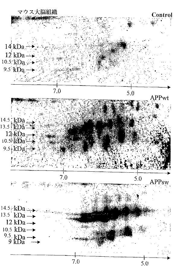
ネズミ大脳組織の1−D分析
ヒトAPPの異化および/または代謝の特異性をより明らかにするために、ラット、非トランスジェニック・マウス、ならびに、野生型ヒトAPP遺伝子(APPwt)またはスウェーデン型変異を有する遺伝子(APPsw)によるトランスジェニック・マウスにおいて、APP−C末端産物を(ヒト大脳組織と同様のやり方で)分析した。ヒト大脳組織のプロファイル(図5A、レーン4)と比較して、コントロールおよびトランスジェニック・マウスのAPP−C末端断片の免疫マーキングを、図5Aに示す。
【0097】
大脳組織ホモジネートの同スポット量を、各電気泳動ウェル中に導入した。APP−C末端断片を、APP−C末端−C17ポリクローナル抗体を用いて検出した。図5のレーン1は、トランスフェクトされていないマウス(負のコントロール)の分析結果に対応し、レーン2は、APPwtヒト遺伝子の単一コピーを有するマウスに対応し、レーン3は、APPswヒト変異遺伝子を有するトランスジェニック・マウスに対応し、レーン4は、コントロールのヒト大脳組織に対応する。矢印は、検出された電気泳動バンドの分子量を示す。
【0098】
コントロールのマウスまたはAPPwt遺伝子の単一コピーを有するものは、基本的に4個のAPP−C末端断片を産生し、一方、APPsw遺伝子を有するマウスおよび正常ヒト大脳組織は、14.5、13.5、12、10.5、および9.5kDaの分子量を有する5個のAPP−C末端断片を産生することが注目される。
【0099】
これらの結果は、5匹の非トランスジェニック・マウス、野生型ヒトAPP遺伝子を有する2匹のトランスジェニック・マウス、およびAPPsw遺伝子を有する5匹のトランスジェニック・マウスで検証された。
【0100】
図5Bのレーン1〜3は、APP−C末端−C17抗体を用いた、様々なAPPswトランスジェニック・マウス系統の免疫ブロットの結果を示し(図5、レーン1〜3)、その中には、Hsiao等(8)によって記述されたリファレンスのAPPsw系統(レーン3)が示される。従って、該APPswトランスジェニック・マウスは、本明細書に記載されるように、特定のプロファイルを体系的に有している。
【0101】
マウス大脳組織のAPP−C末端断片の2−D分析
ヒト大脳組織に対して、上で記載した(図4)と同様の操作を実施する。従って、コントロール・マウス(図6A)、APPwtマウス(図6B)、ならびにAPPswマウス(図6c)において観察される、様々な変異体を特徴づけることが可能である。APPswマウスにおける変異体の発現に亢進が認められ、また、特定の変異体においては、プロファイルの変異が認められる(図6)。これらの分子量および等電点の値は、図7Aおよび表3に示す。
【0102】
細胞モデルにおけるヒトAPPの改変の1−D分析
−非神経モデル
野生型ヒトAPP遺伝子(APPwt)またはAPPsw遺伝子を安定した形でトランスフェクトしたCHO(チャイニーズハムスター卵巣)細胞を、上述のプロトコルに従って分析した。図8の結果は、APPwtまたはAPPsw遺伝子によるトランスジェニック・マウス由来の大脳組織と比較して、CHO細胞のAPP−C末端断片を示す。
【0103】
APPwt CHO細胞(図8、レーン3)では、見掛けの分子量 16、14.5、13.5、12、10.5、および9.5kDaの6本のバンドが検出される。主要なバンドは、16、14.5、13.5、および9.5kDaのバンドである。APPsw CHO細胞(図8、レーン4)では、シグナル全体、特に16、13.5、および9.5kDa断片で減少が観測される。
【0104】
APPwt(レーン1)およびAPPsw(レーン2)トランスジェニック・マウスの大脳組織のホモジネートとの比較は、CHO細胞中において検出されるバンドは、16kDaのバンドを除き、該大脳組織のバンドと共通していることが示す。
【0105】
CCF、U118、およびT9グリア細胞系統は、13.5、12、10.5、9.5、9、および8.5kDaの6本のバンドを具えるプロファイルを有し、9.5から8.5kDaまでの3本のバンドが強いことが認められる(図8B)。同様なプロファイルは、Hela、COS、およびAPPswでトランスフェクトされたSY−5Y細胞系統で観測されている(図8B)。
【0106】
その他、HT−29上皮細胞系統は、14.5、13.5、12、10.5、9.5および9kDaの6本のバンドのプロファイルを有する(図8:HT−29)。該9kDaのバンドは、HT−29上皮細胞では分離しており、グリコシル化ならびにグリコシル化タンパク質の配向を阻害するベンジル−O−GalNacで処理した後に、より強くなる。これは、グリコシル化が、APP−C末端の発現およびプロファイルを制御している、翻訳後修飾であることを示唆する。従って、この修飾は、アルツハイマー化中のAPP−C末端分子の病的な変異の研究において、考慮すべき重要な要素である。
【0107】
−神経モデル
非トランスフェクト細胞と対比させて、APPsw、APPwt、変異PS1、およびタウ遺伝子を安定した形でトランスフェクトしたヒト神経芽細胞腫細胞(SKNSH−SY5Y)を分析した。結果を図9に示す。非トランスフェクト細胞では、APP−C末端シグナルは、10.5kDaバンドに対応する(図9:未変性)。一方、13.5、12、10.5、9.5、9、および8.5kDaの6個の断片が、APPswでトランスフェクトした未分化(non−differentiated)SKNSH細胞中で全て同定されている(図9:APPsw)。6本のバンドのプロファイルは、APPwtをトランスフェクトした細胞にも見られる(図9:野生型)。変異PS1遺伝子のSY5Y細胞へのトランスフェクトは、5本のバンドの増大、その際、強い9.5kDaのバンドが増大し、γ−セクレターゼの作用によって遊離される、6.5kDa断片の増加を誘起する。
【0108】
Kelly細胞、さらには変異APPまたはPS1遺伝子をトランスフェクトしたSKNSH細胞、NT2細胞、hNT細胞などの、他の未変性神経細胞系も、未変性SKNSHタイプのプロファイルを有する。
【0109】
SY5Y細胞を14日にわたり分化させると、6.5kDaのγバンドの出現と、10.5kDaのバンドの減少を伴う、プロファイルの変化が誘起される。同一条件下で、3Rタウ遺伝子をトランスフェクトした細胞および未分化細胞では、「βセクレターゼ」断片の遊離が増大するのに対応して、13.5kDaバンドが出現する。これらの分化細胞では、6.5kDaのγバンドの消失が観測される。
【0110】
これは、遺伝子が、APP−C末端断片の発現および相対的分布を変化させることが可能であることを示している。従って、これらのモデルは、ヒト大脳組織において観察されるように、APP−C末端断片の病的な変化のモデルとなる可能性を有している。
【0111】
ヒト白血球APP−C末端断片の分析
ヒト白血球のペレットを、(医薬または病院の)分析室において利用される標準プロトコルに従って調製した。このペレットを、上述と同様に処理し、APP−C末端−C17抗体を用いた免疫ブロット法によって分析した。この結果は、図10Aに示し。レーン1〜5はコントロールに対応し、レーン6〜11はアルツハイマー病に罹患した患者由来の試料に対応する。性別と年齢を示す(図10A:性別、年齢)。
【0112】
APP−C末端−C17抗体は、分子量 14kDa、10.5kDa、および9.5kDaの3個の断片を検出する。
【0113】
コントロール被験者およびアルツハイマー病と診断された患者由来の白血球の対比1−D分析は、(ヒト大脳組織の研究と同様に)コントロール被験者の白血球(レーン1〜5)と比較して、患者の白血球中におけるAPP−C末端断片の減少(レーン6〜11)を立証することを可能とする。
【0114】
白血球のAPP−C末端断片の変異体は、2−Dでも分析された(図10B)。検出された各スポットの分子量と等電点の値を図7Cおよび表2に示す。
【0115】
様々な免疫プローブ(immunological probes)を用いたAPP−C末端断片の特定
AβペプチドのN末端に対する抗体は、βセクレターゼの切断断片の位置特を可能とする。それは、FCA18抗体である(4)。白血球の14kDa断片は、この抗体によって検出され、それが、βセクレターゼによって作製されるAPP−C末端断片であることを示唆している(図11B)。大脳組織においては、2本のバンドA(14.5kDa)とB(13.5kDa)は、Aβペプチドの5〜11部分に対するモノクローナル抗体(Abeta、GmbH、ハイデルベルク、ドイツ)である、WO2によって認識され、また、バンドBは、FCA18によって検出される。このことは、バンドAおよびBが、βセクレターゼ領域の濃度に応じた、切断によって産生されることを示している(図1参照)。さらに、データベース調査(例えば、SwissProt)から、分子量9.5kDa(図11)、等電点6.99のAPP−C末端断片は、図1に示すように、αセクレターゼ断片に相当することが示される。従って、アルツハイマー病に伴い、改変を受ける、白血球のこの2つの主要なAPP−C末端断片が同定された(図11、AおよびB)。
【0116】
また、5個の断片A〜Cに共通な特定部位は、特異的な改変を受ける場合がある。従って、例えば、リン酸化トレオニン668を特異的に認識するポリクローナル抗体は、バンドA、C、およびDを選択的に認識する。本明細書に記載されている方法により、変性過程に関連している、翻訳後修飾を特定することが可能になる。従って、それは、APP−C末端断片の病的な変異に対する診断キットを有利にもたらす、新たな免疫ツールの開発を可能とするものである。
【0117】
APP−C末端断片のリン酸化は、その病的な変異における決定要素である。
【0118】
APP−C末端の代謝およびリン酸化の改変が、アルツハイマー病で起こる。コントロール被験者およびアルツハイマー病患者の大脳組織のAPP−C末端を、APP−C末端−C17抗体を用いて免疫沈降させると、14.5、13.5、12、10.5、および9.5kDa(断片A、B、C、D、およびE)の5個の断片が、トリス−トリシンゲル中で同一抗体を用いた電気泳動後に見出される(図13、レーン1および2)。この免疫沈降APP−C末端断片を、子ウシ腸アルカリホスファターゼを用いて脱リン酸化し(図13、レーン3および4)、次いで、同じプロトコルに従ってAPP−C末端−C17抗体を用いて検出した。13.5、10.5、および9.5kDaの、3個の主要な断片が、コントロールのホモジネート中に検出される。これら3個の断片は、アルツハイマーのホモジネート中でも検出される。しかし、この13.5kDa断片の量は少量で、一方、10.5および9.5kDa断片は多量である。この結果は、αセクレターゼ切断による、これらの断片の蓄積と、それに、それらのより高いリン酸化を示している。
【0119】
リン酸化指数の変化は、特には、抗リン酸化トレオニン668抗体を用いて実証される(図13、レーン5および6)。トレオニン668上のリン酸化されたAPP−C末端断片は、トリス−トリシンゲル中での電気泳動後、APP−C末端−C17抗体を用いて免疫沈降し検出される。14.5、12、および10.5断片がコントロールのホモジネート中に検出される。アルツハイマーのホモジネート中では、多量に検出されるのは実質的に12.5および10.5の断片である。この結果が、APP−C末端断片CおよびDがよりリン酸化され、かつアルツハイマー病の初期段階で多量にリン酸化され、同時に、断片Aは、668部位で全くまたはほんの僅かしかリン酸化されないことを確認する。
【0120】
要点のまとめ
本発明の実施の上で、特に情報価値のある変異体を、表にまとめて示す(図14〜16)。これら様々な変異体は、神経組織中、ならびに非神経組織、特には、リンパ球中の双方における、APPの病的な変異を検出するための極めて有用なマーカーを構成する。
【0121】
APP−C末端断片のトレオニン、セリン、チロシン上のリン酸化(図1)は、APP−C末端の全般的なプロフィールに寄与する。まず、特には、リン酸化トレオニン668によって示されるように、これら様々なAPP−C末端断片のリン酸化指数に違いが観測される(図11および13)。次いで、脱リン酸化APP−C末端断片のプロファイルが変化し、特には、14.5kDaバンドが消失し、従って、その新たなプロファイルは、APPswをトランスフェクトしたSKNSH細胞モデルのそれとに類似している(図13)。
【0122】
さらに、APP−C末端断片の脱リン酸化は、特には、APP−C末端断片のαセクレターゼ切断産物に対応する、バンドC、D、Eの蓄積(図13)を伴う正常組織とアルツハイマー組織との間のプロファイル変化を説明する。従って、脱リン酸化は、重要な翻訳後の現象であり、APP−C末端断片の病的な変異のマーカーである。
【0123】
この改変は、その細胞モデルによって示されるように(図9)、アルツハイマー病に関与する遺伝子によってその発現が変化を受ける、6.5kDaのγセクレターゼ断片においても反映される。
【0124】
APP−C末端断片の検出は、ヒト神経組織(図12A:ヒト大脳組織)またはヒト非神経組織(図12B:ヒト リンパ球)、ならびに実験動物(図12C:APPswおよびAPPwtトランスジェニック・マウス由来の大脳組織)、または細胞、神経(図12D:神経芽細胞腫細胞)、または非神経(CHO細胞、COSなど)のモデルに適用可能な、病的変異の指標の定義を可能とする。この指標は、0(健常組織)から100%(アルツハイマー化病的組織)まで変化する(図12)。
参考文献
1. De Strooper B, Konig G (1999) Alzheimer’s disease. A firm base for drug development [news] [comment]. Nature, 402, 471−2.
2. Sigurdsson EM, Permanne B, Soto C, Wisniewski T, Frangione B (2000) In vivo reversal of amyloid−beta lesions in rat brain. J. Neuropathol. Exp. Neurol., 59, 11−7.
3. Schenk D, Barbour R, Dunn W, Gordon G, Grajeda H, Guido T, Hu K, Huang J, Johnson−Wood K, Khan K, Kholodenko D, Lee M, Liao Z, Lieberburg I, Motter R, Mutter L, Soriano F, Shopp G, Vasquez N, Vandevert C, Walker S, Wogulis M, Yednock T, Games D, Seubert P. (1999) Immunization with amyloid−beta attenuates Alzheimer−disease−like pathology in the PDAPP mouse. Nature, 400:173−7.
4. Ancolio K, Dumanchin C, Barelli H, Warter JM, Brice A, Campion D, et al. (1999) Unusual phenotypic alteration of beta amyloid precursor protein (betaAPP) maturation by a new Val−715 → Met betaAPP−770 mutation responsible for probable early−onset Alzheimer’s disease. Proc. Natl. Acad. Sc.i U S A, 96, 4119−24.
5. Vaitukaitis J, Robbins JB, Nieschlag E, Ross GT (1971) A method for producing specific antisera with small doses of immunogen. J. Clin. Endocrinol. Metab., 33, 988−91.
6. Delacourte A, David JP, Sergeant N, Buee L, Wattez A, Vermersch P, et al. (1999) The biochemical pathway of neurofibrillary degeneration in aging and Alzheimer’s disease, Neurology, 52, 1158−65.
7. Duff K, Eckman C, Zehr C, Yu X, Prada CM, Perez−tur J, et al. (1996) Increased amyloid−beta42(43) in brains of mice expressing mutant presenilin 1. Nature, 383, 710−3.
8. Hsiao K, Chapman P, Nilsen S, Eckman C, Harigaya Y, Younkin S, et al. (1996) Correlative memory deficits, Abeta elevation, and amyloid plaques in transgenic mice. Science, 274, 99−102.
9. Laemmli UK (1970) Cleavage of structural proteins during the assembly of the head of bacteriophage T4. Nature, 227, 680−5.
10. Schagger H, von Jagow G (1987) Tricine−sodium dodecyl sulfate−polyacrylamide gel electrophoresis for the separation of proteins in the range from 1 to 100 kDa. Anal. Biochem., 166, 368−79.
【図面の簡単な説明】
【図1】
APP−C末端断片の切断を概念的に示す図と、利用した抗体のエピトープの位置を示す図である。
【図2】
ヒト大脳組織の(1−D)ウェスタン・ブロットの結果(図2(A)と図2(B))と、APP−C末端断片のアッセイ結果(図2(C)と図2(D))を示す写真である。
【図3】
ヒト大脳組織分画のウェスタン・ブロットの結果(図3(A)と図3(B))を示す写真である。
【図4】
ヒト大脳組織の(2−D)ウェスタン・ブロットの結果(図4(A)〜図4(C))を示す写真である。
【図5】
トランスジェニック・マウス大脳組織の(l−D)ウェスタン・ブロットの結果(図5(A)と図5(B))を示す写真である。
【図6】
マウス大脳組織の(2−D)ウェスタン・ブロットの結果を示す写真である。
【図7】
マウス大脳組織中(図7(A))、ヒト大脳組織中(図7(B))、ならびにヒト リンパ球中(図7(C))のAPP−C末端断片の2−Dプロファイルを合成した結果を示す図である。
【図8】
WTおよびSWマウスの大脳組織、APPwtおよびAPPswを有する非神経細胞系統のウェスタン・ブロットの結果を示す写真である。
【図9】
APPwt、APPsw、変異PS1、3Rタウ、または4Rタウをトランスフェクトした、またはトランスフェクトしていない神経型細胞系統と、ヒト大脳組織とを比較したウェスタン・ブロットの結果を示す写真である。
【図10】
ヒト リンパ球のl−D分析結果(図10(A))と2−D分析結果(図10(B))を示す図である。
【図11】
抗Aβ−N末端抗体、APP−C末端抗体、およびリン依存性APP−C末端抗体によるAPP−C末端断片の同定の結果を示す図である。
【図12】
病的な変性のインデックスを示す図である。
【図13】
APP−C末端の病的な変性体に対するリン酸化の影響を示す図である。
【図14】
本発明にかかる変異体の一覧表を示す。
【図15】
本発明にかかる変異体の一覧表を示す。
【図16】
本発明にかかる変異体の一覧表を示す。[0001]
The present invention relates to a means for detecting pathological transformation of an APP protein (amyloid precursor protein) in a neurodegenerative disease such as Alzheimer's disease.
[0002]
More particularly, the present invention relates to a method for detecting such alterations, a kit provided therefor, and its therapeutic and diagnostic applications.
[0003]
Alzheimer's disease (AD) is a neurodegenerative disease that results in a loss of intellectual function and thus a progressive and irreversible development of dementia. The disease is becoming one of the major problems in the future as the population ages, as aging is a major risk factor.
[0004]
99% of the observed types are non-familial. In the case of familial autosomal dominant forms, pathological mutations are found on the APP gene on chromosome 21 and on the PS1 and PS2 (preseniline 1 and 2) genes found on
[0005]
Two degenerative processes characterize Alzheimer's disease. Amyloid formation due to dysfunction of APP protein and neurofibrillary degeneration (NFD) corresponding to the accumulation of tau protein in nerve cells.
[0006]
APP protein dysfunction is the very cause of Alzheimer's disease, but the clear cause of neuronal degeneration and death has not yet been elucidated.
[0007]
Two hypotheses have been proposed. According to the most prevalent, first hypothesis, the APP cleavage product, an Aβ peptide containing 39-43 amino acid residues, is a neurotoxin and triggers the NFD chain reaction.
[0008]
According to a second hypothesis, the degeneration, which is deformed in parallel and secondary by the accumulation of Aβ peptide in the form of amyloid plaques, results from the loss or gain of APP function.
[0009]
Transgenic mice carrying a mutated APP or APP + PSI gene allow modeling of amyloid formation. In this mouse, amyloid plaques develop progressively, but NFD is not observed. This mouse has already been used in the pharmaceutical industry to test molecules that can inhibit the production of Aβ peptide. The basic strategy consists of interfering with enzymes involved in the production of Aβ peptide. Currently known enzymes of this type are beta and gamma secretase (1).
[0010]
Molecules that stick on amyloid deposits have also been tested. This molecule is a beta structure breaker (beta sheet breaker) or anti-amyloid agent (2).
[0011]
Vaccination against amyloid peptide is also under evaluation (3).
[0012]
At present, in human tissues affected with Alzheimer's disease, significant and conclusive mutations of the APP protein increase the 1-42Aβ peptide production, increase the 1-42 / 1-40Aβ ratio, and its insoluble deposits and amyloid. Other than aggregation in the form of plaques has not been found.
[0013]
Mutations in the secretion of the APPN-terminal portion, known as sAPP, have also been reported in in vitro studies of the 715 mutation on APP (4).
[0014]
The work performed by the inventors has not been linked to the detection of Aβ peptide, but has noticed a significant alteration of the APP protein that is specifically detectable in human tissues of Alzheimer's disease, as well as in experimental models. I let you.
[0015]
These alterations have been demonstrated in human tissues affected by Alzheimer's disease (central nervous system, peripheral fluids and tissues), and in other neurodegenerative conditions involving APP dysfunction.
[0016]
The important alterations are qualitative and quantitative and are detected upstream of Aβ peptide production.
[0017]
Many similar abnormalities are found in pathological mutations in APP, or in transgenic mice having mutant APP and
[0018]
Thus, the present invention relates to the use of pathological modifications of APP, particularly those detected in human cerebral tissue, as markers in detection methods during the process of neurodegeneration.
[0019]
The invention particularly relates to the use of these markers for the diagnosis and monitoring of neurodegenerative processes, the development of animal models, and the screening of drugs effective against APP pathologies.
[0020]
The present invention also relates to the above-mentioned method for evaluating a medical condition, which includes a definition of an index set for a reference system.
[0021]
A method for detecting a pathological alteration of an APP protein in an analytical sample comprises the step of catabolizing and / or metabolic fragmentation of the carboxy-terminal portion of the specifically modified APP in a neurodegenerative condition in which said APP is involved in its pathogenesis. , A marker composed of an APP-C-terminal fragment hereinafter.
[0022]
Studies conducted by the inventors have unexpectedly shown that the modifications resulting in such fragments are directly linked to the initiation and progression of degenerative processes in the cerebral region, especially in the case of Alzheimer's disease. Clarified.
[0023]
The fragment in question is a product of APP catabolism and / or metabolism that retains the primary structure of the APP-C-terminus, and thus has been observed to be recognized by antibodies to this APP-C-terminal portion. .
[0024]
Accordingly, the term "fragment" as used herein and in the claims, as described in the Examples, refers to those mutants which, to the extent recognized by anti-APP-C-terminal antibodies, e.g. Also included are charge and isoelectric point variants that differ in charge or isoelectric point for post-translational modifications, such as and / or methylation and / or acetylation and / or glycosylation.
[0025]
These combined immunoblotting and detection, in particular with a set of anti-APP-C-terminal polyclonal and / or monoclonal antibodies, as well as a set of antibodies against the post-translational modification of pathological variants of APP-C-terminal, A fragment that can be identified by biochemical analysis of an analysis sample by 1-D electrophoresis (one-dimensional electrophoresis) or 2-D electrophoresis (two-dimensional electrophoresis).
[0026]
The electrophoresis can be one-dimensional and / or two-dimensional.
[0027]
Such fragments undergo specific alterations in diseased tissue. As the neurodegenerative process progresses, a decrease, or even disappearance, of certain of these fragments has been found. At the same time, mutations in the charge, as well as altered solubility, of certain of these fragments have been found.
[0028]
The invention relates in particular to the application of this method for the diagnosis of Alzheimer's disease progression, ie its molecular weight, determined by one-dimensional (1-D) electrophoresis, as a marker of APP dysfunction. , 14.5, 13.5, 12, 10.5, and 9.5 kDa, which utilize five major fragments that are modified during the process of Alzheimerization. For convenience, these fragments are denoted by the letters A, B, C, D, E. It is also noted that a 9 kDa (F) band is present, and a 6.5 kDa band is also present, although to a lesser and varying amount.
[0029]
The disappearing mutant corresponds to a 14.5 kDa 1-D electrophoretic band represented by A. In 2-D, it is separated into two components of 15 kDa and 14.5 kDa, and the 15 kDa component has an isoelectric point of 5. 13 (represented as 15 / 5.13) and 15 / 5.37, and the other component is an isoelectric variant of 14.5 / 5.64 and 14.5 / 5.95. ). 12. separated into two components of 13.5 and 12.7 kDa, including isoelectric point variants of 13.5 / 4.94, and 12.7 / 4.8; 12.7 / 5.29; The fragment corresponding to the electrophoretic band B of 5 kDa also disappears.
[0030]
In the early stages of Alzheimer's disease, useful fragments are also modified. They are
[0031]
The fragments that appear during Alzheimerization include 11 / 4.94, 10.5 / 5.35, 10.5 / 7.5 isoelectric point variants, and in 2-D, 11 kDa and 10.5 kDa. It corresponds to a 10.5 kDa 1-D electrophoretic band separated into two components, as well as a 9.5 kDa, pI 5.77 fragment.
[0032]
At the same time, changes in the solubility of the APP-C terminal marker are also observed with the development of the pathological process of neurodegeneration. During Alzheimerization of cerebral tissue, fragments 14.5 and 13.5 show a decrease in their solubility, and fragments 10.5 and 9.5 show an increase in their solubility.
[0033]
Such fragments are subject to modification in tissues or cells that are described as "alzheimerized." They may be similarly modified in experimental models, such as transgenic animals, carrying mutant or non-mutant genes, or combinations of genes, involved in Alzheimer's pathology.
[0034]
These differences in catabolism and metabolism of APP proteins, as observed in the APP-C-terminal marker, can be attributed to neuronal cell models (eg, neuroblastoma cells such as the SKNSH SY5Y line, the NT2 line, the Kelly line), glia It is interesting and noteworthy that it is observed not only in blastoma cell lines (CCF, U118, T98) but also in non-neural cells such as CHO, COS, HT-29, and Hela lines.
[0035]
Each cell line originally had its own expression pattern of these fragments (expressed molecular weights 13.5, 12, 10.5, 9.5, 9, 8.5, and 6 relative to all proteins in the cell model). It is also noted that the total amount of the 0.5 kDa fragment varies; the relative ratio between each fragment varies.
[0036]
In these models, the expression of fragments A to F is also changed depending on the differentiation state of the cell lineage in a behavior specific to each model.
[0037]
These variations may be due to animal models, especially possible different configurations (APP mutant and / or PS1 or PS2 mutant and / or lack of PS1 expression) and / or mutated or unmutated tau genes (14.5, 13. 5, 12, 10.5, 9.5, 9, 8.5, and 6.5 kDa) are also observed in transgenic mice.
[0038]
Thus, the analytical samples used in the method include neural tissues or cell lines as well as non-neural tissues or cell lines, such as cerebrospinal fluid, blood, and biological fluids such as all or some of its components. It facilitates early diagnosis, assessment of risk factors in patients, or treatment monitoring.
[0039]
Advantageously, these tissues are homogenized or fractionated, for example by centrifugation, for use in the detection method.
[0040]
The above methods are also used for differential diagnosis of neurodegenerative pathologies associated with Alzheimer's disease, such as dementia with Lewy bodies and amyloid angiopathy.
[0041]
The detection method described above makes it possible to define an indicator of a mutation of the APP protein to a disease-associated protein.
[0042]
The invention is particularly directed to a method for assessing the pathological process of neurodegeneration in an analytical sample by immunochemical analysis, as described in the Examples, wherein index 0 (zero) is used for detection in healthy tissue,
[0043]
Such an index has diagnostic and therapeutic benefits. It allows the morbidity to be quickly reflected. The index value makes it possible to determine whether there is in fact a risk of developing Alzheimer's disease. If a high index is obtained, it will help confirm the clinical diagnosis.
[0044]
Also, for the purpose of therapeutic treatment based on APP catabolism and metabolism and its regulation, mutation of such an index allows one to monitor the effects of the treatment.
[0045]
It constitutes a criterion that makes it possible to compare the effects of various molecules on Alzheimer's disease or other neurodegenerative conditions, wherein mutations in the index indicate the efficacy of the test molecule as well as its therapeutic utility. Is shown.
[0046]
The use of such indices also has advantages in the analysis of the early etiological events that contribute to the disease, leading to the discovery of new and effective molecules, as well as the development of new therapeutic strategies.
[0047]
It is also noted that this index can be used to induce degeneration to slow down degeneration and even establish new experimental models. This index also provides information that allows the induction of specific degenerations within therapeutic measures that require elimination of the cell population.
[0048]
The method according to the invention additionally detects post-translational modifications of pathological mutations of the APP-C-terminal fragment, in particular of the phosphorylated and / or methylated and / or acetylated and / or glucosylated form. It is advantageous to use a set of polyclonal and / or monoclonal antibodies to
[0049]
Preferably, the set of polyclonal and / or monoclonal antibodies used is able to account for variations in phosphorylation and expression of the 14.5, 12, 10.5 kDa fragment.
[0050]
The detection method according to the present invention can be suitably used for establishing an experimental model, a cell, or an animal model of Alzheimer's disease, and for verifying the efficacy. The fitted model mimics the modification of the APP-C-terminal fragment observed in human tissues, as described in the examples.
[0051]
Improving patient selectivity can also help in establishing effective therapeutic trials.
[0052]
Another use that is of great interest relates to pharmacological screening to select drugs that are effective against Alzheimer's-type neurodegenerative conditions. That is, the selected molecule can alleviate the above-mentioned symptoms of Alzheimer's by leading to restoration of properties of the APP-C terminal marker or restoration of their physicochemical properties (solubility, charge). There are features.
[0053]
Thus, the present invention allows for a rapid and legitimate pharmacological screening of useful molecules.
[0054]
The present invention also relates to a kit for diagnosing pathological mutations in APP, comprising a set of polyclonal and / or monoclonal antibodies against the carboxy-terminal region of APP. For example, a set of antibodies against post-translational modifications associated with pathological mutations located on APP-C-terminal fragments, such as sites of phosphorylation, methylation, acetylation, or glycosylation on these fragments. The use is beneficial because it provides additional information regarding the validation and quantification of pathological mutations of the APP-C-terminal fragment.
[0055]
Heterozygosity of the APP-C-terminal fragment, as shown by 2-D electrophoresis, was demonstrated by APP-phospho-dependant polyclonal antibody to phosphorylated threonine 668 (as indicated by the number in APP695). -Explained in part by phosphorylation modifications located in the region common to the C-terminal fragment.
[0056]
It also results from glycosylation, as shown by anti-glycosylation drugs that alter the profile of the APP-C-terminal fragment. Methylation and acetylation are also two types of post-translational modifications that involve pathological mutations in the APP-C-terminal fragment. In fact, they are located substantially on lysine residues, causing acidification and concomitant changes in isoelectric point.
[0057]
These are also the result of regulation by genes involved in Alzheimer's disease. By this method, overexpression of tau protein can affect the ratio of the 13.5, 9.5, and 6.5 kDa bands.
[0058]
These antibodies can be used in immunochemical assays such as, for example, ELISAs, Western blots, dot blots, and the like. Therefore, it is convenient that the kit according to the present invention also includes components necessary for performing such an assay.
[0059]
Other features and advantages of the present invention will become apparent in the following examples, with reference to FIGS.
Materials and methods
antibody
Production of APP-C-terminal-C17 polyclonal antibody
Polyclonal antibodies against the carboxy-terminal portion of APP were produced according to the immunization protocol described by Vaitukaitis et al. (5). A New Zealand rabbit was immunized with a peptide comprising the last 17 amino acids of the human APP sequence, corresponding to its carboxy terminus. The peptide was covalently linked to ovalbumin. 200 μg of immunogen was injected every two weeks. Prior to injection, 1 volume of Freund's complete adjuvant was added to the immunogen solution for the first sensitization, and incomplete Freund's adjuvant was used for subsequent sensitizations instead of complete adjuvant. Blood was drawn one week after the fourth injection and one week after each subsequent injection. After blood coagulation and centrifugation at 2000 g for 10 minutes, pure serum was obtained. One volume of glycerol was added to the serum and the mixture was stored at -20C.
[0060]
Purification of APP-C-terminal-C17 antibody
1 mg of the peptide used for immunization was conjugated to NHS-Fast Flow Sepharose® matrix (Amersham Pharmacia Biotech) according to the manufacturer's instructions. The loaded matrix is contacted with 200 μl of pure serum diluted in one volume of 2 × binding buffer (50 mM Tris pH 8.0; 300 mM NaCl; Tween-20 0.1% (v / v)) at 4 ° C. With light stirring overnight. All of the solutions were loaded on a PD-10 column. The matrix was washed with 10 volumes of the binding buffer described above. The purified antibody was eluted with 2 volumes of acetate buffer at pH 3.5. The protein concentration was determined using Pierce's BCA protein quantification kit.
[0061]
Production of APP-668P polyclonal antibody
Following the same protocol as used for APP-C-terminus-C17, corresponding to the sequence of Val-Asp-Ala-Ala-Ala-Val- (phosphorylated) Thr-Pro-Glu-Glu-Arg-His-Leu. Antibodies against the synthetic peptide were produced. This antibody recognizes the immunogen but has the same sequence but does not recognize non-phosphorylated peptides.
[0062]
Specimen
Specimens of human and murine cerebral tissue
Human cerebral tissue is from a patient that has been monitored previously and is described in (6). This cerebral tissue specimen, obtained by necropsy or biopsy and stored at −80 ° C., was dissected using autopsy catalogs and then 1 × 1-D lysis buffer (50 mM Tris-HCl pH 6.8; 4 mM EDTA; Teflon® in 10 volumes of
[0063]
Rat strain used
A sample of murine cerebral tissue was prepared according to the same protocol as a sample of human cerebral tissue. Cerebral tissue specimens were collected from non-transgenic mice and transgenic mice having the wild-type human APP gene (APPwt) or the human APP gene with mutations at codons 670 and 671 (APPsw). This APP mutation is associated with familial Alzheimer's disease, known as Swedish mutation (7). Other specimens were also used, including non-transgenic mice and allogeneic transgenic mice with APPwt, APPsw genes. Hsiao K. A specimen of murine cerebral tissue similar to the transgenic mouse developed by (8) was also used.
[0064]
Cerebral tissue sample after protein fractionation
Cerebral tissue specimens were homogenized at a ratio of 1/10 (w / v) in 10 mM Tris-HCl pH 6.8 buffer, then centrifuged at 4 ° C. for 1 hour at 100,000 g. The supernatant (F1) was obtained, and a pellet re-homogenized in the same buffer containing 0.5% (v / v) of Triton X-100 was added thereto. After centrifugation, save the soluble Triton X-100 fraction, called the F2 fraction. Additional extraction and centrifugation steps are performed under the same conditions in the same buffer to create the F2 'fraction. The pellet is then quickly taken up in 1-D lysis buffer (see below) and designated as F3 fraction.
[0065]
At the time of use, the following corresponding buffers were added to the supernatant according to the biochemical technique used: 1 × D lysis buffer (pH 6.8) for one-dimensional (1-D) electrophoretic analysis. 100 mM Tris-HCl;
[0066]
Immunoprecipitation
The F2 fraction is used to immunoprecipitate the APP-C-terminal fragment. 100 μl of the F2 fraction was transferred to NP. Diluted in 300 μl of 10 mM Tris HCl pH 7.4 buffer containing 401% (immunoprecipitation buffer). 10 μl of APP-C-terminal-C17 antibody or APP668P antibody are added and the mixture is incubated at 4 ° C. under stirring overnight. 40 μl of Protein A fixed on agarose beads (Pierce) are added to the solution and the mixture is incubated for 1 hour at 4 ° C. with stirring. The agarose beads were washed three times in immunoprecipitation buffer and then treated with 50 μl of SDS buffer to release APP-C-terminal fragments and recovered in the centrifugation supernatant. Next, the APP-C-terminal fragment was analyzed by immunoblotting after electrophoresis on a Tris-tricine gel.
[0067]
CHO (hamster) or SKNSH SY5Y (human) cultured cell sample
Cells are stably transfected with a wild-type human APP gene or a human APP gene having a Swedish mutation. Cell pellets were taken up in 10 mM Tris-HCl pH 6.8 buffer and then sonicated. Before use, add 1 volume of 2 × lysis buffer to the sample, corresponding to the (1-D or 2-D) experiment. For 1-D analysis, bring the sample to 100 ° C. for 10 minutes.
[0068]
Human leukocyte sample
Preparation of human leukocytes:
10 ml of blood is collected in an EDTA (ethylenediaminetetraacetic acid) tube. The centrifugation is carried out for 15 minutes at 4500 rpm (number of revolutions per minute). Remove the plasma. In a tube, a solution for lysis of red blood cells (lysis solution: NH4CO3 0.91 mM, NH4(0.132 mM Cl). The tube is gently stirred and placed in a water bath cooled to 5 ° C. Perform centrifugation at 4000 rpm for 15 minutes and remove the supernatant. Following the same procedure, the leukocyte pellet is washed twice with the lysis solution. The pellets are then dewatered and used within 24 hours or frozen at -20 ° C for transport or later use.
[0069]
The leukocyte pellet is taken up in 10 mM Tris-HCl pH 6.8 buffer and then sonicated. Prior to use, one volume of 2 × lysis buffer, corresponding to the (1-D or 2-D) experiment, is added to the sample. For 1-D analysis, bring the sample to 100 ° C. for 10 minutes.
[0070]
Electrophoresis
1-D electrophoresis
Experiments were performed using a Protean IIXi Cell electrophoresis system (Biorad) according to the manufacturer's instructions.
[0071]
1-D electrophoresis was performed according to the gel preparation protocol described by Laemmli (9) and under the electrophoresis conditions described by Schagger and Von Jagow (10). The concentrated gel contains 4% acrylamide and the separation gel used contains 16.5% acrylamide. Electrophoresis is performed in a Tris-Tricine buffer. The electrophoresis program used was as follows: 1 hour 30V constant, then 16 hours at 45 mA.
[0072]
Each well was loaded with an equal amount (about 100 μg / well) of protein.
[0073]
2-D electrophoresis
One-dimensional
One-dimensional, ie, focusing, is performed using a precast gel strip known as IPG Strip®, covering a gradient of pH 3-10, according to the manufacturer's instructions. The material used for isoelectric focusing is the Protean IEF Cell system (Biorad).
[0074]
1 × rehydration buffer (7 M urea, 2 M thiourea, 4% triton X100, 0.5% CHAPS, 0-3% (w / v) Pharmalytes® pH 3-10, 10 mM DTT, orange G 0.01%) to 250 μg of protein to make 400 μl for use. 400 μl of the sample is placed on a strip placed in the device. This is left for 1 hour to passively rehydrate, then kept at 50 V / strip for 10 hours, actively rehydrated, and then start the program according to the manufacturer's instructions. After focusing, use the strips or store at -80 ° C.
[0075]
2D
Prior to use, strips were washed with 1-D buffer (50 mM Tris pH 6.8;
[0076]
Transfer to membrane and immunoblotting
Transcription
Transfers were performed using a Pharmacia LKB multiphor® semi-dry transfer system according to the manufacturer's instructions (Amersham-Pharmacia Biotech). The protein is 0.8 mA / cm2Was transferred to Hybond® ECL nitrocellulose membrane (Pharmacia-Amersham).
[0077]
Immunoblotting
The membrane is incubated in a buffer containing
[0078]
The membrane was diluted in incubation buffer (15 mM Tris, pH 8.0, 150 mM NaCl, 0.1% (v / v) Tween-20, 3% (w / v) skim milk) to a final dilution of 1/2000 (w / v). Incubate with APP-C-terminal-C17 antibody diluted (v / v) for 2 hours at room temperature or overnight at 4 ° C.
[0079]
The membrane is washed three times for 10 minutes each in incubation buffer without skim milk.
[0080]
The membrane is then incubated with Raifort peroxidase-conjugated anti-rabbit goat immunoglobulin in a skim milk-free incubation buffer at a final dilution of 1/4000 (v / v) for 1 hour at room temperature. The membrane is washed three times for 10 minutes each in incubation buffer without skim milk and its immunoreactivity is performed using the ECL® chemiluminescence kit (Pharmacia-Amersham Biotech) according to the manufacturer's instructions. Color the sex polypeptide.
result
A conceptual overview of APP catabolism and / or metabolic cleavage is shown in FIG. 1 (A), and the location of the antibody epitope utilized is shown in FIG. 1 (B).
[0081]
1-D analysis of human cerebral tissue
A sample of human cerebral tissue, prepared as described in the method section for 1-D analysis, was mounted and analyzed. Electrophoretic separation was performed under the above-mentioned Tris-Tricine conditions. A calibrated molecular weight marker (Biorad) was also included to determine the molecular weight of this fragment using ImageMaster® 1-D Elite software (Amersham-Pharmacia Biotech).
[0082]
Transcription of the electrophoretically separated proteins, as well as immunoblotting with the APP-C-terminal-C17 antibody, was performed as described above. The results are shown in A1 of FIG.
[0083]
Normal tissue analysis
In the control human cerebral tissue, the apparent molecular weights are A 14.5 kDa; B 13.5 kDa;
[0084]
Analysis of tissue from patients at different stages of Alzheimer's disease
A) Reduction of the amount of APP-C-terminal fragment during Alzheimerization
FIG. 2B shows tau pathology (FIG. 2, B1) according to the Delacourt et al. Classification in 1999 and control subjects (
[0085]
Statistical studies of a large number of patients show that there is a very high correlation between the progression of the pathological process, indicated by the degree of tau abnormalities, and the decrease in the amount of APP-C-terminal fragments in the analyzed cerebral tissue samples Is known.
[0086]
The amount of APP-C terminal was determined in a control case (Ctrl), a preclinical case (infraAD) corresponding to a patient without dementia but having a lesion characteristic of Alzheimer's disease, and a clinical case in Alzheimer's disease (AD). And in the cortex of four groups of patients in the familial autosomal dominant case of Alzheimer's disease (FAD.AD) (FIG. 2, C). The Mann-Whitney non-parametric statistical test shows that infraAD and AD groups are significantly reduced when compared to control Ctrl (infraAD: p <0.03; AD p <0.002). The average reduction amount of the APP-C terminal is reduced by a factor of 1 / 1.5 in the infraAD group and by a factor of 1 / 1.7 in the AD group, as compared with the control group. There is no significant difference in APP-C-terminal reduction between infraAD and AD groups. This indicates that this APP-C terminal loss is an early phenomenon of Alzheimer's medical pathology.
[0087]
A statistical study of the correlation between APP-C-terminal expression levels for tau abnormalities in the temporal cortex of control patients and various stages of Alzheimer's disease was performed (FIG. 2, D). There is a significant correlation between the amount of APP-C terminus detected in the cortex of each patient studied and the varying degree of each tau abnormality in the temporal and occipital cortices.
Linear regression
The APP-C-terminal fragment is preferably extracted in two phases: a buffer solution containing the non-ionic detergent Triton X100 (F2 fraction), and a solution containing the ionic detergent sodium dodecyl sulfate (SDS) (F3). ) (FIG. 3, A).
[0088]
Upon Alzheimerization of cerebral tissue, a specific change in the solubility of the APP-C-terminal fragment is observed (FIG. 3B). In the F2 fraction, the solubility of the APP-C-terminal fragment with a molecular weight of 9.5 (fragment E) and 10.5 kDa (fragment D) increases, and in the F3 fraction, a molecular weight of 13.5 (fragment B). ) And the 14.5 kDa (fragment A) APP-C terminal fragment were found to have increased insolubility.
[0089]
2-D analysis of normal and pathological human cerebral tissue
The results for three patients are shown in FIG. 4 (A-C).
[0090]
-1 control patient without Alzheimer's cerebral lesion (
-One patient at the very early stage of the clinical stage of Alzheimer's disease (
[0091]
Arrows indicate spots detected using the APP-C-terminal-C17 polyclonal antibody. Their molecular weight is almost the same as the value observed on the 1-D immunoblot. Their isoelectric points are in the range of pH 4.5 to 7.5.
[0092]
Note the variation in the distribution of the spots with the progression of Alzheimer's disease (FIG. 4, C). In patients at a more advanced stage of Alzheimer's disease, the fragment can no longer be detected and cannot be studied with this technique.
[0093]
The APP-C-terminal variants detected by 2-D analysis are schematically illustrated on a two-dimensional calibrated grid, where the Y-axis indicates molecular weight and the X-axis indicates the isoelectric point (Figure 4, C). The whole was calibrated using internal standards of molecular weight and isoelectric point. The isoelectric point controls are γ-enolase (47 / 4.94), α-actin (42 / 5.29), and α-enolase (47 / 6.99). The isoelectric points of 5.54 and 8.05 are standards inside cerebral tissue.
[0094]
The schematic of FIG. 4 shows the use of APP-C-terminal-C17 antibody in human cerebral tissues of control subjects, Alzheimer's disease patients in preclinical disease stage, and Alzheimer's disease patients in early clinical stage of Alzheimer's disease. The 2D profile of the detected APP-C-terminal fragment is shown superimposed. The spots, i.e. the various 2-D isoelectric point variants, observed in both healthy and diseased tissues are indicated by open circles. Spots specific to healthy tissues are indicated by hatched circles, and spots specific to Alzheimer's tissues are indicated by black circles.
[0095]
Table 1 shows the molecular weights and isoelectric points of the spots that were modified during Alzheimer's disease.
[0096]
1-D analysis of rat cerebral tissue
To further elucidate the specificity of human APP catabolism and / or metabolism, rats, non-transgenic mice, and transgenics with wild-type human APP gene (APPwt) or a gene with a Swedish mutation (APPsw) -In mice, the APP-C-terminal product was analyzed (in a manner similar to human cerebral tissue). Immunomarking of the APP-C-terminal fragment of control and transgenic mice compared to the profile of human cerebral tissue (FIG. 5A, lane 4) is shown in FIG. 5A.
[0097]
The same spot amount of cerebral tissue homogenate was introduced into each electrophoresis well. The APP-C-terminal fragment was detected using an APP-C-terminal-C17 polyclonal antibody.
[0098]
Control mice or those with a single copy of the APPwt gene basically produce four APP-C-terminal fragments, while mice and normal human cerebral tissues with the APPsw gene produce 14.5,13. It is noted that it produces five APP-C-terminal fragments with molecular weights of 5, 12, 10.5, and 9.5 kDa.
[0099]
These results were validated in five non-transgenic mice, two transgenic mice with the wild-type human APP gene, and five transgenic mice with the APPsw gene.
[0100]
[0101]
2-D analysis of APP-C-terminal fragment of mouse cerebral tissue
The same operation as described above (FIG. 4) is performed on human cerebral tissue. Thus, it is possible to characterize the various mutants observed in control mice (FIG. 6A), APPwt mice (FIG. 6B), and APPsw mice (FIG. 6c). Enhanced expression of mutants in APPsw mice, and profile mutations in certain mutants (FIG. 6). The molecular weight and isoelectric point values are shown in FIG.
[0102]
1-D analysis of human APP modifications in cell models
-Non-neural model
CHO (Chinese hamster ovary) cells stably transfected with the wild-type human APP gene (APPwt) or the APPsw gene were analyzed according to the protocol described above. The results in FIG. 8 show APP-C-terminal fragments of CHO cells compared to cerebral tissue from transgenic mice with APPwt or APPsw genes.
[0103]
In APPwt CHO cells (FIG. 8, lane 3), six bands with apparent molecular weights of 16, 14.5, 13.5, 12, 10.5, and 9.5 kDa are detected. The major bands are the 16, 14.5, 13.5, and 9.5 kDa bands. In APPsw CHO cells (FIG. 8, lane 4), a decrease is observed in the overall signal, especially in the 16, 13.5 and 9.5 kDa fragments.
[0104]
Comparison with the homogenate of cerebral tissue of APPwt (lane 1) and APPsw (lane 2) transgenic mice shows that the bands detected in CHO cells are in common with those of the cerebral tissue except for the band of 16 kDa. It shows that there is.
[0105]
The CCF, U118, and T9 glial cell lines have profiles with six bands at 13.5, 12, 10.5, 9.5, 9, and 8.5 kDa, and from 9.5 to 8.8. Three bands up to 5 kDa are found to be strong (FIG. 8B). A similar profile has been observed in the SY-5Y cell line transfected with Hela, COS, and APPsw (FIG. 8B).
[0106]
In addition, the HT-29 epithelial cell line has a profile of six bands at 14.5, 13.5, 12, 10.5, 9.5 and 9 kDa (FIG. 8: HT-29). The 9 kDa band is segregated in HT-29 epithelial cells and becomes stronger after treatment with benzyl-O-GalNac, which inhibits glycosylation as well as orientation of glycosylated proteins. This suggests that glycosylation is a post-translational modification that controls APP-C-terminal expression and profile. Therefore, this modification is an important factor to consider in the study of pathological mutations of the APP-C-terminal molecule during Alzheimerization.
[0107]
-Neural model
Human neuroblastoma cells (SKNSH-SY5Y) stably transfected with APPsw, APPwt, mutant PS1, and tau genes were analyzed in contrast to untransfected cells. FIG. 9 shows the results. In untransfected cells, the APP-C-terminal signal corresponds to the 10.5 kDa band (FIG. 9: native). On the other hand, six fragments of 13.5, 12, 10.5, 9.5, 9, and 8.5 kDa were all identified in APPsw transfected undifferentiated (non-differentiated) SKNSH cells. (FIG. 9: APPsw). The profile of the six bands is also seen in cells transfected with APPwt (FIG. 9: wild type). Transfection of the mutated PS1 gene into SY5Y cells induces an increase in five bands, with an increase in the strong 9.5 kDa band and an increase in the 6.5 kDa fragment released by the action of γ-secretase. I do.
[0108]
Other native neuronal cell lines, such as Kelly cells, as well as SKNSH cells, NT2 cells, hNT cells transfected with the mutant APP or PS1 gene, also have a native SKNSH type profile.
[0109]
Differentiation of SY5Y cells over 14 days induces a profile change with the appearance of a 6.5 kDa gamma band and a decrease in the 10.5 kDa band. Under the same conditions, a 13.5 kDa band appears in cells transfected with the 3R tau gene and in undifferentiated cells in response to increased release of the “β-secretase” fragment. In these differentiated cells, the disappearance of the 6.5 kDa γ band is observed.
[0110]
This indicates that the gene can alter the expression and relative distribution of the APP-C-terminal fragment. Thus, these models have the potential to model pathological changes in APP-C-terminal fragments, as observed in human cerebral tissue.
[0111]
Analysis of human leukocyte APP-C terminal fragment
Human leukocyte pellets were prepared according to standard protocols used in the laboratory (medicine or hospital). The pellet was processed as described above and analyzed by immunoblot using the APP-C-terminal-C17 antibody. The result is shown in FIG. 10A. Lanes 1-5 correspond to controls and lanes 6-11 correspond to samples from patients suffering from Alzheimer's disease. Gender and age are shown (FIG. 10A: gender, age).
[0112]
The APP-C-terminal-C17 antibody detects three fragments with molecular weights of 14 kDa, 10.5 kDa, and 9.5 kDa.
[0113]
Contrasting 1-D analysis of leukocytes from control subjects and patients diagnosed with Alzheimer's disease (as in the study of human cerebral tissue) compared to leukocytes of control subjects (lanes 1-5). Reduces the number of APP-C-terminal fragments (lanes 6-11).
[0114]
Variants of the APP-C-terminal fragment of leukocytes were also analyzed in 2-D (FIG. 10B). The molecular weight and isoelectric point value of each detected spot are shown in FIG.
[0115]
Identification of APP-C-terminal fragments using various immunological probes
Antibodies to the N-terminus of the Aβ peptide allow the location of a cleaved fragment of β-secretase. It is the FCA18 antibody (4). A 14 kDa fragment of leukocytes was detected by this antibody, suggesting that it is an APP-C-terminal fragment generated by β-secretase (FIG. 11B). In cerebral tissue, two bands, A (14.5 kDa) and B (13.5 kDa), are recognized by WO2, a monoclonal antibody (Abeta, GmbH, Heidelberg, Germany) against the 5-11 portion of the Aβ peptide. , And band B is detected by
[0116]
In addition, the specific site common to the five fragments A to C may be subjected to specific modification. Thus, for example, a polyclonal antibody that specifically recognizes phosphorylated threonine 668 will selectively recognize bands A, C, and D. The methods described herein allow for the identification of post-translational modifications associated with the denaturation process. Thus, it allows the development of new immunization tools that advantageously provide a diagnostic kit for pathological mutations of the APP-C-terminal fragment.
[0117]
Phosphorylation of APP-C-terminal fragment is determinant in its pathological mutation.
[0118]
Alteration of APP-C-terminal metabolism and phosphorylation occurs in Alzheimer's disease. Immunoprecipitation of APP-C-terminus of cerebral tissues of control subjects and Alzheimer's disease patients with the APP-C-terminus-C17 antibody resulted in 14.5, 13.5, 12, 10.5, and 9.5 kDa ( Five fragments of fragments A, B, C, D and E) are found after electrophoresis with the same antibody in a Tris-tricine gel (FIG. 13,
[0119]
Changes in the phosphorylation index are demonstrated, inter alia, using an anti-phosphorylated threonine 668 antibody (FIG. 13,
[0120]
Summary of the main points
In the practice of the present invention, particularly informative variants are summarized in a table (FIGS. 14-16). These various mutants constitute extremely useful markers for detecting pathological mutations of APP, both in nervous tissue as well as in non-neural tissues, especially in lymphocytes.
[0121]
Phosphorylation of the APP-C-terminal fragment on threonine, serine, tyrosine (FIG. 1) contributes to the general profile of the APP-C-terminus. First, differences are observed in the phosphorylation index of these various APP-C-terminal fragments, particularly as shown by phosphorylated threonine 668 (FIGS. 11 and 13). The profile of the dephosphorylated APP-C-terminal fragment then changes, in particular the disappearance of the 14.5 kDa band, and thus the new profile is similar to that of the SKNSH cell model transfected with APPsw. (FIG. 13).
[0122]
In addition, dephosphorylation of the APP-C-terminal fragment was particularly effective in normal and Alzheimer tissues with accumulation of bands C, D and E (FIG. 13) corresponding to the α-secretase cleavage product of the APP-C-terminal fragment. Will be described. Thus, dephosphorylation is an important post-translational phenomenon and a marker for pathological mutations of the APP-C-terminal fragment.
[0123]
This modification is also reflected in a 6.5 kDa gamma secretase fragment whose expression is altered by genes involved in Alzheimer's disease, as shown by the cell model (FIG. 9).
[0124]
Detection of the APP-C-terminal fragment was based on human neural tissue (FIG. 12A: human cerebral tissue) or human non-neural tissue (FIG. 12B: human lymphocytes), and from experimental animals (FIG. 12C: APPsw and APPwt transgenic mice). Cerebral tissue), or cells, nerves (FIG. 12D: neuroblastoma cells), or non-neural (CHO cells, COS, etc.) models can be defined as indicators of pathological mutations. This index varies from 0 (healthy tissue) to 100% (Alzheimer's diseased tissue) (FIG. 12).
References
1. DeStrooper B, Konig G (1999) Alzheimer's disease. A film base for drug development [news] [comment]. Nature, 402, 471-2.
2. Sigurdsson EM, Permane B, Soto C, Wisniewski T, Fragione B (2000) In vivo reversal of amyloid-beta lengths in rat brain. J. Neuropathol. Exp. Neurol. , 59, 11-7.
3. Schenk D, Barbour R, Dunn W, Gordon G, Grajeda H, Guido T, Hu K, Huang J, Johnson-Wood K, Khan K, R Li, M, R, M, L, M, L, M, L Soriano F, Shop G, Vasquez N, Vandevert C, Walker S, Wogulis M, Yednock T, Games D, Seubert P. (1999) Immunization with amyloid-beta attackenz Alzheimer-disease-like pathology in the PDAPP mouse. Nature, 400: 173-7.
4. Ancolio K, Dumanchin C, Barelli H, Warter JM, Brice A, Campion D, et al. (1999) Unusual phenotypic alteration of beta amyloid precursor protein (betaAPP) b 7 b b M b b b b 19 19 19 19. Proc. Natl. Acad. Sc. i USA, 96, 4119-24.
5. Vaitukaitis J, Robbins JB, Nieschlag E, Ross GT (1971) A method for producing specific antisera with small doses of immunogen. J. Clin. Endocrinol. Metab. , 33, 988-91.
6. Delacourt A, David JP, Sergeant N, Blue L, Wattez A, Vermersch P, et al. (1999) The biochemical path of neurofibrillary generation in aging and Alzheimer's disease, Neurology, 52, 1158-65.
7. Duff K, Eckman C, Zehr C, YuX, Prada CM, Perez-tur J, et al. (1996) Incremented amyloid-beta42 (43) in brains of expression pressing mutant presenilin Nature, 383, 710-3.
8. Hsiao K, Chapman P, Nilsen S, Eckman C, Harigaya Y, Youkin S, et al. (1996) Correlating memory defects, Abeta evolution, and amyloid plates in transgenic mice. Science, 274, 99-102.
9. Laemmli UK (1970) Clearage of structural proteins dur- ing the assembly of the head of bacteriophase T4. Nature, 227, 680-5.
10. Schagger H, von Jagow G (1987) Tricine-sodium dodecyl sulfate-polyacrylamide gel electrophoresis for the separation of medicines. Anal. Biochem. , 166, 368-79.
[Brief description of the drawings]
FIG.
BRIEF DESCRIPTION OF THE DRAWINGS FIG. 1 is a diagram conceptually showing cleavage of an APP-C terminal fragment and a diagram showing the position of an epitope of an antibody used.
FIG. 2
Results of (1-D) Western blot of human cerebral tissue (FIGS. 2 (A) and 2 (B)) and assay results of APP-C terminal fragment (FIGS. 2 (C) and 2 (D)) It is a photograph showing.
FIG. 3
FIG. 4 is a photograph showing the results of a Western blot of human cerebral tissue fractions (FIGS. 3A and 3B).
FIG. 4
It is a photograph which shows the result of a (2-D) western blot of a human cerebral tissue (FIG. 4 (A)-FIG. 4 (C)).
FIG. 5
FIG. 6 is a photograph showing the results of (ID) Western blot of transgenic mouse cerebral tissue (FIGS. 5 (A) and 5 (B)).
FIG. 6
It is a photograph which shows the result of the (2-D) western blot of a mouse cerebral tissue.
FIG. 7
The 2-D profiles of APP-C-terminal fragments in mouse cerebral tissue (FIG. 7 (A)), human cerebral tissue (FIG. 7 (B)), and human lymphocytes (FIG. 7 (C)) were synthesized. It is a figure showing a result.
FIG. 8
FIG. 4 is a photograph showing the results of a Western blot of a non-neuronal cell line having cerebral tissues of WT and SW mice, APPwt and APPsw.
FIG. 9
FIG. 4 is a photograph showing the results of a Western blot comparing a neuronal cell line transfected or not transfected with APPwt, APPsw, mutant PS1, 3R tau, or 4R tau, and human cerebral tissue.
FIG. 10
It is a figure which shows the 1D analysis result (FIG. 10 (A)) and the 2-D analysis result (FIG. 10 (B)) of a human lymphocyte.
FIG. 11
It is a figure which shows the result of identification of the APP-C terminal fragment by an anti- A (beta) -N terminal antibody, an APP-C terminal antibody, and a phosphorus-dependent APP-C terminal antibody.
FIG.
FIG. 3 shows an index of pathological degeneration.
FIG. 13
FIG. 2 shows the effect of phosphorylation on pathological variants of APP-C terminal.
FIG. 14
1 shows a list of mutants according to the present invention.
FIG.
1 shows a list of mutants according to the present invention.
FIG.
1 shows a list of mutants according to the present invention.
Claims (16)
該APPがその病因に関与する、神経変性症状において、特異的改変を受けたAPPのカルボキシ末端部分の異化および/または代謝の断片である、APP−C末端断片により構成されるマーカーを使用することを特徴とする方法。A method for detecting a pathological modification of an APP protein in an analysis sample, comprising:
Use of a marker constituted by an APP-C-terminal fragment, which is a catabolic and / or metabolic fragment of the specifically modified carboxy-terminal portion of APP in neurodegenerative conditions, wherein said APP is involved in its pathogenesis. The method characterized by the above.
14.5、13.5、12、10.5、および9.5kDaの5個の断片、ならびに、これらの断片、特には、ヒトまたは実験モデルにおける病的状態の間に、改変を受けるもの、を構成する、等電点電気泳動のイソ変異体を、APP機能不全マーカーとして使用することを含むことを特徴とする、請求項1または2に記載の方法。Used to detect Alzheimer's progression,
5 fragments of 14.5, 13.5, 12, 10.5 and 9.5 kDa, and those fragments, especially those that are modified during pathological conditions in humans or experimental models, 3. The method according to claim 1 or 2, wherein the method comprises using an isoelectric focusing isomutant as an APP dysfunction marker.
14.5、13.5、12、10.5、9.5、9、8.5、および6.5kDaの8個の断片、ならびに、これらの断片、特には、ヒトまたは実験モデルにおける病的状態の間に、改変を受けるもの、を構成する、等電点電気泳動のイソ変異体を、APP機能不全マーカーとして使用することを含むことを特徴とする、請求項1または2に記載の方法。Used to detect Alzheimer's progression,
Eight fragments of 14.5, 13.5, 12, 10.5, 9.5, 9, 8.5, and 6.5 kDa, and these fragments, especially pathological in human or experimental models 3. A method according to claim 1 or 2, characterized in that it comprises using an isoelectric focusing iso-mutant, which constitutes what undergoes modification during the condition, as a marker for APP dysfunction. .
14.5、13.5、12、10.5、9.5、9、および6.5kDaの断片から選択される1〜7個の断片、ならびに、これらの断片を構成する等電点電気泳動のイソ変異体を、APP機能不全マーカーとして使用することを含むことを特徴とする、請求項4に記載の方法。Used to detect Alzheimer's progression in humans,
1 to 7 fragments selected from 14.5, 13.5, 12, 10.5, 9.5, 9, and 6.5 kDa fragments, and isoelectric focusing constituting these fragments 5. The method according to claim 4, comprising using an iso-mutant of as a marker for APP dysfunction.
14.5、13.5、12、10.5、9.5、9、8.5、および6.5kDaの断片から選択される1〜8個の断片、ならびに、これらの断片を構成する等電点電気泳動のイソ変異体を、APP機能不全マーカーとして使用することを含むことを特徴とする、請求項4に記載の方法。Used to detect Alzheimer's progression in mice,
1 to 8 fragments selected from 14.5, 13.5, 12, 10.5, 9.5, 9, 8.5, and 6.5 kDa fragments, constituting these fragments, etc. 5. The method according to claim 4, comprising using an isoform of electrofocusing as a marker for APP dysfunction.
Applications Claiming Priority (2)
| Application Number | Priority Date | Filing Date | Title |
|---|---|---|---|
| FR0014143A FR2816411B1 (en) | 2000-11-03 | 2000-11-03 | MEANS FOR DETECTING THE PATHOLOGICAL TRANSFORMATION OF THE APP PROTEIN AND THEIR APPLICATIONS |
| PCT/FR2001/003410 WO2002037118A1 (en) | 2000-11-03 | 2001-11-05 | Means for detecting pathological transformation of the app protein and their uses |
Publications (2)
| Publication Number | Publication Date |
|---|---|
| JP2004522940A true JP2004522940A (en) | 2004-07-29 |
| JP4294953B2 JP4294953B2 (en) | 2009-07-15 |
Family
ID=8856055
Family Applications (1)
| Application Number | Title | Priority Date | Filing Date |
|---|---|---|---|
| JP2002539821A Expired - Fee Related JP4294953B2 (en) | 2000-11-03 | 2001-11-05 | Method for detecting pathological degeneration of APP protein and use thereof |
Country Status (13)
| Country | Link |
|---|---|
| US (1) | US7723117B2 (en) |
| EP (1) | EP1330656B1 (en) |
| JP (1) | JP4294953B2 (en) |
| AT (1) | ATE392620T1 (en) |
| AU (2) | AU2374302A (en) |
| CA (1) | CA2427604A1 (en) |
| CY (1) | CY1108182T1 (en) |
| DE (1) | DE60133661T2 (en) |
| DK (1) | DK1330656T3 (en) |
| ES (1) | ES2305126T3 (en) |
| FR (1) | FR2816411B1 (en) |
| PT (1) | PT1330656E (en) |
| WO (1) | WO2002037118A1 (en) |
Cited By (1)
| Publication number | Priority date | Publication date | Assignee | Title |
|---|---|---|---|---|
| JP2008544225A (en) * | 2005-06-08 | 2008-12-04 | サトリス, インコーポレイテッド | Methods and compositions for diagnosis of neurological disorders in body fluids |
Families Citing this family (16)
| Publication number | Priority date | Publication date | Assignee | Title |
|---|---|---|---|---|
| JP2007513337A (en) * | 2003-11-19 | 2007-05-24 | サトリス, インコーポレイテッド | Diagnostic, stratified and monitoring methods for Alzheimer's disease |
| PL1809288T3 (en) | 2004-11-10 | 2012-04-30 | Inst Nat Sante Rech Med | Use of 1,4-bis (3-aminoalkyl) piperazine derivatives in the treatment of neurodegenerative diseases |
| US20090099062A1 (en) * | 2007-05-31 | 2009-04-16 | Ethan Lee | Pyrvinium For The Treatment of Cancer |
| DE202007015863U1 (en) * | 2007-11-12 | 2009-03-26 | Melitta Haushaltsprodukte Gmbh & Co. Kommanditgesellschaft | Filter paper insert |
| CN107254517A (en) | 2008-01-18 | 2017-10-17 | 哈佛大学校长及研究员协会 | Disease or the method for illness mark are detected in body fluid |
| EP2513062A1 (en) | 2009-12-16 | 2012-10-24 | Institut National de la Santé et de la Recherche Médicale | 7-chloro-quinolin-4-amine compounds and uses thereof for the prevention or treatment of diseases involving formation of amyloid plaques and/or where a dysfunction of the app metabolism occurs |
| CA2806291C (en) | 2010-07-23 | 2023-08-29 | President And Fellows Of Harvard College | Methods for detecting signatures of disease or conditions in bodily fluids |
| MX2013000917A (en) | 2010-07-23 | 2013-07-05 | Harvard College | Methods of detecting diseases or conditions using phagocytic cells. |
| CA2806304A1 (en) | 2010-07-23 | 2012-01-26 | President And Fellows Of Harvard College | Methods of detecting prenatal or pregnancy-related diseases or conditions |
| CA2806293A1 (en) | 2010-07-23 | 2012-01-26 | President And Fellows Of Harvard College | Methods of detecting autoimmune or immune-related diseases or conditions |
| US20130096552A1 (en) * | 2011-10-14 | 2013-04-18 | Christopher L. Brace | Hydrodissection Material with Reduced Migration |
| AU2013273984A1 (en) | 2012-06-15 | 2015-01-22 | Harry Stylli | Methods of detecting diseases or conditions using circulating diseased cells |
| WO2014164366A1 (en) | 2013-03-09 | 2014-10-09 | Harry Stylli | Methods of detecting cancer |
| CA2907765C (en) | 2013-03-21 | 2021-07-06 | Eupraxia Pharmaceuticals USA LLC | Injectable sustained release composition and method of using the same for treating inflammation in joints and pain associated therewith |
| WO2017075232A1 (en) | 2015-10-27 | 2017-05-04 | Eupraxia Pharmaceuticals Inc. | Sustained release formulations of local anesthetics |
| US11397188B2 (en) * | 2017-03-30 | 2022-07-26 | Board Of Regents, The University Of Texas System | Method of detecting an APP Alzheimer's disease marker peptide in patients with Alzheimer's disease |
Family Cites Families (6)
| Publication number | Priority date | Publication date | Assignee | Title |
|---|---|---|---|---|
| US5234814A (en) * | 1989-06-01 | 1993-08-10 | Du Pont Merck Pharmaceutical Company | Diagnostic assay for alzheimer's disease |
| AU8215391A (en) * | 1990-06-29 | 1992-01-23 | Case Western Reserve University | Diagnostic and prognostic methods based on soluble derivatives of the beta amyloid protein precursor |
| CA2086165A1 (en) * | 1992-04-09 | 1993-10-10 | Paul P. Tamburini | Diagnostic assay for alzheimer's disease based on the proteolysis of alzheimer's precursor protein |
| US5441870A (en) * | 1992-04-15 | 1995-08-15 | Athena Neurosciences, Inc. | Methods for monitoring cellular processing of β-amyloid precursor protein |
| WO1994007144A1 (en) * | 1992-09-21 | 1994-03-31 | United States Of America, As Represented By The Secretary, Department Of Health And Human Services | A method of screening compounds and methods for treating alzheimer's disease |
| US6114133A (en) * | 1994-11-14 | 2000-09-05 | Elan Pharmaceuticals, Inc. | Methods for aiding in the diagnosis of Alzheimer's disease by measuring amyloid-β peptide (x-≧41) |
-
2000
- 2000-11-03 FR FR0014143A patent/FR2816411B1/en not_active Expired - Fee Related
-
2001
- 2001-11-05 US US10/415,479 patent/US7723117B2/en not_active Expired - Fee Related
- 2001-11-05 DK DK01992896T patent/DK1330656T3/en active
- 2001-11-05 EP EP01992896A patent/EP1330656B1/en not_active Expired - Lifetime
- 2001-11-05 DE DE60133661T patent/DE60133661T2/en not_active Expired - Lifetime
- 2001-11-05 AT AT01992896T patent/ATE392620T1/en active
- 2001-11-05 ES ES01992896T patent/ES2305126T3/en not_active Expired - Lifetime
- 2001-11-05 AU AU2374302A patent/AU2374302A/en active Pending
- 2001-11-05 AU AU2002223743A patent/AU2002223743B2/en not_active Ceased
- 2001-11-05 WO PCT/FR2001/003410 patent/WO2002037118A1/en not_active Ceased
- 2001-11-05 JP JP2002539821A patent/JP4294953B2/en not_active Expired - Fee Related
- 2001-11-05 PT PT01992896T patent/PT1330656E/en unknown
- 2001-11-05 CA CA002427604A patent/CA2427604A1/en not_active Abandoned
-
2008
- 2008-07-09 CY CY20081100718T patent/CY1108182T1/en unknown
Cited By (1)
| Publication number | Priority date | Publication date | Assignee | Title |
|---|---|---|---|---|
| JP2008544225A (en) * | 2005-06-08 | 2008-12-04 | サトリス, インコーポレイテッド | Methods and compositions for diagnosis of neurological disorders in body fluids |
Also Published As
| Publication number | Publication date |
|---|---|
| WO2002037118A1 (en) | 2002-05-10 |
| ATE392620T1 (en) | 2008-05-15 |
| DE60133661D1 (en) | 2008-05-29 |
| EP1330656B1 (en) | 2008-04-16 |
| US7723117B2 (en) | 2010-05-25 |
| US20070026527A1 (en) | 2007-02-01 |
| FR2816411B1 (en) | 2003-07-04 |
| AU2002223743B2 (en) | 2007-01-11 |
| ES2305126T3 (en) | 2008-11-01 |
| EP1330656A1 (en) | 2003-07-30 |
| AU2374302A (en) | 2002-05-15 |
| CY1108182T1 (en) | 2014-02-12 |
| PT1330656E (en) | 2008-07-24 |
| JP4294953B2 (en) | 2009-07-15 |
| CA2427604A1 (en) | 2002-05-10 |
| FR2816411A1 (en) | 2002-05-10 |
| DE60133661T2 (en) | 2009-05-28 |
| DK1330656T3 (en) | 2008-08-11 |
Similar Documents
| Publication | Publication Date | Title |
|---|---|---|
| Blennow et al. | Cerebrospinal fluid and plasma biomarkers in Alzheimer disease | |
| JP4294953B2 (en) | Method for detecting pathological degeneration of APP protein and use thereof | |
| Tomic et al. | Soluble fibrillar oligomer levels are elevated in Alzheimer's disease brain and correlate with cognitive dysfunction | |
| Islam et al. | Phospho-tau serine-262 and serine-356 as biomarkers of pre-tangle soluble tau assemblies in Alzheimer’s disease | |
| Klein | Aβ toxicity in Alzheimer’s disease: globular oligomers (ADDLs) as new vaccine and drug targets | |
| Lachén-Montes et al. | An early dysregulation of FAK and MEK/ERK signaling pathways precedes the β-amyloid deposition in the olfactory bulb of APP/PS1 mouse model of Alzheimer's disease | |
| JP5281397B2 (en) | Diagnosis of brain injury related disorders | |
| Sergeant et al. | Progressive decrease of amyloid precursor protein carboxy terminal fragments (APP‐CTFs), associated with tau pathology stages, in Alzheimer's disease | |
| Ramos-Miguel et al. | Loss of Munc18-1 long splice variant in GABAergic terminals is associated with cognitive decline and increased risk of dementia in a community sample | |
| Zabel et al. | Comparative proteomics in neurodegenerative and non-neurodegenerative diseases suggest nodal point proteins in regulatory networking | |
| CA2936056A1 (en) | Biomarkers for dementia and dementia related neurological disorders | |
| US20240003918A1 (en) | Non-invasive assessment of alzheimer's disease | |
| JP7654126B2 (en) | Drug and method for assessing Alzheimer's disease | |
| TWI853145B (en) | Drugs and methods for determining tau protein pathology and dementia-related diseases | |
| WO1998040748A1 (en) | Diagnosing neurologic disorders | |
| Moore et al. | Proteomics analysis of amyloid and nonamyloid prion disease phenotypes reveals both common and divergent mechanisms of neuropathogenesis | |
| Lennol et al. | Apolipoprotein E imbalance in the cerebrospinal fluid of Alzheimer’s disease patients | |
| Ellis et al. | Identification of high-performing antibodies for the reliable detection of Tau proteoforms by Western blotting and immunohistochemistry | |
| WO2008042190A2 (en) | Detection of neurodegenerative disease | |
| Bilousova et al. | Parallel age-associated changes in brain and plasma neuronal pentraxin receptor levels in a transgenic APP/PS1 rat model of Alzheimer's disease | |
| US20160018416A1 (en) | New dual biomarker of neurodegeneration and of neuroregeneration | |
| Trivedi et al. | Performance of novel tau antibodies across multiple modalities for Alzheimer's disease assessment | |
| WO2014006224A1 (en) | Tropomyosin isoforms related to alzheimers disease and mild cognitive impairment | |
| Tang et al. | Analysis of Brain Protein Stability Changes in a Mouse Model of Alzheimer’s Disease | |
| Vrillon | Clinical study of novel fluid biomarkers in Alzheimer's disease and other neurodegenerative disorders |
Legal Events
| Date | Code | Title | Description |
|---|---|---|---|
| A621 | Written request for application examination |
Free format text: JAPANESE INTERMEDIATE CODE: A621 Effective date: 20041021 |
|
| A977 | Report on retrieval |
Free format text: JAPANESE INTERMEDIATE CODE: A971007 Effective date: 20060802 |
|
| A131 | Notification of reasons for refusal |
Free format text: JAPANESE INTERMEDIATE CODE: A131 Effective date: 20060809 |
|
| A601 | Written request for extension of time |
Free format text: JAPANESE INTERMEDIATE CODE: A601 Effective date: 20061109 |
|
| A602 | Written permission of extension of time |
Free format text: JAPANESE INTERMEDIATE CODE: A602 Effective date: 20061120 |
|
| A521 | Request for written amendment filed |
Free format text: JAPANESE INTERMEDIATE CODE: A523 Effective date: 20070209 |
|
| A131 | Notification of reasons for refusal |
Free format text: JAPANESE INTERMEDIATE CODE: A131 Effective date: 20070606 |
|
| A601 | Written request for extension of time |
Free format text: JAPANESE INTERMEDIATE CODE: A601 Effective date: 20070906 |
|
| A602 | Written permission of extension of time |
Free format text: JAPANESE INTERMEDIATE CODE: A602 Effective date: 20070913 |
|
| A521 | Request for written amendment filed |
Free format text: JAPANESE INTERMEDIATE CODE: A523 Effective date: 20071206 |
|
| A02 | Decision of refusal |
Free format text: JAPANESE INTERMEDIATE CODE: A02 Effective date: 20080326 |
|
| A521 | Request for written amendment filed |
Free format text: JAPANESE INTERMEDIATE CODE: A523 Effective date: 20080624 |
|
| RD13 | Notification of appointment of power of sub attorney |
Free format text: JAPANESE INTERMEDIATE CODE: A7433 Effective date: 20080624 |
|
| A521 | Request for written amendment filed |
Free format text: JAPANESE INTERMEDIATE CODE: A821 Effective date: 20080624 |
|
| A911 | Transfer to examiner for re-examination before appeal (zenchi) |
Free format text: JAPANESE INTERMEDIATE CODE: A911 Effective date: 20080730 |
|
| A131 | Notification of reasons for refusal |
Free format text: JAPANESE INTERMEDIATE CODE: A131 Effective date: 20081112 |
|
| A521 | Request for written amendment filed |
Free format text: JAPANESE INTERMEDIATE CODE: A523 Effective date: 20090210 |
|
| TRDD | Decision of grant or rejection written | ||
| A01 | Written decision to grant a patent or to grant a registration (utility model) |
Free format text: JAPANESE INTERMEDIATE CODE: A01 Effective date: 20090311 |
|
| A01 | Written decision to grant a patent or to grant a registration (utility model) |
Free format text: JAPANESE INTERMEDIATE CODE: A01 |
|
| A61 | First payment of annual fees (during grant procedure) |
Free format text: JAPANESE INTERMEDIATE CODE: A61 Effective date: 20090409 |
|
| FPAY | Renewal fee payment (event date is renewal date of database) |
Free format text: PAYMENT UNTIL: 20120417 Year of fee payment: 3 |
|
| R150 | Certificate of patent or registration of utility model |
Free format text: JAPANESE INTERMEDIATE CODE: R150 |
|
| LAPS | Cancellation because of no payment of annual fees |