EP3934536B1 - Device for monitoring oxygen saturation levels in clinical conditions - Google Patents
Device for monitoring oxygen saturation levels in clinical conditions Download PDFInfo
- Publication number
- EP3934536B1 EP3934536B1 EP20710455.5A EP20710455A EP3934536B1 EP 3934536 B1 EP3934536 B1 EP 3934536B1 EP 20710455 A EP20710455 A EP 20710455A EP 3934536 B1 EP3934536 B1 EP 3934536B1
- Authority
- EP
- European Patent Office
- Prior art keywords
- light
- array
- red
- detector
- detectors
- Prior art date
- Legal status (The legal status is an assumption and is not a legal conclusion. Google has not performed a legal analysis and makes no representation as to the accuracy of the status listed.)
- Active
Links
Images


Classifications
-
- A—HUMAN NECESSITIES
- A61—MEDICAL OR VETERINARY SCIENCE; HYGIENE
- A61B—DIAGNOSIS; SURGERY; IDENTIFICATION
- A61B5/00—Measuring for diagnostic purposes; Identification of persons
- A61B5/145—Measuring characteristics of blood in vivo, e.g. gas concentration or pH-value ; Measuring characteristics of body fluids or tissues, e.g. interstitial fluid or cerebral tissue
- A61B5/1455—Measuring characteristics of blood in vivo, e.g. gas concentration or pH-value ; Measuring characteristics of body fluids or tissues, e.g. interstitial fluid or cerebral tissue using optical sensors, e.g. spectral photometrical oximeters
- A61B5/14551—Measuring characteristics of blood in vivo, e.g. gas concentration or pH-value ; Measuring characteristics of body fluids or tissues, e.g. interstitial fluid or cerebral tissue using optical sensors, e.g. spectral photometrical oximeters for measuring blood gases
-
- A—HUMAN NECESSITIES
- A61—MEDICAL OR VETERINARY SCIENCE; HYGIENE
- A61B—DIAGNOSIS; SURGERY; IDENTIFICATION
- A61B5/00—Measuring for diagnostic purposes; Identification of persons
- A61B5/68—Arrangements of detecting, measuring or recording means, e.g. sensors, in relation to patient
- A61B5/6801—Arrangements of detecting, measuring or recording means, e.g. sensors, in relation to patient specially adapted to be attached to or worn on the body surface
- A61B5/683—Means for maintaining contact with the body
- A61B5/6838—Clamps or clips
-
- A—HUMAN NECESSITIES
- A61—MEDICAL OR VETERINARY SCIENCE; HYGIENE
- A61B—DIAGNOSIS; SURGERY; IDENTIFICATION
- A61B5/00—Measuring for diagnostic purposes; Identification of persons
- A61B5/72—Signal processing specially adapted for physiological signals or for diagnostic purposes
- A61B5/7203—Signal processing specially adapted for physiological signals or for diagnostic purposes for noise prevention, reduction or removal
- A61B5/7207—Signal processing specially adapted for physiological signals or for diagnostic purposes for noise prevention, reduction or removal of noise induced by motion artifacts
-
- A—HUMAN NECESSITIES
- A61—MEDICAL OR VETERINARY SCIENCE; HYGIENE
- A61B—DIAGNOSIS; SURGERY; IDENTIFICATION
- A61B5/00—Measuring for diagnostic purposes; Identification of persons
- A61B5/72—Signal processing specially adapted for physiological signals or for diagnostic purposes
- A61B5/7235—Details of waveform analysis
- A61B5/7264—Classification of physiological signals or data, e.g. using neural networks, statistical classifiers, expert systems or fuzzy systems
-
- A—HUMAN NECESSITIES
- A61—MEDICAL OR VETERINARY SCIENCE; HYGIENE
- A61B—DIAGNOSIS; SURGERY; IDENTIFICATION
- A61B2560/00—Constructional details of operational features of apparatus; Accessories for medical measuring apparatus
- A61B2560/02—Operational features
- A61B2560/0242—Operational features adapted to measure environmental factors, e.g. temperature, pollution
- A61B2560/0247—Operational features adapted to measure environmental factors, e.g. temperature, pollution for compensation or correction of the measured physiological value
-
- A—HUMAN NECESSITIES
- A61—MEDICAL OR VETERINARY SCIENCE; HYGIENE
- A61B—DIAGNOSIS; SURGERY; IDENTIFICATION
- A61B2562/00—Details of sensors; Constructional details of sensor housings or probes; Accessories for sensors
- A61B2562/02—Details of sensors specially adapted for in-vivo measurements
- A61B2562/0219—Inertial sensors, e.g. accelerometers, gyroscopes, tilt switches
-
- A—HUMAN NECESSITIES
- A61—MEDICAL OR VETERINARY SCIENCE; HYGIENE
- A61B—DIAGNOSIS; SURGERY; IDENTIFICATION
- A61B2562/00—Details of sensors; Constructional details of sensor housings or probes; Accessories for sensors
- A61B2562/04—Arrangements of multiple sensors of the same type
- A61B2562/046—Arrangements of multiple sensors of the same type in a matrix array
Definitions
- the following relates generally to patient monitoring arts, oxygen saturation monitoring arts, pulse oximetry arts, motion-compensation arts, and related arts.
- Pulse oximeters are a common device used in clinical settings. Pulse oximeters are used for monitoring a blood-oxygen saturation (SpO 2 ) level of patients.
- a pulse oximeter can be attached (i.e., hooked on) to a patent, and the pulse oximeter continuously measures the SpO 2 level. Since the pulse oximeter needs to be continuously attached to the patient, the pulse oximeter is designed so that it is not attached too tightly to the patient's body part. This device is typically hooked on to one of the index fingers (or any other finger) in adult patients. For pediatric uses, the device is designed to be hooked to leg of the patient. The pulse oximeter should be loose enough so that it does not hurt or cause discomfort to the patient due to long duration of usage. However, this kind of design makes the device vulnerable to movements when patient body moves.
- the pulse oximeter is configured to determine SpO 2 level based on a measured difference in the absorption of red and infrared light by hemoglobin (Hb) and oxygenated hemoglobin (HbO 2 ), and also based on volume of arterial blood in the measuring area of the tissue. Any change or disturbance to the measured difference in the absorption, or in the measured volume, affect the final SpO 2 reading from the pulse oximeter. For example, motion by a patient may induce a change in the location of measurement of the SpO 2 area. This change can lead to differences in the SpO 2 reading since not every area of body tissue has the same volume of arterial blood.
- Hb hemoglobin
- HbO 2 oxygenated hemoglobin
- Another factor that governs measurement accuracy of SpO 2 is a light source of the device, as this can impact accuracy of the measured difference in absorption in the red and infrared (IR) wavelengths or ranges.
- IR red and infrared
- pulse oximeters are typically applied externally (e.g. using a clip that attaches to a finger, earlobe, infant's foot, or so forth), and in these arrangements a gap commonly exists between the light detector sensor and patient skin. This gap can allow ambient light to fall on the light detector of the device, and contribute noise which can adversely impact accuracy of the SpO 2 measurement.
- an optical transducer comprises a light source and a photo-detector.
- the light source generates red and infrared radiation, which is detected by the photo-detector.
- WO 2007/024777 A2 discloses a blood pressure sensor.
- a PPG sensor unit has an array of photodetectors and an LED. To resolve ambient light problems, ambient light is separated on the basis of the differential response among detectors of the array.
- an oxygen saturation monitor includes a clamp having opposing first and second clamp portions.
- An array of light sources is disposed on the first clamp portion. Each light source is switchable between (i) off, (ii) emitting light of a first wavelength or spectral range, (iii) emitting light of a second wavelength or spectral range different from the first wavelength or spectral range; and (iv) emitting light at both the first and second wavelengths or spectral ranges.
- An array of light detectors is disposed on the second clamp portion facing the array of light sources. Each light detector of the array of light detectors is aligned to detect emitted light from a corresponding light source of the array of light sources.
- One advantage resides in providing measurement of blood oxygen saturation levels of a patient with improved accuracy.
- Another advantage resides in reducing the impact of patient motion on blood-oxygen saturation level measurements.
- Another advantage resides in reducing the impact of ambient light on blood-oxygen saturation level measurements.
- Another advantage resides in compensating for motion-induced artifacts on blood-oxygen saturation level measurements.
- Another advantage resides in providing a pulse oximeter that is less sensitive to patient movements and changes in ambient lighting.
- a given embodiment may provide none, one, two, more, or all of the foregoing advantages, and/or may provide other advantages as will become apparent to one of ordinary skill in the art upon reading and understanding the present disclosure.
- a conventional clip-on style pulse oximeter has a clamp design with red and infrared light sources in one clamp piece and a light detector in the opposing clamp piece.
- the device is clamped onto a fingertip, ear lobe, a foot in the case of an infant, or some other body part which is thin enough for light from the light sources to transmit through the body tissue so as to be detected at the light detector.
- the peripheral SpO 2 level is measured.
- the SpO 2 measurement can be adversely affected by stray light picked up by the light detector.
- an array of IR/R light source/photodetector pairs is employed in which each IR/R light source (itself actually a pair of light sources, one emitting red light and the other IR light) is arranged to illuminate a single one of the light detectors.
- the illustrative array is 3x3 though other sizes are contemplated.
- Detectors neighboring the pair used to measure the SpO 2 signal are used to detect any stray light. The following discloses a formula for a correction factor ⁇ for correcting the measured SpO 2 signal based on the intensities measured by the neighboring detectors.
- the pulse oximeter may be provided with a displacement measurement unit comprising an accelerometer and a gyroscope mounted on the device. Both translational and rotational motion can be detected with this combination.
- the displacement measurement can be used in various ways, such as triggering a measurement pause when the device is in motion, triggering a new computation of the stray light correction factor ⁇ (since with the device moved it may receive different exposure to stray light), or to update the choice of IR/R light source/photodetector pair used to measure the (uncorrected) SpO 2 signal.
- machine learning could be used to train the stray light correction factor ⁇ , the displacement corrections, or both.
- the oxygen saturation monitor 10 may be a pulse oximeter, in which one or more vital signs are derived from measurements obtained from the oxygen saturation monitor.
- the oxygen saturation monitor 10 can be configured as a clamp 12 having a first clamp portion 14 and an opposing second clamp portion 16 coupled together by a clamping mechanism 17 such as a hinge with a biasing spring.
- the clamping mechanism 17 biases opposing faces 14F, 16F of the respective first and second clamp pieces 14, 16 toward each other when the clamp 12 is attached to a body part.
- the clamping mechanism 17 is operative to bring the two opposing faces 14F, 16F into sufficiently close proximity to each other to ensure the body part (e.g.
- the clamp 12 is configured as a finger clamp attachable to a finger F of the patient (although the clamp may be attached to any suitable portion of a patient, such as an ankle, wrist, and so forth that is thin enough for light from light sources 18 to transmit through the body tissue of the patient (e.g., through the finger F) so as to be detected by light detectors 20).
- the first clamp portion 14 is disposed on a "top" portion of the finger and the second clamp portion 16 is disposed on a "bottom" portion of the finger.
- the light sources 18 and the light detectors 20 are typically embedded in the respective faces 14F, 16F of the respective clamp portions 14, 16 and hence may be occluded from view when the monitor 10 is clamped to the finger F; this is indicated in FIGURE 1 by showing the light sources 18 and the light detectors 20 using dashed lines. It will also be noted that there may be some gap or space between the light sources 18 and/or the light detectors 20 and the finger F, as shown in FIGURE 1 . It will be appreciated that these gaps or spaces may provide ingress paths via which stray light can reach the detectors 20. Additionally, even if no such gaps are present, the body part (e.g. finger F) onto which the monitor 10 is clamped must be optically translucent in the red and infrared in order for light from the light sources 18 to reach the light detectors 20, and so stray light can pass through the translucent body part to reach the light detectors 20.
- the body part e.g. finger F
- the light sources 18 comprise an array of light sources 18 that is disposed on the first clamp portion 14, and an array of light detectors 20 is disposed on the second clamp portion 16 facing the array of light sources as seen in FIGURE 1 .
- Each light detector of the array of light detectors 20 is aligned to detect emitted light from a corresponding light source of the array of light sources 18.
- the illustrative array of light sources 18 includes 9 light sources 18.1-18.9, and the illustrative array of light detectors 20 includes 9 corresponding light detectors 20.1-20.9.
- the light detectors 20.1-20.9 are arranged to only detect red and/or IR light from a corresponding light source 18.1-18.9 (e.g., the light detector 20.1 only detects light emitted from the light source 18.1, the light detector 20.2 only detects light emitted from the light source 18.2, and so forth).
- the array of light sources 18 and the array of light detectors 20 are both arranged in a 3X3 matrix, although any suitable configuration is possible.
- the number of light sources in the array of light sources 18 (and likewise the number of light detectors in the array of light detectors 20) can be any suitable number other than 9, so long as the number of light sources is the same as the number of light detectors).
- Each of the light sources 18.1-18.9 of the array of light sources 18 is switchable between multiple modes of operation, including (i) off, (ii) emitting light of a first wavelength or spectral range, (iii) emitting light of a second wavelength or spectral range different from the first wavelength or spectral range; and (iv) emitting light at both the first and second wavelengths or spectral ranges.
- the first wavelength or spectral range is red light
- the second wavelength of spectral range is infrared (IR) light.
- each light source 18.1-18.9 includes a red light source and an infrared light source.
- FIGURE 2 This is diagrammatically shown in FIGURE 2 , with the constituent light sources labeled as the IR light source 18IR and the red light source 18R. (This labeling is done only for the light source 18.3 for illustrative convenience, but each of the light sources 18.1-18.9 similarly includes two constituent light sources).
- the oxygen saturation monitoring further includes at least one electronic processor 22 (e.g., a microprocessor) programmed to control the array of light sources 18 to emit switched red and infrared light by a single active light source (e.g., the light source 18.3) of the array of light sources with all other light sources (e.g., the light sources 18.1-18.2 and 18.4-18.9) of the array of light sources being off.
- a single active light source e.g., the light source 18.3
- all other light sources e.g., the light sources 18.1-18.2 and 18.4-18.9 of the array of light sources being off.
- This is suitably done by: activating the infrared light source 18IR to output infrared light; activating the red light source 18R to output red light; or not activating either light source 18IR or 18R when off.
- the electronic processor 22 is also programmed to control operation of the array of light detectors 20.
- the electronic processor 22 is programmed to control (or selectively read) the array of light detectors 20 to detect the switched red and infrared light using the light detector 20.1-20.9 aligned to detect emitted light from the single active light source emitted from a corresponding central light source (e.g., the central light source 18.5 is configured to emit the light, and the corresponding light detector 20.5 is controlled to be the only light detector to detect the emitted light from the central light source).
- the other light detectors are controlled by (or read by) the electronic processor 22 to detect ambient light (e.g., light not emitted from a corresponding light source 18.1-18.4 and 18.6-18.9).
- the oxygen saturation monitor 10 is configured to determine an oxygen saturation value of the patient (and, optionally, one or more vital signs such as a heart rate determined from the pulsatile variation of the red and/or infrared light).
- the electronic processor 22 is programmed to compute a red/infrared light intensity ratio for the detected switched red and infrared light, correct the red/infrared light intensity ratio based on the detected ambient light, and convert the corrected red/infrared light intensity ratio to an oxygen saturation value.
- a peripheral oxygen saturation (SpO 2 ) level is measured.
- R log I ac 1 ⁇ 1 log I ac 2 ⁇ 2
- I ac 1 , I ac 2 are the ac components of the intensity for the red light (i.e., index ac 1) and the IR light (i.e., index ac 2) respectively.
- a pulse i.e. heart rate
- the SpO 2 measurement (or equivalently, the value of the ratio R in the above example) can be adversely affected by stray light picked up by one or more of the light detectors (e.g., the non-central light detectors 20.1-20.4 and 20.6-20.9).
- the array of light sources 18 and the array of light detectors 20 are arranged as pairs. As shown in FIGURE 2 and as labeled for exemplary light source 18.3, the light sources 18.1-18.9 are each arranged as a pair of switched light sources, one light source 18R emitting red light and the other light source 18IR emitting IR light. The light sources 18.1-18.9 illuminate the corresponding light detector 20.1-20.9.
- a single IR/R light source/photodetector pair e.g., the light source 18.5 and the light detector 20.5
- the detectors of neighboring pairs e.g., light detectors 20.1-20.4 and 20.6-20.9
- the factor a j is a contribution factor for detector j (e.g., detectors closer to the edge of the array.
- the factor ⁇ j is proportional to the Euclidean distance of the detector j to the pair used to measure signal R .,.
- the intensities I st 1 and I st 2 are measured without any of the light sources 18.1-18.9 operating for a given source detector 20.1-20.9 combination.
- the intensity I st 1 corresponds to AC components of the intensity for the red light component of stray light observed by detector j
- intensity I st 2 corresponds to ac component of IR intensity component of stray light observed by detector j
- the electronic processor 22 is programmed to correct the red/infrared light intensity ratio based on the detected ambient light using a machine-learned (ML) model.
- SVM support vector machine
- the oxygen saturation monitor 10 optionally further includes at least one motion sensor configured to measure movement of at least one of the first clamp portion 14 and the second clamp portion 16.
- the electronic processor 22 is, in these embodiments, programmed to determine the oxygen saturation value based on the detected red light and infrared light, along with the detected movement.
- the at least one motion sensor includes (i) an accelerometer 24 configured to measure displacement of at least one of the first clamp portion 14 and the second clamp portion 16; and (ii) a gyroscope 26 configured to measure rotation of at least one of the first clamp portion and the second clamp portion.
- the accelerometer 24 is disposed on (and configured to measure displacement of) the first clamp portion 14, and the gyroscope 26 is disposed on (and configured to measure rotation of) the second clamp portion 16, although the opposite arrangement may be implemented. It should be noted that since the first and second clamp portions 14, 16 are mechanically connected by the clamping mechanism 17, it is expected that the first and second clamp portions 14, 16 (together with the clamping mechanism 17) will be displaced or rotated as a single rigid unit.
- the accelerometer 24 is configured to measure movement (for example, lateral movement) data in three dimensions (e.g., along x-, y-, and z-axes) of the first clamp portion 14.
- a first displacement value is determined by the electronic processor 22 from the movement measured data by the accelerometer 24.
- the gyroscope 26 is configured to measure movement (for example, rotational movement) data in three axes (e.g., along pitch-, roll-, and yaw-axes) of the second clamp portion 16.
- a second displacement value is determined by the electronic processor 22 from the movement data measured by the gyroscope 26.
- the electronic processor 22 is programmed to determine a final displacement value by summing the first displacement value (e.g., from the data collected by the accelerometer 24 according to Equation 2) and the second displacement value (e.g., from the data collected by the gyroscope 26 according to Equation 3).
- the electronic processor 22 is programmed to determine the displacement values using a machine-learned (ML) model.
- the motion sensors 24, 26 can be used in conjunction with the ambient light correction in various ways.
- the ambient light correction factors ⁇ can be computed only intermittently. This is based on the expectation that the ambient light is not expected to change except when accompanied by motion of the oxygen saturation monitor 10.
- the ambient light as seen by the monitor 10 may change any time the monitor 10 is moved or rotated, since in such a case the position and/or orientation of the monitor 10 relative to the bedside lamp or other ambient light source(s) may change.
- the ambient light that is "seen" by the monitor 10 is unlikely to change rapidly. Even in the case of the bedside lamp being turned off, e.g.
- the ambient light correction ⁇ is re-measured and re-computed relatively infrequently, e.g. at three minute intervals, but a detected movement of the monitor 10 will trigger an immediate re-measurement and re-computation of ⁇ .
- the oxygen saturation monitor 10 also includes (or is controlled by) a computing device 28 (e.g., typically a workstation computer, or more generally a computer, although another form factor such as a tablet, a smartphone, and so forth is also contemplated).
- the workstation 28 comprises a computer or other electronic data processing device with typical components, such as the at least one electronic processor 22 (which can be alternatively be embedded in the clamp 12), at least one user input device (e.g., a mouse, a keyboard, a trackball, and/or the like) 30, and a display device 32.
- the display device 32 can be a separate component from the computer 28.
- One or more non-transitory storage media 34 are also provided to store data and instructions (e.g. software) that are readable and executable by the computing device 28 to perform oxygen saturation value measurement processes as disclosed herein, and/or executable by the workstation or other controller 18 to control the oxygen saturation monitor 10 to measure the oxygen saturation values (e.g., by determining and using the final displacement value and the corrected red/infrared light intensity ratio as described above).
- data and instructions e.g. software
- the computing device 28 to perform oxygen saturation value measurement processes as disclosed herein, and/or executable by the workstation or other controller 18 to control the oxygen saturation monitor 10 to measure the oxygen saturation values (e.g., by determining and using the final displacement value and the corrected red/infrared light intensity ratio as described above).
- the non-transitory storage media 34 may, by way of non-limiting illustrative example, include one or more of a magnetic disk, RAID, or other magnetic storage medium; a solid-state drive, flash drive, electronically erasable read-only memory (EEROM) or other electronic memory; an optical disk or other optical storage; various combinations thereof; or so forth.
- the storage media 34 may comprise a plurality of different media, optionally of different types, and may be variously distributed.
- the storage media 34 can store instructions executable by the electronic processor 22 to perform an oxygen saturation value determination method or process 100. From the final displacement value and the corrected red/infrared light intensity ratio, the electronic processor 22 is programmed to determine the oxygen saturation value using oxygen saturation value determination method.
- an illustrative embodiment of the oxygen saturation method 100 is diagrammatically shown as a flowchart.
- the electronic processor 22 is programmed to control the central light source/detector pair (e.g., the light source 18.5 and the light detector 20.5) to measure red and IR light, while the remaining light source/detector pairs are controlled to measure ambient light.
- the central light source/detector pair e.g., the light source 18.5 and the light detector 20.5
- the remaining light source/detector pairs are controlled to measure ambient light.
- the electronic processor 22 is programmed to correct the measured red and IR light data by subtracting the ambient light contribution from the measured light to generate corrected oxygen saturation values. This correction can be performed by the electronic processor 22 using Equation 1. In some examples, the corrected oxygen saturation values can be displayed on the display device 32 of the computing device 28.
- the electronic processor 22 is programmed to control the accelerometer 24 and the gyroscope 26 to measure the respective lateral movement and rotational movement data. It will be appreciated that operation 106 can be performed before, after, or simultaneously with operation 102 (i.e., the detection of light).
- the electronic processor 22 is programmed to compute the final displacement value from the motion data measured by the accelerometer 24 and the gyroscope 26. With the final displacement value, the electronic processor is programmed to compute a displacement of the clamp 12 on the patient.
- the original location of the clamp 12 (at operation 102) can be calibrated to have Cartesian coordinates of (0,0,0).
- the displacement coordinates can be computed as ( ⁇ t1, ⁇ t2, ⁇ t3).
- the electronic processor 22 is programmed to map the displacement coordinates ( ⁇ t1, ⁇ t2, ⁇ t3) to a best possible light source/light detector pair that corresponds to the same anatomical area (e.g., where the clamp 12 is attached) as originally measured.
- the "best" light source/light detector pair is the pair that detects the least amount of ambient light (and therefore detects the most amount of red and/or IR light) determined using Equation 1.
- the electronic processor 22 is programmed to determine new ⁇ and ⁇ values of the detectors 20.1-20.9 from Equation 1 to measure ambient light from the mapped displacement coordinates ( ⁇ t1, ⁇ t2, ⁇ t3).
- movement of the clamp 12 relative to the patient area which the clamp is attached may cause a different weight (e.g., ⁇ and/or ⁇ ) for one of the light detectors 20.1-20.9.
- the light source/detector pair 18.5/20.5 is used to record SpO 2 values.
- a clock-wise rotational motion of the finger F causes a similar movement of the clamp 12, which is detected by the gyroscope 26.
- This detected movement can trigger the electronic processor 22 to determine that new source/detector pair (e.g., light source/detector pair 18.8/20.8) should be that maps to corresponding same anatomical area of the finger F that is used to measure SpO2 values before motion (e.g., the portion of the finger covered by the source/detector pair 18.5/20.5).
- This detected movement of the finger F causes a change in the layout of the array of light sources 18 and the array of light detectors 20, which requires for the electronic processor 22 to calculate new ⁇ and ⁇ values for all detectors.
- detector 20.9 will have new ⁇ and ⁇ values that is similar to what detector 20.6 had before the detected rotational motion as detector 20.9 is closer to new source/detector pair 20.8, on a similar line, all other detector weightage can be computed for each light source/detector pair 18.1-18.9/20.1-20.9.
- the light source/detector pair 18.5/20.5 is again used to record SpO 2 values.
- the ⁇ value of the detector 20.6 is high and its ⁇ value is low due to its proximity to detector 20.5. If the patient adjusts the clamp 12 towards the wrist (e.g., by sliding the clamp along the finger F), this translational movement is detected by the accelerometer 24.
- the source/detector pair 18.4/20.4 begins to cover the same anatomical area of the finger F previously covered by the source/detector pair 18.5/20.5.
- the electronic processor 22 determines the new ⁇ and ⁇ values for all detectors 20 to determine that the source/detector pair 18.4/20.4 should be used to measure SpO 2 values.
- the electronic processor 22 is programmed to use the new set of light sensors of the array of light sources 18 to mark this new set as a new light source/detector pair 18.5/20.5 to detect red and IR light signals to determine the oxygen saturation value in the patient.
Landscapes
- Health & Medical Sciences (AREA)
- Life Sciences & Earth Sciences (AREA)
- Engineering & Computer Science (AREA)
- Physics & Mathematics (AREA)
- Biophysics (AREA)
- General Health & Medical Sciences (AREA)
- Veterinary Medicine (AREA)
- Public Health (AREA)
- Animal Behavior & Ethology (AREA)
- Surgery (AREA)
- Molecular Biology (AREA)
- Medical Informatics (AREA)
- Heart & Thoracic Surgery (AREA)
- Pathology (AREA)
- Biomedical Technology (AREA)
- Signal Processing (AREA)
- Artificial Intelligence (AREA)
- Psychiatry (AREA)
- Physiology (AREA)
- Computer Vision & Pattern Recognition (AREA)
- Spectroscopy & Molecular Physics (AREA)
- Optics & Photonics (AREA)
- Evolutionary Computation (AREA)
- Mathematical Physics (AREA)
- Fuzzy Systems (AREA)
- Measurement Of The Respiration, Hearing Ability, Form, And Blood Characteristics Of Living Organisms (AREA)
Description
- The following relates generally to patient monitoring arts, oxygen saturation monitoring arts, pulse oximetry arts, motion-compensation arts, and related arts.
- Pulse oximeters are a common device used in clinical settings. Pulse oximeters are used for monitoring a blood-oxygen saturation (SpO2) level of patients. Typically, in clinical environment settings, a pulse oximeter can be attached (i.e., hooked on) to a patent, and the pulse oximeter continuously measures the SpO2 level. Since the pulse oximeter needs to be continuously attached to the patient, the pulse oximeter is designed so that it is not attached too tightly to the patient's body part. This device is typically hooked on to one of the index fingers (or any other finger) in adult patients. For pediatric uses, the device is designed to be hooked to leg of the patient. The pulse oximeter should be loose enough so that it does not hurt or cause discomfort to the patient due to long duration of usage. However, this kind of design makes the device vulnerable to movements when patient body moves.
- The pulse oximeter is configured to determine SpO2 level based on a measured difference in the absorption of red and infrared light by hemoglobin (Hb) and oxygenated hemoglobin (HbO2), and also based on volume of arterial blood in the measuring area of the tissue. Any change or disturbance to the measured difference in the absorption, or in the measured volume, affect the final SpO2 reading from the pulse oximeter. For example, motion by a patient may induce a change in the location of measurement of the SpO2 area. This change can lead to differences in the SpO2 reading since not every area of body tissue has the same volume of arterial blood. Another factor that governs measurement accuracy of SpO2 is a light source of the device, as this can impact accuracy of the measured difference in absorption in the red and infrared (IR) wavelengths or ranges. However, pulse oximeters are typically applied externally (e.g. using a clip that attaches to a finger, earlobe, infant's foot, or so forth), and in these arrangements a gap commonly exists between the light detector sensor and patient skin. This gap can allow ambient light to fall on the light detector of the device, and contribute noise which can adversely impact accuracy of the SpO2 measurement.
-
US 2013/0172691 A1 discloses a health monitoring apparatus. In an example, an optical transducer comprises a light source and a photo-detector. The light source generates red and infrared radiation, which is detected by the photo-detector. -
WO 2007/024777 A2 discloses a blood pressure sensor. In an example, a PPG sensor unit has an array of photodetectors and an LED. To resolve ambient light problems, ambient light is separated on the basis of the differential response among detectors of the array. - The following discloses new and improved systems and methods to overcome these problems.
- In one disclosed aspect, an oxygen saturation monitor includes a clamp having opposing first and second clamp portions. An array of light sources is disposed on the first clamp portion. Each light source is switchable between (i) off, (ii) emitting light of a first wavelength or spectral range, (iii) emitting light of a second wavelength or spectral range different from the first wavelength or spectral range; and (iv) emitting light at both the first and second wavelengths or spectral ranges. An array of light detectors is disposed on the second clamp portion facing the array of light sources. Each light detector of the array of light detectors is aligned to detect emitted light from a corresponding light source of the array of light sources.
- One advantage resides in providing measurement of blood oxygen saturation levels of a patient with improved accuracy.
- Another advantage resides in reducing the impact of patient motion on blood-oxygen saturation level measurements.
- Another advantage resides in reducing the impact of ambient light on blood-oxygen saturation level measurements.
- Another advantage resides in compensating for motion-induced artifacts on blood-oxygen saturation level measurements.
- Another advantage resides in providing a pulse oximeter that is less sensitive to patient movements and changes in ambient lighting.
- A given embodiment may provide none, one, two, more, or all of the foregoing advantages, and/or may provide other advantages as will become apparent to one of ordinary skill in the art upon reading and understanding the present disclosure.
- The disclosure may take form in various components and arrangements of components, and in various steps and arrangements of steps. The drawings are only for purposes of illustrating the preferred embodiments and are not to be construed as limiting the disclosure.
-
FIGURE 1 diagrammatically shows an oxygen saturation monitor according to one aspect. -
FIGURES 2 and3 diagrammatically show different embodiments of the oxygen saturation monitor ofFIGURE 1 . -
FIGURE 4 shows exemplary flow chart operations of the oxygen saturation monitor ofFIGURE 1 . - A conventional clip-on style pulse oximeter has a clamp design with red and infrared light sources in one clamp piece and a light detector in the opposing clamp piece. The device is clamped onto a fingertip, ear lobe, a foot in the case of an infant, or some other body part which is thin enough for light from the light sources to transmit through the body tissue so as to be detected at the light detector. Based on a ratio of the transmitted infrared (e.g. 950 nm) versus red (e.g. 650 nm) light, the peripheral SpO2 level is measured. However, the SpO2 measurement can be adversely affected by stray light picked up by the light detector.
- In some embodiments disclosed herein, to improve robustness against stray light, an array of IR/R light source/photodetector pairs is employed in which each IR/R light source (itself actually a pair of light sources, one emitting red light and the other IR light) is arranged to illuminate a single one of the light detectors. The illustrative array is 3x3 though other sizes are contemplated. During an SpO2 measurement, only a single IR/R light source/photodetector pair is used to measure the (uncorrected) SpO2 signal. Detectors neighboring the pair used to measure the SpO2 signal are used to detect any stray light. The following discloses a formula for a correction factor κ for correcting the measured SpO2 signal based on the intensities measured by the neighboring detectors.
- In other embodiments disclosed herein, the pulse oximeter may be provided with a displacement measurement unit comprising an accelerometer and a gyroscope mounted on the device. Both translational and rotational motion can be detected with this combination. The displacement measurement can be used in various ways, such as triggering a measurement pause when the device is in motion, triggering a new computation of the stray light correction factor κ (since with the device moved it may receive different exposure to stray light), or to update the choice of IR/R light source/photodetector pair used to measure the (uncorrected) SpO2 signal.
- In an alternative embodiment, machine learning (ML) could be used to train the stray light correction factor κ, the displacement corrections, or both.
- With reference to
FIGURE 1 , an illustrative embodiment of anoxygen saturation monitor 10 is shown. In some embodiments, theoxygen saturation monitor 10 may be a pulse oximeter, in which one or more vital signs are derived from measurements obtained from the oxygen saturation monitor. Theoxygen saturation monitor 10 can be configured as aclamp 12 having afirst clamp portion 14 and an opposingsecond clamp portion 16 coupled together by aclamping mechanism 17 such as a hinge with a biasing spring. In operation, theclamping mechanism 17 biases opposing faces 14F, 16F of the respective first andsecond clamp pieces clamp 12 is attached to a body part. Theclamping mechanism 17 is operative to bring the twoopposing faces opposing faces clamping mechanism 17 is designed to provide sufficient clamping force for holding themonitor 10 to the body part without causing discomfort to the patient by excessive clamp pressure. As shown inFIGURE 1 , theclamp 12 is configured as a finger clamp attachable to a finger F of the patient (although the clamp may be attached to any suitable portion of a patient, such as an ankle, wrist, and so forth that is thin enough for light fromlight sources 18 to transmit through the body tissue of the patient (e.g., through the finger F) so as to be detected by light detectors 20). In the illustrative example, thefirst clamp portion 14 is disposed on a "top" portion of the finger and thesecond clamp portion 16 is disposed on a "bottom" portion of the finger. - It may be noted that the
light sources 18 and thelight detectors 20 are typically embedded in the respective faces 14F, 16F of therespective clamp portions monitor 10 is clamped to the finger F; this is indicated inFIGURE 1 by showing thelight sources 18 and thelight detectors 20 using dashed lines. It will also be noted that there may be some gap or space between thelight sources 18 and/or thelight detectors 20 and the finger F, as shown inFIGURE 1 . It will be appreciated that these gaps or spaces may provide ingress paths via which stray light can reach thedetectors 20. Additionally, even if no such gaps are present, the body part (e.g. finger F) onto which themonitor 10 is clamped must be optically translucent in the red and infrared in order for light from thelight sources 18 to reach thelight detectors 20, and so stray light can pass through the translucent body part to reach thelight detectors 20. - With continuing reference to
FIGURE 1 , and now with reference toFIGURE 2 , thelight sources 18 comprise an array oflight sources 18 that is disposed on thefirst clamp portion 14, and an array oflight detectors 20 is disposed on thesecond clamp portion 16 facing the array of light sources as seen inFIGURE 1 . Each light detector of the array oflight detectors 20 is aligned to detect emitted light from a corresponding light source of the array oflight sources 18. As shown inFIGURE 2 , the illustrative array oflight sources 18 includes 9 light sources 18.1-18.9, and the illustrative array oflight detectors 20 includes 9 corresponding light detectors 20.1-20.9. The light detectors 20.1-20.9 are arranged to only detect red and/or IR light from a corresponding light source 18.1-18.9 (e.g., the light detector 20.1 only detects light emitted from the light source 18.1, the light detector 20.2 only detects light emitted from the light source 18.2, and so forth). As shown inFIGURE 2 , the array oflight sources 18 and the array oflight detectors 20 are both arranged in a 3X3 matrix, although any suitable configuration is possible. In addition, the number of light sources in the array of light sources 18 (and likewise the number of light detectors in the array of light detectors 20) can be any suitable number other than 9, so long as the number of light sources is the same as the number of light detectors). - Each of the light sources 18.1-18.9 of the array of
light sources 18 is switchable between multiple modes of operation, including (i) off, (ii) emitting light of a first wavelength or spectral range, (iii) emitting light of a second wavelength or spectral range different from the first wavelength or spectral range; and (iv) emitting light at both the first and second wavelengths or spectral ranges. For example, in one embodiment the first wavelength or spectral range is red light, and the second wavelength of spectral range is infrared (IR) light. To this end, in one suitable configuration each light source 18.1-18.9 includes a red light source and an infrared light source. This is diagrammatically shown inFIGURE 2 , with the constituent light sources labeled as the IR light source 18IR and thered light source 18R. (This labeling is done only for the light source 18.3 for illustrative convenience, but each of the light sources 18.1-18.9 similarly includes two constituent light sources). - To control operation of the light sources 18.1-18.9 between these multiple modes of operation, the oxygen saturation monitoring further includes at least one electronic processor 22 (e.g., a microprocessor) programmed to control the array of
light sources 18 to emit switched red and infrared light by a single active light source (e.g., the light source 18.3) of the array of light sources with all other light sources (e.g., the light sources 18.1-18.2 and 18.4-18.9) of the array of light sources being off. This is suitably done by: activating the infrared light source 18IR to output infrared light; activating thered light source 18R to output red light; or not activating either light source 18IR or 18R when off. Theelectronic processor 22 is also programmed to control operation of the array oflight detectors 20. For example, theelectronic processor 22 is programmed to control (or selectively read) the array oflight detectors 20 to detect the switched red and infrared light using the light detector 20.1-20.9 aligned to detect emitted light from the single active light source emitted from a corresponding central light source (e.g., the central light source 18.5 is configured to emit the light, and the corresponding light detector 20.5 is controlled to be the only light detector to detect the emitted light from the central light source). Moreover, the other light detectors (e.g., the light detectors 20.1-20.4 and 20.6-20.9, or some subset of these other light detectors) are controlled by (or read by) theelectronic processor 22 to detect ambient light (e.g., light not emitted from a corresponding light source 18.1-18.4 and 18.6-18.9). - The oxygen saturation monitor 10 is configured to determine an oxygen saturation value of the patient (and, optionally, one or more vital signs such as a heart rate determined from the pulsatile variation of the red and/or infrared light). In some embodiments, the
electronic processor 22 is programmed to compute a red/infrared light intensity ratio for the detected switched red and infrared light, correct the red/infrared light intensity ratio based on the detected ambient light, and convert the corrected red/infrared light intensity ratio to an oxygen saturation value. - Based on a ratio of the transmitted red R (e.g. λ 1=650 nm) light versus infrared IR (e.g. λ 2=950 nm) light, a peripheral oxygen saturation (SpO2) level is measured. For example, the ratio:

- However, the SpO2 measurement (or equivalently, the value of the ratio R in the above example) can be adversely affected by stray light picked up by one or more of the light detectors (e.g., the non-central light detectors 20.1-20.4 and 20.6-20.9). To improve robustness against stray light, the array of
light sources 18 and the array oflight detectors 20 are arranged as pairs. As shown inFIGURE 2 and as labeled for exemplary light source 18.3, the light sources 18.1-18.9 are each arranged as a pair of switched light sources, onelight source 18R emitting red light and the other light source 18IR emitting IR light. The light sources 18.1-18.9 illuminate the corresponding light detector 20.1-20.9. During an SpO2 measurement, only a single IR/R light source/photodetector pair (e.g., the light source 18.5 and the light detector 20.5) is used to measure the signal ratio R. However, the detectors of neighboring pairs (e.g., light detectors 20.1-20.4 and 20.6-20.9) operate to detect any stray light. The signal R measured by the chosen pair can be corrected based on intensity values measured by the neighboring detectors (here indexed j) using the correction Rcorrected = R - κ where the correction κ is given by Equation (1):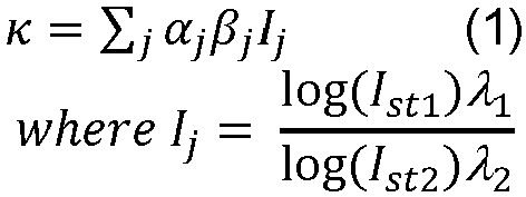
- In the correction κ, the index j runs over a set of detectors neighboring the pair (e.g., the light source 18.5 and the light detector 20.5) used to measure signal R, the factor aj is a contribution factor for detector j (e.g., detectors closer to the edge of the array. (For example detectors 20.3, 20.6, 20.9, as shown in
FIGURE 2 , receive more higher stray light compared to detectors 20.1, 20.4, 20.7) are expected to detect higher stray light intensity and hence have higher α values), the factor βj is proportional to the Euclidean distance of the detector j to the pair used to measure signal R.,. The intensities I st1 and I st2 are measured without any of the light sources 18.1-18.9 operating for a given source detector 20.1-20.9 combination. The intensity I st1 corresponds to AC components of the intensity for the red light component of stray light observed by detector j, and similarly intensity I st2 corresponds to ac component of IR intensity component of stray light observed by detector j - In other embodiments, the
electronic processor 22 is programmed to correct the red/infrared light intensity ratio based on the detected ambient light using a machine-learned (ML) model. The ML model is trained on historical oxygen saturation measurement values. For example, data can be collected for a healthy test subject having SpO2=100%, with a ground truth measurement acquired in complete darkness (e.g., placed in a dark room with no stray light present). Various intensity levels and spatial orientations of stray light can then be applied during SpO2 measurements together with measurements by the other detectors with all light sources of the set oflight sources 18 turned off. The ML model (which may, for example, be a support vector machine (SVM), a neural network, or so forth) is trained to receive these as inputs and to output κ values that correct the measurements to output the a priori known ground truth SpO2=100%. - With reference now to
FIGURE 3 , and with continuing reference toFIGURE 1 , the oxygen saturation monitor 10 optionally further includes at least one motion sensor configured to measure movement of at least one of thefirst clamp portion 14 and thesecond clamp portion 16. Theelectronic processor 22 is, in these embodiments, programmed to determine the oxygen saturation value based on the detected red light and infrared light, along with the detected movement. - The at least one motion sensor includes (i) an
accelerometer 24 configured to measure displacement of at least one of thefirst clamp portion 14 and thesecond clamp portion 16; and (ii) agyroscope 26 configured to measure rotation of at least one of the first clamp portion and the second clamp portion. As shown inFIGURE 3 , theaccelerometer 24 is disposed on (and configured to measure displacement of) thefirst clamp portion 14, and thegyroscope 26 is disposed on (and configured to measure rotation of) thesecond clamp portion 16, although the opposite arrangement may be implemented. It should be noted that since the first andsecond clamp portions clamping mechanism 17, it is expected that the first andsecond clamp portions 14, 16 (together with the clamping mechanism 17) will be displaced or rotated as a single rigid unit. - The
accelerometer 24 is configured to measure movement (for example, lateral movement) data in three dimensions (e.g., along x-, y-, and z-axes) of thefirst clamp portion 14. A first displacement value is determined by theelectronic processor 22 from the movement measured data by theaccelerometer 24. The first displacement value is determined by Equation 2:
accelerometer 24 in x-, y-, and z- directions. - The
gyroscope 26 is configured to measure movement (for example, rotational movement) data in three axes (e.g., along pitch-, roll-, and yaw-axes) of thesecond clamp portion 16. A second displacement value is determined by theelectronic processor 22 from the movement data measured by thegyroscope 26. The second displacement value is determined by Equation 3:

gyroscope 26. It will be appreciated that only movement along the roll axis of the gyroscope 26 (e.g., rotation of the finger F in a clockwise/clockwise direction) is collected, as the finger would not rotate along a pitch axis or a yaw axis of the gyroscope, which are transverse to the roll axis. In determining the oxygen saturation value, theelectronic processor 22 is programmed to determine a final displacement value by summing the first displacement value (e.g., from the data collected by theaccelerometer 24 according to Equation 2) and the second displacement value (e.g., from the data collected by thegyroscope 26 according to Equation 3). - In other embodiments, the
electronic processor 22 is programmed to determine the displacement values using a machine-learned (ML) model. The ML model is trained on historical oxygen saturation measurement values with displacement values compensated for. For example, data can be collected for a healthy test subject having SpOz=100%, with a ground truth measurement acquired with the monitor completely immobile. Various displacement and/or rotation motions can then be applied during SpO2 measurements together with measurements by the accelerometer and gyroscope. The ML model, which may for example be a SVM, a neural network, or so forth, is trained to receive these as inputs and to output motion corrections that correct the measurements to output the a priori known ground truth SpO2=100%. - It will also be appreciated that the
motion sensors monitor 10 may change any time themonitor 10 is moved or rotated, since in such a case the position and/or orientation of themonitor 10 relative to the bedside lamp or other ambient light source(s) may change. On the other hand, as long as themonitor 10 is stationary, the ambient light that is "seen" by themonitor 10 is unlikely to change rapidly. Even in the case of the bedside lamp being turned off, e.g. at lights-out, this will often be accompanied by some motion of the patient. Hence, in some contemplated embodiments, the ambient light correction κ is re-measured and re-computed relatively infrequently, e.g. at three minute intervals, but a detected movement of themonitor 10 will trigger an immediate re-measurement and re-computation of κ. - Referring back to
FIGURE 1 , the oxygen saturation monitor 10 also includes (or is controlled by) a computing device 28 (e.g., typically a workstation computer, or more generally a computer, although another form factor such as a tablet, a smartphone, and so forth is also contemplated). Theworkstation 28 comprises a computer or other electronic data processing device with typical components, such as the at least one electronic processor 22 (which can be alternatively be embedded in the clamp 12), at least one user input device (e.g., a mouse, a keyboard, a trackball, and/or the like) 30, and adisplay device 32. In some embodiments, thedisplay device 32 can be a separate component from thecomputer 28. - One or more
non-transitory storage media 34 are also provided to store data and instructions (e.g. software) that are readable and executable by thecomputing device 28 to perform oxygen saturation value measurement processes as disclosed herein, and/or executable by the workstation orother controller 18 to control the oxygen saturation monitor 10 to measure the oxygen saturation values (e.g., by determining and using the final displacement value and the corrected red/infrared light intensity ratio as described above). Thenon-transitory storage media 34 may, by way of non-limiting illustrative example, include one or more of a magnetic disk, RAID, or other magnetic storage medium; a solid-state drive, flash drive, electronically erasable read-only memory (EEROM) or other electronic memory; an optical disk or other optical storage; various combinations thereof; or so forth. Thestorage media 34 may comprise a plurality of different media, optionally of different types, and may be variously distributed. Thestorage media 34 can store instructions executable by theelectronic processor 22 to perform an oxygen saturation value determination method orprocess 100. From the final displacement value and the corrected red/infrared light intensity ratio, theelectronic processor 22 is programmed to determine the oxygen saturation value using oxygen saturation value determination method. - With reference to
FIGURE 4 , an illustrative embodiment of theoxygen saturation method 100 is diagrammatically shown as a flowchart. At 102, when theclamp 12 is affixed to the patient, theelectronic processor 22 is programmed to control the central light source/detector pair (e.g., the light source 18.5 and the light detector 20.5) to measure red and IR light, while the remaining light source/detector pairs are controlled to measure ambient light. - At 104, the
electronic processor 22 is programmed to correct the measured red and IR light data by subtracting the ambient light contribution from the measured light to generate corrected oxygen saturation values. This correction can be performed by theelectronic processor 22 usingEquation 1. In some examples, the corrected oxygen saturation values can be displayed on thedisplay device 32 of thecomputing device 28. - At 106, the
electronic processor 22 is programmed to control theaccelerometer 24 and thegyroscope 26 to measure the respective lateral movement and rotational movement data. It will be appreciated thatoperation 106 can be performed before, after, or simultaneously with operation 102 (i.e., the detection of light). - At 108, the
electronic processor 22 is programmed to compute the final displacement value from the motion data measured by theaccelerometer 24 and thegyroscope 26. With the final displacement value, the electronic processor is programmed to compute a displacement of theclamp 12 on the patient. For example, the original location of the clamp 12 (at operation 102) can be calibrated to have Cartesian coordinates of (0,0,0). The displacement coordinates can be computed as (δt1, δt2, δt3). - At 110, the
electronic processor 22 is programmed to map the displacement coordinates (δt1, δt2, δt3) to a best possible light source/light detector pair that corresponds to the same anatomical area (e.g., where theclamp 12 is attached) as originally measured. The "best" light source/light detector pair is the pair that detects the least amount of ambient light (and therefore detects the most amount of red and/or IR light) determined usingEquation 1. To do so, theelectronic processor 22 is programmed to determine new α and β values of the detectors 20.1-20.9 fromEquation 1 to measure ambient light from the mapped displacement coordinates (δt1, δt2, δt3). For example, movement of theclamp 12 relative to the patient area which the clamp is attached may cause a different weight (e.g., α and/or β) for one of the light detectors 20.1-20.9. In one example, the light source/detector pair 18.5/20.5 is used to record SpO2 values. A clock-wise rotational motion of the finger F causes a similar movement of theclamp 12, which is detected by thegyroscope 26. This detected movement can trigger theelectronic processor 22 to determine that new source/detector pair (e.g., light source/detector pair 18.8/20.8) should be that maps to corresponding same anatomical area of the finger F that is used to measure SpO2 values before motion (e.g., the portion of the finger covered by the source/detector pair 18.5/20.5). This detected movement of the finger F causes a change in the layout of the array oflight sources 18 and the array oflight detectors 20, which requires for theelectronic processor 22 to calculate new α and β values for all detectors. With the new calculations, for example, detector 20.9 will have new α and β values that is similar to what detector 20.6 had before the detected rotational motion as detector 20.9 is closer to new source/detector pair 20.8, on a similar line, all other detector weightage can be computed for each light source/detector pair 18.1-18.9/20.1-20.9. - In another example, the light source/detector pair 18.5/20.5 is again used to record SpO2 values. The α value of the detector 20.6 is high and its β value is low due to its proximity to detector 20.5. If the patient adjusts the
clamp 12 towards the wrist (e.g., by sliding the clamp along the finger F), this translational movement is detected by theaccelerometer 24. The source/detector pair 18.4/20.4 begins to cover the same anatomical area of the finger F previously covered by the source/detector pair 18.5/20.5. Theelectronic processor 22 determines the new α and β values for alldetectors 20 to determine that the source/detector pair 18.4/20.4 should be used to measure SpO2 values. - At 112, the
electronic processor 22 is programmed to use the new set of light sensors of the array oflight sources 18 to mark this new set as a new light source/detector pair 18.5/20.5 to detect red and IR light signals to determine the oxygen saturation value in the patient. - The disclosure has been described with reference to the preferred embodiments. Modifications and alterations may occur to others upon reading and understanding the preceding detailed description. The invention is defined by the appended claims.
Claims (7)
- An oxygen saturation monitor (10), comprising:a clamp (12) having opposing first (14) and second (16) clamp portions;an array of light sources (18) disposed on the first clamp portion, each light source being switchable between (i) off, (ii) emitting red light, (iii) emitting infrared light; and (iv) emitting both red light and infrared light;an array of light detectors (20) disposed on the second clamp portion facing the array of light sources, wherein each light detector of the array of light detectors is aligned to detect light emitted from a single one of the light sources and each light source is arranged to illuminate only a single one of the light detectors, thereby forming an array of light source / light detector pairs, wherein, for each light source / light detector pair, the light source corresponds to the light detector; andan electronic processor (22) programmed to:control the array of light sources (18) to emit switched red and infrared light by a single active light source (18.1-18.9) of the array of light sources with all other light sources of the array of light sources being off;detect the switched red and infrared light using the light detector (20.1-20.9) of the array of light detectors (20) that is in the light source / light detector pair for the single active light source and is therefore aligned to detect emitted light from the single active light source; anddetect ambient light using the light detectors (20.1-20.9) of the array of light detectors other than the light detector that is aligned to detect emitted light from the single active light source.
- The oxygen saturation monitor (10) of claim 1, wherein the electronic processor (22) is further programmed to:compute a red/infrared light intensity ratio for the detected switched red and infrared light;correct the red/infrared light intensity ratio based on the detected ambient light; andconvert the corrected red/infrared light intensity ratio to an oxygen saturation value.
- The oxygen saturation monitor (10) of claim 2, wherein the electronic processor (22) is programmed to correct the red/infrared light intensity ratio by operations including:calculating a correction factor (κ) from the ambient light detected by the light detectors (20.1-20.9) of the array of light detectors (20) other than the light detector that is aligned to detect emitted light from the single active light source (18.1-18.9) scaled based on Euclidean distances (β) of the respective detectors from the light detector that is aligned to detect emitted light from the single active light source; andcorrecting the red/infrared light intensity ratio by subtracting the correction factor to from the red/infrared light intensity ratio.
- The oxygen saturation monitor (10) of claim 3, wherein the correction factor is


- The oxygen saturation monitor (10) of any one of claims 1-4, wherein the electronic processor (22) is further programmed to:
correct the red/infrared light intensity ratio based on the detected ambient light using a machine-learned model. - The oxygen saturation monitor (10) of any one of claims 1-5, wherein the array of light sources (18) and the array of light detectors (20) are both arranged in a 3 X 3 matrix.
- The oxygen saturation monitor (10) of any one of claims 1-6, further including:at least one motion sensor (24, 26) configured to measure movement of at least one of the first clamp portion (14) and the second clamp portion (16); andat least one electronic processor (22) programmed to determine an oxygen saturation value based at least on red light and infrared light output by the array of light sources and measured by the array of light detectors and further based on the detected movement.
Applications Claiming Priority (2)
| Application Number | Priority Date | Filing Date | Title |
|---|---|---|---|
| US201962814890P | 2019-03-07 | 2019-03-07 | |
| PCT/EP2020/055706 WO2020178346A1 (en) | 2019-03-07 | 2020-03-04 | Device for monitoring oxygen saturation levels in clinical conditions |
Publications (2)
| Publication Number | Publication Date |
|---|---|
| EP3934536A1 EP3934536A1 (en) | 2022-01-12 |
| EP3934536B1 true EP3934536B1 (en) | 2025-01-29 |
Family
ID=69784416
Family Applications (1)
| Application Number | Title | Priority Date | Filing Date |
|---|---|---|---|
| EP20710455.5A Active EP3934536B1 (en) | 2019-03-07 | 2020-03-04 | Device for monitoring oxygen saturation levels in clinical conditions |
Country Status (5)
| Country | Link |
|---|---|
| US (1) | US12502106B2 (en) |
| EP (1) | EP3934536B1 (en) |
| JP (1) | JP7474778B2 (en) |
| CN (1) | CN113543705B (en) |
| WO (1) | WO2020178346A1 (en) |
Families Citing this family (2)
| Publication number | Priority date | Publication date | Assignee | Title |
|---|---|---|---|---|
| EP4342374A4 (en) | 2021-08-11 | 2024-08-28 | Samsung Electronics Co., Ltd. | ELECTRONIC OXYGEN SATURATION CORRECTION DEVICE AND ASSOCIATED CONTROL METHOD |
| CN118592948B (en) * | 2024-05-29 | 2025-02-07 | 河北地质大学 | Brain blood oxygen monitoring method and terminal, brain blood oxygen monitoring system |
Family Cites Families (28)
| Publication number | Priority date | Publication date | Assignee | Title |
|---|---|---|---|---|
| US5368224A (en) | 1992-10-23 | 1994-11-29 | Nellcor Incorporated | Method for reducing ambient noise effects in electronic monitoring instruments |
| DE19537646C2 (en) * | 1995-10-10 | 1998-09-17 | Hewlett Packard Gmbh | Method and device for detecting falsified measurement values in pulse oximetry for measuring oxygen saturation |
| EP0892617B1 (en) * | 1996-04-01 | 2001-09-19 | Linde Medical Sensors AG | Detection of parasitic signals during pulsoxymetric measurement |
| JP2003102694A (en) * | 2001-09-28 | 2003-04-08 | Kenji Sunakawa | Heart rate measuring instrument |
| US20060094941A1 (en) * | 2004-10-29 | 2006-05-04 | Ok-Kyung Cho | Optical measurement apparatus and blood sugar level measuring apparatus using the same |
| ATE535184T1 (en) * | 2004-12-14 | 2011-12-15 | Koninkl Philips Electronics Nv | INTEGRATED PULSE OXIMETER |
| US7957780B2 (en) * | 2005-03-01 | 2011-06-07 | Masimo Laboratories, Inc. | Physiological parameter confidence measure |
| US7641614B2 (en) | 2005-08-22 | 2010-01-05 | Massachusetts Institute Of Technology | Wearable blood pressure sensor and method of calibration |
| US8684900B2 (en) * | 2006-05-16 | 2014-04-01 | Bao Tran | Health monitoring appliance |
| ES2525795T3 (en) * | 2006-05-30 | 2014-12-30 | University Of Massachusetts | Tissue oxygenation measurement |
| US11607152B2 (en) * | 2007-06-12 | 2023-03-21 | Sotera Wireless, Inc. | Optical sensors for use in vital sign monitoring |
| US8251903B2 (en) | 2007-10-25 | 2012-08-28 | Valencell, Inc. | Noninvasive physiological analysis using excitation-sensor modules and related devices and methods |
| JP2009254522A (en) * | 2008-04-15 | 2009-11-05 | Sharp Corp | Optical biological information measuring instrument, and light emitting/receiving unit for measuring biological information |
| US20090326347A1 (en) | 2008-06-30 | 2009-12-31 | Bennett Scharf | Synchronous Light Detection Utilizing CMOS/CCD Sensors For Oximetry Sensing |
| JP5756752B2 (en) * | 2008-07-03 | 2015-07-29 | セルカコール・ラボラトリーズ・インコーポレイテッドCercacor Laboratories, Inc. | Sensor |
| JP2010200911A (en) * | 2009-03-02 | 2010-09-16 | Yu Sys Corp | Apparatus for measuring blood component |
| US10085657B2 (en) | 2009-06-17 | 2018-10-02 | Sotera Wireless, Inc. | Body-worn pulse oximeter |
| US8761853B2 (en) * | 2011-01-20 | 2014-06-24 | Nitto Denko Corporation | Devices and methods for non-invasive optical physiological measurements |
| US9241676B2 (en) * | 2012-05-31 | 2016-01-26 | Covidien Lp | Methods and systems for power optimization in a medical device |
| US20140235977A1 (en) | 2013-02-20 | 2014-08-21 | Perminova Inc. | Necklace-shaped physiological monitor |
| FI126338B (en) * | 2013-05-15 | 2016-10-14 | Pulseon Oy | Portable heart rate monitor |
| CN203290911U (en) | 2013-05-17 | 2013-11-20 | 武汉远光瑞康科技有限公司 | Reflection-type multi-sensor-array blood oxygen detection device |
| CN103705249A (en) * | 2013-12-27 | 2014-04-09 | 中国医学科学院生物医学工程研究所 | Pulse and blood oxygen probe with electromagnetic shielding function |
| EP3261528B1 (en) | 2015-02-23 | 2018-10-03 | Koninklijke Philips N.V. | Multi-state clip-on fixation device for pulse oximeter |
| CN104887246B (en) * | 2015-06-25 | 2017-09-15 | 广州视源电子科技股份有限公司 | Blood oxygen measuring method and blood oxygen measuring device |
| US9866941B2 (en) | 2015-10-20 | 2018-01-09 | Bragi GmbH | Multi-point multiple sensor array for data sensing and processing system and method |
| EP3380012A1 (en) * | 2015-11-24 | 2018-10-03 | Koninklijke Philips N.V. | Pulse oximeter using disposable multi-material stretch bandage |
| JP2019010437A (en) * | 2017-06-30 | 2019-01-24 | ヤマハ株式会社 | Determination device and determination method |
-
2020
- 2020-03-04 US US17/436,804 patent/US12502106B2/en active Active
- 2020-03-04 EP EP20710455.5A patent/EP3934536B1/en active Active
- 2020-03-04 CN CN202080019369.0A patent/CN113543705B/en active Active
- 2020-03-04 JP JP2021551896A patent/JP7474778B2/en active Active
- 2020-03-04 WO PCT/EP2020/055706 patent/WO2020178346A1/en not_active Ceased
Also Published As
| Publication number | Publication date |
|---|---|
| JP2022522370A (en) | 2022-04-18 |
| US12502106B2 (en) | 2025-12-23 |
| CN113543705B (en) | 2024-11-26 |
| US20220167888A1 (en) | 2022-06-02 |
| EP3934536A1 (en) | 2022-01-12 |
| CN113543705A (en) | 2021-10-22 |
| JP7474778B2 (en) | 2024-04-25 |
| WO2020178346A1 (en) | 2020-09-10 |
Similar Documents
| Publication | Publication Date | Title |
|---|---|---|
| US7890153B2 (en) | System and method for mitigating interference in pulse oximetry | |
| US11202582B2 (en) | Device for use in blood oxygen saturation measurement | |
| US10299709B2 (en) | Robust fractional saturation determination | |
| US6711425B1 (en) | Pulse oximeter with calibration stabilization | |
| US9924896B2 (en) | Device, system and method for determining the concentration of a substance in the blood of a subject | |
| US8109882B2 (en) | System and method for venous pulsation detection using near infrared wavelengths | |
| US10314500B2 (en) | Transcutaneous photoplethysmography | |
| US5842979A (en) | Method and apparatus for improved photoplethysmographic monitoring of oxyhemoglobin, deoxyhemoglobin, carboxyhemoglobin and methemoglobin | |
| US9304202B2 (en) | Multiuse optical sensor | |
| US11304634B2 (en) | Non-invasive blood glucose sensor | |
| US20020133068A1 (en) | Compensation of human variability in pulse oximetry | |
| JP6878312B2 (en) | Photoelectric volumetric pulse wave recording device | |
| US20120259190A1 (en) | Detection of oximetry sensor sites based on waveform characteristics | |
| US20190343432A1 (en) | Non-invasive hemoglobin and white blood cell sensors | |
| US20090326347A1 (en) | Synchronous Light Detection Utilizing CMOS/CCD Sensors For Oximetry Sensing | |
| US8588879B2 (en) | Motion compensation in a sensor | |
| EP3934536B1 (en) | Device for monitoring oxygen saturation levels in clinical conditions | |
| WO2020203020A1 (en) | Biological information measurer and biological information measurement method using same | |
| US20210330223A1 (en) | Sensor verification through forward voltage measurements | |
| US20090171172A1 (en) | Method and system for pulse gating | |
| EP4674346A1 (en) | Photoplethysmography sensor | |
| KR20170010714A (en) | Method, device and non-transitory computer-readable recording medium for measuring photoplethysmography signal |
Legal Events
| Date | Code | Title | Description |
|---|---|---|---|
| STAA | Information on the status of an ep patent application or granted ep patent |
Free format text: STATUS: UNKNOWN |
|
| STAA | Information on the status of an ep patent application or granted ep patent |
Free format text: STATUS: THE INTERNATIONAL PUBLICATION HAS BEEN MADE |
|
| PUAI | Public reference made under article 153(3) epc to a published international application that has entered the european phase |
Free format text: ORIGINAL CODE: 0009012 |
|
| STAA | Information on the status of an ep patent application or granted ep patent |
Free format text: STATUS: REQUEST FOR EXAMINATION WAS MADE |
|
| 17P | Request for examination filed |
Effective date: 20211007 |
|
| AK | Designated contracting states |
Kind code of ref document: A1 Designated state(s): AL AT BE BG CH CY CZ DE DK EE ES FI FR GB GR HR HU IE IS IT LI LT LU LV MC MK MT NL NO PL PT RO RS SE SI SK SM TR |
|
| DAV | Request for validation of the european patent (deleted) | ||
| DAX | Request for extension of the european patent (deleted) | ||
| GRAP | Despatch of communication of intention to grant a patent |
Free format text: ORIGINAL CODE: EPIDOSNIGR1 |
|
| STAA | Information on the status of an ep patent application or granted ep patent |
Free format text: STATUS: GRANT OF PATENT IS INTENDED |
|
| INTG | Intention to grant announced |
Effective date: 20240821 |
|
| RIN1 | Information on inventor provided before grant (corrected) |
Inventor name: PATIL, MERU ADAGOUDA |
|
| GRAS | Grant fee paid |
Free format text: ORIGINAL CODE: EPIDOSNIGR3 |
|
| GRAA | (expected) grant |
Free format text: ORIGINAL CODE: 0009210 |
|
| STAA | Information on the status of an ep patent application or granted ep patent |
Free format text: STATUS: THE PATENT HAS BEEN GRANTED |
|
| AK | Designated contracting states |
Kind code of ref document: B1 Designated state(s): AL AT BE BG CH CY CZ DE DK EE ES FI FR GB GR HR HU IE IS IT LI LT LU LV MC MK MT NL NO PL PT RO RS SE SI SK SM TR |
|
| REG | Reference to a national code |
Ref country code: GB Ref legal event code: FG4D |
|
| REG | Reference to a national code |
Ref country code: CH Ref legal event code: EP |
|
| REG | Reference to a national code |
Ref country code: DE Ref legal event code: R096 Ref document number: 602020045485 Country of ref document: DE |
|
| REG | Reference to a national code |
Ref country code: IE Ref legal event code: FG4D Ref country code: DE Ref legal event code: R084 Ref document number: 602020045485 Country of ref document: DE |
|
| PGFP | Annual fee paid to national office [announced via postgrant information from national office to epo] |
Ref country code: DE Payment date: 20250327 Year of fee payment: 6 |
|
| REG | Reference to a national code |
Ref country code: GB Ref legal event code: 746 Effective date: 20250331 |
|
| PGFP | Annual fee paid to national office [announced via postgrant information from national office to epo] |
Ref country code: GB Payment date: 20250325 Year of fee payment: 6 |
|
| REG | Reference to a national code |
Ref country code: NL Ref legal event code: MP Effective date: 20250129 |
|
| PG25 | Lapsed in a contracting state [announced via postgrant information from national office to epo] |
Ref country code: NL Free format text: LAPSE BECAUSE OF FAILURE TO SUBMIT A TRANSLATION OF THE DESCRIPTION OR TO PAY THE FEE WITHIN THE PRESCRIBED TIME-LIMIT Effective date: 20250129 |
|
| PG25 | Lapsed in a contracting state [announced via postgrant information from national office to epo] |
Ref country code: RS Free format text: LAPSE BECAUSE OF FAILURE TO SUBMIT A TRANSLATION OF THE DESCRIPTION OR TO PAY THE FEE WITHIN THE PRESCRIBED TIME-LIMIT Effective date: 20250429 |
|
| PG25 | Lapsed in a contracting state [announced via postgrant information from national office to epo] |
Ref country code: FI Free format text: LAPSE BECAUSE OF FAILURE TO SUBMIT A TRANSLATION OF THE DESCRIPTION OR TO PAY THE FEE WITHIN THE PRESCRIBED TIME-LIMIT Effective date: 20250129 |
|
| PG25 | Lapsed in a contracting state [announced via postgrant information from national office to epo] |
Ref country code: PL Free format text: LAPSE BECAUSE OF FAILURE TO SUBMIT A TRANSLATION OF THE DESCRIPTION OR TO PAY THE FEE WITHIN THE PRESCRIBED TIME-LIMIT Effective date: 20250129 |
|
| PG25 | Lapsed in a contracting state [announced via postgrant information from national office to epo] |
Ref country code: ES Free format text: LAPSE BECAUSE OF FAILURE TO SUBMIT A TRANSLATION OF THE DESCRIPTION OR TO PAY THE FEE WITHIN THE PRESCRIBED TIME-LIMIT Effective date: 20250129 |
|
| REG | Reference to a national code |
Ref country code: LT Ref legal event code: MG9D |
|
| PG25 | Lapsed in a contracting state [announced via postgrant information from national office to epo] |
Ref country code: NO Free format text: LAPSE BECAUSE OF FAILURE TO SUBMIT A TRANSLATION OF THE DESCRIPTION OR TO PAY THE FEE WITHIN THE PRESCRIBED TIME-LIMIT Effective date: 20250429 Ref country code: IS Free format text: LAPSE BECAUSE OF FAILURE TO SUBMIT A TRANSLATION OF THE DESCRIPTION OR TO PAY THE FEE WITHIN THE PRESCRIBED TIME-LIMIT Effective date: 20250529 |
|
| REG | Reference to a national code |
Ref country code: AT Ref legal event code: MK05 Ref document number: 1762638 Country of ref document: AT Kind code of ref document: T Effective date: 20250129 |
|
| PG25 | Lapsed in a contracting state [announced via postgrant information from national office to epo] |
Ref country code: HR Free format text: LAPSE BECAUSE OF FAILURE TO SUBMIT A TRANSLATION OF THE DESCRIPTION OR TO PAY THE FEE WITHIN THE PRESCRIBED TIME-LIMIT Effective date: 20250129 |
|
| PG25 | Lapsed in a contracting state [announced via postgrant information from national office to epo] |
Ref country code: PT Free format text: LAPSE BECAUSE OF FAILURE TO SUBMIT A TRANSLATION OF THE DESCRIPTION OR TO PAY THE FEE WITHIN THE PRESCRIBED TIME-LIMIT Effective date: 20250529 Ref country code: LV Free format text: LAPSE BECAUSE OF FAILURE TO SUBMIT A TRANSLATION OF THE DESCRIPTION OR TO PAY THE FEE WITHIN THE PRESCRIBED TIME-LIMIT Effective date: 20250129 |
|
| PG25 | Lapsed in a contracting state [announced via postgrant information from national office to epo] |
Ref country code: GR Free format text: LAPSE BECAUSE OF FAILURE TO SUBMIT A TRANSLATION OF THE DESCRIPTION OR TO PAY THE FEE WITHIN THE PRESCRIBED TIME-LIMIT Effective date: 20250430 Ref country code: BG Free format text: LAPSE BECAUSE OF FAILURE TO SUBMIT A TRANSLATION OF THE DESCRIPTION OR TO PAY THE FEE WITHIN THE PRESCRIBED TIME-LIMIT Effective date: 20250129 |
|
| PG25 | Lapsed in a contracting state [announced via postgrant information from national office to epo] |
Ref country code: AT Free format text: LAPSE BECAUSE OF FAILURE TO SUBMIT A TRANSLATION OF THE DESCRIPTION OR TO PAY THE FEE WITHIN THE PRESCRIBED TIME-LIMIT Effective date: 20250129 |
|
| PG25 | Lapsed in a contracting state [announced via postgrant information from national office to epo] |
Ref country code: SE Free format text: LAPSE BECAUSE OF FAILURE TO SUBMIT A TRANSLATION OF THE DESCRIPTION OR TO PAY THE FEE WITHIN THE PRESCRIBED TIME-LIMIT Effective date: 20250129 |
|
| PG25 | Lapsed in a contracting state [announced via postgrant information from national office to epo] |
Ref country code: SM Free format text: LAPSE BECAUSE OF FAILURE TO SUBMIT A TRANSLATION OF THE DESCRIPTION OR TO PAY THE FEE WITHIN THE PRESCRIBED TIME-LIMIT Effective date: 20250129 |
|
| PG25 | Lapsed in a contracting state [announced via postgrant information from national office to epo] |
Ref country code: DK Free format text: LAPSE BECAUSE OF FAILURE TO SUBMIT A TRANSLATION OF THE DESCRIPTION OR TO PAY THE FEE WITHIN THE PRESCRIBED TIME-LIMIT Effective date: 20250129 |
|
| PG25 | Lapsed in a contracting state [announced via postgrant information from national office to epo] |
Ref country code: MC Free format text: LAPSE BECAUSE OF FAILURE TO SUBMIT A TRANSLATION OF THE DESCRIPTION OR TO PAY THE FEE WITHIN THE PRESCRIBED TIME-LIMIT Effective date: 20250129 |
|
| PG25 | Lapsed in a contracting state [announced via postgrant information from national office to epo] |
Ref country code: IT Free format text: LAPSE BECAUSE OF FAILURE TO SUBMIT A TRANSLATION OF THE DESCRIPTION OR TO PAY THE FEE WITHIN THE PRESCRIBED TIME-LIMIT Effective date: 20250129 |
|
| PG25 | Lapsed in a contracting state [announced via postgrant information from national office to epo] |
Ref country code: EE Free format text: LAPSE BECAUSE OF FAILURE TO SUBMIT A TRANSLATION OF THE DESCRIPTION OR TO PAY THE FEE WITHIN THE PRESCRIBED TIME-LIMIT Effective date: 20250129 Ref country code: CZ Free format text: LAPSE BECAUSE OF FAILURE TO SUBMIT A TRANSLATION OF THE DESCRIPTION OR TO PAY THE FEE WITHIN THE PRESCRIBED TIME-LIMIT Effective date: 20250129 |
|
| PG25 | Lapsed in a contracting state [announced via postgrant information from national office to epo] |
Ref country code: RO Free format text: LAPSE BECAUSE OF FAILURE TO SUBMIT A TRANSLATION OF THE DESCRIPTION OR TO PAY THE FEE WITHIN THE PRESCRIBED TIME-LIMIT Effective date: 20250129 |
|
| REG | Reference to a national code |
Ref country code: CH Ref legal event code: H13 Free format text: ST27 STATUS EVENT CODE: U-0-0-H10-H13 (AS PROVIDED BY THE NATIONAL OFFICE) Effective date: 20251024 |
|
| PG25 | Lapsed in a contracting state [announced via postgrant information from national office to epo] |
Ref country code: SK Free format text: LAPSE BECAUSE OF FAILURE TO SUBMIT A TRANSLATION OF THE DESCRIPTION OR TO PAY THE FEE WITHIN THE PRESCRIBED TIME-LIMIT Effective date: 20250129 |
|
| REG | Reference to a national code |
Ref country code: DE Ref legal event code: R097 Ref document number: 602020045485 Country of ref document: DE |
|
| PG25 | Lapsed in a contracting state [announced via postgrant information from national office to epo] |
Ref country code: LU Free format text: LAPSE BECAUSE OF NON-PAYMENT OF DUE FEES Effective date: 20250304 |
|
| PLBE | No opposition filed within time limit |
Free format text: ORIGINAL CODE: 0009261 |
|
| STAA | Information on the status of an ep patent application or granted ep patent |
Free format text: STATUS: NO OPPOSITION FILED WITHIN TIME LIMIT |
|
| REG | Reference to a national code |
Ref country code: BE Ref legal event code: MM Effective date: 20250331 |
|
| 26N | No opposition filed |
Effective date: 20251030 |
|
| PG25 | Lapsed in a contracting state [announced via postgrant information from national office to epo] |
Ref country code: FR Free format text: LAPSE BECAUSE OF NON-PAYMENT OF DUE FEES Effective date: 20250329 |
|
| PG25 | Lapsed in a contracting state [announced via postgrant information from national office to epo] |
Ref country code: BE Free format text: LAPSE BECAUSE OF NON-PAYMENT OF DUE FEES Effective date: 20250331 |
|
| PG25 | Lapsed in a contracting state [announced via postgrant information from national office to epo] |
Ref country code: CH Free format text: LAPSE BECAUSE OF NON-PAYMENT OF DUE FEES Effective date: 20250331 |
|
| PG25 | Lapsed in a contracting state [announced via postgrant information from national office to epo] |
Ref country code: IE Free format text: LAPSE BECAUSE OF NON-PAYMENT OF DUE FEES Effective date: 20250304 |