EP3041507B1 - Nucleic acids encoding human antibodies to sialyl-lewis a - Google Patents
Nucleic acids encoding human antibodies to sialyl-lewis a Download PDFInfo
- Publication number
- EP3041507B1 EP3041507B1 EP14852746.8A EP14852746A EP3041507B1 EP 3041507 B1 EP3041507 B1 EP 3041507B1 EP 14852746 A EP14852746 A EP 14852746A EP 3041507 B1 EP3041507 B1 EP 3041507B1
- Authority
- EP
- European Patent Office
- Prior art keywords
- antibody
- sle
- seq
- teaching
- cells
- Prior art date
- Legal status (The legal status is an assumption and is not a legal conclusion. Google has not performed a legal analysis and makes no representation as to the accuracy of the status listed.)
- Active
Links
Images


Classifications
-
- C—CHEMISTRY; METALLURGY
- C07—ORGANIC CHEMISTRY
- C07K—PEPTIDES
- C07K16/00—Immunoglobulins [IGs], e.g. monoclonal or polyclonal antibodies
- C07K16/18—Immunoglobulins [IGs], e.g. monoclonal or polyclonal antibodies against material from animals or humans
- C07K16/28—Immunoglobulins [IGs], e.g. monoclonal or polyclonal antibodies against material from animals or humans against receptors, cell surface antigens or cell surface determinants
- C07K16/2896—Immunoglobulins [IGs], e.g. monoclonal or polyclonal antibodies against material from animals or humans against receptors, cell surface antigens or cell surface determinants against molecules with a "CD"-designation, not provided for elsewhere
-
- A—HUMAN NECESSITIES
- A61—MEDICAL OR VETERINARY SCIENCE; HYGIENE
- A61K—PREPARATIONS FOR MEDICAL, DENTAL OR TOILETRY PURPOSES
- A61K31/00—Medicinal preparations containing organic active ingredients
- A61K31/33—Heterocyclic compounds
- A61K31/335—Heterocyclic compounds having oxygen as the only ring hetero atom, e.g. fungichromin
- A61K31/337—Heterocyclic compounds having oxygen as the only ring hetero atom, e.g. fungichromin having four-membered rings, e.g. taxol
-
- A—HUMAN NECESSITIES
- A61—MEDICAL OR VETERINARY SCIENCE; HYGIENE
- A61K—PREPARATIONS FOR MEDICAL, DENTAL OR TOILETRY PURPOSES
- A61K39/00—Medicinal preparations containing antigens or antibodies
- A61K39/0005—Vertebrate antigens
- A61K39/0011—Cancer antigens
-
- A—HUMAN NECESSITIES
- A61—MEDICAL OR VETERINARY SCIENCE; HYGIENE
- A61K—PREPARATIONS FOR MEDICAL, DENTAL OR TOILETRY PURPOSES
- A61K39/00—Medicinal preparations containing antigens or antibodies
- A61K39/395—Antibodies; Immunoglobulins; Immune serum, e.g. antilymphocytic serum
- A61K39/39533—Antibodies; Immunoglobulins; Immune serum, e.g. antilymphocytic serum against materials from animals
- A61K39/39558—Antibodies; Immunoglobulins; Immune serum, e.g. antilymphocytic serum against materials from animals against tumor tissues, cells, antigens
-
- A—HUMAN NECESSITIES
- A61—MEDICAL OR VETERINARY SCIENCE; HYGIENE
- A61K—PREPARATIONS FOR MEDICAL, DENTAL OR TOILETRY PURPOSES
- A61K45/00—Medicinal preparations containing active ingredients not provided for in groups A61K31/00 - A61K41/00
- A61K45/06—Mixtures of active ingredients without chemical characterisation, e.g. antiphlogistics and cardiaca
-
- A—HUMAN NECESSITIES
- A61—MEDICAL OR VETERINARY SCIENCE; HYGIENE
- A61K—PREPARATIONS FOR MEDICAL, DENTAL OR TOILETRY PURPOSES
- A61K51/00—Preparations containing radioactive substances for use in therapy or testing in vivo
- A61K51/02—Preparations containing radioactive substances for use in therapy or testing in vivo characterised by the carrier, i.e. characterised by the agent or material covalently linked or complexing the radioactive nucleus
- A61K51/04—Organic compounds
- A61K51/08—Peptides, e.g. proteins, carriers being peptides, polyamino acids, proteins
- A61K51/10—Antibodies or immunoglobulins; Fragments thereof, the carrier being an antibody, an immunoglobulin or a fragment thereof, e.g. a camelised human single domain antibody or the Fc fragment of an antibody
- A61K51/1027—Antibodies or immunoglobulins; Fragments thereof, the carrier being an antibody, an immunoglobulin or a fragment thereof, e.g. a camelised human single domain antibody or the Fc fragment of an antibody against receptors, cell-surface antigens or cell-surface determinants
-
- A—HUMAN NECESSITIES
- A61—MEDICAL OR VETERINARY SCIENCE; HYGIENE
- A61K—PREPARATIONS FOR MEDICAL, DENTAL OR TOILETRY PURPOSES
- A61K51/00—Preparations containing radioactive substances for use in therapy or testing in vivo
- A61K51/02—Preparations containing radioactive substances for use in therapy or testing in vivo characterised by the carrier, i.e. characterised by the agent or material covalently linked or complexing the radioactive nucleus
- A61K51/04—Organic compounds
- A61K51/08—Peptides, e.g. proteins, carriers being peptides, polyamino acids, proteins
- A61K51/10—Antibodies or immunoglobulins; Fragments thereof, the carrier being an antibody, an immunoglobulin or a fragment thereof, e.g. a camelised human single domain antibody or the Fc fragment of an antibody
- A61K51/1045—Antibodies or immunoglobulins; Fragments thereof, the carrier being an antibody, an immunoglobulin or a fragment thereof, e.g. a camelised human single domain antibody or the Fc fragment of an antibody against animal or human tumor cells or tumor cell determinants
- A61K51/1057—Antibodies or immunoglobulins; Fragments thereof, the carrier being an antibody, an immunoglobulin or a fragment thereof, e.g. a camelised human single domain antibody or the Fc fragment of an antibody against animal or human tumor cells or tumor cell determinants the tumor cell being from liver or pancreas
-
- A—HUMAN NECESSITIES
- A61—MEDICAL OR VETERINARY SCIENCE; HYGIENE
- A61K—PREPARATIONS FOR MEDICAL, DENTAL OR TOILETRY PURPOSES
- A61K51/00—Preparations containing radioactive substances for use in therapy or testing in vivo
- A61K51/02—Preparations containing radioactive substances for use in therapy or testing in vivo characterised by the carrier, i.e. characterised by the agent or material covalently linked or complexing the radioactive nucleus
- A61K51/04—Organic compounds
- A61K51/08—Peptides, e.g. proteins, carriers being peptides, polyamino acids, proteins
- A61K51/10—Antibodies or immunoglobulins; Fragments thereof, the carrier being an antibody, an immunoglobulin or a fragment thereof, e.g. a camelised human single domain antibody or the Fc fragment of an antibody
- A61K51/1093—Antibodies or immunoglobulins; Fragments thereof, the carrier being an antibody, an immunoglobulin or a fragment thereof, e.g. a camelised human single domain antibody or the Fc fragment of an antibody conjugates with carriers being antibodies
-
- A—HUMAN NECESSITIES
- A61—MEDICAL OR VETERINARY SCIENCE; HYGIENE
- A61P—SPECIFIC THERAPEUTIC ACTIVITY OF CHEMICAL COMPOUNDS OR MEDICINAL PREPARATIONS
- A61P35/00—Antineoplastic agents
-
- A—HUMAN NECESSITIES
- A61—MEDICAL OR VETERINARY SCIENCE; HYGIENE
- A61P—SPECIFIC THERAPEUTIC ACTIVITY OF CHEMICAL COMPOUNDS OR MEDICINAL PREPARATIONS
- A61P37/00—Drugs for immunological or allergic disorders
- A61P37/02—Immunomodulators
- A61P37/04—Immunostimulants
-
- A—HUMAN NECESSITIES
- A61—MEDICAL OR VETERINARY SCIENCE; HYGIENE
- A61P—SPECIFIC THERAPEUTIC ACTIVITY OF CHEMICAL COMPOUNDS OR MEDICINAL PREPARATIONS
- A61P43/00—Drugs for specific purposes, not provided for in groups A61P1/00-A61P41/00
-
- C—CHEMISTRY; METALLURGY
- C07—ORGANIC CHEMISTRY
- C07K—PEPTIDES
- C07K16/00—Immunoglobulins [IGs], e.g. monoclonal or polyclonal antibodies
- C07K16/18—Immunoglobulins [IGs], e.g. monoclonal or polyclonal antibodies against material from animals or humans
- C07K16/28—Immunoglobulins [IGs], e.g. monoclonal or polyclonal antibodies against material from animals or humans against receptors, cell surface antigens or cell surface determinants
- C07K16/30—Immunoglobulins [IGs], e.g. monoclonal or polyclonal antibodies against material from animals or humans against receptors, cell surface antigens or cell surface determinants from tumour cells
- C07K16/3076—Immunoglobulins [IGs], e.g. monoclonal or polyclonal antibodies against material from animals or humans against receptors, cell surface antigens or cell surface determinants from tumour cells against structure-related tumour-associated moieties
-
- G01N33/57525—
-
- G—PHYSICS
- G01—MEASURING; TESTING
- G01N—INVESTIGATING OR ANALYSING MATERIALS BY DETERMINING THEIR CHEMICAL OR PHYSICAL PROPERTIES
- G01N33/00—Investigating or analysing materials by specific methods not covered by groups G01N1/00 - G01N31/00
- G01N33/48—Biological material, e.g. blood, urine; Haemocytometers
- G01N33/50—Chemical analysis of biological material, e.g. blood, urine; Testing involving biospecific ligand binding methods; Immunological testing
- G01N33/53—Immunoassay; Biospecific binding assay; Materials therefor
- G01N33/577—Immunoassay; Biospecific binding assay; Materials therefor involving monoclonal antibodies binding reaction mechanisms characterised by the use of monoclonal antibodies; monoclonal antibodies per se are classified with their corresponding antigens
-
- A—HUMAN NECESSITIES
- A61—MEDICAL OR VETERINARY SCIENCE; HYGIENE
- A61K—PREPARATIONS FOR MEDICAL, DENTAL OR TOILETRY PURPOSES
- A61K39/00—Medicinal preparations containing antigens or antibodies
- A61K2039/505—Medicinal preparations containing antigens or antibodies comprising antibodies
-
- A—HUMAN NECESSITIES
- A61—MEDICAL OR VETERINARY SCIENCE; HYGIENE
- A61K—PREPARATIONS FOR MEDICAL, DENTAL OR TOILETRY PURPOSES
- A61K39/00—Medicinal preparations containing antigens or antibodies
- A61K2039/60—Medicinal preparations containing antigens or antibodies characteristics by the carrier linked to the antigen
- A61K2039/6031—Proteins
- A61K2039/6081—Albumin; Keyhole limpet haemocyanin [KLH]
-
- C—CHEMISTRY; METALLURGY
- C07—ORGANIC CHEMISTRY
- C07K—PEPTIDES
- C07K2317/00—Immunoglobulins specific features
- C07K2317/20—Immunoglobulins specific features characterized by taxonomic origin
- C07K2317/21—Immunoglobulins specific features characterized by taxonomic origin from primates, e.g. man
-
- C—CHEMISTRY; METALLURGY
- C07—ORGANIC CHEMISTRY
- C07K—PEPTIDES
- C07K2317/00—Immunoglobulins specific features
- C07K2317/30—Immunoglobulins specific features characterized by aspects of specificity or valency
- C07K2317/33—Crossreactivity, e.g. for species or epitope, or lack of said crossreactivity
-
- C—CHEMISTRY; METALLURGY
- C07—ORGANIC CHEMISTRY
- C07K—PEPTIDES
- C07K2317/00—Immunoglobulins specific features
- C07K2317/50—Immunoglobulins specific features characterized by immunoglobulin fragments
- C07K2317/56—Immunoglobulins specific features characterized by immunoglobulin fragments variable (Fv) region, i.e. VH and/or VL
-
- C—CHEMISTRY; METALLURGY
- C07—ORGANIC CHEMISTRY
- C07K—PEPTIDES
- C07K2317/00—Immunoglobulins specific features
- C07K2317/60—Immunoglobulins specific features characterized by non-natural combinations of immunoglobulin fragments
- C07K2317/62—Immunoglobulins specific features characterized by non-natural combinations of immunoglobulin fragments comprising only variable region components
- C07K2317/626—Diabody or triabody
-
- C—CHEMISTRY; METALLURGY
- C07—ORGANIC CHEMISTRY
- C07K—PEPTIDES
- C07K2317/00—Immunoglobulins specific features
- C07K2317/70—Immunoglobulins specific features characterized by effect upon binding to a cell or to an antigen
- C07K2317/73—Inducing cell death, e.g. apoptosis, necrosis or inhibition of cell proliferation
- C07K2317/732—Antibody-dependent cellular cytotoxicity [ADCC]
-
- C—CHEMISTRY; METALLURGY
- C07—ORGANIC CHEMISTRY
- C07K—PEPTIDES
- C07K2317/00—Immunoglobulins specific features
- C07K2317/70—Immunoglobulins specific features characterized by effect upon binding to a cell or to an antigen
- C07K2317/73—Inducing cell death, e.g. apoptosis, necrosis or inhibition of cell proliferation
- C07K2317/734—Complement-dependent cytotoxicity [CDC]
-
- C—CHEMISTRY; METALLURGY
- C07—ORGANIC CHEMISTRY
- C07K—PEPTIDES
- C07K2317/00—Immunoglobulins specific features
- C07K2317/70—Immunoglobulins specific features characterized by effect upon binding to a cell or to an antigen
- C07K2317/77—Internalization into the cell
-
- C—CHEMISTRY; METALLURGY
- C07—ORGANIC CHEMISTRY
- C07K—PEPTIDES
- C07K2317/00—Immunoglobulins specific features
- C07K2317/90—Immunoglobulins specific features characterized by (pharmaco)kinetic aspects or by stability of the immunoglobulin
- C07K2317/92—Affinity (KD), association rate (Ka), dissociation rate (Kd) or EC50 value
Definitions
- the present teaching relates generally to antibodies directed against Sialyl-Lewis a (sLe a ), and more specifically to polynucleotides encoding anti-sLe a antibodies and the corresponding encoded antibodies or fragments thereof.
- Pancreatic carcinoma is one of the most aggressive adenocarcinomas and is often associated with a poor prognosis. Pancreatic carcinoma ranks as the fourth leading cause of cancer mortality. Despite advances in the screening for different carcinomas, the reliability of detecting malignant lesions stemming from the pancreas remains poor.
- Positron emission tomography utilizing fluorodeoxyglucase (FDG-PET) has been indicated for the detection and staging of pancreatic cancer.
- FDG-PET is insensitive to differentiating pancreatitis from malignancy and remains problematic in staging small primary lesions ( ⁇ 7mm) and liver metastases ( ⁇ 1 cm).
- One diagnostic screening method used to monitor the state of pancreatic ductal adenocarcinoma (PDAC) patients includes detecting elevated levels of circulating sLe a antigen in sera. Patients with > 37 U/ml of circulating sLe a antigen indicates cancer recurrence. However, development of alternative diagnostic tools that utilize such tumor specific carbohydrates has been slow.
- Antibodies directed against tumor specific carbohydrates may be useful candidates in this cancer treatment.
- many tumor-restricted monoclonal antibodies resulting from immunization of mice with human cancer cells have been shown to be directed against carbohydrate antigens expressed at the cell surface as glycolipids or glycoproteins.
- the carbohydrate sLe a has been shown to be expressed on tumors of the gastrointestinal tract. Expression of sLe a has also been shown to impact metastatic potential and correlates with increased metastatic potential in human colon cancer and pancreatic adenocarcinoma.
- compositions for producing antibodies or functional fragments thereof that bind sLe a include an isolated polynucleotide encoding an antibody or a functional fragment thereof, wherein the antibody includes a variable heavy chain (VH) domain that has an amino acid sequence provided herein.
- the isolated polynucleotide of the teaching can also include a nucleic acid sequence provided herein, wherein the nucleic acid sequence encodes the VH domain of the antibody or functional fragment thereof.
- the isolated polynucleotide can encode an antibody or a functional fragment thereof, wherein the antibody includes a variable light chain (VL) domain that has an amino acid sequence provided herein.
- the isolated polynucleotide of the teaching can also include a nucleic acid sequence provided herein, wherein the nucleic acid sequence encodes the VL domain of the antibody or functional fragment thereof.
- compositions of the teaching also include an isolated antibody or functional fragment thereof, wherein the antibody binds to sLe a .
- the teaching provides an isolated antibody or functional fragment thereof that binds to sLe a , wherein the antibody or functional fragment thereof includes a VH domain having an amino acid sequence provided herein.
- the teaching provides an isolated antibody or functional fragment thereof that binds to sLe a , wherein the antibody or functional fragment thereof includes a VL domain having an amino acid sequence provided herein.
- the teaching provides an isolated antibody or functional fragment thereof that binds to sLe a , wherein the antibody or functional fragment thereof includes both a VH domain and a VL domain, where the VH domain and the VL domain respectively include an amino acid sequence for the respective VH and VL domains of the clonal isolates provided herein.
- the teaching provides a conjugate having an antibody or functional fragment provided herein that is conjugated or recombinantly fused to a diagnostic agent, detectable agent or therapeutic agent.
- a conjugate of the teaching that includes a detectable agent can be used in a method for detecting and/or diagnosing tumor formation is a subject. Such methods can include administering an effective amount of the conjugate to a subject in need thereof.
- the teaching provides pharmaceutical compositions having one or more antibody or functional fragment of the teaching and a pharmaceutically acceptable carrier.
- the teaching also provides a method for treating or preventing a disease in a subject in need thereof, by administering a therapeutically effective amount of a pharmaceutical composition of the teaching.
- the teaching provides administering a second therapeutic agent concurrently or successively with an antibody or functional fragment of the teaching.
- Carbohydrates expressed on the tumor cell surface can be targets for passive immunotherapy.
- the compositions provided herein are based, at least in part, on the identification and characterization of human antibodies that were generated from blood lymphocytes of individuals immunized with a Sialyl-Lewis a -keyhole limpet hemocyanin (sLe a -KLH) conjugate vaccine. At least four antibodies with high affinity for sLe a (5B1, 9H3, 5H11 and 7E3) were identified. Two of these antibodies were expressed as recombinant antibodies (r5B1 and r7E3) and further characterized in in vitro and in vivo models.
- Both antibodies were potent in complement-dependent cytotoxicity (CDC) assays, and the 5B1 antibody was also highly active in antibody-dependent cytotoxicity assays.
- the in vivo efficacy of the antibodies were tested in two xenograft models using either Colo205 tumor cells or DMS-79 tumor cells engrafted into severe combined immunodeficient (SCID) mice.
- SCID severe combined immunodeficient mice.
- the translational relevance of the teaching provided herein is 2 fold: First, the approach provided herein demonstrates that the antibody response elicited by a sLe a -KLH vaccine is useful as a vaccine itself. Second, the most potent antibodies that were generated in a clinical trial can be preserved and ultimately used as therapeutics, or in the generation of therapeutics, for a target cancer population. The high affinity of the antibodies provided herein and their high effector functions support this translational potential.
- the term "antibody” is intended to mean a polypeptide product of B cells within the immunoglobulin class of polypeptides that is able to bind to a specific molecular antigen and is composed of two identical pairs of polypeptide chains, wherein each pair has one heavy chain (about 50-70 kDa) and one light chain (about 25 kDa) and each amino-terminal portion of each chain includes a variable region of about 100 to about 130 or more amino acids and each carboxy-terminal portion of each chain includes a constant region (See Borrebaeck (ed.) (1995) Antibody Engineering, Second Edition, Oxford University Press .; Kuby (1997) Immunology, Third Edition, W.H. Freeman and Company, New York ).
- the specific molecular antigen that can be bound by an antibody of the teaching includes the target carbohydrate sLe a .
- human when used in reference to an antibody or a functional fragment thereof refers an antibody or functional fragment thereof that has a human variable region and/or a human constant region or a portion thereof corresponding to human germline immunoglobulin sequences.
- human germline immunoglobulin sequences are described by Kabat et al. (1991) Sequences of Proteins of Immunological Interest, Fifth Edition, U.S. Department of Health and Human Services, NIH Publication No. 91-3242 .
- a human antibody in the context of the present teaching, can include an antibody that binds to sLe a and is encoded by a nucleic acid sequence that is a naturally occurring somatic variant of the human germline immunoglobulin nucleic acid sequence. Exemplary methods of producing human antibodies are provided in Example I, but any method well known to those skilled in the art can be used.
- the term "monoclonal antibody” refers to an antibody that is the product of a single cell clone or hybridoma or a population of cells derived from a single cell.
- a monoclonal antibody also is intended to refer to an antibody produced by recombinant methods from heavy and light chain encoding immunoglobulin genes to produce a single molecular immunoglobulin species.
- Amino acid sequences for antibodies within a monoclonal antibody preparation are substantially homogeneous and the binding activity of antibodies within such a preparation exhibit substantially the same antigen binding activity.
- polyclonal antibodies are obtained from different B cells within a population, which are a combination of immunoglobulin molecules that bind a specific antigen.
- Each immunoglobulin of the polyclonal antibodies can bind a different epitope of the same antigen.
- Methods for producing both monoclonal antibodies and polyclonal antibodies are well known in the art ( Harlow and Lane., Antibodies: A Laboratory Manual, Cold Spring Harbor Laboratory Press (1989 ) and Borrebaeck (ed.), Antibody Engineering: A Practical Guide, W.H. Freeman and Co., Publishers, New York, pp. 103-120 (1991 )).
- the term "functional fragment" when used in reference to an antibody is intended to refer to a portion of the antibody including heavy or light chain polypeptides that retains some or all of the binding activity as the antibody from which the fragment was derived.
- Such functional fragments can include, for example, an Fd, Fv, Fab, F(ab'), F(ab) 2 , F(ab') 2 , single chain Fv (scFv), diabody, triabody, tetrabody and minibody.
- Other functional fragments can include, for example, heavy or light chain polypeptides, variable region polypeptides or CDR polypeptides or portions thereof so long as such functional fragments retain binding activity.
- Such antibody binding fragments can be found described in, for example, Harlow and Lane, Antibodies: A Laboratory Manual, Cold Spring Harbor Laboratory, New York (1989 ); Myers (ed.), Molec. Biology and Biotechnology: A Comprehensive Desk Reference, New York: VCH Publisher, Inc .; Huston et al., Cell Biophysics, 22:189-224 (1993 ); Plückthun and Skerra, Meth. Enzymol., 178:497-515 (1989 ) and in Day, E.D., Advanced Immunochemistry, Second Ed., Wiley-Liss, Inc., New York, NY (1990 ).
- the term "heavy chain” when used in reference to an antibody refers to a polypeptide chain of about 50-70 kDa, wherein the amino-terminal portion includes a variable region of about 120 to 130 or more amino acids and a carboxy-terminal portion that includes a constant region.
- the constant region can be one of five distinct types, referred to as alpha ( ⁇ ), delta ( ⁇ ), epsilon ( ⁇ ), gamma ( ⁇ ) and mu ( ⁇ ), based on the amino acid sequence of the heavy chain constant region.
- the distinct heavy chains differ in size: ⁇ , ⁇ and ⁇ contain approximately 450 amino acids, while ⁇ and ⁇ contain approximately 550 amino acids.
- heavy chains When combined with a light chain, these distinct types of heavy chains give rise to five well known classes of antibodies, IgA, IgD, IgE, IgG and IgM, respectively, including four subclasses of IgG, namely IgG1, IgG2, IgG3 and IgG4.
- a heavy chain can be a human heavy chain.
- light chain when used in reference to an antibody refers to a polypeptide chain of about 25 kDa, wherein the amino-terminal portion includes a variable region of about 100 to about 110 or more amino acids and a carboxy-terminal portion that includes a constant region.
- the approximate length of a light chain is 211 to 217 amino acids.
- Light chain amino acid sequences are well known in the art.
- a light chain can be a human light chain.
- variable domain refers to a portion of the light or heavy chains of an antibody that is generally located at the amino-terminal of the light or heavy chain and has a length of about 120 to 130 amino acids in the heavy chain and about 100 to 110 amino acids in the light chain, and are used in the binding and specificity of each particular antibody for its particular antigen.
- the variable domains differ extensively in sequence between different antibodies. The variability in sequence is concentrated in the CDRs while the less variable portions in the variable domain are referred to as framework regions (FR).
- FR framework regions
- the CDRs of the light and heavy chains are primarily responsible for the interaction of the antibody with antigen. Numbering of amino acid positions used herein is according to the EU Index, as in Kabat et al. (1991) Sequences of proteins of immunological interest. (U.S. Department of Health and Human Services, Washington, D.C.) 5th ed .
- a variable region can be a human variable region.
- a CDR refers to one of three hypervariable regions (H1, H2 or H3) within the non-framework region of the immunoglobulin (Ig or antibody) VH ⁇ -sheet framework, or one of three hypervariable regions (L1, L2 or L3) within the non-framework region of the antibody VL ⁇ -sheet framework. Accordingly, CDRs are variable region sequences interspersed within the framework region sequences. CDR regions are well known to those skilled in the art and have been defined by, for example, Kabat as the regions of most hypervariability within the antibody variable (V) domains ( Kabat et al., J. Biol. Chem. 252:6609-6616 (1977 ); Kabat, Adv. Prot. Chem.
- CDR region sequences also have been defined structurally by Chothia as those residues that are not part of the conserved ⁇ -sheet framework, and thus are able to adapt different conformations ( Chothia and Lesk, J. Mol. Biol. 196:901-917 (1987 )). Both terminologies are well recognized in the art.
- the positions of CDRs within a canonical antibody variable domain have been determined by comparison of numerous structures ( Al-Lazikani et al., J. Mol. Biol. 273:927-948 (1997 ); Morea et al., Methods 20:267-279 (2000 )).
- CDRs defined according to either the Kabat (hypervariable) or Chothia (structural) designations are set forth in the Table 1 below.
- Table 1 CDR Definitions Kabat 1 Chothia 2 Loop Location V H CDR1 31-35 26-32 linking B and C strands V H CDR2 50-65 53-55 linking C' and C" strands V H CDR3 95-102 96-101 linking F and G strands V L CDR1 24-34 26-32 linking B and C strands V L CDR2 50-56 50-52 linking C' and C" strands V L CDR3 89-97 91-96 linking F and G strands 1 Residue numbering follows the nomenclature of Kabat et al., supra 2 Residue numbering follows the nomenclature of Chothia et al., supra
- One or more CDRs also can be incorporated into a molecule either covalently or noncovalently to make it an immunoadhesin.
- An immunoadhesin can incorporate the CDR(s) as part of a larger polypeptide chain, can covalently link the CDR(s) to another polypeptide chain, or can incorporate the CDR(s) noncovalently.
- the CDRs permit the immunoadhesin to bind to a particular antigen of interest.
- the term "isolated" when used in reference to an antibody, antibody functional fragment or polynucleotide is intended to mean that the referenced molecule is free of at least one component as it is found in nature.
- the term includes an antibody, antibody functional fragment or polynucleotide that is removed from some or all other components as it is found in its natural environment.
- Components of an antibody's natural environment include, for example, erythrocytes, leukocytes, thrombocytes, plasma, proteins, nucleic acids, salts and nutrients.
- Components of an antibody functional fragment's or polynucleotide's natural environment include, for example, lipid membranes, cell organelles, proteins, nucleic acids, salts and nutrients.
- An antibody, antibody functional fragment or polynucleotide of the teaching can also be free or all the way to substantially free from all of these components or any other component of the cells from which it is isolated or recombinantly produced.
- isotype refers to the antibody class that is encoded by heavy chain constant region genes.
- the heavy chains of a given antibody or functional fragment determine the class of that antibody or functional fragment: IgM, IgG, IgA, IgD or IgE.
- Each class can have either ⁇ or ⁇ light chains.
- subclass refers to the minor differences in amino acid sequences of the heavy chains that differentiate the subclasses. In humans there are two subclasses of IgA (subclasses IgA1 and IgA2) and there are four subclasses of IgG (subclasses IgG1, IgG2, IgG3 and IgG4). Such classes and subclasses are well known to those skilled in art.
- binding refers to an interaction between molecules to form a complex. Interactions can be, for example, non-covalent interactions including hydrogen bonds, ionic bonds, hydrophobic interactions, and/or van der Waals interactions. A complex can also include the binding of two or more molecules held together by covalent or non-covalent bonds, interactions or forces. Binding of an antibody or functional fragment thereof can be detected using, for example, an enzyme-linked immunosorbant assay, a method provided in Example I or any one of a number of methods that are well known to those skilled in the art.
- the strength of the total non-covalent interactions between a single antigen-binding site on an antibody or functional fragment and a single epitope of a target molecule, such as sLe a is the affinity of the antibody or functional fragment for that epitope.
- the ratio of association ( k 1 ) to dissociation (k -1 ) of an antibody or functional fragment thereof to a monovalent antigen ( k 1 / k -1 ) is the association constant K , which is a measure of affinity.
- K is a measure of affinity.
- the value of K varies for different complexes of antibody or functional fragment and antigen and depends on both k 1 and k -1 .
- the association constant K for an antibody or functional fragment of the teaching can be determined using any method provided herein or any other method well known to those skilled in the art.
- the affinity at one binding site does not always reflect the true strength of the interaction between an antibody or functional fragment and an antigen.
- complex antigens containing multiple, repeating antigenic determinants such as a polyvalent sLe a
- the strength of such multiple interactions between a multivalent antibody and antigen is called the avidity.
- the avidity of an antibody or functional fragment can be a better measure of its binding capacity than is the affinity of its individual binding sites.
- high avidity can compensate for low affinity as is sometimes found for pentameric IgM antibodies, which can have a lower affinity than IgG, but the high avidity of IgM, resulting from its multivalence, enables it to bind antigen effectively.
- the specificity of an antibody or functional fragment thereof refers to the ability of an individual antibody or functional fragment thereof to react with only one antigen.
- An antibody or functional fragment can be considered specific when it can distinguish differences in the primary, secondary or tertiary structure of an antigen or isomeric forms of an antigen.
- polynucleotide refers to a polymeric form of nucleotides of any length, either deoxyribonucleotides or ribonucleotides or analogs thereof.
- sequence of a polynucleotide is composed of four nucleotide bases: adenine (A); cytosine (C); guanine (G); thymine (T); and uracil (U) for thymine when the polynucleotide is RNA.
- A adenine
- C cytosine
- G guanine
- T thymine
- U uracil
- a polynucleotide can include a gene or gene fragment (for example, a probe, primer, EST or SAGE tag), exons, introns, messenger RNA (mRNA), transfer RNA, ribosomal RNA, ribozymes, cDNA, recombinant polynucleotides, branched polynucleotides, plasmids, vectors, isolated DNA of any sequence, isolated RNA of any sequence, nucleic acid probes and primers.
- mRNA messenger RNA
- RNA messenger RNA
- transfer RNA transfer RNA
- ribosomal RNA ribozymes
- cDNA recombinant polynucleotides
- branched polynucleotides branched polynucleotides
- plasmids vectors, isolated DNA of any sequence, isolated RNA of any sequence, nucleic acid probes and primers.
- Polynucleotide also refers to both double- and single-
- any embodiment of this teaching that is a polynucleotide encompasses both the double-stranded form and each of two complementary single-stranded forms known or predicted to make up the double-stranded form. It is understood that the isolated polynucleotides and nucleic acids described herein are directed to non-naturally occurring polynucleotides and nucleic acids. Non-naturally occurring polynucleotides and nucleic acids can include, but are not limited to, cDNA and chemically synthesized molecules.
- encode or grammatical equivalents thereof as it is used in reference to polynucleotides refers to a polynucleotide in its native state or when manipulated by methods well known to those skilled in the art that can be transcribed to produce mRNA, which is then translated into a polypeptide and/or a fragment thereof.
- the antisense strand is the complement of such a polynucleotide, and the encoding sequence can be deduced therefrom.
- therapeutic agent refers to any agent that can be used in the treatment, management or amelioration of a disease associated with expression of sLe a and/or a symptom related thereto.
- a therapeutic agent refers to an antibody or functional fragment of the teaching.
- a therapeutic agent refers to an agent other than an antibody or functional fragment of the teaching.
- a therapeutic agent can be an agent which is well known to be useful for, or has been or is currently being used for the treatment, management or amelioration of a disease associated with expression of sLe a and/or one or more symptoms related thereto.
- a diagnostic agent refers to a substance administered to a subject that aids in the diagnosis of a disease. Such substances can be used to reveal, pinpoint, and/or define the localization of a disease causing process.
- a diagnostic agent includes a substance that is conjugated to an antibody or functional fragment of the teaching, that when administered to a subject or contacted to a sample from a subject aids in the diagnosis of cancer or tumor formation.
- detectable agent refers to a substance that can be used to ascertain the existence or presence of a desired molecule, such as an antibody or functional fragment of the teaching, in a sample or subject.
- a detectable agent can be a substance that is capable of being visualized or a substance that is otherwise able to be determined and/or measured ( e.g., by quantitation).
- an “effective amount” is an amount sufficient to effect beneficial or desired results.
- An effective amount can be administered in one or more administrations, applications or dosages. Such delivery is dependent on a number of variables including the time period for which the individual dosage unit is to be used, the bioavailability of the agent, the route of administration, etc.
- therapeutically effective amount refers to the amount of a therapeutic agent (e.g., an antibody or functional fragment provided herein or any other therapeutic agent provided herein) which is sufficient to reduce and/or ameliorate the severity and/or duration of a given disease and/or a symptom related thereto.
- a therapeutically effective amount of a therapeutic agent can be an amount necessary for the reduction or amelioration of the advancement or progression of a given disease, reduction or amelioration of the recurrence, development or onset of a given disease, and/or to improve or enhance the prophylactic or therapeutic effect of another therapy (e.g., a therapy other than the administration of an antibody or functional fragment provided herein).
- the compound "Sialyl-Lewis a " (sLe a ), also known as sialyl Le a , Sialyl-Lewis A, Sialylated Lewis a and CA 19.9, is a tetrasaccharide with a molecular formula of C 31 H 52 N 2 O 23 and a molar mass of 820.74 g/mol.
- the structure of sLe a can include Neu5Ac ⁇ 2-3Gal ⁇ 1-3(Fuc ⁇ 1-4)GlcNAc ⁇ and Neu5Gc ⁇ 2-3Gal ⁇ 1-3(Fuc ⁇ 1-4)GlcNAc ⁇ .
- sLe a is widely expressed on tumors of the gastrointestinal tract and is used as a tumor marker in pancreatic and colon cancer.
- sLe a is also a known ligand for E-selection, also known as endothelial leukocyte adhesion molecule (ELAM).
- ELAM endothelial leukocyte adhesion molecule
- the present teaching provides an isolated polynucleotide encoding an antibody heavy or light chain or a functional fragment thereof, wherein an antibody or functional fragment thereof generated using the antibody heavy or light chain binds to sLe a . Accordingly, in some embodiments, the teaching provides an isolated polynucleotide encoding an antibody or a functional fragment thereof, wherein the antibody includes a VH domain that has an amino acid sequence selected from the group consisting of residues 20-142 of SEQ ID NO: 2, residues 20-142 of SEQ ID NO: 6, residues 20-142 of SEQ ID NO: 10, and residues 20-145 of SEQ ID NO: 14.
- the isolated polynucleotide of the teaching can also include a nucleic acid sequence of residues 58-426 of SEQ ID NO: 1, residues 58-426 of SEQ ID NO: 5, residues 58-426 of SEQ ID NO: 9 or residues 58-435 of SEQ ID NO: 13, wherein the nucleic acid sequence encodes the VH domain of the antibody or functional fragment thereof.
- the isolated polynucleotide can encode an antibody or a functional fragment thereof, wherein the antibody includes a VL domain that has an amino acid sequence selected from the group consisting of residues 20-130 of SEQ ID NO: 4, residues 20-129 of SEQ ID NO: 8, residues 20-130 of SEQ ID NO: 12, and residues 23-130 of SEQ ID NO: 16.
- the isolated polynucleotide of the teaching can also include a nucleic acid sequence of residues 58-390 of SEQ ID NO: 3, residues 58-387 of SEQ ID NO: 7, residues 58-390 of SEQ ID NO: 11 or residues 67-390 of SEQ ID NO: 15, wherein the nucleic acid sequence encodes the VL domain of the antibody or functional fragment thereof.
- the teaching provides an isolated polynucleotide encoding an antibody heavy or light chain or a functional fragment thereof, wherein the antibody heavy or light chain or functional fragment thereof encoded by the polynucleotide of the teaching has one or more of the complementarity determining regions (CDRs) depicted in FIGS. 1-8 or listed in Table 2.
- CDRs complementarity determining regions
- An antibody or functional fragment thereof that includes one or more of the CDRs can specifically bind to sLe a as described herein. Specific binding to sLe a can include the specificity, affinity and/or avidity as provided in Example I for any of the antibodies provided herein.
- an antibody or functional fragment thereof encoded by the polynucleotides of the teaching can include the complement dependent cytotoxicity (CDC) activity and/or antibody-dependent cell-mediated cytotoxicity (ADCC) activity of any one of the clonal isolates 5B1, 9H3, 5H11 or 7E3 described herein.
- CDC complement dependent cytotoxicity
- ADCC antibody-dependent cell-mediated cytotoxicity
- the antibody or functional fragment thereof of the teaching includes less than six CDRs. In some embodiments, the antibody or functional fragment thereof includes one, two, three, four, or five CDRs selected from the group consisting of VH CDR1, VH CDR2, VH CDR3, VL CDR1, VL CDR2, and/or VL CDR3. In specific embodiments, the antibody or functional fragment thereof includes one, two, three, four, or five CDRs selected from the group consisting of VH CDR1, VH CDR2, VH CDR3, VL CDR1, VL CDR2, and/or VL CDR3 of clonal isolates 5B1, 9H3, 5H11 or 7E3 described herein.
- the teaching provides an isolated polynucleotide that encodes an antibody or functional fragment thereof, wherein the antibody or functional fragment includes a variable heavy (VH) chain domain having the CDR1, CDR2 and CDR3 amino acid sequence of the clonal isolate 5B1, 9H3, 5H11 or 7E3.
- VH domains can include the amino acid residues 55-62, 70-77 and 116-131 of SEQ ID NO: 2, or alternatively the amino acid residues 45-52, 70-77 and 116-131 of SEQ ID NO: 6, or alternatively the amino acid residues 45-52, 70-77 and 116-131 of SEQ ID NO: 10, or alternatively the amino acid residues 45-52, 70-77 and 116-134 of SEQ ID NO: 14.
- the nucleotide sequence encoding the CDR1, CDR2 and CDR3 of the VH domain can respectively include the nucleotide sequence of residues 133-156, 208-231 and 346-393 of SEQ ID NO: 1, or alternatively the nucleotide sequence of residues 133-156, 208-231 and 346-393 of SEQ ID NO: 5, or alternatively the nucleotide sequence of residues 133-156, 208-231 and 346-393 of SEQ ID NO: 9, or alternatively the nucleotide sequence of residues 133-156, 208-231, 346-402 of SEQ ID NO: 13.
- the teaching provides an isolated polynucleotide encoding an antibody or functional fragment thereof, wherein the antibody includes a variable light (VL) chain domain having the CDR1, CDR2 and CDR3 amino acid sequence of the clonal isolate 5B1, 9H3, 5H11 or 7E3.
- VL domain can include the amino acid residues 45-52, 70-72 and 109-120 of SEQ ID NO: 4, or alternatively the amino acid residues 45-52, 70-72 and 109-119 of SEQ ID NO: 8, or alternatively the amino acid residues 45-52, 70-72 and 109-120 of SEQ ID NO: 12, or alternatively the amino acid residues 49-53, 72-74 and 111-120 of SEQ ID NO: 16.
- the nucleotide sequence encoding the CDR1, CDR2 and CDR3 of the VH domain can respectively include the nucleotide sequence of residues 133-156, 208-216 and 325-360 of SEQ ID NO: 3, or alternatively the nucleotide sequence of residues 133-156, 208-216 and 325-357 of SEQ ID NO: 7, or alternatively the nucleotide sequence of residues 134-156, 208-216 and 325-360 of SEQ ID NO: 11, or alternatively the nucleotide sequence of residues 145-162, 214-222 and 331-360 of SEQ ID NO: 15
- the teaching provides a variant of the polynucleotides provided herein.
- a variant when used in reference to a polynucleotide includes a polynucleotide having one or more modified nucleotides, such as, but not limited to, a methylated nucleotide or a nucleotide analog.
- a variant polynucleotide can include a polynucleotide that is interrupted by non-nucleotide components. Modifications to a polynucleotide can be imparted before or after assembly of the polynucleotide using methods well known to those skilled in the art.
- a polynucleotide can be modified after polymerization by conjugation with a labeling component using either enzymatic or chemical techniques (e.g., as described in Gottfried and Weinhold, 2011, Biochem. Soc. Trans., 39(2):523-628 ; Paredes et al., 2011, Methods, 54(2):251-259 ).
- the polynucleotides can be obtained, and the nucleotide sequence of the polynucleotides determined, by any method well known in the art. Since the amino acid sequences of the variable heavy and light chain domains of 5B1, 9H3, 5H11 and 7E3 are known (see, e.g., SEQ ID NOS: 2, 4, 6, 8, 10, 12, 14 and 16), nucleotide sequences encoding antibodies and modified versions of these antibodies can be determined using methods well known in the art, i.e., nucleotide codons known to encode particular amino acids are assembled in such a way to generate a nucleic acid that encodes the antibody.
- Such a polynucleotide encoding the antibody can be assembled from chemically synthesized oligonucleotides (e.g., as described in Kutmeier et al., 1994, BioTechniques 17:242 ), which, briefly, involves the synthesis of overlapping oligonucleotides containing portions of the sequence encoding the antibody, fragments, or variants thereof, annealing and ligating of those oligonucleotides, and then amplification of the ligated oligonucleotides by PCR.
- a polynucleotide encoding an antibody or a functional fragment thereof of the teaching can be generated using the nucleic acid sequence of the variable heavy and/or light chain domains of isolates 5B1, 9H3, 5H11 or 7E3 ( e.g., SEQ ID NOS: 1, 3, 5, 7, 9, 11, 13 and 15).
- a nucleic acid encoding the antibody or functional fragment can be chemically synthesized or obtained from a suitable source (e.g., cDNA isolated from cells expressing the antibody or functional fragment thereof, such as hybridoma cells selected to express the antibody or functional fragment thereof) by PCR amplification using synthetic primers hybridizable to the 3' and 5' ends of the sequence or by cloning using an oligonucleotide probe specific for the particular nucleic acid sequence. Amplified nucleic acids generated by PCR can then be cloned into replicable cloning vectors using any method well known in the art.
- the present teaching provides an isolated antibody or functional fragment thereof, wherein the antibody binds to sLe a . Accordingly, in some aspects, the teaching provides an isolated antibody or functional fragment thereof that binds to sLe a , wherein the antibody or functional fragment thereof includes a VH domain having an amino acid sequence selected from the group consisting of residues 20-142 of SEQ ID NO: 2, residues 20-142 of SEQ ID NO: 6, residues 20-142 of SEQ ID NO: 10, and residues 20-145 of SEQ ID NO: 14.
- the teaching provides an isolated antibody or functional fragment thereof that binds to sLe a , wherein the antibody or functional fragment thereof includes a VL domain having an amino acid sequence selected from the group consisting of residues 20-130 of SEQ ID NO: 4, residues 20-129 of SEQ ID NO: 8, residues 20-130 of SEQ ID NO: 12, and residues 23-130 of SEQ ID NO: 16.
- the teaching provides an isolated antibody or functional fragment thereof that binds to sLe a , wherein the antibody or functional fragment thereof includes both a VH domain and a VL domain, where the VH domain and the VL domain respectively include an amino acid sequence selected from the group consisting of residues 20-142 of SEQ ID NO: 2 and residues 20-130 of SEQ ID NO: 4; residues 20-142 of SEQ ID NO: 6 and residues 20-129 of SEQ ID NO: 8; residues 20-142 of SEQ ID NO: 10 and residues 20-130 of SEQ ID NO: 12; and residues 20-145 of SEQ ID NO: 14 and residues 23-130 of SEQ ID NO: 16.
- the antibody or functional fragment thereof of the teaching has one or more of the CDRs depicted in FIGS. 1-8 or listed in Table 2.
- An antibody or functional fragment thereof that includes one or more of the CDRs, in particular CDR3, can specifically bind to sLe a as described herein.
- Specific binding to sLe a can include the specificity and affinity as provided in Example I for any of the antibodies provided herein.
- an antibody or functional fragment thereof of the teaching can include the CDC activity and/or ADCC activity of any one of the clonal isolates 5B1, 9H3, 5H11 or 7E3 described herein.
- the teaching provides an isolated antibody or functional fragment thereof, wherein the antibody includes a VH chain domain having the CDR1, CDR2 and CDR3 amino acid sequence of the clonal isolate 5B1, 9H3, 5H11 or 7E3.
- VH domains can include the amino acid residues 55-62, 70-77 and 116-131 of SEQ ID NO: 2, or alternatively the amino acid residues 45-52, 70-77 and 116-131 of SEQ ID NO: 6, or alternatively the amino acid residues 45-52, 70-77 and 116-131 of SEQ ID NO: 10, or alternatively the amino acid residues 45-52, 70-77 and 116-134 of SEQ ID NO: 14.
- the teaching provides an isolated antibody or functional fragment thereof, wherein the antibody includes a VL chain domain having the CDR1, CDR2 and CDR3 amino acid sequence of the clonal isolate 5B1, 9H3, 5H11 or 7E3.
- VL domain can include the amino acid residues 45-52, 70-72 and 109-120 of SEQ ID NO: 4, or alternatively the amino acid residues 45-52, 70-72 and 109-119 of SEQ ID NO: 8, or alternatively the amino acid residues 45-52, 70-72 and 109-120 of SEQ ID NO: 12, or alternatively the amino acid residues 49-53, 72-74 and 111-120 of SEQ ID NO: 16.
- the isolated antibody or functional fragment thereof is a monoclonal antibody. In some aspects of the teaching, the isolated antibody or functional fragment thereof provided herein is an IgG or IgM isotype. In a further aspect of the teaching, the antibody or function fragment thereof is an antibody of the IgG1 subclass.
- the antibody functional fragment of the teaching can be, but is not limited to, a Fab, a Fab', a F(ab') 2 , a Fabc, a scFV, a diabody, a triabody, minibody or a single-domain antibody (sdAB).
- the teaching provides a diabody that includes the amino acid sequence of SEQ ID NO: 18 or 20.
- Such diabodies of the teaching can be, in some aspects, encoded by a polynucleotide having the nucleic acid sequence of SEQ ID NO: 17 or 19.
- the sLe a specific antibody fragments of the teaching can include any of such various antibody forms, alterations and modifications. Examples of such various forms and terms as they are known in the art are set forth below.
- the teaching provides a method of producing an antibody or functional fragment thereof of the teaching.
- the method of the teaching can include introducing a polynucleotide of the teaching into a host cell, culturing the host cell under conditions and for a sufficient period of time to produce the encoded heavy and/or light chain of an antibody or functional fragment of the teaching, and purifying the heavy and/or light chain of an antibody or functional fragment.
- Recombinant expression of an antibody or functional fragment thereof of the teaching that binds to a sLe a antigen can include construction of an expression vector containing a polynucleotide that encodes the heavy and/or light chain of an antibody or functional fragment of the teaching. Once a polynucleotide encoding an antibody or functional fragment thereof (preferably, but not necessarily, containing the heavy and/or light chain variable domain) of the teaching has been obtained, the vector for the production of the antibody or functional fragment can be produced by recombinant DNA technology using techniques well known in the art. Methods for preparing a protein by expressing a polynucleotide containing an antibody or a functional fragment thereof encoding nucleotide sequence are described herein.
- the teaching provides replicable vectors including a nucleotide sequence encoding an antibody or functional fragment thereof of the teaching operably linked to a promoter.
- Such vectors can include the nucleotide sequence encoding the constant region of the antibody molecule (see, e.g., International Publication Nos. WO 86/05807 and WO 89/01036 ; and U.S. Patent No. 5,122,464 ) and the variable domain of the antibody can be cloned into such a vector for expression of the entire heavy, the entire light chain, or both the entire heavy and light chains.
- the expression vector can be transferred to a host cell by conventional techniques and the transfected cells are then cultured by conventional techniques to produce an antibody or functional fragment thereof of the teaching.
- the teaching includes host cells containing a polynucleotide encoding an antibody or functional fragment thereof of the teaching operably linked to a heterologous promoter.
- vectors encoding both the heavy and light chains can be co-expressed in the host cell for expression of the entire immunoglobulin molecule, as detailed below.
- host-expression vector systems can be utilized to express the antibody or functional fragments thereof of the teaching (see, e.g., U.S. Patent No. 5,807,715 ).
- host-expression systems represent vehicles by which the coding sequences of interest can be produced and subsequently purified, but also represent cells which can, when transformed or transfected with the appropriate nucleotide coding sequences, express an antibody molecule of the teaching in situ.
- microorganisms such as bacteria (e.g., E. coli and B.
- subtilis transformed with recombinant bacteriophage DNA, plasmid DNA or cosmid DNA expression vectors containing antibody coding sequences; yeast (e.g., Saccharomyces Pichia ) transformed with recombinant yeast expression vectors containing antibody coding sequences; insect cell systems infected with recombinant virus expression vectors ( e.g., baculovirus) containing antibody coding sequences; plant cell systems infected with recombinant virus expression vectors ( e.g., cauliflower mosaic virus, CaMV; tobacco mosaic virus, TMV) or transformed with recombinant plasmid expression vectors ( e.g., Ti plasmid) containing antibody coding sequences; or mammalian cell systems (e.g., COS, CHO, BHK, 293, NS0, and 3T3 cells) harboring recombinant expression constructs containing promoters derived from the genome of mammalian cells (e.g.,
- bacterial cells such as Escherichia coli , or eukaryotic cells, especially for the expression of whole recombinant antibody, are used for the expression of a recombinant antibody or functional fragment.
- mammalian cells such as Chinese hamster ovary cells (CHO), in conjunction with a vector such as the major intermediate early gene promoter element from human cytomegalovirus is an effective expression system for antibodies ( Foecking et al., 1986, Gene 45:101 ; and Cockett et al., 1990, Bio/Technology 8:2 ).
- antibodies or fragments thereof of the teaching are produced in CHO cells.
- the expression of nucleotide sequences encoding antibodies or functional fragments thereof of the teaching which bind to sLe a is regulated by a constitutive promoter, inducible promoter or tissue specific promoter.
- a number of expression vectors can be advantageously selected depending upon the use intended for the antibody molecule being expressed.
- vectors which direct the expression of high levels of fusion protein products that are readily purified can be desirable.
- Such vectors include, but are not limited to, the E. coli expression vector pUR278 ( Ruther et al., 1983, EMBO 12:1791 ), in which the antibody coding sequence can be ligated individually into the vector in frame with the lac Z coding region so that a fusion protein is produced; pIN vectors ( Inouye & Inouye, 1985, Nucleic Acids Res.
- pGEX vectors can also be used to express foreign polypeptides as fusion proteins with glutathione 5-transferase (GST).
- GST glutathione 5-transferase
- fusion proteins are soluble and can easily be purified from lysed cells by adsorption and binding to matrix glutathione agarose beads followed by elution in the presence of free glutathione.
- the pGEX vectors are designed to include thrombin or factor Xa protease cleavage sites so that the cloned target gene product can be released from the GST moiety.
- Autographa californica nuclear polyhedrosis virus (AcNPV) is used as a vector to express foreign genes.
- the virus grows in Spodoptera frugiperda cells.
- the antibody or functional fragment coding sequence can be cloned individually into non-essential regions (for example the polyhedrin gene) of the virus and placed under control of an AcNPV promoter (for example the polyhedrin promoter).
- a number of viral-based expression systems can be utilized.
- the antibody coding sequence of interest can be ligated to an adenovirus transcription/translation control complex, e.g., the late promoter and tripartite leader sequence.
- This chimeric gene can then be inserted in the adenovirus genome by in vitro or in vivo recombination. Insertion in a non-essential region of the viral genome (e.g., region El or E3) will result in a recombinant virus that is viable and capable of expressing the antibody molecule in infected hosts (e.g., see Logan & Shenk, 1984, Proc. Natl.
- Specific initiation signals can also be used for efficient translation of inserted antibody coding sequences. These signals include the ATG initiation codon and adjacent sequences. Furthermore, the initiation codon must be in phase with the reading frame of the desired coding sequence to ensure translation of the entire insert. These exogenous translational control signals and initiation codons can be of a variety of origins, both natural and synthetic. The efficiency of expression can be enhanced by the inclusion of appropriate transcription enhancer elements, transcription terminators, etc. (see, e.g., Bittner et al., 1987, Methods in Enzymol. 153:51-544 ).
- a host cell strain can be chosen which modulates the expression of the inserted sequences, or modifies and processes the gene product in the specific fashion desired. Such modifications (e.g., glycosylation) and processing (e.g., cleavage) of protein products can be important for the function of the antibody or functional fragment.
- Different host cells have characteristic and specific mechanisms for the post-translational processing and modification of proteins and gene products. Appropriate cell lines or host systems can be chosen to ensure the correct modification and processing of the foreign protein expressed.
- eukaryotic host cells which possess the cellular machinery for proper processing of the primary transcript, glycosylation, and phosphorylation of the gene product can be used.
- Such mammalian host cells include but are not limited to CHO, VERY, BHK, Hela, COS, MDCK, 293, 3T3, W138, BT483, Hs578T, HTB2, BT2O and T47D, NS0 (a murine myeloma cell line that does not endogenously produce any immunoglobulin chains), CRL7O3O and HsS78Bst cells.
- cell lines which stably express the antibody or functional fragment of the teaching can be engineered.
- host cells can be transformed with DNA controlled by appropriate expression control elements (e.g., promoter, enhancer, sequences, transcription terminators, polyadenylation sites, etc .), and a selectable marker.
- appropriate expression control elements e.g., promoter, enhancer, sequences, transcription terminators, polyadenylation sites, etc .
- engineered cells can be allowed to grow for 1-2 days in an enriched media, and then are switched to a selective media.
- the selectable marker in the recombinant plasmid confers resistance to the selection and allows cells to stably integrate the plasmid into their chromosomes and grow to form foci which in turn can be cloned and expanded into cell lines.
- This method can advantageously be used to engineer cell lines which express the antibody molecule.
- a number of selection systems can be used, including but not limited to, the herpes simplex virus thymidine kinase ( Wigler et al., 1977, Cell 11:223 ), hypoxanthineguanine phosphoribosyltransferase ( Szybalska & Szybalski, 1992, Proc. Natl. Acad. Sci. USA 48:202 ), and adenine phosphoribosyltransferase ( Lowy et al., 1980, Cell 22:8-17 ) genes can be employed in tk-, hgprt- or aprt-cells, respectively.
- antimetabolite resistance can be used as the basis of selection for the following genes: dhfr , which confers resistance to methotrexate ( Wigler et al., 1980, Proc. Natl. Acad. Sci. USA. 77(6):3567-70 ; O'Hare et al., 1981, Proc. Natl. Acad. Sci. USA 78:1527 ); glutamine synthetase (GS), which is an enzyme responsible for the biosynthesis of glutamine using glutamate and ammonia ( Bebbington et al., 1992, Biuotechnology 10:169 ); gpt , which confers resistance to mycophenolic acid ( Mulligan & Berg, 1981, Proc. Natl. Acad.
- neo which confers resistance to the aminoglycoside G-418 ( Wu and Wu, 1991, Biotherapy 3:87-95 ; Tolstoshev, 1993, Ann. Rev. Pharmacol. Toxicol. 32:573-596 ; Mulligan, 1993, Science 260:926-932 ; and Morgan and Anderson, 1993, Ann. Rev. Biochem. 62:191-217; May, 1993 , TIB TECH 11(5):155-215 ); and hygro , which confers resistance to hygromycin ( Santerre et al., 1984, Gene 30:147 ).
- the expression levels of an antibody molecule can be increased by vector amplification (for a review, see Bebbington and Hentschel, The use of vectors based on gene amplification for the expression of cloned genes in mammalian cells in DNA cloning, Vol. 3 (Academic Press, New York, 1987 )).
- vector amplification for a review, see Bebbington and Hentschel, The use of vectors based on gene amplification for the expression of cloned genes in mammalian cells in DNA cloning, Vol. 3 (Academic Press, New York, 1987 )).
- a marker in the vector system expressing an antibody or functional fragment thereof is amplifiable
- increase in the level of inhibitor present in culture of host cell will increase the number of copies of the marker gene. Since the amplified region is associated with the antibody gene, production of the antibody will also increase ( Crouse et al., 1983, Mol. Cell. Biol. 3:257 ).
- the host cell can be co-transfected with two expression vectors of the teaching, the first vector encoding a heavy chain derived polypeptide and the second vector encoding a light chain derived polypeptide.
- the two vectors can contain identical selectable markers which enable equal expression of heavy and light chain polypeptides.
- a single vector can be used which encodes, and is capable of expressing, both heavy and light chain polypeptides.
- the light chain can be placed before the heavy chain to avoid an excess of toxic free heavy chain ( Proudfoot, 1986, Nature 322:52 ; and Kohler, 1980, Proc. Natl. Acad. Sci. USA 77:2197-2199 ).
- the coding sequences for the heavy and light chains can include cDNA or genomic DNA.
- polynucleotides encoding the heavy and/or light chains of the antibody or functional fragment of the teaching can be subjected to codon optimization using techniques well known in the art to achieve optimized expression of an antibody or functional fragment of the teaching in a desired host cell. For example, in one method of codon optimization, a native codon is substituted by the most frequent codon from a reference set of genes, wherein the rate of codon translation for each amino acid is designed to be high.
- an antibody molecule of the teaching can be purified by any method known in the art for purification of an immunoglobulin molecule, for example, by chromatography (e.g., ion exchange, affinity, particularly by affinity for the specific antigen after Protein A, and sizing column chromatography), centrifugation, differential solubility, or by any other standard technique for the purification of proteins.
- chromatography e.g., ion exchange, affinity, particularly by affinity for the specific antigen after Protein A, and sizing column chromatography
- centrifugation e.g., ion exchange, affinity, particularly by affinity for the specific antigen after Protein A, and sizing column chromatography
- differential solubility e.g., differential solubility
- an antibody or functional fragment of the teaching can be purified through recombinantly adding a poly-histidine tag (His-tag), FLAG-tag, hemagglutinin tag (HA-tag) or myc-tag among others that are commercially available and utilizing purification methods well known to those skilled in the art.
- His-tag poly-histidine tag
- FLAG-tag FLAG-tag
- HA-tag hemagglutinin tag
- myc-tag among others that are commercially available and utilizing purification methods well known to those skilled in the art.
- a Fab fragment refers to a monovalent fragment consisting of the VL, VH, CL and CH1 domains; a F(ab')2 fragment is a bivalent fragment including two Fab fragments linked by a disulfide bridge at the hinge region; a Fd fragment consists of the VH and CH1 domains; an Fv fragment consists of the VL and VH domains of a single arm of an antibody; and a dAb fragment ( Ward et al., Nature 341:544-546, (1989 )) consists of a VH domain.
- An antibody can have one or more binding sites. If there is more than one binding site, the binding sites can be identical to one another or can be different. For example, a naturally occurring immunoglobulin has two identical binding sites, a single-chain antibody or Fab fragment has one binding site, while a "bispecific" or "bifunctional” antibody has two different binding sites.
- a single-chain antibody refers to an antibody in which a VL and a VH region are joined via a linker (e.g., a synthetic sequence of amino acid residues) to form a continuous polypeptide chain wherein the linker is long enough to allow the protein chain to fold back on itself and form a monovalent antigen binding site (see, e.g., Bird et al., Science 242:423-26 (1988 ) and Huston et al., Proc. Natl. Acad. Sci. USA 85:5879-83 (1988 )).
- a linker e.g., a synthetic sequence of amino acid residues
- Diabodies refer to bivalent antibodies including two polypeptide chains, wherein each polypeptide chain includes VH and VL domains joined by a linker that is too short to allow for pairing between two domains on the same chain, thus allowing each domain to pair with a complementary domain on another polypeptide chain (see, e.g., Holliger et al., Proc. Natl. Acad. Sci. USA 90:6444-48 (1993 ), and Poljak et al., Structure 2:1121-23 (1994 )). If the two polypeptide chains of a diabody are identical, then a diabody resulting from their pairing will have two identical antigen binding sites.
- Polypeptide chains having different sequences can be used to make a diabody with two different antigen binding sites.
- tribodies and tetrabodies are antibodies including three and four polypeptide chains, respectively, and forming three and four antigen binding sites, respectively, which can be the same or different.
- the present teaching also provides an antibody or functional fragment thereof derivative of 5B1, 9H3, 5H11 and/or 7E3, wherein the antibody or functional fragment binds to sLe a .
- Standard techniques well known to those of skill in the art can be used to introduce mutations in the nucleotide sequence encoding an antibody or functional fragment thereof of the teaching, including, for example, site-directed mutagenesis and PCR-mediated mutagenesis which results in amino acid substitutions.
- the derivative includes less than 25 amino acid substitutions, less than 20 amino acid substitutions, less than 15 amino acid substitutions, less than 10 amino acid substitutions, less than 5 amino acid substitutions, less than 4 amino acid substitutions, less than 3 amino acid substitutions, or less than 2 amino acid substitutions relative to the original molecule.
- the teaching provides an antibody or functional fragment thereof having modified forms of naturally occurring amino acids, conservative substitutions, non-naturally occurring amino acids, amino acid analogues and mimetics so long as such the antibody or functional fragment retains functional activity as defined herein.
- the derivative has conservative amino acid substitutions that are made at one or more predicted non-essential amino acid residues.
- a conservative amino acid substitution is one in which the amino acid residue is replaced with an amino acid residue having a side chain with a similar charge. Families of amino acid residues having side chains with similar charges have been defined in the art.
- amino acids with basic side chains e.g., lysine, arginine, histidine
- acidic side chains e.g., aspartic acid, glutamic acid
- uncharged polar side chains e.g., glycine, asparagine, glutamine, serine, threonine, tyrosine, cysteine
- nonpolar side chains e.g., alanine, valine, leucine, isoleucine, proline, phenylalanine, methionine, tryptophan
- beta-branched side chains e.g., threonine, valine, isoleucine
- aromatic side chains e.g., tyrosine, phenylalanine, tryptophan, histidine
- mutations can be introduced randomly along all or part of the coding sequence, such as by saturation mutagenesis, and the resultant mutants can be screened for biological activity to identify mutants that retain activity.
- the encoded antibody or functional fragment thereof can be expressed and the activity of the antibody or functional fragment can be determined.
- the teaching provides an antibody or functional fragment thereof having modified fucosylation, galactosylation and/or sialylation of an Fc fragment contained within an antibody or functional fragment of the teaching.
- modifications of an Fc fragment can effect Fc receptor-mediated activity as discussed in Peipp et al., Blood, 112(6):2390-2399 (2008 ).
- glycoengineered therapeutic antibodies lacking core fucose residues from the Fc N-glycans exhibit strong ADCC at lower concentrations with much higher efficacy compared to fucosylated counterparts. Shields et al., J. Biol.
- defucosylation approaches can be grouped into three methodologies (1) conversion of the N-glycosylation pathway of nonmammalian cells to the 'humanized' non-fucosylation pathway; (2) inactivation of the N-glycan fucosylation pathway of mammalian cells and (3) in vitro chemical synthesis of non-fucosylated N-glycoprotein or enzymatic modification of N-glycans to non-fucosylated forms, as described in Yamane-Ohnuki et al., MAbs., 1(3):230-236 (2009 ). It is understood that any one of these methods or any other method that is well known in the art can be used to produce an antibody or functional fragment thereof having modified fucosylation, galactosylation and/or sialylation.
- Antibodies or functional fragments thereof of the teaching that bind to sLe a can be produced by any method known in the art for the synthesis of antibodies, in particular, by chemical synthesis or by recombinant expression techniques.
- the practice of the teaching employs, unless otherwise indicated, conventional techniques in molecular biology, microbiology, genetic analysis, recombinant DNA, organic chemistry, biochemistry, PCR, oligonucleotide synthesis and modification, nucleic acid hybridization, and related fields within the skill of the art. These techniques are described in the references cited herein and are fully explained in the literature. See, e.g., , Maniatis et al.
- Monoclonal antibodies can be prepared using a wide variety of techniques known in the art including the use of hybridoma and recombinant technologies, or a combination thereof.
- monoclonal antibodies can be produced using hybridoma techniques including those known in the art and taught, for example, in Harlow et al., Antibodies: A Laboratory Manual, (Cold Spring Harbor Laboratory Press, 2nd ed. 1988 ); Hammerling et al., in: Monoclonal Antibodies and T-Cell Hybridomas 563 681 (Elsevier, N.Y., 1981 ).
- a monoclonal antibody is not limited to antibodies produced through hybridoma technology.
- Other exemplary methods of producing monoclonal antibodies are known in the art. Additional exemplary methods of producing monoclonal antibodies are provided in Example I herein.
- Antibody functional fragments which bind sLe a can be generated by any technique well known to those of skill in the art.
- Fab and F(ab') 2 fragments of the teaching can be produced by proteolytic cleavage of immunoglobulin molecules, using enzymes such as papain (to produce Fab fragments) or pepsin (to produce F(ab')2 fragments).
- F(ab') 2 fragments contain the variable region, the light chain constant region and the CH1 domain of the heavy chain.
- the antibody functional fragments of the teaching can also be generated using various phage display methods known in the art.
- functional antibody domains such as the heavy and/or light chain variable regions having one, two, three, four, five or six CDRs provided herein, are displayed on the surface of phage particles which carry the polynucleotide sequences encoding them.
- the DNA encoding the VH and VL domains are recombined together with an scFv linker by PCR and cloned into a phagemid vector.
- the vector is electroporated in E. coli and the E. coli is infected with helper phage.
- Phage used in these methods are typically filamentous phage including fd and M13 and the VH and VL domains are usually recombinantly fused to either the phage gene III or gene VIII.
- Phage expressing an antigen binding domain that binds to a particular antigen, such as sLe a can be selected or identified with antigen, e.g., using labeled antigen or antigen bound or captured to a solid surface or bead.
- Examples of phage display methods that can be used to make the antibody functional fragments of the present teaching include those disclosed in Brinkman et al., 1995, J. Immunol. Methods 182:41-50 ; Ames et al., 1995, J. Immunol.
- the antibody coding regions from the phage can be isolated and used to generate whole antibodies, including human antibodies, or any other desired antigen binding fragment, and expressed in any desired host, including mammalian cells, insect cells, plant cells, yeast, and bacteria, e.g., as described herein.
- PCR primers including VH or VL nucleotide sequences, a restriction site, and a flanking sequence to protect the restriction site can be used to amplify the VH or VL sequences in scFv clones.
- VH constant region e.g., the human gamma 1 constant region
- VL constant region e.g., human kappa or lambda constant regions.
- VH and VL domains can also be cloned into one vector expressing the necessary constant regions.
- the heavy chain conversion vectors and light chain conversion vectors are then co-transfected into cell lines to generate stable or transient cell lines that express full-length antibodies, e.g., IgG, using techniques well known to those of skill in the art.
- an antibody or functional fragment of the teaching is conjugated (covalent or non-covalent conjugations) or recombinantly fused to one or more diagnostic agent, detectable agent or therapeutic agent or any other desired molecule.
- the conjugated or recombinantly fused antibody or functional fragment can be useful for monitoring or diagnosing the onset, development, progression and/or severity of a disease associated with the expression of sLe a , such as cancer or tumor formation, as part of a clinical testing procedure, such as determining the efficacy of a particular therapy.
- Detection and diagnosis can be accomplished, for example, by coupling the antibody or functional fragment of the teaching to detectable substances including, but not limited to, radioactive materials, such as, but not limited to, zirconium ( 89 Zr), iodine ( 131 I, 125 I, 124 I, 123 I, and 121 I,), carbon ( 14 C, 11 C), sulfur ( 35 S), tritium ( 3 H), indium ( 113 In, 112 In, and 111 In,), technetium ( 99 Tc), thallium ( 201 Ti), gallium ( 68 Ga, 67 Ga), palladium ( 103 Pd), molybdenum ( 99 Mo), xenon ( 133 Xe), fluorine ( 18 F), 15 O, 13 N, 64 Cu, 94m Tc, 153 Sm, 177 Lu, 159 Gd, 149 Pm, 140 La, 175y b, 166 Ho, 86 Y, 90 Y, 47 Sc, 186 Re, 188 Re,
- the present teaching further encompasses therapeutic uses of an antibody or functional fragment of the teaching conjugated (covalent or non-covalent conjugations) or recombinantly fused to one or more therapeutic agent.
- the antibody may be conjugated or recombinantly fused to a therapeutic agent, such as a cytotoxin, e.g., a cytostatic or cytocidal agent, or a radioactive metal ion, e.g., alpha-emitters.
- a cytotoxin or cytotoxic agent includes any agent that is detrimental to cells.
- a therapeutic agent can be a chemotherapeutic such as, but is not limited to, an anthracycline (e.g., doxorubicin and daunorubicin (formerly daunomycin)); a taxan (e.g., paclitaxel (Taxol) and docetaxel (Taxotere); an antimetabolite (e.g., methotrexate, 6-mercaptopurine, 6-thioguanine, cytarabine, 5-fluorouracil and decarbazine); or an alkylating agent ( e.g., mechlorethamine, thioepa chlorambucil, melphalan, carmustine (BCNU), lomustine (CCNU), cyclothosphamide, busulfan, dibromomannitol, streptozotocin, mitomycin C, cisdichlorodiamine platinum (II) (DDP) and cisplatin); an antibiotic
- Gemcitabine a DNA-repair enzyme inhibitor (e.g., etoposide and topotecan), a kinase inhibitor (e.g., compound ST1571, also known as Gleevec or imatinib mesylate); a cytotoxic agent (e.g., maytansine, paclitaxel, cytochalasin B, gramicidin D, ethidium bromide, emetine, mitomycin, etoposide, tenoposide, vincristine, vinblastine, colchicin, doxorubicin, daunorubicin, dihydroxy anthracin dione, mitoxantrone, mithramycin, 1-dehydrotestosterone, glucorticoids, procaine, tetracaine, lidocaine, propranolol, puromycin and analogs or homologs thereof, and those compounds disclosed in U.S.
- a DNA-repair enzyme inhibitor e.
- a farnesyl transferase inhibitor e.g., R115777, BMS-214662, and those disclosed by, for example, U.S.
- an antibody or functional fragment of the teaching can be conjugated to a therapeutic agent such as a radioactive metal ion, such as alpha-emitters such as 213 Bi or macrocyclic chelators useful for conjugating radiometal ions, including but not limited to, 131 In, 131 LU, 131 Y, 131 Ho, 131 Sm; or a macrocyclic chelator, such as 1,4,7,10-tetraazacyclododecane-N,N',N",N"'-tetraacetic acid (DOTA) which can be attached to the antibody or functional fragment via a linker molecule.
- a therapeutic agent such as a radioactive metal ion, such as alpha-emitters such as 213 Bi or macrocyclic chelators useful for conjugating radiometal ions, including but not limited to, 131 In, 131 LU, 131 Y, 131 Ho, 131 Sm; or a macrocyclic chelator, such as 1,4,7,10-tetraaza
- an antibody or functional fragment of the teaching may be conjugated (covalent or non-covalent conjugations) or recombinantly fused to a therapeutic agent that modifies a given biological response.
- therapeutic agents are not to be construed as limited to classical chemical therapeutic agents.
- the therapeutic agent can be a protein, peptide, or polypeptide possessing a desired biological activity.
- proteins can include, for example, a toxin ( e.g.
- abrin abrin
- ricin A pseudomonas exotoxin
- cholera toxin diphtheria toxin
- a protein such as tumor necrosis factor, ⁇ -interferon, ⁇ -interferon, nerve growth factor, platelet derived growth factor, tissue plasminogen activator, an apoptotic agent (e.g., TNF- ⁇ , AIM I, AIM II, Fas Ligand and VEGF), an anti-angiogenic agent (e.g., angiostatin, endostatin and a component of the coagulation pathway such as tissue factor); a biological response modifier (e.g.
- a cytokine such as interferon gamma, interleukin-1, interleukin-2, interleukin-5, interleukin-6, interleukin-7, interleukin-9, interleukin-10, interleukin-12, interleukin-15, interleukin-23, granulocyte macrophage colony stimulating factor, and granulocyte colony stimulating factor); a growth factor (e.g., growth hormone), or a coagulation agent (e.g., calcium, vitamin K, tissue factors, such as but not limited to, Hageman factor (factor XII), high-molecular-weight kininogen (HMWK), prekallikrein (PK), coagulation proteins-factors II (prothrombin), factor V, XIIa, VIII, XIIIa, XI, XIa, IX, IXa, X, phospholipid, and fibrin monomer).
- a growth factor e.g., growth hormone
- the present teaching encompasses antibodies or functional fragments of the teaching recombinantly fused or chemically conjugated (covalent or non-covalent conjugations) to a heterologous protein or polypeptide to generate fusion proteins.
- a polypeptide can be about 10, about 20, about 30, about 40, about 50, about 60, about 70, about 80, about 90 or about 100 amino acids in length.
- the teaching provides fusion proteins having a functional fragment of an antibody of the teaching (e.g., a Fab fragment, Fd fragment, Fv fragment, F(ab) 2 fragment, a VH domain, a VH CDR, a VL domain or a VL CDR) and a heterologous protein or polypeptide.
- the heterologous protein or polypeptide that the antibody or functional fragment is fused to is useful for targeting the antibody or functional fragment to a particular cell type, such as a cell that expresses sLe a .
- a conjugated or fusion protein of the teaching includes any antibody or functional fragment of the teaching provided herein conjugated (covalent or non-covalent conjugations) or recombinantly fused to a diagnostic agent, detectable agent or therapeutic agent.
- a conjugated or fusion protein of the teaching includes a 5B1, 9H3, 5H11 or 7E3 antibody, and a diagnostic agent, detectable agent or therapeutic agent.
- a conjugated or fusion protein of the teaching includes a functional fragment of 5B1, 9H3, 5H11 or 7E3 antibodies, and a diagnostic agent, detectable agent or therapeutic agent.
- a conjugated or fusion protein of the teaching includes a VH domain having the amino acid sequence of any one of the VH domains depicted in residues 20-142 of SEQ ID NO: 2, residues 20-142 of SEQ ID NO: 6, residues 20-142 of SEQ ID NO: 10, or residues 20-145 of SEQ ID NO: 14, and/or a VL domain having the amino acid sequence of any one of the VL domains depicted in residues 20-130 of SEQ ID NO: 4, residues 20-129 of SEQ ID NO: 8, residues 20-130 of SEQ ID NO: 12, or residues 23-130 of SEQ ID NO: 16, and a diagnostic agent, detectable agent or therapeutic agent.
- a conjugated or fusion protein of the present teaching includes one or more VH CDRs having the amino acid sequence of any one of the VH CDRs depicted in SEQ ID NOS: 2, 6, 10 or 14, and a diagnostic agent, detectable agent or therapeutic agent.
- a conjugated or fusion protein includes one or more VL CDRs having the amino acid sequence of any one of the VL CDRs depicted in SEQ ID NOS: 4, 8, 12 or 16, and a diagnostic agent, detectable agent or therapeutic agent.
- a conjugated or fusion protein of the teaching includes at least one VH domain and at least one VL domain depicted in residues 20-142 of SEQ ID NO: 2 and residues 20-130 of SEQ ID NO: 4; residues 20-142 of SEQ ID NO: 6 and residues 20-129 of SEQ ID NO: 8; residues 20-142 of SEQ ID NO: 10 and residues 20-130 of SEQ ID NO: 12; or residues 20-145 of SEQ ID NO: 14 and residues 23-130 of SEQ ID NO: 16, respectively, and a diagnostic agent, detectable agent or therapeutic agent.
- a diagnostic agent, detectable agent or therapeutic agent can be attached at the hinge region of a reduced antibody component via disulfide bond formation.
- such agents can be attached to the antibody component using a heterobifunctional cross-linker, such as N-succinyl 3-(2-pyridyldithio)proprionate (SPDP). Yu et al., Int. J. Cancer 56: 244 (1994 ). General techniques for such conjugation are well known in the art.
- a diagnostic agent, detectable agent or therapeutic agent can be conjugated via a carbohydrate moiety in the Fc region of the antibody.
- Methods for conjugating peptides to antibody components via an antibody carbohydrate moiety are well known to those of skill in the art. See, for example, Shih et al., Int. J. Cancer. 41:832-839 (1988 ); Shih et al., Int. J. Cancer. 46:1101-1106 (1990 ); and Shih et al., U.S. Patent No. 5,057,313 .
- the general method involves reacting an antibody component having an oxidized carbohydrate portion with a carrier polymer that has at least one free amine function and that is loaded with a plurality of peptide. This reaction results in an initial Schiff base (imine) linkage, which can be stabilized by reduction to a secondary amine to form the final conjugate.
- a carbohydrate moiety can be introduced into the light chain variable region of a full-length antibody or antibody fragment. See, for example, Leung et al., J. Immunol., 154: 5919 (1995 ); U.S. Patent Nos. 5,443,953 and 6,254,868 .
- the engineered carbohydrate moiety is used to attach the diagnostic agent, detectable agent or therapeutic agent.
- the therapeutic agent conjugated or recombinantly fused to an antibody functional fragment of the teaching that binds to sLe a can be chosen to achieve the desired prophylactic or therapeutic effect(s). It is understood that it is within the skill level of a clinician or other medical personnel to consider the following when deciding which therapeutic agent to conjugate or recombinantly fuse to an antibody or functional fragment of the teaching: the nature of the disease, the severity of the disease, and the condition of the subject.
- a conjugate or fusion antibody or functional fragment of the teaching that is detectably labeled as provided herein and binds to sLe a can be used for diagnostic purposes to detect, diagnose, or monitor a disease, wherein the cells that cause or are associated with the disease express sLe a .
- cancer cells and tumors have been shown to express sLe a , such as, but not limited to, tumors of the gastrointestinal tract, breast cancer, ovarian cancer, colon cancer, colorectal adenocarcinoma, pancreatic cancer, pancreatic adenocarcinoma, small cell carcinoma of the lung, bladder adenocarcinoma, metastatic colon cancer, colorectal cancer, signet ring ovarian cancer and metastatic carcinoma.
- the teaching provides methods for detecting cancer or a tumor formation in a subject by administering an effective amount of a conjugate or fusion antibody or functional fragment of the teaching to a subject in need thereof.
- the detection method can further include assaying the expression of a sLe a on the cells or a tissue sample of a subject using one or more conjugates or fusion antibodies or functional fragments of the teaching that bind to sLe a ; and comparing the level of the sLe a with a control level, e.g., levels in normal tissue samples ( e.g., from a subject not having a disease, or from the same subject before disease onset), whereby an increase in the assayed level of sLe a compared to the control level of the sLe a is indicative of the disease.
- a control level e.g., levels in normal tissue samples (e.g., from a subject not having a disease, or from the same subject before disease onset)
- Such diagnostic methods can allow health professionals to employ preventative measures or aggressive treatment earlier than otherwise possible thereby preventing the development or further progression of the disease.
- An antibody or functional fragment of the teaching can also be used to assay sLe a antigen levels in a biological sample using classical immunohistological methods as provided herein or as well known to those of skill in the art (e.g., see Jalkanen et al., 1985, J. Cell. Biol. 101:976-985 ; and Jalkanen et al., 1987, J. Cell. Biol. 105:3087-3096 ).
- Other antibody-based methods useful for detecting sLe a include immunoassays, such as the enzyme linked immunosorbent assay (ELISA) and the radioimmunoassay (RIA).
- Suitable antibody assay labels include enzyme labels, such as, glucose oxidase; radioisotopes, such as iodine ( 125 I, 121 I), carbon ( 14 C), sulfur ( 35 S), tritium ( 3 H), indium ( 121 In), and technetium ( 99 Tc); luminescent labels, such as luminol; and fluorescent labels, such as fluorescein and rhodamine, and biotin.
- enzyme labels such as, glucose oxidase
- radioisotopes such as iodine ( 125 I, 121 I), carbon ( 14 C), sulfur ( 35 S), tritium ( 3 H), indium ( 121 In), and technetium ( 99 Tc)
- luminescent labels such as luminol
- fluorescent labels such as fluorescein and rhodamine, and biotin.
- the teaching provides for the detection and diagnosis of disease in a human.
- diagnosis includes: a) administering (for example, parenterally, subcutaneously, or intraperitoneally) to a subject an effective amount of a conjugate or fusion protein of the teaching that binds to sLe a ; b) waiting for a time interval following the administering for permitting the conjugate or fusion protein to preferentially concentrate at sites in the subject where sLe a is expressed (and, in some aspects, for unbound conjugate or fusion protein to be cleared to background level); c) determining background level; and d) detecting the conjugate or fusion protein in the subject, such that detection of conjugate or fusion protein above the background level indicates that the subject has a disease.
- Background level can be determined by various methods including, comparing the amount of conjugate or fusion protein detected to a standard value previously determined for a particular system.
- the size of the subject and the imaging system used will determine the quantity of imaging moiety needed to produce diagnostic images and can be readily determined by one of skill in the art.
- the quantity of radioactivity injected will normally range from about 5 to 20 millicuries of 99 Tc.
- the conjugate will then preferentially accumulate at the location of cells which express sLe a .
- In vivo tumor imaging is described in S.W. Burchiel et al., "Immunopharmacokinetics of Radiolabeled Antibodies and Their Fragments.” (Chapter 13 in Tumor Imaging: The Radiochemical Detection of Cancer, S.W. Burchiel and B.A. Rhodes, eds., Masson Publishing Inc. (1982 ).
- the time interval following the administration for permitting the conjugate to preferentially concentrate at sites in the subject and for unbound conjugate to be cleared to background level is 6 to 48 hours or 6 to 24 hours or 6 to 12 hours. In another embodiment, the time interval following administration is 5 to 20 days or 5 to 10 days. In one embodiment, monitoring of a disease is carried out by repeating the method for diagnosing as provided herein, for example, one month after initial diagnosis, six months after initial diagnosis, one year after initial diagnosis, or longer.
- the presence of the conjugate or fusion protein can be detected in the subject using methods known in the art for in vivo scanning. These methods depend upon the type of detectable agent used. A skilled artisan will be able to determine the appropriate method for detecting a particular detectable agent. Methods and devices that may be used in the diagnostic methods of the teaching include, but are not limited to, computed tomography (CT), whole body scan such as position emission tomography (PET), magnetic resonance imaging (MRI), and sonography.
- CT computed tomography
- PET position emission tomography
- MRI magnetic resonance imaging
- sonography sonography
- an antibody or function fragment of the teaching is conjugated to a radioisotope and is detected in the subject using a radiation responsive surgical instrument.
- an antibody or function fragment of the teaching is conjugated to a fluorescent compound and is detected in the subject using a fluorescence responsive scanning instrument.
- an antibody or function fragment of the teaching is conjugated to a positron emitting metal, such as zirconium ( 89 Zr) or any other positron emitting metal provided herein or that is well known in the art to be detectable by positron emission-tomography, and is detected in the subject using positron emission-tomography.
- a positron emitting metal such as zirconium ( 89 Zr) or any other positron emitting metal provided herein or that is well known in the art to be detectable by positron emission-tomography, and is detected in the subject using positron emission-tomography.
- an antibody or function fragment of the teaching is conjugated to a paramagnetic label and is detected in a subject using magnetic resonance imaging (MRI).
- MRI magnetic resonance imaging
- the teaching provides a pharmaceutical composition having an antibody or a functional fragment of the teaching and a pharmaceutically acceptable carrier.
- a pharmaceutically acceptable carrier that can be used in the pharmaceutical compositions of the teaching include any of the standard pharmaceutical carriers known in the art, such as phosphate buffered saline solution, water and emulsions such as an oil and water emulsion, and various types of wetting agents.
- These pharmaceutical compositions can be prepared in liquid unit dose forms or any other dosing form that is sufficient for delivery of the antibody or functional fragment of the teaching to the target area of the subject in need of treatment.
- the pharmaceutical compositions can be prepared in any manner appropriate for the chosen mode of administration, e.g., intravascular, intramuscular, sub-cutaneous, intraperitoneal, etc.
- compositions having due regard to pH, isotonicity, stability and the like, are within the level of skill in the art.
- compositions containing one or more antibodies or functional fragments of the teaching provided herein can be prepared for storage by mixing the antibody having the desired degree of purity with optional physiologically acceptable carriers, excipients or stabilizers ( Remington's Pharmaceutical Sciences (1990) Mack Publishing Co., Easton, PA ), in the form of lyophilized formulations or aqueous solutions.
- Acceptable carriers, excipients, or stabilizers are nontoxic to recipients at the dosages and concentrations employed, and include buffers such as phosphate, citrate, and other organic acids; antioxidants including ascorbic acid and methionine; preservatives (such as octadecyldimethylbenzyl ammonium chloride; hexamethonium chloride; benzalkonium chloride, benzethonium chloride; phenol, butyl or benzyl alcohol; alkyl parabens such as methyl or propyl paraben; catechol; resorcinol; cyclohexanol; 3-pentanol; and m-cresol); low molecular weight (less than about 10 residues) polypeptides; proteins, such as serum albumin, gelatin, or immunoglobulins; hydrophilic polymers such as polyvinylpyrrolidone; amino acids such as glycine, glutamine, asparagine, histidine,
- the teaching provides a method for treating or preventing a disease in a subject in need thereof.
- the methods of the teaching can include administering a therapeutically effective amount of a pharmaceutical composition provided herein to the subject.
- the pharmaceutical composition can include one or more antibody or functional fragment provided herein.
- Diseases that can be treated or prevented using the methods of the teaching include cancer, tumor formation and/or metastasis.
- the methods of the teaching are useful for treating cancers or tumor formation wherein the cancer cells or tumor expresses the carbohydrate sLe a .
- Nonlimiting examples of cancers or tumors that can be treated or prevented using the methods of the teaching include tumors of the gastrointestinal tract, for example, colon cancer, colorectal adenocarcinoma, metastatic colon cancer, colorectal cancer, pancreatic cancer, or pancreatic adenocarcinoma; small cell carcinoma of the lung; bladder adenocarcinoma; signet ring ovarian cancer; ovarian cancer, metastatic carcinoma; and adenocarcinoma of the stomach, esophagus, throat, urogenital tract, or breast.
- tumors of the gastrointestinal tract for example, colon cancer, colorectal adenocarcinoma, metastatic colon cancer, colorectal cancer, pancreatic cancer, or pancreatic adenocarcinoma
- small cell carcinoma of the lung for example, colon cancer, colorectal adenocarcinoma, metastatic colon cancer, colorectal cancer, pancreatic cancer, or pancreatic adenocarcino
- the teaching provides a method for treating cancer or preventing tumor metastasis in a subject in need thereof by administering a therapeutically effective amount of a pharmaceutical composition having an antibody or functional fragment thereof, wherein the antibody or functional fragment binds to sLe a and includes a VH domain having an amino acid sequence selected from the group consisting of residues 20-142 of SEQ ID NO: 2, residues 20-142 of SEQ ID NO: 6, residues 20-142 of SEQ ID NO: 10, and residues 20-145 of SEQ ID NO: 14.
- the teaching provides a method for treating cancer or preventing tumor metastasis in a subject in need thereof by administering a therapeutically effective amount of a pharmaceutical composition having an antibody or functional fragment thereof, wherein the antibody or functional fragment binds to sLe a and includes a VL domain having an amino acid sequence selected from the group consisting of residues 20-130 of SEQ ID NO: 4, residues 20-129 of SEQ ID NO: 8, residues 20-130 of SEQ ID NO: 12, and residues 23-130 of SEQ ID NO: 16.
- the teaching provides a method for treating cancer or preventing tumor metastasis in a subject in need thereof by administering a therapeutically effective amount of a pharmaceutical composition having an antibody or functional fragment thereof, wherein the antibody or functional fragment binds to sLe a and includes both a VH domain and a VL domain, where the VH domain and the VL domain respectively include an amino acid sequence selected from the group consisting of residues 20-142 of SEQ ID NO: 2 and residues 20-130 of SEQ ID NO: 4; residues 20-142 of SEQ ID NO: 6 and residues 20-129 of SEQ ID NO: 8; residues 20-142 of SEQ ID NO: 10 and residues 20-130 of SEQ ID NO: 12; and residues 20-145 of SEQ ID NO: 14 and residues 23-130 of SEQ ID NO: 16.
- Formulations such as those described herein, can also contain more than one active compound as necessary for the particular disease being treated.
- formulations include an antibody or functional fragment of the teaching and one or more active compounds with complementary activities that do not adversely affect each other. Such molecules are suitably present in combination in amounts that are effective for the purpose intended.
- an antibody or functional fragment of the teaching can be combined with one or more other therapeutic agents. Such combined therapy can be administered to the subject concurrently or successively.
- teaching provides a method for treating or preventing a disease by administering a therapeutically effective amount of a pharmaceutical composition provided herein to a subject in need thereof, wherein the pharmaceutical composition includes an antibody or functional fragment of the teaching and a second therapeutic agent.
- the appropriate second therapeutic agent can be readily determined by one of ordinary skill in the art as discussed herein.
- the second therapeutic agent can be Taxol.
- compositions provided herein contain therapeutically effective amounts of one or more of the antibodies of the teaching provided herein, and optionally one or more additional therapeutic agents, in a pharmaceutically acceptable carrier.
- Such pharmaceutical compositions are useful in the prevention, treatment, management or amelioration of a disease, such cancer or tumor formation, or one or more of the symptoms thereof.
- the pharmaceutical compositions can contain one or more antibodies or functional fragments of the teaching.
- the antibodies or functional fragments are formulated into suitable pharmaceutical preparations, such as sterile solutions or suspensions for parenteral administration.
- the antibodies or functional fragments provided herein are formulated into pharmaceutical compositions using techniques and procedures well known in the art (see, e.g. , Ansel (1985) Introduction to Pharmaceutical Dosage Forms, 4th Ed., p. 126 ).
- An antibody or functional fragment of the teaching can be included in the pharmaceutical composition in a therapeutically effective amount sufficient to exert a therapeutically useful effect in the absence of undesirable side effects on the subject treated.
- the therapeutically effective concentration can be determined empirically by testing the compounds in in vitro and in vivo systems using routine methods and then extrapolated therefrom for dosages for humans.
- concentration of an antibody or functional fragment in the pharmaceutical composition will depend on, e.g. , the physicochemical characteristics of the antibody or functional fragment, the dosage schedule, and amount administered as well as other factors well known to those of skill in the art.
- a therapeutically effective dosage produces a serum concentration of an antibody or functional fragment of from about 0.1 ng/ml to about 50-100 ⁇ g/ml.
- the pharmaceutical compositions in another embodiment, provide a dosage of from about 0.001 mg to about 500 mg of antibody per kilogram of body weight per day.
- Pharmaceutical dosage unit forms can be prepared to provide from about 0.01 mg, 0.1 mg or 1 mg to about 30 mg, 100 mg or 500 mg, and in one embodiment from about 10 mg to about 500 mg of the antibody or functional fragment and/or a combination of other optional essential ingredients per dosage unit form.
- the antibody or functional fragment of the teaching can be administered at once, or may be divided into a number of smaller doses to be administered at intervals of time. It is understood that the precise dosage and duration of treatment is a function of the disease being treated and can be determined empirically using known testing protocols or by extrapolation from in vivo or in vitro test data. It is to be noted that concentrations and dosage values can also vary with the severity of the condition to be alleviated. It is to be further understood that for any particular subject, specific dosage regimens can be adjusted over time according to the individual need and the professional judgment of the person administering or supervising the administration of the compositions, and that the concentration ranges set forth herein are exemplary only and are not intended to limit the scope or practice of the claimed compositions.
- the resulting mixture can be a solution, suspension or the like.
- the form of the resulting mixture depends upon a number of factors, including the intended mode of administration and the solubility of the compound in the selected carrier or vehicle.
- the effective concentration is sufficient for ameliorating the symptoms of the disease, disorder or condition treated and may be empirically determined.
- the pharmaceutical compositions are provided for administration to humans and animals in unit dosage forms, such as sterile parenteral solutions or suspensions containing suitable quantities of the compounds or pharmaceutically acceptable derivatives thereof.
- the antibody or functional fragment can be, in one embodiment, formulated and administered in unit-dosage forms or multiple-dosage forms.
- Unit-dose forms refers to physically discrete units suitable for human and animal subjects and packaged individually as is known in the art. Each unit-dose contains a predetermined quantity of the antibody or functional fragment of the teaching sufficient to produce the desired therapeutic effect, in association with the required pharmaceutical carrier, vehicle or diluent. Examples of unit-dose forms include ampoules and syringes. Unit-dose forms can be administered in fractions or multiples thereof.
- a multiple-dose form is a plurality of identical unit-dosage forms packaged in a single container to be administered in segregated unit-dose form.
- Examples of multiple-dose forms include vials or bottles of pints or gallons.
- multiple dose form is a multiple of unit-doses which are not segregated in packaging.
- one or more antibody or functional fragment of the teaching is in a liquid pharmaceutical formulation.
- Liquid pharmaceutically administrable compositions can, for example, be prepared by dissolving, dispersing, or otherwise mixing an antibody or functional fragment as provided herein and optional pharmaceutical adjuvants in a carrier, such as, for example, water, saline, aqueous dextrose, glycerol, glycols, ethanol, and the like, to thereby form a solution.
- the pharmaceutical composition to be administered can also contain minor amounts of nontoxic auxiliary substances such as wetting agents, emulsifying agents, solubilizing agents, pH buffering agents and the like, for example, acetate, sodium citrate, cyclodextrine derivatives, sorbitan monolaurate, triethanolamine sodium acetate, triethanolamine oleate, and other such agents.
- auxiliary substances such as wetting agents, emulsifying agents, solubilizing agents, pH buffering agents and the like, for example, acetate, sodium citrate, cyclodextrine derivatives, sorbitan monolaurate, triethanolamine sodium acetate, triethanolamine oleate, and other such agents.
- Methods for administering a pharmaceutical composition of the teaching are well known in the art. It is understood that the appropriate route of administration of a pharmaceutical composition can be readily determined by a skilled clinician. Exemplary routes of administration include intravenous injection, intramuscular injection, intradermal injection or subcutaneous injection. Moreover, it is understood that the formulation of the pharmaceutical composition can be readily adjusted to accommodate the route of administration.
- the teaching also provides that following administration of a pharmaceutical composition of the teaching, delayed, successive and/or repeated dosages of one or more pharmaceutical composition as provided herein may be administered to the subject.
- the methods of the teaching for treating a disease is intended to include (1) preventing the disease, i.e., causing the clinical symptoms of the disease not to develop in a subject that may be predisposed to the disease but does not yet experience or display symptoms of the disease; (2) inhibiting the disease, i.e., arresting or reducing the development of the disease or its clinical symptoms; or (3) relieving the disease, i.e., causing regression of the disease or its clinical symptoms.
- the methods of the teaching for preventing a disease is intended to include forestalling of a clinical symptom indicative of cancer or tumor formation. Such forestalling includes, for example, the maintenance of normal physiological indicators in a subject. Therefore, preventing can include the prophylactic treatment of a subject to guard them from the occurrence of tumor metastasis.
- the therapeutically effective amount of the pharmaceutical composition used in the methods of the teaching will vary depending on the pharmaceutical composition used, the disease and its severity and the age, weight, etc., of the subject to be treated, all of which is within the skill of the attending clinician.
- a subject that that can be treated by the methods of the teaching include a vertebrate, preferably a mammal, more preferably a human.
- the carbohydrate antigen sLe a is widely expressed on epithelial tumors of the gastrointestinal tract, breast, and pancreas and on small-cell lung cancers. Since over-expression of sLe a appears to be a key event in invasion and metastasis of many tumors and results in susceptibility to antibody-mediated lysis, sLe a is an attractive molecular target for tumor therapy. Accordingly, as described herein, fully human monoclonal antibodies (mAb) from blood lymphocytes from individuals immunized with a sLe a -KLH vaccine were generated and characterized.
- mAb monoclonal antibodies
- mAbs were selected based on ELISA and FACS including two mAbs with high affinity for sLe a (5B1 and 7E3, binding affinities 0.14 and 0.04 nmol/L, respectively) and further characterized. Both antibodies were specific for Neu5Ac ⁇ 2-3Gal ⁇ 1-3(Fuc ⁇ 1-4)GlcNAc ⁇ and Neu5Gc ⁇ 2-3Gal ⁇ 1-3(Fuc ⁇ 1-4)GlcNAc ⁇ as determined by glycan array analysis. Complement-dependent cytotoxicity against DMS-79 cells was higher (EC50 0.1 ⁇ g/mL vs. 1.7 ⁇ g/mL) for r7E3 (IgM) than for r5B1 (IgG1).
- r5B1 antibodies showed high level of antibody-dependent cell-mediated cytotoxicity activity on DMS-79 cells with human NK cells or peripheral blood mononuclear cells.
- the antibodies were tested in a xenograft model with Colo205 tumor cells or DMS-79 tumor cells engrafted into severe combined immunodeficient (SCID) mice.
- SCID severe combined immunodeficient mice.
- treatment during the first 21 days with four doses of r5B1 (100 ⁇ g per dose) doubled the median survival time to 207 days, and three of five animals survived with six doses.
- DMS-79 Pettengill et al., Cancer, 45:906-18 (1980 )
- SW626, EL4, HT29, BxPC3, SK-MEL28, and P3 ⁇ 63Ag8.653 cell lines were purchased from American Type Culture Collection (ATCC).
- Colo205-luc cells (Bioware ultra) were obtained from Caliper Life Sciences.
- the murine control mAb 121SLE (IgM) was purchased from GeneTex.
- sLe a tetrasaccharide Cat # S2279 was purchased from Sigma-Aldrich.
- sLe a -HSA human serum albumin conjugate
- Cat # 07-011 monovalent biotinylated sLe a (sLe a -sp-biotin; Cat # 02-044), polyvalent biotinylated sLe a - PAA (Cat # 01-044), biotin-labeled Le a -PAA (Cat # 01-035), and sLe x -PAA-biotin (Cat # 01-045) were purchased from GlycoTech.
- the tetrasaccharide is incorporated into a polyacrylamide matrix (PAA), thereby creating a 30-kDa multivalent polymer with approximately every fifth amide group of the polymer chain N-substituted with biotin in a 4:1 ratio and approximately 20% carbohydrate content.
- PAA polyacrylamide matrix
- Other HSA or BSA glycoconjugates used in this study were prepared in-house using sLe a pentenyl glycoside as described. Ragupathi et al., Cancer Immunol Immunother, 58:1397-405 (2009 ).
- GD3, fucosyl-GM1, GM2, and GM3 were purchased from Matreya, and GD2 was purchased from Advanced ImmunoChemical.
- PBMC Peripheral blood mononuclear cells
- PBMCs were cultured in RPMI-1640 medium supplemented with L-glutamine, nonessential amino acids, sodium pyruvate, vitamin, penicillin/streptomycin, 10% FBS (Omega Scientific), 10 ng/mL IL-21 (Biosource), and 1 ⁇ g/mL anti-CD40 mAb (G28-5 hybridoma supernatant; ATCC).
- Cells were fused by electrofusion to P3 ⁇ 63Ag8.653 myeloma cells.
- sLe a ELISA plates were coated either with 1 ⁇ g/mL of sLe a -HSA conjugate, monovalent biotinylated sLe a , or with polyvalent biotinylated sLe a -PAA captured on Neutr-Avidin-coated plates. Uncoated wells (PBS) and HSA-coated wells were used as controls. Bound antibodies were initially detected with horseradish peroxidase (HRP)-labeled goat anti-human IgA + G + M (Jackson ImmunoResearch), and positive wells were subsequently probed with IgG-Fc- or IgM-specific secondary antibodies to determine isotypes.
- HRP horseradish peroxidase
- the tested carbohydrate moieties included globo H, Lewis Y, Lewis X, sialyl-Thomson-nouveaux (sTn), clustered sTn, Thomson Friedenreich (TF), Tighe Le b /Le Y mucin, porcine submaxillary mucin (PSM), and sLe a tetrasaccharide and sLe a -HSA conjugate.
- glycan array analysis was done by the Consortium for Functional Glycomics Core H group. 5B1 and 7E3 antibodies were tested at 10 ⁇ g/mL using version 4.1 of the printed array consisting of 465 glycans in replicates of 6.
- Variable region of human mAb heavy and light chain cDNA was recovered by RT-PCR from the individual hybridoma cell line and subcloned into IgG1 or IgM heavy chain or IgK or IgL light chain expression vector as described before. Sawada-Hirai et al., J. Immune Based Ther. Vaccines, 2:5 (2004 ). Ig heavy chain or light chain expression vector was double-digested with Not I and Sal I, and then both fragments were ligated to form a dual-gene expression vector. CHO cells in 6-well plates were transfected with the dual-gene expression vector using Lipofectamine 2000 (Invitrogen).
- transfected cells were transferred to a 10-cm dish with selection medium [DMEM supplemented with 10% dialyzed FBS (Invitrogen), 50 ⁇ mol/L L-methionine sulfoximine (MSX), GS supplement (Sigma-Aldrich), and penicillin/streptomycin (Omega Scientific)]. Two weeks later MSX-resistant transfectants were isolated and expanded. High anti-sLe a antibody-producing clones were selected by measuring the antibody levels in supernatants in a sLe a -specific ELISA assay and expanded for large-scale mAb production.
- selection medium [DMEM supplemented with 10% dialyzed FBS (Invitrogen), 50 ⁇ mol/L L-methionine sulfoximine (MSX), GS supplement (Sigma-Aldrich), and penicillin/streptomycin (Omega Scientific)]. Two weeks later MSX-resistant transfectants were isolated and expanded. High anti-sLe a antibody-producing clo
- Antibodies were purified using the ⁇ kta Explorer (GE Healthcare) system running Unicorn 5.0 software. In brief, stable clones of 5B1 or 7E3 were grown in serum-free culture medium in a Wave bioreactor, and the harvested supernatant was clarified by centrifugation and filtration and stored refrigerated until used. Human IgG antibodies were purified on appropriate-sized protein A columns using 10 mmol/L PBS and 150 mmol/L NaCl running buffer. Human IgM antibodies were purified on a hydroxyapatite column, and IgM was eluted with a gradient of 500 mmol/L phosphate.
- the antibody concentrations were determined by OD 280 using an E 1% of 1.4 and 1.18 for IgG and IgM, respectively, to calculate the concentration.
- the purity of each preparation was evaluated by SDS-PAGE analysis (1-5 ⁇ g per lane) under reducing conditions, and the purity was more than 90% based on the sum of heavy and light chains.
- sLe a -positive or -negative tumor cell lines (0.5 ⁇ 10 6 cells per condition) were washed in PBS/2% FBS (PBSF). Test or control human mAb was then added (1-2 ⁇ g/mL in complete medium) and incubated on ice for 30 minutes. Gilewski et al., Clin Cancer Res, 6:1693-701 (2000 ); Gilewski et al. Proc. Natl. Acad. Sci. U.S.A., 98:3270-5 (2001 ). After washing in PBSF, the cells were incubated with Alexa-488 anti-human IgG-Fcy or anti-human IgM- ⁇ (Invitrogen) for 30 minutes on ice.
- Alexa-488 anti-human IgG-Fcy or anti-human IgM- ⁇ Invitrogen
- Affinity constants were determined using the principle of SPR with a Biacore 3000 (GE Healthcare). Biotin-labeled univalent sLe a (Cat # 02-044) or polyvalent sLe a -PAA-biotin (Cat # 01-044) were coupled to separate flow cells of an SPA biosensor chip according to the manufacturer's instructions. A flow cell blocked with HSA and culture medium containing free biotin was used as a reference cell.
- the binding kinetic parameters were determined from several known concentrations of antibody diluted in HBS-EP buffer (10 mmol/L HEPES, pH 7.4, 150 mmol/ L NaCl, 3.4 mmol/L EDTA, 0.005% surfactant P20) using the sLe a -PAA-biotin-coated flow cell.
- the curve-fitting software provided by the Biacore instrument was used to generate estimates of the association and dissociation rates from which affinities are calculated.
- sLe a antigen-positive and -negative cell lines were used for a 90-minute cytotoxicity assay (Guava PCA-96 Cell-Toxicity kit; Millipore; Cat # 4500-0200) using human complement (Quidel; Cat # A113) and purified human mAbs at various dilutions (0.1-25 ⁇ g/mL) or with positive control mAbs as previously described ( Ragupathi et al. Clin Cancer Res 2003, 9:5214 ; Ragupathi et al. Int J Cancer 2000, 85:659 ; Dickler et al. Cancer Res 1999, 5:2773 ).
- ⁇ 106 target cells were painted with carboxyfluorescein diacetate succinymyl ester (CSFE) to yield green/yellow fluorescent target cells.
- the painted cells (1 ⁇ 10 5 /50 ⁇ L sample) were incubated for 40 minutes with 100 ⁇ L of antibodies on ice.
- complete medium RPMI-1640, 10% FCS
- medium alone was added to triplicate samples and incubated for 90 minutes at 37°C.
- % killed [(% sample - % complement alone)/(% NP40 - % complement alone)] ⁇ 100.
- PBMC effector cells were isolated by Ficoll-Hypaque density centrifugation from blood samples obtained under an MSKCC IRB-approved protocol.
- the target cells were incubated at 5 x 10 6 cells/mL in complete growth media with 15 ⁇ L of 0.1% calcein-AM solution (Sigma-Aldrich) for 30 minutes at 37°C, in the presence of 5% CO 2 .
- the cells were washed twice with 15 mL of PBS-0.02% EDTA and resuspended in 1 mL complete growth medium. Fifty microliters (10,000 cells) of labeled target cells was plated into a 96-well plate in the presence or absence of antibodies at the concentrations described in FIG.
- 5B 1 antibody Internalization of 5B 1 antibody was evaluated by measuring the cytotoxic activity of r5B 1 and Hum-ZAP secondary conjugate (Advanced Targeting Systems) complex against sLe a expressing BxPC3 cells, which were plated into a 96-well plate (2,000 cells/ 90 ⁇ L/well) and incubated overnight in duplicates. Various concentrations of 5B1 antibody were incubated with Hum-ZAP secondary conjugates at RT according to the manufacturer's instruction. Next, 10 ⁇ L/well of r5B1 and Hum-ZAP complex was added to the cells and incubated for 3 days.
- r5B 1 and Hum-ZAP secondary conjugate Advanced Targeting Systems
- Colo205-luc cells 0.5 ⁇ 10 6
- 0.1 mL complete growth media were injected via the tail vein on day 0 using a BD insulin syringe with 28G needle (Becton Dickinson & Co).
- mAb 5B 1 one hundred micrograms of mAb 5B 1 was injected intraperitoneally on days 1, 7, 14, and 21 (experiment 1) or on days 1, 4, 7, 10, 14, and 21 (experiment 2).
- mAb 5B1 100 ⁇ g, 300 ⁇ g or 1 mg of mAb 5B1 was injected intraperitoneally on Day 4 after tumor cell injection, then twice a week for the first two weeks and once a week for the next 7 weeks. Mice were monitored for tumor development.
- DMS-79 xenograft model DMS-79 cells (1 ⁇ 10 6 ) were injected subcutaneously into Female CB17 SCID mice, and the mice began treatment on Day 19 after the tumor length reached 5 mm ( ⁇ 20 mm 2 ).
- mice were then treated with human IgG or 5B1 antibodies given by intraperitoneal injection at 200 ⁇ g per dose, plus cRGD by intravenous injection to increase vascular permeability initially at 80 ⁇ g, then 5 days per week, 40 ⁇ g per dose until day 37.
- HuSA indicates human serum albumin control.
- PBS indicates phosphate buffered saline control.
- Table 4 Binding of the select antibodies to sLe a presented as univalent (mono-) sLe a , multivalent (poly-) sLe a , or sLe a -HSA form.
- the heavy and light chain variable regions from 4 selected antibodies were recovered by RT-PCR and cloned into our full-length IgG1 or IgM expression vectors.
- Molecular sequence analysis using IMGT/V-Quest revealed that the 3 selected IgG antibodies 5B1 (IgG/ ⁇ ), 9H3 (IgG/ ⁇ ), and 5H11 (IgG/ ⁇ ) were derived from the same VH family and all used lambda light chains.
- These IgG1 antibodies showed different CDR sequences with 16, 5, or 3 mutations deviating from the germ line, respectively ( FIGS. 1-6 ; Table 5).
- the IgM antibody (7E3) utilizes the kappa light chain and has 6 heavy chain mutations ( FIGS. 7-8 ; Table 5).
- the increased mutations in 5B1 are indicative of affinity maturation.
- Recombinant antibodies were produced in CHO cell lines in a wave bioreactor system and purified using protein A or hydroxyapatite chromatography for IgG and IgM, respectively.
- the purified recombinant antibodies retained the properties of the original hybridoma-derived antibodies with respect to ELISA binding and specificity.
- Table 5 cDNA Classification of selected human anti-sLe a antibodies derived from vaccinated blood donors.
- Antibo. VH VL Clone ID VH Muta. from germline DH (RF) JH CDR length VL Muta.
- FIG. 11A Flow cytometry showed strong binding of 5B1, 9H3, 5H11, and 7E3 recombinant antibodies to DMS-79 cells, a small-cell lung cancer suspension cell line ( FIG. 11A ). Binding of r5B1 and r7E3 was also confirmed on HT29 colon cancer cells ( FIG. 11B ), BxPC3 pancreatic cancer cells ( FIG. 11C ), SW626 ovarian cancer cells ( FIG. 11D ), and Colo205-luc colon cancer cells ( FIG. 11F ). These antibodies failed to bind to sLe a -negative (SLE121-negative) SK-MEL28 melanoma cells ( FIG. 11E ) or EL4 mouse lymphoma cells (data not shown).
- the relative affinity/avidity of the binding to sLe a was probed by SPR using a streptavidin-coated biosensor chip to capture biotinylated sLe a -PPA.
- r5B1 and r7E3 bind rapidly to sLe a -PPA and show a significantly slower off-rate compared with 121SLE, a commercially available murine IgM anti-sLe a antibody that was used for comparison.
- the affinity of 5B1 was measured at 0.14 nmol/L, and the apparent affinity/avidity of 7E3 was approximately 4 times higher (Table 6).
- Preliminary assays to probe carbohydrate specificity showed that 5B1, 9H3, and 7E3 did not bind to the closely related sLe X , Le a , or Le Y antigens or the gangliosides GD2, GD3, fucosyl-GM1, GM2, and GM3 as measured by ELISA or SPR.
- Additional analysis of 7E3, 5B1 and 121SLE binding to sLe a -PAA-biotin or sLe a -sp-biotin captured on a Biacore avidin chip showed that all three antibodies bound to the polyvalent form of sLe a , whereas 7E3 and 5B1 were found to bind the monovalent form.
- 5B 1 and 7E3 antibodies were also tested by glycan array analysis done by the Consortium for Functional Glycomics Core H group. Both antibodies were tested at 10 ⁇ g/mL on printed arrays consisting of 465 glycans in 6 replicates.
- the EC 50 (50% cytotoxicity) was 1.7 ⁇ g/mL for 5B1 and 0.1 ⁇ g/mL for 7E3, which translates to roughly 85-fold higher potency for 7E3 on a molar basis ( FIG. 12 ).
- IgG antibodies are known to have antibody-dependent cell-mediated cytotoxicity (ADCC) activity, which is thought to be important for tumor killing in vivo.
- ADCC antibody-dependent cell-mediated cytotoxicity
- High levels of cytotoxicity were measured using 5B1 antibody with human PBMC and DMS-79 target cells at various E:T ratios ( FIG. 13A ). Similar levels of cytotoxicity were observed at lower E:T ratios with primary NK cells ( FIG. 13B ).
- a dose-response experiment with PBMC from 2 donors measured at an E/T ratio of 100:1 showed similar efficacy, and more than 85% cytotoxicity was reached at concentrations of 0.5 ⁇ g/mL or more of 5B1 ( FIG. 13C ).
- the cytotoxicity mediated by 5B1 requires Fc ⁇ RIII receptors since it can be blocked with 3G8 anti-CD16 antibodies. High levels of cytotoxicity were also measured using 5B1 antibody with human PBMC against Colo205-luc cells at an E:T ratio of 100:1. The ADCC activity achieved with 1 ⁇ g/mL of 5B1 antibodies was superior to the activity observed with antibodies to GM2, fucosyl-GM1, globo H, or polysialic acid. As expected, 7E3 and murine 121SLE (both are IgM) were inactive in this assay.
- Antibody conjugates directed at antigen "closely related to" Lewis Y were previously shown to be rapidly internalized and very effective in animal models. Hellstrom et al., Cancer Res 50:2183-90 (1990 ); Trail et al., Science 261:212-5 (1993 ).
- To examine whether sLe a is internalized we incubated the pancreatic cell line, BxPC3 with 5B1, and then added Hum-ZAP, an anti-human IgG conjugated to the ribosome- inactivation protein saporin. Kohls et al., Biotechniques 28:162-5 (2000 ). Cells that internalize the saporin-containing complex die, while noninternalized saporin leaves the cells unharmed. As shown in FIG. 14 , BxPC3 cells are effectively killed in the presence of increasing doses of 5B1 while the presence of an isotype-matched IgG1 antibody directed against GD2, which is not expressed on these cells, does not kill the cells.
- the antibodies were tested in two xenograft models using either Colo205-luc tumor cells or DMS-79 tumor cells in SCID mice.
- Colo205-luc tumor cells For the xenograft model using Colo205-luc tumor cells, five mice per group were injected with 0.5 ⁇ 10 6 cells into the tail vein on day 0, and successful injection of the cells was verified by imaging the animals using the IVIS 200 in vivo imaging system (Caliper Life Sciences). One day later, animals were treated with 5B1 antibodies given intraperitoneal or PBS mock injection.
- mice similarly injected with Colo205-luc tumor cells as described above were treated with increasing doses of 5B1 or 7E3 antibodies (100 ⁇ g, 300 ⁇ g or 1 mg). All animals initially received interperitoneal or PBS mock injection (control) of the 5B1 or 7E3 antibody on Day 4 after tumor cell injection, then twice a week for the first two weeks and once a week for the next 7 weeks.
- the delayed treatment with various doses of 5B1 showed a dose dependent protection up to complete cure in SCID mice engrafted with Colo205-luc tumor cells ( FIGS. 16 and 17 ). Treatment with 7E3 antibodies did not show higher protection despite increased apparent affinity (data not shown).
- mice per group were injected subcutaneously with 1 ⁇ 10 6 cells on day 0, and began treatment on day 19 after the tumor length reached 5 mm (-20 mm 2 ).
- the animals were then treated with human IgG or 5B 1 antibodies given by intraperitoneal injection at 200 ⁇ g per dose, plus cRGD by intravenous injection initially at 80 ⁇ g, then 5 days per week, 40 ⁇ g per dose until day 37.
- the growth of established DMS-79 tumors was suppressed or regressed in animals treated with 5B1 or a combination of 5B1 plus cRGD ( FIG. 18A and 18B ).
- Treatment of animals with 5B1 on the day of engraftment with DMS-79 cells in a subcutaneous model completely prevented tumor growth (data not shown).
- Adenocarcinomas are a leading cause of death from cancer. Detection of pancreatic cancer remains especially difficult with diagnosis often made at a late stage. Approaches for earlier detection of primary and metastatic pancreatic cancers could have significant clinical impact. In clinical practice, elevated levels of sLe a antigen are monitored to identify suspected occult malignancy in patients with pancreatic cancer. As described herein, the potential of a novel immunoPET imaging probe targeting sLe a in preclinical models of pancreatic cancer and other sLe a positive adenocarcinomas was investigated.
- the human anti-sLe a monoclonal antibody 5B1 showed positive staining on human adenocarcinomas known to be sLe a positive but not on sLe a negative malignancies or most normal tissues.
- 89 Zr-radiolabeled 5B1 ( 89 Zr-5B1) displayed high labeling (>80%) and purification yields (>95%). Imaging with 89 Zr-5B1 was investigated in subcutaneous, orthotopic and metastatic pancreatic cancer xenografts in female SCID mice. Acquired PET images and biodistribution studies demonstrated exceptional specificity and localization of 89 Zr-5B1 for the sLe a overexpressing BxPC3 xenografts with minimal non-specific binding to healthy tissues.
- the small cell lung cancer DMS79 and BxPC3 pancreas cancer cells were obtained from the American Type Culture Collection (ATCC, Manassas, VA). Colo205-luc colorectal cancer cells (Bioware Ultra) were purchased from Caliper Life Sciences (CLS, Hopkinton, MA). All cells were grown according to the recommendations of ATCC and CLS under 37°C with 5% CO2 humidified atmosphere.
- Example I Flow cytometry with the indicated cultured cancer cell lines was performed as described herein in Example I.
- single cell suspensions of 1x10 6 culture tumor cells per tube were washed in PBS with 3% fetal bovine serum (FBS).
- Human monoclonal antibodies r5B 1 (IgG against sLe a ) was then added at 20 ug/ml per tube, and incubated on ice for 30min.
- 20 ⁇ l of 1:25 diluted goat anti-human IgG labeled with fluorescein-isothiocyanate (FITC, Southern Biotechnology, Birmingham, AL) was added, and the mixture incubated for another 30 minutes on ice.
- FITC fluorescein-isothiocyanate
- the positive population and median fluorescence intensity of stained cells were differentiated using FACS Scan (Becton & Dickinson, San Jose, CA). Cells stained only with goat antihuman IgG labeled with fluorescein-isothiocyanate were used to set the FACScan result at 1% as background for comparison to percent positive cells stained with primary mAb.
- Recombinant 5B 1 antibodies was prepared and purified as described herein.
- the 5B 1 antibodies and a non-specific human IgG were functionalized with p-isothiocyanatobenzyl-desferrioxamine (DFO-Bz-NCS, Macrocyclics, Inc., Dallas, TX) with a 1:4 mAb:DFO-Bz-NCS ratio.
- DFO-Bz-NCS p-isothiocyanatobenzyl-desferrioxamine
- 1:4 mAb:DFO-Bz-NCS ratio For example, to 300 ⁇ L of 5B1 (1.23 mg in PBS, pH ⁇ 9), a volume of 7.2 ⁇ L DFO-Bz-NCS (4.25 mM in DMSO) was added. The reaction was incubated at 37 °C for 1-1.5 h.
- the functionalized antibodies were purified via either PD10 desalting column (GE Healthcare) or a 10 kDa centr
- Zr-89 was produced through proton beam bombardment of yttrium foil and isolated in high purity as Zr-89 oxalate at MSKCC according to previously established procedure. Holland et al., Nuclear Medicine and Biology 36:729-39 (2009 ). Labeling of the antibodies proceeded via methods as described by Holland et al., Journal of Nuclear Medicine official publication, Society of Nuclear Medicine 51:1293-300 (2010 ). In general, Zr-89 oxalate was neutralized to pH 7.0-7.2 with 1 M Na 2 CO 3 . The DFO-antibodies were then added. The reaction was incubated at room temperature for 1-2 hours. Subsequent purification was conducted using either a PD10 desalting column with 0.9% saline.
- mice Female CB17SC-F SCID mice (Jackson Laboratories, 6-8 weeks, 20-22 g) or nude athymic (nu/nu) mice were induced with tumors on hind legs. All cell lines were inoculated subcutaneously in 200 ⁇ L of 1:1 media:Matrigel (BD Biosciences) solution and grown to a maximum tumor volume of 250 mm 3 before use.
- mice were euthanized by asphyxiation with CO 2 .
- Blood was collected immediately via cardiac puncture while the tumor along with chosen organs was harvested.
- the wet weight of each tissue was measured, and the radioactivity bound to each organ was counted using a Wizard 2 2480 gamma counter (Perkin Elmer).
- the percentage of tracer uptake expressed as % injected dose per gram (%ID/g) was calculated as the activity bound to the tissue per organ weight per actual injected dose decay-corrected to the time of counting.
- ROI Regions-of-interest
- Biotinylated 5B1 was prepared by incubating 20x molar excess Sulfo-NHS-LC-biotin (Thermo Scientific/Pierce cat#21327) for 30 minutes at room temperature. Free biotin was removed with ZebraTM desalt spin columns (Thermo Scientific/Pierce, cat # 89889) according to the manufacturer's instructions. The antibodies were buffer exchanged to PBS containing 0.01% sodium azide at a concentration of 1.1 mg/ml. The binding on DMS79 cells was confirmed by FACS and was comparable to the parent 5B1 antibody.
- Preliminary immunohistochemistry staining conditions were determined using Colo205 cells as positive control and SK-MEL28 cells as negative control. Cell pellets were prepared, formalin fixed and paraffin embedded. The slides were incubated with biotinylated 5B1 diluted in 10 % (v/v) normal human serum in PBS (Jackson ImmunoResearch Labs; cat# 009-000-121). The staining was performed by Ventana automation (Discovery XT platform-Ventana Medical Systems, Inc, Arlington, AZ) with standard streptavidin-biotin immunoperoxidase method and DAB detection system as a staining method. Antigen recovery was conducted using heat and Ventana's CC1 conditioning solution.
- mice bearing xenografts of Colo205, BxPC3 and DMS79 were exsanguinated for sLe a antigen assays.
- a group of mice with no tumor served as a control.
- the sLe a levels in the sera of mice were measured using the ST AIA-PACK CA19.9 kit (Cat# 025271, TOSOH Bioscience Inc, South San Francisco, CA).
- the principle of the assay is based on the two site immunoenzyme-metric assay. The analysis was performed as described in the manufacturer's instruction manual.
- the optical density of immunoassay plates were measured by TOSOH AIA2000 Automated immunoassay analyzer (TOSOH Bioscience, Inc, San Francisco, CA).
- 5B1 The binding specificity of 5B1 was probed by staining selected malignant and normal tissue microarrays. 5B1 reactivity was restricted to malignancies and occasional normal tissues previously known to overexpress sLe a ( FIG. 19 ; Table 9). Most normal tissues were completely negative (Table 9). In contrast, strong positive staining was found in 21/34 colon adenocarcinomas (62%), 33/57 adenocarcinoma metastases to the ovary (58%), and 7/9 pancreatic ductal cancers (66%) at various stages (Table 10). As shown in FIG. 19 , typical reactivity was diffuse cytoplasmic staining with some tumor cells clearly showing distinct staining of the cell membrane.
- 5B1 immunostaining for cancer tissues expressing sLe a was the basis for using this mAb as a PET probe.
- Modification of 5B1 with the benzyl-isothiocyanate analog of desferrioxamine (DFO-Bz-NCS) was made at a ratio of 4:1 (chelate:mAb) with subsequent purification via centrifugal filtration using saline as the washing buffer.
- Facile radiolabeling with Zr-89 proceeded at room temperature after pH adjustment to 7.0-7.2. A narrower pH range closer to neutral is necessary to achieve optimum radiolabeling yields of > 80%. Free, unbound Zr-89 was removed via PD10 desalting column.
- FIG. 21 The tumor uptake exceeds 100% due to the small weight (62.4 ⁇ 0.03 mg).
- the %ID at 24 h p.i. was found to be ten-fold higher than that of the non-specific IgG at similar time points ( FIG. 21 Inset).
- Competitive inhibition with 250 ⁇ g of non-radiolabeled 5B1 at 24 h p.i. blocked the tracer accumulation defining the specificity of uptake. Minimal binding of the 89 Zr-5B1 to normal pancreas and the rest of the harvested normal tissues was observed, providing a high tumor-to-tissue contrast at all time points.
- 89 Zr-5B1 was assayed in an orthotopic BxPC3 pancreas tumor model.
- Orthotopic models are clinically relevant and offer an clinically accepted test of the efficacy of the PET probe.
- the tumor growth was monitored weekly via bioluminescent optical imaging. PET imaging experiments were conducted once the tumors are palpable. A comparison of probe tumor delineation properties were made between FDG-PET and 89 Zr-5B1 ( FIG. 25 ).
- 89 Zr-5B1 was assayed in lung and colon cancer models.
- Small animal experiments were conducted using DMS79 small cell lung cancer cells and Colo205-luc colon cancer cells injected subcutaneously on the right hind leg of female SCID mice.
- PET MIP images were acquired after 24-120 h p.i. of 200-300 ⁇ Ci (16-25 ⁇ g) injected intravenously.
- Heterogeneous DMS79 tumor uptake was demonstrated with 38.15 ⁇ 2.12 %ID/g as early as 24 h p.i with excellent signal against background ( FIG. 22A ).
- An increase in tracer tumor accumulation resulted after 48 h p.i.
- mice bearing Colo205, DMS79 and BxPC3 xenografts with a non-tumor bearing group serving as control were performed.
- sLe a values showed high levels of sLe a in mice challenged with Colo205 in comparison to the pancreatic BxPC3 and DSM79 implanted mice (Table 11).
- Table 11 sLe a serum values from mice bearing colorectal (Colo205), pancreas (BxPC3) and small cell lung (DMS79) tumor xenografts compared to control.
- Tumor type Animal # Tumor volume, mm 3 sLe a , U / ml Colo205-luc M1 269.5 3227 M2 257.3 2957 M3 281.3 1318 BxPC3 M1 232.38 N.D. M2 320.00 N.D. M3 220.50 N.D. DMS79 M1 288.0 N.D. M2 245.0 N.D. M3 232.4 N.D. Control M1 - 3 M2 - 3 M3 - 3 N.D. Not detected.
- diabodies Two diabodies were generated using the VH and VL domains of 5B1 and 7E3 clonal isolates described herein, designated 5B 1CysDb and 7E3CysDb, respectively ( FIGS. 9 and 10 ). Both diabodies contained a five amino acid linker region between the VL and VH domains. A poly histidine tag on the C-terminal, which was utilized for purification and detection, was also included for both diabodies.
- 5B1CysDb and 7E3CysDb The binding of 5B1CysDb and 7E3CysDb to three cancer cell lines: (1) DMS-79 cells, a small-cell lung cancer suspension cell line; (2) Capan-2 cells, pancreatic adenocarcinoma cells; and (3) BxPC3 cells, pancreatic cancer cells, was assayed by incubating 0.25 million cells in 0.2 ml with 10 ⁇ g/ml 5B1CysDb or 7E3CysDb, respectively. The cell and diabody combinations were incubated for 40 minutes on ice in PBS/2% FBS.
- Table 12 Binding of 5B 1CysDb and 7E3CysDb to Cell Lines 5B1CysDb 7E3CysDb Cell line Percent (+) MFI Percent (+) MFI DMS-79 98.1 113.0 93.8 124.6 Capan-2 63.8 98.5 65.9 235.3 BxPC3 51.3 39.9 50.2 49.7 MFI - mean fluorescent intensity
- the anti-tumor activity of co-administrating an anti-sLe a antibody (5B1) and the chemotherapeutic agent Taxol (Paclitaxel) was assessed in xenograft models of pancreatic cancer and small cell lung cancer.
- 5B1 and the chemotherapeutic agent Taxol (Paclitaxel) was assessed in xenograft models of pancreatic cancer and small cell lung cancer.
- 1 million BxPc3 cells pancreatic tumor cells
- DMS-79 cells small cell lung cancer cells
Landscapes
- Health & Medical Sciences (AREA)
- Life Sciences & Earth Sciences (AREA)
- Chemical & Material Sciences (AREA)
- Immunology (AREA)
- Medicinal Chemistry (AREA)
- General Health & Medical Sciences (AREA)
- Proteomics, Peptides & Aminoacids (AREA)
- Animal Behavior & Ethology (AREA)
- Public Health (AREA)
- Veterinary Medicine (AREA)
- Pharmacology & Pharmacy (AREA)
- Epidemiology (AREA)
- Organic Chemistry (AREA)
- Oncology (AREA)
- Engineering & Computer Science (AREA)
- Molecular Biology (AREA)
- Physics & Mathematics (AREA)
- Biochemistry (AREA)
- Optics & Photonics (AREA)
- Microbiology (AREA)
- Cell Biology (AREA)
- Biophysics (AREA)
- Genetics & Genomics (AREA)
- Mycology (AREA)
- Bioinformatics & Cheminformatics (AREA)
- Chemical Kinetics & Catalysis (AREA)
- Biomedical Technology (AREA)
- General Chemical & Material Sciences (AREA)
- Nuclear Medicine, Radiotherapy & Molecular Imaging (AREA)
- Urology & Nephrology (AREA)
- Hematology (AREA)
- Gastroenterology & Hepatology (AREA)
- General Physics & Mathematics (AREA)
- Food Science & Technology (AREA)
- Analytical Chemistry (AREA)
- Pathology (AREA)
- Biotechnology (AREA)
- Peptides Or Proteins (AREA)
- Medicines Containing Antibodies Or Antigens For Use As Internal Diagnostic Agents (AREA)
- Medicines That Contain Protein Lipid Enzymes And Other Medicines (AREA)
Description
- The present teaching relates generally to antibodies directed against Sialyl-Lewisa (sLea), and more specifically to polynucleotides encoding anti-sLea antibodies and the corresponding encoded antibodies or fragments thereof.
- Pancreatic carcinoma is one of the most aggressive adenocarcinomas and is often associated with a poor prognosis. Pancreatic carcinoma ranks as the fourth leading cause of cancer mortality. Despite advances in the screening for different carcinomas, the reliability of detecting malignant lesions stemming from the pancreas remains poor. Positron emission tomography utilizing fluorodeoxyglucase (FDG-PET) has been indicated for the detection and staging of pancreatic cancer. However, FDG-PET is insensitive to differentiating pancreatitis from malignancy and remains problematic in staging small primary lesions (< 7mm) and liver metastases (<1 cm). One diagnostic screening method used to monitor the state of pancreatic ductal adenocarcinoma (PDAC) patients includes detecting elevated levels of circulating sLea antigen in sera. Patients with > 37 U/ml of circulating sLea antigen indicates cancer recurrence. However, development of alternative diagnostic tools that utilize such tumor specific carbohydrates has been slow.
- Passive administration of antibodies directed against tumor specific antigens may eliminate tumor cells and early metastases during cancer development. This treatment may also have a significant impact on cancer recurrence. Antibodies directed against tumor specific carbohydrates may be useful candidates in this cancer treatment. For example, many tumor-restricted monoclonal antibodies resulting from immunization of mice with human cancer cells have been shown to be directed against carbohydrate antigens expressed at the cell surface as glycolipids or glycoproteins. The carbohydrate sLea has been shown to be expressed on tumors of the gastrointestinal tract. Expression of sLea has also been shown to impact metastatic potential and correlates with increased metastatic potential in human colon cancer and pancreatic adenocarcinoma. However, carbohydrate chemistry has been rather challenging and the clinical development of antibodies that recognize such tumor specific carbohydrates has been slow. Prior art documents Shitara et al. 1991 (Anticancer Res. 11: 2003-2014), Girgis et al. 2011 (Int. J. Mol. Imaging 2011: 1-9), Girgis et al. 2011 (J. Surg. Res. 170: 169-178), and van Heel & Suresh 1989 (Can. Letters 48: 85-100) mention various antibody conjugates or labelled antibodies binding to sLea. None of these documents, however, provide an antibody and show that this antibody is therapeutically active by killing cells by ADCC or CDC.
- Thus, there exists a need for identifying and generating antibodies that specifically recognize tumor specific carbohydrates, such as sLea, for the treatment of recurring cancers. This teaching satisfies this need and provides related advantages.
- The present invention is based on the general teaching described herein below and is defined by the appended claims.
- In accordance with the present teaching, herein provided are compositions for producing antibodies or functional fragments thereof that bind sLea. The compositions include an isolated polynucleotide encoding an antibody or a functional fragment thereof, wherein the antibody includes a variable heavy chain (VH) domain that has an amino acid sequence provided herein. The isolated polynucleotide of the teaching can also include a nucleic acid sequence provided herein, wherein the nucleic acid sequence encodes the VH domain of the antibody or functional fragment thereof.
- In another embodiment of the teaching, the isolated polynucleotide can encode an antibody or a functional fragment thereof, wherein the antibody includes a variable light chain (VL) domain that has an amino acid sequence provided herein. The isolated polynucleotide of the teaching can also include a nucleic acid sequence provided herein, wherein the nucleic acid sequence encodes the VL domain of the antibody or functional fragment thereof.
- The compositions of the teaching also include an isolated antibody or functional fragment thereof, wherein the antibody binds to sLea. In some embodiments, the teaching provides an isolated antibody or functional fragment thereof that binds to sLea, wherein the antibody or functional fragment thereof includes a VH domain having an amino acid sequence provided herein.
- In some embodiments, the teaching provides an isolated antibody or functional fragment thereof that binds to sLea, wherein the antibody or functional fragment thereof includes a VL domain having an amino acid sequence provided herein.
- In some embodiments, the teaching provides an isolated antibody or functional fragment thereof that binds to sLea, wherein the antibody or functional fragment thereof includes both a VH domain and a VL domain, where the VH domain and the VL domain respectively include an amino acid sequence for the respective VH and VL domains of the clonal isolates provided herein.
- In some embodiments, the teaching provides a conjugate having an antibody or functional fragment provided herein that is conjugated or recombinantly fused to a diagnostic agent, detectable agent or therapeutic agent. In some aspects of the teaching, a conjugate of the teaching that includes a detectable agent can be used in a method for detecting and/or diagnosing tumor formation is a subject. Such methods can include administering an effective amount of the conjugate to a subject in need thereof.
- In some embodiments, the teaching provides pharmaceutical compositions having one or more antibody or functional fragment of the teaching and a pharmaceutically acceptable carrier. In some aspects, the teaching also provides a method for treating or preventing a disease in a subject in need thereof, by administering a therapeutically effective amount of a pharmaceutical composition of the teaching. In still another aspect, the teaching provides administering a second therapeutic agent concurrently or successively with an antibody or functional fragment of the teaching.
-
-
FIG. 1 shows the nucleotide sequence and the encoded amino acid sequence of the variable heavy (VH) chain domain of clone 5B1 and a leader sequence that can be used for recombinant expression. The top portion of the figure shows an alignment between the nucleotide sequence of SEQ ID NO: 1 and amino acid sequence of SEQ ID NO: 2. The three complementarity determining regions (CDR1, CDR2 and CDR3) are also identified. -
FIG. 2 shows the nucleotide sequence and the encoded amino acid sequence of the variable light (VL) chain domain of clone 5B1 and a leader sequence that can be used for recombinant expression. The top portion of the figure shows an alignment between the nucleotide sequence of SEQ ID NO: 3 and amino acid sequence of SEQ ID NO: 4. The three complementarity determining regions (CDR1, CDR2 and CDR3) are also identified. -
FIG. 3 shows the nucleotide sequence and the encoded amino acid sequence of the variable heavy (VH) chain domain of clone 9H3 and a leader sequence that can be used for recombinant expression. The top portion of the figure shows an alignment between the nucleotide sequence of SEQ ID NO: 5 and amino acid sequence of SEQ ID NO: 6. The three complementarity determining regions (CDR1, CDR2 and CDR3) are also identified. -
FIG. 4 shows the nucleotide sequence and the encoded amino acid sequence of the variable light (VL) chain domain of clone 9H3 and a leader sequence that can be used for recombinant expression. The top portion of the figure shows an alignment between the nucleotide sequence of SEQ ID NO: 7 and amino acid sequence of SEQ ID NO: 8. The three complementarity determining regions (CDR1, CDR2 and CDR3) are also identified. -
FIG. 5 shows the nucleotide sequence and the encoded amino acid sequence of the variable heavy (VH) chain domain of clone 5H11 and a leader sequence that can be used for recombinant expression. The top portion of the figure shows an alignment between the nucleotide sequence of SEQ ID NO: 9 and amino acid sequence of SEQ ID NO: 10. The three complementarity determining regions (CDR1, CDR2 and CDR3) are also identified. -
FIG. 6 shows the nucleotide sequence and the encoded amino acid sequence of the variable light (VL) chain domain of clone 5H11 and a leader sequence that can be used for recombinant expression. The top portion of the figure shows an alignment between the nucleotide sequence of SEQ ID NO: 11 and amino acid sequence of SEQ ID NO: 12. The three complementarity determining regions (CDR1, CDR2 and CDR3) are also identified. -
FIG. 7 shows the nucleotide sequence and the encoded amino acid sequence of the variable heavy (VH) chain domain of clone 7E3 and a leader sequence that can be used for recombinant expression. The top portion of the figure shows an alignment between the nucleotide sequence of SEQ ID NO: 13 and amino acid sequence of SEQ ID NO: 14. The three complementarity determining regions (CDR1, CDR2 and CDR3) are also identified. -
FIG. 8 shows the nucleotide sequence and the encoded amino acid sequence of the variable light (VL) chain domain of clone 7E3 and a leader sequence that can be used for recombinant expression. The top portion of the figure shows an alignment between the nucleotide sequence of SEQ ID NO: 15 and amino acid sequence of SEQ ID NO: 16. The three complementarity determining regions (CDR1, CDR2 and CDR3) are also identified. -
FIG. 9 shows the nucleotide sequence and the encoded amino acid sequence of a diabody designated 5B1CysDb having CDR1, CDR2 and CDR2 of both the variable heavy (VH) and variable light (VL) chain domains of clone 5B1. The top portion of the figure shows an alignment between the nucleotide sequence of SEQ ID NO: 17 and amino acid sequence of SEQ ID NO: 18. The three complementarity determining regions (CDR1, CDR2 and CDR3) for both the VH and VL domains are identified in bold and underline text. The linker sequence and polyhistidine tag (Poly His-Tag) with added amino acids are also indicated by italic and underline text. -
FIG. 10 shows the nucleotide sequence and the encoded amino acid sequence of a diabody designated 7E3CysDb having CDR1, CDR2 and CDR2 of both the variable heavy (VH) and variable light (VL) chain domains of clone 7E3. The top portion of the figure shows an alignment between the nucleotide sequence of SEQ ID NO: 19 and amino acid sequence of SEQ ID NO: 20. The three complementarity determining regions (CDR1, CDR2 and CDR3) for both the VH and VL domains are identified in bold and underline text. The linker sequence and polyhistidine tag (Poly His-Tag) with added amino acids are also indicated by italic and underline text. -
FIG. 11 , panels A-E, show the binding of human anti-sLea antibodies to tumor cells analyzed by flow cytometry. Panel A shows DMS-79 cells stained with recombinant (r) 5B1, 9H3, 5H11, and 7E3 antibodies. Panels B-F respectively shows HT29, BxPC3, SW626, SK-MEL28, and Colo205-luc cells stained with 1-2 µg/mL of r5B1 or r7E3 plus IgG or IgM-specific secondary antibody as described in Example I. -
FIG. 12 , panels A and B, show CDC activity of r5B1 and r7E3 antibodies in comparison to murine 121SLE (IgM) in the presence of human complement (Hu C') as measured against DMS-79 cells. Human isotype control antibodies, Hu IgG (◊) and Hu IgM (◆) showed <4% cytotoxicity. A dose response for r5Bl IgG (■), r7E3 IgM (•) and 121SLE mIgM (▲) antibodies is shown in panel A. The calculated EC50 (µg/ml) for r5B1 (IgG), r7E3 (IgM) and 121SLE (mIgM) antibodies is shown in panel B. -
FIG. 13 , panels A-C, show antibody-dependent cell-mediated cytotoxicity (ADCC) of r5B1 antibodies. Panel A shows r5B1-mediated ADCC with human PBMC against DMS-79 cells. PBMC were tested at E:T ratios from 100:1 to 12.5:1 with DMS-79 tumor cells in the presence or absence of 2 µg/mL r5B1. Panel B shows r5B1-mediated ADCC with primary human NK cells against DMS-79 cells. NK cells were tested at lower E:T ratios from 5:1 to 0.6:1 with DMS-79 tumor cells in the presence or absence of 2 µg/mL r5B1. Panel C shows ADCC of r5B1 at various concentrations with PBMCs from 2 donors at an E:T ratio of 1:100 with DMS-79 tumor cells in the presence of the indicated concentrations ofr5B 1. -
FIG. 14 shows internalization of sLea into BxPC3 cells. BxPC3 pancreatic tumor cells were grown in the presence of r5B1 (anti-sLea) or r1B7 (anti-GD2) antibodies complexed with Hum-ZAP, a saporin-conjugated anti-human IgG. After 3 days, the viability of the cells was measured using an 3-(4,5-Dimethylthiazol-2-yl)-2,5-diphenyltetrazolium bromide (MTT) assay and the sample values were normalized to the values of untreated cultures. -
FIG. 15 shows activity of r5B1 antibody in a xenograft model using Colo205-luc cells. Severe combined immunodeficient (SCID) mice (5 per group) received 0.5 million Colo205-luc cells by tail vein injection onday 0. Mice received 100 µg r5B1 by intraperitoneal injection ondays experiment 1, Exp1) or ondays experiment 2, Exp2) for a total dose of 600 µg. Control (Ctrl) animals received PBS mock injections. -
FIG. 16 shows the effect of r5B1 on Colo205-luc tumors in SCID mice. Mice received 100 µg (▼), 300 µg (■) or 1 mg (◆) r5B1 antibody per injection as described in Example I. Control (■) animals received PBS mock injections. -
FIG. 17 shows the fluorescence imaging of five mice per group for r5B1 treated mice having Colo205-luc tumors atDay 0 andWeek 5. The mice received the treatment regiment depicted inFIG. 16 and described in Example I. -
FIG. 18 , panels A and B, shows the anti-tumor activity in a therapeutic subcutaneous xenograft model using DMS-79 cells. Panel A shows the suppression or regression of5B 1 treated mice (5B1 alone (▲) or 5B1 + cRGD (▼)) in comparison to Human IgG (IgG alone (◆) or IgG + cRGD (●)) and PBS injected control (■). Arrows indicate days of antibody or PBS injections. Panel B shows representative images of treated mice. Arrows indicate absence of any visual tumor. -
FIG. 19 , panels A-F, show binding of 5B1 to various tumor types. Panel A is a pancreas, ductal adenocarcinoma, stage III tumor. Panel B is a sigmoid colon, carcinoma stage IIIB tumor. Panel C is a lung, adenocarcinoma, stage IB tumor. Panel D is a urinary bladder, mucinous adenocarcinoma, stage IV tumor. Panel E is a ovary, metastatic carcinoma from colon tumor. Panel F is a lymph node, metastatic carcinoma, stage IIIA tumor. -
FIG. 20 shows serial PET maximum intensity projection (MIP) images acquired from 2-120 h with 89Zr radiolabed-5B1 antibody (89Zr-5B1) intravenously administered to female SCID mice subcutaneously implanted with BxPC3 pancreatic tumors. PET-MIP imaging demonstrates high tumor uptake with clearance of non-specifically bound tracer as early as 24 hours post injection (h p.i.) -
FIG. 21 shows biodistribution results that are in agreement with the PET data ofFIG. 20 , with an observed tumor uptake of 84.73±12.28%ID/g. Because of the small tumor weights, a plot of tumor uptake expressed as %ID versus time is displayed by the inset graph. The tumor %ID display significant tumor uptake by 89Zr-5B1 at all time points, and, is at least seven-fold greater than non-specific 89Zr-IgG. Competitive inhibition with cold 5B1 (200 µg) show a decrease in tumor accumulation. -
FIG. 22 , panels A-C, show PET-MIP images of mice-bearing DMS79 (Panel A) and Colo205-luc xenografts (Panel B). PET-MIP imaging delineation of tumor (T), heart (H) and liver (L) by 89Zr-5B1 are indicated. The colorectal Colo205-luc xenografts model displays 89Zr-5B1 accumulation peaking at 24 h, which eventually decreases while an increase in non-specific binding to the liver was exhibited (Panel C). -
FIG. 23 shows a dose dependent inhibition and regression of tumor growth in a DMS-79 small lung cell carcinoma xenograft model treated with successive co-administration of 5B1 antibody and Taxol (Paclitaxel). Large arrows on the X axis indicate 5B1 treatment. Co-administration of5B 1 antibody and Taxol significantly limited tumor growth and resulted in tumor regression in comparison to control human IgG (HuIgG) or5B 1 antibody and Taxol administered individually. Significantly differences from control by 2-way ANOVA at p<0.01 (**) and p<0.001 (***) are indicated. N=5. -
FIG. 24 shows the inhibition of tumor growth in a BxPc3 pancreatic carcinoma xenograft model treated with successive co-administration of 5B1 antibody and Taxol (Paclitaxel). Large arrows on the X axis indicate Taxol plus 5B1 treatment, whereas the small arrows indicate 5B1 alone treatment. Co-administration of 5B1 antibody and Taxol significantly limited tumor growth in comparison to controls (PBS - Ctrl; human IgG - HuIgG) or5B 1 antibody and Taxol administered individually. -
FIG. 25 , panels A and B, show representative images of mice that were orthotopically transplanted with BxPC3-luc pancreatic tumor xenografts. Panel A: The co-registration of FDG-PET and computed tomography (CT) (left) and planar sections of FDG-PET only (right) displayed minimal tumor detection of the tracer with a high uptake in highly metabolic tissues (i.e. heart, H and bladder, B). Panel B: Acquired 89Zr radiolabed-5B1 antibody (89Zr-5B1) PET image of the same mouse co-registered with CT exhibited exceptional tumor detection of the BxPC3-luc tumor xenografts. - Carbohydrates expressed on the tumor cell surface can be targets for passive immunotherapy. The compositions provided herein are based, at least in part, on the identification and characterization of human antibodies that were generated from blood lymphocytes of individuals immunized with a Sialyl-Lewisa-keyhole limpet hemocyanin (sLea-KLH) conjugate vaccine. At least four antibodies with high affinity for sLea (5B1, 9H3, 5H11 and 7E3) were identified. Two of these antibodies were expressed as recombinant antibodies (r5B1 and r7E3) and further characterized in in vitro and in vivo models. Both antibodies were potent in complement-dependent cytotoxicity (CDC) assays, and the 5B1 antibody was also highly active in antibody-dependent cytotoxicity assays. The in vivo efficacy of the antibodies were tested in two xenograft models using either Colo205 tumor cells or DMS-79 tumor cells engrafted into severe combined immunodeficient (SCID) mice. The translational relevance of the teaching provided herein is 2 fold: First, the approach provided herein demonstrates that the antibody response elicited by a sLea-KLH vaccine is useful as a vaccine itself. Second, the most potent antibodies that were generated in a clinical trial can be preserved and ultimately used as therapeutics, or in the generation of therapeutics, for a target cancer population. The high affinity of the antibodies provided herein and their high effector functions support this translational potential.
- As used herein, the term "antibody" is intended to mean a polypeptide product of B cells within the immunoglobulin class of polypeptides that is able to bind to a specific molecular antigen and is composed of two identical pairs of polypeptide chains, wherein each pair has one heavy chain (about 50-70 kDa) and one light chain (about 25 kDa) and each amino-terminal portion of each chain includes a variable region of about 100 to about 130 or more amino acids and each carboxy-terminal portion of each chain includes a constant region (See Borrebaeck (ed.) (1995) Antibody Engineering, Second Edition, Oxford University Press.; Kuby (1997) Immunology, Third Edition, W.H. Freeman and Company, New York). In the context of the present teaching, the specific molecular antigen that can be bound by an antibody of the teaching includes the target carbohydrate sLea.
- The term "human" when used in reference to an antibody or a functional fragment thereof refers an antibody or functional fragment thereof that has a human variable region and/or a human constant region or a portion thereof corresponding to human germline immunoglobulin sequences. Such human germline immunoglobulin sequences are described by Kabat et al. (1991) Sequences of Proteins of Immunological Interest, Fifth Edition, U.S. Department of Health and Human Services, NIH Publication No. 91-3242. A human antibody, in the context of the present teaching, can include an antibody that binds to sLea and is encoded by a nucleic acid sequence that is a naturally occurring somatic variant of the human germline immunoglobulin nucleic acid sequence. Exemplary methods of producing human antibodies are provided in Example I, but any method well known to those skilled in the art can be used.
- The term "monoclonal antibody" refers to an antibody that is the product of a single cell clone or hybridoma or a population of cells derived from a single cell. A monoclonal antibody also is intended to refer to an antibody produced by recombinant methods from heavy and light chain encoding immunoglobulin genes to produce a single molecular immunoglobulin species. Amino acid sequences for antibodies within a monoclonal antibody preparation are substantially homogeneous and the binding activity of antibodies within such a preparation exhibit substantially the same antigen binding activity. In contrast, polyclonal antibodies are obtained from different B cells within a population, which are a combination of immunoglobulin molecules that bind a specific antigen. Each immunoglobulin of the polyclonal antibodies can bind a different epitope of the same antigen. Methods for producing both monoclonal antibodies and polyclonal antibodies are well known in the art (Harlow and Lane., Antibodies: A Laboratory Manual, Cold Spring Harbor Laboratory Press (1989) and Borrebaeck (ed.), Antibody Engineering: A Practical Guide, W.H. Freeman and Co., Publishers, New York, pp. 103-120 (1991)).
- As used herein, the term "functional fragment" when used in reference to an antibody is intended to refer to a portion of the antibody including heavy or light chain polypeptides that retains some or all of the binding activity as the antibody from which the fragment was derived. Such functional fragments can include, for example, an Fd, Fv, Fab, F(ab'), F(ab)2, F(ab')2, single chain Fv (scFv), diabody, triabody, tetrabody and minibody. Other functional fragments can include, for example, heavy or light chain polypeptides, variable region polypeptides or CDR polypeptides or portions thereof so long as such functional fragments retain binding activity. Such antibody binding fragments can be found described in, for example, Harlow and Lane, Antibodies: A Laboratory Manual, Cold Spring Harbor Laboratory, New York (1989); Myers (ed.), Molec. Biology and Biotechnology: A Comprehensive Desk Reference, New York: VCH Publisher, Inc.; Huston et al., Cell Biophysics, 22:189-224 (1993); Plückthun and Skerra, Meth. Enzymol., 178:497-515 (1989) and in Day, E.D., Advanced Immunochemistry, Second Ed., Wiley-Liss, Inc., New York, NY (1990).
- The term "heavy chain" when used in reference to an antibody refers to a polypeptide chain of about 50-70 kDa, wherein the amino-terminal portion includes a variable region of about 120 to 130 or more amino acids and a carboxy-terminal portion that includes a constant region. The constant region can be one of five distinct types, referred to as alpha (α), delta (δ), epsilon (ε), gamma (γ) and mu (µ), based on the amino acid sequence of the heavy chain constant region. The distinct heavy chains differ in size: α, δ and γ contain approximately 450 amino acids, while µ and ε contain approximately 550 amino acids. When combined with a light chain, these distinct types of heavy chains give rise to five well known classes of antibodies, IgA, IgD, IgE, IgG and IgM, respectively, including four subclasses of IgG, namely IgG1, IgG2, IgG3 and IgG4. A heavy chain can be a human heavy chain.
- The term "light chain" when used in reference to an antibody refers to a polypeptide chain of about 25 kDa, wherein the amino-terminal portion includes a variable region of about 100 to about 110 or more amino acids and a carboxy-terminal portion that includes a constant region. The approximate length of a light chain is 211 to 217 amino acids. There are two distinct types, referred to as kappa (κ) of lambda (λ) based on the amino acid sequence of the constant domains. Light chain amino acid sequences are well known in the art. A light chain can be a human light chain.
- The term "variable domain" or "variable region" refers to a portion of the light or heavy chains of an antibody that is generally located at the amino-terminal of the light or heavy chain and has a length of about 120 to 130 amino acids in the heavy chain and about 100 to 110 amino acids in the light chain, and are used in the binding and specificity of each particular antibody for its particular antigen. The variable domains differ extensively in sequence between different antibodies. The variability in sequence is concentrated in the CDRs while the less variable portions in the variable domain are referred to as framework regions (FR). The CDRs of the light and heavy chains are primarily responsible for the interaction of the antibody with antigen. Numbering of amino acid positions used herein is according to the EU Index, as in Kabat et al. (1991) Sequences of proteins of immunological interest. (U.S. Department of Health and Human Services, Washington, D.C.) 5th ed. A variable region can be a human variable region.
- A CDR refers to one of three hypervariable regions (H1, H2 or H3) within the non-framework region of the immunoglobulin (Ig or antibody) VH β-sheet framework, or one of three hypervariable regions (L1, L2 or L3) within the non-framework region of the antibody VL β-sheet framework. Accordingly, CDRs are variable region sequences interspersed within the framework region sequences. CDR regions are well known to those skilled in the art and have been defined by, for example, Kabat as the regions of most hypervariability within the antibody variable (V) domains (Kabat et al., J. Biol. Chem. 252:6609-6616 (1977); Kabat, Adv. Prot. Chem. 32:1-75 (1978)). CDR region sequences also have been defined structurally by Chothia as those residues that are not part of the conserved β-sheet framework, and thus are able to adapt different conformations (Chothia and Lesk, J. Mol. Biol. 196:901-917 (1987)). Both terminologies are well recognized in the art. The positions of CDRs within a canonical antibody variable domain have been determined by comparison of numerous structures (Al-Lazikani et al., J. Mol. Biol. 273:927-948 (1997); Morea et al., Methods 20:267-279 (2000)). Because the number of residues within a hypervariable region varies in different antibodies, additional residues relative to the canonical positions are conventionally numbered with a, b, c and so forth next to the residue number in the canonical variable domain numbering scheme (Al-Lazikani et al., supra (1997)). Such nomenclature is similarly well known to those skilled in the art.
- For example, CDRs defined according to either the Kabat (hypervariable) or Chothia (structural) designations, are set forth in the Table 1 below.
Table 1: CDR Definitions Kabat1 Chothia2 Loop Location VH CDR1 31-35 26-32 linking B and C strands VH CDR2 50-65 53-55 linking C' and C" strands VH CDR3 95-102 96-101 linking F and G strands VL CDR1 24-34 26-32 linking B and C strands VL CDR2 50-56 50-52 linking C' and C" strands VL CDR3 89-97 91-96 linking F and G strands 1 Residue numbering follows the nomenclature of Kabat et al., supra
2 Residue numbering follows the nomenclature of Chothia et al., supra - One or more CDRs also can be incorporated into a molecule either covalently or noncovalently to make it an immunoadhesin. An immunoadhesin can incorporate the CDR(s) as part of a larger polypeptide chain, can covalently link the CDR(s) to another polypeptide chain, or can incorporate the CDR(s) noncovalently. The CDRs permit the immunoadhesin to bind to a particular antigen of interest.
- As used herein, the term "isolated" when used in reference to an antibody, antibody functional fragment or polynucleotide is intended to mean that the referenced molecule is free of at least one component as it is found in nature. The term includes an antibody, antibody functional fragment or polynucleotide that is removed from some or all other components as it is found in its natural environment. Components of an antibody's natural environment include, for example, erythrocytes, leukocytes, thrombocytes, plasma, proteins, nucleic acids, salts and nutrients. Components of an antibody functional fragment's or polynucleotide's natural environment include, for example, lipid membranes, cell organelles, proteins, nucleic acids, salts and nutrients. An antibody, antibody functional fragment or polynucleotide of the teaching can also be free or all the way to substantially free from all of these components or any other component of the cells from which it is isolated or recombinantly produced.
- As used herein, "isotype" refers to the antibody class that is encoded by heavy chain constant region genes. The heavy chains of a given antibody or functional fragment determine the class of that antibody or functional fragment: IgM, IgG, IgA, IgD or IgE. Each class can have either κ or λ light chains. The term "subclass" refers to the minor differences in amino acid sequences of the heavy chains that differentiate the subclasses. In humans there are two subclasses of IgA (subclasses IgA1 and IgA2) and there are four subclasses of IgG (subclasses IgG1, IgG2, IgG3 and IgG4). Such classes and subclasses are well known to those skilled in art.
- The terms "binds" or "binding" as used herein refer to an interaction between molecules to form a complex. Interactions can be, for example, non-covalent interactions including hydrogen bonds, ionic bonds, hydrophobic interactions, and/or van der Waals interactions. A complex can also include the binding of two or more molecules held together by covalent or non-covalent bonds, interactions or forces. Binding of an antibody or functional fragment thereof can be detected using, for example, an enzyme-linked immunosorbant assay, a method provided in Example I or any one of a number of methods that are well known to those skilled in the art.
- The strength of the total non-covalent interactions between a single antigen-binding site on an antibody or functional fragment and a single epitope of a target molecule, such as sLea, is the affinity of the antibody or functional fragment for that epitope. The ratio of association (k1 ) to dissociation (k-1) of an antibody or functional fragment thereof to a monovalent antigen (k1 / k-1 ) is the association constant K, which is a measure of affinity. The value of K varies for different complexes of antibody or functional fragment and antigen and depends on both k1 and k-1. The association constant K for an antibody or functional fragment of the teaching can be determined using any method provided herein or any other method well known to those skilled in the art.
- The affinity at one binding site does not always reflect the true strength of the interaction between an antibody or functional fragment and an antigen. When complex antigens containing multiple, repeating antigenic determinants, such as a polyvalent sLea, come in contact with antibodies containing multiple binding sites, the interaction of antibody or functional fragment with antigen at one site will increase the probability of a reaction at a second site. The strength of such multiple interactions between a multivalent antibody and antigen is called the avidity. The avidity of an antibody or functional fragment can be a better measure of its binding capacity than is the affinity of its individual binding sites.
- For example, high avidity can compensate for low affinity as is sometimes found for pentameric IgM antibodies, which can have a lower affinity than IgG, but the high avidity of IgM, resulting from its multivalence, enables it to bind antigen effectively.
- The specificity of an antibody or functional fragment thereof refers to the ability of an individual antibody or functional fragment thereof to react with only one antigen. An antibody or functional fragment can be considered specific when it can distinguish differences in the primary, secondary or tertiary structure of an antigen or isomeric forms of an antigen.
- The term "polynucleotide" refers to a polymeric form of nucleotides of any length, either deoxyribonucleotides or ribonucleotides or analogs thereof. The sequence of a polynucleotide is composed of four nucleotide bases: adenine (A); cytosine (C); guanine (G); thymine (T); and uracil (U) for thymine when the polynucleotide is RNA. Thus, the terms "nucleotide sequence" or "nucleic acid sequence" is the alphabetical representation of a polynucleotide. A polynucleotide can include a gene or gene fragment (for example, a probe, primer, EST or SAGE tag), exons, introns, messenger RNA (mRNA), transfer RNA, ribosomal RNA, ribozymes, cDNA, recombinant polynucleotides, branched polynucleotides, plasmids, vectors, isolated DNA of any sequence, isolated RNA of any sequence, nucleic acid probes and primers. Polynucleotide also refers to both double- and single-stranded molecules. Unless otherwise specified or required, any embodiment of this teaching that is a polynucleotide encompasses both the double-stranded form and each of two complementary single-stranded forms known or predicted to make up the double-stranded form. It is understood that the isolated polynucleotides and nucleic acids described herein are directed to non-naturally occurring polynucleotides and nucleic acids. Non-naturally occurring polynucleotides and nucleic acids can include, but are not limited to, cDNA and chemically synthesized molecules.
- The term "encode" or grammatical equivalents thereof as it is used in reference to polynucleotides refers to a polynucleotide in its native state or when manipulated by methods well known to those skilled in the art that can be transcribed to produce mRNA, which is then translated into a polypeptide and/or a fragment thereof. The antisense strand is the complement of such a polynucleotide, and the encoding sequence can be deduced therefrom.
- The phrase "therapeutic agent" refers to any agent that can be used in the treatment, management or amelioration of a disease associated with expression of sLea and/or a symptom related thereto. In certain embodiments, a therapeutic agent refers to an antibody or functional fragment of the teaching. In other embodiments, a therapeutic agent refers to an agent other than an antibody or functional fragment of the teaching. A therapeutic agent can be an agent which is well known to be useful for, or has been or is currently being used for the treatment, management or amelioration of a disease associated with expression of sLea and/or one or more symptoms related thereto.
- The phrase "diagnostic agent" refers to a substance administered to a subject that aids in the diagnosis of a disease. Such substances can be used to reveal, pinpoint, and/or define the localization of a disease causing process. In certain embodiments, a diagnostic agent includes a substance that is conjugated to an antibody or functional fragment of the teaching, that when administered to a subject or contacted to a sample from a subject aids in the diagnosis of cancer or tumor formation.
- The phrase "detectable agent" refers to a substance that can be used to ascertain the existence or presence of a desired molecule, such as an antibody or functional fragment of the teaching, in a sample or subject. A detectable agent can be a substance that is capable of being visualized or a substance that is otherwise able to be determined and/or measured (e.g., by quantitation).
- An "effective amount" is an amount sufficient to effect beneficial or desired results. An effective amount can be administered in one or more administrations, applications or dosages. Such delivery is dependent on a number of variables including the time period for which the individual dosage unit is to be used, the bioavailability of the agent, the route of administration, etc.
- The phrase "therapeutically effective amount" as used herein refers to the amount of a therapeutic agent (e.g., an antibody or functional fragment provided herein or any other therapeutic agent provided herein) which is sufficient to reduce and/or ameliorate the severity and/or duration of a given disease and/or a symptom related thereto. A therapeutically effective amount of a therapeutic agent can be an amount necessary for the reduction or amelioration of the advancement or progression of a given disease, reduction or amelioration of the recurrence, development or onset of a given disease, and/or to improve or enhance the prophylactic or therapeutic effect of another therapy (e.g., a therapy other than the administration of an antibody or functional fragment provided herein).
- The compound "Sialyl-Lewisa" (sLea), also known as sialyl Lea, Sialyl-Lewis A, Sialylated Lewis a and CA 19.9, is a tetrasaccharide with a molecular formula of C31H52N2O23 and a molar mass of 820.74 g/mol. The structure of sLea can include Neu5Acα2-3Galβ1-3(Fucα1-4)GlcNAcβ and Neu5Gcα2-3Galβ1-3(Fucα1-4)GlcNAcβ. sLea is widely expressed on tumors of the gastrointestinal tract and is used as a tumor marker in pancreatic and colon cancer. sLea is also a known ligand for E-selection, also known as endothelial leukocyte adhesion molecule (ELAM).
- In some embodiments, the present teaching provides an isolated polynucleotide encoding an antibody heavy or light chain or a functional fragment thereof, wherein an antibody or functional fragment thereof generated using the antibody heavy or light chain binds to sLea. Accordingly, in some embodiments, the teaching provides an isolated polynucleotide encoding an antibody or a functional fragment thereof, wherein the antibody includes a VH domain that has an amino acid sequence selected from the group consisting of residues 20-142 of SEQ ID NO: 2, residues 20-142 of SEQ ID NO: 6, residues 20-142 of SEQ ID NO: 10, and residues 20-145 of SEQ ID NO: 14. The isolated polynucleotide of the teaching can also include a nucleic acid sequence of residues 58-426 of SEQ ID NO: 1, residues 58-426 of SEQ ID NO: 5, residues 58-426 of SEQ ID NO: 9 or residues 58-435 of SEQ ID NO: 13, wherein the nucleic acid sequence encodes the VH domain of the antibody or functional fragment thereof.
- In another embodiment of the teaching, the isolated polynucleotide can encode an antibody or a functional fragment thereof, wherein the antibody includes a VL domain that has an amino acid sequence selected from the group consisting of residues 20-130 of SEQ ID NO: 4, residues 20-129 of SEQ ID NO: 8, residues 20-130 of SEQ ID NO: 12, and residues 23-130 of SEQ ID NO: 16. The isolated polynucleotide of the teaching can also include a nucleic acid sequence of residues 58-390 of SEQ ID NO: 3, residues 58-387 of SEQ ID NO: 7, residues 58-390 of SEQ ID NO: 11 or residues 67-390 of SEQ ID NO: 15, wherein the nucleic acid sequence encodes the VL domain of the antibody or functional fragment thereof.
- In another embodiment, the teaching provides an isolated polynucleotide encoding an antibody heavy or light chain or a functional fragment thereof, wherein the antibody heavy or light chain or functional fragment thereof encoded by the polynucleotide of the teaching has one or more of the complementarity determining regions (CDRs) depicted in
FIGS. 1-8 or listed in Table 2. An antibody or functional fragment thereof that includes one or more of the CDRs can specifically bind to sLea as described herein. Specific binding to sLea can include the specificity, affinity and/or avidity as provided in Example I for any of the antibodies provided herein. In another aspect, an antibody or functional fragment thereof encoded by the polynucleotides of the teaching can include the complement dependent cytotoxicity (CDC) activity and/or antibody-dependent cell-mediated cytotoxicity (ADCC) activity of any one of the clonal isolates 5B1, 9H3, 5H11 or 7E3 described herein. Methods for assessing the specificity, affinity and/or avidity of an antibody or functional fragment thereof are well known in the art and exemplary methods are provided herein.Table 2: CDRs of Clonal Isolates Nucleic Acid Residues (SEQ ID NO:) Amino Acid Residues (SEQ ID NO:) Variable Domain CDR1 CDR2 CDR3 CDR1 CDR2 CDR3 5B1 VH 133-156 (NO: 1) 208-231 (NO: 1) 346-393 (NO: 1) 55-62 (NO: 2) 70-77 (NO: 2) 116-131 (NO: 2) 5B1 VL 133-156 (NO: 3) 208-216 (NO: 3) 325-360 (NO: 3) 45-52 (NO: 4) 70-72 (NO: 4) 109-120 (NO: 4) 9H3 VH 133-156 (NO: 5) 208-231 (NO: 5) 346-393 (NO: 5) 45-52 (NO: 6) 70-77 (NO: 6) 116-131 (NO: 6) 9H3 VL 133-156 (NO: 7) 208-216 (NO: 7) 325-357 (NO: 7) 45-52 (NO: 8) 70-72 (NO: 8) 109-119 (NO: 8) 5H11 VH 133-156 (NO: 9) 208-231 (NO: 9) 346-393 (NO: 9) 45-52 (NO: 10) 70-77 (NO: 10) 116-131 (NO: 10) 5H11 VL 134-156 (NO: 11) 208-216 (NO: 11) 325-360 (NO: 11) 45-52 (NO: 12) 70-72 (NO: 12) 109-120 (NO: 12) 7E3 VH 133-156 (NO: 13) 208-231 (NO: 13) 346-402 (NO: 13) 45-52 (NO: 13) 70-77 (NO: 13) 116-134 (NO: 14) 7E3 VK 145-162 (NO: 15) 214-222 (NO: 15) 331-360 (NO: 15) 49-53 (NO: 16) 72-74 (NO: 16) 111-120 (NO: 16) - In some embodiments, the antibody or functional fragment thereof of the teaching includes less than six CDRs. In some embodiments, the antibody or functional fragment thereof includes one, two, three, four, or five CDRs selected from the group consisting of VH CDR1, VH CDR2, VH CDR3, VL CDR1, VL CDR2, and/or VL CDR3. In specific embodiments, the antibody or functional fragment thereof includes one, two, three, four, or five CDRs selected from the group consisting of VH CDR1, VH CDR2, VH CDR3, VL CDR1, VL CDR2, and/or VL CDR3 of clonal isolates 5B1, 9H3, 5H11 or 7E3 described herein.
- In some embodiments, the teaching provides an isolated polynucleotide that encodes an antibody or functional fragment thereof, wherein the antibody or functional fragment includes a variable heavy (VH) chain domain having the CDR1, CDR2 and CDR3 amino acid sequence of the clonal isolate 5B1, 9H3, 5H11 or 7E3. Such VH domains can include the amino acid residues 55-62, 70-77 and 116-131 of SEQ ID NO: 2, or alternatively the amino acid residues 45-52, 70-77 and 116-131 of SEQ ID NO: 6, or alternatively the amino acid residues 45-52, 70-77 and 116-131 of SEQ ID NO: 10, or alternatively the amino acid residues 45-52, 70-77 and 116-134 of SEQ ID NO: 14. In another aspect, the nucleotide sequence encoding the CDR1, CDR2 and CDR3 of the VH domain can respectively include the nucleotide sequence of residues 133-156, 208-231 and 346-393 of SEQ ID NO: 1, or alternatively the nucleotide sequence of residues 133-156, 208-231 and 346-393 of SEQ ID NO: 5, or alternatively the nucleotide sequence of residues 133-156, 208-231 and 346-393 of SEQ ID NO: 9, or alternatively the nucleotide sequence of residues 133-156, 208-231, 346-402 of SEQ ID NO: 13.
- In another embodiment, the teaching provides an isolated polynucleotide encoding an antibody or functional fragment thereof, wherein the antibody includes a variable light (VL) chain domain having the CDR1, CDR2 and CDR3 amino acid sequence of the clonal isolate 5B1, 9H3, 5H11 or 7E3. Such VL domain can include the amino acid residues 45-52, 70-72 and 109-120 of SEQ ID NO: 4, or alternatively the amino acid residues 45-52, 70-72 and 109-119 of SEQ ID NO: 8, or alternatively the amino acid residues 45-52, 70-72 and 109-120 of SEQ ID NO: 12, or alternatively the amino acid residues 49-53, 72-74 and 111-120 of SEQ ID NO: 16. In another aspect, the nucleotide sequence encoding the CDR1, CDR2 and CDR3 of the VH domain can respectively include the nucleotide sequence of residues 133-156, 208-216 and 325-360 of SEQ ID NO: 3, or alternatively the nucleotide sequence of residues 133-156, 208-216 and 325-357 of SEQ ID NO: 7, or alternatively the nucleotide sequence of residues 134-156, 208-216 and 325-360 of SEQ ID NO: 11, or alternatively the nucleotide sequence of residues 145-162, 214-222 and 331-360 of SEQ ID NO: 15
- In another embodiment, the teaching provides a variant of the polynucleotides provided herein. A variant when used in reference to a polynucleotide includes a polynucleotide having one or more modified nucleotides, such as, but not limited to, a methylated nucleotide or a nucleotide analog. Additionally, a variant polynucleotide can include a polynucleotide that is interrupted by non-nucleotide components. Modifications to a polynucleotide can be imparted before or after assembly of the polynucleotide using methods well known to those skilled in the art. For example, a polynucleotide can be modified after polymerization by conjugation with a labeling component using either enzymatic or chemical techniques (e.g., as described in Gottfried and Weinhold, 2011, Biochem. Soc. Trans., 39(2):523-628; Paredes et al., 2011, Methods, 54(2):251-259).
- The polynucleotides can be obtained, and the nucleotide sequence of the polynucleotides determined, by any method well known in the art. Since the amino acid sequences of the variable heavy and light chain domains of 5B1, 9H3, 5H11 and 7E3 are known (see, e.g., SEQ ID NOS: 2, 4, 6, 8, 10, 12, 14 and 16), nucleotide sequences encoding antibodies and modified versions of these antibodies can be determined using methods well known in the art, i.e., nucleotide codons known to encode particular amino acids are assembled in such a way to generate a nucleic acid that encodes the antibody. Such a polynucleotide encoding the antibody can be assembled from chemically synthesized oligonucleotides (e.g., as described in Kutmeier et al., 1994, BioTechniques 17:242), which, briefly, involves the synthesis of overlapping oligonucleotides containing portions of the sequence encoding the antibody, fragments, or variants thereof, annealing and ligating of those oligonucleotides, and then amplification of the ligated oligonucleotides by PCR.
- A polynucleotide encoding an antibody or a functional fragment thereof of the teaching can be generated using the nucleic acid sequence of the variable heavy and/or light chain domains of isolates 5B1, 9H3, 5H11 or 7E3 (e.g., SEQ ID NOS: 1, 3, 5, 7, 9, 11, 13 and 15). A nucleic acid encoding the antibody or functional fragment can be chemically synthesized or obtained from a suitable source (e.g., cDNA isolated from cells expressing the antibody or functional fragment thereof, such as hybridoma cells selected to express the antibody or functional fragment thereof) by PCR amplification using synthetic primers hybridizable to the 3' and 5' ends of the sequence or by cloning using an oligonucleotide probe specific for the particular nucleic acid sequence. Amplified nucleic acids generated by PCR can then be cloned into replicable cloning vectors using any method well known in the art.
- In some embodiments, the present teaching provides an isolated antibody or functional fragment thereof, wherein the antibody binds to sLea. Accordingly, in some aspects, the teaching provides an isolated antibody or functional fragment thereof that binds to sLea, wherein the antibody or functional fragment thereof includes a VH domain having an amino acid sequence selected from the group consisting of residues 20-142 of SEQ ID NO: 2, residues 20-142 of SEQ ID NO: 6, residues 20-142 of SEQ ID NO: 10, and residues 20-145 of SEQ ID NO: 14.
- In some embodiments, the teaching provides an isolated antibody or functional fragment thereof that binds to sLea, wherein the antibody or functional fragment thereof includes a VL domain having an amino acid sequence selected from the group consisting of residues 20-130 of SEQ ID NO: 4, residues 20-129 of SEQ ID NO: 8, residues 20-130 of SEQ ID NO: 12, and residues 23-130 of SEQ ID NO: 16.
- In some embodiments, the teaching provides an isolated antibody or functional fragment thereof that binds to sLea, wherein the antibody or functional fragment thereof includes both a VH domain and a VL domain, where the VH domain and the VL domain respectively include an amino acid sequence selected from the group consisting of residues 20-142 of SEQ ID NO: 2 and residues 20-130 of SEQ ID NO: 4; residues 20-142 of SEQ ID NO: 6 and residues 20-129 of SEQ ID NO: 8; residues 20-142 of SEQ ID NO: 10 and residues 20-130 of SEQ ID NO: 12; and residues 20-145 of SEQ ID NO: 14 and residues 23-130 of SEQ ID NO: 16.
- In some embodiments, in order to bind sLea, the antibody or functional fragment thereof of the teaching has one or more of the CDRs depicted in
FIGS. 1-8 or listed in Table 2. An antibody or functional fragment thereof that includes one or more of the CDRs, in particular CDR3, can specifically bind to sLea as described herein. Specific binding to sLea can include the specificity and affinity as provided in Example I for any of the antibodies provided herein. In some aspects, an antibody or functional fragment thereof of the teaching can include the CDC activity and/or ADCC activity of any one of the clonal isolates 5B1, 9H3, 5H11 or 7E3 described herein. - In some embodiments, the teaching provides an isolated antibody or functional fragment thereof, wherein the antibody includes a VH chain domain having the CDR1, CDR2 and CDR3 amino acid sequence of the clonal isolate 5B1, 9H3, 5H11 or 7E3. Such VH domains can include the amino acid residues 55-62, 70-77 and 116-131 of SEQ ID NO: 2, or alternatively the amino acid residues 45-52, 70-77 and 116-131 of SEQ ID NO: 6, or alternatively the amino acid residues 45-52, 70-77 and 116-131 of SEQ ID NO: 10, or alternatively the amino acid residues 45-52, 70-77 and 116-134 of SEQ ID NO: 14.
- In some embodiments, the teaching provides an isolated antibody or functional fragment thereof, wherein the antibody includes a VL chain domain having the CDR1, CDR2 and CDR3 amino acid sequence of the clonal isolate 5B1, 9H3, 5H11 or 7E3. Such VL domain can include the amino acid residues 45-52, 70-72 and 109-120 of SEQ ID NO: 4, or alternatively the amino acid residues 45-52, 70-72 and 109-119 of SEQ ID NO: 8, or alternatively the amino acid residues 45-52, 70-72 and 109-120 of SEQ ID NO: 12, or alternatively the amino acid residues 49-53, 72-74 and 111-120 of SEQ ID NO: 16.
- In some aspects of the teaching, the isolated antibody or functional fragment thereof is a monoclonal antibody. In some aspects of the teaching, the isolated antibody or functional fragment thereof provided herein is an IgG or IgM isotype. In a further aspect of the teaching, the antibody or function fragment thereof is an antibody of the IgG1 subclass.
- In some embodiments, the antibody functional fragment of the teaching can be, but is not limited to, a Fab, a Fab', a F(ab')2, a Fabc, a scFV, a diabody, a triabody, minibody or a single-domain antibody (sdAB). In some aspects, the teaching provides a diabody that includes the amino acid sequence of SEQ ID NO: 18 or 20. Such diabodies of the teaching can be, in some aspects, encoded by a polynucleotide having the nucleic acid sequence of SEQ ID NO: 17 or 19. With respect to antibodies and functional fragments thereof, various forms, alterations and modifications are well known in the art. The sLea specific antibody fragments of the teaching can include any of such various antibody forms, alterations and modifications. Examples of such various forms and terms as they are known in the art are set forth below.
- In some embodiments, the teaching provides a method of producing an antibody or functional fragment thereof of the teaching. The method of the teaching can include introducing a polynucleotide of the teaching into a host cell, culturing the host cell under conditions and for a sufficient period of time to produce the encoded heavy and/or light chain of an antibody or functional fragment of the teaching, and purifying the heavy and/or light chain of an antibody or functional fragment.
- Recombinant expression of an antibody or functional fragment thereof of the teaching that binds to a sLea antigen can include construction of an expression vector containing a polynucleotide that encodes the heavy and/or light chain of an antibody or functional fragment of the teaching. Once a polynucleotide encoding an antibody or functional fragment thereof (preferably, but not necessarily, containing the heavy and/or light chain variable domain) of the teaching has been obtained, the vector for the production of the antibody or functional fragment can be produced by recombinant DNA technology using techniques well known in the art. Methods for preparing a protein by expressing a polynucleotide containing an antibody or a functional fragment thereof encoding nucleotide sequence are described herein.
- Methods which are well known to those skilled in the art can be used to construct expression vectors containing antibody or functional fragments thereof coding sequences and appropriate transcriptional and translational control signals. These methods include, for example, in vitro recombinant DNA techniques, synthetic techniques, and in vivo genetic recombination. The teaching, thus, provides replicable vectors including a nucleotide sequence encoding an antibody or functional fragment thereof of the teaching operably linked to a promoter. Such vectors can include the nucleotide sequence encoding the constant region of the antibody molecule (see, e.g.,
International Publication Nos. WO 86/05807 WO 89/01036 U.S. Patent No. 5,122,464 ) and the variable domain of the antibody can be cloned into such a vector for expression of the entire heavy, the entire light chain, or both the entire heavy and light chains. - The expression vector can be transferred to a host cell by conventional techniques and the transfected cells are then cultured by conventional techniques to produce an antibody or functional fragment thereof of the teaching. Thus, the teaching includes host cells containing a polynucleotide encoding an antibody or functional fragment thereof of the teaching operably linked to a heterologous promoter. In some embodiments for the expression of double-chained antibodies, vectors encoding both the heavy and light chains can be co-expressed in the host cell for expression of the entire immunoglobulin molecule, as detailed below.
- A variety of host-expression vector systems can be utilized to express the antibody or functional fragments thereof of the teaching (see, e.g.,
U.S. Patent No. 5,807,715 ). Such host-expression systems represent vehicles by which the coding sequences of interest can be produced and subsequently purified, but also represent cells which can, when transformed or transfected with the appropriate nucleotide coding sequences, express an antibody molecule of the teaching in situ. These include but are not limited to microorganisms such as bacteria (e.g., E. coli and B. subtilis) transformed with recombinant bacteriophage DNA, plasmid DNA or cosmid DNA expression vectors containing antibody coding sequences; yeast (e.g., Saccharomyces Pichia) transformed with recombinant yeast expression vectors containing antibody coding sequences; insect cell systems infected with recombinant virus expression vectors (e.g., baculovirus) containing antibody coding sequences; plant cell systems infected with recombinant virus expression vectors (e.g., cauliflower mosaic virus, CaMV; tobacco mosaic virus, TMV) or transformed with recombinant plasmid expression vectors (e.g., Ti plasmid) containing antibody coding sequences; or mammalian cell systems (e.g., COS, CHO, BHK, 293, NS0, and 3T3 cells) harboring recombinant expression constructs containing promoters derived from the genome of mammalian cells (e.g., metallothionein promoter) or from mammalian viruses (e.g., the adenovirus late promoter; the vaccinia virus 7.5K promoter). In some aspects, bacterial cells such as Escherichia coli, or eukaryotic cells, especially for the expression of whole recombinant antibody, are used for the expression of a recombinant antibody or functional fragment. For example, mammalian cells such as Chinese hamster ovary cells (CHO), in conjunction with a vector such as the major intermediate early gene promoter element from human cytomegalovirus is an effective expression system for antibodies (Foecking et al., 1986, Gene 45:101; and Cockett et al., 1990, Bio/Technology 8:2). In some embodiments, antibodies or fragments thereof of the teaching are produced in CHO cells. In one embodiment, the expression of nucleotide sequences encoding antibodies or functional fragments thereof of the teaching which bind to sLea is regulated by a constitutive promoter, inducible promoter or tissue specific promoter. - In bacterial systems, a number of expression vectors can be advantageously selected depending upon the use intended for the antibody molecule being expressed. For example, when a large quantity of such an antibody is to be produced, for the generation of pharmaceutical compositions of an antibody molecule, vectors which direct the expression of high levels of fusion protein products that are readily purified can be desirable. Such vectors include, but are not limited to, the E. coli expression vector pUR278 (Ruther et al., 1983, EMBO 12:1791), in which the antibody coding sequence can be ligated individually into the vector in frame with the lac Z coding region so that a fusion protein is produced; pIN vectors (Inouye & Inouye, 1985, Nucleic Acids Res. 13:3101-3109; Van Heeke & Schuster, 1989, J. Biol. Chem. 24:5503-5509); and the like. pGEX vectors can also be used to express foreign polypeptides as fusion proteins with glutathione 5-transferase (GST). In general, such fusion proteins are soluble and can easily be purified from lysed cells by adsorption and binding to matrix glutathione agarose beads followed by elution in the presence of free glutathione. The pGEX vectors are designed to include thrombin or factor Xa protease cleavage sites so that the cloned target gene product can be released from the GST moiety.
- In an insect system, Autographa californica nuclear polyhedrosis virus (AcNPV) is used as a vector to express foreign genes. The virus grows in Spodoptera frugiperda cells. The antibody or functional fragment coding sequence can be cloned individually into non-essential regions (for example the polyhedrin gene) of the virus and placed under control of an AcNPV promoter (for example the polyhedrin promoter).
- In mammalian host cells, a number of viral-based expression systems can be utilized. In cases where an adenovirus is used as an expression vector, the antibody coding sequence of interest can be ligated to an adenovirus transcription/translation control complex, e.g., the late promoter and tripartite leader sequence. This chimeric gene can then be inserted in the adenovirus genome by in vitro or in vivo recombination. Insertion in a non-essential region of the viral genome (e.g., region El or E3) will result in a recombinant virus that is viable and capable of expressing the antibody molecule in infected hosts (e.g., see Logan & Shenk, 1984, Proc. Natl. ). Specific initiation signals can also be used for efficient translation of inserted antibody coding sequences. These signals include the ATG initiation codon and adjacent sequences. Furthermore, the initiation codon must be in phase with the reading frame of the desired coding sequence to ensure translation of the entire insert. These exogenous translational control signals and initiation codons can be of a variety of origins, both natural and synthetic. The efficiency of expression can be enhanced by the inclusion of appropriate transcription enhancer elements, transcription terminators, etc. (see, e.g., Bittner et al., 1987, Methods in Enzymol. 153:51-544).
- In addition, a host cell strain can be chosen which modulates the expression of the inserted sequences, or modifies and processes the gene product in the specific fashion desired. Such modifications (e.g., glycosylation) and processing (e.g., cleavage) of protein products can be important for the function of the antibody or functional fragment. Different host cells have characteristic and specific mechanisms for the post-translational processing and modification of proteins and gene products. Appropriate cell lines or host systems can be chosen to ensure the correct modification and processing of the foreign protein expressed. To this end, eukaryotic host cells which possess the cellular machinery for proper processing of the primary transcript, glycosylation, and phosphorylation of the gene product can be used. Such mammalian host cells include but are not limited to CHO, VERY, BHK, Hela, COS, MDCK, 293, 3T3, W138, BT483, Hs578T, HTB2, BT2O and T47D, NS0 (a murine myeloma cell line that does not endogenously produce any immunoglobulin chains), CRL7O3O and HsS78Bst cells.
- For long-term, high-yield production of recombinant proteins, stable expression is preferred. For example, cell lines which stably express the antibody or functional fragment of the teaching can be engineered. Rather than using expression vectors which contain viral origins of replication, host cells can be transformed with DNA controlled by appropriate expression control elements (e.g., promoter, enhancer, sequences, transcription terminators, polyadenylation sites, etc.), and a selectable marker. Following the introduction of the foreign DNA, engineered cells can be allowed to grow for 1-2 days in an enriched media, and then are switched to a selective media. The selectable marker in the recombinant plasmid confers resistance to the selection and allows cells to stably integrate the plasmid into their chromosomes and grow to form foci which in turn can be cloned and expanded into cell lines. This method can advantageously be used to engineer cell lines which express the antibody molecule.
- A number of selection systems can be used, including but not limited to, the herpes simplex virus thymidine kinase (Wigler et al., 1977, Cell 11:223), hypoxanthineguanine phosphoribosyltransferase (Szybalska & Szybalski, 1992, Proc. Natl. Acad. Sci. USA 48:202), and adenine phosphoribosyltransferase (Lowy et al., 1980, Cell 22:8-17) genes can be employed in tk-, hgprt- or aprt-cells, respectively. Also, antimetabolite resistance can be used as the basis of selection for the following genes: dhfr, which confers resistance to methotrexate (Wigler et al., 1980, Proc. Natl. Acad. Sci. USA. 77(6):3567-70; O'Hare et al., 1981, Proc. Natl. Acad. Sci. USA 78:1527); glutamine synthetase (GS), which is an enzyme responsible for the biosynthesis of glutamine using glutamate and ammonia (Bebbington et al., 1992, Biuotechnology 10:169); gpt, which confers resistance to mycophenolic acid (Mulligan & Berg, 1981, Proc. Natl. Acad. Sci. USA 78:2072); neo, which confers resistance to the aminoglycoside G-418 (Wu and Wu, 1991, Biotherapy 3:87-95; Tolstoshev, 1993, Ann. Rev. Pharmacol. Toxicol. 32:573-596; Mulligan, 1993, Science 260:926-932; and Morgan and Anderson, 1993, Ann. Rev. Biochem. 62:191-217; May, 1993, TIB TECH 11(5):155-215); and hygro, which confers resistance to hygromycin (Santerre et al., 1984, Gene 30:147). Methods well known in the art of recombinant DNA technology can be routinely applied to select the desired recombinant clone, and such methods are described, for example, in Ausubel et al. (eds.), Current Protocols in Molecular Biology, John Wiley & Sons, NY (1993); Kriegler, Gene Transfer and Expression, A Laboratory Manual, Stockton Press, NY (1990); and in
Chapters - The expression levels of an antibody molecule can be increased by vector amplification (for a review, see Bebbington and Hentschel, The use of vectors based on gene amplification for the expression of cloned genes in mammalian cells in DNA cloning, Vol. 3 (Academic Press, New York, 1987)). When a marker in the vector system expressing an antibody or functional fragment thereof is amplifiable, increase in the level of inhibitor present in culture of host cell will increase the number of copies of the marker gene. Since the amplified region is associated with the antibody gene, production of the antibody will also increase (Crouse et al., 1983, Mol. Cell. Biol. 3:257).
- The host cell can be co-transfected with two expression vectors of the teaching, the first vector encoding a heavy chain derived polypeptide and the second vector encoding a light chain derived polypeptide. The two vectors can contain identical selectable markers which enable equal expression of heavy and light chain polypeptides. Alternatively, a single vector can be used which encodes, and is capable of expressing, both heavy and light chain polypeptides. In such situations, the light chain can be placed before the heavy chain to avoid an excess of toxic free heavy chain (Proudfoot, 1986, Nature 322:52; and Kohler, 1980, Proc. Natl. Acad. Sci. USA 77:2197-2199). The coding sequences for the heavy and light chains can include cDNA or genomic DNA.
- Additionally, polynucleotides encoding the heavy and/or light chains of the antibody or functional fragment of the teaching can be subjected to codon optimization using techniques well known in the art to achieve optimized expression of an antibody or functional fragment of the teaching in a desired host cell. For example, in one method of codon optimization, a native codon is substituted by the most frequent codon from a reference set of genes, wherein the rate of codon translation for each amino acid is designed to be high. Additional exemplary methods for generating codon optimized polynucleotides for expression of a desired protein, which can be applied to the heavy and/or light chains of the antibody or functional fragment of the teaching, are described in Kanaya et al., Gene, 238:143-155 (1999), Wang et al., Mol. Biol. Evol., 18(5):792-800 (2001),
U.S. Patent 5,795,737 ,U.S. Publication 2008/0076161 andWO 2008/000632 . - Once an antibody molecule of the teaching has been produced by recombinant expression, it can be purified by any method known in the art for purification of an immunoglobulin molecule, for example, by chromatography (e.g., ion exchange, affinity, particularly by affinity for the specific antigen after Protein A, and sizing column chromatography), centrifugation, differential solubility, or by any other standard technique for the purification of proteins. Further, the antibodies or functional fragments of the present teaching can be fused to heterologous polypeptide sequences provided herein or otherwise known in the art to facilitate purification. For example, an antibody or functional fragment of the teaching can be purified through recombinantly adding a poly-histidine tag (His-tag), FLAG-tag, hemagglutinin tag (HA-tag) or myc-tag among others that are commercially available and utilizing purification methods well known to those skilled in the art.
- A Fab fragment refers to a monovalent fragment consisting of the VL, VH, CL and CH1 domains; a F(ab')2 fragment is a bivalent fragment including two Fab fragments linked by a disulfide bridge at the hinge region; a Fd fragment consists of the VH and CH1 domains; an Fv fragment consists of the VL and VH domains of a single arm of an antibody; and a dAb fragment (Ward et al., Nature 341:544-546, (1989)) consists of a VH domain.
- An antibody can have one or more binding sites. If there is more than one binding site, the binding sites can be identical to one another or can be different. For example, a naturally occurring immunoglobulin has two identical binding sites, a single-chain antibody or Fab fragment has one binding site, while a "bispecific" or "bifunctional" antibody has two different binding sites.
- A single-chain antibody (scFv) refers to an antibody in which a VL and a VH region are joined via a linker (e.g., a synthetic sequence of amino acid residues) to form a continuous polypeptide chain wherein the linker is long enough to allow the protein chain to fold back on itself and form a monovalent antigen binding site (see, e.g., Bird et al., Science 242:423-26 (1988) and Huston et al., Proc. Natl. Acad. Sci. USA 85:5879-83 (1988)). Diabodies refer to bivalent antibodies including two polypeptide chains, wherein each polypeptide chain includes VH and VL domains joined by a linker that is too short to allow for pairing between two domains on the same chain, thus allowing each domain to pair with a complementary domain on another polypeptide chain (see, e.g., Holliger et al., Proc. Natl. Acad. Sci. USA 90:6444-48 (1993), and Poljak et al., Structure 2:1121-23 (1994)). If the two polypeptide chains of a diabody are identical, then a diabody resulting from their pairing will have two identical antigen binding sites. Polypeptide chains having different sequences can be used to make a diabody with two different antigen binding sites. Similarly, tribodies and tetrabodies are antibodies including three and four polypeptide chains, respectively, and forming three and four antigen binding sites, respectively, which can be the same or different.
- The present teaching also provides an antibody or functional fragment thereof derivative of 5B1, 9H3, 5H11 and/or 7E3, wherein the antibody or functional fragment binds to sLea. Standard techniques well known to those of skill in the art can be used to introduce mutations in the nucleotide sequence encoding an antibody or functional fragment thereof of the teaching, including, for example, site-directed mutagenesis and PCR-mediated mutagenesis which results in amino acid substitutions. In some aspects, the derivative includes less than 25 amino acid substitutions, less than 20 amino acid substitutions, less than 15 amino acid substitutions, less than 10 amino acid substitutions, less than 5 amino acid substitutions, less than 4 amino acid substitutions, less than 3 amino acid substitutions, or less than 2 amino acid substitutions relative to the original molecule.
- In some embodiments, the teaching provides an antibody or functional fragment thereof having modified forms of naturally occurring amino acids, conservative substitutions, non-naturally occurring amino acids, amino acid analogues and mimetics so long as such the antibody or functional fragment retains functional activity as defined herein. In one embodiment, the derivative has conservative amino acid substitutions that are made at one or more predicted non-essential amino acid residues. A conservative amino acid substitution is one in which the amino acid residue is replaced with an amino acid residue having a side chain with a similar charge. Families of amino acid residues having side chains with similar charges have been defined in the art. These families include amino acids with basic side chains (e.g., lysine, arginine, histidine), acidic side chains (e.g., aspartic acid, glutamic acid), uncharged polar side chains (e.g., glycine, asparagine, glutamine, serine, threonine, tyrosine, cysteine), nonpolar side chains (e.g., alanine, valine, leucine, isoleucine, proline, phenylalanine, methionine, tryptophan), beta-branched side chains (e.g., threonine, valine, isoleucine) and aromatic side chains (e.g., tyrosine, phenylalanine, tryptophan, histidine). Alternatively, mutations can be introduced randomly along all or part of the coding sequence, such as by saturation mutagenesis, and the resultant mutants can be screened for biological activity to identify mutants that retain activity. Following mutagenesis, the encoded antibody or functional fragment thereof can be expressed and the activity of the antibody or functional fragment can be determined.
- In some embodiments, the teaching provides an antibody or functional fragment thereof having modified fucosylation, galactosylation and/or sialylation of an Fc fragment contained within an antibody or functional fragment of the teaching. Such modifications of an Fc fragment can effect Fc receptor-mediated activity as discussed in Peipp et al., Blood, 112(6):2390-2399 (2008). For example, glycoengineered therapeutic antibodies lacking core fucose residues from the Fc N-glycans exhibit strong ADCC at lower concentrations with much higher efficacy compared to fucosylated counterparts. Shields et al., J. Biol. Chem., 277(30):26733-40 (2002); Okazaki et al., J Mol Biol., 336:1239-1249 (2004); Natsume et al., J. Immunol. Methods., 306:93-103 (2005). Methods for modifying the fucosylation, galactosylation and/or sialylation of an antibody for functional fragment thereof are well known in the art. For example, defucosylation approaches can be grouped into three methodologies (1) conversion of the N-glycosylation pathway of nonmammalian cells to the 'humanized' non-fucosylation pathway; (2) inactivation of the N-glycan fucosylation pathway of mammalian cells and (3) in vitro chemical synthesis of non-fucosylated N-glycoprotein or enzymatic modification of N-glycans to non-fucosylated forms, as described in Yamane-Ohnuki et al., MAbs., 1(3):230-236 (2009). It is understood that any one of these methods or any other method that is well known in the art can be used to produce an antibody or functional fragment thereof having modified fucosylation, galactosylation and/or sialylation.
- Antibodies or functional fragments thereof of the teaching that bind to sLea can be produced by any method known in the art for the synthesis of antibodies, in particular, by chemical synthesis or by recombinant expression techniques. The practice of the teaching employs, unless otherwise indicated, conventional techniques in molecular biology, microbiology, genetic analysis, recombinant DNA, organic chemistry, biochemistry, PCR, oligonucleotide synthesis and modification, nucleic acid hybridization, and related fields within the skill of the art. These techniques are described in the references cited herein and are fully explained in the literature. See, e.g.,, Maniatis et al. (1982) Molecular Cloning: A Laboratory Manual, Cold Spring Harbor Laboratory Press; Sambrook et al. (1989), Molecular Cloning: A Laboratory Manual, Second Edition, Cold Spring Harbor Laboratory Press; Sambrook et al. (2001) Molecular Cloning: A Laboratory Manual, Cold Spring Harbor Laboratory Press, Cold Spring Harbor, NY; Ausubel et al., Current Protocols in Molecular Biology, John Wiley & Sons (1987 and annual updates); Current Protocols in Immunology, John Wiley & Sons (1987 and annual updates) Gait (ed.) (1984) Oligonucleotide Synthesis: A Practical Approach, IRL Press; Eckstein (ed.) (1991) Oligonucleotides and Analogues: A Practical Approach, IRL Press; Birren et al. (eds.) (1999) Genome Analysis: A Laboratory Manual, Cold Spring Harbor Laboratory Press; Borrebaeck (ed.) (1995) Antibody Engineering, Second Edition, Oxford University Press; Lo (ed.) (2006) Antibody Engineering: Methods and Protocols (Methods in Molecular Biology); Vol. 248, Humana Press, Inc.
- Monoclonal antibodies can be prepared using a wide variety of techniques known in the art including the use of hybridoma and recombinant technologies, or a combination thereof. For example, monoclonal antibodies can be produced using hybridoma techniques including those known in the art and taught, for example, in Harlow et al., Antibodies: A Laboratory Manual, (Cold Spring Harbor Laboratory Press, 2nd ed. 1988); Hammerling et al., in: Monoclonal Antibodies and T-Cell Hybridomas 563 681 (Elsevier, N.Y., 1981). A monoclonal antibody is not limited to antibodies produced through hybridoma technology. Other exemplary methods of producing monoclonal antibodies are known in the art. Additional exemplary methods of producing monoclonal antibodies are provided in Example I herein.
- Antibody functional fragments which bind sLea can be generated by any technique well known to those of skill in the art. For example, Fab and F(ab')2 fragments of the teaching can be produced by proteolytic cleavage of immunoglobulin molecules, using enzymes such as papain (to produce Fab fragments) or pepsin (to produce F(ab')2 fragments). F(ab')2 fragments contain the variable region, the light chain constant region and the CH1 domain of the heavy chain.
- The antibody functional fragments of the teaching can also be generated using various phage display methods known in the art. For example, in phage display methods, functional antibody domains, such as the heavy and/or light chain variable regions having one, two, three, four, five or six CDRs provided herein, are displayed on the surface of phage particles which carry the polynucleotide sequences encoding them. The DNA encoding the VH and VL domains are recombined together with an scFv linker by PCR and cloned into a phagemid vector. The vector is electroporated in E. coli and the E. coli is infected with helper phage. Phage used in these methods are typically filamentous phage including fd and M13 and the VH and VL domains are usually recombinantly fused to either the phage gene III or gene VIII. Phage expressing an antigen binding domain that binds to a particular antigen, such as sLea, can be selected or identified with antigen, e.g., using labeled antigen or antigen bound or captured to a solid surface or bead. Examples of phage display methods that can be used to make the antibody functional fragments of the present teaching include those disclosed in Brinkman et al., 1995, J. Immunol. Methods 182:41-50; Ames et al., 1995, J. Immunol. Methods 184:177-186; Kettleborough et al., 1994, Eur. J. Immunol. 24:952-958; Persic et al., 1997, Gene 187:9-18; Burton et al., 1994, Advances in Immunology 57:191-280;
PCT Application No. PCT/GB91/01134 International Publication Nos. WO 90/02809 WO 91/10737 WO 92/01047 WO 92/18619 WO 93/1 1236 WO 95/15982 WO 95/20401 WO97/13844 U.S. Patent Nos. 5,698,426 ,5,223,409 ,5,403,484 ,5,580,717 ,5,427,908 ,5,750,753 ,5,821,047 ,5,571,698 ,5,427,908 ,5,516,637 ,5,780,225 ,5,658,727 ,5,733,743 and5,969,108 . - As described in the above references, after phage selection, the antibody coding regions from the phage can be isolated and used to generate whole antibodies, including human antibodies, or any other desired antigen binding fragment, and expressed in any desired host, including mammalian cells, insect cells, plant cells, yeast, and bacteria, e.g., as described herein.
- Techniques to recombinantly produce Fab, Fab' and F(ab')2 fragments can also be employed using methods known in the art such as those disclosed in
PCT publication No. WO 92/22324 - To generate whole antibodies, PCR primers including VH or VL nucleotide sequences, a restriction site, and a flanking sequence to protect the restriction site can be used to amplify the VH or VL sequences in scFv clones. Utilizing cloning techniques well known to those of skill in the art, the PCR amplified VH domains can be cloned into vectors expressing a VH constant region, e.g., the
human gamma 1 constant region, and the PCR amplified VL domains can be cloned into vectors expressing a VL constant region, e.g., human kappa or lambda constant regions. The VH and VL domains can also be cloned into one vector expressing the necessary constant regions. The heavy chain conversion vectors and light chain conversion vectors are then co-transfected into cell lines to generate stable or transient cell lines that express full-length antibodies, e.g., IgG, using techniques well known to those of skill in the art. - In some embodiments, an antibody or functional fragment of the teaching is conjugated (covalent or non-covalent conjugations) or recombinantly fused to one or more diagnostic agent, detectable agent or therapeutic agent or any other desired molecule. The conjugated or recombinantly fused antibody or functional fragment can be useful for monitoring or diagnosing the onset, development, progression and/or severity of a disease associated with the expression of sLea, such as cancer or tumor formation, as part of a clinical testing procedure, such as determining the efficacy of a particular therapy.
- Detection and diagnosis can be accomplished, for example, by coupling the antibody or functional fragment of the teaching to detectable substances including, but not limited to, radioactive materials, such as, but not limited to, zirconium (89Zr), iodine (131I, 125I, 124I, 123I, and 121I,), carbon (14C, 11C), sulfur (35S), tritium (3H), indium (113In, 112In, and 111In,), technetium (99Tc), thallium (201Ti), gallium (68Ga, 67Ga), palladium (103Pd), molybdenum (99Mo), xenon (133Xe), fluorine (18F), 15O, 13N, 64Cu, 94mTc, 153Sm, 177Lu, 159Gd, 149Pm, 140La, 175yb, 166Ho, 86Y, 90Y, 47Sc, 186Re, 188Re, 142Pr, 105Rh , 97Ru, 68Ge, 57Co, 65Zn, 85Sr, 32P, 153Gd, 169Yb, 51Cr, 54Mn, 75Se, 113Sn, and 117Sn; and positron emitting metals using various positron emission tomographies, various enzymes, such as, but not limited to, horseradish peroxidase, alkaline phosphatase, beta-galactosidase, or acetylcholinesterase; prosthetic groups, such as, but not limited to, streptavidin/biotin and avidin/biotin; fluorescent materials, such as, but not limited to, umbelliferone, fluorescein, fluorescein isothiocynate, rhodamine, dichlorotriazinylamine fluorescein, dansyl chloride or phycoerythrin; luminescent materials, such as, but not limited to, luminol; bioluminescent materials, such as but not limited to, luciferase, luciferin, and aequorin, and non-radioactive paramagnetic metal ions.
- The present teaching further encompasses therapeutic uses of an antibody or functional fragment of the teaching conjugated (covalent or non-covalent conjugations) or recombinantly fused to one or more therapeutic agent. In this context, for example, the antibody may be conjugated or recombinantly fused to a therapeutic agent, such as a cytotoxin, e.g., a cytostatic or cytocidal agent, or a radioactive metal ion, e.g., alpha-emitters. A cytotoxin or cytotoxic agent includes any agent that is detrimental to cells. A therapeutic agent can be a chemotherapeutic such as, but is not limited to, an anthracycline (e.g., doxorubicin and daunorubicin (formerly daunomycin)); a taxan (e.g., paclitaxel (Taxol) and docetaxel (Taxotere); an antimetabolite (e.g., methotrexate, 6-mercaptopurine, 6-thioguanine, cytarabine, 5-fluorouracil and decarbazine); or an alkylating agent (e.g., mechlorethamine, thioepa chlorambucil, melphalan, carmustine (BCNU), lomustine (CCNU), cyclothosphamide, busulfan, dibromomannitol, streptozotocin, mitomycin C, cisdichlorodiamine platinum (II) (DDP) and cisplatin); an antibiotic (e.g., actinomycin D, bleomycin, mithramycin, and anthramycin (AMC)); an Auristatin molecule (e.g., auristatin PHE, bryostatin 1, solastatin 10, monomethyl auristatin E (MMAE) and monomethylauristatin F (MMAF)); a hormone (e.g., glucocorticoids, progestins, androgens, and estrogens); a nucleoside analoge (e.g. Gemcitabine), a DNA-repair enzyme inhibitor (e.g., etoposide and topotecan), a kinase inhibitor (e.g., compound ST1571, also known as Gleevec or imatinib mesylate); a cytotoxic agent (e.g., maytansine, paclitaxel, cytochalasin B, gramicidin D, ethidium bromide, emetine, mitomycin, etoposide, tenoposide, vincristine, vinblastine, colchicin, doxorubicin, daunorubicin, dihydroxy anthracin dione, mitoxantrone, mithramycin, 1-dehydrotestosterone, glucorticoids, procaine, tetracaine, lidocaine, propranolol, puromycin and analogs or homologs thereof, and those compounds disclosed in
U.S. Patent Nos. 6,245,759 ,6,399,633 ,6,383,790 ,6,335,156 ,6,271,242 ,6,242,196 ,6,218,410 ,6,218,372 ,6,057,300 ,6,034,053 ,5,985,877 ,5,958,769 ,5,925,376 ,5,922,844 ,5,911,995 ,5,872,223 ,5,863,904 ,5,840,745 ,5,728,868 ,5,648,239 ,5,587,459 ); a farnesyl transferase inhibitor (e.g., R115777, BMS-214662, and those disclosed by, for example,U.S. Patent Nos: 6,458,935 ,6,451,812 ,6,440,974 ,6,436,960 ,6,432,959 ,6,420,387 ,6,414,145 ,6,410,541 ,6,410,539 ,6,403,581 ,6,399,615 ,6,387,905 ,6,372,747 ,6,369,034 ,6,362,188 ,6,342,765 ,6,342,487 ,6,300,501 ,6,268,363 ,6,265,422 ,6,248,756 ,6,239,140 ,6,232,338 ,6,228,865 ,6,228,856 ,6,225,322 ,6,218,406 ,6,211,193 ,6,187,786 ,6,169,096 ,6,159,984 ,6,143,766 ,6,133,303 ,6,127,366 ,6,124,465 ,6,124,295 ,6,103,723 ,6,093,737 ,6,090,948 ,6,080,870 ,6,077,853 ,6,071,935 ,6,066,738 ,6,063,930 ,6,054,466 ,6,051,582 ,6,051,574 , and6,040,305 ); a topoisomerase inhibitor (e.g., camptothecin, irinotecan, SN-38, topotecan, 9-aminocamptothecin, GG-211 (GI 147211), DX-8951f, IST-622, rubitecan, pyrazoloacridine, XR-5000, saintopin, UCE6, UCE1022, TAN-1518A, TAN 1518B, KT6006, KT6528, ED-110, NB-506, ED-110, NB-506, fagaronine, coralyne, beta-lapachone and rebeccamycin); a DNA minor groove binder (e.g., Hoescht dye 33342 and Hoechst dye 33258); adenosine deaminase inhibitors (e.g., Fludarabine phosphate and 2-Chlorodeoxyadenosine); or pharmaceutically acceptable salts, solvates, clathrates, or prodrugs thereof. A therapeutic agent can be a immunotherapeutic such as, but is not limited to, cetuximab, bevacizumab, heceptin, rituximab). - In addition, an antibody or functional fragment of the teaching can be conjugated to a therapeutic agent such as a radioactive metal ion, such as alpha-emitters such as 213Bi or macrocyclic chelators useful for conjugating radiometal ions, including but not limited to, 131In, 131LU, 131Y, 131Ho, 131Sm; or a macrocyclic chelator, such as 1,4,7,10-tetraazacyclododecane-N,N',N",N"'-tetraacetic acid (DOTA) which can be attached to the antibody or functional fragment via a linker molecule. Such linker molecules are commonly known in the art and described in Denardo et al., 1998, Clin Cancer Res. 4(10):2483-90; Peterson et al., 1999, Bioconjug. Chem. 10(4):553-7; and Zimmerman et al., 1999, Nucl. Med. Biol. 26(8):943-50.
- Further, an antibody or functional fragment of the teaching may be conjugated (covalent or non-covalent conjugations) or recombinantly fused to a therapeutic agent that modifies a given biological response. Thus, therapeutic agents are not to be construed as limited to classical chemical therapeutic agents. For example, the therapeutic agent can be a protein, peptide, or polypeptide possessing a desired biological activity. Such proteins can include, for example, a toxin (e.g., abrin, ricin A, pseudomonas exotoxin, cholera toxin and diphtheria toxin); a protein such as tumor necrosis factor, γ-interferon, α-interferon, nerve growth factor, platelet derived growth factor, tissue plasminogen activator, an apoptotic agent (e.g., TNF-γ, AIM I, AIM II, Fas Ligand and VEGF), an anti-angiogenic agent (e.g., angiostatin, endostatin and a component of the coagulation pathway such as tissue factor); a biological response modifier (e.g., a cytokine such as interferon gamma, interleukin-1, interleukin-2, interleukin-5, interleukin-6, interleukin-7, interleukin-9, interleukin-10, interleukin-12, interleukin-15, interleukin-23, granulocyte macrophage colony stimulating factor, and granulocyte colony stimulating factor); a growth factor (e.g., growth hormone), or a coagulation agent (e.g., calcium, vitamin K, tissue factors, such as but not limited to, Hageman factor (factor XII), high-molecular-weight kininogen (HMWK), prekallikrein (PK), coagulation proteins-factors II (prothrombin), factor V, XIIa, VIII, XIIIa, XI, XIa, IX, IXa, X, phospholipid, and fibrin monomer).
- The present teaching encompasses antibodies or functional fragments of the teaching recombinantly fused or chemically conjugated (covalent or non-covalent conjugations) to a heterologous protein or polypeptide to generate fusion proteins. In some aspects, such a polypeptide can be about 10, about 20, about 30, about 40, about 50, about 60, about 70, about 80, about 90 or about 100 amino acids in length. In some aspects, the teaching provides fusion proteins having a functional fragment of an antibody of the teaching (e.g., a Fab fragment, Fd fragment, Fv fragment, F(ab)2 fragment, a VH domain, a VH CDR, a VL domain or a VL CDR) and a heterologous protein or polypeptide. In one embodiment, the heterologous protein or polypeptide that the antibody or functional fragment is fused to is useful for targeting the antibody or functional fragment to a particular cell type, such as a cell that expresses sLea.
- A conjugated or fusion protein of the teaching includes any antibody or functional fragment of the teaching provided herein conjugated (covalent or non-covalent conjugations) or recombinantly fused to a diagnostic agent, detectable agent or therapeutic agent. In one embodiment, a conjugated or fusion protein of the teaching includes a 5B1, 9H3, 5H11 or 7E3 antibody, and a diagnostic agent, detectable agent or therapeutic agent. In another embodiment, a conjugated or fusion protein of the teaching includes a functional fragment of 5B1, 9H3, 5H11 or 7E3 antibodies, and a diagnostic agent, detectable agent or therapeutic agent. In another embodiment, a conjugated or fusion protein of the teaching includes a VH domain having the amino acid sequence of any one of the VH domains depicted in residues 20-142 of SEQ ID NO: 2, residues 20-142 of SEQ ID NO: 6, residues 20-142 of SEQ ID NO: 10, or residues 20-145 of SEQ ID NO: 14, and/or a VL domain having the amino acid sequence of any one of the VL domains depicted in residues 20-130 of SEQ ID NO: 4, residues 20-129 of SEQ ID NO: 8, residues 20-130 of SEQ ID NO: 12, or residues 23-130 of SEQ ID NO: 16, and a diagnostic agent, detectable agent or therapeutic agent. In another embodiment, a conjugated or fusion protein of the present teaching includes one or more VH CDRs having the amino acid sequence of any one of the VH CDRs depicted in SEQ ID NOS: 2, 6, 10 or 14, and a diagnostic agent, detectable agent or therapeutic agent. In another embodiment, a conjugated or fusion protein includes one or more VL CDRs having the amino acid sequence of any one of the VL CDRs depicted in SEQ ID NOS: 4, 8, 12 or 16, and a diagnostic agent, detectable agent or therapeutic agent. In another embodiment, a conjugated or fusion protein of the teaching includes at least one VH domain and at least one VL domain depicted in residues 20-142 of SEQ ID NO: 2 and residues 20-130 of SEQ ID NO: 4; residues 20-142 of SEQ ID NO: 6 and residues 20-129 of SEQ ID NO: 8; residues 20-142 of SEQ ID NO: 10 and residues 20-130 of SEQ ID NO: 12; or residues 20-145 of SEQ ID NO: 14 and residues 23-130 of SEQ ID NO: 16, respectively, and a diagnostic agent, detectable agent or therapeutic agent.
- Methods for fusing or conjugating diagnostic agents, detectable agents or therapeutic agents (including polypeptides) to antibodies are well known, see, e.g., Arnon et al., "Monoclonal Antibodies For Immunotargeting Of Drugs In Cancer Therapy", in Monoclonal Antibodies And Cancer Therapy, Reisfeld et al. (eds.), pp. 243-56 (Alan R. Liss, Inc. 1985); Hellstrom et al., "Antibodies For Drug Delivery", in Controlled Drug Delivery (2nd Ed.), Robinson et al. (eds.), pp. 623-53 (Marcel Dekker, Inc. 1987); Thorpe, "Antibody Carriers Of Cytotoxic Agents In Cancer Therapy: A Review", in Monoclonal Antibodies 84: Biological And Clinical Applications, Pinchera et al. (eds.), pp. 475-506 (1985); "Analysis, Results, And Future Prospective Of The Therapeutic Use Of Radiolabeled Antibody In Cancer Therapy", in Monoclonal Antibodies For Cancer Detection And Therapy, Baldwin et al. (eds.), pp. 303-16 (Academic Press 1985), Thorpe et al., 1982, Immunol. Rev. 62:119-58;
U.S. Pat. Nos. 5,336,603 ,5,622,929 ,5,359,046 ,5,349,053 ,5,447,851 ,5,723,125 ,5,783,181 ,5,908,626 ,5,844,095 ,5,112,946 ,7,981,695 ,8,039,273 ,8,142,784 ;U.S. Publications 2009/0202536 ,2010/0034837 ,2011/0137017 ,2011/0280891 ,2012/0003247 ;EP 307,434 EP 367,166 EP 394,827 PCT publications WO 91/06570 WO 96/04388 WO 96/22024 WO 97/34631 WO 99/04813 - In another aspect, a diagnostic agent, detectable agent or therapeutic agent can be attached at the hinge region of a reduced antibody component via disulfide bond formation. Alternatively, such agents can be attached to the antibody component using a heterobifunctional cross-linker, such as N-succinyl 3-(2-pyridyldithio)proprionate (SPDP). Yu et al., Int. J. Cancer 56: 244 (1994). General techniques for such conjugation are well known in the art. See, for example, Wong, CHEMISTRY OF PROTEIN CONJUGATION AND CROSS-LINKING (CRC Press 1991); Upeslacis et al., "Modification of Antibodies by Chemical Methods," in MONOCLONAL ANTIBODIES: PRINCIPLES AND APPLICATIONS, Birch et al. (eds.), pages 187-230 (Wiley-Liss, Inc. 1995); Price, "Production and Characterization of Synthetic Peptide-Derived Antibodies," in MONOCLONAL ANTIBODIES: PRODUCTION, ENGINEERING AND CLINICAL APPLICATION, Ritter et al. (eds.), pages 60-84 (Cambridge University Press 1995).
- Alternatively, a diagnostic agent, detectable agent or therapeutic agent can be conjugated via a carbohydrate moiety in the Fc region of the antibody. Methods for conjugating peptides to antibody components via an antibody carbohydrate moiety are well known to those of skill in the art. See, for example, Shih et al., Int. J. Cancer. 41:832-839 (1988); Shih et al., Int. J. Cancer. 46:1101-1106 (1990); and
Shih et al., U.S. Patent No. 5,057,313 . The general method involves reacting an antibody component having an oxidized carbohydrate portion with a carrier polymer that has at least one free amine function and that is loaded with a plurality of peptide. This reaction results in an initial Schiff base (imine) linkage, which can be stabilized by reduction to a secondary amine to form the final conjugate. - However, if the Fc region is absent, for example, if an antibody functional fragment as provided herein is desirable, it is still possible to attach a diagnostic agent, a detectable agent or a therapeutic agent. A carbohydrate moiety can be introduced into the light chain variable region of a full-length antibody or antibody fragment. See, for example, Leung et al., J. Immunol., 154: 5919 (1995);
U.S. Patent Nos. 5,443,953 and6,254,868 . The engineered carbohydrate moiety is used to attach the diagnostic agent, detectable agent or therapeutic agent. - The therapeutic agent conjugated or recombinantly fused to an antibody functional fragment of the teaching that binds to sLea can be chosen to achieve the desired prophylactic or therapeutic effect(s). It is understood that it is within the skill level of a clinician or other medical personnel to consider the following when deciding which therapeutic agent to conjugate or recombinantly fuse to an antibody or functional fragment of the teaching: the nature of the disease, the severity of the disease, and the condition of the subject.
- A conjugate or fusion antibody or functional fragment of the teaching that is detectably labeled as provided herein and binds to sLea can be used for diagnostic purposes to detect, diagnose, or monitor a disease, wherein the cells that cause or are associated with the disease express sLea. For example, as provided herein, cancer cells and tumors have been shown to express sLea, such as, but not limited to, tumors of the gastrointestinal tract, breast cancer, ovarian cancer, colon cancer, colorectal adenocarcinoma, pancreatic cancer, pancreatic adenocarcinoma, small cell carcinoma of the lung, bladder adenocarcinoma, metastatic colon cancer, colorectal cancer, signet ring ovarian cancer and metastatic carcinoma. Accordingly, the teaching provides methods for detecting cancer or a tumor formation in a subject by administering an effective amount of a conjugate or fusion antibody or functional fragment of the teaching to a subject in need thereof. In some aspects, the detection method can further include assaying the expression of a sLea on the cells or a tissue sample of a subject using one or more conjugates or fusion antibodies or functional fragments of the teaching that bind to sLea; and comparing the level of the sLea with a control level, e.g., levels in normal tissue samples (e.g., from a subject not having a disease, or from the same subject before disease onset), whereby an increase in the assayed level of sLea compared to the control level of the sLea is indicative of the disease. Such diagnostic methods can allow health professionals to employ preventative measures or aggressive treatment earlier than otherwise possible thereby preventing the development or further progression of the disease.
- An antibody or functional fragment of the teaching can also be used to assay sLea antigen levels in a biological sample using classical immunohistological methods as provided herein or as well known to those of skill in the art (e.g., see Jalkanen et al., 1985, J. Cell. Biol. 101:976-985; and Jalkanen et al., 1987, J. Cell. Biol. 105:3087-3096). Other antibody-based methods useful for detecting sLea include immunoassays, such as the enzyme linked immunosorbent assay (ELISA) and the radioimmunoassay (RIA). Suitable antibody assay labels are known in the art and include enzyme labels, such as, glucose oxidase; radioisotopes, such as iodine (125I, 121I), carbon (14C), sulfur (35S), tritium (3H), indium (121In), and technetium (99Tc); luminescent labels, such as luminol; and fluorescent labels, such as fluorescein and rhodamine, and biotin.
- In one aspect, the teaching provides for the detection and diagnosis of disease in a human. In one embodiment, diagnosis includes: a) administering (for example, parenterally, subcutaneously, or intraperitoneally) to a subject an effective amount of a conjugate or fusion protein of the teaching that binds to sLea; b) waiting for a time interval following the administering for permitting the conjugate or fusion protein to preferentially concentrate at sites in the subject where sLea is expressed (and, in some aspects, for unbound conjugate or fusion protein to be cleared to background level); c) determining background level; and d) detecting the conjugate or fusion protein in the subject, such that detection of conjugate or fusion protein above the background level indicates that the subject has a disease. Background level can be determined by various methods including, comparing the amount of conjugate or fusion protein detected to a standard value previously determined for a particular system.
- It is understood that the size of the subject and the imaging system used will determine the quantity of imaging moiety needed to produce diagnostic images and can be readily determined by one of skill in the art. For example, in the case of a radioisotope conjugated to an antibody or functional fragment of the teaching, for a human subject, the quantity of radioactivity injected will normally range from about 5 to 20 millicuries of 99Tc. The conjugate will then preferentially accumulate at the location of cells which express sLea. In vivo tumor imaging is described in S.W. Burchiel et al., "Immunopharmacokinetics of Radiolabeled Antibodies and Their Fragments." ().
- Depending on several variables, including the type of detectable agent used and the mode of administration, the time interval following the administration for permitting the conjugate to preferentially concentrate at sites in the subject and for unbound conjugate to be cleared to background level is 6 to 48 hours or 6 to 24 hours or 6 to 12 hours. In another embodiment, the time interval following administration is 5 to 20 days or 5 to 10 days. In one embodiment, monitoring of a disease is carried out by repeating the method for diagnosing as provided herein, for example, one month after initial diagnosis, six months after initial diagnosis, one year after initial diagnosis, or longer.
- The presence of the conjugate or fusion protein can be detected in the subject using methods known in the art for in vivo scanning. These methods depend upon the type of detectable agent used. A skilled artisan will be able to determine the appropriate method for detecting a particular detectable agent. Methods and devices that may be used in the diagnostic methods of the teaching include, but are not limited to, computed tomography (CT), whole body scan such as position emission tomography (PET), magnetic resonance imaging (MRI), and sonography. In one embodiment, an antibody or function fragment of the teaching is conjugated to a radioisotope and is detected in the subject using a radiation responsive surgical instrument. In another embodiment, an antibody or function fragment of the teaching is conjugated to a fluorescent compound and is detected in the subject using a fluorescence responsive scanning instrument. In another embodiment, an antibody or function fragment of the teaching is conjugated to a positron emitting metal, such as zirconium (89Zr) or any other positron emitting metal provided herein or that is well known in the art to be detectable by positron emission-tomography, and is detected in the subject using positron emission-tomography. In yet another embodiment, an antibody or function fragment of the teaching is conjugated to a paramagnetic label and is detected in a subject using magnetic resonance imaging (MRI).
- In one embodiment, the teaching provides a pharmaceutical composition having an antibody or a functional fragment of the teaching and a pharmaceutically acceptable carrier. A pharmaceutically acceptable carrier that can be used in the pharmaceutical compositions of the teaching include any of the standard pharmaceutical carriers known in the art, such as phosphate buffered saline solution, water and emulsions such as an oil and water emulsion, and various types of wetting agents. These pharmaceutical compositions can be prepared in liquid unit dose forms or any other dosing form that is sufficient for delivery of the antibody or functional fragment of the teaching to the target area of the subject in need of treatment. For example, the pharmaceutical compositions can be prepared in any manner appropriate for the chosen mode of administration, e.g., intravascular, intramuscular, sub-cutaneous, intraperitoneal, etc. Other optional components, e.g., pharmaceutical grade stabilizers, buffers, preservatives, excipients and the like can be readily selected by one of skill in the art. The preparation of a pharmaceutically composition, having due regard to pH, isotonicity, stability and the like, is within the level of skill in the art.
- Pharmaceutical formulations containing one or more antibodies or functional fragments of the teaching provided herein can be prepared for storage by mixing the antibody having the desired degree of purity with optional physiologically acceptable carriers, excipients or stabilizers (Remington's Pharmaceutical Sciences (1990) Mack Publishing Co., Easton, PA), in the form of lyophilized formulations or aqueous solutions. Acceptable carriers, excipients, or stabilizers are nontoxic to recipients at the dosages and concentrations employed, and include buffers such as phosphate, citrate, and other organic acids; antioxidants including ascorbic acid and methionine; preservatives (such as octadecyldimethylbenzyl ammonium chloride; hexamethonium chloride; benzalkonium chloride, benzethonium chloride; phenol, butyl or benzyl alcohol; alkyl parabens such as methyl or propyl paraben; catechol; resorcinol; cyclohexanol; 3-pentanol; and m-cresol); low molecular weight (less than about 10 residues) polypeptides; proteins, such as serum albumin, gelatin, or immunoglobulins; hydrophilic polymers such as polyvinylpyrrolidone; amino acids such as glycine, glutamine, asparagine, histidine, arginine, or lysine; monosaccharides, disaccharides, and other carbohydrates including glucose, mannose, or dextrins; chelating agents such as EDTA; sugars such as sucrose, mannitol, trehalose or sorbitol; salt-forming counter-ions such as sodium; metal complexes (e.g., Zn-protein complexes); and/or non-ionic surfactants such as TWEEN™, PLURONICS™ or polyethylene glycol (PEG).
- Thus, in some embodiments, the teaching provides a method for treating or preventing a disease in a subject in need thereof. The methods of the teaching can include administering a therapeutically effective amount of a pharmaceutical composition provided herein to the subject. For example, the pharmaceutical composition can include one or more antibody or functional fragment provided herein. Diseases that can be treated or prevented using the methods of the teaching include cancer, tumor formation and/or metastasis. In particular, the methods of the teaching are useful for treating cancers or tumor formation wherein the cancer cells or tumor expresses the carbohydrate sLea. Nonlimiting examples of cancers or tumors that can be treated or prevented using the methods of the teaching include tumors of the gastrointestinal tract, for example, colon cancer, colorectal adenocarcinoma, metastatic colon cancer, colorectal cancer, pancreatic cancer, or pancreatic adenocarcinoma; small cell carcinoma of the lung; bladder adenocarcinoma; signet ring ovarian cancer; ovarian cancer, metastatic carcinoma; and adenocarcinoma of the stomach, esophagus, throat, urogenital tract, or breast.
- Accordingly, in some aspects, the teaching provides a method for treating cancer or preventing tumor metastasis in a subject in need thereof by administering a therapeutically effective amount of a pharmaceutical composition having an antibody or functional fragment thereof, wherein the antibody or functional fragment binds to sLea and includes a VH domain having an amino acid sequence selected from the group consisting of residues 20-142 of SEQ ID NO: 2, residues 20-142 of SEQ ID NO: 6, residues 20-142 of SEQ ID NO: 10, and residues 20-145 of SEQ ID NO: 14. In another aspect, the teaching provides a method for treating cancer or preventing tumor metastasis in a subject in need thereof by administering a therapeutically effective amount of a pharmaceutical composition having an antibody or functional fragment thereof, wherein the antibody or functional fragment binds to sLea and includes a VL domain having an amino acid sequence selected from the group consisting of residues 20-130 of SEQ ID NO: 4, residues 20-129 of SEQ ID NO: 8, residues 20-130 of SEQ ID NO: 12, and residues 23-130 of SEQ ID NO: 16. In yet another aspect, the teaching provides a method for treating cancer or preventing tumor metastasis in a subject in need thereof by administering a therapeutically effective amount of a pharmaceutical composition having an antibody or functional fragment thereof, wherein the antibody or functional fragment binds to sLea and includes both a VH domain and a VL domain, where the VH domain and the VL domain respectively include an amino acid sequence selected from the group consisting of residues 20-142 of SEQ ID NO: 2 and residues 20-130 of SEQ ID NO: 4; residues 20-142 of SEQ ID NO: 6 and residues 20-129 of SEQ ID NO: 8; residues 20-142 of SEQ ID NO: 10 and residues 20-130 of SEQ ID NO: 12; and residues 20-145 of SEQ ID NO: 14 and residues 23-130 of SEQ ID NO: 16.
- Formulations, such as those described herein, can also contain more than one active compound as necessary for the particular disease being treated. In certain embodiments, formulations include an antibody or functional fragment of the teaching and one or more active compounds with complementary activities that do not adversely affect each other. Such molecules are suitably present in combination in amounts that are effective for the purpose intended. For example, an antibody or functional fragment of the teaching can be combined with one or more other therapeutic agents. Such combined therapy can be administered to the subject concurrently or successively.
- Thus, in some aspects, teaching provides a method for treating or preventing a disease by administering a therapeutically effective amount of a pharmaceutical composition provided herein to a subject in need thereof, wherein the pharmaceutical composition includes an antibody or functional fragment of the teaching and a second therapeutic agent. The appropriate second therapeutic agent can be readily determined by one of ordinary skill in the art as discussed herein. As provided herein in Example IV, in some aspects of the teaching, the second therapeutic agent can be Taxol.
- The pharmaceutical compositions provided herein contain therapeutically effective amounts of one or more of the antibodies of the teaching provided herein, and optionally one or more additional therapeutic agents, in a pharmaceutically acceptable carrier. Such pharmaceutical compositions are useful in the prevention, treatment, management or amelioration of a disease, such cancer or tumor formation, or one or more of the symptoms thereof.
- The pharmaceutical compositions can contain one or more antibodies or functional fragments of the teaching. In one embodiment, the antibodies or functional fragments are formulated into suitable pharmaceutical preparations, such as sterile solutions or suspensions for parenteral administration. In one embodiment, the antibodies or functional fragments provided herein are formulated into pharmaceutical compositions using techniques and procedures well known in the art (see, e.g., Ansel (1985) Introduction to Pharmaceutical Dosage Forms, 4th Ed., p. 126).
- An antibody or functional fragment of the teaching can be included in the pharmaceutical composition in a therapeutically effective amount sufficient to exert a therapeutically useful effect in the absence of undesirable side effects on the subject treated. The therapeutically effective concentration can be determined empirically by testing the compounds in in vitro and in vivo systems using routine methods and then extrapolated therefrom for dosages for humans. The concentration of an antibody or functional fragment in the pharmaceutical composition will depend on, e.g., the physicochemical characteristics of the antibody or functional fragment, the dosage schedule, and amount administered as well as other factors well known to those of skill in the art.
- In one embodiment, a therapeutically effective dosage produces a serum concentration of an antibody or functional fragment of from about 0.1 ng/ml to about 50-100 µg/ml. The pharmaceutical compositions, in another embodiment, provide a dosage of from about 0.001 mg to about 500 mg of antibody per kilogram of body weight per day. Pharmaceutical dosage unit forms can be prepared to provide from about 0.01 mg, 0.1 mg or 1 mg to about 30 mg, 100 mg or 500 mg, and in one embodiment from about 10 mg to about 500 mg of the antibody or functional fragment and/or a combination of other optional essential ingredients per dosage unit form.
- The antibody or functional fragment of the teaching can be administered at once, or may be divided into a number of smaller doses to be administered at intervals of time. It is understood that the precise dosage and duration of treatment is a function of the disease being treated and can be determined empirically using known testing protocols or by extrapolation from in vivo or in vitro test data. It is to be noted that concentrations and dosage values can also vary with the severity of the condition to be alleviated. It is to be further understood that for any particular subject, specific dosage regimens can be adjusted over time according to the individual need and the professional judgment of the person administering or supervising the administration of the compositions, and that the concentration ranges set forth herein are exemplary only and are not intended to limit the scope or practice of the claimed compositions.
- Upon mixing or addition of the antibody or functional fragment of the teaching, the resulting mixture can be a solution, suspension or the like. The form of the resulting mixture depends upon a number of factors, including the intended mode of administration and the solubility of the compound in the selected carrier or vehicle. The effective concentration is sufficient for ameliorating the symptoms of the disease, disorder or condition treated and may be empirically determined.
- The pharmaceutical compositions are provided for administration to humans and animals in unit dosage forms, such as sterile parenteral solutions or suspensions containing suitable quantities of the compounds or pharmaceutically acceptable derivatives thereof. The antibody or functional fragment can be, in one embodiment, formulated and administered in unit-dosage forms or multiple-dosage forms. Unit-dose forms refers to physically discrete units suitable for human and animal subjects and packaged individually as is known in the art. Each unit-dose contains a predetermined quantity of the antibody or functional fragment of the teaching sufficient to produce the desired therapeutic effect, in association with the required pharmaceutical carrier, vehicle or diluent. Examples of unit-dose forms include ampoules and syringes. Unit-dose forms can be administered in fractions or multiples thereof. A multiple-dose form is a plurality of identical unit-dosage forms packaged in a single container to be administered in segregated unit-dose form. Examples of multiple-dose forms include vials or bottles of pints or gallons. Hence, multiple dose form is a multiple of unit-doses which are not segregated in packaging.
- In one embodiment, one or more antibody or functional fragment of the teaching is in a liquid pharmaceutical formulation. Liquid pharmaceutically administrable compositions can, for example, be prepared by dissolving, dispersing, or otherwise mixing an antibody or functional fragment as provided herein and optional pharmaceutical adjuvants in a carrier, such as, for example, water, saline, aqueous dextrose, glycerol, glycols, ethanol, and the like, to thereby form a solution. If desired, the pharmaceutical composition to be administered can also contain minor amounts of nontoxic auxiliary substances such as wetting agents, emulsifying agents, solubilizing agents, pH buffering agents and the like, for example, acetate, sodium citrate, cyclodextrine derivatives, sorbitan monolaurate, triethanolamine sodium acetate, triethanolamine oleate, and other such agents. Actual methods of preparing such dosage forms are known, or will be apparent, to those skilled in this art; for example, see Remington's Pharmaceutical Sciences (1990) Mack Publishing Co., Easton, PA.
- Methods for administering a pharmaceutical composition of the teaching are well known in the art. It is understood that the appropriate route of administration of a pharmaceutical composition can be readily determined by a skilled clinician. Exemplary routes of administration include intravenous injection, intramuscular injection, intradermal injection or subcutaneous injection. Moreover, it is understood that the formulation of the pharmaceutical composition can be readily adjusted to accommodate the route of administration. The teaching also provides that following administration of a pharmaceutical composition of the teaching, delayed, successive and/or repeated dosages of one or more pharmaceutical composition as provided herein may be administered to the subject.
- The methods of the teaching for treating a disease is intended to include (1) preventing the disease, i.e., causing the clinical symptoms of the disease not to develop in a subject that may be predisposed to the disease but does not yet experience or display symptoms of the disease; (2) inhibiting the disease, i.e., arresting or reducing the development of the disease or its clinical symptoms; or (3) relieving the disease, i.e., causing regression of the disease or its clinical symptoms. The methods of the teaching for preventing a disease is intended to include forestalling of a clinical symptom indicative of cancer or tumor formation. Such forestalling includes, for example, the maintenance of normal physiological indicators in a subject. Therefore, preventing can include the prophylactic treatment of a subject to guard them from the occurrence of tumor metastasis.
- The therapeutically effective amount of the pharmaceutical composition used in the methods of the teaching will vary depending on the pharmaceutical composition used, the disease and its severity and the age, weight, etc., of the subject to be treated, all of which is within the skill of the attending clinician. A subject that that can be treated by the methods of the teaching include a vertebrate, preferably a mammal, more preferably a human.
- It is understood that modifications which do not substantially affect the activity of the various embodiments of this teaching are also provided within the definition of the teaching provided herein. Accordingly, the following examples are intended to illustrate but not limit the present teaching.
- The carbohydrate antigen sLea is widely expressed on epithelial tumors of the gastrointestinal tract, breast, and pancreas and on small-cell lung cancers. Since over-expression of sLea appears to be a key event in invasion and metastasis of many tumors and results in susceptibility to antibody-mediated lysis, sLea is an attractive molecular target for tumor therapy. Accordingly, as described herein, fully human monoclonal antibodies (mAb) from blood lymphocytes from individuals immunized with a sLea-KLH vaccine were generated and characterized. Several mAbs were selected based on ELISA and FACS including two mAbs with high affinity for sLea (5B1 and 7E3, binding affinities 0.14 and 0.04 nmol/L, respectively) and further characterized. Both antibodies were specific for Neu5Acα2-3Galβ1-3(Fucα1-4)GlcNAcβ and Neu5Gcα2-3Galβ1-3(Fucα1-4)GlcNAcβ as determined by glycan array analysis. Complement-dependent cytotoxicity against DMS-79 cells was higher (EC50 0.1 µg/mL vs. 1.7 µg/mL) for r7E3 (IgM) than for r5B1 (IgG1). In addition, r5B1 antibodies showed high level of antibody-dependent cell-mediated cytotoxicity activity on DMS-79 cells with human NK cells or peripheral blood mononuclear cells. To evaluate in vivo efficacy, the antibodies were tested in a xenograft model with Colo205 tumor cells or DMS-79 tumor cells engrafted into severe combined immunodeficient (SCID) mice. In the Colo205 xenograft model, treatment during the first 21 days with four doses of r5B1 (100 µg per dose) doubled the median survival time to 207 days, and three of five animals survived with six doses. In the DSM-79 xenograft model, growth of established DMS-79 tumors was suppressed or regressed in animals treated with
r5B 1 antibody. On the basis of the potential of sLea as a target for immune attack and their affinity, specificity, and effector functions, 5B1 and 7E3 have clinical utility in the treatment of cancer. - DMS-79 (Pettengill et al., Cancer, 45:906-18 (1980)), SW626, EL4, HT29, BxPC3, SK-MEL28, and P3 × 63Ag8.653 cell lines were purchased from American Type Culture Collection (ATCC). Colo205-luc cells (Bioware ultra) were obtained from Caliper Life Sciences. The murine control mAb 121SLE (IgM) was purchased from GeneTex. sLea tetrasaccharide (Cat # S2279) was purchased from Sigma-Aldrich. sLea-HSA (human serum albumin) conjugate (Cat # 07-011), monovalent biotinylated sLea (sLea-sp-biotin; Cat # 02-044), polyvalent biotinylated sLea- PAA (Cat # 01-044), biotin-labeled Lea-PAA (Cat # 01-035), and sLex-PAA-biotin (Cat # 01-045) were purchased from GlycoTech. In the polyvalent presentation, the tetrasaccharide is incorporated into a polyacrylamide matrix (PAA), thereby creating a 30-kDa multivalent polymer with approximately every fifth amide group of the polymer chain N-substituted with biotin in a 4:1 ratio and approximately 20% carbohydrate content. Other HSA or BSA glycoconjugates used in this study were prepared in-house using sLea pentenyl glycoside as described. Ragupathi et al., Cancer Immunol Immunother, 58:1397-405 (2009). GD3, fucosyl-GM1, GM2, and GM3 were purchased from Matreya, and GD2 was purchased from Advanced ImmunoChemical.
- Blood samples were obtained from 3 patients in an ongoing trial with sLea-KLH conjugate vaccine in patients with breast cancer initiated at MSKCC under an MSKCC- and FDA-approved IRB protocol and IND. Blood specimens were selected from 2 patients after 3 or 4 vaccinations, which showed antibody titers of 1/160 and 1/320, respectively, against sLea. These sera (and murine mAb 19.9) react well with sLea-positive cell lines in FACS assays and mediate potent CDC. Ragupathi et al., Cancer Immunol Immunother, 58:1397-405 (2009). Peripheral blood mononuclear cells (PBMC) were isolated from approximately 80 to 90 mL of blood by gradient centrifugation on Histopaque-1077 (Sigma-Aldrich).
- PBMCs were cultured in RPMI-1640 medium supplemented with L-glutamine, nonessential amino acids, sodium pyruvate, vitamin, penicillin/streptomycin, 10% FBS (Omega Scientific), 10 ng/mL IL-21 (Biosource), and 1 µg/mL anti-CD40 mAb (G28-5 hybridoma supernatant; ATCC). Cells were fused by electrofusion to P3 × 63Ag8.653 myeloma cells.
- For the sLea ELISA, plates were coated either with 1 µg/mL of sLea-HSA conjugate, monovalent biotinylated sLea, or with polyvalent biotinylated sLea-PAA captured on Neutr-Avidin-coated plates. Uncoated wells (PBS) and HSA-coated wells were used as controls. Bound antibodies were initially detected with horseradish peroxidase (HRP)-labeled goat anti-human IgA + G + M (Jackson ImmunoResearch), and positive wells were subsequently probed with IgG-Fc- or IgM-specific secondary antibodies to determine isotypes.
- Cross-reactivity against the closely related antigens, Lea and sLex, was evaluated by surface plasmon resonance (SPR) and confirmed by ELISA using biotin-labeled Lea-PAA and biotin-sLex-PAA. Binding to gangliosides GD2, GD3, fucosyl-GM1, GM2, and GM3 was tested by ELISA. A competition ELISA was used to evaluate the specificity of the mAbs against several other related carbohydrate moieties. In brief, 2 µg/mL sLea-HSA conjugate was coated onto plates followed by blocking with 3% BSA in PBS. Next, 30 µL of different carbohydrate moieties (40 µg/mL in PBS prepared from 1 mg/mL stock solutions) either unconjugated or conjugated to HSA or BSA was mixed separately with 30 µL of test antibody and incubated at room temperature in a sample plate. After 30
minutes 50 µL of the mixture was transferred to the coated assay plate and incubated for 1 hour, followed by incubation with HRP- labeled goat anti-human IgA + G + M, washing and colorimetric detection of bound antibody using a Versamax spectrofluorometer (all steps were carried out at room temperature). The tested carbohydrate moieties included globo H, Lewis Y, Lewis X, sialyl-Thomson-nouveaux (sTn), clustered sTn, Thomson Friedenreich (TF), Tighe Leb/LeY mucin, porcine submaxillary mucin (PSM), and sLea tetrasaccharide and sLea-HSA conjugate. To determine the fine specificity of the antibodies, glycan array analysis was done by the Consortium for Functional Glycomics Core H group. 5B1 and 7E3 antibodies were tested at 10 µg/mL using version 4.1 of the printed array consisting of 465 glycans in replicates of 6. - Variable region of human mAb heavy and light chain cDNA was recovered by RT-PCR from the individual hybridoma cell line and subcloned into IgG1 or IgM heavy chain or IgK or IgL light chain expression vector as described before. Sawada-Hirai et al., J. Immune Based Ther. Vaccines, 2:5 (2004). Ig heavy chain or light chain expression vector was double-digested with Not I and Sal I, and then both fragments were ligated to form a dual-gene expression vector. CHO cells in 6-well plates were transfected with the dual-gene expression vector using Lipofectamine 2000 (Invitrogen). After 24 hours, transfected cells were transferred to a 10-cm dish with selection medium [DMEM supplemented with 10% dialyzed FBS (Invitrogen), 50 µmol/L L-methionine sulfoximine (MSX), GS supplement (Sigma-Aldrich), and penicillin/streptomycin (Omega Scientific)]. Two weeks later MSX-resistant transfectants were isolated and expanded. High anti-sLea antibody-producing clones were selected by measuring the antibody levels in supernatants in a sLea-specific ELISA assay and expanded for large-scale mAb production.
- Antibodies were purified using the Äkta Explorer (GE Healthcare) system running Unicorn 5.0 software. In brief, stable clones of 5B1 or 7E3 were grown in serum-free culture medium in a Wave bioreactor, and the harvested supernatant was clarified by centrifugation and filtration and stored refrigerated until used. Human IgG antibodies were purified on appropriate-sized protein A columns using 10 mmol/L PBS and 150 mmol/L NaCl running buffer. Human IgM antibodies were purified on a hydroxyapatite column, and IgM was eluted with a gradient of 500 mmol/L phosphate. The antibody concentrations were determined by OD280 using an E 1% of 1.4 and 1.18 for IgG and IgM, respectively, to calculate the concentration. The purity of each preparation was evaluated by SDS-PAGE analysis (1-5 µg per lane) under reducing conditions, and the purity was more than 90% based on the sum of heavy and light chains.
- sLea-positive or -negative tumor cell lines (0.5 × 10⁶ cells per condition) were washed in PBS/2% FBS (PBSF). Test or control human mAb was then added (1-2 µg/mL in complete medium) and incubated on ice for 30 minutes. Gilewski et al., Clin Cancer Res, 6:1693-701 (2000); Gilewski et al. Proc. Natl. Acad. Sci. U.S.A., 98:3270-5 (2001). After washing in PBSF, the cells were incubated with Alexa-488 anti-human IgG-Fcy or anti-human IgM-µ (Invitrogen) for 30 minutes on ice. Cells were washed twice in PBSF and analyzed by flow cytometry using the Guava Personal Cell Analysis-96 (PCA-96) System (Millipore). Colo205-luc cells were incubated with 2 µg/mL of primary antibody, followed by staining with secondary antibodies from SouthernBiotech, and analyzed on a Becton Dickinson FACS Advantage IV instrument using FlowJo 7.2.4 software.
- Affinity constants were determined using the principle of SPR with a Biacore 3000 (GE Healthcare). Biotin-labeled univalent sLea (Cat # 02-044) or polyvalent sLea-PAA-biotin (Cat # 01-044) were coupled to separate flow cells of an SPA biosensor chip according to the manufacturer's instructions. A flow cell blocked with HSA and culture medium containing free biotin was used as a reference cell. The binding kinetic parameters were determined from several known concentrations of antibody diluted in HBS-EP buffer (10 mmol/L HEPES, pH 7.4, 150 mmol/ L NaCl, 3.4 mmol/L EDTA, 0.005% surfactant P20) using the sLea-PAA-biotin-coated flow cell. The curve-fitting software provided by the Biacore instrument was used to generate estimates of the association and dissociation rates from which affinities are calculated.
- sLea antigen-positive and -negative cell lines were used for a 90-minute cytotoxicity assay (Guava PCA-96 Cell-Toxicity kit; Millipore; Cat # 4500-0200) using human complement (Quidel; Cat # A113) and purified human mAbs at various dilutions (0.1-25 µg/mL) or with positive control mAbs as previously described (Ragupathi et al. Clin Cancer Res 2003, 9:5214; Ragupathi et al. ; Dickler et al. Cancer Res 1999, 5:2773). In brief, 2.5 × 10⁶ target cells were painted with carboxyfluorescein diacetate succinymyl ester (CSFE) to yield green/yellow fluorescent target cells. The painted cells (1 × 10⁵/50 µL sample) were incubated for 40 minutes with 100 µL of antibodies on ice. Next, 50 µL of human complement diluted 1:2 in complete medium (RPMI-1640, 10% FCS) or medium alone was added to triplicate samples and incubated for 90 minutes at 37°C. Thus, the final complement dilution in the assay was 1:8. Cells that were killed during this incubation time were labeled by adding the membrane impermeable dye 7-amino-actinomycin D (7-AAD), and samples were analyzed by dual-color immunofluorescence utilizing the Guava CellToxicity software module. Control samples that received NP40 were used to determine maximal killing and samples receiving complement alone served as baseline. The percentage of killed cells was determined by appropriate gating and calculated according to the following formula: % killed = [(% sample - % complement alone)/(% NP40 - % complement alone)] × 100.
- PBMC effector cells were isolated by Ficoll-Hypaque density centrifugation from blood samples obtained under an MSKCC IRB-approved protocol. The target cells were incubated at 5 x 10⁶ cells/mL in complete growth media with 15 µL of 0.1% calcein-AM solution (Sigma-Aldrich) for 30 minutes at 37°C, in the presence of 5% CO2. The cells were washed twice with 15 mL of PBS-0.02% EDTA and resuspended in 1 mL complete growth medium. Fifty microliters (10,000 cells) of labeled target cells was plated into a 96-well plate in the presence or absence of antibodies at the concentrations described in
FIG. 13 , and incubated with 50 µ L of freshly isolated peripheral blood mononuclear cells (effector cells, at 100:1 E/T ratio) accordingly. After 2 hours of incubation, the plate was centrifuged at 300 × g for 10 minutes, and 75 µL of supernatant was transferred into a new flat-bottomed 96-well plate. The fluorescence in the supernatant was measured at 485-nm excitation and 535-nm emission in Fluoroskan Ascent (Thermo Scientific). Spontaneous release was determined from target cells in RPMI-1640 medium with 30% FBS without effector cells and maximum release was determined from target cells in RPMI-1640 medium with 30% FBS and 6% Triton X-100 without effector cells. Percent cytotoxicity was calculated as [(counts in sample - spontaneous release)/(maximum counts - spontaneous release)] × 100. - Internalization of
5B 1 antibody was evaluated by measuring the cytotoxic activity ofr5B 1 and Hum-ZAP secondary conjugate (Advanced Targeting Systems) complex against sLea expressing BxPC3 cells, which were plated into a 96-well plate (2,000 cells/ 90 µL/well) and incubated overnight in duplicates. Various concentrations of 5B1 antibody were incubated with Hum-ZAP secondary conjugates at RT according to the manufacturer's instruction. Next, 10 µL/well of r5B1 and Hum-ZAP complex was added to the cells and incubated for 3 days. Twenty-five microliters of Thiazolyl Blue Tetrazolium Bromide (Sigma-Aldrich) solution (5 mg/mL in PBS) was added to each well and incubated at 37°C. After 2 hours of incubation, 100 µL/well of solubilization solution (20% SDS/50% N,N-dimethylformamide) was added to each well and incubated for another 16 hours at 37°C. The OD was measured at 570/690 nm, and values obtained with medium alone were used for plate background subtraction. Eight parallel cultures without antibody were used to normalize the sample values (sample/mean untreated × 100). - Female CB17 SCID mice (5-8 weeks old) were purchased from Taconic. For the Colo205 xenograft model, Colo205-luc cells (0.5 × 10⁶) in 0.1 mL complete growth media were injected via the tail vein on
day 0 using a BD insulin syringe with 28G needle (Becton Dickinson & Co). For the first study, one hundred micrograms ofmAb 5B 1 was injected intraperitoneally ondays days Day 4 after tumor cell injection, then twice a week for the first two weeks and once a week for the next 7 weeks. Mice were monitored for tumor development. For the DMS-79 xenograft model, DMS-79 cells (1 × 10⁶) were injected subcutaneously into Female CB17 SCID mice, and the mice began treatment onDay 19 after the tumor length reached 5 mm (∼20 mm2). The animals were then treated with human IgG or 5B1 antibodies given by intraperitoneal injection at 200 µg per dose, plus cRGD by intravenous injection to increase vascular permeability initially at 80 µg, then 5 days per week, 40 µg per dose until day 37. - All procedures were done under a protocol approved by the Memorial Sloan Kettering Cancer Center Institutional Animal Care and Use Committee. Kaplan-Meier survival curves were generated using GraphPad Prism 5.1 (GraphPad Software) and analyzed using the Mantel-Haenszel log-rank test.
- Blood samples from 3 vaccinated patients were used for hybridoma generation efforts and many positive wells were detected in the antigen-specific ELISA assays (Table 3). Extensive screening was used to eliminate antibodies that showed inferior or nonspecific binding. Eight human antibody-expressing hybridoma cells (1 IgM and 7 IgG) with strong reactivity against sLea were initially selected, expanded, and subcloned for further characterization. Two antibodies (9H1 and 9H3) showed strong binding to sLea-HSA conjugates but not to sLea-PAA-coated plates. Three antibodies (5B1, 5H11, and 7E3) showed strong binding to monovalent and polyvalent sLea and sLea-HSA conjugates when measured by ELISA assays (Table 4).
Table 3: Binding of candidate hybridoma supernatants containing IgG or IgM monoclonal antibodies to sLea-acetylphenylenediamine(APD)-human serum albumin(HSA) conjugate (sLea-HuSA). Supernatant Isotype OD (490 nm)* HuSA sLea -HuSA PBS EF41-5B1 G 0.000 2.240 0.020 EF41-5H11 G 0.020 2.180 -0.010 EF41-6F7 G 0.010 0.480 -0.010 EF41-9H1 G 0.010 0.730 -0.020 EF41-9H3 G 0.010 1.100 -0.020 EF41-9A10 G 0.010 2.140 -0.010 EF41-10C1 G 0.000 0.040 -0.020 EF40-3C4 G 0.000 0.500 0.000 EF40-10H3 G 0.000 0.130 0.000 EF41-7E3 M -0.020 2.130 0.010 EF41-9A7 M 2.700 2.540 2.610 EF40-5B7 M 0.070 0.070 0.080 * isotype control blank subtracted. HuSA indicates human serum albumin control. PBS indicates phosphate buffered saline control. Table 4: Binding of the select antibodies to sLea presented as univalent (mono-) sLea, multivalent (poly-) sLea, or sLea-HSA form. Supernatant OD (490 nm) PBS NAV NAV+ mono-sLea NAV+ poly-sLea SLeA-HSA* EF41-5B1(G) 0.050 0.050 0.900 2.280 1.740 EF41-5H11(G) 0.040 0.050 1.280 2.130 1.900 EF41-6F7(G) 0.050 0.050 0.050 0.080 0.100 EF41-9H1(G) 0.050 0.050 0.050 0.060 0.300 EF41-9H3 (G) 0.050 0.050 0.050 0.050 0.750 EF41-9A10 (G) 0.040 0.040 0.170 0.870 1.330 EF40-3C4 (G) 0.040 0.050 0.040 0.050 0.070 EF41-7E3 (M) 0.050 0.050 0.970 0.920 1.310 HuSA indicates human serum albumin control. PBS indicates phosphate buffered saline control. NAV indicates Neutral Avidin control. - The heavy and light chain variable regions from 4 selected antibodies were recovered by RT-PCR and cloned into our full-length IgG1 or IgM expression vectors. Molecular sequence analysis using IMGT/V-Quest (Brochet et al., Nucleic Acids Res., 36:W503-8 (2008)) revealed that the 3 selected IgG antibodies 5B1 (IgG/λ), 9H3 (IgG/λ), and 5H11 (IgG/λ) were derived from the same VH family and all used lambda light chains. These IgG1 antibodies showed different CDR sequences with 16, 5, or 3 mutations deviating from the germ line, respectively (
FIGS. 1-6 ; Table 5). The IgM antibody (7E3) utilizes the kappa light chain and has 6 heavy chain mutations (FIGS. 7-8 ; Table 5). The increased mutations in 5B1 are indicative of affinity maturation. Recombinant antibodies were produced in CHO cell lines in a wave bioreactor system and purified using protein A or hydroxyapatite chromatography for IgG and IgM, respectively. The purified recombinant antibodies retained the properties of the original hybridoma-derived antibodies with respect to ELISA binding and specificity.Table 5: cDNA Classification of selected human anti-sLea antibodies derived from vaccinated blood donors. Antibo. VH VL Clone ID VH Muta. from germline DH (RF) JH CDR length VL Muta. from germline JL CDR length 5B1 3-9*01 16 6-25*01 (1) 4*02 8, 8, 16 L1-47*01 4 JL1*01 8, 3, 12 9H3 3-9*01 5 2-8*01 (2) 4*02 8, 8, 16 L1-47*01 2 JL2*01 8, 3, 11 5H11 3-9*01 3 6-25*01(1) 4*02 8, 8, 16 L1-47*01 1 JL1*01 8, 3, 12 7E3 3-30*03 6 2-15*01 (2) 4*02 8, 8, 19 K3-15*01 3 JK2*01 6, 3, 10 - Cell surface binding is crucial for cytotoxic activity and was therefore tested next. Flow cytometry showed strong binding of 5B1, 9H3, 5H11, and 7E3 recombinant antibodies to DMS-79 cells, a small-cell lung cancer suspension cell line (
FIG. 11A ). Binding of r5B1 and r7E3 was also confirmed on HT29 colon cancer cells (FIG. 11B ), BxPC3 pancreatic cancer cells (FIG. 11C ), SW626 ovarian cancer cells (FIG. 11D ), and Colo205-luc colon cancer cells (FIG. 11F ). These antibodies failed to bind to sLea-negative (SLE121-negative) SK-MEL28 melanoma cells (FIG. 11E ) or EL4 mouse lymphoma cells (data not shown). - The relative affinity/avidity of the binding to sLea was probed by SPR using a streptavidin-coated biosensor chip to capture biotinylated sLea-PPA. As shown in Table 6, r5B1 and r7E3 bind rapidly to sLea-PPA and show a significantly slower off-rate compared with 121SLE, a commercially available murine IgM anti-sLea antibody that was used for comparison. The affinity of 5B1 was measured at 0.14 nmol/L, and the apparent affinity/avidity of 7E3 was approximately 4 times higher (Table 6). Determination of 9H3 affinity was hampered since 9H3 antibodies (native and recombinant) failed to bind to the sLea-PAA-coated biosensor chip.
Table 6: Determination of kinetic parameters of anti-sLea antibodies by SPR. mAb Affinity, nmol/L K d, mol/L K a , 1/mol/L Association ka, 1/mol/L s) Dissociation kd, 1/s Isotype r5B1 0.14 1.4 x 10⁻¹⁰ 7.0 x 10⁹ 1.1 x 10⁶ 1.6 x 10⁻⁴ IgG 1/λr7E3 0.04 3.6 x 10⁻¹¹ 2.8 x 10¹⁰ 8.8 x 10⁵ 3.2 x 10⁻⁵ IgM/κ 121SLE 0.35 3.5 x 10⁻¹⁰ 2.8 x 10⁹ 2.7 x 10⁶ 9.4 x 10⁻⁴ m-IgM - Preliminary assays to probe carbohydrate specificity showed that 5B1, 9H3, and 7E3 did not bind to the closely related sLeX, Lea, or LeY antigens or the gangliosides GD2, GD3, fucosyl-GM1, GM2, and GM3 as measured by ELISA or SPR. Additional analysis of 7E3, 5B1 and 121SLE binding to sLea-PAA-biotin or sLea-sp-biotin captured on a Biacore avidin chip showed that all three antibodies bound to the polyvalent form of sLea, whereas 7E3 and 5B1 were found to bind the monovalent form. The binding of 5B1 to sLea-PAA was also inhibited by sLea tetrasaccharide in a dose-dependent manner in a Biacore concentration analysis series (data not shown). These results are consistent with previous observations that sera with high anti-sLea antibody titers were found to be specific for sLea, that is, not reactive with gangliosides GM2, GD2, GD3, fucosyl GM1, or the neutral glycolipids globo H and Ley by ELISA. Ragupathi et al., Cancer Immunol Immunother 58:1397-405 (2009). In a competition assay with 9 distinct related carbohydrate moieties in various presentations (e.g., as ceramide, or conjugated to BSA or HSA), only sLea tetrasaccharide and sLea-HSA conjugate were able to inhibit binding to sLea-HSA conjugate (Table 7).
Table 7: Binding to sLeA-PAA-HSA in the presence of various related glycoconjugates. Antigens r5B1 r9H3 r7E3 Exp 1 Exp 2Exp 1Exp 2Exp 1Exp 2Sialyl Tn-HSA 1.866 1.981 1.882 1.970 2.218 2.259 GloboH-ceramide 1.866 1.852 1.906 1.821 2.098 2.201 sTn(c)-HSA (direct) 1.896 1.864 1.947 1.883 2.131 2.136 sTn-M2-HSA (mono) 1.937 1.857 1.843 1.826 2.040 2.066 LeX-gal-cer 1.893 1.863 1.791 1.810 2.173 2.175 dPSM 1.897 1.890 1.757 1.700 2.218 2.110 Tn-mono allyl M2-HSA 1.837 1.905 2.041 1.991 2.083 2.107 Tighe Leb/LeY mucin 1.808 1.837 1.951 1.964 2.106 2.065 LeX-PAA 1.830 1.873 2.053 2.036 2.099 2.108 LeY-ceramide 1.824 1.821 1.940 1.980 2.143 2.085 Lewis Y ceramide 1.833 1.844 1.941 1.874 2.090 2.111 Tn(c)-HSA 1.881 1.711 1.893 1.917 2.146 2.030 T-serine-BSA 1.809 1.830 2.128 2.089 2.137 2.039 TF(c) HSA 1.874 1.909 2.031 2.032 2.119 2.094 Tn LY-BSA 1.901 1.863 1.944 1.959 2.084 2.118 NPrGBMP-HSA 1.892 1.797 1.944 1.964 2.090 2.111 sLeA - HSA 1.329 1.298 1.373 1.266 1.542 1.621 sLeA tetrasaccharide 0.371 0.312 0.797 0.814 2.114 2.041 None 1.809 1.809 1.993 1.993 2.096 2.096 Blank 0.101 0.093 0.093 0.092 0.108 0.100 - To examine the carbohydrate specificity in further detail,
5B 1 and 7E3 antibodies were also tested by glycan array analysis done by the Consortium for Functional Glycomics Core H group. Both antibodies were tested at 10 µg/mL on printed arrays consisting of 465 glycans in 6 replicates. The results confirmed the high specificity of both antibodies with selective recognition of the sLea tetrasaccharide, Neu5Acα2-3Galβ1-3(Fucα1-4)GlcNAcβ and Neu5Gca2-3Galβ1-3(Fucα1-4)GlcNAcβ and virtual absence of binding to closely related antigens that were present in the array, including sLex, Lea, Lex, and Ley. The results are summarized in Table 8, which shows thetop 5 of 465 glycan structures that were recognized by the respective antibodies.Table 8: Analysis of carbohydrate specificity by glycan array screening. A. 5B1 Chart Number Common Name Glycan Structure Average StDev %CV 237 sLea Neu5Acα2-3Galβ1-3(Fucα1-4)GlcNAcβ-Sp8 38,851 2,797 7 278 sLea Neu5Gcα2-3Galβ1-3(Fucα1-4)GlcNAcβ-Sp0 32,714 2,624 8 329 sLeaLea Neu5Acα2-3Galβ1-3(Fucα1-4)GlcNAcβ1-3Galβ1-3(Fucα1-4)GlcNAcβ-Sp0 6,477 399 9 238 sLeaLex Neu5Acα2-3Galβ1-3(Fucα1-4)GlcNAcβ1-3Galβ1-4(Fucα1-3)G1cNAcβ-Sp0 1,344 131 10 349 Galβ1-4GlcNAcβ1-2Manα1-3(Manα1-6)Manβ1-4G1cNAcβ1-4G1cNAcβ-Sp12 129 62 48 B. 7E3 Chart Number Common Name Glycan Structure Average StDev %CV 237 sLea Neu5Acα2-3Galβ1-3(Fucα1-4)G1cNAcβ-Sp8 40,920 4,676 11 329 sLeaLea Neu5Acα2-3Galβ1-3(Fucα1-4)GlcNAcβ1-3Galβ1-3(Fucα1-4)G1cNAcβ-Sp0 40,210 2,095 5 238 sLeaLex Neu5Acα2-3Galβ1-3(uccαβ1-4)GlcNAcβ1-3Galβ1-4(Fucα1-3)GlcNAcβ-Sp0 39,848 3,621 9 278 sLea Neu5Gcα2-3Galβ1-3(Fucα1-4)GlcNAcβ-Sp0 36,707 2,733 7 349 Galβ1-4GlcNAcβ1-2Manα1-3(Manα1-6)Manβ1-4G1cNAcβ1-4GlcNAcβ-Sp12 692 52 8 - To evaluate the functional activity of 5B1 and 7E3, we tested the cytotoxic activity with DMS-79 cells in the presence of human serum as a source of complement. Both antibodies showed in some assays close to 100% killing activity at 10 µg/mL, while a control antibody with different specificity (1B7, anti-GD2 IgG1 mAb) had no effect at the same concentrations (data not shown). The CDC activity is concentration dependent, and 7E3 was significantly more active than 5B1 in this assay (
FIG. 12 ), which is expected since IgM antibodies are known to be more effective in complement-mediated cytotoxicity assays. The EC50 (50% cytotoxicity) was 1.7 µg/mL for 5B1 and 0.1 µg/mL for 7E3, which translates to roughly 85-fold higher potency for 7E3 on a molar basis (FIG. 12 ). - While 7E3 is significantly more potent in the CDC assay, IgG antibodies are known to have antibody-dependent cell-mediated cytotoxicity (ADCC) activity, which is thought to be important for tumor killing in vivo. High levels of cytotoxicity were measured using 5B1 antibody with human PBMC and DMS-79 target cells at various E:T ratios (
FIG. 13A ). Similar levels of cytotoxicity were observed at lower E:T ratios with primary NK cells (FIG. 13B ). A dose-response experiment with PBMC from 2 donors measured at an E/T ratio of 100:1 showed similar efficacy, and more than 85% cytotoxicity was reached at concentrations of 0.5 µg/mL or more of 5B1 (FIG. 13C ). The cytotoxicity mediated by 5B1 requires FcγRIII receptors since it can be blocked with 3G8 anti-CD16 antibodies. High levels of cytotoxicity were also measured using 5B1 antibody with human PBMC against Colo205-luc cells at an E:T ratio of 100:1. The ADCC activity achieved with 1 µg/mL of 5B1 antibodies was superior to the activity observed with antibodies to GM2, fucosyl-GM1, globo H, or polysialic acid. As expected, 7E3 and murine 121SLE (both are IgM) were inactive in this assay. - Antibody conjugates directed at antigen "closely related to" Lewis Y were previously shown to be rapidly internalized and very effective in animal models. Hellstrom et al., Cancer Res 50:2183-90 (1990); Trail et al., Science 261:212-5 (1993). To examine whether sLea is internalized, we incubated the pancreatic cell line, BxPC3 with 5B1, and then added Hum-ZAP, an anti-human IgG conjugated to the ribosome- inactivation protein saporin. Kohls et al., Biotechniques 28:162-5 (2000). Cells that internalize the saporin-containing complex die, while noninternalized saporin leaves the cells unharmed. As shown in
FIG. 14 , BxPC3 cells are effectively killed in the presence of increasing doses of 5B1 while the presence of an isotype-matched IgG1 antibody directed against GD2, which is not expressed on these cells, does not kill the cells. - To evaluate the activity of 5B1 in vivo, the antibodies were tested in two xenograft models using either Colo205-luc tumor cells or DMS-79 tumor cells in SCID mice. For the xenograft model using Colo205-luc tumor cells, five mice per group were injected with 0.5 × 10⁶ cells into the tail vein on
day 0, and successful injection of the cells was verified by imaging the animals using theIVIS 200 in vivo imaging system (Caliper Life Sciences). One day later, animals were treated with 5B1 antibodies given intraperitoneal or PBS mock injection. Inexperiment days experiment 2 the animals received 100 µg 5B1 ondays FIG. 15 ). Treatment of animals improved survival significantly: the median survival was doubled to 207 days in the group that received 4 doses of 5B1 and 2 of 5 animals survived until termination of the experiment after 301 days (log-rank test, P = 0.0499; HR = 3.46). The proportion of survivors further increased to 3 of 5 mice when 6 doses were administered (log-rank test, P = 0.0064; HR = 6.375). The second experiment was terminated after 308 days, and the surviving animals failed to reveal Colo205-luc tumors at the highest sensitivity of the imaging system (data not shown). - In a second study, mice similarly injected with Colo205-luc tumor cells as described above, were treated with increasing doses of 5B1 or 7E3 antibodies (100 µg, 300 µg or 1 mg). All animals initially received interperitoneal or PBS mock injection (control) of the 5B1 or 7E3 antibody on
Day 4 after tumor cell injection, then twice a week for the first two weeks and once a week for the next 7 weeks. The delayed treatment with various doses of 5B1 showed a dose dependent protection up to complete cure in SCID mice engrafted with Colo205-luc tumor cells (FIGS. 16 and17 ). Treatment with 7E3 antibodies did not show higher protection despite increased apparent affinity (data not shown). - In a xenograft model using DMS-79 cells, five mice per group were injected subcutaneously with 1 × 10⁶ cells on
day 0, and began treatment onday 19 after the tumor length reached 5 mm (-20 mm2). The animals were then treated with human IgG or5B 1 antibodies given by intraperitoneal injection at 200 µg per dose, plus cRGD by intravenous injection initially at 80 µg, then 5 days per week, 40 µg per dose until day 37. The growth of established DMS-79 tumors was suppressed or regressed in animals treated with 5B1 or a combination of 5B1 plus cRGD (FIG. 18A and18B ). Treatment of animals with 5B1 on the day of engraftment with DMS-79 cells in a subcutaneous model completely prevented tumor growth (data not shown). - The above data demonstrates a significant ability to suppress or regress established tumors and provide a survival benefit using 5B1 antibody treatment.
- Adenocarcinomas are a leading cause of death from cancer. Detection of pancreatic cancer remains especially difficult with diagnosis often made at a late stage. Approaches for earlier detection of primary and metastatic pancreatic cancers could have significant clinical impact. In clinical practice, elevated levels of sLea antigen are monitored to identify suspected occult malignancy in patients with pancreatic cancer. As described herein, the potential of a novel immunoPET imaging probe targeting sLea in preclinical models of pancreatic cancer and other sLea positive adenocarcinomas was investigated. The human anti-sLea monoclonal antibody 5B1 showed positive staining on human adenocarcinomas known to be sLea positive but not on sLea negative malignancies or most normal tissues. 89Zr-radiolabeled 5B1 (89Zr-5B1) displayed high labeling (>80%) and purification yields (>95%). Imaging with 89Zr-5B1 was investigated in subcutaneous, orthotopic and metastatic pancreatic cancer xenografts in female SCID mice. Acquired PET images and biodistribution studies demonstrated exceptional specificity and localization of 89Zr-5B1 for the sLea overexpressing BxPC3 xenografts with minimal non-specific binding to healthy tissues. Further analysis in colon and small cell lung cancer subcutaneous xenograft models resulted in excellent tumor delineation by 89Zr-5B1 as well. Accordingly, these results show that 89Zr-5B1 can be used as a molecular probe for early detection of sLea expressing malignancies in the clinic.
- All tissue culture manipulations were performed following sterile techniques. The small cell lung cancer DMS79 and BxPC3 pancreas cancer cells were obtained from the American Type Culture Collection (ATCC, Manassas, VA). Colo205-luc colorectal cancer cells (Bioware Ultra) were purchased from Caliper Life Sciences (CLS, Hopkinton, MA). All cells were grown according to the recommendations of ATCC and CLS under 37°C with 5% CO2 humidified atmosphere.
- Flow cytometry with the indicated cultured cancer cell lines was performed as described herein in Example I. In brief, single cell suspensions of 1x10⁶ culture tumor cells per tube were washed in PBS with 3% fetal bovine serum (FBS). Human monoclonal antibodies r5B 1 (IgG against sLea) was then added at 20 ug/ml per tube, and incubated on ice for 30min. After wash in PBS with 3% FBS, 20 µl of 1:25 diluted goat anti-human IgG labeled with fluorescein-isothiocyanate (FITC, Southern Biotechnology, Birmingham, AL) was added, and the mixture incubated for another 30 minutes on ice. After a final wash, the positive population and median fluorescence intensity of stained cells were differentiated using FACS Scan (Becton & Dickinson, San Jose, CA). Cells stained only with goat antihuman IgG labeled with fluorescein-isothiocyanate were used to set the FACScan result at 1% as background for comparison to percent positive cells stained with primary mAb.
-
Recombinant 5B 1 antibodies was prepared and purified as described herein. The5B 1 antibodies and a non-specific human IgG were functionalized with p-isothiocyanatobenzyl-desferrioxamine (DFO-Bz-NCS, Macrocyclics, Inc., Dallas, TX) with a 1:4 mAb:DFO-Bz-NCS ratio. For example, to 300 µL of 5B1 (1.23 mg in PBS, pH∼9), a volume of 7.2 µL DFO-Bz-NCS (4.25 mM in DMSO) was added. The reaction was incubated at 37 °C for 1-1.5 h. The functionalized antibodies were purified via either PD10 desalting column (GE Healthcare) or a 10 kDa centrifugal filter (Amicon). - Zr-89 was produced through proton beam bombardment of yttrium foil and isolated in high purity as Zr-89 oxalate at MSKCC according to previously established procedure. Holland et al., Nuclear Medicine and Biology 36:729-39 (2009). Labeling of the antibodies proceeded via methods as described by Holland et al., Journal of Nuclear Medicine official publication, Society of Nuclear Medicine 51:1293-300 (2010). In general, Zr-89 oxalate was neutralized to pH 7.0-7.2 with 1 M Na2CO3. The DFO-antibodies were then added. The reaction was incubated at room temperature for 1-2 hours. Subsequent purification was conducted using either a PD10 desalting column with 0.9% saline.
- 89Zr-5B1 was investigated for stability in vitro in 0.9% saline and in 1% bovine serum albumin for 5 days at 37°C. Changes in radiochemical purity were monitored at t=0-5 days via radio iTLC with 50 mM DTPA as mobile phase. In vitro immunoreactivity assays were performed according to the protocol described by Lindmo et al., Journal of Immunological Methods 72:77-89 (1984), to demonstrate the integrity of the Zr-89 radiolabeled antibodies.
- All animal studies were conducted in accordance with the guidelines set by the Institutional Animal Care and Use Committee. Female CB17SC-F SCID mice (Jackson Laboratories, 6-8 weeks, 20-22 g) or nude athymic (nu/nu) mice were induced with tumors on hind legs. All cell lines were inoculated subcutaneously in 200 µL of 1:1 media:Matrigel (BD Biosciences) solution and grown to a maximum tumor volume of 250 mm3 before use.
- Biodistribution studies were performed on several cohorts of mice bearing separate Colo205-luc colorectal, BxPC3 pancreas and DMS79 small cell lung xenografts (n=3-5). Zr-89 mAbs (10-20 µCi, 1-2 µg) in 100 µL 0.9 % saline were administered intravenously in the lateral vein. Additional unlabeled mAb (10-50 µg) was co-injected along with the tracer. A blocking study with 250 µg excess of unlabeled mAb was performed to address specificity of the antibody to sLea in a cohort of mice. After each time point (t=24, 48, 120 h p.i.), the mice were euthanized by asphyxiation with CO2. Blood was collected immediately via cardiac puncture while the tumor along with chosen organs was harvested. The wet weight of each tissue was measured, and the radioactivity bound to each organ was counted using a Wizard2 2480 gamma counter (Perkin Elmer). The percentage of tracer uptake expressed as % injected dose per gram (%ID/g) was calculated as the activity bound to the tissue per organ weight per actual injected dose decay-corrected to the time of counting.
- Imaging experiments were accomplished with a
microPET Focus 120 or R4 scanner (Concorde Microsystems). Mice (n=3-5) were administered Zr-89 labeled antibodies (200-300 µCi, 15-25 µg) in 100-200 µL 0.9% saline formulations via lateral tail vein injections. PET whole body acquisitions were recorded on mice at 24-96 h p.i. while anesthesized with 1.5-2.0% isofluorane (Baxter Healthcare) in oxygen. The images were analyzed using ASIPro VM™ software (Concorde Microsystems). Regions-of-interest (ROI) were drawn and plotted vs. time. - Biotinylated 5B1 was prepared by incubating 20x molar excess Sulfo-NHS-LC-biotin (Thermo Scientific/Pierce cat#21327) for 30 minutes at room temperature. Free biotin was removed with Zebra™ desalt spin columns (Thermo Scientific/Pierce, cat # 89889) according to the manufacturer's instructions. The antibodies were buffer exchanged to PBS containing 0.01% sodium azide at a concentration of 1.1 mg/ml. The binding on DMS79 cells was confirmed by FACS and was comparable to the parent 5B1 antibody.
- Preliminary immunohistochemistry staining conditions were determined using Colo205 cells as positive control and SK-MEL28 cells as negative control. Cell pellets were prepared, formalin fixed and paraffin embedded. The slides were incubated with biotinylated 5B1 diluted in 10 % (v/v) normal human serum in PBS (Jackson ImmunoResearch Labs; cat# 009-000-121). The staining was performed by Ventana automation (Discovery XT platform-Ventana Medical Systems, Inc, Tucson, AZ) with standard streptavidin-biotin immunoperoxidase method and DAB detection system as a staining method. Antigen recovery was conducted using heat and Ventana's CC1 conditioning solution. CA 19.9 mouse monoclonal (clone 116-NS-19-9) from Signet (Covance) gave comparable results in the pilot study. Colo205 cells are strongly positive with biotinylated 5B1 used at 10 µg/ml while SKMEL28 cells were completely negative. Histo-Array™ tissue microarrays were purchased from Imgenex (San Diego, CA). The following slides containing tumor biopsy cores as well as some normal tissue cores were used: IMH-327 (Common Cancers, 59 samples), IMH-359 (colorectal: cancer-metastasis-normal; 59 samples), and IMH-324 (Metastatic cancer to ovary). Pancreatic tumor tissue cores were present on IMH-327.
- Mice bearing xenografts of Colo205, BxPC3 and DMS79 were exsanguinated for sLea antigen assays. A group of mice with no tumor served as a control. The sLea levels in the sera of mice were measured using the ST AIA-PACK CA19.9 kit (Cat# 025271, TOSOH Bioscience Inc, South San Francisco, CA). The principle of the assay is based on the two site immunoenzyme-metric assay. The analysis was performed as described in the manufacturer's instruction manual. The optical density of immunoassay plates were measured by TOSOH AIA2000 Automated immunoassay analyzer (TOSOH Bioscience, Inc, San Francisco, CA).
- Data values were expressed as the mean ± SD unless otherwise stated. Statistical analysis was performed using GraphPad Prism version 5.03 software using one-way ANOVA followed by Dunnett test. A P value of <0.05 is considered statistically significant.
- The binding specificity of 5B1 was probed by staining selected malignant and normal tissue microarrays. 5B1 reactivity was restricted to malignancies and occasional normal tissues previously known to overexpress sLea (
FIG. 19 ; Table 9). Most normal tissues were completely negative (Table 9). In contrast, strong positive staining was found in 21/34 colon adenocarcinomas (62%), 33/57 adenocarcinoma metastases to the ovary (58%), and 7/9 pancreatic ductal cancers (66%) at various stages (Table 10). As shown inFIG. 19 , typical reactivity was diffuse cytoplasmic staining with some tumor cells clearly showing distinct staining of the cell membrane. In addition, some signet ring ovarian cancers, and some cancers of the lung and breast were also found to be strongly positive. In contrast, only 4/43 prostate cancer samples and 0/51 GIST cases were positive (data not shown).Table 9: Survey of 5B1 binding to normal tissues. Normal Tissue Stain Brain negative Breast positive Colon positive Kidney negative Liver negative Lung negative Lymph node negative Muscle negative Pancreas positive Placenta negative Skin negative Spleen negative Stomach negative Table 10: Staining of Pancreatic Ductal Adenocarcinomas with 5B1. IHC SR1 Stage Age Sex Histology neg II 71 M moderately differentiated pos++ III 68 M moderately differentiated neg III 64 F moderately differentiated pos++ III 46 M moderately differentiated pos++ III 54 M moderately differentiated pos++ III 40 M moderately differentiated pos+/- IVA 66 M moderately differentiated pos++ IVA 45 M moderately differentiated poor tissue IVA 64 F moderately differentiated pos++ IVA 69 M poorly differentiated - The high specificity of 5B1 immunostaining for cancer tissues expressing sLea was the basis for using this mAb as a PET probe. Modification of 5B1 with the benzyl-isothiocyanate analog of desferrioxamine (DFO-Bz-NCS) was made at a ratio of 4:1 (chelate:mAb) with subsequent purification via centrifugal filtration using saline as the washing buffer. Facile radiolabeling with Zr-89 proceeded at room temperature after pH adjustment to 7.0-7.2. A narrower pH range closer to neutral is necessary to achieve optimum radiolabeling yields of > 80%. Free, unbound Zr-89 was removed via PD10 desalting column. Concentration of the product was made using a centrifugal filter (MWCO: 10 kDa). A relatively high specific activity of 12.1±1.1 mCi/mg was established. Radiochemical purities of more than 95% were ensured prior to use. Immunoreactivity assays displayed retention of activity for sLea (72.4±1.1%, n=3). Stability in bovine serum albumin at 37°C was maintained at > 95% over 5 days (data not shown). In saline, de-metallation was observed as early as 24 h (>85% complexed) with about >75% radiometal bound after 120 h at 37°C.
- Small animal PET imaging and biodistribution studies were conducted using female SCID mice subcutaneously implanted with BxPC3 pancreas cancer xenografts on the left hind leg. Acquired PET images confirmed substantial delineation of the tumor-associated sLea by 89Zr-5B1. From the maximum intensity projections (MIP) in
FIG. 20 , the BxPC3 xenografts (n=3) showed exceptional accretion of the radiotracer administered intravenously. Regions-of-interest (ROI) drawn on the tumor from the PET images displayed an uptake of 5.0±0.4 %ID/g (2 h), 16.2±2.5 %ID/g (24 h), 23.8±4.7 %ID/g (48 h), 36.8±6.1 %ID/g (96 h) and 49.5±7.7 %ID/g (120 h). Blood pool and normal tissue binding activity appeared to clear after 24 h p.i. Results from the biodistribution experiments are consistent with the PET data. High tumor localization of 89Zr-5B1 at 24 h (84.7±12.3%ID/g, n=4) was observed; increased uptake was exhibited further at 120 h p.i. (114.1±23.1%ID/g, n=4) (FIG. 21 ). The tumor uptake exceeds 100% due to the small weight (62.4±0.03 mg). The %ID at 24 h p.i. was found to be ten-fold higher than that of the non-specific IgG at similar time points (FIG. 21 Inset). Competitive inhibition with 250 µg of non-radiolabeled 5B1 at 24 h p.i. blocked the tracer accumulation defining the specificity of uptake. Minimal binding of the 89Zr-5B1 to normal pancreas and the rest of the harvested normal tissues was observed, providing a high tumor-to-tissue contrast at all time points. - Following the above results, 89Zr-5B1 was assayed in an orthotopic BxPC3 pancreas tumor model. Orthotopic models are clinically relevant and offer an clinically accepted test of the efficacy of the PET probe. After inoculation in the pancreas, the tumor growth was monitored weekly via bioluminescent optical imaging. PET imaging experiments were conducted once the tumors are palpable. A comparison of probe tumor delineation properties were made between FDG-PET and 89Zr-5B1 (
FIG. 25 ). Computed tomography (CT) in tandem with PET afforded an enhanced visualization of the anatomic region of interest. - To evaluate 89Zr-5B1 as a PET probe in other sLea expressing adenocarcinomas, 89Zr-5B1 was assayed in lung and colon cancer models. Small animal experiments were conducted using DMS79 small cell lung cancer cells and Colo205-luc colon cancer cells injected subcutaneously on the right hind leg of female SCID mice. PET MIP images were acquired after 24-120 h p.i. of 200-300 µCi (16-25 µg) injected intravenously. Heterogeneous DMS79 tumor uptake was demonstrated with 38.15±2.12 %ID/g as early as 24 h p.i with excellent signal against background (
FIG. 22A ). An increase in tracer tumor accumulation resulted after 48 h p.i. (44.60±6.47 %ID/g) with retention at 120 h p.i. (41.97±12.23 %ID/g). Non-specific bound 89Zr-5B1 cleared rapidly from normal tissues with minimal to no background uptake at 48 h p.i. In addition, tumor delineation was observed in the Colo205-luc xenografts as shown inFIG. 22B at 24-120 h p.i. The ROIs displayed tumor accumulation with 10.5±0.76, 23.5±2.7, 24.8±4.0, 18.4±4.7, 16.5±2.3 %ID/g at 2, 24, 48, 96 and 120 h respectively. An observable increase in liver accumulation resulted over time with consequent decrease in tumor uptake as shown in the regions-of-interest drawn from the PET images (FIG. 22C ). Data generated from the biodistribution studies correlate well with the observed PET results (data not shown). - The sLea level in mouse serum as tumors progressed was quantified. Exsanguination of SCID mice bearing Colo205, DMS79 and BxPC3 xenografts with a non-tumor bearing group serving as control was performed. sLea values showed high levels of sLea in mice challenged with Colo205 in comparison to the pancreatic BxPC3 and DSM79 implanted mice (Table 11).
Table 11: sLea serum values from mice bearing colorectal (Colo205), pancreas (BxPC3) and small cell lung (DMS79) tumor xenografts compared to control. Tumor type Animal # Tumor volume, mm3 sLea , U/ml Colo205-luc M1 269.5 3227 M2 257.3 2957 M3 281.3 1318 BxPC3 M1 232.38 N.D. M2 320.00 N.D. M3 220.50 N.D. DMS79 M1 288.0 N.D. M2 245.0 N.D. M3 232.4 N.D. Control M1 - 3 M2 - 3 M3 - 3 N.D. = Not detected. - These results demonstrate that a radiolabeled anti-sLea antibody (89Zr-5B1) is specific for the detection and diagnosis of pancreatic adenocarcinoma and other sLea positive adenocarcinomas. 89Zr-5B1 was produced with excellent yields and purity, along with high specific activity and retained immunoreactivity. Evaluation of 89Zr-5B1 in subcutaneous, orthotopic and metastatic pancreas tumor models afforded excellent tumor delineation and diagnosis. Pre-clinical evaluation of this radiotracer in colon and small cell lung tumor-bearing small animals demonstrated the universal utility of this tracer for malignancies expressing sLea.
- Two diabodies were generated using the VH and VL domains of 5B1 and 7E3 clonal isolates described herein, designated 5B 1CysDb and 7E3CysDb, respectively (
FIGS. 9 and10 ). Both diabodies contained a five amino acid linker region between the VL and VH domains. A poly histidine tag on the C-terminal, which was utilized for purification and detection, was also included for both diabodies. - The binding of 5B1CysDb and 7E3CysDb to three cancer cell lines: (1) DMS-79 cells, a small-cell lung cancer suspension cell line; (2) Capan-2 cells, pancreatic adenocarcinoma cells; and (3) BxPC3 cells, pancreatic cancer cells, was assayed by incubating 0.25 million cells in 0.2 ml with 10 µg/ml 5B1CysDb or 7E3CysDb, respectively. The cell and diabody combinations were incubated for 40 minutes on ice in PBS/2% FBS.
- After washing, the cells were incubated for 40 minutes with 0.2 ml ALEXA-488-labeled anti-His antibody diluted 1:1000 (Life Technology, Cat # A21215). Following a second wash, the cells were analyzed with a Guava Flow Cytometer. Both 5B1CysDb and 7E3CysDb demonstrated significant binding to DMS-79, Capan-2 and BxPC3 cells (Table 12).
Table 12: Binding of 5B 1CysDb and 7E3CysDb to Cell Lines 5B1CysDb 7E3CysDb Cell line Percent (+) MFI Percent (+) MFI DMS-79 98.1 113.0 93.8 124.6 Capan-2 63.8 98.5 65.9 235.3 BxPC3 51.3 39.9 50.2 49.7 MFI - mean fluorescent intensity - The anti-tumor activity of co-administrating an anti-sLea antibody (5B1) and the chemotherapeutic agent Taxol (Paclitaxel) was assessed in xenograft models of pancreatic cancer and small cell lung cancer. As described previously herein, 1 million BxPc3 cells (pancreatic tumor cells) or 5 million DMS-79 cells (small cell lung cancer cells) were injected into the hind flank of 6 weeks old female CB17 SCID mice (
Day 0; N=5). DMS79 tumors were allowed to grow for 21 days until the average tumor size was 193±64 mm3. Human IgG or 5B 1 (0.5 or 1 mg) was given ip twice a week (strating on Day 21), and Taxol (0.2 mg/dose) was administered iv ondays 5B 1 antibody and Taxol significantly limited tumor growth and resulted in tumor regression in comparison to control human IgG or5B 1 antibody and Taxol administered individually (FIG. 23 ). - In the BxPc3 xenograft model, tumors were grown for 14 days, at which they reached an average of 126±30 mm3. Taxol was administered iv on
days day 14. Co-administration of 5B1 antibody and Taxol significantly limited tumor growth in comparison to controls or 5B1 antibody and Taxol administered individually (FIG. 24 ). These results demonstrate a synergistic effect of an anti-sLea antibody and a chemotherapeutic agent in preventing tumor growth and/or reducing tumor size for pancreatic and small cell lung cancers. -
- <110> MabVax Therapeutics, Inc.
- <120> Nucleic Acids Encoding Human Antibodies To Sialyl-Lewis A
- <130> 12967-033-228
- <140> TBA
<141> 2014-08-26 - <150>
US 61/870,137
<151> 2013-08-26 - <160> 20
- <170> PatentIn version 3.5
- <210> 1
<211> 426
<212> DNA
<213> Artificial Sequence - <220>
<223> VH chain domain of clone 5B1 - <400> 1

- <210> 2
<211> 142
<212> PRT
<213> Artificial Sequence - <220>
<223> VH chain domain of clone 5B1 - <400> 2


- <210> 3
<211> 390
<212> DNA
<213> Artificial Sequence - <220>
<223> VL chain domain of clone 5B1 - <400> 3
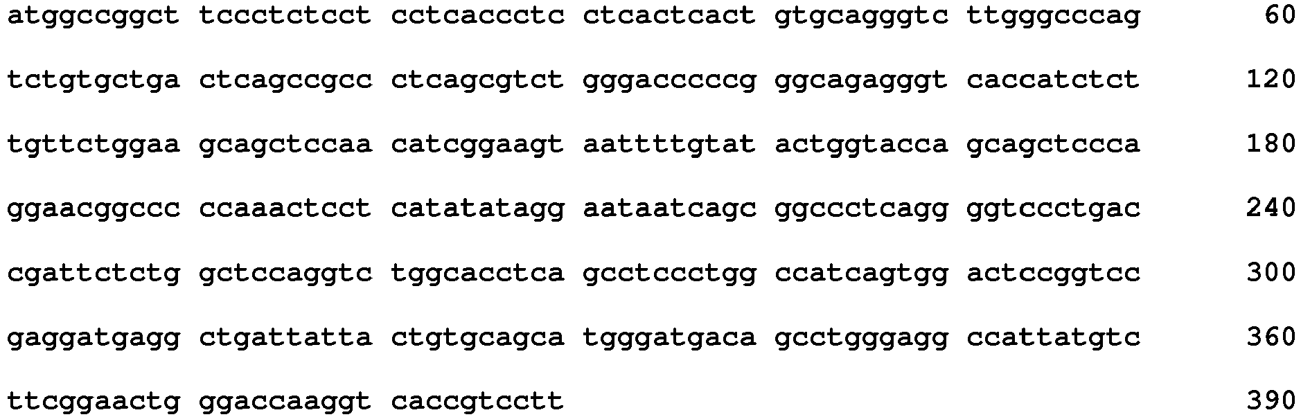
- <210> 4
<211> 130
<212> PRT
<213> Artificial Sequence - <220>
<223> VL chain domain of clone 5B1 - <400> 4

- <210> 5
<211> 426
<212> DNA
<213> Artificial Sequence - <220>
<223> VH chain domain of clone 9H3 - <400> 5
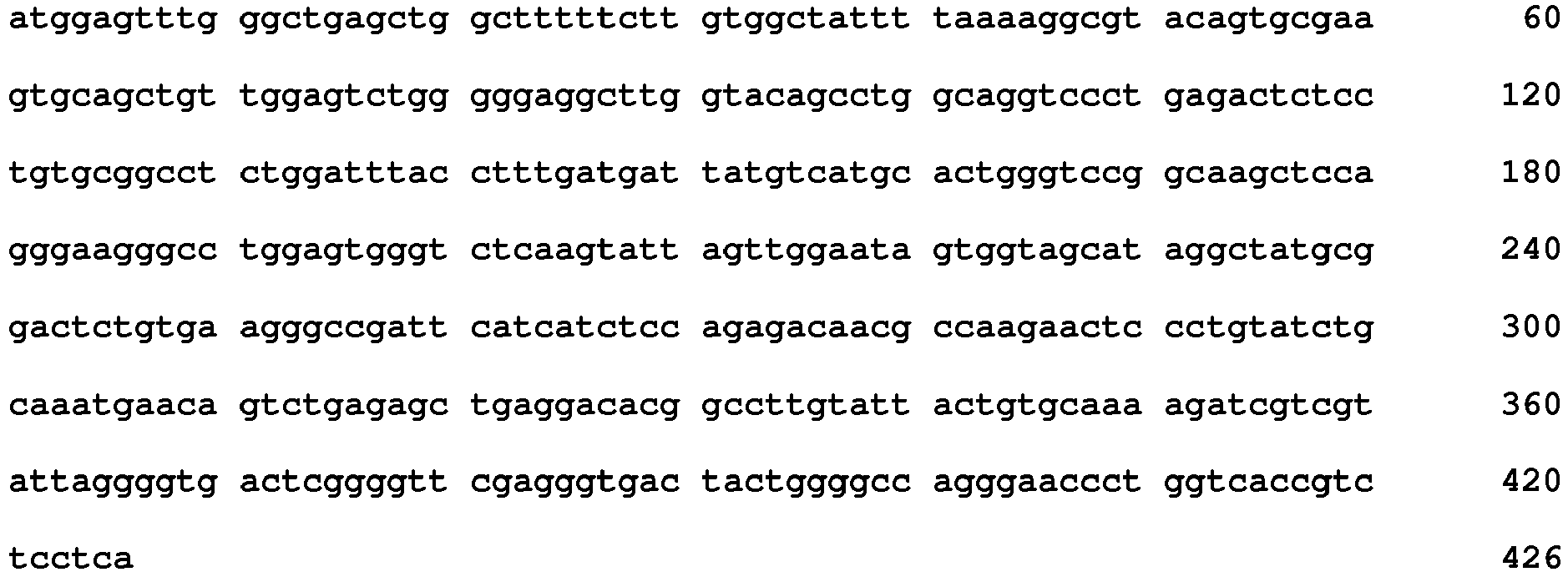
- <210> 6
<211> 142
<212> PRT
<213> Artificial Sequence - <220>
<223> VH chain domain of clone 9H3 - <400> 6

- <210> 7
<211> 387
<212> DNA
<213> Artificial Sequence - <220>
<223> VL chain domain of clone 9H3 - <400> 7
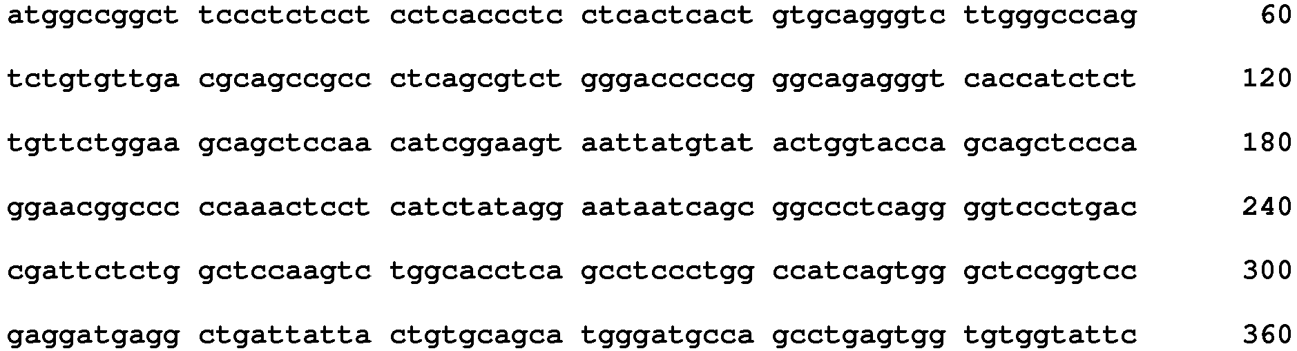

- <210> 8
<211> 129
<212> PRT
<213> Artificial Sequence - <220>
<223> VL chain domain of clone 9H3 - <400> 8

- <210> 9
<211> 426
<212> DNA
<213> Artificial Sequence - <220>
<223> VH chain domain of clone 5H11 - <400> 9

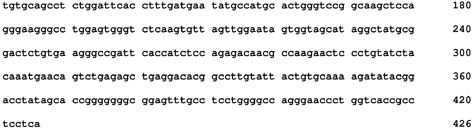
- <210> 10
<211> 142
<212> PRT
<213> Artificial Sequence - <220>
<223> VH chain domain of clone 5H11 - <400> 10

- <210> 11
<211> 390
<212> DNA
<213> Artificial Sequence - <220>
<223> VL chain domain of clone 5H11 - <400> 11

- <210> 12
<211> 130
<212> PRT
<213> Artificial Sequence - <220>
<223> VL chain domain of clone 5H11 - <400> 12
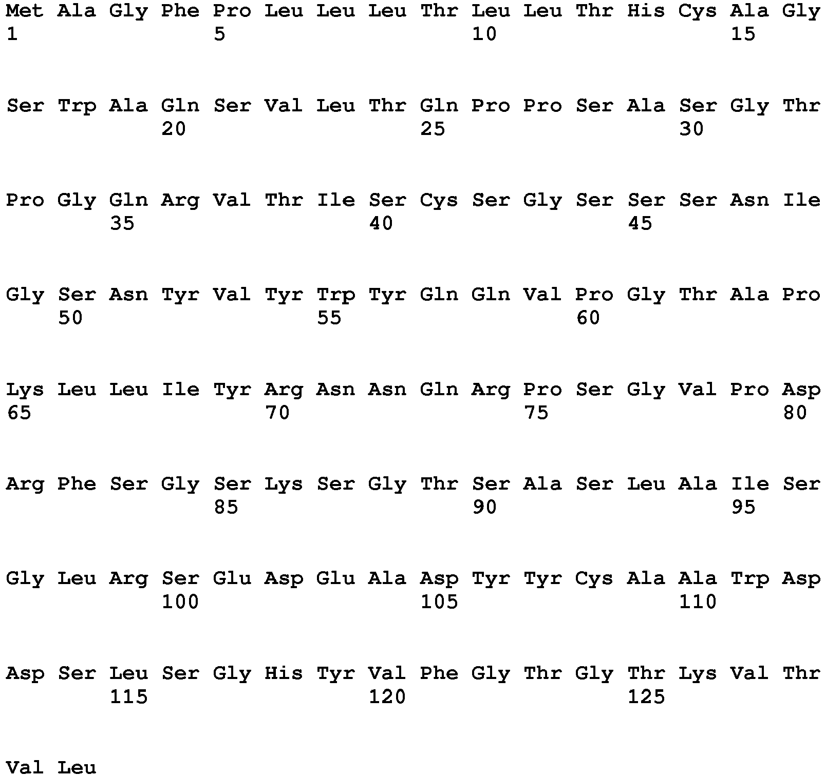

- <210> 13
<211> 435
<212> DNA
<213> Artificial Sequence - <220>
<223> VH chain domain of clone 7E3 - <400> 13
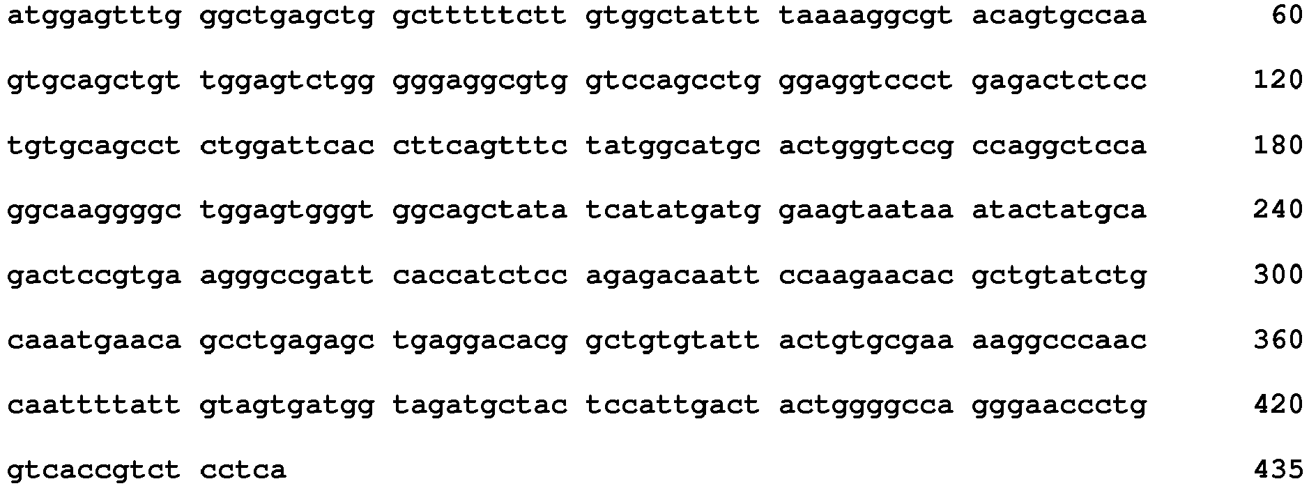
- <210> 14
<211> 145
<212> PRT
<213> Artificial Sequence - <220>
<223> VH chain domain of clone 7E3 - <400> 14
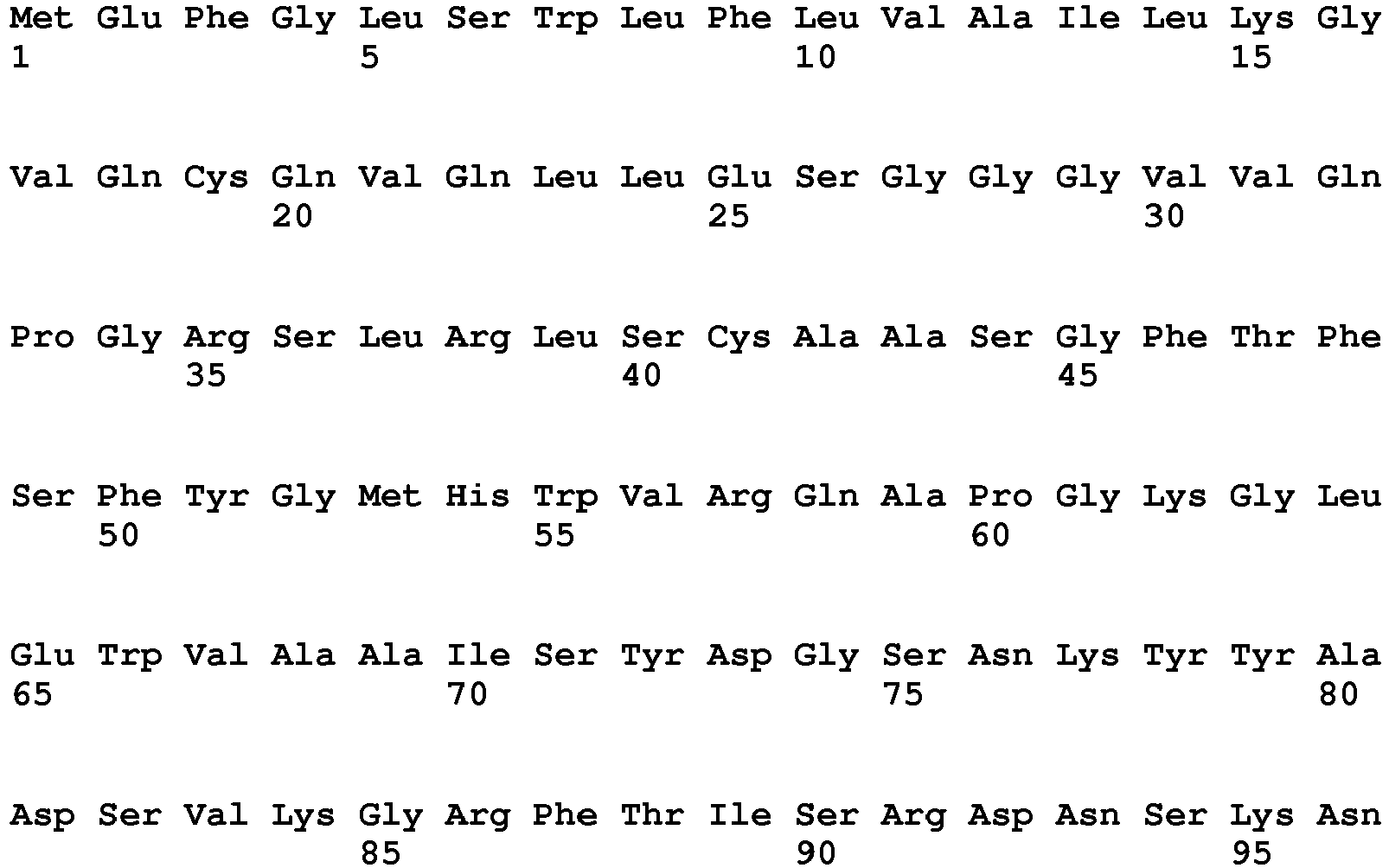

- <210> 15
<211> 390
<212> DNA
<213> Artificial Sequence - <220>
<223> VL chain domain of clone 7E3 - <400> 15
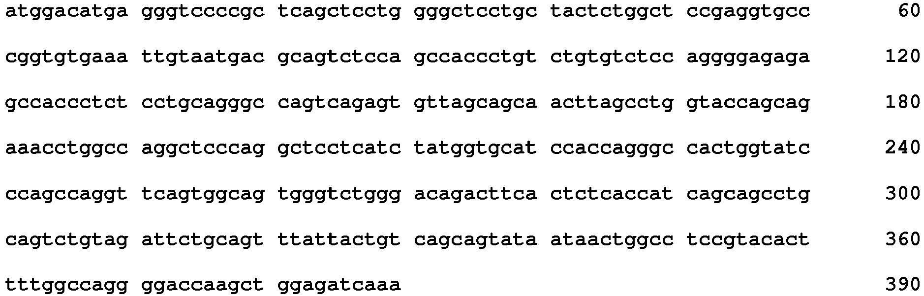
- <210> 16
<211> 130
<212> PRT
<213> Artificial Sequence - <220>
<223> VL chain domain of clone 7E3 - <400> 16
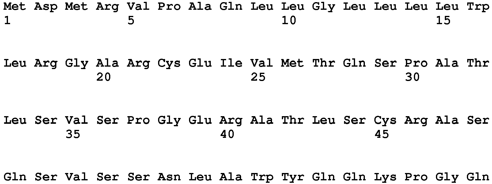

- <210> 17
<211> 750
<212> DNA
<213> Artificial Sequence - <220>
<223> Diabody 5B1CysDb sequence - <400> 17
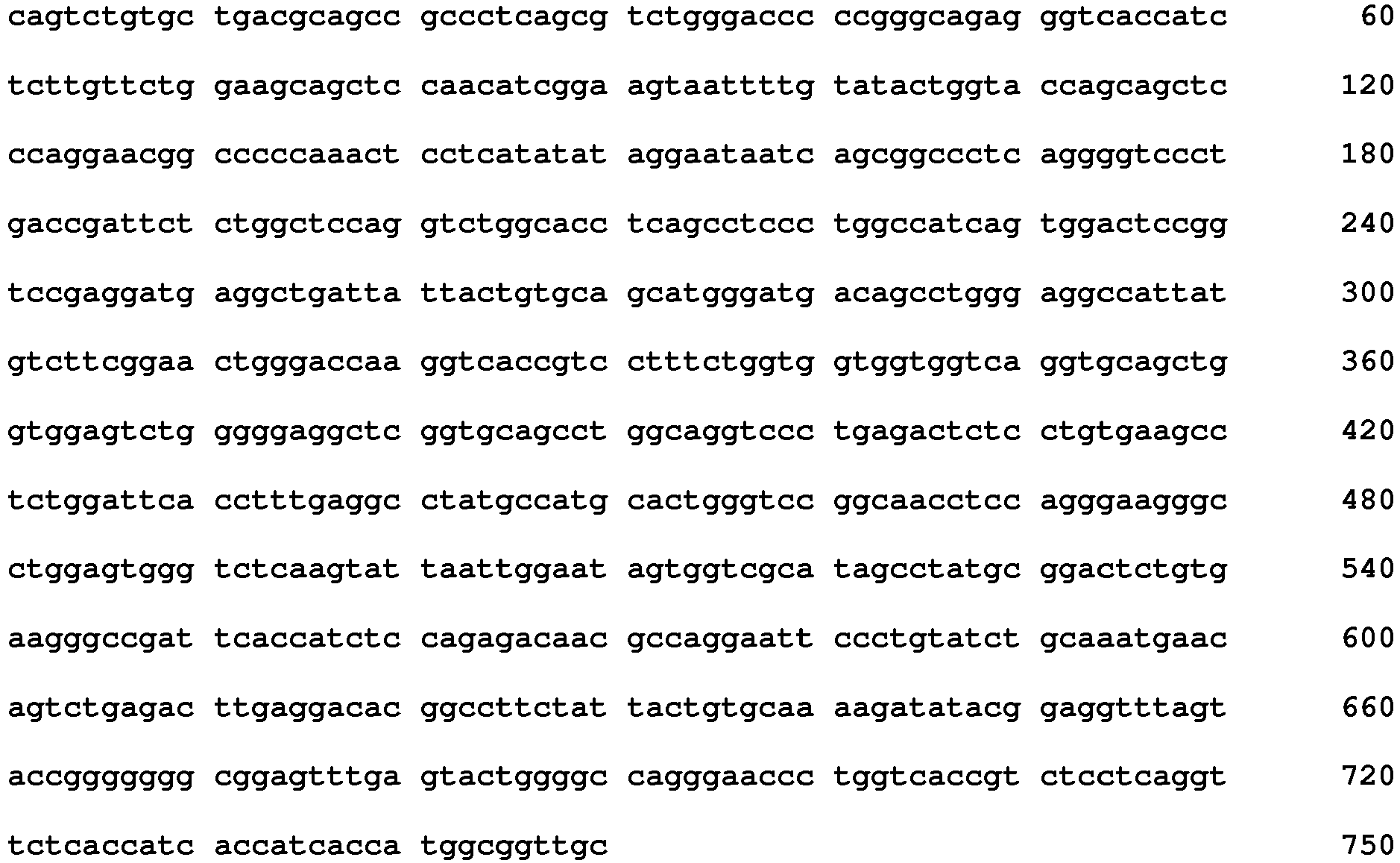
- <210> 18
<211> 250
<212> PRT
<213> Artificial Sequence - <220>
<223> Diabody 5B1CysDb sequence - <400> 18
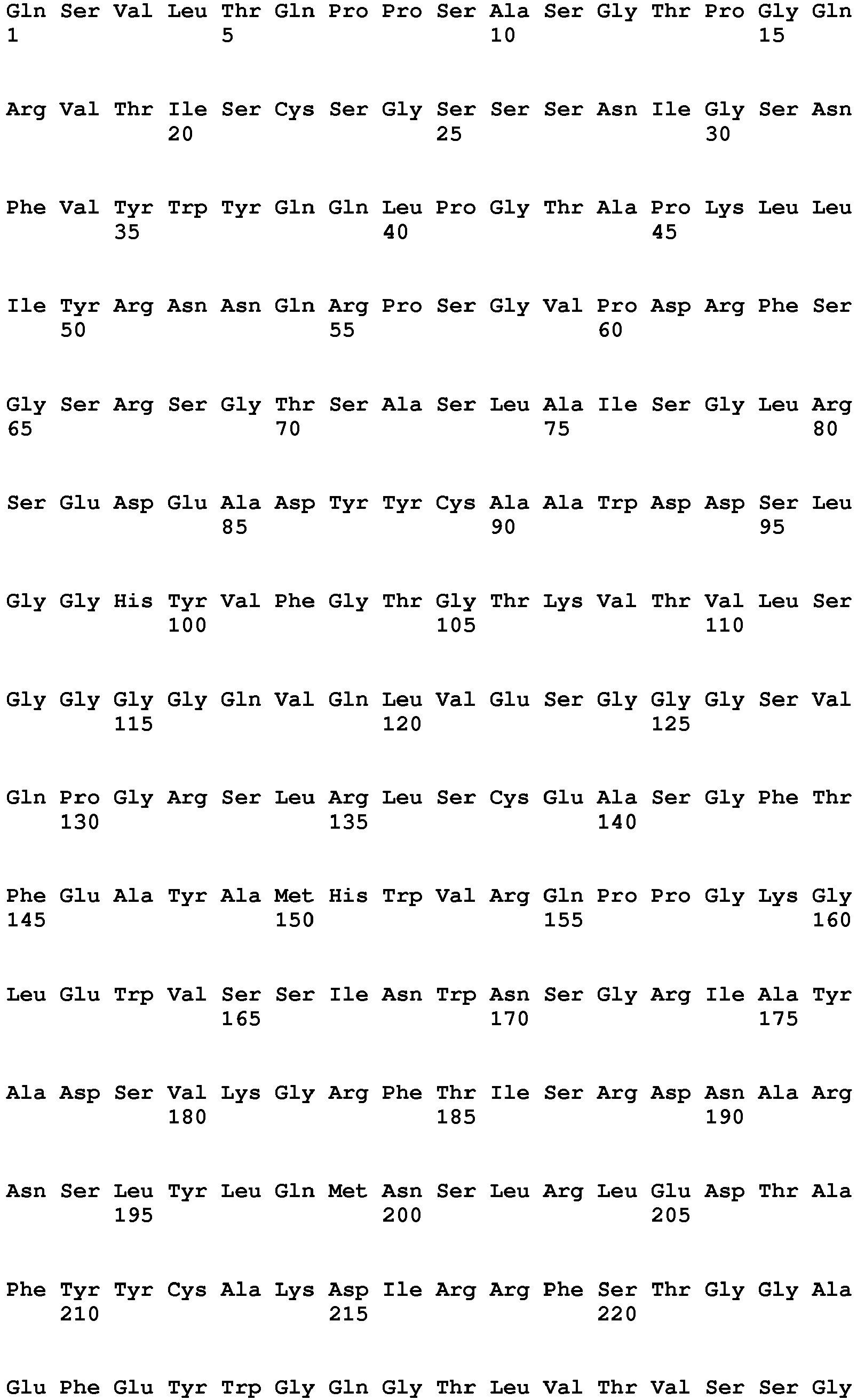

- <210> 19
<211> 750
<212> DNA
<213> Artificial Sequence - <220>
<223> Diabody 7E3CysDb sequence - <400> 19

- <210> 20
<211> 249
<212> PRT
<213> Artificial Sequence - <220>
<223> Diabody 7E3CysDb sequence - <400> 20

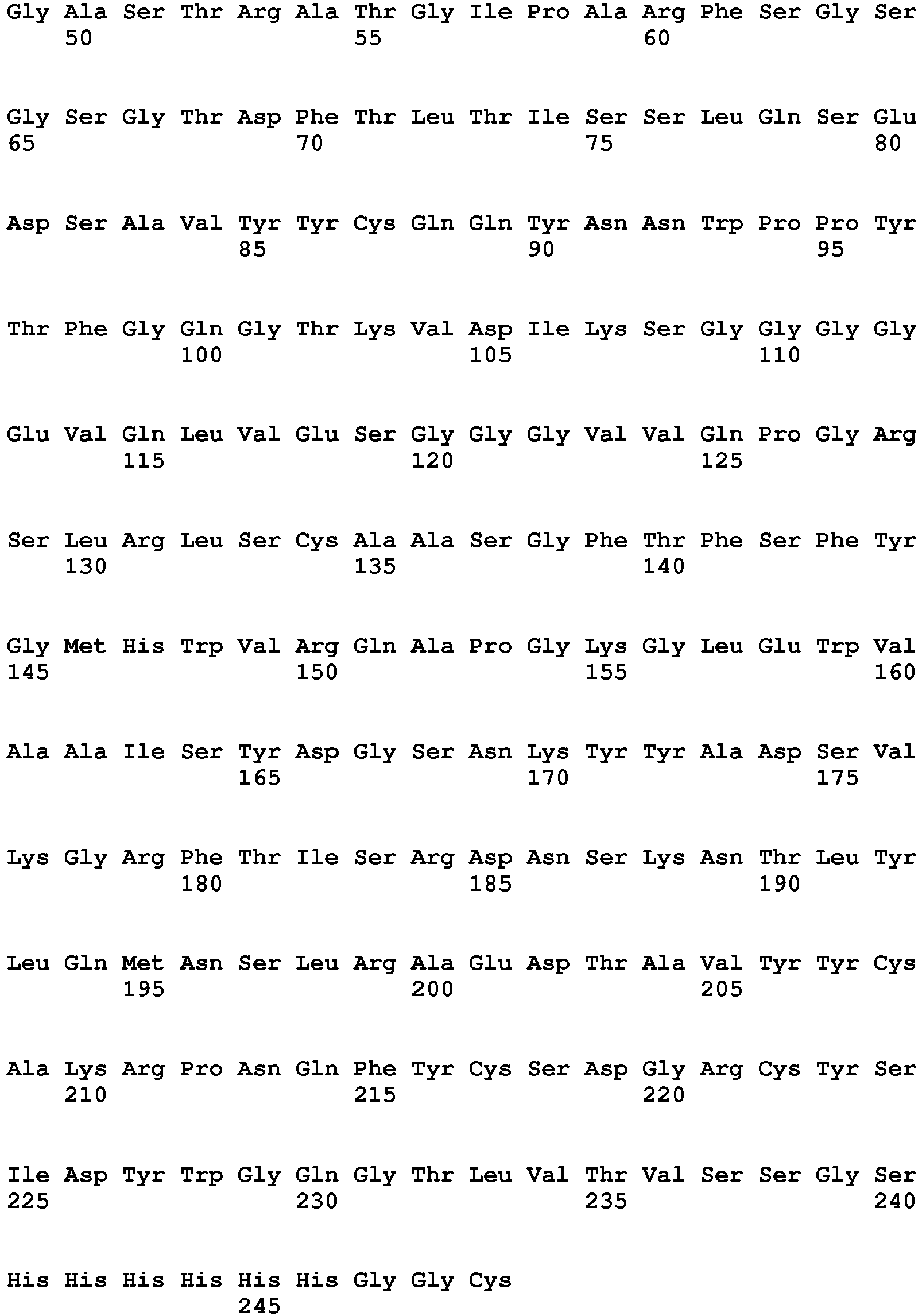
Claims (10)
- An isolated antibody that binds to Sialyl-Lewisa and induces ADCC or CDC activity, said antibody comprising a variable heavy chain (VH) domain and a variable light chain (VL) domain, wherein said VH domain and said VL domain respectively comprise an amino acid sequence of residues 20-142 of SEQ ID NO: 2 and residues 20-130 of SEQ ID NO: 4.
- An isolated polynucleotide encoding an antibody heavy chain, wherein said antibody heavy chain comprises a variable heavy chain (VH) domain having an amino acid sequence of residues 20-142 of SEQ ID NO: 2; and an isolated polynucleotide encoding an antibody light chain, wherein said antibody light chain comprises a variable light chain (VL) domain having an amino acid sequence of residues 20-130 of SEQ ID NO: 4, wherein the antibody comprising said antibody heavy chain and said antibody light chain binds to Sialyl-Lewisa and induces ADCC or CDC activity.
- The isolated polynucleotide of claim 2, wherein said VH domain amino acid sequence is encoded by the nucleic acid sequence of residues 58-426 of SEQ ID NO: 1.
- The isolated polynucleotide of claim 2, wherein said VL domain amino acid sequence is encoded by the nucleic acid sequence of residues 58-390 of SEQ ID NO: 3.
- The isolated antibody of claim 1, wherein said antibody is a human antibody, preferably an IgG or IgM isotype, wherein said IgG antibody is preferably an IgG 1 subclass.
- A pharmaceutical composition comprising the antibody of claim 1 or 5 and a pharmaceutically acceptable carrier.
- A pharmaceutical composition comprising the antibody of claim 1 or 5 and a pharmaceutically acceptable carrier for use in treating or preventing a disease comprising administering a therapeutically effective amount of said pharmaceutical composition to a subject in need thereof.
- The pharmaceutical composition for use of claim 7, wherein said disease is cancer or a tumor formation, wherein the cells of said cancer or said tumor expresses sLea; or wherein said cancer or tumor is selected from the group consisting of a tumor of the gastrointestinal tract, colon cancer, colorectal adenocarcinoma, metastatic colon cancer, colorectal cancer, pancreatic cancer, pancreatic adenocarcinoma, small cell carcinoma of the lung, bladder adenocarcinoma, signet ring ovarian cancer, ovarian cancer, metastatic carcinoma, adenocarcinoma of the stomach, adenocarcinoma of the esophagus, adenocarcinoma of the throat, adenocarcinoma of the urogenital tract, and adenocarcinoma of the breast.
- The pharmaceutical composition for use of claim 7, wherein said pharmaceutical composition is adapted for administering concurrently or successively a second therapeutic agent, preferably said second therapeutic agent is a chemotherapeutic agent or an immunotherapeutic agent.
- The pharmaceutical composition for use of claim 9, wherein the second therapeutic agent is a taxol.
Priority Applications (6)
| Application Number | Priority Date | Filing Date | Title |
|---|---|---|---|
| HRP20211129TT HRP20211129T1 (en) | 2013-08-26 | 2014-08-26 | Nucleic acids encoding human antibodies to sialyl-lewis a |
| PL14852746T PL3041507T3 (en) | 2013-08-26 | 2014-08-26 | NUCLEIC ACIDS CODE HUMAN ANTIBODIES AGAINST SALYL-LEWIS A |
| EP21181759.8A EP3906945A3 (en) | 2013-08-26 | 2014-08-26 | Nucleic acids encoding human antibodies to sialyl-lewis a |
| RS20210929A RS62189B1 (en) | 2013-08-26 | 2014-08-26 | Nucleic acids encoding human antibodies to sialyl-lewis a |
| SI201431854T SI3041507T1 (en) | 2013-08-26 | 2014-08-26 | Nucleic acids encoding human antibodies to sialyl-lewis a |
| SM20210427T SMT202100427T1 (en) | 2013-08-26 | 2014-08-26 | Nucleic acids encoding human antibodies to sialyl-lewis a |
Applications Claiming Priority (2)
| Application Number | Priority Date | Filing Date | Title |
|---|---|---|---|
| US201361870137P | 2013-08-26 | 2013-08-26 | |
| PCT/US2014/052631 WO2015053871A2 (en) | 2013-08-26 | 2014-08-26 | NUCLEIC ACIDS ENCODING HUMAN ANTIBODIES TO SIALYL-LEWISa |
Related Child Applications (1)
| Application Number | Title | Priority Date | Filing Date |
|---|---|---|---|
| EP21181759.8A Division EP3906945A3 (en) | 2013-08-26 | 2014-08-26 | Nucleic acids encoding human antibodies to sialyl-lewis a |
Publications (3)
| Publication Number | Publication Date |
|---|---|
| EP3041507A2 EP3041507A2 (en) | 2016-07-13 |
| EP3041507A4 EP3041507A4 (en) | 2017-07-12 |
| EP3041507B1 true EP3041507B1 (en) | 2021-06-30 |
Family
ID=52480556
Family Applications (2)
| Application Number | Title | Priority Date | Filing Date |
|---|---|---|---|
| EP14852746.8A Active EP3041507B1 (en) | 2013-08-26 | 2014-08-26 | Nucleic acids encoding human antibodies to sialyl-lewis a |
| EP21181759.8A Pending EP3906945A3 (en) | 2013-08-26 | 2014-08-26 | Nucleic acids encoding human antibodies to sialyl-lewis a |
Family Applications After (1)
| Application Number | Title | Priority Date | Filing Date |
|---|---|---|---|
| EP21181759.8A Pending EP3906945A3 (en) | 2013-08-26 | 2014-08-26 | Nucleic acids encoding human antibodies to sialyl-lewis a |
Country Status (22)
| Country | Link |
|---|---|
| US (4) | US9475874B2 (en) |
| EP (2) | EP3041507B1 (en) |
| JP (7) | JP6626445B2 (en) |
| KR (4) | KR102313341B1 (en) |
| CN (2) | CN112695039B (en) |
| AU (4) | AU2014332442B2 (en) |
| CA (2) | CA2922478C (en) |
| CY (1) | CY1124435T1 (en) |
| DK (1) | DK3041507T3 (en) |
| ES (1) | ES2880709T3 (en) |
| HR (1) | HRP20211129T1 (en) |
| HU (1) | HUE056325T2 (en) |
| IL (2) | IL244297B (en) |
| LT (1) | LT3041507T (en) |
| PL (1) | PL3041507T3 (en) |
| PT (1) | PT3041507T (en) |
| RS (1) | RS62189B1 (en) |
| RU (1) | RU2699289C2 (en) |
| SI (1) | SI3041507T1 (en) |
| SM (1) | SMT202100427T1 (en) |
| WO (1) | WO2015053871A2 (en) |
| ZA (1) | ZA201601354B (en) |
Families Citing this family (23)
| Publication number | Priority date | Publication date | Assignee | Title |
|---|---|---|---|---|
| EP3041507B1 (en) * | 2013-08-26 | 2021-06-30 | BioNTech Research and Development, Inc. | Nucleic acids encoding human antibodies to sialyl-lewis a |
| GB201515094D0 (en) * | 2015-08-25 | 2015-10-07 | Univ Nottingham | Sialyl-di-lewis a as expressed on glycoproteins but not glycolipids as a functional cancer target and antibodies thereto |
| EP3368561B1 (en) * | 2015-10-30 | 2025-09-17 | Medimmune Limited | Prevention of n-terminal truncation in igg light chains |
| JP2019529345A (en) * | 2016-07-14 | 2019-10-17 | バイオアークティック アクティエボラーグ | Brain delivery protein |
| SG10201912529SA (en) | 2016-11-18 | 2020-02-27 | Astellas Pharma Inc | Novel anti-human muc1 antibody fab fragment |
| MA46952A (en) | 2016-12-01 | 2019-10-09 | Regeneron Pharma | RADIO-MARKED ANTI-PD-L1 ANTIBODIES FOR IMMUNO-PET IMAGING |
| MA47456A (en) | 2017-02-10 | 2019-12-18 | Regeneron Pharma | RADIO-LABELED ANTI-LAG3 ANTIBODIES FOR IMMUNO-PET IMAGING |
| TWI726217B (en) * | 2017-06-15 | 2021-05-01 | 財團法人生物技術開發中心 | Antibody-drug conjugates containing anti-globo h antibodies and uses thereof |
| TWI795415B (en) * | 2017-07-07 | 2023-03-11 | 日商安斯泰來製藥股份有限公司 | A novel anti-human CEACAM5 antibody Fab fragment |
| MX2020000916A (en) | 2017-07-24 | 2020-09-10 | Regeneron Pharma | ANTI-CD8 ANTIBODIES AND THEIR USES. |
| BR112020012308A2 (en) * | 2017-12-19 | 2020-11-24 | The Rockefeller University | variant human igg fc domains with improved effect function |
| PT110526B (en) | 2018-01-26 | 2021-02-04 | Univ Nova De Lisboa | ANTIBODY, FUNCTIONAL FRAGMENT OR PROBE OF THE SAME AGAINST TUMORAL ANTIGENS |
| KR102115236B1 (en) * | 2018-01-29 | 2020-05-27 | (주)에스엠티바이오 | Chimera antigen receptors for treating pancreatic cancer or biliary tract cancer |
| CA3100317A1 (en) | 2018-05-17 | 2019-11-21 | Astellas Pharma Inc. | Complex having anti-human muc1 antibody fab fragment, peptide linker and/or ligand |
| AU2019337564A1 (en) * | 2018-09-10 | 2021-05-06 | Cold Spring Harbor Laboratory | Methods for treating pancreatitis |
| CN112839683B (en) | 2018-10-10 | 2023-11-14 | 安斯泰来制药株式会社 | Pharmaceutical composition containing labeling part-anti-human antibody Fab fragment complex |
| JP7672340B2 (en) * | 2018-10-19 | 2025-05-07 | メモリアル スローン ケタリング キャンサー センター | Chimeric antigen receptors targeting sialyl lewis a and uses thereof |
| EP4065605A4 (en) * | 2019-11-26 | 2023-07-12 | Ramot at Tel-Aviv University Ltd. | ANTIBODIES AGAINST CARBOHYDRATE ANTIGENS |
| CN114605550B (en) * | 2020-12-08 | 2023-09-22 | 东莞市朋志生物科技有限公司 | Antibodies against CA19-9, applications thereof and kits for detecting CA19-9 |
| US20250297027A1 (en) * | 2021-01-12 | 2025-09-25 | Yeda Research And Development Co. Ltd. | Antibodies to cancer glycosylation and uses thereof |
| JP2024511189A (en) | 2021-03-26 | 2024-03-12 | ビオンテック・ソシエタス・エウロパエア | Combination therapy with anti-CA19-9 antibodies and FOLFIRINOX in the treatment of cancer. |
| WO2025038668A1 (en) | 2023-08-14 | 2025-02-20 | Voro Therapeutics, Inc. | Therapeutic binding agents that conditionally promote myeloid cell activity against target cells and uses thereof |
| WO2025261951A1 (en) | 2024-06-18 | 2025-12-26 | BioNTech SE | Combination therapy with an anti-ca19-9 antibody and folfirinox in the treatment of cancer |
Family Cites Families (126)
| Publication number | Priority date | Publication date | Assignee | Title |
|---|---|---|---|---|
| CA128362A (en) | 1910-07-25 | 1910-09-27 | Victor Herbert Gregory | Furnace for drawing glass |
| US5807715A (en) | 1984-08-27 | 1998-09-15 | The Board Of Trustees Of The Leland Stanford Junior University | Methods and transformed mammalian lymphocyte cells for producing functional antigen-binding protein including chimeric immunoglobulin |
| EP0216846B2 (en) | 1985-04-01 | 1995-04-26 | Celltech Limited | Transformed myeloma cell-line and a process for the expression of a gene coding for a eukaryotic polypeptide employing same |
| GB8601597D0 (en) | 1986-01-23 | 1986-02-26 | Wilson R H | Nucleotide sequences |
| US5057313A (en) | 1986-02-25 | 1991-10-15 | The Center For Molecular Medicine And Immunology | Diagnostic and therapeutic antibody conjugates |
| WO1988007089A1 (en) | 1987-03-18 | 1988-09-22 | Medical Research Council | Altered antibodies |
| GB8717430D0 (en) | 1987-07-23 | 1987-08-26 | Celltech Ltd | Recombinant dna product |
| US5336603A (en) | 1987-10-02 | 1994-08-09 | Genentech, Inc. | CD4 adheson variants |
| US5223409A (en) | 1988-09-02 | 1993-06-29 | Protein Engineering Corp. | Directed evolution of novel binding proteins |
| DE768377T1 (en) | 1988-09-02 | 1998-01-02 | Dyax Corp | Production and selection of recombinant proteins with different binding sites |
| KR900005995A (en) | 1988-10-31 | 1990-05-07 | 우메모또 요시마사 | Modified Interleukin-2 and Method of Making the Same |
| EP0394827A1 (en) | 1989-04-26 | 1990-10-31 | F. Hoffmann-La Roche Ag | Chimaeric CD4-immunoglobulin polypeptides |
| US5112946A (en) | 1989-07-06 | 1992-05-12 | Repligen Corporation | Modified pf4 compositions and methods of use |
| WO1991006570A1 (en) | 1989-10-25 | 1991-05-16 | The University Of Melbourne | HYBRID Fc RECEPTOR MOLECULES |
| AU7247191A (en) | 1990-01-11 | 1991-08-05 | Molecular Affinities Corporation | Production of antibodies using gene libraries |
| US5780225A (en) | 1990-01-12 | 1998-07-14 | Stratagene | Method for generating libaries of antibody genes comprising amplification of diverse antibody DNAs and methods for using these libraries for the production of diverse antigen combining molecules |
| US5427908A (en) | 1990-05-01 | 1995-06-27 | Affymax Technologies N.V. | Recombinant library screening methods |
| US5349053A (en) | 1990-06-01 | 1994-09-20 | Protein Design Labs, Inc. | Chimeric ligand/immunoglobulin molecules and their uses |
| GB9015198D0 (en) | 1990-07-10 | 1990-08-29 | Brien Caroline J O | Binding substance |
| US5698426A (en) | 1990-09-28 | 1997-12-16 | Ixsys, Incorporated | Surface expression libraries of heteromeric receptors |
| DE69129154T2 (en) | 1990-12-03 | 1998-08-20 | Genentech, Inc., South San Francisco, Calif. | METHOD FOR ENRICHING PROTEIN VARIANTS WITH CHANGED BINDING PROPERTIES |
| KR100246529B1 (en) | 1990-12-14 | 2000-04-01 | 스티븐 에이. 서윈. 엠.디. | Chimeric Chains for Receptor-Related Signal Transduction Pathways |
| ATE269401T1 (en) | 1991-04-10 | 2004-07-15 | Scripps Research Inst | LIBRARIES OF HETERODIMERIC RECEPTORS USING PHAGEMIDS |
| EP0590067A1 (en) | 1991-06-14 | 1994-04-06 | Xoma Corporation | Microbially-produced antibody fragments and their conjugates |
| US5844095A (en) | 1991-06-27 | 1998-12-01 | Bristol-Myers Squibb Company | CTLA4 Ig fusion proteins |
| DE69233782D1 (en) | 1991-12-02 | 2010-05-20 | Medical Res Council | Preparation of Autoantibodies on Phage Surfaces Starting from Antibody Segment Libraries |
| US5622929A (en) | 1992-01-23 | 1997-04-22 | Bristol-Myers Squibb Company | Thioether conjugates |
| US6271242B1 (en) | 1992-02-10 | 2001-08-07 | Bristol-Myers Squibb Co. | Method for treating cancer using a tyrosine protein kinase inhibitor |
| US5733743A (en) | 1992-03-24 | 1998-03-31 | Cambridge Antibody Technology Limited | Methods for producing members of specific binding pairs |
| US5447851B1 (en) | 1992-04-02 | 1999-07-06 | Univ Texas System Board Of | Dna encoding a chimeric polypeptide comprising the extracellular domain of tnf receptor fused to igg vectors and host cells |
| JPH0630786A (en) * | 1992-07-14 | 1994-02-08 | Kyowa Hakko Kogyo Co Ltd | Bispecific antobidy |
| HUT74451A (en) | 1993-07-15 | 1996-12-30 | Cancer Res Campaign Tech | Prodrugs of protein tyrosine kinase inhibitors, systems contg. them and process for preparing them |
| US5443953A (en) | 1993-12-08 | 1995-08-22 | Immunomedics, Inc. | Preparation and use of immunoconjugates |
| AU696293B2 (en) | 1993-12-08 | 1998-09-03 | Genzyme Corporation | Process for generating specific antibodies |
| US5925376C1 (en) | 1994-01-10 | 2001-03-20 | Madalene C Y Heng | Method for treating psoriasis using selected phosphorylase kinase inhibitor and additional compounds |
| PT1231268E (en) | 1994-01-31 | 2005-11-30 | Univ Boston | BANKS OF POLYCLONE ANTIBODIES |
| US5516637A (en) | 1994-06-10 | 1996-05-14 | Dade International Inc. | Method involving display of protein binding pairs on the surface of bacterial pili and bacteriophage |
| GB9415379D0 (en) | 1994-07-29 | 1994-09-21 | Smithkline Beecham Plc | Novel compounds |
| CN1117155C (en) | 1994-07-29 | 2003-08-06 | 史密丝克莱恩比彻姆有限公司 | Novel compounds |
| US5587459A (en) | 1994-08-19 | 1996-12-24 | Regents Of The University Of Minnesota | Immunoconjugates comprising tyrosine kinase inhibitors |
| US5911995A (en) | 1994-08-19 | 1999-06-15 | Regents Of The University Of Minnesota | EGF-genistein conjugates for the treatment of cancer |
| US5795737A (en) | 1994-09-19 | 1998-08-18 | The General Hospital Corporation | High level expression of proteins |
| JP4026848B2 (en) | 1995-01-17 | 2007-12-26 | ブリガーム・アンド・ウーメンズ・ホスピタル・インコーポレーテッド | Receptor-specific transepithelial transport of immunogens |
| US6030613A (en) | 1995-01-17 | 2000-02-29 | The Brigham And Women's Hospital, Inc. | Receptor specific transepithelial transport of therapeutics |
| GB9501567D0 (en) | 1995-01-26 | 1995-03-15 | Pharmacia Spa | Hydrosoluble 3-arylidene-2-oxindole derivatives as tyrosine kinase inhibitors |
| GB9515975D0 (en) | 1995-08-04 | 1995-10-04 | Zeneca Ltd | Chemical compounds |
| US5863904A (en) | 1995-09-26 | 1999-01-26 | The University Of Michigan | Methods for treating cancers and restenosis with P21 |
| GB9601081D0 (en) | 1995-10-06 | 1996-03-20 | Cambridge Antibody Tech | Specific binding members for human transforming growth factor beta;materials and methods |
| US6127366A (en) | 1995-11-22 | 2000-10-03 | Merck & Co., Inc. | Inhibitors of farnesyl-protein transferase |
| AU711142B2 (en) | 1995-12-08 | 1999-10-07 | Janssen Pharmaceutica N.V. | Farnesyl protein transferase inhibiting (imidazol-5-YL)methyl-2-quinolinone derivatives |
| US5723125A (en) | 1995-12-28 | 1998-03-03 | Tanox Biosystems, Inc. | Hybrid with interferon-alpha and an immunoglobulin Fc linked through a non-immunogenic peptide |
| US5958769A (en) | 1996-01-18 | 1999-09-28 | Fred Hutchinson Cancer Research Center | Compositions and methods for mediating cell cycle progression |
| JP2978435B2 (en) | 1996-01-24 | 1999-11-15 | チッソ株式会社 | Method for producing acryloxypropyl silane |
| AU703203B2 (en) | 1996-01-30 | 1999-03-18 | Merck & Co., Inc. | Inhibitors of farnesyl-protein transferase |
| JP2000504017A (en) | 1996-01-30 | 2000-04-04 | メルク エンド カンパニー インコーポレーテッド | Farnesyl-protein transferase inhibitor |
| CA2249195A1 (en) | 1996-03-18 | 1997-09-25 | Board Of Regents, The University Of Texas System | Immunoglobin-like domains with increased half lives |
| JP3904238B2 (en) | 1996-03-20 | 2007-04-11 | イムノメディクス, インコーポレイテッド | Glycosylated humanized B cell specific antibody |
| US5891889A (en) | 1996-04-03 | 1999-04-06 | Merck & Co., Inc. | Inhibitors of farnesyl-protein transferase |
| US6063930A (en) | 1996-04-03 | 2000-05-16 | Merck & Co., Inc. | Substituted imidazole compounds useful as farnesyl-protein transferase inhibitors |
| US5883105A (en) | 1996-04-03 | 1999-03-16 | Merck & Co., Inc. | Inhibitors of farnesyl-protein transferase |
| US6080870A (en) | 1996-04-03 | 2000-06-27 | Merck & Co., Inc. | Biaryl substituted imidazole compounds useful as farnesyl-protein transferase inhibitors |
| US6300501B1 (en) | 1996-05-22 | 2001-10-09 | Warner-Lambert Company | Histidine-(N-benzyl glycinamide) inhibitors of protein farnesyl transferase |
| US5648239A (en) | 1996-06-21 | 1997-07-15 | Incyte Pharmaceuticals, Inc. | Human camp-dependent protein kinase inhibitor homolog |
| JPH11512750A (en) | 1996-06-27 | 1999-11-02 | ファイザー インク. | Derivatives of 2- (2-oxo-ethylidene) -imidazolidin-4-one and their use as farnesyl protein transferase inhibitors |
| CZ301044B6 (en) | 1996-08-12 | 2009-10-21 | Mitsubishi Tanabe Pharma | Medicaments comprising Rho kinase inhibiting amide derivatives |
| US6040305A (en) | 1996-09-13 | 2000-03-21 | Schering Corporation | Compounds useful for inhibition of farnesyl protein transferase |
| US5945429A (en) | 1996-09-13 | 1999-08-31 | Schering Corporation | Compounds useful for inhibition of farnesyl protein transferase |
| US6030982A (en) | 1996-09-13 | 2000-02-29 | Schering Corporationm | Compounds useful for inhibition of farnesyl protein transferase |
| US6013662A (en) | 1996-12-30 | 2000-01-11 | Rhone-Poulenc Rorer S.A. | Farnesyl transferase inhibitors, their preparation, the pharmaceutical compositions which contain them and their use in the preparation of medicaments |
| US5939439A (en) | 1996-12-30 | 1999-08-17 | Merck & Co., Inc. | Inhibitors of farnesyl-protein transferase |
| US6093737A (en) | 1996-12-30 | 2000-07-25 | Merck & Co., Inc. | Inhibitors of farnesyl-protein transferase |
| AU5772698A (en) | 1997-01-29 | 1998-08-18 | Zeneca Limited | Inhibitors of farnesyl protein transferase |
| ZA981080B (en) | 1997-02-11 | 1998-08-12 | Warner Lambert Co | Bicyclic inhibitors of protein farnesyl transferase |
| WO1998037417A1 (en) | 1997-02-20 | 1998-08-27 | The Regents Of The University Of California | Plasmon resonant particles, methods and apparatus |
| AU6542898A (en) * | 1997-03-05 | 1998-09-22 | John Wayne Cancer Institute | Sialyl lewis antigens as targets for immunotherapy |
| TW591030B (en) | 1997-03-10 | 2004-06-11 | Janssen Pharmaceutica Nv | Farnesyl transferase inhibiting 1,8-annelated quinolinone derivatives substituted with N- or C-linked imidazoles |
| US6211193B1 (en) | 1997-06-17 | 2001-04-03 | Schering Corporation | Compounds useful for inhibition of farnesyl protein transferase |
| US6159984A (en) | 1997-06-17 | 2000-12-12 | Schering Corporation | Farnesyl protein transferase inhibitors |
| US6239140B1 (en) | 1997-06-17 | 2001-05-29 | Schering Corporation | Compounds useful for inhibition of farnesyl protein transferase |
| US6051582A (en) | 1997-06-17 | 2000-04-18 | Schering Corporation | Compounds useful for inhibition of farnesyl protein transferase |
| US6228865B1 (en) | 1997-06-17 | 2001-05-08 | Schering Corporation | Compounds useful for inhibition of farnesyl protein transferase |
| US6225322B1 (en) | 1997-06-17 | 2001-05-01 | Schering Corporation | Compounds useful for inhibition of farnesyl protein transferase |
| DK1011648T4 (en) | 1997-08-15 | 2005-10-24 | Cephalon Inc | Combination of tyrosine kinase inhibitor and chemical castration to treat prostate cancer |
| US6103723A (en) | 1997-10-17 | 2000-08-15 | Merck & Co., Inc. | Inhibitors of farnesyl-protein transferase |
| WO1999020612A1 (en) | 1997-10-22 | 1999-04-29 | Astrazeneca Uk Limited | Imidazole derivatives and their use as farnesyl protein transferase inhibitors |
| DE69801590T2 (en) | 1997-10-22 | 2002-07-11 | Astrazeneca Ab, Soedertaelje | IMIDAZOLE DERIVATIVES AND THEIR USE AS FARNESYL PROTEIN TRANSFERASE INHIBITORS |
| US6124465A (en) | 1997-11-25 | 2000-09-26 | Rhone-Poulenc S.A. | Farnesyl transferase inhibitors, their preparation, the pharmaceutical compositions which contain them and their use in the preparation of medicaments |
| ID24175A (en) | 1997-11-28 | 2000-07-13 | Lg Chemical Ltd | IMIDAZOL DRIVINGS WHICH HAVE A LIVING ACTIVITY FOR FARNESIL TRANSFERASE AND THE PROCESS OF MAKING IT |
| US6054466A (en) | 1997-12-04 | 2000-04-25 | Merck & Co., Inc. | Inhibitors of farnesyl-protein transferase |
| US6242196B1 (en) | 1997-12-11 | 2001-06-05 | Dana-Farber Cancer Institute | Methods and pharmaceutical compositions for inhibiting tumor cell growth |
| US6335156B1 (en) | 1997-12-18 | 2002-01-01 | The Johns Hopkins University School Of Medicine | 14-3-3σ arrests the cell cycle |
| DE69904302T2 (en) | 1998-02-02 | 2003-08-14 | Lg Chemical Ltd., Seoul/Soul | FARNESYL TRANSFERASE INHIBITORS WITH PIPERID STRUCTURE AND METHOD FOR THE PRODUCTION THEREOF |
| BR9909961A (en) | 1998-04-27 | 2000-12-26 | Warner Lambert Co | Alkyl and alkenyl functionalized derivatives of glycinamides as farnesyl transferase inhibitors |
| HRP20000904A2 (en) | 1998-07-06 | 2001-12-31 | Janssen Pharmaceutica Nv | Farnesyl protein transferase inhibitors for treating arthropathies |
| US6034053A (en) | 1998-07-13 | 2000-03-07 | Wayne Hughes Institute | EGF-isoflavone conjugates for the prevention of restenosis |
| US6787638B1 (en) | 1998-12-02 | 2004-09-07 | Applied Molecular Evolution, Inc. | Tumor specific human monoclonal antibodies and methods of use |
| US6362188B1 (en) | 1998-12-18 | 2002-03-26 | Schering Corporation | Farnesyl protein transferase inhibitors |
| US6372747B1 (en) | 1998-12-18 | 2002-04-16 | Schering Corporation | Farnesyl protein transferase inhibitors |
| FR2787327B1 (en) | 1998-12-21 | 2003-01-17 | Aventis Pharma Sa | COMPOSITIONS CONTAINING FARNESYL TRANSFERASE INHIBITORS |
| US6432959B1 (en) | 1998-12-23 | 2002-08-13 | Schering Corporation | Inhibitors of farnesyl-protein transferase |
| JP4876239B2 (en) | 1999-01-11 | 2012-02-15 | プリンストン ユニバーシティー | High affinity inhibitors for target confirmation and use thereof |
| US6399633B1 (en) | 1999-02-01 | 2002-06-04 | Aventis Pharmaceuticals Inc. | Use of 4-H-1-benzopryan-4-one derivatives as inhibitors of smooth muscle cell proliferation |
| US6245759B1 (en) | 1999-03-11 | 2001-06-12 | Merck & Co., Inc. | Tyrosine kinase inhibitors |
| US6143766A (en) | 1999-04-16 | 2000-11-07 | Warner-Lambert Company | Benzopyranone and quinolone inhibitors of ras farnesyl transferase |
| US6458935B1 (en) | 1999-06-23 | 2002-10-01 | Merck & Co., Inc. | Radiolabeled farnesyl-protein transferase inhibitors |
| US6403581B1 (en) | 2000-01-19 | 2002-06-11 | American Cyanamid Company | Method of inhibition of farnesyl-protein transferase using substituted benz (cd) indol-2-imine and-amine derivatives |
| AU2003228440B2 (en) | 2002-04-01 | 2008-10-02 | Walter Reed Army Institute Of Research | Method of designing synthetic nucleic acid sequences for optimal protein expression in a host cell |
| US20050276812A1 (en) | 2004-06-01 | 2005-12-15 | Genentech, Inc. | Antibody-drug conjugates and methods |
| PT2489364E (en) | 2003-11-06 | 2015-04-16 | Seattle Genetics Inc | Monomethylvaline compounds conjugated to antibodies |
| NZ553500A (en) | 2004-09-23 | 2009-11-27 | Genentech Inc Genentech Inc | Cysteine engineered antibodies and conjugates withCysteine engineered antibodies and conjugates with a free cysteine amino acid in the heavy chain a free cysteine amino acid in the heavy chain |
| WO2007011968A2 (en) | 2005-07-18 | 2007-01-25 | Seattle Genetics, Inc. | Beta-glucuronide-linker drug conjugates |
| EA015925B1 (en) | 2006-06-29 | 2011-12-30 | ДСМ АйПи АССЕТС Б.В. | A method for producing polypeptides |
| EP2078732B1 (en) | 2006-07-10 | 2015-09-16 | Fujita Health University | Method of identifying a candidate diagnostic or therapeutic antibody using flow cytometry |
| US8865875B2 (en) | 2007-08-22 | 2014-10-21 | Medarex, L.L.C. | Site-specific attachment of drugs or other agents to engineered antibodies with C-terminal extensions |
| EP2185931B1 (en) * | 2007-09-07 | 2013-07-10 | Children's Hospital Medical Center | Use of secretor, lewis and sialyl antigen levels as predictors for disease |
| ES2532635T3 (en) | 2008-07-15 | 2015-03-30 | Genentech, Inc. | Anthracycline conjugates, process for its preparation and its use as antitumor compounds |
| CA2735900A1 (en) | 2008-09-19 | 2010-03-25 | Medimmune, Llc | Antibodies directed to dll4 and uses thereof |
| US20110311517A1 (en) * | 2009-02-10 | 2011-12-22 | Shenogen Pharma Group, Ltd. | Antibodies and methods for treating estrogen receptor-associated diseases |
| CN106432495A (en) * | 2010-07-22 | 2017-02-22 | 加利福尼亚大学董事会 | Anti-tumor antigen antibodies and methods of use |
| CA2867700A1 (en) * | 2012-03-23 | 2013-09-26 | Memorial Sloan-Kettering Cancer Center | Potentiating antibody-induced complement-mediated cytotoxicity via pi3k inhibition |
| TW201506041A (en) * | 2013-02-13 | 2015-02-16 | Lab Francais Du Fractionnement | highly galactosylated anti-TNF-alpha antibodies and uses thereof |
| EP3041507B1 (en) * | 2013-08-26 | 2021-06-30 | BioNTech Research and Development, Inc. | Nucleic acids encoding human antibodies to sialyl-lewis a |
| GB201515094D0 (en) * | 2015-08-25 | 2015-10-07 | Univ Nottingham | Sialyl-di-lewis a as expressed on glycoproteins but not glycolipids as a functional cancer target and antibodies thereto |
| US11707522B2 (en) * | 2017-10-13 | 2023-07-25 | Boehringer Ingelheim International Gmbh | Human antibodies to Tn antigen |
| JP7672340B2 (en) * | 2018-10-19 | 2025-05-07 | メモリアル スローン ケタリング キャンサー センター | Chimeric antigen receptors targeting sialyl lewis a and uses thereof |
| JP2024511189A (en) * | 2021-03-26 | 2024-03-12 | ビオンテック・ソシエタス・エウロパエア | Combination therapy with anti-CA19-9 antibodies and FOLFIRINOX in the treatment of cancer. |
-
2014
- 2014-08-26 EP EP14852746.8A patent/EP3041507B1/en active Active
- 2014-08-26 WO PCT/US2014/052631 patent/WO2015053871A2/en not_active Ceased
- 2014-08-26 PL PL14852746T patent/PL3041507T3/en unknown
- 2014-08-26 EP EP21181759.8A patent/EP3906945A3/en active Pending
- 2014-08-26 KR KR1020207019821A patent/KR102313341B1/en active Active
- 2014-08-26 KR KR1020227004406A patent/KR102553717B1/en active Active
- 2014-08-26 KR KR1020167007644A patent/KR20160054501A/en not_active Ceased
- 2014-08-26 ES ES14852746T patent/ES2880709T3/en active Active
- 2014-08-26 KR KR1020217019368A patent/KR102362630B1/en active Active
- 2014-08-26 SM SM20210427T patent/SMT202100427T1/en unknown
- 2014-08-26 PT PT148527468T patent/PT3041507T/en unknown
- 2014-08-26 LT LTEPPCT/US2014/052631T patent/LT3041507T/en unknown
- 2014-08-26 RU RU2016111015A patent/RU2699289C2/en active
- 2014-08-26 SI SI201431854T patent/SI3041507T1/en unknown
- 2014-08-26 CA CA2922478A patent/CA2922478C/en active Active
- 2014-08-26 HU HUE14852746A patent/HUE056325T2/en unknown
- 2014-08-26 CN CN202011316903.2A patent/CN112695039B/en active Active
- 2014-08-26 CN CN201480057851.8A patent/CN105764526B/en active Active
- 2014-08-26 AU AU2014332442A patent/AU2014332442B2/en active Active
- 2014-08-26 US US14/468,827 patent/US9475874B2/en active Active
- 2014-08-26 HR HRP20211129TT patent/HRP20211129T1/en unknown
- 2014-08-26 DK DK14852746.8T patent/DK3041507T3/en active
- 2014-08-26 RS RS20210929A patent/RS62189B1/en unknown
- 2014-08-26 CA CA3090644A patent/CA3090644C/en active Active
- 2014-08-26 JP JP2016539019A patent/JP6626445B2/en active Active
-
2016
- 2016-02-25 IL IL244297A patent/IL244297B/en active IP Right Grant
- 2016-02-26 ZA ZA2016/01354A patent/ZA201601354B/en unknown
- 2016-09-23 US US15/275,174 patent/US20170015756A1/en not_active Abandoned
-
2018
- 2018-12-27 JP JP2018244315A patent/JP7161397B2/en active Active
-
2020
- 2020-02-11 US US16/788,251 patent/US20200247900A1/en not_active Abandoned
- 2020-02-20 AU AU2020201225A patent/AU2020201225B2/en active Active
- 2020-08-19 IL IL276819A patent/IL276819A/en unknown
-
2021
- 2021-06-22 JP JP2021103311A patent/JP2021151263A/en active Pending
- 2021-07-21 CY CY20211100653T patent/CY1124435T1/en unknown
-
2022
- 2022-07-26 AU AU2022209226A patent/AU2022209226B2/en active Active
- 2022-12-28 JP JP2022211698A patent/JP7299405B2/en active Active
-
2023
- 2023-04-14 US US18/134,844 patent/US20230365707A1/en active Pending
- 2023-09-25 JP JP2023160194A patent/JP7635329B2/en active Active
-
2024
- 2024-02-20 JP JP2024023605A patent/JP2024050966A/en active Pending
-
2025
- 2025-05-29 JP JP2025089961A patent/JP2025116141A/en active Pending
- 2025-08-27 AU AU2025223771A patent/AU2025223771A1/en not_active Abandoned
Non-Patent Citations (1)
| Title |
|---|
| CHI VAN DANG: "Editorial Policies Aacrjournals", 9 February 2018 (2018-02-09), XP055450006, Retrieved from the Internet <URL:http://aacrjournals.org/content/authors/editorial-policies> [retrieved on 20180209] * |
Also Published As
Similar Documents
| Publication | Publication Date | Title |
|---|---|---|
| US20230365707A1 (en) | Nucleic acids encoding human antibodies to sialyl-lewis a | |
| AU2014332442A1 (en) | Nucleic acids encoding human antibodies to Sialyl-Lewis | |
| US9856324B2 (en) | Human monoclonal antibodies to ganglioside GD2 | |
| HK40064012A (en) | Nucleic acids encoding human antibodies to sialyl-lewis a | |
| RU2791967C2 (en) | NUCLEIC ACIDS ENCODING ANTIBODIES AGAINST SIALYLATED HUMAN LEWISa ANTIGEN | |
| HK1226295B (en) | Nucleic acids encoding human antibodies to sialyl-lewis a | |
| HK40050582A (en) | Nucleic acids encoding human antibodies to sialyl-lewis a | |
| NZ717428B2 (en) | NUCLEIC ACIDS ENCODING HUMAN ANTIBODIES TO SIALYL-LEWISa | |
| BR122020012421B1 (en) | ISOLATED POLYNUCLEOTIDES, ISOLATED ANTIBODY OR FUNCTIONAL FRAGMENT THEREOF THAT BINDS TO SIALYL-LEWISA, USES THEREOF, CONJUGATION AND COMPOSITIONS | |
| BR112016004133B1 (en) | ISOLATED POLYNUCLEOTIDES, ISOLATED ANTIBODY OR FUNCTIONAL FRAGMENT THEREOF THAT BINDS TO SIALILLEWISA, USES THEREOF, CONJUGATE AND PHARMACEUTICAL COMPOSITION |
Legal Events
| Date | Code | Title | Description |
|---|---|---|---|
| PUAI | Public reference made under article 153(3) epc to a published international application that has entered the european phase |
Free format text: ORIGINAL CODE: 0009012 |
|
| 17P | Request for examination filed |
Effective date: 20160317 |
|
| AK | Designated contracting states |
Kind code of ref document: A2 Designated state(s): AL AT BE BG CH CY CZ DE DK EE ES FI FR GB GR HR HU IE IS IT LI LT LU LV MC MK MT NL NO PL PT RO RS SE SI SK SM TR |
|
| AX | Request for extension of the european patent |
Extension state: BA ME |
|
| DAX | Request for extension of the european patent (deleted) | ||
| RIN1 | Information on inventor provided before grant (corrected) |
Inventor name: SUN, SHU-MAN Inventor name: SAWADA, RITSUKO Inventor name: SCHOLZ, WOLFGANG |
|
| RAP1 | Party data changed (applicant data changed or rights of an application transferred) |
Owner name: MABVAX THERAPEUTICS, INC. |
|
| RIC1 | Information provided on ipc code assigned before grant |
Ipc: A61K 39/395 20060101AFI20170228BHEP Ipc: C07K 16/30 20060101ALI20170228BHEP Ipc: G01N 33/574 20060101ALI20170228BHEP |
|
| A4 | Supplementary search report drawn up and despatched |
Effective date: 20170609 |
|
| RIC1 | Information provided on ipc code assigned before grant |
Ipc: A61K 39/395 20060101AFI20170602BHEP Ipc: G01N 33/574 20060101ALI20170602BHEP Ipc: C07K 16/30 20060101ALI20170602BHEP |
|
| REG | Reference to a national code |
Ref country code: HK Ref legal event code: DE Ref document number: 1226295 Country of ref document: HK |
|
| STAA | Information on the status of an ep patent application or granted ep patent |
Free format text: STATUS: EXAMINATION IS IN PROGRESS |
|
| 17Q | First examination report despatched |
Effective date: 20180219 |
|
| RAP1 | Party data changed (applicant data changed or rights of an application transferred) |
Owner name: BIONTECH RESEARCH AND DEVELOPMENT, INC. |
|
| REG | Reference to a national code |
Ref country code: DE Ref legal event code: R079 Ref document number: 602014078494 Country of ref document: DE Free format text: PREVIOUS MAIN CLASS: A61K0039395000 Ipc: A61K0039000000 |
|
| RIC1 | Information provided on ipc code assigned before grant |
Ipc: A61K 51/10 20060101ALI20201215BHEP Ipc: A61K 39/00 20060101AFI20201215BHEP Ipc: G01N 33/574 20060101ALI20201215BHEP Ipc: A61P 35/00 20060101ALI20201215BHEP Ipc: A61P 43/00 20060101ALI20201215BHEP Ipc: C07K 16/28 20060101ALI20201215BHEP Ipc: A61K 45/06 20060101ALI20201215BHEP Ipc: A61P 37/04 20060101ALI20201215BHEP Ipc: C07K 16/30 20060101ALI20201215BHEP |
|
| GRAP | Despatch of communication of intention to grant a patent |
Free format text: ORIGINAL CODE: EPIDOSNIGR1 |
|
| STAA | Information on the status of an ep patent application or granted ep patent |
Free format text: STATUS: GRANT OF PATENT IS INTENDED |
|
| INTG | Intention to grant announced |
Effective date: 20210122 |
|
| GRAS | Grant fee paid |
Free format text: ORIGINAL CODE: EPIDOSNIGR3 |
|
| GRAA | (expected) grant |
Free format text: ORIGINAL CODE: 0009210 |
|
| STAA | Information on the status of an ep patent application or granted ep patent |
Free format text: STATUS: THE PATENT HAS BEEN GRANTED |
|
| AK | Designated contracting states |
Kind code of ref document: B1 Designated state(s): AL AT BE BG CH CY CZ DE DK EE ES FI FR GB GR HR HU IE IS IT LI LT LU LV MC MK MT NL NO PL PT RO RS SE SI SK SM TR |
|
| REG | Reference to a national code |
Ref country code: CH Ref legal event code: EP |
|
| REG | Reference to a national code |
Ref country code: AT Ref legal event code: REF Ref document number: 1405727 Country of ref document: AT Kind code of ref document: T Effective date: 20210715 Ref country code: DE Ref legal event code: R096 Ref document number: 602014078494 Country of ref document: DE |
|
| REG | Reference to a national code |
Ref country code: FI Ref legal event code: FGE |
|
| REG | Reference to a national code |
Ref country code: RO Ref legal event code: EPE Ref country code: DK Ref legal event code: T3 Effective date: 20210720 Ref country code: PT Ref legal event code: SC4A Ref document number: 3041507 Country of ref document: PT Date of ref document: 20210726 Kind code of ref document: T Free format text: AVAILABILITY OF NATIONAL TRANSLATION Effective date: 20210721 |
|
| REG | Reference to a national code |
Ref country code: NL Ref legal event code: FP |
|
| REG | Reference to a national code |
Ref country code: SE Ref legal event code: TRGR |
|
| REG | Reference to a national code |
Ref country code: IE Ref legal event code: FG4D |
|
| REG | Reference to a national code |
Ref country code: NO Ref legal event code: T2 Effective date: 20210630 |
|
| REG | Reference to a national code |
Ref country code: SK Ref legal event code: T3 Ref document number: E 37771 Country of ref document: SK |
|
| REG | Reference to a national code |
Ref country code: GR Ref legal event code: EP Ref document number: 20210402082 Country of ref document: GR Effective date: 20210915 |
|
| REG | Reference to a national code |
Ref country code: HR Ref legal event code: T1PR Ref document number: P20211129 Country of ref document: HR Ref country code: HR Ref legal event code: ODRP Ref document number: P20211129 Country of ref document: HR Payment date: 20210823 Year of fee payment: 8 Ref country code: EE Ref legal event code: FG4A Ref document number: E021224 Country of ref document: EE Effective date: 20210722 |
|
| REG | Reference to a national code |
Ref country code: ES Ref legal event code: FG2A Ref document number: 2880709 Country of ref document: ES Kind code of ref document: T3 Effective date: 20211125 |
|
| REG | Reference to a national code |
Ref country code: HU Ref legal event code: AG4A Ref document number: E056325 Country of ref document: HU |
|
| REG | Reference to a national code |
Ref country code: DE Ref legal event code: R097 Ref document number: 602014078494 Country of ref document: DE |
|
| PLBE | No opposition filed within time limit |
Free format text: ORIGINAL CODE: 0009261 |
|
| STAA | Information on the status of an ep patent application or granted ep patent |
Free format text: STATUS: NO OPPOSITION FILED WITHIN TIME LIMIT |
|
| 26N | No opposition filed |
Effective date: 20220331 |
|
| REG | Reference to a national code |
Ref country code: HR Ref legal event code: ODRP Ref document number: P20211129 Country of ref document: HR Payment date: 20220818 Year of fee payment: 9 |
|
| P01 | Opt-out of the competence of the unified patent court (upc) registered |
Effective date: 20230525 |
|
| REG | Reference to a national code |
Ref country code: HR Ref legal event code: ODRP Ref document number: P20211129 Country of ref document: HR Payment date: 20230727 Year of fee payment: 10 |
|
| REG | Reference to a national code |
Ref country code: HR Ref legal event code: ODRP Ref document number: P20211129 Country of ref document: HR Payment date: 20240801 Year of fee payment: 11 |
|
| PGFP | Annual fee paid to national office [announced via postgrant information from national office to epo] |
Ref country code: NL Payment date: 20250723 Year of fee payment: 12 Ref country code: LU Payment date: 20250723 Year of fee payment: 12 |
|
| REG | Reference to a national code |
Ref country code: HR Ref legal event code: ODRP Ref document number: P20211129 Country of ref document: HR Payment date: 20250731 Year of fee payment: 12 |
|
| REG | Reference to a national code |
Ref country code: AT Ref legal event code: UEP Ref document number: 1405727 Country of ref document: AT Kind code of ref document: T Effective date: 20210630 |
|
| PGFP | Annual fee paid to national office [announced via postgrant information from national office to epo] |
Ref country code: HU Payment date: 20250815 Year of fee payment: 12 |
|
| PGFP | Annual fee paid to national office [announced via postgrant information from national office to epo] |
Ref country code: SM Payment date: 20250724 Year of fee payment: 12 |
|
| PGFP | Annual fee paid to national office [announced via postgrant information from national office to epo] |
Ref country code: FI Payment date: 20250723 Year of fee payment: 12 Ref country code: PT Payment date: 20250725 Year of fee payment: 12 Ref country code: ES Payment date: 20250901 Year of fee payment: 12 |
|
| PGFP | Annual fee paid to national office [announced via postgrant information from national office to epo] |
Ref country code: LT Payment date: 20250723 Year of fee payment: 12 Ref country code: DK Payment date: 20250723 Year of fee payment: 12 Ref country code: DE Payment date: 20250724 Year of fee payment: 12 |
|
| PGFP | Annual fee paid to national office [announced via postgrant information from national office to epo] |
Ref country code: MC Payment date: 20250728 Year of fee payment: 12 Ref country code: NO Payment date: 20250725 Year of fee payment: 12 Ref country code: GR Payment date: 20250725 Year of fee payment: 12 |
|
| PGFP | Annual fee paid to national office [announced via postgrant information from national office to epo] |
Ref country code: TR Payment date: 20250730 Year of fee payment: 12 Ref country code: IT Payment date: 20250723 Year of fee payment: 12 Ref country code: PL Payment date: 20250725 Year of fee payment: 12 |
|
| PGFP | Annual fee paid to national office [announced via postgrant information from national office to epo] |
Ref country code: BG Payment date: 20250724 Year of fee payment: 12 Ref country code: GB Payment date: 20250725 Year of fee payment: 12 Ref country code: BE Payment date: 20250723 Year of fee payment: 12 |
|
| PGFP | Annual fee paid to national office [announced via postgrant information from national office to epo] |
Ref country code: HR Payment date: 20250731 Year of fee payment: 12 |
|
| PGFP | Annual fee paid to national office [announced via postgrant information from national office to epo] |
Ref country code: AL Payment date: 20250826 Year of fee payment: 12 Ref country code: FR Payment date: 20250723 Year of fee payment: 12 Ref country code: AT Payment date: 20250725 Year of fee payment: 12 |
|
| PGFP | Annual fee paid to national office [announced via postgrant information from national office to epo] |
Ref country code: MT Payment date: 20250730 Year of fee payment: 12 Ref country code: CH Payment date: 20250901 Year of fee payment: 12 Ref country code: SE Payment date: 20250723 Year of fee payment: 12 |
|
| PGFP | Annual fee paid to national office [announced via postgrant information from national office to epo] |
Ref country code: EE Payment date: 20250806 Year of fee payment: 12 Ref country code: IE Payment date: 20250725 Year of fee payment: 12 Ref country code: RS Payment date: 20250811 Year of fee payment: 12 Ref country code: CZ Payment date: 20250730 Year of fee payment: 12 |
|
| PGFP | Annual fee paid to national office [announced via postgrant information from national office to epo] |
Ref country code: RO Payment date: 20250820 Year of fee payment: 12 |
|
| PGFP | Annual fee paid to national office [announced via postgrant information from national office to epo] |
Ref country code: SK Payment date: 20250729 Year of fee payment: 12 |
|
| PGFP | Annual fee paid to national office [announced via postgrant information from national office to epo] |
Ref country code: IS Payment date: 20250812 Year of fee payment: 12 |
|
| PGFP | Annual fee paid to national office [announced via postgrant information from national office to epo] |
Ref country code: LV Payment date: 20250723 Year of fee payment: 12 |
|
| PGFP | Annual fee paid to national office [announced via postgrant information from national office to epo] |
Ref country code: SI Payment date: 20250731 Year of fee payment: 12 |
|
| PGFP | Annual fee paid to national office [announced via postgrant information from national office to epo] |
Ref country code: MK Payment date: 20250730 Year of fee payment: 12 |
|
| REG | Reference to a national code |
Ref country code: CH Ref legal event code: R17 Free format text: ST27 STATUS EVENT CODE: U-0-0-R10-R17 (AS PROVIDED BY THE NATIONAL OFFICE) Effective date: 20251223 |
|
| PGFP | Annual fee paid to national office [announced via postgrant information from national office to epo] |
Ref country code: CY Payment date: 20250724 Year of fee payment: 12 |