EP2574956A1 - Ultrasound imaging system and method with side lobe suppression via coherency factor weighting - Google Patents
Ultrasound imaging system and method with side lobe suppression via coherency factor weighting Download PDFInfo
- Publication number
- EP2574956A1 EP2574956A1 EP11183566A EP11183566A EP2574956A1 EP 2574956 A1 EP2574956 A1 EP 2574956A1 EP 11183566 A EP11183566 A EP 11183566A EP 11183566 A EP11183566 A EP 11183566A EP 2574956 A1 EP2574956 A1 EP 2574956A1
- Authority
- EP
- European Patent Office
- Prior art keywords
- sub
- aperture
- array
- beamforming
- coherency factor
- Prior art date
- Legal status (The legal status is an assumption and is not a legal conclusion. Google has not performed a legal analysis and makes no representation as to the accuracy of the status listed.)
- Withdrawn
Links
- 238000000034 method Methods 0.000 title claims abstract description 38
- 238000012285 ultrasound imaging Methods 0.000 title claims abstract description 12
- 230000001629 suppression Effects 0.000 title description 3
- 230000005540 biological transmission Effects 0.000 claims abstract description 40
- 238000003384 imaging method Methods 0.000 claims abstract description 27
- 230000001427 coherent effect Effects 0.000 claims abstract description 25
- 238000002604 ultrasonography Methods 0.000 claims abstract description 18
- 238000009659 non-destructive testing Methods 0.000 claims abstract description 7
- 238000012360 testing method Methods 0.000 claims description 55
- 238000010304 firing Methods 0.000 claims description 12
- 230000002194 synthesizing effect Effects 0.000 claims description 4
- 238000005259 measurement Methods 0.000 claims description 2
- 230000003044 adaptive effect Effects 0.000 abstract description 9
- 230000003111 delayed effect Effects 0.000 abstract description 5
- 239000000523 sample Substances 0.000 description 26
- 230000007547 defect Effects 0.000 description 14
- 238000003491 array Methods 0.000 description 6
- 238000012545 processing Methods 0.000 description 6
- 238000002592 echocardiography Methods 0.000 description 5
- 239000000463 material Substances 0.000 description 4
- 238000004458 analytical method Methods 0.000 description 3
- 230000001965 increasing effect Effects 0.000 description 3
- 230000003321 amplification Effects 0.000 description 2
- 230000001934 delay Effects 0.000 description 2
- 230000007274 generation of a signal involved in cell-cell signaling Effects 0.000 description 2
- 238000003199 nucleic acid amplification method Methods 0.000 description 2
- 230000035945 sensitivity Effects 0.000 description 2
- 230000015572 biosynthetic process Effects 0.000 description 1
- 238000004364 calculation method Methods 0.000 description 1
- 230000000052 comparative effect Effects 0.000 description 1
- 230000007797 corrosion Effects 0.000 description 1
- 238000005260 corrosion Methods 0.000 description 1
- 238000013500 data storage Methods 0.000 description 1
- 230000007423 decrease Effects 0.000 description 1
- 230000003247 decreasing effect Effects 0.000 description 1
- 230000000694 effects Effects 0.000 description 1
- 230000002708 enhancing effect Effects 0.000 description 1
- 238000011156 evaluation Methods 0.000 description 1
- 230000003993 interaction Effects 0.000 description 1
- 230000035515 penetration Effects 0.000 description 1
- 238000010248 power generation Methods 0.000 description 1
- 230000008569 process Effects 0.000 description 1
- 238000011084 recovery Methods 0.000 description 1
- 238000007670 refining Methods 0.000 description 1
- 230000000246 remedial effect Effects 0.000 description 1
- 230000004044 response Effects 0.000 description 1
- 238000003786 synthesis reaction Methods 0.000 description 1
- 238000011179 visual inspection Methods 0.000 description 1
Images


Classifications
-
- G—PHYSICS
- G01—MEASURING; TESTING
- G01S—RADIO DIRECTION-FINDING; RADIO NAVIGATION; DETERMINING DISTANCE OR VELOCITY BY USE OF RADIO WAVES; LOCATING OR PRESENCE-DETECTING BY USE OF THE REFLECTION OR RERADIATION OF RADIO WAVES; ANALOGOUS ARRANGEMENTS USING OTHER WAVES
- G01S7/00—Details of systems according to groups G01S13/00, G01S15/00, G01S17/00
- G01S7/52—Details of systems according to groups G01S13/00, G01S15/00, G01S17/00 of systems according to group G01S15/00
- G01S7/52017—Details of systems according to groups G01S13/00, G01S15/00, G01S17/00 of systems according to group G01S15/00 particularly adapted to short-range imaging
- G01S7/52046—Techniques for image enhancement involving transmitter or receiver
- G01S7/52047—Techniques for image enhancement involving transmitter or receiver for elimination of side lobes or of grating lobes; for increasing resolving power
-
- G—PHYSICS
- G01—MEASURING; TESTING
- G01N—INVESTIGATING OR ANALYSING MATERIALS BY DETERMINING THEIR CHEMICAL OR PHYSICAL PROPERTIES
- G01N29/00—Investigating or analysing materials by the use of ultrasonic, sonic or infrasonic waves; Visualisation of the interior of objects by transmitting ultrasonic or sonic waves through the object
- G01N29/04—Analysing solids
- G01N29/06—Visualisation of the interior, e.g. acoustic microscopy
- G01N29/0654—Imaging
- G01N29/069—Defect imaging, localisation and sizing using, e.g. time of flight diffraction [TOFD], synthetic aperture focusing technique [SAFT], Amplituden-Laufzeit-Ortskurven [ALOK] technique
-
- G—PHYSICS
- G01—MEASURING; TESTING
- G01N—INVESTIGATING OR ANALYSING MATERIALS BY DETERMINING THEIR CHEMICAL OR PHYSICAL PROPERTIES
- G01N29/00—Investigating or analysing materials by the use of ultrasonic, sonic or infrasonic waves; Visualisation of the interior of objects by transmitting ultrasonic or sonic waves through the object
- G01N29/22—Details, e.g. general constructional or apparatus details
- G01N29/26—Arrangements for orientation or scanning by relative movement of the head and the sensor
- G01N29/262—Arrangements for orientation or scanning by relative movement of the head and the sensor by electronic orientation or focusing, e.g. with phased arrays
-
- G—PHYSICS
- G01—MEASURING; TESTING
- G01S—RADIO DIRECTION-FINDING; RADIO NAVIGATION; DETERMINING DISTANCE OR VELOCITY BY USE OF RADIO WAVES; LOCATING OR PRESENCE-DETECTING BY USE OF THE REFLECTION OR RERADIATION OF RADIO WAVES; ANALOGOUS ARRANGEMENTS USING OTHER WAVES
- G01S15/00—Systems using the reflection or reradiation of acoustic waves, e.g. sonar systems
- G01S15/88—Sonar systems specially adapted for specific applications
- G01S15/89—Sonar systems specially adapted for specific applications for mapping or imaging
- G01S15/8906—Short-range imaging systems; Acoustic microscope systems using pulse-echo techniques
- G01S15/8909—Short-range imaging systems; Acoustic microscope systems using pulse-echo techniques using a static transducer configuration
- G01S15/8915—Short-range imaging systems; Acoustic microscope systems using pulse-echo techniques using a static transducer configuration using a transducer array
- G01S15/8927—Short-range imaging systems; Acoustic microscope systems using pulse-echo techniques using a static transducer configuration using a transducer array using simultaneously or sequentially two or more subarrays or subapertures
-
- G—PHYSICS
- G01—MEASURING; TESTING
- G01S—RADIO DIRECTION-FINDING; RADIO NAVIGATION; DETERMINING DISTANCE OR VELOCITY BY USE OF RADIO WAVES; LOCATING OR PRESENCE-DETECTING BY USE OF THE REFLECTION OR RERADIATION OF RADIO WAVES; ANALOGOUS ARRANGEMENTS USING OTHER WAVES
- G01S15/00—Systems using the reflection or reradiation of acoustic waves, e.g. sonar systems
- G01S15/88—Sonar systems specially adapted for specific applications
- G01S15/89—Sonar systems specially adapted for specific applications for mapping or imaging
- G01S15/8906—Short-range imaging systems; Acoustic microscope systems using pulse-echo techniques
- G01S15/8997—Short-range imaging systems; Acoustic microscope systems using pulse-echo techniques using synthetic aperture techniques
Definitions
- This invention relates generally to non-destructive testing and in particular to ultrasound imaging.
- Non-destructive testing devices can be used to inspect test objects to identify and analyse flaws and defects in the objects. An operator is able to move a probe at or near the surface of the test object in order to perform testing of both the object surface and its underlying structure. Non-destructive testing can be particularly useful in some industries such as aerospace, power generation, oil and gas recovery and refining where object testing must take place without removal of the object from surrounding structures and where hidden defects can be located that would otherwise not be identifiable through visual inspection.
- ultrasonic testing When conducting ultrasonic testing, an ultrasonic pulse can be emitted from a probe and passed through a test object at the characteristic sound velocity of that particular material.
- the sound velocity of a given material depends mainly on the modulus of elasticity, temperature and density of the material.
- Application of an ultrasonic pulse to a test object causes an interaction between the ultrasonic pulse and the test object structure, with sound waves being reflected back to the probe.
- This corresponding evaluation of the signals received by the probe namely the amplitude and time of flight of those signals can allow conclusions to be drawn as to the internal quality of the test object, such as cracks or corrosion without destroying it.
- an ultrasonic testing system includes a probe for sending and receiving signals to and from a test object, a probe cable connecting the probe to an ultrasonic test unit and a screen or monitor for viewing test results.
- the ultrasonic test unit can include power supply components, signal generation, amplification and processing electronics and device controls used to operate the non-destructive testing device.
- Some ultrasonic test units can be connected to computers that control system operations as well as test results processing and display. Electric pulses can be generated by a transmitter and can be fed to the probe where they can be transformed into ultrasonic pulses by ultrasonic transducers.
- Conventional ultrasound imaging systems have an array of ultrasonic transducer elements to scan a targeted object by transmitting a focused ultrasound beam towards the object.
- the reflected acoustic wave is received, beamformed and processed for display.
- the beam pattern profile is determined by the linear array structure.
- the elements interval were set less than half wavelength of the working frequency for the aim of avoiding the grating lobes.
- the level of the side lobes can be suppressed by using different shading windows, however, this widens the main lobe as the trade off price which, further decreases imaging resolution.
- Other methods have been considered for effectively reducing the effect of the side lobes, such as Minimum Variance method, but these generally involve a considerable level of calculation, resulting in increased costs and reduced speed.
- a method of operating an ultrasound imaging system having an array of transducer elements comprising:
- Transmitting a plurality of ultrasound signals using different sub-apertures of the array provides enhanced receiving sensitivity of the defects but without a large number of transmission channels.
- the use of the coherency factor enhances the suppression of the side lobes enhancing the beamforming performance to produce the imaging in enhanced qualities of clarity, contrast and resolution.
- Each sub-aperture may use any splitting of transducer elements in the array.
- the sub-apertures may overlap or not overlap each other producing improved side lobe suppression and enhanced image contrast.
- Preferably the whole array of transducer elements is used to receive the reflected ultrasound waves and produce each received focusing signal.
- the coherency factor preferably corresponds to the proportion of coherent energy in the total non-coherent energy of each received transducer signal.
- the received signals may be focused such as by being beamformed.
- the coherency factor is preferably in the range from 0 to 1 inclusive. A higher value is indicative of a higher proportion of coherent energy contained in the total collected signal energy and thus a higher confidence of good focusing quality.
- an ultrasound imaging system comprising:
- Figure 1 shows an example of an ultrasonic testing system 1.
- the system includes a probe 2 for sending and receiving signals to and from a test object 3.
- the probe 2 is arranged to send and receive a reflected ultrasonic signal from the test object 3.
- the probe could instead be arranged to receive ultrasonic signals transmitted through a test object 3.
- the test object 3 could be any suitable object to be analysed for flaws and defects, such as, for example, panels of a vehicle such as an aircraft or a ship, sections of a pipeline or parts of an industrial plant which may take place without having to remove the object from surrounding structures.
- the probe 2 is moved over the surface of the test object 3 to analyse the structure of the object.
- the probe 2 has an array of transducer elements.
- a probe cable 4 connects the probe 2 to an ultrasonic test unit 5.
- the ultrasonic test unit has a control processor for signal generation, amplification and processing electronics for example to generate electric pulses to be fed to the probe 2 where they can be transformed into ultrasonic pulses by the ultrasonic transducers.
- the ultrasonic test unit 5 may also receive the reflected signal produced by the probe 2.
- the ultrasonic test unit 5 has an electrical output and may include or be connected to a screen or monitor 6 to display results based on the output to a user to enable them to analyse the structure of the test object 3 and identify any possible flaws or defects in the test object.
- the ultrasonic test unit 5 would be combined with the screen or monitor 6 in a single unit.
- the screen or monitor 6 may be provided by a computer which may be connected to the ultrasonic test unit 5 and which may also provide some of the functions of the ultrasonic test unit 5.
- FIG. 2 shows an embodiment of an imaging system 10 in accordance with the present invention.
- This embodiment is an adaptive synthetic Transmit Focusing and Dynamic Receive Focusing (adaptive STF-DRF) beamforming method.
- the probe 2 comprises a plurality of transducer elements 20 in an array 20'.
- Figure 2 shows the probe 2 having a linear array 20' of sixteen transducer elements 20, in this example.
- the upper part 2' schematically shows the probe when transmitting.
- the probe 2 is arranged to transmit a plurality of ultrasound signals using different sub-apertures or sub-arrays.
- sub-array or sub-aperture 1 comprises transmitting with the eight transducer elements 20 on the left of the array
- sub-array or sub-aperture 2 comprises transmitting with the eight transducer elements 20 in the middle of the array 20'
- sub-array or sub-aperture 3 comprises transmitting with the eight transducer elements on the right hand side of the array structure 20' one sub-aperture after the other.
- Any suitable arrangement of sub-apertures of the array 20' using any desired number of transducer elements 20 which may or may not overlap may be used.
- the lower part 2" of the probe 2 as illustrated in Figure 2 shows the use of the transducer array for receiving echoes from the test object 3.
- all of the transducer elements 20 of the array 20' are used to receive the reflected signal.
- the signal generated by each transducer element 20 corresponding to its received ultrasound signal receives an appropriate beamforming delay from delay unit 21 and the delayed received signals are summed in summing unit 22.
- the received beamforming output from each sub-aperture transmission are then weighted 23 by a coherency factor CF1(t), CF2(t), CF3(t) ... corresponding to the proportion of coherent energy in each received signal.
- the weighting 23 may include multiplying by the coherency factor.
- the coherency factor is determined in a coherency factor unit 24 to correspond to the proportion of coherent energy in the total non-coherent energy of each received signal from each sub-aperture transmission.
- the coherency factor weighted received output from each sub-aperture transmission are then summed by synthesis unit 25 to provide a clearer final image such that any defects or flaws may be easily identified.
- control processor such as a computer, microprocessor or by hard wired electronics.
- Figure 2 shows a single probe 2 with the transducer elements 20 first being used in a firing mode with a first sub-array or sub-aperture 1, then in a second firing mode with the second sub-array or sub-aperture 2 and then being used in a third firing mode with a third sub-array or sub- aperture 3.
- each sub-aperture firing mode is shown all of the transducer elements 20 of the array 20' functioning in a receiver mode, weighting 23 each of the received beamforming outputs by a coherency factor 24 and synthesizing 25 all of the coherency factor weighted received beamforming outputs from each sub-aperture transmission.
- Figure 3 shows another example of the probe 2 being controlled with three sub apertures (Fire-1, Fire-2 and Fire-3).
- the sub apertures do not overlap.
- the present invention relates to any suitable number of two or more sub-apertures and each of the sub-apertures may contain any number of two or more transducer elements.
- the ultrasonic test unit 5 is schematically shown including a rotary switch 51 to illustrate sequential firing of different sub-apertures within the array 20' of transducer elements 20.
- the example of Figure 3 illustrates the signal 26 from each transducer element being delayed 27 to produce beamformed, steered focused transmitting beams 28 which enter a test object 3 with a required scan steering angle and focusing depth.
- the sub-apertures may adopt any desired focusing strategy such as fixed focusing or dynamic focusing by controlling the delays in unit 27 accordingly. The corresponding delays are used in the receive section 21.
- sub-apertures for firing pulses into the test object 3 while all elements (N elements 20) in the whole array 2" are active for collecting the echoes increases one or more of the sensitivity, the penetration depth, and signal-to-noise ratio for each round-trip of data processing.
- the sub-arrays can also be defined by overlapping as well. By this means, the number of sub-arrays is increased and the apodization is introduced on the whole array by weighting the overlap elements.
- the k th group denoted as sub-aperture(k) is composed of multiple M transmitting elements.
- M elements are active to incidence pulse into the test object 3 for each firing process and all elements (N elements) in the whole array are active for collecting the echoes stage.
- N elements elements in the whole array are active for collecting the echoes stage.
- the sub-array phase center is right or left translated by X Sub(k)_PhaseCenter from the phase center of the whole array.
- Figure 4 illustrates beamforming steering scan imaging.
- the first summation (index by k) is for L sub-apertures synthetic virtual array transmission
- the second summation (index by i) is for the summation of the transmitting beamforming
- the third summation (index by j) is for the receive beamforming.
- Figure 5 shows a more detailed view of the probe 2 and ultrasonic test unit receiving reflected ultrasound signals from a test object 3.
- all of the transducer elements 20 of the probe 2 are used for collecting reflected echoes from the test object 3 after each sub-aperture transmission.
- the signals 29 from the transducer elements 20 are then delayed 21 by an amount corresponding to the required focusing delay.
- the delayed signals 30 from all of the receiving transducer elements 20 of the array are then summed 22 prior to being weighted by the coherency factor.
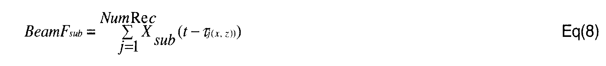
- X sub j (t - ⁇ j (x, z)) is the timed received signal of the i-th receive element 20 in the receive phase array under Sub-th sub-aperture fixed focusing firing.
- Each received echo signal collected through the receive array elements 20 would be timed and aligned at the focusing point (x,z) by applying, as shown by block 21 a corresponding time delay ⁇ j (x,z). All alignment signals are then summed as shown by block 22 and defined as the focusing intensity pixel (x,z) in the imaged image.
- the coherency factor CF Sub (x,z,t) for each sub-aperture transmission can be interpreted as a spatial coherency confidence ratio and is calculated as the proportion or percentage of coherent energy in the total non-coherent energy collected by the alignment focusing received signal from all transducer elements in the array 20'.
- the value of the coherency factor is from 0 to 1 inclusive. The higher the value corresponds to the higher proportion of coherent energy contained in the total collected signal energy indicating a higher confidence of good focusing quality or correctly aligned focused received signal and the lower the value of the coherency factor indicates a poor focusing quality.
- the coherency factor weighted received signals 31 from each of the plurality of sub-aperture transmissions are then synthesized in unit 25 to provide the final image.
- Figure 6a shows resultant images produced by the stage of STF-DRF ultrasound imaging only and Figure 6b shows imaging results for the same test object using an example of the present invention.
- Figure 6b shows imaging results for the same test object using an example of the present invention.
- those produced by an example of the present invention in Figure 6b have significantly better contrast and lower interference such that an operator may interpret these far more easily and be able to obtain information about the detected structure such as its size and be able to decide whether or not to take remedial action more confidently.
- Figure 7 is a comparison of the detected defect pixel intensity at different depths for the method shown in Figure 6a and the method of an example of the present invention shown in Figure 6b .
- the amplitude of the signal for the method shown in Figure 6a is labelled a and the amplitude of the signal produced by the example of the present invention from Figure 6b is labelled b.
- the signals produced by the example of the present invention are far more precise with less "spread" producing a far more precise and true indication of the defects in the test piece to an operator.
- Figure 8a shows another example of an image of a test object with defects produced by an imaging method not using a coherency factor
- Figure 8b shows the results from the same test piece using an example of the present invention.
- the defects are far more easily identifiable using the example of the present invention with less interference enabling an operator to far more reliably identify defects in a test piece and also be able to provide information about the defect, such as how serious it is.
- any number of sub-apertures of the array 20' may be used and each of those sub-apertures may have any desired number of transducer elements 20.
- Examples of the invention may be used to provide 3D beamforming imaging. Any type of array may be used such as a one dimensional or two dimensional array.
Landscapes
- Physics & Mathematics (AREA)
- Engineering & Computer Science (AREA)
- Remote Sensing (AREA)
- Radar, Positioning & Navigation (AREA)
- General Physics & Mathematics (AREA)
- Acoustics & Sound (AREA)
- Computer Networks & Wireless Communication (AREA)
- General Health & Medical Sciences (AREA)
- Biochemistry (AREA)
- Immunology (AREA)
- Pathology (AREA)
- Analytical Chemistry (AREA)
- Chemical & Material Sciences (AREA)
- Life Sciences & Earth Sciences (AREA)
- Health & Medical Sciences (AREA)
- Investigating Or Analyzing Materials By The Use Of Ultrasonic Waves (AREA)
Abstract
Description
- This invention relates generally to non-destructive testing and in particular to ultrasound imaging.
- Non-destructive testing devices can be used to inspect test objects to identify and analyse flaws and defects in the objects. An operator is able to move a probe at or near the surface of the test object in order to perform testing of both the object surface and its underlying structure. Non-destructive testing can be particularly useful in some industries such as aerospace, power generation, oil and gas recovery and refining where object testing must take place without removal of the object from surrounding structures and where hidden defects can be located that would otherwise not be identifiable through visual inspection.
- One example of non-destructive testing is ultrasonic testing. When conducting ultrasonic testing, an ultrasonic pulse can be emitted from a probe and passed through a test object at the characteristic sound velocity of that particular material. The sound velocity of a given material depends mainly on the modulus of elasticity, temperature and density of the material. Application of an ultrasonic pulse to a test object causes an interaction between the ultrasonic pulse and the test object structure, with sound waves being reflected back to the probe. This corresponding evaluation of the signals received by the probe, namely the amplitude and time of flight of those signals can allow conclusions to be drawn as to the internal quality of the test object, such as cracks or corrosion without destroying it.
- Generally, an ultrasonic testing system includes a probe for sending and receiving signals to and from a test object, a probe cable connecting the probe to an ultrasonic test unit and a screen or monitor for viewing test results. The ultrasonic test unit can include power supply components, signal generation, amplification and processing electronics and device controls used to operate the non-destructive testing device. Some ultrasonic test units can be connected to computers that control system operations as well as test results processing and display. Electric pulses can be generated by a transmitter and can be fed to the probe where they can be transformed into ultrasonic pulses by ultrasonic transducers.
- Conventional ultrasound imaging systems have an array of ultrasonic transducer elements to scan a targeted object by transmitting a focused ultrasound beam towards the object. The reflected acoustic wave is received, beamformed and processed for display.
- In conventional beamforming methods, the beam pattern profile is determined by the linear array structure. The elements interval were set less than half wavelength of the working frequency for the aim of avoiding the grating lobes. With less elements in an array structure, it suffers from the inherent drawback of having a wide main lobe and higher level side lobes at predictable angles. This produces lower quality imaging with low resolution caused by less focused response due to the wide main beam, and the low contrast between the true reflections and suffering from significant interference due to the high level unwanted side lobes. The level of the side lobes can be suppressed by using different shading windows, however, this widens the main lobe as the trade off price which, further decreases imaging resolution. Other methods have been considered for effectively reducing the effect of the side lobes, such as Minimum Variance method, but these generally involve a considerable level of calculation, resulting in increased costs and reduced speed.
- Whilst the level of the side lobes can be reduced by using a larger array structure, this increases the cost, size and complexity of any such system.
- It would be desirable to have a portable imaging system and corresponding method which produces a better quality output image with high contrast and acceptable good resolution without being excessively large, complex or expensive.
- According to a first aspect of the present invention, there is provided a method of operating an ultrasound imaging system having an array of transducer elements, the method comprising:
- transmitting a plurality of ultrasound signals each using a different sub-aperture of the array;
- receiving a reflected ultrasound signal corresponding to each sub-aperture transmission using the whole array or a sub-aperture of the array;
- calculating a coherency factor corresponding to the proportion of coherent energy in the received signals from each sub-aperture transmission and weighting the corresponding output signal by the calculated coherency factor; and
- synthesizing all the coherent factor weighted output signals under all different sub-aperture transmissions to produce the final imaging converted pixel intensity.
- Transmitting a plurality of ultrasound signals using different sub-apertures of the array provides enhanced receiving sensitivity of the defects but without a large number of transmission channels. The use of the coherency factor enhances the suppression of the side lobes enhancing the beamforming performance to produce the imaging in enhanced qualities of clarity, contrast and resolution.
- Each sub-aperture may use any splitting of transducer elements in the array. The sub-apertures may overlap or not overlap each other producing improved side lobe suppression and enhanced image contrast. Preferably the whole array of transducer elements is used to receive the reflected ultrasound waves and produce each received focusing signal. The coherency factor preferably corresponds to the proportion of coherent energy in the total non-coherent energy of each received transducer signal.
- The received signals may be focused such as by being beamformed.
- The coherency factor is preferably in the range from 0 to 1 inclusive. A higher value is indicative of a higher proportion of coherent energy contained in the total collected signal energy and thus a higher confidence of good focusing quality.
- According to a second aspect of the invention, there is provided an ultrasound imaging system, the system comprising:
- an array of transducer elements arranged to transmit a plurality of ultrasound signals using different sub-apertures of the array and to receive reflected ultrasound signals from a test piece for each of the sub-aperture transmissions;
- a controller arranged to calculate a coherency factor corresponding to the proportion of coherent energy in the received signals from each sub-aperture transmission and to weight the received signal by the calculated coherency factor; synthetize all weighted outputs from the different sub-aperture transmissions; and
- an output for a providing an output signal to be provided to a display for displaying an image representing a structure of the test piece.
- Examples of the present invention will now be described, by way of example only, with reference to the accompanying drawings in which:
-
Figure 1 shows an example of a typical ultrasonic testing system; -
Figure 2 shows an example of an ultrasonic testing system illustrating the present invention; -
Figure 3 shows a more detailed example of the non-overlapped sub-aperture transmission part of an example of the present invention; -
Figure 4 illustrates beamforming steering scan imaging; -
Figure 5 shows a more detailed example of the receiving part regarding to three sub-aperture transmission infigure 3 of an example of the present invention; -
Figures 6a and 6b provide examples of imaging produced by a system with Synthetic Fixed Transmission Focusing with Dynamic Receive Focusing (STF-DRF) only, and adaptive synthetic STF-DRT imaging produced by an example of the present invention; -
Figure 7 illustrates the detected defect pixel intensity at different depths for the images shown inFigures 6a and 6b ; and -
Figures 8a and 8b show further comparative examples of imaging by synthetic sub-aperture Synthetic Fixed Transmission Focusing with Dynamic Receive Focusing (STF-DTF) method and imaging by an example of the present invention. -
Figure 1 shows an example of anultrasonic testing system 1. The system includes aprobe 2 for sending and receiving signals to and from atest object 3. In this example theprobe 2 is arranged to send and receive a reflected ultrasonic signal from thetest object 3. However, in other examples the probe could instead be arranged to receive ultrasonic signals transmitted through atest object 3. Thetest object 3 could be any suitable object to be analysed for flaws and defects, such as, for example, panels of a vehicle such as an aircraft or a ship, sections of a pipeline or parts of an industrial plant which may take place without having to remove the object from surrounding structures. In use, theprobe 2 is moved over the surface of thetest object 3 to analyse the structure of the object. Theprobe 2 has an array of transducer elements. Aprobe cable 4 connects theprobe 2 to anultrasonic test unit 5. The ultrasonic test unit has a control processor for signal generation, amplification and processing electronics for example to generate electric pulses to be fed to theprobe 2 where they can be transformed into ultrasonic pulses by the ultrasonic transducers. Theultrasonic test unit 5 may also receive the reflected signal produced by theprobe 2. Theultrasonic test unit 5 has an electrical output and may include or be connected to a screen or monitor 6 to display results based on the output to a user to enable them to analyse the structure of thetest object 3 and identify any possible flaws or defects in the test object. For a portable testing system, theultrasonic test unit 5 would be combined with the screen or monitor 6 in a single unit. The screen ormonitor 6 may be provided by a computer which may be connected to theultrasonic test unit 5 and which may also provide some of the functions of theultrasonic test unit 5. -
Figure 2 shows an embodiment of animaging system 10 in accordance with the present invention. This embodiment is an adaptive synthetic Transmit Focusing and Dynamic Receive Focusing (adaptive STF-DRF) beamforming method. Theprobe 2 comprises a plurality oftransducer elements 20 in an array 20'.Figure 2 shows theprobe 2 having a linear array 20' of sixteentransducer elements 20, in this example. The upper part 2' schematically shows the probe when transmitting. As can be seen, theprobe 2 is arranged to transmit a plurality of ultrasound signals using different sub-apertures or sub-arrays. In the example ofFigure 2 , sub-array or sub-aperture 1 comprises transmitting with the eighttransducer elements 20 on the left of thearray 20, sub-array or sub-aperture 2 comprises transmitting with the eighttransducer elements 20 in the middle of the array 20' and sub-array or sub-aperture 3 comprises transmitting with the eight transducer elements on the right hand side of the array structure 20' one sub-aperture after the other. Any suitable arrangement of sub-apertures of the array 20' using any desired number oftransducer elements 20 which may or may not overlap may be used. Thelower part 2" of theprobe 2 as illustrated inFigure 2 shows the use of the transducer array for receiving echoes from thetest object 3. In this example all of thetransducer elements 20 of the array 20' are used to receive the reflected signal. In this example the signal generated by eachtransducer element 20 corresponding to its received ultrasound signal receives an appropriate beamforming delay fromdelay unit 21 and the delayed received signals are summed in summingunit 22. The received beamforming output from each sub-aperture transmission are then weighted 23 by a coherency factor CF1(t), CF2(t), CF3(t) ... corresponding to the proportion of coherent energy in each received signal. Theweighting 23 may include multiplying by the coherency factor. In this example, the coherency factor is determined in acoherency factor unit 24 to correspond to the proportion of coherent energy in the total non-coherent energy of each received signal from each sub-aperture transmission. - The coherency factor weighted received output from each sub-aperture transmission are then summed by
synthesis unit 25 to provide a clearer final image such that any defects or flaws may be easily identified. - In practice, all of the
delay 21,DAS beamforming 22,weighting 23, adaptive coherent factor determining 24 and synthesizing 25 are performed in a control processor such as a computer, microprocessor or by hard wired electronics. -
Figure 2 shows asingle probe 2 with thetransducer elements 20 first being used in a firing mode with a first sub-array or sub-aperture 1, then in a second firing mode with the second sub-array or sub-aperture 2 and then being used in a third firing mode with a third sub-array or sub-aperture 3. Below each sub-aperture firing mode is shown all of thetransducer elements 20 of the array 20' functioning in a receiver mode,weighting 23 each of the received beamforming outputs by acoherency factor 24 and synthesizing 25 all of the coherency factor weighted received beamforming outputs from each sub-aperture transmission. -
Figure 3 shows another example of theprobe 2 being controlled with three sub apertures (Fire-1, Fire-2 and Fire-3). In this example the sub apertures do not overlap. However, the present invention relates to any suitable number of two or more sub-apertures and each of the sub-apertures may contain any number of two or more transducer elements. In this example theultrasonic test unit 5 is schematically shown including arotary switch 51 to illustrate sequential firing of different sub-apertures within the array 20' oftransducer elements 20. The example ofFigure 3 illustrates thesignal 26 from each transducer element being delayed 27 to produce beamformed, steered focused transmitting beams 28 which enter atest object 3 with a required scan steering angle and focusing depth. The sub-apertures may adopt any desired focusing strategy such as fixed focusing or dynamic focusing by controlling the delays inunit 27 accordingly. The corresponding delays are used in the receivesection 21. - The use of sub-apertures for firing pulses into the
test object 3 while all elements (N elements 20) in thewhole array 2" are active for collecting the echoes increases one or more of the sensitivity, the penetration depth, and signal-to-noise ratio for each round-trip of data processing. For non-overlapping synthetic transmission, if M elements were contained by each defined single sub-array, then L number (L=N/M) of sub-arrays are used (where k=1:L ) for the whole synthetic transmitting processing. The sub-arrays can also be defined by overlapping as well. By this means, the number of sub-arrays is increased and the apodization is introduced on the whole array by weighting the overlap elements. - In the stage of STF-DRF beamforming in the presented invention, the kth group denoted as sub-aperture(k), is composed of multiple M transmitting elements. In each firing stage, M elements are active to incidence pulse into the
test object 3 for each firing process and all elements (N elements) in the whole array are active for collecting the echoes stage. With L round-trips for the combined sub-array transmission using the whole array to receive, the data storage was decreased into L*N RF lines. - The array phase center may be predefined by the STF-DRF method, with the sub-array phase-centre defined at each related sub-array geometric center point. It was coordinated in lateral X and depth Z dimensions indicated as (Xsub(k)_PhaseCenter, Zsub(k)_PhaseCenter=O) for the kth sub-aperture(k). The sub-array phase center is right or left translated by XSub(k)_PhaseCenter from the phase center of the whole array.
-
Figure 4 illustrates beamforming steering scan imaging. In order to calculate imaging pixel (x, z) intensity along the beam-line of sub-array(k) steering α angle transmission, the corresponding fixed transmission focus point (Xsub(k)_TransFocus(α)' z Sub(k)_TransFocus(α)) can be calculated through α angle rotated and shifted by related sub-array phase center in lateral direction XSub(k)_PhaseCenter, we have

where, R is the fixed transmission range, for kth sub-array, the pulse travels delay from ith transmission element to its focus point in the travel speed of sound Csound as:
while, for jth element receive element under kth sub-array firing, the echo time delay with dynamic focusing can be described as:
- The synthetic Transmit Focusing with dynamic receive Focusing (STF-DRF) beamform method presents the extracted echoes from steering α angle direction as:

- The first summation (index by k) is for L sub-apertures synthetic virtual array transmission, the second summation (index by i) is for the summation of the transmitting beamforming, and the third summation (index by j) is for the receive beamforming.
-
Figure 5 shows a more detailed view of theprobe 2 and ultrasonic test unit receiving reflected ultrasound signals from atest object 3. In this example all of thetransducer elements 20 of theprobe 2 are used for collecting reflected echoes from thetest object 3 after each sub-aperture transmission. Thesignals 29 from thetransducer elements 20 are then delayed 21 by an amount corresponding to the required focusing delay. The delayed signals 30 from all of the receivingtransducer elements 20 of the array are then summed 22 prior to being weighted by the coherency factor. - An example of a method for determining the coherency factor will now be described.
- For a transducer array 20' with a number of receiving
transducer elements 20 given as NumRec and the number of sub aperture beamforming transmitting sub-arrays being NumSub, the imaging intensity of each receiving pixel (x, z) in a beam steering scan area can be described as:
- Where Xsub j (t - τ j (x, z)) is the timed received signal of the i-th receive
element 20 in the receive phase array under Sub-th sub-aperture fixed focusing firing. - Each received echo signal collected through the receive
array elements 20 would be timed and aligned at the focusing point (x,z) by applying, as shown by block 21 a corresponding time delay τ j (x,z). All alignment signals are then summed as shown byblock 22 and defined as the focusing intensity pixel (x,z) in the imaged image. - During the processing of the received echo signal as illustrated in
Figure 5 , the focusing qualities are constantly evaluated for each steering scan imaging pixel before they are finally synthesized into imaging pixels atunit 25. The coherency factor is introduced and defined inequation 5 below:
- The coherency factor CFSub(x,z,t) for each sub-aperture transmission can be interpreted as a spatial coherency confidence ratio and is calculated as the proportion or percentage of coherent energy in the total non-coherent energy collected by the alignment focusing received signal from all transducer elements in the array 20'. In this example the value of the coherency factor is from 0 to 1 inclusive. The higher the value corresponds to the higher proportion of coherent energy contained in the total collected signal energy indicating a higher confidence of good focusing quality or correctly aligned focused received signal and the lower the value of the coherency factor indicates a poor focusing quality.
- It has been found that applying the adaptive coherent measurement weighting strongly emphasises the in-phase signals, increasing the adaptive coherent confidence ratio whilst significantly suppressing the out-of-phase signals providing enhanced contrast between the true reflections with significantly reduced interference producing higher quality resultant images.
- The resultant adaptive STF-DRF imaging intensity of a pixel (x,z) in a beam steering scan area when multiplied by the coherency factor for a received
transducer element 20 numbered as NumRec and with the number of sub-aperture transmitting arrays NumSub is defined as:
- As explained above, this results in a higher quality resultant image with high contrast between the true reflections and suffering from much less interference.
- The coherency factor weighted received
signals 31 from each of the plurality of sub-aperture transmissions are then synthesized inunit 25 to provide the final image. - When using the testing system with test objects having different properties such as by being made of different materials, some parameters of the equations may be adjusted accordingly.
-
Figure 6a shows resultant images produced by the stage of STF-DRF ultrasound imaging only andFigure 6b shows imaging results for the same test object using an example of the present invention. As can be seen by comparison of these resultant images, those produced by an example of the present invention inFigure 6b have significantly better contrast and lower interference such that an operator may interpret these far more easily and be able to obtain information about the detected structure such as its size and be able to decide whether or not to take remedial action more confidently. -
Figure 7 is a comparison of the detected defect pixel intensity at different depths for the method shown inFigure 6a and the method of an example of the present invention shown inFigure 6b . InFigure 7 , the amplitude of the signal for the method shown inFigure 6a is labelled a and the amplitude of the signal produced by the example of the present invention fromFigure 6b is labelled b. As can be seen, for each of the defects in thetest object 3, the signals produced by the example of the present invention are far more precise with less "spread" producing a far more precise and true indication of the defects in the test piece to an operator. -
Figure 8a shows another example of an image of a test object with defects produced by an imaging method not using a coherency factor andFigure 8b shows the results from the same test piece using an example of the present invention. As can be clearly seen, the defects are far more easily identifiable using the example of the present invention with less interference enabling an operator to far more reliably identify defects in a test piece and also be able to provide information about the defect, such as how serious it is. - Many variations may be made to the examples described above without departing from the scope of the present invention. For example, any number of sub-apertures of the array 20' may be used and each of those sub-apertures may have any desired number of
transducer elements 20. Examples of the invention may be used to provide 3D beamforming imaging. Any type of array may be used such as a one dimensional or two dimensional array.
Claims (20)
- A method of operating an ultrasound imaging system having an array of transducer elements, the method comprising:transmitting a plurality of ultrasound signals, each transmission using a different sub-aperture of the array;receiving a plurality of reflected ultrasound signals by a receive array corresponding to each sub-aperture transmission;calculating a coherency factor corresponding to the proportion of coherent energy in the received signals from each sub-aperture transmission and weighting the received output by the calculated coherency factor; andsynthesizing all weighted outputs under all different sub-aperture transmissions.
- A method according to claim 1, wherein the coherency factor weighted beamforming outputs from each of the plurality of sub-aperture transmissions are synthesized.
- The method according to claim 1 or claim 2, wherein the coherency factor corresponds to the proportion of coherent energy in the total non-coherent energy of timed received transducer signals.
- The method according to any preceding claim, wherein the Delay-and-Sum principle is used for either sub-aperture transmitting beamforming or receiving beamforming or both.
- The method according to any one of the preceding claims, wherein transmit beamforming is used in the form of fixed focus transmission, multiple fixed focus transmission zone, or full dynamic transmitting focus.
- The method according to any preceding claim, wherein dynamic focusing is used in receive focusing beamforming.
- The method according to any preceding claim, wherein the coherency factor is defined in each different coherent measurement either in the energy, amplitude or sign of the timed received signal.
- The method according to any preceding claim, wherein the coherency factor is normalized in the range from 0 to 1 inclusive with a higher value being indicative of a higher proportion of coherent signals contained in the total signal collected by a transducer.
- A method according to any one of the preceding claims, wherein the imaging intensity of each pixel (x,z,t) is determined according to the following equation:

where BeamF sub is the beamforming output under index sub sub-aperture transmitting. - The method according to claim 9, wherein the beamforming output BeamF sub can be either obtained by the conventional DAS method, or advanced method such as Minimum Variance Method (MVM).
- The method according to claim 10, wherein the conventional DAS beamforming is determined according to the following equation:
Therefore, the equation:
where NumRec is the number of receiving transducer elements, Xsub (t - τj (x, z)) is the timed received signal of the j-th receive element 20 in a receive phase array under the Sub-th transmitting sub-aperture firing: t is the time at which a signal is received; τi (x, z), is the applied time delay and CFsub (x,z,t) is the sub-aperture coherency factor. - A method according to any one of the preceding claims, wherein the entire array of transducer elements or a sub-aperture is used to receive a signal corresponding to the reflected ultrasound signal from each sub-aperture transmission.
- A method according to any one of the preceding claims, wherein the sub-apertures of the array can be used as overlap or splitting as non-overlap sub-apertures.
- A method according to any one of the preceding claims, used for 3D beamforming imaging.
- A method according to any one of the preceding claims using a 2D array.
- A method according to any one of the preceding claims, used for non-destructive testing.
- An ultrasound imaging system, the system comprising:an array of transducer elements arranged to transmit a plurality of ultrasound signals using different sub-apertures of the array and to receive reflected ultrasound signals from a test piece for each of the sub-aperture transmissions;a controller arranged to calculate a coherency factor corresponding to the proportion of coherent energy in the received signal from each sub-aperture transmission and to weight the received signal by the calculated coherency factor; andan output for a providing an output signal to be provided to a display for displaying an image representing a structure of the test piece;wherein the controller is arranged to synthesize the coherency factor weighted received signals from each of the plurality of sub-aperture transmissions.
- The ultrasound imaging system of claim 17, wherein the sub-aperture coherency factor corresponds to the proportion of coherent energy in the total non-coherent energy received by each transducer.
- The system according to claims 17 or claim 18, wherein the system is arranged to determine the imaging intensity of each pixel (x,z,t) using the following equation:

where NumRec is the number of receiving transducer elements, Xsub,i (t-τj (x,z)) is the timed received signal of the i-th receive element in a receive phase array under the Sub-th transmitting sub-aperture firing: t is the time at which a signal is received; τj (x, z), is the applied time delay and CFsub (x,y,z) is the sub-aperture Coherency Factor. - The ultrasound imaging system according to any one of claims 17 to 19, wherein all of the transducer elements of the array are used to receive the reflected ultrasound signal.
Priority Applications (4)
| Application Number | Priority Date | Filing Date | Title |
|---|---|---|---|
| EP11183566A EP2574956A1 (en) | 2011-09-30 | 2011-09-30 | Ultrasound imaging system and method with side lobe suppression via coherency factor weighting |
| JP2012206427A JP2013079949A (en) | 2011-09-30 | 2012-09-20 | Imaging system and method |
| CA2790481A CA2790481A1 (en) | 2011-09-30 | 2012-09-20 | Imaging system and method |
| US13/630,736 US20130083628A1 (en) | 2011-09-30 | 2012-09-28 | Imaging system and method |
Applications Claiming Priority (1)
| Application Number | Priority Date | Filing Date | Title |
|---|---|---|---|
| EP11183566A EP2574956A1 (en) | 2011-09-30 | 2011-09-30 | Ultrasound imaging system and method with side lobe suppression via coherency factor weighting |
Publications (1)
| Publication Number | Publication Date |
|---|---|
| EP2574956A1 true EP2574956A1 (en) | 2013-04-03 |
Family
ID=45002567
Family Applications (1)
| Application Number | Title | Priority Date | Filing Date |
|---|---|---|---|
| EP11183566A Withdrawn EP2574956A1 (en) | 2011-09-30 | 2011-09-30 | Ultrasound imaging system and method with side lobe suppression via coherency factor weighting |
Country Status (4)
| Country | Link |
|---|---|
| US (1) | US20130083628A1 (en) |
| EP (1) | EP2574956A1 (en) |
| JP (1) | JP2013079949A (en) |
| CA (1) | CA2790481A1 (en) |
Cited By (2)
| Publication number | Priority date | Publication date | Assignee | Title |
|---|---|---|---|---|
| DE102013004924A1 (en) | 2013-03-22 | 2014-09-25 | GE Sensing & lnspection Technologies GmbH | Imaging system and method |
| EP3259587B1 (en) * | 2015-02-17 | 2024-04-10 | General Electric Company | Method for inspecting a weld seam with ultrasonic phased array |
Families Citing this family (30)
| Publication number | Priority date | Publication date | Assignee | Title |
|---|---|---|---|---|
| US8234923B2 (en) * | 2004-09-20 | 2012-08-07 | Innervision Medical Technologies Inc. | Systems and methods for ultrasound imaging |
| US9282945B2 (en) | 2009-04-14 | 2016-03-15 | Maui Imaging, Inc. | Calibration of ultrasound probes |
| JP6092109B2 (en) | 2010-10-13 | 2017-03-08 | マウイ イマギング,インコーポレーテッド | Concave ultrasonic transducer and 3D array |
| JP6407719B2 (en) | 2011-12-01 | 2018-10-17 | マウイ イマギング,インコーポレーテッド | Motion detection using ping base and multi-aperture Doppler ultrasound |
| EP2797515A4 (en) | 2011-12-29 | 2015-07-22 | Maui Imaging Inc | ULTRASOUND IMAGING IN M MODE OF ARBITRARY PATHWAYS |
| WO2013126559A1 (en) | 2012-02-21 | 2013-08-29 | Maui Imaging, Inc. | Determining material stiffness using multiple aperture ultrasound |
| EP2833791B1 (en) | 2012-03-26 | 2022-12-21 | Maui Imaging, Inc. | Methods for improving ultrasound image quality by applying weighting factors |
| EP2883079B1 (en) | 2012-08-10 | 2017-09-27 | Maui Imaging, Inc. | Calibration of multiple aperture ultrasound probes |
| WO2014031642A1 (en) | 2012-08-21 | 2014-02-27 | Maui Imaging, Inc. | Ultrasound imaging system memory architecture |
| CN103676827A (en) | 2012-09-06 | 2014-03-26 | Ip音乐集团有限公司 | System and method for remotely controlling audio equipment |
| WO2014160291A1 (en) | 2013-03-13 | 2014-10-02 | Maui Imaging, Inc. | Alignment of ultrasound transducer arrays and multiple aperture probe assembly |
| US9651525B2 (en) | 2013-06-27 | 2017-05-16 | TecScan Systems Inc. | Method and apparatus for scanning an object |
| US9883848B2 (en) | 2013-09-13 | 2018-02-06 | Maui Imaging, Inc. | Ultrasound imaging using apparent point-source transmit transducer |
| JP6722656B2 (en) | 2014-08-18 | 2020-07-15 | マウイ イマギング,インコーポレーテッド | Network-based ultrasound imaging system |
| DE102014117721A1 (en) * | 2014-12-02 | 2016-06-02 | Ge Sensing & Inspection Technologies Gmbh | Method and device for non-destructive testing of a test specimen by means of ultrasound |
| JP6770973B2 (en) | 2015-03-30 | 2020-10-21 | マウイ イマギング,インコーポレーテッド | Ultrasound Imaging Systems and Methods for Detecting Object Movement |
| CN105223567B (en) * | 2015-09-28 | 2018-01-30 | 中国科学院声学研究所 | A kind of robust wideband Adaptive beamformer method applied to ultrasonic imaging |
| CA3007284A1 (en) * | 2015-12-01 | 2017-06-08 | Supersonic Imagine | An imaging method, an apparatus implementing said method, a computer program and a computer-readable storage medium |
| CN108778530B (en) | 2016-01-27 | 2021-07-27 | 毛伊图像公司 | Ultrasound imaging with sparse array detectors |
| JP2019203722A (en) * | 2018-05-21 | 2019-11-28 | 三菱日立パワーシステムズ株式会社 | Ultrasonic flaw detection method, system, program, and storage medium |
| EP3581961A1 (en) * | 2018-06-13 | 2019-12-18 | Technische Universität München | Method and apparatus for ultrasound imaging with improved beamforming |
| CN110507355B (en) * | 2019-09-20 | 2022-04-15 | 青岛海信医疗设备股份有限公司 | Ultrasonic imaging system, method, equipment and medium |
| JP2021065449A (en) * | 2019-10-24 | 2021-04-30 | 株式会社リコー | Measuring device, beam measuring method and beam measuring program |
| WO2022086521A1 (en) | 2020-10-21 | 2022-04-28 | Maui Imaging, Inc. | Systems and methods for tissue characterization using multiple aperture ultrasound |
| JP2023548365A (en) | 2020-11-02 | 2023-11-16 | マウイ イマギング,インコーポレーテッド | Systems and methods for improving ultrasound image quality |
| US11717967B2 (en) | 2021-03-04 | 2023-08-08 | TecScan Systems Inc. | System and method for scanning an object using an array of ultrasonic transducers |
| CN113345041B (en) * | 2021-05-20 | 2024-03-15 | 河南工业大学 | Ultrasound coherence factor determination method, ultrasound image reconstruction method and electronic equipment |
| CN115825237B (en) * | 2022-08-29 | 2025-08-19 | 北京动力机械研究所 | Ultrasonic oblique incidence sub-aperture coherent composite divergent wave imaging method |
| GB2624639B (en) | 2022-11-22 | 2025-02-05 | Darkvision Tech Inc | Surface texture of manufactured parts |
| CN116124889A (en) * | 2023-01-17 | 2023-05-16 | 苏州热工研究院有限公司 | Metal pipeline corrosion monitoring method based on synthetic emission aperture imaging |
Citations (1)
| Publication number | Priority date | Publication date | Assignee | Title |
|---|---|---|---|---|
| WO2011095896A1 (en) * | 2010-02-08 | 2011-08-11 | Dalhousie University | Ultrasound imaging system using beamforming techniques for phase coherence grating lobe suppression |
Family Cites Families (9)
| Publication number | Priority date | Publication date | Assignee | Title |
|---|---|---|---|---|
| US4594691A (en) * | 1981-12-30 | 1986-06-10 | Schlumberger Technology Corporation | Sonic well logging |
| JP2795610B2 (en) * | 1994-03-16 | 1998-09-10 | アロカ株式会社 | Ultrasound diagnostic equipment |
| JP3484256B2 (en) * | 1995-05-01 | 2004-01-06 | アロカ株式会社 | Ultrasonic transmission / reception method and ultrasonic diagnostic apparatus |
| US5910115A (en) * | 1997-09-22 | 1999-06-08 | General Electric Company | Method and apparatus for coherence filtering of ultrasound images |
| US7744532B2 (en) * | 2004-03-31 | 2010-06-29 | Siemens Medical Solutions Usa, Inc. | Coherence factor adaptive ultrasound imaging methods and systems |
| US20060173313A1 (en) * | 2005-01-27 | 2006-08-03 | Siemens Medical Solutions Usa, Inc. | Coherence factor adaptive ultrasound imaging |
| GB2450112B (en) * | 2007-06-12 | 2010-12-08 | Ge Inspection Technologies Ltd | Automatic lift-off compensation for pulsed eddy current inspection |
| JP5396242B2 (en) * | 2009-11-11 | 2014-01-22 | 日立アロカメディカル株式会社 | Ultrasonic diagnostic equipment |
| JP5786433B2 (en) * | 2011-04-28 | 2015-09-30 | コニカミノルタ株式会社 | Ultrasonic diagnostic equipment |
-
2011
- 2011-09-30 EP EP11183566A patent/EP2574956A1/en not_active Withdrawn
-
2012
- 2012-09-20 CA CA2790481A patent/CA2790481A1/en not_active Abandoned
- 2012-09-20 JP JP2012206427A patent/JP2013079949A/en active Pending
- 2012-09-28 US US13/630,736 patent/US20130083628A1/en not_active Abandoned
Patent Citations (1)
| Publication number | Priority date | Publication date | Assignee | Title |
|---|---|---|---|---|
| WO2011095896A1 (en) * | 2010-02-08 | 2011-08-11 | Dalhousie University | Ultrasound imaging system using beamforming techniques for phase coherence grating lobe suppression |
Non-Patent Citations (4)
| Title |
|---|
| FRITSCH C ET AL: "Phase Coherence Imaging", IEEE TRANSACTIONS ON ULTRASONICS, FERROELECTRICS AND FREQUENCY CONTROL, IEEE, US, vol. 56, no. 5, 1 May 2009 (2009-05-01), pages 958 - 974, XP011269103, ISSN: 0885-3010, DOI: 10.1109/TUFFC.2009.1128 * |
| K.W. HOLLMAN ET AL: "Coherence factor of speckle from a multi-row probe", 1999 IEEE ULTRASONICS SYMPOSIUM. PROCEEDINGS. INTERNATIONAL SYMPOSIUM (CAT. NO.99CH37027), vol. 2, 1 January 1999 (1999-01-01), pages 1257 - 1260, XP055020986, ISSN: 1051-0117, ISBN: 978-0-78-035722-8, DOI: 10.1109/ULTSYM.1999.849225 * |
| PAI-CHI LI ET AL: "Adaptive imaging using the generalized coherence factor", IEEE TRANSACTIONS ON ULTRASONICS, FERROELECTRICS AND FREQUENCY CONTROL, IEEE, US, vol. 50, no. 2, 1 February 2003 (2003-02-01), pages 128 - 141, XP011368416, ISSN: 0885-3010, DOI: 10.1109/TUFFC.2003.1182117 * |
| TORBATIAN Z ET AL: "A split-aperture transmit beamforming technique with phase coherence grating lobe suppression", IEEE TRANSACTIONS ON ULTRASONICS, FERROELECTRICS AND FREQUENCY CONTROL, IEEE, US, vol. 57, no. 11, 1 November 2010 (2010-11-01), pages 2588 - 2595, XP011320241, ISSN: 0885-3010 * |
Cited By (3)
| Publication number | Priority date | Publication date | Assignee | Title |
|---|---|---|---|---|
| DE102013004924A1 (en) | 2013-03-22 | 2014-09-25 | GE Sensing & lnspection Technologies GmbH | Imaging system and method |
| DE102013004924B4 (en) | 2013-03-22 | 2018-05-03 | GE Sensing & lnspection Technologies GmbH | Imaging system and method |
| EP3259587B1 (en) * | 2015-02-17 | 2024-04-10 | General Electric Company | Method for inspecting a weld seam with ultrasonic phased array |
Also Published As
| Publication number | Publication date |
|---|---|
| US20130083628A1 (en) | 2013-04-04 |
| JP2013079949A (en) | 2013-05-02 |
| CA2790481A1 (en) | 2013-03-30 |
Similar Documents
| Publication | Publication Date | Title |
|---|---|---|
| EP2574956A1 (en) | Ultrasound imaging system and method with side lobe suppression via coherency factor weighting | |
| Le Jeune et al. | Plane wave imaging for ultrasonic non-destructive testing: Generalization to multimodal imaging | |
| US8924164B2 (en) | Apparatus and method for ultrasonic testing | |
| EP1004894B1 (en) | Method and apparatus for high-frame-rate high-resolution ultrasonic image data acquisition | |
| CN112505710A (en) | Multi-beam synthetic aperture sonar three-dimensional imaging algorithm | |
| CN104777485B (en) | The three-dimensional broad beam zonule Quick air formation method of ultrasonic two-dimensional array | |
| JP5306919B2 (en) | Ultrasonic flaw detection method and apparatus | |
| Sutcliffe et al. | Virtual source aperture imaging for non-destructive testing | |
| Huang et al. | Application of sparse synthetic aperture focusing techniques to ultrasound imaging in solids using a transducer wedge | |
| WO2014147122A1 (en) | Imaging system and method | |
| US11796659B2 (en) | Suppression of multiple scattering noise in pulse echo imaging | |
| JP2010266416A (en) | Phased array aperture synthesis processing method and its application effect evaluation method | |
| JP4458407B2 (en) | Harmonic imaging method and apparatus using multiplex transmission | |
| Lukomski et al. | Synthetic aperture focusing technique with virtual transducer for immersion inspection of solid objects | |
| Sutcliffe et al. | Virtual source aperture image processing methods for non-destructive testing | |
| Trots et al. | Golay coded sequences in synthetic aperture imaging systems | |
| CN118330037A (en) | Imaging algorithm of blind area defects in lead sealing based on ultrasonic phased array | |
| Prashar et al. | Comparison and optimisation of fast array-based ultrasound testing | |
| Stepinski | Synthetic aperture focusing technique in ultrasonic inspection of coarse grained materials | |
| Holmes et al. | Post-processing of ultrasonic phased array data for optimal performance | |
| JP2023510403A (en) | Indicative parametric echo sounders and methods for characterizing subseafloor portions of underwater environments | |
| Bazulin et al. | Increasing the signal-to-noise ratio in ultrasonic testing of repair welds using the technology of thinned antenna arrays | |
| CN105548363A (en) | Multi-route identification based ultrasonic detection imaging method | |
| Stepinski | Ultrasonic nondestructive inspection of solid objects | |
| Huang et al. | High Resolution Real Time Synthetic Aperture Imaging in Solids Using Virtual Elements |
Legal Events
| Date | Code | Title | Description |
|---|---|---|---|
| PUAI | Public reference made under article 153(3) epc to a published international application that has entered the european phase |
Free format text: ORIGINAL CODE: 0009012 |
|
| AK | Designated contracting states |
Kind code of ref document: A1 Designated state(s): AL AT BE BG CH CY CZ DE DK EE ES FI FR GB GR HR HU IE IS IT LI LT LU LV MC MK MT NL NO PL PT RO RS SE SI SK SM TR |
|
| AX | Request for extension of the european patent |
Extension state: BA ME |
|
| 17P | Request for examination filed |
Effective date: 20131004 |
|
| RBV | Designated contracting states (corrected) |
Designated state(s): AL AT BE BG CH CY CZ DE DK EE ES FI FR GB GR HR HU IE IS IT LI LT LU LV MC MK MT NL NO PL PT RO RS SE SI SK SM TR |
|
| 17Q | First examination report despatched |
Effective date: 20161116 |
|
| STAA | Information on the status of an ep patent application or granted ep patent |
Free format text: STATUS: THE APPLICATION IS DEEMED TO BE WITHDRAWN |
|
| 18D | Application deemed to be withdrawn |
Effective date: 20170527 |