EP2260875B1 - Folate-receptor targeted imaging agents - Google Patents
Folate-receptor targeted imaging agents Download PDFInfo
- Publication number
- EP2260875B1 EP2260875B1 EP10179508.6A EP10179508A EP2260875B1 EP 2260875 B1 EP2260875 B1 EP 2260875B1 EP 10179508 A EP10179508 A EP 10179508A EP 2260875 B1 EP2260875 B1 EP 2260875B1
- Authority
- EP
- European Patent Office
- Prior art keywords
- cells
- cancer cells
- vitamin
- folate
- tumor
- Prior art date
- Legal status (The legal status is an assumption and is not a legal conclusion. Google has not performed a legal analysis and makes no representation as to the accuracy of the status listed.)
- Expired - Lifetime
Links
- 0 *=C1NC(N)=Nc2c1nc(CNc(cc1)ccc1C(N[C@](CCC(NC[C@@](C(N[C@@](CC(O)=O)C(N[C@@](CS)C(O)=O)O)=O)N)=O)C(O)=O)=O)cn2 Chemical compound *=C1NC(N)=Nc2c1nc(CNc(cc1)ccc1C(N[C@](CCC(NC[C@@](C(N[C@@](CC(O)=O)C(N[C@@](CS)C(O)=O)O)=O)N)=O)C(O)=O)=O)cn2 0.000 description 1
- RKGPQAGUQDVJHG-MHORFTMASA-N N[C@@H](CNC(CC[C@H](C(O)=O)NC(c(cc1)ccc1NCc(cn1)nc2c1N=C(N)NC2=O)=O)=O)C(N[C@@H](CC(O)=O)C(N[C@@H](CS)C(O)=O)=O)=O Chemical compound N[C@@H](CNC(CC[C@H](C(O)=O)NC(c(cc1)ccc1NCc(cn1)nc2c1N=C(N)NC2=O)=O)=O)C(N[C@@H](CC(O)=O)C(N[C@@H](CS)C(O)=O)=O)=O RKGPQAGUQDVJHG-MHORFTMASA-N 0.000 description 1
Images


Classifications
-
- A—HUMAN NECESSITIES
- A61—MEDICAL OR VETERINARY SCIENCE; HYGIENE
- A61K—PREPARATIONS FOR MEDICAL, DENTAL OR TOILETRY PURPOSES
- A61K51/00—Preparations containing radioactive substances for use in therapy or testing in vivo
- A61K51/02—Preparations containing radioactive substances for use in therapy or testing in vivo characterised by the carrier, i.e. characterised by the agent or material covalently linked or complexing the radioactive nucleus
- A61K51/04—Organic compounds
- A61K51/08—Peptides, e.g. proteins, carriers being peptides, polyamino acids, proteins
-
- A—HUMAN NECESSITIES
- A61—MEDICAL OR VETERINARY SCIENCE; HYGIENE
- A61K—PREPARATIONS FOR MEDICAL, DENTAL OR TOILETRY PURPOSES
- A61K49/00—Preparations for testing in vivo
-
- A—HUMAN NECESSITIES
- A61—MEDICAL OR VETERINARY SCIENCE; HYGIENE
- A61K—PREPARATIONS FOR MEDICAL, DENTAL OR TOILETRY PURPOSES
- A61K47/00—Medicinal preparations characterised by the non-active ingredients used, e.g. carriers or inert additives; Targeting or modifying agents chemically bound to the active ingredient
- A61K47/50—Medicinal preparations characterised by the non-active ingredients used, e.g. carriers or inert additives; Targeting or modifying agents chemically bound to the active ingredient the non-active ingredient being chemically bound to the active ingredient, e.g. polymer-drug conjugates
- A61K47/51—Medicinal preparations characterised by the non-active ingredients used, e.g. carriers or inert additives; Targeting or modifying agents chemically bound to the active ingredient the non-active ingredient being chemically bound to the active ingredient, e.g. polymer-drug conjugates the non-active ingredient being a modifying agent
- A61K47/54—Medicinal preparations characterised by the non-active ingredients used, e.g. carriers or inert additives; Targeting or modifying agents chemically bound to the active ingredient the non-active ingredient being chemically bound to the active ingredient, e.g. polymer-drug conjugates the non-active ingredient being a modifying agent the modifying agent being an organic compound
- A61K47/55—Medicinal preparations characterised by the non-active ingredients used, e.g. carriers or inert additives; Targeting or modifying agents chemically bound to the active ingredient the non-active ingredient being chemically bound to the active ingredient, e.g. polymer-drug conjugates the non-active ingredient being a modifying agent the modifying agent being an organic compound the modifying agent being also a pharmacologically or therapeutically active agent, i.e. the entire conjugate being a codrug, i.e. a dimer, oligomer or polymer of pharmacologically or therapeutically active compounds
- A61K47/551—Medicinal preparations characterised by the non-active ingredients used, e.g. carriers or inert additives; Targeting or modifying agents chemically bound to the active ingredient the non-active ingredient being chemically bound to the active ingredient, e.g. polymer-drug conjugates the non-active ingredient being a modifying agent the modifying agent being an organic compound the modifying agent being also a pharmacologically or therapeutically active agent, i.e. the entire conjugate being a codrug, i.e. a dimer, oligomer or polymer of pharmacologically or therapeutically active compounds one of the codrug's components being a vitamin, e.g. niacinamide, vitamin B3, cobalamin, vitamin B12, folate, vitamin A or retinoic acid
-
- A—HUMAN NECESSITIES
- A61—MEDICAL OR VETERINARY SCIENCE; HYGIENE
- A61K—PREPARATIONS FOR MEDICAL, DENTAL OR TOILETRY PURPOSES
- A61K51/00—Preparations containing radioactive substances for use in therapy or testing in vivo
-
- A—HUMAN NECESSITIES
- A61—MEDICAL OR VETERINARY SCIENCE; HYGIENE
- A61K—PREPARATIONS FOR MEDICAL, DENTAL OR TOILETRY PURPOSES
- A61K51/00—Preparations containing radioactive substances for use in therapy or testing in vivo
- A61K51/02—Preparations containing radioactive substances for use in therapy or testing in vivo characterised by the carrier, i.e. characterised by the agent or material covalently linked or complexing the radioactive nucleus
- A61K51/025—Preparations containing radioactive substances for use in therapy or testing in vivo characterised by the carrier, i.e. characterised by the agent or material covalently linked or complexing the radioactive nucleus inorganic Tc complexes or compounds
-
- A—HUMAN NECESSITIES
- A61—MEDICAL OR VETERINARY SCIENCE; HYGIENE
- A61K—PREPARATIONS FOR MEDICAL, DENTAL OR TOILETRY PURPOSES
- A61K51/00—Preparations containing radioactive substances for use in therapy or testing in vivo
- A61K51/02—Preparations containing radioactive substances for use in therapy or testing in vivo characterised by the carrier, i.e. characterised by the agent or material covalently linked or complexing the radioactive nucleus
- A61K51/04—Organic compounds
- A61K51/0402—Organic compounds carboxylic acid carriers, fatty acids
-
- A—HUMAN NECESSITIES
- A61—MEDICAL OR VETERINARY SCIENCE; HYGIENE
- A61K—PREPARATIONS FOR MEDICAL, DENTAL OR TOILETRY PURPOSES
- A61K51/00—Preparations containing radioactive substances for use in therapy or testing in vivo
- A61K51/02—Preparations containing radioactive substances for use in therapy or testing in vivo characterised by the carrier, i.e. characterised by the agent or material covalently linked or complexing the radioactive nucleus
- A61K51/04—Organic compounds
- A61K51/041—Heterocyclic compounds
- A61K51/044—Heterocyclic compounds having nitrogen as a ring hetero atom, e.g. guanethidine, rifamycins
- A61K51/0459—Heterocyclic compounds having nitrogen as a ring hetero atom, e.g. guanethidine, rifamycins having six-membered rings with two nitrogen atoms as the only ring hetero atoms, e.g. piperazine
-
- A—HUMAN NECESSITIES
- A61—MEDICAL OR VETERINARY SCIENCE; HYGIENE
- A61K—PREPARATIONS FOR MEDICAL, DENTAL OR TOILETRY PURPOSES
- A61K51/00—Preparations containing radioactive substances for use in therapy or testing in vivo
- A61K51/02—Preparations containing radioactive substances for use in therapy or testing in vivo characterised by the carrier, i.e. characterised by the agent or material covalently linked or complexing the radioactive nucleus
- A61K51/04—Organic compounds
- A61K51/0497—Organic compounds conjugates with a carrier being an organic compounds
-
- A—HUMAN NECESSITIES
- A61—MEDICAL OR VETERINARY SCIENCE; HYGIENE
- A61K—PREPARATIONS FOR MEDICAL, DENTAL OR TOILETRY PURPOSES
- A61K51/00—Preparations containing radioactive substances for use in therapy or testing in vivo
- A61K51/02—Preparations containing radioactive substances for use in therapy or testing in vivo characterised by the carrier, i.e. characterised by the agent or material covalently linked or complexing the radioactive nucleus
- A61K51/04—Organic compounds
- A61K51/08—Peptides, e.g. proteins, carriers being peptides, polyamino acids, proteins
- A61K51/088—Peptides, e.g. proteins, carriers being peptides, polyamino acids, proteins conjugates with carriers being peptides, polyamino acids or proteins
Definitions
- the invention relates to compounds and methods for targeting an imaging agent to cells of an animal. More particularly, radionuclide-based imaging agents are targeted to cells having receptors for a vitamin by using such a vitamin, or a vitamin receptor binding derivative or an analog thereof, as the targeting ligand for the imaging agent.
- Transmembrane transport is a critical cellular function. Because practitioners have recognized the importance of transmembrane transport to many areas of medical and biological science, including drug therapy and gene transfer, there have been significant research efforts directed to the understanding and application of such processes. Thus, for example, transmembrane delivery of nucleic acids has been attempted through the use of protein carriers, antibody carriers, liposomal delivery systems, electroporation, direct injection, cell fusion, viral carriers, osmotic shock, and calcium-phosphate mediated transformation. However, many of those techniques are limited both by the types of cells in which transmembrane transport occurs and by the conditions required for successful transmembrane transport of exogenous molecules. Furthermore, many of these techniques are limited by the type and size of the exogenous molecule that can be transported across the cell membrane without loss ofbioactivity.
- receptor-mediated endocytosis occurs both in vivo and in vitro.
- Receptor-mediated endocytosis involves the movement of ligands bound to membrane receptors into the interior of an area bounded by the membrane through invagination of the membrane. The process is initiated or activated by the binding of a receptor-specific ligand to the receptor.
- Receptor mediated endocytosis has been utilized for delivering exogenous molecules such as proteins and nucleic acids to cells.
- a specific ligand is chemically conjugated by covalent, ionic, or hydrogen bonding to an exogenous molecule of interest, forming a conjugate molecule having a moiety (the ligand portion) that is still recognized in the conjugate by a target receptor.
- the photo toxic protein psoralen has been conjugated to insulin and internalized by the insulin receptor endocytotic pathway ( Gasparro, Biochem. Biophys. Res. Comm. 141(2), pp. 502-509, Dec.
- the hepatocyte specific receptor for galactose terminal asialoglycoproteins has been utilized for the hepatocyte-specific transmembrane delivery of asialoorosomucoid-poly-lysine non-covalently complexed to a plasmid ( Wu, G. Y., J. Biol. Chem., 262(10), pp. 4429-4432, 1987 ); the cell receptor for EGF has been utilized to deliver polynucleotides covalently linked to EGF to the cell interior (Myers, European Patent Application 86810614.7, published Jun.
- the intestinally situated cellular receptor for the organometallic vitamin B 12 -intrinsic factor complex has been used to mediate delivery of a drug, a hormone, a bioactive peptide and an immunogen complexed with vitamin B 12 to the circulatory system after oral administration (Russell-Jones et al., European patent Application 86307849.9, published Apr. 29, 1987 ); the mannose-6-phosphate receptor has been used to deliver low density lipoproteins to cells ( Murray, G. J. and Neville, D. M., Jr., J.Biol.Chem, Vol. 255 (24), pp.
- the cholera toxin binding subunit receptor has been used to deliver insulin to cells lacking insulin receptors ( Roth and Maddox, J.Coll.Phys. Vol. 115, p. 151, 1983 ); and the human chorionic gonadotropin receptor has been employed to deliver a ricin a-chain coupled to HCG to cells with the appropriate HCG receptor ( Oeltmann and Heath, J.Biol.Chem, vol. 254, p. 1028 (1979 )).
- the present invention involves the transmembrane transport of a radionuclide-based imaging agent across a membrane having receptors for folate.
- a cell membrane bearing folate receptors is contacted with a vitamin-imaging agent conjugate for a time sufficient to initiate and permit transmembrane transport of the conjugate, and the biodistribution of the vitamin-imaging agent conjugate in the animal is monitored.
- the folate moiety simply binds to a cell surface vitamin receptor to concentrate the chelated radionuclide on the cell surface.
- the invention takes advantage of (1) the location of vitamin receptors and (2) the associated receptor-mediated endocytic processes.
- the invention takes advantage of the unique expression, overexpression, or preferential expression of vitamin receptors, transporters, or other surface-presented proteins that specifically bind vitamins, or derivatives or analogs thereof, on tumor cells or other cell types which overexpress such receptors.
- the invention can be used to detect cells, such as tumor cells or other cell types, which overexpress vitamin receptors, or receptors for vitamin derivatives or analogs, by talking advantage of the receptor-mediated endocytic processes that occur when such cells are contacted with the vitamin-imaging agent conjugate.
- Vitamin receptors such as the high-affinity folate receptor (FR) is expressed at high levels, for example, on cancer cells.
- FR high-affinity folate receptor
- Epithelial cancers of the ovary, mammary gland, colon, lung, nose, throat, and brain have all been reported to express elevated levels of the FR.
- greater than 90% of all human ovarian tumors are known to express large amounts of this receptor.
- the present invention can be used for the diagnostic imaging of a variety of tumor types, and of other cell types involved in disease states.
- Radionuclide chelators complexed to ligands have been used as non-invasive probes for diagnostic imaging purposes.
- vasoactive intestinal peptide, somatostatin analogs, and monoclonal antibodies have been used as ligands to localize radionuclides to cells, such as tumor cells.
- Vitamins such as folic acid
- the first folic acid-based targeting complex described for in vivo tumor imaging was a histamine derivative containing 125 Iodine. This complex was not considered a relevant clinical candidate because of the long-lived 125 I radionuclide component.
- a deferoxamine-folate conjugate for tumor imaging was developed (deferoxamine cholates 67 Ga, a gamma-emitting radionuclide that has a 78 hour half-life).
- 99m Tc has been adopted as the preferred radionuclide for diagnostic imaging, because i) the radionuclide is easily obtained from commercially available 99 Mo- 99m Tc generators, ii) the cost of producing large amounts of 99m Tc is insignificant compared to the cost of producing 111 In, and iii) 99m Tc has a much shorter (6 hour) half life, which allows higher radionuclide doses to be administered, yielding higher resolution images without the risk of hazardous radiation exposure to vital organs.
- folate-based 99m Tc conjugates have been developed.
- folate conjugates of 99m Tc-6-hydrazinonicotinamido-hydrazido HYNIC; Guo, et al., J. Nucl. Med., 40(9): 1563-1569 (1999 )
- 99m Tc-12-amino-3,3,9,9-tetramethyl-5-oxa-4,8 diaza-2,10-dodecanedinoe dioxime OXA
- the invention relates to a method of imaging a population of tumour cells in an animal comprising administering an effective amount of a compound of the formula chelated to a cation of a radionuclide.
- V is a vitamin, or a vitamin receptor binding derivative or analog thereof
- L is a divalent linker
- R is a side chain of an amino acid of the formula H 2 NCHRCOOH
- M is a cation of a radionuclide
- n is 1 or
- k is 1 or 0.
- the vitamin is a substrate for receptor-mediated transmembrane transport in vivo.
- composition for diagnostic imaging comprising a compound of the formula wherein V is a vitamin, or a vitamin receptor binding derivative or analog thereof, L is a divalent linker, R is a side chain of an amino acid of the formula H 2 NCHRCOOH, M is a cation of a radionuclide, n is 1 or 0, and a pharmaceutically acceptable carrier therefor.
- the vitamin is a substrate for receptor-mediated transmembrane transport in vivo.
- a method of imaging a population of cells in an animal, wherein the cells are characterized by a vitamin receptor on the surface of the cells.
- the method comprises the steps of administering to the animal an effective amount of a composition comprising a compound of the formula wherein V is a vitamin, or a receptor binding derivative or analog thereof, specific for the cell surface vitamin receptor, L is a divalent linker, R is a side chain of an amino acid of the formula H 2 NCHRCOOH, M is a cation of a radionuclide, n is 1 or 0, and a pharmaceutically acceptable carrier therefor, and monitoring the biodistribution of the compound in the animal.
- V is a vitamin that is a substrate for receptor-mediated transmembrane transport in vivo, or a vitamin receptor binding derivative or analog thereof
- L is a divalent linker
- R is a side chain of an amino acid of the formula H 2 NCHRCOOH
- M is a cation of a radionuclide
- n is 1 or 0, and k is 1 or 0.
- composition for diagnostic imaging comprising a compound of the formula wherein V is a vitamin that is a substrate for receptor-mediated transmembrane transport in vivo, or a vitamin receptor binding derivative or analog thereof, L is a divalent linker, R is a side chain of an amino acid of the formula H 2 NCHRCOOH, M is a cation of a radionuclide, n is 1 or 0, and a pharmaceutically acceptable carrier therefor.
- a method of imaging a population of cells in an animal wherein the cells are characterized by a vitamin receptor on the surface of the cells.
- the method comprises the steps of administering to the animal an effective amount of a composition comprising a compound of the formula wherein V is the vitamin, or a receptor binding derivative or analog thereof, specific for the cell surface vitamin receptor, L is a divalent linker, R is a side chain of an amino acid of the formula H 2 NCHRCOOH, M is a cation of a radionuclide, n is 1 or 0, and a pharmaceutically acceptable carrier therefor, and monitoring the biodistribution of the compound in the animal.
- V in the compound can be, for example, a vitamin selected from the group consisting of folate, riboflavin, thiamine, vitamin B 12 , and biotin, or a derivative or analog thereof.
- the radionuclide in the compound can be selected, for example, from the group consisting of radioisotopes of gallium, indium, copper, technetium, and rhenium.
- methods are provided for targeting radionuclide-based imaging agents to cell populations that uniquely express, overexpress, or preferentially express vitamin receptors.
- a vitamin, or a receptor binding derivative or analog thereof is used as the targeting ligand for the imaging agent.
- the vitamin-imaging agent conjugate can be used to target radionuclides to cells and to concentrate the radionuclides in a cell population, such as a tumor cell population, for use in diagnostic imaging.
- the invention relates to a method of imaging a population of tumour cells in an animal comprising administering an effective amount of a compound of the formula chelated to a cation of a radionuclide.
- the application discloses a composition for diagnostic imaging comprising a compound of the formula or for use in such methods.
- V is a vitamin, or a vitamin receptor binding derivative or analog thereof
- L is a divalent linker
- R is a side chain of an amino acid of the formula H 2 NCHRCOOH
- M is a cation of a radionuclide
- n is 1 or 0.
- the vitamin, or vitamin receptor binding derivative or analog thereof is a substrate for receptor-mediated transmembrane transport in vivo.
- V is a vitamin, or a vitamin receptor binding derivative or analog thereof
- L is a divalent linker
- R is a side chain of an amino acid of the formula H 2 NCHRCOOH
- M is a cation of a radionuclide
- n is 1 or
- k is 1 or 0.
- the vitamin is a substrate for receptor-mediated transmembrane transport in vivo.
- Exemplary of these compounds is a compound referred to as EC20 depicted in Fig. 1 .
- Exemplary of other compounds for use in accordance with this disclosure are compounds denominated as EC11, EC13, EC14, EC15, EC19, EC31, and EC53 (see Fig. 10 ).
- the vitamin moiety e.g ., the folic acid moiety in EC20
- the compounds also contain a bifunctional peptide-based chelator, which provides the site for chelation of the radionuclide, for example, 99m Tc (see Fig. 1 ), and the compounds can, optionally, contain a linker through which the vitamin moiety is covalently bonded to the chelating moiety.
- the vitamin moiety of the compound is a vitamin that is a substrate for receptor-mediated transmembrane transport in vivo, or a vitamin receptor binding derivative or analog thereof.
- the vitamin in the above disclosures is linked, optionally, through a linker (L) to the chelator portion of the compounds.
- EC20 comprises a folic acid analog linked to the chelator moiety because EC20 has the glutamic acid in the D configuration.
- the chelator portion comprises an ⁇ , ⁇ -diaminoprogionic acid moiety linked to a cysteine group through a third amino acid residue.
- vitamin-imaging agent conjugates the compound with bound radionuclide are referred to as "vitamin-imaging agent conjugates.”
- the structure of the linker is not critical to the above disclosures.
- the linker can be any biocompatible divalent linker.
- the linker comprises about 1 to about 30 carbon atoms, more typically about 2 to about 20 carbon atoms.
- Lower molecular weight linkers i.e ., those having an approximate molecular weight of about 30 to about 300
- the vitamin moiety may be a vitamin, or a derivative or analog thereof
- folate contains one glutamic acid in the L configuration linked to pteroic acid.
- EC11 and EC 14 contain two glutamic acid residues and, thus, these compounds can also, for example, be considered derivatives of folic acid ( Fig. 10 ).
- vitamins believed to trigger receptor-mediated endocytosis and having application in accordance with the presently disclosed method are niacin, pantothenic acid, folic acid, riboflavin, thiamine, biotin, vitamin B 12 , and the lipid soluble vitamins A, D, E and K. These vitamins, and their analogs and derivatives, constitute vitamins that can be coupled with imaging agents to form the vitamin-chelator conjugates for use in accordance with the invention.
- Preferred vitamin moieties include folic acid, biotin, riboflavin, thiamine, vitamin B 12, and analogs and derivatives of these vitamin molecules, and other related vitamin receptor-binding molecules.
- Folic acid, folinic acid, pteroic acid, pteropolyglutamic acid, and folate receptor-binding pteridines such as tetrahydropterins, dihydrofolates, tetrahydrofolates, and their deaza and dideaza analogs can be used in accordance with the above disclosures.
- the terms "deaza” and “dideaza” analogs refers to the art-recognized folate analogs having a carbon atom substituted for one or two nitrogen atoms in the naturally occurring folic acid structure.
- the deaza analogs include the 1-deaza, 3-deaza, 5-deaza, 8-deaza, and 1.0-deaza analogs.
- the dideaza analogs include, for example, 1,5 dideaza, 5,10-dideaza, 8,10-dideaza, and 5,8-dideaza analogs.
- the foregoing are folate analogs or derivatives and can bind to folate receptors.
- Other folate analogs or derivatives useful in accordance with the above disclosures are the folate receptor-binding analogs aminopterin, amethopterin (methotrexate), N 10 -methylfolate, 2-deamino-hydroxyfolate, deaza analogs such as 1-deazamethopterin or 3-deazamethopterin, and 3'5'-dichloro-4-amino-4-deoxy-N 10 -methylpteroylglutamic acid (dichloromethotrexate).
- the vitamin, or derivative or analog thereof can be capable of selectively binding to the population of cells to be visualized due to preferential expression on the targeted cells of a receptor for the vitamin, or derivative or analog, wherein the receptor is accessible for binding.
- the binding site for the vitamin can include receptors for any vitamin molecule capable of specifically binding to a receptor wherein the receptor or other protein is uniquely expressed, overexpressed, or preferentially expressed by the population of cells to be visualized.
- a surface-presented protein uniquely expressed, overexpressed, or preferentially expressed by the cells to be visualized is a receptor not present or present at lower amounts on other cells providing a means for selective, rapid, and sensitive visualization of the cells targeted for diagnostic imaging using the vitamin-imaging agent conjugates of the present invention.
- the vitamin-imaging agent conjugate is capable of high affinity binding to receptors on cancer cells or other cells to be visualized.
- the high affinity binding can be inherent to the vitamin moiety or the binding affinity can be enhanced by the use of a chemically modified vitamin (i. e., an analog or a derivative) or by the particular chemical linkage between the vitamin and the chelator moiety that is present in the conjugate.
- the chelator can be conjugated with multiple, different vitamins, or vitamin receptor binding derivatives or analogs, to enhance the opportunity for binding to the respective cell membrane receptors.
- independent portions of the dose of a vitamin-imaging agent conjugate can constitute different vitamin-imaging agent conjugates to enhance the opportunity for binding to the respective cell membrane receptors.
- any manner of forming a complex between the chelator and the vitamin, or vitamin receptor binding derivative or analog can be utilized in accordance with the present disclosures.
- the chelator can form a complex with the vitamin, or vitamin receptor binding derivative or analog, by direct conjugation of the chelator and the vitamin by using a divalent linker.
- the vitamin and the chelator may be conjugated without employing a linker. If a linker is used, the linker can directly conjugate the vitamin, or vitamin receptor binding derivative or analog, and the chelator through a hydrogen, ionic, or covalent bond.
- the divalent linker can comprise an indirect means for associating the chelator with the vitamin, or vitamin receptor binding derivative or analog, such as by connection through intermediary linkers, spacer arms, or bridging molecules. Both direct and indirect means for association must not prevent the binding of the vitamin, or vitamin receptor binding derivative or analog, to the vitamin receptor on the cell membrane for operation of the method of the present invention.
- Covalent bonding of the vitamin, or vitamin receptor binding derivative or analog, and the chelator can occur, whether or not a linker is employed, through the formation of amide, ester or imino bonds between acid, aldehyde, hydroxy, amino, or hydrazo groups.
- a carboxylic acid on the vitamin moiety or on the chelator can be activated using carbonyldiimidazole or standard carbodiimide coupling reagents such as 1-ethyl-3-(3-dimethylaminopropyl)-carbodiimide (EDC) and thereafter reacted with the other component of the conjugate, or with a linker, having at least one nucleophilic group, viz hydroxy, amino, hydrazo, or thiol, to form the vitamin-chelator conjugate coupled, with or without a linker, through ester, amide, or thioester bonds.
- carbonyldiimidazole or standard carbodiimide coupling reagents such as 1-ethyl-3-(3-dimethylaminopropyl)-carbodiimide (EDC) and thereafter reacted with the other component of the conjugate, or with a linker, having at least one nucleophilic group, viz hydroxy, amino, hydr
- the radionuclides suitable for diagnostic imaging include radioisotopes of gallium, indium, copper, technetium and rhenium, including isotopes 111 In, 99m Tc, 64 Cu, 67 Cu, 67 Ga or 68 Ga. These radionuclides are cationic and are complexed with the chelator through the chelating group of the conjugate to form the vitamin-imaging agent conjugate.
- the vitamin-imaging agent conjugate in accordance with the invention is utilized to selectively visualize, using scintigraphic imaging techniques, a population of cells in an animal wherein the population of cells uniquely expresses, overexpresses, or preferentially expresses receptors for a vitamin, or a vitamin receptor binding derivative or analog thereof.
- the vitamin-imaging agent conjugate can be used to visualize populations of pathogenic cells, as long as the cells uniquely or preferentially express or overexpress vitamin receptors or receptors that bind vitamin derivatives or analogs.
- the invention is applicable to populations of pathogenic cells that cause a variety of pathologies including cancer, and diseases mediated by any other type of pathogenic cells that overexpress vitamin receptors, or receptors capable of binding vitamin derivatives or analogs.
- the population of pathogenic cells is tumorigenic, including benign tumors and malignant tumors.
- the cell population is a cancer cell population, and the cancer cells can arise spontaneously or by such processes as mutations present in the germline of the host animal or somatic mutations, or the cancer can be chemically-, virally-, or radiation-induced.
- the invention can be utilized for diagnostic imaging of such cancers as carcinomas, sarcomas, lymphomas, Hodgekin's disease, melanomas, mesotheliomas, Burkitt's lymphoma, nasopharyngeal carcinomas, and myelomas.
- the cancer cell population can include, but is not limited to, oral, nasopharyngeal, thyroid, endocrine, skin, gastric, esophageal, laryngeal, throat, pancreatic, colon, bladder, bone, ovarian, cervical, uterine, breast, testicular, prostate, rectal, kidney, liver, lung, and brain cancers.
- tumor cells including cells of the primary tumor or cells that have metastasized or are in the process of dissociating from the primary tumor, can be visualized using the vitamin-imaging agent conjugate.
- the vitamin-imaging agent conjugates of the present disclosure can be used to diagnose a disease state or to monitor the progression of disease.
- the diagnostic imaging method in accordance with the disclosure can be used to monitor the progression of cancer in combination with prophylactic treatments to prevent return of a tumor after its removal by any therapeutic approach including surgical removal of the tumor, radiation therapy, chemotherapy, or biological therapy.
- the method of the present invention can be used for both human clinical medicine and veterinary applications.
- the animal harboring the population of cells that are visualized can be human or, in the case of veterinary applications, can be a laboratory, agricultural, domestic, or wild animal.
- the present invention can be applied to animals including, but not limited to, humans, laboratory animals such rodents ( e.g ., mice, rats, hamsters, etc.), rabbits, monkeys, chimpanzees, domestic animals such as dogs, cats, and rabbits, agricultural animals such as cows, horses, pigs, sheep, goats, and wild animals in captivity such as bears, pandas, lions, tigers, leopards, elephants, zebras, giraffes, gorillas, dolphins, and whales.
- compositions for diagnostic imaging comprise an amount of the vitamin-imaging agent conjugate effective to visualize the cells targeted for diagnostic imaging in an animal when administered in one or more doses.
- the diagnostic imaging composition containing the vitamin-imaging agent conjugate is preferably administered to the animal parenterally, e.g ., intradermally, subcutaneously, intramuscularly, intraperitoneally, intravenously, or intrathecally.
- the composition containing the vitamin-imaging agent conjugate can be administered to the animal by other medically useful processes, and any effective dose and suitable dosage form can be used, including oral and inhalation dosage forms.
- parenteral dosage forms include aqueous solutions of the vitamin-imaging agent conjugate, in isotonic saline, 5% glucose or other well-known pharmaceutically acceptable liquid carriers such as liquid alcohols, glycols, esters, and amides.
- the parenteral dosage form in accordance with this invention can be in the form of a reconstitutable lyophilizate comprising the dose of the vitamin-imaging agent conjugate.
- the dosage of the vitamin-imaging agent conjugate in the diagnostic imaging composition can vary significantly depending on the size of the animal, the cell population targeted for diagnostic imaging, the specific vitamin-imaging agent conjugate being used, and the route of administration of the conjugate.
- the effective amount to be administered to the animal is based on body surface area, weight, and physician assessment of the condition of the animal.
- An effective dose can range from about 1 ng/kg to about 1 mg/kg, more preferably from about 100 ng/kg to about 500 ⁇ g/kg, and most preferably from about 100 ng/kg to about 25 ⁇ g/kg.
- any effective regimen for administering the diagnostic imaging composition containing the vitamin-imaging agent conjugate can be used.
- the diagnostic imaging composition can be administered as a single dose, or it can be administered in multiple doses, if necessary, to achieve visualization of the targeted cell population. Additional injections of the diagnostic imaging composition containing the vitamin-imaging agent conjugate can be administered to the animal at an interval of days or months after the initial injections(s), and the additional injections can be useful for monitoring the progress of the disease state.
- the diagnostic imaging composition containing the vitamin-imaging agent conjugate can also be administered in combination with unlabeled vitamin.
- the unlabeled vitamin can be either coadministered with the imaging agent or the unlabeled vitamin can be preinjected before administration of the imaging agent to improve image quality.
- the imaging agent can be administered in combination with about 0.5 ng/kg to about 100 mg/kg, or about 1 ⁇ g/kg to about 100 mg/kg, or about 100 ⁇ g/kg to about 100 mg/kg of the unlabeled vitamin.
- the diagnostic imaging composition is typically formulated for parenteral administration and is administered to the animal in an amount effective to enable imaging of the targeted cell population.
- the diagnostic imaging composition containing the vitamin-targeted imaging agent is administered to the animal, and following a period of time to allow delivery and concentration of the vitamin-imaging agent conjugate in the targeted cell population, the animal is subjected to the imaging procedure and imaging is enabled by the vitamin-imaging agent conjugate.
- imaging procedures are typically carried out about 1 to about 6 hours post administration of the diagnostic imaging composition containing the vitamin-imaging agent conjugate.
- the invention provides a method of imaging a population of tumor cells in an animal wherein the cells are characterized by a vitamin receptor on the surface of the cells.
- the method comprises the steps of administering to the animal an effective amount of a composition comprising a compound of the formula
- the method can be used to image a cell population in vitro, e.g., in cell culture, or in vivo , where the cells form part of or otherwise exist in animal tissue.
- the target cells can include, for example, the cells lining the alimentary canal, such as the oral and pharyngeal mucosa, the cells forming the villi of the small intestine, or the cells lining the large intestine.
- Such cells of the alimentary canal can be targeted in accordance with this invention by oral administration of a diagnostic imaging composition comprising the vitamin-imaging agent conjugate.
- cells lining the respiratory system (nasal passages/lungs) of an animal can be targeted by inhalation of the present complexes, and cells of internal organs, including cells of the ovaries and the brain can be targeted, particularly, by parenteral administration of the diagnostic imaging composition.
- N 10 -trifluoroacetylpteroic acid was purchased from Eprova AG, Schaffhausen, Switzerland.
- Peptide synthesis reagents were purchased from NovaBiochem and Bachem.
- 99m Tc Sodium Pertechnetate was supplied by Syncor.
- [Re02 (en) 2] Cl was prepared according to Rouschias (Rouschias, G. , Chem. Rev. , 74: 531 (1974)).
- Cellulose plates and DEAE ion exchange plates were purchased from J. T. Baker.
- EC20 was synthesized on an acid-sensitive Wang resin loaded with Fmoc-L-Cys (Trt) -OH.
- Benzotriazole-1-yl-oxy-tris-pyrrolidino-phosphoniumhexafluorophosphate was applied as the activating reagent to ensure efficient coupling using low equivalents of amino acids. Fmoc protecting groups were removed after every coupling step under standard conditions (20% piperidine in DMF). After the last assembly step the peptide was cleaved from the polymeric support by treatment with 92.5% trifluoroacetic acid containing 2.5% ethanedithiol, 2.5% triisopropylsilane and 2.5% deionized water. This reaction also resulted in simultaneous removal of the t-Bu, Boc and trityl protecting groups, Finally, the trifluoroacetyl moiety was removed in aqueous ammonium hydroxide to give EC20.
- the crude EC20 product was purified by HPLC using an Xterra RP 18 30 x 300 mm, 7 ⁇ m column (Waters); mobile phase 32 mM HCl (A), MeOH (B); gradient conditions starting with 99% A and 1% B, and reaching 89% A and 11% B in 37 min by a flow rate of 20 mL/min. Under these conditions, EC20 monomer typically eluted at 14.38 min, whereas EC20 disulfide dimer (minor contaminant) eluted at 16.83 min. All other compounds shown in Fig. 10 can be prepared using a similar synthesis scheme except for EC15 which is synthesized as shown in Scheme 2 below.
- kits were used for preparation of the 99m Tc-EC20 radioactive drug substance.
- Each kit contained a sterile, non-pyrogenic lyophilized mixture of 0.1 mg EC20, 80 mg sodium ⁇ - D -glucoheptonate, 80 mg tin (II) chloride dihydrate, and sufficient sodium hydroxide or hydrochloric acid to adjust the pH to 6.8 ⁇ 0.2 prior to lyophilization.
- the lyophilized powder was sealed in a 5 mL vial under an argon atmosphere. The kits were then stored frozen at -20°C until use or expiration (current shelf life is > 2 years).
- the tin (II) chloride component is required to reduce the added 99m Tc-pertechnetate, while the sodium ⁇ - D -glucoheptonate component is necessary to stabilize the newly reduced 99m Tc prior to its final chelation to the EC20 compound.
- 99m Tc-EC20 was prepared as follows ( i . e ., chelation of 99m Tc to EC20). First, a boiling water bath containing a partially submerged lead vial shield was prepared. The top of an EC20 vial was swabbed with 70% ethanol to sanitize the surface and the vial was placed in a suitable shielding container. Using a shielded syringe with 27-gauge needle, 1 mL of sterile Sodium Pertechnetate 99m Tc Injection (15 to 20 mCi) in 0.9% sodium chloride was injected into the shielded vial.
- a volume of gas from the vial equal to the volume of pertechnetate added was withdrawn in order to normalize the pressure inside the vial.
- the vial was gently swirled for 30 seconds to ensure complete dissolution of the lyophilized powder.
- the vial was then placed into the lead shield that was standing in the boiling water bath.
- the solution was heated for ⁇ 18 minutes and then cooled to room temperature for a minimum of 15 min. This solution can be stored at room temperature (15-25°C) protected from light, but it should be used within 6 hours of preparation.
- the radiochemical stability of the radioactive drug substance was determined by HPLC after storing at room temperature protected from light for up to 24 hours.
- Samples of the 99m Tc-EC20 solution (20 ⁇ L) were analyzed using an HPLC system consisting of a Waters 600E Multisolvent Delivery System and 490 UV detector, a Bioscan EC-3200 radiodetector, Laura v1.5 radiochromatogram software, and a Waters Nova-Pak C18 (3.9 x 150 mm) column.
- Injected samples were eluted isocratically using an aqueous mobile phase containing 20% methanol and 0.1% trifluoroacetic acid at a flow rate of 1 mL/min.
- the HPLC analysis was monitored with both the UV detector (280 mm) and the gamma radiodetector.
- the radiochemical purity of 99m Tc-EC20 remained greater than 90% for at least 24 hours in all cases.
- the major radiochemical impurities in the preparation of 99m Tc-FC20 will be 1) 99m Tc pertechnetate, 2) 99m Tc-glucoheptonate (ligand exchange precursor), 3) non-specific binding 99m Tc ( 99m Tc bound at a site other than the expected Dap-Asp-Cys chelating moiety of the EC20 molecule), and 4) hydrolyzed 99m Tc. Since 99m Tc-EC20 was being tested for possible clinical use, a three-TLC-based method was developed to determine the amounts of each impurity and to estimate the overall radiochemical purity of 99m Tc-EC20.
- a cellulose plate was developed with deionized water.
- Peaks A through D HPLC analysis of the 99m Tc-EC20 formulation shows four radiochemical components, designated as Peaks A through D. Peak A was confirmed to be free 99m Tc and this by-product is reproducibly present at ⁇ 2%. Peak B, which was different from that of 99m Tc-glucoheptonate (data not shown) eluted with a retention time of 2.8 min. This component represented about 3% of the mixture and was thought to result from 99m Tc chelating at some other site on the EC20 molecule besides the expected Dap-Asp-Cys moiety. Peaks C and D (retention times of 4.8 minutes and 13.2 minutes, respectively), account for the majority of the formulated radiochemical activity.
- rhenium and technetium are Group VIIA metals that have significant similarity in physical and chemical properties. They also form similar complexes with organic ligands. This analogous chemical behavior has been frequently used in structure elucidation of new classes of technetium radiopharmaceuticals based on non-radioactive rhenium analogues.
- HPLC analysis of Re-EC20 also showed two major peaks eluting at 5 and 14.2 minutes, respectively, similar to Peaks C and D for 99m Tc-EC20 (chromatogram not shown).
- FFRPMI folate-free RPMI medium
- HIFCS heat-inactivated fetal calf serum
- FR-positive KB cells were gently trypsinized in 0.25% trypsin/PBS at room temperature for 3 minutes and then diluted in FFRPMI/HIFCS. Following a 5 min 800 x g spin and one PBS wash, the final cell pellet was suspended in FFRPMI 1640 (no serum). Cells were incubated for 15 min on ice with 100 nM of 3 H-folic acid in the absence and presence of increasing concentrations of folate-containing test articles.
- KB cells were incubated with 100 nM 3 H-folic acid in the presence of increasing concentrations of non-radioactive folic acid, EC20, Rhenium-EC20 (isomer A; Peak C), Rhenium-EC20 (isomer B; peak 0), or a related folate-based radiopharmaceutical, DTPA-folate. Following a 15-minute incubation at 4°C, cells were rinsed free of unbound material and counted for residual cell-associated radioactivity. The quantity of bound radioactivity was plotted against the concentration of unlabeled ligand, and IC 50 values (concentration of ligand required to block 50% of 3 H-folic acid binding) were estimated. As shown in Fig.
- EC20 was determined to have an affinity of 0.92 relative to that of folic acid for human FRs. Both isomers of Rhenium-EC20 displayed relative affinity values that were very similar to, if not better than, the parent EC20 molecule (1.42 and 1.37 for Re-EC20 isomer A and isomer B, respectively).
- DTPA-folate an 111 In-chelating folate radiopharmaceutical agent, displayed a relative affinity of 0.87 for the folate receptor.
- chemical modification of folate with various metal chelating motifs did not disturb the vitamin's intrinsic affinity for the FR. Table 2. Relative Affinity Estimations.
- Relative affinities were defined as the inverse molar ratio of compound required to displace 50% of 3 H-folic acid bound to FR-positive KB cells. The relative affinity of folic acid was set to 1. Each test article was evaluated in triplicate. Test Article IC 50 (nM) S.D. RA S.D. Folic Acid 118 ⁇ 19 1.00 EC20 128 ⁇ 25 0.92 ⁇ 0.23 EC20:Re isomer 1 83 ⁇ 16 1.42 ⁇ 0.36 EC20:Re isomer 2 86 ⁇ 3 1.37 ⁇ 0.23 DTPA-Folate 136 ⁇ 12 0.87 ⁇ 0.16
- KB cells were seeded in 12-well Falcon plates and allowed to form sub-confluent monolayers overnight. Following one rinse with 1 mL of fresh FFRPMI/HIFCS, each well received 1 mL of FFRPMI/HIFCS containing 10 nM 99m Tc-EC20. Cells were incubated for predetermined times at 37°C and then rinsed four times with 1 mL of ice-cold PBS, pH 7.4. The cell monolayers were dissolved in 0.5 mL of PBS, pH 7.4 containing 1% sodium dodecyl sulfate for 15 min at room temperature and then counted for radioactivity using a Packard gamma counter.
- the protein in each sample was quantitated using a BioRad DC Protein Assay kit, and cellular protein values were converted to cell number using the conversion factor of 2.23 x 10 -7 mg protein per cell. Final tabulated values were expressed in terms of molecules of EC20 per cell.
- KB cells were seeded in 12-well Falcon plates and allowed to form sub-confluent monolayers overnight. Following one rinse with 1 mL of fresh FERPMI/HIFCS, each well received 1 mL of FFRPMI/HIFCS containing increasing concentrations of 99m Tc-EC20. Cells were incubated for 2 hours at 37°C and then rinsed four times with 1 mL of ice-cold PBS, pH 7.4. The monolayers were dissolved in 0.5 mL of PBS, pH 7.4 containing 1% sodium dodecyl sulfate for 15 min at room temperature and then counted for radioactivity using a Packard gamma counter. Protein content was determined as described above, and final tabulated values were expressed in terms of molecules of EC20 per cell.
- the cell uptake of 99m Tc-EC20 was found to be dependent on the extracellular concentration.
- the particular KB cells used were determined to bind a maximum of four million molecules of the folate radiopharmaceutical per cell. Scatchard analysis of the data estimated the K D of binding to be 3.2 nM, a value comparable with the K D observed for the vitamin folate binding to these same cells.
- Peak B component Although the full identity of the Peak B component was not established, UV absorption analysis indicated that it contained a folate moiety (i . e ., the absorption spectrum contained folate's signature secondary absorption peak at 363 nm).
- This HPLC-purified radiolabeled material (Peak B material) was collected and then added to cultured KB cells. As shown in Fig. 7 , the cell uptake of the 99m Tc-labeled Peak B component was also found to be dependent on the extracellular concentration. Scatchard analysis of the data estimated the K D of binding to be 1.1 nM. Interestingly, the cell association of Peak B was completely blocked in the presence of excess folic acid, indicating that this minor formulation by-product is also capable of targeting FR-positive cells for radiodiagnostic purposes.
- mice used for this study were maintained on a folate-free diet (Harlan #TD-90261) for approximately three weeks prior to dose administration. Acclimation to this special diet is essential because regular rodent diets contain large amounts of folic acid (6 mg/kg chow) and promote high serum folate levels in mice. Furthermore, previous studies have shown that mice placed on a folate-free diet for 3 weeks had maintained a safe serum folate level of 25 ⁇ 7 nM, which is slightly higher than the 9-14 nM concentration measurable in human serum.
- the 99m Tc-EC20 solution was prepared on the day of use and had initially contained 100 ⁇ g of EC20 per milliliter. The solution was further diluted with sterile saline to prepare working stock solutions. The radiochemical purity of the product was estimated to be ⁇ 94% by TLC.
- Each animal received a dose of 50 ⁇ g/kg EC20 (67 nmol/kg) in approximately 0.1 mL volume i.v. via the tail vein during brief diethyl ether anesthesia. At the designated times (see Fig. 8 ) post-injection, each animal was euthanized by CO 2 asphyxiation, and blood was immediately collected by cardiac puncture.
- 99m Tc-EC20 was rapidly removed from circulation in the Balb/c mouse.
- the plasma half life of this radiopharmaceutical was estimated to be ⁇ 4 minutes, and less than 0.2% of the injected 99m Tc-EC20 dose remained in circulation after four hours (assuming that blood represents 5.5% of the total body mass).
- This data indicates that folate conjugates are rapidly removed from circulation following intravenous administration, and that valuable tissue biodistribution data can be obtained after only a few hours post-injection without the concern for non-specific tissue uptake due to blood-borne radioactivity.
- 99m Tc-EC14 which is structurally similar to 99m Tc-EC20 except it contains one additional D -Glu residue (i.e ., Pte- D -Glu- D -Glu- ⁇ Dpr-Asp-Cys), 99m Tc-EC28 (a non-pteroate containing control consisting of benzoyl- D -Glu- n -Glu- ⁇ Dpr-Asp-Cys), and the previously reported 111 In-DTPA-folate radiopharmaceutical were also evaluated in this bioassay.
- the 99m Tc-EC28 control agent will not bind to cell surface FRs because it lacks an essential pteridine ring moiety.
- mice Four to five week-old mice (Balb/c strain) were purchased from Harlan Sprague Dawley, Inc. (Indianapolis, IN) and were maintained on a folate-free diet for a total of three weeks prior to the experiment. Syngeneic, FR-positive M109 tumor cells (1 x 10 6 per animal) were inoculated in the subcutis of the right axilla two weeks prior to the experiment. All mice were females, and the tumor weights were 54.2 ⁇ 29.8 mg on the day of this experiment. A stock 99m Tc-EC20 solution containing 100 ⁇ g of agent per milliliter was prepared on the day of use, and its radiochemical purity was > 96%.
- the two additional 99m Tc ⁇ chelating agents, 99m Tc-EC14 and 99m Tc-EC28 as well as 111 In-DTPA-folate were also prepared to > 90% radiochemical purity. All solutions were diluted with either saline alone or a saline solution containing 100 equivalents of folic acid (for competition) such that the final radiopharmaceutical concentration was 10 ⁇ mol/mL.
- M109 tumor cells (1 x 10 6 per animal) were inoculated in the subcutis of the right axilla of Balb/c mice two weeks prior to the experiment. Animals received an approximate 50 ⁇ mol/kg i.v. dose of test article in 100 ⁇ L volume via a lateral tail vein during brief diethyl ether anesthesia. Four hours post-injection, animals were sacrificed by CO 2 asphyxiation and then placed on top of an image acquisition surface. Whole body image acquisition was performed for 1 minute at a count rate of 50-75,000 counts per minute using a Technicare Omega 500 Sigma 410 Radioisotope Gamma Camera. All data were analyzed using a Medasys MS-DOS-based computer equipped with Medasys Pinnacle software.
- the urinary HPLC speciation profile of 99m Tc-EC20 was obtained using Balb/c mice. Mice ( ⁇ 20 g each) were injected with 1 mCi (6.7 nmol) of 99m Tc-EC20 via a lateral tail vein. Following a 1,4, or 6 hour time period, groups of two mice were euthanized by CO 2 asphyxiation and urine was collected. After filtration through a GV13 Millex filter, the radiochemical speciation was assessed using an HPLC system equipped with a Nova-Pak C18 3.9 x 150 mm column and a radiochemical detector. The system was isocratically eluted with 20% methanol containing 0.1% TFA at a flow rate of 1 mL/minute.
- peak B within this radiochemical profile is believed to be EC20 chelated with 99m Tc at an unconventional, less stable position, however the radioactivity measured in this fraction was not included in the overall radiochemical purity estimation for 99m Tc-EC20. This data collectively indicates that the formulation remained stable in saline solution throughout this 6 hr investigation.
- mice were injected with 1 mCi (6.7 nmol) of 99m Tc-EC20 via a lateral tail vein. At the indicated times, groups of two mice were euthanized and urine was collected. The radiochemical speciation was then determined by HPLC. The area percent sum of peaks C and D ( syn and anti isomers) is used to calculate the overall purity of intact 99m Tc-EC20.
- Peak RT (min) Area Percent 99m Tc-EC20 Standard Urine Samples (two mice/timepoint) 0 hr 1 hr 6 hr 1 hr 4 hr A (pertechnetate) 1.4 2 2.1 1.8 8.3 6.3 9.4 10.2 B (unknown) 3.4 4.5 4.5 4.8 2.5 2.6 5.4 0 C (isomer 1) 5.5 15.5 15.7 15.9 20.4 18.1 7.3 11.1 D (isomer 2) 18.5 78 77.7 77.5 68.8 73 77.9 78.7 Sum C and D 93.5 93.4 93.4 89.2 91.1 85.2 89.8
- the protocols used in this example are similar to those described in Example 11.
- the ability of 99m Tc-EC20 to target tumors in vivo was further assessed using FR-positive M109 and FR-negative 4T1 tumor models.
- Syngeneic, FR-positive M109 tumor cells (2x10 6 P o per animal) or FR-negative 4T1 cells (5 x 10 5 P o per animal) were inoculated subcutaneously in 100 ⁇ l of folate-free RPMI-1640 containing 1% syngeneic mouse serum.
- a stock 99m Tc-EC20 solution containing 100 ⁇ g of agent per milliliter was prepared on the day of use as described above.
- mice Sixteen days after tumor cell inoculation, the animals were injected intravenously with 500 or 1800 nmoles/kg of EC20 for M109 tumor-bearing animals and 500 nmoles/kg of EC20 for 4T1 tumor-bearing animals (3 mice per dose group). All injections were in 100 ⁇ l volumes. Four hours post-injection, animals were sacrificed by CO 2 asphyxiation, and blood was collected by cardiac puncture and the animals were dissected. Selected tissues (heart, lungs, liver, spleen, kidney, intestines, stomach, muscle, and tumor) were removed, weighed, and counted in an automatic gamma counter to determine 99m Tc distribution. Uptake of the radiopharmaceutical in terms of percentage injected dose of wet weight tissue (% ID/g) was calculated by reference to standards prepared from dilutions of the injected preparation.
- the protocols used in this example are similar to those described in Example 11.
- the ability of 99m Tc-EC11 (peptide-A 1 ), 99m Tc-EC13 (peptide-A 3 ), and 99m Tc-EC14 (peptide-A 2 ) to target tumors in vivo was assessed using the FR-positive KB tumor model.
- FR-positive KB tumor cells (0.25 x 10 6 per animal) were inoculated subcutaneously in the intracapsular region.
- the invention provides a method involving a conjugate of a folate and a radionuclide chelator for clinical development as an imaging agent.
- an imaging agent is the newly designed, synthesized, and radiochemically characterized folate-based radionuclide chelator, 99m Tc-EC20.
- 99m Tc-EC20 a small molecular weight peptide derivative of folate that contains a D - ⁇ -Glu peptide linkage (see Fig. 1 ), was synthesized using an efficient solid-phase synthetic procedure.
- folate or pteroyl-glutamate
- a D -Glu enantiomer residue was incorporated into the EC20 molecule.
- substitution of the L -Glu residue in folic acid with a D -Glu residue does not alter the ability of folic acid to bind to the high affinity FR.
- EC20 was found to efficiently chelate 99m Tc when in the presence of ⁇ - D -glucoheptonate and tin (II) chloride.
- > 95% of the resulting 99m Tc-EC20 formulation consisted of a mixture of syn and anti stereoisomers, each equally capable of binding to FR with high affinity (see Fig. 3 ).
- Approximately 3% of the 99m Tc in the formulation was chelated to EC20 at some other site on the EC20 molecule than the expected Dap-Asp-Cys moiety. Although this component was not isolated in sufficient quantity for optimal characterization, it was shown to bind to FR with high affinity (see Fig. 6 ).
- the remaining 2% of the radioactivity in the 99m Tc-EC20 formulation was attributed to free 99m Tc.
- 99m Tc-EC20 demonstrated both time- and concentration-dependent association with FR-positive cells.
- 99m Tc-EC20 was rapidly cleared from the blood (t 1/2 ⁇ 4 min), which is important for diagnostic imaging agents, and 99m Tc-EC20 preferentially accumulated in large amounts within FR-positive tumors.
- HYNIC 99m Tc-6-hydrazinonicotinamido-hydrazido
- a new peptide derivative of folate was created to efficiently chelate 99m Tc.
- This new compound, 99m Tc-EC2D avidly binds to FR-positive tumor cells in vitro and in vivo.
- EC20 was found to bind cultured folate receptor (FR)-positive tumor cells in both a time- and concentration-dependent manner with very high affinity (K D ⁇ 3 nM).
- K D ⁇ 3 nM very high affinity
- EC20 was also found to effectively compete with 3 H-folic acid for cell binding when presented either alone or as a formulated metal chelate.
- 99m Tc-EC20 was rapidly removed from circulation (plasma t 1/2 ⁇ 4 min) and excreted into the urine in a non-metabolized form.
- 99m Tc-EC20 is an effective, non-invasive radiodiagnostic imaging agent for the detection of FR-positive tumors.
- Other EC20-related imaging agents were also shown to be effective, including EC11, EC 13, EC14, and EC53.
Landscapes
- Health & Medical Sciences (AREA)
- Life Sciences & Earth Sciences (AREA)
- Chemical & Material Sciences (AREA)
- Epidemiology (AREA)
- Veterinary Medicine (AREA)
- Public Health (AREA)
- General Health & Medical Sciences (AREA)
- Animal Behavior & Ethology (AREA)
- Medicinal Chemistry (AREA)
- Pharmacology & Pharmacy (AREA)
- Proteomics, Peptides & Aminoacids (AREA)
- Optics & Photonics (AREA)
- Physics & Mathematics (AREA)
- Bioinformatics & Cheminformatics (AREA)
- Engineering & Computer Science (AREA)
- Inorganic Chemistry (AREA)
- Medicines Containing Antibodies Or Antigens For Use As Internal Diagnostic Agents (AREA)
- Peptides Or Proteins (AREA)
- Pharmaceuticals Containing Other Organic And Inorganic Compounds (AREA)
- Investigating Or Analysing Biological Materials (AREA)
- Measuring Or Testing Involving Enzymes Or Micro-Organisms (AREA)
- Medicines That Contain Protein Lipid Enzymes And Other Medicines (AREA)
- Investigating Or Analysing Materials By The Use Of Chemical Reactions (AREA)
Abstract
Description
- The invention relates to compounds and methods for targeting an imaging agent to cells of an animal. More particularly, radionuclide-based imaging agents are targeted to cells having receptors for a vitamin by using such a vitamin, or a vitamin receptor binding derivative or an analog thereof, as the targeting ligand for the imaging agent.
- Transmembrane transport is a critical cellular function. Because practitioners have recognized the importance of transmembrane transport to many areas of medical and biological science, including drug therapy and gene transfer, there have been significant research efforts directed to the understanding and application of such processes. Thus, for example, transmembrane delivery of nucleic acids has been attempted through the use of protein carriers, antibody carriers, liposomal delivery systems, electroporation, direct injection, cell fusion, viral carriers, osmotic shock, and calcium-phosphate mediated transformation. However, many of those techniques are limited both by the types of cells in which transmembrane transport occurs and by the conditions required for successful transmembrane transport of exogenous molecules. Furthermore, many of these techniques are limited by the type and size of the exogenous molecule that can be transported across the cell membrane without loss ofbioactivity.
- One mechanism for transmembrane transport of exogenous molecules having wide applicability is receptor-mediated endocytosis. Advantageously, receptor-mediated endocytosis occurs both in vivo and in vitro. Receptor-mediated endocytosis involves the movement of ligands bound to membrane receptors into the interior of an area bounded by the membrane through invagination of the membrane. The process is initiated or activated by the binding of a receptor-specific ligand to the receptor. Many receptor-mediated endocytotic systems have been characterized, including those resulting in internalization of galactose, mannose, mannose 6-phosphate, transferrin, asialoglycoprotein, folate, transcobalamin (vitamin B12), α-2 macroglobulins, insulin, and other peptide growth factors such as epidermal growth factor (EGF).
- Receptor mediated endocytosis has been utilized for delivering exogenous molecules such as proteins and nucleic acids to cells. Generally, a specific ligand is chemically conjugated by covalent, ionic, or hydrogen bonding to an exogenous molecule of interest, forming a conjugate molecule having a moiety (the ligand portion) that is still recognized in the conjugate by a target receptor. Using this technique the photo toxic protein psoralen has been conjugated to insulin and internalized by the insulin receptor endocytotic pathway (Gasparro, Biochem. Biophys. Res. Comm. 141(2), pp. 502-509, Dec. 15, 1986); the hepatocyte specific receptor for galactose terminal asialoglycoproteins has been utilized for the hepatocyte-specific transmembrane delivery of asialoorosomucoid-poly-lysine non-covalently complexed to a plasmid (Wu, G. Y., J. Biol. Chem., 262(10), pp. 4429-4432, 1987); the cell receptor for EGF has been utilized to deliver polynucleotides covalently linked to EGF to the cell interior (Myers, European Patent Application
86810614.7, published Jun. 6, 1988 86307849.9, published Apr. 29, 1987 - In one embodiment the present invention involves the transmembrane transport of a radionuclide-based imaging agent across a membrane having receptors for folate. A cell membrane bearing folate receptors, is contacted with a vitamin-imaging agent conjugate for a time sufficient to initiate and permit transmembrane transport of the conjugate, and the biodistribution of the vitamin-imaging agent conjugate in the animal is monitored. In another embodiment, the folate moiety simply binds to a cell surface vitamin receptor to concentrate the chelated radionuclide on the cell surface.
- The invention takes advantage of (1) the location of vitamin receptors and (2) the associated receptor-mediated endocytic processes. For example, the invention takes advantage of the unique expression, overexpression, or preferential expression of vitamin receptors, transporters, or other surface-presented proteins that specifically bind vitamins, or derivatives or analogs thereof, on tumor cells or other cell types which overexpress such receptors. Accordingly, the invention can be used to detect cells, such as tumor cells or other cell types, which overexpress vitamin receptors, or receptors for vitamin derivatives or analogs, by talking advantage of the receptor-mediated endocytic processes that occur when such cells are contacted with the vitamin-imaging agent conjugate.
- Vitamin receptors, such as the high-affinity folate receptor (FR) is expressed at high levels, for example, on cancer cells. Epithelial cancers of the ovary, mammary gland, colon, lung, nose, throat, and brain have all been reported to express elevated levels of the FR. In fact, greater than 90% of all human ovarian tumors are known to express large amounts of this receptor. Thus, the present invention can be used for the diagnostic imaging of a variety of tumor types, and of other cell types involved in disease states.
- Radionuclide chelators complexed to ligands have been used as non-invasive probes for diagnostic imaging purposes. For example, vasoactive intestinal peptide, somatostatin analogs, and monoclonal antibodies have been used as ligands to localize radionuclides to cells, such as tumor cells. Monoclonal antibodies, and various fragments thereof, initially received the most attention because it was believed that precise tumor-specific targeting might be achieved using monoclonal antibodies as targeting ligands. Unfortunately, this approach was problematic because i) antibodies have prolonged circulation times due to their large size which is unfavorable for imaging purposes, ii) antibodies are expensive to produce, iii) antibodies can be immunogenic, and, accordingly, must be humanized when multiple doses are used, and iv) tumor to non-target tissue ratios (T/NT) of antibody-linked radionuclides are sub-optimal. Thus, the focus has recently been directed to the use of smaller tumor-specific ligands that do not have such limitations.
- Vitamins, such as folic acid, have been used for the targeting of imaging agents to tumor cells, and are advantageous because of their small size. The first folic acid-based targeting complex described for in vivo tumor imaging was a histamine derivative containing 125Iodine. This complex was not considered a relevant clinical candidate because of the long-lived 125I radionuclide component. Subsequently, a deferoxamine-folate conjugate for tumor imaging was developed (deferoxamine cholates 67Ga, a gamma-emitting radionuclide that has a 78 hour half-life). Hepatobiliary clearance was noted with this conjugate and, thus, preclinical development was stopped due to anticipated problems in accurately imaging regio-abdominal locations. This obstacle was overcome, however, by replacing the deferoxamine chelator with diethylenetriamine pentaacetic acid (DTPA), an efficient chelator of 111In (68 hour half life). The primary route of elimination of 111In-DTPA-folate was confirmed to be through the kidneys.
- More recently, 99mTc has been adopted as the preferred radionuclide for diagnostic imaging, because i) the radionuclide is easily obtained from commercially available 99Mo-99mTc generators, ii) the cost of producing large amounts of 99mTc is insignificant compared to the cost of producing 111In, and iii) 99mTc has a much shorter (6 hour) half life, which allows higher radionuclide doses to be administered, yielding higher resolution images without the risk of hazardous radiation exposure to vital organs.
- Several folate-based 99mTc conjugates have been developed. For example, folate conjugates of 99mTc-6-hydrazinonicotinamido-hydrazido (HYNIC; Guo, et al., J. Nucl. Med., 40(9): 1563-1569 (1999)), 99mTc-12-amino-3,3,9,9-tetramethyl-5-oxa-4,8 diaza-2,10-dodecanedinoe dioxime (OXA) (Linder, et al., Soc. Nucl. Med., Proc. 47th Annual Meeting, 2000, 41(5): 119P), 99mTc-ethylenedicysteine (Ilgan, et al., Cancer Biother. & Radiopharm., 13(6): 427-435 (1998)), and 99mTc-DTPA-folate (Mathias, et al., Bioconjug. Chem., 11(2): 253-257 (2000)) have shown promising in vivo tumor uptake qualities. However, there is a need for alternative vitamin-based 99mTc conjugates, or vitamin-based conjugates employing other radionuclides, that exhibit optimal tumor to non-target tissue ratios (T/NT) and are eliminated through the kidneys. Such vitamin-based conjugates should be suitable for clinical development as tumor imaging agents, and for the diagnosis of other disease states.
- The invention relates to a method of imaging a population of tumour cells in an animal comprising administering an effective amount of a compound of the formula

- In one disclosure is provided a compound of the formula

- In another disclosure is provided a composition for diagnostic imaging comprising a compound of the formula

- In yet another disclosure a method is provided of imaging a population of cells in an animal, wherein the cells are characterized by a vitamin receptor on the surface of the cells. The method comprises the steps of administering to the animal an effective amount of a composition comprising a compound of the formula

- In another disclosure a compound is provided of the formula

- In still another disclosure, a composition for diagnostic imaging is provided comprising a compound of the formula

- In yet another disclosure, a method of imaging a population of cells in an animal is provided wherein the cells are characterized by a vitamin receptor on the surface of the cells. The method comprises the steps of administering to the animal an effective amount of a composition comprising a compound of the formula

- In any of these disclosures, V in the compound can be, for example, a vitamin selected from the group consisting of folate, riboflavin, thiamine, vitamin B12, and biotin, or a derivative or analog thereof. In any of these aspects or disclosures, the radionuclide in the compound can be selected, for example, from the group consisting of radioisotopes of gallium, indium, copper, technetium, and rhenium.
-
-
Fig. 1 . Structure of EC20, an exemplary compound used as an imaging agent in accordance with the invention. -
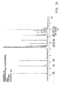
Fig. 2 . HPLC radiochromatogram of 99mTc-EC20. Samples of 99mTc-EC20 were eluted isocratically on a Waters Nova-Pak C18 (3.9 x 150 mm) column using an aqueous mobile phase containing 20% methanol and 0.2% trifluoroacetic acid at a flow rate of 1 mL/min. The HPLC analysis was monitored with both the UV detector (280 nm) and a Bioscan FC-3200 radiodetector. Peak A, free 99mTc; Peak B, a folate-containing chelate of unknown structure; Peaks C and D, diastereomers possessing either a syn or anti configuration of the technetium-oxygen bond in the Dap-Asp-Cys chelating ring of EC20. -
Fig. 3 . Structures of Re-EC20 and 99mTc-EC20 isomers (syn or anti position of metal-oxo bond). -
Fig. 4 . Blocking of 3H-folic acid binding to KB cells with various folate-containing competitors. KB cells were incubated for 15 min on ice with 100 nM 3H-folic acid in the presence and absence of increasing competitor concentrations. (•) Folic acid; (■) EC20; (▲) EC20:Re isomer A; (▼) EC20:Re isomer B; (□) DTPA-Folate. Error bars represent 1 standard deviation (n = 3). -
Fig. 5 . Time-dependent association of 99mTc-EC20. KB cells were incubated with 10 nM 99mTc-EC20 for increasing periods of time at 37°C. Following multiple washes, cells were harvested and counted for associated radioactivity. Error bars represent 1 standard deviation (n = 3). -
Fig. 6 . Concentration-dependent association of 99mTc-EC20. KB cells were incubated for 2 hr at 37°C in the presence of increasing concentrations of 99mTc-EC20. Following multiple washes, cells were harvested and counted for associated radioactivity. Error bars represent 1 standard deviation (n = 3). -
Fig. 7 . Concentration-dependent association of 99mTc-EC20 "peak B." KB cells were incubated for 2 hr at 37°C in the presence of increasing concentrations of "Peak B" that was chromatographically isolated from the 99mTc-EC20 formulation. Following multiple washes, cells were harvested and counted for associated radioactivity. Error bars represent 1 standard deviation (n = 3). (•), Peak B; (∘), Peak B plus 1 mM folic acid. -
Fig. 8 . Blood clearance of 99mTc-EC20 in Balb/c mice. Each animal received an intravenous dose of 50 µg/kg EC20 (67 nmol/kg) in approximately 0.1 mL during brief diethyl ether anesthesia. At the designated times post-injection, each animal was euthanized by CO2 asphyxiation, blood was collected and counted for associated radioactivity. Error bars represent 1 standard deviation (n = 3 animals). -
Fig. 9 . Whole-body gamma images (ventral view). Images were obtained 4 hr following intravenous administration of 99mTc-EC20 to a Balb/c mouse bearing a subcutaneous folate receptor-positive M109 tumor. Only the kidneys (K) and tumor (T) exhibit significant accumulation of this radiotracer. -
Fig. 10 . Structures of EC11, EC13, EC14, EC15, EC19, EC20, EC31, and EC53. -
Fig. 11 . Tissue distribution of 99mTc-EC20 in Balb/c mice bearing FR-postive M109 tumors and FR-negative 4T1 tumors. -
Fig. 12 . HPLC analysis of EC11. -
Fig. 13 . Mass spectroscopy analysis of EC11. -
Fig. 14 . NMR analysis of EC 11. -
Fig. 15 . HPLC analysis of EC 13. -
Fig. 16 . NMR analysis of EC14. -
Fig. 17 . Mass spectroscopy analysis ofEC 15. -
Fig. 18 . HPLC analysis of EC19. -
Fig. 19 . Mass spectroscopy analysis of EC 19. -
Fig. 20 . HPLC analysis of EC31. -
Fig. 21 . HPLC analysis of EC53. -
Fig. 22 . Mass spectroscopy analysis of EC53. -
Fig. 23 . Mass spectroscopy analysis of EC53. - In accordance with the disclosure, methods are provided for targeting radionuclide-based imaging agents to cell populations that uniquely express, overexpress, or preferentially express vitamin receptors. Accordingly, a vitamin, or a receptor binding derivative or analog thereof, is used as the targeting ligand for the imaging agent. The vitamin-imaging agent conjugate can be used to target radionuclides to cells and to concentrate the radionuclides in a cell population, such as a tumor cell population, for use in diagnostic imaging.
- The invention relates to a method of imaging a population of tumour cells in an animal comprising administering an effective amount of a compound of the formula

- The application discloses a composition for diagnostic imaging comprising a compound of the formula


- The application also discloses compounds of the formulas


- Exemplary of these compounds is a compound referred to as EC20 depicted in
Fig. 1 . Exemplary of other compounds for use in accordance with this disclosure are compounds denominated as EC11, EC13, EC14, EC15, EC19, EC31, and EC53 (seeFig. 10 ). The vitamin moiety (e.g., the folic acid moiety in EC20) provides high affinity binding to cellular FRs. The compounds also contain a bifunctional peptide-based chelator, which provides the site for chelation of the radionuclide, for example, 99mTc (seeFig. 1 ), and the compounds can, optionally, contain a linker through which the vitamin moiety is covalently bonded to the chelating moiety. - In accordance with the invention, the vitamin moiety of the compound is a vitamin that is a substrate for receptor-mediated transmembrane transport in vivo, or a vitamin receptor binding derivative or analog thereof. The vitamin in the above disclosures is linked, optionally, through a linker (L) to the chelator portion of the compounds. In the invention, and as shown in
Fig. 1 , EC20 comprises a folic acid analog linked to the chelator moiety because EC20 has the glutamic acid in the D configuration. The chelator portion comprises an α,β-diaminoprogionic acid moiety linked to a cysteine group through a third amino acid residue. The chelator portion of the compound is adapted to bind a radionuclide cation (M) (where k = 1). - In accordance with the invention, the compound with bound radionuclide are referred to as "vitamin-imaging agent conjugates."
- The structure of the linker, if present, is not critical to the above disclosures. Thus, for example, it can be any biocompatible divalent linker. Typically, the linker comprises about 1 to about 30 carbon atoms, more typically about 2 to about 20 carbon atoms. Lower molecular weight linkers (i.e., those having an approximate molecular weight of about 30 to about 300) are typically employed. Furthermore, the vitamin moiety may be a vitamin, or a derivative or analog thereof For example, folate contains one glutamic acid in the L configuration linked to pteroic acid. EC11 and EC 14 contain two glutamic acid residues and, thus, these compounds can also, for example, be considered derivatives of folic acid (
Fig. 10 ). - Among vitamins believed to trigger receptor-mediated endocytosis and having application in accordance with the presently disclosed method are niacin, pantothenic acid, folic acid, riboflavin, thiamine, biotin, vitamin B12, and the lipid soluble vitamins A, D, E and K. These vitamins, and their analogs and derivatives, constitute vitamins that can be coupled with imaging agents to form the vitamin-chelator conjugates for use in accordance with the invention. Preferred vitamin moieties include folic acid, biotin, riboflavin, thiamine, vitamin B12, and analogs and derivatives of these vitamin molecules, and other related vitamin receptor-binding molecules.
- Folic acid, folinic acid, pteroic acid, pteropolyglutamic acid, and folate receptor-binding pteridines such as tetrahydropterins, dihydrofolates, tetrahydrofolates, and their deaza and dideaza analogs can be used in accordance with the above disclosures. The terms "deaza" and "dideaza" analogs refers to the art-recognized folate analogs having a carbon atom substituted for one or two nitrogen atoms in the naturally occurring folic acid structure. For example, the deaza analogs include the 1-deaza, 3-deaza, 5-deaza, 8-deaza, and 1.0-deaza analogs. The dideaza analogs include, for example, 1,5 dideaza, 5,10-dideaza, 8,10-dideaza, and 5,8-dideaza analogs. The foregoing are folate analogs or derivatives and can bind to folate receptors. Other folate analogs or derivatives useful in accordance with the above disclosures are the folate receptor-binding analogs aminopterin, amethopterin (methotrexate), N10-methylfolate, 2-deamino-hydroxyfolate, deaza analogs such as 1-deazamethopterin or 3-deazamethopterin, and 3'5'-dichloro-4-amino-4-deoxy-N10-methylpteroylglutamic acid (dichloromethotrexate).
- The vitamin, or derivative or analog thereof, can be capable of selectively binding to the population of cells to be visualized due to preferential expression on the targeted cells of a receptor for the vitamin, or derivative or analog, wherein the receptor is accessible for binding. The binding site for the vitamin can include receptors for any vitamin molecule capable of specifically binding to a receptor wherein the receptor or other protein is uniquely expressed, overexpressed, or preferentially expressed by the population of cells to be visualized. A surface-presented protein uniquely expressed, overexpressed, or preferentially expressed by the cells to be visualized is a receptor not present or present at lower amounts on other cells providing a means for selective, rapid, and sensitive visualization of the cells targeted for diagnostic imaging using the vitamin-imaging agent conjugates of the present invention.
- In accordance with the invention the vitamin-imaging agent conjugate is capable of high affinity binding to receptors on cancer cells or other cells to be visualized. The high affinity binding can be inherent to the vitamin moiety or the binding affinity can be enhanced by the use of a chemically modified vitamin (i. e., an analog or a derivative) or by the particular chemical linkage between the vitamin and the chelator moiety that is present in the conjugate.
- In accordance with the disclosure, the chelator can be conjugated with multiple, different vitamins, or vitamin receptor binding derivatives or analogs, to enhance the opportunity for binding to the respective cell membrane receptors. Alternatively, independent portions of the dose of a vitamin-imaging agent conjugate can constitute different vitamin-imaging agent conjugates to enhance the opportunity for binding to the respective cell membrane receptors.
- Generally, any manner of forming a complex between the chelator and the vitamin, or vitamin receptor binding derivative or analog, can be utilized in accordance with the present disclosures. The chelator can form a complex with the vitamin, or vitamin receptor binding derivative or analog, by direct conjugation of the chelator and the vitamin by using a divalent linker. Alternatively, the vitamin and the chelator may be conjugated without employing a linker. If a linker is used, the linker can directly conjugate the vitamin, or vitamin receptor binding derivative or analog, and the chelator through a hydrogen, ionic, or covalent bond. Also, in accordance with the above disclosures the divalent linker can comprise an indirect means for associating the chelator with the vitamin, or vitamin receptor binding derivative or analog, such as by connection through intermediary linkers, spacer arms, or bridging molecules. Both direct and indirect means for association must not prevent the binding of the vitamin, or vitamin receptor binding derivative or analog, to the vitamin receptor on the cell membrane for operation of the method of the present invention.
- Covalent bonding of the vitamin, or vitamin receptor binding derivative or analog, and the chelator can occur, whether or not a linker is employed, through the formation of amide, ester or imino bonds between acid, aldehyde, hydroxy, amino, or hydrazo groups. For example, a carboxylic acid on the vitamin moiety or on the chelator can be activated using carbonyldiimidazole or standard carbodiimide coupling reagents such as 1-ethyl-3-(3-dimethylaminopropyl)-carbodiimide (EDC) and thereafter reacted with the other component of the conjugate, or with a linker, having at least one nucleophilic group, viz hydroxy, amino, hydrazo, or thiol, to form the vitamin-chelator conjugate coupled, with or without a linker, through ester, amide, or thioester bonds.
- The radionuclides suitable for diagnostic imaging include radioisotopes of gallium, indium, copper, technetium and rhenium, including isotopes 111In, 99mTc, 64Cu, 67Cu, 67Ga or 68Ga. These radionuclides are cationic and are complexed with the chelator through the chelating group of the conjugate to form the vitamin-imaging agent conjugate.
- The vitamin-imaging agent conjugate in accordance with the invention is utilized to selectively visualize, using scintigraphic imaging techniques, a population of cells in an animal wherein the population of cells uniquely expresses, overexpresses, or preferentially expresses receptors for a vitamin, or a vitamin receptor binding derivative or analog thereof. The vitamin-imaging agent conjugate can be used to visualize populations of pathogenic cells, as long as the cells uniquely or preferentially express or overexpress vitamin receptors or receptors that bind vitamin derivatives or analogs.
- The invention is applicable to populations of pathogenic cells that cause a variety of pathologies including cancer, and diseases mediated by any other type of pathogenic cells that overexpress vitamin receptors, or receptors capable of binding vitamin derivatives or analogs. Thus, the population of pathogenic cells is tumorigenic, including benign tumors and malignant tumors. The cell population is a cancer cell population, and the cancer cells can arise spontaneously or by such processes as mutations present in the germline of the host animal or somatic mutations, or the cancer can be chemically-, virally-, or radiation-induced. The invention can be utilized for diagnostic imaging of such cancers as carcinomas, sarcomas, lymphomas, Hodgekin's disease, melanomas, mesotheliomas, Burkitt's lymphoma, nasopharyngeal carcinomas, and myelomas. The cancer cell population can include, but is not limited to, oral, nasopharyngeal, thyroid, endocrine, skin, gastric, esophageal, laryngeal, throat, pancreatic, colon, bladder, bone, ovarian, cervical, uterine, breast, testicular, prostate, rectal, kidney, liver, lung, and brain cancers. In embodiments where the cell population is a cancer cell population, tumor cells, including cells of the primary tumor or cells that have metastasized or are in the process of dissociating from the primary tumor, can be visualized using the vitamin-imaging agent conjugate.
- The vitamin-imaging agent conjugates of the present disclosure can be used to diagnose a disease state or to monitor the progression of disease. For example, the diagnostic imaging method in accordance with the disclosure can be used to monitor the progression of cancer in combination with prophylactic treatments to prevent return of a tumor after its removal by any therapeutic approach including surgical removal of the tumor, radiation therapy, chemotherapy, or biological therapy.
- The method of the present invention can be used for both human clinical medicine and veterinary applications. Thus, the animal harboring the population of cells that are visualized can be human or, in the case of veterinary applications, can be a laboratory, agricultural, domestic, or wild animal. The present invention can be applied to animals including, but not limited to, humans, laboratory animals such rodents (e.g., mice, rats, hamsters, etc.), rabbits, monkeys, chimpanzees, domestic animals such as dogs, cats, and rabbits, agricultural animals such as cows, horses, pigs, sheep, goats, and wild animals in captivity such as bears, pandas, lions, tigers, leopards, elephants, zebras, giraffes, gorillas, dolphins, and whales.
- The compositions for diagnostic imaging comprise an amount of the vitamin-imaging agent conjugate effective to visualize the cells targeted for diagnostic imaging in an animal when administered in one or more doses. The diagnostic imaging composition containing the vitamin-imaging agent conjugate is preferably administered to the animal parenterally, e.g., intradermally, subcutaneously, intramuscularly, intraperitoneally, intravenously, or intrathecally. Alternatively, the composition containing the vitamin-imaging agent conjugate can be administered to the animal by other medically useful processes, and any effective dose and suitable dosage form can be used, including oral and inhalation dosage forms.
- Examples of parenteral dosage forms include aqueous solutions of the vitamin-imaging agent conjugate, in isotonic saline, 5% glucose or other well-known pharmaceutically acceptable liquid carriers such as liquid alcohols, glycols, esters, and amides. The parenteral dosage form in accordance with this invention can be in the form of a reconstitutable lyophilizate comprising the dose of the vitamin-imaging agent conjugate.
- The dosage of the vitamin-imaging agent conjugate in the diagnostic imaging composition can vary significantly depending on the size of the animal, the cell population targeted for diagnostic imaging, the specific vitamin-imaging agent conjugate being used, and the route of administration of the conjugate. The effective amount to be administered to the animal is based on body surface area, weight, and physician assessment of the condition of the animal. An effective dose can range from about 1 ng/kg to about 1 mg/kg, more preferably from about 100 ng/kg to about 500 µg/kg, and most preferably from about 100 ng/kg to about 25 µg/kg.
- Any effective regimen for administering the diagnostic imaging composition containing the vitamin-imaging agent conjugate can be used. For example, the diagnostic imaging composition can be administered as a single dose, or it can be administered in multiple doses, if necessary, to achieve visualization of the targeted cell population. Additional injections of the diagnostic imaging composition containing the vitamin-imaging agent conjugate can be administered to the animal at an interval of days or months after the initial injections(s), and the additional injections can be useful for monitoring the progress of the disease state. The diagnostic imaging composition containing the vitamin-imaging agent conjugate can also be administered in combination with unlabeled vitamin. "In combination with" means that the unlabeled vitamin can be either coadministered with the imaging agent or the unlabeled vitamin can be preinjected before administration of the imaging agent to improve image quality. For example, the imaging agent can be administered in combination with about 0.5 ng/kg to about 100 mg/kg, or about 1 µg/kg to about 100 mg/kg, or about 100 µg/kg to about 100 mg/kg of the unlabeled vitamin.
- The diagnostic imaging composition is typically formulated for parenteral administration and is administered to the animal in an amount effective to enable imaging of the targeted cell population. Typically, the diagnostic imaging composition containing the vitamin-targeted imaging agent is administered to the animal, and following a period of time to allow delivery and concentration of the vitamin-imaging agent conjugate in the targeted cell population, the animal is subjected to the imaging procedure and imaging is enabled by the vitamin-imaging agent conjugate. When used for monitoring the progression of disease or diagnosis, imaging procedures are typically carried out about 1 to about 6 hours post administration of the diagnostic imaging composition containing the vitamin-imaging agent conjugate.
- As mentioned above, the invention provides a method of imaging a population of tumor cells in an animal wherein the cells are characterized by a vitamin receptor on the surface of the cells. The method comprises the steps of administering to the animal an effective amount of a composition comprising a compound of the formula

- The method can be used to image a cell population in vitro, e.g., in cell culture, or in vivo, where the cells form part of or otherwise exist in animal tissue. Thus, the target cells can include, for example, the cells lining the alimentary canal, such as the oral and pharyngeal mucosa, the cells forming the villi of the small intestine, or the cells lining the large intestine. Such cells of the alimentary canal can be targeted in accordance with this invention by oral administration of a diagnostic imaging composition comprising the vitamin-imaging agent conjugate. Similarly, cells lining the respiratory system (nasal passages/lungs) of an animal can be targeted by inhalation of the present complexes, and cells of internal organs, including cells of the ovaries and the brain can be targeted, particularly, by parenteral administration of the diagnostic imaging composition.
- The application also discloses:
- 1. A compound of the formula

L is a divalent linker;
R is a side chain of an amino acid of the formula H2NCHRCOOH;
M is a cation of a radionuclide;
n or 1 or 0; and
k is 1 or 0. - 2. The compound of 1 wherein V is a vitamin selected from the group consisting of folate, riboflavin, thiamine, vitamin B12, and biotin, or a vitamin receptor binding derivative or analog thereof.
- 3. The compound of 1 wherein the radionuclide is selected from the group consisting of isotopes of gallium, indium, copper, technetium, and rhenium.
- 4. The compound of 3 wherein the radionuclide is an isotope of technetium.
- 5. The compound of 1 wherein V is folate, or a folate receptor binding analog of derivative thereof.
- 6. A composition for diagnostic imaging comprising a compound of the formula

L is a divalent linker;
R is a side chain of an amino acid of the formula H2NCHRCOOH;
M is a cation of a radionuclide;
n or 1 or 0; and
a pharmaceutically acceptable carrier therefor; - 7. The composition of 6 wherein V in the compound is a vitamin selected from the group consisting of folate, riboflavin, thiamine, vitamin B12, and biotin, or a vitamin receptor binding derivative or analog thereof.
- 8. The composition of 6 wherein the radionuclide in the compound is selected from the group consisting of isotopes of gallium, indium, copper, technetium, and rhenium.
- 9. The composition of 8 wherein the radionuclide in the compound is an isotope of technetium.
- 10. The composition of 6 adapted for parenteral administration.
- 11. The compound of 6 wherein V is folate, or a folate receptor binding analog or derivative thereof.
- 12. A method of imaging a population of cells in an animal wherein said cells are characterised by a vitamin receptor on the surface of said cells, the method comprising the steps of
administering to the animal an effective amount of a composition comprising a compound of the formula
L is a divalent linker;
R is a side chain of an amino acid of the formula H2NCBRCOOH;
M is a cation of a radionuclide;
n or 1 or 0; and
a pharmaceutically acceptable carrier therefor; and monitoring the biodistribution of said compound in the animal; - 13. The method of 12 wherein V in the compound is a vitamin selected from the group consisting of folate, riboflavin, thiamine, vitamin B12, and biotin, or a vitamin receptor binding derivative or analog thereof.
- 14. The method of 12 wherein the radionuclide in the compound is selected from the group consisting of isotopes of gallium, indium, copper, technetium, and rhenium.
- 15. The method of 14 wherein the radionuclide in the compound is an isotope of technetium.
- 16. The method of 12 wherein the composition is administered parenterally to the patient.
- 17. The compound of 12 wherein V is folate, or a folate receptor binding analog or derivative thereof.
- 18. A compound of the formula

L is a divalent linker;
R is a side chain of an amino acid of the formula H2NCHRCOOH;
M is a cation of a radionuclide;
n or 1 or 0; and
k is 1 or 0. - 19. A composition for diagnostic imaging comprising a compound of the formula

L is a divalent linker;
R is a side chain of an amino acid of the formula H2NCHRCOOH;
M is a cation of a radionuclide;
n or 1 or 0; and
a pharmaceutically acceptable carrier therefor. - 20. A method of imaging a population of cells in an animal wherein said cells are characterised by a vitamin receptor on the surface of said cells, the method comprising the steps of
administering to the animal an effective amount of a composition comprising a compound of the formula
L is a divalent linker;
R is a side chain of an amino acid of the formula H2NCHRCOOH;
M is a cation of a radionuclide;
n or 1 or 0; and
a pharmaceutically acceptable carrier therefor; and monitoring the biodistribution of said compound in the animal. - Further disclosed are:
- 1A. A use of a compound of the formula

L is a divalent linker;
R is a side chain of an amino acid;
M is a cation of a radionuclide;
n is 1 or 0; and
k is 1 or 0;
for imaging a population of cells in an animal pre-injected with unlabeled folate. - 2A. The use of 1A wherein the radionuclide in the compound is selected from the group consisting of isotopes of gallium, indium, copper, technetium, and rhenium.
- 3A. The use of 1A or 2A wherein the radionuclide in the compound is an isotope of technetium.
- 4A. The use of any one of 1A to 3A wherein the compound has the formula

- 5A. The use of any one of 1A to 4A wherein the population of cells in the animal is a tumor cell population.
- 6A. The use of 5A wherein the tumor cells are tumor cells of the ovary.
- 7A. The use of any one of 1A to 6A wherein the compound is in a parenteral dosage form selected from the group consisting of an intradermal, a subcutaneous, an intramuscular, an intraperitoneal, an intravenous, and an intrathecal dosage form.
- 8A. The use of 7A wherein the dosage form is an intravenous dosage form.
- 9A. A conjugate of the formula

wherein V is a folate, wherein the folate is a substrate for receptor-mediated transmembrane transport in vivo, or a folate receptor binding derivative or analog thereof;
L is a divalent linker;
R is a side chain of an amino acid;
M is a cation of a radionuclide;
n is 1 or 0; and
k is 1 or 0;
in combination with an unlabeled folate. - 10A. The combination of 9A wherein the conjugate has the formula

- 11A. The combination of 9A or 10A wherein a cation of a radionuclide is bound to the conjugate, where the radionuclide is selected from the group consisting of isotopes of gallium, indium, copper, technetium, and rhenium.
- 12A. The combination of any one of 9A to 11A wherein the radionuclide is an isotope of technetium.
- 13A. The combination of any one of 9A to 12A wherein the conjugate is in a dosage form adapted for parenteral administration.
- 14A. The combination of 13A wherein the dosage form is selected from the group consisting of an intradermal, a subcutaneous, an intramuscular, an intraperitoneal, an intravenous, and an intrathecal dosage form.
- 15A. The combination of any one of 9A to 14A wherein the pharmaceutically acceptable carrier is a liquid.
- 16A. The combination of 15A wherein the liquid carrier is selected from the group consisting of saline, 5% glucose, alcohols, glycols, esters, amides, and a combination thereof.
- N10-trifluoroacetylpteroic acid was purchased from Eprova AG, Schaffhausen, Switzerland. Peptide synthesis reagents were purchased from NovaBiochem and Bachem. 99mTc Sodium Pertechnetate was supplied by Syncor. [Re02 (en) 2] Cl was prepared according to Rouschias (Rouschias, G. , Chem. Rev. , 74: 531 (1974)). Cellulose plates and DEAE ion exchange plates were purchased from J. T. Baker.
- EC20 was prepared by a polymer-supported sequential approach using the Fmoc-strategy (see
Scheme 1 below; Fmoc = 9-fluorenylmethyloxycarbonyl; Boc = tert. butyloxycarbonyl; Dap = diaminopropionic acid; DMF = dimethylformamide; DIPEA = diisopropylethylamine). EC20 was synthesized on an acid-sensitive Wang resin loaded with Fmoc-L-Cys (Trt) -OH. Benzotriazole-1-yl-oxy-tris-pyrrolidino-phosphoniumhexafluorophosphate (PyBOP) was applied as the activating reagent to ensure efficient coupling using low equivalents of amino acids. Fmoc protecting groups were removed after every coupling step under standard conditions (20% piperidine in DMF). After the last assembly step the peptide was cleaved from the polymeric support by treatment with 92.5% trifluoroacetic acid containing 2.5% ethanedithiol, 2.5% triisopropylsilane and 2.5% deionized water. This reaction also resulted in simultaneous removal of the t-Bu, Boc and trityl protecting groups, Finally, the trifluoroacetyl moiety was removed in aqueous ammonium hydroxide to give EC20. - The crude EC20 product was purified by HPLC using an
Xterra RP 18 30 x 300 mm, 7 µm column (Waters); mobile phase 32 mM HCl (A), MeOH (B); gradient conditions starting with 99% A and 1% B, and reaching 89% A and 11% B in 37 min by a flow rate of 20 mL/min. Under these conditions, EC20 monomer typically eluted at 14.38 min, whereas EC20 disulfide dimer (minor contaminant) eluted at 16.83 min. All other compounds shown inFig. 10 can be prepared using a similar synthesis scheme except for EC15 which is synthesized as shown inScheme 2 below. - Two milligrams of HPLC-purified EC20 were dissolved in 0.62 mL of D2O2, and a 500 MHz 1H-HMR spectrum was collected. Table 1 (see below) lists the chemical shifts, signal shapes, and J values for all non-exchangeable protons in EC20 molecule.
- EC20 was also analyzed by electrospray-mass pectrometry. Major positive ion peaks (m/z, relative intensity): 746.1, 100; 747.1, 44; 556.8, 32; 570.8, 16.

Table 1. 1H-MMR data for EC20. EC20 was dissolved in D2O and a 500 MHz spectrum was collected. Chemical shifts (δ) are in ppm. The signal for HOD at δ = 4.80 ppm was used as the reference. pD = 4.78; s = singlet; d = doublet; m = multiplet. Residue Protons observed Chemical Shift (δ) Signals J values H-7 8.76 s 2 x H-9 4.64 s Ple H-12 a. H-16 7.68 d 3 J12,13=3 J15,16=8.8 Hz H-13 a. H-15 6.8 d H-2 4.41 dd 3 J2,3=39.1 Hz; 3 J2,3b=4.5 Hz D-Glu H-3a 2.08 m 2 J3a,3b = 14.2 Hz H-3B 2.27 m 2 x H-4 2.44 dd 3 J3a,4 = 3 J4b,4 = 5.6 Hz dd; X of ABX System Dpr H-2
H-3A
H-3B4.1
3.52
3.72dd; A of ABX System 3 J2,3A-6.6 Hz; 3 J2,3B=4.7 Hz 2 JA,B=14.7 Hz dd; B of ABX System dd; X of ABX System Asp H-2
2.81
H-3B4.71
2.62
2.81dd; A of ABX System 3 J2,3A=9.5 hz; 3 J2,3B=4.3 Hz 2 JA,B=16.1 Hz dd; B of ABX System dd; X of ABX System Cys H-2
H-3A
H-3B4.3
2.85
2.89dd; A of ABX System 3 J2,3A=55 bz; 3 J2,3B=4.4 Hz 2 JA,B=14.1 Hz dd; B of ABX System 
- EC20 kits were used for preparation of the 99mTc-EC20 radioactive drug substance. Each kit contained a sterile, non-pyrogenic lyophilized mixture of 0.1 mg EC20, 80 mg sodium α-D-glucoheptonate, 80 mg tin (II) chloride dihydrate, and sufficient sodium hydroxide or hydrochloric acid to adjust the pH to 6.8 ± 0.2 prior to lyophilization. The lyophilized powder was sealed in a 5 mL vial under an argon atmosphere. The kits were then stored frozen at -20°C until use or expiration (current shelf life is > 2 years). Importantly, the tin (II) chloride component is required to reduce the added 99mTc-pertechnetate, while the sodium α-D-glucoheptonate component is necessary to stabilize the newly reduced 99mTc prior to its final chelation to the EC20 compound.
- 99mTc-EC20 was prepared as follows (i.e., chelation of 99mTc to EC20). First, a boiling water bath containing a partially submerged lead vial shield was prepared. The top of an EC20 vial was swabbed with 70% ethanol to sanitize the surface and the vial was placed in a suitable shielding container. Using a shielded syringe with 27-gauge needle, 1 mL of sterile Sodium Pertechnetate 99mTc Injection (15 to 20 mCi) in 0.9% sodium chloride was injected into the shielded vial. Before removal of the syringe from the vial, a volume of gas from the vial equal to the volume of pertechnetate added was withdrawn in order to normalize the pressure inside the vial. The vial was gently swirled for 30 seconds to ensure complete dissolution of the lyophilized powder. The vial was then placed into the lead shield that was standing in the boiling water bath. The solution was heated for ~18 minutes and then cooled to room temperature for a minimum of 15 min. This solution can be stored at room temperature (15-25°C) protected from light, but it should be used within 6 hours of preparation.
- The radiochemical stability of the radioactive drug substance was determined by HPLC after storing at room temperature protected from light for up to 24 hours. Samples of the 99mTc-EC20 solution (20 µL) were analyzed using an HPLC system consisting of a Waters 600E Multisolvent Delivery System and 490 UV detector, a Bioscan EC-3200 radiodetector, Laura v1.5 radiochromatogram software, and a Waters Nova-Pak C18 (3.9 x 150 mm) column. Injected samples were eluted isocratically using an aqueous mobile phase containing 20% methanol and 0.1% trifluoroacetic acid at a flow rate of 1 mL/min. The HPLC analysis was monitored with both the UV detector (280 mm) and the gamma radiodetector. Notably, the radiochemical purity of 99mTc-EC20 remained greater than 90% for at least 24 hours in all cases.
- The major radiochemical impurities in the preparation of 99mTc-FC20 will be 1) 99mTc pertechnetate, 2) 99mTc-glucoheptonate (ligand exchange precursor), 3) non-specific binding 99mTc (99mTc bound at a site other than the expected Dap-Asp-Cys chelating moiety of the EC20 molecule), and 4) hydrolyzed 99mTc. Since 99mTc-EC20 was being tested for possible clinical use, a three-TLC-based method was developed to determine the amounts of each impurity and to estimate the overall radiochemical purity of 99mTc-EC20.
- In the first system a cellulose plate was developed with deionized water. 99mTc-EC20, 99mTc-glucoheptonate, non-specific binding 99mTc and 99mTc pertechnetate move to the solvent front (Rf = 1.0), while hydrolyzed 99mTc remains at the origin (Rf= 0.0). The cellulose plate was cut into two pieces at Rf = 0.3 (1.5 cm from origin) and each piece was counted using a dose calibrator. The percent of hydrolyzed 99mTc was calculated as follows: A = % Hydrolyzed; 99mTc = (µCi in bottom piece/µCi in both pieces) x 100.
- In the second system, a cellulose plate was developed with acetone and 0.9% NaCl (7:3,v/v). 99mTc-pertechnetate moves with Rf= 0.9, while 99mTc-EC20, 99mTc-glucoheptonate, non-specific binding 99mTc and hydrolyzed 99mTc remain at the origin (Rf= 0.0). The cellulose/acetone-saline plate was cut into two pieces at Rf = 0.6 (3.0 cm from the origin) and each piece was counted using a dose calibrator. The percent of 99mTc-pertechnetate was calculated as follows: B = % 99mTc-pertechnetate = (µCi in top piece/µCi in both pieces) x 100.
- Finally, in the third system a DEAE ion exchange plate was developed with 0.3 M Na2SO4. 99mTc-glucoheptonate moves to the solvent front (Rf= 1.0), nonspecific binding 99mTc moves with Rf= 0.6, and 99mTc~EC20, hydrolyzed 99mTc and 99mTc-pertechnetate remain near the origin (99mTc-EC20. Rf = 0.1; hydrolyzed 99mTc: Rf= 0.0; 99mTc pertechnetate: Rf = 0.3). The cellulose/Na2SO4 plate was cut into two pieces at 2.5 cm from the origin and each piece was counted using a dose calibrator. The percent of 99mTc-glucoheptonate and non-specific binding 99mTc were calculated as follows: C = % (99mTc-Glucoheptonate + non-specific binding 99mTc) = (µCi in top piece/µCi in both pieces) x 100. The overall radiochemical purity of 99mTc-EC20 was then calculated as follows: Radiochemical purity = 100 - (A+B+C).
- As shown in
Fig. 2 , HPLC analysis of the 99mTc-EC20 formulation shows four radiochemical components, designated as Peaks A through D. Peak A was confirmed to be free 99mTc and this by-product is reproducibly present at < 2%. Peak B, which was different from that of 99mTc-glucoheptonate (data not shown) eluted with a retention time of 2.8 min. This component represented about 3% of the mixture and was thought to result from 99mTc chelating at some other site on the EC20 molecule besides the expected Dap-Asp-Cys moiety. Peaks C and D (retention times of 4.8 minutes and 13.2 minutes, respectively), account for the majority of the formulated radiochemical activity. - Fifty-two mg (0.010 mmol) of EC20 and [ReO2(en)2]Cl (52 mg, 0.14 mmol) were dissolved in 6 mL and 1 mL argon-purged phosphate buffer (0.05 M, pH 5.8), respectively. The two solutions were combined and heated under an argon atmosphere in a boiling water bath for 2 hours. The reaction mixture was frozen and lyophilized overnight. The crude product was purified by HPLC (Xterra RPI8 column, 19x150 mm, 10 mM NH4OAc/CH3CN,
flow rate 10 mL/mm;gradient 1% to 8%). The fractions were collected, lyophilized and stored at -20°C until use. - Because no mass spectral facilities were available for analysis of radioactive materials, the non-radioactive rhenium analog, Re-EC20, was analyzed. Both rhenium and technetium are Group VIIA metals that have significant similarity in physical and chemical properties. They also form similar complexes with organic ligands. This analogous chemical behavior has been frequently used in structure elucidation of new classes of technetium radiopharmaceuticals based on non-radioactive rhenium analogues. Interestingly, HPLC analysis of Re-EC20 also showed two major peaks eluting at 5 and 14.2 minutes, respectively, similar to Peaks C and D for 99mTc-EC20 (chromatogram not shown). Mass spectral analysis confirmed that these two components were isomers corresponding to the Re-EC20 complex (m/z = 945). In fact, these species were likely diastereomers possessing either a syn or anti configuration of the technetium-oxygen bond in the Dap-Asp-Cys chelating ring, as depicted in
Fig. 3 . Because i) the two peaks in the Re-EC20 chromatogram represent isomeric complexes, and ii) reports of similar isomerism in technetium complexes exist, it is likely that components C and D in the 99mTc-EC20 radiochromatogram are also isomers. - Cells were grown continuously as a monolayer using folate-free RPMI medium (FFRPMI) containing 10% heat-inactivated fetal calf serum (HIFCS) at 37°C in a 5% C02/95% air-humidified atmosphere with no antibiotics. The HIFCS contained its normal complement of endogenous folates which enabled the cells to sustain growth in this more physiologically-relevant medium. All cell experiments were performed using FFRPMI containing 10% HIFCS (FFRPMI/HIFCS) as the growth medium, except where indicated.
- The relative affinity of various folate derivatives was determined according to the method described by Westerhoff et al. (Mol. Pharm., 48: 459-471 (1995)) with slight modification. Briefly, FR-positive KB cells were gently trypsinized in 0.25% trypsin/PBS at room temperature for 3 minutes and then diluted in FFRPMI/HIFCS. Following a 5
min 800 x g spin and one PBS wash, the final cell pellet was suspended in FFRPMI 1640 (no serum). Cells were incubated for 15 min on ice with 100 nM of 3H-folic acid in the absence and presence of increasing concentrations of folate-containing test articles. Samples were centrifuged at 10,000 x g for 5 min, cell pellets were suspended in buffer, were transferred to individual vials containing 5 mL of scintillation cocktail, and were then counted for radioactivity. Negative control tubes contained only the 3H-folic acid in FFRPMI (no competitor). Positive control tubes contained a final concentration of 1 mM folic acid, and CPMs measured in these samples (representing non-specific binding of label) were subtracted from all samples. Notably, relative affinities were defined as the inverse molar ratio of compound required to displace 50% of 3H-folic acid bound to KB FR, and the relative affinity of folic acid for the FR was set to 1. - The capacity of EC20 to directly compete with folic acid for binding to cell surface FRs was measured using this assay. Importantly, a relative affinity value of 1.0 implies that the test article ligand has an affinity for the FR equal to folic acid. Likewise, values lower than unity reflect weaker affinity, and values higher than unity reflect stronger affinity.
- Cultured KB cells were incubated with 100 nM 3H-folic acid in the presence of increasing concentrations of non-radioactive folic acid, EC20, Rhenium-EC20 (isomer A; Peak C), Rhenium-EC20 (isomer B; peak 0), or a related folate-based radiopharmaceutical, DTPA-folate. Following a 15-minute incubation at 4°C, cells were rinsed free of unbound material and counted for residual cell-associated radioactivity. The quantity of bound radioactivity was plotted against the concentration of unlabeled ligand, and IC50 values (concentration of ligand required to block 50% of 3H-folic acid binding) were estimated. As shown in
Fig. 4 and Table 2 (below), EC20 was determined to have an affinity of 0.92 relative to that of folic acid for human FRs. Both isomers of Rhenium-EC20 displayed relative affinity values that were very similar to, if not better than, the parent EC20 molecule (1.42 and 1.37 for Re-EC20 isomer A and isomer B, respectively). DTPA-folate, an 111In-chelating folate radiopharmaceutical agent, displayed a relative affinity of 0.87 for the folate receptor. Thus, chemical modification of folate with various metal chelating motifs did not disturb the vitamin's intrinsic affinity for the FR.Table 2. Relative Affinity Estimations. Relative affinities (RA) were defined as the inverse molar ratio of compound required to displace 50% of 3H-folic acid bound to FR-positive KB cells. The relative affinity of folic acid was set to 1. Each test article was evaluated in triplicate. Test Article IC50 (nM) S.D. RA S.D. Folic Acid 118 ± 19 1.00 EC20 128 ± 25 0.92 ± 0.23 EC20:Re isomer 183 ± 16 1.42 ± 0.36 EC20:Re isomer 286 ± 3 1.37 ± 0.23 DTPA- Folate 136 ± 12 0.87 ± 0.16 - KB cells were seeded in 12-well Falcon plates and allowed to form sub-confluent monolayers overnight. Following one rinse with 1 mL of fresh FFRPMI/HIFCS, each well received 1 mL of FFRPMI/HIFCS containing 10 nM 99mTc-EC20. Cells were incubated for predetermined times at 37°C and then rinsed four times with 1 mL of ice-cold PBS, pH 7.4. The cell monolayers were dissolved in 0.5 mL of PBS, pH 7.4 containing 1% sodium dodecyl sulfate for 15 min at room temperature and then counted for radioactivity using a Packard gamma counter. The protein in each sample was quantitated using a BioRad DC Protein Assay kit, and cellular protein values were converted to cell number using the conversion factor of 2.23 x 10⁻⁷ mg protein per cell. Final tabulated values were expressed in terms of molecules of EC20 per cell.
- The kinetics of 99mTc-EC20 uptake into FR-positive KB cells was quantitatively measured using this protocol. As shown in
Fig. 5 , steady-state uptake was reached within two hours at 37°C, where approximately 3.2 million molecules of EC20 were cell-associated, whereas half-maximal cell association occurred 9 minutes after mixing 10 nM of this radiopharmaceutical with the cells. Interestingly, the half-maximal saturation point was reached in only 37 seconds when cells were incubated with a 10-fold higher concentration of 99mTc-EC20 (100 nM; data not shown). - KB cells were seeded in 12-well Falcon plates and allowed to form sub-confluent monolayers overnight. Following one rinse with 1 mL of fresh FERPMI/HIFCS, each well received 1 mL of FFRPMI/HIFCS containing increasing concentrations of 99mTc-EC20. Cells were incubated for 2 hours at 37°C and then rinsed four times with 1 mL of ice-cold PBS, pH 7.4. The monolayers were dissolved in 0.5 mL of PBS, pH 7.4 containing 1% sodium dodecyl sulfate for 15 min at room temperature and then counted for radioactivity using a Packard gamma counter. Protein content was determined as described above, and final tabulated values were expressed in terms of molecules of EC20 per cell.
- As shown in
Fig. 6 , the cell uptake of 99mTc-EC20 was found to be dependent on the extracellular concentration. The particular KB cells used were determined to bind a maximum of four million molecules of the folate radiopharmaceutical per cell. Scatchard analysis of the data estimated the KD of binding to be 3.2 nM, a value comparable with the KD observed for the vitamin folate binding to these same cells. - Although the full identity of the Peak B component was not established, UV absorption analysis indicated that it contained a folate moiety (i.e., the absorption spectrum contained folate's signature secondary absorption peak at 363 nm). This HPLC-purified radiolabeled material (Peak B material) was collected and then added to cultured KB cells. As shown in
Fig. 7 , the cell uptake of the 99mTc-labeled Peak B component was also found to be dependent on the extracellular concentration. Scatchard analysis of the data estimated the KD of binding to be 1.1 nM. Interestingly, the cell association of Peak B was completely blocked in the presence of excess folic acid, indicating that this minor formulation by-product is also capable of targeting FR-positive cells for radiodiagnostic purposes. - Animals used for this study were maintained on a folate-free diet (Harlan #TD-90261) for approximately three weeks prior to dose administration. Acclimation to this special diet is essential because regular rodent diets contain large amounts of folic acid (6 mg/kg chow) and promote high serum folate levels in mice. Furthermore, previous studies have shown that mice placed on a folate-free diet for 3 weeks had maintained a safe serum folate level of 25 ± 7 nM, which is slightly higher than the 9-14 nM concentration measurable in human serum.
- The 99mTc-EC20 solution was prepared on the day of use and had initially contained 100 µg of EC20 per milliliter. The solution was further diluted with sterile saline to prepare working stock solutions. The radiochemical purity of the product was estimated to be ~94% by TLC. Each animal received a dose of 50 µg/kg EC20 (67 nmol/kg) in approximately 0.1 mL volume i.v. via the tail vein during brief diethyl ether anesthesia. At the designated times (see
Fig. 8 ) post-injection, each animal was euthanized by CO2 asphyxiation, and blood was immediately collected by cardiac puncture. - As shown in
Fig. 8 , 99mTc-EC20 was rapidly removed from circulation in the Balb/c mouse. The plasma half life of this radiopharmaceutical was estimated to be ~ 4 minutes, and less than 0.2% of the injected 99mTc-EC20 dose remained in circulation after four hours (assuming that blood represents 5.5% of the total body mass). This data indicates that folate conjugates are rapidly removed from circulation following intravenous administration, and that valuable tissue biodistribution data can be obtained after only a few hours post-injection without the concern for non-specific tissue uptake due to blood-borne radioactivity. - The ability of 99mTc-EC20 to target tumors in vivo was assessed using a FR-positive M109 model. These tumor cells are syngeneic for the Balb/c mouse, and they reproducibly form subcutaneous solid tumors within two weeks post inoculation. 99mTc-EC14, which is structurally similar to 99mTc-EC20 except it contains one additional D-Glu residue (i.e., Pte-D-Glu-D-Glu-βDpr-Asp-Cys), 99mTc-EC28 (a non-pteroate containing control consisting of benzoyl-D-Glu-n-Glu-βDpr-Asp-Cys), and the previously reported 111In-DTPA-folate radiopharmaceutical were also evaluated in this bioassay. Importantly, the 99mTc-EC28 control agent will not bind to cell surface FRs because it lacks an essential pteridine ring moiety.
- Four to five week-old mice (Balb/c strain) were purchased from Harlan Sprague Dawley, Inc. (Indianapolis, IN) and were maintained on a folate-free diet for a total of three weeks prior to the experiment. Syngeneic, FR-positive M109 tumor cells (1 x 10⁶ per animal) were inoculated in the subcutis of the right axilla two weeks prior to the experiment. All mice were females, and the tumor weights were 54.2 ± 29.8 mg on the day of this experiment. A stock 99mTc-EC20 solution containing 100 µg of agent per milliliter was prepared on the day of use, and its radiochemical purity was > 96%. The two additional 99mTc~chelating agents, 99mTc-EC14 and 99mTc-EC28 as well as 111In-DTPA-folate were also prepared to > 90% radiochemical purity. All solutions were diluted with either saline alone or a saline solution containing 100 equivalents of folic acid (for competition) such that the final radiopharmaceutical concentration was 10 µmol/mL.
- Animals received an approximate 40 µmol/kg i.v. dose of test article in 100 µL volume via a lateral tail vein during brief diethyl ether anesthesia. Four hours post-injection, animals were sacrificed by CO2 asphyxiation, and dissected. Selected tissues were removed, weighed, and counted to determine 99mTc distribution. CPM values were decay-corrected, and results were tabulated as % injected dose per gram of wet weight tissue.
- As shown in Table 3 (below), the three "folate" containing radiopharmaceuticals, 99mTc-EC14, 99mTc-EC20 and 111In-DTPA-Folate, predominantly accumulated in the FR-positive tumor and kidneys, however the kidneys concentrated a higher percent injected dose per gram of tissue (%ID/g) than did the tumor. Interestingly, the net tumor accumulation of 111In-DTPA-Folate and 99mTc-EC20 was nearly the same (19 and 17% ID/g, respectively), whereas the tumor uptake of 99mTc-EC14 was somewhat less at ~ 10% ID/g. Nonetheless, all three agents displayed high tumor to blood ratios (> 30 to 1).
Table 3. Biodistribution of Folate Radiopharmaceuticals in Balb/c Mice Bearing Subcutaneous M109 Tumors. % Injected Dose per Gram Tissue (4 hr post intravenous injection)* 99mTc-EC14 99mTc-EC14 + Folic acid 99mTc-EC20 99mTc-EC20 +Folic acid 111In-DTPA-Folate 111In-DTPA-Folate + Folic acid 99mTc-EC28 Blood 0.31±0.14 0.19±0.07 0.34±0.03 0.09±0.02 0.231±0.10 0.09±0.04 0.06±0.04 Heart 2.39±0.64 0.08±0.01 1.57±0.26 0.08±0.01 2.57±0.82 0.06±0.02 0.03±0.01 Lung 2.08±0.40 0.15±0.04 2.22±0.63 0.31±0.26 1.72±0.61 0.09±0.2 0.05±0.01 Liver 3.44±2.19 1.37±0.98 3.56±0.25 1.15±0.22 5.21±2.63 0.81±0.03 0.50±0.26 Spleen 2.68±2.49 2.99±1.43 0.95±0.15 0.38±0.33 3.30±2.33 1.46±0.73 0.60±0.38 Intestine 1.70±0.55 0.32±0.11 2.56±0.61 2.93±1.49 1.87±0.69 0.82±0.14 0.47±0.19 Kidney 98.0±40.7 5.94±0.52 138±12.4 5.64±2.13 191±79.2 3.14±1.96 0.62±0.14 Muscle 0.99±0.28 0.09±0.11 0.67±0.20 0.06±0.02 1.19±0.48 0.05±0.04 0.02±0.01 Stomach 1.47±0.58 0.10±0.03 1.45±0.55 3.35±5.19 1.62±0.65 0.25±0.20 0.21±0.19 Tumor 9.83±2.77 0.43±0.52 17.2±1.02 0.45±0.18 19.3±5.86 0.46±0.42 0.11±0.06 Tumor/Blood 34.1±7.41 2.00±2.00 51.0±8.20 4.70±1.30 102±43.4 5.00±4.60 2.00±0.50 * Values shown represent the mean ± s.d. of data from 3 animals. - Folate-specific targeting was further demonstrated by two distinct methods. First, the accumulation of 99mTc-EC14, 99mTc-EC20 and 111In-DTPA-folate in the FR-positive tumor and kidneys was effectively blocked (> 94%) when these agents were co-administered with a 100-fold excess of folic acid. Second, the 99mTc-EC28 control agent failed to appreciably accumulate in the kidneys and tumor. Both observations show that an intact "folate-like" (or pteroate) moiety is required to afford targeted uptake and retention of these radiopharmaceutical agents into FR-positive tissues.
- M109 tumor cells (1 x 10⁶ per animal) were inoculated in the subcutis of the right axilla of Balb/c mice two weeks prior to the experiment. Animals received an approximate 50 µmol/kg i.v. dose of test article in 100 µL volume via a lateral tail vein during brief diethyl ether anesthesia. Four hours post-injection, animals were sacrificed by CO2 asphyxiation and then placed on top of an image acquisition surface. Whole body image acquisition was performed for 1 minute at a count rate of 50-75,000 counts per minute using a
Technicare Omega 500 Sigma 410 Radioisotope Gamma Camera. All data were analyzed using a Medasys MS-DOS-based computer equipped with Medasys Pinnacle software. - Uptake of 99mTc-EC20 by the FR-positive M1 09 tumors and kidneys was demonstrated using this gamma scintigraphy protocol. As shown in
Fig. 9 , a ventral image of a mouse injected with 99mTc-EC20 as described above localizes the gamma radiation to the two kidneys (K) and the M109 tumor mass (T; shoulder region). No appreciable radiotracer was observed in other body tissues. A similar image profile has been reported for the 111In-DTPA-Folate radiopharmaceutical. - The urinary HPLC speciation profile of 99mTc-EC20 was obtained using Balb/c mice. Mice (∼20 g each) were injected with 1 mCi (6.7 nmol) of 99mTc-EC20 via a lateral tail vein. Following a 1,4, or 6 hour time period, groups of two mice were euthanized by CO2 asphyxiation and urine was collected. After filtration through a GV13 Millex filter, the radiochemical speciation was assessed using an HPLC system equipped with a Nova-Pak C18 3.9 x 150 mm column and a radiochemical detector. The system was isocratically eluted with 20% methanol containing 0.1% TFA at a flow rate of 1 mL/minute.
- It was previously determined that the primary elimination route for 111In-DTPA-Folate was via the urine. Similar to the HPLC profile shown in
Fig. 2 , both the 99mTc-EC20 standard and the urine samples exhibited four radioactive peaks. As shown in Table 4 (below), the radiochemical purity of the standard (sum of peaks C and D presumably corresponding to the syn and anti 99mTc-EC20) remained constant at ∼ 93% over the 6 hr duration of this experiment. The amount of free 99mTc in the standard (peak A) was ∼ 2%. Importantly, peak B within this radiochemical profile is believed to be EC20 chelated with 99mTc at an unconventional, less stable position, however the radioactivity measured in this fraction was not included in the overall radiochemical purity estimation for 99mTc-EC20. This data collectively indicates that the formulation remained stable in saline solution throughout this 6 hr investigation. - After 1 and 4 hours post-injection into Balb/C mice, the radiochemical speciation profile of 99mTc-EC20 in the mouse urine did not change. The radioactivity present in the urine at 6 hours post-injection, however, was too low to accurately assay by HPLC. The proportion of parent drug among radioactive species recovered in urine remained relatively constant at approximately 90% throughout the four hours during which it could be quantitated. This value is very similar to the 93% purity of the standard indicating that 99mTc-EC20 is predominately excreted into the urine in an unmodified form.
Table 4. Excretion and Metabolism of 99mTc-Ec20 from the Balb/c Mouse. Mice were injected with 1 mCi (6.7 nmol) of 99mTc-EC20 via a lateral tail vein. At the indicated times, groups of two mice were euthanized and urine was collected. The radiochemical speciation was then determined by HPLC. The area percent sum of peaks C and D (syn and anti isomers) is used to calculate the overall purity of intact 99mTc-EC20. Peak RT (min) Area Percent 99mTc-EC20 Standard Urine Samples (two mice/timepoint) 0 hr 1 hr 6 hr 1 hr 4 hr A (pertechnetate) 1.4 2 2.1 1.8 8.3 6.3 9.4 10.2 B (unknown) 3.4 4.5 4.5 4.8 2.5 2.6 5.4 0 C (isomer 1) 5.5 15.5 15.7 15.9 20.4 18.1 7.3 11.1 D (isomer 2) 18.5 78 77.7 77.5 68.8 73 77.9 78.7 Sum C and D 93.5 93.4 93.4 89.2 91.1 85.2 89.8 - Fresh rat serum, and commercial male human serum (type AB donors, Sigma Chemical Co.) were used to evaluate in vitro binding of 99mTc∼EC20 to serum proteins. One minute after 99mTc-EC20 was mixed with 1 mL of serum at room temperature, 0.3 mL of the serum solution was transferred to a clean Amicon Centrifree® ultrafiltration device (30,000 NMWL) in triplicate. Within one minute of loading the centrifuge with the serum solution, the device was spun at 1000 x g for 20 minutes at 20°C. 50 µL samples of the original solution, and of the filtrate from each device, was transferred to a clean tube and counted in an automatic gamma counter. A control solution of 99mTr∼EC2D mixed with 1 mL of normal saline was ultrafiltered in an identical fashion. The percentage of free 99mTc was calculated for each of the three samples.
- While 99mTc-EC20 exhibited only a minor level of non-specific binding to the ultra-filtration device (∼ 50), approximately 70% of it was found to predominantly associate with the > 30kDa serum protein fraction in solutions of rat or human serum (69% and 72%, respectively). Importantly, since 99mTc-EC20 does effectively and preferentially accumulate within FR-positive tissues (see Table 2 and
Fig. 8 ), its apparent affinity for serum proteins does not appear to affect this radiotracer's ability to target FRs in vivo. - The protocols used in this example are similar to those described in Example 11. The ability of 99mTc-EC20 to target tumors in vivo was further assessed using FR-positive M109 and FR-negative 4T1 tumor models. Six week-old female Balb/c mice (n = 3/dose group) were purchased from Harlan Sprague Dawley, Inc. (Indianapolis, IN) and were maintained on a foalte-free diet (Harlan TEKLAD) for a total of seven days prior to tumor cell inoculation.
- Syngeneic, FR-positive M109 tumor cells (2x10⁶ Po per animal) or FR-negative 4T1 cells (5 x 10⁵ Po per animal) were inoculated subcutaneously in 100 µl of folate-free RPMI-1640 containing 1% syngeneic mouse serum. A stock 99mTc-EC20 solution containing 100 µg of agent per milliliter was prepared on the day of use as described above.
- Sixteen days after tumor cell inoculation, the animals were injected intravenously with 500 or 1800 nmoles/kg of EC20 for M109 tumor-bearing animals and 500 nmoles/kg of EC20 for 4T1 tumor-bearing animals (3 mice per dose group). All injections were in 100 µl volumes. Four hours post-injection, animals were sacrificed by CO2 asphyxiation, and blood was collected by cardiac puncture and the animals were dissected. Selected tissues (heart, lungs, liver, spleen, kidney, intestines, stomach, muscle, and tumor) were removed, weighed, and counted in an automatic gamma counter to determine 99mTc distribution. Uptake of the radiopharmaceutical in terms of percentage injected dose of wet weight tissue (% ID/g) was calculated by reference to standards prepared from dilutions of the injected preparation.
- As shown in
Fig. 11 , folate receptor-specific targeting was demonstrated because 99mTc-EC20 predominantly accumulated in the FR-positive M109 tumors and kidneys, and not in the FR-negative 4T1 tumors. Uptake in the FR-negative 4T1 tumors was 7.6-fold lower than in the FR-positive M109 tumors. Uptake of 99mTc-EC20 in normal tissues, except kidney as expected, was low. These results show that 99mTc-EC20 targeting is FR-specific. - The protocols used in this example are similar to those described in Example 11. The ability of 99mTc-EC11 (peptide-A1), 99mTc-EC13 (peptide-A3), and 99mTc-EC14 (peptide-A2) to target tumors in vivo was assessed using the FR-positive KB tumor model. Four week-old male nude mice (n = 4/group) were maintained on a folate-free diet for a total of ten days prior to tumor cell inoculation.
- FR-positive KB tumor cells (0.25 x 10⁶ per animal) were inoculated subcutaneously in the intracapsular region. Fourteen days after tumor cell inoculation, the animals (n = 4/group) were injected intravenously with 99mTc-EC11, 99mTc-EC13, or 99mTc-EC14 at the doses (about 12 µg/kg) of the conjugates shown in Table 5 below. Stocks of 99mTc-EC11, 99mTc-EC13, and 99mTc-EC14 solutions were prepared on the day of use as described above. About a 20-fold excess of free folate (about 200 µg/kg) was co-administered to control animals (n = 4/group). Four hours post-injection, animals were sacrificed by CO2 asphyxiation, and blood was collected by cardiac puncture and the animals were dissected. Selected tissues were removed, weighed, and counted in an automatic gamma counter to determine 99mTc distribution. Uptake of the radiopharmaceutical in terms of percentage injected dose of wet weight tissue (% ID/g) was calculated by reference to standards prepared from dilutions of the injected preparation.
- As shown in Table 5, folate receptor-specific targeting was demonstrated because 99mTc-EC11, 99mTc-EC13, and 99mTc-EC14 predominantly accumulated in the FR-positive KB tumors and kidneys. The accumulation was blocked by co-administration of free folate. These results show that 99mTc-EC11, 99mTc-EC13, and 99mTc-EC14 can target tumors in vivo in a FR-specific manner.
- Similar results (see Table 6 below) were obtained with 99mTc-EC53 (the all D-enantiomer of EC20) using similar protocols except that the dose of 99mTc-EC53 was about 50 µg/kg and about a 100-fold excess of free folate or cold EC53 was used. As shown in Table 6, folate receptor-specific targeting was demonstrated because 99mTc-EC53 predominantly accumulated in the FR-positive KB tumors and kidneys. The accumulation was blocked by co-administration of free folate. These results show that 99mTc-EC53 can target tumors in vivo in a FR-specific manner.
TABLE 5 Percentage of Injected 99mTc Dose per Gram (Tissue Wet Mass) Peptide A1-Folate (EC 11) Peptide A2-Folate (EC14) Peptide A3-Folate (EC13) HYNIC-Folate Tumor mass (g): 0.112 ± 0.027 0.125 ± 0.032 0.160 ± 0.037 0.171 ± 0.044 0.213 ± 0.067 0.0773 ± 0.041 0.179 ± 0.060 0.150 ± 0.068 Animal Mass (g): 28.9 ± 1.3 27.1 ± 1.6 27.6 ± 0.6 27.3 ± 2.7 30.0 ± 1.3 27.5 ± 1.4 27.5 ± 1.4 29.0 ± 2.0 Animal Quantity & Gender: 4M 4M 4M 4M 4M 4M 4M 4M Folate-Conjugate Dose (µg/kg): 11.9 ± 0.5 13.04 ± 1.12 12.6 * 0.4 12.87 ± 1.53 11.7 ± 0.9 13.03 ± 1.05 1.58 ± 0.03 1.46 ± 0.08 Folic Acid Dihydrate Dose† (µg/kg): 0 195.1 ± 17.9 0 192.6 ± 22.9 0 191.7 ± 15.5 0 169.6 ± 9.4 (µmol/kg): 0 0.41 ± 0.04 0 0.40 ± 0.05 0 0.40 ± 0.03 0 0.36 ± 0.02 Blood: . 0.21 ± 0.01 0.25 ± 0.01 0.19 ± 0.02 0.12 ± 0.02 0.25 ± 0.03 0.16 ± 0.02 0.31 ± 0.06 0.25 ± 0.01 Heart: 2.5 ± 0.3 0.36 ± 0.03 3.0 ± 0.5 0.24 ± 0.01 1.6 ± 0.1 0.20 ± 0.03 3.4 ± 0.3 0.28 ± 0.02 Lungs: 1.2 ± 0.2 0.35 ± 0.03 1.6 ± 0.3 0.24 ± 0.02 1.1 ± 0.1 0.23 ± 0.02 1.2 ± 0.2 0.29 ± 0.03 Liver & Gall Bladder: 5.4 ± 1.4 1.6 ± 0.1 4.5 ± 1.0 0.66 ± 0.07 3.5 ± 0.7 0.65 ± 0.04 3.9 ± 0.9 0.62 ± 0.02 Spleen: 0.38 ± 0.03 0.23 ± 0.01 0.41 ± 0.06 0.15 ± 0.01 0.39 ± 0.06 0.15 ± 0.02 0.39 ± 0.12 0.17 ± 0.02 Kidney (one): 67.8 ± 6.9 55.5 ± 2.3 44.2 ± 6.4 20.6 ± 2.4 54.5 ± 2.4 29.1 ± 2.2 41.2 ± 8.4 38.4 ± 2.6 Stomach, Intestines & Contents: 1.4 ± 0.1 1.1 ± 0.3 1.4 ± 0.1 0.50 ± 0.10 2.5 ± 0.2 3.7 ± 0.7 1.5 ± 0.2 1.2 ± 1.0 Muscle: 1.8 ± 0.1 0.38 ± 0.06 2.4 ± 0.4 0.26 ± 0.02 1.7 ± 0.2 0.21 ± 0.03 2.2 ± 0.3 0.36 ± 0.06 Tumor: 2.95 ± 0.57 1.47 ± 0.24 5.57 ± 0.76 2.0 ± 0.5 5.46 ± 0.45 1.67 ± 0.24 4.64 ± 0.67 2.31 ± 0.27 Tumor/Blood: 13.7 ± 2.1 5.9 ± 0.9 29.3 ± 5.2 17.0 ± 4.5 22.5 ± 1.4 10.2 ± 1.5 15.1 ± 2.1 9.1 ± 1.0 Tumor/Liver: 0.57 ± 0.14 0.94 ± 0.09 1.3 ± 0.3 3.0 ± 0.6 1.6 ± 0.2 2.6 ± 0.3 1.3 ± 0.3 3.7 ± 0.5 Tumor/Kidney: 0.043 ± 0.005 0.027 ± 0.005 0.13 ± 0.03 0.10 ± 0.01 0.10 ± 0.01 0.058 ± 0.010 0.11 ± 0.01 0.069 ± 0.003 Tumor/Muscle: 1.6 ± 0.3 3.9 ± 0.08 2.4 ± 0.6 7.7 ± 1.7 3.3 ± 0.3 8.2 ± 2.0 2.1 ± 0.4 6.5 ± 1.3 Values shown represent the mean ± standard deviation. Blood was assumed to account for 5.5% of total body mass. Tumor/Background Tissue ratios based on corresponding % Injected Dose per Gram data
† Folic Acid dose co-injected with the 99mTc-Folate-Conjugate dose.
n=3TABLE 6 99mTc-EC53 99mTc-EC53 plus Folic Acid 99mTc-EC53 plus EC53 Average STD Average STD Average STD Test Article Dose (ug/kg) 50.0 50.0 50.0 (nmol/kg) 67.0 67.0 67.0 Co-dosed competitor (nmol/kg) 6700.0 6700.0 Tumor mass (g) 0.2 0.17 0.20 0.14 0.19 0.15 Animals 3F 3F 3F blood 0.38 0.03 0.244 0.028 0.24 0.06 heart 1.09 0.28 0.15 0.03 0.19 0.03 lung 0.89 0.30 0.23 0.04 0.26 0.07 liver 3.86 0.96 4.49 1.15 3.77 0.48 intestine 3.53 0.86 4.33 3.67 3.96 1.86 kidney 77.99 6.19 10.12 6.91 7.97 1.52 muscle 0.76 0.31 0.11 0.04 0.12 0.06 spleen 0.67 0.22 0.27 0.06 0.41 0.11 stomach 1.04 0.36 0.30 0.15 0.18 0.01 Tumor 11.77 4.26 0.53 0.11 1.88 1.56 Tumor/Blood 31.4 13.7 2.2 0.6 4.5 4.8 Tumor/Liver 3.4 2.2 0.1 0.0 0.3 0.4 Tumor/Muscle 17.1 7.0 5.2 1.4 8.5 8.0 Tumor/Kidney 0.2 0.07 0.07 0.05 0.14 0.16 - The invention provides a method involving a conjugate of a folate and a radionuclide chelator for clinical development as an imaging agent. Exemplary of such an imaging agent is the newly designed, synthesized, and radiochemically characterized folate-based radionuclide chelator, 99mTc-EC20.
- 99mTc-EC20, a small molecular weight peptide derivative of folate that contains a D-γ-Glu peptide linkage (see
Fig. 1 ), was synthesized using an efficient solid-phase synthetic procedure. In its natural form, folate (or pteroyl-glutamate) has a single glutamyl residue present in an L configuration. However, a D-Glu enantiomer residue was incorporated into the EC20 molecule. Importantly, similar to EC20, substitution of the L-Glu residue in folic acid with a D-Glu residue does not alter the ability of folic acid to bind to the high affinity FR. - EC20 was found to efficiently chelate 99mTc when in the presence of α -D-glucoheptonate and tin (II) chloride. When analyzed by radiochemical HPLC, > 95% of the resulting 99mTc-EC20 formulation consisted of a mixture of syn and anti stereoisomers, each equally capable of binding to FR with high affinity (see
Fig. 3 ). Approximately 3% of the 99mTc in the formulation was chelated to EC20 at some other site on the EC20 molecule than the expected Dap-Asp-Cys moiety. Although this component was not isolated in sufficient quantity for optimal characterization, it was shown to bind to FR with high affinity (seeFig. 6 ). Finally, the remaining 2% of the radioactivity in the 99mTc-EC20 formulation was attributed to free 99mTc. - 99mTc-EC20 demonstrated both time- and concentration-dependent association with FR-positive cells. 99mTc-EC20 was rapidly cleared from the blood (t1/2 ∼ 4 min), which is important for diagnostic imaging agents, and 99mTc-EC20 preferentially accumulated in large amounts within FR-positive tumors.
- The performance of 99mTc-EC20 was directly compared to that of a similar FR targeting agent, 111In-DTPA-Folate, using two different methods. First, both folate-based radiopharmaceuticals were found to equally compete with folic acid for binding to KB FRs (see
Fig. 3 and Table 1). Second, the biodistribution of each agent in tumor-bearing mice was nearly identical (see Table 2). High tumor uptake and tumor-to-blood ratios were measured for 99mTc-EC20. Taken together these results suggest that like 111In-DTPA-folate, 99mTc-EC20 will effectively localize in FR-positive tumors when clinically administered to patients. - Several folate-based 99mTc conjugates have previously been described. Limited biodistribution data is available on a 99mTc-12-amino-3,3,9,9-tetramethyl-5-oxa-4,8 diaza-2,10-dodecanedinoe dioxime (OXA) folate conjugate, however moderate levels (∼ 7% ID/g) of tracer uptake in a KB tumor was reported. Studies involving the biodistribution of a 99mTc-ethylenedicysteine-folate conjugate in mammary tumor-bearing rats were also reported. The rats in that study were fed a folate-rich diet. Thus, low tumor uptake and low tumor-to-blood ratios were obtained. Lastly, a 99mTc-6-hydrazinonicotinamido-hydrazido (HYNIC) folate derivative (HYNIC-folate) was shown to accumulate in large amounts within 24JK-FBP tumors. Interestingly, 99mTc-EC20 accumulated within M109 tumors to nearly identical levels as that of HYNTC-folate in 241K-FBP tumors (∼ 17% ID/g) (Table 2). These two agents also displayed roughly 50:1 tumor-to-blood ratios at 4 hours post intravenous injection.
- In summary, a new peptide derivative of folate was created to efficiently chelate 99mTc. This new compound, 99mTc-EC2D, avidly binds to FR-positive tumor cells in vitro and in vivo. EC20, was found to bind cultured folate receptor (FR)-positive tumor cells in both a time- and concentration-dependent manner with very high affinity (KD ∼ 3 nM). Using an in vitro relative affinity assay, EC20 was also found to effectively compete with 3H-folic acid for cell binding when presented either alone or as a formulated metal chelate. Following intravenous injection into Balb/c mice, 99mTc-EC20 was rapidly removed from circulation (plasma t1/2 ∼ 4 min) and excreted into the urine in a non-metabolized form. Data from gamma scintigraphic and quantitative biodistribution studies performed in M109 tumor-bearing Balb/c mice confirmed that 99mTc-EC20 predominantly accumulates in FR-positive tumor and kidney tissues. These results show that 99mTc-EC20 is an effective, non-invasive radiodiagnostic imaging agent for the detection of FR-positive tumors. Other EC20-related imaging agents were also shown to be effective, including EC11, EC 13, EC14, and EC53.
- Each year ∼ 26,000 women in the United States are diagnosed with ovarian cancer, and less than 50% of those women survive more than five years. One reason for the low survival rate is the difficulty in diagnosing this form of cancer. Because of the fear of rupturing an unidentified abdominal mass and the potential for spreading cancer throughout the abdominal cavity, fine needle biopsy is not often performed. Rather, the diagnosis and staging of suspicious ovarian masses is typically done through surgical laparotomy, which is an invasive and expensive procedure. Since 99mTc-EC20 binds tightly to FR present in large amounts on ovarian cancers (among others), this radiopharmaceutical provides an inexpensive, non-invasive but reliable method for the early diagnosis of malignant ovarian cancer. Importantly, 99mTc-EC20 may also help guide the clinical decision process by making possible more definitive and earlier diagnosis of recurrent or residual disease.
Claims (7)
- A method of imaging a population of tumour cells in an animal comprising administering an effective amount of a compound of the formula

- The method according to claim 1, wherein the tumor cells are folate receptor-positive tumor cells.
- The method according to claim 1 or 2, wherein the tumour cells are selected from the group consisting of carcinoma cells, nasopharyngeal carcinoma cells, sarcoma cells, lymphoma cells, Burkitt's lymphoma cells, cells from Hodgekin's disease, melanoma cells, mesothelioma cells and myeloma cells.
- The method according to claim 1 or 2, wherein the tumour cells are selected from the group consisting of oral cancer cells, nasopharyngeal cancer cells, thyroid cancer cells, endocrine cancer cells, skin cancer cells, gastric cancer cells, esophageal cancer cells, laryngeal cancer cells, throat cancer cells, pancreatic cancer cells, colon cancer cells, bladder cancer cells, bone cancer cells, ovarian cancer cells, cervical cancer cells, uterine cancer cells, breast cancer cells, testicular cancer cells, prostate cancer cells, rectal cancer cells, kidney cancer cells, liver cancer cells, lung cancer cells and brain cancer cells.
- The method according to any one of the preceding claims, wherein the radionuclide is a radioisotope of gallium, indium, copper, technetium or rhenium.
- The method according to claim 5, wherein the radionuclide is selected from 99mTc, 111In, 64Cu, 67Cu, 67Ga and 68Ga.
- The method according to claim 6, wherein the radionuclide is 99mTc.
Priority Applications (2)
| Application Number | Priority Date | Filing Date | Title |
|---|---|---|---|
| SI200332355T SI2260875T1 (en) | 2002-05-06 | 2003-05-06 | Folate-receptor targeted imaging agents |
| CY20141100505T CY1115358T1 (en) | 2002-05-06 | 2014-07-04 | Imaging Agents Targeting Folic Acid Receptors |
Applications Claiming Priority (3)
| Application Number | Priority Date | Filing Date | Title |
|---|---|---|---|
| US37857102P | 2002-05-06 | 2002-05-06 | |
| EP03733967A EP1501551B1 (en) | 2002-05-06 | 2003-05-06 | Folate-receptor targeted imaging agents |
| EP09171503.7A EP2151250B1 (en) | 2002-05-06 | 2003-05-06 | Vitamin-Targeted imaging agents |
Related Parent Applications (4)
| Application Number | Title | Priority Date | Filing Date |
|---|---|---|---|
| EP09171503.7A Division EP2151250B1 (en) | 2002-05-06 | 2003-05-06 | Vitamin-Targeted imaging agents |
| EP03733967.8 Division | 2003-05-06 | ||
| EP03733967A Division EP1501551B1 (en) | 2002-05-06 | 2003-05-06 | Folate-receptor targeted imaging agents |
| EP09171503.7 Division | 2009-09-28 |
Publications (3)
| Publication Number | Publication Date |
|---|---|
| EP2260875A2 EP2260875A2 (en) | 2010-12-15 |
| EP2260875A3 EP2260875A3 (en) | 2011-08-24 |
| EP2260875B1 true EP2260875B1 (en) | 2014-04-23 |
Family
ID=29401611
Family Applications (4)
| Application Number | Title | Priority Date | Filing Date |
|---|---|---|---|
| EP09171503.7A Expired - Lifetime EP2151250B1 (en) | 2002-05-06 | 2003-05-06 | Vitamin-Targeted imaging agents |
| EP03733967A Expired - Lifetime EP1501551B1 (en) | 2002-05-06 | 2003-05-06 | Folate-receptor targeted imaging agents |
| EP10179448.5A Expired - Lifetime EP2258401B1 (en) | 2002-05-06 | 2003-05-06 | Folate-receptor targeted imaging agents |
| EP10179508.6A Expired - Lifetime EP2260875B1 (en) | 2002-05-06 | 2003-05-06 | Folate-receptor targeted imaging agents |
Family Applications Before (3)
| Application Number | Title | Priority Date | Filing Date |
|---|---|---|---|
| EP09171503.7A Expired - Lifetime EP2151250B1 (en) | 2002-05-06 | 2003-05-06 | Vitamin-Targeted imaging agents |
| EP03733967A Expired - Lifetime EP1501551B1 (en) | 2002-05-06 | 2003-05-06 | Folate-receptor targeted imaging agents |
| EP10179448.5A Expired - Lifetime EP2258401B1 (en) | 2002-05-06 | 2003-05-06 | Folate-receptor targeted imaging agents |
Country Status (19)
| Country | Link |
|---|---|
| US (5) | US7128893B2 (en) |
| EP (4) | EP2151250B1 (en) |
| JP (1) | JP4801348B2 (en) |
| KR (1) | KR100978418B1 (en) |
| CN (2) | CN100528241C (en) |
| AT (1) | ATE448799T1 (en) |
| AU (1) | AU2003239383C1 (en) |
| CA (1) | CA2484345C (en) |
| CY (1) | CY1114733T1 (en) |
| DE (1) | DE60330135D1 (en) |
| DK (3) | DK2151250T3 (en) |
| ES (4) | ES2336780T3 (en) |
| HK (1) | HK1141440A1 (en) |
| IL (1) | IL164811A (en) |
| NZ (1) | NZ536467A (en) |
| PT (2) | PT2260875E (en) |
| SI (2) | SI2260875T1 (en) |
| WO (1) | WO2003092742A1 (en) |
| ZA (1) | ZA200408664B (en) |
Families Citing this family (98)
| Publication number | Priority date | Publication date | Assignee | Title |
|---|---|---|---|---|
| US7875612B2 (en) | 2001-04-24 | 2011-01-25 | Purdue Research Foundation | Folate mimetics and folate-receptor binding conjugates thereof |
| MXPA03010035A (en) * | 2001-05-02 | 2004-06-30 | Purdue Research Foundation | Treatment and diagnosis of macrophage mediated disease. |
| US8043603B2 (en) | 2002-02-07 | 2011-10-25 | Endocyte, Inc. | Folate targeted enhanced tumor and folate receptor positive tissue optical imaging technology |
| US8043602B2 (en) | 2002-02-07 | 2011-10-25 | Endocyte, Inc. | Folate targeted enhanced tumor and folate receptor positive tissue optical imaging technology |
| DK2151250T3 (en) * | 2002-05-06 | 2013-12-16 | Endocyte Inc | Vitamin-targeted imaging agents |
| JP4814520B2 (en) * | 2002-05-15 | 2011-11-16 | エンドサイト,インコーポレイテッド | Vitamin-mitomycin conjugate |
| ES2395082T3 (en) | 2003-01-27 | 2013-02-08 | Endocyte, Inc. | Folate-Vinblastine Conjugate As Medicine |
| WO2004100983A2 (en) * | 2003-05-06 | 2004-11-25 | Purdue Research Foundation, Inc. | Treatment of lupus targeting the macrophages or the folate receptor |
| ES2317010T3 (en) * | 2003-05-30 | 2009-04-16 | Purdue Research Foundation | METHOD OF DIAGNOSIS OF ARTEROSCLEROSIS. |
| US8288557B2 (en) * | 2004-07-23 | 2012-10-16 | Endocyte, Inc. | Bivalent linkers and conjugates thereof |
| JP2008519859A (en) * | 2004-11-10 | 2008-06-12 | ユニバーシティー オブ サウス フロリダ | Platinum complexes for targeted drug delivery |
| WO2006071754A2 (en) * | 2004-12-23 | 2006-07-06 | Purdue Research Foundation | Positron emission tomography imaging method |
| EP1863816B1 (en) * | 2005-03-16 | 2014-06-25 | Endocyte, Inc. | Synthesis and purification of pteroic acid and conjugates thereof |
| JP5175723B2 (en) | 2005-07-05 | 2013-04-03 | パーデュー・リサーチ・ファウンデーション | Preparation of compositions for treating monocyte-mediated diseases |
| JP2009504783A (en) | 2005-08-19 | 2009-02-05 | エンドサイト,インコーポレイテッド | Ligand conjugates of vinca alkaloids, analogues and derivatives |
| WO2007022494A2 (en) | 2005-08-19 | 2007-02-22 | Endocyte, Inc. | Multi-drug ligand conjugates |
| EP1940473A2 (en) | 2005-09-23 | 2008-07-09 | Purdue Research Foundation | Multiphoton in vivo flow cytometry method and device |
| EP2087337A4 (en) | 2006-11-03 | 2010-09-08 | Purdue Research Foundation | <I> EX VIVO </ I> FLOW CYTOMETRY METHOD AND DEVICE |
| US8586595B2 (en) | 2007-02-07 | 2013-11-19 | Purdue Research Foundation | Positron emission tomography imaging method |
| WO2008101231A2 (en) * | 2007-02-16 | 2008-08-21 | Endocyte, Inc. | Methods and compositions for treating and diagnosing kidney disease |
| EP2609934A1 (en) * | 2007-02-16 | 2013-07-03 | KTB Tumorforschungsgesellschaft mbH | Receptor And Antigen Targeted Prodrug |
| CN101678124A (en) | 2007-03-14 | 2010-03-24 | 恩多塞特公司 | Binding ligand linked drug delivery conjugates of tubulysins |
| KR101605426B1 (en) * | 2007-04-11 | 2016-03-22 | 메르크 앤드 씨에 | Folate-conjugates and corresponding metal-chelate complexes for use in diagnostic imaging and radiotherapy |
| DK2146944T3 (en) | 2007-04-11 | 2014-02-03 | Merck & Cie | PROCEDURE FOR THE PREPARATION OF 18F LABELED FOLATES |
| CA2688308A1 (en) * | 2007-05-25 | 2008-12-04 | Purdue Research Foundation | Method of imaging localized infections |
| BRPI0812970A2 (en) | 2007-06-25 | 2019-09-24 | Endocyte Inc | conjugates containing hydrophilic spacers |
| US9877965B2 (en) | 2007-06-25 | 2018-01-30 | Endocyte, Inc. | Vitamin receptor drug delivery conjugates for treating inflammation |
| SI2187965T1 (en) * | 2007-08-17 | 2020-03-31 | Purdue Research Foundation Office Of Technology Commercialization | Psma binding ligand-linker conjugates and methods for using |
| AU2008316835B2 (en) | 2007-10-25 | 2015-07-16 | Endocyte, Inc. | Tubulysins and processes for preparing |
| CN101903037A (en) * | 2007-11-15 | 2010-12-01 | 恩多塞特公司 | Method of administering conjugates |
| US11298113B2 (en) | 2008-10-01 | 2022-04-12 | Covidien Lp | Device for needle biopsy with integrated needle protection |
| US9782565B2 (en) | 2008-10-01 | 2017-10-10 | Covidien Lp | Endoscopic ultrasound-guided biliary access system |
| US8968210B2 (en) | 2008-10-01 | 2015-03-03 | Covidien LLP | Device for needle biopsy with integrated needle protection |
| US9186128B2 (en) | 2008-10-01 | 2015-11-17 | Covidien Lp | Needle biopsy device |
| US9332973B2 (en) | 2008-10-01 | 2016-05-10 | Covidien Lp | Needle biopsy device with exchangeable needle and integrated needle protection |
| US8426398B2 (en) | 2009-01-13 | 2013-04-23 | Emory University | Conjugates of noscapine and folic acid and their use in treating cancer |
| AU2010221419B2 (en) | 2009-03-02 | 2015-10-01 | Alnylam Pharmaceuticals, Inc. | Nucleic acid chemical modifications |
| EP2403412A4 (en) * | 2009-03-05 | 2013-08-07 | Purdue Research Foundation | EARLY IMAGING METHOD FOR ATHEROSCLEROSIS |
| US9951324B2 (en) | 2010-02-25 | 2018-04-24 | Purdue Research Foundation | PSMA binding ligand-linker conjugates and methods for using |
| DK2556171T3 (en) | 2010-04-05 | 2015-12-14 | Prognosys Biosciences Inc | Spatially CODED BIOLOGICAL ASSAYS |
| US20190300945A1 (en) | 2010-04-05 | 2019-10-03 | Prognosys Biosciences, Inc. | Spatially Encoded Biological Assays |
| US10787701B2 (en) | 2010-04-05 | 2020-09-29 | Prognosys Biosciences, Inc. | Spatially encoded biological assays |
| WO2011130716A2 (en) | 2010-04-16 | 2011-10-20 | Access Pharmaceuticals, Inc. | A nanostructures containing vitamin b12 for facilitated delivery of drugs across biological barriers |
| WO2011133868A2 (en) | 2010-04-22 | 2011-10-27 | Alnylam Pharmaceuticals, Inc. | Conformationally restricted dinucleotide monomers and oligonucleotides |
| US10913767B2 (en) | 2010-04-22 | 2021-02-09 | Alnylam Pharmaceuticals, Inc. | Oligonucleotides comprising acyclic and abasic nucleosides and analogs |
| KR20140026396A (en) | 2011-03-08 | 2014-03-05 | 어섹스 팔마큐티칼스 인코포레이티드 | Targeted nanocarrier systems for delivery of actives across biological membranes |
| GB201106254D0 (en) | 2011-04-13 | 2011-05-25 | Frisen Jonas | Method and product |
| AU2012296937B2 (en) | 2011-08-17 | 2017-10-12 | Merck & Cie | Folate conjugates of albumin-binding entities |
| WO2013103707A1 (en) | 2012-01-03 | 2013-07-11 | Invictus Oncology Pvt. Ltd. | Ligand-targeted molecules and methods thereof |
| WO2013126797A1 (en) | 2012-02-24 | 2013-08-29 | Purdue Research Foundation | Cholecystokinin b receptor targeting for imaging and therapy |
| EP2822386B1 (en) * | 2012-02-29 | 2021-05-05 | Purdue Research Foundation | Folate receptor alpha binding ligands |
| US20140080175A1 (en) | 2012-03-29 | 2014-03-20 | Endocyte, Inc. | Processes for preparing tubulysin derivatives and conjugates thereof |
| EP2877095A4 (en) * | 2012-07-25 | 2016-04-20 | Univ Emory | IMAGING AND THERAPEUTIC METHODS FOR THE TREATMENT OF PARATHYROID TUMORS |
| WO2014062697A2 (en) | 2012-10-16 | 2014-04-24 | Endocyte, Inc. | Drug delivery conjugates containing unnatural amino acids and methods for using |
| US9593365B2 (en) | 2012-10-17 | 2017-03-14 | Spatial Transcriptions Ab | Methods and product for optimising localised or spatial detection of gene expression in a tissue sample |
| KR101551232B1 (en) | 2012-10-26 | 2015-09-10 | 한국원자력연구원 | Novel N3S1 chelator-folate derivatives, preparation method thereof and composition for diagnosis or treatment of cancer containing the same as an active ingredient |
| EA201590783A1 (en) | 2012-11-15 | 2015-11-30 | Эндосайт, Инк. | CONJUGATES FOR DELIVERY OF MEDICINES AND METHODS OF TREATMENT OF DISEASES CAUSED BY CELLS EXPRESSING PSMA |
| WO2014150943A1 (en) * | 2013-03-15 | 2014-09-25 | Purdue Research Foundation | Asthma imaging and therapy |
| US9879313B2 (en) | 2013-06-25 | 2018-01-30 | Prognosys Biosciences, Inc. | Methods and systems for determining spatial patterns of biological targets in a sample |
| DE202014011593U1 (en) | 2013-10-18 | 2023-08-23 | Novartis Ag | Labeled inhibitors of prostate-specific membrane antigen (PSMA), their use as imaging agents and active pharmaceutical ingredients for the treatment of prostate cancer |
| EP3777898B1 (en) | 2013-11-14 | 2025-01-01 | Endocyte, Inc. | Compounds for positron emission tomography |
| EP3071971B1 (en) | 2013-11-19 | 2019-04-24 | Purdue Research Foundation | Patient selection method for inflammation |
| JP2017513931A (en) | 2014-04-03 | 2017-06-01 | インビクタス オンコロジー ピーヴィティー.リミテッド | Supramolecular combinatorial treatment |
| EP3160514B1 (en) | 2014-06-27 | 2024-07-31 | Reiley Pharmaceuticals, Inc. | Conjugates derived from non-steroidal anti-inflammatory drugs and methods of use thereof in imaging |
| US20160166679A1 (en) * | 2014-12-12 | 2016-06-16 | Purdue Research Foundation | Method of treatment using folate conjugates and tyrosine kinase inhibitors |
| US20170369872A1 (en) | 2014-12-18 | 2017-12-28 | Alnylam Pharmaceuticals, Inc. | Reversir tm compounds |
| US10188759B2 (en) | 2015-01-07 | 2019-01-29 | Endocyte, Inc. | Conjugates for imaging |
| EP3693022A3 (en) | 2015-01-09 | 2020-09-16 | Reiley Pharmaceuticals, Inc. | Cox-2-targeting, platinum-containing conjugates and their use in the treatment of tumors and cancers |
| ES2994053T3 (en) | 2015-04-10 | 2025-01-17 | 10X Genomics Sweden Ab | Spatially distinguished, multiplex nucleic acid analysis of biological specimens |
| WO2017015109A1 (en) | 2015-07-17 | 2017-01-26 | Alnylam Pharmaceuticals, Inc. | Multi-targeted single entity conjugates |
| EP3334466B1 (en) * | 2015-08-14 | 2020-05-13 | Endocyte, Inc. | Method of imaging with a chelating compound |
| DK3882357T3 (en) | 2015-12-04 | 2022-08-29 | 10X Genomics Inc | Methods and compositions for the analysis of nucleic acids |
| WO2017161144A1 (en) | 2016-03-16 | 2017-09-21 | Endocyte, Inc. | Carbonic anhydrase ix inhibitor conjugates and uses thereof |
| AU2017234676A1 (en) | 2016-03-16 | 2018-09-20 | Purdue Research Foundation | Carbonic anhydrase IX targeting agents and methods |
| US20170296679A1 (en) * | 2016-04-18 | 2017-10-19 | Intuitive Surgical Operations, Inc. | Compositions of Near IR Closed Chain, Sulfo-Cyanine Dyes and Prostate Specific Membrane Antigen Ligands |
| CN111225688A (en) | 2017-08-22 | 2020-06-02 | 普渡研究基金会 | FBSA-based therapeutic and radioimaging conjugates targeting carbonic anhydrase positive cancers |
| CA3097381A1 (en) | 2018-04-17 | 2019-10-24 | Endocyte, Inc. | Methods of treating cancer |
| US20220125823A1 (en) | 2018-05-07 | 2022-04-28 | Alnylam Pharmaceuticals, Inc. | Extrahepatic delivery |
| EP3856907A1 (en) | 2018-09-28 | 2021-08-04 | Alnylam Pharmaceuticals, Inc. | Transthyretin (ttr) irna compositions and methods of use thereof for treating or preventing ttr-associated ocular diseases |
| US20220211743A1 (en) | 2019-05-17 | 2022-07-07 | Alnylam Pharmaceuticals, Inc. | Oral delivery of oligonucleotides |
| WO2021086982A2 (en) | 2019-10-28 | 2021-05-06 | Beckman Coulter, Inc. | Compounds for the identification of microbial classes and uses thereof |
| MX2022005220A (en) | 2019-11-06 | 2022-08-11 | Alnylam Pharmaceuticals Inc | EXTRAHEPATIC SUPPLY. |
| EP4055165A1 (en) | 2019-11-06 | 2022-09-14 | Alnylam Pharmaceuticals, Inc. | Transthyretin (ttr) irna compositions and methods of use thereof for treating or preventing ttr-associated ocular diseases |
| ES2981265T3 (en) | 2020-06-08 | 2024-10-08 | 10X Genomics Inc | Methods for determining a surgical margin and methods of using it |
| US11981960B1 (en) | 2020-07-06 | 2024-05-14 | 10X Genomics, Inc. | Spatial analysis utilizing degradable hydrogels |
| US20230257745A1 (en) | 2020-07-10 | 2023-08-17 | Alnylam Pharmaceuticals, Inc. | Circular siRNAs |
| WO2022094609A1 (en) | 2020-10-30 | 2022-05-05 | Speer Tod | Oligonucleotide-based therapeutics and uses thereof |
| AU2021411579A1 (en) | 2020-12-31 | 2023-07-13 | Alnylam Pharmaceuticals, Inc. | Cyclic-disulfide modified phosphate based oligonucleotide prodrugs |
| WO2022147223A2 (en) | 2020-12-31 | 2022-07-07 | Alnylam Pharmaceuticals, Inc. | 2'-modified nucleoside based oligonucleotide prodrugs |
| US20240165250A1 (en) * | 2021-01-08 | 2024-05-23 | Lycia Therapeutics, Inc. | Bifunctional Folate Receptor Binding Compounds |
| WO2022256503A1 (en) | 2021-06-03 | 2022-12-08 | 10X Genomics, Inc. | Methods, compositions, kits, and systems for enhancing analyte capture for spatial analysis |
| JP2024527584A (en) | 2021-07-09 | 2024-07-25 | アルナイラム ファーマシューティカルズ, インコーポレイテッド | Bis-RNAi Compounds for CNS Delivery |
| AU2022314619A1 (en) | 2021-07-21 | 2024-01-04 | Alnylam Pharmaceuticals, Inc. | Metabolic disorder-associated target gene irna compositions and methods of use thereof |
| KR20240101590A (en) | 2021-10-15 | 2024-07-02 | 알닐람 파마슈티칼스 인코포레이티드 | Extrahepatic delivery IRNA compositions and methods of using the same |
| WO2023220744A2 (en) | 2022-05-13 | 2023-11-16 | Alnylam Pharmaceuticals, Inc. | Single-stranded loop oligonucleotides |
| WO2024006999A2 (en) | 2022-06-30 | 2024-01-04 | Alnylam Pharmaceuticals, Inc. | Cyclic-disulfide modified phosphate based oligonucleotide prodrugs |
| WO2024073732A1 (en) | 2022-09-30 | 2024-04-04 | Alnylam Pharmaceuticals, Inc. | Modified double-stranded rna agents |
| WO2024238385A2 (en) | 2023-05-12 | 2024-11-21 | Alnylam Pharmaceuticals, Inc. | Single-stranded loop oligonucleotides |
Family Cites Families (27)
| Publication number | Priority date | Publication date | Assignee | Title |
|---|---|---|---|---|
| US2816110A (en) | 1956-11-23 | 1957-12-10 | Merck & Co Inc | Methods for the production of substituted pteridines |
| US5140104A (en) | 1982-03-09 | 1992-08-18 | Cytogen Corporation | Amine derivatives of folic acid analogs |
| US5175343A (en) * | 1985-01-14 | 1992-12-29 | Neorx Corporation | Metal radionuclide labeled proteins for diagnosis and therapy |
| US5242679A (en) * | 1985-01-14 | 1993-09-07 | Neorx Corporation | Metal radionuclide labeled proteins for diagnosis and therapy |
| NZ217821A (en) | 1985-10-10 | 1989-07-27 | Biotech Australia Pty Ltd | Oral delivery system; complex of active agent and vitamin b12 or analogue thereof |
| GB8608335D0 (en) | 1986-04-04 | 1986-05-08 | Pfizer Ltd | Pharmaceutically acceptable salts |
| EP0273085A1 (en) | 1986-12-29 | 1988-07-06 | IntraCel Corporation | A method for internalizing nucleic acids into eukaryotic cells |
| US5053493A (en) * | 1987-04-02 | 1991-10-01 | Centocor Cardiovascular Imaging Partners, L.P. | Method for labeling antibodies with a metal ion |
| US5688488A (en) * | 1989-04-03 | 1997-11-18 | Purdue Research Foundation | Composition and method for tumor imaging |
| US7238340B1 (en) * | 1991-11-27 | 2007-07-03 | Cis Bio International | Monoamine, diamide, thiol-containing metal chelating agents |
| CA2101942C (en) * | 1991-02-08 | 2001-01-30 | Richard T. Dean | Technetium-99m labeled polypeptides for imaging |
| US6335434B1 (en) | 1998-06-16 | 2002-01-01 | Isis Pharmaceuticals, Inc., | Nucleosidic and non-nucleosidic folate conjugates |
| US5159079A (en) | 1991-12-20 | 1992-10-27 | Eli Lilly And Company | 2-piperidones as intermediates for 5-deaza-10-oxo- and 5-deaza-10-thio-5,6,7,8-tetrahydrofolic acids |
| US6022966A (en) | 1993-11-22 | 2000-02-08 | Neorx Corporation | Pretargeting methods and compounds |
| DE4311023C2 (en) * | 1993-03-31 | 1996-05-02 | Diagnostikforschung Inst | Bifunctional chalcogen atom-interrupted chelating agents of type XN¶1¶S¶1¶O¶1¶ for radioactive isotopes, their metal complexes, processes for their preparation and pharmaceutical compositions containing them |
| DE4311021A1 (en) * | 1993-03-31 | 1994-10-27 | Diagnostikforschung Inst | Bifunctional chelating agents, their technetium and rhenium complexes, processes for their preparation and radiopharmaceutical compositions containing these compounds |
| DE4311022C2 (en) * | 1993-03-31 | 1996-07-11 | Diagnostikforschung Inst | Bifunctional chalcogen atom-interrupted chelating agents of the type S¶3¶N¶2¶ for radioactive isotopes and their metal complexes, processes for their preparation and pharmaceutical compositions containing them |
| DE4310999C2 (en) * | 1993-03-31 | 1996-07-18 | Diagnostikforschung Inst | Bifunctional chalkogen atom-interrupted chelating agents of the type XN¶1¶S¶1¶X 'for radioactive isotopes and their metal complexes, processes for their preparation and pharmaceutical compositions containing them |
| DE4425781A1 (en) * | 1994-07-14 | 1996-01-18 | Schering Ag | Technetium sulfonamide complexes, their use, pharmaceutical compositions containing them, and processes for the preparation of the complexes and compositions |
| US6083480A (en) * | 1997-05-01 | 2000-07-04 | Diatide, Inc. | Calcitonin receptor binding reagents |
| US6093382A (en) * | 1998-05-16 | 2000-07-25 | Bracco Research Usa Inc. | Metal complexes derivatized with folate for use in diagnostic and therapeutic applications |
| US6323313B1 (en) * | 1999-06-01 | 2001-11-27 | The University Of Washington | Annexin derivative with endogenous chelation sites |
| ES2518926T3 (en) * | 2000-06-02 | 2014-11-05 | Board Of Regents, The University Of Texas System | Ethylenedicysteine conjugates and a glucose analogue |
| MXPA03010035A (en) * | 2001-05-02 | 2004-06-30 | Purdue Research Foundation | Treatment and diagnosis of macrophage mediated disease. |
| DK2151250T3 (en) * | 2002-05-06 | 2013-12-16 | Endocyte Inc | Vitamin-targeted imaging agents |
| ES2395082T3 (en) * | 2003-01-27 | 2013-02-08 | Endocyte, Inc. | Folate-Vinblastine Conjugate As Medicine |
| WO2004100983A2 (en) * | 2003-05-06 | 2004-11-25 | Purdue Research Foundation, Inc. | Treatment of lupus targeting the macrophages or the folate receptor |
-
2003
- 2003-05-06 DK DK09171503.7T patent/DK2151250T3/en active
- 2003-05-06 EP EP09171503.7A patent/EP2151250B1/en not_active Expired - Lifetime
- 2003-05-06 PT PT101795086T patent/PT2260875E/en unknown
- 2003-05-06 EP EP03733967A patent/EP1501551B1/en not_active Expired - Lifetime
- 2003-05-06 WO PCT/US2003/014379 patent/WO2003092742A1/en active Application Filing
- 2003-05-06 ES ES03733967T patent/ES2336780T3/en not_active Expired - Lifetime
- 2003-05-06 DE DE60330135T patent/DE60330135D1/en not_active Expired - Lifetime
- 2003-05-06 NZ NZ536467A patent/NZ536467A/en not_active IP Right Cessation
- 2003-05-06 US US10/430,519 patent/US7128893B2/en not_active Expired - Lifetime
- 2003-05-06 JP JP2004500925A patent/JP4801348B2/en not_active Expired - Fee Related
- 2003-05-06 DK DK10179448.5T patent/DK2258401T3/en active
- 2003-05-06 KR KR1020047017608A patent/KR100978418B1/en active IP Right Grant
- 2003-05-06 ES ES09171503.7T patent/ES2438008T3/en not_active Expired - Lifetime
- 2003-05-06 ES ES10179508.6T patent/ES2472423T3/en not_active Expired - Lifetime
- 2003-05-06 DK DK10179508.6T patent/DK2260875T3/en active
- 2003-05-06 CA CA2484345A patent/CA2484345C/en not_active Expired - Fee Related
- 2003-05-06 EP EP10179448.5A patent/EP2258401B1/en not_active Expired - Lifetime
- 2003-05-06 CN CNB038146835A patent/CN100528241C/en not_active Expired - Fee Related
- 2003-05-06 SI SI200332355T patent/SI2260875T1/en unknown
- 2003-05-06 EP EP10179508.6A patent/EP2260875B1/en not_active Expired - Lifetime
- 2003-05-06 SI SI200332331T patent/SI2151250T1/en unknown
- 2003-05-06 CN CN200910151274XA patent/CN101648028B/en not_active Expired - Fee Related
- 2003-05-06 ES ES10179448.5T patent/ES2500922T3/en not_active Expired - Lifetime
- 2003-05-06 AT AT03733967T patent/ATE448799T1/en not_active IP Right Cessation
- 2003-05-06 PT PT91715037T patent/PT2151250E/en unknown
- 2003-05-06 AU AU2003239383A patent/AU2003239383C1/en not_active Ceased
-
2004
- 2004-10-25 IL IL164811A patent/IL164811A/en active IP Right Grant
- 2004-10-26 ZA ZA200408664A patent/ZA200408664B/en unknown
-
2006
- 2006-10-11 US US11/546,099 patent/US7862798B2/en not_active Expired - Fee Related
-
2009
- 2009-09-11 US US12/558,163 patent/US8313728B2/en not_active Expired - Fee Related
-
2010
- 2010-08-16 HK HK10107801.2A patent/HK1141440A1/en not_active IP Right Cessation
-
2012
- 2012-10-18 US US13/654,527 patent/US8865126B2/en not_active Expired - Fee Related
-
2013
- 2013-03-11 US US13/793,624 patent/US8834842B2/en not_active Expired - Fee Related
- 2013-12-27 CY CY20131101164T patent/CY1114733T1/en unknown
Also Published As
Similar Documents
| Publication | Publication Date | Title |
|---|---|---|
| EP2260875B1 (en) | Folate-receptor targeted imaging agents | |
| Leamon et al. | Synthesis and biological evaluation of EC20: a new folate-derived, 99mTc-based radiopharmaceutical | |
| Ke et al. | Folate-receptor-targeted radionuclide imaging agents | |
| US9265746B2 (en) | Method for cell-specific targeting | |
| Müller et al. | Isostructural folate conjugates radiolabeled with the matched pair 99mTc/188Re: a potential strategy for diagnosis and therapy of folate receptor-positive tumors | |
| JP4801786B6 (en) | Contrast agents targeting vitamins | |
| US9365599B2 (en) | N3S1 chelator-folate derivatives, preparation method thereof and composition for diagnosis or treatment of cancer containing the same as an active ingredient | |
| US8986650B2 (en) | Complex folate-NOTA-Ga68 | |
| Leung | Abbreviated name: 68 Ga-P3246 |
Legal Events
| Date | Code | Title | Description |
|---|---|---|---|
| PUAI | Public reference made under article 153(3) epc to a published international application that has entered the european phase |
Free format text: ORIGINAL CODE: 0009012 |
|
| AC | Divisional application: reference to earlier application |
Ref document number: 1501551 Country of ref document: EP Kind code of ref document: P Ref document number: 2151250 Country of ref document: EP Kind code of ref document: P |
|
| AK | Designated contracting states |
Kind code of ref document: A2 Designated state(s): AT BE BG CH CY CZ DE DK EE ES FI FR GB GR HU IE IT LI LU MC NL PT RO SE SI SK TR |
|
| PUAL | Search report despatched |
Free format text: ORIGINAL CODE: 0009013 |
|
| AK | Designated contracting states |
Kind code of ref document: A3 Designated state(s): AT BE BG CH CY CZ DE DK EE ES FI FR GB GR HU IE IT LI LU MC NL PT RO SE SI SK TR |
|
| RIC1 | Information provided on ipc code assigned before grant |
Ipc: A61K 49/00 20060101AFI20110718BHEP |
|
| 17P | Request for examination filed |
Effective date: 20120217 |
|
| 17Q | First examination report despatched |
Effective date: 20120913 |
|
| GRAP | Despatch of communication of intention to grant a patent |
Free format text: ORIGINAL CODE: EPIDOSNIGR1 |
|
| RIC1 | Information provided on ipc code assigned before grant |
Ipc: A61K 49/00 20060101AFI20131016BHEP Ipc: A61K 51/04 20060101ALI20131016BHEP Ipc: A61K 51/08 20060101ALI20131016BHEP |
|
| INTG | Intention to grant announced |
Effective date: 20131028 |
|
| GRAS | Grant fee paid |
Free format text: ORIGINAL CODE: EPIDOSNIGR3 |
|
| GRAA | (expected) grant |
Free format text: ORIGINAL CODE: 0009210 |
|
| AC | Divisional application: reference to earlier application |
Ref document number: 1501551 Country of ref document: EP Kind code of ref document: P Ref document number: 2151250 Country of ref document: EP Kind code of ref document: P |
|
| AK | Designated contracting states |
Kind code of ref document: B1 Designated state(s): AT BE BG CH CY CZ DE DK EE ES FI FR GB GR HU IE IT LI LU MC NL PT RO SE SI SK TR |
|
| REG | Reference to a national code |
Ref country code: GB Ref legal event code: FG4D |
|
| REG | Reference to a national code |
Ref country code: CH Ref legal event code: EP |
|
| REG | Reference to a national code |
Ref country code: AT Ref legal event code: REF Ref document number: 663390 Country of ref document: AT Kind code of ref document: T Effective date: 20140515 |
|
| REG | Reference to a national code |
Ref country code: IE Ref legal event code: FG4D |
|
| REG | Reference to a national code |
Ref country code: DE Ref legal event code: R096 Ref document number: 60346068 Country of ref document: DE Effective date: 20140528 |
|
| REG | Reference to a national code |
Ref country code: CH Ref legal event code: NV Representative=s name: RIEDERER HASLER AND PARTNER PATENTANWAELTE AG, LI |
|
| REG | Reference to a national code |
Ref country code: NL Ref legal event code: T3 |
|
| REG | Reference to a national code |
Ref country code: PT Ref legal event code: SC4A Free format text: AVAILABILITY OF NATIONAL TRANSLATION Effective date: 20140612 |
|
| REG | Reference to a national code |
Ref country code: DK Ref legal event code: T3 Effective date: 20140627 |
|
| REG | Reference to a national code |
Ref country code: SE Ref legal event code: TRGR Ref country code: ES Ref legal event code: FG2A Ref document number: 2472423 Country of ref document: ES Kind code of ref document: T3 Effective date: 20140701 |
|
| REG | Reference to a national code |
Ref country code: RO Ref legal event code: EPE |
|
| REG | Reference to a national code |
Ref country code: EE Ref legal event code: FG4A Ref document number: E009261 Country of ref document: EE Effective date: 20140602 |
|
| REG | Reference to a national code |
Ref country code: SK Ref legal event code: T3 Ref document number: E 16663 Country of ref document: SK |
|
| REG | Reference to a national code |
Ref country code: GR Ref legal event code: EP Ref document number: 20140401374 Country of ref document: GR Effective date: 20140901 |
|
| REG | Reference to a national code |
Ref country code: DE Ref legal event code: R097 Ref document number: 60346068 Country of ref document: DE |
|
| PLBE | No opposition filed within time limit |
Free format text: ORIGINAL CODE: 0009261 |
|
| STAA | Information on the status of an ep patent application or granted ep patent |
Free format text: STATUS: NO OPPOSITION FILED WITHIN TIME LIMIT |
|
| 26N | No opposition filed |
Effective date: 20150126 |
|
| REG | Reference to a national code |
Ref country code: HU Ref legal event code: AG4A Ref document number: E022286 Country of ref document: HU |
|
| REG | Reference to a national code |
Ref country code: DE Ref legal event code: R097 Ref document number: 60346068 Country of ref document: DE Effective date: 20150126 |
|
| REG | Reference to a national code |
Ref country code: FR Ref legal event code: PLFP Year of fee payment: 14 |
|
| REG | Reference to a national code |
Ref country code: FR Ref legal event code: PLFP Year of fee payment: 15 |
|
| REG | Reference to a national code |
Ref country code: FR Ref legal event code: PLFP Year of fee payment: 16 |
|
| PGFP | Annual fee paid to national office [announced via postgrant information from national office to epo] |
Ref country code: GR Payment date: 20200528 Year of fee payment: 18 Ref country code: RO Payment date: 20200423 Year of fee payment: 18 Ref country code: FR Payment date: 20200525 Year of fee payment: 18 Ref country code: PT Payment date: 20200427 Year of fee payment: 18 Ref country code: LU Payment date: 20200527 Year of fee payment: 18 Ref country code: IE Payment date: 20200527 Year of fee payment: 18 Ref country code: TR Payment date: 20200421 Year of fee payment: 18 Ref country code: DE Payment date: 20200528 Year of fee payment: 18 Ref country code: DK Payment date: 20200528 Year of fee payment: 18 Ref country code: CZ Payment date: 20200428 Year of fee payment: 18 Ref country code: EE Payment date: 20200421 Year of fee payment: 18 Ref country code: ES Payment date: 20200601 Year of fee payment: 18 Ref country code: FI Payment date: 20200528 Year of fee payment: 18 Ref country code: CH Payment date: 20200603 Year of fee payment: 18 Ref country code: NL Payment date: 20200526 Year of fee payment: 18 Ref country code: CY Payment date: 20200430 Year of fee payment: 18 Ref country code: MC Payment date: 20200423 Year of fee payment: 18 |
|
| PGFP | Annual fee paid to national office [announced via postgrant information from national office to epo] |
Ref country code: SE Payment date: 20200601 Year of fee payment: 18 Ref country code: IT Payment date: 20200522 Year of fee payment: 18 Ref country code: GB Payment date: 20200527 Year of fee payment: 18 Ref country code: SK Payment date: 20200420 Year of fee payment: 18 Ref country code: BG Payment date: 20200521 Year of fee payment: 18 Ref country code: HU Payment date: 20200503 Year of fee payment: 18 Ref country code: BE Payment date: 20200527 Year of fee payment: 18 Ref country code: SI Payment date: 20200421 Year of fee payment: 18 |
|
| PGFP | Annual fee paid to national office [announced via postgrant information from national office to epo] |
Ref country code: AT Payment date: 20200421 Year of fee payment: 18 |
|
| REG | Reference to a national code |
Ref country code: DE Ref legal event code: R119 Ref document number: 60346068 Country of ref document: DE |
|
| REG | Reference to a national code |
Ref country code: FI Ref legal event code: MAE |
|
| REG | Reference to a national code |
Ref country code: EE Ref legal event code: MM4A Ref document number: E009261 Country of ref document: EE Effective date: 20210531 |
|
| REG | Reference to a national code |
Ref country code: DK Ref legal event code: EBP Effective date: 20210531 |
|
| REG | Reference to a national code |
Ref country code: SE Ref legal event code: EUG |
|
| REG | Reference to a national code |
Ref country code: CH Ref legal event code: PL |
|
| REG | Reference to a national code |
Ref country code: NL Ref legal event code: MM Effective date: 20210601 |
|
| REG | Reference to a national code |
Ref country code: AT Ref legal event code: MM01 Ref document number: 663390 Country of ref document: AT Kind code of ref document: T Effective date: 20210506 |
|
| GBPC | Gb: european patent ceased through non-payment of renewal fee |
Effective date: 20210506 |
|
| REG | Reference to a national code |
Ref country code: SK Ref legal event code: MM4A Ref document number: E 16663 Country of ref document: SK Effective date: 20210506 |
|
| PG25 | Lapsed in a contracting state [announced via postgrant information from national office to epo] |
Ref country code: PT Free format text: LAPSE BECAUSE OF NON-PAYMENT OF DUE FEES Effective date: 20211108 Ref country code: RO Free format text: LAPSE BECAUSE OF NON-PAYMENT OF DUE FEES Effective date: 20210506 Ref country code: FI Free format text: LAPSE BECAUSE OF NON-PAYMENT OF DUE FEES Effective date: 20210506 Ref country code: MC Free format text: LAPSE BECAUSE OF NON-PAYMENT OF DUE FEES Effective date: 20210531 Ref country code: LI Free format text: LAPSE BECAUSE OF NON-PAYMENT OF DUE FEES Effective date: 20210531 Ref country code: LU Free format text: LAPSE BECAUSE OF NON-PAYMENT OF DUE FEES Effective date: 20210506 Ref country code: BG Free format text: LAPSE BECAUSE OF NON-PAYMENT OF DUE FEES Effective date: 20211130 Ref country code: CH Free format text: LAPSE BECAUSE OF NON-PAYMENT OF DUE FEES Effective date: 20210531 Ref country code: AT Free format text: LAPSE BECAUSE OF NON-PAYMENT OF DUE FEES Effective date: 20210506 Ref country code: CZ Free format text: LAPSE BECAUSE OF NON-PAYMENT OF DUE FEES Effective date: 20210506 Ref country code: CY Free format text: LAPSE BECAUSE OF NON-PAYMENT OF DUE FEES Effective date: 20210506 Ref country code: EE Free format text: LAPSE BECAUSE OF NON-PAYMENT OF DUE FEES Effective date: 20210531 Ref country code: SE Free format text: LAPSE BECAUSE OF NON-PAYMENT OF DUE FEES Effective date: 20210507 Ref country code: SK Free format text: LAPSE BECAUSE OF NON-PAYMENT OF DUE FEES Effective date: 20210506 Ref country code: HU Free format text: LAPSE BECAUSE OF NON-PAYMENT OF DUE FEES Effective date: 20210507 |
|
| REG | Reference to a national code |
Ref country code: BE Ref legal event code: MM Effective date: 20210531 |
|
| PG25 | Lapsed in a contracting state [announced via postgrant information from national office to epo] |
Ref country code: SI Free format text: LAPSE BECAUSE OF NON-PAYMENT OF DUE FEES Effective date: 20210507 Ref country code: GR Free format text: LAPSE BECAUSE OF NON-PAYMENT OF DUE FEES Effective date: 20211207 |
|
| REG | Reference to a national code |
Ref country code: SI Ref legal event code: KO00 Effective date: 20220121 |
|
| PG25 | Lapsed in a contracting state [announced via postgrant information from national office to epo] |
Ref country code: IE Free format text: LAPSE BECAUSE OF NON-PAYMENT OF DUE FEES Effective date: 20210506 Ref country code: GB Free format text: LAPSE BECAUSE OF NON-PAYMENT OF DUE FEES Effective date: 20210506 Ref country code: DK Free format text: LAPSE BECAUSE OF NON-PAYMENT OF DUE FEES Effective date: 20210531 Ref country code: DE Free format text: LAPSE BECAUSE OF NON-PAYMENT OF DUE FEES Effective date: 20211201 |
|
| PG25 | Lapsed in a contracting state [announced via postgrant information from national office to epo] |
Ref country code: NL Free format text: LAPSE BECAUSE OF NON-PAYMENT OF DUE FEES Effective date: 20210601 Ref country code: FR Free format text: LAPSE BECAUSE OF NON-PAYMENT OF DUE FEES Effective date: 20210531 |
|
| REG | Reference to a national code |
Ref country code: ES Ref legal event code: FD2A Effective date: 20220706 |
|
| PG25 | Lapsed in a contracting state [announced via postgrant information from national office to epo] |
Ref country code: ES Free format text: LAPSE BECAUSE OF NON-PAYMENT OF DUE FEES Effective date: 20210507 Ref country code: BE Free format text: LAPSE BECAUSE OF NON-PAYMENT OF DUE FEES Effective date: 20210531 |
|
| PG25 | Lapsed in a contracting state [announced via postgrant information from national office to epo] |
Ref country code: IT Free format text: LAPSE BECAUSE OF NON-PAYMENT OF DUE FEES Effective date: 20200506 |