EP1853724B1 - Method for investigating cytosine methylations in dna - Google Patents
Method for investigating cytosine methylations in dna Download PDFInfo
- Publication number
- EP1853724B1 EP1853724B1 EP06707458A EP06707458A EP1853724B1 EP 1853724 B1 EP1853724 B1 EP 1853724B1 EP 06707458 A EP06707458 A EP 06707458A EP 06707458 A EP06707458 A EP 06707458A EP 1853724 B1 EP1853724 B1 EP 1853724B1
- Authority
- EP
- European Patent Office
- Prior art keywords
- dna
- methylation
- specific
- methods
- cytosine
- Prior art date
- Legal status (The legal status is an assumption and is not a legal conclusion. Google has not performed a legal analysis and makes no representation as to the accuracy of the status listed.)
- Not-in-force
Links
- 238000000034 method Methods 0.000 title claims abstract description 67
- 230000030933 DNA methylation on cytosine Effects 0.000 title claims abstract description 8
- 230000011987 methylation Effects 0.000 claims abstract description 57
- 238000007069 methylation reaction Methods 0.000 claims abstract description 57
- 238000006243 chemical reaction Methods 0.000 claims abstract description 20
- OPTASPLRGRRNAP-UHFFFAOYSA-N cytosine Chemical class NC=1C=CNC(=O)N=1 OPTASPLRGRRNAP-UHFFFAOYSA-N 0.000 claims abstract description 18
- 108091008146 restriction endonucleases Proteins 0.000 claims abstract description 18
- 238000003753 real-time PCR Methods 0.000 claims abstract description 3
- 108090000790 Enzymes Proteins 0.000 claims description 21
- 102000004190 Enzymes Human genes 0.000 claims description 21
- LSNNMFCWUKXFEE-UHFFFAOYSA-M Bisulfite Chemical compound OS([O-])=O LSNNMFCWUKXFEE-UHFFFAOYSA-M 0.000 claims description 17
- 238000004458 analytical method Methods 0.000 claims description 13
- 238000003752 polymerase chain reaction Methods 0.000 claims description 10
- 206010028980 Neoplasm Diseases 0.000 claims description 9
- 208000037265 diseases, disorders, signs and symptoms Diseases 0.000 claims description 9
- 201000010099 disease Diseases 0.000 claims description 8
- 210000001124 body fluid Anatomy 0.000 claims description 6
- 239000010839 body fluid Substances 0.000 claims description 6
- 239000000872 buffer Substances 0.000 claims description 6
- 201000011510 cancer Diseases 0.000 claims description 5
- 229940104302 cytosine Drugs 0.000 claims description 5
- 238000009396 hybridization Methods 0.000 claims description 5
- 238000012163 sequencing technique Methods 0.000 claims description 5
- HWPZZUQOWRWFDB-UHFFFAOYSA-N 1-methylcytosine Chemical compound CN1C=CC(N)=NC1=O HWPZZUQOWRWFDB-UHFFFAOYSA-N 0.000 claims description 4
- ISAKRJDGNUQOIC-UHFFFAOYSA-N Uracil Chemical compound O=C1C=CNC(=O)N1 ISAKRJDGNUQOIC-UHFFFAOYSA-N 0.000 claims description 4
- 239000000203 mixture Substances 0.000 claims description 4
- 238000003491 array Methods 0.000 claims description 3
- 230000024245 cell differentiation Effects 0.000 claims description 3
- 230000008859 change Effects 0.000 claims description 3
- 238000003745 diagnosis Methods 0.000 claims description 3
- 239000012634 fragment Substances 0.000 claims description 3
- 108010031325 Cytidine deaminase Proteins 0.000 claims description 2
- 230000000857 drug effect Effects 0.000 claims description 2
- 238000004949 mass spectrometry Methods 0.000 claims description 2
- 229940035893 uracil Drugs 0.000 claims description 2
- 102100026846 Cytidine deaminase Human genes 0.000 claims 1
- 108020004414 DNA Proteins 0.000 description 82
- 230000003321 amplification Effects 0.000 description 18
- 238000003199 nucleic acid amplification method Methods 0.000 description 18
- 238000001514 detection method Methods 0.000 description 13
- 230000006378 damage Effects 0.000 description 7
- 239000000523 sample Substances 0.000 description 7
- LRSASMSXMSNRBT-UHFFFAOYSA-N 5-methylcytosine Chemical compound CC1=CNC(=O)N=C1N LRSASMSXMSNRBT-UHFFFAOYSA-N 0.000 description 5
- 210000004369 blood Anatomy 0.000 description 5
- 239000008280 blood Substances 0.000 description 5
- 230000004064 dysfunction Effects 0.000 description 5
- 108020004707 nucleic acids Proteins 0.000 description 5
- 102000039446 nucleic acids Human genes 0.000 description 5
- 150000007523 nucleic acids Chemical class 0.000 description 5
- 230000008569 process Effects 0.000 description 5
- RWQNBRDOKXIBIV-UHFFFAOYSA-N thymine Chemical compound CC1=CNC(=O)NC1=O RWQNBRDOKXIBIV-UHFFFAOYSA-N 0.000 description 5
- 230000007067 DNA methylation Effects 0.000 description 4
- 108091034117 Oligonucleotide Proteins 0.000 description 4
- 238000012552 review Methods 0.000 description 4
- 230000006399 behavior Effects 0.000 description 3
- 238000003776 cleavage reaction Methods 0.000 description 3
- 238000007796 conventional method Methods 0.000 description 3
- 238000011161 development Methods 0.000 description 3
- 230000018109 developmental process Effects 0.000 description 3
- 238000005516 engineering process Methods 0.000 description 3
- 238000002866 fluorescence resonance energy transfer Methods 0.000 description 3
- 239000003550 marker Substances 0.000 description 3
- 125000002496 methyl group Chemical group [H]C([H])([H])* 0.000 description 3
- 238000007855 methylation-specific PCR Methods 0.000 description 3
- 230000007017 scission Effects 0.000 description 3
- 238000012360 testing method Methods 0.000 description 3
- 210000001519 tissue Anatomy 0.000 description 3
- 108091093088 Amplicon Proteins 0.000 description 2
- 208000005623 Carcinogenesis Diseases 0.000 description 2
- 108091029523 CpG island Proteins 0.000 description 2
- 238000007400 DNA extraction Methods 0.000 description 2
- XEEYBQQBJWHFJM-UHFFFAOYSA-N Iron Chemical compound [Fe] XEEYBQQBJWHFJM-UHFFFAOYSA-N 0.000 description 2
- PXHVJJICTQNCMI-UHFFFAOYSA-N Nickel Chemical compound [Ni] PXHVJJICTQNCMI-UHFFFAOYSA-N 0.000 description 2
- 238000002944 PCR assay Methods 0.000 description 2
- 206010036790 Productive cough Diseases 0.000 description 2
- 208000027418 Wounds and injury Diseases 0.000 description 2
- 230000001594 aberrant effect Effects 0.000 description 2
- 238000013459 approach Methods 0.000 description 2
- 230000008901 benefit Effects 0.000 description 2
- 239000012472 biological sample Substances 0.000 description 2
- 230000036952 cancer formation Effects 0.000 description 2
- 231100000504 carcinogenesis Toxicity 0.000 description 2
- 230000000295 complement effect Effects 0.000 description 2
- 238000002405 diagnostic procedure Methods 0.000 description 2
- 230000029087 digestion Effects 0.000 description 2
- 230000000694 effects Effects 0.000 description 2
- 238000001914 filtration Methods 0.000 description 2
- 208000014674 injury Diseases 0.000 description 2
- 210000004185 liver Anatomy 0.000 description 2
- 210000005228 liver tissue Anatomy 0.000 description 2
- 210000003205 muscle Anatomy 0.000 description 2
- 239000000047 product Substances 0.000 description 2
- 238000011896 sensitive detection Methods 0.000 description 2
- 210000002966 serum Anatomy 0.000 description 2
- 210000003491 skin Anatomy 0.000 description 2
- 239000007790 solid phase Substances 0.000 description 2
- 239000002904 solvent Substances 0.000 description 2
- 238000011895 specific detection Methods 0.000 description 2
- 208000024794 sputum Diseases 0.000 description 2
- 210000003802 sputum Anatomy 0.000 description 2
- 239000000126 substance Substances 0.000 description 2
- 229940113082 thymine Drugs 0.000 description 2
- RYHBNJHYFVUHQT-UHFFFAOYSA-N 1,4-Dioxane Chemical compound C1COCCO1 RYHBNJHYFVUHQT-UHFFFAOYSA-N 0.000 description 1
- CKTSBUTUHBMZGZ-SHYZEUOFSA-N 2'‐deoxycytidine Chemical compound O=C1N=C(N)C=CN1[C@@H]1O[C@H](CO)[C@@H](O)C1 CKTSBUTUHBMZGZ-SHYZEUOFSA-N 0.000 description 1
- 208000024172 Cardiovascular disease Diseases 0.000 description 1
- 208000034657 Convalescence Diseases 0.000 description 1
- RYGMFSIKBFXOCR-UHFFFAOYSA-N Copper Chemical compound [Cu] RYGMFSIKBFXOCR-UHFFFAOYSA-N 0.000 description 1
- 102000005381 Cytidine Deaminase Human genes 0.000 description 1
- 102000053602 DNA Human genes 0.000 description 1
- 238000007399 DNA isolation Methods 0.000 description 1
- 206010012289 Dementia Diseases 0.000 description 1
- CKTSBUTUHBMZGZ-UHFFFAOYSA-N Deoxycytidine Natural products O=C1N=C(N)C=CN1C1OC(CO)C(O)C1 CKTSBUTUHBMZGZ-UHFFFAOYSA-N 0.000 description 1
- 108060002716 Exonuclease Proteins 0.000 description 1
- 206010019233 Headaches Diseases 0.000 description 1
- 241000282414 Homo sapiens Species 0.000 description 1
- 206010061218 Inflammation Diseases 0.000 description 1
- 206010058467 Lung neoplasm malignant Diseases 0.000 description 1
- PJKKQFAEFWCNAQ-UHFFFAOYSA-N N(4)-methylcytosine Chemical compound CNC=1C=CNC(=O)N=1 PJKKQFAEFWCNAQ-UHFFFAOYSA-N 0.000 description 1
- 239000000020 Nitrocellulose Substances 0.000 description 1
- 108091028043 Nucleic acid sequence Proteins 0.000 description 1
- 239000004677 Nylon Substances 0.000 description 1
- 238000012408 PCR amplification Methods 0.000 description 1
- 208000037273 Pathologic Processes Diseases 0.000 description 1
- 239000004793 Polystyrene Substances 0.000 description 1
- 208000028017 Psychotic disease Diseases 0.000 description 1
- 238000011529 RT qPCR Methods 0.000 description 1
- 102000006382 Ribonucleases Human genes 0.000 description 1
- 108010083644 Ribonucleases Proteins 0.000 description 1
- 201000001880 Sexual dysfunction Diseases 0.000 description 1
- BQCADISMDOOEFD-UHFFFAOYSA-N Silver Chemical compound [Ag] BQCADISMDOOEFD-UHFFFAOYSA-N 0.000 description 1
- 108020004682 Single-Stranded DNA Proteins 0.000 description 1
- 102100022433 Single-stranded DNA cytosine deaminase Human genes 0.000 description 1
- 101710143275 Single-stranded DNA cytosine deaminase Proteins 0.000 description 1
- 229910000831 Steel Inorganic materials 0.000 description 1
- JLCPHMBAVCMARE-UHFFFAOYSA-N [3-[[3-[[3-[[3-[[3-[[3-[[3-[[3-[[3-[[3-[[3-[[5-(2-amino-6-oxo-1H-purin-9-yl)-3-[[3-[[3-[[3-[[3-[[3-[[5-(2-amino-6-oxo-1H-purin-9-yl)-3-[[5-(2-amino-6-oxo-1H-purin-9-yl)-3-hydroxyoxolan-2-yl]methoxy-hydroxyphosphoryl]oxyoxolan-2-yl]methoxy-hydroxyphosphoryl]oxy-5-(5-methyl-2,4-dioxopyrimidin-1-yl)oxolan-2-yl]methoxy-hydroxyphosphoryl]oxy-5-(6-aminopurin-9-yl)oxolan-2-yl]methoxy-hydroxyphosphoryl]oxy-5-(6-aminopurin-9-yl)oxolan-2-yl]methoxy-hydroxyphosphoryl]oxy-5-(6-aminopurin-9-yl)oxolan-2-yl]methoxy-hydroxyphosphoryl]oxy-5-(6-aminopurin-9-yl)oxolan-2-yl]methoxy-hydroxyphosphoryl]oxyoxolan-2-yl]methoxy-hydroxyphosphoryl]oxy-5-(5-methyl-2,4-dioxopyrimidin-1-yl)oxolan-2-yl]methoxy-hydroxyphosphoryl]oxy-5-(4-amino-2-oxopyrimidin-1-yl)oxolan-2-yl]methoxy-hydroxyphosphoryl]oxy-5-(5-methyl-2,4-dioxopyrimidin-1-yl)oxolan-2-yl]methoxy-hydroxyphosphoryl]oxy-5-(5-methyl-2,4-dioxopyrimidin-1-yl)oxolan-2-yl]methoxy-hydroxyphosphoryl]oxy-5-(6-aminopurin-9-yl)oxolan-2-yl]methoxy-hydroxyphosphoryl]oxy-5-(6-aminopurin-9-yl)oxolan-2-yl]methoxy-hydroxyphosphoryl]oxy-5-(4-amino-2-oxopyrimidin-1-yl)oxolan-2-yl]methoxy-hydroxyphosphoryl]oxy-5-(4-amino-2-oxopyrimidin-1-yl)oxolan-2-yl]methoxy-hydroxyphosphoryl]oxy-5-(4-amino-2-oxopyrimidin-1-yl)oxolan-2-yl]methoxy-hydroxyphosphoryl]oxy-5-(6-aminopurin-9-yl)oxolan-2-yl]methoxy-hydroxyphosphoryl]oxy-5-(4-amino-2-oxopyrimidin-1-yl)oxolan-2-yl]methyl [5-(6-aminopurin-9-yl)-2-(hydroxymethyl)oxolan-3-yl] hydrogen phosphate Polymers Cc1cn(C2CC(OP(O)(=O)OCC3OC(CC3OP(O)(=O)OCC3OC(CC3O)n3cnc4c3nc(N)[nH]c4=O)n3cnc4c3nc(N)[nH]c4=O)C(COP(O)(=O)OC3CC(OC3COP(O)(=O)OC3CC(OC3COP(O)(=O)OC3CC(OC3COP(O)(=O)OC3CC(OC3COP(O)(=O)OC3CC(OC3COP(O)(=O)OC3CC(OC3COP(O)(=O)OC3CC(OC3COP(O)(=O)OC3CC(OC3COP(O)(=O)OC3CC(OC3COP(O)(=O)OC3CC(OC3COP(O)(=O)OC3CC(OC3COP(O)(=O)OC3CC(OC3COP(O)(=O)OC3CC(OC3COP(O)(=O)OC3CC(OC3COP(O)(=O)OC3CC(OC3COP(O)(=O)OC3CC(OC3COP(O)(=O)OC3CC(OC3CO)n3cnc4c(N)ncnc34)n3ccc(N)nc3=O)n3cnc4c(N)ncnc34)n3ccc(N)nc3=O)n3ccc(N)nc3=O)n3ccc(N)nc3=O)n3cnc4c(N)ncnc34)n3cnc4c(N)ncnc34)n3cc(C)c(=O)[nH]c3=O)n3cc(C)c(=O)[nH]c3=O)n3ccc(N)nc3=O)n3cc(C)c(=O)[nH]c3=O)n3cnc4c3nc(N)[nH]c4=O)n3cnc4c(N)ncnc34)n3cnc4c(N)ncnc34)n3cnc4c(N)ncnc34)n3cnc4c(N)ncnc34)O2)c(=O)[nH]c1=O JLCPHMBAVCMARE-UHFFFAOYSA-N 0.000 description 1
- 230000009471 action Effects 0.000 description 1
- 230000016571 aggressive behavior Effects 0.000 description 1
- 229910052782 aluminium Inorganic materials 0.000 description 1
- XAGFODPZIPBFFR-UHFFFAOYSA-N aluminium Chemical compound [Al] XAGFODPZIPBFFR-UHFFFAOYSA-N 0.000 description 1
- 238000003556 assay Methods 0.000 description 1
- 230000003542 behavioural effect Effects 0.000 description 1
- 238000010170 biological method Methods 0.000 description 1
- 210000000988 bone and bone Anatomy 0.000 description 1
- 230000006931 brain damage Effects 0.000 description 1
- 231100000874 brain damage Toxicity 0.000 description 1
- 208000029028 brain injury Diseases 0.000 description 1
- 210000000481 breast Anatomy 0.000 description 1
- 238000001818 capillary gel electrophoresis Methods 0.000 description 1
- 230000015556 catabolic process Effects 0.000 description 1
- 210000003169 central nervous system Anatomy 0.000 description 1
- 210000001175 cerebrospinal fluid Anatomy 0.000 description 1
- 239000003153 chemical reaction reagent Substances 0.000 description 1
- 239000003795 chemical substances by application Substances 0.000 description 1
- 238000004587 chromatography analysis Methods 0.000 description 1
- 210000002808 connective tissue Anatomy 0.000 description 1
- 229910052802 copper Inorganic materials 0.000 description 1
- 239000010949 copper Substances 0.000 description 1
- 230000003247 decreasing effect Effects 0.000 description 1
- 238000006731 degradation reaction Methods 0.000 description 1
- 238000013461 design Methods 0.000 description 1
- 230000001066 destructive effect Effects 0.000 description 1
- 208000035475 disorder Diseases 0.000 description 1
- 238000013399 early diagnosis Methods 0.000 description 1
- 230000008030 elimination Effects 0.000 description 1
- 238000003379 elimination reaction Methods 0.000 description 1
- 230000007368 endocrine function Effects 0.000 description 1
- 230000007515 enzymatic degradation Effects 0.000 description 1
- 230000002255 enzymatic effect Effects 0.000 description 1
- 238000006911 enzymatic reaction Methods 0.000 description 1
- 210000003527 eukaryotic cell Anatomy 0.000 description 1
- 102000013165 exonuclease Human genes 0.000 description 1
- 238000013467 fragmentation Methods 0.000 description 1
- 238000006062 fragmentation reaction Methods 0.000 description 1
- 230000006870 function Effects 0.000 description 1
- 210000001035 gastrointestinal tract Anatomy 0.000 description 1
- 238000001502 gel electrophoresis Methods 0.000 description 1
- 230000002068 genetic effect Effects 0.000 description 1
- 230000011365 genetic imprinting Effects 0.000 description 1
- 239000011521 glass Substances 0.000 description 1
- PCHJSUWPFVWCPO-UHFFFAOYSA-N gold Chemical compound [Au] PCHJSUWPFVWCPO-UHFFFAOYSA-N 0.000 description 1
- 229910052737 gold Inorganic materials 0.000 description 1
- 239000010931 gold Substances 0.000 description 1
- 231100000869 headache Toxicity 0.000 description 1
- 238000004128 high performance liquid chromatography Methods 0.000 description 1
- 238000012203 high throughput assay Methods 0.000 description 1
- 230000036039 immunity Effects 0.000 description 1
- 208000015181 infectious disease Diseases 0.000 description 1
- 230000004054 inflammatory process Effects 0.000 description 1
- 238000011835 investigation Methods 0.000 description 1
- 229910052742 iron Inorganic materials 0.000 description 1
- 238000011901 isothermal amplification Methods 0.000 description 1
- 238000007834 ligase chain reaction Methods 0.000 description 1
- 201000007270 liver cancer Diseases 0.000 description 1
- 208000019423 liver disease Diseases 0.000 description 1
- 208000014018 liver neoplasm Diseases 0.000 description 1
- 210000004072 lung Anatomy 0.000 description 1
- 201000005202 lung cancer Diseases 0.000 description 1
- 208000020816 lung neoplasm Diseases 0.000 description 1
- 238000007403 mPCR Methods 0.000 description 1
- 230000007257 malfunction Effects 0.000 description 1
- 238000005259 measurement Methods 0.000 description 1
- 230000003818 metabolic dysfunction Effects 0.000 description 1
- 238000002493 microarray Methods 0.000 description 1
- 230000004048 modification Effects 0.000 description 1
- 238000012986 modification Methods 0.000 description 1
- 229910052759 nickel Inorganic materials 0.000 description 1
- 229920001220 nitrocellulos Polymers 0.000 description 1
- 239000002773 nucleotide Substances 0.000 description 1
- 125000003729 nucleotide group Chemical group 0.000 description 1
- 229920001778 nylon Polymers 0.000 description 1
- 230000009054 pathological process Effects 0.000 description 1
- 239000008188 pellet Substances 0.000 description 1
- 208000022821 personality disease Diseases 0.000 description 1
- 239000004033 plastic Substances 0.000 description 1
- 229920003023 plastic Polymers 0.000 description 1
- 229920002223 polystyrene Polymers 0.000 description 1
- 108090000623 proteins and genes Proteins 0.000 description 1
- 239000002516 radical scavenger Substances 0.000 description 1
- 239000011541 reaction mixture Substances 0.000 description 1
- 238000011897 real-time detection Methods 0.000 description 1
- 230000022532 regulation of transcription, DNA-dependent Effects 0.000 description 1
- 239000011347 resin Substances 0.000 description 1
- 229920005989 resin Polymers 0.000 description 1
- 210000002345 respiratory system Anatomy 0.000 description 1
- 238000000926 separation method Methods 0.000 description 1
- 231100000872 sexual dysfunction Toxicity 0.000 description 1
- 229910052710 silicon Inorganic materials 0.000 description 1
- 239000010703 silicon Substances 0.000 description 1
- 229910052709 silver Inorganic materials 0.000 description 1
- 239000004332 silver Substances 0.000 description 1
- 238000010561 standard procedure Methods 0.000 description 1
- 239000007858 starting material Substances 0.000 description 1
- 239000010959 steel Substances 0.000 description 1
- 239000013589 supplement Substances 0.000 description 1
- 208000024891 symptom Diseases 0.000 description 1
- 208000011580 syndromic disease Diseases 0.000 description 1
- 238000000108 ultra-filtration Methods 0.000 description 1
- 210000002700 urine Anatomy 0.000 description 1
Images

Classifications
-
- C—CHEMISTRY; METALLURGY
- C12—BIOCHEMISTRY; BEER; SPIRITS; WINE; VINEGAR; MICROBIOLOGY; ENZYMOLOGY; MUTATION OR GENETIC ENGINEERING
- C12Q—MEASURING OR TESTING PROCESSES INVOLVING ENZYMES, NUCLEIC ACIDS OR MICROORGANISMS; COMPOSITIONS OR TEST PAPERS THEREFOR; PROCESSES OF PREPARING SUCH COMPOSITIONS; CONDITION-RESPONSIVE CONTROL IN MICROBIOLOGICAL OR ENZYMOLOGICAL PROCESSES
- C12Q1/00—Measuring or testing processes involving enzymes, nucleic acids or microorganisms; Compositions therefor; Processes of preparing such compositions
- C12Q1/68—Measuring or testing processes involving enzymes, nucleic acids or microorganisms; Compositions therefor; Processes of preparing such compositions involving nucleic acids
- C12Q1/6813—Hybridisation assays
- C12Q1/6827—Hybridisation assays for detection of mutation or polymorphism
- C12Q1/683—Hybridisation assays for detection of mutation or polymorphism involving restriction enzymes, e.g. restriction fragment length polymorphism [RFLP]
-
- C—CHEMISTRY; METALLURGY
- C12—BIOCHEMISTRY; BEER; SPIRITS; WINE; VINEGAR; MICROBIOLOGY; ENZYMOLOGY; MUTATION OR GENETIC ENGINEERING
- C12Q—MEASURING OR TESTING PROCESSES INVOLVING ENZYMES, NUCLEIC ACIDS OR MICROORGANISMS; COMPOSITIONS OR TEST PAPERS THEREFOR; PROCESSES OF PREPARING SUCH COMPOSITIONS; CONDITION-RESPONSIVE CONTROL IN MICROBIOLOGICAL OR ENZYMOLOGICAL PROCESSES
- C12Q1/00—Measuring or testing processes involving enzymes, nucleic acids or microorganisms; Compositions therefor; Processes of preparing such compositions
- C12Q1/68—Measuring or testing processes involving enzymes, nucleic acids or microorganisms; Compositions therefor; Processes of preparing such compositions involving nucleic acids
- C12Q1/6844—Nucleic acid amplification reactions
- C12Q1/6848—Nucleic acid amplification reactions characterised by the means for preventing contamination or increasing the specificity or sensitivity of an amplification reaction
-
- C—CHEMISTRY; METALLURGY
- C12—BIOCHEMISTRY; BEER; SPIRITS; WINE; VINEGAR; MICROBIOLOGY; ENZYMOLOGY; MUTATION OR GENETIC ENGINEERING
- C12Q—MEASURING OR TESTING PROCESSES INVOLVING ENZYMES, NUCLEIC ACIDS OR MICROORGANISMS; COMPOSITIONS OR TEST PAPERS THEREFOR; PROCESSES OF PREPARING SUCH COMPOSITIONS; CONDITION-RESPONSIVE CONTROL IN MICROBIOLOGICAL OR ENZYMOLOGICAL PROCESSES
- C12Q1/00—Measuring or testing processes involving enzymes, nucleic acids or microorganisms; Compositions therefor; Processes of preparing such compositions
- C12Q1/68—Measuring or testing processes involving enzymes, nucleic acids or microorganisms; Compositions therefor; Processes of preparing such compositions involving nucleic acids
- C12Q1/6844—Nucleic acid amplification reactions
- C12Q1/6858—Allele-specific amplification
Definitions
- the present invention relates to a method for the detection of 5-methylcytosine in DNA.
- 5-methylcytosine is the most abundant covalently modified base in the DNA of eukaryotic cells. It plays an important biological role, among others in transcriptional regulation, genetic imprinting and tumorigenesis (for overview: Millar et al .: Five not four: History and significance of the fifth base. In: The Epigenome, S. Beck and A. Olek (eds.), Wiley-VCH Verlag Weinheim 2003, p 3-20 ).
- the identification of 5-methylcytosine as a component of genetic information is therefore of considerable interest.
- detection of methylation is difficult because cytosine and 5-methylcytosine have the same base pairing behavior. Many of the conventional hybridization-based detection methods are therefore unable to distinguish between cytosine and methylcytosine.
- the methylation information is completely lost in a PCR amplification.
- methylation-specific restriction enzymes are used, on the other hand there is a selective chemical conversion of non-methylated cytosines into uracil (so-called: bisulfite treatment, see for example: DE 101 54 317 A1 ; DE 100 29 915 A1 ).
- a cancer diagnosis via a methylation analysis of body fluids located in tumor DNA is therefore possible and already described several times (see, for example: Palmisano et al .: Predicting lung cancer by detecting aberrant methylation in sputum. Cancer Res. 2000 Nov 1; 60 (21): 5954-8 ).
- a particular problem, however, is that in the body fluids in addition to the DNA with the disease-typical methylation pattern is a large amount of DNA of the same sequence but a different methylation pattern.
- the diagnostic methods must therefore be able to detect low levels of particularly methylated DNA against a strong background of DNA of the same sequence but of a different methylation pattern (hereafter: background DNA).
- WO 03/000926 represents such a method for the elimination of background DNA.
- the non-methylated DNA is examined while the methylated DNA is to be separated.
- the background DNA which is not to be analyzed which in this example is partially methylated (black-filled triangles), is shown on the left-hand side. On the right side, there is unmethylated DNA to be analyzed.
- the first step (A) a bisulfite conversion takes place
- the second step (B) the first amplification takes place.
- the background DNA which is not to be analyzed is cleaved. The cleaved background DNA is thus the methylated DNA.
- the remaining DNA is then amplified again.
- methylation-specific primers Via the use of methylation-specific primers or blockers, a selective amplification of only the methylated (or in the case of the reverse approach: unmethylated) DNA should then be ensured.
- the use of methylation-specific primers is known as so-called "methylation-sensitive PCR"("MSP"; Herman et al .: Methylation-specific PCR: a novel PCR assay for methylation status of CpG islands. Proc Natl Acad Sci USA 1996 Sep 3; 93 (18): 9821-6 ).
- a comparably sensitive process is the so-called "heavy methyl" method.
- methylation-specific blocker oligomers for a review: WO 02/072880 .
- MSP and Heavy Methyl are applicable as quantifiable real-time variants. These make it possible to detect the methylation status of fewer positions directly during the course of the PCR, without the need for a subsequent analysis of the products ("MethyLight").
- WO00 / 70090 ; US 6,331,393 One embodiment is the "Taqman” method. This uses probe molecules that carry a fluorescent dye-quencher pair.
- the probes hybridize sequence-specifically to the amplicons and are degraded by the exonuclease activity of the polymerase during the next cycle of amplification.
- the separation of quencher and dye produces a detectable fluorescence signal (see, for example, US Pat Eads et al .: MethyLight: a high-throughput assay to measure DNA methylation. Nucleic Acids Res. 2000 Apr 15; 28 (8): E32 ).
- Another MethyLight embodiment is the so-called Lightcycler method. In this case, two different probes are used, which hybridize in close proximity to each other to the amplificate, and then generate a detectable signal via fluorescence resonance energy transfer (FRET).
- FRET fluorescence resonance energy transfer
- the known amplification methods use primer or blocker sequences containing multiple methylation specific positions.
- these sequence requirements only allow detection of sequences in which a variety of CpG positions occur in a narrow sequence region. These sequence requirements limit the applicability of the methods.
- an enzymatic filtering step is carried out before the bisulfite conversion and the subsequent amplification.
- This filter removes background DNA from the reaction mixture.
- a mixture of different methylation-specific Retrimiesenzymen is used.
- bisulfite conversion and amplification / detection are carried out in a conventional manner. Enzymatic degradation of the background DNA reduces the risk of false positives, allowing more specific detection of methylated cytosine positions.
- the DNA is isolated from a biological sample.
- the DNA to be examined can come from different sources. Tissue samples, but also body fluids, in particular serum, serve as starting material for diagnostic questions. It is also possible to use the DNA from sputum, stool, urine or cerebrospinal fluid. DNA isolation can be done by standard methods, from blood using the Qiagen UltraSens DNA Extraction Kit.
- the DNA is reacted with the aid of at least one methylation-specific restriction enzyme.
- the methylation-specific restriction enzyme degrades the background DNA.
- the skilled worker is aware of a large number of restriction enzymes which can be used according to the invention.
- the REBASE database http://rebase.neb.com/) provides a wealth of information on methylation-sensitive restriction enzymes.
- HpyCH4 IV Hha I, Hpa II; HinP1I; Aci I, Zra I, SNAB1, Sal I; Pml1, PaeR7I, Cla I, BspDI, BsaAI, Ava I
- All enzymes are commercially available, such as via New England Biolabs ( www.neb.com ).
- the reaction conditions of the enzymatic reaction are state of the art and result, for example, from the protocols supplied by the manufacturers.
- the enzymes have different long recognition sequences. It is known to those skilled in the art that by selecting the enzymes, it can influence the frequency of fragmentation. If he or she chooses enzymes that recognize a four-membered sequence, the number of cuts that occur is significantly higher than if enzymes with a longer recognition sequence are used.
- the reaction is carried out by means of a mixture of different restriction enzymes. This ensures that the background DNA is fragmented as completely as possible, and thus is no longer available as a template in the subsequent amplification.
- the enzyme mixture is assembled so that all the enzymes used are active under the chosen buffer and reaction conditions.
- the enzymatically reacted DNA is converted chemically or enzymatically, with unmethylated Cytosine is converted into thymine or another base, which differs in the base pairing behavior of cytosine, while methylcytosine remains unchanged.
- a chemical "bisulfite treatment” is preferably carried out.
- Bisulfite conversion is known to the skilled person in various variations (see, for example: Frommer et al .: A genomic sequencing protocol that yields a positive display of 5-methylcytosine residues in individual DNA strands.
- the DNA is not converted chemically, but enzymatically.
- This is conceivable, for example, through the use of cytidine deaminases, which convert unmethylated cyidines faster than methylated cytidines.
- a corresponding enzyme has recently been identified ( Bransteitter et al .: Activation-induced cytidine deaminase deaminates deoxycytidine on single-stranded DNA but requires the action of RNase. Proc Natl Acad Sci USA 2003 Apr 1; 100 (7): 4102-7 ; See: German patent application: 103 31 107.6 ).
- the converted DNA is analyzed and then deduced from the methylation status of the original DNA.
- the converted DNA can be analyzed by standard molecular biological methods, such as hybidation or sequencing.
- the converted DNA is first amplified.
- the expert is different Methods known, such as ligase chain reactions.
- the DNA is amplified via a polymerase reaction.
- PCR polymerase chain reactions
- the PCR is performed using primers that bind specifically only to positions of the transformed sequence that were previously either methylated (or, in the reverse approach, unmethylated).
- This method is known for bisulfited DNA under the name methylation-sensitive PCR (MSP).
- MSP methylation-sensitive PCR
- primers are used which contain at least one 5'-CpG -3 'dinucleotide; preferred are primers bearing at least three 5'-CpG 3 'positions of which at least one is located at the 3' end. Accordingly, 5'-TG-3 'or 5'-CA-3'-dinucleotides are required for the amplification of the unmethylated sequences or the counterstrands (cf. Herman et al .: Methylation-specific PCR: a novel PCR assay for methylation status of CpG islands. Proc Natl Acad Sci USA 1996 Sep 3; 93 (18): 9821-6 ).
- Another most preferred embodiment is known for bisulfite pretreated DNA under the name "Heavy-Methyl" method.
- a specific amplification of only the originally methylated (or unmethylated) DNA is achieved by using at least one methylation-specific blocker oligomer.
- the blocker binds to a 5'-CG-3 '(or 5'-TG-3'-dinucleotide or 5'-CA-3') - dinucleotide, thus preventing the amplification of the background DNA.
- the embodiment can be designed via the selection of the polymerase or via the modification of the blocker oligomers so that a degradation or an extension of the blocker is minimized (for a review: WO 02/072880 ; Cottrell et al., A real-time PCR assay for DNA methylation using methylation-specific blockers. Nucleic Acids Res. 2004 Jan 13; 32 (1): e10 .).
- the detection of the amplificates can be carried out by conventional methods, for example via methods of length measurement, such as gel electrophoresis, capillary gel electrophoresis and chromatography (for example HPLC). Also, mass spectrometry and sequencing methods such as the Sanger method, the Maxam-Gilbert method, and Sequencing by Hybridization (SBH) can be used.
- the amplificates are detected by primer extension methods (see, for example, Gonzalgo & Jones: Rapid quantitation of methylation differences at specific sites using methylation-sensitive single nucleotide primer extension (Ms-SNuPE). Nucleic Acids Res. 1997 Jun 15; 25 (12): 2529-31 ; DE 100 10 282 ; DE 100 10 280 ).
- the amplificates are analyzed by hybridization to oligomer arrays (an overview of array technology can be found in the extra edition of: Nature Genetics Supplement, Volume 21, January 1999 ).
- the various oligomers may be disposed on a solid phase in the form of a rectangular or hexagonal lattice.
- the solid phase surface is preferably composed of silicon, glass, polystyrene, aluminum, steel, iron, copper, nickel, silver or gold.
- nitrocellulose and plastics such as nylon, which may exist in the form of pellets or as resin matrices, are also possible.
- the approximately fluorescently labeled amplicons are hybridized to the bound oligomers and the unbound fragments are removed.
- the oligomers hybridize over a 12-22 base long section to the DNA to be analyzed and they comprise at least one CG, TG or CA dinucleotide.
- the fluorescence signals can be scanned and processed with software programs (see for example: Adorjan et al., Tumor class prediction and discovery by microarray-based DNA methylation analysis. Nucleic Acids Res. 2002 Mar 1; 30 (5): e21 ).
- the amplificates are analyzed using PCR real-time variants (cf. Heid et al .: Real time quantitative PCR. Genome Res. 1996 Oct; 6 (10): 986-94 . US Pat. 6,331,393 "Methyl Light”).
- the amplification is carried out in the presence of a fluorescently labeled reporter oligonucleotide which hybridizes to a 5'-CG-3'-dinucleotide (or 5'-TG-3'- or 5'-CA-3'-dinucleotide).
- the reporter oligonucleotide preferably binds to the DNA to be examined and indicates its amplification by increasing or decreasing the fluorescence.
- fluorescence change is used directly for analysis and is concluded from the Fluorenzenzsignal on a methylation state.
- a particularly preferred variant is the "Taqman” method.
- an additional flourescently labeled oligomer is used which hybridizes in close proximity to the first reporter oligonucleotide and this hybridization can be detected by fluorescence resonance energy transfer ("Lightcycler" method).
- Another preferred embodiment of the invention is to amplify multiple fragments simultaneously by multiplex PCR. Care must be taken in their design that not only the primer, but also the other oligonucleotides used are not allowed to be complementary to each other, so that a high degree of multiplexing in this case is more difficult than usual.
- a forward primer can never function as a reverse primer, which in turn facilitates the multiplexing and the disadvantage described above substantially balances.
- the detection of the amplificates is again possible via different methods. It is conceivable, for example, the use of real-time variants. However, for amplifications of more than four genes, it is recommended to detect the amplificates in another way. Preference is given to an analysis of arrays (see above).
- a particularly preferred use of the method of the invention is in the diagnosis of cancers or other diseases associated with a change in methylation status.
- diseases include u.a. CNS malfunction, aggression symptoms or behavioral disorders; clinical, psychological and social consequences of brain damage; psychotic disorders and personality disorders; Dementia and / or associated syndromes; cardiovascular disease, dysfunction and damage; Dysfunction, damage or disease of the gastrointestinal tract; Dysfunction, damage or disease of the respiratory system; Injury, inflammation, infection, immunity and / or convalescence; Dysfunction, damage or disease of the body as a deviation in the development process; Dysfunction, damage or disease of the skin, muscles, connective tissue or bones; endocrine and metabolic dysfunction, injury or disease; Headache or sexual dysfunction.
- the method according to the invention is also suitable for the prediction of undesired drug effects and for the differentiation of cell types or tissues or for the investigation of cell differentiation.
- kits which comprises at least one methylation-sensitive restriction enzyme and reagents for a bisulfite conversion and optionally also a polymerase, primers and probes for amplification and detection is also included.
- Table 1 shows the restriction sites of different enzymes that can be used for the method according to the invention.
- the table is taken from the catalog of New England Biolabs ( www.neb.com ).
- the enzymes have their optimum in different buffers (NEB buffer 1-4). Indicated is the relative activity of the enzymes in%. The hatched areas show the buffers where the enzymes are best used.
- several enzymes are used together. In this case, those enzymes are preferably combined that have their optimum at the same buffer concentrations.
- a diagnostic test is to be developed with which liver disease, in particular liver cancer, can be detected very early on from a blood sample.
- a DNA sequence is examined, which is methylated only in liver tissue, in other tissues (eg muscle, lung, skin, breast) but unmethylated present (see below, see: DE 100 32 529 ). If one can detect the specifically methylated sequence in blood, this is an indication that the liver tissue is damaged.
- a technical problem is that in addition to the specifically methylated DNA is a high amount of unmethylated DNA of the same sequence. Therefore, to develop a specific test, it is very advantageous to specifically degrade the background DNA first. For this purpose - as listed below - a combination of three restriction enzymes is used. Subsequently, the DNA is subjected to bisulfite conversion, and the converted DNA is amplified methylation-specifically. Based on the presence or quantity of the amplified products, it can then be concluded that an illness is present.
- the DNA is isolated from blood of the test subject. Different methods are available for this, for example using the Qiagen "UltraSens DNA Extraction Kit”. Subsequently, the three restriction enzymes HpyCH4 IV (cleavage: ACGT), Aci I (CGCC / GGCG) and Hpall (CCGG) are used according to the manufacturer's instructions. Finally, the DNA is in the presence of denaturing solvent bisulfited using special temperature programs and preferably purified by ultrafiltration (see in detail the German patent applications DE 103 47 396.3 ; DE 103 47 397.1 ; DE 103 47 400.5 and DE 103 47 399.8 ). Then we amplified the converted DNA using two methylation-specific primers (see below). The amplificates are preferably detected by means of real-time probes (cf .: "MethyLight" - WO00 / 70090 ; US 6,331,393 ).
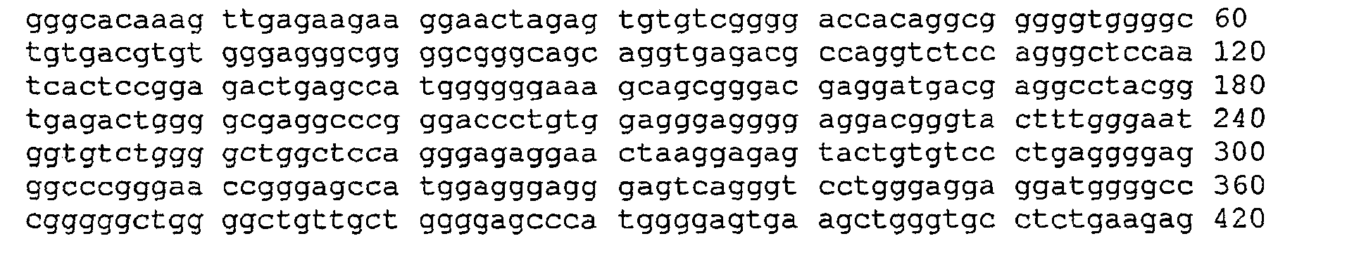
- the figure shows the sequence of the liver marker, the cleavage sites of the restriction enzymes used and the binding sites of the primers (binding to the transformed sequence). Different primers are shown, depending on which of the converted (non-complementary) DNA strands is to be amplified.
- a liver marker which was already described in the German patent application, was investigated DE 100 32 529 is described.
Landscapes
- Chemical & Material Sciences (AREA)
- Organic Chemistry (AREA)
- Life Sciences & Earth Sciences (AREA)
- Zoology (AREA)
- Wood Science & Technology (AREA)
- Proteomics, Peptides & Aminoacids (AREA)
- Health & Medical Sciences (AREA)
- Engineering & Computer Science (AREA)
- Microbiology (AREA)
- Biochemistry (AREA)
- Physics & Mathematics (AREA)
- Molecular Biology (AREA)
- Biotechnology (AREA)
- Biophysics (AREA)
- Analytical Chemistry (AREA)
- Immunology (AREA)
- Bioinformatics & Cheminformatics (AREA)
- General Engineering & Computer Science (AREA)
- General Health & Medical Sciences (AREA)
- Genetics & Genomics (AREA)
- Chemical Kinetics & Catalysis (AREA)
- Measuring Or Testing Involving Enzymes Or Micro-Organisms (AREA)
Abstract
Description
Die vorliegende Erfindung betrifft ein Verfahren zum Nachweis von 5-Methylcytosin in DNA. 5-Methylcytosin ist die häufigste kovalent modifizierte Base in der DNA eukaryontischer Zellen. Sie spielt eine wichtige biologische Rolle, u.a. bei der Transkriptionsregulation, beim genetischen Imprinting und in der Tumorgenese (zur Übersicht:
Die herkömmlichen Methoden zur Methylierungsanalyse arbeiten im wesentlichen nach zwei unterschiedlichen Prinzipien. Zum einen werden methylierungsspezifische Restriktionsenzyme benutzt, zum anderen erfolgt eine selektive chemische Umwandlung von nicht-methylierten Cytosinen in Uracil (sog.: Bisulfit-Behandlung, siehe etwa:
Da die Behandlung mit methylierungsspezifischen Restriktionsenzymen durch die Sequenzspezifität der Enzyme auf bestimmte Sequenzen beschränkt ist, wird für die meisten Anwendungen eine Bisulfit-Behandlung durchgeführt (zur Übersicht
Die herkömmlichen Verfahren zur Methylierungsanalyse lösen dieses Problem nur eingeschränkt. Üblicherweise wird die chemisch vorbehandelte DNA mittels eines PCR-Verfahrens amplifiziert. Über die Verwendung methylierungsspezifischer Primer oder Blocker soll dann eine selektive Amplifikation nur der methylierten (bzw. bei umgekehrten Ansatz: unmethylierten) DNA gewährleistet werden. Der Einsatz methylierungsspezifischer Primer ist als sog. "methylierungssensitive PCR" bekannt ("MSP"; Herman et al.: Methylation-specific PCR: a novel PCR assay for methylation status of CpG islands.
Die Anwendbarkeit dieser Verfahren für einen sensitiven und spezifischen Nachweis methylierter DNA vor einem großen Hintergrund an unmethylierter DNA ist allerdings beschränkt. So besteht die Gefahr, dass es über eine unspezifische Amplifikation von Hintergrund-DNA zu falsch-positiven Ergebnissen kommt. Falsch positive Signale stellen jedoch eines der signifikantesten Probleme des Einsatzes der Methylierungstechnologie für die Krebs-Früherkennung dar. Eine Erhöhung der Spezifität des Methylierungsnachweises bedeutet daher einen bedeutsamen Schritt für die Entwicklung entsprechender Früherkennungstests. Eine zuverlässige, kommerzielle Nutzung der Methylierungsanalyse im Bereich der Tumor-Frühdiagnostik wird so erleichtert.However, the applicability of these methods for sensitive and specific detection of methylated DNA against a large background of unmethylated DNA is limited. Thus, there is a risk that false positive results will occur through non-specific amplification of background DNA. However, false positive signals represent one of the most significant problems of using methylation technology for early cancer detection. Increasing the specificity of methylation detection therefore represents a significant step in the development of appropriate early detection tests. A reliable, commercial use The methylation analysis in the field of tumor early diagnosis is thus facilitated.
Um die Spezifität des Methylierungsnachweises zu erhöhen, verwenden die bekannten Verfahren für die Amplifiktion Primer- oder Blockersequenzen, in denen mehrere methylierungsspezifische Positionen enthalten sind. Diese Sequenzanforderungen erlauben allerdings nur einen Nachweis von Sequenzen, in denen in einem engen Sequenzbereich eine Vielzahl von CpG-Positionen auftreten. Diese Sequenzanforderungen schränken die Anwendbarkeit der Verfahren ein.To increase the specificity of methylation detection, the known amplification methods use primer or blocker sequences containing multiple methylation specific positions. However, these sequence requirements only allow detection of sequences in which a variety of CpG positions occur in a narrow sequence region. These sequence requirements limit the applicability of the methods.
Aufgrund der genannten besonderen biologischen und medizinischen Bedeutung der Cytosin-Methylierung und aufgrund der oben erwähnten Nachteile des Standes der Technik besteht ein großes technisches Bedürfnis an der Entwicklung leistungsfähiger Methoden zur sensitiven und spezifischen Methylierungsanalyse. Im folgenden ist ein überraschend einfaches Verfahren beschrieben, mit dem die Spezifität des Methylierungsnachweises erhöht werden kann.Because of the particular biological and medical significance of cytosine methylation and because of the above-mentioned disadvantages of the prior art, there is a great technical need for the development of efficient methods for sensitive and specific methylation analysis. In the following, a surprisingly simple method is described with which the specificity of the methylation detection can be increased.
Erfindungsgemäß wird vor der Bisulfitumwandlung und der anschließenden Amplifikation ein enzymatischer Filterschritt durchgeführt. Durch diesen Filter wird die Hintergrund DNA aus dem Reaktionsgemisch entfernt. In diesem Filterschritt wird ein Gemisch aus unterschiedlichen methylierungsspezifischen Retriktionsenzymen eingesetzt. Nach dem Restriktionsverdau erfolgen in herkömmlicher Weise Bisulfitumwandlung und Amplifikation/Detektion. Der enzymatische Abbau der Hintergrund-DNA verringert die Gefahr falsch positiver Ergebnisse und erlaubt so einen spezifischeren Nachweis von methylierten Cytosinpositionen.According to the invention, an enzymatic filtering step is carried out before the bisulfite conversion and the subsequent amplification. This filter removes background DNA from the reaction mixture. In this filtering step, a mixture of different methylation-specific Retriktionsenzymen is used. After restriction digestion, bisulfite conversion and amplification / detection are carried out in a conventional manner. Enzymatic degradation of the background DNA reduces the risk of false positives, allowing more specific detection of methylated cytosine positions.
Zwar ist sowohl der Einsatz von methylierungsspezifschen Restriktionsenzymen wie auch der Einsatz der Bisulfitumwandlung in der Methylierungsanalyse seit langem bekannt (s.o.). Eine Kombination aus methylierungsspezifischem Restriktionsverdau mit einer Bisulfitumwandlung und einer anschließenden Amplifikation ist bisher jedoch noch nirgends beschrieben. Aufgrund der besonderen biologischen und medizinischen Bedeutung der Cytosin-methylierung und aufgrund der Nachteile der bekannten Verfahren stellt das Eröffnen dieser vorteilhaften, neuen und überraschend einfachen Technologie einen wichtigen technischen Fortschritt dar.Although the use of methylation-specific restriction enzymes as well as the use of bisulfite in the methylation analysis has long been known (see above). A combination of methylation specific Restriction digestion with a bisulfite conversion followed by amplification has not yet been described. Due to the particular biological and medical importance of cytosine methylation and because of the disadvantages of the known processes, the opening up of this advantageous, novel and surprisingly simple technology represents an important technical advance.
Das erfindungsgemäße Verfahren zur Methylierungsanalyse verläuft in folgenden Schritten:
- 1) aus einer biologischen Probe wird DNA isoliert,
- 2) die DNA wird mit Hilfe mindestens eines methylierungsspezifischen Restriktionsenzyms umgesetzt, wobei das methylierungsspezifische Restriktionsenzym die Hintergrund DNA abbaut,
- 3) die DNA wird chemisch oder enzymatisch umgewandelt, wobei unmethyliertes Cytosin in Thymin oder eine andere Base überführt wird, die sich im Basenpaarungsverhalten von Cytosin unterscheidet, während Methylcytosin unverändert bleibt,
- 4) die umgewandelte DNA wird analysiert.
- 1) DNA is isolated from a biological sample,
- 2) the DNA is reacted with the aid of at least one methylation-specific restriction enzyme, wherein the methylation-specific restriction enzyme degrades the background DNA,
- 3) the DNA is converted chemically or enzymatically, converting unmethylated cytosine into thymine or another base that differs in the base pairing behavior of cytosine, while methylcytosine remains unchanged,
- 4) the converted DNA is analyzed.
Im ersten Schritt des erfindungsgemäßen Verfahrens wird die DNA aus einer biologischen Probe isoliert. Dabei kann die zu untersuchende DNA je nach diagnostischer oder wissenschaftlicher Fragestellung aus unterschiedlichen Quellen stammen. Für diagnostische Fragestellungen dienen als Ausgangsmaterial bevorzugt Gewebeproben, aber auch Körperflüssigkeiten, insbesondere Serum. Möglich ist auch, die DNA aus Sputum, Stuhl, Urin oder Gehirn-Rückenmarks-Flüssigkeit zu verwenden. Die DNA-Isolierung kann nach Standardverfahren erfolgen, aus Blut etwa unter Verwendung des Qiagen UltraSens DNA Extraktions-Kits.In the first step of the method according to the invention, the DNA is isolated from a biological sample. Depending on the diagnostic or scientific question, the DNA to be examined can come from different sources. Tissue samples, but also body fluids, in particular serum, serve as starting material for diagnostic questions. It is also possible to use the DNA from sputum, stool, urine or cerebrospinal fluid. DNA isolation can be done by standard methods, from blood using the Qiagen UltraSens DNA Extraction Kit.
Im zweiten Schritt des erfindungsgemäßen Verfahrens wird die DNA mit Hilfe mindestens eines methylierungsspezifischen Restriktionsenzyms umgesetzt. Dabei baut das methylierungsspezifische Restriktionsenzym die Hintergrund DNA ab. Dem Fachmann sind eine Vielzahl von Restriktionsenzymen bekannt, die erfindungsgemäß einsetzbar sind. Insbesondere bietet die REBASE-Datenbank (http://rebase.neb.com/) vielfältige Informationen zu methylierungssensitiven Restriktionsenzymen. Bevorzugt ist die Verwendung der folgender Enyzme: HpyCH4 IV, Hha I, Hpa II; HinP1I; Aci I, Zra I, SNAB1, Sal I; Pml1, PaeR7I, Cla I, BspDI, BsaAI, Ava I Die Schnittstellen dieser Enzyme sind in Tabelle 1 aufgeführt. Alle Enzyme sind kommerziell erhältlich, etwa über New England Biolabs (www.neb.com). Die Reaktionsbedingungen der enzymatischen Umsetzung sind Stand der Technik und ergeben sich etwa aus den von den Herstellern gelieferten Protokollen.In the second step of the method according to the invention, the DNA is reacted with the aid of at least one methylation-specific restriction enzyme. The methylation-specific restriction enzyme degrades the background DNA. The skilled worker is aware of a large number of restriction enzymes which can be used according to the invention. In particular, the REBASE database (http://rebase.neb.com/) provides a wealth of information on methylation-sensitive restriction enzymes. Preferred is the use of the following enzymes: HpyCH4 IV, Hha I, Hpa II; HinP1I; Aci I, Zra I, SNAB1, Sal I; Pml1, PaeR7I, Cla I, BspDI, BsaAI, Ava I The cleavage sites of these enzymes are listed in Table 1. All enzymes are commercially available, such as via New England Biolabs ( www.neb.com ). The reaction conditions of the enzymatic reaction are state of the art and result, for example, from the protocols supplied by the manufacturers.
Die Enzyme haben unterschiedliche lange Erkennungssequenzen. Dem Fachmann ist bekannt, dass er durch Auswahl der Enzyme die Häufigkeit der Fragmentierung beeinflussen kann. Wählt er Enzyme, die eine Vierersequenz erkennen, so kommt es zu einer deutlich häufigeren Schnitten als bei Verwendung von Enzymen, die eine längere Erkennungssequenz besitzen.The enzymes have different long recognition sequences. It is known to those skilled in the art that by selecting the enzymes, it can influence the frequency of fragmentation. If he or she chooses enzymes that recognize a four-membered sequence, the number of cuts that occur is significantly higher than if enzymes with a longer recognition sequence are used.
In einer bevorzugten Ausführungsform erfolgt die Umsetzung mittels einer Mischung von unterschiedlichen Restriktionsenzymen. So ist gewährleistet, dass die Hintergrund DNA möglichst vollständig fragmentiert wird, und so in der anschließenden Amplifikation als Template nicht mehr zur Verfügung steht.In a preferred embodiment, the reaction is carried out by means of a mixture of different restriction enzymes. This ensures that the background DNA is fragmented as completely as possible, and thus is no longer available as a template in the subsequent amplification.
In einer bevorzugten Ausführungsform ist die Enzymmischung so zusammengestellt, dass alle verwendeten Enzyme bei den gewählten Puffer- und Reaktionsbedingungen aktiv sind.In a preferred embodiment, the enzyme mixture is assembled so that all the enzymes used are active under the chosen buffer and reaction conditions.
Im dritten Schritt des erfindungsgemäßen Verfahrens wird die enzymatisch umgesetzte DNA chemisch oder enzymatisch umgewandelt, wobei unmethyliertes Cytosin in Thymin oder eine andere Base überführt wird, die sich im Basenpaarungsverhalten von Cytosin unterscheidet, während Methylcytosin unverändert bleibt. Bevorzugt wird dabei eine chemische "Bisulfitbehandlung" durchgeführt. Die Bisulfitumwandlung ist dem Fachmann in unterschiedlichen Variationen bekannt (siehe etwa:
In einer anderen bevorzugten Ausführungsform wird die DNA nicht chemisch, sondern enzymatisch umgewandelt. Dies ist etwa durch Einsatz von Cytidin-Deaminasen denkbar, die unmethylierte Cyidine schneller umsetzen als methylierte Cytidine. Ein entsprechendes Enzym ist kürzlich identifiziert worden (
Im letzten Schritt des erfindungsgemäßen Verfahrens wird die umgewandelte DNA analysiert und daraufhin auf den Methylierungsstatus der ursprünglichen DNA geschlossen. Die umgewandelte DNA kann anhand der gängigen molekularbiologischen Verfahren analysiert werden, etwa über Hybidisierung oder Sequenzierung. In bevorzugten Variante, in denen der Methylierungsstatus möglichst sensitiv nachgewiesen werden soll, wird die umgewandelte DNA zunächst amplifiziert. Hierzu sind dem Fachmann unterschiedliche Verfahren bekannt, etwa Ligasekettenreaktionen. In einer bevorzugten Ausführungsform wird die DNA allerdings über eine Polymerasereaktion amplifiziert. Hierzu sind verschiedene Ausgestaltungen denkbar, etwa die Verwendung isothermer Amplifikationsverfahren. Besonders bevorzugt sind allerdings Polymerasekettenreaktionen (PCR). In einer ganz besonders bevorzugten Ausführungsform erfolgt die PCR unter Verwendung von Primern, die spezifisch nur an Positionen der umgewandelten Sequenz binden, die vorher entweder methyliert (oder bei umgekehrtem Ansatz: unmethyliert) waren. Dieses Verfahren ist bei bisulfitierter DNA unter dem Namen methylierungssensitive PCR bekannt (MSP). Dabei werden Primer verwendet, die mindestens ein 5'- CpG -3' Dinukleotid enthalten; bevorzugt sind Primer, die mindestens drei 5'- CpG- 3'-Positionen tragen, von denen mindestens eine am 3' Ende lokalisiert ist. Für die Amplifikation der unmethylierten Sequenzen bzw. der Gegenstränge sind dementsprechend 5'-TG-3' oder 5'-CA-3'-Dinukleotide erforderlich (Vgl.:
Eine andere ganz besonders bevorzugten Ausführungsform ist für Bisulfitvorbehandelte DNA unter dem Namen "Heavy-Methyl"-Methode bekannt. Dabei wird eine spezifische Amplifizierung nur der ursprünglich methylierten (bzw. unmethylierten) DNA durch Einsatz mindestens eines methylierungsspezifischen Blocker-Oligomers erreicht. Der Blocker bindet an ein 5'-CG-3' (bzw. 5'-TG-3'-Dinukleotid oder 5'-CA-3')-Dinukleotid und verhindert so die Amplifikation der Hintergrund-DNA. Die Ausführungsform kann über die Auswahl der Polymerase oder über die Modifikation der Blockeroligomere so ausgestaltet sein, daß ein Abbau oder eine Verlängerung der Blocker minimiert wird (zur Übersicht:
Die Detektion der Amplifikate kann über herkömmliche Verfahren erfolgen, etwa über Methoden der Längenmessung wie Gelelektrophorese, Kapillargelelektrophorese und Chromatographie (z.B. HPLC). Auch Massenspektrometrie und Methoden zur Sequenzierung wie die Sanger-Methode, die Maxam-Gilbert-Methode und Sequencing by Hybridisation (SBH) können verwendet werden. In einer bevorzugten Ausführungsform werden die Amplifikate durch Primer-Extension-Verfahren nachgewiesen (siehe etwa: Gonzalgo & Jones: Rapid quantitation of methylation differences at specific sites using methylation-sensitive single nucleotide primer extension (Ms-SNuPE).
In einer anderen bevorzugten Ausführungsform werden die die Amplifikate mittels Hybridisierung an Oligomer-Arrays analysiert (ein Überblick über Array-Technologie befindet sich in der Extraausgabe von:
Ganz besonders bevorzugt werden die Amplifikate unter Verwendung von PCR-Real-Time-Varianten analysiert (vgl.:
Eine andere bevorzugte Ausführungsform der Erfindung ist es, mehrere Fragmente gleichzeitig mittels einer Multiplex-PCR zu amplifizieren. Bei deren Design muss darauf geachtet werden, daß nicht nur die Primer, sondern auch die weiteren eingesetzten Oligonukleotide nicht zueinander komplementär sein dürfen, so daß eine hochgradige Multiplexierung in diesem Fall schwieriger ist als in üblich. Jedoch hat man bei der enzymatisch vorbehandelten DNA den Vorteil, daß aufgrund des unterschiedlichen G und C-Gehaltes der beiden DNA-Stränge ein Forward-Primer niemals auch als Reverse-Primer fungieren kann, was die Multiplexierung wiederum erleichtert und den oben beschriebenen Nachteil im wesentlichen ausgleicht. Die Detektion der Amplifikate ist wiederum über unterschiedliche Verfahren möglich. Denkbar ist dabei etwa die Verwendung von Real-Time-Varianten. Für Amplifikationen von mehr als vier Genen empfiehlt es sich aber, die Amplifikate auf andere Weise zu detektieren. Bevorzugt ist dabei eine Analyse über Arrays (s.o.).Another preferred embodiment of the invention is to amplify multiple fragments simultaneously by multiplex PCR. Care must be taken in their design that not only the primer, but also the other oligonucleotides used are not allowed to be complementary to each other, so that a high degree of multiplexing in this case is more difficult than usual. However, one has the advantage in enzymatically pretreated DNA that due to the different G and C content of the two DNA strands, a forward primer can never function as a reverse primer, which in turn facilitates the multiplexing and the disadvantage described above substantially balances. The detection of the amplificates is again possible via different methods. It is conceivable, for example, the use of real-time variants. However, for amplifications of more than four genes, it is recommended to detect the amplificates in another way. Preference is given to an analysis of arrays (see above).
Im übrigen wird noch einmal betont, daß alle bekannten Verfahren zur Analyse bisulfit-umgewandelter DNA auch erfindungsgemäß eingesetzt werden können. Der Fachmann findet Angaben über die entsprechenden Verfahren in der wissenschaftlichen Veröffentlichungen und in der Patentliteratur. Eine aktuelle Übersicht über die mögliche Methoden findet sich in:
Eine besonders bevorzugte Verwendung des erfindungsgemäßen Verfahrens liegt in der Diagnose von Krebserkrankungen oder anderen mit einer Veränderung des Methylierungsstatus assoziierten Krankheiten. Hierzu gehören u.a. CNS-Fehlfunktionen, Aggressionssymptome oder Verhaltensstörungen; klinische, psychologische und soziale Konsequenzen von Gehirnschädigungen; psychotische Störungen und Persönlichkeitsstörungen; Demenz und/oder assoziierte Syndrome; kardiovaskuläre Krankheit, Fehlfunktion und Schädigung; Fehlfunktion, Schädigung oder Krankheit des gastrointestinalen Traktes; Fehlfunktion, Schädigung oder Krankheit des Atmungssystems; Verletzung, Entzündung, Infektion, Immunität und/oder Rekonvaleszenz; Fehlfunktion, Schädigung oder Krankheit des Körpers als Abweichung im Entwicklungsprozess; Fehlfunktion, Schädigung oder Krankheit der Haut, der Muskeln, des Bindegewebes oder der Knochen; endokrine und metabolische Fehlfunktion, Schädigung oder Krankheit; Kopfschmerzen oder sexuelle Fehlfunktion.A particularly preferred use of the method of the invention is in the diagnosis of cancers or other diseases associated with a change in methylation status. These include u.a. CNS malfunction, aggression symptoms or behavioral disorders; clinical, psychological and social consequences of brain damage; psychotic disorders and personality disorders; Dementia and / or associated syndromes; cardiovascular disease, dysfunction and damage; Dysfunction, damage or disease of the gastrointestinal tract; Dysfunction, damage or disease of the respiratory system; Injury, inflammation, infection, immunity and / or convalescence; Dysfunction, damage or disease of the body as a deviation in the development process; Dysfunction, damage or disease of the skin, muscles, connective tissue or bones; endocrine and metabolic dysfunction, injury or disease; Headache or sexual dysfunction.
Das erfindungsgemäße Verfahren eignet sich außerdem zur Vorhersage von unerwünschten Arzneimittelwirkungen und zur Unterscheidung von Zelltypen oder Geweben oder zur Untersuchung der Zelldifferenzierung.The method according to the invention is also suitable for the prediction of undesired drug effects and for the differentiation of cell types or tissues or for the investigation of cell differentiation.
Erfindungsgemäß ist schließlich auch ein Kit, der aus mindestens einem methylierungssensitiven Restriktionsenzym und aus Reagenzien für eine Bisulfitumwandlung besteht, sowie optional auch eine Polymerase, Primer und Sonden für eine Amplifkation und Detektion enthält.

Tabelle 1 zeigt die Restriktionsstellen unterschiedlicher Enzyme, die für das erfindungsgemäße Verfahren einsetzbar sind. Die Tabelle ist dem Katalog von New England Biolabs entnommen (www.neb.com). Die Enzyme haben ihr Optimum in unterschiedlichen Puffern (NEB-Puffer 1-4). Angegeben ist die relative Aktivität der Enzyme in %. Die schraffierten Flächen zeigen die Puffer, bei denen die Enzyme am besten eingesetzt werden. In einer bevorzugten Ausführungsform der Erfindung werden mehrere Enzyme zusammen eingesetzt. Dabei werden bevorzugt solche Enzyme kombiniert, die ihr Optimum bei den gleichen Pufferkonzentrationen haben.Table 1 shows the restriction sites of different enzymes that can be used for the method according to the invention. The table is taken from the catalog of New England Biolabs ( www.neb.com ). The enzymes have their optimum in different buffers (NEB buffer 1-4). Indicated is the relative activity of the enzymes in%. The hatched areas show the buffers where the enzymes are best used. In a preferred embodiment of the invention, several enzymes are used together. In this case, those enzymes are preferably combined that have their optimum at the same buffer concentrations.
Es soll ein diagnostischer Test entwickelt werden, mit dem aus einer Blutprobe sehr frühzeitig Lebererkrankungen, insbesondere Leberkrebs, nachgewiesen werden kann. Dazu wird eine DNA-Sequenz untersucht, die nur in Lebergewebe methyliert ist, in anderen Geweben (z.B. Muskel, Lunge, Haut, Brust) aber unmethyliert vorliegt (siehe unten, vgl.:
Hierzu wird zunächst die DNA aus Blut des Probanten isoliert. Hierzu stehen unterschiedliche Verfahren zur Verfügung, etwa unter Verwendung des Qiagen "UltraSens DNA Extraktions-Kits".
Anschließend werden die drei Restriktionsenzyme HpyCH4 IV (Schnittstelle:ACGT), Aci I (CGCC/GGCG) und Hpall(CCGG) nach Herstellerangaben eingesetzt. Schließlich wird die DNA in Gegenwart denaturierender Lösemittel unter Verwendung besondere Temperaturprogramme bisulfitiert und bevorzugt über eine Ultrafiltration aufgereinigt (siehe im einzelnen die deutschen Patentanmeldungen
Subsequently, the three restriction enzymes HpyCH4 IV (cleavage: ACGT), Aci I (CGCC / GGCG) and Hpall (CCGG) are used according to the manufacturer's instructions. Finally, the DNA is in the presence of denaturing solvent bisulfited using special temperature programs and preferably purified by ultrafiltration (see in detail the German patent applications
Die Sequenzen des Markers und die Schnittstellen der Enzyme sind in Abbildung 1 dargestellt.The sequences of the marker and the interfaces of the enzymes are shown in Figure 1.
Abbildung zeigt die Sequenz des Lebermarkers, die Schnittstellen der verwendeten Restriktionsenzyme und die Bindungsstellen der Primer (bindend auf der umgewandelten Sequenz). Es sind unterschiedliche Primer gezeigt, je nachdem welcher der umgewandelten (nicht mehr komplementären) DNA-Stränge amplifiziert werden soll. Untersucht wurde ein Lebermarker, der bereits in der deutschen Patentanmeldung
- <110> Epigenomics AG<110> Epigenomics AG
- <120> Verfahren zur Untersuchung von Cytosin-Methylierungen in DNA<120> Method of assaying cytosine methylations in DNA
- <130> P329105PCT<130> P329105PCT
-
<140>
<141><140>
<141> -
<150>
102005011398.2
<151> 2005-03-03<150>102005011398.2
<151> 2005-03-03 - <160> 6<160> 6
- <170> PatentIn Ver. 2.1<170> Patent In Ver. 2.1
-
<210> 1
<211> 420
<212> DNA
<213> Homo sapiens<210> 1
<211> 420
<212> DNA
<213> Homo sapiens -
<400> 1
<400> 1
-
<210> 2
<211> 22
<212> DNA
<213> Künstliche Sequenz<210> 2
<211> 22
<212> DNA
<213> Artificial sequence -
<220>
<223> Beschreibung der künstlichen Sequenz: Primer<220>
<223> Description of the artificial sequence: primer -
<400> 2
agtgtgtcgg ggaccacagg cg 22<400> 2
agtgtgtcgg ggaccacagg cg 22 -
<210> 3
<211> 22
<212> DNA
<213> Künstliche Sequenz<210> 3
<211> 22
<212> DNA
<213> Artificial sequence -
<220>
<223> Beschreibung der künstlichen Sequenz: Primer<220>
<223> Description of the artificial sequence: primer -
<400> 3
aatatatcga aaaccacaaa cg 22<400> 3
aatatatcga aaaccacaaa cg 22 -
<210> 4
<211> 21
<212> DNA
<213> Künstliche Sequenz<210> 4
<211> 21
<212> DNA
<213> Artificial sequence -
<220>
<223> Beschreibung der künstlichen Sequenz: Primer<220>
<223> Description of the artificial sequence: primer -
<400> 4
gggggctggg gctgttgctg g 21<400> 4
gggggctggg gctgttgctg g 21 -
<210> 5
<211> 21
<212> DNA
<213> Künstliche Sequenz<210> 5
<211> 21
<212> DNA
<213> Artificial sequence -
<220>
<223> Beschreibung der künstlichen Sequenz: Primer<220>
<223> Description of the artificial sequence: primer -
<400> 5
ccagcaacag ccccagcccc c 21<400> 5
ccagcaacag ccccagcccc c 21 -
<210> 6
<211> 21
<212> DNA
<213> Künstliche Sequenz<210> 6
<211> 21
<212> DNA
<213> Artificial sequence -
<220>
<223> Beschreibung der künstlichen Sequenz: Primer<220>
<223> Description of the artificial sequence: primer -
<400> 6
ttagtaatag ttttagtttt c 21<400> 6
daytime day ttttagtttt c 21
Claims (14)
- A method for the examination of cytosine methylation for the diagnosis of cancer or other diseases associated with a change of the methylation status, characterized in that the following steps are carried out:a) DNA is isolated from a tissue sample or from a body fluid,b) the DNA is reacted with the aid of at least one methylation-specific restriction enzyme, wherein the methylation-specific restriction enzyme degrades the background DNA,c) the DNA is chemically or enzymatically converted, wherein non-methylated cytosine is converted into uracil or another base that differs in the base pairing behavior from cytosine whereas methylcytosine remains unchanged,d) analysis of the transformed DNA by real-time PCR.
- The method according to Claim 1, characterized in that one of the following enzymes is used in the second step: HpyCH4 IV, Hha I, Hpa II; HinP1I; Aci I, Zra I, SNAB1, Sal I; Pmi1, PaeR7I, Cla I, BspDI, BsaAI, Ava I.
- The method according to any one of Claims 1-2, characterized in that a mixture of different enzymes is used in the second step.
- The method according to Claim 3, characterized in that the different enzymes used are active under the same buffer and reaction conditions.
- The method according to any one of Claims 1-4, characterized in that the conversion takes place in the third step by means of a bisulfite.
- The method according to any one of Claims 1-4, characterized in that the conversion takes place in the third step by means of a methylation-specific cytidine deaminase.
- The method according to Claim 1, characterized in that the polymerase chain reaction takes place by means of methylation-specific primers.
- The method according to Claim 7, characterized in that at least one methylation-specific blocker oligomer is used in the polymerase chain reaction.
- The method according to the Claims 7 and 8, characterized in that the amplificates are analyzed via methods of the longitudinal measuring, the mass spectrometry or the sequencing.
- The method according to the Claims 7-9, characterized in that the amplificates are analyzed by primer-extension methods.
- The method according to the Claims 7-9, characterized in that the amplificates are analyzed by hybridization on oligomer arrays.
- The method according to Claim 1, characterized in that a TaqMan method or a lightcycler method is carried out.
- The method according to the Claims 7-12, characterized in that several fragments are amplified simultaneously by means of a multiplex reaction.
- The use of the methods according to Claims 1-13 for the prediction of undesired drug effects, for distinguishing cell types and tissues or for the examination of the cell differentiation.
Applications Claiming Priority (2)
| Application Number | Priority Date | Filing Date | Title |
|---|---|---|---|
| DE102005011398A DE102005011398A1 (en) | 2005-03-03 | 2005-03-03 | Method for the investigation of cytosine methylations in DNA |
| PCT/EP2006/002092 WO2006092339A1 (en) | 2005-03-03 | 2006-03-02 | Method for investigating cytosine methylations in dna |
Publications (2)
| Publication Number | Publication Date |
|---|---|
| EP1853724A1 EP1853724A1 (en) | 2007-11-14 |
| EP1853724B1 true EP1853724B1 (en) | 2009-10-21 |
Family
ID=36357672
Family Applications (1)
| Application Number | Title | Priority Date | Filing Date |
|---|---|---|---|
| EP06707458A Not-in-force EP1853724B1 (en) | 2005-03-03 | 2006-03-02 | Method for investigating cytosine methylations in dna |
Country Status (5)
| Country | Link |
|---|---|
| US (1) | US20080286778A1 (en) |
| EP (1) | EP1853724B1 (en) |
| AT (1) | ATE446388T1 (en) |
| DE (2) | DE102005011398A1 (en) |
| WO (1) | WO2006092339A1 (en) |
Families Citing this family (5)
| Publication number | Priority date | Publication date | Assignee | Title |
|---|---|---|---|---|
| JP2008136404A (en) * | 2006-11-30 | 2008-06-19 | Sysmex Corp | Method for confirming amount of dna after conversion treatment of non-methylated cytosine in dna methylation detection |
| JP2012511927A (en) | 2008-12-17 | 2012-05-31 | ライフ テクノロジーズ コーポレーション | Methods, compositions, and kits for detecting allelic variants |
| US20110287424A1 (en) * | 2009-03-27 | 2011-11-24 | Life Technologies Corporation | Methylation-specific competitive allele-specific taqman polymerase chain reaction (cast-pcr) |
| CN102428190B (en) | 2009-03-27 | 2014-02-26 | 生命技术公司 | Methods, compositions and kits for detecting allelic variants |
| AU2013350330B2 (en) | 2012-11-26 | 2019-06-06 | Clinical Genomics Pty Ltd | A method of detecting methylation |
Citations (1)
| Publication number | Priority date | Publication date | Assignee | Title |
|---|---|---|---|---|
| WO2002038811A2 (en) * | 2000-11-08 | 2002-05-16 | University Of Southern California | Assay for the detection and quantitation of hemimethylation |
Family Cites Families (9)
| Publication number | Priority date | Publication date | Assignee | Title |
|---|---|---|---|---|
| WO1997034015A1 (en) * | 1996-03-15 | 1997-09-18 | The Penn State Research Foundation | Detection of extracellular tumor-associated nucleic acid in blood plasma or serum using nucleic acid amplification assays |
| DE10130800B4 (en) * | 2001-06-22 | 2005-06-23 | Epigenomics Ag | Method for the detection of cytosine methylation with high sensitivity |
| WO2003027259A2 (en) * | 2001-09-26 | 2003-04-03 | Epigenx Pharmaceuticals, Inc. | Assays for dna methylation changes |
| US20040072197A1 (en) * | 2001-11-08 | 2004-04-15 | Jones Peter A. | Assay for the detection and quantitation of hemimethylation |
| EP1470255A2 (en) * | 2002-01-30 | 2004-10-27 | Epigenomics AG | Identification of cell differentiation states based on methylation patterns |
| US20040029121A1 (en) * | 2002-08-08 | 2004-02-12 | Susan Cottrell | Methods and nucleic acids for the analysis of CpG dinucleotide methylation status associated with the calcitonin gene |
| US20040146868A1 (en) * | 2003-01-24 | 2004-07-29 | Epigenomics Ag | Methods and nucleic acids for the analysis of CpG dinucleotide methylation status associated with the development of peripheral zone prostate cancer |
| US20040265833A1 (en) * | 2003-06-23 | 2004-12-30 | Cathy Lofton-Day | Methods and nucleic acids for the analysis of colorectal cell proliferative disorders |
| DE10331107B3 (en) * | 2003-07-04 | 2004-12-02 | Epigenomics Ag | Detecting cytosine methylation in DNA, useful e.g. for diagnosis of cancer, comprises using a cytidine deaminase selective for unmethylated residues, and detecting presence or amount of deaminated residues |
-
2005
- 2005-03-03 DE DE102005011398A patent/DE102005011398A1/en not_active Withdrawn
-
2006
- 2006-03-02 AT AT06707458T patent/ATE446388T1/en active
- 2006-03-02 DE DE502006005183T patent/DE502006005183D1/en active Active
- 2006-03-02 US US11/885,706 patent/US20080286778A1/en not_active Abandoned
- 2006-03-02 WO PCT/EP2006/002092 patent/WO2006092339A1/en not_active Ceased
- 2006-03-02 EP EP06707458A patent/EP1853724B1/en not_active Not-in-force
Patent Citations (1)
| Publication number | Priority date | Publication date | Assignee | Title |
|---|---|---|---|---|
| WO2002038811A2 (en) * | 2000-11-08 | 2002-05-16 | University Of Southern California | Assay for the detection and quantitation of hemimethylation |
Also Published As
| Publication number | Publication date |
|---|---|
| WO2006092339A1 (en) | 2006-09-08 |
| EP1853724A1 (en) | 2007-11-14 |
| US20080286778A1 (en) | 2008-11-20 |
| DE502006005183D1 (en) | 2009-12-03 |
| ATE446388T1 (en) | 2009-11-15 |
| DE102005011398A1 (en) | 2006-09-14 |
Similar Documents
| Publication | Publication Date | Title |
|---|---|---|
| EP1423533B1 (en) | Method for high sensitivity detection of cytosine-methylation | |
| EP1370691B1 (en) | Method for detecting cytosine methylation patterns having high sensitivity | |
| DE60029323T3 (en) | METHOD FOR ANALYZING DNA METHYLATION AT HIGH RATE OF PURCHASE | |
| DE10331107B3 (en) | Detecting cytosine methylation in DNA, useful e.g. for diagnosis of cancer, comprises using a cytidine deaminase selective for unmethylated residues, and detecting presence or amount of deaminated residues | |
| DE10151055B4 (en) | Method for detecting cytosine methylation in CpG islands | |
| EP1654388B1 (en) | Method for the detection of cytosine methylations in dna | |
| DE19853398C1 (en) | Identification of 5-methylcytosine positions in genomic DNA by chemical modification, amplification and heteroduplex formation | |
| DE102005034628B4 (en) | Method for the investigation of cytosine methylations in DNA | |
| EP1853724B1 (en) | Method for investigating cytosine methylations in dna | |
| DE10304219B3 (en) | Method for the detection of cytosine methylation patterns with high sensitivity | |
| DE10392538B4 (en) | Method for the analysis of methylated nucleic acids | |
| EP1746169B1 (en) | Method for quantification of methylated DNA | |
| DE10346363B4 (en) | Method for methylation analysis of DNA | |
| EP1412524A2 (en) | Detection of specific dinucleotides in dna-samples by fluorescence resonance energy transfer (fret) | |
| EP1711628B1 (en) | Method for the calibration and verification of methylation analysis methods with the aid of non-methylated dna | |
| DE102004002257B4 (en) | Method for studying cytosine methylations in DNA using DNA repair enzymes | |
| EP1432827B1 (en) | Method for detecting dna methylation using labelled s-adenosylmethionine analogs | |
| DE102006035600B3 (en) | Methylation detection, for diagnosis/prognosis of e.g. cancer disease, comprises converting non-methylated cytosine to uracil (nucleic acid) or into other base, so 5-methylcytosine remains unchanged and catalyzing nucleic acid activity | |
| EP1735460A1 (en) | Method for analysis of cytosine methylation | |
| EP1655377A1 (en) | Method of quantifying methylated DNA | |
| DE10158283A1 (en) | Detecting methylation status of test DNA in a mixture, useful for diagnosis and prognosis of disease, comprises bisulfite treatment then selective amplification of test DNA |
Legal Events
| Date | Code | Title | Description |
|---|---|---|---|
| PUAI | Public reference made under article 153(3) epc to a published international application that has entered the european phase |
Free format text: ORIGINAL CODE: 0009012 |
|
| 17P | Request for examination filed |
Effective date: 20070823 |
|
| AK | Designated contracting states |
Kind code of ref document: A1 Designated state(s): AT BE BG CH CY CZ DE DK EE ES FI FR GB GR HU IE IS IT LI LT LU LV MC NL PL PT RO SE SI SK TR |
|
| 17Q | First examination report despatched |
Effective date: 20080208 |
|
| RIN1 | Information on inventor provided before grant (corrected) |
Inventor name: LESCHE, RALF Inventor name: DISTLER, JUERGEN Inventor name: SCHUSTER, MATTHIAS Inventor name: LEWIN, JOERN |
|
| DAX | Request for extension of the european patent (deleted) | ||
| GRAP | Despatch of communication of intention to grant a patent |
Free format text: ORIGINAL CODE: EPIDOSNIGR1 |
|
| GRAS | Grant fee paid |
Free format text: ORIGINAL CODE: EPIDOSNIGR3 |
|
| GRAA | (expected) grant |
Free format text: ORIGINAL CODE: 0009210 |
|
| AK | Designated contracting states |
Kind code of ref document: B1 Designated state(s): AT BE BG CH CY CZ DE DK EE ES FI FR GB GR HU IE IS IT LI LT LU LV MC NL PL PT RO SE SI SK TR |
|
| REG | Reference to a national code |
Ref country code: GB Ref legal event code: FG4D Free format text: NOT ENGLISH |
|
| REG | Reference to a national code |
Ref country code: CH Ref legal event code: EP |
|
| REG | Reference to a national code |
Ref country code: IE Ref legal event code: FG4D |
|
| REF | Corresponds to: |
Ref document number: 502006005183 Country of ref document: DE Date of ref document: 20091203 Kind code of ref document: P |
|
| LTIE | Lt: invalidation of european patent or patent extension |
Effective date: 20091021 |
|
| NLV1 | Nl: lapsed or annulled due to failure to fulfill the requirements of art. 29p and 29m of the patents act | ||
| PG25 | Lapsed in a contracting state [announced via postgrant information from national office to epo] |
Ref country code: ES Free format text: LAPSE BECAUSE OF FAILURE TO SUBMIT A TRANSLATION OF THE DESCRIPTION OR TO PAY THE FEE WITHIN THE PRESCRIBED TIME-LIMIT Effective date: 20100201 Ref country code: FI Free format text: LAPSE BECAUSE OF FAILURE TO SUBMIT A TRANSLATION OF THE DESCRIPTION OR TO PAY THE FEE WITHIN THE PRESCRIBED TIME-LIMIT Effective date: 20091021 Ref country code: LT Free format text: LAPSE BECAUSE OF FAILURE TO SUBMIT A TRANSLATION OF THE DESCRIPTION OR TO PAY THE FEE WITHIN THE PRESCRIBED TIME-LIMIT Effective date: 20091021 Ref country code: IS Free format text: LAPSE BECAUSE OF FAILURE TO SUBMIT A TRANSLATION OF THE DESCRIPTION OR TO PAY THE FEE WITHIN THE PRESCRIBED TIME-LIMIT Effective date: 20100221 Ref country code: SE Free format text: LAPSE BECAUSE OF FAILURE TO SUBMIT A TRANSLATION OF THE DESCRIPTION OR TO PAY THE FEE WITHIN THE PRESCRIBED TIME-LIMIT Effective date: 20091021 Ref country code: PT Free format text: LAPSE BECAUSE OF FAILURE TO SUBMIT A TRANSLATION OF THE DESCRIPTION OR TO PAY THE FEE WITHIN THE PRESCRIBED TIME-LIMIT Effective date: 20100222 |
|
| REG | Reference to a national code |
Ref country code: IE Ref legal event code: FD4D |
|
| PG25 | Lapsed in a contracting state [announced via postgrant information from national office to epo] |
Ref country code: SI Free format text: LAPSE BECAUSE OF FAILURE TO SUBMIT A TRANSLATION OF THE DESCRIPTION OR TO PAY THE FEE WITHIN THE PRESCRIBED TIME-LIMIT Effective date: 20091021 Ref country code: LV Free format text: LAPSE BECAUSE OF FAILURE TO SUBMIT A TRANSLATION OF THE DESCRIPTION OR TO PAY THE FEE WITHIN THE PRESCRIBED TIME-LIMIT Effective date: 20091021 Ref country code: PL Free format text: LAPSE BECAUSE OF FAILURE TO SUBMIT A TRANSLATION OF THE DESCRIPTION OR TO PAY THE FEE WITHIN THE PRESCRIBED TIME-LIMIT Effective date: 20091021 |
|
| PG25 | Lapsed in a contracting state [announced via postgrant information from national office to epo] |
Ref country code: DK Free format text: LAPSE BECAUSE OF FAILURE TO SUBMIT A TRANSLATION OF THE DESCRIPTION OR TO PAY THE FEE WITHIN THE PRESCRIBED TIME-LIMIT Effective date: 20091021 Ref country code: IE Free format text: LAPSE BECAUSE OF FAILURE TO SUBMIT A TRANSLATION OF THE DESCRIPTION OR TO PAY THE FEE WITHIN THE PRESCRIBED TIME-LIMIT Effective date: 20091021 Ref country code: EE Free format text: LAPSE BECAUSE OF FAILURE TO SUBMIT A TRANSLATION OF THE DESCRIPTION OR TO PAY THE FEE WITHIN THE PRESCRIBED TIME-LIMIT Effective date: 20091021 Ref country code: RO Free format text: LAPSE BECAUSE OF FAILURE TO SUBMIT A TRANSLATION OF THE DESCRIPTION OR TO PAY THE FEE WITHIN THE PRESCRIBED TIME-LIMIT Effective date: 20091021 Ref country code: BG Free format text: LAPSE BECAUSE OF FAILURE TO SUBMIT A TRANSLATION OF THE DESCRIPTION OR TO PAY THE FEE WITHIN THE PRESCRIBED TIME-LIMIT Effective date: 20100121 |
|
| PLBE | No opposition filed within time limit |
Free format text: ORIGINAL CODE: 0009261 |
|
| STAA | Information on the status of an ep patent application or granted ep patent |
Free format text: STATUS: NO OPPOSITION FILED WITHIN TIME LIMIT |
|
| PG25 | Lapsed in a contracting state [announced via postgrant information from national office to epo] |
Ref country code: SK Free format text: LAPSE BECAUSE OF FAILURE TO SUBMIT A TRANSLATION OF THE DESCRIPTION OR TO PAY THE FEE WITHIN THE PRESCRIBED TIME-LIMIT Effective date: 20091021 Ref country code: CZ Free format text: LAPSE BECAUSE OF FAILURE TO SUBMIT A TRANSLATION OF THE DESCRIPTION OR TO PAY THE FEE WITHIN THE PRESCRIBED TIME-LIMIT Effective date: 20091021 |
|
| 26N | No opposition filed |
Effective date: 20100722 |
|
| BERE | Be: lapsed |
Owner name: EPIGENOMICS A.G. Effective date: 20100331 |
|
| PG25 | Lapsed in a contracting state [announced via postgrant information from national office to epo] |
Ref country code: MC Free format text: LAPSE BECAUSE OF NON-PAYMENT OF DUE FEES Effective date: 20100331 Ref country code: GR Free format text: LAPSE BECAUSE OF FAILURE TO SUBMIT A TRANSLATION OF THE DESCRIPTION OR TO PAY THE FEE WITHIN THE PRESCRIBED TIME-LIMIT Effective date: 20100122 |
|
| REG | Reference to a national code |
Ref country code: CH Ref legal event code: PL |
|
| PG25 | Lapsed in a contracting state [announced via postgrant information from national office to epo] |
Ref country code: BE Free format text: LAPSE BECAUSE OF NON-PAYMENT OF DUE FEES Effective date: 20100331 Ref country code: CH Free format text: LAPSE BECAUSE OF NON-PAYMENT OF DUE FEES Effective date: 20100331 Ref country code: LI Free format text: LAPSE BECAUSE OF NON-PAYMENT OF DUE FEES Effective date: 20100331 |
|
| PG25 | Lapsed in a contracting state [announced via postgrant information from national office to epo] |
Ref country code: IT Free format text: LAPSE BECAUSE OF FAILURE TO SUBMIT A TRANSLATION OF THE DESCRIPTION OR TO PAY THE FEE WITHIN THE PRESCRIBED TIME-LIMIT Effective date: 20091021 |
|
| PGFP | Annual fee paid to national office [announced via postgrant information from national office to epo] |
Ref country code: FR Payment date: 20110401 Year of fee payment: 6 |
|
| PGFP | Annual fee paid to national office [announced via postgrant information from national office to epo] |
Ref country code: DE Payment date: 20110331 Year of fee payment: 6 Ref country code: GB Payment date: 20110324 Year of fee payment: 6 |
|
| PG25 | Lapsed in a contracting state [announced via postgrant information from national office to epo] |
Ref country code: CY Free format text: LAPSE BECAUSE OF FAILURE TO SUBMIT A TRANSLATION OF THE DESCRIPTION OR TO PAY THE FEE WITHIN THE PRESCRIBED TIME-LIMIT Effective date: 20091021 |
|
| PG25 | Lapsed in a contracting state [announced via postgrant information from national office to epo] |
Ref country code: HU Free format text: LAPSE BECAUSE OF FAILURE TO SUBMIT A TRANSLATION OF THE DESCRIPTION OR TO PAY THE FEE WITHIN THE PRESCRIBED TIME-LIMIT Effective date: 20100422 Ref country code: LU Free format text: LAPSE BECAUSE OF NON-PAYMENT OF DUE FEES Effective date: 20100302 Ref country code: NL Free format text: LAPSE BECAUSE OF FAILURE TO SUBMIT A TRANSLATION OF THE DESCRIPTION OR TO PAY THE FEE WITHIN THE PRESCRIBED TIME-LIMIT Effective date: 20091021 |
|
| PG25 | Lapsed in a contracting state [announced via postgrant information from national office to epo] |
Ref country code: TR Free format text: LAPSE BECAUSE OF FAILURE TO SUBMIT A TRANSLATION OF THE DESCRIPTION OR TO PAY THE FEE WITHIN THE PRESCRIBED TIME-LIMIT Effective date: 20091021 |
|
| GBPC | Gb: european patent ceased through non-payment of renewal fee |
Effective date: 20120302 |
|
| REG | Reference to a national code |
Ref country code: AT Ref legal event code: MM01 Ref document number: 446388 Country of ref document: AT Kind code of ref document: T Effective date: 20110302 |
|
| REG | Reference to a national code |
Ref country code: FR Ref legal event code: ST Effective date: 20121130 |
|
| REG | Reference to a national code |
Ref country code: DE Ref legal event code: R119 Ref document number: 502006005183 Country of ref document: DE Effective date: 20121002 |
|
| PG25 | Lapsed in a contracting state [announced via postgrant information from national office to epo] |
Ref country code: AT Free format text: LAPSE BECAUSE OF NON-PAYMENT OF DUE FEES Effective date: 20110302 Ref country code: FR Free format text: LAPSE BECAUSE OF NON-PAYMENT OF DUE FEES Effective date: 20120402 Ref country code: GB Free format text: LAPSE BECAUSE OF NON-PAYMENT OF DUE FEES Effective date: 20120302 |
|
| PG25 | Lapsed in a contracting state [announced via postgrant information from national office to epo] |
Ref country code: DE Free format text: LAPSE BECAUSE OF NON-PAYMENT OF DUE FEES Effective date: 20121002 |