EP1842553A1 - Combination of an anti-EDb fibronectin domain antibody/IL2 fusion protein and a further small molecule - Google Patents
Combination of an anti-EDb fibronectin domain antibody/IL2 fusion protein and a further small molecule Download PDFInfo
- Publication number
- EP1842553A1 EP1842553A1 EP06090054A EP06090054A EP1842553A1 EP 1842553 A1 EP1842553 A1 EP 1842553A1 EP 06090054 A EP06090054 A EP 06090054A EP 06090054 A EP06090054 A EP 06090054A EP 1842553 A1 EP1842553 A1 EP 1842553A1
- Authority
- EP
- European Patent Office
- Prior art keywords
- antibody
- fusion protein
- tumor
- cancer
- cells
- Prior art date
- Legal status (The legal status is an assumption and is not a legal conclusion. Google has not performed a legal analysis and makes no representation as to the accuracy of the status listed.)
- Withdrawn
Links
Images


Classifications
-
- C—CHEMISTRY; METALLURGY
- C07—ORGANIC CHEMISTRY
- C07K—PEPTIDES
- C07K16/00—Immunoglobulins [IGs], e.g. monoclonal or polyclonal antibodies
- C07K16/18—Immunoglobulins [IGs], e.g. monoclonal or polyclonal antibodies against material from animals or humans
-
- A—HUMAN NECESSITIES
- A61—MEDICAL OR VETERINARY SCIENCE; HYGIENE
- A61K—PREPARATIONS FOR MEDICAL, DENTAL OR TOILETRY PURPOSES
- A61K31/00—Medicinal preparations containing organic active ingredients
- A61K31/70—Carbohydrates; Sugars; Derivatives thereof
- A61K31/7042—Compounds having saccharide radicals and heterocyclic rings
- A61K31/7052—Compounds having saccharide radicals and heterocyclic rings having nitrogen as a ring hetero atom, e.g. nucleosides, nucleotides
- A61K31/706—Compounds having saccharide radicals and heterocyclic rings having nitrogen as a ring hetero atom, e.g. nucleosides, nucleotides containing six-membered rings with nitrogen as a ring hetero atom
- A61K31/7064—Compounds having saccharide radicals and heterocyclic rings having nitrogen as a ring hetero atom, e.g. nucleosides, nucleotides containing six-membered rings with nitrogen as a ring hetero atom containing condensed or non-condensed pyrimidines
- A61K31/7068—Compounds having saccharide radicals and heterocyclic rings having nitrogen as a ring hetero atom, e.g. nucleosides, nucleotides containing six-membered rings with nitrogen as a ring hetero atom containing condensed or non-condensed pyrimidines having oxo groups directly attached to the pyrimidine ring, e.g. cytidine, cytidylic acid
-
- A—HUMAN NECESSITIES
- A61—MEDICAL OR VETERINARY SCIENCE; HYGIENE
- A61K—PREPARATIONS FOR MEDICAL, DENTAL OR TOILETRY PURPOSES
- A61K38/00—Medicinal preparations containing peptides
- A61K38/16—Peptides having more than 20 amino acids; Gastrins; Somatostatins; Melanotropins; Derivatives thereof
- A61K38/17—Peptides having more than 20 amino acids; Gastrins; Somatostatins; Melanotropins; Derivatives thereof from animals; from humans
- A61K38/19—Cytokines; Lymphokines; Interferons
- A61K38/20—Interleukins [IL]
- A61K38/2013—IL-2
-
- A—HUMAN NECESSITIES
- A61—MEDICAL OR VETERINARY SCIENCE; HYGIENE
- A61K—PREPARATIONS FOR MEDICAL, DENTAL OR TOILETRY PURPOSES
- A61K39/00—Medicinal preparations containing antigens or antibodies
- A61K39/395—Antibodies; Immunoglobulins; Immune serum, e.g. antilymphocytic serum
-
- A—HUMAN NECESSITIES
- A61—MEDICAL OR VETERINARY SCIENCE; HYGIENE
- A61K—PREPARATIONS FOR MEDICAL, DENTAL OR TOILETRY PURPOSES
- A61K39/00—Medicinal preparations containing antigens or antibodies
- A61K39/395—Antibodies; Immunoglobulins; Immune serum, e.g. antilymphocytic serum
- A61K39/39533—Antibodies; Immunoglobulins; Immune serum, e.g. antilymphocytic serum against materials from animals
- A61K39/39541—Antibodies; Immunoglobulins; Immune serum, e.g. antilymphocytic serum against materials from animals against normal tissues, cells
-
- A—HUMAN NECESSITIES
- A61—MEDICAL OR VETERINARY SCIENCE; HYGIENE
- A61K—PREPARATIONS FOR MEDICAL, DENTAL OR TOILETRY PURPOSES
- A61K47/00—Medicinal preparations characterised by the non-active ingredients used, e.g. carriers or inert additives; Targeting or modifying agents chemically bound to the active ingredient
- A61K47/50—Medicinal preparations characterised by the non-active ingredients used, e.g. carriers or inert additives; Targeting or modifying agents chemically bound to the active ingredient the non-active ingredient being chemically bound to the active ingredient, e.g. polymer-drug conjugates
- A61K47/51—Medicinal preparations characterised by the non-active ingredients used, e.g. carriers or inert additives; Targeting or modifying agents chemically bound to the active ingredient the non-active ingredient being chemically bound to the active ingredient, e.g. polymer-drug conjugates the non-active ingredient being a modifying agent
- A61K47/68—Medicinal preparations characterised by the non-active ingredients used, e.g. carriers or inert additives; Targeting or modifying agents chemically bound to the active ingredient the non-active ingredient being chemically bound to the active ingredient, e.g. polymer-drug conjugates the non-active ingredient being a modifying agent the modifying agent being an antibody, an immunoglobulin or a fragment thereof, e.g. an Fc-fragment
- A61K47/6801—Drug-antibody or immunoglobulin conjugates defined by the pharmacologically or therapeutically active agent
- A61K47/6803—Drugs conjugated to an antibody or immunoglobulin, e.g. cisplatin-antibody conjugates
- A61K47/6811—Drugs conjugated to an antibody or immunoglobulin, e.g. cisplatin-antibody conjugates the drug being a protein or peptide, e.g. transferrin or bleomycin
- A61K47/6813—Drugs conjugated to an antibody or immunoglobulin, e.g. cisplatin-antibody conjugates the drug being a protein or peptide, e.g. transferrin or bleomycin the drug being a peptidic cytokine, e.g. an interleukin or interferon
-
- A—HUMAN NECESSITIES
- A61—MEDICAL OR VETERINARY SCIENCE; HYGIENE
- A61K—PREPARATIONS FOR MEDICAL, DENTAL OR TOILETRY PURPOSES
- A61K47/00—Medicinal preparations characterised by the non-active ingredients used, e.g. carriers or inert additives; Targeting or modifying agents chemically bound to the active ingredient
- A61K47/50—Medicinal preparations characterised by the non-active ingredients used, e.g. carriers or inert additives; Targeting or modifying agents chemically bound to the active ingredient the non-active ingredient being chemically bound to the active ingredient, e.g. polymer-drug conjugates
- A61K47/51—Medicinal preparations characterised by the non-active ingredients used, e.g. carriers or inert additives; Targeting or modifying agents chemically bound to the active ingredient the non-active ingredient being chemically bound to the active ingredient, e.g. polymer-drug conjugates the non-active ingredient being a modifying agent
- A61K47/68—Medicinal preparations characterised by the non-active ingredients used, e.g. carriers or inert additives; Targeting or modifying agents chemically bound to the active ingredient the non-active ingredient being chemically bound to the active ingredient, e.g. polymer-drug conjugates the non-active ingredient being a modifying agent the modifying agent being an antibody, an immunoglobulin or a fragment thereof, e.g. an Fc-fragment
- A61K47/6835—Medicinal preparations characterised by the non-active ingredients used, e.g. carriers or inert additives; Targeting or modifying agents chemically bound to the active ingredient the non-active ingredient being chemically bound to the active ingredient, e.g. polymer-drug conjugates the non-active ingredient being a modifying agent the modifying agent being an antibody, an immunoglobulin or a fragment thereof, e.g. an Fc-fragment the modifying agent being an antibody or an immunoglobulin bearing at least one antigen-binding site
- A61K47/6843—Medicinal preparations characterised by the non-active ingredients used, e.g. carriers or inert additives; Targeting or modifying agents chemically bound to the active ingredient the non-active ingredient being chemically bound to the active ingredient, e.g. polymer-drug conjugates the non-active ingredient being a modifying agent the modifying agent being an antibody, an immunoglobulin or a fragment thereof, e.g. an Fc-fragment the modifying agent being an antibody or an immunoglobulin bearing at least one antigen-binding site the antibody targeting a material from animals or humans
-
- A—HUMAN NECESSITIES
- A61—MEDICAL OR VETERINARY SCIENCE; HYGIENE
- A61P—SPECIFIC THERAPEUTIC ACTIVITY OF CHEMICAL COMPOUNDS OR MEDICINAL PREPARATIONS
- A61P35/00—Antineoplastic agents
-
- A—HUMAN NECESSITIES
- A61—MEDICAL OR VETERINARY SCIENCE; HYGIENE
- A61P—SPECIFIC THERAPEUTIC ACTIVITY OF CHEMICAL COMPOUNDS OR MEDICINAL PREPARATIONS
- A61P43/00—Drugs for specific purposes, not provided for in groups A61P1/00-A61P41/00
-
- C—CHEMISTRY; METALLURGY
- C07—ORGANIC CHEMISTRY
- C07K—PEPTIDES
- C07K14/00—Peptides having more than 20 amino acids; Gastrins; Somatostatins; Melanotropins; Derivatives thereof
- C07K14/195—Peptides having more than 20 amino acids; Gastrins; Somatostatins; Melanotropins; Derivatives thereof from bacteria
- C07K14/22—Peptides having more than 20 amino acids; Gastrins; Somatostatins; Melanotropins; Derivatives thereof from bacteria from Neisseriaceae (F)
-
- A—HUMAN NECESSITIES
- A01—AGRICULTURE; FORESTRY; ANIMAL HUSBANDRY; HUNTING; TRAPPING; FISHING
- A01K—ANIMAL HUSBANDRY; AVICULTURE; APICULTURE; PISCICULTURE; FISHING; REARING OR BREEDING ANIMALS, NOT OTHERWISE PROVIDED FOR; NEW BREEDS OF ANIMALS
- A01K2267/00—Animals characterised by purpose
- A01K2267/03—Animal model, e.g. for test or diseases
- A01K2267/0331—Animal model for proliferative diseases
-
- A—HUMAN NECESSITIES
- A61—MEDICAL OR VETERINARY SCIENCE; HYGIENE
- A61K—PREPARATIONS FOR MEDICAL, DENTAL OR TOILETRY PURPOSES
- A61K39/00—Medicinal preparations containing antigens or antibodies
- A61K2039/505—Medicinal preparations containing antigens or antibodies comprising antibodies
-
- C—CHEMISTRY; METALLURGY
- C07—ORGANIC CHEMISTRY
- C07K—PEPTIDES
- C07K2317/00—Immunoglobulins specific features
- C07K2317/60—Immunoglobulins specific features characterized by non-natural combinations of immunoglobulin fragments
- C07K2317/62—Immunoglobulins specific features characterized by non-natural combinations of immunoglobulin fragments comprising only variable region components
- C07K2317/622—Single chain antibody (scFv)
-
- C—CHEMISTRY; METALLURGY
- C07—ORGANIC CHEMISTRY
- C07K—PEPTIDES
- C07K2319/00—Fusion polypeptide
-
- C—CHEMISTRY; METALLURGY
- C07—ORGANIC CHEMISTRY
- C07K—PEPTIDES
- C07K2319/00—Fusion polypeptide
- C07K2319/01—Fusion polypeptide containing a localisation/targetting motif
- C07K2319/035—Fusion polypeptide containing a localisation/targetting motif containing a signal for targeting to the external surface of a cell, e.g. to the outer membrane of Gram negative bacteria, GPI- anchored eukaryote proteins
-
- C—CHEMISTRY; METALLURGY
- C07—ORGANIC CHEMISTRY
- C07K—PEPTIDES
- C07K2319/00—Fusion polypeptide
- C07K2319/70—Fusion polypeptide containing domain for protein-protein interaction
- C07K2319/74—Fusion polypeptide containing domain for protein-protein interaction containing a fusion for binding to a cell surface receptor
Definitions
- the invention refers to the use of L19-IL2 for treatment of a special form of cancer. Further the invention refers to a combination (i) of a fusion protein, comprising an Interleukin 2 part and an antibody part, specifically recognising the extra domain B of fibronectin ( ED-B - fibronectin), and (ii) of a second small molecule, namely Gemcitabine.
- the L19-IL2 or the combination can be used for treatment of cancer, especially pancreatic cancer.
- Adenocarcinoma of the pancreas represents the fifth leading cause of cancer related death in industrialized Western countries.
- Parker SL, Tong T, Bolden S, Wingo PA. Cancer statistics, 1997. CA Cancer J Clin 1997;47:5-27 The prognosis of patients diagnosed with pancreatic cancer is extremely poor with an estimated overall 5-year survival rate of only 1-4%.
- Surgical resection provides the only potentially curative treatment, but locally extended or metastasized disease precludes surgical treatment in most cases.
- Rosewicz S Wiedenmann B. Pancreatic carcinoma. Lancet 1997;349:485-489 .
- Cohen SJ Pinover WH, Watson JC, Meropol NJ. Pancreatic cancer.
- ligand-based targeting of markers associated with tumor angiogenesis represents an appealing therapeutic strategy for a number of reasons.
- targets which are selectively expressed around tumor neo-vasculature and in the tumor stroma are easily accessible to intravenously administered antibody derivatives.
- markers of neo-vasculature are typically produced by endothelial cells and/or fibroblasts, which are genetically more stable than tumor cells.
- angiogenesis is a prerequisite of tumor growth and metastasis, the selective delivery of toxic agents to new-forming blood vessels should offer a therapeutical benefit.
- angiogenesis i.e. the growth of new capillaries from preexisting blood vessels
- these targets can be regarded as pan-tumoral antigens.
- ED-B extra domain B of Fibronectin
- FN Fibronectin
- ECM extracellular matrix
- Various different FN isoforms can be generated due to alternative splicing at the level of the primary transcript.
- the ED-B a small domain of 91 amino acids, which is identical in sequence in mouse and man, is usually absent in both plasma and tissue-fibronectin, except for some blood vessels of the regenerating endometrium and the ovaries.
- Viti F Tarli L, Giovannoni L, Zardi L, Neri D. Increased binding affinity and valence of recombinant antibody fragments lead to improved targeting of tumoral angiogenesis. Cancer Res 1999;59:347-352 .) However, it may become inserted in the fibronectin molecule during active tissue remodeling associated with neo-angiogenesis, thereby accumulating around the neo-vasculature and in the stroma of malignant tumors and in other tissues undergoing angiogenesis. Recently a number of good quality antibodies specific for the ED-B of fibronectin have been generated.
- the human single chain Fv antibody fragment L19 which displays a picomolar binding affinity for ED-B, has been verified to selectively target tumor neovasculature, both in experimental tumor models ( Viti F, Tarli L, Giovannoni L, Zardi L, Neri D. Increased binding affinity and valence of recombinant antibody fragments lead to improved targeting of tumoral angiogenesis. Cancer Res 1999;59:347-352 .) and in patients with cancer ( Santimaria M, Moscatelli G, Viale GL, Giovannoni L, Neri G, Viti F, Leprini A, Borsi L, Castellani P, Zardi L, Neri D, Riva P.
- Interleukin 2 has been characterized as one of the most potent anti-tumor cytokines. It exhibits panoply of immune regulatory effects, including the stimulation of various antitumor effector cells. ( Rosenberg SA. Progress in the development of immunotherapy for the treatment of patients with cancer.
- Tumours cannot grow beyond a certain mass without the formation of new blood vessels (angiogenesis), and a correlation between micro vessel density and tumour invasiveness has been reported for a number of tumours ( Folkmann (1995) Nat. Med., Vol. 1, page 27 ).
- Molecules capable of selectively targeting markers of angiogenesis create clinical opportunities for the diagnosis and therapy of tumours and other diseases characterised by vascular proliferation, such as rheumatoid arthritis, diabetic retinopathy and age-related macular degeneration ( O'Reilly et al. (1996) Nat. Med. Vol.: 2 page 689 , further O'Reilly et al. (1997) Cell, Vol.: 88, page 277 , further Friedlander et al.
- the EP 0 122 707 B1 discloses difluoro-carbohydrate groups for treatment of viruses.
- the EP 0 184 365 B1 (filed 25.11.1985 ) describes the use of gemcitabine [1-(4-Amino-2-oxo-1H-pyrimidin-1-yl)-2-desoxy-2,2-difluororibose] as a medicament for the treatment of susceptible neoplasms.
- Neoplasms stands for an abnormal growth of tissue in mammals, especially the growth of rapidly dividing cells.
- the synthesis of gemcitabine is a multi - step process , described in US 4,526,988 ; US 4,808,614 ; US 5,223,608 ; EP 0 184 365 ; EP 0 577,303 ; EP 0 577,304 and EP 0 587 364 (See WO 96 / 18637 (filed 14.11.1995 ).
- the special compound gemcitabine hydrochloride is an anti - neoplastic agent, described in EP 0 889 275 (filed 09.12.1996 ).
- Pancreatic cancer is a chemoresistant cancer, and stand alone radiation therapy of pancreatic cancer does not result in a survival benefit for the patient.
- Several Clinical trials describe the combination of radiation therapy plus gemcitabine ( Blackstock A.W. et al. J. Clin. Oncol. 17:2208-2212, 1999 ; Mose S. et al. Strahlenther Onkol. Vol.: 178, pages 59-70, 2002 ).
- the use of gemictabine as radiosensitizer in combination with radioimmunotherapy with antibodies for the treatment of pancreatic cancer was investigated in several animal models. E.g. Gold DV et al. (Clin. Can.
- Interleukin-2 (IL-2)
- Interleukin-2 a four alpha helix bundle cytokine produced by T helper 1 cells, plays an essential role in the activation phases of both specific and natural immune responses ( Taniguchi et al. (1993) Cell, Vol.: 73, pages 5 - 8 ). IL-2 promotes proliferation and differentiation of activated T and B lymphocytes and of natural killer (NK) cells, and induces cytotoxic T cell (CTL) activity and NK / lymphokine activated killer (LAK) anti - tumour cytotoxicity.
- CTL cytotoxic T cell
- LAK lymphokine activated killer
- IL-2 has been used in immunotherapy approaches of several human tumours ( Rosenberg (1992) J. Clin. Oncol. Vol.:10, pages 180 - 199 ).
- Administration of recombinant IL-2 (rIL2) alone or in combination with adoptively transferred lymphoid cells has resulted in the regression of established tumours in both animal models and patients.
- rIL2 recombinant IL-2
- its in vivo therapeutic efficacy is limited by its rapid clearance and, at high doses, by a severe toxicity mainly related to a vascular leak syndrome ( Siegel and Puri (1991) Interleukin-2 toxicity in J. Clin. Oncol. Vol.: 9 pages 694 - 704 ).
- IL-2 Delivery of IL-2 to the tumour site by means of an antibody directed against a cell-surface tumour marker may allow achievement of active local concentrations of IL-2, as well as reducing toxicities associated to systemic administration ( Lode et al. (1998) Pharmacol. Ther. Vol.: 80, pages 277 - 292 ).
- tumour cell surface results in activation and/or targeting of effector cells of the immune system, either CD8 + cytotoxic T cells or natural killer (NK) cells, and in the induction of an efficient anti-tumour immune response.
- T or NK cells receive one signal through receptor(s) (for instance T-cell receptor for T cells) specifically recognising appropriate ligands at the tumour cell surface, and a second signal through IL-2 receptor chains by IL-2, also localised at the tumour cell surface ( Lode et al., 1999, PNAS USA, 96: 8591-8596 and references therein).
- fibronectin a sequence of 91 amino acids identical in mice, rats and humans, which is inserted by alternative splicing into the fibronectin molecule, specifically accumulates around neovascular structures and represents a target for molecular intervention ( Zardi et al. (1987) EMBO J. Vol.: 6, pages 2337 - 2342 , further Carnemolla et al. (1989) J. Cell Biol. Vol: 108, pages 1139 - 1148 , further Castellani et al, (1994) Int. J. Cancer, Vol. 59, pages 612 - 618 ).
- Monoclonal antibodies specifically recognising the ED-B - fibronectin domain, are described in WO 97 / 45544 filed 23. 05. 1997 .
- Monoclonal antibody L19 is described in WO 99 / 58570 filed 11.05.1999 .
- WO 01 / 62298 The fusion protein L19-IL 2 is expressively described in the WO 01 / 62298 filed on 22 February 2001.
- the concrete sequence in this application is disclosed in different dependent citations.
- WO 01 / 62298 on page 8 line 12 refers to he L19 VH and L19 VL domain sequences described in Pini et al. (1998) J. Biol. Chem. 273: 21769-21776 .
- Pini et al. describes parts of the sequence of L19 in Table II on page 21772.
- WO 01 / 62298 cites on page 56 lines 28 and 29: (i) Lorenzo TTarli et al. (1999) "A high - affinity human antibody that targets tumoral blood vessels" Blood Vol.: 94, no.
- the problem of the invention is to find a medicament for treatment of pancreatic cancer without undesired side effects.
- a further problem of the invention is to create the effectiveness of antibodies, specifically recognising the ED-B- fibronectin domain against cancer preferably pancreatic cancer.
- the problem is solved by a at least the use of a fusion protein for manufacture of a medicament for treatment of pancreatic cancer, wherein the fusion protein comprises an antibody - part, specifically recognising the EDb -fibronectin domain and an Interleukin-2 part.
- a combination comprising at least a fusion protein and gemcitabine, wherein the fusion protein comprises an antibody - part, specifically recognising the EDb - fibronectin domain and an Interleukin-2 part.
- L19-IL2 efficiently inhibited the growth of established primary pancreatic tumors and reduced or eliminated lymph node metastases, while untargeted IL2 elicited minor or no therapeutic effects.
- the anti-tumor activity of L19-IL2 was exclusively mediated by IL2, as shown by application of the dimeric scFv L19 antibody fragment alone, which failed to achieve any therapeutic benefit.
- the modest performance of untargeted IL2 is not surprising, considering the observation that most anticancer agents feature tumor/organ ratios ⁇ 1, in some cases as low as 1:20 after intravenous injection.
- L19-IL2 alone can inhibit pancreatic cancer growth to a greater degree than gemcitabine, a standard first-line chemotherapeutic agent available for the treatment of human pancreatic cancer.
- two applications of L19-IL2 were sufficient to reduce the tumor load of mice bearing pancreatic cancer to only 25% of that observed in controls.
- the clinical promise and scope of L19-IL2 therapy were shown by the induction of complete remission and the lack of tumor relapse in 40% of long-term surviving mice bearing pancreatic cancer.
- the most dramatic therapeutic results were achieved by treating tumor bearing mice with a combinatorial therapy of L19-IL2 plus gemcitabine, producing a 97% inhibition of primary pancreatic tumor growth, including 70% complete remissions.
- fusion protein according to the invention, which fusion protein has an N - terminal antibody - part and C - terminal Interleukin-2 part or wherein the fusion protein has an N - terminal Interleukin-2 part and an C - terminal antibody - part.
- the fusion protein which fusion protein has linker connecting the antibody - part and the Interleukin-2 part.
- the fusion protein of the invention wherein the antibody has a sequence of the heavy chain of Seq. Id. No. 01 and / or a sequence of the light chain of Seq. Id. No. 02.
- the fusion protein according to the invention wherein the Interleukin-2 part has a sequence according to the Seq. Id No 04.
- fusion protein linker has a sequence according to the Seq. Id No 05.
- the fusion protein according to the invention as a medicament for treatment of cancer, especially pancreatic cancer.
- the fusion protein has an N - terminal antibody - part and C - terminal Interleukin-2 part or wherein the fusion protein has an N - terminal Interleukin-2 part and an C - terminal antibody - part.
- the fusion protein has a fusion protein linker connecting the antibody - part and the Interleukin-2 part.
- cancer is non-small cell lung cancer, head and neck cancer, and breast cancer, preferably pancreatic cancer.
- a combination is a pharmaceutical composition comprising at least two pharmaceutically effective compounds. These compounds can be administered simultaneously or one after the other with a time gap of 1 minutes to 1 day. The compounds can be administered via the same rout or in different manners. So the parallel oral and parenteral administration are possible.
- a composition comprises at least one pharmaceutically effective compound and further pharmaceutically acceptable diluents and carrier.
- a fusion protein is characterised by a protein comprising different amino acid sequences which are defined by the origin and / or by special functions.
- fibronectin a sequence of 91 amino acids identical in mice, rats and humans, which is inserted by alternative splicing into the fibronectin molecule, specifically accumulates around neovascular structures and represents a target for molecular intervention ( Zardi et al. (1987) EMBO J. Vol.: 6, pages 2337 - 2342 , further Carnemolla et al. (1989) J. Cell Biol. Vol: 108, pages 1139 - 1148 , further Castellani et al, (1994) Int. J. Cancer, Vol. 59, pages 612 - 618 ).
- Antibody linker are described in EP 0 573 551 ; EP 0 623679 and EP 0 318554 , which documents are introduced by reference.
- Fusion protein linker are described in EP 0 573 551 ; EP 0 623679 and EP 0 318554 , which documents are introduced by reference.
- Interleukin-2 a four alpha helix bundle cytokine produced by T helper 1 cells, plays an essential role in the activation phases of both specific and natural immune responses ( Taniguchi et al. (1993) Cell, Vol.: 73, pages 5 - 8 ). IL-2 promotes proliferation and differentiation of activated T and B lymphocytes and of natural killer (NK) cells, and induces cytotoxic T cell (CTL) activity and NK / lymphokine activated killer (LAK) anti - tumour cytotoxicity.
- CTL cytotoxic T cell
- LAK lymphokine activated killer
- IL-2 has been used in immunotherapy approaches of several human tumours ( Rosenberg (1992) J. Clin. Oncol. Vol.:10, pages 180 - 199 ).
- Administration of recombinant IL-2 (rIL2) alone or in combination with adoptively transferred lymphoid cells has resulted in the regression of established tumours in both animal models and patients.
- rIL2 recombinant IL-2
- its in vivo therapeutic efficacy is limited by its rapid clearance and, at high doses, by a severe toxicity mainly related to a vascular leak syndrome ( Siegel and Puri (1991) Interleukin-2 toxicity in J. Clin. Oncol. Vol.: 9 pages 694 - 704 ).
- IL-2 Delivery of IL-2 to the tumour site by means of an antibody directed against a cell-surface tumour marker may allow achievement of active local concentrations of IL-2, as well as reducing toxicities associated to systemic administration ( Lode et al. (1998) Pharmacol. Ther. Vol.: 80, pages 277 - 292 ).
- Gemcitabine The EP 0 122 707 B1 (filed 06.03.1984 ) discloses difluoro-carbohydrate groups for treatment of viruses.
- Neoplasms stands for an abnormal growth of tissue in mammals, especially the growth of rapidly dividing cells.
- the synthesis of gemcitabine is a multi - step process , described in US 4,526,988 ; US 4,808,614 ; US 5,223,608 ; EP 0 184 365 ; EP 0 577,303 ; EP 0 577,304 and EP 0 587 364 (See WO 96 / 18637 (filed 14.11.1995 ).
- the special compound gemcitabine hydrochloride is an anti - neoplastic agent, described in EP 0 889 275 (filed 09.12.1996 ).
- cytotoxins include, in general, cytostatic agents, alkylating agents, antimetabolites, anti-proliferative agents, tubulin binding agents, hormones and hormone antagonists, and the like.
- cytostatics that are compatible with the present invention include alkylating substances, such as mechlorethamine, triethylenephosphoride,
- Antibody-dependent cell-mediated cytotoxicity and “ADCC” refers to a cell-mediated reaction in which non-specific cytotoxic cells that express Fc receptors (FcRs) (e.g. Natural Killer (NK) cells, neutrophils, and macrophages) recognise bound antibody on a target cell and subsequently cause lysis of the target cell.
- FcRs Fc receptors
- FcR expression on haematopoietic cells in summarized is Table 3 on Page 464 of Ravetch and Kinet, Annu. Rev. Immunol9:457-92 (1991 ).
- ADCC activity of a molecule of interest may be assessed in vitro, such as that described in US Patent No. 5,500,362 or 5,821,337.
- useful effector cells for such assays include peripheral blood mononuclear cells (PBMC) and Natural Killer (NK) cells.
- PBMC peripheral blood mononuclear cells
- NK Natural Killer
- ADCC activity of the molecule of interest may be assessed in vivo, e.g., in a animal model such as that disclosed in Clynes et al. PNAS (USA) 95:652-656 (1998 ).
- Human effector cells are leukocytes which express one or more FcRs and perform effector functions. Preferably, the cells express at least Fc ⁇ RIII and carry out ADCC effector function. Examples of human leukocytes which mediate ADCC include peripheral blood mononuclear cells (PBMC), natural killer (NK) cells, monocytes, cytotoxic T cells and neutrophils; with PBMCs and NK cells being preferred.
- PBMC peripheral blood mononuclear cells
- NK natural killer cells
- monocytes monocytes
- cytotoxic T cells and neutrophils cytotoxic T cells and neutrophils
- Fc receptor or “FcR” are used to describe a receptor that binds to the Fc region of an antibody.
- the preferred FcR is a native sequence human FcR.
- a preferred FcR is one which binds an IgG antibody (a gamma receptor) and includes receptors of the Fc ⁇ RI, Fc ⁇ RII, and Fc ⁇ RIII subclasses, including allelic variants and alternatively spliced forms of these receptors.
- Fc ⁇ RII receptors include Fc ⁇ RIIA (an “activating receptor") and Fc ⁇ RIIB (an “inhibiting receptor”), which have similar amino acid sequences that differ primarily in the cytoplasmic domains thereof.
- Activating receptor Fc ⁇ RIIA contains an immunoreceptor tyrosine-based activation motif (ITAM) in its cytoplasmic domain.
- Inhibiting receptor Fc ⁇ RIIB contains an immunoreceptor tyrosine-based inhibition motif (ITIM) in its cytoplasmic domain.
- ITAM immunoreceptor tyrosine-based activation motif
- ITIM immunoreceptor tyrosine-based inhibition motif
- FcR FcR
- FcRn neonatal receptor
- Orientation of the fusion protein A combination according to the invention, wherein the fusion protein has an N - terminal antibody - part and C - terminal Interleukin-2 part or wherein the fusion protein has an N - terminal Interleukin-2 part and an C - terminal antibody - part.
- Antibody - part comprises: antibodies, antibody fragments, native antibodies, Fab - fragments, "Fv”, Single-chainFv” or' scFv " antibody fragments; monoclonal antibody; monoclonal antibodies” isolated from phage antibody libraries; chimeric antibodies; humanised antibodies; an antagonist "which binds" an antigen of interest; antibody fragments isolated from antibody phage libraries, human antibodies,
- antibody herein is used in the broadest sense and specifically covers intact monoclonal antibodies, polyclonal antibodies, multispecific antibodies (e.g. bispecific antibodies) formed from at least two intact antibodies, and antibody fragments so long as they exhibit the desired biological activity.
- Antibody fragments comprise a Portion of an intact antibody, preferably comprising the antigen-binding or variable region thereof.
- antibody fragments include Fab, Fab', F(ab') 2 and Fv fragments; diabodies; linear antibodies; single-chain antibody molecules; and multispecific antibodies formed from antibody fragments.
- Native antibodies are usually heterotetrameric glycoproteins of about 150,000 daltons, composed of two identical light (L) chains and two identical heavy (H) chains. Each light chain is linked to a heavy chain by one covalent disulfide bond, while the number of disulfide linkages varies among the heavy chains of different immunoglobulin isotypes. Each heavy and light chain also has regularly spaced intrachain disulfide bridges. Each heavy chain has at one end a variable domain (V H ) followed by a number of constant domains.
- V H variable domain
- Each light chain has a variable domain at one end (V L ) and a constant domain at its other end; the constant domain of the light chain is aligned with the first constant domain of the heavy chain, and the light-chain variable domain is aligned with the variable domain of the heavy chain. Particular amino acid residues are believed to form an interface between the light chain and heavy chain variable domains.
- variable refers to the fact that certain Portions of the variable domains differ extensively in sequence among antibodies and are used in the binding and specificity of each particular antibody for its particular antigen. However, the variability is not evenly distributed throughout the variable domains of antibodies. It is concentrated in three segments called hypervariable regions both in the light chain and the heavy chain variable domains. The more highly conserved portions of variable domains are called the framework regions (FRs).
- the variable domains of native heavy and light chains each comprise four FRs, largely adopting a ⁇ -sheet configuration, connected by three hypervariable regions, which form loops connecting, and in some cases forming part of, the ⁇ -sheet structure.
- the hypervariable regions in each chain are held together in close proximity by the FRs and, with the hypervariable regions from the other chain, contribute to the formation of the antigen-binding site of antibodies (See Kabat et al., Sequences of Proteins Immunological Interest, 5th Ed. Public Health Service, National Institutes of Health, Bethesda, MD. (1991 )).
- the constant domains are not involved directly in binding an antibody to an antigen, but exhibit various effector functions, such as participation of the antibody in antibody dependent cellular cytotoxicity (ADCC).
- Papain digestion of antibodies produces two identical antigen-binding fragments, called " Fab” fragments , each with a Single antigen-binding site, and a residual "Fc” fragment, whose name reflects its ability to crystallize readily. Pepsin treatment yields an F(ab') 2 fragment that has two antigen-binding sites and is still capable of cross-linking antigen.
- Fv is the minimum antibody fragment which contains a complete antigen-recognition and antigen-binding site. This region consists of a dimer of one heavy chain and one light chain variable domain in tight, non-covalent association. It is in this configuration that the three hypervariable regions of each variable domain interact to define an antigen-binding site on the surface of the V H -V L dimer. Collectively, the six hypervariable regions confer antigen-binding specificity to the antibody. However, even a single variable domain (or half of an Fv comprising only three hypervariable regions specific for an antigen) has the ability to recognize and bind antigen, although at a lower affinity than the entire binding site.
- the Fab fragment also contains the constant domain of the light chain and the first constant domain (CH1) of the heavy chain.
- Fab' fragments differ from Fab fragments by the addition of a few residues at the carboxy terminus of the heavy chain CH1 domain including one or more cysteines from the antibody hinge region.
- Fab'-SH is the designation herein for Fab' in which the cysteine residue(s) of the constant domains bear at least one free thiol group.
- F(ab') antibody fragments originally were produced as pairs of Fab' 2 -fragments which have hinge cysteines between them. Other chemical couplings of antibody fragments are also known.
- the " light chains " of antibodies (immunoglobulins) from any vertebrate species can be assigned to one of two clearly distinct types, called kappa ( ⁇ ) and lambda ( ⁇ ), based on the amino acid sequences of their constant domains.
- antibodies can be assigned to different classes. There are five major classes of intact antibodies: IgA, IgD, IgE, IgG, and IgM, and several of these may be further divided into subclasses (isotypes), e.g., IgG1, IgG2, IgG3, IgG4, IgA, and IgA2.
- the heavy-chain constant domains that correspond to the different classes of antibodies are called ⁇ , ⁇ , ⁇ , ⁇ , and ⁇ respectively.
- the subunit structures and three-dimensional configurations of different classes of immunoglobulins are well known.
- Single-chainFv or' scFv " antibody fragments comprise the V H and V L domains of antibody, wherein these domains are present in a Single Polypeptide chain.
- the Fv Polypeptide further comprises a Polypeptide linker between the V H and V L , domains which enables the scFv to form the desired structure for antigen binding.
- antibodies or antibody fragments can be isolated from antibody phage libraries generated using the techniques described in McCafferty et al., Nature, 348552-554 (1990 ). Clackson et al., Nature, 352:624-628 (1991) and Marks et al., J. Mol. Biol., 222581-597 (1991 ) describe the isolation of murine and human antibodies, respectively, using phage libraries.
- non-immunoglobulin polypeptides are substituted for the constant domains of an antibody, or they are substituted for the variable domains of one antigen-combining site of an antibody to create a chimeric bivalent antibody comprising one antigen-combining site having specificity for an antigen and another antigen-combining site having specificity for a different antigen.
- diabodies refers to small antibody fragments with two antigen-binding sites, which fragments comprise a heavy-chain variable domain (V H ) connected to a light-chain variable domain (V L ) in the same polypeptide chain (V H - V L ).
- V H heavy-chain variable domain
- V L light-chain variable domain
- the domains are forced to pair with the complementary domains of another chain and create two antigen-binding sites.
- Diabodies are described more fully in, for example, EP 0 404 097 ; WO 93/11161 ; and Hollinger et al., Proc. Natl. Acad. Sci. USA, 905444-6448 (1993 ).
- monoclonal antibody refers to an antibody obtained from a population of substantially homogeneous antibodies, i. e., the individual antibodies comprising the population are identical except for possible naturally occurring mutations that may be present in minor amounts. Monoclonal antibodies are highly specific, being directed against a Single antigenic site. Furthermore, in contrast to conventional (polyclonal) antibody preparations which typically include different antibodies directed against different determinants (epitopes), each monoclonal antibody is directed against a single determinant on the antigen. In addition to their specificity, the monoclonal antibodies are advantageous in that they are synthesized by the hybridoma culture, uncontaminated by other immunoglobulins.
- the modifier "monoclonal” indicates the character of the antibody as being obtained from a substantially homogeneous population of antibodies, and is not to be construed as sequiring production of the antibody by any particular method.
- the monoclonal antibodies to be used in accordance with the present invention may be made by the hybridoma method first described by Kohler et al., Nature, 256: 495 (1975 ), or may be made by recombinant DNA methods (See, e.g., U.S. Patent No. 4,816,567 ).
- monoclonal antibodies may also be isolated from phage antibody libraries using the techniques described in Clackson et al., Nature, 352:624-628 (1991) and Marks et al., J. Mol. Biol., 222581-597 (1991 ), for example.
- the monoclonal antibodies herein specifically include "chimeric" antibodies (immunoglobulins) in which a portion of the heavy and/or light chain is identical with or homologous to corresponding sequences in antibodies derived from a particular species or belonging to a particular antibody class or subclass, while the remainder of the chain(s) is identical with or homologous to corresponding sequences in antibodies derived from another species or belonging to another antibody class or subclass, as well as fragments of such antibodies, so long as they exhibit the desired biological activity ( U.S. Patent No. 4,s 16,567 ; Morrison et al., Proc. Natl. Acad. Sci. USA, 8 1 :685 1-6855 (1984 )).
- chimeric antibodies immunoglobulins
- Chimeric antibodies of interest herein include " primatised” antibodies comprising variable domain antigen-binding sequences derived from a non-human primate (e.g. Old World Monkey, such as baboon, rhesus or cynomolgus monkey) and human constant region sequences ( US Pat No. 5,693,780 ).
- a non-human primate e.g. Old World Monkey, such as baboon, rhesus or cynomolgus monkey
- human constant region sequences US Pat No. 5,693,780
- Humanised antibodies Methods for humanising non-human antibodies have been described in the art.
- a humanised antibody has one or more amino acid residues introduced into it from a source which is non-human. These non-human amino acid residues are often referred to as "import" residues, which are typicality taken from an "import” variable domain.
- Humanisation can be essentially performed following the method of Winter and Co-workers ( Jones et al., Nature, 321522-525 (1986 ); Riechmann et al., Nature, 332:323-327 (1988) ; Verhoeyen et al., Science,239: 1534- 1536 (1988) ), by substituting hypervariable region sequences for the corresponding sequences of a human antibody.
- "humanised" antibodies are chimeric antibodies ( U.S. Patent No. 4,8 16,567 ) wherein substantially less than an intact human variable domain has been substituted by the corresponding sequence from a non-human species.
- humanised antibodies are typically human antibodies in which some hypervariable region residues and possibly some FR residues are substituted by residues from analogous sites in rodent antibodies.
- the choice of human variable domains, both light and heavy, to be used in making the humanized antibodies is very important to reduce antigenicity.
- the sequence of the variable domain of a rodent antibody is screened against the entire library of known human variable-domain sequences.
- the human sequence which is closest to that of the rodent is then accepted as the human framework region (FR) for the humanised antibody ( Sims et al., J. Immunol., 151:2296 (1993 ); Chothia et al., J. Mol.
- humanised antibodies are prepared by a process of analysis of the parental sequences and various conceptual humanised products using three - dimensional models of the parental and humanized sequences. Three - dimensional immunoglobulin models are commonly available and are familiar to those skilled in the art. Computer programs are available which illustrate and display probable three-dimensional conformational structures of selected candidate immunoglobulin sequences.
- Human antibodies can be generated.
- transgenic animals e.g., mice
- transgenic animals e.g., mice
- JH antibody heavy-chain joining region
- transfer of the human germ-line immunoglobulin gene array in such germ-line mutant mice will result in the production of human antibodies upon antigen challenge. See, e.g., Jakobovits et al., Proc. Natl. Acad. Sci.
- phage display technology McCafferty et al., Nature 348:552-553 (1990)
- V domain gene repertoires from unimmunized donors.
- antibody V domain genes are cloned in-frame into either a major or minor coat protein gene of a filamentous bacteriophage, such as M13 or fd, and displayed as functional antibody fragments on the surface of the phage particle. Because the filamentous particle contains a single-stranded DNA copy of the phage genome, selections based on the functional properties of the antibody also result in selection of the gene encoding the antibody exhibiting those properties.
- the phage mimics some of the properties of the B cell.
- Phage display can be performed in a variety of formats; for their review See, e.g., Johnson, Kevin S. and Chiswell, David J., Current Opinion in Structural Biology 3:564-571 (1993) .
- V-gene Segments can be used for phage display. Clackson et al., Nature, 352:624-628 (1991) isolated a diverse array of anti-oxazolone antibodies from a small random combinatorial library of V genes derived from the Spleens of immunized mice.
- a repertoire of V genes from unimmunized human donors can be constructed and antibodies to a diverse array of antigens (including self-antigens) can be isolated essentially following the techniques described by Marks et al., J. Mol. Biol. 222581-597 (1991 ), or Griffith et al., EMBO J. 12:725-734 (1993) . See, also, US Patent Nos. 5,565,332 and 5,573,905 . Human antibodies may also be generated by in vitro activated B cells (see US Patents 5,567,610 and 5,229,275 ).
- hypervariable region when used herein refers to the amino acid residues of an antibody which are responsible for antigen-binding.
- the hypervariable region comprises amino acid residues from a "complementarily determining region” or "CDR" (e.g. residues 24-34 (L1), 50-56 (L2) and 89-97 (L3) in the light chain variable domain and 31-35 (H1), 50-65 (H2) and 95-102 (H3) in the heavy chain variable domain; Kabat et al., Sequences of Proteins of Immunological Interest, 5th Ed. Public Health Service, National Institutes of Health, Bethesda, MD. (1991 )) and/or those residues from a "hypervariable loop" (e.g.
- Antibody fragments Various techniques have been developed for the production of antibody fragments. Traditionally, these fragments were derived via proteolytic digestion of intact antibodies (See, e.g., Morimoto et al., Journal of Biochemical und Biophysical Methods 24: 107-117 (1992 ) and Brennan et al., Science, 229:81 (1985 )). However, these fragments can now be produced directly by recombinant host cells. For example, the antibody fragments can be isolated from the antibody phage libraries discussed above. Alternatively, Fab'-SH fragments can be directly recovered from E. coli and chemically coupled to form F(ab') 2 fragments ( Carter et al., Biotechnology 10: 163-167 (1992) ).
- F(ab') 2 fragments can be isolated directly from recombinant host cell culture.
- the antibody of choice is a single chain Fv fragment (scFv). See WO 93/16185 ; US Patent NO. 5,571,894 ; and US Patent No. 5,587,458 .
- the antibody fragment may also be a "linear antibody", e.g., as described in US Patent 5,641,870 for example. Such linear antibody fragments may be monospecific or bispecific.
- Amino acid sequence modification(s) of protein or Peptide antagonists or antibody - part described herein are contemplated. For example, it may be desirable to improve the binding affinity and/or other biological properties of the antagonist.
- Amino acid sequence variants of the antagonist are prepared by introducing appropriate nucleotide changes into the antagonist nucleic acid, or by peptide synthesis. Such modifications include, for example, deletions from, and/or insertions into and or Substitutions of, residues within the amino acid sequences of the antagonist. Any combination of deletion, insertion, and substitution is made to arrive at the final construct, provided that the final construct possesses the desired characteristics.
- a variation of 1; 2; 3; 4; 5; 6; 7; 8; 9; 10; 11; 12; 13; 14; 15; 16; 17; or 20 amino acids can be executed.
- Interleukin-2 a variation of 1; 2; 3; 4; 5; 6; 7; 8; 9; 10; 11; 12; 13; 14; 15; 16; 17; 20; 21; 22; 23; 24; 25; 26; 27; 28; 29; 30; 31; 32; or 33 amino acids can be executed.
- a linker a variation of 1; 2; 3; 4; 5; 6; or 7 can be executed.
- the variations are much more flexible, because function is simple to create a sufficient space between the functional amino acid sequences.
- a variation is defined as a deletion, insertion and/or substitution.
- the amino acid changes also may alter post-translational processes of the antagonist, such as changing the number or position of glycosylation sites.
- a useful method for identification of certain residues or regions of the antagonist that are preferred locations for mutagenesis is called "alanine scanning mutagenesis" as described by Cunningham and Welk Science, 244: 108 1-1085 (1989) .
- a residue or group of target residues are identified (e.g., charged residues such as Arg, Asp, His, Lys, and Glu) and replaced by a neutral or negatively charged amino acid (most preferably alanine or polyalanine) to affect the interaction of the amino acids with antigen.
- amino acid locations demonstrating functional sensitivity to the substitutions then are refined by introducing further or other variants at, or for, the sites of substitution.
- site for introducing an amino acid sequence variation is predetermined, the nature of the mutation per se need not be predetermined.
- Amino acid sequence insertions include amino- and/or carboxyl-terminal fusions ranging in length from one residue to Polypeptides containing a hundred or more residues, as well as intrasequence insertions of single or multiple amino acid residues.
- terminal insertions include an antagonist with an N-terminal methionyl residue or the antagonist fused to a cytotoxic polypeptide.
- Other insertional variants of the antagonist molecule include the fusion to the N- or C-terminus of the antagonist of an enzyme, or a polypeptide which increases the serum half-life of the antagonist.
- Another type of variant is an amino acid substitution variant. These variants have at least one amino acid residue in the antagonist molecule replaced by different residue.
- the sites of greatest interest for substitutional mutagenesis of antibody antagonists include the hypervariable regions, but FR alterations are also contemplated. Conservative substitutions are shown in Table 1 under the heading of "preferred substitutions".
- Substantial modifications in the biological properties of the antagonist are accomplished by selecting substitutions that differ significantly in their effect on maintaining (a) the structure of the polypeptide backbone in the area of the substitution, for example, as a sheet or helical conformation, (b) the charge or hydrophobicity of the molecule at the target site, or (c) the bulk of the side chain.
- Non-conservative substitutions will entail exchanging a member of one of these classes for another class.
- Any cysteine residue not involved in maintaining the proper conformation of the antagonist also may be substituted, generally with serine, to improve the oxidative stability of the molecule and prevent aberrant cross linking.
- cysteine bond(s) may be added to the antagonist to improve its stability (particularly where the antagonist is an antibody fragment such as an Fv fragment).
- a particularly preferred type of substitutional variant involves substituting one or more hypervariable region residues of a parent antibody.
- the resulting variant(s) selected for further development will have improved biological properties relative to the parent antibody from which they are generated.
- a convenient way for generating such substitutional variants is affinity maturation using phage display. Briefly, several hypervariable region sites (e.g. 6-7 sites) are mutated to generate all possible amino substitutions at each site.
- the antibody variants thus generated are displayed in a monovalent fashion from filamentous phage particles as fusions to the gene III product of M13 packaged within each particle. The phage-displayed variants are then screened for their biological activity (e.g. binding affinity) as herein disclosed.
- alanine scanning mutagenesis can be performed to identify hypervariable region residues contributing significantly to antigen binding.
- Another type of amino acid variant of the antagonist alters the original glycosylation pattern of the antagonist. By altering is meant deleting one or more carbohydrate moieties found in the antagonist, and/or adding one or more glycosylation sites that are not present in the antagonist.
- N-linked refers to the attachment of the carbohydrate moiety to the side chain of an asparagine residue.
- the tri - peptide sequences asparagine-X-serine and asparagine-X-threonine, where X is any amino acid except proline, are the recognition sequences for enzymatic attachment of the carbohydrate moiety to the asparagine side chain.
- O-linked glycosylation refers to the attachment of one of the sugars N-aceylgalactosamine, galactose, or xylose to a hydroxyamino acid, most commonly serine or threonine, although 5-hydroxyproline or 5-hydroxylysine may also be used.
- glycosylation sites to the antagonist is conveniently accomplished by altering the amino acid sequence such that it contains one or more of the above-described tri - peptide sequences (for N-linked glycosylation sites).
- the alteration may also be made by the addition of, or substitution by, one or more serine or threonine residues to the sequence of the original antagonist (for O-linked glycosylation sites).
- Nucleic acid molecules encoding amino acid sequence variants of the antagonist are prepared by a variety of methods known in the art. These methods include, but are not limited to, isolation from a natural source (in the case of naturally occurring amino acid sequence variants) or preparation by oligonucleotide-mediated (or site-directed) mutagenesis, PCR mutagenesis, and cassette mutagenesis of an earlier prepared variant or a non-variant version of the antagonist.
- ADCC antigen-dependent cell-mediated cyotoxicity
- CDC complement dependent cytotoxicity
- This may be achieved by introducing one or more amino acid substitutions in an Fc region of an antibody antagonist.
- cysteine residue(s) may be introduced in the Fc region, thereby allowing interchain disulfide bond formation in this region.
- the homodimeric antibody thus generated may have improved internalization capability and/or increased complement-mediated cell killing and antibody-dependent cellular cytotoxicity (ADCC). See Caron et al., J. Exp Med.176:1191-1195 (1992) and Shopes, B. J.
- Homodimeric antibodies with enhanced anti-tumour activity may also be prepared using heterobifunctional cross-linkers as described in Wolff et al. Cancer Research53:2560-2565 (1993) .
- an antibody can be engineered which has dual Fc regions and may thereby have enhanced complement lysis and ADCC capabilities. See Stevenson et al. Anti-Cancer Drug Design 3:219-230 (1989) .
- WO 01 / 62298 The fusion protein L19-IL 2 is expressively described in the WO 01 / 62298 filed on 22 February 2001.
- the concrete sequence in this application is disclosed in different dependent citations.
- WO 01 / 62298 on page 8 line 12 refers to he L19 VH and L19 VL domain sequences described in Pini et al. (1998) J. Biol. Chem. Vol.: 273: 21769-21776 .
- Pini et al. describes parts of the sequence of L19 in Table II on page 21772.
- WO 01 / 62298 cites on page 56 lines 28 and 29: (i) Lorenzo TARLI et al. (1999) "A high - affinity human antibody that targets tumoral blood vessels" Blood Vol.: 94, no.
- Variations of the fusion protein L19-IL2 are defined by variations as mentioned before. That means the variations of special sequences, mentioned in the following list of Table 2, which can be modified be deletion, insertion and / or substitution by the following numbers of amino acids: Table 2 Seq. Id No Type Numbers of amino acids, which are deleted, inserted and / or substituted: Seq. Id No 1 VH 1, 2 ,3, 4, 5, 6, 7, 8, 9, 10, 11, 12, 13,14,15,16,17,18, Seq. Id No 2 VL 1, 2 ,3, 4, 5, 6, 7, 8, 9, 10, 11,12,13,14,15,16,17,18, Seq. Id No 3 mAb linker 1, 2 ,3; 4,5, 6, 7, Seq.
- the combination of the invention exhibits pharmacological activity and is, therefore, useful as pharmaceuticals.
- the combination shows pharmacological activity in a number of pathological or disease states in connection with a cancer, especially pancreatic cancer.
- the combination of the invention is therefore indicated for use in treatment of non-small cell lung cancer, head and neck cancer, colon and breast cancer, especially pancreatic cancer.
- the antibodies of the combination of the invention may also be .employed, alone or in combination (simultaneously or in doses at different times).
- the appropriated dosage will, of course, vary depending upon, for example, the host, the mode of administration and the nature and severity of the condition being treated.
- an indicated daily dosage in man is in the range of about 10 - 1,000 ⁇ g/kg body weight, preferred 1 - 100 ⁇ g/kg body weight, more preferred 3 - 30 ⁇ g/kg body weight.
- Appropriate 20 doses of antibody are well known in the art. ( J.A. Ledermann et al. (1991) Int. J. Cancer Vol.: 47 pages 659 - 664 .
- the antibody must be given in a dose that does not cause toxicity of the most sensitive organ in the body (dose limiting organ).
- kidney might be the dose limiting organ.
- Specific binding members of the present invention may be administered to a patient in need of treatment via any suitable route, usually by invention into the bloodstream an/or directly into the site to be treated, e.g. tumour.
- the precise dose will depend upon a number of factors, the route of treatment, the size and location of the area to be treated (e.g. tumour), the precise nature of the antibody (e.g. whole antibody, scFv molecule), and the nature of any detectable label or other molecule attached to the antibody.
- a typical antibody dose will be in the range 10-50 mg. This is a dose for a single treatment of an adult patient, which may be proportionally adjusted for children and infants, and also adjusted for other antibody formats in proportion to molecular weight. Treatments may be repeated at daily, twice-weekly, weekly or monthly intervals, at the discretion of the physician.
- the present invention also provides pharmaceutical compositions comprising the combination of the invention or one antibody (antibody, specifically recognising the ED b - fibronectin domain, or an antibody, specifically recognising the ED b fibronectin receptor, and of a second antibody recognising the EGF receptor) in association with at least one pharmaceutically acceptable carrier or diluent.
- Such composition may be manufactured in conventional manner.
- Pharmaceutically acceptable carriers or diluents are described in Remington's Pharmaceutical Science, 15th ed. Mack Publishing Company, Easton Pennsylvania (1980 ).
- the dosage in which the preparations according to this invention are administered will vary according to the mode of use and route of use, as well as to the requirements of the patient.
- a daily dosage for a systemic therapy for an adult average patient will be about 0.1 to 150 mg/kg body weight / day, preferably 1 to 40 mg/kg/day.
- an ointment for topical administration, can contain from 0.1 to 10 % by weight of the pharmaceutical formulation, especially 0.5 to 5% by weight.
- the pharmaceutical preparation can contain an antioxidant, e.g. tocopherol, N-methyl - tocophermine, butylated hydrocyanisole, ascorbic acid or butylated hydroxytoluene.
- the treatment comprises therapeutical treatment.
- the invention discloses further
- the preferred method of administration is the injection of the combination, the parenteral administration.
- human pancreatic carcinomca cell lines MiaPaca American Type Culture Collection
- DanG and murine lymphoma cell line YAC-1 Deutsche Sammlung von Mikroorganismen und Zellkulturen (DSMZ) GmbH, Braunschweig, Germany
- DSMZ Dulbecco's Eagle medium
- HBSS Hanks' Balanced Salt Solution
- Antibiotic/Antimycotic Solution glutamine and hygromycin B
- FCS fetal calf serum
- trypsin/EDTA penicillin, and streptomycin
- Biochrom Berlin, Germany
- human fusion protein L19-IL2 and human homodimeric scFv antibody fragment L19 Schering AG, Berlin, Germany
- Ficoll-Paque Plus Amersham, Braunschweig, Germany
- collagenase Type 3 Wood, NJ, USA
- AmpliTaq Gold DNA polymerase Applied Biosystems,
- pancreatic carcinoma 17 chronic pancreatitis and 13 normal pancreatic tissue samples were obtained from individuals who underwent surgical resection at the Department of Surgery at Charotti University Hospital. This study was approved by the local ethics committee and all patients gave written informed consent prior to surgery.
- the human pancreatic carcinoma cell lines DAN-G and MiaPaca and the murine lymphoma cell line YAC-1 were cultured as previously described. 18,19 MiaPaca cells, stably transfected with an Angiopoetin 2 DNA construct, were maintained as MiaPaca wild type cells, except for the addition of hygromycine B at a concentration of x ⁇ g/ml.
- mice Female NMRI nude mice (age, approx. 10 weeks; weight 21-25 g) were purchased from Bomholtgard (Ry, Denmark). Animal care followed institutional guidelines and all experiments were approved by local animal research authorities.
- mice were anesthetized by intraperitoneally (i.p.) administering a mixture of ketamine (100 mg/kg) and xylazine (10 mg/kg). After median laparotomy, the peritoneum was opened, and the pancreas was exposed.
- mice were killed to confirm the presence of tumor lesions. Mice were randomized and groups of 10-12 mice were intravenously (i.v.) treated with vehicle (0,9% saline), untargeted IL2, homodimeric scFv L19 or L19-IL2, consisting of IL2 conjugated to the homodimeric scFv L19 or i.p.
- mice were killed, blood samples were collected and tumor volume was calculated using the formula length x width x depth x ⁇ /6.
- the area of lymph nodes of the metastatic MiaPac-A2 tumor model was calculated by determining the largest diameter and its perpendicular diameter and computing the product of the two measurements. In one experiment, mice were kept alive after completion of therapy to obtain long term data. At the end of therapy and biweekly thereafter, mouse blood samples were taken by retroorbital bleeding with heparinized blood collection capillaries to assay serum CA 19-9 level. Survival time of mice was followed until day 100 after cessation of therapy (day 121 after tumor cell transplantation). Mice were considered to be cured if there was no detectable tumor at day 100.
- mice were injected i.p. with 50 ⁇ l anti asialo-GM-1 (ASGM1) Ab 3 days before tumor cell injection and every 4 days for a total of 6 injections. Quality of NK cell depletion was monitored by flow cytometry and cytotoxicity assay.
- ASGM1 anti asialo-GM-1
- Mononuclear cells were prepared from the spleens of mice depleted for NK cells or not as described elsewhere. Briefly, spleens were removed under deep general anesthesia and digested with collagenase for 1 hour at 37°C. Subsequently, contents were forced through a 100 ⁇ m cell strainer and washed twice with HBSS. Mononuclear cells were separated from the cell suspension by running a Ficoll-Hypaque density gradient centrifugation.
- Surgically resected human and murine tissue samples were snap-frozen in liquid nitrogen and after fixation in PFA 4%, pH 7 for 20 minutes, 4 ⁇ m cryostat sections of the frozen tissue were analyzed immunohistochemically using the avidin-biotin-technique as described previously.
- Apoptotic cells were proven by Terminal deoxynucleotidyl transferase mediated dUTP nick end labeling (TUNEL) assay using TumorTACS in situ Apoptosis Detection Kit. Both procedures were carried out following the manufacturer's directions except that 3-amino-9-ethylcarbazole (AEC) was used as the substrate-chromogen. Negative controls were included by omitting the primary antibody (Animal Research Kit) or the terminal deoxynucleotidyl transferase (TUNEL).
- TUNEL Terminal deoxynucleotidyl transferase mediated dUTP nick end labeling
- Quantitative immunohistochemical analysis for the area of vital (non-necrotic) tumor tissue was carried out by computer aided imaging analysis using Axiovision 4.2 software (Zeiss, Jena, Germany).
- Axiovision 4.2 software Zeiss, Jena, Germany.
- For quantification of proliferating, apoptotic and inflammatory cells 20 randomly selected measurement areas (680 x 510 ⁇ m, magnification x 20) of each tumor specimen were analyzed and for evaluation of ED-B FN expression, 10 areas (1390x1000 ⁇ m, magnification x 10) were scanned.
- the results represent the positive staining as a percentage of measurement area (inflammatory cells, ED-B FN) or the ratio of positive tumor cells as a percentage of total tumor cells (Ki67, TUNEL), respectively.
- necrotic tumor area H&E sections were scanned at low power (x 2,5). Total and necrotic tumor area was marked on the screen and areas were calculated by the image analysis system, and the ratio tumor necrosis area/total tumor area was given in percentages.
- Genomic DNA of mouse lymph nodes was extracted using the QlAamp DNA Mini Kit according to the manufacturer's recommended protocol.
- the presence of human specific DNA was proven by polymerase chain reaction (PCR) amplifying an 850 bp fragment of the ⁇ -satellite region of the human chromosome 17 using the forward primer 5'-gggataatttcagctgactaacacg-3' and the reverse primer 5'-ttccgtttagttaggtgcagttatc-3' according to the protocol reported by Becker et al. 23
- Serum CA 19-9 levels were measured by a fully automated chemiluminescence immunoassay on an ADVIA Centaur (Bayer, Leverkusen, Germany) and Lipase levels were determined using a kinetic colorimetric assay on an ADVIA 2400 (Bayer, Leverkusen, Germany).
- ED-B FN is selectively overexpressed in human pancreatic carcinoma
- L19-IL2 exerts significant anti-tumor activity against established pancreatic cancer
- mice were orthotopically injected with the human pancreatic cancer cell line DanG. After establishment of solid tumors, mice were randomized to receive 0,9% saline or 1.43 MIU/kg BW or 4,29 MIU/kg BW IL2 equivalents of either L19-IL2 or untargeted IL2 for 10 days. At the end of therapy, all mice were sacrificed and the tumor volume was calculated.
- mice treated with saline or either targeted or untargeted IL2 were assayed for serum lipase levels as a surrogate marker of pancreatitis.
- L19-IL2 significantly reduces or eliminates established metastases of pancreatic cancer
- mice were orthotopically engrafted with MiaPaca cells, stably transfected with an Ang2 DNA construct (MiaPaca-A2), resulting in a metastatic spread of pancreatic cancer to intraabdominal lymph nodes and liver.
- Metastases were verified by immunohistochemical staining for human pancytokeratin and subsequently investigated for ED-B FN expression. Consistently all lymph nodes infiltrated by neoplastic cells and all liver metastases showed immunoreactivity for ED-B FN, whereas non-infiltrated lymph nodes and liver did not.
- mice bearing orthotopic MiaPaca-A2 pancreatic tumors with established lymph node metastases were randomly treated with vehicle or L19-IL2 at 4,29 MIU/kg BW for 10 days (60-64, 67-71).
- all mice were sacrificed and the primary tumor volume as well as the lymph node area was calculated.
- mice orthotopically injected with DanG cells were randomly assigned to control group or treatment with 4,29 MIU/kg BW L19-IL2 according to the following protocols (a) once weekly, on days 7, 14; (b) thrice weekly, on days 7,9,11,14,16,18; (c) five times by the week, on days 7-11, 14-18 (Fig 4, left panel).
- All regimens resulted in a significant tumor growth delay.
- mice bearing orthotopic DanG pancreatic cancer were treated with 4,29 MIU/kg BW L19-IL2 for 10 days (7-11, 14-18). Subsequently mice were left untreated and tumor relapse was monitored by biweekly evaluation of serum CA 19-9 levels. Between the fourth and the eighth week after termination of therapy, six out of 10 mice experienced a marked elevation of serum tumor marker associated with tumor relapse and subsequently died due to tumor burden. One animal developed a dermal tumor without any increase of serum CA 19-9 level. Four out of 10 mice survived without boost of serum CA 19-9 levels throughout the observation period of three months after cessation of therapy. Autopsy preformed at this time point consistently revealed no evidence of tumor recurrence, and animals were considered to be cured (Fig 5).
- Gemcitabine is considered the best currently available cytotoxic drug for pancreatic cancer.
- L19-IL2 treatment to gemcitabine and assessed whether a combinatorial treatment of these two agents may yield any therapeutical benefit.
- Standard pancreatic cancer treatment of mice bearing orthotopic DanG tumors with gemcitabine i.p. injection of 250 mg/kg BW once weekly, i.e. on day 7, 14
- L19-IL2 treatment i.v. injection of 4,29 MIU/kg BW on days 8-12, 15-19
- L19-IL2 induced tumor regression we initially analyzed tumor samples of vehicle and L19-IL2 treated mice by conventional H&E staining using the orthotopic DanG pancreatic cancer model as a representative model.
- tumor specimens were immunohistochemically assessed for CD11 b+ cells, which predominantly include macrophages and for NK cells.
- mice orthotopically engrafted with DanG cells were randomly assigned to the following treatment groups: NK competent + saline treatment, NK deficient + saline treatment, NK deficient + L19-IL2 treatment (4,29 MIU/kg BW L19 on day 7-11, 14-18) and NK competent + L19-IL2 treatment (4,29 MIU/kg BW L19 on day 7-11, 14-18).
- L19-IL2 treatment did not yield any significant tumor regression in NK deficient mice as compared to NK deficient or NK competent vehicle treated mice.
- L19-IL2 was more than 5 fold more effective in NK competent in comparison with NK deficient mice.
- IL2 is one of the most potent anti-tumor cytokines known. However, the efficacy of systemically applied IL2 has been offset by accompanying toxicities.
- ED-B FN provides a potentially ideal tumor target.
- Castellani P Viale G, Dorcaratto A, Nicolo G, Kaczmarek J, Querze G, Zardi L.
- the scFv L19 has been demonstrated to selectively convey to and concentrate in the tumor mass any one of a number of therapeutic agents to which it is conjugated.
Landscapes
- Health & Medical Sciences (AREA)
- Life Sciences & Earth Sciences (AREA)
- Chemical & Material Sciences (AREA)
- Medicinal Chemistry (AREA)
- Engineering & Computer Science (AREA)
- Bioinformatics & Cheminformatics (AREA)
- General Health & Medical Sciences (AREA)
- Veterinary Medicine (AREA)
- Public Health (AREA)
- Pharmacology & Pharmacy (AREA)
- Animal Behavior & Ethology (AREA)
- Epidemiology (AREA)
- Immunology (AREA)
- Organic Chemistry (AREA)
- Molecular Biology (AREA)
- Proteomics, Peptides & Aminoacids (AREA)
- Biophysics (AREA)
- Microbiology (AREA)
- Zoology (AREA)
- Gastroenterology & Hepatology (AREA)
- Biochemistry (AREA)
- Mycology (AREA)
- Genetics & Genomics (AREA)
- Chemical Kinetics & Catalysis (AREA)
- Nuclear Medicine, Radiotherapy & Molecular Imaging (AREA)
- General Chemical & Material Sciences (AREA)
- Biomedical Technology (AREA)
- Peptides Or Proteins (AREA)
- Pharmaceuticals Containing Other Organic And Inorganic Compounds (AREA)
- Medicines That Contain Protein Lipid Enzymes And Other Medicines (AREA)
- Medicines Containing Antibodies Or Antigens For Use As Internal Diagnostic Agents (AREA)
Abstract
Description
- The invention refers to the use of L19-IL2 for treatment of a special form of cancer. Further the invention refers to a combination (i) of a fusion protein, comprising an Interleukin 2 part and an antibody part, specifically recognising the extra domain B of fibronectin ( ED-B - fibronectin), and (ii) of a second small molecule, namely Gemcitabine. The L19-IL2 or the combination can be used for treatment of cancer, especially pancreatic cancer.
- Adenocarcinoma of the pancreas represents the fifth leading cause of cancer related death in industrialized Western Countries. (Parker SL, Tong T, Bolden S, Wingo PA. Cancer statistics, 1997. CA Cancer J Clin 1997;47:5-27) The prognosis of patients diagnosed with pancreatic cancer is extremely poor with an estimated overall 5-year survival rate of only 1-4%. Surgical resection provides the only potentially curative treatment, but locally extended or metastasized disease precludes surgical treatment in most cases. (Rosewicz S, Wiedenmann B. Pancreatic carcinoma. Lancet 1997;349:485-489. Further: Cohen SJ, Pinover WH, Watson JC, Meropol NJ. Pancreatic cancer. Curr Treat Options Oncol 2000;1:375-386) Moreover currently available palliative strategies have little impact on the aggressive course of this neoplasm, achieving objective response rates of less than 20% with a dismal median survival of 4-6 months. (Burris HA, III, Moore MJ, Andersen J, Green MR, Rothenberg ML, Modiano MR, Cripps MC, Portenoy RK, Storniolo AM, Tarassoff P, Nelson R, Dorr FA, Stephens CD, Von Hoff DD. Improvements in survival and clinical benefit with gemcitabine as first-line therapy for patients with advanced pancreas cancer: a randomized trial. J Clin Oncol 1997;15:2403-2413.) Current chemotherapeutic agents and radiation treatments rely on the rapidly dividing nature of tumor cells, thereby suffering from poor selectivity. Unacceptable toxicities towards proliferating nonmalignant cells limit dose escalation and prevent the administration of a curative dose. Moreover most chemotherapeutic agents do not preferentially accumulate at the tumor site, (Bosslet K, Straub R, Blumrich M, Czech J, Gerken M, Sperker B, Kroemer HK, Gesson JP, Koch M, Monneret C. Elucidation of the mechanism enabling tumor selective prodrug monotherapy. Cancer Res 1998;58:1195-1201.) due to high interstitial pressure and irregular tumor vasculature chemotherapeutic agents fail to achieve adequate levels within the tumor. (Folli S, Pelegrin A, Chalandon Y, Yao X, Buchegger F, Lienard D, Lejeune F, Mach JP. Tumor-necrosis factor can enhance radio-antibody uptake in human colon carcinoma xenografts by increasing vascular permeability. Int J Cancer 1993;53:829-836. Further: Jain RK. Transport of molecules in the tumor interstitium: a review. Cancer Res 1987;47:3039-3051.) One promising avenue to circumvent these obstacles consists in the targeted delivery of therapeutic agents to the tumor microenvironment via antibodies specific for tumor-associated markers. In particular, ligand-based targeting of markers associated with tumor angiogenesis represents an appealing therapeutic strategy for a number of reasons. First, targets which are selectively expressed around tumor neo-vasculature and in the tumor stroma are easily accessible to intravenously administered antibody derivatives. Second, markers of neo-vasculature are typically produced by endothelial cells and/or fibroblasts, which are genetically more stable than tumor cells. Third, as angiogenesis is a prerequisite of tumor growth and metastasis, the selective delivery of toxic agents to new-forming blood vessels should offer a therapeutical benefit. Finally, as angiogenesis, i.e. the growth of new capillaries from preexisting blood vessels, is characteristic of all aggressive solid tumors, these targets can be regarded as pan-tumoral antigens.( Halin C, Zardi L, Neri D. Antibody-based targeting of angiogenesis. News Physiol Sci 2001;16:191-194. Further: Alessi P, Ebbinghaus C, Neri D. Molecular targeting of angiogenesis. Biochim Biophys Acta 2004;1654:39-49.)
- One of the most selective oncofetal markers associated with neo-angiogenesis and tissue remodeling known so far represents the extra domain B (ED-B) of Fibronectin (FN). 8, 9 FNs are high molecular-mass extracellular matrix (ECM) components abundantly expressed in a range of healthy tissues and body fluids. Various different FN isoforms can be generated due to alternative splicing at the level of the primary transcript. The ED-B, a small domain of 91 amino acids, which is identical in sequence in mouse and man, is usually absent in both plasma and tissue-fibronectin, except for some blood vessels of the regenerating endometrium and the ovaries. (Alessi P, Ebbinghaus C, Neri D. Molecular targeting of angiogenesis. Biochim Biophys Acta 2004;1654:39-49. Further: Viti F, Tarli L, Giovannoni L, Zardi L, Neri D. Increased binding affinity and valence of recombinant antibody fragments lead to improved targeting of tumoral angiogenesis. Cancer Res 1999;59:347-352.) However, it may become inserted in the fibronectin molecule during active tissue remodeling associated with neo-angiogenesis, thereby accumulating around the neo-vasculature and in the stroma of malignant tumors and in other tissues undergoing angiogenesis. Recently a number of good quality antibodies specific for the ED-B of fibronectin have been generated. In particular, the human single chain Fv antibody fragment L19, which displays a picomolar binding affinity for ED-B, has been verified to selectively target tumor neovasculature, both in experimental tumor models (Viti F, Tarli L, Giovannoni L, Zardi L, Neri D. Increased binding affinity and valence of recombinant antibody fragments lead to improved targeting of tumoral angiogenesis. Cancer Res 1999;59:347-352.) and in patients with cancer (Santimaria M, Moscatelli G, Viale GL, Giovannoni L, Neri G, Viti F, Leprini A, Borsi L, Castellani P, Zardi L, Neri D, Riva P. Immunoscintigraphic detection of the ED-B domain of fibronectin, a marker of angiogenesis, in patients with cancer. Clin Cancer Res 2003;9:571-579.), thus paving the way for the selective delivery of therapeutic agents to tumor neovasculature. In this context Interleukin 2 has been characterized as one of the most potent anti-tumor cytokines. It exhibits panoply of immune regulatory effects, including the stimulation of various antitumor effector cells. (Rosenberg SA. Progress in the development of immunotherapy for the treatment of patients with cancer. J Intern Med 2001;250:462-475.) However, despite being approved for the clinical treatment of metastatic renal cell carcinoma, systemically applied IL2 has not been proven as successful as one had hoped. Therapeutic efficacy of systemically applied IL2 is thwarted by its serious, potentially life-threatening side effects (e.g. orthostatic hypotension, vascular leak syndrome and profound malaise) that limit dose escalation and prevent the administration of a curative dose. ( Bubenik J, Den Otter W, Huland E. Local cytokine therapy of cancer: interleukin-2, interferons and related cytokines. Cancer Immunol Immunother 2000;49:116-122. Further: Baluna R, Rizo J, Gordon BE, Ghetie V, Vitetta ES. Evidence for a structural motif in toxins and interleukin-2 that may be responsible for binding to endothelial cells and initiating vascular leak syndrome. Proc Natl Acad Sci U S A 1999;96:3957-3962.) Additionally the rapid degradation or elimination of IL2 delivered systemically further decreases its effectiveness. On the other hand, local administration of IL2 has been more successful and has resulted in the control of malignant effusions and the generation of significant remission of established lesions. (Bubenik J, Den Otter W, Huland E. Local cytokine therapy of cancer: interleukin-2, interferons and related cytokines. Cancer Immunol Immunother 2000;49:116-122.( Den Otter W, Dobrowolski Z, Bugajski A, Papla B, Van Der Meijden AP, Koten JW, Boon TA, Siedlar M, Zembala M. Intravesical interleukin-2 in T1 papillary bladder carcinoma: regression of marker lesion in 8 of 10 patients. J Urol 1998;159:1183-1186. Further: Baselmans AH, Koten JW, Battermann JJ, Van Dijk JE, Den Otter W. The mechanism of regression of solid SL2 lymphosarcoma after local IL-2 therapy. Cancer Immunol Immunother 2002;51:492-498. Further: Krastev Z, Koltchakov V, Popov D, Alexiev A, Koten JW, Den Otter W. A case of hepatocellular carcinoma (HCC): treatment with local application of alcohol and interleukin 2 (IL-2). Hepatogastroenterology 2003;50:1647-1649.) In this regard the targeted concentration of the cytokine IL2 to tumor microenvironment by conjugating it to the homodimeric scFv L19, specific for the ED-B of FN, appears to be an attractive concept to enhance the therapeutic index of IL2 and at the same time diminish its toxic side effects. This study was therefore designed to evaluate the therapeutic efficacy of the recombinant fusion protein L19-IL2 in pancreatic carcinoma.
- Tumours cannot grow beyond a certain mass without the formation of new blood vessels (angiogenesis), and a correlation between micro vessel density and tumour invasiveness has been reported for a number of tumours (Folkmann (1995) Nat. Med., Vol. 1, page 27). Molecules capable of selectively targeting markers of angiogenesis create clinical opportunities for the diagnosis and therapy of tumours and other diseases characterised by vascular proliferation, such as rheumatoid arthritis, diabetic retinopathy and age-related macular degeneration (O'Reilly et al. (1996) Nat. Med. Vol.: 2 page 689, further O'Reilly et al. (1997) Cell, Vol.: 88, page 277, further Friedlander et al. (1995) Science Vol.: 270, page 1500, further Pasqualini et al. (1997) Nat. Biotechnol. Vol.: 15 page 542, further Huang et al. (1997) Science, Vol.: 275, page 547, further Kim et al. (1993) Nature, Vol.: 362, page 841, further Schmidt - Erfurth et al. (1997) Br. J. Cancer, Vol.: 75, page 54).
- The
EP 0 122 707 B1EP 0 184 365 B1 (filed 25.11.1985 - Neoplasms stands for an abnormal growth of tissue in mammals, especially the growth of rapidly dividing cells. The synthesis of gemcitabine is a multi - step process , described in
US 4,526,988 ;US 4,808,614 ;US 5,223,608 ;EP 0 184 365EP 0 577,303EP 0 577,304EP 0 587 364WO 96 / 18637 (filed 14.11.1995 EP 0 889 275 (filed 09.12.1996 - Pancreatic cancer is a chemoresistant cancer, and stand alone radiation therapy of pancreatic cancer does not result in a survival benefit for the patient. Several Clinical trials describe the combination of radiation therapy plus gemcitabine (Blackstock A.W. et al. J. Clin. Oncol. 17:2208-2212, 1999; Mose S. et al. Strahlenther Onkol. Vol.: 178, pages 59-70, 2002). The use of gemictabine as radiosensitizer in combination with radioimmunotherapy with antibodies for the treatment of pancreatic cancer was investigated in several animal models. E.g. Gold DV et al. (Clin. Can. Res., Vol.: 9, 3929s-3937s, 2003) describe the combination of Y-90 and I-131 labelled monoclonal antibodies against MUC1 mucin in combination with Gemcitabine, where the combined treatment resulted in significantly improved treatment efficacies.
- Interleukin-2 (IL-2), a four alpha helix bundle cytokine produced by T helper 1 cells, plays an essential role in the activation phases of both specific and natural immune responses (Taniguchi et al. (1993) Cell, Vol.: 73, pages 5 - 8). IL-2 promotes proliferation and differentiation of activated T and B lymphocytes and of natural killer (NK) cells, and induces cytotoxic T cell (CTL) activity and NK / lymphokine activated killer (LAK) anti - tumour cytotoxicity.
- IL-2 has been used in immunotherapy approaches of several human tumours (Rosenberg (1992) J. Clin. Oncol. Vol.:10, pages 180 - 199). Administration of recombinant IL-2 (rIL2) alone or in combination with adoptively transferred lymphoid cells has resulted in the regression of established tumours in both animal models and patients. However, its in vivo therapeutic efficacy is limited by its rapid clearance and, at high doses, by a severe toxicity mainly related to a vascular leak syndrome (Siegel and Puri (1991) Interleukin-2 toxicity in J. Clin. Oncol. Vol.: 9 pages 694 - 704). Delivery of IL-2 to the tumour site by means of an antibody directed against a cell-surface tumour marker may allow achievement of active local concentrations of IL-2, as well as reducing toxicities associated to systemic administration (Lode et al. (1998) Pharmacol. Ther. Vol.: 80, pages 277 - 292).
- The presence of IL-2 bound at a tumour cell surface results in activation and/or targeting of effector cells of the immune system, either CD8+ cytotoxic T cells or natural killer (NK) cells, and in the induction of an efficient anti-tumour immune response. T or NK cells receive one signal through receptor(s) (for instance T-cell receptor for T cells) specifically recognising appropriate ligands at the tumour cell surface, and a second signal through IL-2 receptor chains by IL-2, also localised at the tumour cell surface (Lode et al., 1999, PNAS USA, 96: 8591-8596 and references therein).
- The ED-B domain of fibronectin, a sequence of 91 amino acids identical in mice, rats and humans, which is inserted by alternative splicing into the fibronectin molecule, specifically accumulates around neovascular structures and represents a target for molecular intervention (Zardi et al. (1987) EMBO J. Vol.: 6, pages 2337 - 2342, further Carnemolla et al. (1989) J. Cell Biol. Vol: 108, pages 1139 - 1148, further Castellani et al, (1994) Int. J. Cancer, Vol. 59, pages 612 - 618). Using a human recombinant antibody (L19) to the ED-B domain the possibility of in vivo neovasculature targeting has been demonstrated in different tumour models (Tarli et al. (1999) Blood, Vol.: 94, pages 192 - 198, further Viti et al. (1999) Cancer Res. Vol.: 347).
- Monoclonal antibodies, specifically recognising the ED-B - fibronectin domain, are described in
WO 97 / 45544 filed 23. 05. 1997 - Monoclonal antibody L19 is described in
WO 99 / 58570 filed 11.05.1999 - The fusion protein L19-
IL 2 is expressively described in theWO 01 / 62298 WO 01 / 62298 page 8,line 12 refers to he L19 VH and L19 VL domain sequences described in Pini et al. (1998) J. Biol. Chem. 273: 21769-21776. Pini et al. describes parts of the sequence of L19 in Table II on page 21772. Further,WO 01 / 62298 - The problem of the invention is to find a medicament for treatment of pancreatic cancer without undesired side effects. A further problem of the invention is to create the effectiveness of antibodies, specifically recognising the ED-B- fibronectin domain against cancer preferably pancreatic cancer.
- The problem is solved by a at least the use of a fusion protein for manufacture of a medicament for treatment of pancreatic cancer,
wherein the fusion protein comprises
an antibody - part,
specifically recognising the EDb -fibronectin domain and
an Interleukin-2 part. - The problem is further solved by a combination comprising at least a fusion protein and gemcitabine,
wherein the fusion protein comprises
an antibody - part,
specifically recognising the EDb - fibronectin domain and
an Interleukin-2 part. - Gemcitabine has the formula
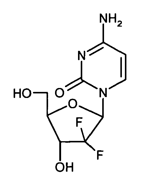
- Several novel aspects can be derived from the results of the study of the invention. The following statements are supported by the examples of the invention.
- In extension to previous reports, we observed selective overexpression of ED-B FN in human pancreatic carcinoma by immunohistochemistry. By contrast, no ED-B FN was detectable in human non-transformed pancreas and chronic pancreatitis. In analogy to the human situation, a qualitative and quantitative comparable ED-B FN expression pattern was confirmed in primary pancreatic tumors as well as in lymph node and liver metastases of the orthotopic mouse models investigated in this study.
- Using these mouse models as clinically representative in vivo models, we demonstrated that administration of L19-IL2 efficiently inhibited the growth of established primary pancreatic tumors and reduced or eliminated lymph node metastases, while untargeted IL2 elicited minor or no therapeutic effects. Of note, the anti-tumor activity of L19-IL2 was exclusively mediated by IL2, as shown by application of the dimeric scFv L19 antibody fragment alone, which failed to achieve any therapeutic benefit. The modest performance of untargeted IL2 is not surprising, considering the observation that most anticancer agents feature tumor/organ ratios <1, in some cases as low as 1:20 after intravenous injection. (Bosslet K, Straub R, Blumrich M, Czech J, Gerken M, Sperker B, Kroemer HK, Gesson JP, Koch M, Monneret C. Elucidation of the mechanism enabling tumor selective prodrug monotherapy. Cancer Res 1998;58:1195-1201.)
- Although L19 mediated delivery of IL2 to tumor microenvironment has previously been verified to enhance the therapeutic performance of this cytokine, using subcutaneous mouse models of teratocarcinoma, small cell lung cancer, and colon adenocarcinoma, (Carnemolla B, Borsi L, Balza E, Castellani P, Meazza R, Berndt A, Ferrini S, Kosmehl H, Neri D, Zardi L. Enhancement of the antitumor properties of interleukin-2 by its targeted delivery to the tumor blood vessel extracellular matrix. Blood 2002;99:1659-1665.) this is the first study to prove the therapeutic efficacy of L19-IL2 in the context of pancreatic carcinoma. Moreover, we for the first time demonstrated the therapeutic potency of L19-IL2 to control metastatic disease, using a pathophysiologically relevant orthotopic mouse model of pancreatic cancer. It is noteworthy that the growth inhibitory effect of L19-IL2 was not cell-type specific, but could be established in two different mouse models of pancreatic cancer, notwithstanding their different quantitative level of ED-B expression.
- Furthermore, our data indicate that L19-IL2 alone can inhibit pancreatic cancer growth to a greater degree than gemcitabine, a standard first-line chemotherapeutic agent available for the treatment of human pancreatic cancer. Indeed, two applications of L19-IL2 were sufficient to reduce the tumor load of mice bearing pancreatic cancer to only 25% of that observed in controls. The clinical promise and scope of L19-IL2 therapy were shown by the induction of complete remission and the lack of tumor relapse in 40% of long-term surviving mice bearing pancreatic cancer. However, the most dramatic therapeutic results were achieved by treating tumor bearing mice with a combinatorial therapy of L19-IL2 plus gemcitabine, producing a 97% inhibition of primary pancreatic tumor growth, including 70% complete remissions.
- The mechanisms underlying the anti-tumor activity of IL2 are still not fully understood. To address this issue, we performed histological investigations. One of the most intriguing features of L19-IL2 treated tumors was extensive tumor necrosis. The degree of necrosis and its location in the central part of tumor tissue where only a scanty leucocyte infiltrate was observed suggest rather other underlying mechanisms than individual tumor cell death caused by direct cell to cell contact by NK cells, macrophages or other immune effector cells. Indeed, recent evidence accumulates that IL2 dependent induction of massive tumor necrosis following local IL2 administration is due to local vascular leak syndrome, which may be provoked by either lymphokines and nitric oxide produced by IL2 activated immune effector cells (De Mik HJ, Koten JW, Maas RA, Dullens HF, Den Otter W. Tumour regression by IL-2 mediated stagnation of blood flow. In Vivo 1991;5:679-684. Further: Sakkoula E, Pipili-Synetos E, Maragoudakis ME. Involvement of nitric oxide in the inhibition of angiogenesis by interleukin-2. Br J Pharmacol 1997;122:793-795) and / or by direct cytotoxic effects of IL2 (Baluna R, Rizo J, Gordon BE, Ghetie V, Vitetta ES. Evidence for a structural motif in toxins and interleukin-2 that may be responsible for binding to endothelial cells and initiating vascular leak syndrome. Proc Natl Acad Sci U S A 1999;96:3957-3962.) and of activated NK cells on endothelium. ( Albertsson PA, Basse PH, Hokland M, Goldfarb RH, Nagelkerke JF, Nannmark U, Kuppen PJ. NK cells and the tumour microenvironment: implications for NK-cell function and anti-tumour activity. Trends Immunol 2003;24:603-609. Further: Di Carlo E, Meazza R, Basso S, Rosso O, Comes A, Gaggero A, Musiani P, Santi L, Ferrini S. Dissimilar anti-tumour reactions induced by tumour cells engineered with the interleukin-2 or interleukin-15 gene in nude mice. J Pathol 2000;191:193-201.) Considering, that endothelial cells migrate along ECM structures containing ED-B FN (Tarli L, Balza E, Viti F, Borsi L, Castellani P, Berndorff D, Dinkelborg L, Neri D, Zardi L. A high-affinity human antibody that targets tumoral blood vessels. Blood 1999;94:192-198.) and that IL2 is selectively delivered to the ECM by linking it to scFv L19, contribution of direct cytotoxic effects of IL2 on endothelial cells to IL2 dependent tumor necrosis and regression offers an extremely attractive hypothesis. Noteworthy, while IL2 has been reported to exert cytotoxicity towards endothelial cells, this cytokine has no direct impact on cancer cells, which can grow unimpeded in vitro in high concentrations of IL2. (Rosenberg SA. Progress in the development of immunotherapy for the treatment of patients with cancer. J Intern Med 2001;250:462-475.) Nevertheless, tumor samples of mice treated with L19-IL2 revealed a considerable decrease in the number of proliferating cells as shown by immunohistochemical analysis for Ki67 expression.
- Regarding potential immune effector cells mediating L19-IL2 induced tumor regression, we observed a L19-IL2 triggered fourfold increase in the number of tumor infiltrating macrophages via immunohistochemistry. This observation is in agreement with the notion that macrophages respond to IL2 stimulation with tumoridical activity and cytokine and growth factor production. Moreover, it has been established that depletion of macrophages attenuates the therapeutic efficacy of IL2. (Masztalerz A, Van Rooijen N, Den Otter W, Everse LA. Mechanisms of macrophage cytotoxicity in IL-2 and IL-12 mediated tumour regression. Cancer Immunol Immunother 2003;52:235-242.) However, macrophage-depletion was not capable of completely abrogating the anti-tumor activity of IL2, indicating the contribution of additional mechanisms. In accordance with this concept, a strongly (70fold) enhanced presence of NK cells was observed in L19-IL2 treated tumors in our study. The functional significance of NK cell infiltration into tumor tissues for IL2 triggered anti-tumor response was corroborated by the observation that NK cell depletion completely abolished the therapeutic effect of L19-IL2 against pancreatic cancer. Moreover, our data are supported by the following lines of evidence:
- (i) Promising anti-tumor effects in addition to extravasation and infiltration of tumor tissues by NK cells have been demonstrated in vitro and in vivo, 40 (ii) NK cells have been considered the predominantly stimulated leukocytes subpopulation by recombinant IL2 in preclinical and clinical settings. (Basse PH, Whiteside TL, Herberman RB. Use of activated natural killer cells for tumor immunotherapy in mouse and human. Methods Mol Biol 2000;121:81-94. Further: Janssen RA, Sleijfer DT, Heijn AA, Mulder NH, The TH, de Leij L. Peripheral blood lymphocyte number and phenotype prior to therapy correlate with response in subcutaneously applied rIL-2 therapy of renal cell carcinoma. Br J Cancer 1992;66:1177-1179.)
- In summary, we have established the recombinant fusion protein L19-IL2 as a novel, highly efficient and specific anticancer agent for the treatment of pancreatic cancer in orthotopic nude mouse models and characterized underlying immune- and nonimmune-mediated mechanisms. Finally, our preclinical data strongly support the initiation of clinical studies using the immunocytokine L19-IL2 in pancreatic cancer.
- Preferred is the use of a fusion protein according to the invention, which fusion protein has an N - terminal antibody - part and C - terminal Interleukin-2 part or wherein the fusion protein has an N - terminal Interleukin-2 part and an C - terminal antibody - part.
- Further, preferred is the use of the fusion protein, which fusion protein has linker connecting the antibody - part and the Interleukin-2 part.
- Preferred is the use of the fusion protein according to the invention, wherein the antibody - part specifically binds to the EDb oncofoetal fibronectin domain.
- Preferred is the use of the fusion protein of the invention, wherein the antibody has a sequence of the heavy chain of Seq. Id. No. 01 and / or a sequence of the light chain of Seq. Id. No. 02.
- Preferred is a the use of a fusion protein according to the invention, wherein the heavy and the light chain are connected by an antibody linker.
- Preferred is the use of the fusion protein according to the invention, wherein the antibody linker has a sequence of Seq. Id. No. 03
- Preferred is the use of the fusion protein according to the invention, wherein the Interleukin-2 part has a sequence according to the Seq. Id No 04.
- Preferred is the use of the fusion protein according to the invention, wherein the fusion protein linker has a sequence according to the Seq.
Id No 05. - Preferred is the use of the fusion protein according to the invention as a medicament.
- More preferred is the use of the fusion protein according to the invention as a medicament for treatment of cancer, especially pancreatic cancer.
- Preferred is the use of the fusion protein in combination with further substances used in cancer therapy.
- Preferred is a combination according to the invention,
wherein the fusion protein has an N - terminal antibody - part and C - terminal Interleukin-2 part or
wherein the fusion protein has an N - terminal Interleukin-2 part and an C - terminal antibody - part. - Further, preferred is a combination of the invention, wherein the fusion protein has a fusion protein linker connecting the antibody - part and the Interleukin-2 part.
- Preferred is a combination according to the invention, wherein the antibody - part specifically binds to the EDb oncofoetal fibronectin domain.
- Preferred is a combination of the invention, wherein the antibody has a sequence of the heavy chain of Seq. Id. No. 01 and / or a sequence of the light chain of Seq. Id. No. 02.
- Preferred is a combination according to the invention, wherein the heavy and the light chain are connected by an antibody linker.
- Preferred is a combination according to the invention, wherein the antibody linker has a sequence of Seq. Id. No. 03
- Preferred is a combination according to the invention, wherein the Interleukin-2 part has a sequence according to the Seq. Id No 04.
- Preferred is a combination according to the invention, wherein the fusion protein linker has a sequence according to the Seq.
ld No 05. - Preferred is a combination according to the invention as a medicament.
- More preferred is a combination according to the invention as a medicament for treatment of cancer.
- Much more preferred is a combination according to the invention, wherein the cancer is non-small cell lung cancer, head and neck cancer, and breast cancer, preferably pancreatic cancer.
- Preferred are combinations with further substances used in cancer therapy.
- A combination is a pharmaceutical composition comprising at least two pharmaceutically effective compounds. These compounds can be administered simultaneously or one after the other with a time gap of 1 minutes to 1 day. The compounds can be administered via the same rout or in different manners. So the parallel oral and parenteral administration are possible.
- A composition comprises at least one pharmaceutically effective compound and further pharmaceutically acceptable diluents and carrier.
- A fusion protein is characterised by a protein comprising different amino acid sequences which are defined by the origin and / or by special functions.
- The ED-B domain of fibronectin, a sequence of 91 amino acids identical in mice, rats and humans, which is inserted by alternative splicing into the fibronectin molecule, specifically accumulates around neovascular structures and represents a target for molecular intervention (Zardi et al. (1987) EMBO J. Vol.: 6, pages 2337 - 2342, further Carnemolla et al. (1989) J. Cell Biol. Vol: 108, pages 1139 - 1148, further Castellani et al, (1994) Int. J. Cancer, Vol. 59, pages 612 - 618). Using a human recombinant antibody (L19) to the ED-B domain the possibility of in vivo neovasculature targeting has been demonstrated in different tumour models (Tarli et al. (1999) Blood, Vol.: 94, pages 192 - 198, further Viti et al. (1999) Cancer Res. Vol.: 347).
- Antibody linker are described in
EP 0 573 551EP 0 623679EP 0 318554 - Fusion protein linker are described in
EP 0 573 551EP 0 623679EP 0 318554 - Interleukin-2 (IL-2), a four alpha helix bundle cytokine produced by T helper 1 cells, plays an essential role in the activation phases of both specific and natural immune responses (Taniguchi et al. (1993) Cell, Vol.: 73, pages 5 - 8). IL-2 promotes proliferation and differentiation of activated T and B lymphocytes and of natural killer (NK) cells, and induces cytotoxic T cell (CTL) activity and NK / lymphokine activated killer (LAK) anti - tumour cytotoxicity.
- IL-2 has been used in immunotherapy approaches of several human tumours (Rosenberg (1992) J. Clin. Oncol. Vol.:10, pages 180 - 199). Administration of recombinant IL-2 (rIL2) alone or in combination with adoptively transferred lymphoid cells has resulted in the regression of established tumours in both animal models and patients. However, its in vivo therapeutic efficacy is limited by its rapid clearance and, at high doses, by a severe toxicity mainly related to a vascular leak syndrome (Siegel and Puri (1991) Interleukin-2 toxicity in J. Clin. Oncol. Vol.: 9 pages 694 - 704). Delivery of IL-2 to the tumour site by means of an antibody directed against a cell-surface tumour marker may allow achievement of active local concentrations of IL-2, as well as reducing toxicities associated to systemic administration (Lode et al. (1998) Pharmacol. Ther. Vol.: 80, pages 277 - 292).
- Gemcitabine: The
EP 0 122 707 B1 (filed 06.03.1984EP 0 184 365 B1 (filed 25.11.1985US 4,526,988 ;US 4,808,614 ;US 5,223,608 ;EP 0 184 365EP 0 577,303EP 0 577,304EP 0 587 364WO 96 / 18637 (filed 14.11.1995 EP 0 889 275 (filed 09.12.1996 - Gemcitabine has the formula

- Further substances used in cancer therapy are described in Exemplary cytotoxins include, in general, cytostatic agents, alkylating agents, antimetabolites, anti-proliferative agents, tubulin binding agents, hormones and hormone antagonists, and the like. Exemplary cytostatics that are compatible with the present invention include alkylating substances, such as mechlorethamine, triethylenephosphoride,
- "Antibody-dependent cell-mediated cytotoxicity" and "ADCC" refers to a cell-mediated reaction in which non-specific cytotoxic cells that express Fc receptors (FcRs) (e.g. Natural Killer (NK) cells, neutrophils, and macrophages) recognise bound antibody on a target cell and subsequently cause lysis of the target cell. The primary cells for mediating ADCC, NK cells, express FcyRIII only, whereas monocytes express FcγRI, FcγRII and FcγRIII. FcR expression on haematopoietic cells in summarized is Table 3 on Page 464 of Ravetch and Kinet, Annu. Rev. Immunol9:457-92 (1991 ). To assess ADCC activity of a molecule of interest, an in vitro ADCC assay, such as that described in
US Patent No. 5,500,362 or5,821,337 may be performed. Useful effector cells for such assays include peripheral blood mononuclear cells (PBMC) and Natural Killer (NK) cells. Alternatively, or additionally, ADCC activity of the molecule of interest may be assessed in vivo, e.g., in a animal model such as that disclosed in Clynes et al. PNAS (USA) 95:652-656 (1998 ). - "Human effector cells" are leukocytes which express one or more FcRs and perform effector functions. Preferably, the cells express at least FcγRIII and carry out ADCC effector function. Examples of human leukocytes which mediate ADCC include peripheral blood mononuclear cells (PBMC), natural killer (NK) cells, monocytes, cytotoxic T cells and neutrophils; with PBMCs and NK cells being preferred.
- The terms "Fc receptor" or "FcR" are used to describe a receptor that binds to the Fc region of an antibody. The preferred FcR is a native sequence human FcR. Moreover, a preferred FcR is one which binds an IgG antibody (a gamma receptor) and includes receptors of the FcγRI, FcγRII, and FcγRIII subclasses, including allelic variants and alternatively spliced forms of these receptors. FcγRII receptors include FcγRIIA (an "activating receptor") and FcγRIIB (an "inhibiting receptor"), which have similar amino acid sequences that differ primarily in the cytoplasmic domains thereof. Activating receptor FcγRIIA contains an immunoreceptor tyrosine-based activation motif (ITAM) in its cytoplasmic domain. Inhibiting receptor FcγRIIB contains an immunoreceptor tyrosine-based inhibition motif (ITIM) in its cytoplasmic domain. (See Daeron, Annu. Rev. Immunol. 15:203-234 (1997) ). FcRs are reviewed in Ravetch and Kinet, Annu. Rev. Immunol9:457-92 (1991 ); Cape1 et al., Immunomethods 4:25-34 ( 1994 ); and de Haas et al., J. Lab. Clin. Med. 126:330-41 (1995). Other FcRs, including those to be identified in the future, are encompassed by the term "FcR" herein. The term also includes the neonatal receptor, FcRn, which is responsible for the transfer of maternal IgGs to the foetus (Guyer et al., J. Immunol. 1 1 7587 (1976 ) and Kim et al., J. Immunol. 24:249 (1994 )).
- Orientation of the fusion protein: A combination according to the invention,
wherein the fusion protein has an N - terminal antibody - part and C - terminal Interleukin-2 part or
wherein the fusion protein has an N - terminal Interleukin-2 part and an C - terminal antibody - part. - Antibody - part comprises: antibodies, antibody fragments, native antibodies, Fab - fragments, "Fv", Single-chainFv" or' scFv " antibody fragments; monoclonal antibody; monoclonal antibodies" isolated from phage antibody libraries; chimeric antibodies; humanised antibodies; an antagonist "which binds" an antigen of interest; antibody fragments isolated from antibody phage libraries, human antibodies,
- The term "antibody" herein is used in the broadest sense and specifically covers intact monoclonal antibodies, polyclonal antibodies, multispecific antibodies (e.g. bispecific antibodies) formed from at least two intact antibodies, and antibody fragments so long as they exhibit the desired biological activity.
- "Antibody fragments" comprise a Portion of an intact antibody, preferably comprising the antigen-binding or variable region thereof. Examples of antibody fragments include Fab, Fab', F(ab')2 and Fv fragments; diabodies; linear antibodies; single-chain antibody molecules; and multispecific antibodies formed from antibody fragments.
- "Native antibodies" are usually heterotetrameric glycoproteins of about 150,000 daltons, composed of two identical light (L) chains and two identical heavy (H) chains. Each light chain is linked to a heavy chain by one covalent disulfide bond, while the number of disulfide linkages varies among the heavy chains of different immunoglobulin isotypes. Each heavy and light chain also has regularly spaced intrachain disulfide bridges. Each heavy chain has at one end a variable domain (VH) followed by a number of constant domains. Each light chain has a variable domain at one end (VL) and a constant domain at its other end; the constant domain of the light chain is aligned with the first constant domain of the heavy chain, and the light-chain variable domain is aligned with the variable domain of the heavy chain. Particular amino acid residues are believed to form an interface between the light chain and heavy chain variable domains.
- The term "variable" refers to the fact that certain Portions of the variable domains differ extensively in sequence among antibodies and are used in the binding and specificity of each particular antibody for its particular antigen. However, the variability is not evenly distributed throughout the variable domains of antibodies. It is concentrated in three segments called hypervariable regions both in the light chain and the heavy chain variable domains. The more highly conserved portions of variable domains are called the framework regions (FRs). The variable domains of native heavy and light chains each comprise four FRs, largely adopting a β-sheet configuration, connected by three hypervariable regions, which form loops connecting, and in some cases forming part of, the β-sheet structure. The hypervariable regions in each chain are held together in close proximity by the FRs and, with the hypervariable regions from the other chain, contribute to the formation of the antigen-binding site of antibodies (See Kabat et al., Sequences of Proteins Immunological Interest, 5th Ed. Public Health Service, National Institutes of Health, Bethesda, MD. (1991)). The constant domains are not involved directly in binding an antibody to an antigen, but exhibit various effector functions, such as participation of the antibody in antibody dependent cellular cytotoxicity (ADCC).
- Papain digestion of antibodies produces two identical antigen-binding fragments, called "Fab" fragments, each with a Single antigen-binding site, and a residual "Fc" fragment, whose name reflects its ability to crystallize readily. Pepsin treatment yields an F(ab')2 fragment that has two antigen-binding sites and is still capable of cross-linking antigen.
- "Fv" is the minimum antibody fragment which contains a complete antigen-recognition and antigen-binding site. This region consists of a dimer of one heavy chain and one light chain variable domain in tight, non-covalent association. It is in this configuration that the three hypervariable regions of each variable domain interact to define an antigen-binding site on the surface of the VH-VL dimer. Collectively, the six hypervariable regions confer antigen-binding specificity to the antibody. However, even a single variable domain (or half of an Fv comprising only three hypervariable regions specific for an antigen) has the ability to recognize and bind antigen, although at a lower affinity than the entire binding site.
- The Fab fragment also contains the constant domain of the light chain and the first constant domain (CH1) of the heavy chain. Fab' fragments differ from Fab fragments by the addition of a few residues at the carboxy terminus of the heavy chain CH1 domain including one or more cysteines from the antibody hinge region. Fab'-SH is the designation herein for Fab' in which the cysteine residue(s) of the constant domains bear at least one free thiol group. F(ab'), antibody fragments originally were produced as pairs of Fab'2-fragments which have hinge cysteines between them. Other chemical couplings of antibody fragments are also known.
- The "light chains" of antibodies (immunoglobulins) from any vertebrate species can be assigned to one of two clearly distinct types, called kappa (κ) and lambda (λ), based on the amino acid sequences of their constant domains.
- Depending on the amino acid sequence of the constant domain of their heavy chains, antibodies can be assigned to different classes. There are five major classes of intact antibodies: IgA, IgD, IgE, IgG, and IgM, and several of these may be further divided into subclasses (isotypes), e.g., IgG1, IgG2, IgG3, IgG4, IgA, and IgA2. The heavy-chain constant domains that correspond to the different classes of antibodies are called α, δ, ε, γ, and µ respectively. The subunit structures and three-dimensional configurations of different classes of immunoglobulins are well known.
- " Single-chainFv" or' scFv " antibody fragments comprise the VH and VL domains of antibody, wherein these domains are present in a Single Polypeptide chain. Preferably, the Fv Polypeptide further comprises a Polypeptide linker between the VH and VL, domains which enables the scFv to form the desired structure for antigen binding. For a review of scFv See Plückthun in The Pharmacology of Monoclonal Antibodies, vol. 113, Rosenburg and Moore eds., Springer-Verlag, New York, pp. 269-3 15 (1994).
- In a further embodiment, antibodies or antibody fragments can be isolated from antibody phage libraries generated using the techniques described in McCafferty et al., Nature, 348552-554 (1990). Clackson et al., Nature, 352:624-628 (1991) and Marks et al., J. Mol. Biol., 222581-597 (1991) describe the isolation of murine and human antibodies, respectively, using phage libraries. Subsequent publications describe the production of high affinity (mM range) human antibodies by chain shuffling (Marks et al., Bio/Technology, 10:779-783 (1992)), as well as combinatorial infection and in vivo recombination as a strategy for constructing very large phage libraries (Waterhouse et al., Nuc. Acids. Res., 21:2265-2266 (1993)). Thus, these techniques are viable alternatives to traditional monoclonal antibody hybridoma techniques for isolation of monoclonal antibodies. The DNA also may be modified, for example, by substituting the coding sequence for human heavy- and light-chain constant domains in place of the homologous murine sequences (
U.S. Patent No. 4,8 16,567 ; Morrison, et al., Proc. Nat. Acad. Sci. USA, 8 1 :685 1 (1984)), or by covalently joining to the immunoglobulin coding sequence all or part of the coding sequence for a non-immunoglobulin polypeptide. Typically such non-immunoglobulin polypeptides are substituted for the constant domains of an antibody, or they are substituted for the variable domains of one antigen-combining site of an antibody to create a chimeric bivalent antibody comprising one antigen-combining site having specificity for an antigen and another antigen-combining site having specificity for a different antigen. - The term "diabodies" refers to small antibody fragments with two antigen-binding sites, which fragments comprise a heavy-chain variable domain (VH) connected to a light-chain variable domain (VL) in the same polypeptide chain (VH - VL). By using a linker that is too short to allow pairing between the two domains on the same chain, the domains are forced to pair with the complementary domains of another chain and create two antigen-binding sites. Diabodies are described more fully in, for example,
EP 0 404 097WO 93/11161 - The term "monoclonal antibody" as used herein refers to an antibody obtained from a population of substantially homogeneous antibodies, i. e., the individual antibodies comprising the population are identical except for possible naturally occurring mutations that may be present in minor amounts. Monoclonal antibodies are highly specific, being directed against a Single antigenic site. Furthermore, in contrast to conventional (polyclonal) antibody preparations which typically include different antibodies directed against different determinants (epitopes), each monoclonal antibody is directed against a single determinant on the antigen. In addition to their specificity, the monoclonal antibodies are advantageous in that they are synthesized by the hybridoma culture, uncontaminated by other immunoglobulins. The modifier "monoclonal" indicates the character of the antibody as being obtained from a substantially homogeneous population of antibodies, and is not to be construed as sequiring production of the antibody by any particular method. For example, the monoclonal antibodies to be used in accordance with the present invention may be made by the hybridoma method first described by Kohler et al., Nature, 256: 495 (1975), or may be made by recombinant DNA methods (See, e.g.,
U.S. Patent No. 4,816,567 ). - The "monoclonal antibodies" may also be isolated from phage antibody libraries using the techniques described in Clackson et al., Nature, 352:624-628 (1991) and Marks et al., J. Mol. Biol., 222581-597 (1991), for example.
- The monoclonal antibodies herein specifically include "chimeric" antibodies (immunoglobulins) in which a portion of the heavy and/or light chain is identical with or homologous to corresponding sequences in antibodies derived from a particular species or belonging to a particular antibody class or subclass, while the remainder of the chain(s) is identical with or homologous to corresponding sequences in antibodies derived from another species or belonging to another antibody class or subclass, as well as fragments of such antibodies, so long as they exhibit the desired biological activity (
U.S. Patent No. 4,s 16,567 - Chimeric antibodies of interest herein include "primatised" antibodies comprising variable domain antigen-binding sequences derived from a non-human primate (e.g. Old World Monkey, such as baboon, rhesus or cynomolgus monkey) and human constant region sequences (
US Pat No. 5,693,780 ). - Humanised antibodies: Methods for humanising non-human antibodies have been described in the art. Preferably, a humanised antibody has one or more amino acid residues introduced into it from a source which is non-human. These non-human amino acid residues are often referred to as "import" residues, which are typicality taken from an "import" variable domain. Humanisation can be essentially performed following the method of Winter and Co-workers (Jones et al., Nature, 321522-525 (1986); Riechmann et al., Nature, 332:323-327 (1988); Verhoeyen et al., Science,239: 1534- 1536 (1988)), by substituting hypervariable region sequences for the corresponding sequences of a human antibody. Accordingly, such "humanised" antibodies are chimeric antibodies (
U.S. Patent No. 4,8 16,567 ) wherein substantially less than an intact human variable domain has been substituted by the corresponding sequence from a non-human species. In practice, humanised antibodies are typically human antibodies in which some hypervariable region residues and possibly some FR residues are substituted by residues from analogous sites in rodent antibodies. The choice of human variable domains, both light and heavy, to be used in making the humanized antibodies is very important to reduce antigenicity. According to the so-called "best-fit" method, the sequence of the variable domain of a rodent antibody is screened against the entire library of known human variable-domain sequences. The human sequence which is closest to that of the rodent is then accepted as the human framework region (FR) for the humanised antibody (Sims et al., J. Immunol., 151:2296 (1993); Chothia et al., J. Mol. Biol., 196:901 (1987)). Another method uses a particular framework region derived from the consensus sequence of all human antibodies of a particular subgroup of light or heavy chains. The Same framework may be used for several different humanised antibodies (Carter et al., Proc. Natl. Acad. Sci. USA, 89:4285 (1992);Presta et al., J. Immunol., 151:2623 (1993)). It is further important that antibodies be humanized with retention of high affinity for the antigen and other favourable biological properties. To achieve this goal, according to a preferred method, humanised antibodies are prepared by a process of analysis of the parental sequences and various conceptual humanised products using three - dimensional models of the parental and humanized sequences. Three - dimensional immunoglobulin models are commonly available and are familiar to those skilled in the art. Computer programs are available which illustrate and display probable three-dimensional conformational structures of selected candidate immunoglobulin sequences. - Inspection of these displays permits analysis of the likely role of the residues in the functioning of the candidate immunoglobulin sequence, i. e., the analysis of residues that influence the ability of the candidate immunoglobulin to bind its antigen. In this way, FR residues can be selected and combined from the recipient and import sequences so that the desired antibody characteristic, such as increased affinity for the target antigen(s), is achieved. In general, the hypervariable region residues are directly and most substantially involved in influencing antigen binding.
- Human antibodies: As an alternative to humanisation, human antibodies can be generated. For example, it is now possible to produce transgenic animals (e.g., mice) that are capable, upon immunisation, of producing a full repertoire of human antibodies in the absence of endogenous immunoglobulin production. For example, it has been described that the homozygous deletion of the antibody heavy-chain joining region (JH) gene in chimeric and germ-line mutant mice results in complete inhibition of endogenous antibody production. Transfer of the human germ-line immunoglobulin gene array in such germ-line mutant mice will result in the production of human antibodies upon antigen challenge. See, e.g., Jakobovits et al., Proc. Natl. Acad. Sci. USA, 90:2551 (1993); Jakobovits et al., Nature, 362:255-258 (1993); Bruggermann et al., Year in Immuno., 7:33 (1993); and
US Patent Nos. 5,591,669 ,5,589,369 and5,545,807 . - Alternatively, phage display technology (McCafferty et al., Nature 348:552-553 (1990)) can be used to produce human antibodies and antibody fragments in vitro, from immunoglobulin variable (V) domain gene repertoires from unimmunized donors. According to this technique, antibody V domain genes are cloned in-frame into either a major or minor coat protein gene of a filamentous bacteriophage, such as M13 or fd, and displayed as functional antibody fragments on the surface of the phage particle. Because the filamentous particle contains a single-stranded DNA copy of the phage genome, selections based on the functional properties of the antibody also result in selection of the gene encoding the antibody exhibiting those properties. Thus, the phage mimics some of the properties of the B cell. Phage display can be performed in a variety of formats; for their review See, e.g., Johnson, Kevin S. and Chiswell, David J., Current Opinion in Structural Biology 3:564-571 (1993). Several sources of V-gene Segments can be used for phage display. Clackson et al., Nature, 352:624-628 (1991) isolated a diverse array of anti-oxazolone antibodies from a small random combinatorial library of V genes derived from the Spleens of immunized mice. A repertoire of V genes from unimmunized human donors can be constructed and antibodies to a diverse array of antigens (including self-antigens) can be isolated essentially following the techniques described by Marks et al., J. Mol. Biol. 222581-597 (1991), or Griffith et al., EMBO J. 12:725-734 (1993). See, also,
US Patent Nos. 5,565,332 and5,573,905 . Human antibodies may also be generated by in vitro activated B cells (seeUS Patents 5,567,610 and5,229,275 ). - The term "hypervariable region" when used herein refers to the amino acid residues of an antibody which are responsible for antigen-binding. The hypervariable region comprises amino acid residues from a "complementarily determining region" or "CDR" (e.g. residues 24-34 (L1), 50-56 (L2) and 89-97 (L3) in the light chain variable domain and 31-35 (H1), 50-65 (H2) and 95-102 (H3) in the heavy chain variable domain; Kabat et al., Sequences of Proteins of Immunological Interest, 5th Ed. Public Health Service, National Institutes of Health, Bethesda, MD. (1991)) and/or those residues from a "hypervariable loop" (e.g. residues 26-32 (L1), 50-52 (L2) and 91-96 (L3) in the light chain variable domain and 26-32 (H1), 53-55 (H2) and 96-101 (H3) in the heavy chain variable domain; Chothia and Lesk J. Mol. Biol. 196:901-917 (1987)). "Framework" or "FR" residues are those variable domain residues other than the hypervariable region residues as herein defined.
- Antibody fragments: Various techniques have been developed for the production of antibody fragments. Traditionally, these fragments were derived via proteolytic digestion of intact antibodies (See, e.g., Morimoto et al., Journal of Biochemical und Biophysical Methods 24: 107-117 (1992) and Brennan et al., Science, 229:81 (1985)). However, these fragments can now be produced directly by recombinant host cells. For example, the antibody fragments can be isolated from the antibody phage libraries discussed above. Alternatively, Fab'-SH fragments can be directly recovered from E. coli and chemically coupled to form F(ab')2 fragments (Carter et al., Biotechnology 10: 163-167 (1992)). According to another approach, F(ab')2 fragments can be isolated directly from recombinant host cell culture. Other techniques for the production of antibody fragments will be apparent to the skilled practitioner. In other embodiments, the antibody of choice is a single chain Fv fragment (scFv). See
WO 93/16185 US Patent NO. 5,571,894 ; andUS Patent No. 5,587,458 . The antibody fragment may also be a "linear antibody", e.g., as described inUS Patent 5,641,870 for example. Such linear antibody fragments may be monospecific or bispecific. - Amino acid sequence modification(s) of protein or Peptide antagonists or antibody - part described herein are contemplated. For example, it may be desirable to improve the binding affinity and/or other biological properties of the antagonist. Amino acid sequence variants of the antagonist are prepared by introducing appropriate nucleotide changes into the antagonist nucleic acid, or by peptide synthesis. Such modifications include, for example, deletions from, and/or insertions into and or Substitutions of, residues within the amino acid sequences of the antagonist. Any combination of deletion, insertion, and substitution is made to arrive at the final construct, provided that the final construct possesses the desired characteristics. Therefore, in the case of the heavy of light chain a variation of 1; 2; 3; 4; 5; 6; 7; 8; 9; 10; 11; 12; 13; 14; 15; 16; 17; or 20 amino acids can be executed. In the case of Interleukin-2 a variation of 1; 2; 3; 4; 5; 6; 7; 8; 9; 10; 11; 12; 13; 14; 15; 16; 17; 20; 21; 22; 23; 24; 25; 26; 27; 28; 29; 30; 31; 32; or 33 amino acids can be executed. In the case of a linker a variation of 1; 2; 3; 4; 5; 6; or 7 can be executed. In case of the linker the variations are much more flexible, because function is simple to create a sufficient space between the functional amino acid sequences. A variation is defined as a deletion, insertion and/or substitution.
- The amino acid changes also may alter post-translational processes of the antagonist, such as changing the number or position of glycosylation sites. A useful method for identification of certain residues or regions of the antagonist that are preferred locations for mutagenesis is called "alanine scanning mutagenesis" as described by Cunningham and Welk Science, 244: 108 1-1085 (1989). Here, a residue or group of target residues are identified (e.g., charged residues such as Arg, Asp, His, Lys, and Glu) and replaced by a neutral or negatively charged amino acid (most preferably alanine or polyalanine) to affect the interaction of the amino acids with antigen. Those amino acid locations demonstrating functional sensitivity to the substitutions then are refined by introducing further or other variants at, or for, the sites of substitution. Thus, while the site for introducing an amino acid sequence variation is predetermined, the nature of the mutation per se need not be predetermined. For example, to analyze the Performance of a mutation at a given site, Ala scanning or random mutagenesis is conducted at the target codon or region and the expressed antagonist variants are screened for the desired activity. Amino acid sequence insertions include amino- and/or carboxyl-terminal fusions ranging in length from one residue to Polypeptides containing a hundred or more residues, as well as intrasequence insertions of single or multiple amino acid residues. Examples of terminal insertions include an antagonist with an N-terminal methionyl residue or the antagonist fused to a cytotoxic polypeptide. Other insertional variants of the antagonist molecule include the fusion to the N- or C-terminus of the antagonist of an enzyme, or a polypeptide which increases the serum half-life of the antagonist. Another type of variant is an amino acid substitution variant. These variants have at least one amino acid residue in the antagonist molecule replaced by different residue. The sites of greatest interest for substitutional mutagenesis of antibody antagonists include the hypervariable regions, but FR alterations are also contemplated. Conservative substitutions are shown in Table 1 under the heading of "preferred substitutions".
Table 1 Original Residue Exemplary substitution Preferred Substitution Ala Val; Leu; Ile Val Arg Lys, Gln, Asn Lys Asn Gln, His, Asp, Lys, Arg Gln Asp Glu, Asn Glu Cys Ser, Ala Ser Gln Asn, Glu Asn Glu Asp, Gln, Asp Gly Ala Ala His Asn, Gln, Lys, Arg Arg Ile Leu, Val, Met, Ala, Phe, Norleucine Leu Leu Norleucine, Ile, Val, Met, Ala, Phe Ile Lys Arg, Gln, Asn Arg Met Leu, Phe, Ile, Leu Phe Leu, Val, Ile, Ala, Tyr Tyr Pro Ala Ala Ser Thr Thr Thr Ser Ser Trp Tyr, Phe Tyr Tyr Trp, Phe, Thr, Ser Phe Val Ile, Leu, Met, Phe, Ala, Norleucine Leu - If such substitutions result in a Change in biological activity, then more substantial changes, denominated "exemplary substitutions" in Table 1, or as further described below in reference to amino acid classes, may be introduced and the products screened.
- Substantial modifications in the biological properties of the antagonist are accomplished by selecting substitutions that differ significantly in their effect on maintaining (a) the structure of the polypeptide backbone in the area of the substitution, for example, as a sheet or helical conformation, (b) the charge or hydrophobicity of the molecule at the target site, or (c) the bulk of the side chain.
- Naturally occurring residues are divided into groups based on common side-chain properties:
- (1) hydrophobic: Norleucine, Met, Ala, Val, Leu, Ile;
- (2) neutral hydrophilic: Cys, Ser, Thr;
- (3) acidic: Asp, Glu;
- (4) basic: Asn, Gln, His, Lys, Arg;
- ( 5) residues that influence chain orientation: Gly, Pro; and
- (6) aromatic: Trp, Tyr, Phe.
- Non-conservative substitutions will entail exchanging a member of one of these classes for another class. Any cysteine residue not involved in maintaining the proper conformation of the antagonist also may be substituted, generally with serine, to improve the oxidative stability of the molecule and prevent aberrant cross linking. Conversely, cysteine bond(s) may be added to the antagonist to improve its stability (particularly where the antagonist is an antibody fragment such as an Fv fragment).
- A particularly preferred type of substitutional variant involves substituting one or more hypervariable region residues of a parent antibody. Generally, the resulting variant(s) selected for further development will have improved biological properties relative to the parent antibody from which they are generated. A convenient way for generating such substitutional variants is affinity maturation using phage display. Briefly, several hypervariable region sites (e.g. 6-7 sites) are mutated to generate all possible amino substitutions at each site. The antibody variants thus generated are displayed in a monovalent fashion from filamentous phage particles as fusions to the gene III product of M13 packaged within each particle. The phage-displayed variants are then screened for their biological activity (e.g. binding affinity) as herein disclosed. In order to identify candidate hypervariable region sites for modification, alanine scanning mutagenesis can be performed to identify hypervariable region residues contributing significantly to antigen binding. Alternatively, or in additionally, it may be beneficial to analyse a crystal structure of the antigen-antibody complex to identify contact points between the antibody and antigen. Such contact residues and neighbouring residues are candidates for substitution according to the techniques elaborated herein. Once such variants are generated, the panel of variants is subjected to screening as described herein and antibodies with superior properties in one or more relevant assays may be selected for further development.
- Another type of amino acid variant of the antagonist alters the original glycosylation pattern of the antagonist. By altering is meant deleting one or more carbohydrate moieties found in the antagonist, and/or adding one or more glycosylation sites that are not present in the antagonist.
- Glycosylation of polypeptides is typically either N-linked or O-linked. N-linked refers to the attachment of the carbohydrate moiety to the side chain of an asparagine residue. The tri - peptide sequences asparagine-X-serine and asparagine-X-threonine, where X is any amino acid except proline, are the recognition sequences for enzymatic attachment of the carbohydrate moiety to the asparagine side chain. Thus, the presence of either of these tri - peptide sequences in a polypeptide creates a potential glycosylation site. O-linked glycosylation refers to the attachment of one of the sugars N-aceylgalactosamine, galactose, or xylose to a hydroxyamino acid, most commonly serine or threonine, although 5-hydroxyproline or 5-hydroxylysine may also be used.
- Addition of glycosylation sites to the antagonist is conveniently accomplished by altering the amino acid sequence such that it contains one or more of the above-described tri - peptide sequences (for N-linked glycosylation sites). The alteration may also be made by the addition of, or substitution by, one or more serine or threonine residues to the sequence of the original antagonist (for O-linked glycosylation sites).
- Nucleic acid molecules encoding amino acid sequence variants of the antagonist are prepared by a variety of methods known in the art. These methods include, but are not limited to, isolation from a natural source (in the case of naturally occurring amino acid sequence variants) or preparation by oligonucleotide-mediated (or site-directed) mutagenesis, PCR mutagenesis, and cassette mutagenesis of an earlier prepared variant or a non-variant version of the antagonist.
- It may be desirable to modify the antagonist of the invention with respect to effector function, e.g. so as to enhance antigen-dependent cell-mediated cyotoxicity (ADCC) and/or complement dependent cytotoxicity (CDC) of the antagonist. This may be achieved by introducing one or more amino acid substitutions in an Fc region of an antibody antagonist. Alternatively or additionally, cysteine residue(s) may be introduced in the Fc region, thereby allowing interchain disulfide bond formation in this region. The homodimeric antibody thus generated may have improved internalization capability and/or increased complement-mediated cell killing and antibody-dependent cellular cytotoxicity (ADCC). See Caron et al., J. Exp Med.176:1191-1195 (1992) and Shopes, B. J. Immunol.148:2918-2922 (1992). Homodimeric antibodies with enhanced anti-tumour activity may also be prepared using heterobifunctional cross-linkers as described in Wolff et al. Cancer Research53:2560-2565 (1993). Alternatively, an antibody can be engineered which has dual Fc regions and may thereby have enhanced complement lysis and ADCC capabilities. See Stevenson et al. Anti-Cancer Drug Design 3:219-230 (1989).
- The fusion protein L19-
IL 2 is expressively described in theWO 01 / 62298 WO 01 / 62298 page 8,line 12 refers to he L19 VH and L19 VL domain sequences described in Pini et al. (1998) J. Biol. Chem. Vol.: 273: 21769-21776. Pini et al. describes parts of the sequence of L19 in Table II on page 21772. Further,WO 01 / 62298 - Variations of the fusion protein L19-IL2 are defined by variations as mentioned before. That means the variations of special sequences, mentioned in the following list of Table 2, which can be modified be deletion, insertion and / or substitution by the following numbers of amino acids:
Table 2 Seq. Id No Type Numbers of amino acids, which are deleted, inserted and / or substituted: Seq. Id No 1 VH 1, 2 ,3, 4, 5, 6, 7, 8, 9, 10, 11, 12, 13,14,15,16,17,18, Seq. Id No 2VL 1, 2 ,3, 4, 5, 6, 7, 8, 9, 10, 11,12,13,14,15,16,17,18, Seq. Id No 3mAb linker 1, 2 ,3; 4,5, 6, 7, Seq. Id No 4IL2 1, 2, 3, 4, 5, 6, 7, 8, 9, 10, 11, 12, 13, 14, 15, 16 17, 18,19,20,21, 22,23,24,25, 26,27,28,29,30 Seq. Id No 5 Fusion protein linker 1, 2 ,3, 4, 5, 6, 7, Seq. Id No 1 VH 1, 2 ,3, 4, 5, 6, 7, 8, 9, 10, 11 - Further modifications are mentioned in Pini et al. (1998) J Biol. Chem., Vol.: 273, pages 21769 - 21776,
WO 02 / 20563 WO 2005/37312 WO 99 / 58570 - The combination of the invention exhibits pharmacological activity and is, therefore, useful as pharmaceuticals. In particular, the combination shows pharmacological activity in a number of pathological or disease states in connection with a cancer, especially pancreatic cancer.
- The combination of the invention is therefore indicated for use in treatment of non-small cell lung cancer, head and neck cancer, colon and breast cancer, especially pancreatic cancer. The antibodies of the combination of the invention may also be .employed, alone or in combination (simultaneously or in doses at different times).
- For the treatment of such conditions, the appropriated dosage will, of course, vary depending upon, for example, the host, the mode of administration and the nature and severity of the condition being treated.
- Concerning the fusion protein: However, an indicated daily dosage in man is in the range of about 10 - 1,000 µg/kg body weight, preferred 1 - 100 µg/kg body weight, more preferred 3 - 30 µg/kg body weight. Appropriate 20 doses of antibody are well known in the art. (J.A. Ledermann et al. (1991) Int. J. Cancer Vol.: 47 pages 659 - 664. The antibody must be given in a dose that does not cause toxicity of the most sensitive organ in the body (dose limiting organ).
- For antibodies the kidney might be the dose limiting organ.
- Specific binding members of the present invention, including those comprising an antibody antigen-binding domain, may be administered to a patient in need of treatment via any suitable route, usually by invention into the bloodstream an/or directly into the site to be treated, e.g. tumour. The precise dose will depend upon a number of factors, the route of treatment, the size and location of the area to be treated (e.g. tumour), the precise nature of the antibody (e.g. whole antibody, scFv molecule), and the nature of any detectable label or other molecule attached to the antibody. A typical antibody dose will be in the range 10-50 mg. This is a dose for a single treatment of an adult patient, which may be proportionally adjusted for children and infants, and also adjusted for other antibody formats in proportion to molecular weight. Treatments may be repeated at daily, twice-weekly, weekly or monthly intervals, at the discretion of the physician.
- The present invention also provides pharmaceutical compositions comprising the combination of the invention or one antibody (antibody, specifically recognising the EDb - fibronectin domain, or an antibody, specifically recognising the EDb fibronectin receptor, and of a second antibody recognising the EGF receptor) in association with at least one pharmaceutically acceptable carrier or diluent. Such composition may be manufactured in conventional manner. Pharmaceutically acceptable carriers or diluents are described in Remington's Pharmaceutical Science, 15th ed. Mack Publishing Company, Easton Pennsylvania (1980).
- Concerning the gemcitabine: The dosage in which the preparations according to this invention are administered will vary according to the mode of use and route of use, as well as to the requirements of the patient. In general a daily dosage for a systemic therapy for an adult average patient will be about 0.1 to 150 mg/kg body weight / day, preferably 1 to 40 mg/kg/day. For topical administration, an ointment, for instance, can contain from 0.1 to 10 % by weight of the pharmaceutical formulation, especially 0.5 to 5% by weight. If desired, the pharmaceutical preparation can contain an antioxidant, e.g. tocopherol, N-methyl - tocophermine, butylated hydrocyanisole, ascorbic acid or butylated hydroxytoluene.
- The treatment comprises therapeutical treatment.
- The invention discloses further
- (α) the use of the pharmaceutical composition of the combination (or L19-IL2) of the invention for manufacture of a medicament for treatment of cancer especially non-small cell lung cancer, head and neck cancer, colon and breast cancer, especially pancreatic cancer;
- (β) a method of treatment of cancer especially non-small cell lung cancer, head and neck cancer, colon and breast cancer, especially pancreatic cancer,
which method comprises an administration of a pharmaceutical composition of the combination (or L19-IL2) of the invention, wherein the amount of combination suppresses the disease and wherein the pharmaceutical composition of the combination is given to a patient, who is in need thereof; - (γ) a pharmaceutical composition of the combination of the invention for treatment of cancer especially non-small cell lung cancer, head and neck cancer, colon and breast cancer, especially pancreatic cancer,
which treatment comprises a pharmaceutical composition of the combination (or L19-IL2) of the invention and at least one pharmaceutically acceptable carrier or diluent. - The preferred method of administration is the injection of the combination, the parenteral administration.
- The following were purchased: human pancreatic carcinomca cell lines MiaPaca (American Type Culture Collection) and DanG and murine lymphoma cell line YAC-1 (Deutsche Sammlung von Mikroorganismen und Zellkulturen (DSMZ) GmbH, Braunschweig, Germany); Dulbecco's Eagle medium (DMEM), RPMI 1640 medium, PBS, Hanks' Balanced Salt Solution (HBSS), Antibiotic/Antimycotic Solution, glutamine and hygromycin B (Invitrogen, Berlin, Germany); fetal calf serum (FCS), trypsin/EDTA, penicillin, and streptomycin (Biochrom, Berlin, Germany); human fusion protein L19-IL2 and human homodimeric scFv antibody fragment L19 (Schering AG, Berlin, Germany); Ficoll-Paque Plus (Amersham, Braunschweig, Germany); collagenase Type 3 (Worthington, NJ, USA); AmpliTaq Gold DNA polymerase (Applied Biosystems, CA, USA); PCR Nucleotide Mix (Roche Diagnostics, Indianapolis, USA); TumorTACS in situ Apoptosis Detection Kit (R&D Systems GmbH, Wiesbaden-Nordenstadt, Germany); monoclonal mouse anti-human-pancytokeratin (C11) Ab (Santa Cruz Biotechnology, CA, USA); monoclonal mouse anti-human Ki67 Ab (Dianova, Hamburg, Germany); Biotin conjugated monoclonal mouse anti mouse NK 1.1 (PK136) Ab (Serotec, Düsseldorf Germany), monoclonal rat anti mouse CD11b (M1/70) Ab and R-PE conjugated monoclonal rat anti mouse CD49b (DX5) Ab (BDBiosciences, Heidelberg, Germany); rabbit anti asialo GM1 antiserum (ASGM1) (Wako Chemicals GmbH, Neuss, Germany); monoclonal mouse anti human CD31 (JC/70A) Ab, monoclonal mouse anti human CA 19-9 (1116-NS-19-9) Ab and AEC-Substrate-Chromogen. (Dako, Hamburg, Germany); Vectastain ABC kit (Vector Laboratories, Wertheim-Bettingen, Germany); Alamar Blue (BioSource, CA, USA); QlAamp DNA Mini Kit (Qiagen, Hilden, Germany); gemcitabine (Lilly, Gießen, Germany),
Rompun 2% (Bayer GmbH, Leverkusen, Germany); Ketavet (Pharmacia GmbH, Erlangen, Germany). All other chemicals and reagents were purchased from Sigma Chemical Co. (Deisenhofen, Germany). - 21 pancreatic carcinoma, 17 chronic pancreatitis and 13 normal pancreatic tissue samples were obtained from individuals who underwent surgical resection at the Department of Surgery at Charité University Hospital. This study was approved by the local ethics committee and all patients gave written informed consent prior to surgery.
- The human pancreatic carcinoma cell lines DAN-G and MiaPaca and the murine lymphoma cell line YAC-1 were cultured as previously described. 18,19 MiaPaca cells, stably transfected with an
Angiopoetin 2 DNA construct, were maintained as MiaPaca wild type cells, except for the addition of hygromycine B at a concentration of x µg/ml. - Female NMRI nude mice (age, approx. 10 weeks; weight 21-25 g) were purchased from Bomholtgard (Ry, Denmark). Animal care followed institutional guidelines and all experiments were approved by local animal research authorities.
- Three orthotopic xenograft mouse models of pancreatic carcinoma were established, including two non-metastatic models by injection of wild type cells of the human pancreatic carcinoma cell lines DanG and MiaPaca, and one metastatic model by implantation of MiaPaca cells stably transfected with an
Angiopoetin 2 DNA construct (MiaPaca-A2) (established by A. Scholz, unpublished data). Orthotopic transplantation was carried out as described. 20 In brief, mice were anesthetized by intraperitoneally (i.p.) administering a mixture of ketamine (100 mg/kg) and xylazine (10 mg/kg). After median laparotomy, the peritoneum was opened, and the pancreas was exposed. Aliquots of1x 106 tumor cells in a volume of 10µl PBS were injected with an insulin syringe into the head of the pancreas to visibly infiltrate the pancreatic tissue. The pancreas was replaced and the abdominal wall was closed in two layers using Vicryl 6-0 for the peritoneum and suture clips for the skin. Before initiation of therapy, 10 mice were killed to confirm the presence of tumor lesions. Mice were randomized and groups of 10-12 mice were intravenously (i.v.) treated with vehicle (0,9% saline), untargeted IL2, homodimeric scFv L19 or L19-IL2, consisting of IL2 conjugated to the homodimeric scFv L19 or i.p. administered with gemcitabine according to different protocols as described in the results section. All therapeutic agents were diluted in 0,9% saline and applied in a volume of 1µl/g BW. After termination of therapy, mice were killed, blood samples were collected and tumor volume was calculated using the formula length x width x depth x π/6. The area of lymph nodes of the metastatic MiaPac-A2 tumor model was calculated by determining the largest diameter and its perpendicular diameter and computing the product of the two measurements. In one experiment, mice were kept alive after completion of therapy to obtain long term data. At the end of therapy and biweekly thereafter, mouse blood samples were taken by retroorbital bleeding with heparinized blood collection capillaries to assay serum CA 19-9 level. Survival time of mice was followed untilday 100 after cessation of therapy (day 121 after tumor cell transplantation). Mice were considered to be cured if there was no detectable tumor atday 100. - NK cell depletion was performed as previously described. 21 Briefly, mice were injected i.p. with 50µl anti asialo-GM-1 (ASGM1)
Ab 3 days before tumor cell injection and every 4 days for a total of 6 injections. Quality of NK cell depletion was monitored by flow cytometry and cytotoxicity assay. - Mononuclear cells were prepared from the spleens of mice depleted for NK cells or not as described elsewhere. Briefly, spleens were removed under deep general anesthesia and digested with collagenase for 1 hour at 37°C. Subsequently, contents were forced through a 100 µm cell strainer and washed twice with HBSS. Mononuclear cells were separated from the cell suspension by running a Ficoll-Hypaque density gradient centrifugation.
- Cytotoxic activity of isolated murine spleen MNC against YAC-1 cells was examined using a 24h alamarBlue cytotoxicity assay as detailed previously. 22
- 1 x 10⁶ spleen MNC isolated from mice depleted for NK cells or not were incubated with 1 µg PE-labeled anti NK 1.1 antibody for 15 min at 4°C. After washing with PBS, the presence of fluorescence positive cells was analyzed by FAC-Scan utilizing Cellquest software (BectonDickensen; Heidelberg, Germany).
- Surgically resected human and murine tissue samples were snap-frozen in liquid nitrogen and after fixation in
PFA 4%,pH 7 for 20 minutes, 4 µm cryostat sections of the frozen tissue were analyzed immunohistochemically using the avidin-biotin-technique as described previously. Slides were incubated for 1 h with the following primary antibodies: biotinylated L19-IL2 (30µg/ml), monoclonal mouse anti-human CD31 at a 1:100 dilution, rat anti-mouse CD11b at a 1:50 dilution, biotinylated mouse anti-mouse CD161b/c/NK1.1 at a 1:50 dilution, biotinylated mouse anti-human pancytokeratin at a 1:50 dilution. Sections stained with PBS instead of the primary antibody were used as a negative control. Proliferating cells were detected with a monoclonal mouse anti human Ki67 Ab applying the Animal Research Kit. Apoptotic cells were proven by Terminal deoxynucleotidyl transferase mediated dUTP nick end labeling (TUNEL) assay using TumorTACS in situ Apoptosis Detection Kit. Both procedures were carried out following the manufacturer's directions except that 3-amino-9-ethylcarbazole (AEC) was used as the substrate-chromogen. Negative controls were included by omitting the primary antibody (Animal Research Kit) or the terminal deoxynucleotidyl transferase (TUNEL). Quantitative immunohistochemical analysis for the area of vital (non-necrotic) tumor tissue was carried out by computer aided imaging analysis using Axiovision 4.2 software (Zeiss, Jena, Germany). For quantification of proliferating, apoptotic and inflammatory cells, 20 randomly selected measurement areas (680 x 510 µm, magnification x 20) of each tumor specimen were analyzed and for evaluation of ED-B FN expression, 10 areas (1390x1000 µm, magnification x 10) were scanned. The results represent the positive staining as a percentage of measurement area (inflammatory cells, ED-B FN) or the ratio of positive tumor cells as a percentage of total tumor cells (Ki67, TUNEL), respectively. For quantitative analysis of necrotic tumor area H&E sections were scanned at low power (x 2,5). Total and necrotic tumor area was marked on the screen and areas were calculated by the image analysis system, and the ratio tumor necrosis area/total tumor area was given in percentages. - Genomic DNA of mouse lymph nodes was extracted using the QlAamp DNA Mini Kit according to the manufacturer's recommended protocol. The presence of human specific DNA was proven by polymerase chain reaction (PCR) amplifying an 850 bp fragment of the α-satellite region of the human chromosome 17 using the forward primer 5'-gggataatttcagctgactaacacg-3' and the reverse primer 5'-ttccgtttagttaggtgcagttatc-3' according to the protocol reported by Becker et al. 23
- Mouse blood samples were obtained by retroorbital bleeding or decapitation. Samples were centrifuged and supernatants were stored at -20°C. Serum CA 19-9 levels were measured by a fully automated chemiluminescence immunoassay on an ADVIA Centaur (Bayer, Leverkusen, Germany) and Lipase levels were determined using a kinetic colorimetric assay on an ADVIA 2400 (Bayer, Leverkusen, Germany).
- Statistical differences were evaluated by two-sided Mann-Whitney U-test and Fishers exact test and correlation was assessed by linear regression using GraphPad statistical software (GraphPad Software Inc., San Diego, CA). Differences were considered statistically significant at p<0.05.
- To evaluate the postulated role of ED-B FN as a suitable target for the selective delivery of IL2 to pancreatic cancer, we initially studied the expression pattern of ED-B FN in human non-transformed pancreatic tissue, chronic pancreatitis, and ductal adenocarcinoma. In parallel, serial sections from the same tissues were stained with a monoclonal antibody against the endothelial cell specific antigen CD31. All but one carcinoma sample analyzed (n = 19) revealed an accumulation of ED-B FN at the abluminal site of tumor blood vessels and in the tumor stroma. Quantification of EDB-FN expression in pancreatic cancer revealed a median ED-B FN level of 7,35% ± 1,60% (Fig1 B) By contrast, blood vessels and stroma of non-transformed pancreatic tissue (n = 11) and chronic pancreatitis (n = 15) showed negligible or no ED-B FN signal, corresponding to an ED-B FN ratio below 0,1%, respectively (Fig1 B). Thus, these data confirm a specific and selective overexpression of ED-B FN in pancreatic carcinoma, thereby laying the groundwork for an ED-B FN based targeted therapy for pancreatic carcinoma.
- To address the therapeutic efficacy of L19-IL2 in pancreatic cancer, we sought to establish a suitable mouse model and orthotopically injected the human pancreatic carcinoma cell lines DanG and MiaPaca into the pancreas of nude mice. Immunohistochemical analysis of the resulting orthotopic pancreatic tumors for the expression pattern of ED-B FN consistently revealed a strong immunostaining around tumoral blood vessels and in the stromal compartment. In comparison with the human situation, the quantitative level of ED-B FN was significantly higher in DanG tumors (22,33% ± 0,65%) but analogous in MiaPaca tumors (7,96% ± 1,32%) (Fig1 B). Verification of qualitative and quantitative ED-B FN expression mimicked the human in vivo situation and allowed us to evaluate the therapeutic impact of L19-IL2 on pancreatic carcinoma.
- To determine the efficacy of L19-IL2 treatment in a pancreatic cancer situation, athymic nude mice were orthotopically injected with the human pancreatic cancer cell line DanG. After establishment of solid tumors, mice were randomized to receive 0,9% saline or 1.43 MIU/kg BW or 4,29 MIU/kg BW IL2 equivalents of either L19-IL2 or untargeted IL2 for 10 days. At the end of therapy, all mice were sacrificed and the tumor volume was calculated. While IL2 treatment had no effect on tumor growth in the lower dose and only a minor effect in the higher dose, administration of L19-IL2 resulted in a statistically significant reduction of tumor volume to 21,4% (1,43 MIU/kg BW) and 2,7% (4,29 MIU/kg BW) of that observed in control mice, thus being significantly superior to untargeted IL2 (Fig2 A, left panel). In a separate experiment we sought to rule out IL2 independent anti-tumor activity of the scFv L19 antibody fragment itself. For this purpose mice were treated with either 4,29 MIU/kg BW L19-IL2 or equimolar amounts of scFv L19 alone for 10 days. In accordance with our first experiment, administration of L19-IL2 reproducibly led to a statistically significant decrease of tumor load compared to control mice. By contrast, scFv L19 failed to achieve any growth inhibitory effect, underscoring the role of IL2 as the therapeutically active effector moiety (Fig2 A, right panel). As a continuous evaluation of tumor size by caliper measurements is not feasible for orthotopically injected tumors, we aimed at establishing a suitable serum marker to monitor tumor progression as well as therapeutic effects on tumor growth and were able to demonstrate a significant correlation between tumor volume and serum concentrations of CA 19-9 (n = 56; p <0,0001) (Fig2 B, left panel). Accordingly and in agreement with the L19-IL2 induced reduction of tumor burden described above, administration of L19-IL2 resulted in a significant decrease of serum CA 19-9 levels in tumor bearing mice, while IL2 did not impinge on serum tumor markers (data not shown). Based on this observation, we evaluated the time-response relationship of L19-IL2 induced tumor growth delay by repeatedly collecting blood samples of vehicle and L19-IL2 treated mice. Interestingly, a L19-IL2 dependent tumor growth delay in terms of two fold lower serum CA 19-9 levels when compared to controls was observed as early as day three after start of treatment and this difference continued to increase until the end of the experiment. In fact, at this time point a more than fifteen fold increase in serum CA 19-9 levels was detectable in the control group, whereas serum tumor markers of mice obtaining L19-IL2 did not differ significantly from baseline at start of therapy (Fig2 B, right panel). Having proven the therapeutic impact of L19-IL2, we next sought to ensure that IL2 activity and subsequent inflammatory response is confined to the tumor not provoking an acute pancreatitis in adjacent non-transformed pancreatic tissue. For this purpose, mice treated with saline or either targeted or untargeted IL2 were assayed for serum lipase levels as a surrogate marker of pancreatitis. However, compared to controls, neither L19-IL2 nor untargeted IL2 interfered with serum Lipase concentrations (Fig2, C). This finding was further corroborated by histological analysis of pancreatic tumors and residual non-transformed pancreatic tissue of animals treated with L19-IL2 using conventional HE staining and immunostaining with antibodies directed against NK 1.1 (natural killer (NK) cells) and CD11b (predominantly macrophages), respectively. Of note, no signs of acute pancreatitis were detectable and inflammatory cells selectively accumulated in the tumor, sparing out adjacent non-transformed pancreatic tissue, thereby screening out pancreatitis as an adverse effect of L19-IL2 treatment (data not shown).
- To investigate whether L19-IL2 induced reduction of tumor growth is cell-type specific, mice were orthotopically injected with the human pancreatic cancer cell line MiaPaca. After establishment of solid tumors, mice were randomized to receive 0,9% saline or 1.43MIU/kg BW or 4,29 MIU/kg BW IL2 equivalents of either L19-IL2 or IL2 for 10 days. While untargeted IL2 was not therapeutic at low dosage, equimolar amounts of L19-IL2 reproducibly led to a significant reduction of tumor load to 10,4 % of control (p = 0,0003), thereby being significantly (8 fold) superior to untargeted IL2 (p = 0,0003). At high dosage, untargeted IL2 yielded a significant decrease of tumor volume to 47,3% (p = 0,0205) of control, however application of equimolar amounts of L19-IL2 achieved an inhibition of tumor growth to 16,8% of control (p = 0,0007), thus being 3 fold more effective than untargeted IL2, although this difference did not reach statistical significance (p = 0,0513) (Fig2 D).
- Having proven effectiveness of L19-IL2 for treatment of primary pancreatic tumors, we next explored its potency in metastatic disease. Therefore, mice were orthotopically engrafted with MiaPaca cells, stably transfected with an Ang2 DNA construct (MiaPaca-A2), resulting in a metastatic spread of pancreatic cancer to intraabdominal lymph nodes and liver. Metastases were verified by immunohistochemical staining for human pancytokeratin and subsequently investigated for ED-B FN expression. Consistently all lymph nodes infiltrated by neoplastic cells and all liver metastases showed immunoreactivity for ED-B FN, whereas non-infiltrated lymph nodes and liver did not. Based on this observation, mice bearing orthotopic MiaPaca-A2 pancreatic tumors with established lymph node metastases, were randomly treated with vehicle or L19-IL2 at 4,29 MIU/kg BW for 10 days (60-64, 67-71). At the end of therapy, all mice were sacrificed and the primary tumor volume as well as the lymph node area was calculated. Besides a significant reduction of primary tumor volume, L19-IL2 resulted in a considerable decrease of median lymph node area to less than 20% of that observed in vehicle-treated controls (p = 0,0120) (Fig3 D, pale bars). Verification of lymph node metastases by immunostaining for pancytokeratin revealed large, polymorph cells with strong immunoreactivity for cytokeratin in lymph nodes of 80% of control mice compared to 20% of L19-IL2 treated mice. However, most lymph nodes of L19-IL2 treated mice displayed small, granulocyte like cells positive for cytokeratin, suggesting cross-reaction of anti-cytokeratin antibodies with granule associated epitopes in granulocytes, which has been reported previously.( Streicher J, Fabian B, Herkner K, Pointner H, Bayer PM. Anticytokeratins are a potential source of false-positive indirect immunofluorescence assays for C-ANCA. J Clin Lab Anal 1998;12:54-59.) To further clarify, whether these granulocyte-like, but cytokeratin-positive cells represent neoplastic or inflammatory cells, lymph nodes were screened for infiltration of human cancer cells by performing a DNA-PCR for detection of a human specific 850-bp fragment on the α-satellite DNA on human chromosome 17. PCR amplificates of the expected size were detectable in lymph nodes of all control mice, but in only 33% of L19-IL2 treated mice. Thus, L19-IL2 treatment resulted in a significant reduction of lymph node metastases. (P = 0,0275) (Fig3 D, dark bars).
- For evaluation of schedule-dependent anti-tumor activity of L19-IL2, mice orthotopically injected with DanG cells were randomly assigned to control group or treatment with 4,29 MIU/kg BW L19-IL2 according to the following protocols (a) once weekly, on
days days - To explore whether L19-IL2 is capable of sustaining long term remission, mice bearing orthotopic DanG pancreatic cancer were treated with 4,29 MIU/kg BW L19-IL2 for 10 days (7-11, 14-18). Subsequently mice were left untreated and tumor relapse was monitored by biweekly evaluation of serum CA 19-9 levels. Between the fourth and the eighth week after termination of therapy, six out of 10 mice experienced a marked elevation of serum tumor marker associated with tumor relapse and subsequently died due to tumor burden. One animal developed a dermal tumor without any increase of serum CA 19-9 level. Four out of 10 mice survived without boost of serum CA 19-9 levels throughout the observation period of three months after cessation of therapy. Autopsy preformed at this time point consistently revealed no evidence of tumor recurrence, and animals were considered to be cured (Fig 5).
- Gemcitabine is considered the best currently available cytotoxic drug for pancreatic cancer. We therefore compared L19-IL2 treatment to gemcitabine and assessed whether a combinatorial treatment of these two agents may yield any therapeutical benefit. For this purpose, Standard pancreatic cancer treatment of mice bearing orthotopic DanG tumors with gemcitabine (i.p. injection of 250 mg/kg BW once weekly, i.e. on
day 7, 14) resulted in a significant reduction of pancreatic tumor volume to 28% as compared to control mice. Of note, L19-IL2 treatment (i.v. injection of 4,29 MIU/kg BW on days 8-12, 15-19) alone reducedtumor volume - To identify the mechanisms underlying L19-IL2 induced tumor regression we initially analyzed tumor samples of vehicle and L19-IL2 treated mice by conventional H&E staining using the orthotopic DanG pancreatic cancer model as a representative model. One of the most intriguing features of L19-IL2 treated tumors was extensive tumor necrosis. Indeed, in mice, receiving L19-IL2, necrosis comprised 42 ± 10,86 % of the tumor area whereas in untreated controls only 2,5 ± 2,177 % of the tumor area was necrotic (p = 0,0159) (Fig 7 A). Analysis of residual vital tumor tissue for apoptosis via TUNEL assay revealed no relevant difference of apoptotic cell index in tumors of L19-IL2 treated mice as compared to vehicle treated controls (Fig 7 B). By contrast analysis for proliferating cells by Ki67 immunostaining demonstrated that the ratio of dividing cells was substantially (4,6 fold) reduced in tumors of mice administered with L19-IL2 in comparison with untreated controls (p = 0,0177) (Fig7 C).
- Besides extensive tumor necrosis H&E staining of L19-IL2 treated tumors demonstrated a substantial round cell inflammatory infiltrate which was not detectable in untreated controls. To further identify the immune effector cells mediating L19-IL2 induced tumor growth inhibition, tumor specimens were immunohistochemically assessed for CD11 b+ cells, which predominantly include macrophages and for NK cells. Indeed, a more than four fold enhanced number of CD11b+ cells was detectable in tumors of L19-IL2 treated animals as compared to untreated controls (p = 0,0012) (Fig8 A), and - even more striking - a more than 70 fold increase in NK cell infiltration was demonstrated (Fig8 B), suggesting that NK cells constitute major players of L191L2 induced anti-tumor response. To further substantiate the suspected role of NK cells, we tested whether NK cell depletion via an anti ASGM-1 antibody interfered with the therapeutic efficacy of L19-IL2. For this purpose, mice orthotopically engrafted with DanG cells, were randomly assigned to the following treatment groups: NK competent + saline treatment, NK deficient + saline treatment, NK deficient + L19-IL2 treatment (4,29 MIU/kg BW L19 on day 7-11, 14-18) and NK competent + L19-IL2 treatment (4,29 MIU/kg BW L19 on day 7-11, 14-18). In agreement with our immunohistochemical data, L19-IL2 treatment did not yield any significant tumor regression in NK deficient mice as compared to NK deficient or NK competent vehicle treated mice. Moreover, L19-IL2 was more than 5 fold more effective in NK competent in comparison with NK deficient mice. (Fig8 C)
- IL2 is one of the most potent anti-tumor cytokines known. However, the efficacy of systemically applied IL2 has been offset by accompanying toxicities. (Bubenik J, Den Otter W, Huland E. Local cytokine therapy of cancer: interleukin-2, interferons and related cytokines. Cancer Immunol Immunother 2000;49:116-122. Further: Baluna R, Rizo J, Gordon BE, Ghetie V, Vitetta ES. Evidence for a structural motif in toxins and interleukin-2 that may be responsible for binding to endothelial cells and initiating vascular leak syndrome. Proc Natl Acad Sci U S A 1999;96:3957-3962.) As a result potential effort has been captured in the development of locoregional IL2 treatment protocols. Recently, local IL2 treatment of patients with advanced pancreatic cancer via arterial or portal venous catheters in combination with poly-chemotherapy has shown encouraging results yielding an increase of response rates, reduction of tumor load and prolongation of survival time. (Abdel-Wahab M, EI Shennawy F, Agha S, Ragab E, Fathi O, Sultan A, Elghawalby N, Ezzat F. Evaluation of cell mediated immunity in advanced pancreatic carcinoma before and after treatment with interleukin-2 (IL-2). Hepatogastroenterology 1999;46 Suppl 1:1293-1296, Further: Lygidakis NJ, Vlachos L, Raptis S, Sgourakis G, Mourikis D, Kehagias D. Consecutive re-explorations for final resection of initially unresectable pancreatic head carcinoma. Hepatogastroenterology 1999;46:2229-2239) In this context, the selective antibody based delivery of IL2 to pancreatic cancer represents an appealing approach, because it would achieve the purpose of specifically accumulating IL2 to the tumor, controlling likewise primary and disseminated tumors while limiting systemic toxicity. In this study, we investigate the therapeutic efficacy of the recombinant fusion protein L19-IL2 in pancreatic cancer as (1) ED-B FN provides a potentially ideal tumor target. (Castellani P, Viale G, Dorcaratto A, Nicolo G, Kaczmarek J, Querze G, Zardi L. The fibronectin isoform containing the ED-B oncofetal domain: a marker of angiogenesis. Int J Cancer 1994;59:612-618) and (2) the scFv L19 has been demonstrated to selectively convey to and concentrate in the tumor mass any one of a number of therapeutic agents to which it is conjugated. ( Carnemolla B, Borsi L, Balza E, Castellani P, Meazza R, Berndt A, Ferrini S, Kosmehl H, Neri D, Zardi L. Enhancement of the antitumor properties of interleukin-2 by its targeted delivery to the tumor blood vessel extracellular matrix. Blood 2002;99:1659-1665. Further: Borsi L, Balza E, Carnemolla B, Sassi F, Castellani P, Berndt A, Kosmehl H, Biro A, Siri A, Orecchia P, Grassi J, Neri D, Zardi L. Selective targeted delivery of TNFalpha to tumor blood vessels. Blood 2003;102:4384-4392. Further: Halin C, Rondini S, Nilsson F, Berndt A, Kosmehl H, Zardi L, Neri D. Enhancement of the antitumor activity of interleukin-12 by targeted delivery to neovasculature. Nat Biotechnol 2002;20:264-269. Further: Nilsson F, Kosmehl H, Zardi L, Neri D. Targeted delivery of tissue factor to the ED-B domain of fibronectin, a marker of angiogenesis, mediates the infarction of solid tumors in mice. Cancer Res 2001;61:711-716.)
-
- Figure 1 Expression of ED-B FN in human pancreatic carcinoma and in two orthotopic nude mouse models of pancreatic cancer. B) ED-B FN expression was quantified by computer aided image analysis. Values given represent the immunostained area as a percentage of measurement frame area with the lines indicating the mean EDB-FN amount of each group. * P < 0,001 versus human pancreatic carcinoma.
- Figure 2 Therapeutic effects of L19-IL2 on orthotopic pancreatic cancer in nude mice. A-C Nude mice were orthotopically injected with the human pancreatic cancer cell line DanG. After establishment of solid tumors, groups of 10-12 mice were randomly treated as indicated for 10 days (days 7-11, 14-18 after tumor cell transplantation). All mice were sacrificed on
day 21 and tumor volume was calculated (A, C). (A) Left panel: Mice were administered with either 0,9% saline or with the indicated concentrations of IL2 equivalents as either L19-IL2 or untargeted IL2. Data shown represent the mean tumor volume ± SEM of each treatment group. *, p < 0,004 (versus control); +, p = 0,0003 (versus equimolar amounts of IL2). Right panel: Mice were treated with saline, 4,29 MIU/kg BW L19-IL2 or equimolar amounts of the homodimeric scFvL19 antibody fragment. Values are expressed as the percentage of tumor volume from vehicle treated controls. * p ≤ 0,004 (versus control and L19). (B) Left panel: Blood samples were collected from mice bearing tumors ranging between 8 and 908 mm3 in volume and from tumor-free mice which served as negative controls. Serum concentrations of CA 19-9 were determined by chemiluminescence immunoassay and CA 19-9 level versus tumor volume was plotted for each individual animal. N = 56, r = 0,7332, p < 0,0001. Right panel: Blood samples from mice treated with vehicle or L19-IL2 were collected at the indicated time points and serum CA 19-9 levels were determined. Each point represents the mean ± SEM of control or treatment group. * p <0,05. (C) Mice were randomly assigned to the indicated treatment groups. At the end of treatment, blood samples were collected and serum lipase was analyzed by kinetic colorimetric assay. Values shown are the mean ± SEM of each treatment group. (D) Nude mice were orthotopically transplanted with the human pancreatic cancer cell line MiaPaca. Solid tumor were allowed to become established untilday 40 and mice were subsequently treated with saline or the indicated concentrations of either L19-IL2 or untargeted IL2 on days 40-44, 47-51 after tumor cell inoculation. All mice were killed at day 54 and the tumor volume was calculated. Data shown are expressed as the mean ± SEM of each treatment group. *, p ≤ 0,0007 (versus control); +, p = 0,0003 (L19-IL2 1,43 MIU/kg BW versus equimolar amounts of IL2); t, p = 0,0513 (L19-IL2 4,29 MIU/kg BW versus equimolar amounts of IL2). - Figure 3 Therapeutic effects of L19-IL2 on established metastases of orthotopic pancreatic cancer xenografts in nude mice..Nude mice were orthotopically injected with cells of the human pancreatic cancer cell line MiaPaca, stably transfected with an Angiopoetin-2 DNA construct, resulting in metastatic spread of pancreatic cancer to intraabdominal lymph nodes and liver. D (pale bars) The area of lymph nodes was calculated by determining the largest diameter and its perpendicular diameter and computing the product of the two measurements. Values given represent the median lymph node area ± SEM of control and treatment group. Lymph nodes were analyzed for tumor infiltration by immunostaining for pancytokeratin. The results of human specific DNA PCR are summarized in (D). Data given represent the number of mice having PCR positive lymph nodes as the percentage of vehicle- or L19-IL2 treated mice, respectively.* P = 0,0275.
- Figure 4 Schedule dependent anti-tumor effects of L19-IL2. Nude mice bearing orthotopic DanG pancreatic tumors were i.v. administered with vehicle (0,9% saline) or 4,29 MIU/kg BW L19-IL2 according to the treatment schedules depicted on the left panel. All mice were killed at
day 21 and pancreatic tumor volume was calculated (right panel). Data shown represent the mean ± SEM of each treatment group. * P ≤ 0,0001 versus saline control. - Figure 5 Long term effects of L19-IL2 treatment. 10 nude mice bearing orthotopic DanG pancreatic cancer were treated with 4,29 MIU/kg BW L19-IL2 on days 7-11, 14-18 after tumor cell transplantation and were subsequently left untreated. At the end of therapy and repeated biweekly, mouse blood samples were collected and serum CA 19-9 levels were determined. Data shown represent the serum CA 19-9 levels of each individual animal.
- Figure 6 Efficacy of treatment with L19-IL2, Gemcitabine and a combination thereof on pancreatic cancer in nude mice. Mice, bearing orthotopic DanG pancreatic tumors were subjected to i.p. treatment with 250 mg/kg BW gemcitabine once weekly (
days 7, 14), i.v. injection of 4,29 MIU/kg BW L19-IL2 on days 8-12, 15-19, combinatorial treatment of L19-IL2 and gemcitabine or vehicle (0,9% saline) treatment. Atday 21, all mice were euthanized and tumor volume was calculated. Results are expressed as the mean ± SEM of each treatment group. * P < 0,01 versus control, x p < 0,01 versus gemcitabine, + p = 0,0435 versus L19-IL2. - Figure 7 Effects of L19-IL2 on tumor necrosis, apoptosis and proliferation. Mice bearing orthotopic DanG tumors were i.v. treated with either 0,9 % saline or 4,29 MIU/kg BW L19-IL2. Left panel: After termination of therapy, tumors were examined by conventional H&E staining (A), apoptosis was assessed by TUNEL assay (B) and proliferating cells were visualized by Ki67 staining (C). Representative examples of vehicle-treated and L19-IL2 treated tumors are shown. Right panel: (A) For quantification of necrosis, necrotic and total tumor area was marked at low power (x 2,5). The areas were calculated by computer aided image analysis system and the ratio tumor necrosis area / total tumor area is given in percentages. Data shown represent the mean ± SEM of each group * P = 0,0159. For quantification of apoptosis (B) - and proliferation index (C), the ratio of positive tumor cells/ all tumor cells was computed. Results are expressed in percentages and represent the mean ± SEM of each group. * P = 0,0177. (A, scale bar 500µm, B,C, scale bar 100µm)
- Figure 8 Identification of the immune effector cells mediating L19-IL2 induced tumor regression. (A, B) Nude mice, bearing orthotopic DanG pancreatic tumors were i.v. administered with 0,9% saline or 4,29 MIU/kg BW L19-IL2. After completion of therapy, tumors were stained with a CD11b antibody recognizing predominantly macrophages (A) and with an NK1.1 antibody specific for NK cells (B). Left panel: Representative examples of vehicle-treated (and L19-IL2 treated mice are shown. (Bar, 100µm). Infiltration of CD11b+ cells (A) and of NK cells (B) was quantified via computer aided image analysis. Results given represent the immunostained area as a percentage of measurement frame area and are expressed as the mean ± SEM of each group. * P = 0,0159 (A), * P = 0,0012 (B). (C) Nude mice, bearing orthotopic DanG tumors were randomly assigned to the indicated treatment groups. After cessation of therapy, tumor volume was determined. Bars represent the mean ± SEM of each group. * P < 0,01 versus any other group.


Claims (24)
- Use of a fusion protein comprising
an antibody - part,
specifically recognising the EDb - fibronectin domain or an Interleukin-2 part for
for manufacture of a medicament for treatment of pancreatic cancer. - A use of a fusion protein according to claim 1,
wherein the fusion protein has an N - terminal antibody - part and C - terminal Interleukin-2 part or
wherein the fusion protein has an N - terminal Interleukin-2 part and an C - terminal antibody - part. - A use of fusion protein according to claim 1 or 2, wherein the fusion protein has a fusion protein linker connecting the antibody - part and the Interleukin-2 part.
- A use of fusion protein according to any one of the proceeding claims, wherein the antibody - part specifically binds to the EDb oncofoetal fibronectin domain.
- A use of fusion protein to any one of the proceeding claims, wherein the antibody has a sequence of the heavy chain of Seq. Id. No. 01 and / or a sequence of the light chain of Seq. Id. No. 02.
- A use of fusion protein according to any of the proceeding claims, wherein the heavy and the light chain are connected by an antibody linker.
- A use of fusion protein according to any of the proceeding claims, wherein the antibody linker has a sequence of Seq. Id.. No. 03.
- A use of fusion protein according to any one of the proceeding claims, wherein the Interleukin-2 part has a sequence according to the Seq. Id. No. 04.
- A use of fusion protein according to any one of the proceeding claims, wherein the fusion protein linker has a sequence according to the Seq. Id. No. 05.
- A use of the fusion protein according to any one of the proceeding claims combined with a further substances used in cancer therapy.
- A combination comprising at least (i) a fusion protein and (ii) gemcitabine,
wherein the fusion protein comprises
an antibody - part,
specifically recognising the EDb - fibronectin domain and an Interleukin-2 part. - A combination according to claim 11,
wherein the fusion protein has an N - terminal antibody - part and C - terminal Interleukin-2 part or
wherein the fusion protein has an N - terminal Interleukin-2 part and an C - terminal antibody - part. - A combination according to claim 11 or 12, wherein the fusion protein has a fusion protein linker connecting the antibody - part and the Interleukin-2 part.
- A combination according to any one of the proceeding claims 11 - 13, wherein the antibody - part specifically binds to the EDb oncofoetal fibronectin domain.
- A combination to any one of the proceeding claims 11 - 14, wherein the antibody has a sequence of the heavy chain of Seq. Id. No. 01 and / or a sequence of the light chain of Seq. Id. No. 02.
- A combination according to any of the proceeding claims 11 - 15, wherein the heavy and the light chain are connected by an antibody linker.
- A combination according to any of the proceeding claims 11 - 16, wherein the antibody linker has a sequence of Seq. Id.. No. 03.
- A combination according to any one of the proceeding claims 11 - 17, wherein the Interleukin-2 part has a sequence according to the Seq. Id. No. 04.
- A combination according to any one of the proceeding claims 11 - 18, wherein the fusion protein linker has a sequence according to the Seq. Id. No. 05.
- A combination according to any one of the proceeding claims 11 - 19 as a medicament.
- A combination according to any one of the proceeding claims 11 - 19 as a medicament for treatment of cancer.
- A combination according to claim 21, wherein the cancer is non-small cell lung cancer, head and neck cancer, and breast cancer.
- A combination according to claim 21, wherein the cancer is pancreatic cancer.
- A combination to any one of the proceeding claims 21 to 23 combined with a further substances used in cancer therapy.
Priority Applications (9)
| Application Number | Priority Date | Filing Date | Title |
|---|---|---|---|
| EP06090054A EP1842553A1 (en) | 2006-04-07 | 2006-04-07 | Combination of an anti-EDb fibronectin domain antibody/IL2 fusion protein and a further small molecule |
| US11/783,274 US7851599B2 (en) | 2006-04-07 | 2007-04-04 | Combination of an anti-ED—B fibronectin domain antibody—IL-2 fusion protein and gemcitabine |
| CA2648109A CA2648109C (en) | 2006-04-07 | 2007-04-05 | Combination of an anti-ed-b fibronectin domain antibody-il-2 fusion protein and gemcitabine |
| DE602007009373T DE602007009373D1 (en) | 2006-04-07 | 2007-04-05 | COMBINATION OF AN ANTI-EDB FIBRONECTIN DOMAIN ANTIBODY / IL2 FUSION PROTEIN AND ANOTHER LITTLE MOLECULE |
| PCT/EP2007/003343 WO2007115837A2 (en) | 2006-04-07 | 2007-04-05 | Combination of an anti-edb fibronectin domain antibody/ il2 fusion protein and a further small molecule |
| AT07724281T ATE481979T1 (en) | 2006-04-07 | 2007-04-05 | COMBINATION OF AN ANTI-EDB FIBRONECTIN DOMAIN ANTIBODIES/IL2 FUSION PROTEIN AND ANOTHER SMALL MOLECULE |
| EP07724281A EP2007415B1 (en) | 2006-04-07 | 2007-04-05 | Combination of an anti-edb fibronectin domain antibody/il2 fusion protein and a further small molecule |
| JP2009503502A JP5096456B2 (en) | 2006-04-07 | 2007-04-05 | Combination of anti-ED-B fibronectin domain antibody-IL-2 fusion protein and gemcitabine |
| ES07724281T ES2352161T3 (en) | 2006-04-07 | 2007-04-05 | COMBINATION OF A FUSION PROTEIN BETWEEN AN ANTIBODY AGAINST THE EDB DOMAIN OF FIBRONECTINE AND IL2 WITH A SMALL ADDITIONAL MOLECULE. |
Applications Claiming Priority (1)
| Application Number | Priority Date | Filing Date | Title |
|---|---|---|---|
| EP06090054A EP1842553A1 (en) | 2006-04-07 | 2006-04-07 | Combination of an anti-EDb fibronectin domain antibody/IL2 fusion protein and a further small molecule |
Publications (1)
| Publication Number | Publication Date |
|---|---|
| EP1842553A1 true EP1842553A1 (en) | 2007-10-10 |
Family
ID=36811417
Family Applications (2)
| Application Number | Title | Priority Date | Filing Date |
|---|---|---|---|
| EP06090054A Withdrawn EP1842553A1 (en) | 2006-04-07 | 2006-04-07 | Combination of an anti-EDb fibronectin domain antibody/IL2 fusion protein and a further small molecule |
| EP07724281A Not-in-force EP2007415B1 (en) | 2006-04-07 | 2007-04-05 | Combination of an anti-edb fibronectin domain antibody/il2 fusion protein and a further small molecule |
Family Applications After (1)
| Application Number | Title | Priority Date | Filing Date |
|---|---|---|---|
| EP07724281A Not-in-force EP2007415B1 (en) | 2006-04-07 | 2007-04-05 | Combination of an anti-edb fibronectin domain antibody/il2 fusion protein and a further small molecule |
Country Status (8)
| Country | Link |
|---|---|
| US (1) | US7851599B2 (en) |
| EP (2) | EP1842553A1 (en) |
| JP (1) | JP5096456B2 (en) |
| AT (1) | ATE481979T1 (en) |
| CA (1) | CA2648109C (en) |
| DE (1) | DE602007009373D1 (en) |
| ES (1) | ES2352161T3 (en) |
| WO (1) | WO2007115837A2 (en) |
Cited By (1)
| Publication number | Priority date | Publication date | Assignee | Title |
|---|---|---|---|---|
| WO2009089858A1 (en) * | 2008-01-17 | 2009-07-23 | Philogen S.P.A. | COMBINATION OF AN ANTI-EDb FIBRONECTIN ANTIBODY-IL-2 FUSION PROTEIN, AND A MOLECULE BINDING TO B CELLS, B CELL PROGENITORS AND/OR THEIR CANCEROUS COUNTERPART |
Families Citing this family (26)
| Publication number | Priority date | Publication date | Assignee | Title |
|---|---|---|---|---|
| DE19932688B4 (en) | 1999-07-13 | 2009-10-08 | Scil Proteins Gmbh | Design of beta-sheet proteins of gamma-II-crystalline antibody-like |
| DE10324447A1 (en) | 2003-05-28 | 2004-12-30 | Scil Proteins Gmbh | Generation of artificial binding proteins based on ubiquitin |
| US20090110680A1 (en) * | 2006-04-07 | 2009-04-30 | Stephanie Sieger | Combination of an anti ED-B fibronectin domain antibody and gemcitabine |
| EP1925664A1 (en) | 2006-11-15 | 2008-05-28 | Scil proteins GmbH | Artificial binding proteins based on a modified alpha helical region of ubiquitin |
| DK2379581T3 (en) | 2009-12-14 | 2014-01-06 | Scil Proteins Gmbh | Method for Identifying Hetero-Multimerically Modified Ubiquitin Proteins with Ability to Link to Ligands |
| WO2012172055A1 (en) | 2011-06-15 | 2012-12-20 | Scil Proteins Gmbh | Dimeric binding proteins based on modified ubiquitins |
| WO2012171541A1 (en) | 2011-06-15 | 2012-12-20 | Scil Proteins Gmbh | Human fusion proteins comprising interferons and hetero-dimeric modified ubiquitin proteins |
| US20150216937A1 (en) * | 2012-03-29 | 2015-08-06 | Altor Bioscience Corporation | Methods for treating neoplasia |
| CN104395342B (en) * | 2013-06-06 | 2017-07-04 | 合肥立方制药股份有限公司 | The antibody in the anti-fibronectin ED B structures domain in people source and application thereof |
| EP3253785B1 (en) | 2015-02-06 | 2019-04-10 | Navigo Proteins Gmbh | Novel egfr binding proteins |
| WO2017009421A1 (en) | 2015-07-16 | 2017-01-19 | Scil Proteins Gmbh | Novel immunoglobulin-binding proteins and their use in affinity purification |
| US10584152B2 (en) | 2015-07-20 | 2020-03-10 | Navigo Proteins Gmbh | Binding proteins based on di-ubiquitin muteins and methods for generation |
| US11813336B2 (en) | 2016-05-04 | 2023-11-14 | Navigo Proteins Gmbh | Targeted compounds for the site-specific coupling of chemical moieties comprising a peptide linker |
| CN117736281A (en) | 2016-08-11 | 2024-03-22 | 瑞普利金公司 | Alkaline stable FC binding proteins for affinity chromatography |
| KR102338660B1 (en) * | 2016-10-17 | 2021-12-10 | 화이자 인코포레이티드 | Anti-EDB Antibodies and Antibody-Drug Conjugates |
| GB201621806D0 (en) | 2016-12-21 | 2017-02-01 | Philogen Spa | Immunocytokines with progressive activation mechanism |
| US11883503B2 (en) * | 2017-09-25 | 2024-01-30 | Dingfu Biotarget Co., Ltd. | Methods and compositions for cancer treatment |
| US11414466B2 (en) | 2017-11-07 | 2022-08-16 | Navigo Proteins Gmbh | Fusion proteins with specificity for ED-B and long serum half-life for diagnosis or treatment of cancer |
| WO2020070150A1 (en) | 2018-10-02 | 2020-04-09 | Philogen S.P.A | Il2 immunoconjugates |
| EP3660039A1 (en) | 2018-11-30 | 2020-06-03 | Philogen S.p.A. | Il2 immunoconjugates |
| WO2020127224A1 (en) | 2018-12-18 | 2020-06-25 | Navigo Proteins Gmbh | Novel folr1 specific binding proteins for cancer diagnosis and treatment |
| WO2020249757A1 (en) | 2019-06-14 | 2020-12-17 | Philogen S.P.A | Immunoconjugates comprising a single chain diabody and interleukin-15 or interleukin-15 and a sushi domain of interleukin-15 receptor alpha |
| CN119072328A (en) | 2022-03-23 | 2024-12-03 | 菲洛根股份公司 | IL2 immunoconjugate preparation |
| CN119836298A (en) | 2022-09-01 | 2025-04-15 | 菲洛根股份公司 | Combination therapy of TNFa and Interleukin-2 for non-melanoma skin cancers |
| WO2025061801A1 (en) | 2023-09-19 | 2025-03-27 | Philochem Ag | Therapeutic combination of an anti-edb fibronectin domain antibody il2 or il12 fusion protein and a lutetium-177 radioconjugate |
| WO2026003203A1 (en) | 2024-06-27 | 2026-01-02 | Philogen S.P.A. | Combination of an immunocytokine comprising targeted il2 and a jak inhibitor |
Citations (2)
| Publication number | Priority date | Publication date | Assignee | Title |
|---|---|---|---|---|
| WO1999058570A2 (en) * | 1998-05-11 | 1999-11-18 | Eidgenössische Technische Hochschule Zürich | Antibodies to the ed-b domain of fibronectin, conjugates containing them and use therefor for diagnosis and therapy of tumors and diseases associated with angiogenesis |
| US20040013640A1 (en) * | 2000-02-24 | 2004-01-22 | Luciano Zardi | Compositions and methods for treatment of angiogenesis in pathological lesions |
-
2006
- 2006-04-07 EP EP06090054A patent/EP1842553A1/en not_active Withdrawn
-
2007
- 2007-04-04 US US11/783,274 patent/US7851599B2/en not_active Expired - Fee Related
- 2007-04-05 WO PCT/EP2007/003343 patent/WO2007115837A2/en not_active Ceased
- 2007-04-05 EP EP07724281A patent/EP2007415B1/en not_active Not-in-force
- 2007-04-05 ES ES07724281T patent/ES2352161T3/en active Active
- 2007-04-05 DE DE602007009373T patent/DE602007009373D1/en active Active
- 2007-04-05 AT AT07724281T patent/ATE481979T1/en active
- 2007-04-05 CA CA2648109A patent/CA2648109C/en not_active Expired - Fee Related
- 2007-04-05 JP JP2009503502A patent/JP5096456B2/en not_active Expired - Fee Related
Patent Citations (2)
| Publication number | Priority date | Publication date | Assignee | Title |
|---|---|---|---|---|
| WO1999058570A2 (en) * | 1998-05-11 | 1999-11-18 | Eidgenössische Technische Hochschule Zürich | Antibodies to the ed-b domain of fibronectin, conjugates containing them and use therefor for diagnosis and therapy of tumors and diseases associated with angiogenesis |
| US20040013640A1 (en) * | 2000-02-24 | 2004-01-22 | Luciano Zardi | Compositions and methods for treatment of angiogenesis in pathological lesions |
Non-Patent Citations (9)
| Title |
|---|
| "Therapeutic efficacy of the targeted immunocytokine L19-IL2 in orthotopic animal models for pancreatic- and hepatocellular carcinoma", 31 March 2006 (2006-03-31), XP002396275, Retrieved from the Internet <URL:http://www.charite.de/fabisch/Abstracts.html#A08> * |
| CARNEMOLLA BARBARA ET AL: "Enhancement of the antitumor properties of interleukin-2 by its targeted delivery to the tumor blood vessel extracellular matrix", BLOOD, W.B.SAUNDERS COMPANY, ORLANDO, FL, US, vol. 99, no. 5, 1 March 2002 (2002-03-01), pages 1659 - 1665, XP002256864, ISSN: 0006-4971 * |
| GRAMONT DE A ET AL: "Investigating the Potential of Bevacizumab in Other Indications: Metastatic Renal Cell, Non-Small Cell Lung, Pancreatic and Breast Cancer", ONCOLOGY, S. KARGER AG, BASEL, CH, vol. 69, no. SUPPL 3, November 2005 (2005-11-01), pages 46 - 56, XP009067365, ISSN: 0030-2414 * |
| HALIN C ET AL: "Antibody-Based Targeting of Angiogenesis", CRITICAL REVIEWS IN THERAPEUTIC DRUG CARRIER SYSTEMS, vol. 18, no. 3, 2001, pages 299 - 339, XP009018707, ISSN: 0743-4863 * |
| KASPAR MANUELA ET AL: "Fibronectin as target for tumor therapy.", INTERNATIONAL JOURNAL OF CANCER. JOURNAL INTERNATIONAL DU CANCER. 15 MAR 2006, vol. 118, no. 6, 15 March 2006 (2006-03-15), pages 1331 - 1339, XP002396271, ISSN: 0020-7136 * |
| MENRAD A ET AL: "ED-B fibronectin as a target for antibody-based cancer treatment", EXPERT OPINION ON THERAPEUTIC TARGETS, ASHLEY PUBLICATIONS, LONDON, GB, vol. 9, no. 3, June 2005 (2005-06-01), pages 491 - 500, XP009052148, ISSN: 1472-8222 * |
| SCHWARZ R E ET AL: "An orthotopic in vivo model of human pancreatic cancer.", SURGERY. SEP 1999, vol. 126, no. 3, September 1999 (1999-09-01), pages 562 - 567, XP002396272, ISSN: 0039-6060 * |
| TOSCHI L ET AL: "Role of gemcitabine in cancer therapy.", FUTURE ONCOLOGY (LONDON, ENGLAND) FEB 2005, vol. 1, no. 1, February 2005 (2005-02-01), pages 7 - 17, XP002396273, ISSN: 1479-6694 * |
| WAGNER KAROLA ET AL: "Therapeutic efficacy of the targeted cytokine L19-Il2 in orthotopic animal models of pancreatic cancer", GASTROENTEROLOGY, vol. 128, no. 4, Suppl. 2, April 2005 (2005-04-01), & ANNUAL MEETING OF THE AMERICAN-GASTROENTEROLOGICAL-ASSOCIATION/DIGEST IVE-DISEASE-WEEK; CHICAGO, IL, USA; MAY 14 -19, 2005, pages A78, XP009071509, ISSN: 0016-5085 * |
Cited By (4)
| Publication number | Priority date | Publication date | Assignee | Title |
|---|---|---|---|---|
| WO2009089858A1 (en) * | 2008-01-17 | 2009-07-23 | Philogen S.P.A. | COMBINATION OF AN ANTI-EDb FIBRONECTIN ANTIBODY-IL-2 FUSION PROTEIN, AND A MOLECULE BINDING TO B CELLS, B CELL PROGENITORS AND/OR THEIR CANCEROUS COUNTERPART |
| EP2085095A1 (en) * | 2008-01-17 | 2009-08-05 | Bayer Schering Pharma Aktiengesellschaft | Combination of an anti-EDb fibronectin antibody-IL-2 fusion protein, and a molecule binding to B cells, B cell progenitors and/or their cancerous counterpart |
| US8796426B2 (en) | 2008-01-17 | 2014-08-05 | Philogen S.P.A. | Combination of an anti-EDb fibronectin antibody-IL-2 fusion protein, and a molecule binding to B cells, B cell progenitors and/or their cancerous counterpart |
| US9289470B2 (en) | 2008-01-17 | 2016-03-22 | Philogen S.P.A. | Combination of an anti-EDb fibronectin antibody-IL-2 fusion protein, and a molecule binding to B cells, B cell progenitors and/or their cancerous counterpart |
Also Published As
| Publication number | Publication date |
|---|---|
| WO2007115837A2 (en) | 2007-10-18 |
| WO2007115837B1 (en) | 2008-03-27 |
| JP2009532408A (en) | 2009-09-10 |
| DE602007009373D1 (en) | 2010-11-04 |
| US20090110660A1 (en) | 2009-04-30 |
| EP2007415A2 (en) | 2008-12-31 |
| ATE481979T1 (en) | 2010-10-15 |
| ES2352161T3 (en) | 2011-02-16 |
| WO2007115837A3 (en) | 2008-01-24 |
| JP5096456B2 (en) | 2012-12-12 |
| CA2648109C (en) | 2012-09-25 |
| US7851599B2 (en) | 2010-12-14 |
| EP2007415B1 (en) | 2010-09-22 |
| CA2648109A1 (en) | 2007-10-18 |
Similar Documents
| Publication | Publication Date | Title |
|---|---|---|
| EP2007415B1 (en) | Combination of an anti-edb fibronectin domain antibody/il2 fusion protein and a further small molecule | |
| US11845792B2 (en) | Epitope specific antibodies that bind cell surface GRP78 and their use for cancer detection | |
| KR102638104B1 (en) | Bispecific HER2 and CD3 binding molecules | |
| EP3405490B1 (en) | Mono and bispecific antibodies for epidermal growth factor receptor variant iii and cd3 and their uses | |
| KR102275513B1 (en) | Anti-ptk7 antibody-drug conjugates | |
| JP2024012382A (en) | Antibodies specific for CD70 and their uses | |
| KR20140091040A (en) | Antibodies specific for trop-2 and their uses | |
| KR20140101738A (en) | Anti il-36r antibodies | |
| JP2020514277A (en) | Immunization with an antibody that binds to programmed death ligand-1 (PD-L1) | |
| JP2017505107A (en) | Antibodies against CCR9 and uses thereof | |
| JP2020513759A (en) | Immunotherapy using antibodies that bind to programmed death-1 (PD-1) | |
| KR20210136071A (en) | IL-4/IL-13 Pathway Inhibitors for Enhanced Efficacy in Treating Cancer | |
| CN120303296A (en) | C-C Chemokine Receptor Type 8 (CCR8) Antagonist Antibody | |
| CA3051484A1 (en) | Bispecific her2 and cd3 binding molecules | |
| JP2016540826A (en) | Anti-EFNA4 antibody-drug conjugate | |
| KR20060003860A (en) | Endothelial cell specific antibodies and uses thereof | |
| US20250144207A1 (en) | Combination therapy comprising anti-ccr9 antibody and vincristine for cancer | |
| BR122024026022A2 (en) | USE OF AN ANTIBODY, OR FUNCTIONAL FRAGMENT OR FUNCTIONAL VARIANT THEREOF, AND A FUSION PROTEIN FOR THE TREATMENT OF CANCER | |
| HK40000515B (en) | Mono and bispecific antibodies for epidermal growth factor receptor variant iii and cd3 and their uses | |
| HK1235708B (en) | Anti-ptk7 antibody-drug conjugates |
Legal Events
| Date | Code | Title | Description |
|---|---|---|---|
| PUAI | Public reference made under article 153(3) epc to a published international application that has entered the european phase |
Free format text: ORIGINAL CODE: 0009012 |
|
| 17P | Request for examination filed |
Effective date: 20060407 |
|
| AK | Designated contracting states |
Kind code of ref document: A1 Designated state(s): AT BE BG CH CY CZ DE DK EE ES FI FR GB GR HU IE IS IT LI LT LU LV MC NL PL PT RO SE SI SK TR |
|
| AX | Request for extension of the european patent |
Extension state: AL BA HR MK YU |
|
| 17Q | First examination report despatched |
Effective date: 20071213 |
|
| AKX | Designation fees paid |
Designated state(s): AT BE BG CH CY CZ DE DK EE ES FI FR GB GR HU IE IS IT LI LT LU LV MC NL PL PT RO SE SI SK TR |
|
| AXX | Extension fees paid |
Extension state: YU Payment date: 20060407 Extension state: MK Payment date: 20060407 Extension state: HR Payment date: 20060407 Extension state: BA Payment date: 20060407 Extension state: AL Payment date: 20060407 |
|
| RAP1 | Party data changed (applicant data changed or rights of an application transferred) |
Owner name: CHARITE-UNIVERSITAETSMEDIZIN BERLIN Owner name: BAYER SCHERING PHARMA AKTIENGESELLSCHAFT |
|
| STAA | Information on the status of an ep patent application or granted ep patent |
Free format text: STATUS: THE APPLICATION HAS BEEN WITHDRAWN |
|
| 18W | Application withdrawn |
Effective date: 20090411 |