EP0938548B1 - Isolated tumor necrosis factor receptor releasing enzyme, compositions comprising the enzyme and methods of the use thereof - Google Patents
Isolated tumor necrosis factor receptor releasing enzyme, compositions comprising the enzyme and methods of the use thereof Download PDFInfo
- Publication number
- EP0938548B1 EP0938548B1 EP97946457A EP97946457A EP0938548B1 EP 0938548 B1 EP0938548 B1 EP 0938548B1 EP 97946457 A EP97946457 A EP 97946457A EP 97946457 A EP97946457 A EP 97946457A EP 0938548 B1 EP0938548 B1 EP 0938548B1
- Authority
- EP
- European Patent Office
- Prior art keywords
- trre
- tnf
- cells
- activity
- pma
- Prior art date
- Legal status (The legal status is an assumption and is not a legal conclusion. Google has not performed a legal analysis and makes no representation as to the accuracy of the status listed.)
- Expired - Lifetime
Links
- 238000000034 method Methods 0.000 title claims abstract description 72
- 108060008683 Tumor Necrosis Factor Receptor Proteins 0.000 title claims abstract description 18
- 102000003298 tumor necrosis factor receptor Human genes 0.000 title claims abstract description 18
- 230000003578 releasing effect Effects 0.000 title claims abstract description 17
- 102000004190 Enzymes Human genes 0.000 title abstract description 31
- 108090000790 Enzymes Proteins 0.000 title abstract description 31
- 239000000203 mixture Substances 0.000 title description 40
- 206010061218 Inflammation Diseases 0.000 claims abstract description 9
- 230000004054 inflammatory process Effects 0.000 claims abstract description 9
- 210000004027 cell Anatomy 0.000 claims description 311
- 108060008682 Tumor Necrosis Factor Proteins 0.000 claims description 133
- 150000001413 amino acids Chemical group 0.000 claims description 47
- 108090000765 processed proteins & peptides Proteins 0.000 claims description 45
- 102000005962 receptors Human genes 0.000 claims description 42
- 108020003175 receptors Proteins 0.000 claims description 42
- 102000004196 processed proteins & peptides Human genes 0.000 claims description 40
- 238000011282 treatment Methods 0.000 claims description 37
- 229920001184 polypeptide Polymers 0.000 claims description 36
- 239000002158 endotoxin Substances 0.000 claims description 29
- 229920006008 lipopolysaccharide Polymers 0.000 claims description 28
- 238000003776 cleavage reaction Methods 0.000 claims description 19
- 239000003814 drug Substances 0.000 claims description 19
- 230000007017 scission Effects 0.000 claims description 19
- 206010040070 Septic Shock Diseases 0.000 claims description 18
- 238000000746 purification Methods 0.000 claims description 18
- 102000003814 Interleukin-10 Human genes 0.000 claims description 16
- 108090000174 Interleukin-10 Proteins 0.000 claims description 16
- 230000002255 enzymatic effect Effects 0.000 claims description 14
- 210000001616 monocyte Anatomy 0.000 claims description 14
- UCTWMZQNUQWSLP-VIFPVBQESA-N (R)-adrenaline Chemical compound CNC[C@H](O)C1=CC=C(O)C(O)=C1 UCTWMZQNUQWSLP-VIFPVBQESA-N 0.000 claims description 13
- 229930182837 (R)-adrenaline Natural products 0.000 claims description 13
- 229960005139 epinephrine Drugs 0.000 claims description 13
- 238000012360 testing method Methods 0.000 claims description 13
- 241001465754 Metazoa Species 0.000 claims description 12
- 206010040047 Sepsis Diseases 0.000 claims description 12
- DGEZNRSVGBDHLK-UHFFFAOYSA-N [1,10]phenanthroline Chemical compound C1=CN=C2C3=NC=CC=C3C=CC2=C1 DGEZNRSVGBDHLK-UHFFFAOYSA-N 0.000 claims description 12
- BFNBIHQBYMNNAN-UHFFFAOYSA-N ammonium sulfate Chemical compound N.N.OS(O)(=O)=O BFNBIHQBYMNNAN-UHFFFAOYSA-N 0.000 claims description 12
- 101000801232 Homo sapiens Tumor necrosis factor receptor superfamily member 1B Proteins 0.000 claims description 11
- 230000036303 septic shock Effects 0.000 claims description 11
- 239000001963 growth medium Substances 0.000 claims description 9
- 239000003001 serine protease inhibitor Substances 0.000 claims description 7
- 229910052921 ammonium sulfate Inorganic materials 0.000 claims description 6
- 235000011130 ammonium sulphate Nutrition 0.000 claims description 6
- 238000005571 anion exchange chromatography Methods 0.000 claims description 5
- 239000003475 metalloproteinase inhibitor Substances 0.000 claims description 5
- 238000001556 precipitation Methods 0.000 claims description 5
- 238000002360 preparation method Methods 0.000 claims description 5
- 101000801228 Homo sapiens Tumor necrosis factor receptor superfamily member 1A Proteins 0.000 claims description 4
- 238000012258 culturing Methods 0.000 claims description 4
- 230000008569 process Effects 0.000 claims description 3
- 238000000926 separation method Methods 0.000 claims description 3
- 101000998548 Yersinia ruckeri Alkaline proteinase inhibitor Proteins 0.000 claims description 2
- 239000001166 ammonium sulphate Substances 0.000 claims description 2
- 238000003306 harvesting Methods 0.000 claims description 2
- 102000003390 tumor necrosis factor Human genes 0.000 claims 4
- 101000749287 Clitocybe nebularis Clitocypin Proteins 0.000 claims 1
- 101000767029 Clitocybe nebularis Clitocypin-1 Proteins 0.000 claims 1
- 229940094664 Cysteine protease inhibitor Drugs 0.000 claims 1
- 229940122055 Serine protease inhibitor Drugs 0.000 claims 1
- 101710102218 Serine protease inhibitor Proteins 0.000 claims 1
- 230000003292 diminished effect Effects 0.000 claims 1
- 238000002415 sodium dodecyl sulfate polyacrylamide gel electrophoresis Methods 0.000 claims 1
- 230000000694 effects Effects 0.000 abstract description 197
- 206010028980 Neoplasm Diseases 0.000 abstract description 117
- 201000011510 cancer Diseases 0.000 abstract description 62
- 208000037265 diseases, disorders, signs and symptoms Diseases 0.000 abstract description 34
- 201000010099 disease Diseases 0.000 abstract description 31
- 230000001965 increasing effect Effects 0.000 abstract description 21
- 230000007423 decrease Effects 0.000 abstract description 15
- 230000003247 decreasing effect Effects 0.000 abstract description 13
- 101000611183 Homo sapiens Tumor necrosis factor Proteins 0.000 abstract description 9
- 206010039073 rheumatoid arthritis Diseases 0.000 abstract description 8
- 230000001105 regulatory effect Effects 0.000 abstract description 7
- 230000008901 benefit Effects 0.000 abstract description 4
- 108010039229 TNF-R releasing enzyme Proteins 0.000 description 504
- 102000000852 Tumor Necrosis Factor-alpha Human genes 0.000 description 129
- PHEDXBVPIONUQT-RGYGYFBISA-N phorbol 13-acetate 12-myristate Chemical compound C([C@]1(O)C(=O)C(C)=C[C@H]1[C@@]1(O)[C@H](C)[C@H]2OC(=O)CCCCCCCCCCCCC)C(CO)=C[C@H]1[C@H]1[C@]2(OC(C)=O)C1(C)C PHEDXBVPIONUQT-RGYGYFBISA-N 0.000 description 87
- PHEDXBVPIONUQT-UHFFFAOYSA-N Cocarcinogen A1 Natural products CCCCCCCCCCCCCC(=O)OC1C(C)C2(O)C3C=C(C)C(=O)C3(O)CC(CO)=CC2C2C1(OC(C)=O)C2(C)C PHEDXBVPIONUQT-UHFFFAOYSA-N 0.000 description 85
- 102100036220 PC4 and SFRS1-interacting protein Human genes 0.000 description 71
- 101710187830 Tumor necrosis factor receptor superfamily member 1B Proteins 0.000 description 71
- 108090000623 proteins and genes Proteins 0.000 description 63
- 239000000523 sample Substances 0.000 description 62
- 102000004169 proteins and genes Human genes 0.000 description 58
- 235000018102 proteins Nutrition 0.000 description 45
- 108091003079 Bovine Serum Albumin Proteins 0.000 description 43
- 235000001014 amino acid Nutrition 0.000 description 43
- 229940024606 amino acid Drugs 0.000 description 42
- 239000002609 medium Substances 0.000 description 41
- 239000003112 inhibitor Substances 0.000 description 40
- 239000012528 membrane Substances 0.000 description 39
- 239000006228 supernatant Substances 0.000 description 39
- 239000012894 fetal calf serum Substances 0.000 description 38
- 102000002274 Matrix Metalloproteinases Human genes 0.000 description 35
- 108010000684 Matrix Metalloproteinases Proteins 0.000 description 35
- FAPWRFPIFSIZLT-UHFFFAOYSA-M Sodium chloride Chemical compound [Na+].[Cl-] FAPWRFPIFSIZLT-UHFFFAOYSA-M 0.000 description 30
- 102000004127 Cytokines Human genes 0.000 description 28
- 108090000695 Cytokines Proteins 0.000 description 28
- 229940088598 enzyme Drugs 0.000 description 28
- 101100007328 Cocos nucifera COS-1 gene Proteins 0.000 description 27
- 239000012980 RPMI-1640 medium Substances 0.000 description 27
- 238000011534 incubation Methods 0.000 description 25
- 238000003556 assay Methods 0.000 description 24
- 239000000872 buffer Substances 0.000 description 24
- 102100033732 Tumor necrosis factor receptor superfamily member 1A Human genes 0.000 description 23
- 230000027455 binding Effects 0.000 description 23
- 238000004519 manufacturing process Methods 0.000 description 23
- 210000001519 tissue Anatomy 0.000 description 23
- 230000014509 gene expression Effects 0.000 description 22
- 102000040430 polynucleotide Human genes 0.000 description 22
- 108091033319 polynucleotide Proteins 0.000 description 22
- 239000002157 polynucleotide Substances 0.000 description 22
- 239000000499 gel Substances 0.000 description 21
- 108020003519 protein disulfide isomerase Proteins 0.000 description 21
- 230000006698 induction Effects 0.000 description 20
- 238000001426 native polyacrylamide gel electrophoresis Methods 0.000 description 20
- 230000000638 stimulation Effects 0.000 description 20
- 238000000338 in vitro Methods 0.000 description 19
- 210000002966 serum Anatomy 0.000 description 19
- 239000000758 substrate Substances 0.000 description 18
- 239000012634 fragment Substances 0.000 description 17
- QKNYBSVHEMOAJP-UHFFFAOYSA-N 2-amino-2-(hydroxymethyl)propane-1,3-diol;hydron;chloride Chemical compound Cl.OCC(N)(CO)CO QKNYBSVHEMOAJP-UHFFFAOYSA-N 0.000 description 16
- 206010006895 Cachexia Diseases 0.000 description 16
- LOKCTEFSRHRXRJ-UHFFFAOYSA-I dipotassium trisodium dihydrogen phosphate hydrogen phosphate dichloride Chemical compound P(=O)(O)(O)[O-].[K+].P(=O)(O)([O-])[O-].[Na+].[Na+].[Cl-].[K+].[Cl-].[Na+] LOKCTEFSRHRXRJ-UHFFFAOYSA-I 0.000 description 16
- 239000002953 phosphate buffered saline Substances 0.000 description 16
- 239000011780 sodium chloride Substances 0.000 description 16
- 230000000699 topical effect Effects 0.000 description 16
- 239000000427 antigen Substances 0.000 description 15
- 102000036639 antigens Human genes 0.000 description 15
- 108091007433 antigens Proteins 0.000 description 15
- 238000002347 injection Methods 0.000 description 15
- 239000007924 injection Substances 0.000 description 15
- RJURFGZVJUQBHK-UHFFFAOYSA-N actinomycin D Natural products CC1OC(=O)C(C(C)C)N(C)C(=O)CN(C)C(=O)C2CCCN2C(=O)C(C(C)C)NC(=O)C1NC(=O)C1=C(N)C(=O)C(C)=C2OC(C(C)=CC=C3C(=O)NC4C(=O)NC(C(N5CCCC5C(=O)N(C)CC(=O)N(C)C(C(C)C)C(=O)OC4C)=O)C(C)C)=C3N=C21 RJURFGZVJUQBHK-UHFFFAOYSA-N 0.000 description 14
- 238000005406 washing Methods 0.000 description 14
- 102000035195 Peptidases Human genes 0.000 description 12
- 108091005804 Peptidases Proteins 0.000 description 12
- 229920005654 Sephadex Polymers 0.000 description 12
- 239000012507 Sephadex™ Substances 0.000 description 12
- -1 e.g. Proteins 0.000 description 12
- 238000001727 in vivo Methods 0.000 description 12
- NOESYZHRGYRDHS-UHFFFAOYSA-N insulin Chemical compound N1C(=O)C(NC(=O)C(CCC(N)=O)NC(=O)C(CCC(O)=O)NC(=O)C(C(C)C)NC(=O)C(NC(=O)CN)C(C)CC)CSSCC(C(NC(CO)C(=O)NC(CC(C)C)C(=O)NC(CC=2C=CC(O)=CC=2)C(=O)NC(CCC(N)=O)C(=O)NC(CC(C)C)C(=O)NC(CCC(O)=O)C(=O)NC(CC(N)=O)C(=O)NC(CC=2C=CC(O)=CC=2)C(=O)NC(CSSCC(NC(=O)C(C(C)C)NC(=O)C(CC(C)C)NC(=O)C(CC=2C=CC(O)=CC=2)NC(=O)C(CC(C)C)NC(=O)C(C)NC(=O)C(CCC(O)=O)NC(=O)C(C(C)C)NC(=O)C(CC(C)C)NC(=O)C(CC=2NC=NC=2)NC(=O)C(CO)NC(=O)CNC2=O)C(=O)NCC(=O)NC(CCC(O)=O)C(=O)NC(CCCNC(N)=N)C(=O)NCC(=O)NC(CC=3C=CC=CC=3)C(=O)NC(CC=3C=CC=CC=3)C(=O)NC(CC=3C=CC(O)=CC=3)C(=O)NC(C(C)O)C(=O)N3C(CCC3)C(=O)NC(CCCCN)C(=O)NC(C)C(O)=O)C(=O)NC(CC(N)=O)C(O)=O)=O)NC(=O)C(C(C)CC)NC(=O)C(CO)NC(=O)C(C(C)O)NC(=O)C1CSSCC2NC(=O)C(CC(C)C)NC(=O)C(NC(=O)C(CCC(N)=O)NC(=O)C(CC(N)=O)NC(=O)C(NC(=O)C(N)CC=1C=CC=CC=1)C(C)C)CC1=CN=CN1 NOESYZHRGYRDHS-UHFFFAOYSA-N 0.000 description 12
- 238000002965 ELISA Methods 0.000 description 11
- 102000005741 Metalloproteases Human genes 0.000 description 11
- 108010006035 Metalloproteases Proteins 0.000 description 11
- 230000008499 blood brain barrier function Effects 0.000 description 11
- 210000001218 blood-brain barrier Anatomy 0.000 description 11
- 230000035939 shock Effects 0.000 description 11
- 101000736088 Homo sapiens PC4 and SFRS1-interacting protein Proteins 0.000 description 10
- 101000801254 Homo sapiens Tumor necrosis factor receptor superfamily member 16 Proteins 0.000 description 10
- 241000699670 Mus sp. Species 0.000 description 10
- 125000003275 alpha amino acid group Chemical group 0.000 description 10
- 230000000875 corresponding effect Effects 0.000 description 10
- 238000002474 experimental method Methods 0.000 description 10
- 102000052073 human NGFR Human genes 0.000 description 10
- YBYRMVIVWMBXKQ-UHFFFAOYSA-N phenylmethanesulfonyl fluoride Chemical compound FS(=O)(=O)CC1=CC=CC=C1 YBYRMVIVWMBXKQ-UHFFFAOYSA-N 0.000 description 10
- 239000012679 serum free medium Substances 0.000 description 10
- 239000013598 vector Substances 0.000 description 10
- WEVYAHXRMPXWCK-UHFFFAOYSA-N Acetonitrile Chemical compound CC#N WEVYAHXRMPXWCK-UHFFFAOYSA-N 0.000 description 9
- 208000008589 Obesity Diseases 0.000 description 9
- MZVQCMJNVPIDEA-UHFFFAOYSA-N [CH2]CN(CC)CC Chemical group [CH2]CN(CC)CC MZVQCMJNVPIDEA-UHFFFAOYSA-N 0.000 description 9
- 239000002299 complementary DNA Substances 0.000 description 9
- 239000012468 concentrated sample Substances 0.000 description 9
- 229940079593 drug Drugs 0.000 description 9
- 238000002523 gelfiltration Methods 0.000 description 9
- 210000002540 macrophage Anatomy 0.000 description 9
- 235000020824 obesity Nutrition 0.000 description 9
- 239000000243 solution Substances 0.000 description 9
- XLYOFNOQVPJJNP-UHFFFAOYSA-N water Substances O XLYOFNOQVPJJNP-UHFFFAOYSA-N 0.000 description 9
- 206010006187 Breast cancer Diseases 0.000 description 8
- 208000026310 Breast neoplasm Diseases 0.000 description 8
- 101000851376 Homo sapiens Tumor necrosis factor receptor superfamily member 8 Proteins 0.000 description 8
- 108010002350 Interleukin-2 Proteins 0.000 description 8
- 102000000588 Interleukin-2 Human genes 0.000 description 8
- 239000004365 Protease Substances 0.000 description 8
- 102100036857 Tumor necrosis factor receptor superfamily member 8 Human genes 0.000 description 8
- 238000002835 absorbance Methods 0.000 description 8
- 239000003795 chemical substances by application Substances 0.000 description 8
- 230000034994 death Effects 0.000 description 8
- 231100000517 death Toxicity 0.000 description 8
- 230000003111 delayed effect Effects 0.000 description 8
- 230000005764 inhibitory process Effects 0.000 description 8
- 230000001575 pathological effect Effects 0.000 description 8
- 230000004044 response Effects 0.000 description 8
- 238000007805 zymography Methods 0.000 description 8
- 108010092160 Dactinomycin Proteins 0.000 description 7
- 102000000589 Interleukin-1 Human genes 0.000 description 7
- 108010002352 Interleukin-1 Proteins 0.000 description 7
- 102000004889 Interleukin-6 Human genes 0.000 description 7
- 108090001005 Interleukin-6 Proteins 0.000 description 7
- ROHFNLRQFUQHCH-YFKPBYRVSA-N L-leucine Chemical compound CC(C)C[C@H](N)C(O)=O ROHFNLRQFUQHCH-YFKPBYRVSA-N 0.000 description 7
- 241000699666 Mus <mouse, genus> Species 0.000 description 7
- RJURFGZVJUQBHK-IIXSONLDSA-N actinomycin D Chemical compound C[C@H]1OC(=O)[C@H](C(C)C)N(C)C(=O)CN(C)C(=O)[C@@H]2CCCN2C(=O)[C@@H](C(C)C)NC(=O)[C@H]1NC(=O)C1=C(N)C(=O)C(C)=C2OC(C(C)=CC=C3C(=O)N[C@@H]4C(=O)N[C@@H](C(N5CCC[C@H]5C(=O)N(C)CC(=O)N(C)[C@@H](C(C)C)C(=O)O[C@@H]4C)=O)C(C)C)=C3N=C21 RJURFGZVJUQBHK-IIXSONLDSA-N 0.000 description 7
- 230000004071 biological effect Effects 0.000 description 7
- 210000004556 brain Anatomy 0.000 description 7
- 239000000969 carrier Substances 0.000 description 7
- 239000012228 culture supernatant Substances 0.000 description 7
- 229960000640 dactinomycin Drugs 0.000 description 7
- 230000001419 dependent effect Effects 0.000 description 7
- 238000000502 dialysis Methods 0.000 description 7
- 210000005260 human cell Anatomy 0.000 description 7
- 239000004615 ingredient Substances 0.000 description 7
- 239000002502 liposome Substances 0.000 description 7
- 210000004072 lung Anatomy 0.000 description 7
- 230000007246 mechanism Effects 0.000 description 7
- 201000001441 melanoma Diseases 0.000 description 7
- 102000006240 membrane receptors Human genes 0.000 description 7
- 108020004084 membrane receptors Proteins 0.000 description 7
- 238000003359 percent control normalization Methods 0.000 description 7
- 230000028327 secretion Effects 0.000 description 7
- 230000002269 spontaneous effect Effects 0.000 description 7
- 206010041823 squamous cell carcinoma Diseases 0.000 description 7
- 208000024891 symptom Diseases 0.000 description 7
- 238000002560 therapeutic procedure Methods 0.000 description 7
- 230000003612 virological effect Effects 0.000 description 7
- MTCFGRXMJLQNBG-REOHCLBHSA-N (2S)-2-Amino-3-hydroxypropansäure Chemical compound OC[C@H](N)C(O)=O MTCFGRXMJLQNBG-REOHCLBHSA-N 0.000 description 6
- 102100026802 72 kDa type IV collagenase Human genes 0.000 description 6
- QTBSBXVTEAMEQO-UHFFFAOYSA-N Acetic acid Chemical compound CC(O)=O QTBSBXVTEAMEQO-UHFFFAOYSA-N 0.000 description 6
- UXVMQQNJUSDDNG-UHFFFAOYSA-L Calcium chloride Chemical compound [Cl-].[Cl-].[Ca+2] UXVMQQNJUSDDNG-UHFFFAOYSA-L 0.000 description 6
- WHUUTDBJXJRKMK-UHFFFAOYSA-N Glutamic acid Natural products OC(=O)C(N)CCC(O)=O WHUUTDBJXJRKMK-UHFFFAOYSA-N 0.000 description 6
- 102000004877 Insulin Human genes 0.000 description 6
- 108090001061 Insulin Proteins 0.000 description 6
- 206010027476 Metastases Diseases 0.000 description 6
- OKKJLVBELUTLKV-UHFFFAOYSA-N Methanol Chemical compound OC OKKJLVBELUTLKV-UHFFFAOYSA-N 0.000 description 6
- HEMHJVSKTPXQMS-UHFFFAOYSA-M Sodium hydroxide Chemical compound [OH-].[Na+] HEMHJVSKTPXQMS-UHFFFAOYSA-M 0.000 description 6
- 230000004913 activation Effects 0.000 description 6
- 230000001464 adherent effect Effects 0.000 description 6
- 238000001042 affinity chromatography Methods 0.000 description 6
- 238000012870 ammonium sulfate precipitation Methods 0.000 description 6
- 239000001110 calcium chloride Substances 0.000 description 6
- 229910001628 calcium chloride Inorganic materials 0.000 description 6
- 210000000170 cell membrane Anatomy 0.000 description 6
- 239000003937 drug carrier Substances 0.000 description 6
- MHMNJMPURVTYEJ-UHFFFAOYSA-N fluorescein-5-isothiocyanate Chemical compound O1C(=O)C2=CC(N=C=S)=CC=C2C21C1=CC=C(O)C=C1OC1=CC(O)=CC=C21 MHMNJMPURVTYEJ-UHFFFAOYSA-N 0.000 description 6
- 239000012737 fresh medium Substances 0.000 description 6
- 230000006870 function Effects 0.000 description 6
- 230000004968 inflammatory condition Effects 0.000 description 6
- 230000002401 inhibitory effect Effects 0.000 description 6
- 208000014674 injury Diseases 0.000 description 6
- 229940125396 insulin Drugs 0.000 description 6
- 108040006732 interleukin-1 receptor activity proteins Proteins 0.000 description 6
- 102000014909 interleukin-1 receptor activity proteins Human genes 0.000 description 6
- 229920000642 polymer Polymers 0.000 description 6
- 238000001356 surgical procedure Methods 0.000 description 6
- 229940124597 therapeutic agent Drugs 0.000 description 6
- IAKHMKGGTNLKSZ-INIZCTEOSA-N (S)-colchicine Chemical compound C1([C@@H](NC(C)=O)CC2)=CC(=O)C(OC)=CC=C1C1=C2C=C(OC)C(OC)=C1OC IAKHMKGGTNLKSZ-INIZCTEOSA-N 0.000 description 5
- SUGXUUGGLDCZKB-UHFFFAOYSA-N 3,4-dichloroisocoumarin Chemical compound C1=CC=C2C(Cl)=C(Cl)OC(=O)C2=C1 SUGXUUGGLDCZKB-UHFFFAOYSA-N 0.000 description 5
- 102000012422 Collagen Type I Human genes 0.000 description 5
- 108010022452 Collagen Type I Proteins 0.000 description 5
- KCXVZYZYPLLWCC-UHFFFAOYSA-N EDTA Chemical compound OC(=O)CN(CC(O)=O)CCN(CC(O)=O)CC(O)=O KCXVZYZYPLLWCC-UHFFFAOYSA-N 0.000 description 5
- 108010010803 Gelatin Proteins 0.000 description 5
- 102100037877 Intercellular adhesion molecule 1 Human genes 0.000 description 5
- 108010074328 Interferon-gamma Proteins 0.000 description 5
- 108090000978 Interleukin-4 Proteins 0.000 description 5
- 102000004388 Interleukin-4 Human genes 0.000 description 5
- AGPKZVBTJJNPAG-WHFBIAKZSA-N L-isoleucine Chemical compound CC[C@H](C)[C@H](N)C(O)=O AGPKZVBTJJNPAG-WHFBIAKZSA-N 0.000 description 5
- 102000000380 Matrix Metalloproteinase 1 Human genes 0.000 description 5
- 108010016113 Matrix Metalloproteinase 1 Proteins 0.000 description 5
- HDFGOPSGAURCEO-UHFFFAOYSA-N N-ethylmaleimide Chemical compound CCN1C(=O)C=CC1=O HDFGOPSGAURCEO-UHFFFAOYSA-N 0.000 description 5
- MQUQNUAYKLCRME-INIZCTEOSA-N N-tosyl-L-phenylalanyl chloromethyl ketone Chemical compound C1=CC(C)=CC=C1S(=O)(=O)N[C@H](C(=O)CCl)CC1=CC=CC=C1 MQUQNUAYKLCRME-INIZCTEOSA-N 0.000 description 5
- 102000007339 Nerve Growth Factor Receptors Human genes 0.000 description 5
- 108010032605 Nerve Growth Factor Receptors Proteins 0.000 description 5
- 102000003923 Protein Kinase C Human genes 0.000 description 5
- 108090000315 Protein Kinase C Proteins 0.000 description 5
- 238000010266 Sephadex chromatography Methods 0.000 description 5
- GBOGMAARMMDZGR-UHFFFAOYSA-N UNPD149280 Natural products N1C(=O)C23OC(=O)C=CC(O)CCCC(C)CC=CC3C(O)C(=C)C(C)C2C1CC1=CC=CC=C1 GBOGMAARMMDZGR-UHFFFAOYSA-N 0.000 description 5
- KZSNJWFQEVHDMF-UHFFFAOYSA-N Valine Chemical compound CC(C)C(N)C(O)=O KZSNJWFQEVHDMF-UHFFFAOYSA-N 0.000 description 5
- 208000009956 adenocarcinoma Diseases 0.000 description 5
- 210000000577 adipose tissue Anatomy 0.000 description 5
- MGSKVZWGBWPBTF-UHFFFAOYSA-N aebsf Chemical compound NCCC1=CC=C(S(F)(=O)=O)C=C1 MGSKVZWGBWPBTF-UHFFFAOYSA-N 0.000 description 5
- 238000004458 analytical method Methods 0.000 description 5
- 229940098773 bovine serum albumin Drugs 0.000 description 5
- 238000004113 cell culture Methods 0.000 description 5
- 210000003169 central nervous system Anatomy 0.000 description 5
- 239000002738 chelating agent Substances 0.000 description 5
- 239000003153 chemical reaction reagent Substances 0.000 description 5
- 238000002512 chemotherapy Methods 0.000 description 5
- YPHMISFOHDHNIV-FSZOTQKASA-N cycloheximide Chemical compound C1[C@@H](C)C[C@H](C)C(=O)[C@@H]1[C@H](O)CC1CC(=O)NC(=O)C1 YPHMISFOHDHNIV-FSZOTQKASA-N 0.000 description 5
- GBOGMAARMMDZGR-TYHYBEHESA-N cytochalasin B Chemical compound C([C@H]1[C@@H]2[C@@H](C([C@@H](O)[C@@H]3/C=C/C[C@H](C)CCC[C@@H](O)/C=C/C(=O)O[C@@]23C(=O)N1)=C)C)C1=CC=CC=C1 GBOGMAARMMDZGR-TYHYBEHESA-N 0.000 description 5
- GBOGMAARMMDZGR-JREHFAHYSA-N cytochalasin B Natural products C[C@H]1CCC[C@@H](O)C=CC(=O)O[C@@]23[C@H](C=CC1)[C@H](O)C(=C)[C@@H](C)[C@@H]2[C@H](Cc4ccccc4)NC3=O GBOGMAARMMDZGR-JREHFAHYSA-N 0.000 description 5
- 230000001086 cytosolic effect Effects 0.000 description 5
- 206010012601 diabetes mellitus Diseases 0.000 description 5
- 238000003745 diagnosis Methods 0.000 description 5
- DEFVIWRASFVYLL-UHFFFAOYSA-N ethylene glycol bis(2-aminoethyl)tetraacetic acid Chemical compound OC(=O)CN(CC(O)=O)CCOCCOCCN(CC(O)=O)CC(O)=O DEFVIWRASFVYLL-UHFFFAOYSA-N 0.000 description 5
- 230000004927 fusion Effects 0.000 description 5
- 102000037865 fusion proteins Human genes 0.000 description 5
- 108020001507 fusion proteins Proteins 0.000 description 5
- 230000000477 gelanolytic effect Effects 0.000 description 5
- 239000008273 gelatin Substances 0.000 description 5
- 229920000159 gelatin Polymers 0.000 description 5
- 238000007804 gelatin zymography Methods 0.000 description 5
- 235000019322 gelatine Nutrition 0.000 description 5
- 235000011852 gelatine desserts Nutrition 0.000 description 5
- 102000057041 human TNF Human genes 0.000 description 5
- 208000015181 infectious disease Diseases 0.000 description 5
- 229940028885 interleukin-4 Drugs 0.000 description 5
- 239000000463 material Substances 0.000 description 5
- 229910052751 metal Inorganic materials 0.000 description 5
- 239000002184 metal Substances 0.000 description 5
- 230000003389 potentiating effect Effects 0.000 description 5
- 239000002244 precipitate Substances 0.000 description 5
- 235000019419 proteases Nutrition 0.000 description 5
- 230000006337 proteolytic cleavage Effects 0.000 description 5
- 238000013391 scatchard analysis Methods 0.000 description 5
- 238000012163 sequencing technique Methods 0.000 description 5
- 239000007787 solid Substances 0.000 description 5
- 239000000126 substance Substances 0.000 description 5
- 230000002459 sustained effect Effects 0.000 description 5
- 230000001225 therapeutic effect Effects 0.000 description 5
- 230000008733 trauma Effects 0.000 description 5
- 239000011592 zinc chloride Substances 0.000 description 5
- JIAARYAFYJHUJI-UHFFFAOYSA-L zinc dichloride Chemical compound [Cl-].[Cl-].[Zn+2] JIAARYAFYJHUJI-UHFFFAOYSA-L 0.000 description 5
- 201000009030 Carcinoma Diseases 0.000 description 4
- 102000011632 Caseins Human genes 0.000 description 4
- 108010076119 Caseins Proteins 0.000 description 4
- 241000282693 Cercopithecidae Species 0.000 description 4
- 229920000858 Cyclodextrin Polymers 0.000 description 4
- 102000016942 Elastin Human genes 0.000 description 4
- 108010014258 Elastin Proteins 0.000 description 4
- 206010014824 Endotoxic shock Diseases 0.000 description 4
- DHMQDGOQFOQNFH-UHFFFAOYSA-N Glycine Chemical compound NCC(O)=O DHMQDGOQFOQNFH-UHFFFAOYSA-N 0.000 description 4
- 101000599852 Homo sapiens Intercellular adhesion molecule 1 Proteins 0.000 description 4
- VEXZGXHMUGYJMC-UHFFFAOYSA-N Hydrochloric acid Chemical compound Cl VEXZGXHMUGYJMC-UHFFFAOYSA-N 0.000 description 4
- 108060003951 Immunoglobulin Proteins 0.000 description 4
- DCXYFEDJOCDNAF-REOHCLBHSA-N L-asparagine Chemical compound OC(=O)[C@@H](N)CC(N)=O DCXYFEDJOCDNAF-REOHCLBHSA-N 0.000 description 4
- 108010016165 Matrix Metalloproteinase 2 Proteins 0.000 description 4
- 108010015302 Matrix metalloproteinase-9 Proteins 0.000 description 4
- GHAZCVNUKKZTLG-UHFFFAOYSA-N N-ethyl-succinimide Natural products CCN1C(=O)CCC1=O GHAZCVNUKKZTLG-UHFFFAOYSA-N 0.000 description 4
- 241000283973 Oryctolagus cuniculus Species 0.000 description 4
- 102000012479 Serine Proteases Human genes 0.000 description 4
- 108010022999 Serine Proteases Proteins 0.000 description 4
- 102100023543 Vascular cell adhesion protein 1 Human genes 0.000 description 4
- 239000002671 adjuvant Substances 0.000 description 4
- 125000000539 amino acid group Chemical group 0.000 description 4
- 238000010171 animal model Methods 0.000 description 4
- 239000002246 antineoplastic agent Substances 0.000 description 4
- 239000012472 biological sample Substances 0.000 description 4
- 230000015572 biosynthetic process Effects 0.000 description 4
- 239000005018 casein Substances 0.000 description 4
- BECPQYXYKAMYBN-UHFFFAOYSA-N casein, tech. Chemical compound NCCCCC(C(O)=O)N=C(O)C(CC(O)=O)N=C(O)C(CCC(O)=N)N=C(O)C(CC(C)C)N=C(O)C(CCC(O)=O)N=C(O)C(CC(O)=O)N=C(O)C(CCC(O)=O)N=C(O)C(C(C)O)N=C(O)C(CCC(O)=N)N=C(O)C(CCC(O)=N)N=C(O)C(CCC(O)=N)N=C(O)C(CCC(O)=O)N=C(O)C(CCC(O)=O)N=C(O)C(COP(O)(O)=O)N=C(O)C(CCC(O)=N)N=C(O)C(N)CC1=CC=CC=C1 BECPQYXYKAMYBN-UHFFFAOYSA-N 0.000 description 4
- 235000021240 caseins Nutrition 0.000 description 4
- 238000005119 centrifugation Methods 0.000 description 4
- 238000006243 chemical reaction Methods 0.000 description 4
- 239000002537 cosmetic Substances 0.000 description 4
- 239000006071 cream Substances 0.000 description 4
- 238000011266 cytolytic assay Methods 0.000 description 4
- 210000000805 cytoplasm Anatomy 0.000 description 4
- 239000012636 effector Substances 0.000 description 4
- 229920002549 elastin Polymers 0.000 description 4
- 238000010828 elution Methods 0.000 description 4
- 230000002068 genetic effect Effects 0.000 description 4
- BRZYSWJRSDMWLG-CAXSIQPQSA-N geneticin Natural products O1C[C@@](O)(C)[C@H](NC)[C@@H](O)[C@H]1O[C@@H]1[C@@H](O)[C@H](O[C@@H]2[C@@H]([C@@H](O)[C@H](O)[C@@H](C(C)O)O2)N)[C@@H](N)C[C@H]1N BRZYSWJRSDMWLG-CAXSIQPQSA-N 0.000 description 4
- 229940088597 hormone Drugs 0.000 description 4
- 239000005556 hormone Substances 0.000 description 4
- JYGXADMDTFJGBT-VWUMJDOOSA-N hydrocortisone Chemical compound O=C1CC[C@]2(C)[C@H]3[C@@H](O)C[C@](C)([C@@](CC4)(O)C(=O)CO)[C@@H]4[C@@H]3CCC2=C1 JYGXADMDTFJGBT-VWUMJDOOSA-N 0.000 description 4
- 102000018358 immunoglobulin Human genes 0.000 description 4
- 239000007943 implant Substances 0.000 description 4
- 239000000411 inducer Substances 0.000 description 4
- 238000001802 infusion Methods 0.000 description 4
- 231100000518 lethal Toxicity 0.000 description 4
- 230000001665 lethal effect Effects 0.000 description 4
- 239000006210 lotion Substances 0.000 description 4
- 208000020816 lung neoplasm Diseases 0.000 description 4
- 210000004698 lymphocyte Anatomy 0.000 description 4
- 230000003211 malignant effect Effects 0.000 description 4
- 108020004999 messenger RNA Proteins 0.000 description 4
- 229910021645 metal ion Inorganic materials 0.000 description 4
- 230000009401 metastasis Effects 0.000 description 4
- 238000007799 mixed lymphocyte reaction assay Methods 0.000 description 4
- 230000004048 modification Effects 0.000 description 4
- 238000012986 modification Methods 0.000 description 4
- 201000006417 multiple sclerosis Diseases 0.000 description 4
- YFCUZWYIPBUQBD-ZOWNYOTGSA-N n-[(3s)-7-amino-1-chloro-2-oxoheptan-3-yl]-4-methylbenzenesulfonamide;hydron;chloride Chemical compound Cl.CC1=CC=C(S(=O)(=O)N[C@@H](CCCCN)C(=O)CCl)C=C1 YFCUZWYIPBUQBD-ZOWNYOTGSA-N 0.000 description 4
- 239000013642 negative control Substances 0.000 description 4
- 230000036961 partial effect Effects 0.000 description 4
- 239000008194 pharmaceutical composition Substances 0.000 description 4
- 239000013612 plasmid Substances 0.000 description 4
- 239000000047 product Substances 0.000 description 4
- 230000002797 proteolythic effect Effects 0.000 description 4
- 238000001959 radiotherapy Methods 0.000 description 4
- 230000002829 reductive effect Effects 0.000 description 4
- 238000002271 resection Methods 0.000 description 4
- 238000006467 substitution reaction Methods 0.000 description 4
- 230000004083 survival effect Effects 0.000 description 4
- 239000000725 suspension Substances 0.000 description 4
- 230000035924 thermogenesis Effects 0.000 description 4
- 230000014616 translation Effects 0.000 description 4
- 239000003981 vehicle Substances 0.000 description 4
- 208000023275 Autoimmune disease Diseases 0.000 description 3
- 102100027207 CD27 antigen Human genes 0.000 description 3
- OYPRJOBELJOOCE-UHFFFAOYSA-N Calcium Chemical compound [Ca] OYPRJOBELJOOCE-UHFFFAOYSA-N 0.000 description 3
- 208000011231 Crohn disease Diseases 0.000 description 3
- 108010062466 Enzyme Precursors Proteins 0.000 description 3
- 102000010911 Enzyme Precursors Human genes 0.000 description 3
- 102000010834 Extracellular Matrix Proteins Human genes 0.000 description 3
- 108010037362 Extracellular Matrix Proteins Proteins 0.000 description 3
- 238000012413 Fluorescence activated cell sorting analysis Methods 0.000 description 3
- 101001057956 Homo sapiens 55 kDa erythrocyte membrane protein Proteins 0.000 description 3
- 101000914511 Homo sapiens CD27 antigen Proteins 0.000 description 3
- 101000837415 Homo sapiens DNA polymerase subunit gamma-2, mitochondrial Proteins 0.000 description 3
- 101001065272 Homo sapiens EGF-containing fibulin-like extracellular matrix protein 1 Proteins 0.000 description 3
- 101001055144 Homo sapiens Interleukin-2 receptor subunit alpha Proteins 0.000 description 3
- 101001072202 Homo sapiens Protein disulfide-isomerase Proteins 0.000 description 3
- 102000008394 Immunoglobulin Fragments Human genes 0.000 description 3
- 108010021625 Immunoglobulin Fragments Proteins 0.000 description 3
- 102100037850 Interferon gamma Human genes 0.000 description 3
- 102100037792 Interleukin-6 receptor subunit alpha Human genes 0.000 description 3
- CKLJMWTZIZZHCS-REOHCLBHSA-N L-aspartic acid Chemical compound OC(=O)[C@@H](N)CC(O)=O CKLJMWTZIZZHCS-REOHCLBHSA-N 0.000 description 3
- 102000016267 Leptin Human genes 0.000 description 3
- 108010092277 Leptin Proteins 0.000 description 3
- 206010024305 Leukaemia monocytic Diseases 0.000 description 3
- 108010013563 Lipoprotein Lipase Proteins 0.000 description 3
- 102000043296 Lipoprotein lipases Human genes 0.000 description 3
- 206010058467 Lung neoplasm malignant Diseases 0.000 description 3
- 102100030417 Matrilysin Human genes 0.000 description 3
- 108090000855 Matrilysin Proteins 0.000 description 3
- 102000001776 Matrix metalloproteinase-9 Human genes 0.000 description 3
- 108010052285 Membrane Proteins Proteins 0.000 description 3
- 206010028851 Necrosis Diseases 0.000 description 3
- 239000000020 Nitrocellulose Substances 0.000 description 3
- 102000007079 Peptide Fragments Human genes 0.000 description 3
- 108010033276 Peptide Fragments Proteins 0.000 description 3
- ZPHBZEQOLSRPAK-UHFFFAOYSA-N Phosphoramidon Natural products C=1NC2=CC=CC=C2C=1CC(C(O)=O)NC(=O)C(CC(C)C)NP(O)(=O)OC1OC(C)C(O)C(O)C1O ZPHBZEQOLSRPAK-UHFFFAOYSA-N 0.000 description 3
- DNIAPMSPPWPWGF-UHFFFAOYSA-N Propylene glycol Chemical compound CC(O)CO DNIAPMSPPWPWGF-UHFFFAOYSA-N 0.000 description 3
- 201000004681 Psoriasis Diseases 0.000 description 3
- 230000006819 RNA synthesis Effects 0.000 description 3
- 206010039491 Sarcoma Diseases 0.000 description 3
- MTCFGRXMJLQNBG-UHFFFAOYSA-N Serine Natural products OCC(N)C(O)=O MTCFGRXMJLQNBG-UHFFFAOYSA-N 0.000 description 3
- BQCADISMDOOEFD-UHFFFAOYSA-N Silver Chemical compound [Ag] BQCADISMDOOEFD-UHFFFAOYSA-N 0.000 description 3
- DBMJMQXJHONAFJ-UHFFFAOYSA-M Sodium laurylsulphate Chemical compound [Na+].CCCCCCCCCCCCOS([O-])(=O)=O DBMJMQXJHONAFJ-UHFFFAOYSA-M 0.000 description 3
- 229920004890 Triton X-100 Polymers 0.000 description 3
- 239000013504 Triton X-100 Substances 0.000 description 3
- 206010054094 Tumour necrosis Diseases 0.000 description 3
- 108010000134 Vascular Cell Adhesion Molecule-1 Proteins 0.000 description 3
- 241000700605 Viruses Species 0.000 description 3
- 230000001154 acute effect Effects 0.000 description 3
- 210000001789 adipocyte Anatomy 0.000 description 3
- 230000001668 ameliorated effect Effects 0.000 description 3
- 238000003453 ammonium sulfate precipitation method Methods 0.000 description 3
- 239000002585 base Substances 0.000 description 3
- 210000000481 breast Anatomy 0.000 description 3
- 239000011575 calcium Substances 0.000 description 3
- 229910052791 calcium Inorganic materials 0.000 description 3
- 150000003943 catecholamines Chemical class 0.000 description 3
- 210000003679 cervix uteri Anatomy 0.000 description 3
- 230000008859 change Effects 0.000 description 3
- 238000012512 characterization method Methods 0.000 description 3
- 238000004587 chromatography analysis Methods 0.000 description 3
- 210000001072 colon Anatomy 0.000 description 3
- 230000021615 conjugation Effects 0.000 description 3
- 238000011109 contamination Methods 0.000 description 3
- NKLPQNGYXWVELD-UHFFFAOYSA-M coomassie brilliant blue Chemical compound [Na+].C1=CC(OCC)=CC=C1NC1=CC=C(C(=C2C=CC(C=C2)=[N+](CC)CC=2C=C(C=CC=2)S([O-])(=O)=O)C=2C=CC(=CC=2)N(CC)CC=2C=C(C=CC=2)S([O-])(=O)=O)C=C1 NKLPQNGYXWVELD-UHFFFAOYSA-M 0.000 description 3
- 239000013078 crystal Substances 0.000 description 3
- 230000009089 cytolysis Effects 0.000 description 3
- GVJHHUAWPYXKBD-UHFFFAOYSA-N d-alpha-tocopherol Natural products OC1=C(C)C(C)=C2OC(CCCC(C)CCCC(C)CCCC(C)C)(C)CCC2=C1C GVJHHUAWPYXKBD-UHFFFAOYSA-N 0.000 description 3
- 238000001514 detection method Methods 0.000 description 3
- 238000010790 dilution Methods 0.000 description 3
- 239000012895 dilution Substances 0.000 description 3
- XEYBRNLFEZDVAW-ARSRFYASSA-N dinoprostone Chemical compound CCCCC[C@H](O)\C=C\[C@H]1[C@H](O)CC(=O)[C@@H]1C\C=C/CCCC(O)=O XEYBRNLFEZDVAW-ARSRFYASSA-N 0.000 description 3
- 208000035475 disorder Diseases 0.000 description 3
- 239000012149 elution buffer Substances 0.000 description 3
- 238000005516 engineering process Methods 0.000 description 3
- 238000006911 enzymatic reaction Methods 0.000 description 3
- 210000002744 extracellular matrix Anatomy 0.000 description 3
- 210000002950 fibroblast Anatomy 0.000 description 3
- 230000012010 growth Effects 0.000 description 3
- 102000057036 human EFEMP1 Human genes 0.000 description 3
- 230000006872 improvement Effects 0.000 description 3
- 230000001939 inductive effect Effects 0.000 description 3
- 102000006495 integrins Human genes 0.000 description 3
- 108010044426 integrins Proteins 0.000 description 3
- 230000003993 interaction Effects 0.000 description 3
- 108040006858 interleukin-6 receptor activity proteins Proteins 0.000 description 3
- 238000007914 intraventricular administration Methods 0.000 description 3
- 230000009545 invasion Effects 0.000 description 3
- 238000002372 labelling Methods 0.000 description 3
- 229940039781 leptin Drugs 0.000 description 3
- NRYBAZVQPHGZNS-ZSOCWYAHSA-N leptin Chemical compound O=C([C@H](CO)NC(=O)[C@H](CC(C)C)NC(=O)[C@H](CC(O)=O)NC(=O)[C@H](CC(C)C)NC(=O)[C@H](CCC(N)=O)NC(=O)[C@H](CC=1C2=CC=CC=C2NC=1)NC(=O)[C@H](CC(C)C)NC(=O)[C@@H](NC(=O)[C@H](CC(O)=O)NC(=O)[C@H](CCC(N)=O)NC(=O)[C@H](CC(C)C)NC(=O)[C@H](CO)NC(=O)CNC(=O)[C@H](CCC(N)=O)NC(=O)[C@@H](N)CC(C)C)CCSC)N1CCC[C@H]1C(=O)NCC(=O)N[C@@H](CS)C(O)=O NRYBAZVQPHGZNS-ZSOCWYAHSA-N 0.000 description 3
- 239000003446 ligand Substances 0.000 description 3
- 239000007788 liquid Substances 0.000 description 3
- 230000004807 localization Effects 0.000 description 3
- 230000033001 locomotion Effects 0.000 description 3
- 201000005202 lung cancer Diseases 0.000 description 3
- 238000005259 measurement Methods 0.000 description 3
- 230000001404 mediated effect Effects 0.000 description 3
- LXCFILQKKLGQFO-UHFFFAOYSA-N methylparaben Chemical compound COC(=O)C1=CC=C(O)C=C1 LXCFILQKKLGQFO-UHFFFAOYSA-N 0.000 description 3
- 239000004005 microsphere Substances 0.000 description 3
- 238000012544 monitoring process Methods 0.000 description 3
- 201000006894 monocytic leukemia Diseases 0.000 description 3
- 239000000178 monomer Substances 0.000 description 3
- 230000017074 necrotic cell death Effects 0.000 description 3
- 229920001220 nitrocellulos Polymers 0.000 description 3
- 239000002674 ointment Substances 0.000 description 3
- 239000002245 particle Substances 0.000 description 3
- 239000000137 peptide hydrolase inhibitor Substances 0.000 description 3
- 235000019271 petrolatum Nutrition 0.000 description 3
- 108010072906 phosphoramidon Proteins 0.000 description 3
- BWSDNRQVTFZQQD-AYVHNPTNSA-N phosphoramidon Chemical compound O([P@@](O)(=O)N[C@H](CC(C)C)C(=O)N[C@H](CC=1[C]2C=CC=CC2=NC=1)C(O)=O)[C@H]1O[C@@H](C)[C@H](O)[C@@H](O)[C@@H]1O BWSDNRQVTFZQQD-AYVHNPTNSA-N 0.000 description 3
- 238000002264 polyacrylamide gel electrophoresis Methods 0.000 description 3
- 230000000770 proinflammatory effect Effects 0.000 description 3
- 238000000159 protein binding assay Methods 0.000 description 3
- 238000001243 protein synthesis Methods 0.000 description 3
- 238000011084 recovery Methods 0.000 description 3
- 230000009467 reduction Effects 0.000 description 3
- 238000004007 reversed phase HPLC Methods 0.000 description 3
- 238000012552 review Methods 0.000 description 3
- 208000021043 septic peritonitis Diseases 0.000 description 3
- 238000013207 serial dilution Methods 0.000 description 3
- 229910052709 silver Inorganic materials 0.000 description 3
- 239000004332 silver Substances 0.000 description 3
- 235000019333 sodium laurylsulphate Nutrition 0.000 description 3
- 230000009870 specific binding Effects 0.000 description 3
- 230000009885 systemic effect Effects 0.000 description 3
- 230000032258 transport Effects 0.000 description 3
- 210000004881 tumor cell Anatomy 0.000 description 3
- 230000006433 tumor necrosis factor production Effects 0.000 description 3
- 230000003442 weekly effect Effects 0.000 description 3
- 230000029663 wound healing Effects 0.000 description 3
- MZOFCQQQCNRIBI-VMXHOPILSA-N (3s)-4-[[(2s)-1-[[(2s)-1-[[(1s)-1-carboxy-2-hydroxyethyl]amino]-4-methyl-1-oxopentan-2-yl]amino]-5-(diaminomethylideneamino)-1-oxopentan-2-yl]amino]-3-[[2-[[(2s)-2,6-diaminohexanoyl]amino]acetyl]amino]-4-oxobutanoic acid Chemical compound OC[C@@H](C(O)=O)NC(=O)[C@H](CC(C)C)NC(=O)[C@H](CCCN=C(N)N)NC(=O)[C@H](CC(O)=O)NC(=O)CNC(=O)[C@@H](N)CCCCN MZOFCQQQCNRIBI-VMXHOPILSA-N 0.000 description 2
- 101710151806 72 kDa type IV collagenase Proteins 0.000 description 2
- 244000215068 Acacia senegal Species 0.000 description 2
- HRPVXLWXLXDGHG-UHFFFAOYSA-N Acrylamide Chemical compound NC(=O)C=C HRPVXLWXLXDGHG-UHFFFAOYSA-N 0.000 description 2
- 206010000830 Acute leukaemia Diseases 0.000 description 2
- 208000010507 Adenocarcinoma of Lung Diseases 0.000 description 2
- 239000000275 Adrenocorticotropic Hormone Substances 0.000 description 2
- 244000144927 Aloe barbadensis Species 0.000 description 2
- 235000002961 Aloe barbadensis Nutrition 0.000 description 2
- XKRFYHLGVUSROY-UHFFFAOYSA-N Argon Chemical compound [Ar] XKRFYHLGVUSROY-UHFFFAOYSA-N 0.000 description 2
- 208000002109 Argyria Diseases 0.000 description 2
- CIWBSHSKHKDKBQ-JLAZNSOCSA-N Ascorbic acid Natural products OC[C@H](O)[C@H]1OC(=O)C(O)=C1O CIWBSHSKHKDKBQ-JLAZNSOCSA-N 0.000 description 2
- 206010003571 Astrocytoma Diseases 0.000 description 2
- IJGRMHOSHXDMSA-UHFFFAOYSA-N Atomic nitrogen Chemical compound N#N IJGRMHOSHXDMSA-UHFFFAOYSA-N 0.000 description 2
- 208000003950 B-cell lymphoma Diseases 0.000 description 2
- 208000028564 B-cell non-Hodgkin lymphoma Diseases 0.000 description 2
- 238000011725 BALB/c mouse Methods 0.000 description 2
- 241000894006 Bacteria Species 0.000 description 2
- 206010005003 Bladder cancer Diseases 0.000 description 2
- 208000011691 Burkitt lymphomas Diseases 0.000 description 2
- 101150013553 CD40 gene Proteins 0.000 description 2
- 208000024172 Cardiovascular disease Diseases 0.000 description 2
- DLGOEMSEDOSKAD-UHFFFAOYSA-N Carmustine Chemical group ClCCNC(=O)N(N=O)CCCl DLGOEMSEDOSKAD-UHFFFAOYSA-N 0.000 description 2
- 241000723438 Cercidiphyllum japonicum Species 0.000 description 2
- 108010035532 Collagen Proteins 0.000 description 2
- 102000008186 Collagen Human genes 0.000 description 2
- 102400000739 Corticotropin Human genes 0.000 description 2
- 101800000414 Corticotropin Proteins 0.000 description 2
- 241000701022 Cytomegalovirus Species 0.000 description 2
- 108020004414 DNA Proteins 0.000 description 2
- AOJJSUZBOXZQNB-TZSSRYMLSA-N Doxorubicin Chemical compound O([C@H]1C[C@@](O)(CC=2C(O)=C3C(=O)C=4C=CC=C(C=4C(=O)C3=C(O)C=21)OC)C(=O)CO)[C@H]1C[C@H](N)[C@H](O)[C@H](C)O1 AOJJSUZBOXZQNB-TZSSRYMLSA-N 0.000 description 2
- 206010014967 Ependymoma Diseases 0.000 description 2
- 241000588724 Escherichia coli Species 0.000 description 2
- WSFSSNUMVMOOMR-UHFFFAOYSA-N Formaldehyde Chemical compound O=C WSFSSNUMVMOOMR-UHFFFAOYSA-N 0.000 description 2
- 102000051325 Glucagon Human genes 0.000 description 2
- 108060003199 Glucagon Proteins 0.000 description 2
- AEMRFAOFKBGASW-UHFFFAOYSA-N Glycolic acid Chemical class OCC(O)=O AEMRFAOFKBGASW-UHFFFAOYSA-N 0.000 description 2
- 102000003886 Glycoproteins Human genes 0.000 description 2
- 108090000288 Glycoproteins Proteins 0.000 description 2
- 102000004269 Granulocyte Colony-Stimulating Factor Human genes 0.000 description 2
- 108010017080 Granulocyte Colony-Stimulating Factor Proteins 0.000 description 2
- 108010017213 Granulocyte-Macrophage Colony-Stimulating Factor Proteins 0.000 description 2
- 102100039620 Granulocyte-macrophage colony-stimulating factor Human genes 0.000 description 2
- 229920000084 Gum arabic Polymers 0.000 description 2
- 102000013271 Hemopexin Human genes 0.000 description 2
- 108010026027 Hemopexin Proteins 0.000 description 2
- 101001046686 Homo sapiens Integrin alpha-M Proteins 0.000 description 2
- 101000959820 Homo sapiens Interferon alpha-1/13 Proteins 0.000 description 2
- 101000573199 Homo sapiens Protein PML Proteins 0.000 description 2
- 108010001336 Horseradish Peroxidase Proteins 0.000 description 2
- DGAQECJNVWCQMB-PUAWFVPOSA-M Ilexoside XXIX Chemical compound C[C@@H]1CC[C@@]2(CC[C@@]3(C(=CC[C@H]4[C@]3(CC[C@@H]5[C@@]4(CC[C@@H](C5(C)C)OS(=O)(=O)[O-])C)C)[C@@H]2[C@]1(C)O)C)C(=O)O[C@H]6[C@@H]([C@H]([C@@H]([C@H](O6)CO)O)O)O.[Na+] DGAQECJNVWCQMB-PUAWFVPOSA-M 0.000 description 2
- 208000022559 Inflammatory bowel disease Diseases 0.000 description 2
- 102100022338 Integrin alpha-M Human genes 0.000 description 2
- 108010064593 Intercellular Adhesion Molecule-1 Proteins 0.000 description 2
- 102100040019 Interferon alpha-1/13 Human genes 0.000 description 2
- 102000008070 Interferon-gamma Human genes 0.000 description 2
- 108090001007 Interleukin-8 Proteins 0.000 description 2
- 102000004890 Interleukin-8 Human genes 0.000 description 2
- 102100026244 Interleukin-9 receptor Human genes 0.000 description 2
- 208000003456 Juvenile Arthritis Diseases 0.000 description 2
- 201000002287 Keratoconus Diseases 0.000 description 2
- COLNVLDHVKWLRT-QMMMGPOBSA-N L-phenylalanine Chemical compound OC(=O)[C@@H](N)CC1=CC=CC=C1 COLNVLDHVKWLRT-QMMMGPOBSA-N 0.000 description 2
- 208000031671 Large B-Cell Diffuse Lymphoma Diseases 0.000 description 2
- 208000018142 Leiomyosarcoma Diseases 0.000 description 2
- 102100029185 Low affinity immunoglobulin gamma Fc region receptor III-B Human genes 0.000 description 2
- 206010025323 Lymphomas Diseases 0.000 description 2
- 102000004083 Lymphotoxin-alpha Human genes 0.000 description 2
- 108090000542 Lymphotoxin-alpha Proteins 0.000 description 2
- 229940124761 MMP inhibitor Drugs 0.000 description 2
- 102100027998 Macrophage metalloelastase Human genes 0.000 description 2
- TWRXJAOTZQYOKJ-UHFFFAOYSA-L Magnesium chloride Chemical compound [Mg+2].[Cl-].[Cl-] TWRXJAOTZQYOKJ-UHFFFAOYSA-L 0.000 description 2
- 208000000172 Medulloblastoma Diseases 0.000 description 2
- 102000018697 Membrane Proteins Human genes 0.000 description 2
- 206010027457 Metastases to liver Diseases 0.000 description 2
- 108010008211 N-Formylmethionine Leucyl-Phenylalanine Proteins 0.000 description 2
- 206010029260 Neuroblastoma Diseases 0.000 description 2
- 201000010133 Oligodendroglioma Diseases 0.000 description 2
- 108700020796 Oncogene Proteins 0.000 description 2
- 208000007571 Ovarian Epithelial Carcinoma Diseases 0.000 description 2
- 208000007452 Plasmacytoma Diseases 0.000 description 2
- 102000007056 Recombinant Fusion Proteins Human genes 0.000 description 2
- 108010008281 Recombinant Fusion Proteins Proteins 0.000 description 2
- 206010040943 Skin Ulcer Diseases 0.000 description 2
- PXIPVTKHYLBLMZ-UHFFFAOYSA-N Sodium azide Chemical compound [Na+].[N-]=[N+]=[N-] PXIPVTKHYLBLMZ-UHFFFAOYSA-N 0.000 description 2
- 102100030416 Stromelysin-1 Human genes 0.000 description 2
- 206010042971 T-cell lymphoma Diseases 0.000 description 2
- 101710120037 Toxin CcdB Proteins 0.000 description 2
- 108010033576 Transferrin Receptors Proteins 0.000 description 2
- 102100026144 Transferrin receptor protein 1 Human genes 0.000 description 2
- DTQVDTLACAAQTR-UHFFFAOYSA-N Trifluoroacetic acid Chemical compound OC(=O)C(F)(F)F DTQVDTLACAAQTR-UHFFFAOYSA-N 0.000 description 2
- 102000004142 Trypsin Human genes 0.000 description 2
- 108090000631 Trypsin Proteins 0.000 description 2
- 102100040245 Tumor necrosis factor receptor superfamily member 5 Human genes 0.000 description 2
- 206010064390 Tumour invasion Diseases 0.000 description 2
- HCHKCACWOHOZIP-UHFFFAOYSA-N Zinc Chemical compound [Zn] HCHKCACWOHOZIP-UHFFFAOYSA-N 0.000 description 2
- QPMSXSBEVQLBIL-CZRHPSIPSA-N ac1mix0p Chemical compound C1=CC=C2N(C[C@H](C)CN(C)C)C3=CC(OC)=CC=C3SC2=C1.O([C@H]1[C@]2(OC)C=CC34C[C@@H]2[C@](C)(O)CCC)C2=C5[C@]41CCN(C)[C@@H]3CC5=CC=C2O QPMSXSBEVQLBIL-CZRHPSIPSA-N 0.000 description 2
- 239000000205 acacia gum Substances 0.000 description 2
- 235000010489 acacia gum Nutrition 0.000 description 2
- 230000021736 acetylation Effects 0.000 description 2
- 238000006640 acetylation reaction Methods 0.000 description 2
- 230000009471 action Effects 0.000 description 2
- 239000012190 activator Substances 0.000 description 2
- 210000003486 adipose tissue brown Anatomy 0.000 description 2
- 235000011399 aloe vera Nutrition 0.000 description 2
- 150000004716 alpha keto acids Chemical class 0.000 description 2
- 208000003455 anaphylaxis Diseases 0.000 description 2
- 230000033115 angiogenesis Effects 0.000 description 2
- 208000022531 anorexia Diseases 0.000 description 2
- 230000003110 anti-inflammatory effect Effects 0.000 description 2
- 238000013459 approach Methods 0.000 description 2
- 206010003246 arthritis Diseases 0.000 description 2
- 239000013060 biological fluid Substances 0.000 description 2
- 230000033228 biological regulation Effects 0.000 description 2
- 230000005540 biological transmission Effects 0.000 description 2
- 201000001531 bladder carcinoma Diseases 0.000 description 2
- 210000004369 blood Anatomy 0.000 description 2
- 239000008280 blood Substances 0.000 description 2
- 238000009835 boiling Methods 0.000 description 2
- 201000003714 breast lobular carcinoma Diseases 0.000 description 2
- 230000009400 cancer invasion Effects 0.000 description 2
- 230000000963 caseinolytic effect Effects 0.000 description 2
- 230000003197 catalytic effect Effects 0.000 description 2
- 125000002091 cationic group Chemical group 0.000 description 2
- 208000015114 central nervous system disease Diseases 0.000 description 2
- 208000019065 cervical carcinoma Diseases 0.000 description 2
- PRQROPMIIGLWRP-BZSNNMDCSA-N chemotactic peptide Chemical compound CSCC[C@H](NC=O)C(=O)N[C@@H](CC(C)C)C(=O)N[C@H](C(O)=O)CC1=CC=CC=C1 PRQROPMIIGLWRP-BZSNNMDCSA-N 0.000 description 2
- 208000006990 cholangiocarcinoma Diseases 0.000 description 2
- HVYWMOMLDIMFJA-DPAQBDIFSA-N cholesterol Chemical compound C1C=C2C[C@@H](O)CC[C@]2(C)[C@@H]2[C@@H]1[C@@H]1CC[C@H]([C@H](C)CCCC(C)C)[C@@]1(C)CC2 HVYWMOMLDIMFJA-DPAQBDIFSA-N 0.000 description 2
- 230000001684 chronic effect Effects 0.000 description 2
- 208000024207 chronic leukemia Diseases 0.000 description 2
- 230000007012 clinical effect Effects 0.000 description 2
- 239000011248 coating agent Substances 0.000 description 2
- 238000000576 coating method Methods 0.000 description 2
- 230000003366 colagenolytic effect Effects 0.000 description 2
- 229960001338 colchicine Drugs 0.000 description 2
- 229920001436 collagen Polymers 0.000 description 2
- 230000009918 complex formation Effects 0.000 description 2
- 201000007717 corneal ulcer Diseases 0.000 description 2
- IDLFZVILOHSSID-OVLDLUHVSA-N corticotropin Chemical compound C([C@@H](C(=O)N[C@@H](CO)C(=O)N[C@@H](CCSC)C(=O)N[C@@H](CCC(O)=O)C(=O)N[C@@H](CC=1NC=NC=1)C(=O)N[C@@H](CC=1C=CC=CC=1)C(=O)N[C@@H](CCCNC(N)=N)C(=O)N[C@@H](CC=1C2=CC=CC=C2NC=1)C(=O)NCC(=O)N[C@@H](CCCCN)C(=O)N1[C@@H](CCC1)C(=O)N[C@@H](C(C)C)C(=O)NCC(=O)N[C@@H](CCCCN)C(=O)N[C@@H](CCCCN)C(=O)N[C@@H](CCCNC(N)=N)C(=O)N[C@@H](CCCNC(N)=N)C(=O)N1[C@@H](CCC1)C(=O)N[C@@H](C(C)C)C(=O)N[C@@H](CCCCN)C(=O)N[C@@H](C(C)C)C(=O)N[C@@H](CC=1C=CC(O)=CC=1)C(=O)N1[C@@H](CCC1)C(=O)N[C@@H](CC(N)=O)C(=O)NCC(=O)N[C@@H](C)C(=O)N[C@@H](CCC(O)=O)C(=O)N[C@@H](CC(O)=O)C(=O)N[C@@H](CCC(O)=O)C(=O)N[C@@H](CO)C(=O)N[C@@H](C)C(=O)N[C@@H](CCC(O)=O)C(=O)N[C@@H](C)C(=O)N[C@@H](CC=1C=CC=CC=1)C(=O)N1[C@@H](CCC1)C(=O)N[C@@H](CC(C)C)C(=O)N[C@@H](CCC(O)=O)C(=O)N[C@@H](CC=1C=CC=CC=1)C(O)=O)NC(=O)[C@@H](N)CO)C1=CC=C(O)C=C1 IDLFZVILOHSSID-OVLDLUHVSA-N 0.000 description 2
- 229960000258 corticotropin Drugs 0.000 description 2
- 230000037029 cross reaction Effects 0.000 description 2
- 208000035250 cutaneous malignant susceptibility to 1 melanoma Diseases 0.000 description 2
- 235000018417 cysteine Nutrition 0.000 description 2
- XUJNEKJLAYXESH-UHFFFAOYSA-N cysteine Natural products SCC(N)C(O)=O XUJNEKJLAYXESH-UHFFFAOYSA-N 0.000 description 2
- 239000002852 cysteine proteinase inhibitor Substances 0.000 description 2
- 102000003675 cytokine receptors Human genes 0.000 description 2
- 108010057085 cytokine receptors Proteins 0.000 description 2
- 229940127089 cytotoxic agent Drugs 0.000 description 2
- 231100000135 cytotoxicity Toxicity 0.000 description 2
- 230000003013 cytotoxicity Effects 0.000 description 2
- 206010061428 decreased appetite Diseases 0.000 description 2
- 230000029087 digestion Effects 0.000 description 2
- 229940042399 direct acting antivirals protease inhibitors Drugs 0.000 description 2
- 238000004090 dissolution Methods 0.000 description 2
- 231100000673 dose–response relationship Toxicity 0.000 description 2
- 238000012377 drug delivery Methods 0.000 description 2
- 230000003246 elastolytic effect Effects 0.000 description 2
- 229920001971 elastomer Polymers 0.000 description 2
- 239000000806 elastomer Substances 0.000 description 2
- 238000001962 electrophoresis Methods 0.000 description 2
- 238000005538 encapsulation Methods 0.000 description 2
- 230000012202 endocytosis Effects 0.000 description 2
- 230000002708 enhancing effect Effects 0.000 description 2
- VJJPUSNTGOMMGY-MRVIYFEKSA-N etoposide Chemical compound COC1=C(O)C(OC)=CC([C@@H]2C3=CC=4OCOC=4C=C3[C@@H](O[C@H]3[C@@H]([C@@H](O)[C@@H]4O[C@H](C)OC[C@H]4O3)O)[C@@H]3[C@@H]2C(OC3)=O)=C1 VJJPUSNTGOMMGY-MRVIYFEKSA-N 0.000 description 2
- 229960005420 etoposide Drugs 0.000 description 2
- 239000013604 expression vector Substances 0.000 description 2
- 239000000835 fiber Substances 0.000 description 2
- 230000037406 food intake Effects 0.000 description 2
- 235000012631 food intake Nutrition 0.000 description 2
- 208000024693 gingival disease Diseases 0.000 description 2
- 229940084910 gliadel Drugs 0.000 description 2
- MASNOZXLGMXCHN-ZLPAWPGGSA-N glucagon Chemical compound C([C@@H](C(=O)N[C@H](C(=O)N[C@@H](CCC(N)=O)C(=O)N[C@@H](CC=1C2=CC=CC=C2NC=1)C(=O)N[C@@H](CC(C)C)C(=O)N[C@@H](CCSC)C(=O)N[C@@H](CC(N)=O)C(=O)N[C@@H]([C@@H](C)O)C(O)=O)C(C)C)NC(=O)[C@H](CC(O)=O)NC(=O)[C@H](CCC(N)=O)NC(=O)[C@H](C)NC(=O)[C@H](CCCNC(N)=N)NC(=O)[C@H](CCCNC(N)=N)NC(=O)[C@H](CO)NC(=O)[C@H](CC(O)=O)NC(=O)[C@H](CC(C)C)NC(=O)[C@H](CC=1C=CC(O)=CC=1)NC(=O)[C@H](CCCCN)NC(=O)[C@H](CO)NC(=O)[C@H](CC=1C=CC(O)=CC=1)NC(=O)[C@H](CC(O)=O)NC(=O)[C@H](CO)NC(=O)[C@@H](NC(=O)[C@H](CC=1C=CC=CC=1)NC(=O)[C@@H](NC(=O)CNC(=O)[C@H](CCC(N)=O)NC(=O)[C@H](CO)NC(=O)[C@@H](N)CC=1NC=NC=1)[C@@H](C)O)[C@@H](C)O)C1=CC=CC=C1 MASNOZXLGMXCHN-ZLPAWPGGSA-N 0.000 description 2
- 229960004666 glucagon Drugs 0.000 description 2
- 239000003862 glucocorticoid Substances 0.000 description 2
- 230000013595 glycosylation Effects 0.000 description 2
- 238000006206 glycosylation reaction Methods 0.000 description 2
- 229910001385 heavy metal Inorganic materials 0.000 description 2
- 206010073071 hepatocellular carcinoma Diseases 0.000 description 2
- 238000004128 high performance liquid chromatography Methods 0.000 description 2
- 125000000487 histidyl group Chemical group [H]N([H])C(C(=O)O*)C([H])([H])C1=C([H])N([H])C([H])=N1 0.000 description 2
- 230000003118 histopathologic effect Effects 0.000 description 2
- 102000054896 human PML Human genes 0.000 description 2
- 229960000890 hydrocortisone Drugs 0.000 description 2
- 230000008105 immune reaction Effects 0.000 description 2
- 230000028993 immune response Effects 0.000 description 2
- 238000012744 immunostaining Methods 0.000 description 2
- 238000011065 in-situ storage Methods 0.000 description 2
- 230000010354 integration Effects 0.000 description 2
- 108040006849 interleukin-2 receptor activity proteins Proteins 0.000 description 2
- 108040006852 interleukin-4 receptor activity proteins Proteins 0.000 description 2
- 108040006859 interleukin-5 receptor activity proteins Proteins 0.000 description 2
- 108040006861 interleukin-7 receptor activity proteins Proteins 0.000 description 2
- 108040006862 interleukin-9 receptor activity proteins Proteins 0.000 description 2
- 230000003834 intracellular effect Effects 0.000 description 2
- 238000007913 intrathecal administration Methods 0.000 description 2
- 201000010985 invasive ductal carcinoma Diseases 0.000 description 2
- 150000002500 ions Chemical class 0.000 description 2
- 210000003734 kidney Anatomy 0.000 description 2
- 238000012007 large scale cell culture Methods 0.000 description 2
- 239000010410 layer Substances 0.000 description 2
- 208000032839 leukemia Diseases 0.000 description 2
- 230000000670 limiting effect Effects 0.000 description 2
- 150000002632 lipids Chemical class 0.000 description 2
- 238000011068 loading method Methods 0.000 description 2
- 208000037841 lung tumor Diseases 0.000 description 2
- 210000001165 lymph node Anatomy 0.000 description 2
- 239000011159 matrix material Substances 0.000 description 2
- 230000023881 membrane protein ectodomain proteolysis Effects 0.000 description 2
- 239000004292 methyl p-hydroxybenzoate Substances 0.000 description 2
- 235000010270 methyl p-hydroxybenzoate Nutrition 0.000 description 2
- 229960002216 methylparaben Drugs 0.000 description 2
- 239000003094 microcapsule Substances 0.000 description 2
- 230000035772 mutation Effects 0.000 description 2
- 208000025113 myeloid leukemia Diseases 0.000 description 2
- 229920005615 natural polymer Polymers 0.000 description 2
- 230000009826 neoplastic cell growth Effects 0.000 description 2
- 230000001537 neural effect Effects 0.000 description 2
- 210000004882 non-tumor cell Anatomy 0.000 description 2
- 231100000252 nontoxic Toxicity 0.000 description 2
- 230000003000 nontoxic effect Effects 0.000 description 2
- GLDOVTGHNKAZLK-UHFFFAOYSA-N octadecan-1-ol Chemical compound CCCCCCCCCCCCCCCCCCO GLDOVTGHNKAZLK-UHFFFAOYSA-N 0.000 description 2
- 239000003921 oil Substances 0.000 description 2
- 210000000056 organ Anatomy 0.000 description 2
- 238000004806 packaging method and process Methods 0.000 description 2
- 201000008129 pancreatic ductal adenocarcinoma Diseases 0.000 description 2
- 239000012188 paraffin wax Substances 0.000 description 2
- 230000001717 pathogenic effect Effects 0.000 description 2
- 230000037361 pathway Effects 0.000 description 2
- 239000008188 pellet Substances 0.000 description 2
- 210000005259 peripheral blood Anatomy 0.000 description 2
- 239000011886 peripheral blood Substances 0.000 description 2
- 230000035699 permeability Effects 0.000 description 2
- 230000026731 phosphorylation Effects 0.000 description 2
- 238000006366 phosphorylation reaction Methods 0.000 description 2
- 239000000049 pigment Substances 0.000 description 2
- 229920002401 polyacrylamide Polymers 0.000 description 2
- 229920002981 polyvinylidene fluoride Polymers 0.000 description 2
- 239000013641 positive control Substances 0.000 description 2
- 230000035755 proliferation Effects 0.000 description 2
- QELSKZZBTMNZEB-UHFFFAOYSA-N propylparaben Chemical compound CCCOC(=O)C1=CC=C(O)C=C1 QELSKZZBTMNZEB-UHFFFAOYSA-N 0.000 description 2
- 201000005825 prostate adenocarcinoma Diseases 0.000 description 2
- 235000019833 protease Nutrition 0.000 description 2
- 238000001742 protein purification Methods 0.000 description 2
- 230000010076 replication Effects 0.000 description 2
- 239000011347 resin Substances 0.000 description 2
- 229920005989 resin Polymers 0.000 description 2
- 201000006845 reticulosarcoma Diseases 0.000 description 2
- 208000029922 reticulum cell sarcoma Diseases 0.000 description 2
- 230000002441 reversible effect Effects 0.000 description 2
- 150000003839 salts Chemical class 0.000 description 2
- HFHDHCJBZVLPGP-UHFFFAOYSA-N schardinger α-dextrin Chemical compound O1C(C(C2O)O)C(CO)OC2OC(C(C2O)O)C(CO)OC2OC(C(C2O)O)C(CO)OC2OC(C(O)C2O)C(CO)OC2OC(C(C2O)O)C(CO)OC2OC2C(O)C(O)C1OC2CO HFHDHCJBZVLPGP-UHFFFAOYSA-N 0.000 description 2
- 238000012216 screening Methods 0.000 description 2
- 229920000260 silastic Polymers 0.000 description 2
- 210000002027 skeletal muscle Anatomy 0.000 description 2
- 208000017520 skin disease Diseases 0.000 description 2
- 231100000019 skin ulcer Toxicity 0.000 description 2
- 239000011734 sodium Substances 0.000 description 2
- 229910052708 sodium Inorganic materials 0.000 description 2
- 239000003381 stabilizer Substances 0.000 description 2
- 238000010186 staining Methods 0.000 description 2
- 230000004936 stimulating effect Effects 0.000 description 2
- 238000003756 stirring Methods 0.000 description 2
- 210000002784 stomach Anatomy 0.000 description 2
- 108091007196 stromelysin Proteins 0.000 description 2
- 230000000475 sunscreen effect Effects 0.000 description 2
- 239000000516 sunscreening agent Substances 0.000 description 2
- 238000003786 synthesis reaction Methods 0.000 description 2
- 229920001059 synthetic polymer Polymers 0.000 description 2
- 229940037128 systemic glucocorticoids Drugs 0.000 description 2
- 230000008685 targeting Effects 0.000 description 2
- 239000004634 thermosetting polymer Substances 0.000 description 2
- 230000007838 tissue remodeling Effects 0.000 description 2
- 238000011200 topical administration Methods 0.000 description 2
- 231100000419 toxicity Toxicity 0.000 description 2
- 230000001988 toxicity Effects 0.000 description 2
- 238000001890 transfection Methods 0.000 description 2
- 239000013638 trimer Substances 0.000 description 2
- 239000012588 trypsin Substances 0.000 description 2
- 229940046728 tumor necrosis factor alpha inhibitor Drugs 0.000 description 2
- 239000002451 tumor necrosis factor inhibitor Substances 0.000 description 2
- 241000701161 unidentified adenovirus Species 0.000 description 2
- 208000010570 urinary bladder carcinoma Diseases 0.000 description 2
- 206010046766 uterine cancer Diseases 0.000 description 2
- 208000012991 uterine carcinoma Diseases 0.000 description 2
- 210000003462 vein Anatomy 0.000 description 2
- 235000019155 vitamin A Nutrition 0.000 description 2
- 239000011719 vitamin A Substances 0.000 description 2
- 235000019154 vitamin C Nutrition 0.000 description 2
- 239000011718 vitamin C Substances 0.000 description 2
- 235000019165 vitamin E Nutrition 0.000 description 2
- 239000011709 vitamin E Substances 0.000 description 2
- 239000001993 wax Substances 0.000 description 2
- 229910052725 zinc Inorganic materials 0.000 description 2
- 239000011701 zinc Substances 0.000 description 2
- JWZZKOKVBUJMES-UHFFFAOYSA-N (+-)-Isoprenaline Chemical compound CC(C)NCC(O)C1=CC=C(O)C(O)=C1 JWZZKOKVBUJMES-UHFFFAOYSA-N 0.000 description 1
- SFLSHLFXELFNJZ-QMMMGPOBSA-N (-)-norepinephrine Chemical compound NC[C@H](O)C1=CC=C(O)C(O)=C1 SFLSHLFXELFNJZ-QMMMGPOBSA-N 0.000 description 1
- DSEKYWAQQVUQTP-XEWMWGOFSA-N (2r,4r,4as,6as,6as,6br,8ar,12ar,14as,14bs)-2-hydroxy-4,4a,6a,6b,8a,11,11,14a-octamethyl-2,4,5,6,6a,7,8,9,10,12,12a,13,14,14b-tetradecahydro-1h-picen-3-one Chemical compound C([C@H]1[C@]2(C)CC[C@@]34C)C(C)(C)CC[C@]1(C)CC[C@]2(C)[C@H]4CC[C@@]1(C)[C@H]3C[C@@H](O)C(=O)[C@@H]1C DSEKYWAQQVUQTP-XEWMWGOFSA-N 0.000 description 1
- UHEPSJJJMTWUCP-NKCAIAFTSA-N (2s,3s,4s,5s)-2-[(1s,2s,3r,4s,6r)-4,6-diamino-3-[(2s,3r,4r,5s,6r)-3-amino-4,5-dihydroxy-6-(1-hydroxyethyl)oxan-2-yl]oxy-2-hydroxycyclohexyl]oxy-5-methyl-4-(methylamino)oxane-3,5-diol;sulfuric acid Chemical compound OS(O)(=O)=O.OS(O)(=O)=O.O1C[C@](O)(C)[C@@H](NC)[C@H](O)[C@@H]1O[C@@H]1[C@@H](O)[C@H](O[C@@H]2[C@@H]([C@@H](O)[C@H](O)[C@@H](C(C)O)O2)N)[C@@H](N)C[C@H]1N UHEPSJJJMTWUCP-NKCAIAFTSA-N 0.000 description 1
- FPVKHBSQESCIEP-UHFFFAOYSA-N (8S)-3-(2-deoxy-beta-D-erythro-pentofuranosyl)-3,6,7,8-tetrahydroimidazo[4,5-d][1,3]diazepin-8-ol Natural products C1C(O)C(CO)OC1N1C(NC=NCC2O)=C2N=C1 FPVKHBSQESCIEP-UHFFFAOYSA-N 0.000 description 1
- FDKXTQMXEQVLRF-ZHACJKMWSA-N (E)-dacarbazine Chemical compound CN(C)\N=N\c1[nH]cnc1C(N)=O FDKXTQMXEQVLRF-ZHACJKMWSA-N 0.000 description 1
- ZORQXIQZAOLNGE-UHFFFAOYSA-N 1,1-difluorocyclohexane Chemical compound FC1(F)CCCCC1 ZORQXIQZAOLNGE-UHFFFAOYSA-N 0.000 description 1
- IIZPXYDJLKNOIY-JXPKJXOSSA-N 1-palmitoyl-2-arachidonoyl-sn-glycero-3-phosphocholine Chemical compound CCCCCCCCCCCCCCCC(=O)OC[C@H](COP([O-])(=O)OCC[N+](C)(C)C)OC(=O)CCC\C=C/C\C=C/C\C=C/C\C=C/CCCCC IIZPXYDJLKNOIY-JXPKJXOSSA-N 0.000 description 1
- FPIPGXGPPPQFEQ-UHFFFAOYSA-N 13-cis retinol Natural products OCC=C(C)C=CC=C(C)C=CC1=C(C)CCCC1(C)C FPIPGXGPPPQFEQ-UHFFFAOYSA-N 0.000 description 1
- VKUYLANQOAKALN-UHFFFAOYSA-N 2-[benzyl-(4-methoxyphenyl)sulfonylamino]-n-hydroxy-4-methylpentanamide Chemical compound C1=CC(OC)=CC=C1S(=O)(=O)N(C(CC(C)C)C(=O)NO)CC1=CC=CC=C1 VKUYLANQOAKALN-UHFFFAOYSA-N 0.000 description 1
- KZMAWJRXKGLWGS-UHFFFAOYSA-N 2-chloro-n-[4-(4-methoxyphenyl)-1,3-thiazol-2-yl]-n-(3-methoxypropyl)acetamide Chemical compound S1C(N(C(=O)CCl)CCCOC)=NC(C=2C=CC(OC)=CC=2)=C1 KZMAWJRXKGLWGS-UHFFFAOYSA-N 0.000 description 1
- WUIABRMSWOKTOF-OYALTWQYSA-N 3-[[2-[2-[2-[[(2s,3r)-2-[[(2s,3s,4r)-4-[[(2s,3r)-2-[[6-amino-2-[(1s)-3-amino-1-[[(2s)-2,3-diamino-3-oxopropyl]amino]-3-oxopropyl]-5-methylpyrimidine-4-carbonyl]amino]-3-[(2r,3s,4s,5s,6s)-3-[(2r,3s,4s,5r,6r)-4-carbamoyloxy-3,5-dihydroxy-6-(hydroxymethyl)ox Chemical compound OS([O-])(=O)=O.N([C@H](C(=O)N[C@H](C)[C@@H](O)[C@H](C)C(=O)N[C@@H]([C@H](O)C)C(=O)NCCC=1SC=C(N=1)C=1SC=C(N=1)C(=O)NCCC[S+](C)C)[C@@H](O[C@H]1[C@H]([C@@H](O)[C@H](O)[C@H](CO)O1)O[C@@H]1[C@H]([C@@H](OC(N)=O)[C@H](O)[C@@H](CO)O1)O)C=1NC=NC=1)C(=O)C1=NC([C@H](CC(N)=O)NC[C@H](N)C(N)=O)=NC(N)=C1C WUIABRMSWOKTOF-OYALTWQYSA-N 0.000 description 1
- HIQIXEFWDLTDED-UHFFFAOYSA-N 4-hydroxy-1-piperidin-4-ylpyrrolidin-2-one Chemical compound O=C1CC(O)CN1C1CCNCC1 HIQIXEFWDLTDED-UHFFFAOYSA-N 0.000 description 1
- 101710134681 40 kDa protein Proteins 0.000 description 1
- OFNXOACBUMGOPC-HZYVHMACSA-N 5'-hydroxystreptomycin Chemical compound CN[C@H]1[C@H](O)[C@@H](O)[C@H](CO)O[C@H]1O[C@@H]1[C@](C=O)(O)[C@H](CO)O[C@H]1O[C@@H]1[C@@H](NC(N)=N)[C@H](O)[C@@H](NC(N)=N)[C@H](O)[C@H]1O OFNXOACBUMGOPC-HZYVHMACSA-N 0.000 description 1
- IDPUKCWIGUEADI-UHFFFAOYSA-N 5-[bis(2-chloroethyl)amino]uracil Chemical compound ClCCN(CCCl)C1=CNC(=O)NC1=O IDPUKCWIGUEADI-UHFFFAOYSA-N 0.000 description 1
- WYWHKKSPHMUBEB-UHFFFAOYSA-N 6-Mercaptoguanine Natural products N1C(N)=NC(=S)C2=C1N=CN2 WYWHKKSPHMUBEB-UHFFFAOYSA-N 0.000 description 1
- 208000030507 AIDS Diseases 0.000 description 1
- 206010048998 Acute phase reaction Diseases 0.000 description 1
- 108020000948 Antisense Oligonucleotides Proteins 0.000 description 1
- 239000004475 Arginine Substances 0.000 description 1
- DCXYFEDJOCDNAF-UHFFFAOYSA-N Asparagine Natural products OC(=O)C(N)CC(N)=O DCXYFEDJOCDNAF-UHFFFAOYSA-N 0.000 description 1
- 201000001320 Atherosclerosis Diseases 0.000 description 1
- 208000030767 Autoimmune encephalitis Diseases 0.000 description 1
- 108090001008 Avidin Proteins 0.000 description 1
- 206010004146 Basal cell carcinoma Diseases 0.000 description 1
- 108010006654 Bleomycin Proteins 0.000 description 1
- 208000003174 Brain Neoplasms Diseases 0.000 description 1
- 206010064063 CHARGE syndrome Diseases 0.000 description 1
- 101100129088 Caenorhabditis elegans lys-2 gene Proteins 0.000 description 1
- 241000282472 Canis lupus familiaris Species 0.000 description 1
- 241000283707 Capra Species 0.000 description 1
- BVKZGUZCCUSVTD-UHFFFAOYSA-L Carbonate Chemical compound [O-]C([O-])=O BVKZGUZCCUSVTD-UHFFFAOYSA-L 0.000 description 1
- 102000013392 Carboxylesterase Human genes 0.000 description 1
- 108010051152 Carboxylesterase Proteins 0.000 description 1
- 208000009458 Carcinoma in Situ Diseases 0.000 description 1
- 102000014914 Carrier Proteins Human genes 0.000 description 1
- 102000053642 Catalytic RNA Human genes 0.000 description 1
- 108090000994 Catalytic RNA Proteins 0.000 description 1
- 206010057248 Cell death Diseases 0.000 description 1
- JWBOIMRXGHLCPP-UHFFFAOYSA-N Chloditan Chemical compound C=1C=CC=C(Cl)C=1C(C(Cl)Cl)C1=CC=C(Cl)C=C1 JWBOIMRXGHLCPP-UHFFFAOYSA-N 0.000 description 1
- 102100038215 Chromodomain-helicase-DNA-binding protein 7 Human genes 0.000 description 1
- 229910021580 Cobalt(II) chloride Inorganic materials 0.000 description 1
- 206010009944 Colon cancer Diseases 0.000 description 1
- 229910021592 Copper(II) chloride Inorganic materials 0.000 description 1
- 206010011703 Cyanosis Diseases 0.000 description 1
- CMSMOCZEIVJLDB-UHFFFAOYSA-N Cyclophosphamide Chemical compound ClCCN(CCCl)P1(=O)NCCCO1 CMSMOCZEIVJLDB-UHFFFAOYSA-N 0.000 description 1
- UHDGCWIWMRVCDJ-CCXZUQQUSA-N Cytarabine Chemical compound O=C1N=C(N)C=CN1[C@H]1[C@@H](O)[C@H](O)[C@@H](CO)O1 UHDGCWIWMRVCDJ-CCXZUQQUSA-N 0.000 description 1
- FBPFZTCFMRRESA-KVTDHHQDSA-N D-Mannitol Chemical compound OC[C@@H](O)[C@@H](O)[C@H](O)[C@H](O)CO FBPFZTCFMRRESA-KVTDHHQDSA-N 0.000 description 1
- ZZZCUOFIHGPKAK-UHFFFAOYSA-N D-erythro-ascorbic acid Natural products OCC1OC(=O)C(O)=C1O ZZZCUOFIHGPKAK-UHFFFAOYSA-N 0.000 description 1
- 208000016192 Demyelinating disease Diseases 0.000 description 1
- 206010012305 Demyelination Diseases 0.000 description 1
- MWWSFMDVAYGXBV-RUELKSSGSA-N Doxorubicin hydrochloride Chemical compound Cl.O([C@H]1C[C@@](O)(CC=2C(O)=C3C(=O)C=4C=CC=C(C=4C(=O)C3=C(O)C=21)OC)C(=O)CO)[C@H]1C[C@H](N)[C@H](O)[C@H](C)O1 MWWSFMDVAYGXBV-RUELKSSGSA-N 0.000 description 1
- 238000012286 ELISA Assay Methods 0.000 description 1
- 241000196324 Embryophyta Species 0.000 description 1
- 206010014561 Emphysema Diseases 0.000 description 1
- 101000686824 Enterobacteria phage N4 Virion DNA-directed RNA polymerase Proteins 0.000 description 1
- 101000644628 Escherichia phage Mu Tail fiber assembly protein U Proteins 0.000 description 1
- 201000008808 Fibrosarcoma Diseases 0.000 description 1
- 229920001917 Ficoll Polymers 0.000 description 1
- 241000192125 Firmicutes Species 0.000 description 1
- GHASVSINZRGABV-UHFFFAOYSA-N Fluorouracil Chemical compound FC1=CNC(=O)NC1=O GHASVSINZRGABV-UHFFFAOYSA-N 0.000 description 1
- 208000034826 Genetic Predisposition to Disease Diseases 0.000 description 1
- 239000004471 Glycine Substances 0.000 description 1
- 208000031886 HIV Infections Diseases 0.000 description 1
- 101710154606 Hemagglutinin Proteins 0.000 description 1
- SQUHHTBVTRBESD-UHFFFAOYSA-N Hexa-Ac-myo-Inositol Natural products CC(=O)OC1C(OC(C)=O)C(OC(C)=O)C(OC(C)=O)C(OC(C)=O)C1OC(C)=O SQUHHTBVTRBESD-UHFFFAOYSA-N 0.000 description 1
- 241000238631 Hexapoda Species 0.000 description 1
- 208000017604 Hodgkin disease Diseases 0.000 description 1
- 208000021519 Hodgkin lymphoma Diseases 0.000 description 1
- 208000010747 Hodgkins lymphoma Diseases 0.000 description 1
- 101000878605 Homo sapiens Low affinity immunoglobulin epsilon Fc receptor Proteins 0.000 description 1
- 101000917858 Homo sapiens Low affinity immunoglobulin gamma Fc region receptor III-A Proteins 0.000 description 1
- 101000917839 Homo sapiens Low affinity immunoglobulin gamma Fc region receptor III-B Proteins 0.000 description 1
- 101000946889 Homo sapiens Monocyte differentiation antigen CD14 Proteins 0.000 description 1
- 101150017040 I gene Proteins 0.000 description 1
- 108010073807 IgG Receptors Proteins 0.000 description 1
- 108090000723 Insulin-Like Growth Factor I Proteins 0.000 description 1
- 102000015271 Intercellular Adhesion Molecule-1 Human genes 0.000 description 1
- 108010050904 Interferons Proteins 0.000 description 1
- 102000014150 Interferons Human genes 0.000 description 1
- 229910021577 Iron(II) chloride Inorganic materials 0.000 description 1
- 206010059176 Juvenile idiopathic arthritis Diseases 0.000 description 1
- QNAYBMKLOCPYGJ-REOHCLBHSA-N L-alanine Chemical compound C[C@H](N)C(O)=O QNAYBMKLOCPYGJ-REOHCLBHSA-N 0.000 description 1
- FFEARJCKVFRZRR-BYPYZUCNSA-N L-methionine Chemical compound CSCC[C@H](N)C(O)=O FFEARJCKVFRZRR-BYPYZUCNSA-N 0.000 description 1
- FBOZXECLQNJBKD-ZDUSSCGKSA-N L-methotrexate Chemical compound C=1N=C2N=C(N)N=C(N)C2=NC=1CN(C)C1=CC=C(C(=O)N[C@@H](CCC(O)=O)C(O)=O)C=C1 FBOZXECLQNJBKD-ZDUSSCGKSA-N 0.000 description 1
- QIVBCDIJIAJPQS-VIFPVBQESA-N L-tryptophane Chemical compound C1=CC=C2C(C[C@H](N)C(O)=O)=CNC2=C1 QIVBCDIJIAJPQS-VIFPVBQESA-N 0.000 description 1
- OUYCCCASQSFEME-QMMMGPOBSA-N L-tyrosine Chemical compound OC(=O)[C@@H](N)CC1=CC=C(O)C=C1 OUYCCCASQSFEME-QMMMGPOBSA-N 0.000 description 1
- KZSNJWFQEVHDMF-BYPYZUCNSA-N L-valine Chemical compound CC(C)[C@H](N)C(O)=O KZSNJWFQEVHDMF-BYPYZUCNSA-N 0.000 description 1
- 239000012741 Laemmli sample buffer Substances 0.000 description 1
- 239000004166 Lanolin Substances 0.000 description 1
- 206010024264 Lethargy Diseases 0.000 description 1
- ROHFNLRQFUQHCH-UHFFFAOYSA-N Leucine Natural products CC(C)CC(N)C(O)=O ROHFNLRQFUQHCH-UHFFFAOYSA-N 0.000 description 1
- 102000052508 Lipopolysaccharide-binding protein Human genes 0.000 description 1
- 108010053632 Lipopolysaccharide-binding protein Proteins 0.000 description 1
- GQYIWUVLTXOXAJ-UHFFFAOYSA-N Lomustine Chemical compound ClCCN(N=O)C(=O)NC1CCCCC1 GQYIWUVLTXOXAJ-UHFFFAOYSA-N 0.000 description 1
- 102100038007 Low affinity immunoglobulin epsilon Fc receptor Human genes 0.000 description 1
- 239000004472 Lysine Substances 0.000 description 1
- KDXKERNSBIXSRK-UHFFFAOYSA-N Lysine Natural products NCCCCC(N)C(O)=O KDXKERNSBIXSRK-UHFFFAOYSA-N 0.000 description 1
- 102000007651 Macrophage Colony-Stimulating Factor Human genes 0.000 description 1
- 108010046938 Macrophage Colony-Stimulating Factor Proteins 0.000 description 1
- 229910021380 Manganese Chloride Inorganic materials 0.000 description 1
- GLFNIEUTAYBVOC-UHFFFAOYSA-L Manganese chloride Chemical compound Cl[Mn]Cl GLFNIEUTAYBVOC-UHFFFAOYSA-L 0.000 description 1
- 229930195725 Mannitol Natural products 0.000 description 1
- 108010076501 Matrix Metalloproteinase 12 Proteins 0.000 description 1
- 108010016160 Matrix Metalloproteinase 3 Proteins 0.000 description 1
- 102000000422 Matrix Metalloproteinase 3 Human genes 0.000 description 1
- 102100030412 Matrix metalloproteinase-9 Human genes 0.000 description 1
- 102100039364 Metalloproteinase inhibitor 1 Human genes 0.000 description 1
- 102100026262 Metalloproteinase inhibitor 2 Human genes 0.000 description 1
- 102100026261 Metalloproteinase inhibitor 3 Human genes 0.000 description 1
- 102000002151 Microfilament Proteins Human genes 0.000 description 1
- 108010040897 Microfilament Proteins Proteins 0.000 description 1
- 102000029749 Microtubule Human genes 0.000 description 1
- 108091022875 Microtubule Proteins 0.000 description 1
- 229930192392 Mitomycin Natural products 0.000 description 1
- 101150101095 Mmp12 gene Proteins 0.000 description 1
- 239000004909 Moisturizer Substances 0.000 description 1
- 102100035877 Monocyte differentiation antigen CD14 Human genes 0.000 description 1
- 101710159910 Movement protein Proteins 0.000 description 1
- 241001529936 Murinae Species 0.000 description 1
- 102000008934 Muscle Proteins Human genes 0.000 description 1
- 108010074084 Muscle Proteins Proteins 0.000 description 1
- NWIBSHFKIJFRCO-WUDYKRTCSA-N Mytomycin Chemical compound C1N2C(C(C(C)=C(N)C3=O)=O)=C3[C@@H](COC(N)=O)[C@@]2(OC)[C@@H]2[C@H]1N2 NWIBSHFKIJFRCO-WUDYKRTCSA-N 0.000 description 1
- 108091061960 Naked DNA Proteins 0.000 description 1
- 229930193140 Neomycin Natural products 0.000 description 1
- 102000056189 Neutrophil collagenases Human genes 0.000 description 1
- 108030001564 Neutrophil collagenases Proteins 0.000 description 1
- PXHVJJICTQNCMI-UHFFFAOYSA-N Nickel Chemical compound [Ni] PXHVJJICTQNCMI-UHFFFAOYSA-N 0.000 description 1
- 208000015914 Non-Hodgkin lymphomas Diseases 0.000 description 1
- 108091028043 Nucleic acid sequence Proteins 0.000 description 1
- 101710093908 Outer capsid protein VP4 Proteins 0.000 description 1
- 101710135467 Outer capsid protein sigma-1 Proteins 0.000 description 1
- 206010061535 Ovarian neoplasm Diseases 0.000 description 1
- 229930012538 Paclitaxel Natural products 0.000 description 1
- 229930040373 Paraformaldehyde Natural products 0.000 description 1
- 208000030852 Parasitic disease Diseases 0.000 description 1
- 208000037273 Pathologic Processes Diseases 0.000 description 1
- 241001483952 Peach chlorotic mottle virus Species 0.000 description 1
- BYPFEZZEUUWMEJ-UHFFFAOYSA-N Pentoxifylline Chemical compound O=C1N(CCCCC(=O)C)C(=O)N(C)C2=C1N(C)C=N2 BYPFEZZEUUWMEJ-UHFFFAOYSA-N 0.000 description 1
- 208000037581 Persistent Infection Diseases 0.000 description 1
- 239000004264 Petrolatum Substances 0.000 description 1
- 229920001213 Polysorbate 20 Polymers 0.000 description 1
- 239000004793 Polystyrene Substances 0.000 description 1
- 229940124158 Protease/peptidase inhibitor Drugs 0.000 description 1
- 101710176177 Protein A56 Proteins 0.000 description 1
- 108010076504 Protein Sorting Signals Proteins 0.000 description 1
- 206010037660 Pyrexia Diseases 0.000 description 1
- 241000700159 Rattus Species 0.000 description 1
- 208000001647 Renal Insufficiency Diseases 0.000 description 1
- 108010081750 Reticulin Proteins 0.000 description 1
- 241000219061 Rheum Species 0.000 description 1
- 108091006300 SLC2A4 Proteins 0.000 description 1
- 240000004808 Saccharomyces cerevisiae Species 0.000 description 1
- 229920002684 Sepharose Polymers 0.000 description 1
- 241000710960 Sindbis virus Species 0.000 description 1
- 208000000453 Skin Neoplasms Diseases 0.000 description 1
- 206010041067 Small cell lung cancer Diseases 0.000 description 1
- 208000021712 Soft tissue sarcoma Diseases 0.000 description 1
- 102000013275 Somatomedins Human genes 0.000 description 1
- 229920002472 Starch Polymers 0.000 description 1
- ZSJLQEPLLKMAKR-UHFFFAOYSA-N Streptozotocin Natural products O=NN(C)C(=O)NC1C(O)OC(CO)C(O)C1O ZSJLQEPLLKMAKR-UHFFFAOYSA-N 0.000 description 1
- 101710108790 Stromelysin-1 Proteins 0.000 description 1
- 102000019197 Superoxide Dismutase Human genes 0.000 description 1
- 108010012715 Superoxide dismutase Proteins 0.000 description 1
- 230000005867 T cell response Effects 0.000 description 1
- 210000001744 T-lymphocyte Anatomy 0.000 description 1
- 101000945165 Tannerella forsythia (strain ATCC 43037 / JCM 10827 / CCUG 33226 / KCTC 5666 / FDC 338) Karilysin Proteins 0.000 description 1
- 208000024313 Testicular Neoplasms Diseases 0.000 description 1
- 206010062122 Testicular choriocarcinoma Diseases 0.000 description 1
- 206010057644 Testis cancer Diseases 0.000 description 1
- AYFVYJQAPQTCCC-UHFFFAOYSA-N Threonine Natural products CC(O)C(N)C(O)=O AYFVYJQAPQTCCC-UHFFFAOYSA-N 0.000 description 1
- 239000004473 Threonine Substances 0.000 description 1
- 208000007536 Thrombosis Diseases 0.000 description 1
- 108010031374 Tissue Inhibitor of Metalloproteinase-1 Proteins 0.000 description 1
- 108010031372 Tissue Inhibitor of Metalloproteinase-2 Proteins 0.000 description 1
- 108010031429 Tissue Inhibitor of Metalloproteinase-3 Proteins 0.000 description 1
- 208000030886 Traumatic Brain injury Diseases 0.000 description 1
- 206010044541 Traumatic shock Diseases 0.000 description 1
- 239000007983 Tris buffer Substances 0.000 description 1
- QIVBCDIJIAJPQS-UHFFFAOYSA-N Tryptophan Natural products C1=CC=C2C(CC(N)C(O)=O)=CNC2=C1 QIVBCDIJIAJPQS-UHFFFAOYSA-N 0.000 description 1
- 101710187743 Tumor necrosis factor receptor superfamily member 1A Proteins 0.000 description 1
- LEHOTFFKMJEONL-UHFFFAOYSA-N Uric Acid Chemical compound N1C(=O)NC(=O)C2=C1NC(=O)N2 LEHOTFFKMJEONL-UHFFFAOYSA-N 0.000 description 1
- TVWHNULVHGKJHS-UHFFFAOYSA-N Uric acid Natural products N1C(=O)NC(=O)C2NC(=O)NC21 TVWHNULVHGKJHS-UHFFFAOYSA-N 0.000 description 1
- 229940122803 Vinca alkaloid Drugs 0.000 description 1
- FPIPGXGPPPQFEQ-BOOMUCAASA-N Vitamin A Natural products OC/C=C(/C)\C=C\C=C(\C)/C=C/C1=C(C)CCCC1(C)C FPIPGXGPPPQFEQ-BOOMUCAASA-N 0.000 description 1
- 229930003268 Vitamin C Natural products 0.000 description 1
- 229930003427 Vitamin E Natural products 0.000 description 1
- 108010031318 Vitronectin Proteins 0.000 description 1
- 102100035140 Vitronectin Human genes 0.000 description 1
- 208000008383 Wilms tumor Diseases 0.000 description 1
- 208000027418 Wounds and injury Diseases 0.000 description 1
- WYWZRNAHINYAEF-AWEZNQCLSA-N [(2s)-2-ethylhexyl] 4-(dimethylamino)benzoate Chemical compound CCCC[C@H](CC)COC(=O)C1=CC=C(N(C)C)C=C1 WYWZRNAHINYAEF-AWEZNQCLSA-N 0.000 description 1
- YSIALVLPAPHPRR-UHFFFAOYSA-N [2-[[8-amino-4,6-dimethyl-7-oxo-1,9-bis[[7,11,14-trimethyl-2,5,9,12,15-pentaoxo-3,10-di(propan-2-yl)-8-oxa-1,4,11,14-tetrazabicyclo[14.3.0]nonadecan-6-yl]carbamoyl]phenoxazin-3-yl]amino]-2-oxoethyl] 2-amino-3-methylbutanoate Chemical compound CC1OC(=O)C(C(C)C)N(C)C(=O)CN(C)C(=O)C2CCCN2C(=O)C(C(C)C)NC(=O)C1NC(=O)C1=C(N)C(=O)C(C)=C2C1=NC1=C(C(=O)NC3C(NC(C(=O)N4CCCC4C(=O)N(C)CC(=O)N(C)C(C(C)C)C(=O)OC3C)C(C)C)=O)C=C(NC(=O)COC(=O)C(N)C(C)C)C(C)=C1O2 YSIALVLPAPHPRR-UHFFFAOYSA-N 0.000 description 1
- 210000003815 abdominal wall Anatomy 0.000 description 1
- 230000001594 aberrant effect Effects 0.000 description 1
- 238000002679 ablation Methods 0.000 description 1
- 230000005856 abnormality Effects 0.000 description 1
- 230000002378 acidificating effect Effects 0.000 description 1
- 108700015901 actinomycin D1 Proteins 0.000 description 1
- 239000004480 active ingredient Substances 0.000 description 1
- 208000024340 acute graft versus host disease Diseases 0.000 description 1
- 230000004658 acute-phase response Effects 0.000 description 1
- 210000000593 adipose tissue white Anatomy 0.000 description 1
- 210000004404 adrenal cortex Anatomy 0.000 description 1
- 210000001943 adrenal medulla Anatomy 0.000 description 1
- 239000000808 adrenergic beta-agonist Substances 0.000 description 1
- 229940009456 adriamycin Drugs 0.000 description 1
- 235000004279 alanine Nutrition 0.000 description 1
- 150000001298 alcohols Chemical class 0.000 description 1
- 239000003513 alkali Substances 0.000 description 1
- FPIPGXGPPPQFEQ-OVSJKPMPSA-N all-trans-retinol Chemical compound OC\C=C(/C)\C=C\C=C(/C)\C=C\C1=C(C)CCCC1(C)C FPIPGXGPPPQFEQ-OVSJKPMPSA-N 0.000 description 1
- 230000000735 allogeneic effect Effects 0.000 description 1
- 150000001280 alpha hydroxy acids Chemical group 0.000 description 1
- 238000003277 amino acid sequence analysis Methods 0.000 description 1
- 208000007502 anemia Diseases 0.000 description 1
- 239000010775 animal oil Substances 0.000 description 1
- 238000005349 anion exchange Methods 0.000 description 1
- 125000000129 anionic group Chemical group 0.000 description 1
- 230000001093 anti-cancer Effects 0.000 description 1
- 230000006023 anti-tumor response Effects 0.000 description 1
- 238000009175 antibody therapy Methods 0.000 description 1
- 239000004599 antimicrobial Substances 0.000 description 1
- 229940041181 antineoplastic drug Drugs 0.000 description 1
- 239000000074 antisense oligonucleotide Substances 0.000 description 1
- 238000012230 antisense oligonucleotides Methods 0.000 description 1
- 230000006907 apoptotic process Effects 0.000 description 1
- ODKSFYDXXFIFQN-UHFFFAOYSA-N arginine Natural products OC(=O)C(N)CCCNC(N)=N ODKSFYDXXFIFQN-UHFFFAOYSA-N 0.000 description 1
- 229910052786 argon Inorganic materials 0.000 description 1
- 235000009582 asparagine Nutrition 0.000 description 1
- 229960001230 asparagine Drugs 0.000 description 1
- 235000003704 aspartic acid Nutrition 0.000 description 1
- 206010003549 asthenia Diseases 0.000 description 1
- 239000012298 atmosphere Substances 0.000 description 1
- 230000003305 autocrine Effects 0.000 description 1
- OHDRQQURAXLVGJ-HLVWOLMTSA-N azane;(2e)-3-ethyl-2-[(e)-(3-ethyl-6-sulfo-1,3-benzothiazol-2-ylidene)hydrazinylidene]-1,3-benzothiazole-6-sulfonic acid Chemical compound [NH4+].[NH4+].S/1C2=CC(S([O-])(=O)=O)=CC=C2N(CC)C\1=N/N=C1/SC2=CC(S([O-])(=O)=O)=CC=C2N1CC OHDRQQURAXLVGJ-HLVWOLMTSA-N 0.000 description 1
- VSRXQHXAPYXROS-UHFFFAOYSA-N azanide;cyclobutane-1,1-dicarboxylic acid;platinum(2+) Chemical compound [NH2-].[NH2-].[Pt+2].OC(=O)C1(C(O)=O)CCC1 VSRXQHXAPYXROS-UHFFFAOYSA-N 0.000 description 1
- 230000001580 bacterial effect Effects 0.000 description 1
- 230000009286 beneficial effect Effects 0.000 description 1
- 102000012740 beta Adrenergic Receptors Human genes 0.000 description 1
- 108010079452 beta Adrenergic Receptors Proteins 0.000 description 1
- 108010019077 beta-Amylase Proteins 0.000 description 1
- OQFSQFPPLPISGP-UHFFFAOYSA-N beta-carboxyaspartic acid Natural products OC(=O)C(N)C(C(O)=O)C(O)=O OQFSQFPPLPISGP-UHFFFAOYSA-N 0.000 description 1
- 108091008324 binding proteins Proteins 0.000 description 1
- 230000000975 bioactive effect Effects 0.000 description 1
- 229960004395 bleomycin sulfate Drugs 0.000 description 1
- 230000017531 blood circulation Effects 0.000 description 1
- 230000037396 body weight Effects 0.000 description 1
- 238000006664 bond formation reaction Methods 0.000 description 1
- 210000000988 bone and bone Anatomy 0.000 description 1
- 108010006025 bovine growth hormone Proteins 0.000 description 1
- 208000030270 breast disease Diseases 0.000 description 1
- 235000011148 calcium chloride Nutrition 0.000 description 1
- 239000003710 calcium ionophore Substances 0.000 description 1
- 238000004364 calculation method Methods 0.000 description 1
- 235000019577 caloric intake Nutrition 0.000 description 1
- 210000000234 capsid Anatomy 0.000 description 1
- 229960004562 carboplatin Drugs 0.000 description 1
- 230000015556 catabolic process Effects 0.000 description 1
- 108020001778 catalytic domains Proteins 0.000 description 1
- 230000009134 cell regulation Effects 0.000 description 1
- 239000006285 cell suspension Substances 0.000 description 1
- 230000001413 cellular effect Effects 0.000 description 1
- 239000001913 cellulose Substances 0.000 description 1
- 229920002678 cellulose Polymers 0.000 description 1
- 229940044683 chemotherapy drug Drugs 0.000 description 1
- VDQQXEISLMTGAB-UHFFFAOYSA-N chloramine T Chemical compound [Na+].CC1=CC=C(S(=O)(=O)[N-]Cl)C=C1 VDQQXEISLMTGAB-UHFFFAOYSA-N 0.000 description 1
- 235000012000 cholesterol Nutrition 0.000 description 1
- 208000014911 choriocarcinoma of testis Diseases 0.000 description 1
- 201000000336 choriocarcinoma of the testis Diseases 0.000 description 1
- 208000019069 chronic childhood arthritis Diseases 0.000 description 1
- 208000037976 chronic inflammation Diseases 0.000 description 1
- 230000006020 chronic inflammation Effects 0.000 description 1
- 208000020832 chronic kidney disease Diseases 0.000 description 1
- 208000022831 chronic renal failure syndrome Diseases 0.000 description 1
- 230000004087 circulation Effects 0.000 description 1
- DQLATGHUWYMOKM-UHFFFAOYSA-L cisplatin Chemical compound N[Pt](N)(Cl)Cl DQLATGHUWYMOKM-UHFFFAOYSA-L 0.000 description 1
- 229960004316 cisplatin Drugs 0.000 description 1
- 238000010367 cloning Methods 0.000 description 1
- 230000015271 coagulation Effects 0.000 description 1
- 238000005345 coagulation Methods 0.000 description 1
- 208000029742 colonic neoplasm Diseases 0.000 description 1
- 238000004440 column chromatography Methods 0.000 description 1
- 150000001875 compounds Chemical class 0.000 description 1
- 238000002591 computed tomography Methods 0.000 description 1
- 239000000470 constituent Substances 0.000 description 1
- 239000000356 contaminant Substances 0.000 description 1
- 230000001276 controlling effect Effects 0.000 description 1
- 238000001816 cooling Methods 0.000 description 1
- ORTQZVOHEJQUHG-UHFFFAOYSA-L copper(II) chloride Chemical compound Cl[Cu]Cl ORTQZVOHEJQUHG-UHFFFAOYSA-L 0.000 description 1
- 230000002596 correlated effect Effects 0.000 description 1
- 230000008878 coupling Effects 0.000 description 1
- 238000010168 coupling process Methods 0.000 description 1
- 238000005859 coupling reaction Methods 0.000 description 1
- 229960004397 cyclophosphamide Drugs 0.000 description 1
- 125000000151 cysteine group Chemical group N[C@@H](CS)C(=O)* 0.000 description 1
- 229960000684 cytarabine Drugs 0.000 description 1
- 231100000433 cytotoxic Toxicity 0.000 description 1
- 230000001472 cytotoxic effect Effects 0.000 description 1
- 229960003901 dacarbazine Drugs 0.000 description 1
- 230000006378 damage Effects 0.000 description 1
- 230000002950 deficient Effects 0.000 description 1
- 238000006731 degradation reaction Methods 0.000 description 1
- 230000000593 degrading effect Effects 0.000 description 1
- 230000018044 dehydration Effects 0.000 description 1
- 238000006297 dehydration reaction Methods 0.000 description 1
- 230000001934 delay Effects 0.000 description 1
- 238000012217 deletion Methods 0.000 description 1
- 230000037430 deletion Effects 0.000 description 1
- 230000008021 deposition Effects 0.000 description 1
- 230000001066 destructive effect Effects 0.000 description 1
- 230000001627 detrimental effect Effects 0.000 description 1
- 238000011161 development Methods 0.000 description 1
- 230000018109 developmental process Effects 0.000 description 1
- UREBDLICKHMUKA-CXSFZGCWSA-N dexamethasone Chemical compound C1CC2=CC(=O)C=C[C@]2(C)[C@]2(F)[C@@H]1[C@@H]1C[C@@H](C)[C@@](C(=O)CO)(O)[C@@]1(C)C[C@@H]2O UREBDLICKHMUKA-CXSFZGCWSA-N 0.000 description 1
- 229960003957 dexamethasone Drugs 0.000 description 1
- SSQJFGMEZBFMNV-PMACEKPBSA-N dexanabinol Chemical compound C1C(CO)=CC[C@@H]2C(C)(C)OC3=CC(C(C)(C)CCCCCC)=CC(O)=C3[C@H]21 SSQJFGMEZBFMNV-PMACEKPBSA-N 0.000 description 1
- 238000010586 diagram Methods 0.000 description 1
- 235000014113 dietary fatty acids Nutrition 0.000 description 1
- 238000009792 diffusion process Methods 0.000 description 1
- 229960002986 dinoprostone Drugs 0.000 description 1
- 239000002270 dispersing agent Substances 0.000 description 1
- MOTZDAYCYVMXPC-UHFFFAOYSA-N dodecyl hydrogen sulfate Chemical compound CCCCCCCCCCCCOS(O)(=O)=O MOTZDAYCYVMXPC-UHFFFAOYSA-N 0.000 description 1
- 229940043264 dodecyl sulfate Drugs 0.000 description 1
- 230000002222 downregulating effect Effects 0.000 description 1
- 230000003828 downregulation Effects 0.000 description 1
- 229960002918 doxorubicin hydrochloride Drugs 0.000 description 1
- 229940125436 dual inhibitor Drugs 0.000 description 1
- 238000013399 early diagnosis Methods 0.000 description 1
- 230000002900 effect on cell Effects 0.000 description 1
- 239000003995 emulsifying agent Substances 0.000 description 1
- 239000000839 emulsion Substances 0.000 description 1
- 239000003822 epoxy resin Substances 0.000 description 1
- 229940011871 estrogen Drugs 0.000 description 1
- 239000000262 estrogen Substances 0.000 description 1
- CJAONIOAQZUHPN-KKLWWLSJSA-N ethyl 12-[[2-[(2r,3r)-3-[2-[(12-ethoxy-12-oxododecyl)-methylamino]-2-oxoethoxy]butan-2-yl]oxyacetyl]-methylamino]dodecanoate Chemical compound CCOC(=O)CCCCCCCCCCCN(C)C(=O)CO[C@H](C)[C@@H](C)OCC(=O)N(C)CCCCCCCCCCCC(=O)OCC CJAONIOAQZUHPN-KKLWWLSJSA-N 0.000 description 1
- 238000011156 evaluation Methods 0.000 description 1
- 230000005284 excitation Effects 0.000 description 1
- 102000036444 extracellular matrix enzymes Human genes 0.000 description 1
- 108091007167 extracellular matrix enzymes Proteins 0.000 description 1
- 210000000416 exudates and transudate Anatomy 0.000 description 1
- 108010052621 fas Receptor Proteins 0.000 description 1
- 102000018823 fas Receptor Human genes 0.000 description 1
- 229930195729 fatty acid Natural products 0.000 description 1
- 239000000194 fatty acid Substances 0.000 description 1
- 150000004665 fatty acids Chemical class 0.000 description 1
- 230000008713 feedback mechanism Effects 0.000 description 1
- 239000003527 fibrinolytic agent Substances 0.000 description 1
- 230000003480 fibrinolytic effect Effects 0.000 description 1
- 239000012847 fine chemical Substances 0.000 description 1
- 238000000684 flow cytometry Methods 0.000 description 1
- 229960002949 fluorouracil Drugs 0.000 description 1
- 238000009472 formulation Methods 0.000 description 1
- 238000004108 freeze drying Methods 0.000 description 1
- 229940044627 gamma-interferon Drugs 0.000 description 1
- WIGCFUFOHFEKBI-UHFFFAOYSA-N gamma-tocopherol Natural products CC(C)CCCC(C)CCCC(C)CCCC1CCC2C(C)C(O)C(C)C(C)C2O1 WIGCFUFOHFEKBI-UHFFFAOYSA-N 0.000 description 1
- 238000001641 gel filtration chromatography Methods 0.000 description 1
- 239000003349 gelling agent Substances 0.000 description 1
- 238000010353 genetic engineering Methods 0.000 description 1
- 208000005017 glioblastoma Diseases 0.000 description 1
- 150000002303 glucose derivatives Chemical class 0.000 description 1
- 230000006377 glucose transport Effects 0.000 description 1
- 235000013922 glutamic acid Nutrition 0.000 description 1
- 239000004220 glutamic acid Substances 0.000 description 1
- 125000000291 glutamic acid group Chemical group N[C@@H](CCC(O)=O)C(=O)* 0.000 description 1
- ZDXPYRJPNDTMRX-UHFFFAOYSA-N glutamine Natural products OC(=O)C(N)CCC(N)=O ZDXPYRJPNDTMRX-UHFFFAOYSA-N 0.000 description 1
- 150000002314 glycerols Chemical class 0.000 description 1
- 229940015045 gold sodium thiomalate Drugs 0.000 description 1
- 230000005484 gravity Effects 0.000 description 1
- 239000003102 growth factor Substances 0.000 description 1
- LNEPOXFFQSENCJ-UHFFFAOYSA-N haloperidol Chemical compound C1CC(O)(C=2C=CC(Cl)=CC=2)CCN1CCCC(=O)C1=CC=C(F)C=C1 LNEPOXFFQSENCJ-UHFFFAOYSA-N 0.000 description 1
- 230000036541 health Effects 0.000 description 1
- 238000010438 heat treatment Methods 0.000 description 1
- 239000000185 hemagglutinin Substances 0.000 description 1
- 230000002008 hemorrhagic effect Effects 0.000 description 1
- 125000005842 heteroatom Chemical group 0.000 description 1
- 239000003906 humectant Substances 0.000 description 1
- 229920002674 hyaluronan Polymers 0.000 description 1
- 230000002209 hydrophobic effect Effects 0.000 description 1
- 229910052588 hydroxylapatite Inorganic materials 0.000 description 1
- OFNXOACBUMGOPC-UHFFFAOYSA-N hydroxystreptomycin Natural products CNC1C(O)C(O)C(CO)OC1OC1C(C=O)(O)C(CO)OC1OC1C(N=C(N)N)C(O)C(N=C(N)N)C(O)C1O OFNXOACBUMGOPC-UHFFFAOYSA-N 0.000 description 1
- 208000006575 hypertriglyceridemia Diseases 0.000 description 1
- 230000002267 hypothalamic effect Effects 0.000 description 1
- 238000003384 imaging method Methods 0.000 description 1
- 230000002519 immonomodulatory effect Effects 0.000 description 1
- 230000001900 immune effect Effects 0.000 description 1
- 210000000987 immune system Anatomy 0.000 description 1
- 230000003053 immunization Effects 0.000 description 1
- 238000002649 immunization Methods 0.000 description 1
- 238000003018 immunoassay Methods 0.000 description 1
- 230000016784 immunoglobulin production Effects 0.000 description 1
- 229940072221 immunoglobulins Drugs 0.000 description 1
- 230000000495 immunoinflammatory effect Effects 0.000 description 1
- 239000000677 immunologic agent Substances 0.000 description 1
- 230000002134 immunopathologic effect Effects 0.000 description 1
- 238000009169 immunotherapy Methods 0.000 description 1
- 230000001771 impaired effect Effects 0.000 description 1
- 230000001976 improved effect Effects 0.000 description 1
- 201000004933 in situ carcinoma Diseases 0.000 description 1
- 210000004969 inflammatory cell Anatomy 0.000 description 1
- 208000027866 inflammatory disease Diseases 0.000 description 1
- 230000002757 inflammatory effect Effects 0.000 description 1
- 206010022000 influenza Diseases 0.000 description 1
- 238000011221 initial treatment Methods 0.000 description 1
- 230000000977 initiatory effect Effects 0.000 description 1
- CDAISMWEOUEBRE-GPIVLXJGSA-N inositol Chemical compound O[C@H]1[C@H](O)[C@@H](O)[C@H](O)[C@H](O)[C@@H]1O CDAISMWEOUEBRE-GPIVLXJGSA-N 0.000 description 1
- 229960000367 inositol Drugs 0.000 description 1
- 230000002452 interceptive effect Effects 0.000 description 1
- 229940079322 interferon Drugs 0.000 description 1
- 229960003130 interferon gamma Drugs 0.000 description 1
- 230000018276 interleukin-1 production Effects 0.000 description 1
- 238000001361 intraarterial administration Methods 0.000 description 1
- 238000007918 intramuscular administration Methods 0.000 description 1
- 238000007912 intraperitoneal administration Methods 0.000 description 1
- 238000001990 intravenous administration Methods 0.000 description 1
- 238000010253 intravenous injection Methods 0.000 description 1
- VBUWHHLIZKOSMS-RIWXPGAOSA-N invicorp Chemical class C([C@@H](C(=O)N[C@@H](CC(C)C)C(=O)N[C@@H](CC(N)=O)C(=O)N[C@@H](CO)C(=O)N[C@@H]([C@@H](C)CC)C(=O)N[C@@H](CC(C)C)C(=O)N[C@@H](CC(N)=O)C(O)=O)NC(=O)[C@H](CCCCN)NC(=O)[C@H](CCCCN)NC(=O)[C@@H](NC(=O)[C@H](C)NC(=O)[C@H](CCSC)NC(=O)[C@H](CCC(N)=O)NC(=O)[C@H](CCCCN)NC(=O)[C@H](CCCNC(N)=N)NC(=O)[C@H](CC(C)C)NC(=O)[C@H](CCCNC(N)=N)NC(=O)[C@@H](NC(=O)[C@H](CC=1C=CC(O)=CC=1)NC(=O)[C@H](CC(N)=O)NC(=O)[C@H](CC(O)=O)NC(=O)[C@@H](NC(=O)[C@H](CC=1C=CC=CC=1)NC(=O)[C@@H](NC(=O)[C@H](C)NC(=O)[C@H](CC(O)=O)NC(=O)[C@H](CO)NC(=O)[C@@H](N)CC=1NC=NC=1)C(C)C)[C@@H](C)O)[C@@H](C)O)C(C)C)C1=CC=C(O)C=C1 VBUWHHLIZKOSMS-RIWXPGAOSA-N 0.000 description 1
- 238000005342 ion exchange Methods 0.000 description 1
- 238000004255 ion exchange chromatography Methods 0.000 description 1
- NMCUIPGRVMDVDB-UHFFFAOYSA-L iron dichloride Chemical compound Cl[Fe]Cl NMCUIPGRVMDVDB-UHFFFAOYSA-L 0.000 description 1
- 238000002955 isolation Methods 0.000 description 1
- 229960000310 isoleucine Drugs 0.000 description 1
- AGPKZVBTJJNPAG-UHFFFAOYSA-N isoleucine Natural products CCC(C)C(N)C(O)=O AGPKZVBTJJNPAG-UHFFFAOYSA-N 0.000 description 1
- OKPOKMCPHKVCPP-UHFFFAOYSA-N isoorientaline Natural products C1=C(O)C(OC)=CC(CC2C3=CC(OC)=C(O)C=C3CCN2C)=C1 OKPOKMCPHKVCPP-UHFFFAOYSA-N 0.000 description 1
- 229940039009 isoproterenol Drugs 0.000 description 1
- 230000000366 juvenile effect Effects 0.000 description 1
- 201000002215 juvenile rheumatoid arthritis Diseases 0.000 description 1
- 201000006370 kidney failure Diseases 0.000 description 1
- 230000002147 killing effect Effects 0.000 description 1
- 229940039717 lanolin Drugs 0.000 description 1
- 235000019388 lanolin Nutrition 0.000 description 1
- 239000000787 lecithin Substances 0.000 description 1
- 235000010445 lecithin Nutrition 0.000 description 1
- 229940067606 lecithin Drugs 0.000 description 1
- 238000011367 less aggressive therapy Methods 0.000 description 1
- 210000000265 leukocyte Anatomy 0.000 description 1
- 206010024378 leukocytosis Diseases 0.000 description 1
- 150000002617 leukotrienes Chemical class 0.000 description 1
- 229920005610 lignin Polymers 0.000 description 1
- 238000012417 linear regression Methods 0.000 description 1
- 235000019626 lipase activity Nutrition 0.000 description 1
- 230000029226 lipidation Effects 0.000 description 1
- 230000004132 lipogenesis Effects 0.000 description 1
- 230000004130 lipolysis Effects 0.000 description 1
- 238000004811 liquid chromatography Methods 0.000 description 1
- 210000004185 liver Anatomy 0.000 description 1
- 229960002247 lomustine Drugs 0.000 description 1
- 210000003810 lymphokine-activated killer cell Anatomy 0.000 description 1
- 229910001629 magnesium chloride Inorganic materials 0.000 description 1
- 238000012423 maintenance Methods 0.000 description 1
- 201000004792 malaria Diseases 0.000 description 1
- 210000004962 mammalian cell Anatomy 0.000 description 1
- 238000007726 management method Methods 0.000 description 1
- 239000011565 manganese chloride Substances 0.000 description 1
- 239000000594 mannitol Substances 0.000 description 1
- 235000010355 mannitol Nutrition 0.000 description 1
- 238000013507 mapping Methods 0.000 description 1
- 239000003550 marker Substances 0.000 description 1
- 230000010534 mechanism of action Effects 0.000 description 1
- SGDBTWWWUNNDEQ-LBPRGKRZSA-N melphalan Chemical compound OC(=O)[C@@H](N)CC1=CC=C(N(CCCl)CCCl)C=C1 SGDBTWWWUNNDEQ-LBPRGKRZSA-N 0.000 description 1
- 229960001924 melphalan Drugs 0.000 description 1
- GLVAUDGFNGKCSF-UHFFFAOYSA-N mercaptopurine Chemical compound S=C1NC=NC2=C1NC=N2 GLVAUDGFNGKCSF-UHFFFAOYSA-N 0.000 description 1
- 229960001428 mercaptopurine Drugs 0.000 description 1
- 230000002503 metabolic effect Effects 0.000 description 1
- 229930182817 methionine Natural products 0.000 description 1
- 229960000485 methotrexate Drugs 0.000 description 1
- 125000002496 methyl group Chemical group [H]C([H])([H])* 0.000 description 1
- 230000000813 microbial effect Effects 0.000 description 1
- 210000003632 microfilament Anatomy 0.000 description 1
- 210000004688 microtubule Anatomy 0.000 description 1
- 230000005012 migration Effects 0.000 description 1
- 238000013508 migration Methods 0.000 description 1
- 239000002480 mineral oil Substances 0.000 description 1
- 235000010446 mineral oil Nutrition 0.000 description 1
- 229960004857 mitomycin Drugs 0.000 description 1
- 229960000350 mitotane Drugs 0.000 description 1
- 239000003607 modifier Substances 0.000 description 1
- 230000001333 moisturizer Effects 0.000 description 1
- 210000005087 mononuclear cell Anatomy 0.000 description 1
- 230000000877 morphologic effect Effects 0.000 description 1
- 238000010172 mouse model Methods 0.000 description 1
- GOQYKNQRPGWPLP-UHFFFAOYSA-N n-heptadecyl alcohol Natural products CCCCCCCCCCCCCCCCCO GOQYKNQRPGWPLP-UHFFFAOYSA-N 0.000 description 1
- 229920003052 natural elastomer Polymers 0.000 description 1
- 229920001194 natural rubber Polymers 0.000 description 1
- 230000002956 necrotizing effect Effects 0.000 description 1
- 229960004927 neomycin Drugs 0.000 description 1
- 230000001613 neoplastic effect Effects 0.000 description 1
- 230000007935 neutral effect Effects 0.000 description 1
- 230000003472 neutralizing effect Effects 0.000 description 1
- 210000000440 neutrophil Anatomy 0.000 description 1
- 229910001453 nickel ion Inorganic materials 0.000 description 1
- 229910052757 nitrogen Inorganic materials 0.000 description 1
- 229960002748 norepinephrine Drugs 0.000 description 1
- SFLSHLFXELFNJZ-UHFFFAOYSA-N norepinephrine Natural products NCC(O)C1=CC=C(O)C(O)=C1 SFLSHLFXELFNJZ-UHFFFAOYSA-N 0.000 description 1
- YAGMLECKUBJRNO-UHFFFAOYSA-N octyl 4-(dimethylamino)benzoate Chemical compound CCCCCCCCOC(=O)C1=CC=C(N(C)C)C=C1 YAGMLECKUBJRNO-UHFFFAOYSA-N 0.000 description 1
- 239000012436 odor-modifying agent Substances 0.000 description 1
- 235000019198 oils Nutrition 0.000 description 1
- 238000011275 oncology therapy Methods 0.000 description 1
- 201000008482 osteoarthritis Diseases 0.000 description 1
- 210000001672 ovary Anatomy 0.000 description 1
- 238000012261 overproduction Methods 0.000 description 1
- 239000012186 ozocerite Substances 0.000 description 1
- 229960001592 paclitaxel Drugs 0.000 description 1
- 229960002638 padimate o Drugs 0.000 description 1
- 238000002559 palpation Methods 0.000 description 1
- 210000000496 pancreas Anatomy 0.000 description 1
- 229920002866 paraformaldehyde Polymers 0.000 description 1
- 238000007911 parenteral administration Methods 0.000 description 1
- 239000006072 paste Substances 0.000 description 1
- 230000009054 pathological process Effects 0.000 description 1
- XYJRXVWERLGGKC-UHFFFAOYSA-D pentacalcium;hydroxide;triphosphate Chemical compound [OH-].[Ca+2].[Ca+2].[Ca+2].[Ca+2].[Ca+2].[O-]P([O-])([O-])=O.[O-]P([O-])([O-])=O.[O-]P([O-])([O-])=O XYJRXVWERLGGKC-UHFFFAOYSA-D 0.000 description 1
- FPVKHBSQESCIEP-JQCXWYLXSA-N pentostatin Chemical compound C1[C@H](O)[C@@H](CO)O[C@H]1N1C(N=CNC[C@H]2O)=C2N=C1 FPVKHBSQESCIEP-JQCXWYLXSA-N 0.000 description 1
- 229960002340 pentostatin Drugs 0.000 description 1
- 229960001476 pentoxifylline Drugs 0.000 description 1
- 239000002304 perfume Substances 0.000 description 1
- 230000010412 perfusion Effects 0.000 description 1
- 210000005105 peripheral blood lymphocyte Anatomy 0.000 description 1
- 230000002093 peripheral effect Effects 0.000 description 1
- 102000013415 peroxidase activity proteins Human genes 0.000 description 1
- 108040007629 peroxidase activity proteins Proteins 0.000 description 1
- 229940066842 petrolatum Drugs 0.000 description 1
- 230000000144 pharmacologic effect Effects 0.000 description 1
- COLNVLDHVKWLRT-UHFFFAOYSA-N phenylalanine Natural products OC(=O)C(N)CC1=CC=CC=C1 COLNVLDHVKWLRT-UHFFFAOYSA-N 0.000 description 1
- 229960005190 phenylalanine Drugs 0.000 description 1
- 239000008363 phosphate buffer Substances 0.000 description 1
- 150000003904 phospholipids Chemical class 0.000 description 1
- NJBFOOCLYDNZJN-UHFFFAOYSA-N pipobroman Chemical compound BrCCC(=O)N1CCN(C(=O)CCBr)CC1 NJBFOOCLYDNZJN-UHFFFAOYSA-N 0.000 description 1
- 229960000952 pipobroman Drugs 0.000 description 1
- 210000002826 placenta Anatomy 0.000 description 1
- 230000004983 pleiotropic effect Effects 0.000 description 1
- 230000008488 polyadenylation Effects 0.000 description 1
- 229920000647 polyepoxide Polymers 0.000 description 1
- 229920005862 polyol Polymers 0.000 description 1
- 229920000098 polyolefin Polymers 0.000 description 1
- 150000003077 polyols Chemical class 0.000 description 1
- 239000000256 polyoxyethylene sorbitan monolaurate Substances 0.000 description 1
- 235000010486 polyoxyethylene sorbitan monolaurate Nutrition 0.000 description 1
- 239000000244 polyoxyethylene sorbitan monooleate Substances 0.000 description 1
- 235000010482 polyoxyethylene sorbitan monooleate Nutrition 0.000 description 1
- 229920000053 polysorbate 80 Polymers 0.000 description 1
- 229920002223 polystyrene Polymers 0.000 description 1
- 229920002635 polyurethane Polymers 0.000 description 1
- 239000004814 polyurethane Substances 0.000 description 1
- 229920001290 polyvinyl ester Polymers 0.000 description 1
- 229920001291 polyvinyl halide Polymers 0.000 description 1
- 229920006214 polyvinylidene halide Polymers 0.000 description 1
- 239000000843 powder Substances 0.000 description 1
- 238000011533 pre-incubation Methods 0.000 description 1
- 239000003755 preservative agent Substances 0.000 description 1
- 230000002335 preservative effect Effects 0.000 description 1
- 230000003449 preventive effect Effects 0.000 description 1
- 238000012545 processing Methods 0.000 description 1
- 229940002612 prodrug Drugs 0.000 description 1
- 239000000651 prodrug Substances 0.000 description 1
- 238000004393 prognosis Methods 0.000 description 1
- 230000009696 proliferative response Effects 0.000 description 1
- 230000002035 prolonged effect Effects 0.000 description 1
- 239000003380 propellant Substances 0.000 description 1
- 239000004405 propyl p-hydroxybenzoate Substances 0.000 description 1
- 235000010232 propyl p-hydroxybenzoate Nutrition 0.000 description 1
- 229960003415 propylparaben Drugs 0.000 description 1
- XEYBRNLFEZDVAW-UHFFFAOYSA-N prostaglandin E2 Natural products CCCCCC(O)C=CC1C(O)CC(=O)C1CC=CCCCC(O)=O XEYBRNLFEZDVAW-UHFFFAOYSA-N 0.000 description 1
- 210000002307 prostate Anatomy 0.000 description 1
- 230000001012 protector Effects 0.000 description 1
- 230000017854 proteolysis Effects 0.000 description 1
- 239000012521 purified sample Substances 0.000 description 1
- JUJWROOIHBZHMG-UHFFFAOYSA-O pyridinium Chemical compound C1=CC=[NH+]C=C1 JUJWROOIHBZHMG-UHFFFAOYSA-O 0.000 description 1
- 239000002510 pyrogen Substances 0.000 description 1
- 230000002285 radioactive effect Effects 0.000 description 1
- 239000000376 reactant Substances 0.000 description 1
- 210000000664 rectum Anatomy 0.000 description 1
- 238000007634 remodeling Methods 0.000 description 1
- 230000003252 repetitive effect Effects 0.000 description 1
- 238000011160 research Methods 0.000 description 1
- 238000012827 research and development Methods 0.000 description 1
- 230000000452 restraining effect Effects 0.000 description 1
- 108091008146 restriction endonucleases Proteins 0.000 description 1
- 230000000717 retained effect Effects 0.000 description 1
- JTQHYPFKHZLTSH-UHFFFAOYSA-N reticulin Natural products COC1CC(OC2C(CO)OC(OC3C(O)CC(OC4C(C)OC(CC4OC)OC5CCC6(C)C7CCC8(C)C(CCC8(O)C7CC=C6C5)C(C)O)OC3C)C(O)C2OC)OC(C)C1O JTQHYPFKHZLTSH-UHFFFAOYSA-N 0.000 description 1
- 230000001177 retroviral effect Effects 0.000 description 1
- 108091092562 ribozyme Proteins 0.000 description 1
- 238000013374 right angle light scattering Methods 0.000 description 1
- 238000009738 saturating Methods 0.000 description 1
- CDAISMWEOUEBRE-UHFFFAOYSA-N scyllo-inosotol Natural products OC1C(O)C(O)C(O)C(O)C1O CDAISMWEOUEBRE-UHFFFAOYSA-N 0.000 description 1
- 230000035945 sensitivity Effects 0.000 description 1
- 208000013223 septicemia Diseases 0.000 description 1
- 230000019491 signal transduction Effects 0.000 description 1
- 239000002356 single layer Substances 0.000 description 1
- 201000000849 skin cancer Diseases 0.000 description 1
- 208000000587 small cell lung carcinoma Diseases 0.000 description 1
- AGHLUVOCTHWMJV-UHFFFAOYSA-J sodium;gold(3+);2-sulfanylbutanedioate Chemical compound [Na+].[Au+3].[O-]C(=O)CC(S)C([O-])=O.[O-]C(=O)CC(S)C([O-])=O AGHLUVOCTHWMJV-UHFFFAOYSA-J 0.000 description 1
- 239000001593 sorbitan monooleate Substances 0.000 description 1
- 235000011069 sorbitan monooleate Nutrition 0.000 description 1
- 229940035049 sorbitan monooleate Drugs 0.000 description 1
- 210000000278 spinal cord Anatomy 0.000 description 1
- 230000000087 stabilizing effect Effects 0.000 description 1
- 239000008107 starch Substances 0.000 description 1
- 235000019698 starch Nutrition 0.000 description 1
- 210000000130 stem cell Anatomy 0.000 description 1
- 239000000021 stimulant Substances 0.000 description 1
- 238000003860 storage Methods 0.000 description 1
- ZSJLQEPLLKMAKR-GKHCUFPYSA-N streptozocin Chemical compound O=NN(C)C(=O)N[C@H]1[C@@H](O)O[C@H](CO)[C@@H](O)[C@@H]1O ZSJLQEPLLKMAKR-GKHCUFPYSA-N 0.000 description 1
- 229960001052 streptozocin Drugs 0.000 description 1
- 238000007920 subcutaneous administration Methods 0.000 description 1
- 230000036561 sun exposure Effects 0.000 description 1
- 231100000617 superantigen Toxicity 0.000 description 1
- 238000011477 surgical intervention Methods 0.000 description 1
- 238000004114 suspension culture Methods 0.000 description 1
- 238000013268 sustained release Methods 0.000 description 1
- 239000012730 sustained-release form Substances 0.000 description 1
- 230000002889 sympathetic effect Effects 0.000 description 1
- 208000011580 syndromic disease Diseases 0.000 description 1
- 230000002194 synthesizing effect Effects 0.000 description 1
- 229920003051 synthetic elastomer Polymers 0.000 description 1
- 239000005061 synthetic rubber Substances 0.000 description 1
- 238000007910 systemic administration Methods 0.000 description 1
- 201000000596 systemic lupus erythematosus Diseases 0.000 description 1
- 239000008399 tap water Substances 0.000 description 1
- 235000020679 tap water Nutrition 0.000 description 1
- RCINICONZNJXQF-MZXODVADSA-N taxol Chemical compound O([C@@H]1[C@@]2(C[C@@H](C(C)=C(C2(C)C)[C@H](C([C@]2(C)[C@@H](O)C[C@H]3OC[C@]3([C@H]21)OC(C)=O)=O)OC(=O)C)OC(=O)[C@H](O)[C@@H](NC(=O)C=1C=CC=CC=1)C=1C=CC=CC=1)O)C(=O)C1=CC=CC=C1 RCINICONZNJXQF-MZXODVADSA-N 0.000 description 1
- 201000003120 testicular cancer Diseases 0.000 description 1
- 229920001169 thermoplastic Polymers 0.000 description 1
- 229920005992 thermoplastic resin Polymers 0.000 description 1
- 239000004416 thermosoftening plastic Substances 0.000 description 1
- 210000001685 thyroid gland Anatomy 0.000 description 1
- 229960003087 tioguanine Drugs 0.000 description 1
- MNRILEROXIRVNJ-UHFFFAOYSA-N tioguanine Chemical compound N1C(N)=NC(=S)C2=NC=N[C]21 MNRILEROXIRVNJ-UHFFFAOYSA-N 0.000 description 1
- 230000017423 tissue regeneration Effects 0.000 description 1
- 239000011732 tocopherol Substances 0.000 description 1
- 229930003799 tocopherol Natural products 0.000 description 1
- 235000010384 tocopherol Nutrition 0.000 description 1
- 229960001295 tocopherol Drugs 0.000 description 1
- 229960001479 tosylchloramide sodium Drugs 0.000 description 1
- 238000012546 transfer Methods 0.000 description 1
- 230000001131 transforming effect Effects 0.000 description 1
- 238000013519 translation Methods 0.000 description 1
- 230000005945 translocation Effects 0.000 description 1
- 230000009529 traumatic brain injury Effects 0.000 description 1
- LENZDBCJOHFCAS-UHFFFAOYSA-N tris Chemical compound OCC(N)(CO)CO LENZDBCJOHFCAS-UHFFFAOYSA-N 0.000 description 1
- 238000003211 trypan blue cell staining Methods 0.000 description 1
- OUYCCCASQSFEME-UHFFFAOYSA-N tyrosine Natural products OC(=O)C(N)CC1=CC=C(O)C=C1 OUYCCCASQSFEME-UHFFFAOYSA-N 0.000 description 1
- 230000003827 upregulation Effects 0.000 description 1
- 229960001055 uracil mustard Drugs 0.000 description 1
- 229940116269 uric acid Drugs 0.000 description 1
- 210000002700 urine Anatomy 0.000 description 1
- 239000004474 valine Substances 0.000 description 1
- 230000002792 vascular Effects 0.000 description 1
- 210000005167 vascular cell Anatomy 0.000 description 1
- 230000008728 vascular permeability Effects 0.000 description 1
- 239000002550 vasoactive agent Substances 0.000 description 1
- 230000002227 vasoactive effect Effects 0.000 description 1
- 235000015112 vegetable and seed oil Nutrition 0.000 description 1
- 239000008158 vegetable oil Substances 0.000 description 1
- 230000035899 viability Effects 0.000 description 1
- 239000013603 viral vector Substances 0.000 description 1
- 229940088594 vitamin Drugs 0.000 description 1
- 239000011782 vitamin Substances 0.000 description 1
- 235000013343 vitamin Nutrition 0.000 description 1
- 229930003231 vitamin Natural products 0.000 description 1
- 229940046009 vitamin E Drugs 0.000 description 1
- 229940045997 vitamin a Drugs 0.000 description 1
- 239000011534 wash buffer Substances 0.000 description 1
- 238000004260 weight control Methods 0.000 description 1
- 208000016261 weight loss Diseases 0.000 description 1
- 230000004580 weight loss Effects 0.000 description 1
- 238000001262 western blot Methods 0.000 description 1
- 239000000080 wetting agent Substances 0.000 description 1
- 229920001285 xanthan gum Polymers 0.000 description 1
- 235000005074 zinc chloride Nutrition 0.000 description 1
- GVJHHUAWPYXKBD-IEOSBIPESA-N α-tocopherol Chemical compound OC1=C(C)C(C)=C2O[C@@](CCC[C@H](C)CCC[C@H](C)CCCC(C)C)(C)CCC2=C1C GVJHHUAWPYXKBD-IEOSBIPESA-N 0.000 description 1
Images
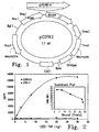


Classifications
-
- C—CHEMISTRY; METALLURGY
- C12—BIOCHEMISTRY; BEER; SPIRITS; WINE; VINEGAR; MICROBIOLOGY; ENZYMOLOGY; MUTATION OR GENETIC ENGINEERING
- C12N—MICROORGANISMS OR ENZYMES; COMPOSITIONS THEREOF; PROPAGATING, PRESERVING, OR MAINTAINING MICROORGANISMS; MUTATION OR GENETIC ENGINEERING; CULTURE MEDIA
- C12N15/00—Mutation or genetic engineering; DNA or RNA concerning genetic engineering, vectors, e.g. plasmids, or their isolation, preparation or purification; Use of hosts therefor
- C12N15/09—Recombinant DNA-technology
- C12N15/11—DNA or RNA fragments; Modified forms thereof; Non-coding nucleic acids having a biological activity
- C12N15/52—Genes encoding for enzymes or proenzymes
-
- C—CHEMISTRY; METALLURGY
- C12—BIOCHEMISTRY; BEER; SPIRITS; WINE; VINEGAR; MICROBIOLOGY; ENZYMOLOGY; MUTATION OR GENETIC ENGINEERING
- C12N—MICROORGANISMS OR ENZYMES; COMPOSITIONS THEREOF; PROPAGATING, PRESERVING, OR MAINTAINING MICROORGANISMS; MUTATION OR GENETIC ENGINEERING; CULTURE MEDIA
- C12N9/00—Enzymes; Proenzymes; Compositions thereof; Processes for preparing, activating, inhibiting, separating or purifying enzymes
- C12N9/14—Hydrolases (3)
- C12N9/48—Hydrolases (3) acting on peptide bonds (3.4)
- C12N9/50—Proteinases, e.g. Endopeptidases (3.4.21-3.4.25)
- C12N9/64—Proteinases, e.g. Endopeptidases (3.4.21-3.4.25) derived from animal tissue
- C12N9/6421—Proteinases, e.g. Endopeptidases (3.4.21-3.4.25) derived from animal tissue from mammals
- C12N9/6489—Metalloendopeptidases (3.4.24)
-
- A—HUMAN NECESSITIES
- A61—MEDICAL OR VETERINARY SCIENCE; HYGIENE
- A61P—SPECIFIC THERAPEUTIC ACTIVITY OF CHEMICAL COMPOUNDS OR MEDICINAL PREPARATIONS
- A61P35/00—Antineoplastic agents
- A61P35/04—Antineoplastic agents specific for metastasis
-
- C—CHEMISTRY; METALLURGY
- C12—BIOCHEMISTRY; BEER; SPIRITS; WINE; VINEGAR; MICROBIOLOGY; ENZYMOLOGY; MUTATION OR GENETIC ENGINEERING
- C12N—MICROORGANISMS OR ENZYMES; COMPOSITIONS THEREOF; PROPAGATING, PRESERVING, OR MAINTAINING MICROORGANISMS; MUTATION OR GENETIC ENGINEERING; CULTURE MEDIA
- C12N15/00—Mutation or genetic engineering; DNA or RNA concerning genetic engineering, vectors, e.g. plasmids, or their isolation, preparation or purification; Use of hosts therefor
- C12N15/09—Recombinant DNA-technology
- C12N15/11—DNA or RNA fragments; Modified forms thereof; Non-coding nucleic acids having a biological activity
-
- A—HUMAN NECESSITIES
- A61—MEDICAL OR VETERINARY SCIENCE; HYGIENE
- A61K—PREPARATIONS FOR MEDICAL, DENTAL OR TOILETRY PURPOSES
- A61K38/00—Medicinal preparations containing peptides
-
- G—PHYSICS
- G01—MEASURING; TESTING
- G01N—INVESTIGATING OR ANALYSING MATERIALS BY DETERMINING THEIR CHEMICAL OR PHYSICAL PROPERTIES
- G01N2333/00—Assays involving biological materials from specific organisms or of a specific nature
- G01N2333/435—Assays involving biological materials from specific organisms or of a specific nature from animals; from humans
- G01N2333/52—Assays involving cytokines
- G01N2333/525—Tumor necrosis factor [TNF]
Definitions
- This invention relates to the purification and characterization of tumor necrosis factor (TNF) receptor (TNF-R) releasing enzyme (TRRE), compositions derived from the enzyme, and methods of use thereof. Modulation of TRRE levels indirectly modulates effective levels of TNF.
- the invention further relates to methods of treatment of pathological conditions caused or exacerbated by altered levels or activity of TNF such as inflammatory conditions including autoimmune diseases, infections, septic shock, obesity, cachexia, and conditions that are associated with decreased levels or activity of TNF such as cancer.
- Tumor necrosis factor TNF or TNF- ⁇
- lymphotoxin LT or TNF- ⁇
- cytokines that share 40 percent amino acid (AA) sequence homology. Old (1987) Nature 330:602-603 . These cytokines are released mainly by macrophages, monocytes and natural killer (NK) cells in response to broad immune reactions. Gorton and Galli (1990) Nature 346:274-276 ; and Dubravec et al. (1990) Proc. Natl. Acad. Sci. USA 87:6758-6761 .
- cytokines Although initially discovered as agents inducing hemorrhagic necrosis of tumors, these cytokines have been shown to have essential roles in both the inductive and effector phases of immune reactions and inflammation.
- the two cytokines cause a broad spectrum of effects on cells in vitro and tissues in vivo , including: (i) vascular thrombosis and tumor necrosis; (ii) inflammation; (iii) activation of macrophages and neutrophils; (iv) leukocytosis; (v) apoptosis; and (vi) shock.
- vascular thrombosis and tumor necrosis including: (i) inflammation; (iii) activation of macrophages and neutrophils; (iv) leukocytosis; (v) apoptosis; and (vi) shock.
- TNF has been associated with a variety of disease states including various forms of cancer, arthritis, psoriasis, endotoxic shock, sepsis, autoimmune diseases, infections, obesity, and cachexia. Attempts have been made to alter the course of a disease by treating the patient with TNF inhibitors with varying degrees of success.
- oxpentifylline did not alter the course of Crohn's disease, a chronic inflammatory bowel disease. Bauditz et al. (1997) Gul 40:470-4 .
- the TNF inhibitor dexanabinol provided protection against TNF following traumatic brain injury. Shohami et al. (1997) J. Neuroimmun. 72:169-77 .
- TNF-R TNF-R on both lymphoid and non-lymphoid cells
- LPS bacterial lipopolysaccharide
- PMA phorbol myristate acetate
- IL-1 interleukin-1
- IFN- ⁇ interferon-gamma
- IL-2 IL-2
- TNF binding proteins were originally identified in the serum and urine of febrile patients, individuals with renal failure, cancer patients, and even certain healthy individuals. Seckinger et al. (1988) J. Exp. Med. 167:1511-1516 ; Engelmann et al. (1989) J. Biol. Chem. 264:11974-11980 ; Seckinger et al. (1989) J. Biol. Chem. 264:11966-11973 ; Peetre et al. (1988) Eur. J. Haematol. 41:414-419 ; Olsson et al. (1989) Eur. J. Haematol. 42:270-275 ; Gatanaga et al.
- human brain and ovarian tumors produced high serum levels of TNF-BP.
- These molecules were subsequently purified, characterized, and cloned by different laboratories.
- Human TNF-BP consist of 30 kDa and 40 kDa proteins found to be identical to the N-terminal extracellular domains of p55 and p75 TNF-R, respectively.
- the 30 kDa and 40 kDa TNF-BP are thus also termed soluble p55 and p75 TNF-R, respectively.
- Studies of these proteins have been facilitated by the availability of human recombinant 30 kDa and 40 kDa TNF-BP and antibodies which specifically recognize each form and allow quantitation by immunoassay.
- TNF trimer binds with three soluble TNF-R (sTNF-R) molecules and the complex can no longer interact with TNF-R.
- sTNF-R soluble TNF-R
- Banner et al. (1993) Cell 73:431-445 The binding of the trimer and sTNF-R, however, is reversible and these reactants are not altered as a result of complex formation.
- both recombinant and native human sTNF-R are potent inhibitors of TNF/LT biological activity in vitro as well as in vivo .
- TNF endogenous mediators
- IL-1 IL-1
- gamma-Interferon IL-8
- IL-6 IL-6
- TNF expression is being tested in treatment of endotoxic shock. Mohler et al. (1994) Nature 370:218-220 . Modulation of TNF-R activity is also being approached by the use of peptides that bind intracellularly to the receptor or other component in the process to prevent receptor shedding.
- Serum levels of sTNF-R rise within minutes and remain high for 7 to 8 hours after the intravenous injection of human recombinant TNF or IL-2 into human cancer patients.
- serum sTNF-R levels are chronically elevated in cancer patients and may remain at high levels for years. Grosen et al.
- sTNF-R are natural inhibitors of these cytokines and regulate their biological activity post secretion. Fusion proteins consisting of a sTNF-R linked to a portion of the human IgG1 have also been developed for treating rheumatoid arthritis and septic shock. Moreland et al. (1997) N. Eng. J. Med. 337:141-7 ; Abraham et al. (1997) JAMA 277:1531-8 .
- cytokine receptors e.g., IL-1R, IL-2R, IL-4R, IL-5R, IL-6R, IL-7R, IL-9R, granulocyte-colony stimulating factor-R (G-CSF-R), granulocyte-macrophage-colony stimulating factor-R (GM-CSF-R), transforming growth factor- ⁇ -R (TGF ⁇ -R), platelet-derived growth factor-R (PDGF-R), and epidermal growth factor-R (EGF-R); (ii) growth factors, e.g., TNF- (pro-TNF-I ), TGF-I , and CSF-1; (iii) adhesion molecules, e.g., intracellular adhesion molecule-1 (ICAM-1/CD54) and vascular cell membrane adhesion molecule (VCAM-1/CD106); (iv)
- Matrix metalloproteinases are a family of enzymes that includes interstitial collagenase (MMP-1), 72 kDa and 92 kDa gelatinases (MMP-2 and MMP-9), stromelysins 1, 2 and 3, neutrophil collagenase, metalloelastase, matrilysin, and gelatinase A. These enzymes are secreted by cells within tissues and by infiltrating inflammatory cells. Collectively, they are capable of degrading most of the proteins in the extracellular matrix (ECM).
- ECM extracellular matrix
- MMPs display different substrate specificities yet have several properties in common. They are all zinc-containing enzymes that require calcium for activity. They are secreted as zymogens and activated in situ, usually by release of an inhibitory N-terminal pro-piece containing a single cysteine residue. The attached pro-piece is believed to coordinate with the zinc in the active site of the proteinase, thereby suppressing the proteolytic activity. Activation may be accompanied by additional proteolytic cleavages that can generate active enzymes of lower molecular weights. All members of the MMP family have a short conserved region consisting of the HEXGH motif that provides two Zn-coordinating histidine residues and a glutamic acid residue that is considered part of the catalytic site.
- MMPs also contain a hemopexin/vitronectin domain.
- the function of the hemopexin domain is unknown. For review see Ray and Stetter-Stevenson (1994) Eur. Respir. J. 7:2062-2072 .
- MMPs are involved in tumor invasion and metastasis.
- a number of methods have been utilized to assess the presence of MMPs in human tumor tissues and serum from cancer patients. Positive correlations have been found between MMP expression and tumor invasion and metastasis in vitro, as well as in in vivo animal models.
- MMPs have been associated with the malignant phenotype in a wide variety of human tissues, including lung, prostate, stomach, colon, breast, ovaries and thyroid, as well as squamous carcinoma of the head and neck. Matrisian et al. (1991); Sato et al. (1992); Levy et al. (1991); and Lyons et al. (1991). To date, the proposed role of MMPs in cancer has been limited to tissue remodeling in invasion and metastasis.
- the MMPs are inhibited by members of the family of tissue inhibitors of metalloproteinases (TIMPs, e.g., TIMP-1, TIMP-2, and TIMP-3), which bind at the active site and block access to substrate.
- TIMPs tissue inhibitors of metalloproteinases
- Matrix remodeling which occurs during various normal and pathological processes depends on a critical balance between activated MMPs and inhibiting TIMPs.
- MMP inhibitors have been described and the use thereof has also been suggested for treating various pathologic indications, including: angiogenesis; wound healing; gum disease; skin disorders; keratoconus; inflammatory conditions; rheumatoid arthritis; cancer; corneal and skin ulcers; cardiovascular disease; central nervous system disorders; and diabetes.
- US Patent Nos. 5,268,384 and 5,270,326 PCT publications WO 94/22309 , WO 95/09913 , WO 90/11287 , WO 90/14363 ; EP patents 211 077, 623 676 ; and Naito et al. (1994) Int. J. Cancer 58:730-735 ; Watson et al. (1995) Cancer Res.
- Both soluble p55 and p75 TNF-R do not appear to be generated from processed mRNA, since only full length receptor mRNA has been detected in human cells in vitro. Gatanaga et al. (1991). Carboxyl-terminal sequencing of the human soluble p55 TNF-R indicates that a cleavage site may exist between Asn 172 and Val 173. Gullberg et al. (1992) Eur. J. Cell. Biol. 58:307-312 . This evidence is supported by the finding that human TNF-R with the mutation at Asn 172 and Val 173 was not released as effectively as native TNF-R on COS-1 cells transduced with cDNA of human TNF-R. Gullberg et al. (1992).
- the cytoplasmic portion of TNF-R does not appear to play an important role in releasing the soluble receptor forms from transduced COS-1 cells.
- COS-1 cells release sTNF-R even when transduced with cDNA of human p55 TNF-R which expresses only the extracellular domain but not the cytoplasmic domain.
- sTNF-R shedding is not affected by dexamethasone, gold sodium thiomalate, or prostaglandin E2.
- Seitz et al. (1997) J. Rheumatology 24:1471-6 .
- Neoplasia resulting in benign tumors can usually be completely cured by removing the mass surgically. If a tumor becomes malignant, as manifested by invasion of surrounding tissue, it becomes much more difficult to eradicate. Once a malignant tumor metastasizes, it is much less likely to be eradicated.
- the three major cancers in terms of morbidity and mortality, are colon, breast and lung.
- New surgical procedures offer an increased survival rate for colon cancer.
- Improved screening methods increase the detection of breast cancer, allowing earlier, less aggressive therapy.
- Lung cancer remains largely refractory to treatment.
- Melanoma is one of the human diseases for which there is an acute need of new therapeutic modalities. It is a particularly aggressive form of skin cancer, and occurs in increased frequency in individuals with regular unguarded sun exposure. In the early disease phases, melanoma is characterized by proliferation at the dermal-epidermal junction, which soon invades adjacent tissue and metastasizes widely. Once it has metastasized, it is often impossible to extirpate and is consequently fatal. Worldwide, 70,000 patients are diagnosed annually with melanoma and it is responsible for 25,000 reported deaths each year. The American Cancer Society projects that by the year 2000, 1 out of every 75 Americans will be diagnosed with melanoma.
- Neuroblastoma is a highly malignant tumor occurring during infancy and early childhood. Except for Wilm's tumor, it is the most common retroperitoneal tumor in children. This tumor metastasizes early, with widespread involvement of lymph nodes, liver, bone, lung, and marrow. While the primary tumor is resolvable by resection, the recurrence rate is high.
- Small cell lung cancer is the most malignant and fastest growing form of lung cancer and accounts for 20-25% of new cases of lung cancer. Approximately 60,000 cases are diagnosed in the U.S. every year. The primary tumor is generally responsive to chemotherapy, but is followed by wide-spread metastasis. The median survival time at diagnosis is approximately 1 year, with a 5 year survival rate of 5-10%.
- Breast cancer is one of the most common cancers and is the third leading cause of death from cancers in the United States with an annual incidence of about 182,000 new cases and nearly 50,000 deaths. In the industrial countries, approximately one in eight women can expect to develop breast cancer. The mortality rate for breast cancer has remained unchanged since 1930. It has increased an average of 0.2% per year, but decreased in women under 65 years of age by an average of 0.3% per year. Preliminary data suggest that breast cancer mortality is beginning to decrease, probably as a result of increased diagnoses of localized cancer and carcinoma in situ . See e.g., Marchant (1994) Contemporary Management of Breast Disease II: Breast Cancer, In: Obstetrics and Gynecology Clinics of North America 21:555-560 ; and Colditz (1993) Cancer Suppl. 71:1480-1489 .
- Non-Hodgkin's B cell lymphomas are cancers of the immune system that are expected to afflict approximately 225,000 patients in the United States in 1996. These cancers are diverse with respect to prognosis and treatment, and are generally classified into one of three grades. The median survival of the lowest grade is 6.6 years and the higher grade cancers have much lower life expectancy. Virtually all non-Hodgkin's B cell lymphomas are incurable. New diagnoses of non-Hodgkins lymphomas have increased approximately 7% annually over the past decade, with 52,700 new diagnoses estimated for 1996. The increase is due in part to the increasing prevalence of lymphomas in the AIDS patient population.
- the method involves creating an mixed lymphocyte reaction (MLR) between the host (cancer patient's) peripheral blood lymphocytes and a donor's allogeneic lymphocytes and administering the MLR directly to the tumor.
- MLR mixed lymphocyte reaction
- This method is described more fully, for example, in WO 93/20186 and JP 62096426 .
- administration of the MLR is preceded by resection of the tumor.
- TNF is linked to the three factors contributing to body weight control: intake, expenditure, and storage of energy.
- TNF may have a key roles in both extremes of weight problems. Abnormalities in its activity may lead to obesity; changes in its production result in the opposite effect, cachexia. Argilés et al. (1997) FASEB J. 11:743-751 .
- Cachexia is pathological weight loss generally associated with anorexia, weakness, anemia, asthenia, and loss of body lipid stores and skeletal muscle protein. This state often accompanies burns, trauma, infection, and neoplastic diseases. Lawson et al. (1982) Annu. Rev. Nutr. 2:277-301 ; Argilés et al. (1988) Mol. Cell. Biochem. 81:3-17 ; and Ogiwara et al. (1994) J. Surg. Oncol. 57:129-133 . TNF concentrations are elevated in many patients with cachexia. Scuderi et al. (1986) Lancet 2:1364-65 ; Grau et al.
- TNF inhibits collagen alpha I gene expression and wound healing in a murine model of cachexia.
- Buck et al. (1996) Am. J. Pathol. 149:195-204 .
- endotoxin concentrations may raise TNF levels, causing cachexia.
- Beutler et al. (1985) Science 229:869-871 ; Tracey et al. (1987) Nature 330:662-664 ; and Michie et al. (1988) New Engl. J. Med. 318:1481-1486 .
- Fat tissue loss is also associated with an increase in lipase activity and inhibition of glucose transport; TNF is also linked to both of these changes. Kawakami et al. (1987) J. Biochem. 331-338 ; Feingold et al. (1992) Endocrinology 130:10-16 ; and Hauner et al. (1995) Diabetologia 38:764-771 .
- TNF mediates hypertriglyceridaemia associated with cachexia. Dessi et al. (1995) Br. J. Cancer 72:1138-43 . TNF also participates in the protein wasting, loss of skeletal muscle and loss of nitrogen associated with cachexia. Costelli et al. (1993) J. Clin. Invest.
- TNF has additional, related roles. It is involved in thermogenesis, particularly nonshivering thermogenesis in brown adipose tissue (BAT), which has an elevated level in cachexia. Nicholls (1983) Biosci. Rep. 3:431-441 ; Rothwell (1993) Int, J. Obesity 17:S98-S101 ; Bianchi et al. (1989) Horm. Metab. Res. 21:11 ; and Oudart et al. (1995) Can. J. Physiol. Pharmacol. 73:1625-1631 . TNF has also been implicated in non-insulin-dependent (type II) diabetes. Hotamisligil et al. (1995) J. Clin. Invest.
- TNF is expressed and Secreted by adipocytes and can travel to the brain. TNF administration also results in an increase in circulating leptin concentrations. Grunfeld et al. (1996) J. Clin. Invest. 97:2152-57 . It is possible to reconcile the participation of TNF in obesity and cachexia. TNF can be considered one of many signals coming from adipose tissue that participate in the feedback mechanism that informs the hypothalamic center about the state of the adipocyte energy depot. It probably counteracts excessive energy intake and is able to stimulate thermogenesis either directly or by increasing sympathetic activity.
- TNF released by adipose tissue will also stimulate lipolysis, decrease LPL activity, decrease the expression of the glucose transporter GLUT4, and inhibit lipogenesis in the adipocyte, thus contributing to the maintenance (but not increased fat deposition) of the adipose tissue mass.
- a high production of TNF by activated macrophages (as a result of a tumor or an infection) also contributes to anorexia, increased thermogenesis, and adipose tissue dissolution. However, it represents a pathological state where there is an excess of the molecules that inform the brain that adipose tissue needs dissolution.
- the invention encompasses compositions of a substantially purified protein having tumor necrosis factor receptor (TNFR) releasing enzymatic activity, termed TRRE.
- TRRE tumor necrosis factor receptor
- the present invention provides a method of purifying TRRE comprising:
- the present invention also provides a purified tumor necrosis factor (TNF) receptor polypeptide having the characteristic of being obtainable by this process.
- TNF tumor necrosis factor
- the TRRE in its native form has an apparent molecular weight of about 120 kDa on sodium dodecyl sulphate polyacrylamide gel eletrophoresis (SDS-PAGE).
- the TRRE has internal amino acid sequences of: D-L-N-L-G-A-Q-A-T-I-T-N-L-P (SEQ ID NO. 1); G-L-D-E-T-Q-N-L-I-T-V-P-Y (SEQ NO. 2); S-E-R-W-P-Q-M-A-N-K-V-S-R (SEQ ID NO.
- I-V-V-T-K- SEQ ID NO. 4
- E-F-P-H/S-P-V-D-A-A-T-R SEQ ID NO. 5
- A-L-F-E-L-I-Y-E-L-L/E-A-Y-I-I/N-V-L SEQ ID NO. 6
- L-D-Y-Q-E/T-S-Y-S-A-A-V-A-R SEQ ID NO. 7
- L-A-L-Q/I-E-S-P-S/P SEQ ID NO. 8
- L-F-L-K-N-T-G-L-A-R SEQ ID NO.
- the TRRE in its native form has an apparent molecular weight of about 60 kDa on SDS-PAGE.
- the TRRE has internal amino acid sequences of D-L-N-L-G-A-Q-A-T-I/L-T-N-L-P (SEQ ID NO. 15); L-A-E-D-Y-L-S-G/L-W-L-E/G-R (SEQ ID NO. 16); and/or L/K-V/L-D/E-Y-D/E-X-L/F-F-Q-N-L (SEQ ID NO. 17. Fragments of TRRE containing TRRE activity are also encompassed by the invention.
- the purified TRRE of the invention is useful for treating a sepsis or inflammation, by administering an amount of TRRE sufficient to indirectly moderate or modulate local levels of tumor necrosis factor.
- the method includes administering an amount of an inhibitor of TRRE effective to decrease the levels of soluble TNF-R.
- TRRE activity compared to control is indicative of the presence of a disease state associated with elevated levels of TRRE.
- the methods include the steps of administering an amount of an inhibitor of TRRE sufficient to modulate the levels of TNF-TNF receptor complexes on cell surfaces.
- the method comprises the steps of comparing TNF-R released in TNF-R expressing cells (TNF-R + ) incubated with TRRE to TNF-R release by TNF-R + cells not incubated with TRRE and to TNF-R release by cells that do not express significant amounts of TNF-R (TNF-R - ).
- TNF-R + cells are recombinant TNF-R - cells transformed to express TNF-R recombinantly.
- the invention further encompasses methods for screening for agents that modulate the activity of TRRE.
- the activity of TREE is measured in the presence (test) or absence (control) of a particular agent or drug. If the activity of the test sample exceeds or is less than that of the control, the agent or drug increases or decreases, respectively, TRRE activity.
- TNF is a major proinflammatory and immunomodulatory cytokine produced during immune responses. TNF also regulates the expression of IL-2R leading to enhanced T cell responses mediated by IL-2 and appears to be required for generating proliferative responses in mixed lymphocyte cultures. Additional studies have shown that CD8 + , CTL and lymphokine activated killer cells are optimally induced with TNF, in combination with IL-2, suggesting the importance of this cytokine in regulating cytotoxic effector function. As discussed in detail above, TNF mediates its activity by binding to a TNF-R. Soluble TNF-Rs inhibit TNF activity by two methods: they decrease the available binding sites on a cell and bind to soluble TNF to decrease the local concentration. The present invention encompasses compositions and methods for modulating the level of soluble TNF-R by decreasing the cleavage of TNF-R from the cell surface and thus indirectly modulating the effect of TNF.
- TRRE TNF-R releasing enzyme
- the invention encompasses compositions of a substantially purified protein having tumor necrosis factor receptor (TNFR) releasing enzymatic activity, termed TRRE.
- TRRE tumor necrosis factor receptor
- the protein can be purified as described in the examples below and, in addition to having the described enzymatic activity, the native enzyme has an apparent molecular weight of about 120 kDa on SDS-PAGE.
- the TRRE has internal amino acid sequences of: D-L-N-L-G-A-Q-A-T-I-T-N-L-P (SEQ ID NO:1); G-L-D-E-T-Q-N-L-I-T-V-P-Y (SEQ ID NO:2); S-E-R-W-P-Q-M-A-N-K-V-S-R (SEQ ID NO:3); I-V-V-T-K (SEQ ID NO:4); E-F-P-H/S-P-V-D-A-A-T-R (SEQ ID NO:5); A-L-F-E-L-I-Y-E-L-L-L/E-A-Y-I-I/N-V-L (SEQ ID NO:6); L-D-Y-Q-E/T-S-Y-S-A-A-V-A-R (SEQ ID NO:7); L-A-L-L-A-A-V-
- the TRRE in its native form has an apparent molecular weight of about 60 kDa on SDS-PAGE.
- the TRRE has internal amino acid sequences of D-L-N-L-G-A-Q-A-T-I/L-T-N-L-P (SEQ ID NO:15); L-A-E-D-Y-L-S-G/L-W-L-E/G-R (SEQ ID NO:16); and L/K-V/L-D/E-Y-D/E-X-L/F-F-Q-N-L (SEQ ID NO:17) where X is an unidentified amino acid residue.
- non-native proteins may have differing apparent molecular weights depending on the degree of glycosylation.
- the enzymatic activity of the TRRE is inhibited by metalloprotease inhibitors.
- the method of inhibiting the activity of TRRE by the addition of metalloprotease inhibitors is also encompassed by the invention.
- polypeptide refers to polymers of amino acid residues of any length.
- the polymer may be linear or branched, it may comprise modified amino acids or amino acid analogs, and it may be interrupted by chemical moieties other than amino acids.
- the terms also encompass an amino acid polymer that has been modified naturally or by intervention; for example, disulfide bond formation, glycosylation, lipidation, acetylation, phosphorylation, or any other manipulation or modification, such as conjugation with a labeling or bioactive component.
- TRRE includes any polypeptide monomer or polymer with TRRE enzymatic specificity, including the intact TRRE, and smaller and larger functionally equivalent polypeptides, as described herein.
- a "fusion polypeptide” is a polypeptide comprising regions in a different position in the sequence than occurs in nature.
- the regions may normally exist in separate proteins and are brought together in the fusion polypeptide; they may normally exist in the same protein but are placed in a new arrangement in the fusion polypeptide; or they may be synthetically arranged.
- the invention encompasses recombinant proteins that are comprised of a functional portion of TRRE and an antibody. Methods of making these fusion proteins are known in the art and are described, for instance, in WO93/07286 .
- a “functionally equivalent fragment” of a TRRE polypeptide varies from the native sequence by addition(s), deletion(s), or substitution(s), or any combination thereof, while preserving at least one functional property of the fragment relevant to the context in which it is being used.
- a functionally equivalent fragment of a TRRE polypeptide typically has the ability to bind membrane bound TNF-R and enzymatically cleave TNF-R to provide soluble TNF-R.
- Amino acid substitutions, if present, are preferably conservative substitutions that do not deleteriously affect folding or functional properties of the peptide.
- Polypeptides of this invention can be in glycosylated or unglycosylated form, can be modified post-translationally (e.g., removal of signal peptide, transmembrane or cytoplasmic regions, acetylation, and phosphorylation) or can be modified synthetically (e.g., by a labeling group).
- compositions of TRRE and a physiologically acceptable buffer include, but are not limited to, saline and phosphate buffered saline (PBS).
- PBS phosphate buffered saline
- TRRE is to be administered to an individual, it is preferably at least 80% pure, more preferably it is at least 90% pure, even more preferably it is at least 95% pure and free of pyrogens and other contaminants.
- percent purity is calculated as a weight percent of the total protein content of the preparation, and does not include constituents which are deliberately added to the composition after the TRRE is purified.
- compositions contain a therapeutically effective amount of substantially purified antibody binding fragment specific for TRRE.
- the antibody neutralizes TRRE TNF-R releasing activity.
- Methods of antibody production and isolation are well known in the art. See, for example, Harlow and Lane (1988) Antibodies: A Laboratory Manual , Cold Spring Harbor Laboratory, New York. Antibodies can be isolated by any technique suitable for immunoglobulins of this isotype.
- Purification methods include salt precipitation (for example, with ammonium sulfate), ion exchange chromatography (for example, on a cationic or anionic exchange column run at neutral pH and eluted with step gradients of increasing ionic strength), gel filtration chromatography (including gel filtration HPLC), and chromatography on affinity resins such as protein A, protein G, hydroxyapatite, and anti-immunoglobulin.
- Anti-TRRE antibodies can also be purified on affinity columns comprising TRRE.
- anti-TRRE antibodies are purified using Protein-A-CL-Sepharose TM 4B chromatography followed by chromatography on a DEAE-Sepharose TM 4B ion exchange column.
- antigen- binding fragment includes any peptide that binds to TRRE in a specific manner. These derivatives include such immunoglobulin fragments as Fab, F(ab') 2 , Fab', scfv (both monomers and polymeric forms) and isolated H and L chains. Antigen - binding fragments retain the specificity of the intact immunoglobulin, although avidity and/or affinity can be altered.
- the antigen -binding fragments are typically generated by genetic engineering, although they can alternatively be obtained by other methods and combinations of methods. This classification includes, but is not limited to, engineered peptide fragments and fusion peptides.
- Preferred compounds include polypeptide fragments of the CDRs, antibody fusion proteins comprising cytokine effector components, antibody fusion proteins comprising adjuvants or drugs, and single-chain V region proteins.
- Scfv can be produced either recombinantly or synthetically.
- an automated synthesizer can be used for synthetic production of scfv.
- a suitable plasmid containing polynucleotide that encodes the scfv can be introduced into a suitable host cell, either eukaryotic, such as yeast, plant, insect or mammalian cells, or prokaryotic, such as E. coli , and the expressed protein can be isolated using standard protein purification techniques.
- pET-22b(+) Novagen, Madison, WI
- pET-22b(+) contains a nickel ion binding domain consisting of 6 sequential histidine residues, which allows the expressed protein to be purified on a suitable affinity resin.
- pcDNA3 Invitrogen, San Diego, CA
- hybrid antibodies in which one pair of H and L chains is obtained from a first antibody, while the other pair of H and L chains is obtained from a different second antibody.
- one pair of L and H chains is from anti-TRRE.
- each L-H chain pair binds different epitopes of TRRE.
- Such hybrids can also be formed using humanized H or L chains.
- peptides in which various immunoglobulin domains have been placed in an order other than that which occurs in nature are also be used as diagnostic and imaging reagents.
- polynucleotides encoding antibodies (or fragments thereof) capable of binding to a TRRE polypeptide (or fragments thereof).
- the polynucleotide can be native or recombinant, and can be incorporated into a vector or plasmid and operably linked to a promoter.
- the invention further encompasses The polypeptides of the invention for use in methods of treating a disease associated with altered levels or activities of TNF by administering an amount of TRRE sufficient to indirectly reduce local levels of TNF.
- Suitable indications for treatment include, but are not limited to, diseases such as cancer, obesity and cachexia, autoimmune diseases such as diabetes, juvenile onset rheumatoid arthritis, systemic lupus erythematosus, and other inflammatory conditions, psoriasis, endotoxin shock, rheumatoid arthritis, trauma, and multiple sclerosis.
- Infections associated with microbial or parasitic infection in which TNF has an important role include, but are not limited to, septic shock and malaria.
- TRRE can be used to treat indications previously associated with aberrant MMP expression.
- these include, but are not limited to, angiogenesis; wound healing; gum disease; skin disorders; keratoconus; inflammatory conditions; rheumatoid arthritis; cancer; corneal and skin ulcers; cardiovascular disease; central nervous system disorders; and diabetes.
- immuno-inflammatory diseases or diseases with an immunological base and a tissue destructive component in which TNF has an important role such as Crohn's disease and inflammatory bowel disease.
- Other inflammatory conditions such as traumatic shock are also included.
- Methods of administration include any known in the art.
- administration is directly to the site of inflammation in the case of rheumatoid arthritis and is parenteral, subcutaneous, intramuscular, intraperitoneal, intracavity, intrathecal, and intravenous in the case of systemic disorders.
- Administration can also be systemic to treat localized disorders or local to treat systemic disorders. Methods of administration are discussed in more detail below.
- Local administration can be achieved by, for example, local infusion during surgery, by direct injection to the site, by means of a catheter, or by means of an implant, said implant being of a porous, non-porous, or gelatinous material, including membranes, such as silastic membranes, or fibers.
- a suitable such membrane is Gliadel® provided by Guilford sciences.
- compositions of effective amounts of TRRE and a topical pharmaceutically or cosmetically acceptable carrier are provided.
- Topical pharmaceutically acceptable carrier is any substantially non-toxic carrier conventionally useable for topical administration of pharmaceuticals in which TRRE will remain stable and bioavailable when applied directly to skin or mucosal surfaces.
- TRRE can be dissolved in a liquid, dispersed or emulsified in a medium in a conventional manner to form a liquid preparation or mixed with a semi-solid (gel) or solid carrier to form a paste, powder, ointment, cream, lotion or the like.
- Suitable topical pharmaceutically acceptable carriers include water, petroleum jelly (Vaseline TM ), petrolatum, mineral oil, vegetable oil, animal oil, organic and inorganic waxes, such as microcrystalline, paraffin and ozocerite wax, natural polymers, such as xanthanes, gelatin, cellulose, collagen, starch, or gum arabic, synthetic polymers, such as discussed below, alcohols, polyols, and the like.
- the carrier can be a water miscible carrier composition that is substantially miscible in water.
- Such water miscible topical pharmaceutically acceptable carrier composition can include those made with one or more appropriate ingredients set forth above but can also include sustained or delayed release carriers, including water containing, water dispersible or water soluble compositions, such as liposomes, microsponges, microspheres or microcapsules, aqueous base ointments, water-in-oil or oil-in-water emulsions, gels or the like.
- sustained or delayed release carriers including water containing, water dispersible or water soluble compositions, such as liposomes, microsponges, microspheres or microcapsules, aqueous base ointments, water-in-oil or oil-in-water emulsions, gels or the like.
- the topical pharmaceutically acceptable carrier comprises a sustained release or delayed release carrier.
- the carrier is any material capable of sustained or delayed release of the TRRE to provide a more efficient administration resulting in one or more of less frequent and/or decreased dosage of the TRRE, ease of handling, and extended or delayed effects on dermatologic conditions.
- the carrier is capable of releasing TRRE when exposed to any oily, fatty, waxy, or moist environment on the area being treated or by diffusing or by release dependent on the degree of loading of TRRE to the carrier in order to obtain release of TRRE.
- Non-limiting examples of such carriers include liposomes, microsponges, microspheres, or microcapsules of natural and synthetic polymers and the like.
- suitable carriers for sustained or delayed release in a moist environment include gelatin, gum arabic, xanthane polymers; by degree of loading include lignin polymers and the like; by oily, fatty or waxy environment include thermoplastic or flexible thermoset resin or elastomer including thermoplastic resins such as polyvinyl halides, polyvinyl esters, polyvinylidene halides and halogenated polyolefins, elastomers such as brasiliensis, polydienes, and halogenated natural and synthetic rubbers, and flexible thermoset resins such as polyurethanes, epoxy resins and the like.
- the sustained or delayed release carrier is a liposome, microsponge, microsphere or gel.
- compositions used in the method of treating dermatologic conditions of the invention are applied directly to the areas to be treated. While not required, it is desirable that the topical composition maintain TRRE at the desired location for about 24 to 48 hours.
- one or more additional ingredients conventionally found in topical pharmaceutical or cosmetic compositions can be included with the carrier, such as a moisturizer, humectants, odor modifier, buffer, pigment, preservative, Vitamins such as A, C and E, emulsifier, dispersing agent, wetting agent, odor-modifying agent, gelling agents, stabilizer, propellant, antimicrobial agents, sunscreen, enzymes and the like.
- a moisturizer such as a moisturizer, humectants, odor modifier, buffer, pigment, preservative, Vitamins such as A, C and E, emulsifier, dispersing agent, wetting agent, odor-modifying agent, gelling agents, stabilizer, propellant, antimicrobial agents, sunscreen, enzymes and the like.
- Suitable non-limiting examples of additional ingredients include superoxide dismutase, stearyl alcohol, isopropyl myristate, sorbitan monooleate, polyoxyethylene stearate, propylene glycol, water, alkali or alkaline earth lauryl sulfate, methylparaben, octyl dimethyl-p-amino benzoic acid (Padimate O), uric acid, reticulin, polymucosaccharides, hyaluronic acids, aloe vera, lecithin, polyoxyethylene sorbitan monooleate, Vitamin A or C, tocopherol (Vitamin E), alpha-hydroxy of alpha-keto acids such as pyruvic, lactic or glycolic acids, or any of the topical ingredients disclosed in U.S. Patents 4,340,586 , 4,695,590 , 4,959,353 or 5,130,298 and 5,140,043 .
- topical carrier can also be a topical cosmetically acceptable carrier.
- topical cosmetically acceptable carrier any substantially non-toxic carrier conventionally useable for topical administration of cosmetics in which TRRE will remain stable and bioavailable when applied directly to the skin surface.
- Suitable cosmetically acceptable carriers include, but are not limited to, cosmetically acceptable liquids, creams, oils, lotions, ointments, gels, or solids, such as conventional cosmetic night creams, foundation creams; suntan lotions, sunscreens, hand lotions, make-up and make-up bases, masks and the like.
- topical cosmetically acceptable carriers and pharmaceutically acceptable carriers are similar, if not often identical, in nature so that most of the earlier discussion on pharmaceutically acceptable carriers also applies to cosmetically acceptable carriers.
- the compositions can contain other ingredients conventional in cosmetics including perfumes, estrogen, Vitamins A, C or E, alpha-hydroxy or alpha-keto acids such as pyruvic, lactic or glycolic acids, lanolin, Vaseline TM petroleum jelly, aloe vera, methyl or propyl paraben, pigments and the like.
- the effective amount of TRRE in the compositions used to treat dermatologic conditions or diseases can vary depending on such factors as condition of the skin, age of the skin, the degree of purity of TRRE employed, the type of formulation and carrier ingredients used, frequency of administration, overall health of the individual being treated and the like.
- the precise amount for any particular patient used can be determined by those of skill in the pharmaceutical art taking into consideration these factors and the present disclosure.
- the composition is administered in at least two doses and no more than about six doses per day, or less when a sustained or delayed release form is used.
- compositions for topical, oral and parenteral administration usually contain from about 0.001 % to about 10% by weight of TRRE compared to the total weight of the composition, preferably from about 0.01 % to about 2% by weight of TRRE to composition, and especially from about 0.1 % to about 1.5% by weight of TRRE to the composition.
- the topical composition is administered by applying a coating or layer to the skin or mucosal area desired to be treated.
- a coating or layer is applied to the skin or mucosal area desired to be treated.
- the applied material is rubbed into the area. Applications need not be rubbed into the skin and the layer or coating can be left on the skin overnight.
- the amount of TRRE administered is sufficient to effectively reduce levels of TNF.
- reduced levels of TNF result in an amelioration of symptoms.
- Ameliorate denotes a lessening of the detrimental effect of the cancer on the individual.
- Administrations are typically conducted on a weekly or biweekly basis until a desired, measurable parameter is detected, such as diminution of disease symptoms. Administration can then be continued on a less frequent basis, such as biweekly or monthly, as appropriate.
- the effective amount is readily determined by one of skill in the art.
- the dosage ranges are those large enough to produce the desired effect in which the symptoms of the disease are ameliorated without causing undue side effects such as unwanted cross-reactions, anaphylactic reactions, and the like.
- the dosage will vary with the age, condition, sex and extent of the disease in the patient and can be determined by one of skill in the art.
- the dosage can be adjusted by the individual physician in the event of any complication.
- the disease treated is cancer.
- the cancer includes, but is not limited to, astrocytoma, oligodendroglioma, ependymoma, medulloblastoma, primitive neural ectodermal tumor, pancreatic ductal adenocarcinoma, small and large cell lung adenocarcinomas, squamous cell carcinoma, bronchoalveolarcarcinoma, epithelial adenocarcinoma and liver metastases thereof, hepatoma, cholangiocarcinoma, ductal and lobular adenocarcinoma, squamous and adenocarcinomas of the uterine cervix, uterine and ovarian epithelial carcinomas, prostatic adenocar
- the TRRE inhibitor can be any known in the art, including, but not limited to, metalloprotease inhibitors, antibodies that block the effective interaction between TNF-R and TRRE, antisense oligonucleotides specific for the gene encoding TRRE and ribozymes specific for the gene encoding TRRE. Methods of making antibodies are known in the art. Antibodies that block effective binding of TRRE to the TNF-R are easily screened for by using the assay method described herein.
- the dosage ranges for the administration of TRRE inhibitors are those large enough to produce the desired effect in which the symptoms of the malignant disease are ameliorated without causing undue side effects such as unwanted cross-reactions, anaphylactic reactions, and the like.
- the dosage will vary with the age, condition, sex and extent of the disease in the patient and can be determined by one of skill in the art.
- the dosage can be adjusted by the individual physician in the event of any complication.
- dosage can vary from about 0.1 mg/kg to about 2000 mg/kg, preferably about 0.1 mg/kg to about 500 mg/kg, in one or more dose administrations daily, for one or several days.
- lower dosages can be used.
- compositions of TRRE inhibitors can be administered by injection or by gradual perfusion over time.
- the inhibitors can be administered intravenously, intraperitoneally, intra-muscularly, subcutaneously, intracavity, intrathecally or transdermally, alone or in combination with effector cells.
- administration is intralesionally, for instance by direct injection directly into the tumor.
- Intralesional administration of various forms of immunotherapy to cancer patients does not cause the toxicity seen with systemic administration of immunologic agents.
- the intralesional administration is in conjunction with the cancer therapy technology described in, for example, U.S. Patent Nos. 5,376,682 , 5,192,537 , and 5,643,740 .
- the therapeutic agent When the site of delivery is the brain, the therapeutic agent must be capable of being delivered to the brain.
- the blood-brain barrier limits the uptake of many therapeutic agents into the brain and spinal cord from the general circulation. Molecules which cross the blood-brain barrier use two main mechanisms: free diffusion; and facilitated transport. Because of the presence of the blood-brain barrier, attaining beneficial concentrations of a given therapeutic agent in the CNS may require the use of drug delivery strategies. Delivery of therapeutic agents to the CNS can be achieved by several methods.
- therapeutic agents can be delivered by direct physical introduction into the CNS, such as intraventricular, intralesional, or intrathecal injection.
- Intraventricular injection can be facilitated by an intraventricular catheter, for example, attached to a reservoir, such as an Ommaya reservoir.
- Methods of introduction can also be provided by rechargeable or biodegradable devices.
- Another approach is the disruption of the blood-brain barrier by substances which increase the permeability of the blood-brain barrier.
- Examples include intra-arterial infusion of poorly diffusible agents such as mannitol, pharmaceuticals which increase cerebrovascular permeability such as etoposide, or vasoactive agents such as leukotrienes.
- poorly diffusible agents such as mannitol
- pharmaceuticals which increase cerebrovascular permeability such as etoposide
- vasoactive agents such as leukotrienes.
- compositions may be desirable to administer the compositions locally to the area in need of treatment; this can be achieved by, for example, local infusion during surgery, by injection, by means of a catheter, or by means of an implant, said implant being of a porous, non-porous, or gelatinous material, including membranes, such as silastic membranes, or fibers.
- a suitable such membrane is Gliadel® provided by Guilford sciences.
- Another method involves pharmacological techniques such as modification or selection of the inhibitor to provide an analog which will cross the blood-brain barrier. Examples include increasing the hydrophobicity of the molecule, decreasing net charge or molecular weight of the molecule, or modifying the molecule, such as to resemble one normally transported across the blood-brain barrier.
- pharmacological techniques such as modification or selection of the inhibitor to provide an analog which will cross the blood-brain barrier. Examples include increasing the hydrophobicity of the molecule, decreasing net charge or molecular weight of the molecule, or modifying the molecule, such as to resemble one normally transported across the blood-brain barrier.
- Encapsulation of an inhibitor in a hydrophobic environment such as liposomes is also effective in delivering drugs to the CNS.
- WO 91/04014 describes a liposomal delivery system in which the drug is encapsulated within liposomes to which molecules have been added that are normally transported across the blood-brain barrier.
- cyclodextrin Another method of formulating the inhibitors to pass through the blood-brain barrier is encapsulation in cyclodextrin.
- Any suitable cyclodextrin which passes through the blood-brain barrier can be employed, including, but not limited to, &-cyclodextrin, K-cyclodextrin and derivatives thereof. See generally, U.S. Patent Nos. 5,017,566 , 5,002,935 and 4,983,586 .
- Such compositions can also include a glycerol derivative as described by U.S. Patent No. 5,153,179 .
- VIPa vasoactive intestinal peptide analog
- Several other specific transport systems have been identified, these include, but are not limited to, those for transferring insulin, or insulin-like growth factors I and II.
- Suitable, non-specific carriers include, but are not limited to, pyridinium, fatty acids, inositol, cholesterol, and glucose derivatives.
- Certain prodrugs have been described whereby, upon entering the central nervous system, the drug is cleaved from the carrier to release the active drug.
- U.S. Patent No. 5.017,566 discloses a variety of compounds that are useful in the central nervous system.
- the TRRE inhibitor can be administered in conjunction with at least one cytokine effective to enhance an immune response against the cancer.
- Suitable cytokines include, but are not limited to, TNF, interleukin 2 (IL-2), interleukin 4 (IL-4), granulocyte macrophage colony stimulating factor (GM-CSF), and granulocyte colony stimulating factor (GCSF).
- the TRRE inhibitor can be further administered in conjunction with at least one chemotherapeutic agent.
- chemotherapeutic agents include, but are not limited to, radioisotopes, vinca alkaloids, adriamycin, bleomycin sulfate, Carboplatin, cisplatin, cyclophosphamide, Cytarabine, dacarbazine, Dactinomycin, Duanorubicin hydrochloride, Doxorubicin hydrochloride, Etoposide, fluorouracil, lomustine, mechlororethamine hydrochloride, melphalan, mercaptopurine, methotrexate, mitomycin, mitotane, pentostatin, pipobroman, procarbaze hydrochloride, streptozotocin, taxol, thioguanine, and uracil mustard.
- Suitable subjects include those who are suspected of being at risk of a pathological effect of any neoplasia, particularly carcinoma, are suitable for treatment with the pharmaceutical compositions of this invention. Those with a history of cancer are especially suitable. Suitable human subjects for therapy comprise two groups, which may be distinguished by clinical criteria. Patients with "advanced disease” or "high tumor burden” are those who bear a clinically measurable tumor. A clinically measurable tumor is one that can be detected on the basis of tumor mass (e.g., by palpation, CAT scan, or X-ray; positive biochemical or histopathological markers on their own are insufficient to identify this population).
- a pharmaceutical composition embodied in this invention is administered to these patients to elicit an anti-tumor response, with the objective of palliating their condition.
- reduction in tumor mass occurs as a result, but any clinical improvement constitutes a benefit.
- Clinical improvement includes decreased risk or rate of progression or reduction in pathological consequences of the tumor.
- a second group of suitable subjects is known in the art as the "adjuvant group". These are individuals who have had a history of cancer, but have been responsive to another mode of therapy. The prior therapy may have included (but is not restricted to) surgical resection, radiotherapy, and traditional chemotherapy. As a result, these individuals have no clinically measurable tumor. However, they are suspected of being at risk for progression of the disease, either near the original tumor site, or by metastases.
- This group may be further subdivided into high-risk and low-risk individuals.
- the subdivision is made on the basis of features observed before or after the initial treatment. These features are known in the clinical arts, and are suitably defined for each different cancer.
- Features typical of high risk subgroups are those in which the tumor has invaded neighboring tissues, or who show involvement of lymph nodes.
- Another suitable group of subjects is those with a genetic predisposition to cancer but who have not yet evidenced clinical signs of cancer. For instance, women testing positive for a genetic mutation associated with breast cancer, but still of childbearing age, may wish to receive TRRE inhibitor treatment prophylactically to prevent the occurrence of cancer until it is suitable to perform preventive surgery.
- a pharmaceutical TRRE inhibitor composition embodied in this invention is administered to patients in the adjuvant group, or in either of these subgroups, in order to elicit an anti-cancer response.
- the composition delays recurrence of the cancer, or even better, reduces the risk of recurrence (i.e., improves the cure rate).
- Such parameters may be determined in comparison with other patient populations and other modes of therapy.
- compositions of this invention can be administered at any time that is appropriate.
- therapy can be conducted before or during traditional therapy of a patient with high tumor burden, and continued after the tumor becomes clinically undetectable.
- Therapy can be continued in a patient who initially fell in the adjuvant group, but is showing signs of recurrence.
- the attending physician can determine how or when the compositions of this invention are to be used.
- the invention also encompasses methods of diagnosing a disease associated with elevated levels of TRRE by obtaining a biological sample from a patient; measuring activity of TRRE in the sample and comparing the activity to the activity of a control biological sample.
- the increased level of TRRE activity compared to the control can indicate that cancer is present.
- measurement of TRRE can indicate the status of the disease and can be an early marker for recurrence.
- treatment, diagnosis and monitoring of cancers includes any cancers known in the art. These include, but are not limited to, glioblastoma, melanoma, neuroblastoma, adenocarcinoma, soft tissue sarcoma, leukemias, lymphomas and carcinoma.
- the invention is particularly useful for treatment, diagnosis and monitoring of carcinomas.
- Carcinomas include, but are not limited to, astrocytoma, oligodendroglioma, ependymoma, medulloblastoma, primitive neural ectodermal tumor, pancreatic ductal adenocarcinoma, small and large cell lung adenocarcinomas, squamous cell carcinoma, bronchoalveolarcarcinoma, epithelial adenocarcinoma and liver metastases thereof, hepatoma, cholangiocarcinoma, ductal and lobular adenocarcinoma, squamous and adenocarcinomas of the uterine cervix, uterine and ovarian epithelial carcinomas, prostatic adenocarcinomas, transitional squamous cell bladder carcinoma, B and T cell lymphomas (nodular and diffuse), plasmacytoma, acute and chronic leukemias, malignant melanoma, soft tissue sarcom
- compositions comprising polynucleotides with a therapeutically relevant genetic sequence as an active ingredient.
- the polynucleotide can be administered, for example, to augment or attenuate the natural level of expression of TRRE within a target cell.
- a polynucleotide encodes an antibody (or a fragment thereof) capable to binding to TRRE (or a fragment thereof).
- the polynucleotide would be introduced into the cell, and then expressed to produce the antibody or antibody fragment, which would then act as described supra to bind to TRRE and modulate its activity.
- a polynucleotide for enhancing or attenuating TRRE expression can be introduced into cells as part of any suitable delivery vehicle known in the art.
- the polynucleotide can be administered to cells or injected into a tissue site as naked DNA, preferably in a supercoiled configuration. It is generally preferred to administer the polynucleotide as part of a composition that enhances expression in the target cell.
- Components of the composition can include those that protect the polynucleotide until delivery to the cell, enhance binding to or localization near target cells, enhance uptake or endocytosis into cells, promote translocation of the polynucleotide across the membrane into the cytoplasm, or enhance transport of the polynucleotide inside the cell to the site of action.
- the composition comprises one half of a ligand-receptor binding pair, the other of which is present on the surface of the target cell. This can promote localization near the cell surface, endocytosis into the cell, or homing to the cell in vivo, or any combination thereof.
- Suitable components for including in the composition include, but are not limited to, antibodies or antibody fragments specific for the target tissue (for example, a tumor-associated antigen), integrins and integrin ligands optionally specific for the target tissue, and ligands for cytokine receptors on the target tissue.
- a particularly preferred ligand is TNF itself. In this way, the composition will be focused towards cells with the phenotype to be treated, in preference to other cell types and cells already treated effectively.
- the composition comprises a delivery vehicle that protects the polynucleotide and enhances its delivery into the cell.
- a delivery vehicle that protects the polynucleotide and enhances its delivery into the cell.
- a liposome that either encapsulates the polynucleotide, or (in the case of cationic liposomes) binds it by charge association.
- Another type of suitable vehicle is the capsid or envelope of a virus, defective viral particle, or synthetic viral particle, encapsidating or enveloping the polynucleotide.
- Preferred amongst such virally related particles are those that are tropic for the target tissue type, and comprise polypeptides (such as the influenza hemagglutinin) that promote fusion and delivery of the polynucleotide.
- composition can also optionally comprise genetic elements of a virus that promotes replication of the therapeutic polynucleotide and/or integration into the genome of the target cell.
- Suitable viral systems for use with this invention include adenovirus vectors, retroviral vectors, adeno-associated viral vectors, Sindbis virus vectors, and the like.
- Such vectors can be produced by packaging systems in which viral elements required only in trans are supplied by a host cell or second virus. See, e.g., Flotte et al. WO 95/13365 .
- a polynucleotide can be enveloped in an adenovirus vector that expresses a targeting molecule like TNF as part of the viral package.
- the vector might alternatively express a coupling molecule, such as an avidin binding site, that can then be coupled with biotin-TNF for purposes of targeting to the target cell.
- COS-1 a monkey kidney fibroblast-like cell line
- ATCC American Type Culture Collection
- FCS fetal calf serum
- the cells were maintained at 37°C in a humidified atmosphere of 5% CO 2
- the transfected COS-1 cell line termed C75R was maintained in the same medium with the addition of 600 ⁇ g/ml Geneticin (G418-sulfate) (GIBCO BRL Life Technologies, Gaithersburg, MD) and passed twice weekly.
- THP-1 a human monocytic leukemia cell line
- PMA was purchased from Sigma Chemical Co. (St. Louis, MO).
- Recombinant forms of human soluble p55 and p75 TNF-R and TNF were kindly provided by Synergen Inc. (Boulder, CO).
- ELISA assays were performed by the following methods. Polyclonal antibodies to human p75 TNF-R were generated by immunization of New Zealand white female rabbits according to the techniques described by Yamamoto et al. (1978) Cell. Immunol. 38:403-416 . The IgG fraction of the immunized rabbit serum was purified using a protein G (Pharmacia Fine Chemicals, Uppsala, Sweden) affinity column by the method of Ey et al. (1978) Immunochemistry 15:429-436 . The IgG fraction was then labeled with horseradish peroxidase (Sigma Chemical Co., St. Louis, MO) as described by Tijssen and Kurstok (1984) Anal. Biochem. 136:451-457 .
- horseradish peroxidase Sigma Chemical Co., St. Louis, MO
- the ELISA for p75 TNF-R was performed as follows: first, 5 ⁇ g of unlabeled IgG in 100 ⁇ l of 0.05 M carbonate buffer (pH 9.6) was bound to a 96-well ELISA microplate (Corning, Corning, NY) by overnight incubation at 4°C. Individual wells were washed three times with 300 ⁇ l of 0.2% Tween-20 in phosphate buffered saline (PBS). The 100 ⁇ l of samples and recombinant receptor standards were then added to each well and incubated at 37°C for 1 to 2 hours.
- PBS phosphate buffered saline
- the concentration of sTNF-R in each sample was calculated from the regression line computed by known standards. Most of the R2 values of the linear regression were greater than 0.99. Duplicate wells of each dilution or sample were tested and the average of the results was reported.
- THP-1 cells growing in logarithmic phase were collected and resuspended to 1x10 6 cells/ml of RPMI-1640 supplemented with 1% FCS and incubated with 10 -6 M PMA for 30 min in 5% CO 2 at 37 °C.
- the cells were collected and washed once with serum-free medium to remove PMA and resuspended in the same volume ofRPMI-1640 with 1% FCS. After 2 hours incubation in 5% CO 2 at 37°C, the cell suspension was collected, centrifuged, and the cell-free supernatant was collected as the TRRE-containing sample.
- the level of TRRE activity in the THP-1 supernatant collected was detected and quantitated as described and was measured with a novel assay system described herein in detail below. Briefly, C75R and COS-1 cells were seeded at 2.5 x 10 5 cells/ml/well in a 24-well culture plate and incubated in 5% CO 2 at 37 °C for 12 to 16 hours. The medium in the wells was aspirated and 300 ⁇ l of TRRE experimental sample was incubated with C75R and COS-1 cells for 30 min in 5% CO 2 at 37 °C. Simultaneously, C75R was incubated with 300 ⁇ l of fresh medium or buffer.
- the objective of this study was to establish an assay system that measures TRRE activity on the human TNF-R in its native conformation integrated into the cell surface membrane.
- the transfected COS-1 cell line was chosen for the assay system since no background of endogenous p75 TNF-R was observed.
- Attempts to study and characterize the enzyme responsible for sTNF-R release have been difficult because the presence of an active form of the proteolytic enzyme is indicated only indirectly by the generation of soluble receptors.
- Studies of release of other membrane bound proteins as well as TNF-R have been carried out by measuring the levels of soluble counterparts by ELISA or by FACS analysis for the presence or absence of the surface antigens. Therefore, the level of the enzyme itself has not yet been quantitated.
- Membrane-associated TNF-R was chosen as the substrate for TRRE instead of the recombinant TNF-R molecule, because the membrane-associated TNF-R simulates a more physiological microenvironment and substrate for the evaluation of TRRE activity. Membrane-associated TNF-R can also assist in alleviating nonspecific cleavage by other proteases which can occur in nonmembrane-associated forms. Since most human cells express only extremely low levels of both TNF-Rs, human p75 TNF-R-overexpressing cells were constructed by cDNA transfection into monkey COS-1 cells which do not express either TNF-Rs.
- the cDNA of the human p75 TNF-R was cloned from a ⁇ gt10 cDNA library derived from human monocytic U-937 cells (Clonetech Laboratories, Palo Alto, CA). The cDNA was then subcloned into the EcoRI site of the mammalian expression vector pCDNA3 (Invitrogen, San Diego, CA) which contains the neomycin-resistance gene for the selection of transfected cells in the presence of G418. This construct was transfected into COS-1 cells using the calcium phosphate-DNA precipitation method described by Chen and Okayama.
- the transfected cells were placed in 600 ⁇ g/ml G418 (GIBCO BRL Life Technologies, Gaithersburg, MD) for the selection of neomycin-resistant clones.
- the resistant cells were pooled and named C75R. These cells expressed approximately 70,000 receptors/cell by Scatchard analysis.
- the first 300 bp on both 5' and 3' ends of the cloned fragment was sequenced and compared to the reported cDNA sequence of human p75 TNF-R.
- the cloned sequence was a 2.3 kb fragment covering positions 58-2380 of the reported p75 TNF-R sequence, which encompasses the full length of the p75 TNF-R-coding sequence from positions 90-1475.
- the 2.3 kb p75 TNF-R cDNA was then subcloned into the multiple cloning site of the pCDNA3 eukaryotic expression vector. The orientation of the p75 TNF-R cDNA was verified by restriction endonuclease mapping.
- the final 7.7 kb construct, pCDTR2 carried the neomycin-resistance gene for the selection of transfected cells in G418, and the expression of the p75 TNF-R was driven by the cytomegalovirus promoter ( Figure 1 ).
- the pCDTR2 was then transfected into monkey kidney COS-1 cells using the calcium phosphate-DNA precipitation method.
- the selected clone in G418 medium, termed C75R was identified and subcultured.
- 125 I was purchased from ICN Pharmaceuticals, Inc. (Costa Mesa, CA) and the human recombinant TNF was radiolabeled using the Chloramine-T method.
- 2 x 10 5 cells/well were plated into a 24-well culture plate and incubated for 12 to 16 hours in 5% CO 2 at 37°C. They were then incubated with 2-30 ng 125 I radiolabeled human recombinant TNF in the presence or absence of 100-fold excess of unlabeled human TNF at 4°C for 2 hours.
- Soluble p75 TNF-R was generated from C75R cells by incubation with TRRE-containing supernatant. After a 30 min incubation, the supernatant was collected and centrifugally concentrated with Centriprep-10 filter (10,000 MW cut-off membrane) (Amicon, Beverly, MA) and applied to 10% acrylamide SDS-PAGE. The proteins were then electrophoretically transferred to a polyvinylidene difluoride membrane (Immobilon) (Millipore, Bedford, MA). Immunostaining was performed using the biotin-streptavidin system (Amersham, Amersham, UK) and the peroxidase substrate kit DAB (Vector Laboratories, Burlingame, CA).
- C75R had a very high level of specific binding of radiolabeled 125 I-TNF, while parental COS-1 cells did not.
- the number of TNF-R expressed on C75R was determined to be 60,000-70,000 receptors/cell by Scatchard analysis ( Figure 2 , inset).
- the level of TNF-R expression in this clone was 40 to 50 times higher than that of THP-1 cells.
- the Kd value calculated from the TNF binding assay of C75R was 5.6 x 10 -10 M. This Kd value was in close agreement to the values previously reported for native p75 TNF-R.
- transfected COS-1 cells expressed high levels of human p75 TNF-R in a form that appeared to be similar to native TNF-R.
- C75R cells were seeded at a density of 2 x 10 5 cells/well in a 24-well cell culture plate and incubated for 12 to 16 hours at 37°C in 5% CO 2 .
- the medium in the wells was aspirated, replaced with fresh medium alone or with TRRE medium, and incubated for 30 min at 37°C.
- the "TRRE-medium" was that collected by stimulation of THP-1 cells with PMA followed by incubation of the cells in fresh medium for 2 hours as described. After this incubation, the medium was replaced with fresh medium containing 30 ng/ml 125 I-labeled TNF.
- the cells were lysed with 0.1 N NaOH and the level of bound radioactivity was measured.
- the level of specific binding of C75R by 125 I-TNF was significantly decreased after incubation with TRRE.
- the radioactive count was 1,393 cpm on the cells incubated with TRRE compared to 10,567 cpm on the cells not treated with TRRE, a loss of 87% of binding capacity.
- C75R cells and COS-1 cells were seeded into 24-well culture plates at a density of 2.5 x 10 5 cells/ml/well and incubated overnight (for 12 to 16 hours) in 5% CO 2 at 37°C. After aspirating the medium in the well, 300 ⁇ l of TRRE medium was incubated in each well of both the C75R and COS-1 plates for 30 min in 5% CO 2 at 37°C (corresponding to A and C mentioned below, respectively). Simultaneously, C75R cells in 24-well plates were also incubated with 300 ⁇ l of fresh medium or buffer (corresponding to B mentioned below). The supernatants were collected, centrifuged, and then assayed for the concentration of soluble p75 TNF-R by ELISA as described above.
- A (amount of soluble p75 TNF-R in a C75R plate treated with the TRRE containing sample); i.e. the total amount of sTNF-R in a C75R plate.
- B (amount of soluble p75 TNF-R spontaneously released in a C75R plate treated with only medium or buffer containing the same reagent as the corresponding samples but without exogenous TRRE); i.e. the spontaneous release of sTNF-R from C75R cells.
- C (amount of soluble p75 TNF-R in a COS-1 plate treated with the TRRE sample or the background level of soluble p75 TNF-R released by THP-1.); i.e. the degraded value of transferred (pre-existing) sTNF-R in the TRRE sample during 30 min incubation in a COS-1 plate. This corresponds to the background level of sTNF-R degraded in a C75R plate.
- TRRE-medium was incubated with C75R and COS-1 cells for varying lengths of time between 5 and 90 min.
- the supernatants were then collected and assayed for the level of soluble p75 TNF-R by ELISA and the net TRRE activity was calculated as described above.
- Detectable levels of soluble receptor were released by TRRE within 5 min and increased up to 30 min ( Figure 5A ).
- Subsequent experiments with longer incubation times showed that the level of TRRE remained relatively constant after 30 min, presumably from the depletion of substrates ( Figure 5B ). Therefore, 30 min was determined to be the optimal incubation time for this assay system.
- the binding assay clearly showed that the parental COS-1 cells did not bind human 125 I-TNF, whereas the transfected C75R cells showed strong specific binding.
- Scatchard analysis indicated receptor levels of 70,000 per cell which were 40 to 50 times higher than that typically found on other cell lines. This higher level of substrate allowed detection of TRRE activity with much more sensitivity than with other cell lines.
- the Kd value calculated from Scatchard analysis was 5.6 x 10 -10 M, similar to the values previously reported for the native human p75 TNF-R.
- the transfected cells provided the membrane form of the receptor in its native configuration, resulting in an excellent source of substrate.
- TRRE medium When C75R cells were incubated with TRRE medium, soluble p75 TNF-R was released into the supernatant which was measurable by ELISA. The amount of receptors released corresponded to level of TRRE activity. As C75R cells were incubated with TRRE medium, another well of C75R cells was simultaneously incubated with medium or buffer alone to measure the level of spontaneous release by C75R. The spontaneous release can be due to an endogenous source of proteolytic enzyme, a homolog of the human TRRE of monkey origin. In addition, TRRE medium was incubated with the parent COS-1 cells to detect the level of soluble receptors that was pre-existing in the sample.
- Example 2 a novel assay system was used to detect and quantitate the proteolytic activity of TRRE. Using this assay system, the mechanism of TRRE production was further investigated including (a) PMA requirement, (b) FCS dependence, (c) universality among other human cell lines and monocytes besides THP-1, (d) time course for secretion, (e) effect of PMA on TRRE secretion and synthesis, and (f) physiological inducers other than PMA.
- THP-1 (a human monocytic leukemia cell line), U-937 (a human histiocytic lymphoma cell line), HL-60 (a human promyelocytic leukemia cell line), Raji (a human Burkitt lymphoma cell line) and K-562 (a human myelogenous leukemia cell line), which grow in suspension, and ME-180 (a human epidermoid carcinoma cell line) and MRC-5 (a human lung fibroblast cell line), which grow adherently, were purchased from American Type Culture Collection (Rockville, MD). These cell lines were passed twice a week in RPMI-1640/10% heat-inactivated FCS.
- Mononuclear cells were harvested at the interface of an isotonic Ficoll cushion (specific gravity, 1.05) from a Leukopac obtained from normal healthy volunteers according to the manufacturer's instructions (Sigma). Then monocytes were isolated by counterflow centrifugal elutriation with a Beckman JE-5.0 system. On average, the purity of the monocyte fraction was over 95%, as judged by morphologic examination and nonspecific esterase staining, and viability was over 98% as assessed by the trypan blue dye exclusion test.
- THP-1 cells and human peripheral blood monocytes at a density of 1 x 10⁶ cells/ml in RPMI-1640/1% FCS-contained were stimulated with 10-8, 10-7 and 10-6 M PMA for 30 min in 5% CO2 at 37°C.
- the stimulated cells were washed with serum-free medium and resuspended in the same volume of PMA-free medium with 1% FCS for 2 more hours in 5% CO 2 at 37°C. Then, the culture supernatants were collected and assayed for TRRE activity.
- THP-1 cells and human monocytes were stimulated with 10 -8 , 10 -7 and 10 -6 M PMA and each supernatant was assayed for TRRE activity as described.
- 10 -6 M PMA had a consistent, strong stimulation of THP-1 cells and human monocytes, inducing TRRE release at concentrations of 1,304 and 883 U/ml, respectively (Table 1).
- 10 -7 M PMA stimulation induced relatively low amounts of TRRE and 10 -8 M PMA stimulation did not induce any TRRE from either monocytes or THP-1 cells (data not shown).
- PMA stimulation at the concentration of 10 -6 M was adopted in all subsequent experiments for the induction of TRRE from THP-1 cells.
- Table 1 Cell Activator TRRE Activity (U) Human Monocyte PMA 10 -7 M 51 10 -6 M 883 THP-1 PMA 10 -7 M 368 10 -6 M 1,304
- Suspension cell lines including THP-1, U-937, HL-60, Raji and K-562 at a density of 1 x 10 6 cells/ml were stimulated in RPMI-1640/1% FCS-with 10 -6 M PMA for 30 min in 5% CO 2 at 37°C. After pelleting the cells and discarding the supernatants, the cells were washed once with serum-free medium and then incubated for 2 more hours at the same density of 1 x 10 6 cells/ml in PMA-free RPMI-1640/1% FCS.
- Adherent cell lines including ME-180 and MRC-5 were seeded in 100 mm cell culture plates with RPMI-1640/10% FCS-at a density of 6 x 10 6 cells/10 ml/plate in 5% CO 2 at 37°C overnight. After discarding the medium from the 100 mm plates, these adherent cells were stimulated in 6 ml of RPMI-1640/1% FCS with 10 -6 M PMA (at an approximate density of 1 x 10 6 cells/ml) for 30 min in 5% CO 2 at 37°C. Following washing of the plates with serum-free medium 3 times, the cells were incubated for 2 more hours in 6 ml of PMA-free RPMI-1640/1% ⁇ FCS. These supernatants from suspension and adherent cells were collected and assayed for TRRE activity (as described in Example 1).
- TRRE activity at concentrations of 2,884 U/ml, 3,288 U/ml, 3,144 U/ml, 2,390 U/ml, 3,356 U/ml, 1.694 U/ml, and 1,477 U/ml per 1 x 10 6 cells, respectively (Table 2).
- TRRE can be induced not only by THP-1 cells but also by all cell lines investigated with PMA-stimulation.
- THP-1 human monocytic leukemia
- 2,884 U-937 human histiocytic lymphoma
- 3,288 HL-60 human promyelocytic leukemia
- 3,144 ME-180 human epidermoid carcinoma
- 1,694 MRC-5 human lung fibroblast
- Raji Raji
- K-562 human myelogenous leukemia
- THP-1 cells were stimulated with 10-6 M PMA in RPMI-1640 with 1% FCS for 30 min. The stimulated cells were then washed and incubated for 2 more hours in PMA-free RPMI 1640 with 0%, 1%, and 10% FCS, which led to the release of 224, 1,356, and 2,275 U/ml TRRE, respectively (Table 3). This suggests that some serum factors are required by cells for a normal response to PMA.
- Table 3 FCS Concentration TRRE Activity (U/ml) 0% 224 1% 1,356 10% 2,275
- THP-1 cells were stimulated with 10 -6 M PMA in RPMI-1640 with 1% FCS for 30 min and then washed and resuspended in the same volume of PMA-free medium with 1% FCS. The cells were incubated for additional 2 to 23 hours making the total induction time of TRRE 3 to 24 hours from initial stimulation.
- This kinetic study revealed that the release of TRRE into culture supernatants peaked as early as 3 hours, followed by a gradual decline afterwards, while the level of sTNF-R derived from THP-1 cells increased over time ( Figure 6 ). Consequently, in order to obtain higher TRRE activity with lower sTNF-R background, 2 hours incubation (total 3 hours induction) was adopted in subsequent experiments.
- TRRE media were serially diluted up to 1 : 256 dilution. Detectable levels of TRRE activity were present in samples diluted 16-fold ( Figure 7 ). While the level of sTNF-R present in TRRE supernatants decreased in proportion to their dilution, no significant differences in TRRE activity were found between the original and 2-fold diluted supernatant, suggesting a depletion of substrates and that the level of TRRE activity in the original supernatant might be saturating the assay system.
- Chx cycloheximide
- ActD actinomycin D
- NEM N-ethylmaleimide
- CytB cytochalasin B
- Col colchicine
- THP-1 cells at a density of 1 x 10 6 cells/ml in RPMI-1640/1% FCS with 10 -6 M PMA was co-stimulated along with 10 ⁇ g/ml Chx, 1 ⁇ g/ml ActD, 1 mM NEM, 0.1 mM CytB, or 0.1 mM Col for 30 min at 37°C in 5% CO 2 . Following centrifugation at 400 x g for 5 min, the supernatant was discarded and the cells were washed once with serum-free medium. The cells were incubated for an additional 2 hours at the same density (1 x 10 6 cells/ml) in PMA-free RPMI-1640 with corresponding inhibitors.
- TRRE activity was collected and assayed for TRRE activity.
- % TRRE production was expressed by dividing TRRE activity induced with PMA plus inhibitors by that of the control.
- THP-1 cells at a density of 1 x 10 6 cells/ml were stimulated in 1% FCS-contained RPMI-1640 with 10 -6 M PMA for 30 min at 37°C in 5% CO 2 . After washing the cells with serum-free medium to remove PMA, the cells were incubated in the same volume of 1% FCS-contained RPMI-1640 without PMA for 2 hours at 37°C in 5% CO 2 . Then the cells were centrifuged down and the supernatant was collected for TRRE assay. These pelleted cells were again resuspended in 1% FCS-contained RPMI-1640 with or without 10 -6 M PMA and incubated for 30 min at 37°C in 5% CO 2 .
- THP-1 cells were stimulated with PMA twice to investigate the successive induction of TRRE as described.
- THP-1 cells released 951 U/ml TRRE.
- these cells which were again stimulated by PMA for 30 min and incubated for additional 2 hours and 4 hours in PMA-free medium, released 1,245 U/ml and 1,044 U/ml TRRE, respectively ( Figure 8 ).
- the THP-1 cells which were again incubated for 2 hours and 4 hours in PMA-free medium following 30 min incubation in PMA-free medium and washing, released no TRRE activity.
- TRRE could be released again by a second PMA-stimulation with nearly identical concentrations between 2 hours and 4 hours incubation; whereas new TRRE release was not detected without the second PMA-stimulation even though once stimulated with PMA.
- the response of THP-1 cells to PMA for the release of TRRE may be very quick and repeatable, however the response does not last more than 2 hours once PMA is removed.
- THP-1 cells at a density of 1 x 10 6 cells/ml were stimulated for 2 hours in 1% FCS-contained RPMI-1640 alone, or with cytokines including IL-1 ⁇ (10 ng/ml), IL-2 (4 ⁇ g/ml), IL-4 (10 ng/ml), IL-6 (100 ng/ml), IL-10 (100 ng/ml), TNF (1 ⁇ g/ml), LT (1 ⁇ g/ml) and IFN- ⁇ (100 ng/ml), and hormones including epinephrine (10 -6 M), insulin (10 -7 M), prostaglandin E 2 (PGE 2 ) (10 -7 M) and hydrocortisone (10 -6 M).
- cytokines including IL-1 ⁇ (10 ng/ml), IL-2 (4 ⁇ g/ml), IL-4 (10 ng/ml), IL-6 (100 ng/ml), IL-10 (100 ng/ml), TNF (1 ⁇ g/ml),
- TRRE assay the level of spontaneous release of soluble p75 TNF-R was obtained by incubating C75R cells in RPMI-1640/1% FCS which contained only the stimulants described above, and no exogenous TRRE.
- TRRE activity is probably not a unique characteristic of transformed cell lines, but a common trait possessed by most human cells. This is in agreement with the previous work that reported most human cells express both TNF-Rs simultaneously.
- cells can autoregulate their susceptibility to TNF by controlling the number of their own TNF-R by synthesizing TRRE.
- THP-1 cells were pulse stimulated for 30 min, washed once to remove PMA and incubated in PMA-free medium for 2 more hours for the induction of TRRE. Also, it is quite possible that these cells release high levels of active TRRE immediately when stimulated with PMA during the 30 min pulse stimulation. The TRRE released during this period can be discarded when the cells are washed and incubated with fresh medium.
- TRRE TRRE induction at 1% FCS concentration was adopted for subsequent experiments.
- PMA is an extremely strong and rapid inducer of TRRE and, indirectly, TNF-R. After a 30 min stimulation of THP-1 cells with 10 -6 M PMA the secretion of TRRE started immediately and reached its maximum as quickly as within 2 hours. This suggests that TRRE is already stored in THP-1 cells ready to be secreted in response to PMA-stimulation.
- PMA is a powerful stimulator of protein kinase C which is anchored inside the cell membrane once activated.
- TRRE is stored in the cytoplasm very close to the cell membrane ready to be secreted through the protein kinase C cascade by PMA stimulation;
- TRRE is a peripheral (or extrinsic) membrane protein which is dissociated from the membrane through the change of interactions with other proteins or with any phospholipid by stimulated protein kinase C; or
- TRRE is an integral (or intrinsic) membrane protein which is cleaved and secreted to be an active form after its cytoplasmic portion interacts directly or indirectly with protein kinase C.
- TRRE induction by PMA did not require de novo protein synthesis, RNA synthesis and transmission inside the cytoplasm, but only membrane internalization and movement. This is compatible with the data that TRRE released very quickly by PMA-stimulation and halted once PMA was removed. With PMA-stimulation, however, TRRE synthesis began at the same time as TRRE release. After the initial release, TRRE accumulated inside the cell or on the cell surface within 2 hours ready to be secreted by the next stimulation. Evidence for direct cleavage of TNF-R is that the shedding of sTNF-R occurs very quickly (5 min) and with maximal shedding within 30 min.
- shedding of sTNF-R has been known to be enhanced by several cytokines including TNF, IL-1, IL-6, IL-10 and IFN, leukocyte migration enhancement factors including formyl-methionyl-leucyl- phenylalanine (fMLP) and C5a, and calcium ionophore.
- cytokines including TNF, IL-1, IL-6, IL-10 and IFN
- leukocyte migration enhancement factors including formyl-methionyl-leucyl- phenylalanine (fMLP) and C5a
- fMLP formyl-methionyl-leucyl- phenylalanine
- C5a calcium ionophore.
- IL-10 is a potent inhibitor of monocyte- and macrophage-functions. Moore (1993) Annu. Rev. Immunol. 11:165-190 . IL-10 has anti-inflammatory activity on monocytes and inhibits the release of pro-inflammatory cytokines including TNF and IL-1. Bogdan et al. (1991) J. Exp. Med. 174:1549-1555 ; Fiorentino et al. (1991) J. Immunol. 147:3815-3822 ; de Waal Malefyt et al. (1991) J. Exp. Med. 174:1209-1220 ; Katsikis et al. (1994) J. Exp. Med. 179:1517-1527 ; Joyce et al.
- IL-10 has also been shown to protect mice against endotoxin shock. Gerard et al. (1993) J. Exp. Med. 177:547-550 ; and Howard et al. (1993) J. Exp. Med. 177:1205-1208 . IL-10 leads to increased levels of mRNA for p75 TNF-R, increased release of soluble p75 TNF-R and a concomitant reduction of surface expression of p75 TNF-R on monocytes. Joyce et al. (1994).
- IL-10 may be considered to reduce the pro-inflammatory potential of TNF by (i) inhibiting the release of TNF itself, and (ii) down-regulating surface TNF-R expression while (iii) increasing production ofsTNF-R capable of neutralizing TNF cytotoxicity. Joyce et al. (1994); and Leeuwenberg et al. (1994) J. Immunol. 152:4036-4043 .
- the data presented herein that IL-10 may induce TRRE activity are consistent with these findings and indicate a newly revealed function of IL-10 as an anti-inflammatory cytokine.
- exogenous epinephrine and isoproterenol a specific ⁇ -adrenergic agonist, inhibit the production of TNF from human blood and THP-1 cells stimulated by LPS.
- Insulin and glucagon have the function to down-regulate TNF-R in addition to epinephrine. Bermudez et al. (1990). Many inflammatory cytokines besides IL-10 may influence the shedding ofsTNF-R including TNF, IL-1, IL-6, and IFN for up-regulation and IL-4 for down-regulation. van der Poll et al. (1995); Gatanaga et al. (1993); and Joyce et al. (1994).
- sTNF-R may be produced by (a) protease(s) other than TRRE or by another form of TRRE which may be membrane-bound.
- TRRE protease
- Another pathway in vivo is involved in chronic or spontaneous induction of sTNF-R seen in cancer patients and even in healthy individuals.
- protease(s) other than TRRE or various forms of TRRE including a membrane-bound form activated by cleavage to a soluble form could be responsible for this increase of sTNF-R.
- the induction of sTNF-R in cancer patients may be at least partially due to increased TRRE activity. This activity was generally higher in human lung tumors than in non-cancerous surrounding tissues.
- TRRE activity was higher in culture supernatants of tumor cells than in that of adjacent non-tumor cells.
- the TRRE activity was approximately 75%, 50%, 30%, 36%, and 60% higher, respectively, in tumor than in non-tumor cells.
- TRRE physiologic properties of TRRE were investigated, including (a) stability versus temperature and pH, (b) metal requirements, (c) mechanistic class of protease, (d) comparison with known proteases, especially MMPs, and (e) conformational relationship between sTNF-R and TRRE.
- the example further provides partial purification steps and results for TRRE.
- TRRE medium was concentrated by 100% saturated ammonium sulfate precipitation method at 4°C.
- the precipitate was pelleted by centrifugation at 10,000 x g for 30 min and resuspended in PBS in approximately twice the volume of the pellet.
- This solution was then dialyzed at 4°C against 10 mM Tris-HCl, 60 mM NaCl, pH 7.0.
- This sample was loaded on an anion-exchange chromatography, Diethylaminoethyl (DEAE)-Sephadex A-25 column (Pharmacia Biotech) (2.5 x 10 cm) previously equilibrated with 50 mM Tris-HCl, 60 mM NaCl, pH 8.0.
- DEAE Diethylaminoethyl
- TRRE was then eluted with an ionic strength linear gradient of 60 to 250 mM NaCl, 50 mM Tris-HCl, pH 8.0. Each fraction was measured for absorbance at 280 nm and assayed for TRRE activity. The DEAE fraction with the highest specific activity (the highest value of TRRE units/A280) was pooled and used in the characterizations of TRRE described in this example. These samples are termed herein "partially-purified" TRRE.
- protease inhibitors were purchased from Sigma Chemical (St. Louis, MO), including phenylmethylsulfonyl fluoride (PMSF), 4-(2-aminoethyl)-benzenesulfonyl fluoride (AEBSF), 3,4-dichloroisocoumarin (3,4-DCI), N- ⁇ -tosyl-L-lysine chloromethyl ketone (TLCK), N-tosyl-L-phenylalanine chloromethyl ketone (TPCK), ethylenediaminetetraacetic acid (EDTA), ethyleneglycol bis (2-aminoethyl ether) tetraacetic acid (EGTA), 1,10-phenanthroline, and phosphoramidon.
- PMSF phenylmethylsulfonyl fluoride
- AEBSF 4-(2-aminoethyl)-benzenesulfonyl fluoride
- TLCK N- ⁇ -tosyl-L
- inhibitors can be divided into the following classes: (i) serine protease inhibitors (4 mM PMSF, 0.5 mM AEBSF, and 0.1 mM 3,4-DCI); (ii) serine and cysteine protease inhibitors (0.1 mM TLCK and TPCK); (iii) chelating agents (2 mM EDTA, EGTA, and 1,10-phenanthroline); (iv) a metallo-endoprotease inhibitor (0.5 mM phosphoramidon); and (v) divalent heavy metal ions (2 mM CaCl 2 , MgCl 2 , MnCl 2 , ZnCl 2 , CoCl 2 , CuCl 2 , and FeCl 2 ).
- the samples were assayed for TRRE activity.
- TRRE assay spontaneous release of soluble p75 TNF-R in each sample from C75R cells was obtained by incubating C75R cells with the corresponding reagents without TRRE. Partially-purified TRRE preincubated at 37°C for 30 min without any reagent and assayed for TRRE activity was taken as a control. The percent activity remaining (% control) was expressed relative to the control.
- the partially-purified TRRE was preincubated with 4 mM CaCl 2 , 0.1 mM ZnCl 2 , 2 mM 1,10-phenanthroline, 2 mM 1,10-phenanthroline plus 2 mM CaCl2, or 2 mM 1,10-phenanthroline plus 2 mM ZnCl2 at 37oC for 30 min prior to assaying for TRRE activity.
- the percent activity remaining (% control) was calculated as above.
- TRRE metal requirement of TRRE
- TRRE with 2 mM 1,10-phenanthroline which is a potent inhibitor of metalloproteases, partially inhibited TRRE and the addition of 2 mM Ca 2+ or 2 mM Zn 2+ partially restored TRRE activity inhibited by 2 mM 1,10-phenanthroline.
- at least Ca 2+ and Zn 2+ modulated TRRE activity since the activity of TRRE appears to be metal dependent, TRRE can be a metalloprotease-like enzyme.
- the gel was washed twice, to remove SDS, with 50 mM Tris-HCl buffer, pH 7.6, containing 5 mM CaCl 2 , 1 ⁇ M ZnCl 2 , 2.5% Triton X-100 (v/v) for 30 min at room temperature with shaking, followed by a brief rinse in washing buffer without Triton X-100.
- the gel was then incubated in 50 mM Tris-HCl buffer, pH 7.6, containing 5 mM CaCl 2 , 1 ⁇ M ZnCl 2 , 1% Triton X-100, 0.02% NaN 3 at 37°C overnight with shaking.
- the enzymatic reaction was terminated by 10% acetic acid, followed by staining with 0.1% Coomassie Brilliant Blue R-250 and destaining with a solution of 10% acetic acid and 10% methanol.
- THP-1 cells were incubated in serum-free RPMI-1640 with 10 -8 M PMA for 24 hours, and after the culture medium was washed away, the cells were incubated in fresh serum-free medium without PMA for additional 24 hours and then the supernatant was harvested.
- MMPs have been reported to be responsible for the cleavage of pro-TNF. Gearing et al. (1994); and Gearing et al. (1995). Since TRRE appears to be a metalloprotease, zymography was performed to detect MMP activities in TRRE samples. Zymography on gelatin-, casein-, elastin-, and type I collagen-containing gels may primarily detect 72 kD gelatinase A and 92 kD gelatinase B (MMP-2 and -9), stromelysin (MMP-3) and matrilysin (MMP-7), macrophage metalloelastase (MMP-12), and interstitial collagenase (MMP-1), respectively.
- MMP-2 and -9 72 kD gelatinase A and 92 kD gelatinase B
- MMP-7 stromelysin
- MMP-7 macrophage metalloelastase
- MMP-1 interstitial collagenase
- FIG. 10A and 10B show the relationship between TRRE activity and gelatin zymography on crude and partially-purified TRRE samples, respectively. Each is a representative example of four different substrate-impregnated zymography gels.
- TRRE appears to have a distinctly different activity than that of known macrophage-associated MMPs.
- TRRE was obtained from PMA-stimulated THP-1 cells in RPMI-1640 with 1% FCS. This source was adjusted to 100% saturation with ammonium sulfate and the precipitate was pelleted and resuspended in PBS followed by dialysis as described for the partial purification. This concentrated TRRE (1 ml) was loaded onto a Sephadex G-150 column (1.0 x 30 cm) (Pharmacia Biotech) which was equilibrated in 10 mM Tris-HCl, 60 mM NaCl, pH 7.0. The flow rate was 1 ml/min, and 1-ml fractions were collected for the measurement of TRRE activity, soluble p75 TNF-R, and absorbance at 280 nm.
- TRRE activity was detected as a single peak accompanied by a similar profile of soluble p75 TNF-R in gel filtration ( Figure 11 ).
- the peak elution of both TRRE and soluble p75 TNF-R was at Fraction Number 10, which had migrated approximately at 150 kDa molecular weight according to the standards.
- the same sample was also applied to DEAE-Sephadex chromatography and a similar profile between TRRE and soluble p75 TNF-R was obtained. This evidence suggests that some of TRRE and sTNF-R, an enzymatic product of TRRE, remain bound in the reacting solution and migrate as a complex in both gel filtration and DEAE columns.
- TRRE was subject to affinity chromatography on soluble p75 TNF-R Sepharose affinity chromatography.
- the activity of partially-purified TRRE was adjusted to 5,000 U/ml with 10 mM Tris-HCl, 60 mM NaCl, pH 7.0.
- This diluted TRRE was incubated with C75R cells growing in logarithmic phase in a 150 mm cell culture plate for 30 min at 37°C in 5 % CO 2 (15 ml of TRRE sample per plate). The supernatants from five plates (75 ml) were collected and centrifugally concentrated to approximately 2 ml with Centriprep-10 filter (Amicon).
- the eluate from the affinity column was applied to gel filtration (Naps 5 column, Pharmacia) to change the elution buffer for the buffer of 10 mM Tris-HCl, 60 mM NaCl, pH 7.0 and 2 ml fractions were collected. Each fraction was measured for TRRE activity and absorbance at 280 nm. The total TRRE activity from the active fractions of gel filtration was considered as the TRRE activity in the eluate of soluble p75 TNF-R-affinity chromatography.
- the flow through and washing of the affinity column whose buffer was 10 mM Tris-HCl, 60 mM NaCl, pH 7.0, were directly measured for TRRE activity without gel filtration.
- TRRE activity was stable when stored at -70°C. TRRE activity, however, was reduced by incubation at 4°C for 2 days, heating at 56°C for 30 min, and boiling for 15 min to 82%, 84% and 16% of its initial activity, respectively.
- TRRE samples treated at various pH levels were pre-incubated at 37°C for 30 min and then applied to the TRRE assay after adjusting the pH of all samples to 7.4.
- TRRE samples pre-incubated at pH 6.0, 7.0, 8.0, and 9.0 showed 52 %, 100%, 69%, and 73%, respectively of TRRE activity contained in the original supernatant (pH 7.4), respectively (Table 8).
- TRRE and sTNF-R may remain associated in the reaction solution in vitro , their affinity was investigated to determine whether this complex functioned as an inhibitor or a protector of TRRE in the enzymatic reaction.
- Partially-purified TRRE was incubated with C75R cells growing in logarithmic phase in a 150 mm cell culture plate for 30 min at 37°C in 5% CO 2 . TRRE activity was assayed before and after the treatment of the culture plate. During the incubation, the substrate (TNF-R) was considered to be much more abundant than the enzyme (TRRE). Relative to the TRRE activity before the reaction, 86% TRRE activity was detected even after the reaction ( Figure 12B ).
- TRRE was treated once with excessive substrate, thereby creating conditions in which there is a high concentration of TRRE/sTNF-R complex, TRRE activity remained comparatively high against the next reaction. Thus, this TRRE/sTNF-R complex form was not inhibitory for TRRE.
- TRRE Due to the metal requirement of TRRE indicated by the effect of chelating agents and divalent heavy metal ions, TRRE appears to be a metalloprotease.
- the reason for incomplete inhibition by chelating agents may be because this partially purified TRRE from DEAE-Sephadex chromatography consists of several other enzymes or factors which also have an influence on the cleavage of TNF-R.
- Another explanation is that the concentration of chelating agents and divalent metal ions necessary to achieve complete inhibition was not attained due to their toxicities to C75R cells in this assay system.
- the inhibition of TRRE activity by 1,10-phenanthroline was partially restored by Ca 2+ and Zn 2+ independently, suggesting that several metal ions including Ca 2+ and Zn 2+ are related to the activity or stability of TRRE.
- Two reports describe the involvement of a metalloprotease in the production of sTNF-R by utilizing a specific metalloprotease inhibitor, TNF- ⁇ protease inhibitor (TAPI).
- TAPI blocks the shedding of soluble p75 and p55 TNF-R, respectively. Crowe et al. (1995); and Mullberg et al. (1995). Moreover, the processing of pro-TNF on the cell membrane was reported to be dependent on a MMP-like enzyme). Gearing et al. (1994); and Gearing et al. (1995). MMPs are a family of structurally related matrix-degrading enzymes that play a major role in tissue remodeling and repair associated with development and inflammation. Matrisian (1990) Trends Genet. 6:121-125 ; Woessner (1991) FASEB J. 5:2145-2154 ; and Birkedal-Hansen et al. (1993) Crit. Rev. Oral Biol.
- MMPs pathological expression of MMPs is responsible for tumor invasiveness, osteoarthritis, atherosclerosis, and pulmonary emphysema.
- MMPs are Zn 2+ -dependent enzymes which have Zn 2+ in their catalytic domains. Ca 2+ stabilizes their tertiary structure significantly. Lowry et al. (1992) Proteins 12:42-48 ; and Lovejoy et al. (1994) Science 263:375-377 . Thus, according to the similar metal dependency, TRRE may be a part of the MMPs family of which 11 MMPs have been cloned.
- MMPs are actually secreted in an inactive soluble proenzyme form (zymogen) which undergoes proteolytic modulation by several serine proteases and an autocatalytic mechanism to be active forms.
- zymogen inactive soluble proenzyme form
- TRRE induction patterns of TRRE and known MMPs by PMA stimulation are quite different.
- monocytic U-937 cells, fibrosarcoma HT-1080 cells, or peritoneal exudate macrophages (PEM) usually have to be stimulated for one to three days with LPS or PMA.
- PEM peritoneal exudate macrophages
- TRRE is released very quickly in culture supernatant following 30 min of PMA-stimulation.
- TRRE is stored in the cell very close to the cell membrane to be secreted immediately by PMA-stimulation, and TRRE is synthesized very quickly within 2 hours also by PMA-stimulation.
- TRRE cannot be classified into one of the pre-existing MMP families, despite their resemblance regarding metal-requirement and involvement of serine proteases in their activation.
- Soluble TNF-R has been shown to bind to TNF or LT and form a complex consisting of 3 sTNF-R with 3 TNF or LT.
- the profile of TRRE and soluble p75 TNF-R was quite similar, with both peaks approximately at 150 kDa. Since the molecular size of soluble p75 TNF-R was reported to be 40 kDa, this suggests that sTNF-R exist as a complex formed with TRRE or TNF, or otherwise as homo oligomers.
- TRRE and sTNF-R form a complex in vitro was confirmed by the experiment that 25% TRRE activity was recovered from soluble p75 TNF-R affinity column.
- TRRE has the ability to bind to its catalytic product, sTNF-R.
- the remaining 75% which did not combine to the affinity column may already be bound to sTNF-R or may not have enough affinity to bind to sTNF-R even though it is in a free form.
- TRRE This EP complex between TRRE and sTNF-R may be formed presumably under in vitro conditions, however it is possible that TRRE, sTNF-R and TNF make up several types of complexes in vivo as well as in vitro, and therefore may have physiological significance.
- TRRE effect and biological significance of TRRE is investigated, including (a) substrate specificity and (b) function in vitro.
- Fluorescein isothiocyanate (FITC)-conjugated anti-CD54 FITC-conjugated goat anti-rabbit and mouse antibodies
- mouse monoclonal anti-CD30 mouse monoclonal anti-CD30
- anti-CD11b and anti-IL-1R Serotec, Washington D.C.
- Rabbit polyclonal anti-p55 and p75 TNF-R were constructed according to the method described by Yamamoto et al. (1978) Cell Immunol. 38:403-416 .
- THP-1 cells were treated for 30 min with 1,000 and/or 5,000 U/ml of TRRE eluted from the DEAE-Sephadex column and transferred to 12 x 75 mm polystyrene tubes (Fischer Scientific, Pittsburgh, PA) at 1 x 10 5 cells/100 ⁇ l/tube.
- the cells were then pelleted by centrifugation at 350 x g for 5 min at 4°C and stained directly with 10 ⁇ l FITC-conjugated anti-CD54 (diluted in cold PBS/0.5% sodium aside), indirectly with FITC-conjugated anti-mouse antibody after treatment of mouse monoclonal anti-CD11b, IL-1R and CD30 and also indirectly with FITC-conjugated anti-rabbit antibody after treatment of rabbit polyclonal anti-p55 and p75 TNF-R.
- THP-1 cells stained with each of the antibodies without treatment of TRRE were utilized as negative controls.
- the tubes were incubated for 45 min at 4°C, agitated every 15 min, washed twice with PBS/2%FCS, repelleted and then resuspended in 200 ⁇ l of 1% paraformaldehyde.
- FACS fluorescence activated cell sorter
- Fluorescent signals were gated on the basis of forward and right angle light scattering to eliminate dead cells and aggregates from analysis.
- Gated signals (10 4 ) were detected at 585 BP filter and analyzed using Lysis II software. Values were expressed as percentage of positive cells, which was calculated by dividing mean channel fluorescence intensity (MFI) of stained THP-1 cells treated with TRRE by the MFI of the cells without TRRE treatment (negative control cells).
- MFI mean channel fluorescence intensity
- L929 cytolytic assay was performed according to the method described by Gatanaga et al. (1990b). Briefly, L929 cells, an adherent murine fibroblast cell line, were plated (70,000 cells/0.1 ml/well in a 96-well plate) overnight. Monolayered L929 cells were pretreated for 30 min with 100, 500 or 2,500 U/ml of partially-purified TRRE and then exposed to serial dilutions of recombinant human TNF for 1 hour.
- the cells were incubated for 18 hours in RPMI-1640 with 10% FCS containing 1 ⁇ g/ml actinomycin D at 37°C in 5% CO 2 . Culture supernatants were then aspirated and 50 ⁇ l of 1 % crystal violet solution was added to each well. The plates were incubated for 15 min at room temperature. After the plates were washed with tap water and air-dried, the cells stained with crystal violet were lysed by 100 ⁇ l per well of 100 mM HCl in methanol. The absorbance at 550 nm was measured using an EAR 400 AT plate reader (SLT-Labinstruments, Salzburg, Austria).
- TRRE was originally defined as a protease which truncated the human p75 TNF-R that was overexpressed on cDNA-transduced COS-1 cells (C75R).
- C75R cDNA-transduced COS-1 cells
- TRRE eluate from the DEAE-Sephadex column was added to THP-1 cells (5 x 10 6 cells/ml) at a final TRRE concentration of 1,000 U/ml for 30 min.
- TRRE was found to truncate both human p55 and p75 TNF-R on THP-1 cells and released 2,382 and 1,662 pg/ml soluble p55 and p75 TNF-R, respectively ( Figure 15 ). Therefore,TRRE was capable of truncating human p75 TNF-R on C75R cells and both human p55 and p75 TNF-R on THP-1 cells.
- THP-1 cells treated with TRRE were investigated by flow cytometry after labeling THP-1 cells with specific antibodies as described above. Following treatment of THP-1 cells with 5,000 U/ml TRRE, the expression of p55 and p75 TNF-R had decreased to 8% and 49% (percent control), respectively ( Figure 14 ), and a dose response to TRRE (1,000 and 5,000 U/ml) was seen in the truncation of p55 and p75 TNF-R.
- L929 cytolytic assay modified by the addition of TRRE treatment, was performed as described above.
- L929 cells without TRRE treatment were utilized as a negative control.
- L929 cells were pretreated for 30 min with 100, 500 or 2,500 U/ml TRRE from DEAE-fractions and then exposed to serial dilutions of recombinant human TNF for 1 hour. After washing the plate with medium to remove the TRRE and TNF, the cells were incubated for 18 hours in 1 ⁇ g/ml ActD, stained with crystal violet (the live cells stained blue) and then assayed for their cytolysis with measuring absorbance at 550 run.
- TRRE The substrate-specificity of TRRE was investigated using membrane receptors and antigens other than the two TNF-Rs. These receptors and antigens are expressed at sufficient levels on THP-1 cells to be detected by FACS analysis including (i) IL-1R, whose soluble form is known to be produced by proteolytic cleavage, (ii) CD30 (ki-1), which belongs to the same receptor family as TNF-R (TNF-R/NGF-R superfamily) and whose soluble form is produced presumally by a Zn 2+ -dependent metalloprotease, (iii) CD54 (ICAM 1), which belongs to immunoglobulin superfamily of adhesion molecules including VCAM-1 and is known to have a soluble form, and (iv) CD 11b, which belongs to the integrin family of adhesion molecules and which has not been shown to have a soluble form.
- IL-1R whose soluble form is known to be produced by proteolytic cleavage
- CD30 ki-1
- TRRE is very specific to only the cleavage of both TNF-Rs and did not affect any other membrane receptors and antigens which have soluble forms.
- TRRE is very specific to only the cleavage of both TNF-Rs and did not affect any other membrane receptors and antigens which have soluble forms.
- the ability of TRRE to cleave both TNF-Rs was supported by the soluble p55 and p75 TNF-R ELISA data presented herein.
- TRRE down-regulated the expression of TNF-R on the cell-surface of L929 cells in the TNF binding assay. Pretreatment with TRRE protected L929 cells from the killing activity of TNF.
- TRRE may control TNF activity by two methods; by reducing the number of TNF-R or by producing sTNF-R which bind and inactivate TNF. Cleavage of TNF-R by TRRE may possibly protect TNF-sensitive cells and organs in diseases associated with high levels of TNF. On the other hand, while high serum levels of TNF and sTNF-R are often associated with various types of cancers, cleavage of TNF-R by TRRE may condition cancer cells more resistant to the effects of TNF.
- TRRE was purified to apparent homogeneity by 100% saturated ammonium sulfate precipitation, DEAE-Sephadex chromatography, and native PAGE. Partially-purified TRRE was fractionated by SDS-PAGE and several protein bands were apparent. Two protein bands, at 60 kDa and 37 kDa, were selected as possible TRRE candidates.
- THP-1 cells were cultured in 8 roller bottles (Coming, Coming, NY) per preparation with 500 ml of 10% FCS-contained RPMI-1640 per roller bottle. The cells were collected for the induction of TRRE when the cell density reached approximately 1 x 10 6 to 1.5 x 10 6 cells/ml. The cells were then incubated in 1% FCS-contained medium with 10 6 M phorbol 12-myristate 13-acetate PMA (Sigma) for 30 min and were washed to obtain PMA-free sample. After washing serum free medium, the cells were incubated for an additional 2 hours in a 4 liter, 1% FCS containing medium with PMA and the supernatants were collected as the source of the enzyme.
- the cell-free supernatants were concentrated by the 100% saturated ammonium sulfate precipitation method. All of the following procedures were performed at 4°C. 69.7 g of solid ammonium sulfate per 100 ml was slowly added to the collected supernatant over 4 hours with gentle stirring followed by an additional 1 hour of stirring. The precipitate was collected by centrifugation at 10,000 x g for 30 min and redissolved in PBS at approximately two times the volume of the pellet.
- the re-dissolved precipitates was then dialyzed against a buffer of 10 mM Tris-HCl, 60 mM NaCl, pH 7.0 (buffer 1) with dialysis tubing which had a nominal molecular weight cut-off (NMWC) of about 6,000 to 8,000 for 60 hours.
- NMWC nominal molecular weight cut-off
- the concentrated samples were diluted in the same volume of a buffer of 50 mM Tris-HCl, 60 mM NaCl, pH 8.0 (buffer 2) and then applied onto an anion-exchange chromatography, DEAE-Sephadex A-25 (Pharmacia Biotech, Uppsala, Sweden) (2.5 x 10 cm), which was previously equilibrated with buffer 2.
- buffer 2 50 mM Tris-HCl, 60 mM NaCl, pH 8.0
- buffer 2 an anion-exchange chromatography
- the flow rate was 1 ml/min, and 4-ml fractions of sample A and 3-ml fractions of sample B were collected. Each fraction was measured for absorbance at 280 nm and assayed for TRRE activity. Several DEAE fractions with the highest specific TRRE activity (the highest value of TRRE units/A280) were collected and applied to the further steps.
- each TRRE active fraction eluted from native PAGE gel was centrifugally concentrated to approximately 50 ⁇ l with Microcon-10 filter (10,000 MW cut-off membrane) (Amicon). These concentrated samples were applied to 8% SDS-PAGE under denaturing conditions, and stained with Coomassie Brilliant Blue R-250 (CBB). The stained gel was then used to determine the existence of a correlation between TRRE activity and the intensity of the protein bands.
- the PMA-stimulated THP-1 cells were prepared at a density of 1.5 x 10 6 cells/ml, and 3,679 ⁇ 144 ml and 3,623 ⁇ 118 ml of the TRRE sources were obtained for sample A and sample B, respectively (mean ⁇ standard error).
- sample A and sample B were dissolved in 49 ⁇ 5 ml and 4.5 ⁇ 1.1 ml of PBS and reached a final volume of 109 ⁇ 12 ml after 60 hours of dialysis and 13.2 ⁇ 2.7 ml after 24 hours of dialysis, respectively.
- sample A and sample B were 34.2 ⁇ 2.3 times and 268 ⁇ 26 times, respectively.
- Sample A had 4,327 ⁇ 1,150 U/ml of TRRE and a total amount of (15.9 ⁇ 2.5) x 10 6 U in the original supernatant.
- the concentrated sample A had (12.1 ⁇ 2.9) x 10 4 U/ml and a total of (13.2 ⁇ 2.1) x 10 6 U of TRRE. Therefore, 81.6 ⁇ 8.1 % TRRE was recovered in sample A through 100% saturated ammonium sulfate precipitation.
- TRRE activity in the original supernatant was unable to be assayed because of the contamination of 10 -6 M PMA.
- the concentrated sample B had (21.9 ⁇ 2.1) x 10 4 U/ml and a total of (28.9 ⁇ 3.8) x 10 5 U of TRRE which corresponded to about one fifth of total TRRE of sample A.
- TRRE activity eluted as a broad single peak as in sample A (data not shown). The elution of proteins, however, held at a low value once it reached a certain point, and so no significant peak was detected. The peak of TRRE activity was at Fr. 18 and the proteins gradually reached their highest value near Fr. 40. With this column, 0 ⁇ 0%, 2.2 ⁇ 0.4%, and 11.2 ⁇ 1.3% TRRE activity were recovered in flow through, washing, and all fractions, respectively, compared to the original TRRE activity loaded on the column (100%).
- TRRE activity of sample A was detected in 4 fractions (strips) from Fr. 8 to 11 in the native PAGE.
- the highest TRRE activity was at Fr. 9 or 10 which had (9.3 ⁇ 1.6) x 10 3 U/ml activity and the total recovery of TRRE activity was 16.2 ⁇ 4.1 % through concentration and native PAGE.
- the highest TRRE fraction was found with a tight group of several bands detected by silver staining of the native PAGE gel.
- TRRE activity of sample B was detected in 3 fractions of Fr. 13 to 15 in native PAGE. The total recovery rate of TRRE activity from these 3 fractions was 8.7 ⁇ 1.0% through concentration and native PAGE. Only 2 or 3 silver-stained protein bands were detected at Fr. 13-15 of the TRRE-active eluates in the native PAGE.
- TRRE Two types of enzyme sources of TRRE were prepared for its purification.
- sample A TRRE was induced to high levels from PMA-stimulated THP-1 cells in 1% FCS-contained medium without PMA. This was a good source of TRRE, but high protein contamination from FCS was observed.
- sample B TRRE was induced much less than sample A from THP-1 cells in PMA-contained medium without FCS.
- a very pure protein sample was obtained without FCS-contamination but much less TRRE activity was detected.
- the recovery of TRRE activity in sample B was markedly less than sample A at every step of the purification procedure including dialysis, centrifugal concentration by a membrane-filter, DEAE column and native PAGE.
- sample B was helpful in identifying the possible TRRE band at the final step of SDS-PAGE due to its purity.
- sample A was utilized to prepare enough of purified TRRE for AA sequencing.
- TRRE eluted in earlier fractions than the main proteins, most of which considered as BSA. Since DEAE-Sephadex also has a gel-filtration effect in addition to an anion-exchange effect, the molecular size of TRRE may be larger than BSA whose molecular size is about 69 kDa. However, the finding that the molecular sizes of possible TRRE candidates at the final purification step were 37 kDa and 60 kDa may also support data presented herein indicating that TRRE exists as a complex formed with sTNF-R and TNF, as TRRE displayed the ability to combine with sTNF-R in Chapter III. Another possibility is that TRRE may exist as a homo or hetero oligomer presumably consisting of 37 kDa and/or 60 kDa monomer.
- TRRE was shown to easily lose its activity through every purification step especially at low protein and salt concentrations.
- TRRE sample was subjected to C4 reverse-phase high-performance liquid chromatography (HPLC) in the 5 to 95% gradient of acetonitrile with moderate resolution.
- TRRE activity was completely inactivated in acetonitrile, although a small amount of the activity was restored after lyophilization.
- FPLC Fast protein liquid chromatography
- Step 1 Large scale cell culture .
- THP-1 were cultured in 4.0 L serum containing medium until the cell density reached 1x10 6 to 1.5x10 6 cell/ml.
- the cells were incubated in 1% FCS containing medium with 10 -6 M PMA (phorbol 12-myristate 13-acetate) (Sigma Chemical, St. Louis, MO) for 30 min. After washing in serum free medium, the cells were incubated for additional 2 hours in 4 L 1% FCS containing medium without PMA and the supernatant was collected as the source of the enzyme.
- PMA phorbol 12-myristate 13-acetate
- Step 2 Ammonium sulfate precipitation .
- the supernatant was concentrated by the 100% saturated ammonium sulfate precipitation method described in Example 4.
- the precipitate was collected and redissolved in PBS and dialyzed against 10 mM Tris-HCl, 60 mM NaCl (pH 7.0) for 60 hours.
- Step 3 DEAE-Sephadex Chromatography .
- the concentrated samples were diluted in the same volume of 50mM Tris-HCl, 60 mM NaCl (pH 8.0) and applied onto an anion-exchange chromatography, DEAE-Sephadex A-25 (Pharmacia Biotech, Uppsala, Sweden) column.
- the samples were eluted with a sodium linear gradient (60 mM to 250 mM NaCl). Several fractions with the highest specific TRRE activity were collected.
- Step 4 Native PAGE .
- the active DEAE fractions were centrifugally concentrated to 500 Tl with a Centriprep-10 filter (10,000 MW cut-off membrane) (Amicon). This concentrated sample was applied to 6% PAGE under non-denaturing native conditions. The gel was sliced horizontally into 5 mm strips and each was eluted into 1 ml PBS. Each TRRE active fraction was centrifugally concentrated to 50 Tl with a Centriprep-10 filter.
- Step 5 SDS-PAGE and Protein blotting .
- the concentrated active samples eluted from native PAGE were loaded on 10% SDS-PAGE.
- the proteins were then electrophoretically transferred to nitrocellulose membrane and stained with 0.1% Ponceau S.
- Step 6 Preparation of peptide fragments for microsequencing .
- Each band was cut out from the nitrocellulose membrane and digested with 1 Tg trypsin (Boehringer) in digestion buffer (0.1 M Tris-HCl pH 8.0, 1 mM CaCl 2 , 10%(v/v) acetonitorile) overnight.
- the digested samples were spun down and the supernatants were injected onto reverse phase HPLC C 18 column (4.6 x 250 mm).
- the peptide fragments were eluted with a linear gradient of 0-60% acetonitrile containing 0.1 % trifluoroacetic acid.
- Several peak fractions were collected and subjected to amino acid sequence analysis using an Applied Biosystems, Inc. peptide sequencer.
- mice were injected with lethal or sublethal levels of LPS, and then with a control buffer or TRRE. Samples of peripheral blood were then collected at intervals to establish if TRRE blocked TNF-induced production of other cytokines in the bloodstream. Animals were assessed grossly for the ability of TRRE to block the clinical effects of shock and then euthanized and tissues examined by histopathological methods.
- mice the traditional animal model for septic shock studies [see, for example, Mack et al. (1997) J. Surg. Res. 69:399-407 ; and Seljelid et al. (1997) Scand. J. Immunol. 45:683-7 ], were placed in a restraining device and injected intravenously via the tail vein with a 0.1 ml solution containing 10 ng to 10 mg of LPS in phosphate buffer saline (PBS). These levels of LPS induce mild to lethal levels of shock in this strain of mice.
- PBS phosphate buffer saline
- Shock results from changes in vascular permeability, fluid loss, and dehydration, and is often accompanied by symptoms including lethargy, a hunched, stationary position, rumpled fur, cessation of eating, cyanosis, and, in serious cases, death within 12 to 24 hours.
- Control mice received an injection of PBS.
- Different amounts (2,000 or 4,000 U) of purified human TRRE were injected IV in a 0.1 ml volume within an hour prior to or after LPS injection.
- Serum (0.1 ml) was collected with a 27 gauge needle and 1 ml syringe IV from the tail vein at 30, 60 and 90 minutes after LPS injection. This serum was heparinized and stored frozen at -20°C.
- mice injected with LPS alone or LPS and a control buffer demonstrated rapid mortality. 50% of the test animals were dead after 8 hours (LPS) or 9 hours (LPS plus control buffer), and 100% of the animals were dead at 15 hours. In contrast, when injections of LPS were accompanied by injections of a 2,000 U of TRRE, death was delayed and death rates were lower. Only 40% of the animals were dead at 24 hours. When 4,000 U of TRRE was injected along with LPS, all of the animals had survived at 24 hours. Thus, TRRE is able to counteract the mortality induced by LPS in test animals.
- cutaneous Meth A tumors were produced on the abdominal wall of fifteen BALB/c mice by intradermal injection of 2 x 20 5 Meth A tumor cells.
- mice were divided into three groups of five mice each and treated as follows:
Landscapes
- Health & Medical Sciences (AREA)
- Life Sciences & Earth Sciences (AREA)
- Engineering & Computer Science (AREA)
- Genetics & Genomics (AREA)
- Chemical & Material Sciences (AREA)
- Biomedical Technology (AREA)
- Organic Chemistry (AREA)
- Zoology (AREA)
- Wood Science & Technology (AREA)
- Bioinformatics & Cheminformatics (AREA)
- General Engineering & Computer Science (AREA)
- Molecular Biology (AREA)
- Biotechnology (AREA)
- General Health & Medical Sciences (AREA)
- Microbiology (AREA)
- Biochemistry (AREA)
- Medicinal Chemistry (AREA)
- Plant Pathology (AREA)
- Biophysics (AREA)
- Physics & Mathematics (AREA)
- Nuclear Medicine, Radiotherapy & Molecular Imaging (AREA)
- Public Health (AREA)
- Chemical Kinetics & Catalysis (AREA)
- Pharmacology & Pharmacy (AREA)
- Oncology (AREA)
- Animal Behavior & Ethology (AREA)
- Veterinary Medicine (AREA)
- General Chemical & Material Sciences (AREA)
- Medicines That Contain Protein Lipid Enzymes And Other Medicines (AREA)
- Medicines Containing Material From Animals Or Micro-Organisms (AREA)
- Pharmaceuticals Containing Other Organic And Inorganic Compounds (AREA)
- Enzymes And Modification Thereof (AREA)
- Measuring Or Testing Involving Enzymes Or Micro-Organisms (AREA)
- Peptides Or Proteins (AREA)
- Heterocyclic Carbon Compounds Containing A Hetero Ring Having Oxygen Or Sulfur (AREA)
Abstract
Description
- This invention relates to the purification and characterization of tumor necrosis factor (TNF) receptor (TNF-R) releasing enzyme (TRRE), compositions derived from the enzyme, and methods of use thereof. Modulation of TRRE levels indirectly modulates effective levels of TNF. The invention further relates to methods of treatment of pathological conditions caused or exacerbated by altered levels or activity of TNF such as inflammatory conditions including autoimmune diseases, infections, septic shock, obesity, cachexia, and conditions that are associated with decreased levels or activity of TNF such as cancer.
- Tumor necrosis factor (TNF or TNF-α) and lymphotoxin (LT or TNF-β) are related cytokines that share 40 percent amino acid (AA) sequence homology. Old (1987) Nature 330:602-603. These cytokines are released mainly by macrophages, monocytes and natural killer (NK) cells in response to broad immune reactions. Gorton and Galli (1990) Nature 346:274-276; and Dubravec et al. (1990) Proc. Natl. Acad. Sci. USA 87:6758-6761. Although initially discovered as agents inducing hemorrhagic necrosis of tumors, these cytokines have been shown to have essential roles in both the inductive and effector phases of immune reactions and inflammation. The two cytokines cause a broad spectrum of effects on cells in vitro and tissues in vivo, including: (i) vascular thrombosis and tumor necrosis; (ii) inflammation; (iii) activation of macrophages and neutrophils; (iv) leukocytosis; (v) apoptosis; and (vi) shock. Beretz et al. (1990) Biorheology 27:455-460; Driscoll (1994) Exp. Lung Res. 20:473-490; Ferrante (1992) Immunol. Ser. 57:417-436; Golstein et al. (1991) Immunol. Rev. 121:29-65; and van der Poll and Lowry (1995) Shock 3:1-12. For a review of the mechanism of action of TNF, see Massague (1996) Cell 85:947-950. TNF has been associated with a variety of disease states including various forms of cancer, arthritis, psoriasis, endotoxic shock, sepsis, autoimmune diseases, infections, obesity, and cachexia. Attempts have been made to alter the course of a disease by treating the patient with TNF inhibitors with varying degrees of success. For example, oxpentifylline did not alter the course of Crohn's disease, a chronic inflammatory bowel disease. Bauditz et al. (1997) Gul 40:470-4. However, the TNF inhibitor dexanabinol provided protection against TNF following traumatic brain injury. Shohami et al. (1997) J. Neuroimmun. 72:169-77.
- Human TNF and LT mediate their biological activities, both on cells and tissues, by binding specifically to two distinct, although related, glycoprotein plasma membrane receptors of 55 kDa and 75 kDa (p55 and p75 TNF-R, respectively). Holtmann and Wallach (1987) J. Immunol. 139:151-153. The two receptors share 28 percent AA sequence homology in their extracellular domains, which are composed of four repeating cysteine-rich regions. Tartaglia and Goeddel (1992) Immunol. Today 13:151-153. However, the receptors lack significant AA sequence homology in their intracellular domains. Dembic et al. (1990) Cytokine 2:231-237. Due to this dissimilarity, they may transduce different signals and, in turn, exercise diverse functions.
- Recent studies have shown that most of the known cellular TNF responses, including cytotoxicity and induction of several genes, may be attributed to p55 TNF-R activation. Engelmann et al. (1990) J. Biol. Chem. 265:1531-1536; Shalaby et al. (1990) J. Exp. Med. 172:1517-1520; and Tartaglia et al. (1991) Proc. Natl. Acad. Sci. USA 88:9292-9296. In addition, the p55 receptor controls early acute graft-versus-host disease. Speiser et al. (1997) J. Immun. 158:5185-90. In contrast, information regarding the biological activities of p75 TNF-R is limited. This receptor shares some activities with p55 TNF-R and specifically participates in regulating proliferation of and secretion of cytokines by T cells. Shalaby et al. (1990); and Gehr et al. (1992) J. Immunol. 149:911-917. Both belong to an ever-increasing family of membrane receptors including low-affinity nerve growth factor receptor (LNGF-R), FAS antigen, CD27, CD30 (Ki-1), CD40 (gp50) and
OX 40. Cosman (1994) Stem Cells (Dayt.) 12:440-455; Meakin and Shooter (1992) Trends Neurosci. 15:323-331; Grell et al. (1994) Euro. J. Immunol. 24:2563-2566; Moller et al. (1994) Int. J. Cancer 57:371-377; Hintzen et al. (1994) J. Immunol. 152:1762-1773; Smith et al. (1993) Cell 73:1349-1360; Corcoran et al. (1994) Eur. J. Biochem. 223:831-840; and Baum et al. (1994) EMBO J. 13:3992-4001. - All of these receptors share a repetitive pattern of cysteine-rich domains in their extracellular regions. In accord with the pleiotropic activities of TNF and LT, most human cells express low levels (2,000 to 10,000 receptors/cell) of both TNF-Rs simultaneously. Brockhaus et al. (1990) Proc. Natl. Acad. Sci. USA 87:3127-3131. Expression of TNF-R on both lymphoid and non-lymphoid cells may be up and down-regulated by many different agents, such as bacterial lipopolysaccharide (LPS), phorbol myristate acetate (PMA; a protein kinase C activator), interleukin-1 (IL-1), interferon-gamma (IFN-γ) and IL-2. Gatanaga et al. (1991) Cell Immunol. 138:1-10; Yui et al. (1994) Placenta 15:819-835; and Dett et al. (1991) J. Immunol. 146:1522-1526. Although expressed in different proportions, each receptor binds TNF and LT with equally high affinity. Brockhaus et al. (1990); and Loetscher et al. (1990) J. Biol. Chem. 265:20131-20138. Initial studies showed that the complexes of human TNF and TNF-R are formed on the cell membrane, internalized wholly, and then either degraded or recycled. Armitage (1994) Curr. Opin. Immunol. 6:407-413; and Fiers (1991) FEBS Lett. 285:199-212.
- TNF binding proteins (TNF-BP) were originally identified in the serum and urine of febrile patients, individuals with renal failure, cancer patients, and even certain healthy individuals. Seckinger et al. (1988) J. Exp. Med. 167:1511-1516; Engelmann et al. (1989) J. Biol. Chem. 264:11974-11980; Seckinger et al. (1989) J. Biol. Chem. 264:11966-11973; Peetre et al. (1988) Eur. J. Haematol. 41:414-419; Olsson et al. (1989) Eur. J. Haematol. 42:270-275; Gatanaga et al. (1990a) Lymphokine Res. 9:225-229; and Gatanaga et al. (1990b) Proc. Natl. Acad. Sci USA 87:8781-8784. In fact, human brain and ovarian tumors produced high serum levels of TNF-BP. Gatanaga et al. (1990a); and Gatanaga et al. (1990b). These molecules were subsequently purified, characterized, and cloned by different laboratories. Gatanaga et al. (1990b); Olsson et al. (1989); Schall et al. (1990) Cell 61:361-370; Nophar et al. (1990) EMBO J. 9:3269-3278; Himmler et al. (1990) DNA Cell Biol. 9:705-715; Loetscher et al. (1990) Cell 61:351-359; and Smith et al. (1990) Science 248:1019-1023. These proteins have been suggested for use in treating endotoxic shock. Mohler et al. (1993) J. Immunol. 151:1548-1561; Porat et al. (1995) Crit. Care Med. 23:1080-1089; Fisher et al. (1996) N. Engl. J. Med. 334:1697-1702; Fenner (1995) Z. Rheumatol. 54:158-164; and Jin et al. (1994) J. Infect. Dis. 170:1323-1326.
- Human TNF-BP consist of 30 kDa and 40 kDa proteins found to be identical to the N-terminal extracellular domains of p55 and p75 TNF-R, respectively. The 30 kDa and 40 kDa TNF-BP are thus also termed soluble p55 and p75 TNF-R, respectively. Studies of these proteins have been facilitated by the availability of human recombinant 30 kDa and 40 kDa TNF-BP and antibodies which specifically recognize each form and allow quantitation by immunoassay. Heller et al. (1990) Proc. Natl. Acad. Sci. USA 87:6151-6155;
US Patent No. 5,395,760 ;EP 418,014 - Increased levels of TNF-R are also associated with clinical sepsis (septic peritonitis), HIV-1 infection, and other inflammatory conditions. Kalinkovich et al. (1995) J. Interferon and Cyto. Res. 15:749-757; Calvano et al. (1996) Arch. Surg. 131:434-437; and Ertel et al. (1994) Arch. Surg. 129:1330-1337. Sepsis, and septic shock affect thousands of patients every year and there is essentially no cure. This lethal syndrome is caused primarily by lipopolysaccharides (LPS) of Gram-negative bacteria and superantigens of Gram-positive bacteria. Clinical symptoms are initiated primarily by the release of endogenous mediators, such as TNF, from activated lymphoid cells into the bloodstream. TNF induces production of a cascade of other cytokines, including IL-1, gamma-Interferon, IL-8, and IL-6. These cytokines, along with other factors, promote the clinical symptoms of shock. Recombinant human sTNF-R is currently being tested in clinical trials to block TNF/LT activity in patients with septic shock and other conditions in which TNF and LT are thought to be pathogenic. Van Zee et al. (1992) Proc. Natl. Acad. Sci. USA 89:4845-4849. Balb/c mice, the primary animal model, and multiple techniques have been used to test the effects of TNF modulators and other treatments on septic peritonitis. Jin et al. (1994) J. Infect. Dis. 170:1323-1326; Mohler et al. (1993) J. Immunol. 151:1548-1561; Porat et al. (1995) Crit. Care Med. 23:1080-1089; and Echtenacher et al. (1996) Nature 381:75-77. Lipopolysaccharide-induced shock has been shown to be ameliorated by
FR167653 - Attempts have been made to ameliorate the untoward effects of TNF by treatment with monoclonal antibodies to TNF or with other proteins that bind TNF, such as modified TNF receptors. Patients with sepsis or septic shock were treated with anti-TNF antibodies. Salat et al. (1997) Shock 6:233-7. Some improvement in the clinical and histopathologic signs of Crohn's disease were afforded by treatment with anti-TNF antibodies. Neurath et al. (1997) Eur. J. Immun. 27:1743-50; van Deventer et al. (1997) Pharm. World Sci. 19:55-9; van Hogezand et al. (1997) Scand J. Gastro. 223:105-7; and Stack et al. (1997) Lancet 349:521-4. In the treatment of experimental autoimmune encephalitis (EAE), an animal model of the human disease multiple sclerosis (MS), treatment with TNF-R fusion protein prevents the disease and the accompanying demyelination, suggesting the possible use of this treatment in MS patients. Klinkert et al. (1997) J. Neuroimmun. 72:163-8. Neither coagulation nor the fibrinolytic system was affected by an anti-TNF antibody in a study of patients with sepsis or septic shock. Satal et al. (1996) Shock 6:233-7.
- Regulation of TNF expression is being tested in treatment of endotoxic shock. Mohler et al. (1994) Nature 370:218-220. Modulation of TNF-R activity is also being approached by the use of peptides that bind intracellularly to the receptor or other component in the process to prevent receptor shedding. PCT patent publications:
WO 95/31544 WO 95/33051 WO 96/01642 EP 568 925 - While low levels of sTNF-R have been identified in the sera of normal individuals, high levels have been found in the sera of patients with chronic inflammation, infection, renal failure and various forms of cancer. Aderka et al. (1992) Lymphokine Cytokine Res. 11:157-159; Olsson et al. (1993) Eur. Cytokine Netw. 4:169-180; Diez-Ruiz et al. (1995) Eur. J. Haematol. 54:1-8; van Deuren (1994) Eur. J. Clin. Microbiol. Infect. Dis. 13 Suppl. 1:S12-6; Lambert et al. (1994) Nephrol. Dial. Transplant. 9:1791-1796; Halwachs et al. (1994) Clin. Investig. 72:473-476; Gatanaga et al. (1990a); and Gatanaga et al. (1990b). Serum levels of sTNF-R rise within minutes and remain high for 7 to 8 hours after the intravenous injection of human recombinant TNF or IL-2 into human cancer patients. Aderka et al. (1991) Cancer Res. 51:5602-5607; and Miles et al. (1992) Br. J. Cancer 66:1195-1199. Contrarily, serum sTNF-R levels are chronically elevated in cancer patients and may remain at high levels for years. Grosen et al. (1993). It is clear that sTNF-R are natural inhibitors of these cytokines and regulate their biological activity post secretion. Fusion proteins consisting of a sTNF-R linked to a portion of the human IgG1 have also been developed for treating rheumatoid arthritis and septic shock. Moreland et al. (1997) N. Eng. J. Med. 337:141-7; Abraham et al. (1997) JAMA 277:1531-8.
- New evidence has yielded information on cellular regulation of secreted cytokines. The evidence indicates that cells release molecules which resemble or contain the binding site of the specific membrane receptors. Massague and Pandiella (1993) Annu. Rev. Biochem. 62:515-541; and Rose-John and Heinrich (1994) Biochem. J. 300:281-290. These soluble forms specifically bind and, in the appropriate molar ratios, inactivate the cytokine by steric inhibition. Therefore, this may be a general phenomenon responsible for the regulation of cytokines and membrane antigens.
- Notably, in addition to TNF-R, various types of membrane molecules have both soluble and membrane forms, including (i) cytokine receptors, e.g., IL-1R, IL-2R, IL-4R, IL-5R, IL-6R, IL-7R, IL-9R, granulocyte-colony stimulating factor-R (G-CSF-R), granulocyte-macrophage-colony stimulating factor-R (GM-CSF-R), transforming growth factor-β-R (TGFβ-R), platelet-derived growth factor-R (PDGF-R), and epidermal growth factor-R (EGF-R); (ii) growth factors, e.g., TNF- (pro-TNF-I ), TGF-I , and CSF-1; (iii) adhesion molecules, e.g., intracellular adhesion molecule-1 (ICAM-1/CD54) and vascular cell membrane adhesion molecule (VCAM-1/CD106); (iv) TNF-R/NGF-R superfamily, e.g., LNGF-R, CD27, CD30, and CD40; and (v) other membrane proteins, e.g. transferrin receptor, CD 14 (receptor for LPS and LPS binding protein), CD16 (FcγRIII), and CD23 (low-affinity receptor for IgE). Colotta et al. (1993) Science 261:472-475; Baran et al. (1988) J. Immunol. 141:539-546; Mosley et al. (1989) Cell 59:335-348; Takaki et al. (1990) EMBO J. 9:4367-4374; Novick et al. (1989) J. Exp. Med. 170:1409-1414; Goodwin et al. (1990) Cell 60:941-951; Renauld et al. (1992) Proc. Natl. Acad. Sci. USA 89:5690-5694; Fukunaga et al. (1990) Proc. Natl. Acad Sci. USA 87:8702-8706; Raines et al. (1991) Proc. Natl. Acad Sci. USA 88:8203-8207; Lopez-Casillas et al. (1991) Cell 67:785-795; Tiesman and Hart (1993) J. Biol. Chem. 268:9621-9628; Khire et al. (1990) Febs. Lett. 272:69-72; Kriegler et al. (1988) Cell 53:45-53; Pandiella and Massague (1991) Proc. Natl. Acad. Sci. USA 88:1726-1730; Stein et al. (1991) Oncogene 6:601-605; Seth et al. (1991) Lancet 338:83-84; Hahne et al. (1994) Eur. J. Immunol. 24:421-428; Zupan et al. (1989) J. Biol. Chem. 264:11714-11720; Loenen et al. (1992) Eur. J. Immunol. 22:447-455; Latza et al. (1995) Am. J Pathol. 146:463-471; Chitambar (1991) Blood 78:2444-2450; Landmann et al. (1992) J. Leukoc. Biol. 52:323-330; Huizinga et al. (1988) Nature 333:667-669; and Alderson et al. (1992) J. Immunol. 149:1252-1257.
- In vitro studies with various types of cells have revealed that there are two mechanisms involved in the production of soluble receptors and cell surface antigens. One involves translation from alternatively spliced mRNAs lacking transmembrane and cytoplasmic regions, which is responsible for the production of soluble IL-4R, IL-5R, IL-7R, IL-9R, G-CSF-R, and GM-CSF-R. Rose-John and Heinrich (1994); and Colotta et al. (1993). The other mechanism involves proteolytic cleavage of the intact membrane receptors and antigens, known as shedding. Proteolysis appears to be involved in the production of soluble LNGF-R, TNF-R, CD27, CD30, IL-1R, IL-6R, TGFβ-R, PDGF-R, and CD14 (Id.).
- Matrix metalloproteinases (MMPs) are a family of enzymes that includes interstitial collagenase (MMP-1), 72 kDa and 92 kDa gelatinases (MMP-2 and MMP-9),
stromelysins - MMPs display different substrate specificities yet have several properties in common. They are all zinc-containing enzymes that require calcium for activity. They are secreted as zymogens and activated in situ, usually by release of an inhibitory N-terminal pro-piece containing a single cysteine residue. The attached pro-piece is believed to coordinate with the zinc in the active site of the proteinase, thereby suppressing the proteolytic activity. Activation may be accompanied by additional proteolytic cleavages that can generate active enzymes of lower molecular weights. All members of the MMP family have a short conserved region consisting of the HEXGH motif that provides two Zn-coordinating histidine residues and a glutamic acid residue that is considered part of the catalytic site. With few exceptions, MMPs also contain a hemopexin/vitronectin domain. The function of the hemopexin domain is unknown. For review see Ray and Stetter-Stevenson (1994) Eur. Respir. J. 7:2062-2072.
- A variety of studies have indicated that MMPs are involved in tumor invasion and metastasis. A number of methods have been utilized to assess the presence of MMPs in human tumor tissues and serum from cancer patients. Positive correlations have been found between MMP expression and tumor invasion and metastasis in vitro, as well as in in vivo animal models. Matrisian et al. (1991) Am. J. Med. Sci. 302:157-162; Sato et al. (1992) Oncogene 7:77-83; Lyons et al. (1991) Biochemistry 30:1449-1456; Levy et al. (1991) Cancer Res. 51:439-444; Bonfil et al. (1989) J. Natl. Cancer Inst. 81:587-594; Sreenath et al. (1992) Cancer Res. 52:4942-4947; and Powell et al. (1993) Cancer Res. 53:415-422. MMPs have been associated with the malignant phenotype in a wide variety of human tissues, including lung, prostate, stomach, colon, breast, ovaries and thyroid, as well as squamous carcinoma of the head and neck. Matrisian et al. (1991); Sato et al. (1992); Levy et al. (1991); and Lyons et al. (1991). To date, the proposed role of MMPs in cancer has been limited to tissue remodeling in invasion and metastasis.
- The MMPs are inhibited by members of the family of tissue inhibitors of metalloproteinases (TIMPs, e.g., TIMP-1, TIMP-2, and TIMP-3), which bind at the active site and block access to substrate. Matrix remodeling, which occurs during various normal and pathological processes depends on a critical balance between activated MMPs and inhibiting TIMPs. For reviews of MMPs and their inhibitors see Alexander and Werb (1991), In: Cell Biology of Extracellular Matrix, ed. Hay, Plenum Press, New York, pp. 205-302; Murphy et al. (1991) Br. J. Rheumatol. 30:25-31; Woessner (1991) FASEB J. 5:2145-2154; Matrisian (1992) Bioessays 14:455-463; Birkedal-Hansen et al. (1993) Crit. Rev. Oral Biol. and Med. 4:197-250; and Denhardt et al. (1993) J. Pharmacol. Ther. 59:329-341.
- Recent studies suggest that metalloproteases may be involved in the cleavage of both TNF-Rs, LNGF-R, IL-6R, pro-TNF-I , VCAM-1, and CD30 and are thereby responsible for the production of the soluble forms. Crowe et al. (1995) J. Exp. Med. 181:1205-1210; Mullberg et al. (1995) J. Immunol. 155:5198-5205; Bjornberg et al. (1995) Scand. J. Immunol. 42:418-424; DiStefano et al. (1993) J. Neurosci. 13:2405-2414; Mohler et al. (1994) Nature 370:218-220; Gearing et al. (1994) Nature 370:555-557; McGeehan et al. (1994) Nature 370:558-561; Leca et al. (1995) J. Immunol. 154:1069-1077; and Hansen et al. (1995) Int. J. Cancer (1995) 63:750-756. Interestingly, a MMP is suggested to be responsible for the cleavage of pro-TNF-I. Gearing et al. (1994); and Gearing et al. (1995) J. Leukoc. Biol. 57:774-777. In addition, levels of
serum matrix metalloproteinase - A number of MMP inhibitors have been described and the use thereof has also been suggested for treating various pathologic indications, including: angiogenesis; wound healing; gum disease; skin disorders; keratoconus; inflammatory conditions; rheumatoid arthritis; cancer; corneal and skin ulcers; cardiovascular disease; central nervous system disorders; and diabetes.
US Patent Nos. 5,268,384 and5,270,326 ;PCT publications WO 94/22309 WO 95/09913 WO 90/11287 WO 90/14363 EP patents 211 077, 623 676 - Both soluble p55 and p75 TNF-R do not appear to be generated from processed mRNA, since only full length receptor mRNA has been detected in human cells in vitro. Gatanaga et al. (1991). Carboxyl-terminal sequencing of the human soluble p55 TNF-R indicates that a cleavage site may exist between Asn 172 and Val 173. Gullberg et al. (1992) Eur. J. Cell. Biol. 58:307-312. This evidence is supported by the finding that human TNF-R with the mutation at Asn 172 and Val 173 was not released as effectively as native TNF-R on COS-1 cells transduced with cDNA of human TNF-R. Gullberg et al. (1992). The cytoplasmic portion of TNF-R does not appear to play an important role in releasing the soluble receptor forms from transduced COS-1 cells. COS-1 cells release sTNF-R even when transduced with cDNA of human p55 TNF-R which expresses only the extracellular domain but not the cytoplasmic domain. (Id.) sTNF-R shedding is not affected by dexamethasone, gold sodium thiomalate, or prostaglandin E2. Seitz et al. (1997) J. Rheumatology 24:1471-6. Collectively, these data support that concept that human sTNF-R are produced by proteolytic cleavage of membrane TNF-R protein.
- Katsura et al, Biochem. Biophys. Res. Com 1996, 222, 298-302 and Katsura et al, Proc. Am. Assn. Cancer. Res. describe identification of enzymatic activity involved in cleavage of p75 TNF-R in the culture supernatant of THP-1 cells.
- It would be useful to purify and characterise the protease that cleaves TNF-R and results in the generation of sTNF-R. The purification and characterisation of this proteinase will reveal the role of sTNF-R in host-tumor interactions and in treatment of pathogenic conditions mediated or exacerbated by TNF. Although claims have been made to the TRRE (
EP 657 536 - In spite of numerous advances in medical research, cancer remains the second leading cause of death in the United States. In the industrialised nations, roughly one in five persons will die of cancer. Traditional modes of clinical care, such as surgical resection, radiotherapy and chemotherapy, have significant failure rates, especially for solid tumors. Failure occurs either because the initial tumor is unresponsive, or because of recurrence due to regrowth at the original site and/or metastases. Even in cancers such as breast cancer where the mortality rate has decreased, successful intervention relies on early detection of the cancerous cells. The etiology, diagnosis and ablation of cancer remain a central focus for medical research and development.
- Neoplasia resulting in benign tumors can usually be completely cured by removing the mass surgically. If a tumor becomes malignant, as manifested by invasion of surrounding tissue, it becomes much more difficult to eradicate. Once a malignant tumor metastasizes, it is much less likely to be eradicated.
- The three major cancers, in terms of morbidity and mortality, are colon, breast and lung. New surgical procedures offer an increased survival rate for colon cancer. Improved screening methods increase the detection of breast cancer, allowing earlier, less aggressive therapy. Lung cancer remains largely refractory to treatment.
- Excluding basal cell carcinoma, there are over one million new cases of cancer per year in the United States alone, and cancer accounts for over one half million deaths per year in this country. In the world as a whole, the five most common cancers are those of lung, stomach, breast, colon/rectum, and uterine cervix, and the total number of new cases per year is over 6 million. Only about half the number of people who develop cancer die of it.
- Melanoma is one of the human diseases for which there is an acute need of new therapeutic modalities. It is a particularly aggressive form of skin cancer, and occurs in increased frequency in individuals with regular unguarded sun exposure. In the early disease phases, melanoma is characterized by proliferation at the dermal-epidermal junction, which soon invades adjacent tissue and metastasizes widely. Once it has metastasized, it is often impossible to extirpate and is consequently fatal. Worldwide, 70,000 patients are diagnosed annually with melanoma and it is responsible for 25,000 reported deaths each year. The American Cancer Society projects that by the
year - Neuroblastoma is a highly malignant tumor occurring during infancy and early childhood. Except for Wilm's tumor, it is the most common retroperitoneal tumor in children. This tumor metastasizes early, with widespread involvement of lymph nodes, liver, bone, lung, and marrow. While the primary tumor is resolvable by resection, the recurrence rate is high.
- Small cell lung cancer is the most malignant and fastest growing form of lung cancer and accounts for 20-25% of new cases of lung cancer. Approximately 60,000 cases are diagnosed in the U.S. every year. The primary tumor is generally responsive to chemotherapy, but is followed by wide-spread metastasis. The median survival time at diagnosis is approximately 1 year, with a 5 year survival rate of 5-10%.
- Breast cancer is one of the most common cancers and is the third leading cause of death from cancers in the United States with an annual incidence of about 182,000 new cases and nearly 50,000 deaths. In the industrial nations, approximately one in eight women can expect to develop breast cancer. The mortality rate for breast cancer has remained unchanged since 1930. It has increased an average of 0.2% per year, but decreased in women under 65 years of age by an average of 0.3% per year. Preliminary data suggest that breast cancer mortality is beginning to decrease, probably as a result of increased diagnoses of localized cancer and carcinoma in situ. See e.g., Marchant (1994) Contemporary Management of Breast Disease II: Breast Cancer, In: Obstetrics and Gynecology Clinics of North America 21:555-560; and Colditz (1993) Cancer Suppl. 71:1480-1489.
- Non-Hodgkin's B cell lymphomas are cancers of the immune system that are expected to afflict approximately 225,000 patients in the United States in 1996. These cancers are diverse with respect to prognosis and treatment, and are generally classified into one of three grades. The median survival of the lowest grade is 6.6 years and the higher grade cancers have much lower life expectancy. Virtually all non-Hodgkin's B cell lymphomas are incurable. New diagnoses of non-Hodgkins lymphomas have increased approximately 7% annually over the past decade, with 52,700 new diagnoses estimated for 1996. The increase is due in part to the increasing prevalence of lymphomas in the AIDS patient population.
- In spite of the difficulties, effective cures using anticancer drugs (alone or in combination with other treatments) have been devised for some formerly highly lethal cancers. Most notable among these are Hodgkin's lymphoma, testicular cancer, choriocarcinoma, and some leukemias and other cancers of childhood. For several of the more common cancers, early diagnosis, appropriate surgery or local radiotherapy enables a large proportion of patients to recover.
- Current methods of cancer treatment are relatively non-selective. Surgery removes the diseased tissue, radiotherapy shrinks solid tumors and chemotherapy kills rapidly dividing cells. Chemotherapy, in particular, results in numerous side effects, in some cases severe enough to preclude the use of potentially effective drugs. Moreover, cancers often develop resistance to chemotherapeutic drugs.
- Recently, a method of in situ treatment of cancers, particularly pancreas, has been shown to be efficacious. The method involves creating an mixed lymphocyte reaction (MLR) between the host (cancer patient's) peripheral blood lymphocytes and a donor's allogeneic lymphocytes and administering the MLR directly to the tumor. This method is described more fully, for example, in
WO 93/20186 JP 62096426 - Like cancers, weight problems are also associated with TNF. TNF is linked to the three factors contributing to body weight control: intake, expenditure, and storage of energy. Administration of either TNF or IL-1, for example, induces a decrease in food intake. Rothwell (1993) Int. J. Obesity 17:S98-S101; Arbos et al. (1992) Mol. Cell. Biochem. 112:53-59; Fargeas et al. (1993) Gastroenterology 104:377-383; Plata-Salaman et al. (1994) Am. J. Physiol. 266:R1711-1715; Schwartz et al. (1995) Am. J. Physiol. 269:R949-957; and Oliff et al. (1987) Cell 50:555-563. Interestingly, TNF may have a key roles in both extremes of weight problems. Abnormalities in its activity may lead to obesity; changes in its production result in the opposite effect, cachexia. Argilés et al. (1997) FASEB J. 11:743-751.
- Cachexia is pathological weight loss generally associated with anorexia, weakness, anemia, asthenia, and loss of body lipid stores and skeletal muscle protein. This state often accompanies burns, trauma, infection, and neoplastic diseases. Lawson et al. (1982) Annu. Rev. Nutr. 2:277-301; Argilés et al. (1988) Mol. Cell. Biochem. 81:3-17; and Ogiwara et al. (1994) J. Surg. Oncol. 57:129-133. TNF concentrations are elevated in many patients with cachexia. Scuderi et al. (1986) Lancet 2:1364-65; Grau et al. (1987) Science 237:1210-1212; and Waage et al. (1986) Scand J. Immunol. 24:739-743. TNF inhibits collagen alpha I gene expression and wound healing in a murine model of cachexia. Buck et al. (1996) Am. J. Pathol. 149:195-204. In septicemia (the invasion of bacteria into the bloodstream), increased endotoxin concentrations may raise TNF levels, causing cachexia. Beutler et al. (1985) Science 229:869-871; Tracey et al. (1987) Nature 330:662-664; and Michie et al. (1988) New Engl. J. Med. 318:1481-1486. During cachexia, the loss of white adipose tissue is caused by the decreased activity of lipoprotein lipase (LPL); TNF lowers the activity of this enzyme. Price et al. (1986) Arch. Biochem. Biophys. 251:738-746; Cornelius et al. (1988) Biochem. J. 249:765-769; Fried et al. (1989) J. Lipid. Res. 30:1917-1923; Semb et al. (1987) J. Biol. Chem. 262:8390-8394; and Evans et al. (1988) Biochem. J. 256:1055-1058. Fat tissue loss is also associated with an increase in lipase activity and inhibition of glucose transport; TNF is also linked to both of these changes. Kawakami et al. (1987) J. Biochem. 331-338; Feingold et al. (1992) Endocrinology 130:10-16; and Hauner et al. (1995) Diabetologia 38:764-771. TNF mediates hypertriglyceridaemia associated with cachexia. Dessi et al. (1995) Br. J. Cancer 72:1138-43. TNF also participates in the protein wasting, loss of skeletal muscle and loss of nitrogen associated with cachexia. Costelli et al. (1993) J. Clin. Invest. 92:2783-2789; Flores et al. (1989) J. Clin. Invest. 83:1614-1622; Goodman (1991) Am. J. Physiol. 260:E727-730; Zamir et al. (1992) Arch. Surg. 127:170-174; Llovera et al. (1993) J. Natl. Cancer Inst. USA 85:1334-1339; and Garcia-Martinez et al. (1993) FEBS Lett. 323:211-214.
- TNF has additional, related roles. It is involved in thermogenesis, particularly nonshivering thermogenesis in brown adipose tissue (BAT), which has an elevated level in cachexia. Nicholls (1983) Biosci. Rep. 3:431-441; Rothwell (1993) Int, J. Obesity 17:S98-S101; Bianchi et al. (1989) Horm. Metab. Res. 21:11; and Oudart et al. (1995) Can. J. Physiol. Pharmacol. 73:1625-1631. TNF has also been implicated in non-insulin-dependent (type II) diabetes. Hotamisligil et al. (1995) J. Clin. Invest. 95:2409-2415; Arner (1996) Diabetes Metab. 13:S85-S86; Spiegelman et al. (1993) Cell 73:625-627; Saghizadeh et al. (1996) J. Clin. Invest. 97:1111-16; and Hofmann et al. (1994) Endocrinology 134:264-270. These data help explain how TNF mediates the opposite effects of obesity and cachexia. TNF has functional similarities to those of leptin, which has been proposed to be an "adipostat." Zhang et al. (1994) Nature 372:425-432; Phillips et al. (1996) Nature Genet. 13:18-19; and Madej et al. (1995) FEBS Lett. 373:13-18. Like leptin, for example, TNF is expressed and Secreted by adipocytes and can travel to the brain. TNF administration also results in an increase in circulating leptin concentrations. Grunfeld et al. (1996) J. Clin. Invest. 97:2152-57. It is possible to reconcile the participation of TNF in obesity and cachexia. TNF can be considered one of many signals coming from adipose tissue that participate in the feedback mechanism that informs the hypothalamic center about the state of the adipocyte energy depot. It probably counteracts excessive energy intake and is able to stimulate thermogenesis either directly or by increasing sympathetic activity. TNF released by adipose tissue will also stimulate lipolysis, decrease LPL activity, decrease the expression of the glucose transporter GLUT4, and inhibit lipogenesis in the adipocyte, thus contributing to the maintenance (but not increased fat deposition) of the adipose tissue mass. In cachexia, however, the situation is different. A high production of TNF by activated macrophages (as a result of a tumor or an infection) also contributes to anorexia, increased thermogenesis, and adipose tissue dissolution. However, it represents a pathological state where there is an excess of the molecules that inform the brain that adipose tissue needs dissolution. The two situations can thus be reconciled; in cachexia there is a pathological overproduction of TNF; in obesity, the physiological action of TNF as a signal to control food intake and energy expenditure is impaired. Argilés et al. (1997). FASEB J. 11:743-751.
- The invention encompasses compositions of a substantially purified protein having tumor necrosis factor receptor (TNFR) releasing enzymatic activity, termed TRRE.
The present invention provides a method of purifying TRRE comprising: - (a) culturing cells so as to express TNF receptor releasing activity in the culture medium;
- (b) harvesting culture medium from the culturing of step a);
- (c) concentrating the polypeptide from the culture medium by ammonium sulphate precipitation;
- (d) purifying the polypeptide by anion exchange chromatography;
- (e) purifying the polypeptide by molecular weight separation; and
- (f) assaying the TNF receptor releasing activity during purification by incubating with cells expressing TNF receptor, and measuring release of the receptor from the surface of the cells caused by TNF receptor releasing enzymatic activity in the solution;
- The present invention also provides a purified tumor necrosis factor (TNF) receptor polypeptide having the characteristic of being obtainable by this process.
- The protein can be purified by any method known in the art, preferably as described in the examples below. In one embodiment, the TRRE in its native form has an apparent molecular weight of about 120 kDa on sodium dodecyl sulphate polyacrylamide gel eletrophoresis (SDS-PAGE). In another embodiment of the present invention, the TRRE has internal amino acid sequences of: D-L-N-L-G-A-Q-A-T-I-T-N-L-P (SEQ ID NO. 1); G-L-D-E-T-Q-N-L-I-T-V-P-Y (SEQ NO. 2); S-E-R-W-P-Q-M-A-N-K-V-S-R (SEQ ID NO. 3); I-V-V-T-K- (SEQ ID NO. 4); E-F-P-H/S-P-V-D-A-A-T-R (SEQ ID NO. 5); A-L-F-E-L-I-Y-E-L-L-L/E-A-Y-I-I/N-V-L (SEQ ID NO. 6); L-D-Y-Q-E/T-S-Y-S-A-A-V-A-R (SEQ ID NO. 7); L-A-L-Q/I-E-S-P-S/P (SEQ ID NO. 8); L-F-L-K-N-T-G-L-A-R (SEQ ID NO. 9); M-A-L-Q-K-G-D-R (SEQ ID NO. 10); K-L-L-E-L-N-V-V-A (SEQ ID NO. 11); V/I-T-D-M-V-V-G-I-X-G (SEQ ID NO. 12) where X is an unidentified amino acid residue; L-V-D-Y-D-X-L-F-Q-N-L (SEQ ID NO. 13); and/or K-E-A-L-I-A-K-I-R (SEQ ID NO. 14). Alternatively, in another embodiment of the present invention, the TRRE in its native form has an apparent molecular weight of about 60 kDa on SDS-PAGE. In another embodiment, the TRRE has internal amino acid sequences of D-L-N-L-G-A-Q-A-T-I/L-T-N-L-P (SEQ ID NO. 15); L-A-E-D-Y-L-S-G/L-W-L-E/G-R (SEQ ID NO. 16); and/or L/K-V/L-D/E-Y-D/E-X-L/F-F-Q-N-L (SEQ ID NO. 17. Fragments of TRRE containing TRRE activity are also encompassed by the invention.
- The purified TRRE of the invention is useful for treating a sepsis or inflammation, by administering an amount of TRRE sufficient to indirectly moderate or modulate local levels of tumor necrosis factor.
- We also describe methods of treating a disease associated with elevated levels of soluble TNF-R. The method includes administering an amount of an inhibitor of TRRE effective to decrease the levels of soluble TNF-R.
- We also describe methods of diagnosing a disease associated with elevated levels of TRRE, comprising obtaining a biological sample from a patient; measuring activity of TRRE in the sample and comparing the measured activity to the TRRE activity of a control biological sample. Excess TRRE activity compared to control is indicative of the presence of a disease state associated with elevated levels of TRRE.
- We also describe methods of treating a disease associated with decreased levels of tumor necrosis factor. The methods include the steps of administering an amount of an inhibitor of TRRE sufficient to modulate the levels of TNF-TNF receptor complexes on cell surfaces.
- We also describe a method for measuring TRRE activity. The method comprises the steps of comparing TNF-R released in TNF-R expressing cells (TNF-R+) incubated with TRRE to TNF-R release by TNF-R+ cells not incubated with TRRE and to TNF-R release by cells that do not express significant amounts of TNF-R (TNF-R-). The amount of TNF-R released by the TNF-R+ cells incubated with TRRE minus the amount of TNF-R released by the TNF-R+ cells without TRRE and the amount of TNF-R released by TNF-R- cells yields the amount of TRRE in a sample. Preferably the TNF-R+ cells are recombinant TNF-R- cells transformed to express TNF-R recombinantly.
- The invention further encompasses methods for screening for agents that modulate the activity of TRRE. The activity of TREE is measured in the presence (test) or absence (control) of a particular agent or drug. If the activity of the test sample exceeds or is less than that of the control, the agent or drug increases or decreases, respectively, TRRE activity.
-
-
Figure 1 is a schematic representation of plasmid pCDTR2 which expresses p75 TNF-R. PCMV stands for cytomegalovirus; BGHpA stands for bovine growth hormone polyadenylation signal. -
Figure 2 is a graph depicting the results of measurement of p75 TNF-R on transfected COS-1 cells (C75R) by the method described herein. The results obtained with the C75R cells (●) is compared to that obtained with that from the parental COS-1 cells (■). The receptor number was calculated from a Scatchard plot (inset). -
Figure 3 is a bar graph depicting the effect of TRRE on TNF binding to C75R. -
Figure 4 depicts the results of Western Blot analysis of soluble receptors released from C75R cells by TRRE. -
Figure 5 is a series of graphs depicting the time course of TNF-R shedding induced by TRRE.Figure 5A depicts the short course (5-30 minutes) andFigure 5B depicts the long course (30-90 minutes). -
Figure 6 is a graph depicting the time course of TRRE induction from PMA-stimulated cells. The kinetics of TRRE activity and sTNF-R were conducted on the incubation time from 3 to 24 hours including the initial 30 min. PMA-stimulation time. -
Figure 7 is a graph depicting the effects of serial dilution of TRRE medium on production ofsTNF-R. -
Figure 8 is a schematic diagram depicting the effect of successive PMA stimulations of THP-1 cells on TRRE production. -
Figure 9 depicts the induction of TRRE from THP-1 cells treated with various cytokines and hormones. -
Figure 10A is a bar graph depicting TRRE activity in PMA stimulated THP-1 cells and controls. 10B is a photograph of gelatin zymography of the samples listed in 10A.Figure 10C is a bar graph depicting TRRE activity of gelatin zymography on a partially purified TRRE sample.Figure 10D is a photograph of gelatin zymography corresponding to the samples in 10C. -
Figure 11 is a graph depicting the TRRE activity of fractions obtained using a Sephadex G-150 column. Each fraction (1 ml) was assayed for TRRE activity (●) and soluble p75 TNF-R (■), and measured for absorbance at 280 nm (▲). The peak elutions of standards, beta-amylase (200 kDa) and bovine serum albumin (66 kDa) are shown. -
Figure 12A depicts the results obtained from the soluble p75 TNF-R affinity column. Total amount of recovered TRRE from the affinity column is adjusted to 100%. 12B depicts successive treatment of the same TRRE sample to C75R cells. TRRE activity of the first treatment to C75R is adjusted to 100%. -
Figure 13 depicts the results obtained from cleavage of both p55 and p75 TNF-R on THP-1 cells by TRRE. -
Figure 14 is a graph depicting the effect of TRRE against various cell surface antigens. -
Figure 15 is a graph depicting the results of a modified in vitro TNF cytolytic assay by TRRE treatment to L929 cells. -
Figure 16 is a graph depicting the DEAE-Sephadex profile of sample A obtained in Example 5. -
Figure 17 is a photograph of a native PAGE profile of sample B obtained in Example 5. 17A depicts the TRRE activity of each sliced strip (fraction) and 17B depicts the silver-stained native PAGE corresponding to 17A. In 17B, the left side is the top of the gel. -
Figure 18 is a photograph of an SDS-PAGE of the highest TRRE eluate of native PAGE of sample A obtained in Example 5. -
Figure 19 is a graph depicting the effect of TRRE on preventing mortality in mice treated with lipopolysaccharide (LPS) to induce septic peritonitis. -
Figure 20 is a bar graph demonstrating TRRE activity in human lung tumor tissue (solid bar) or adjacent non-tumor tissue (white bars). - TNF is a major proinflammatory and immunomodulatory cytokine produced during immune responses. TNF also regulates the expression of IL-2R leading to enhanced T cell responses mediated by IL-2 and appears to be required for generating proliferative responses in mixed lymphocyte cultures. Additional studies have shown that CD8+, CTL and lymphokine activated killer cells are optimally induced with TNF, in combination with IL-2, suggesting the importance of this cytokine in regulating cytotoxic effector function. As discussed in detail above, TNF mediates its activity by binding to a TNF-R. Soluble TNF-Rs inhibit TNF activity by two methods: they decrease the available binding sites on a cell and bind to soluble TNF to decrease the local concentration. The present invention encompasses compositions and methods for modulating the level of soluble TNF-R by decreasing the cleavage of TNF-R from the cell surface and thus indirectly modulating the effect of TNF.
- In this invention, a novel assay system was devised to detect and quantitate TRRE, resulting in the generation of sTNF-R. This proteolytic activity, induced from PMA-stimulated THP-1 cells (a human tumor monocytic cell line) into culture medium, was defined as TNF-R releasing enzyme (TRRE). Based on this assay system, TRRE was characterized and purified. The invention further encompasses the TRRE assay discussed in more detail below.
- The invention encompasses compositions of a substantially purified protein having tumor necrosis factor receptor (TNFR) releasing enzymatic activity, termed TRRE. The protein can be purified as described in the examples below and, in addition to having the described enzymatic activity, the native enzyme has an apparent molecular weight of about 120 kDa on SDS-PAGE. In some embodiments of the present invention, the TRRE has internal amino acid sequences of: D-L-N-L-G-A-Q-A-T-I-T-N-L-P (SEQ ID NO:1); G-L-D-E-T-Q-N-L-I-T-V-P-Y (SEQ ID NO:2); S-E-R-W-P-Q-M-A-N-K-V-S-R (SEQ ID NO:3); I-V-V-T-K (SEQ ID NO:4); E-F-P-H/S-P-V-D-A-A-T-R (SEQ ID NO:5); A-L-F-E-L-I-Y-E-L-L-L/E-A-Y-I-I/N-V-L (SEQ ID NO:6); L-D-Y-Q-E/T-S-Y-S-A-A-V-A-R (SEQ ID NO:7); L-A-L-Q/I-E-S-P-S/P (SEQ ID NO:8); L-F-L-K-N-T-G-L-A-R (SEQ ID NO:9); M-A-L-Q-K-G-D-R (SEQ ID NO:10); K-L-L-E-L-N-V-V-A (SEQ ID NO:11); V/I-T-D-M-V-V-G-I-X-G (SEQ ID NO:12) where X is an unidentified amino acid residue; L-V-D-Y-D-X-L-F-Q-N-L (SEQ ID NO:13); and K-E-A-L-I-A-K-I-R (SEQ ID NO:14). Alternatively, the TRRE in its native form has an apparent molecular weight of about 60 kDa on SDS-PAGE. In some embodiments of the present invention, the TRRE has internal amino acid sequences of D-L-N-L-G-A-Q-A-T-I/L-T-N-L-P (SEQ ID NO:15); L-A-E-D-Y-L-S-G/L-W-L-E/G-R (SEQ ID NO:16); and L/K-V/L-D/E-Y-D/E-X-L/F-F-Q-N-L (SEQ ID NO:17) where X is an unidentified amino acid residue. The banding pattern on SDS-PAGE is somewhat diffuse, indicating that the protein may be a glycoprotein. Thus, recombinant proteins (i.e. "non-native" proteins) may have differing apparent molecular weights depending on the degree of glycosylation.
- The enzymatic activity of the TRRE is inhibited by metalloprotease inhibitors. Thus, the method of inhibiting the activity of TRRE by the addition of metalloprotease inhibitors is also encompassed by the invention.
- The terms "polypeptide", "peptide" and "protein" are used interchangeably herein to refer to polymers of amino acid residues of any length. The polymer may be linear or branched, it may comprise modified amino acids or amino acid analogs, and it may be interrupted by chemical moieties other than amino acids. The terms also encompass an amino acid polymer that has been modified naturally or by intervention; for example, disulfide bond formation, glycosylation, lipidation, acetylation, phosphorylation, or any other manipulation or modification, such as conjugation with a labeling or bioactive component. Unless stated or implied otherwise, the term TRRE includes any polypeptide monomer or polymer with TRRE enzymatic specificity, including the intact TRRE, and smaller and larger functionally equivalent polypeptides, as described herein.
- A "fusion polypeptide" is a polypeptide comprising regions in a different position in the sequence than occurs in nature. The regions may normally exist in separate proteins and are brought together in the fusion polypeptide; they may normally exist in the same protein but are placed in a new arrangement in the fusion polypeptide; or they may be synthetically arranged. For instance, as described below, the invention encompasses recombinant proteins that are comprised of a functional portion of TRRE and an antibody. Methods of making these fusion proteins are known in the art and are described, for instance, in
WO93/07286 - A "functionally equivalent fragment" of a TRRE polypeptide varies from the native sequence by addition(s), deletion(s), or substitution(s), or any combination thereof, while preserving at least one functional property of the fragment relevant to the context in which it is being used. A functionally equivalent fragment of a TRRE polypeptide typically has the ability to bind membrane bound TNF-R and enzymatically cleave TNF-R to provide soluble TNF-R. Amino acid substitutions, if present, are preferably conservative substitutions that do not deleteriously affect folding or functional properties of the peptide. Groups of functionally related amino acids within which conservative substitutions can be made are glycine/alanine; valine/isoleucine/leucine; asparagine/glutamine; aspartic acid/glutamic acid; serine/threonine/methionine; lysine/arginine; and phenylalanine/tyrosine/tryptophan. Polypeptides of this invention can be in glycosylated or unglycosylated form, can be modified post-translationally (e.g., removal of signal peptide, transmembrane or cytoplasmic regions, acetylation, and phosphorylation) or can be modified synthetically (e.g., by a labeling group).
- The invention also encompasses compositions of TRRE and a physiologically acceptable buffer. Suitable physiologically acceptable buffers include, but are not limited to, saline and phosphate buffered saline (PBS). If TRRE is to be administered to an individual, it is preferably at least 80% pure, more preferably it is at least 90% pure, even more preferably it is at least 95% pure and free of pyrogens and other contaminants. In this context, the percent purity is calculated as a weight percent of the total protein content of the preparation, and does not include constituents which are deliberately added to the composition after the TRRE is purified.
- We also describe antibodies (or antigen -binding fragments thereof) specific for TRRE proteins. The compositions contain a therapeutically effective amount of substantially purified antibody binding fragment specific for TRRE. Preferably, the antibody neutralizes TRRE TNF-R releasing activity. Methods of antibody production and isolation are well known in the art. See, for example, Harlow and Lane (1988) Antibodies: A Laboratory Manual, Cold Spring Harbor Laboratory, New York. Antibodies can be isolated by any technique suitable for immunoglobulins of this isotype. Purification methods include salt precipitation (for example, with ammonium sulfate), ion exchange chromatography (for example, on a cationic or anionic exchange column run at neutral pH and eluted with step gradients of increasing ionic strength), gel filtration chromatography (including gel filtration HPLC), and chromatography on affinity resins such as protein A, protein G, hydroxyapatite, and anti-immunoglobulin. Anti-TRRE antibodies can also be purified on affinity columns comprising TRRE. Preferably, anti-TRRE antibodies are purified using Protein-A-CL-Sepharose™ 4B chromatography followed by chromatography on a DEAE-Sepharose™ 4B ion exchange column.
- The term "antigen- binding fragment" includes any peptide that binds to TRRE in a specific manner. These derivatives include such immunoglobulin fragments as Fab, F(ab')2, Fab', scfv (both monomers and polymeric forms) and isolated H and L chains. Antigen - binding fragments retain the specificity of the intact immunoglobulin, although avidity and/or affinity can be altered.
- The antigen -binding fragments (also termed "derivatives" herein) are typically generated by genetic engineering, although they can alternatively be obtained by other methods and combinations of methods. This classification includes, but is not limited to, engineered peptide fragments and fusion peptides. Preferred compounds include polypeptide fragments of the CDRs, antibody fusion proteins comprising cytokine effector components, antibody fusion proteins comprising adjuvants or drugs, and single-chain V region proteins.
- Scfv can be produced either recombinantly or synthetically. For synthetic production of scfv, an automated synthesizer can be used. For recombinant production of scfv, a suitable plasmid containing polynucleotide that encodes the scfv can be introduced into a suitable host cell, either eukaryotic, such as yeast, plant, insect or mammalian cells, or prokaryotic, such as E. coli, and the expressed protein can be isolated using standard protein purification techniques.
- A particularly useful system for the production of scfvs is plasmid pET-22b(+) (Novagen, Madison, WI) in E. coli. pET-22b(+) contains a nickel ion binding domain consisting of 6 sequential histidine residues, which allows the expressed protein to be purified on a suitable affinity resin. Another example of a suitable vector is pcDNA3 (Invitrogen, San Diego, CA), described above.
- We also describe hybrid antibodies, in which one pair of H and L chains is obtained from a first antibody, while the other pair of H and L chains is obtained from a different second antibody. For purposes of this invention, one pair of L and H chains is from anti-TRRE. In one example, each L-H chain pair binds different epitopes of TRRE. Such hybrids can also be formed using humanized H or L chains. Also encompassed by the invention are peptides in which various immunoglobulin domains have been placed in an order other than that which occurs in nature. Additionally, the antigen -binding fragments of this invention can be used as diagnostic and imaging reagents.
- We also describe polynucleotides encoding antibodies (or fragments thereof) capable of binding to a TRRE polypeptide (or fragments thereof). The polynucleotide can be native or recombinant, and can be incorporated into a vector or plasmid and operably linked to a promoter.
- The invention further encompasses The polypeptides of the invention for use in methods of treating a disease associated with altered levels or activities of TNF by administering an amount of TRRE sufficient to indirectly reduce local levels of TNF. Suitable indications for treatment include, but are not limited to, diseases such as cancer, obesity and cachexia, autoimmune diseases such as diabetes, juvenile onset rheumatoid arthritis, systemic lupus erythematosus, and other inflammatory conditions, psoriasis, endotoxin shock, rheumatoid arthritis, trauma, and multiple sclerosis. Infections associated with microbial or parasitic infection in which TNF has an important role include, but are not limited to, septic shock and malaria. In addition, TRRE can be used to treat indications previously associated with aberrant MMP expression. As mentioned above, these include, but are not limited to, angiogenesis; wound healing; gum disease; skin disorders; keratoconus; inflammatory conditions; rheumatoid arthritis; cancer; corneal and skin ulcers; cardiovascular disease; central nervous system disorders; and diabetes. Also included are immuno-inflammatory diseases or diseases with an immunological base and a tissue destructive component in which TNF has an important role such as Crohn's disease and inflammatory bowel disease. Other inflammatory conditions such as traumatic shock are also included.
- Methods of administration include any known in the art. Preferably, administration is directly to the site of inflammation in the case of rheumatoid arthritis and is parenteral, subcutaneous, intramuscular, intraperitoneal, intracavity, intrathecal, and intravenous in the case of systemic disorders. Administration can also be systemic to treat localized disorders or local to treat systemic disorders. Methods of administration are discussed in more detail below. Local administration can be achieved by, for example, local infusion during surgery, by direct injection to the site, by means of a catheter, or by means of an implant, said implant being of a porous, non-porous, or gelatinous material, including membranes, such as silastic membranes, or fibers. A suitable such membrane is Gliadel® provided by Guilford sciences.
- With external ailments such as psoriasis, treatment is by topical application. The invention thus further includes compositions of effective amounts of TRRE and a topical pharmaceutically or cosmetically acceptable carrier.
- "Topical pharmaceutically acceptable carrier" as used herein is any substantially non-toxic carrier conventionally useable for topical administration of pharmaceuticals in which TRRE will remain stable and bioavailable when applied directly to skin or mucosal surfaces. For example, TRRE can be dissolved in a liquid, dispersed or emulsified in a medium in a conventional manner to form a liquid preparation or mixed with a semi-solid (gel) or solid carrier to form a paste, powder, ointment, cream, lotion or the like.
- Suitable topical pharmaceutically acceptable carriers include water, petroleum jelly (Vaseline™), petrolatum, mineral oil, vegetable oil, animal oil, organic and inorganic waxes, such as microcrystalline, paraffin and ozocerite wax, natural polymers, such as xanthanes, gelatin, cellulose, collagen, starch, or gum arabic, synthetic polymers, such as discussed below, alcohols, polyols, and the like. The carrier can be a water miscible carrier composition that is substantially miscible in water. Such water miscible topical pharmaceutically acceptable carrier composition can include those made with one or more appropriate ingredients set forth above but can also include sustained or delayed release carriers, including water containing, water dispersible or water soluble compositions, such as liposomes, microsponges, microspheres or microcapsules, aqueous base ointments, water-in-oil or oil-in-water emulsions, gels or the like.
- In one embodiment of the invention, the topical pharmaceutically acceptable carrier comprises a sustained release or delayed release carrier. The carrier is any material capable of sustained or delayed release of the TRRE to provide a more efficient administration resulting in one or more of less frequent and/or decreased dosage of the TRRE, ease of handling, and extended or delayed effects on dermatologic conditions. The carrier is capable of releasing TRRE when exposed to any oily, fatty, waxy, or moist environment on the area being treated or by diffusing or by release dependent on the degree of loading of TRRE to the carrier in order to obtain release of TRRE. Non-limiting examples of such carriers include liposomes, microsponges, microspheres, or microcapsules of natural and synthetic polymers and the like. Examples of suitable carriers for sustained or delayed release in a moist environment include gelatin, gum arabic, xanthane polymers; by degree of loading include lignin polymers and the like; by oily, fatty or waxy environment include thermoplastic or flexible thermoset resin or elastomer including thermoplastic resins such as polyvinyl halides, polyvinyl esters, polyvinylidene halides and halogenated polyolefins, elastomers such as brasiliensis, polydienes, and halogenated natural and synthetic rubbers, and flexible thermoset resins such as polyurethanes, epoxy resins and the like. Preferably, the sustained or delayed release carrier is a liposome, microsponge, microsphere or gel.
- The compositions used in the method of treating dermatologic conditions of the invention are applied directly to the areas to be treated. While not required, it is desirable that the topical composition maintain TRRE at the desired location for about 24 to 48 hours.
- If desired, one or more additional ingredients conventionally found in topical pharmaceutical or cosmetic compositions can be included with the carrier, such as a moisturizer, humectants, odor modifier, buffer, pigment, preservative, Vitamins such as A, C and E, emulsifier, dispersing agent, wetting agent, odor-modifying agent, gelling agents, stabilizer, propellant, antimicrobial agents, sunscreen, enzymes and the like. Those of skill in the art of topical pharmaceutical formulations can readily select the appropriate specific additional ingredients and amounts thereof. Suitable non-limiting examples of additional ingredients include superoxide dismutase, stearyl alcohol, isopropyl myristate, sorbitan monooleate, polyoxyethylene stearate, propylene glycol, water, alkali or alkaline earth lauryl sulfate, methylparaben, octyl dimethyl-p-amino benzoic acid (Padimate O), uric acid, reticulin, polymucosaccharides, hyaluronic acids, aloe vera, lecithin, polyoxyethylene sorbitan monooleate, Vitamin A or C, tocopherol (Vitamin E), alpha-hydroxy of alpha-keto acids such as pyruvic, lactic or glycolic acids, or any of the topical ingredients disclosed in
U.S. Patents 4,340,586 ,4,695,590 ,4,959,353 or5,130,298 and5,140,043 . - Because dermatologic conditions to be treated can be visible, the topical carrier can also be a topical cosmetically acceptable carrier. By "topical cosmetically acceptable carrier" as used herein is meant any substantially non-toxic carrier conventionally useable for topical administration of cosmetics in which TRRE will remain stable and bioavailable when applied directly to the skin surface. Suitable cosmetically acceptable carriers are known to those of skill in the art and include, but are not limited to, cosmetically acceptable liquids, creams, oils, lotions, ointments, gels, or solids, such as conventional cosmetic night creams, foundation creams; suntan lotions, sunscreens, hand lotions, make-up and make-up bases, masks and the like. Thus, to a substantial extent, topical cosmetically acceptable carriers and pharmaceutically acceptable carriers are similar, if not often identical, in nature so that most of the earlier discussion on pharmaceutically acceptable carriers also applies to cosmetically acceptable carriers. The compositions can contain other ingredients conventional in cosmetics including perfumes, estrogen, Vitamins A, C or E, alpha-hydroxy or alpha-keto acids such as pyruvic, lactic or glycolic acids, lanolin, Vaseline™ petroleum jelly, aloe vera, methyl or propyl paraben, pigments and the like.
- The effective amount of TRRE in the compositions used to treat dermatologic conditions or diseases can vary depending on such factors as condition of the skin, age of the skin, the degree of purity of TRRE employed, the type of formulation and carrier ingredients used, frequency of administration, overall health of the individual being treated and the like. The precise amount for any particular patient used can be determined by those of skill in the pharmaceutical art taking into consideration these factors and the present disclosure. Preferably the composition is administered in at least two doses and no more than about six doses per day, or less when a sustained or delayed release form is used.
- The compositions for topical, oral and parenteral administration usually contain from about 0.001 % to about 10% by weight of TRRE compared to the total weight of the composition, preferably from about 0.01 % to about 2% by weight of TRRE to composition, and especially from about 0.1 % to about 1.5% by weight of TRRE to the composition.
- The topical composition is administered by applying a coating or layer to the skin or mucosal area desired to be treated. As a practical matter of convenience, the applied material is rubbed into the area. Applications need not be rubbed into the skin and the layer or coating can be left on the skin overnight.
- The amount of TRRE administered is sufficient to effectively reduce levels of TNF. Typically, reduced levels of TNF result in an amelioration of symptoms. Ameliorate denotes a lessening of the detrimental effect of the cancer on the individual. Administrations are typically conducted on a weekly or biweekly basis until a desired, measurable parameter is detected, such as diminution of disease symptoms. Administration can then be continued on a less frequent basis, such as biweekly or monthly, as appropriate. The effective amount is readily determined by one of skill in the art. The dosage ranges are those large enough to produce the desired effect in which the symptoms of the disease are ameliorated without causing undue side effects such as unwanted cross-reactions, anaphylactic reactions, and the like. Generally, the dosage will vary with the age, condition, sex and extent of the disease in the patient and can be determined by one of skill in the art. The dosage can be adjusted by the individual physician in the event of any complication.
- We also describe methods of treating a disease associated with elevated levels of soluble TNF-R by administering an amount of an inhibitor of TRRE effective to decrease the levels of soluble TNF-R. Preferably, the disease treated is cancer. More preferably, the cancer includes, but is not limited to, astrocytoma, oligodendroglioma, ependymoma, medulloblastoma, primitive neural ectodermal tumor, pancreatic ductal adenocarcinoma, small and large cell lung adenocarcinomas, squamous cell carcinoma, bronchoalveolarcarcinoma, epithelial adenocarcinoma and liver metastases thereof, hepatoma, cholangiocarcinoma, ductal and lobular adenocarcinoma, squamous and adenocarcinomas of the uterine cervix, uterine and ovarian epithelial carcinomas, prostatic adenocarcinomas, transitional squamous cell bladder carcinoma, B and T cell lymphomas (nodular and diffuse), plasmacytoma, acute and chronic leukemias, malignant melanoma, soft tissue sarcomas and leiomyosarcomas.
- The TRRE inhibitor can be any known in the art, including, but not limited to, metalloprotease inhibitors, antibodies that block the effective interaction between TNF-R and TRRE, antisense oligonucleotides specific for the gene encoding TRRE and ribozymes specific for the gene encoding TRRE. Methods of making antibodies are known in the art. Antibodies that block effective binding of TRRE to the TNF-R are easily screened for by using the assay method described herein.
- The dosage ranges for the administration of TRRE inhibitors are those large enough to produce the desired effect in which the symptoms of the malignant disease are ameliorated without causing undue side effects such as unwanted cross-reactions, anaphylactic reactions, and the like. Generally, the dosage will vary with the age, condition, sex and extent of the disease in the patient and can be determined by one of skill in the art. The dosage can be adjusted by the individual physician in the event of any complication. For instance, when the inhibitor is an antibody, dosage can vary from about 0.1 mg/kg to about 2000 mg/kg, preferably about 0.1 mg/kg to about 500 mg/kg, in one or more dose administrations daily, for one or several days. Generally, when the antibodies are administered conjugated with therapeutic agents, lower dosages, can be used.
- Therapeutic compositions of TRRE inhibitors can be administered by injection or by gradual perfusion over time. The inhibitors can be administered intravenously, intraperitoneally, intra-muscularly, subcutaneously, intracavity, intrathecally or transdermally, alone or in combination with effector cells.
- Preferably, in the case of cancer treatment, administration is intralesionally, for instance by direct injection directly into the tumor. Intralesional administration of various forms of immunotherapy to cancer patients does not cause the toxicity seen with systemic administration of immunologic agents. Fletcher et al. (1987) Lymphokine Res. 6:45; Rabinowich et al. (1987) Cancer Res. 47:173; Rosenberg et al. (1989) Science 233:1318; and Pizz et al (1984) Int. J. Cancer 34:359. Preferably, the intralesional administration is in conjunction with the cancer therapy technology described in, for example,
U.S. Patent Nos. 5,376,682 ,5,192,537 , and5,643,740 . - When the site of delivery is the brain, the therapeutic agent must be capable of being delivered to the brain. The blood-brain barrier limits the uptake of many therapeutic agents into the brain and spinal cord from the general circulation. Molecules which cross the blood-brain barrier use two main mechanisms: free diffusion; and facilitated transport. Because of the presence of the blood-brain barrier, attaining beneficial concentrations of a given therapeutic agent in the CNS may require the use of drug delivery strategies. Delivery of therapeutic agents to the CNS can be achieved by several methods.
- One method relies on neurosurgical techniques. In the case of gravely ill patients, surgical intervention is warranted despite its attendant risks. For instance, therapeutic agents can be delivered by direct physical introduction into the CNS, such as intraventricular, intralesional, or intrathecal injection. Intraventricular injection can be facilitated by an intraventricular catheter, for example, attached to a reservoir, such as an Ommaya reservoir. Methods of introduction can also be provided by rechargeable or biodegradable devices. Another approach is the disruption of the blood-brain barrier by substances which increase the permeability of the blood-brain barrier. Examples include intra-arterial infusion of poorly diffusible agents such as mannitol, pharmaceuticals which increase cerebrovascular permeability such as etoposide, or vasoactive agents such as leukotrienes. Neuwelt and Rappoport (1984) Fed. Proc. 43:214-219; Baba et al. (1991) J. Cereb. Blood Flow Metab. 11:638-643; and Gennuso et al. (1993) Cancer Invest. 11:638-643.
- Further, it may be desirable to administer the compositions locally to the area in need of treatment; this can be achieved by, for example, local infusion during surgery, by injection, by means of a catheter, or by means of an implant, said implant being of a porous, non-porous, or gelatinous material, including membranes, such as silastic membranes, or fibers. A suitable such membrane is Gliadel® provided by Guilford sciences.
- Another method involves pharmacological techniques such as modification or selection of the inhibitor to provide an analog which will cross the blood-brain barrier. Examples include increasing the hydrophobicity of the molecule, decreasing net charge or molecular weight of the molecule, or modifying the molecule, such as to resemble one normally transported across the blood-brain barrier. Levin (1980) J. Med. Chem. 23:682-684; Pardridge (1991) in: Peptide Drug Delivery to the Brain; and Kostis et al. (1994) J. Clin. Pharmacol. 34:989-996.
- Encapsulation of an inhibitor in a hydrophobic environment such as liposomes is also effective in delivering drugs to the CNS. For example,
WO 91/04014 - Another method of formulating the inhibitors to pass through the blood-brain barrier is encapsulation in cyclodextrin. Any suitable cyclodextrin which passes through the blood-brain barrier can be employed, including, but not limited to, &-cyclodextrin, K-cyclodextrin and derivatives thereof. See generally,
U.S. Patent Nos. 5,017,566 ,5,002,935 and4,983,586 . Such compositions can also include a glycerol derivative as described byU.S. Patent No. 5,153,179 . - Yet another method takes advantage of physiological techniques such as conjugation of the inhibitor to a transportable agent to yield a new chimeric transportable I C. For example, vasoactive intestinal peptide analog (VIPa) exerted its vasoactive effects only after conjugation to a Mab to the specific carrier molecule transferrin receptor, which facilitated the uptake of the VIPa-Mab conjugate through the blood-brain barrier. Pardridge (1991); and Bickel et al. (1993) Proc. Natl. Acad. Sci. USA 90:2618-2622. Several other specific transport systems have been identified, these include, but are not limited to, those for transferring insulin, or insulin-like growth factors I and II. Other suitable, non-specific carriers include, but are not limited to, pyridinium, fatty acids, inositol, cholesterol, and glucose derivatives. Certain prodrugs have been described whereby, upon entering the central nervous system, the drug is cleaved from the carrier to release the active drug.
U.S. Patent No. 5.017,566 . - The TRRE inhibitor can be administered in conjunction with at least one cytokine effective to enhance an immune response against the cancer. Suitable cytokines include, but are not limited to, TNF, interleukin 2 (IL-2), interleukin 4 (IL-4), granulocyte macrophage colony stimulating factor (GM-CSF), and granulocyte colony stimulating factor (GCSF).
- The TRRE inhibitor can be further administered in conjunction with at least one chemotherapeutic agent. Suitable chemotherapeutic agents include, but are not limited to, radioisotopes, vinca alkaloids, adriamycin, bleomycin sulfate, Carboplatin, cisplatin, cyclophosphamide, Cytarabine, Dacarbazine, Dactinomycin, Duanorubicin hydrochloride, Doxorubicin hydrochloride, Etoposide, fluorouracil, lomustine, mechlororethamine hydrochloride, melphalan, mercaptopurine, methotrexate, mitomycin, mitotane, pentostatin, pipobroman, procarbaze hydrochloride, streptozotocin, taxol, thioguanine, and uracil mustard.
- Suitable subjects include those who are suspected of being at risk of a pathological effect of any neoplasia, particularly carcinoma, are suitable for treatment with the pharmaceutical compositions of this invention. Those with a history of cancer are especially suitable. Suitable human subjects for therapy comprise two groups, which may be distinguished by clinical criteria. Patients with "advanced disease" or "high tumor burden" are those who bear a clinically measurable tumor. A clinically measurable tumor is one that can be detected on the basis of tumor mass (e.g., by palpation, CAT scan, or X-ray; positive biochemical or histopathological markers on their own are insufficient to identify this population). A pharmaceutical composition embodied in this invention is administered to these patients to elicit an anti-tumor response, with the objective of palliating their condition. Ideally, reduction in tumor mass occurs as a result, but any clinical improvement constitutes a benefit. Clinical improvement includes decreased risk or rate of progression or reduction in pathological consequences of the tumor.
- A second group of suitable subjects is known in the art as the "adjuvant group". These are individuals who have had a history of cancer, but have been responsive to another mode of therapy. The prior therapy may have included (but is not restricted to) surgical resection, radiotherapy, and traditional chemotherapy. As a result, these individuals have no clinically measurable tumor. However, they are suspected of being at risk for progression of the disease, either near the original tumor site, or by metastases.
- This group may be further subdivided into high-risk and low-risk individuals. The subdivision is made on the basis of features observed before or after the initial treatment. These features are known in the clinical arts, and are suitably defined for each different cancer. Features typical of high risk subgroups are those in which the tumor has invaded neighboring tissues, or who show involvement of lymph nodes.
- Another suitable group of subjects is those with a genetic predisposition to cancer but who have not yet evidenced clinical signs of cancer. For instance, women testing positive for a genetic mutation associated with breast cancer, but still of childbearing age, may wish to receive TRRE inhibitor treatment prophylactically to prevent the occurrence of cancer until it is suitable to perform preventive surgery.
- A pharmaceutical TRRE inhibitor composition embodied in this invention is administered to patients in the adjuvant group, or in either of these subgroups, in order to elicit an anti-cancer response. Ideally, the composition delays recurrence of the cancer, or even better, reduces the risk of recurrence (i.e., improves the cure rate). Such parameters may be determined in comparison with other patient populations and other modes of therapy.
- Of course, crossovers between these two patient groups occur, and the pharmaceutical compositions of this invention can be administered at any time that is appropriate. For example, therapy can be conducted before or during traditional therapy of a patient with high tumor burden, and continued after the tumor becomes clinically undetectable. Therapy can be continued in a patient who initially fell in the adjuvant group, but is showing signs of recurrence. The attending physician can determine how or when the compositions of this invention are to be used.
- The invention also encompasses methods of diagnosing a disease associated with elevated levels of TRRE by obtaining a biological sample from a patient; measuring activity of TRRE in the sample and comparing the activity to the activity of a control biological sample. In the case of cancer diagnosis, the increased level of TRRE activity compared to the control can indicate that cancer is present. In the case of monitoring progression or recurrence of the disease, measurement of TRRE can indicate the status of the disease and can be an early marker for recurrence.
- As provided herein, treatment, diagnosis and monitoring of cancers includes any cancers known in the art. These include, but are not limited to, glioblastoma, melanoma, neuroblastoma, adenocarcinoma, soft tissue sarcoma, leukemias, lymphomas and carcinoma. The invention is particularly useful for treatment, diagnosis and monitoring of carcinomas. Carcinomas include, but are not limited to, astrocytoma, oligodendroglioma, ependymoma, medulloblastoma, primitive neural ectodermal tumor, pancreatic ductal adenocarcinoma, small and large cell lung adenocarcinomas, squamous cell carcinoma, bronchoalveolarcarcinoma, epithelial adenocarcinoma and liver metastases thereof, hepatoma, cholangiocarcinoma, ductal and lobular adenocarcinoma, squamous and adenocarcinomas of the uterine cervix, uterine and ovarian epithelial carcinomas, prostatic adenocarcinomas, transitional squamous cell bladder carcinoma, B and T cell lymphomas (nodular and diffuse), plasmacytoma, acute and chronic leukemias, malignant melanoma, soft tissue sarcomas and leiomyosarcomas.
- Embodied in this invention are compositions comprising polynucleotides with a therapeutically relevant genetic sequence as an active ingredient. The polynucleotide can be administered, for example, to augment or attenuate the natural level of expression of TRRE within a target cell.
- In a another approach to attenuate TRRE activity, a polynucleotide encodes an antibody (or a fragment thereof) capable to binding to TRRE (or a fragment thereof). The polynucleotide would be introduced into the cell, and then expressed to produce the antibody or antibody fragment, which would then act as described supra to bind to TRRE and modulate its activity.
- A polynucleotide for enhancing or attenuating TRRE expression can be introduced into cells as part of any suitable delivery vehicle known in the art. The polynucleotide can be administered to cells or injected into a tissue site as naked DNA, preferably in a supercoiled configuration. It is generally preferred to administer the polynucleotide as part of a composition that enhances expression in the target cell. Components of the composition can include those that protect the polynucleotide until delivery to the cell, enhance binding to or localization near target cells, enhance uptake or endocytosis into cells, promote translocation of the polynucleotide across the membrane into the cytoplasm, or enhance transport of the polynucleotide inside the cell to the site of action.
- In one example, the composition comprises one half of a ligand-receptor binding pair, the other of which is present on the surface of the target cell. This can promote localization near the cell surface, endocytosis into the cell, or homing to the cell in vivo, or any combination thereof. Suitable components for including in the composition include, but are not limited to, antibodies or antibody fragments specific for the target tissue (for example, a tumor-associated antigen), integrins and integrin ligands optionally specific for the target tissue, and ligands for cytokine receptors on the target tissue. Where the object is to decrease TNF-R levels on the target cell by enhancing TRRE expression, a particularly preferred ligand is TNF itself. In this way, the composition will be focused towards cells with the phenotype to be treated, in preference to other cell types and cells already treated effectively.
- In another example, the composition comprises a delivery vehicle that protects the polynucleotide and enhances its delivery into the cell. One type of suitable vehicle is a liposome that either encapsulates the polynucleotide, or (in the case of cationic liposomes) binds it by charge association. Another type of suitable vehicle is the capsid or envelope of a virus, defective viral particle, or synthetic viral particle, encapsidating or enveloping the polynucleotide. Preferred amongst such virally related particles are those that are tropic for the target tissue type, and comprise polypeptides (such as the influenza hemagglutinin) that promote fusion and delivery of the polynucleotide. The composition can also optionally comprise genetic elements of a virus that promotes replication of the therapeutic polynucleotide and/or integration into the genome of the target cell. Suitable viral systems for use with this invention include adenovirus vectors, retroviral vectors, adeno-associated viral vectors, sindbis virus vectors, and the like. Preferred are vectors that comprise viral genetic elements required in cis for packaging, the genetic elements required for replication or integration of the therapeutic polynucleotide, but not other viral genetic elements. Such vectors can be produced by packaging systems in which viral elements required only in trans are supplied by a host cell or second virus. See, e.g.,
Flotte et al. WO 95/13365 - It is often preferable to combine several such components and strategies into the composition with the therapeutic polynucleotide. For example, a polynucleotide can be enveloped in an adenovirus vector that expresses a targeting molecule like TNF as part of the viral package. The vector might alternatively express a coupling molecule, such as an avidin binding site, that can then be coupled with biotin-TNF for purposes of targeting to the target cell.
- The following examples are meant to illustrate, but not limit, the claimed invention.
- COS-1, a monkey kidney fibroblast-like cell line, was obtained from American Type Culture Collection (ATCC) (Rockville, MD) were maintained as an adherent monolayer in RPMI-1640 medium (GIBCO Laboratories, Grand Island, NY) supplemented with heat inactivated 10% fetal calf serum (FCS) (Irvine Scientific, Santa Ana, CA) and passed twice weekly. The cells were maintained at 37°C in a humidified atmosphere of 5% CO2, The transfected COS-1 cell line termed C75R was maintained in the same medium with the addition of 600 µg/ml Geneticin (G418-sulfate) (GIBCO BRL Life Technologies, Gaithersburg, MD) and passed twice weekly. THP-1, a human monocytic leukemia cell line, was obtained from ATCC and maintained as a suspension culture in RPMI-1640 supplemented with 10% heat-inactivated FCS. PMA was purchased from Sigma Chemical Co. (St. Louis, MO). Recombinant forms of human soluble p55 and p75 TNF-R and TNF were kindly provided by Synergen Inc. (Boulder, CO).
- ELISA assays were performed by the following methods. Polyclonal antibodies to human p75 TNF-R were generated by immunization of New Zealand white female rabbits according to the techniques described by Yamamoto et al. (1978) Cell. Immunol. 38:403-416. The IgG fraction of the immunized rabbit serum was purified using a protein G (Pharmacia Fine Chemicals, Uppsala, Sweden) affinity column by the method of Ey et al. (1978) Immunochemistry 15:429-436. The IgG fraction was then labeled with horseradish peroxidase (Sigma Chemical Co., St. Louis, MO) as described by Tijssen and Kurstok (1984) Anal. Biochem. 136:451-457.
- The ELISA for p75 TNF-R was performed as follows: first, 5 µg of unlabeled IgG in 100 µl of 0.05 M carbonate buffer (pH 9.6) was bound to a 96-well ELISA microplate (Corning, Corning, NY) by overnight incubation at 4°C. Individual wells were washed three times with 300 µl of 0.2% Tween-20 in phosphate buffered saline (PBS). The 100 µl of samples and recombinant receptor standards were then added to each well and incubated at 37°C for 1 to 2 hours. The wells were then washed in the same manner, 100 µl of horseradish peroxidase-labeled IgG added and incubated for 1 hour at 37 °C. The wells were washed once more and the color was developed for 20 minutes (min) at room temperature with the substrate of 2,2'-azinobis-3-ethylbenzothiazoline-6-sulfonic acid diammonium salt (ABTS) (Pierce, Rockford, IL) and 30% H2O2 (Fisher Scientific, Fair Lawn, NJ) prepared according to manufacturer's instructions. The results were obtained by measuring the absorbance at 405 nm using an EAR 400AT plate reader (SLT-Lab Instruments, Salzburg, Austria). The concentration of sTNF-R in each sample was calculated from the regression line computed by known standards. Most of the R2 values of the linear regression were greater than 0.99. Duplicate wells of each dilution or sample were tested and the average of the results was reported.
- In order to induce secretion of TRRE from THP-1 cells, the following experiment was performed. THP-1 cells growing in logarithmic phase were collected and resuspended to 1x10⁶ cells/ml of RPMI-1640 supplemented with 1% FCS and incubated with 10-6 M PMA for 30 min in 5% CO2 at 37 °C. The cells were collected and washed once with serum-free medium to remove PMA and resuspended in the same volume ofRPMI-1640 with 1% FCS. After 2 hours incubation in 5% CO2 at 37°C, the cell suspension was collected, centrifuged, and the cell-free supernatant was collected as the TRRE-containing sample.
- The level of TRRE activity in the THP-1 supernatant collected was detected and quantitated as described and was measured with a novel assay system described herein in detail below. Briefly, C75R and COS-1 cells were seeded at 2.5 x 10⁵ cells/ml/well in a 24-well culture plate and incubated in 5% CO2 at 37 °C for 12 to 16 hours. The medium in the wells was aspirated and 300 µl of TRRE experimental sample was incubated with C75R and COS-1 cells for 30 min in 5% CO2 at 37 °C. Simultaneously, C75R was incubated with 300 µl of fresh medium or buffer. At the end of the incubation period, the samples from each well were collected and the levels of soluble p75 TNF-R were measured by ELISA. The background level of sTNF-R, which was measured by incubation of the TRRE experimental sample with COS-1, and the spontaneous release of receptors by C75R, which was measured by incubation of medium or buffer alone with C75R cells, were subtracted from the level measured by the TRRE experimental sample incubated with C75R in order to calculate the net TRRE activity.
- The objective of this study was to establish an assay system that measures TRRE activity on the human TNF-R in its native conformation integrated into the cell surface membrane. The transfected COS-1 cell line was chosen for the assay system since no background of endogenous p75 TNF-R was observed. Attempts to study and characterize the enzyme responsible for sTNF-R release have been difficult because the presence of an active form of the proteolytic enzyme is indicated only indirectly by the generation of soluble receptors. Studies of release of other membrane bound proteins as well as TNF-R have been carried out by measuring the levels of soluble counterparts by ELISA or by FACS analysis for the presence or absence of the surface antigens. Therefore, the level of the enzyme itself has not yet been quantitated. We therefore devised a novel assay system to detect and quantitate TRRE. It was found that the level of soluble forms released into the medium depends on the level of expression of surface antigens on the membrane and the rate at which the cells can synthesize more and express these proteins on the membrane. In some studies, the enzyme levels and the kinetics of active enzyme formed have been correlated with the levels of soluble forms released and the kinetics of their release. We have now devised a more defined assay system to detect and also quantitate TRRE specifically and enzymes that cleave membrane receptor proteins in general.
- Membrane-associated TNF-R was chosen as the substrate for TRRE instead of the recombinant TNF-R molecule, because the membrane-associated TNF-R simulates a more physiological microenvironment and substrate for the evaluation of TRRE activity. Membrane-associated TNF-R can also assist in alleviating nonspecific cleavage by other proteases which can occur in nonmembrane-associated forms. Since most human cells express only extremely low levels of both TNF-Rs, human p75 TNF-R-overexpressing cells were constructed by cDNA transfection into monkey COS-1 cells which do not express either TNF-Rs.
- The cDNA of the human p75 TNF-R was cloned from a λgt10 cDNA library derived from human monocytic U-937 cells (Clonetech Laboratories, Palo Alto, CA). The cDNA was then subcloned into the EcoRI site of the mammalian expression vector pCDNA3 (Invitrogen, San Diego, CA) which contains the neomycin-resistance gene for the selection of transfected cells in the presence of G418. This construct was transfected into COS-1 cells using the calcium phosphate-DNA precipitation method described by Chen and Okayama. 24 hours post transfection, the transfected cells were placed in 600 µg/ml G418 (GIBCO BRL Life Technologies, Gaithersburg, MD) for the selection of neomycin-resistant clones. The resistant cells were pooled and named C75R. These cells expressed approximately 70,000 receptors/cell by Scatchard analysis.
- The first 300 bp on both 5' and 3' ends of the cloned fragment was sequenced and compared to the reported cDNA sequence of human p75 TNF-R. The cloned sequence was a 2.3 kb fragment covering positions 58-2380 of the reported p75 TNF-R sequence, which encompasses the full length of the p75 TNF-R-coding sequence from positions 90-1475. The 2.3 kb p75 TNF-R cDNA was then subcloned into the multiple cloning site of the pCDNA3 eukaryotic expression vector. The orientation of the p75 TNF-R cDNA was verified by restriction endonuclease mapping. The final 7.7 kb construct, pCDTR2, carried the neomycin-resistance gene for the selection of transfected cells in G418, and the expression of the p75 TNF-R was driven by the cytomegalovirus promoter (
Figure 1 ). The pCDTR2 was then transfected into monkey kidney COS-1 cells using the calcium phosphate-DNA precipitation method. The selected clone in G418 medium, termed C75R, was identified and subcultured. - 125I was purchased from ICN Pharmaceuticals, Inc. (Costa Mesa, CA) and the human recombinant TNF was radiolabeled using the Chloramine-T method. To determine the level of p75 TNF-R expression on C75R cells, 2 x 10⁵ cells/well were plated into a 24-well culture plate and incubated for 12 to 16 hours in 5% CO2 at 37°C. They were then incubated with 2-30 ng 125I radiolabeled human recombinant TNF in the presence or absence of 100-fold excess of unlabeled human TNF at 4°C for 2 hours. After three washes with ice-cold PBS, cells were lysed with 0.1N NaOH and radioactivity was determined in a Pharmacia Clinigamma counter (Uppsala, Sweden). To determine the effect of TRRE on the surface levels of p75 TNF-R, cells were incubated with or without the TRRE-containing supernatant for 30 min at 37°C, and then the medium was aspirated before incubation with radiolabeled TNF.
- Soluble p75 TNF-R was generated from C75R cells by incubation with TRRE-containing supernatant. After a 30 min incubation, the supernatant was collected and centrifugally concentrated with Centriprep-10 filter (10,000 MW cut-off membrane) (Amicon, Beverly, MA) and applied to 10% acrylamide SDS-PAGE. The proteins were then electrophoretically transferred to a polyvinylidene difluoride membrane (Immobilon) (Millipore, Bedford, MA). Immunostaining was performed using the biotin-streptavidin system (Amersham, Amersham, UK) and the peroxidase substrate kit DAB (Vector Laboratories, Burlingame, CA).
- The results obtained are shown in
Figure 2 , C75R had a very high level of specific binding of radiolabeled 125I-TNF, while parental COS-1 cells did not. The number of TNF-R expressed on C75R was determined to be 60,000-70,000 receptors/cell by Scatchard analysis (Figure 2 , inset). The level of TNF-R expression in this clone was 40 to 50 times higher than that of THP-1 cells. The Kd value calculated from the TNF binding assay of C75R was 5.6 x 10⁻¹⁰ M. This Kd value was in close agreement to the values previously reported for native p75 TNF-R. Thus, transfected COS-1 cells expressed high levels of human p75 TNF-R in a form that appeared to be similar to native TNF-R. - In order to measure the effect of TRRE on membrane-bound TNF-R, the following experiment was performed. C75R cells were seeded at a density of 2 x 10⁵ cells/well in a 24-well cell culture plate and incubated for 12 to 16 hours at 37°C in 5% CO2. The medium in the wells was aspirated, replaced with fresh medium alone or with TRRE medium, and incubated for 30 min at 37°C. Throughout the examples, the "TRRE-medium" was that collected by stimulation of THP-1 cells with PMA followed by incubation of the cells in fresh medium for 2 hours as described. After this incubation, the medium was replaced with fresh medium containing 30 ng/ml 125I-labeled TNF. After 2 hours at 4°C, the cells were lysed with 0.1 N NaOH and the level of bound radioactivity was measured. The level of specific binding of C75R by 125I-TNF was significantly decreased after incubation with TRRE. The radioactive count was 1,393 cpm on the cells incubated with TRRE compared to 10,567 cpm on the cells not treated with TRRE, a loss of 87% of binding capacity.
- In order to determine the size of the p75 TNF-R cleared from C75R by TRRE, the following experiment was performed. 15 x 106 C75R cells were seeded in a 150 mm cell culture plate and incubated at 37°C in 5% CO2 for 12 to 16 hours. TRRE medium was incubated with C75R cells in the 150 mm plate for 30 min and the resulting supernatant was collected and centrifuged. The concentrated sample was applied to 10% acrylamide SDS-PAGE and electrophoretically transferred to a polyvinylidene difluoride membrane (Immobilon). Immunostaining resulted in a single band of 40 kDa, similar to the size found in biological fluids (
Figure 4 ). - The following method and assay were used throughout the Examples to measure TRRE activity. C75R cells and COS-1 cells were seeded into 24-well culture plates at a density of 2.5 x 10⁵ cells/ml/well and incubated overnight (for 12 to 16 hours) in 5% CO2 at 37°C. After aspirating the medium in the well, 300 µl of TRRE medium was incubated in each well of both the C75R and COS-1 plates for 30 min in 5% CO2 at 37°C (corresponding to A and C mentioned below, respectively). Simultaneously, C75R cells in 24-well plates were also incubated with 300 µl of fresh medium or buffer (corresponding to B mentioned below). The supernatants were collected, centrifuged, and then assayed for the concentration of soluble p75 TNF-R by ELISA as described above.
- The following values were assigned and calculations made. A = (amount of soluble p75 TNF-R in a C75R plate treated with the TRRE containing sample); i.e. the total amount of sTNF-R in a C75R plate. B = (amount of soluble p75 TNF-R spontaneously released in a C75R plate treated with only medium or buffer containing the same reagent as the corresponding samples but without exogenous TRRE); i.e. the spontaneous release of sTNF-R from C75R cells. C = (amount of soluble p75 TNF-R in a COS-1 plate treated with the TRRE sample or the background level of soluble p75 TNF-R released by THP-1.); i.e. the degraded value of transferred (pre-existing) sTNF-R in the TRRE sample during 30 min incubation in a COS-1 plate. This corresponds to the background level of sTNF-R degraded in a C75R plate.
- The net release of soluble p75 TNF-R produced only by TRRE activity existing in the initial sample is calculated as follows: (Net release of soluble p75 TNF-R only by TRRE) = A - B - C. We assigned the net release value of soluble p75 TNF-R as the amount of TRRE activity and defined 1 pg of soluble p75 TNF-R net release (A-B-C) as one unit (U) of TRRE activity.
- Once the TRRE assay was devised, the time course of receptor shedding was assayed by the following method. TRRE-medium was incubated with C75R and COS-1 cells for varying lengths of time between 5 and 90 min. The supernatants were then collected and assayed for the level of soluble p75 TNF-R by ELISA and the net TRRE activity was calculated as described above. Detectable levels of soluble receptor were released by TRRE within 5 min and increased up to 30 min (
Figure 5A ). Subsequent experiments with longer incubation times showed that the level of TRRE remained relatively constant after 30 min, presumably from the depletion of substrates (Figure 5B ). Therefore, 30 min was determined to be the optimal incubation time for this assay system. - The binding assay clearly showed that the parental COS-1 cells did not bind human 125I-TNF, whereas the transfected C75R cells showed strong specific binding. Scatchard analysis indicated receptor levels of 70,000 per cell which were 40 to 50 times higher than that typically found on other cell lines. This higher level of substrate allowed detection of TRRE activity with much more sensitivity than with other cell lines. The Kd value calculated from Scatchard analysis was 5.6 x 10⁻¹⁰ M, similar to the values previously reported for the native human p75 TNF-R. Thus, the transfected cells provided the membrane form of the receptor in its native configuration, resulting in an excellent source of substrate.
- When C75R cells were incubated with TRRE medium, soluble p75 TNF-R was released into the supernatant which was measurable by ELISA. The amount of receptors released corresponded to level of TRRE activity. As C75R cells were incubated with TRRE medium, another well of C75R cells was simultaneously incubated with medium or buffer alone to measure the level of spontaneous release by C75R. The spontaneous release can be due to an endogenous source of proteolytic enzyme, a homolog of the human TRRE of monkey origin. In addition, TRRE medium was incubated with the parent COS-1 cells to detect the level of soluble receptors that was pre-existing in the sample. For this purpose, rather than directly measuring the level of soluble receptors in the supernatant by ELISA, we incubated the sample with COS-1 cells because we found that after incubation for 30 min with COS-1 cells, significant degradation of the soluble receptors was observed. The level of initial soluble receptors in the supernatant may decrease up to 50% after a 30 min incubation with COS-1 cells. Incorporating these two sources of background soluble receptors was the most accurate way to calculate the net TRRE activity.
- The premise that increase in the level of soluble receptors in the supernatant was due to the proteolytic cleavage of membrane bound receptors was also supported by the loss of binding of 125I-labeled TNF to C75R cells after incubation with TRRE. Since the receptor generated by TRRE was similar in size to that found in biological fluids, this reinforced the finding that TRRE generates sTNF-R in vivo.
- In Example 2, a novel assay system was used to detect and quantitate the proteolytic activity of TRRE. Using this assay system, the mechanism of TRRE production was further investigated including (a) PMA requirement, (b) FCS dependence, (c) universality among other human cell lines and monocytes besides THP-1, (d) time course for secretion, (e) effect of PMA on TRRE secretion and synthesis, and (f) physiological inducers other than PMA.
- THP-1 (a human monocytic leukemia cell line), U-937 (a human histiocytic lymphoma cell line), HL-60 (a human promyelocytic leukemia cell line), Raji (a human Burkitt lymphoma cell line) and K-562 (a human myelogenous leukemia cell line), which grow in suspension, and ME-180 (a human epidermoid carcinoma cell line) and MRC-5 (a human lung fibroblast cell line), which grow adherently, were purchased from American Type Culture Collection (Rockville, MD). These cell lines were passed twice a week in RPMI-1640/10% heat-inactivated FCS.
- Mononuclear cells were harvested at the interface of an isotonic Ficoll cushion (specific gravity, 1.05) from a Leukopac obtained from normal healthy volunteers according to the manufacturer's instructions (Sigma). Then monocytes were isolated by counterflow centrifugal elutriation with a Beckman JE-5.0 system. On average, the purity of the monocyte fraction was over 95%, as judged by morphologic examination and nonspecific esterase staining, and viability was over 98% as assessed by the trypan blue dye exclusion test.
- THP-1 cells and human peripheral blood monocytes at a density of 1 x 10⁶ cells/ml in RPMI-1640/1% FCS-contained were stimulated with 10-8, 10-7 and 10-6 M PMA for 30 min in 5% CO2 at 37°C. The stimulated cells were washed with serum-free medium and resuspended in the same volume of PMA-free medium with 1% FCS for 2 more hours in 5% CO2 at 37°C. Then, the culture supernatants were collected and assayed for TRRE activity.
- THP-1 cells and human monocytes were stimulated with 10-8, 10-7 and 10-6 M PMA and each supernatant was assayed for TRRE activity as described. 10-6 M PMA had a consistent, strong stimulation of THP-1 cells and human monocytes, inducing TRRE release at concentrations of 1,304 and 883 U/ml, respectively (Table 1). 10-7 M PMA stimulation induced relatively low amounts of TRRE and 10-8 M PMA stimulation did not induce any TRRE from either monocytes or THP-1 cells (data not shown). PMA stimulation at the concentration of 10-6 M was adopted in all subsequent experiments for the induction of TRRE from THP-1 cells.
Table 1 Cell Activator TRRE Activity (U) Human Monocyte PMA 10-7 M 51 10-6 M 883 THP-1 PMA 10-7 M 368 10-6 M 1,304 - Suspension cell lines including THP-1, U-937, HL-60, Raji and K-562 at a density of 1 x 10⁶ cells/ml were stimulated in RPMI-1640/1% FCS-with 10-6 M PMA for 30 min in 5% CO2 at 37°C. After pelleting the cells and discarding the supernatants, the cells were washed once with serum-free medium and then incubated for 2 more hours at the same density of 1 x 10⁶ cells/ml in PMA-free RPMI-1640/1% FCS. Adherent cell lines including ME-180 and MRC-5 were seeded in 100 mm cell culture plates with RPMI-1640/10% FCS-at a density of 6 x 10⁶ cells/10 ml/plate in 5% CO2 at 37°C overnight. After discarding the medium from the 100 mm plates, these adherent cells were stimulated in 6 ml of RPMI-1640/1% FCS with 10-6 M PMA (at an approximate density of 1 x 10⁶ cells/ml) for 30 min in 5% CO2 at 37°C. Following washing of the plates with serum-
free medium 3 times, the cells were incubated for 2 more hours in 6 ml of PMA-free RPMI-1640/1%·FCS. These supernatants from suspension and adherent cells were collected and assayed for TRRE activity (as described in Example 1). - 10-6 M PMA-stimulated suspension cell lines including THP-1, U-937, HL-60, Raji, and K-562 and adherent cell lines including ME-180 and MRC-5 produced TRRE activity at concentrations of 2,884 U/ml, 3,288 U/ml, 3,144 U/ml, 2,390 U/ml, 3,356 U/ml, 1.694 U/ml, and 1,477 U/ml per 1 x 10⁶ cells, respectively (Table 2). Thus, TRRE can be induced not only by THP-1 cells but also by all cell lines investigated with PMA-stimulation.
Table 2 Cell line TRRE
(U/ml/106 cells)THP-1 (human monocytic leukemia) 2,884 U-937 (human histiocytic lymphoma) 3,288 HL-60 (human promyelocytic leukemia) 3,144 ME-180 (human epidermoid carcinoma) 1,694 MRC-5 (human lung fibroblast) 1,477 Raji (Burkitt lymphoma) 2,390 K-562 (human myelogenous leukemia) 3,356 - THP-1 cells were stimulated with 10-6 M PMA in RPMI-1640 with 1% FCS for 30 min. The stimulated cells were then washed and incubated for 2 more hours in PMA-free RPMI 1640 with 0%, 1%, and 10% FCS, which led to the release of 224, 1,356, and 2,275 U/ml TRRE, respectively (Table 3). This suggests that some serum factors are required by cells for a normal response to PMA.
Table 3 FCS Concentration TRRE Activity (U/ml) 0% 224 1% 1,356 10% 2,275 - THP-1 cells were stimulated with 10-6 M PMA in RPMI-1640 with 1% FCS for 30 min and then washed and resuspended in the same volume of PMA-free medium with 1% FCS. The cells were incubated for additional 2 to 23 hours making the total induction time of
TRRE 3 to 24 hours from initial stimulation. This kinetic study revealed that the release of TRRE into culture supernatants peaked as early as 3 hours, followed by a gradual decline afterwards, while the level of sTNF-R derived from THP-1 cells increased over time (Figure 6 ). Consequently, in order to obtain higher TRRE activity with lower sTNF-R background, 2 hours incubation (total 3 hours induction) was adopted in subsequent experiments. - TRRE media were serially diluted up to 1 : 256 dilution. Detectable levels of TRRE activity were present in samples diluted 16-fold (
Figure 7 ). While the level of sTNF-R present in TRRE supernatants decreased in proportion to their dilution, no significant differences in TRRE activity were found between the original and 2-fold diluted supernatant, suggesting a depletion of substrates and that the level of TRRE activity in the original supernatant might be saturating the assay system. - Several inhibitors including cycloheximide (Chx) (an inhibitor of protein synthesis), actinomycin D (ActD) (an inhibitor of RNA synthesis), N-ethylmaleimide (NEM) (an inhibitor of membrane internalization and movement), cytochalasin B (CytB) (an inhibitor of microfilament formation), and colchicine (Col) (an inhibitor of microtubule formation) were purchased from Sigma Chemical (St. Louis, MO). THP-1 cells at a density of 1 x 10⁶ cells/ml in RPMI-1640/1% FCS with 10-6 M PMA was co-stimulated along with 10 µg/ml Chx, 1 µg/ml ActD, 1 mM NEM, 0.1 mM CytB, or 0.1 mM Col for 30 min at 37°C in 5% CO2. Following centrifugation at 400 x g for 5 min, the supernatant was discarded and the cells were washed once with serum-free medium. The cells were incubated for an additional 2 hours at the same density (1 x 10⁶ cells/ml) in PMA-free RPMI-1640 with corresponding inhibitors. These supernatants were collected and assayed for TRRE activity. The supernatant from THP-1 cells stimulated only with 10-6 M PMA, but without any inhibitor, was used as a control. % TRRE production was expressed by dividing TRRE activity induced with PMA plus inhibitors by that of the control.
- To understand the mechanism of PMA in the production of TRRE, several inhibitors were co-incubated with PMA as described. 10 µg/ml Chx, 1 µg/ml ActD, 1 mM NEM, 0.1 mM CytB, and 0.1 mM Col modified PMA-induced TRRE activity to 104%, 97%, 17%, 91%, and 111%, respectively (Table 4). The results obtained indicate that only NEM inhibited PMA-induced TRRE production, suggesting that membrane internalization and movement are only involved in the release of TRRE induced by PMA. Protein synthesis, RNA synthesis and transmission within the cytoplasm may not be required for PMA-induced TRRE release.
Table 4 Inhibitor Concentration % of TRRE production Actinomycin D 1 µg/ml 104 Cycloheximide 10 µg/ml 97 N-ethyl- maleimide 1 mM 17 Cytochalasin B 0.1 mM 91 0.01 mM 103 Colchicine 1 mM 125 0.1 mM 111 0.01 mM 95 - THP-1 cells at a density of 1 x 10⁶ cells/ml were stimulated in 1% FCS-contained RPMI-1640 with 10-6 M PMA for 30 min at 37°C in 5% CO2. After washing the cells with serum-free medium to remove PMA, the cells were incubated in the same volume of 1% FCS-contained RPMI-1640 without PMA for 2 hours at 37°C in 5% CO2. Then the cells were centrifuged down and the supernatant was collected for TRRE assay. These pelleted cells were again resuspended in 1% FCS-contained RPMI-1640 with or without 10-6 M PMA and incubated for 30 min at 37°C in 5% CO2. After washing the cells with serum-free medium to remove PMA, the cells, stimulated or non-stimulated by PMA, were incubated in the same volume of 1% FCS-contained RPMI-1640 without PMA for 2 and 4 hours each at 37°C in 5% CO2. Then, all 4 types of supernatants were collected for TRRE assay (as described in Example 1).
- THP-1 cells were stimulated with PMA twice to investigate the successive induction of TRRE as described. By the first stimulation of PMA, THP-1 cells released 951 U/ml TRRE. Then, these cells, which were again stimulated by PMA for 30 min and incubated for additional 2 hours and 4 hours in PMA-free medium, released 1,245 U/ml and 1,044 U/ml TRRE, respectively (
Figure 8 ). On the other hand, after the first stimulation of PMA, the THP-1 cells, which were again incubated for 2 hours and 4 hours in PMA-free medium following 30 min incubation in PMA-free medium and washing, released no TRRE activity. These data revealed that TRRE could be released again by a second PMA-stimulation with nearly identical concentrations between 2 hours and 4 hours incubation; whereas new TRRE release was not detected without the second PMA-stimulation even though once stimulated with PMA. According to these data, the response of THP-1 cells to PMA for the release of TRRE may be very quick and repeatable, however the response does not last more than 2 hours once PMA is removed. - THP-1 cells at a density of 1 x 10⁶ cells/ml were stimulated for 2 hours in 1% FCS-contained RPMI-1640 alone, or with cytokines including IL-1β (10 ng/ml), IL-2 (4 µg/ml), IL-4 (10 ng/ml), IL-6 (100 ng/ml), IL-10 (100 ng/ml), TNF (1 µg/ml), LT (1 µg/ml) and IFN-γ (100 ng/ml), and hormones including epinephrine (10-6 M), insulin (10-7 M), prostaglandin E2 (PGE2) (10-7 M) and hydrocortisone (10-6 M). After pelleting the cells, culture supernatants were collected and assayed for TRRE activity. For the TRRE assay, the level of spontaneous release of soluble p75 TNF-R was obtained by incubating C75R cells in RPMI-1640/1% FCS which contained only the stimulants described above, and no exogenous TRRE.
- We have shown that PMA is a very strong and rapid inducer of sTNF-R. Gatanaga et al (1991); and Hwang et al. (1993) J. Immunol. 151:5631-5638. However, in order to determine physiological inducers of TRRE other than PMA, several cytokines and hormones were investigated as described. Among cytokines including IL-1β, IL-2, IL-4, IL-6, IL-10, TNF, LT, and IFN-γ, and hormones including epinephrine, insulin, PGE2, and hydrocortisone, epinephrine and IL-10 induced TRRE activity with 2 hours stimulation. The level of TRRE activity induced by IL-10 and epinephrine, was significantly lower than that caused by PMA.
- Tables 1 and 2 showed that the TRRE induction protocol and its assay system were not only effective to all the tumor cell lines investigated, but also to normal human monocytes. Therefore, TRRE activity is probably not a unique characteristic of transformed cell lines, but a common trait possessed by most human cells. This is in agreement with the previous work that reported most human cells express both TNF-Rs simultaneously. Gehr et al. (1992); Naume et al. (1991) J. Immunol. 146:3045-3048; and Porteu et al. (1991) J. Biol. Chem. 266:18846-18853. Thus, cells can autoregulate their susceptibility to TNF by controlling the number of their own TNF-R by synthesizing TRRE. The reason that a PMA-concentration as high as 10-6 M was required to observe an effective induction of TRRE can be due to the protocol used. Here, THP-1 cells were pulse stimulated for 30 min, washed once to remove PMA and incubated in PMA-free medium for 2 more hours for the induction of TRRE. Also, it is quite possible that these cells release high levels of active TRRE immediately when stimulated with PMA during the 30 min pulse stimulation. The TRRE released during this period can be discarded when the cells are washed and incubated with fresh medium.
- Although the production of TRRE was FCS-dependent, FCS-enriched medium alone in the absence of PMA-stimulation did not induce TRRE activity. Therefore, some serum factors may assist in the secretion of TRRE induced by PMA, while serum itself does not induce. Since the incubation of PMA-stimulated THP-1 cells in the presence of 1% FCS would significantly decrease the level of contaminating proteins from FCS and increase the specific activity of TRRE (the value of TRRE units/A280) in the supernatant, TRRE induction at 1% FCS concentration was adopted for subsequent experiments.
- PMA is an extremely strong and rapid inducer of TRRE and, indirectly, TNF-R. After a 30 min stimulation of THP-1 cells with 10-6 M PMA the secretion of TRRE started immediately and reached its maximum as quickly as within 2 hours. This suggests that TRRE is already stored in THP-1 cells ready to be secreted in response to PMA-stimulation. Basically, PMA is a powerful stimulator of protein kinase C which is anchored inside the cell membrane once activated. Thus, it is likely that (i) TRRE is stored in the cytoplasm very close to the cell membrane ready to be secreted through the protein kinase C cascade by PMA stimulation; (ii) TRRE is a peripheral (or extrinsic) membrane protein which is dissociated from the membrane through the change of interactions with other proteins or with any phospholipid by stimulated protein kinase C; or (iii) TRRE is an integral (or intrinsic) membrane protein which is cleaved and secreted to be an active form after its cytoplasmic portion interacts directly or indirectly with protein kinase C.
- TRRE induction by PMA did not require de novo protein synthesis, RNA synthesis and transmission inside the cytoplasm, but only membrane internalization and movement. This is compatible with the data that TRRE released very quickly by PMA-stimulation and halted once PMA was removed. With PMA-stimulation, however, TRRE synthesis began at the same time as TRRE release. After the initial release, TRRE accumulated inside the cell or on the cell surface within 2 hours ready to be secreted by the next stimulation. Evidence for direct cleavage of TNF-R is that the shedding of sTNF-R occurs very quickly (5 min) and with maximal shedding within 30 min.
- Except for PMA, shedding of sTNF-R has been known to be enhanced by several cytokines including TNF, IL-1, IL-6, IL-10 and IFN, leukocyte migration enhancement factors including formyl-methionyl-leucyl- phenylalanine (fMLP) and C5a, and calcium ionophore. Gatanaga (1993) Lymphokine Res. 12:249-253; Porteu (1994) J. Biol. Chem. 269:2834-2840; van der Poll (1995) J. Immunol. 155:5397-5401; Porteu et al. (1991); and Porteu and Natah (1990) J. Exp. Med. 172:599-607. In the experiments provided herein, IL-10 and epinephrine induced TRRE from THP-1 cells with 2 hour stimulation, though their induction was not as strong as PMA.
- IL-10 is a potent inhibitor of monocyte- and macrophage-functions. Moore (1993) Annu. Rev. Immunol. 11:165-190. IL-10 has anti-inflammatory activity on monocytes and inhibits the release of pro-inflammatory cytokines including TNF and IL-1. Bogdan et al. (1991) J. Exp. Med. 174:1549-1555; Fiorentino et al. (1991) J. Immunol. 147:3815-3822; de Waal Malefyt et al. (1991) J. Exp. Med. 174:1209-1220; Katsikis et al. (1994) J. Exp. Med. 179:1517-1527; Joyce et al. (1994) Eur. J. Immunol. 24:2699-2705; and Simon et al. (1994) Proc. Natl. Acad. Sci. USA 91:8562-8566. Elevated levels of IL-10 have been detected in plasma of patients with sepsis, and after administration of LPS to animals. Marchant et al. (1994) Lancet 343:707-708; Derkx et al. (1995) J. Infect. Dis. 171:229-232; Durez et al. (1993) J. Exp. Med. 177:551-555; and Marchant et al. (1994) Eur. J. Immunol. 24:1167-1171. In vivo, IL-10 has also been shown to protect mice against endotoxin shock. Gerard et al. (1993) J. Exp. Med. 177:547-550; and Howard et al. (1993) J. Exp. Med. 177:1205-1208. IL-10 leads to increased levels of mRNA for p75 TNF-R, increased release of soluble p75 TNF-R and a concomitant reduction of surface expression of p75 TNF-R on monocytes. Joyce et al. (1994). Thus, IL-10 may be considered to reduce the pro-inflammatory potential of TNF by (i) inhibiting the release of TNF itself, and (ii) down-regulating surface TNF-R expression while (iii) increasing production ofsTNF-R capable of neutralizing TNF cytotoxicity. Joyce et al. (1994); and Leeuwenberg et al. (1994) J. Immunol. 152:4036-4043. The data presented herein that IL-10 may induce TRRE activity are consistent with these findings and indicate a newly revealed function of IL-10 as an anti-inflammatory cytokine.
- In stressful situations including endotoxic shock, serum levels of catecholamines and glucocorticoids are elevated chiefly from adrenal medulla and adrenal cortex, respectively, in response to high serum level of adrenocorticotropic hormone (ACTH) throughout the whole body system. TNF also has been implicated in the early metabolic events following stressful situations, and infusion of recombinant TNF in dogs was associated with increase of serum levels of catecholamines, glucocorticoids and glucagon. Tracey et al. (1987) Surg. Gynecol. Obstet. 164:415-422. On the other hand, as a local phenomenon, epinephrine and norepinephrine are found in macrophages which express β-adrenergic receptors and these endogenous catecholamines seem to regulate LPS-induced TNF production in an autocrine fashion in vitro. Hjemdahl et al. (1990) Br. J. Clin. Pharmacol. 30:673-682; Hjemdahl et al. (1990) Br. J. Clin. Pharmacol. 30:673-682; Talmadge et al. (1993) Int. J. Immunopharmacol. 15:219-228; and Spengler et al. (1994) J. Immunol. 152:3024-3031. Actually, exogenous epinephrine and isoproterenol, a specific β-adrenergic agonist, inhibit the production of TNF from human blood and THP-1 cells stimulated by LPS. Hu et al. (1991) J. Neuroimmunol. 31:35-42; and Severn (1992) J. Immunol. 148:3441-3445.
- While epinephrine may be an important endogenous inhibitor of TNF production, especially in sepsis, epinephrine also decreases the number of TNF-R on macrophages. Bermudez et al. (1990) Lymphokine Res. 9:137-145. We have shown that in trauma patients both p55 and p75 TNF-R levels were significantly elevated within 1 hour of injury along with high serum level of epinephrine. Tan et al. (1993) J. Trauma 34:634-638. These findings are in agreement with the data that epinephrine induced TRRE activity and may lead to the increase of sTNF-R.
- Insulin and glucagon have the function to down-regulate TNF-R in addition to epinephrine. Bermudez et al. (1990). Many inflammatory cytokines besides IL-10 may influence the shedding ofsTNF-R including TNF, IL-1, IL-6, and IFN for up-regulation and IL-4 for down-regulation. van der Poll et al. (1995); Gatanaga et al. (1993); and Joyce et al. (1994). According to the data presented herein, insulin, IL-1β and IL-6 induced sTNF-R from THP-1 cells with 2 hours stimulation but no apparent TRRE activity, suggesting that the sTNF-R may be produced by (a) protease(s) other than TRRE or by another form of TRRE which may be membrane-bound. Thus, there may be at least two pathways responsible for the shedding of sTNF-R, not only in vitro but also in vivo. In vivo, one is seen in trauma patients, who experience a rapid increase of sTNF-R in the serum. This pattern of increase is similar to that caused by PMA stimulation and therefore is presumably mediated by TRRE. Another pathway in vivo is involved in chronic or spontaneous induction of sTNF-R seen in cancer patients and even in healthy individuals. Gatanaga et al. (1990a); and Gatanaga et al. (1990b). Presumably (a) protease(s) other than TRRE or various forms of TRRE including a membrane-bound form activated by cleavage to a soluble form could be responsible for this increase of sTNF-R. However, the induction of sTNF-R in cancer patients may be at least partially due to increased TRRE activity. This activity was generally higher in human lung tumors than in non-cancerous surrounding tissues. As shown in
Figure 20 , in 9 out of 12 cases, TRRE activity was higher in culture supernatants of tumor cells than in that of adjacent non-tumor cells. Incases - In this Example, the physiologic properties of TRRE were investigated, including (a) stability versus temperature and pH, (b) metal requirements, (c) mechanistic class of protease, (d) comparison with known proteases, especially MMPs, and (e) conformational relationship between sTNF-R and TRRE. The example further provides partial purification steps and results for TRRE.
- In order to purify TRRE, TRRE medium was concentrated by 100% saturated ammonium sulfate precipitation method at 4°C. The precipitate was pelleted by centrifugation at 10,000 x g for 30 min and resuspended in PBS in approximately twice the volume of the pellet. This solution was then dialyzed at 4°C against 10 mM Tris-HCl, 60 mM NaCl, pH 7.0. This sample was loaded on an anion-exchange chromatography, Diethylaminoethyl (DEAE)-Sephadex A-25 column (Pharmacia Biotech) (2.5 x 10 cm) previously equilibrated with 50 mM Tris-HCl, 60 mM NaCl, pH 8.0. TRRE was then eluted with an ionic strength linear gradient of 60 to 250 mM NaCl, 50 mM Tris-HCl, pH 8.0. Each fraction was measured for absorbance at 280 nm and assayed for TRRE activity. The DEAE fraction with the highest specific activity (the highest value of TRRE units/A280) was pooled and used in the characterizations of TRRE described in this example. These samples are termed herein "partially-purified" TRRE.
- The partially-purified TRRE was preincubated at 37°C for 30 min with several classes of protease inhibitors. Protease inhibitors were purchased from Sigma Chemical (St. Louis, MO), including phenylmethylsulfonyl fluoride (PMSF), 4-(2-aminoethyl)-benzenesulfonyl fluoride (AEBSF), 3,4-dichloroisocoumarin (3,4-DCI), N-α-tosyl-L-lysine chloromethyl ketone (TLCK), N-tosyl-L-phenylalanine chloromethyl ketone (TPCK), ethylenediaminetetraacetic acid (EDTA), ethyleneglycol bis (2-aminoethyl ether) tetraacetic acid (EGTA), 1,10-phenanthroline, and phosphoramidon. These inhibitors can be divided into the following classes: (i) serine protease inhibitors (4 mM PMSF, 0.5 mM AEBSF, and 0.1
mM 3,4-DCI); (ii) serine and cysteine protease inhibitors (0.1 mM TLCK and TPCK); (iii) chelating agents (2 mM EDTA, EGTA, and 1,10-phenanthroline); (iv) a metallo-endoprotease inhibitor (0.5 mM phosphoramidon); and (v) divalent heavy metal ions (2 mM CaCl2, MgCl2, MnCl2, ZnCl2, CoCl2, CuCl2, and FeCl2). After the preincubation, the samples were assayed for TRRE activity. For the TRRE assay, spontaneous release of soluble p75 TNF-R in each sample from C75R cells was obtained by incubating C75R cells with the corresponding reagents without TRRE. Partially-purified TRRE preincubated at 37°C for 30 min without any reagent and assayed for TRRE activity was taken as a control. The percent activity remaining (% control) was expressed relative to the control. - The partially-purified TRRE was preincubated with 4 mM CaCl2, 0.1 mM ZnCl2, 2
mM 1,10-phenanthroline, 2mM 1,10-phenanthroline plus 2 mM CaCl2, or 2mM 1,10-phenanthroline plus 2 mM ZnCl2 at 37oC for 30 min prior to assaying for TRRE activity. The percent activity remaining (% control) was calculated as above. - The results obtained are depicted in Table 6. Partial inhibition of TRRE activity was obtained by chelating agents such as 1,10-phenanthroline, EDTA and EGTA (% TRRE activities remaining were 41%, 67% and 73 %, respectively, at 2 mM concentration), which are potent inhibitors of metalloproteases (Table 5). On the other hand, serine protease inhibitors such as PMSF, AEBSF and 3,4-DCI, and serine and cysteine protease inhibitors such as TLCK and TPCK had no effect on the inhibition of TRRE. These data suggest that TRRE requires (a) metal ion(s) for its activity. To assess further the metal requirement of TRRE, the enzymatic activity was assayed in the presence of divalent metal ions at 2 mM concentration. TRRE was slightly activated in the presence of Mn2+, Ca2+, Mg2+, and Co2+ (% TRRE activities remaining were 157%, 151%, 127%, and 123%, respectively), whereas partial inhibition occurred in the presence of Zn2+ and Cu2+ (% TRRE activities remaining were 23% and 47%, respectively) (Table 5).
Table 5 Inhibitor Concentration (mM) % Control of TRRE activity PMSF 4 116 ± 4 AEBSF 0.5 92 ± 8 TLCK 0.1 108 ± 5 TPCK 0.1 107 ± 7 3,4-DCI 0.1 108 ± 4 EDTA 2 67 ± 7 EGTA 2 73 ± 4 1,10- Phenanthroline 2 41 ± 6 Phosphoramidon 0.5 84 ± 13 Ca 2+2 151 ± 23 Mg 2+2 127 ± 9 Mn 2+2 157 ± 33 Zn 2+2 23 ± 15 Co 2+2 123 ± 15 Cu 2+2 47 ± 21 Fe 2+2 98 ± 8 - As shown in Table 6, TREE with 4 mM Ca2+ and 0.1 mM Zn2+ showed 189 ± 4 % and 122 ± 6 % activity (% TRRE activities remaining), respectively (Table 5). As shown previously in Table 7, however, high concentrations of Zn2+ (over 2 mM) partially inhibited TRRE activity. Therefore, Zn2+ has two opposite effects on TRRE activity based upon its concentrations, while Ca2+ can activate TRRE at any concentration. TRRE with 2
mM 1,10-phenanthroline, which is a potent inhibitor of metalloproteases, partially inhibited TRRE and the addition of 2 mM Ca2+ or 2 mM Zn2+ partially restored TRRE activity inhibited by 2mM 1,10-phenanthroline. Thus, at least Ca2+ and Zn2+ modulated TRRE activity. Since the activity of TRRE appears to be metal dependent, TRRE can be a metalloprotease-like enzyme.Table 6 % Control of TRRE Activity Ca2+ (4 mM) 189 ± 4 Zn2+ 0.1 mM) 122 ± 6 1,10-Phenanthroline (2 mM) 47 ± 9 + Ca2+ (2 mM) 80 ± 15 + Zn2+ (2 mM) 62 ± 6 - In order to detect MMP activities, aliquots of crude and partially-purified TRRE samples were assayed by gelatin, casein, elastin, and type I collagen zymography according to the method described by Hibbs et al. (1985) J. Biol. Chem. 260:2493-2500 with slight modifications. Briefly, these samples were dissolved in nonreducing Laemmli sample buffer without boiling and separated by electrophoresis using an 8% SDS polyacrylamide slab gel impregnated with 1 mg/ml gelatin, casein, elastin, and type I collagen. After electrophoresis, the gel was washed twice, to remove SDS, with 50 mM Tris-HCl buffer, pH 7.6, containing 5 mM CaCl2, 1 µM ZnCl2, 2.5% Triton X-100 (v/v) for 30 min at room temperature with shaking, followed by a brief rinse in washing buffer without Triton X-100. The gel was then incubated in 50 mM Tris-HCl buffer, pH 7.6, containing 5 mM CaCl2, 1 µM ZnCl2, 1% Triton X-100, 0.02% NaN3 at 37°C overnight with shaking. The enzymatic reaction was terminated by 10% acetic acid, followed by staining with 0.1% Coomassie Brilliant Blue R-250 and destaining with a solution of 10% acetic acid and 10% methanol.
- In this assay, clear zones against the blue background indicate the presence of gelatinolytic, caseinolytic, elastinolytic, and type I collagenolytic activity in gelatin, casein, elastin, and type I collagen zymography, respectively. These MMP activities on zymography gels were compared with the TRRE activity of the corresponding samples. For the 4 crude TRRE samples, THP-1 cells at a density of 1 x 10⁶ cells/ml were stimulated with or without 10-6 M PMA for 30 min in 0% or 1% FCS-containing RPMI-1640 followed by 2 more hours incubation in PMA-free medium with the same concentrations of FCS. The partially-purified TRRE sample was prepared from serum-free TRRE source as described. For the positive control of MMPs, THP-1 cells were incubated in serum-free RPMI-1640 with 10-8 M PMA for 24 hours, and after the culture medium was washed away, the cells were incubated in fresh serum-free medium without PMA for additional 24 hours and then the supernatant was harvested.
- Previously, MMPs have been reported to be responsible for the cleavage of pro-TNF. Gearing et al. (1994); and Gearing et al. (1995). Since TRRE appears to be a metalloprotease, zymography was performed to detect MMP activities in TRRE samples. Zymography on gelatin-, casein-, elastin-, and type I collagen-containing gels may primarily detect 72 kD gelatinase A and 92 kD gelatinase B (MMP-2 and -9), stromelysin (MMP-3) and matrilysin (MMP-7), macrophage metalloelastase (MMP-12), and interstitial collagenase (MMP-1), respectively. These MMPs have been shown to be secreted by macrophages. Hibbs et al. (1985); Chin et al. (1985) J. Biol. Chem. 260:12367-12376; Miyazaki et al. (1990) Cancer Res. 50:7758-7764; Senior et al. (1991) J. Biol. Chem. 266:7870-7875; Dansette et al. (1979) Anal. Biochem. 97:340-345.
Figures 10A and10B show the relationship between TRRE activity and gelatin zymography on crude and partially-purified TRRE samples, respectively. Each is a representative example of four different substrate-impregnated zymography gels. Only PMA-stimulated THP-1 cells in 0% and 1%FCS-contained supernatants produced TRRE at concentrations of 217 and 2,096 U/ml, respectively (Figure 10A ). The latter crude sample with 1% FCS was treated in the same manner as the enzyme source of TRRE previously described. On gelatin zymography, two gelatinolytic bands derived from gelatinase A and B were detected only in three 1% FCS-contained samples with completely equal intensity, suggesting that these MMP activities resulted not from TRRE but from 1 % FCS. No gelatinolytic activity was detected in partially-purified TRRE without FCS, contrary to its high TRRE activity (3,514 U/ml) (Figure 10B ). Despite the strong gelatinolytic activities, the positive control had no TRRE activity. The casein, elastin, and type I collagen zymography gels showed no MMP activity in either crude or partially-purified samples. Therefore, TRRE appears to have a distinctly different activity than that of known macrophage-associated MMPs. - In order to determine the molecular weight of TRRE by gel filtration, TRRE was obtained from PMA-stimulated THP-1 cells in RPMI-1640 with 1% FCS. This source was adjusted to 100% saturation with ammonium sulfate and the precipitate was pelleted and resuspended in PBS followed by dialysis as described for the partial purification. This concentrated TRRE (1 ml) was loaded onto a Sephadex G-150 column (1.0 x 30 cm) (Pharmacia Biotech) which was equilibrated in 10 mM Tris-HCl, 60 mM NaCl, pH 7.0. The flow rate was 1 ml/min, and 1-ml fractions were collected for the measurement of TRRE activity, soluble p75 TNF-R, and absorbance at 280 nm.
- The results obtained indicate that TRRE activity was detected as a single peak accompanied by a similar profile of soluble p75 TNF-R in gel filtration (
Figure 11 ). The peak elution of both TRRE and soluble p75 TNF-R was atFraction Number 10, which had migrated approximately at 150 kDa molecular weight according to the standards. The same sample was also applied to DEAE-Sephadex chromatography and a similar profile between TRRE and soluble p75 TNF-R was obtained. This evidence suggests that some of TRRE and sTNF-R, an enzymatic product of TRRE, remain bound in the reacting solution and migrate as a complex in both gel filtration and DEAE columns. - In order to assure that TRRE was being specifically purified, purified TRRE was subject to affinity chromatography on soluble p75 TNF-R Sepharose affinity chromatography. The activity of partially-purified TRRE was adjusted to 5,000 U/ml with 10 mM Tris-HCl, 60 mM NaCl, pH 7.0. This diluted TRRE was incubated with C75R cells growing in logarithmic phase in a 150 mm cell culture plate for 30 min at 37°C in 5 % CO2 (15 ml of TRRE sample per plate). The supernatants from five plates (75 ml) were collected and centrifugally concentrated to approximately 2 ml with Centriprep-10 filter (Amicon). This concentrated sample was applied directly to soluble p75 TNF-R affinity chromatography (soluble p75 TNF-R- and Affigel 10 (Bio-Rad)- conjugated column) (column size; 1 x 2 cm) at 4°C which was equilibrated with 10 mM Tris-HCl, 60 mM NaCl, pH 7.0. The column was then washed with 10 ml of the same Tris-buffer and eluted with 5 ml of elution Buffer (ImmunoPure elution Buffer, Pierce). The eluate from the affinity column was applied to gel filtration (
Naps 5 column, Pharmacia) to change the elution buffer for the buffer of 10 mM Tris-HCl, 60 mM NaCl, pH 7.0 and 2 ml fractions were collected. Each fraction was measured for TRRE activity and absorbance at 280 nm. The total TRRE activity from the active fractions of gel filtration was considered as the TRRE activity in the eluate of soluble p75 TNF-R-affinity chromatography. The flow through and washing of the affinity column, whose buffer was 10 mM Tris-HCl, 60 mM NaCl, pH 7.0, were directly measured for TRRE activity without gel filtration. - To verify the hypothesis that a bound form of TRRE and its enzymatic product, sTNF-R, exists in vitro, the reacting solution between TRRE and its substrate-expressing C75R cells was applied to soluble p75 TNF-R-affinity chromatography as described. Among the total amount of recovered TRRE from soluble p75 TNF-R-affinity column (100%), 53%, 22%, and 25% was distributed in the flow through, the wash, and the elution, respectively (
Figure 12A ). This data means that 25% of active TRRE may combine with soluble p75 TNF-R affinity column even after TRRE was once treated with its substrate and then that their binding form might exist in vivo. - The stability of partially-purified TRRE was investigated against various temperatures and pH values. TRRE activity was stable when stored at -70°C. TRRE activity, however, was reduced by incubation at 4°C for 2 days, heating at 56°C for 30 min, and boiling for 15 min to 82%, 84% and 16% of its initial activity, respectively. TRRE samples treated at various pH levels were pre-incubated at 37°C for 30 min and then applied to the TRRE assay after adjusting the pH of all samples to 7.4. TRRE samples pre-incubated at pH 6.0, 7.0, 8.0, and 9.0 showed 52 %, 100%, 69%, and 73%, respectively of TRRE activity contained in the original supernatant (pH 7.4), respectively (Table 8). Thus, the optimal pH of TRRE was around 7.0 and its activity deteriorated more in acidic than basic conditions.
Table 8 Temperature % Control of TRRE Activity pH % Control of TRRE Activity 4°C for 48 hours 82 6.0 52 56°C for 30 min. 84 7.0 100 100°C for 15 min. 16 8.0 69 9.0 73 - Since TRRE and sTNF-R may remain associated in the reaction solution in vitro, their affinity was investigated to determine whether this complex functioned as an inhibitor or a protector of TRRE in the enzymatic reaction. Partially-purified TRRE was incubated with C75R cells growing in logarithmic phase in a 150 mm cell culture plate for 30 min at 37°C in 5% CO2. TRRE activity was assayed before and after the treatment of the culture plate. During the incubation, the substrate (TNF-R) was considered to be much more abundant than the enzyme (TRRE). Relative to the TRRE activity before the reaction, 86% TRRE activity was detected even after the reaction (
Figure 12B ). Therefore, although TRRE was treated once with excessive substrate, thereby creating conditions in which there is a high concentration of TRRE/sTNF-R complex, TRRE activity remained comparatively high against the next reaction. Thus, this TRRE/sTNF-R complex form was not inhibitory for TRRE. - Due to the metal requirement of TRRE indicated by the effect of chelating agents and divalent heavy metal ions, TRRE appears to be a metalloprotease. The reason for incomplete inhibition by chelating agents may be because this partially purified TRRE from DEAE-Sephadex chromatography consists of several other enzymes or factors which also have an influence on the cleavage of TNF-R. Another explanation is that the concentration of chelating agents and divalent metal ions necessary to achieve complete inhibition was not attained due to their toxicities to C75R cells in this assay system. The inhibition of TRRE activity by 1,10-phenanthroline was partially restored by Ca2+ and Zn2+ independently, suggesting that several metal ions including Ca2+ and Zn2+ are related to the activity or stability of TRRE. Two reports describe the involvement of a metalloprotease in the production of sTNF-R by utilizing a specific metalloprotease inhibitor, TNF-α protease inhibitor (TAPI).
- TAPI blocks the shedding of soluble p75 and p55 TNF-R, respectively. Crowe et al. (1995); and Mullberg et al. (1995). Moreover, the processing of pro-TNF on the cell membrane was reported to be dependent on a MMP-like enzyme). Gearing et al. (1994); and Gearing et al. (1995). MMPs are a family of structurally related matrix-degrading enzymes that play a major role in tissue remodeling and repair associated with development and inflammation. Matrisian (1990) Trends Genet. 6:121-125; Woessner (1991) FASEB J. 5:2145-2154; and Birkedal-Hansen et al. (1993) Crit. Rev. Oral Biol. Med. 4:197-250. Pathological expression of MMPs is responsible for tumor invasiveness, osteoarthritis, atherosclerosis, and pulmonary emphysema. Mignatti et al. (1986) Cell 47:487-498; Khokha (1989) Science 243:947-950; Dean et al. (1989) J. Clin. Invest. 84:678-685; Henney et al. (1991) Proc. Natl. Acad. Sci. USA 88:8154-8158; and Senior et al. (1989) Am. Rev. Respir. Dis. 139:1251-1256. MMPs are Zn2+-dependent enzymes which have Zn2+ in their catalytic domains. Ca2+ stabilizes their tertiary structure significantly. Lowry et al. (1992) Proteins 12:42-48; and Lovejoy et al. (1994) Science 263:375-377. Thus, according to the similar metal dependency, TRRE may be a part of the MMPs family of which 11 MMPs have been cloned.
- It has been reported that not only metalloproteases but also serine proteases are involved in the cleavage of TNF-R by adding PMA and serine protease inhibitors simultaneously in the culture medium of THP-1 cells. These results indicate that at least two different kinds of proteases are involved in the induction phase of TRRE and that these enzymes form a cascade for their activation. According to the results presented herein, however, serine protease inhibitors had no effect toward partially-purified TRRE samples whose enzymatic activity was already established. Bjornberg et al. (1995); and Hwang et al. (1993). This evidence suggests that TRRE may be a metalloprotease and a serine protease might act on the activation of TRRE. Most MMPs are actually secreted in an inactive soluble proenzyme form (zymogen) which undergoes proteolytic modulation by several serine proteases and an autocatalytic mechanism to be active forms. VanWart and Birkedal-Hansen (1990) Proc. Natl. Acad. Sci. USA 87:5578-5582.
- Human monocytes and macrophages have been shown to produce several MMPs; however, no gelatinolytic, caseinolytic, elastinolytic, and type I collagenolytic activity was detected in crude and partially-purified TRRE.
- The induction patterns of TRRE and known MMPs by PMA stimulation are quite different. In order to induce MMPs, monocytic U-937 cells, fibrosarcoma HT-1080 cells, or peritoneal exudate macrophages (PEM) usually have to be stimulated for one to three days with LPS or PMA. On the other hand, as compared with this prolonged induction, TRRE is released very quickly in culture supernatant following 30 min of PMA-stimulation. As disclosed in Example 2, TRRE is stored in the cell very close to the cell membrane to be secreted immediately by PMA-stimulation, and TRRE is synthesized very quickly within 2 hours also by PMA-stimulation. Therefore, judging from zymography gel data and the different induction patterns by PMA, TRRE cannot be classified into one of the pre-existing MMP families, despite their resemblance regarding metal-requirement and involvement of serine proteases in their activation.
- Soluble TNF-R has been shown to bind to TNF or LT and form a complex consisting of 3 sTNF-R with 3 TNF or LT. Banner et al. (1993). According to gel filtration analysis presented above, the profile of TRRE and soluble p75 TNF-R was quite similar, with both peaks approximately at 150 kDa. Since the molecular size of soluble p75 TNF-R was reported to be 40 kDa, this suggests that sTNF-R exist as a complex formed with TRRE or TNF, or otherwise as homo oligomers. The hypothesis that TRRE and sTNF-R form a complex in vitro was confirmed by the experiment that 25% TRRE activity was recovered from soluble p75 TNF-R affinity column. This means that free TRRE has the ability to bind to its catalytic product, sTNF-R. The remaining 75% which did not combine to the affinity column may already be bound to sTNF-R or may not have enough affinity to bind to sTNF-R even though it is in a free form.
- Although a considerable amount of enzyme product (EP) complex is thought to exist in the reacting solution, TRRE retained 86% of its activity after treated once with excessive substrate, suggesting that this complex can be easily separated when it meets new substrate. This EP complex does not seem to inhibit the enzymatic reaction of TRRE significantly. While sTNF-R is a potent inhibitor against the biological activities of TNF and LT, it was also shown that sTNF-R has another role in stabilizing TNF activity in vitro. Aderka et al. (1992) J. Exp. Med. 175:323-329. Thus sTNF-R might act as a stabilizer not only for TNF, but also for TRRE by composing complex formation. This EP complex between TRRE and sTNF-R may be formed presumably under in vitro conditions, however it is possible that TRRE, sTNF-R and TNF make up several types of complexes in vivo as well as in vitro, and therefore may have physiological significance.
- In this Example, the effect and biological significance of TRRE is investigated, including (a) substrate specificity and (b) function in vitro.
- Fluorescein isothiocyanate (FITC)-conjugated anti-CD54, FITC-conjugated goat anti-rabbit and mouse antibodies, mouse monoclonal anti-CD30, anti-CD11b and anti-IL-1R (Serotec, Washington D.C.) were utilized in this study. Rabbit polyclonal anti-p55 and p75 TNF-R were constructed according to the method described by Yamamoto et al. (1978) Cell Immunol. 38:403-416. THP-1 cells were treated for 30 min with 1,000 and/or 5,000 U/ml of TRRE eluted from the DEAE-Sephadex column and transferred to 12 x 75 mm polystyrene tubes (Fischer Scientific, Pittsburgh, PA) at 1 x 10⁵ cells/100µl/tube. The cells were then pelleted by centrifugation at 350 x g for 5 min at 4°C and stained directly with 10µl FITC-conjugated anti-CD54 (diluted in cold PBS/0.5% sodium aside), indirectly with FITC-conjugated anti-mouse antibody after treatment of mouse monoclonal anti-CD11b, IL-1R and CD30 and also indirectly with FITC-conjugated anti-rabbit antibody after treatment of rabbit polyclonal anti-p55 and p75 TNF-R.
- THP-1 cells stained with each of the antibodies without treatment of TRRE were utilized as negative controls. The tubes were incubated for 45 min at 4°C, agitated every 15 min, washed twice with PBS/2%FCS, repelleted and then resuspended in 200µl of 1% paraformaldehyde. These labeled THP-1 cells were analyzed using a fluorescence activated cell sorter (FACS) (Becton-Dickinson, San Jose, CA) with a 15 mW argon laser with an excitation of 488 nm. Fluorescent signals were gated on the basis of forward and right angle light scattering to eliminate dead cells and aggregates from analysis. Gated signals (104) were detected at 585 BP filter and analyzed using Lysis II software. Values were expressed as percentage of positive cells, which was calculated by dividing mean channel fluorescence intensity (MFI) of stained THP-1 cells treated with TRRE by the MFI of the cells without TRRE treatment (negative control cells).
- In order to test the in vitro TNF cytolytic assay by TRRE treatment the L929 cytolytic assay was performed according to the method described by Gatanaga et al. (1990b). Briefly, L929 cells, an adherent murine fibroblast cell line, were plated (70,000 cells/0.1 ml/well in a 96-well plate) overnight. Monolayered L929 cells were pretreated for 30 min with 100, 500 or 2,500 U/ml of partially-purified TRRE and then exposed to serial dilutions of recombinant human TNF for 1 hour. After washing the plate with RPMI-1640 with 10% FCS to remove the TRRE and TNF, the cells were incubated for 18 hours in RPMI-1640 with 10% FCS containing 1 µg/ml actinomycin D at 37°C in 5% CO2. Culture supernatants were then aspirated and 50 µl of 1 % crystal violet solution was added to each well. The plates were incubated for 15 min at room temperature. After the plates were washed with tap water and air-dried, the cells stained with crystal violet were lysed by 100 µl per well of 100 mM HCl in methanol. The absorbance at 550 nm was measured using an
EAR 400 AT plate reader (SLT-Labinstruments, Salzburg, Austria). - TRRE was originally defined as a protease which truncated the human p75 TNF-R that was overexpressed on cDNA-transduced COS-1 cells (C75R). To investigate whether TRRE may truncate not only p75 but also p55 TNF-R on human cells, partially-purified TRRE from human THP-1 cells was applied to THP-1 cells which express low levels of both p55 and p75 TNF-R (approximately 1,500 receptors/cell by Scatchard analysis, data not shown). TRRE eluate from the DEAE-Sephadex column was added to THP-1 cells (5 x 10⁶ cells/ml) at a final TRRE concentration of 1,000 U/ml for 30 min. The concentration of soluble p55 and p75 TNF-R in that supernatant was measured by soluble p55 and p75 TNF-R ELISA. TRRE was found to truncate both human p55 and p75 TNF-R on THP-1 cells and released 2,382 and 1,662 pg/ml soluble p55 and p75 TNF-R, respectively (
Figure 15 ). Therefore,TRRE was capable of truncating human p75 TNF-R on C75R cells and both human p55 and p75 TNF-R on THP-1 cells. - For substrate specificity of TRRE, the cell surface expression of p55 and p75 TNF-R, CD54, CD11b, IL-1R, and CD30 on THP-1 cells treated with TRRE was investigated by flow cytometry after labeling THP-1 cells with specific antibodies as described above. Following treatment of THP-1 cells with 5,000 U/ml TRRE, the expression of p55 and p75 TNF-R had decreased to 8% and 49% (percent control), respectively (
Figure 14 ), and a dose response to TRRE (1,000 and 5,000 U/ml) was seen in the truncation of p55 and p75 TNF-R. No significant change, however, was found in the expression of CD54, CD11b, IL-1R and CD30 with (5,000 U/ml) and without TRRE treatment (Figure 14 ). Among the receptors and antigens examined, TRRE was effective against only p55 and p75 TNF-R. The % control was obtained by dividing mean channel fluoresence intensity (MFI) of stained THP-1 cells treated with TRRE by the MFI of the cell without TRRE (control). - In order to investigate the in vitro biological significance of shedding sTNF-R by TRRE, L929 cytolytic assay, modified by the addition of TRRE treatment, was performed as described above. L929 cells without TRRE treatment were utilized as a negative control. L929 cells were pretreated for 30 min with 100, 500 or 2,500 U/ml TRRE from DEAE-fractions and then exposed to serial dilutions of recombinant human TNF for 1 hour. After washing the plate with medium to remove the TRRE and TNF, the cells were incubated for 18 hours in 1 µg/ml ActD, stained with crystal violet (the live cells stained blue) and then assayed for their cytolysis with measuring absorbance at 550 run. The results obtained and depicted in
Figure 15 . The ratios of surviving L929 cells pretreated with 0 (negative control), 100, 500 and 2,500 U/ml TRRE with 4 ng/ml TNF stimulation to those cells without TNF stimulation were 24%, 30%, 38%, and 74%, respectively. TRRE-treated L929 cells were thus protected against TNF-induced lysis, and more importantly, a dose-response for this protection was detected. - The substrate-specificity of TRRE was investigated using membrane receptors and antigens other than the two TNF-Rs. These receptors and antigens are expressed at sufficient levels on THP-1 cells to be detected by FACS analysis including (i) IL-1R, whose soluble form is known to be produced by proteolytic cleavage, (ii) CD30 (ki-1), which belongs to the same receptor family as TNF-R (TNF-R/NGF-R superfamily) and whose soluble form is produced presumally by a Zn2+-dependent metalloprotease, (iii) CD54 (ICAM 1), which belongs to immunoglobulin superfamily of adhesion molecules including VCAM-1 and is known to have a soluble form, and (iv) CD 11b, which belongs to the integrin family of adhesion molecules and which has not been shown to have a soluble form. The FACS analysis presented above revealed that TRRE is very specific to only the cleavage of both TNF-Rs and did not affect any other membrane receptors and antigens which have soluble forms. In addition, the ability of TRRE to cleave both TNF-Rs was supported by the soluble p55 and p75 TNF-R ELISA data presented herein.
- TRRE down-regulated the expression of TNF-R on the cell-surface of L929 cells in the TNF binding assay. Pretreatment with TRRE protected L929 cells from the killing activity of TNF. Thus, TRRE may control TNF activity by two methods; by reducing the number of TNF-R or by producing sTNF-R which bind and inactivate TNF. Cleavage of TNF-R by TRRE may possibly protect TNF-sensitive cells and organs in diseases associated with high levels of TNF. On the other hand, while high serum levels of TNF and sTNF-R are often associated with various types of cancers, cleavage of TNF-R by TRRE may condition cancer cells more resistant to the effects of TNF.
- TRRE was purified to apparent homogeneity by 100% saturated ammonium sulfate precipitation, DEAE-Sephadex chromatography, and native PAGE. Partially-purified TRRE was fractionated by SDS-PAGE and several protein bands were apparent. Two protein bands, at 60 kDa and 37 kDa, were selected as possible TRRE candidates.
- THP-1 cells were cultured in 8 roller bottles (Coming, Coming, NY) per preparation with 500 ml of 10% FCS-contained RPMI-1640 per roller bottle. The cells were collected for the induction of TRRE when the cell density reached approximately 1 x 10⁶ to 1.5 x 10⁶ cells/ml. The cells were then incubated in 1% FCS-contained medium with 106M phorbol 12-myristate 13-acetate PMA (Sigma) for 30 min and were washed to obtain PMA-free sample. After washing serum free medium, the cells were incubated for an additional 2 hours in a 4 liter, 1% FCS containing medium with PMA and the supernatants were collected as the source of the enzyme.
- The cell-free supernatants were concentrated by the 100% saturated ammonium sulfate precipitation method. All of the following procedures were performed at 4°C. 69.7 g of solid ammonium sulfate per 100 ml was slowly added to the collected supernatant over 4 hours with gentle stirring followed by an additional 1 hour of stirring. The precipitate was collected by centrifugation at 10,000 x g for 30 min and redissolved in PBS at approximately two times the volume of the pellet. The re-dissolved precipitates was then dialyzed against a buffer of 10 mM Tris-HCl, 60 mM NaCl, pH 7.0 (buffer 1) with dialysis tubing which had a nominal molecular weight cut-off (NMWC) of about 6,000 to 8,000 for 60 hours.
- The concentrated samples were diluted in the same volume of a buffer of 50 mM Tris-HCl, 60 mM NaCl, pH 8.0 (buffer 2) and then applied onto an anion-exchange chromatography, DEAE-Sephadex A-25 (Pharmacia Biotech, Uppsala, Sweden) (2.5 x 10 cm), which was previously equilibrated with
buffer 2. Following washing of the column with (150 ml) ofbuffer 2, the sample was eluted with an ionic strength linear gradient (total volume; 250 ml) of 60 mM (buffer 2) to 250 mM NaCl, 50 mM Tris-HCl, pH 8.0. The flow rate was 1 ml/min, and 4-ml fractions of sample A and 3-ml fractions of sample B were collected. Each fraction was measured for absorbance at 280 nm and assayed for TRRE activity. Several DEAE fractions with the highest specific TRRE activity (the highest value of TRRE units/A280) were collected and applied to the further steps. - These active DEAE fractions were centrifugally concentrated to approximately 500 µl with Centriprep-10 filter (10,000 MW cut-off membrane) (Amicon). This concentrated sample was then applied to several lanes of a 6% polyacrylamide gel electrophoresis (PAGE) (15 x 10 cm) under non-denaturing native conditions with cooling to recover the biological activity of TRRE after electrophoretic separation. One complete lane was cut off vertically from the side of the native PAGE slab gel and stained with a silver staining kit (Bio-Rad). The remainder of the native PAGE gel was then sliced horizontally into 5 mm strips and each strip was eluted in 1 ml of PBS at 4°C overnight with shaking. Each eluate was assayed for TRRE activity. The TRRE activity of each eluate was compared with protein bands of the silver-stained native gel for the localization of TRRE. Next, each TRRE active fraction eluted from native PAGE gel was centrifugally concentrated to approximately 50 µl with Microcon-10 filter (10,000 MW cut-off membrane) (Amicon). These concentrated samples were applied to 8% SDS-PAGE under denaturing conditions, and stained with Coomassie Brilliant Blue R-250 (CBB). The stained gel was then used to determine the existence of a correlation between TRRE activity and the intensity of the protein bands.
- With 4 L of the original THP-1 cell supernatant (8 roller bottles), representing one lot, the PMA-stimulated THP-1 cells were prepared at a density of 1.5 x 10⁶ cells/ml, and 3,679 ± 144 ml and 3,623 ± 118 ml of the TRRE sources were obtained for sample A and sample B, respectively (mean ± standard error). After 100% saturated ammonium sulfate precipitation, sample A and sample B were dissolved in 49 ± 5 ml and 4.5 ± 1.1 ml of PBS and reached a final volume of 109 ± 12 ml after 60 hours of dialysis and 13.2 ± 2.7 ml after 24 hours of dialysis, respectively. The final fold-concentrations of sample A and sample B were 34.2 ± 2.3 times and 268 ± 26 times, respectively. Sample A had 4,327 ± 1,150 U/ml of TRRE and a total amount of (15.9 ± 2.5) x 106 U in the original supernatant. After dialysis for 60 hours, the concentrated sample A had (12.1 ± 2.9) x 104 U/ml and a total of (13.2 ± 2.1) x 106 U of TRRE. Therefore, 81.6 ± 8.1 % TRRE was recovered in sample A through 100% saturated ammonium sulfate precipitation. For sample B, TRRE activity in the original supernatant was unable to be assayed because of the contamination of 10-6 M PMA. After ammonium sulfate precipitation followed by intensive dialysis for 24 hours to remove ammonium sulfate and PMA, the concentrated sample B had (21.9 ± 2.1) x 104 U/ml and a total of (28.9 ± 3.8) x 105 U of TRRE which corresponded to about one fifth of total TRRE of sample A.
- One lot of dialyzed sample A was loaded onto a DEAE-Sephadex column with 4 aliquots. With this column, both proteins and TRRE activity eluted as similarly-shaped single broad peaks which tapered gradually in the latter fractions. The peak protein fraction (A280) and TRRE activity fraction was always found near Fr. 20 and Fr. 15, respectively (
Figure 16 ). Thus, TRRE was eluted in 4 or 5 fractions prior to the main proteins which is predominantly comprised of bovine serum albumin (BSA). The concentration of the highest TRRE fraction was (25.5 ± 2.4) x 103 U/ml. With this column, 0.3 ± 0.2%, 4.5 ± 1.1%, and 35.1 ± 6.3% TRRE activity were recovered in flow through, washing, and all fractions, respectively, compared to the original TRRE activity loaded on the column (100%). Among the total proteins recovered (100%), 21.8 ± 2.5%, 32.6 ± 2.8%, and 45.6 ± 5.3% proteins were obtained in flow through, washing, and all fractions, respectively. - On the other hand, due to extremely low amounts of proteins, two lots of sample B were combined and loaded onto a DEAE-Sephadex column per column purification. TRRE activity eluted as a broad single peak as in sample A (data not shown). The elution of proteins, however, held at a low value once it reached a certain point, and so no significant peak was detected. The peak of TRRE activity was at Fr. 18 and the proteins gradually reached their highest value near Fr. 40. With this column, 0 ± 0%, 2.2 ± 0.4%, and 11.2 ± 1.3% TRRE activity were recovered in flow through, washing, and all fractions, respectively, compared to the original TRRE activity loaded on the column (100%). 9.5 ± 3.7%, 42.4 ± 5.5%, and 48.1 ± 6.1 % of the total proteins recovered (100%) were located in the flow through, washing, and all fractions, respectively, very similar to the percentages of sample A. The recovered efficacy of TRRE in sample B, however, was lower than sample A.
- Several DEAE fractions with the highest relative TRRE activity were concentrated and loaded onto a native PAGE. TRRE activity of sample A was detected in 4 fractions (strips) from Fr. 8 to 11 in the native PAGE. The highest TRRE activity was at Fr. 9 or 10 which had (9.3 ± 1.6) x 103 U/ml activity and the total recovery of TRRE activity was 16.2 ± 4.1 % through concentration and native PAGE. The highest TRRE fraction was found with a tight group of several bands detected by silver staining of the native PAGE gel. On the other hand, TRRE activity of sample B was detected in 3 fractions of Fr. 13 to 15 in native PAGE. The total recovery rate of TRRE activity from these 3 fractions was 8.7 ± 1.0% through concentration and native PAGE. Only 2 or 3 silver-stained protein bands were detected at Fr. 13-15 of the TRRE-active eluates in the native PAGE.
- SDS-PAGE of concentrated active eluates from the native PAGE revealed several protein bands in sample B. The TRRE eluate with highest-activity had protein bands at approximate 120 kDa, 60 kDa and 37 kDa, while several other TRRE-active eluates had protein bands at 70 kDa, 55 kDa, 40 kDa and 20 kDa including the 120 kDa, 60 kDa and 37 kDa bands. More bands were detected in sample A due to the contaminated proteins from FCS. The intensity of the protein bands of TRRE should correlate with TRRE activity. Thus, the 60 kDa and 37 kDa bands were the strongest candidates as TRRE because of their corresponding increase in intensity with higher levels of TRRE. The 37 kDa band appeared to correlate better with TRRE than the 60 kDa band.
- Two types of enzyme sources of TRRE were prepared for its purification. First, in sample A, TRRE was induced to high levels from PMA-stimulated THP-1 cells in 1% FCS-contained medium without PMA. This was a good source of TRRE, but high protein contamination from FCS was observed. In the second source, named sample B, TRRE was induced much less than sample A from THP-1 cells in PMA-contained medium without FCS. Here a very pure protein sample was obtained without FCS-contamination but much less TRRE activity was detected. Moreover, the recovery of TRRE activity in sample B was markedly less than sample A at every step of the purification procedure including dialysis, centrifugal concentration by a membrane-filter, DEAE column and native PAGE. Therefore, an insufficient amount of purified TRRE was isolated for AA sequencing with the procedure of sample B. However, sample B was helpful in identifying the possible TRRE band at the final step of SDS-PAGE due to its purity. Thus, after a potential band in the SDS-PAGE was identified from the more pure sample B, sample A was utilized to prepare enough of purified TRRE for AA sequencing.
- In DEAE-Sephadex column chromatography TRRE eluted in earlier fractions than the main proteins, most of which considered as BSA. Since DEAE-Sephadex also has a gel-filtration effect in addition to an anion-exchange effect, the molecular size of TRRE may be larger than BSA whose molecular size is about 69 kDa. However, the finding that the molecular sizes of possible TRRE candidates at the final purification step were 37 kDa and 60 kDa may also support data presented herein indicating that TRRE exists as a complex formed with sTNF-R and TNF, as TRRE displayed the ability to combine with sTNF-R in Chapter III. Another possibility is that TRRE may exist as a homo or hetero oligomer presumably consisting of 37 kDa and/or 60 kDa monomer.
- In this Example, TRRE was shown to easily lose its activity through every purification step especially at low protein and salt concentrations. We have performed several other purification procedures in addition to those described in this Example. For example, TRRE sample was subjected to C4 reverse-phase high-performance liquid chromatography (HPLC) in the 5 to 95% gradient of acetonitrile with moderate resolution. TRRE activity, however, was completely inactivated in acetonitrile, although a small amount of the activity was restored after lyophilization. Fast protein liquid chromatography (FPLC) was then applied and turned out to be inappropriate for separating TRRE from major proteins or for large-scale purification. To purify TRRE specifically, we tried two kinds of affinity chromatography such as soluble p75 TNF-R affinity column mentioned in Chapter III and anti-soluble p75 TNF-R antibody affinity column by taking advantage of the ability of TRRE to make a complex form with sTNF-R. These methods were acceptable for obtaining pure TRRE but were unsuitable for handling large amounts necessary for AA sequencing. Taking all these factors into account, our present purification scheme has worked quite efficiently to recover high specific TRRE activity and was capable of handling both large amounts and volume of protein samples necessary for AA sequencing.
- The following protocol was used in purification of TRRE.
1. Supernatant from large scale cell culture of 10-6M PMA stimulated THP-1 ↓ 2. Ammonium sulfate precipitation ↓ 3. DEAE-Sephadex chromatography ↓ 4. 6% Native PAGE ↓ 5. 10% SDS-PAGE ↓ 6. Transfer to nitrocellulose membrane ↓ 7. Trypsin digestion ↓ 8. Reverse-phase HPLC C18 column ↓ 9. Analysis of amino acid sequence - Step 1: Large scale cell culture. THP-1 were cultured in 4.0 L serum containing medium until the cell density reached 1x10⁶ to 1.5x10⁶ cell/ml. The cells were incubated in 1% FCS containing medium with 10-6M PMA (phorbol 12-myristate 13-acetate) (Sigma Chemical, St. Louis, MO) for 30 min. After washing in serum free medium, the cells were incubated for additional 2 hours in 4
L 1% FCS containing medium without PMA and the supernatant was collected as the source of the enzyme. - Step 2: Ammonium sulfate precipitation. The supernatant was concentrated by the 100% saturated ammonium sulfate precipitation method described in Example 4. The precipitate was collected and redissolved in PBS and dialyzed against 10 mM Tris-HCl, 60 mM NaCl (pH 7.0) for 60 hours.
- Step 3: DEAE-Sephadex Chromatography. The concentrated samples were diluted in the same volume of 50mM Tris-HCl, 60 mM NaCl (pH 8.0) and applied onto an anion-exchange chromatography, DEAE-Sephadex A-25 (Pharmacia Biotech, Uppsala, Sweden) column. The samples were eluted with a sodium linear gradient (60 mM to 250 mM NaCl). Several fractions with the highest specific TRRE activity were collected.
- Step 4: Native PAGE. The active DEAE fractions were centrifugally concentrated to 500 Tl with a Centriprep-10 filter (10,000 MW cut-off membrane) (Amicon). This concentrated sample was applied to 6% PAGE under non-denaturing native conditions. The gel was sliced horizontally into 5 mm strips and each was eluted into 1 ml PBS. Each TRRE active fraction was centrifugally concentrated to 50 Tl with a Centriprep-10 filter.
- Step 5: SDS-PAGE and Protein blotting. The concentrated active samples eluted from native PAGE were loaded on 10% SDS-PAGE. The proteins were then electrophoretically transferred to nitrocellulose membrane and stained with 0.1% Ponceau S.
- Step 6: Preparation of peptide fragments for microsequencing. Each band was cut out from the nitrocellulose membrane and digested with 1 Tg trypsin (Boehringer) in digestion buffer (0.1 M Tris-HCl pH 8.0, 1 mM CaCl2, 10%(v/v) acetonitorile) overnight.
- The digested samples were spun down and the supernatants were injected onto reverse phase HPLC C18 column (4.6 x 250 mm). The peptide fragments were eluted with a linear gradient of 0-60% acetonitrile containing 0.1 % trifluoroacetic acid. Several peak fractions were collected and subjected to amino acid sequence analysis using an Applied Biosystems, Inc. peptide sequencer.
- SDS-PAGE of concentrated active eluates from the native PAGE revealed several protein bands. The TRRE eluate with highest activity had protein bands at approximate 120 kDa, 60 kDa, and 37 kDa. The 60 kDa and 37 kDa bands were the strongest candidates as TRRE because of their corresponding increase in intensity with higher levels of TRRE activity. Thus, at first the 60 kDa band (p60) of blotting membrane stained with 0.1 % Ponceau S were cut out and subjected to further analysis.
- The following protocol was followed to test the effects of TRRE in preventing mortality in test animals which were treated with lipopolysaccharides (LPS) to induce sepsis and septic shock.
- Generally, mice were injected with lethal or sublethal levels of LPS, and then with a control buffer or TRRE. Samples of peripheral blood were then collected at intervals to establish if TRRE blocked TNF-induced production of other cytokines in the bloodstream. Animals were assessed grossly for the ability of TRRE to block the clinical effects of shock and then euthanized and tissues examined by histopathological methods.
- More specifically, adult Balb/c mice, the traditional animal model for septic shock studies [see, for example, Mack et al. (1997) J. Surg. Res. 69:399-407; and Seljelid et al. (1997) Scand. J. Immunol. 45:683-7], were placed in a restraining device and injected intravenously via the tail vein with a 0.1 ml solution containing 10 ng to 10 mg of LPS in phosphate buffer saline (PBS). These levels of LPS induce mild to lethal levels of shock in this strain of mice. Shock results from changes in vascular permeability, fluid loss, and dehydration, and is often accompanied by symptoms including lethargy, a hunched, stationary position, rumpled fur, cessation of eating, cyanosis, and, in serious cases, death within 12 to 24 hours. Control mice received an injection of PBS. Different amounts (2,000 or 4,000 U) of purified human TRRE were injected IV in a 0.1 ml volume within an hour prior to or after LPS injection. Serum (0.1 ml) was collected with a 27 gauge needle and 1 ml syringe IV from the tail vein at 30, 60 and 90 minutes after LPS injection. This serum was heparinized and stored frozen at -20°C. Samples from multiple experiments were tested by ELISA for the presence ofsTNF-R, TNF, IL-8 and IL-6. Animals were monitored over the next 12 hours for the clinical effects of shock. Selected animals were euthanized at periods from 3 to 12 hours after treatment, autopsied and various organs and tissues fixed in formalin, imbedded in paraffin, sectioned and stained by hematoxalin-eosin (H and E). Tissue sections were subjected to histopathologic and immunopathologic examination.
- As shown in
Figure 19 , mice injected with LPS alone or LPS and a control buffer demonstrated rapid mortality. 50% of the test animals were dead after 8 hours (LPS) or 9 hours (LPS plus control buffer), and 100% of the animals were dead at 15 hours. In contrast, when injections of LPS were accompanied by injections of a 2,000 U of TRRE, death was delayed and death rates were lower. Only 40% of the animals were dead at 24 hours. When 4,000 U of TRRE was injected along with LPS, all of the animals had survived at 24 hours. Thus, TRRE is able to counteract the mortality induced by LPS in test animals. - The following protocol was followed to test the effects of TRRE in affecting tumor necrosis in test animals in which tumors were produced, and in which TNF was subsequently injected.
- Generally, on
Day 0, cutaneous Meth A tumors were produced on the abdominal wall of fifteen BALB/c mice by intradermal injection of 2 x 205 Meth A tumor cells. - On
Day 7, the mice were divided into three groups of five mice each and treated as follows: - Group 1: Injected intravenously with TNF (1 µg/mouse).
- Group 2: Injected intravenously with TNF (1 µg/mouse) and injected intratumorally with TRRE (400 units/mouse, 6, 12 hours after TNF injection).
- Group 3: Injected intravenously with TNF (1 µg/mouse) and injected intratumorally with control medium (6, 12 hours after TNF injection).
- On
Day 8, tumor necrosis was measured with the following results:% of necrosis Group 1: 100 (5/5) Group 2: 20 (1/5) Group 3: 80 (4/5) - Therefore, injections of TRRE greatly reduced the ability of TNF to induce necrosis in Meth A tumors in BALB/c mice.
- Although the foregoing invention has been described in some detail by way of illustration and example for purposes of clarity and understanding, it will be apparent to those skilled in the art that certain changes and modifications can be practiced. Therefore, the description and examples should not be construed as limiting the scope of the invention which is delineated by the appended claims.
-
- (1) GENERAL INFORMATION
- (i) APPLICANT: Gatanaga, Tetsuya
Granger, Gale A. - (ii) TITLE OF THE INVENTION: ISOLATED TUMOR NECROSIS FACTOR RECEPTOR RELEASING ENZYME, POLY- NUCLEOTIDES ENCODING THE ENZYME, COMPOSITIONS COMPRISING THE ENZYME...
- (iii) NUMBER OF SEQUENCES: 23
- (iv) CORRESPONDENCE ADDRESS:
- (A) ADDRESSEE: MORRISON & FOERSTER
- (B) STREET: 755 PAGE MILL ROAD
- (C) CITY: Palo Alto
- (D) STATE: CA
- (E) COUNTRY: USA
- (F) ZIP: 94304-1018
- (v) COMPUTER READABLE FORM:
- (A) MEDIUM TYPE: Diskette
- (B) COMPUTER: IBM Compatible
- (C) OPERATING SYSTEM: DOS
- (D) SOFTWARE: FastSEQ for Windows Version 2.0
- (vi) CURRENT APPLICATION DATA:
- (A) APPLICATION NUMBER:
- (B) FILING DATE:
- (C) CLASSIFICATION:
- (vii) PRIOR APPLICATION DATA:
- (A) APPLICATION NUMBER:
- (B) FILING DATE:
- (viii) ATTORNEY/AGENT INFORMATION:
- (A) NAME: Lehnhardt, Susan K
- (B) REGISTRATION NUMBER: 33,943
- (C) REFERENCE/DOCKET NUMBER: 22000-30577.20
- (ix) TELECOMMUNICATION INFORMATION:
- (A) TELEPHONE: 415-813-5600
- (B) TELEFAX: 415-494-0792
- (C) TELEX: 706141
- (i) APPLICANT: Gatanaga, Tetsuya
- (2) INFORMATION FOR SEQ ID NO:1:
- (i) SEQUENCE CHARACTERISTICS:
- (A) LENGTH: 14 amino acids
- (B) TYPE: amino acid
- (C) STRANDEDNESS: single
- (D) TOPOLOGY: linear
- (xi) SEQUENCE DESCRIPTION: SEQ ID NO:1:

- (i) SEQUENCE CHARACTERISTICS:
- (2) INFORMATION FOR SEQ ID NO:2:
- (i) SEQUENCE CHARACTERISTICS:
- (A) LENGTH: 13 amino acids
- (B) TYPE: amino acid
- (C) STRANDEDNESS: single
- (D) TOPOLOGY: linear
- (xi) SEQUENCE DESCRIPTION: SEQ ID NO:2:

- (i) SEQUENCE CHARACTERISTICS:
- (2) INFORMATION FOR SEQ ID NO:3:
- (i) SEQUENCE CHARACTERISTICS:
- (A) LENGTH: 13 amino acids
- (B) TYPE: amino acid
- (C) STRANDEDNESS: single
- (D) TOPOLOGY: linear
- (xi) SEQUENCE DESCRIPTION: SEQ ID NO:3:

- (i) SEQUENCE CHARACTERISTICS:
- (2) INFORMATION FOR SEQ ID NO:4:
- (i) SEQUENCE CHARACTERISTICS:
- (A) LENGTH: 5 amino acids
- (B) TYPE: amino acid
- (C) STRANDEDNESS: single
- (D) TOPOLOGY: linear
- (xi) SEQUENCE DESCRIPTION: SEQ ID NO:4:

- (i) SEQUENCE CHARACTERISTICS:
- (2) INFORMATION FOR SEQ ID NO:5:
- (i) SEQUENCE CHARACTERISTICS:
- (A) LENGTH: 11 amino acids
- (B) TYPE: amino acid
- (C) STRANDEDNESS: single
- (D) TOPOLOGY: linear
- (ix) FEATURE:
- (A) NAME/KEY: Other
- (B) LOCATION: 4...0
- (D) OTHER INFORMATION: His or Ser
- (xi) SEQUENCE DESCRIPTION: SEQ ID NO:5:

- (i) SEQUENCE CHARACTERISTICS:
- (2) INFORMATION FOR SEQ ID NO:6:
- (i) SEQUENCE CHARACTERISTICS:
- (A) LENGTH: 17 amino acids
- (B) TYPE: amino acid
- (C) STRANDEDNESS: single
- (D) TOPOLOGY: linear
- (ix) FEATURE:
- (A) NAME/KEY: Other
- (B) LOCATION: 11...0
- (D) OTHER INFORMATION: Leu or Glu
- (A) NAME/KEY: Other
- (B) LOCATION: 15...0
- (D) OTHER INFORMATION: Ile or Asn
- (xi) SEQUENCE DESCRIPTION: SEQ ID NO:6:

- (i) SEQUENCE CHARACTERISTICS:
- (2) INFORMATION FOR SEQ ID NO:7:
- (i) SEQUENCE CHARACTERISTICS:
- (A) LENGTH: 13 amino acids
- (B) TYPE: amino acid
- (C) STRANDEDNESS: single
- (D) TOPOLOGY: linear
- (ix) FEATURE:
- (A) NAME/KEY: Other
- (B) LOCATION: 5...0
- (D) OTHER INFORMATION: Glu or Thr
- (xi) SEQUENCE DESCRIPTION: SEQ ID NO:7:

- (i) SEQUENCE CHARACTERISTICS:
- (2) INFORMATION FOR SEQ ID NO:8:
- (i) SEQUENCE CHARACTERISTICS:
- (A) LENGTH: 8 amino acids
- (B) TYPE: amino acid
- (C) STRANDEDNESS: single
- (D) TOPOLOGY: linear
- (ix) FEATURE:
- (A) NAME/KEY: Other
- (B) LOCATION: 4...0
- (D) OTHER INFORMATION: Gln or Ile
- (A) NAME/KEY: Other
- (B) LOCATION: 8...0
- (D) OTHER INFORMATION: Ser or Pro
- (xi) SEQUENCE DESCRIPTION: SEQ ID NO:8:

- (i) SEQUENCE CHARACTERISTICS:
- (2) INFORMATION FOR SEQ ID NO:9:
- (i) SEQUENCE CHARACTERISTICS:
- (A) LENGTH: 10 amino acids
- (B) TYPE: amino acid
- (C) STRANDEDNESS: single
- (D) TOPOLOGY: linear
- (xi) SEQUENCE DESCRIPTION: SEQ ID NO:9:

- (i) SEQUENCE CHARACTERISTICS:
- (2) INFORMATION FOR SEQ ID NO:10:
- (i) SEQUENCE CHARACTERISTICS:
- (A) LENGTH: 8 amino acids
- (B) TYPE: amino acid
- (C) STRANDEDNESS: single
- (D) TOPOLOGY: linear
- (xi) SEQUENCE DESCRIPTION: SEQ ID NO:10:

- (i) SEQUENCE CHARACTERISTICS:
- (2) INFORMATION FOR SEQ ID NO:11:
- (i) SEQUENCE CHARACTERISTICS:
- (A) LENGTH: 9 amino acids
- (B) TYPE: amino acid
- (C) STRANDEDNESS: single
- (D) TOPOLOGY: linear
- (xi) SEQUENCE DESCRIPTION: SEQ ID NO:11:

- (i) SEQUENCE CHARACTERISTICS:
- (2) INFORMATION FOR SEQ ID NO:12:
- (i) SEQUENCE CHARACTERISTICS:
- (A) LENGTH: 10 amino acids
- (B) TYPE: amino acid
- (C) STRANDEDNESS: single
- (D) TOPOLOGY: linear
- (ix) FEATURE:
- (A) NAME/KEY: Other
- (B) LOCATION: 1...0
- (D) OTHER INFORMATION: Val or Ile
- (A) NAME/KEY: Other
- (B) LOCATION: 9...0
- (D) OTHER INFORMATION: Any amino acid
- (xi) SEQUENCE DESCRIPTION: SEQ ID NO:12:

- (i) SEQUENCE CHARACTERISTICS:
- (2) INFORMATION FOR SEQ ID NO:13:
- (i) SEQUENCE CHARACTERISTICS:
- (A) LENGTH: 11 amino acids
- (B) TYPE: amino acid
- (C) STRANDEDNESS: single
- (D) TOPOLOGY: linear
- (ix) FEATURE:
- (A) NAME/KEY: Other
- (B) LOCATION: 6...0
- (D) OTHER INFORMATION: any amino acid
- (xi) SEQUENCE DESCRIPTION: SEQ ID NO:13:

- (i) SEQUENCE CHARACTERISTICS:
- (2) INFORMATION FOR SEQ ID NO:14:
- (i) SEQUENCE CHARACTERISTICS:
- (A) LENGTH: 9 amino acids
- (B) TYPE: amino acid
- (C) STRANDEDNESS: single
- (D) TOPOLOGY: linear
- (xi) SEQUENCE DESCRIPTION: SEQ ID NO:14:

- (i) SEQUENCE CHARACTERISTICS:
- (2) INFORMATION FOR SEQ ID NO:15:
- (i) SEQUENCE CHARACTERISTICS:
- (A) LENGTH: 14 amino acids
- (B) TYPE: amino acid
- (C) STRANDEDNESS: single
- (D) TOPOLOGY: linear
- (ix) FEATURE:
- (A) NAME/KEY: Other
- (B) LOCATION: 10...0
- (D) OTHER INFORMATION: Ile or Leu
- (xi) SEQUENCE DESCRIPTION: SEQ ID NO:15:

- (i) SEQUENCE CHARACTERISTICS:
- (2) INFORMATION FOR SEQ ID NO:16:
- (i) SEQUENCE CHARACTERISTICS:
- (A) LENGTH: 12 amino acids
- (B) TYPE: amino acid
- (C) STRANDEDNESS: single
- (D) TOPOLOGY: linear
- (ix) FEATURE:
- (A) NAME/KEY: Other
- (B) LOCATION: 8...0
- (D) OTHER INFORMATION: Gly or Leu
- (A) NAME/KEY: Other
- (B) LOCATION: 11...0
- (D) OTHER INFORMATION: Glu or Gly
- (xi) SEQUENCE DESCRIPTION: SEQ ID NO:16:

- (i) SEQUENCE CHARACTERISTICS:
- (2) INFORMATION FOR SEQ ID NO:17:
- (i) SEQUENCE CHARACTERISTICS:
- (A) LENGTH: 11 amino acids
- (B) TYPE: amino acid
- (C) STRANDEDNESS: single
- (D) TOPOLOGY: linear
- (ix) FEATURE:
- (A) NAME/KEY: Other
- (B) LOCATION: 1...3
- (D) OTHER INFORMATION: 1: Leu or Lys 2:Val or Leu 3:Asp or Glu
- (A) NAME/KEY: Other
- (B) LOCATION: 5...0
- (D) OTHER INFORMATION: Asp or Glu
- (A) NAME/KEY: Other
- (B) LOCATION: 7...0
- (D) OTHER INFORMATION: Leu or Phe
- (xi) SEQUENCE DESCRIPTION: SEQ ID NO:17:

- (i) SEQUENCE CHARACTERISTICS:
Claims (14)
- A method of purifying a tumor necrosis factor (TNF) receptor releasing polypeptide (TRRE), comprising:(a) culturing cells so as to express TNF receptor releasing activity in the culture medium;(b) harvesting culture medium from the culturing of step a);(c) concentrating the polypeptide from the culture medium by ammonium sulphate precipitation;(d) purifying the polypeptide by anion exchange chromatography;(e) purifying the polypeptide by molecular weight separation; and(f) assaying the TNF receptor releasing activity during purification by incubating with cells expressing TNF receptor, and measuring release of the receptor from the surface of the cells caused by TNF receptor releasing enzymatic activity in the solution;thereby obtaining a purified polypeptide that causes cleavage and release of TNF receptor expressed on a cell surface in the presence of 4mM Ca++ and 0.1 mM Zn++.
- The method of claim 1, wherein the cells are THP-1, U-937, HL-60, ME-180, MRC-5, Raji, K-562 cells, or normal human monocytes
- The method of claim 1 or 2, wherein the cell is induced to produce the polypeptide by PMA, IL-10 or epinephrine.
- A purified tumor necrosis factor (TNF) receptor releasing polypeptide having the characteristic of being obtainable by the process of any one of claims 1 to 3.
- The purified polypeptide of claim 4, which is at least 95% pure.
- The purified polypeptide of claim 4 or 5, comprising at least one amino acid sequence selected from SEQ ID NOs: 1 to 17.
- The purified polypeptide of any one of claims 4 to 6, having an apparent molecular weight by SDS polyacrylamide gel electrophoresis of about 60,000.
- The purified polypeptide of any one of claims 4 to 7, which has enzymatic activity that is inhibitable by a metalloprotease inhibitor but not by a serine protease inhibitor or a cysteine protease inhibitor.
- The purified polypeptide of any one of claims 4 to 8, which has enzymatic activity that is enhanced in the presence of Ca++ and is diminished in the presence of 1,10-phenanthroline.
- The purified polypeptide of any one of claims 4 to 9, which causes cleavage and release of the human p75 TNF receptor.
- The purified polypeptide of any one of claims 4 to 10, which causes cleavage and release of the human p55 TNF receptor.
- The purified polypeptide of any one of claims 4 to 11, which prevents mortality in test animals treated with lipopolysaccharides (LPS) to induce septic shock.
- The purified polypeptide of any one of claims 4 to 12 for use in a method for treating sepsis or inflammation.
- Use of a purified polypeptide according to any of claims 4 to 13 in the preparation of a medicament for treatment of sepsis or inflammation.
Applications Claiming Priority (3)
| Application Number | Priority Date | Filing Date | Title |
|---|---|---|---|
| US3076196P | 1996-11-06 | 1996-11-06 | |
| US30761P | 1996-11-06 | ||
| PCT/US1997/019930 WO1998020140A1 (en) | 1996-11-06 | 1997-11-05 | Isolated tumor necrosis factor receptor releasing enzyme, compositions comprising the enzyme and methods of the use thereof |
Publications (2)
| Publication Number | Publication Date |
|---|---|
| EP0938548A1 EP0938548A1 (en) | 1999-09-01 |
| EP0938548B1 true EP0938548B1 (en) | 2008-08-06 |
Family
ID=21855891
Family Applications (1)
| Application Number | Title | Priority Date | Filing Date |
|---|---|---|---|
| EP97946457A Expired - Lifetime EP0938548B1 (en) | 1996-11-06 | 1997-11-05 | Isolated tumor necrosis factor receptor releasing enzyme, compositions comprising the enzyme and methods of the use thereof |
Country Status (14)
| Country | Link |
|---|---|
| US (3) | US6569664B1 (en) |
| EP (1) | EP0938548B1 (en) |
| JP (1) | JP2001508648A (en) |
| KR (1) | KR100533531B1 (en) |
| AT (1) | ATE403714T1 (en) |
| AU (1) | AU744873B2 (en) |
| BR (1) | BR9712900A (en) |
| CA (1) | CA2270898A1 (en) |
| DE (1) | DE69738887D1 (en) |
| ID (1) | ID22049A (en) |
| IL (1) | IL129787A0 (en) |
| NO (1) | NO992187L (en) |
| NZ (1) | NZ335864A (en) |
| WO (1) | WO1998020140A1 (en) |
Families Citing this family (18)
| Publication number | Priority date | Publication date | Assignee | Title |
|---|---|---|---|---|
| JP2001508648A (en) * | 1996-11-06 | 2001-07-03 | ザ リージェンツ オブ ザ ユニバーシティ オブ カリフォルニア | Tumor necrosis factor receptor releasing enzyme, composition containing the enzyme, and method of using the same |
| US6930084B1 (en) | 1996-11-06 | 2005-08-16 | The Regents Of The University Of California | Treating arthritis with TNF receptor releasing enzyme |
| US20050191661A1 (en) * | 1996-11-06 | 2005-09-01 | Tetsuya Gatanaga | Treatment of inflammatory disease by cleaving TNF receptors |
| US6593456B1 (en) * | 1996-11-06 | 2003-07-15 | The Regents Of The University Of California | Tumor necrosis factor receptor releasing enzyme |
| KR100794394B1 (en) * | 1998-04-07 | 2008-01-15 | 메디뮨 인코포레이티드 | Derivatives of Choline Binding Proteins from Pneumococci for Vaccines |
| US6911314B2 (en) | 1999-05-14 | 2005-06-28 | The Regents Of The University Of California | Screening for drugs that affect TNF receptor releasing enzyme |
| WO2001058953A2 (en) * | 2000-02-11 | 2001-08-16 | The Government Of The United States Of America As Represented By The Secretary, Department Of Health And Human Services | Identification of a domain in the tumor necrosis factor receptor family that mediates pre-ligand receptor assembly and function |
| AU2001245371A1 (en) * | 2000-02-28 | 2001-09-12 | The Government Of The United States Of America, As Represented By The Secretary, Department Of Health And Human Services | Regulators of type-1 tumor necrosis factor receptor and other cytokine receptor shedding |
| GB0230203D0 (en) * | 2002-12-27 | 2003-02-05 | Domantis Ltd | Fc fusion |
| EP1618878A1 (en) * | 2003-04-30 | 2006-01-25 | Ajinomoto Co., Inc. | Body-temperature lowering depressant |
| WO2005030241A2 (en) * | 2003-09-23 | 2005-04-07 | Meyer Pharmaceuticals, Llc | Treating inflammation using a biological agent that causes cells to release cytokine receptors |
| WO2005087947A2 (en) * | 2004-03-09 | 2005-09-22 | Meyer Pharmaceuticals Llc | Bioengineered proteolytic enzymes with enhanced specificity for cytokine receptors |
| GB0414054D0 (en) | 2004-06-23 | 2004-07-28 | Owen Mumford Ltd | Improvements relating to automatic injection devices |
| EP1926372A2 (en) * | 2005-08-19 | 2008-06-04 | Cylene Pharmaceuticals, Inc. | HUMAN RIBOSOMAL DNA(rDNA) AND RIBOSOMAL RNA (rRNA) NUCLEIC ACIDS AND USES THEREOF |
| US8093017B2 (en) * | 2005-12-07 | 2012-01-10 | Siemens Heathcare Diagnostics Inc. | Detection of soluble adiponectin receptor peptides and use in diagnostics and therapeutics |
| CN107243099A (en) * | 2006-06-30 | 2017-10-13 | 艾伯维生物技术有限公司 | Automated injection device |
| EP2373957B1 (en) | 2009-01-06 | 2020-04-01 | Siemens Healthcare Diagnostics Inc. | Methods for determining a liquid level in a container using imaging |
| WO2012103141A1 (en) | 2011-01-24 | 2012-08-02 | Abbott Biotechnology Ltd. | Automatic injection devices having overmolded gripping surfaces |
Family Cites Families (43)
| Publication number | Priority date | Publication date | Assignee | Title |
|---|---|---|---|---|
| US4340586A (en) | 1977-02-18 | 1982-07-20 | Adam Bekierkunst | Pharmaceutical preparation for treating disorders of the skin |
| US5192537A (en) | 1984-03-30 | 1993-03-09 | Cellcor Inc. | Method of treating renal cell carcinoma using activated mononuclear cells, renal tumor antigen and cimetidine |
| US5643740A (en) | 1983-02-24 | 1997-07-01 | Ronald J. Billing | Monoclonal antibody specific for activated lymphocytes and monocytes |
| US5001061A (en) * | 1983-06-22 | 1991-03-19 | Lubrizol Genetics, Inc. | nifD promoter of Bradyrhizobium |
| DE3343530A1 (en) | 1983-12-01 | 1985-06-13 | Max Planck Gesellschaft | MEDICINE WITH IMPROVED PENETRATION OF THE TISSUE MEMBRANE |
| EP0200757B1 (en) | 1984-10-19 | 1992-03-11 | Chiron Corporation | Promotion of wound healing with human epidermal growth factor prepared from recombinant dna |
| EP0211077B1 (en) | 1985-02-05 | 1993-05-05 | Synergen Biologicals, Inc. | Metalloproteinase inhibitor sequence recombinant vector system for using same and recombinant-dna method for the manufacture of same |
| US4695590A (en) | 1986-05-05 | 1987-09-22 | California Health Technologies | Method for retarding aging |
| US5002935A (en) | 1987-12-30 | 1991-03-26 | University Of Florida | Improvements in redox systems for brain-targeted drug delivery |
| US5017566A (en) | 1987-12-30 | 1991-05-21 | University Of Florida | Redox systems for brain-targeted drug delivery |
| US6869924B1 (en) | 1989-01-31 | 2005-03-22 | The United States Of America As Represented By The Department Of Health And Human Services | Human derived monocyte attracting purified protein product useful in a method of treating infection and neoplasms in a human body, and the cloning of full length cDNA thereof |
| EP0464147B1 (en) | 1989-03-21 | 2002-06-26 | THE UNITED STATES OF AMERICA as represented by the Secretary UNITED STATES DEPARTMENT OF COMMERCE | Matrix metalloproteinase inhibitor peptides |
| US5140043A (en) | 1989-04-17 | 1992-08-18 | Duke University | Stable ascorbic acid compositions |
| US5130298A (en) | 1989-05-16 | 1992-07-14 | Ethicon, Inc. | Stabilized compositions containing epidermal growth factor |
| US6232446B1 (en) * | 1989-05-18 | 2001-05-15 | Yeda Research And Development Co. Ltd. | TNF ligands |
| WO1990014363A1 (en) | 1989-05-19 | 1990-11-29 | Amgen Inc. | Metalloproteinase inhibitor |
| US5395760A (en) | 1989-09-05 | 1995-03-07 | Immunex Corporation | DNA encoding tumor necrosis factor-α and -β receptors |
| AU6501390A (en) | 1989-09-21 | 1991-04-18 | Synergen, Inc. | Method for transporting compositions across the blood brain barrier |
| US5268384A (en) | 1990-11-21 | 1993-12-07 | Galardy Richard E | Inhibition of angiogenesis by synthetic matrix metalloprotease inhibitors |
| US5270326A (en) | 1990-11-21 | 1993-12-14 | University Of Florida | Treatment for tissue ulceration |
| JP3068879B2 (en) | 1991-03-09 | 2000-07-24 | フマキラー株式会社 | Refined mite allergen |
| EP0534907B1 (en) | 1991-09-26 | 1998-08-05 | Nippon Zoki Pharmaceutical Co., Ltd. | The use of furanone derivatives for the prevention or treatment of autoimmune diseases |
| JP3512795B2 (en) | 1991-09-30 | 2004-03-31 | アメリカ合衆国 | Recombinant immunotoxin |
| EP0563485A1 (en) | 1992-03-30 | 1993-10-06 | Schering-Plough | In vitro generation of human dendritic cells and uses thereof |
| IL101769A (en) | 1992-05-03 | 2007-02-11 | Yeda Res & Dev | Tnf receptor action modulation |
| EP0692931A4 (en) | 1993-04-07 | 1996-03-20 | Glycomed Inc | Synthetic matrix metalloprotease inhibitors and uses thereof |
| AU7977394A (en) | 1993-10-07 | 1995-05-01 | Incyte Pharmaceuticals, Inc. | Human monocyte/macrophage derived metalloproteinase inhibitor |
| IL107268A0 (en) * | 1993-10-12 | 1994-01-25 | Yeda Res & Dev | Molecules influencing the shedding of the tnf receptor, their preparation and their use |
| EP0733103B1 (en) | 1993-11-09 | 2004-03-03 | Targeted Genetics Corporation | Generation of high titers of recombinant aav vectors |
| EP0693556B1 (en) * | 1994-01-28 | 2004-10-06 | Ajinomoto Co., Inc. | Transglutaminase originating in japanese oyster |
| IL111125A0 (en) | 1994-05-11 | 1994-12-29 | Yeda Res & Dev | Soluble oligomeric tnf/ngf super family ligand receptors and their use |
| US5741667A (en) | 1994-05-27 | 1998-04-21 | Genentech, Inc. | Tumor necrosis factor receptor-associated factors |
| ZA955430B (en) | 1994-07-07 | 1997-02-13 | Res Dev Foundation | Tumor necrosis factor receptor-ii associated protein kinase and method for their use |
| US5968506A (en) | 1995-08-04 | 1999-10-19 | Geron Corporation | Purified telomerase |
| US6090605A (en) | 1996-03-11 | 2000-07-18 | The Board Of Trustees Of The University Of Arkansas | Purified porcine kidney L-fucose kinase |
| US6068838A (en) | 1996-04-29 | 2000-05-30 | Baxter Aktiengesellschaft | Purified multimerase |
| US5834236A (en) | 1996-06-27 | 1998-11-10 | The Salk Institute For Biological Studies | AATT repeat transcription enhancer element |
| US5853977A (en) * | 1996-07-12 | 1998-12-29 | Schering Corporation | Mammalian TNF-α convertases |
| US6022948A (en) * | 1996-09-17 | 2000-02-08 | Washington University | Method of cell surface activation and inhibition |
| US6593456B1 (en) | 1996-11-06 | 2003-07-15 | The Regents Of The University Of California | Tumor necrosis factor receptor releasing enzyme |
| JP2001508648A (en) * | 1996-11-06 | 2001-07-03 | ザ リージェンツ オブ ザ ユニバーシティ オブ カリフォルニア | Tumor necrosis factor receptor releasing enzyme, composition containing the enzyme, and method of using the same |
| AU726374B2 (en) | 1997-03-05 | 2000-11-02 | University Of Washington | Novel screening methods to identify agents that selectively inhibit hepatitis C virus replication |
| US6183997B1 (en) | 1997-03-21 | 2001-02-06 | Stratagene | Polymerase enhancing factor (PEF) extracts PEF protein complexes isolated PEF proteins and methods for purifying and identifying same |
-
1997
- 1997-11-05 JP JP52164398A patent/JP2001508648A/en active Pending
- 1997-11-05 NZ NZ335864A patent/NZ335864A/en unknown
- 1997-11-05 CA CA002270898A patent/CA2270898A1/en not_active Abandoned
- 1997-11-05 US US08/964,747 patent/US6569664B1/en not_active Expired - Fee Related
- 1997-11-05 ID IDW990460A patent/ID22049A/en unknown
- 1997-11-05 WO PCT/US1997/019930 patent/WO1998020140A1/en not_active Ceased
- 1997-11-05 EP EP97946457A patent/EP0938548B1/en not_active Expired - Lifetime
- 1997-11-05 AU AU51621/98A patent/AU744873B2/en not_active Ceased
- 1997-11-05 AT AT97946457T patent/ATE403714T1/en not_active IP Right Cessation
- 1997-11-05 DE DE69738887T patent/DE69738887D1/en not_active Expired - Lifetime
- 1997-11-05 IL IL12978797A patent/IL129787A0/en not_active IP Right Cessation
- 1997-11-05 BR BR9712900-3A patent/BR9712900A/en not_active Application Discontinuation
- 1997-11-05 KR KR10-1999-7003993A patent/KR100533531B1/en not_active Expired - Fee Related
-
1999
- 1999-05-05 NO NO992187A patent/NO992187L/en not_active Application Discontinuation
-
2000
- 2000-05-02 US US09/562,912 patent/US6858402B2/en not_active Expired - Lifetime
- 2000-05-02 US US09/562,913 patent/US6573062B1/en not_active Expired - Lifetime
Non-Patent Citations (9)
Also Published As
| Publication number | Publication date |
|---|---|
| US20030170776A1 (en) | 2003-09-11 |
| NO992187D0 (en) | 1999-05-05 |
| EP0938548A1 (en) | 1999-09-01 |
| US6569664B1 (en) | 2003-05-27 |
| CA2270898A1 (en) | 1998-05-14 |
| ATE403714T1 (en) | 2008-08-15 |
| AU744873B2 (en) | 2002-03-07 |
| NO992187L (en) | 1999-07-01 |
| KR20000053073A (en) | 2000-08-25 |
| WO1998020140A1 (en) | 1998-05-14 |
| US6858402B2 (en) | 2005-02-22 |
| US6573062B1 (en) | 2003-06-03 |
| BR9712900A (en) | 2000-11-28 |
| JP2001508648A (en) | 2001-07-03 |
| IL129787A0 (en) | 2000-02-29 |
| NZ335864A (en) | 2001-09-28 |
| KR100533531B1 (en) | 2005-12-06 |
| AU5162198A (en) | 1998-05-29 |
| ID22049A (en) | 1999-08-26 |
| DE69738887D1 (en) | 2008-09-18 |
Similar Documents
| Publication | Publication Date | Title |
|---|---|---|
| EP0938548B1 (en) | Isolated tumor necrosis factor receptor releasing enzyme, compositions comprising the enzyme and methods of the use thereof | |
| WO1998020140A9 (en) | Isolated tumor necrosis factor receptor releasing enzyme, compositions comprising the enzyme and methods of the use thereof | |
| EP1210425B2 (en) | Baff receptor (bcma), an immunoregulatory agent | |
| Müllberg et al. | A metalloprotease inhibitor blocks shedding of the IL-6 receptor and the p60 TNF receptor | |
| US6593456B1 (en) | Tumor necrosis factor receptor releasing enzyme | |
| AU747005B2 (en) | Modulators of the function of receptors of the TNF/NGF receptor family | |
| Assohou-Luty et al. | A CD40–CD95L fusion protein interferes with CD40L-induced prosurvival signaling and allows membrane CD40L-restricted activation of CD95 | |
| Xia et al. | Shedding of TNFR1 in regenerative liver can be induced with TNF α and PMA | |
| MXPA99004209A (en) | Isolated tumor necrosis factor receptor releasing enzyme, compositions comprising the enzyme and methods of the use thereof | |
| Montes de Oca-B | Ectodomain shedding and regulated intracellular proteolysis in the central nervous system | |
| de Bont et al. | Lipopolysaccharide-induced cytokine production in peripheral blood mononuclear cells: intracellular localization of tumor necrosis factor α and interleukin 1β detected with a three-color immunofluorescence technique | |
| US20050214886A1 (en) | TNF modulation | |
| Lim | The expression of tissue inhibitor of matrix metalloproteinase-2 in activated T cells: Role in Tcell growth modulation | |
| Woodhead | Phenotypic and functional studies on human peripheral blood dendritic cells | |
| HK1157388A (en) | Baff receptor (bcma), an immunoregulatory agent | |
| MXPA99008574A (en) | Modulators of the function of receptors of the tnf/ngf receptor family |
Legal Events
| Date | Code | Title | Description |
|---|---|---|---|
| PUAI | Public reference made under article 153(3) epc to a published international application that has entered the european phase |
Free format text: ORIGINAL CODE: 0009012 |
|
| 17P | Request for examination filed |
Effective date: 19990531 |
|
| AK | Designated contracting states |
Kind code of ref document: A1 Designated state(s): AT BE CH DE DK ES FI FR GB GR IE IT LI LU MC NL PT SE |
|
| RAP1 | Party data changed (applicant data changed or rights of an application transferred) |
Owner name: THE REGENTS OF THE UNIVERSITY OF CALIFORNIA |
|
| 17Q | First examination report despatched |
Effective date: 20030930 |
|
| GRAP | Despatch of communication of intention to grant a patent |
Free format text: ORIGINAL CODE: EPIDOSNIGR1 |
|
| GRAS | Grant fee paid |
Free format text: ORIGINAL CODE: EPIDOSNIGR3 |
|
| GRAA | (expected) grant |
Free format text: ORIGINAL CODE: 0009210 |
|
| AK | Designated contracting states |
Kind code of ref document: B1 Designated state(s): AT BE CH DE DK ES FI FR GB GR IE IT LI LU MC NL PT SE |
|
| REG | Reference to a national code |
Ref country code: GB Ref legal event code: FG4D |
|
| REG | Reference to a national code |
Ref country code: CH Ref legal event code: EP |
|
| REG | Reference to a national code |
Ref country code: IE Ref legal event code: FG4D |
|
| REF | Corresponds to: |
Ref document number: 69738887 Country of ref document: DE Date of ref document: 20080918 Kind code of ref document: P |
|
| REG | Reference to a national code |
Ref country code: SE Ref legal event code: TRGR |
|
| PG25 | Lapsed in a contracting state [announced via postgrant information from national office to epo] |
Ref country code: ES Free format text: LAPSE BECAUSE OF FAILURE TO SUBMIT A TRANSLATION OF THE DESCRIPTION OR TO PAY THE FEE WITHIN THE PRESCRIBED TIME-LIMIT Effective date: 20081117 |
|
| PG25 | Lapsed in a contracting state [announced via postgrant information from national office to epo] |
Ref country code: FI Free format text: LAPSE BECAUSE OF FAILURE TO SUBMIT A TRANSLATION OF THE DESCRIPTION OR TO PAY THE FEE WITHIN THE PRESCRIBED TIME-LIMIT Effective date: 20080806 Ref country code: AT Free format text: LAPSE BECAUSE OF FAILURE TO SUBMIT A TRANSLATION OF THE DESCRIPTION OR TO PAY THE FEE WITHIN THE PRESCRIBED TIME-LIMIT Effective date: 20080806 |
|
| PG25 | Lapsed in a contracting state [announced via postgrant information from national office to epo] |
Ref country code: BE Free format text: LAPSE BECAUSE OF FAILURE TO SUBMIT A TRANSLATION OF THE DESCRIPTION OR TO PAY THE FEE WITHIN THE PRESCRIBED TIME-LIMIT Effective date: 20080806 |
|
| PG25 | Lapsed in a contracting state [announced via postgrant information from national office to epo] |
Ref country code: DK Free format text: LAPSE BECAUSE OF FAILURE TO SUBMIT A TRANSLATION OF THE DESCRIPTION OR TO PAY THE FEE WITHIN THE PRESCRIBED TIME-LIMIT Effective date: 20080806 |
|
| PG25 | Lapsed in a contracting state [announced via postgrant information from national office to epo] |
Ref country code: PT Free format text: LAPSE BECAUSE OF FAILURE TO SUBMIT A TRANSLATION OF THE DESCRIPTION OR TO PAY THE FEE WITHIN THE PRESCRIBED TIME-LIMIT Effective date: 20090106 |
|
| PLBE | No opposition filed within time limit |
Free format text: ORIGINAL CODE: 0009261 |
|
| STAA | Information on the status of an ep patent application or granted ep patent |
Free format text: STATUS: NO OPPOSITION FILED WITHIN TIME LIMIT |
|
| PG25 | Lapsed in a contracting state [announced via postgrant information from national office to epo] |
Ref country code: MC Free format text: LAPSE BECAUSE OF NON-PAYMENT OF DUE FEES Effective date: 20081130 |
|
| 26N | No opposition filed |
Effective date: 20090507 |
|
| PG25 | Lapsed in a contracting state [announced via postgrant information from national office to epo] |
Ref country code: IT Free format text: LAPSE BECAUSE OF FAILURE TO SUBMIT A TRANSLATION OF THE DESCRIPTION OR TO PAY THE FEE WITHIN THE PRESCRIBED TIME-LIMIT Effective date: 20080806 |
|
| PGFP | Annual fee paid to national office [announced via postgrant information from national office to epo] |
Ref country code: SE Payment date: 20091127 Year of fee payment: 13 Ref country code: LU Payment date: 20091202 Year of fee payment: 13 Ref country code: IE Payment date: 20091124 Year of fee payment: 13 Ref country code: DE Payment date: 20091127 Year of fee payment: 13 Ref country code: CH Payment date: 20091124 Year of fee payment: 13 |
|
| PGFP | Annual fee paid to national office [announced via postgrant information from national office to epo] |
Ref country code: NL Payment date: 20091123 Year of fee payment: 13 |
|
| PGFP | Annual fee paid to national office [announced via postgrant information from national office to epo] |
Ref country code: GB Payment date: 20091125 Year of fee payment: 13 Ref country code: FR Payment date: 20091201 Year of fee payment: 13 |
|
| REG | Reference to a national code |
Ref country code: HK Ref legal event code: WD Ref document number: 1023142 Country of ref document: HK |
|
| PG25 | Lapsed in a contracting state [announced via postgrant information from national office to epo] |
Ref country code: GR Free format text: LAPSE BECAUSE OF FAILURE TO SUBMIT A TRANSLATION OF THE DESCRIPTION OR TO PAY THE FEE WITHIN THE PRESCRIBED TIME-LIMIT Effective date: 20081107 |
|
| REG | Reference to a national code |
Ref country code: NL Ref legal event code: V1 Effective date: 20110601 |
|
| REG | Reference to a national code |
Ref country code: SE Ref legal event code: EUG |
|
| REG | Reference to a national code |
Ref country code: CH Ref legal event code: PL |
|
| GBPC | Gb: european patent ceased through non-payment of renewal fee |
Effective date: 20101105 |
|
| PG25 | Lapsed in a contracting state [announced via postgrant information from national office to epo] |
Ref country code: LI Free format text: LAPSE BECAUSE OF NON-PAYMENT OF DUE FEES Effective date: 20101130 Ref country code: CH Free format text: LAPSE BECAUSE OF NON-PAYMENT OF DUE FEES Effective date: 20101130 |
|
| REG | Reference to a national code |
Ref country code: FR Ref legal event code: ST Effective date: 20110801 |
|
| PG25 | Lapsed in a contracting state [announced via postgrant information from national office to epo] |
Ref country code: NL Free format text: LAPSE BECAUSE OF NON-PAYMENT OF DUE FEES Effective date: 20110601 |
|
| REG | Reference to a national code |
Ref country code: DE Ref legal event code: R119 Ref document number: 69738887 Country of ref document: DE Effective date: 20110601 Ref country code: DE Ref legal event code: R119 Ref document number: 69738887 Country of ref document: DE Effective date: 20110531 |
|
| PG25 | Lapsed in a contracting state [announced via postgrant information from national office to epo] |
Ref country code: SE Free format text: LAPSE BECAUSE OF NON-PAYMENT OF DUE FEES Effective date: 20101106 |
|
| PG25 | Lapsed in a contracting state [announced via postgrant information from national office to epo] |
Ref country code: FR Free format text: LAPSE BECAUSE OF NON-PAYMENT OF DUE FEES Effective date: 20101130 Ref country code: IE Free format text: LAPSE BECAUSE OF NON-PAYMENT OF DUE FEES Effective date: 20101105 |
|
| PG25 | Lapsed in a contracting state [announced via postgrant information from national office to epo] |
Ref country code: GB Free format text: LAPSE BECAUSE OF NON-PAYMENT OF DUE FEES Effective date: 20101105 |
|
| PG25 | Lapsed in a contracting state [announced via postgrant information from national office to epo] |
Ref country code: LU Free format text: LAPSE BECAUSE OF NON-PAYMENT OF DUE FEES Effective date: 20101105 |
|
| PG25 | Lapsed in a contracting state [announced via postgrant information from national office to epo] |
Ref country code: DE Free format text: LAPSE BECAUSE OF NON-PAYMENT OF DUE FEES Effective date: 20110531 |