EP0883343B1 - Compositions for treating ischemic tissue - Google Patents
Compositions for treating ischemic tissue Download PDFInfo
- Publication number
- EP0883343B1 EP0883343B1 EP96936690A EP96936690A EP0883343B1 EP 0883343 B1 EP0883343 B1 EP 0883343B1 EP 96936690 A EP96936690 A EP 96936690A EP 96936690 A EP96936690 A EP 96936690A EP 0883343 B1 EP0883343 B1 EP 0883343B1
- Authority
- EP
- European Patent Office
- Prior art keywords
- tissue
- ischemia
- dna
- use according
- ischemic
- Prior art date
- Legal status (The legal status is an assumption and is not a legal conclusion. Google has not performed a legal analysis and makes no representation as to the accuracy of the status listed.)
- Expired - Lifetime
Links
- 230000000302 ischemic effect Effects 0.000 title claims abstract description 51
- 239000000203 mixture Substances 0.000 title description 4
- 210000001519 tissue Anatomy 0.000 claims abstract description 50
- 210000003205 muscle Anatomy 0.000 claims abstract description 34
- 108020004707 nucleic acids Proteins 0.000 claims abstract description 31
- 102000039446 nucleic acids Human genes 0.000 claims abstract description 31
- 150000007523 nucleic acids Chemical class 0.000 claims abstract description 31
- 208000028867 ischemia Diseases 0.000 claims abstract description 27
- 102000008076 Angiogenic Proteins Human genes 0.000 claims abstract description 25
- 108010074415 Angiogenic Proteins Proteins 0.000 claims abstract description 25
- 201000002818 limb ischemia Diseases 0.000 claims abstract description 11
- 239000008280 blood Substances 0.000 claims abstract description 8
- 210000004369 blood Anatomy 0.000 claims abstract description 8
- 201000006474 Brain Ischemia Diseases 0.000 claims abstract description 5
- 206010048858 Ischaemic cardiomyopathy Diseases 0.000 claims abstract description 5
- 206010063897 Renal ischaemia Diseases 0.000 claims abstract description 5
- 208000031225 myocardial ischemia Diseases 0.000 claims abstract description 5
- 230000002685 pulmonary effect Effects 0.000 claims abstract description 4
- 210000004556 brain Anatomy 0.000 claims abstract description 3
- 230000007812 deficiency Effects 0.000 claims abstract description 3
- 210000003734 kidney Anatomy 0.000 claims abstract description 3
- 210000004072 lung Anatomy 0.000 claims abstract description 3
- 108010073929 Vascular Endothelial Growth Factor A Proteins 0.000 claims description 59
- 108010019530 Vascular Endothelial Growth Factors Proteins 0.000 claims description 57
- 239000007924 injection Substances 0.000 claims description 20
- 238000002347 injection Methods 0.000 claims description 20
- 108010076504 Protein Sorting Signals Proteins 0.000 claims description 6
- 239000002502 liposome Substances 0.000 claims description 5
- 241000701022 Cytomegalovirus Species 0.000 claims description 4
- 108010041308 Endothelial Growth Factors Proteins 0.000 claims description 3
- 125000002091 cationic group Chemical group 0.000 claims description 3
- 239000003814 drug Substances 0.000 claims description 3
- 238000004519 manufacturing process Methods 0.000 claims description 3
- 230000003248 secreting effect Effects 0.000 claims description 3
- 239000003937 drug carrier Substances 0.000 claims description 2
- 102000005789 Vascular Endothelial Growth Factors Human genes 0.000 claims 3
- 238000000034 method Methods 0.000 abstract description 18
- 208000023589 ischemic disease Diseases 0.000 abstract description 6
- 241000124008 Mammalia Species 0.000 abstract 1
- 102000009524 Vascular Endothelial Growth Factor A Human genes 0.000 description 52
- 108090000623 proteins and genes Proteins 0.000 description 36
- 210000003414 extremity Anatomy 0.000 description 25
- 108020004414 DNA Proteins 0.000 description 21
- 241001465754 Metazoa Species 0.000 description 21
- 230000014509 gene expression Effects 0.000 description 20
- 102000004169 proteins and genes Human genes 0.000 description 20
- 230000001684 chronic effect Effects 0.000 description 18
- 235000018102 proteins Nutrition 0.000 description 17
- 229940090044 injection Drugs 0.000 description 16
- 230000001154 acute effect Effects 0.000 description 15
- 210000004027 cell Anatomy 0.000 description 15
- 230000004087 circulation Effects 0.000 description 12
- 210000003090 iliac artery Anatomy 0.000 description 12
- 210000003141 lower extremity Anatomy 0.000 description 12
- 241000283973 Oryctolagus cuniculus Species 0.000 description 11
- 210000001367 artery Anatomy 0.000 description 11
- 238000007918 intramuscular administration Methods 0.000 description 11
- 230000033115 angiogenesis Effects 0.000 description 10
- 238000005259 measurement Methods 0.000 description 10
- XQYZDYMELSJDRZ-UHFFFAOYSA-N papaverine Chemical compound C1=C(OC)C(OC)=CC=C1CC1=NC=CC2=CC(OC)=C(OC)C=C12 XQYZDYMELSJDRZ-UHFFFAOYSA-N 0.000 description 10
- 230000036772 blood pressure Effects 0.000 description 9
- 238000001890 transfection Methods 0.000 description 9
- 210000000689 upper leg Anatomy 0.000 description 9
- 108090000386 Fibroblast Growth Factor 1 Proteins 0.000 description 8
- 102100031706 Fibroblast growth factor 1 Human genes 0.000 description 8
- FAPWRFPIFSIZLT-UHFFFAOYSA-M Sodium chloride Chemical compound [Na+].[Cl-] FAPWRFPIFSIZLT-UHFFFAOYSA-M 0.000 description 8
- 239000011780 sodium chloride Substances 0.000 description 8
- 238000012546 transfer Methods 0.000 description 8
- LEBVLXFERQHONN-UHFFFAOYSA-N 1-butyl-N-(2,6-dimethylphenyl)piperidine-2-carboxamide Chemical compound CCCCN1CCCCC1C(=O)NC1=C(C)C=CC=C1C LEBVLXFERQHONN-UHFFFAOYSA-N 0.000 description 7
- 101000808011 Homo sapiens Vascular endothelial growth factor A Proteins 0.000 description 7
- 108010005774 beta-Galactosidase Proteins 0.000 description 7
- 210000004204 blood vessel Anatomy 0.000 description 7
- 229960003150 bupivacaine Drugs 0.000 description 7
- 244000309466 calf Species 0.000 description 7
- 239000003102 growth factor Substances 0.000 description 7
- 230000001939 inductive effect Effects 0.000 description 7
- 108020004999 messenger RNA Proteins 0.000 description 7
- 239000013612 plasmid Substances 0.000 description 7
- 230000001225 therapeutic effect Effects 0.000 description 7
- 239000013598 vector Substances 0.000 description 7
- 102100024785 Fibroblast growth factor 2 Human genes 0.000 description 6
- 108090000379 Fibroblast growth factor 2 Proteins 0.000 description 6
- 238000002583 angiography Methods 0.000 description 6
- 238000010171 animal model Methods 0.000 description 6
- 230000015572 biosynthetic process Effects 0.000 description 6
- 102000058223 human VEGFA Human genes 0.000 description 6
- 238000001802 infusion Methods 0.000 description 6
- 239000004005 microsphere Substances 0.000 description 6
- 230000004895 regional blood flow Effects 0.000 description 6
- 229930008281 A03AD01 - Papaverine Natural products 0.000 description 5
- 230000002491 angiogenic effect Effects 0.000 description 5
- 230000000694 effects Effects 0.000 description 5
- 210000001105 femoral artery Anatomy 0.000 description 5
- 238000001476 gene delivery Methods 0.000 description 5
- 210000000107 myocyte Anatomy 0.000 description 5
- 229960001789 papaverine Drugs 0.000 description 5
- 229920001184 polypeptide Polymers 0.000 description 5
- 102000004196 processed proteins & peptides Human genes 0.000 description 5
- 108090000765 processed proteins & peptides Proteins 0.000 description 5
- 238000003757 reverse transcription PCR Methods 0.000 description 5
- 241000829100 Macaca mulatta polyomavirus 1 Species 0.000 description 4
- 238000004458 analytical method Methods 0.000 description 4
- WQZGKKKJIJFFOK-FPRJBGLDSA-N beta-D-galactose Chemical compound OC[C@H]1O[C@@H](O)[C@H](O)[C@@H](O)[C@H]1O WQZGKKKJIJFFOK-FPRJBGLDSA-N 0.000 description 4
- 230000017531 blood circulation Effects 0.000 description 4
- 239000002299 complementary DNA Substances 0.000 description 4
- 238000001415 gene therapy Methods 0.000 description 4
- 230000000544 hyperemic effect Effects 0.000 description 4
- 238000001361 intraarterial administration Methods 0.000 description 4
- 239000003550 marker Substances 0.000 description 4
- 230000010076 replication Effects 0.000 description 4
- 239000000523 sample Substances 0.000 description 4
- 239000003981 vehicle Substances 0.000 description 4
- 102000018233 Fibroblast Growth Factor Human genes 0.000 description 3
- 108050007372 Fibroblast Growth Factor Proteins 0.000 description 3
- ZMXDDKWLCZADIW-UHFFFAOYSA-N N,N-Dimethylformamide Chemical compound CN(C)C=O ZMXDDKWLCZADIW-UHFFFAOYSA-N 0.000 description 3
- KWYUFKZDYYNOTN-UHFFFAOYSA-M Potassium hydroxide Chemical compound [OH-].[K+] KWYUFKZDYYNOTN-UHFFFAOYSA-M 0.000 description 3
- 238000002399 angioplasty Methods 0.000 description 3
- 239000000427 antigen Substances 0.000 description 3
- 108091007433 antigens Proteins 0.000 description 3
- 102000036639 antigens Human genes 0.000 description 3
- 230000013158 artery development Effects 0.000 description 3
- 210000001168 carotid artery common Anatomy 0.000 description 3
- 238000012377 drug delivery Methods 0.000 description 3
- 239000012634 fragment Substances 0.000 description 3
- 238000001727 in vivo Methods 0.000 description 3
- 210000001087 myotubule Anatomy 0.000 description 3
- 238000011587 new zealand white rabbit Methods 0.000 description 3
- 239000013600 plasmid vector Substances 0.000 description 3
- 230000000250 revascularization Effects 0.000 description 3
- 230000028327 secretion Effects 0.000 description 3
- 210000002027 skeletal muscle Anatomy 0.000 description 3
- 239000000243 solution Substances 0.000 description 3
- 238000012360 testing method Methods 0.000 description 3
- 108091032973 (ribonucleotides)n+m Proteins 0.000 description 2
- IJGRMHOSHXDMSA-UHFFFAOYSA-N Atomic nitrogen Chemical compound N#N IJGRMHOSHXDMSA-UHFFFAOYSA-N 0.000 description 2
- 101100381481 Caenorhabditis elegans baz-2 gene Proteins 0.000 description 2
- 241000588724 Escherichia coli Species 0.000 description 2
- HTTJABKRGRZYRN-UHFFFAOYSA-N Heparin Chemical compound OC1C(NC(=O)C)C(O)OC(COS(O)(=O)=O)C1OC1C(OS(O)(=O)=O)C(O)C(OC2C(C(OS(O)(=O)=O)C(OC3C(C(O)C(O)C(O3)C(O)=O)OS(O)(=O)=O)C(CO)O2)NS(O)(=O)=O)C(C(O)=O)O1 HTTJABKRGRZYRN-UHFFFAOYSA-N 0.000 description 2
- 206010020751 Hypersensitivity Diseases 0.000 description 2
- 101100372762 Rattus norvegicus Flt1 gene Proteins 0.000 description 2
- 241000714474 Rous sarcoma virus Species 0.000 description 2
- 208000026935 allergic disease Diseases 0.000 description 2
- 230000007815 allergy Effects 0.000 description 2
- AVKUERGKIZMTKX-NJBDSQKTSA-N ampicillin Chemical compound C1([C@@H](N)C(=O)N[C@H]2[C@H]3SC([C@@H](N3C2=O)C(O)=O)(C)C)=CC=CC=C1 AVKUERGKIZMTKX-NJBDSQKTSA-N 0.000 description 2
- 229960000723 ampicillin Drugs 0.000 description 2
- 238000000540 analysis of variance Methods 0.000 description 2
- 102000005936 beta-Galactosidase Human genes 0.000 description 2
- 238000004364 calculation method Methods 0.000 description 2
- 230000006735 deficit Effects 0.000 description 2
- 238000011161 development Methods 0.000 description 2
- 230000018109 developmental process Effects 0.000 description 2
- -1 e.g. Proteins 0.000 description 2
- 210000002889 endothelial cell Anatomy 0.000 description 2
- 239000003623 enhancer Substances 0.000 description 2
- 238000002474 experimental method Methods 0.000 description 2
- 230000002349 favourable effect Effects 0.000 description 2
- 229940126864 fibroblast growth factor Drugs 0.000 description 2
- 230000000004 hemodynamic effect Effects 0.000 description 2
- 229960002897 heparin Drugs 0.000 description 2
- 229920000669 heparin Polymers 0.000 description 2
- 239000000017 hydrogel Substances 0.000 description 2
- 230000006698 induction Effects 0.000 description 2
- 238000011835 investigation Methods 0.000 description 2
- XQZXYNRDCRIARQ-LURJTMIESA-N iopamidol Chemical compound C[C@H](O)C(=O)NC1=C(I)C(C(=O)NC(CO)CO)=C(I)C(C(=O)NC(CO)CO)=C1I XQZXYNRDCRIARQ-LURJTMIESA-N 0.000 description 2
- 239000000463 material Substances 0.000 description 2
- 230000002107 myocardial effect Effects 0.000 description 2
- 239000003690 nonionic contrast media Substances 0.000 description 2
- 230000009467 reduction Effects 0.000 description 2
- 241000894007 species Species 0.000 description 2
- 238000007920 subcutaneous administration Methods 0.000 description 2
- 230000035488 systolic blood pressure Effects 0.000 description 2
- 108010088751 Albumins Proteins 0.000 description 1
- 102000009027 Albumins Human genes 0.000 description 1
- 102000002260 Alkaline Phosphatase Human genes 0.000 description 1
- 108020004774 Alkaline Phosphatase Proteins 0.000 description 1
- 108020004635 Complementary DNA Proteins 0.000 description 1
- 108090000204 Dipeptidase 1 Proteins 0.000 description 1
- 102100037362 Fibronectin Human genes 0.000 description 1
- 108010067306 Fibronectins Proteins 0.000 description 1
- WQZGKKKJIJFFOK-GASJEMHNSA-N Glucose Natural products OC[C@H]1OC(O)[C@H](O)[C@@H](O)[C@@H]1O WQZGKKKJIJFFOK-GASJEMHNSA-N 0.000 description 1
- 235000003332 Ilex aquifolium Nutrition 0.000 description 1
- 241000209027 Ilex aquifolium Species 0.000 description 1
- YQEZLKZALYSWHR-UHFFFAOYSA-N Ketamine Chemical compound C=1C=CC=C(Cl)C=1C1(NC)CCCCC1=O YQEZLKZALYSWHR-UHFFFAOYSA-N 0.000 description 1
- ODKSFYDXXFIFQN-BYPYZUCNSA-N L-arginine Chemical compound OC(=O)[C@@H](N)CCCN=C(N)N ODKSFYDXXFIFQN-BYPYZUCNSA-N 0.000 description 1
- 229930064664 L-arginine Natural products 0.000 description 1
- 235000014852 L-arginine Nutrition 0.000 description 1
- 206010028289 Muscle atrophy Diseases 0.000 description 1
- 238000011887 Necropsy Methods 0.000 description 1
- 206010028980 Neoplasm Diseases 0.000 description 1
- 206010029113 Neovascularisation Diseases 0.000 description 1
- 102000008299 Nitric Oxide Synthase Human genes 0.000 description 1
- 108010021487 Nitric Oxide Synthase Proteins 0.000 description 1
- SNIOPGDIGTZGOP-UHFFFAOYSA-N Nitroglycerin Chemical compound [O-][N+](=O)OCC(O[N+]([O-])=O)CO[N+]([O-])=O SNIOPGDIGTZGOP-UHFFFAOYSA-N 0.000 description 1
- 239000000006 Nitroglycerin Substances 0.000 description 1
- 239000004677 Nylon Substances 0.000 description 1
- 206010030113 Oedema Diseases 0.000 description 1
- 238000012408 PCR amplification Methods 0.000 description 1
- 208000018262 Peripheral vascular disease Diseases 0.000 description 1
- 102000001938 Plasminogen Activators Human genes 0.000 description 1
- 108010001014 Plasminogen Activators Proteins 0.000 description 1
- 102000004278 Receptor Protein-Tyrosine Kinases Human genes 0.000 description 1
- 108090000873 Receptor Protein-Tyrosine Kinases Proteins 0.000 description 1
- 102000007056 Recombinant Fusion Proteins Human genes 0.000 description 1
- 108010008281 Recombinant Fusion Proteins Proteins 0.000 description 1
- 239000004809 Teflon Substances 0.000 description 1
- 229920006362 Teflon® Polymers 0.000 description 1
- 108700019146 Transgenes Proteins 0.000 description 1
- 102000003990 Urokinase-type plasminogen activator Human genes 0.000 description 1
- 108090000435 Urokinase-type plasminogen activator Proteins 0.000 description 1
- 208000024248 Vascular System injury Diseases 0.000 description 1
- 208000012339 Vascular injury Diseases 0.000 description 1
- 206010054880 Vascular insufficiency Diseases 0.000 description 1
- 108010067390 Viral Proteins Proteins 0.000 description 1
- 238000010521 absorption reaction Methods 0.000 description 1
- 230000002378 acidificating effect Effects 0.000 description 1
- 230000002411 adverse Effects 0.000 description 1
- 238000000246 agarose gel electrophoresis Methods 0.000 description 1
- 230000003321 amplification Effects 0.000 description 1
- 239000002870 angiogenesis inducing agent Substances 0.000 description 1
- 230000000692 anti-sense effect Effects 0.000 description 1
- 210000000702 aorta abdominal Anatomy 0.000 description 1
- 238000003556 assay Methods 0.000 description 1
- 230000003416 augmentation Effects 0.000 description 1
- 230000003190 augmentative effect Effects 0.000 description 1
- WQZGKKKJIJFFOK-VFUOTHLCSA-N beta-D-glucose Chemical compound OC[C@H]1O[C@@H](O)[C@H](O)[C@@H](O)[C@@H]1O WQZGKKKJIJFFOK-VFUOTHLCSA-N 0.000 description 1
- 230000004071 biological effect Effects 0.000 description 1
- 230000015624 blood vessel development Effects 0.000 description 1
- 210000000988 bone and bone Anatomy 0.000 description 1
- 238000010804 cDNA synthesis Methods 0.000 description 1
- 210000001043 capillary endothelial cell Anatomy 0.000 description 1
- 210000004903 cardiac system Anatomy 0.000 description 1
- 239000000969 carrier Substances 0.000 description 1
- 230000035605 chemotaxis Effects 0.000 description 1
- 210000003711 chorioallantoic membrane Anatomy 0.000 description 1
- 239000011248 coating agent Substances 0.000 description 1
- 238000000576 coating method Methods 0.000 description 1
- 150000001875 compounds Chemical class 0.000 description 1
- 230000001010 compromised effect Effects 0.000 description 1
- 230000001143 conditioned effect Effects 0.000 description 1
- 210000004087 cornea Anatomy 0.000 description 1
- 239000008121 dextrose Substances 0.000 description 1
- 201000010099 disease Diseases 0.000 description 1
- 208000037265 diseases, disorders, signs and symptoms Diseases 0.000 description 1
- 239000000975 dye Substances 0.000 description 1
- 238000003708 edge detection Methods 0.000 description 1
- 238000005516 engineering process Methods 0.000 description 1
- 230000002708 enhancing effect Effects 0.000 description 1
- YQGOJNYOYNNSMM-UHFFFAOYSA-N eosin Chemical compound [Na+].OC(=O)C1=CC=CC=C1C1=C2C=C(Br)C(=O)C(Br)=C2OC2=C(Br)C(O)=C(Br)C=C21 YQGOJNYOYNNSMM-UHFFFAOYSA-N 0.000 description 1
- 210000002815 epigastric artery Anatomy 0.000 description 1
- ZMMJGEGLRURXTF-UHFFFAOYSA-N ethidium bromide Chemical compound [Br-].C12=CC(N)=CC=C2C2=CC=C(N)C=C2[N+](CC)=C1C1=CC=CC=C1 ZMMJGEGLRURXTF-UHFFFAOYSA-N 0.000 description 1
- 229960005542 ethidium bromide Drugs 0.000 description 1
- 239000013613 expression plasmid Substances 0.000 description 1
- 238000001914 filtration Methods 0.000 description 1
- 239000012530 fluid Substances 0.000 description 1
- 238000009472 formulation Methods 0.000 description 1
- 238000012637 gene transfection Methods 0.000 description 1
- 238000010353 genetic engineering Methods 0.000 description 1
- 229960003711 glyceryl trinitrate Drugs 0.000 description 1
- 230000002962 histologic effect Effects 0.000 description 1
- 238000005286 illumination Methods 0.000 description 1
- 230000008105 immune reaction Effects 0.000 description 1
- 230000001976 improved effect Effects 0.000 description 1
- 238000000338 in vitro Methods 0.000 description 1
- 238000011534 incubation Methods 0.000 description 1
- 230000008595 infiltration Effects 0.000 description 1
- 238000001764 infiltration Methods 0.000 description 1
- 230000002757 inflammatory effect Effects 0.000 description 1
- 230000003993 interaction Effects 0.000 description 1
- 229960003299 ketamine Drugs 0.000 description 1
- 210000005240 left ventricle Anatomy 0.000 description 1
- 230000003902 lesion Effects 0.000 description 1
- 208000032839 leukemia Diseases 0.000 description 1
- 239000007788 liquid Substances 0.000 description 1
- 230000004060 metabolic process Effects 0.000 description 1
- 238000010369 molecular cloning Methods 0.000 description 1
- 210000002864 mononuclear phagocyte Anatomy 0.000 description 1
- 230000003562 morphometric effect Effects 0.000 description 1
- 238000013425 morphometry Methods 0.000 description 1
- 230000020763 muscle atrophy Effects 0.000 description 1
- 201000000585 muscular atrophy Diseases 0.000 description 1
- 229910052757 nitrogen Inorganic materials 0.000 description 1
- 238000003199 nucleic acid amplification method Methods 0.000 description 1
- 239000002773 nucleotide Substances 0.000 description 1
- 125000003729 nucleotide group Chemical group 0.000 description 1
- 229920001778 nylon Polymers 0.000 description 1
- 230000003287 optical effect Effects 0.000 description 1
- 229940100746 papaverine injection Drugs 0.000 description 1
- 230000000149 penetrating effect Effects 0.000 description 1
- 230000010412 perfusion Effects 0.000 description 1
- 229940127126 plasminogen activator Drugs 0.000 description 1
- 230000008488 polyadenylation Effects 0.000 description 1
- 210000003137 popliteal artery Anatomy 0.000 description 1
- 239000013641 positive control Substances 0.000 description 1
- 230000002980 postoperative effect Effects 0.000 description 1
- 238000002360 preparation method Methods 0.000 description 1
- 230000035755 proliferation Effects 0.000 description 1
- 238000009163 protein therapy Methods 0.000 description 1
- 238000004445 quantitative analysis Methods 0.000 description 1
- 238000011555 rabbit model Methods 0.000 description 1
- 238000011084 recovery Methods 0.000 description 1
- 238000011160 research Methods 0.000 description 1
- 208000037803 restenosis Diseases 0.000 description 1
- 230000000284 resting effect Effects 0.000 description 1
- 238000012552 review Methods 0.000 description 1
- 235000002020 sage Nutrition 0.000 description 1
- 230000009528 severe injury Effects 0.000 description 1
- 230000003595 spectral effect Effects 0.000 description 1
- 238000010183 spectrum analysis Methods 0.000 description 1
- 238000010186 staining Methods 0.000 description 1
- 239000012899 standard injection Substances 0.000 description 1
- 238000010561 standard procedure Methods 0.000 description 1
- 238000007619 statistical method Methods 0.000 description 1
- 239000008223 sterile water Substances 0.000 description 1
- 210000003699 striated muscle Anatomy 0.000 description 1
- 230000002459 sustained effect Effects 0.000 description 1
- 230000008685 targeting Effects 0.000 description 1
- 108700004027 tat Genes Proteins 0.000 description 1
- 101150098170 tat gene Proteins 0.000 description 1
- 230000002123 temporal effect Effects 0.000 description 1
- 210000002465 tibial artery Anatomy 0.000 description 1
- GPRLSGONYQIRFK-MNYXATJNSA-N triton Chemical compound [3H+] GPRLSGONYQIRFK-MNYXATJNSA-N 0.000 description 1
- 241001430294 unidentified retrovirus Species 0.000 description 1
- 229960005356 urokinase Drugs 0.000 description 1
- 230000002792 vascular Effects 0.000 description 1
- 208000019553 vascular disease Diseases 0.000 description 1
- 208000023577 vascular insufficiency disease Diseases 0.000 description 1
- 230000008728 vascular permeability Effects 0.000 description 1
- 230000007998 vessel formation Effects 0.000 description 1
- 239000013603 viral vector Substances 0.000 description 1
- XLYOFNOQVPJJNP-UHFFFAOYSA-N water Chemical compound O XLYOFNOQVPJJNP-UHFFFAOYSA-N 0.000 description 1
- BPICBUSOMSTKRF-UHFFFAOYSA-N xylazine Chemical compound CC1=CC=CC(C)=C1NC1=NCCCS1 BPICBUSOMSTKRF-UHFFFAOYSA-N 0.000 description 1
- 229960001600 xylazine Drugs 0.000 description 1
Images


Classifications
-
- A—HUMAN NECESSITIES
- A61—MEDICAL OR VETERINARY SCIENCE; HYGIENE
- A61K—PREPARATIONS FOR MEDICAL, DENTAL OR TOILETRY PURPOSES
- A61K48/00—Medicinal preparations containing genetic material which is inserted into cells of the living body to treat genetic diseases; Gene therapy
- A61K48/0075—Medicinal preparations containing genetic material which is inserted into cells of the living body to treat genetic diseases; Gene therapy characterised by an aspect of the delivery route, e.g. oral, subcutaneous
-
- A—HUMAN NECESSITIES
- A61—MEDICAL OR VETERINARY SCIENCE; HYGIENE
- A61K—PREPARATIONS FOR MEDICAL, DENTAL OR TOILETRY PURPOSES
- A61K38/00—Medicinal preparations containing peptides
- A61K38/16—Peptides having more than 20 amino acids; Gastrins; Somatostatins; Melanotropins; Derivatives thereof
- A61K38/17—Peptides having more than 20 amino acids; Gastrins; Somatostatins; Melanotropins; Derivatives thereof from animals; from humans
- A61K38/18—Growth factors; Growth regulators
- A61K38/1858—Platelet-derived growth factor [PDGF]
- A61K38/1866—Vascular endothelial growth factor [VEGF]
-
- A—HUMAN NECESSITIES
- A61—MEDICAL OR VETERINARY SCIENCE; HYGIENE
- A61K—PREPARATIONS FOR MEDICAL, DENTAL OR TOILETRY PURPOSES
- A61K48/00—Medicinal preparations containing genetic material which is inserted into cells of the living body to treat genetic diseases; Gene therapy
- A61K48/0008—Medicinal preparations containing genetic material which is inserted into cells of the living body to treat genetic diseases; Gene therapy characterised by an aspect of the 'non-active' part of the composition delivered, e.g. wherein such 'non-active' part is not delivered simultaneously with the 'active' part of the composition
- A61K48/0016—Medicinal preparations containing genetic material which is inserted into cells of the living body to treat genetic diseases; Gene therapy characterised by an aspect of the 'non-active' part of the composition delivered, e.g. wherein such 'non-active' part is not delivered simultaneously with the 'active' part of the composition wherein the nucleic acid is delivered as a 'naked' nucleic acid, i.e. not combined with an entity such as a cationic lipid
-
- A—HUMAN NECESSITIES
- A61—MEDICAL OR VETERINARY SCIENCE; HYGIENE
- A61K—PREPARATIONS FOR MEDICAL, DENTAL OR TOILETRY PURPOSES
- A61K48/00—Medicinal preparations containing genetic material which is inserted into cells of the living body to treat genetic diseases; Gene therapy
- A61K48/005—Medicinal preparations containing genetic material which is inserted into cells of the living body to treat genetic diseases; Gene therapy characterised by an aspect of the 'active' part of the composition delivered, i.e. the nucleic acid delivered
-
- A—HUMAN NECESSITIES
- A61—MEDICAL OR VETERINARY SCIENCE; HYGIENE
- A61P—SPECIFIC THERAPEUTIC ACTIVITY OF CHEMICAL COMPOUNDS OR MEDICINAL PREPARATIONS
- A61P43/00—Drugs for specific purposes, not provided for in groups A61P1/00-A61P41/00
-
- A—HUMAN NECESSITIES
- A61—MEDICAL OR VETERINARY SCIENCE; HYGIENE
- A61P—SPECIFIC THERAPEUTIC ACTIVITY OF CHEMICAL COMPOUNDS OR MEDICINAL PREPARATIONS
- A61P5/00—Drugs for disorders of the endocrine system
-
- A—HUMAN NECESSITIES
- A61—MEDICAL OR VETERINARY SCIENCE; HYGIENE
- A61P—SPECIFIC THERAPEUTIC ACTIVITY OF CHEMICAL COMPOUNDS OR MEDICINAL PREPARATIONS
- A61P9/00—Drugs for disorders of the cardiovascular system
-
- A—HUMAN NECESSITIES
- A61—MEDICAL OR VETERINARY SCIENCE; HYGIENE
- A61P—SPECIFIC THERAPEUTIC ACTIVITY OF CHEMICAL COMPOUNDS OR MEDICINAL PREPARATIONS
- A61P9/00—Drugs for disorders of the cardiovascular system
- A61P9/10—Drugs for disorders of the cardiovascular system for treating ischaemic or atherosclerotic diseases, e.g. antianginal drugs, coronary vasodilators, drugs for myocardial infarction, retinopathy, cerebrovascula insufficiency, renal arteriosclerosis
-
- A—HUMAN NECESSITIES
- A61—MEDICAL OR VETERINARY SCIENCE; HYGIENE
- A61K—PREPARATIONS FOR MEDICAL, DENTAL OR TOILETRY PURPOSES
- A61K48/00—Medicinal preparations containing genetic material which is inserted into cells of the living body to treat genetic diseases; Gene therapy
Definitions
- the present invention is directed to a means for enhancing blood vessel development in ischemic tissues.
- angiogenic growth factors such as fibroblast growth factor (FGF) family (Yanagisawa-Miwa, et al., Science, 257:1401-1403 (1992) and Baffour, et al., J Vasc Surg, 16:181-91 (1992)), endothelial cell growth factor (ECGF)(Pu, et al., J Surg Res, 54:575-83 (1993)), and more recently, vascular endothelial growth factor (VEGF) to expedite and/or augment collateral artery development in animal models of myocardial and hindlimb ischemia (Takeshita, et al., Circulation, 90:228-234 (1994) and Takeshita, et al., J Clin Invest
- Gene delivery systems employed to date have been characterized by two principal components: a macro delivery device designed to deliver the DNA/carrier mixture to the appropriate segment of the vessel, and microdelivery vehicles, such as liposomes, utilized to promote transmembrane entry of DNA into the cells of the arterial wall.
- Macrodelivery has typically been achieved using one of two catheters initially developed for local drug delivery: a double-balloon catheter, intended to localize a serum-free arterial segment into which the carrier/DNA mixture can be injected, or a porous-balloon catheter, designed to inject gene solutions into the arterial wall under pressure.
- Double balloon catheters are catheters which have balloons which, when inflated within an artery, leave a space between the balloons.
- the prior efforts have involved infusing DNA-containing material between the balloons, allowing the DNA material to sit for a period of time to allow transfer to the cells, and then deflating the balloons, allowing the remaining genetic material to flush down the artery.
- Perforated balloons are balloons which have small holes in them, typically formed by lasers. In use, fluid containing the genetic material is expelled through the holes in the balloons and into contact with the endothelial cells in the artery.
- Striated animal muscle has been shown to take up and express injected foreign marker genes transferred in the form of plasmid DNA (Wolff, et al., Science, 247:1465-1468 (1990)).
- Therapeutic gene transfection in the form of naked plasmid DNA injected directly into muscles has advantages over techniques using viral vectors and catheter based delivery systems. Mainly, it is free from immunological reactions associated with viral proteins (Miller, Nature, 357:455-60 (1992)), and avoids possible vascular injuries due to catheter delivery or ballooning procedures.
- nucleic acid capable of expressing an angiogenic protein (a protein capable of inducing angiogenesis, i.e., the formation of new blood vessels)
- an angiogenic protein a protein capable of inducing angiogenesis, i.e., the formation of new blood vessels
- the present invention provides for use of a nucleic acid capable of expressing a vascular endothelial growth factor (VEGF) angiogenic protein, the VEGF angiogenic protein being an endothelial growth factor, for the manufacture of a medicament for the treatment of ischemia by the injection of the nucleic acid into ischemic tissue.
- VEGF vascular endothelial growth factor
- the present invention may be used to treat any ischemic tissue; i.e., a tissue having a deficiency in blood as the result of an ischemic disease.
- ischemic tissue i.e., a tissue having a deficiency in blood as the result of an ischemic disease.
- Such tissues can include, for example, muscle, brain, kidney and lung.
- Ischemic diseases include, for example, cerebrovascular ischemia, renal ischemia, pulmonary ischemia, limb ischemia, ischemic cardiomyopathy and myocardial ischemia.
- the ischemic tissue may be injected with the nucleic acid by any injection means.
- One preferred means is a hypodermic needle.
- Other, means include an externally applied local injection apparatus, such as that used to inject antigens for allergy testing; or a transcutaneous "patch" capable of delivery to subcutaneous muscle.
- the nucleic acid may be injected at more than one site in the ischemic tissue. If necessary, the nucleic acid may be reinjected to provide additional expression of the angiogenic protein.
- the present invention does not require a microdelivery vehicle such as cationic liposomes and adenoviral vectors, however, the nucleic acid may be carried by such vehicles. Nucleic acid encoding different angiogenic proteins may be used separately or simultaneously.
- angiogenic protein means any protein, polypeptide, mutein or portion that is capable of, directly or indirectly, inducing the formation of new blood vessels.
- proteins include, for example, acidic and basic fibroblast growth factors (aFGF and bFGF), vascular endothelial growth factor (VEGF).
- aFGF and bFGF acidic and basic fibroblast growth factors
- VEGF vascular endothelial growth factor
- the angiogenic protein contains a secretory signal sequence allowing for secretion of the protein.
- VEGF is the protein upon which the present invention is predicated.
- the term "effective amount” means a sufficient amount of nucleic acid delivered to the cells of the ischemic tissue to produce an adequate level of the angiogenic protein, i.e., levels capable of inducing angiogenesis.
- the important aspect is the level of protein expressed. Accordingly, one can use multiple transcripts or one can have the gene under the control of a promoter that will result in high levels of expression. In an alternative embodiment, the gene would be under the control of a factor that results in extremely high levels of expression, e.g., tat and the corresponding tar element.
- the present invention provides for use of a nucleic acid capable of expressing a vascular endothelial growth factor (VEGF) angiogenic protein, the VEGF angiogenic protein being an endothelial growth factor, for the manufacture of a medicament for the treatment of ischemia by the injection of the nucleic acid into ischemic tissue.
- VEGF vascular endothelial growth factor
- the nucleic acid encoding the angiogenic protein is operably linked to a promoter (nucleic acid cassette) to result in expression of the protein when delivered to the ischemic tissue.
- the resulting expression of the angiogenic protein results in increased blood vessel formation throughout the ischemic tissue.
- the [methods] use of a nucleic acid of the present invention may be used to treat the ischemic tissue that results from ischemic diseases such as cerebrovascular ischemia, renal ischemia, pulmonaryschema, limbschema, ischemic cardiomyopathy and myocardial ischemia.
- the nucleic acid may be any nucleic acid (DNA or RNA) including genomic DNA, cDNA and mRNA, encoding an angiogenic protein i.e., a protein, polypeptide, mutein or portion that is capable of inducing the formation of new blood vessels.
- an angiogenic protein i.e., a protein, polypeptide, mutein or portion that is capable of inducing the formation of new blood vessels.
- proteins include, for example, any protein, polypeptide, mutein or portion thereof that is capable of inducing, either directly or indirectly, the formation of new blood vessels.
- VEGF vascular endothelial growth factor
- Muteins or fragments of an angiogenic protein may be used as long as they induce or promote the formation of new blood vessels.
- VEGF is the angiogenic protein upon which the present invention is predicated.
- ECGF endothelial cell growth factor
- VEGF vascular endothelial cell growth factor
- ECGF intra-muscular endothelial cell growth factor
- VEGF Takeshita, et al., Circulation, 90:228-234 (1994) supra
- VEGF was purified independently as a tumor-secreted factor that included vascular permeability by the Miles assay (Keck, et al, Science, 246:1309-1342 (1989) and Connolly, et al., J . Biol. Chem., 264:20017-20024 (1989)), and thus its alternate designation, vascular permeability factor (VPF).
- Miles assay Keck, et al, Science, 246:1309-1342 (1989) and Connolly, et al., J . Biol. Chem., 264:20017-20024 (1989)
- VPF vascular permeability factor
- angiogenic proteins used in the context of the present invention contain a secretory signal sequence that facilitates secretion of the protein.
- VEGF has native signal sequences.
- Angiogenic proteins that do not have native signal sequences e.g., bFGF, can be modified to contain such sequences using routine genetic manipulation techniques. See, Nabel et al., Nature, 362:844 (1993).
- nucleotide sequence of numerous angiogenic proteins are readily available through a number of computer data bases, for example, GenBank, EMBL and Swiss-Prot. Using this information, a DNA segment encoding the desired protein may be chemically synthesized or, alternatively, such a DNA segment may be obtained using routine procedures in the art, e.g, PCR amplification.
- the nucleic acid is preferably inserted into a cassette where it is operably linked to a promoter.
- the promoter must be capable of driving expression of the protein in cells of the desired target tissue.
- the selection of appropriate promoters can readily be accomplished. Preferably, one would use a high expression promoter.
- An example of a suitable promoter is the 763-base-pair cytomegalovirus (CMV) promoter.
- CMV 763-base-pair cytomegalovirus
- RSV Rous sarcoma virus
- MMT Mobility Management Function
- a cassette can then be inserted into a vector, e.g., a plasmid vector such as pUC118, pBR322, or other known plasmid vectors, that includes, for example, an E. coli origin of replication. See, Sambrook, et al., Molecular Cloning: A Laboratory Manual, Cold Spring Harbor Laboratory press, (1989).
- the plasmid vector may also include a selectable marker such as the ⁇ -lactamase gene for ampicillin resistance, provided that the marker polypeptide does not adversely effect the metabolism of the organism being treated.
- the cassette can also be bound to a nucleic acid binding moiety in a synthetic delivery system, such as the system disclosed in WO 95/22618.
- the DNA may also be used with a microdelivery vehicle such as cationic liposomes and adenoviral vectors.
- a microdelivery vehicle such as cationic liposomes and adenoviral vectors.
- Replication-defective recombinant adenoviral vectors can be produced in accordance with known techniques. See, Quantin, et al., Proc. Natl. Acad. Sci. USA, 89:2581-2584 (1992); Stratford-Perricadet, et al., J . Clin. Invest., 90:626-630 (1992); and Rosenfeld, et al., Cell, 68:143-155 (1992).
- the effective dose of the nucleic acid will be a function of the particular expressed protein, the target tissue, the patient and his or her clinical condition. Effective amount of DNA are between about 1 and 4000 ⁇ g, more preferably about 1000 and 2000, most preferably between about 2000 and 4000.
- nucleic acid's encoding two or more different proteins in order to optimize the therapeutic outcome.
- At least one of the nucleic acids must in any case encode a VEGF.
- DNA encoding two angiogenic proteins e.g., VEGF and bFGF, can be used.
- the angiogenic factor can be combined with other genes or their encoded gene products to enhance the activity of targeted cells, while simultaneously inducing angiogenesis, including, for example, nitric oxide synthase, L-arginine, fibronectin, urokinase, plasminogen activator and heparin.
- the nucleic is formulated with a pharmaceutically acceptable carrier.
- suitable carriers include, saline, albumin, dextrose and sterile water.
- the nucleic acid is injected into the ischemic tissue using standard injection techniques by use of, for example, a hypodermic needle. Hypodermic needle sizes between no. 29 to no. 16 are preferred.
- the nucleic acid may also be injected by an externally applied local injection apparatus, such as that used to inject antigens for allergy testing; or a transcutaneous "patch" capable of delivery to subcutaneous muscle.
- an externally applied local injection apparatus such as that used to inject antigens for allergy testing; or a transcutaneous "patch" capable of delivery to subcutaneous muscle.
- the nucleic acid can be injected at multiple sites throughout the ischemic tissue.
- the nucleic acid capable of expressing the desired angiogenic protein is taken up and expressed by the cells of the tissue. Because the vectors containing the nucleic acid of interest are not normally incorporated into the genome of the cells, expression of the protein of interest takes place for only a limited time. Typically, the angiogenic protein is only expressed in therapeutic levels for about two days to several weeks, preferably for about 1-2 weeks. Reinjection of the DNA can be utilized to provide additional periods of expression of the angiogenic protein. If desired, use of a retrovirus vector to incorporate the heterologous DNA into the genome of the cells will increase the length of time during which the therapeutic polypeptide is expressed, from several weeks to indefinitely.
- angiogenic protein and its secretion from the tissue cells induces angiogenesis, allowing for the treatment of ischemia and thus diseases such as limb ischemia, cerebrovascular ischemia, renal ischemia, pulmonary ischemia, ischemic cardiomyopathy and myocardial ischemia.
- Complementary DNA clones for recombinant human VEFG 165 isolated from cDNA libraries prepared from HL60 leukemia cells, were assembled into a simple eukaryotic expression plasmid that utilizes 736 bp cytomegalovirus promoter/enhancer to drive VEGF expression. Downstream from the VEGF cDNA is an SV40 polyadenylation sequence. Also included in this plasmid is a fragment containing the SV40 origin of replication that includes the 72 bp repeat, but this sequence is not functionally relevant (for autonomous replication) in the absence of SV40 T antigen. These fragments occur in the pUC118 vector which includes an E.
- VEGF 165 The biological activity of VEGF 165 secreted from cells transfected with this construct (phVEGF 165 ) was previously confirmed by evidence that media conditioned by transfected human 293 cells promoted the proliferation of capillary cells (Leung, et al., Science, 246:1306-9 (1989)).
- the plasmid pGSVLacZ (courtesy of Dr. Marie Bonnerot) containing a nuclear targeted ⁇ -galactosidase sequence coupled to the simian virus 40 early promoters (Bonnerot, et al., Proc Natl Acad Sci, U.S.A., 84:6795-9 (1987)) was used for all the control transfection experiments.
- New Zealand white rabbits with operatively induced unilateral hindlimb vascular insufficiency (Takeshita, et al., Circulation, 90:228-234 (1994) supra; Takeshita, et al. , J . Clin. Invest. 93:662-70 (1994), supra; Pu, et al., Circulation, 88:208-215 (1993) supra, were used to model both acute and chronic ischemia. All protocols were approved by the Institutional Animal Care and Use Committee. The care of animals complied with the guidelines of the Canadian Council of Animal Care, the Principles of Laboratory Animal Care, and the Guide for the Care and Use of Laboratory Animals (NIH publication No. 80-23, revised 1985).
- the femoral artery was dissected free along its entire length, as were all major branches of the femoral artery, including the inferior epigastric, deep femoral, lateral circumflex and superficial epigastric arteries.
- the external iliac artery as well as all of the above arteries were ligated.
- the femoral artery was completely excised from its proximal origin as a branch of the external iliac artery to the point distally where it bifurcates into the saphenous and popliteal arteries.
- Intramuscular (IM) gene transfer is a technique that uses Intramuscular (IM) gene transfer.
- Acute limb ischemia Twenty-eight rabbits were used to study the impact of IM gene transfer on acute hindlimb ischemia. Immediately following femoral artery excision as outlined above, five different sites in three major thigh muscles were injected directly with plasmid DNA using a 3 ml syringe and 27-gauge needle advanced through a small skin incision. For each injection, the tip of the needle was inserted into the adductor (2 sites), medial large (2 sites), and semimembranous muscles; care was taken, by directly visualizing each muscle during the injection, to avoid penetrating the muscle with the injectate.
- the rate of injection was in each case slowed to approximately 5 sec so that injected solution would not leak through the epimysium.
- Chronic limb ischemia Thirty-one rabbits were used to study the effects of IM gene therapy for chronic hindlimb ischemia.
- the skin was closed as above.
- Selective angiography Selective internal iliac arteriography was performed as previously described (Doucette, et al., Circulation, 85:1899-1911 (1992)). Briefly, a 3 Fr. infusion catheter (Tracker-18, Target Therapeutic, San Jose CA) was introduced into the common carotid artery and advanced to the internal iliac artery of the ischemic limb using 0.014 in. guidewire (Hi-Torque Floppy II, Advanced Cardiac System, San Diego, CA) under fluoroscopic guidance. The tip of catheter was positioned in the internal iliac artery at the level of the interspace between the seventh lumber and the first sacral vertebrae.
- nitroglycerin (0.25 mg, SoloPak Laboratories, Franklin Park, IL)
- a total of 5 ml of non-ionic contrast media (Isovue-370, Squibb Diagnostics, New Brunswick, NJ) was then injected using an automated angiographic injector (Medrad, Pittsburgh, PA) programmed to reproducibly deliver a flow rate of 1 ml/sec.
- Serial images of the ischemic hindlimb were then recorded on 105-mm spot film at a rate of 1 film/sec for 10 sec.
- Morphometric angiographic analysis of collateral vessel development was performed using a grid overlay comprised of 2.5 mm-diameter circles arranged in rows spaced 5 mm apart. This overlay was placed over the 4-sec angiogram recorded at the level of the medial thigh. A defined area was chosen in which the number of contrast-opacified arteries crossing over circles as well as the total number of circles encompassing the medial thigh area were counted in single blind fashion. An angiographic score was calculated for each film as the ratio of crossing opacified arteries divided by the total number of circles in the defined area of the ischemic thigh.
- Tissue sections were stained for alkaline phosphatase using an indoxyl-tetrazolium method to detect capillary endothelial cells as previously described, and were then counterstained with eosin.
- a total of 20 different fields from one muscle section were randomly selected, and the number of capillaries was counted under a 20X objective to determine the capillary density (mean number of capillaries/mm 2 ).
- capillaries identified at necropsy were also evaluated as a function of muscle fibers in the histologic section.
- the counting scheme used to compute the capillary/myocyte ratio was otherwise identical to that used to compute capillary density.
- Calf blood pressure Calf blood pressure was measured using a Doppler Flowmeter (Model 1059, Parks Medical Electronics, Aloha, OR). The pulse of the posterior tibial artery was identified using a Doppler probe, and the systolic blood pressure in both limbs was determined using standard techniques (Takeshita, et al., J . Clin Invest, 93:662-70 (1994)). The calf blood pressure ratio was defined for each rabbit as the ratio of systolic pressure of the ischemic limb to that of the normal limb.
- Intra-arterial Doppler assessment was also performed on day 0 and 30 before selective internal iliac angiography.
- the tip of the 3 Fr. infusion catheter was positioned 2 cm above the aortic bifurcation.
- a total of 5 ml of non-ionic contrast media (Isovue-370, Squibb Diagnostics, New Brunswick, NJ) was injected using an automated angiographic injector (Medrad, Pittsburgh, PA) at a rate of 1 ml/sec.
- Serial images of the aorta-iliac bifurcation were then recorded on 105-mm spot film at a rate of 1 film/sec for 5 sec. The 0.018 in.
- Doppler guidewire (Cardiometrics, Inc., Mountain View, CA) was then used to measure blood flow velocity as previously described (Doucette, et al., supra).
- the wire was advanced via the 3 Fr. infusion catheter positioned at the origin of the common iliac artery, to the proximal segment of the internal iliac artery supplying the ischemic limb.
- the Doppler wire records a real-time, spectral analysis of the Doppler signal, from which the average peak velocity (APV, the temporal average of the instanteous peak velocity waveform) is calculated and displayed on line.
- ADV average peak velocity
- Maximum APV was recorded after bolus injection of papaverine (Sigma, St.
- the angiographic luminal diameter of the internal iliac artery in the ischemic limb and of the external iliac artery in the normal limb were determined using an automated edge-detection system (Quantum 2000I;QCS, Ann Arbor, MI).
- the film selected for analysis was scanned with a high resolution video camera; the signal produced by the video camera was digitized and displayed on a video monitor (Laser Scan; ImageComm, Santa Clara, CA). Center-lines were traced manually for a 10-mm long segment beginning immediately distal to the tip of the Doppler wire.
- the contours were subsequently detected automatically on the basis of the weighted sum of first and second derivative functions applied to the digitized brightness information.
- the vascular diameter was then measured at the site of the Doppler sample volume (5 mm distal to the wire tip). Cross-sectional area was calculated assuming a circular lumen.
- the mean velocity was estimated as 0.5 x APV by assuming a time-averaged parabolic velocity profile across the vessel.
- the Doppler-derived flow calculated in this fashion has been shown to correlate with flow measurements determined by electromagnetic flow meters both in vitro and in vivo (Doucette, et al., supra). As 2 mg of papaverine had no effect on vessel diameter, we thus used the diameter measurements from the angiogram recorded immediately before the Doppler measurements for the calculation of both rest and maximum flow.
- the tissue samples and reference blood samples were digested with potassium hydroxide, following which microspheres were retrieved by vacuum filtering.
- Results were expressed as mean ⁇ standard deviation (SD). Statistical significance was evaluated using ANOVA followed by Scheffe's test. A value of p ⁇ 0.05 was interpreted to denote statistical significance.
- Angiography Representative angiograms recorded from both control and VEGF-treated animals at day 30 are illustrated in Fig. 1.
- collateral artery development in the medial thigh typically appeared unchanged or progressed only slightly in serial angiograms recorded at days 0 and 30.
- marked progression of collateral artery was observed between days 0 and 30.
- Fig. 2 at baseline (day 0), there were no significant differences in angiographic score among groups (C I: 0.47 ⁇ 0.10, C II: 0.44 ⁇ 0.10, C III: 0.43 ⁇ 0.06, C IV: 0.42 ⁇ 0.10).
- each VEGF-transfected group was significantly improved compared to that of control (C I: 0.76 ⁇ 0.05, C II: 0.72 ⁇ 0.05, C III: 0.52 ⁇ 0.06, C IV: 0.58 ⁇ 0.09, p ⁇ .01) as well as the acute model of limb ischemia (A I: 0.72 ⁇ 0.06, A II: 0.71 ⁇ 0.03, A III 0.48 ⁇ 0.10, p ⁇ .01).
- Administration of bupivacaine had no observable effect.
- VEGF-transfected animals had more capillaries around muscle fibers, in acute ischemia (A I: 248 ⁇ 37, A II: 228 ⁇ 22, A III: 1 88 ⁇ 32/mm 2 , p ⁇ .01) as well as the chronic model of limb ischemia (C I: 259 ⁇ 24, C II: 256 ⁇ 31, C III: 197 ⁇ 18, C IV: 192 ⁇ 31 /mm 2 , p ⁇ .01).
- Calf Blood Pressure Reduction of the hemodynamic deficit in the ischemic limb following intramuscular VEGF-transfection was confirmed by measurement of calf blood pressure. As illustrated in Fig. 5, in animals with acute ischemia, the blood pressure ratio measured on day 30 post-transfection was significantly higher in VEGF-transfected groups than controls (A I: 0.80 ⁇ 0.09; A II: 0.76 ⁇ 0.1 1; A III: 0.56 ⁇ 0.10, p ⁇ .01). There was no difference between bupivacaine and saline treated animals.
- Intro-arterial Doppler guidewire measurement ( Table 1 ).
- VEGF-transfected animals revealed significantly higher flow to the ischemic limb at rest (A I: 21.6 ⁇ 5.2, A II: 19.5 ⁇ 3.2, A III: 13.9 ⁇ 3.7 ml/min. p ⁇ .01) and at papaverine induced hyperemic status (A I: 59.1 ⁇ 16.9, A II: 55.2 ⁇ 5.1, A III: 39.0 ⁇ 11.5, ml/min, p ⁇ .01).
- VEGF transfected animals revealed significantly higher flow to the ischemic limb at rest (C 1:22.7 ⁇ 4.6, C II: 19.9 ⁇ 2.8, C III: 14.9 ⁇ 1.7, C IV: 14.1 ⁇ 1.5 ml/min, p ⁇ .01) and at papaverine induced hyperemic status (C I: 52.5 ⁇ 12.6, C II: 56.0 ⁇ 12.0, C III: 38.4 ⁇ 4.3, C IV: 35.8 ⁇ 5.6 ml/min, p ⁇ .05).
- the baseline and hyperemic flow to the non-ischemic limb was identical in all groups t day 0 as well as at day 30.
- transfected arteries for the presence of human VEGF mRNA by RT-PCR.
- primers employed were selected from a region which is not conserved among different specie.
- Adductor muscles were harvested 3, 7, 14 and 30 days after VEGF gene injection.
- the presence of human VEGF mRNA was readily detected adductor muscles with phVEGF 165 from day 3 to day 14 in both acute and chronic models.
- Rabbit adductor muscles injected with pGSVLacZ gene were negative for human VEGF mRNA ( Figure 6).
- VEGF angiogenic protein
Landscapes
- Health & Medical Sciences (AREA)
- Life Sciences & Earth Sciences (AREA)
- Chemical & Material Sciences (AREA)
- Engineering & Computer Science (AREA)
- Veterinary Medicine (AREA)
- Public Health (AREA)
- General Health & Medical Sciences (AREA)
- Medicinal Chemistry (AREA)
- Pharmacology & Pharmacy (AREA)
- Animal Behavior & Ethology (AREA)
- Epidemiology (AREA)
- Bioinformatics & Cheminformatics (AREA)
- Molecular Biology (AREA)
- Biotechnology (AREA)
- Genetics & Genomics (AREA)
- Nuclear Medicine, Radiotherapy & Molecular Imaging (AREA)
- Organic Chemistry (AREA)
- Chemical Kinetics & Catalysis (AREA)
- General Chemical & Material Sciences (AREA)
- Vascular Medicine (AREA)
- Immunology (AREA)
- Biochemistry (AREA)
- Heart & Thoracic Surgery (AREA)
- Cardiology (AREA)
- Proteomics, Peptides & Aminoacids (AREA)
- Zoology (AREA)
- Gastroenterology & Hepatology (AREA)
- Urology & Nephrology (AREA)
- Endocrinology (AREA)
- Diabetes (AREA)
- Medicines That Contain Protein Lipid Enzymes And Other Medicines (AREA)
- Pharmaceuticals Containing Other Organic And Inorganic Compounds (AREA)
- Medicinal Preparation (AREA)
- Laser Surgery Devices (AREA)
- Agricultural Chemicals And Associated Chemicals (AREA)
- Infusion, Injection, And Reservoir Apparatuses (AREA)
- Steroid Compounds (AREA)
Abstract
Description
- The present invention is directed to a means for enhancing blood vessel development in ischemic tissues.
- The therapeutic implications of angiogenic growth factors were first described by Folkman and colleagues over two decades ago (Folkman, N Engl J Med, 285:1182-1186 (1971)). Recent investigations have established the feasibility of using recombinant angiogenic growth factors, such as fibroblast growth factor (FGF) family (Yanagisawa-Miwa, et al., Science, 257:1401-1403 (1992) and Baffour, et al., J Vasc Surg, 16:181-91 (1992)), endothelial cell growth factor (ECGF)(Pu, et al., J Surg Res, 54:575-83 (1993)), and more recently, vascular endothelial growth factor (VEGF) to expedite and/or augment collateral artery development in animal models of myocardial and hindlimb ischemia (Takeshita, et al., Circulation, 90:228-234 (1994) and Takeshita, et al., J Clin Invest, 93:662-70 (1994)). In studies with recombinant angiogenic growth factors, intra-muscular administration of the growth factor was repeated over a range of 10 to 14 days. Thus, one major limitation of recombinant protein therapy is its potential requirement to maintain an optimally high and local concentration over time.
- Gene delivery systems employed to date have been characterized by two principal components: a macro delivery device designed to deliver the DNA/carrier mixture to the appropriate segment of the vessel, and microdelivery vehicles, such as liposomes, utilized to promote transmembrane entry of DNA into the cells of the arterial wall. Macrodelivery has typically been achieved using one of two catheters initially developed for local drug delivery: a double-balloon catheter, intended to localize a serum-free arterial segment into which the carrier/DNA mixture can be injected, or a porous-balloon catheter, designed to inject gene solutions into the arterial wall under pressure. Jorgensen et al., Lancet 1:1106-1108 (1989); Wolinsky, et al., J. Am. Coll. Cardiol., 15:475-485 (1990); March et al., Cardio Intervention, 2:11-26 (1992)); WO93/00051 and WO93/00052.
- Double balloon catheters are catheters which have balloons which, when inflated within an artery, leave a space between the balloons. The prior efforts have involved infusing DNA-containing material between the balloons, allowing the DNA material to sit for a period of time to allow transfer to the cells, and then deflating the balloons, allowing the remaining genetic material to flush down the artery. Perforated balloons are balloons which have small holes in them, typically formed by lasers. In use, fluid containing the genetic material is expelled through the holes in the balloons and into contact with the endothelial cells in the artery. These gene delivery systems however, have been compromised by issues relating to efficacy and/or safety.
- Certain liabilities, however, inherent in the use of double-balloon and porous balloon catheters have been identified. For example, neither double-balloon nor porous balloon catheters can be used to perform the angioplasty itself. Thus, in those applications requiring both angioplasty and drug delivery, e.g., to inhibit restenosis, two procedures must be preformed. Additionally, the double balloon typically requires long incubation times of 20-30 min., while the high-velocity jets responsible for transmural drug delivery from the porous balloon catheter have been associated with arterial perforation and/or extensive inflammatory infiltration (Wolinsky, et al., supra).
- Recently, the feasibility of intra-arterial gene therapy for treatment of ischemia was demonstrated in a rabbit model with VEGF using another gene delivery system, a Hydrogel-coated angioplasty balloon. Successful transfer and sustained expression of the VEGF gene in the vessel wall subsequently augmented neovascularization in the ischemic limb (Takeshita, et al., Proc Natl Acad Sci USA (In Press)). However, alternative methods for inducing angiogenesis are still desirable for a number of reasons. First, use of catheter based gene delivery systems may bring out unpredictable abrupt closure or severe damage at the site of ballooning. The consequence may be more serious if the damaged artery is the major donor of the present collaterals or the only patent vessel supplying ischemic tissue. Second, it may be difficult to deliver a catheter to the distal lesion especially in cases of diffuse vascular disease. Finally, despite major advances in both surgical and percutaneous revascularization techniques, limb salvage and relief if ischemic pain cannot be achieved in many patients with diffuse peripheral vascular disease. Isner et al., Circulation 88:1534-1557 (1993)).
- Striated animal muscle has been shown to take up and express injected foreign marker genes transferred in the form of plasmid DNA (Wolff, et al., Science, 247:1465-1468 (1990)). Therapeutic gene transfection in the form of naked plasmid DNA injected directly into muscles has advantages over techniques using viral vectors and catheter based delivery systems. Mainly, it is free from immunological reactions associated with viral proteins (Miller, Nature, 357:455-60 (1992)), and avoids possible vascular injuries due to catheter delivery or ballooning procedures. However, direct gene transfer is considered to have insufficient expression to be considered for use in human gene therapy trials (Wolff, et al., supra, and Jiao, et al., Hum Gene Ther, 3:21-33 (1992)). In Circulation, 91, 1995, 2687-2692, delivery of a plasmid encoding VEGF via the hydrogel coating of a balloon catheter to ischaemic tissue resulted in augmentation of collateral vessels.
- It has now been discovered that surprisingly nucleic acid (DNA or RNA) capable of expressing an angiogenic protein (a protein capable of inducing angiogenesis, i.e., the formation of new blood vessels), when injected into ischemic tissue induces angiogenesis, providing the ischemic tissue with increased blood vessels. This allows for the treatment of ischemic tissue associated with ischemic diseases, while avoiding the use of other gene delivery systems.
- The present invention provides for use of a nucleic acid capable of expressing a vascular endothelial growth factor (VEGF) angiogenic protein, the VEGF angiogenic protein being an endothelial growth factor, for the manufacture of a medicament for the treatment of ischemia by the injection of the nucleic acid into ischemic tissue.
- The present invention may be used to treat any ischemic tissue; i.e., a tissue having a deficiency in blood as the result of an ischemic disease. Such tissues can include, for example, muscle, brain, kidney and lung. Ischemic diseases include, for example, cerebrovascular ischemia, renal ischemia, pulmonary ischemia, limb ischemia, ischemic cardiomyopathy and myocardial ischemia.
- The ischemic tissue may be injected with the nucleic acid by any injection means. One preferred means is a hypodermic needle. Other, means include an externally applied local injection apparatus, such as that used to inject antigens for allergy testing; or a transcutaneous "patch" capable of delivery to subcutaneous muscle. The nucleic acid may be injected at more than one site in the ischemic tissue. If necessary, the nucleic acid may be reinjected to provide additional expression of the angiogenic protein.
- The present invention does not require a microdelivery vehicle such as cationic liposomes and adenoviral vectors, however, the nucleic acid may be carried by such vehicles. Nucleic acid encoding different angiogenic proteins may be used separately or simultaneously.
- As used herein the term "angiogenic protein" means any protein, polypeptide, mutein or portion that is capable of, directly or indirectly, inducing the formation of new blood vessels. Such proteins include, for example, acidic and basic fibroblast growth factors (aFGF and bFGF), vascular endothelial growth factor (VEGF). Preferably, the angiogenic protein contains a secretory signal sequence allowing for secretion of the protein. VEGF is the protein upon which the present invention is predicated.
- The term "effective amount" means a sufficient amount of nucleic acid delivered to the cells of the ischemic tissue to produce an adequate level of the angiogenic protein, i.e., levels capable of inducing angiogenesis. Thus, the important aspect is the level of protein expressed. Accordingly, one can use multiple transcripts or one can have the gene under the control of a promoter that will result in high levels of expression. In an alternative embodiment, the gene would be under the control of a factor that results in extremely high levels of expression, e.g., tat and the corresponding tar element.
-
- Figs. 1 A and 1 B sets forth representative angiograms recorded from both control (1B) and VEGF-treated (1A) animals at
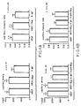
day 30. - Fig. 2A and 2B illustrates the angiographic score seen in the acute (2A) and chronic ischemia models (2B).
- Fig. 3A and 3B illustrates the favorable effect of intramuscular VEGF gene transfer upon revascularization at the capillary level (Fig. 3A VEGF, Fig. 3B control).
- Fig. 4A and 4B illustrates capillary density and capillary/myocyte ratio in both the acute ischemia (4A) and chronic model (4B) of limb ischemia.
- Fig.5A and5B illustrates reduction of the hemodynamic deficit in the ischemic limb in both the acute ischemia (5A) and chronic model (5B) following intramuscular VEGF-transfection as confirmed by measurement of calf blood pressure.
- Fig. 6 illustrates the time course of VEGF expression. Lane 1: marker, Lane 2: positive control, Lane 3: nontransfected muscle, Lane 4: on RT,
Lanes - The present invention provides for use of a nucleic acid capable of expressing a vascular endothelial growth factor (VEGF) angiogenic protein, the VEGF angiogenic protein being an endothelial growth factor, for the manufacture of a medicament for the treatment of ischemia by the injection of the nucleic acid into ischemic tissue.
- The nucleic acid encoding the angiogenic protein is operably linked to a promoter (nucleic acid cassette) to result in expression of the protein when delivered to the ischemic tissue. The resulting expression of the angiogenic protein results in increased blood vessel formation throughout the ischemic tissue. The [methods] use of a nucleic acid of the present invention may be used to treat the ischemic tissue that results from ischemic diseases such as cerebrovascular ischemia, renal ischemia, pulmonaryschema, limbschema, ischemic cardiomyopathy and myocardial ischemia.
- The nucleic acid may be any nucleic acid (DNA or RNA) including genomic DNA, cDNA and mRNA, encoding an angiogenic protein i.e., a protein, polypeptide, mutein or portion that is capable of inducing the formation of new blood vessels. Such proteins include, for example, any protein, polypeptide, mutein or portion thereof that is capable of inducing, either directly or indirectly, the formation of new blood vessels. Folkman, et al., Science, 235:442-447 (1987). These include, for example, vascular endothelial growth factor (VEGF). Muteins or fragments of an angiogenic protein may be used as long as they induce or promote the formation of new blood vessels. VEGF is the angiogenic protein upon which the present invention is predicated.
- Recent investigations have established the feasibility of using recombinant formulations of angiogenic growth factors to expedite and/or augment collateral artery development in animal models of myocardial and hindlimb ischemia. See, Baffour, et al., supra (bFGF); Pu, et al, Circulation, 88:208-215 (1993) (aFGF); Yanagisawa-Miwa, et al., supra (bFGF); Ferrara, et al., Biochem. Biophys. Res. Commun., 161:851-855 (1989) (VEGF).
- In addition, therapeutic angiogenesis has been achieved in the same or closely related models following administration of recombinant endothelial cell growth factor (ECGF) (Pu, et al., Circulation, 88:208-215 (1993)) and VEGF (Takeshita, et al., Circulation, 90:228-234 (1994) supra). Previous studies, employing the animal model of chronic limb ischemia, demonstrated an efficacy of intra-muscular endothelial cell growth factor (ECGF) (Pu, et al., Circulation, 88:208-215 (1993)) or VEGF (Takeshita, et al., Circulation, 90:228-234 (1994) supra) administration.
- VEGF was purified independently as a tumor-secreted factor that included vascular permeability by the Miles assay (Keck, et al, Science, 246:1309-1342 (1989) and Connolly, et al., J. Biol. Chem., 264:20017-20024 (1989)), and thus its alternate designation, vascular permeability factor (VPF). Two features distinguish VEGF from other heparin-binding, angiogenic growth factors. First, the NH2 terminus of VEGF is preceded by a typical signal sequence; therefore, unlike bFGF, VEGF can be secreted by intact cells. Second, its high-affinity binding sites, shown to include the tyrosine kinase receptors Flt-1 and Flt-1/KDR are present on endothelial cells. Ferrara, et al., supra, and Conn, et al., Proc Natl Acad Sci USA, 87:1323-1327 (1990). (Interaction of VEGF with lower affinity binding sites has been shown to induce mononuclear phagocyte chemotaxis). Shen, et al., Blood, 81:2767-2773 (1993) and Clauss, et al., J. Exp. Med., 172:1535-1545 (1990). The DNA encoding VEGF is disclosed in U.S. Patent No. 5,332,671, the disclosure of which is herein incorporated by reference.
- Evidence that VEGF stimulates angiogenesis in vivo had been developed in experiments performed on rat and rabbit cornea (Levy, et al., Growth Factors, 2:9-19 (1989) and Connolly, et al., J. Clin. Invest., 84:1470-1478 (1989)), the chorioallantoic membrane (Ferrara, et al., supra), and the rabbit bone graft model. Connolly, et al., J. Clin. Invest., 84:1470-1478 (1989) supra.
- Preferably, angiogenic proteins used in the context of the present invention contain a secretory signal sequence that facilitates secretion of the protein. VEGF has native signal sequences. Angiogenic proteins that do not have native signal sequences, e.g., bFGF, can be modified to contain such sequences using routine genetic manipulation techniques. See, Nabel et al., Nature, 362:844 (1993).
- The nucleotide sequence of numerous angiogenic proteins, are readily available through a number of computer data bases, for example, GenBank, EMBL and Swiss-Prot. Using this information, a DNA segment encoding the desired protein may be chemically synthesized or, alternatively, such a DNA segment may be obtained using routine procedures in the art, e.g, PCR amplification.
- To simplify the manipulation and handling of the nucleic acid encoding the protein, the nucleic acid is preferably inserted into a cassette where it is operably linked to a promoter. The promoter must be capable of driving expression of the protein in cells of the desired target tissue. The selection of appropriate promoters can readily be accomplished. Preferably, one would use a high expression promoter. An example of a suitable promoter is the 763-base-pair cytomegalovirus (CMV) promoter. The Rous sarcoma virus (RSV) (Davis, et al., Hum Gene Ther 4:151 (1993)) and MMT promoters may also be used. Certain proteins can expressed using their native promoter. Other elements that can enhance expression can also be included such as an enhancer or a system that results in high levels of expression such as a tat gene and tar element. This cassette can then be inserted into a vector, e.g., a plasmid vector such as pUC118, pBR322, or other known plasmid vectors, that includes, for example, an E. coli origin of replication. See, Sambrook, et al., Molecular Cloning: A Laboratory Manual, Cold Spring Harbor Laboratory press, (1989). The plasmid vector may also include a selectable marker such as the β-lactamase gene for ampicillin resistance, provided that the marker polypeptide does not adversely effect the metabolism of the organism being treated. The cassette can also be bound to a nucleic acid binding moiety in a synthetic delivery system, such as the system disclosed in WO 95/22618.
- If desired, the DNA may also be used with a microdelivery vehicle such as cationic liposomes and adenoviral vectors. For a review of the procedures for liposome preparation, targeting and delivery of contents, see Mannino and Gould-Fogerite, BioTechniques, 6:682 (1988). See also, Felgner and Holm, Bethesda Res. Lab. Focus, 11 (2):21 (1989) and Maurer, R.A., Bethesda Res. Lab. Focus, 11 (2):25 (1989).
- Replication-defective recombinant adenoviral vectors, can be produced in accordance with known techniques. See, Quantin, et al., Proc. Natl. Acad. Sci. USA, 89:2581-2584 (1992); Stratford-Perricadet, et al., J. Clin. Invest., 90:626-630 (1992); and Rosenfeld, et al., Cell, 68:143-155 (1992).
- The effective dose of the nucleic acid will be a function of the particular expressed protein, the target tissue, the patient and his or her clinical condition. Effective amount of DNA are between about 1 and 4000µg, more preferably about 1000 and 2000, most preferably between about 2000 and 4000.
- In certain situations, it may be desirable to use nucleic acid's encoding two or more different proteins in order to optimize the therapeutic outcome. At least one of the nucleic acids must in any case encode a VEGF. For example, DNA encoding two angiogenic proteins, e.g., VEGF and bFGF, can be used. Or the angiogenic factor can be combined with other genes or their encoded gene products to enhance the activity of targeted cells, while simultaneously inducing angiogenesis, including, for example, nitric oxide synthase, L-arginine, fibronectin, urokinase, plasminogen activator and heparin.
- In order to facilitate injection, the nucleic is formulated with a pharmaceutically acceptable carrier. Examples of suitable carriers include, saline, albumin, dextrose and sterile water. The nucleic acid is injected into the ischemic tissue using standard injection techniques by use of, for example, a hypodermic needle. Hypodermic needle sizes between no. 29 to no. 16 are preferred.
- The nucleic acid may also be injected by an externally applied local injection apparatus, such as that used to inject antigens for allergy testing; or a transcutaneous "patch" capable of delivery to subcutaneous muscle.
- The nucleic acid can be injected at multiple sites throughout the ischemic tissue.
- Once injected, the nucleic acid capable of expressing the desired angiogenic protein is taken up and expressed by the cells of the tissue. Because the vectors containing the nucleic acid of interest are not normally incorporated into the genome of the cells, expression of the protein of interest takes place for only a limited time. Typically, the angiogenic protein is only expressed in therapeutic levels for about two days to several weeks, preferably for about 1-2 weeks. Reinjection of the DNA can be utilized to provide additional periods of expression of the angiogenic protein. If desired, use of a retrovirus vector to incorporate the heterologous DNA into the genome of the cells will increase the length of time during which the therapeutic polypeptide is expressed, from several weeks to indefinitely.
- Expression of the angiogenic protein and its secretion from the tissue cells induces angiogenesis, allowing for the treatment of ischemia and thus diseases such as limb ischemia, cerebrovascular ischemia, renal ischemia, pulmonary ischemia, ischemic cardiomyopathy and myocardial ischemia.
- The present invention is further illustrated by the following examples.
- Complementary DNA clones for recombinant human VEFG165, isolated from cDNA libraries prepared from HL60 leukemia cells, were assembled into a simple eukaryotic expression plasmid that utilizes 736 bp cytomegalovirus promoter/enhancer to drive VEGF expression. Downstream from the VEGF cDNA is an SV40 polyadenylation sequence. Also included in this plasmid is a fragment containing the SV40 origin of replication that includes the 72 bp repeat, but this sequence is not functionally relevant (for autonomous replication) in the absence of SV40 T antigen. These fragments occur in the pUC118 vector which includes an E. coli origin of replication and the β-galactosidase gene for ampicillin resistance. The biological activity of VEGF165 secreted from cells transfected with this construct (phVEGF165) was previously confirmed by evidence that media conditioned by transfected human 293 cells promoted the proliferation of capillary cells (Leung, et al., Science, 246:1306-9 (1989)).
- The plasmid pGSVLacZ (courtesy of Dr. Claire Bonnerot) containing a nuclear targeted β-galactosidase sequence coupled to the simian virus 40 early promoters (Bonnerot, et al., Proc Natl Acad Sci, U.S.A., 84:6795-9 (1987)) was used for all the control transfection experiments.
- New Zealand white rabbits with operatively induced unilateral hindlimb vascular insufficiency, (Takeshita, et al., Circulation, 90:228-234 (1994) supra; Takeshita, et al. , J. Clin. Invest. 93:662-70 (1994), supra; Pu, et al., Circulation, 88:208-215 (1993) supra, were used to model both acute and chronic ischemia. All protocols were approved by the Institutional Animal Care and Use Committee. The care of animals complied with the guidelines of the Canadian Council of Animal Care, the Principles of Laboratory Animal Care, and the Guide for the Care and Use of Laboratory Animals (NIH publication No. 80-23, revised 1985). Fifty-nine male New Zealand White rabbits (mean weight =3 kg) were anesthetized with ketamine (50 mg/kg) and xylazine (5 mg/kg). Through a longitudinal incision performed in a medial thigh, the femoral artery was dissected free along its entire length, as were all major branches of the femoral artery, including the inferior epigastric, deep femoral, lateral circumflex and superficial epigastric arteries. After further dissecting the popliteal and saphenous arteries distally, the external iliac artery as well as all of the above arteries were ligated. Finally, the femoral artery was completely excised from its proximal origin as a branch of the external iliac artery to the point distally where it bifurcates into the saphenous and popliteal arteries.
- Acute limb ischemia. Twenty-eight rabbits were used to study the impact of IM gene transfer on acute hindlimb ischemia. Immediately following femoral artery excision as outlined above, five different sites in three major thigh muscles were injected directly with plasmid DNA using a 3 ml syringe and 27-gauge needle advanced through a small skin incision. For each injection, the tip of the needle was inserted into the adductor (2 sites), medial large (2 sites), and semimembranous muscles; care was taken, by directly visualizing each muscle during the injection, to avoid penetrating the muscle with the injectate. To the same end, the rate of injection was in each case slowed to approximately 5 sec so that injected solution would not leak through the epimysium. This injection technique was used to administer a total volume of 2.5 ml of a) a 500µg phVEGF165 in saline (n = 8; b) 500 µg phVEGF165 in 0.75% bupivacaine, previously shown to enhance transgene uptake by striated muscle (n = 10) (Danko I, Gene Therapy, 1:114-21 (1994); or c) 500 µg pGSVLacZ encoding nuclear targeted β-galactosidase (n=10). After completing 5 injections (0.5 ml @ for each animal), the skin was then closed using 4.0 nylon.
- Chronic limb ischemia. Thirty-one rabbits were used to study the effects of IM gene therapy for chronic hindlimb ischemia. The sole distinction between the chronic ischemia model and model of acute limb ischemia described above, is that an interval of 10 days was permitted for post-operative recovery, including development of endogenous collateral vessels. Accordingly, 10 days following femoral artery excision, the rabbits were returned to the catherization laboratory. After completing baseline physiological measurements described below, IM gene transfer using the identical technique described above was performed with a) 500 µg phVEGF165 diluted in 2.5 ml of saline (n=8); b) 500 µg phVEGF165 diluted in 0.75 % bupivacaine (n = 8); c) 500 µg of pGSVLacZ diluted in 2.5 ml of saline; or d) 500 µg of pGSVLacZ diluted in 2.5 ml of 0.75% bupivacaine (n=8). In each case, after completing 5 all injections, the skin was closed as above.
- Selective angiography. Selective internal iliac arteriography was performed as previously described (Doucette, et al., Circulation, 85:1899-1911 (1992)). Briefly, a 3 Fr. infusion catheter (Tracker-18, Target Therapeutic, San Jose CA) was introduced into the common carotid artery and advanced to the internal iliac artery of the ischemic limb using 0.014 in. guidewire (Hi-Torque Floppy II, Advanced Cardiac System, San Diego, CA) under fluoroscopic guidance. The tip of catheter was positioned in the internal iliac artery at the level of the interspace between the seventh lumber and the first sacral vertebrae. Following intraarterial injection of nitroglycerin (0.25 mg, SoloPak Laboratories, Franklin Park, IL), a total of 5 ml of non-ionic contrast media (Isovue-370, Squibb Diagnostics, New Brunswick, NJ) was then injected using an automated angiographic injector (Medrad, Pittsburgh, PA) programmed to reproducibly deliver a flow rate of 1 ml/sec. Serial images of the ischemic hindlimb were then recorded on 105-mm spot film at a rate of 1 film/sec for 10 sec.
- Morphometric angiographic analysis of collateral vessel development was performed using a grid overlay comprised of 2.5 mm-diameter circles arranged in rows spaced 5 mm apart. This overlay was placed over the 4-sec angiogram recorded at the level of the medial thigh. A defined area was chosen in which the number of contrast-opacified arteries crossing over circles as well as the total number of circles encompassing the medial thigh area were counted in single blind fashion. An angiographic score was calculated for each film as the ratio of crossing opacified arteries divided by the total number of circles in the defined area of the ischemic thigh.
- Capillary Density and Capillary/Myocyte Ratio. Anatomic evidence of collateral artery formation was further examined by measuring the number of capillaries in light microscopic sections taken from the ischemic hindlimbs (Takeshita, et al., J. Clin. Invest., 93:662-70 (1994) supra). Tissue specimens were obtained as transverse sections from the adductor muscle, the major muscle of the medial thigh, of the ischemic limb at the time of sacrifice (day 30). Muscle samples were embedded in O.C.T. compound (Miles, Elkhart, IN) and snap-frozen in liquid nitrogen. Multiple frozen sections (5 µm in thickness) were then cut from each specimen on a cryostat (Miles), so that the muscle fibers were oriented in a transverse fashion. Tissue sections were stained for alkaline phosphatase using an indoxyl-tetrazolium method to detect capillary endothelial cells as previously described, and were then counterstained with eosin. A total of 20 different fields from one muscle section were randomly selected, and the number of capillaries was counted under a 20X objective to determine the capillary density (mean number of capillaries/mm2). To ensure that analysis of capillary density was not overestimated due to muscle atrophy, or underestimated due to interstitial edema, capillaries identified at necropsy were also evaluated as a function of muscle fibers in the histologic section. The counting scheme used to compute the capillary/myocyte ratio was otherwise identical to that used to compute capillary density.
- Calf blood pressure. Calf blood pressure was measured using a Doppler Flowmeter (Model 1059, Parks Medical Electronics, Aloha, OR). The pulse of the posterior tibial artery was identified using a Doppler probe, and the systolic blood pressure in both limbs was determined using standard techniques (Takeshita, et al., J. Clin Invest, 93:662-70 (1994)). The calf blood pressure ratio was defined for each rabbit as the ratio of systolic pressure of the ischemic limb to that of the normal limb.
- Intra-arterial Doppler assessment was also performed on
day 0 and 30 before selective internal iliac angiography. The tip of the 3 Fr. infusion catheter was positioned 2 cm above the aortic bifurcation. A total of 5 ml of non-ionic contrast media (Isovue-370, Squibb Diagnostics, New Brunswick, NJ) was injected using an automated angiographic injector (Medrad, Pittsburgh, PA) at a rate of 1 ml/sec. Serial images of the aorta-iliac bifurcation were then recorded on 105-mm spot film at a rate of 1 film/sec for 5 sec. The 0.018 in. Doppler guidewire (Cardiometrics, Inc., Mountain View, CA) was then used to measure blood flow velocity as previously described (Doucette, et al., supra). The wire was advanced via the 3 Fr. infusion catheter positioned at the origin of the common iliac artery, to the proximal segment of the internal iliac artery supplying the ischemic limb. The Doppler wire records a real-time, spectral analysis of the Doppler signal, from which the average peak velocity (APV, the temporal average of the instanteous peak velocity waveform) is calculated and displayed on line. We required a stabilized velocity for 2 min before recording resting APV. Maximum APV was recorded after bolus injection of papaverine (Sigma, St. Louis, MO), 2 mg in 0.4 ml saline, via the infusion catheter. The Doppler wire was then pulled back from the internal iliac artery and readvanced to the common iliac artery of the normal limb; the distal tip of the 3 Fr. infusion catheter was repositioned at the origin of the common iliac artery. Blood flow velocity was again recorded at rest and after papaverine injection. After completing all Doppler measurements, the 3 Fr. infusion catheter was redirected to the promixal segment of the internal iliac artery of the ischemic limb, and selective internal iliac angiography was performed as described above. - Quantitative Analysis of Angiography and Flow Calculation. The angiographic luminal diameter of the internal iliac artery in the ischemic limb and of the external iliac artery in the normal limb were determined using an automated edge-detection system (Quantum 2000I;QCS, Ann Arbor, MI). The film selected for analysis was scanned with a high resolution video camera; the signal produced by the video camera was digitized and displayed on a video monitor (Laser Scan; ImageComm, Santa Clara, CA). Center-lines were traced manually for a 10-mm long segment beginning immediately distal to the tip of the Doppler wire. The contours were subsequently detected automatically on the basis of the weighted sum of first and second derivative functions applied to the digitized brightness information. The vascular diameter was then measured at the site of the Doppler sample volume (5 mm distal to the wire tip). Cross-sectional area was calculated assuming a circular lumen.
- Doppler-derived flow was calculated as QD = (πd2 / 4)(0.5 x APV) where QD = Doppler-derived time average flow, d = vessel diameter, and APV = time average of the spectral peak velocity. The mean velocity was estimated as 0.5 x APV by assuming a time-averaged parabolic velocity profile across the vessel. The Doppler-derived flow calculated in this fashion has been shown to correlate with flow measurements determined by electromagnetic flow meters both in vitro and in vivo (Doucette, et al., supra). As 2 mg of papaverine had no effect on vessel diameter, we thus used the diameter measurements from the angiogram recorded immediately before the Doppler measurements for the calculation of both rest and maximum flow.
- Regional Blood Flow to Limb Muscle. Regional tissue perfusion of animals with chronic hindlimb ischemia was determined using colored microspheres, 15µm in diameter (Kowalik, Circulation 83, 974-82 (1991). After the completion of the invasive measurements described above, 3x10⁶ of Dye-Trak colored microspheres (Triton Technology, Inc., San Diego, CA) were injected through a 3 Fr. Teflon catheter into the left ventricle via the common carotid artery. To collect blood samples for a reference flow, a second catheter was inserted into lower abdominal aorta via the common carotid artery and connected to a syringe withdrawal pump (Sage 351, Orion Research, Boston, MA). Through this catheter, the blood sample was withdrawn 10 sec prior to the microsphere injection over 3 min at a rate of 1.2 ml/min. The animals were then sacrificed and tissue samples (weight = 2g) of 2 different muscles (transfected medial thigh (adductor) muscle, and lower limb (gastrocnemius) muscle) of each hindlimb (ischemic and non-ischemic) were retrieved. The tissue samples and reference blood samples were digested with potassium hydroxide, following which microspheres were retrieved by vacuum filtering. After dyes were extracted from microspheres using dimethyl-formamide, the photometric absorption of each sample was determined using a conventional spectrophotometer (Model 8452A, Hewlett Packard, Palo Alto, CA.) Regional blood flow to muscle was calculated as follows:

- To evaluate expression of phVEGF165 gene in skeletal muscle, sixteen additional male New Zealand white rabbits from both acute and chronic ischemia models (2 rabbits at each time point) were sacrificed at 3, 7, 14 and 30 days post-transfection. The presence of human VEGF mRNA was detected using reverse transcription-polymerase chain reaction (RT-PCR) as previously described (Takeshita, et al., Proc Natl Acad Sci (In press), supra). To ensure specificity and avoid amplification of endogenous rabbit VEGF, primers were selected from a region which is not conserved among different species. Sequences of primers used were: 5'-GAGGGCAGAATCATCACGAAGT-3' (sense)(SEQ ID NO:1); 5'-TCCTATGTGCTGGCCTTGGTGA-3' (antisense)(SEQ ID NO:2). RT-PCR products were analyzed by 2% agarose gel electrophoresis. DNA bands were visualized under UV illumination after staining with ethidium bromide.
- Results were expressed as mean±standard deviation (SD). Statistical significance was evaluated using ANOVA followed by Scheffe's test. A value of p < 0.05 was interpreted to denote statistical significance.
- Angiography. Representative angiograms recorded from both control and VEGF-treated animals at
day 30 are illustrated in Fig. 1. In control animals, collateral artery development in the medial thigh typically appeared unchanged or progressed only slightly in serial angiograms recorded atdays 0 and 30. In contrast, in the VEGF-transfected group, marked progression of collateral artery was observed betweendays 0 and 30. As illustrated in Fig. 2, at baseline (day 0), there were no significant differences in angiographic score among groups (C I: 0.47±0.10, C II: 0.44±0.10, C III: 0.43±0.06, C IV: 0.42±0.10). Byday 30, however, the angiographic score of each VEGF-transfected group was significantly improved compared to that of control (C I: 0.76±0.05, C II: 0.72±0.05, C III: 0.52±0.06, C IV: 0.58±0.09, p<.01) as well as the acute model of limb ischemia (A I: 0.72±0.06, A II: 0.71 ±0.03, A III 0.48±0.10, p<.01). Administration of bupivacaine had no observable effect. - Capillary density and capillary/myocyte ratio. A favorable effect of intramuscular VEGF gene transfer upon revascularization was also apparent at the capillary level (Fig. 3A and 3B). The adductor muscle of ischemic limbs was histologically examined at
day 30 in both models. As illustrated in Fig. 4, VEGF-transfected animals had more capillaries around muscle fibers, in acute ischemia (A I: 248±37, A II: 228±22, A III: 1 88 ± 32/mm2, p < .01) as well as the chronic model of limb ischemia (C I: 259 ± 24, C II: 256 ± 31, C III: 197 ± 18, C IV: 192 ± 31 /mm2, p<.01). Analysis of capillary/myocyte ratio was also higher in VEGF-transfected animals in both the acute and chronic of hindlimb ischemia (acute model: A I: 0.73±0.07, A II: 0.70±0.06, A III: 0.61 ±0.09, p<.01; chronic model: C I: 0.78±0.08, C II: 0.76±0.05, C III: 0.51 ±0.06, C IV: 0.55±0.09, p<.01). No differences were observed between animals receiving bupivacaine versus those injected with saline. - Calf Blood Pressure. Reduction of the hemodynamic deficit in the ischemic limb following intramuscular VEGF-transfection was confirmed by measurement of calf blood pressure. As illustrated in Fig. 5, in animals with acute ischemia, the blood pressure ratio measured on
day 30 post-transfection was significantly higher in VEGF-transfected groups than controls (A I: 0.80±0.09; A II: 0.76 ± 0.1 1; A III: 0.56±0.10, p<.01). There was no difference between bupivacaine and saline treated animals. In chronic model, 10 days after induction of ischemia (immediately prior to transfection), the calf blood pressure ratio was virtually identical in all groups (C I: 0.36 ± 0.05; C II: 0.36 ± 0.04; C III: 0.36 ± 0.05; C IV: 0.32 ± 0.03). Byday 30 post-transfection, the blood pressure ratio for the VEGF-transfected groups was significantly higher than for the control (C I: 0.84±0.09; C II: 0.82±0.06; C III: 0.67±0.06; C IV: 0.66±0.10, p<.01). There was no difference between bupivacaine and saline treated animals. - Intro-arterial Doppler guidewire measurement (Table 1). In acute ischemia model, VEGF-transfected animals revealed significantly higher flow to the ischemic limb at rest (A I: 21.6 ± 5.2, A II: 19.5 ± 3.2, A III: 13.9±3.7 ml/min. p<.01) and at papaverine induced hyperemic status (A I: 59.1 ± 16.9, A II: 55.2±5.1, A III: 39.0 ± 11.5, ml/min, p<.01). In chronic model, the rest blood flow to ischemic limb as well as papaverine stimulated flow to ischemic limb were identical in all groups at day 0 (rest flow; C I: 13.7 ± 1.5; C II: 15.5 ± 1.4; C III: 13.7 ± 2.2; C IV: 13.4 ± 1.9 ml/min, hyperemic flow; C I: 28.9 ± 3.6, C II: 30.6 ± 3.0, C III: 31.3±3.7, C IV: 28.1 ±1.7 ml/min). On
day 30, VEGF transfected animals revealed significantly higher flow to the ischemic limb at rest (C 1:22.7 ± 4.6, C II: 19.9 ± 2.8, C III: 14.9±1.7, C IV: 14.1 ± 1.5 ml/min, p<.01) and at papaverine induced hyperemic status (C I: 52.5±12.6, C II: 56.0 ± 12.0, C III: 38.4 ± 4.3, C IV: 35.8 ± 5.6 ml/min, p <.05). The baseline and hyperemic flow to the non-ischemic limb was identical in all groups t day 0 as well as atday 30.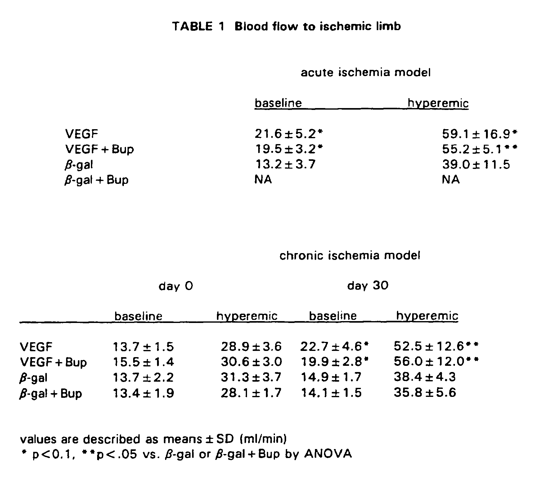
- Regional Blood Flow to Limb Muscle (Table 2). Regional blood flow to ischemic limb muscles was analyzed using colored microsphere technique in chronic ischemia model. Regional blood flow at adductor muscle, transfected medial thigh muscle (C I: 4.3±0.5, C II: 4.6±1.2, C III: 2.9±0.6, C IV: 3.1 ±0.4 ml/min/100g tissue, p<.05), as well as distal lower limb muscle, gastrocnemius (C I: 3.9±0.8, C II: 4.2±0.7, C III: 2.8±0.9, C IV: 2.6±0.7, ml/min/100g tissue, p<.05) was 1.5 folds greater in VEGF transfected animals. No significant differences owing to injected solution were observed. There were no differences in the regional flow to the non-ischemic muscles in all groups (flow to adductor: C I: 5.2±0.5, C II: 5.6±1.0, C III: 4.9±0.6, C IV: 5.6±1.0 ml/min/100g tissue, flow to gastrocnemius: C I: 4.4±1.0, C II: 4.7±1.0; C III: 4.6±1.2, C IV: 5.0±1.1 ml/min/100g tissue).
TABLE 2 Regional blood flow to limb muscle chronic ischemia model adductor gastrocnemius ischemic normal ischemic normal VEGF 4.3±0.5* 5.2±0.5 3.9±0.8* 4.4 ± 1.0 VEGF+Bup 4.6±1.2* 5.6±1.0 4.2±0.7* 4.7±1.0 β-gal 2.9±0.6 4.9±0.6 2.8±0.9 4.6 ± 1.2 β-gal+Bup 3.1 ± 0.4 5.6 ± 1.0 2.6±0.7 5.0±1.1 values are described as means±SD (ml/mm/100g tissue)
* p<0.5 vs. β-gal or β-gal+Bup by ANOVA - To confirm expression of human VEGF gene in transfected rabbit limb muscle in vivo, we analyzed transfected arteries for the presence of human VEGF mRNA by RT-PCR. As indicated above, to ensure the specificity of RT-PCR for human VEGF mRNA resulting from successful transfection (versus endogenous rabbit VEGF mRNA), primers employed were selected from a region which is not conserved among different specie. Adductor muscles were harvested 3, 7, 14 and 30 days after VEGF gene injection. The presence of human VEGF mRNA was readily detected adductor muscles with phVEGF165 from
day 3 to day 14 in both acute and chronic models. Rabbit adductor muscles injected with pGSVLacZ gene were negative for human VEGF mRNA (Figure 6). - We have demonstrated that a gene encoding an angiogenic protein (VEGF) can be successfully transferred into ischemic muscle by injection and where the gene is expressed and induces angiogenesis, providing the ischemic tissue with an increase in blood vessels.
Claims (13)
- Use of a nucleic acid capable of expressing a vascular endothelial growth factor (VEGF) angiogenic protein, the VEGF angiogenic protein being an endothelial growth factor, for the manufacture of a medicament for the treatment of ischemia by the injection of the nucleic acid into ischemic tissue.
- Use according to claim 1, wherein the nucleic acid is DNA.
- Use according to claim 2, wherein said DNA further encodes secretory signal sequence.
- Use according to claim 2 or 3, wherein the DNA is carried by a cationic liposome.
- Use according to any one of claims 2 to 4, wherein at least two DNA's encoding different angiogenic proteins are injected into the human patient.
- Use according to any one of claims 2 to 5, wherein 500µg of the DNA is injected into the tissue.
- Use according to any one of claims 2 to 5, wherein the amount of the DNA injected into the tissue is between about 1,000µg and 2,000µg.
- Use according to any one of claims 2 to 5, wherein the amount of the DNA injected into the tissue is between about 2,000µg and 4,000µg.
- Use according to any one of claims 2 to 5, wherein a volume of 2.5 ml of the DNA in a pharmaceutically acceptable carrier is injected into the tissue.
- Use according to any one of claims 2 to 9, wherein the DNA is injected in a series of five injections into the tissue.
- Use according to any one of claims 2 to 10, wherein the injected DNA is operatively linked to a cytomegalovirus promoter.
- Use according to any one of claims 1 to 11, wherein the ischemic tissue is muscle, brain, kidney or lung.
- Use according to any one of claims 1 to 12, wherein the ischemic tissue has a deficiency in blood as a result of: cerebrovascular ischemia, renal ischemia, pulmonary ischemia, limb ischemia, ischemic cardiomyopathy or myocardial ischemia.
Priority Applications (1)
| Application Number | Priority Date | Filing Date | Title |
|---|---|---|---|
| EP05270064A EP1625858B1 (en) | 1995-10-20 | 1996-10-18 | Compounds for treating ischemic tissue |
Applications Claiming Priority (3)
| Application Number | Priority Date | Filing Date | Title |
|---|---|---|---|
| US545998 | 1995-10-20 | ||
| US08/545,998 US6121246A (en) | 1995-10-20 | 1995-10-20 | Method for treating ischemic tissue |
| PCT/US1996/016723 WO1997014307A1 (en) | 1995-10-20 | 1996-10-18 | Method for treating ischemic tissue |
Related Child Applications (2)
| Application Number | Title | Priority Date | Filing Date |
|---|---|---|---|
| EP05270064A Division EP1625858B1 (en) | 1995-10-20 | 1996-10-18 | Compounds for treating ischemic tissue |
| EP05270064.8 Division-Into | 2005-10-05 |
Publications (3)
| Publication Number | Publication Date |
|---|---|
| EP0883343A1 EP0883343A1 (en) | 1998-12-16 |
| EP0883343A4 EP0883343A4 (en) | 2002-04-10 |
| EP0883343B1 true EP0883343B1 (en) | 2006-12-13 |
Family
ID=24178424
Family Applications (2)
| Application Number | Title | Priority Date | Filing Date |
|---|---|---|---|
| EP05270064A Expired - Lifetime EP1625858B1 (en) | 1995-10-20 | 1996-10-18 | Compounds for treating ischemic tissue |
| EP96936690A Expired - Lifetime EP0883343B1 (en) | 1995-10-20 | 1996-10-18 | Compositions for treating ischemic tissue |
Family Applications Before (1)
| Application Number | Title | Priority Date | Filing Date |
|---|---|---|---|
| EP05270064A Expired - Lifetime EP1625858B1 (en) | 1995-10-20 | 1996-10-18 | Compounds for treating ischemic tissue |
Country Status (8)
| Country | Link |
|---|---|
| US (1) | US6121246A (en) |
| EP (2) | EP1625858B1 (en) |
| JP (3) | JPH11514366A (en) |
| AT (2) | ATE347910T1 (en) |
| AU (1) | AU725290B2 (en) |
| CA (1) | CA2235372C (en) |
| DE (2) | DE69636764T2 (en) |
| WO (1) | WO1997014307A1 (en) |
Cited By (2)
| Publication number | Priority date | Publication date | Assignee | Title |
|---|---|---|---|---|
| US7767643B2 (en) | 2000-12-29 | 2010-08-03 | The Kenneth S. Warren Institute, Inc. | Protection, restoration, and enhancement of erythropoietin-responsive cells, tissues and organs |
| US8404226B2 (en) | 2002-07-03 | 2013-03-26 | The Kenneth S. Warren Institute, Inc. | Tissue protective cytokines for the protection, restoration, and enhancement of responsive cells, tissues and organs |
Families Citing this family (82)
| Publication number | Priority date | Publication date | Assignee | Title |
|---|---|---|---|---|
| US7238673B2 (en) | 1989-03-31 | 2007-07-03 | The Regents Of The University Of Michigan | Treatment of diseases by site-specific instillation of cells or site-specific transformation of cells and kits therefor |
| US6228844B1 (en) | 1991-11-12 | 2001-05-08 | Vical Incorporated | Stimulating vascular growth by administration of DNA sequences encoding VEGF |
| US6706694B1 (en) | 1990-03-21 | 2004-03-16 | Vical Incorporated | Expression of exogenous polynucleotide sequences in a vertebrate |
| WO1995003843A1 (en) * | 1993-07-30 | 1995-02-09 | The Regents Of The University Of California | Endocardial infusion catheter |
| US7109308B1 (en) * | 1994-03-08 | 2006-09-19 | Human Genome Sciences, Inc. | Antibodies to human vascular endothelial growth factor 2 |
| US6040157A (en) | 1994-03-08 | 2000-03-21 | Human Genome Sciences, Inc. | Vascular endothelial growth factor 2 |
| US7186688B1 (en) * | 1994-03-08 | 2007-03-06 | Human Genome Sciences, Inc. | Methods of stimulating angiogenesis in a patient by administering vascular endothelial growth factor 2 |
| DE69434538T2 (en) | 1994-03-08 | 2006-08-10 | Human Genome Sciences, Inc. | Vascular endothelial growth factor 2 |
| US5932540A (en) | 1994-03-08 | 1999-08-03 | Human Genome Sciences, Inc. | Vascular endothelial growth factor 2 |
| US6608182B1 (en) | 1994-03-08 | 2003-08-19 | Human Genome Sciences, Inc. | Human vascular endothelial growth factor 2 |
| US7153827B1 (en) | 1994-03-08 | 2006-12-26 | Human Genome Sciences, Inc. | Vascular endothelial growth factor 2 and methods of use |
| US20030148968A1 (en) * | 1995-02-28 | 2003-08-07 | Hammond H. Kirk | Techniques and compositions for treating cardiovascular disease by in vivo gene delivery |
| JP3431633B2 (en) * | 1995-08-29 | 2003-07-28 | アンジェスエムジー株式会社 | Medicine consisting of HGF gene |
| ES2247621T3 (en) | 1996-02-15 | 2006-03-01 | Biosense Webster, Inc. | EXCAVATION PROBE. |
| US20080305999A1 (en) * | 1996-11-01 | 2008-12-11 | John Francis Martin | Therapeutic use of growth factor, and delivery device, especially for the treatment of intimal hyperplasia |
| JP2001509168A (en) | 1997-01-29 | 2001-07-10 | コーネル リサーチ ファウンデーション、インコーポレイティッド | Multisite delivery of adenovirus vectors to induce angiogenesis |
| WO1998039035A1 (en) * | 1997-03-07 | 1998-09-11 | The Wistar Institute Of Anatomy & Biology | Method and compositions for healing tissue defects and inducing hypervascularity in mammalian tissue |
| CA2304354A1 (en) * | 1997-10-02 | 1999-04-15 | Max-Planck-Gesellschaft Zur Forderung Der Wissenschaften E.V. | Methods for the modulation of neovascularization and/or the growth of collateral arteries and/or other arteries from preexisting arteriolar connections |
| AU749467B2 (en) * | 1997-12-04 | 2002-06-27 | Genzyme Corporation | Compositions and methods for inducing gene expression |
| US6309370B1 (en) | 1998-02-05 | 2001-10-30 | Biosense, Inc. | Intracardiac drug delivery |
| US20040228834A1 (en) * | 1998-03-09 | 2004-11-18 | Jeffrey Isner | Compositions and methods for modulating vascularization |
| US6676937B1 (en) * | 1998-03-09 | 2004-01-13 | Caritas St. Elizabeth's Medical Center Of Boston Inc. | Compositions and methods for modulating vascularization |
| US6958147B1 (en) * | 1998-10-26 | 2005-10-25 | Licentia Ltd | Use of VEGF-C to prevent restenosis |
| US7183448B2 (en) | 1998-11-13 | 2007-02-27 | Daikin Industries, Ltd. | Azeotropic composition, comprising 1, 1, 1, 3,3-pentafluoropropane and 1, 1, 1-trifluoro-3-chloro-2-propene, method of separation and purification of the same, and process for producing 1, 1, 1,3,3-pentafloropropane and 1, 1, 1-trifluoro-3-chloro-2-propene |
| DE19857609A1 (en) | 1998-12-14 | 2000-06-15 | Hannelore Ehrenreich | Use of erythropoietin for the treatment of human cerebral ischemia |
| EP1016726A1 (en) * | 1998-12-30 | 2000-07-05 | Introgene B.V. | Gene therapy to promote angiogenesis |
| US7223724B1 (en) * | 1999-02-08 | 2007-05-29 | Human Genome Sciences, Inc. | Use of vascular endothelial growth factor to treat photoreceptor cells |
| US6440945B1 (en) | 1999-05-27 | 2002-08-27 | Instituto Dermopatico Dell'immacolata | Method of inducing angiogenesis in nonis chemic skeletal muscle |
| US20030236214A1 (en) * | 1999-06-09 | 2003-12-25 | Wolff Jon A. | Charge reversal of polyion complexes and treatment of peripheral occlusive disease |
| EP1067190A1 (en) * | 1999-07-09 | 2001-01-10 | Introgene B.V. | Gene therapy for enhancing and/or inducing angiogenesis |
| DE60044453D1 (en) * | 1999-09-21 | 2010-07-08 | Anges Mg Inc | GENE THERAPY FOR CEREBRAL VASCULAR DISEASES |
| WO2001026694A1 (en) | 1999-10-08 | 2001-04-19 | Medgene Bioscience, Inc. | Gene therapy for cardiomyopathy |
| DK1142590T3 (en) * | 1999-10-29 | 2009-01-26 | Anges Mg Inc | Gene therapy for diabetic ischemic disease |
| US6748258B1 (en) * | 1999-11-05 | 2004-06-08 | Scimed Life Systems, Inc. | Method and devices for heart treatment |
| US6329348B1 (en) | 1999-11-08 | 2001-12-11 | Cornell Research Foundation, Inc. | Method of inducing angiogenesis |
| US20040072785A1 (en) * | 1999-11-23 | 2004-04-15 | Wolff Jon A. | Intravascular delivery of non-viral nucleic acid |
| US6930089B2 (en) | 2000-02-04 | 2005-08-16 | D. Collen Research Foundation Vzw | Use of vascular endothelial growth factor, placenta growth factor or both for preventing or treating ischemic disease or stroke |
| US7727971B2 (en) | 2000-02-04 | 2010-06-01 | Life Sciences Research Partners Vzw | Use of placental growth factor for treating ischemic muscle disease |
| US7105168B1 (en) | 2002-06-04 | 2006-09-12 | D. Collen Research Foundation Vzw | Method of improving ischemic muscle function by administering placental growth factor |
| WO2001057181A2 (en) * | 2000-02-04 | 2001-08-09 | Carmeliet, Peter | Use of vascular endothelial growth factor, placenta growth factor or both for preventing or treating ischemic diseases or stroke |
| CA2413768A1 (en) * | 2000-06-27 | 2002-01-03 | Anges Mg, Inc. | Pharmaceutical compositions for angiogenic therapy |
| AU8473401A (en) * | 2000-08-04 | 2002-02-18 | Human Genome Sciences, Inc. | Vascular endothelial growth factor 2 |
| JPWO2002031138A1 (en) * | 2000-10-06 | 2004-02-19 | 株式会社ディナベック研究所 | Paramyxovirus vectors for introducing foreign genes into skeletal muscle |
| US20040009940A1 (en) * | 2000-10-20 | 2004-01-15 | Coleman Michael E. | Gene delivery formulations and methods for treatment of ischemic conditions |
| US6793918B2 (en) * | 2000-11-01 | 2004-09-21 | Ludwig Institute For Cancer Research | In vivo stimulation of angiogenic activity |
| JP2002142770A (en) * | 2000-11-08 | 2002-05-21 | Dnavec Research Inc | Paramyxovirus vector for transporting gene to circulatory system |
| AU2002224113A1 (en) * | 2000-11-27 | 2002-06-03 | Dnavec Research Inc. | Paramyxovirus vector encoding angiogenesis gene and utilization thereof |
| EP1372678A2 (en) * | 2001-03-09 | 2004-01-02 | Board of Regents, The University of Texas System | Vectors, compositions and methods for treating a vascular disorder |
| US20020131959A1 (en) * | 2001-03-14 | 2002-09-19 | Ivo Buschmann | Means and methods for the modulation of arteriogenesis |
| AU2002303243A1 (en) * | 2001-04-05 | 2002-10-21 | The Johns Hopkins University | Imaging nucleic acid delivery |
| US20050232921A1 (en) * | 2001-04-13 | 2005-10-20 | Rosen Craig A | Vascular endothelial growth factor 2 |
| DK1385864T3 (en) | 2001-04-13 | 2010-08-16 | Human Genome Sciences Inc | Anti-VEGF-2 antibodies |
| CA2444624A1 (en) * | 2001-04-13 | 2002-10-24 | Human Genome Sciences, Inc. | Vascular endothelial growth factor 2 |
| US7182944B2 (en) * | 2001-04-25 | 2007-02-27 | The United States Of America As Represented By The Department Of Health And Human Services | Methods of increasing distribution of nucleic acids |
| DE10122855A1 (en) * | 2001-05-11 | 2002-11-14 | Max Planck Gesellschaft | Means to improve tissue penetration |
| CA2451311A1 (en) * | 2001-06-20 | 2003-01-03 | Ludwig Institute For Cancer Research | Stimulation of vascularization with vegf-b |
| US7217570B2 (en) * | 2001-08-23 | 2007-05-15 | The Wistar Institute Of Anatomy And Biology | Organotypic intestinal culture and methods of use thereof |
| US20040031067A1 (en) * | 2001-10-11 | 2004-02-12 | Meenhard Herlyn | Regulation of human skin healing |
| KR100562824B1 (en) * | 2002-03-20 | 2006-03-23 | 주식회사 바이로메드 | Hybrid hepatocyte growth factor gene that expresses two genes of hepatocyte growth factor with high gene expression efficiency |
| PL195457B1 (en) * | 2002-08-05 | 2007-09-28 | Zaklady Farm Polpharma Sa | Expression cassette, bi-cistron plasmide vector, pharmaceutical composition and their application |
| US20050271639A1 (en) * | 2002-08-22 | 2005-12-08 | Penn Marc S | Genetically engineered cells for therapeutic applications |
| RU2232022C1 (en) * | 2002-12-02 | 2004-07-10 | Государственное образовательное учреждение высшего профессионального образования башкирский государственный медицинский университет | Method for preventing and treating ischemic cardiac disease |
| US7785601B2 (en) | 2002-12-31 | 2010-08-31 | Sygnis Bioscience Gmbh & Co. Kg | Methods of treating neurological conditions with hematopoietic growth factors |
| US6817068B2 (en) * | 2003-01-02 | 2004-11-16 | The Burton Corporation | Adjustable length strap assembly |
| WO2004098534A2 (en) * | 2003-02-24 | 2004-11-18 | Therion Biologics Corporation | Methods for delivering angiogenic modulators using pox viruses |
| US8481504B2 (en) * | 2003-03-12 | 2013-07-09 | Vgx Pharmaceuticals, Inc. | Insulin-like growth factor (IGF-I) plasmid-mediated supplementation for therapeutic applications |
| MXPA05010345A (en) * | 2003-03-27 | 2006-03-08 | Johnson & Johnson | Use of erythropoietin in stroke recovery. |
| KR20060052692A (en) * | 2003-06-05 | 2006-05-19 | 센텔리옹 에스아에스 | Plasmids Encoding Fibroblast Growth Factors for Treating Angiogenic Defects Associated with Hypercholesterolemia or Diabetes |
| US7794713B2 (en) | 2004-04-07 | 2010-09-14 | Lpath, Inc. | Compositions and methods for the treatment and prevention of hyperproliferative diseases |
| US7862812B2 (en) | 2006-05-31 | 2011-01-04 | Lpath, Inc. | Methods for decreasing immune response and treating immune conditions |
| EP2081956B1 (en) | 2006-11-13 | 2013-03-20 | Charité - Universitätsmedizin Berlin | Method of cell culture and method of treatment comprising a vepo protein variant |
| AU2008232739B2 (en) | 2007-03-30 | 2014-03-27 | The Cleveland Clinic Foundation | Method of treating ischemic disorders |
| EP2036571A1 (en) | 2007-09-13 | 2009-03-18 | Sygnis Bioscience GmbH & Co. KG | Use of G-CSF for treating strokes |
| WO2009079451A2 (en) | 2007-12-14 | 2009-06-25 | The Cleveland Clinic Foundation | Compositions and methods of promoting wound healing |
| US20090202606A1 (en) * | 2008-01-25 | 2009-08-13 | Viromed Co., Ltd. | Treatment and Prevention of Cardiac Conditions Using Two or More Isoforms of Hepatocyte Growth Factor |
| ES2556711T3 (en) * | 2008-04-09 | 2016-01-19 | Viromed Co., Ltd. | Lyophilized DNA formulations for increased expression of plasmid DNA |
| CN102740894B (en) | 2009-08-28 | 2015-07-15 | 克利夫兰临床医学基金会 | SDF-1 delivery for treating ischemic tissue |
| ES2549460T3 (en) * | 2009-11-03 | 2015-10-28 | F. Hoffmann-La Roche Ag | NT-pro ANP and sFlt-1 for the differentiation between ischemic and circulatory events |
| RU2426174C1 (en) * | 2010-03-29 | 2011-08-10 | Государственное образовательное учреждение высшего профессионального образования "Курский государственный медицинский университет Федерального агентства по здравоохранению и социальному развитию" | Method of pharmacological correction of extremity ischemia including in l-name induced nitrogen oxide deficiency |
| JP6571537B2 (en) | 2013-03-13 | 2019-09-04 | ザ ユニバーシティー オブ クイーンズランド | Methods for isolating cells for therapy and prevention |
| EP3061457B1 (en) | 2013-10-22 | 2020-01-22 | Helixmith Co., Ltd | Composition for preventing or treating amyotrophic lateral sclerosis using two or more isoforms of hepatocyte growth factor |
| KR20210027493A (en) | 2018-07-19 | 2021-03-10 | 주식회사 헬릭스미스 | Freeze-dried pharmaceutical composition for naked DNA gene therapy |
Family Cites Families (10)
| Publication number | Priority date | Publication date | Assignee | Title |
|---|---|---|---|---|
| EP1026253B2 (en) * | 1989-03-21 | 2012-12-19 | Vical Incorporated | Expression of exogenous polynucleotide sequences in a vertebrate |
| US5693622A (en) * | 1989-03-21 | 1997-12-02 | Vical Incorporated | Expression of exogenous polynucleotide sequences cardiac muscle of a mammal |
| US5703055A (en) * | 1989-03-21 | 1997-12-30 | Wisconsin Alumni Research Foundation | Generation of antibodies through lipid mediated DNA delivery |
| US5328470A (en) * | 1989-03-31 | 1994-07-12 | The Regents Of The University Of Michigan | Treatment of diseases by site-specific instillation of cells or site-specific transformation of cells and kits therefor |
| DE69233725T2 (en) * | 1991-06-28 | 2009-02-12 | The Regents Of The University Of Michigan, Ann Arbor | Use of DNA or RNA for the manufacture of a medicament for the treatment of tumors |
| US5661133B1 (en) * | 1991-11-12 | 1999-06-01 | Univ Michigan | Collateral blood vessel formation in cardiac muscle by injecting a dna sequence encoding an angiogenic protein |
| CA2149771A1 (en) * | 1992-11-18 | 1994-05-26 | Jeffrey M. Leiden | Adenovirus-mediated gene transfer to cardiac and vascular smooth muscle |
| US5593972A (en) * | 1993-01-26 | 1997-01-14 | The Wistar Institute | Genetic immunization |
| AU6778794A (en) * | 1993-04-29 | 1994-11-21 | Washington University | Use of platelet-derived growth factor to improve collateral circulation |
| US5792453A (en) * | 1995-02-28 | 1998-08-11 | The Regents Of The University Of California | Gene transfer-mediated angiogenesis therapy |
-
1995
- 1995-10-20 US US08/545,998 patent/US6121246A/en not_active Expired - Lifetime
-
1996
- 1996-10-18 EP EP05270064A patent/EP1625858B1/en not_active Expired - Lifetime
- 1996-10-18 DE DE69636764T patent/DE69636764T2/en not_active Expired - Lifetime
- 1996-10-18 AT AT96936690T patent/ATE347910T1/en not_active IP Right Cessation
- 1996-10-18 JP JP9516039A patent/JPH11514366A/en not_active Withdrawn
- 1996-10-18 AT AT05270064T patent/ATE419014T1/en not_active IP Right Cessation
- 1996-10-18 EP EP96936690A patent/EP0883343B1/en not_active Expired - Lifetime
- 1996-10-18 AU AU74548/96A patent/AU725290B2/en not_active Ceased
- 1996-10-18 WO PCT/US1996/016723 patent/WO1997014307A1/en not_active Ceased
- 1996-10-18 CA CA002235372A patent/CA2235372C/en not_active Expired - Fee Related
- 1996-10-18 DE DE69637800T patent/DE69637800D1/en not_active Expired - Lifetime
-
2007
- 2007-10-02 JP JP2007259278A patent/JP2008024718A/en not_active Withdrawn
-
2011
- 2011-08-09 JP JP2011174322A patent/JP2011219502A/en not_active Withdrawn
Cited By (2)
| Publication number | Priority date | Publication date | Assignee | Title |
|---|---|---|---|---|
| US7767643B2 (en) | 2000-12-29 | 2010-08-03 | The Kenneth S. Warren Institute, Inc. | Protection, restoration, and enhancement of erythropoietin-responsive cells, tissues and organs |
| US8404226B2 (en) | 2002-07-03 | 2013-03-26 | The Kenneth S. Warren Institute, Inc. | Tissue protective cytokines for the protection, restoration, and enhancement of responsive cells, tissues and organs |
Also Published As
| Publication number | Publication date |
|---|---|
| EP1625858B1 (en) | 2008-12-31 |
| EP0883343A1 (en) | 1998-12-16 |
| JP2008024718A (en) | 2008-02-07 |
| CA2235372C (en) | 2009-01-27 |
| ATE347910T1 (en) | 2007-01-15 |
| EP1625858A1 (en) | 2006-02-15 |
| CA2235372A1 (en) | 1997-04-24 |
| JP2011219502A (en) | 2011-11-04 |
| WO1997014307A1 (en) | 1997-04-24 |
| US6121246A (en) | 2000-09-19 |
| DE69637800D1 (en) | 2009-02-12 |
| DE69636764D1 (en) | 2007-01-25 |
| DE69636764T2 (en) | 2007-10-31 |
| JPH11514366A (en) | 1999-12-07 |
| ATE419014T1 (en) | 2009-01-15 |
| AU725290B2 (en) | 2000-10-12 |
| AU7454896A (en) | 1997-05-07 |
| EP0883343A4 (en) | 2002-04-10 |
Similar Documents
| Publication | Publication Date | Title |
|---|---|---|
| EP0883343B1 (en) | Compositions for treating ischemic tissue | |
| Tabata et al. | Arterial gene transfer of acidic fibroblast growth factor for therapeutic angiogenesis in vivo: critical role of secretion signal in use of naked DNA | |
| Losordo et al. | Gene therapy for myocardial angiogenesis | |
| Tsurumi et al. | Treatment of acute limb ischemia by intramuscular injection of vascular endothelial growth factor gene | |
| Tio et al. | Intramyocardial gene therapy with naked DNA encoding vascular endothelial growth factor improves collateral flow to ischemic myocardium | |
| US6867196B1 (en) | Process for delivering nucleic acids to cardiac tissue | |
| US20030148968A1 (en) | Techniques and compositions for treating cardiovascular disease by in vivo gene delivery | |
| US20090209630A1 (en) | Gene delivery formulations and methods for treatment of ischemic conditions | |
| AU784392B2 (en) | Techniques and compositions for treating cardiovascular disease by in vivo gene delivery | |
| US20070196283A1 (en) | Composition for transfection of DNA into the liver | |
| JP2003513932A (en) | Induction of angiogenesis | |
| US7214369B2 (en) | Devices and processes for distribution of genetic material to mammalian limb | |
| JP2002515065A (en) | Techniques and compositions for treating heart failure and ventricular reconstitution by in vivo delivery of an angiogenic transgene | |
| CA2439584A1 (en) | Nucleic acid constructs, cells transformed therewith and methods utilizing same for inducing liver regeneration and alleviation of portal hypertension | |
| EP1113836A1 (en) | A process for delivering nucleic acids to cardiac tissue | |
| HK1087343A (en) | Compounds for treating ischemic tissue | |
| US20050222075A1 (en) | Process for delivering nucleic acids to cardiac tissue | |
| US7396821B1 (en) | Intravascular delivery of nucleic acid | |
| US20130096500A1 (en) | Nucleic acid based cardiovascular therapeutics | |
| US20050192242A1 (en) | Therapeutic fusion protein transgenes | |
| US20160296674A1 (en) | Nucleic acid based cardiovascular therapeutics | |
| EP1319419A1 (en) | Method of administration of a gene of interest to a vascular tissue | |
| Schratzberger et al. | Angiogenesis and gene therapy | |
| Tsurumi et al. | Molecular and Cellular Cardiology/Gene Transfer: Direct Intramuscular Gene Transfer of Naked DNA Encoding Vascular Endothelial Growth Factor Augments Collateral Development and Tissue Perfusion |
Legal Events
| Date | Code | Title | Description |
|---|---|---|---|
| PUAI | Public reference made under article 153(3) epc to a published international application that has entered the european phase |
Free format text: ORIGINAL CODE: 0009012 |
|
| 17P | Request for examination filed |
Effective date: 19980414 |
|
| AK | Designated contracting states |
Kind code of ref document: A1 Designated state(s): AT BE CH DE DK ES FI FR GB GR IE IT LI LU MC NL PT SE |
|
| RIC1 | Information provided on ipc code assigned before grant |
Free format text: 7A 61K 48/00 A, 7A 01N 43/04 B, 7A 61K 31/70 B |
|
| A4 | Supplementary search report drawn up and despatched |
Effective date: 20020221 |
|
| AK | Designated contracting states |
Kind code of ref document: A4 Designated state(s): AT BE CH DE DK ES FI FR GB GR IE IT LI LU MC NL PT SE |
|
| 17Q | First examination report despatched |
Effective date: 20030919 |
|
| GRAP | Despatch of communication of intention to grant a patent |
Free format text: ORIGINAL CODE: EPIDOSNIGR1 |
|
| RTI1 | Title (correction) |
Free format text: COMPOSITIONS FOR TREATING ISCHEMIC TISSUE |
|
| GRAS | Grant fee paid |
Free format text: ORIGINAL CODE: EPIDOSNIGR3 |
|
| GRAA | (expected) grant |
Free format text: ORIGINAL CODE: 0009210 |
|
| AK | Designated contracting states |
Kind code of ref document: B1 Designated state(s): AT BE CH DE DK ES FI FR GB GR IE IT LI LU MC NL PT SE |
|
| PG25 | Lapsed in a contracting state [announced via postgrant information from national office to epo] |
Ref country code: NL Free format text: LAPSE BECAUSE OF FAILURE TO SUBMIT A TRANSLATION OF THE DESCRIPTION OR TO PAY THE FEE WITHIN THE PRESCRIBED TIME-LIMIT Effective date: 20061213 Ref country code: LI Free format text: LAPSE BECAUSE OF FAILURE TO SUBMIT A TRANSLATION OF THE DESCRIPTION OR TO PAY THE FEE WITHIN THE PRESCRIBED TIME-LIMIT Effective date: 20061213 Ref country code: FI Free format text: LAPSE BECAUSE OF FAILURE TO SUBMIT A TRANSLATION OF THE DESCRIPTION OR TO PAY THE FEE WITHIN THE PRESCRIBED TIME-LIMIT Effective date: 20061213 Ref country code: DK Free format text: LAPSE BECAUSE OF FAILURE TO SUBMIT A TRANSLATION OF THE DESCRIPTION OR TO PAY THE FEE WITHIN THE PRESCRIBED TIME-LIMIT Effective date: 20061213 Ref country code: CH Free format text: LAPSE BECAUSE OF FAILURE TO SUBMIT A TRANSLATION OF THE DESCRIPTION OR TO PAY THE FEE WITHIN THE PRESCRIBED TIME-LIMIT Effective date: 20061213 Ref country code: BE Free format text: LAPSE BECAUSE OF FAILURE TO SUBMIT A TRANSLATION OF THE DESCRIPTION OR TO PAY THE FEE WITHIN THE PRESCRIBED TIME-LIMIT Effective date: 20061213 Ref country code: AT Free format text: LAPSE BECAUSE OF FAILURE TO SUBMIT A TRANSLATION OF THE DESCRIPTION OR TO PAY THE FEE WITHIN THE PRESCRIBED TIME-LIMIT Effective date: 20061213 |
|
| REG | Reference to a national code |
Ref country code: GB Ref legal event code: FG4D |
|
| REG | Reference to a national code |
Ref country code: CH Ref legal event code: EP |
|
| REG | Reference to a national code |
Ref country code: IE Ref legal event code: FG4D |
|
| REF | Corresponds to: |
Ref document number: 69636764 Country of ref document: DE Date of ref document: 20070125 Kind code of ref document: P |
|
| PG25 | Lapsed in a contracting state [announced via postgrant information from national office to epo] |
Ref country code: SE Free format text: LAPSE BECAUSE OF FAILURE TO SUBMIT A TRANSLATION OF THE DESCRIPTION OR TO PAY THE FEE WITHIN THE PRESCRIBED TIME-LIMIT Effective date: 20070313 |
|
| PG25 | Lapsed in a contracting state [announced via postgrant information from national office to epo] |
Ref country code: ES Free format text: LAPSE BECAUSE OF FAILURE TO SUBMIT A TRANSLATION OF THE DESCRIPTION OR TO PAY THE FEE WITHIN THE PRESCRIBED TIME-LIMIT Effective date: 20070324 |
|
| PG25 | Lapsed in a contracting state [announced via postgrant information from national office to epo] |
Ref country code: PT Free format text: LAPSE BECAUSE OF FAILURE TO SUBMIT A TRANSLATION OF THE DESCRIPTION OR TO PAY THE FEE WITHIN THE PRESCRIBED TIME-LIMIT Effective date: 20070514 |
|
| NLV1 | Nl: lapsed or annulled due to failure to fulfill the requirements of art. 29p and 29m of the patents act | ||
| REG | Reference to a national code |
Ref country code: CH Ref legal event code: PL |
|
| ET | Fr: translation filed | ||
| PLBE | No opposition filed within time limit |
Free format text: ORIGINAL CODE: 0009261 |
|
| STAA | Information on the status of an ep patent application or granted ep patent |
Free format text: STATUS: NO OPPOSITION FILED WITHIN TIME LIMIT |
|
| 26N | No opposition filed |
Effective date: 20070914 |
|
| PG25 | Lapsed in a contracting state [announced via postgrant information from national office to epo] |
Ref country code: GR Free format text: LAPSE BECAUSE OF FAILURE TO SUBMIT A TRANSLATION OF THE DESCRIPTION OR TO PAY THE FEE WITHIN THE PRESCRIBED TIME-LIMIT Effective date: 20070314 |
|
| PG25 | Lapsed in a contracting state [announced via postgrant information from national office to epo] |
Ref country code: MC Free format text: LAPSE BECAUSE OF NON-PAYMENT OF DUE FEES Effective date: 20071031 |
|
| PG25 | Lapsed in a contracting state [announced via postgrant information from national office to epo] |
Ref country code: IE Free format text: LAPSE BECAUSE OF NON-PAYMENT OF DUE FEES Effective date: 20071018 |
|
| PGFP | Annual fee paid to national office [announced via postgrant information from national office to epo] |
Ref country code: DE Payment date: 20081201 Year of fee payment: 13 |
|
| PG25 | Lapsed in a contracting state [announced via postgrant information from national office to epo] |
Ref country code: LU Free format text: LAPSE BECAUSE OF NON-PAYMENT OF DUE FEES Effective date: 20071018 Ref country code: DE Free format text: LAPSE BECAUSE OF THE APPLICANT RENOUNCES Effective date: 20090529 |
|
| PGFP | Annual fee paid to national office [announced via postgrant information from national office to epo] |
Ref country code: FR Payment date: 20101105 Year of fee payment: 15 |
|
| PGFP | Annual fee paid to national office [announced via postgrant information from national office to epo] |
Ref country code: IT Payment date: 20101028 Year of fee payment: 15 Ref country code: GB Payment date: 20101025 Year of fee payment: 15 |
|
| GBPC | Gb: european patent ceased through non-payment of renewal fee |
Effective date: 20111018 |
|
| REG | Reference to a national code |
Ref country code: FR Ref legal event code: ST Effective date: 20120629 |
|
| PG25 | Lapsed in a contracting state [announced via postgrant information from national office to epo] |
Ref country code: FR Free format text: LAPSE BECAUSE OF NON-PAYMENT OF DUE FEES Effective date: 20111102 Ref country code: GB Free format text: LAPSE BECAUSE OF NON-PAYMENT OF DUE FEES Effective date: 20111018 Ref country code: IT Free format text: LAPSE BECAUSE OF NON-PAYMENT OF DUE FEES Effective date: 20111018 |