EP0719859A1 - Anti-alpha V-integrin monoclonal antibody - Google Patents
Anti-alpha V-integrin monoclonal antibody Download PDFInfo
- Publication number
- EP0719859A1 EP0719859A1 EP95119233A EP95119233A EP0719859A1 EP 0719859 A1 EP0719859 A1 EP 0719859A1 EP 95119233 A EP95119233 A EP 95119233A EP 95119233 A EP95119233 A EP 95119233A EP 0719859 A1 EP0719859 A1 EP 0719859A1
- Authority
- EP
- European Patent Office
- Prior art keywords
- antibody
- monoclonal antibody
- αvβ3
- cells
- cell
- Prior art date
- Legal status (The legal status is an assumption and is not a legal conclusion. Google has not performed a legal analysis and makes no representation as to the accuracy of the status listed.)
- Granted
Links
- 210000004027 cell Anatomy 0.000 claims abstract description 209
- 108010044426 integrins Proteins 0.000 claims abstract description 80
- 102000006495 integrins Human genes 0.000 claims abstract description 80
- 230000000903 blocking effect Effects 0.000 claims abstract description 21
- 239000000758 substrate Substances 0.000 claims abstract description 19
- 230000005748 tumor development Effects 0.000 claims abstract description 14
- 210000004408 hybridoma Anatomy 0.000 claims abstract description 12
- 108091028043 Nucleic acid sequence Proteins 0.000 claims abstract description 9
- 230000008619 cell matrix interaction Effects 0.000 claims abstract description 6
- 230000001472 cytotoxic effect Effects 0.000 claims abstract description 6
- 125000003275 alpha amino acid group Chemical group 0.000 claims abstract 6
- 108010031318 Vitronectin Proteins 0.000 claims description 60
- 102100035140 Vitronectin Human genes 0.000 claims description 60
- 230000027455 binding Effects 0.000 claims description 49
- 206010028980 Neoplasm Diseases 0.000 claims description 46
- 201000001441 melanoma Diseases 0.000 claims description 36
- 238000002965 ELISA Methods 0.000 claims description 26
- 238000000034 method Methods 0.000 claims description 17
- 108010067306 Fibronectins Proteins 0.000 claims description 9
- 238000010561 standard procedure Methods 0.000 claims description 7
- 230000004614 tumor growth Effects 0.000 claims description 7
- 108010049003 Fibrinogen Proteins 0.000 claims description 5
- 102000008946 Fibrinogen Human genes 0.000 claims description 5
- 102000008394 Immunoglobulin Fragments Human genes 0.000 claims description 5
- 108010021625 Immunoglobulin Fragments Proteins 0.000 claims description 5
- 229940012952 fibrinogen Drugs 0.000 claims description 5
- 230000010261 cell growth Effects 0.000 claims description 4
- 239000008194 pharmaceutical composition Substances 0.000 claims description 4
- 108010047852 Integrin alphaVbeta3 Proteins 0.000 claims description 3
- 230000003053 immunization Effects 0.000 claims description 3
- 102100022337 Integrin alpha-V Human genes 0.000 claims description 2
- 108010048673 Vitronectin Receptors Proteins 0.000 claims description 2
- 239000003937 drug carrier Substances 0.000 claims description 2
- 238000004519 manufacturing process Methods 0.000 claims 3
- 102100037362 Fibronectin Human genes 0.000 claims 1
- 239000003814 drug Substances 0.000 claims 1
- 229940079593 drug Drugs 0.000 claims 1
- 239000003446 ligand Substances 0.000 description 60
- 230000000694 effects Effects 0.000 description 37
- 230000006870 function Effects 0.000 description 37
- 238000003556 assay Methods 0.000 description 31
- 102000005962 receptors Human genes 0.000 description 25
- 108020003175 receptors Proteins 0.000 description 25
- 230000003993 interaction Effects 0.000 description 24
- 230000001404 mediated effect Effects 0.000 description 24
- 239000000872 buffer Substances 0.000 description 22
- 239000012634 fragment Substances 0.000 description 19
- YBJHBAHKTGYVGT-ZKWXMUAHSA-N (+)-Biotin Chemical compound N1C(=O)N[C@@H]2[C@H](CCCCC(=O)O)SC[C@@H]21 YBJHBAHKTGYVGT-ZKWXMUAHSA-N 0.000 description 16
- 238000006243 chemical reaction Methods 0.000 description 16
- 238000004132 cross linking Methods 0.000 description 15
- 238000011161 development Methods 0.000 description 15
- 230000018109 developmental process Effects 0.000 description 15
- TWRXJAOTZQYOKJ-UHFFFAOYSA-L Magnesium chloride Chemical compound [Mg+2].[Cl-].[Cl-] TWRXJAOTZQYOKJ-UHFFFAOYSA-L 0.000 description 14
- 241001529936 Murinae Species 0.000 description 14
- IQFYYKKMVGJFEH-XLPZGREQSA-N Thymidine Chemical compound O=C1NC(=O)C(C)=CN1[C@@H]1O[C@H](CO)[C@@H](O)C1 IQFYYKKMVGJFEH-XLPZGREQSA-N 0.000 description 14
- 238000011580 nude mouse model Methods 0.000 description 14
- 241000699660 Mus musculus Species 0.000 description 13
- 230000012010 growth Effects 0.000 description 13
- 210000004072 lung Anatomy 0.000 description 12
- 108090000765 processed proteins & peptides Proteins 0.000 description 12
- 206010027476 Metastases Diseases 0.000 description 11
- 241001465754 Metazoa Species 0.000 description 11
- FAPWRFPIFSIZLT-UHFFFAOYSA-M Sodium chloride Chemical compound [Na+].[Cl-] FAPWRFPIFSIZLT-UHFFFAOYSA-M 0.000 description 11
- YBYRMVIVWMBXKQ-UHFFFAOYSA-N phenylmethanesulfonyl fluoride Chemical compound FS(=O)(=O)CC1=CC=CC=C1 YBYRMVIVWMBXKQ-UHFFFAOYSA-N 0.000 description 11
- 238000007920 subcutaneous administration Methods 0.000 description 11
- 108010064365 glycyl- arginyl-glycyl-aspartyl-seryl-prolyl-lysine Proteins 0.000 description 10
- 102000004196 processed proteins & peptides Human genes 0.000 description 10
- IYMAXBFPHPZYIK-BQBZGAKWSA-N Arg-Gly-Asp Chemical class NC(N)=NCCC[C@H](N)C(=O)NCC(=O)N[C@@H](CC(O)=O)C(O)=O IYMAXBFPHPZYIK-BQBZGAKWSA-N 0.000 description 9
- 229960002685 biotin Drugs 0.000 description 9
- 239000011616 biotin Substances 0.000 description 9
- 230000021164 cell adhesion Effects 0.000 description 9
- 230000001413 cellular effect Effects 0.000 description 9
- 230000001419 dependent effect Effects 0.000 description 9
- 238000002474 experimental method Methods 0.000 description 9
- 230000005764 inhibitory process Effects 0.000 description 9
- 238000002347 injection Methods 0.000 description 9
- 239000007924 injection Substances 0.000 description 9
- NHBKXEKEPDILRR-UHFFFAOYSA-N 2,3-bis(butanoylsulfanyl)propyl butanoate Chemical compound CCCC(=O)OCC(SC(=O)CCC)CSC(=O)CCC NHBKXEKEPDILRR-UHFFFAOYSA-N 0.000 description 8
- 102000016359 Fibronectins Human genes 0.000 description 8
- 241000699670 Mus sp. Species 0.000 description 8
- 235000020958 biotin Nutrition 0.000 description 8
- 239000003153 chemical reaction reagent Substances 0.000 description 8
- 238000001727 in vivo Methods 0.000 description 8
- 230000002025 microglial effect Effects 0.000 description 8
- 230000003389 potentiating effect Effects 0.000 description 8
- 125000003003 spiro group Chemical group 0.000 description 8
- ZRVZOBGMZWVJOS-VMXHOPILSA-N (2s)-6-amino-2-[[(2s)-1-[(2s)-2-[[(2s)-2-[[2-[[(2s)-2-[(2-aminoacetyl)amino]-5-(diaminomethylideneamino)pentanoyl]amino]acetyl]amino]-3-carboxypropanoyl]amino]-3-hydroxypropanoyl]pyrrolidine-2-carbonyl]amino]hexanoic acid Chemical compound NCCCC[C@@H](C(O)=O)NC(=O)[C@@H]1CCCN1C(=O)[C@H](CO)NC(=O)[C@H](CC(O)=O)NC(=O)CNC(=O)[C@H](CCCN=C(N)N)NC(=O)CN ZRVZOBGMZWVJOS-VMXHOPILSA-N 0.000 description 7
- UXVMQQNJUSDDNG-UHFFFAOYSA-L Calcium chloride Chemical compound [Cl-].[Cl-].[Ca+2] UXVMQQNJUSDDNG-UHFFFAOYSA-L 0.000 description 7
- 230000006820 DNA synthesis Effects 0.000 description 7
- 238000004458 analytical method Methods 0.000 description 7
- 108010072041 arginyl-glycyl-aspartic acid Proteins 0.000 description 7
- 239000001110 calcium chloride Substances 0.000 description 7
- 229910001628 calcium chloride Inorganic materials 0.000 description 7
- 239000002299 complementary DNA Substances 0.000 description 7
- 238000010790 dilution Methods 0.000 description 7
- 239000012895 dilution Substances 0.000 description 7
- 239000012636 effector Substances 0.000 description 7
- 210000002540 macrophage Anatomy 0.000 description 7
- 229910001629 magnesium chloride Inorganic materials 0.000 description 7
- 230000009401 metastasis Effects 0.000 description 7
- HEGSGKPQLMEBJL-RKQHYHRCSA-N octyl beta-D-glucopyranoside Chemical compound CCCCCCCCO[C@@H]1O[C@H](CO)[C@@H](O)[C@H](O)[C@H]1O HEGSGKPQLMEBJL-RKQHYHRCSA-N 0.000 description 7
- 102000004169 proteins and genes Human genes 0.000 description 7
- 108090000623 proteins and genes Proteins 0.000 description 7
- 239000006228 supernatant Substances 0.000 description 7
- DWRXFEITVBNRMK-UHFFFAOYSA-N Beta-D-1-Arabinofuranosylthymine Natural products O=C1NC(=O)C(C)=CN1C1C(O)C(O)C(CO)O1 DWRXFEITVBNRMK-UHFFFAOYSA-N 0.000 description 6
- 108010069514 Cyclic Peptides Proteins 0.000 description 6
- 102000001189 Cyclic Peptides Human genes 0.000 description 6
- IAZDPXIOMUYVGZ-UHFFFAOYSA-N Dimethylsulphoxide Chemical compound CS(C)=O IAZDPXIOMUYVGZ-UHFFFAOYSA-N 0.000 description 6
- KCXVZYZYPLLWCC-UHFFFAOYSA-N EDTA Chemical compound OC(=O)CN(CC(O)=O)CCN(CC(O)=O)CC(O)=O KCXVZYZYPLLWCC-UHFFFAOYSA-N 0.000 description 6
- 108060003951 Immunoglobulin Proteins 0.000 description 6
- IQFYYKKMVGJFEH-UHFFFAOYSA-N beta-L-thymidine Natural products O=C1NC(=O)C(C)=CN1C1OC(CO)C(O)C1 IQFYYKKMVGJFEH-UHFFFAOYSA-N 0.000 description 6
- 238000010367 cloning Methods 0.000 description 6
- 230000003013 cytotoxicity Effects 0.000 description 6
- 231100000135 cytotoxicity Toxicity 0.000 description 6
- 102000018358 immunoglobulin Human genes 0.000 description 6
- 238000001114 immunoprecipitation Methods 0.000 description 6
- 239000003112 inhibitor Substances 0.000 description 6
- 239000000463 material Substances 0.000 description 6
- 239000000203 mixture Substances 0.000 description 6
- HEGSGKPQLMEBJL-UHFFFAOYSA-N n-octyl beta-D-glucopyranoside Natural products CCCCCCCCOC1OC(CO)C(O)C(O)C1O HEGSGKPQLMEBJL-UHFFFAOYSA-N 0.000 description 6
- 230000009257 reactivity Effects 0.000 description 6
- 239000011780 sodium chloride Substances 0.000 description 6
- 229940104230 thymidine Drugs 0.000 description 6
- 230000035899 viability Effects 0.000 description 6
- 238000005406 washing Methods 0.000 description 6
- NFGXHKASABOEEW-UHFFFAOYSA-N 1-methylethyl 11-methoxy-3,7,11-trimethyl-2,4-dodecadienoate Chemical compound COC(C)(C)CCCC(C)CC=CC(C)=CC(=O)OC(C)C NFGXHKASABOEEW-UHFFFAOYSA-N 0.000 description 5
- 108091003079 Bovine Serum Albumin Proteins 0.000 description 5
- 241000699666 Mus <mouse, genus> Species 0.000 description 5
- 229920002684 Sepharose Polymers 0.000 description 5
- 230000009471 action Effects 0.000 description 5
- 230000000295 complement effect Effects 0.000 description 5
- 230000009089 cytolysis Effects 0.000 description 5
- 230000002950 deficient Effects 0.000 description 5
- 239000002158 endotoxin Substances 0.000 description 5
- 239000012894 fetal calf serum Substances 0.000 description 5
- 238000010348 incorporation Methods 0.000 description 5
- 238000011534 incubation Methods 0.000 description 5
- 239000011159 matrix material Substances 0.000 description 5
- 210000000274 microglia Anatomy 0.000 description 5
- 238000001556 precipitation Methods 0.000 description 5
- 238000002360 preparation method Methods 0.000 description 5
- 239000000523 sample Substances 0.000 description 5
- 238000012216 screening Methods 0.000 description 5
- 238000012163 sequencing technique Methods 0.000 description 5
- 238000002415 sodium dodecyl sulfate polyacrylamide gel electrophoresis Methods 0.000 description 5
- 239000000243 solution Substances 0.000 description 5
- 230000001225 therapeutic effect Effects 0.000 description 5
- 108091032973 (ribonucleotides)n+m Proteins 0.000 description 4
- 201000009030 Carcinoma Diseases 0.000 description 4
- LFQSCWFLJHTTHZ-UHFFFAOYSA-N Ethanol Chemical compound CCO LFQSCWFLJHTTHZ-UHFFFAOYSA-N 0.000 description 4
- 230000001745 anti-biotin effect Effects 0.000 description 4
- 230000015572 biosynthetic process Effects 0.000 description 4
- 238000001514 detection method Methods 0.000 description 4
- 238000006471 dimerization reaction Methods 0.000 description 4
- 239000012139 lysis buffer Substances 0.000 description 4
- 230000007246 mechanism Effects 0.000 description 4
- 239000002244 precipitate Substances 0.000 description 4
- 230000002829 reductive effect Effects 0.000 description 4
- 230000002441 reversible effect Effects 0.000 description 4
- 238000012360 testing method Methods 0.000 description 4
- 239000013598 vector Substances 0.000 description 4
- QKNYBSVHEMOAJP-UHFFFAOYSA-N 2-amino-2-(hydroxymethyl)propane-1,3-diol;hydron;chloride Chemical compound Cl.OCC(N)(CO)CO QKNYBSVHEMOAJP-UHFFFAOYSA-N 0.000 description 3
- 241000283707 Capra Species 0.000 description 3
- 108020004414 DNA Proteins 0.000 description 3
- 102000010834 Extracellular Matrix Proteins Human genes 0.000 description 3
- 108010037362 Extracellular Matrix Proteins Proteins 0.000 description 3
- 229910021380 Manganese Chloride Inorganic materials 0.000 description 3
- GLFNIEUTAYBVOC-UHFFFAOYSA-L Manganese chloride Chemical compound Cl[Mn]Cl GLFNIEUTAYBVOC-UHFFFAOYSA-L 0.000 description 3
- 102000018697 Membrane Proteins Human genes 0.000 description 3
- 108010052285 Membrane Proteins Proteins 0.000 description 3
- 238000012408 PCR amplification Methods 0.000 description 3
- DNIAPMSPPWPWGF-UHFFFAOYSA-N Propylene glycol Chemical compound CC(O)CO DNIAPMSPPWPWGF-UHFFFAOYSA-N 0.000 description 3
- 239000004365 Protease Substances 0.000 description 3
- 102000004142 Trypsin Human genes 0.000 description 3
- 108090000631 Trypsin Proteins 0.000 description 3
- 230000004913 activation Effects 0.000 description 3
- 150000001413 amino acids Chemical group 0.000 description 3
- 230000009087 cell motility Effects 0.000 description 3
- 230000005889 cellular cytotoxicity Effects 0.000 description 3
- 239000011248 coating agent Substances 0.000 description 3
- 238000000576 coating method Methods 0.000 description 3
- 239000000539 dimer Substances 0.000 description 3
- 230000002900 effect on cell Effects 0.000 description 3
- 210000002744 extracellular matrix Anatomy 0.000 description 3
- 238000000684 flow cytometry Methods 0.000 description 3
- 229940072221 immunoglobulins Drugs 0.000 description 3
- 230000003834 intracellular effect Effects 0.000 description 3
- 238000002372 labelling Methods 0.000 description 3
- 239000011565 manganese chloride Substances 0.000 description 3
- 239000013612 plasmid Substances 0.000 description 3
- 230000008569 process Effects 0.000 description 3
- 239000000047 product Substances 0.000 description 3
- 230000019491 signal transduction Effects 0.000 description 3
- 238000010186 staining Methods 0.000 description 3
- 238000003786 synthesis reaction Methods 0.000 description 3
- 238000002560 therapeutic procedure Methods 0.000 description 3
- 210000001519 tissue Anatomy 0.000 description 3
- 238000003211 trypan blue cell staining Methods 0.000 description 3
- 239000012588 trypsin Substances 0.000 description 3
- 210000004881 tumor cell Anatomy 0.000 description 3
- 210000003462 vein Anatomy 0.000 description 3
- 102000002260 Alkaline Phosphatase Human genes 0.000 description 2
- 108020004774 Alkaline Phosphatase Proteins 0.000 description 2
- 206010003445 Ascites Diseases 0.000 description 2
- 108010035532 Collagen Proteins 0.000 description 2
- 102000008186 Collagen Human genes 0.000 description 2
- 102000012422 Collagen Type I Human genes 0.000 description 2
- 108010022452 Collagen Type I Proteins 0.000 description 2
- 239000006144 Dulbecco’s modified Eagle's medium Substances 0.000 description 2
- 238000012413 Fluorescence activated cell sorting analysis Methods 0.000 description 2
- 102000009465 Growth Factor Receptors Human genes 0.000 description 2
- 108010009202 Growth Factor Receptors Proteins 0.000 description 2
- 102000012355 Integrin beta1 Human genes 0.000 description 2
- 108010022222 Integrin beta1 Proteins 0.000 description 2
- 206010061309 Neoplasm progression Diseases 0.000 description 2
- 108091034117 Oligonucleotide Proteins 0.000 description 2
- 229930012538 Paclitaxel Natural products 0.000 description 2
- 108091005804 Peptidases Proteins 0.000 description 2
- 239000012979 RPMI medium Substances 0.000 description 2
- 102100037486 Reverse transcriptase/ribonuclease H Human genes 0.000 description 2
- PXIPVTKHYLBLMZ-UHFFFAOYSA-N Sodium azide Chemical compound [Na+].[N-]=[N+]=[N-] PXIPVTKHYLBLMZ-UHFFFAOYSA-N 0.000 description 2
- UIIMBOGNXHQVGW-UHFFFAOYSA-M Sodium bicarbonate Chemical compound [Na+].OC([O-])=O UIIMBOGNXHQVGW-UHFFFAOYSA-M 0.000 description 2
- 108010006785 Taq Polymerase Proteins 0.000 description 2
- JLCPHMBAVCMARE-UHFFFAOYSA-N [3-[[3-[[3-[[3-[[3-[[3-[[3-[[3-[[3-[[3-[[3-[[5-(2-amino-6-oxo-1H-purin-9-yl)-3-[[3-[[3-[[3-[[3-[[3-[[5-(2-amino-6-oxo-1H-purin-9-yl)-3-[[5-(2-amino-6-oxo-1H-purin-9-yl)-3-hydroxyoxolan-2-yl]methoxy-hydroxyphosphoryl]oxyoxolan-2-yl]methoxy-hydroxyphosphoryl]oxy-5-(5-methyl-2,4-dioxopyrimidin-1-yl)oxolan-2-yl]methoxy-hydroxyphosphoryl]oxy-5-(6-aminopurin-9-yl)oxolan-2-yl]methoxy-hydroxyphosphoryl]oxy-5-(6-aminopurin-9-yl)oxolan-2-yl]methoxy-hydroxyphosphoryl]oxy-5-(6-aminopurin-9-yl)oxolan-2-yl]methoxy-hydroxyphosphoryl]oxy-5-(6-aminopurin-9-yl)oxolan-2-yl]methoxy-hydroxyphosphoryl]oxyoxolan-2-yl]methoxy-hydroxyphosphoryl]oxy-5-(5-methyl-2,4-dioxopyrimidin-1-yl)oxolan-2-yl]methoxy-hydroxyphosphoryl]oxy-5-(4-amino-2-oxopyrimidin-1-yl)oxolan-2-yl]methoxy-hydroxyphosphoryl]oxy-5-(5-methyl-2,4-dioxopyrimidin-1-yl)oxolan-2-yl]methoxy-hydroxyphosphoryl]oxy-5-(5-methyl-2,4-dioxopyrimidin-1-yl)oxolan-2-yl]methoxy-hydroxyphosphoryl]oxy-5-(6-aminopurin-9-yl)oxolan-2-yl]methoxy-hydroxyphosphoryl]oxy-5-(6-aminopurin-9-yl)oxolan-2-yl]methoxy-hydroxyphosphoryl]oxy-5-(4-amino-2-oxopyrimidin-1-yl)oxolan-2-yl]methoxy-hydroxyphosphoryl]oxy-5-(4-amino-2-oxopyrimidin-1-yl)oxolan-2-yl]methoxy-hydroxyphosphoryl]oxy-5-(4-amino-2-oxopyrimidin-1-yl)oxolan-2-yl]methoxy-hydroxyphosphoryl]oxy-5-(6-aminopurin-9-yl)oxolan-2-yl]methoxy-hydroxyphosphoryl]oxy-5-(4-amino-2-oxopyrimidin-1-yl)oxolan-2-yl]methyl [5-(6-aminopurin-9-yl)-2-(hydroxymethyl)oxolan-3-yl] hydrogen phosphate Polymers Cc1cn(C2CC(OP(O)(=O)OCC3OC(CC3OP(O)(=O)OCC3OC(CC3O)n3cnc4c3nc(N)[nH]c4=O)n3cnc4c3nc(N)[nH]c4=O)C(COP(O)(=O)OC3CC(OC3COP(O)(=O)OC3CC(OC3COP(O)(=O)OC3CC(OC3COP(O)(=O)OC3CC(OC3COP(O)(=O)OC3CC(OC3COP(O)(=O)OC3CC(OC3COP(O)(=O)OC3CC(OC3COP(O)(=O)OC3CC(OC3COP(O)(=O)OC3CC(OC3COP(O)(=O)OC3CC(OC3COP(O)(=O)OC3CC(OC3COP(O)(=O)OC3CC(OC3COP(O)(=O)OC3CC(OC3COP(O)(=O)OC3CC(OC3COP(O)(=O)OC3CC(OC3COP(O)(=O)OC3CC(OC3COP(O)(=O)OC3CC(OC3CO)n3cnc4c(N)ncnc34)n3ccc(N)nc3=O)n3cnc4c(N)ncnc34)n3ccc(N)nc3=O)n3ccc(N)nc3=O)n3ccc(N)nc3=O)n3cnc4c(N)ncnc34)n3cnc4c(N)ncnc34)n3cc(C)c(=O)[nH]c3=O)n3cc(C)c(=O)[nH]c3=O)n3ccc(N)nc3=O)n3cc(C)c(=O)[nH]c3=O)n3cnc4c3nc(N)[nH]c4=O)n3cnc4c(N)ncnc34)n3cnc4c(N)ncnc34)n3cnc4c(N)ncnc34)n3cnc4c(N)ncnc34)O2)c(=O)[nH]c1=O JLCPHMBAVCMARE-UHFFFAOYSA-N 0.000 description 2
- 230000001464 adherent effect Effects 0.000 description 2
- 102000019997 adhesion receptor Human genes 0.000 description 2
- 108010013985 adhesion receptor Proteins 0.000 description 2
- 229940125528 allosteric inhibitor Drugs 0.000 description 2
- 238000010171 animal model Methods 0.000 description 2
- 239000011324 bead Substances 0.000 description 2
- 230000008827 biological function Effects 0.000 description 2
- 238000007413 biotinylation Methods 0.000 description 2
- 230000006287 biotinylation Effects 0.000 description 2
- 210000004369 blood Anatomy 0.000 description 2
- 239000008280 blood Substances 0.000 description 2
- 210000004556 brain Anatomy 0.000 description 2
- AIYUHDOJVYHVIT-UHFFFAOYSA-M caesium chloride Chemical compound [Cl-].[Cs+] AIYUHDOJVYHVIT-UHFFFAOYSA-M 0.000 description 2
- 238000000423 cell based assay Methods 0.000 description 2
- 230000004663 cell proliferation Effects 0.000 description 2
- 230000003833 cell viability Effects 0.000 description 2
- 230000008614 cellular interaction Effects 0.000 description 2
- 238000005119 centrifugation Methods 0.000 description 2
- 229920001436 collagen Polymers 0.000 description 2
- 229940096422 collagen type i Drugs 0.000 description 2
- 230000002860 competitive effect Effects 0.000 description 2
- 230000004540 complement-dependent cytotoxicity Effects 0.000 description 2
- 230000002596 correlated effect Effects 0.000 description 2
- 230000001085 cytostatic effect Effects 0.000 description 2
- 231100000433 cytotoxic Toxicity 0.000 description 2
- 239000003599 detergent Substances 0.000 description 2
- 230000004069 differentiation Effects 0.000 description 2
- 201000010099 disease Diseases 0.000 description 2
- 208000037265 diseases, disorders, signs and symptoms Diseases 0.000 description 2
- 239000000284 extract Substances 0.000 description 2
- 238000000605 extraction Methods 0.000 description 2
- 230000004927 fusion Effects 0.000 description 2
- 210000005260 human cell Anatomy 0.000 description 2
- 230000002163 immunogen Effects 0.000 description 2
- 230000002401 inhibitory effect Effects 0.000 description 2
- 230000002427 irreversible effect Effects 0.000 description 2
- 230000002147 killing effect Effects 0.000 description 2
- 239000002609 medium Substances 0.000 description 2
- 239000012528 membrane Substances 0.000 description 2
- 230000001394 metastastic effect Effects 0.000 description 2
- 206010061289 metastatic neoplasm Diseases 0.000 description 2
- 239000000178 monomer Substances 0.000 description 2
- 230000001338 necrotic effect Effects 0.000 description 2
- -1 olive oils Chemical compound 0.000 description 2
- 230000003287 optical effect Effects 0.000 description 2
- 229960001592 paclitaxel Drugs 0.000 description 2
- 210000002826 placenta Anatomy 0.000 description 2
- 230000003169 placental effect Effects 0.000 description 2
- 229920001223 polyethylene glycol Polymers 0.000 description 2
- 239000013641 positive control Substances 0.000 description 2
- 230000000750 progressive effect Effects 0.000 description 2
- XJMOSONTPMZWPB-UHFFFAOYSA-M propidium iodide Chemical compound [I-].[I-].C12=CC(N)=CC=C2C2=CC=C(N)C=C2[N+](CCC[N+](C)(CC)CC)=C1C1=CC=CC=C1 XJMOSONTPMZWPB-UHFFFAOYSA-M 0.000 description 2
- 235000019419 proteases Nutrition 0.000 description 2
- 230000002685 pulmonary effect Effects 0.000 description 2
- 238000000746 purification Methods 0.000 description 2
- 230000033300 receptor internalization Effects 0.000 description 2
- 102000027426 receptor tyrosine kinases Human genes 0.000 description 2
- 108091008598 receptor tyrosine kinases Proteins 0.000 description 2
- 230000001105 regulatory effect Effects 0.000 description 2
- 230000000717 retained effect Effects 0.000 description 2
- 102220049163 rs35498994 Human genes 0.000 description 2
- 238000000926 separation method Methods 0.000 description 2
- 210000002966 serum Anatomy 0.000 description 2
- 230000011664 signaling Effects 0.000 description 2
- 239000007787 solid Substances 0.000 description 2
- 230000007480 spreading Effects 0.000 description 2
- 238000003892 spreading Methods 0.000 description 2
- 239000000126 substance Substances 0.000 description 2
- RCINICONZNJXQF-MZXODVADSA-N taxol Chemical compound O([C@@H]1[C@@]2(C[C@@H](C(C)=C(C2(C)C)[C@H](C([C@]2(C)[C@@H](O)C[C@H]3OC[C@]3([C@H]21)OC(C)=O)=O)OC(=O)C)OC(=O)[C@H](O)[C@@H](NC(=O)C=1C=CC=CC=1)C=1C=CC=CC=1)O)C(=O)C1=CC=CC=C1 RCINICONZNJXQF-MZXODVADSA-N 0.000 description 2
- 238000004448 titration Methods 0.000 description 2
- 238000012546 transfer Methods 0.000 description 2
- 230000007704 transition Effects 0.000 description 2
- 230000005747 tumor angiogenesis Effects 0.000 description 2
- 230000005740 tumor formation Effects 0.000 description 2
- 230000005751 tumor progression Effects 0.000 description 2
- YMXHPSHLTSZXKH-RVBZMBCESA-N (2,5-dioxopyrrolidin-1-yl) 5-[(3as,4s,6ar)-2-oxo-1,3,3a,4,6,6a-hexahydrothieno[3,4-d]imidazol-4-yl]pentanoate Chemical compound C([C@H]1[C@H]2NC(=O)N[C@H]2CS1)CCCC(=O)ON1C(=O)CCC1=O YMXHPSHLTSZXKH-RVBZMBCESA-N 0.000 description 1
- NNRFRJQMBSBXGO-CIUDSAMLSA-N (3s)-3-[[2-[[(2s)-2-amino-5-(diaminomethylideneamino)pentanoyl]amino]acetyl]amino]-4-[[(1s)-1-carboxy-2-hydroxyethyl]amino]-4-oxobutanoic acid Chemical compound NC(N)=NCCC[C@H](N)C(=O)NCC(=O)N[C@@H](CC(O)=O)C(=O)N[C@@H](CO)C(O)=O NNRFRJQMBSBXGO-CIUDSAMLSA-N 0.000 description 1
- FJQZXCPWAGYPSD-UHFFFAOYSA-N 1,3,4,6-tetrachloro-3a,6a-diphenylimidazo[4,5-d]imidazole-2,5-dione Chemical compound ClN1C(=O)N(Cl)C2(C=3C=CC=CC=3)N(Cl)C(=O)N(Cl)C12C1=CC=CC=C1 FJQZXCPWAGYPSD-UHFFFAOYSA-N 0.000 description 1
- JKMHFZQWWAIEOD-UHFFFAOYSA-N 2-[4-(2-hydroxyethyl)piperazin-1-yl]ethanesulfonic acid Chemical compound OCC[NH+]1CCN(CCS([O-])(=O)=O)CC1 JKMHFZQWWAIEOD-UHFFFAOYSA-N 0.000 description 1
- XZKIHKMTEMTJQX-UHFFFAOYSA-N 4-Nitrophenyl Phosphate Chemical compound OP(O)(=O)OC1=CC=C([N+]([O-])=O)C=C1 XZKIHKMTEMTJQX-UHFFFAOYSA-N 0.000 description 1
- ODPOAESBSUKMHD-UHFFFAOYSA-L 6,7-dihydrodipyrido[1,2-b:1',2'-e]pyrazine-5,8-diium;dibromide Chemical compound [Br-].[Br-].C1=CC=[N+]2CC[N+]3=CC=CC=C3C2=C1 ODPOAESBSUKMHD-UHFFFAOYSA-L 0.000 description 1
- ITZMJCSORYKOSI-AJNGGQMLSA-N APGPR Enterostatin Chemical compound C[C@H](N)C(=O)N1CCC[C@H]1C(=O)NCC(=O)N1[C@H](C(=O)N[C@@H](CCCN=C(N)N)C(O)=O)CCC1 ITZMJCSORYKOSI-AJNGGQMLSA-N 0.000 description 1
- 208000010507 Adenocarcinoma of Lung Diseases 0.000 description 1
- 229920000936 Agarose Polymers 0.000 description 1
- 238000011729 BALB/c nude mouse Methods 0.000 description 1
- 239000004971 Cross linker Substances 0.000 description 1
- 102000004127 Cytokines Human genes 0.000 description 1
- 108090000695 Cytokines Proteins 0.000 description 1
- 108010014303 DNA-directed DNA polymerase Proteins 0.000 description 1
- 102000016928 DNA-directed DNA polymerase Human genes 0.000 description 1
- QRLVDLBMBULFAL-UHFFFAOYSA-N Digitonin Natural products CC1CCC2(OC1)OC3C(O)C4C5CCC6CC(OC7OC(CO)C(OC8OC(CO)C(O)C(OC9OCC(O)C(O)C9OC%10OC(CO)C(O)C(OC%11OC(CO)C(O)C(O)C%11O)C%10O)C8O)C(O)C7O)C(O)CC6(C)C5CCC4(C)C3C2C QRLVDLBMBULFAL-UHFFFAOYSA-N 0.000 description 1
- 239000005630 Diquat Substances 0.000 description 1
- 238000012286 ELISA Assay Methods 0.000 description 1
- LVGKNOAMLMIIKO-UHFFFAOYSA-N Elaidinsaeure-aethylester Natural products CCCCCCCCC=CCCCCCCCC(=O)OCC LVGKNOAMLMIIKO-UHFFFAOYSA-N 0.000 description 1
- 241000588724 Escherichia coli Species 0.000 description 1
- 101710089384 Extracellular protease Proteins 0.000 description 1
- 206010018338 Glioma Diseases 0.000 description 1
- 108060003393 Granulin Proteins 0.000 description 1
- 239000007995 HEPES buffer Substances 0.000 description 1
- 102000002268 Hexosaminidases Human genes 0.000 description 1
- 108010000540 Hexosaminidases Proteins 0.000 description 1
- 101001008255 Homo sapiens Immunoglobulin kappa variable 1D-8 Proteins 0.000 description 1
- 101001047628 Homo sapiens Immunoglobulin kappa variable 2-29 Proteins 0.000 description 1
- 101001008321 Homo sapiens Immunoglobulin kappa variable 2D-26 Proteins 0.000 description 1
- 101001047619 Homo sapiens Immunoglobulin kappa variable 3-20 Proteins 0.000 description 1
- 101001008263 Homo sapiens Immunoglobulin kappa variable 3D-15 Proteins 0.000 description 1
- 101000829980 Homo sapiens Ral guanine nucleotide dissociation stimulator Proteins 0.000 description 1
- 102000009490 IgG Receptors Human genes 0.000 description 1
- 108010073807 IgG Receptors Proteins 0.000 description 1
- 102000017727 Immunoglobulin Variable Region Human genes 0.000 description 1
- 108010067060 Immunoglobulin Variable Region Proteins 0.000 description 1
- 102100022949 Immunoglobulin kappa variable 2-29 Human genes 0.000 description 1
- 102000008607 Integrin beta3 Human genes 0.000 description 1
- 108010020950 Integrin beta3 Proteins 0.000 description 1
- 108010002350 Interleukin-2 Proteins 0.000 description 1
- 239000007836 KH2PO4 Substances 0.000 description 1
- ZDXPYRJPNDTMRX-VKHMYHEASA-N L-glutamine Chemical compound OC(=O)[C@@H](N)CCC(N)=O ZDXPYRJPNDTMRX-VKHMYHEASA-N 0.000 description 1
- 229930182816 L-glutamine Natural products 0.000 description 1
- 241000239218 Limulus Species 0.000 description 1
- 238000000134 MTT assay Methods 0.000 description 1
- 231100000002 MTT assay Toxicity 0.000 description 1
- 206010027458 Metastases to lung Diseases 0.000 description 1
- 101100370002 Mus musculus Tnfsf14 gene Proteins 0.000 description 1
- 241000204031 Mycoplasma Species 0.000 description 1
- 208000003788 Neoplasm Micrometastasis Diseases 0.000 description 1
- 208000007256 Nevus Diseases 0.000 description 1
- 239000000020 Nitrocellulose Substances 0.000 description 1
- 241000283973 Oryctolagus cuniculus Species 0.000 description 1
- 102000004264 Osteopontin Human genes 0.000 description 1
- 108010081689 Osteopontin Proteins 0.000 description 1
- 108090000526 Papain Proteins 0.000 description 1
- 229930040373 Paraformaldehyde Natural products 0.000 description 1
- 102000057297 Pepsin A Human genes 0.000 description 1
- 108090000284 Pepsin A Proteins 0.000 description 1
- 102000004160 Phosphoric Monoester Hydrolases Human genes 0.000 description 1
- 108090000608 Phosphoric Monoester Hydrolases Proteins 0.000 description 1
- 206010035226 Plasma cell myeloma Diseases 0.000 description 1
- 239000002202 Polyethylene glycol Substances 0.000 description 1
- 229920001213 Polysorbate 20 Polymers 0.000 description 1
- 229940124158 Protease/peptidase inhibitor Drugs 0.000 description 1
- 102100024952 Protein CBFA2T1 Human genes 0.000 description 1
- 101000781681 Protobothrops flavoviridis Disintegrin triflavin Proteins 0.000 description 1
- 206010056342 Pulmonary mass Diseases 0.000 description 1
- 239000012980 RPMI-1640 medium Substances 0.000 description 1
- 102100023320 Ral guanine nucleotide dissociation stimulator Human genes 0.000 description 1
- 239000006146 Roswell Park Memorial Institute medium Substances 0.000 description 1
- 239000012722 SDS sample buffer Substances 0.000 description 1
- 208000000453 Skin Neoplasms Diseases 0.000 description 1
- 238000000692 Student's t-test Methods 0.000 description 1
- 101710120037 Toxin CcdB Proteins 0.000 description 1
- 239000007983 Tris buffer Substances 0.000 description 1
- 241000700605 Viruses Species 0.000 description 1
- 230000003187 abdominal effect Effects 0.000 description 1
- 239000004480 active ingredient Substances 0.000 description 1
- 239000000654 additive Substances 0.000 description 1
- 208000009956 adenocarcinoma Diseases 0.000 description 1
- 230000002776 aggregation Effects 0.000 description 1
- 238000004220 aggregation Methods 0.000 description 1
- 230000003321 amplification Effects 0.000 description 1
- 238000000137 annealing Methods 0.000 description 1
- 239000005557 antagonist Substances 0.000 description 1
- 239000003242 anti bacterial agent Substances 0.000 description 1
- 229940088710 antibiotic agent Drugs 0.000 description 1
- 238000011091 antibody purification Methods 0.000 description 1
- 239000000427 antigen Substances 0.000 description 1
- 102000036639 antigens Human genes 0.000 description 1
- 108091007433 antigens Proteins 0.000 description 1
- 239000003963 antioxidant agent Substances 0.000 description 1
- 239000007864 aqueous solution Substances 0.000 description 1
- 239000003125 aqueous solvent Substances 0.000 description 1
- 108010089975 arginyl-glycyl-aspartyl-serine Proteins 0.000 description 1
- XKRFYHLGVUSROY-UHFFFAOYSA-N argon Substances [Ar] XKRFYHLGVUSROY-UHFFFAOYSA-N 0.000 description 1
- 229910052786 argon Inorganic materials 0.000 description 1
- 238000003491 array Methods 0.000 description 1
- CXQCLLQQYTUUKJ-ALWAHNIESA-N beta-D-GalpNAc-(1->4)-[alpha-Neup5Ac-(2->8)-alpha-Neup5Ac-(2->3)]-beta-D-Galp-(1->4)-beta-D-Glcp-(1<->1')-Cer(d18:1/18:0) Chemical compound O[C@@H]1[C@@H](O)[C@H](OC[C@H](NC(=O)CCCCCCCCCCCCCCCCC)[C@H](O)\C=C\CCCCCCCCCCCCC)O[C@H](CO)[C@H]1O[C@H]1[C@H](O)[C@@H](O[C@]2(O[C@H]([C@H](NC(C)=O)[C@@H](O)C2)[C@H](O)[C@@H](CO)O[C@]2(O[C@H]([C@H](NC(C)=O)[C@@H](O)C2)[C@H](O)[C@H](O)CO)C(O)=O)C(O)=O)[C@@H](O[C@H]2[C@@H]([C@@H](O)[C@@H](O)[C@@H](CO)O2)NC(C)=O)[C@@H](CO)O1 CXQCLLQQYTUUKJ-ALWAHNIESA-N 0.000 description 1
- 230000004071 biological effect Effects 0.000 description 1
- 239000012620 biological material Substances 0.000 description 1
- 230000033228 biological regulation Effects 0.000 description 1
- ZFXVRMSLJDYJCH-UHFFFAOYSA-N calcium magnesium Chemical compound [Mg].[Ca] ZFXVRMSLJDYJCH-UHFFFAOYSA-N 0.000 description 1
- BPKIGYQJPYCAOW-FFJTTWKXSA-I calcium;potassium;disodium;(2s)-2-hydroxypropanoate;dichloride;dihydroxide;hydrate Chemical compound O.[OH-].[OH-].[Na+].[Na+].[Cl-].[Cl-].[K+].[Ca+2].C[C@H](O)C([O-])=O BPKIGYQJPYCAOW-FFJTTWKXSA-I 0.000 description 1
- 125000003178 carboxy group Chemical group [H]OC(*)=O 0.000 description 1
- 239000000969 carrier Substances 0.000 description 1
- 230000020411 cell activation Effects 0.000 description 1
- 230000006037 cell lysis Effects 0.000 description 1
- 238000001516 cell proliferation assay Methods 0.000 description 1
- 239000006285 cell suspension Substances 0.000 description 1
- 230000036755 cellular response Effects 0.000 description 1
- 238000012512 characterization method Methods 0.000 description 1
- 239000002738 chelating agent Substances 0.000 description 1
- 230000001684 chronic effect Effects 0.000 description 1
- 210000001072 colon Anatomy 0.000 description 1
- 150000001875 compounds Chemical class 0.000 description 1
- 239000003431 cross linking reagent Substances 0.000 description 1
- 208000035250 cutaneous malignant susceptibility to 1 melanoma Diseases 0.000 description 1
- 108010050963 cyclo(arginyl-glycyl-aspartyl-phenylalanyl-valyl) Proteins 0.000 description 1
- 238000002784 cytotoxicity assay Methods 0.000 description 1
- 231100000263 cytotoxicity test Toxicity 0.000 description 1
- 238000002298 density-gradient ultracentrifugation Methods 0.000 description 1
- 238000000502 dialysis Methods 0.000 description 1
- 229910003460 diamond Inorganic materials 0.000 description 1
- 239000010432 diamond Substances 0.000 description 1
- 239000005546 dideoxynucleotide Substances 0.000 description 1
- 230000029087 digestion Effects 0.000 description 1
- UVYVLBIGDKGWPX-KUAJCENISA-N digitonin Chemical compound O([C@@H]1[C@@H]([C@]2(CC[C@@H]3[C@@]4(C)C[C@@H](O)[C@H](O[C@H]5[C@@H]([C@@H](O)[C@@H](O[C@H]6[C@@H]([C@@H](O[C@H]7[C@@H]([C@@H](O)[C@H](O)CO7)O)[C@H](O)[C@@H](CO)O6)O[C@H]6[C@@H]([C@@H](O[C@H]7[C@@H]([C@@H](O)[C@H](O)[C@@H](CO)O7)O)[C@@H](O)[C@@H](CO)O6)O)[C@@H](CO)O5)O)C[C@@H]4CC[C@H]3[C@@H]2[C@@H]1O)C)[C@@H]1C)[C@]11CC[C@@H](C)CO1 UVYVLBIGDKGWPX-KUAJCENISA-N 0.000 description 1
- UVYVLBIGDKGWPX-UHFFFAOYSA-N digitonine Natural products CC1C(C2(CCC3C4(C)CC(O)C(OC5C(C(O)C(OC6C(C(OC7C(C(O)C(O)CO7)O)C(O)C(CO)O6)OC6C(C(OC7C(C(O)C(O)C(CO)O7)O)C(O)C(CO)O6)O)C(CO)O5)O)CC4CCC3C2C2O)C)C2OC11CCC(C)CO1 UVYVLBIGDKGWPX-UHFFFAOYSA-N 0.000 description 1
- 239000003085 diluting agent Substances 0.000 description 1
- BNIILDVGGAEEIG-UHFFFAOYSA-L disodium hydrogen phosphate Chemical compound [Na+].[Na+].OP([O-])([O-])=O BNIILDVGGAEEIG-UHFFFAOYSA-L 0.000 description 1
- 229910000397 disodium phosphate Inorganic materials 0.000 description 1
- 231100000673 dose–response relationship Toxicity 0.000 description 1
- 239000000975 dye Substances 0.000 description 1
- 230000001909 effect on DNA Effects 0.000 description 1
- 239000000839 emulsion Substances 0.000 description 1
- 238000005516 engineering process Methods 0.000 description 1
- 210000003743 erythrocyte Anatomy 0.000 description 1
- 150000002148 esters Chemical class 0.000 description 1
- ZMMJGEGLRURXTF-UHFFFAOYSA-N ethidium bromide Chemical compound [Br-].C12=CC(N)=CC=C2C2=CC=C(N)C=C2[N+](CC)=C1C1=CC=CC=C1 ZMMJGEGLRURXTF-UHFFFAOYSA-N 0.000 description 1
- 229960005542 ethidium bromide Drugs 0.000 description 1
- LVGKNOAMLMIIKO-QXMHVHEDSA-N ethyl oleate Chemical compound CCCCCCCC\C=C/CCCCCCCC(=O)OCC LVGKNOAMLMIIKO-QXMHVHEDSA-N 0.000 description 1
- 229940093471 ethyl oleate Drugs 0.000 description 1
- 208000021045 exocrine pancreatic carcinoma Diseases 0.000 description 1
- 239000011536 extraction buffer Substances 0.000 description 1
- 210000002950 fibroblast Anatomy 0.000 description 1
- GNBHRKFJIUUOQI-UHFFFAOYSA-N fluorescein Chemical compound O1C(=O)C2=CC=CC=C2C21C1=CC=C(O)C=C1OC1=CC(O)=CC=C21 GNBHRKFJIUUOQI-UHFFFAOYSA-N 0.000 description 1
- 210000001650 focal adhesion Anatomy 0.000 description 1
- 235000013305 food Nutrition 0.000 description 1
- 238000010230 functional analysis Methods 0.000 description 1
- 238000001502 gel electrophoresis Methods 0.000 description 1
- 238000002523 gelfiltration Methods 0.000 description 1
- 244000144993 groups of animals Species 0.000 description 1
- 239000001963 growth medium Substances 0.000 description 1
- ZJYYHGLJYGJLLN-UHFFFAOYSA-N guanidinium thiocyanate Chemical compound SC#N.NC(N)=N ZJYYHGLJYGJLLN-UHFFFAOYSA-N 0.000 description 1
- 239000000833 heterodimer Substances 0.000 description 1
- 238000005734 heterodimerization reaction Methods 0.000 description 1
- 229920001519 homopolymer Polymers 0.000 description 1
- 238000002649 immunization Methods 0.000 description 1
- 230000016784 immunoglobulin production Effects 0.000 description 1
- 238000003364 immunohistochemistry Methods 0.000 description 1
- 239000012133 immunoprecipitate Substances 0.000 description 1
- 238000000338 in vitro Methods 0.000 description 1
- 230000002779 inactivation Effects 0.000 description 1
- 230000000977 initiatory effect Effects 0.000 description 1
- 238000007912 intraperitoneal administration Methods 0.000 description 1
- 230000009545 invasion Effects 0.000 description 1
- 210000003734 kidney Anatomy 0.000 description 1
- 230000000670 limiting effect Effects 0.000 description 1
- 210000004185 liver Anatomy 0.000 description 1
- 230000004807 localization Effects 0.000 description 1
- 230000007774 longterm Effects 0.000 description 1
- 201000005249 lung adenocarcinoma Diseases 0.000 description 1
- 230000000527 lymphocytic effect Effects 0.000 description 1
- 230000002934 lysing effect Effects 0.000 description 1
- 238000012423 maintenance Methods 0.000 description 1
- 230000003211 malignant effect Effects 0.000 description 1
- 230000010534 mechanism of action Effects 0.000 description 1
- 230000000684 melanotic effect Effects 0.000 description 1
- 108020004999 messenger RNA Proteins 0.000 description 1
- 244000005700 microbiome Species 0.000 description 1
- 230000005012 migration Effects 0.000 description 1
- 238000013508 migration Methods 0.000 description 1
- 239000002480 mineral oil Substances 0.000 description 1
- 235000010446 mineral oil Nutrition 0.000 description 1
- 238000002156 mixing Methods 0.000 description 1
- 230000004048 modification Effects 0.000 description 1
- 238000012986 modification Methods 0.000 description 1
- 238000010369 molecular cloning Methods 0.000 description 1
- 230000009456 molecular mechanism Effects 0.000 description 1
- 229910000402 monopotassium phosphate Inorganic materials 0.000 description 1
- 230000000877 morphologic effect Effects 0.000 description 1
- 238000002703 mutagenesis Methods 0.000 description 1
- 231100000350 mutagenesis Toxicity 0.000 description 1
- 201000000050 myeloid neoplasm Diseases 0.000 description 1
- 210000000822 natural killer cell Anatomy 0.000 description 1
- JPXMTWWFLBLUCD-UHFFFAOYSA-N nitro blue tetrazolium(2+) Chemical compound COC1=CC(C=2C=C(OC)C(=CC=2)[N+]=2N(N=C(N=2)C=2C=CC=CC=2)C=2C=CC(=CC=2)[N+]([O-])=O)=CC=C1[N+]1=NC(C=2C=CC=CC=2)=NN1C1=CC=C([N+]([O-])=O)C=C1 JPXMTWWFLBLUCD-UHFFFAOYSA-N 0.000 description 1
- 229920001220 nitrocellulos Polymers 0.000 description 1
- 239000012457 nonaqueous media Substances 0.000 description 1
- 238000003199 nucleic acid amplification method Methods 0.000 description 1
- 239000002773 nucleotide Substances 0.000 description 1
- 125000003729 nucleotide group Chemical group 0.000 description 1
- 239000004006 olive oil Substances 0.000 description 1
- 150000002895 organic esters Chemical class 0.000 description 1
- 208000008443 pancreatic carcinoma Diseases 0.000 description 1
- 235000019834 papain Nutrition 0.000 description 1
- 229940055729 papain Drugs 0.000 description 1
- 229920002866 paraformaldehyde Polymers 0.000 description 1
- 238000007911 parenteral administration Methods 0.000 description 1
- 230000036961 partial effect Effects 0.000 description 1
- 230000001575 pathological effect Effects 0.000 description 1
- 239000008188 pellet Substances 0.000 description 1
- 229940111202 pepsin Drugs 0.000 description 1
- 239000000137 peptide hydrolase inhibitor Substances 0.000 description 1
- 238000003359 percent control normalization Methods 0.000 description 1
- 230000008823 permeabilization Effects 0.000 description 1
- 239000000546 pharmaceutical excipient Substances 0.000 description 1
- 229920000642 polymer Polymers 0.000 description 1
- 239000000256 polyoxyethylene sorbitan monolaurate Substances 0.000 description 1
- 235000010486 polyoxyethylene sorbitan monolaurate Nutrition 0.000 description 1
- GNSKLFRGEWLPPA-UHFFFAOYSA-M potassium dihydrogen phosphate Chemical compound [K+].OP(O)([O-])=O GNSKLFRGEWLPPA-UHFFFAOYSA-M 0.000 description 1
- 238000011533 pre-incubation Methods 0.000 description 1
- 239000003755 preservative agent Substances 0.000 description 1
- 230000002062 proliferating effect Effects 0.000 description 1
- 230000035755 proliferation Effects 0.000 description 1
- 239000011541 reaction mixture Substances 0.000 description 1
- 230000010076 replication Effects 0.000 description 1
- 238000011160 research Methods 0.000 description 1
- 230000004044 response Effects 0.000 description 1
- 238000009738 saturating Methods 0.000 description 1
- 238000003375 selectivity assay Methods 0.000 description 1
- 235000020183 skimmed milk Nutrition 0.000 description 1
- 201000000849 skin cancer Diseases 0.000 description 1
- 229910000030 sodium bicarbonate Inorganic materials 0.000 description 1
- 239000002904 solvent Substances 0.000 description 1
- 230000000392 somatic effect Effects 0.000 description 1
- 230000009870 specific binding Effects 0.000 description 1
- 238000001228 spectrum Methods 0.000 description 1
- 210000000952 spleen Anatomy 0.000 description 1
- 230000006641 stabilisation Effects 0.000 description 1
- 238000011105 stabilization Methods 0.000 description 1
- 238000011146 sterile filtration Methods 0.000 description 1
- 230000004936 stimulating effect Effects 0.000 description 1
- 210000002784 stomach Anatomy 0.000 description 1
- 238000003860 storage Methods 0.000 description 1
- 239000000725 suspension Substances 0.000 description 1
- 210000000115 thoracic cavity Anatomy 0.000 description 1
- 239000003634 thrombocyte concentrate Substances 0.000 description 1
- 238000000954 titration curve Methods 0.000 description 1
- 231100000331 toxic Toxicity 0.000 description 1
- 230000002588 toxic effect Effects 0.000 description 1
- 102000035160 transmembrane proteins Human genes 0.000 description 1
- 108091005703 transmembrane proteins Proteins 0.000 description 1
- LENZDBCJOHFCAS-UHFFFAOYSA-N tris Chemical compound OCC(N)(CO)CO LENZDBCJOHFCAS-UHFFFAOYSA-N 0.000 description 1
- 235000015112 vegetable and seed oil Nutrition 0.000 description 1
- 239000008158 vegetable oil Substances 0.000 description 1
- 108010047303 von Willebrand Factor Proteins 0.000 description 1
- 102100036537 von Willebrand factor Human genes 0.000 description 1
- 229960001134 von willebrand factor Drugs 0.000 description 1
- XLYOFNOQVPJJNP-UHFFFAOYSA-N water Substances O XLYOFNOQVPJJNP-UHFFFAOYSA-N 0.000 description 1
- 230000003313 weakening effect Effects 0.000 description 1
Images


Classifications
-
- C—CHEMISTRY; METALLURGY
- C07—ORGANIC CHEMISTRY
- C07K—PEPTIDES
- C07K16/00—Immunoglobulins [IGs], e.g. monoclonal or polyclonal antibodies
- C07K16/18—Immunoglobulins [IGs], e.g. monoclonal or polyclonal antibodies against material from animals or humans
- C07K16/36—Immunoglobulins [IGs], e.g. monoclonal or polyclonal antibodies against material from animals or humans against blood coagulation factors
-
- C—CHEMISTRY; METALLURGY
- C07—ORGANIC CHEMISTRY
- C07K—PEPTIDES
- C07K16/00—Immunoglobulins [IGs], e.g. monoclonal or polyclonal antibodies
- C07K16/18—Immunoglobulins [IGs], e.g. monoclonal or polyclonal antibodies against material from animals or humans
- C07K16/28—Immunoglobulins [IGs], e.g. monoclonal or polyclonal antibodies against material from animals or humans against receptors, cell surface antigens or cell surface determinants
- C07K16/2839—Immunoglobulins [IGs], e.g. monoclonal or polyclonal antibodies against material from animals or humans against receptors, cell surface antigens or cell surface determinants against the integrin superfamily
-
- A—HUMAN NECESSITIES
- A61—MEDICAL OR VETERINARY SCIENCE; HYGIENE
- A61P—SPECIFIC THERAPEUTIC ACTIVITY OF CHEMICAL COMPOUNDS OR MEDICINAL PREPARATIONS
- A61P35/00—Antineoplastic agents
-
- C—CHEMISTRY; METALLURGY
- C12—BIOCHEMISTRY; BEER; SPIRITS; WINE; VINEGAR; MICROBIOLOGY; ENZYMOLOGY; MUTATION OR GENETIC ENGINEERING
- C12P—FERMENTATION OR ENZYME-USING PROCESSES TO SYNTHESISE A DESIRED CHEMICAL COMPOUND OR COMPOSITION OR TO SEPARATE OPTICAL ISOMERS FROM A RACEMIC MIXTURE
- C12P21/00—Preparation of peptides or proteins
-
- A—HUMAN NECESSITIES
- A61—MEDICAL OR VETERINARY SCIENCE; HYGIENE
- A61K—PREPARATIONS FOR MEDICAL, DENTAL OR TOILETRY PURPOSES
- A61K38/00—Medicinal preparations containing peptides
Definitions
- the invention relates to a novel monoclonal antibody and a hybridoma cell line producing said antibody.
- the monoclonal antibody a preferred embodiment of which is named 17E6, has the following properties:
- object of the invention is a monoclonal antibody having said properties.
- object of the invention is a monoclonal antibody producing hybridoma cell line having the designation 272-17E6 and deposited under accession number DSM ACC2160, as well as a monoclonal antibody having the properties given above and which is obtainable by said hybridoma cell line.
- the invention relates to DNA sequences and amino acid sequences. The DNA sequences are coding for the antibody or parts of it. The sequences are given in Figure 17a and 17b and in the attached Sequence Protocol.
- Object of the invention is, finally, a parmaceutical composition comprising an antibody as defined above.
- Integrins are a super-family of cell surface adhesion receptors which control the attachment of cells with the solid extracellular environment - both to the extracellular matrix (ECM), and to other cells. Adhesion is of fundamental importance to a cell; it provides anchorage, cues for migration, and signals for growth and differentiation. Integrins are directly involved in numerous normal and pathological conditions, and as such are primary targets for therapeutic intervention. Integrins are integral transmembrane proteins, heterodimers, whose binding specificity depends on which of some 14 ⁇ -chains is combined with which of some 8 ⁇ -chains.
- the integrins are classified in four overlapping subfamilies, containing the ⁇ 1, ⁇ 2, ⁇ 3 or ⁇ v chains, and a particular cell may express several different integrins from each subfamily.
- the last decade has shown that integrins are major receptors involved in cell adhesion. Reports concerning integrins are given, for example, by E. Ruoslahti (J. Clin. Invest., 1991, 87) and R. O. Hynes (Cell, 1992, 69), and so may be a suitable target for therapeutic intervention. Excepting erythrocytes, all human cells express one or more integrins.
- ⁇ v-series integrins are now seen to be a major subfamily, with both classical, and novel functions. As well as classically mediating cell attachment and spreading (Pytela et al., 1985; Cheresh, 1991), ⁇ v integrins have also been implicated in cell locomotion (Seftor et al., 1992), in receptor internalization (Panetti and McKeown Longo, 1993a; Panetti and McKeown Longo, 1993b), as virus co-receptors (Wickham et al., 1993), in management of the extracellular protease cascades (de Boer et al., 1993), and as regulators of tumor progression (Felding-Habermann et al., 1992).
- Dimers with related specificities may be co-expressed on the same cell (eg. ⁇ v ⁇ 3 and ⁇ v ⁇ 5 - for vitronectin on M21 cells) (Wayner et al., 1991), but may control independent functions.
- the over-lapping ligand specificities within the ⁇ v-family itself and also between ⁇ v- and ⁇ 1-series integrins means that assigning a function to a defined receptor within a particular cellular environment is problematic.
- ⁇ v-series integrins Human malignant melanoma is an increasingly prevalent aggressive skin cancer. Elevated levels of integrins a2 ⁇ 1 (Danen et al., 1993; Etoh et al., 1992), ⁇ 3 ⁇ 1 (Natali et al., 1993; Yoshinaga et al., 1993), ⁇ 4 ⁇ 1 (Hart et al., 1991), and ⁇ 6 ⁇ 1 (Hart et al., 1991) have each been implicated in melanoma progression, but the integrins most consistently implicated are those of the ⁇ V-series.
- both the invasion from the primary tumor and distant metastases are characterized histologically by an increased expression of ⁇ v ⁇ 3 integrin, 'the vitronectin receptor'.
- Primary non-invasive tumors and non-malignant melanotic nevi express little detectable ⁇ v ⁇ 3, a receptor rare in healthy adult tissue (Brooks et al., 1994; Buck et al., 1990; Pignatelli et al., 1992; Lessey et al., 1992; Korhonen et al., 1991; Nesbitt et al., 1993).
- the M21 system is elegant, and consists of a suite of cells expressing different ⁇ V-series integrins (Kieffer et al., 1991; Felding-Habermann et al., 1992; Cheresh and Spiro, 1987).
- the parent, M21 expresses ⁇ V ⁇ 3 and ⁇ V ⁇ 5 (Wayner et al., 1991): it attaches to vitronectin and grows as a subcutaneous tumor.
- M21-L a somatic variant of M21, has no detectable ⁇ V (Cheresh and Spiro, 1987): it cannot bind vitronectin and develops slow growing tumors.
- M21-L4 is a transfectant of M21-L, stably re-expressing a full length ⁇ V-chain: it binds vitronectin and grows rapidly as a subcutaneous tumor (Felding-Habermann et al., 1992).
- ⁇ V-integrins is directly correlated with M21 subcutaneous growth.
- M21 was subjected to extreme selection pressures during the establishment of the variant lines M21-L and M21-L4. In this invention, it was found that ⁇ v-integrin function on the native M21 population can be blocked and that a surprising effect on cell behaviour and tumor development can be found.
- Peptidic antagonists can be synthesized easiliy, however, their use is restricted because of their poor bioavailability, short half life in vivo , and rapid clearance from the animals (Humphries et al., 1988).
- Syngeneic antibodies offer an interesting alternative to peptides. They have a long half life in-vivo (Tao and Morrison, 1989; Haba et al., 1985) and their binding specificities can be well demonstrated by standard techniques.
- ⁇ V-specific antibodies such as LM142 (Cheresh and Spiro, 1987)
- Inhibitors of integrin function by contrast, generally occlude the active site, the classic example being RGD-peptides that replicate the integrin recognition site of many ligands of integrins of the ⁇ v-series and of ⁇ 5 ⁇ 1.
- This invention describes such a novel function-blocking antibody directed against the human ⁇ v-integrin chain, which is designated 17E6.
- 17E6 Many reports have discussed the irreversible nature of the interaction between ⁇ v ⁇ 3 and its ligand, for example, vitronectin. Here it is revealed that 17E6 will rapidly trigger the reversal of this so-called irreversible interaction between ⁇ v-series integrins and their substrates, suggesting therapeutic applications.
- This invention analyses the mechanism of action of 17E6 and found it an extremely poor competitor for ligand binding to ⁇ v ⁇ 3, while vitronectin and RGD-based active site probes compete strongly for one another at the receptor. And other antibodies compete strongly for 17E6. Thus, 17E6 acts as an allosteric inhibitor.
- Microorganisms, cell lines, plasmids, phagemids, promoters, resistance markers, replication origins or other fragments of vectors which are mentioned in this application are normally commercially or otherwise generally available.
- the above-mentioned materials are not directly purchasable. However, they are used here only as examples in order to demonstrate properties or effects of the objects according to the invention, and are not essential to fulfill the requirements of disclosure. They can be replaced, as a rule, by other suitable generally obtainable tools and biological materials.
- Monoclonal antibody 17E6 is an antibody which is produced by a hybridoma cell line having the designation 272-17E6.
- the cell line was deposited under accession number DSM ACC2160 at the Deutsche Sammlung für Mikroorganismen, Braunschweig, FRG.
- the techniques and methods which are essential according to the invention are described in detail in the specification. Other techniques which are not described in detail correspond to known standard methods which are well known to a person skilled in the art, or are described more in detail in the cited references and patent applications and in the standard literature.
- the DNA- and amino acid sequences include also slightly varied or altered sequences such as mutants and variants which can be obtained intentionally or randomly by chemical or physical processes. Generally, all such mutants and variants are included which show the described properties and functions.
- the term antibody includes also, as a rule, antibody fragments such as Fab', F(ab') 2 or single-chain Fvs. These fragments can be produced by usual standard techniques
- Levels of reactivity relative to control were graded as follows: 1-2 (-), 2-4 (+), 4-9 (++), >9 (+++).
- the relative fluorescent intensity of the control was typically 30-50 units, and that of LM142 binding was 300-500 units, giving mean relative reactivity around 10(400/40).
- M21-L was permeabilized with 70 % ethanol at -20°C.
- (@)M21-L has intracellular pools of ⁇ 3 chain of VNR integrin.
- the alpha-V group monoclonal antibodies react with integrin ⁇ v-chain Antibody screening by ELISA on purified ⁇ v ⁇ 3 and ⁇ IIb ⁇ 3 revealed five clones, 17E6, 20A9, 23G5, 14D9.F8, 10G2 which reacted specifically with ⁇ v ⁇ 3 (Tab.1). These Mabs are termed 'the alpha-V group'. All were IgG1 isotype.
- anti-integrin antibodies of known specificity against the ⁇ v ⁇ 3 complex (LM609), the ⁇ v chains (LM142), the ⁇ v ⁇ 5 complex (P5H9), the ⁇ IIb ⁇ 3 complex (CP8), the ⁇ 3 chains (AP3), and the ⁇ 1 chains (P4C10), reacted as predicted from the literature (Tab. 1).
- ⁇ v-deficient variant (M21-L) reacted weakly with the alpha-V group and with LM142 and LM609, but showed similar reactivity as M21 with 14E2, 21H6 and 9.2.27.
- M21-L has an intracellular pool of ⁇ 3 subunits which were detected in FACS only when the cells were permeabilized (Tab.2).
- FACS analysis of M21-L4 ( ⁇ v-retransfected M21-L cells (Felding-Habermann et al., 1992)
- the alpha-V group gave reaction patterns as on M21.
- the control vector transfectants, M21-L12 and the GpIIb transfectants, M21-L-IIb showed no reactions with the alpha-V group (Tab.1).
- UCLAP-3 adenocarcinoma reacted with the alpha-V group, with LM142 and P5H9, but not with LM609.
- UCLAP-3 does not express ⁇ 3 ( see introduction )
- the melanoma WM793 had the same reaction pattern as M21.
- the alpha-V group gave the same immunoprecipitation patterns as LM142 (anti- ⁇ v), and LM609 (anti ⁇ v ⁇ 3) (Fig. 2a).
- 17E6 can modify initial cell attachment to ⁇ v-ligands : ⁇ v-integrins can function as receptors for vitronectin, so the alpha-V group was screened for their possible effects on cell attachment to vitronectin substrates.
- ⁇ v-integrins can function as receptors for vitronectin, so the alpha-V group was screened for their possible effects on cell attachment to vitronectin substrates.
- FACS attachment assays
- human melanoma and carcinoma cell lines reacted similarly with the alpha-V group.
- the reaction with 17E6 is summarized (Fig.3).
- the initial attachment to vitronectin of cells reacting in FACS with 17E6 was strongly blocked by that antibody, but only weakly affected by the control antibody 14E2 (Fig.3.).
- Other members of the alpha-V group were less potent ( data not shown ).
- 17E6 On ⁇ v ⁇ 5 (UCLAP3) 17E6 had an IC 50 of ⁇ 30 ng ml -1 . On WM793, it was ⁇ 60 ng ml -1 and for LM609 the IC 50 was ⁇ 600 ng ml -1 .
- UCLAP3 expresses ⁇ v ⁇ 5 but no ⁇ v ⁇ 3 (Wayner et al., 1991), while WM793 expresses high levels of ⁇ v ⁇ 3 (Tab. 1).
- the blocking specificity of 17E6 was confirmed by its lack of effect on cell attachment to other matrix substrates (Fig.5) P4C10 (anti- ⁇ 1) abolished M21 attachment to laminin and collagen.
- M21-L4 (and other cells attached via ⁇ v) were similarly affected by 17E6 on vitronectin surfaces, but not on fibronectin, collagen, or on laminin. 17E6 blocks M21 tumor development in nude mice
- the invention investigated the effect of the ⁇ v-blocking antibody 17E6 on the subcutaneous development of M21 tumors in BALB/c nu/nu mice (Fig.7). In animal models, the development of M21 tumors in nude mice has been correlated with the cell surface expression of ⁇ v-series integrins ( see introduction ). M21 cells were subcutaneously co-injected and endotoxin-free antibodies. 17E6 consistently (4/4 experiments) blocked the subcutaneous development of M21 tumors (Fig.7a).
- the ⁇ v ⁇ 3-deficient line M21-L grew more slowly subcutaneously than M21, and was unaffected by 17E6.
- M21-L controls treated with 14E2 had a take reduced in comparison to untreated animals, similar to that seen in M21 cells (Fig.7b).
- the growth of M21 and M21-L and the effect of the antibody 17E6 were compared in an 'experimental metastasis' tail-vein injection model.
- M21 formed many colonies in a dose dependent manner, while M21-L formed significantly fewer colonies, but did form lung nodules when injected at higher dosage (Fig. 7c).
- tumor growth in the lungs was also enhanced by the presence of cell surface ⁇ v-integrins, and pre-incubation of M21 with 17E6 reduced (by 90%) the numbers of tumor colonies that formed.
- the level of tumor formation was similar to those acheived by M21-L cells in the same experiment.
- the antibody did not altering the numbers of animals in which the tumor grew (Tab.3).
- the presence of ⁇ v at the cell surface promoted M21 tumor formation in both subcutaneous and experimental metastatic models, and the ⁇ v-blocking and reversing antibody 17E6 vigorously suppressed the growth of M21.
- macrophages are the most likely to direct a cytotoxic, antibody mediated response.
- ADCC cellular cytotoxicity
- murine macrophages syngeneic to the antibodies were tested in ADCC against M21 cells.
- effector cells murine brain macrophages (microglia) are especially potent mediators of ADCC (Sutter et al., 1991).
- the positive control, Mab 14.18 G2a caused nearly complete lysis of M21 at ⁇ 10 -9 M, while 17E6 at up to 10 -7 M did not mediate ADCC ( Fig. 8b ).
- M21-L and M21-L-IIb react neither with 17E6 nor LM609, but do react with 14E2 (Fig.1). Many other melanoma cell lines were also tested and their DNA synthesis was shown not to be obviously affected by the alpha-V group (not shown ). 17E6 and 14E2 were IgG1 isotypes, and did not affect complement mediated lysis on M21 cells ( not shown ). It can be concluded that the effects of 17E6 are specific to ⁇ v-integrins, and not to drastic toxic effects on other cellular systems.
- 17E6 action on cellular ⁇ v ⁇ 3 but not ⁇ v ⁇ 1 or ⁇ v ⁇ 5 requires receptor cross-linking: In many biological systems transmembrane signalling is initiated by dimerization of the receptors. Indeed, multimerization of ⁇ v ⁇ 3 alters phosphotyrosinalytion patterns in HUVECs (Bhattacharya et al. 1995). It was, therefore, investigated whether receptor aggregation was involved during 17E6 action. 17E6 inhibited cellular integrins ⁇ v ⁇ 3, ⁇ v ⁇ 5 and ⁇ v ⁇ 1 (Fig.4).
- Intact 17E6 and its F(ab') 2 and F(ab') fragments had similar binding characteristics in ELISA on isolated ⁇ v ⁇ 3 (Fig.10), with 50% saturation being achieved at ⁇ 1 ng ml -1 ( ⁇ 7pM intact Mab).
- the similar ELISA titration curves suggested monovalent interaction between 17E6 and the ELISA plates. But in cell attachment assays the fragments behaved differently.
- the F(ab') 2 and intact 17E6 blocked cell attachment mediated by ⁇ v ⁇ 1 (V+B2 cells) and ⁇ v ⁇ 5 (UCLA-P3 cells) (IC 50 ⁇ 0.1 ⁇ g ml -1 : ⁇ 700pM), and ⁇ v ⁇ 3 attachment (WM164 and M21 cells) (IC 50 ⁇ 0.5 ⁇ g ml -1 : ⁇ 3nM), and the F(ab') fragment was as active as the dimeric antibodies on ⁇ v ⁇ 1 and ⁇ v ⁇ 5 (Fig.11).
- the F(ab') fragment was at least 1x10 4 -fold more active on both ⁇ v ⁇ 1 and ⁇ v ⁇ 5 than on ⁇ v ⁇ 3, where it only blocked 25+10 % at the highest concentrations tested (50 ⁇ g ml -1 :- IC 50 >> 50 ⁇ g ml -1 : >> 400nM) - at these levels, irrelevant antibodies also produced similar degrees of blocking ( not shown ) suggesting this marginal effect to be non-specific.
- a lack of biological activity of a F(ab') antibody fragment which is capable of binding is a classic indicator that dimerization is necessary for antibody mediated function.
- 17E6 F(ab') was cross-linked during the cell attachment assay by addition of a polyclonal anti-F(ab') antibody.
- a classical prozone-like effect was observed where at critical concentrations of both primary and secondary antibodies, and only then, a strong inhibition of cell attachment was observed.
- High quantities of second layer antibody alone or of F(ab') alone did not affect cell adhesion.
- the effect was not the non-specific result of cross-linking ⁇ v ⁇ 3 integrins: cross linking of ⁇ v ⁇ 3 by the binding but non-inhibitory antibody AP3 had no effect on cell attachment, (Fig.12).
- GGSPK classical linear ligand mimetic peptides
- cRGDfV cyclized peptides
- vitronectin, GRGDSPK and cRGDfV each competed for one another and did so both "rationally" and 'symmetrically', in that the IC 50 for competition mirrored the IC 50 for inhibition of vitronectin binding to the ⁇ v ⁇ 3 complex (Fig.13).
- 17E6 binding saturates ⁇ v ⁇ 3 at 0.01-0.1 ⁇ g ml -1 (700-70 pM) (cf.Fig.10).
- the antibody is only a weak inhibitor of ligand binding- Even at 70nM antibody there was less than 30(+10)% inhibition of vitronectin binding.
- LM609 behaved in a similar way, while the ⁇ v ⁇ 3-binding non-inhibitory antibodies (14D9.F8, 20A9 and WAM2-4) were ineffective.
- the ligand mimetic cyclic peptide cRGDfV efficiently blocked receptor-ligand interaction (IC 50 ⁇ 10nM) (Fig.14) but does not affect 17E6 binding. Neither intact nor monovalent 17E6 inhibited (not shown ). 17E6 binds ⁇ v ⁇ 3 in the presence of saturating amounts of challenging ligand.
- the weak competition of 17E6 for ligand in the isolated receptor assay and the availability of strong competitive inhibitors like cRGDfV raised the question of how 17E6 was affecting ⁇ v-function.
- ELISA data (Fig.10) indicated that 17E6 - ⁇ v ⁇ 3 interactions were monovalent, which reduces the possibility that a competition artefact was observed due to very high antibody affinity combined with low ligand affinity.
- ⁇ v and ⁇ 3-specific antibodies were labelled and used these to challenge pre-bound ligand (Fig.15), including the antibodies themselves (Fig.16).
- the antibodies successfully competed for one another and clearly fell into cross competition groups (Fig.16).
- ⁇ 3-specific antibody AP3 competed its own binding but did not affect the binding of and was not affected by antibodies specific either for the ⁇ v ⁇ 3 complex (LM609) or the ⁇ v-chain alone (17E6, 14D9.F8); LM609, 17E6 and 14D9.F8 all cross competed strongly.
- the pre-bound ligands did not affect 17E6 binding to ⁇ v ⁇ 3. It must be concluded that 17E6 did not function by direct competition for the ligand binding site.
- Cloning and sequencing of 17E6 variable regions A primer library designed for PCR amplification of immunoglobulin variable regions was used to clone immunoglobulin enriched cDNA libraries for the heavy and light chain regions of the hybridoma 17E6.
- the redundant variable region primers terminated at the start of the leader.
- the constant region reverse primers terminated 30bp into the constant region.
- the primers were designed with terminal Sal I or Xma I restriction sites for cloning. PCR products of the expected size (420-450bp (Jones and Bendig, 1991)) were obtained, cloned and sequenced (Fig.17).
- the 17E6 immunoglobulin light chain variable region, VL17E6, was 381 bp in length.
- the heavy chain variable region, VH17E6, was 411bp in length.
- the heavy chain variable sequences were characteristic of Kabat group IIb immunoglobulins and the light chain sequences characteristic of Kabat group V (Kabat et al., 1987).
- the cloning strategy is shown schematically (Fig.18).
- Tumor progression and metastasis is classically a disease where cells escape normal growth and adhesion controls and invade, migrate, attach and grow at an inappropriate site. Integrins are now known to control many cell adhesion events, and adhesion can in turn regulate mechanistically interwoven events including growth, differentiation, cell movement and the activity of protease networks, developmental events which are reiterated in the metastatic cascade (Liotta et al., 1991; Stetler Stevenson et al., 1993; Fidler, 1988).
- the heavy chain variable sequences were characteristic of Kabat group IIb immunoglobulins and the light chain sequences characteristic of Kabat group V (Kabat et al., 1987). Thus, the antibody is uniquely defined. 17E6, strongly perturbed cell interactions mediated by aV. Antibodies that perturb function have been vital to our understanding of integrin function, but the aV-field has lacked a potent class-specific blocking antibody. 17E6 blocks aV-mediated cell attachment with an IC 50 of ⁇ 0.3-1nM (antibody MW 150,000); by comparison, the peptidic blocker, GRGDSPK, has an IC 50 of ⁇ 5 ⁇ M.
- 17E6 is a general inhibitor of ⁇ v-integrins. Not only does 17E6 prevent the initial interaction of ⁇ v-integrins with their ligands, but it also causes reversal of well established interactions, causing rapid retraction of cells attached on vitronectin. The interactions of ⁇ v ⁇ 3 with vitronectin has been described as 'non-dissociable' , proceeding in two steps, with an initial blockable interaction being followed rapidly by a stabilization reaction (Orlando and Cheresh, 1991).
- 17E6 blocks several ⁇ v-receptors and can reverse stable interactions mediated by them.
- the difference in activity of 17E6 on attached and attaching M21 cells may lie in different function and localization of ⁇ v ⁇ 3 and ⁇ v ⁇ 5 in each situation.
- the IC 50 for reversing M21-ligand interaction could be exceeded for at least 100h in this model (5 half-lives). It is interesting to compare 17E6 with RGD-peptides. In the B16F10-C57blk6 murine melanoma model coinjected peptides inhibited the development of B16-F10 pulmonary tumors (Humphries et al., 1986; Hardan et al., 1993). With the same assumptions as for 17E6, ⁇ 100 ⁇ M RGD-peptide was present (Hardan et al., 1993), some two orders of magnitude over the dose required to block cell attachment to vitronectin. However, RGDS has a serum half-life of 8min (Humphries et al., 1988).
- 17E6 affect tumor development? As it does not react with murine cells, the effect is on the tumor cells and obvious effects of the antibody on tumor angiogenesis can be excluded (Brooks et al., 1994). The only effects of 17E6 which can be identified are a) its binding to the extracellular domain of ⁇ V-integrins, b) its ability to perturb ⁇ V-mediated cellular interactions. It can be assumed that most sources of antibody-mediated killing available to a nude mouse have been eliminated.
- 17E6 did not chronically or acutely affect M21 cell growth and viability, did not mediate complement fixation, did not affect DNA synthesis, and did not mediate syngeneic macrophage mediated cytotoxicity. M21 cells were sensitive to ADCC for they were effectively killed by microglial cells in the presence of Mab 14.18 G2a. The IgG1 MAb's (like 17E6) effectivelymediate murine macrophage ADCC (Herlyn et al., 1985). The number of ⁇ v ⁇ 3 sites expressed per cell may be below the critical threshold for ADCC to occur.
- ADCC Alzheimer's disease
- Functions which 17E6 might block and where ⁇ v might participate include cell adhesion, interaction with soluble matrix components (Seftor et al., 1992), modulation of a protease/protease inhibitor network (Gehlsen et al., 1992; de Boer et al., 1993; Preissner, 1991), stimulating cell movement (Seftor et al., 1992; Gehlsen et al., 1992), or affecting receptor internalization (Wickham et al., 1993; Panetti and McKeown Longo, 1993a; Panetti and McKeown Longo, 1993b), but which if any is involved remains a matter for further experimentation.
- the blocking effect of 17E6 on ⁇ v ⁇ 3 but not on ⁇ v ⁇ 1 and ⁇ v ⁇ 5 requires dimerization of the target.
- monovalent and divalent 17E6 antibody fragments are equally active in binding ⁇ v ⁇ 3 integrin in ELISA, and in blocking cell adhesion mediated by ⁇ v ⁇ 1 and ⁇ v ⁇ 5, the monovalent fragments are essentially inactive against ⁇ v ⁇ 3.
- cross-linking antibodies are added to the monovalent fragments, they regain the ability to block ⁇ v ⁇ 3 mediated adhesion and show a classic prozone effect in doing so.
- 17E6 is a suprisingly poor blocking reagent, in comparison to peptidic ligand mimetics like EMD66203: cRGDfV.
- cRGDfV gains some 2-3 orders of magnitude in activity (IC 50 from ⁇ 1 ⁇ M to ⁇ 1nM) in transfer from cellular to isolated receptor assay
- 17E6 looses at least 3 orders of magnitude apparent activity (IC 50 from ⁇ 100 ng ml -1 to >100 ⁇ g ml -1 ), and becomes essentially inactive. This anomaly, and the relevance of the cellular antibody cross linking data was resolved by competition experiments.
- the multimerization of integrins by the substrate may be essential to their function, indeed, dimerization of receptor tyrosine kinases of the growth factor receptor family by ligands are the key event in initiating signal transduction (Dougall,WC. et al., 1994). It is intuitively obvious that the stereochemistry of the substrate polymer becomes paramount to orientate the integrins in the membrane and ordain the formation of a signalling complex. Indeed the biological 'logic' of multimeric vitronectin for cell adhesion may arise from this constraint. Vitronectin multimers are from 3 to 16 units in size (Stockmann, A. et al.
- ⁇ v ⁇ 5 having a higher affinity than ⁇ v ⁇ 3 for vitronectin, could compete for ⁇ v ⁇ 3 binding sites on individual multimers of this substrate - shutting down the homo-dimers of ⁇ v ⁇ 3 needed to generate signal, or sustain cell adhesion, replacing them with ⁇ v ⁇ 5- ⁇ v ⁇ 3 hetero-dimers.
- the other ⁇ v-integrins may function in a similar cross regulatory fashion, but firm data to confirm these hypotheses is lacking.
- a suite of monoclonal antibodies against the human ⁇ V-integrin chain was developed.
- 17E6 both blocks and reverses ⁇ V-mediated processes in vivo .
- it also blocks ⁇ V-dependent melanoma development in nude mice.
- the antibody according to the invention can be administered to human patients for therapy. Therefore, it is an object of the invention to provide a pharmaceutical formulation comprising as active ingredient at least one antibody or antibody fragment as defined above and in the claims, associated with one or more pharmaceutically acceptable carrier, excipient or diluent therefore.
- the antibody of this invention will be injected intravenously or parenterally.
- the dosage ranges for the administration of the antibody (or fragments thereof) are large enough to produce the desired tumor suppressing and tumor lysing effect.
- the dosage will depend on age, condition, sex and extent of the disease in the patient and can vary from 0.1 mg/kg to 200 mg/kg, preferably from 0.1 mg/kg to 100 mg/kg/dose in one or more doses administered daily, for one or several days.
- Preparations for parenteral administration includes sterile aqueous or non-aqueous solutions, suspensions, and emulsions.
- non-aqueous solvents are propylene glycol, polyethylene glycol, vegetable oils such as olive oils, and injectable organic esters such as ethyl oleate and other solvents known in the art which are suitable for these purposes.
- the antibodies of this invention can be used in a composition comprising a physiologically acceptable carrier. Examples of such suitable carriers are saline, PBS, Ringer's solution, or lactated Ringer's solution.
- Preservatives and other additives such as antibiotics, antioxidants, and chelating agents may also be present in the pharmaceutical formulations.
- the antibody (or a fragment thereof) can also be conjugated according to known methods to cytokines such as IL-2 in order to support their cytotoxicity.
- the pharmaceutical formulations of the present invention are suitable for the treatment of all kinds of tumors, including melanomas, gliomas and carcinomas, as well as tumors of the circulating system and solid tumors.
- the antibody can be conjugated, for example, to a radio-opaque dye or can be radiolabelled.
- a preferred labelling method is the Iodogen method.
- the antibody will be administered as or scFv fragments for diagnostic purposes. This provides superior results so that backround substraction is unnecessary. Table 1 Reaction Pattern of MAbs in ELISAs and CELISAs.
- mice Animals Mice for antibody production (female BALB/c; 8 weeks old) and for tumor models ("nude mice”: female homozygotic athymic BALB/c nu/nu; 4-5 weeks old) were from Criffa (Barcelona, Spain). Nude mice were maintained in a sterile room in micro-isolator cages, and were given sterilized food and water ad libitum . All manipulations were performed in a laminar flow hood. Proteins: Fibronectin (Ruoslahti et al., 1982), vitronectin (Yatohgo et al., 1988) were purified from fresh frozen human plasma, and fibrinogen (Kazal et al., 1963) from whole blood.
- Laminin was purified from Engelbreth-Holm-Swarm murine tumors (Paulsson et al., 1987). Where not otherwise stated, all manipulations were at 20°C, and all washings were with calcium-magnesium free PBS ("PBS": 137mM NaCl, 2.8mM KCl, 8.1mM Na 2 HPO 4 , 1.5mM KH 2 PO 4; pH7.4).
- PBS++ is PBS with added 1mM MgCl 2 and 1mM CaCl 2.
- chemicals Merck KGaA, Darmstadt
- Cyclic peptides like cRGDfV and cRGDfK were synthesized according to known standard techniques (e.g.
- Linear peptide GRGDSPK which is used as comparison compound is commercially available (e.g. by Bachem, Switzerland).
- Immunization MAbs against the ⁇ v ⁇ 3 were produced by intraperitoneal (ip) injection of purified placental ⁇ v ⁇ 3 immobilized on Sepharose (80 ⁇ g ⁇ v ⁇ 3 on 80 ⁇ l Sepharose in 200 ⁇ l PBS) or of live M21 cells (1x10 6 cells in 0.5ml PBS) every two weeks over twelve weeks. Four days after the last injection, PEG-induced fusion was performed using Friendly Myeloma (Ventrex) as partner. Antibodies to a 200kDa melanoma-associated surface protein were produced by immunising intact M21 cells (1x10 6 cells in 0.5ml PBS). Screening ELISA on receptors and on fixed M21 cells were used.
- 96-well ELISA plates (Dynatech) were coated with purified ⁇ v ⁇ 3 (1 ⁇ g/ml in PBS, 16h;4°C), blocked (1.5% skimmed milk in PBS; 1h;4 °C) and incubated with hybridoma supenatants. Bound immunoglobulins were detected with alkaline-phosphatase conjugate anti-mouse Ig (Dako) using p-nitrophenyl-phosphate as substrate.
- M21 or M21-L, M21-LIIb or UCLAP-3 cells on 96-well tissue culture plates were fixed (4% paraformaldehyde in PBS, 15 min.
- F(ab') 2 and F(ab)' fragments of 17E6 were prepared by standard techniques of pepsin clevage and separation on protein-A columns (Pharmacia), followed by papain clevage and separation by gel filtration (Harlow and Lane, 1988).
- ⁇ v ⁇ 3 was purified from human placenta (Smith and Cheresh, 1988). Term placenta was minced and washed in ⁇ 2vols ice cold solution A (0.05% w/v digitonin, 2mM CaCl 2 ,2mM PMSF, pH7.4), then filtered . The retained material was extracted in ⁇ 4vols ice cold buffer B (100mM octyl- ⁇ -D-glucopyranoside [OG], 1mM CaCl 2, 2mM PMSF, in PBS ) and centrifuged (12000gmax, 45min; 4 °C). The supernatant was re-circulated over an LM609 antibody column (16h;4 °C).
- Outdated platelet concentrates were mixed with one volume of Tyrodes buffer, pelleted (1200gmax)and the pellet extracted (1h;20 ° C) with lysis buffer (50mM OG, 1mM MgCl 2, 1mM CaCl 2 , 1 ⁇ M MnCl 2, 2mM PMSF, 150mM NaCl, 50mM Tris-HCl; pH 7.4). After centrifugation (32000gmax , 30min; 4 °C) the supernatant was recirculated (16h; 4 °C) over a GRGDSPK-conjugated CL-4B Sepharose column.
- lysis buffer 50mM OG, 1mM MgCl 2, 1mM CaCl 2 , 1 ⁇ M MnCl 2, 2mM PMSF, 150mM NaCl, 50mM Tris-HCl; pH 7.4
- the column was washed with lysis buffer ( ⁇ 10cv) and eluted with the GRGDSPK (3mg ml -1 in 90% lysis buffer, 10% DMSO). The peak was concentrated ⁇ 5-fold , dialysed against modified lysis buffer (0.1% NP-40 substituted for OG) and stored at - 70°C.
- the integrin preparations were ⁇ 95% pure as judged by anti-integrin ELISA using ⁇ -and ⁇ -chain specific monoclonal antibodies and by SDS-PAGE.
- Biotinylated cell extracts were immuno-precipitated with purified anti-integrin MAbs coupled to Affigel-10 beads (Biorad), or bound to protein-G Sepharose (Pharmacia). 10 mg coupled MAb was incubated with 5E6 cell equivalents of biotinylated cell extracts overnight at 4 °C. Washed beads were boiled in non-reducing SDS-sample buffer, centrifuged and resolved on 7.5% SDS-PAGE . After electrophoretic transfer to nitrocellulose (Towbin et al., 1979), bands were visualized with goat anti-biotin alkaline phosphatase conjugate (Dako) using NBT-BCIP (Biorad) as substrate.
- Dako goat anti-biotin alkaline phosphatase conjugate
- Flow Cytometry Cells were harvested with EDTA, washed, and 10 6 cells in PBS-1% BSA were incubated with MAb (10 ⁇ g ml -1; 20 min, 4 °C). After washing and labelling (20 min, 4°C) with goat anti-mouse Ig-FITC(Becton Dickinson), cells were incubated with propidium iodide before flow cytometry analysis (EPICS Profile II, Coulter). Living cells were selected by appropriate gates on the side and forward scatter. Fluorescein was excited at 488nm with an argon ion laser and the emitted fluorescence (525nm) was recorded.
- M21-L cells were stained following a brief fixation and permeabilization step (70% ethanol; 5min x -20°C).
- Complement dependent cytotoxicity (CDC) M21 cells (10,000) were plated in a 96-well plate with 50 ⁇ l of complete medium and 20 ⁇ l of antibodies. Rabbit serum (50 ⁇ l, 1:5; Behring) was added as source of complement and plate was incubated (37°C; 60 min). Lysis was quantified by MTT technique (Mosmann, 1983). Percent of lysis was calculated using wells with 1% Tween-20 as 100% lysed controls, and without antibody as 0% lysed control.
- Proliferation assays In order to test chronic antibody effects, 10 6 cells were incubated in presence of MAbs (70 ⁇ g ml -1 PBS; 1h; 20° C) on an end-over-end rotator, washed, then resuspended and further cultured in RPMI- medium. Growth and viability were estimated by Trypan blue dye exclusion. Activation was measured by the MTT assay as described above, and cell numbers were counted dailey using a cell counter (Coulter electronics). Adherent melanoma cells were detached (0.05% Trypsin/0.02% EDTA).
- Washed cells were seeded into 96-well flat-bottom microtest plates (1 x 10 4 cells per well) and cultured without (control) or in the presence of serially diluted antibodies in RPMI medium with 10% FCS. After 48h, cells were pulse labelled for 18h (18.5 kBeq [ 3 H]-thymidine per well ) and harvested. Incorporated radioactivity was measured and expressed as counts per minute.
- ADCC Antibody-directed cellular cytotoxicity assays
- AECM antibody-effector cell mediated cytostasis
- M21 cells were pulsed for 18h with [ 3 H] -thymidine (18kBq per 4x10 3 cells per well) and incubated on 96-well plates with BALB/C microglia (5x10 4 cells per well) in 200 ⁇ l DMEM/FCS (10%) in the presence of antibodies.
- [ 3 H] label retained in the nuclear compartment of target cells was measureds.
- MAb dependent cytotoxicity was calculated using the formula : [(experimental ccpm- spontanous ccpm) / (maximum ccpm-spontanous ccpm)] x100
- ADCC Antibody and effector cell dependent cytostasis
- AECM effector cell dependent cytostasis
- Cell Attachment Assays were as previously described (Goodman et al., 1991), using hexosaminidase activity (Landegren, 1984) to detect attached cells. Dilutions and cell suspensions were in attachment buffer (RPMI, 1% BSA, 25mM HEPES; pH7.4).
- Matrix proteins were coated onto 96- or 48-well plates, the wells were blocked with BSA and serially diluted MAbs were added to the wells followed by cells (2.5x10 4 - 5x10 4 ). After 1h at 37°C, non-adherent cells were washed away and attached cells counted against a standard curve run in parallel. Inhibition of attachment was calculated by using wells with no MAbs as reference. Typically, over 70% of added cells had attached to vitronectin at 1h. Cell attachment to wells coated with BSA alone was routinely less than 5% of specifically attached cells. Surface crosslink assay .
- a minimum of eight animals was used per group.
- cells were harvested (0.05% Trypsin/0.02% EDTA) and were injected into the tail vein of nude mice (0.5x10 6 cells in 0.2 ml PBS++). 7 weeks later the animals were sacrificed, the lungs removed and fixed in a Bouins' solution, and the tumor foci on the surface of the lungs counted.
- RNA and cDNA preparation : .
- First-stranded cDNA was synthesized using a commercial kit (Pharmacia). The synthesis was done in a volume of 15 ⁇ l with 5 ⁇ g of RNA, for 1h at 37°C. First strand cDNA was used directly for PCR amplification.
- PCR amplification The mouse light chain variable and heavy chain variable regions were amplified using a redundant and semi-degenerate sets of PCR primers designed to hybridize to murine Ig leader sequences (Jones and Bendig, 1991). A mixture of 61 oligonucleotides served as 5' primers or the heavy chain variable domain, and a mixture of 405 oligonucleotides served as 5' primers for the kappa chain variable domain. 3' primers were designed to anneal in the constant region: specific IgG1 and kappa mouse primers were used.
- thermostable DNA polymerase 25 ⁇ l reaction mixture containing: 1 ⁇ l of the cDNA-RNA hybrid, 250 nM of the appropriate 5' and 3' primers mixture, 200 ⁇ M of each dNTP, 1mM MgCl 2 (for the light chain), 2mM MgCl 2 (for the heavy chain), and 1 U of Taq polymerase (Cetus), was overlaid with mineral oil and subjected to a hot start at 60°C to the 55°C annealing temperature.
- Colonies were selected on LB plates with 100 ⁇ g/ml carbenicilin and picked for further analysis. Positive colonies were detected by PCR screening or using plasmid digestion (Gussow and Clackson, 1989). Double-stranded plasmid DNA was prepared (Wizard preps, Promega Corp.) for sequencing from transformants. Thermal cycle sequencing using the dideoxynucleotide chain termination method (Sanger et al., 1977) was carried out using Taq polymerase. Up and down-stream sequencing primers complementary for the TA vector were used.
- Integrin ligand binding and competition assays Biotinylation of ligands Ligands in PBS were diluted with 5-fold concentrated ligation buffer (end concentrations:1 mg ml -1 protein, 100mM NaCl. 100mM NaHCO 3 pH8.2) in an end volume of 1mL. Freshly prepared N-hydroxysuccinimido biotin solution (100 ⁇ l; 1 mg ml -1 in DMSO) was added with mixing and the reaction continued for 2h at 20 °C with end-over -end rotation. After exhaustive dialysis at (4°C: PBS, 0.05% NaN 3 ), the protein concentration was assessed and the biotinylated ligand was stored at 4°C and used within 21 days.
- the plate was washed once with buffer B (3% BSA (w/v), 100mM NaCl, 1mM MgCl 2, 1mM CaCl 2 , 10 ⁇ M MnCl 2 , 50mM Tris-HCl; pH 7.4) and incubated 2h at 20°C in the same buffer. After rinsing with buffer C (buffer B with BSA at 0.1% (w/v)) was added containing biotinylated ligand (1 ⁇ g ml -1 end concentration) and serially diluted test peptides or proteins.
- buffer B 3% BSA (w/v)
- biotinylated ligand 1 ⁇ g ml -1 end concentration
- Linear RGD peptide GRGDSPK and cyclic peptide cRGDfV were routinely included in titrations as external standards, as were control wells where binding of biotinylated-ligand to blocked but integrin-free wells, and of anti-biotin antibody to ligand-free integrin were measured: the signal from the control wells was ⁇ 7.5% of specific ligand binding.
- Pre-block-competition assay Plates coated with receptor and blocked as for the Direct Competition Assay were pre-incubated with two-fold concentrated antibodies, matrix molecules or competitive peptides (50 ⁇ l in buffer C: 1 h; 30°C) before addition of biotinylated probing ligand or antibody (50 ⁇ l in buffer C) and continued incubation (3h; 30° C), followed by washing and detection of bound biotin as described for the direct competition assay.
Landscapes
- Chemical & Material Sciences (AREA)
- Health & Medical Sciences (AREA)
- Organic Chemistry (AREA)
- Life Sciences & Earth Sciences (AREA)
- General Health & Medical Sciences (AREA)
- Immunology (AREA)
- Medicinal Chemistry (AREA)
- Genetics & Genomics (AREA)
- Molecular Biology (AREA)
- Biochemistry (AREA)
- Proteomics, Peptides & Aminoacids (AREA)
- Biophysics (AREA)
- Engineering & Computer Science (AREA)
- Chemical Kinetics & Catalysis (AREA)
- General Chemical & Material Sciences (AREA)
- Zoology (AREA)
- Wood Science & Technology (AREA)
- Nuclear Medicine, Radiotherapy & Molecular Imaging (AREA)
- Pharmacology & Pharmacy (AREA)
- Hematology (AREA)
- Public Health (AREA)
- Biotechnology (AREA)
- Microbiology (AREA)
- Animal Behavior & Ethology (AREA)
- Veterinary Medicine (AREA)
- Bioinformatics & Cheminformatics (AREA)
- General Engineering & Computer Science (AREA)
- Peptides Or Proteins (AREA)
- Medicines Containing Antibodies Or Antigens For Use As Internal Diagnostic Agents (AREA)
- Preparation Of Compounds By Using Micro-Organisms (AREA)
- Micro-Organisms Or Cultivation Processes Thereof (AREA)
Abstract
- -- reacting only with the αV-chain of human αV-integrins,
- -- blocking the attachment to the integrin substrate of the αV-integrin bearing cell,
- -- triggering reversal of established cell matrix interaction caused by αV-integrins,
- -- blocking tumor development, and
- -- showing no cytotoxic activity.
Description
- The invention relates to a novel monoclonal antibody and a hybridoma cell line producing said antibody. The monoclonal antibody, a preferred embodiment of which is named 17E6, has the following properties:
- -- reacting only with the αV-chain of human αV-integrins,
- -- blocking the attachment to the integrin substrate of the αV-integrin bearing cell,
- -- triggering reversal of established cell matrix interaction caused by αV-integrins,
- -- blocking tumor development, and
- -- showing no cytotoxic activity.
- Object of the invention is, therefore, a monoclonal antibody having said properties.
In addition, object of the invention is a monoclonal antibody producing hybridoma cell line having the designation 272-17E6 and deposited under accession number DSM ACC2160, as well as a monoclonal antibody having the properties given above and which is obtainable by said hybridoma cell line.
Furthermore, the invention relates to DNA sequences and amino acid sequences. The DNA sequences are coding for the antibody or parts of it. The sequences are given in Figure 17a and 17b and in the attached Sequence Protocol. - Object of the invention is, finally, a parmaceutical composition comprising an antibody as defined above.
- Integrins are a super-family of cell surface adhesion receptors which control the attachment of cells with the solid extracellular environment - both to the extracellular matrix (ECM), and to other cells. Adhesion is of fundamental importance to a cell; it provides anchorage, cues for migration, and signals for growth and differentiation. Integrins are directly involved in numerous normal and pathological conditions, and as such are primary targets for therapeutic intervention. Integrins are integral transmembrane proteins, heterodimers, whose binding specificity depends on which of some 14 α-chains is combined with which of some 8 β-chains. The integrins are classified in four overlapping subfamilies, containing the β1, β2, β3 or αv chains, and a particular cell may express several different integrins from each subfamily. The last decade has shown that integrins are major receptors involved in cell adhesion. Reports concerning integrins are given, for example, by E. Ruoslahti (J. Clin. Invest., 1991, 87) and R. O. Hynes (Cell, 1992, 69), and so may be a suitable target for therapeutic intervention.
Excepting erythrocytes, all human cells express one or more integrins. Their functions are regulated at many levels, but primarily, their ligand specificity depends on which a-chain associates with which β chain in the heterodimer and on the activation state of the integrins (Hynes, 1992; Diamond and Springer, 1994). The cellular background in which the integrins operate (Chan and Hemler, 1993), and the splice-variant form of the integrin which is used (Delwel et al., 1993) may also affect specificity. Given these complexities, one of the few reliable indications of integrin specificity is to directly perturb integrin function and analyse which cellular responses are affected. The history of integrin research has shown that reagents that can specifically block integrin function are decisive factors in functional analysis, from the function blocking CSAT-antibody, which first defined an integrin β1-chain (Neff et al., 1982), to the numerous vital later examples (eg. P1D6, P1B5(Wayner and Carter, 1987), P4C10 (Carter et al., 1990), AIIB2 (Hall et al., 1990), 3A3 (Turner et al., 1989) G0H3 (Sonnenberg et al., 1987), and LM609 (Cheresh and Spiro, 1987)): the field is absolutely dependent on such reagents. - The αv-series integrins are now seen to be a major subfamily, with both classical, and novel functions. As well as classically mediating cell attachment and spreading (Pytela et al., 1985; Cheresh, 1991), α v integrins have also been implicated in cell locomotion (Seftor et al., 1992), in receptor internalization (Panetti and McKeown Longo, 1993a; Panetti and McKeown Longo, 1993b), as virus co-receptors (Wickham et al., 1993), in management of the extracellular protease cascades (de Boer et al., 1993), and as regulators of tumor progression (Felding-Habermann et al., 1992). The specificities of the five known αv-series integrins, αvβ1 (Zhang et al., 1993), -β3 (Pytela et al., 1985; Cheresh et al., 1987), -β5 (Cheresh et al., 1989), -β6 (Busk et al., 1992) and -β8 (Moyle et al., 1991), have been partly defined, and they seem to exclusively recognize ligands bearing the RGD- (NH2-arginine-glycine-aspartic acid- COOH) tripeptide sequences, including those in vitronectin (αvβ1, αvβ3, αvβ5), fibronectin (αvβ1, αvβ3, αvβ5, αvβ6), and von Willebrand factor, fibrino-gen, and osteopontin (αvβ3) (e.g. 1991; Busk et al., 1992; Zhang et al., 1993; Denhardt and Guo, 1993; Smith and Cheresh, 1990;). Dimers with related specificities may be co-expressed on the same cell (eg. αvβ3 and αvβ5 - for vitronectin on M21 cells) (Wayner et al., 1991), but may control independent functions. However, the over-lapping ligand specificities within the αv-family itself and also between αv- and β1-series integrins, means that assigning a function to a defined receptor within a particular cellular environment is problematic. Function blocking antibodies have been vital in clarifying the function of αvβ3 (Cheresh and Spiro, 1987; Chuntharapai et al., 1993) and avβ5 (Wayner et al., 1991). However, for the other αv-integrins, no antibodies which specify the complex and perturb function are known. In particular, few reagents which specify the αv-chain of the complex, and perturb integrin function of the whole family are available. Lehmann et al. (1994) disclosed an αvβx antibody which shows no reversal of cell matrix interaction and no tumor development blocking activity.
- Therefore there is a permanent interest in the function of αv-series integrins in tumor development. Human malignant melanoma is an increasingly prevalent aggressive skin cancer. Elevated levels of integrins a2β1 (Danen et al., 1993; Etoh et al., 1992), α3β1 (Natali et al., 1993; Yoshinaga et al., 1993), α4β1 (Hart et al., 1991), and α6β1 (Hart et al., 1991) have each been implicated in melanoma progression, but the integrins most consistently implicated are those of the αV-series. In particular, both the invasion from the primary tumor and distant metastases are characterized histologically by an increased expression of αvβ3 integrin, 'the vitronectin receptor'. Primary non-invasive tumors and non-malignant melanotic nevi express little detectable αvβ3, a receptor rare in healthy adult tissue (Brooks et al., 1994; Buck et al., 1990; Pignatelli et al., 1992; Lessey et al., 1992; Korhonen et al., 1991; Nesbitt et al., 1993). Immunohistochemistry of staged tumors and metastases showed a progressive increase in αVβ3 with invasive stage (Albelda et al., 1990; Si and Hersey, 1994), screening of melanoma lines uniformly reveals a high expression of αV-series integrins (Sanders et al., 1992; Gehlsen et al., 1992; Marshall et al., 1991), and in addition sprouting blood capillaries express αvβ3 during tumor angiogenesis (Brooks et al., 1994).
- Studies in vivo also implicate αVβ3 in melanoma development. In the murine B16-F10 melanoma system, experimental lung metastasis could be suppressed by high levels of RGD-peptides (Hardan et al., 1993; Humphries et al., 1986), potent blockers of αv-integrin function. More recently, Felding-Habermann and colleagues have shown that αV-series integrins promote subcutaneous tumor growth of M21 human melanoma in immune-deficient mice. The M21 system is elegant, and consists of a suite of cells expressing different αV-series integrins (Kieffer et al., 1991; Felding-Habermann et al., 1992; Cheresh and Spiro, 1987). The parent, M21, expresses αVβ3 and αVβ5 (Wayner et al., 1991): it attaches to vitronectin and grows as a subcutaneous tumor. M21-L, a somatic variant of M21, has no detectable αV (Cheresh and Spiro, 1987): it cannot bind vitronectin and develops slow growing tumors. M21-L4 is a transfectant of M21-L, stably re-expressing a full length αV-chain: it binds vitronectin and grows rapidly as a subcutaneous tumor (Felding-Habermann et al., 1992). Thus the presence of cell surface αV-integrins is directly correlated with M21 subcutaneous growth.
However, M21 was subjected to extreme selection pressures during the establishment of the variant lines M21-L and M21-L4. In this invention, it was found that αv-integrin function on the native M21 population can be blocked and that a surprising effect on cell behaviour and tumor development can be found. Peptidic antagonists can be synthesized easiliy, however, their use is restricted because of their poor bioavailability, short half life in vivo, and rapid clearance from the animals (Humphries et al., 1988). Syngeneic antibodies offer an interesting alternative to peptides. They have a long half life in-vivo (Tao and Morrison, 1989; Haba et al., 1985) and their binding specificities can be well demonstrated by standard techniques. Unfortunately, although there are excellent αV-specific antibodies such as LM142 (Cheresh and Spiro, 1987), there are few that effectively block αV-integrin functions. - The specificity and biological function of the αv-family members is much debated, primarily because no reagent yet exists that will knock out the function of the whole class - there exists no potent αv blocking antibody. The revelation that the classical adhesion receptors of the integrin family support other less conventional biological functions has intensified the search for their molecular mechanisms of action. The search has revealed several unpredicted regions on the integrins that allosterically report their activation state, or, when ligated, can themselves activate integrin function. Inhibitors of integrin function, by contrast, generally occlude the active site, the classic example being RGD-peptides that replicate the integrin recognition site of many ligands of integrins of the αv-series and of α5β1.
- This invention describes such a novel function-blocking antibody directed against the human αv-integrin chain, which is designated 17E6. Many reports have discussed the irreversible nature of the interaction between αvβ3 and its ligand, for example, vitronectin. Here it is revealed that 17E6 will rapidly trigger the reversal of this so-called irreversible interaction between αv-series integrins and their substrates, suggesting therapeutic applications. This invention analyses the mechanism of action of 17E6 and found it an extremely poor competitor for ligand binding to αvβ3, while vitronectin and RGD-based active site probes compete strongly for one another at the receptor. And other antibodies compete strongly for 17E6. Thus, 17E6 acts as an allosteric inhibitor. Cross-linking experiment reveal that αvβ3, but not αvβ1 or αvβ5, needs to be dimerized before 17E6 can block. This discovery reveals that 17E6 functions by a novel mechanism involving a manipulation of integrin-induced signal transduction pathways. The antibody according to the invention blocks tumor development, and has, moreover, no cytotoxic activity.
- Microorganisms, cell lines, plasmids, phagemids, promoters, resistance markers, replication origins or other fragments of vectors which are mentioned in this application are normally commercially or otherwise generally available. In some cases the above-mentioned materials are not directly purchasable. However, they are used here only as examples in order to demonstrate properties or effects of the objects according to the invention, and are not essential to fulfill the requirements of disclosure. They can be replaced, as a rule, by other suitable generally obtainable tools and biological materials.
- Monoclonal antibody 17E6 is an antibody which is produced by a hybridoma cell line having the designation 272-17E6. The cell line was deposited under accession number DSM ACC2160 at the Deutsche Sammlung für Mikroorganismen, Braunschweig, FRG.
The techniques and methods which are essential according to the invention are described in detail in the specification. Other techniques which are not described in detail correspond to known standard methods which are well known to a person skilled in the art, or are described more in detail in the cited references and patent applications and in the standard literature.
The DNA- and amino acid sequences include also slightly varied or altered sequences such as mutants and variants which can be obtained intentionally or randomly by chemical or physical processes. Generally, all such mutants and variants are included which show the described properties and functions.
The term antibody includes also, as a rule, antibody fragments such as Fab', F(ab')2 or single-chain Fvs. These fragments can be produced by usual standard techniques -
- αIIbβ3
- integrin
- αvβ1
- integrin
- αvβ3
- integrin
- αvβ5
- integrin
- αvβ6
- integrin
- αvβ8
- integrin
- 10G2
- Mab
- 14.18 G2a
- Mab
- 14D9.F8
- Mab
- 14E2
- Mab
- 17E6
- Mab
- 20A9
- Mab
- 23G5
- Mab
- 3A3
- Mab
- 9.2.27
- Mab
- ADCC
- antibody directed cellular cytotoxicity
- AECM
- antibody and effector cell dependent cytostatis
- AIIB2
- Mab
- AP3
- Mab
- ATCC
- American Type Culture collection
- B16F10
- cell line
- CP8
- Mab
- CSAT
- Mab
- cRGDfV
- cyclic peptide: Arg-Gly-Asp-DPhe-Val
- cRGDfK
- cyclic peptide: Arg-Gly-Asp-DPhe-Lys
- EMM31
- cell line
- G0H3
- Mab
- GRGDSPK
- peptide: Gly-Arg-Gly-Asp-Ser-Pro-Lys
- LM142
- Mab
- LM609
- Mab
- M21
- cell line
- M21-L
- cell line
- M21-L-IIb
- cell line
- M21-L4
- cell line
- Mab
- monoclonal antibody
- MTT
- 3-(4-5-dimethylthiazol-2-yl)-2-5-diphenyltetrazolium biomide
- NBT-BCIP
- nitro Blue Tetrazolium - Bromochloroindolyl Phosphate
- NP18
- Cell line
- OG
- octyl-glucoside -(detergent)
- P1D6
- Mab
- P1H6
- Mab
- P4C10
- Mab
- P5H9
- Mab
- PMSF
- Phenyl-methyl-sulphonyl-fluoride
- RGD
- Peptide: NH2- Arginine-Glycine-Aspartic acid-COOH
- SDS-PAGE
- sodium-dodecyl-sulphate polyacrylamide gel electrophoresis
- UCLAP-3
- Cell line
- WM164
- Cell line
- WM793
- Cell line
- V+B2
- Cell line
- Antibodies were compared in ELISA and cell ELISA. Reagents and specificities are discussed fully in text. + = positive reaction, - = no reaction.
Key: - Antibody Antibodies characterized in this study (bold), standard reagents (italicized). - Immunogen: αvβ3 = immobilized human placental integrin αvβ3. M21= Intact M21 cells. - Purified α vβ3 and αIIbβ3 were immobilized on 96-well plates for ELISA
- the cell lines M21, M21-L and M21-L-IIb, and UCLAP3 were grown and fixed on the plates and tested for antibody reactivity in CELISA.
- Specificity: (See text) αv = αv chain of mammalian integrins. 200KDa = unknown 200,000 MW surface protein of melanoma cells. αvβ5 = integrin αvβ5. β1 = integrin β1 chain. β3 = integrin β3 chain.
- Levels of reactivity relative to control (secondary antibody only) were graded as follows: 1-2 (-), 2-4 (+), 4-9 (++), >9 (+++). For example, on M21, the relative fluorescent intensity of the control was typically 30-50 units, and that of LM142 binding was 300-500 units, giving mean relative reactivity around 10(400/40).
(*) M21-L was permeabilized with 70 % ethanol at -20°C.
(@)M21-L has intracellular pools of β3 chain of VNR integrin. - M21 and M21-L cells were harvested with trypsin/EDTA, incubated with 17E6 antibody or control antibody, washed and injected into the tail vein of nude mice. 7 weeks later the animals were sacraficed and the lungs examined for surface tumor foci. Pretereatment with 17E6 lowered the numbers of foci that developed. Similar numbers of foci developed when M21-L cells (which lack αv on the cell surface) were injected.
* = Compared to control: not antibody-dependent. - Figure 1. Fluorescence Activated Cell Sorter (FACS) Analysis of the Alpha-V Group Antibodies and Controls to M21 and M21L Human Melanoma Cells. Cells were incubated with 10 µg ml-1 primary antibodies, stained with fluorescently labelled secondary antibodies, counter stained with propidium iodide to allow gating of necrotic cells, and 10 000 cells per sample were analysed. The open peak represents intensity of the second layer antibody alone. The closed peak, the intensity of the specifying primary and secondary together. Vertical axis shows cells per channel, horizontal axis shows log. fluorescent intensity in that channel. M21 carries surface αv integrin, M21-L has none. The pattern of staining for the alpha-V group antibodies closely matches the LM142 (αv-specific) and LM609 (αvβ3-specific) stainings. Especially, they react with M21 but minimally with M21-L. Antibody 9.2.27 reacts with a surface proteoglycan. 14E2 and 21H6 recognize an otherwise undefined 200KDa melanoma surface protein. Their staining patterns are discrete from those of the alpha-V group. Especially, they react similarly with both M21 and M21-L.
- Figure 2. The Alpha-V Group Antibodies Immunoprecipitate Similar Proteins.
Viable M21 melanoma (A) were cell-surface labelled with biotin, extracted with detergent, and immunoprecipitated and resolved on non-reducing 7.5% SDS-PAGE. Standards were run in lane a and weights in KDa shown to the left. Precipitation was with the control antibodies LM142 (anti- αv: lane b) and LM609 (anti-αvβ3: lane c), and 21H6 (lane d), 10G2 (lane e), 20A9 (lane f), 23G5 (lane g), 17E6 (lane h), 14D9 (lane i), and 14E2 (lane j). LM142 and LM609 precipitate a similar pattern of proteins to 10G2, 20A9, 23G5, and 17E6, while 21H6 and 14E2 precipitate a band at ∼200KDa. 14D9 precipitates both patterns. M21 melanoma (B), M21-L αv-deficient melanoma (C), and UCLAP-3 cells (D) were cell-surface labelled with biotin, and immunoprecipitated as in (A). Precipitation was with antibodies LM142 (anti- αv: lane b), LM609 (anti-αvβ3: lane c), 17E6 (lane d), AP3 (anti- β3: lane e), and 20A9 (lane f). Molecular weight markers are run in lane a, and weights in KDa shown to the left. The position of αv, β1, β3, β5 bands is indicated. - Figure 3. 17E6 Interferes with Initial Cell Attachment to Vitronectin.
A series of melanoma lines (Mels.), and carcinoma cell lines (Cars.) were screened by FACS for reactivity with 17E6 Mab. FACS intensities were as described in Tab.2. The cells were then allowed to attach to vitronectin-coated substrates (0.5 µg ml-1 coating) in the presence of hybridoma supernatants from 17E6 (open) or 14E2 (solid). After washing, attached cells were counted as described in Materials and Methods, and the data normalized to attachment in the absence of antibodies. Note that excepting B16F10 (murine melanoma), all cells were of human origin.Malme 3 is a fibroblast line. Absolute percentage of cells attached in the absence of antibodies was for M21 (70%), WM164 (68%), A375 (75%), EMM31 (67%), WM793 (65%), MalMe3 (67%), B16F10 (70%), UCLA-P3 (76%), NP-18 (65%), SW116 (68%), HT29 (65%). Horitontal axis: cell attachment in % of control. - Figure 4. 17E6 Perturbs Adhesion Mediated by both αvβ3 and αvβ5 Integrins.
Purified monoclonal antibodies (Mab) or mouse ascites were coincubated during cell attachment to vitronectin-coated substrates (5 mg ml-1). Cell lines (A, B) M21; (C, D) UCLAP-3; (E,F) WM-793. Symbols represent the following antibodies (specificities): ( ●) = 17E6 ( αv); (▲)= LM609 (αvβ3); (◆)= 14D9.F8 (αv) ; (○)= P4C10 (β1); (□)= P5H9 (αvβ5); (∇)= P5H9 + LM609 [dilution starting at 10 µg ml-1] (αvβ3+αvβ5). Vertival axis: cells attached (%). Horizontal axis: left side: Mab (µg/ml), right side: ascites dilution (1/x). - Figure 5. 17E6 Perturbs Adhesion to Vitronectin but not to Other Matrix
Components. The effect of 17E6 (αv,● ) and P4C10 (β1, ○) on cell attachment to vitronectin (A), laminin (B) or collagen type I (C) was studied. Only on VN coated surfaces did 17E6 block cell attachment. Only on collagen type I and laminin did P4C10 block cell attachment. Horizontal axis: P4C10 dilution(1/x) (upper axis) and Mab (µg/ml) (lower axis); vertical axis: % cells bound. - Figure 6. 17E6 Reverses Established Cell-Vitronectin Contacts.
M21 melanoma were allowed to attach for 24h to surfaces coated with vitronectin (VN - upper row) or fibronectin (FN - lower row). The cells attached within 30min and were well spread, and proliferating by 24h (a,e) when 17E6 was added to the culture medium (b,f). After 30mins on vitronectin (b) cells rounded , while on fibronectin (f) they remained spread. LM609 (c, g) and 14E2 (d,h) had little effect on either substrate. Antibody concentrations:- (b): 0.1 µg ml- 1 (c,d,f-h): 100 µg ml-1. - Figure 7. M21 human melanoma development in BALB/c nude mice is modulated by αv- integrins. A.B. Effect of 17E6 on subcutaneous M21 development. 0.5 x 10⁶ M21 (A.) or M21-L (B) were incubated with buffer (○), 17E6 (●) or 14E2 (△) antibodies and injected subcutaneously into nude mice. With time, the tumor dimensions were measured and the volume plotted. A typical experiment is shown. Error bars show the s.e.m. from 8 animals per group. Vertical axis: tumor volume (mm3), horizontal axis: days post injection.
C. Effect of αv on M21 experimental metastasis.
0.32 x 10⁶ (◆, ■) or 1 x 10⁶ (◇, □) of M21 (◆, ◇) or M21L (■, □) cells were injected into the tail vein of nude mice. At the time shown, groups of animals were killed and the lungs examined for tumor nodules. All the M21 mice were killed at 42 days. For the M21-L mice, 3-6 mice were sacraficed at each point. The tumor burden after 6 weeks in the M21 mice was too high to count (>>250 per lung), so T-statistics are shown for the hypothesis that an M21-L group is from the same population as the control M21 group at 42 days at the <0.001 level (**) or <0.02 (*). Where both hi and lo injected groups have the same significance only one is shown. The bars show the mean tumor number for the groups. Vertival axis: metastases/lung; horizontal axis: days post injection. - Figure 8. 17E6 is not cytotoxic. A Effect of 17E6 on M21 cell proliferation.
5 x 10⁴ M21 were seeded in DMEM/FCS with carrier, (○), or in the presence of 50 µg ml-1, 17E6 (●) or 14 E2 (△) antibodies and the cell numbers were counted daily. Kinetics and saturation density of proliferation are unaffected by the antibodies. Vertical axis: cell number; horizontal axis: days.
B. 17E6-dependent lysis of M21 cells by microglial cells. Thymidine labelled M21 cells were mixed with BALB/c-derived microglial brain macrophages, serially diluted antibodies were added, and following incubation, thymidine release was measured. Controls: (△) M21 cells alone; (◆) M21 + 17E6 (100nM); (◇) M21+14.18 G2a; (▲) M21 + microglia. Mean ± s.d. (n=6). Experimentals: (●) M21 + microglia + 17E6; (□) M21 + microglia + 14.18 G2a. 17E6 did not induce antibody-dependent cell lysis. Mean ± s.d (n=3). Vertical axis: cytotoxicity (%); horizontal axis: antibody concentration (M).
C. 17E6-dependent cytostasis of M21 cells by microglial cells. M21 cells were incubated with microglial cells and serially diluted antibody, and then pulsed with [H3]- thymidine to measure DNA synthesis. Thymidine incorporation ([H3]-thy ) was measured. Symbols as in B.Note the cytostatic activity of the effector cells alone (▲). Microscopical observation of the assay showed that in regions of homogeneous microglial cells at 14.18 G2a ≧ 10-10 M, no M21 cells survived. Vertical axis: [H3]-thyidine incorporation (cpm x 10-3); horizontal axis: antibody concentration (M). - Figure 9. 17E6 does not affect DNA synthesis by M21 cells.
Cell lines (A) M21; (B) M21-L; (C) M21-L4 ; (D) M21-LGpIIb were cultured, and buffer (○), or the antibodies 17E6 ( ●), LM609 (▲) or 14E2 (△) at the concentrations shown were added. In (B) and (D), the routine positive control, taxol (◇) is shown. After 48h, the cells were pulsed with [H3]- thymidine and the incorporated radioactivity was measured. The antibodies had no effect on DNA synthesis. Taxol completely suppresses it. cf. Fig.8: 90 µg ml-1 Mab = 600nM. Vertical axis: [H3]-thyidine incorporation (cpm x 10-3); horizontal axis: Mab concentration (µg / ml). - Figure 10. ELISA of 17E6 and fragments on purified αvβ3. Intact 17E6 (△) its F(ab')2 (○) or F(ab') (□) fragments at the indicated concentrations were allowed to bind plates coated at 1 mg ml-1 with αvβ3 and bound antibody was detected. Vertival axis: optical density (OD) at 450 nm; horizontal axis: antibody (log10 µg/ml).
- Figure 11. Cell attachment mediated by αvβ1 and αvβ5 but not by αvβ3 is blocked by 17E6 F(ab'). Cells as indicated were allowed to attach to 5 mg ml-1 coating of vitronectin in the presence of the indicated concentrations of 17E6, its F(ab')2 or F(ab') fragments. 100% attachment varied from ∼85% (WM164) to ∼50% (V+B2) of total cells added. Vertical axis: % maximum cell attachment; horitontal axis: antibody concentration (µg/ml).
- Figure 12. M21 attachment to vitronectin is blocked by cross-linking 17E6 F(ab'). During M21 cell attachment to vitronectin (as Fig.4,11), 17E6 F(ab') fragments or intact AP3 antibody at 0 (○), 0.1(∇), 1.0 (□), or 10 µg µl-1 (△) were coincubated with the indicated concentrations of cross-linking anti-mouse second layer antibody. The cross linker showed identical ELISA affinities for 17E6 F(ab') and AP3, (not shown). Vertical axis: optical density (OD) at 405 nm; horizontal axis: Goat anti mouse antibody concentration (µg/ml).
- Figure 13. Ligand active site mimetics and vitronectin compete with one another for binding to αvβ3 in direct competition assays. Increasing concentrations of vitronectin (∇), or peptides GRGDSPK (○,●) or cRGDfV (◆,◇) were coincubated with biotinylated vitronectin (1 µg ml-1 ≡ 1.5 nM) or a biotinylated cRGDfV derivative (cRGDfK-bio: 50 nM) on αvβ3 coated plates. Bound biotin was detected with an anti-biotin antibody. Figure assumes a mean Mr for univalent multimeric vitronectin of 0.65 MDa. Vertical axis: % ligand bound; horizontal axis: peptide/ VN (log [(µM]).
- Figure 14. 17E6 does not compete with vitronectin for binding to αvβ3 in direct competition assays. Increasing concentrations of 17E6 (●), other anti-αvβ3 antibodies (□,◇,∇,△) , or ligand mimetic cRGDfV (◆) were coincubated with biotinylated vitronectin (1 µg ml-1) on αvβ3 coated plates. Bound biotin was detected with an anti-biotin antibody (cf. saturation profile under the same αvβ3 coating conditions for 17E6: Fig.10.). Vertical axis: % VN bound; horizontal axis: [Mab] µg/ml - [cRGD] µM.
- Figure 15. Ligands and active site mimetics do not compete with antibodies for binding to αvβ3 in pre-block competition assays. Increasing concentrations of vitronectin (VN,○), fibrinogen (FG,∇), fibronectin (FN, □), cRGDfV (66203, ▲), or the indicated antibodies (◆) were pre-incubated for 1h on αvβ3-coated plates and then probed by addition (no wash) with biotinylated antibody (1 µg ml-1), as shown. Bound biotin was detected. An antibody bound value of 100% indicates that the antibody binding in the presence of the concentrations of challenger shown was as in the control without competitor. The ligands do not affect antibody binding. Vertical axis: % probe anribody bound; horizontal axis (left and right side): ligand concentration (µg/ml).
- Figure 16. Antibodies do compete with one another for binding to αvβ3 in pre-block competition assays. Increasing concentrations of the antibodies 17E6 (●), LM609 (∇), AP3 (□) or 14D9.F8 (△), at the indicated concentrations, were pre-incubated for 1h on αvβ3-coated plates and then probed by addition (no wash) with biotinylated antibody (1 µg ml-1), as shown in each panel. Bound biotin was detected. An antibody bound value of 100% indicates that the antibody binding in the presence of the concentrations of challenger shown was as in the control without competitor. The antibodies compete with one another in cross-competition groups. Vertical axis: % probe anribody bound; horizontal axis (left and right side): antibody concentration (µg/ml).
- Figure 17 a,b.: cDNA sequence of Mab 17E6 variable (Fv) regions.
Light chain: VL17E6Heavy chain (a) and heavy chain VH17E6 (b). The complete nucleotide and the deduced amino acid sequence of variable regions (including leader sequence) are shown. The leader sequences are annotated bold; the CDR sequences are in italic. The heavy chain has the characteristic structure of group II B and the light chain of kappa group V, acording to Kabat classification. - FIGURE 18. Schematic representation of the cloning process.
mRNA encoding the Fv domains of the 17E6 antibody heavy and light chains was extracted form 17E6 hybridoma cells, transcribed into cDNA, and PCR was used to amplify out the variable regions. These were then cloned and sequenced. - Albelda, S.M., et al. (1990), Cancer Res. 50, 6757-6764.
Bhattacharya, S. et al. (1995) J.Biol. Chem. 270: 16781-16787
Brooks, P.C. et al. (1994), Science 264, 569-571.
Buck, C., et al., (1990), Cell Differ. Dev. 32, 189-202.
Busk, M. et al. (1992), J. Biol. Chem. 267, 5790-5796.
Carter, W.G. et al. (1990), J. Cell Biol. 110, 1387-1404.
Chan, B.M. and Hemler, M.E. (1993), J. Cell Biol. 120, 537-543.
Charo, I.F. et al. (1990), J. Cell Biol. 111, 2795-2800.
Cheng, Y.F. et al. (1991), Exp. Cell Res. 194, 69-77.
Cheresh, D.A. et al. (1987), J. Cell Biol. 105, 1163-1173.
Cheresh, D.A. et al. (1989), Cell 57, 59-69.
Cheresh, D.A. (1991), Cancer Metastasis Rev. 10, 3-10.
Cheresh, D.A. and Harper, J.R. (1987), J. Biol. Chem. 262, 1434.
Cheresh, D.A. and Spiro, R.C. (1987), J. Biol. Chem. 262, 17703.
Cherny, R.C.et al.,1993, J. Biol. Chem. 268, 9725-9729.
Chirgwin, J.M. et al. (1979),Biochemistry 18, 5294-5299.
Chuntharapai, A. et al. (1993), Exp. Cell Res. 205, 345-352.
Danen, E.H. et al. (1993), Int. J. Cancer 54, 315-321.
de Boer, H.C. et al. (1993), J. Biol. Chem. 268, 1279-1283.
Delwel, G.O. et al. (1993), J. Biol. Chem. 268, 25865-25875.
Denhardt, D.T. and Guo, X. (1993), FASEB J. 7, 1475-1482.
Diamond, M.S. and Springer, T.A. (1994),Current Biology 4, 506
Dougall, WC., 1994, Oncogene., 9: 2109-23
Etoh, T. et al. (1992), J. Dermatol. 19, 841-846.
Felding-Habermann, B. et al. (1992), J. Clin. Invest. 89, 2018-2022.
Fidler, I.J. (1986), Cancer Metastasis Rev. 5, 29-49.
Fidler, I.J. (1988), Isr. J. Med. Sci. 24, 456-463.
Furihata, K. et al. (1987), J. Clin. Invest. 80, 1624-1630.
Gehlsen, K.R. et al. (1992), Clin. Exp.Metastasis 10, 111-120.
Goodman, S.L. et al (1991), J. Cell Biol. 113, 931-941.
Gussow, D. and Clackson, T. (1989), Nucleic. Acids. Res. 17, 4000
Haba, S. et al. (1985), J. Immunol. 134, 3291-3297.
Hall, D.E. et al (1990), J. Cell Biol. 110, 2175-2184.
Hardan, I. et al. (1993), Int. J. Cancer 55, 1023-1028.
Harel, W. et al (1990), Cancer Res. 50, 6311-6315.
Harlow, E. and Lane, D. (1988). Antibodies - a laboratory Manual (Cold Spring Harbor: Cold Spring Harbor Laboratory).
Hart, I.R. et al. Birch (1991), Cancer Metastasis Rev. 10, 115-128.
Herlyn, D. et al. (1985), Cell Immunol. 92, 105-114.
Herlyn, D. et al. (1990), Cancer Res. 50, 2296-2302.
Humphries, M.J. et al. (1986), Science 233, 467-470.
Humphries, M.J. et al. (1988), J. Clin. Invest. 81, 782-790.
Hynes, R.O. (1992),Cell 69, 11-25.
Jones, S.T. and Bendig, M.M. (1991),Biotechnology 9, 88-89.
Kabat, E.A. et al. (1987), US dept. health and human services, US gouvernment Printing offices).
Kazal, L.A. et al. (1963), Proc. Soc. Exp. Biol. Med. 113, 989-994.
Kieffer, N. et al (1991), J. Cell Biol. 113, 451-461.
Korhonen, M. et al., (1991), Lab. Invest. 65, 347-356.
Landegren, U. (1984), J. Immunol. Methods 67, 379-388.
Lehmann, M. et al. (1994). Cancer Research 54, 2102-2107.
Lessey, B.A. et al. (1992), J. Clin. Invest. 90, 188-195.
Liotta, L.A. et al. (1991), Cell 64, 327-336.
Marshall, J.F. et al (1991),Int. J. Cancer 49, 924-931.
Melvaer, K.L. and Fystro, D. (1982), Appl. Environ. Microbiol. 43, 493-494.
Mosmann, T. (1983), Methods 65, 55-63.
Moyle, M. et al. (1991), J. Biol. Chem. 266, 19650-19658.
Mujoo, K. et al. (1989), Cancer Res. 49, 2857-2861.
Natal, P.G. et al. (1993), Int. J. Cancer 54, 68-72.
Neff, N.T. et al (1982), J. Cell Biol. 95, 654-666.
Nesbitt, S. et al. (1993), J. Biol. Chem. 268, 16737-16745.
Orlando, R.A. and Cheresh, D.A. (1991), J. Biol. Chem. 266, 19543
Panetti, T.S. and McKeown Longo, P.J. (1993a), J. Biol. Chem. 268, 11988-11993.
Panetti, T.S. and McKeown Longo, P.J. (1993b), J. Biol. Chem. 268, 11492-11495.
Paulsson, M. et al (1987), Eur. J. Biochem. 166, 11-19.
Pignatelli, M. et al. (1992), Hum. Pathol. 23, 1159-1166.
Poste, G. et al. (1980), Cancer Res. 40, 1636-1644.
Preissner, K.T. (1991), Annu. Rev. Cell Biol. 7, 275-310.
Pytela, R. et al. (1985), Proc. Natl. Acad. Sci. USA 82, 5766-5770.
Pytela, R. et al. (1986), Science 231, 1559-1562.
Rodeck, U. et al. (1985), J. Immunology 134, 1300-1304.
Ruoslahti, E. et al., (1982), Methods Enzymol. 82 Pt A, 803-831.
Ruoslahti, E., (1991), J. Clin. Invest. 87.)
Sambrook, J. et al. (1989). Molecular Cloning, a laboratory manual (Cold Spring Harbor: Cold Spring Harbor Laboratory Press).
Sanders, L.C. et. al. (1992), Cold Spring Harb. Symp. Quant. Biol. 57, 233-240.
Sanger, F. et al. (1977), Proc. Natl. Acad. Sci. U. S. A. 74, 5463
Seftor, R.E. et al. (1992), Proc. Natl. Acad. Sci. U. S. A. 89, 1557
Si, Z. and Hersey, P. (1994), Pathology. 26, 6-15.
Smith, J.W. et al (1990),. J. Biol. Chem. 265, 11008-11013.
Smith, J.W. and Cheresh, D.A. (1988), J. Biol. Chem. 263, 18726
Smith, J.W. and Cheresh, D.A. (1990), J. Biol. Chem. 265, 2168
Sonnenberg, A. et al (1987), J. Biol. Chem. 262, 10376-10383.
Sonnenberg, A. et al. (1988),. Nature (London) 336, 487-489.
Sonnenberg, A. (1993), Curr. Top. Microbiol. Immunol. 184, 7-35.
Stetler Stevenson, W.G., et al. (1993), Annu. Rev. Cell Biol. 9, 541
Stockmann, A. et al.,1993, J-Biol-Chem., 268: 22874-82
Suffer, A. et al. (1991), Pathobiology 59, 254-258.
Takada, Y. et al. (1987), J. cell. Biochem. 37, 385-393.
Tao, M.H. and Morrison, S.L. (1989), J. Immunol. 143, 2595-2601.
Towbin, H. et al. (1979), Proc. Natl. Acad. Sci. USA 76, 4350-4354.
Turner, D.C. et al. (1989), J. Neurosci. 9, 3287-3296.
Wayner, E.A. et al. (1991), J. Cell Biol. 113, 919-929.
Wayner, E.A. and Carter, W.G. (1987), J. Cell Biol. 105, 1873-1884.
Wickham, T.J. et al. (1993), Cell 73, 309-319.
Yatohgo, T. et al. (1988), Cell Struct. Funct. 13, 281-292.
Yoshinaga, I.G. et al. (1993), Melanoma. Res. 3, 435-441.
Zambruno, G. et al. (1993), J. Cell Sci. 105, 179-190.
Zhang, Z. et al. (1993), J. Cell Biol. 122, 235-242. - The alpha-V group monoclonal antibodies react with integrin αv-chain
Antibody screening by ELISA on purified αvβ3 and αIIbβ3 revealed five clones, 17E6, 20A9, 23G5, 14D9.F8, 10G2 which reacted specifically with αvβ3 (Tab.1). These Mabs are termed 'the alpha-V group'. All were IgG1 isotype. In the same ELISA assay, anti-integrin antibodies of known specificity against the αvβ3 complex (LM609), the αv chains (LM142), the αvβ5 complex (P5H9), the αIIbβ3 complex (CP8), the β3 chains (AP3), and the β1 chains (P4C10), reacted as predicted from the literature (Tab. 1). In ELISA on fixed cells ('CELISA'), with cells expressing αvβ3 and αvβ5 (M21), αvβ5 but no αvβ3 (UCLAP3), neither αvβ3 nor αvβ5 (M21-L), and αIIbβ3 (M21-L-IIb), the alpha-V group showed a reaction pattern consistent with their recognition of the αv-integrin chain, and clearly distinct from a reaction with β3, β5, β1, or other α-chains (Tab.1). The results corroborated the ELISA data with purified receptors. Mabs with specificities for β3, and GpIIb were also obtained in the screen (data not shown), and these reacted in a way clearly discrete from the alpha-v group. 17E6, 14D9.F8, 20A9 and 23G5 bound αvβ3 with similar apparent affinity. 50% binding was achieved at ∼10-20 ng ml- 1 (∼50-100pM - similar to LM609). 10G2 bound similar to LM142 with about 10 times lower affinity). CP8, against αIIbβ3 and 14E2 (see below), showed minimal binding to αvβ3 at concentrations up to 100nM. - The ability of the alpha-V group to recognize native αv-integrins was tested by FACS (Fig.1; Tab.2) and by immunoprecipitation from surface labelled cells (Fig 2). In FACS analysis (Fig.1), the αv-expressing line (M21) reacted strongly with 17E6, 14D9.F8, 20A9, 23G5, and with the αv- defining antibodies LM142 and LM609, moderately with 10G2, and also with the control Mabs 14E2 and 21H6 and Mab 9.2.27. By contrast, αv-deficient variant (M21-L) reacted weakly with the alpha-V group and with LM142 and LM609, but showed similar reactivity as M21 with 14E2, 21H6 and 9.2.27. M21-L has an intracellular pool of β3 subunits which were detected in FACS only when the cells were permeabilized (Tab.2). In FACS analysis of M21-L4 (αv-retransfected M21-L cells (Felding-Habermann et al., 1992)), the alpha-V group gave reaction patterns as on M21. The control vector transfectants, M21-L12 and the GpIIb transfectants, M21-L-IIb (Kieffer et al., 1991), showed no reactions with the alpha-V group (Tab.1). UCLAP-3 adenocarcinoma reacted with the alpha-V group, with LM142 and P5H9, but not with LM609. UCLAP-3 does not express β3 (see introduction) The melanoma WM793 had the same reaction pattern as M21. In immunoprecipitation screening of M21 cells, the alpha-V group gave the same immunoprecipitation patterns as LM142 (anti- αv), and LM609 (anti αvβ3) (Fig. 2a). A strong broad band was seen at ∼92kDa and a weaker band at ∼145kDa, with weak accompanying bands at ∼100kDa, a pattern characteristic of surface labelled αvβ3 and αvβ5 integrins(Wayner et al., 1991). When compared to the precipitation patterns on M21-L, none of the alpha-V group precipitated (data from 17E6 and 20A9 are shown), and neither did LM142 or LM609. (Fig.2c). β1-specific antibodies gave similar precipitation patterns from both cell lines. In M21-L, precipitation with anti-β3 antibodies gave a band at ∼92kDa, due to intracellular β3-labelled in permeable (possibly necrotic) cells. UCLAP3 (Fig. 2d) gave no precipitate with LM609, but a ∼95kDa/145kDa complex was precipitated, by the alpha-V group and by LM142 (Fig. 2d). In summary, ELISA, CELISA, FACS analyses and immunoprecipitations of gave consistent reaction patterns and strongly suggested that Mabs of the alpha-V group react with extracellular domains on human αv-integrin chains.
Mab 17E6 is a potent function blocking antibody.
17E6 can modify initial cell attachment to αv-ligands : αv-integrins can function as receptors for vitronectin, so the alpha-V group was screened for their possible effects on cell attachment to vitronectin substrates. After integrin analysis by FACS, cells were tested in attachment assays (Tab.2, Fig.3). In FACS, human melanoma and carcinoma cell lines reacted similarly with the alpha-V group. The reaction with 17E6 is summarized (Fig.3). The initial attachment to vitronectin of cells reacting in FACS with 17E6 was strongly blocked by that antibody, but only weakly affected by the control antibody 14E2 (Fig.3.). Other members of the alpha-V group were less potent (data not shown). The vigorous attachment of murine cell B16F10 on vitronectin was not affected by 17E6 and B16F10 did not react with 17E6 in FACS. As predicted (Cheresh and Harper, 1987), B16F10 attachment to vitronectin was sensitive to micromolar concentrations of RGD-peptides, suggesting the presence of functional surface αvβ3 (SLG and B.Diefenbach). Thus, 17E6 and the alpha-V group reacted with human but not mouse αv.
The effect of 17E6 on cell attachment was investigated. 17E6 blocked M21 attachment (∼85%%) to vitronectin with an IC50 of ∼0.1 µg ml-1 (Fig.4a). This invention confirms previous studies (Wayner et al., 1991) by showing that M21 attachment was poorly blocked by antibodies to αvβ3 (LM609) or αvβ5 (P5H9) alone, but was strongly blocked when they were added together (Fig.4a,b). Anti-β1 integrin antibodies (P4C10) had no effect on M21 attachment to vitronectin (Fig.4b). The lines UCLAP3 and WM793 attached to vitronectin and this attachment was blocked by 17E6, and also by the complementary αvβ5-specific (P5H9/UCLAP3) or αvβ3-specific antibodies (LM609/WM793) (Fig. 4c-f). On αvβ5 (UCLAP3) 17E6 had an IC50 of ∼30 ng ml-1. On WM793, it was ∼60 ng ml-1 and for LM609 the IC50 was ∼600 ng ml-1. UCLAP3 expresses αvβ5 but no αvβ3 (Wayner et al., 1991), while WM793 expresses high levels of α vβ3 (Tab. 1). The blocking specificity of 17E6 was confirmed by its lack of effect on cell attachment to other matrix substrates (Fig.5) P4C10 (anti-β1) abolished M21 attachment to laminin and collagen. Cell adhesion to these two substrates can be mediated by β1-series integrins (Sonnenberg et al., 1988; Takada et al., 1987).
Taken together with the biochemical data, these results are consistent with the theory that 17E6 bind the αv chain of various integrin complexes and disturbs their interaction with their ligands.
17E6 triggers reversal of established cell matrix interactions mediated by αv-integrins : It was next investigated whether 17E6 could affect established cell-matrix interactions. When 17E6 at low concentrations (∼0.1 µg ml-1) was added to M21 cells, it induced extensive cell rounding after 0.5-1h at 37°C in M21 cultures even after 24h of attachment (Fig.6). The effect was fully reversible and after wash out the cells returned to their spread state within 48h. By contrast, the antibodies LM609 and 14E2 affected morphology only slightly even at high concentrations (100 µg ml-1) . The antibodies had no morphological effects even at 100 µg ml-1 on fibronectin coated substrates (Fig.6). M21-L4 (and other cells attached via αv) were similarly affected by 17E6 on vitronectin surfaces, but not on fibronectin, collagen, or on laminin.
17E6 blocks M21 tumor development in nude mice
The invention investigated the effect of the αv-blocking antibody 17E6 on the subcutaneous development of M21 tumors in BALB/c nu/nu mice (Fig.7). In animal models, the development of M21 tumors in nude mice has been correlated with the cell surface expression of αv-series integrins (see introduction). M21 cells were subcutaneously co-injected and endotoxin-free antibodies. 17E6 consistently (4/4 experiments) blocked the subcutaneous development of M21 tumors (Fig.7a). No tumors (0/32) have taken in the presence of 17E6, and the animals still remain tumor free- now in excess of 6 months. Control tumor take was 75-90%. Non-blocking antibodies against the αv-chain itself and control antibodies against the melanoma cell surface showed variable and inconsistent effects on tumor development. In 14E2 treated controls, take of tumors was reduced depending on experiment 30-60%, but remaining tumors grew as the untreated controls and, like the controls, these animals had pulmonary micro-metastases revealed when the lungs were brought into tissue culture (not shown). By contrast, 17E6 treated animals had neither subcutaneous tumors nor metastases in lungs, liver, kidney, spleen, colon, stomach, nor in thoracic or abdominal body cavities when sacrificed at 6 months. The αvβ3-deficient line M21-L grew more slowly subcutaneously than M21, and was unaffected by 17E6. M21-L controls treated with 14E2 had a take reduced in comparison to untreated animals, similar to that seen in M21 cells (Fig.7b).
The growth of M21 and M21-L and the effect of the antibody 17E6 were compared in an 'experimental metastasis' tail-vein injection model. M21 formed many colonies in a dose dependent manner, while M21-L formed significantly fewer colonies, but did form lung nodules when injected at higher dosage (Fig. 7c). In other words, tumor growth in the lungs was also enhanced by the presence of cell surface αv-integrins, and pre-incubation of M21 with 17E6 reduced (by 90%) the numbers of tumor colonies that formed. Interestingly, the level of tumor formation was similar to those acheived by M21-L cells in the same experiment. The antibody did not altering the numbers of animals in which the tumor grew (Tab.3).
In summary, the presence of αv at the cell surface promoted M21 tumor formation in both subcutaneous and experimental metastatic models, and the αv-blocking and reversing antibody 17E6 vigorously suppressed the growth of M21. In an experimental metastasis model with M21 cells, the vigorous growth of M21 and the poor growth of M21-L closely follows the subcutaneous pattern of tumor growth and M21 growth was also suppressed by pretreatment with 17E6. This data strengthens the link between αv-integrins and the development of human melanoma.
Effects of 17E6 not due to cytotoxicity
After observing its potent effects on attachment, morphology, and tumor development, it was attempted to find out the mechanism of 17E6 action. 17E6 was tested for cytotoxicity (Fig.8). The kinetic of cell growth and the final saturation densities acheived were not greatly influenced by the presence of 17E6 or control antibody (Fig.8a).
Nude mice are immune deficient. Of the few cells immune competent cells which remain, macrophages are the most likely to direct a cytotoxic, antibody mediated response. To test the possibility that the antibodies were directing cellular cytotoxicity (ADCC), murine macrophages syngeneic to the antibodies were tested in ADCC against M21 cells. As effector cells murine brain macrophages (microglia) are especially potent mediators of ADCC (Sutter et al., 1991). The positive control, Mab 14.18 G2a caused nearly complete lysis of M21 at <10-9M, while 17E6 at up to 10-7M did not mediate ADCC (Fig. 8b).
In order to evaluate if 17E6 might exert a cytostatic activity in the presence of effector cells, DNA synthesis of M21 microglial cocultures was measured in the presence of the antibodies (Fig. 8c). At 10-10M 14.18 G2a caused a ≧90% inhibition in [3H]-thymidine incorporation.
Having tested the effects on cell proliferation, ADCC and AECM, it was examined whether the levels of DNA synthesis in M21 cells were affected by the Mabs. 17E6, 14E2, and LM609 at 0.5 µM had no effect on thymidine incorporation (Fig.9). DNA-synthesis in M21-L, M21-L4 and M21-L-IIb cells were also unaffected by the antibodies. M21-L and M21-L-IIb react neither with 17E6 nor LM609, but do react with 14E2 (Fig.1). Many other melanoma cell lines were also tested and their DNA synthesis was shown not to be obviously affected by the alpha-V group (not shown).
17E6 and 14E2 were IgG1 isotypes, and did not affect complement mediated lysis on M21 cells (not shown). It can be concluded that the effects of 17E6 are specific to αv-integrins, and not to drastic toxic effects on other cellular systems.
17E6 action on cellular αvβ3 but not αvβ1 or αvβ5 requires receptor cross-linking: In many biological systems transmembrane signalling is initiated by dimerization of the receptors. Indeed, multimerization of αvβ3 alters phosphotyrosinalytion patterns in HUVECs (Bhattacharya et al. 1995). It was, therefore, investigated whether receptor aggregation was involved during 17E6 action.
17E6 inhibited cellular integrins αvβ3, αvβ5 and αvβ1 (Fig.4). Intact 17E6 and its F(ab')2 and F(ab') fragments had similar binding characteristics in ELISA on isolated αvβ3 (Fig.10), with 50% saturation being achieved at ∼1 ng ml-1 (∼7pM intact Mab). The similar ELISA titration curves suggested monovalent interaction between 17E6 and the ELISA plates. But in cell attachment assays the fragments behaved differently. The F(ab')2 and intact 17E6 blocked cell attachment mediated by αvβ1 (V+B2 cells) and αvβ5 (UCLA-P3 cells) (IC50 ∼0.1 µg ml-1 : ∼700pM), and αvβ3 attachment (WM164 and M21 cells) (IC50 ∼0.5 µg ml-1 : ∼3nM), and the F(ab') fragment was as active as the dimeric antibodies on αvβ1 and αvβ5 (Fig.11). However, the F(ab') fragment was at least 1x10⁴-fold more active on both αvβ1 and αvβ5 than on αvβ3, where it only blocked 25+10 % at the highest concentrations tested (50 µg ml-1 :- IC50 >> 50 µg ml-1: >> 400nM) - at these levels, irrelevant antibodies also produced similar degrees of blocking (not shown) suggesting this marginal effect to be non-specific.
A lack of biological activity of a F(ab') antibody fragment which is capable of binding (as 17E6 F(ab'): (Fig.10)) is a classic indicator that dimerization is necessary for antibody mediated function. To confirm that cross linking was indeed necessary for 17E6 action on αvβ3, 17E6 F(ab') was cross-linked during the cell attachment assay by addition of a polyclonal anti-F(ab') antibody. A classical prozone-like effect was observed where at critical concentrations of both primary and secondary antibodies, and only then, a strong inhibition of cell attachment was observed. High quantities of second layer antibody alone or of F(ab') alone did not affect cell adhesion. Furthermore, the effect was not the non-specific result of cross-linking αvβ3 integrins: cross linking of αvβ3 by the binding but non-inhibitory antibody AP3 had no effect on cell attachment, (Fig.12). The concentrations of F(ab') active after addition of secondary antibody in the cross-link experiments, coincide with the IC50 of intact 17E6 on αvβ3-mediated attachment (cf. Fig.11).
Together, these data indicate that cross linking of αvβ3 integrin is necessary to prevent WM164 and M21 attachment on the cognate ligands, while αvβ5 and αvβ1 do not require cross linking to be inactivated.
17E6 is a poor blocker of receptor function in isolated receptor assay:
The unusual nature of 17E6 function on αvβ3 was studied in detail in isolated receptor assays. The contrast between classical active site blockers and 17E6 are marked. Vitronectin binding to αvβ3 saturates at ∼5 µg ml-1 (∼10nM: assuming average vitronectin multimer size of 10 monomers) (Fig.13), and this binding is specifically competed with both classical linear ligand mimetic peptides (GRGDSPK: IC50 ∼2 µM) and cyclized peptides (cRGDfV: IC50 ∼5 nM). By using a biotin labelled variant of cRGDfV, cRGDfK-biotin, it was possible to demonstrate directly that not only would the inhibitors compete for vitronectin binding but that vitronectin would compete for the inhibitor binding (Fig.13), ie. the system was symmetrical. Indeed, vitronectin, GRGDSPK and cRGDfV each competed for one another and did so both "rationally" and 'symmetrically', in that the IC50 for competition mirrored the IC50 for inhibition of vitronectin binding to the αvβ3 complex (Fig.13).
17E6 binding saturates αvβ3 at 0.01-0.1 µg ml-1 (700-70 pM) (cf.Fig.10). However, the antibody is only a weak inhibitor of ligand binding- Even at 70nM antibody there was less than 30(+10)% inhibition of vitronectin binding. LM609 behaved in a similar way, while the αvβ3-binding non-inhibitory antibodies (14D9.F8, 20A9 and WAM2-4) were ineffective. The ligand mimetic cyclic peptide cRGDfV efficiently blocked receptor-ligand interaction (IC50 ∼10nM) (Fig.14) but does not affect 17E6 binding. Neither intact nor monovalent 17E6 inhibited (not shown).
17E6 binds αvβ3 in the presence of saturating amounts of challenging ligand.
The weak competition of 17E6 for ligand in the isolated receptor assay and the availability of strong competitive inhibitors like cRGDfV raised the question of how 17E6 was affecting αv-function. The blocking activity of 17E6 and its fragments in cellular attachment assays for αvβ3 integrin requires cross linking, behaviour not wholly compatible with an inhibition by competition. It was next investigated whether ligands of αvβ3 interfered directly with 17E6 binding.
Ligands were pre-bound to the receptor, and low concentrations of directly labelled 17E6 were added (Fig.15). The binding of 17E6 was not affected (<10% block) by high concentrations of vitronectin, fibronectin or fibrinogen (all native ligands of αvβ3 {Cheresh and Spiro, 1987}) 100-fold in excess of those needed to saturate specific binding sites on αvβ3. By contrast pre-bound-17E6 competes strongly for binding of subsequently added labeled 17E6 (Fig.15,16), and the native ligands of αvβ3 also compete well for one another (not shown). This falsified our assumption that 17E6 competed with vitronectin for the αvβ3 active site. Furthermore, the strong active site competitors, like the cyclic peptide cRGDfV at 200 µM also have no effect on 17E6 binding, yet block vitronectin binding to αvβ3 with an IC50 of 2nM (Fig.13). - The ELISA data (Fig.10) indicated that 17E6 -αvβ3 interactions were monovalent, which reduces the possibility that a competition artefact was observed due to very high antibody affinity combined with low ligand affinity. However, other αv and β3-specific antibodies were labelled and used these to challenge pre-bound ligand (Fig.15), including the antibodies themselves (Fig.16). The antibodies successfully competed for one another and clearly fell into cross competition groups (Fig.16). The β3-specific antibody AP3 competed its own binding but did not affect the binding of and was not affected by antibodies specific either for the αvβ3 complex (LM609) or the αv-chain alone (17E6, 14D9.F8); LM609, 17E6 and 14D9.F8 all cross competed strongly. By contrast, the pre-bound ligands did not affect 17E6 binding to αvβ3. It must be concluded that 17E6 did not function by direct competition for the ligand binding site.
Cloning and sequencing of 17E6 variable regions:
A primer library designed for PCR amplification of immunoglobulin variable regions was used to clone immunoglobulin enriched cDNA libraries for the heavy and light chain regions of the hybridoma 17E6. The redundant variable region primers terminated at the start of the leader. The constant region reverse primers terminated 30bp into the constant region. The primers were designed with terminal Sal I or Xma I restriction sites for cloning. PCR products of the expected size (420-450bp (Jones and Bendig, 1991)) were obtained, cloned and sequenced (Fig.17). The 17E6 immunoglobulin light chain variable region, VL17E6, was 381 bp in length. The heavy chain variable region, VH17E6, was 411bp in length. The heavy chain variable sequences were characteristic of Kabat group IIb immunoglobulins and the light chain sequences characteristic of Kabat group V (Kabat et al., 1987). The cloning strategy is shown schematically (Fig.18). - Tumor progression and metastasis is classically a disease where cells escape normal growth and adhesion controls and invade, migrate, attach and grow at an inappropriate site. Integrins are now known to control many cell adhesion events, and adhesion can in turn regulate mechanistically interwoven events including growth, differentiation, cell movement and the activity of protease networks, developmental events which are reiterated in the metastatic cascade (Liotta et al., 1991; Stetler Stevenson et al., 1993; Fidler, 1988). In this study antibodies are described directed against human aV-series integrins one of which, 17E6, perturbs initial cell attachment, disrupts stable αv-ligand interactions and interferes with human melanoma development in an in vivo animal model (Fidler, 1986). In biochemical analyses the alpha-V group antibodies showed reaction patterns closely related to LM142- a well defined antibody to human αv - but distinct from the reaction patterns of aVβ3-specific (LM609), aVβ5-specific (P5H9) and from other defined anti-integrin antibodies. Thus, the alpha-V group antibodies are likely to recognize the human aV-integrin chain. cDNA cloning of the 17E6 immunoglobulin transcripts revealed unique, unambiguous sequences. The heavy chain variable sequences were characteristic of Kabat group IIb immunoglobulins and the light chain sequences characteristic of Kabat group V (Kabat et al., 1987). Thus, the antibody is uniquely defined.
17E6, strongly perturbed cell interactions mediated by aV. Antibodies that perturb function have been vital to our understanding of integrin function, but the aV-field has lacked a potent class-specific blocking antibody. 17E6 blocks aV-mediated cell attachment with an IC50 of ∼0.3-1nM (antibody MW 150,000); by comparison, the peptidic blocker, GRGDSPK, has an IC50 of ∼5µM. Cell lines expressing mainly αvβ3 (WM793) or αvβ5 (UCLAP3) are blocked by 17E6, as are cells expressing mainly aVβ1. Thus, 17E6 is a general inhibitor of αv-integrins.
Not only does 17E6 prevent the initial interaction of αv-integrins with their ligands, but it also causes reversal of well established interactions, causing rapid retraction of cells attached on vitronectin. The interactions of αvβ3 with vitronectin has been described as 'non-dissociable' , proceeding in two steps, with an initial blockable interaction being followed rapidly by a stabilization reaction (Orlando and Cheresh, 1991). By contrast, 17E6 rapidly caused retraction and partial detachment of M21 and many other cells (unpublished observations) from vitronectin substrates. The rounding reversed when the antibody was removed. M21 initial attachment on vitronectin is incompletely blocked (∼90%) by 17E6, but its rounding effect affects essentially all the attached cells. Previous studies could be confirmed where M21 initial attachment to vitronectin was fully blocked by mixtures of anti - αvβ3 and anti- αvβ5 antibodies, and by RGDS-peptides, implying that both αvβ3 and αvβ5 were involved (Wayner et al., 1991). As shown in this study, 17E6 blocks several α v-receptors and can reverse stable interactions mediated by them. The difference in activity of 17E6 on attached and attaching M21 cells may lie in different function and localization of αvβ3 and αvβ5 in each situation. On attaching cells both are diffusely distributed, while in attached cells αvβ3 is found in focal contacts and αvβ5 is diffusely distributed (Wayner et al., 1991; Zambruno et al., 1993). It was further investigated whether 17E6 selectively disrupts one of these contacts. - Growth of M21 tumors in nude mice depends strongly on aV-integrins (Felding-Habermann et al., 1992; Sanders et al., 1992). As 17E6 could modulate stable aV-ligand interactions and had a long term effect its effect on tumor development was examined. It was found that 17E6 blocked the development of subcutaneous M21 tumors in nude mice, thus strongly supporting the studies of Felding-Habermann et al. In addition, it could be shown that αv also promoted, and 17E6 inhibited, the development of M21 as experimental lung metastases. This invention has thus independently confirmed the important earlier study, and extended it by using syngeneic antibody-mediated 'therapy' with 17E6. These results emphasize the importance of aV-integrins in the development of the M21 tumor, and eliminate the possibility that the earlier results arose as selection artefacts of cloning.
In vitro 17E6 effects continued for > 24h following wash-out. Given a mouse volume of 20ml, the initial antibody concentration in vivo was ∼10nM, an order of magnitude excess over the IC50 for reversing cell-ligand interactions in culture. 17E6 is syngeneic for the treated animals, BALB/C nu/nu, and the half life of murine IgG1 is 20-200h (Tao and Morrison, 1989; Haba et al., 1985). Thus, the IC50 for reversing M21-ligand interaction could be exceeded for at least 100h in this model (5 half-lives). It is interesting to compare 17E6 with RGD-peptides. In the B16F10-C57blk6 murine melanoma model coinjected peptides inhibited the development of B16-F10 pulmonary tumors (Humphries et al., 1986; Hardan et al., 1993). With the same assumptions as for 17E6, ∼100µM RGD-peptide was present (Hardan et al., 1993), some two orders of magnitude over the dose required to block cell attachment to vitronectin. However, RGDS has a serum half-life of 8min (Humphries et al., 1988). As a general therapeutic goal, it might be preferable to generate long lived blockers for suppressing tumor development.
How might 17E6 affect tumor development? As it does not react with murine cells, the effect is on the tumor cells and obvious effects of the antibody on tumor angiogenesis can be excluded (Brooks et al., 1994). The only effects of 17E6 which can be identified are a) its binding to the extracellular domain of αV-integrins, b) its ability to perturb αV-mediated cellular interactions. It can be assumed that most sources of antibody-mediated killing available to a nude mouse have been eliminated. 17E6 did not chronically or acutely affect M21 cell growth and viability, did not mediate complement fixation, did not affect DNA synthesis, and did not mediate syngeneic macrophage mediated cytotoxicity. M21 cells were sensitive to ADCC for they were effectively killed by microglial cells in the presence of Mab 14.18 G2a. The IgG1 MAb's (like 17E6) effectivelymediate murine macrophage ADCC (Herlyn et al., 1985). The number of αvβ3 sites expressed per cell may be below the critical threshold for ADCC to occur. Previous studies have shown that the effectiveness of ADCC correlates with the target antigen density (Rodeck et al., 1985) Macrophages in contrast to other myeloid or lymphocytic effector cells express all three classes of Fcγ receptors. The data argues against ADCC as the mechanism explaining the inhibition of tumor growth observed in vivo with 17E6.. The antibody was endotoxin free. It cannot be excluded that NK-cell mediated killing is activated by 17E6, but if so it is far more weakly activated by the syngeneic isotype-matched control antibodies which were used.
Thus, 17E6 probably acts by impairing the function of αv-integrins. Functions which 17E6 might block and where αv might participate, include cell adhesion, interaction with soluble matrix components (Seftor et al., 1992), modulation of a protease/protease inhibitor network (Gehlsen et al., 1992; de Boer et al., 1993; Preissner, 1991), stimulating cell movement (Seftor et al., 1992; Gehlsen et al., 1992), or affecting receptor internalization (Wickham et al., 1993; Panetti and McKeown Longo, 1993a; Panetti and McKeown Longo, 1993b), but which if any is involved remains a matter for further experimentation.
The blocking effect of 17E6 on αvβ3 but not on αvβ1 and αvβ5 requires dimerization of the target. Although monovalent and divalent 17E6 antibody fragments are equally active in binding αvβ3 integrin in ELISA, and in blocking cell adhesion mediated by αvβ1 and αvβ5, the monovalent fragments are essentially inactive against αvβ3. Furthermore, when cross-linking antibodies are added to the monovalent fragments, they regain the ability to block αvβ3 mediated adhesion and show a classic prozone effect in doing so. Simple cross-linking of αvβ3 is not sufficient to block activity, however, because cross-linking AP3 has no effect on αvβ3-mediated adhesion: AP3 binds the β3 chain (cf. Fig.2B:e).
A transition from monovalent to bivalent interaction causes a sharp increase in the affinity of an antibody for its target {Lane and Harlow, 1988}. ELISA titration of 17E6 on isolated αvβ3 fails to show this transition, and therefore indicates that a monovalent interaction with both monovalent and divalent elements of 17E6 occurs in this configuration. In ligand-binding receptor assays that use the same ELISA configuration, 17E6 is a suprisingly poor blocking reagent, in comparison to peptidic ligand mimetics like EMD66203: cRGDfV. To put this into context: whereas cRGDfV gains some 2-3 orders of magnitude in activity (IC50 from ∼1 µM to ∼ 1nM) in transfer from cellular to isolated receptor assay, 17E6 looses at least 3 orders of magnitude apparent activity (IC50 from ∼100 ng ml-1 to >100 µg ml-1), and becomes essentially inactive.
This anomaly, and the relevance of the cellular antibody cross linking data was resolved by competition experiments. These indicated that 17E6, in contrast to the peptidic blockers, did not interfere directly with the ligand binding sites. In the absence of direct evidence, it is usual to assume that a blocking reagent functions by steric occlusion of the ligand-receptor interaction site, a view fertilized in the integrin field by ligand active sit-mimetics such as RGD-containing peptides (Hynes, 1992), and the ligand inactivation caused by directed mutagenesis of this site in vitronectin (Cherney et al., 1993). Our data indicates strongly that for the potent blocking antibody 17E6 this is not so. In summary: at the αvβ3 integrin a) ligand mimetic peptides compete for one another and for the ligands themselves b) the ligands compete for the peptides c) neither peptides not ligands compete for 17E6 binding d) 17E6 does not compete for ligand or ligand mimetic binding e) 17E6 is fully competable by itself and by other αv-binding antibodies.
17E6 is an extremely poor competitor for ligand binding to αvβ3, while vitronectin and RGD-based active site probes compete strongly for one another at the receptor. And other antibodies compete strongly for 17E6. Thus, 17E6 acts as an allosteric inhibitor. Cross-linking experiment reveal that αvβ3, but not αvβ1 or αvβ5, needs to be dimerized before 17E6 can block. This discovery reveals that 17E6 functions by a novel mechanism involving a manipulation of integrin-induced signal transduction pathways, rather than simply antagonising integrin-ligand interactions.
As αvβ3 ligands are homopolymers in their active form and inactive as monomers (Preissner, KT. 1991; Stockmann, A. et al. 1993) the multimerization of integrins by the substrate may be essential to their function, indeed, dimerization of receptor tyrosine kinases of the growth factor receptor family by ligands are the key event in initiating signal transduction (Dougall,WC. et al., 1994). It is intuitively obvious that the stereochemistry of the substrate polymer becomes paramount to orientate the integrins in the membrane and ordain the formation of a signalling complex. Indeed the biological 'logic' of multimeric vitronectin for cell adhesion may arise from this constraint. Vitronectin multimers are from 3 to 16 units in size (Stockmann, A. et al. 1993) and can be predicted to produce ordered arrays of integrins at the cell surface and the necessary enhanced affinity by multimeric interaction. The individual lifetime of a monomeric vitronectin-αvβ3 interaction is short, and the affinity low, while the rotation of the unligated integrin in the plane of the membrane is rapid, and antibody affinity and on-rate are high. Thus, it can be predicted that divalent antibodies associated with one integrin at the cell surface may capture dissociated integrins during rotation and trap them in a conformation that cannot bind ligand. This causes a progressive weakening of the molecular-array of αvβ3 and vitronectin linkages and destabilizes the cell-vitronectin interaction - as if by unpeeling a molecular Velcro. This may explain the lack of function of 17E6 in receptor assays, where the receptor is cannot rotate into the necessary orientation to allow divalent binding (evidenced by ELISA data), and its efficacy in the cellular assay, explain the observed lack of necessity for active-site competition in the cellular assay, and explain the inefficiency of the F(ab') fragment in blocking αvβ3-mediated cell adhesion.
It is also implicitly obvious that, as αvβ5 and αvβ1 (and αvβ6, αvβ8 and α5β1) share an overlapping ligand spectrum with αvβ3 it is conceivable that they cross regulate each others function, in a manner directly analogous to the cross regulation by hetero-dimerization of receptors in the RTK growth factor receptor family (Dougall, WC. et al., 1994). For example, αvβ5, having a higher affinity than αvβ3 for vitronectin, could compete for αvβ3 binding sites on individual multimers of this substrate - shutting down the homo-dimers of αvβ3 needed to generate signal, or sustain cell adhesion, replacing them with αvβ5-αvβ3 hetero-dimers. The other αv-integrins may function in a similar cross regulatory fashion, but firm data to confirm these hypotheses is lacking.
In conclusion, a suite of monoclonal antibodies against the human αV-integrin chain was developed. One, 17E6, both blocks and reverses αV-mediated processes in vivo. Interestingly, it also blocks αV-dependent melanoma development in nude mice. This suggests that αV-integrins may be effective candidates for tumor therapy. While this manuscript was in preparation, Lehmann et al. (Lehmann et al, 1994) described a αV-specific antibodies with some properties related to 17E6. It will be of interest to see if it can also reverse αv-mediated interactions and perturb melanoma development. - The antibody according to the invention can be administered to human patients for therapy. Therefore, it is an object of the invention to provide a pharmaceutical formulation comprising as active ingredient at least one antibody or antibody fragment as defined above and in the claims, associated with one or more pharmaceutically acceptable carrier, excipient or diluent therefore. Typically the antibody of this invention will be injected intravenously or parenterally. Generally, the dosage ranges for the administration of the antibody (or fragments thereof) are large enough to produce the desired tumor suppressing and tumor lysing effect. The dosage will depend on age, condition, sex and extent of the disease in the patient and can vary from 0.1 mg/kg to 200 mg/kg, preferably from 0.1 mg/kg to 100 mg/kg/dose in one or more doses administered daily, for one or several days.
Preparations for parenteral administration includes sterile aqueous or non-aqueous solutions, suspensions, and emulsions. Examples of non-aqueous solvents are propylene glycol, polyethylene glycol, vegetable oils such as olive oils, and injectable organic esters such as ethyl oleate and other solvents known in the art which are suitable for these purposes. The antibodies of this invention can be used in a composition comprising a physiologically acceptable carrier. Examples of such suitable carriers are saline, PBS, Ringer's solution, or lactated Ringer's solution. Preservatives and other additives such as antibiotics, antioxidants, and chelating agents may also be present in the pharmaceutical formulations.
The antibody (or a fragment thereof) can also be conjugated according to known methods to cytokines such as IL-2 in order to support their cytotoxicity.
The pharmaceutical formulations of the present invention are suitable for the treatment of all kinds of tumors, including melanomas, gliomas and carcinomas, as well as tumors of the circulating system and solid tumors.
For diagnostic purposes the antibody can be conjugated, for example, to a radio-opaque dye or can be radiolabelled. A preferred labelling method is the Iodogen method. Preferably the antibody will be administered as or scFv fragments for diagnostic purposes. This provides superior results so that backround substraction is unnecessary.Table 1 Reaction Pattern of MAbs in ELISAs and CELISAs. ANTIBODY IMMUNO GEN ISOTYPE αvβ3 α IIbβ3 M21 M21-L M21-L-IIb UCLA -P3 SPECIFIC ITY 17E6 αvβ3 IgG1/κ + - + - - + αV 20A9 M21 IgG1/κ + - + - - + αV 23G5 M21 IgG1/κ + - + - - + αV 14D9.F8 M21 IgG1/κ + - + - - + αV 10G2 M21 IgG1/κ + - + - - + αV 14E2 M21 IgG1/κ - - + + + - 200 KDa 21H6 M21 IgG2a/κ - - + + + - 200 KDa LM609 IgG1/κ + - + - - - αVβ3 LM142 IgG1/κ + - + - - + αV P5H9 IgG1/κ - - + - - + αvβ5 CP8 IgG1/κ - + - - + - αIIbβ3 P4C10 IgG1/κ - - + + + + β1 AP3 IgG1/κ + + + - + - β3 Table 2 Summary of Monoclonal Antibody Reactivity in Flow Cytometry. M21 M21-L M21-L (p) M21-L-IIb M21-L4 M21-L12 UCLA-P3 WM793 Antibody pecificity αvβ3; αvβ5 (β3)@ (β3)@ αIIbβ3 αvβ3; αvβ5 (β3)@ αvβ5 αvβ3; αvβ5 17E6 αv ++ - - - ++ - +++ +++ 20A9 αv ++ - - - ++ - +++ +++ 23G5 αv ++ - - - ++ - +++ +++ 14D9.F8 αv ++ - - - ++ - +++ +++ 14E2 200KDa ++ ++ ++ ++ ++ ++ - +++ LM609 αvβ3 ++ - - - + - - ++ LM142 αv +++ - - - ++ - ++ +++ P5H9 αvβ5 + - - - + - +++ + P4C10 β1 ++ ++ ++ ++ ++ ++ ++ ++ AP3 β3 ++ - ++ ++ + + - + CP8 αIIbβ3 - - - ++ - - - - Table 3 Inhibition of development of M21 tumor foci by 17E6 Mab in BALB/C nu/nu mice lung colonization 'experimental metastasis'assay. Cells and Treatment Tumor Take Number of Tumor Foci % Control Mean±SEM Median (Range) M21 (Control) 9/9 87 ± 110 30 (3-378) 100 M21 + 17E6 7/8 8 ± 7.7 5 (0-21) 9 M21- L 5/6 19 ± 22 8.5 (0-60) 22* - Animals Mice for antibody production (female BALB/c; 8 weeks old) and for tumor models ("nude mice": female homozygotic athymic BALB/c nu/nu; 4-5 weeks old) were from Criffa (Barcelona, Spain). Nude mice were maintained in a sterile room in micro-isolator cages, and were given sterilized food and water ad libitum. All manipulations were performed in a laminar flow hood.
Proteins: Fibronectin (Ruoslahti et al., 1982), vitronectin (Yatohgo et al., 1988) were purified from fresh frozen human plasma, and fibrinogen (Kazal et al., 1963) from whole blood. Laminin was purified from Engelbreth-Holm-Swarm murine tumors (Paulsson et al., 1987). Where not otherwise stated, all manipulations were at 20°C, and all washings were with calcium-magnesium free PBS ("PBS": 137mM NaCl, 2.8mM KCl, 8.1mM Na2HPO4, 1.5mM KH2PO4; pH7.4). PBS++ is PBS with added 1mM MgCl2 and 1mM CaCl2. Where not specifically stated, chemicals (Merck KGaA, Darmstadt) were of highest available purity. Cyclic peptides like cRGDfV and cRGDfK were synthesized according to known standard techniques (e.g. FEBS Let. 291, p. 50-54, 1991). Linear peptide GRGDSPK which is used as comparison compound is commercially available (e.g. by Bachem, Switzerland).
Tumor cell lines and cultures- American Type Culture Collection (ATCC) supplied SW1116, HT29 human carcinomas and A375 human melanoma and the following human cell lines were the generous gift of colleagues: M21 melanoma, its variants M21-L and M21-L4 (Cheresh and Spiro, 1987), M21-L-IIb (prepared according to (Kieffer et al., 1991)), and the UCLA-P3 human lung adenocarcinoma (Cheresh et al., 1989) (Dr. D.A. Cheresh; Scripps), WM793 and WM164 melanoma (Dr. M. Herlyn; Wistar) (Herlyn et al., 1990). NP18, pancreatic carcinoma (Dr.G.Cappellà; Hospital Sant Pau; Barcelona). B16F10 murine melanoma originally from Dr. I.Fidler (Poste et al., 1980) (Dr.S.Aliño; University of Valencia). EMM31 was established in our group from a tumor specimen defined by standard histological criteria as a primary melanoma (Jäggle et al. unpublished observations). All cells were cultured at 37°C in 7.5% CO2 92.5% air in 90% RPMI 1640, 10% fetal calf serum (FCS) plus 2mM L-glutamine, and were consistently free of mycoplasma as evaluated by a proprietary test (Mycoteot Kit; Gibco).
Antibodies- Monoclonal antibody (MAb) fusions, ELISA screening, subcloning and maintenance of cultures were all performed using standard technologies (Harlow and Lane, 1988) unless otherwise specified. - Immunization MAbs against the αvβ3 were produced by intraperitoneal (ip) injection of purified placental αvβ3 immobilized on Sepharose (80 µg αvβ3 on 80 µl Sepharose in 200 µl PBS) or of live M21 cells (1x10⁶ cells in 0.5ml PBS) every two weeks over twelve weeks. Four days after the last injection, PEG-induced fusion was performed using Friendly Myeloma (Ventrex) as partner. Antibodies to a 200kDa melanoma-associated surface protein were produced by immunising intact M21 cells (1x10⁶ cells in 0.5ml PBS).
Screening ELISA on receptors and on fixed M21 cells were used. For receptor ELISA, 96-well ELISA plates (Dynatech) were coated with purified αvβ3 (1µg/ml in PBS, 16h;4°C), blocked (1.5% skimmed milk in PBS; 1h;4 °C) and incubated with hybridoma supenatants. Bound immunoglobulins were detected with alkaline-phosphatase conjugate anti-mouse Ig (Dako) using p-nitrophenyl-phosphate as substrate. For cellular ELISA, M21 or M21-L, M21-LIIb or UCLAP-3 cells on 96-well tissue culture plates were fixed (4% paraformaldehyde in PBS, 15 min. 20°C) and blocked (3% BSA, PBS; 1h;4 °C) before incubation with hybridoma supernatants and detection as in receptor ELISA. Positive hybrids were subcloned three times by limiting dilution and adapted to RPMI medium. Immunoglobulin isotype was determined using subclass-specific heavy-chain antibodies (Zymed) or light-chains antibodies (Promega).
Other murine MAbs used in the studies were the gift of our colleagues LM609 to αVβ3 and LM142 to αv (Cheresh and Spiro, 1987) (Dr. D.A.Cheresh ; Scripps), P4C10 to β1 integrin (Carter et al., 1990) (Telios) and P5H9 to αvβ5 integrin complex (Wayner et al., 1991) (Dr.E.Wayner University of Minnesota), CP8 to αIIbβ3 complex (Dr. Ruggieri; Scripps), AP3 to the β3 chain (Furihata et al., 1987) (ATCC), 9.2.27 against a melanoma cell surface proteoglycan (Harel et al., 1990) (Dr.R.Reisfeld; Scripps) and 14.18 G2a against ganglioside GD2 (Mujoo et al., 1989). - Antibody Purification and Scaling Up- For large scale purification, antibody supernatants were harvested from exponential phase cultures grown in roller bottles. The antibodies purified on protein A-Sepharose CL-4B (Pharmacia) were dialysed against PBS before sterile filtration (0.45 µm) and storage at -70 °C (Harlow and Lane, 1988). Purified antibodies were freed of endotoxins by passage over Kurimover-II columns (Kurita-Vater; Tokyo). This reduced the endotoxin levels from >250 IU endotoxin mg-1 antibody to < 0.2 IU mg-1 in the Limulus assay (Melvaer and Fystro, 1982). F(ab')2 and F(ab)' fragments of 17E6 (murine IgG1) were prepared by standard techniques of pepsin clevage and separation on protein-A columns (Pharmacia), followed by papain clevage and separation by gel filtration (Harlow and Lane, 1988).
- αvβ3 was purified from human placenta (Smith and Cheresh, 1988). Term placenta was minced and washed in ∼2vols ice cold solution A (0.05% w/v digitonin, 2mM CaCl2,2mM PMSF, pH7.4), then filtered . The retained material was extracted in ∼4vols ice cold buffer B (100mM octyl-β-D-glucopyranoside [OG], 1mM CaCl2, 2mM PMSF, in PBS ) and centrifuged (12000gmax, 45min; 4 °C). The supernatant was re-circulated over an LM609 antibody column (16h;4 °C). After washing with buffer C (0.1% NP-40 in PBS; ∼10 cv) and buffer D (0.1% NP-40, 2mM CaCl2, 10mM Na-acetate; pH 4.5 :∼10cv), bound material was eluted with buffer E (buffer D adjusted to pH 3.1). The eluant was neutralized with 3M Tris (pH 8.8), dialysed against buffer C, and concentrated ∼10x using Aquacide II (Calbiochem). The purified receptor was stored at -70 °C.
aIIbβ3 was prepared from human platelets (Pytela et al., 1986). Outdated platelet concentrates were mixed with one volume of Tyrodes buffer, pelleted (1200gmax)and the pellet extracted (1h;20 ° C) with lysis buffer (50mM OG, 1mM MgCl2, 1mM CaCl2, 1µM MnCl2, 2mM PMSF, 150mM NaCl, 50mM Tris-HCl; pH 7.4). After centrifugation (32000gmax , 30min; 4 °C) the supernatant was recirculated (16h; 4 °C) over a GRGDSPK-conjugated CL-4B Sepharose column. The column was washed with lysis buffer (∼10cv) and eluted with the GRGDSPK (3mg ml-1 in 90% lysis buffer, 10% DMSO). The peak was concentrated ∼5-fold , dialysed against modified lysis buffer (0.1% NP-40 substituted for OG) and stored at - 70°C. The integrin preparations were ∼ 95% pure as judged by anti-integrin ELISA using α-and β-chain specific monoclonal antibodies and by SDS-PAGE. - Cell surface biotinylation and extraction - Cells in exponential growth were harvested with EDTA, washed and 5x10⁶ cells in PBS (1ml) were surface-labelled with biotin-N-hydroxysuccinimido ester (10 µ g/ml; Sigma) on an end-over-end rotator (2h; 20°C), washed, and 5x10⁶ cells per millilitre were lysed for 1h at 20°C in extraction buffer (100mM OG 2mM CaCl2, 1mM MgCl2 and 1mM PMSF in PBS). After centrifugation (12000g, 20 min) the supernatant was used for immunoprecipitation.
Immunoprecipitation: Biotinylated cell extracts were immuno-precipitated with purified anti-integrin MAbs coupled to Affigel-10 beads (Biorad), or bound to protein-G Sepharose (Pharmacia). 10 mg coupled MAb was incubated with 5E6 cell equivalents of biotinylated cell extracts overnight at 4 °C. Washed beads were boiled in non-reducing SDS-sample buffer, centrifuged and resolved on 7.5% SDS-PAGE . After electrophoretic transfer to nitrocellulose (Towbin et al., 1979), bands were visualized with goat anti-biotin alkaline phosphatase conjugate (Dako) using NBT-BCIP (Biorad) as substrate.
Flow Cytometry: Cells were harvested with EDTA, washed, and 106 cells in PBS-1% BSA were incubated with MAb (10µg ml -1;20 min, 4 °C). After washing and labelling (20 min, 4°C) with goat anti-mouse Ig-FITC(Becton Dickinson), cells were incubated with propidium iodide before flow cytometry analysis (EPICS Profile II, Coulter). Living cells were selected by appropriate gates on the side and forward scatter. Fluorescein was excited at 488nm with an argon ion laser and the emitted fluorescence (525nm) was recorded. In some experiments, M21-L cells were stained following a brief fixation and permeabilization step (70% ethanol; 5min x -20°C).
Complement dependent cytotoxicity (CDC): M21 cells (10,000) were plated in a 96-well plate with 50 µl of complete medium and 20 µl of antibodies. Rabbit serum (50 µl, 1:5; Behring) was added as source of complement and plate was incubated (37°C; 60 min). Lysis was quantified by MTT technique (Mosmann, 1983). Percent of lysis was calculated using wells with 1% Tween-20 as 100% lysed controls, and without antibody as 0% lysed control. - Proliferation assays: In order to test chronic antibody effects, 106 cells were incubated in presence of MAbs (70 µg ml-1 PBS; 1h; 20° C) on an end-over-end rotator, washed, then resuspended and further cultured in RPMI- medium. Growth and viability were estimated by Trypan blue dye exclusion. Activation was measured by the MTT assay as described above, and cell numbers were counted dailey using a cell counter (Coulter electronics).
Adherent melanoma cells were detached (0.05% Trypsin/0.02% EDTA). Washed cells were seeded into 96-well flat-bottom microtest plates (1 x 10⁴ cells per well) and cultured without (control) or in the presence of serially diluted antibodies in RPMI medium with 10% FCS. After 48h, cells were pulse labelled for 18h (18.5 kBeq [3H]-thymidine per well ) and harvested. Incorporated radioactivity was measured and expressed as counts per minute. - Antibody-directed cellular cytotoxicity assays (ADCC) : Preparation of BALB/C microglial cells and conditions for ADCC and antibody-effector cell mediated cytostasis (AECM) were essentially as desribed (Sutter et al., 1991). M21 cells were pulsed for 18h with [3H] -thymidine (18kBq per 4x10³cells per well) and incubated on 96-well plates with BALB/C microglia (5x10⁴cells per well) in 200µl DMEM/FCS (10%) in the presence of antibodies. After 48 hrs [3H] label retained in the nuclear compartment of target cells was measureds. MAb dependent cytotoxicity was calculated using the formula : [(experimental ccpm- spontanous ccpm) / (maximum ccpm-spontanous ccpm)] x100
- Antibody and effector cell dependent cytostasis (AECM) : As for ADCC, but instead of pulsed cells, freshly passaged unlabeled M21 cells were used. After 24 hrs the effector-target cell cocultures were pulsed with [3H]-thymidine (18kBq per well) for an additional 24hrs and the incorporated nuclear [3H label measured.
Cell Attachment Assays were as previously described (Goodman et al., 1991), using hexosaminidase activity (Landegren, 1984) to detect attached cells. Dilutions and cell suspensions were in attachment buffer (RPMI, 1% BSA, 25mM HEPES; pH7.4). Matrix proteins were coated onto 96- or 48-well plates, the wells were blocked with BSA and serially diluted MAbs were added to the wells followed by cells (2.5x10⁴ - 5x10⁴). After 1h at 37°C, non-adherent cells were washed away and attached cells counted against a standard curve run in parallel. Inhibition of attachment was calculated by using wells with no MAbs as reference. Typically, over 70% of added cells had attached to vitronectin at 1h. Cell attachment to wells coated with BSA alone was routinely less than 5% of specifically attached cells.
Surface crosslink assay . As in the cell attachment assay, cells were allowed to attach to matrix-coated plates in the presence of serially diluted 17E6 or its F(ab')2 or F(ab') fragments, and increasing amount of goat-anti-mouse-F(ab') as cross-linking reagent. Washing and detection of attached cells was as for the normal cell attachment assay.
Attachment reversal assay: Cells were plated in attachment buffer on wells coated and blocked as for cell attachment assays, and incubated at 37°C. After spreading (60-75min), serially diluted MAbs were added and the cells returned to the incubator. Alternatively, the cells were allowed to attach for 24h before addition of antibodies. After 2-3h in the presence of antibody, the supernatants were cautiously removed and replaced 5 times by fresh, pre-warmed attachment buffer - resulting in a final dilution factor > 1x10⁵.
In vivo tumor development : Tumor cells in exponential growth were harvested with EDTA, washed and examined for viability by trypan blue dye exclusion. Viability was between 96-99%. For primary tumor growth, cells (0.5x10⁶ cells in 0.2 ml PBS++) were injected subcutaneously into the flanks of nude mice. Tumor growth was followed by measuring tumor diameters with callipers and the tumor volume was calculated using an approximated formula for a prolate ellipsoid
For experimental lung metastasis, cells were harvested (0.05% Trypsin/0.02% EDTA) and were injected into the tail vein of nude mice (0.5x10⁶ cells in 0.2 ml PBS++). 7 weeks later the animals were sacrificed, the lungs removed and fixed in a Bouins' solution, and the tumor foci on the surface of the lungs counted. For antibody treatment, harvested and washed cells were incubated with purified endotoxin free MAbs (70 µg per 106 cells 0.5ml PBS++) for 30 min at 20°C in an end-over-end rotator before dilution to 0.5x10⁶ cells in 0.2ml PBS and injection. Cells viability was assessed by Trypan blue dye exclusion before and after completing the injection schedule, where no significant difference was found (viability pre-injection = viability post injection ± 5%). The tumor inhibition data was assessed using the 2-tailed Student T-test. - Unless otherwise stated, all molecular biological techniques were as previously described in detail (Sambrook et al., 1989).
RNA and cDNA preparation :. Total RNA was isolated from 17E6 cells by guanidinium thiocyanate extraction and the RNA was isolated by caesium chloride density gradient ultracentrifugation (Chirgwin et al., 1979). First-stranded cDNA was synthesized using a commercial kit (Pharmacia). The synthesis was done in a volume of 15 µl with 5 µg of RNA, for 1h at 37°C. First strand cDNA was used directly for PCR amplification.
PCR amplification : The mouse light chain variable and heavy chain variable regions were amplified using a redundant and semi-degenerate sets of PCR primers designed to hybridize to murine Ig leader sequences (Jones and Bendig, 1991). A mixture of 61 oligonucleotides served as 5' primers or the heavy chain variable domain, and a mixture of 405 oligonucleotides served as 5' primers for the kappa chain variable domain. 3' primers were designed to anneal in the constant region: specific IgG1 and kappa mouse primers were used.
For amplification, with a thermostable DNA polymerase, 25µl reaction mixture containing: 1µl of the cDNA-RNA hybrid, 250 nM of the appropriate 5' and 3' primers mixture, 200µM of each dNTP, 1mM MgCl2 (for the light chain), 2mM MgCl2 (for the heavy chain), and 1 U of Taq polymerase (Cetus), was overlaid with mineral oil and subjected to a hot start at 60°C to the 55°C annealing temperature. After 30 cycles (1'x55°C: 1.5'x 72°C: 45s x 92°C), one tenth of the PCR reaction was run on a 1% agarose TAE gel electrophoresis and ethidium bromide-stained to visualize the resulting PCR products.
Molecular cloning and sequencing : 1µl of each PCR-product was ligated into TA vector (Invitrogen, San Diego). The two DNA ligation reactions, for heavy and light chain variable regions, were transformed by heat-shock into competent E.coli strain TG1 cells to create two DNA libraries, one light variable region of 17E6 Mab and the other one the heavy variable region of 17E6 Mab. Colonies were selected on LB plates with 100µg/ml carbenicilin and picked for further analysis. Positive colonies were detected by PCR screening or using plasmid digestion (Gussow and Clackson, 1989). Double-stranded plasmid DNA was prepared (Wizard preps, Promega Corp.) for sequencing from transformants. Thermal cycle sequencing using the dideoxynucleotide chain termination method (Sanger et al., 1977) was carried out using Taq polymerase. Up and down-stream sequencing primers complementary for the TA vector were used.
Integrin ligand binding and competition assays : Biotinylation of ligands Ligands in PBS were diluted with 5-fold concentrated ligation buffer (end concentrations:1 mg ml-1 protein, 100mM NaCl. 100mM NaHCO3 pH8.2) in an end volume of 1mL. Freshly prepared N-hydroxysuccinimido biotin solution (100 µl; 1 mg ml-1 in DMSO) was added with mixing and the reaction continued for 2h at 20 °C with end-over -end rotation. After exhaustive dialysis at (4°C: PBS, 0.05% NaN3), the protein concentration was assessed and the biotinylated ligand was stored at 4°C and used within 21 days. The synthesis of the biotinylated peptide cRGDfK (cyclic-Arg-Gly-Asp-DPhe-Lys = N-biotin-aminohexanoic acid) has been described in the prior art or can be achieved according to standard techniques.
Direct Competition Assays . Were based with some modifications on earlier methods (Smith et al. 1990). αvβ3 was diluted to 1 µg ml-1 in buffer A (150mM NaCl, 1mM MgCl2, 1mM CaCl2, 10 µM MnCl2, 20mM Tris-HCl; pH 7.4) and 100 ml was applied overnight at 4°C to 96-well microtitre plates. The plate was washed once with buffer B (3% BSA (w/v), 100mM NaCl, 1mM MgCl2, 1mM CaCl2, 10 µM MnCl2, 50mM Tris-HCl; pH 7.4) and incubated 2h at 20°C in the same buffer.
After rinsing with buffer C (buffer B with BSA at 0.1% (w/v)) was added containing biotinylated ligand (1 µg ml-1 end concentration) and serially diluted test peptides or proteins. After incubation (3h, 30° C), unbound ligand was washed from the plate by 3 rinses with buffer C, and incubation continued for 1h with anti-biotin-alkaline phosphatase conjugated antibody (1:10000 in buffer C) (Sigma) followed by PBS wash and detection of bound antibody with NBT-BCIP (Biorad) as substrate. ECM molecules and antibodies were routinely biotinylated and used in such an assay configuration.
Assays were routinely performed in triplicate or quadtuplicate, and repeated at least 3 times to derive an IC50 from an unweighted sigmoid curve fit. Results were normalised against external control peptides to allow intra-assay comparison. Linear RGD peptide GRGDSPK and cyclic peptide cRGDfV were routinely included in titrations as external standards, as were control wells where binding of biotinylated-ligand to blocked but integrin-free wells, and of anti-biotin antibody to ligand-free integrin were measured: the signal from the control wells was < 7.5% of specific ligand binding.
- Pre-block-competition assay Plates coated with receptor and blocked as for the Direct Competition Assay were pre-incubated with two-fold concentrated antibodies, matrix molecules or competitive peptides (50 µl in buffer C: 1 h; 30°C) before addition of biotinylated probing ligand or antibody (50 µl in buffer C) and continued incubation (3h; 30° C), followed by washing and detection of bound biotin as described for the direct competition assay.

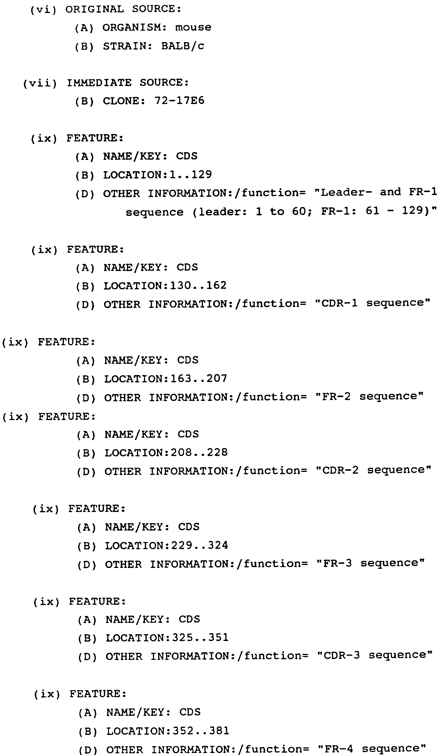


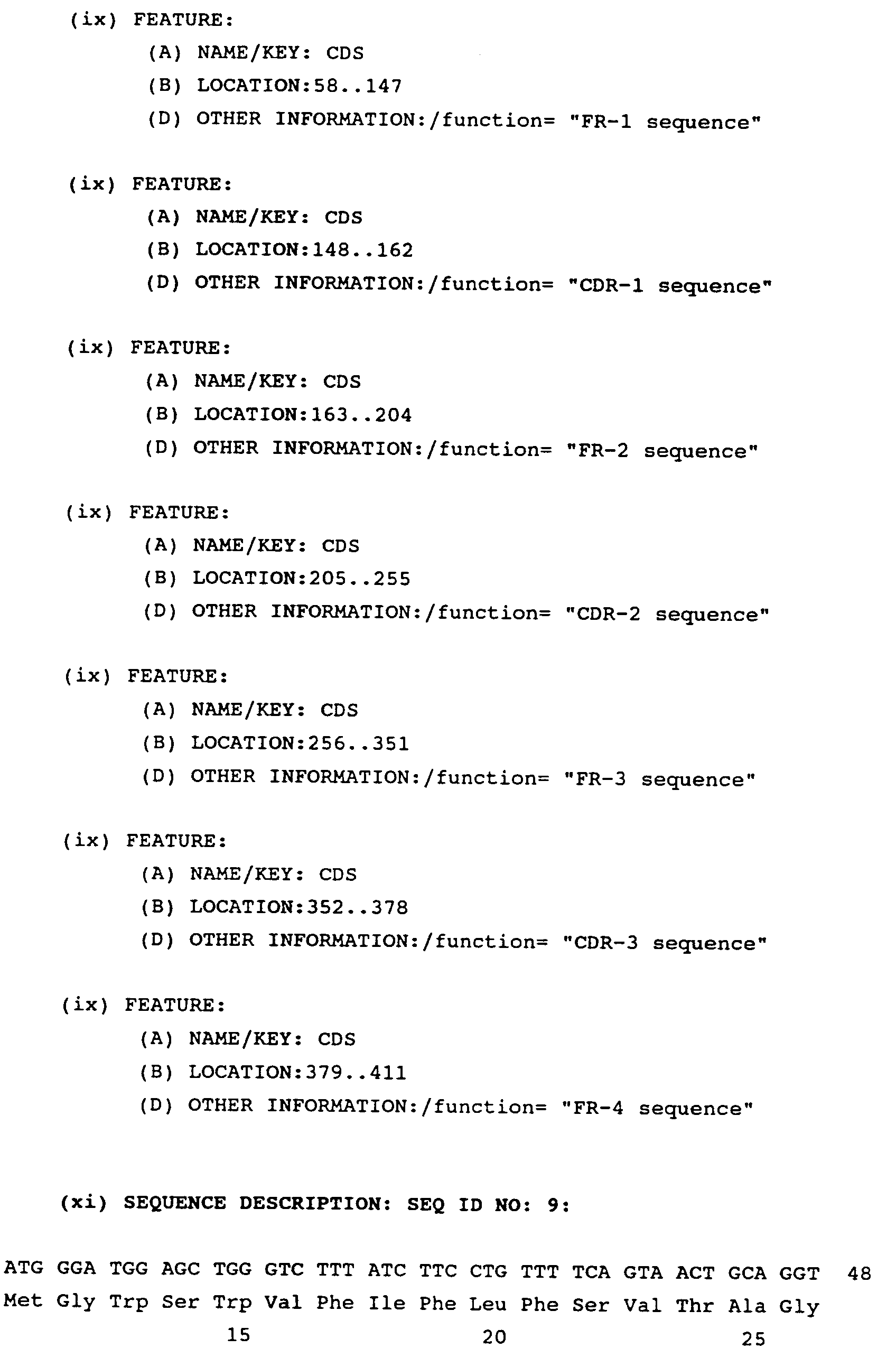


Claims (20)
- Monoclonal antibody having the following properties:-- reacting only with the αV-chain of human αV-integrins,-- blocking the attachment to the integrin substrate of the αV-integrin bearing cell,-- triggering reversal of established cell matrix interaction caused by αV-integrins,-- blocking tumor development, and-- showing no cytotoxic activity.
- Monoclonal antibody of Claim 1, wherein the integrin substrate is vitronectin, fibrinogen, or fibronectin.
- Monoclonal antibody according to Claim 1 or 2, wherein the tumor, the cell growth of which is blocked, is melanoma.
- Monoclonal antibody of Claim 1 to 3 comprising the amino acid sequence of the FRs and CDRs of the light chain given in Figure 17a and the heavy chain given in Figure 17b or mutants and varients thereof.
- Monoclonal antibody fragment having the properties of the monoclonal antibody of Claims 1 - 4.
- Hybridoma cell line having the designation 272-17E6 and deposited under accession number DSM ACC2160 capable of producing a monoclonal antibody of Claims 1 - 4.
- Monoclonal antibody obtainable by the hybridoma cell line DSM ACC2160.
- Amino acid sequence comprising the sequence given in Figure 17a.
- Amino acid sequence comprising the sequence given in Figure 17b.
- Amino acid sequence according to Claim 8 starting from position 21.
- Amino acid sequence according to Claim 9 starting from position 20.
- DNA sequence comprising the DNA sequence of Figure 17a or mutants and variants thereof coding for the FRs and CDRs of the light chain of the variable region of a monoclonal antibody having the properties as defined in Claim 1.
- DNA sequence comprising a DNA sequence of Figure 17b or mutants and variants thereof coding for the FRs and CDRs of the heavy chain of the variable region of a monoclonal antibody having the properties as defined in Claim 1.
- DNA sequence according to Claim 12, comprising the leader sequence given in Figure 17a.
- DNA sequence according to Claim 13, comprising the leader sequence given in Figure 17b.
- Pharmaceutical composition comprising a monoclonal antibody according to one of the Claims 1 - 5 or 7, and a pharmaceutically acceptable carrier.
- Method for the manufacture of a monoclonal antibody according to one of the Claims 1 - 4, by immunizing a mouse with purified αVβ3 integrin, selecting clones binding to the purified concerning αVβ3 receptor by ELISA and producing according to standard echniques a specific cell line which produces said antibody.
- Method according to Claim 16, wherein the integrin is vitronectin.
- Method for the manufacture of a monoclonal antibody of Claim 7, by immunizing a mouse with purified vitronectin, selecting clones binding to purified vitronectin receptor by ELISA and producing according to standard techniques the cell line as defined in Claim 6 which produces said antibody.
- Use of monoclonal antibody according to one of the Claims 1 - 5 or 7, for the manufacture of a drug directed to tumors, preferably melanoma, or for the diagnostic location and assessment of tumor growth.
Priority Applications (2)
| Application Number | Priority Date | Filing Date | Title |
|---|---|---|---|
| SI9530683T SI0719859T1 (en) | 1994-12-20 | 1995-12-06 | Anti-alpha V-integrin monoclonal antibody |
| EP95119233A EP0719859B1 (en) | 1994-12-20 | 1995-12-06 | Anti-alpha V-integrin monoclonal antibody |
Applications Claiming Priority (3)
| Application Number | Priority Date | Filing Date | Title |
|---|---|---|---|
| EP94120165 | 1994-12-20 | ||
| EP94120165 | 1994-12-20 | ||
| EP95119233A EP0719859B1 (en) | 1994-12-20 | 1995-12-06 | Anti-alpha V-integrin monoclonal antibody |
Publications (2)
| Publication Number | Publication Date |
|---|---|
| EP0719859A1 true EP0719859A1 (en) | 1996-07-03 |
| EP0719859B1 EP0719859B1 (en) | 2003-07-02 |
Family
ID=8216547
Family Applications (1)
| Application Number | Title | Priority Date | Filing Date |
|---|---|---|---|
| EP95119233A Expired - Lifetime EP0719859B1 (en) | 1994-12-20 | 1995-12-06 | Anti-alpha V-integrin monoclonal antibody |
Country Status (26)
| Country | Link |
|---|---|
| US (1) | US5985278A (en) |
| EP (1) | EP0719859B1 (en) |
| JP (1) | JP3898245B2 (en) |
| KR (1) | KR100450368B1 (en) |
| CN (1) | CN1117763C (en) |
| AR (1) | AR001778A1 (en) |
| AT (1) | ATE244306T1 (en) |
| AU (1) | AU710234B2 (en) |
| BR (1) | BR9505980B1 (en) |
| CA (1) | CA2165573C (en) |
| CO (1) | CO4480042A1 (en) |
| CZ (1) | CZ290477B6 (en) |
| DE (1) | DE69531187T2 (en) |
| DK (1) | DK0719859T3 (en) |
| ES (1) | ES2202336T3 (en) |
| FI (1) | FI118536B (en) |
| HU (1) | HU221061B1 (en) |
| NO (1) | NO321186B1 (en) |
| PL (1) | PL182961B1 (en) |
| PT (1) | PT719859E (en) |
| RU (1) | RU2205223C2 (en) |
| SI (1) | SI0719859T1 (en) |
| SK (1) | SK284932B6 (en) |
| TR (1) | TR199501614A2 (en) |
| UA (1) | UA40621C2 (en) |
| ZA (1) | ZA9510806B (en) |
Cited By (35)
| Publication number | Priority date | Publication date | Assignee | Title |
|---|---|---|---|---|
| WO1998009651A1 (en) * | 1996-09-03 | 1998-03-12 | Chugai Seiyaku Kabushiki Kaisha | ANTI-INTEGRIN α3 ANTIBODY COMPLEXES |
| WO1999037683A1 (en) * | 1998-01-23 | 1999-07-29 | Merck Patent Gmbh | MONOCLONAL ANTIBODY ANTI αv-INTEGRIN AND ITS USE TO INHIBIT αvβ6-INTEGRIN ATTACHMENT TO FIBRONECTIN |
| EP1135477A4 (en) * | 1998-11-24 | 2002-09-11 | Smithkline Beecham Corp | Humanized monoclonal antibodies |
| WO2003100033A3 (en) * | 2002-03-13 | 2004-07-15 | Biogen Inc | ANTI-αvβ6 ANTIBODIES |
| US7163681B2 (en) | 2000-08-07 | 2007-01-16 | Centocor, Inc. | Anti-integrin antibodies, compositions, methods and uses |
| US7288390B2 (en) | 2000-08-07 | 2007-10-30 | Centocor, Inc. | Anti-dual integrin antibodies, compositions, methods and uses |
| CN100509851C (en) * | 2002-03-13 | 2009-07-08 | 拜奥根Idec马萨诸塞公司 | Anti-alpha*beta* antibodies |
| WO2010072348A1 (en) | 2008-12-23 | 2010-07-01 | Merck Patent Gmbh | Biomarkers for inhibitors with anti-angiogenic activity |
| US7829087B2 (en) * | 2001-07-09 | 2010-11-09 | Elan Pharmaceuticals, Inc. | Methods of treating cognitive impairment |
| US7901683B2 (en) | 2001-07-09 | 2011-03-08 | Elan Pharmaceuticals, Inc. | Methods of inhibiting amyloid toxicity |
| EP2292251A1 (en) | 2001-04-24 | 2011-03-09 | Merck Patent GmbH | Combination therapy using anti-angiogenic agents and TNF-alpha |
| WO2011057709A1 (en) | 2009-11-13 | 2011-05-19 | Merck Patent Gmbh | Anti integrin antibodies linked to nanoparticles loaded with chemotherapeutic agents |
| WO2012107211A1 (en) | 2011-02-11 | 2012-08-16 | Merck Patent Gmbh | Anti-alpha-v integrin antibody for the treatment of prostate cancer |
| WO2012143496A2 (en) | 2011-04-21 | 2012-10-26 | Bayer Intellectual Property Gmbh | Novel binder-drug conjugates (adcs) and their use |
| EP2526967A1 (en) | 2007-07-17 | 2012-11-28 | Merck Patent GmbH | Engineered anti-alpha v-integrin hybrid antibodies |
| USRE44681E1 (en) | 2006-07-10 | 2013-12-31 | Biogen Idec Ma Inc. | Compositions and methods for inhibiting growth of SMAD4-deficient cancers |
| US8992924B2 (en) | 2005-07-08 | 2015-03-31 | Biogen Idec Ma Inc. | Anti-ανβ6 antibodies and uses thereof |
| WO2016207089A1 (en) | 2015-06-22 | 2016-12-29 | Bayer Pharma Aktiengesellschaft | Antibody drug conjugates (adcs) and antibody prodrug conjugates (apdcs) with enzymatically cleavable groups |
| WO2017060322A2 (en) | 2015-10-10 | 2017-04-13 | Bayer Pharma Aktiengesellschaft | Ptefb-inhibitor-adc |
| WO2017162663A1 (en) | 2016-03-24 | 2017-09-28 | Bayer Pharma Aktiengesellschaft | Prodrugs of cytotoxic active agents having enzymatically cleavable groups |
| WO2018046610A1 (en) | 2016-09-08 | 2018-03-15 | INSERM (Institut National de la Santé et de la Recherche Médicale) | Methods for diagnosing and treating nephrotic syndrome |
| WO2018114578A1 (en) | 2016-12-21 | 2018-06-28 | Bayer Pharma Aktiengesellschaft | Antibody drug conjugates (adcs) having enzymatically cleavable groups |
| WO2018114798A1 (en) | 2016-12-21 | 2018-06-28 | Bayer Aktiengesellschaft | Prodrugs of cytotoxic active agents having enzymatically cleavable groups |
| US10022453B2 (en) | 2013-12-23 | 2018-07-17 | Bayer Pharma Aktiengesellschaft | Antibody drug conjugates (ADCs) with kinesin spindel protein (KSP) |
| US10035860B2 (en) | 2013-03-15 | 2018-07-31 | Biogen Ma Inc. | Anti-alpha V beta 6 antibodies and uses thereof |
| US10035859B2 (en) | 2013-03-15 | 2018-07-31 | Biogen Ma Inc. | Anti-alpha V beta 6 antibodies and uses thereof |
| US10435472B2 (en) | 2014-09-17 | 2019-10-08 | Merck Patent Gmbh | Method of treating bone metastasis diseases, medicaments therefore, and a method of predicting the clinical outcome of treating bone metastasis diseases |
| WO2020079209A1 (en) | 2018-10-19 | 2020-04-23 | Merck Patent Gmbh | Abituzumab for the treatment of colorectal cancer |
| WO2020210358A1 (en) * | 2019-04-08 | 2020-10-15 | Biogen Ma Inc. | Anti-integrin antibodies and uses thereof |
| US11001636B2 (en) | 2016-06-15 | 2021-05-11 | Bayer Pharma Aktiengesellschaft | Specific antibody-drug-conjugates (ADCs) with KSP inhibitors and anti-CD123-antibodies |
| US11415581B2 (en) | 2014-09-17 | 2022-08-16 | Merck Patent Gmbh | Method of treating solid cancers and/or metastases thereof with pan AV integrin inhibitor, medicaments therefore, and a method of predicting the clinical outcome of treating solid cancers and/or metastases thereof |
| US11433140B2 (en) | 2016-12-21 | 2022-09-06 | Bayer Pharma Aktiengesellschaft | Specific antibody drug conjugates (ADCs) having KSP inhibitors |
| US11485786B2 (en) | 2015-11-23 | 2022-11-01 | Merck Patent Gmbh | Anti-alpha-v integrin antibody for the treatment of fibrosis and/or fibrotic disorders |
| US11806404B2 (en) | 2015-06-23 | 2023-11-07 | Bayer Pharma Aktiengesellschaft | Site specific homogeneous with KSP inhibitors |
| EP4305072A4 (en) * | 2021-03-09 | 2025-06-18 | The Governing Council Of The University Of Toronto | ANTIBODIES AGAINST INTEGRIN HETERODIMERS AND USES THEREOF |
Families Citing this family (30)
| Publication number | Priority date | Publication date | Assignee | Title |
|---|---|---|---|---|
| EP0846702B1 (en) * | 1996-12-09 | 2009-03-11 | MERCK PATENT GmbH | Soluble recombinant alphaV beta3 adhesion receptor |
| EP1504764B1 (en) * | 1997-08-08 | 2008-10-08 | The Regents Of The University Of California | Treatment of liver fibrosis with antibodies against alpha V beta 6 integrin |
| US6521593B1 (en) * | 1999-02-01 | 2003-02-18 | Childrens Hospital Los Angeles | Methods for inhibiting brain tumor growth |
| US7064180B2 (en) | 2000-07-31 | 2006-06-20 | The General Hospital Corporation | High affinity integrin polypeptides and uses thereof |
| US7153944B2 (en) * | 2000-07-31 | 2006-12-26 | The General Hospital Corporation | High affinity integrin polypeptides and uses thereof |
| US7323552B2 (en) * | 2000-07-31 | 2008-01-29 | The General Hospital Corporation | Variant integrin polypeptides and uses thereof |
| IL157706A0 (en) * | 2001-03-02 | 2004-03-28 | Medimmune Inc | METHODS OF PREVENTING OR TREATING INFLAMMATORY OR AUTOIMMUNE DISORDERS BY ADMINISTERING INTEGRIN alphav beta3 ANTAGONISTS |
| EP1487492A4 (en) * | 2002-03-04 | 2008-10-01 | Medimmune Inc | PREVENTION OR TREATMENT OF CANCER USING INTEGRIN ALPHAVBETA3 ANTAGONISTS COMBINED WITH OTHER AGENTS |
| EP1487489A4 (en) * | 2002-03-04 | 2008-10-01 | Medimmune Inc | METHODS FOR PREVENTING OR TREATING DISORDERS BY ADMINISTERING AN INTEGRIN AVB3 ANTAGONIST ASSOCIATED WITH HMG-COA REDUCTASE INHIBITOR OR BISPHOSPHONATE |
| US20040048312A1 (en) * | 2002-04-12 | 2004-03-11 | Ronghao Li | Antibodies that bind to integrin alpha-v-beta-6 and methods of use thereof |
| US7371383B2 (en) | 2002-04-12 | 2008-05-13 | Medimmune, Inc. | Recombinant anti-interleukin-9 antibodies |
| WO2004066956A2 (en) * | 2003-01-30 | 2004-08-12 | Medimmune, Inc. | Uses of integrin alphavbeta3 antagonists |
| WO2004091510A2 (en) * | 2003-04-11 | 2004-10-28 | Medimmune, Inc. | Recombinant il-9 antibodies and uses thereof |
| US7351739B2 (en) * | 2004-04-30 | 2008-04-01 | Wellgen, Inc. | Bioactive compounds and methods of uses thereof |
| EP1809326A4 (en) | 2004-10-27 | 2009-11-04 | Medimmune Inc | Modulation of antibody specificity by tailoring the affinity to cognate antigens |
| CA2646611A1 (en) | 2006-03-21 | 2008-05-22 | Genentech, Inc. | Combinatorial therapy |
| CA2657581A1 (en) | 2006-07-11 | 2008-01-17 | Roy C. Levitt | Rhinosinusitis prevention and therapy with proinflammatory cytokine inhibitors |
| ES2766770T3 (en) | 2006-08-10 | 2020-06-15 | Roy C Levitt | Anakinra for use in the treatment of bronchiolitis obliterans syndrome |
| WO2008141276A1 (en) * | 2007-05-11 | 2008-11-20 | Centocor, Inc. | Anti-alpha-v immunoliposome composition, methods and uses |
| US20090175784A1 (en) * | 2007-05-11 | 2009-07-09 | Joshua Goldstein | Anti-Alpha V Immunoliposome Composition, Methods, and Uses |
| HUE027911T2 (en) * | 2007-09-26 | 2016-11-28 | Genentech Inc | Novel antibodies |
| CN102365297B (en) | 2009-03-25 | 2014-10-29 | 霍夫曼-拉罗奇有限公司 | Novel anti-α5β1 antibody and its application |
| RU2565539C2 (en) * | 2010-02-18 | 2015-10-20 | Те Риджентс Оф Те Юниверсити Оф Калифорния | INTEGRIN ανβ8 NEUTRALISING ANTIBODIES |
| EP2608804A4 (en) * | 2010-08-27 | 2015-03-11 | Univ Miami | TREATMENT OF RENAL DISEASES |
| EP2744823B1 (en) * | 2011-08-17 | 2017-08-02 | The Regents of the University of California | Antibodies that bind integrin alpha-v beta-8 |
| KR101961715B1 (en) | 2018-07-18 | 2019-03-25 | (주) 삼진정밀 | High efficiency water treatment system |
| KR101999620B1 (en) | 2019-03-26 | 2019-07-12 | 조용진 | Simultaneous treatment of floating sludge and settling sludge |
| CN115151566A (en) | 2019-11-15 | 2022-10-04 | 普利安特治疗公司 | Compositions and methods for activating integrins |
| CN117143241B (en) * | 2023-10-26 | 2024-02-23 | 迈威(上海)生物科技股份有限公司 | Monoclonal antibodies that specifically bind to human integrin protein ITGAV/ITGB8 |
| WO2025149667A1 (en) | 2024-01-12 | 2025-07-17 | Pheon Therapeutics Ltd | Antibody drug conjugates and uses thereof |
Citations (2)
| Publication number | Priority date | Publication date | Assignee | Title |
|---|---|---|---|---|
| WO1993020229A1 (en) * | 1992-04-03 | 1993-10-14 | Genentech, Inc. | ANTIBODIES TO ALPHAvBETA3 INTEGRIN |
| WO1995025543A1 (en) * | 1994-03-18 | 1995-09-28 | The Scripps Research Institute | Methods and compositions useful for inhibition of angiogenesis |
-
1995
- 1995-12-06 DE DE69531187T patent/DE69531187T2/en not_active Expired - Lifetime
- 1995-12-06 DK DK95119233T patent/DK0719859T3/en active
- 1995-12-06 AT AT95119233T patent/ATE244306T1/en active
- 1995-12-06 EP EP95119233A patent/EP0719859B1/en not_active Expired - Lifetime
- 1995-12-06 ES ES95119233T patent/ES2202336T3/en not_active Expired - Lifetime
- 1995-12-06 SI SI9530683T patent/SI0719859T1/en unknown
- 1995-12-06 PT PT95119233T patent/PT719859E/en unknown
- 1995-12-13 AU AU40421/95A patent/AU710234B2/en not_active Ceased
- 1995-12-13 CZ CZ19953288A patent/CZ290477B6/en not_active IP Right Cessation
- 1995-12-18 JP JP32887795A patent/JP3898245B2/en not_active Expired - Fee Related
- 1995-12-18 CA CA2165573A patent/CA2165573C/en not_active Expired - Fee Related
- 1995-12-19 NO NO19955167A patent/NO321186B1/en not_active IP Right Cessation
- 1995-12-19 ZA ZA9510806A patent/ZA9510806B/en unknown
- 1995-12-19 PL PL95311926A patent/PL182961B1/en unknown
- 1995-12-19 UA UA95125376A patent/UA40621C2/en unknown
- 1995-12-19 US US08/574,699 patent/US5985278A/en not_active Expired - Lifetime
- 1995-12-19 HU HU9503638A patent/HU221061B1/en not_active IP Right Cessation
- 1995-12-19 RU RU95121105/13A patent/RU2205223C2/en not_active IP Right Cessation
- 1995-12-19 FI FI956112A patent/FI118536B/en not_active IP Right Cessation
- 1995-12-19 CN CN95120901A patent/CN1117763C/en not_active Expired - Fee Related
- 1995-12-19 SK SK1592-95A patent/SK284932B6/en not_active IP Right Cessation
- 1995-12-20 CO CO95060553A patent/CO4480042A1/en unknown
- 1995-12-20 BR BRPI9505980-6A patent/BR9505980B1/en not_active IP Right Cessation
- 1995-12-20 AR AR33473095A patent/AR001778A1/en active IP Right Grant
- 1995-12-20 TR TR95/01614A patent/TR199501614A2/en unknown
- 1995-12-20 KR KR1019950052440A patent/KR100450368B1/en not_active Expired - Fee Related
Patent Citations (2)
| Publication number | Priority date | Publication date | Assignee | Title |
|---|---|---|---|---|
| WO1993020229A1 (en) * | 1992-04-03 | 1993-10-14 | Genentech, Inc. | ANTIBODIES TO ALPHAvBETA3 INTEGRIN |
| WO1995025543A1 (en) * | 1994-03-18 | 1995-09-28 | The Scripps Research Institute | Methods and compositions useful for inhibition of angiogenesis |
Non-Patent Citations (4)
Cited By (63)
| Publication number | Priority date | Publication date | Assignee | Title |
|---|---|---|---|---|
| WO1998009651A1 (en) * | 1996-09-03 | 1998-03-12 | Chugai Seiyaku Kabushiki Kaisha | ANTI-INTEGRIN α3 ANTIBODY COMPLEXES |
| WO1999037683A1 (en) * | 1998-01-23 | 1999-07-29 | Merck Patent Gmbh | MONOCLONAL ANTIBODY ANTI αv-INTEGRIN AND ITS USE TO INHIBIT αvβ6-INTEGRIN ATTACHMENT TO FIBRONECTIN |
| EP1135477A4 (en) * | 1998-11-24 | 2002-09-11 | Smithkline Beecham Corp | Humanized monoclonal antibodies |
| US7087405B2 (en) | 1998-11-24 | 2006-08-08 | Smithkline Beecham Corporation | Nucleic acids encoding anti-human ανβ3 and ανβ5 antibodies |
| US7163681B2 (en) | 2000-08-07 | 2007-01-16 | Centocor, Inc. | Anti-integrin antibodies, compositions, methods and uses |
| US7288390B2 (en) | 2000-08-07 | 2007-10-30 | Centocor, Inc. | Anti-dual integrin antibodies, compositions, methods and uses |
| US8071333B2 (en) | 2000-08-07 | 2011-12-06 | Centocor, Inc. | Anti-integrin antibodies, compositions, methods and uses |
| US8071729B2 (en) | 2000-08-07 | 2011-12-06 | Centocor, Inc. | Anti-integrin antibodies, compositions, methods and uses |
| US7550142B2 (en) | 2000-08-07 | 2009-06-23 | Centocor, Inc. | Anti-integrin antibodies, compositions, methods and uses |
| EP2292251A1 (en) | 2001-04-24 | 2011-03-09 | Merck Patent GmbH | Combination therapy using anti-angiogenic agents and TNF-alpha |
| US7829087B2 (en) * | 2001-07-09 | 2010-11-09 | Elan Pharmaceuticals, Inc. | Methods of treating cognitive impairment |
| US7901683B2 (en) | 2001-07-09 | 2011-03-08 | Elan Pharmaceuticals, Inc. | Methods of inhibiting amyloid toxicity |
| CN100509851C (en) * | 2002-03-13 | 2009-07-08 | 拜奥根Idec马萨诸塞公司 | Anti-alpha*beta* antibodies |
| AU2003261071C1 (en) * | 2002-03-13 | 2008-12-11 | Biogen Idec Ma Inc. | Anti-AlphavBeta6 antibodies |
| CN102924598A (en) * | 2002-03-13 | 2013-02-13 | 拜奥根Idec马萨诸塞公司 | anti-αvβ6 antibody |
| EA011853B1 (en) * | 2002-03-13 | 2009-06-30 | Байоджен Айдек Ма Инк. | ANTIBODIES AGAINST αβ |
| US7465449B2 (en) | 2002-03-13 | 2008-12-16 | Biogen Idec Ma Inc. | Anti-αvβ6 antibodies |
| WO2003100033A3 (en) * | 2002-03-13 | 2004-07-15 | Biogen Inc | ANTI-αvβ6 ANTIBODIES |
| CN102924598B (en) * | 2002-03-13 | 2014-12-31 | 拜奥根Idec马萨诸塞公司 | anti-αvβ6 antibody |
| US9745376B2 (en) | 2002-03-13 | 2017-08-29 | Biogen Ma Inc. | Anti-ανβ6 antibodies |
| AU2003261071B2 (en) * | 2002-03-13 | 2008-05-15 | Biogen Idec Ma Inc. | Anti-AlphavBeta6 antibodies |
| KR101098109B1 (en) | 2002-03-13 | 2011-12-26 | 더 리전츠 오브 더 유니버시티 오브 캘리포니아 | ANTI αVβ6 ANTIBODIES |
| US8153126B2 (en) | 2002-03-13 | 2012-04-10 | Violette Shelia M | Anti-αvβ6 antibodies |
| US8992924B2 (en) | 2005-07-08 | 2015-03-31 | Biogen Idec Ma Inc. | Anti-ανβ6 antibodies and uses thereof |
| USRE44681E1 (en) | 2006-07-10 | 2013-12-31 | Biogen Idec Ma Inc. | Compositions and methods for inhibiting growth of SMAD4-deficient cancers |
| EP2142188A4 (en) * | 2007-03-21 | 2011-03-23 | Elan Pharm Inc | METHODS OF SUPPRESSING INHIBITION OF LTP |
| EP2526967A1 (en) | 2007-07-17 | 2012-11-28 | Merck Patent GmbH | Engineered anti-alpha v-integrin hybrid antibodies |
| EP2706358A2 (en) | 2008-12-23 | 2014-03-12 | Merck Patent GmbH | Biomarkers for inhibitors with anti-angiogenic activity |
| EP2706358A3 (en) * | 2008-12-23 | 2014-05-07 | Merck Patent GmbH | Biomarkers for inhibitors with anti-angiogenic activity |
| WO2010072348A1 (en) | 2008-12-23 | 2010-07-01 | Merck Patent Gmbh | Biomarkers for inhibitors with anti-angiogenic activity |
| WO2011057709A1 (en) | 2009-11-13 | 2011-05-19 | Merck Patent Gmbh | Anti integrin antibodies linked to nanoparticles loaded with chemotherapeutic agents |
| WO2012107211A1 (en) | 2011-02-11 | 2012-08-16 | Merck Patent Gmbh | Anti-alpha-v integrin antibody for the treatment of prostate cancer |
| WO2012143496A2 (en) | 2011-04-21 | 2012-10-26 | Bayer Intellectual Property Gmbh | Novel binder-drug conjugates (adcs) and their use |
| WO2012143499A2 (en) | 2011-04-21 | 2012-10-26 | Bayer Intellectual Property Gmbh | Novel binder-drug conjugates (adcs) and their use |
| US10035859B2 (en) | 2013-03-15 | 2018-07-31 | Biogen Ma Inc. | Anti-alpha V beta 6 antibodies and uses thereof |
| US10035860B2 (en) | 2013-03-15 | 2018-07-31 | Biogen Ma Inc. | Anti-alpha V beta 6 antibodies and uses thereof |
| US10022453B2 (en) | 2013-12-23 | 2018-07-17 | Bayer Pharma Aktiengesellschaft | Antibody drug conjugates (ADCs) with kinesin spindel protein (KSP) |
| US11306146B2 (en) | 2014-09-17 | 2022-04-19 | Merck Patent Gmbh | Method of treating bone metastasis diseases, medicaments therefore, and a method of predicting the clinical outcome of treating bone metastasis diseases |
| US11415581B2 (en) | 2014-09-17 | 2022-08-16 | Merck Patent Gmbh | Method of treating solid cancers and/or metastases thereof with pan AV integrin inhibitor, medicaments therefore, and a method of predicting the clinical outcome of treating solid cancers and/or metastases thereof |
| US10435472B2 (en) | 2014-09-17 | 2019-10-08 | Merck Patent Gmbh | Method of treating bone metastasis diseases, medicaments therefore, and a method of predicting the clinical outcome of treating bone metastasis diseases |
| WO2016207089A1 (en) | 2015-06-22 | 2016-12-29 | Bayer Pharma Aktiengesellschaft | Antibody drug conjugates (adcs) and antibody prodrug conjugates (apdcs) with enzymatically cleavable groups |
| EP4406606A2 (en) | 2015-06-22 | 2024-07-31 | Bayer Pharma Aktiengesellschaft | Antibody drug conjugates (adcs) and antibody prodrug conjugates (apdcs) with enzymatically cleavable g |
| US12144865B2 (en) | 2015-06-22 | 2024-11-19 | Bayer Pharma Aktiengesellschaft | Antibody drug conjugates with enzymatically cleavable groups |
| US11123439B2 (en) | 2015-06-22 | 2021-09-21 | Bayer Pharma Aktiengesellschaft | Antibody drug conjugates (ADCS) and antibody prodrug conjugates (APDCS) with enzymatically cleavable groups |
| US11806404B2 (en) | 2015-06-23 | 2023-11-07 | Bayer Pharma Aktiengesellschaft | Site specific homogeneous with KSP inhibitors |
| WO2017060322A2 (en) | 2015-10-10 | 2017-04-13 | Bayer Pharma Aktiengesellschaft | Ptefb-inhibitor-adc |
| US12054549B2 (en) | 2015-11-23 | 2024-08-06 | Merck Patent Gmbh | Anti-alpha-v integrin antibody for the treatment of fibrosis and/or fibrotic disorders |
| US11485786B2 (en) | 2015-11-23 | 2022-11-01 | Merck Patent Gmbh | Anti-alpha-v integrin antibody for the treatment of fibrosis and/or fibrotic disorders |
| WO2017162663A1 (en) | 2016-03-24 | 2017-09-28 | Bayer Pharma Aktiengesellschaft | Prodrugs of cytotoxic active agents having enzymatically cleavable groups |
| US11685714B2 (en) | 2016-03-24 | 2023-06-27 | Bayer Pharma Aktiengesellschaft | Prodrugs of cytotoxic active agents having enzymatically cleavable groups |
| US11643469B2 (en) | 2016-06-15 | 2023-05-09 | Bayer Pharma Aktiengesellschaft | Specific antibody-drug-conjugates (ADCs) with KSP inhibitors and anti-CD123-antibodies |
| US11001636B2 (en) | 2016-06-15 | 2021-05-11 | Bayer Pharma Aktiengesellschaft | Specific antibody-drug-conjugates (ADCs) with KSP inhibitors and anti-CD123-antibodies |
| WO2018046610A1 (en) | 2016-09-08 | 2018-03-15 | INSERM (Institut National de la Santé et de la Recherche Médicale) | Methods for diagnosing and treating nephrotic syndrome |
| WO2018114578A1 (en) | 2016-12-21 | 2018-06-28 | Bayer Pharma Aktiengesellschaft | Antibody drug conjugates (adcs) having enzymatically cleavable groups |
| US11660351B2 (en) | 2016-12-21 | 2023-05-30 | Bayer Aktiengesellschaft | Antibody drug conjugates (ADCs) having enzymatically cleavable groups |
| WO2018114798A1 (en) | 2016-12-21 | 2018-06-28 | Bayer Aktiengesellschaft | Prodrugs of cytotoxic active agents having enzymatically cleavable groups |
| US11478554B2 (en) | 2016-12-21 | 2022-10-25 | Bayer Pharma Aktiengesellschaft | Antibody drug conjugates (ADCS) having enzymatically cleavable groups |
| US12059472B2 (en) | 2016-12-21 | 2024-08-13 | Bayer Aktiengesellschaft | Prodrugs of cytotoxic active agents having enzymatically cleavable groups |
| US11433140B2 (en) | 2016-12-21 | 2022-09-06 | Bayer Pharma Aktiengesellschaft | Specific antibody drug conjugates (ADCs) having KSP inhibitors |
| WO2020079209A1 (en) | 2018-10-19 | 2020-04-23 | Merck Patent Gmbh | Abituzumab for the treatment of colorectal cancer |
| US12171829B2 (en) | 2018-10-19 | 2024-12-24 | Merck Patent Gmbh | Abituzumab for the treatment of colorectal cancer |
| WO2020210358A1 (en) * | 2019-04-08 | 2020-10-15 | Biogen Ma Inc. | Anti-integrin antibodies and uses thereof |
| EP4305072A4 (en) * | 2021-03-09 | 2025-06-18 | The Governing Council Of The University Of Toronto | ANTIBODIES AGAINST INTEGRIN HETERODIMERS AND USES THEREOF |
Also Published As
Similar Documents
| Publication | Publication Date | Title |
|---|---|---|
| EP0719859B1 (en) | Anti-alpha V-integrin monoclonal antibody | |
| US8246958B2 (en) | Methods of inhibiting alpha-4-dependent interactions with VCAM-1 with anti-VLA-4 antibodies | |
| ES2210225T5 (en) | Inhibition of lymphocyte adhesion to the vascular endothelium by a new extracellular matrix-ligand receptor interaction | |
| EP2172485A1 (en) | Novel anti CXCR4 antibodies and their use for the treatment of cancer | |
| US8071730B2 (en) | Anti-JAM-A antibodies | |
| JP2009029781A (en) | Therapeutic use of humanized antibodies against alpha-4 integrin | |
| EP1049718B1 (en) | Use of the antibody 271.14d9.f8 (dsm acc2331) to inhibit in vitro alphavbeta6-integrin attachment to fibronectin | |
| JP2013253084A (en) | Method for using anti-cd151 antibody for treatment of cancer | |
| US20070048325A1 (en) | Combination therapies for inhibiting integrin-extracellular matrix interactions | |
| JP2007523092A (en) | Methods and compositions for inhibiting angiogenesis | |
| EP1335746A2 (en) | Recombined anti-gpiib/iiia antibodies used for inhibiting angiogenesis | |
| CZ20002659A3 (en) | Anti-av-integrin monoclonal antibody and its use for inhibition of avbeta-integrin binding to fibronectin | |
| HK1034522A (en) | MONOCLONAL ANTIBODY ANTIαν-INTEGRIN AND ITS USE TO INHIBITανβ6-INTEGRIN ATTACHMENT TO FIBRONECTIN |
Legal Events
| Date | Code | Title | Description |
|---|---|---|---|
| PUAI | Public reference made under article 153(3) epc to a published international application that has entered the european phase |
Free format text: ORIGINAL CODE: 0009012 |
|
| AK | Designated contracting states |
Kind code of ref document: A1 Designated state(s): AT BE CH DE DK ES FR GB GR IE IT LI LU NL PT SE |
|
| AX | Request for extension of the european patent |
Free format text: LT PAYMENT 951206;SI PAYMENT 951206 |
|
| RAX | Requested extension states of the european patent have changed |
Free format text: LT PAYMENT 951206;SI PAYMENT 951206 |
|
| 17P | Request for examination filed |
Effective date: 19961216 |
|
| 17Q | First examination report despatched |
Effective date: 20010615 |
|
| GRAH | Despatch of communication of intention to grant a patent |
Free format text: ORIGINAL CODE: EPIDOS IGRA |
|
| GRAH | Despatch of communication of intention to grant a patent |
Free format text: ORIGINAL CODE: EPIDOS IGRA |
|
| GRAH | Despatch of communication of intention to grant a patent |
Free format text: ORIGINAL CODE: EPIDOS IGRA |
|
| GRAH | Despatch of communication of intention to grant a patent |
Free format text: ORIGINAL CODE: EPIDOS IGRA |
|
| GRAA | (expected) grant |
Free format text: ORIGINAL CODE: 0009210 |
|
| AK | Designated contracting states |
Designated state(s): AT BE CH DE DK ES FR GB GR IE IT LI LU NL PT SE |
|
| AX | Request for extension of the european patent |
Extension state: LT SI |
|
| REG | Reference to a national code |
Ref country code: GB Ref legal event code: FG4D |
|
| REG | Reference to a national code |
Ref country code: CH Ref legal event code: EP |
|
| REG | Reference to a national code |
Ref country code: IE Ref legal event code: FG4D |
|
| REF | Corresponds to: |
Ref document number: 69531187 Country of ref document: DE Date of ref document: 20030807 Kind code of ref document: P |
|
| REG | Reference to a national code |
Ref country code: GR Ref legal event code: EP Ref document number: 20030403937 Country of ref document: GR |
|
| REG | Reference to a national code |
Ref country code: SE Ref legal event code: TRGR |
|
| REG | Reference to a national code |
Ref country code: CH Ref legal event code: NV Representative=s name: ARNOLD & SIEDSMA AG |
|
| REG | Reference to a national code |
Ref country code: ES Ref legal event code: FG2A Ref document number: 2202336 Country of ref document: ES Kind code of ref document: T3 |
|
| ET | Fr: translation filed | ||
| PLBE | No opposition filed within time limit |
Free format text: ORIGINAL CODE: 0009261 |
|
| STAA | Information on the status of an ep patent application or granted ep patent |
Free format text: STATUS: NO OPPOSITION FILED WITHIN TIME LIMIT |
|
| 26N | No opposition filed |
Effective date: 20040405 |
|
| REG | Reference to a national code |
Ref country code: SI Ref legal event code: IF |
|
| REG | Reference to a national code |
Ref country code: CH Ref legal event code: PFA Owner name: MERCK PATENT GMBH Free format text: MERCK PATENT GMBH#FRANKFURTER STRASSE 250#64293 DARMSTADT (DE) -TRANSFER TO- MERCK PATENT GMBH#FRANKFURTER STRASSE 250#64293 DARMSTADT (DE) |
|
| PGFP | Annual fee paid to national office [announced via postgrant information from national office to epo] |
Ref country code: DK Payment date: 20131210 Year of fee payment: 19 |
|
| PGFP | Annual fee paid to national office [announced via postgrant information from national office to epo] |
Ref country code: GB Payment date: 20131204 Year of fee payment: 19 Ref country code: SE Payment date: 20131211 Year of fee payment: 19 Ref country code: DE Payment date: 20131204 Year of fee payment: 19 Ref country code: PT Payment date: 20131213 Year of fee payment: 19 Ref country code: AT Payment date: 20131126 Year of fee payment: 19 Ref country code: CH Payment date: 20131212 Year of fee payment: 19 Ref country code: IE Payment date: 20131210 Year of fee payment: 19 |
|
| PGFP | Annual fee paid to national office [announced via postgrant information from national office to epo] |
Ref country code: ES Payment date: 20131112 Year of fee payment: 19 Ref country code: IT Payment date: 20131107 Year of fee payment: 19 Ref country code: LU Payment date: 20131210 Year of fee payment: 19 Ref country code: FR Payment date: 20131209 Year of fee payment: 19 Ref country code: GR Payment date: 20131114 Year of fee payment: 19 Ref country code: NL Payment date: 20131109 Year of fee payment: 19 |
|
| PGFP | Annual fee paid to national office [announced via postgrant information from national office to epo] |
Ref country code: BE Payment date: 20131217 Year of fee payment: 19 |
|
| REG | Reference to a national code |
Ref country code: PT Ref legal event code: MM4A Free format text: LAPSE DUE TO NON-PAYMENT OF FEES Effective date: 20150608 |
|
| PG25 | Lapsed in a contracting state [announced via postgrant information from national office to epo] |
Ref country code: BE Free format text: LAPSE BECAUSE OF NON-PAYMENT OF DUE FEES Effective date: 20141231 |
|
| REG | Reference to a national code |
Ref country code: DE Ref legal event code: R119 Ref document number: 69531187 Country of ref document: DE |
|
| REG | Reference to a national code |
Ref country code: DK Ref legal event code: EBP Effective date: 20141231 |
|
| REG | Reference to a national code |
Ref country code: NL Ref legal event code: V1 Effective date: 20150701 |
|
| REG | Reference to a national code |
Ref country code: LT Ref legal event code: MM9D Effective date: 20141206 |
|
| REG | Reference to a national code |
Ref country code: NL Ref legal event code: V1 Effective date: 20150701 |
|
| PG25 | Lapsed in a contracting state [announced via postgrant information from national office to epo] |
Ref country code: LU Free format text: LAPSE BECAUSE OF NON-PAYMENT OF DUE FEES Effective date: 20141206 Ref country code: SE Free format text: LAPSE BECAUSE OF NON-PAYMENT OF DUE FEES Effective date: 20141207 Ref country code: PT Free format text: LAPSE BECAUSE OF NON-PAYMENT OF DUE FEES Effective date: 20150608 |
|
| REG | Reference to a national code |
Ref country code: CH Ref legal event code: PL |
|
| REG | Reference to a national code |
Ref country code: SE Ref legal event code: EUG |
|
| REG | Reference to a national code |
Ref country code: AT Ref legal event code: MM01 Ref document number: 244306 Country of ref document: AT Kind code of ref document: T Effective date: 20141206 |
|
| GBPC | Gb: european patent ceased through non-payment of renewal fee |
Effective date: 20141206 |
|
| REG | Reference to a national code |
Ref country code: GR Ref legal event code: ML Ref document number: 20030403937 Country of ref document: GR Effective date: 20150722 |
|
| REG | Reference to a national code |
Ref country code: IE Ref legal event code: MM4A |
|
| REG | Reference to a national code |
Ref country code: FR Ref legal event code: ST Effective date: 20150831 |
|
| PG25 | Lapsed in a contracting state [announced via postgrant information from national office to epo] |
Ref country code: NL Free format text: LAPSE BECAUSE OF NON-PAYMENT OF DUE FEES Effective date: 20150701 |
|
| PG25 | Lapsed in a contracting state [announced via postgrant information from national office to epo] |
Ref country code: LI Free format text: LAPSE BECAUSE OF NON-PAYMENT OF DUE FEES Effective date: 20141231 Ref country code: IE Free format text: LAPSE BECAUSE OF NON-PAYMENT OF DUE FEES Effective date: 20141206 Ref country code: GB Free format text: LAPSE BECAUSE OF NON-PAYMENT OF DUE FEES Effective date: 20141206 Ref country code: DE Free format text: LAPSE BECAUSE OF NON-PAYMENT OF DUE FEES Effective date: 20150701 Ref country code: CH Free format text: LAPSE BECAUSE OF NON-PAYMENT OF DUE FEES Effective date: 20141231 |
|
| PG25 | Lapsed in a contracting state [announced via postgrant information from national office to epo] |
Ref country code: FR Free format text: LAPSE BECAUSE OF NON-PAYMENT OF DUE FEES Effective date: 20141231 Ref country code: GR Free format text: LAPSE BECAUSE OF NON-PAYMENT OF DUE FEES Effective date: 20150722 Ref country code: AT Free format text: LAPSE BECAUSE OF NON-PAYMENT OF DUE FEES Effective date: 20141206 |
|
| REG | Reference to a national code |
Ref country code: PT Ref legal event code: MM4A Free format text: MAXIMUM VALIDITY LIMIT REACHED Effective date: 20151206 |
|
| PG25 | Lapsed in a contracting state [announced via postgrant information from national office to epo] |
Ref country code: IT Free format text: LAPSE BECAUSE OF NON-PAYMENT OF DUE FEES Effective date: 20141206 |
|
| REG | Reference to a national code |
Ref country code: ES Ref legal event code: FD2A Effective date: 20160127 |
|
| PG25 | Lapsed in a contracting state [announced via postgrant information from national office to epo] |
Ref country code: DK Free format text: LAPSE BECAUSE OF NON-PAYMENT OF DUE FEES Effective date: 20141231 |
|
| PG25 | Lapsed in a contracting state [announced via postgrant information from national office to epo] |
Ref country code: PT Free format text: LAPSE BECAUSE OF EXPIRATION OF PROTECTION Effective date: 20151216 |
|
| PG25 | Lapsed in a contracting state [announced via postgrant information from national office to epo] |
Ref country code: ES Free format text: LAPSE BECAUSE OF NON-PAYMENT OF DUE FEES Effective date: 20141207 |
|
| PG25 | Lapsed in a contracting state [announced via postgrant information from national office to epo] |
Ref country code: PT Free format text: LAPSE BECAUSE OF EXPIRATION OF PROTECTION Effective date: 20141206 |