EP0652948B1 - Development of a vector to target gene expression to the epidermis of transgenic animals - Google Patents
Development of a vector to target gene expression to the epidermis of transgenic animals Download PDFInfo
- Publication number
- EP0652948B1 EP0652948B1 EP93910867A EP93910867A EP0652948B1 EP 0652948 B1 EP0652948 B1 EP 0652948B1 EP 93910867 A EP93910867 A EP 93910867A EP 93910867 A EP93910867 A EP 93910867A EP 0652948 B1 EP0652948 B1 EP 0652948B1
- Authority
- EP
- European Patent Office
- Prior art keywords
- vector
- nucleic acid
- keratin
- cassette
- epidermal
- Prior art date
- Legal status (The legal status is an assumption and is not a legal conclusion. Google has not performed a legal analysis and makes no representation as to the accuracy of the status listed.)
- Expired - Lifetime
Links
- 239000013598 vector Substances 0.000 title claims abstract description 136
- 230000014509 gene expression Effects 0.000 title claims abstract description 71
- 210000002615 epidermis Anatomy 0.000 title claims abstract description 47
- 241001465754 Metazoa Species 0.000 title claims abstract description 37
- 230000009261 transgenic effect Effects 0.000 title claims abstract description 24
- 238000011161 development Methods 0.000 title description 14
- 108010076876 Keratins Proteins 0.000 claims abstract description 96
- 102000011782 Keratins Human genes 0.000 claims abstract description 90
- 108090000623 proteins and genes Proteins 0.000 claims abstract description 62
- 150000007523 nucleic acids Chemical class 0.000 claims abstract description 61
- 210000001339 epidermal cell Anatomy 0.000 claims abstract description 53
- 108020004707 nucleic acids Proteins 0.000 claims abstract description 40
- 102000039446 nucleic acids Human genes 0.000 claims abstract description 40
- 102000004169 proteins and genes Human genes 0.000 claims abstract description 28
- 206010028980 Neoplasm Diseases 0.000 claims abstract description 26
- 108020005029 5' Flanking Region Proteins 0.000 claims abstract description 24
- 101150055501 K1 gene Proteins 0.000 claims abstract description 19
- 108020005065 3' Flanking Region Proteins 0.000 claims abstract description 17
- 230000001105 regulatory effect Effects 0.000 claims abstract description 17
- 108090000765 processed proteins & peptides Proteins 0.000 claims abstract description 16
- 201000011510 cancer Diseases 0.000 claims abstract description 15
- 229920001184 polypeptide Polymers 0.000 claims abstract description 14
- 102000004196 processed proteins & peptides Human genes 0.000 claims abstract description 14
- 108020005544 Antisense RNA Proteins 0.000 claims abstract description 11
- 208000002847 Surgical Wound Diseases 0.000 claims abstract description 11
- 239000003184 complementary RNA Substances 0.000 claims abstract description 11
- 238000003780 insertion Methods 0.000 claims abstract description 10
- 230000037431 insertion Effects 0.000 claims abstract description 10
- 201000004681 Psoriasis Diseases 0.000 claims abstract description 9
- 206010040943 Skin Ulcer Diseases 0.000 claims abstract description 7
- 108091023045 Untranslated Region Proteins 0.000 claims abstract description 7
- 108091008146 restriction endonucleases Proteins 0.000 claims abstract description 7
- 231100000019 skin ulcer Toxicity 0.000 claims abstract description 7
- 238000011282 treatment Methods 0.000 claims abstract description 7
- 108091092724 Noncoding DNA Proteins 0.000 claims abstract description 5
- 230000002103 transcriptional effect Effects 0.000 claims abstract description 5
- 210000004027 cell Anatomy 0.000 claims description 64
- 238000000034 method Methods 0.000 claims description 37
- 108700020796 Oncogene Proteins 0.000 claims description 26
- 241000701806 Human papillomavirus Species 0.000 claims description 22
- 108091028043 Nucleic acid sequence Proteins 0.000 claims description 20
- 101800004564 Transforming growth factor alpha Proteins 0.000 claims description 19
- 102400001320 Transforming growth factor alpha Human genes 0.000 claims description 19
- 102000043276 Oncogene Human genes 0.000 claims description 16
- 238000004519 manufacturing process Methods 0.000 claims description 15
- 239000012634 fragment Substances 0.000 claims description 14
- 239000003814 drug Substances 0.000 claims description 13
- 239000011647 vitamin D3 Substances 0.000 claims description 13
- QYSXJUFSXHHAJI-YRZJJWOYSA-N vitamin D3 Chemical compound C1(/[C@@H]2CC[C@@H]([C@]2(CCC1)C)[C@H](C)CCCC(C)C)=C\C=C1\C[C@@H](O)CCC1=C QYSXJUFSXHHAJI-YRZJJWOYSA-N 0.000 claims description 12
- 239000000427 antigen Substances 0.000 claims description 9
- 108091007433 antigens Proteins 0.000 claims description 9
- 102000036639 antigens Human genes 0.000 claims description 9
- 239000002773 nucleotide Substances 0.000 claims description 9
- 125000003729 nucleotide group Chemical group 0.000 claims description 9
- 230000003612 virological effect Effects 0.000 claims description 9
- 206010052428 Wound Diseases 0.000 claims description 8
- 208000027418 Wounds and injury Diseases 0.000 claims description 8
- 239000003102 growth factor Substances 0.000 claims description 8
- NOESYZHRGYRDHS-UHFFFAOYSA-N insulin Chemical compound N1C(=O)C(NC(=O)C(CCC(N)=O)NC(=O)C(CCC(O)=O)NC(=O)C(C(C)C)NC(=O)C(NC(=O)CN)C(C)CC)CSSCC(C(NC(CO)C(=O)NC(CC(C)C)C(=O)NC(CC=2C=CC(O)=CC=2)C(=O)NC(CCC(N)=O)C(=O)NC(CC(C)C)C(=O)NC(CCC(O)=O)C(=O)NC(CC(N)=O)C(=O)NC(CC=2C=CC(O)=CC=2)C(=O)NC(CSSCC(NC(=O)C(C(C)C)NC(=O)C(CC(C)C)NC(=O)C(CC=2C=CC(O)=CC=2)NC(=O)C(CC(C)C)NC(=O)C(C)NC(=O)C(CCC(O)=O)NC(=O)C(C(C)C)NC(=O)C(CC(C)C)NC(=O)C(CC=2NC=NC=2)NC(=O)C(CO)NC(=O)CNC2=O)C(=O)NCC(=O)NC(CCC(O)=O)C(=O)NC(CCCNC(N)=N)C(=O)NCC(=O)NC(CC=3C=CC=CC=3)C(=O)NC(CC=3C=CC=CC=3)C(=O)NC(CC=3C=CC(O)=CC=3)C(=O)NC(C(C)O)C(=O)N3C(CCC3)C(=O)NC(CCCCN)C(=O)NC(C)C(O)=O)C(=O)NC(CC(N)=O)C(O)=O)=O)NC(=O)C(C(C)CC)NC(=O)C(CO)NC(=O)C(C(C)O)NC(=O)C1CSSCC2NC(=O)C(CC(C)C)NC(=O)C(NC(=O)C(CCC(N)=O)NC(=O)C(CC(N)=O)NC(=O)C(NC(=O)C(N)CC=1C=CC=CC=1)C(C)C)CC1=CN=CN1 NOESYZHRGYRDHS-UHFFFAOYSA-N 0.000 claims description 8
- 230000035876 healing Effects 0.000 claims description 7
- VBEQCZHXXJYVRD-GACYYNSASA-N uroanthelone Chemical compound C([C@@H](C(=O)N[C@H](C(=O)N[C@@H](CS)C(=O)N[C@@H](CC(N)=O)C(=O)N[C@@H](CS)C(=O)N[C@H](C(=O)N[C@@H]([C@@H](C)CC)C(=O)NCC(=O)N[C@@H](CC=1C=CC(O)=CC=1)C(=O)N[C@@H](CO)C(=O)NCC(=O)N[C@@H](CC(O)=O)C(=O)N[C@@H](CCCNC(N)=N)C(=O)N[C@@H](CS)C(=O)N[C@@H](CCC(N)=O)C(=O)N[C@@H]([C@@H](C)O)C(=O)N[C@@H](CCCNC(N)=N)C(=O)N[C@@H](CC(O)=O)C(=O)N[C@@H](CC(C)C)C(=O)N[C@@H](CCCNC(N)=N)C(=O)N[C@@H](CC=1C2=CC=CC=C2NC=1)C(=O)N[C@@H](CC=1C2=CC=CC=C2NC=1)C(=O)N[C@@H](CCC(O)=O)C(=O)N[C@@H](CC(C)C)C(=O)N[C@@H](CCCNC(N)=N)C(O)=O)C(C)C)[C@@H](C)O)NC(=O)[C@H](CO)NC(=O)[C@H](CC(O)=O)NC(=O)[C@H](CC(C)C)NC(=O)[C@H](CO)NC(=O)[C@H](CCC(O)=O)NC(=O)[C@@H](NC(=O)[C@H](CC=1NC=NC=1)NC(=O)[C@H](CCSC)NC(=O)[C@H](CS)NC(=O)[C@@H](NC(=O)CNC(=O)CNC(=O)[C@H](CC(N)=O)NC(=O)[C@H](CC(C)C)NC(=O)[C@H](CS)NC(=O)[C@H](CC=1C=CC(O)=CC=1)NC(=O)CNC(=O)[C@H](CC(O)=O)NC(=O)[C@H](CC=1C=CC(O)=CC=1)NC(=O)[C@H](CO)NC(=O)[C@H](CO)NC(=O)[C@H]1N(CCC1)C(=O)[C@H](CS)NC(=O)CNC(=O)[C@H]1N(CCC1)C(=O)[C@H](CC=1C=CC(O)=CC=1)NC(=O)[C@H](CO)NC(=O)[C@@H](N)CC(N)=O)C(C)C)[C@@H](C)CC)C1=CC=C(O)C=C1 VBEQCZHXXJYVRD-GACYYNSASA-N 0.000 claims description 7
- 101800003838 Epidermal growth factor Proteins 0.000 claims description 6
- 102400001368 Epidermal growth factor Human genes 0.000 claims description 6
- 239000002870 angiogenesis inducing agent Substances 0.000 claims description 6
- 229940116977 epidermal growth factor Drugs 0.000 claims description 6
- 108060003393 Granulin Proteins 0.000 claims description 5
- 208000000453 Skin Neoplasms Diseases 0.000 claims description 5
- 229940079593 drug Drugs 0.000 claims description 5
- 230000002500 effect on skin Effects 0.000 claims description 5
- 201000000849 skin cancer Diseases 0.000 claims description 5
- 230000001052 transient effect Effects 0.000 claims description 5
- 102100025064 Cellular tumor antigen p53 Human genes 0.000 claims description 4
- 108010042086 Collagen Type IV Proteins 0.000 claims description 4
- 102000004266 Collagen Type IV Human genes 0.000 claims description 4
- 108010017377 Collagen Type VII Proteins 0.000 claims description 4
- 102000004510 Collagen Type VII Human genes 0.000 claims description 4
- 102000004877 Insulin Human genes 0.000 claims description 4
- 108090001061 Insulin Proteins 0.000 claims description 4
- 108010002352 Interleukin-1 Proteins 0.000 claims description 4
- 108090001005 Interleukin-6 Proteins 0.000 claims description 4
- 108090001007 Interleukin-8 Proteins 0.000 claims description 4
- 108010085895 Laminin Proteins 0.000 claims description 4
- 102000003675 cytokine receptors Human genes 0.000 claims description 4
- 108010057085 cytokine receptors Proteins 0.000 claims description 4
- 210000004602 germ cell Anatomy 0.000 claims description 4
- 229940125396 insulin Drugs 0.000 claims description 4
- 108010008217 nidogen Proteins 0.000 claims description 4
- 102100022987 Angiogenin Human genes 0.000 claims description 3
- 102000004190 Enzymes Human genes 0.000 claims description 3
- 108090000790 Enzymes Proteins 0.000 claims description 3
- 102000004887 Transforming Growth Factor beta Human genes 0.000 claims description 3
- 108090001012 Transforming Growth Factor beta Proteins 0.000 claims description 3
- 239000002253 acid Substances 0.000 claims description 3
- 108010072788 angiogenin Proteins 0.000 claims description 3
- 230000001580 bacterial effect Effects 0.000 claims description 3
- 210000004207 dermis Anatomy 0.000 claims description 3
- 230000028993 immune response Effects 0.000 claims description 3
- 210000001161 mammalian embryo Anatomy 0.000 claims description 3
- 239000003550 marker Substances 0.000 claims description 3
- 230000003071 parasitic effect Effects 0.000 claims description 3
- 229960005486 vaccine Drugs 0.000 claims description 3
- 101150013359 E7 gene Proteins 0.000 claims description 2
- 108010051696 Growth Hormone Proteins 0.000 claims description 2
- 108090000723 Insulin-Like Growth Factor I Proteins 0.000 claims description 2
- 108090001117 Insulin-Like Growth Factor II Proteins 0.000 claims description 2
- 102100037852 Insulin-like growth factor I Human genes 0.000 claims description 2
- 102100025947 Insulin-like growth factor II Human genes 0.000 claims description 2
- 108010076181 Proinsulin Proteins 0.000 claims description 2
- 102100038803 Somatotropin Human genes 0.000 claims description 2
- 102000028718 growth factor binding proteins Human genes 0.000 claims description 2
- 108091009353 growth factor binding proteins Proteins 0.000 claims description 2
- 239000000122 growth hormone Substances 0.000 claims description 2
- 229940088597 hormone Drugs 0.000 claims description 2
- 239000005556 hormone Substances 0.000 claims description 2
- 210000001082 somatic cell Anatomy 0.000 claims description 2
- ZRKFYGHZFMAOKI-QMGMOQQFSA-N tgfbeta Chemical compound C([C@H](NC(=O)[C@H](C(C)C)NC(=O)CNC(=O)[C@H](CCC(O)=O)NC(=O)[C@H](CCCNC(N)=N)NC(=O)[C@H](CC(N)=O)NC(=O)[C@H](CC(C)C)NC(=O)[C@H]([C@@H](C)O)NC(=O)[C@H](CCC(O)=O)NC(=O)[C@H]([C@@H](C)O)NC(=O)[C@H](CC(C)C)NC(=O)CNC(=O)[C@H](C)NC(=O)[C@H](CO)NC(=O)[C@H](CCC(N)=O)NC(=O)[C@@H](NC(=O)[C@H](C)NC(=O)[C@H](C)NC(=O)[C@@H](NC(=O)[C@H](CC(C)C)NC(=O)[C@@H](N)CCSC)C(C)C)[C@@H](C)CC)C(=O)N[C@@H]([C@@H](C)O)C(=O)N[C@@H](C(C)C)C(=O)N[C@@H](CC=1C=CC=CC=1)C(=O)N[C@@H](C)C(=O)N1[C@@H](CCC1)C(=O)N[C@@H]([C@@H](C)O)C(=O)N[C@@H](CC(N)=O)C(=O)N[C@@H](CCC(O)=O)C(=O)N[C@@H](C)C(=O)N[C@@H](CC=1C=CC=CC=1)C(=O)N[C@@H](CCCNC(N)=N)C(=O)N[C@@H](C)C(=O)N[C@@H](CC(C)C)C(=O)N1[C@@H](CCC1)C(=O)N1[C@@H](CCC1)C(=O)N[C@@H](CCCNC(N)=N)C(=O)N[C@@H](CCC(O)=O)C(=O)N[C@@H](CCCNC(N)=N)C(=O)N[C@@H](CO)C(=O)N[C@@H](CCCNC(N)=N)C(=O)N[C@@H](CC(C)C)C(=O)N[C@@H](CC(C)C)C(O)=O)C1=CC=C(O)C=C1 ZRKFYGHZFMAOKI-QMGMOQQFSA-N 0.000 claims description 2
- 230000001131 transforming effect Effects 0.000 claims description 2
- 239000000203 mixture Substances 0.000 claims 4
- 235000013601 eggs Nutrition 0.000 claims 3
- 108090000565 Capsid Proteins Proteins 0.000 claims 1
- 102100023321 Ceruloplasmin Human genes 0.000 claims 1
- 101150071673 E6 gene Proteins 0.000 claims 1
- 102000003974 Fibroblast growth factor 2 Human genes 0.000 claims 1
- 108090000379 Fibroblast growth factor 2 Proteins 0.000 claims 1
- 241000238631 Hexapoda Species 0.000 claims 1
- 230000002708 enhancing effect Effects 0.000 claims 1
- 239000008194 pharmaceutical composition Substances 0.000 claims 1
- 230000000063 preceeding effect Effects 0.000 claims 1
- 208000037265 diseases, disorders, signs and symptoms Diseases 0.000 abstract description 15
- 201000010099 disease Diseases 0.000 abstract description 13
- 238000001727 in vivo Methods 0.000 abstract description 12
- 238000001415 gene therapy Methods 0.000 abstract description 6
- 230000029663 wound healing Effects 0.000 abstract description 4
- 102000016914 ras Proteins Human genes 0.000 description 22
- 108010014186 ras Proteins Proteins 0.000 description 22
- 238000011830 transgenic mouse model Methods 0.000 description 22
- 108020004414 DNA Proteins 0.000 description 20
- 241000699660 Mus musculus Species 0.000 description 19
- 101150078861 fos gene Proteins 0.000 description 19
- 230000008685 targeting Effects 0.000 description 19
- 241000699670 Mus sp. Species 0.000 description 18
- 108010005774 beta-Galactosidase Proteins 0.000 description 15
- 230000018109 developmental process Effects 0.000 description 13
- 230000003902 lesion Effects 0.000 description 13
- 210000003491 skin Anatomy 0.000 description 11
- 239000013604 expression vector Substances 0.000 description 10
- 230000002062 proliferating effect Effects 0.000 description 10
- 210000001519 tissue Anatomy 0.000 description 10
- 238000010171 animal model Methods 0.000 description 9
- 230000004069 differentiation Effects 0.000 description 9
- 108091026890 Coding region Proteins 0.000 description 8
- WQZGKKKJIJFFOK-FPRJBGLDSA-N beta-D-galactose Chemical compound OC[C@H]1O[C@@H](O)[C@H](O)[C@@H](O)[C@H]1O WQZGKKKJIJFFOK-FPRJBGLDSA-N 0.000 description 8
- 238000010361 transduction Methods 0.000 description 8
- 230000026683 transduction Effects 0.000 description 8
- 230000000692 anti-sense effect Effects 0.000 description 7
- 210000002510 keratinocyte Anatomy 0.000 description 7
- 238000003752 polymerase chain reaction Methods 0.000 description 7
- 230000008569 process Effects 0.000 description 7
- OPIFSICVWOWJMJ-AEOCFKNESA-N 5-bromo-4-chloro-3-indolyl beta-D-galactoside Chemical compound O[C@@H]1[C@@H](O)[C@@H](O)[C@@H](CO)O[C@H]1OC1=CNC2=CC=C(Br)C(Cl)=C12 OPIFSICVWOWJMJ-AEOCFKNESA-N 0.000 description 6
- 102100026189 Beta-galactosidase Human genes 0.000 description 6
- 241000282412 Homo Species 0.000 description 6
- 241000725303 Human immunodeficiency virus Species 0.000 description 6
- 108700019146 Transgenes Proteins 0.000 description 6
- GEHJBWKLJVFKPS-UHFFFAOYSA-N bromochloroacetic acid Chemical compound OC(=O)C(Cl)Br GEHJBWKLJVFKPS-UHFFFAOYSA-N 0.000 description 6
- 231100000357 carcinogen Toxicity 0.000 description 6
- 239000003183 carcinogenic agent Substances 0.000 description 6
- 230000011712 cell development Effects 0.000 description 6
- 239000000126 substance Substances 0.000 description 6
- 230000009466 transformation Effects 0.000 description 6
- 101150037317 HK1 gene Proteins 0.000 description 5
- 108010070875 Human Immunodeficiency Virus tat Gene Products Proteins 0.000 description 5
- 208000022361 Human papillomavirus infectious disease Diseases 0.000 description 5
- 206010020649 Hyperkeratosis Diseases 0.000 description 5
- 241000699666 Mus <mouse, genus> Species 0.000 description 5
- 241000700605 Viruses Species 0.000 description 5
- 210000000270 basal cell Anatomy 0.000 description 5
- 230000000694 effects Effects 0.000 description 5
- 230000001855 preneoplastic effect Effects 0.000 description 5
- 238000012031 short term test Methods 0.000 description 5
- 238000010186 staining Methods 0.000 description 5
- 108091032973 (ribonucleotides)n+m Proteins 0.000 description 4
- 108010078791 Carrier Proteins Proteins 0.000 description 4
- 102000001301 EGF receptor Human genes 0.000 description 4
- 108060006698 EGF receptor Proteins 0.000 description 4
- 241000283984 Rodentia Species 0.000 description 4
- 150000001413 amino acids Chemical class 0.000 description 4
- 238000003556 assay Methods 0.000 description 4
- 150000001875 compounds Chemical class 0.000 description 4
- 210000002257 embryonic structure Anatomy 0.000 description 4
- 210000002950 fibroblast Anatomy 0.000 description 4
- 108700025906 fos Genes Proteins 0.000 description 4
- 210000003780 hair follicle Anatomy 0.000 description 4
- 206010020718 hyperplasia Diseases 0.000 description 4
- 230000008696 hypoxemic pulmonary vasoconstriction Effects 0.000 description 4
- 230000007246 mechanism Effects 0.000 description 4
- 208000003154 papilloma Diseases 0.000 description 4
- 239000013612 plasmid Substances 0.000 description 4
- 210000001732 sebaceous gland Anatomy 0.000 description 4
- 208000017520 skin disease Diseases 0.000 description 4
- 206010040882 skin lesion Diseases 0.000 description 4
- 231100000444 skin lesion Toxicity 0.000 description 4
- 230000001225 therapeutic effect Effects 0.000 description 4
- 230000010474 transient expression Effects 0.000 description 4
- 241000340969 Alphapapillomavirus 10 Species 0.000 description 3
- -1 Ca2+ ions Chemical class 0.000 description 3
- 208000005623 Carcinogenesis Diseases 0.000 description 3
- 108091029865 Exogenous DNA Proteins 0.000 description 3
- 102100028314 Filaggrin Human genes 0.000 description 3
- 101710088660 Filaggrin Proteins 0.000 description 3
- 241000713858 Harvey murine sarcoma virus Species 0.000 description 3
- 206010023347 Keratoacanthoma Diseases 0.000 description 3
- 102100031784 Loricrin Human genes 0.000 description 3
- 101001046961 Mus musculus Keratin, type II cytoskeletal 1 Proteins 0.000 description 3
- 101710149951 Protein Tat Proteins 0.000 description 3
- 108700020978 Proto-Oncogene Proteins 0.000 description 3
- 102000052575 Proto-Oncogene Human genes 0.000 description 3
- 108010071563 Proto-Oncogene Proteins c-fos Proteins 0.000 description 3
- 102000007568 Proto-Oncogene Proteins c-fos Human genes 0.000 description 3
- 108700008625 Reporter Genes Proteins 0.000 description 3
- 108700025716 Tumor Suppressor Genes Proteins 0.000 description 3
- 102000044209 Tumor Suppressor Genes Human genes 0.000 description 3
- 230000004913 activation Effects 0.000 description 3
- 238000013459 approach Methods 0.000 description 3
- 230000008901 benefit Effects 0.000 description 3
- 230000015572 biosynthetic process Effects 0.000 description 3
- 230000036952 cancer formation Effects 0.000 description 3
- 231100000504 carcinogenesis Toxicity 0.000 description 3
- 230000004734 cutaneous carcinogenesis Effects 0.000 description 3
- 238000001514 detection method Methods 0.000 description 3
- 210000005175 epidermal keratinocyte Anatomy 0.000 description 3
- 238000007901 in situ hybridization Methods 0.000 description 3
- 108010079309 loricrin Proteins 0.000 description 3
- 230000003211 malignant effect Effects 0.000 description 3
- 230000004044 response Effects 0.000 description 3
- 230000000717 retained effect Effects 0.000 description 3
- 208000017015 squamous papilloma Diseases 0.000 description 3
- 238000013518 transcription Methods 0.000 description 3
- 230000035897 transcription Effects 0.000 description 3
- 239000000717 tumor promoter Substances 0.000 description 3
- 238000002255 vaccination Methods 0.000 description 3
- 208000030507 AIDS Diseases 0.000 description 2
- 201000004384 Alopecia Diseases 0.000 description 2
- 241000713840 Avian erythroblastosis virus Species 0.000 description 2
- 206010004146 Basal cell carcinoma Diseases 0.000 description 2
- 206010061818 Disease progression Diseases 0.000 description 2
- 102000018233 Fibroblast Growth Factor Human genes 0.000 description 2
- 108050007372 Fibroblast Growth Factor Proteins 0.000 description 2
- 241001505332 Polyomavirus sp. Species 0.000 description 2
- 101710130262 Probable Vpr-like protein Proteins 0.000 description 2
- 102000003923 Protein Kinase C Human genes 0.000 description 2
- 108090000315 Protein Kinase C Proteins 0.000 description 2
- 208000000260 Warts Diseases 0.000 description 2
- 231100000360 alopecia Toxicity 0.000 description 2
- 210000001099 axilla Anatomy 0.000 description 2
- 230000004888 barrier function Effects 0.000 description 2
- 230000002146 bilateral effect Effects 0.000 description 2
- 230000036770 blood supply Effects 0.000 description 2
- 230000000711 cancerogenic effect Effects 0.000 description 2
- 210000000170 cell membrane Anatomy 0.000 description 2
- 230000001413 cellular effect Effects 0.000 description 2
- 238000012512 characterization method Methods 0.000 description 2
- 238000006243 chemical reaction Methods 0.000 description 2
- 239000003795 chemical substances by application Substances 0.000 description 2
- 210000000349 chromosome Anatomy 0.000 description 2
- 238000010276 construction Methods 0.000 description 2
- 230000005750 disease progression Effects 0.000 description 2
- 208000035475 disorder Diseases 0.000 description 2
- 230000007613 environmental effect Effects 0.000 description 2
- 230000036566 epidermal hyperplasia Effects 0.000 description 2
- 230000004927 fusion Effects 0.000 description 2
- 230000012010 growth Effects 0.000 description 2
- 230000036541 health Effects 0.000 description 2
- 230000001329 hyperkeratotic effect Effects 0.000 description 2
- 230000003463 hyperproliferative effect Effects 0.000 description 2
- 230000001900 immune effect Effects 0.000 description 2
- 210000000987 immune system Anatomy 0.000 description 2
- 238000000338 in vitro Methods 0.000 description 2
- 208000015181 infectious disease Diseases 0.000 description 2
- 230000000977 initiatory effect Effects 0.000 description 2
- 238000002347 injection Methods 0.000 description 2
- 239000007924 injection Substances 0.000 description 2
- 230000003993 interaction Effects 0.000 description 2
- 210000000936 intestine Anatomy 0.000 description 2
- 102000007236 involucrin Human genes 0.000 description 2
- 108010033564 involucrin Proteins 0.000 description 2
- 231100000225 lethality Toxicity 0.000 description 2
- 230000036210 malignancy Effects 0.000 description 2
- 230000013011 mating Effects 0.000 description 2
- 230000035800 maturation Effects 0.000 description 2
- 230000001404 mediated effect Effects 0.000 description 2
- 239000012528 membrane Substances 0.000 description 2
- 230000035772 mutation Effects 0.000 description 2
- 230000001575 pathological effect Effects 0.000 description 2
- 230000003389 potentiating effect Effects 0.000 description 2
- 230000000750 progressive effect Effects 0.000 description 2
- 108700042226 ras Genes Proteins 0.000 description 2
- 102000005962 receptors Human genes 0.000 description 2
- 108020003175 receptors Proteins 0.000 description 2
- 108090000064 retinoic acid receptors Proteins 0.000 description 2
- 102000003702 retinoic acid receptors Human genes 0.000 description 2
- 201000010153 skin papilloma Diseases 0.000 description 2
- 206010041823 squamous cell carcinoma Diseases 0.000 description 2
- 210000004085 squamous epithelial cell Anatomy 0.000 description 2
- 238000003153 stable transfection Methods 0.000 description 2
- 210000000130 stem cell Anatomy 0.000 description 2
- 238000012360 testing method Methods 0.000 description 2
- 230000008719 thickening Effects 0.000 description 2
- 238000011820 transgenic animal model Methods 0.000 description 2
- 238000012256 transgenic experiment Methods 0.000 description 2
- 238000003146 transient transfection Methods 0.000 description 2
- 210000004291 uterus Anatomy 0.000 description 2
- NMWKYTGJWUAZPZ-WWHBDHEGSA-N (4S)-4-[[(4R,7S,10S,16S,19S,25S,28S,31R)-31-[[(2S)-2-[[(1R,6R,9S,12S,18S,21S,24S,27S,30S,33S,36S,39S,42R,47R,53S,56S,59S,62S,65S,68S,71S,76S,79S,85S)-47-[[(2S)-2-[[(2S)-4-amino-2-[[(2S)-2-[[(2S)-2-[[(2S)-2-[[(2S)-2-[[(2S)-2-amino-3-methylbutanoyl]amino]-3-methylbutanoyl]amino]-3-hydroxypropanoyl]amino]-3-(1H-imidazol-4-yl)propanoyl]amino]-3-phenylpropanoyl]amino]-4-oxobutanoyl]amino]-3-carboxypropanoyl]amino]-18-(4-aminobutyl)-27,68-bis(3-amino-3-oxopropyl)-36,71,76-tribenzyl-39-(3-carbamimidamidopropyl)-24-(2-carboxyethyl)-21,56-bis(carboxymethyl)-65,85-bis[(1R)-1-hydroxyethyl]-59-(hydroxymethyl)-62,79-bis(1H-imidazol-4-ylmethyl)-9-methyl-33-(2-methylpropyl)-8,11,17,20,23,26,29,32,35,38,41,48,54,57,60,63,66,69,72,74,77,80,83,86-tetracosaoxo-30-propan-2-yl-3,4,44,45-tetrathia-7,10,16,19,22,25,28,31,34,37,40,49,55,58,61,64,67,70,73,75,78,81,84,87-tetracosazatetracyclo[40.31.14.012,16.049,53]heptaoctacontane-6-carbonyl]amino]-3-methylbutanoyl]amino]-7-(3-carbamimidamidopropyl)-25-(hydroxymethyl)-19-[(4-hydroxyphenyl)methyl]-28-(1H-imidazol-4-ylmethyl)-10-methyl-6,9,12,15,18,21,24,27,30-nonaoxo-16-propan-2-yl-1,2-dithia-5,8,11,14,17,20,23,26,29-nonazacyclodotriacontane-4-carbonyl]amino]-5-[[(2S)-1-[[(2S)-1-[[(2S)-3-carboxy-1-[[(2S)-1-[[(2S)-1-[[(1S)-1-carboxyethyl]amino]-4-methyl-1-oxopentan-2-yl]amino]-4-methyl-1-oxopentan-2-yl]amino]-1-oxopropan-2-yl]amino]-1-oxopropan-2-yl]amino]-3-(1H-imidazol-4-yl)-1-oxopropan-2-yl]amino]-5-oxopentanoic acid Chemical compound CC(C)C[C@H](NC(=O)[C@H](CC(C)C)NC(=O)[C@H](CC(O)=O)NC(=O)[C@H](C)NC(=O)[C@H](Cc1c[nH]cn1)NC(=O)[C@H](CCC(O)=O)NC(=O)[C@@H]1CSSC[C@H](NC(=O)[C@@H](NC(=O)[C@@H]2CSSC[C@@H]3NC(=O)[C@H](Cc4ccccc4)NC(=O)[C@H](CCC(N)=O)NC(=O)[C@@H](NC(=O)[C@H](Cc4c[nH]cn4)NC(=O)[C@H](CO)NC(=O)[C@H](CC(O)=O)NC(=O)[C@@H]4CCCN4C(=O)[C@H](CSSC[C@H](NC(=O)[C@@H](NC(=O)CNC(=O)[C@H](Cc4c[nH]cn4)NC(=O)[C@H](Cc4ccccc4)NC3=O)[C@@H](C)O)C(=O)N[C@@H](CCCNC(N)=N)C(=O)N[C@@H](Cc3ccccc3)C(=O)N[C@@H](CC(C)C)C(=O)N[C@@H](C(C)C)C(=O)N[C@@H](CCC(N)=O)C(=O)N[C@@H](CCC(O)=O)C(=O)N[C@@H](CC(O)=O)C(=O)N[C@@H](CCCCN)C(=O)N3CCC[C@H]3C(=O)N[C@@H](C)C(=O)N2)NC(=O)[C@H](CC(O)=O)NC(=O)[C@H](CC(N)=O)NC(=O)[C@H](Cc2ccccc2)NC(=O)[C@H](Cc2c[nH]cn2)NC(=O)[C@H](CO)NC(=O)[C@@H](NC(=O)[C@@H](N)C(C)C)C(C)C)[C@@H](C)O)C(C)C)C(=O)N[C@@H](Cc2c[nH]cn2)C(=O)N[C@@H](CO)C(=O)NCC(=O)N[C@@H](Cc2ccc(O)cc2)C(=O)N[C@@H](C(C)C)C(=O)NCC(=O)N[C@@H](C)C(=O)N[C@@H](CCCNC(N)=N)C(=O)N1)C(=O)N[C@@H](C)C(O)=O NMWKYTGJWUAZPZ-WWHBDHEGSA-N 0.000 description 1
- 102000040650 (ribonucleotides)n+m Human genes 0.000 description 1
- BFSVOASYOCHEOV-UHFFFAOYSA-N 2-diethylaminoethanol Chemical compound CCN(CC)CCO BFSVOASYOCHEOV-UHFFFAOYSA-N 0.000 description 1
- ARSRBNBHOADGJU-UHFFFAOYSA-N 7,12-dimethyltetraphene Chemical compound C1=CC2=CC=CC=C2C2=C1C(C)=C(C=CC=C1)C1=C2C ARSRBNBHOADGJU-UHFFFAOYSA-N 0.000 description 1
- 101150085507 AP1 gene Proteins 0.000 description 1
- 102000007469 Actins Human genes 0.000 description 1
- 108010085238 Actins Proteins 0.000 description 1
- 241000388169 Alphapapillomavirus 7 Species 0.000 description 1
- 102000007592 Apolipoproteins Human genes 0.000 description 1
- 108010071619 Apolipoproteins Proteins 0.000 description 1
- 241000271566 Aves Species 0.000 description 1
- 241000713853 Avian sarcoma virus 17 Species 0.000 description 1
- 241000894006 Bacteria Species 0.000 description 1
- 108010039209 Blood Coagulation Factors Proteins 0.000 description 1
- 102000015081 Blood Coagulation Factors Human genes 0.000 description 1
- 206010007269 Carcinogenicity Diseases 0.000 description 1
- 108020004705 Codon Proteins 0.000 description 1
- 108020004635 Complementary DNA Proteins 0.000 description 1
- 108090000695 Cytokines Proteins 0.000 description 1
- 102000004127 Cytokines Human genes 0.000 description 1
- XUIIKFGFIJCVMT-GFCCVEGCSA-N D-thyroxine Chemical compound IC1=CC(C[C@@H](N)C(O)=O)=CC(I)=C1OC1=CC(I)=C(O)C(I)=C1 XUIIKFGFIJCVMT-GFCCVEGCSA-N 0.000 description 1
- VFZRZRDOXPRTSC-UHFFFAOYSA-N DMBA Natural products COC1=CC(OC)=CC(C=O)=C1 VFZRZRDOXPRTSC-UHFFFAOYSA-N 0.000 description 1
- 239000003155 DNA primer Substances 0.000 description 1
- 201000009040 Epidermolytic Hyperkeratosis Diseases 0.000 description 1
- 241000588724 Escherichia coli Species 0.000 description 1
- 102100029974 GTPase HRas Human genes 0.000 description 1
- 102100030708 GTPase KRas Human genes 0.000 description 1
- 101710113436 GTPase KRas Proteins 0.000 description 1
- 102100039788 GTPase NRas Human genes 0.000 description 1
- LNLLNTMHVMIMOG-YUMQZZPRSA-N Gamma-glutamyl-Lysine Chemical compound NCCCC[C@@H](C(O)=O)NC(=O)CC[C@H](N)C(O)=O LNLLNTMHVMIMOG-YUMQZZPRSA-N 0.000 description 1
- 101000584633 Homo sapiens GTPase HRas Proteins 0.000 description 1
- 101000744505 Homo sapiens GTPase NRas Proteins 0.000 description 1
- 101001046960 Homo sapiens Keratin, type II cytoskeletal 1 Proteins 0.000 description 1
- 101500027527 Homo sapiens Transforming growth factor alpha Proteins 0.000 description 1
- 241000341655 Human papillomavirus type 16 Species 0.000 description 1
- 208000007766 Kaposi sarcoma Diseases 0.000 description 1
- 241000713863 Kirsten murine sarcoma virus Species 0.000 description 1
- 102000007547 Laminin Human genes 0.000 description 1
- 208000032420 Latent Infection Diseases 0.000 description 1
- 102000029749 Microtubule Human genes 0.000 description 1
- 108091022875 Microtubule Proteins 0.000 description 1
- 241001529936 Murinae Species 0.000 description 1
- 206010029260 Neuroblastoma Diseases 0.000 description 1
- 238000000636 Northern blotting Methods 0.000 description 1
- 108010043991 Oncogene Proteins v-fos Proteins 0.000 description 1
- 208000001388 Opportunistic Infections Diseases 0.000 description 1
- 240000007594 Oryza sativa Species 0.000 description 1
- 235000007164 Oryza sativa Nutrition 0.000 description 1
- 238000010222 PCR analysis Methods 0.000 description 1
- 241001631646 Papillomaviridae Species 0.000 description 1
- 208000009608 Papillomavirus Infections Diseases 0.000 description 1
- 102000004022 Protein-Tyrosine Kinases Human genes 0.000 description 1
- 108090000412 Protein-Tyrosine Kinases Proteins 0.000 description 1
- CZPWVGJYEJSRLH-UHFFFAOYSA-N Pyrimidine Chemical compound C1=CN=CN=C1 CZPWVGJYEJSRLH-UHFFFAOYSA-N 0.000 description 1
- 108091093078 Pyrimidine dimer Proteins 0.000 description 1
- 101150040459 RAS gene Proteins 0.000 description 1
- 108700025701 Retinoblastoma Genes Proteins 0.000 description 1
- 241000714474 Rous sarcoma virus Species 0.000 description 1
- 101100289792 Squirrel monkey polyomavirus large T gene Proteins 0.000 description 1
- 206010042573 Superovulation Diseases 0.000 description 1
- 108020005038 Terminator Codon Proteins 0.000 description 1
- 102100023132 Transcription factor Jun Human genes 0.000 description 1
- 108060008539 Transglutaminase Proteins 0.000 description 1
- 102000004243 Tubulin Human genes 0.000 description 1
- 108090000704 Tubulin Proteins 0.000 description 1
- 108700005077 Viral Genes Proteins 0.000 description 1
- 229930003316 Vitamin D Natural products 0.000 description 1
- QYSXJUFSXHHAJI-XFEUOLMDSA-N Vitamin D3 Natural products C1(/[C@@H]2CC[C@@H]([C@]2(CCC1)C)[C@H](C)CCCC(C)C)=C/C=C1\C[C@@H](O)CCC1=C QYSXJUFSXHHAJI-XFEUOLMDSA-N 0.000 description 1
- 208000008383 Wilms tumor Diseases 0.000 description 1
- 241000714205 Woolly monkey sarcoma virus Species 0.000 description 1
- 230000006978 adaptation Effects 0.000 description 1
- 238000000246 agarose gel electrophoresis Methods 0.000 description 1
- 230000002776 aggregation Effects 0.000 description 1
- 238000004220 aggregation Methods 0.000 description 1
- 230000003321 amplification Effects 0.000 description 1
- 238000004458 analytical method Methods 0.000 description 1
- 238000004873 anchoring Methods 0.000 description 1
- 230000033115 angiogenesis Effects 0.000 description 1
- 230000033228 biological regulation Effects 0.000 description 1
- 239000003114 blood coagulation factor Substances 0.000 description 1
- 238000006664 bond formation reaction Methods 0.000 description 1
- 210000000234 capsid Anatomy 0.000 description 1
- 231100000260 carcinogenicity Toxicity 0.000 description 1
- 230000007670 carcinogenicity Effects 0.000 description 1
- 230000022131 cell cycle Effects 0.000 description 1
- 230000024245 cell differentiation Effects 0.000 description 1
- 230000010261 cell growth Effects 0.000 description 1
- 230000011748 cell maturation Effects 0.000 description 1
- 238000010367 cloning Methods 0.000 description 1
- 238000010924 continuous production Methods 0.000 description 1
- 230000001276 controlling effect Effects 0.000 description 1
- 238000002316 cosmetic surgery Methods 0.000 description 1
- 238000004132 cross linking Methods 0.000 description 1
- 230000001351 cycling effect Effects 0.000 description 1
- 230000000120 cytopathologic effect Effects 0.000 description 1
- 210000004292 cytoskeleton Anatomy 0.000 description 1
- 230000001419 dependent effect Effects 0.000 description 1
- 238000013461 design Methods 0.000 description 1
- 230000029087 digestion Effects 0.000 description 1
- 210000005069 ears Anatomy 0.000 description 1
- 238000005516 engineering process Methods 0.000 description 1
- 102000052116 epidermal growth factor receptor activity proteins Human genes 0.000 description 1
- 108700015053 epidermal growth factor receptor activity proteins Proteins 0.000 description 1
- 210000002919 epithelial cell Anatomy 0.000 description 1
- 230000009786 epithelial differentiation Effects 0.000 description 1
- 210000000981 epithelium Anatomy 0.000 description 1
- 238000011156 evaluation Methods 0.000 description 1
- 238000002474 experimental method Methods 0.000 description 1
- 238000000605 extraction Methods 0.000 description 1
- 235000013305 food Nutrition 0.000 description 1
- 231100000024 genotoxic Toxicity 0.000 description 1
- 230000001738 genotoxic effect Effects 0.000 description 1
- 239000001963 growth medium Substances 0.000 description 1
- 230000009097 homeostatic mechanism Effects 0.000 description 1
- 230000002390 hyperplastic effect Effects 0.000 description 1
- 238000010166 immunofluorescence Methods 0.000 description 1
- 238000010874 in vitro model Methods 0.000 description 1
- 230000006698 induction Effects 0.000 description 1
- 230000001939 inductive effect Effects 0.000 description 1
- 230000002458 infectious effect Effects 0.000 description 1
- 238000013383 initial experiment Methods 0.000 description 1
- 210000003963 intermediate filament Anatomy 0.000 description 1
- 108700025907 jun Genes Proteins 0.000 description 1
- 230000000366 juvenile effect Effects 0.000 description 1
- 230000003780 keratinization Effects 0.000 description 1
- 238000001638 lipofection Methods 0.000 description 1
- 230000007774 longterm Effects 0.000 description 1
- 210000003712 lysosome Anatomy 0.000 description 1
- 230000001868 lysosomic effect Effects 0.000 description 1
- 230000002101 lytic effect Effects 0.000 description 1
- 239000002609 medium Substances 0.000 description 1
- 230000001394 metastastic effect Effects 0.000 description 1
- 206010061289 metastatic neoplasm Diseases 0.000 description 1
- 210000003632 microfilament Anatomy 0.000 description 1
- 238000000520 microinjection Methods 0.000 description 1
- 210000004688 microtubule Anatomy 0.000 description 1
- 230000005012 migration Effects 0.000 description 1
- 238000013508 migration Methods 0.000 description 1
- 230000003278 mimic effect Effects 0.000 description 1
- 238000012986 modification Methods 0.000 description 1
- 230000004048 modification Effects 0.000 description 1
- 230000000877 morphologic effect Effects 0.000 description 1
- YOHYSYJDKVYCJI-UHFFFAOYSA-N n-[3-[[6-[3-(trifluoromethyl)anilino]pyrimidin-4-yl]amino]phenyl]cyclopropanecarboxamide Chemical compound FC(F)(F)C1=CC=CC(NC=2N=CN=C(NC=3C=C(NC(=O)C4CC4)C=CC=3)C=2)=C1 YOHYSYJDKVYCJI-UHFFFAOYSA-N 0.000 description 1
- 239000013642 negative control Substances 0.000 description 1
- 230000009826 neoplastic cell growth Effects 0.000 description 1
- 230000001613 neoplastic effect Effects 0.000 description 1
- 201000008026 nephroblastoma Diseases 0.000 description 1
- 210000002569 neuron Anatomy 0.000 description 1
- 231100001084 no genetic toxicology Toxicity 0.000 description 1
- 210000000633 nuclear envelope Anatomy 0.000 description 1
- 238000003199 nucleic acid amplification method Methods 0.000 description 1
- 201000008968 osteosarcoma Diseases 0.000 description 1
- 210000003101 oviduct Anatomy 0.000 description 1
- 239000002245 particle Substances 0.000 description 1
- 230000008506 pathogenesis Effects 0.000 description 1
- 231100000915 pathological change Toxicity 0.000 description 1
- 230000036285 pathological change Effects 0.000 description 1
- 230000007170 pathology Effects 0.000 description 1
- PHEDXBVPIONUQT-RGYGYFBISA-N phorbol 13-acetate 12-myristate Chemical compound C([C@]1(O)C(=O)C(C)=C[C@H]1[C@@]1(O)[C@H](C)[C@H]2OC(=O)CCCCCCCCCCCCC)C(CO)=C[C@H]1[C@H]1[C@]2(OC(C)=O)C1(C)C PHEDXBVPIONUQT-RGYGYFBISA-N 0.000 description 1
- 230000035755 proliferation Effects 0.000 description 1
- 238000000746 purification Methods 0.000 description 1
- 239000013635 pyrimidine dimer Substances 0.000 description 1
- 238000002278 reconstructive surgery Methods 0.000 description 1
- 230000001172 regenerating effect Effects 0.000 description 1
- 230000008929 regeneration Effects 0.000 description 1
- 238000011069 regeneration method Methods 0.000 description 1
- 230000022532 regulation of transcription, DNA-dependent Effects 0.000 description 1
- 230000008844 regulatory mechanism Effects 0.000 description 1
- 230000010076 replication Effects 0.000 description 1
- 238000011160 research Methods 0.000 description 1
- 230000024622 response to vitamin Effects 0.000 description 1
- 235000009566 rice Nutrition 0.000 description 1
- 230000011664 signaling Effects 0.000 description 1
- 210000004927 skin cell Anatomy 0.000 description 1
- 239000000243 solution Substances 0.000 description 1
- 210000000434 stratum corneum Anatomy 0.000 description 1
- 238000006467 substitution reaction Methods 0.000 description 1
- 239000000758 substrate Substances 0.000 description 1
- 230000001629 suppression Effects 0.000 description 1
- 208000024891 symptom Diseases 0.000 description 1
- 108700004027 tat Genes Proteins 0.000 description 1
- 101150098170 tat gene Proteins 0.000 description 1
- 229960002197 temoporfin Drugs 0.000 description 1
- 229940124597 therapeutic agent Drugs 0.000 description 1
- 108090000721 thyroid hormone receptors Proteins 0.000 description 1
- 102000004217 thyroid hormone receptors Human genes 0.000 description 1
- 229940034208 thyroxine Drugs 0.000 description 1
- XUIIKFGFIJCVMT-UHFFFAOYSA-N thyroxine-binding globulin Natural products IC1=CC(CC([NH3+])C([O-])=O)=CC(I)=C1OC1=CC(I)=C(O)C(I)=C1 XUIIKFGFIJCVMT-UHFFFAOYSA-N 0.000 description 1
- 230000000699 topical effect Effects 0.000 description 1
- 231100000027 toxicology Toxicity 0.000 description 1
- 230000002463 transducing effect Effects 0.000 description 1
- 238000001890 transfection Methods 0.000 description 1
- 238000012546 transfer Methods 0.000 description 1
- 102000003601 transglutaminase Human genes 0.000 description 1
- 230000010415 tropism Effects 0.000 description 1
- 230000005740 tumor formation Effects 0.000 description 1
- 238000009281 ultraviolet germicidal irradiation Methods 0.000 description 1
- 241000701161 unidentified adenovirus Species 0.000 description 1
- 230000006648 viral gene expression Effects 0.000 description 1
- 239000011710 vitamin D Substances 0.000 description 1
- 235000019166 vitamin D Nutrition 0.000 description 1
- 150000003710 vitamin D derivatives Chemical class 0.000 description 1
- 229940046008 vitamin d Drugs 0.000 description 1
Images


Classifications
-
- C—CHEMISTRY; METALLURGY
- C12—BIOCHEMISTRY; BEER; SPIRITS; WINE; VINEGAR; MICROBIOLOGY; ENZYMOLOGY; MUTATION OR GENETIC ENGINEERING
- C12N—MICROORGANISMS OR ENZYMES; COMPOSITIONS THEREOF; PROPAGATING, PRESERVING, OR MAINTAINING MICROORGANISMS; MUTATION OR GENETIC ENGINEERING; CULTURE MEDIA
- C12N15/00—Mutation or genetic engineering; DNA or RNA concerning genetic engineering, vectors, e.g. plasmids, or their isolation, preparation or purification; Use of hosts therefor
- C12N15/09—Recombinant DNA-technology
- C12N15/63—Introduction of foreign genetic material using vectors; Vectors; Use of hosts therefor; Regulation of expression
- C12N15/79—Vectors or expression systems specially adapted for eukaryotic hosts
- C12N15/85—Vectors or expression systems specially adapted for eukaryotic hosts for animal cells
- C12N15/8509—Vectors or expression systems specially adapted for eukaryotic hosts for animal cells for producing genetically modified animals, e.g. transgenic
-
- A—HUMAN NECESSITIES
- A01—AGRICULTURE; FORESTRY; ANIMAL HUSBANDRY; HUNTING; TRAPPING; FISHING
- A01K—ANIMAL HUSBANDRY; AVICULTURE; APICULTURE; PISCICULTURE; FISHING; REARING OR BREEDING ANIMALS, NOT OTHERWISE PROVIDED FOR; NEW BREEDS OF ANIMALS
- A01K67/00—Rearing or breeding animals, not otherwise provided for; New or modified breeds of animals
- A01K67/027—New or modified breeds of vertebrates
- A01K67/0275—Genetically modified vertebrates, e.g. transgenic
-
- A—HUMAN NECESSITIES
- A61—MEDICAL OR VETERINARY SCIENCE; HYGIENE
- A61K—PREPARATIONS FOR MEDICAL, DENTAL OR TOILETRY PURPOSES
- A61K49/00—Preparations for testing in vivo
- A61K49/0004—Screening or testing of compounds for diagnosis of disorders, assessment of conditions, e.g. renal clearance, gastric emptying, testing for diabetes, allergy, rheuma, pancreas functions
- A61K49/0008—Screening agents using (non-human) animal models or transgenic animal models or chimeric hosts, e.g. Alzheimer disease animal model, transgenic model for heart failure
-
- A—HUMAN NECESSITIES
- A61—MEDICAL OR VETERINARY SCIENCE; HYGIENE
- A61P—SPECIFIC THERAPEUTIC ACTIVITY OF CHEMICAL COMPOUNDS OR MEDICINAL PREPARATIONS
- A61P35/00—Antineoplastic agents
-
- A—HUMAN NECESSITIES
- A61—MEDICAL OR VETERINARY SCIENCE; HYGIENE
- A61P—SPECIFIC THERAPEUTIC ACTIVITY OF CHEMICAL COMPOUNDS OR MEDICINAL PREPARATIONS
- A61P43/00—Drugs for specific purposes, not provided for in groups A61P1/00-A61P41/00
-
- C—CHEMISTRY; METALLURGY
- C07—ORGANIC CHEMISTRY
- C07K—PEPTIDES
- C07K14/00—Peptides having more than 20 amino acids; Gastrins; Somatostatins; Melanotropins; Derivatives thereof
- C07K14/005—Peptides having more than 20 amino acids; Gastrins; Somatostatins; Melanotropins; Derivatives thereof from viruses
-
- C—CHEMISTRY; METALLURGY
- C07—ORGANIC CHEMISTRY
- C07K—PEPTIDES
- C07K14/00—Peptides having more than 20 amino acids; Gastrins; Somatostatins; Melanotropins; Derivatives thereof
- C07K14/435—Peptides having more than 20 amino acids; Gastrins; Somatostatins; Melanotropins; Derivatives thereof from animals; from humans
- C07K14/475—Growth factors; Growth regulators
- C07K14/495—Transforming growth factor [TGF]
-
- C—CHEMISTRY; METALLURGY
- C07—ORGANIC CHEMISTRY
- C07K—PEPTIDES
- C07K14/00—Peptides having more than 20 amino acids; Gastrins; Somatostatins; Melanotropins; Derivatives thereof
- C07K14/81—Protease inhibitors
-
- C—CHEMISTRY; METALLURGY
- C12—BIOCHEMISTRY; BEER; SPIRITS; WINE; VINEGAR; MICROBIOLOGY; ENZYMOLOGY; MUTATION OR GENETIC ENGINEERING
- C12N—MICROORGANISMS OR ENZYMES; COMPOSITIONS THEREOF; PROPAGATING, PRESERVING, OR MAINTAINING MICROORGANISMS; MUTATION OR GENETIC ENGINEERING; CULTURE MEDIA
- C12N15/00—Mutation or genetic engineering; DNA or RNA concerning genetic engineering, vectors, e.g. plasmids, or their isolation, preparation or purification; Use of hosts therefor
- C12N15/09—Recombinant DNA-technology
- C12N15/63—Introduction of foreign genetic material using vectors; Vectors; Use of hosts therefor; Regulation of expression
- C12N15/79—Vectors or expression systems specially adapted for eukaryotic hosts
- C12N15/85—Vectors or expression systems specially adapted for eukaryotic hosts for animal cells
-
- A—HUMAN NECESSITIES
- A01—AGRICULTURE; FORESTRY; ANIMAL HUSBANDRY; HUNTING; TRAPPING; FISHING
- A01K—ANIMAL HUSBANDRY; AVICULTURE; APICULTURE; PISCICULTURE; FISHING; REARING OR BREEDING ANIMALS, NOT OTHERWISE PROVIDED FOR; NEW BREEDS OF ANIMALS
- A01K2217/00—Genetically modified animals
- A01K2217/05—Animals comprising random inserted nucleic acids (transgenic)
-
- A—HUMAN NECESSITIES
- A01—AGRICULTURE; FORESTRY; ANIMAL HUSBANDRY; HUNTING; TRAPPING; FISHING
- A01K—ANIMAL HUSBANDRY; AVICULTURE; APICULTURE; PISCICULTURE; FISHING; REARING OR BREEDING ANIMALS, NOT OTHERWISE PROVIDED FOR; NEW BREEDS OF ANIMALS
- A01K2227/00—Animals characterised by species
- A01K2227/10—Mammal
- A01K2227/105—Murine
-
- A—HUMAN NECESSITIES
- A01—AGRICULTURE; FORESTRY; ANIMAL HUSBANDRY; HUNTING; TRAPPING; FISHING
- A01K—ANIMAL HUSBANDRY; AVICULTURE; APICULTURE; PISCICULTURE; FISHING; REARING OR BREEDING ANIMALS, NOT OTHERWISE PROVIDED FOR; NEW BREEDS OF ANIMALS
- A01K2267/00—Animals characterised by purpose
- A01K2267/03—Animal model, e.g. for test or diseases
- A01K2267/0331—Animal model for proliferative diseases
-
- A—HUMAN NECESSITIES
- A61—MEDICAL OR VETERINARY SCIENCE; HYGIENE
- A61K—PREPARATIONS FOR MEDICAL, DENTAL OR TOILETRY PURPOSES
- A61K48/00—Medicinal preparations containing genetic material which is inserted into cells of the living body to treat genetic diseases; Gene therapy
-
- C—CHEMISTRY; METALLURGY
- C12—BIOCHEMISTRY; BEER; SPIRITS; WINE; VINEGAR; MICROBIOLOGY; ENZYMOLOGY; MUTATION OR GENETIC ENGINEERING
- C12N—MICROORGANISMS OR ENZYMES; COMPOSITIONS THEREOF; PROPAGATING, PRESERVING, OR MAINTAINING MICROORGANISMS; MUTATION OR GENETIC ENGINEERING; CULTURE MEDIA
- C12N2710/00—MICROORGANISMS OR ENZYMES; COMPOSITIONS THEREOF; PROPAGATING, PRESERVING, OR MAINTAINING MICROORGANISMS; MUTATION OR GENETIC ENGINEERING; CULTURE MEDIA dsDNA viruses
- C12N2710/00011—Details
- C12N2710/20011—Papillomaviridae
- C12N2710/20022—New viral proteins or individual genes, new structural or functional aspects of known viral proteins or genes
-
- C—CHEMISTRY; METALLURGY
- C12—BIOCHEMISTRY; BEER; SPIRITS; WINE; VINEGAR; MICROBIOLOGY; ENZYMOLOGY; MUTATION OR GENETIC ENGINEERING
- C12N—MICROORGANISMS OR ENZYMES; COMPOSITIONS THEREOF; PROPAGATING, PRESERVING, OR MAINTAINING MICROORGANISMS; MUTATION OR GENETIC ENGINEERING; CULTURE MEDIA
- C12N2740/00—Reverse transcribing RNA viruses
- C12N2740/00011—Details
- C12N2740/10011—Retroviridae
- C12N2740/16011—Human Immunodeficiency Virus, HIV
- C12N2740/16311—Human Immunodeficiency Virus, HIV concerning HIV regulatory proteins
- C12N2740/16322—New viral proteins or individual genes, new structural or functional aspects of known viral proteins or genes
-
- C—CHEMISTRY; METALLURGY
- C12—BIOCHEMISTRY; BEER; SPIRITS; WINE; VINEGAR; MICROBIOLOGY; ENZYMOLOGY; MUTATION OR GENETIC ENGINEERING
- C12N—MICROORGANISMS OR ENZYMES; COMPOSITIONS THEREOF; PROPAGATING, PRESERVING, OR MAINTAINING MICROORGANISMS; MUTATION OR GENETIC ENGINEERING; CULTURE MEDIA
- C12N2830/00—Vector systems having a special element relevant for transcription
- C12N2830/008—Vector systems having a special element relevant for transcription cell type or tissue specific enhancer/promoter combination
-
- C—CHEMISTRY; METALLURGY
- C12—BIOCHEMISTRY; BEER; SPIRITS; WINE; VINEGAR; MICROBIOLOGY; ENZYMOLOGY; MUTATION OR GENETIC ENGINEERING
- C12N—MICROORGANISMS OR ENZYMES; COMPOSITIONS THEREOF; PROPAGATING, PRESERVING, OR MAINTAINING MICROORGANISMS; MUTATION OR GENETIC ENGINEERING; CULTURE MEDIA
- C12N2830/00—Vector systems having a special element relevant for transcription
- C12N2830/32—Vector systems having a special element relevant for transcription being an silencer not forming part of the promoter region
-
- C—CHEMISTRY; METALLURGY
- C12—BIOCHEMISTRY; BEER; SPIRITS; WINE; VINEGAR; MICROBIOLOGY; ENZYMOLOGY; MUTATION OR GENETIC ENGINEERING
- C12N—MICROORGANISMS OR ENZYMES; COMPOSITIONS THEREOF; PROPAGATING, PRESERVING, OR MAINTAINING MICROORGANISMS; MUTATION OR GENETIC ENGINEERING; CULTURE MEDIA
- C12N2830/00—Vector systems having a special element relevant for transcription
- C12N2830/42—Vector systems having a special element relevant for transcription being an intron or intervening sequence for splicing and/or stability of RNA
-
- C—CHEMISTRY; METALLURGY
- C12—BIOCHEMISTRY; BEER; SPIRITS; WINE; VINEGAR; MICROBIOLOGY; ENZYMOLOGY; MUTATION OR GENETIC ENGINEERING
- C12N—MICROORGANISMS OR ENZYMES; COMPOSITIONS THEREOF; PROPAGATING, PRESERVING, OR MAINTAINING MICROORGANISMS; MUTATION OR GENETIC ENGINEERING; CULTURE MEDIA
- C12N2830/00—Vector systems having a special element relevant for transcription
- C12N2830/80—Vector systems having a special element relevant for transcription from vertebrates
- C12N2830/85—Vector systems having a special element relevant for transcription from vertebrates mammalian
Definitions
- the present invention relates to expression vectors for use in expressing polypeptides in epidermal cells of transgenic animals. More particularly it relates to vectors containing the K1 keratin gene promoter, its 5' flanking region, its 5' transcribed but untranslated region, its first intron and intron/exon boundary, its 3' transcribed but untranslated region, its contiguous non-coding DNA containing the gene's natural transcriptional termination region and its 3' flanking region.
- mice The ability to stably introduce genes into the germline of mice has greatly enhanced prospects for the generation of animal models of human diseases (Palmiter and Brinster, Ann. Rev. Genet., Vol. 20, pp. 465-499 (1986)).
- the need for such animal models is becoming increasingly apparent as novel pharmaceuticals are developed which are specifically designed to inhibit expression of human viruses or counteract the effect of mutated genes that occur in human diseases.
- Current efficacy assessments of these new therapeutic agents are restricted to in vitro models which do not allow evaluation of delivery routes nor assessment of other factors known to affect disease processes in vivo , such as blood supply, an intact immune system, humoral and cell-mediated growth controls and physical barriers to disease progression.
- the prospects for utilizing gene therapy to treat human disorders are coming closer to reality.
- the epidermis is an attractive tissue for the development animal models since it serves as a general model for other squamous epithelia and its accessibility allows macroscopic observation of pathological events and easy assessment of therapeutic potential.
- the development of a vector which specifically targets gene expression to the epidermis of transgenic animals is the subject of this invention.
- the epidermis is a continuously regenerating stratified squamous epithelium.
- Differentiated epidermal cells are the progeny of proliferative cells located in the basal cell layer and there is substantial evidence suggesting that the regeneration process occurs in proliferative units composed of slowly cycling, self-renewing stem cells, proliferative but non-renewing transit amplifying cells, and post-mitotic maturing epidermal cells (Iversen, et al., Cell Tissue Kinet., Vol. 1, pp. 351-367, (1968); MacKenzie, et al., Nature, Vol. 226, pp. 653-655, (1970); Christophers, et al., J. Invest. Dermatol., Vol. 56, pp.
- the maturation process (terminal differentiation) is initiated when epidermal cells withdraw from the cell cycle and migrate from the basal layer into the spinous layer. Maturation continues as spinous cells migrate into the granular layer and terminates with the formation of the stratum corneum. Morphological and biochemical studies have shown that terminal differentiation occurs in stages. (Matoltsy, J. Invest. Dermatol., Vol. 65, pp. 127-142, (1975)).
- Keratins K5 and K14 are major products of basal epidermal cells (Woodcock-Mitchell, et al., J. Cell Biol., Vol. 95, pp. 580-588, (1982)). These proteins assemble into 10 nm filaments (intermediate filaments [IF]) and, together with microtubules (tubulin) and microfilaments (actin), comprise the cytoskeleton of epidermal cells (Steinert, P.M., et al., Cell, Vol. 42, pp. 411-419, (1985)).
- One of the earliest changes associated with the commitment to differentiation and migration into the spinous layer is the induction of another differentiation-specific pair of keratins (K1 and K10).
- IF containing K1 and K10 replace those containing K5 and K14 as the major products of cells in the spinous layer (Woodcock-Mitchell, et al., J. Cell Biol., Vol. 95, pp. 580-588, (1982); Roop, et al., Proc. Natl. Acad. Sci., USA, Vol. 80, pp. 716-720, (1983); Schweizer, et al, Cell, Vol. 37, pp. 159-170, (1984)).
- the keratin IF formed by these proteins assemble into bundles.
- transglutaminase catalyzes the crosslinking of involucrin and loricrin, by the formation of ( ⁇ -glutamyl) lysine isopeptides, into a highly insoluble cornified envelope which is located just beneath the plasma membrane (Rice and Green, Cell Vol. II, pp. 417-422 (1977) Mehrel, et al., Cell, Vol. 61, pp. 1103-1112, (1990)).
- K5 (Lersch, et al., Mol. and Cell Biol., Vol. 8, pp. 486-493, (1988), K14 (Marchuk, et al., Proc. Natl. Acad. Sci, USA, Vol. 82, pp. 1609-1613, (1985); Knapp, et al., J. Biol. Chem, Vol. 262, pp. 938-945, (1987); Roop, et al., Cancer Res., Vol. 48, pp. 3245-3252, (1988), K1 (Steinert, et al., J. Biol.
- loricrin is the only gene encoding a component of the cornified envelope to be studied at the molecular level by in situ hybridization and transcripts of this gene are restricted to the granular layer (Mehrel, et al., Cell, Vol. 61, pp. 1103-1112, (1990)).
- Keratins K5 and K14 expressed in the proliferative compartment of the epidermis, are not only expressed in the epidermis but in all squamous epithelia. Furthermore, these genes are expressed early in development (Dale and Holbrook, In: Current Topics in Developmental Biology, pp. 127-151, (1987)) and this could cause lethality in utero.
- K1 keratin gene may be used to generate a targeting vector for expression of desired proteins in the epidermis but do not identify the specific regulatory sequences that are required.
- the present invention demonstrates that a 12kb fragment of the human keratin gene K1 (HK1) contains sequences regulating tissue and developmental specific expression in transgenic mice. This fragment lacks sequences responsive to negative control of differentiation specific expression resulting in expression of the HK1 gene in some cells of the basal cell compartment of the epidermis.
- regulatory elements of the HK1 gene fail to completely mimic the expression pattern of the endogenous mouse K1 gene, they are ideally suited for targeting gene expression for the following reasons: (1) expression only occurs in the epidermis and not other squamous epithelia; (2) expression occurs at a late stage of development (day 15) and, therefore, is unlikely to result in lethality it utero ; (3) expression occurs in a large proportion of basal cells that have proliferative potential.
- An object of the present invention is a keratin K1 vector for expressing nucleic acid sequences in the epidermis.
- An additional object of the present invention is a keratin K1 vector containing an oncogene.
- a further object of the present invention is a bioreactor for producing proteins, polypeptides and antisense RNA in transduced epidermal cells.
- An additional object of the present invention is an in vivo method of transducing epidermal cells with a keratin K1 vector.
- a further object of the present invention is provision of a transgenic animal containing the keratin K1 epidermal vector.
- An additional object of the present invention is the provision of a transgenic animal for the study of cancer.
- Another object of the present invention is a method of treating skin ulcers.
- a further object of the present invention is an enhanced method of wound healing or healing of surgical incisions.
- An additional object of the present invention is a method of treating psoriasis.
- An additional object of the present invention is a method of treating skin cancer.
- An additional object of the present invention is a vaccination procedure using the keratin K1 epidermal vector.
- a keratin K1 vector for expression of a nucleic acid cassette in the epidermis comprising: a 5' flanking region of the keratin K1 gene, said 5' flanking sequence including a keratin K1 promoter, a 5' transcribed but untranslated region and a first intron all in sequential and positional relationship for expression of a nucleic acid cassette; a 3' flanking region of the keratin K1 gene containing Vitamin D 3 regulatory sequences, a 3' transcribed but untranslated region and contiguous noncoding DNA containing a transcriptional termination region; and a polylinker having a plurality of restriction endonuclease sites, said polylinker cconnecting the 5' flanking region to the 3' flanking region and said polylinker further providing a position for insertion of the nucleic acid cassette.
- the keratin K1 vector has a 5' flanking region of approximately 1.2 kb and a 3' flanking region of approximately 3.9 kb.
- the 5' flanking region may include an intron/exon boundary.
- Vitamin D 3 regulatory element within the human K1 keratin gene is identified and utilized to suppress expression of the keratin K1 vector.
- the keratin K1 vector is used to transduce epidermal cells to form bioreactors.
- the bioreactors produce a variety of proteins, polypeptides and RNAs.
- the vector can be used to form transgenic animals.
- the transgenic animals can be used to study cancer, drug reactions and treatments.
- the keratin K1 vector can also be used for the treatment of a variety of diseases, including wounds, surgical incisions, psoriasis, skin ulcers and skin cancer and can be used for the production of vaccines.
- transformed refers to the process or mechanism of inducing changes in the characteristics (expressed phenotype) of a cell by the mechanism of gene transfer whereby DNA is introduced into a cell in a form where it expresses a specific gene product or alters expression of endogenous gene products.
- transduction refers to the process of introducing a DNA expression vector into a cell.
- Various methods of transduction are possible, including microinjection, CaPO 4 , lipofection (lysosome fusion), use of a gene gun and DNA vector transporter.
- the human keratin K1 vector can be transduced into the epidermal cells by any of the variety of ways described above.
- DNA vector transporter refers to those molecules which bind to DNA vectors and are capable of being taken up by epidermal cells.
- DNA transporter is a molecular complex capable of non-covalent binding to DNA and efficiently transporting the DNA through the cell membrane. Although not necessary, it is preferable that the transporter also transport the DNA through the nuclear membrane.
- transient as used in transient transfection, transient transduction or transiently transformed relates to the introduction of genes into the epidermal cells to express specific proteins, polypeptides and RNA wherein the introduced genes are not integrated into the host cell genome and accordingly are eliminated from the cell over a period of time.
- Transient expression relates to the expression of gene products during the period of transient transfection. Additionally, transient can refer to a stable transfection or transduction into cells, where the cells die and are sloughed off from the skin. Thus, the transformed cells are only transiently available for the expression of the incorporated genes.
- stable as used in stable transfection, stable transduction or stably transformed refers to the introduction of genes into the chromosome of the targeted cell where it integrates and becomes a permanent component of the genetic material in that cell. Gene expression after stable transduction can permanently alter the characteristics of the cell leading to stable transformation.
- An episomal transformation is a variant of stable transformation in which the introduced gene is not incorporated into the host cell chromosomes but rather is replicated as an extrachromosomal element. This can lead to apparently stable transformation of the characteristics of the cell. As indicated above, in epidermal cells, which are sloughed from the body through the skin, stable transformation can become transient transformation because the cells are lost.
- nucleic acid cassette refers to the genetic material of interest which can express a protein, polypeptide or RNA and which is capable of being incorporated into the epidermal cells.
- the nucleic acid cassette is positionally and sequentially oriented within the keratin K1 vector such that the nucleic acid in the cassette can be transcribed into RNA or antisense RNA and, when necessary, translated into proteins or polypeptides in the transformed epidermal cells.
- proteins and polypeptides can be expressed in the transformed epidermal cells by the sequence in the nucleic acid cassette.
- proteins or polypeptides which can be expressed include hormones, growth factors, enzymes, clotting factors, apolipoproteins, receptors, drugs, tumor antigens, viral antigens, parasitic antigens, bacterial antigens and oncogenes.
- specific examples of these compounds include proinsulin, insulin, growth hormone, insulin-like growth factor I, insulin-like growth factor II, insulin growth factor binding protein, epidermal growth factor TGF- ⁇ , dermal growth factor PDGF, angiogenesis factor, for instance, acid fibroblast and basic fibroblast growth factors and angiogenin, matrix protein, such as, Type IV collagen, Type VII collagen, laminin, nidogen and proteins from viral, bacterial and parasitic organisms which can be used to induce an immunologic response.
- the nucleic acid cassette can encode a "transforming gene" which encompasses viral oncogenes, endogenous proto-oncogenes and activated proto-oncogenes.
- transforming gene encompasses viral oncogenes, endogenous proto-oncogenes and activated proto-oncogenes.
- oncogene means those genes which cause cancer and include both viral and cellular oncogenes, many of which are homologous to DNA sequences endogenous to rodents and/or humans.
- the term oncogene includes both the viral sequence and the homologous endogenous sequences.
- the genetic material which is incorporated into the epidermal cells using the keratin K1 vector includes DNA not normally found in epidermal cells, DNA which is normally found in epidermal cells but not expressed at physiologically significant levels, DNA normally found in epidermal cells and normally expressed at physiological desired levels, any other DNA which can be modified for expression in epidermal cells, and any combination of the above.
- keratin K1 vector or "HK1 vector” as used herein is a vector which is useful for expression of a nucleic acid sequence in epidermal cells.
- the keratin K1 vector comprises a 5' flanking region of the keratin K1 gene, said flanking region including a promoter, a first intron and an intron/exon boundary all in sequential and positional relationship for the expression of a nucleic acid cassette; a 3' flanking sequence of a keratin K1 gene; and a poly-linker.
- the poly-linker includes a plurality of restriction endonuclease sites. The polylinker connects the 5' flanking region to the 3' flanking sequence and further provides a position for insertion of the nucleic acid cassette.
- the sequence for the 3' flanking region of the human keratin K1 gene contains regulator elements and is used for preparing the keratin K1 vector. It is shown in SEQ. ID No. 1.
- the keratin K1 vector has a 5' flanking region comprising nucleotides 1 to 1246 of SEQ. ID. No. 1; a 3' flanking sequence containing regulatory sequences comprises nucleotides 6891 to 10747 of SEQ. ID. No. 1; and a poly linker comprising nucleotides 2351 to 2376 of SEQ. ID. No. 2 (the HK1 expression vector).
- the keratin K1 vector has a 5' flanking region of approximately 1.2 kb, an intron and intron/exon boundary of approximately 1.0 kb and a 3' flanking sequence of approximately 3.9 kb.
- restriction endonuclease sites found in the linker and poly-linker of the keratin K1 vector can be any restriction endonucleases which will allow insertion of the nucleic acid cassette. In the preferred embodiment they are usually selected from the group consisting of Bam HI, Kpn I, Cla I, Not I, Xma I, and Bgl II.
- the vectors can be inserted either in vivo or ex vivo .
- the mode of insertion will, to a certain degree, determine the available methods for the insertion.
- the in vivo insertion is preferred for gene therapy.
- the human keratin K1 vector is contacted with epidermal cells for sufficient time to transform the epidermal cells.
- a bioreactor is comprised of transformed epidermal cells which contain the keratin K1 vector. Once the vector is inserted in the epidermal cells, the epidermal cells will express the nucleic cassette and produce the protein, polypeptide or antisense RNA of interest. This can be done either in vivo or ex vivo . Any compound which can be encoded in, and expressed by, the nucleic acid cassette can be produced by the bioreactor.
- One method for ex vivo introduction of the keratin K1 vector into epidermal cells includes a cotransfection of the vector with a selectable marker.
- the selectable marker is used to select those cells which have become transformed.
- the cells can then be used in any of the methods described in the present invention.
- transgenic animals comprising the steps of inserting the human keratin K1 vector into the embryo of the animal.
- the transgenic animal can include the resulting animal in which the vector has been inserted into the embryo or any progeny.
- progeny as used herein includes direct progeny of the transgenic animal as well as any progeny of succeeding progeny.
- progeny includes direct progeny of the transgenic animal as well as any progeny of succeeding progeny.
- two different transgenic animals have been made using different genes in the nucleic acid cassette and they are mated, the possibility exists that some of the resulting progeny will contain two or more introduced sequences.
- transgenic animals with multiple vectors can be made.
- the nucleic acid cassette of the said vector is only expressed in the epidermal cells. This is a distinct advantage over other transgenic animal models where there is not as much control over the expression of the sequence in the tissues.
- the transgenic animal will contain an oncogene sequence in the nucleic acid cassette.
- the animal is a rodent.
- the transgenic animal can be used in any method for studying a variety of diseases including the origin of cancer, the treatment of cancer, interaction of the cancer with the environment as well as for looking at drugs, pharmaceuticals and other chemical interactions.
- the transgenic animals are useful in any assay in which the skin cells of the animal can be used.
- One specific embodiment of the present invention is a method for the enhanced healing of a wound or surgical incision.
- This method comprises the in vivo transduction of epidermal cells with a keratin K1 vector.
- the nucleic acid cassette of said vector contains a nucleic acid sequence for a growth factor.
- a plurality of vectors are introduced into the epidermal cells.
- the cassette of at least one vector contains a nucleic acid sequence for an epidermal growth factor (TGF- ⁇ )
- the cassette of at least one vector contains a dermal growth factor (PDGF)
- PDGF dermal growth factor
- a cassette of at least one vector contains a nucleic acid sequence for a matrix protein to anchor the epidermis to the dermis
- a cassette of at least one vector contains a nucleic acid sequence for an angiogenesis factor.
- the sequence for matrix proteins can be selected from any sequences useful for the anchoring of the epidermis to the dermis but are usually selected from the group consisting of Type IV collagen, laminin, nidogen, and Type VII collagen.
- the angiogenesis factor is usually selected from the group consisting of acid fibroblast and basic fibroblast growth factors, and angiogenin.
- the combination of the vectors provides all of the necessary elements for quick and rapid enhancement of healing of wounds or surgical incisions. This procedure is very helpful in the case of plastic or reconstructive surgery.
- skin ulcers can be treated by following similar procedures as described for wound healing or surgical incision. These procedures for healing of wounds, surgical incisions and skin ulcers are useful in animals and humans.
- the vectors are first transduced into the epidermal cells ex vivo .
- the transformed epidermal cells are transplanted onto the animal or human to be treated.
- Another embodiment of the present invention is a method for treating psoriasis.
- epidermal cells are transduced in in vivo with a keratin K1 vector.
- a nucleic acid cassette in said vector contains a nucleic acid sequence for a protein or polypeptide selected from the group consisting of TGF- ⁇ , a soluble form of cytokine receptor, and an antisense RNA.
- the cytokine receptor can be selected from the group consisting of IL-1, IL-6, and IL-8.
- the antisense RNA sequence is selected from the group consisting of TGF- ⁇ , IL-1, IL-6, and IL-8.
- a method of treating skin cancer comprises the steps of in vivo transduction of epidermal cells with a keratin K1 vector.
- the nucleic acid cassette of either vector contains the nucleic acid sequence coding for antisense RNA for the E6 or E7 genes of the human papilloma virus or coding for the normal p53 protein.
- the keratin K1 vector contains a novel negative regulatory element in its 3' flanking sequence which can be suppressed by Vitamin D 3 .
- Vitamin D 3 regulatory element in the vector, the expression of a nucleic acid cassette can be regulated by Vitamin D, a commonly used substance in animals and humans.
- the human keratin K1 vector can also be modified by the insertion of additional 5' flanking sequences from an 18 kb Eco RV fragment to its 5' end (nucleotides 6090 to 14180 of SEQ. ID. No. 3).
- additional 5' flanking sequences from an 18 kb Eco RV fragment to its 5' end (nucleotides 6090 to 14180 of SEQ. ID. No. 3).
- the addition of these sequences allows the human keratin K1 vector to be expressed exactly like the endogenous K1 gene, that is, post mitotically in cells committed to terminal differentiation. Since these cells are programmed to die and will eventually slough into the environment, this is another way of producing transient expression in cells.
- An additional embodiment of the present invention is a method for vaccination comprising the step of in vivo introduction of a keratin K1 vector into epidermal cells.
- the nucleic acid cassette in the vectors usually codes for a polypeptide which induces an immunological response.
- An example of this is the viral capsid from the human papilloma virus.
- any other variety of proteins can be used to generate a immunologic response and thus produce antibodies for vaccination.
- a vector from the human keratin K1 gene was constructed. Among its many uses, it is useful in making transgenic animals.
- FIG. 1 A schematic showing the structure of the human keratin K1 gene is shown in Figure 1.
- the 12 kb EcoRI fragment containing the entire human keratin K1 gene was originally isolated from lambda clone c55 (Johnson, et al., PNAS, USA, Vol. 82, pp. 1896-1900, (1985)).
- the targeting vector most of the first exon including the ATG was removed, leaving only the 5' non-coding sequences, the first intron and the intron-exon boundaries. In addition, the remainder of the gene up to the termination codon was deleted.
- a poly linker containing the following unique restriction sites (Bam HI, Xma I, Kpn I, Not I, and Cla I) was engineered into a site 3' of the first intron to allow easy insertion of exogenous DNA. These manipulations were performed through the use of polymerase chain reactions (PCR). The unique EcoRI sites were conserved at the ends of the vector to allow easy amplification in pGEM vectors and excision for purification from plasmid sequences prior to injection into embryos.
- the ⁇ -galactosidase reporter gene was cloned into Bam HI and Cla I restriction sites located in the polylinker region of the expression vector ( Figure 1).
- the ⁇ -galactosidase gene has frequently been used as a reporter gene to assess targeting specificity (MacGregor et al., In: Methods in Molecular Biology Vol. 7, pp. 217-235 (1991). This construct was designated pHK1. ⁇ -gal.
- this construct was transfected into primary epidermal keratinocytes and primary dermal fibroblasts. At seventy-two hours post transfection cells were stained with a solution containing the substrate 5-bromo-4-chloro-3-indoyl- ⁇ -galactosidase (X-gal). ⁇ -galactosidase activity, indicated by a blue coloration, was detected in keratinocytes but not fibroblasts. Thus, expression of the HK1. ⁇ -gal construct was cell type specific and resulted in the production of a functional protein.
- pHK1. ⁇ -gal construct utilized in the in vitro studies discussed in Example 2 was used in the production of transgenic mice.
- This construct was digested with EcoRI (see Figure 1) and subjected to preparative agarose gel electrophoresis to purify the pHK1. ⁇ -gal expression construct away from plasmid sequences (pGEM 3) which might interfere with expression.
- the separated expression construct sequences were purified and recovered using NA 45 DEAE membrane (Schleicher & Schuell). DNA was precipitated and resuspended at 1-3 ng/ul.
- ICR outbred female mice (Sasco) were given PMS and HCG to stimulate superovulation, mated to FVB males (Taconic) and the resulting early fertilized embryos (most preferably on cell stage) were collected from the oviducts. DNA was micro-injected into the pronuclei and the embryos were surgically transferred to pseudopregnant recipient females (the result of mating ICR females with vasectomized B 6 D 2 F 1 males (Taconic))
- mice were born. In order to quickly determine if the pHK1. ⁇ -gal transgene was being exclusively expressed in the epidermis of these mice, these animals were sacrificed at birth. A small amount of tissue was removed for extraction of DNA and the remainder of the neonate was rapidly frozen in Tissue-Tek O.C.T. for frozen sections. PCR analysis was performed on the extracted DNA using oligonucleotide primers specific for the intron within the HK1 vector and this demonstrated that 5 of the 40 neonates contained the HK1. ⁇ -gal construct.
- the expression pattern for keratin K14 (Figure 2C) is revealed by immunofluorescence with a specific K14 antiserum of an area from a consecutive section that is comparable to that in Figure 2A. Note staining of the epidermis, as well as, hair follicles and sebaceous glands. If the strategy used in construction the human keratin K1 expression vector had altered its targeting specificity in transgenic mice, then X-Gal staining would have been observed in hair follicles, sebaceous glands, other squamous epithelia, and perhaps even other tissue types. However, expression of the HK1. ⁇ -gal transgene, like the keratin K1 gene itself is restricted to the epidermis.
- a novel Vitamin D 3 responsive element was used to modulate expression levels in the epidermis. Although all of the regulatory elements of the human keratin K1 gene have not been identified, a novel negative regulatory element from the human keratin K1 gene (HK1.NRE) has been identified and this example demonstrates that it is able to suppress a heterologous promoter in response to Vitamin D 3 .
- the HK1.NRE is 70 nucleotides in length (nucleotides 9134 to 9204 of SEQ. ID. No. 1). PCR technology was used to generate Bam HI and Bgl II sites at opposite ends of this fragment.
- mice The ability to stably introduce genes into the germline of mice has greatly enhanced prospects for generation of animal models of human disease (Leder and Stewart U.S. Patent No. 4,736,866 issued April 12, 1988 and Palmiter and Brinster, Ann. Rev. Genet., Vol. 20, pp. 465-499).
- transgenic mice offer the possibility to determine the influence of factors such as blood supply, an intact immune system, humoral and cell-mediated growth controls and physical barriers on disease progression.
- the epidermis is an attractive tissue for targeted gene expression; not only is it a model for epithelial diseases in general but the accessibility of the epidermis allows easy detection of progressive pathological changes that result from transgene expression as well as the assessment of the potential role played by environmental factors in these processes. In addition, the prospects for utilizing gene therapy to treat cancer are coming closer to reality. Therefore, animal models of human cancers would be useful to assess the therapeutic potential of these approaches. The development of animal models of skin disease is dependent upon the ability to specifically target gene expression to the epidermis.
- the human keratin K1 targeting vector described in Example 1 is ideally suited for this purpose.
- ras Ha , ras K1 , ras N One family of proto-oncogenes, the ras family ( ras Ha , ras K1 , ras N ) has been identified in approximately 20% of human tumors by virtue of specific point mutations at codons 12, 13, and 61 which activate their transforming potential.
- the mechanisms whereby ras genes become activated are currently unknown but there is widespread evidence that environmental agents play pivotal roles in the etiology of ras mutations.
- ras Ha activation in basal and squamous cell carcinomas appearing on sun exposed body sites, interestingly at potential pyrimidine dimer sites possibly derived from skin exposure to UV irradiation.
- HK1 ras transgenic mice exhibit the following phenotype: 1) Newborn transgenic mice expressing v- ras Ha (HK1 ras ) exclusively in the epidermis show distinct wrinkled skin at 48 hours and are smaller than litter mates. 2) Juvenile HK1 ras transgenic mice exhibit progressive keratinization which peaks at 14 days. 3) The histotype of newborn HK1 ras mice reveals massive epidermal hyperplasia with up to 20 fold thickening of the epidermis. 4) By day 14 this progresses to massive hyperkeratosis. Both histotypes are pre-neoplastic, papillomatous, non dysplastic and exhibit few appendages.
- the HK1 ras transgenic mice develop benign tumors. Typical lesions appear within 10-12 weeks at single sites. The histotype of these tumors reveals a well differentiated squamous papilloma. Papillomas often appear at sites after wounding. Many of these papillomas are prone to regression. This regression phenomenon suggests that ras Ha alone is insufficient to maintain even a benign phenotype and requires further events which may involve roles for additional oncogenes/antioncogenes.
- v- fos gene can convert to malignancy primary keratinocytes or papilloma cell lines which expressed an activated ras Ha (Greenhalgh, et al., PNAS, USA, Vol. 87, pp. 643-647, (1990); Greenhalgh and Yuspa, Mol. Carcinogen., Vol. 1, pp. 134-143 (1988).
- fos may have an important role in the control of the final stages of keratinocyte differentiation.
- the putative perturbations of this normal role for c- fos in such specialized cells by v- fos can only be explored in the context of targeted expression in transgenic mice.
- the c- fos proto-oncogene is known to function as a transcriptional regulator in conjunction with the c- jun /AP1 gene product and thus, while targeting ras Ha represents studies of membrane signalling on neoplasia, targeting fos explores the role of transcriptional control on this process.
- the fos protein coding sequence from the FBJ/FBR chimeric v- fos plasmid pFBRJ was inserted into the human keratin K1 targeting vector ( Figure 5).
- Figure 5 To discriminate expression of the v- fos transgene from that of the endogenous fos gene, a sequence encoding the human keratin K1 epitope (SEQ. ID. No. 5) was engineered onto the 5' end of the v- fos cassette.
- HK1 fos transgenic mice exhibit the following phenotype: 1) A specific ear phenotype typically appears at 3-4 months initially in the wounded (tagged) ear and then becomes bilateral. 2) In several animals expressing severe phenotypes, the wounded ear lesion can grossly resemble a benign keratoacanthoma. 3) Alopecia and hyperkeratosis of the axilla often develop in older animals (approximately 1 year of age).
- the histotypes of the HK1 fos mice are as follows: 1) The histotype of the initial ear lesions exhibits hyperplasia and hyperkeratosis, a pre-neoplastic pathology with few dysplastic cells and little evidence of further neoplastic progression. 2) At later stages the massive hyperkeratotic histotype resembles a benign keratoacanthoma.
- HPV human papilloma viruses
- HPVs infect basal epithelial cells but do not undergo lytic replication in this compartment, thus, the germinative pool of cells is not subjected to the cytopathic effects of late viral gene expression.
- Production of virus only occurs in terminally differentiated cells that have lost proliferative potential and will be desquamated into the environment. This strategy not only provides for the spread of mature viral particles, but ensures their continuous production by replenishment with cells from the basal compartment. Since the life cycle of the virus is so tightly linked to all stages of differentiation of squamous epithelial cells, establishment of successful culture systems has been difficult.
- HK1 E6/E7 mice exhibit the following phenotypes and histotypes: 1) One mouse exhibited a subtle skin lesion at 7 months characterized by skin rigidity, thickening and roughness underlying the fur which later progresses to a wart like structure by 10 months. 2) The histotype of this lesion exhibits hyperplasia, hyperkeratosis and the beginnings of verrucous formation. 3) The histotype of a lesion from another mouse at 11 months, is characteristic of a typical wart induced by HPV.
- TGF- ⁇ Transforming growth factor alpha
- EGF epidermal growth factor
- TGF- ⁇ and EGF bind to the epidermal growth factor receptor (EGF-R) and stimulate the tyrosine kinase cascade.
- EGF-R epidermal growth factor receptor
- TGF- ⁇ is expressed by both normal and transformed cells and causes proliferation of cultured keratinocytes.
- TGF- ⁇ induces angiogenesis and is more potent than EGF in accelerating wound healing.
- expression of TGF- ⁇ occurs in all layers of the epidermis and in certain areas of the appendages.
- Several cutaneous diseases such as psoriasis, squamous cell carcinoma, and congenital bullous ichthyosiform erythroderma have been associated with altered expression of TGF- ⁇ .
- TGF- ⁇ the protein coding sequence of human TGF- ⁇ was inserted into the human keratin K1 targeting vector (Figure 7). Injection of the HK1.TGF- ⁇ construct into embryos resulted in phenotypic founders that were quite similar to that of ras Ha . The histotype was also similar with epidermal hyperplasia, hyperkeratosis and relative alopecia. One founder (2 1/2 months of age) has developed multiple papillomas. Histologically these appear to be squamous papillomas. To date, none of these lesions have converted to a malignant phenotype.
- HIV human immunodeficiency virus
- patients infected with the human immunodeficiency virus are at high risk for the development of specific AIDS-associated cutaneous disorders.
- AIDS-associated cutaneous disorders Often patients manifesting symptoms have skin lesions ranging from hyperproliferative conditions such as psoriasis to Kaposi's sarcoma and metastatic basal cell carcinoma.
- the precise role of HIV genes, the cells of origin and hence etiology of such skin lesions remains unknown. It may be that specific HIV genes, e.g., the trans-regulatory protein tat , play a role directly or indirectly on the homeostatic mechanisms of host cells and tissues.
- the HIV tat gene may interact with or activate other viral genes present from latent or opportunistic infections, e.g., human papilloma virus (HPV).
- HPV human papilloma virus
- the HIV tat gene is targeted to the epidermis of transgenic mice. Targeting of the tat gene and exclusive expression in keratinocytes is achieved by the use of the human keratin K1 vector ( Figure 8).
- the HK1 vector is an excellent choice for gene therapy.
- the human keratin K1 vector derived from the 12 kb fragment is expressed in proliferating basal cells in the epidermis.
- a larger fragment containing the human keratin K1 gene a 18 kb Eco RV fragment (Shown schematically in Figure 9), is expressed exactly like the endogenous mouse K1 gene, i.e. post mitotically in cells committed to terminal differentiation. These cells are programmed to die and will eventually slough into the environment.
- STTs Short-term tests for genotoxic chemicals were originally developed as fast, inexpensive assays to assess the potential hazard of chemicals to humans.
Landscapes
- Health & Medical Sciences (AREA)
- Life Sciences & Earth Sciences (AREA)
- Chemical & Material Sciences (AREA)
- Organic Chemistry (AREA)
- Genetics & Genomics (AREA)
- Engineering & Computer Science (AREA)
- General Health & Medical Sciences (AREA)
- Zoology (AREA)
- Biophysics (AREA)
- Veterinary Medicine (AREA)
- Molecular Biology (AREA)
- Biomedical Technology (AREA)
- Bioinformatics & Cheminformatics (AREA)
- Medicinal Chemistry (AREA)
- Biotechnology (AREA)
- Biochemistry (AREA)
- Wood Science & Technology (AREA)
- Gastroenterology & Hepatology (AREA)
- General Engineering & Computer Science (AREA)
- Animal Behavior & Ethology (AREA)
- Proteomics, Peptides & Aminoacids (AREA)
- Public Health (AREA)
- Chemical Kinetics & Catalysis (AREA)
- Plant Pathology (AREA)
- Pharmacology & Pharmacy (AREA)
- Nuclear Medicine, Radiotherapy & Molecular Imaging (AREA)
- General Chemical & Material Sciences (AREA)
- Physics & Mathematics (AREA)
- Environmental Sciences (AREA)
- Toxicology (AREA)
- Microbiology (AREA)
- Diabetes (AREA)
- Epidemiology (AREA)
- Virology (AREA)
- Urology & Nephrology (AREA)
- Rheumatology (AREA)
- Pathology (AREA)
- Biodiversity & Conservation Biology (AREA)
- Endocrinology (AREA)
- Animal Husbandry (AREA)
Abstract
Description
- This invention was partially supported by grants from the United States government under HD25479, AI30283 and CA52607 awarded by the National Institutes of Health. Further, this work was partially performed at the National Institutes of Health in the Laboratory of Cellular Carcinogenesis and Tumor Promotion, Division of Cancer Etiology, National Cancer Institute. The government has certain rights in the invention.
- The present invention relates to expression vectors for use in expressing polypeptides in epidermal cells of transgenic animals. More particularly it relates to vectors containing the K1 keratin gene promoter, its 5' flanking region, its 5' transcribed but untranslated region, its first intron and intron/exon boundary, its 3' transcribed but untranslated region, its contiguous non-coding DNA containing the gene's natural transcriptional termination region and its 3' flanking region.
- The ability to stably introduce genes into the germline of mice has greatly enhanced prospects for the generation of animal models of human diseases (Palmiter and Brinster, Ann. Rev. Genet., Vol. 20, pp. 465-499 (1986)). The need for such animal models is becoming increasingly apparent as novel pharmaceuticals are developed which are specifically designed to inhibit expression of human viruses or counteract the effect of mutated genes that occur in human diseases. Current efficacy assessments of these new therapeutic agents are restricted to in vitro models which do not allow evaluation of delivery routes nor assessment of other factors known to affect disease processes in vivo, such as blood supply, an intact immune system, humoral and cell-mediated growth controls and physical barriers to disease progression. In addition, the prospects for utilizing gene therapy to treat human disorders are coming closer to reality. Therefore, animal models of human diseases would be useful to assess the therapeutic potential of these approaches. The epidermis is an attractive tissue for the development animal models since it serves as a general model for other squamous epithelia and its accessibility allows macroscopic observation of pathological events and easy assessment of therapeutic potential. The development of a vector which specifically targets gene expression to the epidermis of transgenic animals is the subject of this invention.
- The epidermis is a continuously regenerating stratified squamous epithelium. Differentiated epidermal cells are the progeny of proliferative cells located in the basal cell layer and there is substantial evidence suggesting that the regeneration process occurs in proliferative units composed of slowly cycling, self-renewing stem cells, proliferative but non-renewing transit amplifying cells, and post-mitotic maturing epidermal cells (Iversen, et al., Cell Tissue Kinet., Vol. 1, pp. 351-367, (1968); MacKenzie, et al., Nature, Vol. 226, pp. 653-655, (1970); Christophers, et al., J. Invest. Dermatol., Vol. 56, pp. 165-170, (1971); Potten, In Stem Cells: Their Identification and Characterization, pp. 200-232, (1983); Cotsarelis, et al., Cell, Vol. 61, pp. 1329-1337, (1990)). The maturation process (terminal differentiation) is initiated when epidermal cells withdraw from the cell cycle and migrate from the basal layer into the spinous layer. Maturation continues as spinous cells migrate into the granular layer and terminates with the formation of the stratum corneum. Morphological and biochemical studies have shown that terminal differentiation occurs in stages. (Matoltsy, J. Invest. Dermatol., Vol. 65, pp. 127-142, (1975)). Keratins K5 and K14 are major products of basal epidermal cells (Woodcock-Mitchell, et al., J. Cell Biol., Vol. 95, pp. 580-588, (1982)). These proteins assemble into 10 nm filaments (intermediate filaments [IF]) and, together with microtubules (tubulin) and microfilaments (actin), comprise the cytoskeleton of epidermal cells (Steinert, P.M., et al., Cell, Vol. 42, pp. 411-419, (1985)). One of the earliest changes associated with the commitment to differentiation and migration into the spinous layer is the induction of another differentiation-specific pair of keratins (K1 and K10). IF containing K1 and K10 replace those containing K5 and K14 as the major products of cells in the spinous layer (Woodcock-Mitchell, et al., J. Cell Biol., Vol. 95, pp. 580-588, (1982); Roop, et al., Proc. Natl. Acad. Sci., USA, Vol. 80, pp. 716-720, (1983); Schweizer, et al, Cell, Vol. 37, pp. 159-170, (1984)). The keratin IF formed by these proteins assemble into bundles. In the granular layer, another high molecular weight non-IF protein is synthesized, which is processed into filaggrin, and is thought to promote keratin filament aggregation and disulfide-bond formation (Dale, B.A., et al., Nature, Vol. 276, pp. 729-731, (1978); Harding, C.R., et al., J. Mol. Biol., Vol. 170, pp. 651-673, (1983)). In the final stage of epidermal cell maturation, transglutaminase catalyzes the crosslinking of involucrin and loricrin, by the formation of (γ-glutamyl) lysine isopeptides, into a highly insoluble cornified envelope which is located just beneath the plasma membrane (Rice and Green, Cell Vol. II, pp. 417-422 (1977) Mehrel, et al., Cell, Vol. 61, pp. 1103-1112, (1990)).
- Genes or cDNAs encoding the major keratins expressed in epidermal cells have now been cloned: K5 (Lersch, et al., Mol. and Cell Biol., Vol. 8, pp. 486-493, (1988), K14 (Marchuk, et al., Proc. Natl. Acad. Sci, USA, Vol. 82, pp. 1609-1613, (1985); Knapp, et al., J. Biol. Chem, Vol. 262, pp. 938-945, (1987); Roop, et al., Cancer Res., Vol. 48, pp. 3245-3252, (1988), K1 (Steinert, et al., J. Biol. Chem., Vol. 260, pp. 7142-7149, (1985) and K10 (Krieg, et al., J. Biol. Chem., Vol. 260, pp. 5867-5870, (1985)). Northern blot analysis and in situ hybridization studies suggest that keratin genes K5 and K14 are predominantly transcribed in the proliferating basal layer and transcription of keratin genes K1 and K10 is induced as cells migrate into the spinous layer (Lersch, et al., Mol. and Cell Biol., Vol. 8, pp. 486-493, (1988); Knapp, et al., J. Biol. Chem., Vol. 262, pp. 938-945, (1987); Roop, et al., Cancer Res., Vol. 48, pp. 3245-3252, (1988)). Genes encoding rat (Haydock, et al., J. Biol. Chem., Vol. 261, pp. 12520-12525, (1986)) and mouse (Rothnagel, et al., J. Biol. Chem., Vol. 262, pp. 15643-15648, (1987)) filaggrin have now been identified and in situ hybridization experiments have confirmed that transcription of this gene is restricted to the granular layer (Rothnagel, et al, J. Biol. Chem., Vol. 262, pp. 15643-15648, (1987); Fisher, et al. J. Invest. Dermatol., Vol. 88, pp. 661-664, (1987)). To date, loricrin is the only gene encoding a component of the cornified envelope to be studied at the molecular level by in situ hybridization and transcripts of this gene are restricted to the granular layer (Mehrel, et al., Cell, Vol. 61, pp. 1103-1112, (1990)).
- From this description of gene expression in the epidermis, there would appear to be many candidate genes from which to choose for targeting to the epidermis. However, this is not the case. Keratins K5 and K14, expressed in the proliferative compartment of the epidermis, are not only expressed in the epidermis but in all squamous epithelia. Furthermore, these genes are expressed early in development (Dale and Holbrook, In: Current Topics in Developmental Biology, pp. 127-151, (1987)) and this could cause lethality in utero. The generation of animal models of hyperproliferative diseases such as cancer and psoriasis would most likely require expression in the basal compartment in cells with proliferative potential, therefore, genes expressed post-mitotically such as keratins K1 and K10 and those encoding filaggrin and components of the cell envelope (involucrin and loricrin) would be excluded on this basis.
- Cell Growth and Differentiation, 1991 vol 2 p107-113 describes expression of human and mouse K1 genes including the regulatory elements which respond to Ca2+ ions. Clin Research 40(2) 1992 and J. Invest. Dermat 95
No 5 1990 59-61 suggest that the K1 keratin gene may be used to generate a targeting vector for expression of desired proteins in the epidermis but do not identify the specific regulatory sequences that are required. - The present invention, however, demonstrates that a 12kb fragment of the human keratin gene K1 (HK1) contains sequences regulating tissue and developmental specific expression in transgenic mice. This fragment lacks sequences responsive to negative control of differentiation specific expression resulting in expression of the HK1 gene in some cells of the basal cell compartment of the epidermis. Although regulatory elements of the HK1 gene fail to completely mimic the expression pattern of the endogenous mouse K1 gene, they are ideally suited for targeting gene expression for the following reasons: (1) expression only occurs in the epidermis and not other squamous epithelia; (2) expression occurs at a late stage of development (day 15) and, therefore, is unlikely to result in lethality it utero; (3) expression occurs in a large proportion of basal cells that have proliferative potential.
- An object of the present invention is a keratin K1 vector for expressing nucleic acid sequences in the epidermis.
- An additional object of the present invention is a keratin K1 vector containing an oncogene.
- A further object of the present invention is a bioreactor for producing proteins, polypeptides and antisense RNA in transduced epidermal cells.
- An additional object of the present invention is an in vivo method of transducing epidermal cells with a keratin K1 vector.
- A further object of the present invention is provision of a transgenic animal containing the keratin K1 epidermal vector.
- An additional object of the present invention is the provision of a transgenic animal for the study of cancer.
- Another object of the present invention is a method of treating skin ulcers.
- A further object of the present invention is an enhanced method of wound healing or healing of surgical incisions.
- An additional object of the present invention is a method of treating psoriasis.
- An additional object of the present invention is a method of treating skin cancer.
- An additional object of the present invention is a vaccination procedure using the keratin K1 epidermal vector.
- Thus, in accomplishing the foregoing objects, there is provided in accordance with one aspect of the present invention a keratin K1 vector for expression of a nucleic acid cassette in the epidermis comprising: a 5' flanking region of the keratin K1 gene, said 5' flanking sequence including a keratin K1 promoter, a 5' transcribed but untranslated region and a first intron all in sequential and positional relationship for expression of a nucleic acid cassette; a 3' flanking region of the keratin K1 gene containing Vitamin D3 regulatory sequences, a 3' transcribed but untranslated region and contiguous noncoding DNA containing a transcriptional termination region; and a polylinker having a plurality of restriction endonuclease sites, said polylinker cconnecting the 5' flanking region to the 3' flanking region and said polylinker further providing a position for insertion of the nucleic acid cassette.
- In specific embodiments of the present invention the keratin K1 vector has a 5' flanking region of approximately 1.2 kb and a 3' flanking region of approximately 3.9 kb. The 5' flanking region may include an intron/exon boundary.
- In alternative embodiments of the present invention there is a further addition of approximately 8.0 kb of 5' flanking sequence from the 18 kb Eco RV fragment onto the end of the vector.
- In another alternative embodiment the Vitamin D3 regulatory element within the human K1 keratin gene is identified and utilized to suppress expression of the keratin K1 vector.
- In the specific embodiments of the present invention the keratin K1 vector is used to transduce epidermal cells to form bioreactors. The bioreactors produce a variety of proteins, polypeptides and RNAs. Additionally, the vector can be used to form transgenic animals. The transgenic animals can be used to study cancer, drug reactions and treatments.
- The keratin K1 vector can also be used for the treatment of a variety of diseases, including wounds, surgical incisions, psoriasis, skin ulcers and skin cancer and can be used for the production of vaccines.
- Other and further objects, features and advantages will be apparent from the following description of the presently preferred embodiments of the invention which are given for the purposes of disclosure when taken in conjunction with the accompanying drawings.
-
- Figure 1 is a schematic drawing of the human keratin K1 gene (HK1) and the expression vector derived from its regulatory sequences.
- Figure 2 shows the expression characteristics of the HK1 vector in vivo in transgenic mice utilizing a reporter gene encoding E. coli β-galactosidase.
- Figure 3 demonstrates the suppression of the SV40 promoter by a novel negative regulatory element from the HK1 gene (HK1.NRE) in the presence of Vitamin D3.
- Figure 4 is a schematic drawing of the HK1 vector containing the coding sequence of v-rasHa protein of Harvey Murine Sarcoma Virus.
- Figure 5 is a schematic drawing of the HK1 vector containing the coding sequence of the v-fos protein from a FBJ/FBR chimeric plasmid.
- Figure 6 is a schematic drawing of the HK1 expression vector containing
the coding sequences of the E6 and E7 proteins from
human papilloma virus 18. - Figure 7 is a schematic drawing of the HK1 vector containing the coding sequence of TGF-α.
- Figure 8 is a schematic drawing of the HK1 vector containing the coding sequence of the trans-regulatory protein tat, from human immunodeficiency virus.
- Figure 9 is a schematic drawing of an 18kb Eco RV fragment containing the HK1 gene.
- Figure 10 is a schematic drawing of a derivative of the HK1 vector containing additional 5' flanking sequences which restrict expression to differentiated epidermal cells.
-
- The drawings are not necessarily to scale, and certain features of the invention may be exaggerated in scale and shown in schematic form in the interest of clarity and conciseness.
- It will be readily apparent to one skilled in the art that varying substitutions and modifications may be made to the invention disclosed herein without departing from the scope and spirit of the invention.
- The term "transformed" as used herein refers to the process or mechanism of inducing changes in the characteristics (expressed phenotype) of a cell by the mechanism of gene transfer whereby DNA is introduced into a cell in a form where it expresses a specific gene product or alters expression of endogenous gene products.
- The term "transduction" as used herein refers to the process of introducing a DNA expression vector into a cell. Various methods of transduction are possible, including microinjection, CaPO4, lipofection (lysosome fusion), use of a gene gun and DNA vector transporter. The human keratin K1 vector can be transduced into the epidermal cells by any of the variety of ways described above.
- The term "DNA vector transporter" as used herein refers to those molecules which bind to DNA vectors and are capable of being taken up by epidermal cells. DNA transporter is a molecular complex capable of non-covalent binding to DNA and efficiently transporting the DNA through the cell membrane. Although not necessary, it is preferable that the transporter also transport the DNA through the nuclear membrane.
- The term "transient" as used in transient transfection, transient transduction or transiently transformed relates to the introduction of genes into the epidermal cells to express specific proteins, polypeptides and RNA wherein the introduced genes are not integrated into the host cell genome and accordingly are eliminated from the cell over a period of time. Transient expression relates to the expression of gene products during the period of transient transfection. Additionally, transient can refer to a stable transfection or transduction into cells, where the cells die and are sloughed off from the skin. Thus, the transformed cells are only transiently available for the expression of the incorporated genes.
The term "stable" as used in stable transfection, stable transduction or stably transformed refers to the introduction of genes into the chromosome of the targeted cell where it integrates and becomes a permanent component of the genetic material in that cell. Gene expression after stable transduction can permanently alter the characteristics of the cell leading to stable transformation. An episomal transformation is a variant of stable transformation in which the introduced gene is not incorporated into the host cell chromosomes but rather is replicated as an extrachromosomal element. This can lead to apparently stable transformation of the characteristics of the cell. As indicated above, in epidermal cells, which are sloughed from the body through the skin, stable transformation can become transient transformation because the cells are lost. - The term "nucleic acid cassette" as used herein refers to the genetic material of interest which can express a protein, polypeptide or RNA and which is capable of being incorporated into the epidermal cells. The nucleic acid cassette is positionally and sequentially oriented within the keratin K1 vector such that the nucleic acid in the cassette can be transcribed into RNA or antisense RNA and, when necessary, translated into proteins or polypeptides in the transformed epidermal cells. A variety of proteins and polypeptides can be expressed in the transformed epidermal cells by the sequence in the nucleic acid cassette. These proteins or polypeptides which can be expressed include hormones, growth factors, enzymes, clotting factors, apolipoproteins, receptors, drugs, tumor antigens, viral antigens, parasitic antigens, bacterial antigens and oncogenes. Specific examples of these compounds include proinsulin, insulin, growth hormone, insulin-like growth factor I, insulin-like growth factor II, insulin growth factor binding protein, epidermal growth factor TGF-α, dermal growth factor PDGF, angiogenesis factor, for instance, acid fibroblast and basic fibroblast growth factors and angiogenin, matrix protein, such as, Type IV collagen, Type VII collagen, laminin, nidogen and proteins from viral, bacterial and parasitic organisms which can be used to induce an immunologic response.
- In addition, the nucleic acid cassette can encode a "transforming gene" which encompasses viral oncogenes, endogenous proto-oncogenes and activated proto-oncogenes. A variety of oncogenes are known in the art. The term "oncogene" means those genes which cause cancer and include both viral and cellular oncogenes, many of which are homologous to DNA sequences endogenous to rodents and/or humans. The term oncogene includes both the viral sequence and the homologous endogenous sequences. Some examples of transforming genes are listed in Table 1.
Transforming Genes ABBREVIATION NAME Ha-ras Harvey Murine Sarcoma Virus Ki-ras Kirsten Murine Sarcoma Virus N-ras Neuroblastoma oncogene fos FBJ or FBR osteosarcoma virus myc Avian MC29 myelocytomatosis virus src Rous sarcoma virus sis Simian sarcoma virus/PDGF β chain erbA Avian erythroblastosis virus/Thyroxine T3 receptor erbB Avian erythroblastosis virus/Truncated EGF receptor jun Avian sarcoma virus 17 p Large T Polyomavirus transforming gene p Middle T Polyomavirus transforming gene HPV E7 Early region transforming gene from human papillomavirus 6, 11, 16, 18HPV E6 Early region transforming gene from human papilloma virus 6, 11, 16, 18HPV E5 Early region transforming gene from human papilloma virus 6, 11, 16, 18tat HIV transforming gene EIA Adenovirus early region 1A Rb Mutated retinoblastoma gene p53 Mutated p53 anti-oncogene WT1 Mutated Wilms tumor anti-oncogene TGF-α Transforming growth factor α TGF-β Transforming growth factor β EGFR Mutated epidermal growth factor receptor RAR Mutated retinoic acid receptor VD3R Mutated vitamin D3 receptor PKC Mutated protein kinase C - The genetic material which is incorporated into the epidermal cells using the keratin K1 vector includes DNA not normally found in epidermal cells, DNA which is normally found in epidermal cells but not expressed at physiologically significant levels, DNA normally found in epidermal cells and normally expressed at physiological desired levels, any other DNA which can be modified for expression in epidermal cells, and any combination of the above.
- The term "keratin K1 vector" or "HK1 vector" as used herein is a vector which is useful for expression of a nucleic acid sequence in epidermal cells. The keratin K1 vector comprises a 5' flanking region of the keratin K1 gene, said flanking region including a promoter, a first intron and an intron/exon boundary all in sequential and positional relationship for the expression of a nucleic acid cassette; a 3' flanking sequence of a keratin K1 gene; and a poly-linker. The poly-linker includes a plurality of restriction endonuclease sites. The polylinker connects the 5' flanking region to the 3' flanking sequence and further provides a position for insertion of the nucleic acid cassette.
- The sequence for the 3' flanking region of the human keratin K1 gene contains regulator elements and is used for preparing the keratin K1 vector. It is shown in SEQ. ID No. 1. The keratin K1 vector has a 5' flanking
region comprising nucleotides 1 to 1246 of SEQ. ID. No. 1; a 3' flanking sequence containing regulatory sequences comprises nucleotides 6891 to 10747 of SEQ. ID. No. 1; and a poly linker comprising nucleotides 2351 to 2376 of SEQ. ID. No. 2 (the HK1 expression vector). - The keratin K1 vector has a 5' flanking region of approximately 1.2 kb, an intron and intron/exon boundary of approximately 1.0 kb and a 3' flanking sequence of approximately 3.9 kb.
- The restriction endonuclease sites found in the linker and poly-linker of the keratin K1 vector can be any restriction endonucleases which will allow insertion of the nucleic acid cassette. In the preferred embodiment they are usually selected from the group consisting of Bam HI, Kpn I, Cla I, Not I, Xma I, and Bgl II.
- One skilled in the art will readily recognize that there are a variety of ways to introduce the keratin K1 vector into epidermal cells. The vectors can be inserted either in vivo or ex vivo. The mode of insertion will, to a certain degree, determine the available methods for the insertion. The in vivo insertion is preferred for gene therapy. In this procedure the human keratin K1 vector is contacted with epidermal cells for sufficient time to transform the epidermal cells.
- One embodiment of the present invention includes a bioreactor. A bioreactor is comprised of transformed epidermal cells which contain the keratin K1 vector. Once the vector is inserted in the epidermal cells, the epidermal cells will express the nucleic cassette and produce the protein, polypeptide or antisense RNA of interest. This can be done either in vivo or ex vivo. Any compound which can be encoded in, and expressed by, the nucleic acid cassette can be produced by the bioreactor.
- One method for ex vivo introduction of the keratin K1 vector into epidermal cells includes a cotransfection of the vector with a selectable marker. The selectable marker is used to select those cells which have become transformed. The cells can then be used in any of the methods described in the present invention.
- Another embodiment of the present invention is a method of making transgenic animals comprising the steps of inserting the human keratin K1 vector into the embryo of the animal. The transgenic animal can include the resulting animal in which the vector has been inserted into the embryo or any progeny. The term progeny as used herein includes direct progeny of the transgenic animal as well as any progeny of succeeding progeny. Thus, one skilled in the art will readily recognize that if two different transgenic animals have been made using different genes in the nucleic acid cassette and they are mated, the possibility exists that some of the resulting progeny will contain two or more introduced sequences. One skilled in the art will readily recognize that by controlling the matings, transgenic animals with multiple vectors can be made.
- In the transgenic animals that contain the human keratin K1 vector in its germ and somatic cells, the nucleic acid cassette of the said vector is only expressed in the epidermal cells. This is a distinct advantage over other transgenic animal models where there is not as much control over the expression of the sequence in the tissues.
- In the preferred embodiment, the transgenic animal will contain an oncogene sequence in the nucleic acid cassette. Preferably the animal is a rodent. The transgenic animal can be used in any method for studying a variety of diseases including the origin of cancer, the treatment of cancer, interaction of the cancer with the environment as well as for looking at drugs, pharmaceuticals and other chemical interactions. The transgenic animals are useful in any assay in which the skin cells of the animal can be used.
- One specific embodiment of the present invention is a method for the enhanced healing of a wound or surgical incision. This method comprises the in vivo transduction of epidermal cells with a keratin K1 vector. The nucleic acid cassette of said vector contains a nucleic acid sequence for a growth factor.
- In the preferred embodiment for the treatment of wounds or surgical incisions, a plurality of vectors are introduced into the epidermal cells. In the plurality of vectors, the cassette of at least one vector contains a nucleic acid sequence for an epidermal growth factor (TGF-α), the cassette of at least one vector contains a dermal growth factor (PDGF), a cassette of at least one vector contains a nucleic acid sequence for a matrix protein to anchor the epidermis to the dermis, and a cassette of at least one vector contains a nucleic acid sequence for an angiogenesis factor. The sequence for matrix proteins can be selected from any sequences useful for the anchoring of the epidermis to the dermis but are usually selected from the group consisting of Type IV collagen, laminin, nidogen, and Type VII collagen. The angiogenesis factor is usually selected from the group consisting of acid fibroblast and basic fibroblast growth factors, and angiogenin. The combination of the vectors provides all of the necessary elements for quick and rapid enhancement of healing of wounds or surgical incisions. This procedure is very helpful in the case of plastic or reconstructive surgery. Furthermore, skin ulcers can be treated by following similar procedures as described for wound healing or surgical incision. These procedures for healing of wounds, surgical incisions and skin ulcers are useful in animals and humans.
- In the ex vivo approach for treating or healing wounds, surgical incisions and skin lesions, the vectors are first transduced into the epidermal cells ex vivo. The transformed epidermal cells are transplanted onto the animal or human to be treated.
- Another embodiment of the present invention is a method for treating psoriasis. In this method, epidermal cells are transduced in in vivo with a keratin K1 vector. A nucleic acid cassette in said vector contains a nucleic acid sequence for a protein or polypeptide selected from the group consisting of TGF-β, a soluble form of cytokine receptor, and an antisense RNA. The cytokine receptor can be selected from the group consisting of IL-1, IL-6, and IL-8. The antisense RNA sequence is selected from the group consisting of TGF-α, IL-1, IL-6, and IL-8.
- In another embodiment of the present invention there is a method of treating skin cancer. This method comprises the steps of in vivo transduction of epidermal cells with a keratin K1 vector. The nucleic acid cassette of either vector contains the nucleic acid sequence coding for antisense RNA for the E6 or E7 genes of the human papilloma virus or coding for the normal p53 protein.
- It has been found that the keratin K1 vector contains a novel negative regulatory element in its 3' flanking sequence which can be suppressed by Vitamin D3. With the Vitamin D3 regulatory element in the vector, the expression of a nucleic acid cassette can be regulated by Vitamin D, a commonly used substance in animals and humans.
- The human keratin K1 vector can also be modified by the insertion of additional 5' flanking sequences from an 18 kb Eco RV fragment to its 5' end (nucleotides 6090 to 14180 of SEQ. ID. No. 3). The addition of these sequences allows the human keratin K1 vector to be expressed exactly like the endogenous K1 gene, that is, post mitotically in cells committed to terminal differentiation. Since these cells are programmed to die and will eventually slough into the environment, this is another way of producing transient expression in cells.
- An additional embodiment of the present invention is a method for vaccination comprising the step of in vivo introduction of a keratin K1 vector into epidermal cells. The nucleic acid cassette in the vectors usually codes for a polypeptide which induces an immunological response. An example of this is the viral capsid from the human papilloma virus. One skilled in the art will readily recognize that any other variety of proteins can be used to generate a immunologic response and thus produce antibodies for vaccination.
- The following examples are offered by way of illustration and are not intended to limit the invention in any manner.
- To target the expression of exogenous DNA to the epidermis a vector from the human keratin K1 gene was constructed. Among its many uses, it is useful in making transgenic animals.
- A schematic showing the structure of the human keratin K1 gene is shown in Figure 1. The 12 kb EcoRI fragment containing the entire human keratin K1 gene was originally isolated from lambda clone c55 (Johnson, et al., PNAS, USA, Vol. 82, pp. 1896-1900, (1985)). In constructing the targeting vector, most of the first exon including the ATG was removed, leaving only the 5' non-coding sequences, the first intron and the intron-exon boundaries. In addition, the remainder of the gene up to the termination codon was deleted. A poly linker containing the following unique restriction sites (Bam HI, Xma I, Kpn I, Not I, and Cla I) was engineered into a site 3' of the first intron to allow easy insertion of exogenous DNA. These manipulations were performed through the use of polymerase chain reactions (PCR). The unique EcoRI sites were conserved at the ends of the vector to allow easy amplification in pGEM vectors and excision for purification from plasmid sequences prior to injection into embryos.
- The rationale for constructing the vector in this manner was as follows. Since the specific elements responsible for the expression characteristics of the 12 kb human keratin K1 fragment have not been defined, the entire 5' and 3' flanking regions were included in the vector construct. One skilled in the art will readily recognize that as these elements are further defined the flanking sequences can be changed accordingly. In addition, sequences within the 3' non-coding region were retained since these may confirm stability to transcripts of exogenous DNA in epidermal cells. The first intron was retained to potentially enhance expression efficiency (Brinster, et al., PNAS, USA, Vol. 82, pp. 1896-1900 (1988).
- To assess the human keratin K1 targeting vector for exclusive expression in epidermal keratinocytes, the β-galactosidase reporter gene was cloned into Bam HI and Cla I restriction sites located in the polylinker region of the expression vector (Figure 1). The β-galactosidase gene has frequently been used as a reporter gene to assess targeting specificity (MacGregor et al., In: Methods in Molecular Biology Vol. 7, pp. 217-235 (1991). This construct was designated pHK1.β-gal. To determine if expression of this construct resulted in the production of a functional protein, and to determine whether the vector retained cell type specificity, this construct was transfected into primary epidermal keratinocytes and primary dermal fibroblasts. At seventy-two hours post transfection cells were stained with a solution containing the substrate 5-bromo-4-chloro-3-indoyl-β-galactosidase (X-gal). β-galactosidase activity, indicated by a blue coloration, was detected in keratinocytes but not fibroblasts. Thus, expression of the HK1.β-gal construct was cell type specific and resulted in the production of a functional protein.
- The same pHK1.β-gal construct utilized in the in vitro studies discussed in Example 2 was used in the production of transgenic mice. This construct was digested with EcoRI (see Figure 1) and subjected to preparative agarose gel electrophoresis to purify the pHK1.β-gal expression construct away from plasmid sequences (pGEM 3) which might interfere with expression. The separated expression construct sequences were purified and recovered using NA 45 DEAE membrane (Schleicher & Schuell). DNA was precipitated and resuspended at 1-3 ng/ul. ICR outbred female mice (Sasco) were given PMS and HCG to stimulate superovulation, mated to FVB males (Taconic) and the resulting early fertilized embryos (most preferably on cell stage) were collected from the oviducts. DNA was micro-injected into the pronuclei and the embryos were surgically transferred to pseudopregnant recipient females (the result of mating ICR females with vasectomized B6D2F1 males (Taconic))
- In the initial experiments, 40 mice were born. In order to quickly determine if the pHK1.β-gal transgene was being exclusively expressed in the epidermis of these mice, these animals were sacrificed at birth. A small amount of tissue was removed for extraction of DNA and the remainder of the neonate was rapidly frozen in Tissue-Tek O.C.T. for frozen sections. PCR analysis was performed on the extracted DNA using oligonucleotide primers specific for the intron within the HK1 vector and this demonstrated that 5 of the 40 neonates contained the HK1.β-gal construct.
- To assess whether expression of the HK1β-gal construct was restricted to the epidermis or expressed in other squamous epithelia, frozen longitudinal sections were cut from several PCR positive and PCR negative embedded neonates and these were stained with X-Gal. Typical results are shown in Figure 2 where PCR positive animal, #30, expressed high levels of β-galactosidase in the epidermis (Figure 2A and a PCR negative sibling, #29, was completely negative (Figure 2B), indicating that endogenous murine β-galactosidase was not expressed at sufficient levels in the epidermis to cause false positives in this assay. Staining of the intestine was observed in both the positive (#30) and negative (#29) neonates. This may represent endogenous enzyme activity or the production of β-galactosidase by bacteria in the intestine. X-gal staining was detected in the basal compartment, although it is not as intense as in the differentiated layers (Figure 2D). Thus, the human keratin K1 expression vector is also expressed in a substantial number of proliferating basal cells.
- The most important finding from these initial transgenic experiments in that the vector constructed from the human keratin K1 gene can target the expression of an exogenous coding sequence exclusively to the epidermis of transgenic mice. This specificity of targeting can be readily seen in Figure 2A. This low power exposure of the skin of #30 demonstrates intense staining with X-Gal. In addition, there are numerous hair follicles and sebaceous glands in this section which are marked by arrows and these do not stain with X-Gal. Keratins K5 and K14 are not only expressed in the epidermis, but in all squamous epithelia, including hair follicles and sebaceous glands. The expression pattern for keratin K14 (Figure 2C) is revealed by immunofluorescence with a specific K14 antiserum of an area from a consecutive section that is comparable to that in Figure 2A. Note staining of the epidermis, as well as, hair follicles and sebaceous glands. If the strategy used in construction the human keratin K1 expression vector had altered its targeting specificity in transgenic mice, then X-Gal staining would have been observed in hair follicles, sebaceous glands, other squamous epithelia, and perhaps even other tissue types. However, expression of the HK1.β-gal transgene, like the keratin K1 gene itself is restricted to the epidermis.
- A novel Vitamin D3 responsive element was used to modulate expression levels in the epidermis. Although all of the regulatory elements of the human keratin K1 gene have not been identified, a novel negative regulatory element from the human keratin K1 gene (HK1.NRE) has been identified and this example demonstrates that it is able to suppress a heterologous promoter in response to Vitamin D3. The HK1.NRE is 70 nucleotides in length (nucleotides 9134 to 9204 of SEQ. ID. No. 1). PCR technology was used to generate Bam HI and Bgl II sites at opposite ends of this fragment. This facilitates generating multiple copies of this fragment since ligation and digestion with Bam HI and Bgl II will select for oligomers which have ligated head to tail. Four tandem copies of the HK1.NRE were inserted into the Bgl II cloning site of pA10.CAT. In the absence of Vitamin D3 this construct is highly expressed when transfected into primary mouse epidermal cells (Figure 3). The addition of increasing concentrations of Vitamin D3 to the culture medium completely suppresses transcription of this heterologous promoter. This observation indicates that the activity of the human keratin K1 expression vector can be modulated in the epidermis. The activity of the human keratin K1 vector is suppressed in the epidermis by topical application of Vitamin D3, or an analogue, to the skin.
- The ability to stably introduce genes into the germline of mice has greatly enhanced prospects for generation of animal models of human disease (Leder and Stewart U.S. Patent No. 4,736,866 issued April 12, 1988 and Palmiter and Brinster, Ann. Rev. Genet., Vol. 20, pp. 465-499). When such genes are combined with regulatory sequences that target their expression to specific tissues, it provides a model to not only study diseases in the context of living organisms, but also in specific tissues suspected of being the targets of these genes. Thus, transgenic mice offer the possibility to determine the influence of factors such as blood supply, an intact immune system, humoral and cell-mediated growth controls and physical barriers on disease progression. The epidermis is an attractive tissue for targeted gene expression; not only is it a model for epithelial diseases in general but the accessibility of the epidermis allows easy detection of progressive pathological changes that result from transgene expression as well as the assessment of the potential role played by environmental factors in these processes. In addition, the prospects for utilizing gene therapy to treat cancer are coming closer to reality. Therefore, animal models of human cancers would be useful to assess the therapeutic potential of these approaches. The development of animal models of skin disease is dependent upon the ability to specifically target gene expression to the epidermis. The human keratin K1 targeting vector described in Example 1 is ideally suited for this purpose.
- One family of proto-oncogenes, the ras family (ras Ha, ras K1, ras N) has been identified in approximately 20% of human tumors by virtue of specific point mutations at
codons 12, 13, and 61 which activate their transforming potential. The mechanisms whereby ras genes become activated are currently unknown but there is widespread evidence that environmental agents play pivotal roles in the etiology of ras mutations. To date few studies have undertaken to study ras activation in human skin malignancies. However recent reports have identified ras Ha activation in basal and squamous cell carcinomas appearing on sun exposed body sites, interestingly at potential pyrimidine dimer sites possibly derived from skin exposure to UV irradiation. In the mouse skin model of chemical carcinogenesis where the three distinct stages of initiation, promotion and malignant conversion have been defined, ras Ha activation has been found in benign squamous papillomas, the end point of initiation and promotion suggesting an early role for ras Ha in skin carcinogenesis. Taken collectively, the above experimental evidence suggests the importance of developing an animal model to further study the mechanism of ras Ha-induced skin carcinogenesis. Toward this end, the sequence encoding the v-ras Ha protein of Harvey Murine Sarcoma Virus (Dhar, et al., Science, Vol. 217, pp. 934-937, (1982) was cloned into the Bam HI and Cla I sites of the human keratin K1 expression vector (Figure 4) To discriminate expression of the v-ras Ha transgene from that of the endogenous ras gene, a sequence encoding the human keratin K6 epitope SEQ. ID. No 4 was engineered onto the 5' end of the v-ras Ha cassette. - HK1 ras transgenic mice exhibit the following phenotype: 1) Newborn transgenic mice expressing v-ras Ha (HK1 ras) exclusively in the epidermis show distinct wrinkled skin at 48 hours and are smaller than litter mates. 2) Juvenile HK1 ras transgenic mice exhibit progressive keratinization which peaks at 14 days. 3) The histotype of newborn HK1 ras mice reveals massive epidermal hyperplasia with up to 20 fold thickening of the epidermis. 4) By day 14 this progresses to massive hyperkeratosis. Both histotypes are pre-neoplastic, papillomatous, non dysplastic and exhibit few appendages.
- The HK1 ras transgenic mice develop benign tumors. Typical lesions appear within 10-12 weeks at single sites. The histotype of these tumors reveals a well differentiated squamous papilloma. Papillomas often appear at sites after wounding. Many of these papillomas are prone to regression. This regression phenomenon suggests that ras Ha alone is insufficient to maintain even a benign phenotype and requires further events which may involve roles for additional oncogenes/antioncogenes.
- Recent in vitro studies have shown that the v-fos gene can convert to malignancy primary keratinocytes or papilloma cell lines which expressed an activated ras Ha (Greenhalgh, et al., PNAS, USA, Vol. 87, pp. 643-647, (1990); Greenhalgh and Yuspa, Mol. Carcinogen., Vol. 1, pp. 134-143 (1988). This suggested that fos could play a later role in epidermal carcinogenesis and cooperate with the benign phenotype imparted by activated ras- Ha expression. Although this alone was sufficient to initiate the establishment of HK1 fos transgenic mice with a view to mate with HK1 ras mice, two further studies have identified a role for fos in normal epidermal differentiation and thus highlights fos as an attractive target for perturbation. Using a c-fos/β-gal fusion gene Curran and co-workers (Smeyne et al, Neuron, Vol. 8, pp. 13-23 (1992)) have shown significant fos expression in the differentiated layers of the epidermis and (Fisher, et al., Development, Vol. III, pp. 253-258, (1991)) have localized c-fos expression to a specific subset of granular cells. Thus fos may have an important role in the control of the final stages of keratinocyte differentiation. The putative perturbations of this normal role for c-fos in such specialized cells by v-fos can only be explored in the context of targeted expression in transgenic mice. In addition the c-fos proto-oncogene is known to function as a transcriptional regulator in conjunction with the c-jun/AP1 gene product and thus, while targeting ras Ha represents studies of membrane signalling on neoplasia, targeting fos explores the role of transcriptional control on this process.
- Thus, the fos protein coding sequence from the FBJ/FBR chimeric v-fos plasmid pFBRJ was inserted into the human keratin K1 targeting vector (Figure 5). To discriminate expression of the v-fos transgene from that of the endogenous fos gene, a sequence encoding the human keratin K1 epitope (SEQ. ID. No. 5) was engineered onto the 5' end of the v-fos cassette.
- HK1 fos transgenic mice exhibit the following phenotype: 1) A specific ear phenotype typically appears at 3-4 months initially in the wounded (tagged) ear and then becomes bilateral. 2) In several animals expressing severe phenotypes, the wounded ear lesion can grossly resemble a benign keratoacanthoma. 3) Alopecia and hyperkeratosis of the axilla often develop in older animals (approximately 1 year of age).
- The histotypes of the HK1 fos mice are as follows: 1) The histotype of the initial ear lesions exhibits hyperplasia and hyperkeratosis, a pre-neoplastic pathology with few dysplastic cells and little evidence of further neoplastic progression. 2) At later stages the massive hyperkeratotic histotype resembles a benign keratoacanthoma.
- Three HK1 fos transgenic mice lines have been established which develop an obvious pre-neoplastic ear phenotype at 3-4 months. The promotion stimulus derived from wounding (i.e. ear tag) appears to accelerate the appearance of this phenotype which eventually becomes bilateral. Also, it appears that friction in the axilla and inguinal area may also promote a pre-neoplastic hyperplastic/hyperkeratotic response after a significant latent period. Collectively these data support a fundamental role for the fos gene in normal keratinocyte differentiation and perturbation by v-fos results in pre-neoplastic differentiation disorders. In several HK1 fos mice severe ear lesions appear to progress to resemble benign keratoacanthomas. Although numbers are low at this time, that this is the resultant tumor type is consistent with a role for fos in the latter stages of terminal differentiation, and low numbers and latency suggest a requirement for additional events.
- There is widespread evidence from clinical and epidemiological studies which implicate human papilloma viruses (HPV) in the etiology of certain squamous epithelial tumors in humans. HPV's have a specific tropism for squamous epithelial cells and different types of HPVs have specificity for the anatomic site that they infect. Additionally, within a specific subgroup of HPVs, certain types are associated with development of either benign (e.g. HPV6 and 11) or malignant (e.g., HPV-16 and 18) disease and this may center on the properties of the E6 and E7 genes. Through adaptation to the differentiation programs of the epithelia that they infect, HPVs have evolved a clever strategy for the production of infectious progeny. HPVs infect basal epithelial cells but do not undergo lytic replication in this compartment, thus, the germinative pool of cells is not subjected to the cytopathic effects of late viral gene expression. Production of virus only occurs in terminally differentiated cells that have lost proliferative potential and will be desquamated into the environment. This strategy not only provides for the spread of mature viral particles, but ensures their continuous production by replenishment with cells from the basal compartment. Since the life cycle of the virus is so tightly linked to all stages of differentiation of squamous epithelial cells, establishment of successful culture systems has been difficult. To date, these host factors, coupled with regulatory mechanisms present within papilloma virus genomes themselves have also hindered attempts to observe pathological effects of HPV gene expression in squamous epithelia in transgenic mice. These restrictions on utilization of the transgenic mouse model have been overcome with the ability to specifically target HPV gene expression to squamous epithelia using the human keratin K1 targeting vector. In the example provided, the coding sequence for the E6 and E7 genes of
HPV 18 were inserted into the human keratin K1 targeting vector at the Bgl II and Cla I sites (Figure 6). - HK1 E6/E7 mice exhibit the following phenotypes and histotypes: 1) One mouse exhibited a subtle skin lesion at 7 months characterized by skin rigidity, thickening and roughness underlying the fur which later progresses to a wart like structure by 10 months. 2) The histotype of this lesion exhibits hyperplasia, hyperkeratosis and the beginnings of verrucous formation. 3) The histotype of a lesion from another mouse at 11 months, is characteristic of a typical wart induced by HPV.
- To date three HK1 E6/E7 transgenic mouse lines have been established which develop HPV-like lesions with low frequency and long latent periods. At this time it is unclear whether this limited appearance of phenotypes reflects the subtle nature of the lesion, and its requirement for a long latency period, or the complex nature of HPV biology. However it is noteworthy that our result is consistent with the epidemiology of HPV infections in humans, e.g. although a large percentage of a given female population can test positive for cervical infection by
HPV 6, 11, 16, and 18 relatively few progress to develop overt lesions. It may be therefore that the apparent delay and low phenotype frequency exhibited by these mice provides a relevant background to study the consequences of HPV expression during epithelial differentiation. In addition, these mice can be useful in assessing the efficacy of novel antisense pharmaceuticals which have been designed to inhibit expression of the E6 and E7 genes ofHPV 18. - Transforming growth factor alpha (TGF-α) is a cytokine with structural and functional characteristics similar to epidermal growth factor (EGF). Both TGF-α and EGF bind to the epidermal growth factor receptor (EGF-R) and stimulate the tyrosine kinase cascade. TGF-α is expressed by both normal and transformed cells and causes proliferation of cultured keratinocytes. In vivo, TGF-α induces angiogenesis and is more potent than EGF in accelerating wound healing. In normal human skin, expression of TGF-α occurs in all layers of the epidermis and in certain areas of the appendages. Several cutaneous diseases such as psoriasis, squamous cell carcinoma, and congenital bullous ichthyosiform erythroderma have been associated with altered expression of TGF-α.
- To determine whether altered expression of TGF-α plays a role in the pathogenesis of these diseases, the protein coding sequence of human TGF-α was inserted into the human keratin K1 targeting vector (Figure 7). Injection of the HK1.TGF-α construct into embryos resulted in phenotypic founders that were quite similar to that of ras Ha. The histotype was also similar with epidermal hyperplasia, hyperkeratosis and relative alopecia. One founder (2 1/2 months of age) has developed multiple papillomas. Histologically these appear to be squamous papillomas. To date, none of these lesions have converted to a malignant phenotype.
- Patients infected with the human immunodeficiency virus (HIV) are at high risk for the development of specific AIDS-associated cutaneous disorders. Often patients manifesting symptoms have skin lesions ranging from hyperproliferative conditions such as psoriasis to Kaposi's sarcoma and metastatic basal cell carcinoma. The precise role of HIV genes, the cells of origin and hence etiology of such skin lesions remains unknown. It may be that specific HIV genes, e.g., the trans-regulatory protein tat, play a role directly or indirectly on the homeostatic mechanisms of host cells and tissues. Alternatively, the HIV tat gene may interact with or activate other viral genes present from latent or opportunistic infections, e.g., human papilloma virus (HPV). To directly assess the role of keratinocytes in the development of AIDS-associated cutaneous disorders, the HIV tat gene is targeted to the epidermis of transgenic mice. Targeting of the tat gene and exclusive expression in keratinocytes is achieved by the use of the human keratin K1 vector (Figure 8). The development of strains of mice which develop cutaneous lesions with predictable kinetics as a result of expression of the HIV tat gene alone or in combination with other oncogenes serves as a useful model for assessing therapeutic potential of antisense pharmaceuticals designed to inhibit expression of the HIV tat gene.
- Where exclusive expression in epidermal cells is desirable and for transient expression the HK1 vector is an excellent choice for gene therapy. Unlike the human keratin K1 gene itself, the human keratin K1 vector derived from the 12 kb fragment is expressed in proliferating basal cells in the epidermis. In more recent transgenic experiments, it has been determined that a larger fragment containing the human keratin K1 gene, a 18 kb Eco RV fragment (Shown schematically in Figure 9), is expressed exactly like the endogenous mouse K1 gene, i.e. post mitotically in cells committed to terminal differentiation. These cells are programmed to die and will eventually slough into the environment. Therefore, for human applications where transient expression is desired, it is possible to design a vector that will only be expressed in cells after they commit to terminal differentiation and begin moving upward toward the outer layers of the epidermis. The vector will be expressed approximately 10-14 days prior to being shed into the environment. This can be accomplished by inserting additional 5' flanking sequences from the 18 kb Eco RV fragment onto the end of the original human keratin K1 vector (See Fig. 10).
- Short-term tests (STTs) for genotoxic chemicals were originally developed as fast, inexpensive assays to assess the potential hazard of chemicals to humans. However, a recent report summarizing the results of a project initiated by the National Toxicology Program to evaluate the ability of STT's to predict rodent carcinogenicity questions the validity of relying solely on STT's. Three of the most potent carcinogens, detected in the rodent assays, produced no genetic toxicity in any of the four STTs evaluated (Tennant et al., Science, Vol. 236, pp. 933-941, (1987). Thus, to receive EPA/FDA approval for new compounds, chemical, agricultural, food and drug companies are currently required to perform two year animal tests costing up to $2 million. The development of new transgenic strains of mice that have been genetically engineered to rapidly detect carcinogens and tumor promoters would substantially reduce the overhead cost of long-term animal studies. The suitability of the transgenic mouse lines claimed in this patent application for rapid detection of carcinogens is initially determined with a known skin carcinogen, DMBA, to determine whether benign lesions appear earlier than in control non-treated litter mates. To determine suitability for detecting tumor promoters, a known promoter, 12-O-tetra-decanoylphorbol-13-acetate (TPA) is applied to ras Ha, and fos mice. Since benign lesions in ras Ha mice and hyperplasia in fos mice appeared at sites of wounding (i.e., tagged ears), and wounding can promote tumor formation, these lines are useful for these studies.
- All patents and publications mentioned in this specification are indicative of the levels of those skilled in the art to which the invention pertains. All patents and publications which are incorporated herein by reference are incorporated to the same extent as if each individual publication was specifically and individually indicated to be incorporated by reference.
- One skilled in the art will readily appreciate that the present invention is well adapted to carry out the objects and obtain the ends and advantages mentioned, as well as those inherent therein. The bioreactors, nucleic acid sequences, transformed epidermal cells, transgenic animals and human keratin K1 vector, along with the methods, procedures, treatments, molecules of specific compounds, are exemplary and are not intended as limitations on the scope of the invention. Changes therein and other uses will occur to those skilled in the art which are encompassed within the spirit of the invention as defined by the scope of the claims.
-
- (1) GENERAL INFORMATION:
- (i) APPLICANT:
- Roop, Dennis R.
- Rothnagel, Joseph A.
- Greenhalgh, David A.
- Yuspa, Stuart H.
- (ii) TITLE OF INVENTION: DEVELOPMENT OF A VECTOR TO TARGET GENE EXPRESSION TO THE EPIDERMIS OF TRANSGENIC ANIMALS
- (iii) NUMBER OF SEQUENCES: 5
- (iv) CORRESPONDENCE ADDRESS:
- (A) ADDRESSEE: Fulbright & Jaworski
- (B) STREET: 1301 McKinney, Suite 5100
- (C) CITY: Houston
- (D) STATE: Texas
- (E) COUNTRY: U.S.A.
- (F) ZIP: 77010-3095
- (v) COMPUTER READABLE FORM:
- (A) MEDIUM TYPE: Floppy disk
- (B) COMPUTER: IBM PC compatible
- (C) OPERATING SYSTEM: PC-DOS/MS-DOS
- (D) SOFTWARE: PatentIn Release #1.0, Version #1.25
- (vi) CURRENT APPLICATION DATA:
- (A) APPLICATION NUMBER: US
- (B) FILING DATE:
- (C) CLASSIFICATION:
- (viii) ATTORNEY/AGENT INFORMATION:
- (A) NAME: Paul, Thomas D.
- (B) REGISTRATION NUMBER: 32,714
- (C) REFERENCE/DOCKET NUMBER: D-5478
- (ix) TELECOMMUNICATION INFORMATION:
- (A) TELEPHONE: 713/651-5325
- (B) TELEFAX: 713/651-5246
- (C) TELEX: 762829
- (2) INFORMATION FOR SEQ ID NO:1:
- (i) SEQUENCE CHARACTERISTICS:
- (A) LENGTH: 10747 base pairs
- (B) TYPE: nucleic acid
- (C) STRANDEDNESS: double
- (D) TOPOLOGY: linear
- (ii) MOLECULE TYPE: DNA (genomic)
- (iii) HYPOTHETICAL: NO
- (iv) ANTI-SENSE: NO
- (xi) SEQUENCE DESCRIPTION: SEQ ID NO:1:

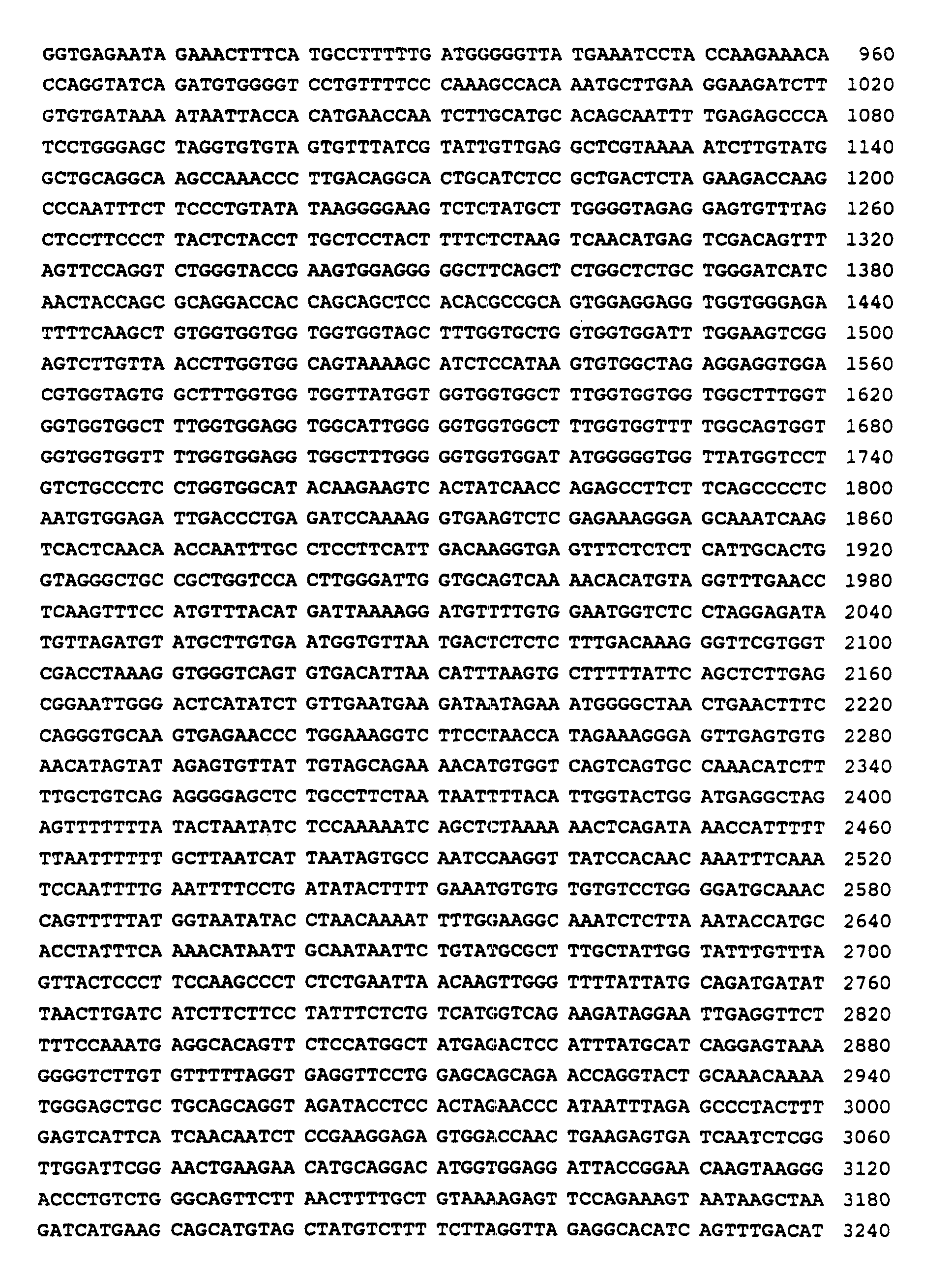
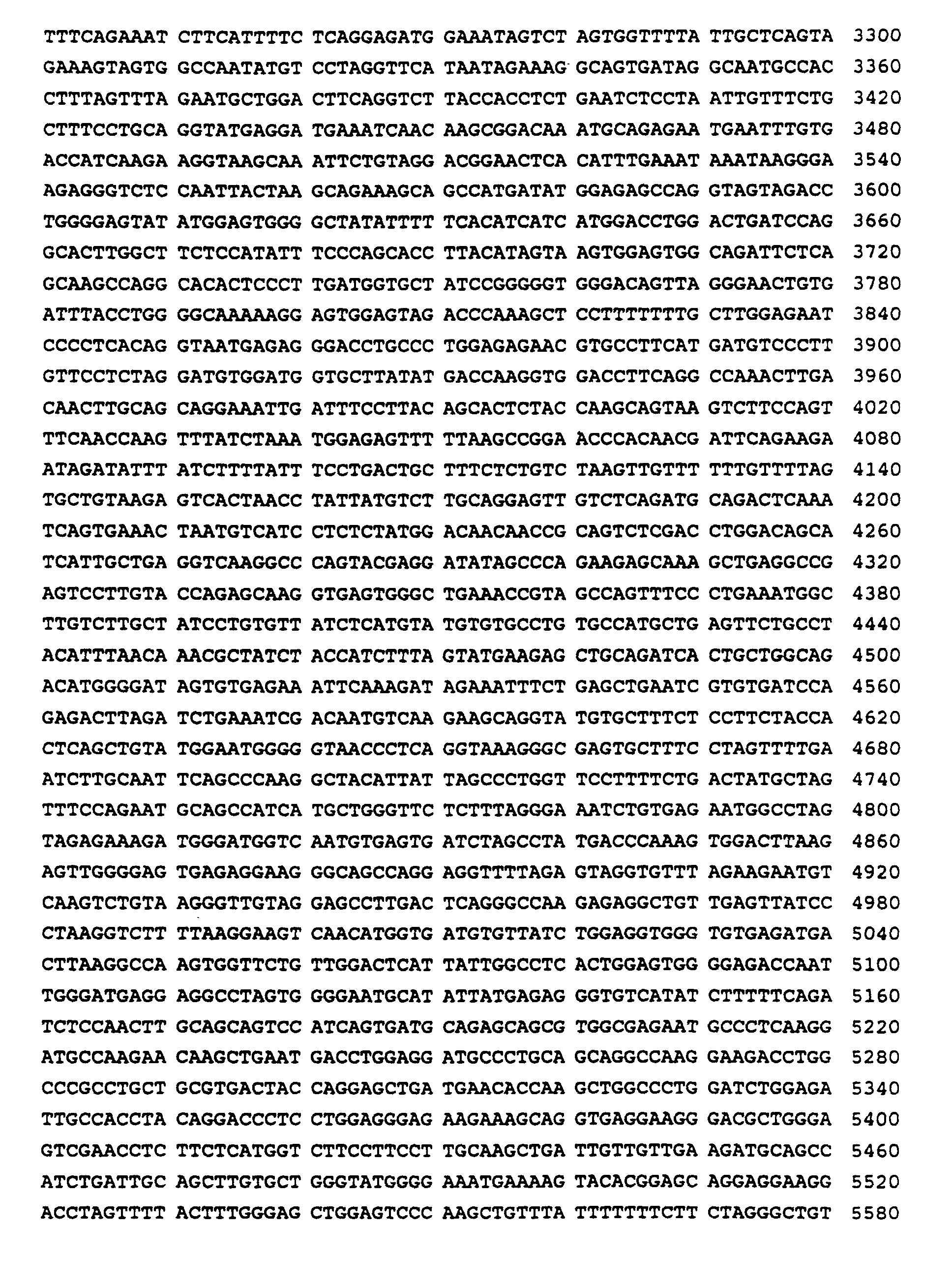


- (2) INFORMATION FOR SEQ ID NO:2:
- (i) SEQUENCE CHARACTERISTICS:
- (A) LENGTH: 6693 base pairs
- (B) TYPE: nucleic acid
- (C) STRANDEDNESS: double
- (D) TOPOLOGY: linear
- (ii) MOLECULE TYPE: DNA (genomic)
- (iii) HYPOTHETICAL: NO
- (iv) ANTI-SENSE: NO
- (xi) SEQUENCE DESCRIPTION: SEQ ID NO:2:
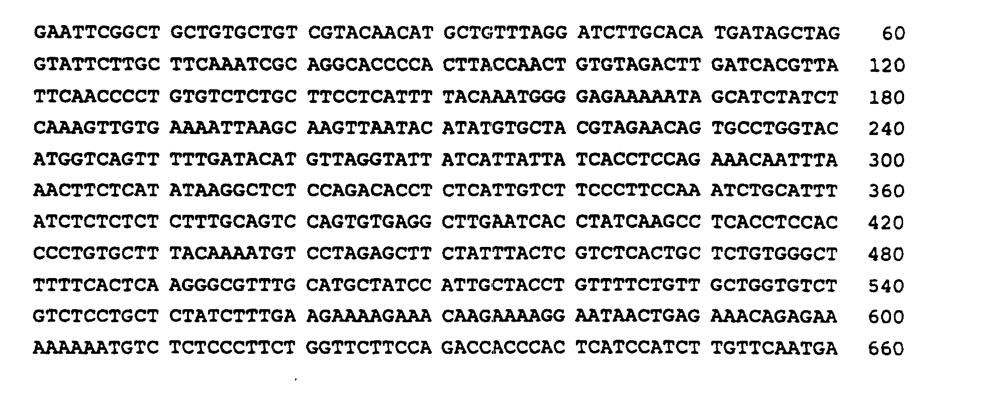


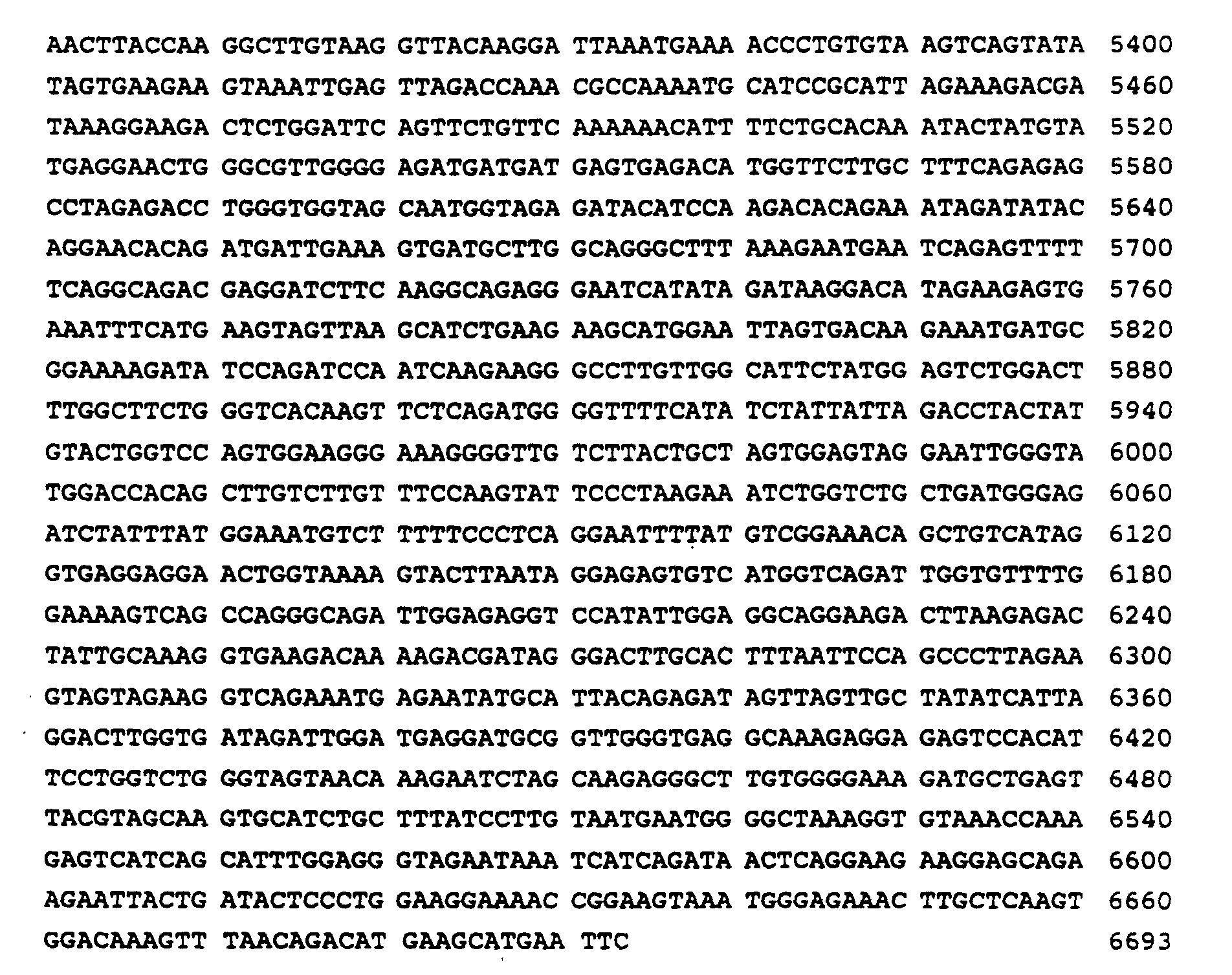
- (2) INFORMATION FOR SEQ ID NO:3:
- (i) SEQUENCE CHARACTERISTICS:
- (A) LENGTH: 24979 base pairs
- (B) TYPE: nucleic acid
- (C) STRANDEDNESS: double
- (D) TOPOLOGY: linear
- (ii) MOLECULE TYPE: DNA (genomic)
- (iii) HYPOTHETICAL: NO
- (iv) ANTI-SENSE: NO
- (xi) SEQUENCE DESCRIPTION: SEQ ID NO:3:


- (2) INFORMATION FOR SEQ ID NO:4:
- (i) SEQUENCE CHARACTERISTICS:
- (A) LENGTH: 8 amino acids
- (B) TYPE: amino acid
- (C) STRANDEDNESS: single
- (D) TOPOLOGY: linear
- (ii) MOLECULE TYPE: peptide
- (iii) HYPOTHETICAL: NO
- (iv) ANTI-SENSE: NO
- (xi) SEQUENCE DESCRIPTION: SEQ ID NO:4:

- (2) INFORMATION FOR SEQ ID NO:5:
- (i) SEQUENCE CHARACTERISTICS:
- (A) LENGTH: 16 amino acids
- (B) TYPE: amino acid
- (C) STRANDEDNESS: single
- (D) TOPOLOGY: linear
- (ii) MOLECULE TYPE: peptide
- (iii) HYPOTHETICAL: NO
- (iv) ANTI-SENSE: NO
- (xi) SEQUENCE DESCRIPTION: SEQ ID NO:5:

-
Claims (26)
- A keratin K1 vector for expression of a nucleic acid cassette in the epidermis comprising:a 5' flanking region of the keratin K1 gene, the 5' flanking sequence including a keratin K1 promoter, a 5' transcribed but untransalated region, and a first intron, all in sequential and positional relationship for expression of the nucleic acid cassette;a 3' flanking region of the keratin K1 gene containing Vitamin D3 regulatory sequences, a 3' transcribed but untranslated region and contiguous non-coding DNA containing a transcriptional termination region; anda polylinker having a plurality of restriction endonuclease sites, the polylinker connecting the 5' flanking region to the 3' flanking region and providing a position for insertion of the nucleic acid cassette, the linker optionally and where necessary additionally comprising the nucleic acid cassette.
- A vector according to claim 1 wherein the 5' flanking region is approximately 1.2 kb and the 3' flanking region is approximately 3.9 kb.
- A vector according to claim 1 or 2 wherein the nucleic acid cassette is present and includes a nucleic acid sequence coding for a protein, polypeptide or antisense RNA.
- A vector according to claim 3 wherein the cassette includes a nucleic acid sequence coding for an oncogene, such as ras, fos, myc, erb, src, sis or jun.
- A vector according to claim 3 wherein the cassette includes a nucleic acid sequence coding for a transforming gene, the E6 or E7 transforming sequence of human papilloma virus or the TGF-α sequence.
- A vector according to any preceding claim wherein the restriction endonuclease site is Bam HI, Xma I, Kpn I, Not I, Cla I or Bgl II.
- A vector according to any preceeding claim further comprising an additional 5' flanking sequence from the 18 kb Eco RV fragment on the end of the vector.
- A human keratin K1 vector according to claim 1 comprisinga 5' flanking region comprising nucleotides 1 to 1246 of SEQ.ID.No.1;a 3' flanking region comprising nucleotides 6891 to 10747 of SEQ.ID.No.1; anda linker comprising nucleotide 2351 to 2376 of SBQ.ID No. 2.
- An epidermal host cell transformed or transfected with a vector according to any of claims 1 to 8.
- A host cell according to claim 9 wherein the cassette has a nucleic acid sequence encoding a hormone, a growth factor, an enzyme, a drug, a tumor antigen, a viral antigen, an insect antigen, a bacterial antigen, or a parasitic antigen, for example the sequence encodes proinsulin, insulin, growth hormone, insulin-like growth factor I, insulin-like growth factor II, insulin growth factor binding protein, epidermal growth factor (TGF-α), dermal growth factor (PDGF), an angiogenesis factor, Type IV collagen, laminin, nidogen, Type VII collagen or a protein which induces an immunological response.
- A bioreactor comprising epidermal cells according to claim 9 or 10.
- A method of ex vivo introduction of a keratin K1 vector into epidermal cells, the method comprising cotransfecting a vector according to any one of claims 3 to 7 with a selectable marker and selecting the transformed cells.
- A method of making a transgenic non-human animal comprising:collecting very early fertilized eggs;inserting a vector according to any one of claims 3 to 7 into the fertilized eggs by micro-injecting the vector into pronuclei; andtransferring the injected eggs into pseudopregnant recipient females.
- A transgenic non-human animal containing a vector according to any of claims 3 to 7 in its germ and somatic cells, wherein the vector was introduced into the animal or an ancestor of the animal at an embryonic stage and the nucleic acid cassette of the vector is only expressed in the epidermis.
- A method of studying the origin of, or treatment for, cancer, the method comprising:making a transgenic non-human animal by injecting an embryo with a humankeratin K1 vector according to any one of claims 3 to 7 and containing an oncogene in the nucleic acid cassette; andusing the resultant animal or the progeny in studies of cancer.
- A method according to claim 15 wherein the animal contains more than one oncogene.
- A keratin K1 vector according to any one of claims 3 to 7 and containing a nucleic acid cassette for use for transient introduction of the nucleic acid cassette into a human by contacting an epidermal cell for sufficient time to transduce the cells with the vector.
- The use of a keratin K1 vector according to claim 3 which includes a nucleic acid cassette having a nucleic acid sequence encoding a growth factor, or an epidermal cell transformed or transduced with such a vector, in the manufacture of a medicament for enhancing healing of a wound or surgical incision or treating skin ulcers.
- The use according to claim 18 wherein a plurality of vectors are used and wherein the cassette of at least one vector includes the nucleic acid sequence of epidermal growth factor (TGF-α), the cassette of at least one vector includes the nucleic acid sequence of dermal growth factor (PDGF), the cassette of at least one vector includes the nucleic acid sequence for a matrix protein to anchor the epidermis to the dermis or the cassette of at least one vector includes the nucleic acid sequence for an angiogenesis factor.
- The use according to claim 19 wherein the matrix protein is Type IV collagen, laminin, nidogen, or Type VII collagen and/or the angiogenesis factor is an acid fibroblast growth factor, basic fibroblast growth factor or angiogenin.
- The use of a keratin K1 vector according to claim 3 which includes a nucleic acid cassette having a nucleic acid sequence encoding for TGF-β, a soluble form of cytokine receptor, or an antisense RNA, or a host epidermal cell transformed or transduced with such a vector, in the manufacture of a medicament for treating psoriasis.
- The use according to claim 21 wherein the soluble form of cytokine receptor is IL-1, IL-6 or IL-8 and/or the antisense RNA is to a sequence of TGF-α, IL-1, IL-6 or IL-8.
- The use of keratin K1 vector according to claim 3 which includes a nucleic acid cassette having a nucleic acid sequence encoding for an antisense RNA for the E6 or E7 gene of human papilloma virus or a sequence encoding for the normal p53 protein, or a host epidermal cell transformed or transduced with such a vector, in the manufacture of a medicament for treating skin cancer.
- The use of a keratin K1 vector according to claim 3 which includes a nucleic acid cassette having a nucleic acid sequence encoding for a protein or polypeptide which induces an immunological response, or a host epidermal cell transformed or transduced with such a vector, in the manufacture of a vaccine.
- The use according to claim 24 wherein the sequence encodes a viral capsid protein, such as from the human papilloma virus.
- A pharmaceutical composition comprising:(a) a vector or epidermal host cell as defined in any of claims 18 to 20, the composition being adapted to enhance healing of a wound or surgical incision or to treat skin ulcers;(b) a vector or epidermal host cell as defined in claim 21 or 22, the composition being adapted to treat psoriasis:(c) a vector or epidermal host cell as defined in claim 23 the composition being adopted to treat skin cancer; or(d) a vector or epidermal host cell as defined in claim 24 the composition being a vaccine.
Applications Claiming Priority (3)
| Application Number | Priority Date | Filing Date | Title |
|---|---|---|---|
| US87628992A | 1992-04-30 | 1992-04-30 | |
| US876289 | 1992-04-30 | ||
| PCT/US1993/003985 WO1993022430A1 (en) | 1992-04-30 | 1993-04-28 | Development of a vector to target gene expression to the epidermis of transgenic animals |
Publications (3)
| Publication Number | Publication Date |
|---|---|
| EP0652948A1 EP0652948A1 (en) | 1995-05-17 |
| EP0652948A4 EP0652948A4 (en) | 1997-04-23 |
| EP0652948B1 true EP0652948B1 (en) | 2001-11-14 |
Family
ID=25367361
Family Applications (1)
| Application Number | Title | Priority Date | Filing Date |
|---|---|---|---|
| EP93910867A Expired - Lifetime EP0652948B1 (en) | 1992-04-30 | 1993-04-28 | Development of a vector to target gene expression to the epidermis of transgenic animals |
Country Status (7)
| Country | Link |
|---|---|
| EP (1) | EP0652948B1 (en) |
| JP (1) | JPH08503844A (en) |
| AT (1) | ATE208814T1 (en) |
| AU (1) | AU672409B2 (en) |
| CA (1) | CA2134675A1 (en) |
| DE (1) | DE69331144T2 (en) |
| WO (1) | WO1993022430A1 (en) |
Families Citing this family (13)
| Publication number | Priority date | Publication date | Assignee | Title |
|---|---|---|---|---|
| US20030157074A1 (en) * | 1994-11-16 | 2003-08-21 | Mitrani Eduardo N. | Vitro micro-organs, and uses related thereto |
| US5721340A (en) * | 1994-11-28 | 1998-02-24 | The Wistar Institute Of Anatomy & Biology | p53 proteins with altered tetramerization domains |
| US5573925A (en) * | 1994-11-28 | 1996-11-12 | The Wistar Institute Of Anatomy And Biology | P53 proteins with altered tetramerization domains |
| US6187993B1 (en) | 1995-02-25 | 2001-02-13 | Imperial Cancer Research Technology Limited | Transgenic animals as model of psoriasis |
| US5847083A (en) * | 1996-08-21 | 1998-12-08 | The Wistar Institute Of Anatomy And Biology | Modified p53 constructs which enhance DNA binding |
| WO1997042213A1 (en) * | 1996-05-08 | 1997-11-13 | Chiron Corporation | MAMMALIAN ADDITIONAL SEX COMBS (MAMMALIAN Asx) ACTS AS AN ONCOGENE |
| AU7334398A (en) * | 1997-04-03 | 1998-10-22 | Vlaams Interuniversitair Instituut Voor Biotechnologie Vzw | Transgenic animal with controllable hyperproliferation and inflammation phenotype in the skin |
| GB9715064D0 (en) * | 1997-07-17 | 1997-09-24 | Ppl Therapeutics Scotland Ltd | Protein expression |
| GB9715085D0 (en) | 1997-07-18 | 1997-09-24 | Jahoda Amanda J | Gene therapy vehicle |
| US20030152562A1 (en) * | 2001-10-23 | 2003-08-14 | Yissum Research Development Company Of The Hebrew University Of Jerusalem | Vitro micro-organs, and uses related thereto |
| GB0010105D0 (en) * | 2000-04-26 | 2000-06-14 | Ml Lab Plc | Cell ablation |
| US9155749B2 (en) | 2006-09-14 | 2015-10-13 | Medgenics Medical Israel Ltd. | Long lasting drug formulations |
| WO2011159758A2 (en) | 2010-06-15 | 2011-12-22 | Medgenics Medical Israel Ltd. | Long lasting drug formulations |
Family Cites Families (2)
| Publication number | Priority date | Publication date | Assignee | Title |
|---|---|---|---|---|
| US4497796A (en) * | 1980-03-26 | 1985-02-05 | The Regents Of The University Of California | Gene transfer in intact mammals |
| US4736866B1 (en) * | 1984-06-22 | 1988-04-12 | Transgenic non-human mammals |
-
1993
- 1993-04-28 EP EP93910867A patent/EP0652948B1/en not_active Expired - Lifetime
- 1993-04-28 CA CA002134675A patent/CA2134675A1/en not_active Abandoned
- 1993-04-28 JP JP5519481A patent/JPH08503844A/en active Pending
- 1993-04-28 DE DE69331144T patent/DE69331144T2/en not_active Expired - Fee Related
- 1993-04-28 WO PCT/US1993/003985 patent/WO1993022430A1/en not_active Ceased
- 1993-04-28 AT AT93910867T patent/ATE208814T1/en not_active IP Right Cessation
- 1993-04-28 AU AU42207/93A patent/AU672409B2/en not_active Ceased
Also Published As
| Publication number | Publication date |
|---|---|
| CA2134675A1 (en) | 1993-11-11 |
| EP0652948A1 (en) | 1995-05-17 |
| EP0652948A4 (en) | 1997-04-23 |
| DE69331144T2 (en) | 2002-03-21 |
| AU672409B2 (en) | 1996-10-03 |
| JPH08503844A (en) | 1996-04-30 |
| DE69331144D1 (en) | 2001-12-20 |
| WO1993022430A1 (en) | 1993-11-11 |
| AU4220793A (en) | 1993-11-29 |
| ATE208814T1 (en) | 2001-11-15 |
Similar Documents
| Publication | Publication Date | Title |
|---|---|---|
| Martinez-Salas et al. | The need for enhancers in gene expression first appears during mouse development with formation of the zygotic nucleus. | |
| Rangarajan et al. | Notch signaling is a direct determinant of keratinocyte growth arrest and entry into differentiation | |
| Herber et al. | Squamous epithelial hyperplasia and carcinoma in mice transgenic for the human papillomavirus type 16 E7 oncogene | |
| Lambert et al. | Epidermal cancer associated with expression of human papillomavirus type 16 E6 and E7 oncogenes in the skin of transgenic mice. | |
| Knezevic et al. | Hoxd-12 differentially affects preaxial and postaxial chondrogenic branches in the limb and regulates Sonic hedgehog in a positive feedback loop | |
| Nehls et al. | Transcription factors nuclear factor I and Spl interact with the murine collagen α1 (I) promoter | |
| EP0652948B1 (en) | Development of a vector to target gene expression to the epidermis of transgenic animals | |
| JPH06504671A (en) | Method for suppressing transformation that occurs via Neu and composition therefor | |
| Gopal-Srivastava et al. | Pax-6 and αB-crystallin/Small Heat Shock Protein Gene Regulation in the Murine Lens INTERACTION WITH THE LENS-SPECIFIC REGIONS, LSR1 AND LSR2 | |
| Greenhalgh et al. | Transgenic Mice Expressing Targeted HPV-18 E6 and E7 Oncogenes in the Epidermis Develop Verrucous Lesions and Spontaneous, ras^ H^ a-activated Papillomas | |
| Von Harsdorf et al. | Identification of a cis-acting regulatory element conferring inducibility of the atrial natriuretic factor gene in acute pressure overload. | |
| Dunn et al. | Regulation of a hair follicle keratin intermediate filament gene promoter | |
| US5958764A (en) | Specific expression vectors and methods of use | |
| US5476926A (en) | Adipocyte-specific DNA sequences and use thereof in the production of transgenic animals exhibiting altered fat tissue metabolism | |
| Okada et al. | Identification of a novel cis-acting element for fibroblast-specific transcription of the FSP1 gene | |
| AU669713B2 (en) | Constitutive and inducible epidermal vector systems | |
| Zhao et al. | A distal element in the HPV-11 upstream regulatory region contributes to promoter repression in basal keratinocytes in squamous epithelium | |
| Scherneck et al. | The hamster polyomavirus—a brief review of recent knowledge | |
| JPH07500490A (en) | Transgenic non-human animal carrying a non-infectious HIV genome | |
| Klessig | Adenovirus—Simian Virus 40 Interactions | |
| Peng et al. | Development of Keratoacanthomas and Squamous Cell Carcinomas in Transgenic Rabbits with Targeted Expression of EJras. Oncogene in Epidermis | |
| Mahony et al. | Analysis of mouse keratin 6a regulatory sequences in transgenic mice reveals constitutive, tissue-specific expression by a keratin 6a minigene | |
| Natarajan et al. | Cis and trans activation of adenovirus IVa2 gene transcription | |
| Cavin et al. | Tissue-specific and ubiquitous factors binding next to the glucocorticoid receptor modulate transcription from the mouse mammary tumor virus promoter | |
| CA2272851A1 (en) | Syndecan enhancer element and its use for targeting gene expression |
Legal Events
| Date | Code | Title | Description |
|---|---|---|---|
| PUAI | Public reference made under article 153(3) epc to a published international application that has entered the european phase |
Free format text: ORIGINAL CODE: 0009012 |
|
| 17P | Request for examination filed |
Effective date: 19941129 |
|
| AK | Designated contracting states |
Kind code of ref document: A1 Designated state(s): AT BE CH DE DK ES FR GB GR IE IT LI LU MC NL PT SE |
|
| A4 | Supplementary search report drawn up and despatched |
Effective date: 19970306 |
|
| AK | Designated contracting states |
Kind code of ref document: A4 Designated state(s): AT BE CH DE DK ES FR GB GR IE IT LI LU MC NL PT SE |
|
| RIN1 | Information on inventor provided before grant (corrected) |
Inventor name: YUSPA, STUART H. Inventor name: GREENHALGH, DAVID A. Inventor name: ROTHNAGEL, JOSEPH A. Inventor name: ROOP, DENNIS R. |
|
| 17Q | First examination report despatched |
Effective date: 19990304 |
|
| GRAG | Despatch of communication of intention to grant |
Free format text: ORIGINAL CODE: EPIDOS AGRA |
|
| GRAG | Despatch of communication of intention to grant |
Free format text: ORIGINAL CODE: EPIDOS AGRA |
|
| GRAH | Despatch of communication of intention to grant a patent |
Free format text: ORIGINAL CODE: EPIDOS IGRA |
|
| GRAH | Despatch of communication of intention to grant a patent |
Free format text: ORIGINAL CODE: EPIDOS IGRA |
|
| GRAA | (expected) grant |
Free format text: ORIGINAL CODE: 0009210 |
|
| AK | Designated contracting states |
Kind code of ref document: B1 Designated state(s): AT BE CH DE DK ES FR GB GR IE IT LI LU MC NL PT SE |
|
| PG25 | Lapsed in a contracting state [announced via postgrant information from national office to epo] |
Ref country code: NL Free format text: LAPSE BECAUSE OF FAILURE TO SUBMIT A TRANSLATION OF THE DESCRIPTION OR TO PAY THE FEE WITHIN THE PRESCRIBED TIME-LIMIT Effective date: 20011114 Ref country code: IT Free format text: LAPSE BECAUSE OF FAILURE TO SUBMIT A TRANSLATION OF THE DESCRIPTION OR TO PAY THE FEE WITHIN THE PRE;WARNING: LAPSES OF ITALIAN PATENTS WITH EFFECTIVE DATE BEFORE 2007 MAY HAVE OCCURRED AT ANY TIME BEFORE 2007. THE CORRECT EFFECTIVE DATE MAY BE DIFFERENT FROM THE ONE RECORDED.SCRIBED TIME-LIMIT Effective date: 20011114 Ref country code: GR Free format text: LAPSE BECAUSE OF FAILURE TO SUBMIT A TRANSLATION OF THE DESCRIPTION OR TO PAY THE FEE WITHIN THE PRESCRIBED TIME-LIMIT Effective date: 20011114 Ref country code: BE Free format text: LAPSE BECAUSE OF FAILURE TO SUBMIT A TRANSLATION OF THE DESCRIPTION OR TO PAY THE FEE WITHIN THE PRESCRIBED TIME-LIMIT Effective date: 20011114 Ref country code: AT Free format text: LAPSE BECAUSE OF FAILURE TO SUBMIT A TRANSLATION OF THE DESCRIPTION OR TO PAY THE FEE WITHIN THE PRESCRIBED TIME-LIMIT Effective date: 20011114 |
|
| REF | Corresponds to: |
Ref document number: 208814 Country of ref document: AT Date of ref document: 20011115 Kind code of ref document: T |
|
| REG | Reference to a national code |
Ref country code: CH Ref legal event code: EP |
|
| REG | Reference to a national code |
Ref country code: IE Ref legal event code: FG4D |
|
| REF | Corresponds to: |
Ref document number: 69331144 Country of ref document: DE Date of ref document: 20011220 |
|
| REG | Reference to a national code |
Ref country code: GB Ref legal event code: IF02 |
|
| PG25 | Lapsed in a contracting state [announced via postgrant information from national office to epo] |
Ref country code: SE Free format text: LAPSE BECAUSE OF FAILURE TO SUBMIT A TRANSLATION OF THE DESCRIPTION OR TO PAY THE FEE WITHIN THE PRESCRIBED TIME-LIMIT Effective date: 20020214 Ref country code: PT Free format text: LAPSE BECAUSE OF FAILURE TO SUBMIT A TRANSLATION OF THE DESCRIPTION OR TO PAY THE FEE WITHIN THE PRESCRIBED TIME-LIMIT Effective date: 20020214 Ref country code: DK Free format text: LAPSE BECAUSE OF FAILURE TO SUBMIT A TRANSLATION OF THE DESCRIPTION OR TO PAY THE FEE WITHIN THE PRESCRIBED TIME-LIMIT Effective date: 20020214 |
|
| REG | Reference to a national code |
Ref country code: CH Ref legal event code: NV Representative=s name: PATENTANWAELTE SCHAAD, BALASS, MENZL & PARTNER AG |
|
| ET | Fr: translation filed | ||
| PGFP | Annual fee paid to national office [announced via postgrant information from national office to epo] |
Ref country code: FR Payment date: 20020410 Year of fee payment: 10 |
|
| PGFP | Annual fee paid to national office [announced via postgrant information from national office to epo] |
Ref country code: GB Payment date: 20020424 Year of fee payment: 10 |
|
| PGFP | Annual fee paid to national office [announced via postgrant information from national office to epo] |
Ref country code: IE Payment date: 20020426 Year of fee payment: 10 |
|
| PG25 | Lapsed in a contracting state [announced via postgrant information from national office to epo] |
Ref country code: MC Free format text: LAPSE BECAUSE OF NON-PAYMENT OF DUE FEES Effective date: 20020428 Ref country code: LU Free format text: LAPSE BECAUSE OF NON-PAYMENT OF DUE FEES Effective date: 20020428 |
|
| PG25 | Lapsed in a contracting state [announced via postgrant information from national office to epo] |
Ref country code: LI Free format text: LAPSE BECAUSE OF NON-PAYMENT OF DUE FEES Effective date: 20020430 Ref country code: CH Free format text: LAPSE BECAUSE OF NON-PAYMENT OF DUE FEES Effective date: 20020430 |
|
| NLV1 | Nl: lapsed or annulled due to failure to fulfill the requirements of art. 29p and 29m of the patents act | ||
| PGFP | Annual fee paid to national office [announced via postgrant information from national office to epo] |
Ref country code: DE Payment date: 20020502 Year of fee payment: 10 |
|
| PG25 | Lapsed in a contracting state [announced via postgrant information from national office to epo] |
Ref country code: ES Free format text: LAPSE BECAUSE OF FAILURE TO SUBMIT A TRANSLATION OF THE DESCRIPTION OR TO PAY THE FEE WITHIN THE PRESCRIBED TIME-LIMIT Effective date: 20020530 |
|
| PLBE | No opposition filed within time limit |
Free format text: ORIGINAL CODE: 0009261 |
|
| STAA | Information on the status of an ep patent application or granted ep patent |
Free format text: STATUS: NO OPPOSITION FILED WITHIN TIME LIMIT |
|
| 26N | No opposition filed | ||
| REG | Reference to a national code |
Ref country code: CH Ref legal event code: PL |
|
| PG25 | Lapsed in a contracting state [announced via postgrant information from national office to epo] |
Ref country code: IE Free format text: LAPSE BECAUSE OF NON-PAYMENT OF DUE FEES Effective date: 20030428 Ref country code: GB Free format text: LAPSE BECAUSE OF NON-PAYMENT OF DUE FEES Effective date: 20030428 |
|
| PG25 | Lapsed in a contracting state [announced via postgrant information from national office to epo] |
Ref country code: DE Free format text: LAPSE BECAUSE OF NON-PAYMENT OF DUE FEES Effective date: 20031101 |
|
| GBPC | Gb: european patent ceased through non-payment of renewal fee | ||
| PG25 | Lapsed in a contracting state [announced via postgrant information from national office to epo] |
Ref country code: FR Free format text: LAPSE BECAUSE OF NON-PAYMENT OF DUE FEES Effective date: 20031231 |
|
| REG | Reference to a national code |
Ref country code: FR Ref legal event code: ST |
|
| REG | Reference to a national code |
Ref country code: IE Ref legal event code: MM4A |