DE69926639T2 - BONE MARROW CELLS AS A SOURCE OF NEURONS FOR THE RECONSTRUCTION OF BRAIN AND BONE MARROW - Google Patents
BONE MARROW CELLS AS A SOURCE OF NEURONS FOR THE RECONSTRUCTION OF BRAIN AND BONE MARROW Download PDFInfo
- Publication number
- DE69926639T2 DE69926639T2 DE69926639T DE69926639T DE69926639T2 DE 69926639 T2 DE69926639 T2 DE 69926639T2 DE 69926639 T DE69926639 T DE 69926639T DE 69926639 T DE69926639 T DE 69926639T DE 69926639 T2 DE69926639 T2 DE 69926639T2
- Authority
- DE
- Germany
- Prior art keywords
- cells
- bone marrow
- bmsc
- cell
- neuronal
- Prior art date
- Legal status (The legal status is an assumption and is not a legal conclusion. Google has not performed a legal analysis and makes no representation as to the accuracy of the status listed.)
- Expired - Fee Related
Links
- 210000002569 neuron Anatomy 0.000 title claims description 63
- 210000001185 bone marrow Anatomy 0.000 title claims description 40
- 210000002798 bone marrow cell Anatomy 0.000 title claims description 20
- 210000004556 brain Anatomy 0.000 title description 26
- 210000004027 cell Anatomy 0.000 claims description 250
- 210000004271 bone marrow stromal cell Anatomy 0.000 claims description 123
- 238000000034 method Methods 0.000 claims description 63
- 230000001537 neural effect Effects 0.000 claims description 47
- 229930002330 retinoic acid Natural products 0.000 claims description 33
- SHGAZHPCJJPHSC-YCNIQYBTSA-N retinoic acid group Chemical group C\C(=C/C(=O)O)\C=C\C=C(\C=C\C1=C(CCCC1(C)C)C)/C SHGAZHPCJJPHSC-YCNIQYBTSA-N 0.000 claims description 33
- 229960001727 tretinoin Drugs 0.000 claims description 28
- 108090000715 Brain-derived neurotrophic factor Proteins 0.000 claims description 23
- 102000004219 Brain-derived neurotrophic factor Human genes 0.000 claims description 23
- 230000001605 fetal effect Effects 0.000 claims description 23
- 239000001963 growth medium Substances 0.000 claims description 20
- 239000002609 medium Substances 0.000 claims description 17
- 239000004033 plastic Substances 0.000 claims description 16
- 229920003023 plastic Polymers 0.000 claims description 16
- 210000003958 hematopoietic stem cell Anatomy 0.000 claims description 14
- 238000004519 manufacturing process Methods 0.000 claims description 12
- 108010025020 Nerve Growth Factor Proteins 0.000 claims description 11
- 102000015336 Nerve Growth Factor Human genes 0.000 claims description 11
- 102000034615 Glial cell line-derived neurotrophic factor Human genes 0.000 claims description 10
- 108091010837 Glial cell line-derived neurotrophic factor Proteins 0.000 claims description 10
- 239000003795 chemical substances by application Substances 0.000 claims description 9
- 210000002536 stromal cell Anatomy 0.000 claims description 8
- 239000003226 mitogen Substances 0.000 claims description 7
- 230000007514 neuronal growth Effects 0.000 claims description 7
- SHGAZHPCJJPHSC-ZVCIMWCZSA-N 9-cis-retinoic acid Chemical compound OC(=O)/C=C(\C)/C=C/C=C(/C)\C=C\C1=C(C)CCCC1(C)C SHGAZHPCJJPHSC-ZVCIMWCZSA-N 0.000 claims description 6
- 229960001445 alitretinoin Drugs 0.000 claims description 6
- 235000015097 nutrients Nutrition 0.000 claims 1
- 108010005774 beta-Galactosidase Proteins 0.000 description 57
- WQZGKKKJIJFFOK-FPRJBGLDSA-N beta-D-galactose Chemical compound OC[C@H]1O[C@@H](O)[C@H](O)[C@@H](O)[C@H]1O WQZGKKKJIJFFOK-FPRJBGLDSA-N 0.000 description 54
- 241000282414 Homo sapiens Species 0.000 description 44
- 241000700159 Rattus Species 0.000 description 29
- 238000002054 transplantation Methods 0.000 description 29
- 230000004069 differentiation Effects 0.000 description 28
- 229940077737 brain-derived neurotrophic factor Drugs 0.000 description 22
- 210000000449 purkinje cell Anatomy 0.000 description 21
- 210000000130 stem cell Anatomy 0.000 description 20
- 210000001519 tissue Anatomy 0.000 description 20
- 241001465754 Metazoa Species 0.000 description 18
- 241000699666 Mus <mouse, genus> Species 0.000 description 17
- 239000003102 growth factor Substances 0.000 description 17
- 238000011282 treatment Methods 0.000 description 17
- 241000699670 Mus sp. Species 0.000 description 16
- 239000003550 marker Substances 0.000 description 16
- 240000006028 Sambucus nigra Species 0.000 description 14
- 210000001577 neostriatum Anatomy 0.000 description 14
- 108090000623 proteins and genes Proteins 0.000 description 14
- 206010003591 Ataxia Diseases 0.000 description 13
- 108091003079 Bovine Serum Albumin Proteins 0.000 description 13
- 108091000117 Tyrosine 3-Monooxygenase Proteins 0.000 description 13
- 102000048218 Tyrosine 3-monooxygenases Human genes 0.000 description 13
- 210000001638 cerebellum Anatomy 0.000 description 12
- 230000000694 effects Effects 0.000 description 12
- 239000000203 mixture Substances 0.000 description 12
- 210000004498 neuroglial cell Anatomy 0.000 description 12
- 210000004940 nucleus Anatomy 0.000 description 12
- 208000037265 diseases, disorders, signs and symptoms Diseases 0.000 description 11
- 210000000463 red nucleus Anatomy 0.000 description 11
- 102000016359 Fibronectins Human genes 0.000 description 10
- 108010067306 Fibronectins Proteins 0.000 description 10
- 230000006378 damage Effects 0.000 description 10
- 229940053128 nerve growth factor Drugs 0.000 description 10
- VBEQCZHXXJYVRD-GACYYNSASA-N uroanthelone Chemical compound C([C@@H](C(=O)N[C@H](C(=O)N[C@@H](CS)C(=O)N[C@@H](CC(N)=O)C(=O)N[C@@H](CS)C(=O)N[C@H](C(=O)N[C@@H]([C@@H](C)CC)C(=O)NCC(=O)N[C@@H](CC=1C=CC(O)=CC=1)C(=O)N[C@@H](CO)C(=O)NCC(=O)N[C@@H](CC(O)=O)C(=O)N[C@@H](CCCNC(N)=N)C(=O)N[C@@H](CS)C(=O)N[C@@H](CCC(N)=O)C(=O)N[C@@H]([C@@H](C)O)C(=O)N[C@@H](CCCNC(N)=N)C(=O)N[C@@H](CC(O)=O)C(=O)N[C@@H](CC(C)C)C(=O)N[C@@H](CCCNC(N)=N)C(=O)N[C@@H](CC=1C2=CC=CC=C2NC=1)C(=O)N[C@@H](CC=1C2=CC=CC=C2NC=1)C(=O)N[C@@H](CCC(O)=O)C(=O)N[C@@H](CC(C)C)C(=O)N[C@@H](CCCNC(N)=N)C(O)=O)C(C)C)[C@@H](C)O)NC(=O)[C@H](CO)NC(=O)[C@H](CC(O)=O)NC(=O)[C@H](CC(C)C)NC(=O)[C@H](CO)NC(=O)[C@H](CCC(O)=O)NC(=O)[C@@H](NC(=O)[C@H](CC=1NC=NC=1)NC(=O)[C@H](CCSC)NC(=O)[C@H](CS)NC(=O)[C@@H](NC(=O)CNC(=O)CNC(=O)[C@H](CC(N)=O)NC(=O)[C@H](CC(C)C)NC(=O)[C@H](CS)NC(=O)[C@H](CC=1C=CC(O)=CC=1)NC(=O)CNC(=O)[C@H](CC(O)=O)NC(=O)[C@H](CC=1C=CC(O)=CC=1)NC(=O)[C@H](CO)NC(=O)[C@H](CO)NC(=O)[C@H]1N(CCC1)C(=O)[C@H](CS)NC(=O)CNC(=O)[C@H]1N(CCC1)C(=O)[C@H](CC=1C=CC(O)=CC=1)NC(=O)[C@H](CO)NC(=O)[C@@H](N)CC(N)=O)C(C)C)[C@@H](C)CC)C1=CC=C(O)C=C1 VBEQCZHXXJYVRD-GACYYNSASA-N 0.000 description 10
- 208000018737 Parkinson disease Diseases 0.000 description 9
- 230000002490 cerebral effect Effects 0.000 description 9
- 210000001259 mesencephalon Anatomy 0.000 description 9
- 208000015122 neurodegenerative disease Diseases 0.000 description 9
- 239000000872 buffer Substances 0.000 description 8
- 102000014823 calbindin Human genes 0.000 description 8
- 108060001061 calbindin Proteins 0.000 description 8
- 201000010099 disease Diseases 0.000 description 8
- 238000009826 distribution Methods 0.000 description 8
- 238000000338 in vitro Methods 0.000 description 8
- 101150066555 lacZ gene Proteins 0.000 description 8
- 210000003098 myoblast Anatomy 0.000 description 8
- 230000004770 neurodegeneration Effects 0.000 description 8
- 102000004169 proteins and genes Human genes 0.000 description 8
- 238000000926 separation method Methods 0.000 description 8
- 210000002966 serum Anatomy 0.000 description 8
- 238000010186 staining Methods 0.000 description 8
- 238000012360 testing method Methods 0.000 description 8
- 208000024827 Alzheimer disease Diseases 0.000 description 7
- 241000124008 Mammalia Species 0.000 description 7
- 206010028980 Neoplasm Diseases 0.000 description 7
- 239000012894 fetal calf serum Substances 0.000 description 7
- 238000001415 gene therapy Methods 0.000 description 7
- 230000003394 haemopoietic effect Effects 0.000 description 7
- 238000002513 implantation Methods 0.000 description 7
- 238000001727 in vivo Methods 0.000 description 7
- 238000011534 incubation Methods 0.000 description 7
- 210000000956 olfactory bulb Anatomy 0.000 description 7
- 238000011084 recovery Methods 0.000 description 7
- APKFDSVGJQXUKY-KKGHZKTASA-N Amphotericin-B Natural products O[C@H]1[C@@H](N)[C@H](O)[C@@H](C)O[C@H]1O[C@H]1C=CC=CC=CC=CC=CC=CC=C[C@H](C)[C@@H](O)[C@@H](C)[C@H](C)OC(=O)C[C@H](O)C[C@H](O)CC[C@@H](O)[C@H](O)C[C@H](O)C[C@](O)(C[C@H](O)[C@H]2C(O)=O)O[C@H]2C1 APKFDSVGJQXUKY-KKGHZKTASA-N 0.000 description 6
- 239000012983 Dulbecco’s minimal essential medium Substances 0.000 description 6
- 102000053171 Glial Fibrillary Acidic Human genes 0.000 description 6
- 101710193519 Glial fibrillary acidic protein Proteins 0.000 description 6
- 102000009664 Microtubule-Associated Proteins Human genes 0.000 description 6
- 108010020004 Microtubule-Associated Proteins Proteins 0.000 description 6
- 208000027418 Wounds and injury Diseases 0.000 description 6
- 230000001464 adherent effect Effects 0.000 description 6
- APKFDSVGJQXUKY-INPOYWNPSA-N amphotericin B Chemical compound O[C@H]1[C@@H](N)[C@H](O)[C@@H](C)O[C@H]1O[C@H]1/C=C/C=C/C=C/C=C/C=C/C=C/C=C/[C@H](C)[C@@H](O)[C@@H](C)[C@H](C)OC(=O)C[C@H](O)C[C@H](O)CC[C@@H](O)[C@H](O)C[C@H](O)C[C@](O)(C[C@H](O)[C@H]2C(O)=O)O[C@H]2C1 APKFDSVGJQXUKY-INPOYWNPSA-N 0.000 description 6
- 229960003942 amphotericin b Drugs 0.000 description 6
- 230000001143 conditioned effect Effects 0.000 description 6
- 238000002474 experimental method Methods 0.000 description 6
- 239000000835 fiber Substances 0.000 description 6
- 210000005046 glial fibrillary acidic protein Anatomy 0.000 description 6
- 230000002163 immunogen Effects 0.000 description 6
- 238000002347 injection Methods 0.000 description 6
- 239000007924 injection Substances 0.000 description 6
- 208000014674 injury Diseases 0.000 description 6
- 239000010410 layer Substances 0.000 description 6
- 230000005012 migration Effects 0.000 description 6
- 238000013508 migration Methods 0.000 description 6
- 230000008569 process Effects 0.000 description 6
- 210000000278 spinal cord Anatomy 0.000 description 6
- 239000006228 supernatant Substances 0.000 description 6
- 102400001368 Epidermal growth factor Human genes 0.000 description 5
- 101800003838 Epidermal growth factor Proteins 0.000 description 5
- 210000000133 brain stem Anatomy 0.000 description 5
- 239000006285 cell suspension Substances 0.000 description 5
- 210000001653 corpus striatum Anatomy 0.000 description 5
- 210000004443 dendritic cell Anatomy 0.000 description 5
- 229940116977 epidermal growth factor Drugs 0.000 description 5
- 239000012091 fetal bovine serum Substances 0.000 description 5
- 210000003754 fetus Anatomy 0.000 description 5
- 210000002950 fibroblast Anatomy 0.000 description 5
- 230000003565 oculomotor Effects 0.000 description 5
- 108090000765 processed proteins & peptides Proteins 0.000 description 5
- 210000003523 substantia nigra Anatomy 0.000 description 5
- 101150084229 ATXN1 gene Proteins 0.000 description 4
- KCXVZYZYPLLWCC-UHFFFAOYSA-N EDTA Chemical compound OC(=O)CN(CC(O)=O)CCN(CC(O)=O)CC(O)=O KCXVZYZYPLLWCC-UHFFFAOYSA-N 0.000 description 4
- 102000008730 Nestin Human genes 0.000 description 4
- 108010088225 Nestin Proteins 0.000 description 4
- 241000906034 Orthops Species 0.000 description 4
- 101000708422 Podarcis siculus Tissue- and phase-specific nuclear protein Proteins 0.000 description 4
- RJKFOVLPORLFTN-LEKSSAKUSA-N Progesterone Chemical compound C1CC2=CC(=O)CC[C@]2(C)[C@@H]2[C@@H]1[C@@H]1CC[C@H](C(=O)C)[C@@]1(C)CC2 RJKFOVLPORLFTN-LEKSSAKUSA-N 0.000 description 4
- 208000006011 Stroke Diseases 0.000 description 4
- 210000001789 adipocyte Anatomy 0.000 description 4
- 239000000427 antigen Substances 0.000 description 4
- 108091007433 antigens Proteins 0.000 description 4
- 102000036639 antigens Human genes 0.000 description 4
- 230000002146 bilateral effect Effects 0.000 description 4
- 210000004369 blood Anatomy 0.000 description 4
- 239000008280 blood Substances 0.000 description 4
- 230000001413 cellular effect Effects 0.000 description 4
- 210000003169 central nervous system Anatomy 0.000 description 4
- 210000001612 chondrocyte Anatomy 0.000 description 4
- 239000002299 complementary DNA Substances 0.000 description 4
- 230000007850 degeneration Effects 0.000 description 4
- VYFYYTLLBUKUHU-UHFFFAOYSA-N dopamine Chemical compound NCCC1=CC=C(O)C(O)=C1 VYFYYTLLBUKUHU-UHFFFAOYSA-N 0.000 description 4
- 239000003814 drug Substances 0.000 description 4
- 210000001671 embryonic stem cell Anatomy 0.000 description 4
- 230000003328 fibroblastic effect Effects 0.000 description 4
- BTCSSZJGUNDROE-UHFFFAOYSA-N gamma-aminobutyric acid Chemical compound NCCCC(O)=O BTCSSZJGUNDROE-UHFFFAOYSA-N 0.000 description 4
- 210000001320 hippocampus Anatomy 0.000 description 4
- 238000003018 immunoassay Methods 0.000 description 4
- 230000006872 improvement Effects 0.000 description 4
- NOESYZHRGYRDHS-UHFFFAOYSA-N insulin Chemical compound N1C(=O)C(NC(=O)C(CCC(N)=O)NC(=O)C(CCC(O)=O)NC(=O)C(C(C)C)NC(=O)C(NC(=O)CN)C(C)CC)CSSCC(C(NC(CO)C(=O)NC(CC(C)C)C(=O)NC(CC=2C=CC(O)=CC=2)C(=O)NC(CCC(N)=O)C(=O)NC(CC(C)C)C(=O)NC(CCC(O)=O)C(=O)NC(CC(N)=O)C(=O)NC(CC=2C=CC(O)=CC=2)C(=O)NC(CSSCC(NC(=O)C(C(C)C)NC(=O)C(CC(C)C)NC(=O)C(CC=2C=CC(O)=CC=2)NC(=O)C(CC(C)C)NC(=O)C(C)NC(=O)C(CCC(O)=O)NC(=O)C(C(C)C)NC(=O)C(CC(C)C)NC(=O)C(CC=2NC=NC=2)NC(=O)C(CO)NC(=O)CNC2=O)C(=O)NCC(=O)NC(CCC(O)=O)C(=O)NC(CCCNC(N)=N)C(=O)NCC(=O)NC(CC=3C=CC=CC=3)C(=O)NC(CC=3C=CC=CC=3)C(=O)NC(CC=3C=CC(O)=CC=3)C(=O)NC(C(C)O)C(=O)N3C(CCC3)C(=O)NC(CCCCN)C(=O)NC(C)C(O)=O)C(=O)NC(CC(N)=O)C(O)=O)=O)NC(=O)C(C(C)CC)NC(=O)C(CO)NC(=O)C(C(C)O)NC(=O)C1CSSCC2NC(=O)C(CC(C)C)NC(=O)C(NC(=O)C(CCC(N)=O)NC(=O)C(CC(N)=O)NC(=O)C(NC(=O)C(N)CC=1C=CC=CC=1)C(C)C)CC1=CN=CN1 NOESYZHRGYRDHS-UHFFFAOYSA-N 0.000 description 4
- 230000033001 locomotion Effects 0.000 description 4
- 210000005036 nerve Anatomy 0.000 description 4
- 210000005055 nestin Anatomy 0.000 description 4
- 210000000963 osteoblast Anatomy 0.000 description 4
- 230000036961 partial effect Effects 0.000 description 4
- 230000035755 proliferation Effects 0.000 description 4
- KIDHWZJUCRJVML-UHFFFAOYSA-N putrescine Chemical compound NCCCCN KIDHWZJUCRJVML-UHFFFAOYSA-N 0.000 description 4
- 230000008439 repair process Effects 0.000 description 4
- 201000003624 spinocerebellar ataxia type 1 Diseases 0.000 description 4
- 239000000126 substance Substances 0.000 description 4
- 230000001225 therapeutic effect Effects 0.000 description 4
- 238000001262 western blot Methods 0.000 description 4
- NMUSYJAQQFHJEW-UHFFFAOYSA-N 5-Azacytidine Natural products O=C1N=C(N)N=CN1C1C(O)C(O)C(CO)O1 NMUSYJAQQFHJEW-UHFFFAOYSA-N 0.000 description 3
- NMUSYJAQQFHJEW-KVTDHHQDSA-N 5-azacytidine Chemical compound O=C1N=C(N)N=CN1[C@H]1[C@H](O)[C@H](O)[C@@H](CO)O1 NMUSYJAQQFHJEW-KVTDHHQDSA-N 0.000 description 3
- 208000007848 Alcoholism Diseases 0.000 description 3
- CIWBSHSKHKDKBQ-JLAZNSOCSA-N Ascorbic acid Chemical compound OC[C@H](O)[C@H]1OC(=O)C(O)=C1O CIWBSHSKHKDKBQ-JLAZNSOCSA-N 0.000 description 3
- 102000004127 Cytokines Human genes 0.000 description 3
- 108090000695 Cytokines Proteins 0.000 description 3
- 108020004414 DNA Proteins 0.000 description 3
- 102000008214 Glutamate decarboxylase Human genes 0.000 description 3
- 108091022930 Glutamate decarboxylase Proteins 0.000 description 3
- 102000019040 Nuclear Antigens Human genes 0.000 description 3
- 108010051791 Nuclear Antigens Proteins 0.000 description 3
- 241000283973 Oryctolagus cuniculus Species 0.000 description 3
- FAPWRFPIFSIZLT-UHFFFAOYSA-M Sodium chloride Chemical compound [Na+].[Cl-] FAPWRFPIFSIZLT-UHFFFAOYSA-M 0.000 description 3
- 102000004142 Trypsin Human genes 0.000 description 3
- 108090000631 Trypsin Proteins 0.000 description 3
- 241000251539 Vertebrata <Metazoa> Species 0.000 description 3
- 150000001413 amino acids Chemical class 0.000 description 3
- 210000001130 astrocyte Anatomy 0.000 description 3
- 229960002756 azacitidine Drugs 0.000 description 3
- 210000000988 bone and bone Anatomy 0.000 description 3
- 201000011510 cancer Diseases 0.000 description 3
- 210000003591 cerebellar nuclei Anatomy 0.000 description 3
- 210000003710 cerebral cortex Anatomy 0.000 description 3
- 230000003247 decreasing effect Effects 0.000 description 3
- 230000001419 dependent effect Effects 0.000 description 3
- 238000010790 dilution Methods 0.000 description 3
- 239000012895 dilution Substances 0.000 description 3
- 208000035475 disorder Diseases 0.000 description 3
- 229960003638 dopamine Drugs 0.000 description 3
- 238000000605 extraction Methods 0.000 description 3
- 230000006870 function Effects 0.000 description 3
- 229960003692 gamma aminobutyric acid Drugs 0.000 description 3
- ZDXPYRJPNDTMRX-UHFFFAOYSA-N glutamine Natural products OC(=O)C(N)CCC(N)=O ZDXPYRJPNDTMRX-UHFFFAOYSA-N 0.000 description 3
- 210000003016 hypothalamus Anatomy 0.000 description 3
- 238000003365 immunocytochemistry Methods 0.000 description 3
- 208000028867 ischemia Diseases 0.000 description 3
- 238000002955 isolation Methods 0.000 description 3
- 238000007898 magnetic cell sorting Methods 0.000 description 3
- 238000010369 molecular cloning Methods 0.000 description 3
- 210000000653 nervous system Anatomy 0.000 description 3
- 210000004248 oligodendroglia Anatomy 0.000 description 3
- 102000013415 peroxidase activity proteins Human genes 0.000 description 3
- 108040007629 peroxidase activity proteins Proteins 0.000 description 3
- 238000003752 polymerase chain reaction Methods 0.000 description 3
- 102000005962 receptors Human genes 0.000 description 3
- 230000008929 regeneration Effects 0.000 description 3
- 238000011069 regeneration method Methods 0.000 description 3
- 238000011160 research Methods 0.000 description 3
- 230000004083 survival effect Effects 0.000 description 3
- 239000000725 suspension Substances 0.000 description 3
- 208000024891 symptom Diseases 0.000 description 3
- 208000011580 syndromic disease Diseases 0.000 description 3
- 239000012588 trypsin Substances 0.000 description 3
- 230000037314 wound repair Effects 0.000 description 3
- DGVVWUTYPXICAM-UHFFFAOYSA-N β‐Mercaptoethanol Chemical compound OCCS DGVVWUTYPXICAM-UHFFFAOYSA-N 0.000 description 3
- YBJHBAHKTGYVGT-ZKWXMUAHSA-N (+)-Biotin Chemical compound N1C(=O)N[C@@H]2[C@H](CCCCC(=O)O)SC[C@@H]21 YBJHBAHKTGYVGT-ZKWXMUAHSA-N 0.000 description 2
- 108091032973 (ribonucleotides)n+m Proteins 0.000 description 2
- 102000002260 Alkaline Phosphatase Human genes 0.000 description 2
- 108020004774 Alkaline Phosphatase Proteins 0.000 description 2
- PAYRUJLWNCNPSJ-UHFFFAOYSA-N Aniline Chemical compound NC1=CC=CC=C1 PAYRUJLWNCNPSJ-UHFFFAOYSA-N 0.000 description 2
- 206010003694 Atrophy Diseases 0.000 description 2
- 206010012289 Dementia Diseases 0.000 description 2
- 238000002965 ELISA Methods 0.000 description 2
- 201000011240 Frontotemporal dementia Diseases 0.000 description 2
- 206010021143 Hypoxia Diseases 0.000 description 2
- 102000008394 Immunoglobulin Fragments Human genes 0.000 description 2
- 108010021625 Immunoglobulin Fragments Proteins 0.000 description 2
- 102000004877 Insulin Human genes 0.000 description 2
- 108090001061 Insulin Proteins 0.000 description 2
- 102100021592 Interleukin-7 Human genes 0.000 description 2
- 108010002586 Interleukin-7 Proteins 0.000 description 2
- XUMBMVFBXHLACL-UHFFFAOYSA-N Melanin Chemical compound O=C1C(=O)C(C2=CNC3=C(C(C(=O)C4=C32)=O)C)=C2C4=CNC2=C1C XUMBMVFBXHLACL-UHFFFAOYSA-N 0.000 description 2
- 241001529936 Murinae Species 0.000 description 2
- 208000012902 Nervous system disease Diseases 0.000 description 2
- 102000008763 Neurofilament Proteins Human genes 0.000 description 2
- 108010088373 Neurofilament Proteins Proteins 0.000 description 2
- 208000025966 Neurological disease Diseases 0.000 description 2
- 239000004677 Nylon Substances 0.000 description 2
- 240000007817 Olea europaea Species 0.000 description 2
- 208000000609 Pick Disease of the Brain Diseases 0.000 description 2
- 229920002873 Polyethylenimine Polymers 0.000 description 2
- 241000288906 Primates Species 0.000 description 2
- 239000005700 Putrescine Substances 0.000 description 2
- BUGBHKTXTAQXES-UHFFFAOYSA-N Selenium Chemical compound [Se] BUGBHKTXTAQXES-UHFFFAOYSA-N 0.000 description 2
- VYPSYNLAJGMNEJ-UHFFFAOYSA-N Silicium dioxide Chemical compound O=[Si]=O VYPSYNLAJGMNEJ-UHFFFAOYSA-N 0.000 description 2
- 102000004338 Transferrin Human genes 0.000 description 2
- 108090000901 Transferrin Proteins 0.000 description 2
- 206010052428 Wound Diseases 0.000 description 2
- 238000004458 analytical method Methods 0.000 description 2
- 238000010171 animal model Methods 0.000 description 2
- 210000004960 anterior grey column Anatomy 0.000 description 2
- 230000037444 atrophy Effects 0.000 description 2
- 210000004227 basal ganglia Anatomy 0.000 description 2
- 230000008901 benefit Effects 0.000 description 2
- 102000005936 beta-Galactosidase Human genes 0.000 description 2
- 230000015572 biosynthetic process Effects 0.000 description 2
- 210000000601 blood cell Anatomy 0.000 description 2
- 210000005013 brain tissue Anatomy 0.000 description 2
- 108010046910 brain-derived growth factor Proteins 0.000 description 2
- 238000002659 cell therapy Methods 0.000 description 2
- 230000008859 change Effects 0.000 description 2
- 210000002987 choroid plexus Anatomy 0.000 description 2
- 229940079593 drug Drugs 0.000 description 2
- 239000000975 dye Substances 0.000 description 2
- 238000001378 electrochemiluminescence detection Methods 0.000 description 2
- 230000002255 enzymatic effect Effects 0.000 description 2
- 230000001815 facial effect Effects 0.000 description 2
- 210000004700 fetal blood Anatomy 0.000 description 2
- 239000012997 ficoll-paque Substances 0.000 description 2
- GNBHRKFJIUUOQI-UHFFFAOYSA-N fluorescein Chemical compound O1C(=O)C2=CC=CC=C2C21C1=CC=C(O)C=C1OC1=CC(O)=CC=C21 GNBHRKFJIUUOQI-UHFFFAOYSA-N 0.000 description 2
- 239000007850 fluorescent dye Substances 0.000 description 2
- 230000002068 genetic effect Effects 0.000 description 2
- 230000012010 growth Effects 0.000 description 2
- 210000001753 habenula Anatomy 0.000 description 2
- 210000000777 hematopoietic system Anatomy 0.000 description 2
- 230000016784 immunoglobulin production Effects 0.000 description 2
- 230000001939 inductive effect Effects 0.000 description 2
- 229940125396 insulin Drugs 0.000 description 2
- 230000010354 integration Effects 0.000 description 2
- 229940100994 interleukin-7 Drugs 0.000 description 2
- 238000011835 investigation Methods 0.000 description 2
- 230000005865 ionizing radiation Effects 0.000 description 2
- 238000002372 labelling Methods 0.000 description 2
- 239000006166 lysate Substances 0.000 description 2
- 210000002418 meninge Anatomy 0.000 description 2
- 108020004999 messenger RNA Proteins 0.000 description 2
- 239000004005 microsphere Substances 0.000 description 2
- 239000007758 minimum essential medium Substances 0.000 description 2
- 239000003068 molecular probe Substances 0.000 description 2
- VYQNWZOUAUKGHI-UHFFFAOYSA-N monobenzone Chemical compound C1=CC(O)=CC=C1OCC1=CC=CC=C1 VYQNWZOUAUKGHI-UHFFFAOYSA-N 0.000 description 2
- 210000003205 muscle Anatomy 0.000 description 2
- 230000035772 mutation Effects 0.000 description 2
- 210000003061 neural cell Anatomy 0.000 description 2
- 210000005155 neural progenitor cell Anatomy 0.000 description 2
- 210000005044 neurofilament Anatomy 0.000 description 2
- 210000004967 non-hematopoietic stem cell Anatomy 0.000 description 2
- 229920001778 nylon Polymers 0.000 description 2
- 210000003977 optic chiasm Anatomy 0.000 description 2
- 230000003287 optical effect Effects 0.000 description 2
- 239000008188 pellet Substances 0.000 description 2
- 229920001184 polypeptide Polymers 0.000 description 2
- 102000004196 processed proteins & peptides Human genes 0.000 description 2
- 239000000047 product Substances 0.000 description 2
- 239000000186 progesterone Substances 0.000 description 2
- 229960003387 progesterone Drugs 0.000 description 2
- 230000000750 progressive effect Effects 0.000 description 2
- 238000003127 radioimmunoassay Methods 0.000 description 2
- 238000011552 rat model Methods 0.000 description 2
- 108020003175 receptors Proteins 0.000 description 2
- 230000011514 reflex Effects 0.000 description 2
- 230000001172 regenerating effect Effects 0.000 description 2
- 230000004044 response Effects 0.000 description 2
- 238000012216 screening Methods 0.000 description 2
- 229910052711 selenium Inorganic materials 0.000 description 2
- 239000011669 selenium Substances 0.000 description 2
- 230000001953 sensory effect Effects 0.000 description 2
- 239000011780 sodium chloride Substances 0.000 description 2
- 239000007787 solid Substances 0.000 description 2
- 208000020431 spinal cord injury Diseases 0.000 description 2
- 230000002739 subcortical effect Effects 0.000 description 2
- 239000000758 substrate Substances 0.000 description 2
- 238000001356 surgical procedure Methods 0.000 description 2
- 210000001103 thalamus Anatomy 0.000 description 2
- 238000002560 therapeutic procedure Methods 0.000 description 2
- 239000012581 transferrin Substances 0.000 description 2
- 210000003462 vein Anatomy 0.000 description 2
- OGNSCSPNOLGXSM-UHFFFAOYSA-N (+/-)-DABA Natural products NCCC(N)C(O)=O OGNSCSPNOLGXSM-UHFFFAOYSA-N 0.000 description 1
- LAQPKDLYOBZWBT-NYLDSJSYSA-N (2s,4s,5r,6r)-5-acetamido-2-{[(2s,3r,4s,5s,6r)-2-{[(2r,3r,4r,5r)-5-acetamido-1,2-dihydroxy-6-oxo-4-{[(2s,3s,4r,5s,6s)-3,4,5-trihydroxy-6-methyloxan-2-yl]oxy}hexan-3-yl]oxy}-3,5-dihydroxy-6-(hydroxymethyl)oxan-4-yl]oxy}-4-hydroxy-6-[(1r,2r)-1,2,3-trihydrox Chemical compound O[C@H]1[C@H](O)[C@H](O)[C@H](C)O[C@H]1O[C@H]([C@@H](NC(C)=O)C=O)[C@@H]([C@H](O)CO)O[C@H]1[C@H](O)[C@@H](O[C@]2(O[C@H]([C@H](NC(C)=O)[C@@H](O)C2)[C@H](O)[C@H](O)CO)C(O)=O)[C@@H](O)[C@@H](CO)O1 LAQPKDLYOBZWBT-NYLDSJSYSA-N 0.000 description 1
- KWTSXDURSIMDCE-QMMMGPOBSA-N (S)-amphetamine Chemical compound C[C@H](N)CC1=CC=CC=C1 KWTSXDURSIMDCE-QMMMGPOBSA-N 0.000 description 1
- GMRQFYUYWCNGIN-ZVUFCXRFSA-N 1,25-dihydroxy vitamin D3 Chemical compound C1([C@@H]2CC[C@@H]([C@]2(CCC1)C)[C@@H](CCCC(C)(C)O)C)=CC=C1C[C@@H](O)C[C@H](O)C1=C GMRQFYUYWCNGIN-ZVUFCXRFSA-N 0.000 description 1
- RNAMYOYQYRYFQY-UHFFFAOYSA-N 2-(4,4-difluoropiperidin-1-yl)-6-methoxy-n-(1-propan-2-ylpiperidin-4-yl)-7-(3-pyrrolidin-1-ylpropoxy)quinazolin-4-amine Chemical compound N1=C(N2CCC(F)(F)CC2)N=C2C=C(OCCCN3CCCC3)C(OC)=CC2=C1NC1CCN(C(C)C)CC1 RNAMYOYQYRYFQY-UHFFFAOYSA-N 0.000 description 1
- IPJDHSYCSQAODE-UHFFFAOYSA-N 5-chloromethylfluorescein diacetate Chemical compound O1C(=O)C2=CC(CCl)=CC=C2C21C1=CC=C(OC(C)=O)C=C1OC1=CC(OC(=O)C)=CC=C21 IPJDHSYCSQAODE-UHFFFAOYSA-N 0.000 description 1
- 108010039627 Aprotinin Proteins 0.000 description 1
- 206010003840 Autonomic nervous system imbalance Diseases 0.000 description 1
- 108010049931 Bone Morphogenetic Protein 2 Proteins 0.000 description 1
- 102100024506 Bone morphogenetic protein 2 Human genes 0.000 description 1
- 206010006187 Breast cancer Diseases 0.000 description 1
- 208000026310 Breast neoplasm Diseases 0.000 description 1
- 101150013553 CD40 gene Proteins 0.000 description 1
- 241000283707 Capra Species 0.000 description 1
- 102000016289 Cell Adhesion Molecules Human genes 0.000 description 1
- 108010067225 Cell Adhesion Molecules Proteins 0.000 description 1
- 108010001857 Cell Surface Receptors Proteins 0.000 description 1
- 206010008025 Cerebellar ataxia Diseases 0.000 description 1
- 208000015879 Cerebellar disease Diseases 0.000 description 1
- 108010005939 Ciliary Neurotrophic Factor Proteins 0.000 description 1
- 102100031614 Ciliary neurotrophic factor Human genes 0.000 description 1
- 102100023804 Coagulation factor VII Human genes 0.000 description 1
- 102000008186 Collagen Human genes 0.000 description 1
- 108010035532 Collagen Proteins 0.000 description 1
- 102000012422 Collagen Type I Human genes 0.000 description 1
- 108010022452 Collagen Type I Proteins 0.000 description 1
- 102000004266 Collagen Type IV Human genes 0.000 description 1
- 108010042086 Collagen Type IV Proteins 0.000 description 1
- 206010013887 Dysarthria Diseases 0.000 description 1
- 102000004190 Enzymes Human genes 0.000 description 1
- 108090000790 Enzymes Proteins 0.000 description 1
- 102000010834 Extracellular Matrix Proteins Human genes 0.000 description 1
- 108010037362 Extracellular Matrix Proteins Proteins 0.000 description 1
- 108010023321 Factor VII Proteins 0.000 description 1
- 102000008857 Ferritin Human genes 0.000 description 1
- 108050000784 Ferritin Proteins 0.000 description 1
- 238000008416 Ferritin Methods 0.000 description 1
- 206010017577 Gait disturbance Diseases 0.000 description 1
- 206010018341 Gliosis Diseases 0.000 description 1
- 102000004269 Granulocyte Colony-Stimulating Factor Human genes 0.000 description 1
- 108010017080 Granulocyte Colony-Stimulating Factor Proteins 0.000 description 1
- 108010043121 Green Fluorescent Proteins Proteins 0.000 description 1
- 102000004144 Green Fluorescent Proteins Human genes 0.000 description 1
- 102000009465 Growth Factor Receptors Human genes 0.000 description 1
- 108010009202 Growth Factor Receptors Proteins 0.000 description 1
- 101000958041 Homo sapiens Musculin Proteins 0.000 description 1
- 206010061598 Immunodeficiency Diseases 0.000 description 1
- 206010062016 Immunosuppression Diseases 0.000 description 1
- 208000026350 Inborn Genetic disease Diseases 0.000 description 1
- 206010061218 Inflammation Diseases 0.000 description 1
- 108090001007 Interleukin-8 Proteins 0.000 description 1
- 102100020880 Kit ligand Human genes 0.000 description 1
- WTDRDQBEARUVNC-LURJTMIESA-N L-DOPA Chemical compound OC(=O)[C@@H](N)CC1=CC=C(O)C(O)=C1 WTDRDQBEARUVNC-LURJTMIESA-N 0.000 description 1
- WTDRDQBEARUVNC-UHFFFAOYSA-N L-Dopa Natural products OC(=O)C(N)CC1=CC=C(O)C(O)=C1 WTDRDQBEARUVNC-UHFFFAOYSA-N 0.000 description 1
- GDBQQVLCIARPGH-UHFFFAOYSA-N Leupeptin Natural products CC(C)CC(NC(C)=O)C(=O)NC(CC(C)C)C(=O)NC(C=O)CCCN=C(N)N GDBQQVLCIARPGH-UHFFFAOYSA-N 0.000 description 1
- 102000007651 Macrophage Colony-Stimulating Factor Human genes 0.000 description 1
- 108010046938 Macrophage Colony-Stimulating Factor Proteins 0.000 description 1
- 102000055008 Matrilin Proteins Human genes 0.000 description 1
- 108010072582 Matrilin Proteins Proteins 0.000 description 1
- 208000000172 Medulloblastoma Diseases 0.000 description 1
- 210000002361 Megakaryocyte Progenitor Cell Anatomy 0.000 description 1
- 102000018697 Membrane Proteins Human genes 0.000 description 1
- 108010052285 Membrane Proteins Proteins 0.000 description 1
- 108091057508 Myc family Proteins 0.000 description 1
- 208000011644 Neurologic Gait disease Diseases 0.000 description 1
- 239000000020 Nitrocellulose Substances 0.000 description 1
- 241000207836 Olea <angiosperm> Species 0.000 description 1
- 102000043276 Oncogene Human genes 0.000 description 1
- 108700020796 Oncogene Proteins 0.000 description 1
- 206010033128 Ovarian cancer Diseases 0.000 description 1
- 206010061535 Ovarian neoplasm Diseases 0.000 description 1
- 229910019142 PO4 Inorganic materials 0.000 description 1
- 229930040373 Paraformaldehyde Natural products 0.000 description 1
- 206010048705 Paraneoplastic cerebellar degeneration Diseases 0.000 description 1
- 206010035226 Plasma cell myeloma Diseases 0.000 description 1
- 239000004698 Polyethylene Substances 0.000 description 1
- 208000005587 Refsum Disease Diseases 0.000 description 1
- 206010038743 Restlessness Diseases 0.000 description 1
- 208000007014 Retinitis pigmentosa Diseases 0.000 description 1
- 208000010112 Spinocerebellar Degenerations Diseases 0.000 description 1
- 108010039445 Stem Cell Factor Proteins 0.000 description 1
- 238000000692 Student's t-test Methods 0.000 description 1
- 230000005867 T cell response Effects 0.000 description 1
- 208000030886 Traumatic Brain injury Diseases 0.000 description 1
- 206010044565 Tremor Diseases 0.000 description 1
- 239000007983 Tris buffer Substances 0.000 description 1
- YZCKVEUIGOORGS-NJFSPNSNSA-N Tritium Chemical compound [3H] YZCKVEUIGOORGS-NJFSPNSNSA-N 0.000 description 1
- 102100040245 Tumor necrosis factor receptor superfamily member 5 Human genes 0.000 description 1
- 206010047631 Vitamin E deficiency Diseases 0.000 description 1
- 230000005856 abnormality Effects 0.000 description 1
- 238000009825 accumulation Methods 0.000 description 1
- OIPILFWXSMYKGL-UHFFFAOYSA-N acetylcholine Chemical compound CC(=O)OCC[N+](C)(C)C OIPILFWXSMYKGL-UHFFFAOYSA-N 0.000 description 1
- 229960004373 acetylcholine Drugs 0.000 description 1
- 208000037919 acquired disease Diseases 0.000 description 1
- 239000013543 active substance Substances 0.000 description 1
- 230000006978 adaptation Effects 0.000 description 1
- 210000000577 adipose tissue Anatomy 0.000 description 1
- 239000002671 adjuvant Substances 0.000 description 1
- 201000007930 alcohol dependence Diseases 0.000 description 1
- 229940025084 amphetamine Drugs 0.000 description 1
- 230000000692 anti-sense effect Effects 0.000 description 1
- 210000000612 antigen-presenting cell Anatomy 0.000 description 1
- VMWNQDUVQKEIOC-CYBMUJFWSA-N apomorphine Chemical compound C([C@H]1N(C)CC2)C3=CC=C(O)C(O)=C3C3=C1C2=CC=C3 VMWNQDUVQKEIOC-CYBMUJFWSA-N 0.000 description 1
- 229960004046 apomorphine Drugs 0.000 description 1
- 229960004405 aprotinin Drugs 0.000 description 1
- 230000001174 ascending effect Effects 0.000 description 1
- 229960005070 ascorbic acid Drugs 0.000 description 1
- 235000010323 ascorbic acid Nutrition 0.000 description 1
- 239000011668 ascorbic acid Substances 0.000 description 1
- 230000001977 ataxic effect Effects 0.000 description 1
- 210000003719 b-lymphocyte Anatomy 0.000 description 1
- 210000002469 basement membrane Anatomy 0.000 description 1
- 238000010009 beating Methods 0.000 description 1
- 230000006399 behavior Effects 0.000 description 1
- 230000027455 binding Effects 0.000 description 1
- 230000005540 biological transmission Effects 0.000 description 1
- 229960002685 biotin Drugs 0.000 description 1
- 235000020958 biotin Nutrition 0.000 description 1
- 239000011616 biotin Substances 0.000 description 1
- 239000002981 blocking agent Substances 0.000 description 1
- 210000003995 blood forming stem cell Anatomy 0.000 description 1
- 230000037396 body weight Effects 0.000 description 1
- 238000010322 bone marrow transplantation Methods 0.000 description 1
- 238000009395 breeding Methods 0.000 description 1
- 230000001488 breeding effect Effects 0.000 description 1
- 238000004364 calculation method Methods 0.000 description 1
- 244000309466 calf Species 0.000 description 1
- 208000035269 cancer or benign tumor Diseases 0.000 description 1
- 210000000845 cartilage Anatomy 0.000 description 1
- 238000004113 cell culture Methods 0.000 description 1
- 239000006143 cell culture medium Substances 0.000 description 1
- 230000032823 cell division Effects 0.000 description 1
- 230000010261 cell growth Effects 0.000 description 1
- 230000006727 cell loss Effects 0.000 description 1
- 239000002771 cell marker Substances 0.000 description 1
- 239000002458 cell surface marker Substances 0.000 description 1
- 210000003198 cerebellar cortex Anatomy 0.000 description 1
- 208000025434 cerebellar degeneration Diseases 0.000 description 1
- 208000030394 cerebellar neoplasm Diseases 0.000 description 1
- 238000006243 chemical reaction Methods 0.000 description 1
- 239000007795 chemical reaction product Substances 0.000 description 1
- 210000000349 chromosome Anatomy 0.000 description 1
- 238000012411 cloning technique Methods 0.000 description 1
- 229920001436 collagen Polymers 0.000 description 1
- 238000004040 coloring Methods 0.000 description 1
- 230000000295 complement effect Effects 0.000 description 1
- 230000021615 conjugation Effects 0.000 description 1
- 230000001054 cortical effect Effects 0.000 description 1
- 210000003792 cranial nerve Anatomy 0.000 description 1
- 238000012136 culture method Methods 0.000 description 1
- 210000004748 cultured cell Anatomy 0.000 description 1
- 230000006735 deficit Effects 0.000 description 1
- 230000003412 degenerative effect Effects 0.000 description 1
- 230000003111 delayed effect Effects 0.000 description 1
- 230000008021 deposition Effects 0.000 description 1
- 238000001514 detection method Methods 0.000 description 1
- 238000011161 development Methods 0.000 description 1
- 230000018109 developmental process Effects 0.000 description 1
- UREBDLICKHMUKA-CXSFZGCWSA-N dexamethasone Chemical compound C1CC2=CC(=O)C=C[C@]2(C)[C@]2(F)[C@@H]1[C@@H]1C[C@@H](C)[C@@](C(=O)CO)(O)[C@@]1(C)C[C@@H]2O UREBDLICKHMUKA-CXSFZGCWSA-N 0.000 description 1
- 229960003957 dexamethasone Drugs 0.000 description 1
- 210000005064 dopaminergic neuron Anatomy 0.000 description 1
- 230000003291 dopaminomimetic effect Effects 0.000 description 1
- 230000004064 dysfunction Effects 0.000 description 1
- 210000003981 ectoderm Anatomy 0.000 description 1
- 238000001962 electrophoresis Methods 0.000 description 1
- 230000008030 elimination Effects 0.000 description 1
- 238000003379 elimination reaction Methods 0.000 description 1
- 210000001900 endoderm Anatomy 0.000 description 1
- 230000003511 endothelial effect Effects 0.000 description 1
- 210000003979 eosinophil Anatomy 0.000 description 1
- 210000003743 erythrocyte Anatomy 0.000 description 1
- 210000002744 extracellular matrix Anatomy 0.000 description 1
- 210000003414 extremity Anatomy 0.000 description 1
- 230000008921 facial expression Effects 0.000 description 1
- 229940012413 factor vii Drugs 0.000 description 1
- 239000000706 filtrate Substances 0.000 description 1
- 238000001914 filtration Methods 0.000 description 1
- 239000000796 flavoring agent Substances 0.000 description 1
- 235000019634 flavors Nutrition 0.000 description 1
- 238000000684 flow cytometry Methods 0.000 description 1
- 238000009472 formulation Methods 0.000 description 1
- 239000012634 fragment Substances 0.000 description 1
- 239000012737 fresh medium Substances 0.000 description 1
- 210000001652 frontal lobe Anatomy 0.000 description 1
- 208000016361 genetic disease Diseases 0.000 description 1
- 210000001654 germ layer Anatomy 0.000 description 1
- 230000002518 glial effect Effects 0.000 description 1
- 230000007387 gliosis Effects 0.000 description 1
- PCHJSUWPFVWCPO-UHFFFAOYSA-N gold Chemical compound [Au] PCHJSUWPFVWCPO-UHFFFAOYSA-N 0.000 description 1
- 239000010931 gold Substances 0.000 description 1
- 229910052737 gold Inorganic materials 0.000 description 1
- 210000003714 granulocyte Anatomy 0.000 description 1
- 230000005484 gravity Effects 0.000 description 1
- 239000005090 green fluorescent protein Substances 0.000 description 1
- 230000036541 health Effects 0.000 description 1
- 230000000971 hippocampal effect Effects 0.000 description 1
- 229940088597 hormone Drugs 0.000 description 1
- 239000005556 hormone Substances 0.000 description 1
- 102000046949 human MSC Human genes 0.000 description 1
- 210000005260 human cell Anatomy 0.000 description 1
- 210000004408 hybridoma Anatomy 0.000 description 1
- 229910052588 hydroxylapatite Inorganic materials 0.000 description 1
- 238000003119 immunoblot Methods 0.000 description 1
- 238000012760 immunocytochemical staining Methods 0.000 description 1
- 230000002055 immunohistochemical effect Effects 0.000 description 1
- 230000001506 immunosuppresive effect Effects 0.000 description 1
- 239000007943 implant Substances 0.000 description 1
- 230000001976 improved effect Effects 0.000 description 1
- 238000007850 in situ PCR Methods 0.000 description 1
- 210000004969 inflammatory cell Anatomy 0.000 description 1
- 230000004054 inflammatory process Effects 0.000 description 1
- ZPNFWUPYTFPOJU-LPYSRVMUSA-N iniprol Chemical compound C([C@H]1C(=O)NCC(=O)NCC(=O)N[C@H]2CSSC[C@H]3C(=O)N[C@@H](CCCCN)C(=O)N[C@@H](C)C(=O)N[C@@H](CCCNC(N)=N)C(=O)N[C@H](C(N[C@H](C(=O)N[C@@H](CCCNC(N)=N)C(=O)N[C@@H](CC=4C=CC(O)=CC=4)C(=O)N[C@@H](CC=4C=CC=CC=4)C(=O)N[C@@H](CC=4C=CC(O)=CC=4)C(=O)N[C@@H](CC(N)=O)C(=O)N[C@@H](C)C(=O)N[C@@H](CCCCN)C(=O)N[C@@H](C)C(=O)NCC(=O)N[C@@H](CC(C)C)C(=O)N[C@@H](CSSC[C@H](NC(=O)[C@H](CC(O)=O)NC(=O)[C@H](CCC(O)=O)NC(=O)[C@H](C)NC(=O)[C@H](CO)NC(=O)[C@H](CCCCN)NC(=O)[C@H](CC=4C=CC=CC=4)NC(=O)[C@H](CC(N)=O)NC(=O)[C@H](CC(N)=O)NC(=O)[C@H](CCCNC(N)=N)NC(=O)[C@H](CCCCN)NC(=O)[C@H](C)NC(=O)[C@H](CCCNC(N)=N)NC2=O)C(=O)N[C@@H](CCSC)C(=O)N[C@@H](CCCNC(N)=N)C(=O)N[C@@H]([C@@H](C)O)C(=O)N[C@@H](CSSC[C@H](NC(=O)[C@H](CC=2C=CC=CC=2)NC(=O)[C@H](CC(O)=O)NC(=O)[C@H]2N(CCC2)C(=O)[C@@H](N)CCCNC(N)=N)C(=O)N[C@@H](CC(C)C)C(=O)N[C@@H](CCC(O)=O)C(=O)N2[C@@H](CCC2)C(=O)N2[C@@H](CCC2)C(=O)N[C@@H](CC=2C=CC(O)=CC=2)C(=O)N[C@@H]([C@@H](C)O)C(=O)NCC(=O)N2[C@@H](CCC2)C(=O)N3)C(=O)NCC(=O)NCC(=O)N[C@@H](C)C(O)=O)C(=O)N[C@@H](CCC(N)=O)C(=O)N[C@H](C(=O)N[C@@H](CC=2C=CC=CC=2)C(=O)N[C@H](C(=O)N1)C(C)C)[C@@H](C)O)[C@@H](C)CC)=O)[C@@H](C)CC)C1=CC=C(O)C=C1 ZPNFWUPYTFPOJU-LPYSRVMUSA-N 0.000 description 1
- 230000000977 initiatory effect Effects 0.000 description 1
- 229910052500 inorganic mineral Inorganic materials 0.000 description 1
- 238000009413 insulation Methods 0.000 description 1
- 239000011229 interlayer Substances 0.000 description 1
- 238000007913 intrathecal administration Methods 0.000 description 1
- 238000007914 intraventricular administration Methods 0.000 description 1
- 230000026045 iodination Effects 0.000 description 1
- 238000006192 iodination reaction Methods 0.000 description 1
- 230000002147 killing effect Effects 0.000 description 1
- 210000003140 lateral ventricle Anatomy 0.000 description 1
- 230000003902 lesion Effects 0.000 description 1
- 108010052968 leupeptin Proteins 0.000 description 1
- GDBQQVLCIARPGH-ULQDDVLXSA-N leupeptin Chemical compound CC(C)C[C@H](NC(C)=O)C(=O)N[C@@H](CC(C)C)C(=O)N[C@H](C=O)CCCN=C(N)N GDBQQVLCIARPGH-ULQDDVLXSA-N 0.000 description 1
- 229960004502 levodopa Drugs 0.000 description 1
- 210000005228 liver tissue Anatomy 0.000 description 1
- 238000002690 local anesthesia Methods 0.000 description 1
- 210000000627 locus coeruleus Anatomy 0.000 description 1
- 230000000998 lymphohematopoietic effect Effects 0.000 description 1
- 239000012139 lysis buffer Substances 0.000 description 1
- 210000004962 mammalian cell Anatomy 0.000 description 1
- 210000001161 mammalian embryo Anatomy 0.000 description 1
- 239000000463 material Substances 0.000 description 1
- 238000005259 measurement Methods 0.000 description 1
- 230000007246 mechanism Effects 0.000 description 1
- 210000002901 mesenchymal stem cell Anatomy 0.000 description 1
- 210000003716 mesoderm Anatomy 0.000 description 1
- 230000001617 migratory effect Effects 0.000 description 1
- 239000011707 mineral Substances 0.000 description 1
- 230000000877 morphologic effect Effects 0.000 description 1
- 210000002161 motor neuron Anatomy 0.000 description 1
- 201000000050 myeloid neoplasm Diseases 0.000 description 1
- 210000001982 neural crest cell Anatomy 0.000 description 1
- 210000001020 neural plate Anatomy 0.000 description 1
- 210000002682 neurofibrillary tangle Anatomy 0.000 description 1
- 230000007971 neurological deficit Effects 0.000 description 1
- 230000009689 neuronal regeneration Effects 0.000 description 1
- 230000008526 neuronal reorganization Effects 0.000 description 1
- 239000002858 neurotransmitter agent Substances 0.000 description 1
- 230000005156 neurotropism Effects 0.000 description 1
- 229920001220 nitrocellulos Polymers 0.000 description 1
- 230000009871 nonspecific binding Effects 0.000 description 1
- 231100001221 nontumorigenic Toxicity 0.000 description 1
- 108020004707 nucleic acids Proteins 0.000 description 1
- 102000039446 nucleic acids Human genes 0.000 description 1
- 150000007523 nucleic acids Chemical class 0.000 description 1
- QYSGYZVSCZSLHT-UHFFFAOYSA-N octafluoropropane Chemical compound FC(F)(F)C(F)(F)C(F)(F)F QYSGYZVSCZSLHT-UHFFFAOYSA-N 0.000 description 1
- 235000019645 odor Nutrition 0.000 description 1
- DIVDFFZHCJEHGG-UHFFFAOYSA-N oxidopamine Chemical compound NCCC1=CC(O)=C(O)C=C1O DIVDFFZHCJEHGG-UHFFFAOYSA-N 0.000 description 1
- 238000002638 palliative care Methods 0.000 description 1
- 229920002866 paraformaldehyde Polymers 0.000 description 1
- 210000002963 paraventricular hypothalamic nucleus Anatomy 0.000 description 1
- 238000007911 parenteral administration Methods 0.000 description 1
- 231100000915 pathological change Toxicity 0.000 description 1
- 230000036285 pathological change Effects 0.000 description 1
- XYJRXVWERLGGKC-UHFFFAOYSA-D pentacalcium;hydroxide;triphosphate Chemical compound [OH-].[Ca+2].[Ca+2].[Ca+2].[Ca+2].[Ca+2].[O-]P([O-])([O-])=O.[O-]P([O-])([O-])=O.[O-]P([O-])([O-])=O XYJRXVWERLGGKC-UHFFFAOYSA-D 0.000 description 1
- 229960001412 pentobarbital Drugs 0.000 description 1
- WEXRUCMBJFQVBZ-UHFFFAOYSA-N pentobarbital Chemical compound CCCC(C)C1(CC)C(=O)NC(=O)NC1=O WEXRUCMBJFQVBZ-UHFFFAOYSA-N 0.000 description 1
- 230000008447 perception Effects 0.000 description 1
- 210000000578 peripheral nerve Anatomy 0.000 description 1
- 239000010452 phosphate Substances 0.000 description 1
- NBIIXXVUZAFLBC-UHFFFAOYSA-K phosphate Chemical compound [O-]P([O-])([O-])=O NBIIXXVUZAFLBC-UHFFFAOYSA-K 0.000 description 1
- 239000013612 plasmid Substances 0.000 description 1
- 238000007747 plating Methods 0.000 description 1
- -1 polyethylene Polymers 0.000 description 1
- 229920000573 polyethylene Polymers 0.000 description 1
- 230000001144 postural effect Effects 0.000 description 1
- 238000002360 preparation method Methods 0.000 description 1
- 238000012545 processing Methods 0.000 description 1
- 230000002062 proliferating effect Effects 0.000 description 1
- 230000000644 propagated effect Effects 0.000 description 1
- 230000001902 propagating effect Effects 0.000 description 1
- 210000004129 prosencephalon Anatomy 0.000 description 1
- 238000000746 purification Methods 0.000 description 1
- 210000002637 putamen Anatomy 0.000 description 1
- 230000002285 radioactive effect Effects 0.000 description 1
- 239000000700 radioactive tracer Substances 0.000 description 1
- 230000000717 retained effect Effects 0.000 description 1
- 230000001177 retroviral effect Effects 0.000 description 1
- 230000002441 reversible effect Effects 0.000 description 1
- 238000012552 review Methods 0.000 description 1
- PYWVYCXTNDRMGF-UHFFFAOYSA-N rhodamine B Chemical compound [Cl-].C=12C=CC(=[N+](CC)CC)C=C2OC2=CC(N(CC)CC)=CC=C2C=1C1=CC=CC=C1C(O)=O PYWVYCXTNDRMGF-UHFFFAOYSA-N 0.000 description 1
- 230000001020 rhythmical effect Effects 0.000 description 1
- 239000000523 sample Substances 0.000 description 1
- 210000000717 sertoli cell Anatomy 0.000 description 1
- 208000000587 small cell lung carcinoma Diseases 0.000 description 1
- FQENQNTWSFEDLI-UHFFFAOYSA-J sodium diphosphate Chemical compound [Na+].[Na+].[Na+].[Na+].[O-]P([O-])(=O)OP([O-])([O-])=O FQENQNTWSFEDLI-UHFFFAOYSA-J 0.000 description 1
- 229940048086 sodium pyrophosphate Drugs 0.000 description 1
- 239000000243 solution Substances 0.000 description 1
- 230000001148 spastic effect Effects 0.000 description 1
- 241000894007 species Species 0.000 description 1
- 210000004989 spleen cell Anatomy 0.000 description 1
- 230000002269 spontaneous effect Effects 0.000 description 1
- 238000010561 standard procedure Methods 0.000 description 1
- 238000002672 stereotactic surgery Methods 0.000 description 1
- 239000013589 supplement Substances 0.000 description 1
- 230000008093 supporting effect Effects 0.000 description 1
- 210000000225 synapse Anatomy 0.000 description 1
- 230000002194 synthesizing effect Effects 0.000 description 1
- 238000012353 t test Methods 0.000 description 1
- 210000003478 temporal lobe Anatomy 0.000 description 1
- 235000019818 tetrasodium diphosphate Nutrition 0.000 description 1
- 239000001577 tetrasodium phosphonato phosphate Substances 0.000 description 1
- MPLHNVLQVRSVEE-UHFFFAOYSA-N texas red Chemical compound [O-]S(=O)(=O)C1=CC(S(Cl)(=O)=O)=CC=C1C(C1=CC=2CCCN3CCCC(C=23)=C1O1)=C2C1=C(CCC1)C3=[N+]1CCCC3=C2 MPLHNVLQVRSVEE-UHFFFAOYSA-N 0.000 description 1
- 230000000542 thalamic effect Effects 0.000 description 1
- 210000001694 thigh bone Anatomy 0.000 description 1
- 210000001541 thymus gland Anatomy 0.000 description 1
- 210000002303 tibia Anatomy 0.000 description 1
- 231100000331 toxic Toxicity 0.000 description 1
- 230000002588 toxic effect Effects 0.000 description 1
- 230000001988 toxicity Effects 0.000 description 1
- 231100000419 toxicity Toxicity 0.000 description 1
- 230000009466 transformation Effects 0.000 description 1
- 230000009261 transgenic effect Effects 0.000 description 1
- 230000008733 trauma Effects 0.000 description 1
- 230000000472 traumatic effect Effects 0.000 description 1
- 230000008736 traumatic injury Effects 0.000 description 1
- LENZDBCJOHFCAS-UHFFFAOYSA-N tris Chemical compound OCC(N)(CO)CO LENZDBCJOHFCAS-UHFFFAOYSA-N 0.000 description 1
- 229910052722 tritium Inorganic materials 0.000 description 1
- 230000001228 trophic effect Effects 0.000 description 1
- 210000002993 trophoblast Anatomy 0.000 description 1
- 231100000588 tumorigenic Toxicity 0.000 description 1
- 230000000381 tumorigenic effect Effects 0.000 description 1
- 210000001364 upper extremity Anatomy 0.000 description 1
- 210000000689 upper leg Anatomy 0.000 description 1
- LSGOVYNHVSXFFJ-UHFFFAOYSA-N vanadate(3-) Chemical compound [O-][V]([O-])([O-])=O LSGOVYNHVSXFFJ-UHFFFAOYSA-N 0.000 description 1
- 230000002792 vascular Effects 0.000 description 1
- 239000003981 vehicle Substances 0.000 description 1
- 230000021542 voluntary musculoskeletal movement Effects 0.000 description 1
- 210000002268 wool Anatomy 0.000 description 1
- 238000002689 xenotransplantation Methods 0.000 description 1
Classifications
-
- C—CHEMISTRY; METALLURGY
- C12—BIOCHEMISTRY; BEER; SPIRITS; WINE; VINEGAR; MICROBIOLOGY; ENZYMOLOGY; MUTATION OR GENETIC ENGINEERING
- C12N—MICROORGANISMS OR ENZYMES; COMPOSITIONS THEREOF; PROPAGATING, PRESERVING, OR MAINTAINING MICROORGANISMS; MUTATION OR GENETIC ENGINEERING; CULTURE MEDIA
- C12N5/00—Undifferentiated human, animal or plant cells, e.g. cell lines; Tissues; Cultivation or maintenance thereof; Culture media therefor
- C12N5/06—Animal cells or tissues; Human cells or tissues
- C12N5/0602—Vertebrate cells
- C12N5/0618—Cells of the nervous system
- C12N5/0619—Neurons
-
- A—HUMAN NECESSITIES
- A61—MEDICAL OR VETERINARY SCIENCE; HYGIENE
- A61P—SPECIFIC THERAPEUTIC ACTIVITY OF CHEMICAL COMPOUNDS OR MEDICINAL PREPARATIONS
- A61P43/00—Drugs for specific purposes, not provided for in groups A61P1/00-A61P41/00
-
- A—HUMAN NECESSITIES
- A61—MEDICAL OR VETERINARY SCIENCE; HYGIENE
- A61K—PREPARATIONS FOR MEDICAL, DENTAL OR TOILETRY PURPOSES
- A61K35/00—Medicinal preparations containing materials or reaction products thereof with undetermined constitution
- A61K35/12—Materials from mammals; Compositions comprising non-specified tissues or cells; Compositions comprising non-embryonic stem cells; Genetically modified cells
- A61K2035/124—Materials from mammals; Compositions comprising non-specified tissues or cells; Compositions comprising non-embryonic stem cells; Genetically modified cells the cells being hematopoietic, bone marrow derived or blood cells
-
- A—HUMAN NECESSITIES
- A61—MEDICAL OR VETERINARY SCIENCE; HYGIENE
- A61K—PREPARATIONS FOR MEDICAL, DENTAL OR TOILETRY PURPOSES
- A61K35/00—Medicinal preparations containing materials or reaction products thereof with undetermined constitution
- A61K35/12—Materials from mammals; Compositions comprising non-specified tissues or cells; Compositions comprising non-embryonic stem cells; Genetically modified cells
-
- C—CHEMISTRY; METALLURGY
- C12—BIOCHEMISTRY; BEER; SPIRITS; WINE; VINEGAR; MICROBIOLOGY; ENZYMOLOGY; MUTATION OR GENETIC ENGINEERING
- C12N—MICROORGANISMS OR ENZYMES; COMPOSITIONS THEREOF; PROPAGATING, PRESERVING, OR MAINTAINING MICROORGANISMS; MUTATION OR GENETIC ENGINEERING; CULTURE MEDIA
- C12N2500/00—Specific components of cell culture medium
- C12N2500/30—Organic components
- C12N2500/46—Amines, e.g. putrescine
-
- C—CHEMISTRY; METALLURGY
- C12—BIOCHEMISTRY; BEER; SPIRITS; WINE; VINEGAR; MICROBIOLOGY; ENZYMOLOGY; MUTATION OR GENETIC ENGINEERING
- C12N—MICROORGANISMS OR ENZYMES; COMPOSITIONS THEREOF; PROPAGATING, PRESERVING, OR MAINTAINING MICROORGANISMS; MUTATION OR GENETIC ENGINEERING; CULTURE MEDIA
- C12N2501/00—Active agents used in cell culture processes, e.g. differentation
- C12N2501/10—Growth factors
- C12N2501/11—Epidermal growth factor [EGF]
-
- C—CHEMISTRY; METALLURGY
- C12—BIOCHEMISTRY; BEER; SPIRITS; WINE; VINEGAR; MICROBIOLOGY; ENZYMOLOGY; MUTATION OR GENETIC ENGINEERING
- C12N—MICROORGANISMS OR ENZYMES; COMPOSITIONS THEREOF; PROPAGATING, PRESERVING, OR MAINTAINING MICROORGANISMS; MUTATION OR GENETIC ENGINEERING; CULTURE MEDIA
- C12N2501/00—Active agents used in cell culture processes, e.g. differentation
- C12N2501/10—Growth factors
- C12N2501/115—Basic fibroblast growth factor (bFGF, FGF-2)
-
- C—CHEMISTRY; METALLURGY
- C12—BIOCHEMISTRY; BEER; SPIRITS; WINE; VINEGAR; MICROBIOLOGY; ENZYMOLOGY; MUTATION OR GENETIC ENGINEERING
- C12N—MICROORGANISMS OR ENZYMES; COMPOSITIONS THEREOF; PROPAGATING, PRESERVING, OR MAINTAINING MICROORGANISMS; MUTATION OR GENETIC ENGINEERING; CULTURE MEDIA
- C12N2501/00—Active agents used in cell culture processes, e.g. differentation
- C12N2501/10—Growth factors
- C12N2501/13—Nerve growth factor [NGF]; Brain-derived neurotrophic factor [BDNF]; Cilliary neurotrophic factor [CNTF]; Glial-derived neurotrophic factor [GDNF]; Neurotrophins [NT]; Neuregulins
-
- C—CHEMISTRY; METALLURGY
- C12—BIOCHEMISTRY; BEER; SPIRITS; WINE; VINEGAR; MICROBIOLOGY; ENZYMOLOGY; MUTATION OR GENETIC ENGINEERING
- C12N—MICROORGANISMS OR ENZYMES; COMPOSITIONS THEREOF; PROPAGATING, PRESERVING, OR MAINTAINING MICROORGANISMS; MUTATION OR GENETIC ENGINEERING; CULTURE MEDIA
- C12N2501/00—Active agents used in cell culture processes, e.g. differentation
- C12N2501/30—Hormones
- C12N2501/38—Hormones with nuclear receptors
- C12N2501/385—Hormones with nuclear receptors of the family of the retinoic acid recptor, e.g. RAR, RXR; Peroxisome proliferator-activated receptor [PPAR]
-
- C—CHEMISTRY; METALLURGY
- C12—BIOCHEMISTRY; BEER; SPIRITS; WINE; VINEGAR; MICROBIOLOGY; ENZYMOLOGY; MUTATION OR GENETIC ENGINEERING
- C12N—MICROORGANISMS OR ENZYMES; COMPOSITIONS THEREOF; PROPAGATING, PRESERVING, OR MAINTAINING MICROORGANISMS; MUTATION OR GENETIC ENGINEERING; CULTURE MEDIA
- C12N2501/00—Active agents used in cell culture processes, e.g. differentation
- C12N2501/30—Hormones
- C12N2501/38—Hormones with nuclear receptors
- C12N2501/39—Steroid hormones
- C12N2501/392—Sexual steroids
-
- C—CHEMISTRY; METALLURGY
- C12—BIOCHEMISTRY; BEER; SPIRITS; WINE; VINEGAR; MICROBIOLOGY; ENZYMOLOGY; MUTATION OR GENETIC ENGINEERING
- C12N—MICROORGANISMS OR ENZYMES; COMPOSITIONS THEREOF; PROPAGATING, PRESERVING, OR MAINTAINING MICROORGANISMS; MUTATION OR GENETIC ENGINEERING; CULTURE MEDIA
- C12N2502/00—Coculture with; Conditioned medium produced by
- C12N2502/08—Coculture with; Conditioned medium produced by cells of the nervous system
-
- C—CHEMISTRY; METALLURGY
- C12—BIOCHEMISTRY; BEER; SPIRITS; WINE; VINEGAR; MICROBIOLOGY; ENZYMOLOGY; MUTATION OR GENETIC ENGINEERING
- C12N—MICROORGANISMS OR ENZYMES; COMPOSITIONS THEREOF; PROPAGATING, PRESERVING, OR MAINTAINING MICROORGANISMS; MUTATION OR GENETIC ENGINEERING; CULTURE MEDIA
- C12N2506/00—Differentiation of animal cells from one lineage to another; Differentiation of pluripotent cells
- C12N2506/13—Differentiation of animal cells from one lineage to another; Differentiation of pluripotent cells from connective tissue cells, from mesenchymal cells
- C12N2506/1346—Differentiation of animal cells from one lineage to another; Differentiation of pluripotent cells from connective tissue cells, from mesenchymal cells from mesenchymal stem cells
- C12N2506/1353—Differentiation of animal cells from one lineage to another; Differentiation of pluripotent cells from connective tissue cells, from mesenchymal cells from mesenchymal stem cells from bone marrow mesenchymal stem cells (BM-MSC)
Landscapes
- Health & Medical Sciences (AREA)
- Engineering & Computer Science (AREA)
- Biomedical Technology (AREA)
- Life Sciences & Earth Sciences (AREA)
- Bioinformatics & Cheminformatics (AREA)
- Organic Chemistry (AREA)
- Chemical & Material Sciences (AREA)
- Biotechnology (AREA)
- Genetics & Genomics (AREA)
- Neurology (AREA)
- Wood Science & Technology (AREA)
- Zoology (AREA)
- General Health & Medical Sciences (AREA)
- Biochemistry (AREA)
- Microbiology (AREA)
- General Engineering & Computer Science (AREA)
- Cell Biology (AREA)
- Neurosurgery (AREA)
- Medicinal Chemistry (AREA)
- General Chemical & Material Sciences (AREA)
- Chemical Kinetics & Catalysis (AREA)
- Nuclear Medicine, Radiotherapy & Molecular Imaging (AREA)
- Pharmacology & Pharmacy (AREA)
- Animal Behavior & Ethology (AREA)
- Public Health (AREA)
- Veterinary Medicine (AREA)
- Micro-Organisms Or Cultivation Processes Thereof (AREA)
- Medicines Containing Material From Animals Or Micro-Organisms (AREA)
- Medicines Containing Antibodies Or Antigens For Use As Internal Diagnostic Agents (AREA)
Description
Anwendungsgebiet field of use
Diese Anmeldung betrifft Verfahren für die Züchtung von Knochenmarkszellen, so daß sie den neuronalen Phänotyp exprimieren, für die Verwendung bei der Transplantation.These Application relates to methods for the breeding of bone marrow cells, so they the neural phenotype express, for the use in transplantation.
Hintergrundinformationbackground information
Neurobiologen gehen seit langem davon aus, daß Neuronen im erwachsenen Gehirn wie ein wertvolles Nestei sind: ein Vermächtnis, das mit der Zeit und bei Krankheiten schrumpft und bei dem der Wiederaufbau schwierig, wenn nicht unmöglich ist. Die Parkinson- und Alzheimer-Krankheit sind Beispiele von neurodegenerativen Krankheiten, auf deren Heilung die Wissenschaftler durch Überwindung der Schwierigkeit des Wiederaufbaus von Neuronen im erwachsenen menschlichen Gehirn warten. Die Parkinson-Krankheit (PD) ist eine Erkrankung in der mittleren oder späten Lebensphase mit sehr sanftem Fortschreiten und hinausgezögertem Verlauf. HARRISONS'PRINCIPLES OF INTERNAL MEDICINE, Bd. 2, 23te Ausg., Hrsg. Isselbacher, Braunwald, Wilson, Martin, Fauci und Kasper, McGraw-Hill Inc., New York City, 1994, S. 2275. Die am regelmäßigsten beobachteten Veränderungen waren bei den Aggregaten von Melanin enthaltenden Nervenzellen im Hirnstamm (Substantia nigra, Locus coeruleus), wo es variierende Ausmaße von Nervenzellverlust mit reaktiver Gliosis (am ausgeprägtesten in der Substantia nigra) gibt, zusammen mit kennzeichnenden eosinophilen intracytoplasmatischen Einschlüssen. (Id. bei 2276).neurobiologists have long assumed that neurons in the adult brain are like a precious nest egg: a legacy, that shrinks with time and diseases and in which the reconstruction difficult if not impossible is. Parkinson's and Alzheimer's disease are examples of neurodegenerative Diseases on which the scientists heal by overcoming the difficulty of rebuilding neurons in the adult wait for the human brain. Parkinson's disease (PD) is one Disease in the middle or late life phase with very gentle Progression and delayed Course. HARRISONS'PRINCIPLES OF INTERNAL MEDICINE, vol. 2, 23rd ed., Ed. Isselbacher, Braunwald, Wilson, Martin, Fauci, and Kasper, McGraw-Hill Inc., New York City, 1994, p. 2275. The most regular observed changes were in the aggregates of melanin - containing neurons in the Brainstem (Substantia nigra, Locus coeruleus), where it varies dimensions of neuronal cell loss with reactive gliosis (most pronounced in the substantia nigra), along with identifying eosinophils intracytoplasmic inclusions. (Id. At 2276).
In ihrer voll entwickelten Form ist PD leicht zu erkennen. Die gebeugte Haltung, die Steife und Langsamkeit der Bewegung, die Unbeweglichkeit des Gesichtsausdrucks, das rhythmische Zittern der Glieder, welches aktiv gewollte Bewegung oder vollständige Entspannung begleitet, sind jedem Kliniker bekannt. Im Allgemeinen begleitet der trippelnde Gang die anderen Charakteristika der voll entwickelten Krankheit, wodurch der Patient, der durch die Abnormalität des Haltungstonus an der Ausführung der erforderlichen Reflexanpassungen gehindert wird, die für ein effektives Gehen erforderlich sind, mit schnellen, schlürfenden Schritten mit erhöhter Geschwindigkeit voranschreitet, als ob er mit dem Schwerkraftzentrum des Körpers mithalten wolle (Id. bei 2276).In PD is easily recognizable in its fully developed form. The bowed Attitude, the stiffness and slowness of movement, immobility the facial expression, the rhythmic trembling of the limbs, which actively accompanied voluntary movement or complete relaxation, are known to every clinician. Generally accompanied by the tripping Gang the other characteristics of the fully developed disease, causing the patient to be affected by the abnormality of the postural tone at the execution the necessary reflex adaptations is prevented, which for an effective Walking is required, with fast, sizzling steps at increased speed progresses as if he is keeping up with the center of gravity of the body wool (Id. at 2276).
Obwohl die moderne Behandlung von PD erfolgreicher ist als jede andere, die vor der Einführung von Levodopa verfügbar war, einschließlich der stereotaktischen Chirurgie, gibt es noch immer viele Probleme (Id. bei 2277). Der unbezweifelte Grund für diese Schwierigkeit liegt viel in der Tatsache, daß keine der therapeutischen Maßnahmen eine Wirkung auf den zugrunde liegenden Krankheitsverlauf hat, der aus neuronaler Degeneration besteht. Letztendlich scheint ein Punkt erreicht zu werden, an dem die Pharmakologie nicht mehr den Verlust an Dopamin der Basalganglien ausgleicht. (Id.)Even though the modern treatment of PD is more successful than any other, the before the introduction available from levodopa was, inclusive stereotactic surgery, there are still many problems (Id. At 2277). The undoubted reason for this difficulty lies much in the fact that no the therapeutic measures has an effect on the underlying disease, the consists of neuronal degeneration. Finally, one point seems to be reached, where the pharmacology is no longer the loss at dopamine of the basal ganglia. (Id.)
Die Alzheimer-Krankheit (AD) hat ihren Grund in einem degenerativen Verlauf, der durch den fortschreitenden Verlust von Zellen aus dem basalen Vorhirn, dem cerebralen Cortex und anderen Bereichen des Gehirns charakterisiert ist. Insbesondere betroffen sind Acetylcholin übertragende Neuronen und ihre Zielnerven. Alzheimer-Plaques und neurofibrilläre Verflechtungen sind anwesend. Die Pick's-Krankheit hat ein ähnliches klinisches Erscheinungsbild wie die Alzheimer-Krankheit, aber einen etwas langsameren klinischen Verlauf und begrenzte Atrophie und betrifft vor allem die frontalen und temporalen Lappen. Ein Tiermodell für die Alzheimer-Krankheit und andere Demenzen zeigt eine vererbliche Tendenz in Richtung der Bildung solcher Plaques. Man denkt, daß wenn ein Wirkstoff eine Wirkung in dem Modell hat, es auch bei mindestens einigen Formen der Alzheimer-Krankheit und der Pick's-Krankheit von Nutzen sein könnte. Gegenwärtig gibt es palliative Behandlungen, aber keine Mittel zur Wiederherstellung der Funktion.The Alzheimer's disease (AD) has its cause in a degenerative Course, characterized by the progressive loss of cells from the basal forebrain, the cerebral cortex and other areas of the brain is characterized. Particularly affected are acetylcholine transmitting Neurons and their target nerves. Alzheimer's plaques and neurofibrillary tangles are present. The Pick's disease has a similar one clinical appearance such as Alzheimer's disease, but one slightly slower clinical course and limited atrophy and especially affects the frontal and temporal lobes. An animal model for Alzheimer's disease and others Dementia shows a hereditary tendency towards education such plaques. One thinks that if An active substance has an effect in the model, even at least some forms of Alzheimer's disease and Pick's disease could be. Currently There are palliative treatments but no means of recovery the function.
Eine Gruppe von degenerativen Erkrankungen ist durch fortschreitende Ataxie wegen der Degeneration des Cerebellum, des Hirnstamms, des Rückenmarks, der peripheren Nerven und gelegentlich der Basalganglien charakterisiert. Viele dieser Syndrome sind erblich; andere treten sporadisch auf. Die spinocerebellaren Degenerationen werden logischerweise in drei Gruppen aufgeteilt: vorherrschend spinale Ataxien, cerebellare Ataxien und Degenerationen in mehreren Systemen. Bis heute gibt es keine Behandlungen. Die Friedrich's Ataxie ist die prototypische spinale Ataxie, deren Vererbung autosomal rezessiv ist. Das verantwortliche Gen wurde auf Chromosom 9 gefunden. Die Symptome beginnen zwischen einem Alter von 5 bis 15 mit unstetem Gang, gefolgt von Ataxien der oberen Extremitäten und Dysarthrie. Den Patienten fehlen die Reflexe und sie verlieren die sensorischen Anwendungen der großen Faser (Schwingungs- und Lagesinn). Zwei andere Erkrankungen haben ähnliche Symptome: Bassen-Kornzweig-Syndrom (A beta-lipoproteinämie und Vitamin E-Mangel) und Refsom's Krankheit (Phytansäure-Speicherkrankheit). Cerebellare corticale Degenerationen treten im Allgemeinen im Alter zwischen 30 und 50 auf. Es können nur klinische Zeichen von cerebellarer Dysfunktion nachgewiesen werden, wobei die pathologischen Veränderungen auf das Cerebellum und gelegentlich auf die tiefer gelegenen Oliven beschränkt sind. Es wurde über vererbte und sporadische Fälle berichtet. Eine ähnliche Degeneration kann auch mit chronischem Alkoholismus einhergehen.A group of degenerative diseases is characterized by progressive ataxia due to the degeneration of the cerebellum, brainstem, spinal cord, peripheral nerves, and occasionally the basal ganglia. Many of these syndromes are hereditary; others occur sporadically. Logically, spinocerebellar degenerations are divided into three groups: predominantly spinal ataxia, cerebellar ataxia, and multiple system degeneracies. To date, there are no treatments. Friedrich's ataxia is the prototypical spinal ataxia whose inheritance is autosomal recessive. The responsible gene was found on chromosome 9. Symptoms begin between the ages of 5 to 15 with unsteady gait, followed by upper extremity ataxia and dysarthria. The patients lack the reflexes and lose the sensory applications of the large fiber (sense of vibration and position). Two other diseases have similar symptoms: Bassen-Kornzweig syndrome (A beta-lipoproteinemia and vitamin E deficiency) and Refsom's disease (phytanic acid storage disease). Cerebellar cortical degenerati Onen are generally between the ages of 30 and 50. Only clinical signs of cerebellar dysfunction can be detected, with pathological changes limited to the cerebellum and occasionally to the lower olives. Inherited and sporadic cases have been reported. Similar degeneration may also be associated with chronic alcoholism.
Bei Degenerationen in mehreren Systemen tritt die Ataxie in der Jugend bis zum mittleren Erwachsenenleben in variierenden Kombinationen mit spastischen Zuständen und extrapyramidaler, sensorischer Dysfunktion der unteren Motorneuronen und autonomer Dysfunktion auf. Bei einigen Familien können auch eine optische Atrophie, Retinitis pigmentosa, Opthalmoplegia und Demenz auftreten.at Degenerations in several systems occurs ataxia in adolescence to middle adult life in varying combinations with spastic states and extrapyramidal sensory dysfunction of the lower motor neurons and autonomic dysfunction. For some families, too an optical atrophy, Retinitis pigmentosa, Opthalmoplegia and Dementia occur.
Eine andere Form der cerebellaren Degeneration ist die paraneoplastische cerebellare Degeneration, die bei gewissen Krebsformen auftritt, wie dem Haferzell-Lungenkrebs, dem Brustkrebs und Eierstockkrebs. In einigen Fällen kann die Ataxie der Entdeckung des Krebses um Wochen bis Jahre vorausgehen. Es werden ständig Purkinje-Zellen verloren, was in Ataxie resultiert. Selbst wenn der Patient dauerhaft von Krebs geheilt ist, kann ihre Funktionsfähigkeit durch den Verlust der Purkinje-Zellen weitgehend behindert sein. Es gibt keine spezifische Behandlung.A Another form of cerebellar degeneration is the paraneoplastic cerebellar degeneration that occurs in certain cancers like oat cell lung cancer, breast cancer and ovarian cancer. In some cases, the ataxia may be the discovery of cancer preceded by weeks to years. There are constantly Purkinje cells lost, which results in ataxia. Even if the patient is permanent cured of cancer, their ability to function may be due to the loss of cancer Purkinje cells be largely disabled. There is no specific treatment.
Auch Schlaganfälle können in einer neuronalen Degeneration und einem Verlust von funktionellen Synapsen resultieren. Gegenwärtig gibt es keine Wiederherstellung und es werden nur palliative und Rehabilitationsmaßnahmen unternommnen.Also strokes can in a neuronal degeneration and a loss of functional Synapses result. Currently there is no recovery and it will only be palliative and rehabilitation measures unternommnen.
Die Neurotransplantation wurde verwendet, um die Entwicklung des zentralen Nervensystems zu erforschen, und zur Reparatur von krankem Gewebe bei Zuständen wie der Parkinson-Krankheit und anderen neurodegenerativen Erkrankungen. Der experimentelle Ersatz von Neuronen durch direkte Transplantation von fötalem Gewebe in das Gehirn wurde bei einer kleinen Anzahl von Patienten in mehreren Forschungsuniversitäten (einschließlich der University of South Florida) vorgenommen; aber bisher war die experimentelle Transplantation von menschlichen fötalen Neuronen wegen des Mangels an geeigneten Gewebequellen, logistischen Problemen, legalen und ethischen Bedenken und dem schlechten Überleben der transplantierten Neuronen in dem menschlichen Empfängergehirn beschränkt.The Neurotransplantation was used to develop the central To explore the nervous system and to repair diseased tissue at states such as Parkinson's disease and other neurodegenerative diseases. The experimental replacement of neurons by direct transplantation of fetal Tissue in the brain was found in a small number of patients in several research universities (including the University of South Florida); but so far that was experimental transplantation of human fetal neurons because of the lack of suitable tissue sources, logistical problems, legal and ethical concerns and poor survival of the transplanted neurons in the human recipient brain limited.
Ein Verfahren ersetzt Neuronen unter Verwendung von Markstromazellen als Stammzellen für nicht-hämatopoietische Gewebe. Markstromazellen können von anderen Zellen im Mark durch ihre Tendenz zur Adhärenz an Gewebekulturplastik isoliert werden. Die Zellen haben viele der Charakteristika von Stammzellen für Gewebe, die grob als mesenchymal definiert werden können, weil sie in Kultur in Osteoblasten, Chondrocyten, Adipocyten und sogar Myoblasten differenziert werden können. Deshalb stellen Markstromazellen ein überzeugendes Modell für die Untersuchung der Differenzierung von Stammzellen dar. Sie haben auch mehrere Charakteristika, die sie potentiell von Nutzen bei der Zell- und Gentherapie machen. Prockop, D. J. Science: 26: 71–74 (1997). Diese Population an Knochenmarkszellen (BMSC) wurde auch verwendet, um dendritische Zellen herzustellen (K. Inaba, et al., J. Experimental Med. 176: 1693–1702 (1992)), die, wie der Name nahelegt, eine Morphologie haben, die mit der von Neuronen verwechselt werden könnte. Dendritische Zellen umfassen ein System von Antigen präsentierenden Zellen, die bei der Initiation der T-Zell-Antworten eine Rolle spielen. Von dem spezifischen Wachstumfaktor, der die Produktion von dendritischen Zellen stimuliert, wurde als Granulocyten/Macrophagen-Kolonie-stimulierender Faktor (GM-CSF) berichtet. K. Inaba, et al., J. Experimental Med. 176: 1693–1702 (1992).One Procedure replaces neurons using medullary cells as stem cells for non-hematopoietic Tissue. Mark stromal cells can of other cells in the marrow due to their tendency to adhere Tissue culture plastic can be isolated. The cells have many of them Characteristics of stem cells for tissues that are coarser than mesenchymal can be defined because they are in culture in osteoblasts, chondrocytes, adipocytes and even myoblasts can be differentiated. That is why medulla cells a convincing one Model for the study of the differentiation of stem cells. They also have several characteristics that they potentially benefit in cell and do gene therapy. Prockop, D.J. Science: 26: 71-74 (1997). This population of bone marrow cells (BMSC) was also used to dendritic cells (K. Inaba, et al., J. Experimental Med. 176: 1693-1702 (1992)), which, as the name suggests, have a morphology that could be confused with that of neurons. Dendritic cells include a system of antigen presenting Cells involved in the initiation of T cell responses. From the specific growth factor, the production of dendritic Cells stimulated became granulocyte / macrophage colony-stimulating Factor (GM-CSF) reported. K. Inaba, et al., J. Experimental Med. 176: 1693-1702 (1992).
Die Anwesenheit von Stammzellen für nicht-hämatopoietische Zellen im Knochenmark wurde zuerst durch Beobachtungen des deutschen Pathologen Cohnheim vor 130 Jahren nahe gelegt. J. Cohnheim, Arch. Path. Anat. Physiol. Klin. Med. 40: 1 (1867). Cohnheim untersuchte die Wundreparatur durch Injektion eines unlöslichen Anilinfarbstoffs in die Venen eines Tieres und dann Beobachtung des Auftauchens von Farbstoff enthaltenden Zellen in Wunden, die er an einer distalen Stelle erzeugte. Er schloß, daß die meisten, wenn nicht alle, Zellen, die in den Wunden erschienen, aus dem Blutstrom kamen und implizit aus dem Knochenmark. Die gefärbten Zellen umfaßten nicht nur Entzündungszellen, sondern auch Zellen, die eine Fibroblasten-ähnliche Morphologie hatten und mit dünnen Fibrillen einhergingen. Daher ergab sich aus Cohnheim's Arbeiten die Möglichkeit, daß das Knochenmark die Quelle von Fibroblasten sein könnte, die als Teil des normalen Prozesses der Wundreparatur Kollagenfasern ablagern. Die Quelle von Fibroblasten bei der Wundreparatur wurde seit Cohnheim's Bericht von 1867 in mehr als 40 Publikationen untersucht. Vgl. zum Beispiel R. Ross, N. B. Everett, R. Tyler, J. Cell. Biol. 44: 645 (1970); J. K. Moen. J. Exp. Med. 61: 247 (1935); N. L. Petrakis, M. Davis, S. P. Lusia, Blood 17: 109 (1961); S. R. S. Rangan, Exp. Cell Res. 46: 477 (1967); J. M. Davidson, in INFLAMMATION: BASIC PRINCIPLES AND CLINICAL CORRELATES, J. I. Gallin, I. M. Goldstein, R. Snyderman, Hrsg. (Rauen, New York City, Ausg. 2, 1992, S. 809–19; R. Bucala, L. A. Spiegel, J. Chesney, N. Hogan, A. Cerami, Mol. Med. 1: 71 (1994). Die meisten dieser Ergebnisse legen nahe, daß die Fibroblasten lokalen Ursprungs sind, die Frage ist aber noch nicht geklärt und wird noch untersucht. Prockop, D. J. Science: 26: 71–74 (1997).The presence of stem cells for non-hematopoietic cells in the bone marrow was first suggested by observations of the German pathologist Cohnheim 130 years ago. J. Cohnheim, Arch. Path. Anat. Physiol. Klin. Med. 40: 1 (1867). Cohnheim examined wound repair by injecting an insoluble aniline dye into the veins of an animal and then observing the appearance of dye-containing cells in wounds that he produced at a distal site. He concluded that most, if not all, of the cells that appeared in the wounds came from the bloodstream and implicitly from the bone marrow. The stained cells included not only inflammatory cells but also cells that had a fibroblast-like morphology and were associated with thin fibrils. Thus, Cohnheim's work has suggested that bone marrow may be the source of fibroblasts that deposit collagen fibers as part of the normal process of wound repair. The source of fibroblasts in wound repair has been studied in more than 40 publications since Cohnheim's 1867 report. See, for example, R. Ross, NB Everett, R. Tyler, J. Cell. Biol. 44: 645 (1970); JK Moen. J. Exp. Med. 61: 247 (1935); Petrakis Petra, M. Davis, SP Lusia, Blood 17: 109 (1961); SRS Rangan, Exp. Cell Res. 46: 477 (1967); JM Davidson, INFLAMMATION: BASIC PRINCIPLES AND CLINICAL CORRELATES, JI Gallin, IM Goldstein, R. Snyderman, eds. (Rauen, New York City, Issue 2, 1992, pp. 809-19, R. Bucala, LA Spiegel, J. Chesney, N. Hogan, A. Cerami, Mol. Med. 1: 71 (1994). Most of these results suggest that the fibroblasts are of local origin, but the issue is still unclear and is still under investigation. Prockop, DJ Science: 26: 71-74 (1997).
Obwohl Cohnheim's These noch nicht belegt wurde gab es seit der Pionierarbeit von Friedenstein, beginnend in der Mitte der 70er, definitive Beweise, daß Knochenmark Zellen enthält, die in Fibroblasten ebenso wie andere mesenchymale Zellen differenzieren können. A. J. Friedenstein, U. Gorskaja, N. N. Kulagina, Exp. Hematol. 4: 276 (1976). Friedenstein plazierte Proben von gesamtem Knochenmark in Plastikkulturgefäßen und goß die Zellen ab, die nicht adhärent waren. Die auffälligste Eigenschaft der adhärenten Zellen war, daß sie die Fähigkeit besaßen, in Kolonien zu differenzieren, die kleinen Ablagerungen von Knochen oder Knorpel ähnelten. Friedenstein's ursprüngliche Beobachtungen wurden während der 80er durch eine Reihe von Forschern erweitert, insbesondere von Piersma und Mitarbeitern (A. H. Piersma, R. E. Ploemacher, K. G. M. Brockbank, Br. J. Haematol. 54: 285 (1983); A. H. Piersma et al. Exp. Hematol. 13: 237 (1985)) und Owen und Mitarbeiter. C. R. Howlett et al. Clin. Orthop. Relat. Res. 213: 251 (1986); H. J. Mardon, J. Bee, K. von der Mark, M. E. Owen, Cell Tissue Res. 250: 157 (1987); J. N. Beresford, J. H. Benett, C. Devlin, P. S. Leboy, M. E. Owen J. Cell Sci. 102: 341 (1992). Diese und andere Untersuchungen (M. E. Owen und A. J. Friedenstein, in Cell and Molecular Biology of Vertebrate Hard Tissues, Ciba Foundation Symp. 136, Wiley, Chichester, UK, 1988, S. 42–60; S. L. Cheng et al., Endocrinology 134: 277 (1994); A. I. Caplan, J. Orthop. Res. 9: 641 (1991); D. J. Richard et al., Dev. Biol. 161: 218 (1994). S. Wakitani, T. Saito und A. J. Caplan (Muscle Nerve 18: 1412 (1995)) zeigten, daß die MSCs nach der Behandlung mit 5-Azacytidin und Amphotericin B (Fungasome, Gibco) in Myoblasten und Myotuben differenzierten. D. Phinney (Prockop, D. J. Science 26: 71–74 (1997)) beobachtete kürzlich, daß die Zellen nach der Behandlung mit Amphotericin B (1 μg/ml) allein in Myoblasten und Myotuben differenzierten; A. J. Friedenstein, R. K. Chailakahyan, U. V. Gerasimov, Cell Tissue Kinet. 20: 263 (1987); A. Keating, W. Horsfall, R. G. Hawley, F. Toneguzzo, Exp. Hematol. 18: 99 (1990); B. R. Clark und A. Keating, Ann. N. Y. Acad. Sci 770: 70 (1995) stellten fest, daß die Markstromazellen (MSCs), die mittels des relativ rohen Verfahrens von Friedenstein isoliert worden waren, multipotent waren und leicht in Osteoblasten, Chondroblasten, Adipocyten und sogar Myoblasten differenzierten.Even though Cohnheim's thesis has not yet been occupied since the pioneering work of Friedenstein, beginning in the mid-70s, definite evidence that bone marrow Contains cells, which differentiate into fibroblasts as well as other mesenchymal cells can. A.J. Friedenstein, U. Gorskaja, N.N. Kulagina, Exp. Hematol. 4: 276 (1976). Friedenstein placed samples of total bone marrow in plastic culture vessels and poured the Cells that are not adherent were. The most striking Property of the adherent Cells was that they the ability possessed, to differentiate into colonies, the small deposits of bone or cartilage resembled. Friedenstein's original Observations were made during The '80s expanded by a number of researchers, in particular by Piersma and coworkers (A.H. Piersma, R.E. Ploemacher, K. G.M. Brockbank, Br. J. Haematol. 54: 285 (1983); A. H. Piersma et al. Exp. Hematol. 13: 237 (1985)) and Owen and co-workers. C. R. Howlett et al. Clin. Orthop. Relat. Res. 213: 251 (1986); H. J. Mardon, J. Bee, K. von der Mark, M.E. Owen, Cell Tissue Res. 250: 157 (1987); J.N. Beresford, J.H. Benett, C. Devlin, P.S. Leboy, M.E. Owen J. Cell Sci. 102: 341 (1992). These and others Studies (M.E. Owen and A.J. Friedenstein, in Cell and Molecular Biology of Vertebrate Hard Tissues, Ciba Foundation Symp. 136, Wiley, Chichester, UK, 1988, pp. 42-60; Cheng, S.L. et al., Endocrinology 134: 277 (1994); A. I. Caplan, J. Orthop. Res. 9: 641 (1991); Richard J. et al., Dev. Biol. 161: 218 (1994). S. Wakitani, T. Saito and A. J. Caplan (Muscle Nerve 18: 1412 (1995)) showed that MSCs after treatment with 5-azacytidine and amphotericin B (Fungasome, Gibco) in myoblasts and myotubes differentiated. D. Phinney (Prockop, D.J. Science 26: 71-74 (1997)) recently observed that the Cells after treatment with amphotericin B (1 μg / ml) alone differentiated into myoblasts and myotubes; A.J. Friedenstein, R.K. Chailakahyan, U.V. Gerasimov, Cell Tissue Kinet. 20: 263 (1987); A. Keating, W. Horsfall, R.G. Hawley, F. Toneguzzo, Exp. Hematol. 18: 99 (1990); R. Clark and A. Keating, Ann. N.Y. Acad. Sci 770: 70 (1995) found that the medullary cells (MSCs), isolated by means of the relatively crude process of Friedenstein were multipotent and easily found in osteoblasts, chondroblasts, Adipocytes and even myoblasts differentiated.
Obwohl die multipotentialen Eigenschaften von MSCs seit mehreren Jahrzehnten bekannt sind, gibt es überraschend große Lücken bei unserer Information über die Zellen selbst. Die Zellen, die durch ihre Adhärenz an Plastik isoliert wurden, wie von Friedenstein beschrieben (A. J. Friedenstein, U. Gorskaja, N. N. Kulagina, Exp. Hematol. 4: 276 (1976)), sind anfänglich heterogen und schwierig zu clonieren. Der Anteil der hämatopoietischen Zellen ist bei anfänglichen Kulturen von Mäusemark relativ hoch, ist aber bei menschlichem Mark weniger als 30% (M. E. Owen und A. J. Friedenstein, in Cell and Molecular Biology of Vertebrate Hard Tissues, Ciba Foundation Symp. 136, Wiley, Chichester, UK, 1988, S. 42–60; S. L. Cheng et al., Endocrinology 134: 277 (1994); A. I. Caplan, J. Orthop. Res. 9: 641 (1991); D. J. Richard et al., Dev. Biol. 161: 218 (1994); A. Keating, W. Horsfall, R. G. Hawley, F. Toneguzzo, Exp. Hematol. 18: 99 (1990); B. R. Clark und A. Keating, Ann. N. Y. Acad. Sci 770: 70 (1995)). Die meisten der leicht identifizierbaren hämatopoietischen Zellen gehen verloren, wenn die Zellen für 2 oder 3 Wochen als Primärkulturen gehalten werden. Die kultivierten MSCs synthetisieren eine extrazelluläre Matrix, die interstitielles Typ I-Kollagen, Fibronectin und das Typ IV-Kollagen und Laminin von Basalmembranen enthält (M. E. Owen und A. J. Friedenstein, in Cell and Molecular Biology of Vertebrate Hard Tissues, Ciba Foundation Symp. 136, Wiley, Chichester, UK, 1988, S. 42–60; S. L. Cheng et al., Endocrinology 134: 277 (1994); A. I. Caplan, J. Orthop. Res. 9: 641 (1991); D. J. Richard et al., Dev. Biol. 161: 218 (1994); A. Keating, W. Horsfall, R. G. Hawley, F. Toneguzzo, Exp. Hematol. 18: 99 (1990); B. R. Clark und A. Keating, Ann. N. Y. Acad. Sci. 770: 70 (1995)). Ein kleiner Anteil der kultivierten Zellen synthetisiert mit dem Faktor VII assoziiertes Antigen und ist daher wahrscheinlich endothelial. Die Zellen sezernieren Cytokine, von denen die wichtigsten Interleukin-7 (IL-7), IL-8, IL-11 und Stammzellfaktor (c-kit-Ligand) zu sein scheinen. Die Bedingungen für die Differenzierung der Zellen sind etwas von der Art abhängig und werden von unvollständig definierten Variablen beeinflußt, wie der Charge an verwendetem fötalem Kälberserum. MSCs von der Maus, der Ratte, dem Kaninchen und dem Menschen differenzieren als Reaktion auf Dexamethason, 1,25-Dihydroxyvitamin D3 oder Cytokine wie BMP-2 (A. J. Friedenstein, U. Gorskaja, N. N. Kulagina, Exp. Hematol. 4: 276 (1976); 5–11) bereitwillig in Kolonien von Osteoblasten (mit der Ablagerung von Mineral in der Form von Hydroxyapatit), Chondrocyten (die Knorpelmatrix synthetisieren) und Adipocyten. Als Reaktion auf 5-Azacytidin mit Amphotericin B (Fungasome, Gibco) oder Amphotericin B allein zeigten S. Wakitani, T. Saito und A. J. Caplan (Muscle Nerve 18: 1412 (1995)), daß MSCs durch Behandlung mit 5-Azacytidin und Amphotericin B in Myoblasten und Myotuben differenzierten. D. Phinney (unveröffentlichte Ergebnisse) beobachtete kürzlich, daß die Zellen nach der Behandlung mit Amphotericin B allein (1 μg/ml) in Myoblasten und Myotuben differenzierten, und sie differenzierten in Myoblasten, die zu rhythmisch schlagenden Myotuben fusionierten.Although the multipotential properties of MSCs have been known for several decades, there are surprisingly large gaps in our information about the cells themselves. The cells that have been isolated by their adherence to plastic, as described by Friedenstein (AJ Friedenstein, U. Gorskaja, NN Kulagina, Exp. Hematol. 4: 276 (1976)) are initially heterogeneous and difficult to clone. The proportion of hematopoietic cells is relatively high in initial cultures of mouse marrow but is less than 30% in human marrow (ME Owen and AJ Friedenstein, Cell and Molecular Biology of Vertebrate Hard Tissues, Ciba Foundation Symp. 136, Wiley, Chichester, Cheng et al., Endocrinology 134: 277 (1994); Al Caplan, J. Orthop. Res. 9: 641 (1991); DJ Richard et al., Dev. Biol. 161: 218 (1994); A. Keating, W. Horsfall, RG Hawley, F. Toneguzzo, Exp. Hematol, 18: 99 (1990); BR Clark and A. Keating, Ann. NY Acad Sci 770: 70 (1994). 1995)). Most of the easily identifiable hematopoietic cells are lost when the cells are kept as primary cultures for 2 or 3 weeks. The cultured MSCs synthesize an extracellular matrix containing interstitial type I collagen, fibronectin, and the type IV collagen and laminin of basement membranes (ME Owen and AJ Friedenstein, in Cell and Molecular Biology of Vertebrate Hard Tissues, Ciba Foundation Symp. Wiley, Chichester, UK, 1988, pp. 42-60; SL Cheng et al., Endocrinology 134: 277 (1994); Al Caplan, J. Orthop. Res. 9: 641 (1991); DJ Richard et al. Dev. Biol., 161: 218 (1994), A. Keating, W. Horsfall, RG Hawley, F. Toneguzzo, Exp. Hematol, 18: 99 (1990); BR Clark and A. Keating, Ann. NY Acad Sci 770: 70 (1995)). A small proportion of the cultured cells synthesize factor VII-associated antigen and are therefore likely to be endothelial. The cells secrete cytokines, of which the most important are interleukin-7 (IL-7), IL-8, IL-11 and stem cell factor (c-kit ligand). The conditions for differentiation of the cells are somewhat species-dependent and are influenced by incompletely defined variables, such as the batch of fetal calf serum used. Murine, rat, rabbit and human MSCs differentiate in response to dexamethasone, 1,25-dihydroxyvitamin D 3 or cytokines such as BMP-2 (AJ Friedenstein, U. Gorskaja, NN Kulagina, Exp. Hematol. 4: 276 (1976); 5-11) readily in colonies of osteoblasts (with the deposition of mineral in the form of hydroxyapatite), chondrocytes (synthesizing the cartilage matrix) and adipocytes. In response to 5-azacytidine with amphotericin B (Fungasome, Gibco) or amphotericin B alone, S. Wakitani, T. Saito and AJ Caplan (Muscle Nerve 18: 1412 (1995)) demonstrated that MSCs were treated by treatment with 5-azacytidine and amphotericin B differentiated into myoblasts and myotubes. D. Phinney (unpublished results) recently observed that cells differentiated into myoblasts and myotubes after treatment with amphotericin B alone (1 μg / ml) and differentiated into myoblasts fused to rhythmically beating myotubes.
Die meisten Experimente zur Differenzierung von MSCs wurden mit Kulturen von MSCs durchgeführt, wie sie von Friedenstein beschrieben wurden (A. J. Friedenstein, U. Gorskaja, N. N. Kulagina, Exp. Hematol. 4: 276 (1976). Zum Beispiel offenbart das US-Patent Nr. 4,714,680, erteilt am 22. Dezember 1987, ein Verfahren zur Gewinnung von Mark von Spendern. Es werden monoclonale Antikörper bereitgestellt, die ein Stadium-spezifisches Antigen oder unreife menschliche Markzellen erkennen. Diese Antikörper sind von Nutzen bei Verfahren zur Isolierung einer Zellsuspension aus menschlichem Blut und von Mark, das bei Knochenmarkstransplantation verwendet werden kann. Es werden auch Zellsuspensionen bereitgestellt, die menschliche pluripotente lympho-hämatopoietische Stammzellen enthalten, ebenso wie therapeutische Verfahren, die die Zellsuspensionen verwenden.Most experiments on differentiation of MSCs have been done with cultures of MSCs, as described by Friedenstein (AJ Friedenstein, U. Gorskaja, NN Kulagina, Exp. Hematol 4: 276 (1976).) For example, U.S. Patent No. 4,714,680, issued December 22, 1987, discloses a method of recovery from donor donors Monoclonal antibodies are provided that recognize a stage-specific antigen or immature human marrow cells These antibodies are useful in isolating a cell suspension from human blood and from marrow that can be used in bone marrow transplantation Also provided are cell suspensions containing human pluripotent lympho-hematopoietic stem cells, as well as therapeutic methods using the cell suspensions.
Mehrere Gruppen von Forschern haben seit 1990 versucht, homogenere Populationen herzustellen. Zum Beispiel offenbart das US-Patent Nr. 5,087,570, erteilt am 11. Februar 1992, wie man homogene Zusammensetzungen an hämatopoietischen Stammzellen des Säugers isolieren kann. Konzentrierte Zusammensetzungen an hämatopoietischen Stammzellen waren im Wesentlichen frei von differenzierten oder festgelegten hämatopoietischen Zellen. Die gewünschten Zellen werden durch Abziehen von anderen Zellen gewonnen, die spezielle Marker haben. Die resultierende Zusammensetzung kann verwendet werden, um individuelle oder Gruppen an hämatopoietischen Linien bereitzustellen, um die Stammzellen des Empfängers wiederherzustellen und um einen Test für einen weiten Bereich von hämatopoietischen Wachstumsfaktoren zu identifizieren.Several Groups of researchers have tried since 1990 to more homogeneous populations manufacture. For example, U.S. Patent No. 5,087,570, issued on February 11, 1992, how to obtain homogeneous compositions at hematopoietic Stem cells of the mammal can isolate. Concentrated compositions of hematopoietic Stem cells were essentially free of differentiated or fixed hematopoietic Cells. The desired Cells are obtained by peeling off other cells that are special Have markers. The resulting composition can be used to provide individual or group hematopoietic lines, around the stem cells of the recipient restore and take a test for a wide range of hematopoietic Identify growth factors.
Das US-Patent Nr. 5,633,426, erteilt am 27. Mai 1997, ist ein anderes Beispiel der Differenzierung und Produktion von hämatopoietischen Zellen. Chimären abwehrgeschwächten Mäusen wird vom Zeitpunkt der Implantation an mindestens 4 Wochen lang menschliches Knochenmark gegeben. Das Knochenmark nahm mit Ausnahme der Erythrocyten die normale Population von Knochenmark an. Diese Mäuse mit menschlichem Knochenmark können zur Untersuchung der Wirkung verschiedener Mittel auf die Proliferation und Differenzierung von menschlichen hämatopoietischen Zellen verwendet werden.The U.S. Patent No. 5,633,426, issued May 27, 1997, is another Example of differentiation and production of hematopoietic Cells. chimeric immunocompromised mice becomes at least 4 weeks from the time of implantation given human bone marrow. The bone marrow took exception erythrocytes target the normal population of bone marrow. These Mice with human bone marrow can to study the effect of various agents on proliferation and differentiation of human hematopoietic cells become.
Das US-Patent Nr. 5,665,557, erteilt am 9. September 1997, betrifft ein Verfahren zur Gewinnung von konzentrierten hämatopoietischen Stammzellen durch Abtrennung einer angereicherten Fraktion von Zellen, die den Marker CDw109 exprimieren. Es werden auch Verfahren zur Gewinnung von Zusammensetzungen bereitgestellt, die angereichert sind an hämatopoietischen Megakaryocyten-Vorläuferzellen. Durch die Erfindung werden auch Zusammensetzungen bereitgestellt, die angereichert sind an Stammzellen und Populationen von Zellen, die davon erhalten werden. Verfahren für die Verwendung der Zellen sind auch eingeschlossen.The U.S. Patent No. 5,665,557, issued September 9, 1997 a method for obtaining concentrated hematopoietic stem cells by separating an enriched fraction of cells containing the Express markers CDw109. There are also methods of extraction of compositions enriched in hematopoietic Megakaryocyte progenitor cells. The invention also provides compositions which are enriched in stem cells and populations of cells, which are obtained from it. Method of using the cells are also included.
Das US-Patent Nr. 5,753,505, erteilt am 5. Juni 1995, ist noch ein anderes Verfahren für die Differenzierung. Primordiales Gewebe wird in immundefiziente Wirte eingebracht, wo sich das primordiale Gewebe entwickelt und differenziert. Der chimäre Wirt erlaubt die Untersuchung der Prozesse und der Entwicklung von xenogenem Gewebe, die Untersuchung der Wirkung verschiedener Mittel auf das Wachstum und die Differenzierung des Gewebes, ebenso wie die Identifizierung von Mitteln, die in das Wachstum und die Differenzierung involviert sind.The U.S. Patent No. 5,753,505, issued June 5, 1995, is yet another Procedure for the differentiation. Primordial tissue becomes immunodeficient Wirts introduced where the primordial tissue develops and differentiated. The chimera Host allows the study of the processes and development of xenogeneic Tissue, the study of the effect of various agents on the Growth and differentiation of tissue, as well as identification of funds involved in growth and differentiation are.
Das US-Patent Nr. 5,753,505, erteilt am 19. Mai 1998, stellt eine isolierte zelluläre Zusammensetzung bereit, die mehr als etwa 90% nicht von einem Tumor abgeleitete, neuronale Vorläuferzellen des Säugers umfaßt, die einen Neuronenspezifischen Marken exprimieren und die Nachkommen ergeben, die in neuronale Zellen differenzieren können. Es werden auch Verfahren zur Behandlung von neuronalen Störungen bereitgestellt, die diese zelluläre Zusammensetzung verwenden.The U.S. Patent No. 5,753,505, issued May 19, 1998, provides an isolated cellular Composition ready, more than about 90% not from a tumor derived neural progenitor cells of the mammal comprises which express a neuron-specific marker and the offspring which can differentiate into neuronal cells. It Also provided are methods for the treatment of neuronal disorders, the this cellular Use composition.
Das US-Patent Nr. 5,759,793, erteilt am 6. August 1996, stellt ein Verfahren für die positive und negative Selektion von mindestens einer Säugerzellpopulation aus Gemischen von Zellpopulation unter Verwendung eines magnetisch stabilisierten fluidisierten Bettes bereit. Eine Anwendung dieses Verfahrens ist die Abtrennung und Reinigung von hämatopoietischen Zellen. Zielzellpopulationen umfassen menschliche Stammzellen.The U.S. Patent No. 5,759,793, issued August 6, 1996, provides a method for the positive and negative selection of at least one mammalian cell population from mixtures of cell population using a magnetic stabilized fluidized bed ready. An application of this Procedure is the separation and purification of hematopoietic Cells. Target cell populations include human stem cells.
Das US-Patent Nr. 5,789,148, erteilt am 4. August 1998, offenbart einen Kit, eine Zusammensetzung und ein Verfahren zur Zelltrennung. Der Kit umfaßt ein zentrifugierbares Behältnis und eine Zelltrennungssuspension auf der Basis von organosilanisierten Silicapartikeln, die geeignet ist für Dichtegradiententrennung, die ein Polylactam enthält und durch Behandlung mit ionisierender Strahlung sterilisiert wird. Die Zusammensetzung umfaßt eine Suspension auf der Basis von silanisierten Silicapartikeln für die Zelltrennung, die mindestens 0,05% Polylactam enthält und bevorzugt mit ionisierender Strahlung behandelt wurde. Es wird auch ein Verfahren für die Isolierung von seltenen Blutzellen aus einem Gemisch an Blutzellen offenbart.The U.S. Patent No. 5,789,148, issued August 4, 1998, discloses a Kit, composition and method of cell separation. Of the Kit includes a centrifugable container and a cell separation suspension based on organosilanized Silica particles suitable for density gradient separation, which contains a polylactam and sterilized by treatment with ionizing radiation. The composition comprises a suspension based on silanized silica particles for the Cell separation containing at least 0.05% polylactam and preferred was treated with ionizing radiation. It will also be a procedure for the Isolation of rare blood cells from a mixture of blood cells disclosed.
Innerhalb der letzten paar Jahre wurden die MSCs als Vehikel für die Zelltherapie und Gentherapie untersucht. Die Zellen sind aus kleinen Absaugungen von Knochenmark relativ leicht zu isolieren, das unter lokaler Betäubung gewonnen werden kann; sie können auch relativ leicht in Kultur vermehrt und mit exogenen Genen transfiziert werden. Prockop, D. J. Science: 26: 71–74 (1997). Daher scheinen MSCs für die Verwendung bei der Gentherapie mehrere Vorteile gegenüber hämatopoietischen Stammzellen (HMCs) zu haben. Die Isolierung angemessener Zahlen an HSCs erfordert große Volumina an Mark (1 Liter oder mehr) und die Zellen sind in Kultur schwer zu vermehren. (Prockop, D. J. (ibid.)).Within For the past few years, MSCs have been used as a vehicle for cell therapy and gene therapy. The cells are made of small aspirations relatively easy to isolate from bone marrow, which is obtained under local anesthesia can be; you can also relatively easily propagated in culture and transfected with exogenous genes become. Prockop, D.J. Science: 26: 71-74 (1997). Therefore, seem MSCs for the use in gene therapy has several advantages over hematopoietic To have stem cells (HMCs). The isolation of reasonable numbers at HSCs requires large Volumes of marrow (1 liter or more) and the cells are in culture difficult to multiply. (Prockop, D.J. (ibid.)).
Es gibt mehrere Quellen für Knochenmarksgewebe, einschließlich des eigenen Knochenmarks des Patienten, das von Blutsverwandten oder anderen mit passendem MHC, Knochenmarksbanken und Banken mit umbilicalen Nabelschnurblut. Es gibt mehrere Patente, die diese Quelle einschließen. Das US-Patent Nr. 5,476,997, erteilt am 17. Mai 1994, offenbart ein Verfahren für die Produktion eines Äquivalents zu menschlichem Knochenmark. In einem abwehrgeschwächten Säugerwirt wird ein menschliches hämatopoietisches System bereitgestellt, wobei das hämatopoietische System für längere Zeiträume funktional ist. Insbesondere werden menschliches fötales Lebergewebe und menschlicher fötaler Thymus in junge, abwehrgeschwächte Mäuse an einer Stelle eingebracht, die mit einem Gefäßsystem versehen ist, wobei das fötale Gewebe in der Bildung von funktionalem menschlichem Knochenmarksgewebe resultiert.It There are several sources for Bone marrow tissue, including the patient's own bone marrow, that of blood relatives or others with matching MHC, bone marrow banks and banks with umbilical umbilical cord blood. There are several patents that use this Include source. U.S. Patent No. 5,476,997, issued May 17, 1994, discloses a method for production of an equivalent to human bone marrow. In a defenseless mammalian host becomes a human hematopoietic system provided, the hematopoietic System for longer periods is functional. In particular, human fetal liver tissue becomes and human fetal Thymus in young, immunosuppressed mice at one Place introduced, which is provided with a vascular system, wherein the fetal Tissue in the formation of functional human bone marrow tissue results.
Eine Quelle für implantierbare Neuronen, die ethisch höchst kontrovers ist, ist die von menschlichem fötalem Gewebe. Das US-Patent Nr. 5,690,927, erteilt am 25. November 1997, verwendet auch menschliches fötales Gewebe. Menschliche fötale, von Neuronen abgeleitete Zelllinien werden in Wirtsgewebe implantiert. Dieses Verfahren erlaubt die Behandlung einer Vielzahl von neurologischen Störungen und anderen Krankheiten. Eine bevorzugte Zelllinie ist SVG.A Source for implantable neurons, which is ethically highly controversial, is the of human fetal Tissue. U.S. Patent No. 5,690,927, issued November 25, 1997, also uses human fetal Tissue. Human fetal, Neuron-derived cell lines are implanted in host tissue. This Procedure allows the treatment of a variety of neurological disorders and other diseases. A preferred cell line is SVG.
Das US-Patent Nr. 5,753,491, erteilt am 19. Mai 1998, offenbart ein Verfahren für die Behandlung eines Wirts durch Implantation von genetisch nicht verwandten Zellen in den Wirt. Insbesondere stellt die vorliegende Erfindung menschliche fötale, von Neuronen abgeleitete Zelllinien bereit und Verfahren zur Behandlung eines Wirts durch Implantation dieser immortalisierten menschlichen fötalen, von Neuronen abgeleiteten Zelllinien in den Wirt. Eine Quelle ist die Maus, was in dem US-Patent Nr. 5,580,777, erteilt am 3. Dezember 1996, eingeschlossen ist. Dieses Patent stellt ein Verfahren für die in vitro-Produktion von Linien von immortalisierten neuralen Vorläuferzellen bereit, einschließlich von Zelllinien, die die Charakteristika von neuronalen und/oder Gliazellen haben, und umfaßt den Schritt der Infektion von Neuroepithel oder neuralen Kammzellen mit einem retroviralen Vektor, der ein Mitglied der myc-Familie von Oncogenen trägt.The U.S. Patent No. 5,753,491, issued May 19, 1998, discloses Procedure for the treatment of a host by implantation of not genetically related cells in the host. In particular, the present invention Invention human fetal, cell lines derived from neurons ready and methods for treating a By implantation of these immortalized human fetal, by Neuron-derived cell lines in the host. One source is the Mouse, which is disclosed in U.S. Patent No. 5,580,777, issued December 3 1996 included. This patent provides a procedure for in vitro production of lines of immortalized neural progenitor cells ready, including of cell lines showing the characteristics of neuronal and / or glial cells have, and embraces the step of infecting neuroepithelium or neural crest cells with a retroviral vector that is a member of the myc family of oncogenes bears.
Das US-Patent Nr. 5,753,506, erteilt am 19. Mai 1998, offenbart ein in vitro-Verfahren, mit dem eine homogene Population von multipotenten Vorläuferzellen von embryonalem Säuger-Neuroepithel (ZNS-Stammzellen) in Kultur bis zu 109-fach vermehrt wurde, während sie ihre multipotente Fähigkeit der Differenzierung in Neuronen, Oligodendrocyten und Astrocyten behielt. Es werden chemische Bedingungen für die Vermehrung einer großen Zahl von Neuronen aus Stammzellen offenbart. Außerdem wurden vier Faktoren identifiziert -PDGF, CNTF, LIF und T3-, die einzeln signifikant höhere Anteile an Neuronen, Astrocyten oder Oligodendrocyten erzeugen. Diese Verfahren haben das Ziel, in großem Maßstab die Gewinnung der ZNS-Stammzellen, Neuronen, Astrocyten und Oligodendrocyten von Säugern zu erlauben. Diese Zellen werden als ein wichtiges Werkzeug bei vielen Therapien für neurologische Störungen auf der Basis von Zellen und Genen vorgeschlagen.U.S. Patent No. 5,753,506, issued May 19, 1998, discloses an in vitro method of propagating a homogenous population of multipotent progenitor cells of embryonic mammalian neuroepithelium (CNS stem cells) in culture up to 10 9 fold while retaining its multipotent ability to differentiate into neurons, oligodendrocytes, and astrocytes. Chemical conditions are disclosed for the proliferation of a large number of stem cell neurons. In addition, four factors have been identified - PDGF, CNTF, LIF and T3-, which individually produce significantly higher levels of neurons, astrocytes or oligodendrocytes. These methods aim to allow, on a large scale, the recovery of mammalian CNS stem cells, neurons, astrocytes and oligodendrocytes. These cells are proposed as an important tool in many therapies for neurological disorders based on cells and genes.
Eine andere Quelle für Stammzellen ist die der embryonalen Stammzellen von Primaten. Das US-Patent Nr. 5,843,780, erteilt am 1. Dezember 1998, verwendet diese Stammzellen. Eine gereinigte Herstellung von Stammzellen wird offenbart. Diese Herstellung wird durch die folgenden Zelloberflächenmarker charakterisiert: SSEA-1 (–); SSEA-3 (+); TRA-1-60 (+); TRA-1-81 (+); und alkalische Phosphatase (+). Bei einer Ausführungsform haben die Zellen der Herstellung normale Karyotypen und fahren mit der Proliferation in einem undifferenzierten Zustand nach kontinuierlicher Kultur elf Monate lang fort. Die embryonalen Stammzelllinien werden auch so beschrieben, daß sie Fähigkeit zur Bildung von Trophoblasten und zur Differenzierung in Gewebe beibehalten, das von allen drei embryonalen Keimschichten (Endoderm, Mesoderm und Ectoderm) abgeleitet ist. Ein Verfahren für die Isolierung einer Embryo-Stammzelllinie des Primaten wird ebenfalls in diesem Patent offenbart.A another source for Stem cells are those of the embryonic stem cells of primates. The U.S. Patent No. 5,843,780, issued December 1, 1998 these stem cells. A purified production of stem cells will disclosed. This preparation is characterized by the following cell surface markers characterized: SSEA-1 (-); SSEA-3 (+); TRA-1-60 (+); TRA-1-81 (+); and alkaline phosphatase (+). In one embodiment The cells produce normal karyotypes and go with them proliferation in an undifferentiated state after continuous Culture continued for eleven months. The embryonic stem cell lines are also described that they ability for the formation of trophoblasts and for differentiation into tissue retained from all three embryonic germ layers (endoderm, Mesoderm and ectoderm). A method for insulation an embryo stem cell line of the primate is also disclosed in this patent disclosed.
Zusammenfassend bestehen bei Tiermodellen und menschlichen Patienten überzeugende Hinweise, daß die neurale Transplantation ein wissenschaftlich möglicher und klinisch vielversprechender Weg für die Behandlung von neurodegenerativen Erkrankungen und Schlaganfall ist, ebenso wie für die Reparatur von traumatischen Verletzungen des Gehirns und des Rückenmarks. Dennoch werden alternative Zellquellen und neue Strategien für die Differenzierung benötigt, um die zahlreichen ethischen und technischen Beschränkungen zu umgehen, die jetzt die breitgestreute Verwendung der neuralen Transplantation beschränken.In summary, there is convincing evidence in animal models and human patients that neural transplantation is a scientifically possible and clinically promising route for the treatment of neurodegenerative diseases and stroke, as well as for the repair of traumatic brain and spinal cord injuries. Nevertheless, alternative cell sources and new strategies for differentiation are needed to circumvent the many ethical and technical limitations that now limit the widespread use of neural transplantation.
ZUSAMMENFASSUNG DER OFFENBARUNGSUMMARY THE REVELATION
Diese Erfindung stellt eine Quelle an neuralem Gewebe bereit, das für die Transplantation in das eigene Gehirn oder Rückenmark des Patienten verwendet werden kann (Autotransplantation). Diese Erfindung umgeht somit das schwierige Problem der Verwendung von vereintem fötalem Gewebe und umgeht ebenfalls die Notwendigkeit der Immunsuppression. Diese Erfindung wird auch verwendet für die Allotransplantation (die Transplantation von vom Knochenmark abgeleiteten neuronalen Zellen von einem Individuum zu einem anderen) und die Xenotransplantation (die Transplantation von vom Knochenmark abgeleiteten neuronalen Zellen von einer Art zu einer anderen). Einfach ausgedrückt können Knochenmarkszellen in vitro und in vivo zur Bildung von Neuronen induziert werden. Weder das Konzept noch die Technik wurden vorher berichtet oder ausgeführt.These Invention provides a source of neural tissue for transplantation into your own brain or spinal cord of the patient can be used (autotransplantation). These Thus, the invention avoids the difficult problem of using united fetal Tissue and also bypasses the need for immunosuppression. This invention is also used for allografting (transplantation of bone marrow-derived neuronal cells from an individual to another) and xenotransplantation (transplantation of bone marrow-derived neuronal cells of some sort to another). Simply put, bone marrow cells can be induced in vitro and in vivo to form neurons. Neither the concept nor the technique was previously reported or executed.
Gemäß der vorliegenden Erfindung wird ein Verfahren für die Produktion von Zellen eines neuronalen Phänotyps aus Knochenmarkszellen bereitgestellt, wobei das besagte Verfahren die folgenden Schritte umfaßt
- a) Bereitstellung von Knochenmarkszellen;
- b) Auswahl einer Vielzahl von Knochenmarkstromazellen aus dem Knochenmark;
- c) Inkubieren der Stromazellen in einem Wachstumsmedium mit einem Mitogen, ausgewählt aus EGF, PDGF oder einer Kombination daraus; und
- d) Inkubieren der Zellen von Schritt (c) in einem neuronalen Wachstumsmedium, welches wenigstens eine Retinolsäure und wenigstens einen differenzierenden Wirkstoff, ausgewählt aus BDNF, GDNF oder NGF, einschließt, wobei Zellen mit einem neuronalen Phänotyp erzeugt werden.
- a) provision of bone marrow cells;
- b) selecting a plurality of bone marrow stromal cells from the bone marrow;
- c) incubating the stromal cells in a growth medium with a mitogen selected from EGF, PDGF or a combination thereof; and
- d) incubating the cells of step (c) in a neuronal growth medium which includes at least one retinoic acid and at least one differentiating agent selected from BDNF, GDNF or NGF to produce cells having a neuronal phenotype.
Bei einer Ausführungsform werden nicht fötale, nicht tumorerzeugende, von Knochenmark abgeleitete Zellen offenbart, die für die Transplantation in das Gehirn oder Rückenmark eines Säugers geeignet sind.at an embodiment do not become fetal, does not disclose tumorigenic bone marrow derived cells, the for suitable for transplantation into the brain or spinal cord of a mammal are.
Somit wird ein Verfahren zur Gewinnung von Neuronen-ähnlichen Zellen aus Knochenmark offenbart, wobei das besagte Verfahren die folgenden Schritte umfaßt: a) Gewinnung von Knochenmarkszellen; b) Selektion auf Knochenmarkstromazellen; und c) Inkubation der Stromazellen mit einem Differenzierungsmittel für einen Zeitraum, der ausreicht, um den Zellphänotyp zu einem neuronal Phänotyp zu verändern. Das Verfahren kann einen zusätzlichen Schritt der Abtrennung von hämatopoietischen Stammzellen umfassen. Das Verfahren der Selektion von Knochenmarkstromazellen kann die Platzierung der Knochenmarkszellen und von geeignetem Kulturmedium in einem Plastikkulturmediumbehälter, die Anheftung der Knochenmarkstromazellen an das Plastik, und die Entfernung der anderen Zellen durch den Ersatz des Mediums umfassen. Das Verfahren stellt ebenfalls die Verwendung von Retinsäure, Wachstumsfaktoren, fötalen neuronalen Zellen oder einer Kombination davon als dem differenzierenden Mittel bereit. Die Wachstumsfaktoren umfassen BDNF, GDNF und NGF. Retinsäure ist 9-cis-Retinsäure, all-trans-Retinsäure oder eine Kombination davon.Consequently is a method for obtaining neurone-like cells from bone marrow discloses, wherein said method comprises the steps of: a) recovery of bone marrow cells; b) selection for bone marrow stromal cells; and c) incubating the stromal cells with a differentiating agent for a period of time, sufficient for the cell phenotype to a neuronal phenotype to change. The process can be an additional Step of the separation of hematopoietic Stem cells include. The procedure of selection of bone marrow stromal cells can be the placement of the bone marrow cells and of appropriate culture medium in a plastic culture medium container, the attachment of the bone marrow stromal cells to the plastic, and the Remove the other cells by replacing the medium. The method also provides the use of retinoic acid, growth factors, fetal neuronal cells or a combination thereof as the differentiating one Ready. The growth factors include BDNF, GDNF and NGF. retinoic acid is 9-cis retinoic acid, all-trans retinoic acid or a combination of them.
Ein Verfahren zur Gewinnung von Neuronen für die Autotransplantation aus dem eigenen Knochenmark eines Individuums umfaßt die Schritte von a) Gewinnung des Knochenmarks; b) Selektion auf Knochenmarkstromazellen; c) Inkubation der Knochenmarkstromazellen in einem Medium, das ein Mitogen enthält, bis es genügend Zellen für die Transplantation gibt; d) Inkubation der Stromazellen von Schritt c) mit einem Differenzierungsmittel für einen Zeitraum, der ausreicht, um den Zellphänotyp zu neuronal und/oder glial zu verändern. Das Mitogen ist EGF, PDGF oder eine Kombination davon. Das Verfahren kann einen zusätzlichen Schritt der Abtrennung von hämatopoietischen Stammzellen umfassen. Das Verfahren der Selektion von Knochenmarkstromazellen kann die Platzierung der Knochenmarkszellen und von geeignetem Kulturmedium in einem Plastikkulturmediumbehälter, die Anheftung der Knochenmarkstromazellen an das Plastik, und die Entfernung der anderen Zellen durch den Ersatz des Mediums umfassen.One Method for obtaining neurons for autotransplantation the own marrow of an individual includes the steps of a) extraction the bone marrow; b) selection for bone marrow stromal cells; c) incubation of the bone marrow stromal cells in a medium containing a mitogen until it enough Cells for the transplant gives; d) Incubation of the stromal cells of step (c) with a differentiating agent for a sufficient period of time around the cell phenotype to change neuronal and / or glial. The mitogen is EGF, PDGF or a combination thereof. The process can take an extra step the separation of hematopoietic Stem cells include. The procedure of selection of bone marrow stromal cells can be the placement of the bone marrow cells and of appropriate culture medium in a plastic culture medium container, the attachment of the bone marrow stromal cells to the plastic, and the Remove the other cells by replacing the medium.
Das Differenzierungsmittel ist ausgewählt aus Retinsäure Wachstumsfaktoren, fötalen neuronalen Zellen oder eine Kombination davon. Die Wachstumsfaktoren sind BDNF, GDNF und NGF, oder eine Kombination davon. Retinsäure ist 9-cis-Retinsäure, all-trans-Retinsäure oder eine Kombination davon.The Differentiation agent is selected from retinoic acid growth factors, fetal neuronal cells or a combination thereof. The growth factors are BDNF, GDNF and NGF, or a combination thereof. Retinoic acid is 9-cis-retinoic acid, all-trans-retinoic acid or a combination of them.
Ebenfalls offenbart wird eine Zelllinie von Knochenmarkstromazellen, die durch eines der vorstehenden Verfahren entwickelt wurde, so daß die Zellen die Fähigkeit haben, zu wandern und an spezifischen neuroanatomischen Regionen zu siedeln, wo sie den zu neuronalen Zellen differenzieren, die typisch sind für die Region, und in charakteristischen architektonischen Mustern integrieren.Also discloses a cell line of bone marrow stromal cells, which One of the above methods was developed so that the cells the ability have, to migrate and to specific neuroanatomical regions where they differentiate the neuronal cells, the typical of the region, and in characteristic architectural patterns integrate.
Ebenfalls offenbart wird ein Kit für die neuronale Zelltransplantation mit einer Spritze, die geeignet ist zur Gewinnung von Knochenmark, einem Plastikkolben mit dehydatisiertem Kulturmedium; und Knochenmarkstammzellen.Also discloses a kit for the neural cell transplant with a syringe that is appropriate is for the extraction of bone marrow, a plastic flask with dehydrated Culture medium; and bone marrow stem cells.
Ebenfalls offenbart wird ein Verfahren zur Behandlung einer neurodegenerativen Störung, welches die Verabreichung einer ausreichenden Menge von Zellen, die nach einem der vorstehenden Verfahren produziert wurden, an ein Individuum mit der besagten neurodegenerativen Störung einschließt. Spezifische neurodegenerative Störungen umfassen die Parkinson-Krankheit, die Alzheimer-Krankheit, die Ischämie, Verletzungen des Rückenmarks, Ataxia und Alkoholismus.Also discloses a method of treating a neurodegenerative disorder which is the administration of a sufficient amount of cells, which were produced by one of the above methods an individual with said neurodegenerative disorder. specific neurodegenerative disorders include Parkinson's disease, Alzheimer's disease, ischemia, spinal cord injury, Ataxia and alcoholism.
Eine andere Ausführungsform ist ein Verfahren für die Durchmusterung auf die Wirkungen von Substanzen auf menschliche neuronale Zellen. Das Verfahren verlangt die Bereitstellung von implantierten menschlichen neuronalen Zellen für einen Säuger, wobei die besagten Zellen zu mindestens einer spezifischen Stelle in dem Gehirn des Säugers gewandert sind; die Verabreichung der Substanz an den Säuger; und die Beobachtung des Säugers auf eine Wirkung der Substanz.A other embodiment is a procedure for the screening of the effects of substances on human neuronal cells. The procedure requires the provision of implanted human neuronal cells for a mammal, said cells migrated to at least one specific site in the brain of the mammal are; the administration of the substance to the mammal; and the observation of the mammal on an effect of the substance.
KURZE BESCHREIBUNG DER ABBILDUNGENBRIEF DESCRIPTION OF THE PICTURES
Die
Die
Die
Die
Die
Die
Die
Die
BESCHREIBUNG DER BEVORZUGTEN AUSFÜHRUNGSFORMDESCRIPTION OF THE PREFERRED Embodiment
Die Maxime der alten Chinesischen Medizin „das Gehirn ist ein Meer von Mark" (W. R. Morse, Chinese Medicine, Paul Hoeber, Inc., New York, 1938) ist in Übereinstimmung mit kürzlichen Forschungsergebnissen bezüglich der Möglichkeit einer Population von Knochenmarkszellen, unter experimentellen Bedingungen in Neuronen zu differenzieren.The The maxim of ancient Chinese medicine "the brain is a sea of Mark "(W.R. Morse, Chinese Medicine, Paul Hoeber, Inc., New York, 1938) is in agreement with recent Research results regarding The possibility a population of bone marrow cells, under experimental conditions to differentiate into neurons.
In der westlichen Medizin wurde die Existenz von nicht-hämatopoietischen Stammzellen in Knochenmark vor über 100 Jahren vermutet, die Isolierung und Differenzierung von Markstromazellen in Osteoblasten, Chondroblasten, Adipocyten und Myoblasten wurde aber erst kürzlich gezeigt. D. J. Prockop, Science 276: 71–74 (1997). In der medizinischen Literatur wurden nicht-hämatopoietische Vorläufer von Knochenmarkstroma als Kolonie-bildende-Einheit-Fibroblasten, mesenchymale Stammzellen oder Knochenmarkstromazellen (BMSC) bezeichnet. Obwohl man von BMSC natürlicherweise erwarten kann, daß sie eine Quelle für umgebendes Gewebe an Knochen, Knorpel und Fett sind, zeigten kürzlich mehrere Berichte, daß diese Zellen unter spezifischen experimentellen Bedingungen wandern und in Muskel- oder Gliazellen differenzieren können. Die systemische Infusion von BMSC in bestrahlte, drei Wochen alte Mäuse resultierte im Erscheinen der Nachkommen der Spenderzellen in einer Vielzahl von nicht-hämatopoietischen Geweben einschließlich dem Gehirn. R. F. Pereira et al., Proc. Natl. Acad. Med. (U.S.A.) 95: 1142–1147 (1998). Es wurde berichtet, daß die Transplantation von genetisch markierten Knochenmarkszellen in immundefiziente Mäuse in der Migration von Markzellen in einen Bereich von chemisch induzierter Muskeldegeneration resultierte. G. Ferrari, et al., Science 279: 1528–1530 (1998). Die von Mark abgeleiteten Zellen erlitten eine myogene Differenzierung und beteiligten sich bei der Regenerierung von beschädigten Muskelfasern. Darüber hinaus resultierte die Infusion von menschlichen BMSC in das Striatum der Ratte im Anwachsen, Migration und Überleben von Zellen. S. A. Azizi et al., Proc. Nat. Acad. Sci. (U.S.A.) 95: 3908–3913 (1998). Nach dem Anwachsen verloren diese Zellen typische BMSC-Marker wie die Immunreaktivität gegenüber Antikörpern gegen Kollagen und Fibronectin. BMSC entwickelten viele der Charakteristika von Astrocyten und ihr Anwachsen und ihre Migration standen in deutlichem Gegensatz zu Fibroblasten, die weiterhin Kollagen produzieren und nach der Implantation eine Gliose erleiden. Nach der Transplantation von Knochenmark in letal bestrahlte Ratten wurden bei von Knochenmark abgeleiteten Zellen gefunden, daß sie zwischen 60 und 80% der Makrophagen des Wirts in sensorischen und autonomen Ganglien ebenso wie in peripheren Nerven ersetzen. K. Vass, W. F. Hickey, R. E. Schmidt, N. Lassmann, Laboratory Investigation 69: 275–282 (1993). Bei dieser Untersuchung wurde kein Versuch gemacht, um zu bestimmen, ob Markzellen in Gliazellen oder neuronale Zellen differenzierten.In Western medicine has been the existence of non-hematopoietic Stem cells in bone marrow before over 100 years ago suspected the isolation and differentiation of medullary cells in osteoblasts, chondroblasts, adipocytes and myoblasts but only recently shown. D.J. Prockop, Science 276: 71-74 (1997). In the medical Literature was non-hematopoietic precursor of bone marrow stroma as colony-forming unit fibroblasts, mesenchymal stem cells or bone marrow stromal cells (BMSC). Although one of BMSC naturally can expect them a source for surrounding tissues on bone, cartilage and fat have recently been several Reports that this Migrate cells under specific experimental conditions and can differentiate into muscle or glial cells. Systemic infusion BMSC into irradiated, three-week old mice resulted in the show the progeny of donor cells in a variety of non-hematopoietic Tissues including the brain. R.F. Pereira et al., Proc. Natl. Acad. Med. (U.S.A.) 95: 1142-1147 (1998). It was reported that the transplant of genetically labeled bone marrow cells in immunodeficient mice in the Migration of marrow cells into a range of chemically induced Muscle degeneration resulted. G. Ferrari, et al., Science 279: 1528-1530 (1998). The Mark-derived cells suffered myogenic differentiation and participated in the regeneration of damaged muscle fibers. About that In addition, infusion of human BMSC into the striatum resulted the rat in the growth, migration and survival of cells. S.A. Azizi et al., Proc. Nat. Acad. Sci. (U.S.A.) 95: 3908-3913 (1998). After growing these cells lost typical BMSC markers such as immunoreactivity to antibodies to Collagen and fibronectin. BMSC developed many of the characteristics of astrocytes and their growth and migration were evident Unlike fibroblasts, which continue to produce collagen and suffer from gliosis after implantation. After the transplant Bone marrow in lethally irradiated rats was used by bone marrow derived cells found that they contained between 60 and 80% of the Macrophages of the host in sensory and autonomic ganglia as well as in peripheral nerves. K. Vass, W.F. Hickey, R.E. Schmidt, N. Lassmann, Laboratory Investigation 69: 275-282 (1993). In this study, no attempt was made to determine whether marrow cells differentiated into glial cells or neuronal cells.
Unser Labor hatte kürzlich Erfolg bei der Differenzierung von BMSC in einen Neuronen-ähnlichen Phänotyp, unter Verwendung einer Kombination von Retinsäure, Wachstumsfaktoren und einer fötalen neuronalen Umgebung. Darüber hinaus transplantierten wir BMSC in denerviertes Striatum der Ratte und fanden, daß BMSC in spezifische neuroanatomische Regionen wandern, wo sie in Zellen differenzieren, die Marken von benachbarten Zellen exprimieren. In diesen Regionen integrieren sich die Zellen in charakteristische architektonische Muster.Our Laboratory had recently Success in differentiating BMSC into a neuron-like one phenotype using a combination of retinoic acid, growth factors and a fetal neural environment. About that In addition, we transplanted BMSC into denervated rat striatum and found that BMSC migrate into specific neuroanatomical regions where they enter cells differentiate expressing markers of neighboring cells. In these regions the cells integrate into characteristic ones architectural patterns.
Definitionen:definitions:
"Neuronale Zellen" sind diejenigen, die mindestens eine Indikation des neuronalen Phänotyps haben, wie die Anfärbung für einen oder mehrere neuronale Marker. Beispiele für neuronale Marker umfassen, sind aber nicht beschränkt auf, Neuronen-spezifisches nucleäres Protein, Tyrosinhydroxylase, mit Mikrotubuli assoziiertes Protein und Calbindin."Neuronal cells" are those which have at least one indication of the neuronal phenotype, such as staining for one or more neuronal markers. Examples of neuronal markers include, but are not limited on, neuron-specific nuclear Protein, tyrosine hydroxylase, microtubule-associated protein and calbindin.
"Nicht tumorerzeugend" betrifft die Tatsache, daß die Zelle nicht Ursache eines Neoplasma oder Tumors ist."Non-tumorigenic" refers to the fact that the Cell is not the cause of a neoplasm or tumor.
Wie hier verwendet betrifft "nicht fötal" die Tatsache, daß die Quelle geboren ist. Sie schließt umbilicales Nabelschnurblut nicht aus.As not used here " fetal "the fact that the source is born. she closes umbilical umbilical cord blood is not out.
Die Selektion auf Knochenmarkstromazellen kann mittels einer Reihe von Wegen durchgeführt werden. Klassisch werden die Stromazellen aus dem Verband gelöst und in einem Plastikgefäß kultiviert. Die Stromazellen werden durch ihr Überleben in spezifischen Medien und ihre Adhärenz an das Plastik abgetrennt.The Selection for bone marrow stromal cells can be done by means of a series of Because of performed become. Classically, the stromal cells are released from the dressing and in cultivated in a plastic container. The stromal cells are characterized by their survival in specific media and her adherence separated to the plastic.
Knochenmarkzellen können dazu induziert werden, eine Anzahl von verschiedenen neuronalen Phänotypen anzunehmen, wie nachstehend durch in vitro- und in vivo-Beobachtungen bewiesen wird. Zusätzliche in vitro-Difterenzierungstechniken können durch die Verwendung von verschiedenen Zellwachstumsfaktoren und gemeinsamen Kulturverfahren, die im Stand der Technik bekannt sind, adaptiert werden. Neben der gemeinsamen Kultur mit fötalen mesencephalischen oder striatalen Zellen (wie nachstehend erfolgreich gezeigt) kann eine Vielzahl von anderen Zellen verwendet werden, einschließlich, aber nicht beschränkt auf, akzessorische Zellen, Sertoli-Zellen und Zellen von anderen Teilen des fötalen und reifen zentralen Nervensystems.Bone marrow cells can be induced to a number of different neuronal phenotypes as demonstrated below by in vitro and in vivo observations becomes. Additional in vitro Difterenzierungstechniken can through the use of different cell growth factors and common culture methods known in the art, be adapted. In addition to the common culture with fetal mesencephalic or striatal cells (as successfully shown below) a variety of other cells are used, including, but not limited on, accessory cells, Sertoli cells and cells of others Sharing the fetal and mature central nervous system.
Allgemeine VerfahrenGeneral procedure
Es wird allgemein molekularbiologischen Standardverfahren, die im Stand der Technik bekannt sind und nicht spezifisch beschrieben werden, gefolgt, wie bei Sambrook et al., MOLECULAR CLONING: A LABORATORY MANUAL, Cold Spring Harbor Laboratory, New York (1989, 1992) und Ausubel et al., CURRENT PROTOCOLS IN MOLECULAR BIOLOGY, John Wiley and Sons, Baltimore, Maryland (1989). Die Polymerase-Kettenreaktion (PCR) wird im Allgemeinen ausgeführt wie in PCR PROTOCOLS: A GUIDE TO METHODS AND APPLICATIONS, Academic Press, San Diego, California (1990). Die Reaktionen und Handhabungen, die andere Nucleinsäuretechniken betreffen, werden, außer anders festgehalten, durchgeführt, wie allgemein beschrieben in Sambrook et al., 1989, MOLECULAR CLONING: A LABORATORY MANUAL, Cold Spring Harbor Laboratory Press, und Methoden, wie dargestellt in den Patenten Nr. 4,666,828; 4,683,202; 4,801,531; 5,192,659; und 5,272,057 der Vereinigten Staaten. Die in situ-PCR in Kombination mit der Durchflußcytometrie kann für den Nachweis von Zellen verwendet werden, die spezifische DNA- und mRNA-Sequenzen enthalten (Testoni et al. Blood 87: 3822 (1996)).It becomes generally molecular biological standard procedure, which in the state are known in the art and are not described specifically, followed, as in Sambrook et al., MOLECULAR CLONING: A LABORATORY MANUAL, Cold Spring Harbor Laboratory, New York (1989, 1992) and Ausubel et al., CURRENT PROTOCOLS IN MOLECULAR BIOLOGY, John Wiley and Sons, Baltimore, Maryland (1989). The polymerase chain reaction (PCR) is generally performed as in PCR PROTOCOLS: A GUIDE TO METHODS AND APPLICATIONS, Academic Press, San Diego, California (1990). The reactions and manipulations, the other nucleic acid techniques will affect, except held differently, performed, as generally described in Sambrook et al., 1989, MOLECULAR CLONING: A LABORATORY MANUAL, Cold Spring Harbor Laboratory Press, and Methods as shown in Patent Nos. 4,666,828; 4,683,202; 4,801,531; 5,192,659; and 5,272,057 of the United States. The in situ PCR in combination with the flow cytometry can for the Detection of cells using specific DNA and mRNA sequences (Testoni et al., Blood 87: 3822 (1996)).
Es wird allgemein Standardverfahren der Immunologie, die im Stand der Technik bekannt sind und nicht spezifisch beschrieben werden, gefolgt, wie bei Stites et al. (Hrsg.), BASIC AND CLINICAL IMMUNOLOGY, 8te Ausg., Appleton & Lange, Norwalk, CT (1994); und Mishell und Shigi (Hrsg.), SELECTED METHODS IN CELLULAR IMMUNOLOGY, W. H. Freeman und Co., New York (1980).It is generally standard immunology method, which in the state of Technique are known and not specifically described, followed, as in Stites et al. (Ed.), BASIC AND CLINICAL IMMUNOLOGY, 8th Ed., Appleton & Lange, Norwalk, CT (1994); and Mishell and Shigi (eds.), SELECTED METHODS IN CELLULAR IMMUNOLOGY, W.H. Freeman and Co., New York (1980).
Immuntestsimmunoassays
Im Allgemeinen werden Immuntests angewendet, um eine Probe auf zum Beispiel Zelloberflächenmarker oder dergleichen zu beurteilen. Immuncytochemische Tests sind dem Durchschnittsfachmann auf dem Fachgebiet wohl bekannt. Bei diesen Tests können sowohl polyclonale als auch monoclonale Antikörper verwendet werden, Wenn angebracht, können andere Immuntests wie enzymverbundene Immunadsorptionstests (ELISAs) und Radioimmuntests (RIA) verwendet werden, wie sie dem Durchschnittsfachmann auf dem Fachgebiet bekannt sind. Verfügbare Immuntests sind ausführlich beschrieben in der Patentliteratur und wissenschaftlichen Literatur. Vgl. zum Beispiel die Patente 3,791,932; 3,839,153; 3,850,752; 3,850,578; 3,853,987; 3,867,517; 3,879,262; 3,901,654; 3,935,074; 3,984,533; 3,996,345; 4,034,074; 4,098,876; 4,879,219; 5,011,771 und 5,281,521 der Vereinigten Staaten ebenso wie Sambrook et al., MOLECULAR CLONING: A LABORATORY MANUAL, Cold Springs Harbor, New York (1989).in the Generally, immunoassays are used to test for Example cell surface marker or the like. Immunocytochemical tests are One of ordinary skill in the art is well-known. In these tests can Both polyclonal and monoclonal antibodies are used when attached, can other immunoassays such as enzyme-linked immunosorbent assays (ELISAs) and radioimmunoassays (RIA) as would be understood by one of ordinary skill in the art are known in the art. Available immunoassays are described in detail in patent literature and scientific literature. See the Example, patents 3,791,932; 3,839,153; 3,850,752; 3,850,578; 3,853,987; 3,867,517; 3,879,262; 3,901,654; 3,935,074; 3,984,533; 3,996,345; 4,034,074; 4,098,876; 4,879,219; 5,011,771 and 5,281,521 the United States as well as Sambrook et al., MOLECULAR CLONING: A LABORATORY MANUAL, Cold Springs Harbor, New York (1989).
AntikörperproduktionAntibody production
Antikörper können monoclonal, polyclonal oder rekombinant sein. Zweckmäßigerweise können die Antikörper gegen das Immunogen oder einen Teil davon, zum Beispiel ein synthetisches Peptid auf der Basis der Sequenz hergestellt werden, oder rekombinant durch Clonierungstechniken hergestellt werden, oder das natürliche Genprodukt und/oder Teile davon können isoliert und als das Immunogen verwendet werden.Antibodies can be monoclonal, polyclonal or recombinant. Conveniently, the antibodies against the immunogen or a part thereof, for example a synthetic one Peptide can be produced on the basis of the sequence, or recombinant produced by cloning techniques, or the natural gene product and / or parts thereof isolated and used as the immunogen.
Immunogene können verwendet werden, um mittels Standardantikörperproduktionstechnik, die dem Durchschnittsfachmann auf dem Fachgebiet wohl bekannt ist, Antikörper zu produzieren, wie allgemein beschrieben bei Harlow und Lane, ANTIBODIES: A LABORATORY MANUAL, Cold Spring Harbor Laboratory, Cold Spring Harbor, NY (1988) und Borrebaeck, ANTIBODY ENGINEERING – A PRACTICAL GUIDE, W. H. Freeman and Co. (1992). Antikörperfragmente können ebenfalls mittels Verfahren, die dem Durchschnittsfachmann auf dem Fachgebiet bekannt sind, von den Antikörpern hergestellt werden und umfassen Fab und F(ab')2.Immunogens may be used to produce antibodies by standard antibody production techniques well known to those of ordinary skill in the art, as generally described by Harlow and Lane, ANTIBODIES: A LABORATORY MANUAL, Cold Spring Harbor Laboratory, Cold Spring Harbor, NY (1988). and Borrebaeck, ANTIBODY ENGINEERING - A PRACTICAL GUIDE, WH Freeman and Co. (1992). Antibody fragments can also be prepared from the antibodies by methods known to those of ordinary skill in the art and include Fab and F (ab ') 2 .
Für die Produktion von polyclonalen Antikörpern wird ein Wirt wie ein Kaninchen oder eine Ziege mit dem Immunogen oder dem Immunogenfragment immunisiert, im Allgemeinen mit einem Adjuvans und, falls erforderlich, gekoppelt an einen Träger; die Antikörper gegen das Immunogen werden aus dem Serum gewonnen. Weiterhin kann der polyclonale Antikörper absorbiert werden, so daß er monospezifisch ist. Das heißt, daß das Serum ähnlichen Immunogenen ausgesetzt werden kann, so daß kreuz-reaktive Antikörper aus dem Serum entfernt werden und das Serum monospezifisch wird.For the production of polyclonal antibodies, a host such as a rabbit or goat is immunized with the immunogen or immunogen fragment, generally with an adjuvant and, if necessary, coupled to a carrier; the antibodies to the immunogen are recovered from the serum. Furthermore, the polyclonal antibody can be absorbed so that it is monospecific. This means, that the serum can be exposed to similar immunogens so that cross-reactive antibodies are removed from the serum and the serum becomes monospecific.
Für die Produktion von monoclonalen Antikörpern wird ein geeigneter Spender mit dem Immunogen hyperimmunisiert, im Allgemeinen eine Maus, und es werden die Antikörper produzierenden Zellen der Milz isoliert. Diese Zellen werden mit immortalen Zellen fusioniert, wie Myelomzellen, um ein fusioniertes Zellhybrid zu ergeben, das immortal ist und den erforderlichen Antikörper sezerniert. Die Zellen werden dann gezüchtet und der monoclonale Antikörper wird aus dem Kulturmedium gewonnen.For the production of monoclonal antibodies if a suitable donor is hyperimmunized with the immunogen, generally a mouse and it will produce the antibodies Spleen cells isolated. These cells are using immortal cells fused, like myeloma cells, to a fused cell hybrid too which is immortal and secretes the required antibody. The cells are then bred and the monoclonal antibody is recovered from the culture medium.
Zur Produktion von rekombinanten Antikörpern wird die Boten-RNA von Antikörper produzierenden B-Lymphocyten von Tieren oder Hybridomen revers transkribiert, um komplementäre DNAs (cDNAs) zu erhalten. Antikörper-cDNA, die vollständiger oder partieller Länge sein kann, wird amplifiziert und in einen Phagen oder ein Plasmid cloniert. Die cDNA kann eine cDNA der schweren und leichten Kette partieller Länge sein, getrennt oder durch einen Linker verbunden. Der Antikörper oder das Antikörperfragment wird unter Verwendung eines geeigneten Expressionssystems exprimiert. Antikörper-cDNA kann auch durch Durchmusterung einschlägiger Expressionsbibliotheken erhalten werden.to Production of recombinant antibodies becomes the messenger RNA of antibody reverse transcribing B-lymphocytes produced by animals or hybridomas, to complementary To obtain DNAs (cDNAs). Antibody cDNA, the more complete or partial length can be amplified and transformed into a phage or a plasmid cloned. The cDNA can be a heavy and light chain cDNA partial length be, separated or connected by a linker. The antibody or the antibody fragment is expressed using a suitable expression system. Antibody cDNA can also be done by screening pertinent expression libraries to be obtained.
Der Antikörper kann an ein festes Trägersubstrat gebunden sein oder mit einer nachweisbaren Gruppe konjugiert sein oder sowohl gebunden als auch konjugiert sein, wie im Stand der Technik wohl bekannt ist. (Für eine allgemeine Diskussion der Konjugation von fluoreszierenden oder enzymatischen Gruppen vgl. Johnstone & Thorpe, IMMUNCYTOCHEMISTRY IN PRACTICE, Blackwell Scientific Publications, Oxford, 1982). Die Bindung von Antikörpern an ein festes Trägersubstrat ist im Stand der Technik ebenfalls wohl bekannt. (Für eine allgemeine Diskussion vgl. Harlow und Lane, ANTIBODIES: A LABORATORY MANUAL, Cold Spring Harbor Laboratory Publications, New York 1988 und Borrebaeck, ANTIBODY ENGINEERING – A PRACTICAL GUIDE, W. H. Freeman and Co., 1992). Die nachweisbaren Gruppen, die bei der vorliegenden Erfindung in Betracht kommen, können fluoreszierende, metallische, enzymatische und radioaktive Marker umfassen, sind aber nicht darauf beschränkt. Beispiele umfassen Biotin, Gold, Ferritin, alkalische Phosphatasen, ⎕-Galactosidase, Peroxidase, Unease, Fluorescein, Rhodamin, Tritium, 14C, Iodierung und grün fluoreszierendes Protein.The antibody may be bound to a solid support substrate or conjugated to a detectable moiety or both bound and conjugated as is well known in the art. (For a general discussion of the conjugation of fluorescent or enzymatic groups, see Johnstone & Thorpe, IMMUNCYTOCHEMISTRY IN PRACTICE, Blackwell Scientific Publications, Oxford, 1982). The binding of antibodies to a solid support substrate is also well known in the art. (For a general discussion, see Harlow and Lane, ANTIBODIES: A LABORATORY MANUAL, Cold Spring Harbor Laboratory Publications, New York 1988, and Borrebaeck, ANTIBODY ENGINEERING - A PRACTICAL GUIDE, WH Freeman and Co., 1992). The detectable groups contemplated by the present invention may include, but are not limited to, fluorescent, metallic, enzymatic and radioactive labels. Examples include biotin, gold, ferritin, alkaline phosphatases, ⎕-galactosidase, peroxidase, unease, fluorescein, rhodamine, tritium, 14 C, iodination, and green fluorescent protein.
GentherapieGene Therapy
Gentherapie, wie sie hier verwendet werden, betrifft die Übertragung von genetischem Material (z.B. DNA oder RNA) von Interesse in einen Wirt, um eine genetische oder erworbene Krankheit oder einen solchen Zustand zu behandeln oder zu verhindern. Das genetische Material von Interesse codiert ein Produkt (z.B. Protein, Polypeptid und Peptid, funktionelle RNA, Antisense), dessen in vivo-Produktion gewünscht ist. Zum Beispiel codiert das genetische Material von Interesse ein Hormon, einen Rezeptor, ein Enzym, Polypeptid oder Peptid von therapeutischem Wert. Alternativ codiert das genetische Material von Interesse ein Selbstmordgen. Für einen Überblick vgl. "Gene Therapy" in ADVANCES IN PHARMACOLOGY 40, Academic Press, San Diego, Kalifornien, 1997.gene therapy as used herein, relates to the transmission of genetic Material (e.g., DNA or RNA) of interest in a host to a host genetic or acquired disease or condition treat or prevent. The genetic material of interest encodes a product (e.g., protein, polypeptide, and peptide, functional RNA, antisense), its in vivo production required is. For example, the genetic material encodes of interest a hormone, a receptor, an enzyme, polypeptide or peptide of therapeutic value. Alternatively, the genetic material codes of interest a suicide gene. For an overview cf. "Gene Therapy" at ADVANCES IN PHARMACOLOGY 40, Academic Press, San Diego, California, 1997.
Verabreichung von ZellenAdministration of cells
Die Zellen der vorliegenden Erfindung werden gemäß guter medizinischer Praxis verabreicht und dosiert, wobei der klinische Zustand des einzelnen Patienten, die Stelle und Art der Verabreichung, die Zeitfolge der Verabreichung, das Alter, Geschlecht, Körpergewicht des Patienten und andere Faktoren berücksichtigt werden, die dem medizinischen Praktiker bekannt sind. Die pharmazeutisch "wirksame Menge" für die Zwecke hier wird somit durch solche Überlegungen bestimmt, wie sie im Stand der Technik bekannt sind. Die Menge muß insofern wirksam sein, als sie eine Verbesserung erreicht, einschließlich, aber nicht beschränkt auf, verbesserter Überlebensrate oder schnellere Genesung oder Verbesserung oder Eliminierung von Symptomen und anderen Indikatoren, wie sie vom Durchschnittsfachmann auf dem Fachgebiet als geeignete Mittel ausgewählt werden.The Cells of the present invention will be in accordance with good medical practice administered and dosed, with the clinical condition of the individual Patients, the location and mode of administration, the time series of Administration, age, gender, body weight of the patient and other factors are taken into account, which are known to the medical practitioner. The pharmaceutically "effective amount" for the purposes here is thus due to such considerations determined as known in the art. The amount must be so far be effective, as it achieves an improvement, including, but not limited on, improved survival or faster recovery or improvement or elimination of Symptoms and other indicators, as defined by the average expert be selected in the art as appropriate means.
Beim Verfahren der vorliegenden Erfindung können die Zellen der vorliegenden Erfindung auf verschiedenen Wegen verabreicht werden, wie es geeignet wäre, um sie in das zentrale Nervensystem zu implantieren, einschließlich, aber nicht beschränkt auf, der parenteralen Verabreichung, der intrathecalen Verabreichung, der intraventrikulären Verabreichung und der intranigralen Verabreichung.At the Methods of the present invention may include the cells of the present invention Be administered in various ways as appropriate would be to including, but to implant them into the central nervous system not limited on parenteral administration, intrathecal administration, the intraventricular Administration and intranigral administration.
BEISPIELEEXAMPLES
Beispiel 1. In Vitro-DifferenzierungExample 1. In Vitro Differentiation
Knochenmark wurde entweder von Oberschenkelknochen der Maus oder menschlichen Knochenmarksabsaugungen gewonnen. Menschliches Knochenmark wurde mit Dulbecco's minimalem essentiellem Medium (DMEM) (GIBCO/BRL) und 10%-igem fötalem Rinderserum (FBS) 1:1 verdünnt und 30 Minuten bei 1.000 g durch einen Dichtegradienten (Ficoll-Paque Plus, 1,077 g/ml, Pharmacia) zentrifugiert. Der Überstand und die Zwischenschicht wurden vereinigt, auf etwa 20 ml mit MEM mit 10%-igem fötalem Kälberserum (FCS) verdünnt und in mit Polyethylenimin beschichteten Plastikkolben ausplattiert. Das Mäuseknochenmark wurde in 10 ml PBS und 5% Rinderalbumin gegeben. Die Zellen wurden in diesem Medium gewaschen und 5 Minbei 2000 U/Minzentrifugiert. Die Zellen wurden in Wachstumsmedium resuspendiert, das aus DMEM bestand, das mit 2 mM Glutamin, 0,001% β-Mercaptoethanol, nicht-essentiellen Aminosäuren und 10% Pferdeserum ergänzt war. Die Zellen wurden in den Kolben zwei Tage inkubiert, wonach die nicht adhärenten Zellen durch Ersatz des Mediums entfernt wurden. Nachdem die Kulturen Konfluenz erreichten, wurden die Zellen durch Anwendung von Trypsin (0,25%) und 1 mM EDTA und 3–4 min Inkubation bei 37°C abgelöst. Einige der Zellen wurden für eine spätere Verwendung eingefroren. Um die Zellpopulation zu vermehren, wurden andere Zellen nach 1:2 oder 1:3 Verdünnung unter Zugabe eines Mitogen wie epidermalem Wachstumsfaktor (EGF) mit einer Konzentration von 10 ng/ml oder von Blutplättchen abgeleitetem Wachstumfaktor (PDGF) mit einer Konzentration von 5 ng/ml erneut ausplattiert. Diese Prozedur wurde über 3–5 Passagen wiederholt.bone marrow was either from mouse thigh bone or human Won bone marrow suction. Human bone marrow was with Dulbecco's minimal essential medium (DMEM) (GIBCO / BRL) and 10% fetal bovine serum (FBS) diluted 1: 1 and for 30 minutes at 1000 g by a density gradient (Ficoll-Paque Plus, 1.077 g / ml, Pharmacia). The supernatant and the intermediate layer were combined, to about 20 ml with MEM containing 10% fetal calf serum (FCS) diluted and plated in polyethylenimine coated plastic flask. The mouse bone marrow was added to 10 ml PBS and 5% bovine albumin. The cells were washed in this medium and centrifuged for 5 min at 2000 rpm. The cells were resuspended in growth medium derived from DMEM with 2 mM glutamine, 0.001% β-mercaptoethanol, nonessential amino acids and 10% horse serum added was. The cells were incubated in the flask for two days, after which the non-adherent Cells were removed by replacement of the medium. After the cultures Reached confluence, the cells were treated by using trypsin (0.25%) and 1 mM EDTA and 3-4 min incubation at 37 ° C replaced. Some of the cells were for a later one Use frozen. To increase the cell population, were other cells after 1: 2 or 1: 3 dilution with the addition of a mitogen like epidermal growth factor (EGF) with a concentration of 10 ng / ml or platelets derived growth factor (PDGF) at a concentration of 5 ng / ml again plated. This procedure was repeated for 3-5 passages.
Die Differenzierung von BMSC-Zellen in Neuronen-ähnliche Zellen war der nächste Schritt. Zellen, die, wie vorstehend beschrieben, vom Boden der Plastikkolben entfernt worden waren, wurden in Kulturgefäßen von 35 mm in der Anwesenheit von neuronalem Wachstumsmedium (N5) (Kaufmann & Barrett, Science 220: 1394, 1983) erneut ausplattiert, das mit 5% BSA, 1% FBS, Transferrin (100 g/ml), Putrescin (60 μM), Insulin (25 g/ml), Progesteron (0,02 μM), Selen (0,03 μM), 9-cis-Retinsäure oder all-trans-Retinsäure (0,05 μM) und irgendeinen von mehreren neuronalen Wachstumsfaktoren mit einer Konzentration von 1–10 ng/ml ergänzt war. Die getesteten Wachstumsfaktoren waren vom Gehirn abgeleiteter Wachstumsfaktor (BDNF), von Gliazellen abgeleiteter neurotropher Faktor (GDNF) und Nervenwachstumsfaktor (NGF). Nach 7–14 Tagen wechselte ein kleiner Teil der BMSC die Morphologie und exprimierte Proteine wie Nestin (ein Marker für Zellen, die dafür bestimmt sind, neurale Zellen zu werden), Neuronen-spezifisches nukleäres Antigen (NeuN), Tyrosinhydroxylase (TH) und fibrilläres saures Protein von Gliazellen (GFAP). 4% der Zellen entwickelten eine Neuronen-ähnliche oder Gliazellenmorphologie, wenn sie immunhistochemisch sichtbar gemacht wurden. Diese Zellen hatten sich nicht in "dendritische Zellen" differenziert, bewiesen durch die fehlende Färbung auf CD40.The Differentiation of BMSC cells into neuron-like cells was the next step. Cells, as described above, from the bottom of the plastic flasks were removed were in culture vessels of 35 mm in the presence Neuronal Growth Medium (N5) (Kaufmann & Barrett, Science 220: 1394, 1983) replated with 5% BSA, 1% FBS, transferrin (100 g / ml), Putrescine (60 μM), Insulin (25 g / ml), progesterone (0.02 μM), selenium (0.03 μM), 9-cis-retinoic acid or all-trans retinoic acid (0.05 μM) and any of several neuronal growth factors with one Concentration of 1-10 ng / ml was supplemented. The growth factors tested were brain derived growth factor (BDNF), glial cell derived neurotrophic factor (GDNF) and nerve growth factor (NGF). After 7-14 A few times BMSC switched morphology and expressed Proteins like nestin (a marker for cells that is destined for this are to become neural cells), neuron-specific nuclear antigen (NeuN), tyrosine hydroxylase (TH) and glial fibrillary acidic protein (GFAP). 4% of the cells developed a neuron-like or glial cell morphology when seen immunohistochemically were made. These cells had not differentiated into "dendritic cells" proved by the lack of coloring on CD40.
Färbungscharakteristika
von BMSC-abgeleiteten Zellen 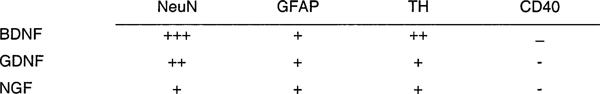
Beispiel 2Example 2
Knochenmarkszellen wurden unter Verwendung von Methoden ähnlich denen, die verwendet werden, um die Differenzierung von embryonalen Stammzellen (ES) zu fördern, in vitro induziert, um Neuronen-ähnliche Zellen zu werden. J. Dinsmore, et al., Cell Transplantation 5: 131–143 (1996). Knochenmark wurde von ausgewachsenen Mäusen gewonnen und unter Verwendung von magnetischer Zellsortierung in zwei Fraktionen aufgeteilt. Zunächst wurden die Knochenmarkszellen unter Verwendung einer Spritze mit einer Nadel #26 G durch Spülen des Schafts mit Puffer (PBS, ergänzt mit 0,5% BSA, pN-Wert 7,2) aus den Oberschenkelknochen und Schienbeinen von Mäusen gesammelt. Die Zellen wurden durch mehrfaches vorsichtiges Pipettieren aus dem Verband gelöst. Die Zellen wurden durch ein Nylon-Sieb von 30 μM gegeben, um verbliebene Gewebeklumpen zu entfernen. Die Zellen wurden durch Zugabe von Puffer gewaschen, indem sie 10 Min bei 200 × g zentrifugiert wurden und der Überstand entnommen wurde. Das Zellpellet wurde in 800 μl Puffer je 108 Zellen resuspendiert. Mit einem magnetischen Zellsortierungskit (Milteny Biotec, Inc, Auburn, TX) wurden die hämatopoietischen Knochenmarkszellen mit Sca1+-Mikrokügelchen markiert. Die markierten Knochenmarkszellen wurden für die positive Abtrennung von Sca1+-Zellen auf eine MS+-Säule gegeben.Bone marrow cells were induced in vitro to become neuron-like cells using methods similar to those used to promote differentiation of embryonic stem cells (ES). J. Dinsmore, et al., Cell Transplantation 5: 131-143 (1996). Bone marrow was obtained from adult mice and split into two fractions using magnetic cell sorting. First, the bone marrow cells were harvested using a needle # 26 G syringe by rinsing the stem with buffer (PBS supplemented with 0.5% BSA, pN 7.2) from the femurs and tibiae of mice. The cells were detached from the dressing by repeated careful pipetting. The cells were passed through a nylon screen of 30 μM to remove residual tissue clumps. The cells were washed by adding buffer by centrifuging at 200 × g for 10 minutes and removing the supernatant. The cell pellet was resuspended in 800 μl of buffer per 10 8 cells. Using a magnetic cell sorting kit (Milteny Biotec, Inc, Auburn, TX), the hematopoietic bone marrow cells were labeled with Sca1 + microspheres. The labeled bone marrow cells were placed on an MS + column for positive separation of Sca1 + cells.
Spezifisch wurden 200 μl Sca1-Multi-sort-Mikrokügelchen pro insgesamt 108 Zellen zugegeben, gemischt und 15 Minbei 6–12°C inkubiert. Eine Fraktion war angereichert an Zellen, die Marken für das Antigen für hämatopoietische Stammzellen der Maus (Sca1) enthielten, und die zweite Fraktion enthielt alle anderen Markzellen (einschließlich der BMSC-Fraktion). Die Zellen wurden durch Zugabe des 5–10-fachen Markierungsvolumens Puffer gewaschen und 10 Minbei 200 × g zentrifugiert und der Überstand wurde entfernt. Das Zellpellet wurde in 500 μl Puffer resuspendiert. Die MS+/RS+-Säule wurde mit 500 μl Puffer gewaschen. Die Zellsuspension wurde auf die Säule aufgetragen und die negativen Zellen liefen durch. Die Säule wurde dann dreimal mit 500 μl Puffer gespült. Die Säule wurde von dem Separator (der den Magnet enthält) entfernt und auf ein geeignetes Sammelröhrchen gesetzt. Auf die Säule wurde 1 ml Puffer pipettiert und die positive Fraktion wurde mit dem Kolben herausgespült, der mit der Säule bereitgestellt wird. Die mit Scal markierten Zellen wurden in 10 ml PBS und 5% BSA gegeben. Die Zellen wurden mit diesem Medium gewaschen und zentrifugiert (5 Min., 2000 U/Min.). Die Zellen wurden in Wachstumsmedium resuspendiert, das aus DMEM bestand, ergänzt mit 2 mM Glutamin, 0,001% β-Mercaptoethanol, nicht-essentiellen Aminosäuren und 10% Pferdeserum. Die Zellen wurden in Polyethylenkolben 2 Tage inkubiert, und die nicht adhärenten Zellen durch Ersatz des Mediums entfernt. Nachdem die Kulturen Konfluenz erreichten, wurden die Zellen durch 3–4 Minuten Inkubation mit Trypsin (0,25%) und 1 mM EDTA bei 37°C gelöst. Sie wurden für eine spätere Verwendung eingefroren oder nach 1:2- oder 1:3-Verdünnung unter Zugabe von Epidermalem Wachstumsfaktor (EGF), 10 ng/ml, wieder ausplattiert. Die Population an Zellen, die fest am Boden der Kulturkolbens heftete, erschien primär fibroblastisch. Die Mehrheit dieser Zellen färbte sich mit Antikörpern gegen Fibronectin. Diese Zellen wurden wegen ihres mesenchymalen oder fibroblastischen Aussehens entweder als mesenchymale Stammzellen bezeichnet oder als Knochenmarkstromazellen (BMSC), weil sie sich aus der komplexen Anordnung von unterstützenden Strukturen zu erheben scheinen, die in Mark gefunden werden. D. J. Prockop, Science 276: 71–74 (1997).Specifically, 200 μl of Sca1 multi-sort microspheres per 10 8 cells in total were added, mixed and incubated at 6-12 ° C for 15 min. One fraction was enriched in cells containing markers for the antigen for murine hematopoietic stem cells (Sca1), and the second fraction contained all other marrow cells (including the BMSC fraction). The cells were washed by adding the 5-10 fold labeling buffer and centrifuged at 200 x g for 10 min and the supernatant was removed. The cell pellet was resuspended in 500 μl of buffer. The MS + / RS + column was washed with 500 μl of buffer. The cell suspension was applied to the column and the negative cells leaked through. The column was then rinsed three times with 500 μl of buffer. The column was removed from the separator (containing the magnet) and placed on a suitable collection tube. 1 ml of buffer was pipetted onto the column and the positive fraction was flushed out with the flask provided with the column. Scal labeled cells were placed in 10 ml PBS and 5% BSA. The cells were washed with this medium and centrifuged (5 min, 2000 rpm). The cells were resuspended in growth medium consisting of DMEM supplemented with 2 mM glutamine, 0.001% β-mercaptoethanol, nonessential amino acids and 10% horse serum. The cells were incubated in polyethylene flasks for 2 days and the non-adherent cells removed by replacement of the medium. After the cultures reached confluency, cells were dissolved by incubation with trypsin (0.25%) and 1 mM EDTA at 37 ° C for 3-4 minutes. They were frozen for later use or replated at 1: 2 or 1: 3 dilution with the addition of epidermal growth factor (EGF), 10 ng / ml. The population of cells that stuck firmly to the bottom of the culture flask appeared to be primarily fibroblastic. The majority of these cells stained with antibodies to fibronectin. These cells, because of their mesenchymal or fibroblastic appearance, have been termed either mesenchymal stem cells or bone marrow stromal cells (BMSC) because they appear to rise from the complex array of supporting structures found in the marrow. DJ Prockop, Science 276: 71-74 (1997).
Vorläufige Ergebnisse
aus unserem Labor ergaben keine Hinweise für dendritische Zellen in BMSC, die
mit Retinsäure
(RA) und Wachstumsfaktor (BDNF, GDNF oder NGF) gezüchtet wurden
(Beispiel 1). Darüber
hinaus verminderte sich die Zahl von gegenüber Fibronectin immunreaktiven
Zellen als Funktion der Behandlung mit RA und Wachstumsfaktor, was
zeigt, daß Retinsäure und
Wachstumsfaktor die Differenzierung von BMSC weg vom fibroblastischen
Phänotyp
induzierten (vgl.
Um
zu bestimmen, ob die zelluläre
Umgebung die Differenzierung dieser Zellen beeinflußt, wurden BMSC
mit fötalen
mesencephalischen Zellen co-kultiviert. Die BMSC wurden von transgenen
lacZ-Mäusen (Jackson
Labs.) gewonnen, die β-Galactosidase (β-Gal) in
Knochenmarkszellen exprimieren. Die lacZ-BMSC wurden mit gleichen
Anteilen mit fötalen
mesencephalischen Zellen ausplattiert, die, wie kürzlich beschrieben (J.
R. Sanchez-Ramos, P. Michel, W. J. Weiner, F. Hefti, Journal of
Neurochemistry 50: 1934–1944
(1988)), von Mäusen
eines anderen Stamms (C57 bl6; Jackson Labs) in Kulturmedium gewonnen
wurden, das 9-cis-Retinsäure
(0,5 μM)
und BDNF (10 ng/ml) enthielt. Nach zwei Wochen wurden die Kulturen
fixiert und für die β-Gal-Histochemie
und Immuncytochemie für
neuronale Marker vorbereitet. β-Gal+ von lacZ-BMSC
wurden durch ein blaues Reaktionsprodukt klar identifiziert, das
mit Hellfeld-Mikrokopie (dunkel markierte Zellen bei den
Beispiel 3. In Vivo-DifferenzierungExample 3. In Vivo Differentiation
Um zu bestimmen, ob die Differenzierung von BMSC-Zellen in Neuronen in vivo eintreten würde, wurden BMSC von lacZ-Mäusen in denerviertes oder normales Striatum der Ratte transplantiert. Vor der Transplantation wurden die BMSC 2 Tage mit cis-9-Retinsäure (0,5 μM) und BDNF (10 ng/ml) behandelt. Drei Wochen vor der Transplantation wurde die Transplantationsstelle der Schwanzstelle des Putamens von Ratten mit unilateralen Injektionen in die Substantia nigra von 6-Hydroxydopamin (6-OH DA) behandelt. Eine einzelne Injektionen von 6-OH DA (Sigma; 2,5 μl, 3,6 μg/μl für eine Gesamtdosis von 9 μg in 0,2%-iger Ascorbinsäure) wurde in das rechte aufsteigende mesostriatale dopaminergische Sytem gemacht (4,4 mm hinter dem Scheitel, –1,2 mm seitlich und –7,8 mm ventral zur harten Hirnhaut mit auf –2,4 mm eingestelltem Zahnbalken). Das 6-OH DA wurde mit einer Rate von 1 μl/Minverabreicht. Die Spritzen wurden vor dem langsamen Herausziehen zusätzliche 5 Minan der Stelle gehalten.To determine whether differentiation of BMSC cells into neurons would occur in vivo, BMSC from lacZ mice were transplanted into rat denervated or normal striatum. Before transplantation, BMSC were treated with cis-9-retinoic acid (0.5 μM) and BDNF (10 ng / ml) for 2 days. Three weeks prior to transplantation, the transplant site of the tail site of the putamen was treated by rats with unilateral injections into the substantia nigra of 6-hydroxydopamine (6-OH DA). A single one Injections of 6-OH DA (Sigma, 2.5 μl, 3.6 μg / μl for a total dose of 9 μg in 0.2% ascorbic acid) were made in the right ascending mesostriatal dopaminergic system (4.4 mm behind the vertex, -1.2 mm laterally and -7.8 mm ventral to the hard meninges with toothed beam set to -2.4 mm). The 6-OH DA was administered at a rate of 1 μl / min. The syringes were held an additional 5 minutes before pulling out slowly.
Teilmengen einer BMSG-Suspension wurden bei 6 Tieren entlang eines einzelnen Nadelkanals auf derselben Seite wie die nigralen Schädigungen von 6-OH DA im Striatum abgelagert. Die Ratten wurden mit Natriumpentobarbital betäubt und in einen stereotaxischen Rahmen gebracht. Teilmengen der Zellsuspension wurden in zwei separaten Stellen im Striatum entlang eines einzelnen Nadelkanals abgelagert. Die Koordinaten für die Injektionen waren 1,2 mm vor dem Scheitel, +2,7 mm lateral und –5,2 mm und –4,7 ventral zur harten Hirnhaut mit auf Null eingestelltem Zahnbalken. Jede Injektion von 2 μl wurde mit einer Rate von 1 μl/Minverabreicht. Die Zahl an Stammzellen, die in das Striatum injiziert wurde, war 20.000 Zellen/μl für insgesamt 80.000 Zellen. Bei zwei Ratten wurde eine zweite Injektion von Zellen in das Striatum an der Stelle ohne Schädigung vorgenommen, was in insgesamt 160.000 Zellen pro Ratte resultierte.subsets BMSG suspension was measured in a single animal in 6 animals Needle channel on the same side as the nigralen damage deposited by 6-OH DA in the striatum. The rats were treated with sodium pentobarbital stunned and placed in a stereotaxic frame. Subsets of the cell suspension were in two separate locations in the striatum along a single needle channel deposited. The coordinates for the injections were 1.2 mm in front of the apex, +2.7 mm lateral and -5.2 mm and -4.7 ventral to the hard meninges with set to zero tooth bars. Each injection of 2 μl was administered at a rate of 1 μl / min. The number of stem cells injected into the striatum was 20,000 cells / μl for a total of 80,000 Cells. In two rats, a second injection of cells in the striatum made at the site without injury, resulting in a total of 160,000 cells per rat resulted.
Ein, zwei und drei Monate nach der Transplantation wurden Paare von Tieren getötet und mit heparinisierter Salzlösung und phosphatgepuffertem Paraformaldehyd perfundiert. Es wurden serielle kryostatische Schnitte von 30 μm Dicke durch die gesamte Länge des Gehirns geschnitten. Die Schnitte wurden zunächst auf β-Gal- Aktivität angefärbt, gefolgt von immunhistochemischer Verarbeitung mit Antikörpern gegen Neuronen-spezifisches nukleäres Protein (NeuN), mehreren Blockierungsmitteln, um die nicht-spezifische Immunmarkierung zu verringern, und einem zweiten Antikörper, der mit Peroxidase und einem rot-braunen Chromagen (Histomouse Kit, Zymed) gekoppelt war. Andere neuronale Marken, die immunhistochemisch bewertet wurden, umfaßten Tyrosinhydroxylase (TH), Glutamatdecarboxylase (GAD), Calbindin und Mikrotubuli-assoziiertes Protein (MAP2).One, Two and three months after the transplant were pairs of animals killed and with heparinized saline and phosphate buffered paraformaldehyde. There were serial Cryostatic sections of 30 microns Thickness through the entire length of the brain. The sections were first stained for β-gal activity, followed by immunohistochemical Processing with antibodies against neuron-specific nuclear protein (NeuN), several Blocking agents to the non-specific immunolabeling too reduce, and a second antibody containing peroxidase and a red-brown Chromagen (Histomouse Kit, Zymed) was coupled. Other neuronal markers that have been evaluated immunohistochemically included Tyrosine hydroxylase (TH), glutamate decarboxylase (GAD), calbindin and microtubule-associated protein (MAP2).
Die
Tiere verhielten und bewegten sich bis zur Tötung normal. Da dieses Experiment
speziell geplant war, um zu bestimmen, ob Transplantate überlebten
und sich differenzierten, wurden die Tiere nicht Apomorphin oder
Amphetamin ausgesetzt, um zu bestimmen, ob die Schädigung mit
6-OH DA und die anschließende Transplantation
das Rotationsverhalten veränderte.
Die Überprüfung der
Gehirngewebeschnitte offenbarte von BMSC abgeleitete β-Gal+-Zellen
in vielen Gehirnregionen, die entfernt lagen von der Stelle der
Implantation im Corpus striatum. Eine erwartungsgemäße Anhäufung von β-Gal+-Zellen
wurde in den folgenden spezifischen Gehirnregionen gefunden: mitrale
Zellzone des Riechkolbens, paraventrikuläre und supraoptische Nuclei
von Hypothalamus, Hippocampus, Habenula, oculomotorischem Nucleus,
rotem Nucleus, S. nigra, Abducens, faciale und hyoglossale Schädelnerv-Nuclei,
der unteren Olive der Medulla, dem ventralen Horn des Rückenmarks
und der cerebellaren Purkinje-Zellschicht. Die Verteilung von β-Gal+-Zellen
war bilateral und nahezu symmetrisch in allen Regionen außer der
S. nigra, wo die unbeschädigte
Stelle eine größere Zahl
an β-Gal+-Zellen
zeigte (
Die
BMSC-β-Gal+-Zellen
nahmen mehrere Phänotypen
an, einschließlich
Epithel-ähnliche
cuboidale Zellen im Plexus choroideus, kleine Oligodendrogliaähnliche
Zellen im optischen Chiasma und der sub-corticalen Substantia alba,
große Neuronen-ähnliche
Zellen im roten Nucleus, der S. nigraund Motornuclei des Hirnstamms. Tabelle
1 Zellzahlen
in Mittelhirnregionen 1
Monat nach der Transplantation von BMSC (mittlere Zahl von 2 Ratten) 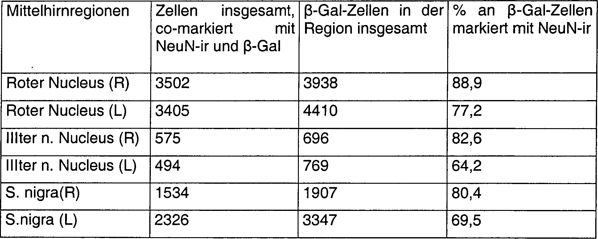

- ** der neuronale Marker in diesen Regionen war Tyrosinhydroxylase (TH). Nur die linke (nicht geschädigte) Seite enthielt BMSC-abgeleitete Zellen
- ** the neuronal marker in these regions was tyrosine hydroxylase (TH). Only the left (not damaged) side contained BMSC-derived cells
Die stereologischen Zahlen an β-Gal+-Zellen (insgesamt und co-markiert mit neuronalen Markern) in Mittelhirnregionen wurden bei 4 Ratten bestimmt, von denen jede zuvor mit einer Injektion von 6-ON-DA in die rechte S. nigrageschädigt worden war, gefolgt von der Transplantation von BMSC 3 Wochen später in den rechten Corpus striatum. Zwei Ratten wurden 1 Monat und 2 Ratten wurden 3 Monate nach der Transplantation getötet. Die Zahlen von TH-ir-Zellen in der S. nigra und dem ventral-tegmentalen Bereich (VTA) wurden nur bei einem Tier bestimmt.The stereological numbers of β-Gal + cells (in total and co-labeled with neuronal markers) in midbrain regions were determined in 4 rats, each of which was previously injected was damaged by 6-ON-DA in the right S. nigraggeschädigt, followed by transplantation of BMSC 3 weeks later into the right corpus striatum. Two rats were 1 month and 2 rats were 3 months after Transplant killed. The numbers of TH-ir cells in the S. nigra and the ventral-tegmentalen Range (VTA) was determined in one animal only.
Cerebellare β-Gal+-Zellen,
wie echte Purkinje-Zellen exprimierten Calbindin (echt schwarz in
den
Die β-Gal+-Zellen
mit neuronalem Phänotyp
im roten Nucleus wurden in mit MAP2 immunreaktiven Fasern vernetzt
(
Die Gesamtzahl an β-Gal+-Zellen und der Anteil der β-Gal+-Zellen, der den neuronalen Marker NeuN exprimiert, wurde unter Verwendung von stereologischen Techniken bei drei getrennten Nuclei des Mittelhirns (roter Nucleus, oculomotorische Nuclei und die S. nigra) in 4 Tieren geschätzt. (vgl. Tabelle 1). Schätzungen von BMSC-abgeleiteten Neuronen wurden unter Verwendung des Optischen Dissektor-Verfahrens bei kryokonservierten Schnitten von 30 um vorgenommen. BMSC-abgeleitete (β-Gal+) Zellen wurden bei einer kleinen Zahl von Schnitten mit vorbestimmten einheitlichen Intervallen für den gesamten Satz an Schnitten, die den roten Nucleus, die S. nigra und den oculomotorischen Nucleus umfaßten, direkt gezählt. Innerhalb jedes zu zählenden Schnittes wurde das Sehfeld unter Verwendung eines 40× Objektivs oben auf den Schnitt fokussiert. Der Fokus wurde dann durch den Schnitt bewegt und die Zahl an BMSC-abgeleiteten neuronalen Profilen im Fokus oben (Höhe des Dissektors = 9,25 μm) wurde gezählt. Die Gesamtzahl an lacZ-Zellen und die insgesamt doppelt markierten Zellen (NeuN + lacZ) in jeder Region wurde unter Verwendung der Berechnung: N = Nv × V (ref) = ΣQ/(ΣP × v(dis) × V (ref) bestimmt; wobei ΣQ die Gesamtzahl an Neuronen ist, die in allen Dissektoren im Referenzvolumen gezählt wurde, und v(dis) das Volumen des Dissektors ist, das gleich ist dem Bereich des Testrahmens, multipliziert mit der Höhe des Dissektors (9,25 μm), was der Abstand zwischen den fokalen Ebenen ist.The total number of β-Gal + cells and the proportion of β-Gal + cells expressing the neuronal marker NeuN was determined using stereological techniques in three separate nuclei of the midbrain (red nucleus, oculomotor nuclei, and S. nigra) in 4 animals estimated. (see Table 1). Estimates of BMSC-derived neurons were made using the optical dissector method on cryopreserved sections of 30 μm. BMSC-derived (β-gal +) cells were counted directly in a small number of slices at predetermined uniform intervals for the entire set of slices comprising the red nucleus, the S. nigra, and the oculomotor nucleus. Within each slice to be counted, the field of view was focused on top of the slice using a 40x objective. The focus was then moved through the slice and the number of BMSC-derived neural profiles in focus up (height of the dissector = 9.25 μm) was counted. The total number of lacZ cells and total double-labeled cells (NeuN + lacZ) in each region was calculated using the calculation: N = N v × V (ref) = ΣQ / (ΣP × v (dis) × V (ref) where ΣQ is the total number of neurons counted in all dissectors in the reference volume and v (dis) is the volume of the dissector equal to the area of the test frame multiplied by the height of the dissector (9.25 μm) What is the distance between the focal planes.
Mit der Ausnahme der S. nigra und der ventral-tegmentalen Fläche (VTA) waren die Zahlen an β-Gal+-Zellen in allen Regionen symmetrisch verteilt, trotz eines unilateralen rechten nigralen Schadens und der gleichseitigen Transplantation in das Striatum. Die Gesamtzahl an β-Gal+-Zellen in der S. nigra und der VTA auf der rechten Seite war niedriger als auf der unbeschädigten linken Seite einen und drei Monate nach der Transplantation. Der Prozentsatz an β-Gal+-Zellen, die NeuN-ir co-exprimierten, reichte von 58,6% im oculomotorischen Nucleus bis 89% im roten Nucleus. Abschätzungen für β-Gal+-Zellen, die mit TH-ir in der S. nigra und der VTA co-markiert waren, wurden in nur einem Tier 3 Monate nach der Transplantation gemacht. Der Prozentsatz an β-Gal+-Zellen, die TH in der S. nigra co-exprimierten, war 12,9% auf der Seite der 6-OH DA-Schädigung und 69,7% auf der unbeschädigten Seite. Ein ähnliches Muster wurde in der VTA gesehen, wo der Prozentsatz an β-Gal+-Zellen, die mit TH co-markiert waren, auf der Seite der Schädigung 19,8% war und 50% auf der unbeschädigten Seite. Abschätzungen der Gesamtzahl an Zellen im Mittelhirn erhöhten sich leicht nach drei Monaten im Vergleich mit den Zellzahlen einen Monat nach der Transplantation. Die Summe aller β-Gal+-Zellen in den Mittelhirnregionen belief sich nach einem Monat auf durchschnittlich 15.067, was 18% der ursprünglich 80.000 in das Striatum transplantierten Zellen darstellt. Nach 3 Monaten belief sich die Summe aller β-Gal+-Zellen in den Mittelhirnregionen auf durchschnittlich 18.635 (23% der insgesamt transplantierten). Die leichte Erhöhung in der Zahl der β-Gal+-Zellen stellt wahrscheinlich nicht eine kontinuierliche Zellteilung dar, weil eine Immunreaktivität gegen das nukleäre Antigen einer proliferierenden Zelle (PCNA-ir) in denjenigen Bereichen nicht nachgewiesen wurde, wo die BMSC eine neuronale Morphologie angenommen hatten. Eine Analyse der beiden Tiere, die bilaterale Transplantationen erhalten hatten, zeigte ähnliche bilaterale Verteilungen und Differenzierungsmuster von BMSC zwei Monate nach der Transplantation. Bei diesen Tieren wurden jedoch keine stereologischen Abschätzungen vorgenommen.With the exception of S. nigra and the ventral-tegmental surface (VTA) were the numbers of β-Gal + cells distributed symmetrically in all regions, despite a unilateral one right nigral damage and the equilateral transplant into the striatum. The total number of β-Gal + cells in the S. nigra and the VTA on the right was lower than on the undamaged left Page one and three months after the transplant. The percentage at β-gal + cells, the NeuN-ir co-expressed, ranged from 58.6% in the oculomotor nucleus to 89% in the red nucleus. estimates for β-gal + cells, which were co-labeled with TH-ir in S. nigra and the VTA done in just one animal 3 months after the transplant. The percentage at β-gal + cells, the TH in S. nigra co-expressed was 12.9% on the side 6-OH DA damage and 69.7% on the undamaged Page. A similar Pattern was seen in the VTA, where the percentage of β-gal + cells, that were co-labeled with TH, on the injury side 19.8% was and 50% on the undamaged Page. estimates the total number of cells in the midbrain increased slightly after three Months compared with cell counts one month after transplantation. The sum of all β-Gal + cells in the midbrain regions, the average was one month 15,067, which is 18% of the original 80,000 cells transplanted into the striatum. After 3 Months, the sum of all β-gal + cells in the midbrain regions was an average of 18,635 (23% of the total transplanted). The slight increase in the number of β-Gal + cells probably does not represent a continuous cell division, because an immunoreactivity against the nuclear Antigen of a proliferating cell (PCNA-ir) in those areas It has not been demonstrated where the BMSC has a neuronal morphology had accepted. An analysis of the two animals, the bilateral Transplantations showed similar bilateral distributions and differentiation patterns of BMSC two months after transplantation. In these animals, however, no stereological estimates performed.
Diese Ergebnisse zeigen, daß unsere behandelten BMSC pluripotente Zellen enthalten, die in Neuronen differenzieren, wie angezeigt durch mehrere neuronale Marker, morphologische Charakteristika und Integration in spezifische architektonische Schichten oder Regionen. Wir stellten einen Stimulus bereit (Schädigung des nigro-stratialen Systems), mit dem wir versuchten die BMSC-Differenzierung zu induzieren. Die nigralen Schädigungen hatten jedoch einen negativen Einfluß auf das lokale Einwachsen an der Stelle der Implantation. Statt dessen wanderten die β-Gal+-BMSC intensiv und differenzierten in einer stellenabhängigen bilateralen symmetrischen Verteilung in allen Teilen des Gehirns, mit der Ausnahme der S. nigra und der VTA. An der Stelle der nigralen Schädigung von 6-OH DA waren weniger β-GaI+-BMSC und ein kleinerer Anteil von ihnen co-exprimierte TH im Vergleich mit der unbeschädigten Seite. Dies kann vom Alter der Schädigung beeinflußt sein (drei Wochen). Üblicherweise gibt es eine optimale Zeit für die Transplantation von Zellen in Modelle der Neurodegeneration und Hypoxia-Ischämie. Von anderen neuralen "Stamm-ähnlichen" Zellen, die in das Gehirn transplantiert wurden, wurde gezeigt, daß sie vorzugsweise an die Stelle der Ischämie in der geschädigten Hemisphäre eines hypoxischen-ischämischen Rattenmodells wandern, optimale Migration und optimales Einwachsen wurden aber erreicht, wenn die Zellen 3–7 Tage nach der Schädigung transplantiert wurden. E. Y. Snyder et al., ADVANCES IN NEUROLOGY BD. 72: NEURONAL REGENERATION, REORGANIZATION, AND REPAIR. (Hrsg. F. J. Seil) Lippincott Raven, Philadelphia, 1997.These results indicate that our treated BMSCs contain pluripotent cells that differentiate into neurons as indicated by multiple neuronal markers, morphological characteristics, and integration into specific architectural strata or regions. We provided a stimulus (damage to the nigro-stratial system) that we used to induce BMSC differentiation. However, the nigral lesions had a negative impact on local ingrowth at the site of implantation. Instead, the β-gal + BMSC migrated intensely and differentiated in a site-dependent bilateral symmetric distribution in all parts of the brain, with the exception of S. nigra and VTA. In place of nigral damage to 6-OH DA, less β-GaI + BMSC and a smaller proportion of them co-expressed TH were compared to the undamaged side. This may be affected by the age of the injury (three weeks). There is usually an optimal time for transplantation of cells into models of neurodegeneration and hypoxia-ischemia. Other neural "stem-like" cells transplanted into the brain have been shown to preferentially migrate to the site of ischemia in the damaged hemisphere of a hypoxic-ischemic rat model, optimal migration and op However, timal ingrowth was achieved when the cells were transplanted 3-7 days post-injury. EY Snyder et al., ADVANCES IN NEUROLOGY BD. 72: NEURONAL REGENERATION, REORGANIZATION, AND REPAIR. (Ed. FJ Rope) Lippincott Raven, Philadelphia, 1997.
Da es unwahrscheinlich ist, daß die Schädigung die stellenabhängige Differenzierung in Regionen beeinflußt, die entfernt liegen vom nigro-stratialen System, war die wahrscheinlichste Erklärung für den Neurotropismus oder die Affinität von BMSC für spezifische neuronale Populationen die Vorbehandlung von BMSC mit Retinsäure und BDNF vor der Transplantation. Im Gegensatz dazu wurde gezeigt, daß unbehandelte menschliche BMSC, die in normales Striatum der Ratte transplantiert wurden, sich nicht unterschiedlich verteilten oder durch die Stelle differenzierten, ungeachtet ihrer weit verteilten Migration. S. A. Azizi, et al., Proc. Nat. Acad. Sci. (U.S.A.) 95: 3908–3913 (1998).There it is unlikely that the damage the job-dependent Differentiation in regions that are distant from the affected nigro-stratial system, was the most likely explanation for neurotropism or the affinity from BMSC for specific neural populations undergo the pretreatment of BMSC retinoic acid and BDNF before transplantation. In contrast, it was shown that untreated human BMSC transplanted into rat normal striatum not differently distributed or differentiated by the location, regardless of their widely distributed migration. S.A. Azizi, et al., Proc. Nat. Acad. Sci. (U.S.A.) 95: 3908-3913 (1998).
Wir wollen nicht durch eine Theorie gebunden sein, schlagen aber dennoch vor, daß die Vorbehandlung von BMSC mit Retinsäure und BDNF die Expression von Zelloberflächenproteinen oder Rezeptoren mit einer Affinität für die entsprechenden trophischen Faktoren, Cytokine oder Zelladhäsionsmoleküle induziert, die normalerweise von spezifischen neuronalen Populationen produziert werden. Dies scheint ein nützliches Modell für die Untersuchung des Mechanismus für die Affinität von BMSC für spezifische Hirnregionen zu sein. Diese Affinität von vorbehandelten BMSC für spezifische Hirnregionen wird die zielgerichtete Lenkung von neuronalen Populationen für zelluläre Therapien erlauben, die von Gentherapeutika bis zu neuraler Wiederherstellung bei neurodegenerativen Erkrankungen, Schlaganfall und Trauma reichen.We do not want to be bound by a theory, but still strike before that the Pretreatment of BMSC with retinoic acid and BDNF expression of cell surface proteins or receptors with an affinity for the corresponding trophic Factors inducing cytokines or cell adhesion molecules, the normally produced by specific neuronal populations become. This seems like a useful model for the Investigation of the mechanism for the affinity from BMSC for to be specific brain regions. This affinity of pretreated BMSC for specific Brain regions will be the targeted guidance of neuronal populations for cellular therapies allow, from gene therapy to neural recovery in neurodegenerative diseases, stroke and trauma.
Um sicherzugehen, daß die β-Gal+-Zellen tatsächlich von BMSC abgeleitet sind, wurden unter Verwendung von BMSC, die mit anderen Markern markiert waren, zwei andere Experimente durchgeführt (Beispiele 7 und 8 nachstehend). Dies war notwendig, weil einige Populationen von normalen, nicht transplantierten Rattenneuronen eine endogene β-Galactosidase-Aktivität exprimieren. Mit der Anwesenheit von mehreren anderen Markern von Maus-BMSC bestimmten wir, daß die β-Gal+-Zellen in der Tat von Mäusen abgeleitet waren und keine endogenen Rattenneuronen waren.Around make sure that the β-gal + cells indeed derived from BMSC were using BMSC, the marked with other markers, two other experiments were performed (examples 7 and 8 below). This was necessary because some populations of normal, non-transplanted rat neurons express endogenous β-galactosidase activity. Determined with the presence of several other markers of mouse BMSC we, that the β-gal + cells indeed of mice were derived and were not endogenous rat neurons.
Beispiel 4. Ataxia-BehandlungExample 4. Ataxia treatment
"Wobbler"-Mäuse (JAX labs) exprimieren eine Mutation, bei der die Purkinje-Zellen des Cerebellum im Alter von 3 bis 4 Wochen rasch degenerieren. Der Verlust an Purkinje-Zellen scheint die vorherrschende Auswirkung der Mutation zu sein, und es degenerieren keine anderen Neuronen als die mitralen Zellen des Riechkolbens und wenige Thalamus-Neuronen. Der Verlust der Purkinje-Zellen korreliert mit dem Beginn eines ataxischen wackeligen Schritts, der das Leben über anhält, der aber die Gesundheit der Tiere nicht auf andere Weise beeinflußt."Wobbler" mice (JAX labs) express a mutation in which the Purkinje cells of the cerebellum degenerate rapidly at the age of 3 to 4 weeks. The loss of Purkinje cells seems to be the predominant effect of the mutation, and it does not degenerate any neurons other than the mitral cells of the Olfactory bulb and few thalamic neurons. The loss of Purkinje cells correlates with the beginning of an ataxic shaky step, the life over continues, but that does not affect the health of the animals in any other way.
Vorkonditionierte BMSC (wie vorstehend beschrieben) werden in das Gehirn dieser Tiere im Alter von 6 Wochen transplantiert. Für jedes Experiment werden insgesamt 12 mutierte Tiere und 12 normale Tiere untersucht. Die Hälfte der Tiere empfängt vorkonditionierte BMSC von lacZ-Mäusen und die Hälfte erhält eine Scheinoperation. Obwohl das Ziel der Ersatz der cerebellaren Purkinje-Zellen ist, wird das Transplantat im Striatum planiert, da wir zeigten (Beispiel 3), daß das Cerebellum eine bevorzugte Region für die Migration und stellenspezifische Differenzierung in Purkinje-ähnliche Neuronen zu sein scheint. Alternative Transplantationstellen sind die Injektion in den lateralen Ventrikel und direkt in das Cerebellum, sorgfältig, um traumatische Ataxia zu vermeiden.preconditioned BMSCs (as described above) are introduced into the brain of these animals transplanted at the age of 6 weeks. For each experiment will be total Examined 12 mutant animals and 12 normal animals. Half of Animals receives preconditioned BMSC from lacZ mice and half receives a sham operation. Although the goal is the replacement of cerebellar Purkinje cells, the graft is leveled in the striatum as we showed (example 3) that the Cerebellum is a preferred region for migration and site specific Differentiation into Purkinje-like Neurons seems to be. Alternative transplant sites are the injection into the lateral ventricle and directly into the cerebellum, careful, to avoid traumatic ataxia.
Um zu bestimmen, ob die Transplantationen in einer funktionellen Verbesserung des neurologischen Defizits resultierten, wird die Grundlinie von spontanen Schritten vor der Operation durch Aufzeichnung der Abdrücke einer mit Tinte versehenen Pfote quantifiziert, die auf einem schmalen, 36 Inch langen Papierstreifen auf dem Boden eines geschlossenen Laufwegs (36'' lang, 3'' breit und 6'' hoch) zurückblieben. Außerdem wurde die Leistung auf einem Balancebalken gemessen (die Fähigkeit und die erforderliche Zeit zur Überquerung eines Stabs, der zwischen zwei Plattformen verläuft). Die Messungen der Fortbewegung und Koordination werden sowohl bei den mutierten Mäusen als auch bei der Kontrollgruppe von normalen Mäusen mit demselben genetischen Hintergrund zu den folgenden Zeitpunkten vorgenommen: 1 Woche vor der Operation und 1 Woche, 1 Monat, 2 Monate und 3 Monate nach der Operation.Around to determine if the transplants in a functional improvement neurological deficit, the baseline of spontaneous steps before the operation by recording the imprints of a quantified with an inked paw on a narrow, 36 inches long strip of paper on the bottom of a closed Walkway (36 '' long, 3 'wide and 6' high) remained. Furthermore the performance was measured on a balance bar (the ability and the time required to cross one Staff, which runs between two platforms). Measurements of locomotion and coordination are seen in both the mutant mice also in the control group of normal mice with the same genetic Background made at the following times: 1 week before the surgery and 1 week, 1 month, 2 months and 3 months after Surgery.
Zu einem Zeitpunkt, bei dem es eine signifikante Verbesserung bei Fortbewegung und Koordination gibt, werden die Tiere getötet und die Gehirne werden auf die Verteilung und das Maß an Differenzierung der transplantierten BMSC untersucht. Der BMSC-Ursprung der Zellen wird durch β-Gal+-Färbung bestimmt. Der Marker für die Purkinje-Zellen ist die Calbindin-Immunreaktivität. Der Anteil der β-Gal+-Zellen, die mit Calbindin co-markiert sind, wird unter Verwendung der stereologischen Technik bestimmt.At a time when there is a significant improvement in locomotion and coordination, the animals are killed and the brains are examined for distribution and degree of differentiation of the transplanted BMSC. The BMSC origin of the cells is determined by β-Gal + staining. The marker for Purkinje cells is calbindin immunoreactivity. The proportion of β-gal + cells that are associated with calbindin are co-labeled is determined using the stereological technique.
Beispiel 5. Differenzierung von Menschlichen Knochenmarkzellen (BMSC) In Vitro.Example 5. Differentiation of Human Bone Marrow Cells (BMSC) In Vitro.
Kurz zusammengefaßt wurden BMSC aus Gesamtknochenmark der Maus, des Kalbs und des Menschen gewonnen. BMSC-Kulturen wurden mit a) epidermalem Wachstumsfaktor (EGF), b) Retinsäure (RA), c) neuronalem N5-Medium und d) vom Gehirn abgeleitetem neurotrophem Faktor (BDNF) plus RA inkubiert.Short summarized BMSC were derived from total bone marrow of mouse, calf and human won. BMSC cultures were treated with a) epidermal growth factor (EGF), b) retinoic acid (C) neuronal N5 medium; and d) brain derived neurotrophic Factor (BDNF) plus RA incubated.
BMSC wurden von verbliebenem Knochenmarkmaterial isoliert (Knochenstücke mit anhängendem stromalen Zellen, Fettgewebe und Splittern), das auf dem Nylonmaschenfilter zurückblieb, das routinemäßig bei der Filtration von frisch gewonnenem menschlichem Mark verwendet wird, das für die Knochenmarktransplantation verwendet werden soll. Das Filtrat enthält den größten Teil der hämatopoietischen Elemente des Knochenmarks, der dann für den Ersatz von menschlichem Knochenmark verarbeitet wird. Wir verwenden die Filter, die üblicherweise verworfen werden. Die Filter wurden fünfmal mit PBS gewaschen. Die PBS-Lösung wurde zentrifugiert, um die schwereren Knochenstücke abzulagern. Der Überstand wurde in Zellkulturmedium resuspendiert und in Gewebekulturkolben ausplattiert (wie in Beispiel 1). Die BMSC wurden durch ihre Adhärenz an die Plastikkulturkolben abgetrennt, während die kleineren hämatopoietischen Stammzellen in dem Medium suspendiert blieben und entfernt wurden, wenn das Kulturmedium durch frisches Medium ersetzt wurde. Die menschliche Knochenmarksfraktion zum Ausplattieren wurde mit Dulbeccos minimal essentiellem Medium (DMEM, GIBCO/BRL) und 10% FBS 1:1 verdünnt und 30 Minbei 1.000 × g über einen Dichtegradienten (Ficoll-Paque Plus, 1,077 g/ml, Pharmacia) zentrifugiert. Der Überstand und die Zwischenschicht wurden vereint, mit MEM mit 10% fötalem Kälberserum (FCS) auf etwa 20 ml verdünnt und in mit Polyethylenimin beschichteten Plastikkolben ausplattiert.BMSC were isolated from remaining bone marrow material (bone pieces with attached stromal Cells, adipose tissue and slivers) on the nylon mesh filter remained, that routinely used the filtration of freshly harvested human marrow that will be for the bone marrow transplant should be used. The filtrate contains the biggest part the hematopoietic Elements of the bone marrow, which are then used for the replacement of human Bone marrow is processed. We use the filters, usually be discarded. The filters were washed five times with PBS. The PBS solution was centrifuged to deposit the heavier pieces of bone. The supernatant was resuspended in cell culture medium and in tissue culture flasks plated out (as in Example 1). The BMSCs were noted for their adherence The plastic culture flasks are separated while the smaller hematopoietic Stem cells remained suspended in the medium and were removed, when the culture medium has been replaced by fresh medium. The human Bone marrow fraction for plating was minimal with Dulbeccos diluted medium: DMEM, GIBCO / BRL and 10% FBS 1: 1 and 30 minutes at 1,000 × g over one Density gradient (Ficoll-Paque Plus, 1.077 g / ml, Pharmacia) centrifuged. The supernatant and the interlayer were pooled with MEM containing 10% fetal calf serum (FCS) diluted to about 20 ml and plated in polyethylenimine coated plastic flask.
Knochenmark der Maus wurde in 10 ml PBS und 5% BSA gebracht. Die Zellen wurden in diesem Medium gewaschen, und zentrifugiert (5 Minbei 2000 U/Min.) Die Zellen wurden in Wachstumsmedium resuspendiert, das aus DMEM bestand, ergänzt mit 2 mM Glutamin, 0,001% β-Mercaptoethanol, nicht essentiellen Aminosäuren und 10% Pferdeserum. Die Zellen wurden in den Kolben 2 Tage inkubiert und die nicht adhärenten Zellen wurden durch Ersatz des Mediums entfernt. Nachdem die Kulturen Konfluenz erreichten wurden die Zellen durch 3–4 Minuten Inkubation mit Trypsin (0,25%) und 1 mM EDTA bei 37°C abgelöst. Sie wurden für eine spätere Verwendung eingefroren oder nach 1:2- oder 1:3-Verdünnung unter Zugabe von Epidermalem Wachstumsfaktor (EGF), 10 ng/ml, oder von Blutplättchen abgeleitetem Wachstumsfaktor (PDGF), 5 ng/ml, wieder ausplattiert. Diese Prozedur wurde für 3–5 Passagen wiederholt.bone marrow The mouse was placed in 10 ml of PBS and 5% BSA. The cells were washed in this medium and centrifuged (5 min at 2000 rpm) The cells were resuspended in growth medium derived from DMEM insisted, added with 2 mM glutamine, 0.001% β-mercaptoethanol, nonessential amino acids and 10% horse serum. The cells were incubated in the flask for 2 days and the non-adherent cells were removed by replacement of the medium. After the cultures confluence The cells were reached by 3-4 minutes incubation with trypsin (0.25%) and 1 mM EDTA at 37 ° C replaced. They were for a later one Use frozen or after 1: 2 or 1: 3 dilution under Addition of epidermal growth factor (EGF), 10 ng / ml, or of platelets derived growth factor (PDGF), 5 ng / ml, replated. This procedure was for 3-5 passages repeated.
Menschlichen
Gesamtknochenmarkzellen wurde erlaubt, 2 Tage an den Kulturkolbenboden
zu adhärieren,
und nach der Entfernung von den Kolben wurden sie in einem Medium
ausplattiert, das das Mitogen epidermaler Wachstumsfaktor (EGF)
enthielt. Dies resultierte in einer Proliferation der Zellen, ohne
die Differenzierung signifikant zu induzieren, Die Zellen wurden
dann von dem Kolbenboden entfernt und in Kulturgefäßen von
35 mm in der Gegenwart von neuronalem Wachstumsmedium (N5) ausplattiert,
das ergänzt
war mit fötalem
Kälberserum
(FCS), 10% Retinsäure
(RA) und einem von mehreren Wachstumsfaktoren (BDNF, GDNF oder NGF)
(oder eingefroren für
eine spätere
Verwendung). Die üblichen
Markstromazellen waren reich an Fibronectin (weiß in
Nach
7 Tagen wurde die Expression der neuronalen Marker (Nestin, Neuronen-Spezifisches
Nukleäres
Protein oder neuN und GFAP) und ein Marker für Knochenmarkstromazellen (Fibronectin)
mittels Western Blot-Analyse und immuncytochemie analysiert (
Darüber hinaus wurden Zellen, die wie oben von dem Kolbenboden entfernt wurden (BMSC) oder mittels magnetischer Zellsortierung abgetrennt wurden, in der Gegenwart von neuronalem Wachstumsmedium (N5) in Kulturschalen von 35mm erneut ausplattiert, das mit 5% Pferdeserum, 1% FBS, Transferrin (100 μg/ml), Putrescin (60 μM), Insulin (25 μg/ml), Progesteron (0,02 μM), Selen (0,03 μM), 9-cis-Retinsäure oder all-trans-Retinsäure (RA) (0,5 μM) und irgendeinem von mehreren neuronalen Wachstumsfaktoren mit einer Konzentration von 1–10 ng/ml (vom Gehirn abgeleiteter Wachstumsfaktor-BDNF, von Gliazellen abgeleiteter neurotropher Faktor-GDNF und Nervenwachstumsfaktor (NGF) ergänzt war. Nach 7 bis 14 Tagen hatten einige der BMSC die Morphologie geändert und sich zu den Phänotypen von Neuronen, Gliazellen und Fibroblasten entwickelt.Furthermore were cells removed from the flask bottom as above (BMSC) or were separated by magnetic cell sorting, in the presence of neuronal growth medium (N5) in culture dishes replated from 35mm, with 5% Horse Serum, 1% FBS, Transferrin (100 μg / ml), putrescine (60 μM), Insulin (25 μg / ml), Progesterone (0.02 μM), Selenium (0.03 μM), 9-cis-retinoic acid or all-trans-retinoic acid (RA) (0.5 μM) and any one of several concentration neuronal growth factors from 1-10 ng / ml (brain-derived growth factor BDNF, glial cells derived neurotrophic factor GDNF and nerve growth factor (NGF) supplements was. After 7 to 14 days, some of the BMSCs had the morphology changed and become the phenotypes developed by neurons, glial cells and fibroblasts.
Beispiel 6. Effekte der gemeinsamen Kultur von menschlichen BMSC mit fötalen striatalen Kulturen der RatteExample 6. Effects of common culture of human BMSC with fetal striatal cultures of the rat
BMSC
der Maus, konditioniert mit RA und BDNF, wurden mit primären neuronalen
mesencephalischen Zellen co-kultiviert, die von fötalen E15-Ratten
gewonnen wurden. Menschliche BMSC wurden unter Verwendung der vorstehend
beschriebenen Verfahren etabliert. Die Zellen wurden mit einer Konzentration
von 10 μM
fluoreszenzgrünem "Cell Tracker" (5-Chlormethylfluoresceindiacetat,
Molecular Probes, Inc.) markiert und auf einem Zellbett von fötalen striatalen
Zellen der Ratte ausplattiert, das drei Tage zuvor hergestellt worden
war. Die Kulturen wurden mit RA (0,5 μM) + BDNF (10 ng/ml) in N5-Medium
gefüttert.
Nach 10 Tagen Kultur wurden die Zellen unter Verwendung von primären Antikörpern gegen
NeuN, GFAP, Nestin und Fibronectin für die Immuncytofluoreszenz
verarbeitet, gefolgt von einem sekundären Antikörper, der mit Texas-Rot fluoreszenzmarkiert
war. Dies erlaubte eine zweifache Markierung von Zellen, um zu bestimmen,
ob eine Zelle, die spezifische Marker für Neuronen, Gliazellen oder
Neuroectoderm zeigte, auch einen Knochenmarkursprung hatte. Vgl.
die
LacZ-markierte BMSC, die mit primären mesencephalischen Kulturen der Ratte co-kultiviert wurden, differenzierten in Neuronen-ähnliche Zellen, die mit NeuN-ir und β-Gal co-markiert waren.LacZ-labeled BMSC with primary mesencephalic cultures of the rat were co-cultured, differentiated in neurons-like Cells labeled NeuN-ir and β-gal co-marked.
Beispiel 7. In VivoExample 7. In Vivo
lacZ-BMSC von Mäusen wurden in denervierten Corpus striatum von Ratten, die zuvor mit 6-OH-Dopamin geschädigt worden waren, und in nicht geschädigte Ratten transplantiert. Einen und drei Monate später wurden die Ratten getötet und die Gehirne wurden für die Histochemie und Immuncytochemie verarbeitet. BMSC-abgeleitete Zellen wurden durch die β-Gal+-Anfärbung oder durch anti-Maus-Antikörper, die gegen das wichtige Histokompatibilitäts-Antigen Typ 1 der Maus gerichtet waren, sichtbar gemacht. Die Expression von neuronalen Markern in BMSC-abgeleiteten Zellen wurde durch ihre Immunreaktivität mit Antikörpern gegen das Neuronen-spezifische nukleäre Antigen (NeuN-ir), Tyrosinhydroxylase (TH), das Mikrotubuli-assoziierte Protein (MAP2), Neurofilament (NF) und Calbindin (CB) bestimmt.lacZ BMSC of mice were in denervated corpus striatum of rats, previously with 6-OH-dopamine damaged had been, and in undamaged Rats transplanted. One and three months later, the rats were killed and the brains were for processed the histochemistry and immunocytochemistry. BMSC-derived cells were characterized by the β-gal + staining or by anti-mouse antibodies that directed against the mouse's major histocompatibility antigen type 1 were made visible. Expression of neuronal markers in BMSC-derived Cells were characterized by their immunoreactivity with antibodies specific to the neuron nuclear Antigen (NeuN-ir), tyrosine hydroxylase (TH), the microtubule-associated protein (MAP2), neurofilament (NF) and calbindin (CB).
BMSC-abgeleitete Zellen, die in das Striatum transplantiert wurden, blieben nicht im Striatum lokalisiert, wohin sie transplantiert worden waren, sondern wurden im Mittelhirn, Thalamus und selten im Cerebellum gefunden, wenn sie mit dem anti-Maus-Antikörper identifiziert wurden. Einige dieser Maus-BMSC co-exprimierten NeuN-ir oder TH-ir. Transplantierte und Kontrollratten (nicht transplantiert) zeigten β-Gal+-Zellen in ähnlichen und unterschiedlichen Verteilungen. β-Gal+-Zellen wurden im Riechkolben, dem supraoptischen und paraventrikulären Hypothalamus, dem roten Nucleus, dem dritten Nerv-Nucleus, der S. nigra gefunden und waren im Cerebellum in einem Muster ähnlich dem von Purkinje-Zellen in nicht transplantierten Ratten verteilt.BMSC-derived Cells transplanted into the striatum did not remain localized in the striatum where they had been transplanted but were found in the midbrain, thalamus, and rarely in the cerebellum, when identified with the anti-mouse antibody were. Some of these mouse BMSC co-expressed NeuN-ir or TH-ir. Transplanted and control rats (not transplanted) showed β-gal + cells in similar and different distributions. β-gal + cells were in the olfactory bulb, the supraoptic and paraventricular hypothalamus, the red Nucleus, the third nerve nucleus found and found in S. nigra similar in cerebellum in a pattern distributed by Purkinje cells in non-transplanted rats.
BMSC, konditioniert mit RA und BDNF, exprimierten neuronale Marken und verminderten die Expression von stromalen und fibroblastischen Markern. Wenn konditionierte lacZ-BMSC mit primären mesencephalischen Neuronen co-kultiviert wurden, co-exprimierten viele BMSC-abgeleitete Zellen NeuN-ir und β-Gal. Wenn sie in denerviertes Striatum transplantiert wurden, exprimierten konditionierte BMSC mehrere neuronale Marker und wanderten von der Stelle der Transplantation zu beiden Seiten des Thalamus und in kleinerem Maß zum Hippocampus und zu der nicht beschädigten S. nigra. Die positive Anfärbung für die β-Gal-Aktivität in Ratten ohne Transplantation ergab einen Artefakt, den man leicht fälschlicherweise für β-Gal-BMSC halten könnte. Ein zweiter Marker für BMSC bestätigte jedoch, daß diese Zellen wanderten und zu Neuronen-ähnlichen Zellen differenzierten.BMSC, conditioned with RA and BDNF, expressed neuronal markers and decreased the expression of stromal and fibroblastic markers. When conditioned lacZ-BMSC with primary mesencepha When cultured in human neurons, many BMSC-derived cells co-expressed NeuN-ir and β-gal. When transplanted into the denervated striatum, conditioned BMSCs expressed multiple neuronal markers and migrated from the site of transplantation to either side of the thalamus and to a lesser extent to the hippocampus and undamaged S. nigra. The positive staining for β-gal activity in non-transplanted rats yielded an artifact that could easily be mistaken for β-gal-BMSC. However, a second marker for BMSC confirmed that these cells migrated and differentiated into neuron-like cells.
Beispiel 8. In vivo-Ergebnisse, die unter Verwendung eines dritten Markers für BMSC zeigen, daß BMSC in neurale Zellen differenzieren (Neuronen oder Gliazellen)Example 8. In vivo Results, using a third marker for BMSC, show that BMSC is in neural Differentiate cells (neurons or glial cells)
Die
vorstehenden Beispiele verwendeten β-GaI+-BMSC von Mäusen und
für Mäuse spezifische
Antikörper.
Bei diesem Experiment wurden BMSC der Maus mit dem roten Fluoreszenzfarbstoff
PKH26 (Sigma, Inc.) oder Cell Tracker Orange (Molecular Probes,
Inc.) vor der Transplantation in denerviertes Striatum, wie vorstehend
beschrieben, vormarkiert. Zwei Wochen nach der Transplantation waren
die fluoreszierenden BMSC von der Stelle der Transplantation weggewandert
(einige zum cerebralen Cortex auf der contralateralen Seite). Nach
nur zwei Wochen hatte ein kleiner Anteil der transplantierten Zellen
angefangen, neurale Marker wie NeuN-ir und GFAP-ir zu exprimieren
(
Industrielle AnwendbarkeitIndustrial applicability
Die Überprüfung von Gehirngewebeschnitten bei den vorstehenden Experimenten offenbarte regenerierende BMSC-abgeleitete β-Gal+-Zellen in mehreren Hirnregionen, die entfernt sind von der Implantationsstelle im Corpus striatum. Die Migration der behandelten BMSCs umfaßt, ist aber nicht beschränkt auf, die folgenden Hirnregionen: mitrale Zellzone des Riechkolbens, paraventrikuläre und supraoptische Nuclei von Hypothalamus, Hippocampus, Habenula, oculomotorischer Nucleus, roter Nucleus, S. nigra, Abducens, faciale und hyoglossale Schädelnerv-Nuclei, die untere Olive der Medulla, das ventrale Horn des Rückenmarks und die cerebellare Purkinje Zellschicht. Die BMSC-β-Gal+-Zellen nahmen vielerlei Phänotypen an, einschließlich Epithel-ähnliche cuboidale Zellen im choroiden Plexus; kleine Oligodendroglia-ähnliche Zellen im optischen Chiasma und der sub-corticalen Substantia alba; große Neuronen-ähnliche Zellen im roten Nucleus, der S. nigra und den Motor-Nuclei des Hirnstamms. Dieses neue Verfahren für die neurale Transplantation ist ein wissenschaftlich durchführbarer und klinisch vielversprechender Weg für die Behandlung von neurodegenerativen Erkrankungen und Schlaganfall ebenso wie für die Reparatur von traumatischen Verletzungen des Gehirns und Rückenmarks.The review of Brain tissue sections revealed in the above experiments regenerating BMSC-derived β-gal + cells in several brain regions, which are distant from the implantation site in the corpus striatum. The migration of treated BMSCs is but not limited on, the following brain regions: mitrale cell zone of the olfactory bulb, paraventricular and Supraoptic Nuclei of Hypothalamus, Hippocampus, Habenula, oculomotor nucleus, red nucleus, S. nigra, abducens, facial and hyoglossal cranial nerve nuclei, the lower Olive of the medulla, the ventral horn of the spinal cord and the cerebellar Purkinje cell layer. The BMSC-β-Gal + cells took many phenotypes on, including epithelial-like cuboidal cells in the choroid plexus; small oligodendroglia-like Cells in the optic chiasm and the sub-cortical substantia alba; size Neuron-like Cells in the red nucleus, the S. nigra, and the motor nuclei of the brainstem. This new procedure for The neural transplant is a scientifically feasible one and clinically promising route for the treatment of neurodegenerative diseases and stroke as well the repair of traumatic injuries of the brain and spinal cord.
Es gibt zahlreiche Verwendungen von BMSC in universitären und pharmazeutischen Forschungslabors. Menschliche BMSC, die im Rattenmodell zum Cerebellum wandern und die Charakteristika von Purkinje-Zellen annehmen, können verwendet werden, um die Wirkungen (sowohl therapeutische als auch toxische) von Wirkstoffen auf menschliche cerebellare Zellen zu testen. In ähnlicher Weise können menschliche BMSC, implantiert in andere Bereiche des Gehirns, die die Charakteristika dieser Zellen annehmen, für die Testung der Toxizität von Wirkstoffen und der therapeutischen Wirksamkeit bei einer Vielzahl von Störungen verwendet werden. Vor der Implantation können die Zellen gentechnisch verändert werden, damit sie Gene tragen, die bei verschiedenen Störungen des Gehirns und Nervensystems eine Rolle spielen.It There are numerous uses of BMSC in university and pharmaceutical research laboratories. Human BMSC in the rat model migrate to the cerebellum and the characteristics of Purkinje cells can accept used to treat the effects (both therapeutic as well toxic) agents to human cerebellar cells testing. In similar Way you can human BMSC, implanted in other areas of the brain, which assume the characteristics of these cells for testing the toxicity of drugs and therapeutic efficacy in a variety of disorders. Before implantation can the cells are genetically modified so that they carry genes that are involved in various disorders of the Brain and nervous system play a role.
Eine mögliche Anwendung der migratorischen und regenerativen Natur der BMSCs ist die Behandlung der Parkinson-Krankheit. Die vorbehandelten BMSCs können fötale Neuronen ersetzen, die bei dieser Krankheit für die Transplantation verwendet wurden. Von den BMSCs, die mit Retinsäure und BCNF behandelt wurden, wurde gezeigt, daß sie zur Substantia nigra wandern, wo ein Defizit an dopaminergen Neuronen PD verursacht. Die Verabreichung von BMSCs in die Substantia nigra könnte nigrale Neuronen regenerieren und den Fluß an Dopamin zu dieser Region wiederherstellen.A possible Application of the migratory and regenerative nature of BMSCs is the treatment of Parkinson's disease. The pretreated BMSCs can fetal Replace neurons used in this disease for transplantation were. Of the BMSCs treated with retinoic acid and BCNF, it was shown that she to migrate to the substantia nigra, where a deficit of dopaminergic neurons PD caused. Administration of BMSCs into the substantia nigra could nigral neurons regenerate and the flow of dopamine to this region restore.
Die Regeneration der cerebellaren Purkinje-Zellschicht kann helfen, um die koordinierte Bewegung des Patienten zu verbessern. Die Wiederherstellung des Cerebellum wäre bei cerebellaren Tumoren von Nutzen, typischerweise einem Medulloblastom, das in der Kindheit auftritt. Bei Erwachsenen kann ein ähnliches Syndrom bei chronischem Alkoholismus beobachtet werden, der eine Degeneration des Kleinhirnwurms verursacht. Der Patient hat einen unsteten, schwankenden ataxischen Gang; er oder sie geht auf einer breiten Basis und schwankt hin und her. Barr, M. L. und Kiernan, J. A. THE HUMAN NERVOUS SYSTEM, AN ANATOMICAL VIEWPOINT, 6te Ausg., J. B. Lippincott Company, Philadelphia (1993).The regeneration of the cerebellar Purkinje cell layer may help to improve the coordinated movement of the patient. Restoration of the cerebellum would be useful in cerebellar tumors, typically a medulloblastoma that occurs in childhood. In adults, a similar syndrome can be observed in chronic alcoholism, which causes degeneration of cerebellar vermis ursacht. The patient has a restless, fluctuating ataxic gait; He or she walks on a wide base and wavers back and forth. Barr, ML and Kiernan, JA THE HUMAN NERVOUS SYSTEM, ANATOMICAL VIEWPOINT, 6th ed., JB Lippincott Company, Philadelphia (1993).
Eine andere Verwendung für die Migration und Regeneration von BMSCs ist in der mitralen Zone des Riechkolbens. Impulse vom Riechkolben werden übermittelt zu den olfaktorischen Bereichen für die subjektive Wahrnehmung von Gerüchen und Aromen. Barr, M. L. und Kiernan (ibid.). Die Migration und Regeneration von BMSCs zu diesen Hirnregionen könnte den Verlust an Geschmack durch toxische Chemikalien, Alter und Verletzung behandeln.A other use for the migration and regeneration of BMSCs is in the mitral zone of the olfactory bulb. Impulses from the olfactory bulb are transmitted to the olfactory areas for subjective perception of odors and flavors. Barr, M.L., and Kiernan (ibid.). The migration and regeneration From BMSCs to these brain regions could be the loss of taste due to toxic chemicals, age and injury.
Die Verabreichung von BMSCs kann bei der Regeneration der Nuclei des facialen und hyoglossalen Schädelnervs und der Wiederherstellung der Beweglichkeit von Gesichtsmuskeln nach einem Schlaganfall oder einer anderen Verletzung verwendet werden.The Administration of BMSCs may assist in the regeneration of the nuclei of the facial and hyoglossal cranial nerve and restoring the mobility of facial muscles used after a stroke or other injury become.
Die Verabreichung von BMSCs könnte bei der Regeneration des ventralen Horns des Rückenmarks und bei der Wiederherstellung von motorischen Fertigkeiten helfen, die bei einem Trauma des Rückgrats verloren ging.The Administration of BMSCs could in the regeneration of the ventral horn of the spinal cord and in recovery of motor skills that help with a trauma of the spine got lost.
Die Verabreichung von BMSCs könnte bei der Regeneration einer verletzten hippocampalen Region helfen.The Administration of BMSCs could help with the regeneration of an injured hippocampal region.
Claims (5)
Applications Claiming Priority (7)
| Application Number | Priority Date | Filing Date | Title |
|---|---|---|---|
| US8453398P | 1998-05-07 | 1998-05-07 | |
| US84533P | 1998-05-07 | ||
| US11297998P | 1998-12-17 | 1998-12-17 | |
| US112979P | 1998-12-17 | ||
| US12968499P | 1999-04-16 | 1999-04-16 | |
| US129684P | 1999-04-16 | ||
| PCT/US1999/010012 WO1999056759A1 (en) | 1998-05-07 | 1999-05-07 | Bone marrow cells as a source of neurons for brain and spinal cord repair |
Publications (2)
| Publication Number | Publication Date |
|---|---|
| DE69926639D1 DE69926639D1 (en) | 2005-09-15 |
| DE69926639T2 true DE69926639T2 (en) | 2006-06-08 |
Family
ID=27374800
Family Applications (1)
| Application Number | Title | Priority Date | Filing Date |
|---|---|---|---|
| DE69926639T Expired - Fee Related DE69926639T2 (en) | 1998-05-07 | 1999-05-07 | BONE MARROW CELLS AS A SOURCE OF NEURONS FOR THE RECONSTRUCTION OF BRAIN AND BONE MARROW |
Country Status (7)
| Country | Link |
|---|---|
| US (2) | US6528245B2 (en) |
| EP (1) | EP1096941B1 (en) |
| JP (1) | JP2002513545A (en) |
| AU (1) | AU3888699A (en) |
| DE (1) | DE69926639T2 (en) |
| ES (1) | ES2246085T3 (en) |
| WO (1) | WO1999056759A1 (en) |
Families Citing this family (117)
| Publication number | Priority date | Publication date | Assignee | Title |
|---|---|---|---|---|
| US6653134B2 (en) * | 1995-03-28 | 2003-11-25 | Cp Hahnemann University | Isolated stromal cells for use in the treatment of diseases of the central nervous system |
| GB9518606D0 (en) * | 1995-09-12 | 1995-11-15 | Inst Of Psychiatry | Neural transplantation |
| US6787355B1 (en) | 1996-08-26 | 2004-09-07 | Mcgill University | Multipotent neural stem cells from peripheral tissues and uses thereof |
| US6969608B1 (en) | 1996-08-26 | 2005-11-29 | Mcgill University | Pharmaceuticals containing multipotential precursor cells from tissues containing sensory receptors |
| US20020123143A1 (en) | 1997-08-22 | 2002-09-05 | Jean Toma | Multipotent stem cells from peripheral tissues and uses thereof |
| US20080206206A1 (en) * | 1998-05-07 | 2008-08-28 | University Of South Florida | Bone marrow-derived neuronal cells |
| US20030082152A1 (en) * | 1999-03-10 | 2003-05-01 | Hedrick Marc H. | Adipose-derived stem cells and lattices |
| US20050169896A1 (en) * | 1999-05-14 | 2005-08-04 | Henry Ford Health System | Bone marrow transplantation for treatment of stroke |
| EP1183035B1 (en) * | 1999-05-14 | 2014-12-03 | Henry Ford Health System | Bone marrow transplantation for treatment of stroke |
| US8017112B2 (en) * | 1999-05-14 | 2011-09-13 | Henry Ford Health System | Transplantation of bone marrow stromal cells for treatment of neurodegenerative diseases |
| US7544509B2 (en) | 2000-01-24 | 2009-06-09 | Mcgill University | Method for preparing stem cell preparations |
| AU2001236854B2 (en) * | 2000-02-11 | 2005-04-14 | Philadelphia Health And Education Corporation | Differentiation of bone marrow cells into neuronal cells and uses therefor |
| EP1263930A4 (en) * | 2000-03-09 | 2004-07-21 | Saneron Ccel Therapeutics Inc | BLOOD CELLS OF THE HUMAN UBILICAL CORD USED AS A SOURCE OF NEURAL TISSUE FOR REPAIRING THE BRAIN AND THE SPINAL CORD |
| US6673606B1 (en) | 2000-04-12 | 2004-01-06 | The Children's Hospital Of Philadelphia | Therapeutic uses for mesenchymal stromal cells |
| AU2001257030A1 (en) * | 2000-04-12 | 2001-10-30 | Children's Hospital Of Philadelphia | Therapeutic uses for mesenchymal stromal cells |
| WO2001092482A1 (en) * | 2000-06-01 | 2001-12-06 | Japan Science And Technology Corporation | Method of concentrating and separating dopaminergic neurons |
| FR2815042B1 (en) * | 2000-10-06 | 2005-01-07 | Haguenauer Odile Cohen | USE OF A COMPOSITION WITH ANTIOXIDANT ACTIVITY FOR THE PACKAGING OF STEM CELLS |
| AU2002224444A1 (en) * | 2000-10-24 | 2002-05-06 | Children's Hospital Of Philadelphia | Therapeutic uses for mesenchymal stromal cells |
| US20040115175A1 (en) * | 2000-11-10 | 2004-06-17 | The Board Of Trustees Of The Leland | Methods for treating disorders of neuronal deficiency with bone marrow-derived cells |
| ES2522890T3 (en) * | 2000-12-06 | 2014-11-19 | Anthrogenesis Corporation | Method to collect placental stem cells |
| ES2180433B1 (en) * | 2001-05-28 | 2004-05-16 | Universidad Miguel Hernandez De Elche. | EMPLOYMENT OF HEMATOPOYETIC MOTHER CELLS IN THE GENERATION OF NEURAL MOTHER CELLS. |
| US20030104997A1 (en) * | 2001-09-05 | 2003-06-05 | Black Ira B. | Multi-lineage directed induction of bone marrow stromal cell differentiation |
| US9969980B2 (en) * | 2001-09-21 | 2018-05-15 | Garnet Biotherapeutics | Cell populations which co-express CD49c and CD90 |
| US7129034B2 (en) * | 2001-10-25 | 2006-10-31 | Cedars-Sinai Medical Center | Differentiation of whole bone marrow |
| EP1452586B1 (en) * | 2001-10-30 | 2012-05-30 | Nc Medical Research Inc. | Method of inducing differentiation of mesodermal stem cells into nervous system cells |
| AU2003216058A1 (en) * | 2002-01-14 | 2003-07-30 | The Board Of Trustees Of The University Of Illinois | Mammalian neural stem cells, compositions and uses thereof |
| CN100497621C (en) * | 2002-02-06 | 2009-06-10 | 桑比欧公司 | Method for inducing differentiation of bone marrow stromal cells into neural cells or skeletal muscle cells by introducing NOTCH gene |
| WO2003069310A2 (en) * | 2002-02-14 | 2003-08-21 | Buck Institute | Neurogenerative or neurotrophic factors for mitigating a symptom of ischemia |
| JP4395633B2 (en) * | 2002-08-29 | 2010-01-13 | 学校法人鈴鹿医療科学大学 | Method for differentiation and maturation of non-neuronal cells into neurons, the composition and the method for searching for the composition |
| WO2004022579A2 (en) * | 2002-09-06 | 2004-03-18 | University Of South Florida | Cellular delivery of natriuretic peptides |
| US7655772B2 (en) * | 2002-09-06 | 2010-02-02 | University Of South Florida | Materials and methods for treatment of allergic diseases |
| US20080214437A1 (en) * | 2002-09-06 | 2008-09-04 | Mohapatra Shyam S | Methods and compositions for reducing activity of the atrial natriuretic peptide receptor and for treatment of diseases |
| US9969977B2 (en) | 2002-09-20 | 2018-05-15 | Garnet Biotherapeutics | Cell populations which co-express CD49c and CD90 |
| WO2004038018A1 (en) | 2002-10-22 | 2004-05-06 | Eisai Co., Ltd. | Gene expressed specifically in dopamine-producing neuron precursor cells after termination of division |
| IL152905A0 (en) * | 2002-11-17 | 2003-06-24 | Univ Ramot | Dopaminergic markers induction in neuronal-like cells isolated from adult human bone marrow stromal cells: implications for novel gene therapy strategy for parkinsons disease |
| US20050265983A1 (en) * | 2002-11-17 | 2005-12-01 | Eldad Melamed | Methods, nucleic acid constructs and cells for treating neurodegenerative disorders |
| CA2505534A1 (en) * | 2002-11-26 | 2004-06-10 | Anthrogenesis Corporation | Cytotherapeutics, cytotherapeutic units and methods for treatments using them |
| EP1636349A1 (en) * | 2003-06-06 | 2006-03-22 | The Hospital for Sick Children | Neural crest stem cells and uses thereof |
| US9592258B2 (en) | 2003-06-27 | 2017-03-14 | DePuy Synthes Products, Inc. | Treatment of neurological injury by administration of human umbilical cord tissue-derived cells |
| US9572840B2 (en) | 2003-06-27 | 2017-02-21 | DePuy Synthes Products, Inc. | Regeneration and repair of neural tissue using postpartum-derived cells |
| US8491883B2 (en) * | 2003-06-27 | 2013-07-23 | Advanced Technologies And Regenerative Medicine, Llc | Treatment of amyotrophic lateral sclerosis using umbilical derived cells |
| US8518390B2 (en) | 2003-06-27 | 2013-08-27 | Advanced Technologies And Regenerative Medicine, Llc | Treatment of stroke and other acute neural degenerative disorders via intranasal administration of umbilical cord-derived cells |
| WO2005001079A2 (en) | 2003-06-27 | 2005-01-06 | Ethicon, Incorporated | Soft tissue repair and regeneration using postpartum-derived cells |
| US7875272B2 (en) * | 2003-06-27 | 2011-01-25 | Ethicon, Incorporated | Treatment of stroke and other acute neuraldegenerative disorders using postpartum derived cells |
| US8790637B2 (en) | 2003-06-27 | 2014-07-29 | DePuy Synthes Products, LLC | Repair and regeneration of ocular tissue using postpartum-derived cells |
| US20100158880A1 (en) * | 2008-12-19 | 2010-06-24 | Ethicon, Incorporated | Regeneration and repair of neural tissue following injury |
| ATE408669T1 (en) * | 2003-12-19 | 2008-10-15 | Yu-Show Fu | METHOD FOR PRODUCING NEURONS FROM STEM CELLS AND MEDIUM FOR CULTURE OF STEM CELLS |
| KR20070001171A (en) * | 2004-02-06 | 2007-01-03 | 테라다임, 인코포레이티드 | Compositions and methods related to coculture with neural stem cells and bone marrow stromal cells |
| US7829332B2 (en) * | 2004-02-13 | 2010-11-09 | Cornell Research Foundation, Inc. | Purines are self-renewal signals for neural stem cells, and purine receptor antagonists promote neuronal and glial differentiation therefrom |
| WO2005094420A2 (en) * | 2004-02-17 | 2005-10-13 | University Of South Florida | Materials and methods for treatment of inflammatory and cell proliferation disorders |
| DK1737950T3 (en) | 2004-04-12 | 2011-12-05 | Sanbio Inc | Cells exhibiting neuronal progenitor cell characteristics |
| GB0409807D0 (en) * | 2004-04-30 | 2004-06-09 | Univ Brunel | Method and system for reinnervation of skeletal muscle |
| WO2005107807A2 (en) * | 2004-05-06 | 2005-11-17 | University Of South Florida | Cerebral intraventricular transplantation as method of treating amyotrophic lateral sclerosis |
| US20080199437A1 (en) | 2004-07-22 | 2008-08-21 | Eisai Co., Ltd. | Lrp4/Corin Dopaminergic Neuron Progenitor Cell Markers |
| WO2006026459A2 (en) * | 2004-08-26 | 2006-03-09 | Finsterwald P Michael | Biological cell acoustic enhancement and stimulation |
| US7732126B2 (en) | 2004-09-03 | 2010-06-08 | University Of Maryland, Baltimore | Integrin CD18 is a novel stromal stem cell marker and functions to promote osteogenesis |
| ES2294650T3 (en) | 2004-09-30 | 2008-04-01 | Reneuron Limited | CELLPHONE LINE. |
| US20060171930A1 (en) * | 2004-12-21 | 2006-08-03 | Agnieszka Seyda | Postpartum cells derived from umbilical cord tissue, and methods of making, culturing, and using the same |
| US20060166361A1 (en) * | 2004-12-21 | 2006-07-27 | Agnieszka Seyda | Postpartum cells derived from placental tissue, and methods of making, culturing, and using the same |
| US20060153815A1 (en) * | 2004-12-21 | 2006-07-13 | Agnieszka Seyda | Tissue engineering devices for the repair and regeneration of tissue |
| PL1831356T3 (en) | 2004-12-23 | 2017-07-31 | DePuy Synthes Products, Inc. | Postpartum cells derived from umbilical cord tissue, and methods of making and using the same |
| AU2005322068B2 (en) | 2004-12-23 | 2011-09-01 | Ethicon Incorporated | Treatment of stroke and other acute neural degenerative disorders using postpartum derived cells |
| PT1855700E (en) | 2005-03-07 | 2013-12-10 | Sanbio Inc | Use of neuronal precursor cells for treatment of central nervous system lesions |
| EP1893747B1 (en) | 2005-06-16 | 2014-09-03 | Ramot at Tel Aviv University Ltd. | Isolated cells and populations comprising same for the treatment of cns diseases |
| JP4926965B2 (en) | 2005-08-18 | 2012-05-09 | エーザイ・アール・アンド・ディー・マネジメント株式会社 | Dopaminergic neuron proliferative progenitor cell marker Nato3 |
| ES2642844T3 (en) * | 2005-12-16 | 2017-11-20 | DePuy Synthes Products, Inc. | Compositions and methods to inhibit an adverse immune response in histocompatibility transplantation that do not match |
| US8741638B2 (en) * | 2005-12-19 | 2014-06-03 | DePuy Synthes Products, LLC | In vitro expansion of postpartum-derived cells in roller bottles |
| US9125906B2 (en) | 2005-12-28 | 2015-09-08 | DePuy Synthes Products, Inc. | Treatment of peripheral vascular disease using umbilical cord tissue-derived cells |
| AU2006330409B2 (en) * | 2005-12-28 | 2012-07-19 | Ethicon, Incorporated | Treatment of peripheral vascular disease using postpartum-derived cells |
| ES2549111T3 (en) | 2005-12-29 | 2015-10-23 | Anthrogenesis Corporation | Placental stem cell populations |
| JP5033121B2 (en) | 2006-04-11 | 2012-09-26 | エーザイ・アール・アンド・ディー・マネジメント株式会社 | Dopaminergic neuron progenitor cell marker 187A5 |
| WO2007127487A2 (en) * | 2006-04-28 | 2007-11-08 | University Of South Florida | Materials and methods for reducing inflammation by inhibition of the atrial natriuretic peptide receptor |
| KR20090086260A (en) * | 2006-12-07 | 2009-08-11 | 테바 파마슈티컬 인더스트리즈 리미티드 | Method of forming and proliferating tissue precursor cells and mature tissue cells from intact bone marrow or intact umbilical cord tissue |
| US7929035B2 (en) * | 2007-03-08 | 2011-04-19 | Imagerlabs, Inc. | Ultra low noise CMOS imager |
| WO2008148218A1 (en) * | 2007-06-06 | 2008-12-11 | The Hospital For Sick Children | Skin-derived precursor cells and uses thereof |
| MX2009013672A (en) * | 2007-06-15 | 2010-03-30 | Garnet Biotherapeutics Inc | Treatment of diseases and disorders using self-renewing colony forming cells cultured and expanded in vitro. |
| DK2173859T3 (en) * | 2007-06-29 | 2016-08-22 | Makoto Funaki | Soft-gel systems for the modulation of stem cell development |
| EP3034606A1 (en) * | 2007-06-29 | 2016-06-22 | Makoto Funaki | Low rigidity gels for msc growth modulation |
| US8034329B2 (en) * | 2007-10-05 | 2011-10-11 | Advanced Technologies And Regenerative Medicine, Llc | Repair and regeneration of renal tissue using human umbilical cord tissue-derived cells |
| EP3696261A1 (en) | 2007-12-03 | 2020-08-19 | SanBio, Inc. | Methods and compositions for modulating differentiation of pluripotential cells |
| US8236538B2 (en) | 2007-12-20 | 2012-08-07 | Advanced Technologies And Regenerative Medicine, Llc | Methods for sterilizing materials containing biologically active agents |
| EP3514229A1 (en) | 2008-05-28 | 2019-07-24 | Ramot at Tel-Aviv University Ltd. | Mesenchymal stem cells for the treatment of cns diseases |
| US8452408B1 (en) | 2008-06-25 | 2013-05-28 | University Of South Florida | Promotion of brain self-repair mechanisms by stereotaxic micro-stimulation |
| CA2734446C (en) * | 2008-08-22 | 2017-06-20 | Anthrogenesis Corporation | Methods and compositions for treatment of bone defects with placental cell populations |
| US8748177B2 (en) | 2008-09-30 | 2014-06-10 | The Hospital For Sick Children | Compositions for proliferation of cells and related methods |
| CN107028983A (en) | 2008-12-19 | 2017-08-11 | 德普伊新特斯产品有限责任公司 | The treatment of PUD D and illness |
| PT2379087E (en) * | 2008-12-19 | 2014-12-02 | Depuy Synthes Products Llc | Umbilical cord tissue derived cells for treating neuropathic pain and spasticity |
| US10179900B2 (en) * | 2008-12-19 | 2019-01-15 | DePuy Synthes Products, Inc. | Conditioned media and methods of making a conditioned media |
| WO2010111663A1 (en) | 2009-03-26 | 2010-09-30 | Ethicon, Inc. | Human umbilical cord tissue cells as therapy for alzheimer' s disease |
| EP2488209B1 (en) | 2009-10-16 | 2016-09-28 | Rutgers, the State University of New Jersey | Method for treating chronic nerve tissue injury using a cell therapy strategy |
| KR20130092394A (en) | 2010-04-08 | 2013-08-20 | 안트로제네시스 코포레이션 | Treatment of sarcoidosis using placental stem cells |
| EP2625264B1 (en) | 2010-10-08 | 2022-12-07 | Terumo BCT, Inc. | Methods and systems of growing and harvesting cells in a hollow fiber bioreactor system with control conditions |
| EP2646775B1 (en) * | 2010-12-01 | 2015-10-14 | Commissariat à l'Énergie Atomique et aux Énergies Alternatives | Method and system for estimating a path of a mobile element or body |
| WO2012092485A1 (en) | 2010-12-31 | 2012-07-05 | Anthrogenesis Corporation | Enhancement of placental stem cell potency using modulatory rna molecules |
| US10184942B2 (en) | 2011-03-17 | 2019-01-22 | University Of South Florida | Natriuretic peptide receptor as a biomarker for diagnosis and prognosis of cancer |
| WO2012166844A2 (en) | 2011-06-01 | 2012-12-06 | Anthrogenesis Corporation | Treatment of pain using placental stem cells |
| JP6090937B2 (en) | 2011-07-27 | 2017-03-08 | 国立大学法人京都大学 | Novel dopaminergic neural progenitor cell marker |
| WO2013055476A1 (en) | 2011-09-09 | 2013-04-18 | Anthrogenesis Corporation | Treatment of amyotrophic lateral sclerosis using placental stem cells |
| AU2012358810B2 (en) | 2011-12-23 | 2018-03-15 | DePuy Synthes Products, Inc. | Detection of human umbilical cord tissue-derived cells |
| JP6329911B2 (en) | 2012-02-22 | 2018-05-23 | ブレインステム バイオテック リミテッド | MicroRNA for the production of astrocytes |
| US9803175B2 (en) | 2012-02-22 | 2017-10-31 | Exostem Biotec Ltd. | Generation of neural stem cells and motor neurons |
| US20140286910A1 (en) * | 2013-03-19 | 2014-09-25 | Nikolai Tankovich | Stem cells and methods incorporating environmental factors as a means for enhancing stem cell proliferation and plasticity |
| US10633625B2 (en) | 2013-11-16 | 2020-04-28 | Terumo Bct, Inc. | Expanding cells in a bioreactor |
| JP6783143B2 (en) | 2014-03-25 | 2020-11-11 | テルモ ビーシーティー、インコーポレーテッド | Passive replenishment of medium |
| WO2016049421A1 (en) | 2014-09-26 | 2016-03-31 | Terumo Bct, Inc. | Scheduled feed |
| WO2017004592A1 (en) | 2015-07-02 | 2017-01-05 | Terumo Bct, Inc. | Cell growth with mechanical stimuli |
| US11965175B2 (en) | 2016-05-25 | 2024-04-23 | Terumo Bct, Inc. | Cell expansion |
| US11685883B2 (en) | 2016-06-07 | 2023-06-27 | Terumo Bct, Inc. | Methods and systems for coating a cell growth surface |
| US11104874B2 (en) | 2016-06-07 | 2021-08-31 | Terumo Bct, Inc. | Coating a bioreactor |
| US11624046B2 (en) | 2017-03-31 | 2023-04-11 | Terumo Bct, Inc. | Cell expansion |
| JP7393945B2 (en) | 2017-03-31 | 2023-12-07 | テルモ ビーシーティー、インコーポレーテッド | cell proliferation |
| US12234441B2 (en) | 2017-03-31 | 2025-02-25 | Terumo Bct, Inc. | Cell expansion |
| US11542524B2 (en) | 2017-11-29 | 2023-01-03 | Research Development Foundation | Elimination of proliferating cells from stem cell-derived grafts |
| CN111139266A (en) * | 2019-12-19 | 2020-05-12 | 吉林大学 | Transfection method for BDNF (brain-derived neurotrophic factor) to transfect mesenchymal stem cells |
| US12043823B2 (en) | 2021-03-23 | 2024-07-23 | Terumo Bct, Inc. | Cell capture and expansion |
| US12209689B2 (en) | 2022-02-28 | 2025-01-28 | Terumo Kabushiki Kaisha | Multiple-tube pinch valve assembly |
| USD1099116S1 (en) | 2022-09-01 | 2025-10-21 | Terumo Bct, Inc. | Display screen or portion thereof with a graphical user interface for displaying cell culture process steps and measurements of an associated bioreactor device |
Family Cites Families (18)
| Publication number | Priority date | Publication date | Assignee | Title |
|---|---|---|---|---|
| US4714680B1 (en) | 1984-02-06 | 1995-06-27 | Univ Johns Hopkins | Human stem cells |
| NZ226750A (en) | 1987-10-29 | 1990-09-26 | Amrad Corp Ltd | Immortalisation of neural precursor cells by introducing a retrovirus vector containing a myc-oncogene |
| US5087570A (en) | 1988-05-10 | 1992-02-11 | Weissman Irving L | Homogeneous mammalian hematopoietic stem cell composition |
| US5633426A (en) | 1990-05-25 | 1997-05-27 | Systemix, Inc. | In vivo use of human bone marrow for investigation and production |
| US5851832A (en) * | 1991-07-08 | 1998-12-22 | Neurospheres, Ltd. | In vitro growth and proliferation of multipotent neural stem cells and their progeny |
| JPH06503001A (en) | 1991-08-02 | 1994-04-07 | システミックス,インコーポレイティド | Primordium transplants in immunocompromised hosts |
| AU690548B2 (en) | 1993-04-13 | 1998-04-30 | Government Of The United States Of America, As Represented By The Secretary Of The Department Of Health And Human Services, The | Use of neuro-derived fetal cell lines for transplantation therapy |
| US5753491A (en) | 1993-04-13 | 1998-05-19 | Us Health | Use of neuro-derived fetal cell lines for transplantation therapy |
| US5405772A (en) * | 1993-06-18 | 1995-04-11 | Amgen Inc. | Medium for long-term proliferation and development of cells |
| WO1995005843A1 (en) * | 1993-08-25 | 1995-03-02 | Systemix, Inc. | Method for producing a highly enriched population of hematopoietic stem cells |
| US5409813A (en) | 1993-09-30 | 1995-04-25 | Systemix, Inc. | Method for mammalian cell separation from a mixture of cell populations |
| US5516977A (en) | 1993-12-17 | 1996-05-14 | Systemix, Inc. | Xenogeneic tissue implant in ear pinna |
| US5665557A (en) | 1994-11-14 | 1997-09-09 | Systemix, Inc. | Method of purifying a population of cells enriched for hematopoietic stem cells populations of cells obtained thereby and methods of use thereof |
| US5843780A (en) | 1995-01-20 | 1998-12-01 | Wisconsin Alumni Research Foundation | Primate embryonic stem cells |
| US6653134B2 (en) * | 1995-03-28 | 2003-11-25 | Cp Hahnemann University | Isolated stromal cells for use in the treatment of diseases of the central nervous system |
| US5753505A (en) | 1995-07-06 | 1998-05-19 | Emory University | Neuronal progenitor cells and uses thereof |
| US5579793A (en) * | 1995-11-15 | 1996-12-03 | Rubbermaid Health Care Products, Inc. | Foldable walker |
| US5753506A (en) | 1996-05-23 | 1998-05-19 | Cns Stem Cell Technology, Inc. | Isolation propagation and directed differentiation of stem cells from embryonic and adult central nervous system of mammals |
-
1999
- 1999-05-07 EP EP99921763A patent/EP1096941B1/en not_active Expired - Lifetime
- 1999-05-07 JP JP2000546784A patent/JP2002513545A/en not_active Ceased
- 1999-05-07 DE DE69926639T patent/DE69926639T2/en not_active Expired - Fee Related
- 1999-05-07 WO PCT/US1999/010012 patent/WO1999056759A1/en not_active Ceased
- 1999-05-07 US US09/307,824 patent/US6528245B2/en not_active Expired - Lifetime
- 1999-05-07 AU AU38886/99A patent/AU3888699A/en not_active Abandoned
- 1999-05-07 ES ES99921763T patent/ES2246085T3/en not_active Expired - Lifetime
-
2003
- 2003-01-27 US US10/353,742 patent/US20060194316A1/en not_active Abandoned
Also Published As
| Publication number | Publication date |
|---|---|
| JP2002513545A (en) | 2002-05-14 |
| EP1096941A4 (en) | 2002-05-15 |
| WO1999056759A1 (en) | 1999-11-11 |
| ES2246085T3 (en) | 2006-02-01 |
| US20020146821A1 (en) | 2002-10-10 |
| US6528245B2 (en) | 2003-03-04 |
| US20060194316A1 (en) | 2006-08-31 |
| AU3888699A (en) | 1999-11-23 |
| EP1096941B1 (en) | 2005-08-10 |
| EP1096941A1 (en) | 2001-05-09 |
| DE69926639D1 (en) | 2005-09-15 |
Similar Documents
| Publication | Publication Date | Title |
|---|---|---|
| DE69926639T2 (en) | BONE MARROW CELLS AS A SOURCE OF NEURONS FOR THE RECONSTRUCTION OF BRAIN AND BONE MARROW | |
| Chen et al. | Intracerebral transplantation of bone marrow with BDNF after MCAo in rat | |
| US8309352B2 (en) | Human cord blood as a source of neural tissue repair of the brain and spinal cord | |
| DE19756864C5 (en) | Neural precursor cells, methods for their production and their use for the therapy of neural defects | |
| US7632680B2 (en) | Compositions and methods for isolation, propagation, and differentiation of human stem cells and uses thereof | |
| AU2001243464A1 (en) | Human cord blood as a source of neural tissue for repair of the brain and spinal cord | |
| AU2002324645A1 (en) | Compositions and methods for isolation, propagation, and differentiation of human stem cells and uses thereof | |
| DE60035191T2 (en) | MATERIALS AND METHODS FOR THE DEVELOPMENT OF DOPAMINERGEN NEURONES | |
| US20100119493A1 (en) | Telencephalic Glial-Restricted Cell Populations and Related Compositions and Methods | |
| EP2071021A2 (en) | Bone marrow-derived neuronal cells | |
| WO2010030199A1 (en) | Stem cell culture | |
| WO2007138577A2 (en) | Methods of generating a neural tissue using muscle-derived cells | |
| Watt et al. | ORIGIN AND IDENTIFICATION OF SKELETAL MUSCLE STEM CELLS | |
| Xu | Remyelination in the central nervous system by transplatation of human fetal glial cells and co-transplantation of insulin like growth factor-1 in the SHIVERER mouse model |
Legal Events
| Date | Code | Title | Description |
|---|---|---|---|
| 8364 | No opposition during term of opposition | ||
| 8339 | Ceased/non-payment of the annual fee |