DE69729954T2 - Identification procedure for substances that bind to the Pyk2 polypeptide - Google Patents
Identification procedure for substances that bind to the Pyk2 polypeptide Download PDFInfo
- Publication number
- DE69729954T2 DE69729954T2 DE69729954T DE69729954T DE69729954T2 DE 69729954 T2 DE69729954 T2 DE 69729954T2 DE 69729954 T DE69729954 T DE 69729954T DE 69729954 T DE69729954 T DE 69729954T DE 69729954 T2 DE69729954 T2 DE 69729954T2
- Authority
- DE
- Germany
- Prior art keywords
- pyk2
- cells
- protein
- tyrosine
- kinase
- Prior art date
- Legal status (The legal status is an assumption and is not a legal conclusion. Google has not performed a legal analysis and makes no representation as to the accuracy of the status listed.)
- Expired - Fee Related
Links
- 238000000034 method Methods 0.000 title claims abstract description 31
- 108090000765 processed proteins & peptides Proteins 0.000 title description 5
- 102000004196 processed proteins & peptides Human genes 0.000 title description 2
- 239000000126 substance Substances 0.000 title description 2
- 101150094745 Ptk2b gene Proteins 0.000 title 1
- 229920001184 polypeptide Polymers 0.000 title 1
- 101000878540 Homo sapiens Protein-tyrosine kinase 2-beta Proteins 0.000 claims abstract description 380
- 102100037787 Protein-tyrosine kinase 2-beta Human genes 0.000 claims abstract description 366
- 230000019491 signal transduction Effects 0.000 claims abstract description 42
- 201000010099 disease Diseases 0.000 claims abstract description 12
- 208000037265 diseases, disorders, signs and symptoms Diseases 0.000 claims abstract description 12
- 238000012216 screening Methods 0.000 claims abstract description 10
- 239000003795 chemical substances by application Substances 0.000 claims abstract description 8
- 230000005856 abnormality Effects 0.000 claims abstract description 4
- 238000006366 phosphorylation reaction Methods 0.000 claims description 116
- 230000026731 phosphorylation Effects 0.000 claims description 114
- OUYCCCASQSFEME-UHFFFAOYSA-N tyrosine Natural products OC(=O)C(N)CC1=CC=C(O)C=C1 OUYCCCASQSFEME-UHFFFAOYSA-N 0.000 claims description 94
- 101800004538 Bradykinin Proteins 0.000 claims description 59
- 102400000967 Bradykinin Human genes 0.000 claims description 58
- QXZGBUJJYSLZLT-UHFFFAOYSA-N H-Arg-Pro-Pro-Gly-Phe-Ser-Pro-Phe-Arg-OH Natural products NC(N)=NCCCC(N)C(=O)N1CCCC1C(=O)N1C(C(=O)NCC(=O)NC(CC=2C=CC=CC=2)C(=O)NC(CO)C(=O)N2C(CCC2)C(=O)NC(CC=2C=CC=CC=2)C(=O)NC(CCCN=C(N)N)C(O)=O)CCC1 QXZGBUJJYSLZLT-UHFFFAOYSA-N 0.000 claims description 58
- QXZGBUJJYSLZLT-FDISYFBBSA-N bradykinin Chemical compound NC(=N)NCCC[C@H](N)C(=O)N1CCC[C@H]1C(=O)N1[C@H](C(=O)NCC(=O)N[C@@H](CC=2C=CC=CC=2)C(=O)N[C@@H](CO)C(=O)N2[C@@H](CCC2)C(=O)N[C@@H](CC=2C=CC=CC=2)C(=O)N[C@@H](CCCNC(N)=N)C(O)=O)CCC1 QXZGBUJJYSLZLT-FDISYFBBSA-N 0.000 claims description 58
- 101150001535 SRC gene Proteins 0.000 claims description 55
- 230000000694 effects Effects 0.000 claims description 48
- 230000027455 binding Effects 0.000 claims description 31
- 102000005962 receptors Human genes 0.000 claims description 28
- 108020003175 receptors Proteins 0.000 claims description 28
- 102000014400 SH2 domains Human genes 0.000 claims description 22
- 108050003452 SH2 domains Proteins 0.000 claims description 22
- 230000011664 signaling Effects 0.000 claims description 20
- 125000001493 tyrosinyl group Chemical group [H]OC1=C([H])C([H])=C(C([H])=C1[H])C([H])([H])C([H])(N([H])[H])C(*)=O 0.000 claims description 17
- 150000001875 compounds Chemical class 0.000 claims description 10
- JYGFTBXVXVMTGB-UHFFFAOYSA-N indolin-2-one Chemical compound C1=CC=C2NC(=O)CC2=C1 JYGFTBXVXVMTGB-UHFFFAOYSA-N 0.000 claims description 10
- 230000003993 interaction Effects 0.000 claims description 7
- 230000037361 pathway Effects 0.000 claims description 6
- 108091006027 G proteins Proteins 0.000 claims description 4
- 102000030782 GTP binding Human genes 0.000 claims description 4
- 108091000058 GTP-Binding Proteins 0.000 claims description 4
- 108010009202 Growth Factor Receptors Proteins 0.000 claims description 3
- 102000009465 Growth Factor Receptors Human genes 0.000 claims description 3
- 238000002156 mixing Methods 0.000 claims description 2
- 108010033266 Lipoprotein(a) Proteins 0.000 claims 1
- 238000011156 evaluation Methods 0.000 claims 1
- 238000011282 treatment Methods 0.000 abstract description 22
- 102000039446 nucleic acids Human genes 0.000 abstract 1
- 108020004707 nucleic acids Proteins 0.000 abstract 1
- 150000007523 nucleic acids Chemical class 0.000 abstract 1
- 210000004027 cell Anatomy 0.000 description 188
- 230000004913 activation Effects 0.000 description 91
- OUYCCCASQSFEME-QMMMGPOBSA-N L-tyrosine Chemical compound OC(=O)[C@@H](N)CC1=CC=C(O)C=C1 OUYCCCASQSFEME-QMMMGPOBSA-N 0.000 description 86
- 108091054455 MAP kinase family Proteins 0.000 description 65
- 102000043136 MAP kinase family Human genes 0.000 description 65
- 101000994632 Homo sapiens Potassium voltage-gated channel subfamily A member 2 Proteins 0.000 description 51
- PHEDXBVPIONUQT-RGYGYFBISA-N phorbol 13-acetate 12-myristate Chemical compound C([C@]1(O)C(=O)C(C)=C[C@H]1[C@@]1(O)[C@H](C)[C@H]2OC(=O)CCCCCCCCCCCCC)C(CO)=C[C@H]1[C@H]1[C@]2(OC(C)=O)C1(C)C PHEDXBVPIONUQT-RGYGYFBISA-N 0.000 description 49
- 230000014509 gene expression Effects 0.000 description 47
- 238000003119 immunoblot Methods 0.000 description 43
- 102000004022 Protein-Tyrosine Kinases Human genes 0.000 description 40
- 108090000412 Protein-Tyrosine Kinases Proteins 0.000 description 40
- 150000001413 amino acids Chemical class 0.000 description 39
- 230000035772 mutation Effects 0.000 description 38
- 102000016621 Focal Adhesion Protein-Tyrosine Kinases Human genes 0.000 description 37
- 108010067715 Focal Adhesion Protein-Tyrosine Kinases Proteins 0.000 description 37
- 101100015729 Drosophila melanogaster drk gene Proteins 0.000 description 36
- 101150098203 grb2 gene Proteins 0.000 description 36
- 108090000623 proteins and genes Proteins 0.000 description 36
- WRGQSWVCFNIUNZ-GDCKJWNLSA-N 1-oleoyl-sn-glycerol 3-phosphate Chemical compound CCCCCCCC\C=C/CCCCCCCC(=O)OC[C@@H](O)COP(O)(O)=O WRGQSWVCFNIUNZ-GDCKJWNLSA-N 0.000 description 34
- AWUCVROLDVIAJX-UHFFFAOYSA-N alpha-glycerophosphate Natural products OCC(O)COP(O)(O)=O AWUCVROLDVIAJX-UHFFFAOYSA-N 0.000 description 34
- 108091000080 Phosphotransferase Proteins 0.000 description 33
- 239000011575 calcium Substances 0.000 description 33
- 102000020233 phosphotransferase Human genes 0.000 description 33
- 230000004044 response Effects 0.000 description 33
- 102000004169 proteins and genes Human genes 0.000 description 32
- 238000004458 analytical method Methods 0.000 description 29
- AIXAANGOTKPUOY-UHFFFAOYSA-N carbachol Chemical compound [Cl-].C[N+](C)(C)CCOC(N)=O AIXAANGOTKPUOY-UHFFFAOYSA-N 0.000 description 29
- 229960004484 carbachol Drugs 0.000 description 29
- 239000002299 complementary DNA Substances 0.000 description 26
- OYPRJOBELJOOCE-UHFFFAOYSA-N Calcium Chemical compound [Ca] OYPRJOBELJOOCE-UHFFFAOYSA-N 0.000 description 25
- 229910052791 calcium Inorganic materials 0.000 description 25
- 230000000638 stimulation Effects 0.000 description 25
- 238000002474 experimental method Methods 0.000 description 22
- 238000003752 polymerase chain reaction Methods 0.000 description 21
- 238000002415 sodium dodecyl sulfate polyacrylamide gel electrophoresis Methods 0.000 description 21
- 102000003688 G-Protein-Coupled Receptors Human genes 0.000 description 20
- 108090000045 G-Protein-Coupled Receptors Proteins 0.000 description 20
- 238000001114 immunoprecipitation Methods 0.000 description 20
- 230000028161 membrane depolarization Effects 0.000 description 20
- 210000000287 oocyte Anatomy 0.000 description 19
- 230000002018 overexpression Effects 0.000 description 19
- 108090000862 Ion Channels Proteins 0.000 description 18
- 102000004310 Ion Channels Human genes 0.000 description 18
- FAPWRFPIFSIZLT-UHFFFAOYSA-M Sodium chloride Chemical compound [Na+].[Cl-] FAPWRFPIFSIZLT-UHFFFAOYSA-M 0.000 description 18
- PHEDXBVPIONUQT-UHFFFAOYSA-N Cocarcinogen A1 Natural products CCCCCCCCCCCCCC(=O)OC1C(C)C2(O)C3C=C(C)C(=O)C3(O)CC(CO)=CC2C2C1(OC(C)=O)C2(C)C PHEDXBVPIONUQT-UHFFFAOYSA-N 0.000 description 16
- WCUXLLCKKVVCTQ-UHFFFAOYSA-M Potassium chloride Chemical compound [Cl-].[K+] WCUXLLCKKVVCTQ-UHFFFAOYSA-M 0.000 description 16
- 102000003923 Protein Kinase C Human genes 0.000 description 16
- 108090000315 Protein Kinase C Proteins 0.000 description 16
- 230000009460 calcium influx Effects 0.000 description 16
- 239000013604 expression vector Substances 0.000 description 16
- 102000016914 ras Proteins Human genes 0.000 description 16
- 210000004556 brain Anatomy 0.000 description 15
- 102000019315 Nicotinic acetylcholine receptors Human genes 0.000 description 14
- 108050006807 Nicotinic acetylcholine receptors Proteins 0.000 description 14
- 101000994660 Rattus norvegicus Potassium voltage-gated channel subfamily A member 2 Proteins 0.000 description 14
- 239000012133 immunoprecipitate Substances 0.000 description 14
- 230000003834 intracellular effect Effects 0.000 description 14
- PUZPDOWCWNUUKD-UHFFFAOYSA-M sodium fluoride Chemical compound [F-].[Na+] PUZPDOWCWNUUKD-UHFFFAOYSA-M 0.000 description 14
- 230000007246 mechanism Effects 0.000 description 13
- 239000000243 solution Substances 0.000 description 13
- OHCQJHSOBUTRHG-KGGHGJDLSA-N FORSKOLIN Chemical compound O=C([C@@]12O)C[C@](C)(C=C)O[C@]1(C)[C@@H](OC(=O)C)[C@@H](O)[C@@H]1[C@]2(C)[C@@H](O)CCC1(C)C OHCQJHSOBUTRHG-KGGHGJDLSA-N 0.000 description 12
- PEDCQBHIVMGVHV-UHFFFAOYSA-N Glycerine Chemical compound OCC(O)CO PEDCQBHIVMGVHV-UHFFFAOYSA-N 0.000 description 12
- 101000892986 Homo sapiens Tyrosine-protein kinase FRK Proteins 0.000 description 12
- 108091034117 Oligonucleotide Proteins 0.000 description 12
- 108010081690 Pertussis Toxin Proteins 0.000 description 12
- 102100040959 Tyrosine-protein kinase FRK Human genes 0.000 description 12
- 210000004899 c-terminal region Anatomy 0.000 description 12
- 229950007919 egtazic acid Drugs 0.000 description 12
- DEFVIWRASFVYLL-UHFFFAOYSA-N ethylene glycol bis(2-aminoethyl)tetraacetic acid Chemical compound OC(=O)CN(CC(O)=O)CCOCCOCCN(CC(O)=O)CC(O)=O DEFVIWRASFVYLL-UHFFFAOYSA-N 0.000 description 12
- 239000003155 DNA primer Substances 0.000 description 11
- ONIBWKKTOPOVIA-BYPYZUCNSA-N L-Proline Chemical compound OC(=O)[C@@H]1CCCN1 ONIBWKKTOPOVIA-BYPYZUCNSA-N 0.000 description 11
- 102000004257 Potassium Channel Human genes 0.000 description 11
- ONIBWKKTOPOVIA-UHFFFAOYSA-N Proline Natural products OC(=O)C1CCCN1 ONIBWKKTOPOVIA-UHFFFAOYSA-N 0.000 description 11
- 239000012634 fragment Substances 0.000 description 11
- 238000000338 in vitro Methods 0.000 description 11
- 230000005764 inhibitory process Effects 0.000 description 11
- 108020001213 potassium channel Proteins 0.000 description 11
- 230000001052 transient effect Effects 0.000 description 11
- 239000003710 calcium ionophore Substances 0.000 description 10
- 230000001086 cytosolic effect Effects 0.000 description 10
- CJAONIOAQZUHPN-KKLWWLSJSA-N ethyl 12-[[2-[(2r,3r)-3-[2-[(12-ethoxy-12-oxododecyl)-methylamino]-2-oxoethoxy]butan-2-yl]oxyacetyl]-methylamino]dodecanoate Chemical compound CCOC(=O)CCCCCCCCCCCN(C)C(=O)CO[C@H](C)[C@@H](C)OCC(=O)N(C)CCCCCCCCCCCC(=O)OCC CJAONIOAQZUHPN-KKLWWLSJSA-N 0.000 description 10
- 210000003734 kidney Anatomy 0.000 description 10
- 230000001404 mediated effect Effects 0.000 description 10
- 230000001105 regulatory effect Effects 0.000 description 10
- 239000000523 sample Substances 0.000 description 10
- 210000001519 tissue Anatomy 0.000 description 10
- 108091032973 (ribonucleotides)n+m Proteins 0.000 description 9
- 101150085381 CDC19 gene Proteins 0.000 description 9
- 101100421901 Caenorhabditis elegans sos-1 gene Proteins 0.000 description 9
- 108091006146 Channels Proteins 0.000 description 9
- 101100234604 Neurospora crassa (strain ATCC 24698 / 74-OR23-1A / CBS 708.71 / DSM 1257 / FGSC 987) ace-8 gene Proteins 0.000 description 9
- 239000000020 Nitrocellulose Substances 0.000 description 9
- 101150093629 PYK1 gene Proteins 0.000 description 9
- 239000013504 Triton X-100 Substances 0.000 description 9
- 229920004890 Triton X-100 Polymers 0.000 description 9
- 230000035578 autophosphorylation Effects 0.000 description 9
- 238000006243 chemical reaction Methods 0.000 description 9
- 230000006870 function Effects 0.000 description 9
- 108020001507 fusion proteins Proteins 0.000 description 9
- 102000037865 fusion proteins Human genes 0.000 description 9
- 230000001965 increasing effect Effects 0.000 description 9
- 238000011534 incubation Methods 0.000 description 9
- 229920001220 nitrocellulos Polymers 0.000 description 9
- 102000027426 receptor tyrosine kinases Human genes 0.000 description 9
- 108091008598 receptor tyrosine kinases Proteins 0.000 description 9
- 210000002966 serum Anatomy 0.000 description 9
- 239000011780 sodium chloride Substances 0.000 description 9
- 230000001629 suppression Effects 0.000 description 9
- 238000001890 transfection Methods 0.000 description 9
- QKNYBSVHEMOAJP-UHFFFAOYSA-N 2-amino-2-(hydroxymethyl)propane-1,3-diol;hydron;chloride Chemical compound Cl.OCC(N)(CO)CO QKNYBSVHEMOAJP-UHFFFAOYSA-N 0.000 description 8
- 238000001712 DNA sequencing Methods 0.000 description 8
- 108010025020 Nerve Growth Factor Proteins 0.000 description 8
- 230000009471 action Effects 0.000 description 8
- 230000003197 catalytic effect Effects 0.000 description 8
- 108020004999 messenger RNA Proteins 0.000 description 8
- 239000002773 nucleotide Substances 0.000 description 8
- 125000003729 nucleotide group Chemical group 0.000 description 8
- 239000001103 potassium chloride Substances 0.000 description 8
- 235000011164 potassium chloride Nutrition 0.000 description 8
- 239000000047 product Substances 0.000 description 8
- JKMHFZQWWAIEOD-UHFFFAOYSA-N 2-[4-(2-hydroxyethyl)piperazin-1-yl]ethanesulfonic acid Chemical compound OCC[NH+]1CCN(CCS([O-])(=O)=O)CC1 JKMHFZQWWAIEOD-UHFFFAOYSA-N 0.000 description 7
- KISWVXRQTGLFGD-UHFFFAOYSA-N 2-[[2-[[6-amino-2-[[2-[[2-[[5-amino-2-[[2-[[1-[2-[[6-amino-2-[(2,5-diamino-5-oxopentanoyl)amino]hexanoyl]amino]-5-(diaminomethylideneamino)pentanoyl]pyrrolidine-2-carbonyl]amino]-3-hydroxypropanoyl]amino]-5-oxopentanoyl]amino]-5-(diaminomethylideneamino)p Chemical compound C1CCN(C(=O)C(CCCN=C(N)N)NC(=O)C(CCCCN)NC(=O)C(N)CCC(N)=O)C1C(=O)NC(CO)C(=O)NC(CCC(N)=O)C(=O)NC(CCCN=C(N)N)C(=O)NC(CO)C(=O)NC(CCCCN)C(=O)NC(C(=O)NC(CC(C)C)C(O)=O)CC1=CC=C(O)C=C1 KISWVXRQTGLFGD-UHFFFAOYSA-N 0.000 description 7
- HIYAVKIYRIFSCZ-CYEMHPAKSA-N 5-(methylamino)-2-[[(2S,3R,5R,6S,8R,9R)-3,5,9-trimethyl-2-[(2S)-1-oxo-1-(1H-pyrrol-2-yl)propan-2-yl]-1,7-dioxaspiro[5.5]undecan-8-yl]methyl]-1,3-benzoxazole-4-carboxylic acid Chemical compound O=C([C@@H](C)[C@H]1O[C@@]2([C@@H](C[C@H]1C)C)O[C@@H]([C@@H](CC2)C)CC=1OC2=CC=C(C(=C2N=1)C(O)=O)NC)C1=CC=CN1 HIYAVKIYRIFSCZ-CYEMHPAKSA-N 0.000 description 7
- 108091003079 Bovine Serum Albumin Proteins 0.000 description 7
- 102000009016 Cholera Toxin Human genes 0.000 description 7
- 102000015336 Nerve Growth Factor Human genes 0.000 description 7
- 102000001253 Protein Kinase Human genes 0.000 description 7
- 239000000556 agonist Substances 0.000 description 7
- HIYAVKIYRIFSCZ-UHFFFAOYSA-N calcium ionophore A23187 Natural products N=1C2=C(C(O)=O)C(NC)=CC=C2OC=1CC(C(CC1)C)OC1(C(CC1C)C)OC1C(C)C(=O)C1=CC=CN1 HIYAVKIYRIFSCZ-UHFFFAOYSA-N 0.000 description 7
- 210000003169 central nervous system Anatomy 0.000 description 7
- 239000012894 fetal calf serum Substances 0.000 description 7
- 210000001650 focal adhesion Anatomy 0.000 description 7
- 238000009396 hybridization Methods 0.000 description 7
- 239000006166 lysate Substances 0.000 description 7
- 239000002609 medium Substances 0.000 description 7
- 210000000653 nervous system Anatomy 0.000 description 7
- DCWXELXMIBXGTH-UHFFFAOYSA-N phosphotyrosine Chemical compound OC(=O)C(N)CC1=CC=C(OP(O)(O)=O)C=C1 DCWXELXMIBXGTH-UHFFFAOYSA-N 0.000 description 7
- 108060006633 protein kinase Proteins 0.000 description 7
- 239000011775 sodium fluoride Substances 0.000 description 7
- 235000013024 sodium fluoride Nutrition 0.000 description 7
- IHIXIJGXTJIKRB-UHFFFAOYSA-N trisodium vanadate Chemical compound [Na+].[Na+].[Na+].[O-][V]([O-])([O-])=O IHIXIJGXTJIKRB-UHFFFAOYSA-N 0.000 description 7
- 241000712461 unidentified influenza virus Species 0.000 description 7
- 108010049048 Cholera Toxin Proteins 0.000 description 6
- 108020004414 DNA Proteins 0.000 description 6
- SUZLHDUTVMZSEV-UHFFFAOYSA-N Deoxycoleonol Natural products C12C(=O)CC(C)(C=C)OC2(C)C(OC(=O)C)C(O)C2C1(C)C(O)CCC2(C)C SUZLHDUTVMZSEV-UHFFFAOYSA-N 0.000 description 6
- 239000006144 Dulbecco’s modified Eagle's medium Substances 0.000 description 6
- WSFSSNUMVMOOMR-UHFFFAOYSA-N Formaldehyde Chemical compound O=C WSFSSNUMVMOOMR-UHFFFAOYSA-N 0.000 description 6
- HVLSXIKZNLPZJJ-TXZCQADKSA-N HA peptide Chemical compound C([C@@H](C(=O)N[C@@H](CC(O)=O)C(=O)N[C@@H](C(C)C)C(=O)N1[C@@H](CCC1)C(=O)N[C@@H](CC(O)=O)C(=O)N[C@@H](CC=1C=CC(O)=CC=1)C(=O)N[C@@H](C)C(O)=O)NC(=O)[C@H]1N(CCC1)C(=O)[C@@H](N)CC=1C=CC(O)=CC=1)C1=CC=C(O)C=C1 HVLSXIKZNLPZJJ-TXZCQADKSA-N 0.000 description 6
- 239000007995 HEPES buffer Substances 0.000 description 6
- 238000000636 Northern blotting Methods 0.000 description 6
- 230000003213 activating effect Effects 0.000 description 6
- 230000033228 biological regulation Effects 0.000 description 6
- 230000015572 biosynthetic process Effects 0.000 description 6
- OHCQJHSOBUTRHG-UHFFFAOYSA-N colforsin Natural products OC12C(=O)CC(C)(C=C)OC1(C)C(OC(=O)C)C(O)C1C2(C)C(O)CCC1(C)C OHCQJHSOBUTRHG-UHFFFAOYSA-N 0.000 description 6
- 239000002858 neurotransmitter agent Substances 0.000 description 6
- 102000037979 non-receptor tyrosine kinases Human genes 0.000 description 6
- 108091008046 non-receptor tyrosine kinases Proteins 0.000 description 6
- MKGIQRNAGSSHRV-UHFFFAOYSA-N 1,1-dimethyl-4-phenylpiperazin-1-ium Chemical compound C1C[N+](C)(C)CCN1C1=CC=CC=C1 MKGIQRNAGSSHRV-UHFFFAOYSA-N 0.000 description 5
- 102000001301 EGF receptor Human genes 0.000 description 5
- 108060006698 EGF receptor Proteins 0.000 description 5
- UQOFGTXDASPNLL-XHNCKOQMSA-N Muscarine Chemical compound C[C@@H]1O[C@H](C[N+](C)(C)C)C[C@H]1O UQOFGTXDASPNLL-XHNCKOQMSA-N 0.000 description 5
- 102000047918 Myelin Basic Human genes 0.000 description 5
- 101710107068 Myelin basic protein Proteins 0.000 description 5
- 102000000395 SH3 domains Human genes 0.000 description 5
- 108050008861 SH3 domains Proteins 0.000 description 5
- 230000003111 delayed effect Effects 0.000 description 5
- 230000001419 dependent effect Effects 0.000 description 5
- 230000004069 differentiation Effects 0.000 description 5
- 230000004064 dysfunction Effects 0.000 description 5
- 239000000499 gel Substances 0.000 description 5
- 210000001320 hippocampus Anatomy 0.000 description 5
- 230000004941 influx Effects 0.000 description 5
- 238000002955 isolation Methods 0.000 description 5
- 238000000021 kinase assay Methods 0.000 description 5
- 239000012528 membrane Substances 0.000 description 5
- 210000002569 neuron Anatomy 0.000 description 5
- 239000013615 primer Substances 0.000 description 5
- 230000007115 recruitment Effects 0.000 description 5
- 238000003786 synthesis reaction Methods 0.000 description 5
- 239000012130 whole-cell lysate Substances 0.000 description 5
- 102000019025 Calcium-Calmodulin-Dependent Protein Kinases Human genes 0.000 description 4
- 108010026870 Calcium-Calmodulin-Dependent Protein Kinases Proteins 0.000 description 4
- 108091006068 Gq proteins Proteins 0.000 description 4
- 102000052606 Gq-G11 GTP-Binding Protein alpha Subunits Human genes 0.000 description 4
- 241000283973 Oryctolagus cuniculus Species 0.000 description 4
- 208000018737 Parkinson disease Diseases 0.000 description 4
- 102000001332 SRC Human genes 0.000 description 4
- 239000000872 buffer Substances 0.000 description 4
- 239000013592 cell lysate Substances 0.000 description 4
- 210000000170 cell membrane Anatomy 0.000 description 4
- 230000001413 cellular effect Effects 0.000 description 4
- 230000001276 controlling effect Effects 0.000 description 4
- 238000007901 in situ hybridization Methods 0.000 description 4
- 239000003446 ligand Substances 0.000 description 4
- 239000012139 lysis buffer Substances 0.000 description 4
- 239000003550 marker Substances 0.000 description 4
- IMYZQPCYWPFTAG-IQJOONFLSA-N mecamylamine Chemical compound C1C[C@@H]2C(C)(C)[C@@](NC)(C)[C@H]1C2 IMYZQPCYWPFTAG-IQJOONFLSA-N 0.000 description 4
- 229960002525 mecamylamine Drugs 0.000 description 4
- 230000001537 neural effect Effects 0.000 description 4
- 210000004940 nucleus Anatomy 0.000 description 4
- YBYRMVIVWMBXKQ-UHFFFAOYSA-N phenylmethanesulfonyl fluoride Chemical compound FS(=O)(=O)CC1=CC=CC=C1 YBYRMVIVWMBXKQ-UHFFFAOYSA-N 0.000 description 4
- 238000003566 phosphorylation assay Methods 0.000 description 4
- 230000002829 reductive effect Effects 0.000 description 4
- 230000002441 reversible effect Effects 0.000 description 4
- 201000000980 schizophrenia Diseases 0.000 description 4
- 108010087686 src-Family Kinases Proteins 0.000 description 4
- UCSJYZPVAKXKNQ-HZYVHMACSA-N streptomycin Chemical compound CN[C@H]1[C@H](O)[C@@H](O)[C@H](CO)O[C@H]1O[C@@H]1[C@](C=O)(O)[C@H](C)O[C@H]1O[C@@H]1[C@@H](NC(N)=N)[C@H](O)[C@@H](NC(N)=N)[C@H](O)[C@H]1O UCSJYZPVAKXKNQ-HZYVHMACSA-N 0.000 description 4
- 210000001103 thalamus Anatomy 0.000 description 4
- 238000012546 transfer Methods 0.000 description 4
- 239000013598 vector Substances 0.000 description 4
- 102000040650 (ribonucleotides)n+m Human genes 0.000 description 3
- 208000024827 Alzheimer disease Diseases 0.000 description 3
- 229930003347 Atropine Natural products 0.000 description 3
- 102000003922 Calcium Channels Human genes 0.000 description 3
- 108090000312 Calcium Channels Proteins 0.000 description 3
- 102000009660 Cholinergic Receptors Human genes 0.000 description 3
- 108010009685 Cholinergic Receptors Proteins 0.000 description 3
- 108020004635 Complementary DNA Proteins 0.000 description 3
- LFQSCWFLJHTTHZ-UHFFFAOYSA-N Ethanol Chemical compound CCO LFQSCWFLJHTTHZ-UHFFFAOYSA-N 0.000 description 3
- 102000034354 Gi proteins Human genes 0.000 description 3
- 108091006101 Gi proteins Proteins 0.000 description 3
- RKUNBYITZUJHSG-UHFFFAOYSA-N Hyosciamin-hydrochlorid Natural products CN1C(C2)CCC1CC2OC(=O)C(CO)C1=CC=CC=C1 RKUNBYITZUJHSG-UHFFFAOYSA-N 0.000 description 3
- 108090000189 Neuropeptides Proteins 0.000 description 3
- -1 RBKZ Proteins 0.000 description 3
- 108091034057 RNA (poly(A)) Proteins 0.000 description 3
- 102100029981 Receptor tyrosine-protein kinase erbB-4 Human genes 0.000 description 3
- 101710100963 Receptor tyrosine-protein kinase erbB-4 Proteins 0.000 description 3
- 208000006011 Stroke Diseases 0.000 description 3
- KKEYFWRCBNTPAC-UHFFFAOYSA-N Terephthalic acid Chemical compound OC(=O)C1=CC=C(C(O)=O)C=C1 KKEYFWRCBNTPAC-UHFFFAOYSA-N 0.000 description 3
- 102100024537 Tyrosine-protein kinase Fer Human genes 0.000 description 3
- 230000000692 anti-sense effect Effects 0.000 description 3
- 238000003556 assay Methods 0.000 description 3
- RKUNBYITZUJHSG-SPUOUPEWSA-N atropine Chemical compound O([C@H]1C[C@H]2CC[C@@H](C1)N2C)C(=O)C(CO)C1=CC=CC=C1 RKUNBYITZUJHSG-SPUOUPEWSA-N 0.000 description 3
- 229960000396 atropine Drugs 0.000 description 3
- 238000000376 autoradiography Methods 0.000 description 3
- 210000003719 b-lymphocyte Anatomy 0.000 description 3
- 210000000133 brain stem Anatomy 0.000 description 3
- 239000001506 calcium phosphate Substances 0.000 description 3
- 229910000389 calcium phosphate Inorganic materials 0.000 description 3
- 235000011010 calcium phosphates Nutrition 0.000 description 3
- 230000024245 cell differentiation Effects 0.000 description 3
- 239000003153 chemical reaction reagent Substances 0.000 description 3
- 230000004186 co-expression Effects 0.000 description 3
- 230000008878 coupling Effects 0.000 description 3
- 238000010168 coupling process Methods 0.000 description 3
- 238000005859 coupling reaction Methods 0.000 description 3
- 238000009826 distribution Methods 0.000 description 3
- 206010015037 epilepsy Diseases 0.000 description 3
- 230000001605 fetal effect Effects 0.000 description 3
- UYTPUPDQBNUYGX-UHFFFAOYSA-N guanine Chemical class O=C1NC(N)=NC2=C1N=CN2 UYTPUPDQBNUYGX-UHFFFAOYSA-N 0.000 description 3
- 238000002703 mutagenesis Methods 0.000 description 3
- 231100000350 mutagenesis Toxicity 0.000 description 3
- 210000005036 nerve Anatomy 0.000 description 3
- 239000002644 phorbol ester Substances 0.000 description 3
- 108010067366 proto-oncogene protein c-fes-fps Proteins 0.000 description 3
- 238000011160 research Methods 0.000 description 3
- 230000001177 retroviral effect Effects 0.000 description 3
- 210000000278 spinal cord Anatomy 0.000 description 3
- 239000000758 substrate Substances 0.000 description 3
- 239000003053 toxin Substances 0.000 description 3
- 231100000765 toxin Toxicity 0.000 description 3
- 108700012359 toxins Proteins 0.000 description 3
- 238000010361 transduction Methods 0.000 description 3
- 230000026683 transduction Effects 0.000 description 3
- QORWJWZARLRLPR-UHFFFAOYSA-H tricalcium bis(phosphate) Chemical compound [Ca+2].[Ca+2].[Ca+2].[O-]P([O-])([O-])=O.[O-]P([O-])([O-])=O QORWJWZARLRLPR-UHFFFAOYSA-H 0.000 description 3
- 238000011144 upstream manufacturing Methods 0.000 description 3
- 238000001262 western blot Methods 0.000 description 3
- 206010001497 Agitation Diseases 0.000 description 2
- 108010051479 Bombesin Proteins 0.000 description 2
- 102000013585 Bombesin Human genes 0.000 description 2
- 108091026890 Coding region Proteins 0.000 description 2
- ZHNUHDYFZUAESO-UHFFFAOYSA-N Formamide Chemical compound NC=O ZHNUHDYFZUAESO-UHFFFAOYSA-N 0.000 description 2
- 102100033067 Growth factor receptor-bound protein 2 Human genes 0.000 description 2
- 108091009389 Growth factor receptor-bound protein 2 Proteins 0.000 description 2
- 108010044467 Isoenzymes Proteins 0.000 description 2
- WHUUTDBJXJRKMK-VKHMYHEASA-N L-glutamic acid Chemical compound OC(=O)[C@@H](N)CCC(O)=O WHUUTDBJXJRKMK-VKHMYHEASA-N 0.000 description 2
- COLNVLDHVKWLRT-QMMMGPOBSA-N L-phenylalanine Chemical compound OC(=O)[C@@H](N)CC1=CC=CC=C1 COLNVLDHVKWLRT-QMMMGPOBSA-N 0.000 description 2
- 125000002842 L-seryl group Chemical group O=C([*])[C@](N([H])[H])([H])C([H])([H])O[H] 0.000 description 2
- 241000713869 Moloney murine leukemia virus Species 0.000 description 2
- 102000014415 Muscarinic acetylcholine receptor Human genes 0.000 description 2
- 108050003473 Muscarinic acetylcholine receptor Proteins 0.000 description 2
- 229940121948 Muscarinic receptor antagonist Drugs 0.000 description 2
- 102000004108 Neurotransmitter Receptors Human genes 0.000 description 2
- 108090000590 Neurotransmitter Receptors Proteins 0.000 description 2
- 108700026244 Open Reading Frames Proteins 0.000 description 2
- 208000002193 Pain Diseases 0.000 description 2
- 229930182555 Penicillin Natural products 0.000 description 2
- JGSARLDLIJGVTE-MBNYWOFBSA-N Penicillin G Chemical group N([C@H]1[C@H]2SC([C@@H](N2C1=O)C(O)=O)(C)C)C(=O)CC1=CC=CC=C1 JGSARLDLIJGVTE-MBNYWOFBSA-N 0.000 description 2
- 102100033237 Pro-epidermal growth factor Human genes 0.000 description 2
- 102000009516 Protein Serine-Threonine Kinases Human genes 0.000 description 2
- 108010009341 Protein Serine-Threonine Kinases Proteins 0.000 description 2
- 102100033019 Tyrosine-protein phosphatase non-receptor type 11 Human genes 0.000 description 2
- 241000269370 Xenopus <genus> Species 0.000 description 2
- JLCPHMBAVCMARE-UHFFFAOYSA-N [3-[[3-[[3-[[3-[[3-[[3-[[3-[[3-[[3-[[3-[[3-[[5-(2-amino-6-oxo-1H-purin-9-yl)-3-[[3-[[3-[[3-[[3-[[3-[[5-(2-amino-6-oxo-1H-purin-9-yl)-3-[[5-(2-amino-6-oxo-1H-purin-9-yl)-3-hydroxyoxolan-2-yl]methoxy-hydroxyphosphoryl]oxyoxolan-2-yl]methoxy-hydroxyphosphoryl]oxy-5-(5-methyl-2,4-dioxopyrimidin-1-yl)oxolan-2-yl]methoxy-hydroxyphosphoryl]oxy-5-(6-aminopurin-9-yl)oxolan-2-yl]methoxy-hydroxyphosphoryl]oxy-5-(6-aminopurin-9-yl)oxolan-2-yl]methoxy-hydroxyphosphoryl]oxy-5-(6-aminopurin-9-yl)oxolan-2-yl]methoxy-hydroxyphosphoryl]oxy-5-(6-aminopurin-9-yl)oxolan-2-yl]methoxy-hydroxyphosphoryl]oxyoxolan-2-yl]methoxy-hydroxyphosphoryl]oxy-5-(5-methyl-2,4-dioxopyrimidin-1-yl)oxolan-2-yl]methoxy-hydroxyphosphoryl]oxy-5-(4-amino-2-oxopyrimidin-1-yl)oxolan-2-yl]methoxy-hydroxyphosphoryl]oxy-5-(5-methyl-2,4-dioxopyrimidin-1-yl)oxolan-2-yl]methoxy-hydroxyphosphoryl]oxy-5-(5-methyl-2,4-dioxopyrimidin-1-yl)oxolan-2-yl]methoxy-hydroxyphosphoryl]oxy-5-(6-aminopurin-9-yl)oxolan-2-yl]methoxy-hydroxyphosphoryl]oxy-5-(6-aminopurin-9-yl)oxolan-2-yl]methoxy-hydroxyphosphoryl]oxy-5-(4-amino-2-oxopyrimidin-1-yl)oxolan-2-yl]methoxy-hydroxyphosphoryl]oxy-5-(4-amino-2-oxopyrimidin-1-yl)oxolan-2-yl]methoxy-hydroxyphosphoryl]oxy-5-(4-amino-2-oxopyrimidin-1-yl)oxolan-2-yl]methoxy-hydroxyphosphoryl]oxy-5-(6-aminopurin-9-yl)oxolan-2-yl]methoxy-hydroxyphosphoryl]oxy-5-(4-amino-2-oxopyrimidin-1-yl)oxolan-2-yl]methyl [5-(6-aminopurin-9-yl)-2-(hydroxymethyl)oxolan-3-yl] hydrogen phosphate Polymers Cc1cn(C2CC(OP(O)(=O)OCC3OC(CC3OP(O)(=O)OCC3OC(CC3O)n3cnc4c3nc(N)[nH]c4=O)n3cnc4c3nc(N)[nH]c4=O)C(COP(O)(=O)OC3CC(OC3COP(O)(=O)OC3CC(OC3COP(O)(=O)OC3CC(OC3COP(O)(=O)OC3CC(OC3COP(O)(=O)OC3CC(OC3COP(O)(=O)OC3CC(OC3COP(O)(=O)OC3CC(OC3COP(O)(=O)OC3CC(OC3COP(O)(=O)OC3CC(OC3COP(O)(=O)OC3CC(OC3COP(O)(=O)OC3CC(OC3COP(O)(=O)OC3CC(OC3COP(O)(=O)OC3CC(OC3COP(O)(=O)OC3CC(OC3COP(O)(=O)OC3CC(OC3COP(O)(=O)OC3CC(OC3COP(O)(=O)OC3CC(OC3CO)n3cnc4c(N)ncnc34)n3ccc(N)nc3=O)n3cnc4c(N)ncnc34)n3ccc(N)nc3=O)n3ccc(N)nc3=O)n3ccc(N)nc3=O)n3cnc4c(N)ncnc34)n3cnc4c(N)ncnc34)n3cc(C)c(=O)[nH]c3=O)n3cc(C)c(=O)[nH]c3=O)n3ccc(N)nc3=O)n3cc(C)c(=O)[nH]c3=O)n3cnc4c3nc(N)[nH]c4=O)n3cnc4c(N)ncnc34)n3cnc4c(N)ncnc34)n3cnc4c(N)ncnc34)n3cnc4c(N)ncnc34)O2)c(=O)[nH]c1=O JLCPHMBAVCMARE-UHFFFAOYSA-N 0.000 description 2
- 230000001133 acceleration Effects 0.000 description 2
- OIPILFWXSMYKGL-UHFFFAOYSA-N acetylcholine Chemical compound CC(=O)OCC[N+](C)(C)C OIPILFWXSMYKGL-UHFFFAOYSA-N 0.000 description 2
- 229960004373 acetylcholine Drugs 0.000 description 2
- 125000000539 amino acid group Chemical group 0.000 description 2
- 210000003050 axon Anatomy 0.000 description 2
- 230000008901 benefit Effects 0.000 description 2
- 230000004071 biological effect Effects 0.000 description 2
- DNDCVAGJPBKION-DOPDSADYSA-N bombesin Chemical compound C([C@@H](C(=O)N[C@@H](CC(C)C)C(=O)N[C@@H](CCSC)C(N)=O)NC(=O)CNC(=O)[C@@H](NC(=O)[C@H](C)NC(=O)[C@H](CC=1NC2=CC=CC=C2C=1)NC(=O)[C@H](CCC(N)=O)NC(=O)[C@H](CC(N)=O)NC(=O)CNC(=O)[C@H](CC(C)C)NC(=O)[C@H](CCCNC(N)=N)NC(=O)[C@H](CCC(N)=O)NC(=O)[C@H]1NC(=O)CC1)C(C)C)C1=CN=CN1 DNDCVAGJPBKION-DOPDSADYSA-N 0.000 description 2
- 108020001778 catalytic domains Proteins 0.000 description 2
- 210000001159 caudate nucleus Anatomy 0.000 description 2
- 230000004663 cell proliferation Effects 0.000 description 2
- 230000005754 cellular signaling Effects 0.000 description 2
- 210000001638 cerebellum Anatomy 0.000 description 2
- 230000001684 chronic effect Effects 0.000 description 2
- 238000010835 comparative analysis Methods 0.000 description 2
- 230000000295 complement effect Effects 0.000 description 2
- 210000000877 corpus callosum Anatomy 0.000 description 2
- 210000001947 dentate gyrus Anatomy 0.000 description 2
- 238000001514 detection method Methods 0.000 description 2
- 238000011161 development Methods 0.000 description 2
- 230000018109 developmental process Effects 0.000 description 2
- 230000003292 diminished effect Effects 0.000 description 2
- 239000000839 emulsion Substances 0.000 description 2
- 229930195712 glutamate Natural products 0.000 description 2
- 239000003102 growth factor Substances 0.000 description 2
- 230000007062 hydrolysis Effects 0.000 description 2
- 238000006460 hydrolysis reaction Methods 0.000 description 2
- 210000003016 hypothalamus Anatomy 0.000 description 2
- 239000003112 inhibitor Substances 0.000 description 2
- 230000004807 localization Effects 0.000 description 2
- 238000004519 manufacturing process Methods 0.000 description 2
- 102000006240 membrane receptors Human genes 0.000 description 2
- 239000003149 muscarinic antagonist Substances 0.000 description 2
- 210000001577 neostriatum Anatomy 0.000 description 2
- 230000004770 neurodegeneration Effects 0.000 description 2
- 208000015122 neurodegenerative disease Diseases 0.000 description 2
- 230000007996 neuronal plasticity Effects 0.000 description 2
- 239000003367 nicotinic antagonist Substances 0.000 description 2
- 210000000956 olfactory bulb Anatomy 0.000 description 2
- 229940049954 penicillin Drugs 0.000 description 2
- 238000004634 pharmacological analysis method Methods 0.000 description 2
- 230000008569 process Effects 0.000 description 2
- 230000009822 protein phosphorylation Effects 0.000 description 2
- 238000011002 quantification Methods 0.000 description 2
- 230000008844 regulatory mechanism Effects 0.000 description 2
- 239000012723 sample buffer Substances 0.000 description 2
- 238000012163 sequencing technique Methods 0.000 description 2
- 238000002741 site-directed mutagenesis Methods 0.000 description 2
- 210000000952 spleen Anatomy 0.000 description 2
- 229960005322 streptomycin Drugs 0.000 description 2
- 210000004281 subthalamic nucleus Anatomy 0.000 description 2
- 239000006228 supernatant Substances 0.000 description 2
- 230000000946 synaptic effect Effects 0.000 description 2
- 230000002123 temporal effect Effects 0.000 description 2
- 230000002103 transcriptional effect Effects 0.000 description 2
- 230000001960 triggered effect Effects 0.000 description 2
- 150000003668 tyrosines Chemical class 0.000 description 2
- 102000038650 voltage-gated calcium channel activity Human genes 0.000 description 2
- 108091023044 voltage-gated calcium channel activity Proteins 0.000 description 2
- MTCFGRXMJLQNBG-REOHCLBHSA-N (2S)-2-Amino-3-hydroxypropansäure Chemical compound OC[C@H](N)C(O)=O MTCFGRXMJLQNBG-REOHCLBHSA-N 0.000 description 1
- SUHQNCLNRUAGOO-KQCZLNONSA-N (4s,5r,6r,7s,8r)-4,6,7,8,9-pentahydroxy-5-[(2-hydroxyacetyl)amino]-2-oxononanoic acid Chemical compound OC[C@@H](O)[C@@H](O)[C@H](O)[C@H](NC(=O)CO)[C@@H](O)CC(=O)C(O)=O SUHQNCLNRUAGOO-KQCZLNONSA-N 0.000 description 1
- JVRZJDZYRFOHQA-UHFFFAOYSA-N 1,2-dimethyl-4-phenylpiperazin-1-ium iodide Chemical compound [I-].C[NH+]1C(CN(CC1)C1=CC=CC=C1)C JVRZJDZYRFOHQA-UHFFFAOYSA-N 0.000 description 1
- 229920000936 Agarose Polymers 0.000 description 1
- 208000019901 Anxiety disease Diseases 0.000 description 1
- 108010039627 Aprotinin Proteins 0.000 description 1
- 101000986916 Arabidopsis thaliana Mitogen-activated protein kinase 20 Proteins 0.000 description 1
- 241000972773 Aulopiformes Species 0.000 description 1
- 102100026189 Beta-galactosidase Human genes 0.000 description 1
- 125000001433 C-terminal amino-acid group Chemical group 0.000 description 1
- 102000005701 Calcium-Binding Proteins Human genes 0.000 description 1
- 108010045403 Calcium-Binding Proteins Proteins 0.000 description 1
- 102000014914 Carrier Proteins Human genes 0.000 description 1
- 108010001857 Cell Surface Receptors Proteins 0.000 description 1
- 102000034573 Channels Human genes 0.000 description 1
- 208000000094 Chronic Pain Diseases 0.000 description 1
- 108020004705 Codon Proteins 0.000 description 1
- 108091035707 Consensus sequence Proteins 0.000 description 1
- 206010010904 Convulsion Diseases 0.000 description 1
- 102000008130 Cyclic AMP-Dependent Protein Kinases Human genes 0.000 description 1
- 108010008286 DNA nucleotidylexotransferase Proteins 0.000 description 1
- 102100033215 DNA nucleotidylexotransferase Human genes 0.000 description 1
- 230000006820 DNA synthesis Effects 0.000 description 1
- 102000004163 DNA-directed RNA polymerases Human genes 0.000 description 1
- 108090000626 DNA-directed RNA polymerases Proteins 0.000 description 1
- 206010012289 Dementia Diseases 0.000 description 1
- 208000020401 Depressive disease Diseases 0.000 description 1
- 101000827763 Drosophila melanogaster Fibroblast growth factor receptor homolog 1 Proteins 0.000 description 1
- 102000002045 Endothelin Human genes 0.000 description 1
- 108050009340 Endothelin Proteins 0.000 description 1
- 102000004190 Enzymes Human genes 0.000 description 1
- 108090000790 Enzymes Proteins 0.000 description 1
- 102000042903 FAK family Human genes 0.000 description 1
- 108091082336 FAK family Proteins 0.000 description 1
- 108090000386 Fibroblast Growth Factor 1 Proteins 0.000 description 1
- 102100031706 Fibroblast growth factor 1 Human genes 0.000 description 1
- 108010091824 Focal Adhesion Kinase 1 Proteins 0.000 description 1
- 102100037813 Focal adhesion kinase 1 Human genes 0.000 description 1
- 108010010803 Gelatin Proteins 0.000 description 1
- 229930182566 Gentamicin Natural products 0.000 description 1
- CEAZRRDELHUEMR-URQXQFDESA-N Gentamicin Chemical compound O1[C@H](C(C)NC)CC[C@@H](N)[C@H]1O[C@H]1[C@H](O)[C@@H](O[C@@H]2[C@@H]([C@@H](NC)[C@@](C)(O)CO2)O)[C@H](N)C[C@@H]1N CEAZRRDELHUEMR-URQXQFDESA-N 0.000 description 1
- 201000004311 Gilles de la Tourette syndrome Diseases 0.000 description 1
- 208000010496 Heart Arrest Diseases 0.000 description 1
- 101710154606 Hemagglutinin Proteins 0.000 description 1
- 229920000209 Hexadimethrine bromide Polymers 0.000 description 1
- 101000878536 Homo sapiens Focal adhesion kinase 1 Proteins 0.000 description 1
- 108010001336 Horseradish Peroxidase Proteins 0.000 description 1
- 102000003839 Human Proteins Human genes 0.000 description 1
- 108090000144 Human Proteins Proteins 0.000 description 1
- 208000023105 Huntington disease Diseases 0.000 description 1
- 206010021143 Hypoxia Diseases 0.000 description 1
- 102100034343 Integrase Human genes 0.000 description 1
- 102000004016 L-Type Calcium Channels Human genes 0.000 description 1
- 108090000420 L-Type Calcium Channels Proteins 0.000 description 1
- 108091026898 Leader sequence (mRNA) Proteins 0.000 description 1
- GDBQQVLCIARPGH-UHFFFAOYSA-N Leupeptin Natural products CC(C)CC(NC(C)=O)C(=O)NC(CC(C)C)C(=O)NC(C=O)CCCN=C(N)N GDBQQVLCIARPGH-UHFFFAOYSA-N 0.000 description 1
- 102000001291 MAP Kinase Kinase Kinase Human genes 0.000 description 1
- 108060006687 MAP kinase kinase kinase Proteins 0.000 description 1
- 241000124008 Mammalia Species 0.000 description 1
- 101710151321 Melanostatin Proteins 0.000 description 1
- 208000019695 Migraine disease Diseases 0.000 description 1
- 108700036166 Mitogen-Activated Protein Kinase 11 Proteins 0.000 description 1
- 108090000744 Mitogen-Activated Protein Kinase Kinases Proteins 0.000 description 1
- 102000004232 Mitogen-Activated Protein Kinase Kinases Human genes 0.000 description 1
- 102100026929 Mitogen-activated protein kinase 11 Human genes 0.000 description 1
- 102100026932 Mitogen-activated protein kinase 12 Human genes 0.000 description 1
- 108700015929 Mitogen-activated protein kinase 12 Proteins 0.000 description 1
- 206010049816 Muscle tightness Diseases 0.000 description 1
- 108090001041 N-Methyl-D-Aspartate Receptors Proteins 0.000 description 1
- RHEXWAQFMZCXIM-UHFFFAOYSA-N NC(N)=N.N=C=S.OC1=CC=CC=C1.ClC(Cl)Cl Chemical compound NC(N)=N.N=C=S.OC1=CC=CC=C1.ClC(Cl)Cl RHEXWAQFMZCXIM-UHFFFAOYSA-N 0.000 description 1
- 102000014649 NMDA glutamate receptor activity proteins Human genes 0.000 description 1
- 206010028980 Neoplasm Diseases 0.000 description 1
- 102000007072 Nerve Growth Factors Human genes 0.000 description 1
- 206010029260 Neuroblastoma Diseases 0.000 description 1
- 102400000064 Neuropeptide Y Human genes 0.000 description 1
- 102000003797 Neuropeptides Human genes 0.000 description 1
- 108010032107 Non-Receptor Type 11 Protein Tyrosine Phosphatase Proteins 0.000 description 1
- 108091028043 Nucleic acid sequence Proteins 0.000 description 1
- 108700020796 Oncogene Proteins 0.000 description 1
- 101710093908 Outer capsid protein VP4 Proteins 0.000 description 1
- 101710135467 Outer capsid protein sigma-1 Proteins 0.000 description 1
- 108010058846 Ovalbumin Proteins 0.000 description 1
- 101150031320 PYK2 gene Proteins 0.000 description 1
- 206010033664 Panic attack Diseases 0.000 description 1
- 229930040373 Paraformaldehyde Natural products 0.000 description 1
- 229940122907 Phosphatase inhibitor Drugs 0.000 description 1
- 102000004422 Phospholipase C gamma Human genes 0.000 description 1
- 108010056751 Phospholipase C gamma Proteins 0.000 description 1
- 102000004160 Phosphoric Monoester Hydrolases Human genes 0.000 description 1
- 108090000608 Phosphoric Monoester Hydrolases Proteins 0.000 description 1
- 229920001213 Polysorbate 20 Polymers 0.000 description 1
- ZLMJMSJWJFRBEC-UHFFFAOYSA-N Potassium Chemical compound [K] ZLMJMSJWJFRBEC-UHFFFAOYSA-N 0.000 description 1
- 101710176177 Protein A56 Proteins 0.000 description 1
- 102000002727 Protein Tyrosine Phosphatase Human genes 0.000 description 1
- 108010092799 RNA-directed DNA polymerase Proteins 0.000 description 1
- 101100390403 Rattus norvegicus Ptk2b gene Proteins 0.000 description 1
- 102000004278 Receptor Protein-Tyrosine Kinases Human genes 0.000 description 1
- 108090000873 Receptor Protein-Tyrosine Kinases Proteins 0.000 description 1
- 239000012722 SDS sample buffer Substances 0.000 description 1
- 240000004808 Saccharomyces cerevisiae Species 0.000 description 1
- 206010039491 Sarcoma Diseases 0.000 description 1
- 229920005654 Sephadex Polymers 0.000 description 1
- 239000012507 Sephadex™ Substances 0.000 description 1
- 229920002684 Sepharose Polymers 0.000 description 1
- 238000012300 Sequence Analysis Methods 0.000 description 1
- MTCFGRXMJLQNBG-UHFFFAOYSA-N Serine Natural products OCC(N)C(O)=O MTCFGRXMJLQNBG-UHFFFAOYSA-N 0.000 description 1
- 108091081024 Start codon Proteins 0.000 description 1
- 108090000190 Thrombin Proteins 0.000 description 1
- 208000000323 Tourette Syndrome Diseases 0.000 description 1
- 208000016620 Tourette disease Diseases 0.000 description 1
- 108700019146 Transgenes Proteins 0.000 description 1
- 208000030886 Traumatic Brain injury Diseases 0.000 description 1
- 239000007983 Tris buffer Substances 0.000 description 1
- 101710116241 Tyrosine-protein phosphatase non-receptor type 11 Proteins 0.000 description 1
- 241000700605 Viruses Species 0.000 description 1
- 102000003734 Voltage-Gated Potassium Channels Human genes 0.000 description 1
- 108090000013 Voltage-Gated Potassium Channels Proteins 0.000 description 1
- 102000016913 Voltage-Gated Sodium Channels Human genes 0.000 description 1
- 108010053752 Voltage-Gated Sodium Channels Proteins 0.000 description 1
- 241000269368 Xenopus laevis Species 0.000 description 1
- MMWCIQZXVOZEGG-HOZKJCLWSA-N [(1S,2R,3S,4S,5R,6S)-2,3,5-trihydroxy-4,6-diphosphonooxycyclohexyl] dihydrogen phosphate Chemical compound O[C@H]1[C@@H](O)[C@H](OP(O)(O)=O)[C@@H](OP(O)(O)=O)[C@H](O)[C@H]1OP(O)(O)=O MMWCIQZXVOZEGG-HOZKJCLWSA-N 0.000 description 1
- 239000002253 acid Substances 0.000 description 1
- 230000002378 acidificating effect Effects 0.000 description 1
- 230000001154 acute effect Effects 0.000 description 1
- 208000013593 acute megakaryoblastic leukemia Diseases 0.000 description 1
- 208000020700 acute megakaryocytic leukemia Diseases 0.000 description 1
- 208000005298 acute pain Diseases 0.000 description 1
- 102000035181 adaptor proteins Human genes 0.000 description 1
- 108091005764 adaptor proteins Proteins 0.000 description 1
- 230000008860 allosteric change Effects 0.000 description 1
- 230000009042 allosteric modification Effects 0.000 description 1
- 230000003321 amplification Effects 0.000 description 1
- 239000005557 antagonist Substances 0.000 description 1
- 230000000890 antigenic effect Effects 0.000 description 1
- 239000002787 antisense oligonuctleotide Substances 0.000 description 1
- 230000036506 anxiety Effects 0.000 description 1
- 229960004405 aprotinin Drugs 0.000 description 1
- 230000037007 arousal Effects 0.000 description 1
- 230000001042 autoregulative effect Effects 0.000 description 1
- PXXJHWLDUBFPOL-UHFFFAOYSA-N benzamidine Chemical compound NC(=N)C1=CC=CC=C1 PXXJHWLDUBFPOL-UHFFFAOYSA-N 0.000 description 1
- 108010005774 beta-Galactosidase Proteins 0.000 description 1
- 108091008324 binding proteins Proteins 0.000 description 1
- 230000005540 biological transmission Effects 0.000 description 1
- 230000000903 blocking effect Effects 0.000 description 1
- 210000004900 c-terminal fragment Anatomy 0.000 description 1
- 201000011510 cancer Diseases 0.000 description 1
- 150000001768 cations Chemical class 0.000 description 1
- 238000004113 cell culture Methods 0.000 description 1
- 230000005779 cell damage Effects 0.000 description 1
- 230000032823 cell division Effects 0.000 description 1
- 230000003915 cell function Effects 0.000 description 1
- 230000010261 cell growth Effects 0.000 description 1
- 208000037887 cell injury Diseases 0.000 description 1
- 230000006037 cell lysis Effects 0.000 description 1
- 230000033077 cellular process Effects 0.000 description 1
- 210000003710 cerebral cortex Anatomy 0.000 description 1
- 238000012512 characterization method Methods 0.000 description 1
- 238000010367 cloning Methods 0.000 description 1
- 238000004891 communication Methods 0.000 description 1
- 230000006957 competitive inhibition Effects 0.000 description 1
- 230000009918 complex formation Effects 0.000 description 1
- 238000010668 complexation reaction Methods 0.000 description 1
- 239000002131 composite material Substances 0.000 description 1
- 238000010276 construction Methods 0.000 description 1
- 238000011109 contamination Methods 0.000 description 1
- 210000000805 cytoplasm Anatomy 0.000 description 1
- 230000007711 cytoplasmic localization Effects 0.000 description 1
- SUYVUBYJARFZHO-RRKCRQDMSA-N dATP Chemical compound C1=NC=2C(N)=NC=NC=2N1[C@H]1C[C@H](O)[C@@H](COP(O)(=O)OP(O)(=O)OP(O)(O)=O)O1 SUYVUBYJARFZHO-RRKCRQDMSA-N 0.000 description 1
- SUYVUBYJARFZHO-UHFFFAOYSA-N dATP Natural products C1=NC=2C(N)=NC=NC=2N1C1CC(O)C(COP(O)(=O)OP(O)(=O)OP(O)(O)=O)O1 SUYVUBYJARFZHO-UHFFFAOYSA-N 0.000 description 1
- 238000001446 dark-field microscopy Methods 0.000 description 1
- 230000007423 decrease Effects 0.000 description 1
- 238000012217 deletion Methods 0.000 description 1
- 230000037430 deletion Effects 0.000 description 1
- 210000003520 dendritic spine Anatomy 0.000 description 1
- 238000000586 desensitisation Methods 0.000 description 1
- 238000013461 design Methods 0.000 description 1
- 229960000633 dextran sulfate Drugs 0.000 description 1
- 238000003745 diagnosis Methods 0.000 description 1
- 238000010790 dilution Methods 0.000 description 1
- 239000012895 dilution Substances 0.000 description 1
- 239000000539 dimer Substances 0.000 description 1
- VHJLVAABSRFDPM-QWWZWVQMSA-N dithiothreitol Chemical compound SC[C@@H](O)[C@H](O)CS VHJLVAABSRFDPM-QWWZWVQMSA-N 0.000 description 1
- 230000007783 downstream signaling Effects 0.000 description 1
- 239000003814 drug Substances 0.000 description 1
- 238000001035 drying Methods 0.000 description 1
- 239000012636 effector Substances 0.000 description 1
- 230000008030 elimination Effects 0.000 description 1
- 238000003379 elimination reaction Methods 0.000 description 1
- ZUBDGKVDJUIMQQ-UBFCDGJISA-N endothelin-1 Chemical compound C([C@@H](C(=O)N[C@@H](CC(C)C)C(=O)N[C@@H](CC(O)=O)C(=O)N[C@@H]([C@@H](C)CC)C(=O)N[C@@H]([C@@H](C)CC)C(=O)N[C@@H](CC=1C2=CC=CC=C2NC=1)C(O)=O)NC(=O)[C@H]1NC(=O)[C@H](CC=2C=CC=CC=2)NC(=O)[C@@H](CC=2C=CC(O)=CC=2)NC(=O)[C@H](C(C)C)NC(=O)[C@H]2CSSC[C@@H](C(N[C@H](CO)C(=O)N[C@@H](CO)C(=O)N[C@H](CC(C)C)C(=O)N[C@@H](CCSC)C(=O)N[C@H](CC(O)=O)C(=O)N[C@@H](CCCCN)C(=O)N[C@@H](CCC(O)=O)C(=O)N2)=O)NC(=O)[C@@H](CO)NC(=O)[C@H](N)CSSC1)C1=CNC=N1 ZUBDGKVDJUIMQQ-UBFCDGJISA-N 0.000 description 1
- 230000002255 enzymatic effect Effects 0.000 description 1
- 229940088598 enzyme Drugs 0.000 description 1
- 102000052116 epidermal growth factor receptor activity proteins Human genes 0.000 description 1
- 108700015053 epidermal growth factor receptor activity proteins Proteins 0.000 description 1
- 239000008273 gelatin Substances 0.000 description 1
- 229920000159 gelatin Polymers 0.000 description 1
- 235000019322 gelatine Nutrition 0.000 description 1
- 235000011852 gelatine desserts Nutrition 0.000 description 1
- 230000007274 generation of a signal involved in cell-cell signaling Effects 0.000 description 1
- 230000012010 growth Effects 0.000 description 1
- 239000001963 growth medium Substances 0.000 description 1
- 210000002216 heart Anatomy 0.000 description 1
- 210000002837 heart atrium Anatomy 0.000 description 1
- 239000000185 hemagglutinin Substances 0.000 description 1
- 210000003958 hematopoietic stem cell Anatomy 0.000 description 1
- 239000005556 hormone Substances 0.000 description 1
- 229940088597 hormone Drugs 0.000 description 1
- 102000057090 human PTK2 Human genes 0.000 description 1
- 102000053968 human PTK2B Human genes 0.000 description 1
- 230000002209 hydrophobic effect Effects 0.000 description 1
- 208000013403 hyperactivity Diseases 0.000 description 1
- 230000007954 hypoxia Effects 0.000 description 1
- 210000000987 immune system Anatomy 0.000 description 1
- 238000001727 in vivo Methods 0.000 description 1
- 230000001939 inductive effect Effects 0.000 description 1
- 208000015181 infectious disease Diseases 0.000 description 1
- 230000002401 inhibitory effect Effects 0.000 description 1
- ZPNFWUPYTFPOJU-LPYSRVMUSA-N iniprol Chemical compound C([C@H]1C(=O)NCC(=O)NCC(=O)N[C@H]2CSSC[C@H]3C(=O)N[C@@H](CCCCN)C(=O)N[C@@H](C)C(=O)N[C@@H](CCCNC(N)=N)C(=O)N[C@H](C(N[C@H](C(=O)N[C@@H](CCCNC(N)=N)C(=O)N[C@@H](CC=4C=CC(O)=CC=4)C(=O)N[C@@H](CC=4C=CC=CC=4)C(=O)N[C@@H](CC=4C=CC(O)=CC=4)C(=O)N[C@@H](CC(N)=O)C(=O)N[C@@H](C)C(=O)N[C@@H](CCCCN)C(=O)N[C@@H](C)C(=O)NCC(=O)N[C@@H](CC(C)C)C(=O)N[C@@H](CSSC[C@H](NC(=O)[C@H](CC(O)=O)NC(=O)[C@H](CCC(O)=O)NC(=O)[C@H](C)NC(=O)[C@H](CO)NC(=O)[C@H](CCCCN)NC(=O)[C@H](CC=4C=CC=CC=4)NC(=O)[C@H](CC(N)=O)NC(=O)[C@H](CC(N)=O)NC(=O)[C@H](CCCNC(N)=N)NC(=O)[C@H](CCCCN)NC(=O)[C@H](C)NC(=O)[C@H](CCCNC(N)=N)NC2=O)C(=O)N[C@@H](CCSC)C(=O)N[C@@H](CCCNC(N)=N)C(=O)N[C@@H]([C@@H](C)O)C(=O)N[C@@H](CSSC[C@H](NC(=O)[C@H](CC=2C=CC=CC=2)NC(=O)[C@H](CC(O)=O)NC(=O)[C@H]2N(CCC2)C(=O)[C@@H](N)CCCNC(N)=N)C(=O)N[C@@H](CC(C)C)C(=O)N[C@@H](CCC(O)=O)C(=O)N2[C@@H](CCC2)C(=O)N2[C@@H](CCC2)C(=O)N[C@@H](CC=2C=CC(O)=CC=2)C(=O)N[C@@H]([C@@H](C)O)C(=O)NCC(=O)N2[C@@H](CCC2)C(=O)N3)C(=O)NCC(=O)NCC(=O)N[C@@H](C)C(O)=O)C(=O)N[C@@H](CCC(N)=O)C(=O)N[C@H](C(=O)N[C@@H](CC=2C=CC=CC=2)C(=O)N[C@H](C(=O)N1)C(C)C)[C@@H](C)O)[C@@H](C)CC)=O)[C@@H](C)CC)C1=CC=C(O)C=C1 ZPNFWUPYTFPOJU-LPYSRVMUSA-N 0.000 description 1
- 238000002347 injection Methods 0.000 description 1
- 239000007924 injection Substances 0.000 description 1
- 230000002452 interceptive effect Effects 0.000 description 1
- 230000031146 intracellular signal transduction Effects 0.000 description 1
- 150000002500 ions Chemical class 0.000 description 1
- BPHPUYQFMNQIOC-NXRLNHOXSA-N isopropyl beta-D-thiogalactopyranoside Chemical compound CC(C)S[C@@H]1O[C@H](CO)[C@H](O)[C@H](O)[C@H]1O BPHPUYQFMNQIOC-NXRLNHOXSA-N 0.000 description 1
- GDBQQVLCIARPGH-ULQDDVLXSA-N leupeptin Chemical compound CC(C)C[C@H](NC(C)=O)C(=O)N[C@@H](CC(C)C)C(=O)N[C@H](C=O)CCCN=C(N)N GDBQQVLCIARPGH-ULQDDVLXSA-N 0.000 description 1
- 108010052968 leupeptin Proteins 0.000 description 1
- 108020001756 ligand binding domains Proteins 0.000 description 1
- 230000000670 limiting effect Effects 0.000 description 1
- 210000004185 liver Anatomy 0.000 description 1
- 238000011866 long-term treatment Methods 0.000 description 1
- 238000013187 longer-term treatment Methods 0.000 description 1
- 210000004072 lung Anatomy 0.000 description 1
- 230000003211 malignant effect Effects 0.000 description 1
- 238000013507 mapping Methods 0.000 description 1
- 239000000463 material Substances 0.000 description 1
- 108020004084 membrane receptors Proteins 0.000 description 1
- 206010027599 migraine Diseases 0.000 description 1
- 239000008267 milk Substances 0.000 description 1
- 235000013336 milk Nutrition 0.000 description 1
- 210000004080 milk Anatomy 0.000 description 1
- 238000003032 molecular docking Methods 0.000 description 1
- 238000000329 molecular dynamics simulation Methods 0.000 description 1
- 230000009456 molecular mechanism Effects 0.000 description 1
- 238000012544 monitoring process Methods 0.000 description 1
- 231100000219 mutagenic Toxicity 0.000 description 1
- 230000003505 mutagenic effect Effects 0.000 description 1
- 206010028417 myasthenia gravis Diseases 0.000 description 1
- YOHYSYJDKVYCJI-UHFFFAOYSA-N n-[3-[[6-[3-(trifluoromethyl)anilino]pyrimidin-4-yl]amino]phenyl]cyclopropanecarboxamide Chemical compound FC(F)(F)C1=CC=CC(NC=2N=CN=C(NC=3C=C(NC(=O)C4CC4)C=CC=3)C=2)=C1 YOHYSYJDKVYCJI-UHFFFAOYSA-N 0.000 description 1
- 210000004897 n-terminal region Anatomy 0.000 description 1
- 229940053128 nerve growth factor Drugs 0.000 description 1
- 210000003061 neural cell Anatomy 0.000 description 1
- 239000003176 neuroleptic agent Substances 0.000 description 1
- 230000000701 neuroleptic effect Effects 0.000 description 1
- 230000010353 neuronal integration Effects 0.000 description 1
- 239000003900 neurotrophic factor Substances 0.000 description 1
- 239000000181 nicotinic agonist Substances 0.000 description 1
- 238000003199 nucleic acid amplification method Methods 0.000 description 1
- URPYMXQQVHTUDU-OFGSCBOVSA-N nucleopeptide y Chemical compound C([C@@H](C(=O)N[C@@H]([C@@H](C)CC)C(=O)N[C@@H](CC(N)=O)C(=O)N[C@@H](CC(C)C)C(=O)N[C@@H]([C@@H](C)CC)C(=O)N[C@@H]([C@@H](C)O)C(=O)N[C@@H](CCCNC(N)=N)C(=O)N[C@@H](CCC(N)=O)C(=O)N[C@@H](CCCNC(N)=N)C(=O)N[C@@H](CC=1C=CC(O)=CC=1)C(N)=O)NC(=O)[C@H](CC=1NC=NC=1)NC(=O)[C@H](CCCNC(N)=N)NC(=O)[C@H](CC(C)C)NC(=O)[C@H](C)NC(=O)[C@H](CO)NC(=O)[C@H](CC=1C=CC(O)=CC=1)NC(=O)[C@H](CC=1C=CC(O)=CC=1)NC(=O)[C@H](CCCNC(N)=N)NC(=O)[C@H](C)NC(=O)[C@H](CC(C)C)NC(=O)[C@H](CC(O)=O)NC(=O)[C@H](CCC(O)=O)NC(=O)[C@H](C)NC(=O)[C@H]1N(CCC1)C(=O)[C@H](C)NC(=O)[C@H](CC(O)=O)NC(=O)[C@H](CCC(O)=O)NC(=O)CNC(=O)[C@H]1N(CCC1)C(=O)[C@H](CC(N)=O)NC(=O)[C@H](CC(O)=O)NC(=O)[C@H]1N(CCC1)C(=O)[C@H](CCCCN)NC(=O)[C@H](CO)NC(=O)[C@H]1N(CCC1)C(=O)[C@@H](N)CC=1C=CC(O)=CC=1)C1=CC=C(O)C=C1 URPYMXQQVHTUDU-OFGSCBOVSA-N 0.000 description 1
- 229960000988 nystatin Drugs 0.000 description 1
- VQOXZBDYSJBXMA-NQTDYLQESA-N nystatin A1 Chemical compound O[C@H]1[C@@H](N)[C@H](O)[C@@H](C)O[C@H]1O[C@H]1/C=C/C=C/C=C/C=C/CC/C=C/C=C/[C@H](C)[C@@H](O)[C@@H](C)[C@H](C)OC(=O)C[C@H](O)C[C@H](O)C[C@H](O)CC[C@@H](O)[C@H](O)C[C@](O)(C[C@H](O)[C@H]2C(O)=O)O[C@H]2C1 VQOXZBDYSJBXMA-NQTDYLQESA-N 0.000 description 1
- 230000008520 organization Effects 0.000 description 1
- 230000010355 oscillation Effects 0.000 description 1
- 229940092253 ovalbumin Drugs 0.000 description 1
- 208000019906 panic disease Diseases 0.000 description 1
- 229920002866 paraformaldehyde Polymers 0.000 description 1
- 230000001575 pathological effect Effects 0.000 description 1
- 239000000813 peptide hormone Substances 0.000 description 1
- 210000001428 peripheral nervous system Anatomy 0.000 description 1
- 238000002205 phenol-chloroform extraction Methods 0.000 description 1
- COLNVLDHVKWLRT-UHFFFAOYSA-N phenylalanine Natural products OC(=O)C(N)CC1=CC=CC=C1 COLNVLDHVKWLRT-UHFFFAOYSA-N 0.000 description 1
- 208000028591 pheochromocytoma Diseases 0.000 description 1
- 150000004633 phorbol derivatives Chemical class 0.000 description 1
- 125000002467 phosphate group Chemical group [H]OP(=O)(O[H])O[*] 0.000 description 1
- DCWXELXMIBXGTH-QMMMGPOBSA-N phosphonotyrosine Chemical group OC(=O)[C@@H](N)CC1=CC=C(OP(O)(O)=O)C=C1 DCWXELXMIBXGTH-QMMMGPOBSA-N 0.000 description 1
- 239000013612 plasmid Substances 0.000 description 1
- 239000000256 polyoxyethylene sorbitan monolaurate Substances 0.000 description 1
- 235000010486 polyoxyethylene sorbitan monolaurate Nutrition 0.000 description 1
- 239000011148 porous material Substances 0.000 description 1
- 230000034918 positive regulation of cell growth Effects 0.000 description 1
- 230000035409 positive regulation of cell proliferation Effects 0.000 description 1
- 208000028173 post-traumatic stress disease Diseases 0.000 description 1
- 230000001242 postsynaptic effect Effects 0.000 description 1
- 239000011591 potassium Substances 0.000 description 1
- 229910052700 potassium Inorganic materials 0.000 description 1
- 230000003389 potentiating effect Effects 0.000 description 1
- 238000001556 precipitation Methods 0.000 description 1
- 238000002203 pretreatment Methods 0.000 description 1
- 230000002265 prevention Effects 0.000 description 1
- 230000035755 proliferation Effects 0.000 description 1
- 230000002035 prolonged effect Effects 0.000 description 1
- 230000001737 promoting effect Effects 0.000 description 1
- 108020000494 protein-tyrosine phosphatase Proteins 0.000 description 1
- 230000004850 protein–protein interaction Effects 0.000 description 1
- 230000009257 reactivity Effects 0.000 description 1
- 230000009467 reduction Effects 0.000 description 1
- 230000014493 regulation of gene expression Effects 0.000 description 1
- 238000012552 review Methods 0.000 description 1
- 235000019515 salmon Nutrition 0.000 description 1
- 230000001953 sensory effect Effects 0.000 description 1
- 102000034285 signal transducing proteins Human genes 0.000 description 1
- 108091006024 signal transducing proteins Proteins 0.000 description 1
- 230000007781 signaling event Effects 0.000 description 1
- 210000003491 skin Anatomy 0.000 description 1
- 239000003998 snake venom Substances 0.000 description 1
- 239000011734 sodium Substances 0.000 description 1
- 239000001488 sodium phosphate Substances 0.000 description 1
- 229910000162 sodium phosphate Inorganic materials 0.000 description 1
- 230000009870 specific binding Effects 0.000 description 1
- 208000020431 spinal cord injury Diseases 0.000 description 1
- 230000010473 stable expression Effects 0.000 description 1
- 238000005728 strengthening Methods 0.000 description 1
- 210000003523 substantia nigra Anatomy 0.000 description 1
- 230000004083 survival effect Effects 0.000 description 1
- 230000002459 sustained effect Effects 0.000 description 1
- 230000003956 synaptic plasticity Effects 0.000 description 1
- 208000011580 syndromic disease Diseases 0.000 description 1
- 230000008685 targeting Effects 0.000 description 1
- 238000012360 testing method Methods 0.000 description 1
- 238000010257 thawing Methods 0.000 description 1
- 229960004072 thrombin Drugs 0.000 description 1
- 206010043554 thrombocytopenia Diseases 0.000 description 1
- 238000013518 transcription Methods 0.000 description 1
- 230000035897 transcription Effects 0.000 description 1
- 230000009466 transformation Effects 0.000 description 1
- 238000003146 transient transfection Methods 0.000 description 1
- 230000014621 translational initiation Effects 0.000 description 1
- 102000035160 transmembrane proteins Human genes 0.000 description 1
- 108091005703 transmembrane proteins Proteins 0.000 description 1
- 230000009529 traumatic brain injury Effects 0.000 description 1
- LENZDBCJOHFCAS-UHFFFAOYSA-N tris Chemical compound OCC(N)(CO)CO LENZDBCJOHFCAS-UHFFFAOYSA-N 0.000 description 1
- RYFMWSXOAZQYPI-UHFFFAOYSA-K trisodium phosphate Chemical compound [Na+].[Na+].[Na+].[O-]P([O-])([O-])=O RYFMWSXOAZQYPI-UHFFFAOYSA-K 0.000 description 1
- 210000004881 tumor cell Anatomy 0.000 description 1
- 241001430294 unidentified retrovirus Species 0.000 description 1
- 210000004291 uterus Anatomy 0.000 description 1
- 238000005406 washing Methods 0.000 description 1
- XLYOFNOQVPJJNP-UHFFFAOYSA-N water Substances O XLYOFNOQVPJJNP-UHFFFAOYSA-N 0.000 description 1
Classifications
-
- C—CHEMISTRY; METALLURGY
- C12—BIOCHEMISTRY; BEER; SPIRITS; WINE; VINEGAR; MICROBIOLOGY; ENZYMOLOGY; MUTATION OR GENETIC ENGINEERING
- C12N—MICROORGANISMS OR ENZYMES; COMPOSITIONS THEREOF; PROPAGATING, PRESERVING, OR MAINTAINING MICROORGANISMS; MUTATION OR GENETIC ENGINEERING; CULTURE MEDIA
- C12N9/00—Enzymes; Proenzymes; Compositions thereof; Processes for preparing, activating, inhibiting, separating or purifying enzymes
- C12N9/10—Transferases (2.)
- C12N9/12—Transferases (2.) transferring phosphorus containing groups, e.g. kinases (2.7)
- C12N9/1205—Phosphotransferases with an alcohol group as acceptor (2.7.1), e.g. protein kinases
-
- G—PHYSICS
- G01—MEASURING; TESTING
- G01N—INVESTIGATING OR ANALYSING MATERIALS BY DETERMINING THEIR CHEMICAL OR PHYSICAL PROPERTIES
- G01N33/00—Investigating or analysing materials by specific methods not covered by groups G01N1/00 - G01N31/00
- G01N33/48—Biological material, e.g. blood, urine; Haemocytometers
- G01N33/50—Chemical analysis of biological material, e.g. blood, urine; Testing involving biospecific ligand binding methods; Immunological testing
- G01N33/53—Immunoassay; Biospecific binding assay; Materials therefor
- G01N33/566—Immunoassay; Biospecific binding assay; Materials therefor using specific carrier or receptor proteins as ligand binding reagents where possible specific carrier or receptor proteins are classified with their target compounds
-
- G—PHYSICS
- G01—MEASURING; TESTING
- G01N—INVESTIGATING OR ANALYSING MATERIALS BY DETERMINING THEIR CHEMICAL OR PHYSICAL PROPERTIES
- G01N2500/00—Screening for compounds of potential therapeutic value
Landscapes
- Health & Medical Sciences (AREA)
- Life Sciences & Earth Sciences (AREA)
- Chemical & Material Sciences (AREA)
- Engineering & Computer Science (AREA)
- Molecular Biology (AREA)
- Biomedical Technology (AREA)
- Immunology (AREA)
- Biotechnology (AREA)
- Hematology (AREA)
- Genetics & Genomics (AREA)
- Microbiology (AREA)
- Wood Science & Technology (AREA)
- Zoology (AREA)
- Biochemistry (AREA)
- Urology & Nephrology (AREA)
- General Health & Medical Sciences (AREA)
- Organic Chemistry (AREA)
- Bioinformatics & Cheminformatics (AREA)
- Medicinal Chemistry (AREA)
- Cell Biology (AREA)
- Food Science & Technology (AREA)
- Physics & Mathematics (AREA)
- Analytical Chemistry (AREA)
- General Physics & Mathematics (AREA)
- Pathology (AREA)
- General Engineering & Computer Science (AREA)
- Medicines That Contain Protein Lipid Enzymes And Other Medicines (AREA)
- Pharmaceuticals Containing Other Organic And Inorganic Compounds (AREA)
- Micro-Organisms Or Cultivation Processes Thereof (AREA)
- Peptides Or Proteins (AREA)
- Measuring Or Testing Involving Enzymes Or Micro-Organisms (AREA)
- Bakery Products And Manufacturing Methods Therefor (AREA)
- Immobilizing And Processing Of Enzymes And Microorganisms (AREA)
- Detergent Compositions (AREA)
- Confectionery (AREA)
- Tires In General (AREA)
- Enzymes And Modification Thereof (AREA)
Abstract
Description
Verwandte Anmeldungenrelative Registrations
Die vorliegende Anmeldung ist mit der US-Anmeldung Seriennr. 08/460,626, eingereicht am 2. Juni 1995, verwandt, bei der es sich um eine Teilfortsetzungsanmeldung der US-Patentanmeldung Seriennr. 08/357/642, eingereicht am 15. Dezember 1994, handelt.The present application is with US application Ser. 08 / 460.626, on June 2, 1995, related to a continuation submission application the US patent application Ser. 08/357/642, filed on December 15, 1994.
Einleitungintroduction
Die vorliegende Erfindung betrifft allgemein ein neuartiges Protein mit der Bezeichnung PYK2 und verwandte Produkte und Verfahren.The The present invention relates generally to a novel protein named PYK2 and related products and processes.
Allgemeiner Stand der Technikgeneral State of the art
Nichts der folgenden Erörterung des allgemeinen Standes der Technik ist als Stand der Technik gegenüber der Erfindung anerkannt.Nothing the following discussion The general state of the art is known as the prior art Recognized invention.
Bei zellulärer Signaltransduktion handelt es sich um einen grundlegenden Mechanismus, bei dem externe Stimuli, die diverse zelluläre Prozesse regulieren, ins Innere von Zellen übertragen werden. Einer der biochemischen Schlüsselmechanismen der Signaltransduktion umfasst die reversible Phosphorylierung von Tyrosinresten auf Proteinen. Der Phosphorylierungszustand eines Proteins wird durch die reziproken Aktionen von Tyrosinphosphatasen (TPs) und Tyrosinkinasen (TKs), einschließlich Rezeptor-Tyrosinkinasen und Nichtrezeptor-Tyrosinkinasen, modifiziert.at cellular Signaling is a fundamental mechanism in the external stimuli that regulate various cellular processes, in Inside of cells transfer become. One of the biochemical key mechanisms of signal transduction involves the reversible phosphorylation of tyrosine residues on proteins. The phosphorylation state of a protein is represented by the reciprocal Actions of tyrosine phosphatases (TPs) and tyrosine kinases (TKs), including Receptor tyrosine kinases and nonreceptor tyrosine kinases, modified.
Rezeptor-Tyrosinkinasen (RTKs) gehören zu einer Familie von Transmembranproteinen und sind in zellulären Signalisie rungspfaden impliziert worden. Bei der vorherrschenden biologischen Aktivität von manchen RTKs handelt es sich um die Stimulation von Zellwachstum und -proliferation, während andere RTKs am Anhalten von Wachstum und Fördern von Differenzierung beteiligt sind. In manchen Fällen kann eine einzige Tyrosinkinase die Zellproliferation je nach der Zellumgebung, in der sie exprimiert wird, hemmen oder stimulieren.Receptor tyrosine kinases (RTKs) to a family of transmembrane proteins and are in cellular signaling pathways been implied. At the predominant biological activity of some RTKs are the stimulation of cell growth and proliferation, while other RTKs are involved in stopping growth and promoting differentiation are. In some cases For example, a single tyrosine kinase may increase cell proliferation Cell environment in which it is expressed, inhibit or stimulate.
RTKs setzen sich aus mindestens drei Domänen zusammen: einer extrazellulären Ligandenbindungsdomäne, einer Transmembrandomäne und einer cytoplasmatischen katalytischen Domäne, die Tyrosin-Reste phosphorylieren kann. Ligandenbindung an membrangebundene Rezeptoren induziert die Bildung von Rezeptordimeren und allosterische Veränderungen, die die intrazellulären Kinasedomänen aktivieren und in der Selbstphosphorylierung (Autophosphorylierung und/oder Transphosphorylierung) des Rezeptors auf Tyrosinresten resultieren. Individuelle Phosphotyrosinreste der cytoplasmatischen Domänen von Rezeptoren können als spezifische Bindungsstellen dienen, die mit einem Wirt von cytoplasmatischen Signalisierungsmolekülen interagieren und dadurch verschiedene Signaltransduktionspfade aktivieren. Die intrazellulären cytoplasmatischen Nichtrezeptor-Proteintyrosinkinasen enthalten keine hydrophobe Transmembrandomäne oder extrazelluläre Domäne und teilen sich nichtkatalytische Domänen zusätzlich dazu, dass sie sich ihre katalytischen Kinasedomänen teilen. Derartige nichtkatalytische Domänen beinhalten die SH2-Domänen und die SH3-Domänen. Von den nichtkatalytischen Domänen wird erachtet, dass sie bei der Regulation von Protein-Protein-Interaktionen während der Signaltransduktion von Bedeutung sind.RTKs are composed of at least three domains: an extracellular ligand binding domain, a Transmembrane domain and a cytoplasmic catalytic domain which phosphorylate tyrosine residues can. Ligand binding to membrane bound receptors induces the Formation of receptor dimers and allosteric changes, the the intracellular kinase domains and in self-phosphorylation (autophosphorylation and / or transphosphorylation) of the receptor on tyrosine residues result. Individual phosphotyrosine residues of the cytoplasmic domains of receptors can be considered serve specific binding sites with a host of cytoplasmic Signaling molecules interact and thereby activate various signal transduction pathways. The intracellular cytoplasmic non-receptor protein tyrosine kinases no hydrophobic transmembrane domain or extracellular domain and share noncatalytic domains in addition to being themselves their catalytic kinase domains share. Such noncatalytic domains include the SH2 domains and the SH3 domains. From the non-catalytic domains is considered to be involved in the regulation of protein-protein interactions while the signal transduction of importance.
Bei fokaler Adhäsionskinase (FAK) handelt es sich um eine cytoplasmatische Proteintyrosinkinase, die an fokalen Adhä sionen lokalisiert ist. Schaller et al., Proc. Natl. Acad. Sci. U.S.A., 89: 5192–5196 (1992); Cobb et al., Molecular and Cellular Biology, 14(1): 147–155 (1994). In manchen Zellen wird die C-terminale Domäne von FAK autonom als ein 41 kDa langes Protein mit der Bezeichnung FRNK exprimiert und die 140 C-terminalen Reste von FAK enthalten eine fokale Adhäsionszieldomäne (focal adhesion targeting domain, FAT-Domäne). Die FRNK kodierenden cDNAs werden in Schaller et al., Molecular and Cellular Biology, 13(2): 785–791 (1993), angegeben. Die FAT-Domäne wurde identifiziert und es wurde von ihr in Hillerbrand et al., The Journal of Cell Biology, 123(4): 993–1005 (1993), gesagt, dass sie für die Lokalisierung von FAK an zellulären fokalen Adhäsionen erforderlich ist.at focal adhesion kinase (FAK) is a cytoplasmic protein tyrosine kinase, the focal adhesions is localized. Schaller et al., Proc. Natl. Acad. Sci. USA., 89: 5192-5196 (1992); Cobb et al., Molecular and Cellular Biology, 14 (1): 147-155 (1994). In some cells, the C-terminal domain of FAK becomes autonomous 41 kDa long protein called FRNK is expressed and the 140 C-terminal residues of FAK contain a focal adhesion target domain (focal adhesion targeting domain, FAT domain). The FRNK-encoding cDNAs in Schaller et al., Molecular and Cellular Biology, 13 (2): 785-791 (1993), specified. The FAT domain was identified and described by her in Hillerbrand et al., The Journal of Cell Biology, 123 (4): 993-1005 (1993), said that she for the localization of FAK to cellular focal adhesions is required is.
Ein Hauptmerkmal der Signaltransduktion ist die reversible Phosphorylierung von bestimmten Proteinen. Rezeptorphosphorylierung stimuliert eine physikalische Assoziation des aktivierten Rezeptors mit Zielmolekülen, die entweder phosphoryliert oder nicht phosphoryliert sind. Einige der Zielmoleküle wie Phospholipase Cγ sind wiederum phosphoryliert und aktiviert. Eine derartige Phosphorylierung überträgt ein Signal an das Cytoplasma. Andere Zielmoleküle sind nicht phosphoryliert, begünstigen jedoch die Signalübertragung, indem sie als Adaptermoleküle für sekundäre Signaltransducerproteine agieren. Beispielsweise rekrutieren Rezeptorphosphorylierung und die anschließenden allosterischen Veränderungen des Rezeptors den Grb2/Sos-Komplex zur katalytischen Domäne des Rezeptors, wo dessen Nähe zur Membran es diesem ermöglicht, Ras zu aktivieren. Die durch aktivierte Rezeptoren erzeugten sekundären Signaltransducermoleküle resultieren in einer Signalkaskade, die Zellfunktionen wie Zellteilung oder -differenzierung reguliert. Abhandlungen, die die intrazelluläre Signaltransduktion beschreiben, sind unter anderem Aaronson, Science, 254: 1146–1153, 1991; Schlessinger, Trends Biochem. Sci., 13: 443–447, 1988; und Ullrich und Schlessinger, Cell, 61: 203–212, 1990.A key feature of signal transduction is the reversible phosphorylation of certain proteins. Receptor phosphorylation stimulates a physical association of the activated receptor with target molecules that are either phosphorylated or not phosphorylated. Some of the target molecules, such as phospholipase Cγ, are in turn phosphorylated and activated. Such phosphorylation transfers a signal to the Cy protoplasm. Other target molecules are not phosphorylated, but they favor signal transduction by acting as adapter molecules for secondary signal transduction proteins. For example, receptor phosphorylation and subsequent allosteric alterations of the receptor recruit the Grb2 / Sos complex to the receptor's catalytic domain, where its proximity to the membrane enables it to activate Ras. The secondary signal transducer molecules generated by activated receptors result in a signaling cascade that regulates cell functions such as cell division or differentiation. Papers describing intracellular signal transduction include Aaronson, Science, 254: 1146-1153, 1991; Schlessinger, Trends Biochem. Sci., 13: 443-447, 1988; and Ullrich and Schlessinger, Cell, 61: 203-212, 1990.
Mehrere Proteintyrosinkinasen werden im zentralen Nervensystem hoch exprimiert und es gibt Belege dafür, dass die Proteinphosphorylierung im Nervensystem eine entscheidende regulatorische Rolle spielt. Neurotrophische Faktoren, die die Differenzierung steuern und das Überleben verschiedener Arten von neuronalen Zellen unterstützen, übermitteln ihre biologischen Effekte, indem sie sich an Zelloberflächenrezeptoren mit intrinsischer Proteintyrosinkinase-Aktivität binden und diese aktivieren. Darüber hinaus ist die Proteinphosphorylierung ein regulatorischer Schlüsselmechanismus der Membranerregbarkeit und Ionenkanalfunktion.Several Protein tyrosine kinases are highly expressed in the central nervous system and there is evidence that protein phosphorylation in the nervous system is a crucial regulatory role plays. Neurotrophic factors that differentiate control and survival support various types of neural cells, transmit their biological effects by engaging in cell surface receptors bind and activate intrinsic protein tyrosine kinase activity. About that In addition, protein phosphorylation is a key regulatory mechanism membrane excitability and ion channel function.
Die Tyrosinphosphorylierung reguliert die Funktion mehrerer Ionenkanäle im zentralen Nervensystem. Proteinkinase C (PKC) kann die Aktion einer Vielfalt von Ionenkanälen, einschließlich spannungsaktivierten Kaliumkanälen, spannungsabhängigen Natriumkanälen sowie auch des nikotinischen Acetylcholinrezeptors, regulieren. Die Aktion des NMDA-Rezeptors kann durch Proteintyrosinkinasen und -phosphatasen moduliert werden. Darüber hinaus erhöht die Tyrosinphosphorylierung der nikotinischen Acetylcholinrezeptoren (AchR) dessen Desensitivierungsgeschwindigkeit und kann bei der Regulation der AchR-Verteilung auf der Zellmembran eine Rolle spielen. Ein weiteres Beispiel ist der K+-Kanal des verzögerten Gleichrichter-Typs mit der Bezeichnung Kv1.2 (auch RAK, RBKZ, RCK5 und NGKI genannt). Dieser Kanal wird im Gehirn und den Herzvorhöfen hoch exprimiert und kann durch Tyrosinphosphorylierung reguliert werden. Die Tyrosinphosphorylierung von Kv1.2 ist mit der Suppression von Kv1.2-Strömen assoziiert. Die Suppression von Kv1.2-Strömen wurde durch eine Vielfalt von Stimuli, einschließlich Carbachol, Bradykinin, PMA und Calciumionophor, induziert. Der Ras/MAP-Kinase-Signaltransduktionspfad ist in der Entwicklung hoch konserviert und spielt bei der Steuerung des Zellwachstums und der Zelldifferenzierung eine wichtige Rolle. Der MAP-Kinase-Signalisierungspfad in PC12-Zellen kann durch NGF, Peptidhormone, die G-Protein-gekoppelte Rezeptoren aktivieren, Phorbolester sowie nach Membrandepolarisierung durch Calciumeinstrom aktiviert werden. Die der Aktivierung des Ras/MAP-Kinase-Signalisierungspfads durch G-Protein-gekoppelte Rezeptoren sowie durch Calciumeinstrom zu Grunde liegenden Mechanismen sind jedoch nicht bekannt. Shc ist am Koppeln sowohl der Rezeptor-Tyrosinkinasen als auch der Nichtrezeptor-Tyrosinkinasen an die Ras/MAPK-Signalisierungspfade beteiligt. Überexpression von Shc führt zur Transformation von 3T3-Zellen und zur Nervendifferenzierung von PC12-Zellen. Weiterhin wird durch Shc induzierte Differenzierung von PC12-Zellen durch eine dominante Mutation von Ras blockiert, was indiziert, dass Shc stromaufwärts von Ras agiert. Tyrosinphosphoryliertes Shc kann die Ras-Signalisierungspfade mittels Binden an die SH2-Domäne des Adapterproteins Grb2, das an den Guaninnukleotid-abgebenden Faktor Sos über dessen SH2-Domänen komplexiert ist, aktivieren.Tyrosine phosphorylation regulates the function of several ion channels in the central nervous system. Protein kinase C (PKC) can regulate the action of a variety of ion channels, including voltage-gated potassium channels, voltage-gated sodium channels as well as the nicotinic acetylcholine receptor. The action of the NMDA receptor can be modulated by protein tyrosine kinases and phosphatases. In addition, tyrosine phosphorylation of nicotinic acetylcholine receptors (AchR) increases its desensitization rate and may play a role in the regulation of AchR distribution on the cell membrane. Another example is the K + channel of the delayed rectifier type called Kv1.2 (also called RAK, RBKZ, RCK5 and NGKI). This channel is highly expressed in the brain and atria and can be regulated by tyrosine phosphorylation. The tyrosine phosphorylation of Kv1.2 is associated with the suppression of Kv1.2 currents. The suppression of Kv1.2 currents was induced by a variety of stimuli, including carbachol, bradykinin, PMA and calcium ionophore. The Ras / MAP kinase signal transduction pathway is highly conserved in development and plays an important role in the control of cell growth and differentiation. The MAP kinase signaling pathway in PC12 cells can be activated by NGF, peptide hormones that activate G protein-coupled receptors, phorbol esters, and after membrane depolarization by calcium influx. However, the mechanisms underlying activation of the Ras / MAP kinase signaling pathway by G protein-coupled receptors as well as calcium influx are not known. Shc is involved in coupling both the receptor tyrosine kinases and the nonreceptor tyrosine kinases to the Ras / MAPK signaling pathways. Overexpression of Shc leads to the transformation of 3T3 cells and to nerve differentiation of PC12 cells. Furthermore, Shc-induced differentiation of PC12 cells is blocked by a dominant mutation of Ras, indicating that Shc acts upstream of Ras. Tyrosine phosphorylated Shc can activate the Ras signaling pathways by binding to the SH2 domain of the adapter protein Grb2, which is complexed to the guanine nucleotide-donating factor Sos via its SH2 domains.
Signaltransduktionspfade, die Ionenkanäle (beispielsweise Kaliumkanäle und Calciumkanäle) regulieren, beziehen G-Proteine mit ein, die als Mittler zwischen Rezeptoren und Effektoren fungieren. Gilman, Ann. Rev. Biochem., 56: 615–649 (1987); Brown und Birnbaumer, Ann. Rev. Physiol., 52: 197–213 (1990). G-Protein-gekoppelte Rezeptoren sind Rezeptoren für Neurotransmitter, Liganden, die sich für die Signalerstellung in Nervenzellen sowie für die Regulation der Proliferation und Differenzierung von Nerven und anderen Zellarten verantwortlich zeichnen. Neurotransmitterrezeptoren existieren in verschiedenen Unterarten, die differenziell in verschiedenen Geweben exprimiert werden, und Neurotransmitter wie Acetylcholin rufen überall im zentralen Nervensystem und im peripheren Nervensystem Reaktionen hervor. Die muskarinen Acetylcholinrezeptoren spielen wichtige Rollen in einer Vielfalt von komplexen neuralen Aktivitäten wie Lernen, Gedächtnis, Erregung und motorischer und sensorischer Modulation. Diese Rezeptoren sind auch bei verschiedenen Erkrankungen des zentralen Nervensystems, wie beispielsweise der Alzheimer-Krankheit, Parkinson-Krankheit, Depression und Schizophrenie, eingesetzt worden.signal transduction, the ion channels (for example potassium channels and calcium channels) involve G proteins that act as mediators between Receptors and effectors act. Gilman, Ann. Rev. Biochem., 56: 615-649 (1987); Brown and Birnbaumer, Ann. Rev. Physiol., 52: 197-213 (1990). G protein-coupled receptors are receptors for neurotransmitters, Ligands that are suitable for the signal generation in nerve cells and for the regulation of proliferation and differentiation of nerves and other cell types to draw. Neurotransmitter receptors exist in different Subspecies that expresses differentially in different tissues and neurotransmitters like acetylcholine are calling everywhere central nervous system and peripheral nervous system reactions out. The muscarinic acetylcholine receptors play important roles in a variety of complex neural activities such as learning, memory, Arousal and motor and sensory modulation. These receptors are also in various diseases of the central nervous system, such as Alzheimer's disease, Parkinson's disease, Depression and schizophrenia have been used.
Manche Mittel, die in einem Signaltransduktionspfad, der einen Ionenkanal, wie beispielsweise einem Kaliumkanal, reguliert, enthalten sind, können auch in einem oder mehreren anderen Pfaden, die einen oder mehrere andere Ionenkanäle, wie beispielsweise einen Calciumkanal, regulieren, enthalten sein. Dolphin, Ann. Rev. Physiol., 52: 243–55 (1990); Wilk-Blasczak et al., Neuron, 12: 109–116 (1994). Ionenkanäle können entweder mit oder ohne einen cytosolischen second Messenger reguliert sein. Hille, Neuron, 9: 187–195 (1992). Ein möglicher cytosolischer second Messenger ist eine Tyrosinkinase. Huang et al., Cell, 75: 1145–1156 (1993).Some Means which are located in a signal transduction pathway, which is an ion channel, such as a potassium channel, regulated, are included, can also in one or more other paths, one or more other ion channels, such as a calcium channel, regulate be included. Dolphin, Ann. Rev. Physiol., 52: 243-55 (1990); Wilk-Blasczak et al., Neuron, 12: 109-116 (1994). ion channels can either with or without a cytosolic second messenger be. Hille, Neuron, 9: 187-195 (1992). A possible cytosolic second messenger is a tyrosine kinase. Huang et al., Cell, 75: 1145-1156 (1993).
Die in den Signaltransduktionspfaden, die Ionenkanäle regulieren, enthaltenen Rezeptoren werden schlussendlich mit den Ionenkanälen durch verschiedene Zwischenereignisse und -mittel verknüpft. Derartige Ereignisse umfassen beispielsweise einen Anstieg des intrazellulären Calciums und Inositoltriphosphats und die Produktion von Endothelin. Frucht et al., Cancer Research, 52: 1114–1122 (1992); Schrey et al., Cancer Research, 52: 1786–1790 (1992). Zwischenmittel umfassen Bombesin, das die DNA-Synthese und die Phosphorylierung eines spezifischen Proteinkinase-C-Substrats sti muliert. Rodriguez-Pena et al., Biochemical and Biophysical Research Communication, 140(1): 379–385 (1986); Fisher und Schonbrunn, The Journal of Biological Chemistry, 263(6): 2208–2816 (1988).The in the signal transduction pathways that regulate ion channels Receptors finally get through with the ion channels various intermediate events and resources linked. Such events include, for example, an increase in intracellular calcium and inositol triphosphate and the production of endothelin. fruit et al., Cancer Research, 52: 1114-1122 (1992); Schrey et al. Cancer Research, 52: 1786-1790 (1992). Intermediate agents include bombesin, DNA synthesis and the phosphorylation of a specific protein kinase C substrate sti mulated. Rodriguez-Pena et al., Biochemical and Biophysical Research Communication, 140 (1): 379-385 (1986); Fisher and Schonbrunn, The Journal of Biological Chemistry, 263 (6): 2208-2816 (1988).
Kurzdarstellung der ErfindungSummary the invention
PYK2-Polypeptide sind in verschiedenen Signaltransduktionspfaden enthalten und somit stellt die vorliegende Erfindung mehrere Mittel und Verfahren bereit, die bei der Diagnose, Behandlung und Prävention von verschiedenen Erkrankungen oder Zuständen, die mit Anomalitäten in diesen Pfaden assoziiert sind, von Nutzen sind.PYK2 polypeptides are contained in different signal transduction pathways and thus the present invention provides several means and methods in the diagnosis, treatment and prevention of various diseases or states, the one with abnormalities are associated in these paths are of benefit.
Die vorliegende Erfindung ist zum Teil auf der Identifizierung und Isolierung einer neuartigen Nichtrezeptor-Tyrosinkinase mit der Bezeichnung PYK2 basiert. Ohne sich auf eine bestimmte Theorie festlegen zu wollen, scheint es, dass PYK2 an mindestens zwei Signaltransduktionspfaden mitwirkt. Der erste Signaltransduktionspfad wird aktiviert, wenn extrazelluläre Signale wie Bradykinin oder Acetylcholin G-Protein gekoppelte Rezeptor-Proteinkinasen binden. Rezeptor-Proteinkinasen, die G-Protein-gekoppelt sind, umfassen die sich in den B-Zellen des Immunsystems befindenden Lyn und Syk, sind aber nicht auf diese beschränkt.The The present invention is in part based on identification and isolation a novel non-receptor tyrosine kinase named PYK2 based. Without wishing to be tied to any particular theory, It seems that PYK2 works on at least two signal transduction pathways participates. The first signal transduction path is activated when extracellular Signals such as bradykinin or acetylcholine G-protein coupled receptor protein kinases tie. Receptor protein kinases that are G protein-coupled include Lyn and Syk, who are in the B-cells of the immune system, but are not limited to these.
Ein weiterer PYK2-Signaltransduktionspfad wird aktiviert, wenn Wachstumsfaktoren Rezeptor-Proteinkinasen binden. Die Rezeptoren dimerisieren und phosphorylieren anschließend einander. Die jetzt an den Rezeptor-Proteinkinasen angelagerten Phosphatgruppen ziehen andere Signalisierungsmoleküle zu diesen sich an der Plasmamembran befindenden Rezeptoren an. Bei derartigen Signalisierungsmolekülen kann es sich unter anderem um Sos, Shc oder Grb handeln. Komplexe wie beispielsweise ein EGFR/Grb2/Sos-Komplex können eben falls sich an der Plasmamembran befindende Ras-Moleküle aktivieren. Ras-Moleküle können dann Signalisierungsmoleküle, die nicht an der Plasmamembran angelagert sind, aktivieren. Diese cytosolischen Signalisierungsmoleküle können ein extrazelluläres Signal an den Nukleus weitergeben und die Produktion von zellulären Mitteln, die für eine Reaktion auf das extrazelluläre Signal erforderlich sind, fördern. Beispiele der cytosolischen Signalisierungsmoleküle sind in der MAP-Kinase-Signalisierungskaskade enthaltene Proteine. Die hier bereitgestellte Beschreibung indiziert, dass PYK2 den G-Protein-gekoppelten Pfad mit dem Sos/Grb-Pfad für die Aktivierung der MAP-Kinase-Signaltransduktion als Reaktion auf die Stimulation durch G-Protein-gekoppelte Rezeptoren kombiniert. Außerdem indiziert die Erfindung, dass PYK2 diese zwei Pfade mittels Binden von Src, einer weiteren an Signaltransduktionsereignissen beteiligten Proteinkinase, zusammenbringt. Somit stellt die Erfindung neue Ziele für bei der Behandlung von mit der Zellproliferation zusammenhängenden Erkrankungen wie Krebs und/oder mit der Zelldifferenzierung zusammenhängenden Funktionsstörungen wirksame Therapeutika bereit.One another PYK2 signal transduction pathway is activated when growth factors Bind receptor protein kinases. The receptors dimerize and phosphorylate subsequently each other. The now attached to the receptor protein kinases Phosphate groups attract other signaling molecules to them at the plasma membrane receptors. In such signaling molecules can These are Sos, Shc or Grb, among others. Complex like For example, an EGFR / Grb2 / Sos complex may also be located at the Activate plasma membrane Ras molecules. Ras molecules can then Signaling molecules, activate not attached to the plasma membrane. These cytosolic signaling molecules can produce an extracellular signal to the nucleus and the production of cellular agents, the for a response to the extracellular signal is needed promote. Examples of cytosolic signaling molecules are in the MAP kinase signaling cascade contained proteins. The description provided here indicates that that PYK2 is the G protein-coupled path with the Sos / Grb path for activation MAP kinase signal transduction in response to stimulation combined by G protein-coupled receptors. Also indicated the invention that PYK2 these two paths by binding Src, another protein kinase involved in signal transduction events, brings together. Thus, the invention provides new goals for the Treatment of cell proliferation-related Diseases such as cancer and / or associated with cell differentiation dysfunction effective therapeutics ready.
PYK2 hat ein vorausberechnetes Molekulargewicht von 111 kD und enthält fünf Domänen: (1) eine relativ lange N-terminale Domäne von Aminosäure 1 bis Aminosäure 417; (2) eine katalytische Kinasedomäne von Aminosäure 418 bis Aminosäure 679 (enthält eine nukleotidbindende Domäne an Aminosäure 431 bis Aminosäure 439 und eine ATP-Bindungsstelle an Aminosäure 457); (3) eine an Prolin reiche Domäne von Aminosäure 713 bis Aminosäure 733; (4) eine weitere an Prolin reiche Domäne von Aminosäure 843 bis Aminosäure 860; und (5) eine C-terminale fokale Adhäsionszieldomäne (FAT-Domäne) von Aminosäure 861 bis Aminosäure 1009. PYK2 enthält keine SH2- oder SH3-Domäne. Die Aminosäuren 696– 1009 von PYK2 zeigen Homologie zu FRWK, den C-terminalen Fragmenten von FAK. Andere Merkmale der PYK2-Sequenz umfassen die Folgenden: (1) Aminosäure 402 ist die Hauptautophosphorylierungsstelle von PYK2 und ist eine Src-SH2-Bindungsstelle; (2) Aminosäure 599 ist eine Autophosphorylierungsstelle in einer Aktivierungsschleife von PYK2-Kinase; (3) Aminosäure 881 ist eine Autophosphorylierungsstelle und eine Grb2-Bindungsstelle; und (4) Aminosäure 906 ist eine Autophosphorylierungsstelle und eine SHP-2(PTP-10)-Bindungsstelle.PYK2 has a predicted molecular weight of 111 kD and contains five domains: (1) a relatively long N-terminal domain from amino acid 1 to amino acid 417; (2) a catalytic kinase domain of amino acid 418 to amino acid 679 (contains a nucleotide binding domain at amino acid 431 to amino acid 439 and an ATP binding site at amino acid 457); (3) one to proline rich domain of amino acid 713 to amino acid 733; (4) another proline-rich domain of amino acid 843 to amino acid 860; and (5) a C-terminal focal adhesion target domain (FAT domain) of amino acid 861 to amino acid 1009. contains PYK2 no SH2 or SH3 domain. The amino acids 696-1009 of PYK2 show homology to FRWK, the C-terminal fragments of FAK. Other features of the PYK2 sequence include the following: (1) amino acid 402 is the major autophosphorylation site of PYK2 and is a Src SH2 binding site; (2) amino acid 599 is an autophosphorylation site in an activation loop of PYK2 kinase; (3) amino acid 881 is an autophosphorylation site and a Grb2 binding site; and (4) amino acid 906 is an autophosphorylation site and an SHP-2 (PTP-10) binding site.
Die FAT-Domäne von PYK2 weist eine Ähnlichkeit von ungefähr 62% zur FAT-Domäne einer anderen Nichtrezeptor-Tyrosinkinase, FAK, auf, die ebenfalls durch G-gekoppelte Rezeptoren aktiviert wird. Die Gesamtähnlichkeit von PYK2 zu FAK beträgt ungefähr 52%. PYK2 wird vornehmlich in neuralen Geweben exprimiert, obwohl Expression auch in hämatopoietischen Zellen in frühen Entwicklungsstufen und in manchen Tumorzelllinien detektiert werden kann. Die Expression von PYK2 entspricht nicht der Expression von FAK.The FAT domain from PYK2 has a similarity of about 62% to the FAT domain another nonreceptor tyrosine kinase, FAK, on that too is activated by G-coupled receptors. The overall similarity from PYK2 to FAK approximately 52%. PYK2 is predominantly expressed in neural tissues, though Expression also in hematopoietic Cells in early Development stages and in some tumor cell lines are detected can. The expression of PYK2 does not correspond to the expression of FAK.
Von PYK2 wird vermutet, dass sie die Aktivität von Kaliumkanälen als Reaktion auf die Signalisierung durch Neurotransmitter reguliert. Die enzymatische Aktivität von PYK2 wird durch die Phosphorylierung auf Tyrosin positiv reguliert und resultiert in einer Reaktion auf die Bindung von Bradykinin, TPA, Calciumionophor, Carbachol, TPA + Forskolin und die Membrandepolarisierung. Die Kombination von Toxinen, von denen bekannt ist, dass sie die Signalisierung durch G-gekoppelte Rezeptoren positiv regulieren (wie beispielsweise Pertussistoxin, Choleratoxine, TPA und Bradykinin), erhöht die Phosphorylierung von PYK2.From PYK2 is thought to potentiate the activity of potassium channels Response to signaling regulated by neurotransmitters. The enzymatic activity of PYK2 is positively regulated by phosphorylation on tyrosine and results in a response to the binding of bradykinin, TPA, calcium ionophore, carbachol, TPA + forskolin and membrane depolarization. The combination of toxins known to be the source of toxins Signaling by G-coupled receptors positively regulate (such as pertussis toxin, cholera toxins, TPA and bradykinin), elevated the phosphorylation of PYK2.
Aktivierte PYK2 phosphoryliert RAK, ein Kaliumkanal des verzögerten Gleichrichter-Typs, und unterdrückt somit die RAK-Aktivität. Im selben System phosphoryliert FAK RAK nicht. PYK2 zeichnet sich für das Regulieren der Signalisierung durch Neurotransmitter verantwortlich und kann daher zum Behandeln von Zuständen des Nervensystems durch Verstärken oder Hemmen einer derartigen Signalisierung verwendet werden.activated PYK2 phosphorylates RAK, a delayed rectifier type potassium channel, and suppressed thus the RAK activity. FAK does not phosphorylate RAK in the same system. PYK2 stands out for regulating responsible for signaling by neurotransmitters therefore for treating conditions of the nervous system by strengthening or inhibiting such signaling.
Ein Aspekt der Erfindung umfasst ein Verfahren zum Detektieren des Vorhandenseins bzw. der Menge einer Verbindung, die eine Bindung mit einem PYK2-Polypeptid eingehen kann, wobei die genannte Verbindung ein Indolinon ist. Das Verfahren umfasst das Inkubieren der Verbindung mit einem PYK2-Polypeptid und das Detektieren des Vorhandenseins bzw. der Menge der Verbindung, die mit dem PYK2-Polypeptid gebunden ist.One Aspect of the invention comprises a method for detecting the presence or the amount of a compound that binds to a PYK2 polypeptide wherein said compound is an indolinone. The method comprises incubating the compound with a PYK2 polypeptide and detecting the presence or amount of the compound, which is linked to the PYK2 polypeptide.
In bevorzugten Ausführungsformen verhindert die Verbindung eine Phosphorylierungsaktivität des PYK2-Polypeptids.In preferred embodiments the compound prevents phosphorylation activity of the PYK2 polypeptide.
In einem weiteren Aspekt umfasst die Erfindung ein Verfahren zum Screening potenziell nützlicher Mittel zur Behandlung einer Erkrankung oder eines Zustands, die bzw. der durch eine Anomalität in einem Signaltransduktionspfad gekennzeichnet ist, der eine Interaktion zwischen einem PYK2-Polypeptid und einem natürlichen Bindungspartner (NBP) beinhaltet. Das Verfahren umfasst als Indikation für ein nützliches Mittel das Auswerten potenzieller Mittel, die in der Lage sind, die Interaktion zu fördern oder zu stören. Der natürliche Bindungspartner ist Src und der Pfad weist einen G-Protein-gekoppelten Rezeptor, jedoch keinen Wachstumsfaktorrezeptor auf, und die potenziellen Mittel sind ein oder mehrere Indolinone.In In another aspect, the invention includes a method of screening potentially more useful Agent for the treatment of a disease or condition which or by an abnormality is characterized in a signal transduction pathway that has an interaction between a PYK2 polypeptide and a natural binding partner (NBP) includes. The method includes as an indication of a useful Means of evaluating potential resources that are capable of to promote interaction or disturb. The natural binding partner is Src and the pathway has a G protein-coupled receptor, however, no growth factor receptor on, and the potential Agents are one or more indolinones.
Vorzugsweise handelt es sich bei dem Indolinon um ein Oxindolinon.Preferably the indolinone is an oxindolinone.
Spezifische Erkrankungen oder Funktionsstörungen, die auf Basis der betroffenen Zellen behandelt oder verhindert werden könnten, umfassen: Myasthenia gravis; Neuroblastom; durch Nervengifte wie Choleratoxin, Pertussistoxin oder Schlangengift verursachte Funktionsstörungen; akute megakaryozytäre Myelose; Thrombozytopenie; Erkrankungen und Funktionsstörungen des zentralen Nervensystems wie Anfälle, Schlaganfall, Schädel-Hirn-Trauma, Rückenmarksverletzung, durch Hypoxie induzierte Nervenzellenschädigung wie beim Herzstillstand oder bei Leiden von Neugeborenen, Epilepsie, neurodegenerative Erkrankungen wie Alzheimer-Krankheit, Chorea Huntington und Parkinson-Krankheit, Dementia, Muskelanspannung, Depression, pathologische Angst, Panikattacken, zwanghafte/kompulsive Funktionsstörung, posttraumatische Belastungsstörung, Schizophrenie, malignes neuroleptisches Syndrom und Tourette-Syndrom. Zustände, die mit PYK2-Inhibitoren behandelt werden können, umfassen Epilepsie, Schizophrenie, extreme Hyperaktivität in Kindern, chronischer Schmerz und akuter Schmerz. Beispiele von Zuständen, die mit PYK2-Verstärkern (beispielsweise einem Phosphatase-Inhibitor) behandelt werden können, sind unter anderem Schlaganfall, Alzheimer-Krankheit, Parkinson-Krankheit, andere neurodegenerative Erkrankungen und Migräne. Bevorzugte Erkrankungen umfassen Epilepsie, Schlaganfall, Schizophrenie und Parkinson-Krankheit, da zwischen diesen Erkrankungen und der Funktion der Kaliumkanäle eine gut etablierte Beziehung besteht.specific Diseases or dysfunctions, which are treated or prevented on the basis of the affected cells could include: myasthenia gravis; neuroblastoma; by nerve agents like Cholera toxin, pertussis toxin or snake venom caused dysfunctions; acute megakaryocytic myelosis; thrombocytopenia; Diseases and dysfunctions of the central nervous system such as seizures, Stroke, traumatic brain injury, Spinal cord injury, Hypoxia-induced nerve cell damage as in cardiac arrest or in neonatal suffering, epilepsy, neurodegenerative diseases such as Alzheimer's disease, Huntington's disease and Parkinson's disease, Dementia, muscle tension, depression, pathological anxiety, panic attacks, obsessive / compulsive dysfunction, post-traumatic stress disorder, schizophrenia, malignant neuroleptic syndrome and Tourette syndrome. States that can be treated with PYK2 inhibitors include epilepsy, schizophrenia, extreme hyperactivity in children, chronic pain and acute pain. Examples of states those with PYK2 amplifiers (for example, a phosphatase inhibitor) can be treated including stroke, Alzheimer's disease, Parkinson's disease, other neurodegenerative diseases and migraine. Preferred diseases include epilepsy, stroke, schizophrenia and Parkinson's disease, there is one between these diseases and the function of the potassium channels well-established relationship exists.
Die Erfindung stellt außerdem ein Verfahren zum Screening nach Modulatoren von PYK2-Signalisierungspfaden bereit, wobei das Verfahren die folgenden Schritte umfasst: Mischen von zwei oder mehr Komponenten, die aus der Gruppe ausgewählt werden, die PYK2, Src, Bradykinin, LPA umfasst, und eines oder mehrerer der genannten Modulatoren, wobei es sich bei den Modulatoren um ein oder mehrere Indolinone handelt; und Überwachen des Effekts der Modulatoren auf die PYK2-Signalisierungspfade.The Invention also provides a method of screening for modulators of PYK2 signaling pathways ready, the method comprising the following steps: mixing of two or more components selected from the group which includes PYK2, Src, bradykinin, LPA, and one or more said modulators, wherein the modulators to one or more indolinones are; and monitoring the effect of the modulators on the PYK2 signaling paths.
Vorzugsweise handelt es sich bei dem Indolinon um ein Oxindolinon. Vorzugsweise umfasst das Verfahren das Screening von Mitteln, welche die Bindung der SH2-Domäne von Src mit dem Tyrosin an der Position 402 von PYK2 fördern oder behindern können.Preferably the indolinone is an oxindolinone. Preferably The method comprises the screening of agents which inhibit binding the SH2 domain of Src with the tyrosine at position 402 of PYK2 or can hamper.
Vorzugsweise handelt es sich bei dem Effekt um einen Phosphorylierungszustand von PYK2 oder Src.Preferably if the effect is a phosphorylation state from PYK2 or Src.
Andere Merkmale und Vorteile der Erfindung werden durch die folgende Beschreibung der bevorzugten Ausführungsformen der Erfindung und durch die Ansprüche ersichtlich.Other Features and advantages of the invention will become apparent from the following description of the preferred embodiments the invention and by the claims.
Kurzbeschreibung der Figuren und TabellenSummary the figures and tables
Beschreibung der bevorzugten Ausführungsformendescription of the preferred embodiments
Die vorliegende Erfindung bezieht sich auf Assays, die PYK2-Polypeptide einsetzen. Der Fachmann wird erkennen, dass viele der im Folgenden in Verbindung mit PYK2, einem NBP oder einem Komplex von PYK2 und einem NBP beschriebenen Verfahren auch in Bezug auf andere Mitglieder dieser Gruppe eingesetzt werden könnten.The The present invention relates to assays that include PYK2 polypeptides deploy. The skilled person will recognize that many of the following in conjunction with PYK2, an NBP or a complex of PYK2 and NBP procedure also with regard to other members this group could be used.
Wir beschreiben die Isolierung und Charakterisierung einer neuartigen Nichtrezeptor-Tyrosinkinase mit der Bezeichnung PYK2, die im Nervensystem und im Gehirn einer erwachsenen Ratte hoch exprimiert wird. Bei PYK2 handelt es sich um ein zweites Mitglied der FAK-Familie der Nichtrezeptor-Proteintyrosinkinasen. PYK2 zeigt jedoch im Gegensatz zur bevorzugten Lokalisierung von FAK in fokalen Adhäsionsbereichen diffuse cytoplasmatische Lokalisierung auf.We describe the isolation and characterization of a novel Non-receptor tyrosine kinase called PYK2, found in the nervous system and is highly expressed in the brain of an adult rat. at PYK2 is a second member of the FAK family of Non-receptor protein tyrosine kinases. However, PYK2 shows in contrast for preferential localization of FAK diffuse in focal adhesion areas cytoplasmic localization.
Die hierin dargestellten Beispiele lassen einen neuartigen Mechanismus der Kopplung zwischen G-Protein-gekoppelten Rezeptoren und dem MAP-Kinase-Signalisierungspfad erkennen. Wir zeigen außerdem auf, dass durch Membrandepolarisierung nach Aktivierung des nikotinischen Acetylcholinrezeptors oder andere Stimuli, die Calciumeinstrom herbeiführen, induzierter Calciumeinstrom zur Aktivierung von PYK2, Tyrosinphosphorylierung von Shc, Rekrutierung von Grb2/Sos und Aktivierung des MAP-Kinase-Signalisierungspfads führt.The Examples presented herein allow a novel mechanism the coupling between G protein-coupled receptors and the MAP kinase signaling pathway detect. We also show on that by membrane depolarization after activation of the nicotinic Acetylcholine receptor or other stimuli that induce calcium influx Calcium influx to activate PYK2, tyrosine phosphorylation of Shc, recruitment of Grb2 / Sos, and activation of the MAP kinase signaling pathway leads.
PYK2 wird durch extrazelluläre Signale aktiviert, die zu Calciumeinstrom oder Calciumabgabe aus internen Speichern führen. PYK2 wird als Reaktion auf eine Vielfalt von externen Stimuli, die die Membrandepolarisierung und Ca2+-Einstrom herbeiführen, wie beispielsweise die Aktivierung des nikotinischen Acetylcholinrezeptors, auf Tyrosinresten phosphoryliert. Die Tyrosinphosphorylierung von PYK2 wird auch durch das Neuropeptid Bradykinin, das einen G-Protein-gekoppelten Rezeptor aktiviert, sowie durch Phorbolmyristatacetat (PMA) stimuliert. Versuche an mit PYK2-mRNA mikroinjizierten transfizierten Zellen und Xenopusoozyten indizieren, dass die Aktivierung von PYK2 zur Tyrosinphosphorylierung eines Proteins eines Kaliumkanals des verzögerten Gleichrichter-Typs und zur Suppression von Kaliumströmen über diesen Kanal führen kann. Diese Ergebnisse deuten auf einen neuartigen Mechanismus hin, durch den eine Nichtre zeptor-Tyrosinkinase im Nervensystem sowohl durch die Aktion von Ionenkanal-Proteinen aktiviert werden als auch diese modulieren kann. Die Aktivierung von PYK2 in PC12-Zellen durch die gleichen Stimuli führt zur Rekrutierung des Shc/Grb2/Sos-Komplexes und zur Aktivierung des MAP-Kinase-Signalisierungspfads, der Signale an den Zellkern überträgt. Die dargestellten Versuche zeigen somit, dass PYK2 auch eine Verknüpfung zwischen G-Protein-gekoppelten Rezeptoren und Calciumeinstrom und dem MAP-Kinase-Signalisierungspfad bereitstellen kann, einem Pfad, der Signale von der Zelloberfläche zur Regulation transkriptioneller Ereignisse im Nukleus überträgt. Die Überexpression von PYK2 führt zur Aktivierung von MAP-Kinase. Des Weiteren lassen die Effekte von PYK2 auf die Tyrosinphosphorylierung und die Aktion des Kaliumkanals Kv1.2 einen neuartigen Mechanismus der heterologen Regulation der Ionenkanalfunktion durch Aktivierung einer Zwischen-Proteintyrosinkinase erkennen. PYK2 kann daher Neuropeptidhormone koppeln, die über G-Protein-gekoppelte Rezeptoren agieren, die Phosphoinositol-Hydrolyse und die Aktion von Zielkanalmolekülen stimulieren.PYK2 is activated by extracellular signals that lead to calcium influx or calcium release from internal stores. PYK2 is phosphorylated on tyrosine residues in response to a variety of external stimuli that cause membrane depolarization and Ca 2+ influx, such as activation of the nicotinic acetylcholine receptor. Tyrosine phosphorylation of PYK2 is also mediated by the neuropeptide bradykinin, which activates a G protein-coupled receptor, as well as phorbol myristate acetate (PMA). Experiments on PYK2 mRNA microinjected transfected cells and xenopus oocytes indicate that activation of PYK2 may lead to tyrosine phosphorylation of a protein of a potassium channel of the delayed rectifier type and to the suppression of potassium currents across this channel. These results suggest a novel mechanism by which a nonreceptor tyrosine kinase in the nervous system can be activated as well as modulated by the action of ion channel proteins. Activation of PYK2 in PC12 cells by the same stimuli results in the recruitment of the Shc / Grb2 / Sos complex and activation of the MAP kinase signaling pathway, which transmits signals to the nucleus. The experiments presented thus demonstrate that PYK2 can also provide a link between G protein-coupled receptors and calcium influx and the MAP kinase signaling pathway, a pathway that transmits signals from the cell surface to regulate transcriptional events in the nucleus. Overexpression of PYK2 leads to the activation of MAP kinase. Furthermore, the effects of PYK2 on tyrosine phosphorylation and the action of potassium channel Kv1.2 reveal a novel mechanism of heterologous regulation of ion channel function by activation of an intermediate protein tyrosine kinase. PYK2 may therefore couple neuropeptide hormones that act via G protein-coupled receptors that stimulate phosphoinositol hydrolysis and the action of target channel molecules.
Versuche einer transienten Koexpression von PYK2 mit dem verzögerten Gleichrichter-K+-Kanal Kv1.2 zeigen, dass das Kanalprotein als Reaktion auf die PYK2-Aktivierung Tyrosinphosphorylierung unterzogen wird. Des Weiteren wurden durch den in Froschoozyten exprimierten Kanal Kv1.2 aufgezeigte Ströme mittels Koexpression des PYK2-Proteins blockiert. Die Koexpression einer Kinase-negativen Mutation von PYK2 löste jedoch die durch PMA induzierte Suppression von Kv1.2-Strömen aus. Die PYK2-Aktivierung kann einen schnellen und hoch lokalisierten Steuermechanismus der Ionenkanalfunktion und durch neuronale Stimuli, die die Menge an intrazellulärem Calcium erhöhen, induzierten Kinase-Akti vierung bereitstellen, was zu neuronaler Integration und synaptischer Wirksamkeit führt.Experiments of transient coexpression of PYK2 with the delayed rectifier K + channel Kv1.2 show that the channel protein undergoes tyrosine phosphorylation in response to PYK2 activation. Furthermore, currents shown by the Kv1.2 channel expressed in frog oocytes were blocked by co-expression of the PYK2 protein. Coexpression of a kinase negative mutation of PYK2, however, elicited the PMA-induced suppression of Kv1.2 currents. PYK2 activation can provide a fast and highly localized control mechanism of ion channel function and kinase activation induced by neuronal stimuli that increase the amount of intracellular calcium, resulting in neuronal integration and synaptic efficacy.
Diese Ergebnisse lassen eine Rolle des PYK2 bei der Aktivierung des MAP-Kinase-Signalisierungspfads durch Ionenkanäle, Calciumeinstrom und G-Protein-gekoppelte Rezeptoren in PC12-Zellen erkennen und können einen Mechanismus der durch diese Stimuli im Nervensystem induzierten Signaltransduktion bereitstellen. Des Weiteren war die Tyrosinphosphorylierung von Shc als Reaktion auf die Membrandepolarisierung und die Behandlung mit Carbachol vom Vorhandensein extrazellulären Calciums abhängig, was indiziert, dass Calciumeinstrom bei der Regulation der Shc-Phosphorylierung durch diese Stimuli eine Rolle spielt. Analog kann PYK2 die Aktion von Ionenkanälen, die ihre Reaktionen vermitteln und auf Konzentration an intrazellulärem Calcium sensitiv reagieren, modulieren. PYK2 kann daher dem Kanal, der sich für die PYK2-Aktivierung verantwortlich zeichnet, eine autoregulatorische Funktion bereitstellen. Ein potenzielles Ziel von PYK2 ist der nikotinische Acetylcholinrezeptor. Die Aktivierung des nikotinischen Acetylcholinrezeptors in PC12-Zellen führt zu starker und schneller Tyrosinphosphorylierung von PYK2. Der nikotinische Acetylcholinrezeptor ist einer Drehung unterworfen und kann die Aktivität der Tyrosinphosphorylierung modulieren. Die Tyrosinphosphorylierung von Shc als Reaktion auf die Behandlung mit Carbachol wird mittels Stimulation des nikotinischen Acetylcholinrezeptors induziert, wie mittels pharmakologischer Analyse bestimmt wurde. Der nikotinische Agonist DMPP induzierte die Phosphorylierung von Shc, wohingegen Muskarin keine Wirkung hatte, der nikotinische Antagonist Mecamylamin blockierte die Wirkung von Carbachol, wohingegen der muskarinische Antagonist Atropin keine Wirkung hatte. Die Wirkung von Carbachol auf die Tyrosinphosphorylierung von Shc war vorübergehend, wobei maximale Tyrosinphosphorylierung nach einer Minute, gefolgt von einem rapiden Rückgang detektiert wurde. NGF induzierte jedoch eine anhaltende Stimulation der Shc-Phosphorylierung für fünf Stunden nach Zugabe des NGF. Die Dauer der Shc-Phosphorylierung kann einen großen Einfluss auf den Ras-Signalisierungspfad und die durch diese Stimuli induzierte Genexpression ausüben. Das hierin dargestellte Modell kann den der durch Calcium vermittelten Regulation von Genexpression in neuronalen Zellen, die durch den MMDA-Rezeptor oder spannungssensitive Calciumkanäle induziert wurde, zu Grunde liegenden Mechanismus darstellen. Das Expressionsmuster von PYK2 und die externen Stimuli, die diese Kinase aktivieren, kombiniert mit deren Rolle bei der Steuerung des MAP-Kinase-Signalisierungspfads deutet auf eine potenzielle Rolle von PYK2 bei der Steuerung einer umfassenden Reihe von Prozessen im zentralen Nervensystem, einschließlich neuronaler Plastizität im Nervensystem, hin.These Results suggest a role for PYK2 in activating the MAP kinase signaling pathway through ion channels, Calcium influx and G-protein coupled receptors in PC12 cells recognize and can a mechanism that induced by these stimuli in the nervous system Provide signal transduction. Furthermore, the tyrosine phosphorylation was of Shc in response to membrane depolarization and treatment with carbachol depending on the presence of extracellular calcium, which indicated that calcium influx in the regulation of Shc phosphorylation plays a role through these stimuli. Analogously, PYK2 can do the action of ion channels, which mediate their reactions and on concentration of intracellular calcium react sensitively, modulate. PYK2 can therefore be the channel that is for the PYK2 activation is responsible, an autoregulatory Provide function. A potential target of PYK2 is the nicotinic Acetylcholine receptor. Activation of the nicotinic acetylcholine receptor in PC12 cells strong and rapid tyrosine phosphorylation of PYK2. The nicotinic Acetylcholine receptor is subject to rotation and may be the activity of tyrosine phosphorylation. Tyrosine phosphorylation Shc in response to treatment with carbachol is by means of Stimulation of the nicotinic acetylcholine receptor induced, as determined by pharmacological analysis. The nicotinic Agonist DMPP induced the phosphorylation of Shc, whereas Muscarin had no effect, the nicotinic antagonist mecamylamine blocked the effect of carbachol, whereas the muscarinic Antagonist atropine had no effect. The effect of carbachol tyrosine phosphorylation of Shc was transient, with maximal tyrosine phosphorylation was detected after a minute followed by a rapid decline. NGF however, induced sustained stimulation of Shc phosphorylation for five hours after addition of the NGF. The duration of Shc phosphorylation may be one huge Influence on the Ras signaling path and by these stimuli exercise induced gene expression. The model presented herein may be that of calcium-mediated Regulation of gene expression in neuronal cells by the MMDA receptor or voltage-sensitive calcium channels was induced represent lying mechanism. The expression pattern of PYK2 and the external stimuli that activate this kinase combined with their role in controlling the MAP kinase signaling pathway indicates a potential role of PYK2 in controlling a comprehensive set of processes in the central nervous system, including neural plasticity in the nervous system, go.
Da die PYK2-Aktivität durch das Niveau an intrazellulärem Calcium reguliert wird, können sowohl das zeitliche als auch das räumliche Muster der PYK2-Aktivierung einen Durchschlag bzw. eine Kopie des räumlichen und zeitlichen Profils der Konzentration an intrazellulärem Calcium darstellen. Die Konzentration an Calcium im Zellinneren ist aufgrund einer Vielfalt von calciumbindenden Proteinen, die einen starken Puffer bereitstellen, hoch lokalisiert. Des Weiteren kann in erregbaren Zellen das Niveau an Calcium durch spannungsabhängige Calciumkanäle, die einen hohen und vorübergehenden Anstieg der Konzentration an intrazellulärem Calcium induzieren, reguliert werden, was zu Calciumoszillationen und Calciumwellen führt. PYK2 kann einen Mechanismus der schnellen und hoch lokalisierten Steuerung der Ionenkanalfunktion sowie eine lokalisierte Aktivierung des MAP-Kinase-Signalisierungspfads bereitstellen. Eine im Vorhinein durchgeführte Immunolokalisierungsanalyse indiziert, dass PYK2 in postsynaptischen dendritischen Dornen des Hippocampus exprimiert wird, was auf eine potenzielle Rolle dieser Kinase bei der durch Calciumeinstrom vermittelten synaptischen Plastizität hindeutet. Kaliumkanäle sind häufige Ziele der Phosphorylierung durch Tyrosinkinasen, die durch Neurotransmitter oder Neuropeptide aktiviert werden. Die Phosphorylierung von anderen spannungsaktivierten Kanälen oder Neurotransmitterrezeptoren stellt einen wichtigen regulatorischen Mechanismus der Modulation bereit. Somit kann PYK2 ein wichtiges Kopplungsmolekül zwischen Neuropeptiden, die G-Protein-gekoppelte Rezeptoren aktivieren, oder Neurotransmittern, die Ca2+-Einstrom stimulieren, und stromabwärts ablaufenden Signalisierungsereignissen, die die neuronale Plastizität, Zellerregbarkeit und synaptische Wirksamkeit regulieren, darstellen. Wir haben demonstriert, dass PYK2 als Reaktion auf eine große Vielfalt an extrazellulären Stimuli schnell aktiviert wird. Diese Stimuli beinhalten die Aktivierung eines Ionenkanals, die Stimulation eines G-Protein-gekoppelten Rezeptors, den Calciumeinstrom nach der Membrandepolarisierung, sowie die Stimulation von Phorbolester. Obwohl die molekularen Mechanismen, durch die diese Signale die Aktivierung von PYK2 induzieren, bisher nicht bekannt sind, zeigen unsere Ergebnisse deutlich; dass die Erhöhung der Konzentrationen an intrazellulärem Calcium für die PYK2-Aktivierung entscheidend ist. Der Effekt von PMA auf die PYK2-Aktivierung indiziert möglicherweise, dass PYK2 auch durch einen PKC-abhängigen Pfad aktiviert werden kann. Die Tatsache, dass PYK2 durch einen Ionenkanal, wie beispielsweise den nikotinischen Acetylcholinrezeptor, und durch intrazelluläres Calcium aktiviert werden kann, hat die Möglichkeit ans Licht gebracht, dass PYK2 die Ionenka nalfunktion mittels Tyrosinphosphorylierung regulieren kann.Since PYK2 activity is regulated by the level of intracellular calcium, both the temporal and spatial patterns of PYK2 activation may be a copy of the spatial and temporal profile of the intracellular calcium concentration. The concentration of calcium in the cell interior is highly localized due to a variety of calcium binding proteins that provide a strong buffer. Furthermore, in excitable cells, the level of calcium can be regulated by voltage-gated calcium channels that induce a high and transient increase in the concentration of intracellular calcium, resulting in calcium oscillations and calcium waves. PYK2 can provide a mechanism of rapid and highly localized control of ion channel function as well as localized activation of the MAP kinase signaling pathway. Preliminary immunolocalization analysis indicates that PYK2 is expressed in postsynaptic dendritic spines of the hippocampus, suggesting a potential role for this kinase in calcium-inflicted synaptic plasticity. Potassium channels are common targets of phosphorylation by tyrosine kinases activated by neurotransmitters or neuropeptides. Phosphorylation of other voltage-activated channels or neurotransmitter receptors provides an important regulatory mechanism of modulation. Thus, PYK2 can be an important coupling molecule between neuropeptides that activate G protein-coupled receptors, or neurotransmitters that stimulate Ca 2+ influx, and downstream signaling events that regulate neuronal plasticity, cell excitability, and synaptic efficacy. We have demonstrated that PYK2 is rapidly activated in response to a wide variety of extracellular stimuli. These stimuli include activation of an ion channel, stimulation of a G protein-coupled receptor, calcium influx after membrane depolarization, and stimulation of phorbol ester. Although the molecular mechanisms by which these signals induce activation of PYK2 are not yet known, our results clearly show; that increasing the levels of intracellular calcium is crucial for PYK2 activation. The effect of PMA on PYK2 activation may indicate that PYK2 may also be activated by a PKC-dependent pathway. The fact that PYK2 can be activated by an ion channel, such as the nicotinic acetylcholine receptor, and by intracellular calcium has revealed the possibility that PYK2 can regulate ion channel function by tyrosine phosphorylation.
Wir haben weiterhin die durch einen Agonisten induzierte MAP-Kinase-Aktivität in PC-12-Zelllinien, die eine dominante störende Mutation von Grb2, der die N-terminale SH3-Domäne (Grb2-DN-SH3) fehlt, stabil überexprimieren, oder in PC12-Zellen, die den an Prolin reichen Schwanz von Sos (Sos-CT) stabil überexprimieren, analysiert. Xie et al., J. Biol. Chem. 270, 30717–30724 (1995); Gishizky et al., Proc. Natl. Acad. Sci. USA 92, 10889–10893 (1995). Die Überexpression von Grb2-DN-SH3 in PC12-Zellen blockierte die durch LPA oder Bradykinin induzierte MAP-Kinase-Aktivierung vollständig. Die Überexpression von Sos-CT verminderte die MAP-Kinase-Aktivierung als Reaktion auf Stimulation mit LPA und Bradykinin erheblich. Die dominanten störenden Mutationen von Grb2 und Sos wirkten sich jedoch nicht auf die Aktivierung von PYK2 oder Src aus, was bestätigt, dass PYK2 und Src in der Ereigniskaskade, die zur MAP-Kinaseaktivierung führt, stromaufwärts von Grb2 und Sos agieren. Die hierin dargestellten Versuche zeigen, dass PYK2 sowohl Gi- als auch Gq-Protein-gekoppelte Rezeptoren mit dem MAP-Kinase-Signalisierungspfad in PC12-Zellen verknüpfen kann. Die Phosphorylierung von PYK2 auf Tyr402 führt zur Bindung der SH2-Domäne von Src und zur anschließenden Src-Aktivierung als Reaktion auf entweder Gi- oder Gq-Protein-gekoppelte Rezeptoren. Die Überexpression von aktiviertem Src (Y527F) in PC12-Zellen induziert die Tyrosinphosphorylierung von PYK2, stimuliert jedoch nicht die PYK2-Kinase-Aktivität.We have agonist-induced MAP kinase activity in PC12 cell lines, the one dominant disturbing Mutation of Grb2, the N-terminal SH3 domain (Grb2-DN-SH3) is absent, stably overexpressing, or in PC12 cells containing the proline-rich tail of Sos (Sos-CT) stably overexpress analyzed. Xie et al., J. Biol. Chem. 270, 30717-30724 (1995); Gishizky et al., Proc. Natl. Acad. Sci. USA 92, 10889-10893 (1995). Overexpression of Grb2-DN-SH3 in PC12 cells blocked by LPA or bradykinin induced MAP kinase activation completely. Overexpression of Sos-CT diminished the MAP kinase activation in response to stimulation with LPA and Bradykinin significantly. The dominant interfering mutations of Grb2 and Sos, however, did not affect the activation of PYK2 or Src out, which confirms that PYK2 and Src in the event cascade, which is responsible for MAP kinase activation leads, upstream of Grb2 and Sos act. The experiments presented here show that PYK2 both Gi- as also Gq protein-coupled receptors with the MAP kinase signaling pathway link to PC12 cells can. Phosphorylation of PYK2 on Tyr402 results in binding of the SH2 domain of Src and for subsequent Src activation in response to either Gi- or Gq-protein coupled receptors. Overexpression of Activated Src (Y527F) in PC12 cells induces tyrosine phosphorylation of PYK2, however, does not stimulate PYK2 kinase activity.
Es ist daher möglich, dass die Tyrosinphosphorylierung von PYK2 teilweise durch Src vermittelt wird, wodurch Andockstellen für zusätzliche Signalisierungsproteine erzeugt werden, die durch PYK2 rekrutiert werden. Wir haben demon striert, dass die Aktivierung von PYK2 sowohl zur direkten Rekrutierung von Grb2/Sos als auch zur indirekten Rekrutierung mittels Tyrosinphosphorylierung von Shc führt. Wir zeigen Versuche auf, die demonstrieren, dass dominante störende Mutationen von Grb2 oder Sos starke Inhibition von durch LPA oder Bradykinin induzierter Aktivierung von MAP-Kinase übertragen. Diese Ergebnisse stimmen mit kürzlich unternommenen Studien überein, die demonstrierten, dass eine Sos-Deletionsmutation oder eine dominante störende Mutation von Shc die durch LPA bzw. Thrombin induzierte Aktivierung von MAP-Kinase blockiert. Van Biesen et al., Nature 376: 781–784 (1995); Chen et al., EMBO J. 15: 1037–1044 (1996).It is therefore possible that tyrosine phosphorylation of PYK2 is partially mediated by Src becomes, whereby docking places for additional Signaling proteins are generated, which recruited by PYK2 become. We have argued that activating PYK2 both for the direct recruitment of Grb2 / Sos as well as for indirect recruitment by means of tyrosine phosphorylation of Shc. We show experiments that demonstrate that dominant disturbing Mutations of Grb2 or Sos strong inhibition of by LPA or Bradykinin-induced activation of MAP kinase transfer. These results agree with recently undertaken Studies agree who demonstrated that an Sos deletion mutation or a dominant disruptive mutation of Shc the LPA or thrombin-induced activation of MAP kinase blocked. Van Biesen et al., Nature 376: 781-784 (1995); Chen et al., EMBO J. 15: 1037-1044 (1996).
Zusammengenommen betonen diese Versuche die zentrale Rolle des Shc/Grb2/Sos-Komplexes beim Vermitteln von MAP-Kinaseaktivierung nicht lediglich durch Rezeptortyrosinkinasen, sondern auch durch Gi- und Gq-Protein-gekoppelte Rezeptoren. Wan et al., Nature 380: 541–544 (1996). In B-Zellen von Vögeln sind sowohl Lyn als auch Syk bei der Aktivierung von MAP-Kinase durch Gi- und Gq-Protein-gekoppelte Rezeptoren von entscheidender Bedeutung. Es scheint daher, dass eine Kombination von verschiedenen Proteintyrosinkinasen in verschiedenen Geweben und Zellarten einen G-Protein-gekoppelten Rezeptor mit dem MAP-Kinase-Signalisierungspfad verknüpfen kann. Proteintyrosinkinasen der Src-Familie, die in jeder Zellart und jedem Gewebe exprimiert werden, scheinen eine übliche und wichtige Komponente dieses Pfads zu sein, indem sie zusammen mit zellartspezifischen Proteintyrosinkinasen, wie beispielsweise PYK2 in PC12-Zellen oder Syk in B-Zellen von Vögeln, agieren, um ein zellartspezifisches Signal zum Verknüpfen von G-Protein-gekoppelten Rezeptoren mit dem MAP-Kinase-Signalisierungspfad und damit den transkriptionellen Mechanismus herbeiführen.Taken together, These experiments emphasize the central role of the Shc / Grb2 / Sos complex in mediating MAP kinase activation not only by Receptor tyrosine kinases, but also by Gi- and Gq-protein-coupled Receptors. Wan et al., Nature 380: 541-544 (1996). In B-cells of fuck Both Lyn and Syk are activating MAP kinase by Gi- and Gq-protein-coupled receptors of decisive Importance. It therefore seems that a combination of different Protein tyrosine kinases in various tissues and cell types G protein-coupled receptor with the MAP kinase signaling pathway link can. Src family protein tyrosine kinases present in each cell type and any tissue, a common and important component of this path by being together with cell-type-specific protein tyrosine kinases, such as PYK2 in PC12 cells or Syk in B-cells of birds, act to be a cell-specific Signal for linking of G protein-coupled receptors with the MAP kinase signaling pathway and thus bring about the transcriptional mechanism.
BeispieleExamples
Die folgenden Beispiele sind nicht einschränkend und stellen lediglich verschiedene Aspekte und Merkmale der Vorgehensweisen dar, die zum Identifizieren der vollständigen Nukleinsäure- und Aminosäuresequenzen von PYK2 eingesetzt werden. PYK2-Expression, Interaktion und Signalisierungsaktivitäten aufzeigende Versuche sind ebenfalls dargelegt.The The following examples are not limiting and merely provide various aspects and features of the practices that contribute to Identify the complete Nucleic acid- and amino acid sequences used by PYK2. Indicating PYK2 expression, interaction, and signaling activities Experiments are also set forth.
Materialien und MethodenMaterials and methods
Chemikalienchemicals
Bradykinin, Pertussistoxin, Choleratoxin, Forskolin, Phorbol-12-myristat-13-acetat (PMA), Calciumionophor A23187, Carbachol, Muskarin, Atrophin, Mecamylamin und 1,2-Dimethyl-4-phenylpiperaziniumiodid (DMPP) wurden von Sigma erworben.bradykinin, Pertussis toxin, cholera toxin, forskolin, phorbol-12-myristate-13-acetate (PMA), calcium ionophore A23187, carbachol, muscarine, atrophin, mecamylamine and 1,2-dimethyl-4-phenylpiperazinium iodide (DMPP) were acquired by Sigma.
Klonen von PYK2-cDNACloning of PYK2 cDNA
Wir verwendeten das Grb2-Adapterprotein als spezifische Sonde für Screening-Expressionsbibliotheken, um die Grb2-Bindungsproteine zu isolieren. Eines der geklonten Proteine kodierte eine Proteintyrosinkinase, die einen an Prolin reichen Bereich enthält, der sich in vitro an die SH3-Domänen von Grb2 binden kann. Dieses Protein wurde mit PYK1 für an Prolin reiche Tyrosinkinase 1 bezeichnet. Ein Vergleich der Aminosäuresequenz von PYK1 mit anderen Tyrosinkinasen indizierte, dass PYK1 mit der Ack-Proteintyrosinkinase verwandt ist. Eine Analyse der PYK1-Sequenz indizierte, dass diese Kinase eine neue Klasse von cytoplasmatischen Proteintyrosinkinasen darstellt. Beim Versuch, die mit PYK1 ver wandten Kinasen zu isolieren, wendeten wir die Polymerase-Kettenreaktion (PCR) unter Einsatz degenerierter Oligonukleotidprimer, die sich gemäß der konservierten Motive der katalytischen Domänen von PTKs von der PYK1-Sequenz ableiteten, an. RNA vom Rückenmark der Ratte wurde zum Erstellen von cDNA unter Einsatz der reversen Transkriptase von Moloney-Maus-Leukämievirus (BRL) gemäß den Vorschriften des Herstellers verwendet. Die cDNA wurde mittels PCR unter Einsatz degenerierter Oligonukleotidprimer, die konservierten Tyrosinkinase-Motiven aus den Subdomänen TK6 und TK9 von PYK1 entsprachen, amplifiziert (die sense- und antisense-Primer entsprachen den Aminosäuresequenzen IHRDLAARN [SEQ ID NO: 3] bzw. WMFGVTLW [SEQ ID NO: 4]). Die PCR wurde unter den folgenden Bedingungen durchgeführt: 1 Min. bei 94°C; 1 Min. bei 50°C und 1 Min. bei 68°C für 35 Zyklen. Die PCR-Produkte wurden elektrophoriert, auf Größe geprüft (bis 210 bp), gereinigt und in pBluescript (Stratagene) subkloniert. Neuartige Klone wurden mittels DNA-Sequenzierung gescreent. Der cDNA-Insert von Klon Nr. 38 wurde als Sonde zum Screenen von cDNA-Bibliotheken eines humanen Gehirns (humanes fötales Gehirn λgt 10 und humanes Gehirn λgt 11, jeweils 6 × 10⁵ rekombinante Klone) verwendet, wie im Wesentlichen von Maniatis beschrieben.We used the Grb2 adapter protein as a specific probe for screening expression libraries, around the Grb2 binding proteins to isolate. One of the cloned proteins encoded a protein tyrosine kinase, which contains a range rich in proline, which in vitro to the SH3 domains of Grb2 can bind. This protein was used with PYK1 for proline rich tyrosine kinase 1 denotes. A comparison of the amino acid sequence of PYK1 with other tyrosine kinases indicated that PYK1 interacted with the Ack protein tyrosine kinase is related. An analysis of the PYK1 sequence indicated that this kinase is a new class of cytoplasmic Represents protein tyrosine kinases. When trying those used with PYK1 To isolate kinases, we used the polymerase chain reaction (PCR) using degenerate oligonucleotide primers that are conserved Motifs of the catalytic domains of PTKs derived from the PYK1 sequence. RNA from the spinal cord The rat was used to create cDNA using the reverse Moloney Murine Leukemia Virus (BRL) transcriptase according to regulations used by the manufacturer. The cDNA was used by PCR degenerate oligonucleotide primers, the conserved tyrosine kinase motifs from the subdomains TK6 and TK9 from PYK1 were amplified (the sense and antisense primers corresponded to the amino acid sequences HERDLAARN [SEQ ID NO: 3] and WMFGVTLW [SEQ ID NO: 4], respectively). The PCR was carried out under the following conditions: 1 min. at 94 ° C; 1 min. at 50 ° C and 1 min. at 68 ° C for 35 Cycles. The PCR products were electrophoresed, size tested (to 210 bp), purified and subcloned into pBluescript (Stratagene). Novel clones were screened by DNA sequencing. Of the cDNA insert from clone # 38 was used as a probe to screen cDNA libraries of a human brain (human fetal Brain λgt 10 and human brain λgt 11, each 6 × 10⁵ recombinant clones), such as essentially from Maniatis described.
Die vollständige Aminosäuresequenz einer neuartigen Proteintyrosinkinase wurde aus der cDNA-Bibliothek eines humanen Gehirns isoliert und als PYK2 bezeichnet. Der offene Leserahmen von PYK2 kodiert ein Protein aus 1009 Aminosäuren, das eine lange N-terminale Sequenz aus 424 Aminosäuren, gefolgt von einer Proteintyrosinkinasedomäne, zwei an Prolin reichen Domänen (29% bzw. 23,3% Prolin) und einer großen Carboxy-terminalen Domäne enthält. Die Kinasedomäne von PYK2 enthält mehrere unter den Proteintyrosinkinasen konservierte Sequenzmotive, einschließlich des Tripeptid-Mo tivs DFG, das in den meisten Kinasen zu finden ist, und eines Consensus-ATP-Bindungsmotivs GXGXXG, gefolgt von der stromabwärts angeordneten AXK-Sequenz aus 17 Aminosäureresten.The full amino acid sequence a novel protein tyrosine kinase was removed from the cDNA library isolated from a human brain and designated PYK2. The open one The reading frame of PYK2 encodes a protein of 1009 amino acids, the a long N-terminal sequence of 424 amino acids, followed by a protein tyrosine kinase domain, two domains rich in proline (29% or 23.3% proline) and a large carboxy-terminal domain. The kinase domain of PYK2 several sequence motifs conserved among the protein tyrosine kinases, including the tripeptide motif DFG found in most kinases, and a consensus ATP-binding motif GXGXXG, followed by the downstream one AXK sequence of 17 amino acid residues.
Ein Vergleich der Aminosäuresequenz der Kinasedomäne von PYK2 mit anderen Proteintyrosinkinasen zeigte, dass der Kinasekern von PYK2 den Proteintyrosinkinasedomänen von FAK, FER, HER4 und Ab1 am ähnlichsten ist. Zusätzlich zur Sequenzhomologie in der Kinasedomäne sind die flankierenden Sequenzen und die gesamte strukturelle Gliederung des PYK2-Proteins denen von FAK ähnlich, was indiziert, dass PYK2 zur selben Familie von Nichtrezeptor-Proteintyrosinkinasen gehört, die den FAK-Proteintyrosinkinasen ähnlich sind.One Comparison of the amino acid sequence the kinase domain of PYK2 with other protein tyrosine kinases showed that the kinase core of PYK2 the protein tyrosine kinase domains of FAK, FER, HER4 and Ab1 most similar is. additionally for sequence homology in the kinase domain are the flanking sequences and the overall structural organization of the PYK2 protein is similar to that of FAK, which indicates that PYK2 belongs to the same family of nonreceptor protein tyrosine kinases belongs, which are similar to the FAK protein tyrosine kinases.
DNA-Sequenzierung und -AnalyseDNA sequencing and analysis
An beiden Strängen wurde unter Einsatz einer Reihe von Oligonukleotidprimern und Subklonen eine DNA-Sequenzierung durchgeführt. Die Nukleotidsequenz und die abgeleitete Aminosäuresequenz wurden einer Homologiesuche mit den Datenbanken von Genbank und PIR unter Anwendung der FastA- und BLAST-Mailserverprogramme unterzogen.At two strands was using a series of oligonucleotide primers and subclones performed a DNA sequencing. The nucleotide sequence and the deduced amino acid sequence became a homology search with the databases of Genbank and PIR using the FastA- and BLAST mail server programs.
Northern-Blot-AnalyseNorthern blot analysis
Die gesamte RNA wurde aus Mausgeweben mittels des sauren Guanidinisothiocyanat-Phenol-Chloroform-Verfahrens (Anal. Biochem. 162; 156, 1987) isoliert. Poly(A)+-RNA wurde mit Formaldehyd denaturiert und auf einem Gel mit 1% Agarose und 0,7% Formaldehyd elektrophoriert. Die RNAs wurden auf eine Nitrocellulosemembran übertragen und mit einer mit 32P markierten Sonde, die den oben beschriebenen cDNA-Insert des Klons Nr. 38 enthielt, hybridisiert.Total RNA was isolated from mouse tissues by the acidic guanidine isothiocyanate-phenol-chloroform method (Anal. Biochem., 162: 156, 1987). Poly (A) + RNA was denatured with formaldehyde and electrophoresed on a gel with 1% agarose and 0.7% formaldehyde. The RNAs were transferred to a nitrocellulose membrane and hybridized with a 32 P-labeled probe containing the above-described cDNA insert of clone # 38.
Antikörperantibody
Antikörper gegen PYK2 wurden in mit entweder dem GST-Fusionsprotein, das die Reste 362–647 enthielt, oder PYK2 oder synthetischem Peptid, das den 15 Aminosäuren am N-terminalen Ende von PYK2 entspricht, immunisierten (HTI) Kaninchen erzeugt. Antiseren wurden mittels Immunopräzipitation und Immunoblotanalyse überprüft und die Spezifität wurde entweder durch Reaktivität gegenüber dem verwandten Protein FAK oder durch Kompetition mit den antigenen oder Kontrollpeptiden bestätigt.Antibodies against PYK2 were probed with either the GST fusion protein containing the residues 362-647 or PYK2 or synthetic peptide corresponding to the 15 amino acids at N-terminal end of PYK2 corresponds to immunized (HTI) rabbit generated. Antisera were checked by immunoprecipitation and immunoblot analysis and the specificity was either by reactivity across from the related protein FAK or by competition with the antigenic or control peptides confirmed.
Antikörper gegen PYK2 wurden in mit entweder dem GST-Fusionsprotein, das die Reste von PYK2 enthielt, oder synthetischem Peptid, das den 15 Aminosäuren am N-terminalen Ende von PYK2 entspricht, immunisierten Kaninchen erzeugt. Die Antikörper waren für PYK2 spezifisch und reagierten nicht mit FAK.Antibodies against PYK2 were probed with either the GST fusion protein containing the residues of PYK2, or synthetic peptide corresponding to the 15 amino acids at the N-terminal end of PYK2 corresponds to immunized rabbit generated. The antibodies were for PYK2 specific and did not react with FAK.
Zellen und ZellkulturenCells and cell cultures
PC12-Phäochromozytomzellen der Ratte wurden in Dulbeccos modifiziertem Eagle-Medium, das 10% Pferdeserum, 5% fötales Kälberserum, 100 μg/ml Streptomycin und 100 Einheiten Penicillin/ml enthielt, kultiviert. NIH3T3-, 293-, GP + E86- und PA317-Zellen wurden in Dulbeccos modifiziertem Eagle-Medium, das 10% fötales Kälberserum, 100 μg/ml Streptomycin und 100 Einheiten Penicillin/ml enthielt, gezüchtet.PC12 pheochromocytoma of the rat were in Dulbecco's modified Eagle's medium containing 10% horse serum, 5% fetal Calf serum, 100 μg / ml Streptomycin and 100 units of penicillin / ml contained, cultured. NIH3T3, 293, GP + E86 and PA317 cells were transfected into Dulbecco's modified Eagle's medium containing 10% fetal calf serum, 100 μg / ml Streptomycin and 100 units of penicillin / ml.
Transfektionen und Infizierungentransfections and infections
Zwecks stabiler Expression in PC12-Zellen wurde PYK2 in den retroviralen Vektor pLXSN (Miller und Rosman, Biotechniques 7: 980, 1989) subkloniert. Das Konstrukt wurde zum Transfizieren von GP + E-86-Zellen mit Lipofectamin-Reagens (GIBCO BRL) eingesetzt. 48 Stunden nach der Transfektion wurden den Virus enthaltende Überstände gesammelt. Den reinen Retrovirus enthaltender, zellfreier Überstand wurde 4 Stunden lang zu PC12-Zellen in Gegenwart von Polybren (8 μg/ml, Aldrich) zugegeben (MCB 12, 491, 1992). Nach 24 Stunden wurden infizierte PC12-Zellen in Wachstumsmedium, das 350 μl/mg G418 enthielt, aufgeteilt. G418-resistente Kolonien wurden zwei bis drei Wochen später isoliert und das Expressionsniveau wurde mittels Western-Blot-Analyse bestimmt.For the purpose of stable expression in PC12 cells was PYK2 in the retroviral Vector pLXSN (Miller and Rosman, Biotechniques 7: 980, 1989). The construct was used to transfect GP + E-86 cells with Lipofectamine reagent (GIBCO BRL). 48 hours after transfection were collected supernatants containing the virus. The pure retrovirus-containing cell-free supernatant became 4 hours long to PC12 cells in the presence of polybrene (8 μg / ml, Aldrich) (MCB 12, 491, 1992). After 24 hours, infected PC12 cells were in Growth medium containing 350 μl / mg G418 contained, split. G418 resistant colonies became two until three weeks later isolated and the level of expression was determined by Western blot analysis certainly.
Mittels Kotransfektion von PYK2, das mit Lipofectamin-Reagens (GIBCO BRL) in pLSV, kombiniert mit pSV2neo subkloniert war, wurden stabile Zelllinien von NIH3T3, die PYK2 überexprimieren, festgestellt. Nach der Transfektion wurden die Zellen in Dulbeccos modifiziertem Eagle-Medium, das 10% fötales Kälberserum und 1 mg/ml G418 enthielt, gezüchtet. Es wurden mit einem Calciumphosphat-Verfahren transiente Transfektionen in 293-Zellen durchgeführt.through Cotransfection of PYK2 Coupled with Lipofectamine Reagent (GIBCO BRL) were subcloned into pLSV, combined with pSV2neo, became stable NIH3T3 cell lines overexpressing PYK2, detected. After transfection, the cells were in Dulbeccos modified Eagle's medium containing 10% fetal calf serum and 1 mg / ml G418 contained, bred. There were transient transfections using a calcium phosphate method performed in 293 cells.
Konstrukteconstructs
GST-PYK2 – Ein DNA-Fragment von λ900 bp, das den Resten 362–647 von PYK2 entspricht, wurde mittels PCR unter Einsatz der folgenden Oligonukleotidprimer amplifiziert: 5' -CGGGATCCTCATCATCCATCCTAGGAAAGA-3' (sense) [SEQ ID NO 5] und 5'-CGGGAATTCGTCGTAGTCCCAGCAGCAGCGGGT-3' (antisense [SEQ ID NO 6].GST-PYK2 - A DNA fragment from λ900 bp, the remains 362-647 of PYK2 was determined by PCR using the following Oligonucleotide primer amplified: 5 '-CGGGATCCTCATCATCCATCCTAGGAAAGA-3' (sense) [SEQ ID NO 5] and 5'-CGGGAATTCGTCGTAGTCCCAGCAGCAGCGGGT-3 '(antisense [SEQ ID NO 6].
Das PCR-Produkt wurde mit BamHI und EcoRI verdaut und in pGEX3X (Pharmacia) subkloniert. Die Expression des GST-PYK2-Fusionsproteins wurde durch 1 mM IPTG induziert, wie im Wesentlichen von Smith et al. (Gene 67: 31, 1988) beschrieben. Das Fusionsprotein wurde mittels Elektroelution aus SDS-PAGE isoliert.The PCR product was digested with BamHI and EcoRI and transformed into pGEX3X (Pharmacia) subcloned. Expression of the GST-PYK2 fusion protein was increased by 1 mM IPTG, as essentially described by Smith et al. (Gen. 67: 31, 1988). The fusion protein was electroeluted isolated from SDS-PAGE.
PYK2 – Die vollständige cDNA-Sequenz von PYK2 wurde in die folgenden Expressionsvektoren von Säugern subkloniert: in den pLSV, stromabwärts des frühen SV40-Promoters; in den pLXSN-retroviralen Vektor, stromabwärts der langen terminalen Wiederholung MoMuLV; in den pRK5, stromabwärts des CMV-Promoters.PYK2 - The complete cDNA sequence of PYK2 was subcloned into the following mammalian expression vectors: in the pLSV, downstream of the early SV40 promoter; in the pLXSN retroviral vector, downstream of the long terminal repeat MoMuLV; in the pRK5, downstream of the CMV promoter.
PYK2-HA – Das Influenzavirus Hämagglutinin-Peptid (YPYDVPDYAS) [SEQ ID NO 7] wurde bei der PCR mit dem C-terminalen Ende von PYK2 unter Einsatz der folgenden Oligonukleotidprimer fusioniert: 5'-CACAATGTCTTCAAACGCCAC-3' [SEQ ID NO 8] und 5'-GGCTCTAGATCACGATGCGTAGTCAGGGACATCGTATGGGRACTCTGCAGGTGGGTGGGCCAG-3' [SEQ ID NO 9]. Das amplifizierte Fragment wurde mit RsrII und XbaI verdaut und zum Ersetzen des entsprechenden Fragments von PYK2 verwendet. Die Nukleotidsequenz des endgültigen Konstrukts wurde durch DNA-Sequenzierung bestätigt.PYK2-HA - the influenza virus Hemagglutinin peptide (YPYDVPDYAS) [SEQ ID NO 7] was used in the PCR with the C-terminal End of PYK2 fused using the following oligonucleotide primer: 5'-CACAATGTCTTCAAACGCCAC-3 '[SEQ ID NO 8] and 5'-GGCTCTAGATCACGATGCGTAGTCAGGGACATCGTATGGGRACTCTGCAGGTGGGTGGGCCAG-3 '[SEQ ID NO 9]. The amplified fragment was digested with RsrII and XbaI and added to the Replacement of the corresponding fragment used by PYK2. The nucleotide sequence of the final Construct was confirmed by DNA sequencing.
Kinase-negative Mutation – Um eine Kinase-negative Mutation zu konstruieren, wurde das Lys an Position 457 mittels ortsgerichteter Mutagenese (Clontech) durch Ala ersetzt. Die Oligonukleotidsequenz wurde so entwickelt, dass sie eine neue Restriktionsstelle von NruI erzeugt. Die Nukleotidsequenz der Mutation wurde durch DNA-Sequenzierung bestätigt. Bei der für die Mutagenese verwendeten Oligonukleotidsequenz handelte es sich um: 5'-CAATGTAGCTGTCGCGACCTGCAAGAAAGAC-3' [SEQ ID NO 10] (NruI-Stelle fett gedruckt, durch Ala-GCG ersetztes Lys-AAC unterstrichen).Kinase negative Mutation - Um To construct a kinase negative mutation became the Lys Position 457 by site-directed mutagenesis (Clontech) Ala replaced. The oligonucleotide sequence was designed so that she creates a new restriction site of NruI. The nucleotide sequence the mutation was confirmed by DNA sequencing. At the for mutagenesis The oligonucleotide sequence used was: 5'-CAATGTAGCTGTCGCGACCTGCAAGAAAGAC-3 '[SEQ ID NO 10] (NruI site in bold, Lys-AAC replaced by Ala-GCG underlined).
RAK-HA – Die RAK-cDNA wurde in pBluescript subkloniert und von Bernardu Rudi (New York University Medical Center) bezogen. Das Influenzavirus Hämagglutinin-Peptid wurde mit dem C-terminalen Ende von RAK fusioniert, wie im Wesentlichen für PYK2 beschrieben. Bei den bei der PCR verwendeten Oligonukleotidprimern handelte es sich um: 5'-GCCAGCAGGCCATGTCACTGG-3' [SEQ ID NO 11] und 5'-CGGAATTCTTACGATGCGTAGTCAGGGACATCGTATGGGTAGACATCAGTTAACATTTTG-3'[SEQ ID NO 12]. Das PCR-Produkt wurde mit BalI und EcoRI verdaut und zum Ersetzen des entsprechenden Fragments am C-terminalen Ende von RAK verwendet. Die RAK-HA-cDNA wurde in den pRK5, stromabwärts des DMV-Promoters, und in den retroviralen Vektor pLXSN, stromabwärts der langen terminalen Wiederholung MoMuLV, subkloniert.RAK-HA - The RAK cDNA was subcloned into pBluescript and published by Bernardu Rudi (New York University Medical Center). The influenza virus hemagglutinin peptide was fused to the C-terminal end of RAK, essentially for PYK2 described. For the oligonucleotide primers used in the PCR these were: 5'-GCCAGCAGGCCATGTCACTGG-3 '[SEQ ID NO 11] and 5'-CGGAATTCTTACGATGCGTAGTCAGGGACATCGTATGGGTAGACATCAGTTAACATTTTG-3 '[SEQ ID NO 12]. The PCR product was digested with BalI and EcoRI and replaced with the corresponding Fragments used at the C-terminal end of RAK. The RAK HA cDNA was in the pRK5, downstream of the DMV promoter, and in the retroviral vector pLXSN, downstream of the long terminal repeat MoMuLV, subcloned.
In-vitro-MutageneseIn vitro mutagenesis
Das erbgutschädigende Oligonukleotid (GAGTCAGACATCTECGCAGAGATTCCC), SEQ ID NO 26, und das trans-Oligonukleotid (GAATTCGATATCRCGCGTGGCCGCCATGGC), SEQ ID NO 27, wurden unter Einsatz eines Clontech-Kits zum Umwandeln des Tyrosins an Position 402 in Phenylalanin von PYK2 verwendet. Lev et al., Nature 376: 737–745 (1995). Die Mutation wurde mittels DNA-Sequenzierung validiert.The mutagenic Oligonucleotide (GAGTCAGACATCTECGCAGAGATTCCC), SEQ ID NO 26, and the trans-oligonucleotide (GAATTCGATATCRCGCGTGGCCGCCATGGC), SEQ ID NO 27, were made using a Clontech kit to convert the Tyrosine at position 402 in phenylalanine used by PYK2. Lev et al., Nature 376: 737-745 (1995). The mutation was validated by DNA sequencing.
Immunopräzipitation und Immunoblotanalyseimmunoprecipitation and immunoblot analysis
Zellen wurden in Lysepuffer, der 50 mM N-2-Hydroxyethylpiperazin-N'-2-ethansulfonsäure (HEPES, pH 7,5), 150 mM NaCl, 10% Glycerin, 1% Triton X-100, 1,5 mM MgCl2, 1 mM Ethylenglykolbis(β-aminoethylether)-N,N,N',N'-tetraessigsäure (EGTA), 10 μg Leupeptin/ml, 10 μg Aprotinin/ml, 1 mM Phenylmethylsulfonylfluorid (PMSF), 200 μM Natriumorthovanadat und 100 mM Natriumfluorid enthielt, lysiert. Mit an spezifische Antikörper gekoppelter Protein-A-Sepharose (Pharmacia) wurden Immunopräzipitationen vorgenommen. Die Immunopräzipitate wurden entweder mit HNTG'-Lösung (20 mM HEPES-Puffer mit pH 7,5, 150 mM NaCl, 10% Glycerin, 0,1 Triton X-100, 100 mM Natriumfluorid, 200 μM Natriumorthovanadat) oder nacheinander mit H'-Lösung (50 mM Tris-HCl mit pH 8, 500 mM NaCl, 0,1% SDS, 0,2% Triton X-100, 100 mM NaF, 200 μM Natriumorthovanadat) und L'-Lösung (10 mM Tris-HCl mit pH 8, 0,1% Triton X-100, 100 mM NaF, 200 μM Natriumorthovanadat) gewaschen.Cells were incubated in lysis buffer containing 50 mM N-2-hydroxyethylpiperazine-N'-2-ethanesulfonic acid (HEPES, pH 7.5), 150 mM NaCl, 10% glycerol, 1% Triton X-100, 1.5 mM MgCl 2 , 1 mM ethylene glycol bis (β-aminoethyl ether) -N, N, N ', N'-tetraacetic acid (EGTA), 10 μg leupeptin / ml, 10 μg aprotinin / ml, 1 mM phenylmethylsulfonyl fluoride (PMSF), 200 μM sodium orthovanadate and 100 mM Sodium fluoride contained, lysed. Immunoprecipitations were performed with protein A-Sepharose (Pharmacia) coupled to specific antibodies. The immunoprecipitates were incubated with either HNTG 'solution (20 mM HEPES pH 7.5 buffer, 150 mM NaCl, 10% glycerol, 0.1 Triton X-100, 100 mM sodium fluoride, 200 μM sodium orthovanadate) or sequentially H'. Solution (50 mM Tris-HCl pH 8, 500 mM NaCl, 0.1% SDS, 0.2% Triton X-100, 100 mM NaF, 200 μM sodium orthovanadate) and L'solution (10 mM Tris-HCl with pH 8, 0.1% Triton X-100, 100 mM NaF, 200 μM sodium orthovanadate).
Die gewaschenen Immunopräzipitate wurden 5 Minuten lang mit Gel-Probenpuffer bei 100°C inkubiert und mittels Natriumdodecylsulfat-Polyacrylamid-Gelelektrophorese (SDS-PAGE) analysiert. In einigen Versuchen wurden die in Gel eingebetteten Proteine elektrophoretisch auf Nitrocellulose übertragen. Der Blot wurde dann mit TBS (10 mM Tris-HCl mit pH 7,4, 150 mM NaCl), die 5% fettreduzierter Milch und 1 Ovalbumin enthielt, blockiert. Antiseren oder gereinigte mAbs wurden anschließend zur selben Lösung hinzugegeben und die Inkubation wurde 1 Stunde lang bei 22°C durchgeführt. Zur Detektion wurden die Filter dreimal mit TBS/0,05% Tween-20 gewaschen (jeder Waschvorgang dauerte 5 Minuten) und 45 Minuten lang bei Raumtemperatur mit einem mit Meerrettichperoxidase konjugiertem Protein A umgesetzt. Das Enzym wurde durch wie oben beschriebenes Waschen entfernt und die Filter wurden 1 Minute lang mit einem Chemilumineszenz-Reagens (ECL, Amersham) umgesetzt und 1–15 Minuten lang auf einem Autoradiographiefilm belichtet.The washed immunoprecipitates were incubated for 5 minutes with gel sample buffer at 100 ° C and by sodium dodecyl sulfate-polyacrylamide gel electrophoresis (SDS-PAGE) analyzed. In some experiments, those embedded in gel were Electrophoretically transferring proteins to nitrocellulose. The blot was then with TBS (10mM Tris-HCl pH 7.4, 150mM NaCl), 5% fat reduced Milk and 1 ovalbumin blocked. Antisera or purified mAbs were subsequently to the same solution and incubation was carried out at 22 ° C for 1 hour. to Detection, the filters were washed three times with TBS / 0.05% Tween-20 (each wash took 5 minutes) and 45 minutes at room temperature reacted with a horseradish peroxidase conjugated protein A. The enzyme was removed by washing as described above and the filters were incubated for 1 minute with a chemiluminescent reagent (ECL, Amersham) and 1-15 Illuminated for minutes on a Autoradiographie film.
In-vitro-KinaseassayIn vitro kinase assay
Dieser Assay wurde 20 Minuten lang bei 22°C auf Immunopräzipitaten in 50 μl HNTG (20 mM HEPES mit pH 7,5, 150 mM NaCl, 20% Glycerin, 0,1% Triton X-100), das 10 mM MnCl2 enthielt, und 5 μCi [mN-32P]ATP durchgeführt. Die Proben wurden mit H'-, M'- und L'-Waschlösungen gewaschen, 5 Minuten lang in Probenpuffer gekocht und mittels SDS-Page getrennt.This assay was performed for 20 minutes at 22 ° C on immunoprecipitates in 50 μl HNTG (20 mM HEPES pH 7.5, 150 mM NaCl, 20% glycerol, 0.1% Triton X-100) containing 10 mM MnCl 2 , and 5 μCi [mN- 32 P] ATP. The samples were washed with H ', M' and L'wash solutions for 5 minutes in sample puff boiled and separated by SDS page.
Isolierung von ACK/PYKIsolation of ACK / PYK
ACK/PYK kann wie in Manser et al., Nature, 362: 364–367, 1993, beschrieben isoliert werden. Eine Vergleichsanalyse der vollständigen Sequenz von ACK/PYK mit anderen Tyrosinkinasen indiziert, dass sie mit keiner davon nah verwandt ist, obwohl sie einige Ähnlichkeit zur fokalen Adhäsionskinase aufweist. Somit stellt ACK/PYK eine separate Klasse von Tyrosinkinasen dar und die Isolierung verwandter Gene, die zur selben Klasse gehören, ist eine hervorragende Leistung.ACK / PYK can be isolated as described in Manser et al., Nature, 362: 364-367, 1993 become. A comparative analysis of the complete sequence of ACK / PYK with other tyrosine kinases indicated that they are none of them is closely related, although it bears some resemblance to the focal adhesion kinase having. Thus, ACK / PYK provides a separate class of tyrosine kinases and isolation of related genes belonging to the same class an excellent performance.
Beispiel 1: Isolierung von PYK2-cDNAExample 1: Isolation of PYK2 cDNA
Um mit der ACK-PYK-Proteintyrosinkinase verwandte Gene zu identifizieren, wurde die Polymerase-Kettenreaktion (PCR) in Kombination mit degenerierten Oligonukleotidprimern, die auf konservierten Motiven der Kinasedomäne von PTKs basierten, angewandt.Around to identify genes that are related to ACK-PYK protein tyrosine kinase, was the polymerase chain reaction (PCR) in combination with degenerate Oligonucleotide primers based on conserved motifs of the kinase domain of PTKs based, applied.
Es wurden spezifisch auf ein hoch konserviertes N-terminales Motiv von PTKs entworfene Oligonukleotidprimer in der Subdomäne TK6 (IHRDLAARN), SEQ ID NO 13, und ACK/PYK-spezifische C-terminale Primer in der Subdomäne TK9 (WMFGVTLW), SEQ ID NO 14, eingesetzt. Die Amplifizierungsreaktionen mit cDNA-Matrizen aus 8 verschiedenen Quellen resultierten in Fragmenten von 0,2–0,9 kb. Die PCR-Produkte wurden in pBluescript subkloniert und mittels DNA-Sequenzierung und Hybridisierung unter Bedingungen mit geringer Stringenz gescreent.It were specifically targeted to a highly conserved N-terminal motif PTKs-designed oligonucleotide primers in subdomain TK6 (IHDLAARN), SEQ ID NO 13, and ACK / PYK-specific C-terminal primers in the subdomain TK9 (WMFGVTLW), SEQ ID NO 14. The amplification reactions with cDNA templates from 8 different sources resulted in fragments from 0.2-0.9 kb. The PCR products were subcloned into pBluescript and analyzed by means of DNA sequencing and hybridization under conditions of low Stringency screened.
Es wurde ein cDNA-Fragment von 210 bp aus dem Rückenmark der Ratte identifiziert, das mit der fokalen Adhäsionskinase (FAK) sehr verwandt ist. Das Fragment wurde in der 3'- und der 5'-Richtung sequenziert und anschließend als Sonde zum Screenen von cDNA-Bibliotheken (humanes fötales Gehirn λgt 10 und humanes Gehirn λgt 11, jeweils 6 × 10⁵ rekombinante Klone) verwendet.A cDNA fragment of 210 bp from the spinal cord of the rat, which is very related to the focal adhesion kinase (FAK), was identified. The fragment was sequenced in the 3 'and 5' directions and subsequently used as a probe to screen cDNA libraries (human fetal brain λgt 10 and human brain λgt 11, 6 × 10 5 recombinant clones each).
Mehrere überlappende Klone, die sich über 1,5–3 kb ausdehnten, wurden isoliert und ihre cDNA-Inserte wurden mittels PCR, Restriktionskartierung und Sequenzierung analysiert. Zwei Klone (Nr. 1 und Nr. 11) wurden für die weitere Analyse und das Subklonieren ausgewählt. Klon Nr. 1 enthält einen Insert von 2,7 kb aus dem 5'-Ende des Gens und Klon Nr. 11 einen Insert von 3 kb aus dem 3'-Ende des Gens.Several overlapping Clones that are over 1,5-3 kb were isolated and their cDNA inserts were PCR, restriction mapping and sequencing analyzed. Two clones (# 1 and # 11) were for the further analysis and the subcloning are selected. Clone # 1 contains one Insert 2.7 kb from the 5 'end of the gene and clone # 11 an insert of 3kb from the 3 'end of the gene.
Mittels Einsatz einer Reihe von Subklonen und synthetisierten Oligonukleotidprimern wurde die vollständige Sequenz von PYK2 bestimmt. Die Sequenzanalyse resultierte in einer zusammengesetzten Sequenz, die 3309 bp lang war und einen 5'-untranslatierten Bereich von 104 bp, einen Kodierbereich von 3021 bp und einen 3'-untranslatierten Bereich von 184 bp enthielt. Dem ATG kodierenden Translationsinitiationskodon gehen in allen Leserahmen vier Translationsstoppkodons voraus.through Use of a number of subclones and synthesized oligonucleotide primers was the full Sequence determined by PYK2. The sequence analysis resulted in a composite sequence that was 3309 bp long and a 5'-untranslated one Range of 104 bp, a coding range of 3021 bp and a 3'-untranslated one Range of 184 bp. The ATG-encoding translation initiation codon In all reading frames, four translational stop codons precede each other.
Der lange offene Leserahmen kodiert ein Protein aus 1007 Aminosäuren (vorausberechnete Molekularmasse von 110.770 d), dessen strukturelle Konstruktion FAK sehr ähnlich ist. Das PYK2-Protein enthält eine lange N-terminale Sequenz aus 422 Aminosäuren, gefolgt von einer katalytischen Tyrosinkinasedomäne. Das PYK2-Protein enthält außerdem die für alle PTKs üblichen strukturellen Motive, zwei an Prolin reiche Domänen (19,6 bzw. 17% Prolin) und ein fokales Adhäsionszielmotiv (FAT-Motiv) im C-terminalen Ende. Eine Vergleichsanalyse der Aminosäuresequenz von PYK2 mit der humanen FAK ließ eine Identität der beiden Proteine von 52% erkennen. Die Kinasedomäne und die FAT-Sequenz waren am meisten miteinander verwandt (Homologie von 62%). Das PYK2-Protein enthält mehrere vorausberechnete Bindungsstellen für intrazelluläre Substrate. Bei YLMV [SEQ ID NO 15] beispielsweise handelt es sich um eine vorausberechnete Bindungsstelle für die Grb2-SH2-Domäne – Tyrosin 879 von PYK2. YVVV [SEQ ID NO 16] ist eine vorausberechnete Bindungsstelle für SHPTP2 – Tyrosin 903 von PYK2. Es bestehen vorausberechnete Phosphorylierungsstellen für PKC, PKA und Ca/Calmodulinkinase. Darüber hinaus handelt es sich bei Tyrosin 402 um eine vorausberechnete Autophosphorylierungsstelle von PYK2, die an der Bindung einer Src-SH2-Domäne beteiligt sein kann. Dies basiert auf der Homologie mit Tyrosin 397 von FAK, das als eine Hauptautophosphorylierungsstelle sowohl in vivo als auch in vitro kartiert wurde. Dieses Tyrosin stellt eine Bindungsstelle hoher Affinität für eine Src-SH2-Domäne bereit. Sowohl Tyrosin 397 von FAK und Tyrosin 402 von PYK2 befinden sich an der Verbindungsstelle der N-terminalen Domäne und der katalytischen Domäne und ihnen folgt die Sequenz (Y)AEI, die dem Consensus des die Src-SH2-Domäne hoher Affinität bindenden Peptids YEEI sehr ähnlich ist.Of the long open reading frame encodes a protein of 1007 amino acids (predicted Molecular mass of 110,770 d), its structural construction Very similar to FAK is. The PYK2 protein contains a long N-terminal sequence of 422 amino acids, followed by a catalytic Tyrosine kinase domain. The PYK2 protein contains Furthermore the for all PTKs usual structural motifs, two proline-rich domains (19.6% and 17% proline, respectively) and a focal adhesion target motif (FAT motif) in the C-terminal end. A comparative analysis of the amino acid sequence from PYK2 with the human FAK left an identity of the two Recognize proteins of 52%. The kinase domain and the FAT sequence were most closely related (homology of 62%). The PYK2 protein contains several predicted binding sites for intracellular substrates. at For example, YLMV [SEQ ID NO 15] is a precalculated one Binding site for the Grb2 SH2 domain - tyrosine 879 from PYK2. YVVV [SEQ ID NO 16] is a pre-calculated binding site for SHPTP2 tyrosine 903 of PYK2. There are precalculated phosphorylation sites for PKC, PKA and Ca / calmodulin kinase. About that In addition, Tyrosine 402 is a predicted one Autophosphorylierungsstelle of PYK2, which participates in the binding of a Src SH2 domain can be. This is based on homology with tyrosine 397 from FAK, as a major autophosphorylation site both in vivo and mapped in vitro. This tyrosine provides a binding site high affinity for one Src SH2 domain ready. Both tyrosine 397 from FAK and tyrosine 402 from PYK2 are located at the junction of the N-terminal domain and the catalytic domain and them follows the sequence (Y) AEI, which is the consensus of the Src SH2 domain higher affinity binding peptide YEEI very similar is.
Die gesamte RNA vom Rückenmark der Ratte wurde zum Erstellen von cDNA unter Einsatz der reversen Transkriptase von Moloney-Maus-Leukämievirus (,Superscript', BRL) gemäß den Vorschriften des Herstellers verwendet. Die cDNA wurde mittels PCR unter Einsatz degenerierter Oligonukleotidprimer, die konservierten Tyrosinkinase-Motiven aus den Subdomänen TK6 und TK9 von PYK1 entsprachen, amplifiziert (die sense- und antisense-Primer entsprachen den Aminosäuresequenzen IHRDLAARN [SEQ ID NO 17] bzw. WMFGVTLW [SEQ ID NO 18]). Die PCR wurde unter den folgenden Bedingungen durchgeführt: 1 Min. bei 94°C; 1 Min. bei 50°C und 1 Min. bei 68°C für 35 Zyklen. Die amplifizierte DNA wurde subkloniert und sequenziert, was in der Identifizierung einer neuartigen Tyrosinkinase mit der Bezeichnung PYK2 resultierte. Die cDNA-Bibliothek eines humanen fötalen Gehirns λgt10 (Clontech) wurde mit einem mit 32P markiertem PCR-Klon, der der PYK2 der Ratte entsprach, gescreent. Vier überlappende Klone wurden isoliert und ihre DNA-Sequenzen wurden an beiden Strängen unter Einsatz einer Reihe von Oligonukleotidprimern bestimmt. Die Consensus-Sequenz von 314 bp enthält einen einzigen offenen Leserahmen aus 3027 Nukleotiden, denen ein 5'-untranslatierter Bereich aus 105 Nukleotiden vorausgeht. Vergleiche von Aminosäuresequenzen wurden auf einem MasPar-Computer unter Anwendung des Smith-Waterman-Algorithmus von MPSRCH (IntelliGenetic) durchgeführt.All rat RNA from rat spinal cord was used to construct cDNA using Moloney Murine Leukemia Virus reverse transcriptase ('Superscript', BRL) according to the protocols of Her used. The cDNA was amplified by PCR using degenerate oligonucleotide primers corresponding to conserved tyrosine kinase motifs from subdomains TK6 and TK9 of PYK1 (the sense and antisense primers corresponded to the amino acid sequences IHRDLAARN [SEQ ID NO 17] and WMFGVTLW [SEQ ID NO 18]). The PCR was performed under the following conditions: 1 min. At 94 ° C; 1 min. At 50 ° C and 1 min. At 68 ° C for 35 cycles. The amplified DNA was subcloned and sequenced, resulting in the identification of a novel tyrosine kinase called PYK2. The cDNA library of a human fetal brain λgt10 (Clontech) was screened with a 32 P-labeled PCR clone corresponding to rat PYK2. Four overlapping clones were isolated and their DNA sequences were determined on both strands using a series of oligonucleotide primers. The 314 bp consensus sequence contains a single 3027 nucleotide open reading frame preceded by a 105 nucleotide 5'-untranslated region. Comparisons of amino acid sequences were performed on a MasPar computer using the Smith-Waterman algorithm of MPSRCH (IntelliGenetic).
Die
abgeleitete Aminosäuresequenz
von humanem PYK2 aus cDNA-Klonen ist in
Beispiel 2: Muster der PYK2-ExpressionExample 2: Pattern of PYK2 expression
PYK2 wird im Nervensystem hoch exprimiert. Wir untersuchten das Expressionsmuster und die Gewebeverteilung bei PYK2-Expression mittels Northern-Blot-Analyse und In-situ-Hybridisierungsanalyse.PYK2 is highly expressed in the nervous system. We examined the expression pattern and tissue distribution upon PYK2 expression by Northern blot analysis and in situ hybridization analysis.
Die Gewebeverteilung bei PYK2-Expression wurde durch Northern-Blot-Analyse bestimmt. Poly(A)+-RNAs wurden aus Mausgeweben (Leber, Lunge, Milz, Niere, Herz, Gehirn, Haut, Uterus) gereinigt und mit zwei verschiedenen Sonden, die zwei verschiedenen Bereichen des PYK2-Gens entsprachen, hybridisiert. Die Ergebnisse waren in beiden Fällen identisch. Ein PYK2-Transkript von 4,2–4,5 kb liegt im Gehirn in relativem Überfluss vor, wurde aber auch in geringeren Niveaus in der Milz und in der Niere gefunden.Tissue distribution upon PYK2 expression was determined by Northern blot analysis. Poly (A) + RNAs were purified from mouse tissues (liver, lung, spleen, kidney, heart, brain, skin, uterus) and hybridized with two different probes corresponding to two different regions of the PYK2 gene. The results were identical in both cases. A 4.2-4.5 kb PYK2 transcript is relatively abundant in the brain but has also been found at lower levels in the spleen and kidney.
Filmautoradiographie eines sagittalen Schnitts durch das Gehirn einer erwachsenen Ratte zeigt sehr hohe Expressionsniveaus im Bulbus olfactorius (OB), Hippocampus (Hi) und Gyrus dentatus (DG). Mäßige Expressionsniveaus sind im Cortex cerebralis (Cx), Striatum (S) und Thalamus (T) zu sehen. Geringe Expressionsniveaus sind im Cerebellum (Cb) und Stammhirn (BS) zu erkennen.Filmautoradiographie a sagittal section through the brain of an adult rat shows very high expression levels in the olfactory bulb (OB), hippocampus (Hi) and dentate gyrus (DG). Moderate expression levels are in the cortex cerebralis (Cx), striatum (S) and thalamus (T) too see. Low levels of expression are in the cerebellum (Cb) and brain stem (BS).
Die Expression von PYK2-mRNA wurde mittels Northern-Blot-Analyse von Poly(A)+ aus verschiedenen humanen Geweben bestimmt. Der Northern-Blot wurde mit einem mit 32P markierten Fragment von 3,9 kb, das die PYK2-cDNA enthielt, in 50%igem Foramid bei 42°C hybridisiert. Der Northern-Blot von mRNA, die aus verschiedenen Abschnitten des humanen Gehirns (Corpus amygdaloideum, Nucleus caudatus, Corpus callosum, Hippocampus, Hypothalamus, Substantia nigra, Nucleus subthalamicus und Thalamus) isoliert wurde, ließ die höchste Expression im Hippocampus und im Corpus amygdaloideum, ein mäßiges Expressionsniveau im Hypothalamus, Thalamus und Nucleus caudatus und ein niedriges Expressionsniveau im Corpus callosum und Nucleus subthala micus erkennen.Expression of PYK2 mRNA was determined by Northern blot analysis of poly (A) + from various human tissues. The Northern blot was hybridized with a 32 P-labeled 3.9 kb fragment containing the PYK2 cDNA in 50% foramide at 42 ° C. Northern blot of mRNA isolated from various sections of the human brain (amygdaloid corpus, caudate nucleus, corpus callosum, hippocampus, hypothalamus, substantia nigra, subthalamic nucleus, and thalamus) has the highest expression in the hippocampus and the amygdaloid corpus. detect a moderate level of expression in the hypothalamus, thalamus, and caudate nucleus, and a low level of expression in the corpus callosum and nucleus subthala micus.
Diese Ergebnisse waren mit einer In-situ-Hybridisierungsanalyse an Schnitten von einem postnatalen Rattengehirn (Tag 7) unter Einsatz von antisense-Sonden, die von der PYK2-Sequenz stammten, konsistent. Die In-situ-Hybridisierungsanalyse zeigte, dass der Bulbus, olfactorius, der Hippocampus und der Gyrus dentatus ein hohes Niveau an PYK2-Transkripten aufweisen. Mäßige PYK2-Expressionsniveaus wurden im Striatum, Cortex cerebralis und Thalamus und geringe Expressionsniveaus im Cerebellum und Stammhirn detektiert.These Results were with an in situ hybridization analysis on sections from a postnatal rat brain (day 7) using antisense probes, consistent from the PYK2 sequence. In situ hybridization analysis showed that the bulb, olfactorius, hippocampus and gyrus dentatus have a high level of PYK2 transcripts. Moderate PYK2 expression levels were in the striatum, cerebral cortex and thalamus and low expression levels detected in the cerebellum and brain stem.
Um das PYK2-Protein zu charakterisieren, wurden NIH3T3-Zellen mit einem Expressionvektor von Säugern, der PYK2-Protein kodiert, mit einem Marker des Influenzavirus Hämagglutinin-Peptid transfiziert. PYK2 wurde entweder mit anti-PYK2- oder anti-HA-Antikörpern aus einer mit 3T3 transfizierten Zelle immunopräzipitiert, wohingegen das endogene PYK2-Protein mit anti-PYK2-Antikörpern aus PC12-Zellen immunopräzipitiert wurde. Diese Antikörper präzipitierten ein Protein, das in SDS-Gele mit einem scheinbaren Molekulargewicht von 112 kDa wanderte. Eine Zugabe von [λ-32P]ATP zu Immunopräzipitaten von mit PYK2 transfizierten Zellen, gefolgt von SDS-PAGE-Analyse und Autoradiographie zeigte, dass PYK2 auf Tyrosinresten eine Phosphorylierung eingeht. Die Expression von PYK2 in verschiedenen Zelllinien wurde mittels IP/IB unter Einsatz von anti-PYK2-Antikörpern, die wie zuvor beschrieben auf die Kinasedomäne gerichtet wurden (GST-PYK2), analysiert. Das Expressionsmuster ist in Tabelle 1 zusammengefasst. Einige der interessanten Beobachtungen sind eine Mobilitätsverschiebung von PYK2 nach der Differenzierung von CHRF und L8057 (prämegakaryozytische Zelllinien) durch TPA, die hohe Expression von FAK und PYK2 in verschiedenen Zelllinien und dass PYK2 in XC- Zellen (Sarkom der Ratte) auf Tyrosin phosphoryliert wird. PYK2 wurde aus NIH3T3-Zellen, die PYK2-HA und PC12-Zellen überexprimieren, immunopräzipitiert. Die Immunokomplexe wurden gewaschen und mittels 7,5%iger SDS-PAGE aufgelöst. Mit anti-PYK2-Antikörpern wurde ein Immunoblotting durchgeführt. In-vitro-Kinase-Aktivität von PYK2. Cos-Zellen wurden transient mit PYK2-HA-Expressionsvektor (+) oder mit einem leeren Vektor (–) transfiziert. Das PYK2-Protein wurde mit anti-HA-Antikörpern immunopräzipitiert und die Immunokomplexe wurden gewaschen und einem In-vitro-Kinaseassay unterzogen.To characterize the PYK2 protein, NIH3T3 cells were transfected with a mammalian expression vector encoding PYK2 protein with a marker of the influenza virus hemagglutinin peptide. PYK2 was immunoprecipitated with either anti-PYK2 or anti-HA antibodies from a 3T3-transfected cell, whereas the endogenous PYK2 protein was immunoprecipitated with anti-PYK2 antibodies from PC12 cells. These antibodies precipitated a protein that migrated into SDS gels with an apparent molecular weight of 112 kDa. Addition of [λ- 32 P] ATP to immunoprecipitates of PYK2 transfected cells, followed by SDS-PAGE analysis and autoradiography revealed that PYK2 undergoes phosphorylation on tyrosine residues. Expression of PYK2 in various cell lines was analyzed by IP / IB using anti-PYK2 antibodies directed to the kinase domain as described previously (GST-PYK2). The expression pattern is summarized in Table 1. Some of the interesting observations are a mobility shift of PYK2 after differentiation of CHRF and L8057 (pre-megakaryocytic cell lines) by TPA, high expression of FAK and PYK2 in different cell lines and phosphorylation of PYK2 in XC cells (rat sarcoma) on tyrosine. PYK2 was immunoprecipitated from NIH3T3 cells overexpressing PYK2-HA and PC12 cells. The immunocomplexes were washed and resolved by 7.5% SDS-PAGE. Immunoblotting was performed with anti-PYK2 antibodies. In vitro kinase activity of PYK2. Cos cells were transiently transfected with PYK2-HA expression vector (+) or with an empty vector (-). The PYK2 protein was immunoprecipitated with anti-HA antibodies and the immunocomplexes were washed and subjected to an in vitro kinase assay.
Die In-situ-Hybridisierung wurde folgendermaßen durchgeführt: Frisch eingefrorene Rattengehirne wurden an einem Kryostaten in 20 mm dicke Schnitte aufgeteilt und auf mit Gelatine beschichteten Objektträgern zum Auftauen aufgebracht. Die Schnitte wurden 30 Minuten lang in 4% Paraformaldehyd in 0,1 M Natriumphosphat (pH = 7,4) fixiert und dreimal jeweils 5 Minuten lang in PBS und einmal 10 Minuten lang in 2 × SSC gespült. Bei der Hybridisierungsanalyse wurden zwei Sonden verwendet, ein zur die Aminosäure 301–317 kodierenden Sequenz komplementäres 51-Basen-Oligonukleotid und ein zur die Aminosäure 559–575 kodierenden Sequenz komplementäres 51-Basen-Oligonukleotid (aus dem PCR-Produkt der Ratte).The In situ hybridization was performed as follows: Fresh frozen rat brains were on a cryostat in 20 mm thick Sections split and placed on gelatin coated slides for Thawing applied. The cuts were made in 4% for 30 minutes. Paraformaldehyde fixed in 0.1 M sodium phosphate (pH = 7.4) and three times for 5 minutes each in PBS and once for 10 minutes in 2 × SSC rinsed. Two probes were used in the hybridization analysis, one to the amino acid 301-317 coding Sequence complementary 51-base oligonucleotide and a 51-base oligonucleotide complementary to the amino acid 559-575 coding sequence (from the rat PCR product).
Die Oligonukleotide wurden mit a-35S dATP (DuPont – New England Nuclear) unter Verwendung terminaler Desoxynukleotidyl-Transferase (Boehringer Mannheim) markiert und mit Sephadex-G-25-Quick-Spin-Säulen (Boehringer Mannheim) gereinigt. Die spezifische Aktivität der markierten Sonden lag zwischen 5 × 10⁸ und 1 × 10⁹ cpm/mg. Prähybridisierung und Hybridisierung wurden in einem Puffer, der 50% deionisiertes Formamid, 4 × SSC, 1 × Denhardts' Lösung, 500 μg/ml denaturierte Lachssperma-DNA, 250 μg/ml Hefe-tRNA und 10 Dextransulfat enthielt, durchgeführt. Das Gewebe wurde 12 Stunden lang bei 45°C in Hybridisierungslösung, die die markierte Sonde (1 × 10⁶ cpm/Schnitt) und 10 mM Dithiothreitol enthielt, inkubiert.The oligonucleotides were labeled with a- 35 S dATP (DuPont - New England Nuclear) using terminal deoxynucleotidyl transferase (Boehringer Mannheim) and purified with Sephadex G-25 Quick Spin columns (Boehringer Mannheim). The specific activity of the labeled probes ranged between 5 × 10 8 and 1 × 10 9 cpm / mg. Prehybridization and hybridization were performed in a buffer containing 50% deionized formamide, 4X SSC, 1X Denhardts's solution, 500 μg / ml denatured salmon sperm DNA, 250 μg / ml yeast tRNA, and 10% dextran sulfate. The tissue was incubated for 12 hours at 45 ° C in hybridization solution containing the labeled probe (1 x 10 6 cpm / section) and 10 mM dithiothreitol.
Kontrollen auf Spezifität wurden an benachbarten Schnitten mittels kompetitiver Inhibition der Hybridisierung der markierten Oligonukleotide mit einer 30-fachen Konzentration an unmarkiertem Oligonukleotid und mittels Hybridisierung mit sense-Sonden durchgeführt. Nach der Hybridisierung wurden die Schnitte in zwei Durchgängen von 2 × SSC 1 Stunde lang bei Raumtemperatur, 1 × SSC 30 Minuten lang bei 55°C, 0,5 × SSC 30 Minuten lang bei 55°C und 0,5 × SSC 15 Minuten lang bei Raumtemperatur gewaschen und anschließend in 60-, 80-, 95- und 100%igem Ethanol dehydriert. Nach Trocknung an der Luft wurden die Schnitte 5 Tage lang auf Röntgenfilm belichtet. Die Abschnitte wurden dann in die fotografische Emulsion Ilford K.5 (Polysciences) getaucht, 4 Wochen lang bei 4°C belichtet und mit Entwickler Kodak D-19 und Schnellfixiermittel entwickelt.controls on specificity were on adjacent sections by competitive inhibition the hybridization of the labeled oligonucleotides with a 30-fold Concentration of unlabeled oligonucleotide and by hybridization performed with sense probes. After hybridization, sections were taken in two rounds of 2 × SSC At room temperature for 1 hour, 1 × SSC at 55 ° C for 30 minutes, 0.5 × SSC 30 For minutes at 55 ° C and 0.5 × SSC Washed for 15 minutes at room temperature and then in 60, 80, 95 and 100% ethanol dehydrated. After drying In the air, the sections were exposed to x-ray film for 5 days. The sections were then dipped into the photographic emulsion Ilford K.5 (Polysciences), 4 weeks at 4 ° C exposed and with developer Kodak D-19 and quick fixer developed.
Die Emulsionsautoradiographie wurde auf einem Zeiss Axioskop mittels Dunkelfeldmikroskopie untersucht. Der Marker des Influenzavirus Hämagglutinin-Peptid (YPYDVPDYAS) [SEQ ID NO 19] wurde bei der PCR zum C-terminalen Ende von PYK2 unter Einsatz der folgenden Oligonukleotidprimer hinzugegeben- 5'-CACAATGTCTTCAAACGCCAC-3' [SEQ ID NO 20] und 5'-GGCTCTAGATCACGATGCGTAGTCAGGGACATCGTATGGGTACTCTGCAGGTGGGTGGCAG-3' [SEQ ID NO 21]. Das amplifizierte Fragment wurde mit RsrII und XbaI verdaut und zum Ersetzen des entsprechenden Fragments von PYK2 verwendet. Die Nukleotidsequenz dieses Konstrukts wurde durch DNR-Sequenzierung bestätigt. Es wurde 20 Minuten lang bei 22°C ein In-vitro-Kinaseassay an Immunopräzipitaten in 50 μl HNTG (20 mM HEPES mit pH 7,5, 150 mM NaCl, 20% Glycerin, 0,1% Triton X-100), das 10 mM MnCl2 enthielt, und 5 mCi [λ-32P]ATP durchgeführt. Die Proben wurden mit H'-Waschlösung (50 mM Tris-HCl mit pH 8, 500 mM NaCl, 0,1% SDS, 0,2% Triton X-100, 5 mM EGTA, 100 mM NaF, 200 μM Natriumorthovanadat), M'-Waschlösung (50 mM Tris-HCl mit pH 8, 150 mM NaCl, 7,5 mM EGTA, 0,1% SDS, 0,2% Triton X-100, 100 mM NaF, 200 μM Natriumorthovanadat) und L'-Waschlösung (10 mM Tris-HCl mit pH 8, 0,1% Triton X-100, 100 mM NaF, 200 μM Natriumorthovanadat) gewaschen, 5 Minuten lang in Probenpuffer gekocht und mittels SDS-PAGE getrennt.The emulsion autoradiography was examined on a Zeiss Axioskop using dark field microscopy. The marker of the influenza virus hemagglutinin peptide (YPYDVPDYAS) [SEQ ID NO 19] was added in the PCR to the C-terminal end of PYK2 using the following oligonucleotide primers - 5'-CACAATGTCTTCAAACGCCAC-3 '[SEQ ID NO 20] and 5' -GGCTCTAGATCACGATGCGTAGTCAGGGACATCGTATGGGTACTCTGCAGGTGGGTGGCAG-3 '[SEQ ID NO 21]. The amplified fragment was digested with RsrII and XbaI and used to replace the corresponding fragment of PYK2. The nucleotide sequence of this construct was confirmed by DNR sequencing. An in vitro kinase assay was performed on immunoprecipitates in 50 μl HNTG (20 mM HEPES pH 7.5, 150 mM NaCl, 20% glycerol, 0.1% Triton X-100) at 22 ° C for 20 minutes 10 mM MnCl 2 , and 5 mCi [λ- 32 P] ATP performed. The samples were washed with H'-wash solution (50 mM Tris-HCl pH 8, 500 mM NaCl, 0.1% SDS, 0.2% Triton X-100, 5 mM EGTA, 100 mM NaF, 200 μM sodium orthovanadate), M 'wash solution (50 mM Tris-HCl pH 8, 150 mM NaCl, 7.5 mM EGTA, 0.1% SDS, 0.2% Triton X-100, 100 mM NaF, 200 μM sodium orthovanadate) and L'. Wash solution (10 mM Tris-HCl pH 8, 0.1% Triton X-100, 100 mM NaF, 200 μM sodium orthovanadate), boiled for 5 minutes in sample buffer and separated by SDS-PAGE.
Antikörper gegen PYK2 wurden in mit GST-Fusionsprotein, das die Reste 62–647 von PYK2 enthielt, immunisierten Kaninchen erzeugt. Antikörper gegen das Influenzavirus (Hämagglutinin-Peptid) wurden von Boehringer Mannheim erworben. Zelllyse, Immunopräzipitationen und Immunoblotting wurden wie im Wesentlichen von Lev et al., Mol. Cell. Biol. 13, 2224–2234, 1993, beschrieben durchgeführt.Antibodies against PYK2 were probed with GST fusion protein containing residues 62-647 of PYK2 contained immunized rabbits. Antibodies against the influenza virus (hemagglutinin peptide) were acquired by Boehringer Mannheim. Cell lysis, immunoprecipitations and immunoblotting were performed essentially as described by Lev et al., Mol. Cell. Biol. 13, 2224-2234, 1993, described.
Beispiel 3: Eigenschaften des PYK2-ProteinsExample 3: Properties of the PYK2 protein
Um die biochemischen Eigenschaften von PYK2 zu analysieren, wurde die vollständige cDNA in die zwei Expressionsvektoren von Säugern, RK5 und pLSV, subkloniert. Parallel dazu wurde ein Expressionsvektor, der das an das Influenzavirus Hämagglutinin-Peptid fusionierte PYK2-Protein kodiert, konstruiert. Dieses Konstrukt wurde zur Identifizierung des Proteins unter Einsatz von anti-HA-Antikörpern verwendet.Around To analyze the biochemical properties of PYK2, the full cDNA into the two mammalian expression vectors, RK5 and pLSV, subcloned. In parallel, an expression vector was added to the influenza virus Hemagglutinin peptide fused PYK2 protein encoded. This construct was used to identify the protein using anti-HA antibodies.
pLSV-PYK2-HA wurde in Cos-Zellen transfiziert. Das Protein wurde bei vorausberechneter Molekularmasse (bis 116 kD), wie mittels IP und IB bestimmt, mit anti-HA-Antikörpern exprimiert. Bei dem Protein handelt es sich um eine aktive Kinase, wie durch ein In-vitro-Kinaseassay unter Einsatz von [λ-32P]ATP oder ein In-vitro-Kinaseassay unter Einsatz von kaltem ATP und Immunoblotting mit anti-Phosphotyrosin-Antikörpern bestimmt wurde.pLSV-PYK2-HA was transfected into Cos cells. The protein was expressed with predicted molecular mass (up to 116 kD) as determined by IP and IB with anti-HA antibodies. The protein is an active kinase as determined by in vitro kinase assay using [λ- 32 P] ATP or in vitro kinase assay using cold ATP and immunoblotting with anti-phosphotyrosine antibodies has been.
Die in pLSV klonierte PYK2-cDNA wurde mit pSV2neo in PC12-Zellen und NIH3T3 kotransfiziert, um stabile Zelllinien zu bilden. G418-resistente Kolonien wurden mittels Immunopräzipitation und Immunoblotting gescreent.The pYK2 cDNA cloned in pLSV was probed with pSV2neo in PC12 cells and NIH3T3 cotransfected to form stable cell lines. G418-resistant Colonies were immunoprecipitated and immunoblotting screened.
Es wurden NIH3T3-Zelllinien gebildet, die die PYK2 und das PYK2-HA-Protein überexprimieren. In diesen Zellen geht PYK2 eine Tyrosinphosphorylierung als Reaktion auf PDGF, EGF und aFGF ein. Das Phosphorylierungsniveau ist nicht sehr hoch. Ein stärkerer Effekt wird durch Behandlung mit TPA (6 μM) nach 15 Minuten langer Inkubation bei 37°C erzielt, wie durch Zeitverlaufsanalyse bestimmt wurde.It NIH3T3 cell lines were formed that overexpress the PYK2 and the PYK2-HA protein. In these cells, PYK2 undergoes a tyrosine phosphorylation reaction on PDGF, EGF and aFGF. The phosphorylation level is not very high. A stronger one Effect is achieved by treatment with TPA (6 μM) after 15 minutes of incubation at 37 ° C as determined by timing analysis.
Beispiel 4: Tyrosinphosphorylierung von PYK2 als Reaktion auf Carbachol, Membrandepolarisierung und Ca2+-EinstromExample 4: Tyrosine phosphorylation of PYK2 in response to carbachol, membrane depolarization and Ca 2+ influx
Die Phosphorylierung von PYK2 auf Tyrosinresten als Reaktion auf verschiedene Stimuli wurde mittels Immunopräzipitation von PYK2 und Immunoblotting mit anti-Phosphotyrosin-Antikörpern und umgekehrt analysiert.The Phosphorylation of PYK2 on tyrosine residues in response to various Stimuli were induced by immunoprecipitation of PYK2 and immunoblotting with anti-phosphotyrosine antibodies and analyzed in reverse.
Die folgenden Behandlungen wurden angewendet: Bradykinin, TPA, Forskolin, Forskolin + TPA, Bradykinin + Forskolin, NGF, Neuropeptid Y, Choleratoxin, Choleratoxin + TPA, Choleratoxin + Bradykinin, Pertussistoxin, Pertussistoxin + TPA, Bradykinin + Pertussistoxin, Calciumionophor A23187, Bombesin.The following treatments were used: bradykinin, TPA, forskolin, Forskolin + TPA, bradykinin + forskolin, NGF, neuropeptide Y, cholera toxin, Cholera toxin + TPA, cholera toxin + bradykinin, pertussis toxin, pertussis toxin + TPA, bradykinin + pertussis toxin, calcium ionophore A23187, bombesin.
Die folgenden Ergebnisse wurden erzielt: PYK2 geht als Reaktion auf TPA (1,6 μM, 15 Min, bei 37°C), Bradykinin (1 μM, 1 Min. bei 37°C) und Calciumionophor (2 μM, 15 Min. bei 37°C) eine Tyrosinphosphorylierung ein. Forskolin verstärkt die Reaktion von TPA, gibt jedoch kein eigenes Signal ab. Choleratoxin gab in Kombination mit TPA und Brady kinin ein stärkeres Signal ab, bewirkte jedoch allein keine Phosphorylierung von PYK2. Pertussistoxin induzierte ebenfalls die Reaktion von TPA und Bradykinin, bewirkte jedoch allein keine Reaktion. Beim Bestimmen, ob der Effekt von Bradykinin durch den PKC-Signalisierungspfad vermittelt wird, ergaben Versuche, PKC durch chronische Behandlung mit TPA (zweimal) herunterzuregulieren, keine klare Antwort. Eine Interpretation dieser Ergebnisse ist, dass PKC und PKA (und möglicherweise Ca/Calmodulinkinase) die Autophosphorylierung von PYK2 als Reaktion auf Ser/Thr-Phosphorylierung induzieren. Diese Interpretation kann durch Einsatz von für PKC und PKA spezifische Inhibitoren und durch Phosphoaminosäure-Analyse überprüft werden.The the following results were obtained: PYK2 reacts TPA (1.6 μM, 15 min, at 37 ° C), bradykinin (1 μM, 1 Min. At 37 ° C) and calcium ionophore (2 μM, 15 min. At 37 ° C) a tyrosine phosphorylation. Forskolin strengthens the Reaction of TPA, but does not give its own signal. cholera toxin gave in combination with TPA and Brady kinin a stronger signal, caused however, no phosphorylation of PYK2 alone. Induced pertussis toxin but also the reaction of TPA and bradykinin, but did alone no reaction. When determining whether the effect of bradykinin is due to the PKC signaling path is mediated, tests, PKC down-regulated by chronic treatment with TPA (twice), no clear answer. An interpretation of these results is that PKC and PKA (and possibly Ca / calmodulin kinase) the autophosphorylation of PYK2 in response on Ser / Thr phosphorylation. This interpretation can through use of for PKC and PKA specific inhibitors and to be checked by phosphoamino acid analysis.
Konfluente PC12-Zellen auf 150-mm-Platten wurden 18 Stunden lang in DMEM, das 0,5% Pferdeserum und 0,25% fötales Kälberserum enthielt, gezüchtet. Die Zellen wurden wie angegeben bei 37°C mit verschiedenen Agonisten stimuliert, mit kalter PBS gewaschen und in 800 ml Lysepuffer lysiert (Lev et al., siehe oben). Die Zelllysate wurden einer Immunopräzipitation mit anti-PYK2-Antikörpern unterzogen. Nach SDS-PAGE und Übertragung auf Nitrocellulose wurden die Proben entweder mit anti-Phoshotyrosin-Antikörpern (RC20, Transduction Laboratories) oder anti-PYK2-Antikörpern einem Immunoblotting unterzogen.confluent PC12 cells on 150 mm plates were incubated for 18 hours in DMEM, the 0.5% horse serum and 0.25% fetal calf serum contained, bred. The cells were as indicated at 37 ° C with various agonists stimulated, washed with cold PBS and lysed in 800 ml lysis buffer (Lev et al., Supra). Cell lysates were immunoprecipitated with anti-PYK2 antibodies subjected. After SDS-PAGE and transmission on nitrocellulose, the samples were probed either with anti-phoshotyrosine antibodies (RC20, Transduction Laboratories) or anti-PYK2 antibodies immunoblotting subjected.
Carbachol induziert die Tyrosinphosphorylierung von PYK2 mittels Aktivierung des nikotinischen Acetylcholinrezeptors. Immunopräzipitate von PYK2 aus PC12-Zellen wurden den folgenden Behandlungen unterzogen: Muskarin (1 mM) oder Carbachol (1 mM) 20 Sekunden lang bei 37°C, Carbachol (1 mM), DMPP (100 μM) oder Carbachol nach Vorbehandlung mit dem muskarinen Antagonisten Atropin (100 nM) oder dem nikotinischen Antagonisten Mecamylamin (10 μM) 5 Minuten lang bei 37°C, Inkubation mit Carbachol in Gegenwart oder Abwe senheit von EGTA (3 mM) wie angegeben. Die Immunokomplexe wurden mittels SDS-PAGE aufgelöst, auf Nitrocellulose übertragen und entweder mit anti-Phosphotyrosin-Antikörpern oder anti-PYK2-Antikörpern wie angegeben sondiert. Die Membrandepolarisierung und Calciumionophor induzieren die Tyrosinphosphorylierung von PYK2. Immunopräzipitate von PYK2 aus ruhigen PC12-Zellen wurden den folgenden Behandlungen unterzogen: Inkubation mit 75 mM KCl in Gegenwart oder Abwesenheit von EGTA (3 mM), Inkubation mit 6 μM des Calciumionophors A23187 15 Minuten lang bei 37°C. Die Immunopräzipitate wurden gewaschen, mittels 7,5%iger SDS-PAGE aufgelöst und entweder mit anti-Phosphotyrosin-Antikörpern oder anti-PYK2-Antikörpern einem Immunoblotting unterzogen.Carbachol induces tyrosine phosphorylation of PYK2 by activation of the nicotinic acetylcholine receptor. Immunoprecipitates of PYK2 from PC12 cells were subjected to the following treatments: muscarine (1 mM) or carbachol (1 mM) for 20 seconds at 37 ° C, carbachol (1 mM), DMPP (100 μM) or carbachol after pretreatment with the muscarinic Antagonists atropine (100 nM) or the nicotinic antagonist mecamylamine (10 μM) for 5 minutes at 37 ° C, incubation with carbachol in the presence or absence of EGTA (3 mM) as indicated. The immunocomplexes were resolved by SDS-PAGE, transferred to nitrocellulose and probed with either anti-phosphotyrosine antibodies or anti-PYK2 antibodies as indicated. Membrane depolarization and calcium ionophore induce tyrosine phos phosphorylation of PYK2. Immunoprecipitates of PYK2 from PC12 silent cells were subjected to the following treatments: Incubation with 75 mM KCl in the presence or absence of EGTA (3 mM), incubation with 6 μM calcium ionophore A23187 at 37 ° C for 15 minutes. The immunoprecipitates were washed, resolved by 7.5% SDS-PAGE and immunoblotted with either anti-phosphotyrosine antibodies or anti-PYK2 antibodies.
Es wurde die Aktivierung von PYK2 durch Carbachol, Membrandepolarisierung und Ca2+-Einstrom untersucht. Da PYK2 im zentralen Nervensystem und in PC12-Zellen hoch exprimiert wird, untersuchten wir den Effekt einer Vielfalt von neuronalen Agonisten auf den Phosphorylierungszustand von PYK2. In diesen Versuchen wurden PC12-Zellen mit einem Agonisten behandelt, lysiert und einer Immunopräzipitation mit anti-PYK2-Antikörpern, gefolgt von SDS-PAGE-Analyse und Immunoblotting mit für Phosphotyrosin spezifischen Antikörpern unterzogen.The activation of PYK2 by carbachol, membrane depolarization and Ca 2+ influx was investigated. Since PYK2 is highly expressed in the central nervous system and in PC12 cells, we investigated the effect of a variety of neuronal agonists on the phosphorylation state of PYK2. In these experiments, PC12 cells were treated with an agonist, lysed and subjected to immunoprecipitation with anti-PYK2 antibodies, followed by SDS-PAGE analysis and immunoblotting with antibodies specific for phosphotyrosine.
Die Stimulation von PC12-Zellen mit Carbachol induziert eine starke Tyrosinphosphorylierung von PYK2. Wir untersuchten die Möglichkeit, ob die Aktivierung von beiden cholinergischen Rezeptorsubtypen nikotinische und muskarine Rezeptoren zur Tyrosinphosphorylierung von PYK2 bringt. Eine pharmakologische Analyse mit entweder für Subtypen spezifischen Agonisten, Muskarin und DMPP, oder mit für Subtypen spezifischen Antagonisten, Atropin und Mecamylamin, indizierte, dass die Aktivierung von PYK2 durch Carbachol über den nikotinischen Acetylcholinrezeptor ver mittelt wird. Die Phosphorylierung von PYK2 als Reaktion auf Carbachol läuft sehr schnell ab; 5 Sekunden nach Anwendung von Carbachol auf die Zellen wurde PYK2 auf Tyrosinresten phosphoryliert. Eine Eliminierung von extrazellulärem Calcium durch EGTA blockierte die durch Agonisten induzierte Tyrosinphosphorylierung von PYK2 vollständig, was indiziert, dass der Calciumeinstrom für eine durch Carbachol induzierte PYK2-Aktivierung erforderlich ist.The Stimulation of PC12 cells with carbachol induces a strong Tyrosine phosphorylation of PYK2. We investigated the possibility whether the activation of both cholinergic receptor subtypes is nicotinic and muscarinic receptors for tyrosine phosphorylation of PYK2. A pharmacological analysis with either subtype specific agonists, Muskarin and DMPP, or with for Subtypes specific antagonists, atropine and mecamylamine, indexed, that activation of PYK2 by carbachol via the nicotinic acetylcholine receptor is mediated. The phosphorylation of PYK2 in response to carbachol is going very well fast off; 5 seconds after application of carbachol to the cells PYK2 was phosphorylated on tyrosine residues. An elimination of extracellular Calcium by EGTA blocked agonist-induced tyrosine phosphorylation from PYK2 completely, which indicates that the calcium influx is induced by carbachol PYK2 activation is required.
Die Stimulation des nikotinischen Acetylcholinrezeptors induziert die Membrandepolarisierung durch Kationeneinstrom über die Ionenkanalpore. Wir haben daher überprüft, ob die durch eine hohe Konzentration an Kaliumchlorid induzierte Membrandepolarisierung den gleichen Effekt auf die PYK2-Tyrosinphosphorylierung haben wird. Die Depolarisierung von PC12-Zellen mit 75 mM KCl induziert eine schnelle Tyrosinphosphorylierung von PYK2. Beim Weglassen von Calcium aus dem extrazellulären Medium fand die PYK2-Tyrosinphosphorylierung überhaupt nicht statt, was indiziert, dass die Aktivierung von PYK2 eher aufgrund des Calciumeinstroms als aufgrund der Membrandepolarisierung als solche stattfindet. Um diese Möglichkeit weiter zu erforschen, untersuchten wir den Effekt eines Calciumionophors auf die PYK2-Aktivierung. PYK2 wird nach Inkubation mit dem Calciumionophor A23187 auf Tyrosinresten phosphoryliert. Diese Ergebnisse zeigen, dass eine Erhöhung der Menge an intrazellulären Calcium als Reaktion auf eine Vielfalt von Stimuli die Tyrosinphosphorylierung von PYK2 zur Folge hat.The Stimulation of the nicotinic acetylcholine receptor induces the Membrane depolarization by cation influx across the ion channel pore. We have therefore checked if the by a high concentration of potassium chloride induced membrane depolarization the same effect on PYK2 tyrosine phosphorylation will have. Depolarization of PC12 cells with 75 mM KCl induced a rapid tyrosine phosphorylation of PYK2. When omitting Calcium from the extracellular Medium, PYK2 tyrosine phosphorylation did not take place at all, indicating that that activation of PYK2 is more likely due to calcium influx as occurs due to the membrane depolarization as such. To this possibility To further explore, we investigated the effect of a calcium ionophore on PYK2 activation. PYK2 is incubated with the calcium ionophore A23187 phosphorylated on tyrosine residues. These results show that an increase the amount of intracellular Calcium, in response to a variety of stimuli, tyrosine phosphorylation of PYK2.
Es wurde die Tyrosinphosphorylierung von PYK2 als Reaktion auf die Aktivierung eines G-Protein-gekoppelten Rezeptors untersucht. Wir analysierten den Effekt von Bradykinin auf den Phosphorylierungszustand von PYK2. Bradykinin induziert eine schnelle Tyrosinphosphorylierung von PYK2 in PC12-Zellen. Im Gegensatz zur Stimulation der PYK2-Phosphorylierung als Reaktion auf eine Behandlung mit Carbachol oder auf die Membrandepolarisierung wurde der Effekt von Bradykinin nicht durch das Weglassen von extrazellulärem Calcium beeinflusst; Bradykinin induzierte die PYK2-Phosphorylierung in Abwesenheit von extrazellulärem Calcium oder in Gegenwart von EGTA.It was the tyrosine phosphorylation of PYK2 in response to the Activation of a G protein-coupled receptor was investigated. We analyzed the effect of bradykinin on the phosphorylation state from PYK2. Bradykinin induces rapid tyrosine phosphorylation of PYK2 in PC12 cells. In contrast to the stimulation of PYK2 phosphorylation as Response to treatment with carbachol or to membrane depolarization The effect of bradykinin was not due to the omission of extracellular calcium affected; Bradykinin induced PYK2 phosphorylation in Absence of extracellular calcium or in the presence of EGTA.
Die Inkubation von PC12-Zellen mit Phorbolmyristatacetat (PMA) induzierte die Tyrosinphosphorylierung von PYK2, was darauf hindeutet, dass die Tyrosinphosphorylierung von PYK2 auch über die Aktivierung von Proteinkinase C (PKC) vermittelt werden könnte. Um zu ermitteln, ob die durch Bradykinin induzierte Phosphorylierung von PYK2 über PKC vermittelt wird, wurden die Zellen nach der Herunterregulierung von durch längerfristige Behandlung mit PMA auf PMA sensitiv reagierenden PKC-Isozymen mit Bradykinin oder PMA behandelt. Die längerfristige Behandlung mit PMA neutralisierte den Effekt von PMA komplett, hatte jedoch nur eine geringe Auswirkung auf die durch Bradykinin stimulierte Tyrosinphosphorylierung von PYK2. Diese Ergebnisse deuten darauf hin, dass die Tyrosinphosphorylierung von PYK2 durch PKC-unabhängige und PKC-abhängige Mechanismen induziert werden kann.The Incubation of PC12 cells with phorbol myristate acetate (PMA) induced the tyrosine phosphorylation of PYK2, suggesting that tyrosine phosphorylation of PYK2 also via activation of protein kinase C (PKC) could be mediated. To determine whether bradykinin-induced phosphorylation from PYK2 over PKC is mediated, the cells were downregulated by longer-term Treatment with PMA on PMA sensitively reacting PKC isozymes with Bradykinin or PMA treated. Long-term treatment with PMA completely neutralized the effect of PMA, but had only one little effect on bradykinin-stimulated tyrosine phosphorylation from PYK2. These results indicate that tyrosine phosphorylation of PYK2 by PKC-independent and PKC-dependent Mechanisms can be induced.
Beispiel 5: Phosphorylierung von RAKExample 5: Phosphorylation from RAK
293-Zellen auf 65-mm-Platten wurden entweder mit Kaliumkanal-RAK-HA allein oder zusammen mit FAK, PYK2 oder der PYK2-Kinase-negativen Mutation (PKN) transient transfiziert. 12 Stunden nach der Transfektion wurden die Zellen 24 Stunden lang in DMEM, das 0,3% fötales Kälberserum enthielt, gezüchtet. Die Zellen wurden entweder 15 Minuten lang bei 37°C mit PMA (1,6 μM) oder Calciumionophor A23187 (6 μM) stimuliert oder unstimuliert belassen. Die Zellen wurden solubilisiert und das Expressionsniveau jedes Proteins wurde mittels Western-Blot-Analyse bestimmt. Das RAK-Protein wurde mit anti-HA-Antikörpern immunopräzipitiert und seine Phosphorylierung auf Tyrosinresten wurde mittels Western-Blot-Analyse unter Einsatz von anti-Phosphotyrosin-Antikörpern nach Immunopräzipitation der Proteine mit entweder anti-PYK2-Antikörpern (bei PYK2 und PKN) oder anti-FAK-Antikörpern (bei FAK) analysiert.293 cells on 65 mm plates were transiently transfected with either potassium channel RAK HA alone or together with FAK, PYK2 or the PYK2 kinase negative mutation (PKN). 12 hours after transfection, the cells were cultured for 24 hours in DMEM containing 0.3% fetal calf serum. Cells were either incubated for 15 minutes at 37 ° C with PMA (1.6 μM) or calcium ionophore A23187 (6 μM). stimulated or unstimulated. The cells were solubilized and the level of expression of each protein was determined by Western blot analysis. The RAK protein was immunoprecipitated with anti-HA antibodies and its phosphorylation on tyrosine residues was determined by Western blot analysis using anti-phosphotyrosine antibodies after immunoprecipitation of the proteins with either anti-PYK2 antibodies (in PYK2 and PKN) or anti-FAK antibodies (in FAK) analyzed.
Das Expressionsniveau jedes Proteins (RAK, PYK2, PKN und FAK) und die Tyrosinphosphorylierung von RAK, PYK2, PKN und FAK wurden gemessen. Nur das Kinase-aktive PYK2-Protein phosphorylierte den Kaliumkanal. Bei Kinase-negativem PYK2 oder FAK wurde keine Phosphorylierung beobachtet.The Expression level of each protein (RAK, PYK2, PKN and FAK) and the Tyrosine phosphorylation of RAK, PYK2, PKN and FAK were measured. Only the kinase-active PYK2 protein phosphorylated the potassium channel. Kinase-negative PYK2 or FAK did not undergo phosphorylation observed.
Beispiel 6: Tyrosinphosphorylierung von PYK2 und Shc als Reaktion auf die Aktivierung von PC12-Zellen durch verschiedene StimuliExample 6: Tyrosine phosphorylation of PYK2 and Shc in response to the activation of PC12 cells different stimuli
PC12-Zellen wurden 18 Stunden lang vor der Stimulation in DMEM, das 0,25% fötales Kälberserum und 0,5% Pferdeserum enthielt, gezüchtet. Nach der Stimulation wurden die Zellen mit kalter PBS gewaschen und in 0,8 ml Lysepuffer lysiert (Lev et al., Mol. Cell. Biol. 13, 225–2234, 1993). PYK2 wurde mit anti-PYK2-Antikörpern immunopräzipitiert, die Immunopräzipitate wurden mittels 7,5%iger SDS-PAGE aufgelöst und entweder mit anti-Phosphotyrosin-Antikörpern (RC20, Transduction Laboratories) oder mit anti-PYK2-Antikörpern einem Immunoblotting unterzogen. Antikörper gegen PYK2 wurden in Kaninchen erzeugt.PC12 cells were for 18 hours before stimulation in DMEM containing 0.25% fetal calf serum and 0.5% horse serum was bred. After stimulation, the cells were washed with cold PBS and lysed in 0.8 ml lysis buffer (Lev et al., Mol. Cell. Biol. 13, 225-2234, 1993). PYK2 was tested with anti-PYK2 antibodies immunoprecipitated, the immunoprecipitates were resolved by 7.5% SDS-PAGE and either with anti-phosphotyrosine antibodies (RC20, Transduction Laboratories) or with anti-PYK2 antibodies Subjected to immunoblotting. antibody against PYK2 were produced in rabbits.
Es wurde die Tyrosinphosphorylierung von PYK2 als Reaktion auf verschiedene Stimuli untersucht. Ruhige PC12-Zellen wurden bei 37°C mit Carbachol (1 mM, 20 Sek.), Bradykinin (1 μM, 1 Min.), KCl (75 mM, 3 Min.), PMA (1,6 μM, 15 Min.) oder A23187 (6 μM, 15 Min.) stimuliert oder unstimuliert belassen (–). PYK2 wurde mit anti-PYK2-Antikörpern aus Zelllysaten immunopräzipitiert, woraufhin SDS-PAGE und ein Immunoblotting mit anti-Phosphotyrosin- oder anti-PYK2-Antikörpern folgte. Die Tyrosinphosphorylierung von Shc als Reaktion auf Bradykinin, Carbachol, PMA und andere Stimuli wurde ebenfalls gemessen. Ruhige PC12-Zellen wurden 5 Minuten lang bei 37°C mit Bradykinin (1 μM), Carbachol (1 mM), KCl (75 mM), PMA (1,6 μM) oder NGF (100 ng/ml) stimuliert oder unstimuliert belassen (–). Die Zellen wurden außerdem in Gegenwart von 3 mM EGTA mit Carbachol (1 mM) oder Kaliumchlorid (75 mM) stimuliert. Stimulationen mit DMPP (100 μM) oder Muskarin (1 mM) wurden unter denselben Bedingungen durchgeführt. Die durch den Zeitverlauf von Carbachol induzierte Tyrosinphosphorylierung von Shc wurde mittels Inkubation der Zellen mit 1 mM Carbachol durchgeführt. Die Shc-Proteine wurden mit anti-Shc-Antikörpern immunopräzipitiert, die Immunopräzipitate wurden mittels SDS-PAGE (8%ig) aufgelöst, auf Nitrocellulose übertragen und mit anti-Phosphotyrosin-Antikörpern einem Immunoblotting unterzogen.It The tyrosine phosphorylation of PYK2 in response to various Stimuli examined. Quiescent PC12 cells were incubated at 37 ° C with carbachol (1 mM, 20 sec), bradykinin (1 μM, 1 min), KCl (75 mM, 3 min), PMA (1.6 μM, 15 min) or A23187 (6 μM, 15 min). stimulated or unstimulated (-). PYK2 was immunized with anti-PYK2 antibodies Cell lysates immunoprecipitated, followed by SDS-PAGE and immunoblotting with anti-phosphotyrosine or anti-PYK2 antibodies followed. Tyrosine phosphorylation of Shc in response to bradykinin, Carbachol, PMA and other stimuli were also measured. quiet PC12 cells were incubated for 5 minutes at 37 ° C with bradykinin (1 μM), carbachol (1 mM), KCl (75 mM), PMA (1.6 μM) or NGF (100 ng / ml) stimulated or unstimulated (-). The Cells were also added in the presence of 3mM EGTA with carbachol (1mM) or potassium chloride (75 mM). Stimulations with DMPP (100 μM) or muscarine (1 mM) carried out under the same conditions. The through the passage of time Carbachol-induced tyrosine phosphorylation of Shc was assessed by Incubate the cells with 1 mM carbachol. The Shc proteins were immunoprecipitated with anti-Shc antibodies, the immunoprecipitates were resolved by SDS-PAGE (8%), transferred to nitrocellulose and immunoblotting with anti-phosphotyrosine antibodies subjected.
Beispiel 7: Assoziation von PYK2 mit Grb2 und Sos1 in intakten ZellenExample 7: Association of PYK2 with Grb2 and Sos1 in intact cells
Um die Möglichkeit, dass durch Calcium induzierte PYK2-Aktivierung sich für die Tyrosinphosphorylierung von Shc und die Aktivierung des Ras/MAPK-Signalisierungspfads verantwortlich zeichnet, zu untersuchen, haben wir die Fähigkeit von PYK2, sich stromaufwärts befindende regulatorische Elemente dieses Signalisierungspfads, wie beispielsweise Shc und Grb2, zu rekrutieren, untersucht. Humane embryonische 293-Zellen wurden transient mit unterschiedlichen Kom binationen von Expressionsvektoren, die die Synthese von PYK2 steuern, einer Kinase-negativen PYK2-Mutation (PKN) und dem Adapterprotein Grb2 transfiziert. Die Ergebnisse zeigen, dass Grb2 direkt mit der Wildtyp-PYK2, jedoch nicht mit der Kinase-negativen Mutation assoziiert ist. Versuche mit dem GST-Fusionsprotein von Grb2 indizieren, dass die Assoziation von Grb2 mit PYK2 über deren SH2-Domäne vermittelt wird. Eine Betrachtung der primären Struktur von PYK2 zeigt, dass auf Tyr881 eine LNV-Sequenz folgt, von der gezeigt wurde, dass sie eine kanonische Bindungsstelle für die SH2-Domäne von Grb219 ist.Around the possibility, calcium-induced PYK2 activation is responsible for tyrosine phosphorylation Shc and activating the Ras / MAPK signaling path To investigate, we have the ability of PYK2 to be upstream regulatory elements of this signaling path, such as Shc and Grb2, to recruit, examined. Human embryonic 293 cells became transient with different combinations of expression vectors, controlling the synthesis of PYK2, a kinase-negative PYK2 mutation (PKN) and the adapter protein Grb2 transfected. The results show that Grb2 interacts directly with the wild-type PYK2, but not with the kinase-negative one Mutation is associated. Experiments with the GST fusion protein of Grb2 indicate that the association of Grb2 with PYK2 over their SH2 domain is taught. A review of the primary structure of PYK2 shows that Tyr881 is followed by an LNV sequence, which was shown to be it is a canonical binding site for the SH2 domain of Grb219 is.
Als nächstes untersuchten wir die Interaktion von PYK2 mit dem Guaninnukleotid abgebenden Faktor Sos1. 293-Zellen von einer humanen embryonischen Niere wurden mit Expressionsvektoren, die Sos1, PYK2 und PKN kodieren, transfiziert und einer Immunopräzipitations-/Immunoblottinganalyse mit anti-Sos1- bzw. anti-PYK2-Antikörpern unterzogen. Wildtyp-PYK2 wurde mit dem Sos1-Protein koimmunopräzipitiert, nicht jedoch die Kinase-negative Mutation (PKN). Somit wird Grb2 über seine SH3-Domänen an Sos1 und über seine SH2-Domäne an PYK2 gebunden, was zur Rekrutierung von Sos durch tyrosinphosphorylierte PYK2 führt.When next we investigated the interaction of PYK2 with the guanine nucleotide donating factor Sos1. 293 cells from a human embryonic Kidney were expressed with expression vectors encoding Sos1, PYK2 and PKN, transfected and an immunoprecipitation / immunoblotting analysis with anti-SOS1 or anti-PYK2 antibodies subjected. Wild-type PYK2 was co-immunoprecipitated with the Sos1 protein, but not the kinase negative mutation (PKN). Thus, Grb2 is about his SH3 domains at Sos1 and over his SH2 domain attached to PYK2, resulting in the recruitment of Sos by tyrosine phosphorylated PYK2 leads.
Durch Wachstumsfaktor induzierte Aktivierung von Rezeptor-Tyrosinkinasen führt zu einer Verschiebung der elektrophoretischen Mobilität des Sos1-Proteins. Es wurde gezeigt, dass die Mobilitätsverschiebung aufgrund der Phosphorylierung durch Serin- und Threoninkinasen, die von der Ras-Aktivierung abhängen, einschließlich der MAP-Kinase 11, 12 und 20, stattfindet. Das Sos1-Protein aus mit PYK1 transfizierten Zellen ließ im Vergleich zum Sos1-Protein eine verminderte elektrophoretische Mobilität erkennen. Dieser Versuch zeigt, dass die PYK2-Überexpression zur Aktivierung der Ser/Thr-Kinasen führt, die sich für die Phosphorylie rung von Sos1 verantwortlich zeichnen.Growth factor-induced activation of receptor tyrosine kinases leads to a shift in the electrophoretic mobility of the Sos1 protein. The mobility shift due to phosphorylation has been shown to occur through serine and threonine kinases that depend on Ras activation, including MAP kinase 11, 12, and 20. Sos1 protein from cells transfected with PYK1 recognize diminished electrophoretic mobility compared to the Sos1 protein. This experiment demonstrates that PYK2 overexpression leads to the activation of Ser / Thr kinases responsible for the phosphorylation of Sos1.
293-Zellen wurden mit dem Calciumphosphat-Präzipitationsverfahren (Wigler et al., Cell 16, 777–785, 1979) transient mit den vollständigen cDNAs von PYK2, PKN-Grb2 und hSos1-HA, die in die sich stromabwärts vom CMV-Promoter befindenden Expressionsvektoren von Säugern, pRK5, geklont wurden, transfiziert. Zwölf Stunden nach der Transfektion wurden die Zellen 24 Stunden lang in einem Medium, das 0,2% fötales Kälberserum enthielt, inkubiert. Die Zellen wurden lysiert, einer Immunopräzipitation unterzogen, mittels SDS-PAGE (15%ig für Grb2-IPS, 7,5%sig für PYK2-IPS) aufgelöst und wie im Wesentlichen beschrieben (Lev et al., Mol. Cell. Biol. 13, 2224–2234, 1993) einem Immunoblotting unterzogen. Für das Immunoblotting verwendeten wir einen monoklonalen Maus-Antikörper gegen Grb2 (Transduction Laboratories, Nr. GI6720). Die Kinase-negative Mutation von PYK2 wurde wie beschrieben konstruiert. Ein Expressionsvektor von Säugern, der den hSos1-HA kodiert, wurde wie beschrieben (Aronheim et al., Cell 78, 949–961, 1994) konstruiert.293 cells were assayed by the calcium phosphate precipitation method (Wigler et al., Cell 16, 777-785, 1979) transient with the complete cDNAs of PYK2, PKN-Grb2 and hSos1-HA, located in the downstream of the CMV promoter containing mammalian expression vectors, pRK5, were cloned, transfected. Twelve Hours after transfection the cells became 24 hours long in a medium containing 0.2% fetal calf serum contained, incubated. The cells were lysed, an immunoprecipitation subjected to SDS-PAGE (15% for Grb2-IPS, 7.5% sig for PYK2-IPS) disbanded and as essentially described (Lev et al., Mol. Cell. Biol. 13, 2224-2234, 1993) immunoblotted. Used for immunoblotting a monoclonal mouse antibody against Grb2 (Transduction Laboratories, No. GI6720). The kinase-negative mutation of PYK2 was constructed as described. A mammalian expression vector, the hSos1-HA was amplified as described (Aronheim et al., Cell 78, 949-961, 1994).
293-Zellen von einer humanen embryonischen Niere wurden transient mit unterschiedlichen Kombinationen von Expressionsvektoren von Säugern, die die Synthese von Grb2 steuern, PYK2 und einer Kinase-negativen PYK2-Punktmutation (PKN) transfiziert. Die Zellen wurden solubilisiert und mit anti-Grb2- oder anti-PYK2-Antikörpern immunopräzipitiert. Die Immunokomplexe wurden gewaschen, mittels SDS-PAGE aufgelöst, auf Nitrocellulose übertragen und entweder mit anti-PYK2- oder anti-Grb2-Antikörpern einem Immunoblotting unterzogen. Das Expressionsniveau von Grb2 in jeder Zelllinie wurde mittels Immunoblotting von Gesamtzelllysaten mit anti-Grb2-Antikörpern bestimmt.293 cells from a human embryonic kidney were transient with different Combinations of Mammalian Expression Vectors Demonstrating the Synthesis of Grb2 control, PYK2 and a kinase negative PYK2 point mutation (PKN) transfected. The cells were solubilized and incubated with anti-Grb2 or anti-PYK2 antibodies immunoprecipitated. The immunocomplexes were washed, resolved by SDS-PAGE Transfer nitrocellulose and immunoblotting with either anti-PYK2 or anti-Grb2 antibodies subjected. The level of expression of Grb2 in each cell line was determined by immunoblotting of whole cell lysates with anti-Grb2 antibodies.
293-Zellen von einer humanen embryonischen Niere wurden transient mit Expressionsvektoren von Säugern, die hSos1-HA kodieren, hSos1-HA zusammen mit PYK2 oder hSos1-HA zusammen mit PKN transfiziert. hSos1 wurde mit anti-HA-Antikörpern aus jeder Zelllinie immunopräzipitiert und das Vorhandensein von PYFC2 in den Immunokomplexen wurde mittels Immunoblotting mit anti-PYK2-Antikörpern bestimmt. Die Expressionsniveaus von hSos1, PYK2 und PKN wurden mittels Immunoblotanalyse von Gesamtzelllysaten mit anti-HA- oder anti-PYK2-Antikörpern bestimmt.293 cells of a human embryonic kidney became transient with expression vectors of mammals, encode hSos1-HA, hSos1-HA together with PYK2 or hSos1-HA transfected together with PKN. hSos1 was made with anti-HA antibodies each cell line immunoprecipitated and the presence of PYFC2 in the immunocomplexes was determined by Immunoblotting with anti-PYK2 antibodies. The expression levels of hSos1, PYK2 and PKN were analyzed by immunoblot analysis of whole cell lysates with anti-HA or anti-PYK2 antibodies.
Beispiel 8: PYK2 induziert die Tyrosinphosphorylierung von Shc und dessen Assoziation mit Grb2Example 8: PYK2 induced the tyrosine phosphorylation of Shc and its association with Grb2
Aktivierter EGF-Rezeptor ist dazu in der Lage, Grb2 direkt und indirekt zu rekrutieren. Wir haben daher untersucht, ob PYK2 die Tyrosinphosphorylierung von Shc und dessen Assoziation mit Grb2 induzieren kann. Shc-Proteine wurden mit anti-Shc-Antikörpern aus Shc, Shc und PYK2 oder Shc und PKN exprimierenden Zellen immunopräzipitiert. Die Proben wurden mittels SDS-PAGE aufgelöst, auf Nitrocellulose übertragen und mit anti-Phosphotyrosin- oder anti-Grb2-Antikörpern einem Immunoblotting unterzogen. Dramatische Tyrosinphosphorylierung von Shc in Zellen, die PYK2 überexprimieren. Des Weiteren wurden mehrere Phosphotyrosin enthaltende Proteine in Shc-Immunopräzipitaten aus PYK2 überexprimierenden Zellen gefunden. Ähnliche Ergebnisse wurden in Zellen beobachtet, die endogene Shc-Proteine exprimieren, die mit PYK2-cDNA transfiziert und einer Immunopräzipitationsanalyse mit anti-Shc-Antikörpern unterzogen wurden. Eine Immunoblotanalyse mit Grb2-Antikörpern von Shc-Immunopräzipitationen indizierte, dass Grb2 mit tyrosinphosphoryliertem Shc in PYK2 überexprimierenden Zellen assoziiert. Wir folgern daher, dass tyrosinphosphorylierte PYK2 Grb2 direkt und indirekt über die Tyrosinphosphorylierung von Shc rekrutiert, was mindestens zwei alternative Wege für die durch PYK2 induzierte Aktivierung des Ras-Signalisierungspfads erkennen lässt.activated EGF receptor is capable of directly and indirectly recruiting Grb2. We therefore investigated whether PYK2 tyrosine phosphorylation of Shc and its association with Grb2. Shc proteins were using anti-shc antibodies immunoprecipitated from Shc, Shc and PYK2 or Shc and PKN expressing cells. The samples were resolved by SDS-PAGE, transferred to nitrocellulose and with anti-phosphotyrosine or anti-Grb2 antibodies Subjected to immunoblotting. Dramatic tyrosine phosphorylation of Shc in cells overexpressing PYK2. Furthermore, several phosphotyrosine containing proteins in Shc immunoprecipitates from PYK2 overexpressing Found cells. Similar results were observed in cells expressing endogenous Shc proteins, transfected with PYK2 cDNA and immunoprecipitation analysis with anti-Shc antibodies were subjected. Immunoblot analysis with Grb2 antibodies from Shc immunoprecipitates indicated that Grb2 is overexpressing with tyrosine phosphorylated Shc in PYK2 Cells associated. We therefore conclude that tyrosine phosphorylated PYK2 Grb2 directly and indirectly via recruited the tyrosine phosphorylation of Shc, giving at least two alternative ways for PYK2-induced activation of the Ras signaling pathway lets recognize.
Die Tyrosinphosphorylierung von Shc in Zellen, die PYK2 koexprimieren, war einheitlich. Zellen, die Shc alleine exprimieren oder Shc zusammen mit entweder PYK2 oder PKN koexprimieren, wurden lysiert und einer Immunopräzipitation mit anti-Shc-Antikörpern oder Präimmunserum (P. I.) unterzogen. Die Immunokomplexe wurden gewaschen, auf einem SDS-Gel laufen gelassen und einem Immunoblotting mit anti-Phosphotyrosin-Antikörpern unterzogen. Es wurden Shc-Proteine (46,52 und 66 kDa) identifiziert.The Tyrosine phosphorylation of Shc in cells co-expressing PYK2, was uniform. Cells that express Shc alone or Shc together coexpressed with either PYK2 or PKN were lysed and one immunoprecipitation with anti-Shc antibodies or preimmune serum (P.I.). The immunocomplexes were washed on one SDS gel was run and subjected to immunoblotting with anti-phosphotyrosine antibodies. Shc proteins (46.52 and 66 kDa) were identified.
PYK2 induziert die Assoziation von Shc mit Grb2. Shc-Proteine wurden aus jeder Zelllinie mit anti-Shc-Antikörpern immunopräzipitiert. Als Kontrolle wurden Lysate von Zellen, die PYK2 und Shc koexprimieren, einer Immunopräzipitation mit Präimmunserum (P. I.) unterzogen. Das Vorhandensein von Grb2 in den Immunokomplexen wurde mittels Immunoblotting mit anti-Grb2-Antikörpern bestimmt.PYK2 induces the association of Shc with Grb2. Shc proteins were from each cell line immunoprecipitated with anti-Shc antibodies. As a control, lysates of cells coexpressing PYK2 and Shc, an immunoprecipitation with preimmune serum (P.I.). The presence of Grb2 in the immunocomplexes was determined by immunoblotting with anti-Grb2 antibodies.
Das Expressionsniveau von PYK2, PKN und Shc in jeder Zelllinie wurde wie angegeben mittels Immunoblotanalyse von Gesamtzelllysaten mit spezifischen Antikörpern bestimmt.The Expression level of PYK2, PKN and Shc in each cell line was as indicated by immunoblot analysis of whole cell lysates with specific antibodies certainly.
Beispiel 9: Aktivierung von MAP-Kinase in PC12-Zellen durch Bradykinin, Carbachol und andere StimuliExample 9: Activation of MAP kinase in PC12 cells by bradykinin, carbachol and others stimuli
Die bisher dargestellten Versuche zeigen, dass dieselben Stimuli, die die Aktivierung von PYK2 induzieren, die Tyrosinphosphorylierung von Shc induzieren. Als nächstes untersuchten wir die Fähigkeit dieser Mittel zur Induzierung der Aktivierung von Kinasen in PC12-Zellen. Ruhige PC12-Zellen wurden mit einer Vielfalt von Stimuli inkubiert. Lysate aus stimulierten Zellen wurden einer Immunopräzipitation mit anti-MAP-Kinase-Antikörpern, gefolgt von einem Immunoblotting mit Phosphotyrosin-Antikörpern unterzogen. Myelinbasisprotein (MBP) wurde beim Bestimmen der MAP-Kinase-Aktivierung als Substrat eingesetzt. Die Zugabe von verschiedenen Liganden zu PC-12-Zellen induzierte in diesen Zellen ein ähnliches Profil sowohl der Tyrosinphosphorylierung als auch der Aktivierung von MAP-Kinase.The Experiments so far show that the same stimuli, the induce the activation of PYK2, the tyrosine phosphorylation of Shc. Next we examined the ability this means of inducing the activation of kinases in PC12 cells. Quiet PC12 cells were incubated with a variety of stimuli. Lysates from stimulated cells were immunoprecipitated with anti-MAP kinase antibodies, followed by immunoblotting with phosphotyrosine antibodies. Myelin basic protein (MBP) has been used as a substrate in determining MAP kinase activation used. The addition of different ligands to PC-12 cells induced a similar in these cells Profile of both tyrosine phosphorylation and activation of MAP kinase.
Da
die Aktivierung von MAP-Kinase als Reaktion auf Stimuli, die die
PYK2-Phosphorylierung induzieren, beobachtet wurde, untersuchten
wir die Möglichkeit,
ob PYK2-Überexpression
die MAP-Kinase-Aktivierung induzieren kann. 293-Zellen einer humanen
embryonischen Niere wurden transient mit zunehmenden Konzentrationen
eines Expressionsvektors von Säugern,
der die Synthese von PYK2 steuert, transfiziert. Die Zellen wurden
24 Stunden lang in Gegenwart von 0,2% Serum gezüchtet, MAPK-1,2-Proteine wurden
immunopräzipitiert,
gewaschen und einem MBP-Phosphorylierungsassay unterzogen. Die in
293-Zellen wurden transient mit Expressionsvektoren von Säugern für Shc allein, Shc zusammen mit PYK2 oder Shc zusammen mit PKN transfiziert. PC12-Zellen wurden 18 Stunden lang wie beschrieben ausgehungert. Die Zellen wurden 5 Minuten lang bei 37°C mit den angegebenen Stimuli stimuliert, lysiert und einer Immunopräzipitation mit anti-MAPK-1,2-Antikörpern (Santa Cruz Biotechnology, Nr. c-14 und Nr. c-16) unterzogen. Die Immunopräzipitate wurden zweimal mit Lysepuffer (Lev et al., Mol. Cell. Biol. 13, 2224–2324, 1993) und einmal mit Tris-Puffer, der 10 mM Tris-HCl mit pH 7,2, 100 mM NaCl, 1 mM Na-Vanadat und 5 mM Benzamidin enthielt, gewaschen. Die Immunokomplexe wurden in 40 μl MAP-Kinase-Puffer, der 30 mM Tris-HCl mit pH 8, 20 mM MgCl2, 2 mM MnCl2, 15 μg MBP, 10 μM ATP und 5 μCi τ-[32P]ATP (Amersham) enthielt, resuspendiert. Die Proben wurden 30 Minuten lang bei 30°C inkubiert und die Reaktionen wurden durch die Zugabe von SDS-Probenpuffer gestoppt. Die Proben wurden mittels 15%iger SDS-PAGE aufgelöst und mittels Autoradiographie analysiert. 293-Zellen einer humanen embryonischen Niere wurden transient mit zunehmenden Konzentrationen an pRK5-PYK2-DNA (0,5 μg) transfiziert. Zwölf Stunden nach der Transfektion wurden die Zellen 24 Stunden lang in einem Medium, das 0,2% Serum enthielt, gezüchtet. Die Zellen wurden lysiert, mit MAPK-1,2-Antikörpern immunopräzipitiert und wie oben beschrieben einem MBP-Phosphorylierungsassay unterzogen.293 cells were transiently transfected with mammalian expression vectors for Shc alone, Shc together with PYK2 or Shc together with PKN. PC12 cells were starved for 18 hours as described. The cells were stimulated for 5 minutes at 37 ° C with the indicated stimuli, lysed and subjected to immunoprecipitation with anti-MAPK-1,2 antibodies (Santa Cruz Biotechnology, # c-14 and # c-16). The immunoprecipitates were washed twice with lysis buffer (Lev et al., Mol. Cell. Biol. 13, 2224-2324, 1993) and once with Tris buffer containing 10 mM Tris-HCl pH 7.2, 100 mM NaCl, 1 mM Na-vanadate and 5 mM benzamidine. The immunocomplexes were incubated in 40 μl MAP kinase buffer containing 30 mM Tris-HCl pH 8, 20 mM MgCl 2 , 2 mM MnCl 2 , 15 μg MBP, 10 μM ATP and 5 μCi τ- [ 32 P] ATP ( Amersham) resuspended. The samples were incubated for 30 minutes at 30 ° C and the reactions were stopped by the addition of SDS sample buffer. The samples were resolved by 15% SDS-PAGE and analyzed by autoradiography. Human embryonic kidney 293 cells were transiently transfected with increasing concentrations of pRK5 PYK2 DNA (0.5 μg). Twelve hours after transfection, the cells were cultured for 24 hours in a medium containing 0.2% serum. The cells were lysed, immunoprecipitated with MAPK-1,2 antibodies and subjected to an MBP phosphorylation assay as described above.
Ruhige PC12-Zellen wurden 5 Minuten lang bei 37 °C mit Bradykinin (1 μM), Carbachol (1 mM), KCl (75 mM), PMA (1,6 μM) oder NGF (100 mg/ml) stimuliert oder unstimuliert belassen (–). Die Zellen wurden lysiert und MAPK-1,2 wurde mit spezifischen Antikörpern immunopräzipitiert. Die Immunokomplexe wurden gewaschen und entweder mittels SDS-PAGE aufgelöst, auf Nitrocellulose übertragen und mit anti-Phosphotyrosin-Antikörpern einem Immunoblotting unterzogen oder einem Phosphorylierungsassay mit Standard-Myelinbasisprotein (MBP) unterzogen.quiet PC12 cells were incubated for 5 minutes at 37 ° C with bradykinin (1 μM), carbachol (1 mM), KCl (75 mM), PMA (1.6 μM) or NGF (100 mg / ml) stimulated or unstimulated (-). The Cells were lysed and MAPK-1,2 was immunoprecipitated with specific antibodies. The immunocomplexes were washed and either by SDS-PAGE resolved transferred to nitrocellulose and with anti-phosphotyrosine antibodies Immunoblotting or a phosphorylation assay with Subjected to standard myelin basic protein (MBP).
Aktivierung von MAP-Kinase durch Überexpression von PYK2. 293-Zellen einer humanen embryonischen Niere wurden transient mit zunehmenden Konzentrationen eines Expressionsvektors von Säugern, der die Synthese von PYK2 steuert, transfiziert. MAPK-1,2-Proteine wurden aus jeder Zelllinie immunopräzipitiert, die Immunokomplexe wurden gewaschen und einem MBP-Phosphorylierungsassay unterzogen. Die Quantifizierung der MAP-Kinase-Aktivität für jede Zelllinie wurde mittels Phosphorimager und ImagQuant Software (Molecular Dynamics Incorporated) bestimmt. Die MAPK-Aktivität in transfizierten Zellen wird mit der in zum Schein transfizierten Kontrollzellen detektierten Aktivität verglichen.activation MAP kinase by overexpression from PYK2. 293 cells of a human embryonic kidney transiently with increasing concentrations of a mammalian expression vector containing the Synthesis of PYK2 controls, transfected. MAPK-1,2 proteins were from each cell line immunoprecipitated, the Immunocomplexes were washed and an MBP phosphorylation assay subjected. The quantification of MAP kinase activity for each cell line was using Phosphorimager and ImagQuant software (Molecular Dynamics Incorporated). MAPK activity in transfected cells is detected with the transfected control cells activity compared.
Beispiel 10: Die Stimulation von PC12-Zellen mit Bradykinin induziert die Tyrosinphosphorylierung von PYK2Example 10: Stimulation of PC12 cells with bradykinin induces tyrosine phosphorylation from PYK2
Es wurden Ligandenstimulation, Immunopräzipitationen und Immunoblotting durchgeführt. Eine chronische Behandlung mit PMA wurde mittels Inkubation der Zellen mit 100 nM PMA für 12 Stunden bei 37°C durchgeführt.It were ligand stimulation, immunoprecipitations and immunoblotting carried out. Chronic treatment with PMA was by incubation of the Cells with 100 nM PMA for 12 hours at 37 ° C performed.
Der Zeitverlauf von Bradykinin induziert die Tyrosinphosphorylierung von PYK2. Ruhige PC12-Zellen wurden angegebene Zeitabschnitte lang bei 37°C mit 1 μM Bradykinin inkubiert. PYK2 wurde aus unbehandelten (–) und behandelten Zellen immunopräzipitiert, die Immunokomplexe wurden gewaschen, mittels SDS-PAGE aufgelöst, auf Nitrocellulose übertragen und entweder mit anti-Phosphotyrosin- oder anti-PYK2-Antikörpern sondiert.The time course of bradykinin induces tyrosine phosphorylation of PYK2. Quiet PC12 cells were incubated for long periods of time at 37 ° C with 1 μM bradykinin. PYK2 was immunoprecipitated from untreated (-) and treated cells, the immunocomplexes were washed, resolved by SDS-PAGE, transferred to nitrocellulose and probed with either anti-phosphotyrosine or anti-PYK2 antibodies.
Ruhige PC12-Zellen wurden wie angegeben in Gegenwart oder Abwesenheit von CaCl oder EGTA (3 μM) entweder mit 1 μM Bradykinin (1 Min. bei 37°C) oder PMA (1,6 μM, 15 Min. bei 37°C) inkubiert. In einigen Fällen wurden die Zellen 12 Stunden lang mit 100 nM PMA vorbehandelt. PYK2 wurde aus stimulierten oder unstimulierten Zellen (–) immunopräzipitiert und mittels Immunoblotanalyse entweder mit anti-Phosphotyrosin- oder anti-PYK2-Antikörpern analysiert.quiet PC12 cells were as indicated in the presence or absence of CaCl or EGTA (3 μM) either with 1 μM Bradykinin (1 min. At 37 ° C) or PMA (1.6 μM, 15 min. At 37 ° C) incubated. In some cases The cells were pretreated for 12 hours with 100 nM PMA. PYK2 was immunoprecipitated from stimulated or unstimulated cells (-) and analyzed by immunoblot analysis with either anti-phosphotyrosine or anti-PYK2 antibodies.
Beispiel 11: Stimulation der Tyrosinphosphorylierung des Kaliumkanals Kv1.2 als Reaktion auf die PYK2-AktivierungExample 11: Stimulation the tyrosine phosphorylation of potassium channel Kv1.2 as a reaction on PYK2 activation
Wir untersuchten die Möglichkeit, ob PYK2 den Kanal Kv1.2 tyrosinphosphorylieren und dessen Funktion regulieren kann. Um diese Möglichkeit zu überprüfen, exprimierten wir das Kv1.2-Protein, Kv1.2 zusammen mit PYK2 und als Kontrolle Kv1.2 mit einer Kinase-negativen PYK2-Mutation (PKN) oder mit der Proteintyrosinkinase FAK in 293-Zellen. Die Zellen wurden 24 Stunden lang in einem Medium, das 0,2% Serum enthielt, gezüchtet und anschließend mit PMA (1,6 μM) oder Calciumionophor (6 μM) stimuliert oder unstimuliert belassen.We investigated the possibility whether PYK2 tyrosine phosphorylates the channel Kv1.2 and its function can regulate. To this possibility to check, expressed we used the Kv1.2 protein, Kv1.2 together with PYK2 and as a control Kv1.2 with a kinase-negative PYK2 mutation (PKN) or with the Protein tyrosine kinase FAK in 293 cells. The cells were left for 24 hours bred in a medium containing 0.2% serum, and subsequently with PMA (1.6 μM) or calcium ionophore (6 μM) stimulated or unstimulated.
Immunoblotanalyse mit Phosphotyrosin-Antikörpern nach Immunopräzipitation von PYK2, PKN und FAK mit spezifischen Antikörpern. PYK2 und FAK wurden sogar in unstimulierten Zellen auf Tyrosin phosphoryliert und die Behandlung mit PMA induzierte die Tyrosinphosphorylierung, während die Behandlung mit Calciumionophor eine schwächere Reaktion induzierte. Das Expressionsniveau der Kinase-negativen Mutation von PYK2 (PKN) war der Expression von Wildtyp-PYK2 oder FAK ähnlich. Trotzdem wurde, wie erwartet, nicht gefunden, dass PYK2 auf Tyrosinresten phosphoryliert worden war. Als nächstes analysierten wir die Tyrosinphosphorylierung des Kanals Kv1.2 in jeder Zelllinie.immunoblot with phosphotyrosine antibodies after immunoprecipitation of PYK2, PKN and FAK with specific antibodies. PYK2 and FAK were even in unstimulated cells phosphorylated on tyrosine and the Treatment with PMA induced tyrosine phosphorylation while the Treatment with calcium ionophore induced a weaker response. The level of expression of the kinase negative mutation of PYK2 (PKN) was similar to expression of wild-type PYK2 or FAK. Nevertheless, how was expected not to phosphorylate PYK2 on tyrosine residues had been. Next we analyzed the tyrosine phosphorylation of the channel Kv1.2 in every cell line.
Wir gaben zum cDNA-Expressionskonstrukt von Kv1.2 einen HA-Marker hinzu und bestimmten mittels Immunoblotanalyse mit anti-HA-Antikörpern das Niveau der Kv1.2-Expression. Es wurde in den transfizierten Zelllinien eine ähnliche Menge an Kv1.2-Protein exprimiert. Das Kv1.2-Protein wurde sowohl aus unstimulierten Zellen als auch aus mit PMA oder Calciumionophor stimulierten Zellen immunopräzipitiert. Die Immunopräzipitate wurden mittels SDS-PAGE aufgelöst und einem Immunoblotting mit anti-Phosphotyrosin-Antikörpern unterzogen. Die Phosphorylierung von Kv1.2 auf Tyrosinresten wurde nur in Zellen beobachtet, die PYK2 koexprimieren. Des Weiteren wurde die Tyrosinphosphorylierung von Kv1.2 durch Behandlungen mit PMA oder Calciumionophor verstärkt, was indiziert, dass die Aktivierung von PYK2 für die durch PYK2 induzierte Tyrosinphosphorylierung des Kaliumkanals erforderlich ist.We added to the cDNA expression construct of Kv1.2 an HA marker and determined by immunoblot analysis with anti-HA antibodies the Level of Kv1.2 expression. It was in the transfected cell lines a similar Amount of Kv1.2 protein expressed. The Kv1.2 protein was both from unstimulated cells as well as from PMA or calcium ionophore stimulated cells immunoprecipitated. The immunoprecipitates were resolved by SDS-PAGE and immunoblotting with anti-phosphotyrosine antibodies. The phosphorylation of Kv1.2 on tyrosine residues was only in cells observed that co-express PYK2. Furthermore, the tyrosine phosphorylation of Kv1.2 amplified by treatments with PMA or calcium ionophore, which indicated that activation of PYK2 is responsible for PYK2-induced tyrosine phosphorylation the potassium channel is required.
293-Zellen einer humanen embryonischen Niere wurden transient mit verschiedenen Kombinationen an Expressionsvektoren von Säugern, die die Synthese von Kv1.2-HA steuern, PYK2, einer Kinase-negativen PYK2-Mutation (PKN) oder der Proteintyrosinkinase FAK transfiziert. Die Zellen wurden 24 Stunden lang in Gegenwart von 0,2% Serum gezüchtet und dann entweder mit PMA (1,6 μM, 10 Min. bei 37°C) oder dem Calciumionophor A23187 (6 μM, 10 Min. bei 37°C) stimuliert oder unstimuliert belassen (–). Die Tyrosinphosphorylierung jedes Proteins wurde nach Immunopräzipitation und Immunoblotting mit anti-Phosphotyrosin-Antikörpern analysiert. Die Expression jedes Proteins wurde mittels Immunoblotanalyse von Gesamtzelllysaten aus jeder Transfektion mit anti-PYK2-, anti-HA- oder anti-FAK-Antikörpern bestimmt. Die Tyrosinphosphorylierung von Kv1.2 wurde mittels Immunopräzipitation des Kv1.2-HA-Proteins aus jeder Zelllinie mit anti-HA-Antikörpern, gefolgt von einer Immunoblotanalyse mit anti-Phosphotyrosin-Antikörpern analysiert. 293-Zellen wurden wie beschrieben (Wigler et al., Cell 16, 777–785, 1979) mit dem Calciumphosphat-Verfahren transfiziert. Der Marker des Influenzavirusses Hämagglutininpeptid (YPYDVPDYAS) [SEQ ID NO 23] wurde bei der PCR zum C-terminalen Ende der Kv1.2-DNA unter Einsatz der folgenden Oligonukleotidprimer hinzugegeben: 5'-GCCAGCAGGCCATGTCACTGG-3' [SEQ ID NO 23] und 5'-CGGAATTCTTACGATGCGTAGTCAGGGACATCGTATGGGTAGACATCAGTTAACATTTTG-3' [SEQ ID NO 24]. Das PCR-Produkt wurde mit BalI und EcoRI verdaut und zum Ersetzen des entsprechenden Fragments am C-terminalen Ende der Kv1.2-cDNA verwendet. Die Kv1.2-HA-cDNA wurde stromabwärts des CMV-Promoters in pRK5 subkloniert.293 cells of a human embryonic kidney became transient with various Combinations of Mammalian Expression Vectors Demonstrating the Synthesis of Controlling Kv1.2-HA, PYK2, a Kinase-negative PYK2 Mutation (PKN) or transfected with the protein tyrosine kinase FAK. The cells were Bred for 24 hours in the presence of 0.2% serum and then either with PMA (1.6 μM, 10 minutes at 37 ° C) or the calcium ionophore A23187 (6 μM, 10 min. at 37 ° C) or leave unstimulated (-). The tyrosine phosphorylation of each protein was after immunoprecipitation and immunoblotting with anti-phosphotyrosine antibodies. The expression Each protein was analyzed by immunoblot analysis of whole cell lysates from any transfection with anti-PYK2, anti-HA or anti-FAK antibodies. Tyrosine phosphorylation of Kv1.2 was by immunoprecipitation of the Kv1.2-HA protein from each cell line with anti-HA antibodies followed analyzed by immunoblot analysis with anti-phosphotyrosine antibodies. 293 cells were as described (Wigler et al., Cell 16, 777-785, 1979). transfected with the calcium phosphate method. The marker of influenza virus hemagglutinin (YPYDVPDYAS) [SEQ ID NO 23] became the C-terminal end in PCR Kv1.2 DNA was added using the following oligonucleotide primers: 5'-GCCAGCAGGCCATGTCACTGG-3 '[SEQ ID NO 23] and 5'-CGGAATTCTTACGATGCGTAGTCAGGGACATCGTATGGGTAGACATCAGTTAACATTTTG-3 '[SEQ ID NO 24]. The PCR product was digested with BalI and EcoRI and replaced of the corresponding fragment at the C-terminal end of the Kv1.2 cDNA used. The Kv1.2 HA cDNA was located downstream of the CMV promoter in pRK5 subcloned.
Eine Kinase-negative Mutation von PYK2 (PKN) wurde durch Ersetzen von Lys475 durch einen Ala-Rest mittels eines ortsgerichteten Mutagenese-Kits (Clontech) konstruiert. Die Oligonukleotidsequenz wurde so entwickelt, dass sie eine neue NruI-Restriktionsstelle erzeugt. Die Nukleotidsequenz der Mutation wurde mittels DNA-Sequenzierung bestätigt. Bei der für die Mutagenese verwendeten Oligonukleotidsequenz handelte es sich um: 5'-CAATGTAGCTGTCGCGACCTGCAAGAAAGAC-3' [SEQ ID NO 25] (NruI-Stelle fett gedruckt, durch Ala-GCG ersetzte Lys-AAC unterstrichen). Die vollständigen cDNAs von PYK2, PKN und FAK wurden stromabwärts des CMV-Promoters in die Expressionsvektoren von Säugern, pRK5, subkloniert.A kinase negative mutation of PYK2 (PKN) was constructed by replacing Lys475 with an Ala residue using a site-directed mutagenesis kit (Clontech). The oligonucleotide sequence was designed to create a new NruI restriction site. The nucleotide sequence of the mutation was confirmed by DNA sequencing. The oligonucleotide sequence used for the mutagenesis was: 5'-CAATGTAGCTGTCGCGACCTGCAAGAAAGAC-3 '[SEQ ID NO 25] (NruI site printed in bold, Lys-AAC replaced by Ala-GCG underlined). The complete cDNAs of PYK2, PKN and FAK were subcloned downstream of the CMV promoter into mammalian expression vectors, pRK5.
Beispiel 12: Suppression der Aktion des Kaliumkanals in Froschoozyten durch PYK2-Expression und Behandlung mit PMAExample 12: Suppression the action of the potassium channel in frog oocytes by PYK2 expression and treatment with PMA
In vitro abgeschlossene RNA-Transkripte von Kv1.2, PYK2 und PKN wurden aus DNA-Matrizen linearisierter Plasmide mit dem mMESSAGE-mMACHINE-Kit (Ambion) gemäß den Anweisungen des Herstellers synthetisiert. Die Produkte der Transkriptionsreaktion (cRNAs) wurden in RNAse-freiem Wasser verdünnt und bei –70°C gelagert. Die Expression der RNAs wurde mittels Injektion von 50 nl RNA in defollikulierte Oozyten der Stufe V und VI aus Xenopus laevis (Iverson et al. J. Neurosc. 10, 2903–2916, 1990) durchgeführt. Die Oozyten, die injiziert worden waren, wurden 2–3 Tage lang bei 20°C in L15-Lösung (1 : 2-Verdünnung von Gibcos Leibovitz's L15-Medium in H2O, mit 50 E/ml Nystatin, 0,1 mg/ml Gentamycin, 30 mM HEPES-Puffer, pH 7,3–7,4, durch eine 0,45- mm-Membran gefiltert) inkubiert. Elektrophysiologische Aufzeichnung und Analyse. Ionenströme wurden mit einer Spannungsklammer (voltage clamp) mit zwei Mikroelektroden wie beschrieben (Iverson et al., siehe oben) aufgezeichnet. Der Strom wurde mit einem 8-Pol-Bessel-Filter bei 1 KHz tiefpassgefiltert und mit dem pClamp-Erfassungssystem (Axon Instruments) auf einem 80286-Mikrocomputer gespeichert. Die Daten wurden mit den Clampfit-Programmen des pClamp-Systems (Axon Instruments) analysiert. Alle Aufzeichnungen wurden bei Raumtemperatur (20–23°C) durchgeführt. Die Aufzeichnungskammer wurde kontinuierlich mit Aufzeichnungslösung durchschwemmt. Um eine Kontaminierung des Oozyten durch Ca2+-aktivierte Cl–-Ströme zu vermeiden, wurde Aufzeichnungslösung mit wenig Cl– verwendet (96 mM Na+-Glutamat, 2 mM K+-Glutamat, 0,5 mM CaCl2, 5 mM MgCl2, 5 mM HEPES-Puffer). Die K+-Ströme wurden in Depolarisationsschritten von –100 bis +40 mV in Inkrementen von 10 mV alle 15 Sekunden ausgelöst. Kv1.2-Ströme aus mit entweder Kv1.2-mRNA, Kv1.2 und PYK2-mRNAs oder Kv1.2 und einer Kinase-negativen Mutation von PYK2-mRNAs (PKN) mikroinjizierten Oozyten. Die Ströme wurden als Reaktion auf Depolarisationsschritte von –100 bis +30 mV in Inkrementen von einem Haltepotential von –110 mV ausgehend ausgelöst. Charakteristische Spuren von Kv1.2-Kanälen vor und nach einer Anwendung eines Bads von 100 nM PMA zum vermerkten Zeitpunkt (8 und 10 Minuten) in derselben Zelle.In vitro completed RNA transcripts of Kv1.2, PYK2 and PKN were synthesized from DNA templates of linearized plasmids with the mMESSAGE mMACHINE kit (Ambion) according to the manufacturer's instructions. The products of the transcription reaction (cRNAs) were diluted in RNAse-free water and stored at -70 ° C. The expression of the RNAs was carried out by injection of 50 nl RNA into defunctulated stage V and VI oocytes from Xenopus laevis (Iverson et al., J. Neurosc., 10, 2903-2916, 1990). The oocytes that were injected were incubated for 2-3 days at 20 ° C in L15 solution (1: 2 dilution of Gibco's Leibovitz's L15 medium in H 2 O, with 50 U / ml nystatin, 0.1 mg / ml gentamycin, 30 mM HEPES buffer, pH 7.3-7.4, filtered through a 0.45 mm membrane). Electrophysiological recording and analysis. Ion currents were recorded with a voltage clamp with two microelectrodes as described (Iverson et al., Supra). The current was low-pass filtered with an 8-pole Bessel filter at 1 KHz and stored on a 80286 microcomputer using the pClamp detection system (Axon Instruments). The data were analyzed with the clampfit programs of the pClamp system (Axon Instruments). All recordings were made at room temperature (20-23 ° C). The recording chamber was continuously flooded with recording solution. By contamination of the oocyte by Ca 2+ -activated Cl - currents to avoid, recording solution was washed with little Cl - used (96 mM Na + glutamate, 2mM K + glutamate, 0.5mM CaCl 2, 5 mM MgCl 2, 5 mM HEPES buffer). The K + currents were triggered in depolarization steps of -100 to +40 mV in increments of 10 mV every 15 seconds. Kv1.2 currents from oocytes microinjected with either Kv1.2 mRNA, Kv1.2 and PYK2 mRNAs or Kv1.2 and a kinase negative mutation of PYK2 mRNAs (PKN). The currents were triggered in response to depolarization steps from -100 to +30 mV in increments from a holding potential of -110 mV. Characteristic traces of Kv1.2 channels before and after applying a bath of 100 nM PMA at the noted time (8 and 10 minutes) in the same cell.
Die Suppression von Kv1.2-Strömen als Reaktion auf PMA wird durch eine Kinase-negative PYK2-Mutation (PKN) blockiert. Inhibition von Kv1.2-Strömen in einem mit Kv1.2-mRNA mikroinjizierten Oozyten vor und 25 Minuten nach der Behandlung mit PMA einer Konzentration von 50 nM oder 100 nM. Aufzeichnungen der Expression von Kv1.2 und PYK2 oder Kv1.2 und einer Kinase-negativen Mutation von PYK2 in einem Oozy ten unter den gleichen Bedingungen wie oben beschrieben. In beiden Versuchen wurde dasselbe Protokoll eingesetzt.The Suppression of Kv1.2 currents in response to PMA is caused by a kinase-negative PYK2 mutation (PKN) blocked. Inhibition of Kv1.2 currents in one with Kv1.2 mRNA microinjected oocytes before and 25 minutes after treatment with PMA at a concentration of 50 nM or 100 nM. records the expression of Kv1.2 and PYK2 or Kv1.2 and a kinase negative Mutation of PYK2 in an oocyte under the same conditions as described above. In both experiments the same protocol was used used.
Wir fragten uns, ob die Stimulation von PYK2 die Kv1.2-Ströme unterdrücken kann. Wir untersuchten den Effekt der PYK2-Expression auf von der Kv1.2-Expression in Xenopusoozyten aufgezeigte Ströme. Oozyten der Stufe V wurden entweder mit Kv1.2-Transkripten oder mit Kv1.2 zusammen mit PYK2 oder PKN-mRNAs mikroinjiziert. Nach zwei oder drei Tagen Inkubation bei 20°C wurden die von den Oozyten aufgezeigten makroskopischen Ströme mit einer Spannungsklammer mit zwei Mikroelektroden wie beschrieben (Iverson et al., J. Neurosc. 10, 2903–2916, 1990) aufgezeichnet. Nach außen gerichtetete Gleichrichterströme wurden nach der Membrandepolarisierung bei mehr als –40 mV aufgezeichnet, was indiziert, dass in den Oozyten ein funktioneller Kv1.2-Kanal exprimiert wird. Die Expression von Kv1.2, PYK2 und PKN in den Froschoozyten wurde durch Immunoblotanalyse mit anti-HA- oder anti-PYK2-Antikörpern bestätigt.We asked us if stimulation of PYK2 can suppress Kv1.2 currents. We examined the effect of PYK2 expression on Kv1.2 expression in Xenopusoozyten indicated currents. Level V oocytes were either with Kv1.2 transcripts or with Kv1.2 together with PYK2 or PKN mRNAs are microinjected. After two or three days incubation at 20 ° C were the macroscopic currents indicated by the oocytes with a Voltage clamp with two microelectrodes as described (Iverson et al., J. Neurosc. 10, 2903-2916, 1990). Outward directed rectifier currents were recorded after membrane depolarization at more than -40 mV, which indicates that a functional Kv1.2 channel is expressed in the oocytes. The expression of Kv1.2, PYK2 and PKN in the frog oocytes was confirmed by immunoblot analysis confirmed with anti-HA or anti-PYK2 antibodies.
Wir untersuchten den Effekt der PYK2-Expression auf Kv1.2-Ströme in Oozyten in Abwesenheit oder Gegenwart von PMA. Wir untersuchten außerdem den Effekt der Kinase-negativen Mutation PKN auf die durch PMA induzierte Suppression von Kv1.2-Strömen, die durch die endogene Proteintyrosinkinase vermittelt wurde. Die Behandlung von Oozyten mit PMA resultierte in der Inhibition der Kv1.2-Ströme. Wie zuvor gezeigt, entwickelte sich die Inhibition der Ströme nach der Anwendung von PMA schrittweise, wobei nach 20 Minuten Inkubation eine Inhibition von 80–90% erreicht wurde (Huang et al., Cell 75, 1145–1156, 1993). Darüber hinaus wurde gefunden, dass die Geschwindigkeit der Kanalblockierung von der angewendeten Konzentration an PMA abhängig ist. Die Koexpression von PYK2 resultierte in der Beschleunigung der Inhibition von Kv1.2-Strömen. Bei jeder geteste ten PMA-Konzentration wurde eine erhebliche Beschleunigung der Inhibition der Ströme beobachtet. So wurde beispielsweise in Oozyten, die Kv1.2 alleine exprimieren, im Vergleich zu der in Oozyten, die Kv1.2 und PYK2-Proteine koexprimieren, beobachteten Inhibition von 95% 8 Minuten nach der Zugabe von 100 nM PMA eine Inhibition des nach außen gerichteten Stroms von 25% beobachtet.We investigated the effect of PYK2 expression on Kv1.2 currents in oocytes in the absence or presence of PMA. We also examined the Effect of the kinase-negative mutation PKN on those induced by PMA Suppression of Kv1.2 currents, which was mediated by the endogenous protein tyrosine kinase. The Treatment of oocytes with PMA resulted in the inhibition of Kv1.2 currents. As shown previously, the inhibition of currents developed the application of PMA gradually, taking incubation after 20 minutes an inhibition of 80-90% (Huang et al., Cell 75, 1145-1156, 1993). In addition, was found that the speed of channel blocking of the applied concentration of PMA. The coexpression PYK2 resulted in the acceleration of inhibition of Kv1.2 currents. at every tested PMA concentration became a significant acceleration Inhibition of currents observed. For example, in oocytes expressing Kv1.2 alone, compared to that in oocytes coexpressing Kv1.2 and PYK2 proteins, observed inhibition of 95% 8 minutes after the addition of 100 nM PMA an outward current inhibition of 25% observed.
Die Inhibition der Ströme durch Behandlung mit PMA in Abwesenheit oder Gegenwart von PYK2-Expression resultierte weder bei der Kinetik noch bei der Spannungsabhängigkeit der verbleibenden Ströme in Veränderungen. Die Koexpression von Kv1.2 zusammen mit der Kinase-negativen Mutation von PYK2 (PKN) führte zu fast vollständiger Inhibition der durch PMA induzierten Kaliumkanalblockierung. Es ist möglich, dass die durch PMA aktivierte endogene Proteintyrosinkinase, die sich für die Suppression von Kv1.2-Strömen in Oozyten verantwortlich zeichnet, den Xenopus-Homolog von PYK2 oder eine nah verwandte Proteintyrosinkinase, auf die sich eine dominante störende Mutation von PYK2 auswirken kann, darstellt.The Inhibition of currents by treatment with PMA in the absence or presence of PYK2 expression resulted neither in kinetics nor in voltage dependence the remaining streams in changes. Co-expression of Kv1.2 together with the kinase negative mutation led by PYK2 (PKN) too nearly complete Inhibition of PMA-induced potassium channel block. It is possible, that the endogenous protein tyrosine kinase activated by PMA, the for the suppression of Kv1.2 currents in oocytes, the Xenopus homologue of PYK2 or a closely related protein tyrosine kinase, to which a dominant disturbing Represents mutation of PYK2.
Beispiel 13: PYK2 wird nach der Stimulation von Zellen mit LPA und Bradykinin phosphoryliertExample 13: PYK2 becomes phosphorylated after stimulation of cells with LPA and bradykinin
Wir haben nun gezeigt, dass Lysophosphatidsäure (LPA) und Bradykinin tatsächlich die Tyrosinphosphorylierung von PYK2 als auch die Komplexbildung von PYK2 und aktiviertem Src induzieren. Diese Beobachtung liefert neue Strategien zum Screenen nach Modulatoren von PYK2-Signalisierungspfaden. Darüber hinaus führt die Tyrosinphosphorylierung von PYK2 zur Bindung der SH2-Domäne von Src an das Tyrosin an Position 402 von PYK2 und zur Aktivierung von Src, wodurch sogar noch mehr beim rationalen Design derartiger Modula toren von PYK2-Signalisierungspfaden nützliche Informationen bereitgestellt werden. Die transiente Überexpression einer dominanten störenden Mutation von PYK2 oder der Proteintyrosinkinase Csk vermindert die durch LPA oder Bradykinin induzierte Aktivierung von MAP-Kinase. Die durch LPA oder Bradykinin induzierte MAP-Kinase-Aktivierung wurde auch durch Überexpression von dominanten störenden Mutationen von Grb2 und Sos inhibiert. Somit vermuten wir, ohne uns auf eine bestimmte Theorie der Erfindung festlegen zu wollen, dass PYK2 mit Src bei der Verknüpfung von Gi- und Gq-gekoppelten Rezeptoren mit Grb2 und Sos zusammenarbeitet, um den MAP-Kinase-Signalisierungspfad in PC12-Zellen zu aktivieren.We have now shown that lysophosphatidic acid (LPA) and bradykinin in fact the Tyrosine phosphorylation of PYK2 as well as the complex formation of Induce PYK2 and activated Src. This observation provides new ones Strategies for screening for modulators of PYK2 signaling paths. Furthermore leads the Tyrosine phosphorylation of PYK2 to bind the SH2 domain of Src to the tyrosine at position 402 of PYK2 and to activate Src, which even more rational in the design of such Modula gates useful from PYK2 signaling paths Information will be provided. Transient overexpression a dominant disturbing Mutation of PYK2 or the protein tyrosine kinase Csk reduces the LPA or bradykinin-induced activation of MAP kinase. LPA or bradykinin-induced MAP kinase activation was also due to overexpression of dominant disturbing Mutations of Grb2 and Sos inhibited. Thus, we suspect, without wanting to commit ourselves to a particular theory of the invention, that PYK2 is linked to Src when linking Gi- and Gq-coupled receptors work together with Grb2 and Sos, to activate the MAP Kinase signaling path in PC12 cells.
PC12-Zellen wurden mit Lysophosphatidsäure (LPA) stimuliert und Lysate von stimulierten oder unstimulierten Zellen wurden einer Immunopräzipitation mit Antikörpern gegen PYK2, gefolgt von einem Immunoblotting mit Antikörpern gegen Phosphotyrosin unterzogen. Der Versuch zeigt eine schnelle Tyrosinphosphorylierung von PYK2 als Reaktion auf die Stimulation mit LPA oder Bradykinin. Eine Vorbehandlung von PC12-Zellen mit Pertussistoxin inhibierte die Tyrosinphosphorylierung von PYK2 als Reaktion auf die Stimulation mit LPA erheblich. Die Vorbehandlung mit Pertussistoxin wirkte sich jedoch nicht auf die durch Bradykinin oder Phorbol-12-myristat-13-acetat (PMA) induzierte Aktivierung von PYK2 aus. Das Entfernen von extrazellulärem Calcium durch die Zugabe von EGTA zum Medium wirkte sich nicht auf die durch LPA induzierte PYK2-Phosphorylierung aus. Diese Ergebnisse indizieren, dass die durch LPA induzierte PYK2-Aktivierung zumindest teilweise von einem auf Pertussistoxin sensitiv reagierenden Gi-abhängigen Pfad in PC12-Zellen abhängt.PC12 cells were with lysophosphatidic acid (LPA) stimulates and lysates of stimulated or unstimulated Cells were subjected to immunoprecipitation with antibodies against PYK2, followed by immunoblotting with antibodies against Subjected to phosphotyrosine. The experiment shows rapid tyrosine phosphorylation of PYK2 in response to stimulation with LPA or bradykinin. Pre-treatment of PC12 cells with pertussis toxin inhibited tyrosine phosphorylation of PYK2 in response to stimulation with LPA significantly. The pretreatment with pertussis toxin had an effect however, not due to bradykinin or phorbol 12-myristate 13-acetate (PMA) induced activation of PYK2. The removal of extracellular calcium the addition of EGTA to the medium did not affect the LPA induced PYK2 phosphorylation. Index these results that the LPY-induced PYK2 activation is at least partially from a pertussis toxin sensitive Gi-dependent pathway in PC12 cells.
PC12-Zellen wurden 3 Minuten lang bei 37°C mit LPA (2,5 mM) oder Bradykinin (1 mM) stimuliert und lysiert.PC12 cells were 3 minutes at 37 ° C stimulated with LPA (2.5 mM) or bradykinin (1 mM) and lysed.
PYK2 wurde mit Antikörpern gegen PYK2 immunopräzipitiert und einem Immunoblotting mit Antikörpern gegen Phosphotyrosin (anti-pTyr) oder gegen PYK2 unterzogen.PYK2 was with antibodies immunoprecipitated against PYK2 and immunoblotting with antibodies to phosphotyrosine (anti-pTyr) or against PYK2.
PC12-Zellen wurden unbehandelt belassen (–) oder 20 Stunden lang mit 100 ng/ml Pertussistoxin (PTx) inkubiert und anschließend 3 Minuten lang bei 37°C mit LPA (2,5 mM) oder Bradykinin (1 mM) oder 10 Minuten lang bei 37°C mit PMA (1 mM) stimuliert. Darüber hinaus wurden Zellen mit LPA in einem Medium, das 3 mM EGTA enthielt, stimuliert. Wir analysierten die Phosphorylierung von immunopräzipitierter PYK2 mittels Immunoblotting mit anti-pTyr- oder anti-PYK2-Antikörpern. LPA, PMA und Bradykinin ergaben in Relation zu unstimulierten Zellen 4-, 12- und 9-fache Erhöhungen im Phosphorylierungszustand von PYK2.PC12 cells were left untreated (-) or incubated for 20 hours with 100 ng / ml pertussis toxin (PTx) and subsequently 3 minutes at 37 ° C with LPA (2.5 mM) or bradykinin (1 mM) or for 10 minutes 37 ° C with Stimulated PMA (1 mM). About that In addition, cells were incubated with LPA in a medium containing 3 mM EGTA. stimulated. We analyzed the phosphorylation of immunoprecipitated PYK2 by immunoblotting with anti-pTyr or anti-PYK2 antibodies. LPA, PMA and bradykinin were related to unstimulated cells 4-, 12- and 9-fold increases in the phosphorylation state of PYK2.
PC12-Zellen wurden transient mit pRK5, PYK2, einer Kinase-negativen Mutation (PKM) und Csk transfiziert. Die Expressign von Csk wurde mittels Blotting von Gesamtzelllysaten mit anti-Csk-Antikörpern bestimmt. PC12-Zellen wurden zum Schein transfiziert (pRK5) oder mit verkürztem EGF-Rezeptor, PKM, Csk oder mit sowohl PKM als auch Csk transfiziert, 3 Minuten lang mit LPA oder Bradykinin stimuliert und lysiert und die MAP-Kinase-Aktivität wurde bestimmt. Die Transfektionseffizienz wurde mit einem β-Galaktosidase-Transgen bestimmt. In drei unabhängigen Versuchen, die zweifach ausgeführt wurden, wurden ähnliche Ergebnisse erhalten. Die MAP-Kinase-Aktivität in PC12-Zellen, die dominante störende Mutationen von Grb2 und Sos überexprimieren, wurde ebenfalls ausgewertet. Zellen wurden mit LPA oder Bradykinin stimuliert und die MAP-Kinase-Aktivität wurde bestimmt. Die Versuche wurden dreimal wiederholt.PC12 cells were transiently transfected with pRK5, PYK2, a kinase negative mutation (PKM) and Csk. The expression of Csk was assessed by blotting whole cell lysates with anti-Csk antibodies certainly. PC12 cells were transfected to appearance (pRK5) or with shortened EGF receptor, PKM, Csk or transfected with both PKM and Csk, Stimulated and lysed for 3 minutes with LPA or bradykinin and the MAP kinase activity was decided. The transfection efficiency was determined with a β-galactosidase transgene. In three independent Try the two-fold were similar Results obtained. The MAP kinase activity in PC12 cells is dominant disturbing Over-express mutations of Grb2 and Sos, was also evaluated. Cells were treated with LPA or bradykinin stimulated and MAP kinase activity was determined. The experiments were repeated three times.
Beispiel 14: PYK2 und Src assoziieren nach der Stimulation von Zellen mit LPA und BradykininExample 14: PYK2 and Src associate after stimulation of cells with LPA and bradykinin
Wir zeigten, dass die Aktivierung von PYK2 durch Erhöhung der Konzentration an intrazellulärem Calcium zur Aktivierung des MAP-Kinase-Signalisierungspfads in PC12-Zellen führen kann. Proteintyrosinkinasen der Src-Familie werden als Reaktion auf Stimulation mit einer Vielfalt von G-Protein-gekoppelten Rezeptoren aktiviert und es wurde von ihnen gezeigt, dass sie für das Verknüpfen von Gi- und Gq-gekoppelten Rezeptoren mit MAP-Kinase-Aktivierung erforderlich sind. Sadoshima & Izumo, EMBO J. 15: 775–787 (1996); Wan et al., Nature 380: 541–544 (1996). Der EGF-Rezeptor und erbB2 wurden ebenfalls in die MAP-Kinase-Aktivierung, die durch LPA und andere Agonisten von G-Protein-gekoppelten Rezeptoren induziert wurde, einbezogen. Daub et al., Nature 379: 557–564 (1996). Wir überprüften, ob LPA und Bradykinin Src und den EGF-Rezeptor in PC12-Zellen aktivieren können. Die Stimulation von PC12-Zellen mit LPA oder Bradykinin führt zu einem ungefähr drei- bis vierfachen Anstieg der Src-Kinase-Aktivität, wohingegen wir nicht in der Lage waren, durch LPA induzierte Aktivierung des EGF-Rezeptors in PC12-Zellen zu detektieren.We showed that activation of PYK2 by increasing the concentration of intracellular calcium for activating the MAP kinase signaling pathway in PC12 cells to lead can. Src family protein tyrosine kinases are reacted on stimulation with a variety of G protein-coupled receptors activated and it was shown by them that they are for linking Gi- and Gq-coupled Receptors with MAP kinase activation are required. Sadoshima & Izumo, EMBO J. 15: 775-787 (1996); Wan et al., Nature 380: 541-544 (1996). The EGF receptor and erbB2 were also involved in MAP kinase activation by LPA and other agonists induced by G protein-coupled receptors was included. Daub et al., Nature 379: 557-564 (1996). We checked if Activate LPA and bradykinin Src and the EGF receptor in PC12 cells can. The stimulation of PC12 cells with LPA or bradykinin leads to a approximately three to four fold increase in Src kinase activity, whereas we were unable to activate the activation induced by LPA To detect EGF receptor in PC12 cells.
Wir untersuchten die Möglichkeit der Assoziation zwischen den zwei Proteintyrosinkinasen PYK2 und Src als Reaktion auf die Stimulation mit LPA oder Bradykinin. Lysate aus stimulierten oder unstimulierten Zellen wurden einer Immunopräzipitation mit Antikörpern gegen Src, gefolgt von einem Immunoblotting mit Antikörpern gegen PYK2 oder gegen Src oder anti-pY416Src-Antikörpern, die aktiviertes Src spezifisch erkennen, unterzogen. Liu et al., Oncogene 8: 1119–1126 (1993). Dieser Versuch zeigte, dass PYK2 als Reaktion auf die Stimulation mit LPA oder Bradykinin mit aktiviertem Src einen Komplex bildet. Des Weiteren deutet die an aktivierte PYK2 gebundene SH2-Domäne von Src an, dass die Bin dung von Src an PYK2 über deren SH2-Domäne vermittelt wird, was möglicherweise zu deren Aktivierung führt.We investigated the possibility of association between the two protein tyrosine kinases PYK2 and Src in response to stimulation with LPA or bradykinin. Lysates from stimulated or unstimulated cells were subjected to immunoprecipitation with antibodies to Src followed by immunoblotting with antibodies to PYK2 or to Src or anti-pY416 Src antibodies specifically recognizing activated Src. Liu et al., Oncogene 8: 1119-1126 (1993). This experiment demonstrated that PYK2 complexes in response to stimulation with LPA or bradykinin with activated Src. Furthermore, Src's SH2 domain attached to activated PYK2 indicates that the binding of Src to PYK2 is mediated through its SH2 domain, potentially leading to its activation.
Um die Interaktion zwischen Src und PYK2 weiter zu analysieren, führten wir einen In-vitro-Bindungsversuch durch, in dem wir ein GST-Fusionsprotein, das die SH2-Domäne von Src enthielt, mit Wildtyp-PYK2, einer Mutationsform von PYK2, in der das Tyrosin an Position 402 durch einen Phenylalanin-Rest (PYK2-Y402F) ersetzt wurde, oder einer Kinase-negativen Mutation von PYK2 verwendeten. Lev et al., 1995, Nature, 376: 737–745. Ein GST-Fusionsprotein, das die SH2-Domäne von Src enthielt, band sich an die tyrosinphosphorylierte PYK2, nicht jedoch an die PYK2-Y402F-Mutation oder PKM. Das Tyrosin an Position 402 stellt die Hauptautophosphorylierungsstelle von PYK2 dar und ist in der Sequenz YAEI, einer Consensus-Bindungsstelle für die SH2-Domäne von Src-Kinasen, zu finden. Songyang et al., Cell 72: 76–778 (1993).Around We continued to analyze the interaction between Src and PYK2 an in vitro binding experiment in which we used a GST fusion protein, the SH2 domain of Src, with wild-type PYK2, a mutation form of PYK2, in which the tyrosine at position 402 is replaced by a phenylalanine residue (PYK2-Y402F) or used a kinase negative mutation of PYK2. Lev et al., 1995, Nature, 376: 737-745. A GST fusion protein, the SH2 domain of Src bound to the tyrosine phosphorylated PYK2, but not the PYK2-Y402F mutation or PKM. The tyrosine on Position 402 represents the major autophosphorylation site of PYK2 and is in the sequence YAEI, a consensus binding site for the SH2 domain of Src kinases. Songyang et al., Cell 72: 76-778 (1993).
PC12-Zellen wurden unbehandelt belassen (–) oder 3 Minuten lang bei 37°C mit LPA (2,5 mM) oder Bradykinin (1 mM) stimuliert. Src wurde immunopräzipitiert und einem Immunoblotting mit Antikörpern gegen Src, anti-pY416Src-Antikörpern oder Antikörpern gegen PYK2 unterzogen.PC12 cells were left untreated (-) or stimulated with LPA (2.5 mM) or bradykinin (1 mM) for 3 minutes at 37 ° C. Src was immunoprecipitated and subjected to immunoblotting with antibodies to Src, anti-pY416 Src antibodies or antibodies to PYK2.
Die selben Lysate wurden mit einem GST-Fusionsprotein, das die SH2-Domäne von Src enthielt, vermischt und die gebundenen Proteine wurden mittels Immunoblotting mit Antikörpern gegen PYK2 analysiert.The same lysates were probed with a GST fusion protein containing the SH2 domain of Src contained and mixed the bound proteins by immunoblotting with antibodies analyzed against PYK2.
293T-Zellen wurden transient mit pRK5, PYK2, PYK2-Y402F und PKM transfiziert. Die Gesamtzelllysate wurden mittels Immunoblotting mit anti-pTyr- oder anti-PYK2-Antikörpern analysiert. Die selben Lysate wurden einer Immunopräzipitation mit Antikörpern gegen Src und einem Immunoblotting mit anti-pY416Src-Antikörpern oder Antikörpern gegen Src unterzogen. Die Src-Kinase-Aktivität wurde als ein Anstieg der pY416Src-Phosphorylierung quantifiziert und als mittlerer ±SD aus vier unabhängigen Versuchen dargestellt.293T cells were transiently transfected with pRK5, PYK2, PYK2-Y402F and PKM. Total cell lysates were analyzed by immunoblotting with anti-pTyr or anti-PYK2 antibodies. The same lysates were subjected to immunoprecipitation with antibodies against Src and immunoblotting with anti-pY416 Src antibodies or antibodies against Src. Src kinase activity was quantified as an increase in pY416 Src phosphorylation and presented as mean ± SD from four independent experiments.
Die Effekte der Koexpression von PYK2 und Csk auf die PYK2-Tyrosinphosphorylierung und die MAP-Kinase-Aktivierung wurden ebenfalls ausgewertet. pRK5, PYK2, PYK2-Y402F oder PYK2 plus zunehmender Konzentrationen an Csk wurden in 293T-Zellen transient transfiziert. Die Gesamtzelllysate wurden mittels Immunoblotting mit Antikörpern gegen Phosphotyrosin, PYK2 und Csk analysiert. Nach längerer Exposition wurde eine schwache Tyrosinphosphorylierung von PYK2-Y402F beobachtet. Die selben Lysate wurden zum Bestimmen der MAP-Kinase-Aktivierung verwendet. Dieser Versuch wurde dreimal wiederholt. Es wurde hierdurch festgestellt, dass PYK2 und Src an der MAP-Kinase-Aktivierung beteiligt sind.The Effects of co-expression of PYK2 and Csk on PYK2 tyrosine phosphorylation and MAP kinase activation were also evaluated. pRK5 PYK2, PYK2-Y402F or PYK2 plus increasing concentrations of Csk were in 293T cells transiently transfected. Total cell lysates were determined by immunoblotting with antibodies analyzed against phosphotyrosine, PYK2 and Csk. After prolonged exposure Weak tyrosine phosphorylation of PYK2-Y402F was observed. The same lysates were used to determine MAP kinase activation. This Trial was repeated three times. It was found by this that PYK2 and Src are involved in MAP kinase activation.
Beispiel 15: PYK2 und Src aktivieren MAPKExample 15: PYK2 and Src enable MAPK
Wir untersuchten weiterhin den Status der Src-Phosphorylierung und der MAP-Kinase-Aktivierung nach Transfektion von PYK2 oder PYK2-Y402F in 293T-Zellen einer humanen embryonischen Niere. Die Überexpression von PYK2 führt zu ungefähr vierfacher Stimulation der endogenen Src-Aktivität in diesen Zellen. Die Überexpression von PYK2-Y402F oder einer Kinase-negativen Mutation von PYK2 (PKM) wirkte sich im selben Assay jedoch nicht auf die Src-Aktivität aus. Diese Versuche zeigten, dass die Autophosphorylierung von PYK2 auf Tyr402 zur Bindung der SH2-Domäne von Src und zu anschließender Src-Aktivierung führt.We further investigated the status of Src phosphorylation and MAP kinase activation following transfection of PYK2 or PYK2-Y402F into 293T cells of a human embryonic kidney. Overexpression of PYK2 results in approximately fourfold stimulation of endogenous Src activity in these cells. However, overexpression of PYK2-Y402F or a kinase negative mutation of PYK2 (PKM) did not affect Src activity in the same assay. These experiments showed that autophosphorylation from PYK2 to Tyr402 leads to binding of the SH2 domain of Src and subsequent Src activation.
Als nächstes überexprimierten wir PYK2, PYK2-Y402F oder PKM mit zunehmenden Mengen von Csk, einer Proteintyrosinkinase, die Src negativ reguliert (Nada et al., Nature 351: 69–72 (1991)), und überprüften, ob die durch PYK2 induzierte Src-Aktivierung zur PYK2-Tyrosinphosphorylierung und durch PYK2 induzierten MAP-Kinase-Aktivierung in 293T-Zellen beiträgt. Die Tyrosinphosphorylierung von PYK2 und die durch PYK2 induzierte MAP-Kinase-Aktivierung, die normalerweise bei PYK2 überexprimierenden Zellen beobachtet wird, waren in Gegenwart von zunehmenden Mengen von Csk erheblich vermindert. Lev et al., Nature 376: 737–745 (1995). Darüber hinaus war die MAP-Kinase-Aktivierung in Zellen, die PYK2-Y402F überexprimieren, im Vergleich zu der durch die Expression von Wildtyp-PYK2 induzierten MAP-Kinase-Aktivierung stark vermindert. PYK2-Y402F wird nach Überexpression schwach tyrosinphosphoryliert. Darüber hinaus aktivierte die Überexpression von PYK2-Y402F Src in diesen Versuchen nicht. Diese Versuche zeigen, das die PYK2-Tyrosinphosphorylierung und die durch PYK2 induzierte MAP-Kinase-Aktivierung zumindest teilweise von der durch das Binden an autophosphorylierte Tyr402 auf PYK2 stimulierten Src-Kinase-Aktivität abhängig ist.When next overexpressed we use PYK2, PYK2-Y402F or PKM with increasing amounts of Csk, one Protein tyrosine kinase, which negatively regulates Src (Nada et al., Nature 351: 69-72 (1991)), and verified that PYK2-induced Src activation for PYK2 tyrosine phosphorylation and PYK2-induced MAP kinase activation in 293T cells. Tyrosine phosphorylation of PYK2 and those induced by PYK2 MAP kinase activation, which is normally overexpressing in PYK2 Cells were observed in the presence of increasing amounts significantly reduced by Csk. Lev et al., Nature 376: 737-745 (1995). About that In addition, MAP kinase activation was overexpressing in cells overexpressing PYK2-Y402F. compared to that induced by the expression of wild-type PYK2 MAP kinase activation greatly reduced. PYK2-Y402F becomes overexpression weakly tyrosine phosphorylated. It also activated overexpression of PYK2-Y402F Src not in these trials. These experiments show that induced PYK2 tyrosine phosphorylation and that induced by PYK2 MAP kinase activation at least in part from that by binding is dependent on autophosphorylated Tyr402 on PYK2 stimulated Src kinase activity.
Als nächstes untersuchten wir die Rolle von PYK2 in der durch LPA oder Bradykinin induzierten MAP-Kinase-Aktivierung unter Einsatz der dominanten störenden Kinase-negativen Mutation von PYK2. Lev et al., Nature 376: 737–745 (1995); Tokiwa et al., Science 273: 792–794 (1996). Wir waren nicht dazu in der Lage, stabile PC12-Zelllinien, die PKM überexprimieren, zu erzeugen und verwendeten daher eine Strategie transienter Überexpression. Die Überexpression von PKM in PC12-Zellen inhibierte die MAP-Kinase-Aktivierung durch LPA oder Bradykinin stark. Die Rolle von Src in der durch LPA oder Bradykinin induzierten MAP-Kinase-Aktivierung wurde weiter durch transiente Überexpression von Csk überprüft. Im Fall, dass Csk in PC12-Zellen überexprimiert wurde, wurde eine starke Verminderung der durch LPA oder Bradykinin induzierten MAP-Kinase-Aktivierung beobachtet. In Zellen, die mit sowohl PKM als auch Csk kotransfi ziert waren, wurde die durch LPA oder Bradykinin induzierte MAP-Kinase-Aktivierung erheblich vermindert. Die Überexpression von PKM oder Csk wirkte sich jedoch nicht auf die durch EGF oder Nervenwachstumsfaktor induzierte MAP-Kinase-Aktivierung in PC12-Zellen aus.When next we investigated the role of PYK2 in the role of LPA or bradykinin induced MAP kinase activation using the dominant disturbing Kinase-negative mutation of PYK2. Lev et al., Nature 376: 737-745 (1995); Tokiwa et al., Science 273: 792-794 (1996). We were not able to produce stable PC12 cell lines, overexpress PKM, and therefore used a transient overexpression strategy. Overexpression of PKM in PC12 cells inhibited MAP kinase activation by LPA or bradykinin strong. The role of Src in the by LPA or Bradykinin induced MAP kinase activation was further through transient overexpression checked by Csk. In the case, that Csk overexpressed in PC12 cells was a strong reduction of the by LPA or bradykinin observed induced MAP kinase activation. In cells with Both PKM and Csk were co-transfected, those by LPA or bradykinin induced significantly reduced MAP kinase activation. Overexpression However, PKM or Csk did not affect the EGF or Nerve growth factor induced MAP kinase activation in PC12 cells.
Zusammengenommen lassen diese Versuche eine spezifische Rolle von PYK2 und Src beim Verknüpfen von G-Protein-gekoppeltem Rezeptor, nicht jedoch Wachstumsfaktorrezeptoren, mit der MAP-Kinase-Aktivierung erkennen.Taken together, These experiments allow for a specific role of PYK2 and Src Link G protein-coupled receptor, but not growth factor receptors, recognize with MAP kinase activation.
SEQUENZPROTOKOLL 


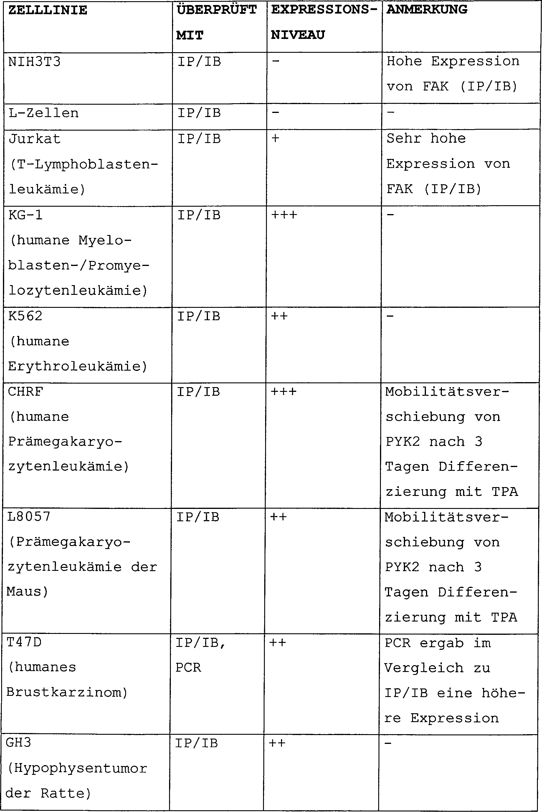

Claims (8)
Applications Claiming Priority (3)
| Application Number | Priority Date | Filing Date | Title |
|---|---|---|---|
| US3282496P | 1996-12-11 | 1996-12-11 | |
| US32824P | 1996-12-11 | ||
| PCT/US1997/022565 WO1998026054A2 (en) | 1996-12-11 | 1997-12-09 | Pyk2 related products and methods |
Publications (2)
| Publication Number | Publication Date |
|---|---|
| DE69729954D1 DE69729954D1 (en) | 2004-08-26 |
| DE69729954T2 true DE69729954T2 (en) | 2005-09-08 |
Family
ID=21867009
Family Applications (1)
| Application Number | Title | Priority Date | Filing Date |
|---|---|---|---|
| DE69729954T Expired - Fee Related DE69729954T2 (en) | 1996-12-11 | 1997-12-09 | Identification procedure for substances that bind to the Pyk2 polypeptide |
Country Status (11)
| Country | Link |
|---|---|
| US (2) | US20020048782A1 (en) |
| EP (1) | EP0948604B1 (en) |
| JP (1) | JP2001505779A (en) |
| AT (1) | ATE271603T1 (en) |
| AU (1) | AU5596998A (en) |
| CA (1) | CA2274805A1 (en) |
| DE (1) | DE69729954T2 (en) |
| DK (1) | DK0948604T3 (en) |
| ES (1) | ES2227729T3 (en) |
| PT (1) | PT948604E (en) |
| WO (1) | WO1998026054A2 (en) |
Families Citing this family (18)
| Publication number | Priority date | Publication date | Assignee | Title |
|---|---|---|---|---|
| AU2209899A (en) * | 1998-12-30 | 2000-07-24 | Sugen, Inc. | Pyk2 (raftk) and inflammation |
| DE19860833C1 (en) * | 1998-12-30 | 2000-09-07 | Albrecht E Sippel | Method for cellular high-throughput detection of receptor-ligand interactions |
| EP1630559A3 (en) * | 1998-12-30 | 2006-06-07 | Sugen, Inc. | PYK2 (RAFTK) and inflammation |
| US6861442B1 (en) | 1998-12-30 | 2005-03-01 | Sugen, Inc. | PYK2 and inflammation |
| WO2000049144A2 (en) * | 1999-02-22 | 2000-08-24 | Sugen, Inc. | Pyk2 binding proteins |
| DE10037759A1 (en) * | 2000-08-03 | 2002-07-04 | Gruenenthal Gmbh | screening process |
| EP1228766A1 (en) * | 2001-01-31 | 2002-08-07 | Max-Planck-Gesellschaft zur Förderung der Wissenschaften e.V. | PYK2 phosphorylation by HER3 induces tumor invasion |
| DE10123055A1 (en) * | 2001-05-11 | 2003-03-20 | Gruenenthal Gmbh | Screening method with PIM1-Kinase or PIM3-Kinase |
| DE10128541A1 (en) * | 2001-06-13 | 2003-02-20 | Gruenenthal Gmbh | Screening procedure with BNPI and DNPI |
| JP2005514013A (en) * | 2001-12-19 | 2005-05-19 | ミレニアム・ファーマシューティカルズ・インコーポレイテッド | Methods and compositions in treating pain and painful disorders using 1465, 1587, 2146, 2207, 32838, 336, and 52908 |
| US20040171062A1 (en) * | 2002-02-28 | 2004-09-02 | Plexxikon, Inc. | Methods for the design of molecular scaffolds and ligands |
| EP1546372A4 (en) * | 2002-09-19 | 2006-08-23 | Pfizer Prod Inc | Inducible focal adhesion kinase cell assay |
| EP1627045A2 (en) * | 2003-02-28 | 2006-02-22 | Plexxikon, Inc. | Pyk2 crystal structure and uses |
| WO2005002573A2 (en) * | 2003-06-06 | 2005-01-13 | Elan Pharmaceuticals, Inc. | Methods for using modulators of proline-rich tyrosine kinase 2 |
| US20050148029A1 (en) * | 2003-09-29 | 2005-07-07 | Biosite, Inc. | Methods and compositions for determining treatment regimens in systemic inflammatory response syndromes |
| AR056857A1 (en) | 2005-12-30 | 2007-10-24 | U3 Pharma Ag | DIRECTED ANTIBODIES TO HER-3 (RECEIVER OF THE HUMAN EPIDERMAL GROWTH FACTOR-3) AND ITS USES |
| WO2009155060A2 (en) * | 2008-05-28 | 2009-12-23 | Mayo Foundation For Medical Education And Research | Anti-pyk2 antibodies |
| CN105999263B (en) | 2009-11-13 | 2021-06-29 | 第一三共欧洲有限公司 | Materials and methods for treating or preventing human epidermal growth factor receptor-3 (HER-3) related diseases |
Family Cites Families (6)
| Publication number | Priority date | Publication date | Assignee | Title |
|---|---|---|---|---|
| US4343940A (en) * | 1979-02-13 | 1982-08-10 | Mead Johnson & Company | Anti-tumor quinazoline compounds |
| DE3069468D1 (en) * | 1979-12-19 | 1984-11-22 | Nat Res Dev | Quinazoline derivatives, processes for their preparation, compositions containing them and their use as anti-cancer agents |
| DE3611194A1 (en) * | 1986-04-04 | 1987-10-08 | Bayer Ag | CANCEROSTATIC AGENT |
| DE3920029C2 (en) * | 1988-06-30 | 1999-05-20 | Clariant Finance Bvi Ltd | Dyes for coloring plastics |
| US5837815A (en) * | 1994-12-15 | 1998-11-17 | Sugen, Inc. | PYK2 related polypeptide products |
| US5880141A (en) * | 1995-06-07 | 1999-03-09 | Sugen, Inc. | Benzylidene-Z-indoline compounds for the treatment of disease |
-
1997
- 1997-12-09 DE DE69729954T patent/DE69729954T2/en not_active Expired - Fee Related
- 1997-12-09 AT AT97952333T patent/ATE271603T1/en not_active IP Right Cessation
- 1997-12-09 PT PT97952333T patent/PT948604E/en unknown
- 1997-12-09 WO PCT/US1997/022565 patent/WO1998026054A2/en not_active Ceased
- 1997-12-09 US US08/987,689 patent/US20020048782A1/en not_active Abandoned
- 1997-12-09 JP JP52689998A patent/JP2001505779A/en not_active Withdrawn
- 1997-12-09 EP EP97952333A patent/EP0948604B1/en not_active Expired - Lifetime
- 1997-12-09 AU AU55969/98A patent/AU5596998A/en not_active Abandoned
- 1997-12-09 DK DK97952333T patent/DK0948604T3/en active
- 1997-12-09 CA CA002274805A patent/CA2274805A1/en not_active Abandoned
- 1997-12-09 ES ES97952333T patent/ES2227729T3/en not_active Expired - Lifetime
-
2002
- 2002-11-13 US US10/292,524 patent/US20030119067A1/en not_active Abandoned
Also Published As
| Publication number | Publication date |
|---|---|
| PT948604E (en) | 2004-10-29 |
| ATE271603T1 (en) | 2004-08-15 |
| AU5596998A (en) | 1998-07-03 |
| EP0948604A2 (en) | 1999-10-13 |
| US20030119067A1 (en) | 2003-06-26 |
| ES2227729T3 (en) | 2005-04-01 |
| DE69729954D1 (en) | 2004-08-26 |
| WO1998026054A2 (en) | 1998-06-18 |
| JP2001505779A (en) | 2001-05-08 |
| WO1998026054A3 (en) | 1998-09-03 |
| CA2274805A1 (en) | 1998-06-18 |
| DK0948604T3 (en) | 2004-11-22 |
| US20020048782A1 (en) | 2002-04-25 |
| EP0948604B1 (en) | 2004-07-21 |
Similar Documents
| Publication | Publication Date | Title |
|---|---|---|
| DE69729954T2 (en) | Identification procedure for substances that bind to the Pyk2 polypeptide | |
| DE69530791T2 (en) | PROTEIN TYROSINE KINASES (PYK2) ITS cDNA CLONING AND ITS USES | |
| Lev et al. | Protein tyrosine kinase PYK2 involved in Ca2+-induced regulation of ion channel and MAP kinase functions | |
| Higuchi et al. | PKA phosphorylates the p75 receptor and regulates its localization to lipid rafts | |
| DE69926731T2 (en) | DIAGNOSIS AND TREATMENT OF AUR1 AND / OR AUR2 RELATED DISEASES | |
| Gardoni et al. | Calcium–calmodulin‐dependent protein kinase II phosphorylation modulates PSD‐95 binding to NMDA receptors | |
| Howell et al. | Mouse disabled (mDab1): a Src binding protein implicated in neuronal development | |
| Fleming et al. | Signal transduction by G proteins in cardiac tissues. | |
| Lindstrom et al. | Molecular studies of the neuronal nicotinic acetylcholine receptor family | |
| Peretz et al. | Hypomyelination and increased activity of voltage‐gated K+ channels in mice lacking protein tyrosine phosphatase ϵ | |
| He et al. | Modulation of actin filament behavior by GAP-43 (neuromodulin) is dependent on the phosphorylation status of serine 41, the protein kinase C site | |
| Raymond et al. | Glutamate receptor modulation by protein phosphorylation | |
| Defer et al. | Tissue specificity and physiological relevance of various isoforms of adenylyl cyclase | |
| DE69434670T2 (en) | METHOD AND PRODUCT FOR REGULATING THE RESPONSE OF CELLS TO OUTER SIGNALS | |
| DE60027295T2 (en) | SPHINGOSINEKINASE | |
| Sanchez et al. | Molecular identification of Kvα subunits that contribute to the oxygen‐sensitive K+ current of chemoreceptor cells of the rabbit carotid body | |
| Fu et al. | Xenopus muscle‐specific kinase: molecular cloning and prominent expression in neural tissues during early embryonic development | |
| Ureña et al. | Expression, synaptic localization, and developmental regulation of Ack1/Pyk1, a cytoplasmic tyrosine kinase highly expressed in the developing and adult brain | |
| DE60316161T2 (en) | NEW KCNQ POLYPEPTIDES AND THEIR USE IN THE DIAGNOSIS OF MENTAL DISEASES | |
| Nathanson | Regulation and development of muscarinic receptor number and function | |
| Molokanova et al. | Subunit contributions to phosphorylation‐dependent modulation of bovine rod cyclic nucleotide‐gated channels | |
| Jordan et al. | Repression of slow myosin heavy chain 2 gene expression in fast skeletal muscle fibers by muscarinic acetylcholine receptor and Gαq signaling | |
| EP1102847B1 (en) | Novel tension-dependent potassium channel and the use thereof in the development of new therapeutic agents | |
| Rasmussen | The potential role of retinal cGMP-interaction partners within the degeneration mechanism causing Retinitis Pigmentosa | |
| Aswathy et al. | Avian Muscarinic Receptors: An Update |
Legal Events
| Date | Code | Title | Description |
|---|---|---|---|
| 8327 | Change in the person/name/address of the patent owner |
Owner name: NEW YORK UNIVERSITY, NEW YORK, N.Y., US Owner name: SUGEN, INC., SAN FRANCISCO, CALIF., US |
|
| 8364 | No opposition during term of opposition | ||
| 8339 | Ceased/non-payment of the annual fee |