DE69727483T2 - TISSUE TRANSPLANT FROM THE MAGIC SUBMUKOSA FOR REPAIRING NEUROLOGICAL TISSUE - Google Patents
TISSUE TRANSPLANT FROM THE MAGIC SUBMUKOSA FOR REPAIRING NEUROLOGICAL TISSUE Download PDFInfo
- Publication number
- DE69727483T2 DE69727483T2 DE69727483T DE69727483T DE69727483T2 DE 69727483 T2 DE69727483 T2 DE 69727483T2 DE 69727483 T DE69727483 T DE 69727483T DE 69727483 T DE69727483 T DE 69727483T DE 69727483 T2 DE69727483 T2 DE 69727483T2
- Authority
- DE
- Germany
- Prior art keywords
- tissue
- submucosal
- submucosal tissue
- growth factor
- tube
- Prior art date
- Legal status (The legal status is an assumption and is not a legal conclusion. Google has not performed a legal analysis and makes no representation as to the accuracy of the status listed.)
- Expired - Lifetime
Links
Classifications
-
- A—HUMAN NECESSITIES
- A61—MEDICAL OR VETERINARY SCIENCE; HYGIENE
- A61K—PREPARATIONS FOR MEDICAL, DENTAL OR TOILETRY PURPOSES
- A61K35/00—Medicinal preparations containing materials or reaction products thereof with undetermined constitution
- A61K35/12—Materials from mammals; Compositions comprising non-specified tissues or cells; Compositions comprising non-embryonic stem cells; Genetically modified cells
- A61K35/37—Digestive system
- A61K35/38—Stomach; Intestine; Goblet cells; Oral mucosa; Saliva
-
- A—HUMAN NECESSITIES
- A61—MEDICAL OR VETERINARY SCIENCE; HYGIENE
- A61K—PREPARATIONS FOR MEDICAL, DENTAL OR TOILETRY PURPOSES
- A61K38/00—Medicinal preparations containing peptides
- A61K38/16—Peptides having more than 20 amino acids; Gastrins; Somatostatins; Melanotropins; Derivatives thereof
- A61K38/17—Peptides having more than 20 amino acids; Gastrins; Somatostatins; Melanotropins; Derivatives thereof from animals; from humans
- A61K38/18—Growth factors; Growth regulators
- A61K38/1825—Fibroblast growth factor [FGF]
-
- A—HUMAN NECESSITIES
- A61—MEDICAL OR VETERINARY SCIENCE; HYGIENE
- A61K—PREPARATIONS FOR MEDICAL, DENTAL OR TOILETRY PURPOSES
- A61K38/00—Medicinal preparations containing peptides
- A61K38/16—Peptides having more than 20 amino acids; Gastrins; Somatostatins; Melanotropins; Derivatives thereof
- A61K38/17—Peptides having more than 20 amino acids; Gastrins; Somatostatins; Melanotropins; Derivatives thereof from animals; from humans
- A61K38/18—Growth factors; Growth regulators
- A61K38/185—Nerve growth factor [NGF]; Brain derived neurotrophic factor [BDNF]; Ciliary neurotrophic factor [CNTF]; Glial derived neurotrophic factor [GDNF]; Neurotrophins, e.g. NT-3
-
- A—HUMAN NECESSITIES
- A61—MEDICAL OR VETERINARY SCIENCE; HYGIENE
- A61P—SPECIFIC THERAPEUTIC ACTIVITY OF CHEMICAL COMPOUNDS OR MEDICINAL PREPARATIONS
- A61P25/00—Drugs for disorders of the nervous system
-
- A—HUMAN NECESSITIES
- A61—MEDICAL OR VETERINARY SCIENCE; HYGIENE
- A61P—SPECIFIC THERAPEUTIC ACTIVITY OF CHEMICAL COMPOUNDS OR MEDICINAL PREPARATIONS
- A61P43/00—Drugs for specific purposes, not provided for in groups A61P1/00-A61P41/00
-
- C—CHEMISTRY; METALLURGY
- C12—BIOCHEMISTRY; BEER; SPIRITS; WINE; VINEGAR; MICROBIOLOGY; ENZYMOLOGY; MUTATION OR GENETIC ENGINEERING
- C12N—MICROORGANISMS OR ENZYMES; COMPOSITIONS THEREOF; PROPAGATING, PRESERVING, OR MAINTAINING MICROORGANISMS; MUTATION OR GENETIC ENGINEERING; CULTURE MEDIA
- C12N5/00—Undifferentiated human, animal or plant cells, e.g. cell lines; Tissues; Cultivation or maintenance thereof; Culture media therefor
- C12N5/06—Animal cells or tissues; Human cells or tissues
- C12N5/0602—Vertebrate cells
- C12N5/0679—Cells of the gastro-intestinal tract
-
- A—HUMAN NECESSITIES
- A61—MEDICAL OR VETERINARY SCIENCE; HYGIENE
- A61K—PREPARATIONS FOR MEDICAL, DENTAL OR TOILETRY PURPOSES
- A61K35/00—Medicinal preparations containing materials or reaction products thereof with undetermined constitution
- A61K35/12—Materials from mammals; Compositions comprising non-specified tissues or cells; Compositions comprising non-embryonic stem cells; Genetically modified cells
-
- C—CHEMISTRY; METALLURGY
- C12—BIOCHEMISTRY; BEER; SPIRITS; WINE; VINEGAR; MICROBIOLOGY; ENZYMOLOGY; MUTATION OR GENETIC ENGINEERING
- C12N—MICROORGANISMS OR ENZYMES; COMPOSITIONS THEREOF; PROPAGATING, PRESERVING, OR MAINTAINING MICROORGANISMS; MUTATION OR GENETIC ENGINEERING; CULTURE MEDIA
- C12N2501/00—Active agents used in cell culture processes, e.g. differentation
- C12N2501/10—Growth factors
- C12N2501/115—Basic fibroblast growth factor (bFGF, FGF-2)
-
- C—CHEMISTRY; METALLURGY
- C12—BIOCHEMISTRY; BEER; SPIRITS; WINE; VINEGAR; MICROBIOLOGY; ENZYMOLOGY; MUTATION OR GENETIC ENGINEERING
- C12N—MICROORGANISMS OR ENZYMES; COMPOSITIONS THEREOF; PROPAGATING, PRESERVING, OR MAINTAINING MICROORGANISMS; MUTATION OR GENETIC ENGINEERING; CULTURE MEDIA
- C12N2501/00—Active agents used in cell culture processes, e.g. differentation
- C12N2501/10—Growth factors
- C12N2501/13—Nerve growth factor [NGF]; Brain-derived neurotrophic factor [BDNF]; Cilliary neurotrophic factor [CNTF]; Glial-derived neurotrophic factor [GDNF]; Neurotrophins [NT]; Neuregulins
Landscapes
- Health & Medical Sciences (AREA)
- Life Sciences & Earth Sciences (AREA)
- Engineering & Computer Science (AREA)
- Chemical & Material Sciences (AREA)
- Biomedical Technology (AREA)
- Bioinformatics & Cheminformatics (AREA)
- Zoology (AREA)
- General Health & Medical Sciences (AREA)
- Medicinal Chemistry (AREA)
- Pharmacology & Pharmacy (AREA)
- Veterinary Medicine (AREA)
- Animal Behavior & Ethology (AREA)
- Public Health (AREA)
- Cell Biology (AREA)
- Organic Chemistry (AREA)
- Biotechnology (AREA)
- Gastroenterology & Hepatology (AREA)
- Immunology (AREA)
- Epidemiology (AREA)
- Proteomics, Peptides & Aminoacids (AREA)
- Wood Science & Technology (AREA)
- Genetics & Genomics (AREA)
- Neurosurgery (AREA)
- Neurology (AREA)
- Microbiology (AREA)
- Nutrition Science (AREA)
- Biochemistry (AREA)
- General Engineering & Computer Science (AREA)
- Chemical Kinetics & Catalysis (AREA)
- General Chemical & Material Sciences (AREA)
- Psychology (AREA)
- Nuclear Medicine, Radiotherapy & Molecular Imaging (AREA)
- Physiology (AREA)
- Developmental Biology & Embryology (AREA)
- Virology (AREA)
- Materials For Medical Uses (AREA)
- Medicines That Contain Protein Lipid Enzymes And Other Medicines (AREA)
- Medicines Containing Material From Animals Or Micro-Organisms (AREA)
- Prostheses (AREA)
Description
Gebiet der ErfindungTerritory of invention
Diese Erfindung bezieht sich auf Gewebetransplantatkonstrukte, die in der Förderung des Wiederwachstums und der Heilung von beschädigten oder kranken neurologisch verwandte Gewebestrukturen nützlich sind. Im Besonderen bezieht sich diese Erfindung auf ein Verfahren zur Induzierung der Bildung von endogenen neurologischen Strukturen an einer Stelle mit Bedarf des Wachstums von endogenem neurologisch verwandtem Gewebe durch Kontaktierung der Stelle mit einem submucosalen Gewebetransplantatkonstrukt.This Invention relates to tissue graft constructs described in of funding of regrowth and healing of damaged or sick neurologically related tissue structures useful are. In particular, this invention relates to a method to induce the formation of endogenous neurological structures at a point in need of growth of endogenous neurologically related Tissue by contacting the site with a submucosal tissue graft construct.
Hintergrund und Zusammenfassung der Erfindungbackground and summary of the invention
Der Neurochirurg wird oft mit der Notwendigkeit der Reparatur duraler Defekte aufgrund von Traumata, Tumorresektion und dekompressiven Prozeduren konfrontiert. Viele Materialien wurden für die Verwendung bei der Reparatur der Dura mater und darunter liegenden Geweben untersucht. Gegenwärtige Optionen umfassen autologe Materialien (z. B. Perikranium, Temporals Fascia und Fascia lata des Spannmuskels), lyophilisierte kadaverartige Materialien (z. B. Dura mater und Fascia lata des Spannmuskels) und synthetische Materialien (z. B. silastische Schichten, Dacron Schichten, Vicrylnetz), jedes dieser Materialien ist jedoch mit signifikanten Einschränkungen verbunden.The Neurosurgeon often becomes dural with the need for repair Defects due to trauma, tumor resection and decompressive Procedures faced. Many materials have been used when repairing the dura mater and underlying tissues examined. current Options include autologous materials (e.g. pericranium, temporals Fascia and Fascia lata of the tension muscle), lyophilized carcass-like Materials (e.g. dura mater and fascia lata of the tension muscle) and synthetic materials (e.g. silastic layers, Dacron Layers, Vicrylnetz), but each of these materials is significant limitations connected.
Eine Aufgabe der vorliegenden Erfindung ist es, ein biozersetzbares Material bereitzustellen, welches als ein Duralersatz dienen kann.A The object of the present invention is a biodegradable material which can serve as a dural replacement.
Viele Individuen haben Schädigungen an deren zentralem Nervensystem erlitten, die das Individuum teilweise paralysiert lassen oder in einer reduzierten motorischen Funktion resultieren. Strategien der Wiederherstellung und Transplantatmaterialien zur Wiederherstellung von Beschädigungen des zentralen Nervensystems existieren gegenwärtig nicht. Insbesondere regenerieren sich Nervenfasern innerhalb des Gehirns und des Rückenmarks, welche sich strukturell von peripheren Nerven unterscheiden, nicht, nachdem diese abgetrennt oder gequetscht worden sind. Beispielsweise besteht keine gegenwärtig bekannte Behandlung für Menschen, die eine funktionelle Regeneration über eine vollständige Durchtrennung des Rückenmarks oder einen abgetrennten Sehnerv fördert.Lots Individuals have injuries suffered on their central nervous system, which the individual partially leave paralyzed or in a reduced motor function result. Recovery strategies and graft materials to restore damage of the central nervous system do not currently exist. Regenerate in particular nerve fibers within the brain and spinal cord, which are structurally different from peripheral nerves, not after they have been separated or squeezed. For example none currently exists known treatment for People who have functional regeneration over a full sever of the spinal cord or promotes a severed optic nerve.
Eine weitere Aufgabe der vorliegenden Erfindung ist es, eine Zusammensetzung und ein Verfahren bereitzustellen, das die Produktion von endogenen Zentralnervenzellen fördert und dadurch die Wiederherstellung von Beschädigungen sowohl von Geweben des zentralen Nervensystems und peripheren Nervengeweben erlaubt.A Another object of the present invention is a composition and to provide a method that enables the production of endogenous Promotes central nerve cells and thereby restoring damage to both tissues of the central nervous system and peripheral nerve tissues.
Es ist bekannt, dass Zusammensetzungen, die die Tunica submucosa enthalten, die sowohl von der Tunica muscularis und wenigstens dem luminalen Bereich der Tunica mucosa des Darms von warmblütigen Wirbeltieren delaminiert sind, als Gewebetransplantatmaterialien verwendet werden können. Siehe beispielsweise US-Patente Nr. 4,902,508 und 5,281,422. Die in diesen Patenten beschriebenen Zusammensetzungen sind durch ausgezeichnete mechanische Eigenschaften gekennzeichnet, einschließlich hohe Dehnbarkeit, hoher Berstdruck und einen wirksamen Porosizitätsindex, welche es derartigen Zusammensetzungen gestatten, für Vaskulartransplantatkonstrukte und in Anwendungen des Ersatzes von Bindegewebe nützlich zu sein. Wenn sie in derartigen Anwendungen eingesetzt werden, scheinen submucosale Transplantatkonstrukte als eine Matrix für das Wiederwachstum der durch die Transplantatkonstrukte ersetzten Gewebe zu dienen. Des Weiteren können, wie in dem US-Patent Nr. 5,275,826 beschrieben ist, fluidisierte Formen von submucosalem Gewebe von Wirbeltieren ebenfalls als injizierbare oder implantierbare Gewebetransplantate ohne Verlust von biotropen Eigenschaften verwendet werden. In signifikanter Weise wurde es zudem in über 600 Überkreuz-Spezies-Implantaten niemals gezeigt, dass aus Submucosa erhaltene Transplantatzusammensetzungen eine Transplantatabstoßungsreaktion des Gewebes erklären.It it is known that compositions containing the tunica submucosa that of both the tunica muscularis and at least the luminal Area of the tunica mucosa of the intestine delaminated by warm-blooded vertebrates are used as tissue graft materials. Please refer for example, U.S. Patents 4,902,508 and 5,281,422. The one in these Compositions described in patents are distinguished by characterized mechanical properties, including high Elasticity, high burst pressure and an effective porosity index, which allow such compositions for vascular graft constructs and useful in connective tissue replacement applications his. When used in such applications, seem submucosal graft constructs as a matrix for regrowth to serve the tissue replaced by the graft constructs. Furthermore, fluidized as described in U.S. Patent No. 5,275,826 Forms of vertebrate submucosal tissue are also injectable or implantable tissue grafts without loss of biotropic Properties are used. Significantly, it did also in over 600 cross-species implants never shown that graft compositions obtained from submucosa a graft rejection response explain the fabric.
Die Anmelder haben entdeckt, dass submucosales Gewebe das Wachstum und die Vermehrung von auf die Neurologie verwandten Geweben induzieren, einschließlich der Dura mater und Nervenzellen des zentralen und peripheren Nervensystems. Dementsprechend ist die vorliegende Erfindung auf die Verwendung von submucosalem Gewebe als ein Transplantatkonstrukt zur Förderung der Wiederherstellung von beschädigten oder kranken neurologisch verwandten Geweben ausgerichtet.The Applicants have discovered that submucosal tissue growth and induce the multiplication of tissues related to neurology, including the dura mater and nerve cells of the central and peripheral nervous system. Accordingly, the present invention is based on the use of submucosal tissue as a graft construct to promote the restoration of damaged or diseased neurologically related tissues.
Detaillierte Beschreibung der bevorzugten Ausführungsbeispieledetailed Description of the preferred embodiments
Es wurde gefunden, dass die submucosalen Gewebekonstrukte der vorliegenden Erfindung das Wachstum von neurologisch verwandten Geweben fördern oder induzieren. In Übereinstimmung mit der vorliegenden Erfindung schließt der Begriff der neurologisch verwandten Gewebe Neuronen und Glialzellen sowie Dura mater, Arachnoid- und Pia mater Gewebe ein. Es wird in Übereinstimmung mit dieser Erfindung ein Verfahren zur Verwendung von Zusammensetzungen enthaltend submucosales Gewebe von warmblütigen Wirbeltieren zur Wiederherstellung oder zur Verbesserung der Wiederherstellung von beschädigten oder kranken neurologisch verwandten Geweben in einem warmblütigen Wirbeltier bereitgestellt. Das Verfahren weist den Schritt der Kontaktierung der Stelle, die einer Wiederherstellung bedarf, mit einer submucosales Gewebe enthaltenden Zusammensetzung auf.It it was found that the submucosal tissue constructs of the present Invention promote the growth of neurologically related tissues or induce. In accordance with the present invention the term includes the neurological related tissue neurons and glial cells as well as dura mater, arachnoid and pia mater tissue. It is in accordance with this invention a method of using compositions containing submucosal Tissue of warm-blooded Vertebrates to restore or improve recovery of damaged or diseased neurologically related tissues in a warm-blooded vertebrate provided. The method has the step of contacting the place that needs restoration, with a submucosal Fabric containing composition.
Submucosales Gewebe, welches zur Verwendung in Übereinstimmung mit der vorliegenden Erfindung geeignet ist, enthält natürliche kollagene Matrizes, welche hochkonservierte Kollagene einschließen, Glycoproteine, Proteoglycane und Glycosaminglycane in deren natürlicher Konfiguration und natürlichen Konzentration ein. Eine Quelle von submucosalem Gewebe ist das Darmgewebe eines warmblütigen Wirbeltiers. Dünndarmgewebe ist eine bevorzugte Quelle von submucosalem Gewebe zur Verwendung gemäß dieser Erfindung.submucosal Fabric intended for use in accordance with the present Invention is suitable contains natural collagen matrices, which include highly conserved collagens, glycoproteins, Proteoglycans and glycosaminoglycans in their natural configuration and natural Concentration. A source of submucosal tissue is the intestinal tissue of a warm-blooded Vertebrate. Small intestine tissue is a preferred source of submucosal tissue for use according to this Invention.
Submucosales Gewebe zur Verwendung in dieser Erfindung wird aus verschiedenen Quellen warmblütiger Wirbeltiere erhalten, einschließlich Darmgewebe, welches von für die Fleischproduktion aufgezogenen Tieren wie beispielsweise Schweinen, Rindern und Schafen oder anderen warmblütigen Wirbeltieren geerntet wurde. Dieses Gewebe kann entweder in dessen natürlicher Konfiguration oder in einer zerkleinerter oder teilweise enzymatisch verdauter fluidisierter Form verwendet werden. Submucosales Wirbeltiergewebe ist ein reichlich vorhandenes Nebenprodukt von Arbeitsgängen der kommerziellen Fleischproduktion und ist daher ein Transplantatmaterial mit geringen Kosten, insbesondere wenn das submucosale Gewebe in dessen natürlicher Schichtenlagenkonfiguration verwendet wird.submucosal Fabrics for use in this invention are made from various Sources more warm-blooded Get vertebrates, including Intestinal tissue, which of for meat production such as pigs, Cattle and sheep or other warm-blooded vertebrates. This tissue can either be in its natural configuration or in a comminuted or partially enzymatically digested fluidized Shape can be used. Submucosal vertebrate tissue is an abundant existing by-product of operations of commercial meat production and is therefore a low cost graft material, in particular when the submucosal tissue is in its natural layered configuration is used.
Geeignetes submucosales Darmgewebe weist typischerweise die von sowohl der Tunica muscularis und wenigstens dem luminalen Bereich der Tunica mucosa delaminierte Tunica submucosa auf. In einem Ausführungsbeispiel nach der vorliegenden Erfindung weist das submucosale Darmgewebe die Tunica submucosa und basilare Bereiche der Tunica mucosa, einschließlich die Lamina muscularis mucosa und das Stratum compactum auf. Es ist bekannt, dass diese Schichten in Abhängigkeit von der Quelle der Wirbeltierspezies in der Dicke und in der Definition variieren.suitable Submucosal intestinal tissue typically exhibits that of both Tunica muscularis and at least the luminal area of the tunica mucosa delaminated tunica submucosa. In one embodiment according to the present invention, the submucosal intestinal tissue the tunica submucosa and basilar areas of the tunica mucosa, including the Lamina muscularis mucosa and the stratum compactum. It is known, that these layers are dependent from the source of the vertebrate species in thickness and in definition vary.
Die Herstellung von submucosalem Gewebe zur Verwendung in Übereinstimmung mit dieser Erfindung wird in dem US-Patent Nr. 3,902,508 beschrieben. Ein Segment des Darms eines Wirbeltieres, vorzugsweise geerntet von Schweine-, Schaf- oder Rinderspezies, aber nicht andere Spezies ausschließend, wird einer Abrasion unter Verwendung einer Streichbewegung in Längsrichtung unterworfen, um die äußeren Schichten, die Glattmuskelgewebe enthalten, und die am weitesten innenliegende Schicht, d. h. den luminalen Bereich der Tunica mucosa, zu entfernen. Das submucosale Gewebe wird mit Salzlösung gespült und wahlweise sterilisiert; es kann in einem hydratisierten oder dehydratisierten Zustand gelagert werden. Lyophilisiertes oder luftgetrocknetes submucosales Gewebe kann rehydratisiert und in Übereinstimmung mit dieser Erfindung ohne signifikanten Verlust von dessen biotropen und mechanischen Eigenschaften verwendet werden.The Manufacture of submucosal tissue for use in accordance with this invention is described in U.S. Patent No. 3,902,508. A segment of the vertebrate intestine, preferably harvested of pig, sheep or bovine species, but not other species exclusive, becomes an abrasion using a longitudinal sweep subjected to the outer layers, which contain smooth muscle tissue, and the innermost Layer, d. H. remove the luminal area of the tunica mucosa. The submucosal tissue is rinsed with saline and optionally sterilized; it can be stored in a hydrated or dehydrated state become. Lyophilized or air-dried submucosal tissue can be rehydrated and in accord with this invention without significant loss of its biotropic and mechanical properties can be used.
Von Organen warmblütiger Wirbeltiere gewonnenes submucosales Gewebe hat typischerweise eine abluminale und eine luminale Oberfläche. Die luminale Oberfläche ist die submucosale Oberfläche, die dem Lumen der Organquelle zugewandt ist und typischerweise benachbart einer inneren Mucosaschicht in der Organquelle ist, wobei die abluminale Oberfläche die submucosale Oberfläche ist, welche von dem Lumen der Organquelle wegweist und typischerweise in Kontakt mit glattem Muskelgewebe in der Organquelle ist.Of Organs more warm-blooded Submucosal tissue obtained from vertebrates typically has an abluminal one and a luminal surface. The luminal surface is the submucosal surface, which faces the lumen of the organ source and is typically adjacent an inner mucosal layer in the organ source, the abluminal surface the submucosal surface which points away from the lumen of the organ source and typically is in contact with smooth muscle tissue in the organ source.
Die submucosalen Gewebetransplantatzusammensetzungen nach der vorliegenden Erfindung können durch Dehnung des Materials in einer Längs- oder Querrichtung vorkonditioniert werden, wie in dem US-Patent Nr. 5,275,826 beschrieben ist. Mehrfache Streifen Stücke von submucosalem Gewebe können auch miteinander vereinigt werden, um ein einheitliches mehrschichtiges submucosales Gewebetransplantat mit einem Oberflächenbereich größer als jeder der individuellen Streifen/Stücke des submucosalen Gewebes zu bilden. Der Prozess der Bildung großer Bereiche/mehrschichtiger submucosaler Gewebekonstrukte ist in der US-Patentanmeldung 08/418,515 beschrieben. Zusammenfassend gesagt enthält der Prozess der Bildung großflächiger Lagen von submucosalem Gewebe die Überlappung wenigstens eines Bereichs von einem Streifen aus submucosalem Gewebe mit wenigstens einem Bereich eines anderen Streifens aus submucosalem Gewebe und Ausübung eines Druckes auf wenigstens die überlappenden Bereiche unter Bedingungen, die eine Dehydratation des submucosalen Gewebes erlauben. Unter diesen Bedingungen werden die überlappten Bereiche "verschmolzen" werden, um eine einheitliche große Gewebelage zu bilden.The submucosal tissue graft compositions of the present invention can be preconditioned by stretching the material in a longitudinal or transverse direction, as described in U.S. Patent No. 5,275,826. Multiple strip pieces of submucosal tissue can also be combined to form a unitary multi-layer submucosal tissue graft with a surface area larger than each of the individual strips / pieces of submucosal tissue. The process of forming large areas / multi-layered submucosal tissue constructs is described in US Patent Application 08 / 418,515. In summary, the process of forming large areas of submucosal tissue includes overlapping at least a portion of a strip of submucosal tissue with at least a portion of another strip of submucosa lem tissue and exerting pressure on at least the overlapping areas under conditions that allow dehydration of the submucosal tissue. Under these conditions, the overlapped areas will be "fused" to form a uniform, large layer of fabric.
Die großflächigen Transplantatkonstrukte bestehen im Wesentlichen aus submucosalem Gewebe, welches frei von potentiell kompromissbehafteten Klebemitteln und chemischen Vorbehandlungen ist, und sie haben eine größere oberflächliche Fläche und größere mechanische Festigkeit als die zur Bildung des Transplantatkonstruktes verwendeten individuellen Streifen. Die viellagigen submucosalen Konstrukte können wahlweise perforiert sein, wie in dem US-Patent 5,711,969 beschrieben ist. Die Perforationen des submucosalen Gewebekonstrukts erlauben es extrazellulären Flüssigkeiten, durch das Gewebetransplantatmaterial zu passieren, eine Fluidretention innerhalb des Transplantats zu verringern und die Remodellierungseigenschaften der Gewebetransplantate zu verstärken. Die Perforation des submucosalen Gewebes ist insbesondere für mehrfach laminierte Gewebetransplantatkonstrukte nützlich, wobei die Perforationen auch die adhäsive Kraft zwischen benachbarten Schichten verstärken.The large-area graft constructs consist essentially of submucosal tissue, which is free of potentially compromising adhesives and chemical pretreatments is, and they have a larger superficial area and bigger mechanical Strength than that used to form the graft construct individual stripes. The multilayer submucosal constructs can optionally perforated as described in U.S. Patent 5,711,969 is. The perforations of the submucosal tissue construct allow it extracellular Liquids, to pass through the tissue graft material, fluid retention reduce within the graft and the remodeling properties of the To reinforce tissue grafts. The perforation of the submucosal tissue is especially for multiple Laminated tissue graft constructs useful with the perforations also the adhesive Increase force between adjacent layers.
Das zur Verwendung in Übereinstimmung mit dieser Erfindung spezifizierte submucosale Gewebe kann auch in einer fluidisierten Form vorliegen. Submucosales Gewebe kann durch Zerkleinerung des Gewebes und wahlweise Aussetzung desselben gegenüber einer enzymatischen Verdauung fluidisiert werden, um eine im Wesentlichen homogene Lösung zu bilden. Die Herstellung von fluidi sierten Formen von submucosalem Gewebe wird in dem US-Patent Nr. 5,275,826 beschrieben, dessen Offenbarung hiermit ausdrücklich durch Inbezugnahme mit eingeschlossen ist. Fluidisierte Formen von submucosalem Gewebe werden durch Zerkleinerung von submucosalem Gewebe durch Zerreißen, Schneiden, Zermahlen oder Scheren des geernteten Gewebes hergestellt. Stücke von submucosalem Gewebe können daher durch Scherung in einem Hochgeschwindigkeitsmischer oder durch Zermahlen der Submucosa in einem gefrorenen oder gefriergetrockneten Zustand zerkleinert werden, um ein Pulver zu erzeugen, welches nachfolgend hierzu mit Wasser oder einer gepufferten Salzlösung hydratisiert werden kann, um eine submucosale Flüssigkeit von flüssiger, gelartiger oder pastenartiger Konsistenz zu bilden. Die Formulierung fluidisierter Submucosa kann des Weiteren mit Enzymen wie beispielsweise Protease, einschließlich Trypsin oder Pepsin, bei einem sauren pH-Wert für einen zur Solubilisierung sämtlicher oder eines Hauptteils der Bestandteile des submucosalen Gewebes behandelt werden und wahlweise filtriert werden, um eine homogene Lösung von teilweise solubilisierter Submucosa bereitzustellen.The for use in accordance submucosal tissue specified with this invention can also be in a fluidized form. Submucosal tissue can by crushing the tissue and optionally suspending it across from enzymatic digestion can be fluidized to essentially one homogeneous solution to build. The production of fluidized forms of submucosal Tissue is described in U.S. Patent No. 5,275,826, the disclosure of which hereby expressly by Inclusion is included. Fluidized forms of submucosal Tissues are made by crushing submucosal tissue Tear, Cutting, grinding or shearing the harvested tissue. pieces of submucosal tissue therefore by shear in a high speed mixer or by Grinding the submucosa in a frozen or freeze-dried state be crushed to produce a powder which is subsequently can be hydrated with water or a buffered saline solution, a submucosal fluid from liquid, to form gel-like or paste-like consistency. The wording Fluidized submucosa can also be used with enzymes such as Protease, including Trypsin or pepsin, at an acidic pH for one for solubilization all or a major part of the components of the submucosal tissue treated and optionally filtered to give a homogeneous solution of partially solubilized submucosa.
Die Transplantatzusammensetzungen gemäß der vorliegenden Erfindung können unter Verwendung herkömmlicher Desinfektions-/Sterilisations-Techniken sterilisiert werden, einschließlich Glutaraldehyd-Gerbung, Formaldehyd-Gerbung bei saurem pH-Wert, Propylenoxidbehandlung, Ethylenoxidbehandlung, Gasplasmasterilisation, Gammastrahl- oder Elektronenstrahlbehandlung und Peressigsäure (PAA)-Desinfektion. Sterilisationstechniken, welche die mechanische Festigkeit, Struktur und biotropen Eigenschaften des submucosalen Gewebes nicht negativ beeinflussen, sind bevorzugt. Beispielsweise kann starke Gammabestrahlung einen Verlust der Festigkeit der Lagen des submucosalen Gewebes bewirken. Bevorzugte Sterilisationstechniken umfassen die Aussetzung des Transplantats gegenüber Peressigsäure, 1–4 Mrad Gammabestrahlung (mehr bevorzugt 1–2.5 Mrad an Gammabestrahlung) oder Gasplasmasterilisation. Typischerweise wird das submucosale Gewebe zwei oder mehr Sterilisationsprozessen unterworfen.The Graft compositions according to the present invention can using conventional Disinfection / sterilization techniques be sterilized, including Glutaraldehyde tanning, formaldehyde tanning at acidic pH, propylene oxide treatment, Ethylene oxide treatment, gas plasma sterilization, gamma-ray or Electron beam treatment and peracetic acid (PAA) disinfection. Sterilization techniques, which the mechanical strength, structure and biotropic properties of submucosal tissue are preferred. For example, strong gamma radiation can result in a loss of strength of the layers of the submucosal tissue. Preferred sterilization techniques include graft exposure to peracetic acid, 1-4 Mrad Gamma radiation (more preferably 1-2.5 Mrads of gamma radiation) or gas plasma sterilization. Typically the submucosal Tissue subjected to two or more sterilization processes.
Nachdem das submucosale Gewebe in einem einleitenden Desinfektionsschritt behandelt wurde, beispielsweise durch Behandlung mit Peressigsäure, kann das Gewebe in eine Kunststoff- oder Folienumhüllung eingewickelt und erneut unter Verwendung von Elektronenstrahl- oder Gammabestrahlungssterilisationstechniken sterilisiert werden.After this the submucosal tissue in an initial disinfection step was treated, for example by treatment with peracetic acid the tissue is wrapped in a plastic or foil wrap and again using electron beam or gamma radiation sterilization techniques be sterilized.
In Übereinstimmung mit einem Ausführungsbeispiel wird submucosales Gewebe als ein Gewebetransplantatkonstrukt für den Ersatz oder die Wiederherstellung von beschädigten oder kranken neurologisch verwandten Geweben verwendet. Insbesondere wurde es gefunden, dass das gegenwärtige submucosale Gewebekonstrukt das Wachstum und die Vermehrung von Neuronen fördert. Dementsprechend können die gegenwärtigen Zusammensetzungen in einem Verfahren der Wiederherstellung von beschädigten oder kranken, neurologisch verwandten Geweben in einem warmblütigen Wirbeltier verwendet werden.In accordance with an embodiment submucosal tissue is used as a tissue graft construct for replacement or the restoration of damaged or sick neurologically related Fabrics used. In particular, it was found that the current submucosal Tissue construct promotes the growth and proliferation of neurons. Accordingly can the current Compositions in a process of restoring damaged or diseased, neurologically related tissues in a warm-blooded vertebrate be used.
Das in Übereinstimmung mit der vorliegenden Erfindung verwendete submucosale Gewebekonstrukt weist submucosales Darmgewebe auf, welches sowohl von der Tunica muscularis und wenigstens dem luminalen Bereich der Tunica mucosa des Darms eines warmblütigen Wirbeltieres oder eines Verdaus desselben delaminiert ist. Das Konstrukt kann mit einem zugefügten Wachstumsfaktor wie einem vaskularen Endothelialwachstumsfaktor, Nervenwachstumsfaktor oder Fibroblastwachstumsfaktor oder einem Wachstumsfaktor enthaltend Extrakte von submucosalem Gewebe kombiniert werden. Alternativ kann das Gewebetransplantatkonstrukt submucosales Gewebe in Kombination mit peripherem neuronalem Gewebe und wahlweise zugefügten Wachstumsfaktoren enthalten.The submucosal tissue construct used in accordance with the present invention comprises submucosal intestinal tissue which is delaminated from both the tunica muscularis and at least the luminal region of the tunica mucosa of the intestine of a warm-blooded vertebrate or digestion thereof. The construct can be with an added growth factor such as a vascular endothelial growth factor, nerve growth factor or fibroblast growth factor or a growth factor containing extracts of submucosal tissue can be combined. Alternatively, the tissue graft construct may contain submucosal tissue in combination with peripheral neuronal tissue and optionally added growth factors.
In einem Ausführungsbeispiel werden feste Formen von submucosalem Gewebe mit einem oder mehr Wachstumsfaktoren durch Einweichen des Gewebes in einer den Wachstumsfaktor enthaltenden gepufferten Lösung kombiniert. Beispielsweise wird das submucosale Gewebe für 7–14 Tage bei 4°C in einer PBS gepufferten Lö sung, die ungefähr 5 bis ungefähr 500 mg/ml enthält, oder mehr bevorzugt 25 bis ungefähr 100 mg/ml des Wachstumsfaktor, eingeweicht. Submucosales Gewebe bindet schnell an Proteine und wird eine Assoziation mit einem bioaktiven Mittel für mehrere Tage zurückbehalten. Um jedoch die Aufnahme der Wachstumsfaktoren in das submucosale Gewebe zu steigern, kann das Gewebe teilweise vor der Kontaktierung der Wachstumsfaktorlösung dehydratisiert werden. Für Zusammensetzungen enthaltend fluidisierte oder solubilisierte Extrakte oder Guanidin-Extrakte von submucosalem Gewebe können lyophilisierte Pulver oder Lösungen von Wachstumsfaktoren direkt mit dem submucosalen Gewebe gemischt werden. Beispielsweise kann fluidisiertes oder solubilisiertes submucosales Gewebe mit einem Wachstumsfaktor vermischt und anschließend innerhalb eines Röhrchens aus submucosalem Gewebe (oder anderes biozersetzbares Gewebe) verpackt werden. Das offene Ende des Röhrchens wird nach Füllung des Röhrchens mit dem fluidisierten oder solubilisierten submucosalem Gewebe abdichtend verschlossen.In an embodiment become solid forms of submucosal tissue with one or more growth factors by soaking the tissue in a tissue containing the growth factor buffered solution combined. For example, the submucosal tissue is used for 7-14 days at 4 ° C in a PBS buffered solution, the roughly 5 to about Contains 500 mg / ml, or more preferably 25 to about 100 mg / ml of the growth factor, soaked. Submucosal tissue quickly binds to proteins and becomes an association with a bioactive one Funds for withheld for several days. However, to include the growth factors in the submucosal Tissue can increase the tissue partially before contacting the growth factor solution be dehydrated. For Compositions containing fluidized or solubilized extracts or guanidine extracts from submucosal tissue can lyophilized powder or solutions of growth factors directly mixed with the submucosal tissue become. For example, fluidized or solubilized submucosal Tissue mixed with a growth factor and then inside of a tube packaged from submucosal tissue (or other biodegradable tissue). The open end of the tube after filling of the tube sealing with the fluidized or solubilized submucosal tissue locked.
In Übereinstimmung mit der vorliegenden Erfindung wird submucosales Gewebe eines warmblütigen Wirbeltieres verwendet, um ein zur Induzierung der Wiederherstellung von neurologischem Gewebe in einem warmblütigen Wirbeltier nützliches Gewebetransplantatkonstrukt herzustellen. Die Herstellung umfasst die Schritte der Kombination des submucosalen Gewebes eines warmblütigen Wirbeltieres, oder eines Verdaus desselben, mit einem zugefügten Wachstumsfaktor ausgewählt aus der Gruppe bestehend aus vaskularem Endothelialwachstumsfaktor, Nervenwachstumsfaktor und Fibroblastwachstumsfaktor.In accordance with the present invention submucosal tissue of a warm-blooded vertebrate used to induce neurological recovery Tissue in a warm-blooded Vertebrate useful To manufacture tissue graft construct. The manufacture includes the steps of combining the submucosal tissue of a warm-blooded vertebrate, or a digestion of the same, selected with an added growth factor the group consisting of vascular endothelial growth factor, Nerve growth factor and fibroblast growth factor.
In einer Ausführungsform wird das submucosale Gewebe verwendet, um ein Transplantatkonstrukt herzustellen, welches sich auf das in vivo-Wachstum von Neuronen entlang einem vorbestimmten Weg richtet. Die Herstellung umfasst die Schritte der Bildung eines Röhrchens aus bioabbaubarem Material und Füllung des Röhrchens mit fluidisiertem submucosalen Gewebe. Das Röhrchen sollte so geformt sein, um einen Durchmesser von ungefähr 0,5 mm bis ungefähr 4 mm für periphere Nervenapplikationen aufzuweisen, und ungefähr 1 mm bis ungefähr 2 cm für Anwendung am Zentralnerv. In einer Ausführungsform wird das Röhrchen aus submucosalem Gewebe gebildet, wobei das submucosale Gewebe manipuliert wird, um einen Zylinder mit einem Durchmesser der bevorzugten Größe zu definieren. Typischerweise wird das submucosale Gewebe direkt aus Darmgewebe präpariert und hat daher eine im Allgemeinen zylindrische Gestalt. Das Gewebe kann manipuliert werden, um einen Zylinder mit dem bevorzugten Durchmesser durch Nähen oder Sicherung des Transplantats in Längsrichtung auf andere Weise und Entfernung des überschüssigen Gewebes zu definieren. Beispielsweise kann das Transplantatkonstrukt durch Ausfall eines sterilen Glasstabes mit einem äußeren Durchmesser, der gleich dem gewünschten Durchmesser des Lumen des gebildeten Transplantatkonstrukts ist, hergestellt werden. Der Glasstab wird in das Lumen des Transplantats eingeführt, redundantes Gewebe wird anschließend gesammelt und der gewünschte Lumendurchmesser wird durch Nähen entlang der Länge des Transplantats oder durch Verwendung anderer bekannter Techniken der Gewebefestlegung erzielt.In one embodiment the submucosal tissue is used to make a graft construct which relates to the in vivo growth of neurons along a predetermined path. The manufacturing includes the steps the formation of a tube made of biodegradable material and filling of the tube with fluidized submucosal tissue. The tube should be shaped about a diameter 0.5 mm to approximately 4 mm for peripheral nerve applications, and approximately 1 mm until about 2 cm for Use on the central nerve. In one embodiment, the tube is made from submucosal tissue is formed, the submucosal tissue being manipulated to define a cylinder with a diameter of the preferred size. Typically, the submucosal tissue is made directly from intestinal tissue prepared and therefore has a generally cylindrical shape. The mesh can be manipulated to a cylinder with the preferred diameter by sewing or securing the graft lengthwise in some other way and removal of the excess tissue define. For example, the graft construct can be made through Failure of a sterile glass rod with an outer diameter that is the same the desired one Diameter of the lumen of the graft construct formed, getting produced. The glass rod is placed in the lumen of the graft introduced, redundant tissue is then collected and the desired lumen diameter is by sewing along the length of the Graft or using other known techniques the tissue fixation achieved.
Alternativ kann das Röhrchen aus submucosalem Gewebe durch Umwickeln von Streifen aus submucosalem Gewebe auf einen Dorn hergestellt werden, wobei das aufgewickelte submucosale Gewebe überlappend angeordnet ist, wobei kein Bereich des darunter liegenden Dornes exponiert gelassen wird. Siehe US Provisional Application Serien Nr. 60/032,679, deren Offenbarung hiermit ausdrücklich mit eingeschlossen ist. Das submucosale Gewebe kann spiralförmig auf einen Dorn als ein kontinuierliches Stück aus submucosalem Gewebe aufgewickelt werden, und eine Mehrzahl von Streifen aus submucosalem Gewebe kann verwendet werden, um die röhrenförmigen Konstrukte zu bilden. Das gewickelte submucosale Gewebe wird anschließend unter dehydratisierenden Bedingungen komprimiert und die röhrchenförmige Prothese wird von dem Dorn entfernt. Die Größe des Überlapps in einem spiralför mig gewickelten Konstrukt in Übereinstimmung mit dieser Ausführungsform liegt in dem Bereich zwischen 10 bis 60% der Weite des vorhergehenden Streifens und mehr bevorzugt ist der überlappende Bereiche ein 50%iger Überlapp.alternative can the tube from submucosal tissue by wrapping strips of submucosal Fabrics are made on a mandrel, the wound submucosal tissue overlapping is arranged, with no area of the underlying mandrel is left exposed. See US Provisional Application series No. 60 / 032,679, the disclosure of which is hereby expressly included. The submucosal tissue can be spiral on a mandrel as a continuous piece of submucosal tissue be wound up, and a plurality of strips of submucosal Tissue can be used to form the tubular constructs. The wrapped submucosal tissue is then dehydrated Conditions compressed and the tubular prosthesis is replaced by the Thorn removed. The size of the overlap in a spiral coiled construct in accordance with this embodiment lies in the range between 10 to 60% of the width of the previous one Streaking and more preferably the overlapping area is a 50% overlap.
Nach der Bildung des biozersetzbaren Röhrchens wird das Röhrchen mit fluidisiertem oder solubilisiertem submucosalen Gewebe gefüllt und das Röhrchen an einem oder beiden Enden des Röhrchens unter Verwendung auf dem Fachgebiet bekannter Verfahren (einschließlich Klammern, Nähen, Bindemittelpasten und Kompression unter dehydratisierenden Bedingungen) verschlossen. Alternativ kann das Röhrchen an einem oder beiden Enden des Röhrchens verschlossen werden, bevor dieses mit fluidisiertem/solubilisiertem Gewebe gefüllt wird. Das Röhrchen kann anschließend durch Injektion von fluidisiertem/solubilisiertem Gewebe in das Lumen durch die Verwendung einer Spritze gefüllt werden.After formation of the biodegradable tube, the tube is filled with fluidized or solubilized submucosal tissue and the tube is sealed at one or both ends of the tube using methods known in the art (including stapling, suturing, binder pastes, and compression under dehydrating conditions). Alternatively, the tube can be closed at one or both ends of the tube before it is fluidized / solubilized Tissue is filled. The tube can then be filled by injecting fluidized / solubilized tissue into the lumen using a syringe.
Die submucosalen Gewebetransplantatkonstrukte nach der vorliegenden Erfindung werden zur Wiederherstellung von neurologisch verwandten Geweben und im Speziellen von Bestandteilen des zentralen und peripheren Nervensystems verwendet. Das Verfahren umfasst die Kontaktierung der einer Wiederherstellung bedürfenden Stelle mit einer Zusammensetzung enthaltend submucosales Darmgewebe, welches von sowohl der Tunica muscularis und wenigstens dem luminalen Bereich der Tunica mucosa des Darms eines warmblütigen Wirbeltieres delaminiert ist. Das submucosale Gewebe kann beispielsweise in Schichten-, Streifen-, Band- oder Schlaufenform verwendet und chirurgisch an der Stelle des Bedarfs einer Wiederherstellung implantiert werden. Die submucosale Gewebezusammensetzung kann des Weiteren in einer fluidisierten Form verabreicht und an der Stelle des Bedarfs einer Wiederherstellung in das warmblütige Wirbeltier injiziert werden. Schließlich kann die Zusammensetzung einen mit fluidisiertem submucosalen Gewebe gefüllten Zylinder aus submucosalem Gewebe aufweisen.The submucosal tissue graft constructs according to the present Invention are used to restore neurologically related Tissues and especially components of the central and peripheral Nervous system used. The procedure includes contacting those in need of restoration Site with a composition containing submucosal intestinal tissue, which of both the tunica muscularis and at least the luminal Delaminated area of the tunica mucosa of the intestine of a warm-blooded vertebrate is. The submucosal tissue can be, for example, in layers, strips, bands or loop shape used and surgically at the point of need be implanted for recovery. The submucosal tissue composition can also be administered and administered in a fluidized form the place of need for restoration in the warm-blooded vertebrate be injected. Finally the composition may be one with fluidized submucosal tissue filled Have cylinders made of submucosal tissue.
In einem Ausführungsbeispiel in Übereinstimmung mit der vorliegenden Erfindung werden die submucosalen Gewebekonstrukte verwendet, um die Bildung von neurologisch verwandtem Gewebe zwischen endogenen neurologischen Gewebestrukturen in einem warmblütigen Wirbeltier zu induzieren. Das Verfahren umfasst die Schritte der chirurgischen Implantation einer Gewebetransplantatzusammensetzung enthaltend submucosales Gewebe eines warmblütigen Wirbeltieres in den Wirt, um die endogenen neurologischen Gewebestrukturen zu überbrücken und ein Wachstum von endogenem neurologisch verwandtem Gewebe zwischen den überbrückten neurologischen Strukturen zu induzieren.In an embodiment in accordance with the present invention the submucosal tissue constructs used between the formation of neurologically related tissue endogenous neurological tissue structures in a warm-blooded vertebrate to induce. The procedure includes the steps of surgical Implantation of a tissue graft composition containing submucosal Tissue of a warm-blooded Vertebrate in the host to the endogenous neurological tissue structures to bridge and a growth of endogenous neurologically related tissue between the bridged neurological To induce structures.
Wenn submucosales Gewebe, welches die Tunica submucosa und die basilaren Bereiche der Tunica mucosal einschließlich der Lamina muscularis mucosa und dem Stratum compactum umfasst, in nicht-fluidisierter Form verwendet wird, ist es bevorzugt, dass dieses so implantiert wird, dass das Stratum compactum die Oberflächengewebe am stärksten flachliegend kontaktiert, um Adhäsionen mit dem Transplantatmaterial zu bilden.If submucosal tissue covering the tunica submucosa and the basilar Areas of the tunica mucosal including the lamina muscularis mucosa and the stratum compactum, in non-fluidized form it is preferred that it be implanted so that the stratum compactum most flatly lies the surface tissues contacted for adhesions to form with the graft material.
In Übereinstimmung mit einer Ausführungsform kann eine Beschädigung des Rückenmarks durch manuelle Trennung benachbarter longitudinaler Nervenfasern in dem Rückenmark repariert werden, wobei die Trennungen oder Einschnitte parallel zu den Axons der Neuronen der Wirbelsäule verlaufen und durch die Dura mater, Arachnoid- und Pia mater-Schichten durchdringt. Streifen von submucosalem Gewebe werden chirurgisch in die vertikalen Trennungen oder in natürliche Furchen implantiert und sind daher in direktem Kontakt mit neurologisch verwandten Geweben und werden durch diese Gewebe an ihrem Platz gehalten. Alternativ können Schichten von submucosalem Gewebe verwendet werden, um das Äußere des beschädigten Bereiches zu umwickeln, um eine Wiederherstellung der beschädigten Gewebe zu fördern. Wahlweise können Nähte verwendet werden, um das mucosale Gewebe an dessen gewünschten Ort zu sichern.In accordance with an embodiment can damage of the spinal cord by manual separation of adjacent longitudinal nerve fibers in the spinal cord be repaired, with the separations or incisions parallel to the axons of the spinal neurons and through the dura mater, arachnoid and pia mater layers. strip of submucosal tissue are surgically separated into the vertical or in natural Furrows are implanted and are therefore in direct contact with neurological related tissues and are held in place by these tissues held. Alternatively, you can Layers of submucosal tissue are used to cover the exterior of the damaged Wrapping the area to restore the damaged tissue to promote. Seams can optionally be used to secure the mucosal tissue in its desired location.
In Anwendungen, in welchen das Rückenmark durchtrennt worden ist, kann das submucosale Gewebe zwischen den zwei getrennten Enden angeordnet werden, um den Spalt zu überbrücken und als ein Rahmen zu dienen, welcher das Wachstum der Neuronen der zwei getrennten Enden in Richtung aufeinander richtet. Fluidisierte Formen von Submucosa können ebenso in Übereinstimmung mit der vorliegenden Erfindung verwendet werden, um beschädigte oder kranke neurologisch verwandte Gewebe wiederherzustellen. Vorteilhafterweise können die fluidisierten Formen in die Stelle, die einer Wiederherstellung bedarf, injiziert werden und können daher in einem weniger invasiven Verfahren verwendet werden, um die Vermehrung von endogenen neurologisch verwandten Geweben zu induzieren.In Applications in which the spinal cord has been severed, the submucosal tissue between the two separate ends can be arranged to bridge the gap and to serve as a framework that supports the growth of the neurons of the two separate ends towards each other. fluidized Forms of submucosa can also in agreement used with the present invention to damage or to restore diseased neurologically related tissues. advantageously, can the fluidized forms in the place of restoration needs, can be injected and therefore used in a less invasive procedure the multiplication of endogenous neurologically related tissues induce.
In Übereinstimmung mit einer Ausführungsform wird eine Gewebetransplantatzusammensetzung enthaltend submucosales Gewebe eines warmblütigen Wirbeltieres einem warmblütigen Wirbeltier an einer Stelle verabreicht, die ein endogenes neurologisch verwandtes Gewebewachstum in einer Menge bedarf, die wirksam ist, ein Wachstum von endogenem, neurologisch verwandtem Gewebe an der Stelle, an welcher die Zusammensetzung verabreicht ist, zu induzieren. Die biotropen Eigenschaften des submucosalen Gewebes fördern das Wachstum von neurologischem Wege entlang „Gebieten", wie sie durch den Weg des implantierten submucosalen Gewebes definiert sind. Dementsprechend kann das Wachstum von Neuronen, einschließlich Neuronen des zentralen und peripheren Nervensystems, auf eine Stelle gerichtet werden, die der Versorgung mit Nerven bedarf. In einer Ausführungsform wird das Wachstum von neurologischem Gewebe durch die Verwendung eines Gewebetransplantatkonstrukts enthaltend ein mit fluidisiertem submucosalen Gewebe gefülltes Röhrchen „ausgerichtet". In dieser Ausführungsform wird gesundes neuronales Gewebe in einem Ende des Röhrchens eingeführt und es wird in direktem Kontakt mit dem innerhalb des Röhrchens enthaltenen fluidisierten submucosalen Gewebe platziert. Das gegenüberliegende Ende des Röhrchens wird anschließend an oder nahe bei dem Ort platziert, welcher eine Innervation bedarf. Das Röhrchen wird an seinem Ort fixiert und stellt eine in vivo-Durchführung für neues neuronales Wachstum und Innervation der gewünschten Stelle bereit. Das Röhrchen aus fluidisiertem submucosalen Gewebe kann ebenfalls verwendet werden, um durchtrennte Nerven wiederherzustellen, wobei die beiden Enden des durchtrennten Nerves in das Röhrchen eingeführt werden, um eine Wiederanbringung der beschädigten Enden zu induzieren und die Nervenfunktion wiederherzustellen.In accordance with one embodiment, a tissue graft composition containing submucosal tissue of a warm-blooded vertebrate is administered to a warm-blooded vertebrate at a location that requires endogenous neurologically related tissue growth in an amount that is effective, growth of endogenous, neurologically related tissue at the location which the composition is administered to induce. The biotropic properties of the submucosal tissue promote the growth of neurological pathways along "areas" as defined by the path of the implanted submucosal tissue. Accordingly, the growth of neurons, including neurons of the central and peripheral nervous system, can be directed to one site, In one embodiment, the growth of neurological tissue is "aligned" through the use of a tissue graft construct containing a tube filled with fluidized submucosal tissue. In this embodiment, healthy neural tissue is inserted into one end of the tube and it is in direct contact with the flui contained within the tube placed submucosal tissue. The opposite end of the tube is then placed at or near the location that requires innervation. The tube is fixed in place and provides an in vivo implementation for new neuronal growth and innervation of the desired site. The fluidized submucosal tissue tube can also be used to restore severed nerves with the two ends of the severed nerve inserted into the tube to induce reattachment of the damaged ends and restore nerve function.
Jedes der nachfolgenden Verfahren kann in Konjugation mit den Gewebetransplantatkonstrukten nach der vorliegenden Erfindung verwendet werden, um eine Durchführung für gerichtetes de novo-Wachstum von neuronalen Geweben bereitzustellen. In einer Ausführungsform wird submucosales Gewebe in der Gestalt eines Röhrchens mit einem Lumen und zwei offenen Enden hergestellt. In einer Ausführungsform wird das Röhrchen aus submucosalem Gewebe direkt in den Wirtsorganismus implantiert und das Ende einer beschädigten Nervenfaser kann in das Lumen des submucosalen Geweberöhrchens eingeführt werden. Eine Spritze wird anschließend verwendet, um das Röhrchen mit zerkleinertem oder solubilisiertem submucosalen Gewebe zu füllen. Alternativ kann submucosales Gewebe in der Gestalt eines Röhrchens ausgebildet werden, welches mit fluidisiertem/solubilisierten submucosalem Gewebe gefüllt und an jedem Ende abgeschlossen ist. Das geschlossene Röhrchen aus submucosalem Gewebe kann anschließend gelagert werden, bis es gebraucht wird. In einer Ausführungsform wird das verschlossene Röhrchen aus submucosalem Gewebe in den Wirtsorganismus eingeführt und an seinem Platz unter Verwendung von dem Fachmann auf diesem Gebiet bekannter Techniken fixiert. Das eingeführte Transplantatkonstrukt stellt eine Durchführung für neues neuronales Gewebewachstum bereit. In einer bevorzugten Ausführungsform wird ein Schlitz oder ein Loch in das Röhrchen aus submucosalem Gewebe geschnitten und ein beschädigtes oder durchtrenntes Ende eines Nervengewebes wird durch den Schlitz oder die Öffnung und in das Lumen des Röhrchens eingeführt.each the following procedures can be conjugated to the tissue graft constructs according to the present invention can be used to carry out a directed de novo growth of neuronal tissues. In one embodiment becomes submucosal tissue in the form of a tube with a lumen and made two open ends. In one embodiment, the tube is made from submucosal tissue directly implanted in the host organism and the end of a damaged nerve fiber can be inserted into the lumen of the submucosal tissue tube. A syringe will then be given used the tube to be filled with crushed or solubilized submucosal tissue. alternative can submucosal tissue be formed in the shape of a tube, which is filled with fluidized / solubilized submucosal tissue and is completed at each end. The closed tube Submucosal tissue can then be stored until it is needed. In one embodiment becomes the closed tube introduced into the host organism from submucosal tissue and in place using the person skilled in the art known techniques fixed. The introduced graft construct represents an implementation for new neural tissue growth ready. In a preferred embodiment becomes a slit or hole in the tube of submucosal tissue cut and a damaged or severed end of a nerve tissue is through the slit or the opening and into the lumen of the tube introduced.
Das in Übereinstimmung mit der vorliegenden Erfindung verwendete submucosale Gewebe kann alleine oder in Kombination mit zugefügten Wachstumsfaktoren wie beispielsweise vaskularem Endothelialwachstumsfaktor, Nervenwachstumsfaktor oder saurem Fibroblastwachstumsfaktor verwendet werden. Des Weiteren können Implantate von peripheren Nerven in Kombination mit submucosalem Gewebe verwendet werden, um die Wiederherstellung der neuronalen Gewebe zu verbessern. Der Begriff des peripheren Nervenimplantats, wie er hier verwendet wird, bezieht sich auf neuronales Gewebe, welches von dem peripheren Nervensystem eines warmblütigen Wirbeltieres geerntet wurde, und vorzugsweise ein autologes peripheres neuronales Gewebe. Zusätzliche Komponenten können zu der Transplantatzusammensetzung des neurologen Gewebes zugefügt werden, die die Zusammensetzungen mit struktureller Unterstützung für Anwendungen unter Beteiligung des Rückenmarks bereitstellen, insbesondere wenn Bereiche der Wirbelsäule fehlen oder bedürfen, ersetzt zu werden. Beispielsweise kann Hydroxyapatit und/oder andere, biokompatibles Calcium enthaltende Mineralien mit der Transplantatzusammensetzung kombiniert werden, oder Metallstifte oder Drähte können ebenfalls in Kombination mit dem submucosalen Gewebe verwendet werden, um eine zusätzliche strukturelle Unterstützung für das Ersatzgewebe zu ergeben.The in accordance submucosal tissue used with the present invention alone or in combination with added growth factors such as for example vascular endothelial growth factor, nerve growth factor or acidic fibroblast growth factor. Furthermore can Peripheral nerve implants in combination with submucosal Tissues are used to restore the neural Improve tissue. The term peripheral nerve implant, as used here refers to neural tissue, which comes from the peripheral nervous system of a warm-blooded vertebrate was harvested, and preferably an autologous peripheral neuronal Tissue. additional Components can are added to the graft composition of the neurological tissue, which the compositions with structural support for applications under Involvement of the spinal cord provide, especially if areas of the spine are missing or need to be replaced. For example, hydroxyapatite and / or others, biocompatible calcium-containing minerals with the graft composition can be combined, or metal pins or wires can also be combined used with the submucosal tissue for an additional structural support for the To yield replacement tissue.
Die submucosalen Gewebetransplantatkonstrukte nach der vorliegenden Erfindung können auch verwendet werden, um das Wachstum und die Vermehrung von anderen mit dem Zentralnerv assoziierten Geweben und von Stützgeweben zu fördern. Submucosales Gewebe verbessert die Wiederherstellung von Glialzellen und Dura mater, Arachnoid- und Pia mater-Geweben.The submucosal tissue graft constructs according to the present Invention can also used to grow and multiply others tissues associated with the central nerve and supporting tissues to promote. Submucosal tissue improves the restoration of glial cells and dura mater, arachnoid and pia mater tissues.
In einer Ausführungsform wird das submucosale Gewebe als ein Duralersatz verwendet, welcher als ein Gewebetransplantatlappen gebildet ist, welches gestaltet ist, um einen in der endogenen Dura mater gebildeten Defekt oder gebildetes Loch zu bedecken. Gegenwärtig verfügbare Optionen für Ersatzmaterialien der Dura mater haben signifikante Beschränkungen: autologe Materialien sind häufig in ihrer Menge nicht adäquat und werden mit der assoziierten Morbidität von zusätzlichen Einschnitten erhalten, und die Handhabungscharakteristika von synthetischen Lagen sind im Vergleich mit biologischen Materialien schlecht. Des Weiteren wurden Befürchtungen bezüglich der Langzeitrisiken von Blutungen durch eine Gewebereaktion mit synthetischen Transplantatmaterialien erhoben. Die Dura von Leichen ist teuer, verschiedentlich in deren Zurverfügungstellung beschränkt und hat nur einigermaßen gute Handhabungscharakteristika. Von größerer Besorgnis ist dessen dokumentierte Rolle als ein Vektor in der Übertragung der langsamen Viren wie beispielsweise der Jakob-Creutzfeldt-Krankheit.In one embodiment the submucosal tissue is used as a dural replacement, which is formed as a tissue graft flap that designs is a defect or defect formed in the endogenous dura mater to cover formed hole. Replacement material options currently available the dura mater have significant limitations: autologous materials are common not adequate in quantity and are obtained with the associated morbidity of additional incisions, and the handling characteristics of synthetic sheets poor compared to biological materials. Furthermore, fears in terms of the long-term risks of bleeding from a tissue reaction synthetic graft materials. The dura of corpses is expensive, limited in its availability and various has to some extent good handling characteristics. This is of greater concern documented role as a vector in the transmission of slow viruses such as Jakob Creutzfeldt disease.
Submucosales Gewebe stellt einen ausgezeichneten Duralersatz bereit, da dieses Material nicht eine nachteilige Immunantwort heraufbeschwört und eine Vermehrung von endogenen Zellen induziert, welche in das Transplantat eindringen und das Transplantat schließlich durch endogene Zellen ersetzen. Ein Experiment wurde unter Verwendung von Ratten als Wirt durchgeführt, um die Brauchbarkeit von Zusammensetzungen aus submucosalem Gewebe als Duralersatz zu bestätigen.Submucosal tissue provides excellent dural replacement because this material does not evoke an adverse immune response and induce proliferation of endogenous cells that enter the graft and eventually replace the graft with endogenous cells. An experiment was performed using rats as hosts to confirm the usefulness of submucosal tissue compositions as a dural substitute.
Wie in Beispiel 1 beschrieben, fungiert submucosales Gewebe, welches in einer Ratte nachfolgend einer Duralresektion implantiert wurde, als ein geeigneter Duralersatz. 28 Tage nach der Implantation hat eine Remodellierung des submucosalen Gewebes begonnen, wie dies durch die Anwesenheit von Spindelzellen, aggressiver Neovaskularisation und eosinophiler Färbung der Bindegewebematrix angezeigt ist. Die Inkorporation und Remodellierung des Transplantats tritt in der Abwesenheit von jeglichen nachteiligen Effekten auf den darunter liegenden cerebralen Cortex ein.How described in Example 1, submucosal tissue acts, which was implanted in a rat following a dural resection, as a suitable dural replacement. 28 days after implantation remodeling of the submucosal tissue started, like this due to the presence of spindle cells, aggressive neovascularization and eosinophilic staining the connective tissue matrix is displayed. The incorporation and remodeling The graft occurs in the absence of any adverse effects Effects on the underlying cerebral cortex.
Beispiel 1example 1
Submucosales Gewebe als ein Duralersatzsubmucosal Tissue as a dural replacement
Experimentelle Ausgestaltung und chirurgische ProzedurExperimental design and surgical procedure
Zwanzig Sprague-Dawley Laborratten mittlerer Größe wurden anästhesiert (Ketamin 90 mg/kg und Xylazin 10 mg/kg, IM) und in einem stereotaktischen Kopfrahmen platziert, um das Kranium zu stabilisieren. Die Haut wurde rasiert, mit Chlorhexadin vorbereitet und mit 1% Lidocain durchtränkt. Nachfolgend einem Einschnitt der Fascia an der oberen temporalen Linie wurden die Schläfenmuskel lateral durch einen Kopfhauteinschnitt entlang der Mittellinie angehoben, wodurch die parietalen Konvexitäten exponiert wurden. Bihemisphere parietale Kraniektomien mit ungefähr 4 mm × 8 mm wurden mit einem elektrischen Handbohrer und einer Fräse gemacht. Die Dura, eine dünne, nahezu transparente Membran in der Ratte, wurde an den kraniektomischen Stellen unter Vergrößerung der Schleife herausgeschnitten. Es wurde Sorge dafür getragen, nicht den darunter liegenden zerebralen Cortex zu verletzen.Twenty Medium-sized Sprague-Dawley laboratory rats were anesthetized (Ketamine 90 mg / kg and xylazine 10 mg / kg, IM) and in a stereotactic Head frame placed to stabilize the cranium. The skin was shaved, prepared with chlorhexadine and with 1% lidocaine saturated. Following an incision of the fascia on the upper temporal Line became the temple muscle laterally raised through a scalp incision along the midline, causing the parietal convexities were exposed. Bihemisphere parietal craniectomies approximately 4 mm x 8 mm were performed made with an electric hand drill and a router. The dura, one thin, almost transparent membrane in the rat, was on the craniectomic Digits while increasing the Cut out the bow. Care was taken, not the one below injured lying cerebral cortex.
Submucosales Transplantatmaterial des Dünndarms wurde in Schichtenform in Übereinstimmung mit der vorliegenden Erfindung hergestellt und durch Aussetzung gegenüber 0,1% Peressigsäure sterilisiert. Das Transplantat wurde auf die ungefähre Größe geschnitten und als ein Auflagetransplantat über eine Konvexität platziert, wobei das Transplantat so orientiert war, dass die Oberfläche des Stratum compactum dem cerebralen Cortex zugewandt war. Die gegenüberliegende laterale Hemisphäre erhielt kein Transplantat, um so als eine Kontrolle für die Wirtsantwort auf das operative Verfahren zu dienen. In zwei Tieren wurde das Knochenfragment ersetzt. Die Wunde wurde mit normaler Salzlösung ausgespült und mit Krampen geschlossen. Eine einzelne postoperative Dosis von Ampicillin (25 mg/kg, SQ) wurde verabreicht. Eine unmittelbare nachoperative Versorgung umfasste die Anordnung auf einem Heizkissen, Abdeckung mit einem Handtuch, Umdrehen alle 15 Minuten bis zum Aufwachen und der Bewegung und Überwachung der Herztätigkeit und Atmung. Die Tiere wurden täglich bezüglich des Auftretens von plötzlichen Anfällen oder neurologischer Defizite, Appetit und Flüssigkeitsaufnahme und Gewicht überwacht. Zehn der Ratten wurden durch eine Überdosis an Barbiturat (150 mg/kg, IC) 7 Tage nach der Transplantatanordnung getötet. Die verbleibenden 10 Ratten wurden 28 Tage nach der Transplantatanordnung getötet.submucosal Small intestine graft material was layered in accordance made with the present invention and by exposure across from 0.1% peracetic acid sterilized. The graft was cut to approximate size and as a overlay graft over placed a convexity, the graft was oriented so that the surface of the Stratum compactum was facing the cerebral cortex. The opposite lateral hemisphere received no graft, so as a control for host response to serve on the operational process. That was in two animals Bone fragment replaced. The wound was rinsed with normal saline and with Clamps closed. A single postoperative dose of ampicillin (25 mg / kg, SQ) was administered. An immediate post-operative Supply included the arrangement on a heating pad, cover with a towel, turning around every 15 minutes until you wake up and of movement and surveillance of cardiac activity and breathing. The animals became daily in terms of of the appearance of sudden seizures or neurological deficits, appetite and fluid intake and weight are monitored. Ten of the rats were overdosed with barbiturate (150 mg / kg, IC) killed 7 days after graft placement. The remaining 10 rats were 28 days after grafting killed.
Histologische Präparation und Auswertunghistological preparation and evaluation
Drei der zwanzig Ratten starben an Komplikationen mit Bezug auf die Anästhesie in dem frühem postoperativen Zeitraum. Die verbleibenden siebzehn Ratten wurden ohne Ereignis von der Prozedur ohne Nachweis von Anfällen, Infektionen oder neurologischen Defiziten zurückerlangt. Acht Ratten wurden an dem 7. Tag getötet. Neun Ratten wurden an dem 28. Tag getötet.Three of the twenty rats died from complications related to anesthesia in the early postoperative period. The remaining seventeen rats were without event from the procedure without evidence of seizures, infections or regained neurological deficits. Eight rats were born killed on the 7th day. Nine rats were killed on the 28th day.
Nachfolgend zu der Tötung wurden die Ratten mit Formalin über Halsschlagaderarterienkatheter perfundiert. Das Kranium wurde anschließend in Formalin fixiert und entkalkt. Abschnitte mit einer Dicke von 6 Mikrometer wurden herausgeschnitten, mit Hematoxylin und Eosin gefärbt und für eine histologische Untersuchung präpariert. Die untersuchten Gewebe umfassten die Zwischenbereiche des Transplantats mit dem Cortex, Knochen und Kopfhaut. Die Kontrollseite wurde auf ähnliche Weise präpariert.following to the killing the rats were over with formalin Carotid artery catheter perfused. The cranium was then in Formalin fixed and descaled. 6 micron thick sections were cut out, stained with hematoxylin and eosin and for one histological examination prepared. The tissues examined included the intermediate areas of the graft with the cortex, bones and scalp. The control page was on similar Prepared way.
Eine mikroskopische Untersuchung der Proben wurde mit einer Quantifizierung des zellulären Infiltrats, der Vaskularität und der Dicke der defekten Stelle unter Verwendung eines Bildanalysesystems (Optimus Image Analysis System; Bioscan, Inc; Edmonds, WA) erweitert. Daten bezüglich der mit dem submucosalen Transplantationsimplantat behandelten defekten Stelle wurden mit der Kontrollstelle an sowohl den Zeitpunkten des 7. Tages und des 28. Tages verglichen. Die numerischen Ergebnisse, die zu den remodellierenden Geweben zugeordnet wurden, basierten auf den in Tabelle 1 wiedergegebenen Kriterien. Die Werte wurden unter Verwendung des Student's T-Tests verglichen. Das Gesamtergebnis für die jeweiligen Gruppen wurde verwendet, um die Nullhypothese zu überprüfen, dass kein Unterschied zwischen den morphologischen Änderungen in der gefüllten Submucosa gegenüber den nicht mit Submucosa gefüllten defekten Stellen an entweder dem 7. oder 28. Tag gesehen wird. Ein p-Wert von weniger als 0,05 wurde als signifikant angesehen.Microscopic examination of the samples was expanded with quantification of the cellular infiltrate, vascularity and thickness of the defect site using an image analysis system (Optimus Image Analysis System; Bioscan, Inc.; Edmonds, WA). Data regarding the defective site treated with the submucosal graft implant was compared to the control site at both the 7th and 28th day times. The numerical results assigned to the remodeling tissues were based on the criteria shown in Table 1. The values were below Using Student's t-test compared. The overall result for the respective groups was used to test the null hypothesis that no difference between the morphological changes in the filled submucosa compared to the non-submucosal defective sites is seen on either the 7th or 28th day. A p-value less than 0.05 was considered significant.
Tabelle
1: Quantitative histologische Beurteilung einer duralen Auflagetransplantation
von Submucosa gegenüber
einer Kontrollprobe 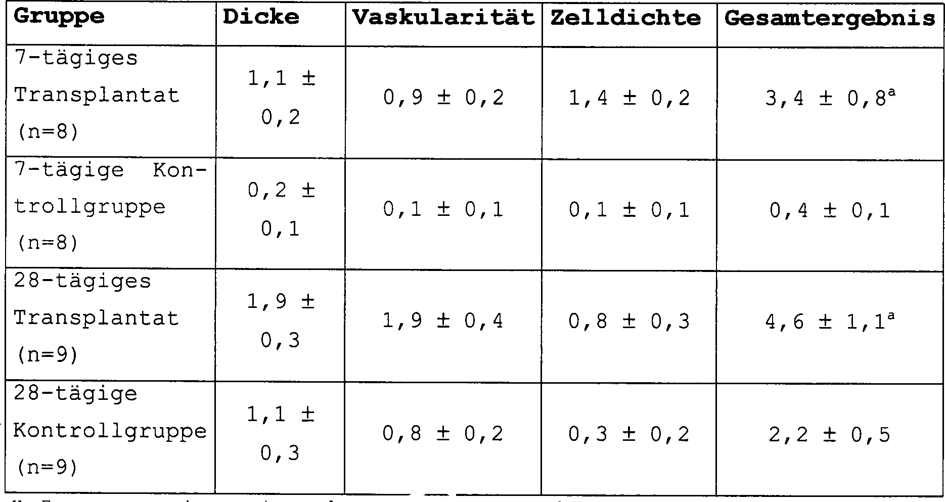
Die Stellen mit Submucosatransplantat sind mit den Kontrollstellen unter Verwendung der Dicke, Vaskularität und Zelldichte als die Ergebniskriterien verglichen. Die angegebenen Werte repräsentieren den Mittelwert ± S. E. M.The Locations with submucosal grafts are below the control sites Use of thickness, vascularity and cell density compared as the result criteria. The specified Represent values the mean ± S. E. M.

Die histologische Untersuchung zeigte eine Infiltration des Transplantats durch spindelförmige mononukleare Zellen, Deposition von Bindegewebe und Neovaskularität. Des Weiteren zeigte die histologische Analyse deutliche Unterschiede zwischen den mit Submucosagewebe wiederhergestellten Defekten gegenüber den Kontrollstellen (d. h. Defekte, die ohne jeglichem an der defekten Stelle platzierten Material der Heilung überlassen wurden). Das Gesamtergebnis für die jeweiligen Gruppen wurde unter Verwendung des Student's T Test verglichen, wobei eine Signifikanz für einen p-Wert von < 0,05 akzeptiert wurde. Ein signifikanter Unterschied zwischen den histologischen Ergebnissen der submucosalen Transplantatstelle und der Kontrollstelle wurde bei 7 Tagen (3,4 + 0,8 versus 0,1 + 0,1) und bei 28 Tagen (4,6 + 1,1 versus 2,2 + 0,5) gefunden. Kein Nachweis einer negativen Wirkung auf den darunter liegenden Cortex wurde beobachtet.The histological examination showed infiltration of the graft through spindle-shaped mononuclear cells, deposition of connective tissue and neovascularity. Furthermore the histological analysis showed clear differences between defects restored with submucosal tissue compared to Checkpoints (i.e. defects that do not involve any of the defective Place placed material left to heal). The overall result for the respective groups were compared using the Student's T test, being significant for a p-value of <0.05 was accepted. A significant difference between the histological Results of the submucosal graft site and the control site were at 7 days (3.4 + 0.8 versus 0.1 + 0.1) and at 28 days (4.6 + 1.1 versus 2.2 + 0.5) found. No evidence of a negative effect was observed on the underlying cortex.
Am siebten Tag waren die Hauptunterschiede zwischen den zwei Gruppen das zelluläre Infiltratat, die Vaskularität und die Dicke des an der Defektstelle deponierten Bindegewebes. Diese morphologischen Veränderungen wurden in einer halbquantitativen Weise, wie in Tabelle 1 definiert, verglichen. Dieses Vergleichsverfahren zeigte eine erhöhte Dicke, erhöhte Vaskularität und größere zellulare Infiltration der mit Submucosa behandelten Defekte gegenüber den nicht mit Submucosa behandelten Kontrolldefekten. Die mononuklearen Zellen, welche innerhalb und um das Submucosamaterial am siebten Tag gesehen wurden, zeigten oftmals eine spindelförmige Gestalt und waren durch die eosinophil anfärbendes extrazelluläres Matrixmaterial (ECM) umgeben. Das remodelierende Submucosamaterial zeigte eine große Anzahl von Gefäßen kapillarer Größe im Gegensatz zu jenen, die in den nicht mit Submucosa behandelten Kontrolldefekten beobachtet wurden.At the Seventh day was the main difference between the two groups the cellular Infiltrate, vascularity and the thickness of the connective tissue deposited at the defect site. These morphological changes were in a semi-quantitative manner as defined in Table 1, compared. This comparison procedure showed an increased thickness, increased vascularity and larger cellular Infiltration of Submucosa Treated Defects versus control defects not treated with submucosa. The mononuclear cells, which are seen within and around the submucosal material on the seventh day often showed a spindle-shaped shape and were through the eosinophil staining extracellular Surround matrix material (ECM). The remodeling submucosa material showed a big one Number of capillary vessels Size in contrast to those in the control defects not treated with submucosa were observed.
Nach 28 Tagen war das zelluläre Infiltrat in den Submucosagefüllten Defekten schwächer geworden und die Menge an ECM war gestiegen. Das eosinophil anfärbende Bindegewebe in und um die Submucosa zeigte eine Orientierung in der Richtung, welche sich von einem Rand der durchtrennten Calvaria zu dem gegenüberliegenden Rand erstrecken würde. Es wurde ebenfalls eine moderate Organisation des Bindegewebes in den nicht-submucosa Defekten gesehen; die Menge von anwesendem Material war jedoch viel geringer als an den Submucosa-Defektstellen. Das Submucosamaterial selber war am 28. Tag nicht unterscheidbar. Das ECM erschien in diesen H & E gefärbten Abschnitten homogen. Das zelluläre Infiltrat war an dem 28. Tag wesentlich geringer als an dem 7. Tag und im Grunde genommen waren sämtliche der anwesenden Zellen mit spindelförmig geschalteten Mesenchymalzellen konsistent.After 28 days, the cellular infiltrate in the submucosal-filled defects had weakened and the amount of ECM had increased. The eosinophil staining connective tissue in and around the submucosa showed an orientation in the direction that would extend from one edge of the severed Calvaria to the opposite edge. Moderate connective tissue organization was also seen in the non-submucosal defects; however, the amount of material present was much less than at the submucosal defect sites. The submucosal material itself was indistinguishable on the 28th day. The ECM appeared homogeneous in these H&E colored sections. The cellular infiltrate was significantly less on the 28th day than on the 7th day and basically all of the cells present were included Spindle-shaped mesenchymal cells consistent.
Gelegentliche Adhäsionen wurden zwischen dem ECM innerhalb der Defektstelle und dem darunter liegenden cerebralen Cortex in sowohl der Submucosa- und der Nichtsubmocosa-Seite festgestellt. Keine der Proben zeigten Veränderungen, die mit einer enzephalitischen Degeneration oder einer Nekrosis konsistent waren.occasional adhesions were between the ECM within the defect site and the one below cerebral cortex in both the submucosa and non-submocosa side detected. None of the samples showed changes with an encephalitic Degeneration or necrosis were consistent.
BEISPIEL 2EXAMPLE 2
Submucosales Gewebe als ein Duralersatz in dem Hunde-Modell.Submucosal tissue as a dural substitute in the dog model.
Experimentelle Ausgestaltung und chirurgische ProzedurExperimental design and surgical procedure
Bastardhunde mittlerer Größe (20 – 30 kg) wurden anästhesiert, intubiert und in der supinierten Brustbeinposition (Einleitung mit 2% Thiopental 1,0 mg/kg, intravenös; Aufrechterhaltung mit Isofluran 1%–2%; Atropin 0,5 mg/ml, intravenös) platziert. Eine Augensalbe wurde verabreicht. Die Kopfhaut wurde rasiert, mit Chlorhexadin behandelt und mit 1% Lidocain infiltriert. Durch einen Einschnitt in die Kopfhaut entlang der Mittellinie und nachfolgendem Einschnitt der Fascia an der oberen Schläfenlinie wurde der Schläfenmuskel unter lateraler Exposition der parietalen Konvexität angehoben. Ein 2 × 3 cm das Schläfenbein betreffende Kraniotomy wurde mit einem elektrischen Handbohrer und einer Knochenfräse durchgeführt. Blutende Knochenränder wurden gewachst. Die Dura wurde von der Kraniotomy-Stelle unter Vergrößerung der Schleife herausgetrennt. Es wurde Sorge dafür getragen, eine Verletzung des darunter liegenden zerebralen Cortex zu vermeiden.mongrel dogs medium size (20 - 30 kg) were anesthetized, intubated and in the supinated sternum position (initiation with 2% thiopental 1.0 mg / kg, intravenously; Maintenance with isoflurane 1% -2%; Atropine 0.5 mg / ml, intravenously) placed. An eye ointment was given. The scalp was shaved, treated with chlorhexadine and infiltrated with 1% lidocaine. Through an incision in the scalp along the midline and subsequent incision of the fascia on the upper temple line became the temple muscle elevated under lateral exposure to parietal convexity. A 2 × 3 cm the temporal bone The craniotomy concerned was performed with an electric hand drill and a bone milling machine carried out. Bleeding edges of bones were waxed. The dura was taken from the craniotomy site Enlargement of the Loop separated. Care was taken to cause an injury to avoid the underlying cerebral cortex.
Das submucosale Gewebetransplantatmaterial wurde geerntet und durch Aussetzung gegenüber 0,1%iger der Essigsäure in 20%igem Ethanol für 120 Minuten sterilisiert. Das Material wurde auf die geeignete Größe geschnitten und mit den verdichteten basalen Schichten der Tunica mucosa auf den zerebralen Cortex platziert und mit einer umsponnenen Nylonnaht gesichert. Bei fünf Tieren wurde eine kontralaterale Prozedur durchgeführt, in welcher die herausgeschnittene autologe Dura verwendet wurde, um den Defekt zu schließen und dadurch als eine Kontrolle für die Wirtsantwort auf die operative Prozedur zu dienen.The submucosal tissue graft material was harvested and by Exposure to 0.1% of acetic acid in 20% ethanol for Sterilized for 120 minutes. The material was cut to the appropriate size and with the densified basal layers of the tunica mucosa placed the cerebral cortex and with a braided nylon suture secured. At five A contralateral procedure was performed in animals which was used to cut out the autologous dura Close defect and thereby as a control for to serve the host response to the surgical procedure.
Bei drei Tieren wurde 60 Tage nach dem Beginn der Prozedur die kontralaterale Seite einer duralen Heraustrennung mit einem Ersatz durch Darmsubmucosa unterworfen. Der kraniotomische Hautlappen wurde ersetzt, die Wunde mit normaler Salzlösung ausgespült und mit Klammern verschlossen. Tribiotische Salbe, ein steriler Kopfverband und ein elisabethanischer Kragen wurden angewandt. Eine antibiotische Behandlung bestehend aus Cephalexin 1000 mg, PO, b. i. d., einen Tag vor der Operation und für drei Tage nach der Operation wurde verabreicht. Eine unmittelbar nach der Operation stattfindende Versorgung umfasste die Abdeckung mit einer Decke und Überwachung der Herztätigkeit und Atmung. Nachoperativer Schmerz wurde mit Butorphanol (2,4 mg, intramuskulär) und Acepromazin (2,0 mg, intramuskulär) behandelt. Die Tiere wurden hinsichtlich des Auftretens von Anfällen oder neurologischen Defiziten, Appetit und Flüssigkeitsaufnahme und Gewicht überwacht. Eine Tötung erfolgte durch eine Überdosis an Barbiturat (150 mg kg, intrakardial) nach 7, 30, 60, 90 und 120 Tagen nach der einleitenden Transplantatanordnung und 7, 30 und 60 Tage nachfolgend der wiederholten Aussetzung gegenüber submucosalem Darmgewebe. Der Tötung vorausgehend wurden 5 cc von CSF über eine subokcipitale Punktion aspiriert und bezüglich verschiedener Zellzählungen untersucht.at three animals became contralateral 60 days after the start of the procedure Side of a dural separation with a replacement by intestinal submucosa subjected. The craniotomic flap was replaced, the wound with normal saline rinsed and closed with brackets. Tribiotic ointment, a sterile Head bandage and an Elizabethan collar were used. A antibiotic treatment consisting of cephalexin 1000 mg, PO, b. i. d., one day before the operation and for three days after the operation was administered. One that takes place immediately after the operation Care included covering with a blanket and monitoring of cardiac activity and breathing. Postoperative pain was treated with butorphanol (2.4 mg, intramuscularly) and acepromazine (2.0 mg, intramuscular). The animals were respected the onset of seizures or neurological deficits, appetite and fluid intake and weight are monitored. A killing was caused by an overdose on barbiturate (150 mg kg, intracardial) after 7, 30, 60, 90 and 120 Days after the initial graft placement and 7, 30 and 60 days following repeated submucosal exposure Intestinal tissue. The killing Previously, 5 cc of CSF were via suboccipital puncture aspirated and regarding different cell counts examined.
Histologische Präparation und Auswertungenhistological preparation and evaluations
Nachfolgend der Tötung wurde der Kopf und Nacken der Hunde mit Formalin über Herzschlagaderarterienkatheter durchströmt. Das Kranium wurde anschließend in Formalin fixiert und entkalkt. Nach der Einbettung in Paraffin wurden 6 um dicke Abschnitte der chirurgischen Stellen herausgeschnitten, mit Hematoxilin und Eosin gefärbt und für eine histologische Untersuchung präpariert. Die untersuchten Gewebe schlossen die Zwischenbereiche des Transplantats mit dem zerebralen Cortex, Knochen und Kopfhaut ein.following of killing the head and neck of the dogs were treated with formalin via cardiac artery catheters flows through. The cranium was subsequently fixed in formalin and decalcified. After embedding in paraffin 6 µm sections of the surgical sites were cut out, stained with hematoxilin and eosin and for prepared a histological examination. The examined tissues closed the intermediate areas of the graft with the cerebral Cortex, bone and scalp.
ErgebnisseResults
Sämtliche acht Hunde wurden ohne Ereignis von dem Verfahren ohne Nachweis von Anfällen, einer Infektion oder einem neurologischen Defizit zurückerhalten. Eine wiederholte Transplantation wurde nicht durch einen klinischen Nachweis einer Sensibilisierung gegenüber dem submucosalen Gewebe begleitet.All Eight dogs were without event from the procedure without evidence of seizures, an infection or a neurological deficit. Repeated transplantation was not clinical Evidence of sensitization to the submucosal tissue accompanied.
An dem siebten Tag nachfolgend der Implantation war eine intensive mononukleare Zellantwort in das submucosale Darmgewebe mit extensiver Neovaskularisation und Deposition von disorganisierter extrazellulärer Matrix um das submucosale Gewebe vorhanden. Es bestand jedoch kein Nachweis für eine Involvierung des darunter liegenden zerebralen Cortex. Es war eine geringe Anzahl von spindelförmig geformten Zellen, die mit Fibroplasten konsistent sind, um und innerhalb der Transplantatstelle vorhanden.On the seventh day following the implantation, there was an intense mononuclear cell response in the Submucosal intestinal tissue with extensive neovascularization and deposition of disorganized extracellular matrix present around the submucosal tissue. However, there was no evidence of involvement of the underlying cerebral cortex. There was a small number of spindle-shaped cells consistent with fibroblasts around and within the graft site.
Bei jeder Untersuchungszeit nachfolgend des Zeitraumes von sieben Tagen (30, 60, 90 und 120 Tage) war ein gut organisiertes, eosinophil anfärbendes, dichtes Bindegewebe offensichtlich vorhanden, wobei kein Nachweis bestand, dass das submucosale Gewebe nach dem sechzigsten Tag zurückblieb. Keine meningozerebralen Adhäsionen waren zu sehen. In den Hunden, welche einer zweiten Aussetzung gegenüber submucosalem Darmgewebe unterworfen wurden, wurde eine Antwort gefunden, die von denen mit einer einfachen Aussetzung gegenüber submucosalem Darmgewebe nicht unterscheidbar war.at every examination period following the period of seven days (30, 60, 90 and 120 days) was a well organized, eosinophil anfärbendes, dense connective tissue is obviously present, with no evidence existed that the submucosal tissue remained after the sixtieth day. No meningo-cerebral adhesions were seen. In dogs, which have a second exposure to submucosal An intestinal tissue was subjected to an answer that was found those with a simple exposure to submucosal intestinal tissue was indistinguishable.
Bei manchen Tieren wurde sowohl an der Teststelle als auch an der Kontrollstelle eine geringe Entzündungsreaktion der Pia mater benachbart dem Gehirn festgestellt. Da diese sowohl an der Kontrollstelle als auch an der Teststelle anwesend war, repräsentiert diese wahrscheinlich eine Antwort auf die chirurgische Prozedur. Bei keinem Tier war ein Nachweis für eine Involvierung des zerebralen Cortex selber gegeben. Eine Neovaskularisa tion war an den Zeitpunkten des siebten und des dreißigsten Tages in den Stellen des submucosalen Gewebes, verglichen mit den Kontrollstellen, intensiver. Es bestand kein Nachweis für eine durch das Immunsystem vermittelte Transplantatreaktion der Wirtsensibilisierung in entweder den ursprünglichen Implantaten oder in den reimplantierten Tieren. Es wurden keine Abnormalitäten in der Serumchemie oder CSF-Zytologie in irgendeiner der nachoperativen Proben gefunden.at some animals were tested both at the test site and at the control site a low inflammatory response the pia mater is found adjacent to the brain. Since this both was present at the control point as well as at the test point this is probably an answer to the surgical procedure. There was no evidence of cerebral involvement in any animal Cortex given itself. A neovascularization was at the time of the seventh and thirtieth In the places of the submucosal tissue compared to the Checkpoints, more intense. There was no evidence of a through the immune system mediated graft response to host sensitization in either the original Implants or in the reimplanted animals. There were none abnormalities in serum chemistry or CSF cytology in any of the postoperative ones Samples found.
Diskussiondiscussion
Gegenwärtige Optionen für einen Duralersatz umfassen autologe Materalien (z. B. Perikranium, Temporalis fascia und Tensor fascia lata), lyopholizierte Leichenmaterialien (z. B. Dura mater und Tensor fascia lata), xenogene Biomaterialien (Perikardium vom Rind und Schaf, Dura des Rinds und rekonstituierten Collagenschwamm vom Rind) und synthetische Materialien (z. B. expandiertes polymeres Fluorethylen, silastische Lagen, Dacron-Lagen, Vicrylnetze). Jedes dieser Materialien ist jedoch mit signifikanten Beschränkungen verbunden.Current options for one Dural replacements include autologous materials (e.g. pericranium, temporalis fascia and tensor fascia lata), lyophilized corpse materials (e.g. Dura mater and Tensor fascia lata), xenogenic biomaterials (Bovine and sheep pericardium, bovine dura and reconstituted Beef collagen sponge) and synthetic materials (e.g. expanded polymeric fluoroethylene, silastic layers, Dacron layers, Vicryl networks). However, each of these materials has significant limitations connected.
Autologe Materialien sind oftmals in der Menge nicht adäquat vorhanden und werden mit der assoziierten Morbidität von zusätzlichen Schnitten erhalten. Die Handhabungscharakteristika von synthetischen Lagen sind im Vergleich zu biologischen Materialien schlecht. Des Weiteren wurden Bedenken bezüglich des Langzeitrisikos von Blutungen aus Gewebereaktionen benachbart zu dem Transplantat erhoben. Die Dura von Leichen ist teuer, in ihrer Anwendung gelegentlich beschränkt und hat nur mäßige Handhabungscharakteristika. Ihre dokumentierte Rolle als ein Vektor in der Übertragung der Jakob-Creutzfeld-Krankheit beschränkt sehr stark deren Anwendung. Gleichermaßen ruft ein kürzlicher Nachweis der Transmission der schwammförmigen Encephalopathie von Rindern auf Menschen Bedenken bezüglich neurologischer Gewebe auf Rinderbasis hervor.autologous Materials are often not adequately available in the quantity and are included associated morbidity of additional Get cuts. The handling characteristics of synthetic Layers are bad compared to biological materials. Of Further concerns were raised about the long-term risk of bleeding from tissue reactions raised to the graft. The dura of corpses is expensive, in occasionally limited in their use and has only moderate handling characteristics. Their documented role as a vector in the transmission of Jakob-Creutzfeld disease limited very much their application. Likewise, a recent calls Evidence of transmission of spongy encephalopathy from Cattle on humans have concerns about neurological tissue based on cattle.
Die Dura mater besteht primär aus Typ I Collagen, und daher sind Produkte auf Collagenbasis vernünftige Kandidaten für einen Duralersatz. Des Weiteren besteht, obwohl eine relative biologische Inertheit früher als ein wünschenswertes Charakteristikum des Transplantats betrachtet wurde, ein zunehmendes Bewusstsein der potentiellen Vorteile einer induzierten günstigen biologischen Antwort auf die Transplantation. In der Tat wurde es gezeigt, dass Fibroblaste und Endothelialzellen entlang eines rekonstituierten Collagengerüstes eindringen und dieses durch neues synthetisiertes Collagen ersetzen. Verschiedene auf Collagen basierende Präparationen wurden untersucht und scheinen viele von diesen Charakteristika zu zeigen. Für einen Duralersatz berichtete Präparationen umfassen verarbeitete Haut und das Peritoneum von Schweinen, das Korium und Perikardium von Rindern, das Perikardium von Schafen und mehrfach rekonstituierte Produkte der menschlichen Plazenta. Eine Transplantatinkorporation mit einer nachfolgenden Resorption wird für jedes dieser Materialien berichtet. Des Weiteren erzeugten diese weder akute Immunantworten noch epileptogene meningoencephalitische Narben, wenn diese am Cortex appliziert wurden.The Dura mater exists primarily from Type I collagen, and therefore collagen-based products are reasonable candidates for one Dural substitute. Furthermore, although a relative biological Inertness earlier as a desirable Characteristic of the graft was considered an increasing Awareness of the Potential Benefits of Induced Favorable biological response to the transplant. Indeed it did demonstrated that fibroblasts and endothelial cells are reconstituted along a collagen scaffold penetrate and replace it with new synthesized collagen. Various preparations based on collagen were examined and seem to show many of these characteristics. For a dural replacement reported preparations include processed skin and the peritoneum of pigs, the corium and pericardium of cattle, the pericardium of sheep and multiple reconstituted products of the human placenta. A graft incorporation with subsequent absorption for each of these materials reported. Furthermore, they did not generate acute immune responses epileptogenic meningoencephalitic scars, if these on the cortex were applied.
Nichtsdestoweniger ist nur das Perikardium von Rindern kommerziell erhältlich. Ein nicht auf Rindern basierendes Collagenpräparat mit günstigen biologischen Eigenschaften scheint viele Vorteile zu haben, während viele Nachteile vermieden werden. Das azelluläre submucosale Material nach der vorliegenden Erfindung stellt ein neuartiges Material für Dura mater-Ersatzkonstrukte bereit. Zusätzlich zu der Abwesenheit einer nachteiligen immunologischen Antwort wird eine unverkennbare Remodelierung des submucosalen Gewebes und Inkorporation in das Wirtsgewebe demonstriert. Die endgültige Form des remodelierten Duraltransplantats scheint histologisch von dem natürlichen Gewebe unterscheidbar zu sein. Des Weiteren wird eine Aussetzung des submucosalen Darmgewebes zwei Monate nachfolgend der einleitenden Transplantation nicht durch nachteilige klinische Ereignisse begleitet. Eine routinemäßige CSF-Zytologie ergibt keinen Nachweis für eine nachteilige Wirtsantwort auf das submucosale Gewebetransplantat.Nonetheless, only cattle pericardium is commercially available. A non-bovine collagen preparation with beneficial biological properties appears to have many advantages while avoiding many disadvantages. The acellular submucosal material according to the present invention provides a novel material for dura mater replacement constructs. In addition to the absence of an adverse immunological response, unmistakable remodeling of the submucosal tissue and incorporation into the host tissue is demonstrated. The final form of the remodeled dural graft appears to be histologically distinguishable from natural tissue. Furthermore, two months after the initial transplant, submucosal intestinal tissue exposure is not accompanied by adverse clinical events. Routine CSF cytology provides no evidence of an adverse host response to the submucosal tissue graft.
Beispiel 3Example 3
In Vitro Wachstum und Differenzierung von auf submucosalem Gewebe kultivierten neuronalen Zellen.In vitro growth and Differentiation of neurons cultured on submucosal tissue Cells.
Pheochromocytomazellen (PC 12-Zellen) sind neuronale Zellen, welche als sphärische chromaffine Zellen in der Abwesenheit eines Nervenwachstumsfaktors (NGF) wachsen aber unter Bildung von sympatikus-artigen Neuronen bei der Exposition gegenüber der Behandlung mit einem Nervenwachstumsfaktor differenzieren. PC 12-Zellen sind eine etablierte und gut studierte Zelllinie, die als Modellsystem verwendet worden ist, um eine neuronale Differenzierung und Vermehrung zu studieren. Die Antwort von PC 12-Zellen auf die Exposition gegenüber verschiedenen Formen von submucosalem Gewebe (Dehydratisierung, ETO, PAA) wurde untersucht, um zu bestimmen, ob submucosales Gewebe die Differenzierung, Wachstum und Vermehrung von neuronalen Zellen fördern würde.pheochromocytoma (PC 12 cells) are neuronal cells, which are called spherical chromaffin cells but grow in the absence of a nerve growth factor (NGF) forming sympathetic-like neurons upon exposure across from differentiate treatment with a nerve growth factor. PC 12 cells are an established and well-studied cell line that has been used as a model system for neuronal differentiation and study propagation. The response of PC 12 cells to exposure across from various forms of submucosal tissue (dehydration, ETO, PAA) was examined to determine whether submucosal tissue the differentiation, growth and proliferation of neuronal cells promote would.
Zellkulturcell culture
Pheochromocytoma (PC 12)-Zellen von Raten wurden von der American Type Culture Collection (Rockville, MD) erhalten und wurden wie von Green Tishler (1983) beschrieben kultiviert. Anfänglich wurde den Zellen gestattet, zu wachsen, um in Monoschichten, die an 75 cm Kunststoffkolben angehaftet waren, zusammenzufließen. Die PC 12-Zellen wurden in RPMI 1640 medium, welches mit 10% Pferdeserum, 5% fötalem Rinderserum, 1% L-Glutamin und 100 Einheiten/ml Penicillin und 100 μg/ml Streptomycin ergänzt war, beibehalten. Die Zellen wurden in Kunststoffkulturenschalen mit sechs Vertiefungen, die mit 0,2 mg/ml Collagen vorbeschichtet waren, platziert. PC 12-Zellen, die als positive Kontrollmittel dienten, wurden durch Behandlung mit Nervenwachstumsfaktor (NGF) bei einer Konzentration von 50 mg/ml für 7 bis 10 Tage in RPMI in Sympathoblast-artige Zellen differenziert. Das NGF enthaltende Medium wurde jeden Tag während dieses Zeitraumes ausgetauscht. Nach 10 Tagen in Kultur waren die PC 12-Zellen unheilbar krank differenziert und zum Überleben von NGF abhängig.pheochromocytoma Rate (PC 12) cells were from the American Type Culture Collection (Rockville, MD) were obtained and were described as described by Green Tishler (1983) cultured. Initially cells were allowed to grow in monolayers containing were attached to 75 cm plastic pistons to flow together. The PC 12 cells were medium in RPMI 1640, which with 10% horse serum, 5% fetal bovine serum, 1% L-glutamine and 100 units / ml penicillin and 100 μg / ml streptomycin added was maintained. The cells were placed in plastic culture dishes with six wells pre-coated with 0.2 mg / ml collagen were placed. PC 12 cells that act as positive control agents served by treatment with nerve growth factor (NGF) at a concentration of 50 mg / ml for 7 to 10 days in RPMI in Differentiated sympathoblast-like cells. The medium containing NGF was during every day exchanged this period. After 10 days in culture they were PC 12 cells incurably differentiated sick and for survival dependent on NGF.
Chemikalienchemicals
Pferdeserum und Rinderserum wurde von Hyclone (Logan, UT) bezogen. RPMI wurde von Gibco BRL (Grand Island, NY) bezogen. Dünndarmsubmucosa wurde in Übereinstimmung mit den in der vorliegenden Beschreibung beschriebenen Prozeduren hergestellt. Sämtliche andere Chemikalien wurden von Sigma Chemical Company bezogen.horse serum and bovine serum was purchased from Hyclone (Logan, UT). RPMI was purchased from Gibco BRL (Grand Island, NY). Small bowel submucosa was in agreement with the procedures described in the present description manufactured. All other chemicals were purchased from Sigma Chemical Company.
1. Wachstum- und Differenzierungsstudien1. growth and differentiation studies
Um zu demonstrieren, dass PC 12-Zellen auf Submucosagewebe differenzieren, wurden Zellen (50.000/ml) in unbeschichteten Plastikkulturplatten mit sechs Vertiefungen platziert, wobei jede Vertiefung 1 bis 2 Quadratinch an submucosalem Gewebe (dehydratisierte Submucosa war als sterile Lage konserviert, ETO, PAA) und RPMI Medium für zehn Tage enthielt. Die Submucosa wurde in kleine Stücke geschnitten und durch sterile Zangen in Vertiefungen von Zellkulturplatten platziert und die Zellen wurden auf die Submucosa pipetiert. Die Zellen wurden unter einem invertierten Lichtmikroskop jeden Tag bezüglich der Veränderungen der PC 12-Zellen beobachtet.Around demonstrate that PC 12 cells differentiate on submucosal tissue, cells (50,000 / ml) in uncoated plastic culture plates with six wells, each well 1 to 2 Square inches of submucosal tissue (dehydrated submucosa was preserved as a sterile layer, ETO, PAA) and RPMI medium for ten days contained. The submucosa was cut into small pieces and sterile Tongs are placed in wells of cell culture plates and the cells were pipetted onto the submucosa. The cells were under one inverted light microscope every day regarding the changes in the PC 12 cells observed.
Alle 2, 4, 6, 8 und 10 Tage wurden Bilder aufgenommen, um den Grad der Differenzierung der PC 12-Zellen durch quantitative Beobachtung der Anzahl von Neuritis und der Neuritlänge zu vergleichen. Jedes Experiment wurde dreimal (n = 3) in dreifacher Ausführung wiederholt. Eine Differenzierung der Zellen wurde für Zellen in direktem Kontakt mit dem submucosalen Gewebe sowie auch für Zellen, die nicht in direktem Kontakt mit dem in der Vertiefung anwesenden submucosalen Gewebe waren, beobachtet.All 2, 4, 6, 8 and 10 days, pictures were taken to show the degree of Differentiation of PC 12 cells by quantitative observation to compare the number of neuritis and the length of neurite. Every experiment was repeated three times (n = 3) in triplicate. A differentiation the cells were made for Cells in direct contact with the submucosal tissue as well for cells, who are not in direct contact with those present in the well submucosal tissues were observed.
Die nachfolgenden Kontrollen wurden verwendet, um die experimentellen Beobachtungen zu vergleichen, wobei jede Kontrolle (umfassend vier Vertiefungen) für dreimal (n = 3) wiederholt wurde.The subsequent controls were used to perform the experimental Compare observations, with each control (comprising four Deepenings) for was repeated three times (n = 3).
Positive Kontrolle: Die Zellen wurden in Kunststoffkulturplatten mit sechs Vertiefungen, die mit 0,2 mg/ml Collagen vorbeschichtet waren, und in Medien, die 50 mg/ml NGF enthielten, platziert. Die Zellen wurden für 10 Tage in NGF enthaltendem RPMI wachsen gelassen, welches alle 2 Tage ausgetauscht wurde.Positive control: The cells were placed in six well plastic culture plates precoated with 0.2 mg / ml collagen and in media containing 50 mg / ml NGF. The cells were the RPMI containing NGF for 10 days which was changed every 2 days.
Negative Kontrolle 1: Die Zellen wurden in Kunststoffkulturplatten mit sechs Vertiefungen, welche mit 0,2 mg/ml Collagen vorbeschichtet waren, in Medien platziert, die kein NGF enthielten.negative Control 1: The cells were placed in plastic culture plates with six Wells which were precoated with 0.2 mg / ml collagen, placed in media that did not contain NGF.
Negative Kontrolle 2: In einer zweiten negativen Kontrolle wurden Zellen in unbeschichteten Kunststoffkulturplatten mit sechs Vertiefungen in Medien platziert, die kein NGF enthielten. Da die Zellen in dem negativem Kontrollmedium 2 nach zwei bis drei Tagen begannen, zu sterben und aufzuschwimmen, wurde diese Kontrolle verworfen und keine experimentellen Daten wurden mit dieser Kontrolle verglichen.negative Control 2: In a second negative control, cells were in uncoated plastic culture plates with six wells placed in media that did not contain NGF. Since the cells in the negative control medium 2 started after two to three days die and float, this control was discarded and no experimental data were compared with this control.
ErgebnisseResults
Neuronale PC12-Zellen, die auf submucosalen Gewebesubstraten in der Abwesenheit von jeglichen zugefügten Wachstumsfaktoren kultiviert wurden, differenzierten und vermehrten sich und schienen auf dem Substrat zu migrieren. Eine zeitbasierende Studie wurde durchgeführt und Beobachtungen wurden nach 2, 4, 6, 8 und 10 Tagen der Kultivierung gemacht, um die Expositionszeit des Submucosasubstrats mit Veränderungen in der Differenzierung neuronaler Zellen zu korrelieren. PC12-Zellen differenzierten auf dem Submucosasubstrat so früh wie einen Tag nach der Aussetzung. An dem zweiten Tag bestand eine erkennbare Differenzierung im Vergleich zu einer negativen Kontrollprobe. Eine größere Anzahl von Zellen differenzierten jedoch und ein höherer Grad der Differenzierung wurde in den Zellen nach zwei Tagen der Kultivierung beobachtet. Die qualitativen Unterschiede zwischen den Kontrollproben und experimentellen Zellen setzten sich nach vier Tagen der Kultivierung fort: an dem vierten Tag zeigte die positive Kontrollprobe einen höheren Differenzierungsgrad als die auf der Submucosa wachsenden Zellen und die negative Kontrollprobe zeigte schließlich den geringsten Grad an Differenzierung.neural PC12 cells on submucosal tissue substrates in the absence from any added Growth factors were cultivated, differentiated and increased themselves and seemed to migrate on the substrate. A time-based Study was conducted and observations were made after 2, 4, 6, 8 and 10 days of cultivation made to change the exposure time of the submucosal substrate to correlate in the differentiation of neuronal cells. PC12 cells differentiated on the submucosal substrate as early as one day after exposure. On the second day there was a noticeable differentiation in the comparison to a negative control sample. A larger number of cells differentiated however and a higher one Degree of differentiation was seen in the cells after two days Cultivation observed. The qualitative differences between the control samples and experimental cells followed up four days of cultivation continued: on the fourth day, the positive control sample a higher Degree of differentiation than the cells growing on the submucosa and the negative control sample eventually showed the least degree Differentiation.
Nach sechstägiger Kultivierung zeigte ein visueller Vergleich der Zellen unter dem Mikroskop an, dass eine Erhöhung in der Zellenanzahl vorlag. Viele Zellen schwammen jedoch auf und klumpten. Dieses Ergebnis ist sehr typisch für in vitro PC12 Zellkulturen und legt nahe, dass jene Zellen, die sich nicht differenzieren, fortfahren sich zu vermehren und dass diese als ein Ergebnis einer höheren Anzahl von Zellen verklumpen. Auf Submucosa kultivierte Zellen hatten an dem sechsten Tag keine bis sehr kleine Neuriten im Vergleich mit einer positiven Kontrollprobe, und viele Zellen migrieren in diesem Stadium auf der Submucosa. Ein höherer Differenzierungsgrad wurde in Zellen beobachtet, die nicht in direktem Kontakt mit der Submucosa in der Kulturplatte waren. Das heißt, dass jene PC12-Zellen unmittelbar benachbart zu dem submucosalen Darmgewebe, aber nicht in direktem Kontakt mit diesem, eine gute Differenzierung zeigten.To six days Cultivation showed a visual comparison of the cells under the Microscope at that an increase was in the number of cells. However, many cells floated and clumped. This result is very typical for in vitro PC12 cell cultures and suggests that those cells that do not differentiate continue to multiply and that as a result of one higher Clump the number of cells. Cells cultivated on submucosa on the sixth day, no to very small neurites compared with a positive control sample, and many cells migrate in this stage on the submucosa. A higher degree of differentiation was observed in cells that were not in direct contact with the Submucosa were in the culture plate. That is, those PC12 cells immediately adjacent to the submucosal intestinal tissue, but not in direct Contact with this, showed good differentiation.
Histologische StudienHistological studies
Eine zeitbasierende histologische Analyse wurde verwendet, um zu untersuchen, wann eine Zellmigration auf dem Submucosasubstrat auftritt und ob oder nicht die migrierenden Zellen differenziert bleiben oder sich in undifferenzierte Zellen und lose Neuriten verändern. Vertiefungen mit submucosalem Material wurden mit PBS (2 ml) gespült und dann anschließend in mit Salzlösung gepuffertem Formalin (4 ml) über Nacht fixiert, bevor sie weiter für eine Objektträgerpräparation verarbeitet werden konnten. Zweifache Proben wurden am 2., 4., 6., 8. und 10. Tag der zeitbasierenden Studien für eine Objektträgerpräparation bereitgestellt. Objektträger sind in der Präparation.A time-based histological analysis was used to examine when a cell migration occurs on the submucosal substrate and whether or not the migrating cells remain differentiated or different change into undifferentiated cells and loose neurites. Wells with submucosal Material was rinsed with PBS (2 ml) and then subsequently in with saline buffered formalin (4 ml) over Fixed at night before moving on to slide preparation could be processed. Duplicate samples were taken on the 2nd, 4th, 6th, 8th and 10th day of time-based studies for a slide preparation provided. slides are in preparation.
Experimente wurden ebenfalls durchgeführt, um zu demonstrieren, welche Seite der Submucosa (mucosal oder serosal) in der Lage war, besser PC12-Zellen zu differenzieren. Verfahren zur Platzierung von PC12-Zellen auf beiden Seiten der Submucosa waren ähnlich, wie oben beschrieben. Es wurde beobachtet, dass die mucosale Seite der Submucosa zur Differenzierung der neuronalen Zellen im Vergleich mit der anderen Seite der Submucosa besser geeignet erschien.experiments were also carried out to demonstrate which side of the submucosa (mucosal or serosal) was able to better differentiate PC12 cells. method for placing PC12 cells on both sides of the submucosa were similar as described above. It was observed that the mucosal side the submucosa to differentiate the neuronal cells in comparison with the other side of the submucosa appeared more suitable.
Um die Wirkung von Gammabestrahlung auf die Differenzierung von PC12-Zellen zu beobachten, wurden PC12-Zellen auf beiden Seiten von gammabestrahlter Submucosa ausgesät. Vorläufige Ergebnisse zeigen an, dass der Differenzierungsgrad von auf gammabestrahlter Submucosa kultivierten Zellen höher war, als bei Zellen, welche auf regulärer Submucosa (hydratisiert, ETO, PAA) kultiviert waren. Diese Studie wurde nur einmal (n = 1) in dreifacher Ausführung gemacht, so dass daher wiederholte Studien erforderlich sind, um die Ergebnisse zu bestätigen. Diese anfänglichen Ergebnisse legen jedoch die Möglichkeit von Gammabestrahlung zur Unterstützung beim Zerbrechen/Stören der mechanischen Struktur von Submucosa und der Freigabe von einigen Arten von die Differenzierung fördernden Faktoren nahe, um eine erhöhte Differenzierung zu zeigen.Around the effect of gamma radiation on the differentiation of PC12 cells To observe, PC12 cells were gamma irradiated on both sides Sown submucosa. preliminary Results indicate that the degree of differentiation from to gamma irradiated Submucosa cultured cells higher than with cells that are hydrated on regular submucosa ( ETO, PAA) were cultivated. This study was performed only once (n = 1) in triplicate made, so repeated studies are required to complete the To confirm results. This initial However, results provide the opportunity from gamma radiation to support breaking / disturbing the mechanical structure of submucosa and the release of some Types of differentiation promoting Factors close to an increased To show differentiation.
Um die Wirkung einer physikalischen Bewegung der Kulturplatte ("mechanische Störungsstudien") auf die Zelldifferenzierung zu bestimmen, wurden PC12-Zellen wie oben beschrieben auf Submucosa ausgesät. Die ausgesäte Kulturplatte wurde für fünf Tage nicht bewegt und die Zellen wurden unter dem Mikroskop überwacht. Das Experiment wurden einmal (n = 1) in sechs Vertiefungen ausgeführt. Eine physikalische Bewegung der Kulturplatte resultierte in einem geringeren Differenzierungsgrad, was impliziert, dass die Bewegungen der Kulturplatte nach dem Aussäen die Zelldifferenzierung stört.Around the effect of a physical movement of the culture plate ("mechanical fault studies") on cell differentiation PC12 cells were determined on Submucosa as described above seeded. The seeded Culture plate was made for five days not moved and the cells were monitored under the microscope. The experiment was carried out once (n = 1) in six wells. A physical movement of the culture plate resulted in less Degree of differentiation, which implies that the movements of the culture plate after sowing cell differentiation interferes.
Zwei Experimente wurden in zweifacher Ausführung durchgeführt, um die Wirkung der Veränderung des Mediums auf die Zelldifferenzierung zu bestimmen. Das RPMI Medium wurde am 2., 4., 6., B. und 10. Tag in den Vertiefungen ersetzt, welche aus Zellen bestanden, die auf der mucosalen Seite der Submucosa und der abluminalen Seite der Submucosa, der negativen Kontrollprobe und Kontrollprobe ausgesät waren, wobei Zellen auf einer unbeschichteten Platte ohne NGF ausgesät wurden. Das Experiment wurde zweimal (n = 2) in dreifacher Ausführung wiederholt. Ergebnisse aus dieser Studie zeigen an, dass eine Veränderung in den Medien keine signifikante Wirkung bei der Zelldifferenzierung unter Verwendung einer der Seiten der Submucosa und der negativen Kontrollprobe hatte. In der Kontrollprobe schwammen jedoch die Zellen nicht auf und starben so früh wie die Kontrollzellen, die ohne eine Veränderung in den Medien kultiviert wurden.Two Experiments were carried out in duplicate to the effect of changing the Medium to determine cell differentiation. The RPMI medium was replaced on the 2nd, 4th, 6th, B. and 10th day in the wells, which consisted of cells located on the mucosal side of the submucosa and the abluminal side of the submucosa, the negative control and control sample are sown where cells were sown on an uncoated plate without NGF. The experiment was repeated twice (n = 2) in triplicate. Results from this study indicate that a change no significant effect in cell differentiation in the media using one of the sides of the submucosa and the negative Had control sample. However, the cells in the control sample floated didn't get up and died so early like the control cells that cultivated without a change in the media were.
Beispiel 4Example 4
Neutralisationsstudien von Antikörpern von auf submucosalem Gewebesubstrat kultivierten PC12-ZellenNeutralization studies of antibodies of PC12 cells cultured on submucosal tissue substrate
Materialien & MethodenMaterials & methods
Zellen: Pheochromocytoma (PC12)-Zellen von Ratten wurden von der American Type Culture Collection (Rockville, MD) erhalten und wurden, wie von Green Tishler (1983) beschrieben, kultiviert. Anfänglich wurden die Zellen wachsen gelassen, um in Monoschichten zusammenzufließen, die an 75 cm Kunststoffkolben angehaftet waren. PC12-Zellen wurden in RPMA 1640 Medium, welches mit 10% Pferdeserum, 5% fötalem Rinderserum, 1% L-Glutamin und 100 Einheiten/ml Penicillin und 100 μg/ml Streptomycin beibehalten. Die Durchläufe 3–4 wurden für diese Studien bei einer Konzentration von 40.000 Zellen/ml verwendet.cells: Pheochromocytoma (PC12) cells from rats were published by the American Type Culture Collection (Rockville, MD) received and was, like described by Green Tishler (1983). Were initially the cells were grown to merge into monolayers that were attached to 75 cm plastic pistons. PC12 cells were in RPMA 1640 medium containing 10% horse serum, 5% fetal bovine serum, 1% L-glutamine and 100 units / ml penicillin and 100 μg / ml streptomycin maintained. The runs 3-4 were for this Studies used at a concentration of 40,000 cells / ml.
Kulturplatten: Platten mit 12 Vertiefungen (Falcon) wurden über Nacht mit 1 ml einer Lösung von 0,2 mg/ml Rattenschwanzcollagen bei 37°C behandelt. Die mit Collagen beschichteten Platten wurden für die Experimente innerhalb von drei Tagen nach der Beschichtung verwendet.Culture plates: 12-well plates (Falcon) were washed overnight with 1 ml of a solution of 0.2 mg / ml rat tail collagen treated at 37 ° C. The one with collages coated panels were made for the experiments used within three days of coating.
Testmedien: Vollständige RPMI Medien und konditionierte Medien mit submucosalem Gewebe (STCM) wurden in diesen Experimenten verwendet. STCM wurde durch Inkubierung von serumfreiem RPMI 1640 Vorratsmedien mit PAA (0,1%) & mit Gammabestrahlung (1 MRad) behandeltem submucosalem Gewebe bei einer Konzentration von 2 g submucosalem Gewebe auf jeweils 15 ml der Medien hergestellt. Die Behandlung erfolgte für 48 Stunden bei 37°C. Das Serum und andere Ergänzungsmittel wurden zu dem mit dem Medien behandelten submucosalem Gewebe zugefügt, um STCM zu erhalten.Test media: full RPMI media and conditioned media with submucosal tissue (STCM) were used in these experiments. STCM was incubated of serum-free RPMI 1640 storage media with PAA (0.1%) & with gamma radiation (1 MRad) treated submucosal tissue at a concentration of 2 g of submucosal tissue per 15 ml of the media. The treatment was for 48 hours at 37 ° C. The serum and other supplements were added to the media treated submucosal tissue to make STCM to obtain.
Wachstumsfaktoren: Maus 7S-NGF (BT-5023) wurde von Harlan bezogen und wurde bei einer Konzentration von 50 ng/ml verwendet. Rekombinantes Rinder FGF2 (1363–719) wurde von Boehringer Mannheim bezogen und wurde bei einer Konzentration von 10 ng/ml verwendet.Growth factors: Mouse 7S-NGF (BT-5023) was purchased from Harlan and was obtained from a Concentration of 50 ng / ml was used. Recombinant cattle FGF2 (1363-719) was obtained from Boehringer Mannheim and was at a concentration of 10 ng / ml used.
Antikörper: Anti-Mäuse NGF (1087–754) wurde von Boehringer Mannheim bezogen und wurde bei einer Konzentration von 150 ng/ml verwendet. Kaninchen Anti-Rinder FGF2 (AB-33-NA) wurde von R & D bezogen und bei einer Konzentration von 40–50 μg/ml verwendet.Antibodies: anti-mice NGF (1087-754) was obtained from Boehringer Mannheim and was at a concentration of 150 ng / ml used. Rabbit anti-bovine FGF2 (AB-33-NA) was sourced from R&D and used at a concentration of 40-50 µg / ml.
Verfahren: PC12-Zellen wurden zweifach bei einer Konzentration von 40.000 Zellen/ml in Platten mit 12 Vertiefungen enthaltend 0,5 ml an RPMI Medium oder STCM plattiert. Wachstumsfaktoren und/oder Antikörper wurden wie geeignet zugefügt. Die Kulturen wurden bezüglich der PC12-Differenzierung 48 Stunden nach der Plattierung durch Zählung von 3-20X Felder je Vertiefung ausgewertet.Method: PC12 cells were duplicated at a concentration of 40,000 cells / ml in 12-well plates containing 0.5 ml of RPMI medium or STCM plated. Growth factors and / or antibodies have been found added as appropriate. The cultures were regarding PC12 differentiation 48 hours after plating by counting 3-20X fields per deepening evaluated.
Differenzierung: PC12-Zellen wurden als differenziert betrachtet, wenn diese wenigstens eine Neurit-artige Erweiterung zeigen, welche sich mindestens zwei Zellkörperdurchmesser von dem Zellkern erstreckte.Differentiation: PC12 cells were considered differentiated, if at least show a neurite-like extension, which is at least two Cell body diameter extended from the cell nucleus.
ErgebnisseResults
RPMI Medienbehandlung: Es wurde entdeckt, dass sowohl NGF als auch FGF2 eine PC12-Differenzierung in den getesteten Konzentrationen induzierten. Vertiefungen, die keine Wachstumsfaktoren enthielten, differenzierten nicht. Es wurde beobachtet, dass die Wachstumsfaktoraktivität wirksam durch die Zugabe der jeweiligen neutralisierenden Antikörper zu dem Neuriteninduzierenden Faktor neutralisiert wurde. Die Zugabe von anti-NGF zu den FGF2 enthaltenden Vertiefungen blockierten ebenfalls die Differenzierung in RPMI 1640 Medien, wobei eine Zugabe von anti-FGF2 zu den NGF enthaltenden Vertiefungen die Differenzierung nicht änderte.RPMI Media treatment: It was discovered that both NGF and FGF2 induced PC12 differentiation in the concentrations tested. Wells that did not contain growth factors differentiated Not. The growth factor activity has been observed to be effective by adding the respective neutralizing antibodies the neurite inducing factor was neutralized. The addition of anti-NGF to the Wells containing FGF2 also blocked differentiation in RPMI 1640 media, adding anti-FGF2 to the NGF containing depressions did not change the differentiation.
STCM Behandlungen: STCM induzierte die Differenzierung von PC12-Zellen. Die Differenzierung wurde wirksam durch die Zugabe von anti-FGF2 neutralisierenden Antikörpern blockiert. Die Zugabe von anti-NGF neutralisierendem Antikörper hatte keine Auswirkung auf die Differenzierung.STCM Treatments: STCM induced the differentiation of PC12 cells. The differentiation was made effective by the addition of anti-FGF2 neutralizing antibodies blocked. The addition of anti-NGF neutralizing antibody had no effect on differentiation.
SchlussfolgerungenConclusions
- 1. STMC enthält eine Substanz, die eine PC12-Differenzierung bewirkt. Die Wirkung dieser Substanz kann durch die Zugabe von anti-FGF2 neutralisierendem Antikörper neutralisiert werden.1. STMC contains a substance called a PC12 differentiation causes. The effect of this substance can go through the addition of anti-FGF2 neutralizing antibody can be neutralized.
- 2. Eine PC12-Differenzierung in STCM wird nicht durch die Zugabe von anti-NGF neutralisierendem Antikörper beeinflusst.2. A PC12 differentiation in STCM is not due to the addition influenced by anti-NGF neutralizing antibody.
- 3. Der anti-NGF neutralisierende Antikörper kann mit Rinder FGF2 eine Kreuzreaktion ergeben. Es ist ebenfalls möglich, dass der Antikörper nicht-spezifisch an den FGF2-Rezeptor bindet.3. The anti-NGF neutralizing antibody can be treated with bovine FGF2 Cross reaction result. It is also possible that the antibody is non-specific binds to the FGF2 receptor.
Beispiel 5Example 5
Regeneration des Ischiasnervregeneration of the sciatic nerve
Experimentelle Anordnung und chirurgische ProzedurExperimental arrangement and surgical procedure
Submucosales Transplantatmaterial des Dünndarms wurde in Schichtenform in Übereinstimmung mit der vorliegenden Erfindung hergestellt und durch Aussetzung gegenüber 0,1%iger Peressigsäure sterilisiert. Eine einzelne Lage von submucosalem Gewebe wurde in der Gestalt eines Röhrchens mit einem luminalen Durchmesser von ungefähr 2 mm gebildet und die gegenüberliegenden Ränder der Gewebelage wurden in Längsrichtung entlang der Achse des Röhrchens vernäht. Das Lumen des submucosalen Geweberöhrchens wurde mit einer Suspension von zerkleinertem submucosalem Gewebe unmittelbar vor der Implantation des Transplantatkonstrukts in die Ratte gefüllt.submucosal Small intestine graft material was layered in accordance made with the present invention and by exposure across from 0.1% peracetic acid sterilized. A single layer of submucosal tissue was found in the shape of a tube with a luminal diameter of approximately 2 mm and the opposite margins the fabric ply were lengthways along the axis of the tube sutured. The lumen of the submucosal tissue tube was suspended of crushed submucosal tissue immediately before implantation of the graft construct are filled into the rat.
Elf Sprague-Dawley Laborratten mittlerer Größe wurden anästhesiert (Ketamin 90 mg/kg und Xylazin 10 mg/kg, I. M.) und ungefähr 6 bis 7 mm des tibialen Zweiges des Ischiasnervs wurden in jeder der Ratten abgetrennt. Der abgetrennte Bereich wurde entweder durch ein leeres silastisches Röhrchen (welches als Kontrollprobe diente) oder ein mit fluidisiertem submucosalem Gewebe gefülltes Röhrchenkonstrukt ersetzt. Die zwei durchtrennten Enden des tibialen Zweiges wurden in die präparierten Röhrchen eingeführt, ein abgetrenntes Nervenende wurde in das erste Ende des Röhrchens und das andere abgetrennte Nervenende wurde in das gegenüberliegende Ende des Röhrchens eingeführt, so dass eine Lücke von ungefähr 6 bis ungefähr 7 mm die beiden Enden des durchtrennten tibialen Zweiges trennte. Fünf der Ratten dienten als Kontrolle, bei denen die durchtrennten Nervenenden innerhalb eines leeren silastischen Röhrchens angeordnet waren, und bei sechs Ratten waren die durchtrennten Nervenenden in die submucosalen Geweberöhrchen in direktem Kontakt mit dem fluidisierten submucosalen Gewebe eingeführt (siehe Tabelle 2). Die Transplantatkonstrukte wurden in ihrer Lage durch Annähen an das umgebende Gewebe fixiert. Die Wunde wurde mit normaler Salzlösung gespült und mit Klammern geschlossen. Unmittelbar erfolgende postoperative Nachsorge umfasste die Platzierung auf einem Heizkissen, Abdeckung mit einem Handtuch, Umdrehen nach jeweils 15 Minuten bis zum Aufwachen und der Bewegung und Überwachung der Herztätigkeit und Atmung.Eleven Medium-sized Sprague-Dawley laboratory rats were anesthetized (Ketamine 90 mg / kg and xylazine 10 mg / kg, I.M.) and about 6 to 7 mm of the tibial branch of the sciatic nerve was cut off in each of the rats. The separated area was either covered by an empty silastic tube (which served as a control sample) or one with fluidized submucosal Fabric filled tubes construct replaced. The two severed ends of the tibial branch were into the groomed tube introduced, a severed nerve end was in the first end of the tube and the other severed nerve end was in the opposite End of the tube introduced, so a gap of about 6 to about 7 mm separated the two ends of the severed tibial branch. Five of the Rats served as controls in which the severed nerve endings were arranged within an empty silastic tube, and at six rats were the severed nerve endings in the submucosal tissue tube introduced in direct contact with the fluidized submucosal tissue (see Table 2). The graft constructs were in place sew on fixed to the surrounding tissue. The wound was rinsed with normal saline and with Brackets closed. Immediate postoperative aftercare included placement on a heating pad, cover with a Towel, turning around every 15 minutes until you wake up and of movement and surveillance of cardiac activity and breathing.
Die Tiere wurden täglich überwacht und nach 30 Tagen wurde beobachtet, dass sämtliche Ratten ihre Beine nur dazu benutzen, um sich abzustoßen. Kein Greifen wurde beobachtet. Vier Tiere wurden am 23. August 1997 getötet. Die zwei Ratten mit implantierten silastischen Röhrchen zeigten einige Anzeichen von neuronalem Gewebewachstum und ein gewisses gerichtetes Wachstum entlang der Röhrchenachse. In den Röhrchenkonstrukten mit submucosalem Gewebe erschien es jedoch, dass die durchtrennten Enden des tibialen Zweiges sich wieder miteinander verbunden hatten.The Animals were monitored daily and after 30 days it was observed that all rats only had their legs to repel yourself. No grasping was observed. Four animals were born on August 23, 1997 killed. The two rats with implanted silastic tubes showed some signs of neuronal tissue growth and some directional growth along the tube axis. In the tube constructs with submucosal tissue, however, it appeared that the severed Ends of the tibial branch had joined together again.
Tabelle
2
Experimentelle Zeittabelle 
Claims (16)
Applications Claiming Priority (3)
| Application Number | Priority Date | Filing Date | Title |
|---|---|---|---|
| US2619796P | 1996-09-16 | 1996-09-16 | |
| US26197P | 1996-09-16 | ||
| PCT/US1997/016294 WO1998010775A1 (en) | 1996-09-16 | 1997-09-16 | Composition and method for repairing neurological tissue |
Publications (2)
| Publication Number | Publication Date |
|---|---|
| DE69727483D1 DE69727483D1 (en) | 2004-03-11 |
| DE69727483T2 true DE69727483T2 (en) | 2004-11-25 |
Family
ID=21830422
Family Applications (2)
| Application Number | Title | Priority Date | Filing Date |
|---|---|---|---|
| DE69727483T Expired - Lifetime DE69727483T2 (en) | 1996-09-16 | 1997-09-16 | TISSUE TRANSPLANT FROM THE MAGIC SUBMUKOSA FOR REPAIRING NEUROLOGICAL TISSUE |
| DE69739928T Expired - Lifetime DE69739928D1 (en) | 1996-09-16 | 1997-09-16 | Tissue graft from the stomach submucosa to repair neurological tissue |
Family Applications After (1)
| Application Number | Title | Priority Date | Filing Date |
|---|---|---|---|
| DE69739928T Expired - Lifetime DE69739928D1 (en) | 1996-09-16 | 1997-09-16 | Tissue graft from the stomach submucosa to repair neurological tissue |
Country Status (9)
| Country | Link |
|---|---|
| US (1) | US6241981B1 (en) |
| EP (1) | EP0925067B1 (en) |
| JP (1) | JP4572009B2 (en) |
| AU (1) | AU731768B2 (en) |
| CA (1) | CA2263898C (en) |
| DE (2) | DE69727483T2 (en) |
| DK (1) | DK0925067T3 (en) |
| ES (1) | ES2213835T3 (en) |
| WO (1) | WO1998010775A1 (en) |
Families Citing this family (62)
| Publication number | Priority date | Publication date | Assignee | Title |
|---|---|---|---|---|
| US6334872B1 (en) | 1994-02-18 | 2002-01-01 | Organogenesis Inc. | Method for treating diseased or damaged organs |
| US6485723B1 (en) * | 1995-02-10 | 2002-11-26 | Purdue Research Foundation | Enhanced submucosal tissue graft constructs |
| US20020095218A1 (en) | 1996-03-12 | 2002-07-18 | Carr Robert M. | Tissue repair fabric |
| US6666892B2 (en) | 1996-08-23 | 2003-12-23 | Cook Biotech Incorporated | Multi-formed collagenous biomaterial medical device |
| CA2263421C (en) | 1996-08-23 | 2012-04-17 | William A. Cook | Graft prosthesis, materials and methods |
| CA2334228C (en) * | 1998-06-05 | 2010-09-28 | Organogenesis Inc. | Bioengineered vascular graft support prostheses |
| DE69940507D1 (en) * | 1998-06-05 | 2009-04-16 | Organogenesis Inc | BIOTECHNICALLY GENERATED VASCOPY THERAPY FOR IMPLANTATION |
| ATE424164T1 (en) | 1998-06-05 | 2009-03-15 | Organogenesis Inc | METHOD FOR PRODUCING A VASCULAR PROSTHESIS |
| DE69940466D1 (en) * | 1998-06-05 | 2009-04-09 | Organogenesis Inc | BIOLOGICALLY MODELED IMPLANTABLE PROSTHESIS |
| US6448076B2 (en) | 1998-09-15 | 2002-09-10 | The Regents Of The University Of Michigan | Method for chemically acellularizing a biological tissue sample |
| GB2360948B (en) * | 1998-12-01 | 2003-12-10 | Purdue Research Foundation | Composition suitable for vocal cord reconstruction |
| US6918396B1 (en) | 1998-12-01 | 2005-07-19 | Purdue Research Foundation | Method for vocal cord reconstruction |
| US8882850B2 (en) | 1998-12-01 | 2014-11-11 | Cook Biotech Incorporated | Multi-formed collagenous biomaterial medical device |
| US20020103542A1 (en) * | 2000-09-18 | 2002-08-01 | Bilbo Patrick R. | Methods for treating a patient using a bioengineered flat sheet graft prostheses |
| DE10146903C1 (en) * | 2001-09-24 | 2002-11-14 | Cytonet Gmbh & Co Kg | In vitro preparation of tissue and organs, useful as grafts for repairing defects and for testing compounds, by seeding matrix that is treated to improve cellular adhesion |
| US20100254900A1 (en) * | 2002-03-18 | 2010-10-07 | Campbell Phil G | Biocompatible polymers and Methods of use |
| US8529956B2 (en) * | 2002-03-18 | 2013-09-10 | Carnell Therapeutics Corporation | Methods and apparatus for manufacturing plasma based plastics and bioplastics produced therefrom |
| CN1713861A (en) * | 2002-05-02 | 2005-12-28 | 普渡研究基金会 | Vascularization enhanced graft constructs |
| WO2003092604A2 (en) * | 2002-05-02 | 2003-11-13 | Purdue Research Foundation | Vascularization enhanced graft constructs |
| ATE472938T1 (en) * | 2002-05-02 | 2010-07-15 | Purdue Research Foundation | TRANSPLANT CONSTRUCTS WITH IMPROVED VASCULARIZATION |
| US7326571B2 (en) | 2003-07-17 | 2008-02-05 | Boston Scientific Scimed, Inc. | Decellularized bone marrow extracellular matrix |
| US20050125049A1 (en) * | 2003-10-21 | 2005-06-09 | The Regents Of The University Of Michigan | Tissue engineered vascular construct and method for producing same |
| CA2554460A1 (en) * | 2004-02-09 | 2005-08-25 | Cook Biotech Incorporated | Stent graft devices having collagen coating |
| US7442206B2 (en) * | 2004-10-28 | 2008-10-28 | Cook Incorporated | Methods and systems for modifying vascular valves |
| US20060292227A1 (en) * | 2005-06-23 | 2006-12-28 | Mcpherson Timothy B | Extracellular matrix material particles and methods of preparation |
| EP1934288A2 (en) | 2005-07-28 | 2008-06-25 | Carnegie Mellon University | Biocompatible polymers and methods of use |
| CN1903144A (en) * | 2005-07-29 | 2007-01-31 | 广东冠昊生物科技有限公司 | Biological artificial ligamentum and method for preparing same |
| CN1903143A (en) * | 2005-07-29 | 2007-01-31 | 广东冠昊生物科技有限公司 | Biological type artificial blood vessel and method for preparing the same |
| CN100482178C (en) * | 2005-08-04 | 2009-04-29 | 广东冠昊生物科技有限公司 | Blood vessel tumor clip with biological film |
| CN1986006A (en) * | 2005-12-20 | 2007-06-27 | 广州知光生物科技有限公司 | Biological nerve duct |
| CN1985778B (en) * | 2005-12-20 | 2010-10-13 | 广东冠昊生物科技股份有限公司 | bioartificial cornea |
| CN1986001B (en) * | 2005-12-20 | 2011-09-14 | 广东冠昊生物科技股份有限公司 | Biological wound-protecting film |
| CN1986007B (en) * | 2005-12-20 | 2011-09-14 | 广东冠昊生物科技股份有限公司 | Biological surgical patch |
| CN101332314B (en) * | 2008-07-22 | 2012-11-14 | 广东冠昊生物科技股份有限公司 | Biotype articular cartilage repair piece |
| CN101332316B (en) * | 2008-07-22 | 2012-12-26 | 广东冠昊生物科技股份有限公司 | Biotype nose bridge implantation body |
| US20100023129A1 (en) * | 2008-07-22 | 2010-01-28 | Guo-Feng Xu | Jawbone prosthesis and method of manufacture |
| US8529959B2 (en) | 2006-10-17 | 2013-09-10 | Carmell Therapeutics Corporation | Methods and apparatus for manufacturing plasma based plastics and bioplastics produced therefrom |
| WO2008097906A2 (en) | 2007-02-02 | 2008-08-14 | The Regents Of The University Of Michigan | System and method for forming bone, ligament and bone-ligament constructs |
| US8361503B2 (en) | 2007-03-02 | 2013-01-29 | University of Pittsburgh—of the Commonwealth System of Higher Education | Extracellular matrix-derived gels and related methods |
| US8257434B2 (en) * | 2007-12-18 | 2012-09-04 | Cormatrix Cardiovascular, Inc. | Prosthetic tissue valve |
| US8679176B2 (en) | 2007-12-18 | 2014-03-25 | Cormatrix Cardiovascular, Inc | Prosthetic tissue valve |
| US8735054B1 (en) | 2008-01-04 | 2014-05-27 | Lifecell Corporation | Acellular tissue matrix preservation solution |
| US9150318B1 (en) | 2009-01-02 | 2015-10-06 | Lifecell Corporation | Method for sterilizing an acellular tissue matrix |
| AU2010216095B2 (en) * | 2009-02-18 | 2015-10-08 | Cormatrix Cardiovascular, Inc. | Compositions and methods for preventing cardiac arrhythmia |
| US20100267143A1 (en) * | 2009-04-16 | 2010-10-21 | Snu R&Db Foundation | Method for Surface Modification of Polymeric Scaffold for Stem Cell Transplantation Using Decellularized Extracellular Matrix |
| AU2012262538B2 (en) | 2011-05-27 | 2015-09-17 | Cormatrix Cardiovascular, Inc. | Sterilized, acellular extracellular matrix compositions and methods of making thereof |
| US9011368B2 (en) * | 2013-09-25 | 2015-04-21 | Amanda C. Rivera-Begeman | Methods for improving cerebrospinal fluid flow |
| US9878071B2 (en) | 2013-10-16 | 2018-01-30 | Purdue Research Foundation | Collagen compositions and methods of use |
| US10213526B2 (en) | 2014-03-21 | 2019-02-26 | University Of Pittsburgh-Of The Commonwealth System Of Higher Education | Methods for preparation of a terminally sterilized hydrogel derived from extracellular matrix |
| WO2016033322A1 (en) | 2014-08-27 | 2016-03-03 | Purdue Research Foundation Office Of Technology Commercialization | Collagen-based therapeutic delivery systems |
| US11919941B2 (en) | 2015-04-21 | 2024-03-05 | Purdue Research Foundation | Cell-collagen-silica composites and methods of making and using the same |
| AU2017227790B2 (en) | 2016-03-02 | 2023-03-16 | University Of Pittsburgh - Of The Commonwealth System Of Higher Education | Matrix bound nanovesicles and their use |
| FR3058321B1 (en) | 2016-11-07 | 2020-01-31 | Tbf Genie Tissulaire (Tbf) | IMPLANTABLE GUIDE FOR TISSUE REGENERATION |
| JP7242553B2 (en) | 2017-01-31 | 2023-03-20 | ジェニフィス,インコーポレイテッド | Methods and compositions for the preparation of substrates |
| US11291688B2 (en) | 2017-03-02 | 2022-04-05 | University of Pittsburgh—of the Commonwealth System of Higher Education | Extracellular matrix (ECM) hydrogel and soluble fraction thereof for the treatment of cancer |
| EP4252842B1 (en) | 2017-03-02 | 2025-06-25 | University of Pittsburgh- Of the Commonwealth System of Higher Education | Ecm hydrogel for treating esophageal inflammation |
| US11739291B2 (en) | 2017-04-25 | 2023-08-29 | Purdue Research Foundation | 3-dimensional (3D) tissue-engineered muscle for tissue restoration |
| US11638724B2 (en) | 2017-05-05 | 2023-05-02 | University of Pittsburgh—of the Commonwealth System of Higher Education | Ocular applications of matrix bound vesicles (MBVs) |
| JP7548567B2 (en) | 2018-05-03 | 2024-09-10 | ユニバーシティ オブ ピッツバーグ - オブ ザ コモンウェルス システム オブ ハイヤー エデュケイション | Matrix-bound vesicles (MBVS) containing IL-33 and uses thereof |
| CN115487364B (en) | 2018-06-21 | 2024-02-23 | 联邦高等教育系统匹兹堡大学 | Extracellular matrix (ECM) hydrogels as submucosal fluid cushions |
| CN115335035A (en) | 2020-01-27 | 2022-11-11 | 格尼菲斯公司 | Biological filler for restoring and regenerating tissue |
| US11826490B1 (en) | 2020-12-29 | 2023-11-28 | Acell, Inc. | Extracellular matrix sheet devices with improved mechanical properties and method of making |
Family Cites Families (7)
| Publication number | Priority date | Publication date | Assignee | Title |
|---|---|---|---|---|
| US4902508A (en) * | 1988-07-11 | 1990-02-20 | Purdue Research Foundation | Tissue graft composition |
| DK0528978T3 (en) * | 1990-05-16 | 2003-02-17 | Southern Res Inst | Microcapsules for regulated delivery and their use in stimulating nerve fiber growth |
| US5281422A (en) | 1991-09-24 | 1994-01-25 | Purdue Research Foundation | Graft for promoting autogenous tissue growth |
| US5275826A (en) * | 1992-11-13 | 1994-01-04 | Purdue Research Foundation | Fluidized intestinal submucosa and its use as an injectable tissue graft |
| GB2282328B (en) | 1993-09-29 | 1997-10-08 | Johnson & Johnson Medical | Absorbable structures for ligament and tendon repair |
| US5695998A (en) * | 1995-02-10 | 1997-12-09 | Purdue Research Foundation | Submucosa as a growth substrate for islet cells |
| US5711969A (en) * | 1995-04-07 | 1998-01-27 | Purdue Research Foundation | Large area submucosal tissue graft constructs |
-
1997
- 1997-09-16 US US09/242,628 patent/US6241981B1/en not_active Expired - Lifetime
- 1997-09-16 ES ES97941608T patent/ES2213835T3/en not_active Expired - Lifetime
- 1997-09-16 AU AU43485/97A patent/AU731768B2/en not_active Expired
- 1997-09-16 EP EP97941608A patent/EP0925067B1/en not_active Expired - Lifetime
- 1997-09-16 WO PCT/US1997/016294 patent/WO1998010775A1/en not_active Ceased
- 1997-09-16 DK DK97941608T patent/DK0925067T3/en active
- 1997-09-16 DE DE69727483T patent/DE69727483T2/en not_active Expired - Lifetime
- 1997-09-16 DE DE69739928T patent/DE69739928D1/en not_active Expired - Lifetime
- 1997-09-16 CA CA002263898A patent/CA2263898C/en not_active Expired - Lifetime
- 1997-09-16 JP JP51394598A patent/JP4572009B2/en not_active Expired - Lifetime
Also Published As
| Publication number | Publication date |
|---|---|
| AU4348597A (en) | 1998-04-02 |
| EP0925067B1 (en) | 2004-02-04 |
| JP4572009B2 (en) | 2010-10-27 |
| CA2263898A1 (en) | 1998-03-19 |
| US6241981B1 (en) | 2001-06-05 |
| CA2263898C (en) | 2009-12-08 |
| EP0925067A1 (en) | 1999-06-30 |
| EP0925067A4 (en) | 2001-05-30 |
| ES2213835T3 (en) | 2004-09-01 |
| DE69739928D1 (en) | 2010-08-19 |
| DK0925067T3 (en) | 2004-05-03 |
| WO1998010775A1 (en) | 1998-03-19 |
| JP2001500512A (en) | 2001-01-16 |
| DE69727483D1 (en) | 2004-03-11 |
| AU731768B2 (en) | 2001-04-05 |
Similar Documents
| Publication | Publication Date | Title |
|---|---|---|
| DE69727483T2 (en) | TISSUE TRANSPLANT FROM THE MAGIC SUBMUKOSA FOR REPAIRING NEUROLOGICAL TISSUE | |
| DE602004000323T2 (en) | Preparations for the restoration and regeneration of human dura mater | |
| DE69720252T2 (en) | TISSUE TRANSPLANT FROM THE MAGIC SUBMUCOSA | |
| DE3588120T2 (en) | Collagen bone replacement material | |
| DE69621595T2 (en) | BONE GRAFT COMPOUNDS | |
| DE69906321T2 (en) | COMPOSITIONS OF COLLAGEN TISSUES | |
| EP1265986B1 (en) | Method for in vitro production of three-dimensional vital cartilage or bone tissue | |
| DE60120667T2 (en) | METHODS AND MEANS FOR TREATMENT OF BELT WASHER | |
| DE69724275T2 (en) | BIOPOLYMER FOAMS FOR TISSUE RENEWAL AND RECONSTRUCTION | |
| DE69434122T2 (en) | ESTEOGENIC PRODUCT AND USE | |
| DE60128933T2 (en) | METHOD OF TREATING A PATIENT WITH A CULTURED BIND WEAVE CONSTRUCTURE | |
| DE60009339T2 (en) | COMPOSITION FOR TISSUE REGENERATION | |
| DE69233558T2 (en) | Keratinocyte-derived granules for use as a wound healing agent | |
| EP2273997B1 (en) | Method and composition for regenerating tissue with the aid of stem or bone marrow cells | |
| DE69913543T3 (en) | Dermal scaffold based on a neutralized chitosan sponge or a neutralized mixed chitosan-collagen sponge | |
| EP2419152B1 (en) | Implant and therapeutic composition for treating damage and/or diseases relating to the human and/or animal musculoskeletal system | |
| DE112008001609T5 (en) | Repair and treatment of bone defects using drug-induced cells made from chondrocytes capable of hypertrophication and a scaffold | |
| EP1481055A1 (en) | Method for the treatment of diseased, degenerated, or damaged tissue using three-dimensional tissue produced in vitro in combination with tissue cells and/or exogenic factors | |
| EP2477666B1 (en) | Tissue graft structure that can be sutured for reconstructing a human or animal organ | |
| DE112008001641T5 (en) | Repair and treatment of bone defect using a drug and scaffold prepared by hypertrophic chondrocytes | |
| EP1819371A2 (en) | Bioresorbable, surface-mineralised material for filling osseous defects | |
| EP1430899B1 (en) | Graft from submucosal intestinal tissue for repairing neurological tissue | |
| DE102005016443A1 (en) | Bioabsorbable and mineralized material, useful for filling of bone defects, comprises a collagen matrix from aggregated collagen chains, where only the surface of the aggregated collagen chain is mineralized | |
| DE102005034421A1 (en) | Bioabsorbable and mineralized material, useful for filling of bone defects, comprises a collagen matrix from aggregated collagen chains, where only the surface of the aggregated collagen chain is mineralized | |
| DE102007012276A1 (en) | Composition for the treatment of bone and / or cartilage defects |
Legal Events
| Date | Code | Title | Description |
|---|---|---|---|
| 8364 | No opposition during term of opposition |