DE10320602B4 - A method for identifying a possible modification of at least one biological parameter using young and old living cells - Google Patents
A method for identifying a possible modification of at least one biological parameter using young and old living cells Download PDFInfo
- Publication number
- DE10320602B4 DE10320602B4 DE10320602A DE10320602A DE10320602B4 DE 10320602 B4 DE10320602 B4 DE 10320602B4 DE 10320602 A DE10320602 A DE 10320602A DE 10320602 A DE10320602 A DE 10320602A DE 10320602 B4 DE10320602 B4 DE 10320602B4
- Authority
- DE
- Germany
- Prior art keywords
- cells
- membrane
- young
- analysis
- pcr
- Prior art date
- Legal status (The legal status is an assumption and is not a legal conclusion. Google has not performed a legal analysis and makes no representation as to the accuracy of the status listed.)
- Expired - Fee Related
Links
Classifications
-
- C—CHEMISTRY; METALLURGY
- C12—BIOCHEMISTRY; BEER; SPIRITS; WINE; VINEGAR; MICROBIOLOGY; ENZYMOLOGY; MUTATION OR GENETIC ENGINEERING
- C12Q—MEASURING OR TESTING PROCESSES INVOLVING ENZYMES, NUCLEIC ACIDS OR MICROORGANISMS; COMPOSITIONS OR TEST PAPERS THEREFOR; PROCESSES OF PREPARING SUCH COMPOSITIONS; CONDITION-RESPONSIVE CONTROL IN MICROBIOLOGICAL OR ENZYMOLOGICAL PROCESSES
- C12Q1/00—Measuring or testing processes involving enzymes, nucleic acids or microorganisms; Compositions therefor; Processes of preparing such compositions
- C12Q1/68—Measuring or testing processes involving enzymes, nucleic acids or microorganisms; Compositions therefor; Processes of preparing such compositions involving nucleic acids
-
- C—CHEMISTRY; METALLURGY
- C12—BIOCHEMISTRY; BEER; SPIRITS; WINE; VINEGAR; MICROBIOLOGY; ENZYMOLOGY; MUTATION OR GENETIC ENGINEERING
- C12N—MICROORGANISMS OR ENZYMES; COMPOSITIONS THEREOF; PROPAGATING, PRESERVING, OR MAINTAINING MICROORGANISMS; MUTATION OR GENETIC ENGINEERING; CULTURE MEDIA
- C12N5/00—Undifferentiated human, animal or plant cells, e.g. cell lines; Tissues; Cultivation or maintenance thereof; Culture media therefor
- C12N5/0062—General methods for three-dimensional culture
-
- C—CHEMISTRY; METALLURGY
- C12—BIOCHEMISTRY; BEER; SPIRITS; WINE; VINEGAR; MICROBIOLOGY; ENZYMOLOGY; MUTATION OR GENETIC ENGINEERING
- C12Q—MEASURING OR TESTING PROCESSES INVOLVING ENZYMES, NUCLEIC ACIDS OR MICROORGANISMS; COMPOSITIONS OR TEST PAPERS THEREFOR; PROCESSES OF PREPARING SUCH COMPOSITIONS; CONDITION-RESPONSIVE CONTROL IN MICROBIOLOGICAL OR ENZYMOLOGICAL PROCESSES
- C12Q1/00—Measuring or testing processes involving enzymes, nucleic acids or microorganisms; Compositions therefor; Processes of preparing such compositions
- C12Q1/02—Measuring or testing processes involving enzymes, nucleic acids or microorganisms; Compositions therefor; Processes of preparing such compositions involving viable microorganisms
- C12Q1/025—Measuring or testing processes involving enzymes, nucleic acids or microorganisms; Compositions therefor; Processes of preparing such compositions involving viable microorganisms for testing or evaluating the effect of chemical or biological compounds, e.g. drugs, cosmetics
-
- A—HUMAN NECESSITIES
- A61—MEDICAL OR VETERINARY SCIENCE; HYGIENE
- A61P—SPECIFIC THERAPEUTIC ACTIVITY OF CHEMICAL COMPOUNDS OR MEDICINAL PREPARATIONS
- A61P17/00—Drugs for dermatological disorders
- A61P17/16—Emollients or protectives, e.g. against radiation
-
- C—CHEMISTRY; METALLURGY
- C12—BIOCHEMISTRY; BEER; SPIRITS; WINE; VINEGAR; MICROBIOLOGY; ENZYMOLOGY; MUTATION OR GENETIC ENGINEERING
- C12N—MICROORGANISMS OR ENZYMES; COMPOSITIONS THEREOF; PROPAGATING, PRESERVING, OR MAINTAINING MICROORGANISMS; MUTATION OR GENETIC ENGINEERING; CULTURE MEDIA
- C12N5/00—Undifferentiated human, animal or plant cells, e.g. cell lines; Tissues; Cultivation or maintenance thereof; Culture media therefor
- C12N5/06—Animal cells or tissues; Human cells or tissues
- C12N5/0697—Artificial constructs associating cells of different lineages, e.g. tissue equivalents
- C12N5/0698—Skin equivalents
Landscapes
- Health & Medical Sciences (AREA)
- Chemical & Material Sciences (AREA)
- Engineering & Computer Science (AREA)
- Life Sciences & Earth Sciences (AREA)
- Organic Chemistry (AREA)
- Zoology (AREA)
- Wood Science & Technology (AREA)
- Bioinformatics & Cheminformatics (AREA)
- Genetics & Genomics (AREA)
- Biotechnology (AREA)
- Biomedical Technology (AREA)
- General Health & Medical Sciences (AREA)
- Proteomics, Peptides & Aminoacids (AREA)
- General Engineering & Computer Science (AREA)
- Biochemistry (AREA)
- Microbiology (AREA)
- Analytical Chemistry (AREA)
- Molecular Biology (AREA)
- Biophysics (AREA)
- Toxicology (AREA)
- Medicinal Chemistry (AREA)
- Immunology (AREA)
- Physics & Mathematics (AREA)
- Cell Biology (AREA)
- Nuclear Medicine, Radiotherapy & Molecular Imaging (AREA)
- Pharmacology & Pharmacy (AREA)
- Animal Behavior & Ethology (AREA)
- General Chemical & Material Sciences (AREA)
- Chemical Kinetics & Catalysis (AREA)
- Dermatology (AREA)
- Public Health (AREA)
- Veterinary Medicine (AREA)
- Measuring Or Testing Involving Enzymes Or Micro-Organisms (AREA)
- Investigating Or Analysing Biological Materials (AREA)
- Cosmetics (AREA)
- Medicines That Contain Protein Lipid Enzymes And Other Medicines (AREA)
Abstract
Verfahren
zur Identifizierung einer möglichen
Modifikation von mindestens einem biologischen Parameter, der aufgrund
von Zellalterung verändert ist,
das dadurch gekennzeichnet ist, dass es umfasst: die vergleichende
Transkriptom- und/oder vergleichende Genomanalyse von
(a) jungen
lebenden Zellen,
(b) gealterten lebenden Zellen,
wobei
mindestens eine dieser beiden Zellklassen in einem dreidimensionalen
Gewebemodell verwendet wird, welches zumindest Fibroblasten umfasst,
dies
erlaubt die mögliche
Identifizierung von mindestens einem biologischen Parameter, der
aufgrund von Zellalterung verändert
ist.A method of identifying a possible modification of at least one biological parameter altered due to cell aging, characterized in that it comprises: the comparative transcriptome and / or comparative genome analysis of
(a) young living cells,
(b) aged living cells,
wherein at least one of these two cell classes is used in a three-dimensional tissue model comprising at least fibroblasts,
this allows for the possible identification of at least one biological parameter that is altered due to cell aging.
Description
Die vorliegende Erfindung betrifft im wesentlichen ein Verfahren zur Identifizierung einer möglichen Veränderung (Modifikation) von mindestens einem biologischen Parameter, der aufgrund von Zellalterung geändert ist, umfassend die vergleichende Transkriptom- und/oder vergleichende Genomanalyse von
- (a) lebenden Zellen junger Individuen, d.h. jungen, lebenden Zellen,
- (b) lebenden Zellen alter Individuen, d.h. gealterten, lebenden Zellen,
- (c) wobei mindestens eine dieser beiden Zellklassen
dies erlaubt die mögliche Identifizierung von mindestens einem biologischen Parameter, der durch Zellalterung verändert ist.The present invention relates generally to a method for identifying a possible alteration (modification) of at least one biological parameter that is altered due to cell aging, comprising comparative transcriptome and / or comparative genome analysis of
- (a) live cells of young individuals, ie, young, living cells,
- (b) living cells of old individuals, ie aged, living cells,
- (c) wherein at least one of these two cell classes
this allows for the possible identification of at least one biological parameter that has been altered by cell aging.
Die vorliegende Erfindung betrifft weiterhin ein Verfahren zur Identifizierung von mindestens einer möglichen wirksamen Substanz, die in der Lage ist, mindestens einen biologischen Parameter, der durch Alterung verändert ist, umzukehren, oder der Veränderung von mindestens einem biologischen Parameter vorzubeugen, der durch Alterung verändert wird.The The present invention further relates to a method of identification of at least one possible effective substance that is capable of at least one biological Parameter that is altered by aging, reverse, or the change of at least one biological parameter to be prevented by Alteration changed becomes.
Die vorliegende Erfindung betrifft weiterhin die Verwendung einer wirksamen Substanz, ausgewählt durch dieses Verfahren, zur Herstellung mindestens einer kosmetischen und/oder pharmazeutischen Zusammensetzung.The The present invention further relates to the use of an effective Substance selected by this method for producing at least one cosmetic and / or pharmaceutical composition.
Die Erfindung betrifft weiterhin eine Substanz, die im Bereich der Kosmetik oder der Pharmazie wirksam ist und die durch solch ein Verfahren ausgewählt wurde.The The invention further relates to a substance used in the field of cosmetics or the pharmacy is effective and by such a procedure selected has been.
Zellen, die als junge Zellen bezeichnet werden, sind entweder Zellen, die aus Biopsien junger Spender stammen, oder Zellen, die nicht so häufig vemehrt wurden, d.h. einer niedrigen Zahl von in vitro-Passagen, oder Zellen, die aus Biopsien stammen, die keiner sehr starken Sonnenstrahlung ausgesetzt waren (Körper, Brust, Bauch, Vorhaut). Zellen, die als gealtert bezeichnet werden, sind entweder Zellen, die aus Biopsien älterer Spender stammen, oder Zellen, die einer erheblichen Zahl von in vitro-Passagen ausgesetzt waren, oder Zellen die aus Biopsien stammen, die von Bereichen stammen, die der Sonne ausgesetzt waren (Nacken, Hand, Gesicht).cells, which are called young cells are either cells that from biopsies of young donors, or cells that are not that abundant were, i. a low number of in vitro passages, or cells, that come from biopsies that do not have very strong solar radiation were exposed (body, chest, Stomach, foreskin). Cells that are termed aged are either cells derived from biopsies of older donors, or Cells exposed to a significant number of in vitro passages or cells derived from biopsies originating from areas exposed to the sun (neck, hand, face).
Die Analyse durch verschiedene genomische Verfahren der Zellmodelle erlaubt es, mögliche therapeutische Targets aufzuzeigen und kosmetische oder pharmazeutische Wirkstoffe auszuwählen, um die Wirkung des Alterns, die so identifiziert wurde, zu verändern. Die Wirksamkeit der kosmetischen oder pharmazeutischen Formulierungen mit diesen Wirkstoffen kann durch diese Zellmodelle untersucht werden.The Analysis by different genomic methods of cell models allows it, possible to show therapeutic targets and cosmetic or pharmaceutical To select drugs to change the effect of aging so identified. The Effectiveness of cosmetic or pharmaceutical formulations with these agents can be studied by these cell models.
Jede Zellfunktion, einschliesslich Zellproliferation, Differenzierung und Tod, wird durch eine Vielzahl von Genen und Zellsignalwegen kontrolliert. Die Analyse von Ergebnissen, erzielt durch in vitro-Monolayerzellkulturen, im allgemeinen von Fibroblasten oder Keratinozyten, die von gesunden oder kranken Spendern stammen, oder von Zellinien führen allerdings nicht immer zu entsprechenden Ergebnissen, wie sie in Biopsien erhalten werden.each Cell function, including cell proliferation, differentiation and death, is due to a variety of genes and cell signaling pathways controlled. Analysis of results obtained by in vitro monolayer cell cultures, In general, fibroblasts or keratinocytes that are of healthy or diseased donors, or cell lines, however not always to the appropriate results, as obtained in biopsies become.
Tatsächlich ist die Regulation der Zellproliferation und der Stoffwechselsynthesen in Monolayerzellmodellen und einer Biopsie oder einem dreidimensionalen Zellmodell, die dieser sehr nahe kommen, sehr unterschiedlich. In gleicher Weise wurden in dreidimensionalen mehrzelligen Modellen Mechanismen der intrazellulären Regulation gezeigt. Diese sind darin begründet, dass verschiedene Zellarten miteinander in Verbindung stehen oder durch die Anwesenheit von diffundierbaren Faktoren, die z.B. die Regulation der Keratinozyten durch Fibroblasten und umgekehrt (G. Saintigny, M. Bonnard, O. Damour und C. Colombel, Acta Derm. Venerol., Stockholm (1973), 73: 175-180, und M. Lacroix, T. Bovy, B.V. Nusgens und C.M. Lapiere, Arch. Dermatol. Res. (1995), 287: 659-664) oder die Differenzierung von dendritischen Vorläufern in T-Zellen und in Makrophagen in komplexeren Modellen von immunkompetent rekonstruierter Haut (A. Black: Structure muturation et dynamique d'un modèle de peau reconstruite humaine ("Structure muturation and dynamics of a model of human reconstructed skin"); Thesis 82/2000 UCBL1, Frankreich) erlaubt.Actually the regulation of cell proliferation and metabolic syntheses in monolayer cell models and a biopsy or a three-dimensional Cell model, which come very close to this, very different. In same way were in three-dimensional multicellular models Mechanisms of intracellular Regulation shown. These are due to the fact that different cell types communicate with each other or through the presence of diffusible factors, e.g. the regulation of keratinocytes by fibroblasts and vice versa (G. Saintigny, M. Bonnard, O. Damour and C. Colombel, Acta Derm. Venerol., Stockholm (1973), 73: 175-180, and M. Lacroix, T. Bovy, B.V. Nusgens and C.M. Lapiere, Arch. Dermatol. Res. (1995), 287: 659-664) or the differentiation of dendritic precursors in T cells and in macrophages in more complex models of immunocompetent reconstructed skin (A. Black: Structure muturation et dynamique d'un modèle de peau reconstruite humaine ("Structure muturation and dynamics of a model of human reconstructed skin "), Thesis 82/2000 UCBL1, France).
Weiterhin ist es nicht immer möglich, Daten zur Proteinexpression und zur Synthese bei physiologischem oder fotoinduziertem Altern zu finden, oder diese Daten scheinen manchmal widersprüchlich zu dem zu sein, was in vivo mit den in Experimenten verwendeten einfachen Modellen zu beobachten ist.Furthermore, it is not always possible to find data on protein expression and synthesis in physiological or photo-induced aging, or these data sometimes seem contradictory to be what can be observed in vivo with the simple models used in experiments.
Das Aufkommen von Technologien, die ursprünglich aus der Proteom- und Genomforschung stammen, wie zweidimensionale Gelelektrophorese, Proteinarrays und DNA-Arrays, erlaubt jetzt den Nachweis einer Vielzahl von Parametern in einer sehr empfindlichen Art und Weise um entsprechend Veränderungen in der Genexpression oder Proteinsynthese von sehr kleinen biologischen Proben aufzuzeigen.The Advent of technologies originally from the proteome and Genome research, such as two-dimensional gel electrophoresis, Protein arrays and DNA arrays, now allows the detection of a variety of parameters in a very sensitive manner changes in gene expression or protein synthesis of very small biological Show samples.
Bis heute wurden diese Technologien für verschiedene Arten von biologischen Proben verwendet, wie humanes Serum oder Kulturmedien von Monolayer-Humanzellen (R.P. Huang, R. Huang, Y. Fan und Y. Lin, Analytical Biochemistry (2001) 294: 55-62) oder Monolayerkulturen von verschiedenen Zellarten, wie z.B. Fibroblasten und Melanozyten (A. Gutsmann-Conrad, A.R. Heydari, S. You und A. Richardson, Exp. Cell Res. (1998) 241(2): 404-413). Entsprechend wurden auch Experimente mit Keratinozyten von Spendern verschiedenen Alters, die als Monolayer kultiviert wurden (C. Compton, T. Tong, N. Trookman, H. Zhao und D. Roy, J. Invest. Dermatol. (1994) 103(1): 127-133), oder von Fibroblasten, die von Spendern verschiedenen Alters stammen, die als Monolayer kultiviert wurden (M.J. Reed, N.S. Ferara und R.B. Vernon, Mechanism of aging and development (2001) 122(11): 1203-1220) oder von Fibroblasten, die in vitro gealtert wurden (K. Nishio, A. Inoue, H.K. Quiao, A. Mimura, Histochem. Cell Biol. (2001) 116: 321-327), untersucht.To Today, these technologies have been used for different types of biological Samples used, such as human serum or culture media from human monolayer cells (R.P. Huang, R. Huang, Y. Fan and Y. Lin, Analytical Biochemistry (2001) 294: 55-62) or monolayer cultures of various cell types, such as. Fibroblasts and melanocytes (A. Gutsmann-Conrad, A.R. Heydari, S. You and A. Richardson, Exp. Cell Res. (1998) 241 (2): 404-413). Accordingly, experiments with keratinocytes were also from donors of different ages, who cultivated as monolayers Compton, T. Tong, N. Trookman, H. Zhao and D. Roy, J. Invest. Dermatol. (1994) 103 (1): 127-133), or fibroblasts derived from Donors of different ages come, who cultivated as monolayer (M.J. Reed, N.S. Ferara and R.B. Vernon, Mechanism of Aging and development (2001) 122 (11): 1203-1220) or of fibroblasts, the Nishio, A. Inoue, H. K. Quiao, A. Mimura, Histochem. Cell Biol. (2001) 116: 321-327).
Verschiedene Untersuchungen erlaubten die modulierende Rolle von ultravioletter Reizung auf normale oder maligne humane Melanozyten (Zellinie oder Melanozyten, aus Tumorgewebe exprimiert), die als Monolayer kultiviert waren, zu studieren (C. Valery, J.J. Grob und P. Verrando, J. Invest. Dermatol. (2001) 117: 1471-1482).Various Investigations allowed the modulating role of ultraviolet Irritation to normal or malignant human melanocytes (cell line or Melanocytes expressed from tumor tissue) cultivated as monolayers were to study (C. Valery, J. J. Grob and P. Verrando, J. Invest. Dermatol. (2001) 117: 1471-1482).
Parallel zu den Entwicklungen der analytischen Techniken wurden verschiedene Modelle für Epidermis (EP-A1-0 789 074 von L'Oreal) oder für rekonstruiertes Epitel (G. Schmalz, H. Schweikl und K.A. Hiller, Eur. J. Oral Sci. (2000) 108: 442-448), aber auch für rekonstruierte Mukosa oder rekonstruierte Haut oder für pigmentierte und/oder immunkompetent rekonstruierte Haut (EP-0 296 778 von CNRS) erstellt. Diese Modelle werden heutzutage weitverbreitet für pharmakotoxikologische Untersuchungen und für Untersuchungen zur Wirksamkeit von pharmazeutischen und kosmetischen Inhaltsstoffen verwendet. Die verwendeten Analysemethoden sind allerdings im wesentlichen histologische Verfahren, die mit einer Bildanalyse kombiniert werden, sowie eine Analyse der Stoffwechselsynthesen und ihre Regulation durch elektrophoretische Western-Blot, Northern-Blot oder RT-PCR-Analyse. Die Proteinarraytechnik, die DNA-Array-Technik oder insbesondere kombinierte Bestimmungen der Cytokine wurden bei diesen Modellen bisher nicht verwendet.Parallel to the developments of the analytical techniques were different Models for Epidermis (EP-A1-0 789 074 to L'Oreal) or for reconstructed epitel (G. Schmalz, H. Schweikl and K. A. Hiller, Eur. J. Oral Sci. (2000) 108: 442-448), but also for reconstructed mucosa or reconstructed skin or for pigmented and / or immunocompetently reconstructed skin (EP-0 296 778 of CNRS) created. These models are now widely used for pharmacotoxicological Investigations and for Investigations on the efficacy of pharmaceutical and cosmetic Ingredients used. However, the analysis methods used are essentially histological procedures involving image analysis combined, as well as an analysis of the metabolic syntheses and their regulation by electrophoretic Western Blot, Northern Blot or RT-PCR analysis. The protein array technique, the DNA array technique or in particular Combined determinations of cytokines were made in these models not used yet.
ZIELE DER VORLIEGENDEN ERFINDUNG:OBJECTIVES OF THE PRESENT INVENTION:
Ein Hauptziel der vorliegenden Erfindung ist die Bereitstellung eines Studienmodells für Zellstoffwechsel, die die in vivo-Situation von Zellen, die einer Alterung ausgesetzt sind, wiedergibt.One The main object of the present invention is to provide a Study model for Cell metabolism, which is the in vivo situation of cells, the one Are exposed to aging.
Ein Ziel der vorliegenden Erfindung besteht in der Bereitstellung eines Verfahrens zur Identifizierung einer möglichen Modifikation von mindestens einem biologischen Parameter, die aufgrund von Zellalterung geändert ist, umfassend die vergleichende Transkriptom- und/oder vergleichende Genomanalyse von
- (a) lebenden Zellen junger Individuen, d.h. jungen, lebenden Zellen,
- (b) lebenden Zellen alter Individuen, d.h. gealterten, lebenden Zellen,
- (c) wobei mindestens eine dieser beiden Zellklassen in einem dreidimensionalen Gewebemodell verwendet wird, welches zumindest Fibroblasten umfasst,
- (a) live cells of young individuals, ie, young, living cells,
- (b) living cells of old individuals, ie aged, living cells,
- (c) at least one of these two cell classes being used in a three-dimensional tissue model comprising at least fibroblasts,
Ein Teil der vorliegenden Erfindung ist die Durchführung einer Untersuchung von Genom- und Proteinprofilen bei vergleichbaren Zellmodellen.One Part of the present invention is the conduct of an investigation of Genome and protein profiles in comparable cell models.
Dieser Vergleich wird einerseits gemacht mit einem Modell, ausgewählt aus
- (1) rekonstruiertem Epithel (einschliesslich rekonstruierter Epidermis), pigmentiertem und/oder immunkompetent rekonstruiertem Epithel, Bindegewebematrix (einschliesslich Chorion, einschliesslich Dermis), rekonstruierter Haut oder Mukosa, pigmentierter und/oder immunkompetent rekonstruierter Haut oder Mukosa, pigmentierter und/oder immunkompetent und/oder mit Endothel versetzter und/oder Makrophagen enthaltender rekonstruierter Haut oder Mukosamembranen, Biopsien, und andererseits:
- (2) den obigen Modellen oder Zellen, die die obigen Modelle bilden, kultiviert in einem Monolayer oder in Suspension, wobei der Vergleich zwischen jungen und gealterten Zellen gemacht wird: • junge Zellen sind Zellen, die aus Biopsien von jungen Spendern erhalten werden oder die nicht sehr stark in vitro vermehrt werden oder die aus Biopsien gewonnen werden, die aus Bereichen des Körpers stammen, die nicht der Sonne ausgesetzt sind (Körper, Brust, Bauch, Vorhaut), • gealterte Zellen sind Zellen, die aus Biopsien von älteren Spendern stammen oder die sehr viel in vitro vermehrt wurden oder die aus Biopsien gewonnen werden, die aus Bereichen des Körpers stammen, die der Sonne ausgesetzt waren (Nacken, Gesicht, Hand).
- (1) reconstructed epithelium (including reconstructed epidermis), pigmented and / or immunocompetently reconstructed epithelium, connective tissue matrix (including chorion, including dermis), reconstructed skin or mucosa, pigmented and / or immunocompetently reconstructed skin or mucosa, pigmented and / or immunocompetent and / or with endothelium and / or macrophages ent reconstructed skin or mucosal membranes, biopsies, and on the other hand:
- (2) the above models or cells constituting the above models cultured in a monolayer or in suspension, making the comparison between young and aged cells: • young cells are cells obtained from biopsies of young donors or are not propagated very strongly in vitro, or are derived from biopsies originating from areas of the body that are not exposed to the sun (body, breast, abdomen, foreskin); aged cells are cells derived from biopsies from older donors or that have been propagated a great deal in vitro, or obtained from biopsies derived from areas of the body exposed to the sun (neck, face, hand).
Der genomische Vergleich erlaubt die Definition von Wirkungstargets, um mindestens einen biologischen Parameter, der während der Alterung verändert wird, umzukehren oder dessen Veränderung vorzubeugen, egal ob dieses Altern physiologisch bedingt ist oder fotoinduziert ist.Of the genomic comparison allows the definition of effect targets, to at least one biological parameter, during the Alteration changed is going to be reversed or its change Regardless of whether this aging is physiological or is photoinduced.
Ein weiteres Ziel der vorliegenden Erfindung ist die Bereitstellung der Verwendung der oben beschriebenen Gewebemodelle zum Zweck einer Untersuchung zur Wirkung eines Wirkstoffs, insbesondere eines kosmetischen oder pharmazeutischen Wirkstoffs, auf das genomische oder transkriptomische Profil.One Another object of the present invention is the provision the use of the tissue models described above for the purpose of Investigation of the effect of an active substance, in particular a cosmetic or pharmaceutical agent, genomic or transcriptomic Profile.
Ein weiteres Ziel der Erfindung ist die Bereitstellung der Verwendung der oben beschriebenen Gewebemodelle zum Zweck der Untersuchung der Wirkung von bestimmten kosmetischen oder pharmazeutischen Formulierungen, die einen Wirkstoff enthalten oder nicht enthalten, auf das genomische Profil.One Another object of the invention is to provide the use the tissue models described above for the purpose of investigation the effect of certain cosmetic or pharmaceutical formulations, which contain an active substance or not, on the genomic Profile.
ZUSAMMENFASSUNG DER ERFINDUNG:SUMMARY OF THE INVENTION:
Die vorliegende Erfindung erlaubt die Lösung der oben dargestellten Probleme.The The present invention allows the solution of the above Problems.
Erfindungsgemäss bedeutet "Genomstudie" das Erstellen einer Aufstellung mindestens eines Teils der Gesamtgene eines Organismus und deren Analyse, um deren Funktion zu untersuchen.According to the invention "genome study" means the creation of a Preparation of at least part of the total genes of an organism and their analysis to investigate their function.
"Transkriptionstudie" bedeutet erfindungsgemäss das Erstellen einer Aufstellung mindestens eines Teils der Gesamt-RNAs, die vom Genom transkribiert wurden, und deren Untersuchung.According to the invention, "transcription study" means the preparation a list of at least part of the total RNAs available from the Genome were transcribed, and their investigation.
Die Erfindung besteht im wesentlichen aus der Bereitstellung eines Verfahrens zur Identifizierung einer möglichen Modifikation von mindestens einem biologischen Parameter der aufgrund von Zellalterung geändert ist, das dadurch gekennzeichnet ist, dass es umfasst: die vergleichende Transkriptom- und/oder vergleichende Genomanalyse von
- (a) lebenden Zellen junger Individuen, d.h. jungen, lebenden Zellen,
- (b) lebenden Zellen alter Individuen, d.h. gealterten, lebenden Zellen,
- (c) wobei mindestens eine dieser beiden Zellklassen in einem dreidimensionalen Gewebemodell verwendet wird, welches zumindest Fibroblasten umfasst,
- (a) live cells of young individuals, ie, young, living cells,
- (b) living cells of old individuals, ie aged, living cells,
- (c) at least one of these two cell classes being used in a three-dimensional tissue model comprising at least fibroblasts,
Junge Zellen sind entweder Zellen, die aus Biopsien von jungen Spendern stammen, oder Zellen, die nicht häufig vermehrt wurden, entsprechend einer relativ geringen Zahl an in vitro-Passagen, oder Zellen, die aus Biopsien stammen, die keiner sehr starken Sonneneinstrahlung ausgesetzt waren (Körper, Brust, Bauch, Vorhaut).Boy Cells are either cells derived from biopsies from young donors or cells that have not been propagated frequently a relatively small number of in vitro passages, or cells derived from Biopsies come from which are not exposed to very strong sunlight were (body, Breast, stomach, foreskin).
Gealterte Zellen sind entweder Zellen, die aus Biopsien von älteren Spendern stammen, oder Zellen, die einer deutlichen Zahl von in vitro-Passagen unterworfen wurden, oder Zellen, die aus Biopsien von Bereichen, die der Sonne sehr stark ausgesetzt waren, stammen (Nacken, Hand, Gesicht).aged Cells are either cells derived from biopsies from older donors or cells that have a significant number of in vitro passages or cells derived from biopsies of areas, who were very exposed to the sun come (neck, hand, Face).
Mit "Passage" ist die Vermehrung durch Trypsinierung gemeint.With "passage" is the multiplication meant by trypsinization.
Das Zellgewebemodell wird als ein Gewebemodell – auch als dreidimensionales Modell bezeichnet – definiert, das mit lebenden Zellen ausgesät werden kann, insbesondere mit dem Ziel der Rekonstruktion eines Gewebes eines Lebewesens, insbesondere ist das Gewebemodell definiert als eines, das ein Bindegewebematrixmodell sein kann; dieses wird im Fall der Haut als Dermis und im Fall der Mukosa als Chorion bezeichnet (enthaltend hautsächlich Stromazellen), ein Epithelmodell (hauptsächlich bestehend aus Epithelzellen), ein Epidermismodell (hauptsächlich bestehend aus Keratinozyten), ein Hautmodell (hauptsächlich bestehend aus einer Epidermis und einer Dermis), ein Modell einer Mukosa (bestehend aus einem Epithel und einem Chorion), einem Biopsiemodell (oder Transplantaten), das am Leben erhalten wird, genauso wie Modelle mit Monolayer oder in Suspension, die die in den oben beschriebenen Modellen vorhandenen Zellen verwenden.The cell tissue model is defined as a tissue model - also referred to as a three-dimensional model - which can be seeded with living cells, in particular with the aim of reconstructing a tissue of a living organism, in particular the tissue model is defined as one that is a connective tissue can be matrix model; this is referred to as dermis in the case of the skin and chorion in the case of the mucosa (containing mainly stromal cells), an epithelial model (consisting mainly of epithelial cells), an epidermis model (mainly composed of keratinocytes), a skin model (mainly consisting of an epidermis and a skin) Dermis), a model of a mucosa (consisting of an epithelium and a chorion), a biopsy model (or transplant) that is kept alive, as well as monolayer or suspension models using the cells present in the models described above.
In den Modellen können normale, gesunde oder pathologische Zellen verwendet werden oder Zellen, die von Zellinien stammen; diese Zellen können menschlichen oder tierischen Ursprungs sein.In the models can normal, healthy or pathological cells are used or Cells derived from cell lines; these cells can be human or of animal origin.
Gemäss einer Variante dieser letzten Eigenschaft umfasst das dreidimensionale Bindegewebematrix-Kultivierungsmodell (Dermis oder Chorion) einen mit Stromazellen ausgesäten Träger, um rekonstruierte Dermis oder rekonstruiertes Chorion zu bilden.According to one Variant of this last property includes the three-dimensional Connective tissue matrix culture model (dermis or chorion) seeded with stromal cells Carrier, to form reconstructed dermis or reconstructed chorion.
Die dreidimensionalen Epidermis- oder Epithelzellmodelle umfassen einen Träger, der gegebenenfalls zuvor mit Stromazellen ausgesät wurde, insbesondere Fibroblasten, und anschliessend werden diese Modelle mit Epithelzellen und insbesondere Keratinozyten ausgesät, um rekonstruiertes Epithel oder Epidermis zu erhalten.The Three-dimensional epidermal or epithelial cell models include one Carrier, which has been previously sown with stromal cells, in particular fibroblasts, and then these models with epithelial cells and in particular Seeded keratinocytes, to obtain reconstructed epithelium or epidermis.
Das dreidimensionale Kultivierungsmodell von rekonstruierter Haut oder Mukosa umfasst einen Matrixträger (dermal oder von Chorion), ausgesät mit Epithelzellen, um eine rekonstruierte Mukosa zu erhalten, oder mit Keratinozyten, um eine rekonstruierte Haut zu erhalten.The Three-dimensional culture model of reconstructed skin or Mucosa includes a matrix carrier (dermal or from chorion), seeded with epithelial cells, around one to get reconstructed mucosa, or with keratinocytes, around one to get reconstructed skin.
Gemäss einer Variante umfasst das verwendete dreidimensionale Kultivierungsmodell ein Modell, in das mindestens eine weitere Zellart, z.B. Epithelzellen (EC) und/oder Lymphozyten und/oder Fettzellen und/oder Hautfortsätze, wie Körperhaar, Haar, Talgdrüsen, eingefügt wurde.According to one Variant includes the three-dimensional cultivation model used a model into which at least one other cell type, e.g. epithelial cells (EC) and / or lymphocytes and / or fat cells and / or skin processes, such as Body Hair, Hair, sebaceous glands, added has been.
Pigmentzellen, immunkompetente Zellen (Langerhans-Zellen und/oder dendritische Zellen), Nervenzellen usw., können vorteilhaft zu dem epithelialen Bereich zugefügt werden.Pigment cells, Immunocompetent cells (Langerhans cells and / or dendritic cells Cells), nerve cells, etc., can advantageously added to the epithelial area.
Die verschiedenen gewonnenen Zellarten (Fibroblasten, Keratinozyten, Melanozyten usw.) werden getrennt vervielfältigt und können von verschiedenen Spendern separat oder zusammen zur Rekonstruierung der dreidimensionalen Modelle, genauso wie für die Kulturen in Monolayern oder in Suspensionen verwendet werden.The different cell types obtained (fibroblasts, keratinocytes, Melanocytes, etc.) are duplicated separately and can be obtained from different donors separately or together to reconstruct the three-dimensional Models, as well as for the cultures are used in monolayers or in suspensions.
Gemäss einer Variante können die dendritischen Vorläufer (interstitielle Dendritenzellen) spontan differenzieren und mindestens zwei weitere Zellarten initiieren, nämlich Endothelzellen und Makrophagen, wenn die dendritischen Vorläufer in einer dreidimensionalen Umgebung in Epithelial- und Stromazellen kultiviert werden.According to one Variant can the dendritic precursors (interstitial dendritic cells) spontaneously differentiate and at least initiate two other cell types, namely endothelial cells and macrophages, when the dendritic precursor in a three-dimensional environment in epithelial and stromal cells be cultivated.
Die oben definierten Gewebemodelle werden am Ende der Kultivierung verwendet, um Genom- und Transkriptomanalysen durchzuführen, die insbesondere die Auswahl, Identifizierung und Charakterisierung möglicher Targets zur Umkehrung oder Vorbeugung mindestens eines biologischen Parameters, der während der Alterung verändert ist, egal ob physiologisch oder fotoinduziert, erlaubt.The Tissue models defined above are used at the end of cultivation, to perform genome and transcriptome analyzes, in particular the Selection, identification and characterization of possible targets for inversion or prevention of at least one biological parameter occurring during the Alteration changed is physiological or photoinduced allowed.
Die möglichen Targets entsprechen den erfindungsgemäss identifizierten biologischen Parametern, die wieder umgekehrt werden sollen oder deren Modifikation verhindert werden soll.The potential Targets correspond to the biological identified according to the invention Parameters that should be reversed again or their modification should be prevented.
Nach Definition der Targets können die gleichen Modelle und Nachweisverfahren zum Screenen von kosmetisch oder pharmazeutisch wirksamen Bestandteilen verwendet werden. Zum Nachweis der Wirksamkeit von kosmetischen oder pharmazeutischen Formulierungen, die diese Bestandteile enthalten oder nicht enthalten, können die gleichen Modelle und Nachweisverfahren verwendet werden.To Definition of the targets can the same models and detection methods for the screening of cosmetically or pharmaceutically active ingredients. To the Proof of the efficacy of cosmetic or pharmaceutical Formulations containing or not containing these ingredients can the same models and detection methods are used.
Unter den verwendeten Analysetechniken können insbesondere die folgenden genannt werden:
- • zur Analyse des Genomprofils: DNA-Arrays und/oder Multiplex-Polymerasekettenreaktion (Multiplex-PCR) und/oder Polymerasekettenreaktion (PCR) und/oder Echtzeit-Polymerasekettenreaktion (Echtzeit-PCR),
- • zur Analyse des Transkriptomprofils: RNA-Arrays, cDNA-Arrays und/oder Multiplex-Reverse-Transkription-Polymerasekettenreaktion (Multiplex-RT-PCR) und/oder Reverse-Transkription-Polymerasekettenreaktion (RT-PCR) und/oder Echtzeit-Reverse-Transkription-Polymerasekettenreaktion (Echtzeit-RT-PCR).
- For the analysis of the genome profile: DNA arrays and / or multiplex polymerase chain reaction (multiplex PCR) and / or polymerase chain reaction (PCR) and / or real-time polymerase chain reaction (real-time PCR),
- For analysis of the transcriptome profile: RNA arrays, cDNA arrays and / or multiplex reverse transcription polymerase chain reaction (multiplex RT-PCR) and / or reverse transcription polymerase chain reaction (RT-PCR) and / or real-time reverse transcription Transcription-polymerase chain reaction (real-time RT-PCR).
AUSFÜHRLICHE BESCHREIBUNG DER ERFINDUNG:DETAILED DESCRIPTION OF THE INVENTION:
Gemäss einem ersten Aspekt betrifft die Erfindung ein Verfahren zur Identifizierung einer möglichen Modifikation von mindestens einem biologischen Parameter, die aufgrund von Zellalterung geändert ist, umfassend die vergleichende Transkriptom- und/oder vergleichende Genomanalyse von:
- (a) lebenden Zellen junger Individuen, d.h. jungen, lebenden Zellen,
- (b) lebenden Zellen alter Individuen, d.h. gealterten, lebenden Zellen,
- (c) wobei mindestens eine dieser beiden Zellklassen in einem dreidimensionalen Gewebemodell verwendet wird, welches zumindest Fibroblasten umfasst,
- (a) live cells of young individuals, ie, young, living cells,
- (b) living cells of old individuals, ie aged, living cells,
- (c) at least one of these two cell classes being used in a three-dimensional tissue model comprising at least fibroblasts,
Bevorzugt werden sowohl die jungen Zellen als auch die gealterten Zellen in einem dreidimensionalen Gewebemodell verwendet.Prefers Both the young cells and the aged cells are in a three-dimensional fabric model used.
Bevorzugt ist der bei Alterung veränderte biologische Parameter durch mindestens einen Unterschied im Stoffwechsel der als junge Zellen bezeichneten Zellen und dem Stoffwechsel der als gealterte Zellen bezeichneten Zellen definiert.Prefers is the changed with aging biological parameters through at least one difference in metabolism the cells designated as young cells and the metabolism of the Defines cells called aged cells.
Bevorzugt sind die in Schritt (a) genannten jungen Zellen entweder Zellen, die aus Biopsien von jungen Spendern mit einem Alter von weniger als 40 bis 45 Jahren stammen, oder Zellen, die nicht sehr häufig vermehrt wurden, entsprechend einer geringen Zahl an in vitro-Passagen, oder Zellen aus Biopsien, die keiner sehr starken Sonneneinstrahlung ausgesetzt waren (z.B. Körper, Brust, Bauch, Vorhaut).Prefers the young cells mentioned in step (a) are either cells, those from biopsies from young donors with an age of less come from 40 to 45 years, or cells that do not multiply very often were, corresponding to a small number of in vitro passages, or Cells from biopsies that do not have very strong sunlight exposed (e.g., body, Breast, stomach, foreskin).
Bevorzugt sind die unter Schritt (b) genannten, gealterten Zellen entweder Zellen, die aus Biopsien von älteren Spendern, d.h. mit einem Alter von bevorzugt mehr als 40 bis 45 Jahren, stammen oder Zellen, die einer grossen Zahl von in vitro-Passagen unterworfen wurden, oder Zellen, die aus Biopsien stammen, die von Bereichen entnommen wurden, die der Sonne ausgesetzt waren (z.B. Hand, Gesicht, Nacken, Hals).Prefers are the aged cells mentioned under step (b) either Cells from biopsies from older ones Donors, i. with an age of preferably more than 40 to 45 Years ago, or cells derived from a large number of in vitro passages or cells derived from biopsies taken from areas were exposed to the sun (e.g., hand, face, Neck).
Bevorzugt sind die unter Schritt (b) genannten gealterten Zellen junge Zellen, die in ein dreidimensionales Gewebemodell, umfassend ein oder mehrere Zellarten, eingebaut wurden, die künstlich durch Kultivieren über einen längeren Zeitraum, bevorzugt mehr als einen Monat, bevorzugter mehr als zwei Monate, gealtert wurden.Prefers are the aged cells mentioned under step (b) young cells, in a three-dimensional tissue model comprising one or more Cell types were incorporated that are artificially cultivated through a cell longer Period, preferably more than one month, more preferably more than two Months, have been aged.
Bevorzugt stammen die jungen Zellen und die gealterten Zellen von mindestens einem Menschen oder von mindestens einem Tier.Prefers The young cells and the aged cells come from at least a human or at least one animal.
Bevorzugt umfasst die Untersuchung mindestens eine Analyse, ausgewählt aus den folgenden Analyseverfahren:
- • zur Analyse des Genomprofils: DNA-Arrays und/oder Multiplex-Polymerasekettenreaktion (Multiplex-PCR) und/oder Polymerasekettenreaktion (PCR) und/oder Echtzeit-Polymerasekettenreaktion (Echtzeit-PCR),
- • zur Analyse des Transkriptomprofils: RNA-Arrays, cDNA-Arrays und/oder Multiplex-Reverse-Transkription-Polymerasekettenreaktion (Multiplex-RT-PCR) und/oder Reverse-Transkription-Polymerasekettenreaktion (RT-PCR) und/oder Echtzeit-Reverse-Transkription-Polymerasekettenreaktion (Echtzeit-RT-PCR).
- For the analysis of the genome profile: DNA arrays and / or multiplex polymerase chain reaction (multiplex PCR) and / or polymerase chain reaction (PCR) and / or real-time polymerase chain reaction (real-time PCR),
- For analysis of the transcriptome profile: RNA arrays, cDNA arrays and / or multiplex reverse transcription polymerase chain reaction (multiplex RT-PCR) and / or reverse transcription polymerase chain reaction (RT-PCR) and / or real-time reverse transcription Transcription-polymerase chain reaction (real-time RT-PCR).
Bevorzugt wird das Gewebemodell kultiviert und/oder unter solchen Bedingungen konserviert, die zumindest teilweise einen Zellstoffwechsel aufrecht erhalten.Prefers The tissue model is cultured and / or under such conditions conserved, which at least partially maintain a cell metabolism receive.
Bevorzugt umfasst das Gewebemodell zumindest Fibroblasten oder Keratinozyten.Prefers For example, the tissue model includes at least fibroblasts or keratinocytes.
Bevorzugt umfasst dieses Modell normale, gesunde oder pathologische Zellen oder Zellen, die von Zellinien stammen, bevorzugt solche Zellen, die von Menschen oder Tieren stammen.Prefers This model includes normal, healthy or pathological cells or cells derived from cell lines, preferably such cells, that come from humans or animals.
Bevorzugt ist das Gewebemodell aus den folgenden Modellen ausgewählt: ein Modell einer Bindegewebematrixmodell, im Fall der Haut als Dermis bezeichnet und im Fall der Mukosa als Chorion bezeichnet (enthaltend hautsächlich Stromazellen), ein Epithelmodell (hauptsächlich bestehend aus Epithelzellen), ein Epidermismodell (hauptsächlich bestehend aus Keratinozyten), ein Hautmodell (hauptsächlich bestehend aus einer Epidermis und einer Dermis), ein Modell einer Mukosa (bestehend aus einem Epithel und einem Chorion).Prefers The fabric model is selected from the following models: a Model of a connective tissue matrix model, in the case of the skin as dermis designated and in the case of the mucosa called chorion (containing hautsächlich Stromal cells), an epithelial model (consisting mainly of epithelial cells), an epidermis model (mainly consisting of keratinocytes), a skin model (consisting mainly an epidermis and a dermis), a model of a mucosa (consisting of from an epithelium and a chorion).
Bevorzugt ist das Gewebemodell ein Gewebemodell einer Bindegewebematrix (Dermis oder Chorion), umfassend einen Matrixträger, bevorzugt ausgewählt aus:
- • einem inerten Träger, ausgewählt aus der Gruppe, bestehend aus einer halbdurchlässigen synthetischen Membran, insbesondere einer halbdurchlässigen Nitrocellulosemembran, einer halbdurchlässigen Nylonmembran, einer Teflonmembran oder einem Teflonschwamm, einer halbdurchlässigen Membran aus Polycarbonat oder Polyethylen, Polypropylen, Polyethylenterephthalat (PET), einer halbdurchlässigen anorganischen Anopore-Membran, einer Celluloseacetat- oder Celluloseester (HATF)-Membran, einer halbdurchlässigen Biopore-CM-Membran, einer halbdurchlässigen Polyestermembran, einer Membran oder einem Film aus Polyglykolsäure. In dieser Gruppe finden sich beispielsweise das Haut-Modell Skin2TM, das Modell ZK1100 sowie Dermagraft® und Transcyte® (Advanced Tissue Sciences);
- • mit Zellkultur behandelter Kunststoff (Bildung einer Haut-Folie: M. Mitchell et al., In Vivo Cell Dev. Biol.-Animal (1999) 35: 318-326),
- • einem Gel oder einer Membran auf Basis von Hyaluronsäure (Hyalograft® 3D – Fidia Advances Biopolymers) und/oder auf Kollagen- und/oder auf Fibronectin- und/oder auf Fibrinbasis; zu dieser Gruppe zählt z.B. das Haut-Modell Vitrix® (Organogenesis);
- • einer porösen Membran, die oberflächenbehandelt sein kann, hergestellt aus Kollagen, die ein oder mehrere Glycosaminoglycane und/oder gegebenenfalls Chitosan enthalten kann (EP-A1-0 296 078 von CNRS, WO 01/911821 und WO 01/92322 von Coletica). In diese Gruppe fällt z.B. das Haut-Modell Mimederm® (Coletica). Diese Matrixträger umfassen Stromazellen, insbesondere Fibroblasten.
- An inert carrier selected from the group consisting of a semipermeable synthetic membrane, in particular a semipermeable nitrocellulose membrane, a semi-permeable nylon membrane, a Teflon membrane or a Teflon sponge, a semipermeable membrane of polycarbonate or polyethylene, polypropylene, polyethylene terephthalate (PET), a semipermeable inorganic Anopore membrane, a cellulose acetate or cellulose ester (HATF) membrane, a semi-permeable Biopore CM membrane, a semi-permeable polyester membrane, a membrane or a film of polyglycolic acid. In this group, there are, for example, the skin model Skin 2 ™, the model ZK1100 and Dermagraft ® and Transcyte ® (Advanced Tissue Sciences);
- Cell culture treated plastic (skin film formation: M. Mitchell et al., In Vivo Cell Dev. Biol. Animal (1999) 35: 318-326),
- • a gel or a membrane based on hyaluronic acid (Hyalograft ® 3D - Fidia Advances Biopolymers) and / or collagen and / or fibronectin and / or fibrin; to this group, for example, counts the skin model Vitrix ® (Organogenesis);
- A porous membrane which may be surface-treated, made of collagen, which may contain one or more glycosaminoglycans and / or optionally chitosan (EP-A1-0 296 078 to CNRS, WO 01/911821 and WO 01/92322 to Coletica). In this group, for example, falls the skin model Mimederm ® (Coletica). These matrix carriers include stromal cells, especially fibroblasts.
Bevorzugt ist das Gewebemodell ein Epidermis-Gewebemodell oder ein Epithel-Gewebemodell, umfassend einen Matrixträger, bevorzugt ausgewählt aus:
- • einem inerten Träger, ausgewählt aus einer halbdurchlässigen synthetischen Membran, insbesondere einer halbdurchlässigen Nitrocellulosemembran, einer halbdurchlässigen Nylonmembran, einer Teflonmembran oder einem Teflonschwamm, einer halbdurchlässigen Membran aus Polycarbonat oder Polyethylen, Polypropylen, Polyethylenterephthalat (PET), einer halbdurchlässigen anorganischen Anopore-Membran, einer Celluloseacetat- oder Celluloseester (HATF)-Membran, einer halbdurchlässigen Biopore-CM-Membran, einer halbdurchlässigen Polyestermembran; hierunter fallen die rekonstruierten Epidermis- und Epithelmodelle (Skinethic®) genauso wie die Modelle EpiDerm®, EpiAirway® und EpiOccular® (Mattek Corporation);
- • einem Film oder eine Membran auf Basis von Hyaluronsäure und/oder auf Kollagenbasis und/oder auf Fibronectinbasis und/oder auf Fibrinbasis; hier können insbesondere die Modelle Mimetop® (Coletica), Laserskin® (Fidia Advanced Polymers), Episkin® (L'Oreal) genannt werden.
- An inert carrier selected from a semipermeable synthetic membrane, in particular a semi-permeable nitrocellulose membrane, a semi-permeable nylon membrane, a Teflon membrane or a Teflon sponge, a semipermeable membrane of polycarbonate or polyethylene, polypropylene, polyethylene terephthalate (PET), a semipermeable inorganic Anopore membrane, a Cellulose acetate or cellulose ester (HATF) membrane, a semi-permeable Biopore CM membrane, a semi-permeable polyester membrane; this includes the reconstructed epidermis and Epithelmodelle (SkinEthic ®) as well as the models EpiDermTM ®, ® and EpiAirway EpiOccular ® (Mattek Corporation);
- • a film or membrane based on hyaluronic acid and / or collagen-based and / or fibronectin-based and / or fibrin-based; especially the models mimetope ® (Coletica) Laserskin® ® (Fidia Advanced Polymers), Episkin ® (L'Oreal) can be mentioned.
Diese Modelle werden zuvor mit Stromazellen, insbesondere Fibroblasten, und dann mit Epithelzellen und insbesondere Keratinozyten ausgesät.These Models are previously treated with stromal cells, especially fibroblasts, and then seeded with epithelial cells and in particular keratinocytes.
Bevorzugt werden in den epithelialen Teil Epithelzellen, Pigmentzellen, immunokompetente Zellen und Nervenzellen zusätzlich zu den Epithelzellen eingefügt, bevorzugt sind die immunkompetenten Zellen Langerhans-Zellen.Prefers become in the epithelial part epithelial cells, pigment cells, immunocompetent Cells and neurons in addition inserted to the epithelial cells, Preferably, the immunocompetent cells are Langerhans cells.
Vorteilhafterweise ist das Gewebemodell ein Gewebemodell einer rekonstruierten Haut oder Mukosa, umfassend einen Haut- oder Chorionmatrixträger, bevorzugt ausgewählt aus:
- • einem inerten Träger, ausgewählt aus der Gruppe, bestehend aus einer halbdurchlässigen synthetischen Membran, insbesondere einer halbdurchlässigen Nitrocellulosemembran, einer halbdurchlässigen Nylonmembran, einer Teflonmembran oder einem Teflonschwamm, einer halbdurchlässigen Membran aus Polycarbonat oder Polyethylen, Polypropylen, Polyethylenterephthalat (PET), einer halbdurchlässigen anorganischen Anopore-Membran, einer Celluloseacetat- oder Celluloseester (HATF)-Membran, einer halbdurchlässigen Biopore-CM-Membran, einer halbdurchlässigen Polyestermembran; dieser inerte Träger enthält Stromazellen, insbesondere Fibroblasten;
- • einem Gel auf Basis von Kollagen und/oder Hyaluronsäure und/oder Fibronectin und/oder Fibrin, umfassend Stromazellen, insbesondere Fibroblasten;
- • einer porösen Membran aus Kollagen, die oberflächenbehandelt sein kann und die ein oder mehrere Glycosaminglycane und/oder gegebenenfalls Chitosan enthalten kann; diese poröse Matrizes integrieren Stromazellen, insbesondere Fibroblasten;
- • deepidermisierte Dermis oder tote Dermis, die menschlichen oder tierischen Ursprungs sein kann.
- An inert carrier selected from the group consisting of a semipermeable synthetic membrane, in particular a semipermeable nitrocellulose membrane, a semi-permeable nylon membrane, a Teflon membrane or a Teflon sponge, a semipermeable membrane of polycarbonate or polyethylene, polypropylene, polyethylene terephthalate (PET), a semipermeable inorganic Anopore membrane, a cellulose acetate or cellulose ester (HATF) membrane, a semi-permeable Biopore CM membrane, a semi-permeable polyester membrane; this inert carrier contains stromal cells, especially fibroblasts;
- A gel based on collagen and / or hyaluronic acid and / or fibronectin and / or fibrin, comprising stromal cells, in particular fibroblasts;
- A porous membrane of collagen which may be surface-treated and which may contain one or more glycosaminoglycans and / or optionally chitosan; these porous matrices integrate stromal cells, especially fibroblasts;
- • deepidermized dermis or dead dermis, which may be of human or animal origin.
Hier können insbesondere die Modelle Mimeskin® (Coletica), Apligraf® (Organogenesis), ATS-2000 (CellSystems® Biotechnologie Vertrieb) sowie Skin2TM (ZK1200-1300-2000 – Advanced Tissue Science) genannt werden.Especially the models Mimeskin ® (Coletica) Apligraf ® (Organogenesis) may ATS-2000 (Cell Systems ® Biotechnology sales) and Skin2TM (ZK1200-1300-2000 - Advanced Tissue Science) ge be called.
Weiterhin existieren Modelle, die zur Gewebetherapie vorgesehen sind, diese können auch Gegenstand dieser Studien sein. Hier können die Modelle EpidexTM (Modex Therapeutiques), Epibase® (Laboratoire Genevrier), EpicellTM (Genzyme), AutodermTM und TransdermTM (Innogenetics) genannt werden.Furthermore, there are models that are intended for tissue therapy, these may also be the subject of these studies. Here the models EpiDex ™ (Modex Thérapeutiques) Epibase ® (Laboratoire Genevrier) Epicell ™ (Genzyme), Autoderm ™ and Transderm ™ (Innogenetics) can be mentioned.
Der Matrixträger wird dann mit Epithelzellen ausgesät, um eine rekonstruierte Mukosa zu erhalten, oder mit Keratinozyten, um rekonstruierte Haut zu erhalten.Of the matrix support is then seeded with epithelial cells to a reconstructed mucosa or with keratinocytes to obtain reconstructed skin.
Vorteilhafterweise umfasst das verwendete Gewebemodell ein Modell, in dem mindestens eine weitere Zellart eingefügt wurde, bevorzugt Endothelzellen (EC) und/oder Immunzellen, wie Lymphozyten, Makrophagen, dendritische Zellen und/oder Fettzellen und/oder Hautfortsätze, wie Körperhaar, Haar und Talgdrüsen.advantageously, For example, the fabric model used includes a model in which at least another cell type inserted endothelial cells (EC) and / or immune cells, such as lymphocytes, Macrophages, dendritic cells and / or fat cells and / or skin processes, such as Body Hair, Hair and sebaceous glands.
Gemäss einem zweiten Aspekt betrifft die Erfindung eines der oben definierten Verfahren zur Durchführung eines Screenens von mindestens einer potentiell wirksamen Substanz, die mindestens einen biologischen Parameter, der, wie oben definiert, während des Alterns verändert ist, umkehren kann.According to one second aspect, the invention relates to one of the above defined Method of implementation a screening of at least one potentially active substance, the at least one biological parameter which, as defined above, while of aging changed is, can turn back.
Vorteilhafterweise umfasst die Erfindung ein Verfahren zur Durchführung eines Screenens mindestens einer potentiell wirksamen Substanz, die in der Lage ist, eine Vorbeugung von oder die Umkehr der Veränderung mindestens eines biologischen Parameters, der während des oben definierten Alterns verändert wird/wurde, bereitzustellen, umfassend:
- (A) In-Kontakt-Bringen dieser möglicherweise wirksamen Substanz mit den oben definierten gealterten Zellen, ausgesät in einem oben definierten Zellmodell oder Gewebemodell, über einen ausreichenden Zeitraum, so dass die möglicherweise wirksame Substanz wirken kann;
- (B) Aussäen der oben definierten jungen Zellen in ein oben definiertes Zell- oder Gewebemodell;
- (C) teilweise oder komplette Transkriptomanalyse und/oder Genomanalyse zur Durchführung einer Untersuchung zur Wirkung dieser Substanz auf den Zellstoffwechsel der gealterten Zellen;
- (D) Vergleichen des Zellstoffwechsels der gealterten Zellen in Anwesenheit der möglicherweise wirksamen Substanz mit dem Stoffwechsel dieser Zellen oder junger Zellen in Abwesenheit dieser Substanz; und
- (E) Identifizieren der Anwesenheit oder Abwesenheit einer Aktivität der möglicherweise aktiven Substanz, genauer umfassend die Identifizierung einer positiven oder negativen Wirkung der Substanz, um einer Veränderung des biologischen Parameters vorzubeugen.
- (A) contacting said potentially active substance with the above-defined aged cells seeded in a cell model or tissue model as defined above for a sufficient period of time for the potentially active substance to act;
- (B) sowing the above-defined young cells into a cell or tissue model as defined above;
- (C) partial or complete transcriptome analysis and / or genome analysis for carrying out a study on the effect of this substance on the cell metabolism of the aged cells;
- (D) comparing the cell metabolism of the aged cells in the presence of the potentially active substance with the metabolism of these cells or young cells in the absence of this substance; and
- (E) identifying the presence or absence of an activity of the potentially active substance, more specifically comprising identifying a positive or negative effect of the substance to prevent a change in the biological parameter.
Gemäss einem anderen Aspekt betrifft die Erfindung ein Verfahren zur Identifizierung mindestens einer möglicherweise wirksamen Substanz, die in der Lage ist, mindestens einen biologischen Parameter, der während des Alterns verändert ist, umzukehren, umfassend:
- (a) Kultivieren von jungen Zellen, wie oben definiert, als Referenz;
- (b) Kultivieren von gealterten Zellen, bevorzugt wie oben definiert, mit einem veränderten biologischen Parameter im Vergleich zu den jungen Zellen, in Anwesenheit mindestens einer möglicherweise wirksamen Substanz über einen Zeitraum, der ausreicht, diese möglicherweise wirksame Substanz auf den Zellstoffwechsel dieser Zellen wirken zu lassen, um zum Stoffwechsel der jungen Zellen zurückzukehren;
- (c) teilweise oder komplette Transkriptomanalyse und/oder Genomanalyse der gealterten Zellen, die in Anwesenheit oder Abwesenheit einer möglicherweise wirksamen Substanz kultiviert wurden;
- (d) Vergleichen der unter (b) durchgeführten Analyse mit der teilweisen oder kompletten Transkriptomanalyse und/oder Genomanalyse, wie oben definiert, von jungen lebenden Zellen, die in Abwesenheit der möglicherweise wirksamen Substanz kultiviert wurden, wie unter (a) beschrieben;
- (e) nach Vergleich der in (c) und (d) durchgeführten Analysen, mögliches Identifizieren mindestens einer wirksamen Substanz, die in der Lage ist, einen biologischen Parameter, der während des Alterns verändert ist, umzukehren.
- (a) culturing young cells as defined above as a reference;
- (b) cultivating aged cells, preferably as defined above, with an altered biological parameter compared to the young cells, in the presence of at least one potentially active substance for a time sufficient to act as a potentially active substance on cell metabolism of those cells to return to the metabolism of the young cells;
- (c) partial or complete transcriptome analysis and / or genome analysis of the aged cells cultured in the presence or absence of a potentially active substance;
- (d) comparing the analysis performed under (b) with the partial or complete transcriptome analysis and / or genome analysis as defined above of young live cells cultured in the absence of the potentially active substance as described under (a);
- (e) after comparing the analyzes performed in (c) and (d), possibly identifying at least one active substance capable of reversing a biological parameter altered during aging.
Gemäss einem anderen Aspekt betrifft die Erfindung ein Verfahren zur Identifizierung mindestens einer möglicherweise wirksamen Substanz, die in der Lage ist, einer Modifikation mindestens eines biologischen Parameters, der während des Alterns verändert ist, vorzubeugen, umfassend:
- (a) In-Kontakt-Bringen der möglicherweise wirksamen Substanz mit den oben definierten gealterten Zellen, ausgesät in einem oben definierten Gewebemodell, über einen ausreichenden Zeitraum, so dass die möglicherweise wirksame Substanz wirken kann;
- (b) teilweise oder komplette Transkriptomanalyse und/oder Genomanalyse der mit den Substanzen in Kontakt gebrachten gealterten Zellen;
- (c) Vergleichen der unter (b) durchgeführten Analyse mit der teilweisen oder kompletten Transkriptomanalyse und/oder Genomanalyse von lebenden Zellen, die in Abwesenheit der möglicherweise wirksamen Substanz kultiviert wurden, den sogenannten Kontrollzellen;
- (d) aufgrund der vergleichenden Analyse gemäss Schritt (c), mögliches Identifizieren mindestens einer wirksamen Verbindung, die der Modifikation mindestens eines biologischen Parameters, der während des Alterns verändert ist, vorbeugt.
- (a) contacting the potentially active substance with the above-defined aged cells seeded in a tissue model as defined above for a sufficient period of time so that the potentially active substance may act;
- (b) partial or complete transcriptome analysis and / or genome analysis of the aged cells contacted with the substances;
- (c) comparing the analysis performed under (b) with the partial or complete transcriptome analysis and / or genome analysis of living cells, which in the absence of the possibly effective sub cultured, the so-called control cells;
- (d) on the basis of the comparative analysis of step (c), possibly identifying at least one active compound which prevents modification of at least one biological parameter altered during aging.
Gemäss einem weiteren Aspekt betrifft die Erfindung die Verwendung einer wirksamen Substanz, ausgewählt mittels der oben definierten Verfahren, zur Herstellung mindestens einer kosmetischen und/oder pharmazeutischen Zusammensetzung.According to one In another aspect, the invention relates to the use of an effective Substance, selected by the method defined above, for the production of at least one cosmetic and / or pharmaceutical composition.
Gemäss einem weiteren Aspekt betrifft die Erfindung eine Substanz, die im Bereich der Kosmetik oder Pharmazie wirksam ist und die aus den oben definierten Verfahren ausgewählt wurde.According to one In another aspect, the invention relates to a substance in the field the cosmetic or pharmaceutical is effective and those defined above Method selected has been.
Gemäss einem weiteren Aspekt betrifft die Erfindung eine wirksame Substanz, die in der Lage ist, einen biologischen Parameter umzukehren, der sich aufgrund von physikalischem, chemischem oder biologischem Reiz als verändert herausstellte, und/oder das Vorbeugen der Veränderung dieses Parameters, der durch vergleichende Studien zwischen Zellmodellen unter Verwendung von sogenannten jungen Zellen und Zellmodellen unter Verwendung von sogenannten alten Zellen identifiziert wurde. Mindestens eines dieser Modelle ist ein Gewebemodell, umfassend mindestens Fibroblasten oder Keratinozyten.According to one In another aspect, the invention relates to an active substance which is able to reverse a biological parameter that is due to physical, chemical or biological stimulus as changed and / or preventing the change of this parameter, using comparative studies between cell models using so-called young cells and cell models was identified by so-called old cells. At least one of these models is a tissue model comprising at least fibroblasts or keratinocytes.
Andere Ziele, Eigenschaften und Vorteile der Erfindung werden aus der folgenden erläuternden Beschreibung unter Bezugnahme auf die Beispiele deutlich. Diese Beispiele werden zur besseren Darstellung aufgeführt, begrenzen aber nicht den Umfang der Erfindung. In den Beispielen sind alle Prozentangaben auf das Gewicht bezogen, die Temperatur ist in °C angegeben und der Druck ist atmosphärischer Druck, sofern nicht anders angegeben.Other Objects, characteristics and advantages of the invention will become apparent from the following explanatory Description with reference to the examples clearly. These Examples are given for better illustration, but do not limit the Scope of the invention. In the examples, all percentages are based on the weight, the temperature is given in ° C and the pressure is atmospheric Printing, unless stated otherwise.
Die Abk. "M", "mM" und "uM" stehen im Folgenden für die Einheiten mol/l, mmol/l bzw. μmol/l.The Abbr. "M", "mM" and "μM" are hereinafter for the Units mol / l, mmol / l or μmol / l.
BEISPIEL 1EXAMPLE 1
Gewinnung und Kultivierung sogenannter junger oder gealterter Zellen:Extraction and cultivation so-called young or aged cells:
Die Zellen, die als junge Zellen bezeichnet werden, sind entweder:
- – von jungen Spendern gewonnene Zellen, d.h. gewonnen aus Biopsien, erhalten durch plastische Chirurgie, bevorzugt Vorhaut oder Bauch oder Brust oder gegebenenfalls Vaginal- oder Zahnbreich, die keiner Sonne ausgesetzt waren,
- – von jungen Spendern gewonnene Zellen, d.h. Spendern, die weniger als 45 Jahre als waren,
- – Zellen die nur sehr wenig passagiert wurden, d.h. weniger als 10 mal für Fibroblasten, weniger als 6 mal für Melanozyten und weniger als 2 mal für Keratinozyten.
- Cells obtained from young donors, ie obtained from biopsies, obtained by plastic surgery, preferably foreskin or abdomen or breast or, where appropriate, vaginal or dental abdomen, which were not exposed to the sun,
- - cells derived from young donors, ie donors who were less than 45 years old,
- - cells that have been passaged very little, ie less than 10 times for fibroblasts, less than 6 times for melanocytes, and less than 2 times for keratinocytes.
Die Zellen, die als gealterte Zellen bezeichnet werden, sind:
- – Zellen, die aus Biopsien von älteren Spendern gewonnen wurden (z.B. von Spendern die älter als 45 Jahre waren), erhalten durch plastische Chirurgie, bevorzugt Bauch oder Brust oder eventuell Vaginal- oder Zahnbereich, die keiner Sonne ausgesetzt waren,
- – Zellen, die aus Biopsien von Spendern verschiedenen Alters gewonnen wurden, erhalten durch plastische Chirurgie von Bereichen, die der Sonne ausgesetzt waren (Gesicht, Hals, Hand),
- – Zellen, die in einer späten Passage verwendet wurden, d.h. grösser als 10 für Fibroblasten, grösser als 6 für Melanozyten und grösser als 2 für Keratinozyten.
- - Cells obtained from biopsies from older donors (eg from donors older than 45 years), obtained by plastic surgery, preferably in the abdomen or breast or possibly vaginal or dental area, which were not exposed to sun,
- - cells obtained from biopsies from donors of various ages, obtained by plastic surgery from areas exposed to the sun (face, neck, hand),
- - cells used in a late passage, ie greater than 10 for fibroblasts, greater than 6 for melanocytes and greater than 2 for keratinocytes.
Die erhaltenen Zelltypen können Fibroblasten sein, gewonnen durch Explantattechniken oder durch enzymatischen Verdau, z.B. mit Kollagenase, Keratinozyten oder Melanozyten, gewonnen nach enzymatischer dermoepidermischer Trennung, insbesondere mit Dispase oder Thermolysin oder Trypsin-EDTA.The obtained cell types can Fibroblasts obtained by explant techniques or enzymatic Digestion, e.g. with collagenase, keratinocytes or melanocytes after enzymatic dermoepidermic separation, in particular with Dispase or thermolysin or trypsin-EDTA.
Nach Aufreinigung werden die Fibroblasten in DMEM-Medium (Dulbecco's Modified Eagles Medium)/Ham F12 Glutamax 50/50 (V/V), versetzt mit 10 % Kälberserum, Penicillin mit einer Endkonzentration von 100 IU/ml, Gentamycin mit einer Endkonzentration von 1 μg/ml und Amphotericin B mit einer Endkonzentration von 1 μg/ml, vermehrt. Die Fibroblasten werden durch Trypsinierung bis zu einer Konfluenz von 90 vermehrt.To Purification, the fibroblasts in DMEM medium (Dulbecco's Modified Eagles Medium) / Ham F12 Glutamax 50/50 (v / v) supplemented with 10% calf serum, Penicillin with a final concentration of 100 IU / ml, gentamycin with a final concentration of 1 μg / ml and amphotericin B at a final concentration of 1 μg / ml. The fibroblasts are made by trypsinization to confluency increased from 90.
Nach Aufreinigung werden die Keratinozyten in K-SFM-Medium (Keratinozyten-Serum-freies Medium – Invitrogen) mit Extrakt aus Rinderhirnanhangdrüse, versetzt mit Penicillin mit einer Endkonzentration von 100 IU/ml, Gentamycin mit einer Endkonzentration von 1 μg/ml und Amphotericin B mit einer Endkonzentration von 1 μg/ml, vermehrt. Die Keratinozyten werden durch Trypsinierung bis zu einer Konfluenz von 90 % vermehrt.After purification, the keratinocytes in K-SFM medium (keratinocyte serum-free medium - Invitrogen) with bovine pituitary extract supplemented with penicillin at a final concentration of 100 IU / ml, gentamycin at a final concentration of 1 μg / ml and amphotericin B at a final concentration of 1 μg / ml. The keratinocytes are increased by trypsinization to a confluence of 90%.
Nach Aufreinigung werden die Melanozyten, in MMK2-Medium (Melanocyte Medium Kit – Sigma), versetzt mit Penicillin mit einer Endkonzentration von 100 IU/ml, Gentamycin mit einer Endkonzentration von 1 μg/ml, Amphotericin mit einer Endkonzentration von 1 μg/ml und Geneticin mit einer Konzentration von 100 μg/ml, für 3 Tage vermehrt, um verbliebene Keratinozyten zu eliminieren. Die Kultur wird dann, mit Ausnahme des Geneticins, im gleichen Medium fortgesetzt. Die Melanozyten werden durch Trypsinierung bei einer erhaltenen Konfluenz von 90 % vermehrt.To Purification, the melanocytes, in MMK2 medium (melanocyte Medium Kit - Sigma), mixed with penicillin at a final concentration of 100 IU / ml, Gentamycin with a final concentration of 1 μg / ml, amphotericin with a Final concentration of 1 μg / ml and geneticin at a concentration of 100 μg / ml, increased for 3 days, remaining Eliminate keratinocytes. The culture is then, except for the Geneticins, continued in the same medium. The melanocytes will be increased by trypsinization at 90% confluence.
BEISPIEL 2EXAMPLE 2
Herstellung von rekonstruierter Dermis, sogenannter junger und gealterter rekonstruierter Dermis, und Gewinnung der RNA, der DNA oder der Proteine:Production of reconstructed Dermis, so-called young and aged reconstructed dermis, and recovery of RNA, DNA or proteins:
500.000 Fibroblasten, die aus einem Pool von drei jungen Spendern (weniger als 45 Jahre alt) und alten Spendern (älter als 45 Jahre) vermehrt wurden, wie in Beispiel 1 beschrieben, wurden auf einem dermalen Substrat aus Kollagen, vernetzt mit Diphenylphosphorylazid, in einem DMEM-Glutamax-Medium, versetzt mit 10 % Kälberserum, Ascorbinsäure-2-phosphat mit einer Endkonzentration von 1 mM, EGF (epidermaler Wachstumsfaktor) mit einer Endkonzentration von 10 ng/ml, Penicillin mit einer Endkonzentration von 100 IU/ml, Amphotericin B mit einer Endkonzentration von 1 μg/ml, für einen Zeitraum von 21 Tagen ausgesät.500000 Fibroblasts coming from a pool of three young donors (less as 45 years old) and old donors (older than 45 years) increased were as described in Example 1, were on a dermal Collagen substrate crosslinked with diphenylphosphoryl azide in one DMEM glutamax medium supplemented with 10% calf serum, ascorbic acid 2-phosphate with a final concentration of 1 mM, EGF (epidermal growth factor) at a final concentration of 10 ng / ml, penicillin at a final concentration of 100 IU / ml, amphotericin B at a final concentration of 1 μg / ml, for one Seeded period of 21 days.
Am Ende des Experiments wurde die rekonstruierte Dermis mit Hilfe eines Biopulverisators in flüssigem Stickstoff zermahlen. Das Zermahlene wurde in Tri Reagent® (T9424 Sigma, St. Louis, USA) aufgenommen und dann mit Chloroform extrahiert. Nach Zentrifugation bei 12.000 × g für 15 Minuten bei 4°C befanden sich die RNAs in der oberen Schicht, die DNAs in der unteren Schicht und die Proteine an der Grenzfläche.At the end of the experiment, the reconstructed dermis was ground using a biopulverizer in liquid nitrogen. The Crushed was added to Tri Reagent ® (T9424 Sigma, St. Louis, USA) and then extracted with chloroform. After centrifugation at 12,000 × g for 15 minutes at 4 ° C, the RNAs were in the upper layer, the DNAs in the lower layer and the proteins at the interface.
BEISPIEL 3EXAMPLE 3
Herstellung von rekonstruierter junger und gealterter Epidermis und Extraktion der RNA, der DNA und der Proteine:Production of reconstructed young and aged epidermis and extraction of RNA, the DNA and the proteins:
4 × 10⁶ Keratinozyten, sogenannte junge Keratinozyten (Spender jünger als 35 Jahre) und gealterte Keratinozyten (Spender im Alter von mehr als 55 Jahren), wurden wie in Beispiel 1 bis zur Passage 1 (1. Vermehrung durch Trypsinierung) vermehrt und in Boyden-Kammereinsätze (Membrandurchlässigkeit: 0,4 μm, Durchmesser: 25 mm) ausgesät.4 × 10 6 keratinocytes, so-called young keratinocytes (donors younger than 35 years) and aged keratinocytes (donors aged> 55 years), were propagated as in Example 1 to passage 1 (1st multiplication by trypsinization) and in Boyden -Kammereinsätze (membrane permeability: 0.4 microns, diameter: 25 mm) seeded.
Diese Kammern wurden zuvor mit Nährfibroblasten-Unterschichten ausgesät, versetzt mit DMEM-Glutamax/Ham F-12 (Verhältnis 3/1 V/V)-Kulturmedium mit 10 % Hyclone II-Kälberserum, Ascorbinsäure-2-phosphat mit einer Endkonzentration von 1 mM, EGF (epidermaler Wachstumsfaktor) mit einer Endkonzentration von 10 ng/ml, Hydrocortison mit einer Endkonzentration von 0,4 μg/ml, Umulin mit einer Endkonzentration von 0,12 IU/ml, Isuprel mit einer Endkonzentration von 0,4 μg/ml Triiodthyronin mit einer Endkonzentration von 2 × 10⁻⁹ M, Adenin mit einer Endkonzentration von 24,3 μg/ml, Penicillin mit einer Endkonzentration von 100 IU/ml, Amphotericin B mit einer Endkonzentration von 1 μg/ml und Gentamycin mit einer Endkonzentration von 20 μg/ml, und für einen Kultivierungszeitraum von 3 bis 8 Tagen eingetaucht.These chambers were previously seeded with nutrient fibroblast sublayers supplemented with DMEM-glutamax / Ham F-12 (ratio 3/1 v / v) culture medium with 10% Hyclone II calf serum, ascorbic acid-2-phosphate at a final concentration of 1 mM , EGF (epidermal growth factor) at a final concentration of 10 ng / ml, hydrocortisone at a final concentration of 0.4 μg / ml, umulin at a final concentration of 0.12 IU / ml, isuprine at a final concentration of 0.4 μg / ml Triiodothyronine with a final concentration of 2 × 10 -9 M, adenine with a final concentration of 24.3 μg / ml, penicillin with a final concentration of 100 IU / ml, amphotericin B with a final concentration of 1 μg / ml and gentamycin with a final concentration of 20 μg / ml, and immersed for a cultivation period of 3 to 8 days.
Die Keratinocytenkulturen wurden für 12 bis 18 Tage im gleichen Kulturmedium wie für die Tauchkultur, mit der Ausnahme, dass Kälberserum, Hydrocortison, Isuprel, Triiodthyronin und Umulin nicht anwesend waren, an die Luft-Flüssig-Grenze verbracht.The Keratinocyte cultures were used for 12 to 18 days in the same culture medium as for the diving culture, with the Exception that calf serum, Hydrocortisone, isuprel, triiodothyronine and umulin are not present were at the air-liquid boundary spent.
Am Ende des Experiments wurde die rekonstruierte Epidermis, die in den Einsätzen vorhanden war, abgekratzt, gesammelt und dann in Tri Reagent® (Sigma) aufgenommen und mit Chloroform extrahiert. Nach Zentrifugation bei 12.000 × g für 15 Minuten bei 4°C befinden sich die RNAs in der oberen Schicht, die DNAs in der unteren Schicht und die Proteine an der Grenzfläche.At the end of the experiment was the reconstructed epidermis, which was present in the inserts, scraped, collected and then added to Tri Reagent ® (Sigma) and extracted with chloroform. After centrifugation at 12,000 x g for 15 minutes at 4 ° C, the RNAs are in the upper layer, the DNAs in the lower layer and the proteins at the interface.
BEISPIEL 4EXAMPLE 4
Herstellung von rekonstruiertem jungen und gealterten mukosalen Zahnfleischepithel und Extraktion der RNA, der DNA und der Proteine:Production of reconstructed young and aged mucosal gum epithelium and extraction RNA, DNA and proteins:
1 bis 2 × 10⁶ Epithelzellen der Zahnfleischmukosa, sogenannte junge Epithelzellen (Passage 1, 1. Amplifikation durch Trypsinierung) und gealterte Epithelzellen (Passage 4, vierte Amplifikation durch Trypsinierung), wurden wie in Beispiel 1 beschrieben gewonnen, in Boyden-Kammereinsätzen ausgesät (Membranporosität: 0,4 μm, Durchmesser: 10 mm) mit DMEM-Glutamax/Ham F-12 (Verhältnis 3/1 V/V)-Kulturmedium, versetzt mit 10 % Hyclone II-Kälberserum, Ascorbinsäure-2-phosphat mit einer Endkonzentration von 1 mM, EGF mit einer Endkonzentration von 10 ng/ml, Hydrocortison mit einer Endkonzentration von 0,4 μg/ml, Umulin mit einer Endkonzentration von 0,12 IU/ml, Isuprel mit einer Endkonzentration von 0,4 μg/ml, Triiodthyronin mit einer Endkonzentration von 2 × 10⁻⁹ M, Adenin mit einer Endkonzentration von 24,3 μg/ml, Penicillin mit einer Endkonzentration von 100 IU/ml, Amphotericin B mit einer Endkonzentration von 1 μg/ml und Gentamycin mit einer Endkonzentration von 20 μg/ml, und für einen Zeitraum von 3 bis 8 Tagen einer Immersionskultur unterworfen.1 to 2 x 10 6 epithelial cells of the gingival mucosa, so-called young epithelial cells (passage 1, 1st amplification by trypsinization) and aged epithelial cells (passage 4, fourth amplification by trypsinization) were obtained as described in example 1, seeded in Boyden chamber inserts ( Membrane porosity: 0.4 μm, diameter: 10 mm) with DMEM-glutamax / Ham F-12 (ratio 3/1 v / v) culture medium supplemented with 10% Hyclone II calf serum, ascorbic acid-2-phosphate at a final concentration 1 mM, EGF at a final concentration of 10 ng / ml, hydrocortisone at a final concentration of 0.4 μg / ml, umulin at a final concentration of 0.12 IU / ml, isuprine at a final concentration of 0.4 μg / ml, Triiodothyronine with a final concentration of 2 × 10 -9 M, adenine with a final concentration of 24.3 μg / ml, penicillin with a final concentration of 100 IU / ml, amphotericin B with a final concentration of 1 μg / ml and gentamycin with a final concentration of 20 μg / ml, and subjected to an immersion culture for a period of 3 to 8 days.
Die Epithelzellkulturen wurden für 12 bis 18 Tage im gleichen Kulturmedium wie für die Immersionskultur, mit der Ausnahme, dass der Prozentsatz an Kälberserum um 10 bis 1 erniedrigt war, in Immersion gehalten.The Epithelial cell cultures were used for 12 to 18 days in the same culture medium as for the immersion culture, with except that the percentage of calf serum is reduced by 10 to 1 was held in immersion.
Am Ende des Experiments wurden die rekonstruierten Epithelien in Tri Reagent® (Sigma) aufgenommen und mit Chloroform extrahiert. Nach Zentrifugieren bei 12.000 × g für 15 Minuten bei 4°C befanden sich die RNAs in der oberen Schicht, die DNAs in der unteren Schicht und die Proteine im Grenzbereich.At the end of the experiment, the reconstructed epithelium were recorded in Tri Reagent ® (Sigma) and extracted with chloroform. After centrifugation at 12,000 × g for 15 minutes at 4 ° C, the RNAs were in the upper layer, the DNAs in the lower layer and the proteins in the border region.
BEISPIEL 5EXAMPLE 5
Dreidimensionales mehrzelliges Modell rekonstruierter sogenannter junger und gealterter Haut und Extraktion der RNA, der DNA und der Proteine:Three-dimensional multicellular Model of reconstructed so-called young and aged skin and Extraction of RNA, DNA and proteins:
400.000 sogenannte junge Fibroblasten (Pool von 3 Spendern mit einem Alter von weniger als 35 Jahren) und sogenannte gealterte Fibroblasten (Pool von 3 Spendern mit einem Alter von mehr als 55 Jahren) wurden wie in Beispiel 1 beschrieben bis zur Passage 5 vermehrt (5. Vermehrung durch Trypsinierung) und dann auf zwei dermalen Substraten, die auf oberflächenbehandelten Schwämmen basieren, ausgesät.400000 so-called young fibroblasts (pool of 3 donors with an age less than 35 years) and so-called aged fibroblasts (Pool of 3 donors aged over 55 years) as described in Example 1 until passage 5 increased (5th Propagation by trypsinization) and then on two dermal substrates, the on surface treated sponges based, seeded.
Kurz dargestellt, wurden die dermalen Substrate wie folgt hergestellt:
- • Trocknen eines 0,75 %-igen Kollagengels bei 25°C, um einen Film zu bilden,
- • Auftragen des Kollagenfilms auf 0,75 %-iges Kollagengel,
- • Lyophilisieren für 24 Stunden und Vernetzen mit DPPA (50 μl/g Diphenylphosphorylazid auf Kollagen in Dimethylformamid als Lösungsmittel und dann Boratpuffer, pH 8,9)
- • nach Spülen mit entmineralisiertem Wasser wurden die oberflächenbehandelten dermalen Substrate wieder lyophilisiert.
- Drying a 0.75% collagen gel at 25 ° C to form a film
- Applying the collagen film to 0.75% collagen gel,
- Lyophilization for 24 hours and cross-linking with DPPA (50 μl / g diphenylphosphoryl azide on collagen in dimethylformamide as solvent and then borate buffer, pH 8.9)
- • After rinsing with demineralised water, the surface treated dermal substrates were lyophilized again.
Das zur Kultivierung der Fibroblasten verwendete Medium ist ein DMEM-Glutamax-Medium, versetzt mit 10 % Hyclone II-Kälberserum, Ascorbinsäure-2-phosphat mit einer Endkonzentration von 1 mM, EGF (epidermaler Wachstumsfaktor) mit einer Endkonzentration von 10 ng/ml, Penicillin mit einer Endkonzentration von 100 IU/ml, Amphotericin B mit einer Endkonzentration von 1 μg/ml und mit Gentamycin mit einer Endkonzentration von 20 μg/ml. Zum Erhalt rekonstruierter Dermis wird eine Kultivierungszeit von 14 Tagen benötigt.The medium used for cultivating the fibroblasts is a DMEM glutamax medium, mixed with 10% Hyclone II calf serum, Ascorbic acid 2-phosphate with a final concentration of 1 mM, EGF (epidermal growth factor) at a final concentration of 10 ng / ml, penicillin at a final concentration of 100 IU / ml, amphotericin B at a final concentration of 1 μg / ml and with gentamicin at a final concentration of 20 μg / ml. To the Receiving reconstructed dermis becomes a cultivation time of 14 Days needed.
Anschliessend wurden 400.000 sogenannte junge Fibroblasten (Pool von 3 Spendern mit einem Alter von weniger als 35 Jahren) und sogenannte gealterte Fibroblasten (Pool von 3 Spendern mit einem Alter von mehr als 55 Jahren), die wie in Beispiel 1 beschrieben gewonnen und bis zur Passage 2 (2. Vermehrung durch Trypsinierung) vermehrt wurden, auf äquivalente dermale Oberflächen auf der Filmseite in einem DMEM-Glutamax/Ham F-12 (Verhältnis 3/1 V/V)-Kulturmedium ausgesät, versetzt mit 10 % Hyclone II-Kälberserum, Ascorbinsäure-2-phosphat mit einer Endkonzentration von 1 mM, EGF mit einer Endkonzentration von 10 ng/ml, Hydrocortison mit einer Endkonzentration von 0,4 μg/ml, Umulin mit einer Endkonzentration von 0,12 IU/ml, Isuprel mit einer Endkonzentration von 0,4 μg/ml Triiodthyronin mit einer Endkonzentration von 2 × 10⁻⁹ M, Adenin mit einer Endkonzentration von 24,3 μg/ml, Penicillin mit einer Endkonzentration von 100 IU/ml, Amphotericin B mit einer Endkonzentration von 1 μg/ml und Gentamycin mit einer Endkonzentration von 20 μg/ml, und für einen Zeitraum von 7 Tagen einer Immersionskultur unterworfen.Subsequently, 400,000 so-called young fibroblasts (pool of 3 donors less than 35 years of age) and so-called aged fibroblasts (pool of 3 donors over 55 years of age) were obtained as described in Example 1 and until passage 2 (2nd multiplication by trypsinization) seeded on equivalent dermal surfaces on the film side in a DMEM glutamax / Ham F-12 (ratio 3/1 v / v) culture medium supplemented with 10% Hyclone II calf serum, Ascorbic acid 2-phosphate at a final concentration of 1 mM, EGF at a final concentration of 10 ng / ml, hydrocortisone at a final concentration of 0.4 μg / ml, umulin at a final concentration of 0.12 IU / ml, isuprine at a final concentration of 0.4 μg / ml triiodothyronine with a final concentration of 2 × 10 -9 M, adenine with a final concentration of 24.3 μg / ml, penicillin with a final concentration of 100 IU / ml, amphotericin B with a final concentration of 1 μg / ml and gentamycin at a final concentration of 20 μg / ml, and subjected to an immersion culture for a period of 7 days.
Die Kulturen wurden für weitere 14 Tage im gleichen Kulturmedium wie für die Immersionskultur, mit der Ausnahme, dass Kälberserum, Hydrocortison, Isuprel, Triiodthyronin und Umulin nicht anwesend waren, an die Luft-Flüssig-Grenze verbracht.The Cultures were for another 14 days in the same culture medium as for the immersion culture, with the Exception that calf serum, Hydrocortisone, isuprel, triiodothyronine and umulin are not present were at the air-liquid boundary spent.
Am Ende des Experiments wurde die rekonstruierte Haut in Tri Reagent® (Sigma) aufgenommen, in flüssigem Stickstoff mit Hilfe eines Biopulverisators vermahlen und dann mit Chloroform extrahiert. Nach Zentrifugieren bei 12.000 × g für 15 Minuten bei 4°C befanden sich die RNAs in der oberen Schicht, die DNAs in der unteren Schicht und die Proteine im Grenzbereich.At the end of the experiment, the reconstructed skin in Tri Reagent ® (Sigma) was added, in liquid nitrogen using a Biopulverisators milled, and then extracted with chloroform. After centrifugation at 12,000 × g for 15 minutes at 4 ° C, the RNAs were in the upper layer, the DNAs in the lower layer and the proteins in the border region.
BEISPIEL 6EXAMPLE 6
Dreidimensionales mehrzelliges Modell von sogenannter junger und gealterter rekonstruierter Haut, enthaltend Populationen von Langerhans-Zellen, interstitiellen dendritischen Zellen, Makrophagen und Endothelzellen, und anschliessende Extraktion der RNA, der DNA und der Proteine:Three-dimensional multicellular Model of so-called young and aged reconstructed skin, containing populations of Langerhans cells, interstitial dendritic Cells, macrophages and endothelial cells, and subsequent extraction RNA, DNA and proteins:
- – Generierung von nicht-differenzierten und unreifen dendritischen Zellen, die in der Lage sind, sich selbst zu entwickeln, bevorzugt zur Differenzierung in Langerhans-Zellen:- Generation of undifferentiated and immature dendritic cells, the are able to develop themselves, preferably for differentiation in Langerhans cells:
Peripheres zirkulierendes Blut wurde als venöse Blutprobe von ein oder mehreren humanen Spendern in Vacutainern, ausgerüstet mit den üblichen Antigerinnungsmitteln, wie Lithiumheparin, gesammelt.peripheral circulating blood was used as a venous blood sample of one or more human donors in vacutainers, equipped with the usual Anticoagulants, such as lithium heparin, collected.
Die Trennung der Monozyten (CD14+) aus diesem zirkulierenden Blut kann gemäss dem Protokoll von Geissmann et al., J. Exp. Med., Bd. 187, Nr. 6, 16. März 1998, Seiten 961-966, veröffentlicht von The Rockefeller University Press, vorteilhaft auf folgende Art und Weise durchgeführt werden:
- • Nach Zentrifugieren auf einem Ficoll®-Gradienten (Natriumdiatrizoat/Polysaccharose-Dichte: 1.077; Lymphoprep Abcys 1053980), wurden die mononuklearen Zellen des zirkulierenden Blutes gewonnen und indirekt mit einem Antikörpercocktail (im wesentlichen Anti-CD3, Anti-CD7 und Anti-CD19, Anti-CD45RA, Anti-CD56, Anti-IgE), gekoppelt mit magnetischen Kügelchen, markiert.
- • Nach der Passage durch eine magnetische Säule wurden nur nicht-magnetisch-markierte Monozyten eluiert.
- • After centrifugation on a Ficoll ® gradient (sodium diatrizoate / polysucrose density: 1.077; Lymphoprep Abcys 1,053,980), the mononuclear cells were recovered in circulating blood and indirectly with an antibody cocktail (essentially anti-CD3, anti-CD7 and anti-CD19 , Anti-CD45RA, anti-CD56, anti-IgE) coupled with magnetic beads.
- • After passage through a magnetic column, only non-magnetically-labeled monocytes were eluted.
Die CD14+-Monozyten können aus dem Eluat gemäss irgendeinem physikalischen Trennungsverfahren, wie sie dem Fachmann bekannt sind, z.B. durch Sedimentation oder Zentrifugieren, gewonnen werden, und können so für die anschliessenden Kulturen benützt werden.The CD14 + monocytes can be recovered from the eluate by any physical separation method known to those skilled in the art, for example by sedimentation or centrifugation, and thus can be used for the subsequent cultures.
Die CD14+-Monozyten werden dann in einem Verhältnis von etwa 1 Million pro Milliliter in einem RPI 1640-Kulturmedium (Rosewell Park Memorial Institute), versetzt mit 10 % Kälberserum, das frei von Zusätzen ist und anfänglich zwei Cytokine enthält, nämlich das Cytokin GM-CSF in einem Verhältnis von 400 IU/ml und das Cytokin TGFβ1 in einem Verhältnis von 10 ng/ml, kultiviert.The CD14 + monocytes are then added at a rate of about 1 million per milliliter in an RPI 1640 culture medium (Rosewell Park Memorial Institute) supplemented with 10% calf serum, which is free of additives and initially contains two cytokines, namely cytokine GM -CSF at a ratio of 400 IU / ml and the cytokine TGFβ1 at a ratio of 10 ng / ml, cultured.
Die Kultivierung wird bei 37°C in feuchter Atmosphäre, enthaltend 5 % CO2, durchgeführt.The cultivation is carried out at 37 ° C in a humid atmosphere containing 5% CO 2 .
Das Kulturmedium ist anfänglich mit einem dritten Cytokin, nämlich Cytokin IL-13 in einem Verhältnis von 10 ng/ml, versetzt. Nach maximal 2-tägiger Kultivierung wurde das gleiche Kulturmedium ohne IL-13 bis zum 6. Tag der Kultur zugegeben. Am 6. Tag der Kultivierung wurden nicht-differenzierte unreife dendritische Zellen generiert, die in der Lage sind, sich selbst zu verändern, bevorzugt zur Differenzierung in Langerhans-Zellen:
- • etwa 60 bis 80 % der in vitro generierten dendritischen Zellen exprimieren intrazellulär Langerin sowie CCR6, dem spezifischen Rezeptor für MIP-3α,
- • die in vitro generierten dendritischen Zellen sind stark chemotaktisch gegenüber MIP-3α, was die Funktionalität des CCR6-Rezeptors zeigt,
- • die in vitro generierten dendritischen Zellen sind unreif, da sie die Marker der Reifung, nämlich CD83, DC-LAMP und CCR7, nicht exprimieren.
- • Das dreidimensionale Modell wurde dann gemäss dem folgenden Protokoll hergestellt: 2 × 10⁵ Fibroblasten wurden von der dritten bis zur zehnten Passage (10 Vermehrungen nach Trypsinierung) wie in Beispiel 1 beschrieben vermehrt und dann auf dem dermalen Substrat, basierend auf Kollagen-Glycosaminglycan-Chitosan, in einem DMEM-Glutamax-Kulturmedium, versetzt mit 10 % Hyclone II-Kälberserum, mit Ascorbinsäure-2-phosphat mit einer Endkonzentration von 1 mM, EGF (epidermaler Wachstumsfaktor) mit einer Endkonzentration von 10 ng/ml, Penicillin mit einer Endkonzentration von 100 IU/ml, Amphotericin B mit einer Endkonzentration von 1 μg/ml und Gentamycin mit einer Endkonzentration von 20 μg/ml für 21 Tage ausgesät. Die Kultivierung wurde für 1 Woche in dem oben beschriebenen Medium ohne EGF fortgesetzt.
- • approximately 60 to 80% of in vitro generated dendritic cells express intracellularly Langerin as well as CCR6, the specific receptor for MIP-3α,
- The dendritic cells generated in vitro are strongly chemotactic towards MIP-3α, demonstrating the functionality of the CCR6 receptor,
- • the in vitro generated dendritic cells are immature as they do not express the markers of maturation, namely CD83, DC-LAMP and CCR7.
- The three-dimensional model was then prepared according to the following protocol: 2 × 10 5 fibroblasts were propagated from the third to the tenth passage (10 multiplications after trypsinization) as described in Example 1 and then on the dermal substrate based on collagen-glycosaminoglycan Chitosan, in a DMEM glutamax culture medium supplemented with 10% Hyclone II calf serum, with ascorbic acid 2-phosphate at a final concentration of 1 mM, EGF (epidermal growth factor) at a final concentration of 10 ng / ml, penicillin at a final concentration of 100 IU / ml, amphotericin B at a final concentration of 1 μg / ml and gentamycin at a final concentration seeded at 20 μg / ml for 21 days. The cultivation was continued for 1 week in the above-described medium without EGF.
Anschliessend wurden 2 × 10⁵ Keratinozyten von Passage 0 bis zur Passage 2 (2. Vermehrung nach Trypsinierung) gemäss Beispiel 1 vermehrt und 1 bis 3 × 10⁵ undifferenzierte in vitro generierte dendritische Zellen auf die dermalen Äquivalente in einem DMEM-Glutamax/Ham F-12-Kulturmedium (Verhältnis 3/1 V/V) ausgesät, versetzt mit 10 % Hyclone II-Kälberserum, Ascorbinsäure-2-phosphat mit einer Endkonzentration von 1 mM, EGF mit einer Endkonzentration von 10 ng/ml, Hydrocortison mit einer Endkonzentration von 0,4 μg/ml, Umulin mit einer Endkonzentration von 0,12 IU/ml, Isuprel mit einer Endkonzentration von 0,4 μg/ml, Triiodthyronin mit einer Endkonzentration von 2 × 10⁻⁹ M, Adenin mit einer Endkonzentration von 24,3 μg/ml, Penicillin mit einer Endkonzentration von 100 IU/ml, Amphotericin B mit einer Endkonzentration von 1 μg/ml und Gentamycin mit einer Endkonzentration von 20 μg/ml, und für einen Zeitraum von 7 Tagen einer Immersionskultur unterworfen.Subsequently, 2 × 10 5 keratinocytes from passage 0 to passage 2 (2nd multiplication after trypsinization) according to example 1 were propagated and 1 to 3 × 10 5 undifferentiated in vitro generated dendritic cells to the dermal equivalents in a DMEM glutamax / Ham F Seeded with 10% Hyclone II calf serum, ascorbic acid 2-phosphate at a final concentration of 1 mM, EGF at a final concentration of 10 ng / ml, final concentration hydrocortisone 0.4 μg / ml, umulin at a final concentration of 0.12 IU / ml, isuprel at a final concentration of 0.4 μg / ml, triiodothyronine at a final concentration of 2 x 10 -9 M, adenine at a final concentration of 24 , 3 μg / ml, penicillin at a final concentration of 100 IU / ml, amphotericin B at a final concentration of 1 μg / ml and gentamycin at a final concentration of 20 μg / ml, and subjected to an immersion culture for a period of 7 days.
Anschliessend wurde die Kultur an die Luft-Flüssig-Grenze verbracht und dort für 21 Tage im gleichen Kulturmedium wie für die Immersionskultur, mit Ausnahme von Kälberserum, Hydrokortison, Isuprel, Triiodthyronin und Umulin, kultiviert.Subsequently the culture was at the air-liquid boundary spent and there for 21 days in the same culture medium as for the immersion culture, with Exception of calf serum, Hydrocortisone, isuprel, triiodothyronine and umulin, cultured.
Unter diesen Bedingungen befinden sich die Langerhans-Zellen in der Epidermis und die interstitiellen dendritischen Zellen, die Macrophagen und die Endothelzellen in der Dermis.Under These conditions include the Langerhans cells in the epidermis and the interstitial dendritic cells Cells, the macrophages and the endothelial cells in the dermis.
Am Ende des Experiments wurde die immunkompetent rekonstruierte Haut in Tri Reagent® (Sigma) aufgenommen, in flüssigem Stickstoff mit Hilfe eines Biopulverisators zermahlen und dann mit Chloroform extrahiert. Nach Zentrifugieren bei 12.000 × g für 15 Minuten bei 4°C befanden sich die RNAs in der oberen Schicht, die DNAs in der unteren Schicht und die Proteine im Grenzbereich.At the end of the experiment, the immunocompetent reconstructed skin in Tri Reagent ® (Sigma) was added, in liquid nitrogen using a Biopulverisators ground and then extracted with chloroform. After centrifugation at 12,000 × g for 15 minutes at 4 ° C, the RNAs were in the upper layer, the DNAs in the lower layer and the proteins in the border region.
BEISPIEL 7EXAMPLE 7
Aufreinigung der RNA, der DNA und der Proteine aus den oben beschriebenen Zellmodellen:Purification of the RNA, the DNA and proteins from the cell models described above:
Die RNAs wurden in einer Menge von 1 μg/μl nach Präzipitation mit 2-Propanol in Wasser gelöst, 15 Minuten bei 12.000 × g zentrifugiert, der Rest mit 80 % Ethanol gewaschen, getrocknet, mit DNAase behandelt (AMBION) und die Absorption bei 260/280 nm gemessen.The RNAs were in an amount of 1 μg / μl after precipitation dissolved in water with 2-propanol, 15 minutes at 12,000 × g centrifuged, the residue washed with 80% ethanol, dried, treated with DNAase (AMBION) and absorbance at 260/280 nm measured.
Die DNAs wurden durch Zentrifugation bei 2.000 × g für 5 Minuten bei 4°C nach Entfernen der oberen Schicht, enthaltend die RNAs, mit Ethanol präzipitiert. Der Überstand wurde aufbewahrt. Dieser enthält die Proteinfraktion. Der Rest, enthaltend die DNA, wurde durch Zentrifugation bei 2.000 × g für 5 Minuten bei 4°C mit 0,1 M Citratpufer, 10 % Ethanol und 75 % Ethanol gewaschen und anschliessend für 5 bis 10 Minuten im Vakuum getrocknet und schliesslich in 8 mM Natriumhydroxid gelöst. Es folgte eine 10-minütige Zentrifugation bei 12.000 × g, um Unlösliches zu entfernen, und die Lösung wurde auf eine Konzentration an DNA von 0,3 μg/μl nach Auslesen der Absorption bei 260 nm eingestellt.The DNAs were removed by centrifugation at 2,000 xg for 5 minutes at 4 ° C after removal the upper layer containing the RNAs, precipitated with ethanol. The supernatant was kept. This contains the protein fraction. The remainder containing the DNA was purified by centrifugation at 2,000 × g for 5 minutes at 4 ° C with 0.1 M citrate buffer, 10% ethanol and 75% ethanol and then washed for 5 to Dried in vacuo for 10 minutes and finally in 8 mM sodium hydroxide solved. It followed a 10-minute Centrifugation at 12,000 × g, about the insoluble to remove, and the solution was adjusted to a concentration of DNA of 0.3 μg / μl after reading the absorbance set at 260 nm.
Die in dem ethanolischen Extrakt enthaltenen Proteine (Überstand) wurden mit Isopropanol nach Zentrifugation bei 12.000 × g für 10 Minuten bei 4°C präzipitiert, dreimal mit 0,3 mol Guanidin·HCl in 95 % Ethanol gewaschen und jeweils bei 7.500 × g für 5 Minuten bei 4°C zentrifugiert. Der Rest wurde dann unter Vakuum für 5 bis 10 Minuten getrocknet und in 1 % SDS gelöst.The Proteins contained in the ethanolic extract (supernatant) were washed with isopropanol after centrifugation at 12,000 xg for 10 minutes at 4 ° C precipitated three times with 0.3 mol of guanidine · HCl washed in 95% ethanol and centrifuged at 7,500 × g for 5 minutes each at 4 ° C. The remainder was then dried under vacuum for 5 to 10 minutes and dissolved in 1% SDS.
BEISPIEL 8EXAMPLE 8
Untersuchung der Wirksamkeit eines Antialterungskomplexes auf junge und gealterte rekonstruierte Haut:Investigation of the effectiveness of an anti-aging complex on young and aged reconstructed Skin:
Rekonstruierte Haut wurde gemäss Beispiel 5 hergestellt. Ein Antialterungskomplex aus drei wirksamen Bestandteilen, einschliesslich Basalin® (modifiziertes Malzprotein, Coletica, Lyon), das die Rekonstruktion der Basalmembran stimuliert, wurden zu dem Immersionskulturmedium in einer Konzentration von 2,25 % und für einen Zeitraum von 14 Tagen hinzugegeben oder nicht zugegeben (nicht behandelte Kontrolle).Reconstructed skin was prepared according to Example 5. An anti-aging complex of three active ingredients, including Basalin ® (modified Malzprotein, Coletica, Lyon), which stimulates the reconstruction of the basement membrane, was added to the immersion culture medium in a concentration of 2.25% and for a period of 14 days or was not added ( untreated control).
Immunhistochemische Studien:Immunohistochemical studies:
Nach 14-tägiger Kultivierung wurden die Kontrollrekonstruierte Haut und die behandelte rekonstruierte Haut in flüssigem Stickstoff eingefroren und Cryoschnitte wurden mit Hilfe eines Kryotoms (Cryostat, Microm 500 M) hergestellt.After 14 days of culture, the reconstituted skin and the treated reconstituted skin were frozen in liquid nitrogen and cryosections were made using a cryotome (Cryostat, Microm 500 M).
Das Vorkommen von Gesamtlaminin, von 5-Laminin, von Kollagen IV und von Kollagen VII wurde mittels immunhistochemischer Techniken bestimmt. Für die Laminine war es NCL-LAMININ, für Kollagen IV NCL-COLL-IV und für Kollagen VII NCL-COLL-VII. Der für 5-Laminin verwendete Antikörper war Kalinin/Laminin β3. Alle diese Antikörper waren an Diaminobenzidin (DAB) als Nachweismittel gebunden.The Incidence of total laminin, 5-laminin, collagen IV and of collagen VII was determined by immunohistochemical techniques. For the Laminins it was NCL-LAMININ, for Collagen IV NCL-COLL-IV and for Collagen VII NCL-COLL-VII. The for 5-laminin used antibodies was kalinin / laminin β3. All these antibodies were bound to diaminobenzidine (DAB) as a detection reagent.
Alle notwendigen Schritte zum Nachweis der untersuchten Moleküle (Antikörper-Molekülbindung, Waschen, Nachweis) wurden mit Hilfe eines Immunhistochemie-Roboters (Nexes, Ventana) durchgeführt.All necessary steps for the detection of the investigated molecules (antibody-molecule binding, Washing, detection) were performed using an immunohistochemistry robot (Nexes, Ventana).
Aufnahmen der Schnitte von rekonstruierter Haut wurden für jeden Antikörper mit Hilfe eines Axiophot-Mikroskops (Zeiss) (40-fache Vergrösserung) gemacht und erlauben den Nachweis einer organisierten Basalmembran guter Qualität.Recordings The sections of reconstructed skin were included for each antibody Help of an Axiophot microscope (Zeiss) (40x magnification) made and allow the detection of an organized basement membrane good quality.
Die erhaltenen Ergebnisse zeigen, dass das Modell für eine rekonstruierte Haut alle Marker der Untersuchung aufzeigen: für Gesamtlaminin, Laminin 5, Typ IV-Kollagen und Typ VII-Kollagen. Die Markierung ist im Fall von mit dem Antialterungskomplex behandelter, rekonstruierter Haut intensiver.The Results obtained show that the model for a reconstructed skin show all markers of the study: for total laminin, laminin 5, Type IV collagen and type VII collagen. The mark is in the case of reconstituted skin treated with the anti-aging complex intense.
Die histologische Analyse erlaubt allerdings nicht das Aufzeigen eines wirklichen Unterschieds zwischen der jungen Haut und der gealterten Haut.The However, histological analysis does not allow the demonstration of a Real difference between the young skin and the aged Skin.
Dot-Blot-Experimente:Dot blot experiments:
In einem ersten Schritt wurden die Kollagene aus den Hautrekonstruktionen nach Verdau mit Pepsin (20 mg/g Trockenprobe) in 0,5 N Essigsäuremedium für 48 Stunden bei 4°C extrahiert.In As a first step, the collagens were reconstructed from the skin after digestion with pepsin (20 mg / g dry sample) in 0.5 N acetic acid medium for 48 Hours at 4 ° C extracted.
Nach Zentrifugation für 30 Minuten bei 15.000 × g wurde das Typ I-Kollagen durch Präzipitation in 0,7 M NaCl für 2 Stunden bei 4°C und Zentrifugation bei 13.000 U/min entfernt.To Centrifugation for 30 minutes at 15,000 × g the type I collagen was precipitated in 0.7 M NaCl for 2 hours at 4 ° C and centrifugation at 13,000 rpm.
Das Typ IV-Kollagen im Überstand wurde mit 1,2 M NaCl über Nacht bei 4°C präzipitiert und dann durch Zentrifugation bei 13.000 U/min gesammelt.The Type IV collagen in the supernatant was overlaid with 1.2 M NaCl Night at 4 ° C precipitated and then collected by centrifugation at 13,000 rpm.
Das Typ VII-Kollagen im Überstand wurde mit einer TCA/Aceton/DTT-Mischung (Biorad) für 2 Stunden bei 4°C und 45 Minuten bei –20°C präzipitiert, durch Zentrifugation für 30 Minuten bei 13.000 U/min gesammelt und anschliessend in DTT/Aceton durch Zentrifugation gewaschen.The Type VII collagen in the supernatant was added with a TCA / acetone / DTT mixture (Biorad) for 2 hours 4 ° C and Precipitated for 45 minutes at -20 ° C, by centrifugation for Collected at 13,000 rpm for 30 minutes and then in DTT / acetone washed by centrifugation.
Die Proben mit den verschiedenen Arten an Kollagen wurden mit TBS-Puffer aufgenommen und dann auf Nitrocellulosemembranen gebracht. Nach Sättigung mit TBS- Puffer, enthaltend 3 % Rinderserumalbumin, für 30 Minuten wurde. die Membran in TBS-Puffer gewaschen und dann für 1 Stunde bei Umgebungstemperatur und unter Rühren der Lösung mit dem primären Antikörper inkubiert. Nach Waschen in TBS-Puffer mit 0,05 % Tween® 20 (ICI) wurde die Membran mit dem sekundären Antikörper für 1 Stunde bei Umgebungstemperatur und unter Schütteln inkubiert. Das Signal wurde dann unter Verwendung des Amplification and Tracing Kit Opti-4CN-Substratkits (Biorad) gemäss den Anweisungen des Herstellers verstärkt. Die Quantifizierung der Banden durch Bildanalyse erlaubt nicht den Nachweis einer signifikanten Wirkung des Antialterungswirkstoffs auf die Synthese von Kollagen IV und VII (Problem der Empfindlichkeit).The samples with the different types of collagen were picked up with TBS buffer and then placed on nitrocellulose membranes. After saturation with TBS buffer containing 3% bovine serum albumin for 30 minutes. the membrane was washed in TBS buffer and then incubated for 1 hour at ambient temperature with stirring of the solution with the primary antibody. After washing in TBS-buffer with 0.05% Tween ® 20 (ICI), the membrane was incubated with the secondary antibody for 1 hour at ambient temperature and under agitation. The signal was then amplified using the Amplification and Tracing Kit Opti-4CN Substrate Kit (Biorad) according to the manufacturer's instructions. The quantification of the bands by image analysis does not allow the detection of a significant effect of the anti-aging agent on the synthesis of collagen IV and VII (sensitivity problem).
Quantitative Echtzeit-PCR-Versuche:Quantitative real-time PCR experiments:
Die
Quantifizierung der mRNAs, codierend für Actin, Kollagen IV und Kollagen
VII, für
die rekonstruierten Modelle wurde mittels quantitativer Echtzeit-PCR-Technik
bestimmt. Dafür
wurden Primer, die die Vermehrung der spezifischen Fragmente von
Kollagen IV (231 Basenpaare) und VII (763 Basenpaare) ermöglichen,
und Primer für
Actinsequenzen (541 Basenpaare) verwendet.
Die Extraktion der RNAs aus den behandelten und nicht-behandelten rekonstruierten Häuten wurde gemäss dem Protokoll von Beispiel 7 durchgeführt.The extraction of RNAs from the treated and untreated reconstructed skins was carried out according to the protocol of Example 7.
Die RT-PCR-Reaktion (reverse Transkription-Polymerasekettenreaktion) wurde mittels Echtzeit-RT-PCR mit Hilfe des Opticon-Systems (MJ Research) durchgeführt.The RT-PCR reaction (reverse transcription-polymerase chain reaction) was using Real-time RT-PCR performed using the Opticon system (MJ Research).
Die in die Vertiefung eingebrachte Reaktionsmischung (50 μl) für jede Probe war wie folgt:
- • 10 μl RNA mit einer Konzentration von 5 ng/μl,
- • es wurden Primer mit verschiedenen Markern verwendet,
- • Reaktionsmischung: Qiagen – 25 μl 2 × QuantiTect SYBR Green RT-PCR-Mastermix, enthaltend 5 mM MgCl2 + 0,5 μl QuantiTect RT-Mix (der Marker SYBR Green I wechselwirkt mit Doppelstrang-DNA während des Verlängerungsschrittes)
- 10 μl of RNA at a concentration of 5 ng / μl,
- • primers with different markers were used,
- • Reaction mixture: Qiagen - 25 μl 2 × QuantiTect SYBR Green RT-PCR master mix containing 5 mM MgCl 2 + 0.5 μl QuantiTect RT-Mix (the marker SYBR Green I interacts with double-stranded DNA during the extension step)
Die RT-PCR-Bedingungen sind wie folgt:
- • Reverse Transkription: 30 Minuten bei 50°C,
- • PCR-Reaktion: 15 Sekunden bei 94°C, 30 Sekunden bei 56°C und 30 Sekunden bei 72°C, 50 Zyklen
- Reverse transcription: 30 minutes at 50 ° C,
- • PCR reaction: 15 seconds at 94 ° C, 30 seconds at 56 ° C, and 30 seconds at 72 ° C, 50 cycles
Die Abwesenheit einer Kontamination und die Reinheit der amplifizierten Produkte wurden durch die Schmelzkurven der amplifizierten PCR-Produkte verifiziert. Die Produkte mit einem Doppelpeak oder einem abnormalen Schmelzpunkt wurden eliminiert.The Absence of contamination and the purity of the amplified Products were characterized by the melting curves of the amplified PCR products Verified. The products with a double peak or an abnormal one Melting point was eliminated.
Analyse und Berechnungsverfahren:Analysis and calculation method:
Die Aufnahme der Fluoreszenz in amplifizierter DNA wurde kontinuierlich während der PCR-Zyklen untersucht. Dieses System erlaubt es, Fluoreszenz-Messkurven als Funktion der Anzahl an PCR-Zyklen zu erhalten und somit die relative Menge der amplifizierten DNA zu bestimmen.The Fluorescence uptake in amplified DNA became continuous while the PCR cycles examined. This system allows fluorescence measurement curves as a function of the number of PCR cycles and thus the to determine relative amount of amplified DNA.
Um die vorhandene Zellpopulation in der rekonstruierten Haut zu berücksichtigen, wurden alle Ergebnisse auf das Signal des Actins, das als "Hauskeeping"-Gen verwendet wurde, bezogen.Around to consider the existing cell population in the reconstructed skin, all results were based on the signal of actin, which was used as a "housekeeping" gene based.
Gemäss den Experimenten
wurde der Grenzwert der Messung des C(T) (= Zyklusgrenzwert) für T auf zwischen
0,05 und 0,01 bestimmt, und anschliessend wurden entsprechende Messeinheiten
für jedes
Gen gemäss
der folgenden Formel berechnet:
- C(T)gene <<x>>
- bezeichnet die C(T)-Messwertgrenze (Zyklusgrenzwert) des Gens<<x>>
- C (T) genes << x >>
- denotes the C (T) measurement limit (cycle limit) of the << x >> gene
Die
Werte der Gene wurden auf das Signal von Actin durch die folgende
Berechnung des Verhältnisses
rückbezogen:
Die Verhältnisse wurden zwischen den behandelten und den unbehandelten Proben verglichen. Die erhaltenen Ergebnisse zeigen, dass der Antialterungskomplex die Expression der mRNAs, codierend für Typ IV-Kollagen (+ 65 % p < 0,05) und Typ VII-Kollagen (+ 63 % p < 0,05), in dem gealterten rekonstruierten Hautmodell signifikant erhöht.The conditions were compared between the treated and the untreated samples. The results obtained show that the anti-aging complex the expression of mRNAs encoding type IV collagen (+ 65% p <0.05) and type VII collagen (+ 63% p <0.05), significantly increased in the aged reconstructed skin model.
BEISPIEL 9EXAMPLE 9
Analyse von Monolayern, rekonstruierter junger und gealterter Dermis und Hautmodelle mittels DNA-Array:Analysis of monolayers, reconstructed young and aged dermis and skin models by means of DNA array:
Derselbe Stamm an Zellen und die gleiche Passage wurden verwendet, um die drei Zellmodelle herzustellen.
- • Fibroblasten (Pool von drei Spendern im Alter von weniger als 35 Jahren und mehr als 55 Jahren) wurden wie in Beispiel 1 gewonnen und zur Konfluenz in Monolayer in DMEM/Ham F-12-Glutamax 50/50 V/V-Medium, versetzt mit 10 % Kälberserum, Penicillin mit einer Endkonzentration von 100 IU/ml, Gentamycin mit einer Endkonzentration von 20 μg/ml, Amphotericin B mit einer Endkonzentration von 1 μg/ml, kultiviert. Diese wurden dann in Tri Reagent® (Sigma) gesammelt.
- • Die als junge und gealterte rekonstruierte Dermis bezeichnete rekonstruierte Dermis wurde hergestellt durch aussäen von 400.000 Fibroblasten, die aus einem Pool von mindestens drei Spendern im Alter von weniger als 35 Jahren und mehr als 55 Jahren stammen, die gemäss Protokoll von Beispiel 1 gewonnen, voneinander getrennt und amplifiziert wurden, auf oberflächenbehandelten Kollagenmatrizes, die mit Diphenylphosphorylazid vernetzt waren. Die rekonstruierte Dermis wurde 15 Tage in DMEM-Glutamax-Medium, versetzt mit 10 % Kälberserum, Ascorbinsäure mit einer Endkonzentration von 1 mM, EGF (epidermaler Wachstumsfaktor) mit einer Endkonzentration von 10 ng/ml, Penicillin mit einer Endkonzentration von 100 IU/ml, Amphotericin B mit einer Endkonzentration von 1 μg/ml, Gentamycin mit einer Endkonzentration von 20 μg/ml ausgesät. Die rekonstruierte Dermis wurde in Tri Reagent® (Sigma) gesammelt.
- • Die rekonstruierte Haut, die als junge und gealterte rekonstruierte Haut bezeichnet wird, wurde hergestellt durch aussäen von 400.000 Fibroblasten, die aus einem Pool von mindestens drei Spendern im Alter von weniger als 35 Jahren und mehr als 55 Jahren stammen, die gemäss Protokoll von Beispiel 1 gewonnen, voneinander getrennt und vermehrt wurden, auf oberflächenbehandelten Kollagenmatrizes, die mit Diphenylphosphorylazid vernetzt waren. Die so hergestellte rekonstruierte Dermis wurde für 15 Tage in DMEM-Glutamax-Medium, versetzt mit 10 Kälberserum, Ascorbinsäure mit einer Endkonzentration von 1 mM, EGF mit einer Endkonzentration von 10 ng/ml, Penicillin mit einer Endkonzentration von 100 IU/ml, Amphotericin B mit einer Endkonzentration von 1 μg/ml, Gentamycin mit einer Endkonzentration von 20 μg/ml, kultiviert. Die Keratinozyten wurden in einem Verhältnis von 400.000 pro rekonstruierter Dermis ausgesät, diese stammen aus dem gleichen Pool von Spendern und wurden gewonnen und getrennt voneinander vermehrt, gemäss dem Protokoll, beschrieben in Beispiel 1. Die Kultur wurde für 1 Woche in Proliferationsmedium, hergestellt aus DMEM-Glutamax/Ham F-12 (Verhältnis 3:1 V/V), versetzt mit 10 % Hyclone II-Kälberserum, Ascorbinsäure-2-phosphat mit einer Endkonzentration von 1 mM, EGF mit einer Endkonzentration von 10 ng/ml, Hydrocortison mit einer Endkonzentration von 0,4 μg/ml, Umulin mit einer Endkonzentration von 0,12 IU/ml, Isoprel mit einer Endkonzentration von 0,4 μg/ml, Triiodthyronin mit einer Endkonzentration von 2 × 10⁻⁹ M, Adenin mit einer Endkonzentration von 24,3 μg/ml, Penicillin mit einer Endkonzentration von 100 IU/ml, Amphotericin B mit einer Endkonzentration von 1 μg/ml und Gentamycin mit einer Endkonzentration von 20 μg/ml fortgesetzt. Die rekonstruierten Häute wurden dann herausgenommen und für weitere 2 Wochen in dem gleichen Medium mit Ausnahme von Kälberserum, Hydrocortison, Isuprel, Triiodthyronin und Umulin, kultiviert. Die rekonstruierten Häute wurden in Tri Reagent® (Sigma) gesammelt.
- Fibroblasts (pool of three donors aged less than 35 years and more than 55 years) were recovered as in Example 1 and allowed to confluence in monolayers in DMEM / Ham F-12 glutamax 50/50 v / v medium with 10% calf serum, penicillin at a final concentration of 100 IU / ml, gentamycin at a final concentration of 20 μg / ml, amphotericin B at a final concentration of 1 μg / ml. These were then collected in Tri Reagent ® (Sigma).
- • Reconstructed dermis, termed young and aged reconstructed dermis, was produced by sowing 400,000 fibroblasts from a pool of at least three donors aged less than 35 years and more than 55 years old, separated, amplified and amplified according to the protocol of Example 1, on surface-treated collagen matrices crosslinked with diphenylphosphoryl azide. The reconstructed dermis was incubated for 15 days in DMEM glutamax medium supplemented with 10% calf serum, ascorbic acid at a final concentration of 1 mM, EGF (epidermal growth factor) at a final concentration of 10 ng / ml, penicillin at a final concentration of 100 IU / ml , Amphotericin B at a final concentration of 1 μg / ml, gentamycin seeded at a final concentration of 20 μg / ml. The reconstructed dermis was collected in Tri Reagent ® (Sigma).
- • Reconstructed skin, termed young and aged reconstructed skin, was prepared by sowing 400,000 fibroblasts derived from a pool of at least three donors aged less than 35 years and over 55 years, according to the protocol of example 1, separated and propagated on surface-treated collagen matrices crosslinked with diphenylphosphoryl azide. The reconstructed dermis so prepared was incubated for 15 days in DMEM-Glutamax medium supplemented with 10% calf serum, ascorbic acid at a final concentration of 1 mM, EGF at a final concentration of 10 ng / ml, penicillin at a final concentration of 100 IU / ml, amphotericin B was cultured at a final concentration of 1 μg / ml, gentamycin at a final concentration of 20 μg / ml. The keratinocytes were seeded at a rate of 400,000 per reconstructed dermis, derived from the same pool of donors, and were harvested and propagated separately according to the protocol described in Example 1. The culture was incubated for 1 week in proliferation medium prepared from DMEM Glutamax / Ham F-12 (ratio 3: 1 v / v) supplemented with 10% Hyclone II calf serum, ascorbic acid 2-phosphate at a final concentration of 1 mM, EGF at a final concentration of 10 ng / ml, hydrocortisone a final concentration of 0.4 μg / ml, umulin at a final concentration of 0.12 IU / ml, isoprene at a final concentration of 0.4 μg / ml, triiodothyronine at a final concentration of 2 x 10 -9 M, adenine at a final concentration continued at 24.3 μg / ml, penicillin at a final concentration of 100 IU / ml, amphotericin B at a final concentration of 1 μg / ml, and gentamycin at a final concentration of 20 μg / ml. The reconstituted skins were then removed and cultured for a further 2 weeks in the same medium except calf serum, hydrocortisone, isuprel, triiodothyronine and umulin. The reconstructed skins were collected in Tri Reagent ® (Sigma).
cDNA-Array:cDNA array:
- • Kurz zusammengefasst wurden die RNAs der Proben gewonnen (nach Vermahlen in Flüssigstickstoff mit Hilfe eines Biopolymerisators für die dreidimensionalen Modelle) und gemäss den Herstellerprotokollen des Tri Reagent® (gesamte Entfernung der DNA) aufgereinigt.The RNAs of the samples were summarized • short recovered (after grinding in liquid nitrogen using a three-dimensional models for the Biopolymerisators) and according to the manufacturer's protocols of Tri Reagent ® (total removal of the DNA) purified.
- • Die aufgereinigten RNAs wurden qualitativ und quantitativ analysiert.• The Purified RNAs were analyzed qualitatively and quantitatively.
- • Als nächstes folge die Aufreinigung der gepoolten Messenger-RNAs (mRNAs) durch Hybridisierung der Poly(A)-Enden der mRNAs mit biotinylierten Oligo(dT)-Primern und selektives Einfangen durch Streptavidinkügelchen gemäss Protokoll von Atlas Pure (Clontech). Die DNA-Proben, die mehrfach mit 33P markiert waren, wurden durch reverse Transkription der an Poly(dT)-Kügelchen gebundenen mRNAs mit Hilfe eines Primerpools, spezifisch zu den Sequenzen der immobilisierten Arrays, in Anwesenheit von [α33P]-dATP hergestellt. Die markierten Sonden wurden dann durch Ausschluss-Säulenchromatografie aufgereinigt und die Qualität und Menge der markierten Proben wurden durch Flüssigszintillationszählung bestimmt.• Next, purification of the pooled messenger RNAs (mRNAs) is followed by hybridization of the poly (A) ends of the mRNAs with biotinylated oligo (dT) primers and selective capture by streptavidin globules according to the protocol of Atlas Pure (Clontech). The DNA samples labeled with 33 P were amplified by reverse transcription of the mRNAs bound to poly (dT) beads using a primer pool specific to the sequences of the immobilized arrays in the presence of [α 33 P] -dATP produced. The labeled probes were then purified by exclusion column chromatography and the quality and amount of labeled samples was determined by liquid scintillation counting.
- • Die kommerziellen Atlas-Membranen wurden vorbehandelt und dann wurden die an jede Membran immobilisierten cDNAs mit den entsprechenden markierten Sonden (68°C, über Nacht) hybridisiert; die Filter wurden vor Durchführung der Analyse gewaschen.• The commercial Atlas membranes were pretreated and then the cDNA immobilized on each membrane with the corresponding labeled probes (68 ° C, overnight) hybridized; the filters were washed before carrying out the analysis.
- • Die Analyse erfolgte mittels Autoradiografie und die Quantifizierung der Radioaktivität des Spots wurde mit Hilfe des Phosphorimagercyclone (Packard Instrument; 3-stündige und dann 72-stündige Aufnahme) und QuantArray-Software von Packard durchgeführt.• The Analysis was done by autoradiography and quantification the radioactivity of the spot was analyzed with the help of the Phosphorimagercyclone (Packard instrument; 3 hours and then 72 hours Recording) and Packard QuantArray software.
- • Die Identifizierung der interessierenden Gene unter den verschiedenen experimentellen Bedingungen geschah wie folgt: Junge Spender gegenüber älteren Spendern. Die Ergebnisse sind als Prozentsatz der Veränderung zwischen den älteren und jüngeren Modellen, bei Monolayer, einzellig dreidimensional und mehrzellig dreidimensional dargestellt.• The Identification of genes of interest among the various experimental conditions happened as follows: Young donors versus older donors. The results are as a percentage of change between the older and the older younger Models, monolayer, unicellular three-dimensional and multicellular shown in three dimensions.
Beim Vergleich der Proteine der extrazellulären Matrix ist aus dem Beispiel zu erkennen, dass die Menge an RNA, codierend für den Vorläufer von Fibronectin, in den Monolayern (× 2,1) und der als gealterte rekonstruierte Dermis bezeichneten Dermis (× 1,8) im Vergleich zu den jungen Modellen erhöht war. Im Gegensatz dazu war die Menge an RNA in dem gealterten rekonstruierten Hautmodell (1,75) im Vergleich zu junger rekonstruierter Haut deutlich erniedrigt. Die erhaltenen Ergebnisse in dem rekonstruierten Hautmodell bestätigen die erhaltenen Ergebnisse durch immunhistochemische Analysen der Hautproben von Spendern verschiedenen Alters. Die erhaltenen Ergebnisse mit den Monolayern, die eine Erhöhung der RNAs für die Vorläufer des Fibronectins zeigen, sind in Übereinstimmung mit den Ergebnissen von Studien, erhalten durch Analyse der mRNAs, die ebenfalls eine Zunahme der Messenger-RNA in Abhängigkeit vom Alter des Spenders, aber auch von der Zahl der Passagen der kultivierten Fibroblasten in Monolayern zeigt. Diese Ergebnisse zeigen die Tatsache, dass Ergebnisse vom verwendeten Zellmodell abhängig sein können und dass die Monolayermodelle oder die unizellulären dreidimensionalen Modelle nicht zur Vorhersage verwendet werden können, da sie nicht die Interaktion zwischen den verschiedenen Zellarten berücksichtigen.When comparing the extracellular matrix proteins, it can be seen from the example that the amount of RNA coding for the precursor of fibronectin in the monolayers (× 2.1) and the dermis referred to as aged reconstructed dermis (× 1.8) was increased compared to the young models. In contrast, the amount of RNA in the aged reconstructed skin model (1.75) was significantly decreased compared to young reconstructed skin. The results obtained in the reconstructed skin model confirm the results obtained by immunohistochemical analyzes of skin samples from donors of various ages. The results obtained with the monolayers indicating an increase in RNAs for the pre Runners of fibronectin are in agreement with the results of studies obtained by analysis of the mRNAs, which also shows an increase in messenger RNA depending on the age of the donor, but also on the number of passages of the cultured fibroblasts in monolayers. These results demonstrate the fact that results may be dependent on the cell model used and that the monolayer models or the unicellular three-dimensional models can not be used for prediction because they do not consider the interaction between the different cell types.
Weiterhin war es möglich zu zeigen, dass verschiedene andere Gene eine unterschiedliche Expression in Fibroblasten-Monolayern, rekonstruierter Dermis und rekonstruierter Haut haben und dass die Expressionsniveaus dieser Gene nicht immer äquivalent in diesen drei unterschiedlichen Modellen sind. Die folgenden Ergebnisse können beispielhaft dargestellt werden (die Ergebnisse sind ausgedrückt im Verhältnis gealtert/jung in Prozent).Farther was it possible to show that different other genes have different expression in fibroblast monolayers, reconstructed dermis and reconstructed Have skin and that the expression levels of these genes are not always equivalent in these three different models are. The following results can exemplified (the results are expressed in the ratio aged / young in percent).
TABELLE
1 
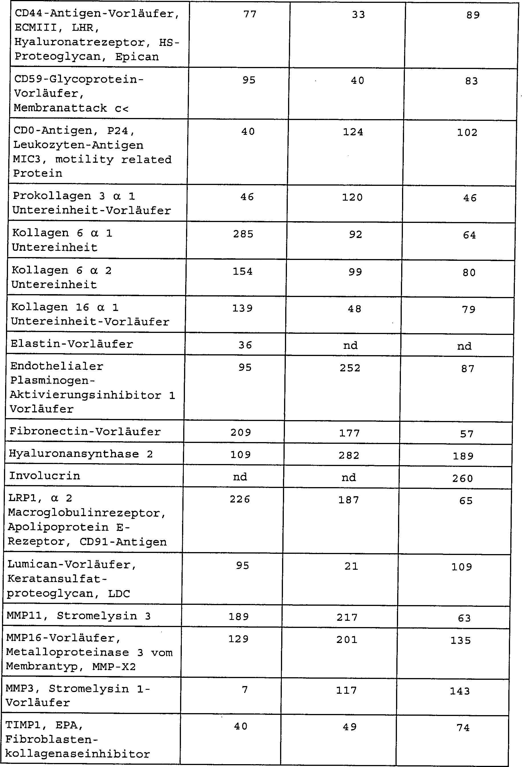


Die erfindungsgemässen Verfahren erlauben daher einen besseren Vergleich der Synthese von verschiedenen Molekülen während der Alterung der Haut und erlauben ein Screenen von Wirkstoffen, die die Veränderung während des Alterns begrenzen oder dieser vorbeugen.The inventive method therefore allow a better comparison of the synthesis of ver different molecules during aging of the skin and allow screening of drugs that limit or prevent the change during aging.
BEISPIEL 10EXAMPLE 10
Northern-Blot-Analyse von mRNAs, codierend für Fibronectin in jungen und gealterten rekonstruierten Hautmodellen:Northern blot analysis of mRNAs coding for Fibronectin in young and aged reconstructed skin models:
Die rekonstruierte Haut, die als junge und gealterte rekonstruierte Haut bezeichnet wird, wurde hergestellt durch aussäen von 400.000 Fibroblasten, die aus einem Pool von mindestens drei Spendern im Alter von weniger als 35 Jahren und mehr als 55 Jahren stammen, gewonnen und getrennt voneinander vermehrt, gemäss Protokoll, beschrieben in Beispiel 1, auf oberflächenbehandelten Kollagenmatrizes, die mit Diphenylphosphorylazid vernetzt waren. Die so hergestellte rekonstruierte Dermis wurde für 15 Tage in DMEM-Glutamax-Medium, versetzt mit 10 Kälberserum, Ascorbinsäure mit einer Endkonzentration von 1 mM, EGF mit einer Endkonzentration von 10 ng/ml, Penicillin mit einer Endkonzentration von 100 IU/ml, Amphotericin B mit einer Endkonzentration von 1 μg/ml, Gentamycin mit einer Endkonzentration von 20 μg/ml, kultiviert. Die Keratinozyten wurden in einem Verhältnis von 400.000 pro rekonstruierter Dermis ausgesät, diese stammen aus dem gleichen Pool von Spendern und wurden gewonnen und getrennt voneinander vermehrt, gemäss dem Protokoll, beschrieben in Beispiel 1. Die Kultur wurde für 1 Woche in Proliferationsmedium, hergestellt aus DMEM-Glutamax/Ham F-12 (Verhältnis 3:1 V/V), versetzt mit 10 Hyclone II-Kälberserum, Ascorbinsäure-2-phosphat mit einer Endkonzentration von 1 mM, EGF mit einer Endkonzentration von 10 ng/ml, Hydrocortison mit einer Endkonzentration von 0,4 μg/ml, Umulin mit einer Endkonzentration von 0,12 IU/ml, Isoprel mit einer Endkonzentration von 0,4 μg/ml, Triiodthyronin mit einer Endkonzentration von 2 × 10⁻⁹ M, Adenin mit einer Endkonzentration von 24,3 μg/ml, Penicillin mit einer Endkonzentration von 100 IU/ml, Amphotericin B mit einer Endkonzentration von 1 μg/ml und Gentamycin mit einer Endkonzentration von 20 μg/ml fortgesetzt. Die rekonstruierten Häute wurden dann herausgenommen und für weitere 2 Wochen in dem gleichen Medium mit Ausnahme von Kälberserum, Hydrocortison, Isuprel, Triiodthyronin und Umulin, kultiviert. Die rekonstruierten Häute wurden in Tri Reagent® (Sigma) gesammelt.Reconstructed skin, termed young and aged reconstructed skin, was obtained by sowing and propagating separately from 400,000 fibroblasts derived from a pool of at least three donors aged less than 35 years and over 55 years of age, according to the protocol described in Example 1, on surface-treated collagen matrices crosslinked with diphenylphosphoryl azide. The reconstructed dermis so prepared was incubated for 15 days in DMEM-Glutamax medium supplemented with 10% calf serum, ascorbic acid at a final concentration of 1 mM, EGF at a final concentration of 10 ng / ml, penicillin at a final concentration of 100 IU / ml, amphotericin B was cultured at a final concentration of 1 μg / ml, gentamycin at a final concentration of 20 μg / ml. The keratinocytes were seeded at a rate of 400,000 per reconstructed dermis, derived from the same pool of donors, and were harvested and propagated separately according to the protocol described in Example 1. The culture was incubated for 1 week in proliferation medium prepared from DMEM Glutamax / Ham F-12 (ratio 3: 1 v / v) supplemented with 10 Hyclone II calf serum, ascorbic acid 2-phosphate at a final concentration of 1 mM, EGF at a final concentration of 10 ng / ml, hydrocortisone with one Final concentration of 0.4 μg / ml, umulin at a final concentration of 0.12 IU / ml, isoprene at a final concentration of 0.4 μg / ml, triiodothyronine at a final concentration of 2 x 10 -9 M, adenine at a final concentration of 24.3 μg / ml, penicillin at a final concentration of 100 IU / ml, amphotericin B at a final concentration of 1 μg / ml and gentamycin to a final concentration of 20 μg / ml. The reconstituted skins were then removed and cultured for a further 2 weeks in the same medium except calf serum, hydrocortisone, isuprel, triiodothyronine and umulin. The reconstructed skins were collected in Tri Reagent ® (Sigma).
Die Gesamt-RNAs wurden gemäss dem in Beispiel 7 beschriebenen Protokoll gewonnen. Nach Quantifizierung der RNAs mittels Densitometrie bei 260 nm wurden die Lösungen auf 1 μg/μl RNA eingestellt. 3 bis 10 μg jeder Probe wurden dann auf ein Agarose/Formaldehyd-Gel zur Auftrennung gebracht. Die RNAs wurden mittels Kapillarkräften auf Hybond-N-Nylon-Membranen (Amersham) über Nacht transferiert. Die RNAs wurden an die Nylon durch Erwärmen auf 80°C für 90 Minuten kovalent gebunden. Die Membranen wurden dann für 20 Minuten bei 68°C in 8 ml ExpressHyb (Clontech) vorhybridisiert, bei 0,1 mg/ml und hybridisiert für 1 Nacht bei 68°C in Anwesenheit der markierten Proben. Am Ende des Inkubationszeitraums wurden die Membranen viermal für 30 Minuten bei 68°C mit zweimal SSC, enthaltend 1 % SDS, gewaschen und anschliessend bei 68°C mit 0,1 × SSC, enthaltend 0,5 % SDS, und schiesslich 1 mal mit 2 × SSC. Das direkte Zählen der Radioaktivität der Spots mit Hilfe eines Phosphoimager (Packard Instruments) erlaubt die Quantifizierung der RNAs. Die Ergebnisse sind ausgedrückt in Prozent Expression in bezug auf das Actinsignal. Die quantitative Analyse mittels Northern-Blot der mRNAs, codierend für Fibronectin, erlaubt das Aufzeigen der Abnahme (Faktor 2) der mRNAs in den sogenannten gealterten rekonstruierten Hautmodellen im Vergleich zu den jungen rekonstruierten Hautmodellen.The Total RNAs were determined according to obtained the protocol described in Example 7. After quantification of the RNAs by densitometry at 260 nm, the solutions were on 1 μg / μl RNA adjusted. 3 to 10 μg Each sample was then placed on an agarose / formaldehyde gel for separation brought. The RNAs were capillary-donated to Hybond N-nylon membranes (Amersham) about Night transferred. The RNAs were attached to the nylon by heating 80 ° C for 90 minutes covalently bound. The membranes were then incubated for 20 minutes at 68 ° C in 8 ml ExpressHyb (Clontech) prehybridized at 0.1 mg / ml and hybridized for 1 night at 68 ° C in the presence of the labeled samples. At the end of the incubation period the membranes were used four times for 30 minutes at 68 ° C with twice SSC containing 1% SDS, and then at 68 ° C with 0.1 × SSC, containing 0.5% SDS, and finally 1 time with 2 × SSC. Direct counting of the radioactivity of the spots with the help of a Phosphoimager (Packard Instruments) allows the Quantification of RNAs. The results are expressed in percent Expression with respect to the actin signal. The quantitative analysis Northern blotting of the mRNAs encoding fibronectin allows Demonstration of the decrease (factor 2) of the mRNAs in the so-called aged ones reconstructed skin models compared to the young reconstructed Skin models.
BEISPIEL 11EXAMPLE 11
Analyse von Typ VII-Kollagen mittels Western-Blot in jungen und gealterten rekonstruierten Hautmodellen:Analysis of type VII collagen Western blot in young and aged reconstructed skin models:
Die junge und gealterte rekonstruierte Dermis wurde hergestellt durch Aussäen von 400.000 jungen (Passage 2, 2. Amplifikation nach Trypsinierung) und alten (Passage 12, 12. Amplifikation durch Trypsinierung), extrahiert und getrennt voneinander vermehrt gemäss dem in Beispiel 1 beschriebenen Protokoll) auf Kollagen-GAG-Chitosan-Matrizes ausgesät und für 15 Tage kultiviert in DMEM-Glutamax-Medium, versetzt mit 10 % Kälberserum, Ascorbinsäure-2-phosphat mit einer Endkonzentration von 2 mM, EGF mit einer Endkonzentration von 10 ng/ml, Penicillin mit einer Endkonzentration von 100 IU/ml, Amphotericin B mit einer Endkonzentration von 1 μg/ml und Gentamycin mit einer Endkonzentration von 20 μg/ml. Die jungen Keratinozyten der Passage P1 und die alten Keratinozyten der Passage P3 wurden gewonnen und getrennt voneinander vermehrt, gemäss Protokoll, beschrieben in Beispiel 1, und wurden dann mit einer Menge von 400.000 pro rekonstruierter Dermis ausgesät. Die Kultur wurde für 1 Woche in Proliferationsmedium, hergestellt aus DMEM-Glutamax/Ham F-12 (Verhältnis 3:1 V/V), versetzt mit 10 Hyclone II-Kälberserum, Ascorbinsäure-2-phosphat mit einer Endkonzentration von 1 mM, EGF mit einer Endkonzentration von 10 ng/ml, Hydrocortison mit einer Endkonzentration von 0,4 μg/ml, Umulin mit einer Endkonzentration von 0,12 IU/ml, Isoprel mit einer Endkonzentration von 0,4 μg/ml, Triiodthyronin mit einer Endkonzentration von 2 × 10⁻⁹ M, Adenin mit einer Endkonzentration von 24,3 μg/ml, Penicillin mit einer Endkonzentration von 100 IU/ml, Amphotericin B mit einer Endkonzentration von 1 μg/ml und Gentamycin mit einer Endkonzentration von 20 μg/ml fortgesetzt. Die rekonstruierten Häute wurden dann herausgenommen und für weitere 2 Wochen in dem gleichen Medium mit Ausnahme von Kälberserum, Hydrocortison, Isuprel, Triiodthyronin und Umulin, kultiviert.The young and aged reconstructed dermis was prepared by seeding 400,000 young (passage 2, 2nd amplification after trypsinization) and old (passage 12, 12th amplification by trypsinization), extracted and separated from each other according to the protocol described in example 1) Seeded and incubated for 15 days in DMEM-glutamax medium supplemented with 10% calf serum, ascorbic acid-2-phosphate at a final concentration of 2 mM, EGF at a final concentration of 10 ng / ml, penicillin a final concentration of 100 IU / ml, amphotericin B at a final concentration of 1 μg / ml and gentamycin at a final concentration of 20 μg / ml. PAGE P1 young keratinocytes and passage P3 old keratinocytes were harvested and propagated separately according to the protocol described in Example 1, and then seeded at a rate of 400,000 per reconstructed dermis. Culture was maintained for 1 week in proliferation medium prepared from DMEM-glutamax / Ham F-12 (ratio 3: 1 v / v) supplemented with 10% Hyclone II calf serum, ascorbic acid-2-phosphate at a final concentration of 1mM, EGF with a final concentration of 10 ng / ml, hydrocortisone with an end concentration of 0.4 μg / ml, umulin at a final concentration of 0.12 IU / ml, isopril at a final concentration of 0.4 μg / ml, triiodothyronine at a final concentration of 2 x 10 -9 M, adenine at a final concentration of 24.3 μg / ml, penicillin at a final concentration of 100 IU / ml, amphotericin B at a final concentration of 1 μg / ml and gentamycin to a final concentration of 20 μg / ml. The reconstituted skins were then removed and cultured for a further 2 weeks in the same medium except calf serum, hydrocortisone, isuprel, triiodothyronine and umulin.
Typ VII-Kollagen wurde gemäss dem Protokoll beschrieben in Beispiel 8 gewonnen und in Elektrophoresepuffer (0,5 M Tris-HCl, pH 6,8, 10 % Glycerin, 2 % SDS, 5 Mercaptoethanol) gelöst. Die Proben wurden auf 95°C für 5 Minuten erwärmt. Die Grössenfraktionierung wurde auf einem 5 % SDS-PAGE durchgeführt. Dann folgte ein Transfer auf eine Polyvinylidenfluorid-Membran. Eine Kontrolle mit aufgereinigtem Kollagen VII wurde parallel zu den extrahierten Proben gemacht, genauso wie ein Standard für das Molekulargewicht.Type VII collagen was prepared according to described in Example 8 and in electrophoresis buffer (0.5 M Tris-HCl, pH 6.8, 10% glycerol, 2% SDS, 5 mercaptoethanol) solved. The samples were at 95 ° C for 5 minutes heated. The size fractionation was performed on a 5% SDS-PAGE. Then a transfer followed on a polyvinylidene fluoride membrane. A control with purified Collagen VII was made in parallel to the extracted samples, as well as a standard for the molecular weight.
Nach Transfer wurden die Membranen für 1 Stunde im Bloting-Puffer (15 nM NaCl, 20 mM Tris-HCl, 0,5 % Tween 20, 3 % BSA, pH 7,4) inkubiert. Der erste Antikollagen Typ VII-Antikörper (polyklonal, Kaninchen 1:1.000) wurde in einer 1 % Lösung PBS/BSA gegeben. nach 2 Stunden bei Raumtemperatur wurden die Membranen mit dem Blockierungspuffer gewaschen und mit dem zweiten markierten HRP-konjugierten Antikörper gegen Kaninchen-IgG für 1 Stunde bei Umgebungstemperatur inkubiert. Nach Waschen mit PBS wurden die Membranen mit einer Lösung von 3,3'-Diaminobenzidin-tetrahydrochlorid entwickelt. Im Fall einer schwachen Markierung kann ein Amplifikationskit (Biorad) verwendet werden und dann das Tracingkit Opti-4CN-Substratkit (Biorad).To Transfer were the membranes for 1 hour in blotting buffer (15 nM NaCl, 20 mM Tris-HCl, 0.5% Tween 20, 3% BSA, pH 7.4). The first anti-collagen type VII antibody (polyclonal, Rabbit 1: 1,000) was added in a 1% solution of PBS / BSA. to At room temperature for 2 hours, the membranes were blocked with the blocking buffer washed and with the second labeled HRP-conjugated antibody against Rabbit IgG for Incubated for 1 hour at ambient temperature. After washing with PBS were the membranes with a solution of 3,3'-diaminobenzidine tetrahydrochloride developed. In the case of weak labeling, an amplification kit (Biorad) and then the Tracingkit Opti 4CN substrate kit (Biorad).
Nach dem Entwickeln wurde die Membran einige Minuten mit Wasser gewaschen und dann auf einem absorbierenden Papier getrocknet.To The membrane was washed with water for a few minutes to develop and then dried on absorbent paper.
Die Analyse der Intensität der Banden durch Bildanalyse erlaubt den Nachweis einer signifikanten Abnahme des Typ VII-Kollagengehalts bei Extrakten von verschiedenen gealterten Proben.The Analysis of the intensity the bands by image analysis allowed the detection of a significant Decrease of type VII collagen content in extracts of different aged samples.
BEISPIEL 12EXAMPLE 12
Analyse von Monolayer-Keratinozyten im Vergleich zu rekonstruierter junger und gealterter Epidermis mittels DNA-Array:Analysis of monolayer keratinocytes compared to reconstructed young and aged epidermis by DNA-Array:
Dieselben Zellen in der gleichen Passage wurden zur Herstellung der drei Zellmodelle verwendet.the same Cells in the same passage were used to produce the three cell models used.
Keratinozyten (Pool von drei Spendern in einem Alter von weniger als 35 Jahren und in einem Alter von mehr als 55 Jahren) wurden wie in Beispiel 1 beschrieben gewonnen und wurden in Monolayer zur Konfluenz kultiviert in K-SFM-Medium (Gibco), Penicillin mit einer Endkonzentration von 100 IU/ml, Gentamycin mit einer Endkonzentration von 20 μg/ml, Amphotericin B mit einer Endkonzentration von 1 μg/ml. Diese wurden dann in Tri Reagent® (Sigma) gesammelt.Keratinocytes (pool of three donors less than 35 years of age and over 55 years of age) were obtained as described in Example 1 and cultured in monolayer to confluency in K-SFM medium (Gibco), penicillin a final concentration of 100 IU / ml, gentamycin with a final concentration of 20 μg / ml, amphotericin B with a final concentration of 1 μg / ml. These were then collected in Tri Reagent ® (Sigma).
Rekonstruierte Epidermis wurde hergestellt mit einer Menge von 4 × 10⁶ Keratinozyten (Pool von drei Spendern mit einem Alter von weniger als 35 Jahren und einem Alter von mehr als 55 Jahren) vermehrt wie in Beispiel 1 beschrieben bis zur Passage 1 (1. Amplifikation durch Trypsinierung) und wurden ausgesät in Boyden-Kammereinsätzen (Membranporosität: 0,4 μm, Durchmesser: 25 mm), diese wurden vorher mit einer Nährunterschicht aus Fibroblasten ausgesät, in DMEM-Glutamax/Ham F-12 (Verhältnis 3:1 V/V) Kulturmedium, versetzt mit 10 % Hyclone II-Kälberserum, Ascorbinsäure-2-phosphat mit einer Endkonzentration von 1 mM, EGF (epidermaler Wachstumsfaktor) mit einer Endkonzentration von 10 ng/ml, Hydrocortison mit einer Endkonzentration von 0,4 μg/ml, Umulin mit einer Endkonzentration von 0,12 IU/ml, Isuprel mit einer Endkonzentration von 0,4 μg/ml, Triiodthyronin mit einer Endkonzentration von 2 × 10⁻⁹ M, Adenin mit einer Endkonzentration von 24,3 μg/ml, Penicillin mit einer Endkonzentration von 100 IU/ml, Amphotericin B mit einer Endkonzentration von 1 μg/ml und Gentamycin mit einer Endkonzentration von 20 μg/ml, und für einen Zeitraum von 3 bis 8 Tagen in Immersionskultur.Reconstructed epidermis was prepared with an amount of 4 x 10 6 keratinocytes (pool of three donors less than 35 years of age and over 55 years of age) propagated as described in Example 1, up to passage 1 (1st amplification) Trypsinization) and seeded in Boyden chamber inserts (membrane porosity: 0.4 μm, diameter: 25 mm) previously seeded with a nutrient sublayer of fibroblasts in DMEM-glutamax / Ham F-12 (ratio 3: 1 v / v ) Culture medium supplemented with 10% Hyclone II calf serum, ascorbic acid 2-phosphate at a final concentration of 1 mM, EGF (epidermal growth factor) at a final concentration of 10 ng / ml, hydrocortisone at a final concentration of 0.4 μg / ml, Umulin at a final concentration of 0.12 IU / ml, isuprel at a final concentration of 0.4 μg / ml, triiodothyronine at a final concentration of 2 x 10 -9 M, adenine at a final concentration of 24.3 μg / ml, penicillin with an en concentration of 100 IU / ml, amphotericin B at a final concentration of 1 μg / ml and gentamycin at a final concentration of 20 μg / ml, and for a period of 3 to 8 days in immersion culture.
Die Keratinozytenkulturen wurden dann an die Luft-Flüssigkeits-Grenze verbracht für 12 bis 18 Tage im gleichen Kulturmedium wie für die Immersionskultur, mit Ausnahme von Kälberserum, Hydrocortison, Isuprel, Triiodthyronin und Umulin.The Keratinocyte cultures were then added to the air-liquid boundary spent for 12 to 18 days in the same culture medium as for the immersion culture, with Exception of calf serum, Hydrocortisone, isuprel, triiodothyronine and umulin.
Am Ende des Experiments wurden die Keratinozyten-Monolayer und die rekonstruierte Epidermis in den Einsätzen abgekratzt, gesammelt und in Tri Reagent® (Sigma) aufgenommen und mit Chloroform extrahiert. Nach Zentrifugation bei 12.000 × g für 15 Minuten bei 4°C befanden sich die RNAs in der oberen Schicht, die DNAs in der unteren Schicht und die Proteine im Grenzbereich.At the end of the experiment the keratinocyte monolayer, and the reconstructed epidermis were scraped in the inserts, collected and added to Tri Reagent ® (Sigma) and extracting with chloroform hiert. After centrifugation at 12,000 × g for 15 minutes at 4 ° C, the RNAs were in the upper layer, the DNAs in the lower layer and the proteins in the border region.
Die
cDNA-Arrays wurden wie in Beispiel 9 beschrieben hergestellt. Die
erhaltenen Ergebnisse sind die folgenden (RE: relative Expressionseinheit: TABELLE
2 

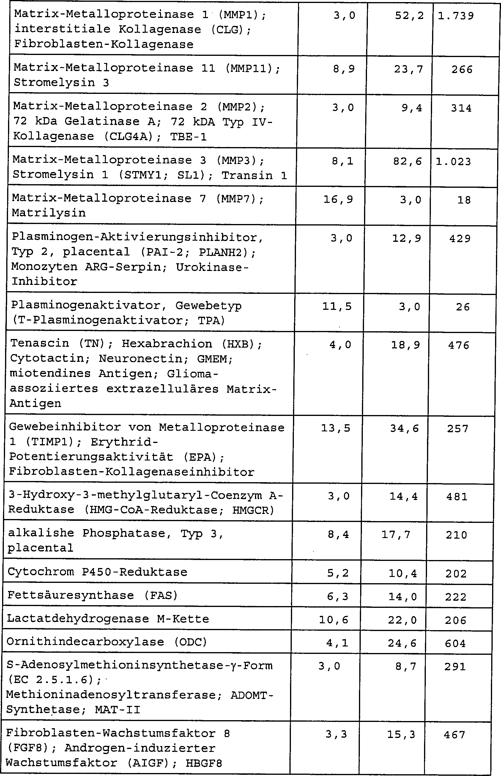

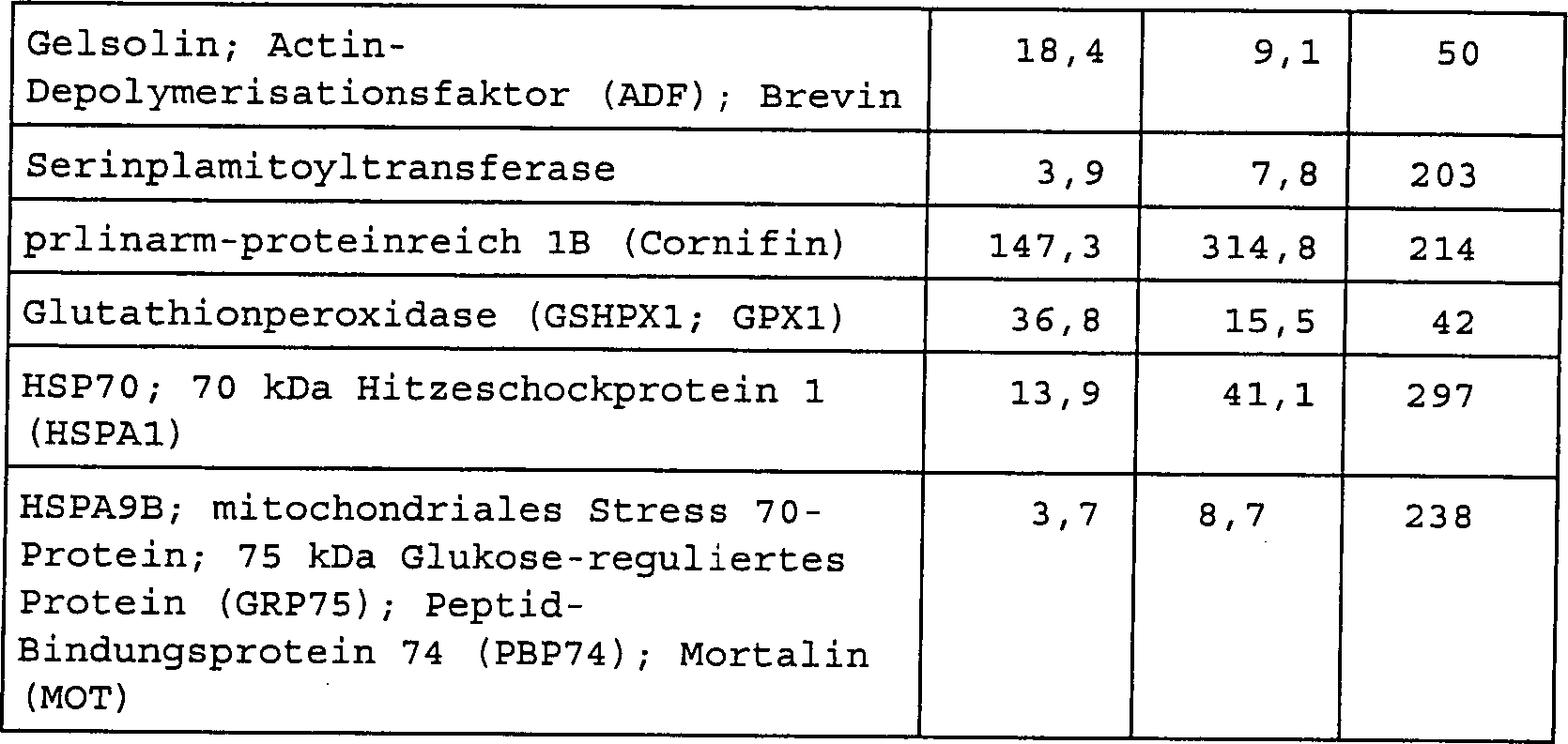
Die erfindungsgemässen Verfahren erlauben somit ein besseres Verständnis zur Synthese der verschiedenen Moleküle während der epidermalen Alterung und erlauben somit das Screenen von Wirkstoffen, die Veränderungen, beobachtet bei der Alterung der Epidermis, entgegenwirken oder diesen vorbeugen.The invention Processes thus allow a better understanding of the synthesis of the various molecules while Epidermal aging and thus allow the screening of drugs that changes, observed in the aging of the epidermis, counteract or this prevent.
BEISPIEL 13EXAMPLE 13
Test zur Wirksamkeit eines kosmetischen Wirkstoffs, der topisch auf junge und gealterte rekonstruierte Haut aufgetragen wird:Test for the effectiveness of a cosmetic active ingredient, which was reconstituted topically on young and aged Skin is applied:
600.000 junge Fibroblasten (gewonnen aus Brustbiopsie) und gealterte Fibroblasten (gewonnen aus einer Gesichtsbiopsie, erhalten nach Gesichtslifting), wurden bis zur Passage 6 vermehrt wie in Beispiel 1 beschrieben und dann auf einem dermalen Substrat auf Basis von Kollagen-Glycosaminglycan-Chitosan ausgesät, in einem DMEM-Glutamax-Kulturmedium, versetzt mit 10 % Hyclone-Kälberserum, Ascorbinsäure-2-phosphat mit einer Endkonzentration von 1 mM, EGF (epidermaler Wachstumsfaktor) mit einer Endkonzentration von 10 ng/ml, Penicillin mit einer Endkonzentration von 100 IU/ml, Amphotericin B mit einer Endkonzentration von 1 μg/ml und Gentamycin mit einer Endkonzentration von 20 μg/ml, für einen Zeitraum von 21 Tagen.600000 young fibroblasts (obtained from breast biopsy) and aged fibroblasts (obtained from a facial biopsy, obtained after facial lifting), were propagated to passage 6 as described in Example 1 and then on a dermal substrate based on collagen-glycosaminoglycan chitosan seeded, in a DMEM glutamax culture medium supplemented with 10% Hyclone calf serum, ascorbic acid 2-phosphate with a final concentration of 1 mM, EGF (epidermal growth factor) at a final concentration of 10 ng / ml, penicillin at a final concentration of 100 IU / ml, amphotericin B at a final concentration of 1 μg / ml and Gentamycin at a final concentration of 20 μg / ml for a period of 21 days.
Anschliessend wurden 600.000 Keratinozyten von jungen Keratinozyten (gewonnen aus Brustbiopsie) und gealterten Keratinozyten (gewonnen aus Gesichtsbiopsie nach Gesichtslifting) wie in Beispiel 1 vermehrt und dann in Passage 1 auf dermale Äquivalente ausgesät in DMEM-Glutamax/Ham F-12-Kulturproliferationsmedium (Verhältnis 3:1 V/V), versetzt mit 10 % Hyclone II-Kälberserum, Ascorbinsäure-2-phosphat mit einer Endkonzentration von 1 mM/l, EGF mit einer Endkonzentration von 10 ng/ml, Hydrocortison mit einer Endkonzentration von 0,4 μg/ml, Umulin mit einer Endkonzentration von 0,12 IU/ml, Isuprel mit einer Endkonzentration von 0,4 μg/ml, Triiodthyronin mit einer Endkonzentration von 2 × 10⁻⁹ M, Adenin mit einer Endkonzentration von 24,3 μg/ml, Penicillin mit einer Endkonzentration von 100 IU/ml, Amphotericin B mit einer Endkonzentration von 1 μg/ml und Gentamycin mit einer Endkonzentration von 20 μg/ml für einen Zeitraum von 5 Tagen in Immersionskultur.Subsequently, 600,000 keratinocytes from young keratinocytes (obtained from breast biopsy) and aged keratinocytes (obtained from facial biopsy after face lifting) were propagated as in Example 1 and then seeded in passage 1 to dermal equivalents in DMEM-glutamax / Ham F-12 culture proliferation medium (ratio 3 1 V / V) supplemented with 10% Hyclone II calf serum, ascorbic acid 2-phosphate at a final concentration of 1 mM / L, EGF at a final concentration of 10 ng / ml, hydrocortisone at a final concentration of 0.4 μg / ml. ml, umulin at a final concentration of 0.12 IU / ml, isuprel at a final concentration of 0.4 μg / ml, triiodothyronine at a final concentration of 2 x 10 -9 M, adenine at a final concentration of 24.3 μg / ml, Penicillin at a final concentration of 100 IU / ml, amphotericin B at a final concentration of 1 μg / ml and gentamycin at a final concentration of 20 μg / ml for a period of 5 days in immersion culture.
Die Kulturen wurden dann an die Luft-Flüssigkeits-Grenze verbracht für 14 Tage in einem Differenzierungsmedium, bestehend aus dem für die Immersionskultur verwendeten Kulturmedium, mit Ausnahme von Kälberserum, Hydrocortison, Isuprel, Triiodthryonin und Umulin.The Cultures were then moved to the air-liquid boundary for 14 Days in a differentiation medium, consisting of that for the immersion culture used culture medium, with the exception of calf serum, hydrocortisone, isuprel, Triiodothryonine and umulin.
8 μl eines Placebos einer kosmetischen Formulierung und einer kosmetischen Formulierung, enthaltend 3 % Wirkstoff, wurden dann auf die als junge und gealterte rekonstruierte Haut bezeichnete rekonstruierte Haut aufgebracht. Nach 2 Tagen Kultur im Differenzierungsmedium wurden die kosmetischen Formulierungen sehr vorsichtig von den rekonstruierten Häuten entfernt und durch die gleiche Menge an frischer Formulierung ersetzt. Dieses wurde 7 mal wiederholt, d.h. für 14 weitere Tage Kultur. Am Ende des Experiments wurden die kosmetischen Formulierungen sehr vorsichtig von der rekonstruierten Haut entfernt und die rekonstruierte Haut mit PBS gewaschen und dann in Tri Reagent® (Sigma) gegeben. Die Wirksamkeit der Behandlung mit dem kosmetischen Wirkstoff in der Formulierung wurde mittels cDNA-Arrayverfahren gemäss Protokoll, beschrieben in Beispiel 9, untersucht.8 μl of a placebo of a cosmetic formulation and a cosmetic formulation containing 3% of active ingredient were then applied to the reconstructed skin, termed young and aged reconstructed skin. After 2 days of culture in the differentiation medium, the cosmetic formulations were very carefully removed from the reconstituted skins and replaced with the same amount of fresh formulation. This was repeated 7 times, ie culture for 14 more days. At the end of the experiment, the cosmetic formulations were very carefully removed from the reconstructed skin and washed the reconstructed skin with PBS and then placed in Tri Reagent ® (Sigma). The effectiveness of the treatment with the cosmetic active ingredient in the formulation was investigated by cDNA array method according to the protocol described in Example 9.
BEISPIEL 14EXAMPLE 14
Test der Wirksamkeit eines kosmetischen Wirkstoffs, der systemisch in jungen und gealterten rekonstruierten Dermismodellen aufgetragen wurde:Test the effectiveness of a cosmetic active ingredient that is systemic in young and aged reconstructed dermis models was applied:
In Beispiel 12 wurde gezeigt, dass der Fibronectingehalt in Modellen von gealterten Zellen im Vergleich zu Modellen mit jungen Zellen erniedrigt war (Abnahme von 43 %).In Example 12 has been shown to increase the fibronectin content in models of aged cells compared to models with young cells was decreased (decrease of 43%).
Rekonstruierte Dermis wurde wie in Beispiel 9 beschrieben hergestellt. Deliner® (Maisextrakt, Coletica, Lyon) wurde in 2 % in Kulturmedium verwendet, dieses wurde jeden zweiten Tag ausgetauscht. Die Kulturmedien wurden gesammelt und der Fibronectingehalt wurde mittels Dot-Blot wie in Beispiel 8 beschrieben quantifiziert, gefolgt von einer Bildanalyse. Die Zunahme des Fibronectingehalts ist 25 %. Deliner® ist daher tatsächlich in der Lage, die Abnahme der Synthese von Fibronectin aufgrund der Alterung zu verhindern.Reconstructed dermis was prepared as described in Example 9. Deliner ® (corn extract, Coletica, Lyon) was used in 2% in the culture medium, this was changed every other day. The culture media were collected and the fibronectin content quantitated by dot-blot as described in Example 8, followed by image analysis. The increase in fibronectin content is 25%. Deliner ® is therefore actually prevent able to decrease the synthesis of fibronectin due to aging.
Es folgt ein Sequenzprotokoll nach WIPO St. 25.It follows a sequence listing according to WIPO St. 25. Dieses kann von der amtlichen Veröffentlichungsplattform des DPMA heruntergeladen werden.This can from the official publication platform downloaded from the DPMA.
Claims (24)
Applications Claiming Priority (2)
| Application Number | Priority Date | Filing Date | Title |
|---|---|---|---|
| FR0214492 | 2002-11-19 | ||
| FR0214492A FR2847269B1 (en) | 2002-11-19 | 2002-11-19 | METHOD FOR IDENTIFYING AN EVENTUAL MODIFICATION OF AT LEAST ONE BIOLOGICAL PARAMETER USING YOUNG AND AGE LIVING CELLS |
Publications (2)
| Publication Number | Publication Date |
|---|---|
| DE10320602A1 DE10320602A1 (en) | 2004-06-09 |
| DE10320602B4 true DE10320602B4 (en) | 2006-05-04 |
Family
ID=8871572
Family Applications (1)
| Application Number | Title | Priority Date | Filing Date |
|---|---|---|---|
| DE10320602A Expired - Fee Related DE10320602B4 (en) | 2002-11-19 | 2003-05-08 | A method for identifying a possible modification of at least one biological parameter using young and old living cells |
Country Status (9)
| Country | Link |
|---|---|
| US (1) | US20040096816A1 (en) |
| JP (2) | JP2004166685A (en) |
| KR (1) | KR100752986B1 (en) |
| CH (1) | CH694097A5 (en) |
| DE (1) | DE10320602B4 (en) |
| ES (1) | ES2255346A1 (en) |
| FR (1) | FR2847269B1 (en) |
| GB (1) | GB2395489B (en) |
| NL (1) | NL1022659C2 (en) |
Families Citing this family (13)
| Publication number | Priority date | Publication date | Assignee | Title |
|---|---|---|---|---|
| DE102004061289B4 (en) * | 2004-12-20 | 2009-04-02 | Euroderm Gmbh | Test kit with test cells and immune cells and manufacturing method therefor |
| WO2006093104A1 (en) * | 2005-03-03 | 2006-09-08 | Sapporo Breweries Limited | Method of controlling the flavor of alcoholic drink |
| EP1736780A1 (en) | 2005-06-24 | 2006-12-27 | Eppendorf Array Technologies S.A. | Method and means for detecting and/or quantifying hierarchical molecular change of a cell in response to an external stimulus |
| CN100372571C (en) * | 2005-10-26 | 2008-03-05 | 武汉共同科技有限公司 | Chitosan-immune RNA composite formulation and preparation method thereof |
| DE102006006461A1 (en) * | 2006-02-10 | 2007-08-23 | Phenion Gmbh & Co. Kg | Skin model with dendritic cells |
| CN100398647C (en) * | 2006-03-02 | 2008-07-02 | 上海交通大学 | A method for rapid extraction of large-fragment sponge metagenomic DNA |
| FR2915382B1 (en) * | 2007-04-27 | 2012-06-01 | Vincience | USE OF AN ACTIVE INGREDIENT FROM MAIZE (ZEA MAYS L) FOR PREPARING A COMPOSITION INTENDED TO ACTIVATE CYTOCHROME C |
| FR2932087B1 (en) * | 2008-06-10 | 2012-10-12 | Oreal | COSMETIC USE OF DESMOPLAKINE-LIKE PROTEINS FOR THE TREATMENT OF SKIN DROUGHT |
| JP2012205516A (en) | 2011-03-29 | 2012-10-25 | Osaka Univ | Method for producing artificial skin model, and artificial skin model |
| RU2466680C1 (en) | 2011-10-03 | 2012-11-20 | Общество С Ограниченной Ответственностью "Витацел" | Diagnostic technique for patient's skin conditions |
| US20130338932A1 (en) * | 2012-06-13 | 2013-12-19 | Agilent Technologies, Inc. | Computational method for mapping peptides to proteins using sequencing data |
| WO2014038599A1 (en) * | 2012-09-04 | 2014-03-13 | 国立大学法人大阪大学 | Artificial skin tissue, artificial skin model and manufacturing method therefor |
| EP3749750A1 (en) * | 2018-02-09 | 2020-12-16 | Helmholtz Zentrum München Deutsches Forschungszentrum für Gesundheit und Umwelt GmbH | Means and methods for monitoring scar development |
Citations (2)
| Publication number | Priority date | Publication date | Assignee | Title |
|---|---|---|---|---|
| DE10100122A1 (en) * | 2001-01-03 | 2002-07-11 | Henkel Kgaa | Method for determining skin aging in vitro |
| EP1064353B1 (en) * | 1998-03-18 | 2002-11-06 | Massachusetts Institute Of Technology | Vascularized perfused microtissue/micro-organ arrays |
Family Cites Families (17)
| Publication number | Priority date | Publication date | Assignee | Title |
|---|---|---|---|---|
| US4458678A (en) * | 1981-10-26 | 1984-07-10 | Massachusetts Institute Of Technology | Cell-seeding procedures involving fibrous lattices |
| US4673649A (en) * | 1983-07-15 | 1987-06-16 | University Patents, Inc. | Process and defined medium for growth of human epidermal keratinocyte cells |
| US4604346A (en) * | 1984-10-09 | 1986-08-05 | Massachusetts Institute Of Technology | Skin-equivalent prepared by the use of punch biopsy |
| DE3851203T2 (en) * | 1987-05-11 | 1994-12-15 | Procyte Corp | Use of GHL-Cu derivatives in the manufacture of a medicament to stimulate hair growth. |
| FR2616318A1 (en) * | 1987-06-15 | 1988-12-16 | Centre Nat Rech Scient | ARTIFICIAL SKIN AND PROCESS FOR PREPARING THE SAME |
| US5744300A (en) * | 1993-03-24 | 1998-04-28 | Geron Corporation | Methods and reagents for the identification and regulation of senescence-related genes |
| US5627025A (en) * | 1994-08-12 | 1997-05-06 | The Rockefeller University | Method for the identification of compounds capable of abrogating human immunodeficiency virus (HIV) infection of dendritic cells and T-lymphocytes |
| FR2743817B1 (en) * | 1996-01-23 | 1998-03-13 | Oreal | SKIN EQUIVALENT COMPRISING LANGERHAN CELLS |
| JP2002515898A (en) * | 1997-02-25 | 2002-05-28 | ザ リージェンツ オブ ザ ユニバーシティ オブ ミシガン | Methods and compositions for preventing and treating aging in human skin |
| US20010048917A1 (en) * | 1998-03-09 | 2001-12-06 | Warren Hoeffler | Skin equivalent and methods of forming and using same |
| US5989837A (en) * | 1998-07-13 | 1999-11-23 | Wisconsin Alumni Research Foundation | Immortalized human keratinocyte cell line |
| FR2792650B1 (en) * | 1999-04-20 | 2003-02-28 | Oreal | EQUIVALENT OF ELDERLY SKIN, ITS PREPARATION METHOD AND ITS USE |
| WO2001012851A2 (en) * | 1999-08-12 | 2001-02-22 | Wisconsin Alumni Research Foundation | Identification of genetic markers of biological age and metabolism |
| KR100747674B1 (en) | 2000-02-21 | 2007-08-08 | 소니 가부시끼 가이샤 | Hologram recording medium, hologram recording device and method |
| FR2809313B1 (en) * | 2000-05-26 | 2005-11-18 | Coletica | PROCESSES FOR THE PREPARATION OF NEW COLLAGEN-BASED MEDIA FOR TISSUE ENGINEERING AND BIOMATERIALS OBTAINED |
| FR2820313A1 (en) * | 2001-02-07 | 2002-08-09 | Oreal | USE OF AT LEAST ONE LEGUMINOUS EXTRACT AS ANTI-POLLUTION AGENT, HEAVY METAL TRAP |
| EP1402068A4 (en) * | 2001-05-09 | 2006-07-12 | Univ Cleveland Hospitals | ASSESSMENT OF SKIN INJURY CAUSED BY ULTRAVIOLETTE RADIATION BY MEANS OF NEW GENETIC MARKERS, METHODS AND COMPOSITIONS THEREFOR |
-
2002
- 2002-11-19 FR FR0214492A patent/FR2847269B1/en not_active Expired - Lifetime
-
2003
- 2003-02-12 US US10/365,894 patent/US20040096816A1/en not_active Abandoned
- 2003-02-12 GB GB0303215A patent/GB2395489B/en not_active Expired - Fee Related
- 2003-02-12 NL NL1022659A patent/NL1022659C2/en not_active IP Right Cessation
- 2003-02-20 CH CH00267/03A patent/CH694097A5/en not_active IP Right Cessation
- 2003-03-31 KR KR1020030020226A patent/KR100752986B1/en not_active Expired - Fee Related
- 2003-04-09 JP JP2003105029A patent/JP2004166685A/en active Pending
- 2003-05-08 DE DE10320602A patent/DE10320602B4/en not_active Expired - Fee Related
- 2003-05-16 ES ES200301142A patent/ES2255346A1/en active Pending
-
2007
- 2007-10-02 JP JP2007258522A patent/JP2008022863A/en active Pending
Patent Citations (2)
| Publication number | Priority date | Publication date | Assignee | Title |
|---|---|---|---|---|
| EP1064353B1 (en) * | 1998-03-18 | 2002-11-06 | Massachusetts Institute Of Technology | Vascularized perfused microtissue/micro-organ arrays |
| DE10100122A1 (en) * | 2001-01-03 | 2002-07-11 | Henkel Kgaa | Method for determining skin aging in vitro |
Non-Patent Citations (2)
| Title |
|---|
| BOXMAN, I.L. u.a.: Proteomic analysis of skin irritation reveals the induction of HSP27 by sodium lauryl sulphate in human skin. Br. J. Dermatol. (Mai 2002) 146 (5) 777-85 * |
| FLETCHER, S.T. u.a.: Gene expression analysis of EpiDerm following exposure to SLS using cDNA microarrays. Toxicol In Vitro. (2001) 15 (4-5) 393-8 * |
Also Published As
| Publication number | Publication date |
|---|---|
| GB2395489A (en) | 2004-05-26 |
| JP2008022863A (en) | 2008-02-07 |
| GB2395489B (en) | 2005-06-08 |
| NL1022659C2 (en) | 2004-06-03 |
| FR2847269B1 (en) | 2006-07-28 |
| KR100752986B1 (en) | 2007-08-30 |
| KR20040044077A (en) | 2004-05-27 |
| ES2255346A1 (en) | 2006-06-16 |
| GB0303215D0 (en) | 2003-03-19 |
| DE10320602A1 (en) | 2004-06-09 |
| JP2004166685A (en) | 2004-06-17 |
| US20040096816A1 (en) | 2004-05-20 |
| FR2847269A1 (en) | 2004-05-21 |
| CH694097A5 (en) | 2004-07-15 |
Similar Documents
| Publication | Publication Date | Title |
|---|---|---|
| DE10320602B4 (en) | A method for identifying a possible modification of at least one biological parameter using young and old living cells | |
| US7553664B2 (en) | Three-dimensional skin model | |
| DE60036047T2 (en) | Aged skin equivalent, its manufacturing processes and uses | |
| US20110008273A1 (en) | Methods of skin whitening and of screening skin-spot-formation inhibiting and/or skin-spot removing factor | |
| WO2021074126A1 (en) | Production of skeletal muscle cells and skeletal muscle tissue from pluripotent stem cells | |
| DE10320633B4 (en) | A method for identifying a possible modification of at least one biological parameter using live cells that have been exposed to a stimulus and living cells that have not been exposed to this stimulus | |
| US20190242880A1 (en) | Method for evaluating the effects of dehydration on children's skin | |
| FR2990106A1 (en) | SYSTEM FOR SURVIVAL MAINTENANCE AND TRANSPORT OF SKIN BIOSPSIES AND ITS APPLICATIONS | |
| DE69011638T2 (en) | Method for determining the effect of a substance on hair growth. | |
| Tomasso et al. | Marvels of spiny mouse regeneration: cellular players and their interactions in restoring tissue architecture in mammals | |
| EP1976979B1 (en) | Method for producing autonomously contracting cardiac muscle cells from adult stem cells, in particular human adult stem cells | |
| EP2743342B1 (en) | Aged skin model | |
| WO2001092476A2 (en) | In vitro tissue test system | |
| DE102013015560B4 (en) | Ex vivo cultures based on human sebaceous glands and their use as a screening tool | |
| DE10260931A1 (en) | Procedure for determining the homeostasis of hairy skin | |
| DE102004061289B4 (en) | Test kit with test cells and immune cells and manufacturing method therefor | |
| JP2012183061A (en) | Method of screening or evaluating skin barrier function adjuster | |
| WO2008028779A1 (en) | Method for the molecular characterization of old-age hair | |
| JP4395221B2 (en) | Screening method for substances effective in the treatment of pigmented disorders | |
| DE10062623A1 (en) | Cartilage replacement, useful particularly for repairing joint defects, comprises differentiated and proliferating chondrocytes in collagen-based biomatrix | |
| WO2008074595A1 (en) | Method for the molecular characterization of graying and pigmented hair | |
| Bolzer et al. | A SEM analysis of DMSO treated hydra polyps | |
| WO2025100384A1 (en) | Three-dimensional skin model | |
| US20170152556A1 (en) | Methods for identifying circadian rhythm-dependent cosmetic agents for skin care compositions | |
| EP1479391A1 (en) | Ex vivo cultured vital slices of mammalian heart tissue, methods for their production and culture as well as uses thereof |
Legal Events
| Date | Code | Title | Description |
|---|---|---|---|
| OP8 | Request for examination as to paragraph 44 patent law | ||
| 8327 | Change in the person/name/address of the patent owner |
Owner name: ENGELHARD LYON, LYON, FR |
|
| 8364 | No opposition during term of opposition | ||
| R119 | Application deemed withdrawn, or ip right lapsed, due to non-payment of renewal fee |