CN1300578C - Analyzing instrument - Google Patents
Analyzing instrument Download PDFInfo
- Publication number
- CN1300578C CN1300578C CNB028147049A CN02814704A CN1300578C CN 1300578 C CN1300578 C CN 1300578C CN B028147049 A CNB028147049 A CN B028147049A CN 02814704 A CN02814704 A CN 02814704A CN 1300578 C CN1300578 C CN 1300578C
- Authority
- CN
- China
- Prior art keywords
- nappe
- kapillary
- substrate
- vinylon
- blood
- Prior art date
- Legal status (The legal status is an assumption and is not a legal conclusion. Google has not performed a legal analysis and makes no representation as to the accuracy of the status listed.)
- Expired - Lifetime
Links
- 239000007788 liquid Substances 0.000 claims abstract description 65
- 229920002978 Vinylon Polymers 0.000 claims abstract description 54
- 239000000758 substrate Substances 0.000 claims description 71
- 238000012360 testing method Methods 0.000 claims description 22
- 238000009736 wetting Methods 0.000 claims description 19
- 239000000463 material Substances 0.000 claims description 8
- 238000004891 communication Methods 0.000 claims description 6
- 238000002955 isolation Methods 0.000 claims description 4
- 238000007791 dehumidification Methods 0.000 abstract description 20
- 238000011176 pooling Methods 0.000 abstract 1
- 210000004369 blood Anatomy 0.000 description 68
- 239000008280 blood Substances 0.000 description 68
- 239000003153 chemical reaction reagent Substances 0.000 description 29
- 230000000694 effects Effects 0.000 description 19
- WQZGKKKJIJFFOK-GASJEMHNSA-N Glucose Natural products OC[C@H]1OC(O)[C@H](O)[C@@H](O)[C@@H]1O WQZGKKKJIJFFOK-GASJEMHNSA-N 0.000 description 15
- 230000015572 biosynthetic process Effects 0.000 description 15
- 238000005755 formation reaction Methods 0.000 description 15
- 239000008103 glucose Substances 0.000 description 15
- 239000000126 substance Substances 0.000 description 11
- 239000004094 surface-active agent Substances 0.000 description 11
- 238000000034 method Methods 0.000 description 9
- 108090000790 Enzymes Proteins 0.000 description 7
- 102000004190 Enzymes Human genes 0.000 description 7
- 229940088598 enzyme Drugs 0.000 description 7
- 230000008676 import Effects 0.000 description 7
- 230000003647 oxidation Effects 0.000 description 7
- 238000007254 oxidation reaction Methods 0.000 description 7
- 238000009826 distribution Methods 0.000 description 6
- 230000009467 reduction Effects 0.000 description 6
- IIZPXYDJLKNOIY-JXPKJXOSSA-N 1-palmitoyl-2-arachidonoyl-sn-glycero-3-phosphocholine Chemical compound CCCCCCCCCCCCCCCC(=O)OC[C@H](COP([O-])(=O)OCC[N+](C)(C)C)OC(=O)CCC\C=C/C\C=C/C\C=C/C\C=C/CCCCC IIZPXYDJLKNOIY-JXPKJXOSSA-N 0.000 description 5
- 238000010521 absorption reaction Methods 0.000 description 5
- 230000006837 decompression Effects 0.000 description 5
- 238000009434 installation Methods 0.000 description 5
- 239000000787 lecithin Substances 0.000 description 5
- 229940067606 lecithin Drugs 0.000 description 5
- 235000010445 lecithin Nutrition 0.000 description 5
- 230000008569 process Effects 0.000 description 5
- 230000004044 response Effects 0.000 description 5
- 239000012086 standard solution Substances 0.000 description 5
- 230000009471 action Effects 0.000 description 4
- 239000000853 adhesive Substances 0.000 description 4
- 230000001070 adhesive effect Effects 0.000 description 4
- 239000011248 coating agent Substances 0.000 description 4
- 238000000576 coating method Methods 0.000 description 4
- 230000002209 hydrophobic effect Effects 0.000 description 4
- 108010015776 Glucose oxidase Proteins 0.000 description 3
- 239000004366 Glucose oxidase Substances 0.000 description 3
- 238000006243 chemical reaction Methods 0.000 description 3
- 238000010586 diagram Methods 0.000 description 3
- 238000011156 evaluation Methods 0.000 description 3
- 239000012530 fluid Substances 0.000 description 3
- 229940116332 glucose oxidase Drugs 0.000 description 3
- 235000019420 glucose oxidase Nutrition 0.000 description 3
- 238000010438 heat treatment Methods 0.000 description 3
- 239000007787 solid Substances 0.000 description 3
- 239000012298 atmosphere Substances 0.000 description 2
- 210000000601 blood cell Anatomy 0.000 description 2
- HVYWMOMLDIMFJA-DPAQBDIFSA-N cholesterol Chemical compound C1C=C2C[C@@H](O)CC[C@]2(C)[C@@H]2[C@@H]1[C@@H]1CC[C@H]([C@H](C)CCCC(C)C)[C@@]1(C)CC2 HVYWMOMLDIMFJA-DPAQBDIFSA-N 0.000 description 2
- 230000008878 coupling Effects 0.000 description 2
- 238000010168 coupling process Methods 0.000 description 2
- 238000005859 coupling reaction Methods 0.000 description 2
- 230000003292 diminished effect Effects 0.000 description 2
- 238000006073 displacement reaction Methods 0.000 description 2
- 230000004438 eyesight Effects 0.000 description 2
- 230000004927 fusion Effects 0.000 description 2
- 239000000499 gel Substances 0.000 description 2
- JVTAAEKCZFNVCJ-UHFFFAOYSA-N lactic acid Chemical compound CC(O)C(O)=O JVTAAEKCZFNVCJ-UHFFFAOYSA-N 0.000 description 2
- 239000007791 liquid phase Substances 0.000 description 2
- 238000003825 pressing Methods 0.000 description 2
- 238000007127 saponification reaction Methods 0.000 description 2
- 238000000926 separation method Methods 0.000 description 2
- 238000003860 storage Methods 0.000 description 2
- 229920005992 thermoplastic resin Polymers 0.000 description 2
- 239000004925 Acrylic resin Substances 0.000 description 1
- RGHNJXZEOKUKBD-UHFFFAOYSA-N D-gluconic acid Natural products OCC(O)C(O)C(O)C(O)C(O)=O RGHNJXZEOKUKBD-UHFFFAOYSA-N 0.000 description 1
- RGHNJXZEOKUKBD-SQOUGZDYSA-N Gluconic acid Natural products OC[C@@H](O)[C@@H](O)[C@H](O)[C@@H](O)C(O)=O RGHNJXZEOKUKBD-SQOUGZDYSA-N 0.000 description 1
- FAPWRFPIFSIZLT-UHFFFAOYSA-M Sodium chloride Chemical compound [Na+].[Cl-] FAPWRFPIFSIZLT-UHFFFAOYSA-M 0.000 description 1
- 230000003321 amplification Effects 0.000 description 1
- 238000005452 bending Methods 0.000 description 1
- 230000000740 bleeding effect Effects 0.000 description 1
- 230000017531 blood circulation Effects 0.000 description 1
- 230000036770 blood supply Effects 0.000 description 1
- 210000001124 body fluid Anatomy 0.000 description 1
- 239000010839 body fluid Substances 0.000 description 1
- 230000008859 change Effects 0.000 description 1
- 235000012000 cholesterol Nutrition 0.000 description 1
- 238000012790 confirmation Methods 0.000 description 1
- 238000007796 conventional method Methods 0.000 description 1
- 206010012601 diabetes mellitus Diseases 0.000 description 1
- 238000001035 drying Methods 0.000 description 1
- 238000005516 engineering process Methods 0.000 description 1
- 238000006911 enzymatic reaction Methods 0.000 description 1
- 239000000835 fiber Substances 0.000 description 1
- 239000011521 glass Substances 0.000 description 1
- 239000000174 gluconic acid Substances 0.000 description 1
- 235000012208 gluconic acid Nutrition 0.000 description 1
- 239000000017 hydrogel Substances 0.000 description 1
- 239000011810 insulating material Substances 0.000 description 1
- 238000009413 insulation Methods 0.000 description 1
- 239000004310 lactic acid Substances 0.000 description 1
- 235000014655 lactic acid Nutrition 0.000 description 1
- 238000003475 lamination Methods 0.000 description 1
- 238000005259 measurement Methods 0.000 description 1
- 230000007246 mechanism Effects 0.000 description 1
- 238000003199 nucleic acid amplification method Methods 0.000 description 1
- 238000005457 optimization Methods 0.000 description 1
- 239000003960 organic solvent Substances 0.000 description 1
- 230000000149 penetrating effect Effects 0.000 description 1
- 239000012071 phase Substances 0.000 description 1
- 229920001296 polysiloxane Polymers 0.000 description 1
- -1 potassium ferricyanide Chemical compound 0.000 description 1
- 238000004321 preservation Methods 0.000 description 1
- 238000006479 redox reaction Methods 0.000 description 1
- 238000000518 rheometry Methods 0.000 description 1
- 239000000243 solution Substances 0.000 description 1
- 230000000007 visual effect Effects 0.000 description 1
- XLYOFNOQVPJJNP-UHFFFAOYSA-N water Substances O XLYOFNOQVPJJNP-UHFFFAOYSA-N 0.000 description 1
- 210000000707 wrist Anatomy 0.000 description 1
Images


Classifications
-
- A—HUMAN NECESSITIES
- A61—MEDICAL OR VETERINARY SCIENCE; HYGIENE
- A61B—DIAGNOSIS; SURGERY; IDENTIFICATION
- A61B5/00—Measuring for diagnostic purposes; Identification of persons
- A61B5/145—Measuring characteristics of blood in vivo, e.g. gas concentration or pH-value ; Measuring characteristics of body fluids or tissues, e.g. interstitial fluid or cerebral tissue
- A61B5/14532—Measuring characteristics of blood in vivo, e.g. gas concentration or pH-value ; Measuring characteristics of body fluids or tissues, e.g. interstitial fluid or cerebral tissue for measuring glucose, e.g. by tissue impedance measurement
-
- A—HUMAN NECESSITIES
- A61—MEDICAL OR VETERINARY SCIENCE; HYGIENE
- A61B—DIAGNOSIS; SURGERY; IDENTIFICATION
- A61B5/00—Measuring for diagnostic purposes; Identification of persons
- A61B5/145—Measuring characteristics of blood in vivo, e.g. gas concentration or pH-value ; Measuring characteristics of body fluids or tissues, e.g. interstitial fluid or cerebral tissue
- A61B5/1468—Measuring characteristics of blood in vivo, e.g. gas concentration or pH-value ; Measuring characteristics of body fluids or tissues, e.g. interstitial fluid or cerebral tissue using chemical or electrochemical methods, e.g. by polarographic means
- A61B5/1486—Measuring characteristics of blood in vivo, e.g. gas concentration or pH-value ; Measuring characteristics of body fluids or tissues, e.g. interstitial fluid or cerebral tissue using chemical or electrochemical methods, e.g. by polarographic means using enzyme electrodes, e.g. with immobilised oxidase
-
- A—HUMAN NECESSITIES
- A61—MEDICAL OR VETERINARY SCIENCE; HYGIENE
- A61B—DIAGNOSIS; SURGERY; IDENTIFICATION
- A61B5/00—Measuring for diagnostic purposes; Identification of persons
- A61B5/15—Devices for taking samples of blood
- A61B5/150007—Details
- A61B5/150015—Source of blood
- A61B5/150022—Source of blood for capillary blood or interstitial fluid
-
- A—HUMAN NECESSITIES
- A61—MEDICAL OR VETERINARY SCIENCE; HYGIENE
- A61B—DIAGNOSIS; SURGERY; IDENTIFICATION
- A61B5/00—Measuring for diagnostic purposes; Identification of persons
- A61B5/15—Devices for taking samples of blood
- A61B5/150007—Details
- A61B5/150053—Details for enhanced collection of blood or interstitial fluid at the sample site, e.g. by applying compression, heat, vibration, ultrasound, suction or vacuum to tissue; for reduction of pain or discomfort; Skin piercing elements, e.g. blades, needles, lancets or canulas, with adjustable piercing speed
- A61B5/150061—Means for enhancing collection
- A61B5/150099—Means for enhancing collection by negative pressure, other than vacuum extraction into a syringe by pulling on the piston rod or into pre-evacuated tubes
-
- A—HUMAN NECESSITIES
- A61—MEDICAL OR VETERINARY SCIENCE; HYGIENE
- A61B—DIAGNOSIS; SURGERY; IDENTIFICATION
- A61B5/00—Measuring for diagnostic purposes; Identification of persons
- A61B5/15—Devices for taking samples of blood
- A61B5/150007—Details
- A61B5/150206—Construction or design features not otherwise provided for; manufacturing or production; packages; sterilisation of piercing element, piercing device or sampling device
- A61B5/150221—Valves
-
- A—HUMAN NECESSITIES
- A61—MEDICAL OR VETERINARY SCIENCE; HYGIENE
- A61B—DIAGNOSIS; SURGERY; IDENTIFICATION
- A61B5/00—Measuring for diagnostic purposes; Identification of persons
- A61B5/15—Devices for taking samples of blood
- A61B5/150007—Details
- A61B5/150358—Strips for collecting blood, e.g. absorbent
-
- A—HUMAN NECESSITIES
- A61—MEDICAL OR VETERINARY SCIENCE; HYGIENE
- A61B—DIAGNOSIS; SURGERY; IDENTIFICATION
- A61B5/00—Measuring for diagnostic purposes; Identification of persons
- A61B5/15—Devices for taking samples of blood
- A61B5/151—Devices specially adapted for taking samples of capillary blood, e.g. by lancets, needles or blades
- A61B5/15101—Details
- A61B5/15115—Driving means for propelling the piercing element to pierce the skin, e.g. comprising mechanisms based on shape memory alloys, magnetism, solenoids, piezoelectric effect, biased elements, resilient elements, vacuum or compressed fluids
- A61B5/15117—Driving means for propelling the piercing element to pierce the skin, e.g. comprising mechanisms based on shape memory alloys, magnetism, solenoids, piezoelectric effect, biased elements, resilient elements, vacuum or compressed fluids comprising biased elements, resilient elements or a spring, e.g. a helical spring, leaf spring, or elastic strap
-
- A—HUMAN NECESSITIES
- A61—MEDICAL OR VETERINARY SCIENCE; HYGIENE
- A61B—DIAGNOSIS; SURGERY; IDENTIFICATION
- A61B5/00—Measuring for diagnostic purposes; Identification of persons
- A61B5/15—Devices for taking samples of blood
- A61B5/151—Devices specially adapted for taking samples of capillary blood, e.g. by lancets, needles or blades
- A61B5/15101—Details
- A61B5/15115—Driving means for propelling the piercing element to pierce the skin, e.g. comprising mechanisms based on shape memory alloys, magnetism, solenoids, piezoelectric effect, biased elements, resilient elements, vacuum or compressed fluids
- A61B5/15123—Driving means for propelling the piercing element to pierce the skin, e.g. comprising mechanisms based on shape memory alloys, magnetism, solenoids, piezoelectric effect, biased elements, resilient elements, vacuum or compressed fluids comprising magnets or solenoids
-
- A—HUMAN NECESSITIES
- A61—MEDICAL OR VETERINARY SCIENCE; HYGIENE
- A61B—DIAGNOSIS; SURGERY; IDENTIFICATION
- A61B5/00—Measuring for diagnostic purposes; Identification of persons
- A61B5/15—Devices for taking samples of blood
- A61B5/151—Devices specially adapted for taking samples of capillary blood, e.g. by lancets, needles or blades
- A61B5/15186—Devices loaded with a single lancet, i.e. a single lancet with or without a casing is loaded into a reusable drive device and then discarded after use; drive devices reloadable for multiple use
- A61B5/15188—Constructional features of reusable driving devices
- A61B5/1519—Constructional features of reusable driving devices comprising driving means, e.g. a spring, for propelling the piercing unit
-
- A—HUMAN NECESSITIES
- A61—MEDICAL OR VETERINARY SCIENCE; HYGIENE
- A61B—DIAGNOSIS; SURGERY; IDENTIFICATION
- A61B5/00—Measuring for diagnostic purposes; Identification of persons
- A61B5/15—Devices for taking samples of blood
- A61B5/151—Devices specially adapted for taking samples of capillary blood, e.g. by lancets, needles or blades
- A61B5/15186—Devices loaded with a single lancet, i.e. a single lancet with or without a casing is loaded into a reusable drive device and then discarded after use; drive devices reloadable for multiple use
- A61B5/15188—Constructional features of reusable driving devices
- A61B5/15192—Constructional features of reusable driving devices comprising driving means, e.g. a spring, for retracting the lancet unit into the driving device housing
- A61B5/15194—Constructional features of reusable driving devices comprising driving means, e.g. a spring, for retracting the lancet unit into the driving device housing fully automatically retracted, i.e. the retraction does not require a deliberate action by the user, e.g. by terminating the contact with the patient's skin
-
- A—HUMAN NECESSITIES
- A61—MEDICAL OR VETERINARY SCIENCE; HYGIENE
- A61B—DIAGNOSIS; SURGERY; IDENTIFICATION
- A61B5/00—Measuring for diagnostic purposes; Identification of persons
- A61B5/15—Devices for taking samples of blood
- A61B5/157—Devices characterised by integrated means for measuring characteristics of blood
-
- B—PERFORMING OPERATIONS; TRANSPORTING
- B01—PHYSICAL OR CHEMICAL PROCESSES OR APPARATUS IN GENERAL
- B01L—CHEMICAL OR PHYSICAL LABORATORY APPARATUS FOR GENERAL USE
- B01L3/00—Containers or dishes for laboratory use, e.g. laboratory glassware; Droppers
- B01L3/50—Containers for the purpose of retaining a material to be analysed, e.g. test tubes
- B01L3/502—Containers for the purpose of retaining a material to be analysed, e.g. test tubes with fluid transport, e.g. in multi-compartment structures
- B01L3/5027—Containers for the purpose of retaining a material to be analysed, e.g. test tubes with fluid transport, e.g. in multi-compartment structures by integrated microfluidic structures, i.e. dimensions of channels and chambers are such that surface tension forces are important, e.g. lab-on-a-chip
-
- B—PERFORMING OPERATIONS; TRANSPORTING
- B01—PHYSICAL OR CHEMICAL PROCESSES OR APPARATUS IN GENERAL
- B01L—CHEMICAL OR PHYSICAL LABORATORY APPARATUS FOR GENERAL USE
- B01L2300/00—Additional constructional details
- B01L2300/06—Auxiliary integrated devices, integrated components
- B01L2300/0627—Sensor or part of a sensor is integrated
- B01L2300/0645—Electrodes
-
- B—PERFORMING OPERATIONS; TRANSPORTING
- B01—PHYSICAL OR CHEMICAL PROCESSES OR APPARATUS IN GENERAL
- B01L—CHEMICAL OR PHYSICAL LABORATORY APPARATUS FOR GENERAL USE
- B01L2300/00—Additional constructional details
- B01L2300/08—Geometry, shape and general structure
- B01L2300/0809—Geometry, shape and general structure rectangular shaped
- B01L2300/0825—Test strips
-
- B—PERFORMING OPERATIONS; TRANSPORTING
- B01—PHYSICAL OR CHEMICAL PROCESSES OR APPARATUS IN GENERAL
- B01L—CHEMICAL OR PHYSICAL LABORATORY APPARATUS FOR GENERAL USE
- B01L2300/00—Additional constructional details
- B01L2300/08—Geometry, shape and general structure
- B01L2300/0887—Laminated structure
-
- B—PERFORMING OPERATIONS; TRANSPORTING
- B01—PHYSICAL OR CHEMICAL PROCESSES OR APPARATUS IN GENERAL
- B01L—CHEMICAL OR PHYSICAL LABORATORY APPARATUS FOR GENERAL USE
- B01L2300/00—Additional constructional details
- B01L2300/10—Means to control humidity and/or other gases
-
- B—PERFORMING OPERATIONS; TRANSPORTING
- B01—PHYSICAL OR CHEMICAL PROCESSES OR APPARATUS IN GENERAL
- B01L—CHEMICAL OR PHYSICAL LABORATORY APPARATUS FOR GENERAL USE
- B01L2400/00—Moving or stopping fluids
- B01L2400/04—Moving fluids with specific forces or mechanical means
- B01L2400/0403—Moving fluids with specific forces or mechanical means specific forces
- B01L2400/0406—Moving fluids with specific forces or mechanical means specific forces capillary forces
-
- G—PHYSICS
- G01—MEASURING; TESTING
- G01N—INVESTIGATING OR ANALYSING MATERIALS BY DETERMINING THEIR CHEMICAL OR PHYSICAL PROPERTIES
- G01N27/00—Investigating or analysing materials by the use of electric, electrochemical, or magnetic means
- G01N27/26—Investigating or analysing materials by the use of electric, electrochemical, or magnetic means by investigating electrochemical variables; by using electrolysis or electrophoresis
- G01N27/28—Electrolytic cell components
- G01N27/30—Electrodes, e.g. test electrodes; Half-cells
- G01N27/327—Biochemical electrodes, e.g. electrical or mechanical details for in vitro measurements
- G01N27/3271—Amperometric enzyme electrodes for analytes in body fluids, e.g. glucose in blood
- G01N27/3272—Test elements therefor, i.e. disposable laminated substrates with electrodes, reagent and channels
Landscapes
- Health & Medical Sciences (AREA)
- Life Sciences & Earth Sciences (AREA)
- Physics & Mathematics (AREA)
- Engineering & Computer Science (AREA)
- General Health & Medical Sciences (AREA)
- Heart & Thoracic Surgery (AREA)
- Animal Behavior & Ethology (AREA)
- Biomedical Technology (AREA)
- Veterinary Medicine (AREA)
- Medical Informatics (AREA)
- Molecular Biology (AREA)
- Surgery (AREA)
- Pathology (AREA)
- Biophysics (AREA)
- Public Health (AREA)
- Hematology (AREA)
- Chemical & Material Sciences (AREA)
- Chemical Kinetics & Catalysis (AREA)
- Optics & Photonics (AREA)
- Manufacturing & Machinery (AREA)
- General Chemical & Material Sciences (AREA)
- Dermatology (AREA)
- Dispersion Chemistry (AREA)
- Analytical Chemistry (AREA)
- Clinical Laboratory Science (AREA)
- Emergency Medicine (AREA)
- Pain & Pain Management (AREA)
- Investigating Or Analysing Biological Materials (AREA)
- Measurement Of The Respiration, Hearing Ability, Form, And Blood Characteristics Of Living Organisms (AREA)
- Sampling And Sample Adjustment (AREA)
- Automatic Analysis And Handling Materials Therefor (AREA)
- Investigating Or Analyzing Non-Biological Materials By The Use Of Chemical Means (AREA)
- Investigating Or Analyzing Materials By The Use Of Electric Means (AREA)
Abstract
The present invention relates to an analyzing instrument (X1) provided with a capillary (5) for moving a sample liquid. The analyzing instrument (X1) includes a dehumidification region for maintaining a constant moisture content in the capillary (5). Preferably, the dehumidification region has a hygroscopicity of no less than 2%. Preferably, at least part of an inner surface of the capillary (5) extends in the moving direction of the sample liquid and is a water-insoluble high-wettability region having a wettability of no less than 57 mN/m. The dehumidification region and the high-wettability region may be made of Vinylon, for example. Preferably, the analyzing instrument (X1) includes a liquid pooling portion (4) communicating with the capillary (5) and having a portion wider than the capillary (5).
Description
Technical field
When the present invention relates to the specific component concentration in measuring test liquid, be installed in the analyzer that uses on the concentration measurement apparatus.
Background technology
As the special component conventional method of measuring in the body fluid of the concentration of glucose in the blood for example, the method for the redox reaction utilized is arranged.On the one hand, at oneself or go out etc. to carry out simply the mensuration of blood glucose value, widely-used picture is contained in the simple type blood-sugar level measuring device of size in the palm.The blood-sugar level measuring device of this simple type when the enzyme reaction field is provided, is installed after the disposable biology sensor, by supply with blood in this biology sensor, carries out the mensuration of blood glucose value.On the other hand, in recent years, be purpose to alleviate user's burden, utilize lancet to carry out skin penetrating and import blood to biology sensor reaching serialization in order to make from skin, also proposed to dispose integratedly the blood-sugar level measuring device of lancet.
As biology sensor, the biology sensor of various forms for example has the biology sensor shown in image pattern 23 and Figure 24 A just in practicability.Illustrated biology sensor 9 be on the paired utmost point 90 of banded landform, the effect utmost point 91 and substrate 93 with reference to the utmost point 92, biology sensor that a pair of division board 94 is formed between middle, stacked nappe 95.Substrate 93, division board 94 and nappe 95 constitute kapillary 96.This kapillary 96 is provided with peristome 96a, 96b at both ends, portion connects the utmost point 90, the effect utmost point 91 a succession ofly and the reagent layer 97 of solid shape is set with reference to the utmost point 92 ground within it.This reagent layer 97 comprises oxidoreducing enzyme and electron transport substance.And, form hydrophilic layer 99 at surfactant such as the inner face of nappe 95 coating lecithin.This hydrophilic layer 99 is, for the high blood of viscosity (blood that the Hct value is big), is that purpose is provided with can suitably moving in kapillary 96 also.
But, because the both ends at kapillary 96 form peristome 96a, 96b, so when the preservation of biology sensor 9, if around this biology sensor 9, there is moisture, moisture can be invaded in the kapillary 96 from these peristomes 96a, 96b, and reagent layer 97 has just exposed to the open air.In more detail, even blood 98 is not imported in the kapillary 96, moisture also makes the electron transport substance reduction.Therefore, as oxidation current, except the electronics that results from glucose response, also become to measure and result from the result of electronics of moisture, the electronics of moisture of resulting from becomes background current (noise), and produces error at measurment.Have again, result from the difference of moisture absorption degree, owing to produce unevenly on the dissolubility of reagent layer 97, thereby uneven for blood being spreaded all over also produce on the needed time (pull up time) in kapillary, the suitable mensuration of being impelled to speak has difficulties.
Surfactant is coated on the nappe 95 and carries out drying with the state in the organic solvent of being dissolved in hydrophilic layer 99 is set, thereby surfactant can be wiped from nappe easily.Therefore, surfactant often separates from nappe 95, for surfactant separate areas, wetting state reduces, translational speed also reduces, and the surfactant that has separated is followed moving of blood 98 (particularly blood cell) in kapillary 96, also often moves together with blood.Hydrophilic surfactant has the tendency of liking hydrophobic parts and moving, but division board 94 is made of the high double sticky tape of hydrophobicity usually or is fixed on substrate 93 and the nappe 95 by the high double sticky tape of hydrophobicity.Therefore, shown in Figure 24 A, the moving of blood, mobile the accelerating along the mobile ratio of division board 94 along nappe 95 inner faces produced uneven on the blood flowing speed that moves through kapillary 96 distributes.
If produce such phenomenon, all just need the time for what make that blood spreads all over reagent layer 97, from the importing of blood when measuring oxidation current behind the preset time, in this stage, reagent layer 97 is not exclusively dissolving sometimes.In addition, according to circumstances, shown in Figure 24 B, the blood that flows along division board 94 arrives the opposing party's peristome 96b earlier, and stops up the second peristome 96b, and the mobile of blood 98 stopped, and the situation as airtrapping 96A takes place in kapillary 96.If airtrapping 96A takes place, the part that is positioned at the reagent layer 97 of this airtrapping 96A can not be dissolved.Like this, if generation is uneven on the velocity distribution of blood flow, just can not make reagent layer 97 dissolvings fully in official hour, it is big that error at measurment becomes, and perhaps being impelled to speak becomes the reason that repeatability worsens.
Under the situation of the blood-sugar level measuring device that uses the lancet separation type, when in biology sensor 9, importing blood, for example use and open disclosed lancet in the flat 9-266898 communique as the spy.Under the situation of using lancet, need utilize the lancet skin puncture, make from the peristome 96a of the blood contact biology sensor 9 of skin outflow.
But, while the user must use and visually confirm that cautiously blood carries out with contacting of peristome 96a.In addition, confirm to reach from the amount of bleeding of skin measure necessary amount after, blood need be imported in the biology sensor 9.If not such, blood is just not enough to the import volume of biology sensor 9, perhaps imports the time that begins to be full of by blood to the kapillary from blood and just not necessarily changes.
Like this, in the blood-sugar level measuring device of lancet separation type, under the situation of applying biological sensor 9, the user is brought big burden.Particularly the burden of vision is big, therefore for example increases the weight of for diabetes and the user of eyesight reduction, very difficult the use, and the easy tendency that reduces of precision is measured in existence.
On the other hand, in lancet integrated blood-sugar level measuring device under the situation of applying biological sensor 9, make the blood that flows out from skin automatically aspect little peristome 96a contacting structure reaching, for the location of biology sensor 9, duration of contact etc., can bring various technical difficulties.
Summary of the invention
The object of the present invention is to provide and a kind ofly supply with test liquid aptly, can precision measure the analyzer of the determination object substrate concentration in the test liquid well to kapillary.
The analyzer of a first aspect of the present invention, it is characterized in that, possess and be used to kapillary that test liquid is moved, above-mentioned kapillary forms by nappe is layered on the substrate, and at least a portion of above-mentioned nappe is formed by vinylon, the hydroscopicity of above-mentioned vinylon is more than 2%, and wetting index is more than the 57mN/m.
Inside capillaceous, by first peristome and second peristome and external communications, above-mentioned vinylon for example is arranged near at least a portion of or inner face capillaceous of at least one side in above-mentioned first peristome and above-mentioned second peristome.
For example reagent layer is set in the inner space, analyzer of the present invention is made biology sensor.
The hydroscopicity in dehumidification function zone is preferably more than 2%.At least a portion of inner face capillaceous preferably, when the moving direction of test liquid extends, wetting index is the above non-water-soluble high wettability zone of 57mN/m.
In this manual, hydroscopicity is represented the value according to the method mensuration of ASTM D570, and wetting index is represented the value according to the method mensuration of JIS K6768.
Kapillary forms by nappe is layered on the substrate, and at least a portion of nappe is preferably formed by vinylon.
As vinylon, preferably using saponification rate is that the vinylon, the acetalation rate that (are preferably more than 99%) more than 95% are that 30~40% vinylon or saponification rate are (to be preferably more than 99%) more than 95% and the acetalation rate is 30~40% vinylon.Vinylon is non-water-soluble, but as physics and chemical characteristic, preferably using wetting index is that 62N/m, while hydroscopicity are the vinylon more than 2%.If form at least a portion of nappe by such vinylon, constitute dehumidification function zone or high wettability zone by this vinylon both just become possibility.For example, by constitute the face that becomes the inner face capillaceous in the nappe by vinylon, can make inner face capillaceous (inner face of nappe) spread all over total length capillaceous as high wettability zone and dehumidification function zone.Should constitute this, and make along the translational speed of the test liquid of the inner face of nappe positively to become greatly, and the uneven of velocity distribution positively diminished, and can accomplish to reduce more reliably the influence of moisture.
In order to ensure such function,, for example preferably use all nappes that forms with the vinylon material or at the nappe that at least simultaneously forms the vinylon layer of base material as nappe.In the nappe of these formations, the end of nappe is made of vinylon, therefore can be with first and second peristomes neighbouring as the dehumidification function zone.In addition, forming all of nappe by vinylon or forming under the situation of vinylon layer as nappe on the two sides of base material, not only will invade moisture in the kapillary, and the moisture that is present in around the kapillary can be removed energetically also from first and second peristomes.
Vinylon is more than the 57mN/m at the wetting index of 80~140 ℃ of heating after 1 second.Therefore, even use heat-fusible adhesive that nappe is fixed on the substrate, also can keep the high wettability of kapillary inner face, with regard to the hydrophobic double sticky tape of unnecessary use in fusion below 140 ℃.Therefore, can remove, the uneven of velocity distribution in the kapillary diminished, can improve and measure precision and repeatability along the fast reason of the test liquid rheology of division board.
Kapillary makes the kapillary that for example has the same square-section.In the case, making the square-section become height dimension H is that 30~100 μ m, width dimensions W are the square-sections of 0.5~1.5mm and W/H<18.
Analyzer of the present invention makes and for example also possesses the liquid that is communicated with kapillary and has the position wideer than kapillary width and accumulate the analyzer of portion.Kapillary for example forms by nappe is laminated on the substrate.In the case, liquid accumulates portion, is formed on substrate or the nappe, and makes that the liquid of opening accumulates portion by the hole portion that connects at thickness direction.
Standard solution at least in substrate or the above-mentioned nappe accumulates the wetting index on the surface of portion, preferably more than the 57mN/m.In order to reach this formation, at least a portion of substrate is for example formed by vinylon.
Analyzer of the present invention possesses the division board that is configured between substrate and the nappe.In the case, division board has the isolation part that regulation kapillary and liquid accumulate the width of portion.
A second aspect of the present invention provides a kind of analyzer, it is characterized in that, possesses: substrate; Be layered in the nappe on the substrate; Be used to kapillary that test liquid is moved; With, the liquid that is communicated with above-mentioned kapillary and has a position wideer than above-mentioned kapillary width accumulates portion, the surface of at least the described liquid portion of accumulating being stipulated in described substrate or the described nappe is made of vinylon, and the hydroscopicity of described vinylon is more than 2%, and wetting index is more than the 57mN/m.
Liquid accumulates portion, is preferably formed on substrate or nappe, and makes that the liquid of opening accumulates portion by the hole portion that connects at thickness direction.
Standard solution at least in substrate or the nappe accumulates the wetting index on the surface of portion, is preferably more than the 57mN/m.
Substrate or nappe for example are made of vinylon.
Analyzer of the present invention makes and possesses the analyzer that is disposed at the division board between substrate and the nappe.In the case, division board has the isolation part that regulation kapillary and liquid accumulate the width of portion.
On substrate or nappe, the adhesion layer of cohesiveness than substrate or covering height is set near notch preferably.
A third aspect of the present invention provides a kind of analyzer, it is characterized in that, possesses: substrate; Be layered in the nappe on the substrate; With, be used to kapillary that test liquid is moved,
And at least a portion of above-mentioned nappe is formed by vinylon.
Description of drawings
Fig. 1 is the stereographic map of the biology sensor of first embodiment of the present invention.
Fig. 2 is the exploded perspective view of the biology sensor of Fig. 1.
Fig. 3 is the stereographic map that can use the blood-sugar level measuring device of biology sensor of the present invention.
Fig. 4 is the cut-away section stereographic map of the major part of expression blood-sugar level measuring device shown in Figure 3.
Fig. 5~Fig. 9 is used to illustrate the major part sectional view that imports the blood-sugar level measuring device of blood action to biology sensor.
Figure 10 is that expression blood is directed into the synoptic diagram of the situation the kapillary from the liquid portion of accumulating.
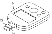
Figure 11 is illustrated in to be used for illustrating that the blood-sugar level measuring device of second embodiment of the invention is equipped with the stereographic map of the state of biology sensor.
Figure 12 is all stereographic maps of expression biology sensor shown in Figure 11.
Figure 13 is along the sectional view of the XIII-XIII of Figure 12 and major part enlarged drawing thereof.
Figure 14 is the state of nappe and division board is removed in expression from a biology sensor shown in Figure 12 planimetric map.
Figure 15 is the major part amplification profile of biology sensor that is used to illustrate other examples of nappe.
Figure 16 is the curve map of expression wetting index with variation heat time heating time of vinylon.
Figure 17 is the curve map of the relation of expression exposure time and pull up time.
Figure 18 A is that expression Hct value is the curve map of repeatability of 0% o'clock biology sensor of the present invention, and Figure 18 B represents that the Hct value is 0% o'clock the curve map to the repeatability of ratio biosensor.
Figure 19 A is that expression Hct value is the curve map of repeatability of 25% o'clock biology sensor of the present invention, and Figure 19 B represents that the Hct value is 25% o'clock the curve map to the repeatability of ratio biosensor.
Figure 20 A is that expression Hct value is the curve map of repeatability of 42% o'clock biology sensor of the present invention, and Figure 20 B represents that the Hct value is 42% o'clock the curve map to the repeatability of ratio biosensor.
Figure 21 A is all stereographic maps of the biology sensor of the 3rd embodiment of the present invention.
Figure 22 is the exploded perspective view of biology sensor shown in Figure 21.
Figure 23 is prior biological sensor sectional view and major part enlarged drawing thereof.
Figure 24 is the planimetric map that is used for illustrating the blood mobile status in the kapillary of prior biological sensor.
Embodiment
At first, referring to figs. 1 through Figure 10 first embodiment of the present invention is described.Fig. 1 and Fig. 2 are the figure that is used to illustrate the biology sensor of present embodiment, and Fig. 3~Figure 10 is the figure that is used to illustrate the using method and the function of biology sensor.
Biology sensor X1 illustrated in figures 1 and 2 is installed in the blood-sugar level measuring device and uses, and makes disposable.Biology sensor X1 has substrate 1, nappe 2 and two division boards 3.On biology sensor X1, utilize substrate 1, nappe 2 and two division boards 3, liquid is set accumulates portion 4 and kapillary 5.
On substrate 1, be provided with the effect utmost point 11, to the utmost point 12 and reagent portion 13.
The position that faces kapillary 5 of the effect utmost point 11 is that width more carefully forms.The utmost point 12 is formed, have the position of two strands of shapes at this thin position of width of the clamping action utmost point 11.Reagent portion 13 is the solid shapes that comprise oxidoreducing enzyme and electron transport substance.The reagent portion 13 and the effect the utmost point 11 and utmost point 12 is connected, the while is arranged on again in the kapillary 5.As oxidoreducing enzyme, make the glucose oxidase of electron transport substance reduction when for example can use the glucose oxidase in the blood to gluconic acid.On the other hand, as electron transport substance, for example can use the potassium ferricyanide.
Below substrate 1, stick adhesiveness sheet 14.Adhesiveness sheet and substrate 1 are roughly the same sizes, have notch 14a.Adhesiveness sheet 14 constitutes the suitable gel sheet that is to use silicone-based by gel sheet that contains hydrogel or acrylate resin etc. or double sticky tape etc.As hereinafter described, when biology sensor X1 supplies with blood, mediating by adhesiveness sheet 14 is contacted with below the substrate 1 skin.Therefore, by on substrate 1, attaching adhesiveness sheet 14, when biology sensor X1 is supplied with blood, can improve the adhesiveness of skin and biology sensor X1.
A pair of division board 3 is of similar shape, and disposes symmetrically with the state that leaves mutually.Each division board 3 in its side, has regulation side 30a capillaceous and accumulates the side 30b of portion with the standard solution that links to each other therewith.Side 30b makes along the flexure plane of the cut line of substrate 1.On each division board 3, form the through hole 30c that connects its thickness direction.Through hole 30c is formed at respectively with the effect utmost point 11 of substrate 1 or to the utmost point 12 corresponding positions.Division board 3, for example the heat-fusible adhesive by double sticky tape or thermoplastic resin constitutes.
But, also can form nappe 2 by the material beyond the vinylon, dehumidification function zone or high wettability zone are set.
Fig. 3 is the stereographic map that can use the blood-sugar level measuring device of biology sensor of the present invention, Fig. 4 is the cut-away section stereographic map of the major part of expression blood-sugar level measuring device shown in Figure 3, and Fig. 5~Fig. 9 is used to illustrate the major part sectional view that imports the blood-sugar level measuring device of blood action to biology sensor.
As shown in Figure 3, blood-sugar level measuring device Y1 possess main part 6, from main part 6 extended press sections 7.At the push switch 62 that the display part 60 for example be made of LCD or light-emitting diode display, the operating switch of operating when a series of processes relevant with blood-sugar level measuring are begun 61 is set on the main part 6, is used to start lancet.As shown in Figure 4 and Figure 5, press section 7 have cylindrical portion 70, the lancet frame 71 that in this cylindrical portion 70, can move back and forth, near the lancet frame 72 the peristome 70a that is fixed on cylindrical portion 70.
In cylindrical portion 70, be connected with pump P by connecting pipe 73 with its internal communication.This pump P is for the internal gas with cylindrical portion 70 is discharged to the outside, makes the inner pressure relief of cylindrical portion 70.Pump P can interiorly ensconce in the main part 6, also can be arranged on the outside as split with carrying.Pump P can be a manual drives.On connecting pipe 73, be provided for detecting the pressure transducer 74 of the internal pressure of cylindrical portion 70.
On cylindrical portion 70, be provided as the relief valve 75 of air-breathing member.This relief valve 75 is in order to suck air outside, to make cylindrical portion 70 pressure inside get back to atmospheric pressure.Relief valve 75 for example is made of solenoid valve.The structure of relief valve 75 is can be by manually opening.
Lancet frame 71 can unload the lancet 77 that can freely keep having puncture needle 77a such as metallic.Lancet frame 71 is connected with not shown reciprocating drive mechanism, by the push switch 62 (with reference to Fig. 3) of pressing main part 6, utilizes for example electromagnetic action or spring effect, 7 axial instantaneously back and forth drive along the press section.Thus, lancet 77 is kept out of the way to the direction that keeps away oral area 70a after instantaneous displacement takes place the peristome 70a of cylindrical portion 70.
The pair of connectors 78 that connects sensor installation portion 72b is installed on sensor frame 72.But, the connector 78 of front side only is shown in Fig. 4 and Fig. 5.Each connector 78 keeps coupling bar 79, makes its end flexibly outstanding.To install on the sensor installation portion 72b under the state of biology sensor X1, in order reaching with the effect utmost point 11 of biology sensor X1 the utmost point 12 to be contacted, each coupling bar 79 is outstanding from connector 78.
In order to use blood-sugar level measuring device Y1 to carry out blood-sugar level measuring with above formation, at first, as shown in Figure 5, make the front end of the press section 7 of the blood-sugar level measuring device Y1 that biology sensor X1 is housed, touch the s skin S of user's wrist or finger tip etc.At this moment, according to the surface of cylindrical portion 70 and s skin S, press section 7 temporarily becomes air-tight state.
Then, at this state, press the operating switch 61 (with reference to Fig. 3) of main part 6, driving pump P.Thus, press section 7 little by little becomes decompression state to the inside of cylindrical portion 70.So, stop up the s skin S of the front end of press section 7, follow decompression and protuberance little by little, as shown in Figure 6, contact with biology sensor X1 on being installed in sensor frame 72.At this, because post adhesiveness sheet 14, so biology sensor X1 and s skin S adhere to reliably on the surface of the substrate 1 of biology sensor X1.Like this, if biology sensor X1 adheres on the s skin S, when supply blood described later, for example can prevent that blood from immersing between biology sensor X1 and the s skin S.
Detect at pressure transducer 74 and to make s skin S produce protuberance and reach the pressure that adheres on the biology sensor X1 to be the moment of predefined pressure, to stop the driving of pump P that decompression process finishes.At this moment, can report the situation that decompression process finishes at display part 60 (with reference to Fig. 3).
Then, the user by pressing push switch 62 (with reference to Fig. 3), moves back and forth lancet frame 71 after confirming that decompression finishes instantaneously.Thus, the puncture needle 77a of lancet 77, at first as shown in Figure 7, by sensor frame 72 the 72d of window portion, skin puncture S.At this moment, contact with lancet 77, limit moving of lancet 77 by sensor frame 72.Specifically, at the state of puncture needle 77a with the degree of depth skin puncture S of appropriateness, sensor 77 feeler framves 72, puncture needle 77a enter in the s skin S can not surpass the above degree of depth.Thus, can prevent the excessive puncture of the s skin S that causes by puncture needle 77a.
Then, as shown in Figure 8, after lancet 77 is kept out of the way, flow out blood B from the s skin S of utilizing puncture needle 77a moderately to puncture.Biology sensor X1 is installed on the sensor frame 72, makes liquid accumulate the site of puncture of portion 4 near s skin S, and therefore, the blood B of outflow is accumulated portion 4 by the liquid of biology sensor X1 immediately and holds.
Figure 10 A to Figure 10 D schematically shows the figure that supplies with the situation of blood B to biology sensor X1.In the biology sensor X1 shown in Figure 10 to Figure 10 D, omitted substrate 1.
The blood B of fluid in process shown in Figure 8, shown in Figure 10 A, the place 41 of accumulating portion 4 with liquid contacts.At this moment, the fluid position, even for example accumulate the Width generation displacement of portion 4 from liquid, liquid accumulates portion 4 wider width ground and forms, its result, also containment blood B suitably.Because the surface wettability in place 41 is good, so shown in Figure 10 B, the blood B that contacts with place 41 wetting expansion on place 41, and accumulate the inner face of portion 4 and enlarge along liquid.Therefore, can prevent suitably that blood B from accumulating that local path is walked by portion 4 and the inlet that arrives kapillary 5 accumulates portion 4 to liquid simultaneously and supplies with the necessary abundant amount of blood-sugar level measurings at liquid.
Shown in Figure 10 C, if blood B stops up the inlet of kapillary 5, just immediately owing to capillarity, shown in Figure 10 D, blood B imports to kapillary 5.
Biology sensor X1 can accumulate portion 4 by roomy liquid and supply with blood B, even therefore in by the visual lancet integrated blood-sugar level measuring device Y1 that can not confirm, also can seek the robotization of good blood supply.In the moment that blood B stops up the inlet of kapillary 5, beginning is supplied with blood to kapillary 5, even therefore in not reaching the fluid way of measuring necessary amount, also can accumulate portion 4 to liquid and supply with blood B.
On the other hand, be imported into the blood B in the kapillary 5, in kapillary 5, the reagent portion 13 (with reference to Fig. 2) that is arranged on the substrate 1 dissolved at once, and constitute liquid-phase reaction system.So, in the blood contained glucose just because to be included in the effect of the oxidoreducing enzyme in the reagent portion 13 (with reference to Fig. 2) oxidized.Electronics by this reaction is taken out from glucose reaches electron transport substance by enzyme.That is, by enzyme, electron transport substance is reduced.After this, by pair of connectors 78, when applying voltage, just the effect utmost point 11 is supplied with electronics (with reference to Fig. 2 and Fig. 9) from liquid-phase reaction system at the effect utmost point 11 with between to the utmost point 12.Blood-sugar level measuring device Y1 measures as oxidation current by the amount of electrons that the effect utmost point 11 is supplied with, and calculates blood glucose value based on this oxidation current.
After supplying with blood B end to biology sensor X1, as shown in Figure 9, the inside of press section 7 utilizes relief valve 75 to carry out atmosphere opening.Thus, atmospheric pressure is got back in the inside of cylindrical portion 70, and s skin S returns to the original form.Utilize the atmosphere opening of relief valve 75, can be undertaken, also can after blood-sugar level measuring device Y1 detects the blood that imports among the biology sensor X1, in blood-sugar level measuring device Y1, automatically carry out by user's manual manipulation.
In the present embodiment, though be described as example to be installed in the biology sensor that uses on the blood-sugar level measuring device, scope of the present invention is not limited to this.For example, replace concentration of glucose, when obtaining cholesterol value or lactic acid value etc., also can use the formation of above-mentioned embodiment.In the above-described embodiment, be used for liquid is accumulated the notch 10 of portion's 4 openings, be arranged on substrate 1 one sides, but in the present invention, replace this, also can form notch at nappe 2.But, under the situation that adopts such formation, in blood-sugar level measuring, biology sensor X1 need be installed on the sensor frame 72 of blood-sugar level measuring device Y1, make nappe 2 towards peristome 70a, nappe 2 can adhere to s skin S.
Below, specifically describe second embodiment of the present invention with reference to Figure 11 to Figure 14.
As shown in figure 11, biology sensor X2 makes simple type, and is installed on the blood-sugar level measuring device Y2 that lancet forms with separate form and uses.
As Figure 12 to shown in Figure 14, biology sensor X2 have make division board 3 ' between the centre with the substrate 1 of nappe 2 ' be layered in rectangle ' on form.In this biology sensor X2, utilize substrate 1 ', nappe 2 ' and division board 3 ', along substrate 1 ' Width be formed extended at both sides kapillary 5 '.But the such liquid of biology sensor X1 that biology sensor X2 does not possess as depicted in figs. 1 and 2 accumulates portion 4.
Substrate 1 ' above, the formation effect utmost point 11 ', to the utmost point 12 ' and with reference to the utmost point 15 ' three electrodes.Three electrodes 11 ', 12 ' and 15 ', form along substrate 1 ' the length direction band shape of extending, these electrodes 11 ', 12 ' and 15 ' between be provided with insulation course 16 '.
Three electrodes 11 ', 12 ' and 15 ' on form the reagent layer 13 of solid shape '.Three electrodes 11 of this reagent layer 13 ' cover a succession ofly ', 12 ' and 15 ' and along substrate 1 ' Width extend and form.
Two division boards 3 ' reagent layer 13 ' sandwich reagent layer 13 ' and along substrate 1 ' Width extend.Division board 3 ' constitute by the heat-fusible adhesive of for example double sticky tape or thermoplastic resin.
Nappe 2 ' be is with substrate 21 ' by vinylal fibre sheet 22 ' the carry out form of lamination.Substrate 21 ' thickness for example be 100~150 μ m, vinylon sheet 22 ' thickness for example be 15~20 μ m.
Certainly, as shown in figure 15, nappe 2 ' all also can form with vinylon, in this occasion, nappe 2 ' inner face all become dehumidification function zone and high wettability zone.In addition, also can only constitute the part of nappe 2 ' inner face with vinylon, for example nappe 2 ' at least one end or central portion, in this occasion, can constitute the dehumidification function zone with vinylon.
Kapillary 5 ', it is inner by peristome 5a ', 5b ' and external communications.Kapillary 5 ' have same square-section, its depth of section size H and cross-sectional width size W, by division board 3 ' thickness and configuration space regulation.In the present invention, as hereinafter described, preferred heights size H is set at 30~100 μ m, width dimensions W and is set at 0.5~1.5mm, W/H and is set at<and 18.
Biology sensor X2 discussed above as shown in figure 11, under the state that is installed on blood-sugar level measuring device Y2, uses from peristome 5a ' importing blood.If from peristome 5a ' importing blood, then because capillarity makes blood through kapillary 5 ' interior moving.In biology sensor X2, nappe 2 ' inner face become non-water-soluble high wettability zone, therefore, even blood is the blood of high viscosity, blood also moves along nappe 2 ' energetically.This high wettability zone is non-water-soluble, therefore because the moving of blood, nappe 2 ' the wetting state of inner face do not change yet, can not produce bad to the translational speed of blood.
In addition, if nappe 2 ' inner face constitute by non-water-soluble vinylon, just improve water wettability, therefore there is no need nappe 2 ' the surface applied surfactant.Its result can not sneak into surfactant yet in test liquid as at present, surfactant moves with test liquid.Therefore, no matter division board 3 ' whether forms by hydrophobic material, along division board 3 ' the mobile of test liquid can not accelerate significantly than other positions, thereby can make velocity distribution uneven little of test liquid.If make the translational speed of test liquid big and make the uneven little of velocity distribution, just make kapillary 5 ' interior in advance and positively be full of by test liquid, can make reagent layer 13 ' in advance and dissolving positively.Its result can accomplish that precision measures blood glucose value well, and measures repeatability and also improve.
Inventor's confirmation, as shown in figure 16, even the wetting index of vinylon also changes after 1 second hardly 140 ℃ of heating.Therefore, use heat-fusible adhesive in fusion below 140 ℃, also can with nappe 2 ' be fixed in substrate 1 ' on.Thus, just do not need with hydrophobic double sticky tape constitute division board 3 ', therefore can suppress along division board 3 ' test liquid flow than nappe 2 ' the situation that accelerates of inner face, make the uneven little of velocity distribution, can do and improve mensuration precision and repeatability.
In addition, at biology sensor X2, near the inner face of and the kapillary 5 of peristome 5a ', 5b ' ' (nappe 2 ') becomes the dehumidification function zone, though the moisture therefore in the gas phase to invade kapillary 5 ' interior or invaded kapillary 5 ' in, this moisture is all removed by moisture absorption in the dehumidification function zone.And, if nappe 2 ' the outside also constitute by vinylon, the moisture that is present in kapillary 5 ' is on every side just removed by moisture absorption.Therefore, the reagent layer 13 that suppresses to cause because of moisture ' expose (reduction of electron transport substance) to the open air thus when improving storage stability, can lower and result from the background current of moisture, can precision measure the concentration of the determination object in the test liquid well.And then, make to result from each of each biology sensor of difference of moisture absorption degree and attract the uneven reduction of speed, can improve the mensuration precision thus.
As following illustrated, the inventor has studied influence (embodiment 1), mensuration repeatability (embodiment 2) and the only capillary size (embodiment 3) of the hygroscopic capacity in the reagent layer to attracting speed.
In the present embodiment, studied the hygroscopic capacity in the reagent layer to attracting the influence of speed.As biology sensor, used to have the biology sensor of the present invention that is similar to Figure 12 biology sensor X2 form extremely shown in Figure 14 and have the contrast glucose sensor that is similar to biology sensor form shown in Figure 23.Detailed formation with regard to these biology sensors is shown in the following table 1.Shown in following table 1, biology sensor of the present invention is different to biology sensor X1 shown in Figure 14 with Figure 12, only pastes the vinylon sheet in the one side of PET.On the other hand, in to ratio biosensor, at the lecithin of the one side of PET coating, as nappe as surfactant.
Table 1 (formation of biology sensor)
| The formation of nappe | Capillary size | |||
| Total length | Height H | Width W | ||
| Biology sensor of the present invention | Paste 17 μ m vinylon sheets in the one side of 100 μ mPET | 6mm | 120μm | 1.2mm |
| To ratio biosensor | One side coating lecithin at 100 μ mPET carries out hydrophilic treatment | |||
In the present embodiment, prepare biology sensor of the present invention and to each four of ratio biosensors.Respectively to biology sensor of the present invention with to four groups of ratio biosensor, measuring and making exposure time is the pull up time of 0 minute, 30 minutes, 60 minutes and 180 minutes.It the results are shown among Figure 17.Moreover it is 80% that the condition of exposing to the open air is defined as 30 ℃, relative humidity, and pull up time is defined as the time that is full of fully to the kapillary, measures.
As shown in figure 17, in biology sensor of the present invention, irrelevant with the length of exposure time, pull up time is roughly certain.In contrast, in to ratio biosensor, exposure time is long more, and it is short more that pull up time becomes.From these results as can be seen, in to ratio biosensor, the elongated more hygroscopic capacity of exposure time becomes big more, the dissolubility of reagent layer improves, and pull up time shortens, in contrast, and in biology sensor of the present invention, by the regional moisture absorption that suppresses reagent layer of the dehumidification function that utilizes vinylon to realize, the dissolubility of reagent layer maintains necessarily.Therefore as can be seen, biology sensor storage stability of the present invention is good, and the translational speed in kapillary necessarily changes, and is suppressed at the uneven of measured value between each biology sensor, measures repeatability and improves.
Studied the mensuration repeatability in the present embodiment.By biology sensor of the present invention relatively with to the variation in time of the response current value in the ratio biosensor, studied the mensuration repeatability.By to reagent layer pay standard liquid, measure between the effect utmost point and electrode, give this moment 200mV decide current potential the time the oxidation current value, carry out the mensuration of response current value.The modulation of titer is as follows, dissolves glucose in normal saline solution, makes concentration of glucose become 100mg/dl, makes blood cell concentration (Hct) become desired value simultaneously.With the results are shown among Figure 18 A to Figure 20 A in the biology sensor of the present invention, will be to the results are shown among Figure 18 B to Figure 20 B in the ratio biosensor.Have, Figure 18, Figure 19, Figure 20 illustrate the result of Hct0%, Hct25%, Hct42% respectively again.In each biology sensor,, respectively 10 samples have been measured the response current value about each Hct.
If comparison diagram 18A and Figure 18 B, Figure 19 A Figure 19 B are then as can be known, under the situation of Hct value little (below 25%), the biology sensor of the present invention and repeatability between the ratio biosensor almost do not had difference.In contrast, if comparison diagram 20A and Figure 20 B are then as can be known, be under 42% the situation in Hct value, apply voltage begin to 5 seconds with interior time range in, it is especially good that repeatability that we can say biology sensor of the present invention is compared ratio biosensor.For clear and definite this point, be under 42% the situation in the Hct value, respectively for biology sensor of the present invention with to ratio biosensor, will be after applying voltage and beginning, after 4 seconds to 3 seconds and the repeatability of the response current value in 10 biology sensors after 5 seconds be shown in the following table 2 with the form of relative standard deviation (C.V.[%]).
Table 2 (evaluation result of repeatability)
| Sample time | After 3 seconds | After 4 seconds | After 5 seconds | ||||||
| Concentration of glucose [mg/dl] | 100 | 400 | 600 | 100 | 400 | 600 | 100 | 400 | 600 |
| Biology sensor C.V.[% of the present invention] | 2.46 | 1.67 | 1.50 | 2.27 | 1.48 | 1.18 | 2.12 | 1.53 | 0.97 |
| To ratio biosensor C.V.[%] | 5.20 | 2.94 | 2.17 | 3.70 | 2.99 | 1.85 | 3.13 | 3.16 | 1.80 |
According to table 2 as can be known, biology sensor of the present invention no matter concentration of glucose is how, begins to 3~5 seconds scopes applying voltage, and ratio biosensor is compared, and measured value uneven little, it is good that repeatability also becomes.Therefore, biology sensor of the present invention, by utilizing vinylon that the high wettability zone is set, improve from applying the repeatability that voltage begins to the short time range, particularly big to Hct value blood (test liquid of high viscosity) be we can say and can minute be shortened.
In the present embodiment, carry out the optimization of capillary size.In the present embodiment, make the biology sensor of the present invention of basic formation and embodiment 1 and identical, simultaneously,, measure pull up time for the different biology sensor 1~12 of capillary size to ratio biosensor.With regard to pull up time, measure in the same manner with embodiment 1.But for the high viscosity test liquid is attracted speed evaluation, as titer, using the Hct value is 70%.It the results are shown in the following table 3.Use actually under the situation that biology sensor measures, in order to seek the shorteningization of minute, preferably in 2.5 seconds, be full of in the kapillary, therefore shown in the table 3 with pull up time be below 2 seconds as ◎, 2~2.5 seconds as zero, 2.5~5 second as △, more than 5 seconds as the * result that estimates.
Table 3 (attracting the evaluation result of speed)
| The formation of nappe | Capillary size | Estimate | ||||
| Total length | Height H | Width W | W/ | |||
| Biology sensor | ||||||
| 1 | On the one side of 100 μ mPET, paste 17 μ m vinylon sheets | 6mm | 60μm | 0.75mm | 12.5 | △ |
| | 1.00mm | 16.7 | △ | |||
| | 1.20mm | 20.0 | × | |||
| | 90μm | 0.75mm | 8.3 | ◎ | ||
| | 1.00mm | 11.1 | ◎ | |||
| | 1.20mm | 13.3 | ◎ | |||
| | On the one side of 100 μ mPET, apply lecithin, carry out hydrophilic treatment | 60μm | 0.75mm | 12.5 | × | |
| | 1.00mm | 16.7 | × | |||
| | 1.20mm | 20.0 | × | |||
| | 90μm | 0.75mm | 8.3 | ○ | ||
| | 1.00mm | 11.1 | ○ | |||
| | 1.20mm | 13.3 | △ | |||
According to table 3 as can be known, utilize vinylon to make the inner face of kapillary (nappe) become the biology sensor 1~6 in dehumidification function zone and high wettability zone, compare at the biology sensor of the inner face coating lecithin of PET as nappe when making capillary size identical, we can say attraction speed height.In addition, for the test liquid of high viscosity, we can say capillary size preferably, height dimension H is that 30~100 μ m, width dimensions W are that 0.5~1.5mm, W/H are<18.
Certainly, technological thought of the present invention as the 3rd embodiment, also can be used in the biology sensor X3 of Figure 21 and form shown in Figure 22.The formation that biology sensor X3 shown in these figure also has substrate 1 ", division board 3 " and nappe 2 ", but kapillary 5 " form and electrode 11 ", 12 " is different with previously described biology sensor X2.
In biology sensor X3,, when the length direction of division board 3 " upper edge substrate 1 " extends, leading section 18 " open slit 19 " is set nappe 2 " on through hole 29 is set ".At substrate 4 " go up stacked division board 3 " and nappe 2 " time, slit 19 " and through hole 29 " is communicated with, and forms kapillary 5 ", slit 19 " leading section 18 " becomes the sample introducing port.At substrate 1 " on be provided with effect the utmost point 11 " with to the utmost point 12 ".Reagent layer 13 " cover effect the utmost point 11 " a succession ofly and to the utmost point 12 " the formation of leading section ground.
The inner face of " all, perhaps attach the vinylon sheet by the surface at base materials such as PET, also can be with kapillary 5 " becomes dehumidification function zone and high wettability zone even in such biology sensor X3, by formed nappe 2 by vinylon.Certainly, even in biology sensor X3, also can be by the part of nappe 2 " constitute kapillary 5 " inner face as dehumidification function zone and high wettability zone, or only select near the of through hole 29 " or sample introducing port 18 " as the dehumidification function zone.
Biology sensor X2, the X3 of the second and the 3rd embodiment do not possess liquid and accumulates portion, possesses the biology sensor that liquid accumulates portion but these biology sensors X2, X3 can be made yet.
Claims (15)
1. analyzer is characterized in that:
Possess the kapillary that is used for mobile test liquid,
Described kapillary forms by nappe is layered on the substrate, and at least a portion of described nappe forms by vinylon,
The hydroscopicity of described vinylon is more than 2%, and wetting index is more than the 57mN/m.
2. analyzer according to claim 1 is characterized in that:
Described inside capillaceous, by first peristome and second peristome and external communications,
Described vinylon is arranged near at least one at least a portion of or inner face capillaceous in described first peristome and described second peristome.
3. analyzer according to claim 1 is characterized in that: all being formed by vinylon of described nappe.
4. analyzer according to claim 1 is characterized in that: the vinylon layer that described nappe has tabular base material and forms on the one side at least of this base material.
5. analyzer according to claim 1 is characterized in that:
Described kapillary has the same square-section,
Described square-section is, height dimension H is 30~100 μ m, and width dimensions W is 0.5~1.5mm, and W/H<18.
6. analyzer according to claim 1 is characterized in that: also possess the liquid that is communicated with described kapillary and has the position wideer than described kapillary width and accumulate portion.
7. analyzer according to claim 6 is characterized in that:
Described kapillary forms by nappe is layered on the substrate,
The described liquid portion of accumulating is formed on described substrate or the described nappe and by the hole portion that connects at thickness direction and carries out opening.
8. analyzer according to claim 6 is characterized in that: the wetting index on the surface of at least the described liquid portion of accumulating being stipulated in described substrate or the described nappe is more than the 57mN/m.
9. analyzer according to claim 6 is characterized in that: at least a portion of described substrate is made of vinylon.
10. analyzer according to claim 6 is characterized in that:
Possess the division board that between described substrate and described nappe, disposes,
Described division board has described kapillary and described liquid is accumulated the isolation part that the width of portion is stipulated.
11. an analyzer is characterized in that: possess:
Substrate;
Be layered in the nappe on the substrate;
The kapillary that is used for mobile test liquid; With
The liquid that is communicated with described kapillary and has a position wideer than described kapillary width accumulates portion,
The surface of at least the described liquid portion of accumulating being stipulated in described substrate or the described nappe is made of vinylon,
The hydroscopicity of described vinylon is more than 2%, and wetting index is more than the 57mN/m.
12. analyzer according to claim 11 is characterized in that: the described liquid portion of accumulating is formed on described substrate or the described nappe and by the hole portion that connects at thickness direction and carries out opening.
13. analyzer according to claim 11 is characterized in that: described substrate or described nappe are made of vinylon.
14. analyzer according to claim 11 is characterized in that:
Possess the division board that between described substrate and described nappe, disposes,
Described division board has described kapillary and described liquid is accumulated the isolation part that the width of portion is stipulated.
15. analyzer according to claim 11 is characterized in that:
On described substrate or described nappe, the adhesion layer of cohesive than described substrate or described covering height is set near described notch.
Applications Claiming Priority (4)
| Application Number | Priority Date | Filing Date | Title |
|---|---|---|---|
| JP2001227777 | 2001-07-27 | ||
| JP227777/2001 | 2001-07-27 | ||
| JP2001242486 | 2001-08-09 | ||
| JP242486/2001 | 2001-08-09 |
Publications (2)
| Publication Number | Publication Date |
|---|---|
| CN1535377A CN1535377A (en) | 2004-10-06 |
| CN1300578C true CN1300578C (en) | 2007-02-14 |
Family
ID=26619436
Family Applications (1)
| Application Number | Title | Priority Date | Filing Date |
|---|---|---|---|
| CNB028147049A Expired - Lifetime CN1300578C (en) | 2001-07-27 | 2002-07-26 | Analyzing instrument |
Country Status (6)
| Country | Link |
|---|---|
| US (2) | US7824616B2 (en) |
| EP (2) | EP1413880B1 (en) |
| JP (1) | JP4282477B2 (en) |
| CN (1) | CN1300578C (en) |
| DE (1) | DE10193213T8 (en) |
| WO (1) | WO2003010530A1 (en) |
Families Citing this family (85)
| Publication number | Priority date | Publication date | Assignee | Title |
|---|---|---|---|---|
| US6036924A (en) | 1997-12-04 | 2000-03-14 | Hewlett-Packard Company | Cassette of lancet cartridges for sampling blood |
| US6391005B1 (en) | 1998-03-30 | 2002-05-21 | Agilent Technologies, Inc. | Apparatus and method for penetration with shaft having a sensor for sensing penetration depth |
| US8641644B2 (en) | 2000-11-21 | 2014-02-04 | Sanofi-Aventis Deutschland Gmbh | Blood testing apparatus having a rotatable cartridge with multiple lancing elements and testing means |
| US7749174B2 (en) | 2001-06-12 | 2010-07-06 | Pelikan Technologies, Inc. | Method and apparatus for lancet launching device intergrated onto a blood-sampling cartridge |
| US7041068B2 (en) | 2001-06-12 | 2006-05-09 | Pelikan Technologies, Inc. | Sampling module device and method |
| US7344507B2 (en) | 2002-04-19 | 2008-03-18 | Pelikan Technologies, Inc. | Method and apparatus for lancet actuation |
| US8337419B2 (en) | 2002-04-19 | 2012-12-25 | Sanofi-Aventis Deutschland Gmbh | Tissue penetration device |
| US9427532B2 (en) | 2001-06-12 | 2016-08-30 | Sanofi-Aventis Deutschland Gmbh | Tissue penetration device |
| US7981056B2 (en) | 2002-04-19 | 2011-07-19 | Pelikan Technologies, Inc. | Methods and apparatus for lancet actuation |
| DE60234597D1 (en) | 2001-06-12 | 2010-01-14 | Pelikan Technologies Inc | DEVICE AND METHOD FOR REMOVING BLOOD SAMPLES |
| WO2002100460A2 (en) | 2001-06-12 | 2002-12-19 | Pelikan Technologies, Inc. | Electric lancet actuator |
| US9795747B2 (en) | 2010-06-02 | 2017-10-24 | Sanofi-Aventis Deutschland Gmbh | Methods and apparatus for lancet actuation |
| US9226699B2 (en) | 2002-04-19 | 2016-01-05 | Sanofi-Aventis Deutschland Gmbh | Body fluid sampling module with a continuous compression tissue interface surface |
| EP1404233B1 (en) | 2001-06-12 | 2009-12-02 | Pelikan Technologies Inc. | Self optimizing lancing device with adaptation means to temporal variations in cutaneous properties |
| WO2002100461A2 (en) | 2001-06-12 | 2002-12-19 | Pelikan Technologies, Inc. | Method and apparatus for improving success rate of blood yield from a fingerstick |
| DE60238914D1 (en) | 2001-06-12 | 2011-02-24 | Pelikan Technologies Inc | INTEGRATED BLOOD SAMPLE ANALYSIS SYSTEM WITH MULTI-USE SAMPLING MODULE |
| CN1300578C (en) * | 2001-07-27 | 2007-02-14 | 爱科来株式会社 | Analyzing instrument |
| US7344894B2 (en) | 2001-10-16 | 2008-03-18 | Agilent Technologies, Inc. | Thermal regulation of fluidic samples within a diagnostic cartridge |
| US9314194B2 (en) | 2002-04-19 | 2016-04-19 | Sanofi-Aventis Deutschland Gmbh | Tissue penetration device |
| US7901362B2 (en) | 2002-04-19 | 2011-03-08 | Pelikan Technologies, Inc. | Method and apparatus for penetrating tissue |
| US7141058B2 (en) | 2002-04-19 | 2006-11-28 | Pelikan Technologies, Inc. | Method and apparatus for a body fluid sampling device using illumination |
| US7909778B2 (en) | 2002-04-19 | 2011-03-22 | Pelikan Technologies, Inc. | Method and apparatus for penetrating tissue |
| US7491178B2 (en) | 2002-04-19 | 2009-02-17 | Pelikan Technologies, Inc. | Method and apparatus for penetrating tissue |
| US7331931B2 (en) | 2002-04-19 | 2008-02-19 | Pelikan Technologies, Inc. | Method and apparatus for penetrating tissue |
| US7244265B2 (en) | 2002-04-19 | 2007-07-17 | Pelikan Technologies, Inc. | Method and apparatus for penetrating tissue |
| US7563232B2 (en) | 2002-04-19 | 2009-07-21 | Pelikan Technologies, Inc. | Method and apparatus for penetrating tissue |
| US9248267B2 (en) | 2002-04-19 | 2016-02-02 | Sanofi-Aventis Deustchland Gmbh | Tissue penetration device |
| US7524293B2 (en) | 2002-04-19 | 2009-04-28 | Pelikan Technologies, Inc. | Method and apparatus for penetrating tissue |
| US7674232B2 (en) | 2002-04-19 | 2010-03-09 | Pelikan Technologies, Inc. | Method and apparatus for penetrating tissue |
| US7976476B2 (en) | 2002-04-19 | 2011-07-12 | Pelikan Technologies, Inc. | Device and method for variable speed lancet |
| US8360992B2 (en) | 2002-04-19 | 2013-01-29 | Sanofi-Aventis Deutschland Gmbh | Method and apparatus for penetrating tissue |
| US8784335B2 (en) | 2002-04-19 | 2014-07-22 | Sanofi-Aventis Deutschland Gmbh | Body fluid sampling device with a capacitive sensor |
| US7892183B2 (en) | 2002-04-19 | 2011-02-22 | Pelikan Technologies, Inc. | Method and apparatus for body fluid sampling and analyte sensing |
| US7648468B2 (en) | 2002-04-19 | 2010-01-19 | Pelikon Technologies, Inc. | Method and apparatus for penetrating tissue |
| WO2003088824A2 (en) | 2002-04-19 | 2003-10-30 | Pelikan Technologies, Inc. | Device and method for variable speed lancet |
| US7232451B2 (en) | 2002-04-19 | 2007-06-19 | Pelikan Technologies, Inc. | Method and apparatus for penetrating tissue |
| US7485128B2 (en) | 2002-04-19 | 2009-02-03 | Pelikan Technologies, Inc. | Method and apparatus for penetrating tissue |
| US7229458B2 (en) | 2002-04-19 | 2007-06-12 | Pelikan Technologies, Inc. | Method and apparatus for penetrating tissue |
| US7717863B2 (en) | 2002-04-19 | 2010-05-18 | Pelikan Technologies, Inc. | Method and apparatus for penetrating tissue |
| US8372016B2 (en) | 2002-04-19 | 2013-02-12 | Sanofi-Aventis Deutschland Gmbh | Method and apparatus for body fluid sampling and analyte sensing |
| US7371247B2 (en) | 2002-04-19 | 2008-05-13 | Pelikan Technologies, Inc | Method and apparatus for penetrating tissue |
| US7374544B2 (en) | 2002-04-19 | 2008-05-20 | Pelikan Technologies, Inc. | Method and apparatus for penetrating tissue |
| US9795334B2 (en) | 2002-04-19 | 2017-10-24 | Sanofi-Aventis Deutschland Gmbh | Method and apparatus for penetrating tissue |
| US7291117B2 (en) | 2002-04-19 | 2007-11-06 | Pelikan Technologies, Inc. | Method and apparatus for penetrating tissue |
| US7547287B2 (en) | 2002-04-19 | 2009-06-16 | Pelikan Technologies, Inc. | Method and apparatus for penetrating tissue |
| US7297122B2 (en) | 2002-04-19 | 2007-11-20 | Pelikan Technologies, Inc. | Method and apparatus for penetrating tissue |
| US7708701B2 (en) | 2002-04-19 | 2010-05-04 | Pelikan Technologies, Inc. | Method and apparatus for a multi-use body fluid sampling device |
| US8579831B2 (en) | 2002-04-19 | 2013-11-12 | Sanofi-Aventis Deutschland Gmbh | Method and apparatus for penetrating tissue |
| US7410468B2 (en) | 2002-04-19 | 2008-08-12 | Pelikan Technologies, Inc. | Method and apparatus for penetrating tissue |
| US7582099B2 (en) | 2002-04-19 | 2009-09-01 | Pelikan Technologies, Inc | Method and apparatus for penetrating tissue |
| US8267870B2 (en) | 2002-04-19 | 2012-09-18 | Sanofi-Aventis Deutschland Gmbh | Method and apparatus for body fluid sampling with hybrid actuation |
| US8221334B2 (en) | 2002-04-19 | 2012-07-17 | Sanofi-Aventis Deutschland Gmbh | Method and apparatus for penetrating tissue |
| US8702624B2 (en) | 2006-09-29 | 2014-04-22 | Sanofi-Aventis Deutschland Gmbh | Analyte measurement device with a single shot actuator |
| US8574895B2 (en) | 2002-12-30 | 2013-11-05 | Sanofi-Aventis Deutschland Gmbh | Method and apparatus using optical techniques to measure analyte levels |
| DE602004028463D1 (en) | 2003-05-30 | 2010-09-16 | Pelikan Technologies Inc | METHOD AND DEVICE FOR INJECTING LIQUID |
| ES2490740T3 (en) | 2003-06-06 | 2014-09-04 | Sanofi-Aventis Deutschland Gmbh | Apparatus for blood fluid sampling and analyte detection |
| WO2006001797A1 (en) | 2004-06-14 | 2006-01-05 | Pelikan Technologies, Inc. | Low pain penetrating |
| EP1635700B1 (en) | 2003-06-13 | 2016-03-09 | Sanofi-Aventis Deutschland GmbH | Apparatus for a point of care device |
| AU2004268431A1 (en) * | 2003-09-01 | 2005-03-10 | Inverness Medical Switzerland Gmbh | Sampling device with capillary action |
| US7682833B2 (en) * | 2003-09-10 | 2010-03-23 | Abbott Point Of Care Inc. | Immunoassay device with improved sample closure |
| US8282576B2 (en) | 2003-09-29 | 2012-10-09 | Sanofi-Aventis Deutschland Gmbh | Method and apparatus for an improved sample capture device |
| US9351680B2 (en) | 2003-10-14 | 2016-05-31 | Sanofi-Aventis Deutschland Gmbh | Method and apparatus for a variable user interface |
| US7822454B1 (en) | 2005-01-03 | 2010-10-26 | Pelikan Technologies, Inc. | Fluid sampling device with improved analyte detecting member configuration |
| EP1706026B1 (en) * | 2003-12-31 | 2017-03-01 | Sanofi-Aventis Deutschland GmbH | Method and apparatus for improving fluidic flow and sample capture |
| US8828203B2 (en) | 2004-05-20 | 2014-09-09 | Sanofi-Aventis Deutschland Gmbh | Printable hydrogels for biosensors |
| US9775553B2 (en) | 2004-06-03 | 2017-10-03 | Sanofi-Aventis Deutschland Gmbh | Method and apparatus for a fluid sampling device |
| EP1765194A4 (en) | 2004-06-03 | 2010-09-29 | Pelikan Technologies Inc | Method and apparatus for a fluid sampling device |
| US8652831B2 (en) | 2004-12-30 | 2014-02-18 | Sanofi-Aventis Deutschland Gmbh | Method and apparatus for analyte measurement test time |
| ES2374541T3 (en) | 2005-03-22 | 2012-02-17 | F. Hoffmann-La Roche Ag | TEST ELEMENT FOR BODY FLUID ANALYSIS. |
| EP1963817A2 (en) * | 2005-12-22 | 2008-09-03 | Honeywell International Inc. | Portable sample analyzer cartridge |
| CN103226150B (en) | 2007-10-30 | 2015-01-21 | 松下健康医疗器械株式会社 | Analyzing device, analyzing apparatus using the device, and analyzing method |
| EP2265324B1 (en) | 2008-04-11 | 2015-01-28 | Sanofi-Aventis Deutschland GmbH | Integrated analyte measurement system |
| US9375169B2 (en) | 2009-01-30 | 2016-06-28 | Sanofi-Aventis Deutschland Gmbh | Cam drive for managing disposable penetrating member actions with a single motor and motor and control system |
| JP4674283B2 (en) * | 2009-06-01 | 2011-04-20 | アークレイ株式会社 | Analytical tool and manufacturing method thereof |
| KR101104391B1 (en) * | 2009-06-30 | 2012-01-16 | 주식회사 세라젬메디시스 | Sensors used in conjunction with detectors to measure biomaterials and devices using them |
| US8357276B2 (en) * | 2009-08-31 | 2013-01-22 | Abbott Diabetes Care Inc. | Small volume test strips with large sample fill ports, supported test strips, and methods of making and using same |
| US8965476B2 (en) | 2010-04-16 | 2015-02-24 | Sanofi-Aventis Deutschland Gmbh | Tissue penetration device |
| US11430279B2 (en) * | 2012-05-09 | 2022-08-30 | Wisconsin Alumni Research Foundation | Functionalized microfluidic device and method |
| US9523653B2 (en) | 2013-05-09 | 2016-12-20 | Changsha Sinocare Inc. | Disposable test sensor with improved sampling entrance |
| US9518951B2 (en) | 2013-12-06 | 2016-12-13 | Changsha Sinocare Inc. | Disposable test sensor with improved sampling entrance |
| US9897566B2 (en) | 2014-01-13 | 2018-02-20 | Changsha Sinocare Inc. | Disposable test sensor |
| US9939401B2 (en) | 2014-02-20 | 2018-04-10 | Changsha Sinocare Inc. | Test sensor with multiple sampling routes |
| JP7034947B2 (en) * | 2016-06-17 | 2022-03-14 | エフ.ホフマン-ラ ロシュ アーゲー | Test system for analyzing body fluid samples |
| JP6938149B2 (en) * | 2016-12-22 | 2021-09-22 | リンテック株式会社 | Inspection cover film, inspection member, and manufacturing method of inspection member |
| JP6966860B2 (en) * | 2017-04-07 | 2021-11-17 | リンテック株式会社 | Manufacturing method of inspection cover film, inspection member, and inspection cover film |
Citations (6)
| Publication number | Priority date | Publication date | Assignee | Title |
|---|---|---|---|---|
| JPH0295352A (en) * | 1988-09-30 | 1990-04-06 | Advance Co Ltd | Automatic blood taking device |
| JPH06229970A (en) * | 1993-02-03 | 1994-08-19 | Matsushita Electric Ind Co Ltd | Humor constituent measuring device |
| JPH11347018A (en) * | 1998-04-09 | 1999-12-21 | Matsushita Electric Ind Co Ltd | Humor examination device |
| WO2000040150A1 (en) * | 1999-01-04 | 2000-07-13 | Terumo Kabushiki Kaisha | Assembly having lancet and means for collecting and detecting body fluid |
| JP2000221121A (en) * | 1999-01-29 | 2000-08-11 | Kdk Corp | Quantitative equipment using sample collection tools |
| JP2000258382A (en) * | 1999-03-05 | 2000-09-22 | Arkray Inc | Specimen small-quantity-type bio sensor |
Family Cites Families (39)
| Publication number | Priority date | Publication date | Assignee | Title |
|---|---|---|---|---|
| JPS57208997A (en) * | 1981-06-17 | 1982-12-22 | Fuji Photo Film Co Ltd | Liquid analyzing material for oxidase enzyme reaction system |
| US4790640A (en) * | 1985-10-11 | 1988-12-13 | Nason Frederic L | Laboratory slide |
| US4849340A (en) * | 1987-04-03 | 1989-07-18 | Cardiovascular Diagnostics, Inc. | Reaction system element and method for performing prothrombin time assay |
| US5066465A (en) * | 1989-12-27 | 1991-11-19 | Olympus Optical Co., Ltd. | Reaction apparatus |
| JP3382632B2 (en) * | 1992-03-13 | 2003-03-04 | オリンパス光学工業株式会社 | Method for measuring biological substance and reaction vessel used for the method |
| JPH0755795A (en) | 1993-08-18 | 1995-03-03 | Kanebo Ltd | Test piece for analysis |
| JPH0755793A (en) | 1993-08-18 | 1995-03-03 | Kanebo Ltd | Test piece for analysis |
| JP3027306B2 (en) | 1994-06-02 | 2000-04-04 | 松下電器産業株式会社 | Biosensor and manufacturing method thereof |
| FR2744607B1 (en) | 1996-02-12 | 1998-04-30 | Lvmh Rech | PERFECTED BRUSH FOR MAKING EYELASHES |
| JP3621502B2 (en) | 1996-04-02 | 2005-02-16 | アプルス株式会社 | Lancet assembly |
| US6001307A (en) * | 1996-04-26 | 1999-12-14 | Kyoto Daiichi Kagaku Co., Ltd. | Device for analyzing a sample |
| US6332871B1 (en) | 1996-05-17 | 2001-12-25 | Amira Medical | Blood and interstitial fluid sampling device |
| EP1579814A3 (en) | 1996-05-17 | 2006-06-14 | Roche Diagnostics Operations, Inc. | Methods and apparatus for sampling and analyzing body fluid |
| US6015392A (en) | 1996-05-17 | 2000-01-18 | Mercury Diagnostics, Inc. | Apparatus for sampling body fluid |
| US5951493A (en) | 1997-05-16 | 1999-09-14 | Mercury Diagnostics, Inc. | Methods and apparatus for expressing body fluid from an incision |
| US5879311A (en) | 1996-05-17 | 1999-03-09 | Mercury Diagnostics, Inc. | Body fluid sampling device and methods of use |
| DE19781098B4 (en) | 1996-05-17 | 2005-05-19 | Amira Medical, Scotts Valley | Blood and interstitial fluid sampling device for analysis=processing - includes disposable lancet mounted in housing also carrying capillary tube which can be slid in longitudinal direction relative to lancet |
| US5951492A (en) | 1996-05-17 | 1999-09-14 | Mercury Diagnostics, Inc. | Methods and apparatus for sampling and analyzing body fluid |
| EP0937249B1 (en) | 1996-10-30 | 2002-12-18 | Amira Medical | Synchronized analyte testing system |
| US6093156A (en) * | 1996-12-06 | 2000-07-25 | Abbott Laboratories | Method and apparatus for obtaining blood for diagnostic tests |
| AT404992B (en) | 1997-04-17 | 1999-04-26 | Avl List Gmbh | SENSOR FOR DETERMINING AN ENZYME SUBSTRATE |
| US5759364A (en) * | 1997-05-02 | 1998-06-02 | Bayer Corporation | Electrochemical biosensor |
| US5948695A (en) | 1997-06-17 | 1999-09-07 | Mercury Diagnostics, Inc. | Device for determination of an analyte in a body fluid |
| DE19753848A1 (en) | 1997-12-04 | 1999-06-10 | Roche Diagnostics Gmbh | Modification of surfaces to increase surface tension |
| DE19753850A1 (en) * | 1997-12-04 | 1999-06-10 | Roche Diagnostics Gmbh | Sampling device |
| DE19753847A1 (en) | 1997-12-04 | 1999-06-10 | Roche Diagnostics Gmbh | Analytical test element with capillary channel |
| US5997817A (en) | 1997-12-05 | 1999-12-07 | Roche Diagnostics Corporation | Electrochemical biosensor test strip |
| JP3896435B2 (en) * | 1997-12-17 | 2007-03-22 | アークレイ株式会社 | Sensor and sensor assembly |
| US5975153A (en) | 1998-02-13 | 1999-11-02 | Roche Diagnostics Corporation | Capillary fill test device with improved fluid delivery |
| JPH11304748A (en) | 1998-04-23 | 1999-11-05 | Omron Corp | Biosensor |
| US6767440B1 (en) | 2001-04-24 | 2004-07-27 | Roche Diagnostics Corporation | Biosensor |
| US6662439B1 (en) * | 1999-10-04 | 2003-12-16 | Roche Diagnostics Corporation | Laser defined features for patterned laminates and electrodes |
| JP2001159618A (en) * | 1999-12-03 | 2001-06-12 | Matsushita Electric Ind Co Ltd | Biosensor |
| US6571651B1 (en) * | 2000-03-27 | 2003-06-03 | Lifescan, Inc. | Method of preventing short sampling of a capillary or wicking fill device |
| US6488828B1 (en) | 2000-07-20 | 2002-12-03 | Roche Diagnostics Corporation | Recloseable biosensor |
| US6540890B1 (en) | 2000-11-01 | 2003-04-01 | Roche Diagnostics Corporation | Biosensor |
| US6447657B1 (en) | 2000-12-04 | 2002-09-10 | Roche Diagnostics Corporation | Biosensor |
| JP4736323B2 (en) * | 2001-07-13 | 2011-07-27 | アークレイ株式会社 | Puncture element integrated body for concentration measuring device provided with analysis tool, and body fluid collection tool |
| CN1300578C (en) * | 2001-07-27 | 2007-02-14 | 爱科来株式会社 | Analyzing instrument |
-
2002
- 2002-07-26 CN CNB028147049A patent/CN1300578C/en not_active Expired - Lifetime
- 2002-07-26 DE DE10193213T patent/DE10193213T8/en active Active
- 2002-07-26 JP JP2003515851A patent/JP4282477B2/en not_active Expired - Lifetime
- 2002-07-26 EP EP02749377.4A patent/EP1413880B1/en not_active Expired - Lifetime
- 2002-07-26 US US10/484,955 patent/US7824616B2/en not_active Expired - Fee Related
- 2002-07-26 EP EP10193213.5A patent/EP2290358B1/en not_active Expired - Lifetime
- 2002-07-26 WO PCT/JP2002/007654 patent/WO2003010530A1/en active Application Filing
-
2010
- 2010-10-12 US US12/902,647 patent/US8425841B2/en not_active Expired - Lifetime
Patent Citations (6)
| Publication number | Priority date | Publication date | Assignee | Title |
|---|---|---|---|---|
| JPH0295352A (en) * | 1988-09-30 | 1990-04-06 | Advance Co Ltd | Automatic blood taking device |
| JPH06229970A (en) * | 1993-02-03 | 1994-08-19 | Matsushita Electric Ind Co Ltd | Humor constituent measuring device |
| JPH11347018A (en) * | 1998-04-09 | 1999-12-21 | Matsushita Electric Ind Co Ltd | Humor examination device |
| WO2000040150A1 (en) * | 1999-01-04 | 2000-07-13 | Terumo Kabushiki Kaisha | Assembly having lancet and means for collecting and detecting body fluid |
| JP2000221121A (en) * | 1999-01-29 | 2000-08-11 | Kdk Corp | Quantitative equipment using sample collection tools |
| JP2000258382A (en) * | 1999-03-05 | 2000-09-22 | Arkray Inc | Specimen small-quantity-type bio sensor |
Also Published As
| Publication number | Publication date |
|---|---|
| DE10193213T8 (en) | 2013-04-25 |
| EP1413880A1 (en) | 2004-04-28 |
| JP4282477B2 (en) | 2009-06-24 |
| EP2290358A2 (en) | 2011-03-02 |
| EP1413880B1 (en) | 2015-09-02 |
| US20110060203A1 (en) | 2011-03-10 |
| EP2290358A3 (en) | 2012-02-29 |
| US7824616B2 (en) | 2010-11-02 |
| DE10193213T1 (en) | 2013-01-31 |
| EP1413880A4 (en) | 2010-06-02 |
| CN1535377A (en) | 2004-10-06 |
| EP2290358B1 (en) | 2015-11-11 |
| WO2003010530A1 (en) | 2003-02-06 |
| JPWO2003010530A1 (en) | 2004-11-18 |
| US8425841B2 (en) | 2013-04-23 |
| US20040197231A1 (en) | 2004-10-07 |
Similar Documents
| Publication | Publication Date | Title |
|---|---|---|
| CN1300578C (en) | Analyzing instrument | |
| CN100432659C (en) | Biology sensor | |
| US9011656B2 (en) | Small volume test strips with large sample fill ports, supported test strips, and methods of making and using same | |
| CN1160564C (en) | Electrochemical sensor | |
| US7462265B2 (en) | Reduced volume electrochemical sensor | |
| CN1198137C (en) | Biosensor and method for manufacturing the same | |
| CN101052727A (en) | Analyte test system for determing concentration of an analyte in physiological or aqueous fluid | |
| CN1463360A (en) | Biosensor | |
| US7544277B2 (en) | Electrochemical test sensors | |
| CN1705877A (en) | Analytical tool | |
| JP2015200570A (en) | Biosensor and manufacturing method of biosensor | |
| CN1349096A (en) | Electrochemical electrode test piece and manufacturing method thereof | |
| US20110017594A1 (en) | Analyte sensor fabrication | |
| JP4343736B2 (en) | Biosensor | |
| CN1367382A (en) | Non-enzyme disposable detection electrode strip, its production method and application | |
| JP4493376B2 (en) | Biosensor | |
| TW201922472A (en) | Laminate | |
| US20200261006A1 (en) | Sensor for detecting an analyte in a body fluid and method of manufacturing |
Legal Events
| Date | Code | Title | Description |
|---|---|---|---|
| C06 | Publication | ||
| PB01 | Publication | ||
| C10 | Entry into substantive examination | ||
| SE01 | Entry into force of request for substantive examination | ||
| C14 | Grant of patent or utility model | ||
| GR01 | Patent grant | ||
| CX01 | Expiry of patent term |
Granted publication date: 20070214 |
|
| CX01 | Expiry of patent term |