CN116355082A - Antibodies that specifically bind to poliovirus type II antigens - Google Patents
Antibodies that specifically bind to poliovirus type II antigens Download PDFInfo
- Publication number
- CN116355082A CN116355082A CN202310619962.4A CN202310619962A CN116355082A CN 116355082 A CN116355082 A CN 116355082A CN 202310619962 A CN202310619962 A CN 202310619962A CN 116355082 A CN116355082 A CN 116355082A
- Authority
- CN
- China
- Prior art keywords
- antibody
- amino acid
- antigen
- variable region
- chain variable
- Prior art date
- Legal status (The legal status is an assumption and is not a legal conclusion. Google has not performed a legal analysis and makes no representation as to the accuracy of the status listed.)
- Granted
Links
- 239000000427 antigen Substances 0.000 title claims abstract description 120
- 102000036639 antigens Human genes 0.000 title claims abstract description 120
- 108091007433 antigens Proteins 0.000 title claims abstract description 120
- 241000991587 Enterovirus C Species 0.000 title claims abstract description 89
- 125000003275 alpha amino acid group Chemical group 0.000 claims description 62
- 238000009739 binding Methods 0.000 claims description 30
- 230000027455 binding Effects 0.000 claims description 29
- 108091033319 polynucleotide Proteins 0.000 claims description 16
- 239000002157 polynucleotide Substances 0.000 claims description 16
- 102000040430 polynucleotide Human genes 0.000 claims description 16
- 239000002773 nucleotide Substances 0.000 claims description 14
- 125000003729 nucleotide group Chemical group 0.000 claims description 14
- 230000004071 biological effect Effects 0.000 claims description 13
- 239000012634 fragment Substances 0.000 claims description 12
- 239000003814 drug Substances 0.000 claims description 11
- 239000013598 vector Substances 0.000 claims description 9
- 239000008194 pharmaceutical composition Substances 0.000 claims description 7
- 108010047041 Complementarity Determining Regions Proteins 0.000 claims description 5
- 229940127121 immunoconjugate Drugs 0.000 claims description 5
- 208000015181 infectious disease Diseases 0.000 claims description 5
- 229940124597 therapeutic agent Drugs 0.000 claims description 5
- 108090000790 Enzymes Proteins 0.000 claims description 3
- 102000004190 Enzymes Human genes 0.000 claims description 3
- 229940079593 drug Drugs 0.000 claims description 3
- 102000004127 Cytokines Human genes 0.000 claims description 2
- 108090000695 Cytokines Proteins 0.000 claims description 2
- 201000010099 disease Diseases 0.000 claims description 2
- 208000037265 diseases, disorders, signs and symptoms Diseases 0.000 claims description 2
- 239000003937 drug carrier Substances 0.000 claims description 2
- 238000004519 manufacturing process Methods 0.000 claims description 2
- 239000003053 toxin Substances 0.000 claims description 2
- 231100000765 toxin Toxicity 0.000 claims description 2
- 108700012359 toxins Proteins 0.000 claims description 2
- 238000001514 detection method Methods 0.000 description 27
- 210000004027 cell Anatomy 0.000 description 25
- 210000004408 hybridoma Anatomy 0.000 description 25
- 230000003472 neutralizing effect Effects 0.000 description 19
- 238000002965 ELISA Methods 0.000 description 15
- 239000006228 supernatant Substances 0.000 description 15
- YBJHBAHKTGYVGT-ZKWXMUAHSA-N (+)-Biotin Chemical compound N1C(=O)N[C@@H]2[C@H](CCCCC(=O)O)SC[C@@H]21 YBJHBAHKTGYVGT-ZKWXMUAHSA-N 0.000 description 14
- 108090000623 proteins and genes Proteins 0.000 description 13
- 239000000243 solution Substances 0.000 description 13
- 239000000523 sample Substances 0.000 description 12
- 238000012360 testing method Methods 0.000 description 12
- 206010003445 Ascites Diseases 0.000 description 11
- 229960002685 biotin Drugs 0.000 description 11
- 239000011616 biotin Substances 0.000 description 11
- 238000000034 method Methods 0.000 description 11
- 239000013642 negative control Substances 0.000 description 11
- 230000005764 inhibitory process Effects 0.000 description 10
- 239000007788 liquid Substances 0.000 description 9
- 230000004048 modification Effects 0.000 description 9
- 238000012986 modification Methods 0.000 description 9
- 239000002671 adjuvant Substances 0.000 description 8
- 238000010790 dilution Methods 0.000 description 8
- 239000012895 dilution Substances 0.000 description 8
- 102000004169 proteins and genes Human genes 0.000 description 8
- 241000699666 Mus <mouse, genus> Species 0.000 description 7
- 235000020958 biotin Nutrition 0.000 description 7
- 239000011248 coating agent Substances 0.000 description 7
- 238000000576 coating method Methods 0.000 description 7
- 230000000694 effects Effects 0.000 description 7
- 239000002245 particle Substances 0.000 description 7
- 235000018102 proteins Nutrition 0.000 description 7
- 238000012795 verification Methods 0.000 description 7
- 208000000474 Poliomyelitis Diseases 0.000 description 6
- 241000700605 Viruses Species 0.000 description 6
- 239000002609 medium Substances 0.000 description 6
- 238000006386 neutralization reaction Methods 0.000 description 6
- 108010021625 Immunoglobulin Fragments Proteins 0.000 description 5
- 102000008394 Immunoglobulin Fragments Human genes 0.000 description 5
- 230000002163 immunogen Effects 0.000 description 5
- 238000004321 preservation Methods 0.000 description 5
- 238000005406 washing Methods 0.000 description 5
- 241000699670 Mus sp. Species 0.000 description 4
- 241001112090 Pseudovirus Species 0.000 description 4
- 238000004113 cell culture Methods 0.000 description 4
- 238000006243 chemical reaction Methods 0.000 description 4
- 108020004999 messenger RNA Proteins 0.000 description 4
- 238000002360 preparation method Methods 0.000 description 4
- XOJVVFBFDXDTEG-UHFFFAOYSA-N pristane Chemical compound CC(C)CCCC(C)CCCC(C)CCCC(C)C XOJVVFBFDXDTEG-UHFFFAOYSA-N 0.000 description 4
- 238000007789 sealing Methods 0.000 description 4
- 230000028327 secretion Effects 0.000 description 4
- 210000002966 serum Anatomy 0.000 description 4
- 239000000758 substrate Substances 0.000 description 4
- 239000006144 Dulbecco’s modified Eagle's medium Substances 0.000 description 3
- PEDCQBHIVMGVHV-UHFFFAOYSA-N Glycerine Chemical compound OCC(O)CO PEDCQBHIVMGVHV-UHFFFAOYSA-N 0.000 description 3
- 108060003951 Immunoglobulin Proteins 0.000 description 3
- 101710120037 Toxin CcdB Proteins 0.000 description 3
- 238000002835 absorbance Methods 0.000 description 3
- 235000001014 amino acid Nutrition 0.000 description 3
- 150000001413 amino acids Chemical class 0.000 description 3
- 239000007853 buffer solution Substances 0.000 description 3
- 210000001728 clone cell Anatomy 0.000 description 3
- 230000000875 corresponding effect Effects 0.000 description 3
- 238000011161 development Methods 0.000 description 3
- 239000012091 fetal bovine serum Substances 0.000 description 3
- 230000004927 fusion Effects 0.000 description 3
- 102000018358 immunoglobulin Human genes 0.000 description 3
- 238000000338 in vitro Methods 0.000 description 3
- 230000001681 protective effect Effects 0.000 description 3
- 238000000746 purification Methods 0.000 description 3
- 238000003118 sandwich ELISA Methods 0.000 description 3
- 238000012216 screening Methods 0.000 description 3
- 239000013049 sediment Substances 0.000 description 3
- 230000035945 sensitivity Effects 0.000 description 3
- 229960005486 vaccine Drugs 0.000 description 3
- IJGRMHOSHXDMSA-UHFFFAOYSA-N Atomic nitrogen Chemical compound N#N IJGRMHOSHXDMSA-UHFFFAOYSA-N 0.000 description 2
- 108090001008 Avidin Proteins 0.000 description 2
- 238000011725 BALB/c mouse Methods 0.000 description 2
- 108091003079 Bovine Serum Albumin Proteins 0.000 description 2
- 108090000565 Capsid Proteins Proteins 0.000 description 2
- 102100023321 Ceruloplasmin Human genes 0.000 description 2
- IAZDPXIOMUYVGZ-UHFFFAOYSA-N Dimethylsulphoxide Chemical compound CS(C)=O IAZDPXIOMUYVGZ-UHFFFAOYSA-N 0.000 description 2
- LFQSCWFLJHTTHZ-UHFFFAOYSA-N Ethanol Chemical compound CCO LFQSCWFLJHTTHZ-UHFFFAOYSA-N 0.000 description 2
- MHAJPDPJQMAIIY-UHFFFAOYSA-N Hydrogen peroxide Chemical compound OO MHAJPDPJQMAIIY-UHFFFAOYSA-N 0.000 description 2
- 108010054477 Immunoglobulin Fab Fragments Proteins 0.000 description 2
- 102000001706 Immunoglobulin Fab Fragments Human genes 0.000 description 2
- 102000018071 Immunoglobulin Fc Fragments Human genes 0.000 description 2
- 108010091135 Immunoglobulin Fc Fragments Proteins 0.000 description 2
- 108060001084 Luciferase Proteins 0.000 description 2
- 239000005089 Luciferase Substances 0.000 description 2
- 108091092724 Noncoding DNA Proteins 0.000 description 2
- 108091028043 Nucleic acid sequence Proteins 0.000 description 2
- 238000012408 PCR amplification Methods 0.000 description 2
- 108020004511 Recombinant DNA Proteins 0.000 description 2
- 101710172711 Structural protein Proteins 0.000 description 2
- QAOWNCQODCNURD-UHFFFAOYSA-N Sulfuric acid Chemical compound OS(O)(=O)=O QAOWNCQODCNURD-UHFFFAOYSA-N 0.000 description 2
- 210000001015 abdomen Anatomy 0.000 description 2
- 210000000683 abdominal cavity Anatomy 0.000 description 2
- 238000001042 affinity chromatography Methods 0.000 description 2
- XAGFODPZIPBFFR-UHFFFAOYSA-N aluminium Chemical compound [Al] XAGFODPZIPBFFR-UHFFFAOYSA-N 0.000 description 2
- 229910052782 aluminium Inorganic materials 0.000 description 2
- 230000003321 amplification Effects 0.000 description 2
- 239000003708 ampul Substances 0.000 description 2
- 238000003556 assay Methods 0.000 description 2
- 230000000903 blocking effect Effects 0.000 description 2
- 239000006143 cell culture medium Substances 0.000 description 2
- 230000001413 cellular effect Effects 0.000 description 2
- 239000002299 complementary DNA Substances 0.000 description 2
- 239000012228 culture supernatant Substances 0.000 description 2
- 238000012258 culturing Methods 0.000 description 2
- 239000003085 diluting agent Substances 0.000 description 2
- 238000007710 freezing Methods 0.000 description 2
- 230000008014 freezing Effects 0.000 description 2
- 239000001963 growth medium Substances 0.000 description 2
- 230000028993 immune response Effects 0.000 description 2
- 230000003053 immunization Effects 0.000 description 2
- 238000002649 immunization Methods 0.000 description 2
- 238000002372 labelling Methods 0.000 description 2
- 230000035772 mutation Effects 0.000 description 2
- 238000003199 nucleic acid amplification method Methods 0.000 description 2
- 239000013612 plasmid Substances 0.000 description 2
- 229920001184 polypeptide Polymers 0.000 description 2
- 102000004196 processed proteins & peptides Human genes 0.000 description 2
- 108090000765 processed proteins & peptides Proteins 0.000 description 2
- 239000000047 product Substances 0.000 description 2
- 239000012898 sample dilution Substances 0.000 description 2
- 230000003248 secreting effect Effects 0.000 description 2
- 238000002415 sodium dodecyl sulfate polyacrylamide gel electrophoresis Methods 0.000 description 2
- 230000009870 specific binding Effects 0.000 description 2
- 210000004989 spleen cell Anatomy 0.000 description 2
- 239000012086 standard solution Substances 0.000 description 2
- 239000012089 stop solution Substances 0.000 description 2
- 239000000126 substance Substances 0.000 description 2
- 208000024891 symptom Diseases 0.000 description 2
- 230000001225 therapeutic effect Effects 0.000 description 2
- 230000003612 virological effect Effects 0.000 description 2
- 230000001018 virulence Effects 0.000 description 2
- OZFAFGSSMRRTDW-UHFFFAOYSA-N (2,4-dichlorophenyl) benzenesulfonate Chemical compound ClC1=CC(Cl)=CC=C1OS(=O)(=O)C1=CC=CC=C1 OZFAFGSSMRRTDW-UHFFFAOYSA-N 0.000 description 1
- 108091032973 (ribonucleotides)n+m Proteins 0.000 description 1
- GEYOCULIXLDCMW-UHFFFAOYSA-N 1,2-phenylenediamine Chemical group NC1=CC=CC=C1N GEYOCULIXLDCMW-UHFFFAOYSA-N 0.000 description 1
- 108010032595 Antibody Binding Sites Proteins 0.000 description 1
- 241000283707 Capra Species 0.000 description 1
- BVKZGUZCCUSVTD-UHFFFAOYSA-L Carbonate Chemical compound [O-]C([O-])=O BVKZGUZCCUSVTD-UHFFFAOYSA-L 0.000 description 1
- 108091026890 Coding region Proteins 0.000 description 1
- 208000035473 Communicable disease Diseases 0.000 description 1
- 241000557626 Corvus corax Species 0.000 description 1
- 239000012591 Dulbecco’s Phosphate Buffered Saline Substances 0.000 description 1
- 241000709661 Enterovirus Species 0.000 description 1
- 241000233756 Fabriciana elisa Species 0.000 description 1
- WQZGKKKJIJFFOK-GASJEMHNSA-N Glucose Natural products OC[C@H]1OC(O)[C@H](O)[C@@H](O)[C@@H]1O WQZGKKKJIJFFOK-GASJEMHNSA-N 0.000 description 1
- 108010067060 Immunoglobulin Variable Region Proteins 0.000 description 1
- 102000017727 Immunoglobulin Variable Region Human genes 0.000 description 1
- QNAYBMKLOCPYGJ-REOHCLBHSA-N L-alanine Chemical compound C[C@H](N)C(O)=O QNAYBMKLOCPYGJ-REOHCLBHSA-N 0.000 description 1
- 241001465754 Metazoa Species 0.000 description 1
- 241001529936 Murinae Species 0.000 description 1
- 101000894412 Mycolicibacterium paratuberculosis (strain ATCC BAA-968 / K-10) Bacterioferritin Proteins 0.000 description 1
- 241000320412 Ogataea angusta Species 0.000 description 1
- 108091034117 Oligonucleotide Proteins 0.000 description 1
- 206010033799 Paralysis Diseases 0.000 description 1
- 208000033952 Paralysis flaccid Diseases 0.000 description 1
- 241000709664 Picornaviridae Species 0.000 description 1
- 206010035226 Plasma cell myeloma Diseases 0.000 description 1
- 241000276498 Pollachius virens Species 0.000 description 1
- 229920001030 Polyethylene Glycol 4000 Polymers 0.000 description 1
- 229920001213 Polysorbate 20 Polymers 0.000 description 1
- 101000933967 Pseudomonas phage KPP25 Major capsid protein Proteins 0.000 description 1
- 231100000645 Reed–Muench method Toxicity 0.000 description 1
- 208000010476 Respiratory Paralysis Diseases 0.000 description 1
- 240000004808 Saccharomyces cerevisiae Species 0.000 description 1
- GLNADSQYFUSGOU-GPTZEZBUSA-J Trypan blue Chemical compound [Na+].[Na+].[Na+].[Na+].C1=C(S([O-])(=O)=O)C=C2C=C(S([O-])(=O)=O)C(/N=N/C3=CC=C(C=C3C)C=3C=C(C(=CC=3)\N=N\C=3C(=CC4=CC(=CC(N)=C4C=3O)S([O-])(=O)=O)S([O-])(=O)=O)C)=C(O)C2=C1N GLNADSQYFUSGOU-GPTZEZBUSA-J 0.000 description 1
- 239000002250 absorbent Substances 0.000 description 1
- 230000002745 absorbent Effects 0.000 description 1
- 238000010521 absorption reaction Methods 0.000 description 1
- 230000001154 acute effect Effects 0.000 description 1
- 230000010933 acylation Effects 0.000 description 1
- 238000005917 acylation reaction Methods 0.000 description 1
- 230000006978 adaptation Effects 0.000 description 1
- 239000000654 additive Substances 0.000 description 1
- 230000000996 additive effect Effects 0.000 description 1
- 235000004279 alanine Nutrition 0.000 description 1
- 230000004075 alteration Effects 0.000 description 1
- WNROFYMDJYEPJX-UHFFFAOYSA-K aluminium hydroxide Chemical compound [OH-].[OH-].[OH-].[Al+3] WNROFYMDJYEPJX-UHFFFAOYSA-K 0.000 description 1
- 238000004458 analytical method Methods 0.000 description 1
- 230000000538 anti-polioviral effect Effects 0.000 description 1
- 238000011091 antibody purification Methods 0.000 description 1
- 230000010056 antibody-dependent cellular cytotoxicity Effects 0.000 description 1
- 230000001580 bacterial effect Effects 0.000 description 1
- 238000010009 beating Methods 0.000 description 1
- 230000009286 beneficial effect Effects 0.000 description 1
- WQZGKKKJIJFFOK-VFUOTHLCSA-N beta-D-glucose Chemical compound OC[C@H]1O[C@@H](O)[C@H](O)[C@@H](O)[C@@H]1O WQZGKKKJIJFFOK-VFUOTHLCSA-N 0.000 description 1
- 230000005540 biological transmission Effects 0.000 description 1
- 229920001222 biopolymer Polymers 0.000 description 1
- 210000004369 blood Anatomy 0.000 description 1
- 239000008280 blood Substances 0.000 description 1
- 229940098773 bovine serum albumin Drugs 0.000 description 1
- 210000004899 c-terminal region Anatomy 0.000 description 1
- 230000007910 cell fusion Effects 0.000 description 1
- 239000006285 cell suspension Substances 0.000 description 1
- 210000003169 central nervous system Anatomy 0.000 description 1
- 239000003153 chemical reaction reagent Substances 0.000 description 1
- 239000003795 chemical substances by application Substances 0.000 description 1
- 230000002759 chromosomal effect Effects 0.000 description 1
- 238000003776 cleavage reaction Methods 0.000 description 1
- 239000005515 coenzyme Substances 0.000 description 1
- 230000004154 complement system Effects 0.000 description 1
- 230000002596 correlated effect Effects 0.000 description 1
- 229940127089 cytotoxic agent Drugs 0.000 description 1
- 239000002254 cytotoxic agent Substances 0.000 description 1
- 231100000599 cytotoxic agent Toxicity 0.000 description 1
- 230000007547 defect Effects 0.000 description 1
- 238000010586 diagram Methods 0.000 description 1
- MTHSVFCYNBDYFN-UHFFFAOYSA-N diethylene glycol Chemical compound OCCOCCO MTHSVFCYNBDYFN-UHFFFAOYSA-N 0.000 description 1
- 239000012470 diluted sample Substances 0.000 description 1
- 238000010494 dissociation reaction Methods 0.000 description 1
- 230000005593 dissociations Effects 0.000 description 1
- 238000001035 drying Methods 0.000 description 1
- 241001493065 dsRNA viruses Species 0.000 description 1
- 239000012636 effector Substances 0.000 description 1
- 238000001493 electron microscopy Methods 0.000 description 1
- 238000005516 engineering process Methods 0.000 description 1
- 230000002255 enzymatic effect Effects 0.000 description 1
- 238000011156 evaluation Methods 0.000 description 1
- 238000002474 experimental method Methods 0.000 description 1
- 239000013604 expression vector Substances 0.000 description 1
- 235000013861 fat-free Nutrition 0.000 description 1
- 208000028331 flaccid paralysis Diseases 0.000 description 1
- -1 for example Substances 0.000 description 1
- 239000008103 glucose Substances 0.000 description 1
- 230000013595 glycosylation Effects 0.000 description 1
- 238000006206 glycosylation reaction Methods 0.000 description 1
- 230000002519 immonomodulatory effect Effects 0.000 description 1
- 230000005965 immune activity Effects 0.000 description 1
- 210000000987 immune system Anatomy 0.000 description 1
- 230000000091 immunopotentiator Effects 0.000 description 1
- 238000011534 incubation Methods 0.000 description 1
- 239000007924 injection Substances 0.000 description 1
- 238000002347 injection Methods 0.000 description 1
- 239000007951 isotonicity adjuster Substances 0.000 description 1
- 230000007774 longterm Effects 0.000 description 1
- 210000004962 mammalian cell Anatomy 0.000 description 1
- 230000011987 methylation Effects 0.000 description 1
- 238000007069 methylation reaction Methods 0.000 description 1
- 235000013336 milk Nutrition 0.000 description 1
- 239000008267 milk Substances 0.000 description 1
- 210000004080 milk Anatomy 0.000 description 1
- 239000000203 mixture Substances 0.000 description 1
- 201000000050 myeloid neoplasm Diseases 0.000 description 1
- 210000002569 neuron Anatomy 0.000 description 1
- 229910052757 nitrogen Inorganic materials 0.000 description 1
- 238000004806 packaging method and process Methods 0.000 description 1
- 239000008055 phosphate buffer solution Substances 0.000 description 1
- 230000026731 phosphorylation Effects 0.000 description 1
- 238000006366 phosphorylation reaction Methods 0.000 description 1
- 239000002504 physiological saline solution Substances 0.000 description 1
- 239000000256 polyoxyethylene sorbitan monolaurate Substances 0.000 description 1
- 235000010486 polyoxyethylene sorbitan monolaurate Nutrition 0.000 description 1
- 239000013641 positive control Substances 0.000 description 1
- 239000000843 powder Substances 0.000 description 1
- 239000003755 preservative agent Substances 0.000 description 1
- 230000001566 pro-viral effect Effects 0.000 description 1
- 230000008569 process Effects 0.000 description 1
- 238000003908 quality control method Methods 0.000 description 1
- 238000003259 recombinant expression Methods 0.000 description 1
- 230000010076 replication Effects 0.000 description 1
- 239000012488 sample solution Substances 0.000 description 1
- 238000005070 sampling Methods 0.000 description 1
- 230000007017 scission Effects 0.000 description 1
- 238000001338 self-assembly Methods 0.000 description 1
- 235000020183 skimmed milk Nutrition 0.000 description 1
- 241000894007 species Species 0.000 description 1
- 210000000278 spinal cord Anatomy 0.000 description 1
- 210000000952 spleen Anatomy 0.000 description 1
- 239000003381 stabilizer Substances 0.000 description 1
- 238000010186 staining Methods 0.000 description 1
- 239000004575 stone Substances 0.000 description 1
- 238000007920 subcutaneous administration Methods 0.000 description 1
- 239000004094 surface-active agent Substances 0.000 description 1
- 239000000725 suspension Substances 0.000 description 1
- 210000001519 tissue Anatomy 0.000 description 1
- 238000002604 ultrasonography Methods 0.000 description 1
- 241000701161 unidentified adenovirus Species 0.000 description 1
- 241001430294 unidentified retrovirus Species 0.000 description 1
- XLYOFNOQVPJJNP-UHFFFAOYSA-N water Chemical compound O XLYOFNOQVPJJNP-UHFFFAOYSA-N 0.000 description 1
- 239000008215 water for injection Substances 0.000 description 1
Images

Classifications
-
- A—HUMAN NECESSITIES
- A61—MEDICAL OR VETERINARY SCIENCE; HYGIENE
- A61K—PREPARATIONS FOR MEDICAL, DENTAL OR TOILETRY PURPOSES
- A61K47/00—Medicinal preparations characterised by the non-active ingredients used, e.g. carriers or inert additives; Targeting or modifying agents chemically bound to the active ingredient
- A61K47/50—Medicinal preparations characterised by the non-active ingredients used, e.g. carriers or inert additives; Targeting or modifying agents chemically bound to the active ingredient the non-active ingredient being chemically bound to the active ingredient, e.g. polymer-drug conjugates
- A61K47/51—Medicinal preparations characterised by the non-active ingredients used, e.g. carriers or inert additives; Targeting or modifying agents chemically bound to the active ingredient the non-active ingredient being chemically bound to the active ingredient, e.g. polymer-drug conjugates the non-active ingredient being a modifying agent
- A61K47/68—Medicinal preparations characterised by the non-active ingredients used, e.g. carriers or inert additives; Targeting or modifying agents chemically bound to the active ingredient the non-active ingredient being chemically bound to the active ingredient, e.g. polymer-drug conjugates the non-active ingredient being a modifying agent the modifying agent being an antibody, an immunoglobulin or a fragment thereof, e.g. an Fc-fragment
- A61K47/6835—Medicinal preparations characterised by the non-active ingredients used, e.g. carriers or inert additives; Targeting or modifying agents chemically bound to the active ingredient the non-active ingredient being chemically bound to the active ingredient, e.g. polymer-drug conjugates the non-active ingredient being a modifying agent the modifying agent being an antibody, an immunoglobulin or a fragment thereof, e.g. an Fc-fragment the modifying agent being an antibody or an immunoglobulin bearing at least one antigen-binding site
- A61K47/6839—Medicinal preparations characterised by the non-active ingredients used, e.g. carriers or inert additives; Targeting or modifying agents chemically bound to the active ingredient the non-active ingredient being chemically bound to the active ingredient, e.g. polymer-drug conjugates the non-active ingredient being a modifying agent the modifying agent being an antibody, an immunoglobulin or a fragment thereof, e.g. an Fc-fragment the modifying agent being an antibody or an immunoglobulin bearing at least one antigen-binding site the antibody targeting material from viruses
- A61K47/6841—Medicinal preparations characterised by the non-active ingredients used, e.g. carriers or inert additives; Targeting or modifying agents chemically bound to the active ingredient the non-active ingredient being chemically bound to the active ingredient, e.g. polymer-drug conjugates the non-active ingredient being a modifying agent the modifying agent being an antibody, an immunoglobulin or a fragment thereof, e.g. an Fc-fragment the modifying agent being an antibody or an immunoglobulin bearing at least one antigen-binding site the antibody targeting material from viruses the antibody targeting a RNA virus
-
- A—HUMAN NECESSITIES
- A61—MEDICAL OR VETERINARY SCIENCE; HYGIENE
- A61P—SPECIFIC THERAPEUTIC ACTIVITY OF CHEMICAL COMPOUNDS OR MEDICINAL PREPARATIONS
- A61P31/00—Antiinfectives, i.e. antibiotics, antiseptics, chemotherapeutics
- A61P31/12—Antivirals
- A61P31/14—Antivirals for RNA viruses
-
- C—CHEMISTRY; METALLURGY
- C07—ORGANIC CHEMISTRY
- C07K—PEPTIDES
- C07K16/00—Immunoglobulins [IGs], e.g. monoclonal or polyclonal antibodies
- C07K16/08—Immunoglobulins [IGs], e.g. monoclonal or polyclonal antibodies against material from viruses
- C07K16/10—Immunoglobulins [IGs], e.g. monoclonal or polyclonal antibodies against material from viruses from RNA viruses
- C07K16/1009—Picornaviridae, e.g. hepatitis A virus
-
- G—PHYSICS
- G01—MEASURING; TESTING
- G01N—INVESTIGATING OR ANALYSING MATERIALS BY DETERMINING THEIR CHEMICAL OR PHYSICAL PROPERTIES
- G01N33/00—Investigating or analysing materials by specific methods not covered by groups G01N1/00 - G01N31/00
- G01N33/48—Biological material, e.g. blood, urine; Haemocytometers
- G01N33/50—Chemical analysis of biological material, e.g. blood, urine; Testing involving biospecific ligand binding methods; Immunological testing
- G01N33/53—Immunoassay; Biospecific binding assay; Materials therefor
- G01N33/569—Immunoassay; Biospecific binding assay; Materials therefor for microorganisms, e.g. protozoa, bacteria, viruses
- G01N33/56983—Viruses
-
- A—HUMAN NECESSITIES
- A61—MEDICAL OR VETERINARY SCIENCE; HYGIENE
- A61K—PREPARATIONS FOR MEDICAL, DENTAL OR TOILETRY PURPOSES
- A61K39/00—Medicinal preparations containing antigens or antibodies
- A61K2039/505—Medicinal preparations containing antigens or antibodies comprising antibodies
-
- C—CHEMISTRY; METALLURGY
- C07—ORGANIC CHEMISTRY
- C07K—PEPTIDES
- C07K2317/00—Immunoglobulins specific features
- C07K2317/50—Immunoglobulins specific features characterized by immunoglobulin fragments
- C07K2317/56—Immunoglobulins specific features characterized by immunoglobulin fragments variable (Fv) region, i.e. VH and/or VL
- C07K2317/565—Complementarity determining region [CDR]
-
- G—PHYSICS
- G01—MEASURING; TESTING
- G01N—INVESTIGATING OR ANALYSING MATERIALS BY DETERMINING THEIR CHEMICAL OR PHYSICAL PROPERTIES
- G01N2333/00—Assays involving biological materials from specific organisms or of a specific nature
- G01N2333/005—Assays involving biological materials from specific organisms or of a specific nature from viruses
- G01N2333/08—RNA viruses
- G01N2333/085—Picornaviridae, e.g. coxsackie virus, echovirus, enterovirus
- G01N2333/105—Poliovirus
-
- G—PHYSICS
- G01—MEASURING; TESTING
- G01N—INVESTIGATING OR ANALYSING MATERIALS BY DETERMINING THEIR CHEMICAL OR PHYSICAL PROPERTIES
- G01N2469/00—Immunoassays for the detection of microorganisms
- G01N2469/10—Detection of antigens from microorganism in sample from host
-
- Y—GENERAL TAGGING OF NEW TECHNOLOGICAL DEVELOPMENTS; GENERAL TAGGING OF CROSS-SECTIONAL TECHNOLOGIES SPANNING OVER SEVERAL SECTIONS OF THE IPC; TECHNICAL SUBJECTS COVERED BY FORMER USPC CROSS-REFERENCE ART COLLECTIONS [XRACs] AND DIGESTS
- Y02—TECHNOLOGIES OR APPLICATIONS FOR MITIGATION OR ADAPTATION AGAINST CLIMATE CHANGE
- Y02A—TECHNOLOGIES FOR ADAPTATION TO CLIMATE CHANGE
- Y02A50/00—TECHNOLOGIES FOR ADAPTATION TO CLIMATE CHANGE in human health protection, e.g. against extreme weather
- Y02A50/30—Against vector-borne diseases, e.g. mosquito-borne, fly-borne, tick-borne or waterborne diseases whose impact is exacerbated by climate change
Landscapes
- Health & Medical Sciences (AREA)
- Life Sciences & Earth Sciences (AREA)
- Virology (AREA)
- Chemical & Material Sciences (AREA)
- Molecular Biology (AREA)
- Immunology (AREA)
- Medicinal Chemistry (AREA)
- General Health & Medical Sciences (AREA)
- Engineering & Computer Science (AREA)
- Organic Chemistry (AREA)
- Urology & Nephrology (AREA)
- Pharmacology & Pharmacy (AREA)
- Hematology (AREA)
- Animal Behavior & Ethology (AREA)
- Public Health (AREA)
- Veterinary Medicine (AREA)
- Communicable Diseases (AREA)
- Biomedical Technology (AREA)
- Biochemistry (AREA)
- General Chemical & Material Sciences (AREA)
- Nuclear Medicine, Radiotherapy & Molecular Imaging (AREA)
- Chemical Kinetics & Catalysis (AREA)
- Biophysics (AREA)
- Genetics & Genomics (AREA)
- Proteomics, Peptides & Aminoacids (AREA)
- Tropical Medicine & Parasitology (AREA)
- Oncology (AREA)
- Biotechnology (AREA)
- Cell Biology (AREA)
- Epidemiology (AREA)
- Microbiology (AREA)
- Bioinformatics & Cheminformatics (AREA)
- Food Science & Technology (AREA)
- Physics & Mathematics (AREA)
- Analytical Chemistry (AREA)
- General Physics & Mathematics (AREA)
- Pathology (AREA)
- Peptides Or Proteins (AREA)
Abstract
Antibodies that specifically bind to poliovirus type II antigens are disclosed. The antibody has the characteristics of strong specificity and high affinity, can be used for detecting the existence or the level of the type II poliovirus in a sample, and has good application prospect for treating the poliovirus.
Description
Technical Field
The invention relates to the field of biological medicine, in particular to an antibody specifically binding to poliovirus type II antigen.
Background
Poliomyelitis commonly called as poliomyelitis, which is an acute infectious disease caused by Poliovirus (PV) 3 serotypes (type I, type II and type III), most of infected people have no obvious symptoms or are very light in clinical symptoms. However, in some rare cases, polioviruses can invade the central nervous system, damage spinal cord anterior horn motor nerve cells, cause limb flaccid paralysis in patients, severe respiratory paralysis and even death, and on average, 1 paralyzed case occurs in every 200 infected persons. It is an extremely old virus that was present in human society as early as the first of a metric element, with the earliest evidence from a person with a pronounced poliomyelitis characteristic on the stone mural of ancient Egypt. Three polioviruses were isolated successively until the first 30 th century, thereby pulling the preamble of polioviral vaccine studies.
Poliovirus is a non-enveloped single-stranded positive-strand RNA virus belonging to the genus Enterovirus of the family Picornaviridae. The virus particles are in a regular icosahedron spherical structure with the diameter of 27-30 nm. The genome is about 7500bp in length and consists of a 5 '-end non-coding region, a coding region (a P1 region, a P2 region and a P3 region) and a 3' -end non-coding region. The P1 region encodes the major structural protein P1, which can be further broken down into three proteins VP0, VP1, and VP3, which assemble into proviral particles with the progeny RNA, and VP0 is further cleaved into VP4 and VP2 proteins during assembly, thereby completing assembly of the mature viral particles. VP0, VP1 and VP3 are assembled into one multimeric protein unit, five multimeric protein units are assembled into one multimeric protein, and 12 multimeric proteins are assembled into one 80S pre-capsid protein, i.e. one complete particle.
Polioviruses can produce two antigens during replication: dense (D) antigens and Coreless (C) antigens. Wherein the antigen D is a natural antigen, is a main effective component of virus, and can excite organism to produce protective neutralizing antibody. The D antigen sequences of polioviruses type I, type II and type III are different. Polioviruses are characterised by thermal instability, the viral antigen capsid proteins undergo conformational changes when heated at 50 ℃ or higher, converting from the natural form of the D antigen to the unnatural or heated form of the C antigen, whereas the C antigen principle does not stimulate the body to produce protective neutralizing antibodies. Thus, the D antigen can be a therapeutic or detection target for poliomyelitis. For example, the primary quality control evaluation criteria for current poliovirus vaccines is the D antigen content of the vaccine. However, neutralizing antibodies with strong specificity against the D antigen are lacking in the prior art. Therefore, it is urgent to solve the above problems.
Disclosure of Invention
The technical problems to be solved are as follows:
aiming at the defect that a neutralizing antibody with strong specificity aiming at poliovirus type II D antigen is lacking in the prior art, the invention provides an antibody which specifically binds to the poliovirus type II antigen.
The technical scheme provided by the invention is as follows:
an antibody that specifically binds to a poliovirus type II antigen, said antibody comprising: the amino acid sequences of the CDR1, CDR2 and CDR3 regions of the heavy chain variable region shown in SEQ ID NO.1-3 in sequence and the amino acid sequences of the CDR1, CDR2 and CDR3 regions of the light chain variable region shown in SEQ ID NO.4-6 in sequence, or amino acid sequences having more than 80% identity and the same or substantially the same biological activity as the amino acid sequences.
In the present disclosure, the nucleotide and amino acid sequences having 80% identity or more are referred to as having 80% identity or more to the nucleotide and amino acid sequences of the present invention, which may be 80%, 81%, 82%, 83%, 84%, 85%, 86%, 87%, 88%, 89%, 90%, 91%, 92%, 93%, 94%, 95%, 96%, 97%, 98%, 99% identity, and the nucleotide and amino acid sequences of the present invention may be randomly or engineered in a suitable manner for the purpose of, for example, obtaining better expression levels, affinity and/or dissociation properties, and the mutated nucleotide or amino acid sequences are included in the scope of the present invention.
In the present disclosure, the biological activity generally refers to immune activity. Antibodies of the present disclosure may be modified in order to achieve certain functions without affecting the biological activity of the mutant as a whole. The modification may be at least one selected from glycosylation modification, phosphorylation modification, methylation modification, coenzyme modification, or acylation modification.
In some embodiments of the present disclosure, the antibody may comprise at least one of the amino acid sequences of CDR1, CDR2, and CDR3 regions of the heavy chain variable region shown in sequence SEQ ID No.1-3, or at least one of the amino acid sequences of CDR1, CDR2, and CDR3 regions of the light chain variable region shown in sequence SEQ ID No.4-6, or an amino acid sequence having more than 80% identity and identical or substantially identical biological activity to said amino acid sequences.
In other embodiments of the present disclosure, the antibody may comprise at least one of the amino acid sequences of CDR1, CDR2, and CDR3 regions of the heavy chain variable region shown in sequence SEQ ID nos. 1-3, and at least one of the amino acid sequences of CDR1, CDR2, and CDR3 regions of the light chain variable region shown in sequence SEQ ID nos. 4-6, or an amino acid sequence having 80% or more identity and identical or substantially identical biological activity to said amino acid sequences.
In some embodiments of the disclosure, the antibody further comprises an Fc fragment. The Fc fragment may be modified to enhance ADCC activity.
Preferably, in some embodiments of the present disclosure, the antibody comprises an amino acid sequence of a heavy chain variable region as set forth in SEQ ID No.7 and an amino acid sequence of a light chain variable region as set forth in SEQ ID No.8, or an amino acid sequence having more than 80% identity to said amino acid sequences and having the same or substantially the same biological activity.
In some embodiments of the disclosure, the antibodies described above may be human, humanized or chimeric antibodies.
In another aspect of the invention there is provided an antigen binding portion that specifically binds to a poliovirus type II antigen, said antigen binding portion comprising: amino acid sequences of CDR1, CDR2 and CDR3 regions of the heavy chain variable region shown in SEQ ID nos. 1 to 3 in sequence and amino acid sequences of CDR1, CDR2 and CDR3 regions of the light chain variable region shown in SEQ ID nos. 4 to 6 in sequence, or amino acid sequences having 80% or more identity and the same or substantially the same biological activity as said amino acid sequences;
wherein the antigen binding portion is Fab, fab ', F (ab') 2 Fd, dAb, complementarity determining region fragments or single chain antibodies.
In some embodiments of the present disclosure, there is also provided a multivalent antibody comprising one or more of the above antibodies or antigen binding portions.
In some embodiments of the present disclosure, there is also provided a bispecific antibody comprising an antibody or antigen binding portion as described above. The other part of the bispecific antibody comprises an antibody or antigen binding portion that specifically binds to other target proteins.
In another aspect of the invention, there is provided an isolated polynucleotide encoding the amino acid sequence of the antibody or antigen binding portion.
Preferably, in some embodiments of the disclosure, the isolated polynucleotide encodes an amino acid sequence of the heavy chain variable region or an amino acid sequence of a light chain variable region;
the polynucleotide sequence for encoding the heavy chain variable region amino acid sequence is shown as SEQ ID NO.9, the polynucleotide sequence for encoding the light chain variable region amino acid sequence is shown as SEQ ID NO.10, or a polynucleotide sequence which has more than 80% of identity with the polynucleotide sequence and has the same or substantially the same biological activity.
The polynucleotide sequences of the invention may be inserted in any suitable expression vector, for example, bacterial plasmids, phage, yeast plasmids, plant cell viruses, mammalian cell viruses such as adenoviruses, retroviruses, or other vectors, in a suitable manner.
In another aspect of the invention, there is provided a vector comprising the sequence of the polynucleotide.
In another aspect of the invention, there is provided an isolated cell comprising said antibody, or said antigen binding portion.
In another aspect of the invention, a kit is provided, said kit comprising said antibody, or said antigen binding portion.
In some embodiments of the present disclosure, there is also provided a second antibody that specifically binds to poliovirus type i antigen, said second antibody comprising CDR1, CDR2 and CDR3 regions of the heavy chain variable region shown in SEQ ID nos. 11-13 and/or CDR1, CDR2 and CDR3 regions of the light chain variable region shown in SEQ ID nos. 14-16, or an amino acid sequence that has more than 80% identity and identical or substantially identical biological activity to said amino acid sequence.
Preferably, in some embodiments of the present disclosure, the second antibody comprises a heavy chain variable region amino acid sequence as set forth in SEQ ID No.17 and/or a light chain variable region amino acid sequence as set forth in SEQ ID No.18, or an amino acid sequence having more than 80% identity to said amino acid sequence and having the same or substantially the same biological activity.
Preferably, in some embodiments of the present disclosure, the second antibody comprises a polynucleotide sequence encoding the heavy chain variable region amino acid sequence set forth in SEQ ID No.19, and a polynucleotide sequence encoding the light chain variable region amino acid sequence set forth in SEQ ID No. 20.
In another aspect of the invention there is provided the use of said antibody, said antigen binding portion, said isolated nucleotide, said vector, said isolated cell or said kit for detecting the presence or level of a poliovirus type II antigen in a sample.
In another aspect of the invention, a pharmaceutical composition is provided comprising the antibody, the antigen binding portion, the isolated nucleotide, the vector, the isolated cell, and a pharmaceutically acceptable carrier.
The pharmaceutical compositions of the present disclosure may contain any pharmaceutically acceptable additive, for example, physiological saline, cell culture medium, glucose, water for injection, glycerol, ethanol, and combinations thereof, stabilizers, surfactants, preservatives, isotonic agents, and the like, in addition to the above-described components.
Likewise, the pharmaceutical compositions of the present disclosure may also be used in combination with other suitable drugs.
In another aspect of the invention there is provided an immunoconjugate comprising said antibody or said antigen binding portion linked to a therapeutic agent or a detectable label;
the therapeutic agent includes a drug, enzyme, toxin, cytokine or radionuclide.
The therapeutic agent may be selected from the group consisting of a cytotoxic agent, an immunomodulatory agent, a therapeutic protein, a biopolymer, or an oligonucleotide.
In another aspect, the invention provides the use of the pharmaceutical composition or the immunoconjugate in the manufacture of a medicament for treating poliovirus type II infection and a disease caused thereby. The pharmaceutical composition or the immunoconjugate may have a function of neutralizing poliovirus type II.
The invention has the beneficial effects that:
the antibody specifically combined with the poliovirus type II antigen provided by the invention has the characteristics of strong specificity and high affinity, can be used for detecting the existence or the level of the type II poliovirus in a sample, and has good application prospect for treating the poliovirus.
Drawings
Fig. 1 is a diagram of electron microscopy observations of antigen poliovirus type II VLPs prepared in examples of the present disclosure.
DESCRIPTION OF THE SEQUENCES
SEQ ID NO.1-3 are the amino acid sequences of CDR1-3 of the heavy chain variable region of the antibody in the examples of the present disclosure;
SEQ ID NO.4-6 is the amino acid sequence of CDR1-3 of the light chain variable region of the antibody in the examples of the present disclosure;
SEQ ID NO.7 is the amino acid sequence of the heavy chain variable region of the antibody in the examples of the present disclosure;
SEQ ID NO.8 is the amino acid sequence of the light chain variable region of the antibody in the examples of the present disclosure;
SEQ ID NO.9 is the heavy chain variable region nucleotide sequence of the antibody in the examples of the present disclosure;
SEQ ID NO.10 is the light chain variable region nucleotide sequence of the antibody in the examples of the present disclosure;
SEQ ID NOS.11-13 are amino acid sequences of CDR1-3 of the heavy chain variable region of a second antibody in an embodiment of the present disclosure;
SEQ ID NOS.14-16 are amino acid sequences of CDR1-3 of the light chain variable region of a second antibody in examples of the present disclosure;
SEQ ID NO.17 is the heavy chain variable region amino acid sequence of the second antibody of the examples of the present disclosure;
SEQ ID NO.18 is the light chain variable region amino acid sequence of the second antibody of the examples of the present disclosure;
SEQ ID NO.19 is the heavy chain variable region nucleotide sequence of the second antibody in the examples of the present disclosure;
SEQ ID NO.20 is the light chain variable region nucleotide sequence of the second antibody in the examples of the present disclosure.
Detailed Description
The invention discloses an antibody specifically binding to poliovirus type II antigen, and a person skilled in the art can refer to the content of the invention to properly improve the technological parameters. It is to be particularly pointed out that all similar substitutes and modifications apparent to those skilled in the art are deemed to be included in the invention and that the relevant person can make modifications and appropriate alterations and combinations of what is described herein to make and use the technology without departing from the spirit and scope of the invention.
In this disclosure, unless otherwise indicated, scientific and technical terms used herein have the meanings commonly understood by one of ordinary skill in the art. The definition of common terminology in molecular biology can be found in Lewis's GENES XII, jocellyn E.Krebs/Elliott S.Goldstein/Stephen T.Kilpatrick, published Jones & Bartlett,2018.
Throughout the specification and claims, unless explicitly stated otherwise, the term "comprise" or variations thereof such as "comprises" or "comprising", etc. will be understood to include the stated element or component without excluding other elements or components. The term "a," "an," and "the") includes plural referents. The term "plurality" refers to two (species) or more. The terms "such as," "for example," and the like are intended to refer to exemplary embodiments and are not intended to limit the scope of the present disclosure.
Definition:
the term "antibody" refers to an immunoglobulin molecule that is typically composed of two pairs of polypeptide chains, each pair having a "light" (L) chain and a "heavy" (H) chain. Antibody light chains can be classified as kappa and lambda light chains. Heavy chains can be classified as μ, δ, γ, α or ε, and the isotypes of antibodies are defined as IgM, igD, igG, igA and IgE, respectively. Within the light and heavy chains, the variable and constant regions are linked by a "J" region of about 12 or more amino acids, and the heavy chain also comprises a "D" region of about 3 or more amino acids. Each heavy chain consists of a heavy chain variable region (VH) and a heavy chain constant region (CH). The heavy chain constant region consists of 3 domains (CH 1, CH2 and CH 3). Each light chain consists of a light chain variable region (VL) and a light chain constant region (CL). The light chain constant region consists of one domain CL. The constant region of an antibody may mediate the binding of an immunoglobulin to host tissues or factors, including various cells of the immune system (e.g., effector cells) and the first component of the classical complement system (C1 q). VH and VL regions can also be subdivided into regions of high variability, termed Complementarity Determining Regions (CDRs), interspersed with regions that are more conserved, termed Framework Regions (FR). Each VH and VL is prepared from the following sequence: FR1, CDR1, FR2, CDR2, FR3, CDR3, FR4 consist of 3 CDRs and 4 FRs arranged from amino-terminus to carboxy-terminus. The variable regions (VH and VL) of each heavy/light chain pair form the antibody binding sites, respectively. The term "antibody" is not limited by any particular method of producing an antibody. For example, it includes, in particular, recombinant antibodies, monoclonal antibodies and polyclonal antibodies. The antibodies may be of different isotypes, for example, igG (e.g., igG1, igG2, igG3, or IgG4 subclasses), igA1, igA2, igD, igE, or IgM antibodies.
The term "antigen-binding portion" refers to a polypeptide comprising a fragment of a full-length antibody that retains the ability to specifically bind to the same antigen to which the full-length antibody binds, and/or competes with the full-length antibody for specific binding to an antigen, also referred to as an "antigen-binding fragment. See generally Fundamental Immunology, ch.7 (Paul, W., ed., 2 nd edition, raven Press, N.Y. (1989), which is incorporated herein by reference in its entirety for all purposes, antigen binding fragments of antibodies may be generated by recombinant DNA techniques or by enzymatic or chemical cleavage of intact antibodies, in some cases, antigen binding fragments include Fab, fab ', F (ab') 2 Fd, fv, etc.
The term "Fab fragment" means an antibody fragment consisting of VL, VH, CL and CH1 domains; the term "F (ab') 2 fragment" means an antibody fragment comprising two Fab fragments linked by a disulfide bridge at the hinge region. The term "Fd fragment" means an antibody fragment consisting of VH and CH1 domains; the term "Fv fragment" means an antibody fragment consisting of the VL and VH domains of a single arm of an antibody.
In this context, unless the context clearly indicates otherwise, when referring to the term "antibody" it includes not only whole antibodies, but also antigen-binding fragments of antibodies.
The term "monoclonal antibody" refers to an antibody or a fragment of an antibody from a population of highly homologous antibody molecules, i.e., a population of identical antibody molecules except for natural mutations that may occur spontaneously. Monoclonal antibodies have a high specificity for a single epitope on an antigen. Polyclonal antibodies are relative to monoclonal antibodies, which typically comprise at least 2 or more different antibodies, which typically recognize different epitopes on an antigen. Monoclonal antibodies are generally obtainable using the hybridoma technique first reported by Kohler et al (Nature, 256:495, 1975), but can also be obtained using recombinant DNA techniques (see, e.g., u.s. P4, 816, 567).
The term "specific binding" refers to a non-random binding reaction between two molecules, such as a reaction between an antibody and an antigen against which it is directed.
The term "neutralizing antibody" refers to an antibody or antibody fragment that clearly or significantly reduces the virulence of a virus or pseudovirus of interest.
The term "epitope" refers to a site on an antigen to which an immunoglobulin or antibody specifically binds, and may be either a linear epitope or a conformational epitope.
"monoclonal antibody" and "mab" as used in the present invention have the same meaning and are used interchangeably.
In the present invention, amino acids are generally referred to by single-letter and three-letter abbreviations well known in the art, such as: alanine can be represented by A or Ala.
The term "adjuvant" refers to a non-specific immunopotentiator that, when mixed with an antigen, enhances the body's immune response to the antigen or alters the type of immune response, including but not limited to aluminum adjuvants such as aluminum hydroxide, freund's complete adjuvant, freund's incomplete adjuvant, and the like.
The term "poliovirus VLPs" refers to virus-like particles assembled from poliovirus structural proteins expressed in vitro, e.g. poliovirus type i VLPs refer to virus-like particles assembled from poliovirus type i structural proteins expressed in vitro.
In order to enable those skilled in the art to better understand the technical solution of the present invention, the present invention will be further described in detail with reference to specific embodiments. The technical means used in the examples are conventional means well known to those skilled in the art unless otherwise indicated; the reagents used in the examples were commercially available.
Examples
Example 1: preparation and screening of hybridoma cells
(1) Preparation of immunogens
The immunogen used to prepare monoclonal antibodies to poliovirus type II antigens is a poliovirus type II VLP protein, which has poliovirus type D antigen activity. The VLP is formed by utilizing hansenula polymorpha cells through in-vitro recombinant expression and self-assembly, and virus-like particles shown in the figure 1 are observed through a transmission electron microscope, and the diameter is between 20 and 40nm, so that the VLP is spherical. Poliovirus type II antigen is mixed with equal volume of Freund's complete adjuvant (CFA) or Freund's incomplete adjuvant (IFA), and fully emulsified by ultrasound to prepare the corresponding immunogen.
(2) Immunization of animals
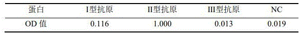
Day 0, 3 mice were immunized with immunogen mixed with Freund's complete adjuvant (CFA) by subcutaneous multi-point injection via the back, 4 times per mouse, 1 week apart, and mouse serum was collected after 3 rd immunization, and antibody titers were detected by indirect ELISA as shown below, as the results in Table 1 show that the mouse serum antibody titers were all 1:50000 or more can be used for fusion.
TABLE 1 ELISA detection results of serum antibodies from three mice

(3) Indirect ELISA method
Poliovirus type II antigens were coated on 96-well plates, 100 ng/well. Negative Control (NC) wells were added with 100. Mu.l of PBS solution containing 5% nonfat dry milk, overnight at 4 ℃; removing liquid in the hole, washing the plate 3 times by PBS, adding PBS solution containing 5% skimmed milk powder, 250 μl/hole, and sealing at room temperature for 1 hr; removing liquid in the wells, washing the plates for 1 time by using PBS, adding primary antibodies (tail blood is diluted in a gradient manner from 1:500 to 1:50000; hybridoma cell culture supernatant is diluted in a proportion of 1:1; mouse ascites is diluted in a gradient manner from 1:1000 to 1:200000), and incubating for 1 hour at room temperature; the wells were discarded, the plates were washed 3 times with PBS, HRP-conjugated goat anti-mouse IgG Fc (1:2000 dilution) was added and incubated for 1 hour at room temperature; removing liquid in the hole, washing the plate for 5 times by using PBS, adding substrate A and substrate B, and reacting for 20min at room temperature in a dark place with 50 μl/hole; the reaction was stopped by adding 50. Mu.l of stop solution, and A450 was measured by a microplate reader, with A450 (positive) >2 times A450 (NC) as a criterion.
(4) Preparation and screening of hybridoma cells
The 3# mice with the highest antibody titers were taken for fusion. 3 days prior to fusion, boost was performed with immunogen mixed with Freund's incomplete adjuvant (IFA), 0.08 ml/min, spleen isolated spleen cells were taken for cell fusion. Individual spleen cells were first isolated by PEG4000 (500 g/L) and murine myeloma SP 2/0 cells at 4:1 ratio, culturing in 96-well plate at 37deg.C for 14 days in DMEM medium containing 20% FBS and 2% HAT; collecting supernatant, screening positive clone cells, further performing amplification culture by using a DMEM medium containing 20% FBS, collecting supernatant, performing rescreening, performing amplification culture on the screened positive clone cells, collecting supernatant, obtaining positive clones through freezing and resuscitating, and collecting supernatant. The culture supernatant was examined by indirect ELISA, and the supernatant-positive cell lines were selected. The results are shown in Table 2. And 9 positive cell strains are selected from 564 cell strains, and finally 7 total cell strains capable of stably secreting monoclonal antibodies against poliovirus type II antigens are selected from the cell strains, wherein NC is negative control, and PC is positive control.
TABLE 2 ELISA detection results of cell line supernatants stably secreting monoclonal antibodies to poliovirus type II antigens

(5) Specific antibody detection of hybridoma cell secretion supernatant
The coating antigen is poliovirus type I antigen, poliovirus type II antigen and poliovirus type III antigen, respectively, 100 ng/well by using an indirect ELISA method. The detection of the antibody secreted by the 9 hybridoma cells in the cell supernatant shows that 7 hybridoma cells secreted by the cell supernatant react positively with poliovirus type II antigens and react negatively with poliovirus type I antigens and poliovirus type III antigens (shown in Table 2), and the 7 hybridoma cells are determined to be type-specific antibodies.
(6) Neutralizing antibody Activity assay
The neutralizing activity of the antibodies and their antibody titer were measured using a polio pseudovirus neutralization assay to measure the ability of the antibodies to clear or significantly reduce pseudovirus virulence, as follows:
1) The serum to be tested was diluted in 96-well cell culture plates using cell culture medium, a total of 8 gradients, and control wells of medium-replacement antibody (negative control) and medium control wells (blank control) were established, each 100 μl per well.
2) Polio type II pseudoviruses were diluted to working titer using medium, 100 μl per well was added to the plate, and blank wells were added to 100 μl of medium and incubated for 2 hours at 37 ℃.
3) RD cells were made to viable cell density 3X 10 5 Per ml of cell suspension, 100. Mu.l per well was added to the plate, 37℃and 5% CO concentration 2 Culturing in an incubator for 12-20 hours.
4) The medium in the 96-well cell culture plate is thrown off and gently buckled on absorbent paper, 50 μl of DPBS is added into each well, 50 μl of prepared Luciferase reaction substrate (note the light shielding of the sample adding groove) is added, the reaction is carried out for 2min at room temperature and light shielding, and the RLU value of each well is detected on the machine (1000 ms is read by a Luciferase channel).
5) The inhibition of infection in the test wells was calculated according to the following formula: infection inhibition ratio = 1- (test well RLU-control RLU)/(negative control RLU-control RLU). The neutralization titer of the test sample is calculated by using a Reed-Muench method: the minimum value of the inhibition rate of more than 50% in each dilution of the test sample is taken as an inhibition rate of more than 50%, and the maximum value of the inhibition rate of less than 50% is taken as an inhibition rate of less than 50%; calculating a distance ratio: distance ratio = (inhibition of greater than 50% -50)/(inhibition of greater than 50% -inhibition of less than 50%) the 50% inhibition dilution logarithm: 50% inhibition dilution log = 50% dilution log + distance ratio x dilution factor log, the neutralization titer in the test sample was 50% positive endpoint dilution.
The results are shown in Table 3, and 3 hybridoma cell lines 2B7, 2G4 and 3C10 were clones having neutralizing activity.
TABLE 3 detection of neutralizing antibody Activity of hybridoma cell secretion supernatants

(7) Ascites preparation, antibody purification and antibody titer detection
And 6 hybridoma cells are selected to prepare ascites, 2 mice are inoculated to each clone cell, the ascites is collected in 8-10 days, the ascites is purified by using a Protein G affinity column, a purified antibody is obtained, and ELISA detection is carried out on the ascites antibody and the purified antibody. As shown in tables 4 and 5, the antibody titers secreted by 3 hybridoma cells 2B7, 2G4 and 3C10 all reached over 2 ten thousand, and the antibody recognition immunogen titers after purification all reached 0.005ug/ml.
TABLE 4 ELISA detection results of mouse ascites antibody

TABLE 5 ELISA detection results of purified antibodies

(8) Biotin labeling
The purified 6 antibodies were individually biotin-labeled, followed by different antibody pairing tests. Pairing experiments with biotin-labeled antibodies and unlabeled antibodies as coated antibodies showed a gradient in each pairing profile, and 6 of these pairs were selected for reproducibility testing. The final results showed that the two sets of 5H10-2G4-biotin and 3C10-2G4-biotin had higher pairing signals and better NC, and that the paired 5H10 was determined as the coating antibody and 2G4-biotin as the detection antibody, considering the poor specificity of monoclonal antibody 3C10, as shown in Table 6.
TABLE 6 results of paired antibody ELISA detection

(9) Subculture and preservation of hybridoma cells
The hybridoma cells are continuously cultured and passaged in a DMEM culture medium containing 10% fetal bovine serum, after the culture is carried out for 10 generations, two strains of 2G4 and 5H10 hybridoma cells can still grow well and stably passaged, and the anti-polio virus type II monoclonal antibodies can be continuously and stably secreted.
After obtaining hybridoma cells producing a desired monoclonal antibody, a portion of the hybridoma cells are maintained, or else mutations or chromosomal shifts may occur during serial passage to the point that inherent or antibody-producing properties are lost; in addition, the cell lines are inevitably destroyed in the long-term culture process without pollution, so that the cell lines are necessary to be preserved. The preservation method comprises the following steps: the old culture medium in the cell culture flask was removed, and 10% FCS-1640 was added to suspend the cells. Centrifuging at 1000rpm/min for 10min, and removing supernatant. The cell sediment is prepared into suspension by 10% dimethyl sulfoxide protective solution to form 1.0X10 7 Cells/ml. Sampling, staining with trypan blue, and counting living cells with the number of more than 95%. And finally, sub-packaging the cells into ampoule bottles by using a sterile syringe, wherein each bottle is 0.5ml to 1.0ml, and sealing the ampoule bottles in a sealing way. Standing at 4deg.C for 2 hr, transferring into refrigerator at-70deg.C for 15 hr, and transferring into liquid nitrogen for freezing.
Example 2: identification of 5H10 antibodies
(1) Subculture and preservation of hybridoma cells
Adult BALB/c mice were selected, and were inoculated intraperitoneally with pristane, 0.5ml each. The 16 th generation clone5H10 hybridoma cells are inoculated in the abdominal cavity after 7-10 days, and each mouse is 1 multiplied by 10 6 ~2×10 6 And each. After 5 days of interval, when the abdomen is obviously enlarged and touched by hands, the skin has tension, and ascites is collected by a 9-gauge needle.
(2) Purification of antibodies
The ascites was centrifuged at 13000rpm/min for 30 minutes to remove cellular components and other sediments and the supernatant was collected. Purifying by Protein G affinity chromatography to obtain monoclonal antibody clone5H10 of poliovirus type II antigen.
(3) Antibody purity detection
The purified antibody was subjected to 12% SDS-PAGE with a purity of 95% or more.
(4) Determination of antibody light chain and heavy chain variable region gene sequence
Extracting mRNA of the 5H10 hybridoma cells, reversely transcribing the mRNA into cDNA, performing high-fidelity PCR amplification by using a variable region universal primer, inserting a PCR product fragment into a T vector for DNA sequence determination, and determining the variable region gene sequence of the 5H 10: the heavy chain is shown as SEQ ID NO.19, and the light chain is shown as SEQ ID NO. 20. The obtained nucleotide sequence is translated into an amino acid sequence. 5H10 variable region amino acid sequence: the heavy chain is shown as SEQ ID NO.17, and the light chain is shown as SEQ ID NO. 18.
(5) Detection of antibody type specificity
As shown by the detection result of the specific antibody of the hybridoma cell secretion supernatant in the example 1 (5), the 5H10 antibody is positive only for poliovirus type II antigens, and is negative for poliovirus type I antigens and poliovirus type III antigens, and the 5H10 antibody is proved to be a type-specific monoclonal antibody.
(6) Identification of antibody neutralizing Activity
The results of the antibody neutralization activity test as in example 1 (6) show that the 5H10 antibody has a neutralizing activity as a monoclonal antibody. The purified antibody was detected by the method for detecting a neutralizing antibody according to example 1 (6), and the result showed that the neutralizing titer was 3227.
Example 3: identification of 2G4 antibodies
(1) Subculture and preservation of hybridoma cells
Adult BALB/c mice were selected, and were inoculated intraperitoneally with pristane, 0.5ml each. The 16 th generation clone2G4 hybridoma cells are inoculated in the abdominal cavity after 7-10 days, and each mouse is 1 multiplied by 10 6 ~2×10 6 And each. After 5 days of interval, when the abdomen is obviously enlarged and touched by hands, the skin has tension, and ascites is collected by a 9-gauge needle.
(2) Purification of antibodies
The ascites was centrifuged at 13000rpm/min for 30 minutes to remove cellular components and other sediments and the supernatant was collected. Purifying by Protein G affinity chromatography to obtain monoclonal antibody clone2G4 of poliovirus type II antigen.
(3) Antibody purity detection
The purified antibody was subjected to 12% SDS-PAGE with a purity of 95% or more.
(4) Determination of antibody light chain and heavy chain variable region gene sequence
Extracting mRNA of the 2G4 hybridoma cells, reversely transcribing the mRNA into cDNA, performing high-fidelity PCR amplification by using a variable region universal primer, inserting a PCR product fragment into a T vector for DNA sequence determination, and determining the variable region gene sequence of the 2G 4: the heavy chain is shown as SEQ ID NO.9, and the light chain is shown as SEQ ID NO. 10. The obtained nucleotide sequence is translated into an amino acid sequence. 2G4 variable region amino acid sequence: the heavy chain is shown as SEQ ID NO.7, and the light chain is shown as SEQ ID NO. 8.
(5) Detection of antibody type specificity
As shown by the detection result of the specific antibody of the hybridoma cell secretion supernatant in the example 1 (5), the 2G4 antibody only reacts positively to the poliovirus type II antigen, and is negative to the poliovirus type I antigen and the poliovirus type III antigen, and the 2G4 antibody is proved to be a type-specific monoclonal antibody.
(6) Identification of antibody neutralizing Activity
As shown by the results of the antibody neutralization activity detection in example 1 (6), the 2G4 antibody was a monoclonal antibody having the neutralization activity. The purified antibody was detected by the method for detecting a neutralizing antibody according to example 1 (6), and the result showed that the neutralizing titer was 42739.
Example 4: poliovirus type II antigen detection kit
Development of a poliovirus type II double-antibody sandwich ELISA kit for detecting the level of poliovirus type II antigen was performed using the monoclonal antibodies 2G4 and 5H10 obtained in example 1.
(1) Establishment of double-antibody sandwich ELISA method
1) Principle of the kit:
the two antibodies of 2G4 and 5H10 are poliovirus type II specific antibodies and have neutralizing activity, 5H10 is used as a coating antibody, 2G4 is used as a biotin-labeled antibody, and a double-antibody sandwich ELISA detection kit is established. When the sample solution to be detected or the standard solution is added into the ELISA plate which is pre-coated with the 5H10 antibody, the 2G4-Biotin secondary antibody is added, the HRP-marked avidin is added, the color development is carried out by using a color developing solution, the sample absorption value and the content of the poliovirus type II antigen in the sample are positively correlated in a linear range, and the content of the poliovirus type II antigen in the sample can be obtained by comparing with a standard curve. Meanwhile, the concentration range of the poliovirus type II antigen in the sample can be roughly judged by comparing the color of the poliovirus type II antigen standard solution with the serial concentration according to the color of the ELISA plate.
2) Composition of the kit
a. ELISA plate coated with coated antibody: the coated antibody is 5H10 type specific neutralizing monoclonal antibody, the coated antibody is diluted to 10 mug/ml by using a coating buffer solution, the coated antibody is coated on a 96-well ELISA plate, 100 mug of the coated antibody is added into each well, and the coated antibody is placed in an environment of 2-8 ℃ for incubation for 8 hours. The coating liquid is discarded, the washing liquid is washed for 3 times, and the coating liquid is patted dry. Blocking solution was then added to the elisa plate and incubated at 37 ℃ for 1.5 hours at 100 μl per well. Pouring out the liquid in the hole, beating to dry, and vacuum sealing and preserving by using an aluminum film after drying. Wherein the coating buffer solution is carbonate buffer solution with pH value of 9.5 and 0.5 mol/L; the blocking solution used was PBS containing bovine serum albumin at a final concentration of 3% by mass.
b. Biotin-labeled antibody: the labeling substance is biotin, the labeled antibody is 5H10 type specific monoclonal antibody, and the working concentration of the biotin-labeled antibody is 0.5 mug/ml.
c. Enzyme-labeled avidin: the working concentration was 1:100 fold dilution.
d. BSA: 3.0g of PBS containing 3% BSA by mass as a diluent was prepared for each pack.
e. Substrate color development liquid: the dye consists of a developing solution A and a developing solution B, wherein the developing solution A is hydrogen peroxide, and the developing solution B is o-phenylenediamine.
f. Stop solution: 1-2mol/L sulfuric acid solution.
g. Concentrated 20X wash: phosphate buffer solution containing Tween 20 and 0.3mol/L at a final concentration of 0.5% (mass percent) in the concentrated washing solution; 50 ml/bottle, 1 bottle.
h. Kit instructions.
(2) Kit specificity verification
The poliovirus I antigen, the poliovirus II antigen and the poliovirus III antigen are prepared into 80ng/ml test samples, ELISA detection is carried out on the poliovirus I antigen, the poliovirus II antigen and the poliovirus III antigen by using the poliovirus II antigen detection kit, the result is shown in Table 7, the poliovirus II antigen detection kit is positive for the poliovirus II antigen, and is negative for the poliovirus I antigen and the poliovirus III antigen, and the specificity of the kit is good.
TABLE 7 poliovirus type II antigen kit specificity verification
(3) Kit sensitivity verification
The lowest antigen concentration detected by the kit is the key to determining the sensitivity of the kit. Poliovirus type II antigens were formulated as 10ug/ml test samples, diluted to 40ng/ml, 20ng/ml, 10ng/ml, 5ng/ml and 2.5ng/ml, respectively. And repeatedly measuring the diluted sample for 6 times by using the kit, and taking the average value of the lower limit of detection of the 6 standard curves, thereby finally determining that the sensitivity of the kit is 2.5ng/ml.
(4) Kit precision verification
1) Precision verification-intra-plate variability
3 poliovirus type II antigens at concentrations of 30ng/ml,15ng/ml and 7.5ng/ml, respectively, were added to the elisa plate, 5 duplicate wells (n=6) were made per antigen concentration, and the sample dilutions were used as negative controls. The OD value variation coefficient (Coefficient of variation, CV) between wells of the same concentration in the elisa plate was calculated, and the results are shown in table 8, wherein the in-plate CV (%) values were all less than 10%, indicating that the in-plate precision of the kit was good.
TABLE 8 variation coefficient in poliovirus type II antigen kit plate

2) Plate-to-plate differences
3 poliovirus type II antigens at concentrations of 30ng/ml,15ng/ml and 7.5ng/ml, respectively, were added to the elisa plate, 5 duplicate wells (n=6) were made per antigen concentration, and the sample dilutions were used as negative controls. The OD value variation coefficient (Coefficient of variation, CV) between wells of the same concentration in the elisa plate was calculated, and the results are shown in table 9, wherein the values of the CV (%) between the plates were all less than 20%, indicating that the accuracy in the plates of the kit was good.
TABLE 9 variation coefficient between plates of poliovirus type II antigen kit

(5) Kit accuracy verification
And adding 3 poliovirus type II antigens with the concentration of 30ng/ml,15ng/ml and 7.5ng/ml respectively into the ELISA plate, and calculating the protein concentration of each antigen by taking the absorbance value of the antigen content corresponding to the antigen content as a standard curve to analyze the accuracy of the kit. The results are shown in Table 10, with each antigen ranging from 100% to 117% accuracy and good kit accuracy.
TABLE 10 accuracy analysis of poliovirus type II antigen kits

(6) Kit repeatability verification
Poliovirus type II antigen is dilutedReleasing to 5 concentrations of 40ng/ml, 20ng/ml, 10ng/ml, 5ng/ml and 2.5ng/ml, taking sample diluent as negative control, taking the content of poliovirus type II antigen in the test sample as standard curve corresponding to absorbance value, detecting each concentration 6 times, and linear correlation coefficient R of the standard curve 2 The results are shown in Table 11, the correlation coefficient R 2 And the detection method is not lower than 0.985, which shows that the detection method has good linear correlation and repeatability.
TABLE 11 Linear correlation and reproducibility verification of poliovirus type II antigen kits

(7) Shelf life test of kit
The preservation condition of the kit is 2-8 ℃, and the maximum absorbance value (zero standard) of the kit is measured after 6 months, so that the actual measured value of the poliovirus type II antigen is within the normal range. The kit can be temporarily considered to be stored for at least 6 months at 2-8 ℃.
The foregoing is merely a preferred embodiment of the present invention and it should be noted that modifications and adaptations to those skilled in the art may be made without departing from the principles of the present invention, which are intended to be comprehended within the scope of the present invention.
Claims (13)
1. An antibody that specifically binds to a poliovirus type II antigen, characterized in that the antibody comprises: the amino acid sequences of the CDR1, CDR2 and CDR3 regions of the heavy chain variable region shown in SEQ ID NO.1-3 in sequence and the amino acid sequences of the CDR1, CDR2 and CDR3 regions of the light chain variable region shown in SEQ ID NO.4-6 in sequence, or amino acid sequences having more than 80% identity and the same or substantially the same biological activity as the amino acid sequences.
2. An antibody that specifically binds to a poliovirus type II antigen, characterized in that said antibody comprises an amino acid sequence of a heavy chain variable region as shown in SEQ ID No.7 and an amino acid sequence of a light chain variable region as shown in SEQ ID No.8, or an amino acid sequence that has more than 80% identity to said amino acid sequences and has the same or substantially the same biological activity.
3. An antigen binding portion that specifically binds to a poliovirus type II antigen, characterized in that the antigen binding portion comprises: amino acid sequences of CDR1, CDR2 and CDR3 regions of the heavy chain variable region shown in SEQ ID nos. 1 to 3 in sequence and amino acid sequences of CDR1, CDR2 and CDR3 regions of the light chain variable region shown in SEQ ID nos. 4 to 6 in sequence, or amino acid sequences having 80% or more identity and the same or substantially the same biological activity as said amino acid sequences;
wherein the antigen binding portion is Fab, fab ', F (ab') 2 Fd, dAb, complementarity determining region fragments or single chain antibodies.
4. The antibody of claim 1 or 2, wherein the antibody is a humanized antibody.
5. An isolated polynucleotide encoding an amino acid sequence as set forth in the antibody or antigen-binding portion of claim 1, 2 or 3.
6. The isolated polynucleotide of claim 5, wherein the isolated polynucleotide encodes the amino acid sequence of the heavy chain variable region or the amino acid sequence of the light chain variable region of claim 2;
the polynucleotide sequence for encoding the heavy chain variable region amino acid sequence is shown as SEQ ID NO.9, and the polynucleotide sequence for encoding the light chain variable region amino acid sequence is shown as SEQ ID NO. 10.
7. A vector comprising the sequence of the polynucleotide of claim 5 or 6.
8. An isolated cell comprising the antibody of claim 1, 2 or 4, or the antigen binding portion of claim 3.
9. A kit comprising the antibody of claim 1, 2 or 4, or the antigen binding portion of claim 3.
10. Use of the antibody of claim 1, 2 or 4, the antigen binding portion of claim 3, the isolated nucleotide of claim 5 or 6, the vector of claim 7, the isolated cell of claim 8 or the kit of claim 9 in detecting the presence or level of poliovirus type II antigen in a sample.
11. A pharmaceutical composition comprising the antibody of claim 1, 2 or 4, the antigen-binding portion of claim 3, the isolated nucleotide of claim 5 or 6, the vector of claim 7, the isolated cell of claim 8, and a pharmaceutically acceptable carrier.
12. An immunoconjugate comprising the antibody of claim 1, 2 or 4 or the antigen binding portion of claim 3 linked to a therapeutic agent or detectable label;
the therapeutic agent includes a drug, enzyme, toxin, cytokine or radionuclide.
13. Use of a pharmaceutical composition according to claim 11 or an immunoconjugate according to claim 12 for the manufacture of a medicament for the treatment of poliovirus type II infections and diseases caused thereby.
Priority Applications (1)
| Application Number | Priority Date | Filing Date | Title |
|---|---|---|---|
| CN202310619962.4A CN116355082B (en) | 2023-05-30 | 2023-05-30 | Antibodies that specifically bind to poliovirus type II antigens |
Applications Claiming Priority (1)
| Application Number | Priority Date | Filing Date | Title |
|---|---|---|---|
| CN202310619962.4A CN116355082B (en) | 2023-05-30 | 2023-05-30 | Antibodies that specifically bind to poliovirus type II antigens |
Publications (2)
| Publication Number | Publication Date |
|---|---|
| CN116355082A true CN116355082A (en) | 2023-06-30 |
| CN116355082B CN116355082B (en) | 2023-09-05 |
Family
ID=86941917
Family Applications (1)
| Application Number | Title | Priority Date | Filing Date |
|---|---|---|---|
| CN202310619962.4A Active CN116355082B (en) | 2023-05-30 | 2023-05-30 | Antibodies that specifically bind to poliovirus type II antigens |
Country Status (1)
| Country | Link |
|---|---|
| CN (1) | CN116355082B (en) |
Citations (5)
| Publication number | Priority date | Publication date | Assignee | Title |
|---|---|---|---|---|
| US5332567A (en) * | 1989-08-24 | 1994-07-26 | Immunomedics | Detection and treatment of infections with immunoconjugates |
| CN102876707A (en) * | 2012-10-15 | 2013-01-16 | 北京工业大学 | Preparation method of recombinant poliovirus I-type virus-like particles |
| CN104371979A (en) * | 2014-10-16 | 2015-02-25 | 北京科兴生物制品有限公司 | Sabin strain poliovirus type II monoclonal antibody and application thereof |
| CN105907724A (en) * | 2016-07-02 | 2016-08-31 | 中国食品药品检定研究院 | Poliovirus type II monoclonal antibody and application thereof |
| CN108130332A (en) * | 2016-12-01 | 2018-06-08 | 上海泽润生物科技有限公司 | Recombinant poliovirus type III virus-like particle |
-
2023
- 2023-05-30 CN CN202310619962.4A patent/CN116355082B/en active Active
Patent Citations (5)
| Publication number | Priority date | Publication date | Assignee | Title |
|---|---|---|---|---|
| US5332567A (en) * | 1989-08-24 | 1994-07-26 | Immunomedics | Detection and treatment of infections with immunoconjugates |
| CN102876707A (en) * | 2012-10-15 | 2013-01-16 | 北京工业大学 | Preparation method of recombinant poliovirus I-type virus-like particles |
| CN104371979A (en) * | 2014-10-16 | 2015-02-25 | 北京科兴生物制品有限公司 | Sabin strain poliovirus type II monoclonal antibody and application thereof |
| CN105907724A (en) * | 2016-07-02 | 2016-08-31 | 中国食品药品检定研究院 | Poliovirus type II monoclonal antibody and application thereof |
| CN108130332A (en) * | 2016-12-01 | 2018-06-08 | 上海泽润生物科技有限公司 | Recombinant poliovirus type III virus-like particle |
Non-Patent Citations (1)
| Title |
|---|
| 张春梅,等: "脊髓灰质炎病毒2型和3型D抗原单克隆抗体的制备及鉴定", 细胞与分子免疫学杂志, vol. 30, no. 11, pages 1174 - 1175 * |
Also Published As
| Publication number | Publication date |
|---|---|
| CN116355082B (en) | 2023-09-05 |
Similar Documents
| Publication | Publication Date | Title |
|---|---|---|
| CN110551212A (en) | Preparation method and application of anti-GII.4 norovirus capsid protein VP1 and VLP (virus-like particle) monoclonal antibody | |
| CN105367652B (en) | A kind of monoclonal antibody, preparation method and the application of anti-HPV16 L1 albumen | |
| CN105085673B (en) | Monoclonal antibody and application thereof for detecting coxsackie virus A 16-type virus full particle | |
| CN115724958B (en) | Monoclonal antibody of anti-norovirus GII genomic capsid protein VP1 and application thereof | |
| CN105153302B (en) | A kind of antibody, preparation method and the application of anti-HPV6L1 albumen | |
| CN116355083B (en) | Antibodies that specifically bind to poliovirus type I antigens | |
| CN116410309B (en) | Antibodies that specifically bind to poliovirus type III antigens | |
| TWI487537B (en) | Monoclonal antibodies against enterovirus 71 and uses thereof | |
| WO2022179535A1 (en) | Anti-sars-cov-2 nucleocapsid protein monoclonal antibody, and preparation method therefor and use thereof | |
| CN114957453B (en) | Antibody of human papilloma virus type 6L 1 protein and preparation method thereof | |
| CN119119256A (en) | Monoclonal antibody against Japanese encephalitis virus and Zika virus and its application | |
| CN116355082B (en) | Antibodies that specifically bind to poliovirus type II antigens | |
| CN113817052A (en) | Anti SARS-CoV-2 nucleocapsid protein monoclonal antibody and its preparation method and use | |
| CN115724950B (en) | HPV6 type capsid protein L1 monoclonal antibody, preparation method and application | |
| CN115960220A (en) | Monoclonal antibody specifically binding to coxsackievirus A6 and application thereof | |
| CN115894672A (en) | Monoclonal antibody aiming at norovirus GII genotype VP1 protein and application thereof | |
| CN114957452A (en) | Antibody of human papilloma virus 11 type L1 protein and preparation method thereof | |
| CN118359711B (en) | Anti-poliovirus type 1 antibody, and preparation method and application thereof | |
| CN116239682A (en) | Monoclonal antibody for identifying Coxsackie viruses A2 and A4 and application thereof | |
| CN116514965B (en) | Hepatitis A virus antibody and application thereof | |
| CN115838417B (en) | Antibody for resisting novel crown mutant N protein, preparation method and application thereof | |
| TWI642683B (en) | Test kit containing monoclonal antibody against vp1 protein of enterovirus71(ev71) | |
| US20250092122A1 (en) | Single-domain antibody against cpv and uses thereof | |
| CN116284360A (en) | Monoclonal antibody for recognizing CV-A4 VP1 protein N terminal and application thereof | |
| CN110551211B (en) | Detection kit containing anti-enterovirus 71 type VP1 protein monoclonal antibody |
Legal Events
| Date | Code | Title | Description |
|---|---|---|---|
| PB01 | Publication | ||
| PB01 | Publication | ||
| SE01 | Entry into force of request for substantive examination | ||
| SE01 | Entry into force of request for substantive examination | ||
| GR01 | Patent grant | ||
| GR01 | Patent grant |