CN116348140A - Anti-adenosine receptor (A2aR) antibody - Google Patents
Anti-adenosine receptor (A2aR) antibody Download PDFInfo
- Publication number
- CN116348140A CN116348140A CN202180067415.9A CN202180067415A CN116348140A CN 116348140 A CN116348140 A CN 116348140A CN 202180067415 A CN202180067415 A CN 202180067415A CN 116348140 A CN116348140 A CN 116348140A
- Authority
- CN
- China
- Prior art keywords
- amino acid
- antibody
- acid sequence
- seq
- a2ar
- Prior art date
- Legal status (The legal status is an assumption and is not a legal conclusion. Google has not performed a legal analysis and makes no representation as to the accuracy of the status listed.)
- Pending
Links
Images


Classifications
-
- C—CHEMISTRY; METALLURGY
- C07—ORGANIC CHEMISTRY
- C07K—PEPTIDES
- C07K16/00—Immunoglobulins [IGs], e.g. monoclonal or polyclonal antibodies
- C07K16/18—Immunoglobulins [IGs], e.g. monoclonal or polyclonal antibodies against material from animals or humans
- C07K16/28—Immunoglobulins [IGs], e.g. monoclonal or polyclonal antibodies against material from animals or humans against receptors, cell surface antigens or cell surface determinants
- C07K16/286—Immunoglobulins [IGs], e.g. monoclonal or polyclonal antibodies against material from animals or humans against receptors, cell surface antigens or cell surface determinants against neuromediator receptors, e.g. serotonin receptor, dopamine receptor
-
- A—HUMAN NECESSITIES
- A61—MEDICAL OR VETERINARY SCIENCE; HYGIENE
- A61P—SPECIFIC THERAPEUTIC ACTIVITY OF CHEMICAL COMPOUNDS OR MEDICINAL PREPARATIONS
- A61P35/00—Antineoplastic agents
-
- C—CHEMISTRY; METALLURGY
- C12—BIOCHEMISTRY; BEER; SPIRITS; WINE; VINEGAR; MICROBIOLOGY; ENZYMOLOGY; MUTATION OR GENETIC ENGINEERING
- C12N—MICROORGANISMS OR ENZYMES; COMPOSITIONS THEREOF; PROPAGATING, PRESERVING, OR MAINTAINING MICROORGANISMS; MUTATION OR GENETIC ENGINEERING; CULTURE MEDIA
- C12N15/00—Mutation or genetic engineering; DNA or RNA concerning genetic engineering, vectors, e.g. plasmids, or their isolation, preparation or purification; Use of hosts therefor
- C12N15/09—Recombinant DNA-technology
- C12N15/63—Introduction of foreign genetic material using vectors; Vectors; Use of hosts therefor; Regulation of expression
-
- C—CHEMISTRY; METALLURGY
- C07—ORGANIC CHEMISTRY
- C07K—PEPTIDES
- C07K2317/00—Immunoglobulins specific features
- C07K2317/20—Immunoglobulins specific features characterized by taxonomic origin
- C07K2317/24—Immunoglobulins specific features characterized by taxonomic origin containing regions, domains or residues from different species, e.g. chimeric, humanized or veneered
-
- C—CHEMISTRY; METALLURGY
- C07—ORGANIC CHEMISTRY
- C07K—PEPTIDES
- C07K2317/00—Immunoglobulins specific features
- C07K2317/30—Immunoglobulins specific features characterized by aspects of specificity or valency
- C07K2317/33—Crossreactivity, e.g. for species or epitope, or lack of said crossreactivity
-
- C—CHEMISTRY; METALLURGY
- C07—ORGANIC CHEMISTRY
- C07K—PEPTIDES
- C07K2317/00—Immunoglobulins specific features
- C07K2317/50—Immunoglobulins specific features characterized by immunoglobulin fragments
- C07K2317/56—Immunoglobulins specific features characterized by immunoglobulin fragments variable (Fv) region, i.e. VH and/or VL
- C07K2317/565—Complementarity determining region [CDR]
-
- C—CHEMISTRY; METALLURGY
- C07—ORGANIC CHEMISTRY
- C07K—PEPTIDES
- C07K2317/00—Immunoglobulins specific features
- C07K2317/70—Immunoglobulins specific features characterized by effect upon binding to a cell or to an antigen
- C07K2317/76—Antagonist effect on antigen, e.g. neutralization or inhibition of binding
-
- C—CHEMISTRY; METALLURGY
- C07—ORGANIC CHEMISTRY
- C07K—PEPTIDES
- C07K2317/00—Immunoglobulins specific features
- C07K2317/90—Immunoglobulins specific features characterized by (pharmaco)kinetic aspects or by stability of the immunoglobulin
- C07K2317/92—Affinity (KD), association rate (Ka), dissociation rate (Kd) or EC50 value
Landscapes
- Health & Medical Sciences (AREA)
- Chemical & Material Sciences (AREA)
- Organic Chemistry (AREA)
- Genetics & Genomics (AREA)
- Life Sciences & Earth Sciences (AREA)
- Engineering & Computer Science (AREA)
- General Health & Medical Sciences (AREA)
- Biomedical Technology (AREA)
- Immunology (AREA)
- Molecular Biology (AREA)
- Medicinal Chemistry (AREA)
- Biophysics (AREA)
- Biochemistry (AREA)
- Biotechnology (AREA)
- Zoology (AREA)
- Wood Science & Technology (AREA)
- Bioinformatics & Cheminformatics (AREA)
- General Engineering & Computer Science (AREA)
- Proteomics, Peptides & Aminoacids (AREA)
- Neurology (AREA)
- Pharmacology & Pharmacy (AREA)
- Veterinary Medicine (AREA)
- Physics & Mathematics (AREA)
- Public Health (AREA)
- Animal Behavior & Ethology (AREA)
- Plant Pathology (AREA)
- Nuclear Medicine, Radiotherapy & Molecular Imaging (AREA)
- Microbiology (AREA)
- General Chemical & Material Sciences (AREA)
- Chemical Kinetics & Catalysis (AREA)
- Medicines Containing Antibodies Or Antigens For Use As Internal Diagnostic Agents (AREA)
- Peptides Or Proteins (AREA)
- Saccharide Compounds (AREA)
- Pharmaceuticals Containing Other Organic And Inorganic Compounds (AREA)
- Preparation Of Compounds By Using Micro-Organisms (AREA)
- Micro-Organisms Or Cultivation Processes Thereof (AREA)
- Medicines That Contain Protein Lipid Enzymes And Other Medicines (AREA)
Abstract
The present invention provides an anti-A2 aR antigen binding molecule, including antibodies and antigen binding fragments thereof, and methods of using the same to treat a variety of diseases associated with aberrant adenosine signaling, including cancer, chronic diseases, chronic infections, autoimmune diseases, inflammatory diseases, neurodegenerative diseases, and fibrotic diseases.
Description
The present application relates to and claims priority to U.S. provisional application No. 63/085,612 filed on 9/30/2020. The entire contents of the above application are expressly incorporated herein by reference.
Technical Field
The present invention relates to antibodies for cancer treatment, in particular to antibodies for adenosine receptor A2aR for cancer immunotherapy.
Background
The immune system plays an important role in the recognition and elimination of tumor cells. Tumor cells utilize various mechanisms to evade immune-mediated tumor cell damage. Among these pathways, tumor cells utilize the adenosine (a highly potent inhibitor of effector T cell function) signaling pathway to increase adenosine levels and responsiveness to adenosine to circumvent immune defenses.
Adenosine is a purine nucleoside produced by the degradation of Adenosine Triphosphate (ATP). Under adverse conditions (such as hypoxia, ischemia, inflammation or cancer), the extracellular levels of adenosine are significantly increased. Once released, adenosine activates the cell signaling pathway by acting with four known G protein-coupled receptors, namely the adenosine A1 receptor subtype (A1R), the adenosine A2A receptor subtype (A2 aR), the adenosine A2B receptor subtype (A2 bR) and the adenosine A3 receptor subtype (A3R).
Adenosine levels are primarily controlled by the activity of CD39 and CD 73. CD39 and CD73 are two extracellular enzymes that co-act in a two-step reaction to convert proinflammatory ATP to immunosuppressive adenosine. CD39 hydrolyzes ATP to AMP, which is further hydrolyzed by CD73 to adenosine, which can readily enter most cells. In addition, when tumor cells undergo cell death due to metabolic or hypoxic stress, they release intracellular stored ATP (which is generally not permeable to the cells) into the extracellular space.
In the tumor microenvironment, the adenosine produced by CD73 can promote the growth and survival of tumor cells while inhibiting anti-tumor immune responses. Cancer cells express CD73 at high levels in tumor tissue, and accumulation of CD73 is associated with breast cancer and ovariesLow overall survival and low recurrence-free survival in cancer patients are associated. CD73 and adenosine support the formation of pro-growth new blood vessels in cancer cells, and metastasis and survival of cancer cells. Adenosine binds to A2A (or a on T cells 2A ) Receptor (A2 aR) and activates intracellular signaling cascades, thereby inhibiting T cell activation and function. A2aR is a member of the G protein-coupled receptor adenosine receptor, which also includes A1R, A2bR and A3R, and A2aR is an anti-inflammatory effector of extracellular adenosine, expressed primarily in brain and lymphoid tissue cells.
An abnormally high concentration of adenosine in the immune microenvironment activates A2aR and initiates a negative feedback loop that allows tumors to evade immune recognition. Specifically, adenosine-mediated activation of A2aR enables tumors to escape immune surveillance by: inhibiting ifnγ production, inhibiting various anti-tumor immune cells (including CD8 + Activity of T cells, dendritic cells, natural killer cells and M1 macrophages, while enhancing immune suppressive cell types (including myeloid-derived suppressor cells (MDSCs) and T-regulatory (T) reg ) Cells). Activation of A2aRs on tumor cells can also promote tumor cell metastasis.
Although some small-molecule antagonists of the A2A receptor have entered the stage of clinical trials for the treatment of parkinson's disease and cancer, there is still a lack of blocking A2aR with biological agent candidates in the treatment of cancer. Mice treated with an A2aR antagonist (e.g., ZM 241385) showed significant delay in tumor growth due to reduced immunosuppression of effector T cells. This was further emphasized by the A2aR knockout mice, which showed increased tumor rejection. In addition, blocking A2aR with a small molecule antagonist in combination with a monoclonal antibody inhibits PD-1/PD-L1 or CTLA-4 has a synergistic effect on enhancing immune response compared to blocking a single PD-1/PD-L1 or CTLA-4 pathway.
Thus, modulation of A2aR activity, adenosine concentration and/or CD39/CD73 expression and activation of effector immune cells in the tumor microenvironment is an attractive therapeutic strategy that may limit tumor progression, improve anti-tumor immune responses, avoid treatment-induced immune bias, and potentially limit toxicity to normal tissues. There is a need in the art for compositions and methods for treating cancer by modulating (e.g., inhibiting) A2aR activity of immune cells.
Disclosure of Invention
The present invention provides an antigen binding molecule (e.g., an anti-A2 aR antibody or antigen binding fragment thereof) for modulating (e.g., enhancing or inhibiting) the activity of A2aR by specifically binding A2 aR. The A2aR may be on the cell surface, the cell being, for example, a mammalian cell (e.g., a mammalian immune cell (e.g., a mouse immune cell, a cynomolgus monkey immune cell, or a human immune cell)). The invention also provides methods of using an antigen binding molecule of the invention (e.g., an anti-A2 aR antibody or antigen binding fragment thereof) for modulating (e.g., inhibiting) A2aR activity, or for treating a subject (e.g., a subject suffering from or susceptible to A2 aR-related disease) that benefits from modulating (e.g., inhibiting) A2aR activity.
In one aspect, the invention provides an isolated antigen binding molecule, such as an antibody or antigen binding fragment thereof, that binds to the human adenosine A2A receptor (A2 aR). The antibody comprises a heavy chain variable region (VH) comprising three heavy chain complementarity determining regions HCDR1, HCDR2 and HCDR3 from the N-terminus to the C-terminus; and a light chain variable region (VL) comprising three light chain complementarity determining regions LCDR1, LCDR2 and LCDR3 from the N-terminus to the C-terminus; wherein (a) HCDR1 comprises amino acid sequence X 1 -X 2 -W-M-N (SEQ ID NO: 8), wherein X 1 Is S or R, X 2 Y or F; (b) HCDR2 comprises the amino acid sequence R-I-D-P-X 3 -D-S-E-X 4 -X 5 -Y-X 6 -H-K-F-W-X 7 (SEQ ID NO: 9), wherein X 3 Is S or Y, X 4 Is A or T, X 5 Is H or Q, X 6 Is H or N, X 7 Is D or G; (c) HCDR3 comprises the amino acid sequence SLYGGDY (SEQ ID NO: 3); (d) LCDR1 contains the amino acid sequence R-S-S-Q-S-X 17 -V-H-X 18 -N-G-N-T-Y-L-E (SEQ ID NO: 30), wherein X 17 Is L or I, X 18 R or S; (e) LCDR2 comprises the amino acid sequence K-V-S-N-R-F-S (SEQ ID NO: 26); and (f) LCDR3 comprises amino acid sequence X 19 -Q-G-S-H-V-P-L-T (SEQ ID NO: 31), wherein X 19 Y or F.
In various aspects of the invention and embodiments thereof, the antibody is an antigen-binding fragment of an antibody. In various aspects of the invention and embodiments thereof, human A2aR comprises the sequence shown as SEQ ID NO. 50.
In one embodiment, (a) HCDR1 comprises an amino acid sequence selected from the group consisting of SEQ ID NOs 1, 4 and 6; (b) HCDR2 comprises an amino acid sequence selected from the group consisting of SEQ ID NOs 2, 5 and 7; (c) HCDR3 comprises the amino acid sequence shown in SEQ ID NO. 3; (d) LCDR1 comprises the amino acid sequence shown in SEQ ID NO. 25 or 28; (e) LCDR2 comprises the amino acid sequence shown in SEQ ID NO. 26; and (f) LCDR3 comprises the amino acid sequence shown in SEQ ID NO 27 or 29.
In another embodiment, the isolated antigen binding molecule, e.g., the antibody, comprises: (a) HCDR1 comprising the amino acid sequence shown in SEQ ID NO. 1, HCDR2 comprising the amino acid sequence shown in SEQ ID NO. 2, HCDR3 comprising the amino acid sequence shown in SEQ ID NO. 3, LCDR1 comprising the amino acid sequence shown in SEQ ID NO. 25, LCDR2 comprising the amino acid sequence shown in SEQ ID NO. 26, LCDR3 comprising the amino acid sequence shown in SEQ ID NO. 27; (b) HCDR1 comprising the amino acid sequence shown in SEQ ID NO. 4, HCDR2 comprising the amino acid sequence shown in SEQ ID NO. 5, HCDR3 comprising the amino acid sequence shown in SEQ ID NO. 3, LCDR1 comprising the amino acid sequence shown in SEQ ID NO. 28, LCDR2 comprising the amino acid sequence shown in SEQ ID NO. 26, LCDR3 comprising the amino acid sequence shown in SEQ ID NO. 29; or (c) HCDR1 comprising the amino acid sequence shown in SEQ ID NO. 6, HCDR2 comprising the amino acid sequence shown in SEQ ID NO. 7, HCDR3 comprising the amino acid sequence shown in SEQ ID NO. 3, LCDR1 comprising the amino acid sequence shown in SEQ ID NO. 25, LCDR2 comprising the amino acid sequence shown in SEQ ID NO. 26, LCDR3 comprising the amino acid sequence shown in SEQ ID NO. 29.
In yet another embodiment, (a) HCDR1 comprises an amino acid sequence selected from the group consisting of SEQ ID NOs 21, 22, 23 and 24; and (b) HCDR3 comprises an amino acid sequence selected from the group consisting of SEQ ID NOS: 12, 15 and 20.
In yet another embodiment, the antigen binding molecule, e.g., the antibody, comprises: (a) A Heavy Chain Variable Region (HCVR) comprising an amino acid sequence selected from the group consisting of SEQ ID NOs 35, 36, 37, 38, 39 and 40; and (b) a Light Chain Variable Region (LCVR) comprising an amino acid sequence selected from the group consisting of SEQ ID NOs 41, 42 and 43.
In one embodiment, the antigen binding molecule, e.g., the antibody, comprises: (a) HCVR comprising the amino acid sequence shown in SEQ ID No. 35 or 38, and LCVR comprising the amino acid sequence shown in SEQ ID No. 41; (b) HCVR comprising the amino acid sequence shown in SEQ ID No. 36 or 39, and LCVR comprising the amino acid sequence shown in SEQ ID No. 42; or (c) a HCVR comprising the amino acid sequence shown in SEQ ID NO. 37 or 40, and a LCVR comprising the amino acid sequence shown in SEQ ID NO. 43.
In another aspect, the invention provides an isolated antigen binding molecule, such as an antibody that binds to the human adenosine A2A receptor (A2 aR). The antigen binding molecule, e.g., the antibody, comprises: a heavy chain variable region (VH) comprising three heavy chain complementarity determining regions HCDR1, HCDR2 and HCDR3 from the N-terminus to the C-terminus; and a light chain variable region (VL) comprising three light chain complementarity determining regions LCDR1, LCDR2 and LCDR3 from the N-terminus to the C-terminus; wherein (a) HCDR1 comprises an amino acid sequence having about 80%, 85%, 90%, 95%, 96%, 97%, 98%, 99% to about 100% identity to an amino acid sequence selected from the group consisting of SEQ ID NOs 1, 4 and 6; (b) HCDR2 comprises an amino acid sequence having about 80%, 85%, 90%, 95%, 96%, 97%, 98%, 99% to about 100% identity with an amino acid sequence selected from the group consisting of SEQ ID NOs 2, 5 and 7; (c) HCDR3 comprises an amino acid sequence having about 80%, 85%, 90%, 95%, 96%, 97%, 98%, 99% to about 100% identity with the amino acid sequence of SEQ ID No. 3; (d) LCDR1 comprises an amino acid sequence having about 80%, 85%, 90%, 95%, 96%, 97%, 98%, 99% to about 100% identity to an amino acid sequence selected from the group consisting of SEQ ID NOs 25 and 28; (e) LCDR2 comprises amino acid sequences having about 80%, 85%, 90%, 95%, 96%, 97%, 98%, 99% to about 100% identity to the amino acid sequence of SEQ ID NO. 26; and (f) LCDR3 comprises an amino acid sequence having about 80%, 85%, 90%, 95%, 96%, 97%, 98%, 99% to about 100% identity to an amino acid sequence selected from SEQ ID NO 27 or 29.
In one embodiment, (a) HCDR1 comprises a sequence obtained from SEQ ID NO:1, 4 or 6 by 1 or 2 amino acid substitutions; (b) HCDR2 comprises a sequence obtained from SEQ ID No. 2, 5 or 7 by substitution of 1, 2, 3, 4 or 5 amino acids; (c) HCDR3 comprises the sequence shown in SEQ ID NO. 3 or comprises the sequence obtained by 1 or 2 amino acid substitutions of SEQ ID NO. 3; (d) LCDR1 comprises a sequence obtained from SEQ ID NO 25 or 28 substituted with 1, 2, 3 or 4 amino acids; (e) LCDR2 comprises the sequence shown in SEQ ID NO. 26 or comprises a sequence obtained from SEQ ID NO. 26 by 1 or 2 amino acid substitutions; and (f) LCDR3 comprises a sequence obtained from SEQ ID NO 27 or 29 substituted with 1 or 2 amino acids. In another embodiment, the amino acid substitution is a conservative substitution. In another embodiment, (a) HCDR1 comprises an amino acid substitution at position 1 or 2 of HCDR 1; (b) HCDR2 comprises an amino acid substitution at position 5, 9, 10, 12 or 17 of HCDR 2; (c) LCDR1 comprises an amino acid substitution at position 6 or 9 of LCDR 1; or (d) LCDR3 comprises an amino acid substitution at position 1 of LCDR 3. In one embodiment, the amino acid substitution is a conservative substitution. In one embodiment, the amino acid substitution is a conservative substitution. In yet another embodiment, HCDR1, HCDR2, HCDR3, LCDR1, LCDR2 and LCDR3 are defined based on the Kabat numbering scheme.
In one embodiment, (a) HCDR1 comprises a sequence obtained from SEQ ID NO 10, 13 or 16 by 1, 2 or 3 amino acid substitutions; (b) HCDR2 comprises a sequence obtained from SEQ ID No. 11, 14 or 17 by substitution of 1, 2 or 3 amino acids; (c) HCDR3 comprises a sequence obtained from SEQ ID No. 12 or 15 by substitution of 1, 2 or 3 amino acids; (d) LCDR1 comprises a sequence obtained from SEQ ID NO. 32 or 33 substituted with 1, 2, 3 or 4 amino acids; and (e) LCDR3 comprises a sequence obtained from SEQ ID NO 27 or 29 substituted with 1 or 2 amino acids. In another embodiment, the amino acid substitution is a conservative substitution. In another embodiment, (a) HCDR1 comprises an amino acid substitution at position 3, 6 or 7 of HCDR 1; (b) HCDR2 comprises an amino acid substitution at position 4 or 8 of HCDR 2; (c) HCDR3 comprises an amino acid substitution at position 1 of HCDR 3; (d) LCDR1 comprises an amino acid substitution at position 3 or 6 of LCDR 1; or (e) LCDR3 comprises an amino acid substitution at position 1 of LCDR 3. In one embodiment, the amino acid substitution is a conservative substitution. In yet another embodiment, HCDR1, HCDR2, HCDR3, LCDR1, LCDR2 and LCDR3 are defined based on IMGT numbering scheme.
In another aspect, the invention provides an antigen binding molecule, such as an antibody. The antibody comprises: (a) A Heavy Chain Variable Region (HCVR) comprising an amino acid sequence having about 80%, 85%, 90%, 95%, 96%, 97%, 98%, 99% to about 100% identity to an amino acid sequence selected from the group consisting of SEQ ID nos. 35, 36 and 37; and (b) a Light Chain Variable Region (LCVR) comprising an amino acid sequence that is about 80%, 85%, 90%, 95%, 96%, 97%, 98%, 99% to about 100% identical to an amino acid sequence selected from the group consisting of SEQ ID NOS: 41, 42 and 43.
In one embodiment, (a) the HCVR comprises a sequence obtained from SEQ ID NO 35, 36 or 37 substituted with 1, 2, 3, 4, 5, 6, 7, 8, 9, 10, 11 or 12 amino acids; (b) LCVR comprises sequences derived from SEQ ID NOs 41, 42 or 43 substituted with 1, 2, 3, 4, 5, 6, 7, 8, 9 or 10 amino acids. In another embodiment, the amino acid substitution is a conservative substitution.
In one aspect, the invention provides an antigen binding molecule, such as an antibody, that binds to the human adenosine A2A receptor (A2 aR). The antibody comprises: (a) A Heavy Chain Variable Region (HCVR) comprising the amino acid sequence shown in SEQ ID No. 35 or 38; and (b) a Light Chain Variable Region (LCVR) comprising the amino acid sequence shown in SEQ ID NO. 41.
In another aspect, the invention provides an antigen binding molecule, such as an antibody, that binds to the human adenosine 2A receptor (human A2 aR). The antibody comprises: (a) A Heavy Chain Variable Region (HCVR) comprising the amino acid sequence shown in SEQ ID No. 36 or 39; and (b) a Light Chain Variable Region (LCVR) comprising the amino acid sequence shown in SEQ ID NO. 42.
In yet another aspect, the invention provides an antigen binding molecule, such as an antibody, that binds to the human adenosine 2A receptor (human A2 aR). The antibody comprises: (a) A Heavy Chain Variable Region (HCVR) comprising the amino acid sequence shown in SEQ ID No. 37 or 40; and (b) a Light Chain Variable Region (LCVR) comprising the amino acid sequence shown in SEQ ID NO. 43.
In one embodiment of the various aspects of the invention, the heavy and/or light chain of the antigen binding molecule (e.g., the antibody) is N-terminal to a pyroglutamic acid (pE) residue.
In another embodiment, (i) the antibody competes for binding to human A2aR with a monoclonal antibody selected from the group consisting of 1B5-3D7, 3F6-9G5, and 3F8-12E 9; (ii) the antibody inhibits the activity of A2aR; (iii) the antibody enhances an immune response; (iv) the antibody specifically binds to cell surface human A2aR; (v) the antibody reduces cAMP concentration in the tissue; (vi) the antibody reduces the activity of protein kinase a; (vii) The antibody reduces phosphorylation of cAMP response element of the A2aR signaling pathway; or (viii) any combination of (i) - (vii).
In one embodiment, the binding of the antibody to A2aR or cell surface A2aR is determined using a flow cytometry-based assay as described in examples 5, 6 and 7 or an assay substantially similar thereto. In another embodiment, competitive binding of the antibody to A2aR or cell surface A2aR is determined using assays known in the art, including, for example, those described by Harms et al in Microtiter plate-based anti-bodies-competition assay to determine binding affinities and plasma/blood stability of CXCR ligands, scientific Reports,2020:10:16036, doi.org/10.1038/s41598-020-73012-4, or substantially similar thereto. In another embodiment, inhibition of A2aR activity is determined using the assay described in example 4 or an assay substantially similar thereto. In yet another embodiment, the decrease in cAMP concentration is measured using the assay described in example 4 or an assay substantially similar thereto. In one embodiment, the use of Karege et al in Anon-radioactive assay for the cAMP-dependent protein kinase activity in rat Brain homogenates and age-related changes in hippocampus and cortex, brain Res.,2001Jun 8;903 (1-2) 86-93, doi:10.1016/s0006-8993 (01) 02009-x or a method substantially similar thereto to determine a decrease in protein kinase A activity and/or a decrease in phosphorylation of cAMP response elements of the A2aR signaling pathway. In another embodiment, the enhancement of the immune response is determined by methods well known in the art, including, for example, determining an increase in the concentration of inflammatory cytokines in the tissue, an increase in the number of cytotoxic cd8+ T cells.
In one embodiment, the antibody inhibits (e.g., reduces) the activity of A2aR by at least about 10%, about 20%, about 30%, about 40%, about 50%, about 60%, about 70%, about 80%, about 90%, about 95%, or about 100%. In another embodiment, the antibody enhances an immune response by at least about 10%, about 20%, about 30%, about 40%, about 50%, about 60%, about 70%, about 80%, about 90%, about 95%, about 100%, about 1.5-fold, about 2-fold, about 4-fold, or more. In yet another embodiment, the antibody reduces cAMP concentration in a tissue by at least about 10%, about 20%, about 30%, about 40%, about 50%, about 60%, about 70%, about 80%, about 90%, about 95%, or about 100%. In yet another embodiment, the antibody reduces the activity of protein kinase a by at least about 10%, about 20%, about 30%, about 40%, about 50%, about 60%, about 70%, about 80%, about 90%, about 95%, or about 100%. In one embodiment, the antibody reduces phosphorylation of cAMP response element of the A2aR signaling pathway by at least about 10%, about 20%, about 30%, about 40%, about 50%, about 60%, about 70%, about 80%, about 90%, about 95%, or about 100%.
In another embodiment, the antibody specifically binds to human A2aR and/or cynomolgus monkey A2aR. In yet another embodiment, the antibodies specifically bind human A2aR and/or cynomolgus monkey A2aR with similar affinity. In one embodiment, the antibody does not bind to A2aR of a non-primate or binds to A2aR of a non-primate with significantly lower affinity than to human A2aR and/or cynomolgus monkey A2aR. In another embodiment, the antibody reduces the production of intracellular cAMP. In one embodiment, the antibody reduces the concentration of cAMP in the tissue. In one embodiment, the antibody reduces intracellular concentration of cAMP. In another embodiment, the antibody reduces extracellular concentration of cAMP in the tissue. In various aspects of the invention and embodiments thereof, cynomolgus monkey A2aR comprises the sequence shown as SEQ ID NO. 51.
In one aspect, the invention provides an isolated antigen binding molecule, e.g. an antibody, which competes with the antibody of any aspect for binding to human A2aR.
In one embodiment, the antigen binding molecule, e.g., an antibody, is a humanized or chimeric antibody. In another embodiment, the antibody comprises a heavy chain constant region selected from the IgA, igD, igE, igG or IgM classes. In another embodiment, the antibody comprises a heavy chain constant region of the IgG class, wherein IgG is selected from the group consisting of IgG4, igGl, igG2, and IgG3.
In another aspect, the invention provides an isolated polynucleotide encoding the antigen binding molecule, e.g., an antibody, HCVR thereof, LCVR thereof, light chain thereof, heavy chain thereof, or antigen binding fragment thereof of any aspect and various embodiments thereof.
In yet another aspect, the invention provides an expression vector comprising the polynucleotide.
In another aspect, the invention provides a recombinant cell comprising said polynucleotide or said expression vector.
In one aspect, the invention provides methods of making the antigen binding molecules (e.g., antibodies of any aspect and various embodiments thereof). The method comprises expressing the antibody in a recombinant cell and isolating the expressed antibody.
In one aspect, the invention provides a pharmaceutical composition. The pharmaceutical composition comprises an antigen binding molecule, e.g., the antibody, of any aspect and various embodiments thereof, and a pharmaceutically acceptable carrier or diluent.
In one embodiment, the pharmaceutical composition comprises an effective amount of the antibody to (a) specifically bind to cell surface human or cynomolgus monkey A2aR; (b) reducing cAMP concentration in the tissue; (c) inhibiting the activity of human A2aR; (d) Reduces phosphorylation of cAMP response element of the A2aR signaling pathway; (e) enhancing the immune response of the immune cells; (f) decreasing protein kinase a activity; or (g) any combination of (a) - (f). In another embodiment, the decrease in the concentration of cAMP in the tissue as compared to the baseline level is achieved by decreasing intracellular cAMP production and/or decreasing intracellular and/or extracellular concentration of cAMP. In another embodiment, inhibition of A2aR activity is achieved by inhibiting the physiological activity of adenosine.
In one aspect, the invention provides a method of inhibiting A2aR activity expressed on the surface of a cell, comprising contacting the cell with an isolated antibody of any aspect or a pharmaceutical composition of any aspect, thereby inhibiting A2aR activity on the surface of the cell. In one embodiment, inhibition of A2aR results in a decrease in cAMP concentration in the tissue of at least about 10%, about 20%, about 30%, about 40%, about 50%, about 60%, about 70%, about 80%, about 90%, about 95%, or about 100%. In another embodiment, the method is for treating cancer or a neurodegenerative disease. In yet another embodiment, the antigen binding molecule, e.g., an antibody, inhibits A2aR activity by at least about 10%, about 20%, about 30%, about 40%, about 50%, about 60%, about 70%, about 80%, about 90%, about 95%, or about 100%. In another embodiment, the method is for treating cancer or a neurodegenerative disease.
In another aspect, the invention provides a method of enhancing an immune response in a subject. The method comprises administering to the subject the isolated antibody of any aspect or the pharmaceutical composition of any aspect, thereby enhancing the immune response of the subject. In one embodiment, the immune response includes, but is not limited to a) promotion of effector T cell function; b) Lowering T reg Activity; c) Prevent T reg Amplifying; d) Enhancing NK cell function; e) Promoting type 1 activation of antigen presenting cells; or f) reducing immunosuppression in the tumor microenvironment. In certain embodiments, the methods described herein increase the immune response by at least about 10%, about 20%, about 50%, about 60%, about 70%, about 80%, about 90%, about 1-fold, about 2-fold, about 4-fold, or more as compared to baseline levels.
In another aspect, the invention provides a method of inhibiting tumor growth in a subject. The method comprises administering to the subject the isolated antibody of any aspect or the pharmaceutical composition of any aspect, thereby inhibiting tumor growth.
In yet another aspect, the invention provides a method of treating cancer in a subject comprising administering an isolated antibody of any aspect or a pharmaceutical composition of any aspect, thereby treating cancer. In one embodiment, the cancer is any cancer described herein. In a specific embodiment, the cancer is selected from the group consisting of Triple Negative Breast Cancer (TNBC), pancreatic Ductal Adenocarcinoma (PDAC), metastatic castration resistant prostate cancer (mCRPC), renal Cell Carcinoma (RCC), multiple myeloma, colorectal cancer (CRC), and diffuse large B-cell lymphoma (DLBCL).
In yet another aspect, the invention provides a method of treating a neurodegenerative disease in a subject comprising administering an isolated antibody of any aspect or a pharmaceutical composition of any aspect, thereby treating the neurodegenerative disease.
In one embodiment, the method of any of the above aspects results in activation of T cells and directs them to kill tumor target cells.
In another embodiment, the method of any of the above aspects further comprises administering an additional therapeutic agent. In one embodiment, the additional therapeutic agent comprises any of the therapeutic agents described herein. In another embodiment, the other therapeutic agent comprises an anti-tumor agent, radiation therapy, a chemotherapeutic agent, surgery, a cancer vaccine, an agonist of an immune cell stimulating receptor, a cytokine, a cell therapy, or a checkpoint inhibitor. In one embodiment, the other therapeutic agent is an antibody, including a multispecific antibody, e.g., a bispecific antibody.
In another embodiment, the checkpoint inhibitor is selected from the group consisting of inhibitors of PD-1, PD-L1, TIGIT, CTLA-4, PD-1, PD-L2, LAG-3, TIM-3, neurite, BTLA, CECAM-1, CECAM-5, IL-1R8, VISTA, LAIR1, LILRB2, LILRB3, LILRB4, LILRB5, CD96, CD112R, CD160, 2B4, TGF beta-R, KIR, NKG A, or any combination thereof. In yet another embodiment, the inhibitor inhibits an interaction between PD-1 and PD-L1, the inhibitor being selected from the group consisting of pamil mab, na Wu Liyou mab, alemtuzumab, dulcis You Shan mab, BMS-936559, singdi Li Shan mab, terep Li Shan mab, tirelizumab, karilizumab, shu Geli mab, p An Puli mab, kadonil Li Shan mab, sulfamonomethoxine 1, and sulfamonomethoxine 2. In one embodiment, the CTLA4 inhibitor is selected from ipilimumab, california Li Shan antibody, and YH001 (both in the case of the same and in the case of the same).
In yet another embodiment, the other therapeutic agent is an agonist of an immune cell stimulating receptor selected from the group consisting of: ligands for OX40, CD2, CD27, CDS, ICAM-1, LFA-1, ICOS (CD 278), 4-1BB (CD 137), GITR, CD28, CD30, CD40, BAFFR, HVEM, CD7, LIGHT, NKG2C, NKG2D, SLAMF, NKp46, NKp80, CD160, B7-H3, CD83, and any combination thereof.
In one embodiment, the additional therapeutic agent is formulated in the same pharmaceutical composition as the antibody. In another embodiment, the additional therapeutic agent is formulated in a different pharmaceutical composition than the antibody.
In yet another embodiment, the additional therapeutic agent is administered prior to administration of the antigen binding molecule, e.g., antibody, of the various aspects. In another embodiment, the additional therapeutic agent is administered after administration of the antigen binding molecule, e.g., antibody. In another embodiment, the additional therapeutic agent is administered concurrently with the antigen binding molecule, e.g., antibody.
In one aspect, the invention provides a kit. The kit comprises the pharmaceutical composition of any aspect. In one embodiment, the pharmaceutical composition further comprises any one or more of the other therapeutic agents described herein.
Drawings
FIG. 1 shows a graph of antibody titers against human A2aR in serum induced by immunization of mice with DNA encoding human A2aR (one curve represents one mouse serum). Antibody titers in the serum of eight mice (M1-M8) were tested by binding to the Expi293 cells overexpressing human A2aR using flow cytometry. MFI: average fluorescence intensity.
FIG. 2 shows Fluorescence Activated Cell Sorting (FACS) flow scatter plots showing the binding of anti-A2R antibodies isolated from hybridoma cell cultures of four hybridoma clones 1B5-3D7, 3F6-9G5, 3F8-12E9, and 8D5-16E 2. The names of clones, 1B5-3D7, 3F6-9G5, 3F8-12E9 and 8D5-16E2, as used herein, may also represent monoclonal antibodies isolated from these clones, depending on the context. Expi293: an Expi293 cell; a2aR/Expi293: an Expi293 cell transfected with a human A2aR expression vector.
FIG. 3 shows in vitro blocking activity of exemplary antibodies 1B5-3D7, 3F6-9G5, 3F8-12E9 and 8D5-16E2 of the invention. The exemplary anti-human A2aR mAb is a whole IgG molecular form purified from hybridoma culture broth using a cell cAMP-based assay. RLU: relative light units; ZM241385: small molecule A2aR antagonists (Sigma, cat No. Z0153).
FIG. 4 shows in vitro blocking activity of exemplary antibodies 1B5-3D7 and 3F6-9G5 of the invention. The exemplary anti-human A2aR mAb is an antibody in the form of a recombinant whole IgG molecule purified from culture supernatant of an Expi293 cell transiently transfected with an exemplary antibody sequence of the present invention, using a cell cAMP-based assay.
FIG. 5 shows that exemplary antibodies 1B5-3D7 and 3F6-9G5 of the invention specifically bind to cell surface expressed human A2aR. The figure also shows that binding of other sources of anti-human A2aR antibodies to human A2aR expressed on the cell surface was not detected or was very weak. 1B5:1B5-3D7;3F6:3F6-9G5; MAB9497: human adenosine A2aR antibody (R & D Systems, cat# MAB 9497); SDIX-10: the human adenosine A2aR antibody disclosed in U.S. patent publication No. US 2014/032366A 1, clone No. 864H10; SDIX-14: the human adenosine A2aR antibody disclosed in U.S. patent publication No. US 2014/032366A 1, clone No. 864H14; mIgG2a iso: a mouse IgG2a isotype control; hIgG1 iso: human IgG1 isotype control.
FIG. 6 is a flow cytometry scatter plot showing that exemplary antibodies 3F6-9G5 of the present invention bind CD8 derived from human and cynomolgus monkey Peripheral Blood Mononuclear Cells (PBMC) + CD3 + CD 8T cells and CD8 - CD3 + Human A2aR and cynomolgus monkey A2aR expressed on the surface of CD 4T cells. GMI: geometric mean fluorescence intensity.
Detailed description of the preferred embodiments
The present invention and the accompanying drawings are described so as to enable those skilled in the art to practice the invention. It will be appreciated, however, by one skilled in the art that the invention described below may be practiced without such specific details, or that it may be used for other purposes than those described herein. Indeed, it may be modified and used in conjunction with products and techniques known to those skilled in the art in view of this disclosure. The figures and description are intended to illustrate various aspects of the present invention and are not intended to narrow the scope of the claims. Furthermore, it is to be understood that the figures may show aspects of the invention separately, and that elements of one figure may be used in combination with elements shown in other figures.
It should be understood that reference throughout this specification to an aspect, feature, advantage or similar language does not imply that all of the aspects and advantages are readily available with the present invention, nor are they intended to be or have been implemented in any single embodiment of the invention. Rather, language referring to the aspects and advantages is understood to mean that a specific aspect, feature, advantage, or characteristic described in connection with an embodiment is included in at least one embodiment of the present invention. Thus, discussion of the aspects and advantages, and similar language, throughout this specification may, but do not necessarily, refer to the same embodiment.
The described aspects, features, advantages, and properties of the invention may be combined in any suitable manner in one or more further embodiments. Furthermore, those skilled in the relevant art will recognize that the invention may be practiced without one or more of the specific aspects or advantages of a particular embodiment. In other instances, additional aspects, features, and advantages may be recognized and required in certain embodiments that may not be present in all embodiments of the invention.
Unless defined otherwise, all technical and scientific terms used herein have the same meaning as commonly understood by one of ordinary skill in the art to which this application belongs. Those skilled in the art will recognize that many techniques and materials similar or equivalent to those described herein can be used in the practice of the various aspects and embodiments of the present invention. The aspects and embodiments described herein are not limited to the described methods and materials.
Furthermore, any cited references, any issued patent or patent application disclosures described in this application are expressly incorporated herein by reference in the guidance of the present invention.
1. Definition of the definition
In order that the invention may be more readily understood, certain terms are first defined. Furthermore, it should be noted that whenever a value or range of values of a parameter is recited, it is intended to mean that values of the recited value and intermediate values of the range are also part of the present invention.
The use of the terms "a" and "an" and "the" and similar referents in the context of describing the invention (especially in the context of the following claims) are to be construed to cover both the singular and the plural (i.e. one or more). Unless otherwise indicated, the terms "comprising," "having," "including," and "containing" are to be construed as open-ended terms (i.e., meaning "including, but not limited to"). Recitation of ranges of values herein are merely intended to serve as a shorthand method of referring individually to each separate value falling within the range, unless otherwise indicated herein, and each separate value is incorporated into the specification as if it were individually recited.
When the phrases "in one embodiment," "in another embodiment," "in other embodiments," "in some embodiments," or "in certain embodiments" are used, the present disclosure should be understood to include any combination of features defining the different embodiments thereof, unless such features are not combined with each other or such features are mutually exclusive or explicitly disclaimed herein.
The term "about" or "approximately" generally means within 10%, preferably within 5%, or more preferably within 1% of a given value or range.
Ranges may be expressed herein as from "about" one particular value, and/or to "about" another particular value. When such a range is expressed, another embodiment includes from the one particular value and/or to the other particular value. Similarly, when values are expressed as approximations, by use of the antecedent "about," it will be understood that the particular value forms another embodiment. It will also be understood that the endpoints of each of the ranges are significant both in relation to the other endpoint, and independent. It is also to be understood that a number of values are disclosed herein, each of which includes, in addition to the value itself, the "about" value of that particular value. For example, if the value "10" is disclosed, then "about 10" is also disclosed. It is also understood that when a value is disclosed, the possible ranges between "less than or equal to" (the value), "greater than or equal to" (the value), and the value are also disclosed, as would be well understood by one of ordinary skill in the art. For example, if the value "10" is disclosed, "less than or equal to 10" and "greater than or equal to 10" are also disclosed.
The term "agent" as used herein refers to any substance, compound (e.g., molecule), supramolecular complex, material, or combination or mixture thereof. The compound may be any agent represented by a chemical formula, chemical structure, or sequence. Examples of agents include, for example, small molecules, polypeptides, nucleic acids (e.g., RNAi agents, antisense oligonucleotides, nucleic acid aptamers), lipids, polysaccharides, and the like. In general, reagents may be obtained using any suitable method known in the art. The ordinarily skilled artisan will select an appropriate method based on, for example, the nature of the reagent. The reagent may be at least partially purified. In some embodiments, the agent may be provided as part of a composition, which in various embodiments may include, in addition to the agent, for example, a counter ion, an aqueous or non-aqueous diluent or carrier, a buffer, a preservative, or other ingredients. In some embodiments, the agent may be provided in the form of a salt, ester, hydrate, or solvate. In some embodiments, the agent is cell permeable, which is within the scope of, for example, a typical agent that is taken up by a cell and acts within the cell (e.g., acts within a mammalian cell to produce a biological effect). Certain compounds may exist in particular geometric or stereoisomeric forms. Such compounds include cis and trans isomers, E-and Z-isomers, R-and S-enantiomers, diastereomers, (D) -isomers, (L) -isomers, (-) -and (+) -isomers, racemic mixtures thereof, and other mixtures thereof, unless otherwise indicated, such compounds are included in the various embodiments of the disclosure. Certain compounds may exist in multiple or protonated states, may have multiple configurations, may exist as solvates (e.g., with water (i.e., hydrates) or common solvents) and/or may have different crystalline forms (e.g., polymorphs) or different tautomeric forms. The present disclosure includes embodiments exhibiting such alternative protonation states, configurations, solvates and forms where applicable.
In certain embodiments, an "agent" is also understood to include a method of treatment, such as radiation therapy, chemotherapy, or surgery, depending on the context.
The term "amino acid" refers to twenty common natural amino acids. Natural amino acids include alanine (Ala; A), arginine (Arg; R), asparagine (Asn; N), aspartic acid (Asp; D), cysteine (Cys; C); glutamic acid (Glu; E), glutamine (Gin; Q), glycine (Gly; G); histidine (His; H), isoleucine (He; I), leucine (Leu; L), lysine (Lys; K), methionine (Met; M), phenylalanine (Phe; F), proline (Pro; P), serine (Ser; S), threonine (Thr; T), tryptophan (Trp; W), tyrosine (Tyr; Y) and valine (Val; V).
The term "antagonist" or "inhibitor" refers to a substance that prevents, blocks, inhibits, neutralizes, or reduces the biological activity or effect of another molecule (e.g., a receptor).
The term "agonist" refers to a substance that promotes (i.e., induces, causes, enhances, or increases) the biological activity or action of another molecule. The term agonist includes substances that bind to the receptor (e.g., antibodies) as well as substances that promote receptor function but do not bind thereto (e.g., promote receptor function by activating a related protein).
The term "antibody" as used herein refers to any antigen binding molecule or molecular complex comprising at least one Complementarity Determining Region (CDR) that specifically binds to a particular antigen (e.g.A2 aR) or interact therewith. The term "antibody" includes immunoglobulin molecules comprising four polypeptide chains, i.e., two heavy (H) chains and two light (L) chains, interconnected by disulfide bonds, and multimers thereof (e.g., igM). Each heavy chain comprises a heavy chain variable region (abbreviated herein as HCVR or VH) and a heavy chain constant region. The heavy chain constant region comprises three domains, C H 1、C H 2 and C H 3. Each light chain comprises a light chain variable region (abbreviated herein as LCVR or VL) and a light chain constant region. The light chain constant region comprises a domain (C L 1)。V H And V L The regions may be further subdivided into regions of higher variability, termed Complementarity Determining Regions (CDRs), interspersed with regions that are more conserved, termed Framework Regions (FR). Each V H And V L Consists of three CDRs and four FRs, arranged from amino-terminus to carboxyl-terminus in the following order: FR1, CDR1, FR2, CDR2, FR3, CDR3, FR4. In various embodiments of the invention, the FR of the anti-A2 aR antibody (or antigen binding fragment thereof) may be identical to the murine or human germline sequence, or may be modified naturally or artificially. Amino acid consensus sequences can be defined based on side-by-side analysis of two or more CDRs.
The term "antibody" as used herein also includes antigen binding fragments of whole antibody molecules. The terms "antigen binding portion" of an antibody, an "antigen binding fragment" of an antibody, and the like as used herein include any naturally occurring, enzymatically, synthetically or genetically engineered polypeptide or glycoprotein that specifically binds an antigen to form a complex. Antigen binding fragments of antibodies can be obtained from an intact antibody molecule by using any suitable standard technique, such as proteolytic or recombinant genetic engineering techniques, including manipulation and expression of DNA encoding the variable and optional constant regions of the antibody. Such DNA is known and/or readily available, e.g., from commercial sources, DNA libraries (including, e.g., phage-antibody libraries), or may be synthesized. The DNA may be sequenced and manipulated by chemical or molecular biological techniques, e.g., arranging one or more variable and/or constant regions into a suitable configuration, or introducing codons, producing cysteine residues, modifying, adding or deleting amino acids, etc.
Non-limiting examples of antigen binding fragments include: (i) Fab fragments; (ii) F (ab') 2 Fragments; (iii) Fd fragment; (iv) Fv fragments; (v) a single chain Fv (scFv) molecule; (vi) a dAb fragment; and (vii) a minimal recognition unit consisting of amino acid residues that mimic an antibody hypervariable region (e.g., an isolated Complementarity Determining Region (CDR), such as a CDR3 peptide) or a restricted FR3-CDR3-FR4 peptide. Other engineered molecules, such as domain-specific antibodies, single domain antibodies, domain-deleted antibodies, chimeric antibodies, CDR-grafted antibodies, diabodies, triabodies, tetrabodies, minibodies, nanobodies (e.g., monovalent nanobodies, bivalent nanobodies, etc.), small Modular Immunopharmaceuticals (SMIPs), and shark variable IgNAR domains are also included in the expression "antigen-binding fragments" as used herein.
The antigen binding fragment of an antibody typically comprises at least one variable domain. The variable domain can have any size or amino acid composition and generally comprises at least one CDR adjacent to or in frame with one or more framework sequences. In the presence of V H Domain and V L Domain-linked antigen binding fragments, V H And V L The domains may be positioned relative to each other in any suitable arrangement. For example, the variable region may be a dimer and comprise V H -V H 、V H -V L Or V L -V L A dimer. Alternatively, the antigen-binding fragment of the antibody may contain monomer V H Or V L A domain.
In certain embodiments, the antigen binding fragment of an antibody may contain at least one variable domain covalently linked to at least one constant domain. Non-limiting exemplary configurations of the variable and constant domains comprised by the antigen binding fragments of the antibodies of the invention include: (i) V (V) H -C H 1;(ii)V H -C H 2;(iii)V H -C H 3;(iv)V H -C H 1-C H 2;(v)V H -C H 1-C H 2-C H 3;(vi)V H -C H 2-C H 3;(vii)V H -C L ;(viii)V L -C H 1;(ix)V L -C H 2;(x)V L -C H 3;(xi)V L -C H 1-C H 2;(xii)V L -C H 1-C H 2-C H 3;(xiii)V L -C H 2-C H 3, a step of; and (xiv) V L -C L . In any configuration of variable and constant domains (including any of the exemplary configurations listed above), the variable and constant domains may be directly linked to each other or may be linked by a complete or partial hinge or linker region. The hinge region may be comprised of at least 2 (e.g., 5, 10, 15, 20, 40, 60 or more) amino acids, thereby forming a flexible or semi-flexible linkage between adjacent variable and/or constant domains in a single polypeptide molecule. Furthermore, the antigen binding fragments may comprise homo-or heterodimers (or other multimers) of any of the variable and constant domain configurations listed above, which are non-covalently linked to each other and/or which are linked to one or more monomers V H Or V L The domains are non-covalently linked (e.g., by disulfide bonds).
As with the intact antibody molecule, the antigen binding fragment may be monospecific or multispecific (e.g., bispecific). The multispecific antigen-binding fragment of an antibody typically comprises at least two different variable domains, wherein each variable domain is capable of specifically binding to a separate antigen or a different epitope on the same antigen. Any multispecific antibody format (including the exemplary bispecific antibody formats disclosed herein) may be adapted for use in the context of an antigen-binding fragment of an antibody of the invention, using conventional techniques available in the art.
The antibodies of the invention may be isolated antibodies. As used herein, an "isolated" molecule, such as an isolated antibody or an isolated polypeptide, refers to a molecule, such as an antibody, that has been identified and isolated and/or recovered from at least one component of its natural environment. For example, a molecule (e.g., an antibody) that has been isolated or removed from at least one component of an organism, or from a naturally occurring or naturally occurring tissue or cell of an antibody, is an "isolated" molecule (e.g., an antibody) for purposes of the present invention. Isolated molecules (e.g., antibodies) also include in situ molecules (e.g., antibodies) within the recombinant cell. In certain embodiments, an isolated molecule (e.g., an antibody) is a molecule (e.g., an antibody) that has undergone at least one purification or isolation step. According to certain embodiments, the isolated molecule (e.g., antibody) may be substantially free of other cellular material and/or chemicals.
The invention also includes single arm antibodies that bind A2 aR. As used herein, "single arm antibody" refers to an antigen binding molecule comprising a single antibody heavy chain and a single antibody light chain. The single arm antibodies of the invention may comprise any of the HCVR/LCVR or CDR amino acid sequences as listed in tables 1-9.
The anti-A2 aR antibodies or antigen binding domains thereof described herein may comprise one or more amino acid substitutions, insertions and/or deletions in the framework and/or CDR regions of the heavy and light chain variable domains thereof, as compared to the corresponding germline sequences from which the antigen binding molecules or antigen binding domains are derived. Such mutations can be readily determined by comparing the amino acid sequences disclosed herein to germline sequences available from, for example, public antibody sequence databases. The invention includes antibodies and antigen binding domains thereof obtained from any of the amino acid sequences disclosed herein, wherein one or more amino acids within one or more framework regions and/or CDR regions are mutated to a corresponding residue in a germline sequence from which the antibody is derived, or a corresponding residue of another human germline sequence, or conservative amino acid substitutions of a corresponding germline residue (such sequence changes are collectively referred to herein as "germline mutations"). Numerous antibodies and antigen binding fragments can be readily prepared by one of ordinary skill in the art starting with the heavy and light chain variable region sequences disclosed herein, which comprise one or more individual germline mutations or combinations thereof. In certain embodiments, V H And/or V L All framework and/or CDR residues within the domain are mutated back to residues in the original germline sequence from which the antibody was derived. In other embodiments, only certain residues are mutated back to the original germline sequence, e.g., mutated residues within only the first 8 amino acids of FR1 or within the last 8 amino acids of FR4, or mutated residues within only CDR1, CDR2, or CDR 3. In other embodiments, one or more frame regions andor CDR residues into corresponding residues of a different germline sequence (i.e., a germline sequence different from the germline sequence from which the antibody was derived). Furthermore, the antibodies or antigen binding domains thereof of the invention may comprise any combination of two or more germline mutations within the framework and/or CDR regions, e.g., wherein certain individual residues are mutated to corresponding residues of a particular germline sequence, while certain other residues that differ from the original germline sequence are retained or mutated to corresponding residues of a different germline sequence. Once an antibody or antigen binding fragment thereof is obtained that contains one or more germline mutations, it can be readily tested for one or more desired properties, such as improved binding specificity, increased binding affinity, improved or enhanced antagonistic or agonistic biological properties (as the case may be), reduced immunogenicity, and the like. Antibodies or antigen-binding fragments thereof obtained in this general manner are included in the present invention.
The invention also includes anti-A2 aR antibodies comprising variants of any of the HCVR, LCVR and/or CDR amino acid sequences disclosed herein. Exemplary variants encompassed by this aspect of the invention include variants of any HCVR, LCVR and/or CDR amino acid sequence disclosed herein having one or more conservative substitutions. For example, the invention encompasses an anti-A2 aR antibody and antigen-binding protein having HCVR, LCVR and/or CDR amino acid sequences that comprise 10 or fewer, 8 or fewer, 6 or fewer, 4 or fewer, etc. conservative amino acid substitutions relative to any HCVR, LCVR and/or CDR amino acid sequences listed in the tables herein.
The light chain is classified as kappa or lambdamko (kappa, lambda). Each heavy chain class may be associated with a kappa or lambda light chain. In general, the light and heavy chains are covalently bonded to each other, and when the immunoglobulin is produced by a hybridoma, B cell or genetically engineered host cell, the "tail" portions of the two heavy chains are bonded to each other by covalent disulfide bonds or non-covalent bonds. In the heavy chain, the amino acid sequence is from the N-terminus at the forked end of the Y configuration to the C-terminus at the bottom of each chain.
The term "light chain constant region" or "CL" as used herein is used interchangeably to refer to the amino acid sequence derived from the light chain of an antibody. Preferably, the light chain constant region comprises at least one of a constant kappa domain or a constant lambda domain.
The term "heavy chain constant region" as used herein includes amino acid sequences derived from immunoglobulin heavy chains. The polypeptide comprising a heavy chain constant region comprises at least one of the following groups: a CH1 domain, a hinge (e.g., upper, middle, and/or lower portions of a hinge region), a CH2 domain, a CH3 domain, or a variant or fragment thereof. For example, an antigen binding polypeptide for use in the present disclosure may comprise a polypeptide chain comprising a CHl domain; a polypeptide chain comprising a CHl domain, at least a portion of a hinge domain, and a CH2 domain; a polypeptide chain comprising a CH1 domain and a CH3 domain; a polypeptide chain comprising a CH1 domain, at least a portion of a hinge domain, and a CH3 domain, or a polypeptide chain comprising a CH1 domain, at least a portion of a hinge domain, a CH2 domain, and a CH3 domain. In some embodiments, the polypeptides of the invention include a polypeptide chain comprising a CH3 domain. Furthermore, antibodies used in the present disclosure may lack at least a portion of a CH2 domain (e.g., all or a portion of a CH2 domain). It will be appreciated that the heavy chain constant region may be modified to differ in amino acid sequence from the native immunoglobulin molecule.
The heavy chain constant regions of the antibodies disclosed herein can be derived from different immunoglobulin molecules. For example, the heavy chain constant region of the polypeptide may comprise a polypeptide derived from IgG 1 CH1 domain of a molecule and derived from IgG 3 Hinge region of the molecule. In another example, the heavy chain constant region may comprise a portion derived from IgG 1 Molecular and partially derived from IgG 3 Hinge region of the molecule. In another example, the heavy chain portion can comprise a portion derived from IgG 1 Molecular and partially derived from IgG 4 A chimeric hinge of the molecule.
"light chain-heavy chain pair" refers to a collection of light and heavy chains, wherein the light and heavy chains can form dimers through disulfide bonds between the CL domain of the light chain and the CH1 domain of the heavy chain.
Subunit structures and three-dimensional configurations of the constant regions of various immunoglobulins are well known. The term "VH domain" as used herein includes the N-terminal variable domain of an immunoglobulin heavy chain and the term "CH1 domain" includes the first (N-terminal most) constant region domain of an immunoglobulin heavy chain. The CH1 domain is adjacent to the VH domain and is N-terminal to the immunoglobulin heavy chain molecule hinge region.
The term "CH2 domain" as used herein includes the portion of the heavy chain molecule extending from, for example, residue 244 to residue 360 of an antibody, wherein numbering uses conventional numbering schemes (residues 244 to 360, the Kabat numbering system; or residues 231-340, the EU numbering system). The CH2 domain is unique in that it is not tightly paired with another domain. In contrast, two N-linked branched chains are inserted between two CH2 domains of a complete native IgG molecule. The CH3 domain extends from the CH2 domain to the C-terminus of the IgG molecule, comprising about 108 residues.
The term "hinge region" as used herein includes the portion of the heavy chain molecule that connects the CH1 domain to the CH2 domain. The hinge region comprises about 25 residues and is flexible, thus allowing independent movement of the two N-terminal antigen binding regions. The hinge region can be subdivided into three distinct domains: an upper hinge domain, a middle hinge domain, and a lower hinge domain.
The term "disulfide" as used herein includes covalent bonds formed between two sulfur atoms. Cysteine comprises one thiol group which may form a disulfide bond or bridge with the second thiol group. In most naturally occurring IgG molecules, the CH1 and CL regions are linked by disulfide bonds, and the two heavy chains are linked by two disulfide bonds corresponding to positions 239 and 242 (Kabat numbering system), positions 226 or 22 in the EU numbering system.
The term "epitope" refers to an antigenic determinant that interacts with a specific antigen binding site in the variable region of an antibody molecule, referred to as the paratope. A single antigen may have more than one epitope. Thus, different antibodies may bind to different regions of an antigen and may have different biological effects. Epitopes may be conformational or linear. Conformational epitopes are produced by spatially juxtaposed amino acids of different fragments of a linear polypeptide chain. Linear epitopes are produced by adjacent amino acid residues in a polypeptide chain. In some cases, an epitope may include a sugar, phosphoryl, or sulfonyl moiety on an antigen.
When referring to a nucleic acid or fragment thereof, the term "substantial identity" or "substantially identical" means that when optimally aligned with an appropriate nucleotide insertion or deletion with another nucleic acid (or its complementary strand), there is at least about 95% identity, more preferably at least about 96%, 97%, 98% or 99% nucleotide base identity with the nucleotide sequence, as determined by any well-known sequence identity algorithm, such as FASTA, BLAST or Gap methods, as discussed below. In some cases, a nucleic acid molecule having substantial identity to a reference nucleic acid molecule may encode a polypeptide having the same or substantially similar amino acid sequence as the polypeptide encoded by the reference nucleic acid molecule.
The term "substantial similarity" or "substantial similarity" when applied to polypeptides refers to two peptide sequences that share at least 95% sequence identity, even more preferably at least 98% or 99% sequence identity, when optimally aligned using default GAP weights by a program such as GAP or BESTFIT. Preferably, the residue positions that are not identical differ by conservative amino acid substitutions. "conservative amino acid substitution" refers to the substitution of one amino acid residue with another amino acid residue that has a side chain (R group) of similar chemical nature (e.g., charge or hydrophobicity). Generally, conservative amino acid substitutions do not substantially alter the functional properties of the protein. In the case where two or more amino acid sequences differ from each other by conservative substitutions, the percent sequence identity or similarity may be adjusted up to correct for the conservation of the substitutions. Methods of making such adjustments are well known to those skilled in the art. See, e.g., pearson (1994) Methods mol. BioI.24:307-331. Examples of groups of amino acids having side chains of similar chemical nature include: (1) aliphatic side chains: glycine, alanine, valine, leucine and isoleucine; (2) aliphatic hydroxyl side chains: serine and threonine; (3) an amide-containing side chain: asparagine and glutamine; (4) aromatic side chains: phenylalanine, tyrosine, and tryptophan; (5) basic side chain: lysine, arginine, and histidine; (6) acidic side chain: aspartic acid and glutamic acid, (7) sulfur-containing side chains: cysteine and methionine. Preferred conservative amino acid substitutions are: valine-leucine-isoleucine, phenylalanine-tyrosine, lysine-arginine, alanine-valine, glutamic acid-aspartic acid and asparagine-glutamine. Alternatively, conservative substitutions are any changes with positive values in the PAM250 log likelihood matrix disclosed in Gonnet et al (1992) Science 256:1443-1445. A "moderately conservative" substitution is any change with a non-negative value in the PAM250 log likelihood matrix.
Sequence similarity, also known as sequence identity, of polypeptides is typically determined using sequence analysis software. Protein analysis software matches similar sequences using similarity metrics assigned to various substitutions, deletions, and other modifications, including conservative amino acid substitutions. For example, GCG software contains programs such as Gap and Bestfit that can use default parameters to determine sequence homology or sequence identity between closely related polypeptides (e.g., homologous polypeptides from different biological species, or homologous polypeptides of wild-type proteins and their mutant proteins). See, e.g., GCG version 6.1. The polypeptide sequences can also be compared by FASTA using default or recommended parameters, a procedure in GCG version 6.1. FASTA (e.g., FASTA2 and FASTA 3) provide alignment and percent sequence identity (Pearson (2000) supra) of the optimal overlap region between query and search sequences. Another preferred algorithm when comparing sequences of the invention to a database containing a large number of sequences from different organisms is the computer program BLAST, in particular BLASTP or TBLASTN, using default parameters. See, for example, altschul et al, (1990) J.mol.BioI.215:403-410 and Altschul et al, (1997) Nucleic Acids Res.25:3389-402.
The term "antibody" includes a wide variety of polypeptides that are biochemically distinguishable. Those skilled in the art will appreciate that heavy chains are classified into alpha, delta, epsilon, gamma and mu (or α, δ, ε, γ, and μ) and some subclasses thereof (e.g., γ1- γ4). The nature of this chain determines the "class" of antibody as IgG, igM, igA, igD or IgE, respectively. Immunoglobulin subclasses (isotypes) such as IgG1, igG2, igG3, igG4, igG5, etc. are well characterized and are known to confer functional specialization. Modified forms of each of these categories and isoforms are readily discernible to those of skill in the art in view of this disclosure and are therefore included within the scope of this disclosure. All immunoglobulin classes are within the scope of the present disclosure, and immunoglobulin molecules generally directed to the IgG class are discussed below.
Antibodies of the present disclosure include, but are not limited to, polyclonal antibodies, monoclonal antibodies, multispecific antibodies, bispecific antibodies, trispecific antibodies, human antibodies, humanized antibodies, primate antibodies, chimeric antibodies, and single chain antibodies. The antibodies disclosed herein can be from any animal source, including birds and mammals. Preferably, the antibody is a human, murine, donkey, rabbit, goat, guinea pig, camel, llama, horse or chicken antibody. In some embodiments, the variable region may be derived from the genus angle (e.g., from shark).
The term "humanized antibody" as used herein refers to a genetically engineered non-human antibody comprising a human antibody constant domain and a modified non-human variable domain having a high degree of sequence homology to the human variable domain. This can be achieved by grafting six non-human antibody Complementarity Determining Regions (CDRs) forming the antigen binding site onto a cognate human acceptor Framework Region (FR). To reestablish the binding affinity and specificity of the parent antibody, it is necessary to replace the framework region residues of the parent antibody (i.e., the non-human antibody) with human framework region residues (back mutation). Structural homology modeling can help identify amino acid residues in the framework regions that are important for antibody binding properties. Thus, a humanized antibody may comprise non-human CDR sequences, predominantly human framework regions, optionally comprising one or more amino acid back mutations to non-human amino acid sequences, as well as fully human constant regions. Optionally, other amino acid modifications (which are not necessarily back-mutations) may be applied to obtain humanized antibodies with preferred characteristics such as affinity and biochemical properties.
The phrase "chimeric antibody" as used herein refers to an antibody in which the immunoreactive region or site is obtained or derived from a first species and the constant region (which may be intact, partial or modified according to the present disclosure) is obtained from a second species. In certain embodiments, the target binding region or site is of non-human origin (e.g., mouse or primate) and the constant region is of human origin.
"variable single chain fragment" or "scFv" refers to a fusion protein of an immunoglobulin heavy chain variable region (VH) and a light chain variable region (VL). In some aspects, these regions are linked by a short linker peptide of 10 to about 25 amino acids. The linker may be glycine-rich to increase flexibility, serine or threonine-rich to increase solubility, and may link the N-terminus of VH to the C-terminus of VL, or vice versa. Although the constant region is removed from the protein and a linker is introduced, it retains the original immunoglobulin specificity.
With respect to IgG, a standard immunoglobulin molecule comprises two identical light chain polypeptides having a molecular weight of about 23,000da and two identical heavy chain polypeptides having a molecular weight of 53,000-70,000 da. These 4 chains are typically linked by disulfide bonds in the "Y" configuration, where the light chain surrounds the heavy chain, starting at the "Y" mouth and continuing through the variable region.
The term "variant" as used herein refers to a polypeptide or polynucleotide, such as an antibody or polynucleotide, obtained by inserting, replacing or deleting one or more amino acids or nucleotides in a precursor polypeptide or polynucleotide (e.g., a "parent" polypeptide or polynucleotide). In certain embodiments, a variant polypeptide or polynucleotide has at least about 85% amino acid or nucleotide sequence identity, e.g., about 90%, about 91%, about 92%, about 93%, about 94%, about 95%, about 96%, about 97%, about 98%, about 99% or about 100% amino acid or nucleotide sequence identity, to the total amino acid or nucleotide sequence of the parent polypeptide or polynucleotide. Variants of proteins or peptides substantially retain the structure, function or activity of the protein. For example, variants of an antibody retain the function or activity of specifically binding to its antigen and/or modulate, e.g., inhibit, the activity of the antigen. In the case of polynucleotides, variants thereof retain the function or activity of their parent polynucleotide. For example, a variant polynucleotide may encode a protein or peptide having a similar function or activity to the polypeptide encoded by the parent polynucleotide. The term "sequence identity" as used herein refers to a comparison between a pair of nucleic acids or a pair of polypeptide molecules, i.e., a correlation between two amino acid sequences or between two nucleotide sequences. Typically, sequences are aligned to obtain the highest order match. Methods of determining sequence identity are known and can be determined by commercially available computer programs that can calculate the percent identity between two or more sequences. Typical examples of such computer programs are BLAST or CLUSTAL.
The term "specific binding" or "specifically binds" as used herein refers to the ability to distinguish between likely binding partners in the environment in which the binding occurs. In some embodiments, an antibody that interacts with a particular antigen, e.g., preferentially interacts, when other potential antibodies are present, is referred to as "specifically binding" the antigen with which it interacts. In some embodiments, specific binding is assessed by detecting or determining the extent of binding between an antibody and its targeted antigen. In some embodiments, specific binding is assessed by detecting or determining the extent of dissociation of the antibody-antigen complex. In some embodiments, specific binding is assessed by detecting or determining the ability of an antibody to compete with alternative interactions between another antibody and its target. In some embodiments, specific binding is assessed by performing such assays or assays over a range of concentrations. Typically, an antibody binds an epitope through its antigen binding domain, and this binding requires some complementarity between the antigen binding domain and the epitope. Thus, an antibody is said to "specifically bind" an epitope when it binds to that epitope more readily through its antigen binding domain than it binds to a random, unrelated epitope. The term "specific" is used herein to define the relative affinity of a particular antibody to bind a particular epitope. For example, antibody "a" may be considered to have a higher specificity for a given epitope than antibody "B", or antibody "a" may be considered to have a higher specificity for binding epitope "C" than the relevant epitope "D". In some embodiments, if the dissociation constant (K d ) Is 10 -6 M or less, 10 -7 M or less, 10 -8 M or moreSmall, 10 -9 M or less, or 10 -10 M or less, the antibody or antibody fragment is "specific" for the antigen. In certain embodiments, specific binding of an antigen binding molecule (e.g., an anti-human A2aR antibody or antigen binding fragment thereof) is demonstrated by preferential binding of the antigen binding molecule to a cell surface-expressed human A2aR using the detection methods described in examples 4-7, or a substantially similar method.
The term "A2aR" as used herein refers to an adenosine type A2A receptor. Unless otherwise indicated (e.g., specific reference to human A2 aR), the term "A2aR" includes natural A2aR from all mammalian species such as humans, primates, rodents, canines, felines, equines, and bovids. The nucleotide and amino acid sequences of A2aR are known and can be found, for example, in GenBank accession Nos. NP-000666.2, NP-033760.2, XP-038954384.1, EHH65694.1, EAW59658.1, XP-015313061.1, NP-445746.3, and XP_001095531.1, each of which is incorporated by reference in its entirety. The following are exemplary human A2aR amino acid sequences:
MPIMGSSVYITVELAIAVLAILGNVLVCWAVWLNSNLQNVTNYFVVSLAAADIAVGVLAIPFAITISTGFCAACHGCLFIACFVLVLTQSSIFSLLAIAIDRYIAIRIPLRYNGLVTGTRAKGIIAICWVLSFAIGLTPMLGWNNCGQPKEGKNHSQGCGEGQVACLFEDVVPMNYMVYFNFFACVLVPLLLMLGVYLRIFLAARRQLKQMESQPLPGERARSTLQKEVHAAKSLAIIVGLFALCWLPLHIINCFTFFCPDCSHAPLWLMYLAIVLSHTNSVVNPFIYAYRIREFRQTFRKIIRSHVLRQQEPFKAAGTSARVLAAHGSDGEQVSLRLNGHPPGVWANGSAPHPERRPNGYALGLVSGGSAQESQGNTGLPDVELLSHELKGVCPEPPGLDDPLAQDGAGVS(SEQ ID NO:50)
an exemplary cynomolgus monkey A2aR amino acid sequence is shown below:
VPIMGSSVYITVELAIAVLAILGNVLVCWAVWLNSNLQNVTNYFVVSLAAADIAVGVLAIPFAITISTGFCAACHGCLFIACFVLVLTQSSIFSLLAIAIDRYIAIRIPLRYNGLVTGTRAKGIIAICWVLSFAIGLTPMLGWNNCGQPKEGKNHSQGCGEGQVACLFEDVVPMNYMVYFNFFACVLVPLLLMLGVYLRIFLAARRQLKQMESQPLPGERARSTLQKEVHAAKSLAIIVGLFALCWLPLHIINCFTFFCPDCNHAPLWLMYLAIVLSHTNSVVNPFIYAYRIREFRQTFRKIIRSHVLRQQEPFKAAGTSARVLAAHGSDGEQVSLRLNGHPPGVWANGSAPHPERRPNGYALGLVSGGSTQESQGNTSLPDVELLSHELKGVCPEPPGLDDPLAQGGAGVS(SEQ ID NO:51)
The term "anti-A2 aR antibody" or "A2aR antibody" refers to an antibody or polypeptide that specifically binds A2 aR. In certain embodiments, the anti-A2 aR antibody is capable of inhibiting A2aR biological activity and/or inhibiting an A2 aR-mediated downstream signaling pathway. anti-A2 aR antibodies include antibodies or polypeptides that contain an antigen binding domain in the form of one or more CDRs or variable regions. In certain embodiments, the anti-A2 aR antibodies of the invention block, antagonize, inhibit, or reduce (to any extent, including significantly) A2aR biological activity, including downstream events mediated by A2aR, such as A2aR binding and downstream signaling, stimulation of tumor growth, inhibition of anti-tumor immune responses, and immunosuppression in immunocompromised disease states.
The phrase "small molecule drug" refers to a molecular entity, typically organic or organometallic, not polymeric, that is pharmaceutically active and has a molecular weight of less than about 2kDa, less than about 1kDa, less than about 900Da, less than about 800Da, or less than about 700Da. The term includes most pharmaceutical compounds other than proteins or nucleic acids, which are referred to as "drugs", but small peptides or nucleic acid analogs can be considered as small molecule drugs. Examples include chemotherapeutic anticancer agents and enzyme inhibitors. Small molecule drugs may be obtained synthetically, semisynthetically (i.e., from naturally occurring precursors) or biologically.
The term "recombinant" as used herein refers to a polypeptide or polynucleotide that does not occur in nature, and which may be produced by combining polynucleotides or polypeptides in an arrangement that does not normally occur together.
When polypeptide domain arrangements are described in hyphens between the individual domains (e.g., CH2-CH 3), it is understood that the order of the listed domains is from N-terminus to C-terminus.
The term "immunoconjugate" refers to an antibody fused to a peptide or small molecule drug by a covalent bond. The peptide or small molecule drug may be linked to the C-terminus of the constant heavy chain or to the N-terminus of the variable light and/or heavy chain.
In the context of A2aR activity level, the terms "inhibit", "reduce" and "reduce" refer to a statistically significant decrease in such level. May be reduced, e.g., by at least 20%, 25%, 30%, 35%, 40%, 45%, 50%, 55%, 60%, 65%, 70%, 75%, 80%, 85%, 90% or 95%, or less than the detection level of the detection method. For example, inhibition or reduction of A2aR activity can be determined by a reduction in intracellular cAMP concentration using the method described in example 4.
The term "treating" refers to ameliorating one or more symptoms associated with a disease or disorder. Thus, these terms refer to any indication of success in treating or ameliorating a lesion, disease, pathology, or condition, including preventing or delaying the onset of one or more symptoms of the disease or condition; reducing the severity or frequency of one or more symptoms of the disease or disorder; any objective or subjective parameter, such as emissions reduction; relief; symptom relief or patients are more tolerant of injury, pathology or condition; degradation or slowing down of the decay; the degeneration endpoint weakness is reduced; and/or improve physical and mental health of the patient. "treatment" may also include prophylactic treatment.
The phrases "patient in need thereof", "patient in need of treatment" or "subject in need of treatment" include subjects (e.g., mammalian subjects) who would benefit from administration of the antibodies of the present disclosure in the treatment of a cell proliferative disorder.
The term "preventing" refers to reducing the occurrence of a disease symptom (e.g., associated with A2aR activity or function thereof) in a patient. As described above, the prevention may be complete (no detectable symptoms) or partial, such that fewer symptoms are observed than would occur without treatment.
The terms "therapeutically effective amount," "pharmacologically effective amount," and "physiologically effective amount" are used interchangeably to refer to an amount of an active agent sufficient to ameliorate at least one symptom of a disease or disorder. For example, a therapeutically effective amount will exhibit an increase or decrease of at least 5%, 10%, 15%, 20%, 25%, 40%, 50%, 60%, 75%, 80%, 90%, 95%, 99% or at least 100% for a given parameter. Efficacy may also be expressed as an increase or decrease by a "factor". For example, a therapeutically effective amount may have an effect of at least 1.2-fold, 1.5-fold, 2-fold, 5-fold, or more than a control. The exact amount will depend on many factors, such as the particular active agent, the components and physical properties of the composition, the intended patient population, patient considerations, including body weight, sex, etc., and can be readily determined by one skilled in the art based on the information provided herein or other information provided in the relevant literature.
The terms "improve," "increase," or "decrease" as used herein refer to a value or parameter measured relative to baseline/control/reference, such as a measurement in a cell or tissue prior to initiation of a treatment described herein, or in the absence of a treatment described herein, a measurement in the same individual prior to initiation of a treatment described herein, or a measurement in a control individual without a treatment described herein (or a standard measurement in a plurality of control individuals, such as an average of a plurality of control individuals).
A "control individual" is an individual having a similar disorder, e.g., an individual having the same cell proliferative disorder as the treated individual, and having about the same age as the treated individual (to ensure that the stage of the disorder is comparable in the treated individual and the control individual). The subject being treated (also referred to as a "patient" or "subject") may be a fetus, infant, child, adolescent, or adult suffering from a cell proliferative disorder.
The term "cell proliferative disorder" refers to a disorder characterized by abnormal proliferation of cells. Proliferative disease does not imply any limitation on the growth rate of cells, but merely loss of normal control of growth and cell division. Thus, in some embodiments, cells of a proliferative disorder may have the same cell division rate as normal cells but not respond to signals that limit such growth. The scope of "cell proliferative disorders" includes neoplasms, cancers or tumors.
The term "cancer" refers to any of a variety of malignant tumors characterized by proliferation of cells with the ability to invade surrounding tissues and/or metastasize to new sites of colonization, including carcinomas, sarcomas, adenocarcinomas, melanomas, leukemias, lymphomas, germ cell tumors, and lymphomas, including solid cancers and lymphoid cancers. Exemplary cancers that may be treated according to the compositions and methods described herein include brain, bladder, breast, cervical, colon, head and neck, kidney, lung, non-small cell lung, mesothelioma, ovarian, prostate, stomach and uterus, leukemia and medulloblastoma.
The term "cancer" refers to malignant growth in which epithelial cells tend to infiltrate surrounding tissue and cause metastasis. Exemplary cancers include, for example, acinar (acinar caryoma), acinar (acinous carcinoma), adenocystic (adenocystic carcinoma), adenoid cystic (adenoid cystic carcinoma), adenocarcinoma (carcinoma adenomatosum), adrenocortical (adrenoalveolar), alveolar (alveolar) cell carcinoma, basal cell carcinoma (basal cell carcinoma), basal cell carcinoma (carcinoma basocellulare), basal cell-like carcinoma (basaloid carcinoma), basal squamous cell carcinoma (basosquamous cell carcinoma), bronchioloalveolar (bronchioalveolar carcinoma), bronchi (bronchiolar carcinoma), bronchiogenic carcinoma (bronchiolar carcinoma), brain-like carcinoma (bronchiolar carcinoma), intrahepatic cholangiocellular carcinoma (bronchiolar carcinoma), choriocarcinoma (bronchiolar carcinoma), glue-like carcinoma (bronchiolar carcinoma), comedo (comedo carcinoma), uterine body carcinoma (corbicinoma), sieve-like carcinoma (bronchiolar carcinoma), thymus carcinoma (bronchiolar carcinoma), skin carcinoma (bronchiolar carcinoma), columnar cell carcinoma (bronchiolar carcinoma), ductal carcinoma, hard carcinoma (carbyloma), embryonal carcinoma (bronchiolar carcinoma), medullary carcinoma (bronchiolar carcinoma), ependymal carcinoma (bronchiolar carcinoma), choriocarcinoma (bronchiolar carcinoma), glioblastoma (bronchiolar carcinoma), and fibrous cancer (bronchiolar carcinoma), giant cell carcinoma (carcinoma gigantocellulare), adenocarcinoma (glandular carcinoma), granulosa cell carcinoma, hair matrix carcinoma (hematoid carcinoma), hepatoma (hepatocellular carcinoma), eosinophilic carcinoma (Hurthle cell carcinoma), hyaluronan carcinoma (hyaline carcinoma), kidney-like clear cell carcinoma (hypemephroid carcinoma), naive embryonal carcinoma (infantile embryonal carcinoma), carcinoma in situ, intraepidermal carcinoma (intraepidermal carcinoma), intraepithelial carcinoma (intraepithelial carcinoma), gram intraepithelial carcinoma mole carcinoma (Krompcher' scarcinoma), kulchitzky cell carcinoma (Kulchitzky-cell carpcinoma), large cell carcinoma (large-cell carpcinoma), lenticular carcinoma (intraepithelial carcinoma) bean-shaped adenocarcinoma (intraepithelial carcinoma), lipoma carcinoma (intraepithelial carcinoma), lymphoepithelial carcinoma (intraepithelial carcinoma), medullary carcinoma (intraepithelial carcinoma), melanin carcinoma (intraepithelial carcinoma), soft carcinoma (carb molle), mucous carcinoma (intraepithelial carcinoma), mucous cell carcinoma (intraepithelial carcinoma), mucous epidermoid carcinoma (intraepithelial carcinoma), mucous carcinoma (mucous carboma), myxoma carcinoma (intraepithelial carcinoma), oat cell carcinoma (intraepithelial carcinoma), osseous carcinoma (intraepithelial carcinoma), bone-like carcinoma (intraepithelial carcinoma), papillary carcinoma (intraepithelial carcinoma), periportal carcinoma (intraepithelial carcinoma), pre-invasive carcinoma (preinvasive carcinoma), spinocellular carcinoma (prickle cell carcinoma), mushy carcinoma (pultaceous carcinoma), renal cell carcinoma of the kidney (renal cell carcinoma of kidney), reserve cell carcinoma (reserve cell carcinoma), sarcoidosis (carcinoma sarcomatodes), schneider's carcinoma (schneiderian carcinoma), hard carcinoma (scirrhous carcinoma), scrotum carcinoma (carpinoma scinti), ring cell carcinoma (sign net-ring cell carcinoma), simple carcinoma (carcinoma simplex), small cell carcinoma (small-cell cancer), potato-like carcinoma (solanoid carcinoma), globular cell carcinoma (spheroidal cell carcinoma), spindle cell carcinoma (spindle cell carcinoma), medullary carcinoma (carcinoma spongiosum), squamous carcinoma (squamous carcinoma), squamous cell carcinoma (squamous cell carcinoma), string-bound carcinoma (string-cell carcinoma), vasodilating carcinoma (carcinoma telangiectaticum), vasodilating carcinoma (carcinoma telangiectodes), transitional cell carcinoma (transitional cell carcinoma), nodular skin carcinoma (carcinoma tuberosum), nodular skin carcinoma (tuberous carcinoma), wart-like carcinoma (verrucous carcinoma), and villous carcinoma (carcinoma villosum).
The term "sarcoma" refers to a tumor composed of a substance such as embryonic connective tissue, typically composed of closely packed cells embedded in a fibrous or homogeneous substance. Exemplary sarcomas include, for example, chondrosarcoma, fibrosarcoma, lymphosarcoma, melanoma, myxosarcoma, osteosarcoma, arabinessarcoma (Abemethyl's sarcomas), liposarcoma (adimose sarcomas), liposarcoma (liposarcomas), acinoid soft tissue sarcoma (alveolar soft part sarcoma), ameloblastic sarcoma, botyoid sarcoma (botyoid sarcomas), chloroma, choriocarcinoma, embryogenic sarcoma (embryonal sarcoma), wilms' tumor sarcoma (Wilms 'tumorosarcoma), endometrial sarcoma (endometrial sarcoma), mesenchymal sarcoma (stroma), ewing's sarcomas (Ewing's sarcomas), fascicles sarcoma (fascicol sarcomas), fibroblastic sarcoma (fibroblastic sarcoma), giant cell sarcoma (giant cell sarcoma), granuloma (granulocytic sarcoma), hodgkin's sarcomas, idiopathic multiple sarcoma (idiopathic multiple pigmented hemorrhagic sarcoma), choriocarcinoma (immunoblastic sarcoma of B cells), hemangiosarcoma (Kaposi's), hemangiosarcoma (24), lymphosarcoma (24-Kaposi's), lymphosarcoma (24-Kaplan's), lymphosarcoma (Kaplan), and other sarcomas (Kaplan's) and lymphosarcoma (Kaplan's sarcoma (Kaplan's sarcoma) Reticulocyte sarcoma (reticulocytic sarcoma), rous sarcoma (Rous sarcoma), serous cystic sarcoma (serocystic sarcoma), synovial sarcoma, and telangiectasia sarcoma (telangiectatic sarcoma).
The term "melanoma" refers to a tumor produced by the melanocyte system of the skin and other organs. Melanoma includes, for example, acrofreckle nevus melanoma (acral-lentiginous melanoma), melanophoresis-free melanoma (amelanotic melanoma), benign young melanoma (benign juvenile melanoma), claudeman' S melanoma, S91 melanoma, haemaggle melanoma (Harding-Passey melanoma), young melanoma (juvenile melanoma), malignant lentigo melanoma (lentigo maligna melanoma), malignant melanoma (malignant melanoma), nodular melanoma (nodularis melanoma), subungual melanoma (subungal melanoma), and superficial diffuse melanoma (superficial spreading melanoma).
The term "lymphoma" refers to cancers that affect hematopoietic and lymphoid tissues, which result from lymphocytes (blood cells that are primarily found in lymph nodes, spleen, thymus, and bone marrow). Two major types of lymphoma are non-hodgkin's lymphoma and hodgkin's disease. Hodgkin's disease accounts for about 15% of all established lymphomas. This is a cancer associated with Reed-Stlbe Gray malignant B lymphocytes (Reed-Sternberg malignant B lymphocytes). Non-hodgkin lymphomas (NHL) can be classified according to the growth rate of the cancer and the cell type involved. There are aggressive (higher) and indolent (lower) NHL types. Depending on the cell type involved, there are NHLs of B cells and T cells. Exemplary B-cell lymphomas include, but are not limited to, small lymphocytic lymphomas, mantle cell lymphomas, follicular lymphomas, peri-mucosal associated lymphoid tissue extranodal lymphomas (extranodal (MALT) lymphoma), lymph node monocytic B-cell lymphomas (nodal B-cell) lymphomas, splenic lymphomas, diffuse large B-cell lymphomas, burkitt lymphomas (Burkitt lymphomas), lymphoblastic lymphomas, immunoblastic large cell lymphomas (immunoblastic large cell lymphoma), or precursor B-lymphoblastic lymphomas (precusor B-lymphoblastic lymphoma). Exemplary T cell lymphomas include, but are not limited to, cutaneous T cell lymphomas, peripheral T cell lymphomas, anaplastic large cell lymphomas, mycosis fungoides, and precursor T lymphoblastic lymphomas.
The term "leukemia" refers to a progressive malignant disease of the hematopoietic organ, generally characterized by abnormal proliferation and development of leukocytes and their precursors in the blood and bone marrow. Exemplary leukemias include, for example, acute non-lymphoblastic leukemia, chronic lymphocytic leukemia, acute myelogenous leukemia, chronic myelogenous leukemia, acute promyelocytic leukemia, adult T-cell leukemia, non-lymphoblastic leukemia (aleukemic leukemia), non-lymphoblastic leukemia (aleukocythemic leukemia), basophilic leukemia (basophylic leukemia), blast leukemia, bovine leukemia, chronic myelogenous leukemia, skin leukemia, embryogenic leukemia (embryonal leukemia), eosinophilic leukemia (eosinophilic leukemia), gros' leukemia, hairy leukemia, hematopoietic leukemia (hemoblastic leukemia), hematogenic leukemia (hemocytoblastic leukemia), histiocytic leukemia, stem cell leukemia, acute monocytic leukemia, leukopenic leukemia, lymphoblastic leukemia (lymphatic leukemia), lymphoblastic leukemia (lymphoblastic leukemia), lymphoblastic leukemia (lymphogenous leukemia), lymphoblastic leukemia (lymphoid leukemia), lymphosarcoma cell leukemia, mast cell leukemia, megakaryoblastic leukemia, myeloblastic leukemia (3838), myelogenous leukemia (49), myelogenous leukemia (3938), myelogenous leukemia (3956), myelogenous leukemia (3938), myelogenous leukemia (39i) Promyelocytic leukemia, li delta cell leukemia (Rieder cell leukemia), schilling's leukemia, stem cell leukemia, sub Bai Xiexing leukemia, and undifferentiated cell leukemia.
Other cancers include, for example, multiple myeloma, neuroblastoma, breast cancer, ovarian cancer, lung cancer, rhabdomyosarcoma, primary thrombocythemia, primary macroglobulinemia, small cell lung cancer, primary brain tumor, gastric cancer, colon cancer, malignant pancreatic islet leaf tumor, malignant carcinoid, premalignant skin lesions, testicular cancer, thyroid cancer, neuroblastoma, esophageal cancer, genitourinary tract cancer, malignant hypercalcemia, cervical cancer, endometrial cancer, and adrenocortical cancer.
2. A2aR antibodies and antigen binding proteins
The present invention provides an A2aR antigen binding molecule that specifically binds A2 aR. The term "antigen binding molecule" as used herein refers to a protein, polypeptide or molecular complex that specifically binds to a particular antigen, comprising or consisting of at least one Complementarity Determining Region (CDR), either alone or in combination with one or more other CDRs and/or Framework Regions (FRs). In certain embodiments, the antigen binding molecule is an antibody or antigen binding fragment thereof, as the term is defined elsewhere herein. In certain embodiments, the antigen binding molecules of the invention inhibit one or more biological functions of A2 aR.
In certain embodiments, A2aR is human A2aR. An exemplary human A2aR comprises the amino acid sequence shown as SEQ ID NO. 50. In some embodiments, the A2aR is cynomolgus monkey A2aR. An exemplary cynomolgus monkey A2aR comprises the amino acid sequence shown in SEQ ID NO. 51.
1. Exemplary antigen binding molecule sequences
The A2aR antigen binding molecule may be in the form of a monoclonal antibody; one or more forms of a polypeptide fragment comprising one or more A2aR antigen binding domains; or one or more nucleic acids encoding one or more A2aR binding domains.
In various exemplary embodiments of the invention, an antigen binding molecule, e.g., an anti-A2 aR antibody or antigen binding fragment thereof, comprises (1) a heavy chain variable region comprising three complementarity determining regions (HCDRs): HCDR1, HCDR2 and HCDR3 wherein HCDR1 comprises an amino acid sequence having about 80%, 85%, 90%, 95%, 96%, 97%, 98%, 99% to about 100% identity to an amino acid sequence selected from the group consisting of SEQ ID NOs 1, 4 and 6; HCDR2 comprises an amino acid sequence having about 80%, 85%, 90%, 95%, 96%, 97%, 98%, 99% to about 100% identity with an amino acid sequence selected from the group consisting of SEQ ID NOs 2, 5 and 7; HCDR3 comprises an amino acid sequence having about 80%, 85%, 90%, 95%, 96%, 97%, 98%, 99% to about 100% identity with the amino acid sequence shown in SEQ ID No. 3; and (2) a light chain variable region comprising three complementarity determining regions (LCDRs): LCDR1, LCDR2 and LCDR3 wherein LCDR1 comprises an amino acid sequence having about 80%, 85%, 90%, 95%, 96%, 97%, 98%, 99% to about 100% identity to an amino acid sequence selected from the group consisting of SEQ ID NOs 25 and 28; LCDR2 comprises an amino acid sequence having about 80%, 85%, 90%, 95%, 96%, 97%, 98%, 99% to about 100% identity to the amino acid sequence set forth in SEQ ID NO. 26; LCDR3 comprises an amino acid sequence having about 80%, 85%, 90%, 95%, 96%, 97%, 98%, 99% to about 100% identity to an amino acid sequence selected from SEQ ID NOs 27 and 29; wherein the antigen binding molecule specifically binds human A2aR. Amino acid sequences of exemplary HCDR and LCDR corresponding to exemplary anti-human A2aR monoclonal antibodies disclosed herein are shown in tables 1-5.
The amino acid sequence boundaries of the CDRs can be determined by one of skill in the art using any of a variety of known numbering schemes, including those described by Kabat et al, supra ("Kabat" numbering scheme); the protocol described in Al-Lazikani et Al, 1997, J.mol.biol.,273:927-948 ("Chothia" numbering scheme); the scheme described by MacCallum et al, 1996, J.mol. Biol.262:732-745 ("Contact" numbering scheme); the scheme described by Lefranc et al, dev. Comp. Immunol.,2003,27:55-77 ("IMGT" numbering scheme); and protocols described by Hongge and Pluckaphun, J.mol.biol,2001,309:657-70 ("AHo" numbering scheme); each of which is incorporated by reference in its entirety. Tables 1 and 2 show the heavy chain CDRs sequences of exemplary antibodies of the invention obtained according to the Kabat numbering scheme and IMGT numbering scheme, respectively. Table 3 shows the sequences of the heavy chain CDRs of exemplary antibodies of the invention, wherein the CDR sequences are defined according to the combination of CDRs based on the Kabat and IMGT numbering schemes. Tables 4 and 5 show the light chain CDR sequences of exemplary antibodies of the invention obtained according to the Kabat numbering scheme and the IMGT numbering scheme.
In certain embodiments, the invention includes an antigen binding molecule, e.g., an anti-A2 aR antibody or antigen binding fragment thereof, comprising CDRs defined based on Kabat and IMGT numbering schemes, or a combination thereof. In certain embodiments, the invention includes an antigen binding molecule, e.g., an anti-A2 aR antibody or antigen binding fragment thereof, comprising (1) HCDR1 having an amino acid sequence selected from the group consisting of the HCDR1 sequences listed in table 2; (2) HCDR2 having an amino acid sequence selected from the group consisting of HCDR2 sequences listed in table 2; (3) HCDR3 having an amino acid sequence selected from the group consisting of HCDR3 sequences listed in table 2; (4) LCDR1 having an amino acid sequence selected from the group consisting of LCDR1 sequences listed in table 5; (5) LCDR2 having an amino acid sequence selected from the group consisting of LCDR2 sequences listed in table 5; and (6) LCDR3 having an amino acid sequence selected from the group consisting of LCDR3 sequences listed in table 5.
In certain embodiments, the invention includes an antigen binding molecule, e.g., an anti-A2 aR antibody or antigen binding fragment thereof, comprising (1) HCDR1 having an amino acid sequence selected from the group consisting of the HCDR1 sequences listed in table 3; (2) HCDR2 having an amino acid sequence selected from the group consisting of HCDR2 sequences listed in table 3; (3) HCDR3 having an amino acid sequence selected from the group consisting of HCDR3 sequences listed in table 3; (4) LCDR1 having an amino acid sequence selected from the group consisting of LCDR1 sequences listed in table 4; (5) LCDR2 having an amino acid sequence selected from the group consisting of LCDR2 sequences listed in table 4; and (6) LCDR3 having an amino acid sequence selected from the group consisting of LCDR3 sequences listed in table 4.
"site" in a CDR as referred to herein refers to the amino acids counted from the N-terminus of the CDR. For example, position 1 in HCDR1 refers to the first amino acid in HCDR 1. Thus, in mAB 1B5-3D7, site 1 of HCDR1 based on the Kabat numbering scheme is arginine (R).
Table 1: amino acid sequences of heavy chain CDRs of exemplary antibodies (Kabat numbering scheme)

Herein, X 1 Is S or R, X 2 Is Y or F, X 3 Is S or Y, X 4 Is A or T, X 5 Is H or Q, X 6 Is H or N, and X 7 Is D or G.
Table 2: amino acid sequences of heavy chain CDRs of exemplary antibodies (IMGT numbering scheme)


Herein, X 8 Is A or T, X 9 Is R or S, X 10 Is F or Y, X 15 Is S or Y, and X 16 Is G or L.
Table 3: amino acid sequences of heavy chain CDRs of exemplary antibodies (combination of Kabat and IMGT numbering schemes)

Herein, X 14 Is A or T, X 15 Is R or S, and X 16 Is F or Y.
Table 4: amino acid sequences of the light chain CDRs of exemplary antigen binding molecules (Kabat numbering scheme)

Herein, X 17 Is L or I, X 18 R or S, and X 19 Y or F.
Table 5: amino acid sequences of the light chain CDRs of exemplary antigen binding molecules (IMGT numbering scheme)


Herein, X 20 Is L or I, and X 21 Is R or S.
In some embodiments, the antibody or antigen binding fragment thereof comprises: (1) A Heavy Chain Variable Region (HCVR) comprising an amino acid sequence having about 80%, 85%, 90%, 95%, 96%, 97%, 98%, 99% to about 100% identity to an amino acid sequence selected from the group consisting of SEQ ID nos. 35, 36 and 37; and (2) a Light Chain Variable Region (LCVR) comprising an amino acid sequence having about 80%, 85%, 90%, 95%, 96%, 97%, 98%, 99% to about 100% identity to an amino acid sequence selected from the group consisting of SEQ ID NOs 41, 42 and 43, wherein said antibody or antigen binding fragment thereof specifically binds human A2aR. Tables 6-8 show amino acid sequences of exemplary HCVR and LCVR of exemplary anti-human A2aR monoclonal antibodies disclosed herein. Tables 9 and 10 show exemplary nucleotide sequences of DNA encoding HCVR and LCVR, respectively, of exemplary anti-human A2aR antibodies of the invention.
TABLE 6 amino acid sequences of HCVRs of exemplary antigen binding molecules
TABLE 7 amino acid sequence of exemplary antigen binding molecules pyroglutamyl HCVRs

TABLE 8 amino acid sequences of LCVRs of exemplary antigen binding molecules
TABLE 9 nucleotide sequences of HCVRs encoding exemplary antigen binding molecules

TABLE 10 nucleotide sequences of LCVRs encoding exemplary antigen binding molecules

In certain embodiments, the antigen binding molecules of the invention, e.g., antibodies or antigen binding fragments thereof, are post-translationally modified. Examples of post-translational modifications include cleavage of lysine at the C-terminus of the heavy chain by carboxypeptidase; the glutamine or glutamic acid at the N end of the heavy chain and the light chain is modified into pyroglutamic acid through pyroglutamyl; glycosylation; oxidizing; deamidation; and saccharification, which is known to occur in a variety of antibodies (see journal of Pharmaceutical Sciences,2008, volume 97, pages 2426-2447, incorporated herein by reference in its entirety). Examples of post-translationally modified antigen binding molecules such as antibodies or antigen binding fragments thereof include antigen binding molecules such as antibodies or antigen binding fragments thereof that have been pyroglutamyl at the N-terminus of the heavy chain variable region and/or antigen binding molecules such as antibodies or antigen binding fragments thereof that have deleted lysine at the C-terminus of the heavy chain. The sequences of exemplary antigen binding molecules with pyroglutamyl N-terminal are listed in table 7. As used herein, "pE" when used in reference to an amino acid in a polypeptide refers to pyroglutamic acid.
2. Variants of antigen binding molecules
In certain embodiments, the A2aR antigen binding molecules of the invention, e.g., the anti-A2 aR antibodies, may be monoclonal antibodies, chimeric antibodies, humanized antibodies, fab, (Fab) 2 An scFv or multispecific antibody comprising other binding specificities described herein.
In certain embodiments, the anti-A2 aR antibodies described herein may be linked to an Fc comprising one or more modifications to alter one or more functional properties of the antibody, such as serum half-life, complement fixation, fc receptor binding, and/or antigen-dependent cytotoxicity. Furthermore, the antibodies described herein may be chemically modified (e.g., to attach one or more chemical moieties to the antibody) or modified to alter glycosylation, thereby altering one or more functional properties of the antibody. More specifically, in certain embodiments, antibodies of the invention may comprise modifications in the Fc region to produce Fc variants having the following characteristics: (a) increased or decreased antibody-dependent cell-mediated cytotoxicity (ADCC), (b) increased or decreased complement-mediated cytotoxicity (CDC), (c) increased or decreased affinity for Clq, and/or (d) increased or decreased affinity for Fc receptors compared to the parent Fc. Such Fc region variants typically comprise at least one amino acid modification in the Fc region. It is highly desirable to combine amino acid modifications. For example, an Fc region variant may include two, three, four, five, etc. substitutions of a particular Fc region site, e.g., as described herein.
For applications where complete avoidance of effector function is desired, for example, where antigen binding alone is sufficient to produce the desired therapeutic effect and effector function results in only undesired side effects (or increases the risk thereof), igG4 antibodies or IgG 1L 234F, L235E and P331S variants without ADCC effect may be used, or antibodies or fragments lacking the Fc region or a substantial portion thereof may be designed, or Fc may be mutated to completely eliminate glycosylation (e.g., N297A). Alternatively, hybrid constructs of human IgG2 (CH 1 domain and hinge region) and human IgG4 (CH 2 and CH3 domain) can be generated that have no effector function and lack the ability to bind fcγrs (as IgG 2) and activate complement (as IgG 4). When using an IgG4 constant domain, it is generally preferred to include S228P substitutions that mimic the IgG1 hinge sequence and prevent Fab arm exchange to stabilize the R409K mutation of the IgG4 molecule, thereby reducing Fab arm exchange between the therapeutic antibody and endogenous IgG4 in the patient undergoing treatment.
In certain embodiments, the anti-A2 aR antibody or fragment thereof may be modified to provide an extended biological half-life. Various methods may be employed, including, for example, methods that increase the binding affinity of the Fc region for FcRn. In one embodiment, the CH1 or CL region of the antibody is altered to include a salvage receptor binding epitope from both loops of the CH2 domain of the Fc region of IgG, as described in U.S. patent nos. 5,278,288 and 6,121,022. The residue number of the Fc region is the residue number of the EU index of Kabat. Sequence variants disclosed herein are provided with reference to residue numbering followed by amino acids that replace naturally occurring amino acids, optionally preceded by a natural residue at that position. Where multiple amino acids may be present at a given site, for example, if the sequences differ between naturally occurring isoforms, or if there may be multiple mutations at the substitution at that site, they are separated by a diagonal line (e.g., "X/Y/Z").
Exemplary Fc variants that increase binding to FcRn and/or improve pharmacokinetic properties include substitutions at positions 259, 308, and 434, including, for example, 259I, 308F, 428L, 428M, 434S, 434H, 434F,434Y, and 434M. Other variants that increase Fc binding to FcRn include: 250E, 250Q, 428L, 428F, 250Q/428L (Hinton et al, 2004, J.biol. Chem.279 (8): 6213-6216, hinton et al, 2006Journal of Immunology 176:346-356), 256A,272A,305A,307A,311A,312A,378Q,380A,382A,434A (Shields et al, (2001) J.biol. Chem.,276 (9): 6591-6604), 252F,252Y,252W,254T,256Q,256E,256D,433R, 254F, 254Y, 252Y/254T/256E,433K/434F/436H (Dall 'Acqua et al, (2002) J.Immunol.,169:5171-5180, da' Acqua et al, (2006) J.biol. Chem., 281:35 8,367,805, and U.S. patent numbers.
Modification of certain conserved residues (I253, H310, Q311, H433, N434) in IgG Fc, such as the N434A variant (Yeung et al, (2009) J. Immunol. 182:7663), can increase affinity to FcRn, thereby extending the half-life of the antibody in the circulation (WO 98/023289). Fc variants comprising a combination of M428L and N434S have been shown to increase binding to FcRn and to extend serum half-life by a factor of five (Zalevsky et al, (2010) nat. Biotechnol.28:157). Fc variants comprising a combination of T307A, E a and N434A modifications also extend the half-life of IgG1 antibodies (Petkova et al, (2006) int. Immunol. 18:1759). In addition, fc variants comprising a combination of M252Y-M428L, M428L-N434H, M L-N434F, M428L-N434Y, M L-N434A, M428L-N434M and M428L-N434S may also extend half-life (US 2006/173170). Furthermore, it has been reported that combined Fc variants comprising M252Y, S254T and T256E can increase half-life by nearly 4-fold (Dall' Acqua et al, (2006) j.biol. Chem.281: 23514).
In certain embodiments, the A2aR antigen binding molecules of the invention are bispecific antibodies comprising: a first targeting domain that specifically binds A2aR and a second targeting domain that specifically binds another epitope or another protein in A2 aR. In some embodiments, the first targeting domain comprises an antigen binding fragment of any of the A2aR antibodies described herein.
In certain embodiments, the antigen binding molecules of the invention (e.g., anti-A2 aR antibodies or antigen binding fragments thereof) are chemically conjugated to one or more therapeutically active peptides and/or small molecule drugs. The peptide or small molecule drug may be attached to, for example, a reduced SH group and/or a carbohydrate side chain. Methods of preparing covalent or non-covalent conjugates of peptides or small molecule drugs with antibodies are known in the art and any such known methods may be used.
In some embodiments, the peptide or small molecule drug is linked to the hinge region of the reduced antibody component by disulfide bonds. Alternatively, heterobifunctional cross-linking reagents such as N-succinyl 3- (2-pyridyldithio) propionate (SPDP) may be used to link such reagents. General techniques for such coupling are well known in the art. In some embodiments, the peptide or small molecule drug is coupled through a carbohydrate moiety in the Fc region of the antibody. The carbohydrate groups may be used to increase the loading of the same agent that binds to the thiol groups, or the carbohydrate moieties may be used to bind different therapeutic or diagnostic agents. Methods for coupling peptide inhibitors or small molecule drugs to antibodies via the carbohydrate moiety of the antibody are well known to those skilled in the art. For example, in one embodiment, the method involves reacting an antibody component having an oxidized carbohydrate moiety with a carrier polymer having at least one free amine functional group. The reaction causes an initial schiff base (imine) linkage, which can be stabilized by reduction to a secondary amine to form the final conjugate. Exemplary methods of coupling small molecule drugs and peptides to antibodies are described in U.S. patent publication 2014/0356385.
The size of the A2aR antibody (including fragments thereof and multispecific forms thereof) may range from 50kD to 300kD, 50kD to 250kD, 60kD to 250kD, 80kD to 250kD, 100kD to 250kD, 125kD to 250kD, 150kD to 250kD, 60kD to 225kD, 75kD to 225kD, 100kD to 225kD, 125kD to 225kD, 150kD to 225kD, 60kD to 200kD, 75kD to 200kD, 100kD to 200kD, 150kD to 200kD, 60kD to 150kD, 75kD to 150kD, 60kD to 125kD, 75kD to 100kD, or any combination of integers listed in any of the foregoing ranges, or any combination of integers specified by any of the foregoing ranges.
3. Biological Properties of antibodies and antigen binding molecules
The invention includes an antibody and antigen binding fragments thereof that bind to human and cynomolgus monkey A2 aR.
The present invention includes an A2aR antigen binding molecule, such as an A2aR antibody or antigen binding fragment thereof, capable of specifically binding to human and cynomolgus monkey A2aR expressed on the cell surface and inhibiting A2aR activity or function. According to certain embodiments, the antigen binding molecule blocks the interaction between a cell surface expressed human A2aR and an A2aR agonist. The extent to which an A2aR antigen binding protein (e.g., an A2aR antibody or antigen binding fragment thereof) inhibits A2aR activity can be assessed by the detection method described in example 4 or a substantially similar method. The present invention includes an antigen binding molecule, e.g., an antibody, which blocks the interaction between a cell surface expressed human A2aR and an A2aR agonist, and whose IC is measured using the assay described in example 4 or a substantially similar assay 50 The value was 4.5X10 -9 M to about 1.5X10 -9 M, or lower.
The present invention includes an A2aR antigen binding molecule, such as an A2aR antibody, that specifically binds to human A2aR expressed on the surface of a cell. In certain embodiments, binding of the antigen binding molecules of the invention to human adenosine receptors other than A2aR or A2aR (e.g., murine A2 aR) of certain non-human mammals is undetectable or weak when detected using the method described in example 5 or a substantially similar method.
The invention includes an A2aR antigen binding molecule, such as an A2aR antibody or antigen binding fragment thereof, that specifically binds to a non-human primate A2aR expressed on the cell surface, such as cynomolgus monkey A2aR. In certain embodiments, the A2aR antigen binding molecule (e.g., an A2aR antibody or antigen binding fragment thereof) binds non-human primate A2aR with similar affinity when assayed using the method described in example 7 or a substantially similar method.
The invention includes an A2aR antigen binding molecule, such as an A2aR antibody or antigen binding fragment thereof, that specifically binds endogenous human and non-human primate A2aR, such as cynomolgus monkey A2aR, expressed on the surface of human or non-human primate immune cells. In certain embodiments, the A2aR antigen binding molecule (e.g., an A2aR antibody or antigen binding fragment thereof) binds to immune cell surface expressed human or non-human primate A2aR in Peripheral Blood Mononuclear Cells (PBMCs) when assayed using the method described in example 7 or a substantially similar method.
4. Species selectivity and species cross-reactivity
According to certain embodiments, the invention provides an antigen binding molecule that binds human A2aR but not A2aR of other species. The invention also includes an antigen binding molecule that binds to human A2aR and A2aR of one or more non-human species (e.g., non-human primate).
According to certain exemplary embodiments of the present invention, the present invention provides an antigen binding molecule that binds to human A2aR and may or may not bind (as the case may be) to mouse, rat, guinea pig, hamster, gerbil, pig, cat, dog, rabbit, goat, sheep, cow, horse, camel, cynomolgus monkey, marmoset, rhesus monkey, or chimpanzee A2aR. For example, in certain exemplary embodiments of the invention, the invention provides an antigen binding molecule comprising an antigen binding domain that binds to human A2aR and non-human primate (e.g., cynomolgus monkey) A2aR.
3. Therapeutic uses of anti-A2 aR antigen binding molecules
The anti-A2 aR antigen binding molecules of the invention (including antibodies, antigen binding fragments thereof, and multispecific antibodies thereof) have a variety of in vitro, in vivo, and ex vivo uses related to enhancing immune responses by blocking adenosine signaling and other signaling in cancer therapy. Without wishing to be bound by any theory, it is hypothesized that the antigen binding molecules (e.g. anti-A2 aR antibodies or antigen binding fragments thereof) of the invention bind and inhibit the activity of A2aR expressed on the cell surface, the inhibition of A2aR activity resulting in e.g. a decrease in intracellular cAMP concentration. Thus, the antigen binding molecules (and therapeutic compositions comprising the same) of the invention are useful, inter alia, in the treatment of any disease or disorder that may benefit from inhibiting A2aR activity (e.g., stimulating and/or activating an immune response). In view of the broad expression of A2aR and the diverse effects mediated by adenosine and A2aR, the anti-A2 aR antigen binding molecules (e.g., antibodies or antigen binding fragments thereof) of the present invention may be used alone or in combination with a variety of active agents for the treatment of a wide range of diseases or conditions, including a variety of cancers.
Accordingly, the present invention provides a method of reducing intracellular cAMP concentration comprising contacting a cell with an antigen binding molecule (e.g. an anti-A2 aR antibody or antigen binding fragment thereof) according to the invention. The decrease in intracellular cAMP concentration can be measured by the method described in example 4 or by a substantially similar method. In certain embodiments, the methods described herein reduce the concentration of intracellular cAMP by at least about 10%, about 20%, about 50%, about 60%, about 70%, about 80%, about 90%, about 95%, or more, compared to a baseline level.
In some embodiments, the antigen binding molecules described herein (e.g., anti-A2 aR antibodies or antigen binding fragments thereof) are administered to cells cultured (in vitro) or to human subjects (in vivo or ex vivo) to enhance immunity against a variety of diseases. In one embodiment, a method of stimulating an immune response in a subject in need thereof comprises administering to the subject an anti-A2 aR antibody, antigen-binding fragment thereof (e.g., HCVR and LCVR against A2 aR), or a multispecific anti-A2 aR antibody described herein to enhance, stimulate, up-regulate an immune response in the subject, thereby, e.g., inhibiting tumor growth, stimulating anti-tumor T cell immunity, and/or stimulating antimicrobial immunity.
In one aspect, a method of enhancing an immune response (e.g., a T cell response) in a subject comprises administering an anti-A2 aR antibody described herein to the subject to enhance the immune response (e.g., T cell response) in the subject. In some embodiments, the subject is a subject with a tumor against which an immune response is enhanced. The tumor may be a solid tumor or a liquid tumor, such as a hematological malignancy. In certain embodiments, the tumor is an immunogenic tumor. In other embodiments, the tumor is non-immunogenic. In other embodiments, the subject is a pathogen-bearing subject whose immune response to the pathogen is enhanced by administration of an anti-A2 aR antibody described herein. Such immune responses include, but are not limited to: a) Promoting effector T cell function; b) Reducing Treg activity; c) Prevent Treg expansion; d) Enhancing NK cell function; or e) promote type 1 activation of antigen presenting cells.
In certain embodiments, the methods described herein increase the immune response by at least about 10%, about 20%, about 50%, about 60%, about 70%, about 80%, about 90%, about 1-fold, about 2-fold, about 4-fold, or more compared to baseline levels.
Preferred subjects include human patients in need of an enhanced immune response. The methods are particularly useful for treating human patients suffering from a condition treatable by an enhanced immune response (e.g., T cell mediated immune response). The method is particularly useful for treating cancer, chronic infections and chronic inflammatory diseases. Preferably, the antibodies described herein for use in the methods of the present disclosure are human antibodies or humanized antibodies.
In one embodiment, a method of inhibiting tumor cell growth in a subject comprises administering to the subject an anti-A2 aR antibody described herein, thereby inhibiting tumor growth in the subject. Inhibition of tumor growth can be measured by a variety of methods. The growth of the tumor can be determined using the following method: for example, talkington, A and Durrett, R, estimating Tumor Growth Rates in vivo, bull Math biol.,2015Oct 77 (10): methods described in 1934-54 (which may be described inhttps://www.ncbi.nlm.nih.gov/pmc/articles/PMC4764475/Acquisition, the entire contents of which are incorporated herein by reference). Inhibition of tumor growth can also be measured by a decrease in tumor size. In certain embodiments, the methods described herein inhibit tumor growth by at least about 10%, about 20%, about 50%, about 60%, about 70%, about 80%, about 90%, about 95% or more compared to baseline levels.
In certain embodiments, the antigen binding molecules of the invention, e.g., anti-A2 aR antibodies or antigen binding fragments thereof, are useful for reducing immunosuppression in tumor microenvironments. This decrease can be measured by various methods. For example, the level of immunosuppression in a tumor microenvironment may be measured by the presence and/or abundance of certain biomarkers (e.g., PD-L1, CD73, IL-10, or TGF- β) in the tumor. Also by CD8+ cytotoxic T cells and regulatory T (T) reg ) The ratio of cells is used to determine the level of immunosuppression. In general, immunosuppression reduces CD8+ cytotoxic T cells and T reg Is a ratio of (2). In certain embodiments, an antigen binding molecule, e.g., an anti-A2 aR antibody or antigen binding fragment thereof, of the invention reduces the level of immunosuppression in the tumor microenvironment by at least about 10%, about 20%, about 50%, about 60%, about 70%, about 80%, about 90%, about 95% or more as compared to baseline levels.
Also included herein is elimination of T from the tumor microenvironment of a subject having a tumor (e.g., cancerous tumor) reg A method of cells comprising administering to a subject a therapeutically effective amount of an anti-A2 aR antibody described herein comprising stimulating elimination of T in a tumor microenvironment reg Fc of cells. For example, it may be an Fc with suitable or enhanced effector functions (caused by one or more activated Fc receptors).
In certain preferred embodiments, T is in a tumor microenvironment reg Is eliminated T eff Is not significantly eliminated or inhibited, and T outside the tumor microenvironment eff Cells and T reg None of the cells were significantly eliminated or inhibited. In some implementationsIn a variant, T in the tumor microenvironment of said subject reg A2aR levels on cells are higher than T eff A2aR levels on cells. In certain embodiments, the anti-A2 aR antibody can eliminate T in a tumor regs And/or T in Tumor Infiltrating Lymphocytes (TILs) regs 。
In certain preferred embodiments, the subject has a cell proliferative disease or cancer. Blocking adenosine signaling through A2aR with the antigen binding molecules (e.g., anti-A2 aR antibodies or antigen binding fragments thereof) of the present invention can enhance the patient's immune response to cancer cells. Accordingly, the invention provides methods of treating a subject having cancer comprising administering to the subject an anti-A2 aR antigen binding molecule, e.g., an antibody or antigen binding fragment thereof, as described herein, such that the subject is treated, e.g., inhibits or reduces the growth of cancerous tumors and/or causes tumor regression. The anti-A2 aR antibodies can be used alone to inhibit the growth of cancerous tumors. Alternatively, the anti-A2 aR antibodies may be used in combination with one or more other targeted active agents, such as other anti-cancer targets, immunogenic agents, standard cancer treatments, or other antibodies, as described below. The antigen binding molecules of the invention are useful in the treatment of, for example, primary and/or metastatic tumors. The invention also includes methods of treating residual cancer in a subject. The term "residual cancer" as used herein refers to one or more cancer cells that are present or persist in a subject after treatment with an anti-cancer therapy.
In one aspect, a method of treating cancer comprises administering to a subject in need thereof a therapeutically effective amount of an anti-A2 aR antibody described herein. Preferably, the antibody inhibits the activity of human A2aR and comprises one or more of HCVR and LCVR described herein. Furthermore, the anti-A2 aR antigen binding molecules (e.g., antibodies) used in the method may include chimeric or humanized non-human anti-A2 aR antibodies thereof. The efficacy of treatment of cancer can be determined by a variety of methods. For example, the efficacy of treating cancer may be measured by an increase in survival or a decrease in tumor size. In certain embodiments, the methods described herein increase the efficacy of treating cancer by at least about 10%, about 20%, about 50%, about 60%, about 70%, about 80%, about 90%, about 95%, about 1-fold, about 2-fold, about 4-fold, or more as compared to baseline levels. In the context of cancer treatment, the baseline level refers to the efficacy of treatment with placebo if the A2aR antigen binding molecule of the invention is the sole therapeutic agent; if the A2aR antigen binding molecules of the present invention are used in combination with other therapeutic agents, the baseline level refers to the efficacy of the placebo or other therapeutic agent.
Cancers that can be inhibited by the antibodies of the invention from growing include a variety of cancers that are, in particular, non-responsive or prone to non-response to monotherapy with other antibodies or chemotherapeutic agents. Non-limiting examples of cancers for treatment include squamous cell carcinoma, small-cell lung carcinoma, non-small cell lung carcinoma, squamous non-small cell lung carcinoma (NSCLC), non-NSCLC, glioma, gastrointestinal cancer, renal cancer (e.g., clear cell carcinoma), ovarian cancer, liver cancer, colorectal cancer, endometrial cancer, renal cancer (e.g., renal Cell Carcinoma (RCC)), prostate cancer (e.g., hormone refractory prostate cancer), thyroid cancer, neuroblastoma, pancreatic cancer, glioblastoma (glioblastoma multiforme), cervical cancer, gastric cancer, bladder cancer, hepatoma, breast cancer, colon and head and neck cancer, gastric cancer, germ cell tumor, pediatric sarcoma, natural killer cells of the sinuses melanoma (e.g., metastatic malignant melanoma (e.g., cutaneous or intraocular malignant melanoma)), bone cancer, skin cancer, uterine cancer, anal region cancer, testicular cancer, fallopian tube cancer, endometrial cancer, cervical cancer, vaginal cancer, vulvar cancer, esophageal cancer, small intestine cancer, cancer of the endocrine system, parathyroid cancer, adrenal cancer, soft tissue sarcoma, urinary tract cancer, penile cancer, childhood solid tumor, ureter cancer, renal pelvis cancer, central Nervous System (CNS) tumor, primary CNS lymphoma, tumor angiogenesis, spinal cord shaft tumor, brain stem glioma, pituitary adenoma, kaposi's sarcoma), epidermoid carcinoma, squamous cell carcinoma, T cell lymphoma, environmentally-induced cancers (including asbestos-induced cancers), virus-associated cancers (e.g., human Papillomavirus (HPV) -associated tumors) and hematological malignancies derived from either of two major blood cell lineages, namely myeloid cell lines (producing granulocytes, erythrocytes, platelets, macrophages and mast cells) or lymphoid cell lines (producing B, T, NK and plasma cells), such as ALL types of leukemias, lymphomas and myelomas (e.g., acute, chronic, lymphocytic and/or myelogenous leukemias, e.g., acute leukemia (ALL), acute Myelogenous Leukemia (AML), chronic Lymphoblastic Leukemia (CLL) chronic myelogenous leukemia (cil), undifferentiated AML (MO), myeloblastic leukemia (M1), myeloblastic leukemia (M2; cell maturation), promyelocytic leukemia (M3 or M3 variant [ M3V ]), granulomonocytic leukemia (M4 or M4 variant with eosinophilia [ M4E ]), monocytic leukemia (M5), erythroleukemia (M6), megakaryoblastic leukemia (M7), solitary granulocytic leukemia sarcoma and green tumors, lymphomas (e.g., hodgkin's Lymphoma (HL), non-Hodgkin's lymphoma (NEIL), B-cell lymphoma, T-cell lymphoma, lymphoplasmacytoid lymphoma, monocytic B-cell lymphoma, mucosa-associated lymphoid tissue (MALT) lymphoma, anaplastic (e.g., ki1+) large cell lymphoma, adult T-cell lymphoma/leukemia, mantle cell lymphoma, angioimmunoblastic T cell lymphoma, angiocentrum lymphoma, intestinal T cell lymphoma, primary mediastinum B cell lymphoma, precursor T lymphoblastic lymphoma, T lymphoblastic; and lymphomas/leukemias (T-Lbly/T-ALL), peripheral T-cell lymphomas, lymphoblastic lymphomas, post-transplantation lymphoproliferative diseases, eukaryotic lymphomas, primary central nervous system lymphomas, primary exudative lymphomas, lymphoblastic lymphomas (LBL), lymphohematopoietic tumors, acute lymphoblastic leukemia, diffuse large B-cell lymphomas, burkitt's lymphoma, follicular lymphomas, diffuse Histiocytic Lymphomas (DHL), immunoblastic large cell lymphomas, precursor B lymphoblastic lymphomas, cutaneous T-cell lymphomas (CTLC) (or referred to as mycosis fungoides or Sezary syndrome), and lymphoplasmacytoid lymphomas (LPL) with megaglobulinemia (Waldenstrom's macroglobulinemia); myeloma (e.g., igG myeloma, light chain myeloma, non-secretory myeloma, smoky myeloma (also known as indolent myeloma), isolated plasmacytoma and multiple myeloma), chronic Lymphocytic Leukemia (CLL), hairy cell lymphoma; hematopoietic tumors of myeloid lineage, tumors of mesenchymal origin (including fibrosarcoma and rhabdomyosarcoma); seminomas, teratocarcinomas, central and peripheral nervous tumors (including astrocytomas, schwannomas); tumors of mesenchymal origin (including fibrosarcoma, rhabdomyosarcoma and osteosarcoma); and other tumors (including melanoma, pigment xeroderma, keratoacanthoma, seminoma, thyroid follicular cancer, and teratocarcinoma), hematopoietic tumors of lymphoid lineage (e.g., T cell and B cell tumors, including but not limited to T cell diseases, such as T-young lymphocytic leukemia (T-PLL), including small cell and brain-like cell types, large particle lymphocytic leukemia (LGL), preferably of T cell type, a/d T-NHL hepatosplenic lymphoma, peripheral/postthymus T cell lymphomas (polymorphic and immunoblastic subtypes), vascular center (nasal) T cell lymphomas); head and neck cancer, kidney cancer, rectal cancer, thyroid cancer; acute myelogenous lymphoma, and any combination of such cancers. The methods described herein can also be used to treat metastatic cancer, refractory cancer (e.g., cancer that was refractory to prior immunotherapy (e.g., using antibodies that block CTLA-4 or PD-1)), and recurrent cancer.
In some embodiments, treatment of cancer patients with the anti-A2 aR antibodies and/or other active agents described herein may produce a long-lasting response, including long-term survival of at least 1, 2, 3, 4, 5, 10 years or more and/or relapse-free survival of at least 1, 2, 3, 4, 5, 10 years or more, relative to current standards of care. In certain embodiments, treatment of cancer patients with anti-A2 aR antibodies and/or other active agents described herein may prevent or delay cancer recurrence, e.g., for 1, 2, 3, 4, 5, 10 years or more. The anti-A2 aR treatment may be used as a primary or secondary treatment.
Bone marrow transplantation is currently used to treat a variety of tumors of hematopoietic origin. However, the consequence of this treatment is graft versus host disease, while inhibition of A2aR reduces the graft versus tumor response, thereby increasing the effectiveness of donor transplanted tumor-specific T cells.
In some embodiments, ex vivo activation and expansion of antigen-specific T cells in the presence of anti-A2 aR antibodies and adoptive transfer of these cells to the recipient may increase the frequency and activity of adoptively transferred T cells to stimulate antigen-specific T cells against cancer or viral infection.
The routes described herein suitable for in vivo, ex vivo or in vitro administration of the antigen binding molecules of the invention (e.g., anti-A2 aR antibodies and antigen binding fragments thereof) are well known in the art and can be selected by one of ordinary skill. For example, the antibody composition may be administered by parenteral injection (e.g., intravenous or subcutaneous). As described further below, the appropriate dosage depends on the age and weight of the subject and the concentration and/or formulation of the antibody composition.
Neurodegenerative diseases are associated with increased A2aR activity. Thus, in certain embodiments, the invention provides methods of treating a neurodegenerative disease comprising administering to a subject in need thereof an antigen binding molecule described herein, e.g., an anti-A2 aR antibody or antigen binding fragment thereof, thereby treating the neurodegenerative disease.
4. Combination therapy
In another aspect, the invention provides therapeutic compositions and combination therapies that enhance antigen-specific T cell responses, reduce immunosuppression, and/or slow tumor growth in a subject. The present invention includes compositions and therapeutic formulations comprising any (e.g., herein) exemplary antigen binding molecule in combination with one or more other therapeutic agents, as well as methods of treatment comprising administering such compositions to a subject in need thereof. The term "other therapeutic agent" as used herein refers to any agent that can be used to treat a disease or disorder, as well as any method for treating certain diseases or disorders. For example, radiation therapy and surgery are considered "other therapeutic agents" when used in combination with an antigen binding molecule of the invention, such as an anti-A2 aR antibody or antigen binding fragment thereof.
In certain embodiments, the additional therapeutic agent may be an A2aR antagonist other than the antigen binding molecules described herein (e.g., an anti-A2 aR antibody or antigen binding fragment thereof). Exemplary A2aR antagonists include, but are not limited to, AZD4635 (AstraZeneca), NIR178 (Novartis), AB928 (Arcus), CPI-444 (Corvus), EOS850 (iTeos) and MK-3814 (MerckSharp and Dolme).
In one embodiment, the additional therapeutic agent may be administered in the form of an antibody or antibody fragment thereof, directed against a member of another adenosine signaling pathway, such as an A1aR antagonist, an A2bR antagonist, an A3R antagonist, a CD39 antagonist, a CD73 antagonist, or a combination thereof. Exemplary CD39 antagonists include, but are not limited to, exemplary anti-CD 39 antibodies and antigen binding portions thereof (described in U.S. patent nos. 10,738,128, 10,662,253, and 10,556,959). Exemplary small molecule CD73 antagonists include, but are not limited to, AB421, MEDI9447, and BMS-986179. Exemplary anti-CD 73 antibodies and antigen binding sites thereof are described in patents 10,766,966, 10,584,169, 10,556,968 and 10,167,343.
In some embodiments, an anti-A2 aR antigen binding molecule of the invention, e.g., an anti-A2 aR antibody or antigen binding fragment thereof, is co-administered with one or more additional therapeutic agents, wherein the additional therapeutic agents are in an amount effective to stimulate an immune response and/or apoptosis, to further enhance, stimulate or up-regulate the immune response and/or apoptosis in the subject. In addition, the one or more additional therapeutically active agents are administered before or after treatment with the anti-A2 aR antibody.
In certain embodiments, the anti-A2 aR antibodies described herein are administered in combination or simultaneously with one or more other active agents (e.g., anti-cancer antibodies or polypeptides, chemotherapeutic agents, and radiopharmaceuticals). In other embodiments, the anti-A2 aR antibodies described herein are administered in combination or concurrently with standard cancer treatment (e.g., surgery or radiation therapy).
Co-administration of the anti-A2 aR antibodies with these agents or modes of treatment may address the following clinical drawbacks: resistance, changes in antigenicity that result in tumor cells not reacting with antibodies, and toxicity (by administration of lower doses of one or more agents). A2aR inhibition is particularly suitable for use in combination with chemotherapy regimens that are otherwise refractory to efficacy. In these cases, it is possible to achieve enhanced efficacy while reducing the dose of chemotherapeutic agent administered (Mokyr et al, (1998) Cancer Research 58:5301-5304). The rationale for inhibiting A2aR with radiation or chemotherapy is that the cytotoxic effects of radiation and most chemotherapeutic compounds promote cell death, which can further lead to increased levels of tumor antigens in the antigen presentation pathway. Other combination therapies that add or act synergistically with A2aR inhibition by cell death include surgery and hormone deprivation or inhibition. Each of these protocols further produces a source of tumor antigen in the host.
In some embodiments, the anti-A2 aR antibodies described herein are linked to another active agent in the form of an immune complex, an immunoconjugate or a fusion protein. Alternatively, the anti-A2 aR antibody may be administered separately from the other active agents. In this case, the anti-A2 aR antibody and other antagonist may be administered before, after, or simultaneously with the other active agent, or may be co-administered with other known therapies (e.g., other anticancer agents, radiation, etc.). Accordingly, the present invention provides a composition and method for providing two or more anticancer agents that add or act synergistically through different mechanisms to beneficially exert cytotoxic and immunoprotection effects in human cancer cells.
For example, in some embodiments, an anti-A2 aR antibody described herein may be combined with the following anti-cancer agents: such as alkylating agents; anthracyclines; antimetabolites; antidote; an interferon; polyclonal or monoclonal antibodies; an EGFR inhibitor; HER2 inhibitors; histone deacetylase inhibitors; a hormone; mitotic inhibitors; phosphatidylinositol-3-kinase (PI 3K) inhibitors; inhibitors of Akt; mammalian target of rapamycin (mTOR) inhibitors; a proteasome inhibitor; poly (ADP-ribose) polymerase (PARP) inhibitors; ras/MAPK pathway inhibitors; a centrosome cluster removal agent; a multi-kinase inhibitor; serine/threonine kinase inhibitors; tyrosine kinase inhibitors; VEGF/VEGFR inhibitors; taxane or taxane derivatives, aromatase inhibitors, anthracyclines, microtubule-targeted drugs, topoisomerase toxic drugs, inhibitors of molecular targets or enzymes (e.g., kinases or protein methyltransferases), cytidine analogs, or combinations thereof.
Exemplary alkylating agents include, but are not limited to, cyclophosphamide (Cytoxan; neosar); chlorambucil (leuker); horse flange (Alkeran); carmustine (BiCNU); busulfan (busulfan); lomustine (CeeNU); dacarbazine (DTIC-home); oxaliplatin (Eloxatin); carmustine (Gliadel); ifosfamide (Ifex); dichloromethyl diethylamine (Mustargen); busulfan (Myleran); carboplatin (Paraplatin); cisplatin (CDDP; platinol); temozolomide (Temodar); thiotepa (Thioplex); bendamustine (Treanda); or streptozotocin (Zanosar).
Exemplary anthracyclines include, but are not limited to, doxorubicin (Adriamycin); doxorubicin liposomes (Doxil); mitoxantrone (Novantrone); bleomycin (Blenoxane); daunorubidine (Cerubidine); daunorubicin liposomes (DaunoXome); dactinomycin (Cosmegen); epirubicin (Ellence); idarubicin (Idamycin); plicamycin (Mithracin); mitomycin (Mutamycin); penstatin (niset); or valrubicin (Valstar).
Exemplary antimetabolites include, but are not limited to, fluorouracil (Adrucil); capecitabine (hiloda); hydroxyurea (hydroea); mercaptopurine (Purinethol); pemetrexed (alista); fludarabine (Fludara); nelarabine (aranon); cladribine (Cladribine Novaplus); clofarabine (Clolar); cytosar-U; decitabine (Dacogen); cytarabine liposomes (DepoCyt); hydroxyurea (drocia); pralatrexed (Folotyn); fluorouridine (FUDR); gemcitabine (Gemzar); cladribine (Leustatin); fludarabine (Oforta); methotrexate (MTX; rheumatrex); methotrexate (Trexall); thioguanine (tableoid); TS-1 or cytarabine PFS.
Exemplary antidotes include, but are not limited to, amifostine (ethyl) or mesna (Mesnex).
Exemplary interferons include, but are not limited to, interferon alpha-2 b (Intron A) or interferon alpha-2 a (Roferon-A).
Exemplary polyclonal or monoclonal antibodies include, but are not limited to, trastuzumab (Herceptin); ofatuzumab (Arzerra); bevacizumab (avastin); rituximab (Rituxan); cetuximab (Erbitux); panitumumab (victimb); tositumomab/iodine 131 tositumomab (Bexxar); alemtuzumab (Campath); tilmimumab (Zevalin; in-111; Y-90 Zevalin); gemtuzumab (Mylotarg); exkulizumab (Soliris) or Deshumab.
Exemplary EGFR inhibitors include, but are not limited to gefitinib (Iressa); lapatinib (Tykerb); cetuximab (Erbitux); erlotinib (Tarceva); panitumumab (vectabix); PKI-166; kanetinib (CI-1033); matuzumab (Emd 7200) or EKB-569.
Exemplary HER2 inhibitors include, but are not limited to, trastuzumab (Herceptin); lapatinib (Tykerb) or AC-480.
Exemplary histone deacetylase inhibitors include, but are not limited to, vorinostat (Zolinza), valproic acid, romidepsin, entinostat, ibestat, ji Weinuo st (givinostat), and mocetinostat (mocetinostat).
Exemplary hormones include, but are not limited to, tamoxifen (Soltamox; nolvadex); raloxifene (Evista); megestrol (Megace); leuprolide (Lupron; lupron Depot; eligard; viadur); fulvestrant (Faslodex); letrozole (Femara); triptorelin (Trelstar LA; trelstar spot); exemestane (aromas in); goserelin (Zoladex); bicalutamide (Casodex); anastrozole (arimidx); fluoxymestin (Androxy; haloestetin); medroxyprogesterone (Provera; depo-Provera); estramustine (Emcyt); fluotamide (Eulexin); toremifene (farston); degarelix (ficmagon); nilutamide (Nilandron); abarelix (plaxis); or testosterone (Teslac).
Exemplary mitotic inhibitors include, but are not limited to, paclitaxel (Taxol; onxol; abraxane); docetaxel (Taxotere); vincristine (Oncovin; vinasar PFS); vinblastine (Velban); etoposide (Toposar; etoposis; vePesild); teniposide (vucon); ixempron (Ixempra); nocodazole; epothilones; vinorelbine (Navelbine); camptothecins (CPT); irinotecan (Camptosar); topotecan (Hycamtin); amsacrine or lamellarin D (LAM-D).
Exemplary phosphatidylinositol-3 kinase (PI 3K) inhibitors include wortmannin (an irreversible inhibitor of PI 3K), norviridin (a derivative of wortmannin), LY294002 (a reversible inhibitor of PI 3K); BKM120 (Buparlisib); eritiris (a PI3K Delta inhibitor); du Weili Sibutus (IPI-145, a PI3K delta and gamma inhibitor); april (BYL 719, an alpha-specific PI3K inhibitor); TGR 1202 (formerly RP5264, an oral pi3kδ inhibitor); and domainly (BAY 80-6946, an inhibitor of predominantly the PI3kα, δ subtype).
Exemplary Akt inhibitors include, but are not limited to, miltefosine, AZD5363, GDC-0068, MK2206, pirifustine, RX-0201, PBI-05204, GSK2141795 and SR13668.
Exemplary MTOR inhibitors include, but are not limited to, everolimus (Afinitor) or temsirolimus (Torisel); leporine (rapamune), li Luomo st (ridaforolimus); gespholimus (Deforolimus, AP 23573), AZD8055 (AstraZeneca), OSI-027 (OSI), INK-128, BEZ235, PI-103, torrin 1, PP242, PP30, ku-0063794, WAY-600, WYE-687, WYE-354 and CC-223.
Exemplary proteasome inhibitors include, but are not limited to, bortezomib (PS-341), I Sha Zuo meters (MLN 2238), MLN 9708, delazomib (CEP-18770), carfilzomib (PR-171), YU101, oprozomib (ONX-0912), marinazomib (NPI-0052), and disulfiram.
Exemplary PARP inhibitors include, but are not limited to, olaparib, iniparib (iniparib), veliptinib, BMN-673, BSI-201, AG014699, ABT-888, GPI21016, MK4827, INO-1001, CEP-9722, PJ-34, tiq-A, phen, PF-01367338, and combinations thereof.
Exemplary Ras/MAPK pathway inhibitors include, but are not limited to, trametinib, semetinib, cobicitinib, CI-1040, PD0325901, AS703026, R04987655, R05068760, AZD6244, GSK1120212, TAK-733, U0126, MEK1627, and GG.
Exemplary hub decoking agents include, but are not limited to, griseofulvin; narcotine, narcotine derivatives, such as narcotine bromide (e.g., 9-bromonarcotine), reduced Bromonarcotine (RBN), N- (3-bromobenzyl) narcotine, amino narcotine and water-soluble derivatives thereof; CW069; phenanthrene-derived poly (ADP-ribose) polymerase inhibitors, PJ-34; n2- (3-pyridylmethyl) -5-nitro-2-furanamide, N2- (2-thienylmethyl) -5-nitro-2-furanamide, and N2-benzyl-5-nitro-2-furanamide.
Exemplary multi-kinase inhibitors include, but are not limited to, regorafenib; sorafenib (Nexavar); sunitinib (Sutent); BIBW 2992; e7080; zd6474; PKC-412; motsemiib; or AP24534.
Exemplary serine/threonine kinase inhibitors include, but are not limited to, luberta; eril/easudil hydrochloride; fraapine degree; plug Li Xili (CYC 202; roscovitine); SNS-032 (BMS-387032); pkc412 and 412; bryostatin; KAI-9803; SF1126; VX-680; azd1152; arry-142886 (AZD-6244); SCIO-469; GW681323; CC-401; CEP-1347 or PD 332991.
Exemplary tyrosine kinase inhibitors include, but are not limited to, erlotinib (Tarceva); gefitinib (Iressa); imatinib (Gleevec); sorafenib (Nexavar); sunitinib (Sutent); trastuzumab (Herceptin); bevacizumab (Avastin); rituximab (Rituxan); lapatinib (Tykerb); cetuximab (Erbitux); panitumumab (vectabix); everolimus (Afinitor); alemtuzumab (Campath); gemtuzumab (Mylotarg); temsirolimus (Torisel); pazopanib (Votrient); dasatinib (Sprycel); nilotinib (Tasigna); varanib (Ptk 787; ZK 222584); CEP-701; SU5614; MLN518; XL999; VX-322; azd0530 and 0530; BMS-354825; SKI-606CP-690; AG-490; WHI-P154; WHI-P131; AC-220; or AMG888.
Exemplary VEGF/VEGFR inhibitors include, but are not limited to, bevacizumab (Avastin); sorafenib (Nexavar); sunitinib (Sutent); leizumab; peganini; or vandetanib.
Exemplary microtubule-targeted drugs include, but are not limited to, paclitaxel, docetaxel, vincristine, vinblastine, nocodazole, epothilone, and vinorelbine.
Exemplary topoisomerase-toxic drugs include, but are not limited to, teniposide, etoposide, doxorubicin, camptothecin, daunorubicin, dactinomycin, mitoxantrone, amsacrine, epirubicin, and idarubicin.
Exemplary taxanes or taxane derivatives include, but are not limited to, paclitaxel and docetaxel.
Exemplary general chemotherapeutic agents, antineoplastic agents, antiproliferative agents include, but are not limited to, altretamine (Hexalen); isotretinoin (Accutane; amneseem; claravis; sotret); tretinoin (Vesanoid); azacytidine (Vidaza); bortezomib (Velcade); asparaginase (Elspar); levamisole (Ergamisol); mitotane (Lysodren); methyl benzyl hydrazine (matullane); peganese (Oncaspar); dinium interleukin diftitox (Ontak); porphin (photofrimer); aldesleukin (Proleukin); lenalidomide (revlimit); bexarotene (Targretin); thalidomide (Thalomid); temsirolimus (Torisel); arsenic trioxide (Trisenox); verteporfin (Visudyne); mimosol (leucoenol); (1M tegafur-0.4M potassium 5-chloro-2, 4-dihydroxypyrimidine-1M oxonate) or lovastatin.
In certain embodiments, A2aR inhibition is performed in combination with CD3 stimulation, CD3 stimulation being prior to, concurrent with, or subsequent to anti-A2 aR antibody treatment. For example, in one embodiment, a method of enhancing an antigen-specific T cell response comprises contacting a T cell with an anti-A2 aR antibody described herein and a CD3 expressing cell such that the antigen-specific T cell response is enhanced and A2aR mediated immunosuppression is reduced. Any suitable indicator of antigen-specific T cell responses may be used to determine antigen-specific T cell responses. Non-limiting examples of such suitable indicators include increased T cell proliferation in the presence of antibodies and/or increased cytokine production in the presence of antibodies. In a preferred embodiment, the production of interleukin-2 and/or interferon-gamma by the antigen specific T cells is enhanced.
In some embodiments, the anti-A2 aR antibodies described herein may also be used in combination with bispecific antibodies that target effector cells expressing fcα or fcγ receptors to tumor cells (see, e.g., U.S. patent nos. 5,922,845 and 5,837,243). Such bispecific antibodies can be used to target two different antigens. For example, bispecific antibodies against Fc receptors/anti-tumor antigens (e.g., her-2/neu) have been used to target macrophages to tumor sites. Such targeting may be more effective in activating tumor-specific responses. The T cell arms of these reactions are enhanced by inhibition of A2 aR. Alternatively, the antigen may be delivered directly to the DCs using bispecific antibodies that bind to tumor antigens and dendritic cell-specific cell surface markers.
In all of the above methods, A2aR inhibition may be combined with other forms of immunotherapy as follows: such as cytokine therapy (e.g., interferon, GM-CSF, G-CSF, IL-2), or bispecific antibody therapy using two different binding specificities to provide enhanced tumor antigen presentation.
In some embodiments, the additional therapeutic agent for any of the foregoing methods of treatment, for an antigen binding molecule or pharmaceutical composition is an immunostimulant selected from the group consisting of: (a) Agents that block immune cell inhibitory receptor (immune checkpoint) signaling or ligands thereof (immune checkpoint inhibitors) or nucleic acids encoding such agents; (b) Agonists of immune cell stimulating receptors or nucleic acids encoding such agonists; (c) a cytokine or a nucleic acid encoding a cytokine; (d) an oncolytic virus or nucleic acid encoding an oncolytic virus; (e) T cells expressing a chimeric antigen receptor; (f) Bispecific or multispecific T cell-targeting antibodies or nucleic acids encoding such antibodies; (g) anti-TGF- β antibodies or nucleic acids encoding such antibodies; (h) a TGF-beta trap or a nucleic acid encoding such a trap; (i) A vaccine for a cancer-associated antigen comprising such antigen or a nucleic acid encoding such antigen, (j) cell therapy, and (k) combinations of the above. In some embodiments, the other therapeutic agent is an agent that blocks signaling of an inhibitory receptor of an immune cell or a ligand thereof selected from the group consisting of PD-1, PD-L1, TIGIT, CTLA-4, PD-1, PD-Ll, PD-L2, LAG-3, TIM-3, neurite, BTLA, CECAM-1, CECAM-5, IL-1R8, VISTA, LAIR1, LILRB2, LILRB3, LILRB4, LILRB5, CD96, CD112R, CD, 2B4, TGF beta-R, KIR, NKG a, and combinations thereof, or a nucleic acid encoding such agent. In some embodiments, the other therapeutic agent is an agonist of an immune cell stimulating receptor selected from the group consisting of OX40, CD2, CD27, CDs, ICAM-1, LFA-1 (CDl 1a/CD 18), ICOS (CD 278), 4-1BB (CD 137), GITR, CD28, CD30, CD40, BAFFR, HVEM, CD7, LIGHT, KG2C, SLAMF7, NKG2C, NKG2D, NKp, NKp80, CD160, B7-H3, ligands of CD83, and combinations thereof, or a nucleic acid encoding such an agonist. In some embodiments, the additional therapeutic agent is a cytokine or a nucleic acid encoding a cytokine selected from the group consisting of IL-2, IL-5, IL-7, IL-12, IL-15, IL-2I, and combinations thereof. In some embodiments, the additional therapeutic agent is an oncolytic virus or a nucleic acid encoding an oncolytic virus selected from the group consisting of herpes simplex virus, vesicular stomatitis virus, adenovirus, newcastle disease virus, vaccinia virus, maraba virus, and combinations thereof. In some embodiments, the other therapeutic agent is a cell therapy. Cell therapies may include T cells, NK cells, or macrophages with Chimeric Antigen Receptors (CARs). In some embodiments, the cell therapy comprises a bispecific or multispecific T cell targeting antibody.
In certain embodiments, the invention provides methods of treating a disease or disorder, such as cancer, in a subject. The method comprises administering an antigen binding molecule of the invention, e.g., an anti-A2 aR antibody or antigen binding fragment thereof, alone or in combination with a second or more other therapeutic agents to a subject, wherein the subject has previously been treated with the first or more other therapeutic agents. In certain embodiments, the treatment of a disease in a subject with a first or more other therapeutic agents may exhibit low efficacy. For example, the treatment with the first one or more therapeutic agents may be treatment with an anti-PD 1 antibody to which the subject may exhibit resistance. In some embodiments, the second one or more additional therapeutic agents are the same as the first one or more additional therapeutic agents. In some embodiments, the second one or more additional therapeutic agents are different from the first one or more additional therapeutic agents.
In certain embodiments, the immune checkpoint inhibitor is an antibody that specifically interacts with an immune checkpoint. In some embodiments, the other therapeutic agent comprises an immunostimulant. In some aspects, the immune checkpoint inhibitor is selected from the group consisting of an anti-PD-1 antibody (e.g., palbociclizumab or nal Wu Liyou mab), and an anti-PD-L antibody (e.g., atilizumab), and an anti-CTLA-4 antibody (e.g., ipilimumab), and combinations thereof. In some aspects, the immune checkpoint inhibitor is pamphlet Li Zhushan antibody. In some aspects, the immune checkpoint inhibitor is nal Wu Liyou mab. In some aspects, the immune checkpoint inhibitor is an acter Li Zhushan antibody.
In some embodiments, the additional therapeutic agent is an agent that inhibits an interaction between PD-1 and PD-L1. In some aspects, the additional therapeutic agent that inhibits interactions between PD-1 and PD-L1 is selected from the group consisting of antibodies, peptidomimetics, and small molecules. In some aspects, the additional therapeutic agent that inhibits interaction between PD-1 and PD-L1 is selected from the group consisting of palbociclib, nal Wu Liyou mab, atilizumab, avermectin, dulcit You Shan, BMS-936559, sulfamonomethoxine 1, and sulfamonomethoxazole 2.
In some embodiments, the anti-A2 aR antibody is administered in combination or simultaneously with an immunogenic agent. Non-limiting examples of immunogenic agents include cancer cells, tumor vaccines, and purified tumor antigens (including recombinant proteins, peptides, and carbohydrate molecules); oncolytic viruses; cells transfected with genes encoding immunostimulatory cytokines and the like.
In certain embodiments, the anti-A2 aR antibody is administered with an antigen of interest or an antigen known to be present in a subject to be treated (e.g., a tumor-bearing or virus-bearing subject) to enhance antigen-specific immunity. When the anti-A2 aR antibody is administered with another agent, the two may be administered separately or simultaneously.
In certain embodiments, an antigen-specific immune response may be enhanced by co-administering one or more of any of the anti-A2 aR antibodies described herein with an antigen of interest (e.g., a vaccine). Thus, in one embodiment, a method of enhancing an immune response to an antigen in a subject comprises administering to the subject: (i) an antigen; (ii) Antibodies based on A2aR, thereby enhancing the immune response of the subject to the antigen. The antigen may be, for example, a tumor antigen, a viral antigen, a bacterial antigen, or an antigen from a pathogen. Non-limiting examples of such antigens include those described in the section above, such as the tumor antigens (or tumor vaccines) discussed above, or antigens from viruses, bacteria, or other pathogens described above.
In view of the synergistic benefits of the active agent compositions, in certain embodiments, the anti-A2 aR antibody and the other active agent are each administered to a subject in need thereof at a sub-therapeutic dose relative to the dose used in its monotherapy.
In certain embodiments, A2aR inhibition is used in combination with standard cancer therapies (e.g., surgery, radiation therapy, and chemotherapy). In these cases, it is possible to reduce the dose of chemotherapeutic agent administered. The combined use of A2aR inhibition and chemotherapy may exacerbate apoptosis and promote tumor antigen presentation to achieve killer cell immunity. Other synergistic combination therapies include A2aR inhibition in combination with radiation therapy, surgery or hormone deprivation or inhibition. Each of these protocols produces a source of tumor antigen in the host.
The additional therapeutic agent may be administered prior to, concurrently with, or after administration of the antigen binding molecules of the invention; (for the purposes of this disclosure, such an administration regimen is considered to be "combined" administration of the antigen binding molecule with other therapeutically active ingredients).
The invention includes a pharmaceutical composition wherein the antigen binding molecules of the invention are co-formulated with one or more other therapeutic agents described herein.
5. Nucleic acids and host cells expressing anti-A2 aR antibodies
In another aspect, the invention provides a nucleic acid encoding an antigen binding molecule of the invention (e.g., an anti-A2 aR antibody or antigen binding fragment thereof), as well as expression vectors comprising such nucleic acid. In some embodiments, the nucleic acid encodes a HCVR and/or LCVR fragment of an antibody or fragment of an embodiment described herein, or encodes any other antibody or antibody fragment described herein.
DNA encoding the antigen binding site in a monoclonal antibody can be isolated from hybridoma cells and sequenced using conventional procedures (e.g., using oligonucleotide probes capable of specifically binding to genes encoding the heavy and light chains of the monoclonal antibody). Alternatively, the amino acid sequence of the immunoglobulin of interest may be determined by direct protein sequencing, and the appropriate coding nucleotide sequence may be designed according to a universal codon table. In other cases, the nucleotide and amino acid sequences of the antigen binding site or other immunoglobulin sequences, including constant regions, hinge regions, and the like, may be obtained from published sources well known in the art.
The expression vector may be used to synthesize the antibodies described in the present disclosure in cells cultured in vitro, or it may be administered directly to a patient to express the antibodies described in the present disclosure in vivo or ex vivo. As used herein, "expression vector" refers to a viral or non-viral vector comprising a polynucleotide encoding one or more antibodies described in the present disclosure in a form suitable for expressing the polynucleotide in a host cell for antibody production purposes or for direct administration as a therapeutic agent.
A nucleic acid sequence is said to be "operably linked" to another nucleic acid sequence when the sequence is placed into a functional relationship with the other nucleic acid sequence. For example, if the DNA of a pre sequence or signal peptide expresses a pre protein involved in the secretion of a polypeptide, the DNA of the pre sequence or signal peptide is operably linked to the DNA of the polypeptide; a promoter or enhancer is operably linked to a coding sequence if it affects the transcription of the sequence; or operably linked to a coding sequence if the ribosome binding site is positioned for translation. Typically, "operably linked" means that the DNA sequences being linked are contiguous and, in the presence of a signal peptide, they are contiguous and in the read phase. However, the enhancers do not have to be contiguous. Ligation is accomplished by ligation at convenient restriction sites. If such sites are not present, synthetic oligonucleotide adaptors or linkers can be used as is conventional.
Nucleic acid sequences for expressing antibodies of the present disclosure typically include an N-terminal signal peptide sequence that is removed from the mature protein. Because the signal peptide sequence can affect expression levels, the polynucleotide can encode any of a variety of different N-terminal signal peptide sequences. It will be appreciated by those skilled in the art that the design of the expression vector may depend on the following factors: the choice of host cell to be transformed, the level of expression of the protein of interest, etc.
The term "regulatory sequence" as used herein refers to a DNA sequence necessary for expression of an operably linked coding sequence in one or more host organisms. The term "regulatory sequence" includes promoters, enhancers and other expression control elements (e.g., polyadenylation signals). Regulatory sequences include those that direct constitutive expression of a nucleotide sequence in many types of host cells, or those that direct expression of a nucleotide sequence only in certain host cells (e.g., tissue-specific regulatory sequences). Expression vectors typically contain sequences for transcription termination and may additionally contain one or more elements that positively affect mRNA stability.
The expression vectors comprise one or more transcriptional regulatory elements, including promoters and/or enhancers, for directing the expression of the antibodies described in the present disclosure. Promoters comprise DNA sequences that function to initiate transcription from a relatively fixed location relative to the transcription initiation site. Promoters contain the core elements required for the substantial interaction of RNA polymerase and transcription factors, and can operate with other upstream elements and response elements.
The term "promoter" as used herein should be construed broadly to include, for example, transcriptional regulatory elements (tress) from genomic genes or chimeric tress thereof, including TATA boxes or initiation elements for precise transcription initiation, with or without other tress (i.e., upstream activating sequences, transcription factor binding sites, enhancers and silencers), which regulate activation or inhibition of an operably linked gene in response to developmental and/or external stimuli, as well as trans-acting regulatory proteins or nucleic acids. The promoter may comprise a genomic fragment or it may comprise a chimera of one or more TREs combined together.
Preferred promoters are those capable of directing high levels of expression in the target cell of interest. The promoter may include a constitutive promoter (e.g., a promoter of HCMV, SV40, elongation factor-1 a (EF-1 a)), or a promoter that exhibits preferential expression in a particular cell type of interest. Enhancers generally refer to DNA sequences that function away from the transcription initiation site, which may be located at the 5 'or 3' end of the transcription unit. Furthermore, enhancers may be within introns and within coding sequences. Its length is typically between 10 and 300bp and functions in cis. Enhancers function to increase and/or regulate transcription from nearby promoters. Preferred enhancers include those that direct high levels of expression in the antibody-producing cell. A cell or tissue specific Transcriptional Regulatory Element (TRE) may be incorporated into an expression vector to limit expression to a desired cell type. The Pol III promoter (H1 or U6) is particularly useful for expressing shRNA that can express siRNA. Expression vectors can be designed to facilitate expression of antibodies described in the present disclosure in one or more cell types.
siRNA is a double stranded RNA that can be engineered to induce sequence-specific post-transcriptional gene silencing of mRNA. The synthesized siRNA mimics structurally the siRNA type that is normally processed by Dicer enzymes in cells. When expressed with an expression vector, the expression vector is designed to transcribe the treated transcribed short double-stranded hairpin-like RNA (shRNA) into the targeted siRNA within the cell. siRNA and shRNA can be designed using well known algorithms and synthesized using conventional DNA/RNA synthesizers.
To co-express separate chains of the antibodies of the present disclosure, suitable splice donor and splice acceptor sequences may be incorporated to express both products. Alternatively, an internal ribosome binding sequence (IRES) or 2A peptide sequence can be used to express a variety of products from one promoter. IRES provides a structure to which ribosomes can bind, which need not be located at the 5' end of mRNA. Thus, it can direct the ribosome to begin translation at a second start codon in the mRNA, such that a single mRNA produces more than one polypeptide. The 2A peptide comprises a short sequence that mediates co-translational self-cleavage of the peptide upstream and downstream of the 2A site such that equimolar amounts of a single transcript produce two different proteins. Chesel is a non-limiting example of a 2A peptide that allows translation of eukaryotic ribosomes to release their growing polypeptide chain being synthesized without dissociation from mRNA. The ribosome continues translation, producing a second polypeptide.
Expression vectors may include viral vectors or non-viral vectors. Viral vectors may be derived from adeno-associated virus (AAV), adenovirus, herpes virus, vaccinia virus, polio virus, poxvirus, retrovirus (including lentiviruses such as HIV-1 and HIV-2), sindbis (Sindbis) virus and other RNA viruses, alphaviruses, astroviruses, coronaviruses, orthomyxoviruses, papovaviruses, paramyxoviruses, parvoviruses, picornaviruses, togaviruses, and the like. A non-viral vector is simply a "naked" expression vector that is not packaged together with the viral-derived components (e.g., capsid and/or envelope).
In some cases, viral vectors can be engineered to target certain diseases or cell populations by using targeting properties inherent to or engineered into the vectors. Specific cells may be "targeted" for delivery and expression of polynucleotides. In this context, the term "targeted" refers to delivery of a particular cell using endogenous or heterologous binding agents in the form of capsids, envelope proteins, antibodies, or to restriction of expression to a particular subset of cells using tissue-specific regulatory elements, or both.
In some embodiments, expression of the antibody chain is under the control of regulatory elements (e.g., tissue specific or ubiquitous promoters). In some embodiments, a ubiquitous promoter (e.g., CMV promoter, CMV-chicken β -actin hybridization (CAG) promoter, tissue-specific or tumor-specific promoter) controls expression of a particular antibody heavy or light chain or single chain derivative.
The non-viral expression vector may be used for transfer of the non-viral gene by direct injection of naked DNA or by encapsulation of the polynucleotide encoding the antibody in liposomes, microparticles, microcapsules, virus-like particles or erythrocyte ghosts. Such compositions can be further linked to a targeting domain by chemical coupling to facilitate targeted delivery of the nucleic acid and/or into a desired target cell. In addition, plasmid vectors can be incubated with synthetic gene transfer molecules (e.g., polymeric DNA binding cations such as polylysine, protamine, and albumin) and linked to cell targeting ligands (e.g., asialoglycoprotein, insulin, galactose, lactose, or transferrin).
Alternatively, naked DNA may be used. The uptake efficiency of naked DNA can be increased by compression or by using biodegradable latex beads. Such delivery may be further improved by treating the beads to increase hydrophobicity and facilitate disruption of endosomes and release of DNA into the cytoplasm.
6. Method for producing anti-A2 aR antibodies
In another aspect, the invention provides a host cell transformed with a nucleic acid or expression vector encoding an anti-A2 aR HCVR and/or LCVR. The host cell may be a bacterial or eukaryotic cell capable of expressing a nucleic acid or expression vector encoding an anti-A2 aR HCVR and/or LCVR or any other co-administered antibody or antagonist described herein.
In another aspect, a method of making an antibody described in the present disclosure comprises culturing a host cell transformed with a nucleic acid or expression vector encoding an anti-A2 aR HCVR and/or LCVR under conditions that allow for the production of the antibody or fragment thereof, and purifying the antibody from the cell.
In another aspect, the invention provides a method of producing an antibody comprising culturing a cell that transiently or stably expresses one or more constructs encoding one or more polypeptide chains in the antibody; and purifying the antibody from the cultured cells. Any cell capable of producing a functional antibody may be used. In a preferred embodiment, the antibody expressing cell is a eukaryotic or mammalian cell, preferably a human cell. Cells of various tissue cell types may be used to express antibodies. In other embodiments, the cell is a yeast cell, an insect cell, or a bacterial cell. Preferably, the antibody-producing cells are stably transformed with an antibody-expressing vector.
One or more expression vectors encoding the heavy or light chain of the antibody may be introduced into the cell by any conventional method including, for example, naked DNA techniques, cationic lipid-mediated transfection, polymer-mediated transfection, peptide-mediated transfection, virus-mediated infection, physical or chemical agents or treatments, electroporation, and the like. In addition, the cells may be transfected with one or more expression vectors that express the antibody, along with a selection marker that facilitates selection of stably transformed clones that express the antibody. Antibodies produced by such cells may be collected and/or purified according to techniques known in the art, such as centrifugation, chromatography, and the like.
Examples of selectable markers suitable for use in mammalian cells include dihydrofolate reductase (DHFR), thymidine kinase, neomycin analog G418, hydromycin, and puromycin. When such selectable markers are successfully transferred into mammalian host cells, the transformed mammalian host cells can survive under selection pressure. There are two widely used different options. The first is cell-based metabolism and the use of mutant cell lines that lack the ability to grow independently of the supplemented media. Two examples are CHO DHFR - Cells and mouse LTV cells. Without the addition of nutrients such as thymidine or hypoxanthine, these cells cannot grow. Because these cells lack certain genes necessary for the complete nucleotide synthesis pathway, they cannot survive unless the deleted nucleotides are provided in the supplemented medium. Another method of supplementing the medium is to introduce the complete DHFR or TK gene into cells lacking the corresponding gene, thereby altering their growth requirements. Individual cells not transformed with DHFR or TK genes will not survive in the non-supplemented medium.
The second category is dominant selection, which refers to a selection scheme that can be used in any cell type, without the need to use mutant cell lines. In these protocols, drugs are typically used to prevent growth of the host cell. Cells with the novel gene will express the protein that transmits resistance and will survive the selection. Examples of such dominant selection include the use of the drugs neomycin, mycophenolic acid or hygromycin. In these three examples, bacterial genes under eukaryotic control develop resistance to the appropriate drugs G418 or neomycin (geneticin), xgpt (mycophenolic acid) or hygromycin, respectively. Other drugs include the neomycin analog G418 and puromycin.
Exemplary antibody expressing cells include human T lymphocyte leukemia cells, human Embryonic Kidney (HEK) 293 cells, chinese Hamster Ovary (CHO) cells, mouse WEHI fibrosarcoma cells, and single cell protozoan species such as leishmania (Leishmania tarentolae). In addition, primary cells immortalized with c-myc or other immortalizing agents may be used to generate stably transformed antibody-producing cell lines.
In one embodiment, the cell line comprises a stably transformed leishmania cell line, such as leishmania (Leishmania tarentolae). Leishmania can provide a powerful, fast growing single cell host that can be used to express high levels of eukaryotic proteins that exhibit mammalian glycosylation patterns. Leishmania eukaryotic expression kit (Jena Bioscience GmbH, yes, germany) is commercially available.
In some embodiments, the cell line expresses at least 1mg, at least 2mg, at least 5mg, at least 10mg, at least 20mg, at least 50mg, or at least 100mg of antibody per liter of culture.
The antibodies of the invention can be administered in any suitable medium (e.g., RPMI, DMEM, and AIM ) And then isolated from antibody expressing cells. Antibodies can be purified using conventional protein purification methods (e.g., affinity purification, chromatography, etc.), including immunoaffinity purification using protein-a or protein-G. In some embodiments, the antibodies are designed to be secreted into the culture supernatant for isolation therefrom.
7. Pharmaceutical compositions and methods of administration
In one aspect, the pharmaceutical compositions of the invention include an antigen binding molecule (e.g., an A2aR antibody or antigen binding fragment thereof) as described herein, and a pharmaceutically acceptable carrier. In other embodiments, the A2aR antibody or antigen binding fragment thereof is administered in combination with a pharmaceutically acceptable carrier. As described herein, the anti-A2 aR composition may comprise one or more different antibodies, one or more multispecific antibodies, one or more fusion proteins, one or more immunoconjugates, or a combination thereof.
The present invention provides a pharmaceutical composition comprising an antigen binding molecule of the invention. The pharmaceutical compositions of the present invention are formulated with suitable carriers, excipients and other agents that may improve transfer, delivery, tolerance, and the like. Many suitable formulations are included in the prescriptions known to all pharmaceutical chemists: remington's Pharmaceutical Sciences, mack Publishing Company, easton, PA. These include, for example, powders, pastes, ointments, jellies, waxes, oils, lipids, vesicle-containing lipids (cationic or anionic) (e.g., LIPOFECTIN) TM Life Technologies, carlsbad, CA), DNA conjugates, anhydrous absorption pastes, oil-in-water and water-in-oil emulsions, carbowax emulsions (polyethylene glycols of various molecular weights), semi-solid gels, and semi-solid mixtures containing carbowax. See also Powell et al, "Compendium of excipients for parenteral formulations" PDA (1998) JPharm Sci Technol 52:238-311.
The dose of antigen binding molecule administered to a patient may vary depending on the age and size of the patient, the disease of interest, the condition, the route of administration, and the like. The preferred dosage is typically calculated from body weight or body surface area. When the bispecific antigen binding molecules of the invention are used to treat adult patients, it is advantageous to administer the bispecific antigen binding molecules of the invention intravenously in a single dose of about 0.01 to about 20mg/kg body weight, more preferably in a dose of about 0.02 to about 7, about 0.03 to about 5, or about 0.05 to about 3mg/kg body weight. The frequency and duration of treatment may be adjusted according to the severity of the condition. The effective dose and timing of administration of the bispecific antigen binding molecule can be determined empirically; for example, the progress of a patient's condition may be monitored by periodic assessment and the dosage adjusted accordingly. In addition, dose interspecific scaling may be performed using methods well known in the art (e.g., mordinti et al, 1991, pharmacut. Res. 8:1351).
A variety of delivery systems are known and may be used to administer the pharmaceutical compositions of the present invention, e.g., liposome encapsulation, microparticles, microcapsules, recombinant cells expressing mutant viruses, receptor-mediated endocytosis (see, e.g., wu et al, 1987, j. Biol. Chem. 262:4429-4432). Methods of introduction include, but are not limited to, intradermal, intramuscular, intraperitoneal, intravenous, subcutaneous, intranasal, epidural, and oral routes. The composition may be administered by any convenient route, for example by infusion or bolus injection, by absorption through epithelial or mucocutaneous linings (e.g., oral mucosa, rectal and intestinal mucosa, etc.), and may be administered with other bioactive agents. Can be administered systemically or locally.
The pharmaceutical compositions of the present invention may be delivered subcutaneously or intravenously using standard needles and syringes. Furthermore, with respect to subcutaneous delivery, pen-type delivery devices are readily used to deliver the pharmaceutical compositions of the present invention. Such pen delivery devices may be reusable or disposable. Reusable pen delivery devices typically use a replaceable cartridge containing a pharmaceutical composition. Once all of the pharmaceutical composition within the cartridge has been administered and the cartridge is empty, the empty cartridge may be discarded and replaced with a new cartridge containing the pharmaceutical composition. The pen delivery device may then be reused. In disposable pen delivery devices, there is no replaceable cartridge. Instead, the disposable pen delivery device is pre-filled with a pharmaceutical composition that is held in a reservoir within the device. Once the reservoir is emptied of the pharmaceutical composition, the entire device is discarded.
In certain instances, the pharmaceutical composition may be delivered in a controlled release system. In one embodiment, a pump (see Langer, supra; sefton,1987,CRC Crit.Ref.Biomed.Eng.14:201) may be used. In another embodiment, a polymeric material may be used; see, medical Applications of Controlled Release, langer and Wise (eds.), 1974, crc Pres., boca Raton, florida. In yet another embodiment, the controlled release system may be placed in proximity to the target of the composition so that only a portion of the systemic dose is required (see, e.g., goodson,1984,in Medical Applications of Controlled Release, supra, volume 2, pages 115-138). Other controlled release systems are discussed in the review by Langer,1990,Science 249:1527-1533.
The injection may include dosage forms for intravenous, subcutaneous, intradermal and intramuscular injection, instillation, and the like. These injections can be prepared by known methods. For example, injectable formulations can be prepared, for example, by dissolving, suspending or emulsifying the above-described antibodies or salts thereof in a sterile aqueous or oily medium conventionally used for injection. The aqueous medium for injection includes, for example, physiological saline, isotonic solution containing an auxiliary agent such as glucose, etc., which may be used in combination with the following suitable solubilizing agents: such as alcohols (e.g., ethanol), polyols (e.g., propylene glycol, polyethylene glycol), nonionic surfactants (e.g., polysorbate 80, HCO-50 (an adduct of polyoxyethylene (50 mol) hydrogenated castor oil)), and the like. The oily medium includes, for example, sesame oil, soybean oil, etc., which may be used in combination with a solubilizing agent such as benzyl benzoate, benzyl alcohol, etc. The injection is preferably prepared in a suitable ampoule.
It is advantageous to prepare the pharmaceutical compositions described above for oral or parenteral use in unit dosage forms suitable for the dosage of the active ingredient. Such unit dosage forms include, for example, tablets, pills, capsules, injections (ampoules), suppositories, and the like. The amount of such antibodies is typically from about 5 to about 500mg per dosage form in a unit dose; in particular, in the form of an injection, it is preferable to contain about 5 to about 100mg of the above antibody, and for other dosage forms, it is preferable to contain about 10 to about 250mg.
In another aspect, a method of treating a cell proliferative disorder, such as cancer, chronic infection, or an immunocompromised disease state, comprises administering to a subject in need thereof a pharmaceutical composition comprising said anti-A2 aR antibody or antigen binding fragment thereof in combination with a pharmaceutically acceptable carrier. In some embodiments, the method restores, enhances or enhances lymphocyte activity in a subject in need thereof. In certain preferred embodiments, the antibody or fragment is a human or humanized anti-A2 aR antibody that reduces or eliminates signaling of A2 aR.
In some embodiments, administration of the pharmaceutical composition increases the activity of lymphocytes (e.g., T cells) in a patient suffering from a disease, wherein an increase in lymphocyte activity is beneficial to the disease, or the disease is caused by or characterized by immunosuppression, immunosuppressive cells, or adenosine produced by, e.g., CD4T cells, CD8T cells, B cells. The methods described herein are particularly useful, for example, in patients with solid tumors, where the tumor microenvironment (and the adenosine produced therein) may help reduce recognition of the immune system (immune escape). For example, a tumor may be characterized by immune cells that express (or overexpress) A2aR, such as CD4T cells, CD8T cells, T-regs, B cells.
In certain embodiments, the methods and compositions are useful for treating a variety of cancers and other proliferative diseases. Since these methods can reduce adenosine levels, thereby inhibiting the anti-tumor activity of lymphocytes, they are useful for a very wide range of cancers, especially solid tumors, where adenosine in the microenvironment of the tumor can inhibit anti-tumor immune responses.
Non-limiting cancers treated with the antigen binding molecules described herein (e.g., anti-A2 aR antibodies or antigen binding fragments thereof) include, for example, liver cancer, bone cancer, pancreatic cancer, skin cancer, head or neck cancer, breast cancer, lung cancer, non-small cell lung cancer (NSCLC), castration-resistant prostate cancer (CRPC), melanoma, uterine cancer, colon cancer, rectal cancer, anal cancer, stomach cancer, testicular cancer, uterine cancer, fallopian tube cancer, endometrial cancer, cervical cancer, vaginal cancer, vulval cancer, non-Hodgkin's lymphoma, esophageal cancer, small intestine cancer, endocrine system cancer, thyroid cancer, parathyroid cancer, adrenal cancer, soft tissue sarcoma, urinary tract cancer, penile cancer, childhood solid tumor, lymphocytic lymphoma, bladder cancer, kidney or ureter, pelvis cancer, central Nervous System (CNS) tumors, primary CNS lymphoma, tumor angiogenesis, spinal axis tumors, brain stem glioma, pituitary adenoma, kaposi sarcoma, epidermoid carcinoma, squamous cell carcinoma, environmentally induced cancers (including cancers induced by asbestos), hematological malignancies, including, for example, multiple myeloma, B-cell lymphoma, hodgkin's lymphoma/primary mediastinum B-cell lymphoma, non-Hodgkin's lymphoma, acute myelogenous lymphoma, chronic myelogenous leukemia, chronic lymphocytic leukemia, follicular lymphoma, diffuse large B-cell lymphoma, burkitt's lymphoma, immunoblastic large cell lymphoma, precursor B-lymphoblastic lymphoma, mantle cell lymphoma, acute lymphoblastic leukemia, mycosis fungoides, anaplastic large cell lymphoma, T-cell lymphoma, and precursor T-lymphoblastic lymphoma, or any combination of these cancers. The present disclosure is also applicable to the treatment of metastatic cancer. One or more of the above-described clinical attributes of the patient may be tested or selected before, during, or after treatment.
In one embodiment, an effective amount of the anti-A2 aR antibody is administered to achieve and/or maintain (e.g., for 1, 2, 3, 4 weeks, and/or until after administration of the antigen binding compound) a concentration of at least EC in the blood of the individual 50 Optionally EC 70 Optionally substantially EC 100 To neutralize the enzymatic activity of A2 aR. In one embodiment, the active amount of the anti-A2 aR antibody is at least EC that effects neutralization of A2aR enzymatic activity in the extravascular tissue of the individual 50 Optionally EC 70 Optionally substantially EC 100 An effective amount of (a) is provided. In one embodiment, the active amount of the anti-A2 aR antibody is an EC that effects (or maintains) inhibition of the activity of the enzyme that neutralizes A2aR in the individual 50 Optionally EC 70 Optionally substantially EC 100 An effective amount of (a) is provided.
Optionally, in one embodiment, the anti-A2 aR antibody is primarily a blocker (without substantial fcγ receptor-mediated activity) as opposed to some antibodies that eliminate A2 aR-expressing tumor cells by ADCC (which may, for example, exert full efficacy at concentrations equal to or significantly below that which provide receptor saturation), and is administered in an amount effective to neutralize the enzymatic activity of A2aR for a desired period of time (e.g., 1 week, 2 weeks, one month) until the next sequential administration of the anti-A2 aR antibody.
In one embodiment, an effective amount of the anti-A2 aR antibody is administered to achieve and/or maintain (e.g., for 1, 2, 3, 4 weeks, and/or until subsequent administration of the anti-A2 aR antibody) at least EC that inhibits A2 aR-mediated AMP to adenosine catabolism in an individual 50 Optionally EC 70 Optionally substantially EC 100 Is rich in bloodDegree. In one embodiment, the amount of the anti-A2 aR antibody is such that it inhibits (or maintains) an A2 aR-mediated catabolism of AMP to adenosine in an extravascular tissue of the subject 50 Optionally EC 70 Optionally substantially EC 100 An effective amount of (a) is provided.
In one embodiment, the invention provides a method of treating or preventing cancer in an individual, the method comprising administering to an individual suffering from a disease an amount of an anti-A2 aR antibody that achieves or maintains a circulating concentration over a specified period of time, optionally in an extravascular tissue of interest (e.g., tumor or tumor environment), at a concentration that is higher than the concentration required for circulating A2aR expressing cells to achieve 50%, 70% or complete (e.g., 90%) receptor saturation (e.g., as assessed in PBMCs). Optionally, the concentration achieved is at least 20%, 50% or 100% higher than that required for saturation of the receptor.
In one embodiment, the invention provides a method of treating or preventing cancer in a subject, the method comprising administering to the subject an amount of an anti-A2 aR antibody that is in circulation, optionally at a concentration in an extravascular tissue of interest (e.g., tumor or tumor environment) that is higher than the EC for binding to cells expressing A2aR, for a specified period of time 50 Optionally EC 70 Or optionally EC 100 . Optionally, the concentration ratio achieved is for binding EC of A2aR expressing cells 50 Optionally EC 70 Or optionally EC 100 At least 20%, 50% or 100% higher.
In any embodiment, for example, the antibody binds to EC of A2aR expressing cells in human PBMC 50 Optionally EC 70 Or optionally EC 100 Between 0.5 and 100ng/ml, optionally 1 to 100ng/ml, optionally 30 to 100ng/ml, e.g. about 30 to 90ng/ml. For example, the EC 50 May be about 30, 37, 39, 43, 57, 58, 61, 62, 90, 95, 143ng/ml.
EC of neutralizing enzymatic Activity of A2aR with anti-A2 aR antibody 50 May be, for example, between about 0.01 μg/ml and 1 μg/ml, optionally between 0.1 μg/ml and 10 μg/ml, optionally between 0.1 μg/ml and 1 μg/ml.For example, the EC 50 May be about 0.1 μg/ml, about 0.2 μg/ml or about 0.3 μg/ml. Thus, for example, an amount of the anti-A2 aR antibody is administered to achieve and/or maintain a blood concentration of at least 0.1 μg/ml, optionally at least 0.2 μg/ml, optionally at least 1 μg/ml, or optionally at least 2 μg/ml.
When tissue outside the vasculature is targeted (tumor environment, e.g., in the treatment of solid tumors), a dose that is about 10 times higher is typically required as compared to a dose that provides the corresponding concentration in the circulation. An amount of administration of anti-A2 aR antibody that achieves (and/or maintains) a concentration of about 1 μg/ml, 2 μg/ml, 10 μg/ml, or 20 μg/ml in the circulating (blood) is expected to achieve (and/or maintain) an extravascular tissue (e.g., tumor tissue) concentration of about 0.1 μg/ml, 0.2 μg/ml, 1 μg/ml, 2 μg/ml, respectively.
In one embodiment, for example, an amount of anti-A2 aR antibody is administered to achieve and/or maintain a tissue (e.g., tumor environment) concentration of at least 0.1 μg/ml, optionally at least 0.2 μg/ml, optionally at least 1 μg/ml, or optionally at least 2 μg/ml. The antibodies can be administered, for example, in an amount that achieves and/or maintains a blood concentration of at least about 1 μg/ml, 2 μg/ml, 10 μg/ml, or 20 μg/ml, for example, 1-100 μg/ml, 10-100 μg/ml, 1-50 μg/ml, 1-20 μg/ml, or 1-10 μg/ml. The amount of administration can be adjusted to maintain the desired concentration for a specified period of time (e.g., 1, 2, 3, 4 weeks, etc.) after administration.
In some embodiments, an amount of anti-A2 aR antibody is administered to obtain at least EC for neutralizing the enzymatic activity of A2aR in blood (serum) or extravascular tissue (e.g., tumor environment) 70 Or EC (EC) 100 Is a concentration of (3). The antibodies can be administered, for example, in an amount that achieves and/or maintains a blood or extravascular tissue (e.g., tumor environment) concentration of at least about 1 μg/ml, 2 μg/ml, 10 μg/ml, or 20 μg/ml.
EC given an A2aR antibody 50 、EC 70 And EC (EC) 100 Can be evaluated, for example, in a cellular assay for neutralizing the enzymatic activity of A2 aR. "EC" neutralizing the enzymatic Activity of A2aR 50 "means that 50% of the reaction or effect of maximally neutralizing the enzymatic activity can be produced against AConcentration of 2aR antibody. "EC" neutralizing the enzymatic Activity of A2aR 70 "means the concentration of anti-A2 aR antibody that produces 70% of the maximum response or effect. "EC" neutralizing the enzymatic Activity of A2aR 100 "means the effective concentration of the anti-A2 aR antibody that produces the greatest reaction or effect of this neutralizing enzyme activity. In certain embodiments, depending on the context, EC 50 、EC 70 Or EC (EC) 100 May be respectively referred to as IC 50 、IC 70 Or IC (integrated circuit) 100 To reflect that an antigen binding molecule (e.g., an anti-A2 aR antibody or antigen binding fragment thereof) inhibits the activity of A2 aR. IC (integrated circuit) xx Refers to the concentration of drug required to inhibit xx% of a biological process.
In some embodiments, particularly for the treatment of solid tumors, the concentration achieved is designed such that the concentration in the tissue (outside the vasculature, e.g., in the tumor or tumor environment) corresponds to at least EC that neutralizes enzymatic activity 50 Optionally about or at least about EC 100 。
In one embodiment, the amount of anti-A2 aR antibody is between 1 and 20mg/kg body weight. In one embodiment, the amount is administered to the individual weekly, biweekly, monthly or bi-monthly.
In one embodiment, a method of treating cancer in a subject in need thereof comprises administering to the subject an effective amount of an anti-A2 aR antibody described herein for at least one administration period (optionally at least 2, 3, 4 or more administration periods), wherein the period is eight weeks or less, wherein for each of the at least one period, one, two, three or four doses of the anti-A2 aR antibody are administered at a dose of 1-20mg/kg body weight. In one embodiment, the anti-A2 aR antibody is administered by intravenous infusion.
A treatment regimen suitable for treating, for example, a human subject includes, for example, administering an anti-A2 aR antibody to a patient in an amount disclosed herein, wherein the method includes at least one administration cycle, in which at least one dose of the anti-A2 aR antibody is administered. Optionally, at least 2, 3, 4, 5, 6, 7, or 8 doses of the anti-A2 aR antibody are administered. In one embodiment, the administration period is from 2 weeks to 8 weeks.
In one embodiment, a method of treating or preventing a disease (e.g., cancer, solid tumor, hematological tumor) in a subject comprises administering to the subject an anti-A2 aR antibody that neutralizes the enzymatic activity of A2aR for at least one administration period comprising at least a first and second (and optionally 3 rd, 4 th, 5 th, 6 th, 7 th, and/or 8 th or more) administrations of the anti-A2 aR antibody, wherein the anti-A2 aR antibody is administered in an amount effective to achieve or maintain an amount of blood (serum) concentration between two consecutive administrations of at least 0.1 μg/ml, at least 0.2 μg/ml, at least 1 μg/ml, at least 2 μg/ml, at least 10 μg/ml, at least 20 μg/ml,1-100 μg/ml,1-50 μg/ml,1-20 μg/ml,1-10 μg/ml, or a range between any of the above concentrations.
In one embodiment, a specified continuous blood concentration is maintained, wherein the blood concentration is not significantly lower than the specified blood concentration for a specified period of time (e.g., between antibody administrations, weeks, 1 week, 2 weeks, 3 weeks, 4 weeks). In other words, the specified blood concentration maintained represents a minimum or "valley" concentration, although the blood concentration may vary over a specified period of time.
In one embodiment, a therapeutically active amount of an anti-A2 aR antibody refers to an amount capable of providing (at least) EC in blood and/or tissue 50 Concentration, optionally EC 70 Concentration, optionally EC 100 The concentration is in an amount of such antibodies that neutralize the enzymatic activity of A2aR for a period of time of at least about 1 week, about 2 weeks, or about 1 month after administration of the antibodies.
The expression level of A2aR, CD39 and/or CD73 in the cells may be assessed in and/or near the tumor of the patient prior to or during treatment with an anti-A2 aR antibody described in the present disclosure; percentage of A2aR expressing cells, CD39 expressing cells and/or CD73 expressing cells; and/or adenosine, ADP and/or AMP levels to assess whether the patient is suitable for treatment and is likely to respond to treatment. An increase in the foregoing levels or expression may indicate that the individual is suitable for treatment with (e.g., may benefit from) the anti-A2 aR antibodies described in the present disclosure.
In some embodiments, the step of assessing the expression level of A2aR, CD39 and/or CD73 and the concentration of adenosine, ADP and/or AMP in and/or near a tumor tissue sample of a patient comprises obtaining a biological sample of human tissue selected from the group consisting of tissue of a cancer patient, such as cancer tissue, tissue near or surrounding a cancer, cancer adjacent tissue, adjacent non-tumor tissue or normal adjacent tissue, and the expression level of A2aR, CD39 and/or CD73 and the concentration of adenosine, ADP and/or AMP in the tissue. The expression level or nucleotide concentration in the patient can be compared to a reference level (e.g., the level of a healthy individual).
A decrease in adenosine, ADP, and/or AMP levels after administration (or antibody administration) as compared to the levels prior to treatment (or antibody administration) may indicate that the subject is benefiting from treatment with an anti-A2 aR antibody described in the present disclosure, including, but not limited to, an antibody that inhibits substrate binding to A2 aR. Optionally, if the patient benefits from treatment with an anti-A2 aR antibody, the method may further comprise administering to the patient an additional dose of the anti-A2 aR antibody alone or in combination with another active agent (e.g., continuing the treatment).
In view of the foregoing, in certain embodiments, the method comprises the steps of: (a) Determining the level of expression of A2aR, CD39 and/or CD73 and/or the concentration of adenosine, ADP and/or AMP in the tumor environment, optionally in the tumor and/or adjacent tissue, and after determining that the level of A2aR, CD39, CD73, adenosine, ADP and/or AMP in the tumor environment is increased compared to its corresponding reference level, (b) administering an anti-A2 aR antibody to the individual.
In certain embodiments, determining the level of A2aR, CD39, CD73, adenosine, ADP, and/or AMP in the tumor environment comprises obtaining a biological sample from the subject containing cancerous tissue and/or tissue adjacent to or surrounding the tumor (e.g., cancerous adjacent tissue, adjacent non-cancerous tissue, or normal adjacent tissue), and detecting the level and/or relative percentage of A2aR expressing cells, CD39 expressing cells, and/or CD73 expressing cells, and/or the level of adenosine, ADP, and/or AMP. The A2aR expressing cells, CD39 expressing cells, and/or CD73 expressing cells may include, for example, tumor cells, CD4T cells, CD8T cells, B cells, and combinations thereof. The expression level of A2aR, CD39, CD73 can be determined by assessing mRNA expression (by, for example, RT-PCR methods) or polypeptide expression (by, for example, western blotting methods, immunofluorescent staining methods) of A2aR, CD39, CD73, using techniques well known to those of ordinary skill in the art, either by comparison to reference levels in healthy subjects or to reference levels prior to treatment.
Subjects with cancer may be treated with anti-A2 aR antibodies with or without assessing A2aR, CD39, CD73, adenosine, ADP and/or AMP levels in the tumor microenvironment (e.g., tumor cells, CD4T cells, CD8T cells, B cells).
In comparison to the reference, an assay of a biological sample comprising cells overexpressing A2aR, CD39 and/or CD73, and/or cells containing high concentrations of adenosine, ADP and/or AMP, indicates that the subject has cancer that may benefit from treatment with an A2aR inhibitor. In some embodiments, the term "over-express" refers to expressing A2aR, CD39, and/or CD73 polypeptide in a plurality of cells taken from a given patient, e.g., at least 10%, at least 20%, at least 30%, at least 40%, at least 50%, at least 60%, at least 70%, at least 80%, at least 90%, at least 95% or more of the tumor cells or lymphocytes taken from the subject.
In one embodiment, a method of treating or preventing cancer in a subject in need thereof comprises the steps of: (a) Detecting the percentage of cells and/or the extent of expression of A2aR, CD39 and/or CD73 in the tumor environment, optionally in the tumor and/or in adjacent tissues, and after determining that the tumor environment comprises cells that overexpress A2aR, CD39 and/or CD73, optionally after an increase in the level of A2aR, CD39 and/or CD73 compared to a suitable reference level, (b) administering an anti-A2 aR antibody to the subject. In one embodiment, the cell is a tumor cell. In another embodiment, the cells within the tumor environment, tumor and/or adjacent tissue are non-malignant immune cells, such as T cells.
In some embodiments, determining the extent of A2aR, CD39, and/or CD73 expression in a tumor environment comprises obtaining a biological sample from an individual, the biological sample comprising cancerous tissue and/or tissue proximate to or surrounding the cancer (e.g., cancerous adjacent tissue, adjacent non-cancerous tissue, or normal adjacent tissue), contacting the cells with an antibody that binds to the A2aR polypeptide, CD39 polypeptide, and/or CD73 polypeptide, and detecting the percentage of cells and/or the extent of expression of A2aR, CD39, and/or CD 73. In certain embodiments, immunohistochemistry is used to assess expression of A2aR, CD39 and/or CD73 by cell surface expression of A2aR, CD39 and/or CD 73.
The antibody compositions may be used as monotherapy or in combination therapy with one or more other therapeutic agents, including agents commonly used for the particular therapeutic purpose for which the antibody is to be administered. See "combination therapy" above. The agent is typically administered in an amount and treatment regimen typically used in monotherapy with the other therapeutic agent to treat the particular disease or disorder. Such therapeutic agents include, but are not limited to, anticancer agents and chemotherapeutic agents.
As described above, the methods of using the pharmaceutical compositions described herein comprise administering to a subject in need thereof an effective amount of a pharmaceutical composition described in the present disclosure.
Any suitable route or pattern of administration may be employed for providing a patient with a therapeutically or prophylactically effective dose of the antibody. Exemplary routes or modes of administration include parenteral (e.g., intravenous, intra-arterial, intramuscular, subcutaneous, intratumoral), oral, topical (nasal, transdermal, intradermal, or intraocular), mucosal (e.g., nasal, sublingual, buccal, rectal, vaginal), inhalation, intralymphatic, intraspinal, intracranial, intraperitoneal, intratracheal, intravesical, intrathecal, enteral, intrapulmonary, intralymphatic, intracavity, intraorbital, intracapsular, and transurethral administration, and local delivery via a catheter or stent.
Pharmaceutical compositions comprising the anti-A2 aR antibodies described in the present disclosure may be formulated in any pharmaceutically acceptable carrier or excipient. The term "pharmaceutically acceptable carrier" as used herein includes any and all solvents, dispersion media, coatings, antibacterial and antifungal agents, isotonic and absorption delaying agents, and the like, which are physiologically compatible. The pharmaceutical composition may comprise a suitable solid or gel phase carrier or excipient. Exemplary carriers or excipients include, but are not limited to, calcium carbonate, calcium phosphate, various sugars, starches, cellulose derivatives, gelatin, and polymers such as polyethylene glycol. Exemplary pharmaceutically acceptable carriers include one or more of water, saline, phosphate buffered saline, dextrose, glycerol, ethanol, and the like, and combinations thereof. In many cases, it is preferred to include an isotonic agent, for example, a sugar, a polyalcohol such as mannitol, sorbitol, or sodium chloride in the composition. The pharmaceutically acceptable carrier may also contain minor amounts of auxiliary substances, such as wetting or emulsifying agents, preservatives, or buffers which may enhance the shelf life or effectiveness of the therapeutic agent.
In certain preferred embodiments, the therapeutically active agent may be incorporated into a pharmaceutical composition suitable for parenteral administration. Pharmaceutical compositions for parenteral administration may be formulated by injection, for example by bolus injection or continuous infusion.
Suitable buffers include, but are not limited to, sodium succinate, sodium citrate, sodium phosphate, or potassium phosphate. Sodium chloride at a concentration of 0-300mM (150 mM optimal for liquid dosage forms) can be used to alter the toxicity of the solution. The lyophilized dosage form may comprise a cryoprotectant, predominantly 0-10% sucrose (optimally 0.5-1.0%). Other suitable cryoprotectants include trehalose and lactose. The lyophilized dosage form may comprise a bulking agent, predominantly 1-10% mannitol (optimally 2-4%). Stabilizers are useful in liquid and lyophilized dosage forms, primarily 1-50mM L-methionine (optimally 5-10 mM). Other suitable fillers include glycine, arginine, and 0-0.05% polysorbate-80 (optimally 0.005-0.01%). Other surfactants include, but are not limited to, polysorbate 20 and BRIJ surfactants.
The therapeutic agent formulation may be lyophilized and stored as a sterile powder, preferably under vacuum, and then reconstituted in bacteriostatic (including, for example, benzyl alcohol preservative) or sterile water prior to injection. The therapeutic agent in the pharmaceutical composition may be formulated as a "therapeutically effective amount" or a "prophylactically effective amount". "therapeutically effective amount" refers to an amount effective to achieve the desired therapeutic result over the necessary dosage and period of time. The therapeutically effective amount of the antibody or active agent can vary depending on the following: the condition to be treated, the severity and course of the condition, the mode of administration, whether the antibody or active agent is administered for prophylactic or therapeutic purposes, the bioavailability of the particular agent, the ability of the antibody to elicit a desired response in the individual, previous treatments, the age, weight and sex of the patient, the clinical history and response of the patient to the antibody, the type of antibody used, the discretion of the attending physician, and the like. A therapeutically effective amount is also an amount that has a therapeutically beneficial effect that is greater than any toxic or detrimental effect of the recombinant vector. "prophylactically effective amount" refers to an amount effective to achieve the desired prophylactic result over the necessary dosage and period of time.
Preferably, the polypeptide domains used in the antibodies or other active agents described herein are derived from the same host in which they are administered to reduce the inflammatory response caused by administration of the therapeutic agent. As suggested above, the therapeutic agent is suitably administered to the subject at one time or in a series of treatments, and may be administered to the patient at any time from the beginning of the diagnosis. The A2aR antibodies may be administered as a sole treatment or in combination with other active agents or therapies useful in the treatment of the condition in question.
Whether administered once or multiple times, it is generally recommended that a therapeutically or prophylactically effective amount of the A2aR antibody (or other active agent) be administered in the range of about 1ng/kg body weight/day to about 100mg/kg body weight/day. In a specific embodiment, each A2aR antibody or agent is administered in a range of about 1ng/kg body weight/day to about 10mg/kg body weight/day, about 1ng/kg body weight/day to about 1mg/kg body weight/day, about 1ng/kg body weight/day to about 100 μg/kg body weight/day, about 1ng/kg body weight/day to about 10 μg/kg body weight/day, about 1ng/kg body weight/day to about 1 μg/kg body weight/day, about 1ng/kg body weight/day to about 100ng/kg body weight/day, about 1ng/kg body weight/day to about 10ng/kg body weight/day, about 10ng/kg body weight/day to about 100mg/kg body weight/day, about 10ng/kg body weight/day to about 10mg/kg body weight/day, about 10ng/kg body weight/day to about 1mg/kg body weight/day, about 10ng/kg body weight/day to about 100 μg/kg body weight/day, about 10ng/kg body weight/day to about 10 μg/kg body weight/day, about 10ng/kg body weight/day to about 1 μg/kg body weight/day, about 10ng/kg body weight/day to about 100ng/kg body weight/day, about 100ng/kg body weight/day to about 100mg/kg body weight/day, about 100ng/kg body weight/day to about 10mg/kg body weight/day, about 100ng/kg body weight/day to about 1mg/kg body weight/day, about 100ng/kg body weight/day to about 100 μg/kg body weight/day, about 100ng/kg body weight/day to about 10 μg/kg body weight/day, about 100ng/kg body weight/day to about 1 μg/kg body weight/day, about 1 μg/kg body weight/day to about 10mg/kg body weight/day, about 1 μg/kg body weight/day to about 1mg/kg body weight/day, about 1 μg/kg body weight/day to about 100 μg/kg body weight/day, about 1 μg/kg body weight/day to about 10 μg/kg body weight/day, about 10 μg/kg body weight/day to about 100mg/kg body weight/day, about 10 μg/kg body weight/day to about 10mg/kg body weight/day, about 10 μg/kg body weight/day to about 1mg/kg body weight/day, about 100 μg/kg body weight/day to about 100 mg/day, about 1 μg/kg body weight/day to about 100 mg/day, about 10 μg/kg body weight/day to about 100 mg/day.
In other embodiments, the A2aR antibody and/or active agent is administered at a dose of 500 μg to 20g every three days or 25mg/kg body weight every three days.
In other embodiments, each A2aR antibody and/or active agent is administered in a range of about 10ng to about 100ng, about 10ng to about 1 μg, about 10ng to about 10 μg, about 10ng to about 100 μg, about 10ng to about 1mg, about 10ng to about 10mg, about 10ng to about 100mg, about 10ng to about 1000mg, about 10ng to about 10,000mg, about 100ng to about 1 μg, about 100ng to about 10 μg, about 100ng to about 100 μg, about 100ng to about 1mg, about 100ng to about 10mg, about 100ng to about 1000mg, about 100ng to about 10,000mg, about 1 μg to about 10 μg, about 1 μg to about 100 μg, about 1 μg to about 1mg, about 1 μg to about 10mg, about 1 μg to about 100mg, about 1 μg to about 1000mg, about 1 μg to about 10,000mg, about 10 μg to about 100 μg, about 10 μg to about 1mg, about 10 μg to about 10mg, about 10 μg to about 100mg, about 10 μg to about 1000mg, about 10 μg to about 10,000mg, about 100 μg to about 1mg, about 100 μg to about 10mg, about 100 μg to about 100mg, about 100 μg to about 1000mg, about 100 μg to about 10,000mg, about 1mg to about 10mg, about 1mg to about 100mg, about 1mg to about 1000mg, about 1mg to about 10,000mg, about 10mg to about 100mg, about 10mg to about 1000mg, about 10mg to about 10,000mg, about 100mg to about 100mg, or about 1000mg to about 10,000mg. Antibodies described in the present disclosure may be administered daily, every 2, 3, 4, 5, 6, or 7 days, or every 1, 2, 3, or 4 weeks.
In other embodiments, the amount of each A2aR antibody or active agent administered may be the following: about 0.0006 mg/day, 0.001 mg/day, 0.003 mg/day, 0.006 mg/day, 0.01 mg/day, 0.03 mg/day, 0.06 mg/day, 0.1 mg/day, 0.3 mg/day, 0.6 mg/day, 1 mg/day, 3 mg/day, 6 mg/day, 10 mg/day, 30 mg/day, 60 mg/day, 100 mg/day, 300 mg/day, 600 mg/day, 1000 mg/day, 2000 mg/day, 5000 mg/day, or 10,000 mg/day.
In certain embodiments, the coding sequences for the A2aR antibody and/or other active agent are incorporated into a suitable expression vector (e.g., viral or non-viral vector) to express an effective amount of the A2aR antibody or other active agent in a subject in need of treatment according to the methods described above. In certain embodiments comprising administering, for example, one or more recombinant AAV (rAAV) viruses, the pharmaceutical composition can comprise rAAVs in an amount comprising at least 10 per kilogram 10 At least 10 11 At least 10 12 At least 10 13 Or at least 10 14 Individual Genome Copies (GC) or recombinant viral particles, or any range thereof. In certain embodiments, the pharmaceutical composition comprises an effective amount of the recombinant virus, e.g., rAAV, in an amount comprising at least 10 per subject 10 At least 10 11 At least 10 12 At least 10 13 At least 10 14 At least 10 15 Individual genomic copies or recombinant viral particle genomic copies, or any range thereof.
Dosages may be tested in one or more art-accepted animal models appropriate for any particular cell proliferative disorder or immunocompromised disease state.
Delivery methods may also include the use of polycationic concentrated DNA, ligand-linked DNA, liposomes, eukaryotic cell delivery vector cells, deposition of photopolymerized hydrogel materials, use of hand-held gene transfer particle guns, ionizing radiation, neutralization or fusion of nuclear charge with cell membranes, particle-mediated gene transfer, and the like, with killed viruses or unligated.
8. Diagnostic uses of antibodies
The antigen binding molecules (e.g., antibodies or antigen binding fragments thereof) of the invention may also be used to detect and/or measure human or cynomolgus monkey A2aR, or human or cynomolgus monkey A2aR expressing cells in a sample, e.g., for diagnostic purposes. For example, an anti-A2 aR antibody or antigen-binding fragment thereof may be used to diagnose a disorder or disease characterized by aberrant expression (e.g., over-expression, under-expression, lack of expression, etc.) of A2 aR. Exemplary diagnostic assays for A2aR include, for example, contacting a sample obtained from a patient with an antibody of the invention, wherein the antibody is labeled with a detectable label or reporter molecule. Alternatively, unlabeled antibodies may be conjugated to a secondary antibody that is itself detectably labeled for diagnostic applications. The detectable label or reporter may be a radioisotope, e.g 3 H、 14 C、 18 F、 32 p、 35 S or 125 I, a step of I; fluorescent or chemiluminescent moieties, such as fluorescein isothiocyanate or rhodamine; or an enzyme, such as alkaline phosphatase, beta-galactosidase, horseradish peroxidase, or luciferase. Specific exemplary assays that can be used to detect or determine A2aR in a sample include enzyme-linked immunosorbent assay (ELISA), radioimmunoassay (RIA) and Fluorescence Activated Cell Sorting (FACS). Samples useful in the A2aR diagnostic assays described herein include any tissue or fluid sample obtainable from a patient that contains a detectable amount of A2aR protein or fragment thereof under normal or pathological conditions. Typically, A2aR levels in a particular sample obtained from a healthy patient (e.g., a patient not suffering from a disease or disorder associated with abnormal A2aR levels or activity) are measured to initially establish a baseline or standard level of A2aR. This baseline A2aR level may then be compared to a measured A2aR level in a sample obtained from an individual suspected of having an A2 aR-related disease or disorder.
In addition, human A2aR may be purified by immunoaffinity purification using an anti-A2 aR antibody as described herein.
9. Kit for detecting a substance in a sample
Any of the compositions described herein, e.g., the anti-A2 aR antigen binding molecules described herein, and/or other therapeutic agents, may be included in a kit. In a non-limiting example, the kit comprises an antigen binding molecule, such as an antibody or antigen binding fragment thereof. In certain embodiments, the kit further comprises other therapeutic agents described herein.
The kit may also include reagents or instructions for treating a disease or disorder. It may also include one or more buffers.
The components of the kit may be packaged in aqueous medium or lyophilized form. The container means of the kit typically comprise at least one vial, test tube, flask, bottle, syringe or other container means into which the components may be placed, preferably appropriately dispensed. If there is more than one component in the kit (the labeled reagent and label may be packaged together), the kit will typically also contain a second, third or other container into which the other components may be placed separately. The kit may further comprise a second container means for holding a sterile pharmaceutically acceptable buffer and/or other diluents. In addition, various combinations of components may be contained in the vials. The kits of the invention generally also include means for containing the compositions of the invention (e.g., anti-A2 aR antigen binding molecules and/or other therapeutic agents), as well as any other reagent containers closed for commercial sale.
When the components of the kit are provided in the form of one and/or more liquid solutions, the liquid solutions are aqueous solutions, particularly preferably sterile aqueous solutions. Furthermore, the components of the kit may be provided in dry powder form. When the reagents and/or components are provided in dry powder form, the dry powder may be reconstituted by the addition of a suitable solvent. It is assumed that the solvent may also be provided in another container means.
The invention is further illustrated by the following examples, which should not be construed as limiting the invention. The contents of all references, patents and published patent applications cited throughout this application are hereby incorporated by reference.
Examples
Example 1: generation of anti-A2 aR monoclonal antibodies
Immunized mice
Mice were immunized by injecting 6-8 week old C57BL/6 mice with a plasmid encoding human A2aR (human A2aR: SEQ ID NO: 50) according to IACUC approval protocol. Briefly, human A2aR gene fragments were cloned into modified expression vectors. DNA plasmids were then prepared from E.coli (strain HB 101) using the Mega purification kit (Qiagen, cat. 12981). Each six to eight week old C57/B6 mouse (Tacouc farm) was immunized with rounds of human A2aR encoding plasmid delivered by either a gene gun (Bio-rad) system or Intradermal (ID) injection followed by electroporation (BTX-Harvard instrument). Serum samples were collected before the first immunization and 7 days after the last immunization.
Serum titration was performed using Fluorescence Activated Cell Sorting (FACS) according to standard procedures. Human A2aR expressing Expi293 cells were first added to 96-well plates (1 x10 per well 5 Individual cells), 50 μl of serial dilutions (1:50, 1; 150. 1:450, 1:1350, 1:4053, 1:12150) serum from each mouse was incubated with cells on ice for 30 minutes. After washing with FACS buffer (2% fetal bovine serum, 2mM EDTA in PBS), alex Fluor 647 conjugated anti-mouse IgG was added and incubated on ice for 20 min. After two washes, cells were resuspended with 100 μl FACS buffer and prepared for FACS analysis (BD LSR II flow cytometer, HTS). As shown in fig. 1, some mice showed strong A2 aR-specific antibody responses after immunization with DNA encoding A2aR, without binding in pre-pooled serum. (MFI: mean fluorescence intensity).
Mice with high specific titers were then selected for euthanasia to aseptically isolate the spleen.
Fusion of Sp2/0 cells with spleen cells
Spleen tissue was homogenized. Spleen single cell suspensions were prepared and then fused with SP2/0 myeloma cells at a 1:1 ratio by electrofusion (BTX-Harvard Apparatus). The cells were seeded in 96-well plates containing hypoxanthine-aminopterin-thymidine (HAT) medium (Millipore-Sigma, cat No. H0262). The cells were cultured for 12-14 days, and medium was changed twice to reduce non-specific background for the next screening.
Hybridoma preliminary screening
Hybridoma supernatants in 96-well plates were initially screened using the same procedure as described for FACS procedure in serum titration except for the following changes: 1) Expi293 cells expressing human A2aR (expressing GFP) were mixed with parental Expi293 cells (without A2aR expression) at a 1:1 ratio and added to 96-well plates (2 x10 per well) 5 Individual cells), 2) 50 μl of hybridoma supernatant was transferred from each well of the 96-well plate and incubated with the cells on ice for 30 minutes. Wells positive for human A2aR expressing cells but not for parental Expi293 cells were selected and treated for subcloning.
Subcloning
Subcloning was performed by limiting dilution. Briefly, FACS-identified positive wells were seeded into 96-well plates at an average density of 1 cell/well. The 96-well plate was placed in an incubator for 7-10 days to allow cell growth. Cell growth was monitored periodically. On about day 4, wells on the plate were observed under a phase contrast microscope and wells that appeared to have monoclonal growth (i.e., only one hybridoma pellet) were recorded. When cell confluency reached about 50%, cells of the monoclonal wells were screened by FACS using the same procedure as described for the primary screening of hybridomas. As shown in FIG. 2, the antibodies isolated from hybridoma clones 1B5-3D7, 3F6-9G5 and 3F8-12E9, 8D5-16E2 showed specific binding to human A2aR expressing cells, but they did not bind to the parental Expi293 cells.
Hybridoma cell culture and antibody purification
Hybridoma cells were grown in BD Cell mAb Quantum yield medium (Thermo Fisher, cat No. 220511) supplemented with low IgG fetal bovine serum (Millipore Sigma, cat No. F1283). The supernatant was collected when cell viability was reduced to about 50%. The antibodies were purified using protein G resin according to the manufacturer's protocol.
Example 2: cloning of antibody variable regions from anti-A2 aR mouse hybridomas
The smart RACE kit (TaKaRa, cat 634858) was used to clone DNA fragments encoding exemplary antibody variable regions (i.e., HCVR and LCVR) in 4 mouse hybridomas. Four mouse hybridoma cells were cultured and determined by a subclass test to produce monoclonal antibodies having H and L chains consisting of γ2a and κ chains, respectively. From 3x10 using Qiagen RNA kit 6 Total RNA was prepared in individual cells.
First strand cDNA was first synthesized and SMART sequence was added at its 5' end to use this site for downstream amplification and cloning with total RNA as template according to the protocol of the SMARTer RACE cDNA amplification kit. For each sample, two PCR reactions were performed simultaneously, and universal forward primers (provided in the kit) and reverse primers (designed based on the constant region sequences of the mouse IgG2a and mouse Kappa classes registered in the NCBI nucleotide database) were used for the H chain and the L chain, respectively.
After agarose gel electrophoresis, the PCR product amplified In the above reaction was purified by gel extraction using a NuceloSpin gel and PCR clear kit (Qiagen, cat. 740609), and cloned into a linearized pRACE vector using an In-Fusion HD clone (provided In a SMART RACE kit). The H and L variable region sequences were analyzed and determined by Sanger sequencing.
The amino acid sequences of the CDRs, HCVR and LCVR of exemplary antibodies 1B5-3D7, 3F6-9G5 and 3F8-12E9, as defined by the Kabat and IMGT numbering schemes, are shown in tables 1-5. Tables 10 and 11 show exemplary nucleic acid sequences encoding exemplary antibodies 1B5-3D7, 3F6-9G5, and 3F8-12E 9.
Example 3: recombinant expression and purification of mouse IgG antibodies
For expression in mammalian cell lines, the antibody genes encoding 1B5-3D7 and 3F6-9G5 are first re-cloned into suitable expression vectors. After preparation of plasmid DNA on a small scale, an Expi293 cell line was used for transient transfection. Supernatants were collected 5 to 7 days post transfection and antibodies in IgG form were purified using protein G resin (GE, cat. GE 17-0618-01) according to manufacturer's protocol.
Example 4: in vitro blocking Activity of anti-human A2aR mAbs 8D5-16E2, 3F6-9G5, 1B5-3D7 and 3F8-12E9
The activity of A2aR is mediated by the gαs protein, which activates adenylate cyclase, which can cause the synthesis of intracellular cAMP. The level of cAMP correlates with the corresponding adenosine (agonist) level. cAMP can be detected using a variety of commercial cAMP detection kits.
HEK293 cells stably expressing human A2aR (BPS Bioscience, cat No. 79381) were inoculated in 200 μl starvation medium (MEM (Hyclone, cat No. SH 30024.01) +2% charcoal stripped serum (Thermo Fisher, cat No. a 3382101)) at a density of 5000 cells/well and incubated overnight at 37 ℃. The following day, after 3 washes with 200. Mu.l warm PBS, 2-fold or 3-fold serial dilutions of A2aR mAb (1B 5-3D7, 3F6-9G5 and 3F8-12E 9) or ZM241385 control (standard small molecule antagonists of A2 aR) were preincubated with cells in induction buffer (PBS w/500. Mu.M 3-isobutyl-1-methylxanthine (IBMX) (Millipore Sigma, cat# I7018) +100. Mu.M Ro 20-1724,Millipore Sigma, cat# B8279) for 15 min at 37 ℃. After incubation with antibody or ZM241385 (Millipore Sigma, Z0153), stable adenosine agonist NECA (Millipore Sigma, E2387) was added to a final concentration of 300nM or 500nM. The cells were then incubated at 37℃for an additional 1 hour. Using cAMP-Glo TM The detection kit (Promega, cat. V1501) was subjected to cell lysis and cAMP was detected. The results (relative light units, RLU) were read using a luminometer. RLU can be converted to cAMP (nM) using cAMP standard curve and Prism software.
As shown in FIG. 3, the exemplary antibodies of the invention, in the form of mouse IgG2a purified from hybridoma supernatants, are directed against human A2aR mAbs 1B5-3D7, 3F6-9G5 and 3F8-12E9, blocking the activity of cell surface expressed human A2aR, and IC 50 The value is about 4.5X10 -9 M to about 1.5X10 -9 M. Table 11 shows exemplary antibodies blocking NECA binding to cell surface human A2 aR-induced cAMP IC 50 Values. IC of exemplary antibodies 50 Values 100-fold lower than Small Molecule Inhibitor (SMI) ZM241385, indicating that exemplary antibodies inhibit at least 100-fold more strongly than SMI ZM241385 in vitro.
Table 11: exemplary antibody knotsIC of human A2aR on cell surface 50 Value of
| 1B5-3D7 | 3F6-9G5 | 3F8-12E9 | ZM241385 | |
| IC 50 (M) | 4.48E-09 | 1.57E-09 | 4.35E-09 | 4.72E-07 |
As shown in FIG. 4, exemplary antibodies of the invention (anti-human A2aR mAbs 1B5-3D7 and 3F6-9G 5) showed similar IC's for blocking the activity of human A2aR expressed on the cell surface 50 Wherein the exemplary antibodies are in the form of mouse IgG2a purified from Expi293 cells transiently transfected with recombinant expression vectors expressing antibody genes.
Example 5: determination of anti-human A2aR antibody specificity
The purpose of this assessment was to determine the specificity of exemplary anti-human A2aR antibodies (clones 1B5-3D7 and 3F6-9G 5) for adenosine receptor family members (including human A1R, A bR and A3R) and their cross-reactivity with mouse A2 aR.
Method
To test the specificity of the antigen binding molecules described herein (e.g., anti-A2 aR antibodies or antigen binding fragments thereof) for human A2aR but not for any other 3 adenosine receptor family members, cell binding was determined by flow cytometry using Multispan Inc (hewano, california). HEK293T cells stably expressing various adenosine receptors (i.e., human A1R, A2bR and A3R), as well as mouse A2R produced by Multispan Inc and HEK293T parental cell lines, were used for the assay. Briefly, cell lines overexpressing the adenosine receptor and HEK293T parental cell lines were incubated with 2, 10 and 50nM of anti-human A2aR monoclonal antibodies (clones 1B5-3D7 and 3F6-9G 5) and 10 and 50nM of mouse IgG2a isotype control at 4℃for 60 minutes in the absence of light. anti-FLAG antibodies (Abcam, ab72469, 2. Mu.g/mL) were used as positive controls for cell lines that overexpressed various adenosine receptors. After washing 3 times with FACS buffer (PBS plus 0.1% bsa and 0.2% sodium azide), cells were stained with anti-mouse IgG-PE (Invitrogen, cat No. P852) for 45 minutes at 4 ℃. Cells were then washed 3 times with FACS buffer and analyzed on FACSort (Becton Dickinson). Data were analyzed using CellQuest Pro (Becton Dickinson).
Results
Two exemplary antibodies of the invention 1B5-3D7 and 3F6-9G5 specifically bind human A2aR. Its binding to other human adenosine receptors or mouse A2aR was similar to the binding levels detected in the negative control (HEK 293T), or (HEK 293T-A1) was significantly weaker than the binding to human A2aR (table 12).
Table 12 summarizes the binding signals of exemplary antibodies 1B-3D7 and 3F6-9G5 to parental HEK293T cells or HEK293 cells expressing different adenosine receptors as measured by repeated FACS testing.
Table 12: binding of exemplary antibodies to HEK293 cells expressing different adenosine receptors
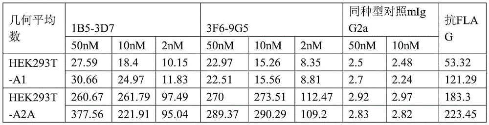

HEK293T-A1: HEK293T cells expressing human adenosine receptor A1
HEK293T-A2A: HEK293T cells expressing human adenosine receptor A2A
HEK293T-A2B: HEK293T cells expressing human adenosine receptor A2B
HEK293T-A3: HEK293T cells expressing human adenosine receptor A3
HEK293T-mA2A: HEK293T cells expressing mouse adenosine receptor A2A
HEK293T: HEK293T cells without exogenous adenosine receptor
Implementation of the embodiments example 6: comparison of binding affinity between exemplary anti-human A2aR antibodies of the invention and other anti-human A2aR antibodies
Human A2aR expressing Expi293 cells or parental Expi293 cells were first added to 96-well plates (1 x10 per well) 5 Individual cells). Samples of 12 concentration spots were serially diluted 1:2 from 7.5. Mu.g/ml for 50. Mu.l (50. Mu.l) of each antibody and cells were incubated on ice for 30 minutes. After washing with FACS buffer (DPBS solution of 2% foetal calf serum), 100. Mu.l of 1. Mu.g/ml Alex Fluor 633-conjugated anti-mouse IgG (Life technology, cat. A21050) or Alex Fluor 647-conjugated anti-human IgG was added and incubated on ice for 30 minutes. After two washes, cells were resuspended in 100. Mu.l of FACS buffer. Mu.l of 7-AAD for live/dead cell staining diluted 1:50 was added to a cell suspension which was ready for FACS analysis (BD Accuri C6 plus or LSR II flow cytometer, HTS).
As shown in fig. 5, exemplary antibodies 1B5-3D7 and 3F6-9G5 in the form of mIgG2a show strong dose-dependent specific binding to hA2aR-Expi293, but they do not bind to the parent Expi 293. Surprisingly, no binding to hA2aR-Expi293 cells was detected in staining with anti-A2 aR monoclonal antibodies MAB9497R (R & D Systems, clone 599717R) or SDIX-14 (U.S. patent publication US 2014/032366A 1, clone 864H 14). Furthermore, the anti-hA 2aR monoclonal antibody SDIX-10 (U.S. patent publication US 2014/032366 A1, clone 864H 10) showed non-specific binding to hA2aR-Expi293 cells and parental Expi293 cells and had a similar lower GMI (at 50nM concentration < 2000). (GMI: geometric mean fluorescence intensity).
Example 7: determination of binding of anti-human A2aR antibodies to human and cynomolgus primary cells and cross-reactivity with non-human primate
A2aR is reported to be expressed by T cells in humans. Anti-human A2aR antibodies blocking adenosine (clone 3F6-9G 5) were tested for potential binding to human and cynomolgus PBMC.
Method
Human and cynomolgus PBMCs were purchased from Cytologics LLC (san diego, california) and iQ Biosciences (berkeley, california), respectively. Primary cell binding was determined using rabbit Fc containing anti-human A2aR clone 3F6-9G 5. Briefly, 10 in 100 μ1 FACS buffer 6 PBMCs of individuals and/or cynomolgus monkeys were incubated with 3F6-9G5 (2. Mu.g/ml) for 60 minutes at 4 ℃. The subpopulations of live immune cells were determined using cynomolgus monkey cross-reactive anti-human CD3 (clone SP34, BD), anti-human CD8 (clone RPA-T8, bioleged) and Zombie Green fixable Viability dye (bioleged, cat No. 423111) according to the manufacturer's instructions. After 3 washes with FACS buffer, cells were stained with PE-conjugated donkey anti-rabbit Fc detection antibody (1:200, biolegend, cat. No. 406421) for 30 min at 4 ℃. After 3 washes with FACS buffer, cells were analyzed on LSRII (Becton Dickinson). Data were analyzed using FlowJo (Becton Dickinson).
Results
Anti-human A2aR clone 3F6-9G5 (instead of the anti-HEL rabbit Fc isotype control (Biointron, cat No. B730001)) bound to a small subset of human T cells (cd4+ and cd8+) (fig. 6). It also cross-reacted with cynomolgus T cells (cd4+ and cd8+) (fig. 6).
The above description is intended to guide one of ordinary skill in the art in the practice of the invention and is not intended to detail all of the obvious modifications and alterations that will become apparent to those skilled in the art upon reading the present specification. However, all such obvious modifications and variations are intended to be included within the scope of the present invention, which is defined by the appended claims. The claims are intended to cover the claimed components and steps in any order that is effective to meet the objectives intended for the claims, unless the context specifically indicates the contrary.
Claims (39)
1. An isolated antibody or antigen-binding fragment thereof that binds to the human adenosine A2A receptor (A2 aR), comprising:
a heavy chain variable region (VH) comprising three heavy chain complementarity determining regions HCDR1, HCDR2 and HCDR3 from the N-terminus to the C-terminus; and
a light chain variable region (VL) comprising three light chain complementarity determining regions LCDR1, LCDR2 and LCDR3 from N-terminus to C-terminus; wherein the method comprises the steps of
(a) HCDR1 comprises amino acid sequence X 1 -X 2 -W-M-N (SEQ ID NO: 8), wherein X 1 Is S or R, X 2 Y or F;
(b) HCDR2 comprises the amino acid sequence R-I-D-P-X 3 -D-S-E-X 4 -X 5 -Y-X 6 -H-K-F-W-X 7 (SEQ ID NO: 9), wherein X 3 Is S or Y, X 4 Is A or T, X 5 Is H or Q, X 6 Is H or N, X 7 Is D or G;
(c) HCDR3 comprises the amino acid sequence SLYGGDY (SEQ ID NO: 3);
(d) LCDR1 comprises an amino acid sequence
R-S-S-Q-S-X 17 -V-H-X 18 -N-G-N-T-Y-L-E (SEQ ID NO: 30), wherein X 17 Is L or I, X 18 R or S;
(e) LCDR2 comprises the amino acid sequence K-V-S-N-R-F-S (SEQ ID NO: 26); and
(f) LCDR3 contains amino acid sequence X 19 -Q-G-S-H-V-P-L-T (SEQ ID NO: 31), wherein X 19 Y or F.
2. The isolated antibody or antigen-binding fragment thereof of claim 1, wherein:
(a) HCDR1 comprises an amino acid sequence selected from the group consisting of SEQ ID NOs 1, 4 and 6;
(b) HCDR2 comprises an amino acid sequence selected from the group consisting of SEQ ID NOs 2, 5 and 7;
(c) HCDR3 comprises the amino acid sequence shown in SEQ ID NO. 3;
(d) LCDR1 comprises the amino acid sequence shown in SEQ ID NO. 25 or 28;
(e) LCDR2 comprises the amino acid sequence shown in SEQ ID NO. 26; and
(f) LCDR3 comprises the amino acid sequence shown in SEQ ID NO 27 or 29.
3. The isolated antibody or antigen-binding fragment thereof of claim 2, wherein the antibody comprises:
(a) HCDR1 comprising the amino acid sequence shown in SEQ ID NO. 1, HCDR2 comprising the amino acid sequence shown in SEQ ID NO. 2, HCDR3 comprising the amino acid sequence shown in SEQ ID NO. 3, LCDR1 comprising the amino acid sequence shown in SEQ ID NO. 25, LCDR2 comprising the amino acid sequence shown in SEQ ID NO. 26, LCDR3 comprising the amino acid sequence shown in SEQ ID NO. 27;
(b) HCDR1 comprising the amino acid sequence shown in SEQ ID NO. 4, HCDR2 comprising the amino acid sequence shown in SEQ ID NO. 5, HCDR3 comprising the amino acid sequence shown in SEQ ID NO. 3, LCDR1 comprising the amino acid sequence shown in SEQ ID NO. 28, LCDR2 comprising the amino acid sequence shown in SEQ ID NO. 26, LCDR3 comprising the amino acid sequence shown in SEQ ID NO. 29; or (b)
(c) HCDR1 comprising the amino acid sequence shown in SEQ ID NO. 6, HCDR2 comprising the amino acid sequence shown in SEQ ID NO. 7, HCDR3 comprising the amino acid sequence shown in SEQ ID NO. 3, LCDR1 comprising the amino acid sequence shown in SEQ ID NO. 25, LCDR2 comprising the amino acid sequence shown in SEQ ID NO. 26, LCDR3 comprising the amino acid sequence shown in SEQ ID NO. 29.
4. The isolated antibody or antigen-binding fragment thereof of claim 1, wherein
(a) HCDR1 comprises an amino acid sequence selected from the group consisting of SEQ ID NOs 21, 22, 23 and 24; and
(b) HCDR3 comprises an amino acid sequence selected from the group consisting of SEQ ID NOS: 12, 15 and 20.
5. The isolated antibody or antigen-binding fragment thereof of any one of claims 1-4, wherein the antibody comprises:
(a) A Heavy Chain Variable Region (HCVR) comprising an amino acid sequence selected from the group consisting of SEQ ID NOs 35, 36, 37, 38, 39 and 40; and
(b) A Light Chain Variable Region (LCVR) comprising an amino acid sequence selected from the group consisting of SEQ ID NOS: 41, 42 and 43.
6. The isolated antibody or antigen-binding fragment thereof of claim 5, wherein the antibody comprises:
(a) HCVR comprising the amino acid sequence shown in SEQ ID No. 35 or 38, and LCVR comprising the amino acid sequence shown in SEQ ID No. 41;
(b) HCVR comprising the amino acid sequence shown in SEQ ID No. 36 or 39, and LCVR comprising the amino acid sequence shown in SEQ ID No. 42; or (b)
(c) HCVR comprising the amino acid sequence shown in SEQ ID No. 37 or 40, and LCVR comprising the amino acid sequence shown in SEQ ID No. 43.
7. An isolated antibody or antigen-binding fragment thereof that binds to the human adenosine A2A receptor (human A2 aR), comprising:
a heavy chain variable region (VH) comprising three heavy chain complementarity determining regions HCDR1, HCDR2 and HCDR3 from the N-terminus to the C-terminus; and
a light chain variable region (VL) comprising three light chain complementarity determining regions LCDR1, LCDR2 and LCDR3 from N-terminus to C-terminus; wherein the method comprises the steps of
(a) HCDR1 comprises an amino acid sequence having about 80%, 85%, 90%, 95%, 96%, 97%, 98%, 99% to about 100% identity with an amino acid sequence selected from the group consisting of SEQ ID NOs 1, 4 and 6;
(b) HCDR2 comprises an amino acid sequence having about 80%, 85%, 90%, 95%, 96%, 97%, 98%, 99% to about 100% identity with an amino acid sequence selected from the group consisting of SEQ ID NOs 2, 5 and 7;
(c) HCDR3 comprises an amino acid sequence having about 80%, 85%, 90%, 95%, 96%, 97%, 98%, 99% to about 100% identity with the amino acid sequence shown in SEQ ID No. 3;
(d) LCDR1 comprises an amino acid sequence having about 80%, 85%, 90%, 95%, 96%, 97%, 98%, 99% to about 100% identity to an amino acid sequence selected from the group consisting of SEQ ID NOs 25 and 28;
(e) LCDR2 comprises amino acid sequences having about 80%, 85%, 90%, 95%, 96%, 97%, 98%, 99% to about 100% identity to the amino acid sequence of SEQ ID NO. 26; and
(f) LCDR3 comprises amino acid sequences having about 80%, 85%, 90%, 95%, 96%, 97%, 98%, 99% to about 100% identity to an amino acid sequence selected from SEQ ID NO 27 or 29.
8. An isolated antibody or antigen-binding fragment thereof that binds to the human adenosine A2A receptor (human A2 aR), comprising:
(a) A Heavy Chain Variable Region (HCVR) comprising an amino acid sequence having about 80%, 85%, 90%, 95%, 96%, 97%, 98%, 99% to about 100% identity to an amino acid sequence selected from the group consisting of SEQ ID nos. 35, 36 and 37; and
(b) A Light Chain Variable Region (LCVR) comprising an amino acid sequence having about 80%, 85%, 90%, 95%, 96%, 97%, 98%, 99% to about 100% identity to an amino acid sequence selected from the group consisting of SEQ ID NOs 41, 42 and 43.
9. An isolated antibody or antigen-binding fragment thereof that binds to the human adenosine A2A receptor (human A2 aR), comprising:
(a) A Heavy Chain Variable Region (HCVR) comprising the amino acid sequence shown in SEQ ID No. 35 or 38; and
(b) A Light Chain Variable Region (LCVR) comprising the amino acid sequence shown in SEQ ID NO. 41.
10. An isolated antibody or antigen-binding fragment thereof that binds to the human adenosine A2A receptor (human A2 aR), comprising:
(a) A Heavy Chain Variable Region (HCVR) comprising the amino acid sequence shown in SEQ ID No. 36 or 39; and
(b) A Light Chain Variable Region (LCVR) comprising the amino acid sequence shown in SEQ ID NO. 42.
11. The isolated antibody or antigen-binding fragment thereof of any one of claims 1-10, wherein the N-terminus of the heavy and/or light chain is a pyroglutamic acid (pE) residue.
12. The isolated antibody or antigen-binding fragment thereof of any one of claims 1-11, wherein
(i) The antibody competes for binding to human A2aR with a monoclonal antibody selected from 1B5-3D7, 3F6-9G5, or 3F8-12E 9;
(ii) The antibodies inhibit the activity of A2aR;
(iii) The antibodies enhance immune responses;
(iv) The antibody specifically binds to cell surface human A2aR;
(v) The antibody reduces cAMP concentration in the tissue;
(vi) The antibody reduces the activity of protein kinase a;
(vii) The antibody reduces phosphorylation of cAMP response element of the A2aR signaling pathway; or (b)
(viii) The antibodies specifically bind to human and/or cynomolgus monkey A2aR.
13. An isolated antibody or antigen binding fragment thereof that competes with the antibody of any one of claims 1-12 for binding to human A2aR.
14. The antibody or antigen-binding fragment thereof of any one of claims 1-13, wherein the antibody is a humanized antibody or a chimeric antibody.
15. The antibody or antigen-binding fragment thereof of any one of claims 1-14, wherein the antibody comprises a heavy chain constant region selected from the group consisting of IgA, igD, igE, igG and IgM classes.
16. The antibody or antigen binding fragment thereof of claim 15, wherein the antibody comprises a heavy chain constant region of the IgG class, wherein IgG is selected from the group consisting of IgG4, igGl, igG2, and IgG3.
17. An isolated polynucleotide encoding the antibody or antigen-binding fragment thereof, HCVR thereof, LCVR thereof, light chain thereof, heavy chain thereof, or antigen-binding fragment thereof of any one of claims 1-16.
18. An expression vector comprising the polynucleotide of claim 17.
19. A recombinant cell comprising the polynucleotide of claim 17 or the expression vector of claim 18.
20. A method of making the antibody or antigen-binding fragment thereof of any one of claims 1-16, comprising expressing the antibody in the recombinant cell of claim 17 and isolating the expressed antibody.
21. A pharmaceutical composition comprising the antibody or antigen-binding fragment thereof of any one of claims 1-16, and a pharmaceutically acceptable carrier or diluent.
22. The pharmaceutical composition of claim 19, wherein the pharmaceutical composition comprises an effective amount of the antibody or antigen-binding fragment thereof such that it (a) specifically binds cell surface human or cynomolgus monkey A2aR; (b) reducing cAMP concentration in the tissue; (c) inhibiting the activity of human A2aR; (d) Reduces phosphorylation of cAMP response element of the A2aR signaling pathway; (e) enhancing the immune response of the immune cells; (f) decreasing protein kinase a activity; or (g) any combination of (a) - (f).
23. A method of inhibiting the activity of A2aR expressed on the surface of a cell comprising contacting the cell with the isolated antibody or antigen-binding fragment thereof of any one of claims 1-16, or the pharmaceutical composition of claim 21 or 22, thereby inhibiting the activity of A2aR in the cell.
24. A method of enhancing an immune response in a subject comprising administering to the subject the isolated antibody or antigen-binding fragment thereof of any one of claims 1-16 or the pharmaceutical composition of claim 21 or 22, thereby enhancing the immune response in the subject.
25. A method of inhibiting tumor growth in a subject comprising administering to the subject the isolated antibody or antigen-binding fragment thereof of any one of claims 1-16 or the pharmaceutical composition of claim 21 or 22, thereby inhibiting tumor growth.
26. A method of treating cancer in a subject, comprising administering to the subject the isolated antibody or antigen-binding fragment thereof of any one of claims 1-16 or the pharmaceutical composition of claim 21 or 22, thereby treating cancer.
27. The method of any one of claims 23-26, wherein the method activates T cells and directs them to kill tumor target cells.
28. The method of any one of claims 23-27, further comprising administering an additional therapeutic agent.
29. The method of claim 28, wherein the other therapeutic agent comprises an anti-tumor agent, radiation therapy, a chemotherapeutic agent, surgery, a cancer vaccine, an agonist of immune cell stimulating receptors, a cytokine, a cell therapy, or a checkpoint inhibitor.
30. The method of claim 29, wherein the checkpoint inhibitor is selected from the group consisting of inhibitors of PD-1, PD-L1, TIGIT, CTLA-4, PD-1, PD-L2, LAG-3, TIM-3, neurite, BTLA, CECAM-1, CECAM-5, IL-1R8, VISTA, LAIR1, LILRB2, LILRB3, LILRB4, LILRB5, CD96, CD112R, 2B4, tgfβ -R, KIR, NKG a, and any combination thereof.
31. The method of claim 30, wherein the inhibitor inhibits an interaction between PD-1 and PD-L1, the inhibitor being selected from the group consisting of pamil mab, na Wu Liyou mab, atuzumab, aviuzumab, dulcis You Shan mab, BMS-936559, singdi Li Shan mab, terep Li Shan mab, tirelizumab, karilizumab, shu Geli mab, p An Puli mab, calonet Li Shan mab, sulfamonomethoxine 1, and sulfamethoxazole 2.
32. The method of claim 30, wherein the CTLA4 inhibitor is selected from ipilimumab, california Li Shan antibody, and YH001 (i.e., and medicine).
33. The method of claim 29, wherein the other therapeutic agent is an agonist of an immune cell stimulating receptor selected from the group consisting of: OX40, CD2, CD27, CD3, ICAM-1, LFA-1, ICOS (CD 278), 4-1BB (CD 137), GITR, CD28, CD30, CD40, BAFFR, HVEM, CD7, LIGHT, NKG2C, NKG2D, SLAMF7, NKp46, NKp80, CD160, and any combination thereof.
34. The method of any one of claims 28-33, wherein the additional therapeutic agent is formulated in the same pharmaceutical composition as the antibody.
35. The method of any one of claims 28-33, wherein the additional therapeutic agent is formulated in a different pharmaceutical composition than the antibody.
36. The method of any one of claims 28-33 and 35, wherein the additional therapeutic agent is administered before and/or after administration of the antibody.
37. The method of any one of claims 28-35, wherein the additional therapeutic agent is administered concurrently with the antibody or antigen-binding fragment thereof.
38. A kit comprising the pharmaceutical composition of claim 21 or 22.
39. The kit of claim 38, further comprising an additional therapeutic agent.
Applications Claiming Priority (3)
| Application Number | Priority Date | Filing Date | Title |
|---|---|---|---|
| US202063085612P | 2020-09-30 | 2020-09-30 | |
| US63/085,612 | 2020-09-30 | ||
| PCT/US2021/052819 WO2022072601A1 (en) | 2020-09-30 | 2021-09-30 | ANTI-ADENOSINE RECEPTOR (A2aR) ANTIBODIES |
Publications (1)
| Publication Number | Publication Date |
|---|---|
| CN116348140A true CN116348140A (en) | 2023-06-27 |
Family
ID=80951827
Family Applications (1)
| Application Number | Title | Priority Date | Filing Date |
|---|---|---|---|
| CN202180067415.9A Pending CN116348140A (en) | 2020-09-30 | 2021-09-30 | Anti-adenosine receptor (A2aR) antibody |
Country Status (7)
| Country | Link |
|---|---|
| US (1) | US20230340132A1 (en) |
| EP (1) | EP4222173A4 (en) |
| JP (1) | JP2023543896A (en) |
| CN (1) | CN116348140A (en) |
| AU (1) | AU2021351500A1 (en) |
| CA (1) | CA3194285A1 (en) |
| WO (1) | WO2022072601A1 (en) |
Cited By (1)
| Publication number | Priority date | Publication date | Assignee | Title |
|---|---|---|---|---|
| WO2025011590A1 (en) * | 2023-07-11 | 2025-01-16 | 清华大学 | Antibody derivative and use thereof |
Families Citing this family (4)
| Publication number | Priority date | Publication date | Assignee | Title |
|---|---|---|---|---|
| KR20250004779A (en) | 2022-04-13 | 2025-01-08 | 길리애드 사이언시즈, 인코포레이티드 | Combination therapy for treating TROP-2 expressing cancers |
| CN116333127A (en) * | 2022-06-28 | 2023-06-27 | 北京科诺信诚科技有限公司 | Nanometer antibody targeting human LILRB2 and application thereof |
| WO2025137640A1 (en) | 2023-12-22 | 2025-06-26 | Gilead Sciences, Inc. | Azaspiro wrn inhibitors |
| WO2025257365A1 (en) | 2024-06-14 | 2025-12-18 | Institut National de la Santé et de la Recherche Médicale | Adenosine a2a antagonists for the treatment of endometriosis |
Family Cites Families (7)
| Publication number | Priority date | Publication date | Assignee | Title |
|---|---|---|---|---|
| CN104356236B (en) * | 2005-07-01 | 2020-07-03 | E.R.施贵宝&圣斯有限责任公司 | Human monoclonal antibody against programmed death ligand 1 (PD-L1) |
| JP2011097869A (en) * | 2009-11-05 | 2011-05-19 | Japan Science & Technology Agency | ANTI-HUMAN ADENOSINE A2a RECEPTOR MONOCLONAL ANTIBODY |
| WO2013040358A2 (en) * | 2011-09-14 | 2013-03-21 | University Of Washington Through Its Center For Commercialization | Assays and compositions for detection of agr2 |
| SG10201700698WA (en) * | 2012-05-15 | 2017-02-27 | Bristol Myers Squibb Co | Cancer immunotherapy by disrupting pd-1/pd-l1 signaling |
| US20140322236A1 (en) * | 2013-03-15 | 2014-10-30 | Sdix, Llc | Anti-human adora2a antibodies |
| AU2017330346C1 (en) * | 2016-09-21 | 2025-03-06 | Nextcure, Inc. | Antibodies for Siglec-15 and methods of use thereof |
| EP3938396A1 (en) * | 2019-03-11 | 2022-01-19 | Jounce Therapeutics, Inc. | Anti-icos antibodies for the treatment of cancer |
-
2021
- 2021-09-30 CN CN202180067415.9A patent/CN116348140A/en active Pending
- 2021-09-30 JP JP2023520021A patent/JP2023543896A/en active Pending
- 2021-09-30 CA CA3194285A patent/CA3194285A1/en active Pending
- 2021-09-30 AU AU2021351500A patent/AU2021351500A1/en active Pending
- 2021-09-30 US US18/028,917 patent/US20230340132A1/en active Pending
- 2021-09-30 WO PCT/US2021/052819 patent/WO2022072601A1/en not_active Ceased
- 2021-09-30 EP EP21876458.7A patent/EP4222173A4/en active Pending
Cited By (1)
| Publication number | Priority date | Publication date | Assignee | Title |
|---|---|---|---|---|
| WO2025011590A1 (en) * | 2023-07-11 | 2025-01-16 | 清华大学 | Antibody derivative and use thereof |
Also Published As
| Publication number | Publication date |
|---|---|
| AU2021351500A9 (en) | 2024-09-05 |
| JP2023543896A (en) | 2023-10-18 |
| CA3194285A1 (en) | 2022-04-07 |
| WO2022072601A1 (en) | 2022-04-07 |
| AU2021351500A2 (en) | 2023-08-10 |
| AU2021351500A1 (en) | 2023-06-08 |
| US20230340132A1 (en) | 2023-10-26 |
| EP4222173A1 (en) | 2023-08-09 |
| EP4222173A4 (en) | 2024-10-30 |
Similar Documents
| Publication | Publication Date | Title |
|---|---|---|
| US20230331857A1 (en) | Antitumor immune checkpoint regulator antagonists | |
| US20180185482A1 (en) | Checkpoint regulator antagonists | |
| CN114340735B (en) | Antitumor antagonists composed of mutant TGFβ1-RII extracellular domain and immunoglobulin scaffold | |
| CN116348140A (en) | Anti-adenosine receptor (A2aR) antibody | |
| US20250206828A1 (en) | ANTI-ADENOSINE RECEPTOR (A2aR) ANTIBODIES AND THE USE THEREOF | |
| HK40124840A (en) | Anti-adenosine receptor (a2ra) antibodies and the use thereof | |
| US20250171542A1 (en) | Anti-programmed death-ligand 1 (pd-l1) antibodies | |
| WO2023192289A2 (en) | Anti-trophoblast cell surface antigen 2 (trop-2) antibodies | |
| EA045974B1 (en) | ANTI-TUMOR ANTAGONISTS OF IMMUNE CHECKPOINT REGULATORS |
Legal Events
| Date | Code | Title | Description |
|---|---|---|---|
| PB01 | Publication | ||
| PB01 | Publication | ||
| SE01 | Entry into force of request for substantive examination | ||
| SE01 | Entry into force of request for substantive examination |