CN116250849A - An EEG Signal Recognition Method Based on Information Separator and Regional Convolutional Network - Google Patents
An EEG Signal Recognition Method Based on Information Separator and Regional Convolutional Network Download PDFInfo
- Publication number
- CN116250849A CN116250849A CN202211559232.1A CN202211559232A CN116250849A CN 116250849 A CN116250849 A CN 116250849A CN 202211559232 A CN202211559232 A CN 202211559232A CN 116250849 A CN116250849 A CN 116250849A
- Authority
- CN
- China
- Prior art keywords
- node
- convolution
- regional
- graph
- data
- Prior art date
- Legal status (The legal status is an assumption and is not a legal conclusion. Google has not performed a legal analysis and makes no representation as to the accuracy of the status listed.)
- Pending
Links
Images

Classifications
-
- A—HUMAN NECESSITIES
- A61—MEDICAL OR VETERINARY SCIENCE; HYGIENE
- A61B—DIAGNOSIS; SURGERY; IDENTIFICATION
- A61B5/00—Measuring for diagnostic purposes; Identification of persons
- A61B5/24—Detecting, measuring or recording bioelectric or biomagnetic signals of the body or parts thereof
- A61B5/316—Modalities, i.e. specific diagnostic methods
- A61B5/369—Electroencephalography [EEG]
- A61B5/372—Analysis of electroencephalograms
-
- A—HUMAN NECESSITIES
- A61—MEDICAL OR VETERINARY SCIENCE; HYGIENE
- A61B—DIAGNOSIS; SURGERY; IDENTIFICATION
- A61B5/00—Measuring for diagnostic purposes; Identification of persons
- A61B5/72—Signal processing specially adapted for physiological signals or for diagnostic purposes
- A61B5/7235—Details of waveform analysis
- A61B5/725—Details of waveform analysis using specific filters therefor, e.g. Kalman or adaptive filters
-
- A—HUMAN NECESSITIES
- A61—MEDICAL OR VETERINARY SCIENCE; HYGIENE
- A61B—DIAGNOSIS; SURGERY; IDENTIFICATION
- A61B5/00—Measuring for diagnostic purposes; Identification of persons
- A61B5/72—Signal processing specially adapted for physiological signals or for diagnostic purposes
- A61B5/7235—Details of waveform analysis
- A61B5/7253—Details of waveform analysis characterised by using transforms
- A61B5/726—Details of waveform analysis characterised by using transforms using Wavelet transforms
-
- A—HUMAN NECESSITIES
- A61—MEDICAL OR VETERINARY SCIENCE; HYGIENE
- A61B—DIAGNOSIS; SURGERY; IDENTIFICATION
- A61B5/00—Measuring for diagnostic purposes; Identification of persons
- A61B5/72—Signal processing specially adapted for physiological signals or for diagnostic purposes
- A61B5/7235—Details of waveform analysis
- A61B5/7264—Classification of physiological signals or data, e.g. using neural networks, statistical classifiers, expert systems or fuzzy systems
- A61B5/7267—Classification of physiological signals or data, e.g. using neural networks, statistical classifiers, expert systems or fuzzy systems involving training the classification device
-
- G—PHYSICS
- G06—COMPUTING OR CALCULATING; COUNTING
- G06N—COMPUTING ARRANGEMENTS BASED ON SPECIFIC COMPUTATIONAL MODELS
- G06N3/00—Computing arrangements based on biological models
- G06N3/02—Neural networks
- G06N3/08—Learning methods
-
- Y—GENERAL TAGGING OF NEW TECHNOLOGICAL DEVELOPMENTS; GENERAL TAGGING OF CROSS-SECTIONAL TECHNOLOGIES SPANNING OVER SEVERAL SECTIONS OF THE IPC; TECHNICAL SUBJECTS COVERED BY FORMER USPC CROSS-REFERENCE ART COLLECTIONS [XRACs] AND DIGESTS
- Y02—TECHNOLOGIES OR APPLICATIONS FOR MITIGATION OR ADAPTATION AGAINST CLIMATE CHANGE
- Y02D—CLIMATE CHANGE MITIGATION TECHNOLOGIES IN INFORMATION AND COMMUNICATION TECHNOLOGIES [ICT], I.E. INFORMATION AND COMMUNICATION TECHNOLOGIES AIMING AT THE REDUCTION OF THEIR OWN ENERGY USE
- Y02D30/00—Reducing energy consumption in communication networks
- Y02D30/70—Reducing energy consumption in communication networks in wireless communication networks
Landscapes
- Health & Medical Sciences (AREA)
- Life Sciences & Earth Sciences (AREA)
- Engineering & Computer Science (AREA)
- Physics & Mathematics (AREA)
- Artificial Intelligence (AREA)
- Molecular Biology (AREA)
- Biophysics (AREA)
- General Health & Medical Sciences (AREA)
- Biomedical Technology (AREA)
- Veterinary Medicine (AREA)
- Psychiatry (AREA)
- Heart & Thoracic Surgery (AREA)
- Surgery (AREA)
- Animal Behavior & Ethology (AREA)
- Pathology (AREA)
- Public Health (AREA)
- Medical Informatics (AREA)
- Signal Processing (AREA)
- Computer Vision & Pattern Recognition (AREA)
- Physiology (AREA)
- Evolutionary Computation (AREA)
- Mathematical Physics (AREA)
- Theoretical Computer Science (AREA)
- Fuzzy Systems (AREA)
- Computational Linguistics (AREA)
- Data Mining & Analysis (AREA)
- Computing Systems (AREA)
- General Engineering & Computer Science (AREA)
- General Physics & Mathematics (AREA)
- Software Systems (AREA)
- Psychology (AREA)
- Measurement And Recording Of Electrical Phenomena And Electrical Characteristics Of The Living Body (AREA)
Abstract
The invention discloses an electroencephalogram signal identification method based on an information separator and a regional convolution network, which comprises the following steps: acquiring electroencephalogram data, and carrying out preprocessing such as filtering, wavelet packet decomposition, data standardization and the like on the electroencephalogram data; constructing a topological graph according to the electrode installation position, and embedding the preprocessed electroencephalogram data into the graph; acquiring independent source information of each node by using a constructed graph creation information separator, and extracting electroencephalogram signal characteristics by using a regional convolution network which focuses on regional characteristics and sequentially increases the region range of interest, wherein the regional convolution network comprises three convolution layers which are a node convolution layer for extracting the characteristics of the node itself, a domain convolution layer for extracting the domain characteristics taking the node as the center and a global convolution layer for extracting global characteristics; and finally, completing classification and identification by the full connection layer. The invention can obviously reduce the information redundancy between the electrodes and effectively improve the recognition accuracy of the brain electrical signals.
Description
Technical Field
The invention relates to the field of intelligent biomedical science, in particular to an electroencephalogram signal identification method based on an information separator and a regional convolution network.
Background
Brain-computer interface technology (BCI) is a man-machine interaction technology that enables direct communication with computers or other electronic devices through the human brain. The human brain contains hundreds of billions of neurons, and the electroencephalogram (EEG) signal is an electrical signal generated by activity between these neurons. Electroencephalogram signals have been widely used in the fields of artificial limbs, exercise rehabilitation, brain disease diagnosis, fatigue detection, and the like. When the brain performs mental activities, specific electrical signals are generated and mapped to the cerebral cortex, and how to decode the human intent from these electrical signals is critical for the brain-machine interface.
When a person is moving or imagining a movement, phenomena of event-related desynchronization (ERD) and event-related synchronization (ERS) that help to decode human intent can be observed. So the motion imagination is a popular research topic in the field of the brain electrical signals at present, and more researches lead the decoding of the motion imagination brain electrical signals to be reliable. Electroencephalogram signals have most of the characteristics of bioelectric signals, such as nonlinearity, instability, randomness, and also contain artifacts and noise of other bioelectric signals. The amplitude of these noise is several times or even tens of times that of the EEG signal. Thus, conventional methods cannot extract the valid features of the EEG signal. In recent years, due to the rapid development of deep learning and the strong processing power of deep learning on random and nonlinear data, the deep learning is gradually one of the main methods of electroencephalogram research.
Currently, the deep learning-based EEG signal intention decoding method mainly treats the original EEG signal as a two-dimensional matrix, and two dimensions are respectively an electrode and an acquired EEG time sequence. Feature extraction and classification is then performed using deep learning algorithms such as Convolutional Neural Networks (CNNs) or long-term memory (LSTM). Such methods achieve good performance, but it is worth mentioning that they use raw EEG data represented by a two-dimensional matrix as input data. However, since the brain can be regarded as a sphere with weak electric signals, and the cortex is a continuous electric potential surface, the collected electric signals of each electrode will have information generated from the positions of other electrodes, so that the direct use of the original data will cause information redundancy. Independent Component Analysis (ICA) is an important tool for blind source separation, which can effectively reduce such information redundancy, but for EEG data, the number of sources cannot be determined, and ICA can destroy the original electrode ordering order, so that the data lose the original spatial information. Such problems can be effectively avoided by creating a graph.
Graph rolling networks (GCNs) are a classical graph data processing method. It uses a symmetric normalized adjacency matrix to aggregate neighbor information at each layer, and then uses a weight matrix shared by each node to perform feature transformation. It achieves good results in semi-supervised node classification and some graph classification tasks. Simplified graph convolutional neural networks (SGCNs) are an improved approach based on GCN. It removes the feature transformation of each layer compared to GCN, but achieves nearly the same effect. This illustrates that the aggregation of neighbor information plays a major role in GCN. The above two methods provide a concept of data processing in the graph, namely, generating new node information by utilizing the adjacency relationship between nodes. On the other hand, when the brain performs mental activities, there is a great difference in the degree of activity of neurons in different brain regions. However, most of the current methods focus on the overall spatial information of all electrodes, neglecting the spatial information of each local area that should be focused more in actual operation.
CN113128552a, an electroencephalogram emotion recognition method based on depth separable causal graph rolling network. Existing neural networks for recognizing brain electrical emotion lack consideration of directional functional relationships between channels. The invention is as follows: 1. and collecting brain electrical data of the tested person. 2. And constructing an adjacency matrix. 3. And calculating a regularized Laplacian matrix corresponding to the electroencephalogram data. 4. And performing feature extraction and emotion classification by using a depth separable causal graph convolution model. The invention adopts the Grangel causal relationship to model the relationship among EEG signal channels, builds a directed asymmetric matrix, fully considers the causal relationship among the channels, is consistent with the actual electroencephalogram signal generation condition, and can effectively improve the accuracy rate of emotion type identification. In addition, the invention adopts the depth separable convolution to fully extract the local characteristics in the electroencephalogram data, thereby further improving the classification accuracy.
The patent uses a tangent ratio snow fu polynomial according to a regularized laplace matrix to perform a graph convolution operation to accomplish electroencephalogram emotion recognition. In the patent, directly acquired original data is used as the characteristics of the brain electricity, and the conductivity of the electrical signals is ignored, so that higher information redundancy exists between electrodes of the original data, and the characteristics of the brain electricity cannot be effectively extracted. Meanwhile, the graph convolution operation based on the tangent ratio snow fu polynomial of the regularized Laplace matrix only generates new node characteristics through repeated aggregation neighbor information in the field of nodes, and the graph convolution operation based on the tangent ratio snow fu polynomial has limited characteristic extraction capability due to lack of attention to spatial range characteristics with different sizes. In the patent, an information separator is used for acquiring independent source information of the nodes so as to reduce information redundancy among the nodes, and then the space range of interest is used for fully extracting the space range features with different sizes from the node-field-global sequentially increasing regional convolution network.
Disclosure of Invention
The invention aims to solve the problem that the brain electrical signal recognition rate is not high in the prior art. An electroencephalogram signal identification method based on an information separator and a regional convolution network is provided. The technical scheme of the invention is as follows:
an electroencephalogram signal identification method based on an information separator and a regional convolution network comprises the following steps:
step 1: acquiring electroencephalogram data, and preprocessing the electroencephalogram data including filtering, wavelet packet decomposition and data standardization;
step 2: constructing a topological graph according to the electrode installation position, and embedding the preprocessed electroencephalogram data into the topological graph;
step 3: according to the constructed topological graph, a conversion matrix capable of acquiring node source information is created and called as an information separator, and then the information separator is used for acquiring independent source information of each node;
step 4: extracting self characteristics of the nodes by using a node convolution layer of the regional convolution network;
step 5: extracting domain features centered on the node by using a domain convolution layer of the domain convolution network;
step 6: reducing the size of the graph using graph pooling;
step 7: extracting global features of all functional area blocks by using a global convolution layer of the area convolution network and finishing classification;
step 8: setting a loss function cross entropy loss function;
further, the preprocessing for filtering, wavelet packet decomposing and data standardization of the electroencephalogram data specifically comprises:
band-pass filtering brain electrical data at 0.5-100Hz, and decomposing the filtered data into an approximate part and a detailed part by using a first-level wavelet packetDividing the brain wave into low frequency and detail part into high frequency, discarding the detail part as noise according to rhythm characteristics of brain electricity, and only keeping an approximate part; the decomposed approximation data was normalized using the Z-Score normalization method, with the Z-Score normalization formula: wherein x is the decomposed approximation data, μ is the sample mean of the approximation data, ++>
 Is the standard deviation of the data, N is the number of samples, x z Is normalized data, x z The mean value of (2) is 0 and the standard deviation is 1.
Further, the step of constructing the map according to the electrode installation position includes:
taking each electrode as a node, taking the preprocessed electrode collected data as node characteristics, and forming a node set N by all the nodes; setting a distance threshold value, selecting a node with a distance smaller than the threshold value from a target node as a neighbor of the node, wherein the closer the distance is, the larger the distance is in an electric field, the weight of the edges of the adjacent nodes is set as the reciprocal of the distance, and all the edges form an edge set V, so that the constructed characteristic topological graph is as follows: g= { N, V }.
Further, the method for creating the information separator according to the constructed topological graph to obtain the independent source information of each node specifically includes:
the elements of the adjacency matrix a from the constructed graph are:
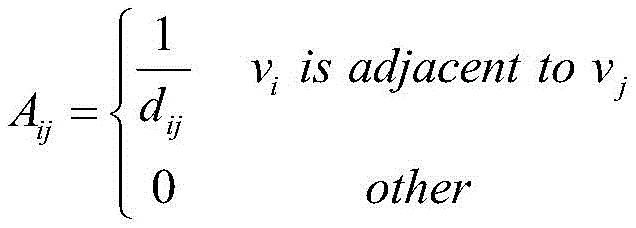
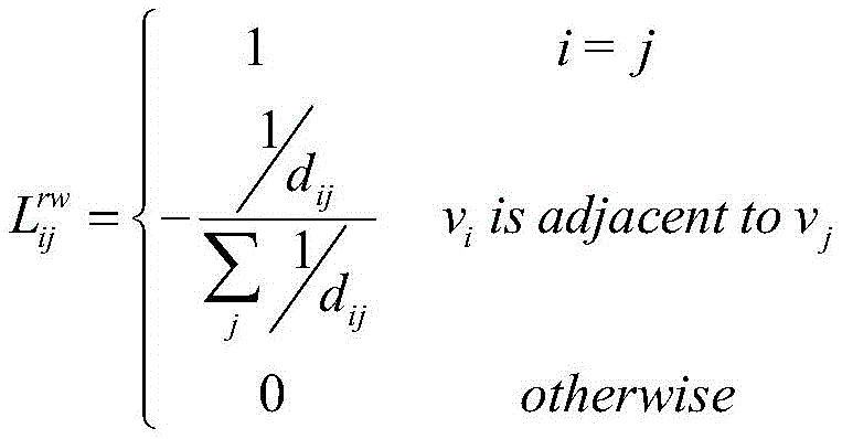
wherein d ij Is the distance between node i and node j, the laplace matrix of the graph is: l=d-a, wherein, is the degree matrix of A; ladder of scalar fieldThe degree of divergence represents the intensity of a source in the field, the degree of divergence of a gradient of the scalar field is represented by a Laplacian, and a Laplacian matrix is a discrete form of the Laplacian, so that node independent source information is obtained by the Laplacian matrix; to preserve the magnitude of the data, a random walk laplace matrix is used to normalize the laplace matrix: l (L) rw =L/D=I-A/D,L rw The elements of (2) are:

adding trainable parameters to fully acquire node source information: where W is a weight matrix of the same shape as A, I is an identity matrix, "
Further, the step of node convolution layer of the regional convolution network includes:
firstly, extracting information of different time periods on each node through one-dimensional convolution, wherein the operation formula of the one-dimensional convolution is as follows: wherein (1)>
 Represents the j-th feature of node c at layer 1, S c,j Is +.>
 Is input feature set of->
 And f (·) are the connection weight, bias, and ELU activation functions, respectively; topological structure of characteristic topological graph after convolutionThe convolution layer is not changed and maximum pooling is used to mitigate the oversensitivity of the convolution layer to time position.
Further, the step of domain convolution layer of the domain convolution network includes:
firstly, a common convolution kernel with a graph structure is established, the topological structure of the convolution kernel is the same as that of a characteristic topological graph, and then the local area with the characteristic of the graph output by a convolution layer as a center is subjected to convolution by using the convolution kernel, and the specific operation is as follows:
for each node, selecting the characteristics and convolution kernel channels of the node and the neighbor from the common convolution kernel; recombining the selected characteristics and the convolution kernel channels into a matrix according to the same sequence; for the characteristics after reorganization, considering that the attention degree of different areas to different nodes is different, then using the node attention to acquire the attention coefficients of the current area block to different subordinate nodes, and the node attention coefficient calculation step is as follows: (1) calculating a characteristic average value of the nodes: wherein L is the characteristic quantity of the node, u i Is the ith feature of the node; (2) the attention coefficients of the current region block to different nodes are calculated as follows: s=σ (W 2 δ(W 1 u)), where W 1 And W is 2 Is the weight of the linear transformation matrix, sigma and delta are the sigmod and ELU activation functions, respectively; the current region block is then reorganized as:
And then, performing convolution operation by using the recombined characteristic of the recombined convolution check, wherein the operation formula of the convolution layer is as follows: wherein N is c Is a region block centered on node c, i.e. node c and node c neighbors, +.>
 Represents the j-th feature of node c at layer 2, S m,j Is +.>
 Is input feature set of->
 And f (·) are the connection weight, bias, and ELU activation functions, respectively; finally, the regional characteristics of each node are re-embedded into the graph;
the convolved graph is pooled and features of the global region are extracted by a pooling technique.
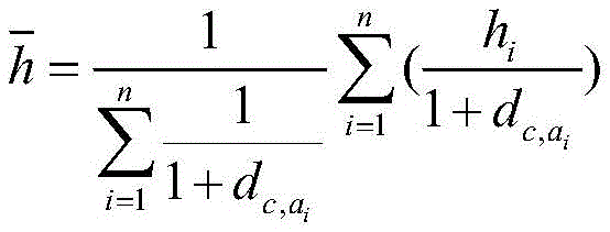
Further, the step of pooling the map comprises the following steps: (1) selecting the node closest to the geometric center in the 8 functional area blocks as a center node, and taking all the nodes in the functional area blocks as subordinate nodes; (2) calculating the distance between the subordinate node and the central node: where n is the number of nodes (including the center node) included in the current functional area block, a i For the i-th subordinate node of the current region block,/->
 And->
And-> The j-th feature of the j-th node and the i-th subordinate node of the central node respectively; the method comprises the steps of carrying out a first treatment on the surface of the (3) Calculating a new node representing the current functional area block, the new node being characterized by:

wherein h is i Is characteristic of the i-th node of the current region block, is the contribution of the ith node to the new node.
Further, the step of global convolution layer of the regional convolution network includes:
establishing a convolution kernel with the same structure as the pooled graph, extracting global area characteristics of 8 functional area blocks, wherein the operation formula of the convolution layer is as follows: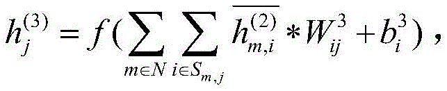 wherein N represents the set of all functional area blocks, < ->
 Represents the jth feature of layer 3, S m,j Is +.>
 Is input feature set of->
 And f (·) are the connection weight, bias, and ELU activation functions, respectively; finally, the full connection layer and softmax activation function are used to obtain the final classification result.
Further, the loss function is a cross entropy loss function: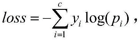 where c is the number of categories, y is the true label, and p is the prediction probability corresponding to the category.
The invention has the advantages and beneficial effects as follows:
according to the invention, the electroencephalogram signal is constructed into the graph, and the independent source information of the nodes is acquired from the Laplace matrix of the graph, so that the information redundancy is greatly reduced, and the information utilization rate is improved; meanwhile, the regional convolution network is used for fully extracting the characteristics of different regional ranges from the node, the local and the global, and the identification accuracy of the electroencephalogram signals is obviously improved.
The innovation of the invention mainly consists in using the random walk Laplacian plus the weight matrix of the update domain limited by the adjacent matrix to generate a conversion matrix capable of acquiring node source information in the step 3. The difference between the random walk Laplace matrix and the symmetric planning Laplace matrix is that the random walk Laplace matrix omits the symmetric characteristic and performs normalization processing by using the degree of the node, thereby being more beneficial to extracting the source information of the node; limiting the weight matrix of the update domain by the adjacency matrix can avoid interference of some difficult-to-measure factors, such as differences in electrode connection quality. In addition, step 4 uses a node convolution layer to fully extract the characteristics of the node itself, taking into account the uncertainty and periodicity of the specific start time of the motor imagery. And 5, selecting and reorganizing the characteristics of the regional blocks centering on each node by establishing a common convolution kernel with the same topological structure as the characteristic topological graph to finish convolution operation, and calculating the attention coefficients of the different regional blocks to the different nodes by using the attention of the nodes in consideration of the difference of the attention of each regional block to the different nodes during characteristic reorganization. Step 6, reducing the size of the map by using a map pooling technology according to the division of the cerebral cortex functional area. And 7, extracting global information of all the functional area blocks by using a global convolution layer to carry out final classification, and completing identification of the electroencephalogram signals.
Drawings
Fig. 1 is a flow chart of an electroencephalogram identification method based on an information separator and a regional convolution network in accordance with a preferred embodiment of the present invention.
Detailed Description
The technical solutions in the embodiments of the present invention will be clearly and specifically described below with reference to the drawings in the embodiments of the present invention. The described embodiments are only a few embodiments of the present invention.
The technical scheme for solving the technical problems is as follows:
referring to fig. 1, the invention provides an electroencephalogram identification method based on an information separator and a regional convolution network, which comprises the following steps.
And S01, acquiring brain electrical data. The brain electrical signal data acquisition can use a brain electrical signal acquisition instrument with 32 channels, the sampling frequency of the electrodes is set to 128HZ according to the international standard electrode method, and the acquired brain electrical signal is transmitted to a computer through a wire. The test was run from t=0Initially, the buzzer prompts 0.3 seconds, and a 2 second fixed cross appears on the screen, prompting the subject to focus. Starting from t=2s, the motor imagery task of the trial was prompted, the subject started motor imagery for 4s, then the screen turned black, resting for 4s. The motor imagery task contains four total movements, left hand, right hand, tongue and foot respectively. Data preprocessing, namely carrying out band-pass filtering on electroencephalogram data at 0.5-100Hz, decomposing the filtered data into an approximate (low-frequency) part and a detail (high-frequency) part by using a first-level wavelet packet, discarding the detail part as noise according to the rhythm characteristics of the electroencephalogram, and only keeping the approximate part. Since the electroencephalogram signal is bipolar data and the maximum value in the data is difficult to estimate, the approximate part of the decomposed data is normalized by using a Z-Score normalization method, and a Z-Score normalization formula is as follows: wherein x is the decomposed approximation data, μ is the sample mean of the approximation data, ++>
 Is the standard deviation of the data, N is the number of samples, x z Is normalized data. X is x z The mean value of (2) is 0 and the standard deviation is 1.
Step S02, constructing a graph. The step of constructing a feature topology map from the electrode mounting locations includes: and taking each electrode as a node, taking the preprocessed electrode acquired data as node characteristics, and forming a node set N by all the nodes. Setting a distance threshold value, selecting a node with a distance smaller than the threshold value from a target node as a neighbor of the node, setting the weight of the edges of the adjacent nodes as the inverse of the distance as the distance is bigger when the distance is closer in an electric field, and constructing a graph as follows: g= { N, V }.
Step S03: and acquiring node source information. The step of creating an information separator from the graph and acquiring node source information includes:
the elements of the adjacency matrix a from the constructed graph are:

wherein d ij Is the distance between node i and node j. The laplacian matrix of the graph is: l=d-a, wherein, is the degree matrix of a. The degree of scattering of the gradient of the scalar field represents the intensity of the source in the field, the calculation mode of the Laplace operator is the degree of scattering of the gradient, and the Laplace matrix is a discrete form of the Laplace operator, so that node independent source information can be obtained by using the Laplace matrix. To preserve the magnitude of the data, a random walk laplace matrix is used to normalize the laplace matrix: l (L) rw =L/D=I-A/D,L rw The elements of (2) are:

taking into account some factors that are difficult to measure, such as the connection quality of each electrode being different, adding trainable parameters to fully acquire node source information: where W is a weight matrix of the same shape as A, I is an identity matrix, and ". Cndot." represents a dot product operation. The source information of the node can be expressed as +.>
 And S04, extracting time characteristics on the nodes. The step of extracting temporal features on the nodes by the node convolution layer of the regional convolution layer comprises:
the method comprises the steps that since thinking activities of a subject in a motor imagery process are periodic and the accurate starting time of the motor imagery is difficult to determine, information of different time periods on each node is firstly extracted through one-dimensional convolution, and an operation formula of the convolution is as follows: wherein (1)>
 Representing the j-th feature of node c at layer 1. S is S c,j Is at node c
 Is input feature set of->
 And f (·) are the connection weight, bias, and ELU activation functions, respectively. The topology of the post-convolution feature topology is unchanged and maximum pooling is used to mitigate the oversensitivity of the convolution layer to time position.
And S05, extracting the domain characteristics of the local area. The step of extracting the domain features by the domain convolution layer of the domain convolution layer comprises:
firstly, a common convolution kernel with a graph structure is established, the topological structure of the convolution kernel is the same as that of a characteristic topological graph, and then the characteristic of the graph output by the convolution layer in the claim 5 is convolved by using the convolution kernel to take each node as a central local area, and the specific operation is as follows:
for each node, selecting the characteristics and convolution kernel channels of the node and the neighbor from the common convolution kernel; recombining the selected characteristics and the convolution kernel channels into a matrix according to the same sequence; for the characteristics after reorganization, considering that the attention degree of different areas to different nodes is different, then the attention coefficients of the current area block to the different nodes are obtained by using the attention of the nodes, and the node attention coefficient calculation steps are as follows: (1) calculating a characteristic average value of the nodes: wherein L is the characteristic quantity of the node, u i Is the ith feature of the node. (2) The attention coefficients of the current region block to different nodes are calculated as follows: s=σ (W 2 δ(W 1 u)), where W 1 And W is 2 Is a linear transformationThe weights of the matrix, σ and δ, are Sigmod and ELU activation functions, respectively. The current region block is then reorganized as:
And then, performing convolution operation by using the recombined characteristic of the recombined convolution check, wherein the operation formula of the convolution layer is as follows: wherein N is c Is a region block centered on node c, i.e. node c and node c neighbors, +.>
 Represents the j-th feature of node c at layer 2, S m,j Is +.>
 Is input feature set of->
 And f (·) are the connection weight, bias, and ELU activation functions, respectively. Finally, the regional characteristics of each node are re-embedded into the graph.
The human cerebral cortex can be divided into four large functional areas (frontal lobe, parietal lobe, temporal lobe and occipital lobe), and then the cerebral cortex can be divided into 8 functional area blocks according to the left brain and the right brain, so that the graph after the convolution can be pooled into smaller graphs by a graph pooling technology so as to be beneficial to calculating and extracting the characteristics of the global area. The step of pooling is as follows: (1) and selecting the node closest to the geometric center in the 8 functional area blocks as a center node, and taking other nodes in the functional area blocks as auxiliary nodes. (2) Calculating the distance between the auxiliary node and the center: wherein n is the number of nodes contained in the current functional area block, a i I-th node (including center node) of the current region block, is +,>
 and->
and-> The j-th feature of the j-th node and the i-th node of the center node, respectively. (3) Calculating a new node representing the current functional area block, the new node being characterized by:

wherein h is i Is characteristic of the i-th node of the current region block, is the contribution of the ith node to the new node.
Step S06, extracting and classifying the global features of the global area. The step of extracting global features from the global convolution layer of the regional convolution layer comprises:
establishing a convolution kernel with the same structure as the pooled graph, extracting global area characteristics of 8 functional area blocks, wherein the operation formula of the convolution layer is as follows: wherein N represents the set of all functional area blocks, < ->
 Represents the jth feature of layer 3, S m,j Is +.>
 Is input feature set of->
 And f (·) are the connection weight, bias, and ELU activation functions, respectively. Finally, the full connection layer and the Softmax activation function are used to obtain the final classification result.
The system, apparatus, module or unit set forth in the above embodiments may be implemented in particular by a computer chip or entity, or by a product having a certain function.
It should also be noted that the terms "comprises," "comprising," or any other variation thereof, are intended to cover a non-exclusive inclusion, such that a process, method, article, or apparatus that comprises a list of elements does not include only those elements but may include other elements not expressly listed or inherent to such process, method, article, or apparatus. Without further limitation, an element defined by the phrase "comprising one … …" does not exclude the presence of other like elements in a process, method, article or apparatus that comprises the element.
The above examples should be understood as illustrative only and not limiting the scope of the invention. Various changes and modifications to the present invention may be made by one skilled in the art after reading the teachings herein, and such equivalent changes and modifications are intended to fall within the scope of the invention as defined in the appended claims.
Claims (9)
1. An electroencephalogram signal identification method based on an information separator and a regional convolution network is characterized by comprising the following steps of:
acquiring electroencephalogram data, and preprocessing the electroencephalogram data including filtering, wavelet packet decomposition and data standardization;
constructing a topological graph according to the electrode installation position, and embedding the preprocessed electroencephalogram data into the topological graph;
according to the constructed topological graph, a conversion matrix capable of acquiring node source information is established and is called an information separator, and then the information separator is used for acquiring independent source information of each node;
then extracting the electroencephalogram signal features by using a regional convolution network with focusing regional features and sequentially increasing concerned regional ranges, wherein the regional convolution network comprises three convolution layers, namely a node convolution layer for extracting the self features of the nodes, a domain convolution layer for extracting the domain features taking the nodes as the centers and a global convolution layer for extracting the global features;
and finally, completing classification and identification by the full connection layer.
2. The electroencephalogram signal identification method based on the information separator and the regional convolution network according to claim 1, wherein the preprocessing of filtering, wavelet packet decomposition and data standardization of electroencephalogram data specifically comprises:
band-pass filtering is carried out on the electroencephalogram data at 0.5-100Hz, the filtered data is decomposed into an approximate part and a detail part by using a first-level wavelet packet, the approximate part is low frequency, the detail part is high frequency, the detail part is discarded as noise according to the rhythm characteristic of the electroencephalogram, and only the approximate part is reserved; the decomposed approximation data was normalized using the Z-Score normalization method, with the Z-Score normalization formula: wherein x is the decomposed approximation data, μ is the sample mean of the approximation data, ++>
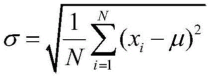 Is the standard deviation of the data, N is the number of samples, x z Is normalized data, x z The mean value of (2) is 0 and the standard deviation is 1.
3. The method for recognizing brain electrical signals based on an information separator and a regional convolution network according to claim 1, wherein said step of constructing a characteristic topological graph according to an electrode installation position comprises:
taking each electrode as a node, taking the preprocessed electrode collected data as node characteristics, and forming a node set N by all the nodes; setting a distance threshold value, selecting a node with a distance smaller than the threshold value from a target node as a neighbor of the node, wherein the closer the distance is, the larger the distance is in an electric field, the weight of edges of adjacent nodes is set as the reciprocal of the distance, and all the edges form an edge set V, so that the constructed graph is as follows: g= { N, V }.
4. The electroencephalogram signal identification method based on the information separator and the regional convolution network according to claim 1, wherein the creating the information separator according to the constructed graph to obtain the independent source information of each node specifically comprises:
the elements of the adjacency matrix a from the constructed graph are:

wherein d ij Is the distance between node i and node j, the laplace matrix of the graph is: l=d-a, wherein,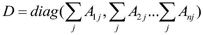 is the degree matrix of A; the degree of scattering of the gradient of the scalar field represents the intensity of a source in the field, the degree of scattering of the gradient of the scalar field is represented by a Laplacian, and the Laplacian matrix is a discrete form of the Laplacian, so that node independent source information is obtained by the Laplacian matrix; to preserve the magnitude of the data, a random walk laplace matrix is used to normalize the laplace matrix: l (L) rw =L/D=I-A/D,L rw The elements of (2) are:
adding trainable parameters to fully acquire node source information: where W is a weight matrix of the same shape as A, I is an identity matrix, and ". Cndot." represents a dot product operation. Will->
 Naming the nameIs an information separator. The source information of the node can be expressed as +.>
5. The electroencephalogram signal identification method based on an information separator and a regional convolution network according to claim 1, wherein the step of node convolution layer of the regional convolution network comprises:
firstly, extracting information of different time periods on each node through one-dimensional convolution, wherein the operation formula of the one-dimensional convolution is as follows: wherein (1)>
 Represents the j-th feature of node c at layer 1, S c,j Is +.>
 Is input feature set of->
 And f (·) are the connection weight, bias, and ELU activation functions, respectively; the topology of the post-convolution feature topology is unchanged and maximum pooling is used to mitigate the oversensitivity of the convolution layer to time position.
6. The electroencephalogram signal identification method based on the information separator and the regional convolution network according to claim 1, wherein the regional convolution layer step of the regional convolution network comprises the steps of:
firstly, a common convolution kernel with a graph structure is established, the topological structure of the common convolution kernel is the same as that of a characteristic topological graph, and then the local area with the characteristic of the graph output by a convolution layer as a center is convolved by using the convolution kernel, and the specific operation is as follows:
for each node, selecting the characteristics and convolution kernel channels of the node and the neighbor from the common convolution kernel; recombining the selected characteristics and the convolution kernel channels into a matrix according to the same sequence; for the characteristics after reorganization, considering that the attention degree of different areas to different nodes is different, then using the node attention to acquire the attention coefficients of the current area block to different subordinate nodes, and the node attention coefficient calculation step is as follows: (1) calculating a characteristic average value of the nodes: wherein L is the characteristic quantity of the node, u i Is the ith feature of the node; (2) the attention coefficients of the current region block to different nodes are calculated as follows: s=σ (W 2 δ(W 1 u)), where W 1 And W is 2 Is the weight of the linear transformation matrix, sigma and delta are the sigmod and ELU activation functions, respectively; the current region block is then reorganized as:
And then, performing convolution operation by using the recombined characteristic of the recombined convolution check, wherein the operation formula of the convolution layer is as follows: wherein N is c Is a region block centered on node c, i.e. node c and node c neighbors, +.>
 Represents the j-th feature of node c at layer 2, S m,j Is +.>
 Is input feature set of->
 And f (·) are respectively the connection weight, offset and EAn LU activation function; finally, the regional characteristics of each node are re-embedded into the graph;
the convolved graph is pooled and features of the global region are extracted by a pooling technique.
7. The method for recognizing brain electrical signals based on an information separator and a regional convolution network according to claim 6, wherein said step of pooling is: (1) selecting the node closest to the geometric center in the 8 functional area blocks as a center node, and taking all the nodes in the functional area blocks as subordinate nodes; (2) calculating the distance between the subordinate node and the central node: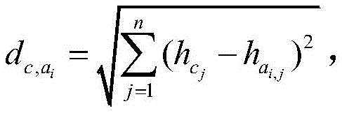 where n is the number of nodes (including the center node) included in the current functional area block, a i For the i-th subordinate node of the current region block,/->
 And->
And-> The j-th feature of the j-th node and the i-th subordinate node of the central node respectively; the method comprises the steps of carrying out a first treatment on the surface of the (3) Calculating a new node representing the current functional area block, the new node being characterized by:
wherein h is i Is characteristic of the i-th node of the current region block, is the contribution of the ith node to the new node.
8. The method for recognizing brain electrical signals based on an information separator and a regional convolution network according to claim 7, wherein said step of global convolution layer of said regional convolution network comprises:
establishing a convolution kernel with the same structure as the pooled graph, extracting global area characteristics of 8 functional area blocks, wherein the operation formula of the convolution layer is as follows: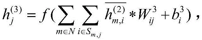 wherein N represents the set of all functional area blocks, < ->
 Represents the jth feature of layer 3, S m,j Is +.>
 Is input feature set of->
 And f (·) are the connection weight, bias, and ELU activation functions, respectively; finally, the full connection layer and softmax activation function are used to obtain the final classification result.
9. The method of claim 8, wherein the loss function is a cross entropy loss function: where c is the number of categories, y is the true label, and p is the prediction probability corresponding to the category. />
Priority Applications (1)
| Application Number | Priority Date | Filing Date | Title |
|---|---|---|---|
| CN202211559232.1A CN116250849A (en) | 2022-12-06 | 2022-12-06 | An EEG Signal Recognition Method Based on Information Separator and Regional Convolutional Network |
Applications Claiming Priority (1)
| Application Number | Priority Date | Filing Date | Title |
|---|---|---|---|
| CN202211559232.1A CN116250849A (en) | 2022-12-06 | 2022-12-06 | An EEG Signal Recognition Method Based on Information Separator and Regional Convolutional Network |
Publications (1)
| Publication Number | Publication Date |
|---|---|
| CN116250849A true CN116250849A (en) | 2023-06-13 |
Family
ID=86678249
Family Applications (1)
| Application Number | Title | Priority Date | Filing Date |
|---|---|---|---|
| CN202211559232.1A Pending CN116250849A (en) | 2022-12-06 | 2022-12-06 | An EEG Signal Recognition Method Based on Information Separator and Regional Convolutional Network |
Country Status (1)
| Country | Link |
|---|---|
| CN (1) | CN116250849A (en) |
Cited By (2)
| Publication number | Priority date | Publication date | Assignee | Title |
|---|---|---|---|---|
| CN116448019A (en) * | 2023-06-14 | 2023-07-18 | 山西首科工程质量检测有限公司 | Intelligent detection device and method for quality flatness of building energy-saving engineering |
| CN117909868A (en) * | 2024-03-19 | 2024-04-19 | 华南理工大学 | Electroencephalogram cognitive load analysis method and system based on neuroimaging priori dynamic graph convolution |
Citations (12)
| Publication number | Priority date | Publication date | Assignee | Title |
|---|---|---|---|---|
| CN110236536A (en) * | 2019-06-04 | 2019-09-17 | 电子科技大学 | A detection system of EEG high frequency oscillation signal based on convolutional neural network |
| CN110399857A (en) * | 2019-08-01 | 2019-11-01 | 西安邮电大学 | A EEG Emotion Recognition Method Based on Graph Convolutional Neural Network |
| CN111461176A (en) * | 2020-03-09 | 2020-07-28 | 华南理工大学 | Multi-mode fusion method, device, medium and equipment based on normalized mutual information |
| CN112426162A (en) * | 2020-11-23 | 2021-03-02 | 重庆邮电大学 | Fatigue detection method based on electroencephalogram signal rhythm entropy |
| CN112690793A (en) * | 2020-12-28 | 2021-04-23 | 中国人民解放军战略支援部队信息工程大学 | Emotion electroencephalogram migration model training method and system and emotion recognition method and equipment |
| CN112890827A (en) * | 2021-01-14 | 2021-06-04 | 重庆兆琨智医科技有限公司 | Electroencephalogram identification method and system based on graph convolution and gate control circulation unit |
| CN113128552A (en) * | 2021-03-02 | 2021-07-16 | 杭州电子科技大学 | Electroencephalogram emotion recognition method based on depth separable causal graph convolution network |
| CN113476056A (en) * | 2021-06-25 | 2021-10-08 | 西北工业大学 | Motor imagery electroencephalogram signal classification method based on frequency domain graph convolution neural network |
| WO2021226778A1 (en) * | 2020-05-11 | 2021-11-18 | 浙江大学 | Epileptic electroencephalogram recognition system based on hierarchical graph convolutional neural network, terminal, and storage medium |
| CN113729729A (en) * | 2021-08-13 | 2021-12-03 | 北京航空航天大学 | Early detection system of schizophrenia based on graph neural network and brain network |
| CN114081492A (en) * | 2021-11-25 | 2022-02-25 | 中国科学院大学宁波华美医院 | An EEG Emotion Recognition System Based on Learnable Adjacency Matrix |
| CN115054272A (en) * | 2022-07-27 | 2022-09-16 | 安徽大学 | Electroencephalogram signal identification method and system for dyskinesia function remodeling |
-
2022
- 2022-12-06 CN CN202211559232.1A patent/CN116250849A/en active Pending
Patent Citations (12)
| Publication number | Priority date | Publication date | Assignee | Title |
|---|---|---|---|---|
| CN110236536A (en) * | 2019-06-04 | 2019-09-17 | 电子科技大学 | A detection system of EEG high frequency oscillation signal based on convolutional neural network |
| CN110399857A (en) * | 2019-08-01 | 2019-11-01 | 西安邮电大学 | A EEG Emotion Recognition Method Based on Graph Convolutional Neural Network |
| CN111461176A (en) * | 2020-03-09 | 2020-07-28 | 华南理工大学 | Multi-mode fusion method, device, medium and equipment based on normalized mutual information |
| WO2021226778A1 (en) * | 2020-05-11 | 2021-11-18 | 浙江大学 | Epileptic electroencephalogram recognition system based on hierarchical graph convolutional neural network, terminal, and storage medium |
| CN112426162A (en) * | 2020-11-23 | 2021-03-02 | 重庆邮电大学 | Fatigue detection method based on electroencephalogram signal rhythm entropy |
| CN112690793A (en) * | 2020-12-28 | 2021-04-23 | 中国人民解放军战略支援部队信息工程大学 | Emotion electroencephalogram migration model training method and system and emotion recognition method and equipment |
| CN112890827A (en) * | 2021-01-14 | 2021-06-04 | 重庆兆琨智医科技有限公司 | Electroencephalogram identification method and system based on graph convolution and gate control circulation unit |
| CN113128552A (en) * | 2021-03-02 | 2021-07-16 | 杭州电子科技大学 | Electroencephalogram emotion recognition method based on depth separable causal graph convolution network |
| CN113476056A (en) * | 2021-06-25 | 2021-10-08 | 西北工业大学 | Motor imagery electroencephalogram signal classification method based on frequency domain graph convolution neural network |
| CN113729729A (en) * | 2021-08-13 | 2021-12-03 | 北京航空航天大学 | Early detection system of schizophrenia based on graph neural network and brain network |
| CN114081492A (en) * | 2021-11-25 | 2022-02-25 | 中国科学院大学宁波华美医院 | An EEG Emotion Recognition System Based on Learnable Adjacency Matrix |
| CN115054272A (en) * | 2022-07-27 | 2022-09-16 | 安徽大学 | Electroencephalogram signal identification method and system for dyskinesia function remodeling |
Non-Patent Citations (1)
| Title |
|---|
| 张俊晓;薄华;: "基于卷积神经网络的脑电情绪识别研究", 现代计算机(专业版), no. 23, 15 August 2018 (2018-08-15), pages 1 - 4 * |
Cited By (4)
| Publication number | Priority date | Publication date | Assignee | Title |
|---|---|---|---|---|
| CN116448019A (en) * | 2023-06-14 | 2023-07-18 | 山西首科工程质量检测有限公司 | Intelligent detection device and method for quality flatness of building energy-saving engineering |
| CN116448019B (en) * | 2023-06-14 | 2023-08-25 | 山西首科工程质量检测有限公司 | Intelligent detection device and method for quality flatness of building energy-saving engineering |
| CN117909868A (en) * | 2024-03-19 | 2024-04-19 | 华南理工大学 | Electroencephalogram cognitive load analysis method and system based on neuroimaging priori dynamic graph convolution |
| CN117909868B (en) * | 2024-03-19 | 2024-07-05 | 华南理工大学 | Electroencephalogram cognitive load analysis method and system based on neuroimaging priori dynamic graph convolution |
Similar Documents
| Publication | Publication Date | Title |
|---|---|---|
| CN114533086B (en) | A motor imagery EEG decoding method based on spatial feature time-frequency transformation | |
| Sadiq et al. | Toward the development of versatile brain–computer interfaces | |
| CN111259761B (en) | Electroencephalogram emotion recognition method and device based on movable attention neural network | |
| CN110969108B (en) | Limb action recognition method based on autonomic motor imagery electroencephalogram | |
| CN109165556B (en) | Identity recognition method based on GRNN | |
| CN115221969B (en) | Motor imagery EEG signal recognition method based on EMD data enhancement and parallel SCN | |
| CN113378650B (en) | An Emotion Recognition Approach Based on Brain Power Imaging and Regularized Common Spatial Patterns | |
| CN108776788A (en) | A kind of recognition methods based on brain wave | |
| CN111407243B (en) | Pulse signal pressure identification method based on deep learning | |
| CN116250849A (en) | An EEG Signal Recognition Method Based on Information Separator and Regional Convolutional Network | |
| CN115414051B (en) | Emotion classification recognition method for electroencephalogram signal self-adaptive window | |
| CN117503157A (en) | An EEG signal emotion recognition method based on SGCRNN model | |
| CN113569997A (en) | Emotion classification method and system based on graph convolutional neural network | |
| CN118395273B (en) | EEG signal classification method based on spiking neural network and Stockwell transform | |
| CN113128384A (en) | Brain-computer interface software key technical method of stroke rehabilitation system based on deep learning | |
| CN119513664A (en) | Emotion recognition method and system based on multi-scale convolutional network with bidirectional attention | |
| Jiang et al. | Analytical comparison of two emotion classification models based on convolutional neural networks | |
| KR102630840B1 (en) | EMG signal-based recognition information extraction system and EMG signal-based recognition information extraction method using the same | |
| CN116662782B (en) | A method for decoding motor imagery EEG based on MSFF-SENet | |
| CN116595434A (en) | A Lie Detection Method Based on Dimension and Classification Algorithm | |
| CN118797410B (en) | A dual-stream adaptive convolutional recurrent hybrid EEG emotion recognition method combined with attention mechanism | |
| CN118975805A (en) | Fatigue detection device and detection method based on electroencephalogram | |
| CN118673398A (en) | Fine-granularity target recognition task-oriented small sample electroencephalogram space-time frequency global decoding method, system and storage medium | |
| CN116258898A (en) | A multi-modal fine-grained feature hybrid recognition system and method | |
| CN115238744A (en) | Electroencephalogram emotion recognition method based on data uncertainty |
Legal Events
| Date | Code | Title | Description |
|---|---|---|---|
| PB01 | Publication | ||
| PB01 | Publication | ||
| SE01 | Entry into force of request for substantive examination | ||
| SE01 | Entry into force of request for substantive examination |