The application is a divisional application of Chinese invention patent application with application number of 201880023083.2, application date of 2018, 03 month and 29 days, and the invention name of 'application of compound or traditional Chinese medicine extract in preparing nucleic acid delivery reagent and related products', and is a national stage application with international application number of PCT/CN 2018/081155.
Detailed Description
The present application is further described below, but is not limited in any way, and any modifications based on the teachings of the present application fall within the scope of the present application.
The application extracts fat-soluble components in traditional Chinese medicines (comprising rhodiola rosea (Rhodiola crenulata), dandelion (Taraxacum mongolicum), andrographis paniculata (Andrographis paniculata) and honeysuckle (Lonicera japonica)) by using a Bligh & Dyer method, identifies lipid components (138 lipid components are identified altogether, 125 lipid components are identified in a cation mode and 13 lipid components are identified in an anion mode) by using HPLC-MS/MS, and 71 lipid components (see tables 1-1 to 1-3) are used for preparing lipid nucleic acid mixtures, and whether the lipid components can promote cell absorption and entry of exogenous nucleic acids or not is observed. It should be noted that, the lipids used in the present application are all commercially purchased or synthesized commercially, and are not directly extracted from the traditional Chinese medicine. The inventors have surprisingly found that a variety of lipids can form lipid-nucleic acid complexes that effectively promote cellular uptake and entry of nucleic acids (see FIGS. 1-116), hopefully increasing the efficiency of nucleic acid drug delivery in the clinic. Further studies have shown that the lipid nucleic acid mixtures of the present application promote nucleic acid uptake and efficiency into cells on different cell lines, but that there are differences between different cell lines (see fig. 1-10), which provides the possibility for targeted drug delivery. Furthermore, the nucleic acid delivery of the lipid nucleic acid complex has no sequence selectivity, and can deliver nucleic acid fragments of different sequences corresponding to the size of small RNAs (such as about 20 bp) (see FIG. 11). In addition, fluorescent in situ hybridization experiments (Fluorescence in situ hybridization, FISH) demonstrated that decoction-derived lipid-formed lipid nucleic acid mixtures can effectively promote the entry of exogenous nucleic acids into the cytoplasm (see fig. 12). The inventors have unexpectedly found that lipid nucleic acid mixtures prepared by either water boiling or reverse evaporation are capable of promoting the passage of nucleic acids such as sRNA into the blood circulation and into target tissues via non-invasive (e.g. trans-or trans-respiratory and topical) routes (see fig. 14-15). The inventors have also unexpectedly found that the lipids of the present application are capable of promoting the entry of nucleic acids, such as sRNA, into cells and modulating (e.g., inhibiting) the expression of their target sequences, while not exhibiting such modulation for non-target sequences, exhibiting target-specific modulation, and are useful as a means of delivery of nucleic acid drugs (see fig. 13).
Based on the above-described series of unexpected findings, the inventors have thus obtained the present application.
In one aspect, the present application provides compounds extracted from traditional Chinese medicine that facilitate delivery of nucleic acid, wherein the compounds are selected from lysolecithin, ceramide, diglyceride, phosphatidylethanolamine, phosphatidylcholine, triglyceride, monogalactosyldiglyceride, (nerve) sphingosine, phosphatidylethanol, monoacylglycerol, fatty acid, platelet-activating factor, or dimethylphospholipid ethanolamine, preferably from the lipids shown in table 1. In one embodiment, the lipid is non-natural, e.g., synthetic, or produced by fermentation.
In one embodiment, the lipid is used to deliver nucleic acid into a target cell. In another embodiment, the lipid is used to deliver nucleic acid into a subject in need thereof into the blood circulation and/or target site/cell thereof.
In a preferred embodiment, the lipid is selected from phosphatidylcholine, such as 1-stearoyl-2-oleoyl-sn-glycero-3-phosphorylcholine (PC (18:0/18:2), i.e. lipid 11 in Table 1), and 1-palmitoyl-2-oleoyl-sn-glycero-3-phosphorylcholine (PC (16:0/18:2), i.e. lipid 12 in Table 1). Both of these Phosphatidylcholines (PCs) are capable of efficiently encapsulating nucleic acids or facilitating entry of nucleic acids into cells. In one embodiment, the lipid may be lipid 41 in Table 1, i.e., sphinganine (d22:0), which is capable of efficiently encapsulating nucleic acid or facilitating entry of nucleic acid into a cell.
In another aspect, the present application provides a pharmaceutical composition comprising the above lipid and a nucleic acid, preferably the nucleic acid is a small RNA.
In one embodiment, the pharmaceutical compositions of the present application may be formulated for non-invasive (e.g., topical) and/or injectable administration, e.g., formulated for enteral, respiratory, and/or injectable administration, e.g., oral, inhaled, and/or injectable administration. In some cases, it is preferred to use invasive routes of administration (e.g., injection administration, including intramuscular injection, subcutaneous injection, intravenous injection, arterial injection, intraperitoneal injection, intra-target tissue injection); in other cases, however, a non-invasive route of administration is preferred.
In another embodiment, at least a portion or all of the lipids and nucleic acids in the pharmaceutical compositions of the present application may be formulated in the form of a lipid nucleic acid mixture. There are a number of different methods for preparing lipid nucleic acid mixtures, and suitable protocols for preparing lipid nucleic acid complexes can be selected according to the actual needs.
In a third aspect, the present application provides a kit comprising a lipid as described herein and a nucleic acid, wherein the lipid and nucleic acid are each independently provided in a first container and a second container, which may be the same or different. In some embodiments, the lipid and the nucleic acid are at least partially or fully formulated into a lipid nucleic acid complex immediately prior to use.
In a fourth aspect, the present application provides a method of delivering a nucleic acid into a target tissue/cell, wherein the nucleic acid is provided in the form of a pharmaceutical composition or kit of parts as described herein.
In a fifth aspect, the present application provides a method of delivering a nucleic acid in vivo into a subject in need thereof, wherein the nucleic acid is provided in the form of a pharmaceutical composition or kit of parts as described herein, e.g. delivering the nucleic acid in vivo into the subject's blood circulation or into a target tissue/cell, e.g. wherein the lipid is administered non-invasively (e.g. topically) and/or by injection, e.g. via the digestive tract, via the respiratory tract and/or by injection, e.g. orally, inhaled and/or by injection.
In a sixth aspect, the present application provides a method of preventing and/or treating a disease/disorder that can be prevented and/or treated with a nucleic acid comprising providing to a subject in need thereof a pharmaceutical composition or kit as described herein, e.g., wherein the lipid is administered non-invasively (e.g., topically) and/or by injection, e.g., via the digestive tract, via the respiratory tract, and/or by injection, e.g., orally, inhaled, and/or by injection, with the nucleic acid. Surprisingly, such non-invasive modes of administration (e.g., transdigestive, transrespiratory, including oral, gastric lavage, inhalation, etc.) can significantly facilitate the entry and function of nucleic acids.
In a seventh aspect, the present application provides a method of preparing a pharmaceutical composition or kit of parts as described above, and the use of a pharmaceutical composition and/or kit of parts for the method described in the above aspects. Also provided are lipids, pharmaceutical compositions and/or kit of parts for use in the various methods described herein above.
In various embodiments of the present application, the nucleic acid may be a small RNA, for example, the small RNA may be 14-32bp, 16-28bp, 18-24bp in length, in particular 14, 15, 16, 17, 18, 19, 20, 21, 22, 23, 24, 25, 26, 27, 28, 29, 30, 31, 32bp in length. In addition, the small RNAs described herein may be single stranded, e.g., joined by a stem-loop structure, or may be double stranded. For example, the nucleic acid described herein may be HJT-sRNA-m7, which has the following sequence: ugagguagua gguugugugg uuguaagc (SEQ ID NO: 20).
In one embodiment, the pharmaceutical composition or kit of parts or compounds of the present application may be used for the treatment of diseases, such as cancer, e.g. gastric cancer, lung cancer, etc.
In one embodiment, the pharmaceutical composition or kit of parts or compounds of the present application may be used for in vitro or in vivo treatment, e.g. inhibiting the growth of NCI-N87 cells (gastric cancer cells), MRC-5 cells (lung fibroblasts), a549 cells (lung cancer cells).
In various embodiments of the present application, the lipid nucleic acid mixture may be obtained in a variety of ways, such as reverse evaporation or water boiling. Adding aqueous solution of nucleic acid into lipid organic solvent solution by reverse evaporation method, ultrasonic treating, evaporating to volatilize organic solvent, and hydrating to obtain mixture of lipid and nucleic acid. The water boiling method refers to adding an organic solvent solution of lipid into an aqueous solution of nucleic acid, and boiling at about 100 ℃ for 30min to obtain a mixture of lipid and nucleic acid. Reverse evaporation and water boiling processes are performed under controlled temperature and mixing conditions. Suitable processing times and temperatures can be readily determined by those skilled in the art. For example, the temperature of the reverse evaporation process is preferably in the range of about 25 ℃ to about 70 ℃, more preferably about 30 ℃ to about 65 ℃, more preferably about 40 ℃ to about 60 ℃, and particularly preferably about 55 ℃. The temperature of the water boiling process (also referred to as heating process) is preferably in the range of about 25 ℃ to about 100 ℃, more preferably about 50 ℃ to about 100 ℃, more preferably about 95 ℃ to about 100 ℃, and particularly preferably about 100 ℃.
Exemplary embodiments of the present application include, but are not limited to, the following:
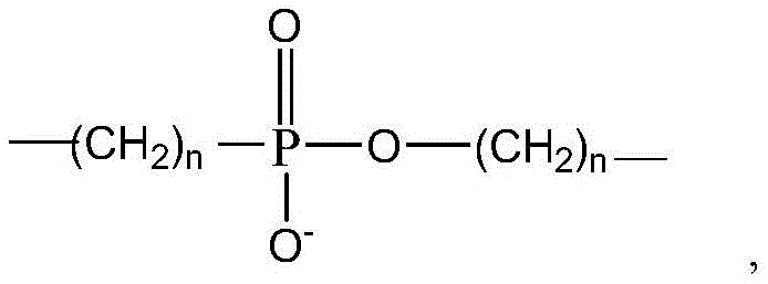
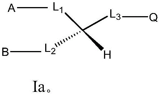
item 1 use of a compound from any natural (including traditional Chinese medicine extracts) or synthetic having the formula in the preparation of an agent for nucleic acid delivery, wherein the extract has the formula or comprises a compound having the formula:
Wherein L is
1 、L
2 、L
3 Absence, or L
1 、L
2 、L
3 Each independently selected from the group consisting of-C (O) O-CH
2 -,-CH(OH)-,-C(O)-NH-CH
2 -,-CH
2 -O-C(O)-,-CH
2 -NH-C(O)-,-C(O)O-,-C(O)NH-,-O-C(O)-,-NH-C(O)-,-CH
2 -,
Provided that L 1 、L 2 、L 3 At most two of which are absent;
for divalent radicals L 1 、L 2 In other words, the left dash "-" is attached to groups a and B, respectively, and the right dash "-" is attached to the central carbon atom, respectively;
for divalent radicals L 3 In other words, the dash "-" on the left is attached to the central carbon atom, while the dash "-" on the right is attached to Q;
a, B and Q are each independently selected from H, -OH, C 1-20 Alkyl, C 1-20 Alkenyl, C 1-20 Heteroalkyl, C 1-20 Heteroalkenyl, -NH 2 and-NR 3 + R is H or C 1-6 An alkyl group; and
n is an integer 0,1,2,3 or 4;
wherein preferably the nucleic acid is a small nucleic acid, preferably single-stranded or double-stranded, preferably the small nucleic acid is 14-32bp, 16-28bp or 18-24bp in length;
preferably, the traditional Chinese medicine is selected from rhodiola rosea, dandelion, common andrographis herb and honeysuckle flower traditional Chinese medicine decoction pieces, preferably the extract is obtained by extracting fat-soluble components through a Bligh & Dyer method, more preferably the extract is obtained by soaking the traditional Chinese medicine decoction pieces in water, then sequentially carrying out strong fire decoction and weak fire decoction, concentrating the decocted traditional Chinese medicine liquid, then sequentially adding chloroform-methanol, chloroform and water, stirring, and taking a chloroform layer;
Preferably, the agent is an oral agent; preferably, the nucleic acid is for use in the treatment of a disease, such as cancer, e.g. gastric cancer or lung cancer.
Item 2. Use of item 1, wherein in the structure
L 1 Absence, or L 1 Selected from the group consisting of-C (O) O-CH 2 -and-CH (OH) -,
L 2 absence, or L 2 Selected from the group consisting of-C (O) O-and-C (O) NH-,
L
3 absence, or L
3 Selected from-C (O) O-, -CH
2 -O-C(O)-,-CH
2 -sum of
A is selected from H, C 1-20 Alkyl and C 1-20 Alkenyl groups;
b is selected from H, -NH 2 ,C 1-20 Alkyl and C 1-20 Alkenyl groups;
q is selected from H, -OH, C 1-20 Alkyl and C 1-20 Alkenyl, and-NR 3 + Wherein R is H or C 1-6 An alkyl group.
The use of item 3, item 1 or 2, wherein the compound has the formula
Item 4 the use of any one of the preceding items, wherein in the structure
A is selected from H, C 10-20 Alkyl and C 10-20 Alkenyl groups;
b is selected from H, -NH 2 ,C 10-20 Alkyl and C 10-20 Alkenyl groups;
q is selected from H, -OH, C 10-20 Alkyl and C 10-20 Alkenyl, and-NR 3 + Wherein R is H or C 1-4 An alkyl group.
Item 5. Use of item 4, wherein in the structure
A is selected from H, straight chain C 15-18 Alkyl and straight chain C 15-18 Alkenyl groups;
b is selected from H, -NH 2 Straight chain C 15-18 Alkyl and straight chain C 15-18 Alkenyl groups;
q is selected from H, -OH, straight chain C 15-18 Alkyl and straight chain C 15-18 Alkenyl, and-NR 3 + Wherein R is H or C 1-4 An alkyl group;
in A, B, Q, the alkenyl group has 1 to 5 double bonds.
The use of item 5, wherein in a, B, Q of the structure the alkenyl group has 1-4 double bonds and is in the Z configuration.
Use of item 7. Use of item 6, wherein: in A, B, Q, the alkenyl group has 1 to 3 double bonds and is in the Z configuration.
The use of any one of the preceding items, wherein the extract is selected from the group consisting of the following formulas or comprises a compound selected from the group consisting of the following formulas:
wherein the method comprises the steps of
A is selected from straight chain C 15-18 Alkyl and straight chain C 15-18 Alkenyl groups;
b is selected from straight chain C 15-18 Alkyl and straight chain C 15-18 Alkenyl groups;
q is selected from H, -OH, straight chain C 15-18 Alkyl and straight chain C 15-18 Alkenyl, and-NR 3 + Wherein R is H or methyl; l (L) 3 is-C (O) O-.
The use of any one of the preceding items, wherein the extract is or comprises lysolecithin, ceramide, diglycerides, phosphatidylethanolamine, phosphatidylcholine, triglycerides, monogalactodiglycerides, (nerve) sphingosine, phosphatidylethanol, monoacylglycerol, fatty acids, platelet activating factor, or dimethyl phosphatidylethanolamine.
The use of any one of the preceding items, wherein the extract is selected from the group consisting of the lipids shown in table 1 or comprises any one or more of the lipids shown in table 1.
The use of any of the preceding items, wherein the extract comprises any one of the lipids shown in table 1, no. 41, no. 71, no. 11, no. 12, no. 38, no. 64, no. 40, no. 37, no. 39, no. 60, no. 62, or a combination thereof with any one or more other lipids in table 1, or with any one or more lipids and other related chemicals.
Use of a combination comprising any one or more lipids selected from those shown in table 1, wherein preferably the combination comprises any one of the lipids shown in table 1 No. 41, no. 71, no. 11, no. 12, no. 38, no. 64, no. 40, no. 37, no. 39, no. 60, no. 62, or a combination thereof with any one or more other lipids of table 1, or with any one or more lipids and other related chemicals, in the preparation of a reagent for nucleic acid delivery, wherein preferably the nucleic acid is a small nucleic acid, preferably single-stranded or double-stranded, preferably the small nucleic acid is 14-32bp, 16-28bp, or 18-24bp in length; preferably the agent is an oral agent; preferably, the nucleic acid is for use in the treatment of a disease, such as cancer, e.g. gastric cancer or lung cancer.
Use of a traditional Chinese medicine in the preparation of a reagent for nucleic acid delivery, wherein preferably the nucleic acid is a small nucleic acid, preferably single-stranded or double-stranded, preferably the small nucleic acid is 14-32bp, 16-28bp or 18-24bp in length; preferably the agent is an oral agent; preferably, the nucleic acid is for use in the treatment of a disease, such as cancer, e.g. gastric cancer or lung cancer.
The use of item 14, item 13, wherein the herbal medicine is selected from the group consisting of rhodiola rosea, dandelion, andrographis paniculata and honeysuckle.
The use according to item 15, item 13 or 14, wherein the agent contains a compound extracted from a traditional Chinese medicine or synthesized, preferably the compound is obtained by extracting fat-soluble components by the Bligh & dyr method, or by preparing and extracting by decocting a traditional Chinese medicine, more preferably by immersing the decoction pieces of the traditional Chinese medicine in water, then sequentially performing strong fire decoction and weak fire decoction, concentrating the decocted traditional Chinese medicine liquid, then sequentially adding chloroform-methanol, chloroform and water, stirring, and taking a chloroform layer.
The use of item 16, item 15, wherein the compound has the structure shown in any one of items 1 to 11, or the agent comprises any one or more of the lipids shown in table 1, preferably any one of the lipids shown in table 1, no. 41, no. 71, no. 11, no. 12, no. 38, no. 64, no. 40, no. 37, no. 39, no. 60, no. 62, or a combination thereof with any one or more other lipids in table 1, or with any one or more lipids and other related chemicals.
The use of item 17, item 16, wherein the compound is selected from lysolecithin, ceramide, diglyceride, phosphatidylethanolamine, phosphatidylcholine, triglyceride, monogalactose diglyceride, (nerve) sphingosine, phosphatidylethanol, monoacylglycerol, fatty acid, platelet activating factor, or dimethyl phosphatidylethanolamine.
The use of item 18, item 17, wherein the compound is selected from table 1.
The use of item 19, item 18, wherein the compound is selected from the group consisting of lipids shown in table 1 under No. 41, no. 71, no. 11, no. 12, no. 38, no. 64, no. 40, no. 37, no. 39, no. 60, no. 62.
The use of any one of items 13-18, wherein said delivery comprises in vitro cell delivery, or in vivo gut delivery.
The use of any one of items 13-20, comprising preparing a lipid nucleic acid mixture.
The use of item 22, item 21, wherein said lipid nucleic acid mixture is prepared by water boiling, or by reverse evaporation, or by direct mixing.
The use of item 23, item 22, wherein the preparation temperature of the water boiling process is from about 4 ℃ to about 100 ℃, from about 25 ℃ to about 100 ℃, preferably from about 80 ℃ to about 100 ℃, such as 4 ℃,37 ℃,60 ℃,80 ℃, or 100 ℃; the reverse evaporation process is carried out at a temperature of from about 25 ℃ to about 70 ℃, preferably about 55 ℃.
A pharmaceutical composition comprising one or more lipid extracts of the structure described in any one of items 1-11, preferably the lipid is selected from any one or more of table 1, preferably any one of the lipids indicated in table 1 under No. 41, no. 71, no. 11 and No. 12, no. 38, no. 64, no. 40, no. 37, no. 39, no. 60, no. 62, or a combination thereof with any one or more other lipids in table 1, or with any one or more lipids and other related chemicals, wherein preferably the nucleic acid is a small nucleic acid, preferably single-stranded or double-stranded, preferably the small nucleic acid is 14-32bp, 16-28bp, or 18-24bp in length; preferably, the pharmaceutical composition is an oral pharmaceutical composition; preferably, the pharmaceutical composition is for use in the treatment of a disease, such as cancer, e.g. gastric cancer or lung cancer.
The pharmaceutical composition of item 24, wherein the lipid and nucleic acid are present at least partially or wholly in the form of a lipid nucleic acid mixture.
The pharmaceutical composition of item 26, item 25, wherein the lipid nucleic acid mixture is prepared by water boiling, or by reverse evaporation, or by direct mixing.
The pharmaceutical composition of item 27, item 26, wherein the preparation temperature of the water boiling process is from about 4 ℃ to about 100 ℃,25 ℃ to about 100 ℃, preferably from about 80 ℃ to 100 ℃, e.g., 4 ℃,37 ℃,60 ℃,80 ℃, or 100 ℃; the reverse evaporation process is carried out at a temperature of from about 25 ℃ to about 70 ℃, preferably about 55 ℃.
Item 28. A kit of parts comprising one or more lipids of the structure according to any one of items 1-11, preferably the lipids are selected from any one or more of table 1, preferably No. 41, no. 71, no. 11 and No. 12, no. 38, no. 64, no. 40, no. 37, no. 39, no. 60, no. 62 or a combination thereof with any one or more other lipids of table 1 or a combination thereof with any one or more lipids and other related chemicals, and a nucleic acid, wherein the lipids and nucleic acid are each provided independently in a first container and a second container, the first container and the second container being the same or different, wherein preferably the nucleic acid is a small nucleic acid, preferably single-stranded or double-stranded, preferably the small nucleic acid is 14-32bp, 16-28bp or 18-24bp in length; preferably, the kit is an oral kit; preferably, the kit of parts is for use in the treatment of a disease, such as cancer, e.g. gastric cancer or lung cancer.
Item 29. The kit of parts of item 28, wherein the lipid and the nucleic acid are at least partially or fully formulated into a lipid nucleic acid complex immediately prior to use.
The kit of parts of item 29, wherein the lipid nucleic acid complex is formulated by water boiling, reverse evaporation, or direct mixing.
The kit of parts of part 30, wherein the water boiling process has a preparation temperature of about 4 ℃ to about 100 ℃,25 ℃ to about 100 ℃, preferably about 80 ℃ to about 100 ℃, such as 4 ℃,37 ℃,60 ℃,80 ℃ or 100 ℃, and the reverse evaporation process has a preparation temperature of about 25 ℃ to about 70 ℃, preferably about 55 ℃.
A method of delivering a nucleic acid into a target cell, wherein the nucleic acid is provided as the pharmaceutical composition of any one of items 24-27 or as the kit of any one of items 28-31, wherein preferably the nucleic acid is a small nucleic acid, preferably single-stranded or double-stranded, preferably the small nucleic acid is 14-32bp, 16-28bp, or 18-24bp in length; preferably, the nucleic acid is for use in the treatment of a disease, such as cancer, e.g. gastric cancer or lung cancer.
Item 33. A method of delivering a nucleic acid in vivo into a subject in need thereof, wherein the nucleic acid is provided in the form of the pharmaceutical composition of any one of items 24-27 or in the form of the kit of any one of items 28-31, wherein preferably the nucleic acid is a small nucleic acid, preferably single-stranded or double-stranded, preferably the small nucleic acid is 14-32bp, 16-28bp or 18-24bp in length; preferably, the nucleic acid is for use in the treatment of a disease, such as cancer, e.g. gastric cancer or lung cancer.
Item 34. The method of item 33, wherein the subject is a human or animal, such as a mammal.
The method of any one of clauses 33-34, wherein the nucleic acid is delivered in vivo into the subject's blood circulation or into a target tissue/cell.
The method of item 35, comprising delivering the pharmaceutical composition of any one of items 24-27 directly or in the kit of any one of items 28-31 through the alimentary canal to a subject in need thereof.
The pharmaceutical composition of any one of items 24-27, or the kit of any one of items 28-31, wherein the nucleic acid and lipid are formulated for topical administration and/or injection administration.
Item 38. The pharmaceutical composition or kit of items 37, wherein the nucleic acid and lipid are formulated for administration via the alimentary canal, via the respiratory tract.
Item 39. The pharmaceutical composition or kit of items 37 or 38, wherein the nucleic acid and lipid are formulated for oral, inhaled administration.
Item 40. The pharmaceutical composition or kit of any one of items 37-39, wherein the nucleic acid is a small RNA.
Item 41. The pharmaceutical composition or kit of any one of items 37-40, wherein the nucleic acid has a stem-loop structure.
The pharmaceutical composition, or kit of parts, according to any one of items 37-41, wherein the small RNA is 14-32bp, 18-24bp in length, e.g., 14, 15, 16, 17, 18, 19, 20, 21, 22, 23, 24, 25, 26, 27, 28, 29, 30, 31, 32bp in length.
Item 43. Compounds extracted from traditional Chinese medicine or synthetic and useful for nucleic acid delivery having the structure:
L
1 、L
2 、L
3 absence, or L
1 、L
2 、L
3 Each independently selected from the group consisting of-C (O) O-CH
2 -,-CH(OH)-,-C(O)-NH-CH
2 -,-CH
2 -O-C(O)-,-CH
2 -NH-C(O)-,-C(O)O-,-C(O)NH-,-O-C(O)-,-NH-C(O)-,-CH
2 -,
Provided that L 1 、L 2 、L 3 At most two of which are absent;
for divalent radicals L 1 、L 2 In other words, the left dash "-" is attached to groups a and B, respectively, and the right dash "-" is attached to the central carbon atom, respectively;
For divalent radicals L 3 In other words, the dash "-" on the left is attached to the central carbon atom, while the dash "-" on the right is attached to Q;
a, B and Q are each independently selected from H, -OH, C 1-20 Alkyl, C 1-20 Alkenyl, C 1-20 Heteroalkyl, C 1-20 Heteroalkenyl, -NH 2 and-NR 3 + R is H or C 1-6 An alkyl group; and
n is an integer 0,1,2,3 or 4, preferably the compound is an oral compound; preferably, the nucleic acid is for use in the treatment of a disease, such as cancer, e.g. gastric cancer or lung cancer.
The compound of item 44, item 43, wherein
L 1 Absence, or L 1 Selected from the group consisting of-C (O) O-CH 2 -and-CH (OH) -,
L 2 absence, or L 2 Selected from the group consisting of-C (O) O-and-C (O) NH-,
L
3 absence, or L
3 Selected from-C (O) O-, -CH
2 -O-C(O)-,-CH
2 -sum of
A is selected from H, C 1-20 Alkyl and C 1-20 Alkenyl groups;
b is selected from H, -NH 2 ,C 1-20 Alkyl and C 1-20 Alkenyl groups;
q is selected from H, -OH, C 1-20 Alkyl and C 1-20 Alkenyl, and-NR 3 + Wherein R is H or C 1-6 Alkyl, wherein the Chinese medicine is preferably selected from rhodiola rosea, dandelion, common andrographis herb and honeysuckle flower Chinese medicinal decoction pieces, and the Chinese medicine is preferably selectedThe compound was purified by Bligh&The fat-soluble component is extracted by the Dyer method, more preferably, the method comprises the steps of soaking the traditional Chinese medicine decoction pieces in water, then sequentially carrying out strong fire decoction and weak fire decoction, concentrating the decocted traditional Chinese medicine liquid, then sequentially adding chloroform-methanol, chloroform and water, stirring, and taking a chloroform layer, wherein the nucleic acid is preferably small nucleic acid, preferably single-stranded or double-stranded, and the length of the small nucleic acid is preferably 14-32bp, 16-28bp or 18-24bp.
Item 45A compound of item 43 or 44 having the formula
The compound of any one of clauses 43-45, wherein:
a is selected from H, C 10-20 Alkyl and C 10-20 Alkenyl groups;
b is selected from H, -NH 2 ,C 10-20 Alkyl and C 10-20 Alkenyl groups;
q is selected from H, -OH, C 10-20 Alkyl and C 10-20 Alkenyl, and-NR 3 + Wherein R is H or C 1-4 An alkyl group.
The compound of any one of clauses 43-46, wherein:
a is selected from H, straight chain C 15-18 Alkyl and straight chain C 15-18 Alkenyl groups;
b is selected from H, -NH 2 Straight chain C 15-18 Alkyl and straight chain C 15-18 Alkenyl groups;
q is selected from H, -OH, straight chain C 15-18 Alkyl and straight chain C 15-18 Alkenyl, and-NR 3 + Wherein R is H or C 1-4 An alkyl group;
in A, B, Q, the alkenyl group has 1 to 5 double bonds.
The compound of any one of clauses 43-47, wherein: in A, B, Q, the alkenyl group has 1 to 4 double bonds and is in the Z configuration.
The compound of any one of clauses 43-48, wherein: in A, B, Q, the alkenyl group has 1 to 3 double bonds and is in the Z configuration.
The compound of any one of clauses 43-49, selected from the group consisting of:
wherein the method comprises the steps of
A is selected from straight chain C 15-18 Alkyl and straight chain C 15-18 Alkenyl groups;
b is selected from straight chain C 15-18 Alkyl and straight chain C 15-18 Alkenyl groups;
q is selected from H, -OH, straight chain C 15-18 Alkyl and straight chain C 15-18 Alkenyl, and-NR 3 + Wherein R is H or methyl;
L 3 is-C (O) O-.
The compound of any one of clauses 43-50, selected from the group consisting of the lipids shown in table 1.
The compound of any one of items 43-51, selected from the group consisting of lipids shown in table 1 under No. 41, no. 71, no. 11, no. 12, no. 38, no. 64, no. 40, no. 37, no. 39, no. 60, or No. 62.
Item 53 a method of facilitating nucleic acid delivery comprising subjecting a nucleic acid and a traditional Chinese medicine extract or any natural or synthetic compound as described in any of items 1 to 11, preferably a lipid, to a heating or warming treatment, preferably at a temperature in the range of from about 4 ℃ to about 100 ℃, from about 25 ℃ to about 100 ℃, more preferably from about 50 ℃ to about 100 ℃, more preferably from about 95 ℃ to about 100 ℃, particularly preferably from about 80 ℃ to about 100 ℃, such as 4 ℃,37 ℃,60 ℃,80 ℃ or 100 ℃, wherein preferably the nucleic acid is a small nucleic acid, preferably single-stranded or double-stranded, preferably the small nucleic acid is 14-32bp, 16-28bp or 18-24bp in length; preferably, the nucleic acid delivery is by oral administration; preferably, the nucleic acid is for use in the treatment of a disease, such as cancer, e.g. gastric cancer or lung cancer.
Item 54. The method of item 53, wherein the herbal extract comprises a compound of the structure as shown in items 1-9.
The method of item 55, item 53, wherein the herbal extract comprises any one or more of the lipids shown in table 1.
The method of item 56, item 53, wherein the herbal extract comprises any one of the lipids shown in table 1, no. 41, no. 71, no. 11, no. 12, no. 38, no. 64, no. 40, no. 37, no. 39, no. 60, no. 62, or a combination thereof with any one or more other lipids in table 1, or with any one or more lipids and other related chemicals.
Item 57. Use of item 11, 12 or 16, pharmaceutical composition of item 24, or kit of items 28, wherein the combination is a combination of any one of: no. 8, 41 = 6:1 lipid combination; lipid combination No. 38, 41 = 6:1; no. 39, 41 = 6:1 lipid combination; no. 40, 41 = 6:1 lipid combination; lipid combination No. 38:12:41:29 = 1:2:1:1; lipid combination 40:12:41 = 2:4:3; lipid combination No. 12:41 = 1:6; lipid combination No. 12:41 = 1:1; lipid combination No. 12:41 = 6:1; lipid combination 40:12:41 = 2:2:2; lipid combination No. 4:12:41 = 1:1:1; DG combinations 1:2:3:19:35 =1:1:1:1:1; TG combinations of numbers 6:9:10:13:15:16:18:20:21:22:23:24:25:26:27:28:32:33 = 1:1:1:1:1:1:1:1:1:1:1:1:1:1:1:1:1:1; LPC combination number 36:37 = 1:1; PC combination 11:12 = 1:1; PE combination number 8:38 = 1:1; cer combination No. 4:14 = 1:1; a So combination of 17:30:31=1:1:1; no equal volume combinations of nos. 1-36 of nos. 5, 7; no equal volume combinations of nos. 1-36 of nos. 5, 7, 34; no equal volume combinations of nos. 1-36 of nos. 5, 7, 1, 2, 3, 19, 35; no equal volume combinations No. 1-36 of numbers 5, 7, 6, 9, 10, 13, 15, 16, 18, 20, 21, 22, 23, 24, 25, 26, 27, 28, 32, 33; no equal volume combinations of nos. 1-36 of nos. 5, 7, 36, 37; no equal volume combinations of nos. 1-36 of nos. 5, 7, 11, 12; no equal volume combinations of nos. 1-36 of nos. 5, 7, 8; no equal volume combinations of nos. 1-36 of nos. 5, 7, 4, 14; no equal volume combinations of nos. 1-36 of nos. 5, 7, 29; lipid No. 1: no. 34 = 2:1; lipid No. 1, DG combination = 2:1; lipid No. 1, TG combination = 2:1; lipid No. 1, the LPC combination = 2:1; lipid No. 1: no. 8 = 2:1; lipid No. 1: no. 12 = 2:1; lipid No. 1: the Cer combination = 2:1; lipid No. 1: so combination = 2:1; lipid No. 1: no. 29 = 2:1; lipid No. 1 to No. 8 to No. 12 = 1:1:1; lipid No. 8: no. 34 = 2:1; lipid No. 8: DG combination = 2:1; lipid No. 8: TG combination = 2:1; lipid number 8 LPC combination = 2:1; lipid No. 8, no. 37 = 4:1; lipid No. 8: no. 12 = 2:1; lipid No. 8: cer combination = 2:1; lipid No. 8: so combination = 2:1; lipid No. 8: no. 31 = 6:1; lipid No. 8: no. 29 = 2:1; no. 12, no. 34 = 2:1; DG combination=2:1; TG combination=2:1; LPC combination=2:1; lipid No. 8 = 2:1; cer combination=2:1; socombination=2:1; no. 12, no. 29 = 2:1; lipid No. 8, no. 1&2 = 2:1:1; lipid No. 8 No. 15 = 2:1:1; lipid No. 8, no. 36&37 = 2:1:1; lipid No. 8 No. 11 = 2:1:1; lipid No. 8 No. 12 = 2:1:1; lipid No. 8 No. 4 = 2:1:1; lipid No. 8 No. 31 = 2:1:1; lipid No. 8 No. 29 = 2:1:1; lipid No. 8, no. 34 = 3:2:1; lipid No. 8, no. 34 = 4:2:3; lipid No. 12, lipid No. 8, lipid No. 2 = 4:2:3; lipid No. 12, lipid No. 8, lipid No. 2 = 16:8:3; lipid No. 8 No. 32 = 4:2:3; lipid No. 8 No. 37 = 4:2:3; lipid No. 8 No. 11 = 4:2:3; lipid No. 8 No. 38 = 4:2:3; lipid No. 8 No. 4 = 4:2:3; lipid No. 8 No. 31 = 4:2:3; lipid No. 8 No. 29 = 4:2:3; lipid No. 8 No. 29 No. 31 No. = 2:1:1:1; lipid No. 8, no. 29, no. 31, no. 34 = 4:2:2:2:5; lipid No. 12, lipid No. 8, no. 29, no. 31, lipid No. 2 = 4:2:2:2:5; lipid No. 8 No. 29 No. 31 No. 32=4:2:2:2:5; lipid No. 8 No. 29 No. 31 No. 11=4:2:2:2:5; lipid No. 8 No. 29 No. 31 No. 37 = 4:2:2:2:5; lipid No. 8 No. 29 No. 31 No. 38 No. = 4:2:2:2:5; lipid No. 8 No. 29 No. 31 No. 4=4:2:2:2:5; lipid No. 12, lipid No. 8, no. 29, no. 31, no. 4, lipid No. 1, no. 16 = 2:1:1:3:2:2:3; lipid No. 1, lipid No. 8, lipid No. 12, lipid No. 1&2 = 2:2:2:3; lipid No. 1, lipid No. 8, no. 12, no. 15 = 2:2:2:3; lipid No. 1, lipid No. 8, no. 12, no. 36&37 = 2:2:2:3; lipid No. 1, lipid No. 8, no. 12 = 2:2:2:3; lipid No. 1, lipid No. 8, no. 12, no. 4 = 2:2:2:3; lipid No. 1, lipid No. 8, no. 12, no. 31 = 2:2:2:3; lipid No. 1, lipid No. 8, no. 12, no. 29 = 2:2:2:3; lipid No. 8: no. 34 lipid No. 1&2 = 2:1:1; lipid No. 8:34 No. 15 = 2:1:1; lipid No. 8:34 No. 36&37 = 2:1:1; lipid No. 8:34 No. 12 = 2:1:1; lipid No. 8:34 No. 4 = 2:1:1; lipid No. 8:34 No. 31 = 2:1:1; lipid No. 8:34 No. 29 = 2:1:1; lipid No. 8: no. 31: no. 34 = 12:3:5; lipid No. 8: no. 31 lipid No. 2 = 12:3:5; lipid No. 8: no. 31: no. 37 = 12:3:5; lipid No. 8: no. 31: no. 11 = 12:3:5; lipid No. 8: no. 31: no. 12 = 12:3:5; lipid No. 8: no. 31: no. 4 = 12:3:5; lipid No. 8: no. 31: no. 29 = 12:3:5; lipid No. 8: no. 31: no. 32 = 12:3:5; lipid No. 8:no. 4:no. 34 = 12:3:5; lipid No. 8 to No. 4 lipid No. 2 = 12:3:5; lipid No. 8: no. 4: no. 37 = 12:3:5; lipid No. 8: no. 4: no. 12 = 12:3:5; lipid No. 8: no. 4: no. 31 = 12:3:5; lipid No. 8: no. 4: no. 29 = 12:3:5; lipid No. 8: no. 4: no. 32 = 12:3:5; no. 38, no. 34 = 2:1; lipid No. 38, no. 1 = 2:1; lipid No. 38, lipid No. 2 = 2:1; no. 38, no. 1&2 = 2:1; no. 38, no. 15 = 2:1; no. 38, no. 32=2:1; no. 38, no. 37 = 2:1; no. 38, no. 37 = 4:1; no. 38, no. 11 = 2:1; no. 38, no. 12 = 2:1; no. 38, no. 11&12 = 2:1; no. 38, no. 12 = 4:1; lipid No. 38, no. 8 = 2:1; no. 38, no. 4 = 2:1; no. 38 So (30) =2:1; no. 38, no. 31 = 2:1; no. 38, no. 29 = 2:1; lipid No. 1: no. 38: no. 12: no. 34 = 2:2:2:3; lipid No. 1: no. 38: no. 12: no. 15 = 2:2:2:3; lipid No. 1: no. 38: no. 12: no. 37 = 2:2:2:3; lipid No. 1, no. 38, no. 12, lipid No. 8 = 2:2:2:3; lipid No. 1: no. 38: no. 12: no. 4 = 2:2:2:3; lipid No. 1: no. 38: no. 12: no. 31 = 2:2:2:3; lipid No. 1: no. 38: no. 12: no. 29 = 2:2:2:3; no. 38, no. 34, lipid No. 1 = 2:1:3; no. 38 to 34 No. 15 = 2:1:3; no. 38 to 34 to 37 = 2:1:3; no. 38 to 34 No. 12 = 2:1:3; no. 38, no. 34, lipid No. 8 = 2:1:3; no. 38 to 34 No. 4 = 2:1:3; no. 38 to 34 to 31 = 2:1:3; no. 38 to 34 No. 29 = 2:1:3; no. 38, no. 12, lipid No. 1 = 2:1:3; no. 38, no. 12, lipid No. 2 = 4:1:3; no. 38 to No. 12 to No. 15 = 2:1:3; no. 38 to No. 12 to No. 37 = 2:1:3; no. 38, no. 12, lipid No. 8 = 2:1:3; no. 38 to No. 12 to No. 4 = 2:1:3; no. 38 to No. 12 to No. 31 = 2:1:3; no. 38 to No. 12 to No. 29 = 2:1:3; no. 38, no. 12, lipid No. 1, no. 15, no. 34 = 22:22:22:33:36; no. 38, no. 12, lipid No. 1, no. 15, no. 37 = 22:22:22:33:36; no. 38, no. 12, lipid No. 1, no. 15, no. 4 = 22:22:22:33:36; no. 38, no. 12, lipid No. 1, no. 15, no. 31 = 22:22:22:33:36; no. 38, no. 12, lipid No. 1, no. 15, no. 29 = 22:22:22:33:36; no. 38, no. 34, no. 37, lipid No. 1 = 44:22:33:36; no. 38 to 34 to 37 to 15 = 44:22:33:36; no. 38 to 34 to 37 to 12 = 44:22:33:36; no. 38 to 34 to 37 to 4 = 44:22:33:36; no. 38 to 34 to 37 to 31 = 44:22:33:36; no. 38 to No. 12 to No. 4 to No. 34 = 44:22:33:36; no. 38, no. 12, no. 4, lipid No. 1 = 44:22:33:36; no. 38 to No. 12 to No. 4 to No. 15 = 44:22:33:36; no. 38 to No. 12 to No. 4 to No. 37 = 44:22:33:36; no. 38 to No. 12 to No. 4 to No. 37 = 8 to 2 to 5 to 3; no. 38 to No. 12 to No. 4 to No. 31 = 44:22:33:36; no. 38 to No. 12 to No. 4 to No. 29 = 44:22:33:36; no. 38, no. 12, no. 4, no. 29, no. 34 = 88:44:66:72:135; no. 38, no. 12, no. 4, no. 29, no. 1 lipid = 88:44:66:72:135; no. 38, no. 12, no. 4, no. 29, no. 15 = 88:44:66:72:135; no. 38, no. 12, no. 4, no. 29, no. 37 = 88:44:66:72:135; no. 38, no. 12, no. 4, no. 29, no. 31 = 88:44:66:72:135; no. 38, no. 12, no. 4, lipid No. 2 = 20:10:15:9; no. 38 to No. 12 to No. 4 to No. 6 = 20 to 10 to 15 to 9; no. 38 to No. 12 to No. 4 to No. 17 = 20 to 10 to 15 to 9; no. 38 to No. 12 to No. 4 to No. 29 = 20 to 10 to 15 to 9; no. 38 to No. 12 to No. 4 to No. 34 = 20 to 10 to 15 to 9; no. 38 to No. 12 to No. 4 to No. 37 = 20 to 10 to 15 to 9; no. 38, no. 12, no. 31, no. 34 = 2:1:3:3; no. 38, no. 12, no. 31, lipid No. 1 = 2:1:3:3; no. 38 to No. 12 to No. 31 to No. 15 = 2:1:3:3; no. 38, no. 12, no. 31, no. 37 = 2:1:3:3; no. 38, no. 12, no. 31, no. 4 = 2:1:3:3; no. 38, no. 12, no. 31, no. 29 = 2:1:3:3; no. 38, no. 34, no. 37, no. 31, no. 1 lipid = 88:44:66:72:135; no. 38, no. 34, no. 37, no. 31, no. 15 = 88:44:66:72:135; no. 38, no. 34, no. 37, no. 31, no. 12 = 88:44:66:72:135; no. 38, no. 34, no. 37, no. 31, no. 4 = 88:44:66:72:135; no. 38, no. 34, no. 37, no. 31, no. 29 = 88:44:66:72:135; no. 38 to No. 37, no. 34 = 4:2:3; no. 38, no. 37, lipid No. 1 = 4:2:3; no. 38, no. 37, lipid No. 2 = 4:2:3; no. 38, no. 37, no. 1&2 = 4:2:3; no. 38, no. 37, lipid No. 2 = 32:8:5; no. 38 to No. 37, no. 32=32:8:5; no. 38 to No. 37 to No. 15 = 4:2:3; no. 38 to No. 37, no. 32 = 4:2:3; no. 38, no. 37, lipid No. 8 = 4:2:3; no. 38 to No. 37 to No. 11 = 4:2:3; no. 38 to No. 37 to No. 12 = 4:2:3; no. 38, no. 37, no. 11&12 = 4:2:3; no. 38 to No. 37 to No. 12 = 4:1:1; no. 38 to No. 37 to No. 4 = 4:2:3; no. 38 to No. 37, no. 30 = 4:2:3; no. 38 to No. 37 to No. 31 = 4:2:3; no. 38 to No. 37 to No. 29 = 4:2:3; lipid No. 8: no. 37: no. 32 = 4:1:2; lipid No. 8: no. 37 lipid No. 2 = 4:1:2; no. 38, no. 37, no. 15, no. 34 = 64:16:10:45; no. 38, no. 37, no. 15, lipid No. 1 = 64:16:10:45; no. 38, no. 37, no. 15, no. 12 = 64:16:10:45; no. 38, no. 37, no. 15, no. 4 = 64:16:10:45; no. 38 to No. 37 to No. 15 to No. 31 = 64 to 16 to 10 to 45; no. 38, no. 37, no. 15, no. 29 = 64:16:10:45; lipid No. 2, no. 37 = 4:2:3; lipid No. 2 No. 31 = 4:2:3; lipid No. 2, no. 29 = 4:2:3; lipid No. 2, no. 34 = 4:2:3; lipid No. 2, no. 32 = 4:2:3; lipid No. 2, lipid No. 12 = 4:2:3; lipid No. 2, no. 12 = 4:5:1; lipid No. 38: lipid No. 2: no. 4 = 4:2:3, lipid No. 1&2, 11&12 or 36&37 represent lipid nos. 1 and 2, 11 and 12 or 36 and 37, respectively, in any ratio.
Use of a compound having the structure:
wherein,,
wherein L is 1 、L 2 、L 3 Absence, or L 1 、L 2 、L 3 Each independently selected from the group consisting of-C (O) O-CH 2 -,-CH(OH)-,-CH 2 -O-C(O)-,-C(O)O-,-C(O)NH-;
Provided that L 1 、L 2 、L 3 At most two of which are absent;
for divalent radicals L 1 、L 2 In other words, the left dash "-" is attached to groups a and B, respectively, and the right dash "-" is attached to the central carbon atom, respectively;
for divalent radicals L 3 In other words, the dash "-" on the left is attached to the central carbon atom, while the dash "-" on the right is attached to Q;
a, B and Q are each independently selected from H, -OH, C 1-20 Alkyl, C 1-20 Alkenyl, -NH 2 and-NR 3 + R is H or C 1-6 An alkyl group; preferably the agent is an oral agent; preferably, the method comprises the steps of,the nucleic acids are useful for treating diseases, such as cancer, e.g., gastric cancer or lung cancer.
The use of item 59, item 58, wherein the compound has the structure:
wherein,,
a is selected from straight chain C 10-20 Alkyl and straight chain C 10-20 Alkenyl groups;
b is selected from straight chain C 10-20 Alkyl and straight chain C 10-20 Alkenyl groups;
q is-OH;
preferably, the method comprises the steps of,
a is selected from straight chain C 15-20 Alkyl and straight chain C 15-20 Alkenyl groups;
b is selected from straight chain C 15-20 Alkyl and straight chain C 15-20 Alkenyl groups;
q is-OH;
preferably, the method comprises the steps of,
a is selected from straight chain C 15-18 Alkyl and straight chain C 15-18 Alkenyl groups;
b is selected from straight chain C 15-18 Alkyl and straight chain C 15-18 Alkenyl groups;
q is-OH.
The use of item 60, item 58, wherein the compound has the structure:
wherein,,
a is selected from straight chain C 10-20 Alkyl and straight chain C 10-22 Alkenyl groups;
b is selected from straight chain C 10-20 Alkyl and straight chain C 10-22 Alkenyl groups;
q is selected from straight chain C 10-20 Alkyl and straight chain C 10-22 Alkenyl groups;
preferably, the method comprises the steps of,
a is selected from straight chain C 15-18 Alkyl and straight chainC 15-22 Alkenyl groups;
b is selected from straight chain C 15-18 Alkyl and straight chain C 15-22 Alkenyl groups;
q is selected from straight chain C 15-18 Alkyl and straight chain C 15-22 Alkenyl groups;
preferably, the method comprises the steps of,
a is selected from straight chain C 15-18 Alkyl and straight chain C 15-20 Alkenyl groups;
b is selected from straight chain C 15-18 Alkyl and straight chain C 15-20 Alkenyl groups;
q is selected from straight chain C 15-18 Alkyl and straight chain C 15-20 Alkenyl groups.
The use of item 61, item 58, wherein the compound has the structure:
wherein,,
a is selected from straight chain C 10-20 Alkyl and straight chain C 10-20 Alkenyl groups;
b is selected from straight chain C 10-20 Alkyl and straight chain C 10-20 Alkenyl groups;
q is-OH;
preferably, the method comprises the steps of,
a is selected from straight chain C 15-20 Alkyl and straight chain C 15-18 Alkenyl groups;
b is selected from straight chain C 15-18 Alkyl and straight chain C 15-18 Alkenyl groups;
q is-OH;
preferably, the method comprises the steps of,
a is straight chain C 15-20 An alkyl group;
b is straight chain C 15-18 An alkyl group;
q is-OH.
The use of item 58, wherein the compound has the structure:
wherein,,
a is selected from straight chain C 10-20 Alkyl and straight chain C 10-20 Alkenyl groups;
q is-OH;
preferably, the method comprises the steps of,
a is selected from straight chain C 10-20 Alkyl and straight chain C 15-18 Alkenyl groups;
q is-OH;
preferably, the method comprises the steps of,
a is straight chain C 15-20 An alkyl group;
q is-OH.
The use of any one of items 1-23, the pharmaceutical composition of any one of items 24-27, the kit of any one of items 28-31, the method of any one of items 32-36 and 53-56, or the method of item 43, wherein the lipid or compound is a compound having the structure:
wherein,,
a is selected from straight chain C 10-20 Alkyl and straight chain C 10-20 Alkenyl groups;
b is selected from straight chain C 10-20 Alkyl and straight chain C 10-20 Alkenyl groups;
q is-OH;
preferably, the method comprises the steps of,
a is selected from straight chain C 15-20 Alkyl and straight chain C 15-20 Alkenyl groups;
b is selected from straight chain C 15-20 Alkyl and straight chain C 15-20 Alkenyl groups;
q is-OH;
preferably, the method comprises the steps of,
a is selected from straight chain C 15-18 Alkyl and straight chain C 15-18 Alkenyl groups;
b is selected from straight chain C 15-18 Alkyl and straight chain C 15-18 Alkenyl groups;
q is-OH.
The use, pharmaceutical composition, kit of parts, or method of item 64, item 63, wherein the compound has the structure:
wherein,,
a is selected from straight chain C 10-20 Alkyl and straight chain C 10-22 Alkenyl groups;
b is selected from straight chain C 10-20 Alkyl and straight chain C 10-22 Alkenyl groups;
q is selected from straight chain C 10-20 Alkyl and straight chain C 10-22 Alkenyl groups;
preferably, the method comprises the steps of,
a is selected from straight chain C 15-18 Alkyl and straight chain C 15-22 Alkenyl groups;
b is selected from straight chain C 15-18 Alkyl and straight chain C 15-22 Alkenyl groups;
q is selected from straight chain C 15-18 Alkyl and straight chain C 15-22 Alkenyl groups;
preferably, the method comprises the steps of,
a is selected from straight chain C 15-18 Alkyl and straight chain C 15-20 Alkenyl groups;
b is selected from straight chain C 15-18 Alkyl and straight chain C 15-20 Alkenyl groups;
q is selected from straight chain C 15-18 Alkyl and straight chain C 15-20 Alkenyl groups.
Item 65, the use, pharmaceutical composition, kit of parts, or method of item 63, wherein the compound has the structure:
wherein,,
a is selected from straight chain C 10-20 Alkyl and straight chain C 10-20 Alkenyl groups;
b is selected from straight chain C 10-20 Alkyl and straight chain C 10-20 Alkenyl groups;
q is-OH;
preferably, the method comprises the steps of,
a is selected from straight chain C 15-20 Alkyl and straight chain C 15-18 Alkenyl groups;
b is selected from straight chain C 15-18 Alkyl and straight chain C 15-18 Alkenyl groups;
q is-OH;
preferably, the method comprises the steps of,
a is straight chain C 15-20 An alkyl group;
b is straight chain C 15-18 An alkyl group;
q is-OH.
The use, pharmaceutical composition, kit of parts, or method of item 66, item 63, wherein the compound has the structure:
wherein,,
a is selected from straight chain C 10-20 Alkyl and straight chain C 10-20 Alkenyl groups;
q is-OH;
preferably, the method comprises the steps of,
a is selected from straight chain C 10-20 Alkyl and straight chain C 15-18 Alkenyl groups;
q is-OH;
preferably, the method comprises the steps of,
a is straight chain C 15-20 An alkyl group;
q is-OH.
Examples
The following examples are merely illustrative of the invention disclosed herein and should not be construed as limiting the scope of the appended claims in any way.
TABLE 2 Small RNAs and sequences thereof used in the examples
Description: the symbols with the prefix "si-" are denoted double stranded sRNA.
Experimental examples on the 1-32 # lipids in Table 1
1. Extraction of lipids from Chinese medicinal materials
1.1 preparation of Chinese herbs by decoction
1) 100g of Chinese herbal pieces (rhodiola rosea, purchased from Ningbo sea-block biological technology Co., ltd.) were taken; dandelion, honeysuckle, common andrographis herb, purchased from Beijing Tongren Tang drug store), 1000mL ddH is added 2 O soaking for 30min.
2) The traditional Chinese medicine decoction pot is subjected to strong fire decoction for 15min and weak fire decoction for 20min.
3) 400mL of the decocted traditional Chinese medicine liquid is added into a rotary evaporator, and is concentrated to 100mL at 60 ℃ and 60rpm for 30min.
1.2 extraction of lipids
1) To 160mL of the Chinese medicinal decoction obtained according to item 1.1 above (concentrated by rotary evaporator) 600mL of chloroform-methanol mixture (chloroform: methanol=1:2, v/v) was added so that chloroform: methanol: water=1:2:0.8, and stirred and mixed well for 10-15min.
2) 200mL of chloroform was added to the flask, and the mixture was stirred and mixed for 10 minutes.
3) 200ml ddH was added to the Erlenmeyer flask 2 OmL chloroform to methanol to water=2:2:1.8, and stirring and mixing for 10min.
4) Removing insoluble substances in the upper liquid and the middle layer, taking the chloroform layer of the lower layer, and freezing at-40deg.C.
1.3HPLC-MS/MS identification of lipid components
Instrument conditions
1) Chromatographic conditions:
instrument: ultimate 3000; chromatographic column: kineex C18 (100×2.1mm,1.9 μm); column temperature: 45 ℃; mobile phase: a: acetonitrile: water (V/V, 60:40), solution containing 10mmol/L ammonium formate, mobile phase B: acetonitrile: isopropyl alcohol (10:90, V/V), the solution containing 10mmol/L ammonium formate and 0.1% formic acid. Flow rate: 0.4mL/min; sample injection amount: 4. Mu.L.
2) Mass spectrometry parameters:
a) Positive mode: heater Temp 300 ℃, sheath Gas Flow rate,45arb,Aux Gas Flow Rate,15arb,Sweep Gas Flow Rate,1arb,spray voltage,3.0KV,Capillary Temp,350 ℃, S-Lens RF Level,30%. Scan roles: 200-1500.
b) Negative mode: heater Temp 300 ℃, sheath Gas Flow rate,45arb,Aux Gas Flow Rate,15arb,Sweep Gas Flow Rate,1arb,spray voltage,2.5KV,Capillary Temp,350 ℃, S-Lens RF Level,60%. Scan roles: 200-1500.
1.4 identification of lipid derived from traditional Chinese medicine
The lipid components are identified by HPLC-MS/MS, 138 lipid components from Chinese medicine are identified, 125 lipid components are identified by cation mode, and 13 lipid components are identified by anion mode. The following experiment was continued with the compounds 1 to 32 shown in Table 1.
It should be noted that the lipids tested below were either commercially purchased or commercially synthesized and were used in the manner described in Table 1-1.
2. Preparation of lipid nucleic acid complexes
2.1 reverse evaporation method:
600 μl of lipid diethyl ether solution was prepared and grouped according to the lipid numbers shown in Table 1, wherein the diethyl ether solution concentration in the 1/2/4/9/14/18/19/20/21/22/23/24/25/26/27/28/29/30/32 th lipid group was 0.017857mg/mL, the diethyl ether concentration in the 3/8/10/11/12/13 th lipid group was 0.035714mg/mL, and the diethyl ether concentration in the 6/15/16/17/31 th lipid group was 0.0035714mg/mL; the lipid solution was added to 120. Mu.l of DEPC-treated aqueous solution (15 nmol) of HJT-sRNA-m7 single-stranded RNA at a volume ratio of 5:1, sonicated for 3min, the ether was removed by evaporation at 55℃and 600. Mu.l of DEPC-treated water was then added for hydration to give HJT-sRNA-m7 lipid mixture.
2.2 Water boiling method:
60 μl of lipid chloroform solution was prepared and grouped according to the lipid numbers shown in Table 1, wherein the chloroform solution concentration in the 1/2/4/9/14/18/19/20/21/22/23/24/25/26/27/28/29/30/32 lipid group was 5mg/mL, the chloroform solution concentration in the 3/8/10/11/12/13 lipid group was 10mg/mL, and the chloroform solution concentration in the 6/15/16/17/32 group was 1mg/mL; the lipid chloroform solution was mixed with 600. Mu.l of DEPC-treated aqueous solution of HJT-sRNA-m7 (15 nmol) single-stranded RNA, and heated at 100℃for 30 minutes, to obtain HJT-sRNA-m7 lipid mixture.
3. In vitro delivery experiments of lipid nucleic acid complexes
3.1 culturing NCI-N87 cells (gastric cancer cells), MRC-5 cells (lung fibroblasts), A549 cells (lung cancer cells) to logarithmic phase, and plating to six-well plates with cell density of 1×10 6 2mL of culture medium/well; wherein MRC-5 cells are cultured in Eagle's MEM medium (MEM, gibco); a549 cells were cultured in Ham's F-12 medium (HyClone); NCI-N87 cells were cultured in RPMI-1640 medium (HyClone); incubation was carried out overnight at 37℃and subsequent experiments were carried out after cell attachment.
3.2 experimental groupings were as follows:
1) NC group: refers to untreated cells; this group served as a negative control group.
2) RNAimax treatment group: diluting 2. Mu.l of RNAimax transfection reagent and HJT-sRNA-m7 solution with 100. Mu.l of opti-MEM culture medium respectively, mixing the two, standing for 15min, adding into cells, and mixing to obtain HJT-sRNA-m7 with a final concentration of 200nM; this group served as a positive control group.
3) Free uptake (Free uptake) group: directly into HJT-sRNA-m7 solution (final concentration 200 nM), this group served as a negative control.
4) Lipid nucleic acid mixture treatment group: the lipid prepared in step 2 was added to the cells with HJT-sRNA-m7 mixture, and the final concentration of RNA was controlled to 200nM.
3.3 incubating the small RNA with the cells for 3 hours, washing the cells for 2-3 times by using PBS, collecting the cells by using TRIzol lysate, extracting total RNA, detecting the abundance of the small RNA entering the cells by using RT-qPCR, and positioning the RNA by using a fluorescence in situ hybridization method; the detection method comprises the following steps:
3.3.1RT-qPCR detection of small RNA (Taqman Probe method)
1) Reverse transcription of sRNA to cDNA: through reverse transcription kit
MicroRNA Reverse Transcription Kit, cat.no. 4366597), the sRNA was reverse transcribed into cDNA, the reverse transcription system being as follows: 100mM dNTPs(with dTTP)0.15μl,MultiScribe
TM Reverse transcriptase, 50U/. Mu.L 1.00. Mu.L, 10 XRT buffer 1.5. Mu.L, RNase inhibitor (20U/. Mu.L) 0.19. Mu.L, no nuclease H
2 O4.6 μl, adding 5 μl RNA template (200 ng/. Mu.l) after mixing, adding 3 μl 5 x Taqman probe primer after mixing, instantly centrifuging after mixing, standing on ice for 5min, and placing into a PCR instrument for reaction under the following reaction conditions: (1) 16 ℃ for 30min; (2) 42 ℃ for 30min; (3) 85 ℃ for 5min; (4) terminating the reaction at 4 ℃. After completion of the reaction, 10. Mu.l of RNase-free ddH was added
2 O, make up the final volume to 25. Mu.l. Taqman probe primers used in the reverse transcription were synthesized by Invitrogen corporation (U6:4440887, HJT-sRNA-m7: 4398987).
2) Quantitative PCR amplification reaction: the total volume of the qPCR reaction system is 10 μl, comprising: 5 mu L
Universal Master Mix II with UNG, 0.5. Mu.l 20X Taqman primer, 1. Mu.l cDNA reverse transcribed, 3.5. Mu.l RNase-free dH
2 O. The PCR reaction conditions were: the PCR amplification cycle was started after 2min at 50℃and 10min at 95 ℃): (1) 95℃for 15s; (2) 60 ℃,60s; (3) 60 ℃,60s; a total of 40 cycles were performed; and finally, continuously cooling at 40 ℃ for 10 seconds. Taqman probes for the amplification reaction were designed and synthesized by Invitrogen corporation (U6:4440887, HJT-sRNA-m7: 4398987)
3) The relative expression level was calculated by the 2- ΔCt method.
3.3.2RT-qPCR detection of small RNA (SYBR Green dye method)
1) Reverse transcription of sRNA into cDNA: sRNA was reverse transcribed into cDNA by the stem-loop method (stem-loop method) using a reverse transcription kit (High-Capacity cDNA Reverse Transcription Kits, applied Biosystems, cat. No. 4368813) in the following manner: template RNA (150 ng/. Mu.l), 10. Mu.l 10 XRT buffer, 2.0. Mu.l 25 XdNTP Mix (100 mM), 0.8. Mu.l U6 RT step-loop primer, 2.0. Mu.l HJT-sRNA-m7 RT step-loop primer, 2.0. Mu.l MultiScribe TM 1.0. Mu.l of reverse transcriptase, 1.0. Mu.l of RNase inhibitor, no nuclease H 2 O1.2 μl, and placing into a PCR instrument for reaction under the following reaction conditions:(1) 25 ℃ for 10min; (2) 37 ℃ for 120min; (3) 85 ℃ for 5min; (4) terminating the reaction at 4 ℃. After completion of the reaction, 20. Mu.l of RNase-free ddH was added 2 O, make up the final volume to 40. Mu.l. The stem-loop primers used in the reverse transcription were synthesized by Beijing Optimu Biotechnology Co., ltd. (U6 RT primer: GTCGTATCCAGTGCAGGGTCCGAGGTATTCGCACTGGATACGACAAAAATATG (SEQ ID NO: 21); HJT-sRNA-m7 RT step-loop primer: GTCGTATCCAGTGCACGCTCCGAGGTATTCGCACTGGATACGACGCTTACAA (SEQ ID NO: 22)).
2) Quantitative PCR amplification reaction: the total volume of the qPCR reaction system is 10 μl, comprising: 5. Mu.L of 2X SYBR Green Master Mix, 0.5. Mu.L of forward primer (10. Mu.M), 0.5. Mu.L of reverse primer (10. Mu.M), 1. Mu.L of reverse transcribed cDNA, 3. Mu.L of RNase-free dH 2 O. The PCR reaction conditions were: pre-denaturation was continued for 5min at 95 ℃ and started into PCR amplification cycle: (1) 95 ℃ for 10s; (2) 55 ℃,10s; (3) 72 ℃,20s; a total of 40 cycles were performed; and finally, continuously cooling at 40 ℃ for 10 seconds. Both the forward and reverse primers for the amplification reaction were designed and synthesized by Beijing Optimago Biotechnology Inc. (U6F primer: GCGCGTCGTGAAGCGTTC (SEQ ID NO: 23), U6R primer: GTGCAGGGTCCGAGGT (SEQ ID NO: 24), HJT-sRNA-m 7F primer: TCGCGCTGAGGTAGTAGGTT (SEQ ID NO: 25), HJT-sRNA-m 7R primer: GTGCACGCTCCGAGGT (SEQ ID NO: 26)).
3) The relative expression level was calculated by the 2- ΔCt method.
3.3.3 Small RNA Fluorescence In Situ Hybridization (FISH)
1) The medium was removed and washed 3 times with 500. Mu.l/well PBS.
2) 500 μl/well of 4% paraformaldehyde (PBS phosphate buffer) was fixed at room temperature for 20min.
3) 500 μl/well 1 XPBS was washed and soaked in fresh 1 XPBS (500 μl/well) for 5min.
4) PBS, PK (proteinase K) buffer was removed and cells permeabilized at room temperature for 10min.
5) 500 μl/well 1 XPBS was washed and 500 μl/well 4% paraformaldehyde (PBS buffer) was fixed at room temperature for 10min.
6) 1 XPBS was washed and soaked in fresh 1 XPBS (500. Mu.l/well) for 5min.
7) Cells were treated with 0.1M TEA at room temperature for 10min.
8) 500 μl/well 1 XPBS was washed and soaked in fresh 1 XPBS (500 μl/well) for 5min.
9) The plates were placed in hybridization cassettes and pre-incubated in hybridization buffer (50% formamide,5 XSSC, 5 XDenharts, 250. Mu.g/mL yeast RNA, 500. Mu.g/mL herring sperm DNA) for 1h at room temperature.
10 RNA probe (HJT-sRNA-m 7 probe: 5'-GCTTACAACCACACAACCTACTACCTCA-3' (SEQ ID NO: 27), scramble probe: 5'-CAGTACTTTTGTGTAGTACAA-3' (SEQ ID NO: 28), U6 probe: 5'-TTTGCGTGTCATCCTTGCG-3' (SEQ ID NO: 29)) was added to a hybridization buffer (RNA probe concentration 0.1-0.2 ng/. Mu.l), denatured at 85℃for 5min, and rapidly placed on ice.
11 Removing the prehybridization buffer of step 9), changing to the hybridization buffer containing RNA probe of step 10), placing in a hybridization box, and incubating at 65 ℃ overnight (12-16 hours).
12 Pre-heating the 0.2 XSSC solution to 65℃and washing with 0.2 XSSC three times (1 mL/well) for 20min each.
13 0.2 XSSC solution (1 mL/well) at room temperature was added thereto, and the mixture was allowed to stand for 5 minutes.
14 0.2 XSSC was aspirated, buffer B1 (0.1M Tris-HCl (pH 7.4-7.5), 150mM NaCl) was added, and the mixture was washed twice at room temperature for 5min each.
15 Three washes with PBS for 5min each.
16 Confocal microscopy.
3.4 Effect of Chinese medicinal extract on nucleic acid absorption and entry into cells
1) 30 lipids shown in table 1 were selected in the experiment, and the lipid nucleic acid mixture was prepared according to the reverse evaporation method and the water boiling method described in step 2, and the in vitro delivery experiment of the lipid nucleic acid mixture was performed according to steps 3.1 to 3.3, and the abundance of intracellular RNAs was detected, according to the lipid numbers shown in table 1.
The experimental results are shown in FIGS. 1-4. FIGS. 1-2, among other things, demonstrate that lipid nucleic acid mixtures prepared by reverse evaporation were able to successfully deliver nucleic acids to NCI-N87, MRC-5 cells; FIGS. 3-4 show that lipid nucleic acid complexes prepared by the water boiling method were successful in delivering nucleic acids to MRC-5 and A549 cells.
2) Further, 200. Mu.l of an ether solution of the combined lipid (concentration of the lipid 1/2/4/9/18/19/20/21/22/23/24/25/26/27/28/29 group: 0.00326mg/mL, concentration of the lipid 3/8/10/13 group: 0.00652mg/mL, concentration of the lipid 15/16/17 group: 0.000652 mg/mL) and 3. Mu.l of a chloroform solution of the combined lipid (concentration of the lipid 1/2/4/9/18/19/20/21/22/23/24/25/26/27/28/29 group: 5mg/mL, concentration of the lipid 3/8/10/13 group: 10mg/mL, concentration of the lipid 5/16/17 group: 1 mg/mL) were combined, and the above-mentioned lipids were mixed in equal volumes to obtain a mixed lipid and lipid nucleic acid mixtures were prepared according to the reverse evaporation method and the water boiling method described below, respectively, and in vitro delivery experiments of lipid nucleic acid complexes were performed according to the steps 3.1 to 3.3, and the abundance of intracellular RNA was detected.
Reverse evaporation method for preparing lipid combination and nucleic acid mixture:
200 μl of diethyl ether solution of the combined lipids was added with 40 μl of aqueous HJT-sRNA-M7 (5 μM) according to the volume ratio of lipid solution to RNA solution of 5:1, sonicated for 3min, and then the diethyl ether was removed by volatilization at 55deg.C, followed by addition of 200 μl of DEPC treated water for hydration to give a mixture of lipids and nucleic acids.
The water boiling method prepares a mixture of lipid composition and nucleic acid:
mu.l of the chloroform solution of the combined lipids was mixed with 100. Mu.l of an aqueous HJT-sRNA-M7 (2. Mu.M) solution and heated at 100℃for 30min.
The experimental results are shown in FIGS. 5-8. FIGS. 5-6, among others, demonstrate that a mixture of a lipid composition and nucleic acid prepared using reverse evaporation successfully facilitates the entry of nucleic acid into a target cell; FIGS. 7-8 demonstrate that mixtures of lipid compositions prepared using the water boiling method with nucleic acids successfully promote the entry of nucleic acids into target cells.
3) The different classes of lipids in table 1, e.g. mixtures of TG, mixtures of DG, etc., are combined and lipid nucleic acid mixtures are prepared according to the reverse evaporation method and the water boiling method steps, respectively, in vitro delivery experiments of lipid nucleic acid complexes are performed according to steps 3.1-3.3 as described above, and the abundance of intracellular RNAs, intracellular localization and targeting regions thereof are detected.
Combination of different classes of lipids:
combination 1: lipid combinations No. 1-32, no. 1/2/3/4/6/8/9/10/13-32, lacking lipids # 5,7, 11, 12;
combination 2: lack of lipid #29 compared to combination 1;
combination 3: the absence of lipids # 1,2,3, 19 compared to combination 1;
combination 4: lack of lipid #4, 14 compared to combination 1;
combination 5: lipid # 6,9, 10, 13, 15, 16, 18, 20-28, 32 was absent compared to combination 1;
combination 6: lack of lipid #8 compared to combination 1;
combination 7: compared to combination 1 lacking lipid # 17,30, 31;
FA: lipid #29;
DG combination: lipid # 1,2,3, 19 combinations;
cer combination: lipid #4, 14 combination;
TG combination: lipid # 6,9, 10, 13, 15, 16, 18, 20-28, 32 combinations;
PE group: lipid #8
So combination: lipid # 17,30,31 in combination.
The results of the experiments, see FIGS. 9-10, demonstrate that different classes of lipid combinations (e.g., mixtures of TG, mixtures of DG, etc.) promote nucleic acid entry into target cells by different methods (either water boiling or reverse evaporation).
4) Further, lipids #11 and #12 were selected for experiments to investigate the efficiency of delivery of lipids to nucleic acid fragments of different sequences, as well as the localization and targeting of nucleic acids to gene regions. The experimental procedure was as follows:
Soy lecithin (Soybean PC), lipid #11 (18:0/18:2) and lipid #12 (16:0/18:2) were prepared by reverse evaporation, mixed with different small RNAs (see table 3 below) and added to the a549 cell line (sRNA final concentration 200 nM); the negative control group (control) was directly added with the same concentration of sRNA; the positive control group (RNAimax) was transfected with Lipofectamine RNAimax (6. Mu.l/well transfection reagent). And 3h later, detecting the abundance of sRNA in the cells by using a Taqman probe, and calculating the relative expression quantity of the sRNA by using a 2-delta Ct method.
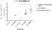
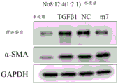
The experimental results are shown in FIGS. 11-13. Wherein FIGS. 11A-C show that both lipids (lipid #11 (18:0/18:2) and lipid #12 (16:0/18:2)) are effective in promoting the entry of nucleic acid molecules of various sequences into a variety of cells as compared to controls; FIG. 12 shows that nucleic acids enter and are predominantly localized in the cytoplasm under delivery of lipid #11 (18:0/18:2) and lipid #12 (16:0/18:2); furthermore, referring to fig. 13, the inventors unexpectedly found that both lipids #11 and #12 promote entry of small fragment nucleic acids and act on the wild-type 3' utr of their target genes, reducing the relative expression value of luciferases of the wild-type 3' utr of the target genes, while not acting on the 3' utr after mutation of the target genes. Can be used as a delivery mode of nucleic acid medicines.
4. In vivo delivery experiments of lipid nucleic acid mixtures
4.1 experimental procedure:
1. preparation of lipid nucleic acid mixture: see steps 2.1-2.2, using reverse evaporation and water boiling to prepare mixtures of lipid #11, lipid #12 and nucleic acids, and mixtures of lipid #1/2/4/9/14/18/19/20/21/22/23/24/25/26/27/28/29/30/32, lipid #3/8/10/11/12/13, lipid #6/15/16/17/31 combinations and nucleic acids, respectively.
2.6-8 week old male C57 mice were gavaged: 200 μl/piece, grouped as follows:
(1) Control group (free intake group): HJT-sRNA-m7 was administered without any treatment or lavage;
(2) Lipid #11 (18:0/18:2) group: a blend of lavage lipid #11 (18:0/18:2) or of lavage lipid #11 (18:0/18:2) with HJT-sRNA-m7;
(3) Lipid #12 (16:0/18:2) group: lavage lipid #12 (16:0/18:2 or a mixture of lavage lipid #12 (16:0/18:2 and HJT-sRNA-m7;
3. and (3) sample collection: after 6h of gastric lavage, 1.5mL TRIzol-LS or 3mL TRIzol was used to obtain whole blood (500 μl) and whole lung (110 mg) of mice, respectively, and the mice were homogenized and frozen at-80deg.C;
4. total RNA extraction: (1) TRIzol or TRIzol-LS lysate (Sigma Co.) was added to the cells, left at room temperature for 5 minutes,fully lysing (1.0 mL TRIzol lysate is added to 100mg of tissue for lung tissue of mice, the mixture is ground by a homogenizer at 12,000rpm and centrifuged at 4 ℃ C. For 10min to remove tissue sediment which cannot be fully homogenized; 1.5mL TRIzol-LS lysate is added to 500 μl of whole blood for whole blood of mice, and the mixture is centrifuged at 12,000rpm and 4 ℃ C. For 10min to remove sediment which cannot be fully lysed); (2) centrifugation at 12,000rpm at 4℃for 5min, discarding the precipitate; (3) Chloroform was added in a proportion of 200. Mu.l/mL TRIzol, and the mixture was stirred and mixed well and left at room temperature for 15 minutes. (4) Centrifuging at 12,000rpm and 4 ℃ for 15min, and sucking the upper water phase into another centrifuge tube; (5) Repeating step 4, adding equal amount of chloroform according to the upper water phase, standing at room temperature for 10min,12,000rpm, and centrifuging for 15min; (6) Sucking the upper water phase into another new EP pipe, adding isopropanol into TRIzol with the volume of 0.5mL/mL, mixing well, and standing at room temperature for 5-10min; (7) centrifuging at 12,000rpm and 4℃for 10min, and discarding the supernatant; (8) Adding 1mL of 75% ethanol, gently oscillating the centrifuge tube, and suspending and precipitating; (9) 8000g, centrifuging at 4 ℃ for 5min, and discarding the supernatant as much as possible; (10) Air-dried at room temperature for 5-10min, treated with 50. Mu.l DEPC-treated H 2 O dissolves RNA samples.
RT-qPCR detection: see the method described in 3.3.1 and 3.3.2 above.
4.2 experimental results
Referring to FIG. 14, the inventors have unexpectedly found that lipid #11 (18:0/18:2) and lipid #12 (16:0/18:2) facilitate the entry of small fragments of nucleic acids into the blood and lungs, which can be used as a means of nucleic acid drug delivery, via such (non-invasive) gastric administration. Surprisingly, the lipid nucleic acid complexes obtained by direct water boiling achieve a significant delivery effect.
Referring to fig. 15, the inventors have unexpectedly found that through such (non-invasive) gastric administration, the mixing of 28 lipids can facilitate entry of small fragments of nucleic acid into the blood, which can be used as a means of delivery of nucleic acid drugs. Surprisingly, the mixture of lipid combinations and nucleic acids obtained by direct water boiling achieves a significant delivery effect.
Experimental examples on the lipids 1 to 71 in Table 1
Method
1. Extraction of lipids from Chinese medicinal materials
1.1 preparation of Chinese herbs by decoction
1) Taking 100g of Chinese medicinal decoction pieces (radix Rhodiolae, herba Taraxaci, flos Lonicerae, herba Andrographitis, purchased from Beijing Tongren Tang drug store), adding 1000mL ddH 2 O soaking for 30min.
2) The traditional Chinese medicine decoction pot is subjected to strong fire decoction for 15min and weak fire decoction for 20min.
3) 400mL of the decocted traditional Chinese medicine liquid is added into a rotary evaporator, and is concentrated to 100mL at 60 ℃ and 60rpm for 30min.
1.2 extraction of lipids
1) To 160mL of the Chinese medicinal decoction obtained according to item 1.1 above (concentrated by rotary evaporator) 600mL of chloroform-methanol mixture (chloroform: methanol=1:2, v/v) was added so that chloroform: methanol: water=1:2:0.8, and stirred and mixed well for 10-15min.
2) 200mL of chloroform was added to the flask, and the mixture was stirred and mixed for 10 minutes.
3) 200mL ddH was added to the Erlenmeyer flask 2 And O, stirring and mixing chloroform and methanol in a ratio of water=2:2:1.8 for 10min.
4) Removing insoluble substances in the upper liquid and the middle layer, taking the chloroform layer of the lower layer, and freezing at-40deg.C.
1.3HPLC-MS/MS identification of lipid components
Instrument conditions
1) Chromatographic conditions:
instrument: ultimate 3000; chromatographic column: kineex C18 (100×2.1mm,1.9 μm); column temperature: 45 ℃; mobile phase: a: acetonitrile: water (V/V, 60:40), solution containing 10mmol/L ammonium formate, mobile phase B: acetonitrile: isopropyl alcohol (10:90, V/V), the solution containing 10mmol/L ammonium formate and 0.1% formic acid. Flow rate: 0.4mL/min; sample injection amount: 4. Mu.L.
2) Mass spectrometry parameters:
a) Positive mode: heater Temp 300 ℃, sheath Gas Flow rate,45arb,Aux Gas Flow Rate,15arb,Sweep Gas Flow Rate,1arb,spray voltage,3.0KV,Capillary Temp,350 ℃, S-Lens RF Level,30%. Scan roles: 200-1500.
b) Negative mode: heater Temp 300 ℃, sheath Gas Flow rate,45arb,Aux Gas Flow Rate,15arb,Sweep Gas Flow Rate,1arb,spray voltage,2.5KV,Capillary Temp,350 ℃, S-Lens RF Level,60%. Scan roles: 200-1500.
1.4 identification of lipid derived from traditional Chinese medicine
The lipid components are identified by HPLC-MS/MS, 138 lipid components from Chinese medicine are identified, 125 lipid components are identified by cation mode, and 13 lipid components are identified by anion mode. The following experiment was continued with the compounds 1 to 69 shown in Table 1. It should be noted that the lipids tested below were either commercially purchased or commercially synthesized and were used in the manner described in Table 1-1.
2. Preparation of lipid nucleic acid complexes
2.1 reverse evaporation method:
100. Mu.L of lipid diethyl ether solution was prepared and grouped according to the lipid numbers shown in Table 1 (lipid concentrations see Table below), and the lipid solution was added to 20. Mu.L of nucleic acid solution (HJT sRNA or siRNA) in a volume ratio of 5:1, sonicated for 3min, and after removal of diethyl ether by evaporation at 55℃and hydration with 100. Mu.L of DEPC treated water to give a nucleic acid lipid mixture.
TABLE 3 Table 3
2.2 Water boiling method:
100. Mu.L of nucleic acid solution (HJT sRNA or siRNA) was added to 2-5. Mu.L of lipid solution (concentration shown in Table 1), and after mixing, the mixture was heated at 80-100℃for 15-30min to obtain a nucleic acid lipid mixture.
3. In vitro delivery experiments of lipid nucleic acid complexes
3.1 real-time fluorescent quantitative PCR (RT-qPCR) detection of the expression level of intracellular lipid delivery nucleic acid
3.1.1 culturing MRC-5 cells (lung embryo fibroblasts), A549 cells (human lung adenocarcinoma cells), caco-2 cells (human colon adenocarcinoma cells) (purchased from the cell resource center of the basic medical institute of China medical science, academy of sciences) to logarithmic phase, then plating onto 12-well plates respectively,cell density of 6X 10 5 1mL of culture medium/well; wherein MRC-5 and Caco-2 cells are cultured in Eagle's MEM medium (MEM, gibco); a549 cells were cultured in Ham's F-12 medium (HyClone); incubation was carried out overnight at 37℃and subsequent experiments were carried out after cell attachment.
3.1.2 experimental groupings were as follows:
1) Untreated group: refers to untreated cells, which served as a blank.
2) RNAiMAX treatment group: mu.L Lipofectamine was diluted with 100. Mu.L of opti-MEM medium (available from Invitrogen, thermo Fisher Scientific), respectively TM RNAiMAX transfection reagent (reagent all is Lipofectamine RNAimax, invitrogen, thermo Fisher Scientific) and HJT-sRNA-m7 solution, which are mixed and placed for 15min, added to the cells, mixed, and the final concentration of HJT-sRNA-m7 is 100nM, and this group is used as a positive control group.
3) Free uptake (Free uptake) group: directly adding HJT-sRNA-m7 solution (final concentration is 100 nM), and taking the group as a negative control group;
4) Lipid nucleic acid mixture treatment group: the lipid prepared in step 2 was added to the cells with HJT-sRNA-m7 mixture, and the final concentration of HJT-sRNA-m7 was 100nM.
3.1.3 after 12-24h incubation with cells, cells were washed 2 times with PBS, cells were harvested with TRIzol lysate (purchased from Sigma-Aldrich), total RNA was extracted therefrom, and the abundance of HJT-sRNA-m7 entering the cells was detected by RT-qPCR (SYBR Green dye method) as follows:
1) Extracting total RNA of cells:
a.12 well plate cultured cells (about 1X 10) 6 Individual cells/well), after 1mL of TRIzol lysate was added to each well, the wells were placed on ice and left at room temperature for 5min after all samples were added with TRIzol, allowing them to lyse well;
b.4 ℃, centrifuging at 12,000rpm for 5min, discarding the precipitate, and transferring TRIzol into a new centrifuge tube;
C. adding chloroform according to 200 mu L chloroform/mL TRIzol, fully oscillating, uniformly mixing, and standing at room temperature for 5min;
d.4 ℃,12,000rpm, centrifuging for 15min;
E. sucking the upper water phase, adding isopropanol into another centrifuge tube, adding isopropanol into 0.5mL isopropanol/mL TRIzol, mixing, and standing at room temperature for 5-10min;
Centrifuging at F.4 deg.C and 12,000rpm for 15min, discarding supernatant, and precipitating RNA at the bottom of the tube;
G. adding 1mL of 75% ethanol, gently shaking the centrifuge tube, and suspending and precipitating;
centrifuging at H.4 deg.C and 12,000rpm for 10min, discarding supernatant, adding 1mL of 75% ethanol, gently shaking the centrifuge tube, and suspending the precipitate;
i.4 ℃,12,000rpm, centrifugation for 10min, removal of supernatant, air-drying at room temperature, dissolving RNA sample with 50. Mu.L of RNase-free H2O, and measuring O.D value to quantify RNA concentration.
2) Reverse transcription of total RNA into cDNA: sRNA was reverse transcribed into cDNA by a reverse transcription kit (High-Capacity cDNA Reverse Transcription Kits, applied Biosystems, cat. No. 4368813) using the stem-loop method (stem-loop method) (see, e.g., real-time quantification of microRNAs by stem-loop RT-PCR, nucleic Acids Res.2005Nov 27;33 (20): e 179), the reverse transcription system being as follows: template RNA (150 ng/. Mu.L), 10. Mu.L of 10 XRT buffer, 2.0. Mu.L of 25 XdNTP Mix (100 mM), 0.8. Mu.L of U6 RT step-loop primer, 2.0. Mu.L of HJT-sRNA-m7 RT step-loop primer, multiScribe TM 1.0. Mu.L of reverse transcriptase, 1.0. Mu.L of RNase inhibitor, no nuclease H 2 O1.2 mu L, and after instantaneous centrifugation, the mixture was put into a PCR instrument for reaction under the following conditions: (1) 25 ℃ for 10min; (2) 37 ℃ for 120min; (3) 85 ℃ for 5min; (4) terminating the reaction at 4 ℃. After completion of the reaction, 20. Mu.L of RNase-free ddH was added 2 O, make up the final volume to 40. Mu.L. The stem-loop method primer used in the reverse transcription process is synthesized by Beijing qingke new industry biotechnology Co., ltd (U6 RT primer, because the RT-qPCR reaction can only relatively quantify the small RNA, U6 is used as a standard reference gene to calculate the relative expression quantity thereof): GTCGTATCCAGTGCAGGGTCCGAGGTATTCGCACTGGATACGACAAAAATATG (SEQ ID NO: 21); HJT-sRNA-m7 RT stem-loop primer: GTCGTATCCAGTGCACGCTCCGAGGTATTCGCACTGGATACGACGCTTACAA (SEQ ID NO: 22)).
3) Quantitative PCR amplification reaction: the total volume of the qPCR reaction system is 10 mu L, comprising: 5 mu L2XSYBR Green Master Mix, 0.5. Mu.L forward primer (10. Mu.M), 0.5. Mu.L reverse primer (10. Mu.M), 1. Mu.L cDNA reverse transcribed, 3. Mu.L RNase-free dH 2 O. The PCR reaction conditions were: pre-denaturation was continued for 5min at 95 ℃ and started into PCR amplification cycle: (1) 95 ℃ for 10s; (2) 55 ℃,10s; (3) 72 ℃,20s; a total of 40 cycles were performed; and finally, continuously cooling at 40 ℃ for 10 seconds. Both the forward and reverse primers for the amplification reaction were designed and synthesized by Beijing Optimago Biotechnology Inc. (U6 forward primer: GCGCGTCGTGAAGCGTTC (SEQ ID NO: 23), U6 reverse primer: GTGCAGGGTCCGAGGT (SEQ ID NO: 24), HJT-sRNA-m7 forward primer: TCGCGCTGAGGTAGTAGGTT (SEQ ID NO: 25), HJT-sRNA-m7 reverse primer: GTGCACGCTCCGAGGT (SEQ ID NO: 26)).
4) The relative entry (single-stranded or double-stranded RNA) was calculated using the 2-delta Ct method (relative gene expression = 2- (Ct target gene-Ct reference gene).
3.2 real-time fluorescent quantitative PCR (RT-qPCR) detection of mRNA expression levels
3.2.1 culturing THP-1 cells (human monocytes) to logarithmic phase and then plating them into 12 well plates, respectively, with cell density of 6X 10 5 1mL of culture medium/well; THP-1 cells were cultured in RPMI-1640 medium (HyClone); incubation was carried out overnight at 37℃and subsequent experiments were carried out after cell attachment.
3.2.2 experimental groupings were as follows:
1) Untreated group: refers to untreated THP-1 cells, which served as a blank.
2) RNAiMAX treatment group: mu.L Lipofectamine was diluted with 100. Mu.Lopti-MEM (Invitrogen, thermo Fisher Scientific) medium, respectively TM RNAiMAX transfection reagent (nvitro, thermo Fisher Scientific) and nucleic acid solution (TNF-. Alpha.siRNA) were mixed and placed for 15min, added to cells, mixed, and the final concentration of nucleic acid was 400nM, which served as a positive control.
3) Free uptake (Free uptake) group: directly adding nucleic acid solution (TNF-alpha siRNA with final concentration of 400 nM), and taking the group as negative control group;
4) Lipid nucleic acid mixture treatment group: the lipid and nucleic acid mixture prepared in step 2 was added to the cells, and mixed well, the final concentration of nucleic acid was 400nM.
3.2.3 treatment of cells 24h later, 1. Mu.g/mL of E.coli Lipopolysaccharide (LPS, escherichia coli 0111: B4, L4391, sigma-Aldrich) was given for stimulation, and after 9h the cells were harvested with TRIzol lysate, total RNA was extracted therefrom, and the mRNA expression level of TNF-. Alpha.was detected by RT-qPCR (SYBR Green dye method) (the results are shown in the figures depending on the type of target genes in the subsequent examples) as follows:
1) The method for extracting the total RNA of the cells comprises the same steps as 3.1.3.
2) Reverse transcription of total RNA into cDNA: total RNA was reverse transcribed into cDNA by a reverse transcription kit (High-Capacity cDNA Reverse Transcription Kits, applied Biosystems, cat.no. 4368813) in the following manner: template RNA (150 ng/. Mu.L) 10. Mu.L, 10 XRT buffer 2.0. Mu.L, 25 XdNTP Mix (100 mM) 0.8. Mu.L, random primer 2.0. Mu.L, multiScribe TM 1.0. Mu.L of reverse transcriptase, 1.0. Mu.L of RNase inhibitor, no nuclease H 2 O3.2 mu L, and after instantaneous centrifugation, putting the mixture into a PCR instrument for reaction under the following reaction conditions: (1) 25 ℃ for 10min; (2) 37 ℃ for 120min; (3) 85 ℃ for 5min; (4) terminating the reaction at 4 ℃. After completion of the reaction, 20. Mu.L of RNase-free ddH was added 2 O, make up the final volume to 40. Mu.L.
3) Quantitative PCR amplification reaction: the total volume of the qPCR reaction system is 10 mu L, comprising: 5. Mu.L of 2X SYBR Green Master Mix, 0.5. Mu.L of forward primer (10. Mu.M), 0.5. Mu.L of reverse primer (10. Mu.M), 1. Mu.L of cDNA obtained by reverse transcription, 3. Mu.L of RNase-free ddH 2 O. The PCR reaction conditions were: pre-denaturation was continued for 5min at 95 ℃ and started into PCR amplification cycle: (1) 95 ℃ for 10s; (2) 55 ℃,10s; (3) 72 ℃,20s; a total of 40 cycles were performed; and finally, continuously cooling at 40 ℃ for 10 seconds. The forward primer and the reverse primer of the amplification reaction are designed and synthesized by Beijing qingke new biotechnology Co., ltd, the primer sequence (reference gene UBC forward primer CTGGAAGATGGTCGTACCCTG (SEQ ID NO: 30), reference gene UBC reverse primer GGTCTTGCCAGTGAGTGTCT (SEQ ID NO: 31), target gene TNF-alpha forward primer CTGCCCCAATCCCTTTATT (SEQ ID NO: 32) target gene TNF-alpha reverse primer:CCCAATTCTCTTTTTGAGCC(SEQ ID NO:33))。
4) The relative expression level was calculated by the 2- ΔCt method as above.
3.3 Western blotting method for detecting protein expression level (Western blot)
3.3.1 culturing MRC-5 cells (lung embryo fibroblasts) or A549 cells (human lung adenocarcinoma cells) to logarithmic phase, then plating to 12-well plates, respectively, with cell density of 6X10 5 1mL of culture medium/well; wherein MRC-5 cells are cultured in Eagle's MEM medium (MEM, gibco); a549 cells were cultured in Ham's F-12 medium (HyClone); incubation was carried out overnight at 37℃and subsequent experiments were carried out after cell attachment.
3.3.2 experimental groupings were as follows:
1) Untreated group: refers to untreated cells, which served as a blank.
2) RNAiMAX treatment group: mu.L Lipofectamine was diluted with 100. Mu.L of opti-MEM medium (Invitrogen, thermo Fisher Scientific), respectively TM RNAiMAX transfection reagent (Invitrogen, thermo Fisher Scientific) and nucleic acid solution were mixed and placed for 15min, added to the cells, mixed, and the final concentration of nucleic acid was 400nM, which served as a positive control.
3) Free uptake (Free uptake) group: directly adding nucleic acid solution (final concentration 400 nM), and taking the group as negative control group;
4) Lipid nucleic acid mixture treatment group: the lipid and nucleic acid mixture prepared in step 2 was added to the cells, and mixed well, the final concentration of nucleic acid was 400nM.
3.3.3 after 24h incubation with cells, cells were stimulated with a stimulus (1. Mu.g/mL double stranded RNA virus mimetic poly (I: C) (P1530, sigma-Aldrich) or 3ng/mL transforming growth factor TGF. Beta.1 (Pepro Tech)), after a certain period of time, cells were harvested with a strong RIPA lysate, protein expression levels of the relevant genes (relevant gene types, as the case may be, are shown in the corresponding figures) were detected using Western blot (protein expression levels of REL-A were detected 24h after poly (I: C) stimulation of A549 cells, the reference protein was β -actin, fibronectin and α -SMA protein expression were detected after 72h stimulation of MRC-5 cells with TGF-. Beta.1, the reference protein was GAPDH, and protein expression of the corresponding knockdown gene was detected by siRNA delivery experiments), as follows:
1) Collection of protein samples and BCA method concentration determination
A. Discarding the culture medium, washing 12-hole plate cells with 1mL PBS buffer solution for one time, adding 100 mu L of precooled strong RIPA lysate for each hole, scraping the cells with a gun head, transferring the cells into a centrifuge tube, and placing the cells on ice for cracking for 20min;
centrifuging at 12,000rpm at 4 ℃ for 10min, and transferring the supernatant to a new centrifuge tube;
C. fully and uniformly mixing BCA reagent A and B (50:1, v/v) to prepare BCA working solution;
D. respectively taking 25 mu L of freshly prepared BSA standard solution and a sample to be tested, adding the freshly prepared BSA standard solution and the sample to be tested into a 96-well plate, adding 200 mu L of BCA working solution into each well, and fully and uniformly mixing; incubating for 30min at 37 ℃;
E. detecting absorbance at 562nm by using an ultraviolet spectrophotometer (a Synergy 4 multifunctional enzyme-labeled instrument), and calculating protein concentration in the sample according to a standard curve;
F. regulating the concentration of the samples by using RIPA lysate and Loading Buffer to ensure that the concentration of each sample is consistent;
g.95℃and denaturation treatment for 10min.
2) Western blot detection (Western blot)
A. And (3) glue preparation: separating gel with concentration of 10% (lower gel) and concentrated gel with concentration of 5% (upper gel), making lanes by 15-hole comb, and loading protein sample in each lane with equal amount;
B. protein electrophoresis: adding electrophoresis buffer solution, and electrophoresis initial voltage is 80V; after the bromophenol blue dye reaches the bottom of the separation gel, increasing the voltage to 120V to continue electrophoresis until the bromophenol blue dye reaches the bottom of the separation gel or completely swims out of the gel;
C. Wet transfer: assembling according to the sequence of a membrane rotating clamping plate (negative electrode) -sponge-filter paper-gel-PVDF membrane-filter paper-sponge-membrane rotating clamping plate (positive electrode); after the installation, the whole film transfer device is placed in a cold chamber at 4 ℃; constant current 300mA, film transferring for 120min;
D. closing: after the transfer film is finished, placing the membrane in a 3% BSA blocking solution, and blocking for 1h at room temperature;
E. incubation resistance: transferring the sealed PVDF membrane into a hybridization bag, adding a 3% BSA sealing solution containing a corresponding primary antibody (the information of the primary antibody is as follows), expelling bubbles in the bag, and incubating at 4 ℃ overnight after sealing;
TABLE 4 Table 4
F. Washing the film: taking out the PVDF film, and washing the film 3 times by TBST for 10min each time;
G. secondary antibody incubation: TBST was discarded, and a 3% bsa blocking solution (secondary antibody dilution ratio 1:5000) containing a goat anti-rabbit or goat anti-mouse secondary antibody (purchased from hangzhou bian biotechnology limited) with horseradish peroxidase (HRP) was added and incubated at room temperature for 1 hour;
H. washing the film: washing the membrane with TBST for 3 times and 10min each time;
I. developing: preparing a Western chromogenic solution (1:1, V/V, merck Millipore, ECL chemiluminescent chromogenic solution is purchased from Millipore company), and uniformly dripping the prepared chromogenic solution on one side of the membrane-associated protein; carefully wrapping the film with a preservative film, and observing after color development;
J. Analysis: analysis was performed with Image J software.
4. In vivo delivery experiments of lipid nucleic acid mixtures
4.1 experimental procedure:
1) Preparation of lipid nucleic acid mixture: prepared by water boiling, 400. Mu.L of aqueous solution treated with HJT-sRNA-m7 (5 nmol) single-stranded RNA DEPC was added with 9. Mu.L or 18. Mu.L of lipid combination (lipid PE (No 38) & LPC (No 37) & TG (No 32), 4:2:3, V/V/V) respectively, and after mixing, heated at 100℃for 30min.
2) Male C57BL/6J wild-type mice 6-8 weeks old were gavaged with RNA: the HJT-sRNA-m7 aqueous solution or the lipid and HJT-sRNA-m7 mixed solution were administered separately using a gastric lavage needle at 400. Mu.L/dose (HJT-sRNA- m 7,5 nmol/dose) and the groups were as follows:
A. control group (untreated group): mice without any treatment;
B. negative control group (lipid group): gastric lavage 9. Mu.L lipid combination (lipid PE (No 38) & LPC (No 37) & TG (No 32), 4:2:3, V/V/V);
C. free feeding group (Free uptake): directly lavage HJT-sRNA-m7 single-stranded RNA solution;
D. lipid and nucleic acid mixture group: a mixture of a gastric lavage lipid combination with HJT-sRNA-m7 single stranded RNA.
3) And (3) sample collection: after 3h of gastric administration, the whole lung of the mice was lysed with 3mL TRIzol, homogenized and stored frozen at-80 ℃.
4) Total RNA extraction:
A. adding 3.0ml of LTRIzol lysate into the lung tissue of a mouse, grinding by a homogenizer at 12,000rpm and 4 ℃, and centrifuging for 10min to remove tissue sediment which cannot be homogenized sufficiently;
B. Adding chloroform according to the proportion of 200 mu L/mLTRIzol, fully oscillating and uniformly mixing, and standing at room temperature for 15min;
c, centrifuging at 4 ℃ for 15min at 12,000rpm, and sucking the upper water phase into another centrifuge tube;
D. repeating the steps, adding equal amount of chloroform according to the upper water phase, fully mixing, and standing at room temperature for 10min;
e.12,000rpm,4 ℃, centrifugation for 15min;
F. sucking the upper water phase into another new EP pipe, adding isopropanol according to 0.5mL/mLTRIzol, mixing well, and standing at room temperature for 5-10min;
g.12,000rpm,4 ℃, centrifuging for 15min, and discarding the supernatant;
H. adding 1mL of 75% ethanol, gently shaking the centrifuge tube, and suspending and precipitating;
i.12,000rpm,4 ℃, centrifuging for 10min, and discarding the supernatant as much as possible;
J. air-dried at room temperature for 5-10min, and dissolve RNA samples with 50. Mu.L of DEPC-treated H2O.
5) RT-qPCR (SYBR Green universal dye method) detects the abundance of HJT-sRNA-m 7.
Unless otherwise indicated, HJT-sRNA-m7 single-stranded solution refers to an aqueous DEPC treatment solution of HJT-sRNA-m7 single strand. HJT-sRNA-m7 duplex solution refers to an aqueous DEPC treatment solution of HJT-sRNA-m7 duplex. Example 1-1: delivery of single stranded nucleic acids into MRC-5 cells by different classes of lipid combinations
(one) experimental grouping:
1) Untreated group: untreated MRC-5 cells;
2) RNAiMAX group: diluting 2 mu LRNAiMAX transfection reagent and DEPC treatment aqueous solution of HJT-sRNA-m7 single strand with 100 mu L opti-MEM culture medium respectively, mixing the two, standing for 15min, adding into cells, mixing, and keeping the final concentration of HJT-sRNA-m7 single strand at 200nM;
3) Free uptake (Free uptake) group: directly adding HJT-sRNA-m7 single-stranded solution (final concentration is 200 nM);
4) Lipid and nucleic acid mixture treatment group: and adding 3 mu L of lipid monomer or lipid combination into the cells respectively with the mixture of HJT-sRNA-m7 single-stranded nucleic acid solution treated by the water boiling method, and uniformly mixing, wherein the final concentration of RNA is 200nM.
(II) Experimental procedure
1) The water boiling method conditions are as follows: 100. Mu.L of HJT-sRNA-m7 single-stranded solution was added with 3. Mu.L of lipid monomer or lipid combination chloroform solution (lipid No1/2/4/9/14/18/19/20/21/22/23/24/25/26/27/28/29/30/32 chloroform solution concentration 5mg/mL, lipid No3/8/10/11/12/13/33/34/35/36 chloroform solution concentration 10mg/mL, lipid No6/15/16/17/31 chloroform solution concentration 1 mg/mL), and heated at 100℃for 30min;
a) Lipid combination:
b) MG (monoacylglycerol): 3 mu LNo34 lipid;
c) DG (diglyceride): 3. Mu.L of No1/2/3/19/35 in equal volume of chloroform solution;
d) TG (triglycolide, triglyceride): 3. Mu.L of No6/9/10/13/15/16/18/20/21/22/23/24/25/26/27/28/32/33 in equal volume of chloroform solution;
e) LPC (lysophosphotidylinder, lysolecithin): 3. Mu.L of a mixture of No. 36/37 equal volume of chloroform solution;
f) PC (phosphotidylindoline), phosphatidylcholine: 3. Mu.L of a mixture of No. 11/12 equal volume of chloroform solution;
g) PE (phosphotidylethanolamine): 3. Mu.L of a mixture of No. 8/38 equal volume of chloroform solution;
h) Cer (Ceramides, ceramide): 3. Mu.L of a mixture of No. 4/14 equal volume of chloroform solution;
i) So (Sphingoshine), sphingosine: 3. Mu.L of a mixture of No. 17/30/31 in equal volume of chloroform solution;
j) FA (fatty acid): 3 mu L No29
k) Mixture: 3. Mu.L of No. 1-36 (No No. 5/7) in equal volume of chloroform solution;
l) mixture 1: 3. Mu.L of No. 1-36 (No No. 5/7/34) in an equal volume of chloroform solution;
m) mixture 2: 3. Mu.L of a mixture of equal volumes of No. 1-36 (No. 5/7/1/2/3/19/35) in chloroform;
n) mixture 3: 3. Mu.L of a mixture of equal volumes of No. 1-36 (No. 5/7/6/9/10/13/15/16/18/20/21/22/23/24/25/26/27/28/32/33) in chloroform;
o) mixture 4: 3. Mu.L of No. 1-36 (No No. 5/7/36/37) in an equal volume of chloroform solution;
p) mixture 5: 3. Mu.L of No. 1-36 (No No. 5/7/11/12) in an equal volume of chloroform solution;
q) mixture 6: 3. Mu.L of No. 1-36 (No No. 5/7/8) in an equal volume of chloroform solution;
r) mixture 7: 3. Mu.L of No. 1-36 (No No. 5/7/4/14) in an equal volume of chloroform solution;
s) mixture 8: 3. Mu.L of No. 1-36 (No No. 5/7/29) in an equal volume of chloroform solution;
2) Experimental conditions: HJT-sRNA-m7 final concentration was 200nM, and after 12h of addition to the cells, the cells were examined for the amount of HJT-sRNA-m7 by RT-qPCR (SYBR Green universal dye method). For specific experimental methods, see above for "real-time fluorescent quantitative PCR for detecting the expression level of intracellular lipid delivery nucleic acids". All experiments were performed in triplicate.
Conclusion: the results indicate that the above lipid combinations can deliver nucleic acids into cells effectively (see fig. 16), with the hope of improving the efficiency of nucleic acid drug delivery clinically, compared to the free uptake group. Wherein, mixture 2, mixture 3, mixture 5, mixture 7, mixture 8 mediate higher amounts of nucleic acid into MRC-5 cells.
Examples 1-2: delivery of single stranded nucleic acids into MRC-5 cells and Caco-2 cells by lipid combination
(one) experimental grouping:
the test cells are MRC-5 cells and Caco-2 cells
1) Untreated group: untreated cells;
2) RNAiMAX group: diluting 2 mu L of RNAiMAX transfection reagent and HJT-sRNA-m7 single-stranded solution with 100 mu L of opti-MEM culture medium respectively, mixing the two, standing for 15min, adding into cells, and mixing to obtain HJT-sRNA-m7 single-stranded solution with a final concentration of 200nM;
3) Free uptake (Free uptake) group: directly adding HJT-sRNA-m7 single-stranded solution (final concentration is 200 nM);
4) Lipid monomer and nucleic acid treatment group: mu.L of a mixture of a lipid monomer (No 1 or No8 or No 12) and a single-stranded nucleic acid solution of HJT-sRNA-m7, respectively, after the treatment by the water boiling method, was added to the cells, and the mixture was mixed uniformly, and the final concentration of RNA was 200nM.
5) Lipid combination mix and nucleic acid mix treatment group: mu.L of the lipid combination (No. 1/8/12 equal volume mixture) was added to the cells as a mixture of each of the lipid combinations and a single-stranded nucleic acid solution of HJT-sRNA-m7 treated by the water boiling method, and the final concentration of RNA was 200nM.
6) Lipid combination and nucleic acid mixture treatment group: mu.L of lipid combinations (2. Mu.L of lipid monomer No1 or No8 or No12 was mixed with 1. Mu.L of lipid class (MG, DG, TG, LPC, cer, so or FA) described below) were added to cells as a mixture of HJT-sRNA-m7 single-stranded nucleic acid solution treated by the water boiling method, and the mixture was mixed well so that the final concentration of RNA was 200nM. In FIGS. 17A and 17B, the treatment groups are collectively denoted as No.12 μL+mix1 μL, no.82 μL+mix1 μL, and No.12 2μL+mix1 μL, respectively, where within the lateral coverage, MG represents 2 μL of lipid monomer No.1 or No.8 or No. 12+1μL MG, DG represents 2 μL of lipid monomer No.1 or No.8 or No. 12+1μL, TG represents 2 μL of lipid monomer No.1 or No.8 or No. 12+1μL, LPC represents 2 μL of lipid monomer No.1 or No.8 or No. 12+1μL LLPC, cer represents 2 μL of lipid monomer No.1 or No.8 or No. 12+1μL LCer, so represents 2 μL of lipid monomer No.1 or No.8 or No. 12+1μL LSo, and FA represents 2 μL of lipid monomer No.1 or No.8 or 12+1μL.
(II) Experimental procedure
1) The water boiling method conditions are as follows: 100. Mu.L of HJT-sRNA-m7 single-stranded solution was added with 3. Mu.L of lipid monomer (lipid No1 chloroform solution concentration 5mg/mL, no8/12 chloroform solution concentration 10 mg/mL) or lipid combination, and heated at 100℃for 30min;
MG (monoacylglycerol): 2. Mu.L of No34 lipid;
DG (diglyceride): 2. Mu.L of No1/2/3/19/35 in equal volume of chloroform solution;
TG (triglycolide, triglyceride): 2. Mu.L of No6/9/10/13/15/16/18/20/21/22/23/24/25/26/27/28/32/33 in equal volume of chloroform solution;
LPC (lysophosphotidylinder, lysolecithin): 2. Mu.L of a mixture of No. 36/37 equal volume of chloroform solution;
cer (Ceramides, ceramide): 2. Mu.L of a mixture of No. 4/14 equal volume of chloroform solution;
so (Sphingoshine), sphingosine: 2. Mu.L of a mixture of No. 17/30/31 in equal volume of chloroform solution;
FA (fatty acid): 2 mu L No29
2) Experimental conditions: HJT-sRNA-m7 final concentration was 200nM, and after 24h of addition, the cells were examined for cell entry of HJT-sRNA-m7 by RT-qPCR (SYBR Green universal dye method). For specific experimental methods, see above for "real-time fluorescent quantitative PCR for detecting the expression level of intracellular lipid delivery nucleic acids". All experiments were performed in triplicate.
Conclusion: the results indicate that for MRC-5 cells, the mixture (equal volume mix No. 1/8/12), no.1 2. Mu.L+No. 8 1. Mu.L, no.1 2. Mu.L+No. 12. Mu.L, no.1 2. Mu.L+MG 1. Mu.L, no.8 2. Mu.L+MG 1. Mu.L, no. 12. Mu.L+No. 8 1. Mu.L, and No.12 2. Mu.L+So1. Mu.L are more effective in delivering nucleic acids.
For Caco-2 cells, the mixture (equal volume mix No. 1/8/12), no.1 2. Mu.L+No. 8 1. Mu.L, no.1 2. Mu.L+No. 12. Mu.L, no.1 2. Mu.L+MG 1. Mu.L, no.8 2. Mu.L+MG 1. Mu.L, no. 122. Mu.L+No. 8 1. Mu.L, no. 12. Mu.L+LPC 1. Mu.L, and No.12 2. Mu.L+So1. Mu.L were more effective in delivering nucleic acids.
Examples 1-3: lipid combination delivery of single stranded nucleic acids into cells
Cell type: a549, MRC-5 and Caco-2 cells
(one) experimental grouping:
1) Untreated group: untreated cells;
2) RNAiMAX group: diluting 2 mu L of RNAiMAX transfection reagent and HJT-sRNA-m7 single-stranded solution with 100 mu L of opti-MEM culture medium respectively, mixing the two, standing for 15min, adding into cells, and mixing to obtain HJT-sRNA-m7 single-stranded solution with a final concentration of 100nM;
3) Free uptake (Free uptake) group: directly adding HJT-sRNA-m7 single-stranded solution (final concentration is 100 nM);
4) Lipid monomer and nucleic acid treatment group: mu.L of the mixture of the lipid monomer (No. 8 or No. 12) and the single-stranded nucleic acid solution of HJT-sRNA-m7, respectively, after the water boiling treatment, was added to the cells, and the mixture was mixed uniformly, and the final concentration of RNA was 100nM.
5) Lipid combination PC (No 12) & PE (No 8) with nucleic acid mixture treatment group: 2.25. Mu.L of a mixture of lipid combination (PC (No 12) & PE (No 8), 2:1, V/V) and a single-stranded nucleic acid solution of HJT-sRNA-m7, respectively, was added to the cells, and mixed well, and the final concentration of RNA was 100nM.
6) Lipid combination and nucleic acid mixture treatment group: 3. Mu.L of lipid combination (2.25. Mu.L of lipid combination PC (No 12) & PE (No 8) was mixed with 0.75. Mu.L of lipid class DG, TG, LPC, PC, cer, so or FA described below) was added to the cells as a mixture of HJT-sRNA-m7 single-stranded nucleic acid solution treated by the water boiling method, and the mixture was mixed uniformly so that the final concentration of RNA was 100nM. In FIG. 18, the treatment group covered by the upper horizontal line of 2.25. Mu.L+0.75. Mu.L is the mixture treatment group.
(II) Experimental procedure
1) The water boiling method conditions are as follows: 100. Mu.L of HJT-sRNA-m7 single-stranded solution is added with lipid monomer (lipid No8/12 chloroform solution concentration is 10 mg/mL) or lipid combination, and heated at 100 ℃ for 30min;
DG (diglyceride): 0.75. Mu.L of a mixture of No. 1/2 equal volume of chloroform solution;
TG (triglycolide, triglyceride): 0.75. Mu.L No. 15 chloroform solution;
LPC (lysophosphotidylinder, lysolecithin): 0.75. Mu.L of No36/37 equal volume chloroform solution;
PC (lysophosphophosphotidyline, lysolecithin): 0.75. Mu.L No. 12 chloroform solution;
cer (Ceramides, ceramide): 0.75. Mu.L No. 4 chloroform solution;
so (Sphingoshine), sphingosine: 0.75. Mu.L No. 31 chloroform solution;
FA (fatty acid): 0.75 mu L No. 29
2) Experimental conditions: HJT-sRNA-m7 final concentration 100nM, cells were added for 24h and the cells were assayed for entry of HJT-sRNA-m7 by RT-qPCR (SYBR Green universal dye method). For specific experimental methods, see above for "real-time fluorescent quantitative PCR for detecting the expression level of intracellular lipid delivery nucleic acids". All experiments were performed in triplicate.
Conclusion: the results indicate that both the above lipid monomers and lipid combinations can deliver nucleic acids into cells efficiently (see figure 18), with the hope of increasing the efficiency of nucleic acid drug delivery clinically, compared to the free uptake group.
For A549, MRC-5 and Caco-2 cells, 2.25. Mu.L PC (No 12) & PE (No 8) +0.75. Mu.L DG (mixture of equal volumes of chloroform solution No 1/2) were the best delivery efficiency.
Examples 1 to 4: lipid combination delivery of single stranded nucleic acids into cells
Cell type: a549, MRC-5 and Caco-2 cells
(one) experimental grouping:
1) Untreated group: untreated cells;
2) RNAiMAX group: diluting 2 mu L of RNAiMAX transfection reagent and HJT-sRNA-m7 single-stranded solution with 100 mu L of opti-MEM culture medium respectively, mixing the two, standing for 15min, adding into cells, and mixing to obtain HJT-sRNA-m7 single-stranded solution with a final concentration of 100nM;
3) Free uptake (Free uptake) group: directly adding HJT-sRNA-m7 single-stranded solution (final concentration is 100 nM);
4) Lipid monomer and nucleic acid treatment group: mu.L of a mixture of a lipid monomer (No 1 or No8 or No 12) and a single-stranded nucleic acid solution of HJT-sRNA-m7, respectively, after the treatment by the water boiling method, was added to the cells, and the mixture was mixed uniformly, and the final concentration of RNA was 100nM.
5) Lipid combinations DG (No 1) & PE (No 8) & PC (No 12) with nucleic acid mixture treatment group: the mixture of 3. Mu.L of the lipid combination (DG (No 1) & PE (No 8) & PC (No 12), 1:1:1, V/V/V) and the single-stranded nucleic acid solution of HJT-sRNA-m7, which was treated by the water boiling method, was added to the cells, and the mixture was mixed uniformly, wherein the final concentration of RNA was 100nM.
6) Lipid combination and nucleic acid mixture treatment group: mu.L of a mixture of the lipid combination (2. Mu.L of the lipid combination DG (No 1) & PE (No 8) & PC (No 12) and 1. Mu.L of the lipid class DG, TG, LPC, PC, cer, so or FA described below) respectively treated with a single-stranded nucleic acid solution of HJT-sRNA-m7 by the water boiling method was added to the cells, and the mixture was mixed uniformly so that the final concentration of RNA was 100nM. In FIG. 19, the treatment group covered by the upper horizontal line of 2. Mu.L of the lipid combination DG (No. 1) & PE (No. 8) & PC (No. 12) +1. Mu.L is the mixture treatment group.
(II) Experimental procedure
1) The water boiling method conditions are as follows: 100. Mu.L of HJT-sRNA-m7 single-stranded solution was added with 3. Mu.L of lipid monomer (lipid No1 chloroform solution concentration 5mg/mL, no8/12 chloroform solution concentration 10 mg/mL) or lipid combination, and heated at 100℃for 30min;
DG (diglyceride): 1. Mu.L of a mixture of No. 1/2 equal volume of chloroform solution;
TG (triglycolide, triglyceride): 1 μl of No15 chloroform solution;
LPC (lysophosphotidylinder, lysolecithin): 1. Mu.L of a mixture of No. 36/37 equal volume of chloroform solution;
PC (lysophosphophosphotidyline, lysolecithin): 1 μl of No12 chloroform solution;
cer (Ceramides, ceramide): 1 μl of No4 chloroform solution;
so (Sphingoshine), sphingosine: 1 μl of No31 chloroform solution;
FA (fatty acid): 1 mu L No29
2) Experimental conditions: HJT-sRNA-m7 final concentration 100nM, cells were added for 24h and the cells were assayed for entry of HJT-sRNA-m7 by RT-qPCR (SYBR Green universal dye method). For specific experimental methods, see above for "real-time fluorescent quantitative PCR for detecting the expression level of intracellular lipid delivery nucleic acids". All experiments were performed in triplicate.
Conclusion: the results indicate that the above lipid combinations can deliver nucleic acids into cells effectively (see fig. 19), with the hope of improving the efficiency of nucleic acid drug delivery clinically, compared to the free uptake group.
For cell types A549, MRC-5 and Caco-2 cells, 2. Mu.L of lipid combination DG (No 1) & PE (No 8) & PC (No 12) gave the best delivery with 1. Mu.L of TG (15).
Examples 1 to 5: lipid combination delivery of single stranded nucleic acids into cells
Cell type: a549, MRC-5 and Caco-2 cells
(one) experimental grouping:
1) Untreated group: untreated cells;
2) RNAiMAX group: diluting 2 mu L of RNAiMAX transfection reagent and HJT-sRNA-m7 single-stranded solution with 100 mu L of opti-MEM culture medium respectively, mixing the two, standing for 15min, adding into cells, and mixing to obtain HJT-sRNA-m7 single-stranded solution with a final concentration of 100nM;
3) Free uptake (Free uptake) group: directly adding HJT-sRNA-m7 single-stranded solution (final concentration is 100 nM);
4) Lipid monomer and nucleic acid treatment group: adding 3 mu L of a mixture of lipid monomer No8 and HJT-sRNA-m7 single-stranded nucleic acid solution treated by a water boiling method into cells, and uniformly mixing, wherein the final concentration of RNA is 100nM;
5) Lipid combination PE (No 8) & MG (No 34) with nucleic acid mixture treatment group: the mixture of the above-mentioned 2.25. Mu.L lipid combination (PE (No. 8) & MG (No. 34), 2:1, V/V) and HJT-sRNA-m7 single-stranded nucleic acid solution, which had been treated by the water boiling method, was added to the cells, and mixed uniformly, the final concentration of RNA was 100nM.
6) Lipid combination and nucleic acid mixture treatment group: mu.L of a lipid combination (2.25. Mu.L of a lipid combination PE (No 8) & MG (No 34) was mixed with 0.75. Mu.L of a lipid class DG, TG, LPC, PC, cer, so or FA described below) was added to the cells as a mixture of HJT-sRNA-m7 single-stranded nucleic acid solution treated by the water boiling method, and the mixture was mixed uniformly so that the final concentration of RNA was 100nM. In FIG. 20, 2.25. Mu.L of lipid combination PE (No. 8) & MG (No. 34) +0.75. Mu.L of the upper horizontal line covers the treatment group of the mixture.
(II) Experimental procedure
1) The water boiling method conditions are as follows: 100. Mu.L of HJT-sRNA-m7 single-stranded solution is added with lipid monomer (lipid No8 chloroform solution concentration is 10 mg/mL) or lipid combination, and heated at 100 ℃ for 30min;
DG (diglyceride): 0.75. Mu.L of a mixture of No. 1/2 equal volume of chloroform solution;
TG (triglycolide, triglyceride): 0.75. Mu.L No. 15 chloroform solution;
LPC (lysophosphotidylinder, lysolecithin): 0.75. Mu.L of No36/37 equal volume chloroform solution;
PC (lysophosphophosphotidyline, lysolecithin): 0.75. Mu.L No. 12 chloroform solution;
cer (Ceramides, ceramide): 0.75. Mu.L No. 4 chloroform solution;
so (Sphingoshine), sphingosine: 0.75. Mu.L No. 31 chloroform solution;
FA (fatty acid): 0.75 mu L No. 29
2) Experimental conditions: HJT-sRNA-m7 final concentration 100nM, cells were added for 24h and the cells were assayed for entry of HJT-sRNA-m7 by RT-qPCR (SYBR Green universal dye method). For specific experimental methods, see above for "real-time fluorescent quantitative PCR for detecting the expression level of intracellular lipid delivery nucleic acids". All experiments were performed in triplicate.
Conclusion: the results indicate that both the above lipid monomers and lipid combinations can deliver nucleic acids into cells efficiently (see figure 20), with the hope of increasing the efficiency of nucleic acid drug delivery clinically, compared to the free uptake group.
For cell types A549, MRC-5 and Caco-2 cells, 2.25. Mu.L PE (No 8) and MG (No 34) +0.75. Mu.L So (31) gave the best delivery results.
Examples 1 to 6: delivery of single-stranded nucleic acids into a549 cells by lipid composition
(one) experimental grouping:
1) Untreated group: untreated a549 cells;
2) RNAiMAX group: diluting 2 mu L of RNAiMAX transfection reagent and HJT-sRNA-m7 single-stranded solution with 100 mu L of opti-MEM culture medium respectively, mixing the two, standing for 15min, adding into cells, and mixing to obtain HJT-sRNA-m7 single-stranded solution with a final concentration of 100nM;
3) Free uptake (Free uptake) group: directly adding HJT-sRNA-m7 single-stranded solution (final concentration is 100 nM);
4) Lipid monomer and nucleic acid treatment group: mu.L of the mixture of lipid monomer No38 and HJT-sRNA-m7 single-stranded nucleic acid solution treated by the water boiling method was added to the cells, and the mixture was mixed uniformly, wherein the final concentration of RNA was 100nM.
5) Lipid combination and nucleic acid mixture treatment group: the above 3. Mu.L lipid combination (2. Mu.L lipid monomer No38 mixed with 1. Mu.L lipid class MG, DG, TG, LPC, PC, PE, cer, so or FA described below) was added to the cells as a mixture of HJT-sRNA-m7 single-stranded nucleic acid solution, which was subjected to the water boiling method, and the final concentration of RNA was 100nM.
(II) Experimental procedure
1) The water boiling method conditions are as follows: 100. Mu.L of HJT-sRNA-m7 single-stranded solution was added with 3. Mu.L of lipid monomer (lipidNO 38 chloroform solution concentration 10 mg/mL) or lipid combination, and heated at 100℃for 30min;
MG (monoacylglycerol): 1 μl of No34 lipid;
DG (diglyceride): 1. Mu.L of No1 chloroform solution;
TG (triglycolide, triglyceride): 1 μl of No15 chloroform solution;
LPC (lysophosphotidylinder, lysolecithin): 1 μl of No37 chloroform solution;
PC (phosphotidylindoline), phosphatidylcholine: 1 μl of No12 chloroform solution;
PE (phosphotidylethanolamine): 1 μl of No8 chloroform solution;
cer (Ceramides, ceramide): 1 μl of No4 chloroform solution;
so (Sphingoshine), sphingosine: 1 μl of No31 chloroform solution;
FA (fatty acid): 1 μl of No29 chloroform solution.
2) Experimental conditions: HJT-sRNA-m7 final concentration 100nM, cells were added for 24h and the cells were assayed for entry of HJT-sRNA-m7 by RT-qPCR (SYBR Green universal dye method). For specific experimental methods, see above for "real-time fluorescent quantitative PCR for detecting the expression level of intracellular lipid delivery nucleic acids". All experiments were performed in triplicate.
Conclusion: for a549 cells, 2 μl of lipid monomer No38 and 1 μl of PC (37), TG (15), PC (12), DG (1) can effectively deliver nucleic acid into cells compared to the free uptake group (see fig. 21), with the hope of improving the efficiency of nucleic acid drug delivery clinically.
EXAMPLES 1-7 lipid combination delivery of Single-stranded nucleic acids into A549 cells
(one) experimental grouping:
1) Untreated group: untreated a549 cells;
2) RNAiMAX group: diluting 2 mu L of RNAiMAX transfection reagent and HJT-sRNA-m7 single-stranded solution with 100 mu L of opti-MEM culture medium respectively, mixing the two, standing for 15min, adding into cells, and mixing to obtain HJT-sRNA-m7 single-stranded solution with a final concentration of 100nM;
3) Free uptake (Free uptake) group: directly adding HJT-sRNA-m7 single-stranded solution (final concentration is 100 nM);
4) Lipid combinations DG (No 1) & PE (No 38) & PC (No 12) with nucleic acid mixture treatment group: the mixture of 3. Mu.L of the lipid combination (DG (No 1) & PE (No 38) & PC (No 12), 1:1:1, V/V/V) and the single-stranded nucleic acid solution of HJT-sRNA-m7, which was treated by the water boiling method, was added to the cells, and the mixture was uniformly mixed, wherein the final concentration of RNA was 100nM.
5) Lipid combination and nucleic acid mixture treatment group: mu.L of a mixture of the lipid combination (2. Mu.L of the lipid combination DG (No 1) & PE (No 38) & PC (No 12) and 1. Mu.L of the lipid class MG, TG, LPC, PE, cer, so or FA described below) respectively treated with a single-stranded nucleic acid solution of HJT-sRNA-m7 by the water boiling method was added to the cells, and the mixture was mixed uniformly so that the final concentration of RNA was 100nM.
(II) Experimental procedure
1) The water boiling method conditions are as follows: 100. Mu.L of HJT-sRNA-m7 single-stranded solution is added with 3. Mu.L of lipid combination, and heated for 30min at 100 ℃;
MG (monoacylglycerol): 1 μl of No34 lipid;
TG (triglycolide, triglyceride): 1 μl of No15 chloroform solution;
LPC (lysophosphotidylinder, lysolecithin): 1 μl of No37 chloroform solution;
PE (phosphotidylethanolamine): 1 μl of No8 chloroform solution;
cer (Ceramides, ceramide): 1 μl of No4 chloroform solution;
so (Sphingoshine), sphingosine: 1 μl of No31 chloroform solution;
FA (fatty acid): 1 μl of No29 chloroform solution.
2) Experimental conditions: HJT-sRNA-m7 final concentration 100nM, cells were added for 24h and the cells were assayed for entry of HJT-sRNA-m7 by RT-qPCR (SYBR Green universal dye method). For specific experimental methods, see above for "real-time fluorescent quantitative PCR for detecting the expression level of intracellular lipid delivery nucleic acids". All experiments were performed in triplicate.
Conclusion: the results showed that 2 μl of lipid combination DG (No 1) & PE (No 38) & PC (No 12) together with 1 μl of TG (15), cer (4), so (31), FA (29), LPC (37), PE (8) can effectively deliver nucleic acid into a549 cells compared to the free intake group (see fig. 22), with the hope of improving the efficiency of nucleic acid drug delivery clinically.
EXAMPLES 1-8 lipid combination delivery of Single-stranded nucleic acids into A549 cells
(one) experimental grouping:
1) Untreated group: untreated a549 cells;
2) RNAiMAX group: diluting 2 mu L of RNAiMAX transfection reagent and HJT-sRNA-m7 single-stranded solution with 100 mu L of opti-MEM culture medium respectively, mixing the two, standing for 15min, adding into cells, and mixing to obtain HJT-sRNA-m7 single-stranded solution with a final concentration of 100nM;
3) Free uptake (Free uptake) group: directly adding HJT-sRNA-m7 single-stranded solution (final concentration is 100 nM);
4) Lipid combination PE (No 38) & MG (No 34) with nucleic acid mixture treatment group: the above 3. Mu.L of lipid combination (PE (No 38) & MG (No 34), 2:1, V/V) was added to the cells as a mixture of each of the above-mentioned lipid combinations (PE (No 38) & MG (No 34), and a single-stranded nucleic acid solution of HJT-sRNA-m7, which was subjected to a water boiling method, and the final concentration of RNA was 100nM.
5) Lipid combination and nucleic acid mixture treatment group: mu.L of a mixture of a lipid combination (2. Mu.L of a lipid combination PE (No 38) & MG (No 34) and 1. Mu.L of a lipid class DG, TG, LPC, PC, PE, cer, so or FA described below) and HJT-sRNA-m7, respectively, treated with a single-stranded nucleic acid solution by the water boiling method, was added to the cells, and mixed well, and the final concentration of RNA was 100nM.
(II) Experimental procedure
1) The water boiling method conditions are as follows: 100. Mu.L of HJT-sRNA-m7 single-stranded solution is added with 3. Mu.L of lipid combination, and heated for 30min at 100 ℃;
DG (diglyceride): 1. Mu.L of No1 chloroform solution;
TG (triglycolide, triglyceride): 1 μl of No15 chloroform solution;
LPC (lysophosphotidylinder, lysolecithin): 1 μl of No37 chloroform solution;
PC (phosphotidylindoline), phosphatidylcholine: 1 μl of No12 chloroform solution;
PE (phosphotidylethanolamine): 1 μl of No8 chloroform solution;
cer (Ceramides, ceramide): 1 μl of No4 chloroform solution;
so (Sphingoshine), sphingosine: 1 μl of No31 chloroform solution;
FA (fatty acid): 1 μl of No29 chloroform solution.
2) Experimental conditions: HJT-sRNA-m7 final concentration 100nM, cells were added for 24h and the cells were assayed for entry of HJT-sRNA-m7 by RT-qPCR (SYBR Green universal dye method). For specific experimental methods, see above for "real-time fluorescent quantitative PCR for detecting the expression level of intracellular lipid delivery nucleic acids". All experiments were performed in triplicate.
Conclusion: the results indicate that the above lipid combinations can effectively deliver nucleic acids into cells (see fig. 23), and hopefully improve the clinical nucleic acid drug delivery efficiency. Wherein, 2. Mu.L of lipid combination PE (No 38) & MG (No 34) and 1. Mu.LLPC (37) achieve the best delivery effect.
EXAMPLES 1-9 delivery of Single-stranded nucleic acids into A549 cells by lipid composition
(one) experimental grouping:
1) Untreated group: untreated cells;
2) RNAiMAX group: diluting 2 mu L of RNAiMAX transfection reagent and HJT-sRNA-m7 single-stranded solution with 100 mu L of opti-MEM culture medium respectively, mixing the two, standing for 15min, adding into cells, and mixing to obtain HJT-sRNA-m7 single-stranded solution with a final concentration of 100nM;
3) Free uptake (Free uptake) group: directly adding HJT-sRNA-m7 single-stranded solution (final concentration is 100 nM);
4) Lipid combination PE (No 38) & PC (No 12) with nucleic acid mixture treatment group: the above 3. Mu.L lipid combination (PE (No 38) & PC (No 12), 2:1, V/V) was added to the cells as a mixture of each of the above-mentioned lipid combinations (PE (No 38) & PC (No 12), and a single-stranded nucleic acid solution of HJT-sRNA-m7, which was subjected to a water boiling method, and the final concentration of RNA was 100nM.
5) Lipid combination and nucleic acid mixture treatment group: mu.L of a lipid combination (2. Mu.L of lipid combination PE (No 38) & PC (No 12) was mixed with 1. Mu.L of lipid class MG, DG, TG, LPC, PE, cer, so or FA described below) was added to the cells as a mixture of HJT-sRNA-m7 single-stranded nucleic acid solution treated by the water boiling method, and the mixture was mixed uniformly, and the final concentration of RNA was 100nM.
(II) Experimental procedure
1) The water boiling method conditions are as follows: 100. Mu.L of HJT-sRNA-m7 single-stranded solution is added with 3. Mu.L of lipid combination, and heated for 30min at 100 ℃;
MG (monoacylglycerol): 1 μl of No34 lipid;
DG (diglyceride): 1. Mu.L of No1 chloroform solution;
TG (triglycolide, triglyceride): 1 μl of No15 chloroform solution;
LPC (lysophosphotidylinder, lysolecithin): 1 μl of No37 chloroform solution;
PE (phosphotidylethanolamine): 1 μl of No8 chloroform solution;
cer (Ceramides, ceramide): 1 μl of No4 chloroform solution;
so (Sphingoshine), sphingosine: 1 μl of No31 chloroform solution;
FA (fatty acid): 1 mu L No29
2) Experimental conditions: HJT-sRNA-m7 final concentration 100nM, cells were added for 24h and the cells were assayed for entry of HJT-sRNA-m7 by RT-qPCR (SYBR Green universal dye method). For specific experimental methods, see above for "real-time fluorescent quantitative PCR for detecting the expression level of intracellular lipid delivery nucleic acids". All experiments were performed in triplicate.
Conclusion: the results indicate that the above lipid combinations can effectively deliver nucleic acids into cells (see fig. 24), and hopefully improve the efficiency of nucleic acid drug delivery in clinic. Among them, 2. Mu.L of lipid combination PE (No 38) & PC (No 12) and 1. Mu.L of Cer (4) gave the best effect.
EXAMPLES 1-10 lipid combination delivery of Single-stranded nucleic acids into A549 cells
(one) experimental grouping:
1) Untreated group: untreated cells;
2) RNAiMAX group: diluting 2 mu L of RNAiMAX transfection reagent and HJT-sRNA-m7 single-stranded solution with 100 mu L of opti-MEM culture medium respectively, mixing the two, standing for 15min, adding into cells, and mixing to obtain HJT-sRNA-m7 single-stranded solution with a final concentration of 100nM;
3) Free uptake (Free uptake) group: directly adding HJT-sRNA-m7 single-stranded solution (final concentration is 100 nM);
4) Lipid combination PE (No 38) & PC (No 12) & DG (No 1) & TG (No 15) with nucleic acid mixture treatment group: the above 3. Mu.L lipid combination (PE (No 38) & PC (No 12) & DG (No 1) & TG (No 15), 2:2:2:3, V/V/V/V) was added to the cells as a mixture of each of the above-mentioned lipid combinations (PE (No 38) & PC (No 1) & DG (No 15), and each of the above-mentioned lipid combinations was subjected to a water boiling treatment with a single-stranded nucleic acid solution of HJT-sRNA-m7, followed by mixing, and the final concentration of RNA was 100nM.
5) Lipid combination and nucleic acid mixture treatment group: mu.L of lipid combination (2.2. Mu.L of lipid combination PE (No 38) & PC (No 12) & DG (No 1) & TG (No 15) and 0.8. Mu.L of lipid class MG, LPC, cer, so or FA described below) were mixed, and the mixture was added to cells after the single-stranded nucleic acid solution of HJT-sRNA-m7 was treated by the water boiling method, and the final concentration of RNA was 100nM.
(II) Experimental procedure
1) The water boiling method conditions are as follows: 100. Mu.L of HJT-sRNA-m7 single-stranded solution is added with 3. Mu.L of lipid combination, and heated for 30min at 100 ℃;
MG (monoacylglycerol): 0.8 μl of No34 lipid;
LPC (lysophosphotidylinder, lysolecithin): 0.8. Mu.L of No37 chloroform solution;
cer (Ceramides, ceramide): 0.8. Mu.L of No4 chloroform solution;
so (Sphingoshine), sphingosine: 0.8. Mu.L of No31 chloroform solution;
FA (fatty acid): 0.8 mu L No. 29
2) Experimental conditions: HJT-sRNA-m7 final concentration 100nM, cells were added for 24h and the cells were assayed for entry of HJT-sRNA-m7 by RT-qPCR (SYBR Green universal dye method). For specific experimental methods, see above for "real-time fluorescent quantitative PCR for detecting the expression level of intracellular lipid delivery nucleic acids". All experiments were performed in triplicate.
Conclusion: the results indicate that the above lipid combinations can effectively deliver nucleic acids into cells (see fig. 25), and hopefully improve the clinical nucleic acid drug delivery efficiency. Among them, 2.2. Mu.L of lipid combination PE (No 38) & PC (No 12) & DG (No 1) & TG (No 15), and 2.2. Mu.L of lipid combination PE (No 38) & PC (No 12) & DG (No 1) & TG (No 15) and 0.8. Mu.L of LPC (37) or So (31) gave good delivery efficiency.
EXAMPLES 1-11 lipid combination delivery of Single-stranded nucleic acids into A549 cells
(one) experimental grouping:
1) Untreated group: untreated cells;
2) RNAiMAX group: diluting 2 mu L of RNAiMAX transfection reagent and HJT-sRNA-m7 single-stranded solution with 100 mu L of opti-MEM culture medium respectively, mixing the two, standing for 15min, adding into cells, and mixing to obtain HJT-sRNA-m7 single-stranded solution with a final concentration of 100nM;
3) Free uptake (Free uptake) group: directly adding HJT-sRNA-m7 single-stranded solution (final concentration is 100 nM);
4) Lipid combination PE (No 38) & MG (No 34) & LPC (No 37) with nucleic acid mixture treatment group: the above 3. Mu.L lipid combination PE (No 38) & MG (No 34) & LPC (No 37), 4:2:3, V/V/V) and HJT-sRNA-m7, respectively, are added into cells, and the final concentration of RNA is 100nM.
5) Lipid combination and nucleic acid mixture treatment group: mu.L of a mixture of lipid combination (2.2. Mu.L of lipid combination PE (No 38) & MG (No 34) & LPC (No 37) and 0.8. Mu.L of lipid class DG, TG, PC, cer described below, or So) respectively treated with a single-stranded nucleic acid solution of HJT-sRNA-m7 by the water boiling method was added to the cells, and the mixture was homogenized, and the final concentration of RNA was 100nM.
(II) Experimental procedure
1) The water boiling method conditions are as follows: 100. Mu.L of HJT-sRNA-m7 single-stranded solution is added with 3. Mu.L of lipid combination, and heated for 30min at 100 ℃;
DG (diglyceride): 0.8. Mu.L of No1 chloroform solution;
TG (triglycolide, triglyceride): 0.8. Mu.L of No15 chloroform solution;
PC (phosphotidylindoline), phosphatidylcholine: 0.8. Mu.L of No12 chloroform solution;
cer (Ceramides, ceramide): 0.8. Mu.L of No4 chloroform solution;
So (Sphingoshine), sphingosine: 0.8. Mu.L of No31 chloroform solution;
2) Experimental conditions: HJT-sRNA-m7 final concentration 100nM, cells were added for 24h and the cells were assayed for entry of HJT-sRNA-m7 by RT-qPCR (SYBR Green universal dye method). For specific experimental methods, see above for "real-time fluorescent quantitative PCR for detecting the expression level of intracellular lipid delivery nucleic acids". All experiments were performed in triplicate.
Conclusion: the results indicate that the above lipid combinations can effectively deliver nucleic acids into cells (see fig. 26), and hopefully improve the efficiency of nucleic acid drug delivery in clinic.
EXAMPLES 1-12 lipid combination delivery of Single-stranded nucleic acids into A549 cells
(one) experimental grouping:
1) Untreated group: untreated cells;
2) RNAiMAX group: diluting 2 mu L of RNAiMAX transfection reagent and HJT-sRNA-m7 single-stranded solution with 100 mu L of opti-MEM culture medium respectively, mixing the two, standing for 15min, adding into cells, and mixing to obtain HJT-sRNA-m7 single-stranded solution with a final concentration of 100nM;
3) Free uptake (Free uptake) group: directly adding HJT-sRNA-m7 single-stranded solution (final concentration is 100 nM);
4) Lipid combination PE (No 38) & PC (No 12) & Cer (No 4) with nucleic acid mixture treatment group: the above 3. Mu.L lipid combination PE (No 38) & PC (No 12) & Cer (No 4), 4:2:3, V/V/V) and HJT-sRNA-m7, respectively, are added into cells, and the final concentration of RNA is 100nM.
5) Lipid combination and nucleic acid mixture treatment group: mu.L of a mixture of a lipid combination (2.2. Mu.L of a lipid combination PE (No 38) & PC (No 12) & Cer (No 4) and 0.8. Mu.L of a lipid class MG, DG, TG, LPC, so or FA described below) respectively treated with a single-stranded nucleic acid solution of HJT-sRNA-m7 by the water boiling method was added to the cells, and the mixture was homogenized, and the final concentration of RNA was 100nM.
(II) Experimental procedure
1) The water boiling method conditions are as follows: 100. Mu.L of HJT-sRNA-m7 single-stranded solution is added with 3. Mu.L of lipid combination, and heated for 30min at 100 ℃;
MG (monoacylglycerol): 0.8 μl of No34 lipid;
DG (diglyceride): 0.8. Mu.L of No1 chloroform solution;
TG (triglycolide, triglyceride): 0.8. Mu.L of No15 chloroform solution;
LPC (lysophosphotidylinder, lysolecithin): 0.8. Mu.L of No37 chloroform solution;
so (Sphingoshine), sphingosine: 0.8. Mu.L of No31 chloroform solution;
FA (fatty acid): 0.8. Mu.L No. 29 chloroform solution.
2) Experimental conditions: HJT-sRNA-m7 final concentration 100nM, cells were added for 24h and the cells were assayed for entry of HJT-sRNA-m7 by RT-qPCR (SYBR Green universal dye method). For specific experimental methods, see above for "real-time fluorescent quantitative PCR for detecting the expression level of intracellular lipid delivery nucleic acids". All experiments were performed in triplicate.
Conclusion: the results indicate that the above lipid combinations can effectively deliver nucleic acids into cells (see fig. 27), and hopefully improve the efficiency of nucleic acid drug delivery in clinic. Of these, 2.2. Mu.L of lipid combined PE (No 38) & PC (No 12) & Cer (No 4) and 0.8. Mu.L of FA (29) were the most efficient in delivery.
EXAMPLES 1-13 lipid combination delivery of Single-stranded nucleic acids into A549 cells
(one) experimental grouping:
1) Untreated group: untreated cells;
2) RNAiMAX group: diluting 2 mu L of RNAiMAX transfection reagent and HJT-sRNA-m7 single-stranded solution with 100 mu L of opti-MEM culture medium respectively, mixing the two, standing for 15min, adding into cells, and mixing to obtain HJT-sRNA-m7 single-stranded solution with a final concentration of 100nM;
3) Free uptake (Free uptake) group: directly adding HJT-sRNA-m7 single-stranded solution (final concentration is 100 nM);
4) Lipid combination PE (No 38) & PC (No 12) & Cer (No 4) & FA (No 29) with nucleic acid mixture treatment group: the above 3. Mu.L lipid combination PE (No 38) & PC (No 12) & Cer (No 4) & FA (No 29), 44:22:33:36, V/V/V) and HJT-sRNA-m7, respectively, are added into cells, and the final concentration of RNA is 100nM.
5) Lipid combination and nucleic acid mixture treatment group: mu.L of a mixture of lipid combinations (PE (No 38) & PC (No 12) & Cer (No 4) & FA (No 29) and 1. Mu.L of each lipid class described below, respectively, and single-stranded nucleic acid solutions of HJT-sRNA-m7 were added to cells by the water boiling method, and the mixture was mixed uniformly so that the final concentration of RNA was 100nM.
(II) Experimental procedure
1) The water boiling method conditions are as follows: 100. Mu.L of HJT-sRNA-m7 single-stranded solution is added with 3. Mu.L of lipid combination, and heated for 30min at 100 ℃;
MG (monoacylglycerol): 1 μl of No34 lipid;
DG (diglyceride): 1. Mu.L of No1 chloroform solution;
TG (triglycolide, triglyceride): 1 μl of No15 chloroform solution;
LPC (lysophosphotidylinder, lysolecithin): 1 μl of No37 chloroform solution;
so (Sphingoshine), sphingosine: 1 μl of No31 chloroform solution.
2) Experimental conditions: HJT-sRNA-m7 final concentration 100nM, cells were added for 24h and the cells were assayed for entry of HJT-sRNA-m7 by RT-qPCR (SYBR Green universal dye method). For specific experimental methods, see above for "real-time fluorescent quantitative PCR for detecting the expression level of intracellular lipid delivery nucleic acids". All experiments were performed in triplicate.
Conclusion: the results indicate that the above lipid combinations can effectively deliver nucleic acids into cells (see fig. 28), and hopefully improve the efficiency of nucleic acid drug delivery in clinic.
Examples 1 to 14: delivery of single-stranded nucleic acids into a549 cells by lipid composition
(one) experimental grouping:
1) Untreated group: untreated cells;
2) RNAiMAX group: diluting 2 mu L of RNAiMAX transfection reagent and HJT-sRNA-m7 single-stranded solution with 100 mu L of opti-MEM culture medium respectively, mixing the two, standing for 15min, adding into cells, and mixing to obtain HJT-sRNA-m7 single-stranded solution with a final concentration of 100nM;
3) Free uptake (Free uptake) group: directly adding HJT-sRNA-m7 single-stranded solution (final concentration is 100 nM);
4) Lipid combination PE (No 38) & PC (No 12) & So (No 31) with nucleic acid mixture treatment group: the above 3. Mu.L lipid combination PE (No 38) & PC (No 12) & So (No 31), 2:1:3, V/V/V) and HJT-sRNA-m7, respectively, are added into cells, and the final concentration of RNA is 100nM.
5) Lipid combination and nucleic acid mixture treatment group: mu.L of a mixture of lipid combinations (2. Mu.L of PE (No 38) & PC (No 12) & So (No 31) and 1. Mu.L of each lipid class MG, DG, TG, LPC, cer, FA described below) and HJT-sRNA-m7 single-stranded nucleic acid solution treated by the water boiling method were added to cells, and mixed well, and the final concentration of RNA was 100nM.
(II) Experimental procedure
1) The water boiling method conditions are as follows: 100. Mu.L of HJT-sRNA-m7 single-stranded solution is added with 3. Mu.L of lipid combination, and heated for 30min at 100 ℃;
DG (diglyceride): 1. Mu.L of No1 chloroform solution;
TG (triglycolide, triglyceride): 1 μl of No15 chloroform solution;
PC (phosphotidylindoline), phosphatidylcholine: 1 μl of No12 chloroform solution;
cer (Ceramides, ceramide): 1 μl of No4 chloroform solution;
FA (fatty acid): 1 μl of No29 chloroform solution.
2) Experimental conditions: HJT-sRNA-m7 final concentration 100nM, cells were added for 24h and the cells were assayed for entry of HJT-sRNA-m7 by RT-qPCR (SYBR Green universal dye method). For specific experimental methods, see above for "real-time fluorescent quantitative PCR for detecting the expression level of intracellular lipid delivery nucleic acids". All experiments were performed in triplicate.
Conclusion: the results indicate that the above lipid combinations can effectively deliver nucleic acids into cells (see fig. 29), and hopefully improve the efficiency of nucleic acid drug delivery in clinic. Among them, 2. Mu.L PE (No 38) & PC (No 12) & So (No 31) and 1. Mu.L FA (29) were used for optimal delivery.
EXAMPLES 1-15 lipid combination delivery of Single-stranded nucleic acids into A549 cells
(one) experimental grouping:
1) Untreated group: untreated cells;
2) RNAiMAX group: diluting 2 mu L of RNAiMAX transfection reagent and HJT-sRNA-m7 single-stranded solution with 100 mu L of opti-MEM culture medium respectively, mixing the two, standing for 15min, adding into cells, and mixing to obtain HJT-sRNA-m7 single-stranded solution with a final concentration of 100nM;
3) Free uptake (Free uptake) group: directly adding HJT-sRNA-m7 single-stranded solution (final concentration is 100 nM);
4) Lipid combination PE (No 38) & MG (No 34) & LPC (No 37) & So (No 31) with nucleic acid mixture treatment group: the above 3. Mu.L lipid combination PE (No 38) & MG (No 34) & LPC (No 37) & So (No 31), 44:22:33:36, V/V/V) and HJT-sRNA-m7, respectively, are added into cells, and the final concentration of RNA is 100nM.
5) Lipid combination and nucleic acid mixture treatment group: mu.L of a mixture of lipid combinations (2. Mu.LPE (No 38) & MG (No 34) & LPC (No 37) & So (No 31) and 1. Mu.L of each lipid class DG, TG, PC, cer, FA described below) respectively treated with a single-stranded nucleic acid solution of HJT-sRNA-m7 by the water boiling method was added to the cells, and the mixture was homogenized, and the final concentration of RNA was 100nM.
(II) Experimental procedure
1) The water boiling method conditions are as follows: 100. Mu.L of HJT-sRNA-m7 single-stranded solution is added with 3. Mu.L of lipid combination, and heated for 30min at 100 ℃;
DG (diglyceride): 1. Mu.L of No1 chloroform solution;
TG (triglycolide, triglyceride): 1 μl of No15 chloroform solution;
PC (phosphotidylindoline), phosphatidylcholine: 1 μl of No12 chloroform solution;
cer (Ceramides, ceramide): 1 μl of No4 chloroform solution;
FA (fatty acid): 1 μl of No29 chloroform solution.
2) Experimental conditions: HJT-sRNA-m7 final concentration 100nM, cells were added for 24h and the cells were assayed for entry of HJT-sRNA-m7 by RT-qPCR (SYBR Green universal dye method). For specific experimental methods, see above for "real-time fluorescent quantitative PCR for detecting the expression level of intracellular lipid delivery nucleic acids". All experiments were performed in triplicate.
Conclusion: the results showed that the addition of 1. Mu.L DG (1), TG (15), PC (12), cer (4) and FA (29) on the basis of 2. Mu.L PE (No 38) and MG (No 34) and LPC (No 37) and So (No 31) can effectively deliver nucleic acid into cells compared with the free uptake group (see FIG. 30), and hopefully improve the clinical nucleic acid drug delivery efficiency. Wherein the addition of 1 μLPC (12) optimally enhances nucleic acid delivery efficiency.
EXAMPLES 1-16 lipid combination delivery of Single-stranded nucleic acids into A549 cells
(one) experimental grouping:
1) Untreated group: untreated cells;
2) RNAiMAX group: diluting 2 mu L of RNAiMAX transfection reagent and HJT-sRNA-m7 single-stranded solution with 100 mu L of opti-MEM culture medium respectively, mixing the two, standing for 15min, adding into cells, and mixing to obtain HJT-sRNA-m7 single-stranded solution with a final concentration of 100nM;
3) Free uptake (Free uptake) group: directly adding HJT-sRNA-m7 single-stranded solution (final concentration is 100 nM);
4) Lipid combination PE (No 38) & LPC (No 37) with nucleic acid mixture treatment group: the above 3. Mu.L of lipid was combined with PE (No 38) & LPC (No 37), 2:1, V/V) and HJT-sRNA-m7, respectively, are added into cells, and the final concentration of RNA is 100nM.
5) Lipid combination and nucleic acid mixture treatment group: mu.L of lipid combinations (2. Mu.L of LPE (No 38) & LPC (No 37) were mixed with 1. Mu.L of each lipid class MG, DG, TG, PC, cer, so, FA described below) and the mixture of each lipid combination with HJT-sRNA-m7 single-stranded nucleic acid solution treated by the water boiling method was added to the cells, and the mixture was mixed well, and the final concentration of RNA was 100nM.
(II) Experimental procedure
1) The water boiling method conditions are as follows: 100. Mu.L of HJT-sRNA-m7 single-stranded solution is added with 3. Mu.L of lipid combination, and heated for 30min at 100 ℃;
MG (monoacylglycerol): 1 μl of No34 lipid;
DG (diglyceride): 1. Mu.L of No1 chloroform solution;
TG (triglycolide, triglyceride): 1 μl of No15 chloroform solution;
PC (phosphotidylindoline), phosphatidylcholine: 1 μl of No12 chloroform solution;
cer (Ceramides, ceramide): 1 μl of No4 chloroform solution;
so (Sphingoshine), sphingosine: 1 μl of No31 chloroform solution;
FA (fatty acid): 1 μl of No29 chloroform solution.
2) Experimental conditions: HJT-sRNA-m7 final concentration 100nM, cells were added for 24h and the cells were assayed for entry of HJT-sRNA-m7 by RT-qPCR (SYBR Green universal dye method). For specific experimental methods, see above for "real-time fluorescent quantitative PCR for detecting the expression level of intracellular lipid delivery nucleic acids". All experiments were performed in triplicate.
Conclusion: the results indicate that the above lipid combinations can deliver nucleic acids into cells effectively (see fig. 31), with the hope of improving the efficiency of nucleic acid drug delivery clinically, compared to the free uptake group. The best effect of nucleic acid delivery was achieved by adding 1. Mu.L of LTG (15) to 2. Mu.L of lipid combination PE (No 38) and LPC (No 37).
EXAMPLES 1-17 lipid combination delivery of Single-stranded nucleic acids into A549 cells
(one) experimental grouping:
1) Untreated group: untreated cells;
2) RNAiMAX group: diluting 2 mu L of RNAiMAX transfection reagent and HJT-sRNA-m7 single-stranded solution with 100 mu L of opti-MEM culture medium respectively, mixing the two, standing for 15min, adding into cells, and mixing to obtain HJT-sRNA-m7 single-stranded solution with a final concentration of 100nM;
3) Free uptake (Free uptake) group: directly adding HJT-sRNA-m7 single-stranded solution (final concentration is 100 nM);
4) Lipid combination PE (No 38) & LPC (No 37) & TG (No 15) with nucleic acid mixture treatment group: the mixture of the above 3. Mu.L lipid combination (PE (No 38) & LPC (No 37) & TG (No 15), 32:8:5, V/V/V) and HJT-sRNA-m7 single-stranded nucleic acid solution treated by the water boiling method was added to the cells, and the final concentration of RNA was 100nM.
5) Lipid combination and nucleic acid mixture treatment group: mu.L of lipid combinations (2. Mu.LPE (No 38) & LPC (No 37) & TG (No 15) were mixed with 1. Mu.L of each lipid class (MG, DG, PC, cer, so or FA) described below, and the mixture was added to cells after the single-stranded nucleic acid solution of HJT-sRNA-m7 was subjected to the water boiling method, and the mixture was mixed uniformly so that the final concentration of RNA was 100nM.
(II) Experimental procedure
1) The water boiling method conditions are as follows: 100. Mu.L of HJT-sRNA-m7 single-stranded solution is added with 3. Mu.L of lipid combination, and heated for 30min at 100 ℃;
MG (monoacylglycerol): 1 μl of No34 lipid;
DG (diglyceride): 1. Mu.L of No1 chloroform solution;
PC (phosphotidylindoline), phosphatidylcholine: 1 μl of No12 chloroform solution;
cer (Ceramides, ceramide): 1 μl of No4 chloroform solution;
so (Sphingoshine), sphingosine: 1 μl of No31 chloroform solution;
FA (fatty acid): 1 μl of No29 chloroform solution.
2) Experimental conditions: HJT-sRNA-m7 final concentration was 100nM, and after 24h of addition to cells, the cells were assayed for entry of HJT-sRNA-m7 by RT-qPCR DG. For specific experimental methods, see above for "real-time fluorescent quantitative PCR for detecting the expression level of intracellular lipid delivery nucleic acids". All experiments were performed in triplicate.
Conclusion: the results indicate that the above lipid combinations can effectively deliver nucleic acids into cells (see fig. 32), and hopefully improve the efficiency of nucleic acid drug delivery in clinic. The lipid combination PE (No 38) & LPC (No 37) & TG (No 15) effectively delivered the nucleic acids into the cells. Further addition of other lipid classes to this lipid combination PE (No 38) and LPC (No 37) and TG (No 15) failed to enhance this effect.
Example 2-1: lipid combination delivery of double stranded nucleic acids into MRC-5 cells
(one) experimental grouping:
1) Untreated group: untreated cells;
2) RNAiMAX group: diluting 2 mu L of RNAiMAX transfection reagent and HJT-sRNA-m7 double-stranded solution with 100 mu L of opti-MEM culture medium respectively, mixing the two, standing for 15min, adding into cells, and mixing to obtain the final concentration of HJT-sRNA-m7 double strand of 100nM;
3) Free uptake (Free uptake) group: directly adding HJT-sRNA-m7 double-stranded RNA solution (final concentration is 100 nM);
4) Lipid monomer and nucleic acid treatment group: mu.L of the mixture of lipid monomer No38 and HJT-sRNA-m7 double-stranded nucleic acid solution treated by the water boiling method was added to the cells, and the mixture was mixed uniformly, wherein the final concentration of RNA was 100nM.
5) Lipid combination and nucleic acid mixture treatment group: the above 3. Mu.L lipid combinations (2. Mu.L lipid monomer No38 and 1. Mu.L lipid chloroform solution No.8, 1, 2, 11, 12, 34, 37, 4, 30, 31, 29, 32, 1+2 (equal volume mix) or 11+12 (equal volume mix) were mixed) and the mixture treated with the double-stranded nucleic acid solution of HJT-sRNA-m7 by the water boiling method was added to the cells, and the mixture was homogenized, and the final concentration of RNA was 100nM.
(II) Experimental procedure
1) The water boiling method conditions are as follows: 100. Mu.L of HJT-sRNA-m7 double-stranded solution was added with 3. Mu.L of lipid monomer (lipidNO 38 chloroform solution concentration 10 mg/mL) or lipid combination, and heated at 100℃for 30min;
2) Experimental conditions: HJT-sRNA-m7 final concentration 100nM, cells were added for 24h and the cells were assayed for entry of HJT-sRNA-m7 by RT-qPCR (SYBR Green universal dye method). For specific experimental methods, see above for "real-time fluorescent quantitative PCR for detecting the expression level of intracellular lipid delivery nucleic acids". All experiments were performed in triplicate.
Conclusion: the results indicate that both the above lipid monomers and lipid combinations can effectively deliver nucleic acids into cells (see fig. 33), with the hope of improving the efficiency of nucleic acid drug delivery in clinic. The lipid monomer 38 is capable of efficiently delivering double stranded nucleic acids into MRC-5 cells with an effect approaching that of the transfection reagent RNAiMAX. The addition of other lipids on this basis did not further enhance this effect.
Example 2-2: lipid combination delivery of double stranded nucleic acids into MRC-5 cells
(one) experimental grouping:
1) Untreated group: untreated cells;
2) RNAiMAX group: diluting 2 mu L of RNAiMAX transfection reagent and HJT-sRNA-m7 double-stranded solution with 100 mu L of opti-MEM culture medium respectively, mixing the two, standing for 15min, adding into cells, and mixing to obtain the final concentration of HJT-sRNA-m7 double strand of 100nM;
3) Free uptake (Free uptake) group: directly adding HJT-sRNA-m7 double-stranded solution (final concentration is 100 nM);
4) Lipid combinations (No 38& No37,2:1, V/V) with nucleic acid treatment groups: a mixture of 3. Mu.L of the lipid combination and HJT-sRNA-m7 double-stranded nucleic acid solution treated by the water boiling method was added to the cells, and the mixture was mixed uniformly, wherein the final concentration of RNA was 100nM.
5) Lipid combination and nucleic acid mixture treatment group: the above 3. Mu.L lipid combinations (2. Mu.L of the mixture of LNo38& No37 with 1. Mu.L of lipid chloroform solution No.8, 1, 2, 11, 12, 34, 37, 4, 30, 31, 29, 32, 1+2 (equal volume mix) or 11+12 (equal volume mix) were added to the cells as a mixture of the mixture and HJT-sRNA-m7 double-stranded nucleic acid solution by the water boiling method, and the final concentration of RNA was 100nM.
(II) Experimental procedure
1) The water boiling method conditions are as follows: 100. Mu.L of HJT-sRNA-m7 double-stranded solution was added with 3. Mu.L of lipid combination, and heated at 100℃for 30min;
2) Experimental conditions: HJT-sRNA-m7 final concentration 100nM, cells were added for 24h and the cells were assayed for entry of HJT-sRNA-m7 by RT-qPCR (SYBR Green universal dye method). For specific experimental methods, see above for "real-time fluorescent quantitative PCR for detecting the expression level of intracellular lipid delivery nucleic acids". All experiments were performed in triplicate.
Conclusion: the results indicate that both the above lipid monomers and lipid combinations can effectively deliver nucleic acids into cells (see fig. 34), with the hope of improving the efficiency of nucleic acid drug delivery in clinic. The No38& No37 mixture is effective for delivering double stranded nucleic acids into MRC-5 cells. This effect can be enhanced by adding 1. Mu.L of lipids other than the 11 and 34 lipids to the 2. Mu.LNo 38& No. 37 mixture. Furthermore, unexpectedly, increasing 1. Mu.L of lipid 32 on the basis of the 2. Mu.LNo 38& No37 mixture gave the best results, even significantly better than RNAiMAX.
EXAMPLES 2-3 delivery of double stranded nucleic acids into A549 cells by lipid composition
(one) experimental grouping:
1) Untreated group: untreated cells;
2) RNAiMAX group: diluting 2 mu L of RNAiMAX transfection reagent and HJT-sRNA-m7 double-stranded solution with 100 mu L of opti-MEM culture medium respectively, mixing the two, standing for 15min, adding into cells, and mixing to obtain the final concentration of HJT-sRNA-m7 double strand of 100nM;
3) Free uptake (Free uptake) group: directly adding HJT-sRNA-m7 double-stranded solution (final concentration is 100 nM);
4) Lipid combination (PE (No 38) & PC (No 12) & Cer (No 4)) with nucleic acid treatment group: mu.L of a mixture of lipid combination (PE (No 38) & PC (No 12) & Cer (No 4), 4:2:3, V/V/V) and HJT-sRNA-m7 double-stranded nucleic acid solution treated by the water boiling method was added to the cells, and the mixture was mixed uniformly, wherein the final concentration of RNA was 100nM.
5) Lipid combination and nucleic acid mixture treatment group: the mixture of 3. Mu.L of the lipid combination (2.5. Mu.L PE (No 38) & PC (No 12) & Cer (No 4)) and 0.5. Mu.L of the lipids (DG (2), TG (6), so (17), FA (29), MG (34) and LPC (37)) were mixed, and the mixture was added to cells after the treatment with a double-stranded nucleic acid solution of HJT-sRNA-m7 by the water boiling method, and the final concentration of RNA was 100nM.
(II) Experimental procedure
1) The water boiling method conditions are as follows: 100. Mu.L of HJT-sRNA-m7 double-stranded solution was added with 3. Mu.L of lipid combination, and heated at 100℃for 30min;
2) Experimental conditions: HJT-sRNA-m7 final concentration 100nM, cells were added for 24h and the cells were assayed for entry of HJT-sRNA-m7 by RT-qPCR (SYBR Green universal dye method). For specific experimental methods, see above for "real-time fluorescent quantitative PCR for detecting the expression level of intracellular lipid delivery nucleic acids". All experiments were performed in triplicate.
Conclusion: the results indicate that both the above lipid monomers and lipid combinations can effectively deliver nucleic acids into cells (see fig. 35), with the hope of improving the efficiency of nucleic acid drug delivery in clinic. The addition of 1/5 LPC (37) on the basis of the LPE (No 38) and PC (No 12) and Cer (No 4) mixture can significantly increase the delivery effect of nucleic acid. Furthermore, the addition of DG (2) and TG (16) may also increase further enhancing the delivery effect.
EXAMPLES 2-4 lipid combination delivery of double stranded nucleic acids into A549 cells
(one) experimental grouping:
1) Untreated group: untreated cells;
2) RNAiMAX group: diluting 2 mu L of RNAiMAX transfection reagent and HJT-sRNA-m7 double-stranded solution with 100 mu L of opti-MEM culture medium respectively, mixing the two, standing for 15min, adding into cells, and mixing to obtain the final concentration of HJT-sRNA-m7 double strand of 100nM;
3) Free uptake (Free uptake) group: directly adding HJT-sRNA-m7 double-stranded solution (final concentration is 100 nM);
4) Lipid combination (PE (No 38) & DG (No 2)) with nucleic acid treatment group: 3. Mu.L of lipid combination (PE (No 38) & DG (No 2)), 2:1, V/V) and HJT-sRNA-m7 by water boiling, and mixing the mixture with 100nM final concentration of RNA.
5) Lipid combination and nucleic acid mixture treatment group: the mixture of 3. Mu.L of the lipid combination (2. Mu.L of the mixture of No38 and DG (No 2) and 1. Mu.L of the other lipid No.37, 31, 29, 34, 12 or 4) was added to cells, and the mixture was mixed with a double-stranded nucleic acid solution of HJT-sRNA-m7 by the water boiling method, and the final concentration of RNA was 100nM.
(II) Experimental procedure
1) The water boiling method conditions are as follows: 100. Mu.L of HJT-sRNA-m7 double-stranded solution was added with 3. Mu.L of lipid combination, and heated at 100℃for 30min;
2) Experimental conditions: HJT-sRNA-m7 final concentration 100nM, and cells were added for 24h and the cells were assayed for entry of HJT-sRNA-m7 by RT-qPCR. For specific experimental methods, see above for "real-time fluorescent quantitative PCR for detecting the expression level of intracellular lipid delivery nucleic acids". All experiments were performed in triplicate.
Conclusion: the results indicate that the above lipid combinations can effectively deliver nucleic acids into cells (see fig. 36), and hopefully improve the efficiency of nucleic acid drug delivery in clinic. The lipid combination (2. Mu. LPE (No 38) & DG (No 2) mixture can effectively deliver double stranded nucleic acid into A549 cells the lipid combination (2. Mu. LPE (No 38) & DG (No 2) and 37, 31, 12 or 4 mixed in a 2:1 ratio can increase the delivery effect compared to the lipid combination.
EXAMPLES 2-5 lipid combination delivery of double stranded nucleic acids into A549 cells
(one) experimental grouping:
1) Untreated group: untreated cells;
2) RNAiMAX group: diluting 2 mu L of RNAiMAX transfection reagent and HJT-sRNA-m7 double-stranded solution with 100 mu L of opti-MEM culture medium respectively, mixing the two, standing for 15min, adding into cells, and mixing to obtain the final concentration of HJT-sRNA-m7 double strand of 100nM;
3) Free uptake (Free uptake) group: directly adding HJT-sRNA-m7 double-stranded solution (final concentration is 100 nM);
4) A mixture of lipid combinations ((PE (No 38) & LPC (No 37), 4:1, V/V) and HJT-sRNA-m7 in double-stranded nucleic acid solution by water boiling was added to the cells, and mixed well, the final concentration of RNA was 100nM.
(II) Experimental procedure
1) The water boiling method conditions are as follows: 100. Mu.L of HJT-sRNA-m7 double-stranded solution was added with 3. Mu.L of lipid combination, and heated at 70℃for 30min;
2) Experimental conditions: HJT-sRNA-m7 final concentration 100nM, and cells were added for 24h and the cells were assayed for entry of HJT-sRNA-m7 by RT-qPCR. For specific experimental methods, see above for "real-time fluorescent quantitative PCR for detecting the expression level of intracellular lipid delivery nucleic acids". All experiments were performed in triplicate.
Conclusion: the results show that the above lipid combinations can effectively deliver nucleic acid into cells (see figure 37), hopefully improving the efficiency of nucleic acid drug delivery in clinic, and the effect is close to that of RNAiMAX.
EXAMPLES 2-6 lipid combination delivery of double stranded nucleic acids into A549 cells
(one) experimental grouping:
1) Untreated group: untreated cells;
2) RNAiMAX group: diluting 2 mu L of RNAiMAX transfection reagent and HJT-sRNA-m7 double-stranded solution with 100 mu L of opti-MEM culture medium respectively, mixing the two, standing for 15min, adding into cells, and mixing to obtain the final concentration of HJT-sRNA-m7 double strand of 100nM;
3) Free uptake (Free uptake) group: directly adding HJT-sRNA-m7 double-stranded solution (final concentration is 100 nM);
4) A mixture of lipid combinations ((PE (No 38) & PC (No 12), 4:1, V/V) and HJT-sRNA-m7 in double-stranded nucleic acid solution treated by the water boiling method was added to cells, and mixed uniformly, and the final concentration of RNA was 100nM.
(II) Experimental procedure
1) The water boiling method conditions are as follows: 100. Mu.L of HJT-sRNA-m7 double-stranded solution was added with 3. Mu.L of lipid combination, and heated at 70℃for 30min;
2) Experimental conditions: HJT-sRNA-m7 final concentration 100nM, and cells were added for 24h and the cells were assayed for entry of HJT-sRNA-m7 by RT-qPCR. For specific experimental methods, see above for "real-time fluorescent quantitative PCR for detecting the expression level of intracellular lipid delivery nucleic acids". All experiments were performed in triplicate.
Conclusion: the results indicate that the above lipid combinations can effectively deliver nucleic acids into cells (see figure 38), hopefully improving the clinical nucleic acid drug delivery efficiency, and are better than or equal to RNAiMAX.
EXAMPLES 2-7 lipid combination delivery of double stranded nucleic acids into A549 cells
(one) experimental grouping:
1) Untreated group: untreated cells;
2) RNAiMAX group: diluting 2 mu L of RNAiMAX transfection reagent and HJT-sRNA-m7 double-stranded solution with 100 mu L of opti-MEM culture medium respectively, mixing the two, standing for 15min, adding into cells, and mixing to obtain the final concentration of HJT-sRNA-m7 double strand of 100nM;
3) Free uptake (Free uptake) group: directly adding HJT-sRNA-m7 double-stranded solution (final concentration is 100 nM);
4) A mixture of lipid combination ((PE (No 38) & PC (No 12) & DG (No 2), 4:1:5, V/V/V) and HJT-sRNA-m7 double-stranded nucleic acid solution treated by the water boiling method was added into cells, and the final concentration of RNA was 100nM.
(II) Experimental procedure
1) The water boiling method conditions are as follows: 100. Mu.L of HJT-sRNA-m7 double-stranded solution was added with 2. Mu.L of lipid combination, and heated at 80℃for 30min;
2) Experimental conditions: HJT-sRNA-m7 final concentration 100nM, and cells were added for 24h and the cells were assayed for entry of HJT-sRNA-m7 by RT-qPCR. For specific experimental methods, see above for "real-time fluorescent quantitative PCR for detecting the expression level of intracellular lipid delivery nucleic acids". All experiments were performed in triplicate.
Conclusion: the results indicate that the above lipid combinations can effectively deliver nucleic acids into cells (see fig. 39), with the hope of improving the efficiency of nucleic acid drug delivery in clinic. The lipid combination ((PE (No 38) & PC (No 12) & DG (No 2), 4:1:5, V/V/V) was better than RNAiMAX in terms of double-stranded nucleic acid delivery to A549 cells.
EXAMPLES 2-8 lipid combination delivery of double stranded nucleic acids into A549 cells
(one) experimental grouping:
1) Untreated group: untreated cells;
2) RNAiMAX group: diluting 2 mu L of RNAiMAX transfection reagent and HJT-sRNA-m7 double-stranded solution with 100 mu L of opti-MEM culture medium respectively, mixing the two, standing for 15min, adding into cells, and mixing to obtain the final concentration of HJT-sRNA-m7 double strand of 100nM;
3) Free uptake (Free uptake) group: directly adding HJT-sRNA-m7 double-stranded solution (final concentration is 100 nM);
4) A mixture of lipid combinations ((PE (No 38) & LPC (No 37) & DG (No 2), 32:8:5, V/V/V) and HJT-sRNA-m7 double-stranded nucleic acid solution treated by the water boiling method was added to cells, and the mixture was mixed uniformly, wherein the final concentration of RNA was 100nM.
(II) Experimental procedure
1) The water boiling method conditions are as follows: 100. Mu.L of HJT-sRNA-m7 double-stranded solution was added with 2. Mu.L of lipid combination, and heated at 80℃for 30min;
2) Experimental conditions: HJT-sRNA-m7 final concentration 100nM, and cells were added for 24h and the cells were assayed for entry of HJT-sRNA-m7 by RT-qPCR. For specific experimental methods, see above for "real-time fluorescent quantitative PCR for detecting the expression level of intracellular lipid delivery nucleic acids". All experiments were performed in triplicate.
Conclusion: the results indicate that the above lipid combination can effectively deliver nucleic acid into cells (see fig. 40), hopefully improving the efficiency of nucleic acid drug delivery in clinic, with an effect approaching RNAiMAX.
EXAMPLES 2-9 lipid combination delivery of double stranded nucleic acids into A549 cells
(one) experimental grouping:
1) Untreated group: untreated cells;
2) RNAiMAX group: diluting 2 mu L of RNAiMAX transfection reagent and HJT-sRNA-m7 double-stranded solution with 100 mu L of opti-MEM culture medium respectively, mixing the two, standing for 15min, adding into cells, and mixing to obtain the final concentration of HJT-sRNA-m7 double strand of 100nM;
3) Free uptake (Free uptake) group: directly adding HJT-sRNA-m7 double-stranded solution (final concentration is 100 nM);
4) A mixture of lipid combinations ((PE (No 8) & PC (No 12), 1:2, V/V) and HJT-sRNA-m7 in double-stranded nucleic acid solution treated by the water boiling method was added to cells, and mixed well, and the final concentration of RNA was 100nM.
(II) Experimental procedure
1) The water boiling method conditions are as follows: 100. Mu.L of HJT-sRNA-m7 double-stranded solution was added with 2. Mu.L of lipid combination, and heated at 80℃for 30min;
2) Experimental conditions: HJT-sRNA-m7 final concentration 100nM, and cells were added for 24h and the cells were assayed for entry of HJT-sRNA-m7 by RT-qPCR. For specific experimental methods, see above for "real-time fluorescent quantitative PCR for detecting the expression level of intracellular lipid delivery nucleic acids". All experiments were performed in triplicate.
Conclusion: the results indicate that the above lipid combinations can effectively deliver nucleic acids into cells (see fig. 41), with the hope of improving the efficiency of nucleic acid drug delivery in clinic. The lipid combination ((PE (No 8) & PC (No 12), 1:2, V/V) was significantly better than RNAiMAX in terms of double-stranded nucleic acid delivery to A549 cells.
EXAMPLES 2-10 lipid combination delivery of double stranded nucleic acids into A549 cells
(one) experimental grouping:
1) Untreated group: untreated cells;
2) RNAiMAX group: diluting 2 mu L of RNAiMAX transfection reagent and HJT-sRNA-m7 double-stranded solution with 100 mu L of opti-MEM culture medium respectively, mixing the two, standing for 15min, adding into cells, and mixing to obtain the final concentration of HJT-sRNA-m7 double strand of 100nM;
3) Free uptake (Free uptake) group: directly adding HJT-sRNA-m7 double-stranded solution (final concentration is 100 nM);
4) A mixture of lipid combinations ((PE (No 8) & LPC (No 37), 4:1, V/V) and HJT-sRNA-m7 in double-stranded nucleic acid solution by water boiling was added to the cells, and mixed well, the final concentration of RNA was 100nM.
(II) Experimental procedure
1) The water boiling method conditions are as follows: 100. Mu.L of HJT-sRNA-m7 double-stranded solution was added with 2. Mu.L of lipid combination, and heated at 80℃for 30min;
2) Experimental conditions: HJT-sRNA-m7 final concentration 100nM, and cells were added for 24h and the cells were assayed for entry of HJT-sRNA-m7 by RT-qPCR. For specific experimental methods, see above for "real-time fluorescent quantitative PCR for detecting the expression level of intracellular lipid delivery nucleic acids". All experiments were performed in triplicate.
Conclusion: the results indicate that the above lipid combinations can effectively deliver nucleic acids into cells (see fig. 42), with the hope of improving the efficiency of nucleic acid drug delivery in clinic.
EXAMPLES 2-11 delivery of double stranded nucleic acids into MRC-5 cells by lipid combination
(one) experimental grouping:
1) Untreated group: untreated cells;
2) RNAiMAX group: diluting 2 mu L of RNAiMAX transfection reagent and HJT-sRNA-m7 double-stranded solution with 100 mu L of opti-MEM culture medium respectively, mixing the two, standing for 15min, adding into cells, and mixing to obtain the final concentration of HJT-sRNA-m7 double strand of 100nM;
3) Free uptake (Free uptake) group: directly adding HJT-sRNA-m7 double-stranded solution (final concentration is 100 nM);
4) Adding a mixture of lipid combination ((PE (No 8) & PC (No 12), 1:2, V/V) and HJT-sRNA-m7 double-stranded nucleic acid solution treated by a water boiling method into cells, and uniformly mixing, wherein the final concentration of RNA is 100nM;
5) Lipid combination with HJT-sRNA-m7 mixture treatment group: 3. Mu.L of lipid combination (2. Mu.L of PE (No 8) and PC (No 12) were mixed with 1. Mu.L of other types of lipids (MG (34), DG (2), TG (32), LPC (37), PC (11), PE (38), cer (4), so (31) or FA (29)), and the mixture of the lipid combination and the double-stranded nucleic acid solution of HJT-sRNA-m7 was subjected to the water boiling method, and the mixture was added to the cells, and the mixture was mixed uniformly, wherein the final concentration of RNA was 100nM.
(II) Experimental procedure
1) The water boiling method conditions are as follows: 100. Mu.L of HJT-sRNA-m7 double-stranded solution was added with 3. Mu.L of lipid combination, and heated at 80℃for 30min;
2) Experimental conditions: HJT-sRNA-m7 final concentration 100nM, and cells were added for 24h and the cells were assayed for entry of HJT-sRNA-m7 by RT-qPCR. For specific experimental methods, see above for "real-time fluorescent quantitative PCR for detecting the expression level of intracellular lipid delivery nucleic acids". All experiments were performed in triplicate.
Conclusion: the results indicate that the above lipid combinations can effectively deliver nucleic acids into cells (see fig. 43), with the hope of improving the efficiency of nucleic acid drug delivery in clinic. PE (No. 8) and PC (No. 12) can effectively deliver nucleic acid into cells, and the effect is obviously better than RNAiMAX. This effect can be enhanced by mixing PE (No 8) & PC (No 12) and Cer (4) or PE (38) in a ratio of 2:1 as compared to PE (No 8) & PC (No 12).
EXAMPLES 2-12 delivery of double stranded nucleic acids into MRC-5 cells by lipid combination
(one) experimental grouping:
1) Untreated group: untreated cells;
2) RNAiMAX group: diluting 2 mu L of RNAiMAX transfection reagent and HJT-sRNA-m7 double-stranded solution with 100 mu L of opti-MEM culture medium respectively, mixing the two, standing for 15min, adding into cells, and mixing to obtain the final concentration of HJT-sRNA-m7 double strand of 100nM;
3) Free uptake (Free uptake) group: directly adding HJT-sRNA-m7 double-stranded solution (final concentration is 100 nM);
4) A mixture of lipid combinations ((PE (No 8) & PC (No 12) & DG (No 2), 8:16:3, V/V/V) and HJT-sRNA-m7 double-stranded nucleic acid solution treated by the water boiling method was added to cells, and the mixture was mixed uniformly, wherein the final concentration of RNA was 100nM.
5) Experimental procedure
1) The water boiling method conditions are as follows: 100. Mu.L of HJT-sRNA-m7 double-stranded solution was added with 2. Mu.L of lipid combination, and heated at 80℃for 30min;
2) Experimental conditions: HJT-sRNA-m7 final concentration 100nM, and cells were added for 24h and the cells were assayed for entry of HJT-sRNA-m7 by RT-qPCR. For specific experimental methods, see above for "real-time fluorescent quantitative PCR for detecting the expression level of intracellular lipid delivery nucleic acids". All experiments were performed in triplicate.
Conclusion: the results show that the delivery effect of the lipid combination ((PE (No 8) & PC (No 12) & DG (No 2), 8:16:3, V/V/V) is significantly higher than that of RNAiMAX compared to the free uptake and RNAiMAX group (see FIG. 44), with the hope of increasing the efficiency of nucleic acid drug delivery clinically.
EXAMPLES 2-13 delivery of double stranded nucleic acids into MRC-5 cells by lipid combination
(one) experimental grouping:
1) Untreated group: untreated cells;
2) RNAiMAX group: diluting 2 mu L of RNAiMAX transfection reagent and HJT-sRNA-m7 double-stranded solution with 100 mu L of opti-MEM culture medium respectively, mixing the two, standing for 15min, adding into cells, and mixing to obtain the final concentration of HJT-sRNA-m7 double strand of 100nM;
3) Free uptake (Free uptake) group: directly adding HJT-sRNA-m7 double-stranded solution (final concentration is 100 nM);
4) The mixture of the lipid combination and the double-stranded nucleic acid solution of HJT-sRNA-m7, which was treated by the water boiling method, was added to the cells, and the mixture was mixed uniformly, wherein the final concentration of RNA was 100nM.
Mixture 1: PE (8): LPC (37): TG (32) -4:1:2
Mixture 2: PE (8): LPC (37): DG (2) -4:1:2
Mixture 3: PE (8): PC (12): so (31): FA (29) -1:2:1:1
5) Experimental procedure
1) The water boiling method conditions are as follows: 100. Mu.L of HJT-sRNA-m7 double-stranded solution is added with 2.5. Mu.L of lipid combination, and heated for 15min at 90 ℃;
2) Experimental conditions: HJT-sRNA-m7 final concentration 100nM, and cells were added for 24h and the cells were assayed for entry of HJT-sRNA-m7 by RT-qPCR. For specific experimental methods, see above for "real-time fluorescent quantitative PCR for detecting the expression level of intracellular lipid delivery nucleic acids". All experiments were performed in triplicate.
Conclusion: the results indicate that the above lipid combinations can effectively deliver nucleic acids into cells (see fig. 45), with the hope of increasing the efficiency of nucleic acid drug delivery in clinic. Compared to RNAiMAX, mixture 1: PE (8): LPC (37): TG (32) -4:1:2 and mixture 2: PE (8): LPC (37): DG (2) -4:1:2 delivery was comparable, whereas mixture 3: PE (8) PC (12) So (31) FA (29) -1:2:1:1 are significantly better.
EXAMPLES 2-14 delivery of double stranded nucleic acids into MRC-5 cells by lipid combination
(one) experimental grouping:
1) Untreated group: untreated cells;
2) RNAiMAX group: diluting 2 mu L of RNAiMAX transfection reagent and HJT-sRNA-m7 double-stranded solution with 100 mu L of opti-MEM culture medium respectively, mixing the two, standing for 15min, adding into cells, and mixing to obtain the final concentration of HJT-sRNA-m7 double strand of 100nM;
3) Free uptake (Free uptake) group: directly adding HJT-sRNA-m7 double-stranded solution (final concentration is 100 nM);
4) Lipid composition cocktail with HJT-sRNA-m7 cocktail: 3. Mu.L of lipid composition mixture (PE (8), PC (12), so (31), FA (29) -1:2:1:1) and HJT-sRNA-m7 were added to the cells, and the mixture was mixed well, wherein the final concentration of RNA was 100nM;
5) Lipid combination and HJT-sRNA-m7 cocktail interaction group: a mixture of 3. Mu.L of lipid combination mix (2. Mu.L of lipid combination mix mixed with 1. Mu.L of other types of lipids, i.e., lipid 34, 2, 32, 11, 37, 38 or 4 in FIG. 46) and HJT-sRNA-m7 double-stranded nucleic acid solution treated by the water boiling method was added to cells, and mixed well, and the final concentration of RNA was 100nM.
(II) Experimental procedure
1) The water boiling method conditions are as follows: 100. Mu.L of HJT-sRNA-m7 double-stranded solution is added with 3. Mu.L of lipid combination, and heated for 15min at 90 ℃;
2) Experimental conditions: HJT-sRNA-m7 final concentration 100nM, and cells were added for 24h and the cells were assayed for entry of HJT-sRNA-m7 by RT-qPCR. For specific experimental methods, see above for "real-time fluorescent quantitative PCR for detecting the expression level of intracellular lipid delivery nucleic acids". All experiments were performed in triplicate.
Conclusion: the results indicate that the above lipid combinations can effectively deliver nucleic acids into cells (see fig. 46), with the hope of increasing the efficiency of nucleic acid drug delivery in clinic. The delivery effect of the mixture (PE (8): PC (12): so (31): FA (29) -1:2:1:1) was significantly better than that of RNAiMAX. The addition of the mixture (PE (8): PC (12): so (31): FA (29) -1:2:1:1) at a ratio of 2:1 significantly enhances this delivery compared to the mixture (PE (8): PC (12): so (31): FA (29) -1:2:1:1) with lipid 2, 38 or 4.
EXAMPLES 2-15 delivery of double stranded nucleic acids into MRC-5 cells by lipid combination
(one) experimental grouping:
1) Untreated group: untreated cells;
2) RNAiMAX group: diluting 2 mu L of RNAiMAX transfection reagent and HJT-sRNA-m7 double-stranded solution with 100 mu L of opti-MEM culture medium respectively, mixing the two, standing for 15min, adding into cells, and mixing to obtain the final concentration of HJT-sRNA-m7 double strand of 100nM;
3) Free uptake (Free uptake) group: directly adding HJT-sRNA-m7 double-stranded solution (final concentration is 100 nM);
4) A mixture of lipid combinations ((PE (No 8) & So (No 31), 6:1, V/V) and HJT-sRNA-m7 in double-stranded nucleic acid solution treated by the water boiling method was added to cells, and mixed well, and the final concentration of RNA was 100nM.
5) Experimental procedure
1) The water boiling method conditions are as follows: 100. Mu.L of HJT-sRNA-m7 double-stranded solution is added with 2. Mu.L of lipid combination, and heated for 15min at 90 ℃;
2) Experimental conditions: HJT-sRNA-m7 final concentration 100nM, and cells were added for 24h and the cells were assayed for entry of HJT-sRNA-m7 by RT-qPCR. For specific experimental methods, see above for "real-time fluorescent quantitative PCR for detecting the expression level of intracellular lipid delivery nucleic acids". All experiments were performed in triplicate.
Conclusion: the results show that the delivery effect of the lipid combination ((PE (No 8) & So (No 31), 6:1, V/V) is significantly better than that of RNAiMAX (see FIG. 47), with the hope of improving the efficiency of nucleic acid drug delivery in clinic.
EXAMPLES 2-16 lipid combination delivery of double stranded nucleic acids into MRC-5 cells
(one) experimental grouping:
1) Untreated group: untreated cells;
2) RNAiMAX group: diluting 2 mu L of RNAiMAX transfection reagent and HJT-sRNA-m7 double-stranded solution with 100 mu L of opti-MEM culture medium respectively, mixing the two, standing for 15min, adding into cells, and mixing to obtain the final concentration of HJT-sRNA-m7 double strand of 100nM;
3) Free uptake (Free uptake) group: directly adding HJT-sRNA-m7 double-stranded solution (final concentration is 100 nM);
4) Lipid combination (PE (8): so (31), 4:1, V/V) and HJT-sRNA-m7 cocktail action group: adding 2 mu L of a mixture of the lipid combination and HJT-sRNA-m7 double-stranded nucleic acid solution treated by a water boiling method into cells, and uniformly mixing, wherein the final concentration of RNA is 100nM;
5) Lipid combination and HJT-sRNA-m7 cocktail interaction group: lipid composition (PE (8)) So (31), 4:1, V/V) was mixed with other classes of lipids (MG (34), DG (2), LPC (37), PC (12), PC (11), cer (4), FA (29) or TG (32)), and the mixture of 12:3:5, V/V, FIG. 48) and HJT-sRNA-m7 double-stranded nucleic acid solution was added to the cells by water boiling, and the final concentration of RNA was 100nM.
(II) Experimental procedure
1) The water boiling method conditions are as follows: 100. Mu.L of HJT-sRNA-m7 double-stranded solution is added with 2. Mu.L of lipid combination, and heated for 15min at 90 ℃;
2) Experimental conditions: HJT-sRNA-m7 final concentration 100nM, cells were added for 12h and the cells were assayed for entry of HJT-sRNA-m7 by RT-qPCR. For specific experimental methods, see above for "real-time fluorescent quantitative PCR for detecting the expression level of intracellular lipid delivery nucleic acids". All experiments were performed in triplicate.
Conclusion: the results indicate that the above lipid combinations can effectively deliver nucleic acids into cells (see fig. 48), with the hope of increasing the efficiency of nucleic acid drug delivery in clinic. PE (8) So (31) (4:1, V/V) can effectively deliver nucleic acid into cells with an effect approaching RNAiMAX. PE (8) in which SO (31) is mixed in a ratio of 12:3:5 compared to PE (8) in which SO (31) is MG (34), DG (2), PC (12), PC (11), or TG (32) can increase the nucleic acid delivery effect, and PE (8) in which SO (31) is PC (11) is the best effect, which is significantly better than RNAiMAX.
EXAMPLES 2-17 delivery of double stranded nucleic acids into MRC-5 cells by lipid combination
(one) experimental grouping:
1) Untreated group: untreated cells;
2) RNAiMAX group: diluting 2 mu L of RNAiMAX transfection reagent and HJT-sRNA-m7 double-stranded solution with 100 mu L of opti-MEM culture medium respectively, mixing the two, standing for 15min, adding into cells, and mixing to obtain the final concentration of HJT-sRNA-m7 double strand of 100nM;
3) Free uptake (Free uptake) group: directly adding HJT-sRNA-m7 double-stranded solution (final concentration is 100 nM);
4) Lipid combination (PE (8): cer (4), 4:1, V/V) and HJT-sRNA-m7 cocktail action group: adding 2 mu L of a mixture of the lipid combination and HJT-sRNA-m7 double-stranded nucleic acid solution treated by a water boiling method into cells, and uniformly mixing, wherein the final concentration of RNA is 100nM;
5) Lipid combination and HJT-sRNA-m7 cocktail interaction group: lipid composition PE (8) Cer (4) was mixed with other classes of lipids MG (34), DG (2), LPC (37), PC (12), PC (31), FA (29) or TG (32) (12:3:5, V/V, FIG. 49) and the mixture of HJT-sRNA-m7 double-stranded nucleic acid solution treated by the water boiling method was added to the cells, and the mixture was homogenized, and the final concentration of RNA was 100nM.
(II) Experimental procedure
1) The water boiling method conditions are as follows: 100. Mu.L of HJT-sRNA-m7 double-stranded solution is added with 2. Mu.L of lipid combination, and heated for 15min at 90 ℃;
2) Experimental conditions: HJT-sRNA-m7 final concentration 100nM, cells were added for 12h and the cells were assayed for entry of HJT-sRNA-m7 by RT-qPCR. For specific experimental methods, see above for "real-time fluorescent quantitative PCR for detecting the expression level of intracellular lipid delivery nucleic acids". All experiments were performed in triplicate.
Conclusion: the results indicate that the above lipid combinations can effectively deliver nucleic acids into cells (see fig. 49), with the hope of improving the efficiency of nucleic acid drug delivery in clinic. Lipid combination PE (8) Cer (4) can effectively deliver nucleic acid into cells with an effect approaching RNAiMAX. The combination of PE (8) So (31) in a ratio of 12:3:5 compared to PE (8) So (31) may increase nucleic acid delivery, and FA (29) may significantly increase PE (8) So (31) delivery over RNAiMAX.
Example 3 lipid combination promotes nucleic acid passage through the digestive tract into the lung
The lipid combinations were as follows:
lipid PE (No 38) & LPC (No 37) & TG (No 32), 4:2:3, V/V
1. Preparation of lipid-nucleic acid complexes:
the method comprises the following steps: water boiling method
mu.L of DEPC-treated aqueous solution of HJT-sRNA-m7 (5 nmol) single-stranded RNA was added with 9. Mu.L or 18. Mu.L of lipid combination (lipid PE (No 38) & LPC (No 37) & TG (No 32), 4:2:3, V/V/V) respectively, and after mixing, heated at 100℃for 30min.
2. Digestive tract delivery experiments of nucleic acids
Male C57 mice 6-8 weeks old were gavaged for RNA: the HJT-sRNA-m7 aqueous solution or the lipid and HJT-sRNA-m7 mixed solution were administered separately using a gastric lavage needle at 400. Mu.L/dose (HJT-sRNA- m 7,5 nmol/dose) and the groups were as follows:
(1) Control group (untreated group): mice without any treatment;
(2) Negative control group (lipid group): gastric lavage 9. Mu.L lipid combination (lipid PE (No 38) & LPC (No 37) & TG (No 32), 4:2:3, V/V/V);
(3) Free feeding group (Free uptake): directly lavage HJT-sRNA-m7 single-stranded RNA;
(4) Lipid and nucleic acid mixture group: a mixture of a gastric lavage lipid combination with HJT-sRNA-m7 single stranded RNA.
After 3h of gastric lavage, the whole lung of the mice was lysed with 3mL TRIzol, total RNA was extracted and the abundance of HJT-sRNA-m7 was detected by RT-qPCR.
Conclusion: as shown in fig. 50, the 9 μl or 18 μl lipid combination (lipid PE (No 38) & LPC (No 37) & TG (No 32), 4:2:3, V/V) significantly promoted entry of small fragment nucleic acids into lung tissue compared to the free feeding group (P values less than 0.05). Through such (non-invasive) gastric lavage administration, the lipid combination (lipid PE (No 38) & LPC (No 37) & TG (No 32), 4:2:3, V/V/V) can facilitate the entry of small fragment nucleic acids into lung tissue, and can be used as a delivery means for nucleic acid drugs.
Example 4 functional experiments of lipid mixtures derived from traditional Chinese medicine to mediate double-stranded nucleic acid into cells
1No.8 (PE) No.12 (PC) (v: v=1:2) lipid mixture mediates nucleic acid entry into cells for functioning
The experimental method comprises the following steps: western blotting, see in particular "western blotting to detect protein expression levels" above.
1) No.8 (PE): no.12 (PC) (v: v=1:2) mediates double-stranded anti-fibrotic HJT-sRNA-m7 entry into MRC-5 cells
As shown in FIG. 51, the lipid mixture of No.8 (PE) No.12 (PC) (v: v=1:2) was effective in delivering nucleic acid into cells by both the water boiling method and the reverse evaporation method.
Untreated group: refers to untreated MRC-5 cells, i.e., a blank control group;
tgfβg1 group: MRC-5 cells are stimulated by adding TGF beta 1 protein (final concentration is 3 ng/mL), and samples are collected after 72 hours;
NC group: lipid combination No.8 (PE): no.12 (PC) (v: v=1:2) was added to MRC5 cells and mixed with NC-chemicals double-stranded mixture, the final concentration of nucleic acid was 200nM, after 24h, TGF-beta 1 protein (final concentration 3 ng/mL) was added for stimulation, and after 72h, the sample was collected;
m7 group: lipid combination No.8 (PE): no.12 (PC) (v: v=1:2) and HJT-sRNA-m7 double-stranded mixture were added to MRC5 cells, mixed well, and the final concentration of nucleic acid was 200nM, and after 24 hours, TGF-beta 1TGFb1 protein (final concentration 3 ng/mL) was added for stimulation, and after 72 hours, samples were collected.
2) No.8 (PE) No.12 (PC) (v: v=1:2) mediates siRNA into A549 cells
As shown in FIGS. 52 and 53, by the heating method, no.8 (PE) No.12 (PC) (v: v=1:2) lipid mixture can efficiently deliver nucleic acid into cells to exert knockdown protein expression.
Untreated group in fig. 52: refers to untreated cells, i.e., blank control;
si-NC: lipid combination No.8 (PE): no.12 (PC) (v: v=1:2) and si-NC (synthesized by Guangzhou Ruibo corporation, unknown sequence) were added to A549 cells, and mixed well to a final concentration of 400nM; cells were harvested 48h later and protein samples were harvested from cells lysed in RIPA strong lysate.
si-CPSF30: lipid combination No.8 (PE): no.12 (PC) (v: v=1:2) and si-CPSF30 mixture was added to A549 cells, mixed well, final concentration 400nM; cells were harvested 48h later and protein samples were harvested from cells lysed in RIPA strong lysate.
si-LAMP1: lipid combination No.8 (PE): no.12 (PC) (v: v=1:2) and si-LAMP1 mixture were added to A549 cells, mixed well, and the final concentration was 400nM; cells were harvested 48h later and protein samples were harvested from cells lysed in RIPA strong lysate.
si-LAMP2: lipid combination No.8 (PE): no.12 (PC) (v: v=1:2) and si-LAMP2 mixture was added to A549 cells, mixed well, and the final concentration was 400nM; cells were harvested 48h later and protein samples were harvested from cells lysed in RIPA strong lysate.
Free uptake (Free uptake) group in fig. 53: direct addition of nucleic acid solution
Lipo 2000 group: mu.L of opti-MEM medium was used to dilute 2. Mu.L of the foldofectamine, respectively TM 2000 transfection reagent (Invitrogen, thermo Fisher Scientific) and si-NF κB solution, which were mixed and left for 15min, added to the cells, mixed well, and the final concentration of nucleic acid solution was 400nM; polyI: C stimulation (1. Mu.g/mL concentration 1. Mu.g/mL) was added 24h later and protein samples were harvested 6h later.
No.8 (PE): no.12 (PC) (1:2): no.12 (PC) (1:2) was mixed with si-NF-. Kappa.B solution by heating, and added to cells, the nucleic acid solution was 400nM final concentration; after 24h polyI: C stimulation (1. Mu.g/mL) was added and after 6h protein samples were harvested.
Types and sequences of the above nucleic acids: see table 2.
3) No.8 (PE) No.12 (PC) (v: v=1:2) mediates siRNA into THP-1 cells
As shown in FIG. 54, the lipid mixture of No.8 (PE) No.12 (PC) (v: v=1:2) was effective in delivering nucleic acid into cells by the water boiling method.
native group: refers to untreated cells, i.e., blank control;
LPS group: no siRNA is added, only LPS is added for stimulation, the final concentration is 1 mug/mL, and RNA samples and cell supernatants are collected after 9 hours;
si-NC group: lipid combination No.8 (PE): no.12 (PC) (v: v=1:2) and si-NC mixtures were added to THP-1 cells, mixed well, and the final concentration was 400nM; after 24h LPS was added at a final concentration of 1. Mu.g/mL, cells TRIzol lysate was collected after 9h stimulation and the supernatant was collected for ELISA detection.
si-tnfα group: lipid combination No.8 (PE): no.12 (PC) (v: v=1:2) and si-TNFα double-stranded mixture was added to THP-1A549 cells, mixed well, added with LPS at a final concentration of 400nM after 24 hours, and after 9 hours of stimulation, cell TRIzol-lysed samples were collected and the supernatant collected for ELISA detection.
2No.8 (PE): no.12 (PC): no.2 (DG) (v: v=2:4:3) the lipid mixture mediates the function of the nucleic acid in entering the cell
1) No.8 (PE): no.12 (PC): no.2 (DG) (v: v=2:4:3) mediates anti-fibrotic HJT-sRNA-m7 entry into MRC-5 cells
As shown in FIG. 55, anti-fibrotic HJT-sRNA-m7 was effectively delivered into MRC-5MRC5 cells to reduce fibronectin expression by water boiling, no.8 (PE): no.12 (PC): no.2 (DG) (v: v=2:4:3).
2) No.8 (PE): no.12 (PC): no.2 (DG) (v: v: v=2:4:3) lipid mixture mediated XRN2siRNA into A549 cells inhibiting gene expression
As shown in FIG. 56, the addition of the lipid mixture of No.2 (DG) (v: v: v=2:4:3) to the lipid mixture of No.8 (PE), no.12 (PC) (v: v=1:2) was effective in delivering nucleic acid into cells by the water-boiling method.
Untreated group: untreated a549 cells;
NCsiRNA group: no.8 (PE): no.12 (PC): no.2 (DG) (v: v: v=2:4:3) the lipid mixture was added to the cells with the siNC mixture prepared by the water boiling method, and the mixture was mixed well to give a final nucleic acid concentration of: 400nM;
XRN2 siRNA group: the mixture prepared by the water boiling method of the lipid mixture of No.8 (PE) No.12 (PC) No.2 (DG) (v: v=2:4:3) and XRN2 siRNA is added into cells, and the mixture is uniformly mixed, wherein the final concentration of nucleic acid is as follows: 400nM.
3No.8 (PE): no.12 (PC): no.4 (Cer) (v: v: v=1:2:1) the lipid mixture mediates the function of nucleic acid entry into cells
1) No.8 (PE): no.12 (PC): no.4 (Cer) (v: v: v=1:2:1) lipid mixture mediated anti-fibrosis HJT-sRNA-m7 into MRC-5 cells (water cooking method)
As shown in FIG. 57, the addition of the No.4 (Cer) (v: v: v=1:2:1) lipid blend on the basis of the No.8 (PE): no.12 (PC) (v: v: v=1:2) lipid blend was effective in delivering anti-fibrosis HJT-sRNA-m7 into MRC-5MRC5 cells to exert an effect of reducing expression of fibrotic proteins by the water boiling method.
Untreated group: untreated cells;
TGF-. Beta.1 group: adding TGF-beta 1 protein (final concentration 3 ng/mL) for stimulation, and collecting samples after 72 hours;
NC group: the NC-micrometers was delivered with lipid combination No.38 (PE) No.37 (LPC) No.32 (TG) (v: v: v=32:8:5) for 24h, then stimulation with TGF-beta 1 protein (final concentration 3 ng/mL) was added, and after 72h the samples were collected;
m7 group: lipid combination No.8 (PE) No.12 (PC) No.4 (Cer) (v: v: v=1:2:1) and HJT-sRNA-m7 double stranded mixture were added to MRC5 cells, mixed well, and the final concentration of nucleic acid was 400nM; after 24h, TGF-beta 1 protein (final concentration 3 ng/mL) was added for stimulation, and after 72h, samples were collected.
2) No.8 (PE): no.12 (PC): no.4 (Cer) (v: v: v=1:2:1) lipid mixture mediated inhibition of gene expression by NFκBsiRNA into A549 cells (water boiling method)
As shown in FIG. 58, addition of No.4 (Cer) (v: v: v=1:2:1) to the lipid blend of No.8 (PE), no.12 (PC) (v: v=1:2) can be effective in delivering nucleic acid into cells.
Untreated group: untreated cells;
siNC group: the expression No.8 (PE) No.12 (PC) No.4 (Cer) (v: v: v=1:2:1) the lipid mixture was added to the cells with the siNC mixture, and the mixture was mixed to give a final nucleic acid concentration of: 400nM;
si-nfkb group: the expression No.8 (PE) No.12 (PC) No.4 (Cer) (v: v: v=1:2:1) the lipid mixture and the NFkB siRNA mixture were added to the cells, and mixed well, and the final concentration of nucleic acid was: 400nM.
4No.8 (PE): no.12 (PC): no. PC (11) (v: v=1:2:1) the lipid mixture mediates the function of nucleic acids into cells
1) No.8 (PE): no.12 (PC): no. PC (11) (v: v: v=1:2:1) lipid mixture mediated XRN2siRNA into A549 cells inhibiting gene expression
As shown in FIG. 59, addition of No.11 (PC) (v: v: v=1:2:1) to the lipid blend of No.8 (PE), no.12 (PC) (v: v=1:2) was effective in delivering nucleic acid into cells.
Untreated group: untreated cells;
si-NC group: the expression No.8 (PE) No.12 (PC) No. PC (11) (v: v: v=1:2:1) the lipid mixture was added to the cells with the siNC mixture, and the mixture was mixed to give a final nucleic acid concentration of: 400nM;
si-XRN2 group: the expression No.8 (PE) No.12 (PC) No. PC (11) (v: v: v=1:2:1) the lipid mixture was added to the cells with the XRN2siRNA mixture, and the mixture was mixed to give a final concentration of nucleic acid: 400nM.
5No.8 (PE): no.12 (PC): no. LPC (37) (v: v=1:2:1) the lipid mixture mediates the function of the nucleic acid into the cell
1) No.8 (PE): no.12 (PC): no. LPC (37) (v: v: v=1:2:1) lipid mixture mediated XRN2siRNA duplex into A549 cells inhibiting gene expression
As shown in FIG. 60, addition of No.37 (LPC) (v: v: v=1:2:1) to the lipid blend of No.8 (PE), no.12 (PC) (v: v=1:2) was effective in delivering nucleic acid into cells.
Untreated group: untreated cells;
siNC group: the expression No.8 (PE) No.12 (PC) No. LPC (37) (v: v: v=1:2:1) the lipid mixture was added to the cells with the siNC mixture, and the mixture was mixed to give a final nucleic acid concentration of: 400nM;
si-XRN2 group: the expression No.8 (PE) No.12 (PC) No. LPC (37) (v: v: v=1:2:1) the lipid mixture was added to the cells with the XRN2siRNA mixture, and the mixture was mixed to give a final concentration of nucleic acid: 400nM;
6No.8 (PE): no.12 (PC): no. MG (34) (v: v=2:3:1) lipid mixture mediates nucleic acid entry into cells to function
1) No.8 (PE): no.12 (PC): no. MG (34) (v: v: v=2:3:1) lipid mixture mediated CPSF4siRNA into A549 cell inhibiting gene expression
Untreated group: untreated cells;
siNC group: the expression No.8 (PE) No.12 (PC) No. MG (34) (v: v: v=2:3:1) lipid mixture was added to the cells with the siNC mixture, and the mixture was mixed to give a final nucleic acid concentration of: 400nM;
si-CPSF4 group: the expression No.8 (PE) No.12 (PC) No. MG (34) (v: v: v=2:3:1) lipid mixture and CPSF4 siRNA mixture were added to the cells, and mixed well, the final concentration of nucleic acid was: 400nM.
As shown in FIG. 61, the lipid mixture of No.8 (PE): no.12 (PC): no. MG (34) (v: v: v=2:3:1) was effective in delivering nucleic acid into cells.
7No.38 (PE) No.37 (LPC) No.32 (TG) (v: v=32:8:5) lipid mixture mediates the function of nucleic acids into cells
1) No.38 (PE): no.37 (LPC): no.32 (TG) (v: v=32:8:5) lipid mixtures mediate anti-fibrosis HJT-sRNA-m7 into MRC-5 cells (water boiling method)
As shown in FIG. 62, the m7 detail strip was shallower than the control. The effect of M7 is not sufficient to restore cells to unstimulated levels.
Untreated group: refers to untreated cells, i.e., blank control;
tgfβ1 group: stimulation with TGF-beta 1 protein (final concentration 3 ng/mL) was performed, and samples were collected after 72 h;
NC group: the NC-samples were delivered with lipid combination No.38 (PE) No.37 (LPC) No.32 (TG) (v: v: v=32:8:5) for 24h, stimulated with TGF-beta 1 protein (final concentration 3 ng/mL) and harvested after 72 h;
m7 group: lipid combination No.38 (PE) No.37 (LPC) No.32 (TG) (v: v=32:8:5) and HJT-sRNA-m7 double stranded mixture were added to MRC-5MRC5 cells, mixed well, and the final concentration of nucleic acid was 400nM; after 24h, TGF-beta 1 protein (final concentration 3 ng/mL) was added for stimulation, and after 72h, samples were collected.
2) No.38 (PE) No.37 (LPC) No.32 (TG) (v: v: v=32:8:5) lipid mixture mediated XRN2 siRNA into A549 cells inhibiting gene expression
As shown in FIG. 63, the lipid mixture of No.38 (PE): no.37 (LPC): no.32 (TG) (v: v: v=32:8:5) was effective in delivering nucleic acids into cells.
siNC group: the expression No.38 (PE) No.37 (LPC) No.32 (TG) (v: v: v=32:8:5) the lipid mixture was added to the cells with the siNC mixture, and mixed well to give a final concentration of nucleic acid: 400nM;
si-XRN2 group: no.38 (PE) No.37 (LPC) No.32 (TG) (v: v: v=32:8:5) the lipid mixture was added to the cells with the XRN2 siRNA mixture, and mixed well to give final nucleic acid concentrations of: 400nM;
the lipid mixtures of 8No.1 (DG), 8 (PE), 12 (PC), 4 (Cer), 31 (So), 29 (FA), 16 (TG) (v: v: v: v: v=2:1:2:2:3:1:3) mediate the function of nucleic acids into cells
1) As shown in FIG. 64, no.1 (DG), no.8 (PE), no.12 (PC), no.4 (Cer), no.31 (So), no.29 (FA), no.16 (TG) (v: v: v: v: v=2:1:2:2:3:1:3) mediated anti-fibrosis HJT-sRNA-m7 into MRC-5 cells (water boiling method)
Untreated group: refers to untreated cells, i.e., blank control;
TGF-. Beta.1 group: adding TGF-beta 1 protein (final concentration 3 ng/mL) for stimulation, and collecting samples after 72 hours;
NC group: NC samples were stimulated with lipid combinations No.1 (DG), no.8 (PE), no.12 (PC), no.4 (Cer), no.31 (So), no.29 (FA), no.16 (TG) (v: v: v: v: v=2:1:2:3:1:3) for 24h followed by addition of TGF- β1 protein (final concentration 3 ng/mL) and samples were collected after 72 h;
m7 group: lipid combinations No.1 (DG), no.8 (PE), no.12 (PC), no.4 (Cer), no.31 (So), no.29 (FA), no.16 (TG) (v: v: v: v: v=2:1:2:3:1:3) and HJT-sRNA-m7 single-stranded mixture were added to MRC5 cells, and mixed uniformly, the final concentration of nucleic acid was 400nM; after 24h, TGF-beta 1 protein (final concentration 3 ng/mL) is added for stimulation, and after 72h, samples are collected;
2) As shown in FIG. 65, the lipid mixtures of No.1 (DG), no.8 (PE), no.12 (PC), no.4 (Cer), no.31 (So), no.29 (FA), no.16 (TG) (v: v: v: v: v=2:1:2:2:3:1:3) mediate XRN2siRNA into A549 cell inhibition gene expression (water-boiling method)
The lipid mixtures of No.1 (DG), no.8 (PE), no.12 (PC), no.4 (Cer), no.31 (So), no.29 (FA), no.16 (TG) (v: v: v: v: v=2:1:2:3:1:3) can function to efficiently deliver nucleic acids into cells.
siNC group: the lipid mixtures of No.1 (DG), no.8 (PE), no.12 (PC), no.4 (Cer), no.31 (So), no.29 (FA), no.16 (TG) (v: v: v: v: v: v = 2:1:2:2:3:1:3) were added to the cells with the siNC mixture, and mixed to give final nucleic acid concentrations of: 400nM;
si-XRN2 group: the lipid mixtures of No.1 (DG), no.8 (PE), no.12 (PC), no.4 (Cer), no.31 (So), no.29 (FA), no.16 (TG) (v: v: v: v: v: v=2:1:2:2:3:1:3) were added to the cells with the XRN2siRNA mixture, and mixed well to give final nucleic acid concentrations of: 400nM;
9No.8 (PE): no.12 (PC): no.31 (So): no.29 (FA): no.4 (Cer) (v: v: v: v=2:4:2:2:5) the lipid mixture mediated nucleic acid into cells to function
1) As shown in FIG. 66, no.8 (PE): no.12 (PC): no.31 (So): no.29 (FA): no.4 (Cer) (v: v: v: v=2:4:2:2:2.5) mediated entry of HJT microRNAs HJT-sRNA-3, HJT-sRNA-a2, HJT-sRNA-h3, HJT-sRNA-m7 into MRC5 cells (water boiling method)
Untreated group: refers to untreated cells, i.e., blank control;
TGF-. Beta.1 group: adding TGF-beta 1 protein (final concentration 3 ng/mL) for stimulation, and collecting samples after 72 hours;
NC group: the NC-mics was delivered with lipid combination No.8 (PE) No.12 (PC) No.31 (So) No.29 (FA) No.4 (Cer) (v: v: v: v=2:4:2:2:5) for 24h and stimulated with TGF-beta 1 protein (final concentration 3 ng/mL) and harvested after 72 h;
M7 group: lipid combination No.8 (PE): no.12 (PC): no.31 (So): no.29 (FA): no.4 (Cer) (v: v: v: v=2:4:2:2:5) and HJT-sRNA-m7 single strand mixture were added to MRC5 cells, and mixed well, the final concentration of nucleic acid was 400nM; after 24h, TGF-beta 1 protein (final concentration 3 ng/mL) is added for stimulation, and after 72h, samples are collected;
2) As shown in FIG. 67, no.8 (PE): no.12 (PC): no.31 (So): no.29 (FA): no.4 (Cer) (v: v: v: v=2:4:2:2:5) lipid mixtures mediated XRN2 siRNA into A549 cell inhibition gene expression (water boiling method)
The lipid mixture of No.8 (PE): no.12 (PC): no.31 (So): no.29 (FA): no.4 (Cer) (v: v: v: v=2:4:2:2:5) can function to efficiently deliver nucleic acids into cells.
siNC group: the lipid mixture of No.8 (PE) No.12 (PC) No.31 (So) No.29 (FA) No.4 (Cer) (v: v: v: v=2:4:2:2:5) was added to the cells with the siNC mixture, and the mixture was mixed to give a final nucleic acid concentration of: 400nM;
si-XRN2 group: the lipid mixture of No.8 (PE) No.12 (PC) No.31 (So) No.29 (FA) No.4 (Cer) (v: v: v: v=2:4:2:2:5) and XRN2 siRNA mixture were added to the cells and mixed well, and the final concentration of nucleic acid was: 400nM;
10No.38 (PE) No.37 (LPC) (v: v=4:1) lipid mixture mediates the function of nucleic acids into cells
1) As shown in FIG. 68, no.38 (PE) No.37 (LPC) (v: v=4:1) mediates entry of HJT microRNAs HJT-sRNA-3, HJT-sRNA-a2, HJT-sRNA-h3, HJT-sRNA-m7 with anti-fibrotic effect into MRC5 cells (water boiling method)
Untreated group: refers to untreated cells, i.e., blank control;
TGF-. Beta.1 group: adding TGF-beta 1 protein (final concentration 3 ng/mL) for stimulation, and collecting samples after 72 hours;
NC group: delivery of NC-microphones with lipid combination No.37 (LPC) (v: v=4:1) for 24h followed by stimulation with TGF- β1 protein (final concentration 3 ng/mL) and sampling after 72 h;
m7 group: lipid combination No.38 (PE) No.37 (LPC) (v: v=4:1) was added to MRC5 cells with HJT-sRNA-3, HJT-sRNA-a2, HJT-sRNA-h3, HJT-sRNA-m7, mixed well, and the final concentration of nucleic acid was 400nM; after 24h, TGF-beta 1 protein (final concentration 3 ng/mL) is added for stimulation, and after 72h, samples are collected;
2) As shown in FIG. 69, no.38 (PE) No.37 (LPC) (v: v=4:1) lipid mixture mediated XRN2siRNA into A549 cell inhibitor gene expression (water boiling method)
No.38 (PE) No.37 (LPC) (v: v=4:1) lipid mixture can function to efficiently deliver nucleic acids into cells.
siNC group: no.38 (PE) No.37 (LPC) (v: v=4:1) lipid mixture was added to the cells with the siNC mixture, mixed well, and the final concentration of nucleic acid was: 400nM;
si-XRN2 group: no.38 (PE) No.37 (LPC) (v: v=4:1) lipid mixture and XRN2siRNA mixture were added to the cells, mixed well, and the final concentration of nucleic acid was: 400nM;
11No.38 (PE): no.12 (PC): no. 2 (DG) (v: v: v=4:1:3) the lipid mixture mediates the function of the nucleic acid in entering the cell
As shown in FIG. 70, the lipid mixture of No.38 (PE): no.12 (PC): no.2 (DG) (v: v: v=4:1:3) mediated XRN2 siRNA into A549 cells inhibiting gene expression.
The replacement of No.8 (PE) with No.38 (PE) and the combination of the lipid blend with No.12 (PC) and No.2 (DG) (v: v: v=4:1:3) can be effective in delivering nucleic acid into cells.
siNC group: the lipid mixture of No.38 (PE) No.12 (PC) No.2 (DG) (v: v: v=4:1:3) was added to the cells with the siNC mixture, and the mixture was mixed to give a final nucleic acid concentration of: 400nM;
si-XRN2 group: the expression No.38 (PE): no.12 (PC): no.2 (DG) (v: v: 4:1:3) the lipid mixture was added to the cells with the XRN2 siRNA mixture, and the mixture was mixed well to give a final concentration of nucleic acid: 400nM;
12No.38 (PE): no.37 (LPC): no.12 (PC) (v: v: v=4:1:1) lipid mixture mediates nucleic acid entry into cells to function
As shown in FIG. 71, no.38 (PE): no.37 (LPC): no.12 (PC) (v: v: v=4:1:1) lipid mixture mediated XRN2 siRNA into A549 cell inhibition gene expression (reverse evaporation method)
Addition of No.12 (PC) (v: v=4:1:1) to No.38 (PE) No.37 (LPC) (v: v=4:1) lipid mixtures was effective to deliver nucleic acids into cells to inhibit gene expression.
siNC group: no.38 (PE) No.37 (LPC) No.12 (PC) (v: v: v=4:1:1) the lipid mixture was added to the cells with the siNC mixture, and mixed well to give a final nucleic acid concentration of: 400nM;
si-RNA group: no.38 (PE) No.37 (LPC) No.12 (PC) (v: v: v=4:1:1) lipid mixture was added to cells with XRN 2siRNA, beta-actin siRNA, ssu 72siRNA or CPSF4 siRNA mixture, and mixed well with final nucleic acid concentration: 400nM;
13No.4 (Cer): no.12 (PC): no.38 (PE): no.37 (LPC) (v: v: v=5:2:8:3) the lipid mixture mediates the function of the nucleic acid in the cell
1) As shown in FIG. 72, addition of No.4 (Cer), no.4 (Cer): no.12 (PC): no.38 (PE): no.37 (LPC) (v: v: v=5:2:8:3) to the lipid mixture of No.38 (PE), no.37 (LPC), no.12 (PC) was effective in reducing the expression level of fibronectin by inhibiting the entry of the fibrotic HJT-sRNA-3, HJT-sRNA-a2, HJT-sRNA-h3, HJT-sRNA-m7 double-stranded RNA into MRC5 cells (water boiling method)
Untreated group: refers to untreated cells, i.e., blank control;
TGF-. Beta.1 group: adding TGF-beta 1 protein (final concentration 3 ng/mL) for stimulation, and collecting samples after 72 hours;
NC group: delivering NC-mics with lipid combination No.4 (Cer): no.12 (PC): no.38 (PE): no.37 (LPC) (v: v: v=5:2:8:3), adding TGF-beta 1 protein (final concentration 3 ng/mL) for 24h, stimulating, and collecting samples after 72 h;
HJT-3& a2& H3 group: the lipid mixture of No.4 (Cer): no.12 (PC): no.38 (PE): no.37 (LPC) (v: v: v: v=5:2:8:3) was added to cells with the mixture of HJT-sRNA-3, HJT-sRNA-a2, HJT-sRNA-h3 double-stranded nucleic acid, and mixed well to give a final nucleic acid concentration of: 400nM;
m7: the lipid mixture of No.4 (Cer): no.12 (PC): no.38 (PE): no.37 (LPC) (v: v: v=5:2:8:3) and the HJT-sRNA-m7 mixture were added to the cells, and mixed well, the final concentration of nucleic acid was: 400nM;
2) As shown in FIG. 73, the lipid mixture mediated XRN2 siRNA, v: v: v=5:2:8:3, of No.4 (Cer): no.12 (PC): no.38 (PE): no.37 (LPC) (v: v: v=5:2:8:3) was effective in delivering nucleic acid into cells to inhibit gene expression.
siNC group: the lipid mixture of No.4 (Cer): no.12 (PC): no.38 (PE): no.37 (LPC) (v: v: v: v=5:2:8:3) was added to the cells with the siNC mixture, and the mixture was mixed to give a final nucleic acid concentration of: 400nM;
si-XRN2 group: the mixture of lipids and XRN2 was added to the cells, mixed, and the final concentration of nucleic acid was: 400nM;
14No.38 (PE): no.2 (DG): no.31 (So) (v: v=4:2:3) the lipid mixture mediates the function of the nucleic acid in entering the cell
1) As shown in FIG. 74, no.38 (PE): no.2 (DG): no.31 (So) (v: v: v=4:2:3) lipid mixture mediates the function of anti-fibrosis HJT-sRNA-3, HJT-sRNA-a2, HJT-sRNA-h3, HJT-sRNA-m7 double stranded RNA into MRC5 cells, decreasing the expression level of fibronectin (water boiling method)
Untreated group: refers to untreated cells, i.e., blank control;
TGF-. Beta.1 group: adding TGF-beta 1 protein (final concentration 3 ng/mL) for stimulation, and collecting samples after 72 hours;
NC group: the NC-micrometers was delivered with lipid combination No.38 (PE): no.2 (DG): no.31 (So) (v: v: 4:2: 3) for 24h followed by stimulation with TGF-beta 1 protein (final concentration 3 ng/mL) and after 72h the samples were collected;
HJT-3& a2& H3 group: no.38 (PE) No.37 (LPC) (v: v=4:1) lipid mixture was added to cells with HJT-sRNA-3, HJT-sRNA-a2, HJT-sRNA-h3 mixture, and mixed well to give final nucleic acid concentrations: 400nM;
m7: no.38 (PE) No.37 (LPC) (v: v=4:1) lipid mixture was added to cells with HJT-sRNA-m7 mixture, and mixed well to give final nucleic acid concentration: 400nM;
2) As shown in FIG. 75, no.38 (PE): no.2 (DG): no.31 (So) (v: v: 4:2: 3) lipid mixture mediated XRN2 siRNA into A549 cell inhibition gene expression (water boiling method)
No.38 (PE): no.2 (DG): no.31 (So) (v: v: v=4:2:3) the lipid mixture can function to efficiently deliver nucleic acids into cells.
siNC group: the expression No.38 (PE): no.2 (DG): no.31 (So) (v: v: v=4:2:3) the lipid mixture was added to the cells with the siNC mixture, and the mixture was mixed to give a final nucleic acid concentration of: 400nM;
si-XRN2 group: the expression No.38 (PE): no.2 (DG): no.31 (So) (v: v: v=4:2:3) the lipid mixture was added to the cells with the XRN2 mixture, and mixed well to give a final nucleic acid concentration of: 400nM;
Example 5: effect verification of lipid 41 and its composition
Lipid monomers deliver nucleic acids (double-stranded RNA and single-stranded RNA) into cells by different methods of preparation (reverse evaporation and water boiling)
Lipid 41.Sphinganine (d 22:0)
1. Fluorescent real-time quantitative PCR (real-time PCR) to detect lipid delivery nucleic acid efficiency
As shown in fig. 76, lipid 41 delivered HJT-sRNA-m7 double stranded RNA into a549 cells by different preparation methods (water boiling or reverse evaporation). For a549 cells, the delivery effect of lipid 41 was about 2 times that of RNAimax in the case of the water boiling method, while the delivery effect of lipid 41 was also significantly higher than that of RNAimax in the case of the reverse evaporation method.
As shown in fig. 77, lipid 41 delivered HJT-sRNA-m7 double stranded RNA into MRC5 cells by different preparation methods (water boiling or reverse evaporation). For MRC5 cells, lipid 41 delivers double stranded RNA into MRC5 cells in the case of the water-boiling method, whereas in the case of the reverse evaporation method the delivery effect of lipid 41 is significantly higher than that of RNAimax.
As shown in fig. 78, lipid 41 delivered HJT-sRNA-m7 single-stranded RNA into a549 and MRC5 cells by water boiling.
2. Digital PCR (ddPCR) technique to detect lipid delivery nucleic acid efficiency
2.1 experimental materials: a549 cells were purchased from the cell center of basic medical institute of the national academy of sciences of medicine, TRIzol lysate was purchased from Sigma, high capacity cRNA Reverse Transcription Kit reverse transcription kit was purchased from ABI, usa, and digital PCR-related reagents were purchased from Bio-rad.
2.3 experimental method: total cellular RNA was collected and extracted from TRIzol lysates as described above, reverse transcribed into cDNA using High capacity cRNA Reverse Transcription Kit, and the different sets of cDNA were subjected to digital PCR reactions. Specific procedure reference is made to the QX200 Droplet Reader and QuantaSoft Software specification and the results are analyzed using QuantaSoft software. Wherein the grouping situation: (1) untreated group: a549 cells without any treatment; (2) free uptake group: directly incubating HJT-sRNA-m7 dsRNA with the cells for 6h; (3) RNAimax group: transfecting HJT-sRNA-m7 dsRNA into A549 cells by RNAimax, and collecting samples and detecting after 6 hours; (4) group No. 41: lipid 41 was tested by delivering double stranded RNA into a549 cells by different preparation methods (water boiling or reverse evaporation) after 6 h.
Experimental results and analysis: as shown in FIG. 79, no.41 lipid was able to efficiently deliver HJT-sRNA-m7 dsRNA into A549 cells in either the water-boiling method or the reverse evaporation method, which was superior to the reverse evaporation method.
3. Flow cytometry to detect lipid delivery nucleic acid efficiency
Experimental materials: a549 cells (purchased from the cell center of the national academy of medical science), FAM-sRNA (purchased from the company of the sharp biotechnology limited),
lipid 41,
c6 instrument (from BD company of America)
The experimental method comprises the following steps: PGY-sRNA-6-FAM was dissolved in 100. Mu.l of water, and mixed with 4. Mu.l of lipid, and prepared by the water boiling method. The mixture is then put into A549 cells, incubated for 6 hours, collected and detected, washed three times with PBS, digested with pancreatin for three minutes, removed, washed with PBS and blown down. By using
And C6, measuring by an instrument.
As shown in fig. 80, the experimental results: lipid 41 delivered PGY-sRNA-6 single stranded RNA with 94.1% efficiency, which was higher than 69.4% of positive control RNAiMAX. Meanwhile, the efficiency of the lipid 41 for delivering PGY-sRNA-6 double-stranded RNA is 96.7%, and compared with 94.9% of positive control RNAiMAXd, the lipid 41 can efficiently deliver single-stranded and double-stranded nucleic acid into A549 cells.
4. Confocal fluorescence microscopy observes localization of lipid delivery nucleic acids in cells
Experimental materials: a549 cells (from the center of China medical science center), PGY-sRNA-6-Cy3 (from the company of sharp biosciences),
lipid 41, zeiss lsm780 (from the company Zeiss, germany), alexa
488 phaslloidin (from invitrogen, usa), DAPI (from invitrogen, usa), paraformaldehyde (from sigma company, usa)
The experimental method comprises the following steps: PGY-sRNA-6-Cy3 was dissolved in 100. Mu.l of water, and mixed with 4. Mu.l of lipid, and prepared by the water boiling method. The mixture was then placed into A549 cells and incubated for 6h, after three washes with PBS, 4% paraformaldehyde was fixed, after three washes with PBS, alexa
488 phaslloidin staining 30min, PBS washing three times, dapi staining 5min, PBS washing, and sealing.
As shown in fig. 81, the experimental results: under confocal microscopy, entry of red PGY-sRNA-6-Cy3 was clearly observed, and lipid 41 was effective in delivering double-stranded nucleic acid into A549 cells.
5.Western Blotting assay to detect the efficiency of lipid delivery of nucleic acids
As shown in FIG. 82, lipid monomer No.41 mediates sRNAi into MRC5A549 cells to exert knockdown protein expression (reverse evaporation). At the protein level, the lipid monomer No.41 mediated protein knockdown effect is significantly higher than the RNAimax mediated HJT-sRNA-m7 inhibition effect.
Untreated: untreated MRC5A549 cells
siNC: means that the mixture of lipid monomer No.41 and siNC was added to the cells, mixed well, and the final concentration of nucleic acid was: 400nM;
siRNA the mixture of the lipid monomer No.41 and LAMP2, XPN2, ssu72, CPSF4 or beta actin siRNA is added into cells, and the mixture is uniformly mixed, and the final concentration of nucleic acid is as follows: 400nM;
free uptake group: directly adding the test substance
RNAimax 2. Mu.l RNAiMAX transfection reagent and nucleic acid solution were diluted with 100. Mu.l opti-MEM medium, respectively, and the two were mixed and placed for 15min, added to the cells, and mixed well to give a final concentration of nucleic acid of 400nM.
So (41) (reverse evaporation method): the lipid 41 and nucleic acid mixture was added to the cells, and mixed well, the final concentration of nucleic acid was 400nM.
As shown in FIG. 83, lipid monomer No.41 mediated anti-fibrosis HJT-sRNA-m7 double strand into MRC5 cells (reverse evaporation method). At the protein level, the inhibition effect of HJT-sRNA-m7 mediated by the lipid monomer No.41 is higher than that of HJT-sRNA-m7 mediated by RNAimax.
Tgfβ1 group: adding TGF-beta 1 protein (final concentration 3 ng/mL) for stimulation, and collecting samples after 72 hours;
NC group: the TGF-beta 1 protein (final concentration 3 ng/mL) is added to stimulate after delivering NC chemicals for 24 hours by using the lipid monomer No.41, and samples are collected after 72 hours;
HJT-3& a2& H3 group: the mixture of the lipid monomer No.41 and HJT-sRNA-3, HJT-sRNA-a2 and HJT-sRNA-h3 is added into cells, and the mixture is uniformly mixed, wherein the final concentration of nucleic acid is as follows: 400nM;
m7: the mixture of lipid monomer No.41 and HJT-sRNA-m7 is added into cells, and the mixture is uniformly mixed, wherein the final concentration of nucleic acid is as follows: 400nM;
6. In vivo experimental results summary of lipid 41
[ experimental method ] 6-8 week old mice, 22-24g, were kept in SPF-class animal houses at the animal center of the basic medical institute of China medical sciences, fasted for 12 hours before the mice were intragastrically, and were randomly divided into 3 groups, respectively: (1) control group, gavage 400 μl DEPC treated water; (2) The free intake group, the gastric lavage small RNAs (PGY-sRNA-26, PGY-sRNA-32 and PGY-sRNA-23, each 1 nmol/each in 400. Mu.l DEPC treated water, and (3) the lipid 41 group, a mixture of gastric lavage small RNAs (PGY-sRNA-26, PGY-sRNA-32) and lipid 41 prepared by heating, each 1 nmol/each in 4110. Mu.l/each in 400. Mu.l DEPC treated water, were harvested after lavage for 6h, all small RNAs were 3p end 2-O-methylation modified single stranded RNAs.
[ experimental results ]
As shown in fig. 108, lipid 41 may promote the entry of small RNAs into the blood, protecting them from degradation in the blood
As shown in fig. 109, lipid 41 may promote the entry of small RNAs into gastric cells, protecting them from degradation in the stomach
As shown in fig. 110, lipid 41 can promote small RNA to enter small intestine cells, protecting them from degradation in the small intestine
As shown in fig. 111, lipid 41 can promote the entry of small RNAs into the liver, protecting them from degradation in the liver
7. Lipid combinations comprising lipid 41 are effective in nucleic acid delivery
(1) Role of lipid combination 1 (No. 8+no. 41=6:1) and lipid combination 2 (No. 38+no. 41=6:1) in nucleic acid delivery
As shown in fig. 84, lipid combination 1 (No. 8+no. 41=6:1) and lipid combination 2 (No. 38+no. 41=6:1) mediated anti-fibrosis HJT-3& a2& h3, hjt-sRNA-m7 into MRC5 cells (heating method), and the HJT-sRNA-m7 inhibition effect mediated on protein level was significant.
TGF: adding TGF-beta 1 protein (final concentration 3 ng/mL) for stimulation, and collecting samples after 72 hours;
NC group: represents stimulation with TGF-beta 1 protein (final concentration 3 ng/mL) added after 24h of NC-chemicals delivery with lipid monomer No.41, and sample collection after 72 h;
HJT-3& a2& H3 group: representing the addition of the lipid mixture to cells with a mixture of HJT-sRNA-3, HJT-sRNA-a2, HJT-sRNA-h3, the final concentration of nucleic acid was: 400nM;
HJT-m7: representing the addition of the lipid mixture to the cells with the HJT-sRNA-m7 mixture, the final concentration of nucleic acid was: 400nM;
(2) Role of lipid combination 3 (No. 39+no. 41=6:1) and lipid combination 4 (No. 40+no. 41=6:1) in nucleic acid delivery
As shown in fig. 85, lipid combination 3 (No. 39+no. 41=6:1) and lipid combination 4 (No. 40+no. 41=6:1) mediated anti-fibrosis HJT-3& a2& h3, hjt-sRNA-m7 into MRC5 cells (heating method), and the HJT-sRNA-m7 inhibition effect mediated on protein level was significant.
TGF: adding TGF-beta 1 protein (final concentration 3 ng/mL) for stimulation, and collecting samples after 72 hours;
NC group: represents stimulation with TGF-beta 1 protein (final concentration 3 ng/mL) added 24h after delivery of NC-chemicals with lipid mixture, and sample collection after 72 h;
HJT-3& a2& H3 group: representing the addition of the lipid mixture to cells with a mixture of HJT-sRNA-3, HJT-sRNA-a2, HJT-sRNA-h3, the final concentration of nucleic acid was: 400nM;
HJT-m7: representing the addition of the lipid mixture to the cells with the HJT-sRNA-m7 mixture, the final concentration of nucleic acid was: 400nM;
(3) Role of lipid combination 5 (38+12+41+29=1:2:1:1) in nucleic acid delivery
As shown in fig. 86, lipid combination 5 (38+12+41+29=1:2:1:1) mediated anti-fibrosis HJT-3& a2& h3, hjt-sRNA-m7 into MRC5 cells (heating method), and the HJT-sRNA-m7 inhibition effect mediated at the protein level was significant.
TGF: adding TGF-beta 1 protein (final concentration 3 ng/mL) for stimulation, and collecting samples after 72 hours;
NC group: represents stimulation with TGF-beta 1 protein (final concentration 3 ng/mL) added 24h after delivery of NC-chemicals with lipid mixture, and sample collection after 72 h;
HJT-3& a2& H3 group: representing the addition of the lipid mixture to cells with a mixture of HJT-sRNA-3, HJT-sRNA-a2, HJT-sRNA-h3, the final concentration of nucleic acid was: 400nM;
HJT-m7: representing the addition of the lipid mixture to the cells with the HJT-sRNA-m7 mixture, the final concentration of nucleic acid was: 400nM;
(4) Role of lipid combination 6 (40 (PE) +12 (PC) +41 (So) =2:4:3) in nucleic acid delivery
As shown in fig. 87, lipid combination 6 (40 (PE) +12 (PC) +41 (So) =2:4:3) mediated anti-fibrosis HJT-3& a2& h3, hjt-sRNA-m7 into MRC5 cells (heating and reverse evaporation), and the protein-level mediated HJT-3& a2& h3, hjt-sRNA-m7 inhibition was significant.
TGF: adding TGF-beta 1 protein (final concentration 3 ng/mL) for stimulation, and collecting samples after 72 hours;
3' -NC group: represents stimulation with TGF-beta 1 protein (final concentration 3 ng/mL) added 24h after delivery of NC-chemicals with lipid mixture, and sample collection after 72 h;
3' -3& a2& h3 group: representing the addition of the lipid mixture to cells with a mixture of HJT-sRNA-3, HJT-sRNA-a2, HJT-sRNA-h3, the final concentration of nucleic acid was: 400nM;
3' -m7: representing the addition of the lipid mixture to the cells with the HJT-sRNA-m7 mixture, the final concentration of nucleic acid was: 400nM;
right figure: lipid combination 6 (40 (PE) +12 (PC) +41 (So) =2:4:3) was effective in delivering XRN2, ssu72, CPSF4siRNA into a549 cells, significantly reducing expression levels at the protein level, by reverse evaporation to prepare lipid-RNA mixtures.
siNC: means that the lipid mixture and the siNC mixture are added to the cells, mixed well, and the final concentration of nucleic acid is: 400nM;
siRNA, which means that the lipid mixture and the XRN2, ssu72 and CPSF4siRNA mixture are added into cells, and are uniformly mixed, and the final concentration of nucleic acid is as follows: 400nM;
(5) Role of lipid combination 7 (12 (PC) +41 (So) =1:6) and lipid combination 8 (12 (PC) +41 (So) =1:1) in nucleic acid delivery
As shown in fig. 88, lipid combination 7 (12 (PC) +41 (So) =1:6) and lipid combination 8 (12 (PC) +41 (So) =1:1) were effective for delivering Ssu72, CPSF4siRNA into a549 cells using reverse evaporation, significantly reducing expression levels at the protein level.
siNC: means that the lipid mixture and the siNC mixture are added to the cells, mixed well, and the final concentration of nucleic acid is: 400nM;
siRNA, which means that the lipid mixture and the XRN2, ssu72 and CPSF4siRNA mixture are added into cells, and are uniformly mixed, and the final concentration of nucleic acid is as follows: 400nM;
(6) Role of lipid combination 9 (12 (PC) +41 (So) =6:1) and lipid combination 10 (40 (PE) +12 (PC) +41 (So) =2:2:2) in nucleic acid delivery
As shown in fig. 89, lipid combination 9 (12 (PC) +41 (So) =6:1) and lipid combination 10 (40 (PE) +12 (PC) +41 (So) =2:2:2) were effective in delivering XRN2, ssu72, CPSF4siRNA into a549 cells using reverse evaporation, significantly reducing expression levels at the protein level.
siNC: means that the lipid mixture and the siNC mixture are added to the cells, mixed well, and the final concentration of nucleic acid is: 400nM;
siRNA, which means that the lipid mixture and the XRN2, ssu72 and CPSF4siRNA mixture are added into cells, and are uniformly mixed, and the final concentration of nucleic acid is as follows: 400nM;
(7) The role of lipid combination 11 (4 (Cer) +12 (PC) +41 (So) =1:1:1) in nucleic acid delivery as shown in fig. 90, lipid combination 11 (4 (Cer) +12 (PC) +41 (So) =1:1:1) was effective in delivering Ssu72siRNA into a549 cells using reverse evaporation, significantly reducing expression levels at the protein level.
siNC: means that the lipid mixture and the siNC mixture are added to the cells, mixed well, and the final concentration of nucleic acid is: 400nM;
SiSsu 72-means that the lipid mixture and Ssu72siRNA mixture were added to the cells, mixed well, and the final nucleic acid concentration was: 400nM.
Example 6: efficacy validation of lipid 38 and compositions thereof
Lipid 38.PE (16:0/16:1)
1. Fluorescent real-time quantitative PCR (real-time PCR) to detect lipid delivery nucleic acid efficiency
(1) Lipid 38 delivers double stranded RNA into A549 cells and MRC5 cells by water boiling
As shown in fig. 91, lipid 38 delivered double stranded RNA into a549 cells and MRC5 cells by heating. For MRC5 cells, the delivery effect of lipid 38 on double stranded RNA was about 2 times the effect of RNAimax in the case of the heating method.
(1) Untreated group: a549 cells without any treatment;
(2) Free uptake group: directly incubating HJT-sRNA-m7 dsRNA with the cells for 12h; the final concentration of nucleic acid was 100nM;
RNAimax group: diluting 2 mu L of RNAiMAX transfection reagent and HJT-sRNA-m7 double-stranded solution with 100 mu L of opti-MEM culture medium respectively, mixing the two, standing for 15min, adding into cells, and mixing to obtain the final concentration of HJT-sRNA-m7 double strand of 100nM;
4) Lipid and nucleic acid treatment group: 2.5. Mu.L of the double-stranded nucleic acid solution of lipid monomer No.38 and HJT-sRNA-m7 was added to A549 cells by the water boiling method or the reverse evaporation method, and the final concentration of RNA was 100nM. And (5) collecting samples after 12 hours, and detecting the entering amount. (2) Delivery of HJT-sRNA-m7 single-stranded RNA into A549 cells and MRC5 cells by aqueous digestion of lipid 38
As shown in FIG. 92, lipid 38 delivers HJT-sRNA-m7 single-stranded RNA into A549 cells and MRC5 cells by heating, with a much higher delivery efficiency than RNAimax.
(1) Untreated group: a549 cells without any treatment;
(2) Free uptake group: directly incubating HJT-sRNA-m7 single-stranded nucleic acid solution with cells for 12 hours, wherein the final concentration of nucleic acid is 100nM;
(3) RNAimax group: diluting 2 mu L of RNAiMAX transfection reagent and HJT-sRNA-m7 single-stranded solution with 100 mu L of opti-MEM culture medium respectively, mixing the two, standing for 15min, adding into cells, and mixing to obtain HJT-sRNA-m7 single-stranded solution with a final concentration of 100nM;
(4) Lipid and nucleic acid treatment group: 2.5. Mu.L of a double-stranded nucleic acid solution of lipid monomer No.64 and HJT-sRNA-m7 was added to A549 cells by water boiling or reverse evaporation to give a final concentration of RNA of 100nM. And (5) collecting samples after 12 hours, and detecting the entering amount.
2. Digital PCR (ddPCR) technique to detect lipid delivery nucleic acid efficiency
2.1 experimental materials: a549 cells were purchased from the cell center of basic medical institute of the national academy of sciences of medicine, TRIzol lysate was purchased from Sigma, high capacity cRNA Reverse Transcription Kit reverse transcription kit was purchased from ABI, usa, and digital PCR-related reagents were purchased from Bio-rad.
2.2 experimental method: total cellular RNA was collected and extracted from TRIzol lysates as described above, reverse transcribed into cDNA using High capacity cRNA Reverse Transcription Kit, and the different sets of cDNA were subjected to digital PCR reactions. Specific procedure with reference to QX200 Droplet Reader and QuantaSoft Software Specification, results were analyzed using QuantaSoft software
(1) Untreated group: a549 cells without any treatment;
(2) Free uptake group: directly incubating HJT-sRNA-m7 dsRNA with the cells for 6h;
(3) RNAimax group: transfecting HJT-sRNA-m7 dsRNA into A549 cells by RNAimax, and collecting samples and detecting after 6 hours;
(4) No.38 group: lipid 38 delivered double stranded RNA into a549 cells by different preparation methods (water boiling or reverse evaporation), and was detected after 6 h.
Experimental results and analysis: as shown in FIG. 93, no.38 lipid was effective in delivering HJT-sRNA-m7 dsRNA into A549 cells in either the water boiling or reverse evaporation method.
3. Flow cytometry to detect lipid delivery nucleic acid efficiency
Experimental materials: a549 cells (from cell center, national academy of medical science), FAM-sRNA (from sharp biosciences),
lipid 38,
c6 instrument (from BD company of America)
The experimental method comprises the following steps: PGY-sRNA-6-FAM was dissolved in 100. Mu.l of water and mixed with 4. Mu.l of lipid, and a lipid-sRNA mixture was prepared by water boiling, added to A549 cells, mixed well, incubated for 6 hours and then collected for detection. After three times of PBS washing, the cells were digested with pancreatin to single cells, resuspended in PBS, and used
The C6 instrument detects the relative amount of access.
As shown in fig. 94, the experimental results: lipid 38 delivered single stranded RNA with an efficiency of 72.5% approaching the positive control RNAiMAX delivery efficiency.
4. Confocal fluorescence microscopy observes localization of lipid delivery nucleic acids in cells
Experimental materials: a549 cells (from cell center, national academy of sciences of medicine), PGY-sRNA-6-Cy3 (from Ruibo biosciences Co., ltd.),
lipid 38, zeiss LSM780 (from Zeiss, germany), alexa
488phalloidin (available from Invitrogen, USA), DAPI (available from Invitrogen, USA), paraformaldehyde (available from sigma, USA)
The experimental method comprises the following steps: PGY-sRNA-6-was dissolved in 100. Mu.l of water, and mixed with 4. Mu.l of lipid, and prepared by the water boiling method. The mixture was then placed into A549 cells and incubated for 6h, after three washes with PBS, 4% paraformaldehyde was fixed, after three washes with PBS, alexa
488 phaslloidin staining 30min, PBS washing three times, dapi staining 5min, PBS washing, and sealing.
As shown in fig. 95, the experimental results: under confocal microscope observation, the entry of red PGY-sRNA-6-Cy3 can be obviously observed, and the lipid-sRNA mixture No.38 prepared by the water boiling method can effectively deliver double chains into A549 cells.
Example 7: efficacy validation of lipid 64 and its compositions
Lipid 64.PE (15:0/24:1 (15Z))
1. Fluorescent real-time quantitative PCR (real-time PCR) to detect lipid delivery nucleic acid efficiency
(1) Lipid 64 delivered HJT-sRNA-m7 double stranded RNA into A549 cells by different methods of preparation (water boiling or reverse evaporation)
As shown in fig. 96, lipid 64 delivered HJT-sRNA-m7 double stranded RNA into a549 cells by different methods of preparation (water boiling or reverse evaporation). For a549 cells, the delivery effect of lipid 64 was about 3 times the effect of RNAimax in the case of the water-boiling method.
(3) Untreated group: a549 cells without any treatment;
(4) Free uptake group: directly incubating HJT-sRNA-m7 dsRNA with the cells for 12h; the final concentration of nucleic acid was 100nM;
RNAimax group: diluting 2 mu L of RNAiMAX transfection reagent and HJT-sRNA-m7 double-stranded solution with 100 mu L of opti-MEM culture medium respectively, mixing the two, standing for 15min, adding into cells, and mixing to obtain the final concentration of HJT-sRNA-m7 double strand of 100nM;
4) Lipid and nucleic acid treatment group: 2.5. Mu.L of a double-stranded nucleic acid solution of lipid monomer No.64 and HJT-sRNA-m7 was added to A549 cells by water boiling or reverse evaporation to give a final concentration of RNA of 100nM. And (5) collecting samples after 12 hours, and detecting the entering amount.
2. Flow cytometry to detect lipid delivery nucleic acid efficiency
Experimental materials: a549 cells (from cell center, national academy of medical science), FAM-sRNA (from sharp biosciences),
lipid 64,
c6 instrument (from BD company of America)
The experimental method comprises the following steps: FAM-sRNA was dissolved in 100. Mu.l of water and mixed with 4. Mu.l of lipid and prepared by water boiling. The lipid-sRNA mixture is added into A549 cells, evenly mixed, incubated for 6 hours and then collected for detection. After three times of PBS washing, the cells were digested with pancreatin to single cells, resuspended in PBS, and used
The C6 instrument detects the relative amount of access.
As shown in fig. 97, the experimental results: lipid 64 delivery of PGY-sRNA-6 single stranded RNA can be as efficient as about 1/2 of positive control RNAiMAX.
3. Confocal fluorescence microscopy observes localization of lipid delivery nucleic acids in cells
Experimental materials: a549 cells (from cell center of the national academy of medical science), PGY-sRNA-6-Cy3 (from Ruibo biosciences Co., ltd.),
lipid 64, zeiss LSM780 (from Zeiss, germany), alexa
488 phaslloidin (from invitrogen, usa), DAPI (from invitrogen, usa), paraformaldehyde (from sigma company, usa)
The experimental method comprises the following steps: PGY-sRNA-6-was dissolved in 100. Mu.l of water, and mixed with 4. Mu.l of lipid, and prepared by the water boiling method. The mixture was then dropped into a549 cells and incubated for 6h, after three washes with PBS, 4%Paraformaldehyde is fixed, and Alexa is washed three times with PBS
488 phaslloidin staining 30min, PBS washing three times, dapi staining 5min, PBS washing, and sealing.
As shown in fig. 98, the experimental results: under confocal microscopy, the entry of red PGY-sRNA-6-Cy3 was clearly observed. Lipid 64 delivers single-stranded RNA into a549 cells.
Example 8: verification of the Effect of lipid 40 and its compositions
Lipid 40.PE (16:0-22:1)
1. Fluorescent real-time quantitative PCR (real-time PCR) to detect lipid delivery nucleic acid efficiency
Lipid 40 delivers double stranded RNA into a549 cells by different methods of preparation (water boiling or reverse evaporation)
As shown in fig. 99, lipid 40 delivered double-stranded RNA into a549 cells by different preparation methods (water boiling or reverse evaporation). For a549 cells, the delivery effect of lipid 40 was about 1/2 of that of RNAimax in the case of reverse evaporation.
Group: a549 cells without any treatment;
free uptake group: directly incubating HJT-sRNA-m7 dsRNA with the cells for 12h; nucleic acid terminal
Concentration 100nM;
RNAimax group: diluting 2 mu L of RNAiMAX transfection reagent and HJT-sRNA-m7 double-stranded solution with 100 mu L of opti-MEM culture medium respectively, mixing the two, standing for 15min, adding into cells, and mixing to obtain the final concentration of HJT-sRNA-m7 double strand of 100nM;
4) Lipid and nucleic acid treatment group: 2.5. Mu.L of a double-stranded nucleic acid solution of lipid monomer No.40 and HJT-sRNA-m7 was added to A549 cells by water boiling or reverse evaporation to give a final concentration of RNA of 100nM. And (5) collecting samples after 12 hours, and detecting the entering amount.
2. Digital PCR (ddPCR) technique to detect lipid delivery nucleic acid efficiency
2.1 experimental materials: a549 cells were purchased from the cell center of basic medical institute of the national academy of sciences of medicine, TRIzol lysate was purchased from Sigma company, taqMan TM MicroRNA Reverse Transcription KitHigh reverse transcription kit was purchased from Semerle Feier technology (China) Co., and digital PCR-related reagents were purchased from Bio-rad Co.
2.3 experimental method: total RNA was collected and extracted from cells using TRIzol lysate as described above using TaqMan TM MicroRNA Reverse Transcription KitHigh is reverse transcribed into cDNA and the different sets of cDNA are subjected to digital PCR reactions. Specific procedure reference is made to the QX200 Droplet Reader and QuantaSoft Software specification and the results are analyzed using QuantaSoft software.
(1)
Group: a549 cells without any treatment;
(2) Free uptake group: directly incubating HJT-sRNA-m7 dsRNA with the cells for 6h;
(3) RNAimax group: transfecting HJT-sRNA-m7 dsRNA into A549 cells by RNAimax, and collecting samples and detecting after 6 hours;
(4) No.40 group: lipid 40 was tested by delivering double stranded RNA into a549 cells by different methods of preparation (water boiling or reverse evaporation) after 6 h.
(5) Experimental results and analysis: as shown in FIG. 100, no.40 lipid was effective in delivering HJT-sRNA-m7 dsRNA into A549 cells in either the water boiling or reverse evaporation method.
3. Confocal fluorescence microscopy observes localization of lipid delivery nucleic acids in cells
Experimental materials: a549 cells (from cell center of the national academy of medical science), PGY-sRNA-6-Cy3 (from Ruibo biosciences Co., ltd.),
lipid 40, zeiss LSM780 (from Zeiss, germany), alexa
488 phaslloidin (from invitrogen, usa), DAPI (from invitrogen, usa), paraformaldehyde (from sigma company, usa)
The experimental method comprises the following steps: PGY-sRNA-6-Cy3 was dissolved in 100. Mu.l of water, and mixed with 4. Mu.l of lipid, and prepared by the water boiling method. The mixture was then placed into A549 cells and incubated for 6h, after three washes with PBS, 4% paraformaldehyde was fixed, after three washes with PBS, alexa
488 phaslloidin staining 30min, PBS washing three times, dapi staining 5min, PBS washing, and sealing for observation.
As shown in fig. 101, the experimental results: under confocal microscope observation, the entry of red PGY-sRNA-6-Cy3 can be obviously observed, and the No.40 lipid can effectively deliver HJT-sRNA-m7 dsRNA into A549 cells.
4.Western Blotting assay to detect the efficiency of lipid delivery of nucleic acids
As shown in FIG. 102, phosphatidylethanolamine lipid monomer lipid 40 mediates the entry of anti-fibrotic double-stranded RNA HJT-sRNA-m7 into MRC5 cells, down-regulating fibronectin protein expression levels
TGF: adding TGF-beta 1 protein (final concentration 3 ng/mL) for stimulation, and collecting samples after 72 hours;
3' -NC group: represents stimulation with TGF-beta 1 protein (final concentration 3 ng/mL) added 24h after delivery of NC-chemicals with lipid mixture, and sample collection after 72 h;
3' -m7: the mixture of the lipid mixture and HJT-sRNA-m7 double-stranded nucleic acid solution is added into cells, and the mixture is uniformly mixed, wherein the final concentration of the nucleic acid is as follows: 400nM;
example 9: verification of lipid 37 Effect
ipid 37.LPC(18:3)
1. Fluorescent real-time quantitative PCR (real-time PCR) to detect lipid delivery nucleic acid efficiency
Lipid 37 delivers single stranded RNA into A549 cells and MRC5 cells by water boiling
As shown in figure 103, single stranded RNA was delivered into a549 cells and MRC5 cells by water boiling.
Untreated group: a549 cells without any treatment;
free uptake group: directly incubating HJT-sRNA-m7 dsRNA with the cells for 3h; nucleic acid terminal
Concentration 100nM;
RNAimax group: diluting 2 mu L of RNAiMAX transfection reagent and HJT-sRNA-m7 single-stranded solution with 100 mu L of opti-MEM culture medium respectively, mixing the two, standing for 15min, adding into cells, and mixing to obtain HJT-sRNA-m7 single-stranded solution with a final concentration of 100nM;
lipid and nucleic acid treatment group: a mixture was prepared by water boiling 2.5. Mu.L of a single-stranded nucleic acid solution of lipid monomer No.37 and HJT-sRNA-m7, and the final concentration of RNA was 100nM. And (3) collecting samples after 3 hours, and detecting the entering amount.
Example 10: verification of lipid 39 Effect
Lipid 39.PE (16:1-18:1)
1. Fluorescent real-time quantitative PCR (real-time PCR) to detect lipid delivery nucleic acid efficiency
As shown in fig. 104, lipid 39 delivered double-stranded RNA into a549 cells by different preparation methods (water boiling or reverse evaporation method)
(5)
Group: a549 cells without any treatment;
(6) Free uptake group: directly incubating HJT-sRNA-m7 dsRNA with the cells for 6h; the final concentration of nucleic acid was 100nM;
RNAimax group: diluting 2 mu L of RNAiMAX transfection reagent and HJT-sRNA-m7 double-stranded solution with 100 mu L of opti-MEM culture medium respectively, mixing the two, standing for 15min, adding into cells, and mixing to obtain the final concentration of HJT-sRNA-m7 double strand of 100nM;
4) Lipid and nucleic acid treatment group: a mixture of 2.5. Mu.L of a double-stranded nucleic acid solution of lipid monomer No.39 and HJT-sRNA-m7 was prepared by both the water boiling method and the reverse evaporation method, and the final concentration of RNA was 100nM in the cells. And (5) collecting samples after 12 hours, and detecting the entering amount.
2. Digital PCR (ddPCR) technique to detect lipid delivery nucleic acid efficiency
2.1 experimental materials: a549 cells were purchased from the cell center of basic medical institute of the national academy of sciences of medicine, TRIzol lysate was purchased from Sigma, high capacity cRNA Reverse Transcription Kit reverse transcription kit was purchased from ABI, usa, and digital PCR-related reagents were purchased from Bio-rad.
2.3 experimental method: total cellular RNA was collected and extracted from TRIzol lysates as described above, reverse transcribed into cDNA using High capacity cRNA Reverse Transcription Kit, and the different sets of cDNA were subjected to digital PCR reactions. Specific procedure reference is made to the QX200 Droplet Reader and QuantaSoft Software specification and the results are analyzed using QuantaSoft software.
(1) Untreated group: a549 cells without any treatment;
(2) Free uptake group: directly incubating HJT-sRNA-m7 dsRNA with the cells for 6h;12h;
(3) RNAimax group: transfecting HJT-sRNA-m7 dsRNA into A549 cells by RNAimax, and collecting samples and detecting after 6h and 12h;
(4) No.39 group: lipid 39 delivered double stranded RNA into a549 cells by reverse evaporation, and was sampled 6h12h later.
As shown in FIG. 105, no.39 lipid was effective in delivering HJT-sRNA-m7 double-stranded nucleic acid into A549 cells by reverse evaporation.
Example 11: effect verification of lipids 60 and 62
Lipid 60.dMePE (16:1/16:1)
1. Fluorescent real-time quantitative PCR (real-time PCR) to detect lipid delivery nucleic acid efficiency
As shown in fig. 106, lipid 60 delivered double-stranded RNA into a549 cells by different preparation methods (water boiling or reverse evaporation method)
(7) Untreated group: a549 cells without any treatment;
(8) Free uptake group: directly incubating HJT-sRNA-m7 dsRNA with the cells for 6h; the final concentration of nucleic acid was 100nM;
RNAimax group: diluting 2 mu L of RNAiMAX transfection reagent and HJT-sRNA-m7 double-stranded solution with 100 mu L of opti-MEM culture medium respectively, mixing the two, standing for 15min, adding into cells, and mixing to obtain the final concentration of HJT-sRNA-m7 double strand of 100nM;
4) Lipid and nucleic acid treatment group: a mixture of 2.5. Mu.L of a double-stranded nucleic acid solution of lipid monomer No.60 and HJT-sRNA-m7 was prepared by both the water boiling method and the reverse evaporation method, and the final concentration of RNA was 100nM. And (5) collecting samples and detecting after 12 hours.
Lipid 62.dMePE (16:1/18:1)
1. Fluorescent real-time quantitative PCR (real-time PCR) to detect lipid delivery nucleic acid efficiency
As shown in fig. 107, lipid 62 delivered double-stranded RNA into a549 cells by different preparation methods (water boiling or reverse evaporation method)
(1) Untreated group: a549 cells without any treatment;
(2) Free uptake group: directly incubating HJT-sRNA-m7 dsRNA with the cells for 6h; the final concentration of nucleic acid was 100nM;
(3) RNAimax group: diluting 2 mu L of RNAiMAX transfection reagent and HJT-sRNA-m7 double-stranded solution with 100 mu L of opti-MEM culture medium respectively, mixing the two, standing for 15min, adding into cells, and mixing to obtain the final concentration of HJT-sRNA-m7 double strand of 100nM;
(4) Lipid and nucleic acid treatment group: a mixture of 2.5. Mu.L of a double-stranded nucleic acid solution of lipid monomer No.62 and HJT-sRNA-m7 was prepared by both the water boiling method and the reverse evaporation method, and the final concentration of RNA was 100nM. And (5) collecting samples and detecting after 12 hours.
In vivo delivery experiments of lipid nucleic acid complexes
1. Experimental animals: c57 mice, male, about 6 weeks old.
2. Lipid mixture preparation: the preparation was carried out at a dose of 10. Mu.l lipid-1 nmol sRNA per mouse, 1nmol of sRNA was dissolved in a glass tube with 500. Mu.l DEPC water, 10. Mu.l of the corresponding lipid was added, and the mixture was blown to mix thoroughly, heated in a water bath at 90℃for 15min, and then cooled naturally and filled into the stomach.
3.sRNA:PGY-sRNA-26,PGY-sRNA-32
4. Experimental grouping:
1) Untreated group: lavage 500 μl of physiological saline;
2) RNAimax treatment group: each mouse was homogenized according to 10. Mu.l RNAimax-1nmol sRNA and lavaged. This group served as a positive control group. RNAimax was purchased from Invitrogen.
3) Free uptake group: direct gavage of sRNA solution (1 nmol/500. Mu.l), this group served as a negative control group;
4) Lipid nucleic acid mixture treatment group: the lipid-sRNA mixture prepared in step 2 was subjected to gastric lavage.
5. Detecting relative entering amount:
1) Tissue sampling and RNA extraction: after 6h of mice lavage, 500 μl of blood was collected from the eye, 1.5. 1.5ml Trizol Reagent LS was added and the mixture was thoroughly mixed and lysed, 3ml Trizol Reagent (purchased from Invitrogen) was added to the tissue sample collection portion to homogenize the mixture and the tissue sample was taken: liver/stomach/small intestine.
2) Reverse transcription of sRNA to cDNA: total RNA was reverse transcribed into cDNA by a reverse transcription kit (High-Capacity cDNA Reverse Transcription Kits, applied Biosystems, cat.no. 4368813) in the following manner: template RNA (150 ng/. Mu.L) 10. Mu.L, 10 XRT buffer 2.0. Mu.L, 25 XdNTP Mix (100 mM) 0.8. Mu.L, random primer 2.0. Mu.L, multiScribe TM 1.0. Mu.L of reverse transcriptase, 1.0. Mu.L of RNase inhibitor, no nuclease H 2 O3.2 mu L, and after instantaneous centrifugation, putting the mixture into a PCR instrument for reaction under the following reaction conditions: (1) 25 ℃ for 10min; (2) 37 ℃ for 120min; (3) 85 ℃ for 5min; (4) terminating the reaction at 4 ℃. After completion of the reaction, 20. Mu.L of RNase-free ddH was added 2 O, make up the final volume to 40. Mu.L.
3) Quantitative PCR amplification reaction: the total volume of the qPCR reaction system is 10 μl, comprising: 5. Mu.L of 2X SYBR Green Master Mix, 0.5. Mu.L of forward primer (10. Mu.M), 0.5. Mu.L of reverse primer (10. Mu.M), 1. Mu.L of cDNA obtained by reverse transcription, 3. Mu.L of RNase-free dH2O. The PCR reaction conditions were: pre-denaturation was continued for 5min at 95 ℃ and started into PCR amplification cycle: (1) 95 ℃ for 10s; (2) 55 ℃,10s; (3) 72 ℃,20s; a total of 40 cycles were performed; and finally, continuously cooling at 40 ℃ for 10 seconds. Both the forward and reverse primers for the amplification reaction were designed and synthesized by Beijing Optimu Biotechnology Co., ltd (U6F primer: GCGCGTCGTGAAGCGTTC (SEQ ID NO: 23), U6R primer: GTGCAGGGTCCGAGGT (SEQ ID NO: 24)).
3) The relative expression level was calculated by the 2- ΔCt method.
Example 12-1: liposome No.41 delivers single stranded nucleic acids into the body
1. Experimental animals: c57 mice, male, about 6 weeks old.
1) Untreated group: lavage 500 μl of physiological saline;
2) RNAimax treatment group: each mouse was homogenized according to 10. Mu.l RNAimax-1nmol sRNA and lavaged. This group served as a positive control group. RNAimax was purchased from Invitrogen.
3) Free uptake (free uptake) group: directly adding sRNA single-stranded mixed solution (1 nmol each);
4) Lipid monomer and nucleic acid treatment group: mu.L of the mixture of the lipid monomer (No. 41) and the sRNA single strand mixed solution (PGY-sRNA-23, PGY-sRNA-26 and PGY-sRNA-32) (1 nmol each) after the heating treatment was administered to mice by stomach lavage.
2. After 12h of gastric lavage, the eyeball was bled, and simultaneously, each tissue (liver/stomach/small intestine) was taken, and after TRIzol was fully lysed, RNA was extracted for detection of entry.
Conclusion:
as shown in fig. 108, so monomer (No. 41) can effectively deliver sRNA single-stranded nucleic acid orally into mouse blood to protect sRNA from degradation, and the delivery effect is superior to POPC and Lipofectamine RNAimax.
As shown in FIG. 109, the So monomer (No. 41) can be effective for oral delivery of sRNA single-stranded nucleic acid into the stomach of mice, protecting sRNA from degradation.
As shown in FIG. 110, the So monomer (No. 41) can be effective for oral delivery of sRNA single-stranded nucleic acid into the small intestine of mice, protecting sRNA from degradation.
As shown in FIG. 111, the So monomer (No. 41) can effectively deliver the sRNA single-stranded nucleic acid orally into the liver of mice, protecting the sRNA from degradation.
Example 12-2: liposome No.38 delivers single stranded nucleic acids into the body
1. Experimental animals: c57 mice, male, about 6 weeks old.
1) Untreated group: lavage 500 μl of physiological saline;
2) RNAimax treatment group: each mouse was homogenized according to 10. Mu.l RNAimax-1nmol sRNA and lavaged. This group served as a positive control group. RNAimax was purchased from Invitrogen.
3) Free uptake (free uptake) group: directly adding sRNA single-stranded mixed solution (1 nmol each);
4) POPC and nucleic acid treatment group: the mixture of 10. Mu.L OPC and sRNA single strand PGY-sRNA-32 mixed solution (1 nmol) treated by heating method was administered to mice by stomach infusion.
5) Lipid monomer and nucleic acid treatment group: mu.L of a mixture of a lipid monomer (No 38) and sRNA single strand mixed solution (PGY-sRNA-32) (1 nmol) after the heating treatment was administered to mice by stomach infusion.
2. After 12h of gastric lavage, eyeballs are bled, and RNA is extracted by cleavage through a TRIzol method to detect the entering amount.
Conclusion: as shown in FIG. 112, PE monomer (No. 38) can effectively deliver sRNA single-stranded nucleic acid orally into mouse blood, and the delivery effect is superior to that of POPC and Lipofectamine RNAimax.
Example 12-3: liposome No.40 delivers single stranded nucleic acids into the body
1. Experimental animals: c57 mice, male, about 6 weeks old.
1) Untreated group: lavage 500 μl of physiological saline;
2) RNAimax treatment group: each mouse was homogenized according to 10. Mu.l RNAimax-1nmol sRNA and lavaged. This group served as a positive control group. RNAimax was purchased from Invitrogen.
3) Free uptake (free uptake) group: directly adding sRNA single-stranded mixed solution (1 nmol each);
4) POPC and nucleic acid treatment group: the mixture of 10. Mu.L OPC and sRNA single-stranded mixed solutions (1 nmol each) after the heating treatment was administered to mice by gavage.
5) Lipid monomer and nucleic acid treatment group: mu.L of the mixture of the lipid monomer (No. 40) and the sRNA single-strand mixed solution (PGY-sRNA-26 and PGY-sRNA-32) (1 nmol each) after the heating treatment was administered to mice by stomach infusion.
2. After 12h of gastric lavage, eyeballs are bled, and RNA is extracted by cleavage through a TRIzol method to detect the entering amount.
Conclusion: as shown in FIG. 113, PE monomer (No. 40) can effectively deliver sRNA single-stranded nucleic acid into mouse blood orally, and the delivery effect is superior to that of POPC and Lipofectamine RNAimax.
Examples 12 to 4: liposome No.64 delivers single stranded nucleic acids into the body
1. Experimental animals: c57 mice, male, about 6 weeks old.
1) Untreated group: lavage 500 μl of physiological saline;
2) RNAimax treatment group: each mouse was homogenized according to 10. Mu.l RNAimax-1nmol sRNA and lavaged. This group served as a positive control group. RNAimax was purchased from Invitrogen.
3) Free uptake (free uptake) group: directly adding sRNA single-stranded mixed solution (1 nmol each);
4) POPC and nucleic acid treatment group: the mixture of 10. Mu.L OPC and sRNA single-stranded mixed solutions (1 nmol each) after the heating treatment was administered to mice by gavage.
5) Lipid monomer and nucleic acid treatment group: mu.L of a mixture of a lipid monomer (No. 64) and sRNA single strand mixed solution (PGY-sRNA-32) (1 nmol) after the heating treatment was administered to mice by stomach infusion.
2. After 12h of gastric lavage, eyeballs are bled, and RNA is extracted by cleavage through a TRIzol method to detect the entering amount.
Conclusion: as shown in FIG. 114, PE monomer (No. 64) can effectively deliver sRNA single-stranded nucleic acid into mouse blood orally, and the delivery effect is superior to that of POPC and Lipofectamine RNAimax.
Examples 12 to 5: liposome No.71 delivers single stranded nucleic acids into the body
1. Experimental animals: c57 mice, male, about 6 weeks old.
1) Untreated group: lavage 500 μl of physiological saline;
2) RNAimax treatment group: each mouse was homogenized according to 10. Mu.l RNAimax-1nmol sRNA and lavaged. This group served as a positive control group. RNAimax was purchased from Invitrogen.
3) Free uptake (free uptake) group: directly adding sRNA single-stranded mixed solution (1 nmol each);
4) POPC and nucleic acid treatment group: the mixture of 10. Mu.L OPC and sRNA single-stranded mixed solutions (1 nmol each) after the heating treatment was administered to mice by gavage.
5) Lipid monomer and nucleic acid treatment group: mu.L of a mixture of a lipid monomer (No 71) and sRNA single-strand mixed solution (PGY-sRNA-32) (1 nmol) after the heating treatment was administered to mice by stomach infusion.
2. After 12h of gastric lavage, eyeballs are bled, and RNA is extracted by cleavage through a TRIzol method to detect the entering amount.
Conclusion: as shown in FIG. 115, PE monomer (No. 71) can effectively deliver sRNA single-stranded nucleic acid into mouse blood orally, and the delivery effect is superior to that of POPC and Lipofectamine RNAimax.
Example 13: lipids efficiently deliver single stranded nucleic acids into MRC5 cells at different temperature gradients
(one) experimental grouping:
1) Untreated group: untreated cells;
2) RNAiMAX group: diluting 2 mu L of RNAiMAX transfection reagent and HJT-sRNA-m7 single-stranded solution with 100 mu L of opti-MEM culture medium respectively, mixing the two, standing for 15min, adding into cells, and mixing to obtain HJT-sRNA-m7 single-stranded solution with a final concentration of 100nM;
3) Lipid monomer and nucleic acid treatment group: 2.5. Mu.L of a mixture of a lipid monomer (No. 38) and a HJT-sRNA-m7 double-stranded nucleic acid solution treated by a water boiling method at different temperatures was added to the cells, and the mixture was mixed uniformly so that the final concentration of RNA was 100nM.
4 ℃ C:: 100. Mu.L of HJT-sRNA-m7 single-stranded solution was added with 2.5. Mu.L of lipid monomer, and left at 4℃for 15min; after 6 hours of addition of the cells, the expression level of HJT-sRNA-m7 in the cells was detected by RT-qPCR.
37 ℃ C.: 100. Mu.L of HJT-sRNA-m7 single-stranded solution was added with 2.5. Mu.L of lipid monomer, and left at 37℃for 15min; after 6 hours of addition of the cells, the expression level of HJT-sRNA-m7 in the cells was detected by RT-qPCR.
60 ℃ C: 100. Mu.L of HJT-sRNA-m7 single-stranded solution was added with 2.5. Mu.L of lipid monomer, and heated at 60℃for 15min; after 6 hours of addition of the cells, the expression level of HJT-sRNA-m7 in the cells was detected by RT-qPCR.
80 ℃ C:: 100. Mu.L of HJT-sRNA-m7 single-stranded solution is added with 2.5. Mu.L of lipid monomer, and heated at 80 ℃ for 15min; after 6 hours of addition of the cells, the expression level of HJT-sRNA-m7 in the cells was detected by RT-qPCR.
100 ℃ C: 100. Mu.L of HJT-sRNA-m7 single-stranded solution is added with 2.5. Mu.L of lipid monomer, and heated at 100 ℃ for 15min; after 6 hours of addition of the cells, the expression level of HJT-sRNA-m7 in the cells was detected by RT-qPCR.
Conclusion: as shown in fig. 116, the results demonstrate that lipids can efficiently deliver nucleic acids into cells under different temperature conditions of the water boiling process (statistically significant differences, p < 0.01), hopefully increasing the efficiency of nucleic acid drug delivery in clinic.