CN115243623A - Catheter and method for isolating a region in a hollow organ of a mammal, and system based on such a catheter, and use of such a catheter - Google Patents
Catheter and method for isolating a region in a hollow organ of a mammal, and system based on such a catheter, and use of such a catheter Download PDFInfo
- Publication number
- CN115243623A CN115243623A CN202080097873.2A CN202080097873A CN115243623A CN 115243623 A CN115243623 A CN 115243623A CN 202080097873 A CN202080097873 A CN 202080097873A CN 115243623 A CN115243623 A CN 115243623A
- Authority
- CN
- China
- Prior art keywords
- catheter
- hollow organ
- functional
- balloon
- channel
- Prior art date
- Legal status (The legal status is an assumption and is not a legal conclusion. Google has not performed a legal analysis and makes no representation as to the accuracy of the status listed.)
- Granted
Links
Images


Classifications
-
- A—HUMAN NECESSITIES
- A61—MEDICAL OR VETERINARY SCIENCE; HYGIENE
- A61F—FILTERS IMPLANTABLE INTO BLOOD VESSELS; PROSTHESES; DEVICES PROVIDING PATENCY TO, OR PREVENTING COLLAPSING OF, TUBULAR STRUCTURES OF THE BODY, e.g. STENTS; ORTHOPAEDIC, NURSING OR CONTRACEPTIVE DEVICES; FOMENTATION; TREATMENT OR PROTECTION OF EYES OR EARS; BANDAGES, DRESSINGS OR ABSORBENT PADS; FIRST-AID KITS
- A61F2/00—Filters implantable into blood vessels; Prostheses, i.e. artificial substitutes or replacements for parts of the body; Appliances for connecting them with the body; Devices providing patency to, or preventing collapsing of, tubular structures of the body, e.g. stents
- A61F2/02—Prostheses implantable into the body
- A61F2/04—Hollow or tubular parts of organs, e.g. bladders, tracheae, bronchi or bile ducts
-
- A—HUMAN NECESSITIES
- A61—MEDICAL OR VETERINARY SCIENCE; HYGIENE
- A61B—DIAGNOSIS; SURGERY; IDENTIFICATION
- A61B10/00—Instruments for taking body samples for diagnostic purposes; Other methods or instruments for diagnosis, e.g. for vaccination diagnosis, sex determination or ovulation-period determination; Throat striking implements
- A61B10/0045—Devices for taking samples of body liquids
-
- A—HUMAN NECESSITIES
- A61—MEDICAL OR VETERINARY SCIENCE; HYGIENE
- A61M—DEVICES FOR INTRODUCING MEDIA INTO, OR ONTO, THE BODY; DEVICES FOR TRANSDUCING BODY MEDIA OR FOR TAKING MEDIA FROM THE BODY; DEVICES FOR PRODUCING OR ENDING SLEEP OR STUPOR
- A61M25/00—Catheters; Hollow probes
- A61M25/10—Balloon catheters
- A61M25/1011—Multiple balloon catheters
-
- A—HUMAN NECESSITIES
- A61—MEDICAL OR VETERINARY SCIENCE; HYGIENE
- A61B—DIAGNOSIS; SURGERY; IDENTIFICATION
- A61B10/00—Instruments for taking body samples for diagnostic purposes; Other methods or instruments for diagnosis, e.g. for vaccination diagnosis, sex determination or ovulation-period determination; Throat striking implements
- A61B10/0045—Devices for taking samples of body liquids
- A61B2010/0061—Alimentary tract secretions, e.g. biliary, gastric, intestinal, pancreatic secretions
-
- A—HUMAN NECESSITIES
- A61—MEDICAL OR VETERINARY SCIENCE; HYGIENE
- A61B—DIAGNOSIS; SURGERY; IDENTIFICATION
- A61B17/00—Surgical instruments, devices or methods
- A61B2017/00743—Type of operation; Specification of treatment sites
- A61B2017/00818—Treatment of the gastro-intestinal system
-
- A—HUMAN NECESSITIES
- A61—MEDICAL OR VETERINARY SCIENCE; HYGIENE
- A61F—FILTERS IMPLANTABLE INTO BLOOD VESSELS; PROSTHESES; DEVICES PROVIDING PATENCY TO, OR PREVENTING COLLAPSING OF, TUBULAR STRUCTURES OF THE BODY, e.g. STENTS; ORTHOPAEDIC, NURSING OR CONTRACEPTIVE DEVICES; FOMENTATION; TREATMENT OR PROTECTION OF EYES OR EARS; BANDAGES, DRESSINGS OR ABSORBENT PADS; FIRST-AID KITS
- A61F2/00—Filters implantable into blood vessels; Prostheses, i.e. artificial substitutes or replacements for parts of the body; Appliances for connecting them with the body; Devices providing patency to, or preventing collapsing of, tubular structures of the body, e.g. stents
- A61F2/02—Prostheses implantable into the body
- A61F2/04—Hollow or tubular parts of organs, e.g. bladders, tracheae, bronchi or bile ducts
- A61F2002/041—Bile ducts
-
- A—HUMAN NECESSITIES
- A61—MEDICAL OR VETERINARY SCIENCE; HYGIENE
- A61M—DEVICES FOR INTRODUCING MEDIA INTO, OR ONTO, THE BODY; DEVICES FOR TRANSDUCING BODY MEDIA OR FOR TAKING MEDIA FROM THE BODY; DEVICES FOR PRODUCING OR ENDING SLEEP OR STUPOR
- A61M25/00—Catheters; Hollow probes
- A61M25/10—Balloon catheters
- A61M2025/1043—Balloon catheters with special features or adapted for special applications
- A61M2025/1052—Balloon catheters with special features or adapted for special applications for temporarily occluding a vessel for isolating a sector
-
- A—HUMAN NECESSITIES
- A61—MEDICAL OR VETERINARY SCIENCE; HYGIENE
- A61M—DEVICES FOR INTRODUCING MEDIA INTO, OR ONTO, THE BODY; DEVICES FOR TRANSDUCING BODY MEDIA OR FOR TAKING MEDIA FROM THE BODY; DEVICES FOR PRODUCING OR ENDING SLEEP OR STUPOR
- A61M25/00—Catheters; Hollow probes
- A61M25/10—Balloon catheters
- A61M2025/1043—Balloon catheters with special features or adapted for special applications
- A61M2025/1095—Balloon catheters with special features or adapted for special applications with perfusion means for enabling blood circulation while the balloon is in an inflated state or in a deflated state, e.g. permanent by-pass within catheter shaft
Landscapes
- Health & Medical Sciences (AREA)
- Life Sciences & Earth Sciences (AREA)
- Heart & Thoracic Surgery (AREA)
- Public Health (AREA)
- General Health & Medical Sciences (AREA)
- Engineering & Computer Science (AREA)
- Veterinary Medicine (AREA)
- Biomedical Technology (AREA)
- Animal Behavior & Ethology (AREA)
- Hematology (AREA)
- Pulmonology (AREA)
- Pathology (AREA)
- Biophysics (AREA)
- Anesthesiology (AREA)
- Surgery (AREA)
- Child & Adolescent Psychology (AREA)
- Molecular Biology (AREA)
- Medical Informatics (AREA)
- Cardiology (AREA)
- Vascular Medicine (AREA)
- Transplantation (AREA)
- Oral & Maxillofacial Surgery (AREA)
- Gastroenterology & Hepatology (AREA)
- Media Introduction/Drainage Providing Device (AREA)
- Surgical Instruments (AREA)
- External Artificial Organs (AREA)
Abstract
一种用于隔离哺乳动物的中空器官中的区域的导管,该导管具有长形本体,该长形本体被设计成插置到哺乳动物中空器官的管腔中,并且该导管配备有两个球囊,这些球囊被配置成经膨胀以隔离中空器官的在这些球囊之间的内部;其中,在本体中延伸的功能性通道具有设置在本体中在球囊之间的功能性开口;并且其中,该功能性通道被设计成允许在隔离内部中产生负压以从隔离内部中获取生物流体或生物气态介质或允许将液态或气态介质供应到隔离内部;并且其中,在本体中延伸的另一通道在相反端部处设置有入口和出口,该入口和该出口均设置在本体中、在由球囊限定的本体部的外侧。
A catheter for isolating a region in a mammalian hollow organ, the catheter having an elongated body designed to be inserted into the lumen of a mammalian hollow organ and equipped with two bulbs balloons configured to be inflated to isolate the interior of the hollow organ between the balloons; wherein a functional channel extending in the body has a functional opening disposed in the body between the balloons; and wherein the functional channel is designed to allow a negative pressure to be created in the interior of the enclosure to obtain a biological fluid or biogaseous medium from the interior of the enclosure or to allow supply of a liquid or gaseous medium to the interior of the enclosure; and wherein another extending in the body A channel is provided at opposite ends with an inlet and an outlet, both provided in the body, outside the body portion defined by the balloon.
Description
技术领域technical field
本发明涉及医学,更特别地涉及用于诊断和/或治疗炎性、自身免疫、感染性、良性和/或恶性的疾病的医疗设备,这些疾病发生在哺乳动物的中空器官以及与它们相关的器官中,这些疾病特别是胰腺、胆管、肝脏、胃肠道的疾病,以及哺乳动物的中空器官的缺损和损伤、瘘管、狭窄、动脉瘤和憩室扩张,以及其他中空器官的疾病。The present invention relates to medicine, and more particularly to medical devices for the diagnosis and/or treatment of inflammatory, autoimmune, infectious, benign and/or malignant diseases occurring in the hollow organs of mammals and their associated Among the organs, these diseases are particularly diseases of the pancreas, bile duct, liver, gastrointestinal tract, and defects and injuries of mammalian hollow organs, fistulas, stenosis, aneurysm and diverticulosis, and diseases of other hollow organs.
背景技术Background technique
本领域已知用于诊断和/或治疗哺乳动物的中空器官中发生的炎性、自身免疫、感染性、良性和/或恶性的疾病,特别是胰腺疾病、胆管疾病、肝脏疾病、以及胃肠壁缺损、中空器官的壁损伤、动脉瘤和憩室扩张、中空器官狭窄、中空器官出血以及其他中空器官疾病的各种设备和器具。Known in the art for diagnosing and/or treating inflammatory, autoimmune, infectious, benign and/or malignant diseases occurring in the hollow organs of mammals, particularly pancreatic diseases, bile duct diseases, liver diseases, and gastrointestinal diseases Various devices and appliances for wall defects, wall damage of hollow organs, aneurysm and diverticulum dilatation, hollow organ stenosis, hollow organ hemorrhage, and other hollow organ diseases.
根据用于诊断和治疗胰腺癌的指南,允许确认肿瘤的组织学类型的主要技术是细针经皮芯活检(fine-needle percutaneous core biopsy)、细针功能活检、上皮刮片的细胞学分析(在本领域中也被称为刷拭活检)、诊断性腹腔镜活检、以及在腹腔镜手术或剖腹手术期间从腹腔获得的洗涤物的细胞学分析(胰腺癌指南,国家癌症综合网络,2019版2.2019)。这些官方推荐的活检技术基于不同的原理,因此这些技术不能被视为与本发明最接近的现有技术。According to guidelines for the diagnosis and treatment of pancreatic cancer, the main techniques that allow confirmation of the histological type of the tumor are fine-needle percutaneous core biopsy, fine-needle functional biopsy, cytological analysis of epithelial scrapings ( Also known in the art as brush biopsies), diagnostic laparoscopic biopsies, and cytological analysis of washings obtained from the abdominal cavity during laparoscopic or laparotomy (Pancreatic Cancer Guidelines, National Comprehensive Cancer Network, 2019 edition 2.2019). These officially recommended biopsy techniques are based on different principles and therefore cannot be considered the closest prior art to the present invention.
特别地,本领域已知一种用于研究胰腺的超声方法(明科A.B.[Minko A.B.],胰腺疾病的复合射束诊断[Complex beam diagnostics of pancreas diseases]/A.B.明科[A.B.Minko]、B.C.普罗下司基[B.C.Pruchansky],L.I.科特迪瓦[Korytova],SPb:希波克拉底[SPb:Hippokrat],2001年,第134页,马丁尼兹-诺格拉A.[Martínez-Noguera A.]、蒙特塞拉特E.[Montserrat E.]、托鲁维亚S.[Torrubia S.]等人,2001)胰腺的超声:更新和争议[Ultrasound of the pancreas:update and controversies],欧洲电台11[EurRadiol 11]:1594-1606)。超声方法基于对来自器官组织的波的反射和吸收的变化的评估,以及允许确定腺体轮廓、肝脏轮廓和高回声区域或低回声区域的交替。此外,超声方法允许确定主胰管构造及其直径、胆管构造及其直径、以及其他构造及其直径。超声方法的主要缺点为检查结果取决于专家的资格和装置的分辨率。在这种情况下,超声检查的灵敏度为70-80%。如果构造的尺寸小于1.5cm,则超声方法的有效性会大大降低。特别地,超声方法无法检测导管内的肿瘤,不能完全评估胰管,不能评估胰液和胆汁的生化组成,以及不能对胰液和胆汁进行细胞学检查。In particular, an ultrasound method for studying the pancreas is known in the art (Minko A.B., Complex beam diagnostics of pancreas diseases/A.B. Minko, B.C. Proprietus Pruchansky, L.I. Korytova, SPb: Hippocrates, 2001, p. 134, Martínez-Noguera A., Montserrat [Montserrat E.], Torrubia S. [Torrubia S.] et al., 2001) Ultrasound of the pancreas: update and controversies [Ultrasound of the pancreas: update and controversies], European Radio 11 [EurRadiol 11] : 1594-1606). Ultrasound methods are based on the assessment of changes in the reflection and absorption of waves from organ tissues, and allow determination of gland contours, liver contours, and alternation of hyperechoic or hypoechoic regions. In addition, ultrasound methods allow the determination of the main pancreatic duct structure and its diameter, the bile duct structure and its diameter, and other structures and their diameters. The main disadvantage of the ultrasound method is that the examination results depend on the qualifications of the specialist and the resolution of the device. In this case, the sensitivity of ultrasonography is 70-80%. If the dimensions of the construct are less than 1.5 cm, the effectiveness of the ultrasonic method is greatly reduced. In particular, ultrasound methods cannot detect intraductal tumors, cannot fully assess the pancreatic duct, cannot assess the biochemical composition of pancreatic juice and bile, and cannot perform cytological examination of pancreatic juice and bile.
此外,本领域已知一种基于与上述超声方法相同原理的内窥镜超声检查方法(参见小川M.[Ogawa M.]、川口Y.[Kawaguchi Y.],2011,伊格莱西亚斯-加西亚J.[Iglesias-Garcia J.],2012)。内窥镜超声检查优于超声方法的优点在于:可以在紧密靠近关注的部位执行活检。凸形探针(如果有)允许用细针功能性活检来检查可疑的构造,然后进行细胞学检查。内窥镜超声检查的缺点如下:需要具有高技能的专家,需要执行麻醉辅助,成本高,以及在某些情况下由于某些解剖特征而无法执行检查。此外,内窥镜超声检查不适合执行肝脏和胆管的细胞学诊断,只能通过执行吸引获取少量的细胞学材料,因此难以解译获得的细胞学材料,从而导致了高比例的假阳性结果和假阴性结果。此外,内窥镜超声检查不允许对胆汁或胰液的成分进行定性分析。Furthermore, an endoscopic ultrasound examination method based on the same principle as the above-mentioned ultrasound method is known in the art (see Ogawa M. [Ogawa M.], Kawaguchi Y. [Kawaguchi Y.], 2011, Iglesias- Garcia J. [Iglesias-Garcia J.], 2012). The advantage of endoscopic ultrasonography over ultrasound methods is that biopsies can be performed in close proximity to the site of interest. A convex probe (if available) allows a fine-needle functional biopsy to examine suspicious structures followed by cytology. The disadvantages of endoscopic ultrasonography are as follows: the need for a highly skilled specialist, the need to perform anesthesia assistance, the high cost, and in some cases the inability to perform the exam due to certain anatomical features. In addition, endoscopic ultrasonography is not suitable for performing cytological diagnosis of the liver and bile ducts, only a small amount of cytological material can be obtained by performing aspiration, so it is difficult to interpret the obtained cytological material, resulting in a high rate of false positive results and False negative results. Furthermore, endoscopic ultrasonography does not allow for qualitative analysis of the composition of bile or pancreatic juice.
此外,本领域已知细针活检,其中细针芯活检总是与上述超声方法和上述内窥镜超声检查结合使用,并且允许获取用于组织学检查的材料(赫鲁班R.H.[Hruban R.H.]、高折K.[Takaori K.]、凯米斯D.S.[Klimstra D.S.],关于胰腺上皮内瘤变和导管内乳头状粘液性肿瘤的分类的共识说明[An illustrated consensus on the classification ofpancreatic intraepithelial neoplasia and intraductal papillary mucinousneoplasms],美国外科病理学杂志[Am J.Surg.Pathol],2004年,第28卷第8期,第977-87页)。细针活检是用于组织性地验证胰腺疾病的主要方法。细针活检的缺点是可能的并发症:出血、瘘管形成、脓肿、癌细胞沿穿刺通道扩散,以及组织细胞学(histocytological)检查的无信息、假阳性或假阴性的结果。此外,细针活检不适合用于诊断胆管和胆囊的疾病,也不能对胆汁或胰液进行实验室评估。Furthermore, fine needle biopsies are known in the art, wherein fine needle core biopsies are always used in conjunction with the above-mentioned ultrasound methods and the above-mentioned endoscopic ultrasonography, and allow access to material for histological examination (Hruban R.H. [Hruban R.H.], Takaori K. [Takaori K.], Kemis D.S. [Klimstra D.S.], An illustrated consensus on the classification of pancreatic intraepithelial neoplasia and intraductal papillary mucinousneoplasms], American Journal of Surgical Pathology [Am J. Surg. Pathol], 2004, Vol. 28, No. 8, pp. 977-87). Fine-needle biopsy is the primary method used for histological verification of pancreatic disease. Disadvantages of fine-needle biopsy are possible complications: bleeding, fistula formation, abscesses, spread of cancer cells along the puncture channel, and uninformative, false-positive, or false-negative results on histocytological tests. In addition, fine needle biopsy is not suitable for diagnosing diseases of the bile duct and gallbladder, nor for laboratory evaluation of bile or pancreatic juice.
此外,本领域已知一种具有静脉内造影剂的螺旋计算机辅助的断层扫描(MSCT)的方法(卡勒M.R.[Callery M.R.]等人,2009;克劳布M.[Klaub M.]等人,2009)。螺旋计算机辅助的断层扫描基于对薄“切片”断层图像的计算机辅助的处理、腺体或肿瘤组织关于造影剂吸收程度的评估,以及导管直径的测量。螺旋计算机辅助的断层扫描允许构造的可视化,以及基于器官密度参数的变化来决定是否观察到相应器官的急性或慢性炎症的变化。螺旋计算机辅助的断层扫描的缺点是其灵敏度会在构造的尺寸小于1.5cm时显著降低,并且灵敏度和特异性在诊断导管内肿瘤时低。此外,螺旋计算机辅助的断层扫描不能对疾病进行组织学确认,也不能对胰腺分泌物进行分析。Furthermore, a method of helical computer-assisted tomography (MSCT) with intravenous contrast agent is known in the art (Callery M.R. [Callery M.R.] et al., 2009; Klaub M. [Klaub M.] et al. , 2009). Helical computer-assisted tomography is based on computer-assisted processing of thin "slice" tomographic images, assessment of glandular or tumor tissue with respect to the degree of contrast absorption, and measurement of catheter diameter. Helical computer-aided tomography allows visualization of constructs, as well as decisions on whether changes in acute or chronic inflammation of the corresponding organ are observed based on changes in organ density parameters. Disadvantages of helical computer-assisted tomography are that its sensitivity is significantly reduced for constructs less than 1.5 cm in size, and sensitivity and specificity are low in diagnosing intraductal tumors. In addition, helical computer-assisted tomography cannot provide histological confirmation of disease nor the analysis of pancreatic secretions.
此外,本领域已知内窥镜逆行胰胆管造影术(ERCP)方法(川口Y.[Kawaguchi Y.]、小川M.[Ogawa M.]、奥玛特F.[Omata F.],内窥镜逆行胰胆管造影术后的胰腺支架植入术来防止胰腺炎的随机控制对照试验[Randomized controlled trial of pancreaticstenting to prevent pancreatitis after endoscopic retrogradecholangiopancreatography],世界胃肠病学杂志[World Journal ofGastroenterology],2012年,第18卷第14期,第1635-1641页)。在内窥镜逆行胰胆管造影术中,局灶性构造和结石以填充缺陷的形式显示。内窥镜逆行胰胆管造影术的缺点是需要麻醉辅助、阻止某些操纵的一些限制、以及与手术相关的损伤,从而可能导致破坏性胰腺炎或急性胆管炎。Furthermore, endoscopic retrograde cholangiopancreatography (ERCP) methods are known in the art (Kawaguchi Y. [Kawaguchi Y.], Ogawa M.], Omata F. [Omata F.], Endoscopic Randomized controlled trial of pancreatic stenting to prevent pancreatitis after endoscopic retrograde cholangiopancreatography, World Journal of Gastroenterology, 2012, Vol. 18, No. 14, pp. 1635-1641). On endoscopic retrograde cholangiopancreatography, focal structures and stones are shown as filling defects. Disadvantages of endoscopic retrograde cholangiopancreatography are the need for anesthesia assistance, some limitations preventing certain manipulations, and the injury associated with the procedure, which can lead to destructive pancreatitis or acute cholangitis.
此外,本领域已知一种通过用ChirhostimTM(促胰液素的合成类似物)来刺激胰腺的排泄功能从十二指肠内部吸引胰液的内窥镜吸引法(季长M.[Suenaga M.]、贞苅Y.[Sadakari Y.]、阿马里奥J.A.[Almario J.A.]等人,使用内窥镜远侧帽从壶腹收集胰液[Using an endoscopic distal cap to collect pancreatic fluid from theampulla],胃肠道内窥镜杂志[Gastrointest Endosc.],2017年,第86卷第6期,第1152-1156页;神田M.[Kanda M.]、贞苅Y.[Sadakari Y.]、博尔赫斯M.[Borges M.]等人,来自具有胰腺癌或高度发育异常患者的十二指肠胰液样本中的突变TP53[Mutant TP53 inDuodenal samples of pancreatic juice from patients with pancreatic cancer orhigh-grade dysplasia.Clin Gastroenterol Hepatol],临床胃肠病学和肝脏病学[ClinGastroenterol Hepatol.],2013年,第11卷第6期,第719-730页)。内窥镜吸引法用于对材料进行采样,然后进行细胞学检查和/或分子遗传学分析。内窥镜吸引法的缺点如下:缺乏公众接受度,以及受药物作用持续时间短的手术时间对于经采样的胰腺分泌量的限制。值得注意的是,胰液的成分可能会在施用促胰液素或其类似物时发生变化,因此在这种情况下经采样的胰腺分泌物不具有其天然成分,因此当该功能受到药物的刺激时并非不可能评估胰液的真实成分和分泌清除率。内窥镜吸引法也不适合用于收集和分析胆汁。In addition, an endoscopic suction method for suctioning pancreatic juice from the inside of the duodenum by stimulating the excretory function of the pancreas with Chirhostim ™ (a synthetic analogue of secretin) is known in the art (Ji Chang M. [Suenaga M. ], Zhenzhu Y. [Sadakari Y.], Almario JA, et al. Using an endoscopic distal cap to collect pancreatic fluid from theampulla, gastric Journal of Enteroscopy [Gastrointest Endosc.], 2017, Vol. 86, No. 6, pp. 1152-1156; Kanda M. [Kanda M.], Sadakari Y. [Sadakari Y.], Borges M. [Borges M.] et al. Mutant TP53 in Duodenal samples of pancreatic juice from patients with pancreatic cancer or high-grade dysplasia. Clin Gastroenterol Hepatol], Clinical Gastroenterology and Hepatology [ClinGastroenterol Hepatol.], 2013, Vol. 11, No. 6, pp. 719-730). Endoscopic aspiration is used to sample material, followed by cytological examination and/or molecular genetic analysis. Disadvantages of endoscopic aspiration are as follows: lack of public acceptance, and limitations on the amount of pancreatic secretion sampled due to the short duration of drug action and the limited duration of surgery. It is worth noting that the composition of pancreatic juice may change when secretin or its analogs are administered, so the sampled pancreatic secretion in this case does not have its natural composition, so when this function is stimulated by the drug It is not impossible to assess the true composition and secretory clearance of pancreatic juice. Endoscopic aspiration is also not suitable for collection and analysis of bile.
此外,本领域已知用于确定胰腺和/或胆管的肿瘤和炎性变化的磁共振成像(magnetic resonance imaging,MRI)方法(阿科斯MF[Akisik MF]、桑德拉赛拉娜K.[Sandrasegaran K.]、艾森AA[Aisen AA],促胰液素动态增强MR胰胆管造影[Dynamicsecretin enhanced MR cholangiopancreatography],放射影像学,2006年26期,第665-677页)磁共振成像是一种非侵入性方法,并且当其与放射影像混浊化或胰腺排泄功能的刺激进行结合使用时,可提供更高的灵敏度和特异性。磁共振成像的缺点是不能评估胆汁或胰液的成分和清除率,也不可能对胆汁或胰液进行细胞学检查。In addition, magnetic resonance imaging (MRI) methods for determining neoplastic and inflammatory changes in the pancreas and/or bile ducts are known in the art (Akisik MF, Sandra Selana K.[ Sandrasegaran K.], Aisen AA [Dynamicsecretin enhanced MR cholangiopancreatography], Radiology Imaging, 2006, 26, pp. 665-677) Magnetic resonance imaging is a Non-invasive method, and when used in conjunction with radiographic opacification or stimulation of pancreatic excretory function, provides greater sensitivity and specificity. Disadvantages of MRI are the inability to assess the composition and clearance of bile or pancreatic juice, and the impossibility of cytological examination of bile or pancreatic juice.
此外,本领域已知一种进行鼻胰或鼻胆管引流然后对基质进行隔离采样的方法(神田K.[Handa K.]、美奈美T.[Minami T.]、清水A.[Shimizu A.]等人,ERCP在胰腺癌早期诊断中的作用[Roles of ERCP in the Early Diagnosis of Pancreatic Cancer],诊断[Diagnostics],2019年,第9卷第1期,第30页;布津斯基S.A.[Budzinsky S.A.]、沙破瓦伦斯S.G.[Shapovalyants S.G.]、费德罗夫E.D.[Fedorov E.D.]、赛布丽娜A.V.[ShabrinA.V.],在胰瘘治疗中的内窥镜下经乳头胰腺支架置入术(附上A.G.克里格[A.G.Krieger]的评论)[Endoscopic transpapillary pancreatic stenting in the treatment ofpancreatic fistulas(with a commentary by A.G.Krieger)],以N.I.皮罗戈夫[N.I.Pirogov]命名的期刊,2017年,第2期第32-44页)。鼻胰或鼻胆管引流这两者都用于治疗炎性胰腺疾病和执行它们的细胞学验证,其中,鼻胰或鼻胆管引流适用于生物流体的成分分析。此外,鼻胰或鼻胆管引流是一种单一的、允许独立采样胰液和胆汁的方法。鼻胰或鼻胆管引流的缺点是实施的复杂性。在1980年首次描述了鼻胰或鼻胆管引流的使用,但鼻胰或鼻胆管引流不是常规方法,并且仅用于高度专业化的机构,特别是用于解决任务的有限范围,主要用于治疗急性胰腺炎。在某些情况下,特别是由于某些解剖特征的存在,可能无法实施鼻胰或鼻胆管引流,并且可能导致各种并发症诸如胰腺炎、胆管炎和出血。Furthermore, a method of performing nasopancreatic or nasobiliary drainage followed by isolated sampling of the stroma is known in the art (Kanda K. [Handa K.], Minami T.], Shimizu A. [Shimizu A. ] et al, Roles of ERCP in the Early Diagnosis of Pancreatic Cancer, Diagnosis [Diagnostics], 2019, Vol. 9, No. 1, p. 30; Budzinsky S.A.], Shapovalyants S.G. [Shapovalyants S.G.], Fedorov E.D. [Fedorov E.D.], Sabrina A.V. [Shabrin A.V.], Endoscopic Transpapillary Pancreas in the Treatment of Pancreatic Fistulas Stenting (with a commentary by A.G. Krieger) [Endoscopic transpapillary pancreatic stenting in the treatment ofpancreatic fistulas (with a commentary by A.G. Krieger)], the journal named after N.I. Pirogov, 2017,
此外,本领域已知用于执行十二指肠插管的双通道Dreiling管(德赖林管)(史蒂文斯T.[Stevens T.]、康威DL[Conwell DL]、祖卡洛G.[Zuccaro G.]等人,比较在患者上进行测试的促胰液素刺激内窥镜和腔管胰腺功能以用于评估慢性胰腺炎的前瞻性交叉研究[A prospective crossover study comparing secretin-stimulated endoscopic andDreiling tube pancreatic function testing in patients evaluated for chronicpancreatitis],胃肠内窥镜学杂志[Gastrointestinal endoscopy],2008年,第67卷第3期,第458-466页;波拉克BJ[Pollack BJ]、格伦德尔JH[Grendell JH],所有德赖林管都去哪儿了?[Where have all the dreiling tubes gone?],美国胃肠病学杂志,2006年2月,第101卷第2期:356-九)。Dreiling管允许对胰液和胆汁进行非侵入性采样,然后对其进行生化测试和/或细胞学检查以及确定每日的清除率。Dreiling管的缺点是不能对十二指肠的内容物进行选择性采样,从而导致对肠内容物进行逆向采样。由于十二指肠的蠕动,排泄物采样的被动性质导致了其在远侧部分的部分损失。然而,值得注意的是,管的被动定位是通过管的远侧部分的橄榄重量和胃肠道的蠕动来实现的,这会导致管定位所需的时间过长,并且需要执行放射影像来校正其位置,其中,难以提供用于采样胰液和胆汁的管的通道的适当定位。使用刺激性药物(比如促胰液素)不能被视为适当的解决方案,因为它们仅增加了由胰腺细胞提供的碳酸氢盐缓冲排泄,即仅刺激了覆盖导管的上皮细胞的活性,并且外分泌腺器管中的大部分外分泌腺器管保持不活动。因此,Dreiling探针没有刺激腺癌、粘液性肿瘤和神经内分泌肿瘤的功能。此外,Dreiling管是内窥镜逆行胰胆管造影的替代方法,仅可用于诊断慢性胰腺炎,其中通过使用Dreiling管不能原位显示导管内粘液性和囊状的形成物和癌。此外,当使用Dreiling管时,由于胆汁或胰液的一部分通过Dreiling管旁边的十二指肠,因此很难评估胆汁分泌。In addition, dual channel Dreiling tubes (Dreiling tubes) for performing duodenal intubation are known in the art (Stevens T. [Stevens T.], Conwell DL], Zuccaro G. [Zuccaro G.] et al, A prospective crossover study comparing secretin-stimulated endoscopy and luminal pancreatic function tested on patients for the evaluation of chronic pancreatitis endoscopic and Dreiling tube pancreatic function testing in patients evaluated for chronic pancreatitis], Gastrointestinal endoscopy, 2008, vol. 67, no. 3, pp. 458-466; Pollack BJ, Grid Rendell JH [Grendell JH], Where have all the dreiling tubes gone? [Where have all the dreiling tubes gone?], American Journal of Gastroenterology, February 2006, Vol 101 No 2: 356- Nine). Dreiling tubes allow non-invasive sampling of pancreatic juice and bile followed by biochemical testing and/or cytology and determination of daily clearance. The disadvantage of the Dreiling tube is the inability to selectively sample the contents of the duodenum, resulting in retrograde sampling of the intestinal contents. The passive nature of fecal sampling results in its partial loss in the distal portion due to peristalsis of the duodenum. It is worth noting, however, that passive positioning of the tube is achieved by the olive weight of the distal part of the tube and the peristalsis of the gastrointestinal tract, which results in excessive time required for tube positioning and the need to perform radiographs to correct Its location, where it is difficult to provide proper positioning of the channels of the tubes for sampling pancreatic juice and bile. The use of stimulatory drugs (such as secretin) cannot be considered an appropriate solution because they only increase the bicarbonate-buffered excretion provided by pancreatic cells, i.e. only stimulate the activity of the epithelial cells covering the ducts and the exocrine glands. Most of the exocrine glandular ducts remain inactive. Therefore, the Dreiling probe did not stimulate function in adenocarcinoma, mucinous tumors and neuroendocrine tumors. Furthermore, Dreiling's canal is an alternative to endoscopic retrograde cholangiopancreatography and can only be used to diagnose chronic pancreatitis, where intraductal mucinous and cystic formations and carcinomas cannot be visualized in situ by using the Dreiling's canal. Furthermore, when the Dreiling's canal is used, it is difficult to assess bile secretion because a portion of bile or pancreatic juice passes through the duodenum next to the Dreiling's canal.
此外,本领域已知通过使用内窥镜技术插置至胰腺主导管中的鼻胰腺支架置入术(奥斯内斯M.[Osnes M.]、皮特森H.[Petersen H.]、施伦普E.[Schrumpf E.],在十二指肠吸引期间获得的汁液与用人体内的外源性促胰液素刺激后的在主胰管插管期间获得汁液之间的比较[Comparison of juice obtained during duodenal aspiration andcannulation of the main pancreatic duct after stimulation with exogenoussecretin in man],Scand J Gastroenterol,1978年,第13卷第4期:453-8;美奈美T.[Minami T.]、神田K.[Hanada K.]、平野N.[Hirano N.]等人,连环胰液吸引的细胞学检查和内窥镜超声引导的细针吸引在小胰腺癌中的临床应用[Clinical Usefulness ofSerial Pancreatic-Juice Aspiration Cytological Examination and EndoscopicUltrasound-Guided Fine-Needle Aspiration in Small Pancreatic Cancer],152,第5期,增补1,第S897页;BiY.、JiB.、雷蒙多M[Raimondo M.],如何从十二指肠吸取胰液:内窥镜,导管或帽辅助[How to suction pancreatic juice from the duodenum:Endoscope,catheter,or cap-assisted],第86卷第6期,2017年,胃肠内窥镜学杂志,第1157-1159页)。在内窥镜下乳头括约肌切开术(papilosphincterotomy)期间,鼻胰腺支架允许插置探针,因此允许获得胰腺分泌物。鼻胰腺支架的缺点是其创伤性会导致胰腺炎、胆管炎或阻塞性黄疸的发展。在3-10%的病例中,内窥镜逆行胰胆管造影术可能引发急性胰腺炎,因此在这种情况下,所有患者都必须经过特殊的预防性治疗。此外,在乳头括约肌切开术期间,可能会损伤大血管,从而导致出血。需要注意的是,鼻胰腺支架只能由具有该手术经验的专家在高度专业化的中心使用。In addition, nasopancreatic stenting by insertion into the main pancreatic duct using endoscopic techniques is known in the art (Osnes M. [Osnes M.], Petersen H. [Petersen H.], Shi [Schrumpf E.], Comparison of juice obtained during duodenal aspiration and during main pancreatic duct cannulation after stimulation with exogenous secretin in humans obtained during duodenal aspiration and cannulation of the main pancreatic duct after stimulation with exogenoussecretin in man], Scand J Gastroenterol, 1978, Vol. 13, No. 4: 453-8; Minami T. [Minami T.], Kanda K. [ Hanada K.], Hirano N.[Hirano N.], et al. Clinical application of serial pancreatic juice aspiration cytology and endoscopic ultrasound-guided fine needle aspiration in small pancreatic cancer [Clinical Usefulness of Serial Pancreatic-Juice Aspiration Cytological Examination and Endoscopic Ultrasound-Guided Fine-Needle Aspiration in Small Pancreatic Cancer], 152, No. 5,
此外,本领域已知在内窥镜的控制下通过鼻通道插置的双腔十二指肠探针,允许十二指肠探针通过幽门十二指肠区域前进(BiY.、JiB.、雷蒙多M.[Raimondo M.],如何从十二指肠吸取胰液:内窥镜,导管或帽辅助?[How to suction pancreatic juice from theduodenum:Endoscope,catheter,or cap-assisted?],第86卷第6期,2017年,胃肠内窥镜学杂志,第1157-1159页;Go VL、霍夫曼AF[Hofmann AF]、萨默斯基尔WH[Summerskill WH],使用灌注技术对人体中的总胰腺、胆汁和胃输出量的同时测量[Simultaneous measurementsof total pancreatic,biliary,and gastric outputs in man using a perfusiontechnique],胃肠病学[Gastroenterology],1970年,第58卷,第321-328页)。十二指肠探针的缺点是无法对胰液和胆汁进行选择性采样,并且缺乏将排泄物与肠胃内容物混合的屏障,这会导致胰酶的活化和探针中生物材料的消化。此外,值得注意的是,十二指肠探针的设计不允许人们影响胰液从胰腺的流出,因此为了获得汁液并执行其检查,胰腺必须受到诸如促胰液素等药物的刺激。In addition, dual-lumen duodenal probes are known in the art to be inserted through the nasal passage under endoscopic control, allowing the duodenal probe to be advanced through the pyloric duodenal region (BiY., JiB., Raimondo M. [Raimondo M.], How to suction pancreatic juice from the duodenum: Endoscope, catheter, or cap-assisted?], p. Volume 86
此外,本领域已知一种胰液吸引设备,该胰液吸引设备被配置为通过使用内窥镜(形成为装配在纤维内窥镜上的帽盖)吸引胰液,其中帽盖允许在刺激器官的外分泌功能后收集胰腺分泌物(季长M[Suenaga M]、贞苅Y[Sadakari Y]、阿尔马里奥JA[Almario JA]等人,使用内窥镜远侧端部帽盖以从壶腹收集胰液(带视频,消化内镜[GastrointestEndosc],2017年,第86卷,第1152-1156页)。吸引设备的缺点如下:程序复杂,因为只能在高度专业化的中心实施该程序,并且收集的胰腺分泌物的量少(其中胰腺分泌物量的可用量是分子测试的关键方面)。在这种情况下,基于使用吸引设备的程序是通过使用麻醉剂来执行的,其中不能长时间执行该程序。In addition, known in the art is a pancreatic juice suction device configured to aspirate pancreatic juice by using an endoscope (formed as a cap fitted on a fiberoptic endoscope), wherein the cap allows the exocrine secretion of an organ to be stimulated Pancreatic secretions were collected post-function (Ji Chang M [Suenaga M], Zhenzhu Y [Sadakari Y], Almario JA [Almario JA] et al, using the distal end cap of the endoscope to collect pancreatic juice from the ampulla (With video, Digestive Endoscopy [GastrointestEndosc], 2017, Vol. 86, pp. 1152-1156) The disadvantages of suction devices are as follows: the procedure is complicated, as it can only be performed in highly specialized centers, and the collected The amount of pancreatic secretions is low (where the availability of pancreatic secretions is a key aspect of molecular testing.) In this case, procedures based on the use of suction devices are performed by the use of anesthetics, where the procedure cannot be performed for long periods of time.
本领域已知一种通过使用血清学和分子遗传学方法来诊断血液中的病毒性肝炎的方法。然而,在20%的病例中,疾病仍未得到证实,因为疾病受病毒的生命周期以及疾病对肝细胞和胆管上皮细胞的嗜性的制约,导致病毒性胆管炎和疾病的慢性化(沙克欧兰德I.V.[Shakhgildyan I.V.]、米哈伊洛夫M.I.[Mikhailov M.I.]、奥尼先科G.G.[Onishchenko G.G.],肝肠炎(流行病学,诊断,预防)[Parenteral viral hepatitis(epidemiology,diagnosis,prevention)],莫斯科:GOU VUNMTS MZ RF,2003年;伯加特LJ[Burgart LJ],病毒性疾病中的胆管炎[Cholangitis in Viral Disease],梅奥诊所论文集[Mayo Clinic Proceedings];1998年,第73卷第5期,第479-482页)。该诊断方法不允许获得胆汁用于进一步的实验室分析。A method for diagnosing viral hepatitis in blood by using serological and molecular genetic methods is known in the art. However, in 20% of cases the disease remains unproven because the disease is constrained by the life cycle of the virus and the tropism of the disease on hepatocytes and bile duct epithelial cells, leading to viral cholangitis and chronicity of the disease (Shaq Oland I.V.[Shakhgildyan I.V.], Mikhailov M.I.[Mikhailov M.I.], Onishchenko G.G.[Onishchenko G.G.], Hepatitis (epidemiology, diagnosis, prevention) [Parenteral viral hepatitis (epidemiology, diagnosis, prevention)], Moscow: GOU VUNMTS MZ RF, 2003; Burgart LJ, Cholangitis in Viral Disease, Mayo Clinic Proceedings; 1998 , Vol. 73, No. 5, pp. 479-482). This diagnostic method does not allow obtaining bile for further laboratory analysis.
本领域已知一种通过执行经皮活检然后进行组织学和分子遗传学分析来诊断肝脏疾病(病毒性肝炎、自身免疫性肝炎、硬化性胆管炎、肝肿瘤)的方法(邦特EM[Bunt EM],肝炎的肝活检诊断:有临床意义报告的线索[Liver Biopsy Diagnosis of Hepatitis:Clues to Clinically-Meaningful Reporting],莫医学[Mo Med],2010年,第107卷第2期,第113-118页)。在某些情况下,这种方法导致了并发症(出血、胆汁性腹膜炎)。同时,该方法不允许人们揭示大约30%的病例的病理变化。此外,这种方法专门用于初级诊断,且实际上从不重复使用,例如用于监测疾病的动态或发展和进展。A method for diagnosing liver diseases (viral hepatitis, autoimmune hepatitis, sclerosing cholangitis, liver tumors) by performing a percutaneous biopsy followed by histological and molecular genetic analysis is known in the art (Bunt EM [Bunt EM] EM], Liver Biopsy Diagnosis of Hepatitis: Clues to Clinically-Meaningful Reporting, Mo Med, 2010, Vol. 107 No. 2, No. 113- 118 pages). In some cases, this approach has resulted in complications (bleeding, biliary peritonitis). At the same time, the method does not allow one to reveal pathological changes in about 30% of cases. Furthermore, this method is dedicated to primary diagnosis and is virtually never reused, for example to monitor disease dynamics or development and progression.
此外,本领域已知一种治疗急性胰腺炎的方法,该方法通过主胰管支架置入术以恢复胰腺酶的流出。这种方法用于治疗和预防胰腺炎之前执行内窥镜逆行胰胆管造影术(穆卡罗夫斯基V.V.[Mozharovsky V.V.]、穆特内赫A.G.[Mutnykh A.G.]、朱可夫I.N.[Zhukov I.N.]、穆卡罗夫斯基K.V.[Mozharovsky K.V.],主胰管支架置入影响从急性胰腺炎患者获得的治疗结果[Stenting of the main pancreatic duct influences thetreatment results obtained for patients with an acute pancreatitis],手术,以N.I.皮罗戈夫[N.I.Pirogov]命名的期刊,2019年,第9期,第13-17页;迪蒙索JM.[Dumonceau JM.]、安德里乌利A.[Andriulli A.]、艾尔芒泽BJ.[Elmunzer BJ.]等人,ERCP术后胰腺炎的预防:欧洲胃肠内窥镜学会(ESGE)指南-2014年6月更新[Prophylaxis ofpost-ERCP pancreatitis:European Society of Gastrointestinal Endoscopy(ESGE)Guideline-updated June 2014],内窥镜检查[Endoscopy],2014年9月,第46卷第9期,第799-815页)。该方法的缺点在于只能在高度专业化的中心执行支架植入手术,其中在某些情况下,由于患者管道系统的解剖特征,根本无法进行该手术。此外,由于与十二指肠麻痹相关的胰液流出受阻,该手术不能解决胰腺炎的问题。In addition, a method of treating acute pancreatitis by stenting of the main pancreatic duct to restore the outflow of pancreatic enzymes is known in the art. This method is used for the treatment and prevention of pancreatitis before performing endoscopic retrograde cholangiopancreatography (Mukarovsky V.V. [Mozharovsky V.V.], Mutnykh A.G. [Mutnykh A.G.], Zhukov I.N. [Zhukov I.N.], [Mozharovsky K.V.], Stenting of the main pancreatic duct influences the treatment results obtained for patients with an acute pancreatitis, Surgery, to Journals Named N.I. Pirogov, 2019,
此外,平均1-3%的患者会在对腹腔器官执行手术后出现肠瘘(司莫汀I.S.[Smotrin I.S.],用于治疗胃肠瘘的闭塞剂[Obturating agents for treatinggastrointestinal fistulas],国立医科大学执业医师杂志[Journal of the StateMedical University for Practicing Physicians],2007年,第4期)。与这种病理相关联的总死亡率范围在16.5%与57.5%之间,与这种病理相关联的术后死亡率范围在10%与21.4%之间,其中观察到脱离实体的肠瘘(disembodied intestinal fistula)的死亡率最高,范围在36%与71.7%之间。实体的肠瘘(embodied intestinal fistula)的死亡率几乎没有达到4%。In addition, an average of 1-3% of patients develop an intestinal fistula following surgery on abdominal organs (Smotrin I.S., Obturating agents for treating gastrointestinal fistulas, National Medical University Journal of the State Medical University for Practicing Physicians, 2007, No. 4). The overall mortality associated with this pathology ranged between 16.5% and 57.5%, and the postoperative mortality associated with this pathology ranged between 10% and 21.4%, with detached intestinal fistulas observed ( disembodied intestinal fistula) had the highest mortality rate, ranging between 36% and 71.7%. The mortality rate of solid intestinal fistula (embodied intestinal fistula) hardly reaches 4%.
本领域已知一种通过使用产生负压的系统来治疗胃肠瘘的方法,其中该方法基于从腹腔不断排出所有病理性分泌物,从而治愈缺陷(巴特基维茨A[Bobkiewicz A]、瓦尔扎克D[Walczak D]、斯摩棱斯基S.[Smoliński S.]等人,在开放腹部治疗中采用负压伤口疗法治疗肠大气瘘的管理:多中心观察研究[Management of enteroatmospheric fistulawith negative pressure wound therapy in open abdomen treatment:a multicenterobservational study],国际伤口杂志[Int Wound J],2017年2月,第14卷第1期,第255-264页;东特M.[D'Hondt M.]、戴夫连特D.[Devriendt D.]、范罗伊F.[VanRooyF.]等人,在开放腹部与局部负压疗法的小肠瘘的治疗[Treatment of small-bowel fistulae in theopen abdomen with topical negative-pressure therapy],美国外科杂志[Am J Surg],2011年,第202卷第2期,e20-4)。此外,在使用该方法的病例中,由同一作者收集的统计数据表明,这种缺陷在30-47%的病例由于内容物不断产生和从中空器官的管腔接收而未闭合(未愈合)。A method is known in the art for the treatment of gastrointestinal fistulas by using a system that generates negative pressure, wherein the method is based on the continuous excretion of all pathological secretions from the abdominal cavity, thereby healing the defect (Bobkiewicz A, Wahl Walczak D, Smoliński S., et al. Management of enteroatmospheric fistula with negative pressure wound therapy in open abdominal therapy: a multicenter observational study. pressure wound therapy in open abdomen treatment: a multicenterobservational study], International Journal of Wounds [Int Wound J], February 2017, Vol. 14, No. 1, pp. 255-264; D'Hondt M. [D'Hondt M. ], D. [Devriendt D.], F. [VanRooy F.], et al. Treatment of small-bowel fistulae in the open abdomen with local negative pressure therapy topical negative-pressure therapy], American Journal of Surgery [Am J Surg], 2011, Vol. 202 No. 2, e20-4). Furthermore, in cases using this method, statistics collected by the same authors showed that this defect did not close (did not heal) in 30-47% of cases due to the constant production of contents and receipt from the lumen of the hollow organ.
此外,有大量不同的类似闭塞器的设备,这些设备旨在断开具有壁缺损的中空器官的管腔。然而,种类繁多的此类设备表明其实施和实现预期效果的困难。此外,所有这些众所周知的设备仅旨在断开具有壁缺损的中空器官的管腔,而不旨在影响与壁缺损邻近的区域(维岑B.A.[Vitsyn B.A.]、巴格科E.M.[Blagitko E.M.],形成和未形成的外肠瘘[Formed and unformed external intestinal fistulas],新西伯利亚:Nauka[Novosibirsk:Nauka.],1983年,第142页;马卡连柯T.P.[Makarenko T.P.]、保达诺夫A.V.[Bogdanov A.V.],胃肠瘘[Gastrointestinal fistulas],M.:医学,1986年,第144页,USSRAS764685,M.class A 61M 27/00,用于暂时关闭胃肠道瘘的闭塞器[Obturator fortemporary closure of a gastrointestinal fistula]/VM乌多德[VM Udod]和E.G.卡斯滕[E.G.Karsten],以申请2723729/28-13公布于1978年12月22日,出版号09/23/80.BI35)。In addition, there are a number of different obturator-like devices designed to open the lumen of hollow organs with wall defects. However, the wide variety of such devices indicates difficulties in their implementation and in achieving the desired effect. Furthermore, all these well-known devices are only intended to disconnect the lumen of a hollow organ with a wall defect and are not intended to affect the area adjacent to the wall defect (Vitsyn B.A. [Vitsyn B.A.], Blagitko E.M. [Blagitko E.M.] , Formed and unformed external intestinal fistulas, Novosibirsk: Nauka [Novosibirsk: Nauka.], 1983, p. 142; Makarenko T.P. [Makarenko T.P.], Podanov A.V. [Bogdanov A.V.], Gastrointestinal fistulas, M.: Medicine, 1986, p. 144, USSRAS764685, M. class A 61M 27/00, Obturator for temporary closure of gastrointestinal fistulas modern closure of a gastrointestinal fistula]/VM Udod and E.G. Karsten, published as application 2723729/28-13 December 22, 1978, publication number 09/23/80. BI35).
本领域已知的治疗胃肠道出血的方法基于使用内窥镜方法、夹闭、结扎、将硬化剂或血管活性药物注射到粘膜层中,以及使用热和电方法的凝固(安吉斯H[Anjiki H]、神崎T[Kamisawa T]、佐中M[Sanaka M]、石井T[Ishii T]、久山Y[Kuyama Y],用于上消化道出血的内窥镜止血技术:综述[Endoscopic hemostasis techniques for uppergastrointestinal hemorrhage:A review],世界肠胃内镜杂志[World J GastrointestEndosc],2010年,第2卷第2期,第54-60页)。此外,在腐烂性肿瘤、坏死性食管炎或非特定糜烂性结肠炎出血的情况下,即在没有明显来源发生弥漫性粘膜出血的情况下,这些已知的方法仍然无效,以及这些方法也不能客观控制止血的稳定性。Methods known in the art for the treatment of gastrointestinal bleeding are based on the use of endoscopic methods, clipping, ligation, injection of sclerosing agents or vasoactive drugs into the mucosal layer, and coagulation using thermal and electrical methods (Angis H[ Anjiki H], Kanzaki T [Kamisawa T], Sazhong M [Sanaka M], Ishii T [Ishii T], Kuyama Y [Kuyama Y], Endoscopic hemostasis for upper gastrointestinal bleeding: a review [Endoscopic hemostasis] techniques for upper gastrointestinal hemorrhage: A review], World Journal of Gastrointestinal Endoscopy [World J GastrointestEndosc], 2010, Vol. 2 No. 2, pp. 54-60). Furthermore, these known methods remain ineffective in the setting of bleeding from rotten tumors, necrotizing esophagitis, or unspecified erosive colitis, i.e., in the absence of diffuse mucosal hemorrhage from an obvious source, and neither can these methods Objectively control the stability of hemostasis.
本领域已知一种诊断血管动脉瘤扩张的方法,该方法通过在有或没有支架的情况下施用管腔内血管内导管,包括在荧光镜检查的控制下引导导管和用支架闭塞动脉瘤(罗斯泽尔BN[Roszelle BN]、耐尔P[Nair P]、冈萨雷斯LF[Gonzalez LF]、海塞姆巴比克尔M[HaithemBabiker M]、里安J[Ryan J]、弗雷克斯D[Frakes D],不同高孔隙率支架配置的比较:大型脑动脉瘤中的治疗的血流动力学效果[Comparison among different highporosity stent configurations:hemodynamic effects of treatment in a largecerebral aneurysm],J生物机械工程[J Biomech Eng],2014年2月,第136卷第2期:021013)。然而,如果实施该已知方法,则在以正确方式定位导管和识别缺陷期间,可能会损害周向血流,并且可能会继续从动脉瘤腔出血。因此,现有的类似物与要求保护的技术不同。Known in the art is a method of diagnosing vascular aneurysm dilation by administering an intraluminal intravascular catheter with or without a stent, including guiding the catheter under the control of fluoroscopy and occluding the aneurysm with a stent ( Roszelle BN [Roszelle BN], Nair P [Nair P], Gonzalez LF [Gonzalez LF], Haithem Babiker M [Haithem Babiker M], Ryan J [Ryan J], Frey Frakes D, Comparison among different highporosity stent configurations: hemodynamic effects of treatment in a large cerebral aneurysm, J Bio Mechanical Engineering [J Biomech Eng], Feb 2014, Vol 136 No 2: 021013). However, if this known method is implemented, circumferential blood flow may be compromised and bleeding from the aneurysm cavity may continue during proper positioning of the catheter and identification of defects. Thus, existing analogs differ from the claimed technology.
本领域已知一种通过将各种着色或不透射线的物质施用到中空器官中来检测中空器官损伤的方法(欧默祖CJ[Ozimok CJ]、梅尔妮可VM[Mellnick VM]、帕特拉斯MN[Patlas MN],一项旨在评估CT方案中口腔和直肠造影在穿透性躯干创伤中的应用的国际调查[An international survey to assess use of oral and rectal contrast in CTprotocols for penetrating torso trauma],急诊电台[Emerg Radiol],2019年4月,第26卷第2期,第117-121页;布罗德JS[Broder JS]、哈姆达尼AG[Hamedani AG]、刘SW[Liu SW]、艾玛曼CL[Emerman CL],用于腹部/盆腔计算机断层扫描的急诊科对比实践—一项全国性调查以及与美国放射学院的适当性标准的比较[Emergency department contrastpractices for abdominal/pelvic computed tomography-a national survey andcomparison with the american college of radiology appropriateness criteria],急诊医学杂志[J Emerg Med.],2013年2月,第44卷第2期,第423-33页)。然而,操纵本身仅允许确定损伤,其中这并不总是可能的,因为确定损伤的成功很大程度上取决于损伤的位置和物质施用的特性。换言之,这种已知方法并不总是能够准确地定位中空器官的损伤并且不能治疗该损伤。A method is known in the art to detect hollow organ damage by administering various pigmented or radiopaque substances to the hollow organ (Ozimok CJ, Melnick VM, Pa Patlas MN, An international survey to assess use of oral and rectal contrast in CT protocols for penetrating torso trauma], Emerg Radiol, April 2019, Vol. 26, No. 2, pp. 117-121; Broder JS [Broder JS], Hamedani AG [Hamedani AG], Liu SW [ Liu SW], Emerman CL [Emerman CL], Emergency department contrast practices for abdominal/pelvic computed tomography—a national survey and comparison with the American College of Radiology's appropriateness criteria [Emergency department contrastpractices for abdominal/ pelvic computed tomography-a national survey and comparison with the american college of radiology appropriateness criteria], Journal of Emergency Medicine [J Emerg Med.], February 2013, Vol. 44, No. 2, pp. 423-33). However, the manipulation itself only allows the determination of the injury, which is not always possible since the success of determining the injury depends largely on the location of the injury and the nature of the substance application. In other words, this known method is not always able to accurately locate and treat damage to the hollow organ.
用于隔离人体中空器官中的区域的类似示例在公布于2015年6月30日的美国专利号9,526,874中公开了。US 9,526,874中公开的导管包括长形本体,该长形本体被设计为插置至人体中空器官的管腔中以及配备有两个球囊,这些球囊被配置成经膨胀以隔离中空器官的在球囊之间的内部,其中,在本体中延伸的功能性通道具有设置在本体中在球囊之间的功能性开口,并且其中,功能性通道被设计成允许在隔离内部中产生负压以从该隔离内部获取对中空器官为特定的生物流体,或允许将液态或气态介质供应到隔离内部中。A similar example for isolating regions in human hollow organs is disclosed in US Pat. No. 9,526,874, issued June 30, 2015. The catheter disclosed in US 9,526,874 includes an elongated body designed to be inserted into the lumen of a human hollow organ and equipped with two balloons configured to be inflated to isolate the hollow organ at The interior between the balloons, wherein the functional channel extending in the body has a functional opening disposed in the body between the balloons, and wherein the functional channel is designed to allow a negative pressure to be created in the isolated interior to From the interior of the enclosure, biological fluids specific to the hollow organ are obtained, or allow the supply of liquid or gaseous media into the interior of the enclosure.
US 9,526,874中公开的导管的缺点在于其不能长时间插置到哺乳动物的中空器官的管腔中,因为与中空器管的被经膨胀的球囊隔离的区域相邻的中空器官的这些部分之间缺乏生理连接。A disadvantage of the catheter disclosed in US 9,526,874 is that it cannot be inserted into the lumen of a mammalian hollow organ for extended periods of time because of the portion of the hollow organ adjacent to the area of the hollow organ that is isolated by the inflated balloon lack of physiological connections.
因此,将进一步开发用于隔离哺乳动物的中空器官中的区域的导管,特别是这种导管允许在哺乳动物的中空器官内的长期使用。Accordingly, catheters for isolating regions in mammalian hollow organs will be further developed, in particular such catheters allow long-term use within mammalian hollow organs.
因此,本发明将要解决的技术问题是开发一种用于隔离哺乳动物的中空器官中的区域的导管,该导管将至少部分地消除现有技术导管的上述缺点,即消除缺乏将导管插置到哺乳动物的中空器官的管腔中,并在维持哺乳动物的中空器官的功能的情况下将导管长时间维持在管腔处的可能性的问题。Therefore, the technical problem to be solved by the present invention is to develop a catheter for isolating a region in a hollow organ of a mammal that will at least partially obviate the above-mentioned disadvantages of prior art catheters, namely the lack of insertion of the catheter into the The problem is the possibility of maintaining a catheter at the lumen of a mammalian hollow organ and maintaining the function of the mammalian hollow organ for an extended period of time.
发明内容SUMMARY OF THE INVENTION
本发明的目的是开发一种隔离哺乳动物的中空器官中的区域的导管和用于方法,该导管至少解决了上述技术问题。The object of the present invention is to develop a catheter and a method for isolating a region in a hollow organ of a mammal which solves at least the above-mentioned technical problems.
为了实现如本文所体现和广泛描述的本发明的目的,在本发明的一个方面中,提供了一种用于隔离哺乳动物的中空器官中的区域的导管,该导管包括长形本体,该长形本体被设计为插置到哺乳动物的中空器官的管腔中并配备有两个球囊,所述球囊被配置成经膨胀以隔离中空器官的在球囊之间的内部,其中,在本体中延伸的功能性通道具有设置在本体中在球囊之间的功能性开口,并且其中,功能性通道被设计成允许在隔离内部中产生负压以从隔离内部中获取生物流体或生物气态介质,或允许将液态或气态介质供应到隔离内部中,并且其中,设置了在本体中延伸的另一通道,该另一通道配备有(多个)入口和(多个)出口,入口和出口设置在本体中、位于由球囊限定的本体部的外侧的相反端部处。To achieve the objects of the invention as embodied and broadly described herein, in one aspect of the invention there is provided a catheter for isolating a region in a hollow organ of a mammal, the catheter comprising an elongate body, the elongate The shaped body is designed to be inserted into the lumen of a mammalian hollow organ and is equipped with two balloons configured to be inflated to isolate the interior of the hollow organ between the balloons, wherein the A functional channel extending in the body has functional openings disposed in the body between the balloons, and wherein the functional channel is designed to allow a negative pressure to be created in the isolated interior to obtain biofluid or biogaseous from the isolated interior medium, or allowing a liquid or gaseous medium to be supplied into the isolated interior, and wherein another channel extending in the body is provided, the other channel being equipped with inlet(s) and outlet(s), inlet and outlet Disposed in the body at opposite ends outside the body portion defined by the balloon.
根据本发明的用于隔离哺乳动物的中空器官中的区域的导管提供的技术效果是防止或排除由于所讨论的中空器官特有的粘液和其他生物内容物在膨胀的隔离球囊中的一个的外侧积聚而在中空器官中形成充血和/或炎症过程。特别地,在本发明中,由于以下事实防止或排除了中空器官中充血和/或炎症过程的形成:所讨论的中空器官特有的粘液和其他生物内容物在积聚在中空器官中时可以进入设置在导管本体中在导管球囊中的一个导管球囊的外侧的(多个)入口,并且可以通过设置在导管本体中在另一个导管球囊的外侧的(多个)出口离开。The technical effect provided by the catheter for isolating a region in a hollow organ of a mammal according to the present invention is to prevent or exclude the outside of one of the inflated isolation balloons due to mucus and other biological contents specific to the hollow organ in question Accumulation and formation of hyperemia and/or inflammatory processes in hollow organs. In particular, in the present invention, the formation of hyperemic and/or inflammatory processes in the hollow organ is prevented or excluded due to the fact that mucus and other biological contents specific to the hollow organ in question can enter the setting when accumulated in the hollow organ Inlet(s) in the catheter body on the outside of one of the catheter balloons and may exit through outlet(s) provided in the catheter body on the outside of the other catheter balloon.
在本发明的一个实施方案中,在本体中延伸的附加通道可以将流体或气态介质输送到导管的球囊以使球囊膨胀。通过设置在导管本体中的附加通道将流体输送到导管球囊中以使球囊膨胀提供了进一步的技术效果,这包含简化了维持用于隔离被导管插置的中空器官的管腔中的内部所需的球囊尺寸,以及简化了导管球囊膨胀尺寸或程度的调节。In one embodiment of the invention, additional channels extending in the body can deliver fluid or gaseous media to the balloon of the catheter to inflate the balloon. The delivery of fluid into the catheter balloon through additional channels provided in the catheter body to inflate the balloon provides a further technical effect including simplifying maintenance of the interior in the lumen used to isolate the catheterized hollow organ The desired balloon size, and the adjustment of the size or degree of inflation of the catheter balloon is simplified.
在本发明的另一个实施方案中,在导管本体中延伸的附加通道还可以被配置为向导管本体供应液体,并且可以进一步在导管的远侧端部处设置有附加出口,该远侧端部用于将导管插置到哺乳动物的中空器官的管腔中。导管本体中的附加通道提供了进一步的技术效果,即对哺乳动物的中空器官进行卫生处理(特别是对食道、胃、十二指肠、小肠、大肠以及呼吸道中的空气、尿道中的尿液或血管中的血液进行卫生处理),同时能够提供医疗产品。肠内营养混合物可以完全为生物体提供基本营养素、能量、维生素、常量营养素、和/或微量营养素等,也可以提供给到中空器官的内部,同时从中空器官的隔离区域采样生物流体或生物气态介质或将液态或气态介质供应到中空器官的隔离区域。这些特征允许导管长时间插置到哺乳动物的中空器官的管腔中。In another embodiment of the invention, an additional channel extending in the catheter body may also be configured to supply liquid to the catheter body, and may further be provided with an additional outlet at the distal end of the catheter, the distal end For inserting a catheter into the lumen of a mammalian hollow organ. Additional channels in the catheter body provide a further technical effect of sanitizing mammalian hollow organs (especially esophagus, stomach, duodenum, small and large intestines and air in the respiratory tract, urine in the urethra) or blood in blood vessels for hygienic treatment), while being able to provide medical products. Enteral nutrition mixtures can fully provide the organism with essential nutrients, energy, vitamins, macronutrients, and/or micronutrients, etc., or can be provided to the interior of hollow organs, while sampling biological fluids or biogases from isolated areas of the hollow organ medium or supply of liquid or gaseous medium to the isolated area of the hollow organ. These features allow prolonged insertion of the catheter into the lumen of a mammalian hollow organ.
在本发明的另一个实施方案中,导管中的通道可以彼此密封隔离。导管中的密封隔离通道提供了进一步的技术效果,特别是防止不同的内容物从其他导管通道进入功能性通道,并因此防止不同的内容物与生物流体或生物气态介质混合,这将对针对生物流体或生物气态介质获得的实验室仪器分析结果的可靠性或代表性产生负面影响,或者防止不同的内容物与供应到中空器官的隔离空间的液态或气态介质混合。In another embodiment of the invention, the channels in the conduit may be hermetically isolated from each other. The sealed isolation channel in the catheter provides a further technical effect, in particular preventing the different contents from entering the functional channel from other catheter channels, and thus preventing the mixing of the different contents with the biological fluid or biogaseous medium, which will be beneficial to the biological Negatively affect the reliability or representativeness of analytical results obtained by laboratory instruments for fluid or biogaseous media, or prevent the mixing of different contents with the liquid or gaseous media supplied to the isolated space of the hollow organ.
在本发明的一些实施方案中,导管的功能性开口可以定位在设置在导管本体上的两个封闭突起之间。包围设置在导管本体中的功能性开口的突起提供了进一步的技术效果,即防止中空器官组织、特别是中空器官的粘膜组织被功能性开口抽吸,因为封闭突起不允许功能性开口在由导管球囊隔离的中空器官内部产生负压时直接接触组织或以一段适当用于抽吸组织的距离接近组织。In some embodiments of the invention, the functional opening of the catheter may be positioned between two closing protrusions provided on the catheter body. The protrusion surrounding the functional opening provided in the catheter body provides a further technical effect of preventing the hollow organ tissue, in particular the mucosal tissue of the hollow organ, from being sucked by the functional opening, since the closing protrusion does not allow the functional opening to be blocked by the catheter. The balloon-isolated hollow organ directly contacts the tissue or approaches the tissue at a distance suitable for aspiration of the tissue when negative pressure is created inside the balloon-isolated hollow organ.
在本发明的其他实施方案中,导管本体的封闭突起可以是环形的并且可以定位成邻近或抵靠功能性开口。封闭突起的形状和它们相对于功能性开口的位置还有助于进一步的技术效果,即防止中空器官组织被抽吸到功能性开口中,并因此防止功能性开口的阻塞或闭塞。In other embodiments of the invention, the closure protrusion of the catheter body may be annular and may be positioned adjacent to or against the functional opening. The shape of the closing protrusions and their position relative to the functional opening also contributes to the further technical effect of preventing hollow organ tissue from being drawn into the functional opening and thus preventing blockage or occlusion of the functional opening.
在本发明的一些其他实施方案中,设置在导管本体中在两个球囊之间的功能性开口可以用网(net)覆盖。覆盖功能性开口的网还有助于进一步的技术效果,即防止中空器官组织被抽吸到功能性开口中,并因此防止功能性开口的阻塞或闭塞。In some other embodiments of the invention, the functional opening provided in the catheter body between the two balloons may be covered with a net. The mesh covering the functional opening also contributes to the further technical effect of preventing hollow organ tissue from being drawn into the functional opening and thus preventing blockage or occlusion of the functional opening.
根据本发明的一个实施方案,导管的本体还可以设置有网格外壳(gridenclosure),该网格外壳被设计成使网格外壳至少部分地包围由球囊限定的导管部,从而覆盖设置在本体中的功能性开口。覆盖功能性开口的网格外壳还有助于进一步的技术效果,即防止中空器官组织被抽吸到功能性开口中,并因此防止功能性开口的阻塞或闭塞。According to one embodiment of the present invention, the body of the catheter may also be provided with a grid enclosure designed such that the grid enclosure at least partially surrounds the catheter portion defined by the balloon, thereby covering the portion provided in the body. Functional openings in . The mesh shell covering the functional opening also contributes to the further technical effect of preventing hollow organ tissue from being drawn into the functional opening and thus preventing blockage or occlusion of the functional opening.
根据本发明的又一实施方案,设置在导管本体上的网格外壳可以被附接到导管的球囊,使得当使球囊膨胀时外壳变得拉紧。在球囊膨胀期间拉紧网格外壳提供了进一步的技术效果,即防止网格外壳的一部分被抽吸到功能性开口中,并因此防止功能性开口的阻塞或闭塞。According to yet another embodiment of the present invention, a mesh housing provided on the catheter body may be attached to the balloon of the catheter such that the housing becomes taut when the balloon is inflated. Tightening the mesh shell during balloon inflation provides a further technical effect of preventing a portion of the mesh shell from being drawn into the functional opening, and thus preventing blockage or occlusion of the functional opening.
为了实现如本文所体现和广泛描述的本发明的目的,在本发明的另一个方面,提供了一种用于隔离哺乳动物的中空器官中的区域的系统,该系统具有功能性设备、以及根据上述实施方案中的任一实施方案的隔离哺乳动物的中空器官中的区域的导管,该功能性设备连接到功能性通道以允许从功能性通道中排出空气或向功能性通道供应液态或气态介质。To achieve the objects of the invention as embodied and broadly described herein, in another aspect of the invention, there is provided a system for isolating a region in a hollow organ of a mammal, the system having a functional device, and according to The catheter of any of the above embodiments isolating a region in a hollow organ of a mammal, the functional device being connected to the functional channel to allow air to be expelled from the functional channel or to supply a liquid or gaseous medium to the functional channel .
为了实现如本文所体现和广泛描述的本发明的目的,在本发明的另一个方面,提供了一种隔离哺乳动物的中空器官中的区域的方法,该方法具有将根据上述实施方案中的任一实施方案的导管插置到哺乳动物的中空器官的管腔中的步骤;使导管球囊膨胀以隔离中空器官的在球囊之间的内部;通过导管功能性通道在隔离内部产生负压,用于从隔离内部获取生物流体或生物气态介质或通过导管功能性通道将液态或气态介质供应到隔离内部。To achieve the objects of the present invention as embodied and broadly described herein, in another aspect of the present invention, there is provided a method of isolating a region in a hollow organ of a mammal, the method having the The steps of an embodiment of inserting a catheter into a lumen of a mammalian hollow organ; inflating the catheter balloon to isolate the interior of the hollow organ between the balloons; creating a negative pressure within the isolation through the catheter functional channel, For obtaining biological fluids or biogaseous media from inside the isolation or supplying liquid or gaseous media to the interior of the isolation through conduit functional channels.
在本发明的另一个实施方案中,导管的插置可以由内窥镜或射线照相设备控制。由内窥镜或射线照相设备提供的导管插置控制提供了进一步的技术效果,即导管在哺乳动物的中空器官的管腔中放置的准确性更高,能够视觉控制导管球囊的膨胀程度、并在导管被推进到中空器官的管腔中的所需位置时防止损坏中空器官组织,特别是中空器官的粘膜组织。In another embodiment of the invention, the insertion of the catheter may be controlled by an endoscope or radiographic equipment. Catheter placement control provided by endoscopy or radiographic equipment provides further technical effects, namely greater accuracy of catheter placement in the lumen of a mammalian hollow organ, visual control of the degree of inflation of the catheter balloon, And prevent damage to the hollow organ tissue, especially the mucosal tissue of the hollow organ, when the catheter is advanced to the desired position in the lumen of the hollow organ.
根据本发明的上述实施方案中的任一实施方案的用于隔离哺乳动物的中空器官中的区域的导管可以用于诊断或监测至少一种疾病的发展,该疾病选自包括以下项的组:胃炎、胰腺炎、胰腺癌、胆管癌、胆管细胞癌、肝细胞癌、胆管炎、胆石症、中空器官壁缺损、自身免疫性肝炎、传染性肝炎、中空器官壁的动脉瘤或憩室突起、中空器官内部出血、中空器官狭窄和肠道神经肌肉紊乱、胃神经肌肉紊乱、十二指肠神经肌肉紊乱、小肠和大肠的神经肌肉紊乱、胆管神经肌肉紊乱、泌尿系统的神经肌肉紊乱和呼吸道的神经肌肉紊乱。A catheter for isolating a region in a hollow organ of a mammal according to any of the above-described embodiments of the present invention may be used to diagnose or monitor the development of at least one disease selected from the group consisting of: Gastritis, pancreatitis, pancreatic cancer, cholangiocarcinoma, cholangiocarcinoma, hepatocellular carcinoma, cholangitis, cholelithiasis, hollow organ wall defect, autoimmune hepatitis, infectious hepatitis, aneurysm or diverticulum protrusion in hollow organ wall, hollow Intra-organ hemorrhage, stenosis of hollow organs and neuromuscular disorders of the intestine, neuromuscular disorders of the stomach, neuromuscular disorders of the duodenum, neuromuscular disorders of the small and large intestine, neuromuscular disorders of the bile duct, neuromuscular disorders of the urinary system, and nerves of the respiratory tract Muscle disorders.
此外,根据本发明的上述实施方案中的任一实施方案的用于隔离哺乳动物的中空器官中的区域的导管可以用于治疗至少一种疾病,该疾病选自包括以下项的组:胰腺炎、胆管炎、胃肠壁缺损、血管动脉瘤、静脉血栓、输尿管缺损、呼吸道缺损、胃肠道出血、气管(喉管)出血、支气管出血、肺出血、子宫出血、输卵管狭窄和椎管病变。Furthermore, the catheter for isolating a region in a hollow organ of a mammal according to any of the above embodiments of the present invention may be used to treat at least one disease selected from the group consisting of: pancreatitis , cholangitis, gastrointestinal wall defect, vascular aneurysm, venous thrombosis, ureteral defect, respiratory defect, gastrointestinal bleeding, tracheal (laryngopharyngeal) bleeding, bronchial bleeding, pulmonary bleeding, uterine bleeding, fallopian tube stenosis and spinal canal disease.
此外,根据本发明的上述实施方案中的任一实施方案的用于隔离哺乳动物的中空器官中的区域的导管可以在执行外科手术时用于隔离血管区域。Furthermore, a catheter for isolating a region in a hollow organ of a mammal according to any of the above-described embodiments of the present invention may be used to isolate a vascular region when performing a surgical procedure.
附图说明Description of drawings
尽管说明书以特别指出并清楚地要求保护本发明的权利要求书结尾,但相信从以下结合附图的描述中将更好地理解本发明,附图以非限制性方式展示了目前设想的用于实施本发明的最佳模式,并且贯穿附图相同的附图标记表示相同的部件,在附图中:While the specification concludes with claims particularly pointing out and distinctly claiming the invention, it is believed that the invention will be better understood from the following description taken in conjunction with the accompanying drawings, which illustrate in a non-limiting manner what is presently contemplated for use in The best mode for carrying out the invention, and like reference numerals refer to like parts throughout the drawings, in which:
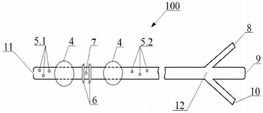
图1示意性地展示了根据本发明的用于隔离哺乳动物的中空器官中的区域的导管;Figure 1 schematically illustrates a catheter for isolating a region in a hollow organ of a mammal according to the present invention;
图2展示了设置在图1的导管的一部分中的功能性开口;Figure 2 illustrates a functional opening provided in a portion of the catheter of Figure 1;
图3展示了具有网格外壳的导管在球囊放气时的状态;Figure 3 shows the state of the catheter with the mesh shell when the balloon is deflated;
图4展示了具有网格外壳的导管在球囊膨胀时的状态。Figure 4 shows the state of the catheter with the mesh shell when the balloon is inflated.
具体实施方案specific implementation
在本文件的上下文中,除非另有明确说明,否则术语“患者”首先是指寻求医疗建议或处于医学观察下以进行诊断和/或治疗疾病的潜在患病的人(哺乳动物类的成员),其中术语“患者”还指仍处于医学观察下以诊断和/或治疗其疾病的潜在患病的哺乳动物。In the context of this document, unless expressly stated otherwise, the term "patient" refers primarily to a potentially ill person (member of the class Mammalian) seeking medical advice or under medical observation for the diagnosis and/or treatment of a disease , wherein the term "patient" also refers to a potentially diseased mammal still under medical observation to diagnose and/or treat its disease.
此外,在本文件的上下文中,除非另有明确说明,否则术语“哺乳动物”是指人或动物,特别是类人猿和非人灵长类动物、狗、猫、马、骆驼、驴、牛、羊、猪和其他众所周知的哺乳动物。Furthermore, in the context of this document, unless expressly stated otherwise, the term "mammal" refers to humans or animals, in particular apes and non-human primates, dogs, cats, horses, camels, donkeys, cattle, Sheep, pigs and other well known mammals.
此外,在本文件的上下文中,除非另有明确说明,否则术语“用户”是指被授权将根据本发明的导管插置至哺乳动物的中空器官(特别是人的中空器官)中,从哺乳动物的中空器官中取出根据本发明的导管和/或操纵插置到哺乳动物的中空器官中的根据本发明的导管的任何合适的技术熟练的医疗保健专业人员,其中,医疗保健专业人员可以是例如外科医生、肿瘤学家、内窥镜医师、胸外科医生、血管外科医生、泌尿科医生、兽医等。Furthermore, in the context of this document, unless expressly stated otherwise, the term "user" means authorized to insert the catheter according to the present invention into a hollow organ of a mammal (in particular a human hollow organ), from breastfeeding Any suitable skilled healthcare professional who removes a catheter according to the invention from a hollow organ of an animal and/or manipulates a catheter according to the invention inserted into a hollow organ of a mammal, wherein the healthcare professional may be For example surgeons, oncologists, endoscopists, thoracic surgeons, vascular surgeons, urologists, veterinarians, etc.
如今,最先进的诊断和/或治疗发生在哺乳动物的中空器官或与这些中空器官相关的器官中的炎性、自身免疫、感染性、良性和/或恶性疾病,特别是胰腺、胆管、肝脏、胃肠道的疾病以及哺乳动物的中空器官的缺损和损伤、瘘管、狭窄、和/或动脉瘤和憩室扩张等的方法是基于对生物体的、为哺乳动物的中空器官或与该中空器管相关的器官所特有的生物液体或生物流体中的疾病特异性特征的确定的液体活检。特别地,用于诊断和/或治疗哺乳动物的处于初始阶段和癌前转化的胰腺癌的液体活检是基于对循环病理细胞、循环肿瘤DNA、RNA、蛋白质、肽、代谢物、以及在哺乳动物的生物体的生物液体(诸如血液和胰液)中循环肿瘤外泌体的检测。Today, state-of-the-art diagnosis and/or treatment of inflammatory, autoimmune, infectious, benign and/or malignant diseases that occur in mammalian hollow organs or organs associated with these hollow organs, in particular the pancreas, bile ducts, liver , diseases of the gastrointestinal tract, and defects and injuries of mammalian hollow organs, fistulas, stenosis, and/or aneurysm and diverticulum dilatation, etc. are based on the biological, mammalian hollow organ or with the hollow organ Deterministic liquid biopsy of a biological fluid specific to a tube-related organ or a disease-specific signature in a biological fluid. In particular, liquid biopsy for the diagnosis and/or treatment of primary and precancerous pancreatic cancer in mammals is based on the analysis of circulating pathological cells, circulating tumor DNA, RNA, proteins, peptides, metabolites, and in mammals Detection of circulating tumor exosomes in biological fluids such as blood and pancreatic juice of an organism.
液体活检的一个关键方面是采样材料的量,因此需要有足够量的采样材料来执行诊断。液体活检的一个更关键的方面是哺乳动物生物体中疾病的定位,特别是肿瘤定位,因为从样品中分离的病理遗传性或其他诊断材料可能是哺乳动物的不同中空器官或与这些中空器管相关的器官的癌症的典型特征。A key aspect of liquid biopsy is the amount of sampled material, so there needs to be a sufficient amount of sampled material to perform a diagnosis. A more critical aspect of liquid biopsy is the localization of disease, especially tumor localization, in mammalian organisms, since the pathological hereditary or other diagnostic material isolated from the sample may be different hollow organs of mammals or related to these hollow tubes. Typical features of cancer of related organs.
特别地,鉴于上述原因,为了诊断胰腺的癌症、粘液性和上皮内肿瘤,胰液是用于检测循环肿瘤细胞、DNA、RNA、蛋白质、肽、代谢物、它们的外泌体的最适当的诊断液体。然而,胰液的采样是最困难的问题,根据本发明的下述实施方案中的任一实施方案的导管100有效地解决了这一问题。根据本发明的导管100的结构和设计特征和功能在下文中在解决从十二指肠乳头收集胰液的说明性任务的背景下进行详细描述,但是本发明的范围不限于于此。In particular, for the above reasons, for the diagnosis of cancer, mucinous and intraepithelial tumors of the pancreas, pancreatic juice is the most appropriate diagnostic for detecting circulating tumor cells, DNA, RNA, proteins, peptides, metabolites, their exosomes liquid. However, sampling of pancreatic juice is the most difficult problem, and the
需要注意的是,小十二指肠乳头和大十二指肠乳头(在本领域中也分别被称为圣托里尼氏乳头(Santorini's papilla)和法特里氏乳头(Fateri's papilla))各自代表呈定位于十二指肠的下降部的中部中的粘膜的纵向皱襞的末端、特别是幽门下方约12-14cm处的半球形、锥形或扁平隆起的形式的解剖结构,其中大多数情况下用于胆管和胰管共有的一个开口暴露于十二指肠腔,而在其他情况下胰管暴露在十二指肠乳头的上方2-4cm处。肝胰壶腹定位在十二指肠乳头中,该壶腹用于接收胰腺的胆汁和消化液,并容纳调节胆汁或胰液流入到十二指肠以及防止肠道内容物进入胆管和胰管的Oddi括约肌。因此,哺乳动物的十二指肠中的大十二指肠乳头通常在幽门的下方12-14cm处,而小十二指肠乳头在大十二指肠乳头的上方2-4cm处。It should be noted that the small and large duodenal papillae (also known in the art as Santorini's papilla and Fateri's papilla, respectively) are each Represents anatomical structures in the form of hemispherical, conical or flat bulges located at the ends of the longitudinal folds of the mucosa located in the middle of the descending part of the duodenum, in particular about 12-14 cm below the pylorus, in most cases An opening common to the bile and pancreatic ducts is exposed to the duodenal lumen, while in other cases the pancreatic duct is exposed 2-4 cm above the duodenal papilla. The hepatopancreatic ampulla is located in the duodenal papilla and serves to receive bile and digestive juices from the pancreas and to accommodate the regulation of the inflow of bile or pancreatic juice into the duodenum and to prevent the entry of intestinal contents into the bile and pancreatic ducts Sphincter of Oddi. Thus, the large duodenal papilla in the duodenum of mammals is usually 12-14 cm below the pylorus, while the small duodenal papilla is 2-4 cm above the large duodenal papilla.
特别地,为了使胰液流入到十二指肠腔中,有必要在法特里氏乳头或大十二指肠乳头周围产生在40-100mmH2O水平的生理负压,这通常通过十二指肠的蠕动来实现(消化生理学[Physiology of digestion],S.提萨卢[S.Teesalu],1987,塔尔图,塔尔图州立大学[Tartu State University],第84页;胰腺[The pancreas],第三版,2018,布莱克威尔,英国,1300)。另一个标准是需要对含有非活性消化酶,而不含胃液和十二指肠液及内容物的胰液进行隔离采样,因为胃液和十二指肠液及内容物的存在会导致酶的活化和诊断所需的细胞、DNA、RNA、蛋白质、肽、代谢物、外泌体的消化。In particular, in order to allow the inflow of pancreatic juice into the duodenal lumen, it is necessary to generate a physiological negative pressure at the level of 40-100 mmH2O around the papilla of Fatry or the papilla of the greater duodenum, which usually passes through the duodenal peristalsis (Physiology of digestion, S. Teesalu, 1987, Tartu, Tartu State University, p. 84; The pancreas, Third Edition, 2018, Blackwell, UK, 1300). Another criterion is the need for isolated sampling of pancreatic juice containing inactive digestive enzymes, but not gastric and duodenal fluids and contents, since the presence of gastric and duodenal fluids and contents can lead to enzyme activation and diagnostic criteria. Digestion of desired cells, DNA, RNA, proteins, peptides, metabolites, exosomes.
图1-图4示意性地展示了根据本发明的用于隔离中空器官中的区域的导管100,其中导管100是由用户插置到中空器官的管腔中的导管,并且其中导管100的腔体或本体形成为柔性中空管,该柔性中空管具有尺寸,特别是长度和厚度,适合于用户辅助将其在中空器官的管腔内朝向放置部位的插置或推进。操纵导管100的用户可以是适当技术熟练的医疗保健专业人员,例如外科医生、肿瘤学家、内窥镜医师、胸外科医生、血管外科医生、泌尿科医师、兽医等。用于隔离中空器官中的区域的导管100可以用于任何患者,特别是任何人或动物。Figures 1-4 schematically illustrate a
图1的导管100在其远侧端部处设置有轴向开口11,该远侧端部用于将导管100施用或插置到患者中,然后将导管100推进到患者的中空器官中(特别是在胃肠道、胆管、呼吸道、泌尿系统、患者血管、与子宫和阴道相关的腔等中)的管腔内的放置部位。此外,导管100在其与导管100的远侧端部相对的近侧端部装配有三通连接器12,其中该近侧端部在导管100被插置到患者的中空器官的管腔中时定位在患者体外。特别地,当导管100用于隔离患者的十二指肠中的区域时,将导管100通过其远侧端部施用或插置到到患者的鼻腔通道中,随后将插置的导管100沿着患者的十二指肠推进到患者的十二指肠中的放置部位。The
如图1所示,导管100中的三通连接器12可以是设置有三个分支或端子的管子或管:中心端子9、以及相对于中心端子9密封隔离的两个侧端子8和10,其中端子8、9、10各自在其自由端部处设置有相应的开口,并且每一个端子都被配置成连接到或接合到适当的功能性器具或功能性设备。As shown in Figure 1, the
定位在三通连接器12中的两个侧端子8、10之间的中心端子9被配置为连接到或接合到适当的(第一)功能性器具或功能性设备,其中第一功能性设备可以是本领域已知的用于供应气态介质或流体(未示出),例如肠内营养混合物的任何设备。用于供应气态介质或流体如连接到中心端子9的设备可以是,例如,注射器,该注射器例如填充有肠内营养混合物以向患者的生物体提供基本营养素、能量、维生素、常量营养素、和/或微量营养素等;或者该设备是填充有例如肠内营养混合物的医用滴管,或其他自动化或半自动化的、适合将中心端子9连接到其上并将气态介质或流体例如肠内营养混合物供应至中心端子9的设备和器具。因此,当导管100被插置到十二指肠管的腔(或胃肠道的其他部分)中时,用于供应气态介质或流体连接、或附接到中心端子9的设备可以用于向患者提供例如营养支持或临床营养。此外,当连接或附接到中心端子9时,用于供应液态或气态介质的设备可用于例如对患者的胃和十二指肠进行消毒。The
作为三通连接器12中的两个侧端子之一的侧端子8被设计为连接或附接到适当的(第二)功能性器具或功能性设备,其中第二功能性设备可以例如被实现为医用抽吸装置、吸引设备或吸引器(未示出),该医用抽吸装置、吸引设备或吸引器包括用于收集生物气态介质、生物液体和/或生物流体的储器或储存容器(未示出),以及用于吸引或排出空气或其他适当的气态介质的空气压缩机(未示出)。本领域技术人员可以针对对应的中空器管基于现有技术文件(例如美国专利号6712798)中公开的信息来调节用于吸引或排出空气或另一种适当的气态介质的压力。The
此外,在三通连接器12中被附接到侧端子8的第二功能性设备可以形成为特殊设备,或用于供应气态介质或流体(例如,医疗产品)的设备,例如填充有待供应的液体的注射器,或填充有待供应的液体的医用滴管,或自动化或半自动化的其他设备或器具,这些设备或器具适用于连接到侧端子8以及将所述气态介质或流体供应到侧端子8。Furthermore, the second functional device attached to the
三通连接器12中的另一侧端子10被配置成连接或附接到适当的(第三)功能性器具或功能性设备,其中,第三功能性设备可以被实现为特殊设备,或用于供应液体或压力下的气态介质(未示出,例如,水或空气)的设备,特别是填充有待供应的液态或气态介质(例如,水或空气)的注射器,或填充有待供应的液体(例如,水)的医用滴管,或自动化或半自动化的其他设备和器具,这些设备或器具适用于连接到侧端子10以及将液态或气态介质供应到侧端子10。The
需要注意的是,上述的第一功能性设备(未示出)可连接到导管100的用于供应液体(例如,肠内营养液)的中心端子9,上述的第二功能性设备(未示出)可连接到导管100的用于采样生物流体或生物气态介质或供应液态或气态介质的侧端子8,和/或第三功能性设备(未示出)可连接到导管100的用于供应液体(例如,水)的侧端子10,结合图1-图4中示出的导管100,可以形成用于隔离哺乳动物(未示出)的中空器官中的区域的系统,该系统可以用于对哺乳动物的特定中空器官所特有的生物流体或生物气态介质(例如,生物液体)进行采样,或者可以构成这种系统的相应部分。特别地,上述系统可用于隔离哺乳动物的十二指肠中的区域以对胰液和/或胆汁进行采样。It should be noted that the above-mentioned first functional device (not shown) can be connected to the
此外,如图1-图4所示,导管100的本体在其外侧设置有两个隔离球囊4,每个隔离球囊4形成为扩张或膨胀的柔性储器,其中隔离球囊4彼此以预定距离间隔开并且与导管100的远侧端部间隔开。功能性孔口或开口7设置在导管100的本体中的隔离球囊4之间。当导管100插置到患者的中空器官的管腔中时,隔离球囊4中的一个隔离球囊,即离导管100的远侧端部最远的隔离球囊,被定位成比中空器官中的关注区域更远,而另一个隔离球囊4被隔离直至中空器官中的关注区域,其中功能性开口7与关注区域相对或靠近关注区域,例如与小十二指肠乳头和大十二指肠乳头中的一个相对或相邻,在小十二指肠乳头和大十二指肠乳头之间,与肠瘘相对或相邻,与壁损伤相对或相邻,与肿瘤相对或相邻等。In addition, as shown in FIGS. 1-4 , the body of the
此外,用于隔离中空器官中的区域的导管100包括设置在导管100的长形本体中的三个单独的功能性通道:主通道1,用于将液态或气态介质供应到隔离球囊4的供应通道2,以及与供应通道2分开并且相对于主通道1密封隔离的功能性通道3,供应通道2相对于主通道1密封隔离、以及具有设置在导管本体100中的孔,并且每个孔都通向相应的隔离球囊4之一的内部。供应通道2和功能性通道3在主通道1的一部分内沿主通道的长度延伸。Furthermore, the
基本上沿导管100的整个长度延伸的主通道1与三通连接器12的中心端子9连通,其中主通道1具有入口孔或与设置在导管100的近侧端部处的中心端子9中的开口相对应的入口,以及具有出口孔或与在导管100的远侧端部处的轴向孔11相对应的出口。当导管100插置到患者中空器官的管腔中时,主通道1的入口定位在患者身体的外侧以与周围大气或环境连通,主通道1的出口与器官(例如食道、胃和十二指肠、小肠或大肠,以及呼吸道、胆道、泌尿道、血管等)的管腔连通。因此,在上述第一功能性设备(未示出)被连接到在三通连接器12的中心端子9的情况下,医疗产品、特别是肠内或肠外营养混合物最初可以在压力下从第一功能性设备通过其中制成的孔供应到中心端子9,然后从中心端子9供应到主通道1,最后从主通道1通过图1所示的轴向孔11供应到中空器官的内部。The
基本上沿着导管100的部分长度延伸的供应通道2通过设置在导管100的本体中的相应出口13、14中的一个出口与隔离球囊4中的每个隔离球囊连通(如图2所示),以及与三通连接器12的侧端子10连通,其中,供应通道2具有入口孔或与设置在导管100近侧端部处的侧端子10中的开口相对应的入口。当导管100插置到患者的中空器官的管腔中时,供应通道2的入口定位于患者身体的外侧以与周围大气或环境连通。因此,在上述第二功能性设备(未示出)被连接到三通连接器12的侧端子10的情况下,气态介质或流体(例如,空气或水)最初可以在压力下以给定体积从第二功能性设备通过其中制作的孔供应到侧端子10,然后从侧端子10供应到供应通道2,最后从供应通道2通过相应的在导管本体100中制成的孔供应到两个隔离球囊4,从而通过使用特别是导管100的供应通道2用水或气体膨胀或填充隔离球囊4,以提供整体填满或膨胀的球囊4,每个球囊具有增大的尺寸或体积。本领域技术人员可以基于现有技术文件(例如,美国专利号7,722,568)中公开的信息来选择用于为任何特定中空器官用流体或气体填充或膨胀隔离球囊4的压力。The
需要注意的是,隔离球囊4的尺寸或体积的增加会导致患者的中空器官的管腔(例如,被导管100插置的患者的十二指肠的管腔)的双侧封闭或阻塞,从而允许导管100具有功能性开口7的部分被隔离在患者的中空器官中的膨胀的隔离球囊4之间。因此,膨胀的隔离球囊4允许患者的中空器官的一部分,例如十二指肠的大十二指肠乳头和小十二指肠乳头、动脉瘤、中空器官的壁缺损等,与中空器官的近侧部分和远侧部分隔离,并且,因此,它们排除或防止目标生物液体与其他生物液体混合,并允许导管100被固定在患者的中空器官的管腔内,例如固定在患者的十二指肠的管腔内,这是因为球囊4的外侧与中空器官的内壁表面的紧密相邻。特别地,在将导管100插置到十二指肠的管腔的情况下,膨胀的隔离球囊4允许患者的十二指肠的小十二指肠乳头和/或大十二指肠乳头与胃肠道的近侧部分和远侧部分隔离,并且因此,防止目标生物液体与其他生物液体(诸如胃液和内容物和/或十二指肠内容物)混合,其中,目标生物液体可以是胰液和胆汁的混合物。这确保了胰液中所含有的消化酶保持无活性。It should be noted that an increase in the size or volume of the
在本发明的一个实施方案中,隔离球囊4可以通过供应通道2在这些隔离球囊之间连通,从而例如在肠道或食道的蠕动波通过期间,确保用于使隔离球囊扩张或膨胀的液态或气态介质均匀再分配。在本发明的另一实施方案中,隔离球囊4可以形成为例如自动膨胀的球囊,或由通过三通连接器12的侧端子10中的孔供应到导管100的供应通道2的空气而膨胀的球囊,或以本领域已知的任何其他方式膨胀的球囊。In one embodiment of the present invention, isolation balloons 4 may communicate between these isolation balloons through
在本发明的其中一个实施方案中,三通连接器12中的侧端子10可以可选地配备有手动操作截止阀(未示出)以防止气态介质或流体(特别是位于隔离球囊4、供应通道2和侧端子10中的气体或水)回流或逸出。还需要注意的是,隔离阀可以被导管用户用来调节隔离球囊4的膨胀程度,其中用户可以通过使用内窥镜视觉上控制膨胀程度。特别地,用户可以通过手动打开止回阀从供应通道4偏转或排出气态介质或流体、特别是气体或水来减小膨胀的隔离球囊4的尺寸或体积。In one of the embodiments of the present invention, the
大致沿导管100的部分长度延伸的功能性通道3与三通连接器12的侧端子8连通,其中功能性通道3具有入口或与功能性开口7对应的入口端口,以及出口或与设置在导管100近侧端部处的侧端子8中的开口对应的出口端口。因此,在上述第三功能性设备(未图示)被实现为吸引器,该吸引器被连接到三通连接器12的侧端子8的情况下,功能性通道3将基本上用作吸引通道,并且功能性通道3将基本上用作吸引开口,其中,将在中空器官的内部产生或支承负空气压力或负空气介质压力,中空器官内部由如上所述膨胀的隔离球囊4隔离。在患者的中空器官的隔离内部中产生的负压(在本领域中也被称为技术真空)促进了生物气态介质或生物流体、特别是生物液体(诸如胆汁和胰液或胰液分泌物或脓液,或血液,或支气管分泌物)的排出或去除,首先将所述气态介质或流体从隔离内部通过功能性开口7排出到功能性通道3,然后从功能性通道3排出到侧端子8,最后通过侧端子8中制成的相应孔从侧端子8排出到吸引器的储存容器。需要注意的是,具有功能性开口7的功能性通道3不仅可以用于对特定中空器官所特有的生物气态介质或生物流体进行采样,而且还可以用于供应所需的液态或气态介质,例如呈液态或气态形式的药物或着色剂,在上述第三功能性设备(未示出)被实现为用于供应气态介质或流体的装置(例如,医用注射器或医用滴管)被连接到三通连接器12的侧端子8的情况下,将该所需的液态或气态介质供应到隔离内部。A
需要注意的是,吸引器作为可连接到三通连接器12的侧端子8的上述第三功能性设备的可能实施方案中的一种可能实施方案,当吸引器由用户激活时,激活作为吸引器一部分的空气压缩机。激活的空气压缩机从患者的中空器官内部,例如从以上述方式由膨胀的隔离球囊4隔离的患者的十二指肠的内部、以及从吸引器储存容器中的可用空间提供空气介质或空气的吸引或排出,可用空间不被填充生物气态介质或生物流体(例如,生物流体,诸如胆汁和胰液,或胰腺分泌物)以在中空器官的隔离内部(例如,在患者的十二指肠的管腔)中产生负压,从而通过功能性开口7获取生物介质并通过功能性通道3将获取的生物介质移除或供应到储存容器,以用于在预定时间段内积聚或收集在储存容器中。It should be noted that the aspirator as one of the possible embodiments of the above-described third functional device connectable to the
需要注意的是,在这些实施方案中的一种实施方案中,三通连接器12的侧端子8可以用于管理由功能性通道3所携带的、且通过功能性开口7进入隔离球囊4之间的内部的液态或气态物质的通过。所管理的气态或液态物质(例如药物或着色剂)可以影响被球囊4隔离的中空器官的区域,以及具有在该区域上的诊断和/或治疗效果。It should be noted that in one of these embodiments, the
在本发明的一个实施方案中,仅上述的用于将流体(例如,水)供应到与上述侧端子10连通的隔离球囊4以及与上述侧端子8连通的上述功能性通道3的供应通道2可以设置在导管本体100中。In one embodiment of the invention, only the above-mentioned supply channels for supplying fluid (eg, water) to the
此外,导管100的本体在其外侧设置有两个大致相同的隆起部6,每个隆起部形成为环形突起或环,环形突起或环具有的半径大于导管100的本体的半径或小于膨胀的隔离球囊4中的任一个隔离球囊的半径,其中,隆起部6中的每个隆起部沿导管100的本体的长度定位在相对于功能性开口7相邻或靠近的一侧上,并且其中隆起部6优选地在隔离球囊4之间相对于功能性开口7等距间隔。需要注意的是,在导管100插置到患者的中空器官的管腔中的情况下,基本上作为包围功能性开口7的突出侧的隆起部6,在连接到导管100的功能性通道3的吸引器被用户激活时,防止功能性开口7接触患者的中空器官的黏膜或接近患者的中空器官的黏膜一段适合于将粘膜组织抽吸到功能性开口7中的距离,并且因此,当由中空器官的管腔中的隔离内部内的吸引器提供负压时,会损伤中空器官的黏膜组织。在本发明的一个实施方案中,隆起部6可以具有不同于环或环形形式的任何其他形式,只要这种形式防止功能性开口7与患者的中空器官的粘膜接触,或在与功能性开口7连通的中空器官的隔离内部产生负压时,防止功能性开口7接近患者的中空器官的粘膜一段适合于将粘膜抽吸到功能性开口7中的距离。Furthermore, the body of the
此外,如图3-图4所示,导管100的本体在其外侧处设置有两个凸起(protrusion)或突起15,其中,这些突起15的每个突起设置在由隔离球囊4限定的导管本体部的外侧与相应的球囊4之一相距预定距离,其中,弹性网或网格外壳16被附接到突起15以完全或至少部分地覆盖隔离球囊4和由球囊4限定的导管100的本体的一部分,从而完全或至少部分地包封或覆盖功能性开口7。当使球囊4膨胀时,如图4所示,网格外壳16变得拉紧或拉伸,从而将中空器官中的内壁组织移动或移位距离功能性开口7预定距离。处于完全或至少部分应变状态的网格外壳16形成弹性外部壳体或框架,该弹性外部壳体或框架完全或至少部分包围由球囊4限定或定位在球囊4之间的导管本体部,从而完全或至少部分包封或覆盖功能性开口7。由网格外壳16形成的外部框架具有圆柱形状,并且由于其细胞结构而可被液体穿透,从而允许对哺乳动物的某个中空器官特定的生物流体穿透或穿过网格外壳16的材料并进入功能性开口7。特别地,在吸引设备(未示出)连接到侧端子8以通过导管100中的功能性通道3和功能性开口7提供在隔离球囊4之间的隔离区域中的负压的情况下,拉伸或拉紧的网格外壳16将阻碍中空器官(例如,血管壁、肠粘膜、支气管、胃、输尿管等)中的内壁组织与功能性开口7的粘合或粘附,从而允许生物材料或生物流体(例如胆汁、胰液、支气管分泌物等)被不断吸引到吸引设备的容器中。Furthermore, as shown in FIGS. 3-4 , the body of the
在本发明的一个实施方案中,网格外壳16可以被附接到两个隔离球囊4,使得网格外壳完全或至少部分地包围由球囊4限定或定位在球囊4之间的导管本体部,从而完全或至少部分包封或覆盖功能性开口7。当使球囊4膨胀的,如图4所示,网格外壳16变得拉紧或拉伸,从而允许移除中空器官中的内壁组织或以距离功能性开口7预定距离偏移中空器官中的内壁组织。In one embodiment of the invention, the
在本发明的另一个实施方案中,网格外壳16可以形成为以应变状态或至少部分应变状态初步固定在隆起部6上的网状材料或网,使得网格外壳完全或至少部分包围由球囊4限定或定位在球囊4之间的导管本体部,从而完全或至少部分地包封或覆盖功能性开口7。In another embodiment of the invention, the
在本发明的一些其他实施方案中,网格外壳16可以通过本领域已知的任何合适的紧固方式以初步应变状态固定在导管100的本体上,使得网格外壳完全或至少部分地包围由球囊4限定或定位在球囊4之间的导管本体部,从而完全或至少部分地包封或覆盖功能性开口7。In some other embodiments of the invention, the
在本发明的其他实施方案中,网格外壳16可以具有允许网格外壳16固定在导管100的本体上、隆起部6上、或隔离球囊4上以完全或至少部分包封或覆盖功能性开口7的任何形式。In other embodiments of the present invention, the
此外,三个辅助孔5.1、5.2设置在隔离球囊4的外侧的导管本体100中,因此在设置有功能性开口7并由隔离球囊4限定的导管本体部的外侧。将导管100插置到患者的中空器官的管腔中时,定位于距离导管100远侧端部较远的辅助孔5.2用作入口,定位于距离导管100远侧端部较近的辅助孔5.1用作出口。需要注意的是,辅助孔5.1、5.2允许中空器官内容物(例如空气、尿液、血液、具有非活性消化酶的胃或肠内容物)通过中空器官的远侧部段,而不进入由导管100的膨胀的隔离球囊6限定的区域,从而防止或消除在患者的中空器官中充血和/或炎症过程的形成,这些过程特别是由血液、尿液、空气、粘液和其他内容物在膨胀的隔离球囊4外侧、位于距离导管100的远侧端部较远或最远的患者中空器官中的积聚引起的。Furthermore, three auxiliary holes 5 . 1 , 5 . 2 are provided in the
取决于根据本发明的导管100的用途以及中空器官的解剖参数、导管100的长度和直径、导管100的壁厚、隔离球囊4在导管100中的位置和直径,孔的位置可以变化。本领域技术人员可以取决于特定中空器官的尺寸容易地调节导管100的任何参数。Depending on the use of the
导管100的通道的厚度和隔离球囊4的大小可以由本领域技术人员基于本领域公开的信息(例如,美国专利号9526874、美国专利号6692465、美国专利号5843050、美国专利号5919163、国际公开WO2009/035581、美国专利号5397305、美国专利号8398589、美国专利号7722568、美国专利号6712798、美国专利号6638245、美国专利号1009865和/或其他现有技术信息源)进行调节。The thickness of the channel of the
导管100的长度、隔离球囊4之间的距离、以及隔离球囊4到远侧端部的距离可以由本领域技术人员基于本领域公开的信息(例如,US专利号5314409、美国专利号5658264、美国专利申请号20150150572、美国专利号5843050、美国专利号5397305、美国专利号7070606、美国专利号6712798、美国专利号1009865和/或其他现有技术信息源)进行匹配。The length of the
例如,在本发明的优选实施方案中的一个优选实施方案中,导管100可以被实现为胰消化导管并且可以具有130cm的长度。在本发明的本实施方案中,隔离球囊4距离导管100的远侧端部的相应距离分别为15sm和25sm,该远侧端部用于将导管100插置到胃腔和患者的十二指肠的管腔中,使得隔离球囊4的扩张或膨胀允许十二指肠的隔离区域具有至少10cm长度。在本发明的本实施方案中,功能性开口7可以在隔离球囊4之间的中间,即在距离隔离球囊4中的每个隔离球囊5cm的距离处以及距离导管100的远侧端部20cm的距离处。For example, in one of the preferred embodiments of the present invention, the
根据本发明的导管100用于隔离患者的中空器官中的区域的用途示意性地在上文关于患者的十二指肠进行了描述。然而,导管100的用途不受患者的十二指肠的限制。因此,本领域技术人员清楚的是,根据上述实施方案中的任何实施方案的导管100可以类似地插置到患者(特别是哺乳动物)的任何其他中空(管状)内部器官的管腔中,例如插置到患者的食道、胃、十二指肠、小肠、大肠、呼吸道、泌尿道(泌尿生殖系统道)、静脉、动脉、阴道、子宫、子宫(输卵管)管、椎管或任何适当的内部管状器官中,患者管状器官与哺乳动物生物体的相应功能系统(器官系)相关,这些功能系统来自一组系统,包括:消化系统、呼吸系统、泌尿和生殖系统(组合成泌尿生殖系统或生殖泌尿系统)、内分泌系统、循环系统和免疫系统以及骨骼系统。The use of the
因此,当使用时,根据本发明的导管100可以由用户在内窥镜(未示出)或射线照相设备(例如,荧光透视设备)的控制下插置到患者的十二指肠的管腔中,使得相应的隔离球囊4中的距离导管100的远侧端部最远的一个隔离球囊定位在患者十二指肠的球部中,其中内窥镜可由用户或协助用户的内窥镜专家操纵。特别要注意的是,导管100插置到患者的十二指肠的管腔的过程、在对所需量的生物液体进行采样后将导管100从患者十二指肠的管腔移除的过程、以及吸引过程是无损伤的并且不依赖于患者和肿瘤两者的解剖特征。需要注意的是,当用户将导管100插置到患者的十二指肠的腔时,隔离球囊4处于放气状态。Thus, when in use, the
根据一个示例,导管100可以预先配备有至少一个环,该环被设计成用活检钳将其抓持。为了将导管100插置到患者的十二指肠中所需的放置部位,用凡士林油充分润滑的导管100的远侧端部被施用通过鼻道并推进到胃腔;然后内窥镜进一步与导管100并行地被施用或推进通过患者口腔到患者的胃腔,以通过内窥镜活检钳捕获导管100的(多个)环;最后,被捕获的导管100与内窥镜一起被引导或推进到患者的十二指肠。随后,在内窥镜的控制下,将导管100的相应的隔离球囊4中距离导管100的远侧端部最远的一个隔离球囊放置在患者的十二指肠的球部中。According to one example, the
根据另一个示例,金属导丝可以被预先施用或插置到导管100的主通道1中。为了将导管100插置到患者的十二指肠中的所需放置部位,用凡士林油充分润滑的导管100的远侧端部被插置通过鼻道,然后通过使用导管100的金属导丝被推进到胃腔;然后内窥镜与导管100并行地被插置或推进通过患者口腔到达患者胃腔,以在检测到导管100的远侧端部时通过内窥镜活检钳捕获第一结扎线,并且通过使用经捕获的第一结扎线向上提拉导管100而将导管100压向内窥镜。随后,内窥镜和被压靠在内窥镜上的导管100被引导通过幽门到达患者的十二指肠,并且导管100的相应的隔离球囊4中距离导管100远侧端部最远的一个隔离球囊在内窥镜控制下被放置在患者的十二指肠的球部内。According to another example, a metal guide wire may be pre-applied or inserted into the
然后,用户将上述第二功能性设备(未示出)连接或附接到三通连接器12的侧端子10,此时第二功能性设备旨在将液态或气态介质(例如,气体或水)以所需的量或体积、特别是40-70ml体积的水供应到导管100的供应通道2,以允许隔离球囊4的扩张和膨胀以紧密地联结到十二指肠的粘膜,从而隔离患者的包含大十二指肠乳头和小十二指肠乳头的十二指肠的所需区域,特别是防止胃内容物和/或具有非活性消化酶的十二指肠内容物进入隔离区域。特别要注意的是,作为示意性示例,发明人通过实验获得的结果表明,需要40-70ml的空气将隔离球囊4充分膨胀到用于双侧封闭或闭塞导管100可插置到的患者的十二指肠的内腔所需的大小。The user then connects or attaches the aforementioned second functional device (not shown) to the
然后,用户将实现为吸引器的上述第三功能性设备(未示出)结合或连接到功能性通道3,其中吸引器被初步配置用于恒定或可变操作模式,该操作模式根据特定任务提供所需的工作压力。由用户激活的吸引器允许通过功能性开口7和与功能性开口连通的功能性通道3从患者的十二指肠的隔离内部抽吸空气介质或空气,隔离内部与十二指肠的壶腹周围区对应,从而在内部产生与通常由十二指肠蠕动产生的压力对应的负压,特别是40-100mm H2O(9-14kPa))的负压。在患者的十二指肠的隔离内部中提供的负压允许以胰液或胰腺分泌物与胆汁结合的形式的生物液体通过大十二指肠乳头和小十二指肠乳头从胰腺排出或取回,从而通过功能性开口7和功能性通道3从患者的十二指肠的隔离区域去除所述生物液体,以及将生物液体收集到吸引器的储存容器(未示出)中。在某些情况下,容器中还可以进一步收集胆管和胰管的黏膜分泌物,黏膜分泌物是十二指肠特有的多种生物液体中的一种生物液体,和/或一些生物内容物(其由于反流而进入胆管和胰管、并且将会包含由生物体产生的液体,这些液体是人类十二指肠特定的)。随后,被收集在功能性设备(未示出)的储存容器中的生物液体可以被输送以用于细胞学检查和/或分子遗传学检查或其他分析,以评估胰腺分泌物的细胞成分的形态结构特征,细胞成分允许人们检测或揭示例如导管内肿瘤、神经内分泌肿瘤或胰腺癌特定的病理过程,并在发展的早期检测肿瘤,并通过确定特异性标记物(NKX2、S100P、CEA、EFR3A/B、MUC1、MUC2、MUC5、ANXA1、A2、KRT7、MMP7、MMP9、IGFBP3、PSCA、PRSS2、SHh、KRas、TP53、SMAD4、BRCA1、miRNA21和miRNA155)的表达对不同类型肿瘤之间执行鉴别诊断。The user then incorporates or connects to the
此外,在将吸引器连接到功能性通道3之后,或者代替吸引器,用于供应气态介质或流体的设备(例如,注射器或医用滴管)可以被连接到功能性通道3,以实现将所需的流体或气态介质(例如,液体或气态形式的药物)输送到患者的十二指肠的隔离内部。Furthermore, after connecting the aspirator to the
需要注意的是,根据本发明的导管100可以在插置状态下保持足够长的时间段,例如长达7天(即长达168小时),从而允许以适当的量收集用于可靠地鉴定和验证病理性肿瘤材料的胰腺分泌物,从而大体上提高诊断效率,从而提高胰腺癌的后续治疗。此外,延长采样增加了检测病理细胞和不仅可以用于胰腺癌、还可以用于胆管和肝脏的恶性疾病(包括胆管癌和/或肝细胞癌)的其他生物标志物的可能性。It should be noted that the
还需要注意的是,在使用导管100时不需要刺激胰液和胆汁的分泌,因此经收集的生物材料具有真实的生化和生理指标,呈现出胰腺、肝脏、胆管和胆囊收缩性的真实功能状态。It should also be noted that the secretion of pancreatic juice and bile does not need to be stimulated when using the
如果需要,可以通过使用根据本发明的导管100检查以上述方式收集在储存容器(未示出)中的胰腺分泌物与胆汁组合的生化成分来评估胰腺的排泄功能。If desired, the excretory function of the pancreas can be assessed by examining the biochemical composition of pancreatic secretions combined with bile collected in a storage container (not shown) in the manner described above using the
此外,胰液和胆汁的定性和定量分析允许评估生物材料和每日分泌量的真实生化指标,从而诊断器官的功能状况。In addition, qualitative and quantitative analysis of pancreatic juice and bile allows the assessment of biomaterials and true biochemical indicators of daily secretion, thereby diagnosing the functional status of the organ.
根据本发明的上述实施方案中的任一实施方案的导管100可用于诊断或监测至少一种疾病的发展,该疾病选自包括以下项的组:胃炎、胰腺炎、胰腺癌、胆管癌、胆管细胞癌、肝细胞癌、胆管炎、胆石症、中空器官壁缺损、自身免疫性肝炎、传染性肝炎、中空器官壁的动脉瘤或憩室突出、中空器官内部出血、中空器官狭窄和食道神经肌肉紊乱,胃、十二指肠、小肠和大肠、胆管、泌尿系统和呼吸道的异常。The
此外,根据本发明的上述实施方案中的任一实施方案中的导管100可用于治疗至少一种疾病,该疾病选自包括以下项的组:胰腺炎、胆管炎、胃肠壁缺损、血管瘤、静脉血栓、输尿管缺损、呼吸道缺损、胃肠道出血、气管(喉管)出血、支气管出血、肺出血和子宫出血。Furthermore, the
此外,根据本发明的上述实施方案中的任一实施方案的导管100可用于在执行外科手术或执行外科操纵时隔离血管的区域、特别是关于血管的内部壁的组织的区域。Furthermore, the
特别地,当使用导管100时,通过恢复和促进胆汁或胰液从相应的管道排入到十二指肠中而实现了治疗或治疗效果。急性炎性疾病(诸如胰腺炎和胆管炎)的发病机理至少部分是由胰液和胆汁流出的损害或中断引起的,流出损害特别是由哺乳动物的十二指肠的运动性(蠕动)受损引起的,因此具有可控负压(例如,40-100mm H2O)的隔离区域通过使用导管100产生胰液和胆汁吸引,允许人们解决这个医学问题,因此有助于治疗上述急性疾病。In particular, when the
此外,当使用导管100时,通过从与胃肠道壁缺损有关的区域排出内容物而实现了治疗或治疗效果,从而具有可控的负压(例如,在70-100mm水柱)的隔离区允许人们从缺损区域排出所有生物液体并解决该医疗问题,因此有助于解决患者的所述医疗问题。Furthermore, when the
对于本领域的技术人员来说很清楚,根据本发明的导管100可以由基于现有技术(例如,美国专利号7,722,568或美国专利号6,638,245)中公开信息的任何合适的材料制成,其中导管100可以优选地由聚合材料制成。It will be clear to those skilled in the art that the
示例Example
1号示例。1号患者因上腹部的束带样痛入院。血液中的淀粉酶为1150U/L。超声检查结果如下:胰腺的尺寸增大;头部为35mm;本体为32mm;尾部为21mm;异质性强烈;轮廓模糊不清;胰腺旁组织水肿;未检测到液体。MSCT图片对应于急性水肿性胰腺炎。患者根据俄罗斯外科医师协会的建议开始治疗,其中将根据本发明的导管100插置到患者的十二指肠中持续24小时,并执行主动吸引法。24小时后,血液中的淀粉酶的水平降至230U/L,疼痛综合征得到控制。Example 1.
2号示例。2号患者因阻塞性黄疸的临床表现入院。检查显示白细胞的水平升高至14.1×10⁹,其中总胆红素为145mmol/L,直接胆红素为113.2mmol/L。血液中的淀粉酶为57U/L,丙氨酸氨基转移酶(ALT)为391U/L,天冬氨酸氨基转移酶(AST)为90U/L。超声检查结果如下:胆囊中检测到了多个直径达1.3cm的结石,并且胆管扩张至11mm。磁共振成像(MRI)的结果如下:在胆总管的远侧部分检测到与6mm结石对应的阴影。内窥镜逆行胰胆管造影术(ERCP)的结果如下:发现单个8mm结石,然后在内窥镜下将其移除。为了预防急性胰腺炎,将根据本发明的导管100插置到患者的十二指肠中持续24小时,并执行主动吸引法。在术后期间,血液中的淀粉酶水平的增加没有超过87U/L。Example 2.
3号示例。3号患者因常规检查和治疗入院。先前执行的MSCT检测到胰腺中形成了异常体(anomalous body)。内窥镜超声检测到一个具有2cm×3cm大小的异常体的形成,其中细针活检在技术上是不可能的。为了选择性地对胰液进行采样,将根据本发明的导管100插置到患者的十二指肠中。在因细胞学和分子学检查而获得的胰液中,检测到腺癌细胞和驱动基因中的突变、外泌体和蛋白质标志物。Example 3.
4号示例。4号患者因阻塞性黄疸的临床表现入院。MRI的结果如下:在肝叶左右肝管的汇合处发现了块体,该块体与IIA型肝门部胆管癌(Klatskin瘤)对应。将根据本发明的用于隔离中空器官中的区域的导管100插置到患者的十二指肠中,并执行主动吸引。在因细胞学和分子学检查而获得的胆汁中,检测到异常细胞和恶性肿瘤的标志物。Example 4.
5号示例。5号患者被诊断为慢性胰腺炎。尽管患者接受了胃肠病学家的治疗和处方酶类药物,但仍观察到体重减轻、排便紊乱和腹泻。为了评估胰腺的功能状态,将根据本发明的用于隔离中空器官中的区域的导管100插置到患者的十二指肠持续24小时,并执行主动吸引。作为对获得的胰液执行的生化测试的结果,发现α淀粉酶和脂肪酶的水平降低,并且还发现胰液和胆汁的每日排泄减少。结果,增加了患者服用的酶类药物的剂量,并额外开具了熊去氧胆酸性药物和胆汁替代品。Example 5. Patient 5 was diagnosed with chronic pancreatitis. Despite the patient's treatment by a gastroenterologist and prescription of enzymes, weight loss, bowel disturbances, and diarrhea were observed. To assess the functional state of the pancreas, the
6号示例。6号患者因患有黄疸住进了该市的传染病门诊。体检结果显示肝脏肿大,其中,生化血液检查结果显示如下:总胆红素为329mmol/L,直接胆红素为141mmol/L,ALT为1040U/L,AST为804U/L。血液中HCV抗原的血清学分析,HBsAg为阴性。将用于隔离中空器官中的区域的导管插置到患者体内,在体内通过主动吸引获得100ml胆汁,通过胆汁的PCR分析检测到HCV病毒的高拷贝数。
7号示例。7号患者在传染病诊所针对经证实的病毒性肝炎进行治疗。在干扰素疗程之后,对血液的重复PCR分析没有发现活跃的病毒过程。将用于隔离中空器官中的区域的导管100插置到患者体内,在体内通过主动吸引获得200ml胆汁,并且对获得的胆汁的PCR分析显示正在进行的病毒过程,因此改变了治疗方案和持续时间。Example 7.
8号示例。8号患者被诊断为肠瘘和腹膜炎。在紧急情况下执行腹腔的手术、卫生处理和引流。为了隔离肠壁缺损,减少其与肠内容物的接触,改善愈合过程,并且还为了通过分离腹腔和病理基质来提供用于治疗腹膜炎的附加方式,将根据本发明的用于隔离中空器官中的区域的导管100插置到肠的损伤区域中,使得隔离球囊4定位在缺损的上方和下方。在使球囊膨胀后,创建隔离区域,并将另一个通道连接到抽吸装置,以对肠瘘区域提供卫生处理,以促进缺损愈合。还通过导管100将肠内营养施用到患者。
9号示例。9号患者因腹部的束带样痛被紧急送到医院。基于检查结果,根据亚特兰大分类,患者被诊断为中度急性胰腺炎。入院时,淀粉酶水平为1320U/L。将根据本发明的用于隔离中空器官中的区域的导管100插置到患者体内,在体内功能性通道连接到用于产生可控负压的泵,同时通过同一导管100将肠内营养和医疗产品施用到患者。结果,生化参数的动态在24小时内按照BISAP量表转为阳性,7天后患者康复。Example 9.
10号示例。10号患者因右侧胁部疼痛、潮热和周期性皮肤发黄而被紧急送到医院。既往病历显示,在当前住院前3个月,患者接受了与胆管相关的内窥镜操纵,以从胆管去除结石。超声波、MSCT显示了肝脏、导管没有变化或脓肿的存在。为了诊断胆管炎,将用于隔离中空器官中的区域的导管100插置到患者体内,并采样50.0ml胆汁以进行细菌学检查。结果,发现了克雷伯氏菌的大量生长,并确定其对抗菌药物的敏感性。在选择特异性抗生素治疗之后,胆管炎症状消失,患者出院接受门诊治疗。
11号示例。11号患者由于存在肿块而接受了乙状结肠切除术。在肠活动期间发现,一结构侵入了肠的所有层,其中存在区域性淋巴结肿大并累及腹膜后组织。当使有肿瘤的肠活动时,与左肾受损的输尿管建立了联系。泌尿科医生被叫到手术室并通过手术治疗输尿管的缺损。为了防止并发症,将根据本发明的用于隔离中空器官中的区域的经调整尺寸的导管100沿着导丝逆行施用,并且球囊中的一个球囊在左肾的骨盆中膨胀,而另一个气囊(下气囊)在膀胱中膨胀,其中功能性通道连接到用于产生可控负压的抽吸装置。在14天内移除导管100,左肾的排泄功能未受损。患者出院接受门诊治疗。
12号示例。12号患者因肺出血而入院。支气管镜检查(bronchoscopy)发现右主支气管有一个腐败性出血肿瘤。为了止血和保持支气管通畅的目的,以及为了防止肺部扩张不全,将根据本发明的用于隔离中空器官中的区域的导管100施用到右支气管中。插置导管100使得肿瘤位于球囊之间,其中球囊的膨胀阻止血液流向支气管的其他部分。为了止血,通过导管100的功能性通道施用止血剂。结果,观察三(3)天没有复发出血。随后移除导管100。十(10)天后,患者出院接受门诊治疗。Example 12.
13号示例。13号患者经诊所仪器证实为纵隔炎而紧急入院。检查发现食道缺损是由一块肉引起的坏死所致。为了治疗食道,将插置根据本发明的用于隔离中空器官中的区域的导管100。将导管100的球囊膨胀以隔离缺损,并将功能性通道连接到抽吸装置以提供可控的负压。结果,患者在1.5个月后出院接受门诊治疗。Example 13. Patient No. 13 was hospitalized urgently for mediastinitis confirmed by clinic instruments. Examination revealed that the esophageal defect was caused by necrosis caused by a piece of meat. To treat the esophagus, a
14号示例。14号患者患有头痛和一般脑部症状而紧急入院。静脉造影增强的MSCT显示在右侧大脑中动脉的盆地中的动脉瘤有挤压迹象。患者被带到血管造影手术室,其中,通过股骨途径施用根据本发明的用于隔离中空器官中的区域的导管100。在荧光透视的控制下执行导管100的定位,使得球囊隔离动脉瘤的区域,其中功能性通道与动脉瘤相对。通过功能性通道施用填充动脉瘤腔的填充溶液。在必要的暴露之后,允许导管100维持周向血流。随后移除导管100。患者在21天后出院。Example 14.
15号示例。15号患者因胰腺癌的常规手术治疗而入院。检查显示头部肿瘤侵入下腔静脉。为了减少失血,将根据本发明的用于隔离中空器官中的区域的导管100插置到静脉中,并且使球囊在胰头部肿瘤的上方和下方膨胀,从而使血液分流并使血液返回到心脏。在腺体切除过程中,用下肢隐静脉自体成形术切除下腔静脉的一部分。失血量为500ml。因此,为患者和外科医生均创造了最佳的临时和生理条件。Example 15.
16号示例。16号患者因乙状结肠癌的手术治疗而入院。在术后,患者在周长的1/3处出现结肠直肠吻合(colo-rectoanastomosis)失败。为了治疗结肠,将根据本发明的用于隔离中空器官中的区域的导管100逆行插置到直肠中,使得导管100的一个球囊定位在相对于缺损的最近侧,而导管100的另一个球囊定位在相对于缺损的最远侧。当导管100的球囊膨胀时,缺损区域与大肠的其他部分隔离开。由于主通道,用水清洗肠的近侧部分。结果,在3周内使缺损闭合。Example 16.
17号示例。17号患者因疑似肝脏肿瘤而入院。为了执行液体活检,将插置根据本发明的用于隔离中空器官中的区域的导管100;通过导管100的球囊隔离与大十二指肠乳头和小十二指肠乳头相关的区域,开始吸引胆汁。当导管100被移除时,输送胆汁吸引物用于遗传学和细胞学检查,其中没有检测到病理标志物和细胞。在取胆汁时,移除覆盖导管100的球囊的袜状物以及用缓冲液清洗,于是收集水,离心分离之后得到细胞沉淀物。经获得的细胞沉淀物的细胞学检查发现了肝细胞癌细胞的存在。Example 17. Patient 17 was admitted to hospital with suspected liver tumor. In order to perform a liquid biopsy, a
18号示例。18号患者因子宫出血而紧急入院。阴道镜检查发现了腐烂的宫颈癌。为了止血,将导管100插置到阴道和子宫腔中,并通过导管100的球囊隔离肿瘤区域。通过导管100的功能性通道对肿瘤进行止血药物的滴注。出血被止住,且患者出院。Example 18. Patient 18 was hospitalized urgently due to uterine bleeding. Colposcopy found decaying cervical cancer. For hemostasis, the
因此,上述导管100允许对任何中空器官进行或执行功能性研究,并且还允许提供适当的治疗。此外,通过使用导管100收集的生物液体的检查和/或实验室分析允许人们以高精度区分诊断炎症形成、良性形成和恶性生长以及感染性疾病。通过排出特定的液体,导管100允许治疗炎性疾病、出血和中空器官壁的缺损。在手术操作期间产生具有旁路功能的隔离区域允许人们控制止血,并且还进行手术操作,特别是切除血管部位及其随后的成形术。还应注意,根据本发明的导管100或上述用于隔离哺乳动物的中空器官中的区域的系统(未示出)可以包括导管100,提供收集大量处于正常生理和生化状态的生物材料的能力,而无需额外刺激与中空器官相连的器官的中空器官的排泄功能,特别是由于在中空器官(导管100插置到中空器官的管腔中)的隔离区域中产生与例如由于肠的蠕动可能会产生的生理负压对应的负压。Thus, the
Claims (15)
Applications Claiming Priority (5)
| Application Number | Priority Date | Filing Date | Title |
|---|---|---|---|
| RU2020100187 | 2019-12-31 | ||
| RU2020100187A RU2743144C1 (en) | 2019-12-31 | 2019-12-31 | Catheter and method for sampling biological duodenal papillae fluids, as well as system based on such catheter and using such catheter |
| RU2020140524 | 2020-12-09 | ||
| RU2020140524A RU2761080C1 (en) | 2020-12-09 | 2020-12-09 | Catheter and a method for creating an isolated zone in a mammalian hollow organ, as well as a system based on such a catheter and the use of such a catheter |
| PCT/RU2020/050399 WO2021137739A1 (en) | 2019-12-31 | 2020-12-28 | Catheter and method for isolating a region in a hollow organ of a mammal, and system based on the catheter, and use of the catheter |
Publications (2)
| Publication Number | Publication Date |
|---|---|
| CN115243623A true CN115243623A (en) | 2022-10-25 |
| CN115243623B CN115243623B (en) | 2025-04-22 |
Family
ID=75108760
Family Applications (1)
| Application Number | Title | Priority Date | Filing Date |
|---|---|---|---|
| CN202080097873.2A Active CN115243623B (en) | 2019-12-31 | 2020-12-28 | catheter |
Country Status (11)
| Country | Link |
|---|---|
| US (1) | US20230037101A1 (en) |
| EP (1) | EP4084695A1 (en) |
| JP (2) | JP7726889B2 (en) |
| KR (1) | KR20220137642A (en) |
| CN (1) | CN115243623B (en) |
| BR (1) | BR112022012983A2 (en) |
| CA (1) | CA3166497A1 (en) |
| MX (1) | MX2022008113A (en) |
| PH (1) | PH12022551614A1 (en) |
| WO (1) | WO2021137739A1 (en) |
| ZA (1) | ZA202208475B (en) |
Cited By (3)
| Publication number | Priority date | Publication date | Assignee | Title |
|---|---|---|---|---|
| CN116747413A (en) * | 2023-06-27 | 2023-09-15 | 无锡帕母医疗技术有限公司 | Catheter device and heart failure therapeutic apparatus |
| CN117982197A (en) * | 2023-12-21 | 2024-05-07 | 中国人民解放军海军第九七一医院 | Calculus smashing and collecting device |
| WO2025096840A1 (en) * | 2023-11-01 | 2025-05-08 | Ventaris Surgical, Inc. | Systems and methods for removal of biological objects from anatomical structures within a body |
Families Citing this family (6)
| Publication number | Priority date | Publication date | Assignee | Title |
|---|---|---|---|---|
| US11259858B1 (en) * | 2018-02-06 | 2022-03-01 | Jason RAHIMZADEH | Endoscopy tube and device for cryotherapy |
| JP2024540252A (en) * | 2021-11-01 | 2024-10-31 | パンディカ リミテッド | CATHETER, SYSTEM, AND METHOD FOR ISOLATING THE INTERIOR OF A MAMMALIAN HOLLOW ORGAN, AND USE OF THE CATHETER - Patent application |
| US20230248949A1 (en) * | 2022-02-07 | 2023-08-10 | Olympus Medical Systems Corp. | Treatment method and treatment system |
| WO2023212415A1 (en) | 2022-04-29 | 2023-11-02 | Arctx Medical, Inc. | Apparatus and method for cooling and/or heating an organ |
| CN120225122A (en) * | 2022-08-12 | 2025-06-27 | 潘迪卡有限公司 | Applications of catheters for isolating regions within hollow organs of mammals (various) |
| US20250359856A1 (en) * | 2022-09-09 | 2025-11-27 | Bard Peripheral Vascular, Inc. | Coaxial needles and biopsy devices for performing a biopsy |
Citations (8)
| Publication number | Priority date | Publication date | Assignee | Title |
|---|---|---|---|---|
| US4781677A (en) * | 1985-07-17 | 1988-11-01 | Wilcox Gilbert M | Method of treatment utilizing a double balloon nasobiliary occlusion catheter |
| SU1461471A1 (en) * | 1985-11-19 | 1989-02-28 | А. Ф. Малышев и В. И. Пуздаев | Catheter |
| US5222941A (en) * | 1990-01-12 | 1993-06-29 | Don Michael T Anthony | Method of dissolving an obstruction in a vessel |
| US5460610A (en) * | 1990-01-12 | 1995-10-24 | Don Michael; T. Anthony | Treatment of obstructions in body passages |
| US5951514A (en) * | 1997-03-07 | 1999-09-14 | Sahota; Harvinder | Multi-lobe perfusion balloon |
| CA2377516A1 (en) * | 1999-07-17 | 2001-01-25 | John A. Karpiel | Devices for extracting biliary or urinary stones |
| US20150150572A1 (en) * | 2013-12-04 | 2015-06-04 | The Johns Hopkins University | Dual Balloon Biliary Stone Extraction Device |
| JP2017176385A (en) * | 2016-03-29 | 2017-10-05 | テルモ株式会社 | Filling method and medical long body |
Family Cites Families (17)
| Publication number | Priority date | Publication date | Assignee | Title |
|---|---|---|---|---|
| US1009865A (en) | 1910-12-19 | 1911-11-28 | James H Sherman | Hollow metal tile. |
| EP0492361B1 (en) | 1990-12-21 | 1996-07-31 | Advanced Cardiovascular Systems, Inc. | Fixed-wire dilatation catheter with rotatable balloon assembly |
| JP3282831B2 (en) * | 1991-02-16 | 2002-05-20 | テルモ株式会社 | Catheter tube |
| US5135535A (en) | 1991-06-11 | 1992-08-04 | Advanced Cardiovascular Systems, Inc. | Catheter system with catheter and guidewire exchange |
| US5314409A (en) | 1993-03-11 | 1994-05-24 | Uva Patents Foundation | Catheter for esophageal perfusion |
| US5658264A (en) | 1994-11-10 | 1997-08-19 | Target Therapeutics, Inc. | High performance spiral-wound catheter |
| US5843050A (en) | 1995-11-13 | 1998-12-01 | Micro Therapeutics, Inc. | Microcatheter |
| US5868708A (en) | 1997-05-07 | 1999-02-09 | Applied Medical Resources Corporation | Balloon catheter apparatus and method |
| US5919163A (en) | 1997-07-14 | 1999-07-06 | Delcath Systems, Inc. | Catheter with slidable balloon |
| US6638245B2 (en) | 2001-06-26 | 2003-10-28 | Concentric Medical, Inc. | Balloon catheter |
| US6712798B2 (en) | 2002-03-18 | 2004-03-30 | Corazon Technologies, Inc. | Multilumen catheters and methods for their use |
| US7070606B2 (en) | 2002-05-28 | 2006-07-04 | Mercator Medsystems, Inc. | Methods and apparatus for aspiration and priming of inflatable structures in catheters |
| US7722568B2 (en) | 2007-01-29 | 2010-05-25 | Onset Medical Corporation | Expandable intra-aortic balloon pump sheath |
| WO2009035581A1 (en) | 2007-09-10 | 2009-03-19 | Ziad Anwar Ali | Intra-aortic multi-balloon catheter for improving coronary and visceral perfusion |
| BRPI0919884B8 (en) | 2008-11-03 | 2023-04-04 | Atlanta Catheter Therapies Inc | occlusion perfusion catheter |
| RU2607929C2 (en) * | 2015-06-30 | 2017-01-11 | Государственное бюджетное образовательное учреждение высшего профессионального образования "Санкт-Петербургский государственный педиатрический медицинский университет" Министерства здравоохранения Российской Федерации (ГБОУ ВПО СПбГПМУ Минздрава России) | Device for treating pancreatitis |
| US11524148B2 (en) * | 2016-12-06 | 2022-12-13 | University Of Maryland, Baltimore | Balloon catheter flow device |
-
2020
- 2020-12-28 CA CA3166497A patent/CA3166497A1/en active Pending
- 2020-12-28 MX MX2022008113A patent/MX2022008113A/en unknown
- 2020-12-28 CN CN202080097873.2A patent/CN115243623B/en active Active
- 2020-12-28 KR KR1020227026552A patent/KR20220137642A/en active Pending
- 2020-12-28 BR BR112022012983A patent/BR112022012983A2/en unknown
- 2020-12-28 PH PH1/2022/551614A patent/PH12022551614A1/en unknown
- 2020-12-28 JP JP2022541016A patent/JP7726889B2/en active Active
- 2020-12-28 US US17/790,320 patent/US20230037101A1/en active Pending
- 2020-12-28 EP EP20866922.6A patent/EP4084695A1/en active Pending
- 2020-12-28 WO PCT/RU2020/050399 patent/WO2021137739A1/en not_active Ceased
-
2022
- 2022-07-28 ZA ZA2022/08475A patent/ZA202208475B/en unknown
-
2025
- 2025-08-07 JP JP2025132305A patent/JP2025163233A/en active Pending
Patent Citations (8)
| Publication number | Priority date | Publication date | Assignee | Title |
|---|---|---|---|---|
| US4781677A (en) * | 1985-07-17 | 1988-11-01 | Wilcox Gilbert M | Method of treatment utilizing a double balloon nasobiliary occlusion catheter |
| SU1461471A1 (en) * | 1985-11-19 | 1989-02-28 | А. Ф. Малышев и В. И. Пуздаев | Catheter |
| US5222941A (en) * | 1990-01-12 | 1993-06-29 | Don Michael T Anthony | Method of dissolving an obstruction in a vessel |
| US5460610A (en) * | 1990-01-12 | 1995-10-24 | Don Michael; T. Anthony | Treatment of obstructions in body passages |
| US5951514A (en) * | 1997-03-07 | 1999-09-14 | Sahota; Harvinder | Multi-lobe perfusion balloon |
| CA2377516A1 (en) * | 1999-07-17 | 2001-01-25 | John A. Karpiel | Devices for extracting biliary or urinary stones |
| US20150150572A1 (en) * | 2013-12-04 | 2015-06-04 | The Johns Hopkins University | Dual Balloon Biliary Stone Extraction Device |
| JP2017176385A (en) * | 2016-03-29 | 2017-10-05 | テルモ株式会社 | Filling method and medical long body |
Cited By (5)
| Publication number | Priority date | Publication date | Assignee | Title |
|---|---|---|---|---|
| CN116747413A (en) * | 2023-06-27 | 2023-09-15 | 无锡帕母医疗技术有限公司 | Catheter device and heart failure therapeutic apparatus |
| WO2025002128A1 (en) * | 2023-06-27 | 2025-01-02 | 无锡帕母医疗技术有限公司 | Catheter device and heart failure treatment instrument |
| WO2025096840A1 (en) * | 2023-11-01 | 2025-05-08 | Ventaris Surgical, Inc. | Systems and methods for removal of biological objects from anatomical structures within a body |
| US12419656B2 (en) | 2023-11-01 | 2025-09-23 | Ventaris Surgical, Inc. | Systems and methods for removal of biological objects from anatomical structures within a body |
| CN117982197A (en) * | 2023-12-21 | 2024-05-07 | 中国人民解放军海军第九七一医院 | Calculus smashing and collecting device |
Also Published As
| Publication number | Publication date |
|---|---|
| WO2021137739A4 (en) | 2021-10-07 |
| BR112022012983A2 (en) | 2022-09-13 |
| US20230037101A1 (en) | 2023-02-02 |
| WO2021137739A1 (en) | 2021-07-08 |
| CN115243623B (en) | 2025-04-22 |
| EP4084695A1 (en) | 2022-11-09 |
| KR20220137642A (en) | 2022-10-12 |
| ZA202208475B (en) | 2023-03-29 |
| JP7726889B2 (en) | 2025-08-20 |
| JP2023514802A (en) | 2023-04-11 |
| MX2022008113A (en) | 2022-09-19 |
| JP2025163233A (en) | 2025-10-28 |
| PH12022551614A1 (en) | 2023-11-06 |
| CA3166497A1 (en) | 2021-07-08 |
Similar Documents
| Publication | Publication Date | Title |
|---|---|---|
| JP7726889B2 (en) | Catheter and system for isolating the interior of a mammalian hollow organ | |
| US20110208022A1 (en) | Device and methods for sampling prostate fluid | |
| WO2024033700A1 (en) | Use of a catheter for isolating a region in a hollow organ of a mammal (variants) | |
| CN118510452A (en) | Stent-like catheter for isolating a region in a hollow organ of a mammal, and a system based on the catheter | |
| RU2743144C1 (en) | Catheter and method for sampling biological duodenal papillae fluids, as well as system based on such catheter and using such catheter | |
| RU2783089C1 (en) | Use of a catheter to create an isolated zone in a mammalian hollow organ (versions) | |
| RU2761080C1 (en) | Catheter and a method for creating an isolated zone in a mammalian hollow organ, as well as a system based on such a catheter and the use of such a catheter | |
| Holt et al. | Urological Disorders of the Dog and Cat: Investigation, Diagnosis, Treatment | |
| RU2805216C1 (en) | Application of catheter to create isolated zone in the hollow organ of mammal (variants) | |
| RU2807166C1 (en) | Using a catheter to create an isolated area in the hollow organ of a mammal for treating damage to the wall of the jeonum or ileum (options) | |
| EA045140B1 (en) | CATHETER AND METHOD FOR CREATING AN ISOLATED AREA IN THE HOLOR ORGAN OF A MAMMAL, AS WELL AS A SYSTEM BASED ON SUCH CATHETER AND APPLICATION OF SUCH CATHETER | |
| RU2569026C1 (en) | Method for selecting tactics of treatment of intestinal obstruction, caused by tumour of left half of colon and rectum | |
| RU1607112C (en) | Method of diagnostics of cancer of colon | |
| RU57123U1 (en) | Cylinder catheter with tags for intraoperative endobiliary dilatation | |
| CN110693559B (en) | A blocking device for transnatural orifice surgery | |
| RU2359613C2 (en) | Method of radiodiagnostics of pelvic organs | |
| RU2621168C1 (en) | Treatment method of ruptured diverticulum of sigmoid colon | |
| Isa et al. | Complete tubular duplication of colon presenting as rectovestibular fistula: A case report | |
| RU2145794C1 (en) | Method of x-ray examination of biliary ducts | |
| Kong et al. | Clinical Application of the Reverse Puncture Technique for Total Laparoscopic Colorectal Resection with Natural Orifice Specimen Extraction | |
| Fischer et al. | Cholestatic jaundice in adults: Algorithms for diagnosis | |
| Boiko et al. | Research methods of the urinary and male reproductive system | |
| Akhmadova et al. | General Diagnostics and Surgical Treatment of Complicated Forms of Liver Echinococcosis | |
| Kothari et al. | Endoscopic Ultrasound (EUS) Guided Fiducial Placement for Stereotactic Body Radiotherapy: A New, 22-gauge,“Pre-loaded” EUS Fiducial Needle Reduces Procedure Time Compared to the Traditional Needle “Re-loading” Technique: 507 | |
| RO135252A2 (en) | Device and process for treatment of esophageal perforations and ruptures |
Legal Events
| Date | Code | Title | Description |
|---|---|---|---|
| PB01 | Publication | ||
| PB01 | Publication | ||
| SE01 | Entry into force of request for substantive examination | ||
| SE01 | Entry into force of request for substantive examination | ||
| CB02 | Change of applicant information | ||
| CB02 | Change of applicant information |
Country or region after: Britain Address after: Britain Camb Applicant after: Pandika Co.,Ltd. Address before: Britain Camb Applicant before: Pandex Ltd. Country or region before: Britain |
|
| GR01 | Patent grant | ||
| GR01 | Patent grant |