CN115175999A - High throughput ligation of multiple transcripts - Google Patents
High throughput ligation of multiple transcripts Download PDFInfo
- Publication number
- CN115175999A CN115175999A CN202180017456.7A CN202180017456A CN115175999A CN 115175999 A CN115175999 A CN 115175999A CN 202180017456 A CN202180017456 A CN 202180017456A CN 115175999 A CN115175999 A CN 115175999A
- Authority
- CN
- China
- Prior art keywords
- cell
- molecule
- cdna
- mrna
- cells
- Prior art date
- Legal status (The legal status is an assumption and is not a legal conclusion. Google has not performed a legal analysis and makes no representation as to the accuracy of the status listed.)
- Pending
Links
Images


Classifications
-
- C—CHEMISTRY; METALLURGY
- C12—BIOCHEMISTRY; BEER; SPIRITS; WINE; VINEGAR; MICROBIOLOGY; ENZYMOLOGY; MUTATION OR GENETIC ENGINEERING
- C12N—MICROORGANISMS OR ENZYMES; COMPOSITIONS THEREOF; PROPAGATING, PRESERVING, OR MAINTAINING MICROORGANISMS; MUTATION OR GENETIC ENGINEERING; CULTURE MEDIA
- C12N15/00—Mutation or genetic engineering; DNA or RNA concerning genetic engineering, vectors, e.g. plasmids, or their isolation, preparation or purification; Use of hosts therefor
- C12N15/09—Recombinant DNA-technology
- C12N15/10—Processes for the isolation, preparation or purification of DNA or RNA
- C12N15/1003—Extracting or separating nucleic acids from biological samples, e.g. pure separation or isolation methods; Conditions, buffers or apparatuses therefor
- C12N15/1006—Extracting or separating nucleic acids from biological samples, e.g. pure separation or isolation methods; Conditions, buffers or apparatuses therefor by means of a solid support carrier, e.g. particles, polymers
- C12N15/1013—Extracting or separating nucleic acids from biological samples, e.g. pure separation or isolation methods; Conditions, buffers or apparatuses therefor by means of a solid support carrier, e.g. particles, polymers by using magnetic beads
-
- C—CHEMISTRY; METALLURGY
- C12—BIOCHEMISTRY; BEER; SPIRITS; WINE; VINEGAR; MICROBIOLOGY; ENZYMOLOGY; MUTATION OR GENETIC ENGINEERING
- C12N—MICROORGANISMS OR ENZYMES; COMPOSITIONS THEREOF; PROPAGATING, PRESERVING, OR MAINTAINING MICROORGANISMS; MUTATION OR GENETIC ENGINEERING; CULTURE MEDIA
- C12N15/00—Mutation or genetic engineering; DNA or RNA concerning genetic engineering, vectors, e.g. plasmids, or their isolation, preparation or purification; Use of hosts therefor
- C12N15/09—Recombinant DNA-technology
- C12N15/10—Processes for the isolation, preparation or purification of DNA or RNA
- C12N15/1034—Isolating an individual clone by screening libraries
- C12N15/1068—Template (nucleic acid) mediated chemical library synthesis, e.g. chemical and enzymatical DNA-templated organic molecule synthesis, libraries prepared by non ribosomal polypeptide synthesis [NRPS], DNA/RNA-polymerase mediated polypeptide synthesis
-
- C—CHEMISTRY; METALLURGY
- C12—BIOCHEMISTRY; BEER; SPIRITS; WINE; VINEGAR; MICROBIOLOGY; ENZYMOLOGY; MUTATION OR GENETIC ENGINEERING
- C12N—MICROORGANISMS OR ENZYMES; COMPOSITIONS THEREOF; PROPAGATING, PRESERVING, OR MAINTAINING MICROORGANISMS; MUTATION OR GENETIC ENGINEERING; CULTURE MEDIA
- C12N15/00—Mutation or genetic engineering; DNA or RNA concerning genetic engineering, vectors, e.g. plasmids, or their isolation, preparation or purification; Use of hosts therefor
- C12N15/09—Recombinant DNA-technology
- C12N15/10—Processes for the isolation, preparation or purification of DNA or RNA
- C12N15/1034—Isolating an individual clone by screening libraries
- C12N15/1075—Isolating an individual clone by screening libraries by coupling phenotype to genotype, not provided for in other groups of this subclass
-
- C—CHEMISTRY; METALLURGY
- C12—BIOCHEMISTRY; BEER; SPIRITS; WINE; VINEGAR; MICROBIOLOGY; ENZYMOLOGY; MUTATION OR GENETIC ENGINEERING
- C12N—MICROORGANISMS OR ENZYMES; COMPOSITIONS THEREOF; PROPAGATING, PRESERVING, OR MAINTAINING MICROORGANISMS; MUTATION OR GENETIC ENGINEERING; CULTURE MEDIA
- C12N15/00—Mutation or genetic engineering; DNA or RNA concerning genetic engineering, vectors, e.g. plasmids, or their isolation, preparation or purification; Use of hosts therefor
- C12N15/09—Recombinant DNA-technology
- C12N15/10—Processes for the isolation, preparation or purification of DNA or RNA
- C12N15/1096—Processes for the isolation, preparation or purification of DNA or RNA cDNA Synthesis; Subtracted cDNA library construction, e.g. RT, RT-PCR
Landscapes
- Health & Medical Sciences (AREA)
- Life Sciences & Earth Sciences (AREA)
- Genetics & Genomics (AREA)
- Chemical & Material Sciences (AREA)
- Engineering & Computer Science (AREA)
- Biomedical Technology (AREA)
- Bioinformatics & Cheminformatics (AREA)
- Organic Chemistry (AREA)
- Zoology (AREA)
- Wood Science & Technology (AREA)
- Biotechnology (AREA)
- General Engineering & Computer Science (AREA)
- Crystallography & Structural Chemistry (AREA)
- Plant Pathology (AREA)
- Molecular Biology (AREA)
- Microbiology (AREA)
- Biophysics (AREA)
- Physics & Mathematics (AREA)
- Biochemistry (AREA)
- General Health & Medical Sciences (AREA)
- Bioinformatics & Computational Biology (AREA)
- Analytical Chemistry (AREA)
- Measuring Or Testing Involving Enzymes Or Micro-Organisms (AREA)
Abstract
High throughput methods for physically linking cDNA molecules derived from mRNA molecules expressed by the same cells are provided, as well as libraries of linked cDNA molecules produced by the methods. The method includes reverse transcribing mRNA from a single cell in a first container to produce a cDNA molecule, and ligating the cDNA molecule in a second container. The method surprisingly produces a library of cDNA molecules with an increased number of molecules correctly linked to other molecules derived from the same cell.
Description
Cross Reference to Related Applications
This application claims priority to U.S. provisional application No. 62/964,319, filed on 22/1/2020, which is incorporated herein by reference for all purposes.
Background
In recent years, a number of methods have been developed to achieve high-throughput capture of naturally paired immune repertoire sequences (heavy-light chain pairs in the case of B cells, and alpha-beta chain pairs in the case of T cells). For example, the information gathered from the resulting data set can provide unique insights into the internal workings of human immune responses and enable more intelligent methods of developing therapeutics and vaccines.
One class of such methods involves massively parallel isolation of individual immune cells in micro-containers (e.g., droplets), followed by cell lysis within these containers, and capture of mRNA transcripts onto poly-T capture beads via the poly-a tail of the mRNA strand. Typically, the beads are then recovered from their containers and mRNA is attached by hybridization. These beads are then washed, individually repackaged into secondary micro-containers, reverse transcribing the desired transcripts (heavy and light chain or alpha and beta chains) into cdnas, and finally amplifying and ligating together those cDNA sequences to form a single amplicon comprising the heavy and light chain sequences or alpha and beta chain sequences from the cell of origin. Subsequent sequencing of the ligated amplicons produced 1,00,000+ unique ligated immune cell receptor sequences from the sample with minimal operator time. In addition, the ligated amplicon fragments can be further manipulated into a display format (e.g., phage display, yeast display) to interrogate an immune library that binds to sequences of a particular target (e.g., cell, protein of particular interest).
These methods have the drawback of transcript mismatch and propensity for loss as a result of non-covalent attachment of the mRNA transcript to the capture bead. Because the cell transcript is not covalently linked to the bead, there are multiple opportunities for the cell transcript to end up on the wrong bead and thus lose the link to the single cell. One example of such a lost connection occurs in an intermediate period between the two encapsulation steps mentioned above and detailed in the following description. During this time between micro-containers, the bead samples must be handled very carefully: the wash solution must be carefully adjusted according to ionic strength, pH, and the presence of surfactants and rnases, the sample must be kept cold at all times, and the washing step must be performed as quickly as possible to minimize transcript loss, the extent of random hybridization to shuffling (shuffling) or otherwise with the capture beads, and degradation of the relatively fragile mRNA transcripts.
Loss of the transcript may occur due to dehybridization of the mRNA from the capture beads or degradation of the mRNA transcript itself. De-hybridization may be due to insufficient ionic strength or the presence of certain surfactants or other reagents in the wash solution, failure to keep the bead solution cold at all times, or simply an extended time interval between extraction of the beads from the first container and re-encapsulation in the second container. Degradation of mRNA transcripts may be due to improper pH exposure to RNase or wash solutions through contamination. The consequence of degradation or de-hybridization is the lack of sensitivity and accuracy of correct immune cell sequence pairing.
Mismatches occur due to random binding of mRNA transcripts to the capture beads outside of their original container. This may be caused by any of the above-mentioned situations. Furthermore, since the capture rate is almost always below 100% or because the beads are saturated with transcripts, there is almost always some excess transcript remaining free in solution in the original container. Once removed from its original container, these excess free transcripts have the opportunity to randomly bind to other capture beads, and this random binding also leads to mismatches.
The present disclosure describes a solution to the above identified problems of the current methods.
Disclosure of Invention
Described herein are methods for high-throughput ligation of multiple transcripts that are highly sensitive and virtually eliminate the sources of transcript loss and mismatches described above. Methods for generating a physically linked amplicon library derived from the same single cell are also described. The method provides the unexpected advantage of increasing the percentage of amplicons that are correctly linked to amplicons from the same cell in the library.
In one aspect, there is provided a method for producing two or more linked nucleic acid molecules from a single cell, the method comprising:
(i) Isolating the single cell in a first container and lysing the single cell to release the mRNA molecule;
(ii) Reverse transcribing the mRNA molecules in a first vessel to produce cDNA molecules; and
(iii) (iii) ligating in a second container cDNA molecules derived from the single cell of step (ii),
thereby producing a linked nucleic acid molecule.
In some embodiments, the first container comprises one or more solid supports attached to an oligonucleotide comprising a sequence complementary to a portion of an mRNA molecule. In some embodiments, the mRNA molecule is attached to the oligonucleotide by binding to a complementary sequence. In some embodiments, reverse transcription comprises extending an oligonucleotide with a reverse transcriptase to produce a cDNA molecule.
In some embodiments, the oligonucleotide is attached to the solid support through a linker. In some embodiments, the linker is located between the surface of the solid support and the sequence complementary to a portion of the mRNA molecule.
In some embodiments, the linker is a photocleavable linker. In some embodiments, the cDNA molecules are released from the solid support by exposing the photocleavable linker to light. In some embodiments, the linker is cleaved by Ultraviolet (UV) light. In some embodiments, the cDNA molecules are released from the solid support in the second container. In some embodiments, the cDNA molecules are released from the solid support by exposing the photocleavable linker to light in a second container.
In some embodiments, the cDNA molecule from step (ii) above is covalently linked to a solid support. In certain embodiments, each of the one or more solid supports is separated (or dispersed) into a different second vessel prior to step (iii).
In some embodiments, 1 to 20 solid supports are present in the first vessel. In some embodiments, an average of 3, 4, or 5 solid supports are present in the first vessel. In some embodiments, there are an average of 15 solid supports in the first vessel.
In some embodiments, the solid support is a bead or particle. In some embodiments, the solid support is a spherical particle having a diameter of 1 to 20 microns. In some embodiments, the solid support has an average diameter of 5 to 10 microns.
In some embodiments, ligating the cDNA molecules in step (iii) comprises amplifying and ligating the cDNA molecules by overlap extension PCR. In some embodiments, overlap extension PCR comprises amplifying cDNA molecules using one or more internal primers comprising a biotin tag. In some embodiments, the cDNA molecules comprising the biotin tag are removed after the ligation step. In some embodiments, overlap extension PCR comprises amplifying cDNA molecules using one or more external primers that are chemically modified to resist nuclease degradation. In some embodiments, one or more outer primers are chemically modified to include a phosphorothioate linkage. In some embodiments, the cDNA molecule is contacted with a 5' -exonuclease after the ligating step. 5' -exonuclease digests and degrades any molecule that does not contain chemically modified external primers at both ends. In some embodiments, the cDNA molecules are released from the solid support prior to amplification and ligation of the cDNA molecules.
In some embodiments, the single cell is an immune system cell, such as a B cell, a memory B cell, an activated B cell, a precursor (blasting) B cell, a plasma cell, a plasmablast, a T cell, or a Natural Killer T (NKT) cell.
In some embodiments, the mRNA molecule encodes a heavy chain variable region and a light chain variable region.
In certain embodiments, the cDNA molecules encode a cognate pair of a heavy chain variable region and a light chain variable region. In some embodiments, the cDNA molecules encode cognate pairs of the α and β chains of the T cell receptor.
In some embodiments, the first container and/or the second container comprises a partition, aqueous droplets in an emulsion, microbubbles, a tube, or a well in a multiwell plate.
In some embodiments, the droplets have a diameter of 2 to 500 micrometers.
In some embodiments, the method further comprises digesting the mRNA after step (ii). In some embodiments, the mRNA is digested in the first vessel or between steps (ii) and (iii).
In another aspect, a method for generating a library of linked nucleic acid molecules is described, the method comprising:
a) Isolating a plurality of single cells in a plurality of first containers, wherein the first containers comprise single cells;
b) Lysing the single cell in a first container to release the mRNA molecule;
c) Reverse transcribing the mRNA molecules in a first container to produce cDNA molecules derived from a single cell;
d) Ligating the cDNA molecules from step (c) in a second container;
e) Combining the ligated cDNA molecules from step (d) to generate a library of ligated nucleic acid molecules.
In some embodiments, step (d) comprises amplifying and ligating cDNA molecules by overlap extension PCR. In some embodiments, overlap extension PCR comprises amplifying cDNA molecules using one or more internal primers comprising a biotin tag. In some embodiments, the cDNA molecule comprising the biotin tag is removed after step (d). In some embodiments, overlap extension PCR comprises amplifying cDNA molecules using one or more external primers that are chemically modified to resist nuclease degradation. In some embodiments, one or more of the outer primers are chemically modified to include a phosphorothioate linkage. In some embodiments, the cDNA molecule is contacted with a 5' -exonuclease after step (d).
In some embodiments, the single cell is a B cell and the percentage of heavy chain variable regions that are correctly paired with homologous light chain variable regions in the library is increased compared to the method of performing steps (c) and (d) in the same vessel.
In some embodiments, the single cell is a T cell and the percentage of T cell receptor alpha chains that pair correctly with the cognate T cell receptor beta chains in the library is increased as compared to performing the methods of steps (c) and (d) in the same vessel.
In some embodiments, the single cell is an NKT cell and the percentage of T cell receptor alpha chains that correctly pair with the cognate T cell receptor beta chains in the library is increased as compared to performing the methods of steps (c) and (d) in the same vessel.
In another aspect, a method for producing two or more linked nucleic acid molecules from a single cell is described, the method comprising:
(i) Isolating the single cell in a first container and lysing the single cell to release the mRNA molecule;
(ii) Hybridizing the mRNA molecule to a capture oligonucleotide attached to a solid support, wherein the capture oligonucleotide comprises a sequence complementary to a portion of the mRNA sequence;
(iii) Reverse transcribing the mRNA molecules in a first vessel to produce cDNA molecules attached to a solid support;
(iv) (iv) ligating the cDNA molecules derived from step (iii) in a second container,
thereby producing a linked nucleic acid molecule.
In some embodiments, step (iv) comprises amplifying and ligating cDNA molecules by overlap extension PCR. In some embodiments, overlap extension PCR comprises amplifying cDNA molecules using one or more internal primers comprising a biotin tag. In some embodiments, the cDNA molecule comprising the biotin tag is removed after step (iv). In some embodiments, overlap extension PCR comprises amplifying cDNA molecules using one or more external primers that are chemically modified to resist nuclease degradation. In some embodiments, one or more of the outer primers are chemically modified to include a phosphorothioate linkage. In some embodiments, the cDNA molecule is contacted with a 5' -exonuclease after step (iv).
In some embodiments, the capture oligonucleotide further comprises a linker between the solid support and a sequence complementary to a portion of the mRNA sequence. Thus, when the capture oligonucleotide is extended by reverse transcriptase to generate cDNA, the cDNA is covalently attached to the capture oligonucleotide and the cDNA is thereby attached to the surface of the solid support through the linker.
In any of the embodiments described herein, the linker may be cleaved so as to release the cDNA molecules from the solid support prior to the step of amplifying and ligating the cDNA molecules into individual amplicons.
In any of the embodiments described herein, ligating the cDNA molecules can comprise amplifying and ligating the cDNA molecules by overlap extension PCR. In some embodiments, overlap extension PCR comprises amplifying cDNA molecules using one or more internal primers comprising a biotin tag. In some embodiments, the molecule comprising the biotin tag is removed after the overlap extension PCR step. The biotin tag-containing molecule can be removed, for example, by the following steps: contacting the molecule with streptavidin attached to a solid support, such as a bead or a magnetic bead, and separating the biotin-tag-containing molecules bound to the streptavidin from unbound molecules not containing the biotin-tag. In some embodiments, overlap extension PCR comprises amplifying cDNA molecules using one or more external primers that are chemically modified to resist nuclease degradation. In some embodiments, one or more outer primers are chemically modified to include a phosphorothioate linkage. In some embodiments, the cDNA molecules are contacted with a 5' -exonuclease after the overlap extension PCR step to digest and degrade molecules without chemically modified external primers on both ends. Removing the biotin tag-containing molecule and/or degrading the non-ligated single-stranded molecule prior to further amplification provides the advantage of increased yield and correct pairing of the final ligated product.
Drawings
Fig. 1 shows a schematic diagram of a state of the art method and two embodiments of the present disclosure. In all three cases, step 1 involves lysing the cells in a container and then hybridizing the mRNA template to the capture beads. In the case of the current state of the art methods, the mRNA template remains hybridized to the capture beads during step 2. In step 3, the emulsion is broken and the beads are washed. At this stage, some mRNA template is often lost from the beads, shuffled among the beads, or randomly captured from the contents of another vessel. In step 4, the beads are re-encapsulated in a second container and then reverse transcribed into cDNA, and amplification and ligation between target cDNA can be achieved. In one embodiment of the present disclosure, step 2 involves reverse transcription of the mRNA target directly onto the capture beads, followed by digestion of the original mRNA template. In step 3, the beads are extracted from their containers and washed without risk of losing cDNA targets, as they are covalently bound to the beads. In step 4, the beads are re-packaged into a secondary container, where the cDNA can be amplified and the desired products linked together in the form of individual amplicons. In an alternative embodiment of the present disclosure, step 2 involves reverse transcription of the mRNA target into cDNA directly on the capture beads. In step 3, the beads are extracted from the vessel, washed, and the mRNA template is digested away. There is no risk of losing the cDNA target, as the cDNA target is covalently bound to the beads. In step 4, the beads are re-packed into a secondary vessel where the cDNA can be amplified and the desired products ligated together as individual amplicons.
Fig. 2 shows a schematic of a microfluidic droplet chip with an oil input channel in a flow focusing configuration for droplet formation and the following aqueous input channels: (1) Cells in suspension buffer and (2) capture beads in lysis/Reverse Transcription (RT) mixture. By combining or splitting the various components (cells, beads, lysis mixtures, RT mixtures) in different microfluidic channels, which all converge to combine their components in a ratio to constitute the desired final mixture in the droplets, a number of different embodiments are possible. Barcoded beads and cells were loaded into aqueous droplets as described in Poisson distribution (Poisson distribution). The average value (λ) of the beads per droplet and the cells per droplet varies with the concentration of these components in its input stream. The droplets are the reaction vessels where the cell lysis and reverse transcription reactions take place.
Figure 3 shows a representative ligation strategy for conjugating capture DNA oligonucleotides to a solid support. Copper-free click chemistry methods use azide-modified oligonucleotides and DBCO-functionalized solid supports. In carboxyl-amine coupling, amine modified oligonucleotides are conjugated to carboxylic acid functionalized solid supports. Non-covalent but strong bonds can also be achieved by coupling biotinylated oligonucleotides to a solid support that has been modified with streptavidin molecules.
Detailed Description
Term(s) for
The term "derived from" refers to a compound or molecule that is produced directly or indirectly from another molecule. The term "derived from a single cell" refers to a molecule that is isolated directly from a single cell, or a molecule that is synthesized from a molecule isolated from a single cell. If the molecule isolated from a single cell is a nucleic acid molecule, the term includes molecules comprising a sequence that is complementary or reverse complementary to the isolated nucleic acid molecule. For example, if the cDNA is synthesized from an mRNA template molecule isolated from a single cell, the cDNA molecule is derived from the single cell.
The term "solid support" refers to a composition comprising a solid surface suitable for binding or attaching nucleic acids thereto.
The terms "polynucleotide" and "nucleic acid" refer to DNA molecules and RNA molecules and analogs thereof (e.g., DNA or RNA generated using nucleotide analogs or using nucleic acid chemistry). Polynucleotides can be prepared, for example, chemically synthetically using nucleic acids as is recognized in the art, or enzymatically using, for example, a polymerase, and can be modified, if desired. Typical modifications include methylation, biotinylation, and other modifications known in the art. In addition, the polynucleotide may be single-stranded or double-stranded, and linked to a detectable moiety as needed. In some aspects, a polynucleotide can include, for example, a hybrid molecule comprising DNA and RNA.
"G", "C", "A", "T" and "U" each generally represent nucleotides containing guanine, cytosine, adenine, thymidine and uracil as bases, respectively. However, it is to be understood that the term "ribonucleotide" or "nucleotide" may also refer to a modified nucleotide or to an alternative replacement moiety. It will be clear to those skilled in the art that guanine, cytosine, adenine and uracil may be substituted by other moieties without substantially altering the base pairing properties of an oligonucleotide comprising a nucleotide bearing such substituted moiety. For example, but not limited to, a nucleotide comprising inosine as its base may base pair with a nucleotide containing adenine, cytosine, or uracil. Thus, nucleotides containing uracil, guanine, or adenine may be replaced in the nucleotide sequence by nucleotides containing, for example, inosine. In another example, adenine and cytosine at any position in the oligonucleotide can be replaced with guanine and uracil, respectively, to form G-U wobble base pairing with the target mRNA. Sequences containing such substituted moieties are suitable for use in the compositions and methods described herein.
As used herein, and unless otherwise indicated, the term "complementary," when used to describe a first nucleotide sequence in relation to a second nucleotide sequence, refers to the ability of a polynucleotide comprising the first nucleotide sequence to hybridize and form a duplex structure under certain conditions with a polynucleotide comprising the second nucleotide sequence, as understood by the skilled artisan. Such conditions may be, for example, stringent conditions, wherein stringent conditions may comprise: 400mM NaCl, 40mM PIPES pH 6.4, 1mM EDTA,50 ℃ or 70 ℃ for 12 to 16 hours, and then washed. Other conditions such as physiologically relevant conditions that may be encountered in an organism may be applicable. Depending on the final application of the hybridizing nucleotide, the skilled person will be able to determine the set of conditions that best suits the test for complementarity of the two sequences.
The complementary sequence comprises base pairing of a region of a polynucleotide comprising the first nucleotide sequence with a region of a polynucleotide comprising the second nucleotide sequence over the length of one or both nucleotide sequences or a portion of said length. Such sequences may be referred to herein as being "complementary" to each other. However, where a first sequence is said to be "substantially complementary" with respect to a second sequence herein, the two sequences may be complementary, or they may include one or more, but typically no more than about 5, 4,3, or 2 mismatched base pairs within the region of base pairing. For two sequences with mismatched base pairs, the two nucleotide sequences are considered "substantially complementary" as long as the two sequences bind to each other through base pairing.
As used herein, a "complementary" sequence may also include or be formed entirely of non-Watson-Crick (non-Watson-Crick) base pairs and/or base pairs formed from non-natural and modified nucleotides, insofar as the above embodiments are satisfied with respect to their ability to hybridize. Such non-Watson-Crick base pairs include, but are not limited to, G.U.Wobble or Hoogstein base pairing.
The term "percent identity," in the context of two or more nucleic acid or polypeptide sequences, refers to two or more sequences or subsequences that have a specified percentage of nucleotides or amino acid residues that are the same, when aligned and aligned for maximum correspondence, as measured using one of the sequence alignment algorithms described below (e.g., BLASTP and BLASTN or other algorithms available to the skilled artisan), or by visual inspection. Depending on the application, the "identity" percentage may be present over a region of the sequences being compared, for example over a functional domain, or alternatively over the entire length of the two sequences to be compared.
For sequence alignment, typically one sequence serves as a reference sequence, which is compared to a test sequence. When using a sequence alignment algorithm, test and reference sequences are entered into a computer, subsequence coordinates are designated, if necessary, and sequence algorithm program parameters are designated. The sequence comparison algorithm then calculates the percent sequence identity of the test sequence relative to the reference sequence based on the specified program parameters.
Optimal alignment of sequences for alignment can be performed, for example, by: the local homology algorithm of Smith and Waterman, adv.Appl.Math.2:482 (1981); homology alignment algorithm of Needleman and Wunsch, J.mol.biol.48:443 (1970); search for similarity methods by Pearson and Lipman, proc.nat' l.acad.sci.usa 85 (1988); by computerized implementation of these algorithms (GAP, BESTFIT, FASTA and TFASTA in Wisconsin Genetics Software Package,575Science Dr., madison, wis.) or by visual inspection (see generally Ausubel et al, infra).
One example of an algorithm suitable for determining percent sequence identity and sequence similarity is the BLAST algorithm, which is described in Altschul et al, J.mol.biol.215:403-410 (1990). Software for performing BLAST analysis is publicly available through the National Center for Biotechnology Information (National Center for Biotechnology Information) website.
The identical sequence includes 100% identity of a polynucleotide comprising the first nucleotide sequence to a polynucleotide comprising the second nucleotide sequence over the entire length of one or both of the nucleotide sequences. Such sequences may be referred to herein as being "identical" with respect to one another. However, in some aspects, where a first sequence is referred to herein as "substantially identical" relative to a second sequence, the two sequences may be fully complementary, or they may have one or more mismatched nucleotides when aligned. In some aspects, where a first sequence is referred to herein as "substantially identical" relative to a second sequence, the two sequences may be fully complementary, or they may be at least about 50%, 60%, 70%, 80%, 81%, 82%, 83%, 84%, 85%, 86%, 87%, 88%, 89%, 90%, 91%, 92%, 93%, 94%, 95%, 96%, 97%, 98%, or 99% identical to each other.
Conventional notation is used herein to describe nucleotide sequences: the left-hand end of the single-stranded nucleotide sequence is the 5' -end; the left-hand direction of the double-stranded nucleotide sequence is referred to as the 5' -direction. The direction in which nucleotides are added 5 'to 3' to the nascent RNA transcript is referred to as the direction of transcription. A DNA strand having the same sequence as mRNA is called "coding strand"; a sequence on a DNA strand having the same sequence as mRNA transcribed from the DNA and located 5 'to the 5' end of an RNA transcript is referred to as an "upstream sequence"; sequences on the DNA strand having the same sequence as the RNA and located 3 'to the 3' end of the coding RNA transcript are referred to as "downstream sequences".
The term "messenger RNA" or "mRNA" refers to RNA that does not contain introns and is translatable into a polypeptide.
The term "cDNA" refers to DNA that is complementary or identical to mRNA in either single-or double-stranded form.
The term "amplicon" refers to the amplification product of a nucleic acid amplification reaction, such as RT-PCR.
The term "hybridization" refers to a sequence-specific non-covalent binding interaction with a complementary nucleic acid. Hybridization can occur over all or a portion of a nucleic acid sequence. One skilled in the art will recognize that the stability of a nucleic acid duplex or hybrid can be determined by the Tm. Additional guidance regarding hybridization conditions can be found in: current Protocols in Molecular Biology, john Wiley & Sons, N.Y.,1989,6.3.1-6.3.6 and Sambrook et al, molecular Cloning, a Laboratory Manual, cold Spring Harbor Laboratory Press,1989, vol.3.
As used herein, "region" refers to a contiguous portion of the nucleotide sequence of a polynucleotide. Examples of regions described herein include an identification region and a sample identification region. In some aspects, a polynucleotide may comprise one or more regions. In some aspects, the regions can be coupled. In some aspects, the regions may be operably coupled. In some aspects, the regions can be physically coupled.
As used herein, "variable region" refers to a variable nucleotide sequence produced by a recombination event, for example, it may include V, J and/or D regions of an immunoglobulin or T cell receptor sequence isolated from a T cell or B cell of interest (such as an activated T cell or an activated B cell).
As used herein, "B cell variable immunoglobulin region" refers to a variable immunoglobulin nucleotide sequence isolated from a B cell. For example, the variable immunoglobulin sequence may comprise the V, J, and/or D regions of an immunoglobulin sequence isolated from a B cell of interest (such as a memory B cell, an activated B cell, or a plasmablast).
As used herein, the term "native pair" or "cognate pair" refers to immunoglobulin genes encoding the variable regions of the heavy and light chains expressed by the same B cell, or the T Cell Receptor (TCR) genes encoding the alpha and beta chains expressed by the same T cell.
As used herein, an "identification region" refers to a first nucleotide sequence (e.g., a unique barcode sequence) that can be coupled to a second, distinct nucleotide sequence to allow, for example, later identification of the second nucleotide sequence.
As used herein, "barcode" or "barcode sequence" refers to any unique sequence that can be coupled to at least one nucleotide sequence to allow, for example, later identification of the at least one nucleotide sequence.
As used herein, an "immunoglobulin region" refers to a contiguous portion of a nucleotide sequence from one or both chains (heavy and light chains) of an antibody.
The term "antibody" refers to an intact immunoglobulin of any isotype, or fragments thereof, that can compete with intact antibodies for specific binding to a target antigen, and includes, for example, chimeric, humanized, fully human, and bispecific antibodies. An "antibody" is an antigen binding protein. A full antibody typically comprises at least two full length heavy chains and two full length light chains, but in some cases may comprise fewer chains, such as an antibody naturally occurring in a camel, which may comprise only heavy chains. An antibody may be derived from only a single source, or may be "chimeric," i.e., different portions of an antibody may be derived from two different antibodies. Antigen binding proteins, antibodies or binding fragments may be produced in hybridomas by recombinant DNA techniques or by enzymatic or chemical cleavage of intact antibodies. Unless otherwise indicated, the term "antibody" includes, in addition to antibodies comprising two full-length heavy chains and two full-length light chains, derivatives, variants, fragments and muteins thereof. Furthermore, unless expressly excluded, antibodies include monoclonal antibodies, bispecific antibodies, minibodies, domain antibodies, synthetic antibodies (sometimes referred to herein as "antibody mimetics"), chimeric antibodies, humanized antibodies, human antibodies, antibody fusions (sometimes referred to herein as "antibody conjugates"), and fragments thereof, respectively. In some embodiments, the term also encompasses peptide antibodies.
The term "container" refers to an enclosed or partially enclosed space suitable for carrying out the molecular biological reactions described herein, and includes partitions, aqueous droplets in an emulsion, microbubbles, wells in a tube or multiwell plate.
The term "capture oligonucleotide" refers to an oligonucleotide comprising a nucleic acid sequence that is complementary to at least a portion of another nucleic acid sequence. For example, the capture oligonucleotide may include a sequence that is complementary to at least a portion of an mRNA sequence present in the sample.
The term "about" when used herein to modify a numerical value, encompasses normal variations encountered by those of ordinary skill in the art. Thus, the term "about" includes variations of plus or minus 0.1%, 0.5%, 1.0%, 2%, 5%, or 10% of the modified value. All ranges provided herein are inclusive of the endpoints and all values between the endpoint and the first significant digit.
Method for linking transcripts
Described herein are methods for ligating transcripts from cells that are highly sensitive and virtually eliminate the source of transcript loss and mismatches that occur using current methods known in the art. This high sensitivity and increased accuracy is achieved by reverse transcribing the mRNA template into cDNA that is covalently attached to a solid support (e.g., capture beads) that remains in its original container. In one aspect, the Reverse Transcription (RT) step is performed in a separate vessel from the amplification and ligation steps. In some embodiments, the mRNA transcript is disrupted by digestion before the solid support exits the first container. The method can unexpectedly increase the sensitivity of subsequent PCR steps, and thus only the sequences present in their original containers are amplified and ligated together. The main benefit of this innovative step is a significant increase in pair fidelity and sensitivity compared to existing methods. For example, the inventors have unexpectedly found that performing a reverse transcription step in a first vessel results in a significant increase in the percentage of ligated cdnas (e.g., naturally paired cdnas) derived from the same cell as compared to performing an RT step after removing the solid support from the first vessel and before adding the solid support to a second vessel, or as compared to performing an RT step after adding the solid support to the second vessel. The method also provides the second benefit that the process is more robust and can be paused after extraction of the solid carriers from their original container and before they are added to the second container. The second benefit provides the advantage of greater workflow flexibility.
Unless otherwise indicated, the methods described herein may include conventional methods of protein chemistry, biochemistry, recombinant DNA technology, and pharmacology within the skill of the art. Such techniques are explained fully in the literature. See, e.g., T.E.Creighton, proteins: structures and Molecular Properties (W.H.Freeman and Company, 1993); l. lehninger, biochemistry (Worth Publishers, inc., currently added); green & Sambrook et al, molecular Cloning: A Laboratory Manual (4 th edition, 2012); methods In Enzymology (s.colorwick and n.kaplan, eds., academic Press, inc.); remington's Pharmaceutical Sciences, 18 th edition (Easton, pennsylvania: mack Publishing Company, 1990); carey and Sundberg Advanced Organic Chemistry, 3 rd edition (Plenum Press), volumes A and B (1992); current Protocols in Molecular Biology (2002-; wiley; online ISBN:9780471142720 DOI; current Protocols in Immunology (2001-; wiley; online ISBN: 9780471142737.
In one aspect, a method for producing two or more linked nucleic acid molecules is described. The methods described herein differ from those currently used in the art in that the cell lysis and reverse transcription reactions are performed in a first vessel (vessel 1) using oligo-dT primers conjugated to a solid support, such as a bead, thereby producing cDNA covalently linked to the solid support in a first droplet, while PCR amplification reactions of the linked cDNA are performed in a second vessel (vessel 2). Advantages provided by the present method include (i) reduced contamination (e.g., cross-contamination with transcripts from different samples bound to a solid support from other containers, resulting in the linked cDNA molecules no longer being derived from the same sample) because the cDNA is permanently and covalently linked to the solid support, and (ii) increased sensitivity of the RT reaction. The examples provide representative embodiments of the process. In one representative embodiment, the steps of the method include encapsulating the cells individually into emulsion droplets, lysing the cells within the droplets, reverse transcribing to generate cDNA, incorporating the cDNA into droplet 2, and performing PCR in droplet 2 to link the cDNA molecules together. In some embodiments, the linked cDNA molecules encode immunoglobulin heavy and light chains derived from a single cell.
In some embodiments, the nucleic acid molecule is initially present in a biological sample, such as a cell. In some embodiments, the nucleic acid molecule encodes an immune system protein, such as IgG heavy and light chain variable regions or T cell receptor alpha and beta chains. In some embodiments, the nucleic acid molecule encodes a natural pair of IgG heavy and light chain variable regions or T cell receptor alpha and beta chains (also referred to as a "cognate pair").
In some embodiments, the method comprises (i) isolating a single cell in a first container and lysing the cell to release the nucleic acid molecule, (ii) producing complementary copies of the nucleic acid molecule in the first container; and (iii) ligating complementary copies of the nucleic acid molecule in a second container, thereby producing a ligated nucleic acid molecule. In some embodiments, the nucleic acid molecule is an RNA molecule. In some embodiments, the nucleic acid molecule is a messenger RNA (mRNA) molecule.
Thus, in some embodiments, the method comprises (i) isolating a single cell in a first container and lysing the cell to release the mRNA molecule, (ii) reverse transcribing the mRNA molecule in the first container to produce a cDNA molecule; and (iii) ligating the cDNA molecules in a second container, thereby producing ligated nucleic acid molecules. In some embodiments, the steps of the method occur in the following order: (i), then (ii), then (iii).
In some embodiments, the cDNA molecule in step (iii) is derived from an mRNA molecule present in a single cell. In other words, when the cells are lysed, the mRNA molecules present in the single cell are released from the single cell and reverse transcribed into cDNA using methods known in the art. For example, an mRNA molecule can be contacted with an oligonucleotide primer comprising a nucleic acid sequence complementary to a portion of the mRNA molecule under conditions that promote hybridization of the oligonucleotide primer to a complementary sequence in the mRNA, and the primer can be extended by contacting the mRNA/oligonucleotide heteroduplex with an enzyme having reverse transcriptase activity.
In some embodiments, the first container comprises one or more solid supports attached to an oligonucleotide comprising a sequence complementary to a portion of an mRNA molecule. The oligonucleotide may hybridize to a complementary sequence of a portion of the mRNA molecule such that the mRNA molecule is attached to the oligonucleotide by binding to the complementary sequence. In some embodiments, the cDNA molecules are produced by reverse transcription of mRNA by extending the oligonucleotides with reverse transcriptase such that the cDNA molecules are covalently attached to a solid support.
In some embodiments, the oligonucleotide attached to the solid support functions to hybridize to an mRNA transcript (i.e., "capture" an mRNA transcript, and thus alternatively referred to as a "capture oligonucleotide") and serves as a primer for an initial reverse transcription reaction to reverse transcribe an mRNA molecule into a cDNA molecule (extension of the oligonucleotide primer by reverse transcriptase). In some embodiments, the linker is located between the solid support surface and the oligonucleotide such that the oligonucleotide is indirectly attached to the solid support surface through the linker. In some embodiments, the linker is a photocleavable linker. In some embodiments, the linker is cleavable by Ultraviolet (UV) light.
After the solid support attached to the cDNA molecules is generated in the first container, the solid support is removed from the first container and transferred to a second container. In some embodiments, the mRNA template hybridized to the cDNA may be enzymatically digested prior to removing the solid support from the first container. Thus, in some embodiments, the RNA template is disrupted prior to removing the solid support from the first container. While not being bound by theory, disrupting the RNA template prior to performing the ligation step may provide the advantage of reducing cross-contamination with transcripts from different samples bound to the solid support from other containers, such that the ligated cDNA molecules are no longer from the same sample. In the case of immunoglobulin variable regions, such cross-contamination can result in the joined cdnas not encoding the natural pair of heavy and light chain polypeptides (also referred to as a cognate pair).
In some embodiments, a thermostable rnase is used to digest the RNA template. In some embodiments, the thermostable rnase is rnase H. In one embodiment, the thermostable rnase maintains minimal activity during the RT reaction, and then the temperature is increased to promote rnase activity and extensive digestion of the RNA template.
In some embodiments, the mRNA digestion step is performed in the original container. In some embodiments, the mRNA digestion step is performed after the solid support is extracted from the original container and before the solid support is re-packaged in the second container. In some embodiments, the mRNA digestion step is performed after the reverse transcription step. In some embodiments, the mRNA digestion step is performed after the reverse transcription step and before the amplification and/or ligation step. In some embodiments, the mRNA transcript is not intentionally disrupted, but rather persists during the washing step and is encapsulated in a second container.
After the carriers are removed from the first container and before they are added to the second container, the solid carriers may be washed to remove cellular material, RNA and enzymes. After transferring the solid support to the second container, the cDNA molecules may be physically linked. In some embodiments, the cDNA molecules are amplified prior to physical ligation. In some embodiments, the cDNA molecules are amplified and physically linked in the same reaction, for example, by using the overlap extension Polymerase Chain Reaction (PCR) ("oePCR"). In some embodiments, the cDNA molecules are physically linked by joining the molecules to each other, for example, by contacting the molecules with a ligase. In some embodiments, the cDNA molecules are physically ligated by homologous end fusion using the Gibson reaction (Gibson reaction) or one-step PCR plus ligation reaction.
In some embodiments, each solid support from the first container is added to a different second container such that one or more solid supports from the first container are dispersed into the one or more second containers and each second container contains a single solid support. Thus, in some embodiments, each of the one or more solid supports extracted from the first container is added to a different (distinct) second container prior to the linking step, such that each second container contains a single solid support.
Removal of single-stranded fragments after overlap PCR to improve pairing fidelity
The presence of single-stranded fragments from the overlap-PCR step can interfere with the subsequent amplification and cloning of the paired heavy and light chains, resulting in a mismatch of the heavy and light chains. Minimizing single-stranded fragments prior to amplification can greatly improve yield and pair fidelity of the final product. Thus, in another aspect, a method is provided by which unpaired fragments are distinguished from correctly paired overlapping products and removed from the system. In some embodiments, the method comprises introducing a differential primer during the overlap-PCR reaction.
In some embodiments, the differential primers include an inner primer for amplifying a single strand, but the primer is not present in the final overlap-PCR product. In some embodiments, the difference factor is a tag that can be used to help remove any single stranded fragments left behind by the overlap-PCR step.
For example, in some embodiments, the inner primers can be modified with a 5' molecular tag, such as a biotin tag. A streptavidin system such as magnetic streptavidin beads can be used to remove any biotin-tagged DNA molecules left after the overlap-PCR reaction. Because the correctly paired dual heavy and light chain linked overlapping fragments will no longer contain biotinylated molecules, the required correctly paired and linked heavy and light chain PCR fragments will remain, while the single chain contaminating fragments can be removed with streptavidin beads.
Alternatively, the outer primers that amplify the final overlap products can be modified to include a differentiating factor. In some embodiments, the differentiating agent comprises a chemical modification. In some embodiments, for example when both outer primers are present on the molecule, the outer primers can be modified to resist depletion or degradation. In some embodiments, the outer primer may be chemically modified to resist nuclease or 5' -exonuclease degradation. Thus, in some embodiments, the outer primer may be modified to include phosphorothioate linkages in the backbone by the inclusion of a locked base. The mixture of ligated partner molecules and single stranded molecules may be treated with 5' -exonuclease prior to further amplification. Thus, only molecules with modified outer primers at both ends (e.g., the attached heavy and light chains) are resistant to exonuclease degradation. On the other hand, single stranded molecules containing only one modified outer primer are not resistant to exonuclease degradation and can be digested with 5' -exonuclease prior to further amplification, thereby removing them from the reaction to reduce mismatches.
Method for generating a library of linked nucleic acid molecules
In another aspect, the method produces a library of linked nucleic acid molecules. In some embodiments, the method comprises:
a) Isolating or distributing a plurality of single cells in a plurality of first containers, wherein the first containers comprise single cells;
b) Lysing the single cell to release the mRNA molecule into a first container;
c) Reverse transcribing the mRNA molecules in a first vessel to produce cDNA molecules;
d) Ligating the cDNA molecules in a second container; and
e) The ligated cDNA molecules are pooled to generate a library of ligated nucleic acid molecules.
In some embodiments, the single cell is a B cell and the percentage of heavy chain variable regions that are correctly paired with homologous light chain variable regions in the library is increased compared to methods in which the steps of reverse transcription and ligation are performed in the same vessel.
In some embodiments, the single cell is a T cell and the percentage of T cell receptor alpha chains that pair correctly with the cognate T cell receptor beta chains in the library is increased compared to a method in which the reverse transcription and ligation steps are performed in the same vessel.
In some embodiments, the single cell is an NKT cell and the percentage of T cell receptor alpha chains that pair correctly with homologous T cell receptor beta chains in the library is increased compared to performing the methods of steps (c) and (d) in the same vessel.
In some embodiments, the cDNA molecules attached to the solid support are released or cleaved from the surface of the solid support prior to the amplification (e.g., PCR) step in the second vessel. The inventors have unexpectedly found that product yield (e.g., the number of heavy and light chain pairs recovered) and product purity (e.g., the ratio of naturally paired heavy and light chains) can be improved if cDNA molecules are released from a solid support prior to performing overlap extension PCR that links the cDNA molecules together to form a single amplicon. In some embodiments, the yield is increased by at least 5%, 10%, 15% or more compared to a method in which the amplification step is performed without releasing the cDNA molecules from the surface of the solid support. In some embodiments, the purity is increased by at least 10%, 20%, 30%, 40%, 50%, 60%, 70%, 80%, 90%, 100%, 110%, 120%, 130%, 140%, 150% or more as compared to a method in which the amplification step is performed without releasing the cDNA molecules from the surface of the solid support.
In some embodiments, the solid support encapsulated in the second container is attached to the cDNA molecule using a linker as described herein. In some embodiments, the linker is cleaved to release the cDNA molecule prior to the initiation of the amplification (PCR) step. For example, in some embodiments, the linker is a photocleavable linker, and the linker is exposed to light comprising a wavelength capable of cleaving the linker, thereby releasing the cDNA molecules from the surface of the solid support. In some embodiments, the photocleavable linker is exposed to Ultraviolet (UV) light (365 nM) for a period of time suitable for cleaving the linker (e.g., 5-10 minutes). After cleavage of the linker, the cDNA molecules can be amplified, for example by PCR.
Joint
Various chemical methods can be used to attach oligonucleotides to solid supports. In some embodiments, 5' oligonucleotide modifications compatible with different types of beads are used. Representative examples of 5' oligonucleotide/bead modifications include: biotin/streptavidin, thiol/NHS ester, and azide/DBCO (click chemistry). In some embodiments, a primary amine is added to the oligonucleotide, which allows for reaction with NHS esters on the surface of the solid support. In some embodiments, amino-modified oligonucleotides can be coupled to carboxylic acid-modified solid supports to form amide bonds by 5' amino modification using 1-ethyl =3- (3-dimethylaminopropyl) -carbodiimide hydrochloride (EDC). A representative method for coupling oligonucleotides to a solid support is shown in figure 3. Methods for Attaching Oligonucleotides to Solid Supports are described in "Strategies for Attaching Oligonucleotides to Solid Supports" (Integrated DNA technologies, 2014, v 6).
Bar code
In some embodiments, the oligonucleotides attached to the solid support comprise an identification sequence, also known as a nucleic acid barcode, which can be used to identify the solid support that binds mRNA from a single cell. Examples of suitable barcodes are described in PCT/US2012/000221 (corresponding to US 2015/0133317) and PCT/US2014/072898 (corresponding to US 2015/0329891), which are incorporated herein by reference.
In some embodiments, the oligonucleotides attached to the solid support comprise two different or two distinct barcode sequences. In some embodiments, one (or the first) barcode sequence identifies the sample from which the mRNA transcript was isolated. In some embodiments, the sample comprises one or more cells, or a single cell. Thus, in some embodiments, the first barcode is referred to as a "cell barcode". In some embodiments, the other (or second) barcode sequence identifies a transcript isolated from a sample, such as a cell. Thus, in some embodiments, the second barcode is referred to as a "transcript barcode".
In some embodiments, the barcode sequence comprises 8 to 32 nucleotides. In some embodiments, the first barcode sequence and/or the second barcode sequence comprises 8 to 16 nucleotides. In some embodiments, the barcode sequence comprises 16 to 32 nucleotides.
In some embodiments, the solid support is attached to the oligonucleotide via a linker or spacer. In some embodiments, the linker or spacer comprises 5 or more nucleotides.
In some embodiments, the oligonucleotide attached to the solid support further comprises a poly-T sequence. In some embodiments, the poly-T sequence comprises 10-25 nucleotides.
In some embodiments, the oligonucleotides attached to the solid support comprise a linker or spacer of 5 or more nucleotides, a first barcode sequence or cellular barcode sequence of 8 to 16 nucleotides, a second barcode sequence or transcript barcode sequence of 8 to 16 nucleotides, and a poly T sequence of 10-25 nucleotides.
Libraries
Also provided are linked amplicon libraries produced by the methods described herein. The library comprises physically ligated amplicons generated in a first vessel by reverse transcription of mRNA and amplification and ligation of the amplicons in a second vessel. In some embodiments, the ligated amplicons are derived from the same cell, i.e., they are amplified from cDNA prepared by reverse transcription of mRNA from the same cell in the first container.
In some embodiments, the library comprises linked amplicons encoding IgG heavy and light chain sequences from B cells. In some embodiments, the library comprises linked amplicons encoding IgG heavy and light chain sequences from a single or the same B cell. In some embodiments, the library comprises linked amplicons encoding cognate pairs of IgG heavy and light chain sequences. The linker between amplicons may include a linker for scFv antibody fragment expression or a constant region sequence for Fab antibody fragment expression.
In some embodiments, the library comprises linked amplicons encoding the alpha and beta chains of the T cell receptor. In some embodiments, the library comprises linked amplicons encoding cognate pairs of T cell receptor alpha and beta chains. In some embodiments, the library comprises linked amplicons encoding T cell receptor alpha and beta chains from a single or the same T cell.
In some embodiments, expression (e.g., transcription and/or translation) of the amplified nucleic acid sequence is not required. In these embodiments, the linker may be any stretch of nucleotides, for example 15-30 nucleotides in length, without significant secondary structure.
Container with a lid
In some embodiments, the first container and/or the second container is a tube, a well in a multi-well or microtiter plate, a well in a microwell or nanopore plate, a partition, a droplet or nano-droplet, or a microbubble. In some embodiments, the first container and/or the second container is an aqueous droplet in an oil emulsion.
In some embodiments, the droplets have a diameter of about 2 microns to about 500 microns, or any value in between. For example, in some embodiments, the droplets have a diameter of about 2 to about 450 microns, about 2 to about 400 microns, about 2 to about 350 microns, about 2 to about 300 microns, about 2 to about 250 microns, about 2 to about 200 microns, about 2 to about 150 microns, about 2 to about 100 microns, about 2 to about 50 microns; about 2 to about 20 microns; about 5 to about 500 microns, about 5 to about 450 microns, about 5 to about 400 microns, about 5 to about 350 microns, about 5 to about 300 microns, about 5 to about 250 microns, about 5 to about 200 microns, about 5 to about 150 microns, about 5 to about 100 microns, about 5 to about 50 microns, about 5 to about 20 microns; about 10 to about 500 microns, about 10 to about 450 microns, about 10 to about 400 microns, about 10 to about 350 microns, about 10 to about 300 microns, about 10 to about 250 microns, about 10 to about 200 microns, about 10 to about 150 microns, about 10 to about 100 microns, about 10 to about 50 microns; or about 20 to about 500 microns, about 30 to about 500 microns, about 40 to about 500 microns, about 50 to about 500 microns, about 60 to about 500 microns, about 70 to about 500 microns, about 80 to about 500 microns, about 90 to about 500 microns, about 100 to about 500 microns, about 200 to about 500 microns, about 300 to about 500 microns, or about 400 to about 500 microns. In some embodiments, the droplets have a diameter of about 2 microns to about 10 microns, such as about 2 microns to about 5 microns.
In some embodiments, the first container and the second container are aqueous droplets. In some embodiments, the diameter of the first droplet is the same as or similar to the diameter of the second droplet. In some embodiments, the diameter of the first droplet is different from the diameter of the second droplet.
Solid support
In some embodiments, the first container comprises one or more solid supports attached to an oligonucleotide comprising a sequence complementary to a portion of an mRNA molecule. In some embodiments, the mRNA molecule is attached to the oligonucleotide by binding to a complementary sequence. In some embodiments, the solid support is a bead, a magnetic bead, an agarose bead, or a particle. Beads or particles attached to an oligonucleotide comprising a sequence complementary to a portion of an mRNA molecule are sometimes referred to herein as "capture beads". While the term "bead" may be used to describe embodiments herein, it is understood that the term solid support may be used interchangeably with the term bead.
In some embodiments, mRNA attached to an oligonucleotide is reverse transcribed into cDNA. In some embodiments, the CDNA is covalently linked to a solid support.
In some embodiments, 1 to 20 solid supports are present in the first container (e.g., 1, 2, 3, 4, 5, 6, 7, 8, 9, 10, 11, 12, 13, 14, 15, 16, 17, 18, 19, or 20 solid supports are present in the first container). In some embodiments, an average of 3, 4, 5, or 6 solid supports are present in the first vessel.
In some embodiments, the solid support is a spherical particle having a diameter of 1 to 20 microns, or a diameter of 5 to 10 microns, or an average diameter of 5 to 10 microns. In some embodiments, the solid support is a bead having a diameter of 1 to 20 microns, or a diameter of 5 to 10 microns, or an average diameter of 5 to 10 microns.
Methods for attaching or conjugating nucleic acids and oligonucleotides to solid supports are known in the art and include conjugation of amino oligonucleotides to solid supports comprising N-hydroxysuccinimide (NHS) ester ligands, wherein the oligonucleotides are modified with primary amino groups that react with N-hydroxysuccinimide (NHS) functional groups to form stable amide linkages. Other examples of commonly used strategies include, but are not limited to, conjugation of biotinylated oligonucleotides to streptavidin-functionalized solid supports, and conjugation of thiolated oligonucleotides to gold solid supports or maleimide-functionalized supports.
Microfluidic system
In some embodiments, microfluidic systems are used to generate water-in-oil droplets for sequestering cells with mRNA capture beads and other molecular biological components required for cell lysis and reverse transcription reactions. Figure 2 shows a representative example of one system, a droplet apparatus, previously described in U.S. patent application No. 14/586,857 (US 20150329891; now U.S. patent No. 9,580,736), which is incorporated herein by reference. The device joins aqueous streams of cell suspension, bead suspension and cell lysis mixture/RT mixture at a connection to a stream focusing oil channel that breaks off nearly uniform volumes of aqueous droplets at regular intervals. Such monodisperse sets of droplets are kept separate from each other by the oil phase, which also contains surfactants that stabilize the individual droplets and prevent them from engaging or exchanging their contents to any significant extent. Thus, each droplet contains a container in which lysis and reverse transcription can be performed without being affected by surrounding droplets. For a given droplet size, the average number of cells and barcoded beads per droplet can be adjusted by adjusting the concentration of these components in their respective aqueous input streams. There are a number of options for oil and surfactant systems for generating droplets on microfluidic devices. In some embodiments, the oil phase comprises 2% fluorosurfactant (RAN biotechnology, beverly, MA) in HFE-7500 fluorinated oil (3 m, st. Paul, mn).
One of ordinary skill in the art will appreciate that the methods described herein can be used with any number of different oil/surfactant systems, so long as they promote adequate droplet stability and compatibility with the molecular biology involved. Typical oil systems include fluorinated oils, mineral oils, and silicone oils, and any of these may be used in any of the emulsification steps.
Sample (I)
The methods described herein can be applied to a biological sample comprising cells. The methods described herein may be used for any application that involves the engagement of multiple transcriptome targets from any given single cell population. The method can be used for many different cell types from different biological tissues. Cells can be isolated from mammals including, but not limited to, mice, rats, companion animals (such as cats and dogs), farm animals (such as cows, pigs, and horses), and humans. In some embodiments, the cells are sorted into single cells. For example, single cells can be sorted using flow cytometry, fluorescence Activated Cell Sorting (FACS), magnetic Activated Cell Sorting (MACS), or panning. In some embodiments, single cells are added to a container described herein, such as a water-in-oil droplet.
In some embodiments, the sample comprises a single immune cell, such as a single B cell or a single T cell (T lymphocyte). B cells include, for example, activated B cells, precursor B cells, plasma cells, plasmablasts, memory B cells, B1 cells, B2 cells, marginal zone B cells, and follicular B cells. T cells (T lymphocytes) include, for example, cells that express a T cell receptor. T cells include activated T cells, precursor T cells, helper T cells (effector T cells or Th cells), cytotoxic T Cells (CTLs), memory T cells, central memory T cells, effector memory T cells, and regulatory T cells. In some embodiments, the sample comprises Natural Killer T (NKT) cells.
In some embodiments, the B cells are activated B cells having a diameter of about 8-20 μm (e.g., a diameter of about 8, 9, 10, 11, 12, 13, 14, 15, 16, 17, 18, 19, 20, or greater than 20 μm). In some aspects, the area of activated B cells is about 60, 70, 80, 90, 100, 120, 130, 140, 150, 200, 250, 300, 350, or greater than 350 μm 2 . In some aspects, the volume of activated B cells is about 250, 268, 300, 400, 500, 600, 700, 800, 900, 1000, 2000, 3000, 4000, or greater than 4000 μ ι η 3 . In some aspects, the diameter size of the activated B cells is 10% or greater, 15% or greater, or 20% or greater than the median diameter of the control resting B cells. In some aspects, the activated B cells are capable of secreting immunoglobulins. In some aspects, the Forward Scatter (FSC) of B cells is greater than 1.2 times the FSC mean of resting B lymphocytes by flow cytometry. In some aspects, the FSC mean of the B cells is between 0.7-1.15 times the FSC mean of the human monocytes by flow cytometry. In some aspects, the B cell is a CD19 positive B cell, a CD38 positive B cell, a CD27 positive B cell, or a CD20 negative B cell. In some aspects, the B cells are CD19+ CD20-CD27+ CD38hi B cells.
Single B cells can be sorted by flow cytometry from blood, a plurality of Peripheral Blood Mononuclear Cells (PBMCs), a plurality of B cells, plasmablasts, plasma cells, memory B cells, or other B cell populations.
Methods for sorting B cells into single cells are described in US 2015/0133317. Briefly, blood can be collected in heparin tube (Beckton Dickinson and Company, catalog No. BD 366664) or CPT tube (Beckton Dickinson and Company, catalog BD 362761) tubes. In one representative method for processing heparin tubes, one milliliter of blood is transferred to a microcentrifuge tube and pelleted for 3 minutes at 12,000rpm, plasma is collected and frozen at-80 ℃ (for later testing for antibody reactivity), the remaining blood can be layered on Ficoll and centrifuged at 800g (for heparin tubes) for 20 minutes at room temperature in a Beckman Coulter Allegra X-15R bench top centrifuge with an SX4750 swinging bucket rotor with minimal acceleration and no brake, and a layer of Peripheral Blood Mononuclear Cells (PBMCs) is collected. Alternatively, the CPT tube can be centrifuged directly at 1,500g for 20min at room temperature with minimal acceleration and no brake, and the PMBC layer collected. The collected PB MC can be washed twice with PBs before use.
Isolation and enrichment of cells and cell subsets
Plasmablasts. For some samples, plasmablasts can be obtained by enriching PBMCs using modified plasma cell isolation kit II (Miltenyi 130-093-628).
Memory B cells. CD19+ microbeads (Miltenyi 130-050-301) and CD27+ microbeads (130-051-601) can be used to enrich for memory B cells prior to cell sorting to reduce sorting time. Other enrichment methods, such as memory B cell isolation kits (Miltenyi 130-093-546) can also be used, provided they enrich for CD19 + CD27 + A cell.
Total B cells. CD19+ microbeads (Miltenyi 130-050-301) can be used to enrich total B cells prior to cell sorting, e.g., to shorten sorting time. Other enrichment methods can also be used to enrich for CD19 + A cell.
Other cell types. MACS enrichment of the desired cell population can shorten sorting times. Other cell populations, including plasma cells, other B cell populations, and non-B cell populations can also be enriched using MACS or other systems using appropriate reagents. For example, CD3+ microbeads can be used to enrich for T cells, and CD8+ and CD4+ microbeads can be used to separate effector and helper T cells, respectively. CD45RO microbeads can be used to isolate memory T cells and used in conjunction with CD8+ or CD4+ beads for the isolation of memory effector T cells or memory helper T cells, respectively.
Single cell sorting
Sorting does not require MACS enrichment, but can be performedMACS enrichment of plasmablasts to shorten sorting time. If MACS enrichment is performed on PBMCs, a tandem assay can also be performed on aliquots of uninriched PBMCs (approximately 100 ten thousand cells), allowing determination of the baseline plasmablast percentage in the sample. To sort plasmablasts, cells were stained with manufacturer recommended volumes of CD3-V450 (BD 560365), igA-FITC (AbD Serotec STAR 142F), igM-FITC (AbD Serotec STAR 146F) or Ig M-PE (AbD Serotec STAR146 PE), CD20-PerCP-Cy5.5 (BD 340955), CD38-PE-Cy7 (BD 335808), CD19-APC (BD 340437), and CD27-APC-H7 (BD 560222) in 50. Mu.L FACS buffer (PBS or HBSS containing 2% FBS) on ice for 20 minutes in darkness. Some cells were also stained with IgG-PE (BD 555787), CD138-PE (eBioscience 12-1389-42) or HLA-DR-PE (BD 555812) together with IgM-FITC. For simultaneous sorting of plasmablasts, memory and naive B cells, the following staining protocol can be used: igD-FITC (Biolegend 348205), igG-PE (BD 555787), CD20-PerCP-Cy5.5, CD38-PECy7, igM-APC (BD 551062), CD27-APC-H7, igA-biotin (AbD Serotec 205008), followed by streptavidin-eFluor 710 (eBioscience 49-4317-82) and CD19-BV421 (Biolegend 302233). Memory B cells can be classified as CD19 + CD27 + IgG + Or CD19 + CD20 + IgG + The primary B cells can be sorted into CD19 + IgD + IgM + 。IgA + Plasmablast cell is defined as CD19 + CD20 - CD27 + CD38 ++ IgA + IgM - . Other cell surface markers may also be used, and as long as B cells or other cell populations can be phenotyped using cell surface markers, the populations can be single cell sorted. The cells can then be washed once with 2mL of FACS buffer and resuspended at the appropriate volume for FACS. Cells can be first sorted on BD Aria II into 5mL round bottom tubes. Typically, obtained from the first sorting>80% purity. For IgG + The plasmablast, gating (cell selection) strategy may comprise targeting the marker CD19 + CD20 - CD27 + CD38 ++ IgA - IgM - And (5) sorting. The sorted plates can be sealed with aluminum plate sealant (Axygen PCR-AS-600) and dried immediatelyFrozen on ice and stored at-80 ℃.
Single cell sorting gating strategy.
B cells. For B cells, the gating method may comprise sorting one or more of the following markers: igM, igG, igA, igD, CD19 or CD20. For total IgG + B cells, the gating method may include for IgG + And (5) sorting. For total IgA + B cells, the gating method may comprise targeting IgA + And (5) sorting. For total IgM + B cells, the gating method may comprise targeting IgM + And (4) sorting.
Activating the B cells. Activated B cells include B cells that have been stimulated by binding of their membrane antigen receptor to their cognate antigen and/or have received T cell help from T cells that identify epitopes derived from the same macromolecular antigen. Activated B cells can be identified by a variety of characteristics, including increased cell size (e.g., "precursor B cells"; see below), expression of one or more cell surface markers, expression of one or more intracellular markers, expression of one or more transcription factors, withdrawal from the gap 0 (G0) phase of the cell cycle, progression through the cell cycle, production of cytokines or other factors, and/or down-regulation of one or more cell surface markers, one or more intracellular markers, transcription factors, or other factors. One method of identifying activated B cells is to combine the detection of B cell markers (such as CD19 or immunoglobulins) with activation markers (such as increased cell size or volume), cell surface activation marker CD69, or progression through the cell cycle based on cell permeable acridine orange DNA staining or another cell cycle assay.
Precursor B cells. A "precursor B cell" is a B cell that is activated and increased in size relative to a resting B cell. Precursor B cells include plasmablasts and other activated B cell populations, and are physically larger than resting B cells. Precursor B cells can be single cell sorted using several different methods including gating (sorting) B cells that are physically larger based on cell diameter, cell volume, electrical impedance, FSC, integral (arese:Sub>A) of FSC pulse (FSC-se:Sub>A), FSC height (FSC-H), forward scatter pulse width (FCS-W), side Scatter (SSC), side scatter pulse arese:Sub>A (SSC-se:Sub>A), side scatter height (SSC-H), side scatter width (SSC-W), autofluorescence, and/or other measures of cell size.
In flow cytometry, forward Scatter (FSC) is measured using a light beam coincident with the cell flow and provides information about the proportional size and diameter of each cell. B cells with FSCs greater than the median FSC of resting B cells can be selected using FSCs, e.g., FSC-se:Sub>A or FSC-H is 5% greater than resting B cells, 10% greater than resting B cells, 15% greater than resting B cells, 20% greater than resting B cells, 30% greater than resting B cells, 40% greater than resting B cells, 50% greater than resting B cells, 60% greater than resting B cells. By analyzing calibration beads of a particular size, the relative size of the B cells relative to the calibration beads can be determined using the FSC. By doing so, one can specifically gate through and thereby select B cells that are about 8um, >9um, >10um, >11um, >12um, >13um, >14um, >15um, >16um, >17um, >18um, >19um, or >20um in diameter.
Another measure of cell size is cell volume. The "gold standard" for cell volume uses the Coulter principle based on electronic measurements (Tzur et al, PLoS ONE,6 (1): e16053.Doi:10.1371/jour nal. Bone. 0016053, 2011). Although the method of sorting by droplet charging and deflection is used above all in devices for measuring cell volumes by impedance, currently available flow cytometers only perform optical measurements. FSC measurements, in particular FSC-A (FSC integrated arese:Sub>A), are commonly used to assess cell size, although FSC measurements may be affected by differences in refractive index between particles and fluids (Tzur et al, PLoS ONE,6 (1): e16053.Doi:10.1371/journal. Bone. 0016053, 2011). Some have shown that volume estimation can be improved by combining optical parameters including FSC-W, SSC and 450/50-A autofluorescence (Tzur et al, PLoS ONE,6 (1): e16053.Doi:10.1371/journal. Pon e.0016053, 2011).
For example, selection of activated B cells based on increased size can be achieved by identifying B cells using se:Sub>A marker such as CD19 and assessing size by FSC or FSC-se:Sub>A. Other B cell markers and/or parameters for assessing size are described herein.
Plasmablasts. For plasmablast isolation, the gating method may comprise CD19 + CD38 ++ B cells were sorted. For IgG + The method for isolating and gating plasmablasts can include CD19 + CD38 ++ IgA - IgM - B cells were sorted. For IgA + plasmablast isolation, the gating method may comprise CD19 + CD38 ++ IgA + B cells were sorted. For IgM + plasmablast isolation, the gating method may comprise CD19 + CD38 ++ IgM + B cells were sorted. In addition, other gating strategies can be used to isolate a sufficient number of plasmablasts to perform the methods described herein. Plasmablasts can also use the following marker expression pattern CD19 Low/+ 、CD20 Low ion power 、CD27 + And CD38 ++ Separation is carried out. Although the use of all of these markers will generally yield the purest plasmablast population from single cell sorting, it is not necessary to use all of these markers. For example, plasmablasts can also be isolated using the following gating strategy: forward scattering height (FSC) of large cells hi )、FSC hi CD19 lo Cells, FSC hi And CD27 + 、CD38 ++ Or CD20 - A cell. Any of these markers, or a combination of other markers found to be able to distinguish plasmablasts from other B cells, will generally increase the purity of sorted plasmablasts, but either of the above markers alone (including FSC) hi ) Plasmablasts can be distinguished from other B cells, albeit in lower purity.
Memory B cells. For IgG + The memory B cells, the gating method may comprise targeting CD19 + CD27 + IgG + Or CD19 + CD20 + IgG + And (5) sorting. For IgA + Memory B cells, the gating strategy may include CD19 + CD27 + IgA + Or CD19 + CD20 + IgA + . For IgM + Memory B cells, the gating strategy may include CD19 + CD27 + IgM + Or CD19 + CD20 + IgM + 。
Other cell types. As long as B cells, T cells, or other cell populations can be phenotyped using cell markers, the populations can be single cell sorted. For example, T cells can be identified as CD3 + Or a TCR + Naive T cells were identified as CD3 + CD45RA + Memory T cells were identified as CD3 + CD45RO + . Effector T cells and helper T cells can be identified as CD3 cells, respectively + CD8 + And CD3 + CD4 + . By using a combination of markers (such as CD3 of memory helper T cells) + CD4 + CD45RO + ) The cell population is further subdivided.
Examples
The examples are provided for illustrative purposes only and are not intended to limit the scope of any embodiment of the present invention in any way. Efforts have been made to ensure accuracy with respect to numbers used (e.g., amounts used, temperature, etc.) but some experimental error and deviation should be accounted for.
Example 1
This example describes representative embodiments of the methods described herein.
In the first droplet (droplet 1), the cells are individually encapsulated, one (1) cell per droplet, and the droplet typically contains more than 10 mRNA capture beads per droplet. The cells in the droplet are lysed and a reverse transcription reaction takes place in droplet 1. The RT reaction involves temporary hybridization of poly-A RNA to oligo-dT conjugated beads. cDNA synthesis is then performed by primer extension of the oligo dT primer, resulting in cDNA covalently attached to the capture beads in droplet 1. RNA hybridized to cDNA is destroyed by enzymatic digestion in droplet 1, and thus RNA is not isolated from droplet 1. The droplet 1 breaks and the beads are washed. The washed beads separated from droplet 1 are free of significant amounts of cellular material, including RNA, from the lysed cells.
The isolated beads comprising covalently linked cDNA are then incorporated into the second droplet (droplet 2) at a concentration of 1 bead/droplet or less. A PCR reaction was performed to ligate the heavy and light chain cdnas and release the ligated heavy and light chain cdnas from the beads.
Example 2
This example describes representative embodiments of the methods described herein.
Reverse Transcription (RT) in droplet 1
Materials:
RT reaction reagent: invitrogen SuperScript IV RT (ref. No. 18090050), dNTPs, 1M DTT, BSA, ribollock, tween-20, NEB RNase H (Cat. No. M0523S); oligo d (T) 25 beads (NEB, cat # S1419S); 1 × Bead Wash Buffer (BWB): 5mM Tris-HCl (pH 7.5), 0.5mM EDTA,1M NaCl; PBS; optiprep (Sigma, catalog number D1556); FC 40%; plastic: manifold (Striptube), 1.5mL microcentrifuge tube; a Mushroom magnet; zeroStat milk antistatic gun.
RT reaction mixture.
1. Making an RT master mix:
| SSIV RT | stock solution concentration | Final concentration | 300uL | ||
| SSIV RT buffer | 5 | x | 2 | x | 120.0 |
| dNTP | 10 | |
1 | mM | 30.0 |
| DTT | 1000 | mM | 10 | mM | 3.0 |
| BSA | 20 | mg/ |
1 | mg/mL | 15.0 |
| Ribolock | 40 | U/uL | 0.5 | U/uL | 3.8 |
| SSIV RT enzymes | 200 | U/uL | 20 | U/uL | 30.0 |
| RNase H | 5 | U/uL | 0.05 | U/uL | 3.0 |
| Tween-20 | 20 | % | 1 | % | 15.0 |
| Water (W) | 80.3 |
2. Vials of FACS sorted cells were thawed.
3. Cells were resuspended at 1x10^6 cells/mL (or 1000 cells/uL) in PBS with 20% optiprep, concentrated and divided into 100uL aliquots.
4. Take 45uL (enough 1X10 in 1 mL) 6 Individual cells) were placed in a 1.5mL tube and the tube was placed on a magnet. The storage buffer was aspirated and washed once with 500uL of BWB. Again placed on the magnet and resuspended in 300uL of PBS. Calculate the bead volume required for the run: for example, for 300,000 cells, 300uL/1,000,000 cells 300,000 cells =90uL.
5. The OdT beads were resuspended in the correct volume of RT reaction master mix ("mmix"; same volume as cells) and divided into 100uL aliquots.
6. The sample was run through the droplet machine.
one example of a functional droplet device is shown in fig. 2. Unless otherwise noted, all part numbers listed refer to IDEX Health & Science (Oak Harbor, WA) parts. A.01 and b.01 are connected to a pressure pump in which the a.01 reservoir is filled with HFE-7500 fluorinated oil containing 2% ran fluorosurfactant and the b.01 reservoir is filled with water. B.11 connected to a syringe pump.
7. The cell suspension and RT/lysis/bead suspension were loaded into two reservoirs of the droplet device, respectively, using syringe pumps to draw the sample into the sample loop. In B.15, an air gap of 1cm is maintained between the propulsion liquid and the sample.
A.10 and B.15 connected to a fluorinated 2R chip with 100um etch depth (Dolomite part No. 3200510)
9. The samples were run at a flow rate of 15uL/min for each water line (B.15) and 180uL/min for the oil line (A.10).
10. The emulsion was collected in a 15mL conical tube kept on ice.
11. two 1.5mL tubes were provided, 5% of 500uL in each tube RanFCC 40.
12. The emulsion was treated using a wide bore tip and an anti-static gun (ZeroStat milk) was used on the tip to prevent shearing of the emulsion.
13. The emulsion was collected from 15mL Falcon and 5% ran was aspirated into one of the tubes. Suck some 5% ran from the bottom of the tube and gently wash the emulsion with it. And repeating.
14. The emulsion is collected from the first wash pipe to another wash pipe and washed again.
15. The washed emulsion was collected in a manifold, 90 uL/well, and placed on a thermal cycler.
16. Run RT + rnase H thermocycling program: 55C 20min,65C 10min and 80C 10min.
the required materials are as follows:
lithium lysis buffer: 100mM Tris (pH 7.5), 500mM LiCl,10mM EDTA,1% (w/v) lithium dodecyl sulfate, 5mM DTT.
Washing buffer 1:100mM Tris (pH 7.5), 500mM LiCl,1mM EDTA.
Washing buffer 2:20mM Tris (pH 7.5), 3mM MgCl,50mM KCl.
20mL of oil mixture: 19010uL of mineral oil, 900uL of Span80, 80uL of Tween-80, and 10uL of Triton X-100.
20%PFO。
KOD Xtreme hot start DNA polymerase (Millipore, cat. No. 71975).
Forward and reverse primers for the target amplicon, such as V and J gene primers for the heavy and light chain variable regions.
IKA dispersion pipe and emulsion dispersion equipment
Zymo DNA cleaning and concentration kit.
Dynabeads MyOne C1 streptavidin beads (catalog No. 65001, 65002).
2 × Bead Wash Buffer (BWB): 10mM Tris-HCl (pH 7.5), 1mM EDTA,2MNaCl.
Plate/tube: 98-well plate, 1.5mL tube.
RT product wash and set PCR1:
1. fresh lithium lysis buffer was prepared daily. Wash buffers 1 and 2 should be prepared fresh every 3-6 months.
2. A fresh oil mixture was prepared and placed on ice.
3. Preparation of PCR1 reaction mixture:

4. collect the reverse transcribed emulsion from the manifold into a new 1.5mL and add an equal volume of 20% pfo (1: 1) to break the emulsion.
5. Vortex the sample and centrifuge at 2000x g for 2min.
6. The oil layer was removed (bottom) and the water layer measured (top) and the supernatant transferred to a new tube.
7. The sample was placed on a magnet.
8. All washing steps were done quickly and mixing by pipetting was avoided to prevent the beads from becoming sticky and stuck in the pipette tip.
9. The supernatant was aspirated and 100uL of wash buffer 1 was added. Mix by pipetting (exception) and immediately move to a new tube. Is placed on the magnet.
10. The supernatant was aspirated and 100uL of lithium lysis buffer was added. The tube is not removed from the magnet. Mixing by rotating the tube: the beads should move from one side of the tube to the other. If the beads appear sticky, the sides of the tube are tapped to help them fall off.
11. The supernatant was aspirated and 100uL of wash buffer 1 was added and aspirated immediately.
12. Again, 100uL of wash buffer 1 was added and the tube was washed by holding it on the magnet but rotating it (step 10).
13. The supernatant was aspirated and 100uL of wash buffer 2 was added and washed by holding the tube on a magnet but rotating it (step 10).
14. Placed on a magnet, the supernatant aspirated, and 200uL of PCR1 mmix added to the beads. Let stand on ice for several minutes. Resuspend the wells by moving the fluid up and down.
15. Add more PCR1 mmix to the sample, up to 2.8mL
16. The IKA dispersion tube was filled with 9mL of cold oil mixture and the tube was attached to the emulsion dispersion apparatus. The apparatus was set at 600rpm and the PCR1 mmix with beads was dispersed drop wise into the oil using an electronic disperser. Each sample required 5min to run and make an emulsion.
17. A continuous pipette with a large bore or stop tip was used and 100uL of the emulsion was transferred to each well of a 96-well plate. Two plates were required for each sample.
18. Place the plate on the thermocycler and run the KOD xtreme thermocycling program named "KOD xteme phase PCR 1": 2min at 94 ℃;35 cycles: 30s at 94 ℃, 30s at 60 ℃ and 45s at 68 ℃; 5min at 68 ℃.
PCR1 post-treatment of samples:
19. the hydrated ether was prepared by mixing water and diethyl ether 1 (1 mL water and 9mL diethyl ether) in a fume hood. The mixture was vortexed and placed on ice, and allowed to stand for about 5min. Only the top layer is used.
20. The PCR1 emulsion was collected from the plate into 1.5mL tubes, no more than 1 mL/tube.
21. Centrifuge the tubes at 10,000x g for 10min.
22. After centrifugation, the top oil layer and any coalesced material at the bottom were aspirated.
23. The volume of the emulsion in the tube was estimated and the hydrated ether was added at a ratio of 1.
24. Work was continued in the fume hood until the first Zymo wash was completed.
25. Vortex well and centrifuge again at 10,000x g for 10min.
26. The aqueous layer (bottom) was pipetted into a new 1.5mL tube. The volume is measured.
27. The samples were cleaned with 3x binding buffer Zymo. According to Zymo SOP and eluted in 20uL (2X 10 uL) of 10mM Tris.
PCR2
Materials:
q5 (NEB catalog number M0493S/L).
Agarose, SYBRSafe gel dye, TBE buffer
Qiagen MinElute gel recovery kit (catalog number 28006)
10mM Tris
Plastic: 1.5mL tube, union or 96-well plate
Setting PCR2 reaction:
test PCR2 reactions were set to determine the correct number of cycles for each sample: master mix was prepared (25 uL/cycle total), using primers Fab-K/LF-R2-v1 (pool) and #1032, and 1uL of PCR1 product as template.
Run 13, 17, 21, 25 cycles. Using a thermal cycling program: 30s at 98 ℃;13/17/21/25 cycles: 15s at 98 ℃, 30s at 62 ℃, 45s at 72 ℃; 5min at 72 ℃.
Once the PCR was completed, the 5uL product was run on the gel.
Run actual PCR2 to prepare the product for library preparation for sequencing: a master mix was prepared and the 2uL PCR1 product was used as template.
After completion of PCR, the entire sample (50 uL) was loaded onto the gel.
Samples were recovered by gel recovery using Qiagen MinElute gel recovery kit. Eluted in 20uL of 10mM Tris.
After PCR 2:
adapters for NGS sequencing can be ligated using standard sequencing kits and methods. See, for example, rajan et al (2018), "Recombinant human B cell reporters enabled scientific for rare, specific, and native personal antibodies," Communications Biolo, 1 (1), 5, and McDaniel et al (2016), "Ultra-high-throughput sequencing of the animal receiver reporters from microorganisms of physiology," Nature Proto-11 (3), 429-442.
Master mix:
| SSIV RT mmix | stock solution concentration | Final concentration | 300uL | ||
| SSIV RT buffer | 5 | x | 2 | x | 120.0 |
| dNTP | 10 | |
1 | mM | 30.0 |
| DTT | 1000 | mM | 10 | mM | 3.0 |
| BSA | 20 | mg/ |
1 | mg/mL | 15.0 |
| Ribolock | 40 | U/uL | 0.5 | U/uL | 3.8 |
| SSIV RT enzymes | 200 | U/uL | 20 | U/uL | 30.0 |
| RNase H | 5 | U/uL | 0.05 | U/uL | 3.0 |
| Tween-20 | 20 | % | 1 | % | 15.0 |
| Water (I) | 80.3 |


| |
Stock solution [ concentration ]] | Final [ concentration of] | 10mL |
| Tris(pH 7.5) | 1M | 100mM | 1000.0 |
| MgCl | 200mM | 3mM | 150.0 |
| KCl | 3M | 50mM | 166.7 |
| H2O | 8683.3 |
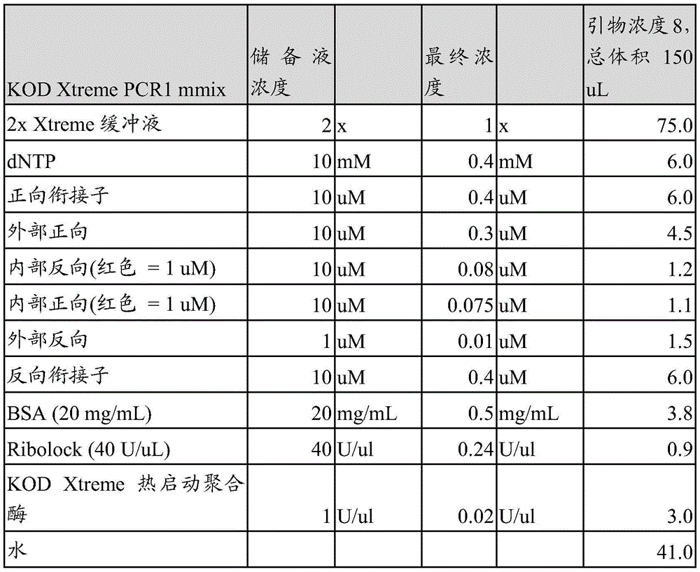


One of ordinary skill in the art will appreciate that a variety of other components may be used to provide the same functionality. Alternative components may be used as lysing agents, RT enzymes, rnases, primer sequences, and DNA polymerases, among others.
Example 3
This example provides experimental data showing that performing the reverse transcription step in a separate vessel from the amplification and ligation steps improves the natural pairing of amplicons.
When Reverse Transcription (RT) of single-cell mRNA was performed in the first vessel (droplet 1), between the first and second vessels (between droplets 1 and 2 (B/w)), or in the second vessel (droplet 2), experiments were performed to test the percentage of correctly paired amplicons in the library.
The method comprises the following steps:
and (3) testing conditions are as follows:
(i) RT in droplet 1: standard procedures were followed (v 2.1 droplet phage display).
(ii) Droplet 1 and RT after washing: the standard procedure for the preparation of drop 1 was followed, but instead of the subsequent RT incubation, the emulsion was broken and the beads were washed with 20% -pfo according to the standard procedure (drop 1 post-wash step). RT (standard conditions) was performed on the washed beads. After incubation, the sample was placed on a magnet and the beads were washed again according to standard procedures (drop 1 post-wash step). In KOD Xtreme for PCR1 TM (EMD Millipore) the washed beads were resuspended in the reaction mixture and the droplet 2 step was continued on DT-20. post-PCR 1 compliance targetAnd (5) performing quasi-procedure.
(iii) RT in droplet 2 (RT-PCR kit, SSIV and Titan): the standard procedure for the preparation of drop 1 was followed, but instead of the subsequent RT incubation, the emulsion was broken and the beads were washed with 20% -pfo according to the standard procedure (drop 1 post-wash step). Resuspend the washed beads in RT-PCR reaction mixtures (SuperScriptIV, titan or Quanta) and prepare the droplets for RT incubation and PCR1 cycles using DT-20 (following the recommended cycling conditions for each reaction mixture). Standard procedures were followed after PCR 1.
As a result: as shown in the table below, reverse transcription in droplet 1 increased the percentage of amplicons that were correctly paired and linked to another amplicon from the same cell. % DNA native refers to the fraction of heavy chain amplicons that are correctly paired with native light chain amplicons in the library. Purity refers to the fraction of natural and unnatural pairings in the library. As can be seen from the table, when RT was performed in droplet 1, 65% -77% of the amplicons in the library were correctly paired, while when RT was performed between droplets 1 and 2, 9% -32% of the amplicons were correctly paired, and when RT was performed in droplet 2, 23% -40% of the amplicons were correctly paired.
The unique CDRH3 was used to determine the overall recovery of the ligated amplicons in the library. RT in droplet 1 reduced the overall recovery.
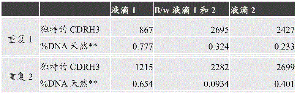
The data provided above show that performing the RT step in the first vessel increases the natural pairing of amplicons in the library produced by the methods described herein.
Example 4
This example describes the use of a photocleavable linker between the bead and the capture oligonucleotide to increase the yield and purity of the amplification product. The process of this example is similar to that described in example 1 with the changes detailed below.
In this example, custom mRNA capture beads were prepared by conjugating oligodT ssDNA to the beads. A photocleavable linker, such as the nitrobenzyl linker (modification code '/iSPPC/') provided by IDT, is located between the oligodT capture/priming sequence and the bead surface. To test the successful photo-lysis and release of ssDNA from the beads, the suspension of beads was exposed to 365nm UV light for six minutes. The suspension was then centrifuged to pellet the beads and the supernatant was assayed by Qubit (Thermo Fisher catalog No. Q10212) to determine the amount of ssDNA released from the beads.
Reagent: DBCO-modified 10 μ M diameter polystyrene beads (Creative Diagnostics catalog No. DNM-M006). Azide and photocleavable linker modified mRNA capture oligonucleotide (IDT):
5 '-Azide-iSPPC-TTTTTTTTTTTTTTTTTTTTTTTTT-3' (SEQ ID NO: 1). The droplets for EvaGreen produced oil (BioRad catalog No. 1864112).
The method comprises the following steps:
stock solutions of mRNA capture beads were prepared as follows:
1. 1000 ten thousand DBCO beads were washed 5 times in 500uL of 0.1X PBS +0.001% Tween-20
2. Transfer 800 ten thousand beads to a 0.5mL PCR tube
3. Centrifuging, sucking out supernatant
4. Resuspend beads in the following reaction mixture:

5. vortex the tube, wrap with aluminum foil, place on a rotator or shaker, incubate for one hour at 72C
6. After incubation, transfer from the coupling tube to a separate 0.5mL PCR tube
7. Centrifuged, the supernatant aspirated, resuspended in 0.5mL of 0.1X PBS +0.001%
8. Washing 3 times in 0.5mL of 0.1X TE +0.001% Tween-20
9. Photolysis/Qubit assays as described above were performed to determine conjugation yield and to test the successful release of oligodT from the bead surface
The same procedure as in example 1 was followed, except that step 4 of paragraph [0126] ('RT reaction mix') used 500 ten thousand custom beads instead of a 45uL bead stock, and steps 15 and 16 of paragraph [0144] ("RT product wash and set PCR 1") were replaced by the following steps:
1. adding more PCR1 mmix to the sample, up to 500uL
2. The same droplet apparatus as before was used, with the a.01 reservoir now filled with BioRad droplet generating oil for EvaGreen and the b.01 reservoir filled with water. B.11 connected to a syringe pump.
3. Half (250 uL) of the bead/PCR mixed suspension was loaded in each of the two reservoirs of the droplet device using a syringe pump to draw the sample into the sample loop. In B.15, an air gap of 1cm is maintained between the propulsion liquid and the sample.
A.10 and B.15 connected to a fluorinated 2R chip with 100um etch depth (Dolomite part No. 3200510)
5. The samples were run at a flow rate of 12uL/min for each water line (B.15) and at a flow rate of 180uL/min for the oil line (A.10).
6. The emulsion was collected in a 15mL conical tube kept on ice.
7. After the emulsion was produced and before aliquoting the emulsion into a PCR plate, the emulsion was exposed to 365nm UV light for 6 minutes, and the tube was rotated so that all parts of the emulsion were uniformly exposed.
By combining the UV cleavage step, the inventors observed an increase in the proportion of unique heavy chain sequences that reliably pair with the light chain from 71.1% to 80.6%.
This example shows that the number of heavy and light chain pairs recovered and the proportion of naturally paired heavy and light chains can be increased using the methods described above.
An informal sequence table:
1, SEQ ID NO: 5 '-Azide-iSPPC-TTTTTTTTTTTTTTTTTTTTTTTTTTT-3'.
It is understood that the examples and embodiments described herein are for illustrative purposes only and that various modifications are intended to be included within the scope of the appended claims after a careful reading of the disclosure. All publications, sequence accession numbers, patents, and patent applications cited herein are hereby incorporated by reference in their entirety for all purposes.
Claims (53)
Applications Claiming Priority (3)
| Application Number | Priority Date | Filing Date | Title |
|---|---|---|---|
| US202062964319P | 2020-01-22 | 2020-01-22 | |
| US62/964,319 | 2020-01-22 | ||
| PCT/US2021/014631 WO2021150903A1 (en) | 2020-01-22 | 2021-01-22 | High throughput linking of multiple transcripts |
Publications (1)
| Publication Number | Publication Date |
|---|---|
| CN115175999A true CN115175999A (en) | 2022-10-11 |
Family
ID=74672406
Family Applications (1)
| Application Number | Title | Priority Date | Filing Date |
|---|---|---|---|
| CN202180017456.7A Pending CN115175999A (en) | 2020-01-22 | 2021-01-22 | High throughput ligation of multiple transcripts |
Country Status (5)
| Country | Link |
|---|---|
| US (1) | US20230220376A1 (en) |
| EP (1) | EP4093869A1 (en) |
| JP (1) | JP2023511440A (en) |
| CN (1) | CN115175999A (en) |
| WO (1) | WO2021150903A1 (en) |
Citations (5)
| Publication number | Priority date | Publication date | Assignee | Title |
|---|---|---|---|---|
| CN102797044A (en) * | 2012-08-16 | 2012-11-28 | 北京诺兰信生化科技有限责任公司 | Fast and efficient construction method of normalized full-length cDNA library |
| US20150024959A1 (en) * | 2012-03-30 | 2015-01-22 | Hitachi, Ltd. | METHOD FOR AMPLIFYING cDNA DERIVED FROM TRACE AMOUNT OF SAMPLE |
| CN104619862A (en) * | 2012-06-15 | 2015-05-13 | 得克萨斯系统大学董事会 | High throughput sequencing of multiple transcripts of a single cell |
| CN108893464A (en) * | 2018-07-13 | 2018-11-27 | 广州华银医学检验中心有限公司 | A kind of construction method of immune group library high-throughput sequencing library |
| CN110684829A (en) * | 2018-07-05 | 2020-01-14 | 深圳华大智造科技有限公司 | A high-throughput single-cell transcriptome sequencing method and kit |
Family Cites Families (7)
| Publication number | Priority date | Publication date | Assignee | Title |
|---|---|---|---|---|
| DK3415619T3 (en) | 2011-04-28 | 2021-03-08 | Univ Leland Stanford Junior | IDENTIFICATION OF POLYNUCLEOTIDS ASSOCIATED WITH A SAMPLE |
| US9816088B2 (en) * | 2013-03-15 | 2017-11-14 | Abvitro Llc | Single cell bar-coding for antibody discovery |
| WO2015103339A1 (en) | 2013-12-30 | 2015-07-09 | Atreca, Inc. | Analysis of nucleic acids associated with single cells using nucleic acid barcodes |
| CA3021735A1 (en) * | 2016-04-19 | 2017-10-26 | President And Fellows Of Harvard College | Immobilization-based systems and methods for genetic analysis and other applications |
| US10550429B2 (en) * | 2016-12-22 | 2020-02-04 | 10X Genomics, Inc. | Methods and systems for processing polynucleotides |
| US20200216840A1 (en) * | 2017-07-27 | 2020-07-09 | Board Of Regents, The University Of Texas System | Amplification of paired protein-coding mrna sequences |
| AU2019227700B2 (en) * | 2018-03-01 | 2025-09-11 | University Of Kansas | Techniques for generating cell-based therapeutics using recombinant T cell receptor genes |
-
2021
- 2021-01-22 CN CN202180017456.7A patent/CN115175999A/en active Pending
- 2021-01-22 WO PCT/US2021/014631 patent/WO2021150903A1/en not_active Ceased
- 2021-01-22 US US17/759,091 patent/US20230220376A1/en active Pending
- 2021-01-22 EP EP21707056.4A patent/EP4093869A1/en not_active Withdrawn
- 2021-01-22 JP JP2022544751A patent/JP2023511440A/en active Pending
Patent Citations (5)
| Publication number | Priority date | Publication date | Assignee | Title |
|---|---|---|---|---|
| US20150024959A1 (en) * | 2012-03-30 | 2015-01-22 | Hitachi, Ltd. | METHOD FOR AMPLIFYING cDNA DERIVED FROM TRACE AMOUNT OF SAMPLE |
| CN104619862A (en) * | 2012-06-15 | 2015-05-13 | 得克萨斯系统大学董事会 | High throughput sequencing of multiple transcripts of a single cell |
| CN102797044A (en) * | 2012-08-16 | 2012-11-28 | 北京诺兰信生化科技有限责任公司 | Fast and efficient construction method of normalized full-length cDNA library |
| CN110684829A (en) * | 2018-07-05 | 2020-01-14 | 深圳华大智造科技有限公司 | A high-throughput single-cell transcriptome sequencing method and kit |
| CN108893464A (en) * | 2018-07-13 | 2018-11-27 | 广州华银医学检验中心有限公司 | A kind of construction method of immune group library high-throughput sequencing library |
Non-Patent Citations (1)
| Title |
|---|
| JONATHAN R MCDANIEL: ""Ultra-high-throughput sequencing of the immune receptor repertoire from millions of lymphocytes"", 《NATURE PROTOCOLS》, 4 February 2016 (2016-02-04), pages 429 - 442 * |
Also Published As
| Publication number | Publication date |
|---|---|
| US20230220376A1 (en) | 2023-07-13 |
| EP4093869A1 (en) | 2022-11-30 |
| JP2023511440A (en) | 2023-03-17 |
| WO2021150903A1 (en) | 2021-07-29 |
Similar Documents
| Publication | Publication Date | Title |
|---|---|---|
| US20220389471A1 (en) | Analysis of nucleic acids associated with single cells using nucleic acid barcodes | |
| CN110431233B (en) | System and method for massively parallel combinatorial analysis of single cells | |
| AU2016348439B2 (en) | Combinatorial sets of nucleic acid barcodes for analysis of nucleic acids associated with single cells | |
| EP3268462B1 (en) | Genotype and phenotype coupling | |
| EP3980537A2 (en) | Methods of barcoding nucleic acid for detection and sequencing | |
| JP2009284834A (en) | Large-scale parallel nucleic acid analysis method | |
| US20210032677A1 (en) | Methods to Improve the Sequencing of Polynucleotides with Barcodes Using Circularisation and Truncation of Template | |
| JP7667739B2 (en) | Compositions and methods for preparing nucleic acid sequencing libraries using CRISPR/CAS9 immobilized on a solid support | |
| CN115175999A (en) | High throughput ligation of multiple transcripts | |
| WO2024098076A1 (en) | Magnetic bead cell pelleting during in situ cell library preparation and/or barcoding | |
| US20250147038A1 (en) | Compositions and methods for characterizing binding characteristics of antigen binding molecules from single cells | |
| US20230349901A1 (en) | Methods For Identification of Cognate Pairs of Ligands and Receptors | |
| KR20250094720A (en) | Method for amplifying complementary DNA strands |
Legal Events
| Date | Code | Title | Description |
|---|---|---|---|
| PB01 | Publication | ||
| PB01 | Publication | ||
| SE01 | Entry into force of request for substantive examination | ||
| SE01 | Entry into force of request for substantive examination |