CN115103647A - Systems and methods for optical interrogation of ablative lesions - Google Patents
Systems and methods for optical interrogation of ablative lesions Download PDFInfo
- Publication number
- CN115103647A CN115103647A CN202180013945.5A CN202180013945A CN115103647A CN 115103647 A CN115103647 A CN 115103647A CN 202180013945 A CN202180013945 A CN 202180013945A CN 115103647 A CN115103647 A CN 115103647A
- Authority
- CN
- China
- Prior art keywords
- tissue
- energy
- ablation
- light
- sensor
- Prior art date
- Legal status (The legal status is an assumption and is not a legal conclusion. Google has not performed a legal analysis and makes no representation as to the accuracy of the status listed.)
- Pending
Links
- 230000003287 optical effect Effects 0.000 title claims abstract description 87
- 238000000034 method Methods 0.000 title claims description 40
- 230000003902 lesion Effects 0.000 title claims description 28
- 229930027945 nicotinamide-adenine dinucleotide Natural products 0.000 claims abstract description 136
- BOPGDPNILDQYTO-NNYOXOHSSA-N nicotinamide-adenine dinucleotide Chemical compound C1=CCC(C(=O)N)=CN1[C@H]1[C@H](O)[C@H](O)[C@@H](COP(O)(=O)OP(O)(=O)OC[C@@H]2[C@H]([C@@H](O)[C@@H](O2)N2C3=NC=NC(N)=C3N=C2)O)O1 BOPGDPNILDQYTO-NNYOXOHSSA-N 0.000 claims abstract description 120
- 239000013307 optical fiber Substances 0.000 claims abstract description 73
- 229950006238 nadide Drugs 0.000 claims abstract description 16
- 229910052739 hydrogen Inorganic materials 0.000 claims abstract description 14
- 239000001257 hydrogen Substances 0.000 claims abstract description 14
- -1 nicotinamide adenine dinucleotide hydrogen Chemical class 0.000 claims abstract description 14
- 238000002679 ablation Methods 0.000 claims description 189
- 230000007423 decrease Effects 0.000 claims description 33
- 238000004891 communication Methods 0.000 claims description 22
- 238000012544 monitoring process Methods 0.000 claims description 17
- 102000008186 Collagen Human genes 0.000 claims description 14
- 108010035532 Collagen Proteins 0.000 claims description 14
- 229920001436 collagen Polymers 0.000 claims description 14
- 230000000541 pulsatile effect Effects 0.000 claims description 14
- 238000002604 ultrasonography Methods 0.000 claims description 14
- 238000004520 electroporation Methods 0.000 claims description 13
- 239000000126 substance Substances 0.000 claims description 11
- 230000002438 mitochondrial effect Effects 0.000 claims description 10
- 238000003384 imaging method Methods 0.000 claims description 4
- 230000003176 fibrotic effect Effects 0.000 claims description 3
- 230000002688 persistence Effects 0.000 claims description 3
- 210000001519 tissue Anatomy 0.000 description 200
- 238000001228 spectrum Methods 0.000 description 15
- 206010003658 Atrial Fibrillation Diseases 0.000 description 14
- 238000012384 transportation and delivery Methods 0.000 description 13
- 230000002262 irrigation Effects 0.000 description 11
- 238000003973 irrigation Methods 0.000 description 11
- 210000003492 pulmonary vein Anatomy 0.000 description 9
- 238000010317 ablation therapy Methods 0.000 description 8
- 238000013507 mapping Methods 0.000 description 8
- 238000005286 illumination Methods 0.000 description 7
- 210000004165 myocardium Anatomy 0.000 description 7
- 230000000694 effects Effects 0.000 description 6
- 239000000835 fiber Substances 0.000 description 6
- 230000006870 function Effects 0.000 description 6
- 230000015572 biosynthetic process Effects 0.000 description 5
- 230000006378 damage Effects 0.000 description 5
- 210000005003 heart tissue Anatomy 0.000 description 5
- 230000004044 response Effects 0.000 description 5
- 238000011010 flushing procedure Methods 0.000 description 4
- 238000005259 measurement Methods 0.000 description 4
- 238000012545 processing Methods 0.000 description 4
- 239000003999 initiator Substances 0.000 description 3
- 230000008569 process Effects 0.000 description 3
- 238000001356 surgical procedure Methods 0.000 description 3
- 238000002560 therapeutic procedure Methods 0.000 description 3
- 238000010521 absorption reaction Methods 0.000 description 2
- OIRDTQYFTABQOQ-KQYNXXCUSA-N adenosine Chemical compound C1=NC=2C(N)=NC=NC=2N1[C@@H]1O[C@H](CO)[C@@H](O)[C@H]1O OIRDTQYFTABQOQ-KQYNXXCUSA-N 0.000 description 2
- 210000003484 anatomy Anatomy 0.000 description 2
- 230000001746 atrial effect Effects 0.000 description 2
- 230000008901 benefit Effects 0.000 description 2
- 239000000560 biocompatible material Substances 0.000 description 2
- 230000005540 biological transmission Effects 0.000 description 2
- 239000008280 blood Substances 0.000 description 2
- 210000004369 blood Anatomy 0.000 description 2
- 210000004027 cell Anatomy 0.000 description 2
- 230000005779 cell damage Effects 0.000 description 2
- 238000004590 computer program Methods 0.000 description 2
- 238000010586 diagram Methods 0.000 description 2
- 230000005284 excitation Effects 0.000 description 2
- 239000012530 fluid Substances 0.000 description 2
- 238000002955 isolation Methods 0.000 description 2
- 239000000463 material Substances 0.000 description 2
- 239000012528 membrane Substances 0.000 description 2
- 230000002503 metabolic effect Effects 0.000 description 2
- 238000012986 modification Methods 0.000 description 2
- 230000004048 modification Effects 0.000 description 2
- BASFCYQUMIYNBI-UHFFFAOYSA-N platinum Chemical compound [Pt] BASFCYQUMIYNBI-UHFFFAOYSA-N 0.000 description 2
- 230000009467 reduction Effects 0.000 description 2
- 210000005245 right atrium Anatomy 0.000 description 2
- 230000001225 therapeutic effect Effects 0.000 description 2
- 210000002620 vena cava superior Anatomy 0.000 description 2
- 230000000007 visual effect Effects 0.000 description 2
- 239000002126 C01EB10 - Adenosine Substances 0.000 description 1
- 208000005189 Embolism Diseases 0.000 description 1
- 206010016654 Fibrosis Diseases 0.000 description 1
- 208000010496 Heart Arrest Diseases 0.000 description 1
- 241000699670 Mus sp. Species 0.000 description 1
- 238000011887 Necropsy Methods 0.000 description 1
- FAPWRFPIFSIZLT-UHFFFAOYSA-M Sodium chloride Chemical compound [Na+].[Cl-] FAPWRFPIFSIZLT-UHFFFAOYSA-M 0.000 description 1
- 208000006011 Stroke Diseases 0.000 description 1
- 241000282887 Suidae Species 0.000 description 1
- 208000001435 Thromboembolism Diseases 0.000 description 1
- RTAQQCXQSZGOHL-UHFFFAOYSA-N Titanium Chemical compound [Ti] RTAQQCXQSZGOHL-UHFFFAOYSA-N 0.000 description 1
- 230000009692 acute damage Effects 0.000 description 1
- 229960005305 adenosine Drugs 0.000 description 1
- 238000004458 analytical method Methods 0.000 description 1
- 230000006907 apoptotic process Effects 0.000 description 1
- 206010003119 arrhythmia Diseases 0.000 description 1
- 238000003556 assay Methods 0.000 description 1
- 206010003668 atrial tachycardia Diseases 0.000 description 1
- 230000000747 cardiac effect Effects 0.000 description 1
- 210000004413 cardiac myocyte Anatomy 0.000 description 1
- 238000013153 catheter ablation Methods 0.000 description 1
- 210000000170 cell membrane Anatomy 0.000 description 1
- 230000036755 cellular response Effects 0.000 description 1
- 230000008859 change Effects 0.000 description 1
- 238000004883 computer application Methods 0.000 description 1
- 230000003247 decreasing effect Effects 0.000 description 1
- 238000002405 diagnostic procedure Methods 0.000 description 1
- 201000010099 disease Diseases 0.000 description 1
- 208000037265 diseases, disorders, signs and symptoms Diseases 0.000 description 1
- 229940079593 drug Drugs 0.000 description 1
- 239000003814 drug Substances 0.000 description 1
- 230000005684 electric field Effects 0.000 description 1
- 238000005516 engineering process Methods 0.000 description 1
- 230000004761 fibrosis Effects 0.000 description 1
- 238000001506 fluorescence spectroscopy Methods 0.000 description 1
- 238000002189 fluorescence spectrum Methods 0.000 description 1
- 210000002064 heart cell Anatomy 0.000 description 1
- 230000001771 impaired effect Effects 0.000 description 1
- 238000001802 infusion Methods 0.000 description 1
- 230000001678 irradiating effect Effects 0.000 description 1
- 230000002427 irreversible effect Effects 0.000 description 1
- 210000005246 left atrium Anatomy 0.000 description 1
- 229910052751 metal Inorganic materials 0.000 description 1
- 239000002184 metal Substances 0.000 description 1
- 239000000203 mixture Substances 0.000 description 1
- 210000003205 muscle Anatomy 0.000 description 1
- 230000003387 muscular Effects 0.000 description 1
- 230000002107 myocardial effect Effects 0.000 description 1
- 229910052697 platinum Inorganic materials 0.000 description 1
- HWLDNSXPUQTBOD-UHFFFAOYSA-N platinum-iridium alloy Chemical compound [Ir].[Pt] HWLDNSXPUQTBOD-UHFFFAOYSA-N 0.000 description 1
- 239000011148 porous material Substances 0.000 description 1
- 230000002035 prolonged effect Effects 0.000 description 1
- 230000005180 public health Effects 0.000 description 1
- 238000012216 screening Methods 0.000 description 1
- 239000011780 sodium chloride Substances 0.000 description 1
- 239000007787 solid Substances 0.000 description 1
- 230000003595 spectral effect Effects 0.000 description 1
- 229910001220 stainless steel Inorganic materials 0.000 description 1
- 239000010935 stainless steel Substances 0.000 description 1
- 230000004936 stimulating effect Effects 0.000 description 1
- 230000000638 stimulation Effects 0.000 description 1
- 230000002459 sustained effect Effects 0.000 description 1
- 229910052719 titanium Inorganic materials 0.000 description 1
- 239000010936 titanium Substances 0.000 description 1
- 238000012546 transfer Methods 0.000 description 1
- 230000001960 triggered effect Effects 0.000 description 1
- 210000003462 vein Anatomy 0.000 description 1
Images


Classifications
-
- A—HUMAN NECESSITIES
- A61—MEDICAL OR VETERINARY SCIENCE; HYGIENE
- A61B—DIAGNOSIS; SURGERY; IDENTIFICATION
- A61B18/00—Surgical instruments, devices or methods for transferring non-mechanical forms of energy to or from the body
- A61B18/18—Surgical instruments, devices or methods for transferring non-mechanical forms of energy to or from the body by applying electromagnetic radiation, e.g. microwaves
- A61B18/20—Surgical instruments, devices or methods for transferring non-mechanical forms of energy to or from the body by applying electromagnetic radiation, e.g. microwaves using laser
- A61B18/22—Surgical instruments, devices or methods for transferring non-mechanical forms of energy to or from the body by applying electromagnetic radiation, e.g. microwaves using laser the beam being directed along or through a flexible conduit, e.g. an optical fibre; Couplings or hand-pieces therefor
- A61B18/24—Surgical instruments, devices or methods for transferring non-mechanical forms of energy to or from the body by applying electromagnetic radiation, e.g. microwaves using laser the beam being directed along or through a flexible conduit, e.g. an optical fibre; Couplings or hand-pieces therefor with a catheter
-
- A—HUMAN NECESSITIES
- A61—MEDICAL OR VETERINARY SCIENCE; HYGIENE
- A61B—DIAGNOSIS; SURGERY; IDENTIFICATION
- A61B17/00—Surgical instruments, devices or methods
- A61B17/32—Surgical cutting instruments
- A61B17/320068—Surgical cutting instruments using mechanical vibrations, e.g. ultrasonic
-
- A—HUMAN NECESSITIES
- A61—MEDICAL OR VETERINARY SCIENCE; HYGIENE
- A61B—DIAGNOSIS; SURGERY; IDENTIFICATION
- A61B18/00—Surgical instruments, devices or methods for transferring non-mechanical forms of energy to or from the body
- A61B18/02—Surgical instruments, devices or methods for transferring non-mechanical forms of energy to or from the body by cooling, e.g. cryogenic techniques
-
- A—HUMAN NECESSITIES
- A61—MEDICAL OR VETERINARY SCIENCE; HYGIENE
- A61B—DIAGNOSIS; SURGERY; IDENTIFICATION
- A61B18/00—Surgical instruments, devices or methods for transferring non-mechanical forms of energy to or from the body
- A61B18/04—Surgical instruments, devices or methods for transferring non-mechanical forms of energy to or from the body by heating
- A61B18/08—Surgical instruments, devices or methods for transferring non-mechanical forms of energy to or from the body by heating by means of electrically-heated probes
-
- A—HUMAN NECESSITIES
- A61—MEDICAL OR VETERINARY SCIENCE; HYGIENE
- A61B—DIAGNOSIS; SURGERY; IDENTIFICATION
- A61B18/00—Surgical instruments, devices or methods for transferring non-mechanical forms of energy to or from the body
- A61B18/04—Surgical instruments, devices or methods for transferring non-mechanical forms of energy to or from the body by heating
- A61B18/12—Surgical instruments, devices or methods for transferring non-mechanical forms of energy to or from the body by heating by passing a current through the tissue to be heated, e.g. high-frequency current
- A61B18/14—Probes or electrodes therefor
- A61B18/1492—Probes or electrodes therefor having a flexible, catheter-like structure, e.g. for heart ablation
-
- A—HUMAN NECESSITIES
- A61—MEDICAL OR VETERINARY SCIENCE; HYGIENE
- A61B—DIAGNOSIS; SURGERY; IDENTIFICATION
- A61B18/00—Surgical instruments, devices or methods for transferring non-mechanical forms of energy to or from the body
- A61B18/18—Surgical instruments, devices or methods for transferring non-mechanical forms of energy to or from the body by applying electromagnetic radiation, e.g. microwaves
- A61B18/1815—Surgical instruments, devices or methods for transferring non-mechanical forms of energy to or from the body by applying electromagnetic radiation, e.g. microwaves using microwaves
-
- A—HUMAN NECESSITIES
- A61—MEDICAL OR VETERINARY SCIENCE; HYGIENE
- A61B—DIAGNOSIS; SURGERY; IDENTIFICATION
- A61B5/00—Measuring for diagnostic purposes; Identification of persons
- A61B5/0059—Measuring for diagnostic purposes; Identification of persons using light, e.g. diagnosis by transillumination, diascopy, fluorescence
- A61B5/0071—Measuring for diagnostic purposes; Identification of persons using light, e.g. diagnosis by transillumination, diascopy, fluorescence by measuring fluorescence emission
-
- A—HUMAN NECESSITIES
- A61—MEDICAL OR VETERINARY SCIENCE; HYGIENE
- A61B—DIAGNOSIS; SURGERY; IDENTIFICATION
- A61B5/00—Measuring for diagnostic purposes; Identification of persons
- A61B5/0059—Measuring for diagnostic purposes; Identification of persons using light, e.g. diagnosis by transillumination, diascopy, fluorescence
- A61B5/0082—Measuring for diagnostic purposes; Identification of persons using light, e.g. diagnosis by transillumination, diascopy, fluorescence adapted for particular medical purposes
- A61B5/0084—Measuring for diagnostic purposes; Identification of persons using light, e.g. diagnosis by transillumination, diascopy, fluorescence adapted for particular medical purposes for introduction into the body, e.g. by catheters
-
- A—HUMAN NECESSITIES
- A61—MEDICAL OR VETERINARY SCIENCE; HYGIENE
- A61N—ELECTROTHERAPY; MAGNETOTHERAPY; RADIATION THERAPY; ULTRASOUND THERAPY
- A61N1/00—Electrotherapy; Circuits therefor
- A61N1/18—Applying electric currents by contact electrodes
- A61N1/32—Applying electric currents by contact electrodes alternating or intermittent currents
- A61N1/327—Applying electric currents by contact electrodes alternating or intermittent currents for enhancing the absorption properties of tissue, e.g. by electroporation
-
- A—HUMAN NECESSITIES
- A61—MEDICAL OR VETERINARY SCIENCE; HYGIENE
- A61B—DIAGNOSIS; SURGERY; IDENTIFICATION
- A61B18/00—Surgical instruments, devices or methods for transferring non-mechanical forms of energy to or from the body
- A61B18/04—Surgical instruments, devices or methods for transferring non-mechanical forms of energy to or from the body by heating
- A61B18/06—Surgical instruments, devices or methods for transferring non-mechanical forms of energy to or from the body by heating caused by chemical reaction, e.g. moxaburners
-
- A—HUMAN NECESSITIES
- A61—MEDICAL OR VETERINARY SCIENCE; HYGIENE
- A61B—DIAGNOSIS; SURGERY; IDENTIFICATION
- A61B18/00—Surgical instruments, devices or methods for transferring non-mechanical forms of energy to or from the body
- A61B18/04—Surgical instruments, devices or methods for transferring non-mechanical forms of energy to or from the body by heating
- A61B18/08—Surgical instruments, devices or methods for transferring non-mechanical forms of energy to or from the body by heating by means of electrically-heated probes
- A61B18/082—Probes or electrodes therefor
-
- A—HUMAN NECESSITIES
- A61—MEDICAL OR VETERINARY SCIENCE; HYGIENE
- A61B—DIAGNOSIS; SURGERY; IDENTIFICATION
- A61B17/00—Surgical instruments, devices or methods
- A61B2017/00017—Electrical control of surgical instruments
- A61B2017/00022—Sensing or detecting at the treatment site
- A61B2017/00057—Light
- A61B2017/00061—Light spectrum
-
- A—HUMAN NECESSITIES
- A61—MEDICAL OR VETERINARY SCIENCE; HYGIENE
- A61B—DIAGNOSIS; SURGERY; IDENTIFICATION
- A61B17/00—Surgical instruments, devices or methods
- A61B2017/00017—Electrical control of surgical instruments
- A61B2017/00115—Electrical control of surgical instruments with audible or visual output
- A61B2017/00128—Electrical control of surgical instruments with audible or visual output related to intensity or progress of surgical action
-
- A—HUMAN NECESSITIES
- A61—MEDICAL OR VETERINARY SCIENCE; HYGIENE
- A61B—DIAGNOSIS; SURGERY; IDENTIFICATION
- A61B17/00—Surgical instruments, devices or methods
- A61B17/32—Surgical cutting instruments
- A61B17/320068—Surgical cutting instruments using mechanical vibrations, e.g. ultrasonic
- A61B2017/320069—Surgical cutting instruments using mechanical vibrations, e.g. ultrasonic for ablating tissue
-
- A—HUMAN NECESSITIES
- A61—MEDICAL OR VETERINARY SCIENCE; HYGIENE
- A61B—DIAGNOSIS; SURGERY; IDENTIFICATION
- A61B18/00—Surgical instruments, devices or methods for transferring non-mechanical forms of energy to or from the body
- A61B2018/00005—Cooling or heating of the probe or tissue immediately surrounding the probe
- A61B2018/00011—Cooling or heating of the probe or tissue immediately surrounding the probe with fluids
- A61B2018/00029—Cooling or heating of the probe or tissue immediately surrounding the probe with fluids open
-
- A—HUMAN NECESSITIES
- A61—MEDICAL OR VETERINARY SCIENCE; HYGIENE
- A61B—DIAGNOSIS; SURGERY; IDENTIFICATION
- A61B18/00—Surgical instruments, devices or methods for transferring non-mechanical forms of energy to or from the body
- A61B2018/00053—Mechanical features of the instrument of device
- A61B2018/0016—Energy applicators arranged in a two- or three dimensional array
-
- A—HUMAN NECESSITIES
- A61—MEDICAL OR VETERINARY SCIENCE; HYGIENE
- A61B—DIAGNOSIS; SURGERY; IDENTIFICATION
- A61B18/00—Surgical instruments, devices or methods for transferring non-mechanical forms of energy to or from the body
- A61B2018/00053—Mechanical features of the instrument of device
- A61B2018/00214—Expandable means emitting energy, e.g. by elements carried thereon
- A61B2018/0022—Balloons
-
- A—HUMAN NECESSITIES
- A61—MEDICAL OR VETERINARY SCIENCE; HYGIENE
- A61B—DIAGNOSIS; SURGERY; IDENTIFICATION
- A61B18/00—Surgical instruments, devices or methods for transferring non-mechanical forms of energy to or from the body
- A61B2018/00053—Mechanical features of the instrument of device
- A61B2018/00214—Expandable means emitting energy, e.g. by elements carried thereon
- A61B2018/00267—Expandable means emitting energy, e.g. by elements carried thereon having a basket shaped structure
-
- A—HUMAN NECESSITIES
- A61—MEDICAL OR VETERINARY SCIENCE; HYGIENE
- A61B—DIAGNOSIS; SURGERY; IDENTIFICATION
- A61B18/00—Surgical instruments, devices or methods for transferring non-mechanical forms of energy to or from the body
- A61B2018/00315—Surgical instruments, devices or methods for transferring non-mechanical forms of energy to or from the body for treatment of particular body parts
- A61B2018/00345—Vascular system
- A61B2018/00351—Heart
- A61B2018/00375—Ostium, e.g. ostium of pulmonary vein or artery
-
- A—HUMAN NECESSITIES
- A61—MEDICAL OR VETERINARY SCIENCE; HYGIENE
- A61B—DIAGNOSIS; SURGERY; IDENTIFICATION
- A61B18/00—Surgical instruments, devices or methods for transferring non-mechanical forms of energy to or from the body
- A61B2018/00571—Surgical instruments, devices or methods for transferring non-mechanical forms of energy to or from the body for achieving a particular surgical effect
- A61B2018/00577—Ablation
-
- A—HUMAN NECESSITIES
- A61—MEDICAL OR VETERINARY SCIENCE; HYGIENE
- A61B—DIAGNOSIS; SURGERY; IDENTIFICATION
- A61B18/00—Surgical instruments, devices or methods for transferring non-mechanical forms of energy to or from the body
- A61B2018/00571—Surgical instruments, devices or methods for transferring non-mechanical forms of energy to or from the body for achieving a particular surgical effect
- A61B2018/00613—Irreversible electroporation
-
- A—HUMAN NECESSITIES
- A61—MEDICAL OR VETERINARY SCIENCE; HYGIENE
- A61B—DIAGNOSIS; SURGERY; IDENTIFICATION
- A61B18/00—Surgical instruments, devices or methods for transferring non-mechanical forms of energy to or from the body
- A61B2018/00636—Sensing and controlling the application of energy
- A61B2018/00773—Sensed parameters
- A61B2018/00779—Power or energy
- A61B2018/00785—Reflected power
-
- A—HUMAN NECESSITIES
- A61—MEDICAL OR VETERINARY SCIENCE; HYGIENE
- A61B—DIAGNOSIS; SURGERY; IDENTIFICATION
- A61B18/00—Surgical instruments, devices or methods for transferring non-mechanical forms of energy to or from the body
- A61B18/02—Surgical instruments, devices or methods for transferring non-mechanical forms of energy to or from the body by cooling, e.g. cryogenic techniques
- A61B2018/0212—Surgical instruments, devices or methods for transferring non-mechanical forms of energy to or from the body by cooling, e.g. cryogenic techniques using an instrument inserted into a body lumen, e.g. catheter
-
- A—HUMAN NECESSITIES
- A61—MEDICAL OR VETERINARY SCIENCE; HYGIENE
- A61B—DIAGNOSIS; SURGERY; IDENTIFICATION
- A61B18/00—Surgical instruments, devices or methods for transferring non-mechanical forms of energy to or from the body
- A61B18/04—Surgical instruments, devices or methods for transferring non-mechanical forms of energy to or from the body by heating
- A61B18/12—Surgical instruments, devices or methods for transferring non-mechanical forms of energy to or from the body by heating by passing a current through the tissue to be heated, e.g. high-frequency current
- A61B18/14—Probes or electrodes therefor
- A61B2018/1467—Probes or electrodes therefor using more than two electrodes on a single probe
-
- A—HUMAN NECESSITIES
- A61—MEDICAL OR VETERINARY SCIENCE; HYGIENE
- A61B—DIAGNOSIS; SURGERY; IDENTIFICATION
- A61B18/00—Surgical instruments, devices or methods for transferring non-mechanical forms of energy to or from the body
- A61B18/18—Surgical instruments, devices or methods for transferring non-mechanical forms of energy to or from the body by applying electromagnetic radiation, e.g. microwaves
- A61B18/1815—Surgical instruments, devices or methods for transferring non-mechanical forms of energy to or from the body by applying electromagnetic radiation, e.g. microwaves using microwaves
- A61B2018/1861—Surgical instruments, devices or methods for transferring non-mechanical forms of energy to or from the body by applying electromagnetic radiation, e.g. microwaves using microwaves with an instrument inserted into a body lumen or cavity, e.g. a catheter
-
- A—HUMAN NECESSITIES
- A61—MEDICAL OR VETERINARY SCIENCE; HYGIENE
- A61B—DIAGNOSIS; SURGERY; IDENTIFICATION
- A61B18/00—Surgical instruments, devices or methods for transferring non-mechanical forms of energy to or from the body
- A61B18/18—Surgical instruments, devices or methods for transferring non-mechanical forms of energy to or from the body by applying electromagnetic radiation, e.g. microwaves
- A61B18/20—Surgical instruments, devices or methods for transferring non-mechanical forms of energy to or from the body by applying electromagnetic radiation, e.g. microwaves using laser
- A61B18/22—Surgical instruments, devices or methods for transferring non-mechanical forms of energy to or from the body by applying electromagnetic radiation, e.g. microwaves using laser the beam being directed along or through a flexible conduit, e.g. an optical fibre; Couplings or hand-pieces therefor
- A61B2018/2205—Characteristics of fibres
- A61B2018/2211—Plurality of fibres
-
- A—HUMAN NECESSITIES
- A61—MEDICAL OR VETERINARY SCIENCE; HYGIENE
- A61B—DIAGNOSIS; SURGERY; IDENTIFICATION
- A61B34/00—Computer-aided surgery; Manipulators or robots specially adapted for use in surgery
- A61B34/20—Surgical navigation systems; Devices for tracking or guiding surgical instruments, e.g. for frameless stereotaxis
- A61B2034/2046—Tracking techniques
- A61B2034/2051—Electromagnetic tracking systems
-
- A—HUMAN NECESSITIES
- A61—MEDICAL OR VETERINARY SCIENCE; HYGIENE
- A61B—DIAGNOSIS; SURGERY; IDENTIFICATION
- A61B90/00—Instruments, implements or accessories specially adapted for surgery or diagnosis and not covered by any of the groups A61B1/00 - A61B50/00, e.g. for luxation treatment or for protecting wound edges
- A61B90/36—Image-producing devices or illumination devices not otherwise provided for
- A61B90/37—Surgical systems with images on a monitor during operation
- A61B2090/378—Surgical systems with images on a monitor during operation using ultrasound
- A61B2090/3782—Surgical systems with images on a monitor during operation using ultrasound transmitter or receiver in catheter or minimal invasive instrument
- A61B2090/3784—Surgical systems with images on a monitor during operation using ultrasound transmitter or receiver in catheter or minimal invasive instrument both receiver and transmitter being in the instrument or receiver being also transmitter
-
- A—HUMAN NECESSITIES
- A61—MEDICAL OR VETERINARY SCIENCE; HYGIENE
- A61B—DIAGNOSIS; SURGERY; IDENTIFICATION
- A61B2218/00—Details of surgical instruments, devices or methods for transferring non-mechanical forms of energy to or from the body
- A61B2218/001—Details of surgical instruments, devices or methods for transferring non-mechanical forms of energy to or from the body having means for irrigation and/or aspiration of substances to and/or from the surgical site
- A61B2218/002—Irrigation
-
- A—HUMAN NECESSITIES
- A61—MEDICAL OR VETERINARY SCIENCE; HYGIENE
- A61B—DIAGNOSIS; SURGERY; IDENTIFICATION
- A61B34/00—Computer-aided surgery; Manipulators or robots specially adapted for use in surgery
- A61B34/20—Surgical navigation systems; Devices for tracking or guiding surgical instruments, e.g. for frameless stereotaxis
Landscapes
- Health & Medical Sciences (AREA)
- Life Sciences & Earth Sciences (AREA)
- Surgery (AREA)
- Engineering & Computer Science (AREA)
- Veterinary Medicine (AREA)
- Biomedical Technology (AREA)
- Public Health (AREA)
- General Health & Medical Sciences (AREA)
- Animal Behavior & Ethology (AREA)
- Nuclear Medicine, Radiotherapy & Molecular Imaging (AREA)
- Heart & Thoracic Surgery (AREA)
- Medical Informatics (AREA)
- Molecular Biology (AREA)
- Physics & Mathematics (AREA)
- Otolaryngology (AREA)
- Biophysics (AREA)
- Electromagnetism (AREA)
- Plasma & Fusion (AREA)
- Optics & Photonics (AREA)
- Cardiology (AREA)
- Radiology & Medical Imaging (AREA)
- Pathology (AREA)
- Dentistry (AREA)
- Mechanical Engineering (AREA)
- Investigating, Analyzing Materials By Fluorescence Or Luminescence (AREA)
- Surgical Instruments (AREA)
- Laser Surgery Devices (AREA)
- Endoscopes (AREA)
Abstract
在一些实施例中,一种用于光学组织询问的系统包括导管,其具有以阵列部署在导管的远端处的多个电极,所述多个电极被配置为向组织递送消融能量;以及一根或多根光纤,延伸穿过导管以将来自光源的光递送到组织并将包括烟酰胺腺嘌呤二核苷酸氢(NADH)荧光的光学信息从组织递送到传感器,其中所述多个电极中的每个电极与所述一根或多根光纤中的至少一根光纤相关联。
In some embodiments, a system for optical tissue interrogation includes a catheter having a plurality of electrodes deployed in an array at a distal end of the catheter, the plurality of electrodes configured to deliver ablative energy to tissue; and a one or more optical fibers extending through the catheter to deliver light from the light source to the tissue and deliver optical information including nicotinamide adenine dinucleotide hydrogen (NADH) fluorescence from the tissue to the sensor, wherein the plurality of electrodes Each electrode in is associated with at least one optical fiber of the one or more optical fibers.
Description
相关申请Related applications
本申请要求于2020年1月8日提交的美国临时申请序列No.62/958,419和2021年1月8日提交的美国实用程序申请序列No.17/145,188的权益和优先权,它们通过引用整体并入本文。This application claims the benefit of and priority to US Provisional Application Serial No. 62/958,419, filed January 8, 2020, and US Utility Application Serial No. 17/145,188, filed January 8, 2021, which are incorporated by reference in their entirety Incorporated herein.
技术领域technical field
本公开一般而言涉及消融和光学组织询问系统和方法,以光学询问组织以评估能量递送到组织的影响并评估导管和组织之间的接触质量。The present disclosure generally relates to ablation and optical tissue interrogation systems and methods to optically interrogate tissue to assess the impact of energy delivery to the tissue and to assess the quality of contact between a catheter and tissue.
背景技术Background technique
心房颤动(AF)是世界上最常见的持续性心律失常,目前影响数百万人。在美国,预计到2050年AF将影响1000万人。AF与增加的死亡率、发病率和受损的生活质量相关联,并且是中风的独立风险因素。发展的AF的实质终生的风险突显疾病的公共卫生负担,仅仅在美国,这一疾病的年治疗费用就超过了70亿美元。Atrial fibrillation (AF) is the most common sustained cardiac arrhythmia in the world, currently affecting millions of people. In the United States, AF is projected to affect 10 million people by 2050. AF is associated with increased mortality, morbidity, and impaired quality of life, and is an independent risk factor for stroke. The substantial lifetime risk of developing AF highlights the public health burden of the disease, with annual treatment costs of over $7 billion in the United States alone.
已知AF患者的大多数发作是由源自延伸到肺静脉(PV)的肌袖内的病灶电活动引发的。心房颤动也可以由上腔静脉或其它心房结构(即,心脏的传导系统内的其它心脏组织)内的病灶活动引发。这些病灶引发物还可以造成由可重入的电活动(或转子)驱动的心房心动过速,然后可能将其分裂成作为心房颤动的特点的多个电子小波。此外,长时间的AF可以造成心脏细胞膜的功能改变,并且这些变化进一步延续心房颤动。Most episodes in AF patients are known to be triggered by focal electrical activity originating within the muscular sleeve extending into the pulmonary vein (PV). Atrial fibrillation can also be caused by focal activity within the superior vena cava or other atrial structures (ie, other cardiac tissue within the conduction system of the heart). These focal initiators can also cause atrial tachycardia driven by reentrant electrical activity (or rotors), which may then split into the multiple electronic wavelets characteristic of atrial fibrillation. In addition, prolonged AF can cause functional changes in cardiac cell membranes, and these changes further perpetuate atrial fibrillation.
消融系统被医生用来治疗心房颤动。医师使用导管来指引能量,以或者破坏病灶引发物或者形成将引发物与心脏的剩余组织和传导系统隔离的电隔离线。后一种技术通常用于所谓的肺静脉隔离(PVI)。但是,AF消融手术的成功率保持相对停滞,术后一年复发的估计高达30%至50%。导管消融后复发的最常见原因是PVI线中的一个或多个间隙。间隙通常是可能在手术期间暂时阻断电信号但是随时间推移而愈合并促进心房颤动复发的错过区域或低效或不完全病变的结果。Ablation systems are used by doctors to treat atrial fibrillation. Physicians use catheters to direct energy to either destroy the focal initiator or create an electrically isolated wire that isolates the initiator from the remaining tissue and conduction system of the heart. The latter technique is commonly used in so-called pulmonary vein isolation (PVI). However, the success rate of AF ablation procedures remains relatively stagnant, with estimates of recurrence as high as 30% to 50% one year after surgery. The most common cause of recurrence after catheter ablation is one or more gaps in the PVI line. Gaps are often the result of missed areas or inefficient or incomplete lesions that may temporarily block electrical signals during surgery but heal over time and promote recurrence of atrial fibrillation.
低效或不完全的病变常常是导管与心肌接触不良的结果。由于接触不良,能量从导管到心肌的转移是低效的并且常常不足以引起适当的病变。间歇性接触也会是不安全的。Inefficient or incomplete lesions are often the result of poor catheter-myocardial contact. Due to poor contact, the transfer of energy from the catheter to the myocardium is inefficient and often insufficient to cause appropriate lesions. Intermittent exposure would also be unsafe.
因此,需要用于形成和验证消融损伤以改善结果并降低成本的系统和方法。Accordingly, there is a need for systems and methods for creating and validating ablation lesions to improve outcomes and reduce costs.
发明内容SUMMARY OF THE INVENTION
本公开提供了用于执行和监视组织消融的系统和方法。The present disclosure provides systems and methods for performing and monitoring tissue ablation.
在一些方面,本公开提供了一种用于光学组织询问的系统,包括:导管,其具有以阵列部署在导管的远端处的多个电极,这多个电极被配置为向组织递送消融能量;以及一根或多根光纤,延伸穿过导管以将来自光源的光递送到组织并将包括烟酰胺腺嘌呤二核苷酸氢(NADH)荧光的光学信息从组织递送到传感器,其中多个电极中的每个电极与一根或多根光纤中的至少一根相关联。In some aspects, the present disclosure provides a system for optical tissue interrogation, comprising: a catheter having a plurality of electrodes deployed in an array at a distal end of the catheter, the plurality of electrodes configured to deliver ablation energy to tissue and one or more optical fibers extending through the catheter to deliver light from the light source to the tissue and to deliver optical information including nicotinamide adenine dinucleotide hydrogen (NADH) fluorescence from the tissue to the sensor, wherein a plurality of Each of the electrodes is associated with at least one of the one or more optical fibers.
在一些实施例中,光源具有足以激发组织中的线粒体烟酰胺腺嘌呤二核苷酸氢(NADH)的至少一种波长。在一些实施例中,传感器被配置为接收具有至少一种波长的光以检测来自组织的NADH荧光。在一些实施例中,消融能量是脉动场消融能量。在一些实施例中,消融能量选自电穿孔能量、射频能量、微波能量、电能量、电磁能量、低温能量、激光能量、超声能量、声能量、化学能量和热能量。在一些实施例中,用于照射组织的光具有介于大约300nm和大约400nm之间的至少一种波长。在一些实施例中,传感器被配置为接收具有在大约375nm和大约650nm之间的至少一种波长的光。在一些实施例中,多个电极中的每一个都包括光学端口,并且一根或多根光纤与光学端口对准以使光能够穿过光学端口。在一些实施例中,多个电极部署在导管的远端处的可膨胀构件上。In some embodiments, the light source has at least one wavelength sufficient to excite mitochondrial nicotinamide adenine dinucleotide hydrogen (NADH) in the tissue. In some embodiments, the sensor is configured to receive light having at least one wavelength to detect NADH fluorescence from tissue. In some embodiments, the ablation energy is pulsatile field ablation energy. In some embodiments, the ablation energy is selected from electroporation energy, radio frequency energy, microwave energy, electrical energy, electromagnetic energy, cryogenic energy, laser energy, ultrasonic energy, acoustic energy, chemical energy, and thermal energy. In some embodiments, the light used to illuminate the tissue has at least one wavelength between about 300 nm and about 400 nm. In some embodiments, the sensor is configured to receive light having at least one wavelength between about 375 nm and about 650 nm. In some embodiments, each of the plurality of electrodes includes an optical port, and the one or more optical fibers are aligned with the optical port to enable light to pass through the optical port. In some embodiments, a plurality of electrodes are deployed on the expandable member at the distal end of the catheter.
在一些实施例中,该系统还包括与传感器通信并被配置为生成NADH荧光的数字表示以区分消融的组织与未消融的组织的处理器。在一些实施例中,该系统还包括与传感器通信并被编程为以下的处理器:在组织的消融期间从传感器获得NADH荧光;生成NADH荧光的数字表示,用于监视组织的消融的进展,其中NADH荧光的降低指示组织的消融的进展,以使用户能够确定是否需要进一步消融,以及当组织被消融时,监视NADH荧光的减少并更新数字表示以示出贯穿组织的消融的NADH荧光的减少。在一些实施例中,光学信息被用于预测组织中通过消融组织而产生的损伤的持久性。在一些实施例中,光和传感器被配置为接收具有至少一种波长的光以检测来自组织的胶原荧光。在一些实施例中,该系统还包括与传感器通信并被配置为生成胶原荧光的数字表示以评估组织的纤维化负担的处理器。In some embodiments, the system further includes a processor in communication with the sensor and configured to generate a digital representation of NADH fluorescence to distinguish ablated tissue from non-ablated tissue. In some embodiments, the system further includes a processor in communication with the sensor and programmed to: obtain NADH fluorescence from the sensor during ablation of the tissue; generate a digital representation of the NADH fluorescence for monitoring the progress of the ablation of the tissue, wherein A decrease in NADH fluorescence indicates the progress of ablation of the tissue to enable the user to determine whether further ablation is required, and as the tissue is ablated, monitor the decrease in NADH fluorescence and update the numerical representation to show the decrease in NADH fluorescence throughout the ablation of the tissue. In some embodiments, optical information is used to predict the durability of lesions in tissue created by ablating the tissue. In some embodiments, the light and sensor are configured to receive light having at least one wavelength to detect collagen fluorescence from tissue. In some embodiments, the system further includes a processor in communication with the sensor and configured to generate a digital representation of collagen fluorescence to assess the fibrotic burden of the tissue.
在一些方面,本公开提供了一种用于光学组织询问的系统,包括:光源,其提供用于照亮组织的光,光具有足以激发组织中的线粒体烟酰胺腺嘌呤二核苷酸氢(NADH)的至少一种波长;传感器,用于检测来自组织的NADH荧光,传感器被配置为接收具有至少一种波长的光以检测来自组织的NADH荧光;以及护套,包括一根或多根延伸穿过护套的光纤,以将来自光源的光递送到组织并将光学信息从组织递送到传感器,其中护套被配置为接收穿过其中的导管以将一根或多根光纤中的至少一根与部署在导管的远端处的电极相关联,电极被配置为向组织递送消融能量。In some aspects, the present disclosure provides a system for optical tissue interrogation, comprising: a light source that provides light for illuminating the tissue, the light having sufficient to excite mitochondrial nicotinamide adenine dinucleotide hydrogens in the tissue ( at least one wavelength of NADH); a sensor for detecting NADH fluorescence from tissue, the sensor configured to receive light having at least one wavelength to detect NADH fluorescence from tissue; and a sheath comprising one or more extensions an optical fiber through the sheath to deliver light from the light source to the tissue and optical information from the tissue to the sensor, wherein the sheath is configured to receive a catheter therethrough to connect at least one of the one or more optical fibers The roots are associated with electrodes deployed at the distal end of the catheter, the electrodes are configured to deliver ablative energy to the tissue.
在一些实施例中,消融能量是脉动场消融能量。在一些实施例中,消融能量选自脉动场消融能量、电穿孔能量、射频能量、微波能量、电能量、电磁能量、低温能量、激光能量、超声能量、声能量、化学能量和热能量。在一些实施例中,用于照亮组织的光具有介于大约300nm和大约400nm之间的至少一种波长。在一些实施例中,传感器被配置为接收具有在大约375nm和大约650nm之间的至少一种波长的光。在一些实施例中,电极包括光学端口并且一根或多根光纤与光学端口对准以使光能够穿过光学端口。在一些实施例中,光学信息包括NADH荧光。In some embodiments, the ablation energy is pulsatile field ablation energy. In some embodiments, the ablation energy is selected from the group consisting of pulsatile field ablation energy, electroporation energy, radio frequency energy, microwave energy, electrical energy, electromagnetic energy, cryogenic energy, laser energy, ultrasonic energy, acoustic energy, chemical energy, and thermal energy. In some embodiments, the light used to illuminate the tissue has at least one wavelength between about 300 nm and about 400 nm. In some embodiments, the sensor is configured to receive light having at least one wavelength between about 375 nm and about 650 nm. In some embodiments, the electrode includes an optical port and one or more optical fibers are aligned with the optical port to enable light to pass through the optical port. In some embodiments, the optical information includes NADH fluorescence.
在一些实施例中,该系统还包括与传感器通信的处理器,该处理器被配置为生成NADH荧光的数字表示以区分消融的组织与未消融的组织。在一些实施例中,该系统还包括与传感器通信并被编程为以下的处理器:在组织的消融期间从传感器获得NADH荧光;生成NADH荧光的数字表示,用于监视组织的消融的进展,其中NADH荧光的降低指示组织的消融的进展,以使用户能够确定是否需要进一步消融,以及当组织被消融时,监视NADH荧光的减少并更新数字表示以示出贯穿组织的消融的NADH荧光的减少。In some embodiments, the system further includes a processor in communication with the sensor, the processor configured to generate a digital representation of NADH fluorescence to distinguish ablated tissue from non-ablated tissue. In some embodiments, the system further includes a processor in communication with the sensor and programmed to: obtain NADH fluorescence from the sensor during ablation of the tissue; generate a digital representation of the NADH fluorescence for monitoring the progress of the ablation of the tissue, wherein A decrease in NADH fluorescence indicates the progress of ablation of the tissue to enable the user to determine whether further ablation is required, and as the tissue is ablated, monitor the decrease in NADH fluorescence and update the numerical representation to show the decrease in NADH fluorescence throughout the ablation of the tissue.
在一些实施例中,本公开提供了一种用于光学组织询问的系统,包括:导管,其具有以阵列部署在导管的远端处的多个电极;消融能量源,与多个电极连通,用于通过多个电极中的一个或多个电极消融组织;光源,提供用于照亮组织的光,光具有足以激发组织中的线粒体烟酰胺腺嘌呤二核苷酸氢(NADH)的至少一种波长;传感器,用于检测来自组织的NADH荧光,传感器被配置为接收具有至少一种波长的光以检测来自组织的NADH荧光;以及护套,包括延伸穿过护套的一根或多根光纤,以将来自光源的光递送到组织并将NADH荧光递送到传感器,其中护套被配置为接收穿过其中的导管以将一根或多根光纤中的至少一根与多个电极中的电极相关联。In some embodiments, the present disclosure provides a system for optical tissue interrogation, comprising: a catheter having a plurality of electrodes deployed in an array at a distal end of the catheter; a source of ablation energy in communication with the plurality of electrodes, for ablating tissue through one or more of a plurality of electrodes; a light source providing light for illuminating the tissue, the light having at least one level sufficient to excite mitochondrial nicotinamide adenine dinucleotide hydrogen (NADH) in the tissue wavelengths; a sensor for detecting NADH fluorescence from tissue, the sensor configured to receive light having at least one wavelength to detect NADH fluorescence from tissue; and a sheath including one or more roots extending through the sheath an optical fiber to deliver light from the light source to the tissue and to deliver the NADH fluorescence to the sensor, wherein the sheath is configured to receive a catheter therethrough to connect at least one of the one or more optical fibers to the one of the plurality of electrodes associated with the electrodes.
在一些实施例中,消融能量是脉动场消融能量。在一些实施例中,消融能量选自脉动场消融能量、电穿孔能量、射频能量、微波能量、电能量、电磁能量、低温能量、激光能量、超声能量、声能量、化学能量和热能量。在一些实施例中,用于照亮组织的光具有介于大约300nm和大约400nm之间的至少一种波长。在一些实施例中,传感器被配置为接收具有在大约375nm和大约650nm之间的至少一种波长的光。在一些实施例中,电极包括光学端口并且一根或多根光纤与光学端口对准以使光能够穿过光学端口。In some embodiments, the ablation energy is pulsatile field ablation energy. In some embodiments, the ablation energy is selected from the group consisting of pulsatile field ablation energy, electroporation energy, radio frequency energy, microwave energy, electrical energy, electromagnetic energy, cryogenic energy, laser energy, ultrasonic energy, acoustic energy, chemical energy, and thermal energy. In some embodiments, the light used to illuminate the tissue has at least one wavelength between about 300 nm and about 400 nm. In some embodiments, the sensor is configured to receive light having at least one wavelength between about 375 nm and about 650 nm. In some embodiments, the electrode includes an optical port and one or more optical fibers are aligned with the optical port to enable light to pass through the optical port.
在一些实施例中,该系统还包括与传感器通信的处理器,该处理器被配置为生成NADH荧光的数字表示以区分消融的组织与未消融的组织。在一些实施例中,该系统还包括与传感器通信并被编程为以下的处理器:在组织的消融期间从传感器获得NADH荧光;生成NADH荧光的数字表示,用于监视组织的消融的进展,其中NADH荧光的降低指示组织的消融的进展,以使用户能够确定是否需要进一步消融,以及当组织被消融时,监视NADH荧光的减少并更新数字表示以示出贯穿组织的消融的NADH荧光的减少。In some embodiments, the system further includes a processor in communication with the sensor, the processor configured to generate a digital representation of NADH fluorescence to distinguish ablated tissue from non-ablated tissue. In some embodiments, the system further includes a processor in communication with the sensor and programmed to: obtain NADH fluorescence from the sensor during ablation of the tissue; generate a digital representation of the NADH fluorescence for monitoring the progress of the ablation of the tissue, wherein A decrease in NADH fluorescence indicates the progress of ablation of the tissue to enable the user to determine whether further ablation is required, and as the tissue is ablated, monitor the decrease in NADH fluorescence and update the numerical representation to show the decrease in NADH fluorescence throughout the ablation of the tissue.
在一些方面,本公开提供了一种用于光学组织询问的系统,包括:导管,具有多个电极,这多个电极以阵列部署在导管的远端处,多个电极被配置为向组织递送消融能量;以及护套,被配置为可滑动地接纳穿过其中的导管,护套包括一根或多根光纤,该光纤延伸穿过护套以将来自光源的光递送到组织并将烟酰胺腺嘌呤二核苷酸氢(NADH)荧光从组织递送到传感器,其中护套被配置为将多个电极中的每一个与一根或多根光纤中的至少一根相关联。In some aspects, the present disclosure provides a system for optical tissue interrogation, comprising: a catheter having a plurality of electrodes deployed in an array at a distal end of the catheter, the plurality of electrodes configured for delivery to tissue ablation energy; and a sheath configured to slidably receive a catheter therethrough, the sheath including one or more optical fibers extending through the sheath to deliver light from a light source to tissue and nicotinamide Adenine dinucleotide hydrogen (NADH) fluorescence is delivered from the tissue to the sensor, wherein the sheath is configured to associate each of the plurality of electrodes with at least one of the one or more optical fibers.
在一些实施例中,光源具有足以激发组织中的线粒体烟酰胺腺嘌呤二核苷酸氢(NADH)的至少一种波长。在一些实施例中,传感器被配置为接收具有至少一种波长的光以检测来自组织的NADH荧光。在一些实施例中,消融能量是脉动场消融能量。在一些实施例中,消融能量选自电穿孔能量、射频能量、微波能量、电能量、电磁能量、低温能量、激光能量、超声能量、声能量、化学能量和热能量。在一些实施例中,用于照亮组织的光具有介于大约300nm和大约400nm之间的至少一种波长。在一些实施例中,传感器被配置为接收具有在大约375nm和大约650nm之间的至少一种波长的光。在一些实施例中,多个电极中的每一个都包括光学端口并且一根或多根光纤与光学端口对准以使光能够穿过光学端口。在一些实施例中,多个可偏转延伸部从护套的远端延伸,多个可偏转臂中的每个可偏转臂具有延伸穿过其中的至少一根光纤。In some embodiments, the light source has at least one wavelength sufficient to excite mitochondrial nicotinamide adenine dinucleotide hydrogen (NADH) in the tissue. In some embodiments, the sensor is configured to receive light having at least one wavelength to detect NADH fluorescence from tissue. In some embodiments, the ablation energy is pulsatile field ablation energy. In some embodiments, the ablation energy is selected from electroporation energy, radio frequency energy, microwave energy, electrical energy, electromagnetic energy, cryogenic energy, laser energy, ultrasonic energy, acoustic energy, chemical energy, and thermal energy. In some embodiments, the light used to illuminate the tissue has at least one wavelength between about 300 nm and about 400 nm. In some embodiments, the sensor is configured to receive light having at least one wavelength between about 375 nm and about 650 nm. In some embodiments, each of the plurality of electrodes includes an optical port and one or more optical fibers are aligned with the optical port to enable light to pass through the optical port. In some embodiments, a plurality of deflectable extensions extend from the distal end of the sheath, each deflectable arm of the plurality of deflectable arms having at least one optical fiber extending therethrough.
在一些实施例中,该系统还包括与传感器通信的处理器,该处理器被配置为生成NADH荧光的数字表示以区分消融的组织与未消融的组织。在一些实施例中,该系统还包括与传感器通信并被编程为以下的处理器:在组织的消融期间从传感器获得NADH荧光;生成NADH荧光的数字表示,用于监视组织的消融的进展,其中NADH荧光的降低指示组织的消融的进展,以使用户能够确定是否需要进一步消融,以及当组织被消融时,监视NADH荧光的减少并更新数字表示以示出贯穿组织的消融的NADH荧光的减少。In some embodiments, the system further includes a processor in communication with the sensor, the processor configured to generate a digital representation of NADH fluorescence to distinguish ablated tissue from non-ablated tissue. In some embodiments, the system further includes a processor in communication with the sensor and programmed to: obtain NADH fluorescence from the sensor during ablation of the tissue; generate a digital representation of the NADH fluorescence for monitoring the progress of the ablation of the tissue, wherein A decrease in NADH fluorescence indicates the progress of ablation of the tissue to enable the user to determine whether further ablation is required, and as the tissue is ablated, monitor the decrease in NADH fluorescence and update the numerical representation to show the decrease in NADH fluorescence throughout the ablation of the tissue.
在一些方面,本公开提供了一种用于对组织进行成像的系统,包括:光源,其提供用于照亮组织的光,光具有足以激发组织中的线粒体烟酰胺腺嘌呤二核苷酸氢(NADH)的至少一种波长;传感器,用于检测来自组织的NADH荧光,传感器被配置为接收具有至少一种波长的光以检测来自组织的NADH荧光;以及护套,包括延伸穿过护套的一根或多根光纤以将来自光源的光递送到组织并将包括NADH荧光的光学信息递送到传感器,其中护套被配置为接收穿过其中的导管以将一根或多根光纤中的至少一根与部署在导管的远端处的电极相关联,电极被配置为向组织递送消融能量,并且在护套的远端处的可独立移动或可转向的臂被配置为将光纤定位成与组织接触以光学地询问组织。In some aspects, the present disclosure provides a system for imaging tissue, comprising: a light source that provides light for illuminating the tissue, the light having sufficient to excite mitochondrial nicotinamide adenine dinucleotide hydrogens in the tissue (NADH) at least one wavelength; a sensor for detecting NADH fluorescence from tissue, the sensor configured to receive light having at least one wavelength to detect NADH fluorescence from tissue; and a sheath including extending through the sheath one or more optical fibers to deliver light from the light source to the tissue and optical information including NADH fluorescence to the sensor, wherein the sheath is configured to receive a catheter therethrough to connect the light in the one or more optical fibers At least one is associated with an electrode deployed at the distal end of the catheter, the electrode is configured to deliver ablation energy to the tissue, and the independently movable or steerable arm at the distal end of the sheath is configured to position the optical fiber to Contact with tissue to optically interrogate the tissue.
在一些实施例中,本公开提供了一种用于光学组织询问的方法,包括:接收来自组织的NADH荧光,其中通过与一个或多个电极相关联的一根或多根光纤照亮组织,电极被配置为将消融能量递送到组织;指示一个或多个电极中的哪些电极与组织接触,其中消融能量仅从一个或多个电极的与组织接触的电极递送;以及生成NADH荧光的数字表示,用于监视组织的消融的进展。In some embodiments, the present disclosure provides a method for optical tissue interrogation, comprising: receiving NADH fluorescence from tissue, wherein the tissue is illuminated by one or more optical fibers associated with one or more electrodes, The electrodes are configured to deliver ablation energy to tissue; indicate which of the one or more electrodes are in contact with the tissue, wherein ablation energy is delivered only from the tissue-contacting electrodes of the one or more electrodes; and generate a digital representation of NADH fluorescence , for monitoring the progress of tissue ablation.
在一些实施例中,来自被照亮的组织的NADH荧光的减少指示组织的消融的进展,以使用户能够确定进一步消融的需要。在一些实施例中,该方法还包括在组织被消融时确定检测到的NADH荧光的减少并且更新数字表示以示出贯穿组织的消融的检测到的NADH荧光的减少。在一些实施例中,消融能量是脉动能量消融能量。在一些实施例中,消融能量选自电穿孔能量、射频能量、微波能量、电能量、电磁能量、低温能量、激光能量、超声能量、声能量、化学能量和热能量。在一些实施例中,组织用具有在大约300nm和大约400nm之间的至少一种波长的光照亮。在一些实施例中,通过检测从组织返回的具有在大约375nm和大约650nm之间的至少一种波长的光来监视NADH荧光。在一些实施例中,本方法的一个或多个步骤是使用本公开的一个或多个系统来实现的。In some embodiments, a decrease in NADH fluorescence from the illuminated tissue indicates the progress of ablation of the tissue to enable the user to determine the need for further ablation. In some embodiments, the method further includes determining a decrease in detected NADH fluorescence as the tissue is ablated and updating the numerical representation to show the decrease in detected NADH fluorescence throughout the ablation of the tissue. In some embodiments, the ablation energy is pulsatile energy ablation energy. In some embodiments, the ablation energy is selected from electroporation energy, radio frequency energy, microwave energy, electrical energy, electromagnetic energy, cryogenic energy, laser energy, ultrasonic energy, acoustic energy, chemical energy, and thermal energy. In some embodiments, the tissue is illuminated with light having at least one wavelength between about 300 nm and about 400 nm. In some embodiments, NADH fluorescence is monitored by detecting light returning from the tissue having at least one wavelength between about 375 nm and about 650 nm. In some embodiments, one or more steps of the method are implemented using one or more systems of the present disclosure.
附图说明Description of drawings
将参考附图进一步解释目前公开的实施例,其中贯穿若干视图,相同的结构由相同的标号表示。所示出的附图不一定按比例绘制,而是将重点一般放在说明目前公开的实施例的原理上。The presently disclosed embodiments will be further explained with reference to the accompanying drawings, wherein like structures are designated by like reference numerals throughout the several views. The drawings shown are not necessarily to scale, emphasis instead generally being placed upon illustrating the principles of the presently disclosed embodiments.
图1A图示了本公开的消融光学组织询问和监视系统的实施例;1A illustrates an embodiment of an ablation optical tissue interrogation and monitoring system of the present disclosure;
图1B是与本公开的消融光学组织询问和监视系统结合使用的光学组织询问系统的实施例的图;Figure IB is a diagram of an embodiment of an optical tissue interrogation system used in conjunction with the ablation optical tissue interrogation and monitoring systems of the present disclosure;
图1C图示了适合与本公开的系统和方法结合使用的示例性计算机系统;1C illustrates an exemplary computer system suitable for use in conjunction with the systems and methods of the present disclosure;
图2图示了细胞对脉动场消融(PFA)的响应;Figure 2 illustrates cellular response to pulsatile field ablation (PFA);
图3A、3B和3C图示了本公开的导管的各种实施例;3A, 3B and 3C illustrate various embodiments of the catheters of the present disclosure;
图4是消融导管的示例性实施例;4 is an exemplary embodiment of an ablation catheter;
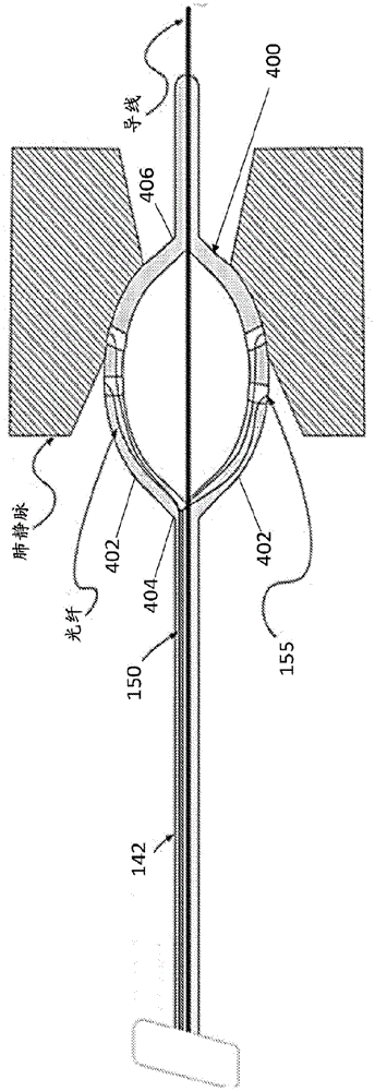
图5A是消融导管的示例性实施例,在其远端具有可膨胀构件;5A is an exemplary embodiment of an ablation catheter having an expandable member at its distal end;
图5B是图5A的可膨胀构件的透视图;Figure 5B is a perspective view of the expandable member of Figure 5A;
图5C是消融导管的示例性实施例,在其远端上具有可膨胀构件;5C is an exemplary embodiment of an ablation catheter having an expandable member on its distal end;
图5D是图5A和5C的可膨胀构件的远端视图;Figure 5D is a distal end view of the expandable member of Figures 5A and 5C;
图6是消融导管的示例性实施例,在其远端上具有可膨胀构件;6 is an exemplary embodiment of an ablation catheter having an expandable member on its distal end;
图7是消融导管的示例性实施例,在其远端上具有球囊形式的可膨胀构件;7 is an exemplary embodiment of an ablation catheter having an expandable member in the form of a balloon on its distal end;
图8A-8D是包括可以与消融导管一起使用的光学组件的护套的示例性实施例;8A-8D are exemplary embodiments of a sheath including an optical assembly that may be used with an ablation catheter;
图9图示了用于询问、监视和消融组织的方法的流程图;9 illustrates a flowchart of a method for interrogating, monitoring, and ablating tissue;
图10图示了具有与组织接触的电极和不与组织接触的电极及其对应的响应信号的消融导管的示例性实施例;10 illustrates an exemplary embodiment of an ablation catheter having electrodes in contact with tissue and electrodes not in contact with tissue and their corresponding response signals;
图11图示了当消融导管的电极不与组织接触时响应光强度的示例性曲线图;11 illustrates an exemplary graph of response light intensity when the electrodes of the ablation catheter are not in contact with tissue;
图12、13和14示出了PFA前后的光强度;Figures 12, 13 and 14 show the light intensity before and after PFA;
图15图示了消融期间NADH荧光随时间的示例性曲线图;以及Figure 15 illustrates an exemplary graph of NADH fluorescence over time during ablation; and
图16A、16B、16C和16D示出了健康组织和消融损伤的光学标测。Figures 16A, 16B, 16C and 16D show optical mapping of healthy tissue and ablation lesions.
虽然上述附图阐述了目前公开的实施例,但是其它实施例也是预期的,如在讨论中所指出的。本公开作为表示而不是限制来给出说明性实施例。本领域技术人员可以设计出众多其它修改和实施例,这些修改和实施例属于目前公开的实施例的原理的范围和精神内。While the above figures illustrate presently disclosed embodiments, other embodiments are contemplated, as noted in the discussion. The present disclosure presents illustrative embodiments by way of representation and not limitation. Numerous other modifications and embodiments can be devised by those skilled in the art that fall within the scope and spirit of the principles of the presently disclosed embodiments.
具体实施方式Detailed ways
本公开提供了用于损伤评估的方法和系统。在一些实施例中,使用消融能量形成损伤,例如脉动场消融(PFA)能量,以通过电穿孔引起损伤。在一些实施例中,本公开的系统包括被配置为起两个功能的导管:将消融疗法(例如,脉动场消融)递送到目标组织的治疗功能以及从导管和组织的接触点收集特征频谱以访问病变的诊断功能。在一些实施例中,本公开的系统和方法可被用于使用烟酰胺腺嘌呤二核苷酸氢(NADH)荧光(fNADH)对组织进行成像。一般而言,系统可以包括具有用于在组织和导管之间交换光的光学系统的导管。在一些实施例中,本系统允许由紫外线(UV)激发诱导的组织的NADH荧光或其缺乏的直接光学组织询问。从组织返回的NADH荧光特征可以被用来确定能量对组织的影响以及组织和导管系统之间的接触质量。The present disclosure provides methods and systems for damage assessment. In some embodiments, the lesions are formed using ablation energy, such as pulsatile field ablation (PFA) energy, to induce lesions by electroporation. In some embodiments, the systems of the present disclosure include a catheter configured to perform two functions: a therapeutic function to deliver ablation therapy (eg, pulsatile field ablation) to target tissue and to collect characteristic spectra from the point of contact of the catheter and tissue to Access diagnostic functions for lesions. In some embodiments, the systems and methods of the present disclosure can be used to image tissue using nicotinamide adenine dinucleotide hydrogen (NADH) fluorescence (fNADH). In general, the system may include a catheter with an optical system for exchanging light between the tissue and the catheter. In some embodiments, the present system allows for direct optical tissue interrogation of NADH fluorescence of tissue, or its lack thereof, induced by ultraviolet (UV) excitation. NADH fluorescence signatures returned from tissue can be used to determine the effect of energy on tissue and the quality of contact between tissue and the duct system.
在一些实施例中,导管包括消融疗法系统,以在其远端处递送PFA并耦合到诊断单元,该诊断单元包括光源,诸如激光器和光谱仪。在一些实施例中,可以使用分离的导管形成损伤或者可以询问先前形成的损伤。导管可以包括从光源和频谱仪延伸到导管的远侧尖端的一根或多根光纤,以向导管和组织之间的接触点提供照射光并且接收并从接触点向频谱仪递送特征NADH频谱。特征NADH频谱可以被用来评估目标组织中的病变。在一些实施例中,本公开的方法包括照射具有病变的组织,接收组织的特征频谱,以及基于来自组织的特征频谱对病变执行定性评估。分析可以在消融病变形成之前、期间和之后实时地发生。应当注意的是,虽然结合心脏组织和NADH频谱描述了本公开的系统和方法,但是本公开的系统和方法可以与其它类型的组织和其它类型的荧光结合使用。In some embodiments, the catheter includes an ablation therapy system to deliver PFA at its distal end and is coupled to a diagnostic unit that includes a light source, such as a laser and a spectrometer. In some embodiments, the lesions may be formed using separate catheters or previously formed lesions may be interrogated. The catheter may include one or more optical fibers extending from the light source and spectrometer to the distal tip of the catheter to provide illumination light to the point of contact between the catheter and tissue and to receive and deliver the characteristic NADH spectrum from the point of contact to the spectrometer. The characteristic NADH spectrum can be used to assess lesions in the target tissue. In some embodiments, the methods of the present disclosure include irradiating tissue having a lesion, receiving a characteristic spectrum of the tissue, and performing a qualitative assessment of the lesion based on the characteristic spectrum from the tissue. Analysis can occur in real-time before, during, and after ablation lesion formation. It should be noted that although the systems and methods of the present disclosure are described in conjunction with cardiac tissue and NADH spectra, the systems and methods of the present disclosure may be used in conjunction with other types of tissue and other types of fluorescence.
系统:诊断单元System: Diagnostic Unit
参考图1A,用于提供消融疗法的系统100可以包括消融疗法系统110、光学组织询问系统120以及导管140。在一些实施例中,系统100还可以包括一个或多个冲洗系统170、超声系统190以及导航系统200。系统还可以包括显示器180,其可以是单独的显示器或光学组织询问系统120的一部分,如下所述。在一些实施例中,该系统包括消融发生器、冲洗泵170、冲洗尖端消融导管140以及光学组织询问系统120。Referring to FIG. 1A , a
在一些实施例中,消融疗法系统110被设计为向导管140供给消融能量。在一些实施例中,消融疗法系统110可以包括脉动场消融(PFA)能量以通过电穿孔引起损伤。各种系统可以被用于递送PFA能量。如图2中所示,可以调制和施加脉动电场以在细胞膜中形成不可逆的孔,从而触发细胞凋亡。当使用PFA能量时,脉冲持续时间足够短,以至于在能量递送期间形成的蒸汽最少、没有蒸汽球膨胀并且没有电弧。PFA能量的示例性参数范围可以是递送500-3000V/cm的电压,递送1-100个脉冲,在微秒的波长上,具有1-5Hz的频率范围。PFA的影响几乎是瞬时的。例如,单次PFA递送在一次心跳内完成,并且通常可以通过3-5次PFA递送来创建病变。作为PFA的补充或替代,可以使用可以生成射频(RF)能量、微波能量、电能量、电磁能量、低温能量、激光能量、超声能量、声能量、化学能量、热能量、电穿孔能量或任何其它类型的能量的一种或多种能量源来消融组织。In some embodiments,
参考图1B,光学组织询问系统120可以包括光源122、光测量仪器124和计算机系统126。Referring to FIG. 1B , the optical
在一些实施例中,光源122可以具有在目标荧光团(在一些实施例中是NADH)吸收范围内的输出波长,以便在健康心肌细胞中诱导荧光。在一些实施例中,光源122是可以生成UV光以激发NADH荧光的固态激光器。在一些实施例中,波长可以是大约355nm或355nm+/-30nm。在一些实施例中,光源122可以是UV激光器。激光器生成的UV光可以提供多得多的用于照射的功率,并且可以更高效地耦合到基于光纤的照明系统中,如在导管140的一些实施例中所使用的。在一些实施例中,本系统可以使用具有高达150mW的可调节功率的激光器。In some embodiments, the
光源122上的波长范围可以由所关注的解剖结构界定,用户具体选择造成最大NADH荧光而不激发胶原蛋白的过度荧光的波长,其在仅稍短的波长处表现出吸收峰。在一些实施例中,光源122生成具有在250nm和450nm之间的至少一种波长的光。在一些实施例中,光源122生成具有在300nm和400nm之间的至少一种波长的光。在一些实施例中,光源122生成具有在330nm和385nm之间的至少一种波长的光。在一些实施例中,光源122生成具有在330nm至355nm之间的至少一种波长的光。在一些实施例中,可以使用窄带355nm的源。光源122的输出功率可以足够高,以产生可恢复的组织荧光特征,但还没有高到导致细胞损害。如下面将要描述的,光源122可以耦合到光纤,以将光递送到导管140和从导管140递送。The range of wavelengths on the
在一些实施例中,本公开的系统可以利用频谱仪作为光测量仪器124,但是可以采用其它光测量仪器。光纤可以将收集到的光递送到光测量仪器124。计算机系统126从光测量仪器124获取信息并将其显示给医师。In some embodiments, the systems of the present disclosure may utilize a spectrometer as the
返回去参考图1A,在一些实施例中,本公开的系统100还可以包括超声系统190。导管140可以配备有与超声系统190连通的超声换能器。在一些实施例中,超声可以示出组织深度,其与代谢活动或病变深度相结合可以被用来确定病变是否事实上是透壁的。在一些实施例中,超声换能器可以位于导管140的远侧区段中,并且可选地位于远侧电极的尖端中。超声换能器可以被配置为或者在导管尖端的下方或者邻近导管尖端处评估组织厚度。在一些实施例中,导管140可以包括适于提供覆盖导管尖端相对于垂直于心肌或相对平行于心肌的情况的深度信息的多个换能器。Referring back to FIG. 1A , in some embodiments, the
参考图1A,如上面所指出的,系统100还可以包括冲洗系统170。在一些实施例中,冲洗系统170将盐水泵入导管140中,以在消融疗法期间冷却尖端电极。这可以有助于预防血栓栓塞(可以或可以不通过血液的凝块)、蒸汽爆裂和炭的形成。在一些实施例中,冲洗流体相对于导管140外部的压力维持在正压力,用于连续地冲刷一个或多个开口154,如图3A中所示。Referring to FIG. 1A , as noted above, the
参考图1A,系统100还可以包括用于定位和导航导管140的导航系统200。在一些实施例中,导管140可以包括与导航系统200连通的一个或多个电磁位置传感器。在一些实施例中,电磁位置传感器可以被用来将导管的尖端定位在导航系统200中。传感器从源位置拾取电磁能量,并通过三角测量或其它手段计算位置。在一些实施例中,导管140包括适于将导管体142的位置和导管体的曲率呈现在导航系统显示器上的多于一个的换能器。在一些实施例中,导航系统200可以包括一个或多个磁体,并且由磁体在电磁传感器上产生的磁场的更改可以将导管的尖端偏转到期望的方向。还可以采用其它导航系统,包括手动导航。Referring to FIG. 1A , the
计算机系统126可以被编程为控制系统100的各种模块,包括例如对光源122的控制、对光测量仪器124的控制,特定于应用的软件的执行、对超声、导航和冲洗系统的控制,以及类似操作。作为示例,图1C示出了可以结合本公开的方法和系统使用的典型处理体系架构308的图。计算机处理设备340可以耦合到显示器340AA,用于图形输出。处理器342可以是能够执行软件的计算机处理器342。典型的示例可以是计算机处理器(诸如
程序349可以是包含指令和/或数据的计算机程序或计算机可读代码,并且可以存储在存储设备348上。指令可以包括来自任何计算机编程语言的代码,包括例如C、C++、C#、Visual Basic、Java、Python、Perl和JavaScript。在典型的场景中,处理器204可以将程序349的一些或全部指令和/或数据加载到存储器346中,以供执行。程序349可以是任何计算机程序或过程,包括但不限于web浏览器、浏览器应用、地址注册过程、应用或者任何其它计算机应用或过程。程序349可以包括各种指令和子程序,当其被加载到存储器346中并由处理器342执行时,使得处理器342执行各种操作,其中一些或全部操作可以实现本文公开的用于管理医疗护理的方法。程序349可以存储在任何类型的非瞬态计算机可读介质上,诸如但不限于硬盘驱动器、可移动驱动器、CD、DVD或任何其它类型的计算机可读介质。
在一些实施例中,计算机系统可以被编程为执行本公开的方法的步骤,并且控制本系统的各个部分,以执行必要的操作来实现本公开的方法。在一些实施例中,处理器可以被编程为从用通过导管的远侧尖端的UV光照射的组织接收NADH荧光数据,其中组织在径向方向、轴向方向或两个方向上被照射;从被照射的组织中的NADH荧光水平确定何时导管的远侧尖端与组织接触;以及在确定远侧尖端与组织接触时(或者自动地或者通过提示用户)使消融能量递送到组织,以在组织中形成病变。In some embodiments, a computer system may be programmed to perform the steps of the methods of the present disclosure, and control various parts of the system to perform the necessary operations to implement the methods of the present disclosure. In some embodiments, the processor may be programmed to receive NADH fluorescence data from tissue irradiated with UV light through the distal tip of the catheter, wherein the tissue is irradiated in a radial direction, an axial direction, or both; from The level of NADH fluorescence in the irradiated tissue determines when the distal tip of the catheter is in contact with the tissue; and upon determining that the distal tip is in contact with the tissue (either automatically or by prompting the user) to deliver ablation energy to the tissue to lesions formed.
处理器还可以被编程为在递送消融能量期间监视NADH荧光的水平,以确认远侧尖端保持与组织接触。在一些实施例中,在递送消融能量期间监视NADH荧光的水平可以被用来确定远侧尖端和组织之间的接触的稳定性。在一些实施例中,当远端尖端和组织之间的接触不稳定时,可以停止组织的消融。在一些实施例中,处理器还可以被编程为收集从被照射的组织返回的荧光的光谱,以区分组织类型。The processor can also be programmed to monitor the level of NADH fluorescence during delivery of ablation energy to confirm that the distal tip remains in contact with the tissue. In some embodiments, monitoring the level of NADH fluorescence during delivery of ablation energy can be used to determine the stability of the contact between the distal tip and tissue. In some embodiments, ablation of the tissue may be stopped when the contact between the distal tip and the tissue is unstable. In some embodiments, the processor may also be programmed to collect spectra of fluorescence returned from the irradiated tissue to differentiate tissue types.
在一些实施例中,监视具有在大约450nm和470nm之间的波长的返回光的水平。在一些实施例中,被监视的光谱可以在420nm和500nm之间。在一些实施例中,被监视的光谱可以在400nm和520nm之间。附加地或可替代地,可以监视更宽的光谱,诸如作为非限制性示例,在375nm和650nm之间。在一些实施例中,可以同时向用户显示NADH荧光光谱和更宽的光谱。在一些实施例中,损伤可以通过消融PFA能量产生。在一些实施例中,当检测到NADH荧光峰值时,可以开始(由处理器或通过由处理器提示用户)手术,因此可以在整个手术过程中被监视。如上面所指出的,处理器可以结合其它诊断方法(诸如超声监视)来执行这些方法。In some embodiments, the level of returned light having wavelengths between approximately 450 nm and 470 nm is monitored. In some embodiments, the monitored spectrum may be between 420 nm and 500 nm. In some embodiments, the spectrum being monitored may be between 400 nm and 520 nm. Additionally or alternatively, a wider spectrum can be monitored, such as, by way of non-limiting example, between 375 nm and 650 nm. In some embodiments, the NADH fluorescence spectrum and the broader spectrum can be displayed to the user simultaneously. In some embodiments, the lesions can be created by ablating PFA energy. In some embodiments, surgery may be initiated (either by the processor or by prompting the user by the processor) when a peak in NADH fluorescence is detected, and thus may be monitored throughout the procedure. As noted above, the processor may perform these methods in conjunction with other diagnostic methods, such as ultrasound monitoring.
系统:导管System: Catheter
在一些实施例中,如上面所讨论的,导管140可以基于具有用于容纳照明和频谱的光纤的标准消融导管。在一些实施例中,导管140是可转向的、被冲洗的消融导管(例如,PFA消融导管),其可以经由标准的越隔规程和常见的访问工具(access tools)通过护套递送到心内膜空间。在导管的手柄147上,为了疗法,可以存在用于标准消融发生器和冲洗系统170的连接。导管手柄147还使光纤通过,然后光纤连接到诊断单元,以获得组织测量。In some embodiments, as discussed above, the
返回去参考图1A,导管140包括具有近端144和远端146的导管体142。导管体142可以由生物相容性材料制成,并且可以足够柔软,以使导管140能够转向和前进到消融的部位。在一些实施例中,导管体142可以具有可变刚度的区。例如,导管140的刚度可以从近端144朝着远端146增加。在一些实施例中,导管主体142的刚度被选择为使得能够将导管140递送到期望的心脏位置。在一些实施例中,导管140可以是可转向的消融导管,其可以通过护套被递送到心内膜空间,并且在心脏左侧的情况下,使用常见的访问工具经由标准越隔规程。导管140可以包括在近端144处的手柄147。手柄147可以与导管的一个或多个管腔连通,以允许仪器或材料通过导管140。在一些实施例中,为了疗法,手柄147可以包括用于标准PFA发生器和冲洗系统170的连接。在一些实施例中,导管140还可以包括被配置为容纳用于照明和频谱的光纤的一个或多个适配器。Referring back to FIG. 1A , the
在一些实施例中,远侧尖端148可以被配置为充当用于诊断目的(诸如用于电描记图感测)、用于治疗目的(诸如用于发射消融能量)或二者兼有的电极。在需要消融能量的一些实施例中,导管140的远侧尖端148可以充当消融电极或消融元件。在一些实施例中,导管的远端可以包括一个或多个电极。在一些实施例中,导管的远端可以包括包含多个电极的电极的阵列。在一些实施例中,光纤可以与电极相关联以确定组织接触并决定在消融期间使用哪些电极。在一些实施例中,电极的阵列可以部署在导管的远端上,如图3A-3C中所示。在一些实施例中,电极的阵列可以部署在可膨胀构件上。可膨胀构件可以是多种形式,包括图5A-5B中所示的线篮布置、图5C中所示的花布置以及图7中所示的球囊。多个电极部署在可膨胀构件上,使得多个电极中的至少一个可以与组织接触。该系统具有确定多个电极中的哪一个具有组织接触的能力,使得只有具有适当组织接触的那些电极用于组织消融。该系统还具有确定每个电极所接触的组织的类型的能力,使得只有与正确类型的组织具有适当组织接触的那些电极用于组织消融。例如,如果确定电极与胶原蛋白接触,那么该电极将不用于消融。In some embodiments, the
在一些实施例中,远侧尖端148上的电极耦合到消融能量源(在导管外部),例如通过线或可以传送消融能量的另一个管腔,消融能量可以穿过导管的管腔。远侧尖端148可以包括与导管的一个或多个管腔连通的端口。远侧尖端148可以由任何生物相容性材料制成。在一些实施例中,如果远侧尖端148被配置为充当电极,则远侧尖端148可以由金属制成,包括但不限于铂、铂-铱、不锈钢、钛或类似材料。In some embodiments, electrodes on the
参考图1A和3A-3C,示出了示例性消融导管的远端,其包括可以从图1A的光学组织询问系统120穿过导管主体142的成像束150,使得每根光纤152可以通过阵列中的每个电极。在一些实施例中,每根光纤与每个电极对准。在一些实施例中,可替代地或附加地,成像束包括不与电极相关联的光纤。在远端146处,导管140可以包括远侧尖端148,具有侧壁156和前壁158。前壁158可以是例如平坦的、圆锥形的或圆顶形的。远端146可以设有一个或多个与电极155的阵列相关联的光学端口154,用于在导管和组织之间交换光能。在一些实施例中,光学端口可以制成穿过电极,使得光可以从光纤穿过电极。在一些实施例中,即使具有多个开口154,也不会损害远侧尖端148作为消融电极的功能。开口可以部署在前壁156上、侧壁158上或两者上。开口154也可以用作冲洗端口。光由光纤150递送到远侧尖端148,在那里它照亮远侧尖端148附近的组织。这种照明光或者返回,或者使组织发出荧光。由组织返回并从组织发出荧光的光可以由远侧尖端148内的光纤150收集并传送回光学组织询问系统120。在一些实施例中,相同的光纤或光纤束150可以被用于将光指引到远侧尖端的照明室以照亮导管140外部的组织并收集来自组织的光。1A and 3A-3C, the distal end of an exemplary ablation catheter is shown that includes an
图4图示了具有远端146的消融导管140的实施例,该远端146具有电极155的阵列,电极155包括布置在远端146处的多个电极。FIG. 4 illustrates an embodiment of an
如图5A和5B中所示,电极155的阵列可以定位在可膨胀构件400上,该构件具有多个延伸部或花键402,这些延伸部或花键402被配置为移动到膨胀的位置。虽然图5A图示了两个示例性延伸部402,但是将理解的是,可膨胀构件400可以包括任何数量的延伸部402。在图5A中所示的膨胀的位置,每个延伸部402可以成弧形,使得每个延伸部的近端404彼此耦合并且每个延伸部的远端406彼此耦合。可膨胀构件400的每个延伸部402可以包括形成在其上的至少一个电极155,并且每个电极155耦合到光纤束150中的至少一根光纤152。在一些实施例中,每个电极155耦合到单根光纤152。As shown in Figures 5A and 5B, the array of
与图5A中所示的可膨胀构件类似,图5C中所示的可膨胀构件410包括多个延伸部或花键412,使得当处于如图5C中所示的膨胀的位置时,可膨胀构件410的近端414和远端416之间的距离小于图5A中所示的距离,从而使可膨胀构件410沿着垂直轴线呈椭圆形。随着可膨胀构件的近端和远端之间的距离改变,导管的远端相对于组织的位置改变。例如,如图5C中所示,导管140的远端146可以定位在肺静脉之外,同时当电极155面对导管140的远端146时仍然允许电极155与组织接触。Similar to the expandable member shown in Figure 5A, the
图5D图示了图5A-5C中所示的导管140和可膨胀构件400、410的远端视图。可以改变可膨胀构件的形状以最大化与组织接触的多个电极。例如,可膨胀构件压缩得越多(即,可膨胀构件在垂直方向上越长),将有越多的电极面向组织。这可以允许更多的电极接触组织并且使导管的远端定位成与组织相邻。例如,在导管定位成与肺静脉相邻的情况下,导管可以定位在心房侧上的肺静脉外侧,同时仍然有足够的电极面向静脉以接触期望的组织。Figure 5D illustrates a distal end view of the
图6图示了具有在近端424处彼此耦合的多个延伸部或臂422的可膨胀构件420的实施例。多个臂422中的每一个的远端426远离导管主体142延伸,使得部署在臂422上的多个电极155可以与组织接触。将理解的是,臂的数量以及臂的尺寸和形状可以根据各种因素而变化,包括要消融的组织的位置。FIG. 6 illustrates an embodiment of an
图7图示了具有部署在其上的多个电极155的球囊形式的可膨胀构件430的实施例,每个电极与光纤相关联。可以改变球囊的形状以最大化与组织接触的多个电极155。FIG. 7 illustrates an embodiment of an
参考图3A-3C,在一些实施例中,导管可以具有光学组织询问管腔161,光纤150可以通过其前进通过导管主体142。光纤150可以前进通过光学组织询问管腔161以照亮组织并接收通过开口154的返回光。在必要时,光纤150可以前进通过开口154。3A-3C, in some embodiments, the catheter can have an optical
除了光学组织询问管腔161,导管140还可以包括用于使冲洗流体从冲洗系统170传递到远侧尖端148中的开口154(冲洗端口)的冲洗管腔163和用于使消融能量从消融疗法系统110传递到远侧尖端148(诸如通过使用于PFA消融能量的电线穿过消融管腔164)的消融腔164。应当注意的是,导管的管腔可以被用于多个目的,并且可以为相同的目的使用多于一个管腔。此外,虽然图3A和图3B示出管腔是同心的,但是可以使用管腔的其它构造。In addition to the optical
如图3A和图3B中所示,在一些实施例中,导管的中央管腔可以被用作光学组织询问管腔161。在一些实施例中,如图3C中所示,光学组织询问管腔161可以相对于导管140的中心通路偏移。As shown in FIGS. 3A and 3B , in some embodiments, the central lumen of the catheter may be used as the optical
在一些实施例中,光也可以相对于导管轴向地和径向地被指引。以这种方式,导管和组织之间的光能交换可以相对于导管的纵向中心轴线在轴向、径向或两者上发生在多条路径上。当解剖结构不允许导管尖端与目标部位正交时,这是有用的。当要求增加的照明时,这也会是有用的。在一些实施例中,附加的光纤150可以被使用并且可以相对于导管140在径向方向上偏转,以允许照明和返回的光沿着导管的长度离开和进入。In some embodiments, the light may also be directed axially and radially relative to the conduit. In this manner, the exchange of light energy between the catheter and the tissue can take place in multiple paths axially, radially, or both, relative to the longitudinal central axis of the catheter. This is useful when the anatomy does not allow the catheter tip to be orthogonal to the target site. This can also be useful when increased lighting is required. In some embodiments, additional
参考图8A-8D,在一些实施例中,消融导管可以与包括光纤的护套结合使用,使得与护套相关联的光纤可以相对于导管定位以允许光纤与电极相关联。在一些实施例中,护套可以是可偏转和/或可转向的护套的形式,使得护套的远端和与护套相关联的光学组件可以相对于组织定位在期望的位置处以进行消融。Referring to Figures 8A-8D, in some embodiments, an ablation catheter can be used in conjunction with a sheath that includes an optical fiber such that the optical fiber associated with the sheath can be positioned relative to the catheter to allow the optical fiber to be associated with the electrode. In some embodiments, the sheath may be in the form of a deflectable and/or steerable sheath such that the distal end of the sheath and the optical assembly associated with the sheath may be positioned relative to tissue at a desired location for ablation .
护套和消融导管的组合可以具有各种配置。在图8A中所示的一个实施例中,护套500包括从护套500的远端504延伸的多个延伸部或臂502。每个臂具有延伸穿过其中的至少一根光纤。护套包括延伸穿过其中的内部管腔,使得消融导管可以穿过护套并延伸超过远端。消融导管可以具有各种构造,但在图8A中,消融导管140包括多个延伸部或臂,每个延伸部具有部署在其上的至少一个电极。护套的远端的延伸部和消融导管的延伸部相对于彼此定位,以允许通过护套的臂中的光纤询问组织,同时经由消融导管的电极递送消融能量。在一些实施例中,护套的延伸部可以是可偏转的和/或可转向的,以允许延伸部相对于电极和组织的适当定位。在一些实施例中,护套的延伸部可以具有多于一根光纤,以在消融能量递送之前、期间和之后光学地询问组织。在一些实施例中,容纳光纤的护套的延伸部分也可以在其上具有电极以进一步评估组织。将理解的是,图8A中所示的护套可以与本文所述的任何消融导管一起使用。The combination of sheath and ablation catheter can have various configurations. In one embodiment shown in FIG. 8A , the
图8B图示了具有从其远端延伸的可偏转和/或可转向的延伸部或臂512的护套510的实施例。护套510的远端可以被拆分以形成臂或延伸部512。虽然图8B中所示的护套可以与本文所述的任何消融导管一起使用,但是消融导管140可以穿过护套510的内部管腔,该护套具有沿着其长度间隔开的多个电极155。在一些实施例中,护套的臂可以具有多于一根光纤以在消融能量递送之前、期间和之后光学地询问组织。在一些实施例中,容纳光纤的护套的臂也可以在其上具有电极以进一步评估组织。8B illustrates an embodiment of a
图8C图示了可偏转和/或可转向的护套520的实施例,该护套520具有从护套520的远端延伸的多根光纤152。虽然图8C中所示的护套可与本文所述的任何消融导管一起使用,但消融导管140可以穿过护套520的内部管腔,其具有沿着其长度部署的至少一个电极155。从护套延伸的光纤152具有使得它们可以接触组织和消融导管的电极155的长度。FIG. 8C illustrates an embodiment of a deflectable and/or
图8D图示了可偏转和/或可转向的护套530的实施例,该护套具有从护套的远端延伸的多根光纤。延伸穿过护套的消融导管可以具有各种构造,包括本文所述的任何消融导管,但在图8D中,消融导管包括多个延伸部或臂532,每个延伸部具有部署在其上的至少一个电极155。护套的远端的延伸部和消融导管的延伸部相对于彼此定位,以允许护套的延伸部中的光纤与消融导管延伸部上的电极之间的连接。Figure 8D illustrates an embodiment of a deflectable and/or
图8D还图示了在导管远端处具有多个电极的可偏转和/或可转向导管的实施例。消融导管的各种柔性消融臂可以具有各种构造,但在图8D中,消融导管包括多个延伸部或臂,每个延伸部具有部署在其上的至少一个电极。消融导管的延伸部相对于彼此定位,以便以圆周模式向组织最优地递送能量,同时,当与本公开的护套结合时,使用经由每个光学端口将光递送到组织的光纤光学地询问组织。Figure 8D also illustrates an embodiment of a deflectable and/or steerable catheter having multiple electrodes at the distal end of the catheter. The various flexible ablation arms of the ablation catheter can have various configurations, but in Figure 8D the ablation catheter includes a plurality of extensions or arms, each extension having at least one electrode deployed thereon. The extensions of the ablation catheter are positioned relative to each other to optimally deliver energy to the tissue in a circumferential pattern, while, when combined with the sheaths of the present disclosure, optically interrogated using optical fibers that deliver light to the tissue via each optical port organize.
图9是使用本公开的系统的方法的流程图。9 is a flowchart of a method of using the system of the present disclosure.
参考图9,图示了本公开的系统100的操作。最初,将导管140插入要消融的心脏组织的区域,诸如肺静脉/左心房交界处或心脏的其它区域(步骤600)。例如,如图5A和5C中所示,在一些实施例中,导管140可以通过肺静脉前进并压靠在心脏组织上。可以从视野中去除血液,例如通过冲洗。9, the operation of the
接下来,在步骤602中,可以确认电极与组织之间的接触。在一些实施例中,在光纤与电极相关联的情况下,可以通过光纤消除组织并且询问返回的光以确认各个电极与组织之间的接触。例如,如图10和图11中所示,对于不与心脏组织接触的电极,将不会检测到NADH荧光。Next, in
在步骤604中,与组织接触的电极可以被激活以消融组织。在步骤608处,使用光纤照亮组织,并且检测和分析从组织返回的光,以便在步骤608中实时显示消融的进展。如图12-14中所示,NADH荧光的强度随着消融的进行而降低。这种效应是由于代谢活性的降低,因此当细胞被消融时NADH荧光降低。这种下降可以被用作何时停止消融的指示。例如,参考图15,可以向用户呈现NADH荧光的图表或另一个图形表示,其示出NADH荧光的改变以帮助用户监视消融的进展,如步骤610所表示的。在这一步,还使得用户能够确定是继续还是停止消融。在一些实施例中,消融可以一直继续到实现NADH荧光的量值的期望改变,此时可以或者手动或者由系统自动停止消融。例如,在一些实施例中,消融可以在NADH信号减少80%或更多时停止。一旦消融停止,本实施例的系统就可以被用于在步骤612中绘制组织,以识别受损细胞的区域(诸如通过消融)或可以被消融的健康心肌的区域,如图16A-16D中所示。系统可以存储此类消融前和消融后信号和光学信息,用于记录能量递送到给定位置的组织的程度。在一些实施例中,可以经由算法来分析来自所存储信号的实时或消融后的此类数据,以评估或预测产生持久损伤的概率。In
在一些实施例中,可以收集和分析光谱特征以确定组织成分。例如,胶原蛋白组织的频谱图不同于在健康心肌上观察到的频谱图。当在这种情况下用355nm UV光源被照射时,由于胶原蛋白荧光的增加的影响,当对较短波长在胶原蛋白组织之上成像时,光谱的峰向左偏移(从大约470nm到大约445nm)。这可以被用户用来识别被视为主要是心肌的区域或者被胶原蛋白覆盖的、更难消融的区域。特别地,胶原荧光的数字表示指示组织中纤维化负荷的组织中的纤维形成。在消融手术期间,存在与返回到医师的光谱的信息内容相关联的潜在益处。可以使用从导管或具体而言是位于导管远侧尖端的消融电极将光耦合到组织中的技术来确定和评估导管或电极与组织接触的质量。在消融能量部署之前知道关于被消融的组织的类型或者组织中要被消融的胶原蛋白是否存在以及有可能消融的程度的更多信息也会影响医师用于那个病变的最佳创建的消融策略和技术。例如,在胶原蛋白存在的情况下,医师可以选择一个消融能量源而不是另一个,并且功率或持续时间或温度限制可以被调节得更高,以获得更深的病变,给定被消融的组织的胶原性质。与肌肉组织相比,胶原组织可以具有不同的纤维化负荷,因此要求不同的消融策略。In some embodiments, spectral signatures can be collected and analyzed to determine tissue composition. For example, the spectrogram of collagen tissue differs from that observed on healthy myocardium. When illuminated with a 355 nm UV light source in this case, the peak of the spectrum shifts to the left (from about 470 nm to about 445nm). This can be used by the user to identify areas that are considered primarily myocardium or areas covered by collagen that are more difficult to ablate. In particular, the numerical representation of collagen fluorescence indicates fibrillar formation in tissues with fibrosis burden in the tissues. During an ablation procedure, there is a potential benefit associated with the information content of the spectrum returned to the physician. The quality of catheter or electrode-tissue contact can be determined and assessed using techniques that couple light into tissue from the catheter or, specifically, an ablation electrode located at the distal tip of the catheter. Knowing more about the type of tissue being ablated or whether collagen to be ablated in the tissue is present and the extent to which it is likely to be ablated prior to ablation energy deployment can also influence the physician's optimally created ablation strategy for that lesion and technology. For example, in the presence of collagen, the physician can select one source of ablation energy over the other, and the power or duration or temperature limits can be adjusted higher to obtain deeper lesions, given the amount of tissue being ablated. Collagen properties. Collagen tissue can have a different fibrotic load compared to muscle tissue, thus requiring different ablation strategies.
提出以下示例是为了向本领域普通技术人员提供关于如何进行和使用本公开的测定、筛选和治疗方法的完整公开和描述,并且不旨在限制发明人认为是他们的发明的范围。The following examples are presented to provide those of ordinary skill in the art with a complete disclosure and description of how to make and use the assays, screening, and therapeutic methods of the present disclosure, and are not intended to limit the scope of what the inventors believe to be their inventions.
示例Example
一系列3只麻醉猪在右心房接受PFA消融。将8电极圆形导管置于右心房高处,靠近上腔静脉,以模拟肺静脉隔离,作为AF消融规程的一部分。将光学导管放置在刺激电极对之间的圆形导管附近。施予一团腺苷以产生心搏停止窗口以避免对T波的刺激。双极PFA在药物输注后立即递送,并实时记录和显示来自导管的光学特征。记录电图并用光学导管在PFA递送后的以下时间间隔执行损伤的标测:0分钟、15秒、30秒、1分钟(60秒)、15分钟、1小时和3小时。尸检和组织学遵循该规程。A series of 3 anesthetized pigs underwent PFA ablation in the right atrium. An 8-electrode round catheter was placed high in the right atrium, close to the superior vena cava, to simulate pulmonary vein isolation as part of the AF ablation protocol. An optical catheter is placed near the circular catheter between the pair of stimulating electrodes. A bolus of adenosine was administered to create an asystole window to avoid stimulation of the T waves. Bipolar PFA is delivered immediately after drug infusion, and optical signatures from the catheter are recorded and displayed in real time. Electrograms were recorded and mapping of lesions was performed using the optical catheter at the following time intervals after PFA delivery: 0 minutes, 15 seconds, 30 seconds, 1 minute (60 seconds), 15 minutes, 1 hour, and 3 hours. Necropsy and histology followed this protocol.
对于这个示例,使用分离的PFA导管和光学组织询问导管。对于光学组织询问,使用以下参数:激发波长355nm;胶原蛋白响应375至400nm;心肌响应450至475nm和465nm处的峰值跟踪与时间。For this example, a separate PFA catheter and optical tissue interrogation catheter were used. For optical tissue interrogation, the following parameters were used: excitation wavelength 355 nm; collagen response 375 to 400 nm;
图12-14代表PFA前后的光强度。图10示出了对PFA的光强度响应。Figures 12-14 represent light intensities before and after PFA. Figure 10 shows the light intensity response to PFA.
在PFA脉冲序列期间,光学信号的强度明显更高。光学信号在标测间隔内示出立即显著下降和缓慢但稳定的衰减。电图减少伴随PFA施加,并且在标测间隔内也示出显著减少。与预测的健康未消融心肌相比,损伤处的光学信号幅度显著降低。During the PFA pulse train, the intensity of the optical signal is significantly higher. The optical signal showed an immediate significant drop and a slow but steady decay over the mapping interval. Electrographic reductions accompany PFA application and also show significant reductions within the mapping interval. The optical signal amplitude at the lesion was significantly reduced compared to the predicted healthy unablated myocardium.
这些结果指示,光学标测在PFA期间以这些能量水平检测即时组织改变,因此可以是在PFA能量施加期间和之后评估损伤形成的可行方法。光学信号指示细胞损害在这些能量水平上立即发生,并且与射频能量造成的损伤相比,PFA能量造成的损伤继续缓慢进展。研究结果还表明,光学标测可以实时识别由PFA能量造成的急性损伤,这意味着光学标测可以被用作PFA间隙检测器。研究结果还表明,光学组织询问或标测可以用于预测PFA损伤的持久性或非持久性。These results indicate that optical mapping detects immediate tissue changes at these energy levels during PFA and thus may be a viable method to assess lesion formation during and after PFA energy application. The optical signal indicated that cellular damage occurred immediately at these energy levels, and that damage from PFA energy continued to progress slowly compared to damage from radio frequency energy. The findings also show that optical mapping can identify acute damage caused by PFA energy in real time, implying that optical mapping can be used as a PFA gap detector. The findings also suggest that optical tissue interrogation or mapping can be used to predict the persistence or non-persistence of PFA damage.
其它实施例Other embodiments
从前面的描述,显然可以对本公开的实施例进行变化和修改以将其应用于各种用途和条件。此类实施例也在以下权利要求的范围内。From the foregoing description, it will be apparent that changes and modifications can be made to the embodiments of the present disclosure to adapt them to various usages and conditions. Such embodiments are also within the scope of the following claims.
本文对变量的任何定义中的元素列表的叙述包括将该变量定义为任何单个元素或所列元素的组合(或子组合)。本文对实施例的叙述包括作为任何单个实施例或与任何其它实施例或其部分组合的实施例。Recitation of a listing of elements in any definition of a variable herein includes defining the variable as any single element or combination (or subcombination) of the listed elements. The recitation of an embodiment herein includes an embodiment as any single embodiment or in combination with any other embodiments or portions thereof.
本说明书中提及的所有专利和出版物均通过引用并入本文,其程度与每个独立专利和出版物被具体和单独地指示通过引用并入的程度相同。All patents and publications mentioned in this specification are incorporated herein by reference to the same extent that each individual patent and publication was specifically and individually indicated to be incorporated by reference.
Claims (50)
Applications Claiming Priority (3)
| Application Number | Priority Date | Filing Date | Title |
|---|---|---|---|
| US202062958419P | 2020-01-08 | 2020-01-08 | |
| US62/958,419 | 2020-01-08 | ||
| PCT/US2021/012836 WO2021142368A1 (en) | 2020-01-08 | 2021-01-08 | Systems and methods for optical interrogation of ablation lesions |
Publications (1)
| Publication Number | Publication Date |
|---|---|
| CN115103647A true CN115103647A (en) | 2022-09-23 |
Family
ID=76655720
Family Applications (1)
| Application Number | Title | Priority Date | Filing Date |
|---|---|---|---|
| CN202180013945.5A Pending CN115103647A (en) | 2020-01-08 | 2021-01-08 | Systems and methods for optical interrogation of ablative lesions |
Country Status (5)
| Country | Link |
|---|---|
| US (1) | US12076081B2 (en) |
| EP (1) | EP4087511A4 (en) |
| JP (1) | JP2023510326A (en) |
| CN (1) | CN115103647A (en) |
| WO (1) | WO2021142368A1 (en) |
Families Citing this family (11)
| Publication number | Priority date | Publication date | Assignee | Title |
|---|---|---|---|---|
| CN104066368B (en) | 2011-09-22 | 2017-02-22 | 乔治华盛顿大学 | Systems and methods for visualizing ablated tissue |
| US11096584B2 (en) | 2013-11-14 | 2021-08-24 | The George Washington University | Systems and methods for determining lesion depth using fluorescence imaging |
| WO2015077474A1 (en) | 2013-11-20 | 2015-05-28 | The George Washington University | Systems and methods for hyperspectral analysis of cardiac tissue |
| WO2016073492A1 (en) | 2014-11-03 | 2016-05-12 | Luxcath, Llc. | Systems and methods for assessment of contact quality |
| AU2015343258B2 (en) | 2014-11-03 | 2020-07-16 | 460Medical, Inc. | Systems and methods for lesion assessment |
| US10779904B2 (en) | 2015-07-19 | 2020-09-22 | 460Medical, Inc. | Systems and methods for lesion formation and assessment |
| CN115103647A (en) | 2020-01-08 | 2022-09-23 | 460医学股份有限公司 | Systems and methods for optical interrogation of ablative lesions |
| WO2021243061A1 (en) | 2020-05-27 | 2021-12-02 | The George Washington University | Lesion visualization using dual wavelength approach |
| US12076071B2 (en) | 2020-08-14 | 2024-09-03 | Kardium Inc. | Systems and methods for treating tissue with pulsed field ablation |
| WO2025227090A1 (en) * | 2024-04-26 | 2025-10-30 | Anumana, Inc. | Method and apparatus for predicting pulsed field ablation durability |
| US12527621B1 (en) | 2025-01-27 | 2026-01-20 | Anumana, Inc. | Apparatus and method for determining a tissue susceptibility value |
Citations (7)
| Publication number | Priority date | Publication date | Assignee | Title |
|---|---|---|---|---|
| US20090005773A1 (en) * | 2007-06-29 | 2009-01-01 | Christopher Beeckler | Ablation catheter with optically transparent, electrically conductive tip |
| CN102038480A (en) * | 2005-04-04 | 2011-05-04 | 因维蒂有限公司 | Surgery illumination system |
| US20150196202A1 (en) * | 2013-11-14 | 2015-07-16 | The George Washington University | Systems and Methods for Determining Lesion Depth Using Fluorescence Imaging |
| CN105377168A (en) * | 2013-05-08 | 2016-03-02 | 波士顿科学国际有限公司 | Systems and methods for temperature monitoring and control during an ablation procedure |
| US20170014202A1 (en) * | 2015-07-19 | 2017-01-19 | LuxCath, LLC | Systems and Methods for Lesion Formation and Assessment |
| CN106852708A (en) * | 2015-12-09 | 2017-06-16 | 韦伯斯特生物官能(以色列)有限公司 | Ablation catheter with the feeler based on light |
| CN107106221A (en) * | 2014-11-25 | 2017-08-29 | 拉克斯凯瑟有限责任公司 | visualization catheter |
Family Cites Families (385)
| Publication number | Priority date | Publication date | Assignee | Title |
|---|---|---|---|---|
| US3387305A (en) | 1966-02-09 | 1968-06-11 | Rocky Mountain Sports | Knee protector |
| US3831467A (en) | 1973-03-16 | 1974-08-27 | R Moore | Knee brace |
| US4024873A (en) | 1976-05-24 | 1977-05-24 | Becton, Dickinson And Company | Balloon catheter assembly |
| US4619247A (en) | 1983-03-31 | 1986-10-28 | Sumitomo Electric Industries, Ltd. | Catheter |
| JPS60182928A (en) | 1984-03-01 | 1985-09-18 | オリンパス光学工業株式会社 | Endoscope having solid image pick-up element mounted therein |
| JPS63262613A (en) | 1987-04-20 | 1988-10-28 | Olympus Optical Co Ltd | Stereoscopic vision endoscope device |
| US5419323A (en) | 1988-12-21 | 1995-05-30 | Massachusetts Institute Of Technology | Method for laser induced fluorescence of tissue |
| US5421337A (en) | 1989-04-14 | 1995-06-06 | Massachusetts Institute Of Technology | Spectral diagnosis of diseased tissue |
| US5584799A (en) | 1989-09-11 | 1996-12-17 | Gray; James C. | Splint/therapeutic device |
| US5074306A (en) | 1990-02-22 | 1991-12-24 | The General Hospital Corporation | Measurement of burn depth in skin |
| JP3164609B2 (en) | 1990-10-31 | 2001-05-08 | オリンパス光学工業株式会社 | Endoscope device |
| US6485413B1 (en) | 1991-04-29 | 2002-11-26 | The General Hospital Corporation | Methods and apparatus for forward-directed optical scanning instruments |
| CA2042075C (en) | 1991-05-08 | 2001-01-23 | Branko Palcic | Endoscopic imaging system |
| US5540681A (en) | 1992-04-10 | 1996-07-30 | Medtronic Cardiorhythm | Method and system for radiofrequency ablation of tissue |
| US5350375A (en) | 1993-03-15 | 1994-09-27 | Yale University | Methods for laser induced fluorescence intensity feedback control during laser angioplasty |
| WO1995003843A1 (en) | 1993-07-30 | 1995-02-09 | The Regents Of The University Of California | Endocardial infusion catheter |
| US5749830A (en) | 1993-12-03 | 1998-05-12 | Olympus Optical Co., Ltd. | Fluorescent endoscope apparatus |
| US5590660A (en) | 1994-03-28 | 1997-01-07 | Xillix Technologies Corp. | Apparatus and method for imaging diseased tissue using integrated autofluorescence |
| AU2373695A (en) | 1994-05-03 | 1995-11-29 | Board Of Regents, The University Of Texas System | Apparatus and method for noninvasive doppler ultrasound-guided real-time control of tissue damage in thermal therapy |
| US20080154257A1 (en) | 2006-12-22 | 2008-06-26 | Shiva Sharareh | Real-time optoacoustic monitoring with electophysiologic catheters |
| US5810802A (en) | 1994-08-08 | 1998-09-22 | E.P. Technologies, Inc. | Systems and methods for controlling tissue ablation using multiple temperature sensing elements |
| US6423055B1 (en) | 1999-07-14 | 2002-07-23 | Cardiofocus, Inc. | Phototherapeutic wave guide apparatus |
| US8025661B2 (en) | 1994-09-09 | 2011-09-27 | Cardiofocus, Inc. | Coaxial catheter instruments for ablation with radiant energy |
| US6572609B1 (en) | 1999-07-14 | 2003-06-03 | Cardiofocus, Inc. | Phototherapeutic waveguide apparatus |
| US5954665A (en) | 1995-06-07 | 1999-09-21 | Biosense, Inc. | Cardiac ablation catheter using correlation measure |
| US6023638A (en) * | 1995-07-28 | 2000-02-08 | Scimed Life Systems, Inc. | System and method for conducting electrophysiological testing using high-voltage energy pulses to stun tissue |
| US5713364A (en) | 1995-08-01 | 1998-02-03 | Medispectra, Inc. | Spectral volume microprobe analysis of materials |
| US6309352B1 (en) | 1996-01-31 | 2001-10-30 | Board Of Regents, The University Of Texas System | Real time optoacoustic monitoring of changes in tissue properties |
| US5885258A (en) | 1996-02-23 | 1999-03-23 | Memory Medical Systems, Inc. | Medical instrument with slotted memory metal tube |
| WO1997037622A1 (en) | 1996-04-08 | 1997-10-16 | The University Of Southern California | Method and apparatus for using laser-induced fluorescence during photoretractive keratectomy |
| US5904651A (en) | 1996-10-28 | 1999-05-18 | Ep Technologies, Inc. | Systems and methods for visualizing tissue during diagnostic or therapeutic procedures |
| JP3003597B2 (en) | 1996-11-18 | 2000-01-31 | 日本電気株式会社 | Solid-state imaging device |
| US5833688A (en) | 1997-02-24 | 1998-11-10 | Boston Scientific Corporation | Sensing temperature with plurality of catheter sensors |
| US6208886B1 (en) | 1997-04-04 | 2001-03-27 | The Research Foundation Of City College Of New York | Non-linear optical tomography of turbid media |
| US6124597A (en) | 1997-07-07 | 2000-09-26 | Cedars-Sinai Medical Center | Method and devices for laser induced fluorescence attenuation spectroscopy |
| US6656153B1 (en) | 1997-09-12 | 2003-12-02 | Nippon Zeon Co., Ltd | Balloon catheter |
| US6238389B1 (en) | 1997-09-30 | 2001-05-29 | Boston Scientific Corporation | Deflectable interstitial ablation device |
| US6289236B1 (en) | 1997-10-10 | 2001-09-11 | The General Hospital Corporation | Methods and apparatus for distinguishing inflamed and tumorous bladder tissue |
| US6937885B1 (en) | 1997-10-30 | 2005-08-30 | Hypermed, Inc. | Multispectral/hyperspectral medical instrument |
| DE69938493T2 (en) | 1998-01-26 | 2009-05-20 | Massachusetts Institute Of Technology, Cambridge | ENDOSCOPE FOR DETECTING FLUORESCENCE IMAGES |
| US6174291B1 (en) | 1998-03-09 | 2001-01-16 | Spectrascience, Inc. | Optical biopsy system and methods for tissue diagnosis |
| US6251107B1 (en) | 1998-06-25 | 2001-06-26 | Cardima, Inc. | Ep catheter |
| US6112123A (en) | 1998-07-28 | 2000-08-29 | Endonetics, Inc. | Device and method for ablation of tissue |
| US8024027B2 (en) | 1998-09-03 | 2011-09-20 | Hyperspectral Imaging, Inc. | Infrared endoscopic balloon probes |
| EP1112022A4 (en) | 1998-09-11 | 2004-08-04 | Spectrx Inc | Multi-modal optical tissue diagnostic system |
| US6178346B1 (en) | 1998-10-23 | 2001-01-23 | David C. Amundson | Infrared endoscopic imaging in a liquid with suspended particles: method and apparatus |
| US6701176B1 (en) | 1998-11-04 | 2004-03-02 | Johns Hopkins University School Of Medicine | Magnetic-resonance-guided imaging, electrophysiology, and ablation |
| US6210406B1 (en) | 1998-12-03 | 2001-04-03 | Cordis Webster, Inc. | Split tip electrode catheter and signal processing RF ablation system |
| US6423057B1 (en) | 1999-01-25 | 2002-07-23 | The Arizona Board Of Regents On Behalf Of The University Of Arizona | Method and apparatus for monitoring and controlling tissue temperature and lesion formation in radio-frequency ablation procedures |
| JP4849755B2 (en) | 1999-07-02 | 2012-01-11 | ハイパースペクトラル・イメージング・インコーポレイテツド | Imaging apparatus and sample analysis method |
| JP2001017379A (en) | 1999-07-09 | 2001-01-23 | Fuji Photo Film Co Ltd | Fluorescent diagnostic device |
| US6219566B1 (en) | 1999-07-13 | 2001-04-17 | Photonics Research Ontario | Method of measuring concentration of luminescent materials in turbid media |
| US9033961B2 (en) | 1999-07-14 | 2015-05-19 | Cardiofocus, Inc. | Cardiac ablation catheters for forming overlapping lesions |
| US8540704B2 (en) | 1999-07-14 | 2013-09-24 | Cardiofocus, Inc. | Guided cardiac ablation catheters |
| US8900219B2 (en) | 1999-07-14 | 2014-12-02 | Cardiofocus, Inc. | System and method for visualizing tissue during ablation procedures |
| WO2001005306A1 (en) | 1999-07-19 | 2001-01-25 | Epicor, Inc. | Apparatus and method for ablating tissue |
| US6343228B1 (en) | 1999-10-19 | 2002-01-29 | The Hong Kong University Of Science And Technology | Method and apparatus for fluorescence imaging of tissue |
| US6542767B1 (en) | 1999-11-09 | 2003-04-01 | Biotex, Inc. | Method and system for controlling heat delivery to a target |
| US8221402B2 (en) | 2000-01-19 | 2012-07-17 | Medtronic, Inc. | Method for guiding a medical device |
| US6663622B1 (en) | 2000-02-11 | 2003-12-16 | Iotek, Inc. | Surgical devices and methods for use in tissue ablation procedures |
| GR1004180B (en) | 2000-03-28 | 2003-03-11 | ����������� ����� ��������� (����) | Method and system for characterization and mapping of tissue lesions |
| US20020107514A1 (en) | 2000-04-27 | 2002-08-08 | Hooven Michael D. | Transmural ablation device with parallel jaws |
| AU2001249874A1 (en) | 2000-04-27 | 2001-11-12 | Medtronic, Inc. | System and method for assessing transmurality of ablation lesions |
| JP2003533267A (en) | 2000-05-12 | 2003-11-11 | カーディマ・インコーポレイテッド | Multi-channel RF energy transmission system capable of reducing agglomerates |
| US7252664B2 (en) | 2000-05-12 | 2007-08-07 | Cardima, Inc. | System and method for multi-channel RF energy delivery with coagulum reduction |
| US6975898B2 (en) | 2000-06-19 | 2005-12-13 | University Of Washington | Medical imaging, diagnosis, and therapy using a scanning single optical fiber system |
| IL138683A0 (en) | 2000-09-25 | 2001-10-31 | Vital Medical Ltd | Apparatus and method for monitoring tissue vitality parameters |
| US6663561B2 (en) | 2000-10-05 | 2003-12-16 | Pentax Corporation | Video endoscope system |
| US6450971B1 (en) | 2000-10-05 | 2002-09-17 | Scimed Life Systems, Inc. | Temperature measuring balloon |
| US7047068B2 (en) | 2000-12-11 | 2006-05-16 | C.R. Bard, Inc. | Microelectrode catheter for mapping and ablation |
| JP2002253500A (en) | 2001-03-05 | 2002-09-10 | Olympus Optical Co Ltd | Light source device for endoscope |
| US6743225B2 (en) | 2001-03-27 | 2004-06-01 | Uab Research Foundation | Electrophysiologic measure of endpoints for ablation lesions created in fibrillating substrates |
| US6989010B2 (en) | 2001-04-26 | 2006-01-24 | Medtronic, Inc. | Ablation system and method of use |
| US6663627B2 (en) | 2001-04-26 | 2003-12-16 | Medtronic, Inc. | Ablation system and method of use |
| US7250048B2 (en) | 2001-04-26 | 2007-07-31 | Medtronic, Inc. | Ablation system and method of use |
| US7959626B2 (en) | 2001-04-26 | 2011-06-14 | Medtronic, Inc. | Transmural ablation systems and methods |
| US6648883B2 (en) | 2001-04-26 | 2003-11-18 | Medtronic, Inc. | Ablation system and method of use |
| DE60230499D1 (en) | 2001-04-27 | 2009-02-05 | Bard Inc C R | CATHETER FOR THE THREE-DIMENSIONAL ILLUSTRATION OF ELECTRIC ACTIVITY IN BLOOD VESSELS |
| EP1418972A1 (en) | 2001-05-01 | 2004-05-19 | C.R. Bard, Inc. | Method and apparatus for altering conduction properties along pathways in the heart and in vessels in conductive communication with the heart |
| US7727229B2 (en) | 2001-05-01 | 2010-06-01 | C.R. Bard, Inc. | Method and apparatus for altering conduction properties in the heart and in adjacent vessels |
| US20030208252A1 (en) | 2001-05-14 | 2003-11-06 | O' Boyle Gary S. | Mri ablation catheter |
| US7992573B2 (en) | 2001-06-19 | 2011-08-09 | The Trustees Of The University Of Pennsylvania | Optically guided system for precise placement of a medical catheter in a patient |
| US7596404B2 (en) | 2001-06-28 | 2009-09-29 | Chemimage Corporation | Method of chemical imaging to determine tissue margins during surgery |
| US8078268B2 (en) | 2001-06-28 | 2011-12-13 | Chemimage Corporation | System and method of chemical imaging using pulsed laser excitation and time-gated detection to determine tissue margins during surgery |
| US6761716B2 (en) | 2001-09-18 | 2004-07-13 | Cardiac Pacemakers, Inc. | System and method for assessing electrode-tissue contact and lesion quality during RF ablation by measurement of conduction time |
| CA2460501A1 (en) | 2001-09-28 | 2003-04-10 | Institut De Cardiologie De Montreal | Method for identification and visualization of atrial tissue |
| WO2003053491A2 (en) | 2001-11-09 | 2003-07-03 | Cardio-Optics, Inc. | Coronary sinus access catheter with forward-imaging |
| US20030120144A1 (en) | 2001-11-16 | 2003-06-26 | Grabek James R. | Intrapericardial temperature measurement device and method |
| US12121289B2 (en) | 2008-05-09 | 2024-10-22 | Atricure, Inc. | Conduction block systems and methods |
| US7749157B2 (en) | 2001-12-04 | 2010-07-06 | Estech, Inc. (Endoscopic Technologies, Inc.) | Methods and devices for minimally invasive cardiac surgery for atrial fibrillation |
| US20040092806A1 (en) | 2001-12-11 | 2004-05-13 | Sagon Stephen W | Microelectrode catheter for mapping and ablation |
| US6825928B2 (en) | 2001-12-19 | 2004-11-30 | Wisconsin Alumni Research Foundation | Depth-resolved fluorescence instrument |
| US6873868B2 (en) | 2001-12-31 | 2005-03-29 | Infraredx, Inc. | Multi-fiber catheter probe arrangement for tissue analysis or treatment |
| US20040215310A1 (en) | 2002-01-17 | 2004-10-28 | Omar Amirana | Stent and delivery method for applying RF energy to a pulmonary vein and the atrial wall around its ostium to eliminate atrial fibrillation while preventing stenosis of the pulmonary vein thereafter |
| US7967816B2 (en) | 2002-01-25 | 2011-06-28 | Medtronic, Inc. | Fluid-assisted electrosurgical instrument with shapeable electrode |
| US20050075629A1 (en) | 2002-02-19 | 2005-04-07 | Afx, Inc. | Apparatus and method for assessing tissue ablation transmurality |
| US7192427B2 (en) | 2002-02-19 | 2007-03-20 | Afx, Inc. | Apparatus and method for assessing transmurality of a tissue ablation |
| US7846157B2 (en) | 2002-03-15 | 2010-12-07 | C.R. Bard, Inc. | Method and apparatus for control of ablation energy and electrogram acquisition through multiple common electrodes in an electrophysiology catheter |
| US6746401B2 (en) | 2002-05-06 | 2004-06-08 | Scimed Life Systems, Inc. | Tissue ablation visualization |
| ATE371413T1 (en) | 2002-05-06 | 2007-09-15 | Covidien Ag | BLOOD DETECTOR FOR CHECKING AN ELECTROSURGICAL UNIT |
| US8956280B2 (en) | 2002-05-30 | 2015-02-17 | Intuitive Surgical Operations, Inc. | Apparatus and methods for placing leads using direct visualization |
| EP1870018A3 (en) * | 2002-05-30 | 2008-08-06 | The Board of Trustees of The Leland Stanford Junior University | Apparatus and methods for coronary sinus access |
| AU2003238656A1 (en) | 2002-06-25 | 2004-01-06 | Glucon Inc. | Method and apparatus for performing myocardial revascularization |
| US20060173359A1 (en) | 2002-09-30 | 2006-08-03 | Lin Wei C | Optical apparatus for guided liver tumor treatment and methods |
| US7306593B2 (en) | 2002-10-21 | 2007-12-11 | Biosense, Inc. | Prediction and assessment of ablation of cardiac tissue |
| US7001383B2 (en) | 2002-10-21 | 2006-02-21 | Biosense, Inc. | Real-time monitoring and mapping of ablation lesion formation in the heart |
| WO2004073505A2 (en) | 2003-02-20 | 2004-09-02 | Prorhythm, Inc. | Cardiac ablation devices |
| WO2004095359A2 (en) | 2003-04-18 | 2004-11-04 | Medispectra, Inc. | Systems for identifying, displaying, marking, and treating suspect regions of tissue |
| US6980843B2 (en) | 2003-05-21 | 2005-12-27 | Stereotaxis, Inc. | Electrophysiology catheter |
| US7539530B2 (en) | 2003-08-22 | 2009-05-26 | Infraredx, Inc. | Method and system for spectral examination of vascular walls through blood during cardiac motion |
| US7534204B2 (en) | 2003-09-03 | 2009-05-19 | Guided Delivery Systems, Inc. | Cardiac visualization devices and methods |
| US20060009755A1 (en) | 2003-09-04 | 2006-01-12 | Sra Jasbir S | Method and system for ablation of atrial fibrillation and other cardiac arrhythmias |
| AU2004273992B2 (en) | 2003-09-19 | 2010-01-21 | The General Hospital Corporation | Fluorescence polarization imaging devices and methods |
| US8172747B2 (en) | 2003-09-25 | 2012-05-08 | Hansen Medical, Inc. | Balloon visualization for traversing a tissue wall |
| US7395118B2 (en) | 2003-09-25 | 2008-07-01 | Advanced Neuromodulation Systems, Inc. | System and method for implantable stimulation lead employing optical fibers |
| WO2005032342A2 (en) | 2003-09-30 | 2005-04-14 | Vanderbilt University | Methods and apparatus for optical spectroscopic detection of cell and tissue death |
| EP1682025A4 (en) | 2003-10-20 | 2011-04-06 | Univ Johns Hopkins | Catheter and method for ablation of atrial tissue |
| US7232437B2 (en) | 2003-10-30 | 2007-06-19 | Medical Cv, Inc. | Assessment of lesion transmurality |
| WO2005044124A1 (en) | 2003-10-30 | 2005-05-19 | Medical Cv, Inc. | Apparatus and method for laser treatment |
| US20050137459A1 (en) | 2003-12-17 | 2005-06-23 | Scimed Life Systems, Inc. | Medical device with OLED illumination light source |
| US7587236B2 (en) | 2004-01-08 | 2009-09-08 | Lawrence Livermore National Security, Llc | Optical spectroscopy for the detection of ischemic tissue injury |
| US20050215899A1 (en) | 2004-01-15 | 2005-09-29 | Trahey Gregg E | Methods, systems, and computer program products for acoustic radiation force impulse (ARFI) imaging of ablated tissue |
| US20050228452A1 (en) | 2004-02-11 | 2005-10-13 | Mourlas Nicholas J | Steerable catheters and methods for using them |
| US20050197623A1 (en) | 2004-02-17 | 2005-09-08 | Leeflang Stephen A. | Variable steerable catheters and methods for using them |
| US20050251116A1 (en) | 2004-05-05 | 2005-11-10 | Minnow Medical, Llc | Imaging and eccentric atherosclerotic material laser remodeling and/or ablation catheter |
| AU2005244868A1 (en) | 2004-05-14 | 2005-12-01 | Medtronic, Inc. | Method and devices for treating atrial fibrillation by mass ablation |
| US7640046B2 (en) | 2004-06-18 | 2009-12-29 | Cardiac Pacemakers, Inc. | Methods and apparatuses for localizing myocardial infarction during catheterization |
| US7527625B2 (en) | 2004-08-04 | 2009-05-05 | Olympus Corporation | Transparent electrode for the radiofrequency ablation of tissue |
| US20060089637A1 (en) | 2004-10-14 | 2006-04-27 | Werneth Randell L | Ablation catheter |
| US20060089636A1 (en) | 2004-10-27 | 2006-04-27 | Christopherson Mark A | Ultrasound visualization for transurethral needle ablation |
| US7417740B2 (en) | 2004-11-12 | 2008-08-26 | Medeikon Corporation | Single trace multi-channel low coherence interferometric sensor |
| CA2588390C (en) | 2004-11-17 | 2016-06-07 | Biosense Webster, Inc. | Apparatus for real time evaluation of tissue ablation |
| US10413188B2 (en) | 2004-11-17 | 2019-09-17 | Lawrence Livermore National Security, Llc | Assessment of tissue or lesion depth using temporally resolved light scattering spectroscopy |
| US20060229515A1 (en) | 2004-11-17 | 2006-10-12 | The Regents Of The University Of California | Fiber optic evaluation of tissue modification |
| US8548570B2 (en) | 2004-11-29 | 2013-10-01 | Hypermed Imaging, Inc. | Hyperspectral imaging of angiogenesis |
| JP4656924B2 (en) | 2004-12-03 | 2011-03-23 | 株式会社トプコン | Spectral fundus image data measurement device |
| US7367944B2 (en) | 2004-12-13 | 2008-05-06 | Tel Hashomer Medical Research Infrastructure And Services Ltd. | Method and system for monitoring ablation of tissues |
| US8858495B2 (en) | 2004-12-28 | 2014-10-14 | St. Jude Medical, Atrial Fibrillation Division, Inc. | Five degree of freedom ultrasound catheter and catheter control handle |
| US7776033B2 (en) | 2005-01-08 | 2010-08-17 | Boston Scientific Scimed, Inc. | Wettable structures including conductive fibers and apparatus including the same |
| US7862561B2 (en) | 2005-01-08 | 2011-01-04 | Boston Scientific Scimed, Inc. | Clamp based lesion formation apparatus with variable spacing structures |
| US7727231B2 (en) | 2005-01-08 | 2010-06-01 | Boston Scientific Scimed, Inc. | Apparatus and methods for forming lesions in tissue and applying stimulation energy to tissue in which lesions are formed |
| US7729750B2 (en) | 2005-01-20 | 2010-06-01 | The Regents Of The University Of California | Method and apparatus for high resolution spatially modulated fluorescence imaging and tomography |
| US8137333B2 (en) | 2005-10-25 | 2012-03-20 | Voyage Medical, Inc. | Delivery of biological compounds to ischemic and/or infarcted tissue |
| US7860556B2 (en) | 2005-02-02 | 2010-12-28 | Voyage Medical, Inc. | Tissue imaging and extraction systems |
| US8050746B2 (en) | 2005-02-02 | 2011-11-01 | Voyage Medical, Inc. | Tissue visualization device and method variations |
| US8078266B2 (en) | 2005-10-25 | 2011-12-13 | Voyage Medical, Inc. | Flow reduction hood systems |
| US20080015569A1 (en) | 2005-02-02 | 2008-01-17 | Voyage Medical, Inc. | Methods and apparatus for treatment of atrial fibrillation |
| US7918787B2 (en) | 2005-02-02 | 2011-04-05 | Voyage Medical, Inc. | Tissue visualization and manipulation systems |
| US7930016B1 (en) | 2005-02-02 | 2011-04-19 | Voyage Medical, Inc. | Tissue closure system |
| US20080009747A1 (en) | 2005-02-02 | 2008-01-10 | Voyage Medical, Inc. | Transmural subsurface interrogation and ablation |
| US7860555B2 (en) | 2005-02-02 | 2010-12-28 | Voyage Medical, Inc. | Tissue visualization and manipulation system |
| US10064540B2 (en) | 2005-02-02 | 2018-09-04 | Intuitive Surgical Operations, Inc. | Visualization apparatus for transseptal access |
| WO2006107947A2 (en) | 2005-04-04 | 2006-10-12 | Hypermed, Inc. | Hyperspectral imaging in diabetes and peripheral vascular disease |
| WO2006122061A1 (en) | 2005-05-06 | 2006-11-16 | Acumen Medical, Inc. | Complexly shaped steerable catheters and methods for making and using them |
| DE102005021205B4 (en) | 2005-05-07 | 2007-08-16 | Mfd Diagnostics Gmbh | Method and device for the local detection of the vitality of living cells in cell cultures or in tissue |
| WO2007002323A2 (en) | 2005-06-23 | 2007-01-04 | Epoc, Inc. | System and method for monitoring of end organ oxygenation by measurement of in vivo cellular energy status |
| US8556851B2 (en) | 2005-07-05 | 2013-10-15 | Angioslide Ltd. | Balloon catheter |
| DE102005032755B4 (en) | 2005-07-13 | 2014-09-04 | Siemens Aktiengesellschaft | System for performing and monitoring minimally invasive procedures |
| EP3028645B1 (en) | 2005-08-01 | 2019-09-18 | St. Jude Medical International Holding S.à r.l. | Medical apparatus system having optical fiber load sensing capability |
| US8583220B2 (en) | 2005-08-02 | 2013-11-12 | Biosense Webster, Inc. | Standardization of catheter-based treatment for atrial fibrillation |
| US7877128B2 (en) | 2005-08-02 | 2011-01-25 | Biosense Webster, Inc. | Simulation of invasive procedures |
| US7681579B2 (en) | 2005-08-02 | 2010-03-23 | Biosense Webster, Inc. | Guided procedures for treating atrial fibrillation |
| US7740584B2 (en) | 2005-08-16 | 2010-06-22 | The General Electric Company | Method and system for mapping physiology information onto ultrasound-based anatomic structure |
| JP4681981B2 (en) | 2005-08-18 | 2011-05-11 | Hoya株式会社 | Electronic endoscope device |
| US7824397B2 (en) | 2005-08-19 | 2010-11-02 | Boston Scientific Scimed, Inc. | Occlusion apparatus |
| WO2007023407A1 (en) | 2005-08-25 | 2007-03-01 | Koninklijke Philips Electronics, N.V. | System and method for electrophysiology regaining support to continue line and ring ablations |
| US20070270717A1 (en) | 2005-09-30 | 2007-11-22 | Cornova, Inc. | Multi-faceted optical reflector |
| US20070078500A1 (en) | 2005-09-30 | 2007-04-05 | Cornova, Inc. | Systems and methods for analysis and treatment of a body lumen |
| US8929973B1 (en) | 2005-10-24 | 2015-01-06 | Lockheed Martin Corporation | Apparatus and method for characterizing optical sources used with human and animal tissues |
| US8221310B2 (en) | 2005-10-25 | 2012-07-17 | Voyage Medical, Inc. | Tissue visualization device and method variations |
| US8403925B2 (en) | 2006-12-06 | 2013-03-26 | St. Jude Medical, Atrial Fibrillation Division, Inc. | System and method for assessing lesions in tissue |
| US9492226B2 (en) | 2005-12-06 | 2016-11-15 | St. Jude Medical, Atrial Fibrillation Division, Inc. | Graphical user interface for real-time RF lesion depth display |
| US8406866B2 (en) | 2005-12-06 | 2013-03-26 | St. Jude Medical, Atrial Fibrillation Division, Inc. | System and method for assessing coupling between an electrode and tissue |
| WO2007067939A2 (en) | 2005-12-06 | 2007-06-14 | St. Jude Medical, Arial Fibrillation Division, Inc. | Assessment of electrode coupling for tissue ablation |
| US8603084B2 (en) | 2005-12-06 | 2013-12-10 | St. Jude Medical, Atrial Fibrillation Division, Inc. | System and method for assessing the formation of a lesion in tissue |
| US9254163B2 (en) | 2005-12-06 | 2016-02-09 | St. Jude Medical, Atrial Fibrillation Division, Inc. | Assessment of electrode coupling for tissue ablation |
| US8998890B2 (en) | 2005-12-06 | 2015-04-07 | St. Jude Medical, Atrial Fibrillation Division, Inc. | Assessment of electrode coupling for tissue ablation |
| US8317783B2 (en) | 2005-12-06 | 2012-11-27 | St. Jude Medical, Atrial Fibrillation Division, Inc. | Assessment of electrode coupling for tissue ablation |
| BRPI0621229A2 (en) | 2006-01-13 | 2011-12-06 | Smm Medical Ab | device and system for compression treatment of a body part |
| EP2289398A3 (en) | 2006-01-19 | 2011-04-06 | The General Hospital Corporation | Methods and systems for optical imaging of epithelial luminal organs by beam scanning thereof |
| US9186066B2 (en) | 2006-02-01 | 2015-11-17 | The General Hospital Corporation | Apparatus for applying a plurality of electro-magnetic radiations to a sample |
| US20070185479A1 (en) | 2006-02-06 | 2007-08-09 | Liming Lau | Methods and devices for performing ablation and assessing efficacy thereof |
| US7918850B2 (en) | 2006-02-17 | 2011-04-05 | Biosense Wabster, Inc. | Lesion assessment by pacing |
| JP4828633B2 (en) | 2006-03-17 | 2011-11-30 | ボエッジ メディカル, インコーポレイテッド | Organization visualization and operation system |
| US20070225697A1 (en) | 2006-03-23 | 2007-09-27 | Ketan Shroff | Apparatus and methods for cardiac ablation |
| US20080058786A1 (en) | 2006-04-12 | 2008-03-06 | Searete Llc, A Limited Liability Corporation Of The State Of Delaware | Autofluorescent imaging and target ablation |
| US20080058785A1 (en) | 2006-04-12 | 2008-03-06 | Searete Llc, A Limited Liability Corporation Of The State Of Delaware | Autofluorescent imaging and target ablation |
| US8129105B2 (en) | 2006-04-13 | 2012-03-06 | Ralph Zuckerman | Method and apparatus for the non-invasive measurement of tissue function and metabolism by determination of steady-state fluorescence anisotropy |
| US8628520B2 (en) | 2006-05-02 | 2014-01-14 | Biosense Webster, Inc. | Catheter with omni-directional optical lesion evaluation |
| US20070270792A1 (en) | 2006-05-08 | 2007-11-22 | Willard Hennemann | Interferometric characterization of ablated tissue |
| US20100198065A1 (en) | 2009-01-30 | 2010-08-05 | VyntronUS, Inc. | System and method for ultrasonically sensing and ablating tissue |
| CA2652126C (en) | 2006-05-12 | 2015-12-01 | Hira V. Thapliyal | Device for ablating body tissue |
| JP2007313169A (en) | 2006-05-29 | 2007-12-06 | Olympus Corp | Lesion extractor and lesion extraction method |
| JP2009538672A (en) | 2006-05-30 | 2009-11-12 | コーニンクレッカ フィリップス エレクトロニクス エヌ ヴィ | Apparatus for depth-resolved measurement of tissue attributes |
| EP2034890A4 (en) | 2006-06-01 | 2011-02-16 | Gen Hospital Corp | IN VIVO OPTICAL IMAGING METHOD COMPRISING THE ANALYSIS OF DYNAMIC IMAGES |
| US9220402B2 (en) | 2006-06-07 | 2015-12-29 | Intuitive Surgical Operations, Inc. | Visualization and treatment via percutaneous methods and devices |
| US8567265B2 (en) | 2006-06-09 | 2013-10-29 | Endosense, SA | Triaxial fiber optic force sensing catheter |
| US8048063B2 (en) | 2006-06-09 | 2011-11-01 | Endosense Sa | Catheter having tri-axial force sensor |
| US7662152B2 (en) | 2006-06-13 | 2010-02-16 | Biosense Webster, Inc. | Catheter with multi port tip for optical lesion evaluation |
| US20080125634A1 (en) | 2006-06-14 | 2008-05-29 | Cornova, Inc. | Method and apparatus for identifying and treating myocardial infarction |
| EP2038002A4 (en) | 2006-06-28 | 2010-12-29 | Bard Inc C R | METHODS AND APPARATUS FOR ASSESSING AND ENHANCING ELECTRODE CONTACT WITH CARDIAC TISSUE |
| US20080033241A1 (en) | 2006-08-01 | 2008-02-07 | Ruey-Feng Peh | Left atrial appendage closure |
| US20080097476A1 (en) | 2006-09-01 | 2008-04-24 | Voyage Medical, Inc. | Precision control systems for tissue visualization and manipulation assemblies |
| EP2063781A4 (en) | 2006-09-01 | 2010-07-28 | Voyage Medical Inc | Electrophysiology mapping and visualization system |
| US10004388B2 (en) | 2006-09-01 | 2018-06-26 | Intuitive Surgical Operations, Inc. | Coronary sinus cannulation |
| US20110042580A1 (en) | 2006-09-06 | 2011-02-24 | University Health Network | Fluorescence quantification and image acquisition in highly turbid media |
| US8147484B2 (en) | 2006-10-23 | 2012-04-03 | Biosense Webster, Inc. | Apparatus and method for monitoring early formation of steam pop during ablation |
| US10335131B2 (en) | 2006-10-23 | 2019-07-02 | Intuitive Surgical Operations, Inc. | Methods for preventing tissue migration |
| DE102006050885B4 (en) | 2006-10-27 | 2016-11-03 | Siemens Healthcare Gmbh | Device for generating tissue section images |
| WO2008054423A1 (en) | 2006-10-31 | 2008-05-08 | University Of Washington | Magnetically controllable elongate device, systems and methods |
| US8986298B2 (en) | 2006-11-17 | 2015-03-24 | Biosense Webster, Inc. | Catheter with omni-directional optical tip having isolated optical paths |
| US20080183036A1 (en) | 2006-12-18 | 2008-07-31 | Voyage Medical, Inc. | Systems and methods for unobstructed visualization and ablation |
| US8758229B2 (en) | 2006-12-21 | 2014-06-24 | Intuitive Surgical Operations, Inc. | Axial visualization systems |
| US8131350B2 (en) | 2006-12-21 | 2012-03-06 | Voyage Medical, Inc. | Stabilization of visualization catheters |
| US7766907B2 (en) | 2006-12-28 | 2010-08-03 | St. Jude Medical, Atrial Fibrillation Division, Inc. | Ablation catheter with sensor array and discrimination circuit to minimize variation in power density |
| US7591816B2 (en) | 2006-12-28 | 2009-09-22 | St. Jude Medical, Atrial Fibrillation Division, Inc. | Irrigated ablation catheter having a pressure sensor to detect tissue contact |
| US8460285B2 (en) | 2006-12-29 | 2013-06-11 | St. Jude Medical, Atrial Fibrillation Division, Inc. | Ablation catheter electrode having multiple thermal sensors and method of use |
| EP2131741A4 (en) | 2007-03-02 | 2010-05-05 | Wisconsin Alumni Res Found | USE OF ENDOGENOUS FLUORESCENCE TO IDENTIFY INVASIVE METASTATIC BREAST TUMER CELLS |
| US20080221448A1 (en) | 2007-03-07 | 2008-09-11 | Khuri-Yakub Butrus T | Image-guided delivery of therapeutic tools duing minimally invasive surgeries and interventions |
| US20080228079A1 (en) | 2007-03-16 | 2008-09-18 | Donaldson Brenda L | Clinical utilization of contrast agents to define specific areas within the myocardial wall to provide guidance and localization for ablation, cyroablation, or other techniques in patients with post myocardial infarction |
| JP2008229024A (en) | 2007-03-20 | 2008-10-02 | Olympus Corp | Fluorescence observation device |
| JP5336465B2 (en) | 2007-03-26 | 2013-11-06 | ボストン サイエンティフィック リミテッド | High resolution electrophysiology catheter |
| US9155452B2 (en) | 2007-04-27 | 2015-10-13 | Intuitive Surgical Operations, Inc. | Complex shape steerable tissue visualization and manipulation catheter |
| US8657805B2 (en) | 2007-05-08 | 2014-02-25 | Intuitive Surgical Operations, Inc. | Complex shape steerable tissue visualization and manipulation catheter |
| WO2008141238A1 (en) | 2007-05-11 | 2008-11-20 | Voyage Medical, Inc. | Visual electrode ablation systems |
| US10220187B2 (en) | 2010-06-16 | 2019-03-05 | St. Jude Medical, Llc | Ablation catheter having flexible tip with multiple flexible electrode segments |
| WO2008154578A1 (en) | 2007-06-11 | 2008-12-18 | Board Of Regents, The University Of Texas System | Characterization of a near-infrared laparoscopic hyperspectral imaging system |
| US7976537B2 (en) | 2007-06-28 | 2011-07-12 | Biosense Webster, Inc. | Optical pyrometric catheter for tissue temperature monitoring during cardiac ablation |
| US20090030276A1 (en) | 2007-07-27 | 2009-01-29 | Voyage Medical, Inc. | Tissue visualization catheter with imaging systems integration |
| US8131379B2 (en) | 2007-08-27 | 2012-03-06 | St. Jude Medical Atrial Fibrillation Division, Inc. | Cardiac tissue elasticity sensing |
| US8235985B2 (en) | 2007-08-31 | 2012-08-07 | Voyage Medical, Inc. | Visualization and ablation system variations |
| US20090062790A1 (en) | 2007-08-31 | 2009-03-05 | Voyage Medical, Inc. | Direct visualization bipolar ablation systems |
| DE102007043732A1 (en) | 2007-09-13 | 2009-04-02 | Siemens Ag | A myocardial tissue ablation device for the treatment of cardiac arrhythmias by ablation of myocardial tissue in a patient, and associated catheter and associated method |
| DE102007043731A1 (en) | 2007-09-13 | 2009-04-02 | Siemens Ag | Medical image recording device, in particular for the production of image recordings in the context of a treatment of cardiac arrhythmias, and associated method |
| US20090082660A1 (en) | 2007-09-20 | 2009-03-26 | Norbert Rahn | Clinical workflow for treatment of atrial fibrulation by ablation using 3d visualization of pulmonary vein antrum in 2d fluoroscopic images |
| NL2002010C2 (en) | 2007-09-28 | 2009-10-06 | Gen Electric | Imaging and navigation system for atrial fibrillation treatment, displays graphical representation of catheter position acquired using tracking system and real time three-dimensional image obtained from imaging devices, on display |
| US8535308B2 (en) | 2007-10-08 | 2013-09-17 | Biosense Webster (Israel), Ltd. | High-sensitivity pressure-sensing probe |
| JP5372356B2 (en) | 2007-10-18 | 2013-12-18 | オリンパスメディカルシステムズ株式会社 | Endoscope apparatus and method for operating endoscope apparatus |
| US8195271B2 (en) | 2007-11-06 | 2012-06-05 | Siemens Aktiengesellschaft | Method and system for performing ablation to treat ventricular tachycardia |
| US20090125022A1 (en) | 2007-11-12 | 2009-05-14 | Voyage Medical, Inc. | Tissue visualization and ablation systems |
| US8702690B2 (en) | 2007-11-16 | 2014-04-22 | St. Jude Medical, Atrial Fibrillation Division, Inc. | Device and method for real-time lesion estimation during ablation |
| US8500730B2 (en) | 2007-11-16 | 2013-08-06 | Biosense Webster, Inc. | Catheter with omni-directional optical tip having isolated optical paths |
| US8906011B2 (en) | 2007-11-16 | 2014-12-09 | Kardium Inc. | Medical device for use in bodily lumens, for example an atrium |
| US8849380B2 (en) | 2007-11-26 | 2014-09-30 | Canfield Scientific Inc. | Multi-spectral tissue imaging |
| US20090143640A1 (en) | 2007-11-26 | 2009-06-04 | Voyage Medical, Inc. | Combination imaging and treatment assemblies |
| US8353907B2 (en) | 2007-12-21 | 2013-01-15 | Atricure, Inc. | Ablation device with internally cooled electrodes |
| US8998892B2 (en) | 2007-12-21 | 2015-04-07 | Atricure, Inc. | Ablation device with cooled electrodes and methods of use |
| US9204927B2 (en) | 2009-05-13 | 2015-12-08 | St. Jude Medical, Atrial Fibrillation Division, Inc. | System and method for presenting information representative of lesion formation in tissue during an ablation procedure |
| US7996078B2 (en) | 2007-12-31 | 2011-08-09 | St. Jude Medical, Atrial Fibrillation Division, Inc. | Systems and methods of photodynamic-based cardiac ablation via the esophagus |
| US8858609B2 (en) | 2008-02-07 | 2014-10-14 | Intuitive Surgical Operations, Inc. | Stent delivery under direct visualization |
| US20090306643A1 (en) | 2008-02-25 | 2009-12-10 | Carlo Pappone | Method and apparatus for delivery and detection of transmural cardiac ablation lesions |
| US10219742B2 (en) | 2008-04-14 | 2019-03-05 | Novadaq Technologies ULC | Locating and analyzing perforator flaps for plastic and reconstructive surgery |
| US8298227B2 (en) | 2008-05-14 | 2012-10-30 | Endosense Sa | Temperature compensated strain sensing catheter |
| CA3162577C (en) | 2008-05-20 | 2023-09-26 | University Health Network | Device and method for fluorescence-based imaging and monitoring |
| US10568535B2 (en) | 2008-05-22 | 2020-02-25 | The Trustees Of Dartmouth College | Surgical navigation with stereovision and associated methods |
| US8357149B2 (en) | 2008-06-05 | 2013-01-22 | Biosense Webster, Inc. | Filter for simultaneous pacing and ablation |
| US20100041986A1 (en) | 2008-07-23 | 2010-02-18 | Tho Hoang Nguyen | Ablation and monitoring system including a fiber optic imaging catheter and an optical coherence tomography system |
| JP2010029382A (en) | 2008-07-28 | 2010-02-12 | Olympus Medical Systems Corp | Endoscope insertion aid and endoscope apparatus |
| US8203709B2 (en) | 2008-09-17 | 2012-06-19 | Fujifilm Corporation | Image obtaining method and image obtaining apparatus |
| US20100081873A1 (en) | 2008-09-30 | 2010-04-01 | AiHeart Medical Technologies, Inc. | Systems and methods for optical viewing and therapeutic intervention in blood vessels |
| US8333012B2 (en) | 2008-10-10 | 2012-12-18 | Voyage Medical, Inc. | Method of forming electrode placement and connection systems |
| EP2349452B1 (en) | 2008-10-21 | 2016-05-11 | Microcube, LLC | Microwave treatment devices |
| US9192789B2 (en) | 2008-10-30 | 2015-11-24 | Vytronus, Inc. | System and method for anatomical mapping of tissue and planning ablation paths therein |
| US8414508B2 (en) | 2008-10-30 | 2013-04-09 | Vytronus, Inc. | System and method for delivery of energy to tissue while compensating for collateral tissue |
| US9220924B2 (en) | 2008-10-30 | 2015-12-29 | Vytronus, Inc. | System and method for energy delivery to tissue while monitoring position, lesion depth, and wall motion |
| US9033885B2 (en) | 2008-10-30 | 2015-05-19 | Vytronus, Inc. | System and method for energy delivery to tissue while monitoring position, lesion depth, and wall motion |
| US9468364B2 (en) | 2008-11-14 | 2016-10-18 | Intuitive Surgical Operations, Inc. | Intravascular catheter with hood and image processing systems |
| JP5941281B2 (en) | 2008-11-17 | 2016-06-29 | バイトロナス, インコーポレイテッド | System and method for ablating body tissue |
| US20100152728A1 (en) | 2008-12-11 | 2010-06-17 | Park Christopher J | Method and apparatus for determining the efficacy of a lesion |
| US20100160768A1 (en) | 2008-12-24 | 2010-06-24 | Marrouche Nassir F | Therapeutic outcome assessment for atrial fibrillation |
| US8864757B2 (en) | 2008-12-31 | 2014-10-21 | St. Jude Medical, Atrial Fibrillation Division, Inc. | System and method for measuring force and torque applied to a catheter electrode tip |
| WO2010090673A1 (en) | 2009-01-20 | 2010-08-12 | The Trustees Of Dartmouth College | Method and apparatus for depth-resolved fluorescence, chromophore, and oximetry imaging for lesion identification during surgery |
| US20100204561A1 (en) | 2009-02-11 | 2010-08-12 | Voyage Medical, Inc. | Imaging catheters having irrigation |
| GB0903534D0 (en) | 2009-03-03 | 2009-04-08 | Leicester Gordon C | Safety harness |
| AU2010246299A1 (en) | 2009-04-28 | 2011-12-01 | Cadence Biomedical, Inc | Adjustable prosthesis |
| CN104605928B (en) | 2009-05-08 | 2018-01-05 | 圣犹达医疗用品国际控股有限公司 | System for controlling lesion size in catheter-based ablation therapy |
| WO2010131178A1 (en) | 2009-05-15 | 2010-11-18 | Koninklijke Philips Electronics N.V. | Apparatus, method and computer program for determining a property of a heart |
| US9259290B2 (en) | 2009-06-08 | 2016-02-16 | MRI Interventions, Inc. | MRI-guided surgical systems with proximity alerts |
| US8597222B2 (en) | 2009-06-12 | 2013-12-03 | Under Armour, Inc. | Garment with adjustable compression |
| CN102596133B (en) | 2009-06-16 | 2017-04-12 | 奥托·博克保健有限公司 | Support bandage |
| US20100331838A1 (en) | 2009-06-25 | 2010-12-30 | Estech, Inc. (Endoscopic Technologies, Inc.) | Transmurality clamp systems and methods |
| DE102009034249A1 (en) | 2009-07-22 | 2011-03-24 | Siemens Aktiengesellschaft | A method and apparatus for controlling ablation energy to perform an electrophysiology catheter application |
| WO2011025640A1 (en) | 2009-08-27 | 2011-03-03 | New Jersey Institute Of Technology | Integrated fiber optic raman spectroscopy and radio frequency ablation |
| WO2011034925A1 (en) | 2009-09-15 | 2011-03-24 | C.R. Bard | System and method for predicting lesion size shortly after onset of rf energy delivery |
| WO2011035253A1 (en) | 2009-09-18 | 2011-03-24 | Mahon Joseph A | Adjustable prosthetic interfaces and related systems and methods |
| WO2011041638A2 (en) | 2009-10-02 | 2011-04-07 | Cardiofocus, Inc. | Cardiac ablation system with automatic safety shut-off feature |
| EP2482744A4 (en) | 2009-10-02 | 2017-01-04 | Cardiofocus, Inc. | Cardiac ablation system with inflatable member having multiple inflation settings |
| WO2011044248A2 (en) | 2009-10-06 | 2011-04-14 | Cardiofocus, Inc. | Cardiac ablation image analysis system and process |
| US20110257563A1 (en) | 2009-10-26 | 2011-10-20 | Vytronus, Inc. | Methods and systems for ablating tissue |
| US8568401B2 (en) | 2009-10-27 | 2013-10-29 | Covidien Lp | System for monitoring ablation size |
| US8382750B2 (en) | 2009-10-28 | 2013-02-26 | Vivant Medical, Inc. | System and method for monitoring ablation size |
| CN102695541B (en) | 2009-12-16 | 2015-04-22 | 迈克罗普拉塔公司 | Substaintially rigid and stable endoluminal surgical suite for treating a gastrointestinal lesion |
| WO2011072401A1 (en) | 2009-12-18 | 2011-06-23 | University Health Network | System and method for sub-surface fluorescence imaging |
| US8926604B2 (en) | 2009-12-23 | 2015-01-06 | Biosense Webster (Israel) Ltd. | Estimation and mapping of ablation volume |
| US10561318B2 (en) | 2010-01-25 | 2020-02-18 | University Health Network | Device, system and method for quantifying fluorescence and optical properties |
| JP5719159B2 (en) | 2010-03-15 | 2015-05-13 | ソニー株式会社 | Evaluation device |
| BR112012023287A2 (en) | 2010-03-17 | 2017-03-21 | Zeng Haishan | apparatus and method for multispectral imaging, and method for quantifying physiological and morphological information of tissue |
| JP5432793B2 (en) | 2010-03-29 | 2014-03-05 | オリンパス株式会社 | Fluorescence endoscope device |
| CA2796269A1 (en) | 2010-04-13 | 2011-10-20 | Sentreheart, Inc. | Methods and devices for accessing and delivering devices to a heart |
| US9918787B2 (en) | 2010-05-05 | 2018-03-20 | St. Jude Medical, Atrial Fibrillation Division, Inc. | Monitoring, managing and/or protecting system and method for non-targeted tissue |
| US20140171806A1 (en) | 2012-12-17 | 2014-06-19 | Biosense Webster (Israel), Ltd. | Optical lesion assessment |
| DE102010032755B4 (en) | 2010-07-29 | 2019-05-23 | Siemens Healthcare Gmbh | Method of visualizing an atrium of the heart of a patient |
| BR112013003943A2 (en) | 2010-08-23 | 2016-07-12 | Koninkl Philips Electronics Nv | system for mapping interluminal structures |
| JP5663240B2 (en) | 2010-08-31 | 2015-02-04 | 株式会社トプコン | Optical tomographic imaging apparatus and operating method thereof |
| RU2013121803A (en) | 2010-10-14 | 2014-11-20 | Конинклейке Филипс Электроникс Н.В. | DEFINING PROPERTIES DEVICE FOR DETERMINING PROPERTIES OF THE OBJECT |
| US9254090B2 (en) | 2010-10-22 | 2016-02-09 | Intuitive Surgical Operations, Inc. | Tissue contrast imaging systems |
| US20120123276A1 (en) | 2010-11-16 | 2012-05-17 | Assaf Govari | Catheter with optical contact sensing |
| EP2640528B1 (en) | 2010-11-18 | 2015-12-30 | Koninklijke Philips N.V. | Medical device with ultrasound transducers embedded in flexible foil |
| US8998893B2 (en) | 2010-12-07 | 2015-04-07 | Boaz Avitall | Catheter systems for cardiac arrhythmia ablation |
| US11246653B2 (en) | 2010-12-07 | 2022-02-15 | Boaz Avitall | Catheter systems for cardiac arrhythmia ablation |
| JP5485190B2 (en) | 2011-01-19 | 2014-05-07 | 富士フイルム株式会社 | Endoscope device |
| JP2012147937A (en) | 2011-01-19 | 2012-08-09 | Sony Corp | Laser therapy apparatus, laser therapy system and assessment method |
| JP5485191B2 (en) | 2011-01-19 | 2014-05-07 | 富士フイルム株式会社 | Endoscope device |
| US9265557B2 (en) * | 2011-01-31 | 2016-02-23 | Medtronic Ablation Frontiers Llc | Multi frequency and multi polarity complex impedance measurements to assess ablation lesions |
| JP2014512869A (en) | 2011-02-10 | 2014-05-29 | ディーシー ディヴァイシーズ インコーポレイテッド | Apparatus and method for forming and maintaining an intraatrial pressure relief opening |
| US9233241B2 (en) | 2011-02-28 | 2016-01-12 | Ethicon Endo-Surgery, Inc. | Electrical ablation devices and methods |
| CN103458816B (en) | 2011-03-29 | 2017-05-10 | 皇家飞利浦有限公司 | Ablation Monitoring Based on Functional Imaging |
| US8986292B2 (en) | 2011-04-13 | 2015-03-24 | St. Jude Medical, Inc. | Optical feedback RF ablator and ablator tip |
| US9387031B2 (en) | 2011-07-29 | 2016-07-12 | Medtronic Ablation Frontiers Llc | Mesh-overlayed ablation and mapping device |
| JP6320920B2 (en) | 2011-08-05 | 2018-05-09 | エムシーテン、インコーポレイテッド | Balloon catheter device and sensing method using sensing element |
| US8900228B2 (en) | 2011-09-01 | 2014-12-02 | Biosense Webster (Israel) Ltd. | Catheter adapted for direct tissue contact and pressure sensing |
| CN104066368B (en) | 2011-09-22 | 2017-02-22 | 乔治华盛顿大学 | Systems and methods for visualizing ablated tissue |
| US9014789B2 (en) * | 2011-09-22 | 2015-04-21 | The George Washington University | Systems and methods for visualizing ablated tissue |
| DE102011083522B4 (en) | 2011-09-27 | 2015-06-18 | Friedrich-Alexander-Universität Erlangen-Nürnberg | Method and device for visualizing the quality of an ablation procedure |
| US10791950B2 (en) | 2011-09-30 | 2020-10-06 | Biosense Webster (Israel) Ltd. | In-vivo calibration of contact force-sensing catheters using auto zero zones |
| JP5830348B2 (en) | 2011-10-26 | 2015-12-09 | オリンパス株式会社 | Imaging device |
| WO2013068885A1 (en) | 2011-11-07 | 2013-05-16 | Koninklijke Philips Electronics N.V. | Detection apparatus for determining a state of tissue |
| US10456196B2 (en) | 2011-12-15 | 2019-10-29 | Biosense Webster (Israel) Ltd. | Monitoring and tracking bipolar ablation |
| WO2013102072A1 (en) | 2011-12-28 | 2013-07-04 | Boston Scientific Scimed, Inc. | Ablation probe with ultrasonic imaging capability |
| US9687289B2 (en) | 2012-01-04 | 2017-06-27 | Biosense Webster (Israel) Ltd. | Contact assessment based on phase measurement |
| CN104039257A (en) | 2012-01-10 | 2014-09-10 | 波士顿科学医学有限公司 | Electrophysiology system |
| US20150044098A1 (en) | 2012-01-30 | 2015-02-12 | Scanadu Incorporated | Hyperspectral imaging systems, units, and methods |
| JP6038957B2 (en) | 2012-02-14 | 2016-12-07 | セント・ジュード・メディカル・エイトリアル・フィブリレーション・ディヴィジョン・インコーポレーテッド | A system for evaluating the effects of ablation treatment of cardiac tissue using photoacoustic method |
| US8992514B2 (en) | 2012-02-24 | 2015-03-31 | Isolase, Ltd. | Ablation techniques for the treatment of atrial fibrillation |
| US20130281920A1 (en) | 2012-04-20 | 2013-10-24 | Elwha LLC, a limited liability company of the State of Delaware | Endometrial Ablation |
| US20130282005A1 (en) | 2012-04-24 | 2013-10-24 | Siemens Corporation | Catheter navigation system |
| US20130296840A1 (en) | 2012-05-01 | 2013-11-07 | Medtronic Ablation Frontiers Llc | Systems and methods for detecting tissue contact during ablation |
| US8900225B2 (en) | 2012-05-07 | 2014-12-02 | Biosense Webster (Israel) Ltd. | Automatic ablation tracking |
| CN107374723B (en) | 2012-05-11 | 2020-08-28 | 美敦力Af卢森堡有限责任公司 | Catheter apparatus |
| US20130310680A1 (en) | 2012-05-21 | 2013-11-21 | The Regents Of The University Of Colorado, A Body Corporate | Three-dimensional optical imaging and therapy of prostate cancer |
| CA2876080C (en) * | 2012-06-14 | 2023-03-14 | Robert Schwartz | Devices, systems, and methods for diagnosis and treatment of overactive bladder |
| US20150209551A1 (en) | 2012-08-15 | 2015-07-30 | Everette C. Burdette | Mri compatible ablation catheter system incorporating directional high-intensity ultrasound for treatment |
| US20140058244A1 (en) | 2012-08-21 | 2014-02-27 | Regents Of The University Of Minnesota | Photoacoustic monitoring |
| US20140073907A1 (en) | 2012-09-12 | 2014-03-13 | Convergent Life Sciences, Inc. | System and method for image guided medical procedures |
| US20140058246A1 (en) | 2012-08-27 | 2014-02-27 | Birinder Robert Boveja | System and methods for real-time cardiac mapping |
| US8923959B2 (en) | 2012-08-27 | 2014-12-30 | Birinder Robert Boveja | Methods and system for real-time cardiac mapping |
| US10098692B2 (en) | 2012-11-30 | 2018-10-16 | Intuitive Surgical Operations, Inc. | Apparatus and method for delivery and monitoring of ablation therapy |
| US20140163360A1 (en) | 2012-12-07 | 2014-06-12 | Boston Scientific Scimed, Inc. | Irrigated catheter |
| US20140171936A1 (en) | 2012-12-17 | 2014-06-19 | Biosense Webster (Israel) Ltd. | Irrigated catheter tip with temperature sensor and optic fiber arrays |
| US9615878B2 (en) | 2012-12-21 | 2017-04-11 | Volcano Corporation | Device, system, and method for imaging and tissue characterization of ablated tissue |
| US20140243843A1 (en) | 2012-12-28 | 2014-08-28 | Cook Medical Technologies Llc | Rapid expansion balloon catheter |
| JP2016505263A (en) | 2013-01-08 | 2016-02-25 | ザ ブリガム アンド ウィメンズ ホスピタル インコーポレイテッドThe Brigham and Women’s Hospital, Inc. | Metabolic imaging methods for evaluation of oocytes and embryos |
| AU2014214756B2 (en) * | 2013-02-08 | 2018-07-26 | Enchannel Medical, Ltd. | Expandable catheter assembly with flexible printed circuit board |
| US20140276771A1 (en) | 2013-03-15 | 2014-09-18 | Volcano Corporation | Systems and methods for controlled tissue ablation |
| US10194830B2 (en) | 2013-03-15 | 2019-02-05 | University Of Utah Research Foundation | High temporal resolution monitoring of contact between catheter tip and target tissue during a real-time-MRI-guided ablation |
| US20140276687A1 (en) | 2013-03-15 | 2014-09-18 | Volcano Corporation | Assessment of varicose vein ablation via imaging or functional measurement analysis |
| AU2014251039B2 (en) | 2013-04-08 | 2018-12-06 | Apama Medical, Inc. | Cardiac ablation catheters and methods of use thereof |
| US10098694B2 (en) | 2013-04-08 | 2018-10-16 | Apama Medical, Inc. | Tissue ablation and monitoring thereof |
| US20140378845A1 (en) | 2013-06-19 | 2014-12-25 | The General Hospital Corporation | Apparatus, devices and methods for obtaining omnidirectional viewing by a catheter |
| EP3748338A1 (en) | 2013-06-19 | 2020-12-09 | The General Hospital Corporation | Omni-directional viewing apparatus |
| US9907471B2 (en) * | 2013-10-08 | 2018-03-06 | The Board Of Trustees Of The Leland Stanford Junior University | Visualization of heart wall tissue |
| CN203525125U (en) | 2013-10-30 | 2014-04-09 | 山西医科大学 | Visual special device for bladder water sac expansion |
| EP3068288A4 (en) | 2013-11-14 | 2017-07-26 | The George Washington University | Systems and methods for determining lesion depth using fluorescence imaging |
| WO2015077474A1 (en) | 2013-11-20 | 2015-05-28 | The George Washington University | Systems and methods for hyperspectral analysis of cardiac tissue |
| US10278775B2 (en) | 2013-12-31 | 2019-05-07 | Biosense Webster (Israel) Ltd. | Catheter utilizing optical spectroscopy for measuring tissue contact area |
| US11278231B2 (en) * | 2014-03-25 | 2022-03-22 | Acutus Medical, Inc. | Cardiac analysis user interface system and method |
| AU2015343258B2 (en) | 2014-11-03 | 2020-07-16 | 460Medical, Inc. | Systems and methods for lesion assessment |
| WO2016073492A1 (en) | 2014-11-03 | 2016-05-12 | Luxcath, Llc. | Systems and methods for assessment of contact quality |
| US20160228206A1 (en) | 2015-02-06 | 2016-08-11 | Aeon Scientific AG | Method and System for Interventional Navigation with a Medical Device and Medical Device Therefor |
| US20170020394A1 (en) | 2015-07-07 | 2017-01-26 | Stereotaxis, Inc. | Mechanically and/or magnetically navigable catheter with fiber optic position or shape sensors |
| JP6615977B2 (en) * | 2015-08-05 | 2019-12-04 | ボストン サイエンティフィック サイムド,インコーポレイテッド | Smart device for bladder mapping |
| JP6750013B2 (en) | 2015-11-13 | 2020-09-02 | ボストン サイエンティフィック サイムド,インコーポレイテッドBoston Scientific Scimed,Inc. | Direct visualization device, system and method for transseptal transection |
| IL265937B2 (en) * | 2016-10-11 | 2024-05-01 | Acutus Medical Inc | Excision system with intensity control |
| US10939959B2 (en) * | 2017-10-17 | 2021-03-09 | Medtronic, Inc. | Ablation catheter with dual optical-electrical stimulation and sensing |
| EP3755254A1 (en) * | 2018-02-21 | 2020-12-30 | Medtronic, Inc. | Focal pulsed field ablation devices and methods |
| US11344356B2 (en) * | 2018-02-28 | 2022-05-31 | Medtronic Cryocath Lp | Apparatus and method for targeted bronchial denervation by cryo-ablation |
| WO2019226222A1 (en) * | 2018-05-21 | 2019-11-28 | Medtronic, Inc. | Handheld pulsed field ablation generator |
| US11672952B2 (en) * | 2018-12-28 | 2023-06-13 | Biosense Webster (Israel) Ltd. | Finding elongation of expandable distal end of catheter |
| US12070560B2 (en) | 2019-04-18 | 2024-08-27 | UNandUP, LLC | Magnetically controlled linkage based devices |
| WO2020250753A1 (en) | 2019-06-12 | 2020-12-17 | 株式会社カネカ | Optical therapy/diagnosis device, and method of operation therefor |
| CN115103647A (en) | 2020-01-08 | 2022-09-23 | 460医学股份有限公司 | Systems and methods for optical interrogation of ablative lesions |
| WO2021243061A1 (en) | 2020-05-27 | 2021-12-02 | The George Washington University | Lesion visualization using dual wavelength approach |
| WO2022026625A1 (en) | 2020-07-28 | 2022-02-03 | 460Medical, Inc. | Systems and methods for lesion formation and assessment |
| WO2022099013A1 (en) | 2020-11-05 | 2022-05-12 | 460Medical, Inc. | Systems and methods for optimizing tissue ablation |
| WO2023245174A2 (en) | 2022-06-16 | 2023-12-21 | 460Medical, Inc. | Magnetic catheter |
-
2021
- 2021-01-08 CN CN202180013945.5A patent/CN115103647A/en active Pending
- 2021-01-08 WO PCT/US2021/012836 patent/WO2021142368A1/en not_active Ceased
- 2021-01-08 JP JP2022542281A patent/JP2023510326A/en active Pending
- 2021-01-08 EP EP21738925.3A patent/EP4087511A4/en active Pending
- 2021-01-08 US US17/145,188 patent/US12076081B2/en active Active
Patent Citations (7)
| Publication number | Priority date | Publication date | Assignee | Title |
|---|---|---|---|---|
| CN102038480A (en) * | 2005-04-04 | 2011-05-04 | 因维蒂有限公司 | Surgery illumination system |
| US20090005773A1 (en) * | 2007-06-29 | 2009-01-01 | Christopher Beeckler | Ablation catheter with optically transparent, electrically conductive tip |
| CN105377168A (en) * | 2013-05-08 | 2016-03-02 | 波士顿科学国际有限公司 | Systems and methods for temperature monitoring and control during an ablation procedure |
| US20150196202A1 (en) * | 2013-11-14 | 2015-07-16 | The George Washington University | Systems and Methods for Determining Lesion Depth Using Fluorescence Imaging |
| CN107106221A (en) * | 2014-11-25 | 2017-08-29 | 拉克斯凯瑟有限责任公司 | visualization catheter |
| US20170014202A1 (en) * | 2015-07-19 | 2017-01-19 | LuxCath, LLC | Systems and Methods for Lesion Formation and Assessment |
| CN106852708A (en) * | 2015-12-09 | 2017-06-16 | 韦伯斯特生物官能(以色列)有限公司 | Ablation catheter with the feeler based on light |
Also Published As
| Publication number | Publication date |
|---|---|
| US12076081B2 (en) | 2024-09-03 |
| US20210205017A1 (en) | 2021-07-08 |
| EP4087511A1 (en) | 2022-11-16 |
| EP4087511A4 (en) | 2024-02-14 |
| WO2021142368A1 (en) | 2021-07-15 |
| JP2023510326A (en) | 2023-03-13 |
Similar Documents
| Publication | Publication Date | Title |
|---|---|---|
| JP7130710B2 (en) | Accessibility evaluation system and method | |
| US12076081B2 (en) | Systems and methods for optical interrogation of ablation lesions | |
| US11559352B2 (en) | Systems and methods for lesion assessment | |
| JP7116151B2 (en) | Systems for lesion formation and assessment | |
| CN116056651A (en) | Systems and methods for damage formation and assessment | |
| US20220133172A1 (en) | Systems and methods for optimizing tissue ablation |
Legal Events
| Date | Code | Title | Description |
|---|---|---|---|
| PB01 | Publication | ||
| PB01 | Publication | ||
| SE01 | Entry into force of request for substantive examination | ||
| SE01 | Entry into force of request for substantive examination |