CN114994308A - Kit for detecting Desmoglein1-IgG antibody - Google Patents
Kit for detecting Desmoglein1-IgG antibody Download PDFInfo
- Publication number
- CN114994308A CN114994308A CN202210491000.0A CN202210491000A CN114994308A CN 114994308 A CN114994308 A CN 114994308A CN 202210491000 A CN202210491000 A CN 202210491000A CN 114994308 A CN114994308 A CN 114994308A
- Authority
- CN
- China
- Prior art keywords
- kit
- desmoglein
- igg antibody
- tag
- antibody
- Prior art date
- Legal status (The legal status is an assumption and is not a legal conclusion. Google has not performed a legal analysis and makes no representation as to the accuracy of the status listed.)
- Pending
Links
Images


Classifications
-
- G—PHYSICS
- G01—MEASURING; TESTING
- G01N—INVESTIGATING OR ANALYSING MATERIALS BY DETERMINING THEIR CHEMICAL OR PHYSICAL PROPERTIES
- G01N33/00—Investigating or analysing materials by specific methods not covered by groups G01N1/00 - G01N31/00
- G01N33/48—Biological material, e.g. blood, urine; Haemocytometers
- G01N33/50—Chemical analysis of biological material, e.g. blood, urine; Testing involving biospecific ligand binding methods; Immunological testing
- G01N33/53—Immunoassay; Biospecific binding assay; Materials therefor
- G01N33/543—Immunoassay; Biospecific binding assay; Materials therefor with an insoluble carrier for immobilising immunochemicals
- G01N33/54306—Solid-phase reaction mechanisms
-
- G—PHYSICS
- G01—MEASURING; TESTING
- G01N—INVESTIGATING OR ANALYSING MATERIALS BY DETERMINING THEIR CHEMICAL OR PHYSICAL PROPERTIES
- G01N21/00—Investigating or analysing materials by the use of optical means, i.e. using sub-millimetre waves, infrared, visible or ultraviolet light
- G01N21/75—Systems in which material is subjected to a chemical reaction, the progress or the result of the reaction being investigated
- G01N21/76—Chemiluminescence; Bioluminescence
-
- G—PHYSICS
- G01—MEASURING; TESTING
- G01N—INVESTIGATING OR ANALYSING MATERIALS BY DETERMINING THEIR CHEMICAL OR PHYSICAL PROPERTIES
- G01N33/00—Investigating or analysing materials by specific methods not covered by groups G01N1/00 - G01N31/00
- G01N33/48—Biological material, e.g. blood, urine; Haemocytometers
- G01N33/50—Chemical analysis of biological material, e.g. blood, urine; Testing involving biospecific ligand binding methods; Immunological testing
- G01N33/53—Immunoassay; Biospecific binding assay; Materials therefor
- G01N33/543—Immunoassay; Biospecific binding assay; Materials therefor with an insoluble carrier for immobilising immunochemicals
- G01N33/54313—Immunoassay; Biospecific binding assay; Materials therefor with an insoluble carrier for immobilising immunochemicals the carrier being characterised by its particulate form
- G01N33/54326—Magnetic particles
-
- G—PHYSICS
- G01—MEASURING; TESTING
- G01N—INVESTIGATING OR ANALYSING MATERIALS BY DETERMINING THEIR CHEMICAL OR PHYSICAL PROPERTIES
- G01N33/00—Investigating or analysing materials by specific methods not covered by groups G01N1/00 - G01N31/00
- G01N33/48—Biological material, e.g. blood, urine; Haemocytometers
- G01N33/50—Chemical analysis of biological material, e.g. blood, urine; Testing involving biospecific ligand binding methods; Immunological testing
- G01N33/68—Chemical analysis of biological material, e.g. blood, urine; Testing involving biospecific ligand binding methods; Immunological testing involving proteins, peptides or amino acids
- G01N33/6893—Chemical analysis of biological material, e.g. blood, urine; Testing involving biospecific ligand binding methods; Immunological testing involving proteins, peptides or amino acids related to diseases not provided for elsewhere
Landscapes
- Health & Medical Sciences (AREA)
- Life Sciences & Earth Sciences (AREA)
- Immunology (AREA)
- Engineering & Computer Science (AREA)
- Chemical & Material Sciences (AREA)
- Molecular Biology (AREA)
- Biomedical Technology (AREA)
- Hematology (AREA)
- Urology & Nephrology (AREA)
- Physics & Mathematics (AREA)
- General Health & Medical Sciences (AREA)
- Pathology (AREA)
- General Physics & Mathematics (AREA)
- Biochemistry (AREA)
- Analytical Chemistry (AREA)
- Biotechnology (AREA)
- Medicinal Chemistry (AREA)
- Food Science & Technology (AREA)
- Cell Biology (AREA)
- Microbiology (AREA)
- Chemical Kinetics & Catalysis (AREA)
- Proteomics, Peptides & Aminoacids (AREA)
- Plasma & Fusion (AREA)
- Peptides Or Proteins (AREA)
Abstract
The invention provides a kit for detecting an anti-Desmoglein 1-IgG antibody, and relates to the technical field of biological detection. The invention provides application of a reagent for detecting an anti-human Desmoglein1 autoantibody in preparation of a nephrotic syndrome detection tool, and establishes a kit for detecting an anti-Desmoglein 1-IgG antibody based on the reagent, wherein the kit is based on a target Desmoglein1 and a corresponding autoantibody thereof. The kit provided by the invention can detect the autoantibody from blood, serum, tissues or body fluid through an immune reaction with the antigen protein Desmoglein 1. The kit can qualitatively and quantitatively detect the Desmoglein1-IgG antibody, can diagnose or assist in diagnosing the vascular endothelial injury, and fills the blank of identifying the vascular endothelial injury autoantibody at home and abroad.
Description
Technical Field
The invention belongs to the technical field of biological detection, and particularly relates to a kit for detecting an anti-Desmoglein 1-IgG antibody.
Background
Blood, blood vessels, and the heart constitute the blood circulation system of the human body. Blood in the blood circulation system flows through blood vessels and flows through the whole body organs such as the heart, lungs, and liver. Vascular endothelial cells are attached to the innermost layer of the blood vessel, are a layer of mononuclear cells between blood flow and vascular wall tissues, and can secrete a series of vasoactive substances such as NO, PGI2, ET-1 and the like through three ways of autocrine, endocrine and paracrine to play the functions of regulating the blood vessel tone, resisting thrombosis, inhibiting smooth muscle cell proliferation, inhibiting vascular wall inflammatory reaction and the like. NO is the most important vasodilator factor produced by endothelial cells, and is generated by the action of NO synthase (eNOS) of the endothelial cells on L-arginine, and the NO can diffuse to vascular wall smooth muscle cells to activate ornithine cyclase and mediate cGMP-regulated vasodilation. Moreover, NO also has the effects of inhibiting platelet aggregation, inhibiting monocyte adhesion to endothelial cells, and inhibiting smooth muscle cell proliferation. However, when the vascular endothelium is affected by a series of harmful factors, the release of the vasomotor factors by endothelial cells is reduced, the vasomotor factors are increased, the vascular equilibrium is broken, and finally a series of cardiovascular events are caused. The vascular endothelial cell autoantibody can cause vascular endothelial cell damage and induce dysfunction of blood circulation system, thereby causing damage to organs such as heart, lung, liver and the like and causing diseases related to each organ, including nephrotic syndrome.
Endothelial cells, which are a monolayer of cells inside blood vessels, have high metabolic activity and play a key role in many physiological processes, including regulation of vasomotor tone, blood cell trafficking between blood and tissue, maintenance of blood fluidity, permeability, angiogenesis and innate and adaptive immunity, are involved in the pathophysiological processes of most diseases, and are the major determinants or victims of pathophysiology. The kidney has the most abundant and diverse population of endothelial cells compared to the rest of the organs, and this wide diversity includes the different transport capacities that the kidney endothelial cells contribute to across the various parts of the kidney and the different endothelial cells withstand the oxygen content and osmotic pressure in the environment. Thus, damage to vascular endothelial cells can lead to a variety of visceral diseases including nephrotic syndrome, with serious risks.
Minimal Change Disease (MCD) is the main cause of nephrotic syndrome in children and accounts for 10-15% of nephrotic syndrome in adults. Glomeruli of patients with minimal disease appeared essentially normal under light microscopy, and the only histopathological abnormality seen under electron microscopy was the disappearance of diffuse podocyte foot process fusion. MCD is therefore considered to be a primary podocyte disease. Complete remission of proteinuria after corticosteroid treatment is a marker of MCD and, in general, progressive renal failure is rare. However, MCD can lead to serious complications. Complications associated with the disease observed in adults include mainly venous thrombosis and severe acute kidney injury requiring temporary dialysis. In addition, because MCD is characterized by a chronic, recurrent course, prolonged immunosuppressive therapy is often required to maintain proteinuria remission. However, long-term immunosuppressive therapy increases the risk of serious infection and carries a long-term risk of malignancy.
Although the observed podocyte injury is a major classical feature of MCD, the disease mechanism may also involve glomerular vascular endothelial cells. Nevertheless, it is still unclear to date what are the causative agents responsible for the damage to the glomerular endothelial cells. A series of glomerular vascular endothelial cell autoantibodies were screened and identified by the applicant's research team in patients with MCD and primary Focal Segmental Glomerulosclerosis (FSGS) nephrotic syndrome through previous studies. Animal experiments prove that the glomerular vascular endothelial cell self-antibody can cause severe damage to the glomerular vascular endothelial cells of the mice. In vitro cell culture experiments also indicate that these autoantibodies affect the morphology and function of vascular endothelial cells. Clinical studies have shown that these autoantibodies to glomerular vascular endothelial cells are associated with a high coagulation status and poor prognosis in patients. However, the correlation between the autoantibodies of the glomerular vascular endothelial cells and the pathogenesis of nephrotic syndrome cannot be directly determined, and the anti-Desmoglein 1 antibody which is a clinical detection kit is lacking in the market.
Disclosure of Invention
In view of the above, the present invention aims to provide a kit for detecting an anti-Desmoglein 1-IgG antibody, which can detect the anti-Desmoglein 1-IgG antibody qualitatively and quantitatively, and has good accuracy, sensitivity, specificity and detection speed.
In order to achieve the above object, the present invention provides the following technical solutions:
the invention provides an application of a reagent for detecting an anti-human Desmoglein1 autoantibody in preparation of a vascular endothelial injury detection tool.
Preferably, the detection means comprises a detection kit.
Preferably, the anti-human Desmoglein1 autoantibody comprises an anti-human Desmoglein1-IgG antibody.
The invention also provides a kit for detecting the anti-Desmoglein 1-IgG antibody, which comprises the antigen protein Desmoglein1, a solid phase carrier and a labeled antibody which are independently packaged;
the antigenic protein Desmoglein1 comprises the whole or a fragment of protein accession BC 153001.
Preferably, the labeled antibody comprises a secondary antibody labeled by enzyme, fluorescence-encoded microsphere, magnetic stripe chip, chemiluminescent agent or biotin;
the secondary antibody comprises an anti-human IgG antibody.
Preferably, the solid phase carrier comprises a nitrocellulose membrane, a magnetic microparticle and/or an enzyme labeling micropore plate.
Preferably, a tag is further bound to the N-terminal or the C-terminal of the antigenic protein Desmoglein1, and the tag comprises a His tag, thioredoxin, GST tag, maltose binding protein, SA tag of glutathione transferase, C-Myc tag, Flag tag or biotin tag.
Preferably, the detection sample of the kit comprises blood, serum, tissue or body fluid.
Has the beneficial effects that: the invention provides application of a reagent for detecting an anti-human Desmoglein1 autoantibody in preparation of a vascular endothelial injury detection tool, and establishes a kit for detecting an anti-Desmoglein 1-IgG antibody based on the application, wherein the kit is based on a target Desmoglein1 and a corresponding autoantibody thereof. The kit can detect the autoantibody from tissues or body fluid through immune reaction with the Desmoglein 1; the invention identifies IgG autoantibody aiming at Desmoglein1 for the first time, and the kit is used for detecting the anti-Desmoglein 1-IgG antibody in the serum of 298 nephrotic syndrome patients, and the result shows that 120 patients have anti-Desmoglein 1-IgG antibody positivity, namely the positive detection rate of the anti-Desmoglein 1-IgG antibody is 40.95%. The kit of the invention uses IgG antibody of human anti-tag peptide as standard substance, and combines biotin-avidin amplification system and magnetic particle chemiluminescence immunoassay to greatly improve the accuracy, sensitivity, specificity and detection speed of detection. The kit can qualitatively and quantitatively detect the Desmoglein-1-IgG antibody, and provides a basis for researching molecular mechanisms and clinical diagnosis and treatment of nephrotic syndrome.
Drawings
FIG. 1 shows that Desmoglein1 protein on glomerular vascular endothelial cells is the main target antigen for autoantibodies in patients with nephrotic syndrome; wherein A: the primary antibody is a two-dimensional electrophoresis protein spot of human serum of healthy people; b: the first antibody is a two-dimensional electrophoresis protein spot of serum of a nephrotic syndrome patient; c: mass spectrometric identification of the target antigen Desmoglein1 protein;
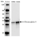
FIG. 2 is an SDS-PAGE identification of the expressed recombinant protein Desmoglein 1;
FIG. 3 is a solid-phase membrane immunoassay kit for detecting anti-Desmoglein 1-IgG antibodies in serum of patients with nephrotic syndrome;
FIG. 4 is a schematic diagram of the magnetic particle chemiluminescence immunoassay kit for detecting anti-Desmoglein 1-IgG antibody;
FIG. 5 is a schematic diagram of an antigenic protein Desmoglein1 coated carboxyl magnetic particle;
FIG. 6 shows the detection of anti-Desmoglein 1-IgG antibodies in patients with renal disease of various types, in which NS: nephrotic syndrome, HP: allergic purpura, HPN: purpuric nephritis, KD: kawasaki disease, NC: a healthy child;
FIG. 7 is a linear correlation of anti-Desmoglein 1-IgG antibodies to a marker of glomerular vascular endothelial cell injury.
Detailed Description
The invention provides application of a reagent for detecting an anti-human Desmoglein1 autoantibody in preparation of a vascular endothelial injury detection tool.
Blood, blood vessels and the heart constitute the blood circulation system of the human body, and blood in the blood circulation system flows through the blood vessels and flows through the organs of the whole body such as the heart, the lung, the liver and the like. The innermost layer of the blood vessel is attached with vascular endothelial cells, and the vascular endothelial cell self antibody can cause damage to the vascular endothelial cells, induce dysfunction of blood circulation system, further cause damage to organs such as heart, lung and liver, and cause related diseases of various organs including nephrotic syndrome, but the vascular endothelial cells of different organs are the same. Therefore, the method can be used for clinical vascular endothelial cell injury based on the detection of vascular endothelial cell autoantibodies in the blood circulation system. The antigen protein Desmoglein1 of the anti-human Desmoglein1 autoantibody is desmosomal protein 1, which is DSG1 for short. In the present invention, the DSG1 preferably comprises the whole or a fragment of protein accession No. BC153001, and the amino acid sequence (MKKLADISLGKESYPDLDPSWPP QSTEPVCLPQETEPVVSGHPPISPHFGTTTVISESTYPSGPGVLHPKPILDPLGYGNVTVTESYTTSDTLKPSVHVHDNRPASNVVVTERVVGPISGADLHGMLEMPDLRDGSNVIVTERVIAPSSSLPTSLTIHHPRESSNVVVTERVIQPTSGMIGSLSMHPELANAHNVIVTERVVSGAGVTGISGTTGISGGIGSSGLVGTSMGAGSGALSGAGISGGGIGLSSLGGTASIGHMRSSSDHHFNQTIGSASPSTARSRITKYSTVQYSKHHHHHH) shown in SEQ ID No.1 is exemplified for convenience of description in the examples, but it cannot be considered as the full scope of the present invention.
The source of the antigen protein is not specially limited, and the antigen protein can be expressed in bacteria such as escherichia coli, yeast and mammalian cells and is obtained by Ni column affinity chromatography, molecular sieve chromatography, ion exchange chromatography and hydrophobic column purification. The antigenic proteins of the invention preferably include fusion proteins, such as those that incorporate a tag at the N-terminus or C-terminus of the antigenic protein, to confer some biological or physical function, and to specifically bind to a sequence or domain of a ligand; the label preferably comprises a His label, thioredoxin, a GST label, maltose binding protein, a SA label of glutathione transferase, a c-Myc label, a Flag label or a biotin label, and the existence of the label is favorable for purification, fixation and precipitation of antigen protein.
The detection means according to the invention preferably comprises a detection kit. The reagent of the present invention preferably includes a reagent for detecting an anti-Desmoglein 1-IgG antibody.
The invention also provides a kit for detecting the anti-Desmoglein 1-IgG antibody, which comprises the antigen protein Desmoglein1, a solid phase carrier and a labeled antibody which are independently packaged;
the antigenic protein Desmoglein1 comprises the whole or a fragment of protein accession BC 153001.
The antigenic protein Desmoglein1 of the present invention is preferably the same as described above and will not be described herein. The antigenic protein Desmoglein1 can be fixed on a solid phase carrier, and the solid phase carrier preferably comprises a nitrocellulose membrane (NC membrane), a fluorescent coding microsphere, a magnetic stripe chip, a magnetic particle and/or an enzyme labeling microporous plate. In the invention, when the solid phase carriers are selected differently, different kits can be obtained, for example, when magnetic particles are used as the solid phase carriers, a magnetic particle chemiluminescence immunoassay kit for detecting the anti-Desmoglein 1-IgG antibody can be constructed; when the nitrocellulose membrane is used as a solid phase carrier, a solid phase membrane immunoassay kit for detecting the anti-Desmoglein 1-IgG antibody can be constructed, and of course, a plurality of different solid phase carriers can coexist in the same kit, thereby completing qualitative and quantitative detection.
The immobilization according to the present invention preferably means binding to a solid support insoluble in water of the Desmoglein1 antigenic protein, which solid support or support is insoluble in water, more preferably by covalent bonding, electrostatic interaction, hydrophobic interaction, or interaction by disulfide bond, most preferably by one or more covalent bonds. The method for fixing is not particularly limited, and the fixing can be a direct fixing mode or an indirect fixing mode. The direct immobilization according to the invention preferably comprises separating the immobilized molecules from the aqueous solution together with the insoluble carrier by filtration, centrifugation or chromatography; also included is the immobilization of Desmoglein1 antigenic protein in a reversible or irreversible manner. For example, the antigenic protein is immobilized to the carrier by a cleavable covalent bond (e.g., a disulfide bond that can be cleaved by addition of a thiol-containing reagent), which is reversible. In addition, if the antigenic protein is immobilized to the carrier by a covalent bond that does not cleave in aqueous solution (a bond formed by the reaction of an epoxide group with the amine group coupling the lysine side chain to the affinity column), the immobilization is irreversible. The invention indirectly fixes the antibody with specific affinity to the antigen protein, and then forms an antigen protein-antibody compound to achieve the purpose of fixing. In the invention, the immobilization mode is slightly different aiming at different solid phase carriers, such as that the antigen protein Desmoglein1 is bonded to a nitrocellulose membrane or a polystyrene microporous plate through a physical adsorption mode or a non-covalent bond; the magnetic particles with the carboxyl functional groups are combined with the amino group of the antigen protein Desmoglein1, and the antigen protein Desmoglein1 is combined on the magnetic particles in a chemical coupling mode.
The labeled antibody of the present invention preferably comprises a secondary antibody labeled with an enzyme, a chemiluminescent agent or biotin; the secondary antibody comprises an anti-human IgG antibody. The enzyme label of the present invention preferably comprises a Horseradish Peroxidase (HRP) label, and the chemiluminescent label preferably comprises an acridinium ester label.
The kit preferably further comprises antigen diluent, sample diluent buffer, antigen diluent, substrate color development solution, washing solution, standard substance, positive quality control product and negative quality control product. The standard substance and the positive quality control substance are preferably recombinant human anti-tag peptide immunoglobulin G or fragments thereof, or anti-Desmoglein 1-IgG antibodies extracted from patient serum; the negative quality control product is preferably serum of a healthy physical examiner. The substrate color developing solution preferably comprises any one of TMB, hydrogen peroxide, AMPPD, 4-MUP and BCIP; the antigen diluent is preferably 1 XPBS (phosphate buffer solution) containing 0.15mol/L NaCl and 1% (W/V) TritonX-100, and the pH value is 7.35; the sample dilution buffer is preferably 0.01M PBS containing 10% (W/V) BSA, pH 7.35; the antibody diluent is preferably 0.01M PBS containing 1M D-glucose, 2% (W/V) glycerol, 0.3% (W/V) Tween20, pH 7.35; the washing solution is preferably 1 XPBS, pH7.35, containing 0.15mol/LNaCl, 10% (W/V) glycerol, 1% (W/V) TritonX-100.
The detection kit provided by the invention utilizes an indirect method reaction principle, firstly, Desmoglein1 antigen is adsorbed on a solid phase carrier to serve as coating antigen, then positive quality control products or standard products or samples to be detected are added for incubation, then labeled secondary antibody is added for reaction, if anti-Desmoglein 1-IgG antibody is contained in the serum to be detected, a ternary complex of the coating antigen Desmoglein 1-anti-Desmoglein 1-IgG antibody of the serum to be detected and labeled anti-human IgG antibody is formed, and finally, a light signal is detected by utilizing a photocolor method, a chemiluminescence method and a fluorescence method, so that the purpose of qualitatively or quantitatively analyzing the anti-Desmoglein 1-IgG antibody in human serum is achieved. The test sample of the present invention preferably comprises blood, serum, tissue or body fluid. The tissue of the present invention preferably comprises kidney biopsy tissue; the body fluid is preferably body fluid in the conventional sense in the art, including both intracellular and extracellular fluids, with extracellular fluid being predominantly plasma.
The detection kit disclosed by the invention relates to a Desmoglein1-IgG antibody in human serum subjected to solid-phase membrane immunoassay qualitative analysis, and the human anti-tag peptide IgG antibody is used as a standard substance, so that the detection accuracy is greatly improved. The solid-phase membrane immunoassay qualitative detection is simple to operate, the reagent dosage is less, and the solid-phase membrane immunoassay qualitative detection is saved by about 10 times compared with the traditional ELISA; in addition, the adsorption capacity of the NC membrane is extremely close to 100%, and trace antigens can be completely adsorbed and fixed on the NC membrane; the NC membrane with adsorbed antigen or antibody or existing result can be preserved for a long time (half a year at-20 ℃), and the activity of the NC membrane is not influenced; in addition, the kit for qualitatively detecting the anti-Desmoglein 1-IgG antibody in the human serum by the solid-phase membrane immunoassay is introduced into a biotin-avidin amplification system, so that the detection sensitivity is greatly improved. The detection kit also relates to a magnetic particle chemiluminescence immunoassay quantitative detection method for Desmoglein1-IgG antibody in human serum, and the magnetic particle is used as a solid phase carrier, the diameter of the magnetic particle is only 1.0 mu m, so that the coating surface area is greatly increased, the adsorption quantity of the antigen is increased, the reaction speed is improved, the cleaning and separation are simpler and more convenient, the pollution is reduced, and the cross infection probability is reduced. On the other hand, the acridine ester luminescent agent is adopted to directly mark the anti-human IgG, the chemical reaction is simple and quick, and no catalyst is needed; the acridinium ester chemiluminescence is of the scintillation type by initiating the luminescent reagent (H) 2 O 2 NaOH) can reach the maximum after 0.4s, the half-life period is 0.9s, and the emission intensity is basically within 2sAnd finishing, and facilitating quick detection.
The following examples are provided to illustrate the kit for detecting anti-Desmoglein 1-IgG antibodies of the present invention in detail, but they should not be construed as limiting the scope of the present invention.
Example 1
Desmoglein1 protein on vascular endothelial cells is a main target antigen for autoantibody in patients with nephrotic syndrome
(1) Extraction of vascular endothelial cell total protein: a vascular endothelial cell strain (EAhy926) was cultured, washed 2 to 3 times with PBS, then sufficiently lysed on ice in a lysis buffer containing 30mM Tris-HCl, 8M urea, 4% CHAPS, and a protease inhibitor (# ab 65621; Abcam, 1:200 dilution) with a focused ultrasound machine (Covaris S220, Gene), and then the sample was centrifuged at 12000g, 4 ℃ for 30 min. Collecting the supernatant, namely the total protein of the vascular endothelial cells. The total protein concentration of the collected vascular endothelial cells was measured using the BCA protein concentration measurement kit.
(2) Two-dimensional electrophoresis: extracting total protein of vascular endothelial cell, performing two-dimensional electrophoresis, transferring to nitrocellulose membrane, incubating with serum of healthy person and nephrotic syndrome patient as primary antibody, and developing with secondary antibody, shown as A in FIG. 1 and B in FIG. 1.
(3) Matrix-assisted laser desorption/ionization time-of-flight mass spectrometry: differential analysis of positive spots was performed after imaging in step (2), protein spots were selected on two-dimensional electrophoresis gel which were strongly positive for nephrotic syndrome patients and negative or weakly positive for healthy persons, the selected protein spots were removed from the gel, the dried gel was digested with trypsin (0.1. mu.g/. mu.L), 10. mu.L of 25mM ammonium bicarbonate was added to the reaction mixture, incubated overnight at 37 ℃, and peptides were then extracted from the gel with trifluoroacetic acid (0.1%). The extracted peptides were analyzed by matrix assisted laser desorption/ionization time of flight mass spectrometry (MALDI-TOF-MS) mass spectrometer to obtain a peptide mass spectrum, which was identified as Desmoglein1 protein (fig. 1, C).
Example 2
Expression and purification of recombinant antigen protein Desmoglein1
The gene of Desmoglein1 protein is used as a template by a genetic engineering method for PCR amplification, and then an expression vector is constructed for protein expression. The antigen protein expressed by the invention contains a tag peptide of His tag. The expressed recombinant protein is purified by nickel column affinity chromatography, and finally, the molecular weight of the recombinant protein Desmoglein1 is identified by SDS-PAGE, and is shown in figure 2.
Example 3
An orthogonal table was selected based on 4 factors such as antigen Desmoglein1 coating concentration (five coating concentrations of 20. mu.g, 40. mu.g, 60. mu.g, 80. mu.g, and 100. mu.g), each reaction time (10min, 20min, 30min, and 40min), and temperature (25 ℃, 37 ℃), enzyme-labeled secondary antibody optimal dilution (four dilutions of 1:200, 1:400, 1:800, and 1:1600 ℃), each factor repeatedly determining standard positive serum and standard negative serum at 2 levels, and selecting a ratio (P/N) of the highest light signal value (P) of positive serum to the lowest light signal value (N) of negative serum.
Through orthogonal design, the optimal antigen Desmoglein1 coating concentration of the kit is 60 mu g/mL, the optimal antigen-antibody reaction temperature of the anti-Desmoglein 1-IgG antibody kit for solid-phase membrane immunodetection is 25 ℃, the optimal antigen-antibody reaction time is 20min, and the optimal work dilution of the optimal biotin-labeled anti-human IgG antibody is 1:400, respectively; the optimal antigen-antibody reaction temperature of the kit for detecting the anti-Desmoglein 1-IgG antibody by magnetic particle chemiluminescence immunoassay is 37 ℃, the optimal antigen-antibody reaction time is 20min, and the optimal working dilution of the optimal acridinium ester labeled anti-human IgG antibody is 1: 400.
example 4
Preparation of solid-phase membrane immunoassay kit for detecting anti-Desmoglein 1-IgG antibody
4.1 composition of solid-phase membrane immunoassay kit for detecting anti-Desmoglein 1-IgG antibody:
antigen: recombinant protein Desmoglein1
Solid phase carrier: sataurus CN140 nitrocellulose membrane
Positive quality control (standard): human anti-His tag immunoglobulin G (purchased from Huzhou Yingchuang)
Negative quality control product: serum for health physical examination person
Labeling of the antibody: biotin-labeled anti-human IgG antibody
Antigen diluent
Sample dilution buffer
Antibody diluent
Cleaning solution
Enzyme working solution: alkaline phosphatase-streptavidin
Substrate color development solution: BCIP color developing solution.
4.2 detection procedure of the solid-phase membrane immunoassay kit for detecting anti-Desmoglein 1-IgG antibody is as follows:
4.2.1 coating, sealing: placing 10 μ L Desmoglein1 antigen direct contact with concentration of 60 μ g/mL on nitrocellulose membrane, drying in 37 deg.C incubator for 20min, placing nitrocellulose membrane in detection plate, adding 200 μ L5% (W/V) BSA, sealing in 37 deg.C incubator for 20min, discarding the sealing solution, and washing with washing solution for 2 times;
4.2.2 antigen incubation: adding 10 μ L of antibody standard or serum to be detected diluted with diluent into the detection plate, performing negative control and positive control, incubating at 25 deg.C for 20min, and arranging 3 parallel holes for each sample;
4.2.3 Secondary antibody incubation: discarding the liquid in the detection plate, washing with the washing solution for 5 times × 1min, adding 20 μ L1:400 biotin-labeled anti-human IgG antibody, and incubating at 25 deg.C for 20 min;
4.2.4 color development: discarding the liquid in the detection plate, washing with washing solution for 3 times × 3min, adding 500 μ L alkaline phosphatase-streptavidin, incubating at room temperature for 20min, discarding the liquid in the detection plate, washing with washing solution for 3 times × 3min, adding BCIP color developing solution, reacting at room temperature for 20min, washing the detection plate with running water, and terminating the enzyme reaction. And taking out the test nitrocellulose membrane strip, drying the membrane strip by using a blower, qualitatively judging by using a colorimetric card by naked eyes, and drawing a semi-quantitative analysis on the anti-Desmoglein 1-IgG antibody level in the serum by drawing a standard curve by using analysis software carried by a developing instrument and taking the concentration of a reference standard substance as a vertical coordinate and a gray value read by the instrument as a horizontal coordinate, wherein the positive result is shown in figure 3 when a brown spot appears, or placing the membrane strip on the developing instrument for scanning.
Example 5
Preparation of magnetic particle chemiluminescence immunoassay kit for detecting Desmoglein1-IgG antibody
5.1 anti-Desmoglein 1-IgG antibody chemiluminescence detection kit, including the following components:
(1) acridinium ester labeled anti-human IgG;
(2) carboxyl magnetic beads coupled with Desmoglein1 antigen;
(3) chemiluminescent pre-excitation liquid H 2 O 2 And exciting liquid NaOH;
(4) anti-Desmoglein 1-IgG antibody series standard solution, standard concentration: 0. mu.g/mL, 4. mu.g/mL, 8. mu.g/mL, 16. mu.g/mL, 20. mu.g/mL, 40. mu.g/mL;
(5) the antigen diluent is 1 XPBS containing 0.15mol/LNaCl and 1% (W/V) TritonX-100, and the pH value is 7.35; the sample dilution buffer was 0.01M PBS containing 10% (W/V) BSA, pH 7.35; the antibody diluent is 0.01MPBS containing 1M D-glucose, 2% (W/V) glycerol and 0.3% (W/V) Tween20, and has a pH value of 7.35; the washing solution is as follows: 1 XPBS with 0.15mol/LNaCl, 10% (W/V) glycerol, 1% (W/V) TritonX-100, pH 7.35.
5.2 preparation of magnetic bead-coupled antigen (FIG. 5)
(1) 1mg of carboxyl magnetic particles are put into a 0.5mL centrifuge tube, 200 mu L of 0.1mol/L MES buffer solution is added, the mixture is evenly mixed by vortex, the mixture is placed on a magnetic frame and is kept stand for 5min, the magnetic particles are separated from liquid, and supernatant liquid is discarded. Wash 3 times, then add 200 μ Ι _ MES (pH 5.0) buffer and vortex;
(2) adding 18 mu LDesmoglein 1 antigen, whirling, rotating the reaction tube, and incubating for 20min at room temperature;
(3) adding 10 mu L of 10mg/mL coupling reagent EDC, vortexing, rotating the reaction tube, and incubating for 1h at room temperature;
(4) the supernatant was removed and washed 3 times with 200. mu.L of washing buffer (TBS + 0.05% (W/V) Tween-20);
(5) blocking with 1% (W/V) BSA in buffer was repeated 4 times for 10min each. The magnetic particle suspension was stored at 2-8 ℃.
5.3 preparation of acridinium ester-labeled antibody
(1) mu.L of anti-human IgG antibodyPutting the body into dialysis bag, putting the dialysis bag into 1L labeled buffer solution, dialyzing for at least 3 times, dialyzing overnight for the last time, wherein the labeled buffer solution is Na 2 CO 3 -NaHCO 3 A buffer solution with pH of 10.0 and concentration of 0.1 mol/L;
(2) weighing 1.7mg of acridinium ester NSP-DMAE-NHS, and dissolving in 447 mu L of anhydrous dimethylformamide DMF to form 6.5mmol/LNSP-DMAE-NHS DMF solution;
(3) placing the dialyzed antibody solution into a 500-mu-L centrifuge tube, adding 100-mu-L6.5 mmol/L NSP-DMAE-NHS DMF solution, wherein the molar ratio of acridine ester to the antibody is 7.4:1, adding 200-mu-L labeling buffer solution, reacting at room temperature for 45min, adding 10-mu-L lysine, and continuing to react for 15min to terminate the labeling reaction;
(4) separating the marker NSP-DMAE-NHS-Ab from free NSP-DMAE-NHS by Sephadex G-50 column (1X 25cm), using purification buffer solution with pH of 6.3 and concentration of 0.1 mol/L;
(5) during the separation process, detecting protein peaks by using a chromatograph, and respectively measuring the chemiluminescence intensity of effluent and the absorbance at 430 nm;
(6) the high-light, high-absorbance eluate was collected, 1% BSA (by volume) was added, and stored on ice.
5.4 sample preparation-sample dilution 1:10
5.5 detection procedure of the chemiluminescence kit for detecting anti-Desmoglein 1-IgG antibody is as follows:
(1) sequentially adding 100 mu L of sample to be detected, 100 mu L of coupled magnetic powder suspension and 100 mu L of acridinium ester labeled secondary antibody into a reaction tube, shaking up and mixing, and keeping the temperature at 37 ℃ for 20 min;
(2) washing for 5 times;
(3) fully shaking the washed reaction container to uniformly disperse the magnetic particles;
(4) adding 100 μ L of chemiluminescent pre-excitation solution H 2 O 2 Then, 100. mu.L of NaOH was added to the chemiluminescence excitation solution, and the relative luminescence intensity was measured. The content of anti-Desmoglein 1-IgG antibody in the sample was directly proportional to its luminescence intensity (FIG. 4).
Example 6
Clinical application of kit for detecting serum anti-Desmoglein 1-IgG antibody
6.1 Subjects included patients diagnosed with various types of nephropathies from 6 months in 2018 to 6 months in 2020, including 298 Nephrotic Syndrome (NS), 100 Henoch Schonlein purpura (HP), 100 Henoch Schonlein nephritis (HPN), 100 Kawasaki Disease (KD), and 100 healthy children (NC) at the same time. Serum samples were taken from various renal patients and healthy controls. All subjects received a first serum sample collection prior to no immunosuppressive treatment.
6.2 detection of anti-Desmoglein 1-IgG antibodies in patients with various types of nephropathy the kit of the present invention was used to detect the anti-Desmoglein 1-IgG antibody levels in the serum of patients diagnosed with various types of nephropathy from 6 months 2018 to 6 months 2020, including 298 nephrotic syndrome, 100 Henoch-Schonlein purpura nephritis, 100 Kawasaki disease and 100 healthy children at the same time, and the results showed that 120 patients among the nephrotic syndrome patients were positive for anti-Desmoglein 1-IgG antibodies, and that anti-Desmoglein 1-IgG antibodies in Henoch-Schonlein nephritis, Henoch-Schonlein purpura, Kawasaki disease and healthy children were negative, as shown in FIG. 6.
6.3 serum anti-Desmoglein 1-IgG antibody of nephrotic syndrome patient is linearly related to the expression level of vascular endothelial injury marker, the kit of the invention is used for detecting the expression level of anti-Desmoglein 1-IgG antibody in the serum of nephrotic syndrome patient diagnosed from 6 months 2018 to 6 months 2020, and detecting the expression level of vascular endothelial injury marker Plvap in the serum of patient, the result shows that the expression level of anti-Desmoglein 1-IgG antibody of nephrotic syndrome patient is linearly related to the expression level of vascular endothelial injury marker, the nephrotic syndrome is related to vascular endothelial injury, see figure 7.
The foregoing is only a preferred embodiment of the present invention, and it should be noted that, for those skilled in the art, various modifications and amendments can be made without departing from the principle of the present invention, and these modifications and amendments should also be considered as the protection scope of the present invention.
Sequence listing
<110> Zhejiang university
<120> kit for detecting anti-Desmoglein 1-IgG antibody
<160> 1
<170> SIPOSequenceListing 1.0
<210> 1
<211> 301
<212> PRT
<213> Artificial Sequence (Artificial Sequence)
<400> 1
Met Lys Lys Leu Ala Asp Ile Ser Leu Gly Lys Glu Ser Tyr Pro Asp
1 5 10 15
Leu Asp Pro Ser Trp Pro Pro Gln Ser Thr Glu Pro Val Cys Leu Pro
20 25 30
Gln Glu Thr Glu Pro Val Val Ser Gly His Pro Pro Ile Ser Pro His
35 40 45
Phe Gly Thr Thr Thr Val Ile Ser Glu Ser Thr Tyr Pro Ser Gly Pro
50 55 60
Gly Val Leu His Pro Lys Pro Ile Leu Asp Pro Leu Gly Tyr Gly Asn
65 70 75 80
Val Thr Val Thr Glu Ser Tyr Thr Thr Ser Asp Thr Leu Lys Pro Ser
85 90 95
Val His Val His Asp Asn Arg Pro Ala Ser Asn Val Val Val Thr Glu
100 105 110
Arg Val Val Gly Pro Ile Ser Gly Ala Asp Leu His Gly Met Leu Glu
115 120 125
Met Pro Asp Leu Arg Asp Gly Ser Asn Val Ile Val Thr Glu Arg Val
130 135 140
Ile Ala Pro Ser Ser Ser Leu Pro Thr Ser Leu Thr Ile His His Pro
145 150 155 160
Arg Glu Ser Ser Asn Val Val Val Thr Glu Arg Val Ile Gln Pro Thr
165 170 175
Ser Gly Met Ile Gly Ser Leu Ser Met His Pro Glu Leu Ala Asn Ala
180 185 190
His Asn Val Ile Val Thr Glu Arg Val Val Ser Gly Ala Gly Val Thr
195 200 205
Gly Ile Ser Gly Thr Thr Gly Ile Ser Gly Gly Ile Gly Ser Ser Gly
210 215 220
Leu Val Gly Thr Ser Met Gly Ala Gly Ser Gly Ala Leu Ser Gly Ala
225 230 235 240
Gly Ile Ser Gly Gly Gly Ile Gly Leu Ser Ser Leu Gly Gly Thr Ala
245 250 255
Ser Ile Gly His Met Arg Ser Ser Ser Asp His His Phe Asn Gln Thr
260 265 270
Ile Gly Ser Ala Ser Pro Ser Thr Ala Arg Ser Arg Ile Thr Lys Tyr
275 280 285
Ser Thr Val Gln Tyr Ser Lys His His His His His His
290 295 300
Claims (8)
Priority Applications (1)
| Application Number | Priority Date | Filing Date | Title |
|---|---|---|---|
| CN202210491000.0A CN114994308A (en) | 2022-05-07 | 2022-05-07 | Kit for detecting Desmoglein1-IgG antibody |
Applications Claiming Priority (1)
| Application Number | Priority Date | Filing Date | Title |
|---|---|---|---|
| CN202210491000.0A CN114994308A (en) | 2022-05-07 | 2022-05-07 | Kit for detecting Desmoglein1-IgG antibody |
Publications (1)
| Publication Number | Publication Date |
|---|---|
| CN114994308A true CN114994308A (en) | 2022-09-02 |
Family
ID=83025077
Family Applications (1)
| Application Number | Title | Priority Date | Filing Date |
|---|---|---|---|
| CN202210491000.0A Pending CN114994308A (en) | 2022-05-07 | 2022-05-07 | Kit for detecting Desmoglein1-IgG antibody |
Country Status (1)
| Country | Link |
|---|---|
| CN (1) | CN114994308A (en) |
Cited By (1)
| Publication number | Priority date | Publication date | Assignee | Title |
|---|---|---|---|---|
| CN117074667A (en) * | 2023-03-02 | 2023-11-17 | 浙江大学 | Use of polypeptides or fragments thereof in preparing kits for detecting vascular endothelial cell damage |
Citations (4)
| Publication number | Priority date | Publication date | Assignee | Title |
|---|---|---|---|---|
| WO2008097439A2 (en) * | 2007-02-07 | 2008-08-14 | The Trustees Of The University Of Pennsylvania | Anti-autoimmune antibodies for treatment of pemphigus |
| US20200123230A1 (en) * | 2018-10-22 | 2020-04-23 | Euroimmun Medizinische Labordiagnostika Ag | Diagnosis of Blistering Autoimmune Disease |
| WO2022043415A1 (en) * | 2020-08-27 | 2022-03-03 | INSERM (Institut National de la Santé et de la Recherche Médicale) | Methods for detecting the presence of pemphigus-specific autoantibodies in a sample |
| CN114152754A (en) * | 2021-11-26 | 2022-03-08 | 钱华 | ELISA kit for detecting human Dsg1IgG antibody and application thereof |
-
2022
- 2022-05-07 CN CN202210491000.0A patent/CN114994308A/en active Pending
Patent Citations (5)
| Publication number | Priority date | Publication date | Assignee | Title |
|---|---|---|---|---|
| WO2008097439A2 (en) * | 2007-02-07 | 2008-08-14 | The Trustees Of The University Of Pennsylvania | Anti-autoimmune antibodies for treatment of pemphigus |
| US20200123230A1 (en) * | 2018-10-22 | 2020-04-23 | Euroimmun Medizinische Labordiagnostika Ag | Diagnosis of Blistering Autoimmune Disease |
| CN111072768A (en) * | 2018-10-22 | 2020-04-28 | 欧蒙医学实验诊断股份公司 | Diagnosis of vesicular autoimmune diseases |
| WO2022043415A1 (en) * | 2020-08-27 | 2022-03-03 | INSERM (Institut National de la Santé et de la Recherche Médicale) | Methods for detecting the presence of pemphigus-specific autoantibodies in a sample |
| CN114152754A (en) * | 2021-11-26 | 2022-03-08 | 钱华 | ELISA kit for detecting human Dsg1IgG antibody and application thereof |
Non-Patent Citations (1)
| Title |
|---|
| 胡智祥主编: "《医院临床检验技术操作规范与实(化)验室管理全书》", 30 August 2004 * |
Cited By (3)
| Publication number | Priority date | Publication date | Assignee | Title |
|---|---|---|---|---|
| CN117074667A (en) * | 2023-03-02 | 2023-11-17 | 浙江大学 | Use of polypeptides or fragments thereof in preparing kits for detecting vascular endothelial cell damage |
| CN117074667B (en) * | 2023-03-02 | 2024-06-04 | 浙江大学 | Application of polypeptide or its fragment in preparing kit for detecting vascular endothelial cell injury |
| CN118348240A (en) * | 2023-03-02 | 2024-07-16 | 浙江大学 | Application of polypeptide or its fragment in preparing a kit for detecting vascular endothelial cell injury |
Similar Documents
| Publication | Publication Date | Title |
|---|---|---|
| EP2064551B1 (en) | Combination hepatitis c virus antigen and antibody detection method | |
| CN113447659B (en) | A kit for detecting anti-proteasome subunit α1-IgG antibody | |
| EP1328811B1 (en) | Hcv mosaic antigen composition | |
| CN113447658B (en) | Kit for detecting anti-peroxiredoxin-1-IgG antibody | |
| CN112415195A (en) | Kit for detecting dual targets of novel coronavirus and its application | |
| CN104991077A (en) | Troponin I competition turbidimetry detecting kit | |
| CN114994330A (en) | Kit for detecting anti-HSP 90-beta-IgG autoantibody and application thereof | |
| CN113447649B (en) | Kit for detecting anti-adhesion plaque protein-IgG antibody | |
| CN114994308A (en) | Kit for detecting Desmoglein1-IgG antibody | |
| CN114720700A (en) | Application of reagent for detecting anti-cytoskeleton-associated protein4-IgG autoantibody in preparation of kit for detecting vascular endothelial injury | |
| CN114895023A (en) | Application of reagent for detecting anti-Talin-1-IgG autoantibody in preparation of kit for detecting vascular endothelial injury | |
| CN114966045A (en) | Application of reagent for detecting anti-myosin light chain1-IgG autoantibody in preparation of kit for detecting vascular endothelial injury | |
| CN114924081A (en) | Application of neuroblast differentiation related protein-IgG in preparation of vascular endothelial injury kit | |
| CN115060898A (en) | Kit for detecting antigen myosin 1-IgG antibody | |
| CN113447656B (en) | Kit for detecting anti-filamentous actin cap-forming protein beta-IgG antibody | |
| CN114910647A (en) | Application of filamin-A-IgG antibody in preparation of kit for detecting vascular endothelial injury | |
| CN114910649A (en) | Application of reagent for detecting anti-alpha-enolase-IgG antibody in preparation of kit for detecting vascular endothelial injury | |
| CN114910650A (en) | Application of reagent for detecting anti-moesin-IgG antibody in preparation of kit for detecting vascular endothelial injury | |
| CN113447657B (en) | Detection kit for detecting anti-aconitate hydratase-IgG antibody | |
| CN113447648B (en) | Kit for detecting anti-serine/arginine-rich splicing factor 9-IgG antibody | |
| CN113447650B (en) | Detection kit for anti-peptidyl prolyl cis-trans isomerase D-IgG antibody | |
| CN114910643A (en) | A method and reagent for identifying antibodies that bind to mutant antigens | |
| DE DK et al. | HCV MOSAIK ANTIGEN ZUSAMMENSETZUNG COMPOSITION D’ANTIGENE MOSAIQUE DU VIRUS DE L’HEPATITE C (VHC) | |
| JPH06249852A (en) | D-amino acid oxidizing enzyme polychlornal antibody and measurement of d-amino acid oxidizing enzyme using the antibody |
Legal Events
| Date | Code | Title | Description |
|---|---|---|---|
| PB01 | Publication | ||
| PB01 | Publication | ||
| SE01 | Entry into force of request for substantive examination | ||
| SE01 | Entry into force of request for substantive examination | ||
| RJ01 | Rejection of invention patent application after publication | ||
| RJ01 | Rejection of invention patent application after publication |
Application publication date: 20220902 |