CN114502584A - CD33 antibodies and methods of using the same for treating cancer - Google Patents
CD33 antibodies and methods of using the same for treating cancer Download PDFInfo
- Publication number
- CN114502584A CN114502584A CN202080029923.3A CN202080029923A CN114502584A CN 114502584 A CN114502584 A CN 114502584A CN 202080029923 A CN202080029923 A CN 202080029923A CN 114502584 A CN114502584 A CN 114502584A
- Authority
- CN
- China
- Prior art keywords
- antibody
- antigen
- immunoglobulin
- seq
- binding fragment
- Prior art date
- Legal status (The legal status is an assumption and is not a legal conclusion. Google has not performed a legal analysis and makes no representation as to the accuracy of the status listed.)
- Granted
Links
Images


Classifications
-
- C—CHEMISTRY; METALLURGY
- C07—ORGANIC CHEMISTRY
- C07K—PEPTIDES
- C07K16/00—Immunoglobulins [IGs], e.g. monoclonal or polyclonal antibodies
- C07K16/18—Immunoglobulins [IGs], e.g. monoclonal or polyclonal antibodies against material from animals or humans
- C07K16/28—Immunoglobulins [IGs], e.g. monoclonal or polyclonal antibodies against material from animals or humans against receptors, cell surface antigens or cell surface determinants
- C07K16/2803—Immunoglobulins [IGs], e.g. monoclonal or polyclonal antibodies against material from animals or humans against receptors, cell surface antigens or cell surface determinants against the immunoglobulin superfamily
-
- A—HUMAN NECESSITIES
- A61—MEDICAL OR VETERINARY SCIENCE; HYGIENE
- A61K—PREPARATIONS FOR MEDICAL, DENTAL OR TOILETRY PURPOSES
- A61K39/00—Medicinal preparations containing antigens or antibodies
- A61K39/395—Antibodies; Immunoglobulins; Immune serum, e.g. antilymphocytic serum
- A61K39/39533—Antibodies; Immunoglobulins; Immune serum, e.g. antilymphocytic serum against materials from animals
- A61K39/3955—Antibodies; Immunoglobulins; Immune serum, e.g. antilymphocytic serum against materials from animals against proteinaceous materials, e.g. enzymes, hormones, lymphokines
-
- A—HUMAN NECESSITIES
- A61—MEDICAL OR VETERINARY SCIENCE; HYGIENE
- A61K—PREPARATIONS FOR MEDICAL, DENTAL OR TOILETRY PURPOSES
- A61K45/00—Medicinal preparations containing active ingredients not provided for in groups A61K31/00 - A61K41/00
- A61K45/06—Mixtures of active ingredients without chemical characterisation, e.g. antiphlogistics and cardiaca
-
- A—HUMAN NECESSITIES
- A61—MEDICAL OR VETERINARY SCIENCE; HYGIENE
- A61K—PREPARATIONS FOR MEDICAL, DENTAL OR TOILETRY PURPOSES
- A61K51/00—Preparations containing radioactive substances for use in therapy or testing in vivo
- A61K51/02—Preparations containing radioactive substances for use in therapy or testing in vivo characterised by the carrier, i.e. characterised by the agent or material covalently linked or complexing the radioactive nucleus
- A61K51/04—Organic compounds
- A61K51/08—Peptides, e.g. proteins, carriers being peptides, polyamino acids, proteins
- A61K51/10—Antibodies or immunoglobulins; Fragments thereof, the carrier being an antibody, an immunoglobulin or a fragment thereof, e.g. a camelised human single domain antibody or the Fc fragment of an antibody
- A61K51/1018—Antibodies or immunoglobulins; Fragments thereof, the carrier being an antibody, an immunoglobulin or a fragment thereof, e.g. a camelised human single domain antibody or the Fc fragment of an antibody against material from animals or humans
-
- A—HUMAN NECESSITIES
- A61—MEDICAL OR VETERINARY SCIENCE; HYGIENE
- A61K—PREPARATIONS FOR MEDICAL, DENTAL OR TOILETRY PURPOSES
- A61K51/00—Preparations containing radioactive substances for use in therapy or testing in vivo
- A61K51/02—Preparations containing radioactive substances for use in therapy or testing in vivo characterised by the carrier, i.e. characterised by the agent or material covalently linked or complexing the radioactive nucleus
- A61K51/04—Organic compounds
- A61K51/08—Peptides, e.g. proteins, carriers being peptides, polyamino acids, proteins
- A61K51/10—Antibodies or immunoglobulins; Fragments thereof, the carrier being an antibody, an immunoglobulin or a fragment thereof, e.g. a camelised human single domain antibody or the Fc fragment of an antibody
- A61K51/1027—Antibodies or immunoglobulins; Fragments thereof, the carrier being an antibody, an immunoglobulin or a fragment thereof, e.g. a camelised human single domain antibody or the Fc fragment of an antibody against receptors, cell-surface antigens or cell-surface determinants
-
- A—HUMAN NECESSITIES
- A61—MEDICAL OR VETERINARY SCIENCE; HYGIENE
- A61P—SPECIFIC THERAPEUTIC ACTIVITY OF CHEMICAL COMPOUNDS OR MEDICINAL PREPARATIONS
- A61P25/00—Drugs for disorders of the nervous system
- A61P25/28—Drugs for disorders of the nervous system for treating neurodegenerative disorders of the central nervous system, e.g. nootropic agents, cognition enhancers, drugs for treating Alzheimer's disease or other forms of dementia
-
- A—HUMAN NECESSITIES
- A61—MEDICAL OR VETERINARY SCIENCE; HYGIENE
- A61P—SPECIFIC THERAPEUTIC ACTIVITY OF CHEMICAL COMPOUNDS OR MEDICINAL PREPARATIONS
- A61P35/00—Antineoplastic agents
-
- A—HUMAN NECESSITIES
- A61—MEDICAL OR VETERINARY SCIENCE; HYGIENE
- A61P—SPECIFIC THERAPEUTIC ACTIVITY OF CHEMICAL COMPOUNDS OR MEDICINAL PREPARATIONS
- A61P35/00—Antineoplastic agents
- A61P35/02—Antineoplastic agents specific for leukemia
-
- C—CHEMISTRY; METALLURGY
- C07—ORGANIC CHEMISTRY
- C07K—PEPTIDES
- C07K16/00—Immunoglobulins [IGs], e.g. monoclonal or polyclonal antibodies
- C07K16/18—Immunoglobulins [IGs], e.g. monoclonal or polyclonal antibodies against material from animals or humans
- C07K16/28—Immunoglobulins [IGs], e.g. monoclonal or polyclonal antibodies against material from animals or humans against receptors, cell surface antigens or cell surface determinants
- C07K16/2803—Immunoglobulins [IGs], e.g. monoclonal or polyclonal antibodies against material from animals or humans against receptors, cell surface antigens or cell surface determinants against the immunoglobulin superfamily
- C07K16/2809—Immunoglobulins [IGs], e.g. monoclonal or polyclonal antibodies against material from animals or humans against receptors, cell surface antigens or cell surface determinants against the immunoglobulin superfamily against the T-cell receptor (TcR)-CD3 complex
-
- C—CHEMISTRY; METALLURGY
- C07—ORGANIC CHEMISTRY
- C07K—PEPTIDES
- C07K16/00—Immunoglobulins [IGs], e.g. monoclonal or polyclonal antibodies
- C07K16/44—Immunoglobulins [IGs], e.g. monoclonal or polyclonal antibodies against material not provided for elsewhere, e.g. haptens, metals, DNA, RNA, amino acids
-
- G01N33/575—
-
- A—HUMAN NECESSITIES
- A61—MEDICAL OR VETERINARY SCIENCE; HYGIENE
- A61K—PREPARATIONS FOR MEDICAL, DENTAL OR TOILETRY PURPOSES
- A61K39/00—Medicinal preparations containing antigens or antibodies
- A61K2039/505—Medicinal preparations containing antigens or antibodies comprising antibodies
-
- C—CHEMISTRY; METALLURGY
- C07—ORGANIC CHEMISTRY
- C07K—PEPTIDES
- C07K2317/00—Immunoglobulins specific features
- C07K2317/20—Immunoglobulins specific features characterized by taxonomic origin
- C07K2317/24—Immunoglobulins specific features characterized by taxonomic origin containing regions, domains or residues from different species, e.g. chimeric, humanized or veneered
-
- C—CHEMISTRY; METALLURGY
- C07—ORGANIC CHEMISTRY
- C07K—PEPTIDES
- C07K2317/00—Immunoglobulins specific features
- C07K2317/30—Immunoglobulins specific features characterized by aspects of specificity or valency
- C07K2317/31—Immunoglobulins specific features characterized by aspects of specificity or valency multispecific
-
- C—CHEMISTRY; METALLURGY
- C07—ORGANIC CHEMISTRY
- C07K—PEPTIDES
- C07K2317/00—Immunoglobulins specific features
- C07K2317/30—Immunoglobulins specific features characterized by aspects of specificity or valency
- C07K2317/34—Identification of a linear epitope shorter than 20 amino acid residues or of a conformational epitope defined by amino acid residues
-
- C—CHEMISTRY; METALLURGY
- C07—ORGANIC CHEMISTRY
- C07K—PEPTIDES
- C07K2317/00—Immunoglobulins specific features
- C07K2317/40—Immunoglobulins specific features characterized by post-translational modification
-
- C—CHEMISTRY; METALLURGY
- C07—ORGANIC CHEMISTRY
- C07K—PEPTIDES
- C07K2317/00—Immunoglobulins specific features
- C07K2317/50—Immunoglobulins specific features characterized by immunoglobulin fragments
- C07K2317/51—Complete heavy chain or Fd fragment, i.e. VH + CH1
-
- C—CHEMISTRY; METALLURGY
- C07—ORGANIC CHEMISTRY
- C07K—PEPTIDES
- C07K2317/00—Immunoglobulins specific features
- C07K2317/50—Immunoglobulins specific features characterized by immunoglobulin fragments
- C07K2317/515—Complete light chain, i.e. VL + CL
-
- C—CHEMISTRY; METALLURGY
- C07—ORGANIC CHEMISTRY
- C07K—PEPTIDES
- C07K2317/00—Immunoglobulins specific features
- C07K2317/50—Immunoglobulins specific features characterized by immunoglobulin fragments
- C07K2317/52—Constant or Fc region; Isotype
- C07K2317/524—CH2 domain
-
- C—CHEMISTRY; METALLURGY
- C07—ORGANIC CHEMISTRY
- C07K—PEPTIDES
- C07K2317/00—Immunoglobulins specific features
- C07K2317/50—Immunoglobulins specific features characterized by immunoglobulin fragments
- C07K2317/56—Immunoglobulins specific features characterized by immunoglobulin fragments variable (Fv) region, i.e. VH and/or VL
- C07K2317/565—Complementarity determining region [CDR]
-
- C—CHEMISTRY; METALLURGY
- C07—ORGANIC CHEMISTRY
- C07K—PEPTIDES
- C07K2317/00—Immunoglobulins specific features
- C07K2317/60—Immunoglobulins specific features characterized by non-natural combinations of immunoglobulin fragments
- C07K2317/62—Immunoglobulins specific features characterized by non-natural combinations of immunoglobulin fragments comprising only variable region components
- C07K2317/622—Single chain antibody (scFv)
-
- C—CHEMISTRY; METALLURGY
- C07—ORGANIC CHEMISTRY
- C07K—PEPTIDES
- C07K2317/00—Immunoglobulins specific features
- C07K2317/60—Immunoglobulins specific features characterized by non-natural combinations of immunoglobulin fragments
- C07K2317/64—Immunoglobulins specific features characterized by non-natural combinations of immunoglobulin fragments comprising a combination of variable region and constant region components
-
- C—CHEMISTRY; METALLURGY
- C07—ORGANIC CHEMISTRY
- C07K—PEPTIDES
- C07K2317/00—Immunoglobulins specific features
- C07K2317/70—Immunoglobulins specific features characterized by effect upon binding to a cell or to an antigen
- C07K2317/71—Decreased effector function due to an Fc-modification
-
- C—CHEMISTRY; METALLURGY
- C07—ORGANIC CHEMISTRY
- C07K—PEPTIDES
- C07K2317/00—Immunoglobulins specific features
- C07K2317/90—Immunoglobulins specific features characterized by (pharmaco)kinetic aspects or by stability of the immunoglobulin
- C07K2317/92—Affinity (KD), association rate (Ka), dissociation rate (Kd) or EC50 value
Landscapes
- Health & Medical Sciences (AREA)
- Chemical & Material Sciences (AREA)
- Immunology (AREA)
- Life Sciences & Earth Sciences (AREA)
- Organic Chemistry (AREA)
- Medicinal Chemistry (AREA)
- General Health & Medical Sciences (AREA)
- Proteomics, Peptides & Aminoacids (AREA)
- Pharmacology & Pharmacy (AREA)
- Animal Behavior & Ethology (AREA)
- Public Health (AREA)
- Veterinary Medicine (AREA)
- Molecular Biology (AREA)
- Biochemistry (AREA)
- Biophysics (AREA)
- Genetics & Genomics (AREA)
- Nuclear Medicine, Radiotherapy & Molecular Imaging (AREA)
- General Chemical & Material Sciences (AREA)
- Chemical Kinetics & Catalysis (AREA)
- Engineering & Computer Science (AREA)
- Bioinformatics & Cheminformatics (AREA)
- Epidemiology (AREA)
- Biomedical Technology (AREA)
- Neurology (AREA)
- Neurosurgery (AREA)
- Physics & Mathematics (AREA)
- Optics & Photonics (AREA)
- Hospice & Palliative Care (AREA)
- Psychiatry (AREA)
- Hematology (AREA)
- Microbiology (AREA)
- Oncology (AREA)
- Mycology (AREA)
- Endocrinology (AREA)
- Peptides Or Proteins (AREA)
- Medicines Containing Antibodies Or Antigens For Use As Internal Diagnostic Agents (AREA)
- Medicinal Preparation (AREA)
- Medicines That Contain Protein Lipid Enzymes And Other Medicines (AREA)
- Urology & Nephrology (AREA)
- Preparation Of Compounds By Using Micro-Organisms (AREA)
Abstract
本公开文本总体上涉及可以与CD33蛋白结合的免疫球蛋白相关组合物(例如,抗体或其抗原结合片段)。本技术的抗体可用于检测和治疗有需要的受试者的阿尔茨海默病或CD33相关癌症的方法中。
The present disclosure generally relates to immunoglobulin-related compositions (eg, antibodies or antigen-binding fragments thereof) that can bind to CD33 proteins. The antibodies of the present technology can be used in methods of detecting and treating Alzheimer's disease or CD33-related cancer in a subject in need thereof.
Description
Cross Reference to Related Applications
This application claims the benefit and priority of U.S. provisional patent application No. 62/809,091 filed on 22/2/2019, the entire contents of which are incorporated herein by reference.
Technical Field
The present technology relates generally to the preparation of immunoglobulin-related compositions (e.g., antibodies or antigen-binding fragments thereof) that specifically bind to CD33 protein and uses of the immunoglobulin-related compositions. In particular, the present technology relates to the preparation of CD33 binding antibodies and the use of said antibodies in the detection and treatment of CD33 related cancers and Alzheimer's disease.
Background
The following description of the background of the invention is provided merely to aid in understanding the present technology and is not an admission that the description describes or constitutes prior art against the present technology.
Acute Myeloid Leukemia (AML) accounts for 25% of pediatric leukemia, but accounts for more than half of deaths from childhood leukemia. The 5-year survival rate of pediatric AML is worst among childhood cancers compared to acute lymphocytic leukemia, which is curable in > 80% of children. In adults, AML is curable in 35% -40% of patients diagnosed before the age of 60, while only 5% -15% of those appearing later can be cured (Dohner et al, N Engl J Med 373:1136-52 (2015)).
Disclosure of Invention
In one aspect, the disclosure provides a polypeptide comprising a heavy chain immunoglobulin variable domain (V)H) And a light chain immunoglobulin variable domain (V)L) The antibody or antigen-binding fragment thereof of (a), wherein (a) the VHV comprising GYSFTDYN (SEQ ID NO:154)HThe CDR1 sequence, V of IDPYKGGT (SEQ ID NO:155)HThe CDR2 sequences and V of AREMITAYYFDY (SEQ ID NO:156)H-a CDR3 sequence, and (b) the VLV comprising QDINKY (SEQ ID NO:157)LThe CDR1 sequence, V of YAS (SEQ ID NO:158)LThe CDR2 sequence and V of LQYDNLLT (SEQ ID NO:159)L-CDR3 sequence.
In one aspect, the disclosure provides a composition comprising a heavy chain immunoglobulin variable domain (V)H) And a light chain immunoglobulin variable domain (V) L) The antibody or antigen-binding fragment thereof of (1), wherein: (a) the V isHComprising an amino acid sequence selected from the group consisting of: 2, 3, 4, 5, or SEQ ID NO:6. 7 and 133; and/or (b) said VLComprising an amino acid sequence selected from the group consisting of: 9, 10, 11, 12 and 13.
In any of the above embodiments, the antibody may further comprise an Fc domain selected from the isotypes of IgG1, IgG2, IgG3, IgG4, IgA1, IgA2, IgM, IgD, and IgE. In some embodiments, the antibody comprises an IgG1 constant region comprising one or more amino acid substitutions selected from N297A and K322A. Additionally or alternatively, in some embodiments, the antibody comprises an IgG4 constant region comprising the S228P mutation. In certain embodiments, the antigen binding fragment is selected from the group consisting of Fab, F (ab')2、Fab’、scFvAnd Fv. In some embodiments, the antibody is a monoclonal antibody, a chimeric antibody, a humanized antibody, or a bispecific antibody. In certain embodiments, the antibody or antigen-binding fragment binds to the IgC2 domain of CD 33.
In another aspect, the disclosure provides an antibody comprising a Heavy Chain (HC) amino acid sequence comprising SEQ ID NO 16, 20, 22, 136, 139, 141 or variants thereof having one or more conservative amino acid substitutions and/or a Light Chain (LC) amino acid sequence comprising SEQ ID NO 14, 18, 24, 26, 27, 28, 29, 134, 138, 140 or variants thereof having one or more conservative amino acid substitutions. In certain embodiments, the antibody comprises HC and LC amino acid sequences selected from the group consisting of: 16 and 14(chHIM 34X CD3 BsAb); 20 and 18(BC249-hHIM34 x CD3 BsAb); 136 and 134(BC275-hHIM34x CD3 BsAb); 22 and 18(BC267-hHIM34 x CD3 BsAb); 22 and 24(BC268-hHIM34 x CD3 BsAb); 20 and 26(VL3VH5 x mC 825); 20 and 27(VL3VH5 x hC 825); SEQ ID NO:22 and SEQ ID NO:26(VL3VH6x mC 825); SEQ ID NO:22 and SEQ ID NO:27 (VL3VH6x hC 825); 22 and 28(VL4VH 6x mC 825); SEQ ID NO:22 and SEQ ID NO:29(VL4VH 6x hC825), SEQ ID NO:139 and SEQ ID NO:138 (mouse VL-mouse VH x mC 825); and SEQ ID NO 141 and SEQ ID NO 140 (mouse VL-mouse VH x hC 825).
In one aspect, the disclosure provides an antibody comprising (a) a light chain immunoglobulin variable domain sequence that is at least 80%, at least 85%, at least 90%, at least 95%, or at least 99% identical to the light chain immunoglobulin variable domain sequence present in any one of SEQ ID NOs 9, 10, 11, 12, or 13; and/or (b) a heavy chain immunoglobulin variable domain sequence that is at least 80%, at least 85%, at least 90%, at least 95%, or at least 99% identical to the heavy chain immunoglobulin variable domain sequence present in any one of SEQ ID NOs 2, 3, 4, 5, 6, 7, or 133.
In another aspect, the disclosure provides an antibody comprising (a) an LC sequence at least 80%, at least 85%, at least 90%, at least 95%, or at least 99% identical to the LC sequence present in any one of SEQ ID NOs 14, 18, 24, 26, 27, 28, 29, 134, 138, or 140; and/or (b) a HC sequence that is at least 80%, at least 85%, at least 90%, at least 95%, or at least 99% identical to the HC sequence present in any of SEQ ID NOs 16, 20, 22, 136, 139, or 141.
In any of the above embodiments, the antibody is a chimeric antibody, a humanized antibody, or a bispecific antibody. Additionally or alternatively, in some embodiments, the antibody comprises an IgG1 constant region comprising one or more amino acid substitutions selected from N297A and K322A. In certain embodiments, the antibodies of the present technology comprise an IgG4 constant region comprising the S228P mutation. In any of the above embodiments, the antibody binds to the IgC2 domain of CD 33. Additionally or alternatively, in some embodiments, the antibodies of the present technology lack alpha-1, 6-fucose modifications.
In one aspect, the disclosure provides a bispecific antibody or antigen-binding fragment comprising an amino acid sequence that is at least 95% identical to the amino acid sequence selected from any one of SEQ ID Nos. 30-113 or 142-153. In certain embodiments, the bispecific antibody or antigen binding fragment comprises an amino acid sequence selected from any one of SEQ ID Nos. 30-113 or 142-153.
In one aspect, the present disclosure provides a bispecific antigen-binding fragment comprising a first polypeptide chain, wherein: the first polypeptide chain comprises in an N-terminal to C-terminal direction: (i) a heavy chain variable domain of a first immunoglobulin capable of specifically binding to a first epitope; (ii) comprising an amino acid sequence (GGGGS)6The flexible peptide linker of (1); (iii) a light chain variable domain of the first immunoglobulin; (iv) comprising an amino acid sequence (GGGGS)4The flexible peptide linker of (1); (v) a heavy chain variable domain of a second immunoglobulin capable of specifically binding to a second epitope; (vi) comprising an amino acid sequence (GGGGS)6The flexible peptide linker of (1); (vii) a light chain variable domain of the second immunoglobulin; (viii) a flexible peptide linker sequence comprising amino acid sequence TPLGDTTHT; and (ix) a self-assembling disassembly (SADA) polypeptide, wherein the heavy chain variable domain of the first immunoglobulin is selected from the group consisting of: 2, 3, 4, 5, 6, 7 and 133; and/or the light chain variable domain of the first immunoglobulin is selected from the group consisting of: 9, 10, 11, 12 and 13.
In another aspect, the present disclosure provides a bispecific antigen-binding fragment comprising a first polypeptide chain, wherein: the first polypeptide chain comprises in the N-terminal to C-terminal direction: (i) a light chain variable domain of a first immunoglobulin capable of specifically binding to a first epitope; (ii) comprising an amino acid sequence (GGGGS)6The flexible peptide linker of (1); (iii) a heavy chain variable domain of the first immunoglobulin; (iv) comprising an amino acid sequence (GGGGS)4The flexible peptide linker of (1); (v) a heavy chain variable domain of a second immunoglobulin capable of specifically binding to a second epitope; (vi) comprising an amino acid sequence (GGGGS)6The flexible peptide linker of (1); (vii) a light chain variable domain of the second immunoglobulin; (viii) flexible peptide linker comprising amino acid sequence TPLGDTTHTA sequence; and (ix) a self-assembling disassembly (SADA) polypeptide, wherein the heavy chain variable domain of the first immunoglobulin is selected from the group consisting of: 2, 3, 4, 5, 6, 7 and 133; and/or the light chain variable domain of the first immunoglobulin is selected from the group consisting of: 9, 10, 11, 12 and 13. In certain embodiments of the bispecific antigen-binding fragments disclosed herein, the SADA polypeptide comprises a tetramerization, pentamerisation, or hexamerization domain. In some embodiments, the SADA polypeptide comprises a tetramerization domain of any one of p53, p63, p73, hnRNPC, SNA-23, stemin B, KCNQ4, and CBFA2T 1. Additionally or alternatively, in some embodiments, the bispecific antigen binding fragment comprises an amino acid sequence selected from SEQ ID Nos. 30-113 or 142-153.
In one aspect, the present disclosure provides a bispecific antibody comprising a first polypeptide chain, a second polypeptide chain, a third polypeptide chain, and a fourth polypeptide chain, wherein the first polypeptide chain and the second polypeptide chain are covalently bonded to each other, the second polypeptide chain and the third polypeptide chain are covalently bonded to each other, and the third polypeptide chain and the fourth polypeptide chain are covalently bonded to each other, and wherein: (a) each of the first polypeptide chain and the fourth polypeptide chain comprises, in an N-terminal to C-terminal direction: (i) a light chain variable domain of a first immunoglobulin capable of specifically binding to a first epitope; (ii) a light chain constant domain of the first immunoglobulin; (iii) comprising an amino acid sequence (GGGGS)3The flexible peptide linker of (1); and (iv) a light chain variable domain of a second immunoglobulin linked to a complementary heavy chain variable domain of the second immunoglobulin, or a heavy chain variable domain of the second immunoglobulin linked to a complementary light chain variable domain of the second immunoglobulin, wherein the light chain variable domain and the heavy chain variable domain of the second immunoglobulin are capable of specifically binding to a second epitope and are linked via a linker comprising an amino acid sequence (GGGGS) 6The flexible peptide linkers of (a) are linked together to form a single-chain variable fragment; and (b) said second polypeptide chain and said third polypeptide chainEach of the peptide chains comprises in the N-terminal to C-terminal direction: (i) a heavy chain variable domain of a first immunoglobulin capable of specifically binding to said first epitope; and (ii) a heavy chain constant domain of said first immunoglobulin; and wherein the heavy chain variable domain of the first immunoglobulin is selected from the group consisting of: 2, 3, 4, 5, 6, 7 and 133; and/or the light chain variable domain of the first immunoglobulin is selected from the group consisting of: 9, 10, 11, 12 and 13. In certain embodiments, the second immunoglobulin binds to CD3, CD4, CD8, CD20, CD19, CD21, CD23, CD46, CD80, HLA-DR, CD74, CD22, CD14, CD15, CD16, CD123, TCR γ/δ, NKp46, KIR, or a small molecule DOTA hapten.
In one aspect, the disclosure provides a recombinant nucleic acid sequence encoding any of the antibodies or antigen-binding fragments described herein. In some embodiments, the recombinant nucleic acid sequence is selected from the group consisting of: 15, 17, 19, 21, 23, 25, 135 and 137 SEQ ID NOs.
In another aspect, the disclosure provides a host cell or vector comprising any of the recombinant nucleic acid sequences disclosed herein.
In one aspect, the present disclosure provides compositions comprising an antibody or antigen-binding fragment of the present technology and a pharmaceutically acceptable carrier, wherein the antibody or antigen-binding fragment is optionally conjugated to an agent selected from the group consisting of: isotopes, dyes, chromogens (chromagens), contrast agents, drugs, toxins, cytokines, enzymes, enzyme inhibitors, hormones, hormone antagonists, growth factors, radionuclides, metals, liposomes, nanoparticles, RNA, DNA, or any combination thereof.
In some embodiments of the bispecific antibodies or antigen-binding fragments of the present technology, the bispecific antibody binds to a T cell, a B cell, a myeloid cell, a plasma cell, or a mast cell. Additionally or alternatively, in some embodiments, the bispecific antibody or antigen-binding fragment binds to CD3, CD4, CD8, CD20, CD19, CD21, CD23, b,CD46, CD80, HLA-DR, CD74, CD22, CD14, CD15, CD16, CD123, TCR γ/δ, NKp46, KIR or small molecule DOTA hapten binding. The small molecule DOTA hapten can be selected from DOTA, DOTA-Bn, DOTA-deferoxamine, DOTA-Phe-Lys (HSG) -D-Tyr-Lys (HSG) -NH 2、Ac-Lys(HSG)D-Tyr-Lys(HSG)-Lys(Tscg-Cys)-NH2、DOTA-D-Asp-D-Lys(HSG)-D-Asp-D-Lys(HSG)-NH2;DOTA-D-Glu-D-Lys(HSG)-D-Glu-D-Lys(HSG)-NH2、DOTA-D-Tyr-D-Lys(HSG)-D-Glu-D-Lys(HSG)-NH2、DOTA-D-Ala-D-Lys(HSG)-D-Glu-D-Lys(HSG)-NH2、DOTA-D-Phe-D-Lys(HSG)-D-Tyr-D-Lys(HSG)-NH2、Ac-D-Phe-D-Lys(DOTA)-D-Tyr-D-Lys(DOTA)-NH2、Ac-D-Phe-D-Lys(DTPA)-D-Tyr-D-Lys(DTPA)-NH2、Ac-D-Phe-D-Lys(Bz-DTPA)-D-Tyr-D-Lys(Bz-DTPA)-NH2、Ac-D-Lys(HSG)-D-Tyr-D-Lys(HSG)-D-Lys(Tscg-Cys)-NH2、DOTA-D-Phe-D-Lys(HSG)-D-Tyr-D-Lys(HSG)-D-Lys(Tscg-Cys)-NH2、(Tscg-Cys)-D-Phe-D-Lys(HSG)-D-Tyr-D-Lys(HSG)-D-Lys(DOTA)-NH2、Tscg-D-Cys-D-Glu-D-Lys(HSG)-D-Glu-D-Lys(HSG)-NH2、(Tscg-Cys)-D-Glu-D-Lys(HSG)-D-Glu-D-Lys(HSG)-NH2、Ac-D-Cys-D-Lys(DOTA)-D-Tyr-D-Ala-D-Lys(DOTA)-D-Cys-NH2、Ac-D-Cys-D-Lys(DTPA)-D-Tyr-D-Lys(DTPA)-NH2、Ac-D-Lys(DTPA)-D-Tyr-D-Lys(DTPA)-D-Lys(Tscg-Cys)-NH2And Ac-D-Lys (DOTA) -D-Tyr-D-Lys (DOTA) -D-Lys (Tscg-Cys) -NH2。
In another aspect, the present disclosure provides a method of treating a CD 33-associated cancer or alzheimer's disease in a subject in need thereof, the method comprising administering to the subject an effective amount of any of the antibodies or antigen-binding fragments disclosed herein. In certain embodiments, the antibody comprises HC amino acid sequences and LC amino acid sequences selected from the group consisting of seq id nos: 16 and 14(chHIM 34X CD3 BsAb); 20 and 18(BC249-hHIM34 x CD3 BsAb); 136 and 134(BC275-hHIM34 x CD3 BsAb); 22 and 18(BC267-hHIM34 x CD3 BsAb); 22 and 24(BC268-hHIM34 x CD3 BsAb); 20 and 26(VL3VH5 x mC 825); 20 and 27(VL3VH5 x hC 825); 22 and 26(VL3VH6 x mC 825); SEQ ID NO:22 and SEQ ID NO:27(VL3VH6 x hC 825); 22 and 28(VL4VH6 x mC 825); SEQ ID NO:22 and SEQ ID NO:29(VL4VH6 x hC825), SEQ ID NO:139 and SEQ ID NO:138 (mouse VL-mouse VH x mC 825); and SEQ ID NO 141 and 140 (mouse VL-mouse VH x hC825), wherein the antibody specifically binds CD 33. In some embodiments, the antibody or antigen binding fragment comprises an amino acid sequence selected from any one of SEQ ID Nos. 30-113 or 142-153.
In some embodiments, the CD 33-related cancer is AML, bi-epi leukemia, bi-lineage leukemia, myelodysplastic syndrome, chronic myelomonocytic leukemia, myeloid blast crisis of chronic myelogenous leukemia, or acute lymphoblastic leukemia.
Additionally or alternatively, in some embodiments of the method, the antibody or antigen-binding fragment is administered to the subject separately, sequentially or simultaneously with an additional therapeutic agent. Examples of additional therapeutic agents include one or more of the following: alkylating agents, platinum agents, taxanes, vinca agents, antiestrogens, aromatase inhibitors, ovarian inhibitors, VEGF/VEGFR inhibitors, EGF/EGFR inhibitors, PARP inhibitors, cytostatic alkaloids, cytotoxic antibiotics, antimetabolites, endocrine/hormone agents, bisphosphonate therapeutics.
In another aspect, the present disclosure provides a method for detecting a tumor in a subject in vivo, the method comprising (a) administering to the subject an effective amount of an antibody or antigen-binding fragment of the present technology, wherein the antibody or antigen-binding fragment is configured to localize to a tumor that expresses CD33 and is labeled with a radioisotope; and (b) detecting the presence of a tumor in the subject by detecting a level of radioactivity emitted by the antibody or antigen-binding fragment that is above a reference value. In some embodiments, the subject is diagnosed with or suspected of having cancer. The level of radioactivity emitted by the antibody or antigen-binding fragment can be detected using positron emission tomography or single photon emission computed tomography.
Additionally or alternatively, in some embodiments, the method further comprises administering to the subject an effective amount of an immunoconjugate comprising an antibody or antigen-binding fragment of the present technology conjugated to a radionuclide. In some embodiments, the radionuclide is an alpha particle-emitting isotope, a beta particle-emitting isotope, an Auger (Auger) emitter, or any combination thereof. Examples of beta particle emitting isotopes include86Y、90Y、89Sr、165Dy、186Re、188Re、177Lu and67and (3) Cu. In some embodiments of the methods, nonspecific FcR-dependent binding in normal tissues is eliminated or reduced (e.g., via a N297A mutation in the Fc region that results in deglycosylation).
Also disclosed herein are kits for detecting and/or treating CD 33-associated cancer or alzheimer's disease, comprising at least one immunoglobulin-related composition of the technology (e.g., any of the antibodies or antigen-binding fragments described herein) or a functional variant thereof (e.g., a substitution variant), and instructions for use. In certain embodiments, the immunoglobulin-related composition is conjugated to one or more detectable labels. In one embodiment, the one or more detectable labels comprise a radioactive label, a fluorescent label, or a chromogenic label.
Additionally or alternatively, in some embodiments, the kit further comprises a secondary antibody that specifically binds to an anti-CD 33 immunoglobulin-related composition described herein. In some embodiments, the secondary antibody is conjugated to at least one detectable label selected from a radioactive label, a fluorescent label, or a chromogenic label.
In another aspect, the present disclosure provides a method for selecting a subject for pre-targeted radioimmunotherapy, the method comprising (a) administering to the subject an effective amount of a complex comprising a radiolabeled DOTA hapten and a bispecific antibody or antigen-binding fragment of the present technology bound to the radiolabeled DOTA hapten and a CD33 antigen, wherein the complex is configured to localize to a tumor expressing the CD33 antigen recognized by the bispecific antibody or antigen-binding fragment of the complex; (b) detecting the level of radioactivity emitted by the complex; and (c) selecting the subject for pre-targeted radioimmunotherapy when the level of radioactivity emitted by the complex is above a reference value.
In one aspect, the disclosure provides a method for increasing the sensitivity of a tumor to radiotherapy in a subject diagnosed with a CD 33-associated cancer, the method comprising administering to the subject an effective amount of a complex comprising a radiolabeled DOTA hapten and a bispecific antibody or antigen-binding fragment of the present technology that recognizes and binds the radiolabeled DOTA hapten and a CD33 antigen target, wherein the complex is configured to localize to a tumor that expresses the CD33 antigen target recognized by the bispecific antibody or antigen-binding fragment of the complex.
In another aspect, the present disclosure provides a method for treating cancer in a subject in need thereof, the method comprising administering to the subject an effective amount of a complex comprising a radiolabeled DOTA hapten and a bispecific antibody or antigen-binding fragment of the present technology that recognizes and binds the radiolabeled DOTA hapten and a CD33 antigen target, wherein the complex is configured to localize to a tumor that expresses the CD33 antigen target recognized by the bispecific antibody or antigen-binding fragment of the complex.
In any of the above embodiments of the methods disclosed herein, the complex is administered intravenously, intramuscularly, intraarterially, intrathecally, intracapsularly, intraorbitally, intradermally, intraperitoneally, transtracheally, subcutaneously, intracerebroventricularly, orally, intratumorally, or intranasally. In some embodiments of the methods disclosed herein, the subject is a human. Additionally, or alternatively, in any of the above embodiments of the methods disclosed herein, the radiolabeled DOTA hapten comprises213Bi、211At、225Ac、152Dy、212Bi、223Ra、219Rn、215Po、211Bi、221Fr、217At、255Fm、86Y、90Y、89Sr、165Dy、186Re、188Re、177Lu、67Cu、111In、67Ga、51Cr、58Co、99mTc、103mRh、195mPt、119Sb、161Ho、189mOs、192Ir、201Tl、203Pb、68Ga、227Th or64Cu, and optionally an alpha particle-emitting isotope, a beta particle-emitting isotope, or an auger emitter.
In one aspect, the disclosure provides a method for increasing the sensitivity of a tumor to radiotherapy in a subject diagnosed with a CD 33-associated cancer, the method comprising (a) administering to the subject an effective amount of an anti-DOTA bispecific antibody or antigen binding fragment of the present technology, wherein the anti-DOTA bispecific antibody or antigen binding fragment is configured to localize to a tumor expressing a CD33 antigen target; and (b) administering to the subject an effective amount of a radiolabeled DOTA hapten, wherein the radiolabeled DOTA hapten is configured to bind to the anti-DOTA bispecific antibody or antigen-binding fragment. In another aspect, the disclosure provides a method for treating cancer in a subject in need thereof, the method comprising (a) administering to the subject an effective amount of an anti-DOTA bispecific antibody or antigen-binding fragment of the present technology, wherein the anti-DOTA bispecific antibody or antigen-binding fragment is configured to localize to a tumor expressing a CD33 antigen target; and (b) administering to the subject an effective amount of a radiolabeled DOTA hapten, wherein the radiolabeled DOTA hapten is configured to bind to the anti-DOTA bispecific antibody or antigen-binding fragment. In some embodiments, the methods of the present technology further comprise administering to the subject an effective amount of a clearing agent prior to administering the radiolabeled DOTA hapten.
Additionally, or alternatively, in any of the above embodiments of the methods disclosed herein, the radiolabeled DOTA hapten comprises213Bi、211At、225Ac、152Dy、212Bi、223Ra、219Rn、215Po、211Bi、221Fr、217At、255Fm、86Y、90Y、89Sr、165Dy、186Re、188Re、177Lu、67Cu、111In、67Ga、51Cr、58Co、99mTc、103mRh、195mPt、119Sb、161Ho、189mOs、192Ir、201Tl、203Pb、68Ga、227Th or64Cu, and optionally an alpha particle-emitting isotope, a beta particle-emitting isotope, or an auger emitter. In any of the above embodiments of the methods disclosed herein, the subject is a human.
Drawings
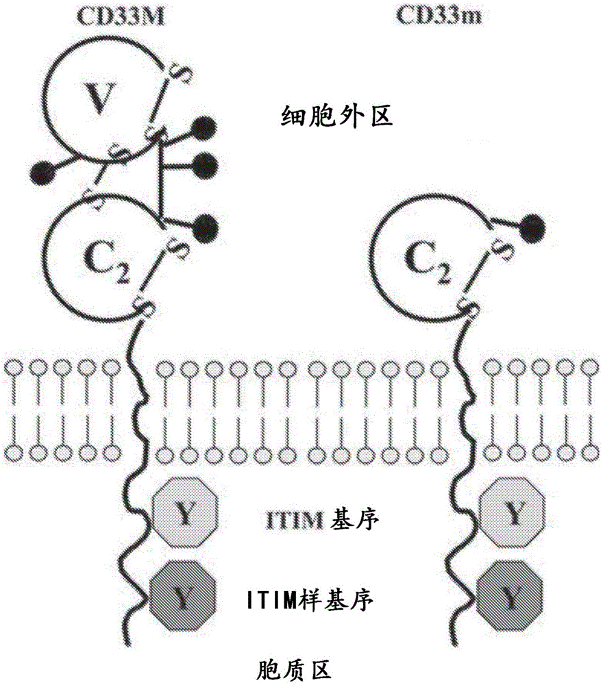
Figure 1 shows the schematic structure of the CD33 isoform. The full-length CD33 protein is shown on the left, and the short isoform (splice variant lacking the external IgV domain) is shown on the right.
Figure 2 shows a list of existing anti-CD 33 antibodies and their domain specificities as indicated by their ability to bind to full-length CD33 protein and/or short isoforms of CD 33.
FIG. 3 shows a schematic of a modular tetravalent IgG-scFv format comprising an IgG molecule with two binding sites covalently linked to two scFv providing two additional binding domains.
FIG. 4 shows FACS data demonstrating the binding of chimeric HIM34 x CD3 bispecific antibody (chHIM34 BsAb) to CD33(+) AML cell lines U937, THP1, SET2, C1498-CD33 and M-07e without harming CD33(-) leukemia cell line CMLT 1.
Figure 5(a) shows a schematic of the full-length CD33 protein and short isoform (splice variant of CD 33). Fig. 5(B) shows FACS data demonstrating that chim 34 BsAb binds to the full-length and short isoforms of CD33, whereas BsAb based on humanized M195 (BC133) does not bind to the short isoforms.
FIGS. 6(A) and 6(B) show T cell-dependent cytotoxicity assays using chHIM34 BsAb on CD33(+) AML cell lines C1498-CD33 and THP1, respectively.
Fig. 7(a) shows a bioluminescent image showing the growth of MOLM13 AML tumors in different groups. Fig. 7(B) shows the quantification of signal from different groups of mice in fig. 7 (a).
FIG. 8 shows the amino acid sequence of the murine and humanized HIM34 heavy chain variable domains (SEQ ID NOS: 1-7 and 133). HIM34_ VH-1, HIM34_ VH-2, HIM _34_ VH-3, HIM34_ VH-4, HIM34_ VH-5, HIM34-VH-6, HIM-34-VH-7 are 7 versions of a humanized HIM34 heavy chain variable domain. VH-CDR1、VHCDR2 and VHthe-CDR 3 sequence is shown in bold.
FIG. 9 shows the amino acid sequences of the murine and humanized HIM34 light chain variable domains (SEQ ID NOS: 8-13). HIM34_ VL-1, HIM34_ VL-2, HIM _34_ VL-3, HIM34_ VL-4 and HIM34_ VL-5 are 5 versions of the humanized HIM34 light chain variable domain. VL-CDR1、VLCDR2 and V Lthe-CDR 3 sequence is shown in bold.
FIGS. 10(A) and 10(B) show the amino acid and nucleotide sequences of the light and heavy chains of chHIM34 BsAb, respectively (SEQ ID NOS: 14-17). The signal peptide is underlined, the variable domains of the chimeric anti-CD 33 antibody are indicated in bold font, and the linker sequence is in italics and underlined. Also disclosed are V's of chHIM34 BsAbHAnd VLThe amino acid sequence of the domain (SEQ ID NO: 114-115).
FIGS. 11(A) and 11(B) show the amino acid and nucleotide sequences of the light and heavy chains of humanized HIM34 x CD3 BsAb (BC249), respectively (SEQ ID NOS: 18-21). FIGS. 11(C) and 11(D) show the amino acid and nucleotide sequences of the light and heavy chains of humanized HIM34 x CD3 BsAb (BC275), respectively (SEQ ID NO: 134-137). The signal peptide is underlined, the variable domains of the humanized anti-CD 33 antibody are indicated in bold font, and the linker sequence is in italics and underlined.
FIGS. 12(A) and 12(B) show the amino acid and nucleotide sequences (SEQ ID NOS: 18, 19, 22, and 23) of the light and heavy chains of humanized HIM34 x CD3 BsAb (BC267), respectively. The signal peptide is underlined, the variable domains of the humanized anti-CD 33 antibody are indicated in bold font, and the linker sequence is in italics and underlined.
FIGS. 13(A) and 13(B) show the amino acid and nucleotide sequences of the light and heavy chains of humanized HIM34 x CD3 BsAb (BC268), respectively (SEQ ID NOS: 24, 25, 22 and 23). The signal peptide is underlined, the variable domains of the humanized anti-CD 33 antibody are indicated in bold font, and the linker sequence is in italics and underlined.
FIGS. 14(A) -14(F) show the amino acid sequences of the light and heavy chains of humanized HIM34 x C825 (anti-DOTA) BsAb in the form of an IgG-scFv. The light chain amino acid sequences are shown in SEQ ID NOS 26-29 and the heavy chain amino acid sequences are shown in SEQ ID NOS 20 and 22. The signal peptide is underlined, the variable domains of the humanized anti-CD 33 antibody are indicated in bold font, and the linker sequence is in italics and underlined.
FIGS. 14(G) -14(H) show the amino acid sequences of the light and heavy chains of murine HIM34 x C825 (anti-DOTA) BsAb in the form of an IgG-scFv. The light chain amino acid sequences are shown in SEQ ID NOS 138 and 140 and the heavy chain amino acid sequences are shown in SEQ ID NOS 139 and 141. The signal peptide is underlined, the variable domain of the mouse anti-CD 33 antibody is indicated in bold font, and the linker sequence is in italics and underlined.
FIGS. 15(A) to 15(BB) show the amino acid sequence of humanized HIM34 x C825 (anti-DOTA) BsAb in the form of single-chain bispecific tandem fragment variable (scBsAfv) (SEQ ID NOS: 30-113). The signal peptide is underlined, the variable domains of the humanized anti-CD 33 antibody are indicated in bold font, and the linker sequence is in italics and underlined.
FIGS. 15(CC) to 15(FF) show the amino acid sequence of mouse HIM34 x C825 (anti-DOTA) BsAb in the form of a single-chain bispecific tandem fragment variable (scBsAFv) (SEQ ID NO: 142-153). The signal peptide is underlined, the variable domain of the mouse-based anti-CD 33 antibody is indicated in bold font, and the linker sequence is in italics and underlined.
Fig. 16(a) and 16(B) show a summary of the characteristics of chimeric and humanized HIM34 BsAb, including the binding kinetics.
Fig. 17 shows FACS analysis of MOLM13 CD33(+) cells contacted with different concentrations of humanized CD33 BsAb of the present technology.
Fig. 18(a) to 18(B) show the cytotoxicity of humanized BsAb of the present technology on MOLM13 CD33(+) cells in a T cell dependent cytotoxicity assay (TDCC).
Figure 19 shows the stability of humanized BsAb of the present technology at 40 ℃.
Figure 20(a) shows T cell engaging bispecific antibodies (BsAb) recruiting T cells to AML cells. Fig. 20(B) shows that BC275 BsAb disclosed herein binds to the membrane proximal domain (IgC) of the CD33 extracellular region, while BC133 based on the M195 clone (Lintuzumab) and BC269 based on My96 (Gemtuzumab)) binds to the membrane distal domain of the CD33 extracellular region. Fig. 20(C) shows CD33 binding properties of BC275, BC133, and BC269 BsAb on various CD33(+) and CD33(-) human cancer cell lines.
Fig. 21(a) to 21(C) show T cell-dependent cell-mediated cytotoxic effects of BC275, BC133, and BC269 BsAb on CD33(+) and CD33(-) human cancer cell lines. Figure 21(D) plots the correlation between the level of CD33 expression on cancer cells and the potency of test BsAb in the TDCC assay. Pooled data for all three CD3 xcd 33BsAb are shown.
FIGS. 22(A) to 22(B) show tumor sizes of immunodeficient NSG mice intravenously inoculated with CD33(+) MOLM 13-luciferase (having CC genotype with rs12459419 SNP) human AML xenografts (10)6Individual cells) and subsequently treated with BC275, BC133 and BC269 BsAb (CD3 × CD33BsAb) or control BC119 BsAb (CD3 × GD2 BsAb). Mice received 5X 10⁶A single injection of activated T cells and 0.025. mu.g BsAb (10. mu.g/Kg/dose). The timing of T cell and antibody injections is shown in fig. 22 (B).
FIGS. 23(A) to 2323(B) shows tumor size of immunodeficient NSG mice that were inoculated intravenously with NALM 6-luciferase leukemia xenografts transduced with human CD33 (0.5X 10)6Individual cells) and subsequently treated with BC275, BC133 and BC269 BsAb (CD3 × CD33BsAb) or control BC119 BsAb (CD3 × GD2 BsAb). Mice received two injections of activated T cells (2.7X 10) 6And 5X 10⁶One week apart between doses) and 10ng BsAb/106And (4) T cells. The timing of T cell and BsAb injections is shown in fig. 23 (B). Fig. 23(C) shows survival curves for animals treated with three CD3 xcd 33 BsAb. The potency of CD3 xcd 33 BsAb is ranked as follows: BC275>BC269=BC133。
Fig. 24(a) to 24(D) show tumor sizes of immunodeficient DKO mice that were subcutaneously inoculated with THP1 human leukemia xenografts (which have a CC genotype with rs12459419 SNP) and subsequently treated with BC275, BC133 and BC269 BsAb (CD3 × CD33 BsAb) or control BC119 BsAb (CD3 × GD2 BsAb). Mice received two injections of activated T cells (10)7With one week interval between doses) and 0.05. mu.g or 0.5. mu.g BsAb (. apprxeq.2. mu.g/kg/dose or 20. mu.g/kg/dose). The timing of T cell and antibody injections is shown in fig. 24 (a). The potency of CD3 xcd 33 BsAb is ranked as follows: BC275>BC269>BC133。
Fig. 25(a) to 25(D) show tumor sizes of immunodeficient DKO mice that were subcutaneously inoculated with K562 human leukemia xenografts having a TT genotype with an rs12459419SNP, and subsequently treated with BC275, BC133, and BC269 BsAb (CD3 × CD33 BsAb) or control BC119 BsAb (CD3 × GD2 BsAb). Mice received three injections of activated T cells (10) 7With one week interval between doses) and 0.05. mu.g or 0.5. mu.g BsAb (. apprxeq.2. mu.g/kg/dose or 20. mu.g/kg/dose). The timing of T cell and antibody injections is shown in fig. 25 (a). The potency of CD3 xcd 33 BsAb is ranked as follows: BC275>BC269>BC133。
Detailed Description
It is to be understood that certain aspects, modes, embodiments, variations and features of the methods of the present invention are described below in varying degrees of detail to provide a substantial understanding of the present technology.
The present disclosure generally provides immunoglobulin-related compositions (e.g., antibodies or antigen-binding fragments thereof) that can specifically bind to CD33 polypeptides. The immunoglobulin-related compositions of the present technology can be used in methods of detecting or treating CD 33-associated cancer or alzheimer's disease in a subject in need thereof. Thus, various aspects of the methods of the invention relate to the preparation, characterization and manipulation of anti-CD 33 antibodies. The immunoglobulin-related compositions of the present technology can be used alone or in combination with additional therapeutic agents for the treatment of cancer. In some embodiments, the immunoglobulin-related composition is a humanized antibody, a chimeric antibody, or a bispecific antibody.
In practicing the methods of the present invention, many conventional techniques in molecular biology, protein biochemistry, cell biology, immunology, microbiology, and recombinant DNA are used. See, e.g., Sambrook and Russell, eds (2001) Molecular Cloning A Laboratory Manual, 3 rd edition; the book Ausubel et al, eds (2007) Current Protocols in Molecular Biology; book Methods in Enzymology (Academic Press, Inc., New York); MacPherson et al (1991) PCR 1: A Practical Approach (IRL Press at Oxford University Press); MacPherson et al (1995) PCR 2: A Practical Approach; harlow and Lane editors (1999) Antibodies, A Laboratory Manual; freshney (2005) Culture of Animal Cells A Manual of Basic Technique, 5 th edition; gait editor (1984) Oligonucleotide Synthesis; U.S. Pat. nos. 4,683,195; hames and Higgins editors (1984) Nucleic Acid Hybridization; anderson (1999) Nucleic Acid Hybridization; hames and Higgins editions (1984) transformation and transformation; immobilized Cells and Enzymes (IRL Press (1986)); perbal (1984) A Practical Guide to Molecular Cloning; miller and Calos editor (1987) Gene Transfer Vectors for Mammalian Cells (Cold Spring Harbor Laboratory); makrides editors (2003) Gene Transfer and Expression in Mammarian Cells; mayer and Walker, eds (1987) Immunochemical Methods in Cell and Molecular Biology (Academic Press, London); and Herzenberg et al, eds (1996) Weir's Handbook of Experimental Immunology. Methods for detecting and measuring the level of polypeptide gene expression product (i.e., the level of gene translation) are well known in the art and include the use of polypeptide detection methods, such as antibody detection and quantification techniques. (see also Strachan and Read, Human Molecular Genetics, second edition (John Wiley and Sons, Inc., 1999)).
Definition of
Unless defined otherwise, all technical and scientific terms used herein have the same meaning as commonly understood by one of ordinary skill in the art to which this invention belongs. As used in this specification and the appended claims, the singular forms "a," "an," and "the" include plural referents unless the context clearly dictates otherwise. For example, reference to "a cell" includes a combination of two or more cells, and the like. Generally, the nomenclature used herein and the laboratory procedures in cell culture, molecular genetics, organic chemistry, analytical chemistry, and nucleic acid chemistry and hybridization described below are those well known and commonly employed in the art.
As used herein, the term "about" with respect to a number is generally considered to include numbers that fall within 1%, 5%, or 10% of either direction (greater than or less than) the number (except where such number falls below 0% or exceeds 100% of the possible value), unless the context indicates otherwise or is otherwise evident.
As used herein, "administering" an agent or drug to a subject includes any route of introducing or delivering a compound to a subject to perform its intended function. Administration may be by any suitable route, including but not limited to oral, intranasal, parenteral (intravenous, intramuscular, intraperitoneal or subcutaneous), rectal, intrathecal, intratumoral or topical. Administration includes self-administration and administration by another person.
"adjuvant" refers to one or more substances that cause stimulation of the immune system. In this case, the adjuvant is used to enhance the immune response to one or more vaccine antigens or antibodies. The adjuvant may be administered to the subject prior to, in combination with, or after administration of the vaccine. Examples of chemical compounds useful as adjuvants include aluminum compounds, oils, block polymers, immunostimulatory complexes, vitamins and minerals (e.g., vitamin E, vitamin a, selenium, and vitamin B12), Quil a (saponins), bacterial and fungal cell wall components (e.g., lipopolysaccharides, lipoproteins, and glycoproteins), hormones, cytokines, and co-stimulatory factors.
As used herein, the term "antibody" refers collectively to immunoglobulins or immunoglobulin-like molecules, including, for example and without limitation, IgA, IgD, IgE, IgG, and IgM, combinations thereof, and similar molecules (such as shark immunoglobulins) produced during an immune response in any vertebrate, for example, in mammals (such as humans, goats, rabbits, and mice), as well as non-mammalian species. As used herein, "antibodies" (including intact immunoglobulins) and "antigen-binding fragments" specifically bind to a molecule of interest (or a group of highly similar molecules of interest) while substantially excluding binding to other molecules (e.g., binding constants for the molecule of interest are at least 10 greater than binding constants for other molecules in a biological sample 3M-1At least 10 greater4M-1Or at least 10 greater5M-1Antibodies and antibody fragments of (a). The term "antibody" also includes genetically engineered forms such as chimeric antibodies (e.g., humanized murine antibodies), heteroconjugate antibodies (e.g., bispecific antibodies). See also Pierce Catalog and Handbook, 1994-; kuby, j., Immunology, 3 rd edition, w.h&Co., new york, 1997.
More specifically, an antibody refers to a polypeptide ligand comprising at least a light chain immunoglobulin variable region or a heavy chain immunoglobulin variable region that specifically recognizes and binds an epitope. Antibodies are composed of heavy and light chains, each of which has a variable region, termed heavy chain variable (V)H) Variable domains and light chains (V)L) And (4) a zone. VHRegion and VLThe regions are collectively responsible for binding to the antigen recognized by the antibody. Generally, immunoglobulins have a heavy (H) chain and a light (L) chain interconnected by disulfide bonds. There are two types of light chains, i.e., λ (l)ambda) and κ (kappa). There are five major heavy chain classes (or isotypes): IgM, IgD, IgG, IgA and IgE, which determine the functional activity of the antibody molecule. Each heavy and light chain contains a constant region and a variable region (the regions are also referred to as "domains"). In combination, the heavy chain variable region and the light chain variable region specifically bind to an antigen. The light and heavy chain variable regions contain a "framework" region interrupted by three hypervariable regions (also known as "complementarity determining regions" or "CDRs"). The extent of the framework regions and CDRs has been defined (see Kabat et al, Sequences of Proteins of Immunological Interest, U.S. department of Health and Human Services,1991, which is hereby incorporated by reference). Kabat databases are currently maintained online. The sequences of the framework regions of different light or heavy chains are relatively conserved within a species. The framework regions of the antibody (i.e., the combined framework regions of the constituent light and heavy chains) adopt predominantly a β -sheet conformation, and the CDRs form loops that connect, and in some cases form part of, the β -sheet structure. Thus, the framework regions serve to form a scaffold that positions the CDRs in the correct orientation through interchain non-covalent interactions.
The CDRs are primarily responsible for binding to an epitope of the antigen. The CDRs of each chain are commonly referred to as CDR1, CDR2, and CDR3, numbered sequentially from the N-terminus, and are also typically identified by the chain in which the particular CDR is located. Thus, VHCDR3 is located in the variable domain of the heavy chain of the antibody in which it is found, and VLCDR1 is the CDR1 from the variable domain of the light chain of the antibody in which it is found. Antibodies that bind to CD33 protein will have a specific VHRegion and VLRegion sequences, and thus specific CDR sequences. Antibodies with different specificities (i.e., different binding sites for different antigens) have different CDRs. Despite the differences in CDRs between different antibodies, only a limited number of amino acid positions within a CDR are directly involved in antigen binding. These positions within the CDRs are called Specificity Determining Residues (SDRs). As used herein, "immunoglobulin-related composition" refers to antibodies (including monoclonal antibodies, polyclonal antibodies, humanized antibodies, chimeric antibodies, recombinant antibodies, multispecific antibodies, bispecific antibodies, etc.) as well as anti-antibodiesA body fragment. The antibody or antigen-binding fragment thereof specifically binds to an antigen.
As used herein, the term "antibody-related polypeptide" means an antigen-binding antibody fragment, including a single chain antibody, which may comprise one or more variable regions alone or in combination with all or part of the following polypeptide elements: hinge region, CH, of antibody molecule 1、CH2And CH3A domain. Also included in the technology are one or more variable and hinge regions, CH1、CH2And CH3Any combination of domains. Antibody-related molecules which can be used in the methods of the invention are for example, but not limited to, Fab 'and F (ab')2Fd, single chain fv (scFv), single chain antibody, disulfide-linked fv (sdFv) and compositions comprising VLOr VHA fragment of a domain. Examples include: (i) fab fragments, i.e. consisting of VL、VH、CLAnd CH1Monovalent fragments consisting of domains; (ii) a F (ab')2 fragment, i.e. a bivalent fragment comprising two Fab fragments linked by a disulfide bridge of the hinge region; (iii) from VHAnd CH1Domain-forming Fd fragments; (iv) v with one arm consisting of antibodyLAnd VH(iii) an Fv fragment consisting of a domain; (v) dAb fragments (Ward et al, Nature341:544-546,1989) consisting of VHDomain composition; and (vi) an isolated Complementarity Determining Region (CDR). Thus, an "antibody fragment" or "antigen-binding fragment" may comprise a portion of a full-length antibody, typically the antigen-binding or variable region thereof. Examples of antibody fragments or antigen-binding fragments include Fab, Fab ', F (ab')2And Fv fragments; a diabody; a linear antibody; a single chain antibody molecule; and multispecific antibodies formed from antibody fragments.
As used herein, "bispecific antibody" or "BsAb" refers to an antibody that can simultaneously bind to two targets having different structures (e.g., two different target antigens, two different epitopes on the same target antigen, or a hapten and a target antigen or an epitope on a target antigen). A variety of different bispecific antibody structures are known in the art. In some embodiments, each antigen binding moiety in a bispecific antibody All comprise VHAnd/or VLA zone; in some such embodiments, VHAnd/or VLRegions are those found in a particular monoclonal antibody. In some embodiments, a bispecific antibody contains two antigen-binding portions, each antigen-binding portion comprising a V from a different monoclonal antibodyHAnd/or VLAnd (4) a zone. In some embodiments, the bispecific antibody contains two antigen binding portions, wherein one of the two antigen binding portions comprises a heavy chain having a VHAnd/or VLImmunoglobulin molecule of region VHAnd/or VLThe region contains a CDR from a first monoclonal antibody; and another antigen binding moiety comprises a peptide having VHAnd/or VLAntibody fragments of regions (e.g., Fab, F (ab')2Fd, Fv, dAB, scFv, etc.), said VHAnd/or VLThe region contains a CDR from a second monoclonal antibody.
As used herein, a "clearing agent" is an agent that binds to an excess of bispecific antibody present in the blood compartment of a subject to facilitate rapid clearance via the kidney. The use of a scavenger prior to hapten administration (e.g., DOTA) helps to achieve better tumor to background ratios in pre-targeted radioimmunotherapy (PRIT) systems. Examples of scavengers include 500 kD-dextran-DOTA-Bn (Y) (Orcutt et al, Mol Cancer Ther.11(6): 1365-.
The term "conjugated" as used herein refers to the association of two molecules by any method known to those skilled in the art. Suitable types of associations include chemical bonds and physical bonds. Chemical bonds include, for example, covalent bonds and coordination bonds. Physical bonding includes, for example, hydrogen bonding, dipole interactions, van der waals forces, electrostatic interactions, hydrophobic interactions, and aromatic ring stacking.
The term "diabodies" as used herein refers to small antibody fragments having two antigen binding sites, which fragments are comprised in the same polypeptide chain as the light chain variable domain (V)L) Linked heavy chain variable domains (V)H)(VH VL)。By using a linker that is too short to allow pairing between two domains on the same chain, the domains are forced to pair with the complementary domains of the other chain and two antigen binding sites are created. Diabodies are more fully described in, for example, the following documents: EP 404,097; WO 93/11161; and Hollinger et al, Proc. Natl. Acad. Sci. USA,90: 6444-.
As used herein, the term "single chain antibody" or "single chain Fv (scFv)" refers to the two domains V of an Fv fragmentLAnd VHThe antibody fusion molecule of (1). Single chain antibody molecules may comprise polymers having multiple individual molecules, such as dimers, trimers, or other polymers. Furthermore, although F vTwo domains of the fragment VLAnd VHEncoded by separate genes, but they can be joined using recombinant methods by synthetic linkers, enabling them to be single protein chains in which V is presentLAnd VHRegion pairing to form a monovalent molecule (referred to as single-stranded F)v(scFv)). Bird et al (1988) Science 242: 423-. Such single chain antibodies can be prepared by recombinant techniques or enzymatic or chemical cleavage of intact antibodies.
Any of the above antibody fragments are obtained using conventional techniques known to those skilled in the art, and the fragments are screened for binding specificity and neutralizing activity in the same manner as intact antibodies.
As used herein, "antigen" refers to a molecule to which an antibody (or antigen-binding fragment thereof) can selectively bind. The target antigen may be a protein, carbohydrate, nucleic acid, lipid, hapten or other naturally occurring or synthetic compound. In some embodiments, the target antigen may be a polypeptide (e.g., a CD33 polypeptide). Antigens can also be administered to animals to generate an immune response in the animal.
The term "antigen-binding fragment" refers to a fragment of an intact immunoglobulin structure having a portion of a polypeptide responsible for binding to an antigen. Examples of antigen-binding fragments that can be used in the present technology include scFv, (scFv) 2、scFvFc, Fab 'and F (ab')2But is not limited thereto.
By "binding affinity" is meant the strength of the overall non-covalent interaction between a single binding site of a molecule (e.g., an antibody) and a binding partner of the molecule (e.g., an antigen or an antigenic peptide). The affinity of a molecule X for its partner Y can generally be determined by the dissociation constant (K)D) And (4) showing. Affinity can be measured by standard methods known in the art, including those described herein. Low affinity complexes contain antibodies that generally tend to dissociate readily from the antigen, while high affinity complexes contain antibodies that generally tend to remain bound to the antigen for longer periods of time.
As used herein, the term "biological sample" means sample material derived from living cells. Biological samples can include tissues, cells, protein or membrane extracts of cells, and biological fluids (e.g., ascites or cerebrospinal fluid (CSF)) isolated from a subject, as well as tissues, cells, and fluids present in a subject. Biological samples of the present technology include, but are not limited to, samples taken from: breast tissue, kidney tissue, cervix, endometrium, head or neck, gall bladder, parotid gland tissue, prostate, brain, pituitary gland, kidney tissue, muscle, esophagus, stomach, small intestine, colon, liver, spleen, pancreas, thyroid tissue, heart tissue, lung tissue, bladder, adipose tissue, lymph node tissue, uterus, ovarian tissue, adrenal tissue, testicular tissue, tonsil, thymus, blood, hair, cheek, skin, serum, plasma, CSF, sperm, prostatic fluid, semen, urine, stool, sweat, saliva, sputum, mucus, bone marrow, lymph, and tears. Biological samples may also be obtained from biopsies of internal organs or from cancer. A biological sample may be obtained from a subject for diagnosis or study; or may be obtained from an unaffected individual, either as a control or for use in basic studies. Samples can be obtained by standard methods including, for example, venipuncture and surgical biopsy. In certain embodiments, the biological sample is a tissue sample obtained by needle biopsy.
As used herein, the term "CDR-grafted antibody" means an antibody in which at least one CDR of the "acceptor" antibody is replaced by a CDR "graft" from the "donor" antibody with the desired antigen specificity.
As used herein, the term "chimeric antibody" means an antibody in which the Fc constant region of a monoclonal antibody from one species (e.g., a mouse Fc constant region) is replaced with the Fc constant region of an antibody from another species (e.g., a human Fc constant region) using recombinant DNA techniques. See, generally, Robinson et al, PCT/US 86/02269; akira et al, European patent application 184,187; taniguchi, european patent application 171,496; morrison et al, European patent application 173,494; neuberger et al, WO 86/01533; cabilly et al, U.S. Pat. Nos. 4,816,567; cabilly et al, European patent application 0125,023; better et al, Science 240: 1041-; liu et al, Proc.Natl.Acad.Sci.USA 84:3439-3443, 1987; liu et al, J.Immunol 139:3521-3526, 1987; sun et al, Proc.Natl.Acad.Sci.USA 84: 214-; nishimura et al, Cancer Res 47: 999-; wood et al, Nature 314:446-449, 1885; and Shaw et al, J.Natl.cancer Inst.80:1553-1559, 1988.
As used herein, the term "consensus FR" means the Framework (FR) antibody region in a consensus immunoglobulin sequence. The FR region of the antibody is not in contact with the antigen.
As used herein, a "control" is a surrogate sample used in an experiment for comparison purposes. Controls may be "positive" or "negative". For example, where the objective of an experiment is to determine the relevance of a therapeutic agent to the efficacy of treatment of a particular type of disease, a positive control (a compound or composition known to exhibit the desired therapeutic effect) and a negative control (a subject or sample that received no treatment or a placebo) are typically used.
As used herein, the term "effective amount" refers to an amount sufficient to achieve a desired therapeutic and/or prophylactic effect, e.g., an amount that results in the prevention or reduction of a disease or disorder described herein or one or more signs or symptoms associated with a disease or disorder described herein. In the case of therapeutic or prophylactic use, the amount of the composition administered to a subject will vary depending on the composition, the extent, type and severity of the disease, and on the characteristics of the individual, such as general health, age, sex, weight and drug tolerance. The skilled person will be able to determine the appropriate dosage in view of these and other factors. The compositions may also be administered in combination with one or more additional therapeutic compounds. In the methods described herein, the therapeutic composition can be administered to a subject having one or more signs or symptoms of a disease or disorder described herein. As used herein, a "therapeutically effective amount" of a composition refers to the level of the composition wherein the physiological effects of the disease or disorder are ameliorated or eliminated. A therapeutically effective amount may be administered in one or more administrations.
As used herein, the term "effector cell" means an immune cell that participates in the effector phase of an immune response, as opposed to the cognitive and activation phases of an immune response. Exemplary immune cells include cells of myeloid or lymphoid origin, such as lymphocytes (e.g., B cells and T cells, including cytolytic T Cells (CTLs)), killer cells, natural killer cells, macrophages, monocytes, eosinophils, neutrophils, polymorphonuclear cells, granulocytes, mast cells, and basophils. Effector cells express specific Fc receptors and perform specific immune functions. The effector cells may induce antibody-dependent cell-mediated cytotoxicity (ADCC), such as neutrophils capable of inducing ADCC. For example, monocytes, macrophages, neutrophils, eosinophils, and lymphocytes that express Fc α R are involved in specific killing of target cells and presenting antigen to other components of the immune system, or bind to cells presenting antigen.
As used herein, the term "epitope" means a protein determinant capable of specific binding to an antibody. Epitopes usually consist of chemically active surface groups of molecules (e.g. amino acids or sugar side chains) and usually have specific three-dimensional structural characteristics, as well as specific charge characteristics. Conformational and non-conformational epitopes differ by: in the presence of denaturing solvents, binding to conformational epitopes is lost rather than to non-conformational epitopes. In some embodiments, an "epitope" of a CD33 protein is a region of the protein that specifically binds to an anti-CD 33 antibody of the present technology. In some embodiments, the epitope is a conformational epitope or a non-conformational epitope. To screen for anti-CD 33 antibodies that bind to the epitope, conventional cross-blocking assays can be performed, such as the assays described in the following references: antibodies, A Laboratory Manual, Cold Spring Harbor Laboratory, Harlow and David Lane eds (1988). This assay can be used to determine whether an anti-CD 33 antibody binds to the same site or epitope as an anti-CD 33 antibody of the present technology. Alternatively or additionally, epitope mapping can be performed by methods known in the art. For example, antibody sequences can be mutagenized, such as by alanine scanning, to identify contact residues. In different approaches, peptides corresponding to different regions of the CD33 protein can be used in competition assays with a variety of test antibodies, or with one test antibody and an antibody having a characterized or known epitope.
As used herein, "expression" includes one or more of the following: transcription of the gene into precursor mRNA; splicing and other processing of the precursor mRNA to produce a mature mRNA; mRNA stability; translation of mature mRNA into protein (including codon usage and tRNA availability); and other modifications of the glycosylation and/or translation products (if required for proper expression and function).
As used herein, the term "gene" means a segment of DNA that contains all the information for regulated biosynthesis of an RNA product, including promoters, exons, introns, and other untranslated regions that control expression.
"homology" or "identity" or "similarity" refers to sequence similarity between two peptides or between two nucleic acid molecules. Homology can be determined by comparing the position in each sequence, which can be aligned for comparison purposes. When a position in the compared sequences is occupied by the same base or amino acid, then the molecules are homologous at that position. The degree of homology between sequences varies with the number of matching or homologous positions shared by the sequences. "sequence identity" that a polynucleotide or polynucleotide region (or polypeptide region) has a percentage (e.g., at least 60%, 65%, 70%, 75%, 80%, 85%, 90%, 95%, 98%, or 99%) of another sequence means that when aligned, the percentage of bases (or amino acids) are the same in comparing two sequences. This alignment, as well as the percent homology or sequence identity, can be determined using software programs known in the art. In some embodiments, default parameters are used for alignment. One alignment program is BLAST, using default parameters. Specifically, the programs are BLASTN and BLASTP, using the following default parameters: the genetic code is standard; no filter; two chains; cutoff is 60; desirably 10; BLOSUM 62; describe 50 sequences; ranking by HIGH SCORE (HIGH SCORE); database-not redundant-GenBank + EMBL + DDBJ + PDB + GenBank CDS translation + SwissProtein + SPupdate + PIR. Details of these procedures can be found at the National Center for Biotechnology Information. Biologically equivalent polynucleotides are those polynucleotides having a specified percentage of homology and encoding polypeptides having the same or similar biological activity. Sequences are considered "unrelated" or "non-homologous" if they share less than 40% identity or less than 25% identity with each other.
As used herein, a "humanized" form of a non-human (e.g., murine) antibody is a chimeric antibody that contains minimal sequences derived from a non-human immunoglobulin. For the most part, humanized antibodies are human immunoglobulins in which hypervariable region residues of the acceptor are replaced by hypervariable region residues (donor antibody) from a non-human species (e.g., mouse, rat, rabbit or non-human primate) having the desired specificity, affinity, and capacity. In some embodiments, Fv Framework Region (FR) residues of the human immunoglobulin are replaced by corresponding non-human residues. In addition, humanized antibodies may comprise residues that are not found in the recipient antibody or in the donor antibody. These modifications are made to further refine antibody performance such as binding affinity. Typically, a humanized antibody will comprise at least one, and typically two, variable domains (e.g., Fab ', F (ab')2Or Fv) in which all or substantially all of the hypervariable loops correspond to those of a non-human immunoglobulin and all or substantially all of the FR regions are human immunoglobulinsThose FR regions of the protein consensus FR sequence, but may comprise one or more amino acid substitutions that improve binding affinity. The number of these amino acid substitutions in the FR is usually no more than 6 in the H chain and no more than 3 in the L chain. The humanized antibody optionally may also comprise at least a portion of an immunoglobulin constant region (Fc), typically at least a portion of a human immunoglobulin constant region. For further details, see Jones et al, Nature 321:522-525 (1986); reichmann et al, Nature 332: 323-E329 (1988); and Presta, curr, Op, Structure, biol.2:593-596 (1992). See, e.g., Ahmed and Cheung, FEBS Letters 588(2): 288-.
As used herein, the term "hypervariable region" refers to the amino acid residues of an antibody which are responsible for antigen-binding. Hypervariable regions typically comprise amino acid residues from a "complementarity determining region" or "CDR" (e.g., V)LBefore and after residues 24-34(L1), 50-56(L2) and 89-97(L3), and VH31-35B (H1), 50-65(H2), and 95-102(H3) in tandem (Kabat et al, Sequences of Proteins of Immunological Interest, published Health Service 5 th edition, National Institutes of Health, Besserda, Maryland. (1991)) and/or those residues from "hypervariable loops" (e.g., V.V.LResidues 26-32(L1), 50-52(L2) and 91-96(L3), and VH26-32(H1), 52A-55(H2) and 96-101(H3) (Chothia and Lesk J. mol. biol.196:901-917 (1987)).
As used herein, the term "identical" or percent "identity" when used in the context of two or more nucleic acid or polypeptide sequences refers to two or more sequences or subsequences that are the same or have a specified percentage of amino acid residues or nucleotides that are the same (i.e., about 60%, 65%, 70%, 75%, 80%, 85%, 90%, 91%, 92%, 93%, 94%, 95%, 96%, 97%, 98%, 99% or more identity over a specified region (e.g., a nucleotide sequence encoding an antibody described herein or an amino acid sequence of an antibody described herein) as measured using the BLAST or BLAST 2.0 sequence comparison algorithm using default parameters described below or by manual alignment and visual inspection (e.g., the NCBI website) when compared and aligned for maximum correspondence over a comparison window or specific region. Such sequences are referred to as "substantially identical". This term also refers to or may be applied to the complement of the test sequence. The term also includes sequences having deletions and/or additions, as well as those sequences having substitutions. In some embodiments, there is identity over a region that is at least about 25 amino acids or nucleotides in length or 50-100 amino acids or nucleotides in length.
As used herein, the term "intact antibody" or "intact immunoglobulin" means an antibody having at least two heavy (H) chain polypeptides and two light (L) chain polypeptides interconnected by disulfide bonds. Each heavy chain is composed of a heavy chain variable region (abbreviated herein as HCVR or V)H) And a heavy chain constant region. The heavy chain constant region is composed of three domains CH1、CH2And CH3And (4) forming. Each light chain is composed of a light chain variable region (abbreviated herein as LCVR or V)L) And a light chain constant region. The light chain constant region consists of a domain CLAnd (4) forming. VHAnd VLRegions can be further subdivided into regions of high denaturation, called Complementarity Determining Regions (CDRs), interspersed with regions that are more conserved, called Framework Regions (FRs). Each VHAnd VLConsists of three CDRs and four FRs, arranged from amino-terminus to carboxy-terminus in the following order: FR1、CDR1、FR2、CDR2、FR3、CDR3、FR4. The variable regions of the heavy and light chains contain binding domains that interact with antigens. The constant region of the antibody can mediate the binding of the immunoglobulin to host tissues or factors, including various cells of the immune system (e.g., effector cells) and the first component of the classical complement system (Clq).
As used herein, the term "individual", "patient" or "subject" can be a separate organism, vertebrate, mammal, or human. In some embodiments, the individual, patient, or subject is a human.
As used herein, the term "monoclonal antibody" refers to an antibody obtained from a substantially homogeneous population of antibodies, i.e., the individual antibodies comprising the population are identical except for possible naturally occurring mutations that may be present in minor amounts. For example, a monoclonal antibody may be an antibody derived from a single clone (including any eukaryotic, prokaryotic, or phage clone) rather than the method by which it was produced. Monoclonal antibody compositions exhibit a single binding specificity and affinity for a particular epitope. Monoclonal antibodies are highly specific for a single antigenic site. Furthermore, each monoclonal antibody is directed against a single determinant on the antigen, in contrast to conventional (polyclonal) antibody preparations which typically include different antibodies directed against different determinants (epitopes). The modifier "monoclonal" indicates that the antibody is characterized as being obtained from a substantially homogeneous population of antibodies, and is not to be construed as requiring production of the antibody by any particular method. Monoclonal antibodies can be prepared using a variety of techniques known in the art including, for example, but not limited to, hybridoma, recombinant, and phage display techniques. For example, monoclonal antibodies to be used in accordance with the methods of the present invention can be prepared by the hybridoma method originally described by Kohler et al, Nature 256:495(1975), or can be prepared by recombinant DNA methods (see, e.g., U.S. Pat. No. 4,816,567). For example, "monoclonal antibodies" can also be isolated from phage antibody libraries using the techniques described in Clackson et al, Nature 352: 624-.
As used herein, the term "pharmaceutically acceptable carrier" is intended to include any and all solvents, dispersion media, coatings, antibacterial and antifungal compounds, isotonic and absorption delaying compounds, and the like, compatible with pharmaceutical administration. Pharmaceutically acceptable carriers and formulations thereof are known to those skilled in the art and are described, for example, in Remington's Pharmaceutical Sciences (20 th edition, a.gennaro,2000, compiled by Lippincott, Williams & Wilkins, philadelphia, pa).
As used herein, the term "polyclonal antibody" means a preparation of antibodies derived from at least two (2) different antibody-producing cell lines. The use of this term includes preparations of at least two (2) antibodies that contain antibodies that specifically bind to different epitopes or regions of an antigen.
As used herein, the term "polynucleotide" or "nucleic acid" means any RNA or DNA, which may be unmodified or modified RNA or DNA. Polynucleotides include, but are not limited to, single-and double-stranded DNA, DNA that is a mixture of single-and double-stranded regions, single-and double-stranded RNA, RNA that is a mixture of single-and double-stranded regions, and hybrid molecules comprising DNA and RNA that may be single-stranded or, more typically, double-stranded or a mixture of single-and double-stranded regions. Further, a polynucleotide refers to a triple-stranded region comprising RNA or DNA or both RNA and DNA. The term polynucleotide also includes DNA or RNA containing one or more modified bases, as well as DNA or RNA with modified backbones for stability or other reasons.
As used herein, the terms "polypeptide," "peptide," and "protein" are used interchangeably herein to mean a polymer comprising two or more amino acids linked to each other by peptide bonds or modified peptide bonds (i.e., peptide isosteres). Polypeptides refer to both short chains, commonly referred to as peptides, glycopeptides or oligomers, and longer chains, commonly referred to as proteins. The polypeptide may contain amino acids other than those encoded by the 20 genes. Polypeptides include amino acid sequences that have been modified by natural processes, such as post-translational processing, or by chemical modification techniques well known in the art. Such modifications are well described in basic texts and in more detailed monographs, as well as in long research literature.
As used herein, "PRIT" or "pre-targeted radioimmunotherapy" refers to a multi-step process that addresses the slow blood clearance of tumor-targeting antibodies, which results in undesirable toxicity to normal tissues such as bone marrow. In pretargeting, a radionuclide or other diagnostic or therapeutic agent is attached to a small hapten. A pre-targeted bispecific antibody with a binding site for a hapten as well as a target antigen is administered first. Unbound antibody is then cleared from the circulation and the hapten is subsequently administered.
As used herein, the term "recombinant" when used in relation to, for example, a cell or nucleic acid, protein or vector indicates that the cell, nucleic acid, protein or vector has been modified by the introduction of a heterologous nucleic acid or protein or the alteration of a native nucleic acid or protein, or that the material originates from a cell so modified. Thus, for example, recombinant cells express genes that are not found within the native (non-recombinant) form of the cell, or express native genes that are otherwise abnormally expressed, under-expressed, or not expressed at all.
As used herein, the term "separate" therapeutic use refers to the administration of at least two active ingredients simultaneously or substantially simultaneously by different routes.
As used herein, the term "sequential" therapeutic use refers to the administration of at least two active ingredients at different times, the routes of administration being the same or different. More specifically, sequential use means that the entire administration of one active ingredient is preceded by the start of the administration of the other active ingredient or ingredients. Thus, one active ingredient may be administered within minutes, hours or days before another active ingredient or ingredients is administered. In this case, there is no concurrent treatment.
As used herein, "specific binding" refers to a molecule (e.g., an antibody or antigen-binding fragment thereof) that recognizes and binds another molecule (e.g., an antigen) but does not substantially recognize and bind other molecules. As used herein, the term "specifically binds" a particular molecule (e.g., a polypeptide or an epitope on a polypeptide), "specifically binds" to the particular molecule, or is "specific" for the particular molecule may be, for example, through the K of the molecule to which it bindsdIs about 10-4M、10-5M、10-6M、10-7M、10- 8M、10-9M、10-10M、10-11M or 10-12M. The term "specifically binds" may also refer to binding wherein a molecule (e.g., an antibody or antigen-binding fragment thereof) binds to a particular polypeptide (e.g., a CD33 polypeptide) or an epitope on a particular polypeptide, while not substantially binding to any other polypeptide or polypeptide epitope.
As used herein, the term "simultaneous" therapeutic use refers to the administration of at least two active ingredients by the same route and at the same or substantially the same time.
As used herein, the term "therapeutic agent" is intended to mean a compound that, when present in an effective amount, produces a desired therapeutic effect in a subject in need thereof.
As used herein, "treating" or "treatment" encompasses treating a disease or disorder described herein in a subject, such as a human, and includes: (i) inhibiting the disease or disorder, i.e., arresting its development; (ii) alleviating the disease or disorder, i.e., causing regression of the disorder; (iii) slowing the progression of the disorder; and/or (iv) inhibiting, alleviating or slowing the progression of one or more symptoms of the disease or disorder. In some embodiments, treatment means that the symptoms associated with the disease are, for example, alleviated, reduced, cured, or in a state of remission.
It is also to be understood that the various treatment modalities for disorders as described herein are intended to mean "substantially", which includes complete treatment but also less than complete treatment, and in which some biologically or medically relevant result is achieved. The treatment may be a continuous prolonged treatment for chronic diseases or a single or several administrations of treatment for acute conditions.
One or more amino acid sequence modifications of the anti-CD 33 antibodies described herein are contemplated. For example, it may be desirable to improve the binding affinity and/or other biological properties of an antibody. Amino acid sequence variants of the anti-CD 33 antibody were prepared by introducing appropriate nucleotide changes into the antibody nucleic acid or by peptide synthesis. Such modifications include, for example, deletions from and/or insertions into and/or substitutions of residues within the amino acid sequence of the antibody. Any combination of deletion, insertion and substitution may be made to obtain the objective antibody as long as the obtained antibody has the desired properties. Modifications also include changes in the glycosylation pattern of the protein. The sites of most interest for substitution mutagenesis include hypervariable regions, but FR alterations are also contemplated. "conservative substitutions" are shown in the table below.
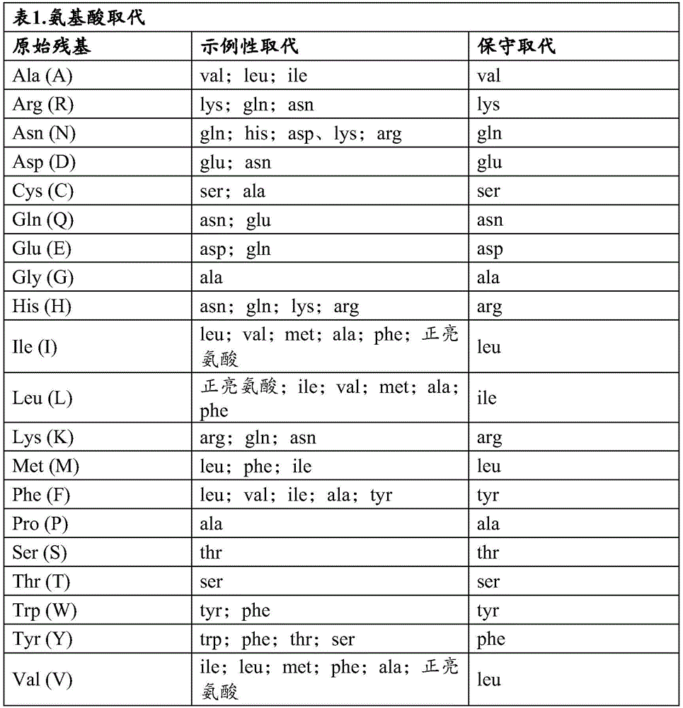
One type of substitution variant involves substituting one or more hypervariable region residues of a parent antibody. One convenient method for generating such substitution variants involves affinity maturation using phage display. In particular, several hypervariable region sites (e.g., 6-7 sites) are mutated to generate all possible amino acid substitutions at each site. The antibody variants so produced are displayed in a monovalent fashion from filamentous phage particles as fusions to the gene III product of M13 packaged in each particle. The phage-displayed variants are then screened for their biological activity (e.g., binding affinity), as disclosed herein. To identify candidate hypervariable region sites for modification, alanine scanning mutagenesis can be performed to identify hypervariable region residues that significantly contribute to antigen binding. Alternatively or additionally, it may be beneficial to analyze the crystal structure of the antigen-antibody complex to identify contact points between the antibody and the antigen. Such contact residues and adjacent residues are candidates for substitution according to the techniques detailed herein. Once such variants are generated, the set of variants is screened as described herein and antibodies having similar or superior properties in one or more relevant assays may be selected for further development.
CD33
CD33 (also known as Siglec-3, Siglec3, gp67, p67) is a 67kDa plasma membrane protein that binds sialic acid, and is a member of the sialic acid-binding Ig-associated lectin (Siglec) protein family. Siglec proteins are thought to be involved in a variety of biological processes such as hematopoiesis, neuronal development and immunity (Visson et al, J.biol.chem.271:9267-9272 (1996)). Studies have also shown that Siglec proteins mediate cell adhesion/cell signaling by recognizing sialylated cell surface glycans (Kelm et al, Glycoconj. J.13:913-926 (1996); Kelm et al, Eur. J.biochem.255:663-672 (1998); Vinson et al, J.biol. chem.271:9267-9272 (1996)). The extracellular portion of CD33 contains two immunoglobulin domains (one IgV and one IgC2 domain) (fig. 1). The intracellular portion of CD33 contains the Immunoreceptor Tyrosine Inhibitory Motif (ITIM). In an immune response, CD33 may act as an inhibitory receptor upon ligand-induced tyrosine phosphorylation by recruiting one or more cytoplasmic phosphatases that block signal transduction by dephosphorylation of signaling molecules.
CD33 is known to be expressed on myeloid lineage cells. CD33 expression has also been reported on many malignant cells. anti-CD 33 agents are generally divided into four groups: naked antibodies, antibody toxin conjugates, radionuclide conjugates and bispecific antibodies. Although CD33 has been targeted for the treatment of cancer, such as acute myeloid leukemia, there is currently no effective CD33 targeted treatment on the market. Existing anti-CD 33 agents have, inter alia, poor tumor antigen binding affinity and short half-life in vivo. Furthermore, none of these antibodies could target the short isoform of CD33 (splice variant of CD 33) (fig. 2).
CD33 is expressed on most leukemic cells, but nearly 50% of leukemic cells lack the external IgV domain due to the rs12459419 polymorphism (J Clin Oncol.35(23):2674-2682 (2017)). These splice variants, which account for almost 50% of AML patients, do not benefit from current anti-CD 33-IgV domain antibody-based therapies (e.g., gemtuzumab Ozogamicin (Ozogamicin), lintuzumab Ozogamicin, AMV 564).
Immunoglobulin-related compositions of the technology
The present technology describes methods and compositions for generating and using anti-CD 33 immunoglobulin-related compositions (e.g., anti-CD 33 antibodies or antigen-binding fragments thereof). The anti-CD 33 immunoglobulin-related compositions of the present disclosure are useful for diagnosing or treating CD 33-related cancer or alzheimer's disease. anti-CD 33 immunoglobulin-related compositions within the scope of the present technology include, for example, but are not limited to, monoclonal antibodies, chimeric antibodies, humanized, bispecific antibodies, and diabodies that specifically bind to a target polypeptide, homolog, derivative, or fragment thereof. The present disclosure also provides an antigen binding fragment of any of the anti-CD 33 antibodies disclosed herein, wherein the antigen binding fragment is selected from the group consisting of Fab, F (ab) '2, Fab', scF vAnd Fv. The present technology discloses anti-CD 33 bispecific antibody formats that, in addition to binding to both the full length CD33 protein and the short isoform of CD33, also address the existing problems of poor tumor antigen binding affinity and short in vivo half-life. Of importanceIs that a short isoform of CD33 is expressed on half of AML patients. In one aspect, the present technology provides chimeric and humanized variants of HIM34, including multispecific immunoglobulin-related compositions (e.g., bispecific antibody agents).
In one aspect, the disclosure provides a composition comprising a heavy chain immunoglobulin variable domain (V)H) And a light chain immunoglobulin variable domain (V)L) The antibody or antigen-binding fragment thereof of (a), wherein (a) the VHV comprising GYSFTDYN (SEQ ID NO:154)HThe CDR1 sequence, V of IDPYKGGT (SEQ ID NO:155)HThe CDR2 sequences and V of AREMITAYYFDY (SEQ ID NO:156)H-a CDR3 sequence, and (b) the VLV comprising QDINKY (SEQ ID NO:157)LThe CDR1 sequence, V of YAS (SEQ ID NO:158)LThe CDR2 sequence and V of LQYDNLLT (SEQ ID NO:159)L-CDR3 sequence.
In one aspect, the present technology provides a composition comprising a heavy chain immunoglobulin variable domain (V)H) And a light chain immunoglobulin variable domain (V) L) The antibody or antigen-binding fragment thereof of (a), wherein said VHComprising an amino acid sequence selected from the group consisting of: 2, 3, 4, 5, 6, 7 and 133; and/or (b) said VLComprising an amino acid sequence selected from the group consisting of: 9, 10, 11, 12 and 13.
In any of the above embodiments, the antibody further comprises an Fc domain of any isotype, such as, but not limited to, IgG (including IgG1, IgG2, IgG3, and IgG4), IgA (including IgA)1And IgA2) IgD, IgE or IgM and IgY. Non-limiting examples of constant region sequences include:
human IgD constant region, Uniprot: p01880(SEQ ID NO:116)
APTKAPDVFPIISGCRHPKDNSPVVLACLITGYHPTSVTVTWYMGTQSQPQRTFPEIQRRDSYYMTSSQLSTPLQQWRQGEYKCVVQHTASKSKKEIFRWPESPKAQASSVPTAQPQAEGSLAKATTAPATTRNTGRGGEEKKKEKEKEEQEERETKTPECPSHTQPLGVYLLTPAVQDLWLRDKATFTCFVVGSDLKDAHLTWEVAGKVPTGGVEEGLLERHSNGSQSQHSRLTLPRSLWNAGTSVTCTLNHPSLPPQRLMALREPAAQAPVKLSLNLLASSDPPEAASWLLCEVSGFSPPNILLMWLEDQREVNTSGFAPARPPPQPGSTTFWAWSVLRVPAPPSPQPATYTCVVSHEDSRTLLNASRSLEVSYVTDHGPMK
Human IgG1 constant region, Uniprot: p01857(SEQ ID NO:117)
ASTKGPSVFPLAPSSKSTSGGTAALGCLVKDYFPEPVTVSWNSGALTSGVHTFPAVLQSSGLYSLSSVVTVPSSSLGTQTYICNVNHKPSNTKVDKKVEPKSCDKTHTCPPCPAPELLGGPSVFLFPPKPKDTLMISRTPEVTCVVVDVSHEDPEVKFNWYVDGVEVHNAKTKPREEQYNSTYRVVSVLTVLHQDWLNGKEYKCKVSNKALPAPIEKTISKAKGQPREPQVYTLPPSRDELTKNQVSLTCLVKGFYPSDIAVEWESNGQPENNYKTTPPVLDSDGSFFLYSKLTVDKSRWQQGNVFSCSVMHEALHNHYTQKSLSLSPGK
Human IgG2 constant region, Uniprot: p01859(SEQ ID NO:118)
ASTKGPSVFPLAPCSRSTSESTAALGCLVKDYFPEPVTVSWNSGALTSGVHTFPAVLQSSGLYSLSSVVTVPSSNFGTQTYTCNVDHKPSNTKVDKTVERKCCVECPPCPAPPVAGPSVFLFPPKPKDTLMISRTPEVTCVVVDVSHEDPEVQFNWYVDGVEVHNAKTKPREEQFNSTFRVVSVLTVVHQDWLNGKEYKCKVSNKGLPAPIEKTISKTKGQPREPQVYTLPPSREEMTKNQVSLTCLVKGFYPSDISVEWESNGQPENNYKTTPPMLDSDGSFFLYSKLTVDKSRWQQGNVFSCSVMHEALHNHYTQKSLSLSPGK
Human IgG3 constant region, Uniprot: p01860(SEQ ID NO:119)
ASTKGPSVFPLAPCSRSTSGGTAALGCLVKDYFPEPVTVSWNSGALTSGVHTFPAVLQSSGLYSLSSVVTVPSSSLGTQTYTCNVNHKPSNTKVDKRVELKTPLGDTTHTCPRCPEPKSCDTPPPCPRCPEPKSCDTPPPCPRCPEPKSCDTPPPCPRCPAPELLGGPSVFLFPPKPKDTLMISRTPEVTCVVVDVSHEDPEVQFKWYVDGVEVHNAKTKPREEQYNSTFRVVSVLTVLHQDWLNGKEYKCKVSNKALPAPIEKTISKTKGQPREPQVYTLPPSREEMTKNQVSLTCLVKGFYPSDIAVEWESSGQPENNYNTTPPMLDSDGSFFLYSKLTVDKSRWQQGNIFSCSVMHEALHNRFTQKSLSLSPGK
Human IgM constant region, Uniprot: p01871(SEQ ID NO:120)
GSASAPTLFPLVSCENSPSDTSSVAVGCLAQDFLPDSITLSWKYKNNSDISSTRGFPSVLRGGKYAATSQVLLPSKDVMQGTDEHVVCKVQHPNGNKEKNVPLPVIAELPPKVSVFVPPRDGFFGNPRKSKLICQATGFSPRQIQVSWLREGKQVGSGVTTDQVQAEAKESGPTTYKVTSTLTIKESDWLGQSMFTCRVDHRGLTFQQNASSMCVPDQDTAIRVFAIPPSFASIFLTKSTKLTCLVTDLTTYDSVTISWTRQNGEAVKTHTNISESHPNATFSAVGEASICEDDWNSGERFTCTVTHTDLPSPLKQTISRPKGVALHRPDVYLLPPAREQLNLRESATITCLVTGFSPADVFVQWMQRGQPLSPEKYVTSAPMPEPQAPGRYFAHSILTVSEEEWNTGETYTCVAHEALPNRVTERTVDKSTGKPTLYNVSLVMSDTAGTCY
Human IgG4 constant region, Uniprot: p01861(SEQ ID NO:121)
ASTKGPSVFPLAPCSRSTSESTAALGCLVKDYFPEPVTVSWNSGALTSGVHTFPAVLQSSGLYSLSSVVTVPSSSLGTKTYTCNVDHKPSNTKVDKRVESKYGPPCPSCPAPEFLGGPSVFLFPPKPKDTLMISRTPEVTCVVVDVSQEDPEVQFNWYVDGVEVHNAKTKPREEQFNSTYRVVSVLTVLHQDWLNGKEYKCKVSNKGLPSSIEKTISKAKGQPREPQVYTLPPSQEEMTKNQVSLTCLVKGFYPSDIAVEWESNGQPENNYKTTPPVLDSDGSFFLYSRLTVDKSRWQEGNVFSCSVMHEALHNHYTQKSLSLSLGK
Human IgA1 constant region, Uniprot: p01876(SEQ ID NO:122)
ASPTSPKVFPLSLCSTQPDGNVVIACLVQGFFPQEPLSVTWSESGQGVTARNFPPSQDASGDLYTTSSQLTLPATQCLAGKSVTCHVKHYTNPSQDVTVPCPVPSTPPTPSPSTPPTPSPSCCHPRLSLHRPALEDLLLGSEANLTCTLTGLRDASGVTFTWTPSSGKSAVQGPPERDLCGCYSVSSVLPGCAEPWNHGKTFTCTAAYPESKTPLTATLSKSGNTFRPEVHLLPPPSEELALNELVTLTCLARGFSPKDVLVRWLQGSQELPREKYLTWASRQEPSQGTTTFAVTSILRVAAEDWKKGDTFSCMVGHEALPLAFTQKTIDRLAGKPTHVNVSVVMAEVDGTCY
Human IgA2 constant region, Uniprot: p01877(SEQ ID NO:123)
ASPTSPKVFPLSLDSTPQDGNVVVACLVQGFFPQEPLSVTWSESGQNVTARNFPPSQDASGDLYTTSSQLTLPATQCPDGKSVTCHVKHYTNPSQDVTVPCPVPPPPPCCHPRLSLHRPALEDLLLGSEANLTCTLTGLRDASGATFTWTPSSGKSAVQGPPERDLCGCYSVSSVLPGCAQPWNHGETFTCTAAHPELKTPLTANITKSGNTFRPEVHLLPPPSEELALNELVTLTCLARGFSPKDVLVRWLQGSQELPREKYLTWASRQEPSQGTTTFAVTSILRVAAEDWKKGDTFSCMVGHEALPLAFTQKTIDRMAGKPTHVNVSVVMAEVDGTCY
Human Ig κ constant region, Uniprot: p01834(SEQ ID NO:124)
TVAAPSVFIFPPSDEQLKSGTASVVCLLNNFYPREAKVQWKVDNALQSGNSQESVTEQDSKDSTYSLSSTLTLSKADYEKHKVYACEVTHQGLSSPVTKSFNRGEC
In some embodiments, the immunoglobulin-related compositions of the technology comprise a heavy chain constant region that is at least 80%, at least 85%, at least 90%, at least 95%, at least 99%, or 100% identical to SEQ ID NO 116-123. Additionally or alternatively, in some embodiments, immunoglobulin-related compositions of the technology comprise a light chain constant region that is at least 80%, at least 85%, at least 90%, at least 95%, at least 99%, or 100% identical to SEQ ID No. 124. In some embodiments, the immunoglobulin-related compositions of the present technology bind to the IgC2 domain of CD 33. In some embodiments, the epitope is a conformational epitope.
In another aspect, the disclosure provides an isolated immunoglobulin-related composition (e.g., an antibody or antigen-binding fragment thereof) comprising a Heavy Chain (HC) amino acid sequence comprising SEQ ID NO:16, SEQ ID NO:20, SEQ ID NO:22, SEQ ID NO:136, SEQ ID NO:139, SEQ ID NO:141, or a variant thereof having one or more conservative amino acid substitutions.
Additionally or alternatively, in some embodiments, the immunoglobulin-related compositions of the technology comprise a Light Chain (LC) amino acid sequence comprising SEQ ID No. 14, SEQ ID No. 18, SEQ ID No. 24, SEQ ID No. 26, SEQ ID No. 27, SEQ ID No. 28, SEQ ID No. 29, SEQ ID No. 134, SEQ ID No. 138, SEQ ID No. 140, or variants thereof having one or more conservative amino acid substitutions.
In some embodiments, the immunoglobulin-related compositions of the technology comprise HC amino acid sequences and LC amino acid sequences selected from the group consisting of: 16 and 14(chHIM 34X CD3 BsAb); 20 and 18(BC249-hHIM34 x CD3 BsAb); 136 and 134(BC275-hHIM34x CD3 BsAb); 22 and 18(BC267-hHIM34 x CD3 BsAb); 22 and 24(BC268-hHIM34 x CD3 BsAb); 20 and 26(VL3VH5 x mC 825); 20 and 27(VL3VH5 x hC 825); SEQ ID NO:22 and SEQ ID NO:26(VL3VH6x mC 825); 22 and 27(VL3VH 6x hC 825); 22 and 28(VL4VH 6x mC 825); SEQ ID NO:22 and SEQ ID NO:29(VL4VH 6x hC825), SEQ ID NO:139 and SEQ ID NO:138 (mouse VL-mouse VH x mC 825); and SEQ ID NO 141 and SEQ ID NO 140 (mouse VL-mouse VH x hC 825).
In any of the above embodiments of the immunoglobulin-related composition, the HC and LC immunoglobulin variable domain sequences form an antigen binding site that binds to the IgC2 domain of CD 33. In some embodiments, the epitope is a conformational epitope.
In some embodiments, the HC and LC immunoglobulin variable domain sequences are components of the same polypeptide chain. In other embodiments, the HC and LC immunoglobulin variable domain sequences are components of different polypeptide chains. In certain embodiments, the antibody is a full length antibody.
In some embodiments, the immunoglobulin-related compositions of the technology specifically bind to at least one CD33 polypeptide. In some embodiments, the immunoglobulin-related compositions of the present technology are present at about 10-3M、10-4M、10-5M、10-6M、10-7M、10-8M、10-9M、10-10M、10-11M or 10-12Dissociation constant (K) of MD) Binds to at least one CD33 polypeptide. In certain embodiments, the immunoglobulin-related composition is a monoclonal antibody, a chimeric antibody, a humanized antibody, or a bispecific antibody. In some embodiments, the antibody comprises a human antibody framework region.
In certain embodiments, the immunoglobulin-related composition comprises one or more of the following characteristics: (a) a light chain immunoglobulin variable domain sequence that is at least 80%, at least 85%, at least 90%, at least 95%, or at least 99% identical to the light chain immunoglobulin variable domain sequence present in any one of SEQ ID NOs 9, 10, 11, 12, or 13; and/or (b) a heavy chain immunoglobulin variable domain sequence that is at least 80%, at least 85%, at least 90%, at least 95%, or at least 99% identical to the heavy chain immunoglobulin variable domain sequence present in any one of SEQ ID NOs 2, 3, 4, 5, 6, 7, or 133. In another aspect, one or more amino acid residues in an immunoglobulin-related composition provided herein is substituted with another amino acid. The substitution may be a "conservative substitution" as defined herein.
In some embodiments, the immunoglobulin-related composition comprises (a) an LC sequence that is at least 80%, at least 85%, at least 90%, at least 95%, or at least 99% identical to an LC sequence present in any one of SEQ ID NOs 14, 18, 24, 26, 27, 28, 29, 134, 138, or 140; and/or (b) a HC sequence that is at least 80%, at least 85%, at least 90%, at least 95%, or at least 99% identical to the HC sequence present in any of SEQ ID NOs 16, 20, 22, 136, 139, or 141.
In one aspect, the disclosure provides an immunoglobulin-related composition comprising an amino acid sequence that is at least 80%, at least 85%, at least 90%, at least 95%, or at least 99% identical to an amino acid sequence selected from the group consisting of SEQ ID Nos. 30-113 or 142-153. In certain embodiments, the immunoglobulin-related compositions of the disclosure comprise an amino acid sequence selected from the group consisting of SEQ ID Nos. 30-113 or 142-153.
In one aspect, the present disclosure provides a bispecific antigen-binding fragment comprising a first polypeptide chain, wherein: the first polypeptide chain comprises in an N-terminal to C-terminal direction: (i) a heavy chain variable domain of a first immunoglobulin capable of specifically binding to a first epitope; (ii) comprising an amino acid sequence (GGGGS) 6The flexible peptide linker of (1); (iii) a light chain variable domain of the first immunoglobulin; (iv) comprising an amino acid sequence (GGGGS)4The flexible peptide linker of (1); (v) a heavy chain variable domain of a second immunoglobulin capable of specifically binding to a second epitope; (vi) comprising an amino acid sequence (GGGGS)6The flexible peptide linker of (1); (vii) a light chain variable domain of the second immunoglobulin; (viii) a flexible peptide linker sequence comprising amino acid sequence TPLGDTTHT; and (ix) a self-assembling disassembly (SADA) polypeptide, wherein the heavy chain variable domain of the first immunoglobulin is selected from the group consisting of: 2, 3, 4, 5, 6, 7 and 133; and/or the light chain variable domain of the first immunoglobulin is selected from the group consisting of: 9, 10, 11, 12 and 13.
In another aspect, the present disclosure provides a bispecific antigen-binding fragment comprising a first polypeptide chain, wherein: the first polypeptide chain comprises in an N-terminal to C-terminal direction: (i) a light chain variable domain of a first immunoglobulin capable of specifically binding to a first epitope; (ii) containing ammonia Amino acid sequence (GGGGS)6The flexible peptide linker of (1); (iii) a heavy chain variable domain of the first immunoglobulin; (iv) comprising an amino acid sequence (GGGGS)4The flexible peptide linker of (1); (v) a heavy chain variable domain of a second immunoglobulin capable of specifically binding to a second epitope; (vi) comprising an amino acid sequence (GGGGS)6The flexible peptide linker of (1); (vii) a light chain variable domain of the second immunoglobulin; (viii) a flexible peptide linker sequence comprising amino acid sequence TPLGDTTHT; and (ix) a self-assembling disassembly (SADA) polypeptide, wherein the heavy chain variable domain of the first immunoglobulin is selected from the group consisting of: 2, 3, 4, 5, 6, 7 and 133; and/or the light chain variable domain of the first immunoglobulin is selected from the group consisting of: 9, 10, 11, 12 and 13.
In certain embodiments of the bispecific antigen-binding fragments disclosed herein, the SADA polypeptide comprises a tetramerization, pentamerisation, or hexamerization domain. In some embodiments, the SADA polypeptide comprises a tetramerization domain of any one of p53, p63, p73, hnRNPC, SNA-23, stemin B, KCNQ4, and CBFA2T 1. Additionally or alternatively, in some embodiments, the bispecific antigen binding fragment comprises an amino acid sequence selected from SEQ ID Nos. 30-113 or 142-153.
In one aspect, the present disclosure provides a bispecific antibody comprising a first polypeptide chain, a second polypeptide chain, a third polypeptide chain, and a fourth polypeptide chain, wherein the first polypeptide chain and the second polypeptide chain are covalently bonded to each other, the second polypeptide chain and the third polypeptide chain are covalently bonded to each other, and the third polypeptide chain and the fourth polypeptide chain are covalently bonded to each other, and wherein: (a) each of the first polypeptide chain and the fourth polypeptide chain comprises, in an N-terminal to C-terminal direction: (i) a light chain variable domain of a first immunoglobulin capable of specifically binding to a first epitope; (ii) a light chain constant domain of the first immunoglobulin; (iii) comprising an amino acid sequence (GGGGS)3The flexible peptide linker of (1); and (iv) complementary heavy with a second immunoglobulinA light chain variable domain of the second immunoglobulin linked to a chain variable domain, or a heavy chain variable domain of the second immunoglobulin linked to a complementary light chain variable domain of the second immunoglobulin, wherein the light chain variable domain and the heavy chain variable domain of the second immunoglobulin are capable of specifically binding to a second epitope and are linked to the second epitope via a chain variable domain comprising an amino acid sequence (GGGGS) 6The flexible peptide linkers of (a) are linked together to form a single-chain variable fragment; and (b) each of the second polypeptide chain and the third polypeptide chain comprises in an N-terminal to C-terminal direction: (i) a heavy chain variable domain of a first immunoglobulin capable of specifically binding to said first epitope; and (ii) a heavy chain constant domain of said first immunoglobulin; and wherein the heavy chain variable domain of the first immunoglobulin is selected from the group consisting of: 2, 3, 4, 5, 6, 7 and 133; and/or the light chain variable domain of the first immunoglobulin is selected from the group consisting of: 9, 10, 11, 12 and 13. In certain embodiments, the second immunoglobulin binds to CD3, CD4, CD8, CD20, CD19, CD21, CD23, CD46, CD80, HLA-DR, CD74, CD22, CD14, CD15, CD16, CD123, TCR γ/δ, NKp46, KIR, or a small molecule DOTA hapten.
In certain embodiments, the immunoglobulin-related composition comprises an IgG1 constant region comprising one or more amino acid substitutions selected from N297A and K322A. Additionally or alternatively, in some embodiments, the immunoglobulin-related composition comprises an IgG4 constant region comprising the S228P mutation.
In some aspects, the anti-CD 33 immunoglobulin-related compositions described herein contain structural modifications to promote rapid binding and cellular uptake and/or slow release. In some aspects, an anti-CD 33 immunoglobulin-related composition (e.g., an antibody) of the present technology may contain a deletion in the CH2 constant heavy chain region to promote rapid binding and cellular uptake and/or slow release. In some aspects, Fab fragments are used to promote rapid binding and cellular uptake and/or slow release. In some aspects, F (ab)'2Fragments for promotingRapid binding and cellular uptake and/or slow release.
In one aspect, the present technology provides a nucleic acid sequence encoding any of the immunoglobulin-related compositions described herein. Also disclosed herein are recombinant nucleic acid sequences encoding any of the antibodies described herein. In some embodiments, the nucleic acid sequence is selected from the group consisting of SEQ ID NOS 15, 17, 19, 21, 23, 25, 135, and 137.
In another aspect, the present technology provides a host cell that expresses any of the nucleic acid sequences encoding any of the immunoglobulin-related compositions described herein.
Immunoglobulin-related compositions of the present technology (e.g., anti-CD 33 antibodies) can be monospecific, bispecific, trispecific, or have greater multispecific properties. Multispecific antibodies may be specific for different epitopes of one or more CD33 polypeptides, or may be specific for both one or more CD33 polypeptides as well as for heterologous compositions (e.g., heterologous polypeptides or solid support materials). See, e.g., WO 93/17715; WO 92/08802; WO 91/00360; WO 92/05793; tutt et al, J.Immunol.147:60-69 (1991); U.S. Pat. nos. 5,573,920, 4,474,893, 5,601,819, 4,714,681, 4,925,648; 6,106,835, respectively; kostelny et al, J.Immunol.148:1547-1553 (1992). In some embodiments, the immunoglobulin-related composition is chimeric. In certain embodiments, the immunoglobulin-related composition is humanized.
The immunoglobulin-related compositions of the technology can be further recombinantly fused at the N-terminus or C-terminus to a heterologous polypeptide, or chemically conjugated (including covalent and non-covalent conjugation) to a polypeptide or other composition. For example, the immunoglobulin-related compositions of the present technology can be recombinantly fused or conjugated to molecules and effector molecules (e.g., heterologous polypeptides, drugs, or toxins) that can be used as labels in detection assays. See, e.g., WO 92/08495; WO 91/14438; WO 89/12624; U.S. patent nos. 5,314,995; and EP 0396387.
In any of the above embodiments of the immunoglobulin-related compositions of the technology, the antibody or antigen-binding fragment may optionally be conjugated to an agent selected from the group consisting of: isotopes, dyes, chromogens, contrast agents, drugs, toxins, cytokines, enzymes, enzyme inhibitors, hormones, hormone antagonists, growth factors, radionuclides, metals, liposomes, nanoparticles, RNA, DNA, or any combination thereof. For chemical bonding or physical binding, functional groups on the immunoglobulin-related composition are typically associated with functional groups on the agent. Alternatively, the functional group on the agent is associated with a functional group on the immunoglobulin-related composition.
The functional group on the agent and the functional group on the immunoglobulin-related composition may be directly associated. For example, a functional group (e.g., a thiol group) on an agent can associate with a functional group (e.g., a thiol group) on an immunoglobulin-related composition to form a disulfide bond. Alternatively, the functional groups may be associated by a crosslinking agent (i.e., linker). Some examples of crosslinking agents are described below. The cross-linking agent can be attached to the agent or immunoglobulin-related composition. The number of agents or immunoglobulin-related compositions in the conjugate is also limited by the number of functional groups present on the other. For example, the maximum number of agents associated with a conjugate depends on the number of functional groups present on the immunoglobulin-related composition. Alternatively, the maximum number of immunoglobulin-related compositions associated with an agent depends on the number of functional groups present on the agent.
In yet another embodiment, the conjugate comprises an immunoglobulin-related composition associated with an agent. In one embodiment, the conjugate comprises at least one agent chemically bonded (e.g., conjugated) to at least one immunoglobulin-related composition. The agent can be chemically bound to the immunoglobulin-related composition by any method known to those skilled in the art. For example, the functional group on the agent can be directly attached to a functional group on the immunoglobulin-related composition. Some examples of suitable functional groups include, for example, amino, carboxyl, thiol, maleimide, isocyanate, isothiocyanate, and hydroxyl groups.
The agent may also be chemically bonded to the immunoglobulin-related composition by a crosslinking agent (e.g., dialdehyde, carbodiimide, bismaleimide, etc.). The cross-linking agent may be obtained, for example, from Pierce Biotechnology, inc. The Pierce Biotechnology, inc. Additional crosslinking agents include platinum crosslinking agents described in the following references: united states patent number 5,580,990 to Kreatech Biotechnology, b.v. of amsterdam, the netherlands; 5,985,566, respectively; and 6,133,038.
Alternatively, the functional groups on the agent and the immunoglobulin-related composition may be the same. The same bifunctional crosslinking agent is generally used to crosslink the same functional groups. Examples of homobifunctional crosslinkers include EGS (i.e., ethyleneglycol bis [ succinimidylsuccinate ]), DSS (i.e., disuccinimidysuberate), DMA (i.e., dimethyladipimidate.2HCl), DTSSP (i.e., 3' -dithiobis [ sulfosuccinimidylpropionate ]), DPDPDPPB (i.e., 1, 4-bis- [3' - (2' -pyridyldithio) -propionamido ] butane), and BMH (i.e., bismaleimidohexane). Such homobifunctional crosslinkers are also available from Pierce Biotechnology, inc.
In other cases, it may be beneficial to cleave the agent from the immunoglobulin-related composition. The website of Pierce Biotechnology, inc. above may also provide the skilled person with an aid in selecting a suitable cross-linking agent which can be cleaved, for example by an enzyme in the cell. Thus, the agent can be separated from the immunoglobulin-related composition. Examples of cleavable linkers include SMPT (i.e., 4-succinimidyloxycarbonyl-methyl-alpha- [ 2-pyridyldithio ] toluene), sulfo-LC-SPDP (i.e., sulfosuccinimidyl 6- (3- [ 2-pyridyldithio ] -propionamido) hexanoate), LC-SPDP (i.e., succinimidyl 6- (3- [ 2-pyridyldithio ] -propionamido) hexanoate), sulfo-LC-SPDP (e.g., sulfosuccinimidyl 6- (3- [ 2-pyridyldithio ] -propionamido) hexanoate), SPDP (i.e., N-succinimidyl 3- [ 2-pyridyldithio ] -propionamidohexanoate), and AEDP (i.e., 3- [ (2-aminoethyl) dithio ] propionic acid HCl).
In another embodiment, the conjugate comprises at least one agent physically associated with at least one immunoglobulin-related composition. The agent can be physically associated with the immunoglobulin-related composition using any method known to those skilled in the art. For example, the immunoglobulin-related composition and the agent may be mixed by any method known to those skilled in the art. The order of mixing is not critical. For example, the agent can be physically mixed with the immunoglobulin-related composition by any method known to those skilled in the art. For example, the immunoglobulin-related composition and the pharmaceutical agent may be placed in a container and stirred by, for example, shaking the container to mix the immunoglobulin-related composition and the pharmaceutical agent.
Immunoglobulin-related compositions can be modified by any method known to those skilled in the art. For example, as described above, the immunoglobulin-related composition may be modified by a crosslinking agent or a functional group.
A. Methods of making anti-CD 33 antibodies of the present technology
Overview. First, a target polypeptide is selected against which antibodies of the present technology can be raised. For example, antibodies can be raised against a full-length CD33 protein, a CD33 protein lacking an external IgV domain, or against a portion of the extracellular domain of a CD33 protein (e.g., the IgC2 domain). Techniques for generating antibodies against such target polypeptides are well known to those skilled in the art. Examples of such techniques include, but are not limited to, techniques involving display libraries, xenogeneic or human mice, hybridomas, and the like. Target polypeptides within the scope of the present technology include any polypeptide derived from the CD33 protein that contains an extracellular domain (e.g., IgC2 domain) capable of eliciting an immune response.
It will be appreciated that recombinantly engineered antibodies and antibody fragments (e.g., antibody-related polypeptides) directed to CD33 protein and fragments thereof are suitable for use in accordance with the present disclosure.
anti-CD 33 antibodies that can be subjected to the techniques described herein include monoclonal and polyclonal antibodies, as well as antibody fragments, such as Fab, Fab ', F (ab')2Fd, scFv, diabody, antibody light chain, antibody heavy chain, and/or antibody fragment. Polypeptides such as Fab 'and F (ab')2Methods for antibody fragments. See U.S. Pat. Nos. 5,648,237。
Typically, the antibody is obtained from the species of origin. More specifically, the nucleic acid or amino acid sequence of the variable portion of the light chain, heavy chain, or both of the antibody of the species of origin is obtained with specificity for the target polypeptide antigen. The species of origin is any species that can be used to generate an antibody or antibody library of the present technology, e.g., rat, mouse, rabbit, chicken, monkey, human, etc.
Phage or phagemid display technology is a technology that can be used to derive antibodies of the present technology. Techniques for producing and cloning monoclonal antibodies are well known to those skilled in the art. Expression of sequences encoding the antibodies of the technology can be carried out in E.coli.
Due to the degeneracy of the nucleic acid coding sequence, other sequences that encode amino acid sequences substantially identical to those of a naturally occurring protein may be used in the practice of the present technology. Such sequences include, but are not limited to, nucleic acid sequences including all or part of the nucleic acid sequence encoding the polypeptide described above which has been altered by substitution of different codons for functionally equivalent amino acid residues within the coding sequence to produce silent changes. It will be appreciated that the nucleotide Sequence of the immunoglobulin according to the present technology tolerates up to 25% variation in Sequence homology as calculated by standard Methods ("Current Methods in Sequence compatibility and Analysis," macromolecular Sequencing and Synthesis, Selected Methods and Applications, p.127-149, 1998, Alan R.Liss, Inc.), as long as such variants form effective antibodies recognizing the CD33 protein. For example, one or more amino acid residues within a polypeptide sequence may be substituted with another amino acid of similar polarity that acts as a functional equivalent, resulting in a silent change. The substituents of amino acids within a sequence may be selected from other members of the class to which the amino acid belongs. For example, non-polar (hydrophobic) amino acids include alanine, leucine, isoleucine, valine, proline, phenylalanine, tryptophan, and methionine. Polar neutral amino acids include glycine, serine, threonine, cysteine, tyrosine, asparagine, and glutamine. Positively charged (basic) amino acids include arginine, lysine and histidine. Negatively charged (acidic) amino acids include aspartic acid and glutamic acid. Also included within the scope of the present technology are proteins, or fragments or derivatives thereof, which undergo differential modification during or after translation, for example by glycosylation, proteolytic cleavage, linkage to antibody molecules or other cellular ligands, and the like. In addition, immunoglobulin-encoding nucleic acid sequences may be mutated in vitro or in vivo to generate and/or disrupt translation, initiation, and/or termination of the sequences, or to generate variations in the coding regions and/or to form new restriction endonuclease sites or to disrupt preexisting such sites to facilitate further in vitro modifications. Any mutagenesis technique known in the art may be used, including but not limited to in vitro site-directed mutagenesis (J.biol.chem.253:6551), the use of a Tab linker (Pharmacia), and the like.
Preparation of polyclonal antisera and immunogen. Methods of producing antibodies or antibody fragments of the present technology generally comprise immunizing a subject (typically a non-human subject, such as a mouse or rabbit) with purified CD33 protein or a fragment thereof, or with cells expressing CD33 protein or a fragment thereof. Suitable immunogenic formulations may contain, for example, recombinantly expressed CD33 protein or chemically synthesized CD33 peptide. Using standard techniques for polyclonal and monoclonal antibody preparation, the extracellular domain of CD33 protein, or a portion or fragment thereof (e.g., the IgC2 domain) can be used as an immunogen to generate anti-CD 33 antibodies that bind to CD33 protein, or a portion or fragment thereof.
The full-length CD33 protein or a fragment thereof can be used as an immunogen. In some embodiments, the CD33 fragment comprises the IgC2 domain of CD33 such that antibodies raised against the peptide form a specific immune complex with a CD33 protein, which CD33 protein comprises a short isoform of CD33 (a CD33 splice variant lacking the IgV domain).
The IgC2 domain of CD33 is 84 amino acids in length. In some embodiments, the antigenic CD33 peptide comprises at least 5, 8, 10, 15, 20, 30, 40, 50, 60, 70, or 80 amino acid residues. Depending on the application and according to methods well known to those skilled in the art, longer antigenic peptides are sometimes required instead of shorter antigenic peptides. Multimers of a given epitope are sometimes more effective than monomers.
If desired, the immunogenicity of the CD33 protein (or fragment thereof) may be increased by fusion or conjugation to a hapten, such as Keyhole Limpet Hemocyanin (KLH) or Ovalbumin (OVA). Many such haptens are known in the art. The CD33 protein may also be combined with conventional adjuvants, such as freund's complete or incomplete adjuvant, to enhance the subject's immune response to the polypeptide. Various adjuvants used to enhance the immune response include, but are not limited to, Freund's (complete and incomplete), mineral gels (e.g., aluminum hydroxide), surface active substances (e.g., lysolecithin, pluronic polyols, polyanions, peptides, oil emulsions, dinitrophenol, etc.), human adjuvants (e.g., Bacillus Calmette-Guerin) and Corynebacterium parvum (Corynebacterium parvum)) or similar immunostimulatory compounds. These techniques are standard in the art.
In describing the present technology, an immune response may be described as a "primary" or "secondary" immune response. A primary immune response, also referred to as a "protective" immune response, refers to an immune response generated in an individual as a result of an initial exposure (e.g., an initial "immunization") to a particular antigen (e.g., CD33 protein). In some embodiments, immunization can be performed by vaccinating an individual with a vaccine comprising an antigen. For example, the vaccine may be a CD33 vaccine comprising one or more antigens derived from CD33 protein. Over time, the primary immune response may be attenuated or diminished, and may even disappear or at least become too attenuated to be detected. Thus, the present technology also relates to "secondary" immune responses, also referred to herein as "memory immune responses". The term secondary immune response refers to an immune response elicited in an individual after a primary immune response has been generated.
Thus, a secondary immune response may be elicited, for example to enhance an existing immune response that has been attenuated or weakened, or to regenerate a previous immune response that has disappeared or can no longer be detected. The secondary or memory immune response may be a humoral (antibody) response or a cellular response. Memory B cells generated upon first presentation of antigenSecondary or memory humoral responses occur following stimulation. The Delayed Type Hypersensitivity (DTH) response is CD4+The type of cellular secondary or memory immune response mediated by T cells. The first exposure to the antigen initiates the immune system and the additional exposure or exposures result in DTH.
After appropriate immunization, anti-CD 33 antibodies can be prepared from the serum of the subject. If desired, antibody molecules directed against the CD33 protein can be isolated from a mammal (e.g., from blood) and further purified by well-known techniques such as polypeptide A chromatography to obtain an IgG fraction.
A monoclonal antibody. In one embodiment of the present technology, the antibody is an anti-CD 33 monoclonal antibody. For example, in some embodiments, the anti-CD 33 monoclonal antibody can be a human or mouse anti-CD 33 monoclonal antibody. For the preparation of monoclonal antibodies against CD33 protein or a derivative, fragment, analog or homologue thereof, any technique for the production of antibody molecules by continuous cell line culture may be used. Such techniques include, but are not limited to, hybridoma technology (see, e.g., Kohler and Milstein,1975.Nature 256: 495-; triple source hybridoma technology; human B cell hybridoma technology (see, e.g., Kozbor et al, 1983.immunol. today 4:72) and EBV hybridoma technology to produce human MONOCLONAL ANTIBODIES (see, e.g., Cole et al, 1985.In: MONOCLONAL ANTIBODIES AND CANCER THERAPY, Alan R.Liss, Inc., pages 77-96). Human MONOCLONAL ANTIBODIES can be used In the practice of the present technology and can be produced by using human hybridomas (see, e.g., Cote et al, 1983.proc. Natl. Acad. Sci. USA 80: 2026-. For example, a population of nucleic acids encoding the antibody region can be isolated. PCR using primers derived from sequences encoding conserved regions of antibodies is used to amplify sequences encoding portions of the antibodies from the population, and then reconstitute DNA encoding the antibodies or fragments thereof (e.g., variable domains) from the amplified sequences. Such amplified sequences may also be fused to DNA encoding other proteins-such as a phage coat or bacterial cell surface protein, -to express and display the fusion polypeptide on a phage or bacterium. The amplified sequence may then be expressed and further selected or isolated based on, for example, the affinity of the expressed antibody or fragment thereof for an antigen or epitope present on the CD33 protein. Alternatively, a hybridoma expressing an anti-CD 33 monoclonal antibody may be prepared by immunizing a subject and then isolating the hybridoma from the subject's spleen using conventional methods. See, e.g., Milstein et al (Galfre and Milstein, Methods Enzymol (1981)73: 3-46). Screening of hybridomas using standard methods will yield monoclonal antibodies with different specificities (i.e., directed against different epitopes) and affinities. Selected monoclonal antibodies having the desired properties (e.g., CD33 binding) can be used as expressed by a hybridoma, can be conjugated to a molecule such as polyethylene glycol (PEG) to alter the properties, or the cDNA encoding the monoclonal antibody can be isolated, sequenced, and manipulated in a variety of ways. Synthetic dendromeric trees may be added to reactive amino acid side chains such as lysine to enhance the immunogenic properties of the CD33 protein. In addition, CPG-dinucleotide technology can be used to enhance the immunogenic properties of CD33 proteins. Other manipulations include substitutions or deletions of specific aminoacyl residues that promote instability of the antibody during storage or after administration to a subject, as well as affinity maturation techniques to improve the affinity of the antibody to CD33 protein.
Hybridoma technology. In some embodiments, the antibodies of the present technology are anti-CD 33 monoclonal antibodies produced by a hybridoma that includes a B cell obtained from a transgenic non-human animal (e.g., a transgenic mouse) having a genome comprising a human heavy chain transgene and a light chain transgene fused to an immortalized cell. Hybridoma techniques include those known in the art and taught in the following references: harlow et al, Antibodies A Laboratory Manual Cold Spring Harbor Laboratory, Cold Spring Harbor, NY,349 (1988); hammerling et al, Monoclonal Antibodies And T-Cell hybrids, 563-681 (1981). Other methods for producing hybridomas and monoclonal antibodies are well known to those skilled in the art.
Phage display technology. As indicated above, it is possible to use recombinant DNA techniques and phageThe body display technology to generate antibodies of the present technology. For example, various phage display methods known in the art can be used to generate anti-CD 33 antibodies. In the phage display method, functional antibody domains are displayed on the surface of phage particles that carry polynucleotide sequences encoding the functional antibody domains. Phage with the desired binding properties are selected from a library or combinatorial antibody library (e.g., human or murine) by selection directly with an antigen (typically an antigen bound or captured to a solid surface or bead). The phage used in these methods are typically filamentous phage comprising fd and M13 with Fab, Fv or disulfide stabilized Fv antibody domains recombinantly fused to phage gene III or gene VIII proteins. Furthermore, the method is suitable for the construction of Fab expression libraries (see, e.g., Huse et al, Science 246:1275-1281,1989) for the rapid and efficient identification of monoclonal Fab fragments, e.g., polypeptides or derivatives, fragments, analogs or homologs thereof, having the desired specificity for CD33 polypeptide. Other examples of phage display methods that can be used to make antibodies of the present technology include those disclosed in the following references: huston et al, Proc.Natl.Acad.Sci U.S.A.,85: 5879-; chaudhary et al, Proc. Natl.Acad.Sci U.S.A.,87: 1066-; brinkman et al, J.Immunol.methods 182:41-50,1995; ames et al, J.Immunol.methods 184:177-186, 1995; kettleborough et al, Eur.J.Immunol.24:952-958, 1994; persic et al, Gene 187:9-18,1997; burton et al, Advances in Immunology 57:191-280, 1994; PCT/GB 91/01134; WO 90/02809; WO 91/10737; WO 92/01047; WO 92/18619; WO 93/11236; WO 95/15982; WO 95/20401; WO 96/06213; WO 92/01047 (medical research Committee et al); WO 97/08320 (Morphosys); WO 92/01047 (CAT/MRC); WO 91/17271(Affyma x) and U.S. Pat. nos. 5,698,426, 5,223,409, 5,403,484, 5,580,717, 5,427,908, 5,750,753, 5,821,047, 5,571,698, 5,427,908, 5,516,637, 5,780,225, 5,658,727 and 5,733,743. U.S. Pat. No. 6,753,136 to Lohning has described a method for displaying polypeptides on the surface of phage particles by attaching the polypeptides via disulfide bonds. After phage selection, as described in the above references, one can Antibody coding regions from bacteriophages are isolated and used to produce whole antibodies (including human antibodies) or any other desired antigen-binding fragment and expressed in any desired host (including mammalian cells, insect cells, plant cells, yeast and bacteria). For example, recombinant production of Fab, Fab 'he F (ab')2The techniques of fragmentation can also be utilized using methods known in the art, such as those disclosed in the following references: WO 92/22324; mullinax et al, BioTechniques 12:864-869, 1992; and Sawai et al, AJRI 34:26-34,1995; and Better et al, Science 240: 1041-.
In general, the hybrid antibody or hybrid antibody fragment cloned into a display vector can be selected against the appropriate antigen to identify variants that retain good binding activity, as the antibody or antibody fragment will be present on the surface of a phage or phagemid particle. See, e.g., Barbas III et al, Phage Display, A Laboratory Manual (Cold Spring Harbor Laboratory Press, Cold Spring Harbor, New York, 2001). However, other vector formats can be used for this process, such as cloning the antibody fragment library into a lytic phage vector (modified T7 or Lambda Zap system) for selection and/or screening.
Expression of recombinant anti-CD 33 antibody. As described above, antibodies of the present technology can be produced by applying recombinant DNA technology. Recombinant polynucleotide constructs encoding the anti-CD 33 antibodies of the art typically include expression control sequences, including naturally associated or heterologous promoter regions, operably linked to the coding sequence for the anti-CD 33 antibody chain. Accordingly, another aspect of the present technology includes vectors containing one or more nucleic acid sequences encoding anti-CD 33 antibodies of the technology. For recombinant expression of one or more polypeptides of the present technology, a nucleic acid comprising all or a portion of a nucleotide sequence encoding an anti-CD 33 antibody is inserted into an appropriate cloning or expression vector (i.e., a vector containing the necessary elements for transcription and translation of the inserted polypeptide coding sequence) by recombinant DNA techniques well known in the art and described in detail below. U.S. Pat. Nos. 6,291,160 and 6,680,192 to Lerner et al have described methods for generating multiple vector populations.
In general, expression vectors useful in recombinant DNA techniques are typically in the form of plasmids. In the present disclosure, "plasmid" and "vector" are used interchangeably as the plasmid is the most commonly used form of vector. However, the present technology is intended to encompass such other forms of expression vectors, such as viral vectors (e.g., replication defective retroviruses, adenoviruses, and adeno-associated viruses), which are not technically plasmids, and which serve equivalent functions. Such viral vectors allow infection of a subject and expression of the construct in the subject. In some embodiments, the expression control sequence is a eukaryotic promoter system in a vector capable of transforming or transfecting a eukaryotic host cell. Once the vector is incorporated into a suitable host, the host is maintained under conditions suitable for high level expression of the nucleotide sequence encoding the anti-CD 33 antibody and collection and purification of the anti-CD 33 antibody (e.g., a cross-reactive anti-CD 33 antibody). See generally U.S. 2002/0199213. These expression vectors are generally replicable in the host organism as episomes or as an integral part of the host chromosomal DNA. Typically, expression vectors contain a selectable marker, such as ampicillin resistance or hygromycin resistance, to allow detection of those cells transformed with the desired DNA sequence. The vector may also encode a signal peptide, such as pectin lyase, which may be used to direct secretion of extracellular antibody fragments. See U.S. patent No. 5,576,195.
The recombinant expression vectors of the present technology comprise a nucleic acid encoding a protein having CD33 binding properties in a form suitable for expression of said nucleic acid in a host cell, which means that the recombinant expression vector comprises one or more regulatory sequences selected according to the host cell used for expression, which are operably linked to the nucleic acid sequence to be expressed. In a recombinant expression vector, "operably linked" is intended to mean that the nucleotide sequence of interest is linked to one or more regulatory sequences in a manner that allows for expression of the nucleotide sequence (e.g., in an in vitro transcription/translation system or in a host cell when the vector is introduced into the host cell). The term "regulatory sequence" is intended to include promoters, enhancers and other expression control elements (e.g., polyadenylation signals). Such regulatory sequences are described, for example, in the following documents: goeddel, GENE EXPRESSION TECHNOLOGY: METHODS IN ENZYMOLOGY 185, Academic Press, san Diego, Calif. (1990). Regulatory sequences include those which direct constitutive expression of a nucleotide sequence in many types of host cells and those which direct expression of the nucleotide sequence only in certain host cells (e.g., tissue-specific regulatory sequences). One skilled in the art will appreciate that the design of an expression vector may depend on factors such as: selection of the host cell to be transformed, the level of expression of the desired polypeptide, and the like. Typical regulatory sequences useful as promoters for the expression of recombinant polypeptides (e.g., anti-CD 33 antibodies) include, for example, but are not limited to, promoters for 3-phosphoglycerate kinase and other glycolytic enzymes. Inducible yeast promoters include, inter alia, promoters from: alcohol dehydrogenase, isocytochrome C, and enzymes responsible for maltose and galactose utilization. In one embodiment, a polynucleotide encoding an anti-CD 33 antibody of the present technology is operably linked to an ara B promoter and is expressible in a host cell. See us patent 5,028,530. The expression vectors of the present technology can be introduced into host cells to produce polypeptides or peptides, including fusion polypeptides (e.g., anti-CD 33 antibodies, etc.), encoded by nucleic acids described herein.
Another aspect of the technology relates to host cells expressing anti-CD 33 antibodies that contain nucleic acids encoding one or more anti-CD 33 antibodies. The recombinant expression vectors of the present technology can be designed for expression of anti-CD 33 antibodies in prokaryotic or eukaryotic cells. For example, the anti-CD 33 antibody can be expressed in bacterial cells (e.g., Escherichia coli), insect cells (using baculovirus expression vectors), fungal cells (e.g., yeast cells), or mammalian cells. Suitable host cells are further discussed in the following references: goeddel, GENE EXPRESSION TECHNOLOGY: METHODS IN ENZYMOLOGY 185, Academic Press, san Diego, Calif. (1990). Alternatively, the recombinant expression vector can be transcribed and translated in vitro, for example, using T7 promoter regulatory sequences and T7 polymerase. Methods have previously been described that can be used to prepare and screen polypeptides (e.g., anti-CD 33 antibodies) having predetermined properties via expression of randomly generated polynucleotide sequences. See U.S. patent No. 5,763,192; 5,723,323; 5,814,476, respectively; 5,817,483, respectively; 5,824,514, respectively; 5,976,862, respectively; 6,492,107, respectively; 6,569,641.
Expression of polypeptides in prokaryotes is most often carried out in E.coli using vectors containing constitutive or inducible promoters directing the expression of either fusion or non-fusion polypeptides. Fusion vectors add a number of amino acids to the polypeptide encoded therein, typically to the amino terminus of the recombinant polypeptide. Such fusion vectors generally serve three purposes: (i) increasing expression of the recombinant polypeptide; (ii) increasing the solubility of the recombinant polypeptide; and (iii) aid in the purification of the recombinant polypeptide by acting as a ligand in affinity purification. Typically, in fusion expression vectors, a proteolytic cleavage site is introduced at the junction of the fusion moiety and the recombinant polypeptide to enable isolation of the recombinant polypeptide from the fusion moiety after purification of the fusion polypeptide. Such enzymes and their cognate recognition sequences include factor Xa, thrombin and enterokinase. Typical fusion expression vectors include pGEX (Pharmacia Biotech Inc; Smith and Johnson,1988.Gene 67:31-40), pMAL (New England Biolabs, Bevery, Mass.), and pRIT5(Pharmacia, Piscatavir, N.J.) which fuse glutathione S-transferase (GST), maltose E binding polypeptide, or polypeptide A, respectively, to the target recombinant polypeptide.
Examples of suitable inducible non-fusion E.coli EXPRESSION vectors include pTrc (Amran et al, (1988) Gene 69:301-315) and pET 11d (student et al, GENE EXPRESSION TECHNOLOGY: METHOD DS IN ENZYMOLOGY 185, Academic Press, san Diego, Calif. (1990) 60-89). U.S. patent numbers 6,294,353 to Pack et al; 6,692,935 have described methods for the targeted assembly of different active peptides or protein domains via polypeptide fusion to produce multifunctional polypeptides. One strategy to maximize the expression of recombinant polypeptides (e.g., anti-CD 33 antibodies) in e.coli is to express the polypeptides in host bacteria with an impaired ability to proteolytically cleave the recombinant polypeptide. See, e.g., Gottesman, GENE EXPRESSION TECHNOLOGY: METHODS IN ENZYMOLOGY 185, Academic Press, san Diego, Calif. (1990) 119-. Another strategy is to alter the nucleic acid sequence of the nucleic acid to be inserted into an expression vector such that the individual codons for each amino acid are those preferentially used in the expression host (e.g., E.coli) (see, e.g., Wada et al, 1992. acids Res.20: 2111-2118). Such alteration of the nucleic acid sequence of the present technology can be performed by standard DNA synthesis techniques.
In another embodiment, the anti-CD 33 antibody expression vector is a yeast expression vector. Examples of vectors for expression in Saccharomyces cerevisiae include pYepSec1(Baldari et al, 1987.EMBO J.6: 229. 234), pMFa (Kurjan and Herskowitz, Cell 30:933-943,1982), pJRY88(Schultz et al, Gene 54: 113. sup. 123,1987), pYES2(Invitrogen Corporation, san Diego, Calif.) and picZ (Invitrogen Corp, san Diego, Calif.). Alternatively, a baculovirus expression vector may be used to express the anti-CD 33 antibody in insect cells. Baculovirus vectors useful for expressing polypeptides (e.g., anti-CD 33 antibodies) in cultured insect cells (e.g., SF9 cells) include the pAc series (Smith et al, mol. cell. biol.3:2156-2165,1983) and the pVL series (Lucklow and Summers,1989.Virology 170: 31-39).
In yet another embodiment, a nucleic acid encoding an anti-CD 33 antibody of the present technology is expressed in mammalian cells using a mammalian expression vector. Examples of mammalian expression vectors include, for example, but are not limited to, pCDM8(Seed, Nature 329:840,1987) and pMT2PC (Kaufman et al, EMBO J.6:187-195, 1987). When used in mammalian cells, the control functions of the expression vector are typically provided by viral regulatory elements. For example, commonly used promoters are derived from polyoma virus, adenovirus 2, cytomegalovirus and simian virus 40. For other suitable expression systems for both prokaryotic and eukaryotic cells that can be used to express the anti-CD 33 antibodies of the present technology, see, e.g., Sambrook et al, MOLECULAR CLONING: A LABORATORY MANUAL, 2 nd edition, Cold Spring Harbor LABORATORY Press, Cold Spring Harbor, New York, Chapter 16 and 17 of 1989.
In another embodiment, the recombinant mammalian expression vector is capable of directing expression of the nucleic acid in a particular cell type (e.g., a tissue-specific regulatory element). Tissue-specific regulatory elements are known in the art. Non-limiting examples of suitable tissue-specific promoters include the albumin promoter (liver-specific; Pinkert et al, Genes Dev.1:268-277,1987), the lymphoid-specific promoter (Calame and Eaton, adv. Immunol.43:235-275,1988), the promoters of T-Cell receptors (Winto and Baltimore, EMBO J.8:729-733,1989) and immunoglobulins (Banerji et al, 1983.Cell 33: 729-740; Queen and Balmore, Cell 33:741-748,1983.), the neuron-specific promoters (e.g., neurofilament promoters; Byrne and Ruddle, Proc. Natl. Acad. Sci.USA 86:5473-5477,1989), the pancreas-specific promoters (Eund et al, 1985. Sci.230: 912-264), and the mammary gland specific promoters (e.g., milk laid-open No. 874,166; European patent application No. 873,166, European patent publication No. 873). Developmentally regulated promoters are also contemplated, such as the murine hox promoter (Kessel and Gruss, Science 249:374-379,1990) and the alpha fetoprotein promoter (Campes and Tilghman, Genes Dev.3:537-546, 1989).
Another aspect of the methods of the invention relates to host cells into which a recombinant expression vector of the present technology has been introduced. The terms "host cell" and "recombinant host cell" are used interchangeably herein. It is understood that such terms refer not only to the particular subject cell, but also to the progeny or potential progeny of such a cell. Because certain modifications may occur in succeeding generations due to either mutation or environmental influences, such progeny may not, in fact, be identical to the parent cell, but are still included within the scope of the term as used herein.
The host cell may be any prokaryotic or eukaryotic cell. For example, the anti-CD 33 antibody can be expressed in bacterial cells (e.g., e.coli), insect cells, yeast, or mammalian cells. Mammalian cells are suitable hosts for expression of nucleotide segments encoding immunoglobulins or fragments thereof. See Winnacker, From Genes To Clones, (VCH Publishers, New York, 1987). Many suitable host cell lines capable of secreting intact heterologous proteins have been developed in the art and include Chinese Hamster Ovary (CHO) cell lines, various COS cell lines, HeLa cells, L cells and myeloma cell lines. In some embodiments, the cell is non-human. Expression vectors for these cells may include expression control sequences such as origins of replication, promoters, enhancers, and necessary processing information sites such as ribosome binding sites, RNA splice sites, polyadenylation sites, and transcription terminator sequences. Queen et al, Immunol. Rev.89:49,1986. Illustrative expression control sequences are promoters derived from endogenous genes, cytomegalovirus, SV40, adenovirus, bovine papilloma virus, and the like. Co et al, J Immunol.148:1149,1992. Other suitable host cells are known to those skilled in the art.
Vector DNA can be introduced into prokaryotic or eukaryotic cells via conventional transformation or transfection techniques. As used herein, the terms "transformation" and "transfection" are intended to refer to a variety of art-recognized techniques for introducing foreign nucleic acid (e.g., DNA) into a host cell, including calcium phosphate or calcium chloride co-precipitation, DEAE-dextran-mediated transfection, lipofection, electroporation, gene gun, or virus-based transfection. Other methods for transforming mammalian cells include the use of polybrene, protoplast fusion, liposomes, electroporation, and microinjection (see generally, Sambrook et al, Molecular Cloning). Suitable methods for transforming or transfecting host cells can be found in the following references: sambrook et al (Molecula clone: A LABORATORY Manual, 2 nd edition, Cold Spring Harbor LABORATORY Press, Cold Spring Harbor, New York, 1989) and other LABORATORY manuals. Depending on the type of cellular host, vectors containing the DNA segment of interest can be transferred into the host cell by well-known methods.
For stable transfection of mammalian cells, it is known that only a small fraction of cells can integrate the foreign DNA into their genome, depending on the expression vector and transfection technique used. To identify and select these integrants, a gene encoding a selectable marker (e.g., resistance to antibiotics) is typically introduced into the host cell along with the gene of interest. Various selectable markers include those that confer resistance to drugs such as G418, hygromycin and methotrexate. The nucleic acid encoding the selectable marker may be introduced into the host cell on the same vector as the vector encoding the anti-CD 33 antibody, or may be introduced on a separate vector. Cells stably transfected with the introduced nucleic acid can be identified by drug selection (e.g., cells that have incorporated the selectable marker gene will survive, while other cells die).
Host cells (e.g., prokaryotic or eukaryotic host cells in culture) comprising the anti-CD 33 antibodies of the present technology can be used to produce (i.e., express) recombinant anti-CD 33 antibodies. In one embodiment, the method comprises culturing the host cell (into which a recombinant expression vector encoding an anti-CD 33 antibody has been introduced) in a suitable medium, thereby producing the anti-CD 33 antibody. In another embodiment, the method further comprises the step of isolating the anti-CD 33 antibody from the culture medium or the host cell. Once expressed, a collection of anti-CD 33 antibodies, e.g., anti-CD 33 antibodies or anti-CD 33 antibody-related polypeptides, are purified from the culture medium and host cells. The anti-CD 33 antibody can be purified according to standard procedures in the art, including HPLC purification, column chromatography, gel electrophoresis, and the like. In one embodiment, anti-CD 33 antibodies are produced in a host organism by the method of Boss et al, U.S. patent No. 4,816,397. Typically, the anti-CD 33 antibody chain is expressed along with the signal sequence and thereby released into the culture medium. However, if the anti-CD 33 antibody chains are not naturally secreted by the host cell, the anti-CD 33 antibody chains can be released by treatment with mild detergents. Purification of recombinant polypeptides is well known in the art and includes ammonium sulfate precipitation, affinity chromatography Purification techniques, column chromatography, ion exchange Purification techniques, gel electrophoresis, and the like (see generally Scopes, Protein Purification (Springer-Verlag, new york, 1982)).
A polynucleotide encoding an anti-CD 33 antibody, such as the coding sequence for an anti-CD 33 antibody, can be incorporated into a transgene for introduction into the genome of a transgenic animal and subsequent expression in the milk of the transgenic animal. See, e.g., U.S. patent nos. 5,741,957, 5,304,489, and 5,849,992. Suitable transgenes include coding sequences for light and/or heavy chains operably linked to promoters and enhancers from mammary gland-specific genes such as casein or β -lactoglobulin. For the production of transgenic animals, transgenes may be microinjected into fertilized oocytes, or transgenes may be incorporated into the genome of embryonic stem cells and the nuclei of such cells transferred into enucleated oocytes.
A single chain antibody. In one embodiment, the anti-CD 33 antibody of the present technology is a single chain anti-CD 33 antibody. In accordance with the present techniques, the techniques may be adapted to produce single chain antibodies specific for the CD33 protein (see, e.g., U.S. patent No. 4,946,778). Examples of techniques that can be used to generate single chain Fv's and antibodies of the present technology include those described in: U.S. Pat. nos. 4,946,778 and 5,258,498; huston et al, Methods in Enzymology,203:46-88,1991; shu, L. et al, Proc.Natl.Acad.Sci.USA,90: 7995-; and Skerra et al, Science 240: 1038-.
Chimeric and humanized antibodies. In one embodiment, the anti-CD 33 antibodies of the present technology are chimeric anti-CD 33 antibodies. In one embodiment, the anti-CD 33 antibody of the present technology is a humanized anti-CD 33 antibody. In one embodiment of the present technology, the donor antibody and the acceptor antibody are monoclonal antibodies from different species. For example, the recipient antibody is a human antibody (to minimize its antigenicity in humans), in which case the resulting CDR-grafted antibody is referred to as a "humanized" antibody.
Recombinant anti-CD 33 antibodies (e.g., chimeric monoclonal antibodies and humanized monoclonal antibodies) comprising human and non-human portions can be prepared using standard recombinant DNA techniques and are within the scope of the present technology. For certain uses, including the in vivo use of the anti-CD 33 antibodies of the present technology in humans and the use of these agents in vitro detection assays, chimeric or humanized anti-CD 33 antibodies may be used. Such chimeric monoclonal antibodies and humanized monoclonal antibodies can be produced by recombinant DNA techniques known in the art. Such useful methods include, for example and without limitation, the methods described in the following documents: international application No. PCT/US 86/02269; U.S. Pat. nos. 5,225,539; european patent No. 184187; european patent No. 171496; european patent number 173494; PCT international publication numbers WO 86/01533; U.S. Pat. nos. 4,816,567; 5,225,539; european patent No. 125023; better et al, 1988, Science 240: 1041-; liu et al, 1987, Proc.Natl.Acad.Sci.USA 84: 3439-; liu et al, 1987 J.Immunol.139: 3521-3526; sun et al, 1987.Proc. Natl.Acad.Sci.USA 84: 214-; nishimura et al, 1987 Cancer Res.47: 999-1005; wood et al, 1985.Nature 314: 446-449; shaw et al, 1988 J.Natl.cancer Inst.80: 1553-1559; morrison (1985) Science 229: 1202-1207; oi et al (1986) BioTechniques 4: 214; jones et al, 1986.Nature 321: 552-525; verhoeyan et al, 1988, Science 239: 1534; morrison, Science 229:1202,1985; oi et al, BioTechniques 4:214,1986; gillies et al, J.Immunol.methods,125:191-202, 1989; U.S. patent nos. 5,807,715; and Beidler et al, 1988 J.Immunol.141: 4053-4060. For example, antibodies can be humanized using a variety of techniques including CDR grafting (EP 0239400; WO 91/09967; U.S. Pat. No. 5,530,101; 5,585,089; 5,859,205; 6,248,516; EP460167), veneering or resurfacing (EP 0592106; EP 0519596; Padlan E.A., Molecular Immunology,28:489-498,1991; Studnica et al, Protein Engineering 7:805-814,1994; Roguska et al, PNAS 91:969-973,1994) and chain shuffling (U.S. Pat. No. 5,565,332). In one embodiment, a cDNA encoding a mouse anti-CD 33 monoclonal antibody is digested with a specifically selected restriction enzyme to remove sequences encoding Fc constant regions and replace the equivalent portion of the cDNA encoding human Fc constant regions (see Robinson et al, PCT/US 86/02269; Akira et al, European patent application 184,187; Taniguchi, European patent application 171,496; Morrison et al, European patent application 173,494; Neuberger et al, WO 86/01533; Cabilly et al, U.S. Pat. No. 4,816,567; Cabilly et al, European patent application 125,023; Better et al (1988) Science 240: Res1-1043; Liu et al (1987) Proc. Natl. Acad. Sci. USA 84:3439- -43; Liu et al (1987) J.3521-; 19826; Natl et al, Natl. 1987: Acad. 19811: 1987; Wood et al, Natl. 1989: 3578; Wood et al, WO 9: Nature 9: Wosgene 155250,152,152; Nature 9: Woshi et al, Woshi et al, Woshi No. 250,214; Woshi et al, Woshi, 240; Woshi, U.58; Woshi et al, Woshi et al, U.58; Woshi et al, Woshi, 240; Woshi, 35, Woshi, 35, 240, 35; Woshi, 9; Woshi, 35 .
In one embodiment, the present technology provides for the construction of humanized anti-CD 33 antibodies that are less likely to induce human anti-mouse antibody (hereinafter "HAMA") responses while still having potent antibody effector functions. As used herein, the terms "human" and "humanization" with respect to antibodies relate to any antibody that is expected to elicit a therapeutically tolerable weak immunogenic response in a human subject. In one embodiment, the present technology provides humanized anti-CD 33 antibodies, heavy and light chain immunoglobulins.
CDR antibody. In some embodiments, the anti-CD 33 antibodies of the present technology are anti-CD 33CDR antibodies. Typically, the donor and acceptor antibodies used to generate the anti-CD 33CDR antibodies are monoclonal antibodies from different species; typically, the recipient antibody is a human antibody (to minimize its antigenicity in humans), in which case the resulting CDR-grafted antibody is referred to as a "humanized" antibody. The graft may have a single V of recipient antibodyHOr VLA single CDR (or even a portion of a single CDR) within, or may have a VHAnd VLA plurality of CDRs (or portions thereof) within one or both of (a) and (b). Typically, all three CDRs in all variable domains of the acceptor antibody will be replaced by the corresponding donor CDRs, but only the required number of substitutions is required to allow sufficient binding of the resulting CDR-grafted antibody to the CD33 protein. Methods for producing CDR-grafted and humanized antibodies are taught in the following documents: U.S. Pat. Nos. 5,585,089 to Queen et al; U.S. Pat. nos. 5,693,761; U.S. Pat. nos. 5,693,762; and Winter U.S.5,225,539; and EP 0682040. Can be used for preparing V HAnd VLMethods for polypeptides are taught in the following documents: winter et al, U.S. Pat. No. 4,816,397; 6,291,158; 6,291,159, respectively; 6,291,161, respectively; 6,545,142, respectively; EP 0368684; EP 0451216; and EP 0120694.
Upon selection of suitable framework region candidates from the same family and/or members of the same family, one or both of the heavy and light chain variable regions are generated by grafting CDRs from the species of origin into hybrid framework regions. The assembly of hybrid antibodies or hybrid antibody fragments having hybrid variable chain regions for any of the above aspects can be accomplished using conventional methods known to those skilled in the art. For example, DNA sequences encoding the hybrid variable domains described herein (i.e., based on the framework of the target species and CDRs from the originating species) can be generated by oligonucleotide synthesis and/or PCR. Nucleic acids encoding the CDR regions can also be isolated from the antibody of the species of origin using suitable restriction enzymes and ligated into the framework of the target species by ligation using suitable ligases. Alternatively, the framework regions of the variable chain of an antibody of the species of origin may be altered by site-directed mutagenesis.
Since hybrids are constructed from a selection between multiple candidates corresponding to each framework region, there are many sequence combinations that are suitable for construction according to the principles described herein. Thus, libraries of hybrids can be assembled, the members of which have different combinations of individual framework regions. Such libraries may be an electronic database collection of sequences or a physical collection of hybrids.
This process does not generally alter the FR of the recipient antibody flanking the grafted CDR. However, one skilled in the art can sometimes improve the antigen binding affinity of the resulting anti-CD 33 CDR-grafted antibody by substituting certain residues of a given FR so that the FR is more similar to the corresponding FR of the donor antibody. Suitable substitution positions include amino acid residues adjacent to the CDRs, or amino acid residues capable of interacting with the CDRs (see, e.g., US 5,585,089, especially columns 12-16). Or one skilled in the art can start with a donor FR and modify it to make it more similar to an acceptor FR or human consensus FR. Techniques for making these modifications are known in the art. In particular, if the resulting FR matches a human consensus FR at that position or is at least 90% or more identical to such a consensus FR, doing so may not significantly increase the antigenicity of the resulting modified anti-CD 33 CDR-grafted antibody as compared to the same antibody having fully human FRs.
Bispecific antibodies (BsAb). Bispecific antibodies are antibodies that can simultaneously bind to two targets having different structures (e.g., two different target antigens, two different epitopes on the same target antigen, or a hapten and a target antigen or epitope on a target antigen). BsAb may be prepared, for example, by combining heavy and/or light chains that recognize different epitopes of the same or different antigens. In some embodiments, a bispecific binding agent binds one antigen (or epitope) on one of its two binding arms (one VH/VL pair) and a different antigen (or epitope) on its second arm (a different VH/VL pair) by molecular function. By this definition, a bispecific binding agent has two different antigen binding arms (both specificity and CDR sequence are different) and is monovalent for each antigen it binds to.
Bispecific antibodies (BsAb) and bispecific antibody fragments (BsFab) of the present technology have at least one arm that specifically binds to, for example, CD33 and at least one other arm that specifically binds to a second target antigen. In some embodiments, the second target antigen is an antigen or epitope of a B cell, T cell, myeloid cell, plasma cell, or mast cell. Additionally or alternatively, in certain embodiments, the second target antigen is selected from CD3, CD4, CD8, CD20, CD19, CD21, CD23, CD46, CD80, HLA-DR, CD74, CD22, CD14, CD15, CD16, CD123, TCR γ/δ, NKp46, and KIR. In certain embodiments, the BsAb is capable of binding to tumor cells that express the CD33 antigen on the cell surface. In some embodiments, BsAb has been engineered to facilitate killing of tumor cells by directing (or recruiting) cytotoxic T cells to the tumor site. Other exemplary BsAb include those having a first antigen-binding site specific for CD33 and a second antigen-binding site specific for a small molecule hapten (e.g., DTP a, IMP288, DOTA-Bn, DOTA deferoxamine, other DOTA chelates described herein, biotin, fluorescein, or Goodwin, D a. et al, 1994, Cancer res.54(22): 5937-.
A variety of bispecific fusion proteins can be produced using molecular engineering. For example, BsAb have been constructed using intact immunoglobulin frameworks (e.g., IgG), single chain variable fragments (scFv), or a combination thereof. In some embodiments, the bispecific fusion protein is bivalent comprising, for example, an scFv with a single binding site for one antigen and a Fab fragment with a single binding site for a second antigen. In some embodiments, the bispecific fusion protein is bivalent comprising, for example, a scFv having a single binding site for one antigen and another scFv fragment having a single binding site for a second antigen. In other embodiments, the bispecific fusion protein is tetravalent, comprising, for example, an immunoglobulin (e.g., IgG) having two binding sites for one antigen and two identical scfvs for a second antigen. BsAb composed of two tandem scFv units has been shown to be a clinically successful bispecific antibody format. In some embodiments, the BsAb comprises two tandem single chain variable fragments (scfvs) designed such that an scFv that binds a tumor antigen (e.g., CD33) is linked to an scFv that engages a T cell (e.g., by binding to CD 3). In this way, T cells are recruited to the tumor site so that they can mediate cytotoxic killing of tumor cells. See, e.g., Dreier et al, J.Immunol.170:4397-4402 (2003); bargou et al, Science 321: 974-. In some embodiments, the BsAb of the present technology comprises two tandem single chain variable fragments (scfvs) designed such that an scFv that binds a tumor antigen (e.g., CD33) is linked to an scFv that engages a small molecule DOTA hapten.
Recent methods for producing BsAb include engineering recombinant monoclonal antibodies with additional cysteine residues so that they are more strongly cross-linked than the more common immunoglobulin isotypes. See, e.g., FitzGerald et al, Protein Eng.10(10):1221-1225 (1997). Another approach is to engineer a recombinant fusion protein that links two or more different single chain antibody or antibody fragment segments with the desired dual specificity. See, e.g., Coloma et al, Nature Biotech.15:159-163 (1997). A variety of bispecific fusion proteins can be produced using molecular engineering.
Bispecific fusion proteins linking two or more different single chain antibodies or antibody fragments are produced in a similar manner. Recombinant methods can be used to produce a variety of fusion proteins. In some particular embodiments, BsAb according to the present technology comprises an immunoglobulin comprising a heavy chain and a light chain and a scFv. In some particular embodiments, the scFv is linked to the C-terminus of the heavy chain of any of the CD33 immunoglobulins disclosed herein. In some particular embodiments, the scFv is conjugated to any CD33 immunoglobulin disclosed hereinIs linked to the C-terminus of the light chain of (a). In various embodiments, the scFv is linked to the heavy or light chain via a linker sequence. The appropriate linker sequence necessary for in-frame ligation of the heavy chain Fd to the scFv was introduced into V by PCR reaction LAnd VκA domain of structure. The DNA fragment encoding the scFv was then ligated into a staging vector containing a DNA sequence encoding the CH1 domain. The resulting scFv-CH1 construct was excised and ligated to a V containing the encoded CD33 antibodyHThe DNA sequence of the region. The resulting vector can be used to transfect an appropriate host cell, such as a mammalian cell, to express the bispecific fusion protein.
In some embodiments, the linker is at least 2, 3, 4, 5, 6, 7, 8, 9, 10, 11, 12, 13, 14, 15, 16, 17, 18, 19, 20, 21, 22, 23, 24, 25, 26, 27, 28, 29, 30, 35, 40, 45, 50, 55, 60, 65, 70, 75, 80, 85, 90, 95, 100 or more amino acids in length. In some embodiments, the linker is characterized in that it tends not to adopt a rigid three-dimensional structure, but rather provides flexibility to the polypeptide (e.g., the first and/or second antigen binding site). In some embodiments, linkers are employed in the bsabs described herein based on the particular properties imparted to the BsAb, such as increased stability. In some embodiments, the BsAb of the present technology comprises G4And (4) an S joint. In some particular embodiments, the BsAb of the technology comprises (G) 4S)nA linker, wherein n is 1, 2, 3, 4, 5, 6, 7, 8, 9, 10, 11, 12, 13, 14, 15, or more.
Self-assembling dissociation (SADA) conjugates. In some embodiments, the anti-CD 33 antibodies of the present technology comprise one or more SADA domains. SADA domains can be designed and/or tailored to achieve environmentally dependent multimerization with beneficial kinetic, thermodynamic, and/or pharmacological properties. For example, it has been recognized that the SADA domain may be part of a conjugate that allows for efficient delivery of a payload to a target site of interest while minimizing the risk of off-target interactions. anti-CD 33 antibodies of the present technology can comprise a SADA domain linked to one or more binding domains. In some embodiments, such conjugates are characterized in that they multimerize to form a complex of a desired size under relevant conditions (e.g., in a solution in which the conjugate is present above a threshold concentration or pH and/or when present at a target site characterized by a relevant level or density of receptors of the payload) and decompose to smaller forms under other conditions (e.g., in the absence of a relevant environmental multimerization trigger).
The SADA conjugates can have improved characteristics compared to conjugates without the SADA domain. In some embodiments, the improved characteristics of the multimeric conjugates include: increased affinity/binding to a target, increased specificity for a target cell or tissue, and/or extended initial serum half-life. In some embodiments, the improved characteristics include that the SADA conjugate exhibits reduced non-specific binding, reduced toxicity, and/or improved renal clearance by dissociating into smaller states (e.g., dimers or monomers). In some embodiments, the SADA conjugates comprise a SADA polypeptide having an amino acid sequence that exhibits at least 75% identity to the amino acid sequence of a human homo-multimerized polypeptide and consist of one or more multimerization dissociation constants (K)D) And (5) characterizing.
In some embodiments, the SADA conjugate is constructed and arranged such that it adopts a first multimerization state and one or more higher-order multimerization states. In some embodiments, the first multimerization state is less than about 70kDa in size. In some embodiments, the first multimerization state is a non-multimerization state (e.g., a monomer or a dimer). In some embodiments, the first multimerization state is a monomer. In some embodiments, the first multimerization state is a dimer. In some embodiments, the first multimerization state is a multimerization state (e.g., a trimer or tetramer). In some embodiments, the higher order multimerization state is a homotetramer or higher order homomultimer of greater than 150kDa in size. In some embodiments, when the higher order homomultimerization conjugate is higher than the SADA polypeptide K DThe conjugate is stable in aqueous solution when present at a concentration of (a). In some embodiments, when the concentration of the SADA conjugate is less than the concentration of SADA polypeptide KDThe conjugate transitions from one or more higher order multimerization states to a first multimerization state under physiological conditions.
In some embodiments, the SADA polypeptide is covalently attached to the binding domain via a linker. Any suitable linker known in the art may be used. In some embodiments, the SADA polypeptide is linked to the binding domain via a polypeptide linker. In some embodiments, the polypeptide linker is a Gly-to-Ser linker. In some embodiments, the polypeptide linker is or comprises a sequence of (GGGGS) n, wherein n represents the number of repetitive GGGGS units and is 1, 2, 3, 4, 5, 6, 7, 8, 9, 10, 11, 12, 13, 14, 15, 16, 17, 18, 19, 20, 25, 30 or more. In some embodiments, the binding domain is fused directly to the SADA polypeptide.
In some embodiments, the SADA domain is a human polypeptide or a fragment and/or derivative thereof. In some embodiments, the SADA domain is substantially non-immunogenic in a human. In some embodiments, the SADA polypeptide is stable as a multimer. In some embodiments, the SADA polypeptide lacks unpaired cysteine residues. In some embodiments, the SADA polypeptide does not have a large exposed hydrophobic surface. In some embodiments, the SADA domain has or is predicted to have a structure comprising helical bundles, which may be associated in a parallel or anti-parallel orientation. In some embodiments, the SADA polypeptide is capable of reversible multimerization. In some embodiments, the SADA domain is a tetramerization domain, heptamerisation domain, hexamerization domain, or octamerisation domain. In certain embodiments, the SADA domain is a tetramerization domain. In some embodiments, the SADA domain is comprised of multimerization domains each comprised of helical bundles associated in a parallel or anti-parallel orientation. In some embodiments, the SADA domain is selected from one of the following human proteins: p53, p63, p73, heterogeneous ribonucleoprotein C (hnRNPC), the N-terminal domain of synaptosome associated protein 23 (SNAP-23), Stefin B (cystatin B), KQT member 4 of the potassium voltage gated channel subfamily (KCNQ4) or cyclin D associated protein (CBFA2T 1). Examples of suitable SADA domains are described in PCT/US2018/031235, which is hereby incorporated by reference in its entirety. Polypeptide sequences of exemplary SADA domains are provided below.
The amino acid sequence of human p53 tetramerization domain (321-359)
KPLDGEYFTLQIRGRERFEMFRELNEALELKDAQAGKEP(SEQ ID NO:125)
Human p63 tetramerization domain amino acid sequence (396-450)
RSPDDELLYLPVRGRETYEMLLKIKESLELMQYLPQHTIETYRQQQQQQHQHLLQKQ(SEQ ID NO:126)
Human p73 tetramerization domain amino acid sequence (348-)
RHGDEDTYYLQVRGRENFEILMKLKESLELMELVPQPLVDSYRQQQQLLQRP(SEQ ID NO:127)。
Human HNRNPC tetramerization domain amino acid sequence (194-220)
QAIKKELTQIKQKVDSLLENLEKIEKE(SEQ ID NO:128)
Human SNAP-23 tetramerization domain amino acid sequence (23-76)
STRRILGLAIESQDAGIKTITMLDEQKEQLNRIEEGLDQINKDMRETEKTLTEL(SEQ ID NO:129)
Human Stefin B tetramerization domain amino acid sequence (2-98)
MCGAPSATQPATAETQHIADQVRSQLEEKENKKFPVFKAVSFKSQVVAGTNYFIKVHVGDEDFVHLRVFQSLPHENKPLTLSNYQTNKAKHDELTYF(SEQ ID NO:130)
KCNQ4 tetramerization domain amino acid sequence (611-640)
DEISMMGRVVKVEKQVQSIEHKLDLLLGFY(SEQ ID NO:131)
CBFA2T1 tetramerization domain amino acid sequence (462-521)
TVAEAKRQAAEDALAVINQQEDSSESCWNCGRKASETCSGCNTARYCGSFCQHKDWEKHH(SEQ ID NO:132)
In some embodiments, the SADA polypeptide is or comprises a tetramerization domain of p53, p63, p73, heterogeneous ribonucleoprotein c (hnrnpc), the N-terminal domain of synaptosome-associated protein 23 (SNAP-23), Stefin B (cystatin B), member 4 of the potassium voltage-gated channel subfamily ktq 4, or a cyclin D-associated protein (CBFA2T 1). In some embodiments, the SADA polypeptide is or comprises a sequence that is at least 75%, 80%, 85%, 90%, 91%, 92%, 93%, 94%, 95%, 96%, 97%, 98%, 99% or 100% identical to the sequence set forth in any one of SEQ ID NO 125-132.
And (3) Fc modification. In some embodiments, the anti-CD 33 antibodies of the present technology comprise a variant Fc region, wherein the variant Fc region comprises at least one amino acid modification relative to a wild-type Fc region (or parent Fc region) such that the affinity of the molecule for an Fc receptor (e.g., fcyr) is altered, provided that the variant Fc region has no substitution at the position of direct contact with the Fc receptor based on crystallographic and structural analysis of Fc-Fc receptor interactions (such as those disclosed by Sondermann et al, Nature,406:267-273 (2000)). Examples of positions within the Fc region which are in direct contact with Fc receptors, such as Fc. gamma.R, include the amino acids 234-239 (hinge region), the amino acids 265-269(B/C loop), the amino acids 297-299(C7E loop) and the amino acids 327-332(F/G) loop.
In some embodiments, the anti-CD 33 antibodies of the present technology have altered affinity for activating and/or inhibiting receptors, wherein the variant Fc region has one or more amino acid modifications, wherein the one or more amino acid modifications is a substitution of N297 to alanine or a substitution of K322 to alanine.
And (3) glycosylation modification. In some embodiments, the anti-CD 33 antibodies of the present technology have an Fc region that contains variant glycosylation as compared to a parent Fc region. In some embodiments, variant glycosylation comprises the absence of fucose; in some embodiments, the variant glycosylation is due to expression in GnT 1-deficient CHO cells.
In some embodiments, an antibody of the present technology can have a modified glycosylation site relative to an appropriate reference antibody that binds to an antigen of interest (e.g., CD33) without altering the functionality of the antibody, e.g., binding activity to the antigen. As used herein, "glycosylation site" includes any particular amino acid sequence in an antibody that will be specifically and covalently attached to an oligosaccharide (i.e., a carbohydrate containing two or more monosaccharides linked together).
Oligosaccharide side chains are typically attached to the backbone of the antibody via an N-linkage or an O-linkage. N-linked glycosylation refers to the side chain attachment of the oligosaccharide moiety to an asparagine residue. O-linked glycosylation refers to the attachment of an oligosaccharide moiety to a hydroxyamino acid, e.g., serine, threonine. For example, Fc-glycoforms lacking certain oligosaccharides (including fucose) and terminal N-acetylglucosamines (hCD33-IgGln) can be produced in special CHO cells and exhibit enhanced ADCC effector function.
In some embodiments, the carbohydrate content of the immunoglobulin-related compositions disclosed herein is modified by the addition or deletion of glycosylation sites. Methods of modifying the carbohydrate content of antibodies are well known in the art and are included in the present technology, see, e.g., U.S. Pat. nos. 6,218,149; EP 0359096B 1; U.S. patent publication nos. US 2002/0028486; international patent application publication WO 03/035835; U.S. patent publication numbers 2003/0115614; U.S. Pat. nos. 6,218,149; U.S. patent nos. 6,472,511; the above patents are all incorporated herein by reference in their entirety. In some embodiments, the carbohydrate content of an antibody (or a related portion or component thereof) is modified by deleting one or more endogenous carbohydrate moieties of the antibody. In some particular embodiments, the present techniques include deletion of the glycosylation site of the Fc region of the antibody by modifying the asparagine at position 297 to alanine.
The engineered glycoforms can be used for a variety of purposes, including but not limited to enhancing or attenuating effector function. Engineered glycoforms can be produced by any method known to those skilled in the art, such as by expressing a strain using engineered or variant expression, by co-expression with one or more enzymes (e.g., N-acetylglucosamine transferase iii (gntiii)), by expressing a molecule comprising an Fc region in various organisms or cell lines from various organisms, or by modifying one or more carbohydrates after a molecule comprising an Fc region has been expressed. Methods for producing engineered glycoforms are known in the art and include, but are not limited to, those described in: umana et al, 1999, nat. Biote chol.17: 176-180; davies et al, 2001, Biotechnol.Bioeng.74: 288-294; shield et al, 2002, J.biol.chem.277: 26733-26740; sh Inkawa et al, 2003, J.biol.chem.278: 3466-; U.S. Pat. nos. 6,602,684; U.S. patent application serial No. 10/277,370; U.S. patent application serial No. 10/113,929; international patent application publication WO 00/61739a 1; WO 01/292246a 1; WO 02/311140a 1; WO 02/30954a 1; POLILEGENTTMTechnology (Biowa, inc., princeton, new jersey); GLYCOMABTMGlycosylation engineering technology (GLYCART biotechnology AG, zurich, switzerland); each of these documents is incorporated herein by reference in its entirety. See, for example, international patent application publication WO 00/061739; U.S. patent application publication numbers 2003/0115614; okazaki et al, 2004, JMB,336: 1239-49.
A fusion protein. In one embodiment, the anti-CD 33 antibodies of the present technology are fusion proteins. The anti-CD 33 antibodies of the present technology can be used as an antigen tag when fused to a second protein. Examples of domains that can be fused to a polypeptide include not only heterologous signal sequences, but also other heterologous functional regions. Fusion is not necessarily direct, but may be through a linker sequence. Furthermore, the fusion proteins of the present technology can also be engineered to improve the characteristics of anti-CD 33 antibodies. For example, regions of additional amino acids (particularly charged amino acids) may be added to the N-terminus of the anti-CD 33 antibody to improve stability and durability during purification from the host cell or subsequent handling and storage. In addition, a peptide moiety may be added to the anti-CD 33 antibody to facilitate purification. Such regions may be removed prior to the final preparation of the anti-CD 33 antibody. The addition of peptide moieties to facilitate processing of polypeptides is a routine technique well known in the art. The anti-CD 33 antibodies of the present technology can be fused to a marker sequence, such as a peptide that facilitates purification of the fusion polypeptide. In selected embodiments, the tag amino acid sequence is a hexa-histidine peptide, particularly a tag as provided in the pQE vector (QIAGEN, inc., Chatsworth, ca), many of which are commercially available. As described in Gentz et al, Proc. Natl.Acad.Sci.USA 86:821-824,1989, for example, hexahistidine makes it possible to conveniently purify the fusion protein. Another peptide tag that can be used for purification, the "HA" tag, corresponds to an epitope derived from the influenza hemagglutinin protein. Wilson et al, Cell 37:767,1984.
Thus, any of these above-described fusion proteins can be engineered using the polynucleotides or polypeptides of the present technology. In addition, in some embodiments, the fusion proteins described herein exhibit increased half-life in vivo.
Fusion proteins with disulfide-linked dimeric structures (due to IgG) can bind and neutralize other molecules more efficiently than monomeric secreted proteins or protein fragments alone. Foutoulakis et al, J.biochem.270: 3958-.
Similarly, EP-A-O464533 (Canadian counterpart 2045869) discloses fusion proteins comprising portions of the constant region of an immunoglobulin molecule and another human protein or fragment thereof. In many cases, the Fc portion of the fusion protein is beneficial in therapy and diagnosis, and may therefore lead to, for example, improved pharmacokinetic properties. See EP-A0232262. Alternatively, it may be desirable to delete or modify the Fc portion after expression, detection and purification of the fusion protein. For example, if the fusion protein is used as an antigen for immunization, the Fc portion may interfere with therapy and diagnosis. In drug discovery, for example, human proteins (e.g., hIL-5) have been fused to Fc portions for the purpose of high throughput screening assays to identify antagonists of hIL-5. Bennett et al, J. molecular Recognition 8:52-58,1995; johanson et al, J.biol.chem.,270:9459-9471, 1995.
Labeled anti-CD 33 antibodies. In one embodiment, the anti-CD 33 antibodies of the present technology are conjugated to a label moiety, i.e., a detectable group. The particular label or detectable group conjugated to the anti-CD 33 antibody is not a critical aspect of the present technology, so long as it does not significantly interfere with the specific binding of the anti-CD 33 antibody of the present technology to CD33 protein. The detectable group can be any material having a detectable physical or chemical property. Such detectable labels have been well developed in the fields of immunoassays and imaging. In general, nearly any label that can be used in such methods can be applied to the present techniques. Thus, a label is any composition detectable by spectroscopic, photochemical, biochemical, immunochemical, electrical, optical or chemical means. In the practice of the present techniqueLabels used include magnetic beads (e.g., Dynabeads)TM) Fluorescent dyes (e.g., fluorescein isothiocyanate, texas red, rhodamine, etc.), radioactive labels (e.g.,3H、14C、35S、125I、121I、131I、112In、99mTc), other imaging agents such as microbubbles (for ultrasound imaging),18F、11C、15O (for positron emission tomography),99mTC、111In (for single photon emission tomography), enzymes (e.g., horseradish peroxidase, alkaline phosphatase, and other enzymes commonly used In ELISA), and calorimetric labels such as colloidal gold or colored glass or plastic (e.g., polystyrene, polypropylene, latex, etc.) beads. Patents describing the use of such labels include U.S. Pat. nos. 3,817,837; 3,850,752, respectively; 3,939,350, respectively; 3,996,345; 4,277,437; 4,275,149; and 4,366,241, each of which is incorporated herein by reference in its entirety and for all purposes. See also Handbook of Fluorescent Probes and Research Chemicals (6 th edition, Molecular Probes, Inc., Eugene OR.).
Labels may be coupled directly or indirectly to the desired components of the assay according to methods well known in the art. As described above, a variety of markers may be used, the choice of which depends on factors such as: the required sensitivity, ease of conjugation with the compound, stability requirements, available instrumentation and handling provisions.
The non-radioactive label is typically attached by indirect means. Typically, a ligand molecule (e.g., biotin) is covalently bound to the molecule. The ligand is then bound to an anti-ligand (e.g., streptavidin) molecule that is either inherently detectable or covalently bound to a signaling system, such as a detectable enzyme, fluorescent compound, or chemiluminescent compound. Many ligands and anti-ligands can be used. Where the ligand (e.g. biotin, thyroxine and cortisol) has a natural anti-ligand, the ligand may be used in conjunction with a labelled naturally occurring anti-ligand. Alternatively, any hapten or antigenic compound can be used in combination with an antibody, such as an anti-CD 33 antibody.
The molecule may also be conjugated directly to a signal generating compound, for example by conjugation to an enzyme or fluorophore. The enzyme of interest as a label will be primarily a hydrolase, in particular a phosphatase, esterase and glycosidase, or an oxidoreductase, in particular a peroxidase. Fluorescent compounds that can be used as a labeling moiety include, but are not limited to, for example, fluorescein and its derivatives, rhodamine and its derivatives, dansyl, umbelliferone, and the like. Chemiluminescent compounds that can be used as labeling moieties include, but are not limited to, for example, fluorescein and 2, 3-dihydrophthalazinedione, e.g., luminol. For a review of the various markers or signal generating systems that may be used, see U.S. patent No. 4,391,904.
Means for detecting the label are well known to those skilled in the art. Thus, for example, where the label is a radioactive label, the detection means comprises a scintillation counter or a film, as in autoradiography. Where the label is a fluorescent label, it may be detected by exciting a fluorescent dye with light of an appropriate wavelength and detecting the resulting fluorescence. Fluorescence can be detected visually, with the aid of film, by using an electron detector such as a Charge Coupled Device (CCD) or photomultiplier, etc. Similarly, an enzyme label can be detected by providing an appropriate substrate for the enzyme and detecting the resulting reaction product. Finally, simple colorimetric labels can be detected simply by observing the color associated with the label. Thus, in various dip stick assays, conjugated gold is often pink in color, while various conjugated beads appear in the color of the beads.
Some assay formats do not require the use of labeled components. For example, agglutination assays can be used to detect the presence of target antibodies, such as anti-CD 33 antibodies. In this case, the antigen-coated particles are agglutinated by a sample containing the target antibody. In this format, no components need to be labeled and the presence of the target antibody is detected by simple visual inspection.
B. Identification and characterization of anti-CD 33 antibodies of the technology
Methods for identifying and/or screening anti-CD 33 antibodies of the present technology. Methods for identifying and screening antibodies having the desired specificity for CD33 protein (e.g., those that bind to the IgC2 domain of CD 33) among antibodies directed against CD33 polypeptides include any immune-mediated technique known in the art. The components of the immune response can be detected in vitro by various methods well known to those of ordinary skill in the art. For example, (1) cytotoxic T lymphocytes can be incubated with radiolabeled target cells and lysis of these target cells detected by the release of radioactivity; (2) helper T lymphocytes can be incubated with antigen and antigen presenting cells and cytokine synthesis and secretion measured by standard methods (Windhagen A et al, Immunity,2:373-80, 1995); (3) antigen presenting cells can be incubated with whole protein antigen and the presentation of the antigen on MHC can be detected by T lymphocyte activation assays or biophysical methods (Harding et al, Proc. Natl. Acad. Sci.,86: 4230-; (4) mast cells can be incubated with reagents that crosslink their Fc-epsilon receptors and histamine release measured by enzyme immunoassay (Siraganian et al, TIPS,4: 432-; (5) enzyme-linked immunosorbent assay (ELISA).
Similarly, the products of the immune response in a model organism (e.g., a mouse) or a human subject can also be detected by various methods well known to those of ordinary skill in the art. For example, (1) the production of antibodies in response to vaccination can be readily detected by standard methods currently used in clinical laboratories, such as ELISA; (2) the migration of immune cells to the site of inflammation can be detected by scratching the skin surface and placing a sterile container to capture the migrating cells at the site of the scratch (Peters et al, Blood,72: 1310-; (3) can use3H-thymidine measures the proliferation of Peripheral Blood Mononuclear Cells (PBMCs) in response to mitogen or mixed lymphocyte responses; (4) the phagocytic capacity of granulocytes, macrophages and other phagocytic cells in PBMC can be measured by placing PBMC together with labeled particles in the wells (Peters et al, Blood,72: 1310-; and (5) differentiation of immune system cells can be measured by labeling PBMCs with antibodies against CD molecules (e.g., CD4 and CD8) and measuring the fraction of PBMCs expressing these markers.
In one embodiment, anti-CD 33 antibodies of the present technology are selected using the display of CD33 peptide on the surface of a replicable genetic package. See, e.g., U.S. patent nos. 5,514,548; 5,837,500, respectively; 5,871,907, respectively; 5,885,793, respectively; 5,969,108, respectively; 6,225,447, respectively; 6,291,650, respectively; 6,492,160, respectively; EP 585287; EP 605522; EP 616640; EP 1024191; EP 589877; EP 774511; EP 844306. Methods have been described that can be used to generate/select filamentous bacteriophage particles containing a phagemid genome encoding a binding molecule with a desired specificity. See, e.g., EP 774511; US 5871907; US 5969108; US 6225447; US 6291650; US 6492160.
In some embodiments, the anti-CD 33 antibodies of the present technology are selected using the display of CD33 peptide on the surface of yeast host cells. Methods that can be used to isolate scFv polypeptides by yeast surface display have been described by Kieke et al, Protein eng.1997, month 11; 10(11) 1303-10.
In some embodiments, the anti-CD 33 antibodies of the present technology are selected using ribosome display. Methods that can be used to identify ligands in peptide libraries using ribosome display have been described by Mattheakis et al, Proc.Natl.Acad.Sci.USA 91: 9022-; and Hanes et al, Proc.Natl.Aca d.Sci.USA 94:4937-42, 1997.
In certain embodiments, tRNA display of the CD33 peptide is used to select anti-CD 33 antibodies of the present technology. Methods that can be used for in vitro selection of ligands using tRNA display have been described by Merryman et al, chem.biol.,9:741-46, 2002.
In one embodiment, the anti-CD 33 antibodies of the present technology are selected using RNA display. Methods that can be used to select peptides and proteins using RNA display libraries have been described by Roberts et al Proc.Natl.Acad.Sci.USA,94:12297-302, 1997; and Nemoto et al, FEBS Lett.,414: 405-. Methods useful for selecting peptides and proteins using non-native RNA display libraries have been described by Frankel et al, curr. Opin. struct. biol.,13:506-12, 2003.
In some embodiments, the anti-CD 33 antibodies of the present technology are expressed in the periplasm of gram-negative bacteria and mixed with a labeled CD33 protein. See WO 02/34886. In clones expressing recombinant polypeptides having affinity for the CD33 protein, the concentration of labeled CD33 protein bound to anti-CD 33 antibody is increased and the cells are allowed to separate from the rest of the library as described in Harvey et al, Proc. Natl.Acad.Sci.22: 9193-982004 and U.S. patent publication No. 2004/0058403.
After selecting the desired anti-CD 33 antibody, it is contemplated that the antibody may be produced in large quantities by any technique known to those skilled in the art (e.g., prokaryotic or eukaryotic cell expression, etc.). anti-CD 33 antibodies (which are, for example, but not limited to, anti-CD 33 hybrid antibodies or fragments) can be generated by: expression vectors encoding antibody heavy chains in which the CDRs required for the binding specificity of the species of origin antibody and, if desired, a minimal portion of the variable region framework (as engineered according to the techniques described herein) are retained are derived from the species of origin antibody and the remainder of the antibody is derived from an immunoglobulin of the target species that can be manipulated as described herein are constructed using conventional techniques, thereby generating a vector for expression of hybrid antibody heavy chains.
Measurement of CD33 binding. In some embodiments, a CD33 binding assay refers to an assay format in which a CD33 protein and an anti-CD 33 antibody are mixed under conditions suitable for binding between a CD33 protein and an anti-CD 33 antibody and assessing the amount of binding between a CD33 protein and an anti-CD 33 antibody. The amount of binding is compared to a suitable control, which may be the amount of binding in the absence of CD33 protein, the amount of binding in the presence of a non-specific immunoglobulin composition, or both. The amount of binding can be assessed by any suitable method. Binding assays include, for example, ELISA, radioimmunoassays, proximity scintigraphy, fluorescence energy transfer assays, liquid chromatography, membrane filtration assays, and the like. Biophysical assays for direct measurement of CD33 protein binding to anti-CD 33 antibodies are, for example, nuclear magnetic resonance, fluorescence polarization, surface plasmon resonance (BIACORE chip), and the like. Specific binding is determined by standard assays known in the art, such as radioligand binding assays, ELISA, FRET, immunoprecipitation, SPR, NMR (2D-NMR), mass spectrometry, and the like. A candidate anti-CD 33 antibody can be used as an anti-CD 33 antibody against the present technology if the specific binding of the candidate anti-CD 33 antibody is at least 1% higher than the binding observed in the absence of the candidate anti-CD 33 antibody. In some embodiments, the CD33 protein is a short isoform of CD33 (lacking splice variants of the IgV domain).
Use of anti-CD 33 antibodies of the present technology
Overview. anti-CD 33 antibodies of the present technology can be used in methods known in the art to be associated with the localization and/or quantification of CD33 protein (e.g., for measuring the level of CD33 protein in an appropriate physiological sample, for diagnostic methods, for polypeptide imaging, etc.). Antibodies of the present technology can be used to isolate CD33 protein by standard techniques such as affinity chromatography or immunoprecipitation. The anti-CD 33 antibodies of the present technology can facilitate purification of native immunoreactive CD33 protein from biological samples such as mammalian serum or cells, as well as purification of recombinantly produced immunoreactive CD33 protein expressed in a host system. In addition, anti-CD 33 antibodies can be used to detect immunoreactive CD33 protein (e.g., in plasma, cell lysate, or cell supernatant) to assess the abundance and pattern of expression of immunoreactive polypeptides. anti-CD 33 antibodies of the present technology can be used to diagnostically monitor immunoreactive CD33 protein levels in tissues as part of a clinical testing procedure, for example, to determine the efficacy of a given treatment regimen. As described above, detection may be facilitated by coupling (i.e., physically linking) the anti-CD 33 antibodies of the present technology to a detectable substance.
Detection of CD33 protein. An exemplary method for detecting the presence or absence of immunoreactive CD33 protein in a biological sample involves obtaining a biological sample from a test subject and contacting the biological sample with an anti-CD 33 antibody of the present technology capable of detecting immunoreactive CD33 protein, thereby detecting the presence of immunoreactive CD33 protein in the biological sample. Detection may be accomplished by a detectable label attached to the antibody.
The term "labeled" with respect to an anti-CD 33 antibody is intended to encompass direct labeling of the antibody by coupling (i.e., physically linking) a detectable substance to the antibody, as well as indirect labeling of the antibody by reactivity with another compound that is directly labeled, such as a secondary antibody. Examples of indirect labeling include detection of primary antibodies using fluorescently labeled secondary antibodies and end-labeling of the DNA probes with biotin so that they can be detected with fluorescently labeled streptavidin.
In some embodiments, the anti-CD 33 antibodies disclosed herein are conjugated to one or more detectable labels. For such uses, the anti-CD 33 antibody may be detectably labeled by covalent or non-covalent attachment of a chromogenic agent, an enzymatic agent, a radioisotope agent, an isotopic agent, a fluorescent agent, a toxic agent, a chemiluminescent agent, a nuclear magnetic resonance contrast agent, or other label.
Examples of suitable chromophoric labels include diaminobenzidine and 4-hydroxyazo-benzene-2-carboxylic acid. Examples of suitable enzyme labels include malate dehydrogenase, staphylococcal nuclease, delta-5-steroid isomerase, yeast alcohol dehydrogenase, alpha-glycerol phosphate dehydrogenase, triose phosphate isomerase, peroxidase, alkaline phosphatase, asparaginase, glucose oxidase, beta-galactosidase, ribonuclease, urease, catalase, glucose-6-phosphate dehydrogenase, glucoamylase and acetylcholinesterase.
Examples of suitable radioisotope labels include3H、111In、125I、131I、32P、35S、14C、51Cr、57To、58Co、59Fe、75Se、152Eu、90Y、67Cu、217Ci、211At、212Pb、47Sc、109Pd, etc.111In is an exemplary isotope when In vivo imaging is used, as it avoids125I or131The problem of dehalogenation of the I-labeled CD 33-binding antibody by the liver. In addition, this isotope has a gamma emission energy which is more favorable for imaging (Perkins et al, Eur. J. Nucl. Med.70: 296-287 (1985); Carasquilo et al, J. Nucl. Med.25:281-287 (1987)). For example, conjugated to monoclonal antibodies with 1- (P-isothiocyanatobenzyl) -DPTA111In exhibits little uptake In non-tumor tissues, particularly the liver, and enhances the specificity of tumor localization (Esteban et al, J.Nucl. Med.28:861-870 (1987)). Suitable non-radioactive isotopic labels Examples of (2) include157Gd、55Mn、162Dy、52Tr and56Fe。
examples of suitable fluorescent labels include152Eu label, fluorescein label, isothiocyanate label, rhodamine label, phycoerythrin label, phycocyanin label, allophycocyanin label, Green Fluorescent Protein (GFP) label, o-phthalaldehyde label, and fluorescamine label. Examples of suitable toxin labels include diphtheria toxin, ricin and cholera toxin.
Examples of chemiluminescent labels include luminol labels, isoluminol labels, aromatic acridinium ester (acridinium ester) labels, imidazole labels, acridinium salt (acridinium salt) labels, oxalate ester labels, luciferin labels, luciferase labels, and aequorin labels. Examples of nuclear magnetic resonance contrast agents include heavy metal nuclei such as Gd, Mn and iron.
The detection methods of the present technology can be used to detect immunoreactive CD33 protein in biological samples in vitro as well as in vivo. In vitro techniques for detecting immunoreactive CD33 protein include enzyme-linked immunosorbent assay (ELISA), western blot, immunoprecipitation, radioimmunoassay, and immunofluorescence. In addition, in vivo techniques for detecting immunoreactive CD33 protein include introducing a labeled anti-CD 33 antibody into a subject. For example, the anti-CD 33 antibody can be labeled with a radioactive label whose presence and location in the subject can be detected by standard imaging techniques. In one embodiment, the biological sample contains a CD33 protein molecule from a test subject.
Immunoassays and imaging. The anti-CD 33 antibodies of the present technology can be used to determine immunoreactive CD33 protein levels in a biological sample (e.g., human plasma) using antibody-based techniques. For example, protein expression in tissues can be studied using classical immunohistological methods. Jalkanen, M. et al, J.cell.biol.101: 976-; jalkanen, M. et al, J.cell.biol.105:3087-3096, 1987. Other antibody-based methods that can be used to detect protein gene expression include immunoassays, such as enzyme-linked immunosorbent assays (ELISAs) and Radioimmunoassays (RIA). Is suitably aAntibody assay labels are known in the art and include enzyme labels (e.g., glucose oxidase) and radioisotopes or other radioactive agents (e.g., iodine: (ii) (iii))125I、121I、131I) Carbon (C)14C) Sulfur (S), (S)35S), tritium (3H) Indium (I) and (II)112In) and technetium (99mTc)) and fluorescent labels such as fluorescein, rhodamine and Green Fluorescent Protein (GFP), as well as biotin.
In addition to determining immunoreactive CD33 protein levels in biological samples, anti-CD 33 antibodies of the present technology may also be used for in vivo imaging of CD 33. Antibodies useful in this method include those detectable by radiography, NMR or ESR. For radiography, suitable labels include radioisotopes, such as barium or cesium, which emit detectable radiation but do not significantly harm the subject. Suitable labels for NMR and ESR include those labels with detectably characteristic spins, such as deuterium, which can be incorporated into the anti-CD 33 antibody by labeling the nutrients for the relevant scFv clones.
Will have been treated with a suitable detectable imaging moiety (e.g. a radioisotope (e.g. R)131I、112In、99mTc), a radiopaque substance, or a material detectable by nuclear magnetic resonance) into a subject (e.g., parenterally, subcutaneously, or intraperitoneally). It will be understood in the art that the size of the subject and the imaging system used will determine the amount of imaged portion needed to produce a diagnostic image. In the case of the radioisotope moiety, the amount of radioactivity injected is typically between about 5 and 20 millicuries for a human subject99A range of mTc. The labeled anti-CD 33 antibody will then accumulate at the cellular location containing the particular target polypeptide. For example, labeled anti-CD 33 antibodies of the present technology will accumulate in cells and tissues in a subject where CD33 protein has been localized.
Accordingly, the present technology provides a method of diagnosing a medical condition involving: (a) determining the expression of immunoreactive CD33 protein by measuring the binding of an anti-CD 33 antibody of the present technology in cells or body fluids of an individual; (b) comparing the amount of immunoreactive CD33 protein present in the sample to a standard reference, wherein an increase or decrease in the level of immunoreactive CD33 protein as compared to the standard is indicative of a medical condition.
And (4) affinity purification. The anti-CD 33 antibodies of the present technology can be used to purify immunoreactive CD33 protein from a sample. In some embodiments, the antibody is immobilized on a solid support. Examples of such solid supports include plastics (e.g., polycarbonate), complex carbohydrates (e.g., agarose (aga rose) and sepharose), acrylics, and beads such as polyacrylamide and latex. Techniques for coupling antibodies to such solid supports are well known in the art (Weir et al, "Handbo ok of Experimental Immunology", 4 th edition, Blackwell Scientific Publications, Oxford, UK, Chapter 10 (1986); Jacoby et al, meth.enzym.34 Academic Press, New York (1974)).
The simplest method of binding the antigen to the antibody-support matrix is to collect the beads in a column and pass the antigen solution down the column. The efficiency of this method depends on the contact time between the immobilized antibody and the antigen, which can be extended by using a low flow rate. The immobilized antibody captures the antigen as it flows through. Alternatively, the antigen may be contacted with the antibody support matrix by: the antigen solution is mixed with the support (e.g., beads) and the slurry is spun or shaken to achieve maximum contact between the antigen and the immobilized antibody. After the binding reaction was complete, the slurry was passed through a column to collect the beads. The beads are washed with a suitable wash buffer and then the pure or substantially pure antigen is eluted.
The antibody or polypeptide of interest can be conjugated to a solid support (e.g., a bead). In addition, if desired, a first solid support, such as a bead, can also be conjugated to a second solid support (which can be a second bead or other support) by any suitable means, including those disclosed herein for conjugating a polypeptide to a support. Thus, any of the conjugation methods and means disclosed herein for conjugation of a polypeptide to a solid support may also be used to conjugate a first support to a second support, wherein the first and second solid supports may be the same or different.
Suitable linkers (which may be cross-linkers) for conjugating the polypeptide to the solid support include a variety of agents that can react with functional groups present on the surface of the support or with the polypeptide, or both. Reagents useful as crosslinking agents include homobifunctional reagents and in particular heterobifunctional reagents. Useful bifunctional crosslinkers include, but are not limited to, N-SIAB, bismaleimide, DTNB, N-SATA, N-SPDP, SMCC, and 6-HYNIC. The cross-linking agent may be selected to provide a selectively cleavable bond between the polypeptide and the solid support. For example, a photolabile crosslinking agent such as 3-amino- (2-nitrophenyl) propionic acid may be used as a means for cleaving the polypeptide from the solid support. (Brown et al, mol. Divers, pages 4-12 (1995); Rothschild et al, Nucl. acids Res.,24:351-66 (1996); and U.S. Pat. No. 5,643,722). Other crosslinking agents are well known in the art. (see, e.g., Wong (1991), supra; and Hermanson (1996), supra).
The antibody or polypeptide may be immobilized on a solid support (e.g., a bead) by a covalent amide bond formed between the carboxyl-functionalized bead and the amino terminus of the polypeptide, or conversely, by a covalent amide bond formed between the amino-functionalized bead and the carboxyl terminus of the polypeptide. Alternatively, the bifunctional trityl linker may be attached to the support via an amino resin through an amino or carboxyl group on the resin, for example a 4-nitrophenyl active ester attached to a resin (e.g. Wang resin). When using the bifunctional trityl method, the solid support may need to be treated with a volatile acid (such as formic acid or trifluoroacetic acid) to ensure that the polypeptide is cleaved and can be removed. In this case, the polypeptide may be deposited as a bead-free plaque on the bottom of the wells of the solid support or on a flat surface of the solid support. After addition of the matrix solution, the polypeptide may be desorbed into the MS.
Hydrophobic trityl linkers may also be used as acid labile linkers by cleaving the amino-linked trityl group from the polypeptide using a volatile acid or a suitable matrix solution (e.g., a matrix solution containing 3-HPA). Acid lability can also vary. For example, trityl, monomethoxytrityl, dimethoxytrityl or trimethoxytrityl groups may be changed to the appropriate para-substituted or more acid labile tritylamine derivatives of the polypeptide, i.e. trityl ether and trityl amine linkages may be formed to the polypeptide. Thus, the polypeptide may be removed from the hydrophobic linker, e.g., by breaking the hydrophobic attraction under acidic conditions, including (if desired) under typical MS conditions, where a matrix such as 3-HPA is used as the acid, or by cleaving a trityl ether or tritylamine linkage.
Orthogonally cleavable linkers can also be used to bind a first solid support (e.g., a bead) to a second solid support, or can be used to bind a polypeptide of interest to a solid support. Using such linkers, a first solid support (e.g., a bead) can be selectively cleaved from a second solid support without cleaving the polypeptide from the support; the polypeptide can then be cleaved from the bead at a later time. For example, a disulfide linker that can be cleaved using a reducing agent such as DTT can be used to bind the bead to a second solid support, and an acid-cleavable bifunctional trityl can be used to immobilize the polypeptide to the support. If desired, the attachment of the polypeptide to the solid support may be cleaved first, e.g., leaving the attachment between the first and second supports intact. The trityl linker may provide covalent or hydrophobic conjugation and, regardless of the nature of the conjugation, the trityl group is readily cleaved under acidic conditions.
For example, the bead may be bound to the second support via a linking group, which may be selected to have a length and chemistry that promotes high density binding of the bead to a solid support or high density binding of the polypeptide to the bead. Such a linking group may have, for example, a "tree-like" structure, providing multiple functional groups for each attachment site on the solid support. Examples of such linking groups include polylysine, polyglutamic acid, penta-erythritol (penta-erythrolose), and trishydroxyaminomethane.
Non-covalent binding association. The antibody or polypeptide may be conjugated to a solid support, or the first solid support may also be conjugated to a second solid support, by non-covalent interactions. For example, magnetic beads made of ferromagnetic material capable of being magnetized may be attracted to a magnetic solid support and may be released from the support by removing the magnetic field. Alternatively, the solid support may have ionic or hydrophobic moieties, which may allow the ionic or hydrophobic moieties to interact with a polypeptide (e.g., a polypeptide containing an attached trityl group) or with a second solid support having hydrophobic characteristics, respectively.
The solid support may also have a member of a specific binding pair and may therefore be conjugated to a polypeptide containing a complementary binding moiety or a second solid support. For example, beads coated with avidin or with streptavidin may be bound to a polypeptide in which a biotin moiety is incorporated, or to a second solid support coated with biotin or a biotin derivative (e.g., iminobiotin).
It will be appreciated that any binding member disclosed herein or otherwise known in the art may be reversed. Thus, for example, biotin may be incorporated into the polypeptide or solid support, and conversely, avidin or other biotin-binding moieties may be incorporated into the support or polypeptide, respectively. Other specific binding pairs contemplated for use herein include, but are not limited to, hormones and their receptors, enzymes and their substrates, nucleotide sequences and their complements, antibodies and their specifically interacting antigens, and other such pairs known to those of skill in the art.
A. Diagnostic uses of anti-CD 33 antibodies of the present technology
Overview. anti-CD 33 antibodies of the present technology are useful in diagnostic methods. Accordingly, the present technology provides methods of diagnosing CD33 activity in a subject using the antibodies. anti-CD 33 antibodies of the present technology can be selected such that they have any level of epitope binding specificity and very high binding affinity for CD33 protein. In general, the higher the binding affinity of the antibody, the more stringent washing conditions can be performed in the immunoassay to remove non-specifically bound material without removing the target polypeptide. Thus, anti-CD 33 antibodies of the present technology for use in diagnostic assays typically have a molecular weight of about 108M-1、109M-1、1010M-1、1011M-1Or 1012M-1Binding affinity of (4). Furthermore, it is desirable that the anti-CD 33 antibodies used as diagnostic reagents have a sufficient kinetic association rate to reach equilibrium under standard conditions for at least 12 hours, at least five (5) hours, or at least one (1) hour.
anti-CD 33 antibodies can be used to detect immunoreactive CD33 protein in a variety of standard assay formats. Such formats include immunoprecipitation, western blotting, ELISA, radioimmunoassays, and immunometric assays. See Harlow and Lane, Antibodies, A Laboratory Manual (Cold Spring Harbor Publications, New York, 1988); U.S. Pat. nos. 3,791,932; 3,839,153, respectively; 3,850,752, respectively; 3,879,262, respectively; 4,034,074, respectively; 3,791,932; 3,817,837; 3,839,153, respectively; 3,850,752, respectively; 3,850,578, respectively; 3,853,987, respectively; 3,867,517; 3,879,262, respectively; 3,901,654, respectively; 3,935,074, respectively; 3,984,533, respectively; 3,996,345; 4,034,074, respectively; and 4,098,876. The biological sample may be obtained from any tissue or body fluid of the subject. In certain embodiments, the subject is at an early stage of cancer. In one embodiment, the early stage of cancer is determined by the level or expression pattern of CD33 protein in a sample obtained from the subject. In certain embodiments, the sample is selected from the group consisting of urine, blood, serum, plasma, saliva, amniotic fluid, cerebrospinal fluid (CSF), and biopsy body tissue.
An immunoassay or sandwich assay is one form of diagnostic method of the present technology. See U.S. Pat. nos. 4,376,110, 4,486,530, 5,914,241, and 5,965,375. Such assays use one antibody (e.g., an anti-CD 33 antibody or anti-CD 33 antibody population) immobilized to a solid phase and another anti-CD 33 antibody or anti-CD 33 antibody population in solution. Typically, the solution anti-CD 33 antibody or anti-CD 33 antibody population is labeled. If a population of antibodies is used, the population may contain antibodies that bind specifically to different epitopes within the target polypeptide. Thus, the same population can be used for both solid phase and solution antibodies. If an anti-CD 33 monoclonal antibody is used, first and second CD33 monoclonal antibodies with different binding specificities are used for the solid and solution phases. The solid phase (also referred to as "capture") and solution (also referred to as "detection") antibodies can be contacted with the target antigen in any order or simultaneously. If the solid phase antibody is contacted first, the assay is referred to as a forward assay. Conversely, if the solution antibody is first contacted, the assay is referred to as a reverse assay. If the target is contacted with both antibodies simultaneously, the assay is referred to as a simultaneous assay. After contacting the CD33 protein with the anti-CD 33 antibody, the sample is incubated for a period of time, which typically varies from about 10 minutes to about 24 hours, and is typically about 1 hour. A washing step is then performed to remove components of the sample that are not specifically bound by the anti-CD 33 antibody used as a diagnostic reagent. When the solid phase antibody and the solution antibody are combined in separate steps, washing may be performed after either or both of the combining steps. After washing, the binding is quantified, typically by detecting the label attached to the solid phase via binding of a labeled solution antibody. Typically, for a given antibody pair or population of antibodies and given reaction conditions, a calibration curve is made from a sample containing a known concentration of the target antigen. The concentration of immunoreactive CD33 protein in the sample being tested is then read by interpolation from the calibration curve (i.e., the standard curve). The analyte can be measured from the amount of bound labeled solution antibody at equilibrium or by kinetic measurements of bound labeled solution antibody at a series of time points before equilibrium is reached. The slope of this curve is a measure of the concentration of CD33 protein in the sample.
Suitable supports for use in the above methods include, for example, nitrocellulose membranes, nylon membranes, and derivatized nylon membranes, and also include particles such as agarose, dextran-based gels, dipsticks, microparticles, microspheres, magnetic particles, test tubes, microtiter wells, SEPHADEXTM(Amersham Pharmacia Biotech, Piscataway, N.J.), and the like. Immobilization may be by absorption or by covalent attachment. Optionally, the anti-CD 33 antibody may be linked to a linker molecule (e.g., biotin) for attachment to a surface-bound linker (e.g., avidin).
In some embodiments, the present disclosure provides anti-CD 33 antibodies of the present technology conjugated to a diagnostic agent. The diagnostic agent may comprise a radioactive or non-radioactive label, a contrast agent (e.g. for magnetic resonance imaging, computed tomography or ultrasound), and the radioactive label may be a gamma, beta, alpha, auger electron or positron emitting isotope. Diagnostic agents are molecules administered conjugated to antibody moieties, i.e., antibodies or antibody fragments or subfragments, and can be used to diagnose or detect disease by localizing antigen-containing cells.
Useful diagnostic agents include, but are not limited to, radioisotopes, dyes (e.g., using biotin-streptavidin complexes), contrast agents, fluorescent compounds or molecules, and enhancing agents for Magnetic Resonance Imaging (MRI) (e.g., paramagnetic ions). U.S. patent No. 6,331,175 describes MRI techniques and the preparation of antibodies conjugated to MRI enhancers and is incorporated by reference in its entirety. In some embodiments, the diagnostic agent is selected from the group consisting of a radioisotope, an enhancer for magnetic resonance imaging, and a fluorescent compound. In order for an antibody component to be loaded with radioactive metal or paramagnetic ions, it may be necessary to react it with a reagent having a long tail to which a variety of chelating groups for binding ions are attached. Such tails may be polymers such as polylysine, polysaccharides, or other derivatized or derivatized chains having pendant groups that can be bound to chelating groups such as ethylenediaminetetraacetic acid (EDTA), diethylenetriaminepentaacetic acid (DTPA), porphyrins, polyamines, crown ethers, bis-thiosemicarbazones, polyoximes, and similar groups known to be useful for this purpose. The chelates may be conjugated to the antibodies of the present technology using standard chemical methods. The chelate is typically attached to the antibody through a group that is capable of forming a bond with the molecule with minimal loss of immune responsiveness and minimal aggregation and/or internal cross-linking. Other methods and reagents for conjugating chelates to antibodies are disclosed in U.S. Pat. No. 4,824,659. Particularly useful metal-chelate combinations include 2-benzyl-DTPA and its monomethyl and cyclohexyl analogs, used in radioimaging with a diagnostic isotope. The same chelates, when complexed with non-radioactive metals (such as manganese, iron and gadolinium), can be used for MRI when used with the CD33 antibodies of the present technology.
Macrocyclic chelates such as NOTA (1,4, 7-triaza-cyclononane-N, N', N "-trisAcetic acid), DOTA, and TETA (p-bromoacetamido-benzyl-tetraethylammonium tetraacetic acid) are used with a variety of metals and radioactive metals, such as radionuclides of gallium, yttrium, and copper, respectively. Such metal-chelate complexes can be stabilized by adapting the ring size to the metal of interest. Other examples of DOTA chelates include (i) DOTA-Phe-Lys (HSG) -D-Tyr-Lys (HSG) -NH2;(ii)Ac-Lys(HSG)D-Tyr-Lys(HSG)-Lys(Tscg-Cys)-NH2;(iii)DOTA-D-Asp-D-Lys(HSG)-D-Asp-D-Lys(HSG)-NH2;(iv)DOTA-D-Glu-D-Lys(HSG)-D-Glu-D-Lys(HSG)-NH2;(v)DOTA-D-Tyr-D-Lys(HSG)-D-Glu-D-Lys(HSG)-NH2;(vi)DOTA-D-Ala-D-Lys(HSG)-D-Glu-D-Lys(HSG)-NH2;(vii)DOTA-D-Phe-D-Lys(HSG)-D-Tyr-D-Lys(HSG)-NH2;(viii)Ac-D-Phe-D-Lys(DOTA)-D-Tyr-D-Lys(DOTA)-NH2;(ix)Ac-D-Phe-D-Lys(DTPA)-D-Tyr-D-Lys(DTPA)-NH2;(x)Ac-D-Phe-D-Lys(Bz-DTPA)-D-Tyr-D-Lys(Bz-DTPA)-NH2;(xi)Ac-D-Lys(HSG)-D-Tyr-D-Lys(HSG)-D-Lys(Tscg-Cys)-NH2;(xii)DOTA-D-Phe-D-Lys(HSG)-D-Tyr-D-Lys(HSG)-D-Lys(Tscg-Cys)-NH2;(xiii)(Tscg-Cys)-D-Phe-D-Lys(HSG)-D-Tyr-D-Lys(HSG)-D-Lys(DOTA)-NH2;(xiv)Tscg-D-Cys-D-Glu-D-Lys(HSG)-D-Glu-D-Lys(HSG)-NH2;(xv)(Tscg-Cys)-D-Glu-D-Lys(HSG)-D-Glu-D-Lys(HSG)-NH2;(xvi)Ac-D-Cys-D-Lys(DOTA)-D-Tyr-D-Ala-D-Lys(DOTA)-D-Cys-NH2;(xvii)Ac-D-Cys-D-Lys(DTPA)-D-Tyr-D-Lys(DTPA)-NH2;(xviii)Ac-D-Lys(DTPA)-D-Tyr-D-Lys(DTPA)-D-Lys(Tscg-Cys)-NH2(ii) a And (xix) Ac-D-Lys (DOTA) -D-Tyr-D-Lys (DOTA) -D-Lys (Tscg-Cys) -NH2。
Also contemplated are methods for stabilizing bound species (e.g., for RAIT)223Ra) other cyclic chelates of interest, such as macrocyclic polyethers.
B. Therapeutic uses of anti-CD 33 antibodies of the present technology
Immunoglobulin-related compositions (e.g., antibodies or antigen-binding fragments thereof) of the present technology are useful for treating CD 33-related cancers and alzheimer's disease. Such treatment may be for patients identified as having pathologically high levels of CD33 (e.g., those diagnosed by the methods described herein) or diagnosed as having a disease known to be associated with such pathological levels. In one aspect, the disclosure provides a method for treating a CD 33-associated cancer or alzheimer's disease in a subject in need thereof, the method comprising administering to the subject an effective amount of an antibody (or antigen-binding fragment thereof) of the present technology. Examples of cancers that can be treated by the antibodies of the present technology include, but are not limited to: AML, bi-epi leukemia, bi-lineage leukemia, myelodysplastic syndrome, chronic myelomonocytic leukemia, myeloid blast crisis of chronic myelogenous leukemia or acute lymphoblastic leukemia.
The compositions of the present technology may be used in combination with other therapeutic agents useful in the treatment of CD 33-related cancers. For example, an antibody of the present technology can be administered separately, sequentially or simultaneously with at least one additional therapeutic agent selected from: alkylating agents, platinum agents, taxanes, vinca agents, antiestrogens, aromatase inhibitors, ovarian inhibitors, VEGF/VEGFR inhibitors, EGF/EGFR inhibitors, PARP inhibitors, cytostatic alkaloids, cytotoxic antibiotics, antimetabolites, endocrine/hormonal agents, bisphosphonate therapeutics, and targeted biotherapeutics (e.g., therapeutic peptides described in US 6306832, WO 2012007137, WO 2005000889, WO 2010096603, etc.). In some embodiments, the at least one additional therapeutic agent is a chemotherapeutic agent. Specific chemotherapeutic agents include, but are not limited to, cyclophosphamide, fluorouracil (or 5-fluorouracil or 5-FU), methotrexate, edatrexate (10-ethyl-10-deaza-aminopterin), thiotepa, carboplatin, cisplatin, taxane, paclitaxel, protein-bound paclitaxel, docetaxel, vinorelbine, tamoxifen, raloxifene, toremifene, fulvestrant, gemcitabine, irinotecan, ixabepilone, temozolomide, topotecan, vincristine, vinblastine, eribulin, mutamycin, capecitabine, anastrozole, exemestane, letrozole, leuprolide, abarelix, buserelin, sertraline, megestrol acetate, risedronate, pamidronate, ibandronate, alendronate, denomumab, zoledronate, and the like, Trastuzumab, lapatinib, anthracyclines (e.g., daunorubicin and doxorubicin), bevacizumab, oxaliplatin, melphalan, etoposide, nitrogen mustard, bleomycin, microtubule poisons, annonaceous acetogenins or combinations thereof.
The compositions of the present technology can optionally be administered to a subject in need thereof as a single bolus. Alternatively, the dosing regimen may comprise multiple administrations at different times after the appearance of the tumor.
The method for treating alzheimer's disease may further comprise administering to the subject, sequentially, separately or simultaneously, at least one additional therapy selected from the group consisting of: donepezil (donepezil), galantamine (galantamine), memantine, rivastigmine (rivastigmine), memantine extended release and donepezil (Namzaric), adodenumab (aducanumab), sorafenib (solaneezumab), insulin, vebestat (verubestat), AADvac1, CSP-1103 and inteipridine (intepirdine).
Administration may be by any suitable route, including oral, intranasal, parenteral (intravenous, intramuscular, intraperitoneal or subcutaneous), rectal, intracranial, intratumoral, intrathecal or topical. Administration includes self-administration and administration by another person. It is also to be understood that the various modes of treatment of a medical condition as described herein are intended to mean "substantially", which includes complete treatment but also less than complete treatment, and in which some biologically or medically relevant result is achieved.
In some embodiments, the antibodies of the present technology comprise a pharmaceutical formulation that can be administered to a subject in need thereof in one or more doses. Dosage regimens can be adjusted to provide the desired response (e.g., therapeutic response).
Generally, an effective amount of an antibody composition of the present technology sufficient to achieve a therapeutic effect ranges from about 0.000001mg per kg of body weight per day to about 10,000mg per kg of body weight per day. Typically, the dosage range is from about 0.0001mg per kg body weight per day to about 100mg per kg body weight per day. For administration of anti-CD 33 antibodies, the dosage range is about 0.0001 to 100mg/kg of subject body weight, and more typically 0.01 to 5mg/kg of subject body weight, weekly, biweekly, or every three weeks. For example, the dose may be 1mg/kg body weight or 10mg/kg body weight weekly, biweekly or every three weeks, or in the range of 1-10mg/kg weekly, biweekly or every three weeks. In one embodiment, the single dose of antibody ranges from 0.1 to 10,000 micrograms per kilogram of body weight. In one embodiment, the concentration of antibody in the carrier ranges from 0.2 to 2000 micrograms per milliliter delivered. Exemplary treatment regimens entail administration once every two weeks or once a month or once every 3 to 6 months. The anti-CD 33 antibody may be administered on multiple occasions. The interval between individual doses may be hourly, daily, weekly, monthly or yearly. The intervals may also be irregular, as indicated by measuring blood levels of antibodies in the subject. In some methods, the dose is adjusted to achieve the following serum antibody concentrations in the subject: about 75 μ g/mL to about 125 μ g/mL, 100 μ g/mL to about 150 μ g/mL, about 125 μ g/mL to about 175 μ g/mL, or about 150 μ g/mL to about 200 μ g/mL. Alternatively, the anti-CD 33 antibody may be applied as a sustained release formulation, in which case less frequent application is required. The dose and frequency will vary depending on the half-life of the antibody in the subject. The dosage and frequency of administration may vary depending on whether the treatment is prophylactic or therapeutic. In prophylactic applications, relatively low doses are administered at relatively infrequent intervals over a long period of time. In therapeutic applications, it is sometimes desirable to administer relatively high doses at relatively short intervals until progression of the disease is reduced or terminated, or until the subject shows partial or complete improvement in disease symptoms. Thereafter, a prophylactic regimen may be administered to the patient.
In another aspect, the present disclosure provides a method for detecting a tumor in a subject in vivo, the method comprising (a) administering to the subject an effective amount of an antibody (or antigen-binding fragment thereof) of the present technology, wherein the antibody is configured to localize to a tumor that expresses CD33 and is labeled with a radioisotope; and (b) detecting the presence of a tumor in the subject by detecting the level of radioactivity emitted by the antibody above a reference value. In some embodiments, the reference value is expressed as the injected dose per gram (% ID/g). The reference value may be calculated by: radioactivity levels present in non-tumor (normal) tissues were measured and the mean radioactivity levels ± standard deviation present in non-tumor (normal) tissues were calculated. In some embodiments, the ratio of radioactivity levels between tumor and normal tissue is about 2:1, 3:1, 4:1, 5:1, 6:1, 7:1, 8:1, 9:1, 10:1, 15:1, 20:1, 25:1, 30:1, 35:1, 40:1, 45:1, 50:1, 55:1, 60:1, 65:1, 70:1, 75:1, 80:1, 85:1, 90:1, 95:1, or 100: 1.
In some embodiments, the subject is diagnosed with or suspected of having cancer. The level of radioactivity emitted by the antibody may be detected using positron emission tomography or single photon emission computed tomography.
Additionally or alternatively, in some embodiments, the method further comprises administering to the subject an effective amount of an immunoconjugate comprising an antibody of the present technology conjugated to a radionuclide. In some embodiments, the radionuclide is an alpha particle-emitting isotope, a beta particle-emitting isotope, an Auger (Auger) emitter, or any combination thereof. Examples of beta particle emitting isotopes include86Y、90Y、89Sr、165Dy、186Re、188Re、177Lu and67and (3) Cu. Examples of alpha particle emitting isotopes include213Bi、211At、225Ac、152Dy、212Bi、223Ra、219Rn、215Po、211Bi、221Fr、217At and255and Fm. Examples of auger emitters include111In、67Ga、51Cr、58Co、99mTc、103mRh、195mPt、119Sb、161Ho、189mOs、192Ir、201Tl and203and Pb. In some embodiments of the methods, nonspecific FcR-dependent binding in normal tissues is eliminated or reduced (e.g., via the N297A mutation in the Fc region, which isResulting in deglycosylation). The therapeutic effectiveness of such immunoconjugates can be determined by calculating the area under the curve (AUC) tumor-AUC normal tissue ratio. In some embodiments, the immunoconjugate has an AUC tumor to AUC normal tissue ratio of about 2:1, 3:1, 4:1, 5:1, 6:1, 7:1, 8:1, 9:1, 10:1, 15:1, 20:1, 25:1, 30:1, 35:1, 40:1, 45:1, 50:1, 55:1, 60:1, 65:1, 70:1, 75:1, 80:1, 85:1, 90:1, 95:1, or 100: 1.
PRIT. In one aspect, the present disclosure provides a method for detecting a tumor in a subject in need thereof, the method comprising (a) administering to the subject an effective amount of a complex comprising a radiolabeled DOTA hapten and a bispecific antibody of the present technology bound to the radiolabeled DOTA hapten and a CD33 antigen, wherein the complex is configured to localize to a solid tumor expressing the CD33 antigen recognized by the bispecific antibody of the complex; and (b) detecting the presence of a solid tumor in the subject by detecting the level of radioactivity emitted by the complex above a reference value. In some embodiments, the subject is a human.
In another aspect, the present disclosure provides a method for selecting a subject for pre-targeted radioimmunotherapy, the method comprising (a) administering to the subject an effective amount of a complex comprising a radiolabeled DOTA hapten and a bispecific antibody of the present technology bound to the radiolabeled DOTA hapten and a CD33 antigen, wherein the complex is configured to localize to a tumor that expresses a CD33 antigen recognized by the bispecific antibody of the complex; (b) detecting the level of radioactivity emitted by the complex; and (c) selecting the subject for pre-targeted radioimmunotherapy when the level of radioactivity emitted by the complex is above a reference value. In some embodiments, the subject is a human.
Examples of DOTA haptens include (i) DOTA-Phe-Lys (HSG) -D-Tyr-Lys (HSG) -NH2;(ii)Ac-Lys(HSG)D-Tyr-Lys(HSG)-Lys(Tscg-Cys)-NH2;(iii)DOTA-D-Asp-D-Lys(HSG)-D-Asp-D-Lys(HSG)-NH2;(iv)DOTA-D-Glu-D-Lys(HSG)-D-Glu-D-Lys(HSG)-NH2;(v)DOTA-D-Tyr-D-Lys(HSG)-D-Glu-D-Lys(HSG)-NH2;(vi)DOTA-D-Ala-D-Lys(HSG)-D-Glu-D-Lys(HSG)-NH2;(vii)DOTA-D-Phe-D-Lys(HSG)-D-Tyr-D-Lys(HSG)-NH2;(viii)Ac-D-Phe-D-Lys(DOTA)-D-Tyr-D-Lys(DOTA)-NH2;(ix)Ac-D-Phe-D-Lys(DTPA)-D-Tyr-D-Lys(DTPA)-NH2;(x)Ac-D-Phe-D-Lys(Bz-DTPA)-D-Tyr-D-Lys(Bz-DTPA)-NH2;(xi)Ac-D-Lys(HSG)-D-Tyr-D-Lys(HSG)-D-Lys(Tscg-Cys)-NH2;(xii)DOTA-D-Phe-D-Lys(HSG)-D-Tyr-D-Lys(HSG)-D-Lys(Tscg-Cys)-NH2;(xiii)(Tscg-Cys)-D-Phe-D-Lys(HSG)-D-Tyr-D-Lys(HSG)-D-Lys(DOTA)-NH2;(xiv)Tscg-D-Cys-D-Glu-D-Lys(HSG)-D-Glu-D-Lys(HSG)-NH2;(xv)(Tscg-Cys)-D-Glu-D-Lys(HSG)-D-Glu-D-Lys(HSG)-NH2;(xvi)Ac-D-Cys-D-Lys(DOTA)-D-Tyr-D-Ala-D-Lys(DOTA)-D-Cys-NH2;(xvii)Ac-D-Cys-D-Lys(DTPA)-D-Tyr-D-Lys(DTPA)-NH2;(xviii)Ac-D-Lys(DTPA)-D-Tyr-D-Lys(DTPA)-D-Lys(Tscg-Cys)-NH2;(xix)Ac-D-Lys(DOTA)-D-Tyr-D-Lys(DOTA)-D-Lys(Tscg-Cys)-NH2And (xx) DOTA. The radiolabel may be an alpha-particle emitting isotope, a beta-particle emitting isotope or an auger emitter. Examples of radioactive labels include213Bi、211At、225Ac、152Dy、212Bi、223Ra、219Rn、215Po、211Bi、221Fr、217At、255Fm、86Y、90Y、89Sr、165Dy、186Re、188Re、177Lu、67Cu、111In、67Ga、51Cr、58Co、99mTc、103mRh、195mPt、119Sb、161Ho、189mOs、192Ir、201Tl、203Pb、68Ga、227Th or64Cu。
In some embodiments of the methods disclosed herein, the radioactivity level emitted by the complex is detected using positron emission tomography or single photon emission computed tomography. Additionally or alternatively, in some embodiments of the methods disclosed herein, the subject is diagnosed with, or is suspected of having, alzheimer's disease or a CD 33-associated cancer, such as AML, bi-epi leukemia, bi-lineage leukemia, myelodysplastic syndrome, chronic myelomonocytic leukemia, myeloid primitive cell crisis of chronic myelogenous leukemia, or acute lymphoblastic leukemia.
Additionally or alternatively, in some embodiments of the methods disclosed herein, the complex is administered intravenously, intramuscularly, intraarterially, intrathecally, intracapsularly, intraorbitally, intradermally, intraperitoneally, transtracheally, subcutaneously, intracerebroventricularly, orally, intratumorally, or intranasally. In certain embodiments, the complex is administered into the cerebrospinal fluid or blood of the subject.
In some embodiments of the methods disclosed herein, the level of radioactivity emitted by the complex is detected between 2 and 120 hours after administration of the complex. In certain embodiments of the methods disclosed herein, the radioactivity level emitted by the complex is expressed as a percentage of injected dose per gram of tissue (% ID/g). The reference value may be calculated by: radioactivity levels present in non-tumor (normal) tissues were measured and the mean radioactivity levels ± standard deviation present in non-tumor (normal) tissues were calculated. In some embodiments, the reference value is a Standard Uptake Value (SUV). See Thie JA, J Nucl Med.45(9):1431-4 (2004). In some embodiments, the ratio of radioactivity levels between tumor and normal tissue is about 2:1, 3:1, 4:1, 5:1, 6:1, 7:1, 8:1, 9:1, 10:1, 15:1, 20:1, 25:1, 30:1, 35:1, 40:1, 45:1, 50:1, 55:1, 60:1, 65:1, 70:1, 75:1, 80:1, 85:1, 90:1, 95:1, or 100: 1.
In another aspect, the disclosure provides a method for increasing the sensitivity of a tumor to radiotherapy in a subject diagnosed with a CD 33-associated cancer, the method comprising (a) administering to the subject an effective amount of an anti-DOTA bispecific antibody of the present technology, wherein the anti-DOTA bispecific antibody is configured to localize to a tumor expressing a CD33 antigen target; and (b) administering to the subject an effective amount of a radiolabeled DOTA hapten, wherein the radiolabeled DOTA hapten is configured to bind to the anti-DOTA bispecific antibody. In some embodiments, the subject is a human.
The anti-DOTA bispecific antibody is administered under conditions and for a time (e.g., according to a dosing regimen) sufficient to saturate the tumor cells. In some embodiments, after administration of the anti-DOTA bispecific antibody, unbound anti-DOTA bispecific antibody is removed from the bloodstream. In some embodiments, the radiolabeled DOTA hapten is administered after a period of time sufficient to allow clearance of unbound anti-DOTA bispecific antibody.
The radiolabeled DOTA hapten can be administered at any time between 1 minute and 4 or more days after administration of the anti-DOTA bispecific antibody. For example, in some embodiments, 1 minute, 2 minutes, 3 minutes, 4 minutes, 5 minutes, 10 minutes, 15 minutes, 20 minutes, 25 minutes, 30 minutes, 35 minutes, 40 minutes, 45 minutes, 50 minutes, 55 minutes, 1 hour, 1.25 hours, 1.5 hours, 1.75 hours, 2 hours, 2.5 hours, 3 hours, 3.5 hours, 4 hours, 4.5 hours, 5 hours, 5.5 hours after administration of the anti-DOTA bispecific antibody, 6 hours, 6.5 hours, 7 hours, 7.5 hours, 8 hours, 8.5 hours, 9 hours, 9.5 hours, 10 hours, 11 hours, 12 hours, 13 hours, 14 hours, 15 hours, 16 hours, 17 hours, 18 hours, 19 hours, 20 hours, 21 hours, 22 hours, 23 hours, 24 hours, 48 hours, 72 hours, 96 hours, or any range therein, administering the radiolabeled DOTA hapten. Alternatively, the radiolabeled DOTA hapten may be administered at any time after 4 or more days after administration of the anti-DOTA bispecific antibody.
Additionally or alternatively, in some embodiments, the method further comprises administering to the subject an effective amount of a clearing agent prior to administering the radiolabeled DOTA hapten. The scavenger may be any molecule (dextran or dendrimer or polymer) capable of conjugating with the C825 hapten. In some embodiments, the scavenger is no more than 2000kD, 1500kD, 1000kD, 900kD, 800kD, 700kD, 600kD, 500kD, 400kD, 300kD, 200kD, 100kD, 90kD, 80kD, 70kD, 60kD, 50kD, 40kD, 30kD, 20kD, 10kD, or 5 kD. In some embodiments, the scavenger is a 500kD aminodextran-DOTA conjugate (e.g., 500kD dextran-DOTA-bn (y), (500 kD dextran-DOTA-bn (lu), or 500kD dextran-DOTA-bn (in), etc.).
In some embodiments, the scavenger and the radiolabeled DOTA hapten are administered without further administration of an anti-DOTA bispecific antibody of the present technology. For example, in some embodiments, an anti-DOTA bispecific antibody of the present technology is administered according to a regimen comprising at least one of the following cycles: (i) administering an anti-DOTA bispecific antibody of the present technology (optionally, saturating the relevant tumor cells); (ii) administering a radiolabeled DOTA hapten and optionally a scavenger; (iii) optionally additionally administering the radiolabeled DOTA hapten and/or the scavenger without additionally administering the anti-DOTA bispecific antibody. In some embodiments, the method can include a plurality of such cycles (e.g., 1, 2, 3, 4, 5, 6, 7, 8, 9, 10, or more cycles).
Additionally or alternatively, in some embodiments of the methods, the anti-DOTA bispecific antibody and/or the radiolabeled DOTA hapten is administered intravenously, intramuscularly, intra-arterially, intrathecally, intracapsularly, intraorbitally, intradermally, intraperitoneally, transtracheally, subcutaneously, intracerebroventricularly, intratumorally, orally, or intranasally.
In one aspect, the disclosure provides a method for increasing the sensitivity of a tumor to radiotherapy in a subject diagnosed with a CD 33-associated cancer, the method comprising administering to the subject an effective amount of a complex comprising a radiolabeled DOTA hapten and a bispecific antibody of the present technology that recognizes and binds the radiolabeled DOTA hapten and a CD33 antigen target, wherein the complex is configured to localize to a tumor that expresses the CD33 antigen target recognized by the bispecific antibody of the complex. The complex may be administered intravenously, intramuscularly, intraarterially, intrathecally, intracapsularly, intraorbitally, intradermally, intraperitoneally, transtracheally, subcutaneously, intracerebroventricularly, orally, intratumorally or intranasally. In some embodiments, the subject is a human.
In another aspect, the disclosure provides a method for treating cancer in a subject in need thereof, the method comprising (a) administering to the subject an effective amount of an anti-DOTA bispecific antibody of the present technology, wherein the anti-DOTA bispecific antibody is configured to localize to a tumor that expresses a CD33 antigen target; and (b) administering to the subject an effective amount of a radiolabeled DOTA hapten, wherein the radiolabeled DOTA hapten is configured to bind to the anti-DOTA bispecific antibody. The anti-DOTA bispecific antibody is administered under conditions and for a time (e.g., according to a dosing regimen) sufficient to saturate the tumor cells. In some embodiments, after administration of the anti-DOTA bispecific antibody, unbound anti-DOTA bispecific antibody is removed from the bloodstream. In some embodiments, the radiolabeled DOTA hapten is administered after a period of time sufficient to allow clearance of unbound anti-DOTA bispecific antibody. In some embodiments, the subject is a human.
Thus, in some embodiments, the method further comprises administering to the subject an effective amount of a clearing agent prior to administering the radiolabeled DOTA hapten. The radiolabeled DOTA hapten can be administered at any time between 1 minute and 4 or more days after administration of the anti-DOTA bispecific antibody. For example, in some embodiments, 1 minute, 2 minutes, 3 minutes, 4 minutes, 5 minutes, 10 minutes, 15 minutes, 20 minutes, 25 minutes, 30 minutes, 35 minutes, 40 minutes, 45 minutes, 50 minutes, 55 minutes, 1 hour, 1.25 hours, 1.5 hours, 1.75 hours, 2 hours, 2.5 hours, 3 hours, 3.5 hours, 4 hours, 4.5 hours, 5 hours, 5.5 hours after administration of the anti-DOTA bispecific antibody, 6 hours, 6.5 hours, 7 hours, 7.5 hours, 8 hours, 8.5 hours, 9 hours, 9.5 hours, 10 hours, 11 hours, 12 hours, 13 hours, 14 hours, 15 hours, 16 hours, 17 hours, 18 hours, 19 hours, 20 hours, 21 hours, 22 hours, 23 hours, 24 hours, 48 hours, 72 hours, 96 hours, or any range therein, administering the radiolabeled DOTA hapten. Alternatively, the radiolabeled DOTA hapten may be administered at any time after 4 or more days after administration of the anti-DOTA bispecific antibody.
The scavenger can be a 500kD aminodextran-DOTA conjugate (e.g., 500kD dextran-DOTA-Bn (Y), 500kD dextran-DOTA-Bn (Lu), or 500kD dextran-DOTA-Bn (in), etc.). In some embodiments, the clearing agent and the radiolabeled DOTA hapten are administered without further administration of the anti-DOTA bispecific antibody. For example, in some embodiments, an anti-DOTA bispecific antibody is administered according to a regimen comprising at least one of the following cycles: (i) administering an anti-DOTA bispecific antibody of the present technology (optionally, saturating the relevant tumor cells); (ii) administering a radiolabeled DOTA hapten and optionally a scavenger; (iii) optionally additionally administering the radiolabeled DOTA hapten and/or the scavenger without additionally administering the anti-DOTA bispecific antibody. In some embodiments, the method can include a plurality of such cycles (e.g., 1, 2, 3, 4, 5, 6, 7, 8, 9, 10, or more cycles).
Also provided herein are methods for treating cancer in a subject in need thereof, comprising administering to the subject an effective amount of a complex comprising a radiolabeled DOTA hapten and a bispecific antibody of the present technology that recognizes and binds the radiolabeled DOTA hapten and a CD33 antigen target, wherein the complex is configured to localize to a tumor that expresses the CD33 antigen target recognized by the bispecific antibody of the complex. The therapeutic effectiveness of such complexes can be determined by calculating the area under the curve (AUC) tumor to AUC normal tissue ratio. In some embodiments, the AUC tumor to AUC normal tissue ratio of the complex is about 2:1, 3:1, 4:1, 5:1, 6:1, 7:1, 8:1, 9:1, 10:1, 15:1, 20:1, 25:1, 30:1, 35:1, 40:1, 45:1, 50:1, 55:1, 60:1, 65:1, 70:1, 75:1, 80:1, 85:1, 90:1, 95:1, or 100: 1.
Toxicity. Optimally, an effective amount (e.g., dose) of an anti-CD 33 antibody described herein will provide therapeutic benefit without causing substantial toxicity to the subject. Toxicity of the anti-CD 33 antibodies described herein can be determined by standard pharmaceutical procedures in cell cultures or experimental animals, e.g., by determining LD50(dose lethal to 50% of the population) or LD100(dose lethal to 100% of the population). The dose ratio between toxic and therapeutic effects is the therapeutic index. The data obtained from these cell culture assays and animal studies can be used to formulate a range of doses that are non-toxic to humans. The doses of anti-CD 33 antibodies described herein are within the range of circulating concentrations, including effective doses with little or no toxicity. The dosage may vary within this range depending upon the dosage form employed and the route of administration utilized. The exact formulation, route of administration and dosage may be selected by the individual physician according to the condition of the subject. See, for example, Fingl et al, The Pharmacological Basis of Therapeutics, Chapter 1 (1975).
Formulations of pharmaceutical compositions. The anti-CD 33 antibody may be incorporated into a pharmaceutical composition suitable for administration according to the methods of the present technology. The pharmaceutical compositions generally comprise a recombinant or substantially purified antibody and a pharmaceutically acceptable carrier, in a form suitable for administration to a subject. The pharmaceutically acceptable carrier will depend, in part, on the particular composition being administered, as well as on the particular method used to administer the composition. Thus, there are a variety of suitable formulations of Pharmaceutical compositions for administration of antibody compositions (see, e.g., Remington's Pharmaceutical Sciences, Mack Publishing co., Easton, PA 18 th edition, 1990). Pharmaceutical compositions are typically formulated to be sterile, substantially isotonic and fully compliant with all Good Manufacturing Practice (GMP) regulations of the U.S. food and drug administration.
The terms "pharmaceutically acceptable", "physiologically tolerable" or "pharmaceutically acceptable compositions thereof"and grammatical variations thereof, as they relate to compositions, carriers, diluents, and reagents, are used interchangeably and mean that the material can be administered to or on a subject without producing undesirable physiological effects to an extent that would prohibit administration of the composition. For example, "pharmaceutically acceptable excipient" means an excipient that can be used to prepare a pharmaceutical composition that is generally safe, non-toxic, and desirable, and includes excipients that are acceptable for veterinary as well as human pharmaceutical use. Such excipients may be solid, liquid, semi-solid, or in the case of aerosol compositions, gaseous. By "pharmaceutically acceptable salts and esters" is meant salts and esters that are pharmaceutically acceptable and have the desired pharmacological properties. Such salts include those that may be formed where the acidic protons present in the composition are capable of reacting with an inorganic or organic base. Suitable inorganic salts include those formed with alkali metals such as sodium and potassium, magnesium, calcium and aluminum. Suitable organic salts include those formed with organic bases such as amine bases, e.g., ethanolamine, diethanolamine, triethanolamine, tromethamine, N-methylglucamine, and the like. Such salts also include acid addition salts formed with inorganic acids (e.g., hydrochloric and hydrobromic acids) and organic acids (e.g., acetic acid, citric acid, maleic acid, and alkane and arene sulfonic acids such as methanesulfonic acid and benzenesulfonic acid). Pharmaceutically acceptable esters include esters formed from carboxy, sulfonyloxy, and phosphonoxy groups present in anti-CD 33 antibodies, e.g., C 1-6An alkyl ester. When two acidic groups are present, the pharmaceutically acceptable salt or ester can be a mono-or di-salt or ester of the mono-acid; and similarly, when more than two acidic groups are present, some or all of such groups may be salted or esterified. anti-CD 33 antibodies named in this art may exist in unsalted or unesterified form, or in salified and/or esterified form, and the designation of such anti-CD 33 antibodies is intended to include the original (unsalted and unesterified) compounds and pharmaceutically acceptable salts and esters thereof. In addition, certain embodiments of the present technology may exist in more than one stereoisomeric form, and the nomenclature of such anti-CD 33 antibodies is intended to include all single stereoisomers as well as such stereoisomersAll mixtures (whether racemic or otherwise). One of ordinary skill in the art will readily determine the appropriate timing, sequence, and dosage for administering the particular drugs and compositions of the present technology.
Examples of such carriers or diluents include, but are not limited to, water, saline, Ringer's solution, dextrose solution, and 5% human serum albumin. Liposomes and non-aqueous vehicles, such as fixed oils, can also be used. The use of such media and compounds for pharmaceutically active substances is well known in the art. Except insofar as any conventional media or compound is incompatible with the anti-CD 33 antibody, its use in the compositions is contemplated. Supplementary active compounds may also be incorporated into the compositions.
The pharmaceutical compositions of the present technology are formulated to be compatible with their intended route of administration. The anti-CD 33 antibody compositions of the present technology can be administered parenterally, topically, intravenously, orally, subcutaneously, intraarterially, intradermally, transdermally, rectally, intracranially, intrathecally, intraperitoneally, intranasally; or intramuscular route, or as an inhalant. The anti-CD 33 antibodies can optionally be administered in combination with other agents that are at least partially effective in treating various CD 33-related cancers or alzheimer's disease.
Solutions or suspensions for parenteral, intradermal, or subcutaneous administration may include the following components: sterile diluents such as water for injection, saline solution, fixed oils, polyethylene glycols, glycerin, propylene glycol or other synthetic solvents; antibacterial compounds such as benzyl alcohol or methyl paraben; antioxidants, such as ascorbic acid or sodium bisulfite; chelating compounds such as ethylenediaminetetraacetic acid (EDTA); buffers (such as acetate, citrate or phosphate), and compounds for tonicity adjustment (such as sodium chloride or dextrose). The pH can be adjusted with an acid or base (e.g., hydrochloric acid or sodium hydroxide). The parenteral formulations may be enclosed in ampoules, disposable syringes or multiple dose vials made of glass or plastic.
Pharmaceutical compositions suitable for injectable use include sterile aqueous solutions (where water soluble) or dispersions and sterile powders for the extemporaneous preparation of sterile injectable solutions or dispersionsAnd (4) grinding. For intravenous administration, suitable carriers include physiological saline, bacteriostatic water, Cremophor ELTM(BASF, Peipain, N.J.) or Phosphate Buffered Saline (PBS). In all cases, the composition must be sterile and should be fluid to the extent that easy injection is possible. The compositions must be stable under the conditions of manufacture and storage and must be protected from the contaminating action of microorganisms such as bacteria and fungi during storage. The carrier can be a solvent or dispersion medium containing, for example, water, ethanol, polyol (for example, glycerol, propylene glycol, and liquid polyethylene glycol, and the like), and suitable mixtures thereof. Proper fluidity can be maintained, for example, by the use of a coating such as lecithin, by the maintenance of the required particle size in the case of dispersions and by the use of surfactants. Prevention of the action of microorganisms can be achieved by various antibacterial and antifungal compounds (e.g., parabens, chlorobutanol, phenol, ascorbic acid, thimerosal, and the like). In many cases, it is desirable to include isotonic compounds, for example, sugars, polyols (e.g., mannitol, sorbitol), sodium chloride in the composition. Prolonged absorption of the injectable compositions can be brought about by including in the composition a compound which delays absorption, for example, aluminum monostearate and gelatin.
Sterile injectable solutions can be prepared by: the anti-CD 33 antibodies of the present technology are incorporated in the required amount in an appropriate solvent, optionally with one or a combination of the ingredients listed above, followed by filter sterilization. Typically, dispersions are prepared by incorporating the anti-CD 33 antibody into a sterile vehicle that contains a base dispersion medium and the required other ingredients from those enumerated above. In the case of sterile powders for the preparation of sterile injectable solutions, the methods of preparation are vacuum drying and freeze-drying which yields a powder of the active ingredient plus any additional desired ingredient from a previously sterile-filtered solution thereof. The antibodies of the present technology can be administered in the form of depot injections or implant formulations, which can be formulated in a manner that allows for sustained or pulsed release of the active ingredient.
Oral compositions typically include an inert diluent or an edible carrier. They may be encapsulated in gelatin capsules or compressed into tablets. For the purpose of oral therapeutic administration, the anti-CD 33 antibody can be incorporated with excipients and used in the form of tablets, lozenges, or capsules. Oral compositions may also be prepared using a fluid carrier for use as a mouthwash, wherein the compound in the fluid carrier is administered orally and swished and expectorated or swallowed. Pharmaceutically compatible binding compounds and/or auxiliary materials may be included as part of the composition. Tablets, pills, capsules, lozenges, and the like may contain any of the following ingredients or compounds with similar properties: binders, such as microcrystalline cellulose, tragacanth or gelatin; excipients, such as starch or lactose, disintegrating compounds, such as alginic acid, Primogel or corn starch; lubricants, such as magnesium stearate or Sterotes; glidants, such as colloidal silicon dioxide; sweet compounds, such as sucrose or saccharin; or a flavoring compound such as peppermint, methyl salicylate, or orange flavoring.
For administration by inhalation, the anti-CD 33 antibody is delivered in the form of an aerosol spray from a pressurized container or dispenser containing a suitable propellant, e.g., a gas such as carbon dioxide, or a nebulizer.
Systemic administration can also be by transmucosal or transdermal means. For transmucosal or transdermal administration, penetrants appropriate to the barrier to be permeated are used in the formulation. Such penetrants are generally known in the art, and include (e.g., for transmucosal administration) detergents, bile salts, and fusidic acid derivatives. Transmucosal administration can be accomplished through the use of nasal sprays or suppositories. For transdermal administration, the anti-CD 33 antibody is formulated as an ointment, salve, gel, or cream as is commonly known in the art.
anti-CD 33 antibodies can also be prepared as pharmaceutical compositions in the form of suppositories (e.g., with conventional suppository bases such as cocoa butter and other glycerides) or retention enemas for rectal delivery.
In one embodiment, the anti-CD 33 antibody is prepared with a carrier that prevents rapid clearance of the anti-CD 33 antibody from the body, such as a controlled release formulation, including implants and microencapsulated delivery systems. Biodegradable biocompatible polymers such as ethylene vinyl acetate, polyanhydrides, polyglycolic acid, collagen, polyorthoesters, and polylactic acid may be used. Methods for preparing such formulations will be clear to those skilled in the art. The material is also commercially available from Alza Corporation and Nova Pharmaceuticals, Inc. Liposomal suspensions (including liposomes targeted to infected cells with monoclonal antibodies to viral antigens) can also be used as pharmaceutically acceptable carriers. These can be prepared according to methods known to those skilled in the art, for example as described in U.S. Pat. No. 4,522,811.
C. Reagent kit
The present technology provides kits for detecting and/or treating CD 33-associated cancer or alzheimer's disease, comprising at least one immunoglobulin-related composition of the present technology (e.g., any of the antibodies or antigen-binding fragments described herein) or a functional variant (e.g., a substitution variant) thereof. Optionally, the above components of the kits of the present technology are packaged in suitable containers and labeled for diagnosis and/or treatment of CD 33-related cancer or alzheimer's disease. The above components may be stored in unit or multi-dose containers (e.g., sealed ampoules, vials, bottles, syringes, and test tubes) as aqueous solutions (preferably sterile solutions) or as lyophilized (preferably sterile) formulations for reconstitution. The kit may further comprise a second container containing a diluent suitable for diluting the pharmaceutical composition to a larger volume. Suitable diluents include, but are not limited to, pharmaceutically acceptable excipients for pharmaceutical compositions and saline solutions. Further, the kit may comprise instructions for diluting the pharmaceutical composition and/or instructions for administering the diluted or undiluted pharmaceutical composition. The container may be made of a variety of materials, such as glass or plastic, and may have a sterile access port (e.g., the container may be an intravenous solution bag or a vial with a stopper pierceable by a hypodermic injection needle). The kit may also comprise further containers containing pharmaceutically acceptable buffers, such as phosphate buffered saline, ringer's solution and dextrose solution. The kit may also include other materials desirable from a commercial and user standpoint, including other buffers, diluents, filters, needles, syringes, media for one or more suitable hosts. The kit may optionally include instructions, typically contained in a commercial package of the therapeutic or diagnostic product, containing information regarding, for example, indications, usage, dosage, manufacture, administration, contraindications and/or warnings concerning the use of such therapeutic or diagnostic product.
The kit may be used to detect the presence of immunoreactive CD33 protein in a biological sample, such as any bodily fluid, including but not limited to, for example, serum, plasma, lymph fluid, cyst fluid, urine, feces, cerebrospinal fluid, ascites fluid, or blood, and including biopsy samples of body tissue. For example, the kit may comprise: one or more humanized, chimeric, or bispecific anti-CD 33 antibodies (or antigen-binding fragments thereof) of the present technology capable of binding CD33 protein in a biological sample; means for determining the amount of CD33 protein in the sample; and means for comparing the amount of immunoreactive CD33 protein in the sample to a standard. One or more of the anti-CD 33 antibodies may be labeled. The kit components (e.g., reagents) may be packaged in suitable containers. The kit may further comprise instructions for using the kit to detect an immunoreactive CD33 protein.
For antibody-based kits, the kit can comprise, for example, 1) a first antibody, e.g., a humanized, chimeric, or bispecific CD33 antibody (or antigen-binding fragment thereof) of the present technology, that binds to CD33 protein, attached to a solid support; and, optionally; 2) a second, different antibody that binds to CD33 protein or the first antibody and is conjugated to a detectable label.
The kit may also contain, for example, buffers, preservatives, or protein stabilizers. The kit may also comprise other components required for the detection of the detectable label, such as an enzyme or a substrate. The kit may also contain a control sample or a series of control samples, which can be assayed and compared to the test sample. Each component of the kit may be enclosed in a separate container, and all of the different containers may be placed in a single package along with instructions for interpreting the results of the assays performed using the kit. The kits of the present technology may contain the written product on or in a kit container. The written product describes how to use the reagents contained in the kit, for example for detecting CD33 protein in vitro or in vivo, or for treating CD 33-related cancers in a subject in need thereof. In certain embodiments, the reagents may be used in accordance with the methods of the present technology.
Examples
The present technology is further illustrated by the following examples, which should not be construed as limiting in any way. The following examples demonstrate the preparation, characterization and use of illustrative anti-CD 33 antibodies of the present technology. The following examples demonstrate the generation of chimeric, humanized and bispecific antibodies of the present technology, as well as their binding specificity and in vitro and in vivo biological activity characterization.
Example 1: structure and binding affinity of chimeric and humanized anti-CD 33 antibodies of the present disclosure
Humanization of murine HIM 34. The CDRs of the heavy and light chains of HIM34 were grafted onto the human IgG1 framework based on their homology to the human framework IGHV1-2 x 02IGHJ4 x 01 (for VH), IGKV1-33 x 01-IGKJ2 x 01 (for VL), respectively. Based on the six heavy chain and five light chain design, 30 forms of huHIM34 were genetically synthesized and expressed in DG44 cells. The amino acid sequences of the murine, chimeric and humanized HIM34 variable heavy chains are shown in SEQ ID NOs 2-7 and 114 and FIGS. 8 and 10 (B). The amino acid sequences of murine, chimeric and humanized HIM34 variable light chains are shown in SEQ ID Nos. 9-13 and 115 and FIGS. 9 and 10 (A).
Production of CD33-BsAb bispecific antibody. CD33-BsAb was designed using the IgG-scFv format (FIG. 3). By passing the humanized OKT3 scFv through (G)4S)3Linkers were fused to the C-terminus of the light chain of the chimeric or humanized anti-CD 33 antibody to construct CD33-BsAb, as previously described in: xu H et alCancer Immunology Research 3: 266-. The N297A and K322A mutations were introduced into the Fc region of the antibody to eliminate FcR and complement binding activity, respectively (Shields RL et al, Journal of Biological Chemistry 276: 6591-. The DNA construct was then transfected into CHO-S cells and stable clones were selected for high antibody production levels. For larger scale antibody purification, selected stable clones were amplified in shake flasks. Bispecific antibodies were purified from the supernatant using one-step protein a affinity chromatography.
FIG. 10 shows the amino acid sequences of the light and heavy chains of chimeric CD33-BsAb (chHIM34-BsAb), which correspond to SEQ ID NO:14 and SEQ ID NO:16, respectively. Fig. 11(a) to 11(B), fig. 11(C) to 11(D), fig. 12(a) to 12(B) and fig. 13(a) to 13(B) show the light and heavy chains of the following four humanized CD33-BsAb (hHIM 34-BsAb): BC249, BC275, BC267, and BC268, respectively. FIGS. 11(A) through 11(B) show the light and heavy chain amino acid sequences of BC249, which correspond to SEQ ID NO:18 and SEQ ID NO:20, respectively, and are based on humanized HIM34 variable light chain VL-3 and variable heavy chain VH-5. FIGS. 11(C) through 11(D) show the light and heavy chain amino acid sequences of BC275, which correspond to SEQ ID NO:134 and SEQ ID NO:136, respectively, and are based on humanized HIM34 variable light chain VL-4 and variable heavy chain VH-5.
FIGS. 12(A) -12 (B) show the light and heavy chain amino acid sequences of BC267, which correspond to SEQ ID NO:18 and SEQ ID NO:22, respectively, and are based on humanized HIM34 variable light chain VL-3 and variable heavy chain VH-6. FIGS. 13(A) through 13(B) show the light and heavy chain amino acid sequences of BC268, which correspond to SEQ ID NO:24 and SEQ ID NO:22, respectively, and are based on humanized HIM34 variable light chain VL-4 and variable heavy chain VH-6.
The binding kinetics of the chimeric and humanized forms of anti-CD 33 antibody were compared by surface plasmon resonance. To determine whether the humanized anti-CD 33 HIM34 antibody bound CD33 with equal affinity as the chimeric version, a surface plasmon resonance assay was performed using Biacore T-200. Biacore T-200 biosensorCM5 sensor chip and related reagents were purchased from GE Healthcare. CD33 as the active surface and a blank as a reference were immobilized at approximately 1000RU (response units) onto a CM5 sensor chip using an amino coupling kit (GE Healthcare). The CD33 antibody and related control antibody were diluted in HBS-EP buffer (0.01M HEPES pH 7.4, 0.15M NaCl, 3mM EDTA, 0.05% v/v surfactant P20) at various concentrations (31-500 nM) before analysis. The sample was injected on the sensor surface at a flow rate of 30. mu.L/min over 1 min. After the association phase was completed, dissociation was monitored for 3min in HBS-EP buffer at the same flow rate, or for 30 min at 37 ℃ for antibodies with slow koff. At the end of each cycle, the surface was regenerated using 10mM NaOH at a flow rate of 50. mu.L/min for 30 seconds. The kinetic analysis was performed after subtracting the biosensor curve obtained after injecting the sample on the active surface from the control curve obtained with the sample injected on the reference surface. Data were analyzed using Biacore T-200 evaluation software. As shown in FIGS. 16(A) to 16(B), the humanized anti-CD 33 HIM34 antibody was raised to resemble the chimeric form in k onAnd koffAnd (4) combining.
These results demonstrate that the antibodies or antigen-binding fragments thereof of the present technology specifically bind to the CD33 antigen with high binding affinity. Thus, the immunoglobulin-related compositions disclosed herein may be used to detect CD33 protein in a sample.
Example 2: biological Activity of the T cell-conjugated CD33-BsAb antibodies of the present technology
Chimeric CD33-BsAb bound to AML cell line. Binding of chHIM34 BsAb to target cells was tested by FACS immunostaining. It binds to CD33(+) AML cell line U937, THP1, SET2, C1498-CD33 and M-07e, while sparing CD33(-) leukemia cell line CMLT1 (FIG. 4).
Chimeric CD33-BsAb bound to both splice variants and full-length CD 33. The full-length CD33 molecule has two extracellular domains: membrane distal IgV and membrane proximal IgC2 domains (fig. 1). No FDA-approved antibody binds to CD33 splice variants lacking the IgV domain (fig. 2). Importantly, 50% of AML patients do not benefit from current CD33 antibody drugs because they have spliced CD33 protein. The chimeric CD33-BsAb, chHIM34 BsAb, of the present disclosure binds to both the full-length and splice variants of CD33, while the humanized M195-based BsAb (BC133) does not bind to splice variant CD33 (fig. 5(a) to fig. 5 (B)). Figure 19 shows the stability of humanized BsAb of the present technology (hHIM34 x CD3) at 40 ℃ determined using HPLC. Of all clones tested, the hHIM34L4H5(BC275) clone showed the highest stability at 40 ℃.
Chimeric CD33-BsAb redirected T cells to kill CD33(+) AML cell line. To evaluate whether chimeric CD33-BsAb can redirect T cells to kill leukemia cells, T cells were tested for cytotoxicity against CD33(+) AML cells in a standard 4 hour 51Cr release assay. Significant killing of AML cell lines was observed when chHIM34 BsAb was present, with EC50 as low as 1fM (for C1498-CD33) to 200fM (for THP1) (fig. 6(a) to 6 (B)).
The binding kinetics of the chimeric and humanized versions of HIM34 bispecific antibody were compared by surface plasmon resonance. The affinity of humanized HIM34BsAb for CD33 was tested using surface plasmon resonance techniques. The results are shown below:
| ka(1/Ms) | kd(1/s) | KD(M) | |
| chH34 | 914000 | 0.000117 | 1.28E-10 |
| L3H4 | 1840000 | 0.00175 | 9.55E-10 |
| L4H1 | 4950000 | 0.00597 | 1.21E-09 |
| L4H4 | 4500000 | 0.00743 | 1.65E-09 |
| L4H5 | 692000 | 0.000154 | 2.22E-10 |
| L4H7 | 861000 | 0.00017 | 1.98E-10 |
mean Fluorescence Intensity (MFI) is a reliable criterion for predicting the in vitro potency of the CD33 antibody of the present technology. To test binding of humanized HIM34BsAb, MOLM13 CD33(+) cells were contacted with different concentrations of humanized anti-CD 33 BsAb disclosed herein. The flow cytometry results are shown in fig. 17. The cytotoxicity of humanized BsAb on MOLM13 cells was tested in a T cell dependent cytotoxicity assay (TDCC). As shown in fig. 18(a) to 18(B), clones with excellent binding (higher MFI) to CD33 were more effective (lower EC 50). These results demonstrate that MFI is a reliable criterion for predicting the in vitro potency of the CD33 antibody of the present technology.
These results demonstrate that the antibodies or antigen-binding fragments thereof of the present technology specifically target CD33 positive cancer cells. Accordingly, the immunoglobulin-related compositions disclosed herein may be used to detect and treat CD 33-related cancers in a subject in need thereof.
Example 3: in vivo therapeutic Studies Using CD33-BsAb
Prognosis of patients with FMS-like tyrosine kinase 3(FLT3) internal tandem repeat (ITD) mutations (FLT3/ITD) is generally poor. The negative consequences of these mutations are more pronounced in pediatric AML (Levis and Small, Leukemia 17:1738-52 (2003)). To evaluate whether chimeric CD33-BsAb could redirect T cells to AML with FLT3/ITD mutations, in vivo experiments were performed with nod. cg-Prkdcscid Il2rgtm1Wjl Tg (CMV-Il3, CSF2, KITLG)1Eav/mloyszj (nsgs) mice. Mice were randomized into 4 groups and all received 100 million MOLM13 cells containing the firefly luciferase gene: group 1: activating T cells only (ATC); group 2: ATC plus chHIM34 BsAb (0.1. mu.g/dose); group 3: ATC plus control BsAb (0.1 μ g/dose); and group 4: ATC plus chHIM34 BsAb (0.01. mu.g/dose). Treatment began on day 7, at which time leukemia was established. Within three weeks, mice received a weak injection of 1000 thousand ATCs mixed with BsAb. BsAb was administered twice a week, mixed once with ATC and one time separately. A total of five doses of BsAb were injected. To support T cell growth in vivo, 1000IU of interleukin 2 was administered subcutaneously twice a week. Bioluminescence imaging was performed weekly to assess leukemia burden. As shown in fig. 7(a) to 7(B), administration of ATC in the presence of 100 or 10 ng/dose of chHIM34 BsAb treated leukemic mice. However, the 100 ng/dose of control BsAb plus ATC had no anti-tumor effect, similar to the ATC only group.
Accordingly, the immunoglobulin-related compositions disclosed herein may be used to detect and treat CD 33-related cancers in a subject in need thereof.
Example 4: anti-CD 33 of the present technology
Comparison of BsAb Activity with existing CD33 antibody
The BC275(CD3 × CD33) BsAb disclosed herein binds to the membrane proximal domain (IgC) of the CD33 extracellular region. See fig. 20(a) to 20 (B). Other examples of existing CD3 × CD33 BsAb, BC133 based on M195 clone (lintuzumab) and BC269 based on My96 (gemtuzumab) bind to the membrane distal domain of the CD33 extracellular region (fig. 20 (B)). Various CD33(+) and CD33(-) human cancer cell lines were contacted with three bispecific antibodies to assess their binding to CD 33. Secondary antibodies were used to detect the binding of BsAb to human cancer cell lines. As shown in fig. 20(C), all three bsabs bound CD33(+) cells, but did not react with CD33(-) cells. Activated human T cells were mixed with various CD33(+) or CD33(-) (NALM6) cancer cells to assess T cell-dependent cell-mediated cytotoxicity (TDCC) of three CD3 xcd 33 BsAb. As shown in fig. 21(a) to 21(C), all three bsabs efficiently lysed leukemia cells that were CD33(+), but did not harm CD33(-) NALM6 cells. These results demonstrate that CD3 xcd 33 BsAb selectively targets CD33(+) cancer. Furthermore, there was a positive correlation between the level of CD33 expression on cancer cells and the potency of BsAb in the TDCC assay (pooled data for all three CD3 × CD33 BsAb are shown in fig. 21 (D)).
Several animal experiments were performed to evaluate and compare the efficacy of the three BsAb in vivo. In a first experiment, immunodeficient NSG mice were inoculated intravenously with CD33(+) MOLM 13-luciferase (which has a CC genotype with rs12459419 SNP) human AML xenografts (10)6Individual cells). Treatment was started 11 days later (fig. 22(a) to 22 (B)). Mice received 5X 10⁶A single injection of individual activated T cells and 0.025. mu.g of test BsAb (10. mu.g/Kg/dose). The timing of T cell and antibody injections is shown in fig. 22 (B). In the control group, mice received T cells plus BC119(CD3 × GD2 BsAb). Injection of BsAb alone was continued for 4 more doses. Growth of leukemia was monitored using IVIS bioluminescence imager. All three CD3 xcd 33 BsAb redirected T cells to treat AML cell-seeded mice, while mice receiving control BsAb and T cells showed leukemia progression (fig. 22(a) to 22 (B)).
Next, immunodeficient NSG mice were inoculated intravenously with human CD33 transduced NALM 6-luciferase leukemia xenografts (0.5X 10)6Individual cells). Treatment was started 3 days later (fig. 23 (a)). Mice were allowed to receiveTwo injections (2.7X 10) of activated T cells6And 5X 10⁶One week apart between doses) and 10ng BsAb/10 6And (4) T cells. In the control group, mice received T cells plus BC119(CD3 × GD2 BsAb). The timing of T cell and BsAb injections is shown in fig. 23 (B). Growth of leukemia was monitored using IVIS bioluminescence imager. To support T cell transplantation, all mice received 2 subcutaneous injections of interleukin 2(1000IU) during the first week of treatment. Fig. 23(C) shows survival curves of mice treated with three CD3 xcd 33 BsAb. The most effective BsAb was BC275 (CD 33 antibody of the present technology), BC275 promoted a higher degree of survival compared to conventional BC269 and BC133 BsAb (which showed equivalent effects).
The next experiment was performed on immunodeficient DKO mice subcutaneously inoculated with THP1 human leukemia xenografts (which have a CC genotype with rs12459419 SNP). Treatment was started 14 days later (fig. 24(a) to 24 (D)). Mice received two injections of activated T cells (10)7With one week interval between doses) and 0.05. mu.g or 0.5. mu.g BsAb (. apprxeq.2. mu.g/kg/dose or 20. mu.g/kg/dose). In the control group, mice received T cells plus BC119(CD3 × GD2 BsAb). The timing of T cell and antibody injections is shown in fig. 24 (a). Leukemia progression was monitored by measuring tumor size. To support T cell transplantation, all mice received 2 subcutaneous injections of interleukin 2(1000IU) weekly over the first three weeks of T cell injection. The most effective BsAb was BC275 (CD 33 antibody of the present technology), with BC275 showing the highest degree of tumor reduction (i.e., BC 275) compared to conventional BC269 and BC133 BsAb >BC269>BC133)。
Animal experiments were performed on immunodeficient DKO mice subcutaneously inoculated with K562 human leukemia xenografts (which have a TT genotype with an rs12459419 SNP). Treatment was started 7 days later (fig. 25(a) to 25 (D)). Mice received three injections of activated T cells (10)7With one week interval between doses) and 0.05. mu.g or 0.5. mu.g BsAb (. apprxeq.2. mu.g/kg/dose or 20. mu.g/kg/dose). In the control group, mice received T cells plus BC119(CD3 × GD2 BsAb). The timing of T cell and antibody injections is shown in fig. 25 (a). Monitoring leukemia progression by measuring tumor size. To support T cell transplantation, all mice received 2 subcutaneous injections of interleukin 2(1000IU) weekly over the first three weeks of T cell injection. The most effective BsAb was BC275 (CD 33 antibody of the present technology), with BC275 showing the highest degree of tumor reduction (i.e., BC 275) compared to conventional BC269 and BC133 BsAb>BC269>BC133)。
Accordingly, the immunoglobulin-related compositions disclosed herein may be used to detect and treat CD 33-related cancers in a subject in need thereof.
Example 5: anti-CD 33 BsAb using the present technology in PRIT
IgG-based CD33-C825 BsAb. CD33(+) leukemia cells were injected into animals subcutaneously, intraperitoneally, intravenously, or via other routes. After tumor establishment (depending on tumor type and route of injection), treatment will begin. Treatment consists of one or more cycles. Each cycle included administration of test BsAb (250 μ g intravenously), followed by injection of scavenger (DOTA dextran or DOTA dendrimer; dose 5% -15% of BsAb dose, see chemical SM et al, Mol Cancer Ther 13:1803-12,2014) after 24 to 48 hours. 4 hours later, intravenous injection of DOTA- 177Lu (up to 1.5mCi) or DOTA-225Ac (1. mu. Ci). Generally, DOTA-225Ac ratio DOTA-177Lu is more effective and may require fewer cycles to eradicate the tumor.
BsAb tetramerization. CD33(+) leukemia cells were injected into the animal subcutaneously, intraperitoneally, intravenously, or via other routes, and after tumor establishment (depending on the type of tumor and the route of injection), treatment will begin. Treatment consists of one or more cycles. Each cycle consists of: BsAb (250. mu.g intravenous) was administered followed by intravenous injection of DOTA-177Lu (maximum 1.5mCi) or DOTA-225Ac (1. mu. Ci). Generally, DOTA-225Ac ratio DOTA-177Lu is more effective and may require fewer cycles to eradicate the tumor.
These results demonstrate that the antibodies or antigen-binding fragments of the present technology can detect tumors and inhibit the progression of tumor growth and/or metastasis. Accordingly, the immunoglobulin-related compositions disclosed herein may be used to detect and treat CD 33-related cancers in a subject in need thereof.
Example 6: anti-CD 33 of the present technology
Application of BsAb in treating Alzheimer disease
Various rodent models for Alzheimer's Disease (AD) have been developed (Nazem et al, J neuroinfilfamation 17; 12:74 (2015)). Since HIM34 antibody binds to human CD33 and does not cross-react with mouse CD33, a knock-in mouse model of human CD33(hCD33) will be generated. hCD33 mice were then crossed with alzheimer's disease transgenic mice of Nazem et al (2015) and the progeny used to study the therapeutic effect of the CD33 antibodies or antigen binding fragments of the present technology in vivo.
In one experiment, an anti-CD 33 BsAb including a C825 anti-DOTA binding moiety disclosed herein is injected intracranially or into cerebrospinal fluid, followed by intravenous injection of a radiolabeled metal-DOTA conjugate, e.g.177Lu-DOTA-Bn. In addition to behavioral analysis of mice, histopathological assessment of brain tissue over time was also used for outcome measures. It is expected that animals receiving the anti-CD 33 BsAb of the present technology will show an improvement in one or more symptoms of Alzheimer's Disease (AD) compared to that observed in untreated controls.
Accordingly, the immunoglobulin-related compositions disclosed herein may be used to treat alzheimer's disease in a subject in need thereof.
Equivalent content
The present technology is not intended to be limited to the specific embodiments described herein, which are intended as single illustrations of individual aspects of the present technology. As will be apparent to those skilled in the art, many modifications and variations can be made to the present technology without departing from the spirit and scope of the present technology. It will be clear to those skilled in the art from the foregoing description that functionally equivalent methods and apparatuses are within the technical scope of the present invention, in addition to those enumerated herein. Such modifications and variations are intended to fall within the scope of the present technology. It is to be understood that the present technology is not limited to particular methods, reagents, compounds, compositions or biological systems, which can, of course, vary. It is also to be understood that the terminology used herein is for the purpose of describing particular embodiments only, and is not intended to be limiting.
In addition, where features or aspects of the disclosure are described in terms of Markush groups (Markush groups), those skilled in the art will recognize that the disclosure is also thereby described in terms of any individual member or subgroup of members of the Markush group.
As will be understood by those skilled in the art, for any and all purposes, particularly in terms of providing a written description, all ranges disclosed herein also encompass any and all possible subranges and combinations of subranges thereof. Any listed range can be readily identified as sufficiently describing the same range and enabling the same range to be broken down into at least equal halves, thirds, quarters, fifths, tenths, etc. As a non-limiting example, each range discussed herein may be readily broken down into a lower third, a middle third, an upper third, and the like. As also understood by those skilled in the art, all words such as "up to," "at least," "greater than," "less than," and the like include the stated number and refer to ranges that can subsequently be resolved into subranges as stated above. Finally, as can be appreciated by one skilled in the art, a range includes each individual member. Thus, for example, a group having 1-3 cells refers to a group having 1, 2, or 3 cells. Similarly, a group having 1-5 cells refers to a group having 1, 2, 3, 4, or 5 cells, and so forth.
All patents, patent applications, provisional applications, and publications mentioned or cited herein are incorporated by reference in their entirety, including all figures and tables, to the extent they are not inconsistent with the explicit teachings of this specification.
Claims (59)
1. A polypeptide comprising a heavy chain immunoglobulin variable domain (V)H) And a light chain immunoglobulin variable domain (V)L) Or an antigen-binding fragment thereof, wherein
(a) The V isHComprises GYSFTDYV of N (SEQ ID NO:154)HThe CDR1 sequence, V of IDPYKGGT (SEQ ID NO:155)HThe CDR2 sequences and V of AREMITAYYFDY (SEQ ID NO:156)H-a CDR3 sequence; and is
(b) The V isLV comprising QDINKY (SEQ ID NO:157)LThe CDR1 sequence, V of YAS (SEQ ID NO:158)LThe CDR2 sequence and V of LQYDNLLT (SEQ ID NO:159)L-CDR3 sequence.
2. A composition comprising a heavy chain immunoglobulin variable domain (V)H) And a light chain immunoglobulin variable domain (V)L) The antibody or antigen-binding fragment thereof of (1), wherein:
(a) the V isHComprising an amino acid sequence selected from the group consisting of: 2, 3, 4, 5, 6, 7 and 133; and/or
(b) The V isLComprising an amino acid sequence selected from the group consisting of: 9, 10, 11, 12 and 13.
3. The antibody or antigen-binding fragment of claim 1 or 2, further comprising an Fc domain selected from the isotypes of IgG1, IgG2, IgG3, IgG4, IgA1, IgA2, IgM, IgD, and IgE.
4. The antibody of claim 3, comprising an IgG1 constant region comprising one or more amino acid substitutions selected from the group consisting of N297A and K322A.
5. The antibody of claim 3, comprising an IgG4 constant region comprising the S228P mutation.
6. The antigen-binding fragment of claim 1 or 2, wherein the antigen-binding fragment is selected from the group consisting of Fab, F (ab')2、Fab’、scFvAnd Fv。
7. The antibody or antigen-binding fragment of any one of claims 1-6, wherein the antibody or antigen-binding fragment binds to the IgC2 domain of CD 33.
8. The antibody of any one of claims 1-5 or 7, wherein the antibody is a monoclonal antibody, a chimeric antibody, a humanized antibody, or a bispecific antibody.
9. An antibody comprising a Heavy Chain (HC) amino acid sequence comprising SEQ ID NO 16, 20, 22, 136, 139, 141 or variants thereof having one or more conservative amino acid substitutions and/or a Light Chain (LC) amino acid sequence comprising SEQ ID NO 14, 18, 24, 26, 27, 28, 29, 134, 138, 140 or variants thereof having one or more conservative amino acid substitutions.
10. The antibody of claim 9, comprising HC and LC amino acid sequences selected from:
16 and 14(chHIM 34X CD3 BsAb);
20 and 18(BC249-hHIM 34X CD3 BsAb);
136 and 134(BC275-hHIM34 x CD3 BsAb);
22 and 18(BC267-hHIM34 x CD3 BsAb);
22 and 24(BC268-hHIM34 x CD3 BsAb);
20 and 26(VL3VH5 x mC 825);
20 and 27(VL3VH5 x hC 825);
22 and 26(VL3VH6 x mC 825);
SEQ ID NO:22 and SEQ ID NO:27(VL3VH6 x hC 825);
22 and 28(VL4VH6 x mC 825);
SEQ ID NO:22 and SEQ ID NO:29(VL4VH6 x hC 825);
139 and 138 (mouse VL-mouse VH x mC 825); and
SEQ ID NO:141 and SEQ ID NO:140 (mouse VL-mouse VH x hC 825).
11. An antibody comprising (a) a light chain immunoglobulin variable domain sequence that is at least 95% identical to the light chain immunoglobulin variable domain sequence of any one of SEQ ID NOs 9, 10, 11, 12, or 13; and/or
(b) A heavy chain immunoglobulin variable domain sequence that is at least 95% identical to the heavy chain immunoglobulin variable domain sequence of any one of SEQ ID NOs 2, 3, 4, 5, 6, 7, or 133.
12. An antibody, comprising:
(a) an LC sequence that is at least 95% identical to the LC sequence present in any one of SEQ ID NOs 14, 18, 24, 26, 27, 28, 29, 134, 138, or 140; and/or
(b) A HC sequence that is at least 95% identical to the HC sequence present in any of SEQ ID NOs 16, 20, 22, 136, 139, or 141.
13. The antibody of any one of claims 9-12, wherein the antibody is a chimeric antibody, a humanized antibody, or a bispecific antibody.
14. The antibody of any one of claims 9-13, wherein the antibody binds to the IgC2 domain of CD 33.
15. The antibody of any one of claims 9-14, wherein the antibody comprises an IgG1 constant region comprising one or more amino acid substitutions selected from N297A and K322A.
16. The antibody of any one of claims 9-14, wherein the antibody comprises an IgG4 constant region comprising the S228P mutation.
17. A bispecific antibody or antigen-binding fragment comprising an amino acid sequence which is at least 95% identical to the amino acid sequence selected from any one of SEQ ID No.30-113 or 142-153.
18. The antibody or antigen-binding fragment of claim 17, wherein the antibody or antigen-binding fragment comprises an amino acid sequence selected from any one of SEQ ID nos. 30-113 or 142-153.
19. A recombinant nucleic acid sequence encoding the antibody or antigen-binding fragment of any one of claims 1-18.
20. A recombinant nucleic acid sequence selected from the group consisting of: 15, 17, 19, 21, 23, 25, 135 and 137 SEQ ID NOs.
21. A host cell or vector comprising the recombinant nucleic acid sequence of claim 19 or claim 20.
22. A composition comprising the antibody or antigen-binding fragment of any one of claims 1-8 and a pharmaceutically acceptable carrier, wherein the antibody or antigen-binding fragment is optionally conjugated to an agent selected from the group consisting of: isotopes, dyes, chromogens, contrast agents, drugs, toxins, cytokines, enzymes, enzyme inhibitors, hormones, hormone antagonists, growth factors, radionuclides, metals, liposomes, nanoparticles, RNA, DNA, or any combination thereof.
23. A composition comprising an antibody of any one of claims 9-18 and a pharmaceutically acceptable carrier, wherein the antibody is optionally conjugated to an agent selected from the group consisting of: isotopes, dyes, chromogens, contrast agents, drugs, toxins, cytokines, enzymes, enzyme inhibitors, hormones, hormone antagonists, growth factors, radionuclides, metals, liposomes, nanoparticles, RNA, DNA, or any combination thereof.
24. The antibody of any one of claims 1-5, 7, or 8, wherein the antibody lacks alpha-1, 6-fucose modifications.
25. The antibody of any one of claims 9-16, wherein the antibody lacks an alpha-1, 6-fucose modification.
26. The bispecific antibody of claim 8 or 13, wherein the bispecific antibody binds to a T cell, a B cell, a myeloid cell, a plasma cell, or a mast cell.
27. The bispecific antibody or antigen-binding fragment of claim 8, 13, 17, or 18, wherein the bispecific antibody or antigen-binding fragment binds to CD3, CD4, CD8, CD20, CD19, CD21, CD23, CD46, CD80, HLA-DR, CD74, CD22, CD14, CD15, CD16, CD123, TCR γ/δ, NKp46, KIR, or a small molecule DOTA hapten.
28. A method for treating a CD 33-associated cancer or alzheimer's disease in a subject in need thereof, the method comprising administering to the subject an effective amount of an antibody comprising a HC amino acid sequence and a LC amino acid sequence, each comprising a sequence selected from the group consisting of seq id nos:
16 and 14(chHIM 34X CD3 BsAb);
20 and 18(BC249-hHIM 34X CD3 BsAb);
136 and 134(BC275-hHIM34 x CD3 BsAb);
22 and 18(BC267-hHIM34 x CD3 BsAb);
22 and 24(BC268-hHIM34 x CD3 BsAb);
20 and 26(VL3VH5 x mC 825);
20 and 27(VL3VH5 x hC 825);
22 and 26(VL3VH6 x mC 825);
SEQ ID NO:22 and SEQ ID NO:27(VL3VH6 x hC 825);
22 and 28(VL4VH6 x mC 825);
SEQ ID NO:22 and SEQ ID NO:29(VL4VH6 x hC 825);
139 and 138 (mouse VL-mouse VH x mC 825);
141 and 140 (mouse VL-mouse VH x hC825), wherein the antibody specifically binds to CD 33.
29. A method for treating a CD 33-associated cancer or alzheimer's disease in a subject in need thereof, the method comprising administering to the subject an effective amount of a bispecific antibody or antigen-binding fragment comprising an amino acid sequence selected from any one of SEQ ID nos. 30-113 or 142-153.
30. The method of claim 28 or 29, wherein the CD 33-associated cancer is Acute Myeloid Leukemia (AML), bi-epi leukemia, bi-lineage leukemia, myelodysplastic syndrome, chronic myelomonocytic leukemia, myeloid blast crisis of chronic myelogenous leukemia, or acute lymphoblastic leukemia.
31. The method of any one of claims 28-30, wherein the antibody or antigen-binding fragment is administered to the subject separately, sequentially, or simultaneously with an additional therapeutic agent.
32. The method of claim 31, wherein the additional therapeutic agent is one or more of: alkylating agents, platinum agents, taxanes, vinca agents, antiestrogens, aromatase inhibitors, ovarian inhibitors, VEGF/VEGFR inhibitors, EGF/EGFR inhibitors, PARP inhibitors, cytostatic alkaloids, cytotoxic antibiotics, antimetabolites, endocrine/hormone agents, bisphosphonate therapeutics.
33. A method for detecting a tumor in a subject in vivo, the method comprising
(a) Administering to the subject an effective amount of the antibody or antigen-binding fragment of any one of claims 1-18, wherein the antibody is configured to localize to a tumor that expresses CD33 and is labeled with a radioisotope; and is
(b) Detecting the presence of a tumor in the subject by detecting a level of radioactivity emitted by the antibody or antigen-binding fragment that is above a reference value.
34. The method of claim 33, wherein the subject is diagnosed with or suspected of having cancer or alzheimer's disease.
35. The method of claim 33 or 34, wherein the level of radioactivity emitted by the antibody or antigen-binding fragment is detected using positron emission tomography or single photon emission computed tomography.
36. The method of any one of claims 33-35, further comprising administering to the subject an effective amount of an immunoconjugate comprising the antibody or antigen-binding fragment of any one of claims 1-18 conjugated to a radionuclide.
37. The method of claim 36, wherein the radionuclide is an alpha particle-emitting isotope, a beta particle-emitting isotope, an auger emitter, or any combination thereof.
38. The method of claim 37, wherein the beta particle-emitting isotope is selected from the group consisting of86Y、90Y、89Sr、165Dy、186Re、188Re、177Lu and67Cu。
39. a kit comprising the antibody or antigen-binding fragment of any one of claims 1-18 and instructions for use.
40. The kit of claim 39, wherein the antibody or antigen-binding fragment of any one of claims 1-18 is conjugated to at least one detectable label selected from the group consisting of a radioactive label, a fluorescent label, and a chromogenic label.
41. The kit of claim 39 or 40, further comprising a secondary antibody that specifically binds to the antibody of any one of claims 1-18.
42. The bispecific antibody or antigen-binding fragment of claim 8, 13, 17 or 18, wherein the bispecific antibody binds to a radiolabeled DOTA hapten and CD33 antigen.
43. A method for selecting a subject for pre-targeted radioimmunotherapy, the method comprising
(a) Administering to the subject an effective amount of a complex comprising a radiolabeled DOTA hapten and the bispecific antibody or antigen-binding fragment of claim 42, wherein the complex is configured to localize to a tumor that expresses CD 33;
(b) detecting the level of radioactivity emitted by the complex; and is
(c) Selecting the subject for pre-targeted radioimmunotherapy when the level of radioactivity emitted by the complex is above a reference value.
44. A method for increasing the sensitivity of a tumor to radiation therapy in a subject diagnosed with a CD 33-associated cancer, the method comprising administering to the subject an effective amount of a complex comprising a radiolabeled DOTA hapten and the bispecific antibody or antigen-binding fragment of claim 42, wherein the complex is configured to localize to a tumor that expresses CD 33.
45. A method for treating cancer in a subject in need thereof, the method comprising administering to the subject an effective amount of a complex comprising a radiolabeled DOTA hapten and the bispecific antibody or antigen-binding fragment of claim 42, wherein the complex is configured to localize to a CD 33-expressing tumor.
46. A method for increasing the sensitivity of a tumor to radiation therapy in a subject diagnosed with a CD 33-associated cancer, the method comprising
(a) Administering an effective amount of the bispecific antibody or antigen-binding fragment of claim 42, wherein the bispecific antibody or antigen-binding fragment is configured to localize to a tumor that expresses CD 33; and is
(b) Administering to the subject an effective amount of a radiolabeled DOTA hapten, wherein the radiolabeled DOTA hapten is configured to bind to the bispecific antibody or antigen-binding fragment.
47. A method for treating cancer in a subject in need thereof, the method comprising
(a) Administering an effective amount of the bispecific antibody or antigen-binding fragment of claim 42, wherein the bispecific antibody or antigen-binding fragment is configured to localize to a tumor that expresses CD 33; and is
(b) Administering to the subject an effective amount of a radiolabeled DOTA hapten to the subject, wherein the radiolabeled DOTA hapten is configured to bind to the bispecific antibody or antigen-binding fragment.
48. The method of claim 46 or 47, further comprising administering to the subject an effective amount of a clearing agent prior to administering the radiolabeled DOTA hapten.
49. The method of any one of claims 43-48, wherein the subject is a human.
50. The method of any one of claims 43-45, wherein the complex is administered intravenously, intramuscularly, intraarterially, intrathecally, intracapsularly, intraorbitally, intradermally, intraperitoneally, transtracheally, subcutaneously, intracerebroventricularly, orally, intratumorally, or intranasally.
51. The method according to any one of claims 43-50, wherein the radiolabeled DOTA hapten comprises an alpha particle-emitting isotope, a beta particle-emitting isotope, or an Auger emitter.
52. The method of any one of claims 43-51, wherein the radiolabeled DOTA hapten comprises213Bi、211At、225Ac、152Dy、212Bi、223Ra、219Rn、215Po、211Bi、221Fr、217At、255Fm、86Y、90Y、89Sr、165Dy、186Re、188Re、177Lu、67Cu、111In、67Ga、51Cr、58Co、99mTc、103mRh、195mPt、119Sb、161Ho、189mOs、192Ir、201Tl、203Pb、68Ga、227Th or64Cu。
53. A bispecific antigen-binding fragment comprising a first polypeptide chain, wherein:
The first polypeptide chain comprises in the N-terminal to C-terminal direction:
i. a heavy chain variable domain of a first immunoglobulin capable of specifically binding to a first epitope;
comprising the amino acid sequence (GGGGS)6The flexible peptide linker of (1);
a light chain variable domain of the first immunoglobulin;
comprising amino acidsSequence (GGGGS)4The flexible peptide linker of (1);
v. a heavy chain variable domain of a second immunoglobulin capable of specifically binding to a second epitope;
comprises the amino acid sequence (GGGGS)6The flexible peptide linker of (1);
a light chain variable domain of said second immunoglobulin;
a flexible peptide linker sequence comprising amino acid sequence TPLGDTTHT; and
self-assembling disassembly (SADA) polypeptide;
wherein the heavy chain variable domain of the first immunoglobulin is selected from the group consisting of: 2, 3, 4, 5, 6, 7 and 133; and/or the light chain variable domain of the first immunoglobulin is selected from the group consisting of: 9, 10, 11, 12 and 13.
54. A bispecific antigen-binding fragment comprising a first polypeptide chain, wherein:
the first polypeptide chain comprises in an N-terminal to C-terminal direction:
i. A light chain variable domain of a first immunoglobulin capable of specifically binding to a first epitope;
comprising the amino acid sequence (GGGGS)6The flexible peptide linker of (1);
a heavy chain variable domain of the first immunoglobulin;
comprising the amino acid sequence (GGGGS)4The flexible peptide linker of (1);
v. a heavy chain variable domain of a second immunoglobulin capable of specifically binding to a second epitope;
comprises the amino acid sequence (GGGGS)6The flexible peptide linker of (1);
a light chain variable domain of said second immunoglobulin;
a flexible peptide linker sequence comprising amino acid sequence TPLGDTTHT; and
self-assembling disassembly (SADA) polypeptide;
wherein the heavy chain variable domain of the first immunoglobulin is selected from the group consisting of: 2, 3, 4, 5, 6, 7 and 133; and/or the light chain variable domain of the first immunoglobulin is selected from the group consisting of: 9, 10, 11, 12 and 13.
55. The antigen-binding fragment of claim 53 or 54, wherein the SADA polypeptide comprises a tetramerization, pentamerisation or hexamerization domain.
56. The antigen-binding fragment of claim 55, wherein the SADA polypeptide comprises a tetramerization domain of any one of p53, p63, p73, hnRNPC, SNA-23, Stefin B, KCNQ4, or CBFA2T 1.
57. The antigen-binding fragment of any one of claims 53 to 56, wherein said antigen-binding fragment comprises an amino acid sequence selected from the group consisting of SEQ ID No.30-113 or 142-153.
58. A bispecific antibody comprising a first polypeptide chain, a second polypeptide chain, a third polypeptide chain, and a fourth polypeptide chain, wherein the first polypeptide chain and the second polypeptide chain are covalently bonded to each other, the second polypeptide chain and the third polypeptide chain are covalently bonded to each other, and the third polypeptide chain and the fourth polypeptide chain are covalently bonded to each other, and wherein:
a. each of the first polypeptide chain and the fourth polypeptide chain comprises in the N-terminal to C-terminal direction:
i. a light chain variable domain of a first immunoglobulin capable of specifically binding to a first epitope;
a light chain constant domain of the first immunoglobulin;
comprising the amino acid sequence (GGGGS)3The flexible peptide linker of (1); and
a second immunoglobulin linked to a complementary heavy chain variable domain of said second immunoglobulinOr a heavy chain variable domain of a second immunoglobulin linked to a complementary light chain variable domain of the second immunoglobulin, wherein the light chain variable domain and the heavy chain variable domain of the second immunoglobulin are capable of specifically binding to a second epitope and are linked via a linker comprising an amino acid sequence (GGGGS) 6The flexible peptide linkers of (a) are linked together to form a single-chain variable fragment; and is
b. Each of the second polypeptide chain and the third polypeptide chain comprises in an N-terminal to C-terminal direction:
i. a heavy chain variable domain of the first immunoglobulin capable of specifically binding to the first epitope;
a heavy chain constant domain of the first immunoglobulin; and is
Wherein the heavy chain variable domain of the first immunoglobulin is selected from the group consisting of: 2, 3, 4, 5, 6, 7 and 133; and/or the light chain variable domain of the first immunoglobulin is selected from the group consisting of: 9, 10, 11, 12 and 13.
59. The bispecific antibody of claim 58, wherein the second immunoglobulin binds to CD3, CD4, CD8, CD20, CD19, CD21, CD23, CD46, CD80, HLA-DR, CD74, CD22, CD14, CD15, CD16, CD123, TCR γ/δ, NKp46, KIR, or a small molecule DOTA hapten.
Priority Applications (1)
| Application Number | Priority Date | Filing Date | Title |
|---|---|---|---|
| CN202411916396.4A CN120329441A (en) | 2019-02-22 | 2020-02-21 | CD33 antibodies and methods of treating cancer using the same |
Applications Claiming Priority (3)
| Application Number | Priority Date | Filing Date | Title |
|---|---|---|---|
| US201962809091P | 2019-02-22 | 2019-02-22 | |
| US62/809,091 | 2019-02-22 | ||
| PCT/US2020/019351 WO2020172621A1 (en) | 2019-02-22 | 2020-02-21 | Cd33 antibodies and methods of using the same to treat cancer |
Related Child Applications (1)
| Application Number | Title | Priority Date | Filing Date |
|---|---|---|---|
| CN202411916396.4A Division CN120329441A (en) | 2019-02-22 | 2020-02-21 | CD33 antibodies and methods of treating cancer using the same |
Publications (2)
| Publication Number | Publication Date |
|---|---|
| CN114502584A true CN114502584A (en) | 2022-05-13 |
| CN114502584B CN114502584B (en) | 2025-01-10 |
Family
ID=72144738
Family Applications (2)
| Application Number | Title | Priority Date | Filing Date |
|---|---|---|---|
| CN202080029923.3A Active CN114502584B (en) | 2019-02-22 | 2020-02-21 | CD33 antibodies and methods of treating cancer using the same |
| CN202411916396.4A Pending CN120329441A (en) | 2019-02-22 | 2020-02-21 | CD33 antibodies and methods of treating cancer using the same |
Family Applications After (1)
| Application Number | Title | Priority Date | Filing Date |
|---|---|---|---|
| CN202411916396.4A Pending CN120329441A (en) | 2019-02-22 | 2020-02-21 | CD33 antibodies and methods of treating cancer using the same |
Country Status (11)
| Country | Link |
|---|---|
| US (1) | US12441793B2 (en) |
| EP (1) | EP3927749A4 (en) |
| JP (2) | JP7598324B2 (en) |
| KR (1) | KR20210142628A (en) |
| CN (2) | CN114502584B (en) |
| AU (1) | AU2020225631A1 (en) |
| BR (1) | BR112021016398A2 (en) |
| CA (1) | CA3131019A1 (en) |
| CO (1) | CO2021011944A2 (en) |
| MX (1) | MX2021009975A (en) |
| WO (1) | WO2020172621A1 (en) |
Families Citing this family (10)
| Publication number | Priority date | Publication date | Assignee | Title |
|---|---|---|---|---|
| EP3658172A4 (en) | 2017-07-25 | 2021-05-05 | TrueBinding, Inc. | CANCER TREATMENT BY BLOCKING THE INTERACTION OF TIM-3 AND ITS LIGAND |
| EP3636752A4 (en) | 2018-07-25 | 2021-04-28 | Alteogen, Inc. | NEW HYDROLYZING ENZYME MUTANT HYALURONIC ACID AND PHARMACEUTICAL COMPOSITION CONTAINING IT |
| CN120058944A (en) | 2019-01-30 | 2025-05-30 | 真和制药有限公司 | Anti-GAL3 antibodies and uses thereof |
| WO2021083467A1 (en) * | 2019-10-28 | 2021-05-06 | Y-Mabs Therapeutics, Inc. | Administration of cd22, cd33 and/or tim-3 antibodies using ommaya reservoirs for the treatment of cns cancer or neurodegenerative diseases |
| MX2022014786A (en) | 2020-05-26 | 2023-01-16 | Truebinding Inc | Methods of treating inflammatory diseases by blocking galectin-3. |
| JP2023527385A (en) * | 2020-05-27 | 2023-06-28 | メモリアル スローン ケタリング キャンサー センター | Anti-GD2 SADA conjugates and uses thereof |
| JP7622088B2 (en) | 2020-08-07 | 2025-01-27 | アルテオジェン・インコーポレイテッド | Methods for Producing Recombinant Hyaluronidase |
| WO2023069341A2 (en) * | 2021-10-20 | 2023-04-27 | Memorial Sloan Kettering Cancer Center | Anti-tshr multi-specific antibodies and uses thereof |
| WO2024092001A1 (en) * | 2022-10-26 | 2024-05-02 | Memorial Sloan-Kettering Cancer Center | Cd33 antibody compositions for treating alzheimer's disease |
| WO2024250014A1 (en) * | 2023-06-01 | 2024-12-05 | Actinium Pharmaceuticals, Inc. | Combination therapies for the treatment of cd33-positive hematological malignancies harboring mutations |
Citations (3)
| Publication number | Priority date | Publication date | Assignee | Title |
|---|---|---|---|---|
| CN101210048A (en) * | 2006-12-29 | 2008-07-02 | 中国医学科学院血液学研究所 | Anti-CD33 engineering antibody for targeting myeloid leukemia cells and its expression vector and use |
| CN107124884A (en) * | 2014-07-25 | 2017-09-01 | 纪念斯隆-凯特林癌症中心 | Bispecific HER2 and CD3 binding molecule |
| CN107922480A (en) * | 2015-06-12 | 2018-04-17 | 艾利妥 | anti-CD 33 antibodies and methods of use thereof |
Family Cites Families (7)
| Publication number | Priority date | Publication date | Assignee | Title |
|---|---|---|---|---|
| US20130309223A1 (en) * | 2012-05-18 | 2013-11-21 | Seattle Genetics, Inc. | CD33 Antibodies And Use Of Same To Treat Cancer |
| MX2018006385A (en) * | 2015-11-25 | 2019-09-11 | Visterra Inc | Antibody molecules to april and uses thereof. |
| EP3436070A4 (en) * | 2016-03-29 | 2019-11-27 | University of Southern California | CHIMERIC ANTIGENIC RECEPTORS TARGETING CANCER |
| KR20250072712A (en) * | 2016-12-02 | 2025-05-26 | 앤젤레스 테라퓨틱스, 인코포레이티드 | Synthetic immune receptors and methods of use thereof |
| EP3615076A4 (en) * | 2017-04-24 | 2021-06-02 | Memorial Sloan Kettering Cancer Center | Anti-cd33 antibody agents |
| BR112019023789A2 (en) * | 2017-08-03 | 2020-07-28 | Alector Llc | anti-cd33 antibodies and methods of using them |
| JOP20190116A1 (en) | 2018-05-24 | 2019-11-24 | Janssen Biotech Inc | CD33 antibody, and CD33 bis-specific antibody 33 (CD33) / CD3 and their uses |
-
2020
- 2020-02-21 WO PCT/US2020/019351 patent/WO2020172621A1/en not_active Ceased
- 2020-02-21 KR KR1020217029876A patent/KR20210142628A/en active Pending
- 2020-02-21 BR BR112021016398A patent/BR112021016398A2/en unknown
- 2020-02-21 JP JP2021548675A patent/JP7598324B2/en active Active
- 2020-02-21 MX MX2021009975A patent/MX2021009975A/en unknown
- 2020-02-21 CN CN202080029923.3A patent/CN114502584B/en active Active
- 2020-02-21 AU AU2020225631A patent/AU2020225631A1/en active Pending
- 2020-02-21 US US17/432,265 patent/US12441793B2/en active Active
- 2020-02-21 CN CN202411916396.4A patent/CN120329441A/en active Pending
- 2020-02-21 EP EP20758925.0A patent/EP3927749A4/en active Pending
- 2020-02-21 CA CA3131019A patent/CA3131019A1/en active Pending
-
2021
- 2021-09-13 CO CONC2021/0011944A patent/CO2021011944A2/en unknown
-
2024
- 2024-10-02 JP JP2024173444A patent/JP2025016471A/en active Pending
Patent Citations (3)
| Publication number | Priority date | Publication date | Assignee | Title |
|---|---|---|---|---|
| CN101210048A (en) * | 2006-12-29 | 2008-07-02 | 中国医学科学院血液学研究所 | Anti-CD33 engineering antibody for targeting myeloid leukemia cells and its expression vector and use |
| CN107124884A (en) * | 2014-07-25 | 2017-09-01 | 纪念斯隆-凯特林癌症中心 | Bispecific HER2 and CD3 binding molecule |
| CN107922480A (en) * | 2015-06-12 | 2018-04-17 | 艾利妥 | anti-CD 33 antibodies and methods of use thereof |
Non-Patent Citations (1)
| Title |
|---|
| 童宁: "血液CD33的检测对阿尔茨海默病的诊断作用", 中国医师杂志, vol. 17, no. 9 * |
Also Published As
| Publication number | Publication date |
|---|---|
| JP2025016471A (en) | 2025-02-04 |
| CN120329441A (en) | 2025-07-18 |
| JP2022523188A (en) | 2022-04-21 |
| WO2020172621A8 (en) | 2022-01-20 |
| US20220259307A1 (en) | 2022-08-18 |
| KR20210142628A (en) | 2021-11-25 |
| CA3131019A1 (en) | 2020-08-27 |
| CO2021011944A2 (en) | 2021-09-30 |
| JP7598324B2 (en) | 2024-12-11 |
| US12441793B2 (en) | 2025-10-14 |
| EP3927749A1 (en) | 2021-12-29 |
| BR112021016398A2 (en) | 2021-11-23 |
| WO2020172621A1 (en) | 2020-08-27 |
| MX2021009975A (en) | 2021-12-10 |
| EP3927749A4 (en) | 2022-10-26 |
| AU2020225631A1 (en) | 2021-09-16 |
| CN114502584B (en) | 2025-01-10 |
Similar Documents
| Publication | Publication Date | Title |
|---|---|---|
| CN114502584B (en) | CD33 antibodies and methods of treating cancer using the same | |
| JP7805287B2 (en) | Anti-STEAP1 antibodies and uses thereof | |
| JP7791836B2 (en) | Anti-CD3 antibodies and uses thereof | |
| CN114401744B (en) | Anti-CD33 antibodies for cancer treatment | |
| JP7730295B2 (en) | CD19 antibodies and methods of using same | |
| JP2023138957A (en) | A33 antibody compositions and methods of use thereof in radioimmunotherapy | |
| US20260015435A1 (en) | Anti-polysialic acid antibodies and uses thereof | |
| US12509515B2 (en) | CD22 antibodies and methods of using the same | |
| US20220242967A1 (en) | Anti-glypican-3 antibodies and uses thereof | |
| AU2023320486A1 (en) | Anti-cd24 antibodies and uses thereof | |
| US20260042860A1 (en) | Anti-cd24 antibodies and uses thereof |
Legal Events
| Date | Code | Title | Description |
|---|---|---|---|
| PB01 | Publication | ||
| PB01 | Publication | ||
| SE01 | Entry into force of request for substantive examination | ||
| SE01 | Entry into force of request for substantive examination | ||
| GR01 | Patent grant | ||
| GR01 | Patent grant |