CN114494261B - Disease data processing method for underwater structure - Google Patents
Disease data processing method for underwater structure Download PDFInfo
- Publication number
- CN114494261B CN114494261B CN202210403079.7A CN202210403079A CN114494261B CN 114494261 B CN114494261 B CN 114494261B CN 202210403079 A CN202210403079 A CN 202210403079A CN 114494261 B CN114494261 B CN 114494261B
- Authority
- CN
- China
- Prior art keywords
- disease
- image
- dimensional array
- neural network
- original image
- Prior art date
- Legal status (The legal status is an assumption and is not a legal conclusion. Google has not performed a legal analysis and makes no representation as to the accuracy of the status listed.)
- Active
Links
Images


Classifications
-
- G—PHYSICS
- G06—COMPUTING OR CALCULATING; COUNTING
- G06T—IMAGE DATA PROCESSING OR GENERATION, IN GENERAL
- G06T7/00—Image analysis
- G06T7/0002—Inspection of images, e.g. flaw detection
- G06T7/0004—Industrial image inspection
-
- G—PHYSICS
- G06—COMPUTING OR CALCULATING; COUNTING
- G06F—ELECTRIC DIGITAL DATA PROCESSING
- G06F18/00—Pattern recognition
- G06F18/20—Analysing
- G06F18/21—Design or setup of recognition systems or techniques; Extraction of features in feature space; Blind source separation
- G06F18/214—Generating training patterns; Bootstrap methods, e.g. bagging or boosting
-
- G—PHYSICS
- G06—COMPUTING OR CALCULATING; COUNTING
- G06F—ELECTRIC DIGITAL DATA PROCESSING
- G06F18/00—Pattern recognition
- G06F18/20—Analysing
- G06F18/24—Classification techniques
-
- G—PHYSICS
- G06—COMPUTING OR CALCULATING; COUNTING
- G06N—COMPUTING ARRANGEMENTS BASED ON SPECIFIC COMPUTATIONAL MODELS
- G06N3/00—Computing arrangements based on biological models
- G06N3/02—Neural networks
- G06N3/04—Architecture, e.g. interconnection topology
- G06N3/045—Combinations of networks
-
- G—PHYSICS
- G06—COMPUTING OR CALCULATING; COUNTING
- G06N—COMPUTING ARRANGEMENTS BASED ON SPECIFIC COMPUTATIONAL MODELS
- G06N3/00—Computing arrangements based on biological models
- G06N3/02—Neural networks
- G06N3/08—Learning methods
-
- G—PHYSICS
- G06—COMPUTING OR CALCULATING; COUNTING
- G06T—IMAGE DATA PROCESSING OR GENERATION, IN GENERAL
- G06T7/00—Image analysis
- G06T7/10—Segmentation; Edge detection
- G06T7/13—Edge detection
-
- G—PHYSICS
- G06—COMPUTING OR CALCULATING; COUNTING
- G06T—IMAGE DATA PROCESSING OR GENERATION, IN GENERAL
- G06T2207/00—Indexing scheme for image analysis or image enhancement
- G06T2207/10—Image acquisition modality
- G06T2207/10024—Color image
-
- G—PHYSICS
- G06—COMPUTING OR CALCULATING; COUNTING
- G06T—IMAGE DATA PROCESSING OR GENERATION, IN GENERAL
- G06T2207/00—Indexing scheme for image analysis or image enhancement
- G06T2207/20—Special algorithmic details
- G06T2207/20081—Training; Learning
-
- G—PHYSICS
- G06—COMPUTING OR CALCULATING; COUNTING
- G06T—IMAGE DATA PROCESSING OR GENERATION, IN GENERAL
- G06T2207/00—Indexing scheme for image analysis or image enhancement
- G06T2207/20—Special algorithmic details
- G06T2207/20084—Artificial neural networks [ANN]
-
- G—PHYSICS
- G06—COMPUTING OR CALCULATING; COUNTING
- G06T—IMAGE DATA PROCESSING OR GENERATION, IN GENERAL
- G06T2207/00—Indexing scheme for image analysis or image enhancement
- G06T2207/30—Subject of image; Context of image processing
- G06T2207/30108—Industrial image inspection
- G06T2207/30132—Masonry; Concrete
-
- Y—GENERAL TAGGING OF NEW TECHNOLOGICAL DEVELOPMENTS; GENERAL TAGGING OF CROSS-SECTIONAL TECHNOLOGIES SPANNING OVER SEVERAL SECTIONS OF THE IPC; TECHNICAL SUBJECTS COVERED BY FORMER USPC CROSS-REFERENCE ART COLLECTIONS [XRACs] AND DIGESTS
- Y02—TECHNOLOGIES OR APPLICATIONS FOR MITIGATION OR ADAPTATION AGAINST CLIMATE CHANGE
- Y02A—TECHNOLOGIES FOR ADAPTATION TO CLIMATE CHANGE
- Y02A40/00—Adaptation technologies in agriculture, forestry, livestock or agroalimentary production
- Y02A40/80—Adaptation technologies in agriculture, forestry, livestock or agroalimentary production in fisheries management
- Y02A40/81—Aquaculture, e.g. of fish
Landscapes
- Engineering & Computer Science (AREA)
- Theoretical Computer Science (AREA)
- Physics & Mathematics (AREA)
- Data Mining & Analysis (AREA)
- General Physics & Mathematics (AREA)
- Computer Vision & Pattern Recognition (AREA)
- General Engineering & Computer Science (AREA)
- Life Sciences & Earth Sciences (AREA)
- Evolutionary Computation (AREA)
- Artificial Intelligence (AREA)
- Biomedical Technology (AREA)
- General Health & Medical Sciences (AREA)
- Evolutionary Biology (AREA)
- Health & Medical Sciences (AREA)
- Bioinformatics & Computational Biology (AREA)
- Biophysics (AREA)
- Computational Linguistics (AREA)
- Bioinformatics & Cheminformatics (AREA)
- Molecular Biology (AREA)
- Computing Systems (AREA)
- Mathematical Physics (AREA)
- Software Systems (AREA)
- Quality & Reliability (AREA)
- Image Processing (AREA)
- Image Analysis (AREA)
Abstract
The invention discloses a disease data processing method of an underwater structure, which comprises the steps of shooting an image of the underwater structure through an underwater robot, and dividing the image containing the disease from the image to serve as an original image; acquiring a training image set, wherein the training image set comprises a training original image and a training disease image, preprocessing the training original image, marking diseases in the processed training original image to obtain a training disease image, and training a neural network model to obtain a trained neural network model; inputting the original image into a neural network model after preprocessing, outputting to obtain a disease image, and outputting the disease image; and analyzing the disease image to obtain characteristic data of the disease. According to the invention, the underwater robot shoots the image of the underwater structure, the neural network model is introduced to process the image of the underwater structure, the disease image of the underwater structure is obtained, and the disease image is analyzed and processed, so that the research requirement is met.
Description
Technical Field
The invention relates to the field of data processing, in particular to a method for processing disease data of an underwater structure.
Background
Underwater structures refer to structures that are below the water surface and, due to standing under water, are subjected to long term water currents and attack by underwater organisms, the surface of which often develops some disease, namely cracks in the underwater structure. When detecting diseases of the underwater structures, a manual detection mode is often used, namely a researcher wears the diving suit to submerge the diving suit and goes deep into the surface of the underwater structures, and the diseases of the underwater structures are observed and measured, so that the latest disease data of the underwater structures are obtained, and research and observation on the shore are facilitated. However, the method needs to submerge researchers into water for data acquisition, and no recording facilities exist under water, and only disease data can be obtained for research by means of the memory of the researchers, so that the disease data are quite easy to be unrealistic and concrete, errors are quite large, and meanwhile, the method is not a preferable method from the aspects of safety and research efficiency of the researchers.
Disclosure of Invention
The invention aims to overcome the problems in the prior art and provide the underwater structure disease data processing method, which is characterized in that an underwater robot shoots an image of an underwater structure, a neural network model is introduced to process the image of the underwater structure to obtain a disease image of the underwater structure, and the disease image is analyzed and processed, so that the research requirement is met.
Therefore, the invention provides a method for processing disease data of an underwater structure, which comprises the following steps:
shooting an image of an underwater structure by an underwater robot, and dividing the image containing diseases from the image to serve as an original image;
acquiring a training image set, wherein the training image set comprises a training original image and a training disease image, preprocessing the training original image, and marking diseases in the processed training original image to obtain the training disease image;
establishing a neural network model, taking the training original image as input after preprocessing, taking the training disease image as output, and training the neural network model to obtain a trained neural network model;
inputting the original image into the neural network model after preprocessing, outputting to obtain a disease image, and outputting the disease image;
taking the original image as input after preprocessing, taking the disease image as output, correcting the neural network model, and updating the neural network model;
and analyzing the disease image to obtain characteristic data of the disease.
Further, when the original image is preprocessed, the method includes the following steps:
the original image is adjusted to be of a set pixel size, and the pixel size corresponds to the neural network model one by one;
carrying out normalization processing on the adjusted original image, so that the displacement, rotation degree and scale transformation of the original image are uniformly distributed in a specific range;
and converting the original image after normalization processing into a format converted into a three-dimensional array.
Still further, the neural network model is a U-shaped neural network, which includes a plurality of downsampling modules, a plurality of asymmetric residual modules, a plurality of weak bottleneck modules, and a plurality of upsampling modules arranged.
Further, the original image is adjusted to 1024×512 pixels, and the original image in the format of the three-dimensional array is a 1024×512×3 three-dimensional array, and the U-shaped neural network includes: an input layer i, an output layer O23, a plurality of downsampling modules D1, D2 and D8, a plurality of asymmetric residual modules A3-A7, A21-A22, a plurality of weak bottleneck modules N9-N16, N18-N19, and a plurality of upsampling modules U17 and U20.
Further, the U-shaped neural network comprises the following steps in operation:
the input layer i transmits the original image into D1 in a format of a three-dimensional array of 1024 x 512 x 3, and outputs the three-dimensional array with the size of 512 x 256 x 16 to D2;
d2 outputs a three-dimensional array with the size of 256 x 128 x 64 to A3 after receiving the input;
sequentially entering A3-A7, respectively outputting three-dimensional arrays with the size of 256-128-64 to D8, and respectively discarding pixels in A3-A7;
enter D8, output the three-dimensional array with the size of 256 x 128 x 64 to N9;
sequentially entering N9-N12, setting the void ratio of N9-N12 as 2, 4, 8 and 16, and outputting a three-dimensional array with the size of 128 x 64 x 256 in each layer;
sequentially entering N13-N16 to be a cavity convolution layer, setting the cavity rate of N13-N16 to be 2, 4, 8 and 16 in sequence, and outputting a three-dimensional array with the size of 128 x 64 x 128 in each layer;
u17 receives the three-dimensional array output by N16 and converts the three-dimensional array into 256×128×64 three-dimensional arrays;
sequentially entering N18-N19 and outputting a three-dimensional array of 256 x 128 x 64;
u20 converts the input 256 x 128 x 64 three-dimensional array into a 512 x 256 x 16 three-dimensional array;
sequentially entering A21-A22 and outputting a three-dimensional array of 512 x 256 x 16;
the output layer O23 restores the three-dimensional array of 512 x 256 x 16 to the three-dimensional array of 1024 x 512 x 3 to obtain the format of the three-dimensional array of the disease image, and outputs the disease image.
Further, after the a22 outputs the three-dimensional array of 512×256×16, the method includes the following steps:
respectively judging the pixel position of each pixel;
performing convolution classification on the pixel with the pixel position being discriminated as the central pixel position to obtain a pixel label of the pixel;
mapping the center pixel label to obtain a super pixel label;
all pixels are traversed to complete updating of the 512 x 256 x 16 three-dimensional array and enter the output layer O23.
Further, when analyzing the disease image to obtain the characteristic data of the disease, the method comprises the following steps:
converting the disease image into a two-dimensional array, and extracting a disease contour in the disease image;
judging the disease type according to the disease profile, and obtaining a characteristic extraction mode corresponding to the disease type;
completing the extraction of the characteristic data of the diseases in the disease type according to the characteristic extraction mode;
and outputting the disease type and the corresponding characteristic data.
Further, the pixel points of the disease image are represented by means of coordinates, and the disease profile is represented by means of a set of coordinates.
Further, when the feature extraction mode corresponding to the disease type is obtained, the method comprises the following steps:
respectively acquiring slopes between every two adjacent coordinate points in the disease profile, and inducing coordinate points corresponding to the slopes with errors within a set range together to obtain a profile line;
calculating the length of each contour line according to the number of coordinates, and enabling the length and the slope of each contour line to correspond to each other one by one;
searching diseases with consistent slopes and proportional lengths of all contour lines in a disease database, and obtaining corresponding characteristic extraction modes;
the disease database is used for storing the slope and the length of each contour line corresponding to the disease and the characteristic extraction mode.
The method for processing the disease data of the underwater structure provided by the invention has the following beneficial effects:
according to the invention, the underwater robot shoots the image of the underwater structure, the neural network model is introduced to process the image of the underwater structure to obtain the disease image of the underwater structure, and the disease image is analyzed and processed, so that the research requirement is met;
because the photographed image of the underwater robot is positioned in water, the obtained original image is blurred under the influence of water flow, and therefore, the U-shaped neural network is used, the pixels of the image are processed, the original image obtained by the underwater robot is directly generated into a black-and-white disease image, and meanwhile, the original image is reserved, so that the position of the disease can be quickly found and the disease characteristics can be clearly known in the research process;
in the using process of the neural network model, the neural network model is continuously corrected, so that the obtained disease image is gradually and completely matched with the research requirement, and the subsequent workload of the research is reduced;
after the disease image is obtained, the pixels of the disease image are analyzed, the pixels around the disease image are combined to correct the disease image, and the corrected disease image is updated, so that the disease in the obtained disease image is clearer and more continuous, and the actual condition of the disease is reflected.
Drawings
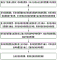
FIG. 1 is a schematic block diagram of the overall flow of the present invention;
FIG. 2 is a schematic block diagram of a process for preprocessing in accordance with the present invention;
FIG. 3 is a schematic block diagram of the U-shaped neural network of the present invention in operation;
FIG. 4 is a schematic block diagram of a process for optimizing disease images according to the present invention;
FIG. 5 is a comparative diagram of various processes of the present invention;
FIG. 6 is a schematic block diagram of a flow chart for analyzing disease images according to the present invention;
fig. 7 is a schematic block diagram of a feature extraction method corresponding to a disease type.
Detailed Description
One embodiment of the present invention will be described in detail below with reference to the attached drawings, but it should be understood that the scope of the present invention is not limited by the embodiment.
As shown in fig. 1 to 7, the present embodiment provides a method for processing disease data of an underwater structure, comprising the steps of:
shooting an image of an underwater structure by an underwater robot, and dividing the image containing diseases from the image as an original image;
secondly, acquiring a training image set, wherein the training image set comprises a training original image and a training disease image, preprocessing the training original image, and marking diseases in the processed training original image to obtain the training disease image;
thirdly, building a neural network model, taking the training original image as input after preprocessing, taking the training disease image as output, and training the neural network model to obtain a trained neural network model;
inputting the original image into the neural network model after preprocessing, outputting to obtain a disease image, and outputting the disease image;
fifthly, taking the original image as input after preprocessing, taking the disease image as output, correcting the neural network model, and updating the neural network model;
and (six) analyzing the disease image to obtain characteristic data of the disease.
The steps (I) - (VI) are sequentially carried out according to a logic sequence, the underwater robot is placed in water, the underwater robot finds an underwater structure according to a set program and completes shooting, so that the problem that a researcher can observe the underwater structure by self-launching during shooting can be completely solved, the accuracy of data and the personal safety problem of the researcher are ensured, and because the underwater structure receives the impact of water flow and the interference of aquatic organisms during shooting, the shot image is processed, and a neural network model is used during processing. Before using the neural network model, the step (two) is to acquire a training set, the data are used for carrying out preliminary training on the neural network, so that after the acquired original image is input, the disease image obtained after the original image is processed reaches the preliminary requirement, the approximate situation of the disease can be represented, at the moment, the disease image has a certain error with the expected one, in order to eliminate the error, when the neural network is used subsequently, the neural network is continuously corrected, namely, the content in the step (five), the obtained disease image is more and more close to the expected one, when the correction of the neural network reaches a certain amount, the disease image can be considered to be consistent with the expected one, at the moment, the obtained disease image is consistent with the expected one, finally in the step (six), the disease image is analyzed, the characteristic data of the disease are obtained, so that researchers can study the disease of the underwater structure by combining the disease image and the extracted characteristic data.
In the invention, to input an original image into a neural network model, the original image needs to be preprocessed so as to meet the requirement of the model, and when the original image is preprocessed, the method comprises the following steps:
(1) The original image is adjusted to be of a set pixel size, and the pixel size corresponds to the neural network model one by one;
(2) Carrying out normalization processing on the adjusted original image, so that the displacement, rotation degree and scale transformation of the original image are uniformly distributed in a specific range;
(3) And converting the original image after normalization processing into a format converted into a three-dimensional array.
The steps (1) - (3) are sequentially carried out according to a logic sequence, the original image is converted into an array format, and the array is subjected to model processing.
Meanwhile, the neural network model is a U-shaped neural network, and the U-shaped neural network comprises a plurality of downsampling modules, a plurality of asymmetric residual modules, a plurality of weak bottleneck modules and a plurality of upsampling modules which are arranged. The number of individual modules included in each class of neural network is different, and thus, a variety of neural network models can be composed.
Specifically, in the present invention, the original image is adjusted to a size of 1024×512 pixels, and the original image in the format of the three-dimensional array is a three-dimensional array of 1024×512×3, and the U-shaped neural network includes: the input layer i, the output layer O23, a plurality of downsampling modules D1, D2 and D8, a plurality of asymmetric residual modules A3-A7, A21-A22, a plurality of weak bottleneck modules N9-N16, N18-N19, a plurality of upsampling modules U17, U20, and 22 layers in total.
Based on the composition of the neural network model, the corresponding original image is 1024 x 512 pixels in size, and the U-shaped neural network comprises the following steps when in operation:
<1> the input layer i transmits the original image into D1 in a format of a three-dimensional array of 1024×512×3, and outputs a three-dimensional array of size 512×256×16 to D2;
after <2> d2 receives the input, outputting a three-dimensional array with a size of 256×128×64 to A3;
sequentially entering A3-A7, respectively outputting three-dimensional arrays with the size of 256-128-64 to D8, and respectively discarding pixels in A3-A7;
<4> enter D8, outputting a three-dimensional array of size 256 x 128 x 64 to N9;
<5> sequentially entering N9-N12, setting the void ratio of N9-N12 to be 2, 4, 8 and 16, and outputting a three-dimensional array with the size of 128 x 64 x 256 in each layer;
<6> sequentially entering N13-N16 to be a cavity convolution layer, setting the cavity rate of N13-N16 to be 2, 4, 8 and 16 in sequence, and outputting a three-dimensional array with the size of 128 x 64 x 128 by each layer;
<7> u17 receives the three-dimensional array output by N16 and converts it into a three-dimensional array of 256 x 128 x 64;
<8> sequentially entering N18-N19, and outputting a three-dimensional array of 256 x 128 x 64;
<9> u20 converts the input 256 x 128 x 64 three-dimensional array into 512 x 256 x 16 three-dimensional array;
<10> sequentially enters A21-A22, and outputs a three-dimensional array of 512 x 256 x 16;
the <11> output layer O23 restores the three-dimensional array of 512 x 256 x 16 to the three-dimensional array of 1024 x 512 x 3 to obtain the three-dimensional array format of the disease image, and outputs the disease image.
The downsampling module can enlarge the receptive field of the convolution layer on one hand, so that the context information is enriched, and the classification accuracy is improved; on the other hand, the feature diagram size can be obviously reduced, and the network calculation complexity and the memory occupancy rate are reduced. The network directly carries out downsampling processing on the first two layers of the network, thereby further improving the running speed of the network model. The downsampling process is to splice the outputs obtained through convolution (step length of 2) and maximum pooling operations respectively in the channel dimension to obtain the final output of the module.
Compared with the traditional conventional residual error module, the asymmetric residual error module has higher calculation efficiency, and can realize the same receptive field as an n convolution by stacking the n convolution and the n convolution, but the network parameters are greatly reduced, and the complexity of the model is also obviously reduced.
As network depth increases, a bottleneck module is generally used to reduce the parameters and accuracy loss of the model, but the bottleneck structure is generally affected by degradation, so that each convolution layer is decomposed by using a combination of 1D filters, and the resulting low-dimensional hierarchical layer has a simple structure and reduces the calculation cost.
The up-sampling module is used for up-sampling operation by using transpose convolution for the problems of information loss, precision reduction and the like caused by down-sampling operation, so that the spatial information loss is effectively relieved, and the image precision loss is reduced. The transpose convolution step is 2.
In order to ensure the clear and continuous of the obtained disease images, and facilitate research and observation of researchers, after the three-dimensional array of 512 x 256 x 16 is output by the A22, the method comprises the following steps:
1> respectively judging the pixel position of each pixel;
2> carrying out convolution classification on the pixel with the pixel position judged as the central pixel position to obtain a pixel label of the pixel;
3> mapping the center pixel label to obtain a super pixel label;
4> traversing all pixels to complete updating the 512 x 256 x 16 three-dimensional array and entering the output layer O23.
According to the invention, the pixel coordinate information of the central position of the super pixel is added in the last layer of the U-shaped neural network (U-NET), only the 1x 1 convolution classification is carried out on the pixels at the central position, so that the U-NET carries out disease position image segmentation on the super pixel instead of the pixels, the U-NET speed is effectively improved, the improved U-NET architecture parameters of the network adopt the original parameters of the U-NET, the convolution kernel parameters are changed, and the detail contents are as follows: before the final layer carries out convolution classification, the pixel position is judged; if the pixel position is the central pixel position, carrying out convolution classification to obtain a pixel label, otherwise, continuing to judge the pixel position; and mapping the center pixel label to obtain a super pixel label, and completing the image segmentation of the disease position.
Fig. 5 is a graph comparing the image processing procedure and results at various stages of the present invention.
In the invention, when obtaining and disease images, the research of researchers is more convenient, the characteristics in the disease images are extracted, effective data are provided for the researchers, and when analyzing the disease images to obtain the characteristic data of the diseases, the invention comprises the following steps:
firstly, converting the disease image into a two-dimensional array, and extracting a disease contour in the disease image;
secondly, judging the disease type according to the disease profile, and obtaining a characteristic extraction mode corresponding to the disease type;
third, completing the extraction of the characteristic data of the disease in the disease type according to the characteristic extraction mode;
and outputting the disease type and the corresponding characteristic data.
The steps < first > - < fourth > are sequentially carried out according to a logic sequence, and since the disease images are black and white images, the disease contours can be rapidly extracted by converting the disease images into arrays of 0 and 1, wherein 0 represents white, 1 represents black, corresponding coordinates can be obtained at the same time, the corresponding feature extraction modes are inconsistent due to inconsistent shapes of different diseases, the step < second > is to obtain the feature extraction modes of the diseases according to the disease contours, the step < third > is to extract the feature data of the diseases, and finally output the feature data, so that researchers can study the feature data of the diseases while contrasting the disease images, and the efficiency and the accuracy of study are improved.
In the present invention, for clarity of the outline representation, the pixel points of the disease image are represented by means of coordinates, and the disease outline is represented by means of a set of coordinates.
Meanwhile, when the feature extraction mode corresponding to the disease type is obtained, the method comprises the following steps:
respectively acquiring slopes between every two adjacent coordinate points in the disease profile, and inducing coordinate points corresponding to the slopes with errors within a set range together to obtain a profile line;
secondly, calculating the length of each contour line according to the number of coordinates, and enabling the length and the slope of each contour line to correspond to each other one by one;
thirdly, searching diseases with consistent slopes and proportional lengths of all contour lines in a disease database, and obtaining corresponding characteristic extraction modes;
the disease database is used for storing the slope and the length of each contour line corresponding to the disease and the characteristic extraction mode.
The steps one to three are sequentially carried out according to the logic sequence, the slope of each point is calculated, the points with the same slope are connected according to the positions to form contour lines, one disease contour comprises a plurality of contour lines, each contour line has a corresponding length and slope, the graph shape of the disease contour can be obtained through the lengths and slopes of the contour lines, the obtained contour lines are respectively compared with data in a disease database, the corresponding disease type and the corresponding extraction mode can be obtained, and accordingly the characteristic extraction mode of the disease is completed.
In summary, the invention uses the asymmetric residual error module to increase the running speed when the image calculation amount is large, and uses the weak bottleneck module to reduce the parameter amount and reduce the precision loss when the module channels are more and the network parameter amount is large. Compared with the traditional U-shaped structural neural network, the method converts the image into the array for rapid operation, and experiments prove that the speed and the accuracy of the network for identifying the bridge underwater pile foundation diseases are greatly improved.
The foregoing disclosure is merely illustrative of some embodiments of the invention, but the embodiments are not limited thereto and variations within the scope of the invention will be apparent to those skilled in the art.
Claims (7)
1. The method for processing the disease data of the underwater structure is characterized by comprising the following steps:
shooting an image of an underwater structure by an underwater robot, and dividing the image containing diseases from the image to serve as an original image;
acquiring a training image set, wherein the training image set comprises a training original image and a training disease image, preprocessing the training original image, and marking diseases in the processed training original image to obtain the training disease image;
establishing a neural network model, taking the training original image as input after preprocessing, taking the training disease image as output, and training the neural network model to obtain a trained neural network model;
inputting the original image into the neural network model after preprocessing, and outputting to obtain a disease image;
taking the original image as input after preprocessing, taking the disease image as output, correcting the neural network model, and updating the neural network model;
converting the disease image into a two-dimensional array, converting the disease image into an array of 0 and 1 because the disease image is a black-and-white image, and extracting a disease contour in the disease image;
judging the disease type according to the disease profile, and obtaining a characteristic extraction mode corresponding to the disease type;
completing the extraction of the characteristic data of the diseases in the disease type according to the characteristic extraction mode; when the feature extraction mode corresponding to the disease type is obtained, the method comprises the following steps:
respectively acquiring slopes between every two adjacent coordinate points in the disease profile, and inducing coordinate points corresponding to the slopes with errors within a set range together to obtain a profile line;
calculating the length of each contour line according to the number of coordinates, and enabling the length and the slope of each contour line to correspond to each other one by one;
searching diseases with consistent slopes and proportional lengths of all contour lines in a disease database, and obtaining corresponding characteristic extraction modes;
the disease contour comprises a plurality of contour lines, each contour line has a corresponding length and a corresponding slope, the graph shape of the disease contour is obtained through the lengths and the slopes of the contour lines, and the obtained contour lines are respectively compared with data in a disease database to obtain a corresponding disease type and a corresponding extraction mode;
the disease database is used for storing the slope and the length of each contour line corresponding to the disease and the characteristic extraction mode;
and outputting the disease type and the corresponding characteristic data.
2. The method for processing disease data of an underwater structure according to claim 1, wherein when the original image is preprocessed, comprising the steps of:
the original image is adjusted to be of a set pixel size, and the pixel size corresponds to the neural network model one by one;
carrying out normalization processing on the adjusted original image, so that the displacement, rotation degree and scale transformation of the original image are uniformly distributed in a specific range;
and converting the original image after normalization processing into a format converted into a three-dimensional array.
3. The method for processing disease data of an underwater structure according to claim 2, wherein the neural network model is a U-shaped neural network, and the U-shaped neural network comprises a plurality of downsampling modules, a plurality of asymmetric residual modules, a plurality of weak bottleneck modules and a plurality of upsampling modules which are arranged.
4. A method for processing disease data of an underwater structure as in claim 3, wherein the original image is adjusted to 1024 x 512 pixels, and the original image of the three-dimensional array format is a three-dimensional array of 1024 x 512 x 3, and the U-shaped neural network comprises: an input layer i, an output layer O23, a plurality of downsampling modules D1, D2 and D8, a plurality of asymmetric residual modules A3-A7, A21-A22, a plurality of weak bottleneck modules N9-N16, N18-N19, and a plurality of upsampling modules U17 and U20.
5. The method for processing disease data of an underwater structure according to claim 4, wherein the U-shaped neural network comprises the steps of, when in operation:
the input layer i transmits the original image into D1 in a format of a three-dimensional array of 1024 x 512 x 3, and outputs the three-dimensional array with the size of 512 x 256 x 16 to D2;
d2 outputs a three-dimensional array with the size of 256 x 128 x 64 to A3 after receiving the input;
sequentially entering A3-A7, respectively outputting three-dimensional arrays with the size of 256-128-64 to D8, and respectively discarding pixels in A3-A7;
enter D8, output the three-dimensional array with the size of 256 x 128 x 64 to N9;
sequentially entering N9-N12, setting the void ratio of N9-N12 as 2, 4, 8 and 16, and outputting a three-dimensional array with the size of 128 x 64 x 256 in each layer;
sequentially entering N13-N16 to be a cavity convolution layer, setting the cavity rate of N13-N16 to be 2, 4, 8 and 16 in sequence, and outputting a three-dimensional array with the size of 128 x 64 x 128 in each layer;
u17 receives the three-dimensional array output by N16 and converts the three-dimensional array into 256×128×64 three-dimensional arrays;
sequentially entering N18-N19 and outputting a three-dimensional array of 256 x 128 x 64;
u20 converts the input 256 x 128 x 64 three-dimensional array into a 512 x 256 x 16 three-dimensional array;
sequentially entering A21-A22 and outputting a three-dimensional array of 512 x 256 x 16;
the output layer O23 restores the three-dimensional array of 512 x 256 x 16 to the three-dimensional array of 1024 x 512 x 3 to obtain the format of the three-dimensional array of the disease image, and outputs the disease image.
6. The method for processing disease data of an underwater structure according to claim 5, wherein after the a22 outputs the three-dimensional array of 512 x 256 x 16, comprising the steps of:
respectively judging the pixel position of each pixel;
performing convolution classification on the pixel with the pixel position being discriminated as the central pixel position to obtain a pixel label of the pixel;
mapping the center pixel label to obtain a super pixel label;
all pixels are traversed to complete updating of the 512 x 256 x 16 three-dimensional array and enter the output layer O23.
7. The method of claim 1, wherein the pixels of the disease image are represented by coordinates and the disease profile is represented by a set of coordinates.
Priority Applications (1)
| Application Number | Priority Date | Filing Date | Title |
|---|---|---|---|
| CN202210403079.7A CN114494261B (en) | 2022-04-18 | 2022-04-18 | Disease data processing method for underwater structure |
Applications Claiming Priority (1)
| Application Number | Priority Date | Filing Date | Title |
|---|---|---|---|
| CN202210403079.7A CN114494261B (en) | 2022-04-18 | 2022-04-18 | Disease data processing method for underwater structure |
Publications (2)
| Publication Number | Publication Date |
|---|---|
| CN114494261A CN114494261A (en) | 2022-05-13 |
| CN114494261B true CN114494261B (en) | 2023-04-25 |
Family
ID=81489679
Family Applications (1)
| Application Number | Title | Priority Date | Filing Date |
|---|---|---|---|
| CN202210403079.7A Active CN114494261B (en) | 2022-04-18 | 2022-04-18 | Disease data processing method for underwater structure |
Country Status (1)
| Country | Link |
|---|---|
| CN (1) | CN114494261B (en) |
Families Citing this family (1)
| Publication number | Priority date | Publication date | Assignee | Title |
|---|---|---|---|---|
| CN114862830A (en) * | 2022-05-30 | 2022-08-05 | 武汉理工大学 | Underwater structure surface disease image acquisition method and device and electronic equipment |
Citations (4)
| Publication number | Priority date | Publication date | Assignee | Title |
|---|---|---|---|---|
| CN104318227A (en) * | 2014-11-19 | 2015-01-28 | 天津工业大学 | Corn disease recognition method based on Curvelet-SC |
| KR20150043581A (en) * | 2013-10-11 | 2015-04-23 | 대우조선해양 주식회사 | Automatic set up insulation box of lng ship's using three dimensional vision system and thereof apparatus |
| AU2020102885A4 (en) * | 2020-10-20 | 2020-12-17 | Xijing University | Disease recognition method of winter jujube based on deep convolutional neural network and disease image |
| CN113971660A (en) * | 2021-09-30 | 2022-01-25 | 哈尔滨工业大学 | Computer vision method for bridge health diagnosis and intelligent camera system |
Family Cites Families (7)
| Publication number | Priority date | Publication date | Assignee | Title |
|---|---|---|---|---|
| CN106087677B (en) * | 2016-06-02 | 2018-07-31 | 上海华城工程建设管理有限公司 | Asphalt pavement crack type automatic identifying method |
| CN110533069B (en) * | 2019-07-25 | 2022-02-11 | 西安电子科技大学 | Two-dimensional foil strip distribution characteristic identification method based on support vector machine algorithm |
| CN111127399A (en) * | 2019-11-28 | 2020-05-08 | 东南大学 | An underwater bridge pier disease identification method based on deep learning and sonar imaging |
| CN111951289B (en) * | 2020-08-13 | 2023-10-31 | 江苏东印智慧工程技术研究院有限公司 | A method of underwater sonar image data segmentation based on BA-Unet |
| CN112581482A (en) * | 2020-08-13 | 2021-03-30 | 江苏东印智慧工程技术研究院有限公司 | Underwater sonar image data segmentation method based on path-unet |
| CN112508901B (en) * | 2020-12-01 | 2024-04-05 | 广州大学 | Underwater structure disease identification method, system, device and storage medium |
| CN114018932B (en) * | 2021-11-02 | 2023-05-30 | 西安电子科技大学 | Measuring Method of Pavement Disease Index Based on Rectangular Calibration Object |
-
2022
- 2022-04-18 CN CN202210403079.7A patent/CN114494261B/en active Active
Patent Citations (4)
| Publication number | Priority date | Publication date | Assignee | Title |
|---|---|---|---|---|
| KR20150043581A (en) * | 2013-10-11 | 2015-04-23 | 대우조선해양 주식회사 | Automatic set up insulation box of lng ship's using three dimensional vision system and thereof apparatus |
| CN104318227A (en) * | 2014-11-19 | 2015-01-28 | 天津工业大学 | Corn disease recognition method based on Curvelet-SC |
| AU2020102885A4 (en) * | 2020-10-20 | 2020-12-17 | Xijing University | Disease recognition method of winter jujube based on deep convolutional neural network and disease image |
| CN113971660A (en) * | 2021-09-30 | 2022-01-25 | 哈尔滨工业大学 | Computer vision method for bridge health diagnosis and intelligent camera system |
Non-Patent Citations (2)
| Title |
|---|
| Jun Xie 等.Shape matching and modeling using skeletal context.《Pattern Recognition》.2008,第41卷(第5期),第1756-1767页. * |
| 孙坤. 不规则轮廓的机器视觉检测算法研究.《中国优秀硕士学位论文全文数据库信息科技辑》.2018,第2018年卷(第12期),第I138-1476页. * |
Also Published As
| Publication number | Publication date |
|---|---|
| CN114494261A (en) | 2022-05-13 |
Similar Documents
| Publication | Publication Date | Title |
|---|---|---|
| CN112270249B (en) | A target pose estimation method integrating RGB-D visual features | |
| CN114022408B (en) | Cloud Detection Method in Remote Sensing Images Based on Multi-scale Convolutional Neural Network | |
| CN112967271B (en) | A Casting Surface Defect Recognition Method Based on Improved DeepLabv3+ Network Model | |
| CN112418171B (en) | A method for zebrafish spatial posture and heart position estimation based on deep learning | |
| CN113449691A (en) | Human shape recognition system and method based on non-local attention mechanism | |
| CN114782770A (en) | A method and system for license plate detection and license plate recognition based on deep learning | |
| CN116740528B (en) | A Target Detection Method and System Based on Shadow Features in Side-Scan Sonar Images | |
| CN115937552B (en) | An image matching method based on fusion of manual features and deep features | |
| CN113762358A (en) | Semi-supervised learning three-dimensional reconstruction method based on relative deep training | |
| CN109376589A (en) | Recognition method of ROV deformable target and small target based on convolution kernel screening SSD network | |
| CN114898212A (en) | A method for extracting multi-object change information from high-resolution remote sensing images | |
| CN113486894A (en) | Semantic segmentation method for satellite image feature component | |
| CN116704188A (en) | An Image Segmentation Algorithm of Wheat Grains with Different Test Weights Based on Improved U-Net Network | |
| CN114494261B (en) | Disease data processing method for underwater structure | |
| CN114120359A (en) | Method for measuring body size of group-fed pigs based on stacked hourglass network | |
| CN116824347A (en) | A road crack detection method based on deep learning | |
| CN116740703B (en) | Wheat phenotype parameter change rate estimation method and device based on point cloud information | |
| CN114120237A (en) | Construction site safety belt identification method and system | |
| CN116863321A (en) | SSE-YOLO deep learning model-based forward-looking sonar image small target recognition method | |
| CN116385425A (en) | A YOLOv5 fabric defect detection method with improved CA attention mechanism | |
| CN115546469A (en) | Key point detection method based on deep neural network | |
| CN118967785B (en) | Leaf phenotypic parameter calculation method based on soybean leaf tiled image generation model | |
| CN119516580A (en) | Method and device for identifying disease degree of fish school | |
| CN119380334A (en) | A quantification method for DUS traits in peanut pods based on key point detection | |
| CN112819823A (en) | Furniture board-oriented circular hole detection method, system and device |
Legal Events
| Date | Code | Title | Description |
|---|---|---|---|
| PB01 | Publication | ||
| PB01 | Publication | ||
| SE01 | Entry into force of request for substantive examination | ||
| SE01 | Entry into force of request for substantive examination | ||
| GR01 | Patent grant | ||
| GR01 | Patent grant |