CN114438218A - Gene Panel for detecting various tumors, kit and application - Google Patents
Gene Panel for detecting various tumors, kit and application Download PDFInfo
- Publication number
- CN114438218A CN114438218A CN202210339492.1A CN202210339492A CN114438218A CN 114438218 A CN114438218 A CN 114438218A CN 202210339492 A CN202210339492 A CN 202210339492A CN 114438218 A CN114438218 A CN 114438218A
- Authority
- CN
- China
- Prior art keywords
- tumors
- genes
- gene
- syndrome
- detection
- Prior art date
- Legal status (The legal status is an assumption and is not a legal conclusion. Google has not performed a legal analysis and makes no representation as to the accuracy of the status listed.)
- Granted
Links
- 108090000623 proteins and genes Proteins 0.000 title claims abstract description 89
- 206010028980 Neoplasm Diseases 0.000 title claims abstract description 75
- 238000001514 detection method Methods 0.000 claims abstract description 51
- 206010033128 Ovarian cancer Diseases 0.000 claims abstract description 30
- 206010061535 Ovarian neoplasm Diseases 0.000 claims abstract description 29
- 208000011580 syndromic disease Diseases 0.000 claims abstract description 27
- 206010006187 Breast cancer Diseases 0.000 claims abstract description 25
- 208000026310 Breast neoplasm Diseases 0.000 claims abstract description 25
- 208000000236 Prostatic Neoplasms Diseases 0.000 claims abstract description 18
- 230000034431 double-strand break repair via homologous recombination Effects 0.000 claims abstract description 16
- 208000009849 Female Genital Neoplasms Diseases 0.000 claims abstract description 13
- 210000001035 gastrointestinal tract Anatomy 0.000 claims abstract description 13
- 208000033640 Hereditary breast cancer Diseases 0.000 claims abstract description 12
- 208000025581 hereditary breast carcinoma Diseases 0.000 claims abstract description 12
- 230000037361 pathway Effects 0.000 claims abstract description 12
- 239000000523 sample Substances 0.000 claims description 79
- 230000035772 mutation Effects 0.000 claims description 75
- 108020004414 DNA Proteins 0.000 claims description 45
- 238000000034 method Methods 0.000 claims description 22
- 238000012163 sequencing technique Methods 0.000 claims description 19
- 102100034484 DNA repair protein RAD51 homolog 3 Human genes 0.000 claims description 8
- 108010067741 Fanconi Anemia Complementation Group N protein Proteins 0.000 claims description 8
- 102000016627 Fanconi Anemia Complementation Group N protein Human genes 0.000 claims description 8
- 102100034553 Fanconi anemia group J protein Human genes 0.000 claims description 8
- 101001132271 Homo sapiens DNA repair protein RAD51 homolog 3 Proteins 0.000 claims description 8
- 101000848171 Homo sapiens Fanconi anemia group J protein Proteins 0.000 claims description 8
- 108010011536 PTEN Phosphohydrolase Proteins 0.000 claims description 8
- 102000014160 PTEN Phosphohydrolase Human genes 0.000 claims description 8
- 238000012165 high-throughput sequencing Methods 0.000 claims description 8
- 101150008921 Brca2 gene Proteins 0.000 claims description 7
- 101000785776 Homo sapiens Artemin Proteins 0.000 claims description 7
- 101000981336 Homo sapiens Nibrin Proteins 0.000 claims description 7
- 102100024403 Nibrin Human genes 0.000 claims description 7
- 102000000872 ATM Human genes 0.000 claims description 6
- 108010004586 Ataxia Telangiectasia Mutated Proteins Proteins 0.000 claims description 6
- 101700002522 BARD1 Proteins 0.000 claims description 6
- 102100024641 BRCA1-A complex subunit Abraxas 1 Human genes 0.000 claims description 6
- 102100028048 BRCA1-associated RING domain protein 1 Human genes 0.000 claims description 6
- 108700020462 BRCA2 Proteins 0.000 claims description 6
- 102000052609 BRCA2 Human genes 0.000 claims description 6
- 102100039116 DNA repair protein RAD50 Human genes 0.000 claims description 6
- 102100034483 DNA repair protein RAD51 homolog 4 Human genes 0.000 claims description 6
- 101000760704 Homo sapiens BRCA1-A complex subunit Abraxas 1 Proteins 0.000 claims description 6
- 101000743929 Homo sapiens DNA repair protein RAD50 Proteins 0.000 claims description 6
- 101001132266 Homo sapiens DNA repair protein RAD51 homolog 4 Proteins 0.000 claims description 6
- 101000777277 Homo sapiens Serine/threonine-protein kinase Chk2 Proteins 0.000 claims description 6
- 102100031075 Serine/threonine-protein kinase Chk2 Human genes 0.000 claims description 6
- 108010078814 Tumor Suppressor Protein p53 Proteins 0.000 claims description 6
- 238000003766 bioinformatics method Methods 0.000 claims description 6
- 230000007918 pathogenicity Effects 0.000 claims description 6
- 102100034157 DNA mismatch repair protein Msh2 Human genes 0.000 claims description 5
- 238000004458 analytical method Methods 0.000 claims description 5
- 108091007854 Cdh1/Fizzy-related Proteins 0.000 claims description 4
- 102000038594 Cdh1/Fizzy-related Human genes 0.000 claims description 4
- 102100021147 DNA mismatch repair protein Msh6 Human genes 0.000 claims description 4
- 101001134036 Homo sapiens DNA mismatch repair protein Msh2 Proteins 0.000 claims description 4
- 101000968658 Homo sapiens DNA mismatch repair protein Msh6 Proteins 0.000 claims description 4
- 101000628562 Homo sapiens Serine/threonine-protein kinase STK11 Proteins 0.000 claims description 4
- 229910015837 MSH2 Inorganic materials 0.000 claims description 4
- 108010074346 Mismatch Repair Endonuclease PMS2 Proteins 0.000 claims description 4
- 102000013609 MutL Protein Homolog 1 Human genes 0.000 claims description 4
- 108010026664 MutL Protein Homolog 1 Proteins 0.000 claims description 4
- 102100026715 Serine/threonine-protein kinase STK11 Human genes 0.000 claims description 4
- 238000002360 preparation method Methods 0.000 claims description 4
- 101150072950 BRCA1 gene Proteins 0.000 claims description 3
- 102100027161 BRCA2-interacting transcriptional repressor EMSY Human genes 0.000 claims description 3
- 101100514311 Caenorhabditis elegans mre-11 gene Proteins 0.000 claims description 3
- 102100038111 Cyclin-dependent kinase 12 Human genes 0.000 claims description 3
- 102100034490 DNA repair and recombination protein RAD54B Human genes 0.000 claims description 3
- 102100033934 DNA repair protein RAD51 homolog 2 Human genes 0.000 claims description 3
- 102100033996 Double-strand break repair protein MRE11 Human genes 0.000 claims description 3
- 102000012804 EPCAM Human genes 0.000 claims description 3
- 101150084967 EPCAM gene Proteins 0.000 claims description 3
- 102100033962 GTP-binding protein RAD Human genes 0.000 claims description 3
- 101001057996 Homo sapiens BRCA2-interacting transcriptional repressor EMSY Proteins 0.000 claims description 3
- 101000884345 Homo sapiens Cyclin-dependent kinase 12 Proteins 0.000 claims description 3
- 101001132263 Homo sapiens DNA repair and recombination protein RAD54B Proteins 0.000 claims description 3
- 101000591400 Homo sapiens Double-strand break repair protein MRE11 Proteins 0.000 claims description 3
- 101001095815 Homo sapiens E3 ubiquitin-protein ligase RING2 Proteins 0.000 claims description 3
- 101001132495 Homo sapiens GTP-binding protein RAD Proteins 0.000 claims description 3
- 101001057193 Homo sapiens Membrane-associated guanylate kinase, WW and PDZ domain-containing protein 1 Proteins 0.000 claims description 3
- 101000777293 Homo sapiens Serine/threonine-protein kinase Chk1 Proteins 0.000 claims description 3
- 101000802948 Homo sapiens Serine/threonine-protein phosphatase 2A 55 kDa regulatory subunit B alpha isoform Proteins 0.000 claims description 3
- 101000740048 Homo sapiens Ubiquitin carboxyl-terminal hydrolase BAP1 Proteins 0.000 claims description 3
- 101000740049 Latilactobacillus curvatus Bioactive peptide 1 Proteins 0.000 claims description 3
- 102100027240 Membrane-associated guanylate kinase, WW and PDZ domain-containing protein 1 Human genes 0.000 claims description 3
- 101710018890 RAD51B Proteins 0.000 claims description 3
- 102100031081 Serine/threonine-protein kinase Chk1 Human genes 0.000 claims description 3
- 102100035728 Serine/threonine-protein phosphatase 2A 55 kDa regulatory subunit B alpha isoform Human genes 0.000 claims description 3
- 101150057140 TACSTD1 gene Proteins 0.000 claims description 3
- 108700020463 BRCA1 Proteins 0.000 claims 2
- 102000036365 BRCA1 Human genes 0.000 claims 2
- 102000008071 Mismatch Repair Endonuclease PMS2 Human genes 0.000 claims 1
- 102000015098 Tumor Suppressor Protein p53 Human genes 0.000 claims 1
- 239000012661 PARP inhibitor Substances 0.000 abstract description 6
- 229940121906 Poly ADP ribose polymerase inhibitor Drugs 0.000 abstract description 6
- 210000001519 tissue Anatomy 0.000 abstract description 6
- 210000004602 germ cell Anatomy 0.000 abstract description 5
- 230000002068 genetic effect Effects 0.000 abstract description 4
- BASFCYQUMIYNBI-UHFFFAOYSA-N platinum Chemical compound [Pt] BASFCYQUMIYNBI-UHFFFAOYSA-N 0.000 abstract description 4
- 239000002246 antineoplastic agent Substances 0.000 abstract description 3
- 229940044683 chemotherapy drug Drugs 0.000 abstract description 3
- 229910052697 platinum Inorganic materials 0.000 abstract description 2
- 239000011324 bead Substances 0.000 description 30
- 239000000203 mixture Substances 0.000 description 26
- LFQSCWFLJHTTHZ-UHFFFAOYSA-N Ethanol Chemical compound CCO LFQSCWFLJHTTHZ-UHFFFAOYSA-N 0.000 description 20
- 239000006228 supernatant Substances 0.000 description 17
- 239000007788 liquid Substances 0.000 description 13
- 238000002156 mixing Methods 0.000 description 11
- 238000006243 chemical reaction Methods 0.000 description 10
- 206010060862 Prostate cancer Diseases 0.000 description 9
- 230000002939 deleterious effect Effects 0.000 description 9
- 238000009396 hybridization Methods 0.000 description 9
- 239000011534 wash buffer Substances 0.000 description 9
- 206010053138 Congenital aplastic anaemia Diseases 0.000 description 8
- 201000004939 Fanconi anemia Diseases 0.000 description 8
- 206010061902 Pancreatic neoplasm Diseases 0.000 description 8
- 208000015486 malignant pancreatic neoplasm Diseases 0.000 description 8
- 201000002528 pancreatic cancer Diseases 0.000 description 8
- 208000008443 pancreatic carcinoma Diseases 0.000 description 8
- 238000012216 screening Methods 0.000 description 8
- 238000005119 centrifugation Methods 0.000 description 7
- 238000007481 next generation sequencing Methods 0.000 description 7
- 206010009944 Colon cancer Diseases 0.000 description 6
- 208000001333 Colorectal Neoplasms Diseases 0.000 description 6
- 206010064571 Gene mutation Diseases 0.000 description 6
- 108010090804 Streptavidin Proteins 0.000 description 6
- 239000000090 biomarker Substances 0.000 description 6
- 230000000694 effects Effects 0.000 description 6
- 238000005516 engineering process Methods 0.000 description 6
- 201000007741 female breast cancer Diseases 0.000 description 6
- 201000002276 female breast carcinoma Diseases 0.000 description 6
- 230000001717 pathogenic effect Effects 0.000 description 6
- 238000000746 purification Methods 0.000 description 6
- 239000000725 suspension Substances 0.000 description 6
- XLYOFNOQVPJJNP-UHFFFAOYSA-N water Substances O XLYOFNOQVPJJNP-UHFFFAOYSA-N 0.000 description 6
- 102100025064 Cellular tumor antigen p53 Human genes 0.000 description 5
- 210000004369 blood Anatomy 0.000 description 5
- 239000008280 blood Substances 0.000 description 5
- 239000000872 buffer Substances 0.000 description 5
- 238000012217 deletion Methods 0.000 description 5
- 230000037430 deletion Effects 0.000 description 5
- 239000003814 drug Substances 0.000 description 5
- 238000003780 insertion Methods 0.000 description 5
- 230000037431 insertion Effects 0.000 description 5
- 238000012986 modification Methods 0.000 description 5
- 230000004048 modification Effects 0.000 description 5
- 230000008439 repair process Effects 0.000 description 5
- 238000012360 testing method Methods 0.000 description 5
- BPYKTIZUTYGOLE-IFADSCNNSA-N Bilirubin Chemical compound N1C(=O)C(C)=C(C=C)\C1=C\C1=C(C)C(CCC(O)=O)=C(CC2=C(C(C)=C(\C=C/3C(=C(C=C)C(=O)N\3)C)N2)CCC(O)=O)N1 BPYKTIZUTYGOLE-IFADSCNNSA-N 0.000 description 4
- 206010014733 Endometrial cancer Diseases 0.000 description 4
- 206010014759 Endometrial neoplasm Diseases 0.000 description 4
- 208000004059 Male Breast Neoplasms Diseases 0.000 description 4
- 229920000776 Poly(Adenosine diphosphate-ribose) polymerase Polymers 0.000 description 4
- 230000003321 amplification Effects 0.000 description 4
- 210000000481 breast Anatomy 0.000 description 4
- 238000010276 construction Methods 0.000 description 4
- 229940079593 drug Drugs 0.000 description 4
- 238000003364 immunohistochemistry Methods 0.000 description 4
- 238000007689 inspection Methods 0.000 description 4
- 201000003175 male breast cancer Diseases 0.000 description 4
- 208000010907 male breast carcinoma Diseases 0.000 description 4
- 230000036438 mutation frequency Effects 0.000 description 4
- 238000003199 nucleic acid amplification method Methods 0.000 description 4
- 102100037480 Mismatch repair endonuclease PMS2 Human genes 0.000 description 3
- 238000003556 assay Methods 0.000 description 3
- 230000008901 benefit Effects 0.000 description 3
- 201000011510 cancer Diseases 0.000 description 3
- 210000004027 cell Anatomy 0.000 description 3
- 238000011156 evaluation Methods 0.000 description 3
- 230000004927 fusion Effects 0.000 description 3
- 230000008569 process Effects 0.000 description 3
- 239000000047 product Substances 0.000 description 3
- 239000002096 quantum dot Substances 0.000 description 3
- 230000035945 sensitivity Effects 0.000 description 3
- 239000000243 solution Substances 0.000 description 3
- 238000012795 verification Methods 0.000 description 3
- 102000001554 Hemoglobins Human genes 0.000 description 2
- 108010054147 Hemoglobins Proteins 0.000 description 2
- 238000012408 PCR amplification Methods 0.000 description 2
- 206010034764 Peutz-Jeghers syndrome Diseases 0.000 description 2
- 101710179684 Poly [ADP-ribose] polymerase Proteins 0.000 description 2
- 102100023712 Poly [ADP-ribose] polymerase 1 Human genes 0.000 description 2
- FAPWRFPIFSIZLT-UHFFFAOYSA-M Sodium chloride Chemical compound [Na+].[Cl-] FAPWRFPIFSIZLT-UHFFFAOYSA-M 0.000 description 2
- 208000005718 Stomach Neoplasms Diseases 0.000 description 2
- DTQVDTLACAAQTR-UHFFFAOYSA-N Trifluoroacetic acid Chemical compound OC(=O)C(F)(F)F DTQVDTLACAAQTR-UHFFFAOYSA-N 0.000 description 2
- 239000003153 chemical reaction reagent Substances 0.000 description 2
- 238000001035 drying Methods 0.000 description 2
- 238000010828 elution Methods 0.000 description 2
- 239000003623 enhancer Substances 0.000 description 2
- 230000004907 flux Effects 0.000 description 2
- 239000012634 fragment Substances 0.000 description 2
- 206010017758 gastric cancer Diseases 0.000 description 2
- PCHJSUWPFVWCPO-UHFFFAOYSA-N gold Chemical compound [Au] PCHJSUWPFVWCPO-UHFFFAOYSA-N 0.000 description 2
- 201000001441 melanoma Diseases 0.000 description 2
- 238000003752 polymerase chain reaction Methods 0.000 description 2
- 238000003757 reverse transcription PCR Methods 0.000 description 2
- 102200108658 rs137852789 Human genes 0.000 description 2
- 238000007480 sanger sequencing Methods 0.000 description 2
- 201000011549 stomach cancer Diseases 0.000 description 2
- 238000012546 transfer Methods 0.000 description 2
- 230000001052 transient effect Effects 0.000 description 2
- UFTFJSFQGQCHQW-UHFFFAOYSA-N triformin Chemical compound O=COCC(OC=O)COC=O UFTFJSFQGQCHQW-UHFFFAOYSA-N 0.000 description 2
- GUAHPAJOXVYFON-ZETCQYMHSA-N (8S)-8-amino-7-oxononanoic acid zwitterion Chemical compound C[C@H](N)C(=O)CCCCCC(O)=O GUAHPAJOXVYFON-ZETCQYMHSA-N 0.000 description 1
- 101150005355 36 gene Proteins 0.000 description 1
- 206010003594 Ataxia telangiectasia Diseases 0.000 description 1
- 101150065175 Atm gene Proteins 0.000 description 1
- 108700040618 BRCA1 Genes Proteins 0.000 description 1
- 108700010154 BRCA2 Genes Proteins 0.000 description 1
- 101150059668 Bard1 gene Proteins 0.000 description 1
- 101150003676 Brip1 gene Proteins 0.000 description 1
- 101150064168 CHEK2 gene Proteins 0.000 description 1
- 206010008342 Cervix carcinoma Diseases 0.000 description 1
- 208000012609 Cowden disease Diseases 0.000 description 1
- 201000002847 Cowden syndrome Diseases 0.000 description 1
- 102000053602 DNA Human genes 0.000 description 1
- 208000025939 DNA Repair-Deficiency disease Diseases 0.000 description 1
- 208000034826 Genetic Predisposition to Disease Diseases 0.000 description 1
- 208000002927 Hamartoma Diseases 0.000 description 1
- 101100220617 Homo sapiens CHEK2 gene Proteins 0.000 description 1
- 101100518728 Homo sapiens PALB2 gene Proteins 0.000 description 1
- 229940076838 Immune checkpoint inhibitor Drugs 0.000 description 1
- 208000026350 Inborn Genetic disease Diseases 0.000 description 1
- 102000037984 Inhibitory immune checkpoint proteins Human genes 0.000 description 1
- 108091008026 Inhibitory immune checkpoint proteins Proteins 0.000 description 1
- 201000011062 Li-Fraumeni syndrome Diseases 0.000 description 1
- 208000032818 Microsatellite Instability Diseases 0.000 description 1
- 208000008770 Multiple Hamartoma Syndrome Diseases 0.000 description 1
- 101150081841 NBN gene Proteins 0.000 description 1
- 208000007571 Ovarian Epithelial Carcinoma Diseases 0.000 description 1
- 101150099884 PALB2 gene Proteins 0.000 description 1
- 101150073900 PTEN gene Proteins 0.000 description 1
- 102000012338 Poly(ADP-ribose) Polymerases Human genes 0.000 description 1
- 108010061844 Poly(ADP-ribose) Polymerases Proteins 0.000 description 1
- 208000037062 Polyps Diseases 0.000 description 1
- 108700020978 Proto-Oncogene Proteins 0.000 description 1
- 102000052575 Proto-Oncogene Human genes 0.000 description 1
- 101150046721 RAD51C gene Proteins 0.000 description 1
- 101150068031 RAD51D gene Proteins 0.000 description 1
- 101150040067 STK11 gene Proteins 0.000 description 1
- 101100484967 Solanum tuberosum PVS1 gene Proteins 0.000 description 1
- 108700025695 Suppressor Genes Proteins 0.000 description 1
- 101150080074 TP53 gene Proteins 0.000 description 1
- 208000006105 Uterine Cervical Neoplasms Diseases 0.000 description 1
- 230000004913 activation Effects 0.000 description 1
- 238000000246 agarose gel electrophoresis Methods 0.000 description 1
- 230000004075 alteration Effects 0.000 description 1
- 238000003149 assay kit Methods 0.000 description 1
- 230000009286 beneficial effect Effects 0.000 description 1
- 210000000601 blood cell Anatomy 0.000 description 1
- 101150083915 cdh1 gene Proteins 0.000 description 1
- 201000010881 cervical cancer Diseases 0.000 description 1
- 239000012295 chemical reaction liquid Substances 0.000 description 1
- 238000004140 cleaning Methods 0.000 description 1
- 239000013068 control sample Substances 0.000 description 1
- 238000007796 conventional method Methods 0.000 description 1
- 230000007812 deficiency Effects 0.000 description 1
- 238000013461 design Methods 0.000 description 1
- 238000011161 development Methods 0.000 description 1
- 238000003745 diagnosis Methods 0.000 description 1
- 201000010099 disease Diseases 0.000 description 1
- 208000037265 diseases, disorders, signs and symptoms Diseases 0.000 description 1
- 238000001962 electrophoresis Methods 0.000 description 1
- 238000002474 experimental method Methods 0.000 description 1
- 238000000605 extraction Methods 0.000 description 1
- 208000020603 familial colorectal cancer Diseases 0.000 description 1
- 238000001914 filtration Methods 0.000 description 1
- 208000016361 genetic disease Diseases 0.000 description 1
- 230000007614 genetic variation Effects 0.000 description 1
- 208000016356 hereditary diffuse gastric adenocarcinoma Diseases 0.000 description 1
- 208000024331 hereditary diffuse gastric cancer Diseases 0.000 description 1
- 239000012274 immune-checkpoint protein inhibitor Substances 0.000 description 1
- 238000007901 in situ hybridization Methods 0.000 description 1
- 230000002779 inactivation Effects 0.000 description 1
- 238000011534 incubation Methods 0.000 description 1
- 239000003112 inhibitor Substances 0.000 description 1
- 230000010354 integration Effects 0.000 description 1
- 230000002452 interceptive effect Effects 0.000 description 1
- 239000003550 marker Substances 0.000 description 1
- 239000000463 material Substances 0.000 description 1
- 230000033607 mismatch repair Effects 0.000 description 1
- 101150071637 mre11 gene Proteins 0.000 description 1
- 239000002773 nucleotide Substances 0.000 description 1
- 125000003729 nucleotide group Chemical group 0.000 description 1
- 230000010355 oscillation Effects 0.000 description 1
- 108700025694 p53 Genes Proteins 0.000 description 1
- 208000015768 polyposis Diseases 0.000 description 1
- 238000011176 pooling Methods 0.000 description 1
- 210000002307 prostate Anatomy 0.000 description 1
- 102000004169 proteins and genes Human genes 0.000 description 1
- 238000001303 quality assessment method Methods 0.000 description 1
- 238000003908 quality control method Methods 0.000 description 1
- 238000011002 quantification Methods 0.000 description 1
- 101150010682 rad50 gene Proteins 0.000 description 1
- 239000011541 reaction mixture Substances 0.000 description 1
- 238000011160 research Methods 0.000 description 1
- 239000012898 sample dilution Substances 0.000 description 1
- 239000011780 sodium chloride Substances 0.000 description 1
- 239000000126 substance Substances 0.000 description 1
- 230000009897 systematic effect Effects 0.000 description 1
- 230000001225 therapeutic effect Effects 0.000 description 1
- 238000002560 therapeutic procedure Methods 0.000 description 1
- 239000002699 waste material Substances 0.000 description 1
Images


Classifications
-
- C—CHEMISTRY; METALLURGY
- C12—BIOCHEMISTRY; BEER; SPIRITS; WINE; VINEGAR; MICROBIOLOGY; ENZYMOLOGY; MUTATION OR GENETIC ENGINEERING
- C12Q—MEASURING OR TESTING PROCESSES INVOLVING ENZYMES, NUCLEIC ACIDS OR MICROORGANISMS; COMPOSITIONS OR TEST PAPERS THEREFOR; PROCESSES OF PREPARING SUCH COMPOSITIONS; CONDITION-RESPONSIVE CONTROL IN MICROBIOLOGICAL OR ENZYMOLOGICAL PROCESSES
- C12Q1/00—Measuring or testing processes involving enzymes, nucleic acids or microorganisms; Compositions therefor; Processes of preparing such compositions
- C12Q1/68—Measuring or testing processes involving enzymes, nucleic acids or microorganisms; Compositions therefor; Processes of preparing such compositions involving nucleic acids
- C12Q1/6876—Nucleic acid products used in the analysis of nucleic acids, e.g. primers or probes
- C12Q1/6883—Nucleic acid products used in the analysis of nucleic acids, e.g. primers or probes for diseases caused by alterations of genetic material
- C12Q1/6886—Nucleic acid products used in the analysis of nucleic acids, e.g. primers or probes for diseases caused by alterations of genetic material for cancer
-
- C—CHEMISTRY; METALLURGY
- C07—ORGANIC CHEMISTRY
- C07K—PEPTIDES
- C07K14/00—Peptides having more than 20 amino acids; Gastrins; Somatostatins; Melanotropins; Derivatives thereof
- C07K14/435—Peptides having more than 20 amino acids; Gastrins; Somatostatins; Melanotropins; Derivatives thereof from animals; from humans
- C07K14/46—Peptides having more than 20 amino acids; Gastrins; Somatostatins; Melanotropins; Derivatives thereof from animals; from humans from vertebrates
- C07K14/47—Peptides having more than 20 amino acids; Gastrins; Somatostatins; Melanotropins; Derivatives thereof from animals; from humans from vertebrates from mammals
-
- C—CHEMISTRY; METALLURGY
- C12—BIOCHEMISTRY; BEER; SPIRITS; WINE; VINEGAR; MICROBIOLOGY; ENZYMOLOGY; MUTATION OR GENETIC ENGINEERING
- C12Q—MEASURING OR TESTING PROCESSES INVOLVING ENZYMES, NUCLEIC ACIDS OR MICROORGANISMS; COMPOSITIONS OR TEST PAPERS THEREFOR; PROCESSES OF PREPARING SUCH COMPOSITIONS; CONDITION-RESPONSIVE CONTROL IN MICROBIOLOGICAL OR ENZYMOLOGICAL PROCESSES
- C12Q1/00—Measuring or testing processes involving enzymes, nucleic acids or microorganisms; Compositions therefor; Processes of preparing such compositions
- C12Q1/68—Measuring or testing processes involving enzymes, nucleic acids or microorganisms; Compositions therefor; Processes of preparing such compositions involving nucleic acids
- C12Q1/6806—Preparing nucleic acids for analysis, e.g. for polymerase chain reaction [PCR] assay
-
- C—CHEMISTRY; METALLURGY
- C12—BIOCHEMISTRY; BEER; SPIRITS; WINE; VINEGAR; MICROBIOLOGY; ENZYMOLOGY; MUTATION OR GENETIC ENGINEERING
- C12Q—MEASURING OR TESTING PROCESSES INVOLVING ENZYMES, NUCLEIC ACIDS OR MICROORGANISMS; COMPOSITIONS OR TEST PAPERS THEREFOR; PROCESSES OF PREPARING SUCH COMPOSITIONS; CONDITION-RESPONSIVE CONTROL IN MICROBIOLOGICAL OR ENZYMOLOGICAL PROCESSES
- C12Q1/00—Measuring or testing processes involving enzymes, nucleic acids or microorganisms; Compositions therefor; Processes of preparing such compositions
- C12Q1/68—Measuring or testing processes involving enzymes, nucleic acids or microorganisms; Compositions therefor; Processes of preparing such compositions involving nucleic acids
- C12Q1/6869—Methods for sequencing
-
- C—CHEMISTRY; METALLURGY
- C12—BIOCHEMISTRY; BEER; SPIRITS; WINE; VINEGAR; MICROBIOLOGY; ENZYMOLOGY; MUTATION OR GENETIC ENGINEERING
- C12Q—MEASURING OR TESTING PROCESSES INVOLVING ENZYMES, NUCLEIC ACIDS OR MICROORGANISMS; COMPOSITIONS OR TEST PAPERS THEREFOR; PROCESSES OF PREPARING SUCH COMPOSITIONS; CONDITION-RESPONSIVE CONTROL IN MICROBIOLOGICAL OR ENZYMOLOGICAL PROCESSES
- C12Q2600/00—Oligonucleotides characterized by their use
- C12Q2600/156—Polymorphic or mutational markers
Landscapes
- Chemical & Material Sciences (AREA)
- Life Sciences & Earth Sciences (AREA)
- Health & Medical Sciences (AREA)
- Organic Chemistry (AREA)
- Proteomics, Peptides & Aminoacids (AREA)
- Zoology (AREA)
- Wood Science & Technology (AREA)
- Engineering & Computer Science (AREA)
- Genetics & Genomics (AREA)
- Analytical Chemistry (AREA)
- Biochemistry (AREA)
- Molecular Biology (AREA)
- Biophysics (AREA)
- Immunology (AREA)
- General Health & Medical Sciences (AREA)
- Microbiology (AREA)
- Biotechnology (AREA)
- Physics & Mathematics (AREA)
- Bioinformatics & Cheminformatics (AREA)
- General Engineering & Computer Science (AREA)
- Pathology (AREA)
- Oncology (AREA)
- Hospice & Palliative Care (AREA)
- Chemical Kinetics & Catalysis (AREA)
- Toxicology (AREA)
- Gastroenterology & Hepatology (AREA)
- Medicinal Chemistry (AREA)
- Measuring Or Testing Involving Enzymes Or Micro-Organisms (AREA)
Abstract
The invention discloses a gene Panel for detecting various tumors, a kit and application. The gene Panel comprises hereditary breast cancer ovarian cancer syndrome BRCA related genes, Ringqi syndrome related genes, other genes related to various tumors and homologous recombination repair pathway key genes. The gene can be used for detecting the germ line level or the combined tumor tissue level aiming at the genes related to the hereditary breast cancer ovarian cancer syndrome BRCA, the genes related to the forest syndrome, other genes related to various tumors and the key genes of the homologous recombination repair pathway, accurately evaluating the genetic risks of the genes related to gynecological tumors, breast cancers, prostate cancers and digestive tract tumors according to the detection result and guiding the application condition of tumor patients related to platinum chemotherapeutic drugs and PARP inhibitors. The comprehensive guidance on the accurate treatment of various tumors such as gynecological tumors, breast cancers, prostatic cancers, digestive tract tumors and the like is realized through one-time detection.
Description
Technical Field
The invention relates to the technical field of tumor polygene detection, in particular to a gene Panel for detecting various tumors, a kit and application.
Background
According to statistics, the morbidity and mortality of gynecological tumors, breast cancers, prostate cancers and digestive tract tumors are very high. Clinical studies have classified tumors as a genetic disorder, primarily due to activation of proto-oncogenes or inactivation of suppressor genes. With the great progress of high-throughput sequencing (NGS) and biotechnology, a plurality of driver genes related to the development of cancer and biomarkers which can be used for guiding tumor treatment are discovered, and a plurality of drugs are developed aiming at the driver genes and the biomarkers, so that precise treatment can be carried out according to the detected driver gene mutation or the biomarkers in clinical treatment. Therefore, the detection of tumor-associated driver gene mutations or biomarkers becomes critical for accurate therapy.
Currently, technical means for accurate therapeutic gene detection include Sanger sequencing (first generation sequencing), RT-PCR (polymerase chain reaction), IHC (immunohistochemistry), FISH (fluorescence in situ hybridization), and high throughput sequencing (NGS) techniques, among others. Wherein, Sanger sequencing is a gold standard for sequencing, but one reaction of the sequencing technology can only obtain one sequence, so the sequencing flux is low; although individual reactions are inexpensive, the cost of obtaining large quantities of sequencing is high; the detection sensitivity is low, and generally, only mutations with mutation abundance of more than 20% can be detected, and the possibility of missing detection exists for some low-frequency mutations. RT-PCR can only detect known sites, and can not find unknown sites. IHC is mainly used for detecting protein expression, and cannot detect point mutation (SNV) of a gene, small fragment insertion deletion (Indel), and the like, and therefore, detection that requires medication guidance according to the SNV/Indel gene cannot be achieved by IHC. The FISH assay is a gold standard for identifying gene Fusion (Fusion) and amplification (CNV), but the SNV/Indel assay is not applicable. The high throughput sequencing (NGS) technique has the following advantages: high flux (several genes to hundreds of genes and even whole exome are detected at one time), high sensitivity, lower detection limit and stronger exploration capacity, can find unknown mutation, can detect multiple mutation types at one time (SNV/Indel/Fusion/CNV) and can also detect genome biomarkers (tumor mutation load/microsatellite instability). Comprehensive guidance on precise treatment can be achieved by only one test. However, at present, there is no high throughput sequencing technology capable of simultaneously detecting various tumors such as gynecological tumor, breast cancer, prostate cancer, and digestive tract tumor.
Disclosure of Invention
In order to solve the problems in the prior art, the invention provides the following technical scheme.
The invention provides a gene Panel for detecting various tumors, wherein the various tumors comprise gynecological tumors, breast cancers, prostatic cancers and digestive tract tumors, and the gene Panel comprises hereditary breast cancer ovarian cancer syndrome BRCA related genes, Ringqi syndrome related genes, other genes related to the various tumors and homologous recombination repair pathway key genes.
Preferably, the hereditary breast cancer ovarian cancer syndrome BRCA-associated genes include: BRCA1, BRCA 2.
Preferably, the genes associated with the lindie syndrome include: MLH1, MSH2, MSH6, PMS2, EPCAM.
Preferably, the other genes associated with the plurality of tumors include: PTEN, STK11, TP53, BRIP1, RAD51C, RAD51D, ATM, PALB2, CDH1, NBN, CHEK2, BARD1, RAD50, MRE 11.
Preferably, the homologous recombination repair pathway key genes include: ABRAXAS1(FAM175A), ATM, ATR, BAP1, BARD1, BRCA1, BRCA2, BRIP1, C11ORF30(EMSY), CDK12, CHEK1, CHEK2, FACCA, FACCC, FACCD 2, FACCI, FACCL, MRE11, NBN, PALB2, PPP2R2A, PTEN, RAD50, RAD51B, RAD51C, RAD51D, RAD54B, RAD 54L.
The second aspect of the invention provides a probe Panel for detecting various tumors, wherein the probe Panel is a detection probe for the genes related to the BRCA, the LINQI syndrome, other genes related to the various tumors and key genes of homologous recombination repair pathways.
In a third aspect, the present invention provides a kit for detecting multiple tumors, which comprises at least one dose of the probe Panel according to the second aspect.
The fourth aspect of the present invention provides the use of the gene Panel of the first aspect or the probe Panel of the second aspect in the preparation of various tumor detection devices.
In a fifth aspect, the present invention provides an apparatus for detecting multiple tumors, comprising:
the sequencing module is used for extracting DNA of a sample to be tested and carrying out high-throughput sequencing to obtain a sequencing result;
the analysis module is used for performing bioinformatics analysis on the high-throughput sequencing result to obtain mutation information of the detection sample;
and the comparison module compares the mutation information with the gene Panel in the first aspect to judge the pathogenicity of the mutation.
In a sixth aspect, the present invention provides a method for detecting multiple tumors, comprising:
extracting DNA of a detection sample;
constructing a detection sample DNA library;
constructing a detection probe for the gene Panel as described in the first aspect;
the detection probe is hybridized and captured with the DNA library and sequenced;
performing bioinformatics analysis on the sequencing result to obtain mutation information of the detection sample;
comparing the mutation information with the gene Panel of the first aspect to judge the pathogenicity of the mutation information.
The invention has the beneficial effects that: the gene Panel, the kit and the application provided by the invention can detect the germ line level or the combined tumor tissue level aiming at the genes related to the hereditary breast cancer ovarian cancer syndrome BRCA, the genes related to the forest syndrome, other genes related to various tumors and the key genes of the homologous recombination repair pathway, accurately evaluate the genetic risks of the genes related to gynecological tumors, breast cancers, prostate cancers and digestive tract tumors according to the detection result, and guide the application condition of tumor patients related to platinum chemotherapeutic drugs and PARP inhibitors.
Drawings
FIG. 1 is a screenshot of a positive variation evidence description report according to an embodiment of the present invention;
fig. 2 is a report screenshot of a description of the clinical significance or possible benefit associated with the BRCA1 mutation according to an embodiment of the present invention.
Detailed Description
For better understanding of the above technical solutions, the following detailed descriptions will be provided in conjunction with the drawings and the detailed description of the embodiments.
The gene contained in the gene Panel provided by the embodiment of the invention is obtained by screening by the following method:
screening HRR (homologous recombination repair) pathway key genes (HRR) in research data related to gynecological tumor, breast Cancer, prostate Cancer and digestive tract tumor from a TCGA (The Cancer Genome Atlas) database;
screening was performed according to the polygene Panel recommendation for hereditary breast cancer ovarian cancer in the national cancer integration network (NCCN) guidelines;
screening HRR genes related to the curative effect of PARP [ poly (ADP-ribose) polymerase, PARP, poly (adenosine diphosphate ribose) polymerase ] inhibitor from literature reports;
screening HRR genes related to gynecological tumors, breast cancers, prostatic cancers and digestive tract tumors according to a PARP inhibitor curative effect clinical test;
screening is carried out according to the marketed gene Panel related to gynecological tumor, breast cancer, prostatic cancer and digestive tract tumor.
In the embodiment of the present invention, the genes related to the BRCA (breast cancer ovarian cancer) syndrome obtained by screening include: BRCA1, BRCA 2. Among them, the harmful mutation of BRCA1 gene can significantly increase the risk of female breast cancer and ovarian cancer, and also increase the risk of male breast cancer, pancreatic cancer, prostate cancer, etc. The BRCA1 mutation may result in hereditary breast cancer-ovarian cancer syndrome, inherited in an autosomal dominant manner. The harmful mutation of the BRCA2 gene can obviously increase the risk of female breast cancer, ovarian cancer, male breast cancer and pancreatic cancer, and also increase the risk of melanoma, prostate cancer and the like. The BRCA2 mutation may result in hereditary breast cancer-ovarian cancer syndrome, inherited in an autosomal dominant manner. BRCA2, also known as FANCD1, biallelic mutations that result in Fanconi Anemia (Fanconi Anemia-D1), are inherited in an autosomal recessive manner.
The screened related genes of the forest syndrome comprise: MLH1, MSH2, MSH6, PMS2, EPCAM. These gene mutations can significantly increase the risk of colorectal cancer, endometrial cancer, ovarian cancer, prostate cancer, gastric cancer, pancreatic cancer and the like. Furthermore, the above gene mutation may cause a Linch Syndrome (LS)/Hereditary Colorectal Cancer with Non-Polyposis (HNPCC), which is inherited in an autosomal dominant manner. Biallelic mutations to MLH1, MSH2, MSH6, PMS2 can lead to the constitutive mismatch repair deficiency syndrome (CMMR-D), inherited in an autosomal recessive manner.
Other genes screened for association with the various tumors include: PTEN, STK11, TP53, BRIP1, RAD51C, RAD51D, ATM, PALB2, CDH1, NBN, CHEK2, BARD1, RAD50, MRE 11. Wherein, the harmful mutation of the PTEN gene can obviously increase the risk of various cancers such as breast cancer, endometrial cancer, colorectal cancer and the like in women. PTEN mutations can result in PTEN hamartoma syndrome/Cowden syndrome, inherited in an autosomal dominant manner.
Harmful mutation of STK11 gene can increase risk of non-epithelial ovarian cancer, endometrial cancer, cervical cancer, female breast cancer, colorectal cancer, pancreatic cancer, etc. The STK11 mutation can result in the dark spot polyp syndrome/Peutz-Jeghers syndrome (PJS), inherited in an autosomal dominant manner.
Harmful mutation of the TP53 gene can obviously increase the risk of female breast cancer, and simultaneously increase the risk of multiple cancers such as endometrial cancer, ovarian cancer, colorectal cancer and pancreatic cancer. The TP53 mutation may result in Li-Fraumeni syndrome, inherited in an autosomal dominant manner.
Deleterious mutations in the BRIP1 gene can significantly increase the risk of ovarian cancer, and may increase the risk of breast cancer in women. BRIP1, also known as FANCJ, mutations that result in Fanconi Anemia (Fanconi Anemia-J), are inherited in an autosomal recessive manner.
Deleterious mutations in the RAD51C gene can significantly increase the risk of ovarian cancer, and can increase the risk of ER/PR negative breast cancer in women. RAD51C, also known as FANCO, mutations that result in Fanconi Anemia (Fanconi Anemia-O) are inherited in an autosomal recessive manner.
Deleterious mutations in the RAD51D gene can significantly increase the risk of ovarian cancer, and can increase the risk of ER/PR negative breast cancer in women.
Deleterious mutations in the ATM gene can increase the risk of acquiring breast, colorectal and pancreatic cancer in women, potentially increasing the risk of acquiring ovarian cancer. ATM mutations can lead to ataxia telangiectasia (a-T), inherited in an autosomal recessive manner.
Harmful mutations in the PALB2 gene can increase the risk of acquiring female breast cancer, male breast cancer, ovarian cancer, pancreatic cancer and the like. PALB2, also known as FANCN, mutations that result in Fanconi Anemia (Fanconi Anemia-N), are inherited in an autosomal recessive manner.
Harmful mutation of CDH1 gene can increase the risk of female breast cancer, gastric cancer and colorectal cancer. The CDH1 mutation can cause hereditary diffuse gastric cancer, inherited in an autosomal dominant manner.
Deleterious mutations in the NBN gene may increase the risk of acquiring breast, prostate, and ovarian cancer, among others, in women. NBN mutations can lead to the nyjmengnen break chromosome instability syndrome (NBS), inherited in an autosomal recessive manner.
Deleterious mutations in the CHEK2 gene can increase the risk of breast cancer in women and men.
Deleterious mutations in the BARD1 gene can increase the risk of breast cancer in women.
Deleterious mutations in the RAD50 gene may increase the risk of acquiring breast and ovarian cancer.
Deleterious mutations in the MRE11 gene may increase the risk of developing breast and ovarian cancer.
In the embodiment of the invention, the screened homologous recombination repair pathway key genes related to gynecological tumors, breast cancers, prostate cancers and digestive tract tumors comprise: ABRAXAS1(FAM175A), ATM, ATR, BAP1, BARD1, BRCA1, BRCA2, BRIP1, C11ORF30(EMSY), CDK12, CHEK1, CHEK2, FACCA, FACCC, FACCD 2, FACCI, FACCL, MRE11, NBN, PALB2, PPP2R2A, PTEN, RAD50, RAD51B, RAD51C, RAD51D, RAD54B, RAD 54L.
In the embodiment of the invention, the genes obtained by screening not only comprise all biomarkers corresponding to the PARP inhibitors and the immune drugs which are on the market at present, but also can be used for fully guiding the accurate treatment of tumors; and the generated gene Panel is simplified and accurate, can comprehensively and accurately cover the genes related to HRR pathways, and realizes that the small Panel comprehensively meets the detection requirement of the PARP inhibitor sensitivity marker.
In addition, since the gene Panel of the present invention also includes a hereditary tumor-associated gene, the risk of hereditary tumor can be sufficiently evaluated. When the subject has a pathogenic or possibly pathogenic genetic susceptibility gene mutation, healthy people who do not suffer from the disease in the family can be recommended to carry out genetic evaluation, so that the risk of the corresponding tumor is known and controlled in advance.
In order to realize accurate treatment on gynecological tumors, breast cancers, prostate cancers and digestive tract tumors, the invention adopts a target region capture next-generation sequencing technology to analyze 36 tumor drugs (PARP inhibitors and immune checkpoint inhibitors) taken individually from tumors, all exon regions of genes related to genetic risks, hot spot regions of partial genes and SNP sites related to chemotherapeutic drugs. Thereby detecting the gene point mutation (SNV) and small segment insertion deletion (Indel) which have clear clinical significance with gynecological tumor, breast cancer, prostate cancer and digestive tract tumor, and further guiding the accurate treatment of tumor. Meanwhile, the gene mutation detected by a tumor patient can be used for assisting the grouping of corresponding clinical tests. Provides guiding significance for clinical treatment, diagnosis and discovery of new targets.
In the embodiment of the present invention, the following method may be specifically adopted to achieve the detection of multiple tumors:
extracting DNA of a detection sample;
constructing a detection sample DNA library;
constructing a detection probe of the gene Panel as described above;
the detection probe is hybridized and captured with the DNA library and sequenced;
performing bioinformatics analysis on the sequencing result to obtain mutation information of the detection sample;
comparing the mutation information with the gene Panel to judge the pathogenicity of the mutation information.
Wherein the sample DNA comprises blood sample DNA and tumor tissue sample DNA. The following steps are mainly adopted for constructing the DNA library: DNA breaking, end repairing and A adding, joint connection, library amplification and purification.
The following method is adopted to verify the effect of the scheme provided by the invention, and the specific verification method and result are as follows:
1. initial volume assessment of reservoir building
The effect of different starting amounts on the test results was evaluated. 2 samples were taken and 30ng, 50ng and 100ng of starting pools were used, respectively, with three replicates per sample. And (4) comparing the influence of the initial amount on the detection result. The results show that: the minimum input amount is 30 ng.
2. Positive match rate assessment
54 samples (23 embryonic lines, 31 systematic cases) were tested, 46 BRCA gene-related mutations, and the sample sources included cell lines and clinical samples. The detection results are positive. Namely the coincidence rate of positive sites is 100 percent.
3. Negative match rate evaluation
20 negative samples (10 embryonic lines and 10 system samples) are detected, the sample sources are clinical samples, the detection results are all negative, namely, the negative coincidence rate is 100%.
4. Minimum detection limit evaluation
5 cell line samples were selected, the types of variation including Single Nucleotide Variation (SNV), small fragment insertion deletion (Indel), diluted to 1%, 2%, 5% for each sample, and 3 replicates for each mutation frequency. The results show that 2% frequency of one sample dilution was 0.96% verified by ddPCR, which is a point where NGS detection was not detected. Other samples were verified by ddPCR that the mutation sites with a mutation frequency of 1% were partially not detected, and the sites with a mutation frequency of 2% and 5% were all detected. The final results show that: the minimum detection limit was 2%.
5. Repeatability verification
7 samples (clinical specimens and cell lines) were selected and tested for 3 replicates in each batch and 3 replicates in each batch. The detection result shows that the results of 5 times of repetition are consistent.
6. Interferent analysis
2 samples were taken and the interfering substances bilirubin (concentration 342. mu.M/L), triglyceride (concentration 37 mM/L), hemoglobin (concentration 2 g/L) and 80% ethanol were added, 3 replicates per sample per treatment. The results show that: bilirubin (concentration 342. mu.M/L), triglyceride (concentration 342 mM/L), hemoglobin (concentration 2 g/L) and 80% ethanol all had no effect on the assay results.
7. DNA quality assessment
7 samples of different quality grades were selected and 100ng of DNA was used for pooling, each sample being repeated 3 times. Thereby evaluating the effect of samples of different masses on mutation detection. The mutation sites of the samples with DNA main bands more than 500bp can be detected.
According to the verification, the tumor content of the sample is more than or equal to 20% by adopting the scheme of the invention, so that the point mutation and the insertion deletion with the mutation frequency of 2% can be detected by using 30ng of sample input amount aiming at the sample with the tumor content of more than or equal to 20%.
DETAILED DESCRIPTION OF EMBODIMENT (S) OF INVENTION
The invention is exemplified by using the 36 gene segments obtained by enrichment for mutation detection of ovarian cancer patients based on next generation sequencing technology to guide the patients to be treated accurately.
The detection process mainly comprises the following steps: preparing a DNA sample library, constructing the DNA library, hybridizing a detection probe with the DNA library, detecting based on a next generation sequencing technology, analyzing and identifying mutation, deciphering the mutation, and giving a report according to the deciphering result.
The method comprises the following steps: and extracting detection sample DNA (the sample DNA comprises blood sample DNA and tumor tissue sample DNA).
Step two: and (5) detecting the construction of a sample DNA library. The method mainly comprises the following steps: DNA breaking, end repairing and A adding, joint connection, PCR enrichment, library amplification and purification. The method can be implemented according to the following steps:
1. sample preparation
For paraffin-embedded Tissue samples, QIAamp DNA FFPE Tissue Kit from QIAGEN was used, and for Blood samples, QIAamp DNA Blood Mini Kit was used, and the procedures were strictly followed. The DNA needs to be quantitatively detected, the extracted DNA is detected by using the Qubit dsDNA HS Assay Kit and a matched instrument, and the total extraction amount is not less than 50 ng.
2. DNA library construction
Library construction was performed using the commercial Library construction Kit IDT xGEN Prism DNA Library Prep Kit (IDT, cat No. 10006203) according to the following protocol.
2.1 sample interruption
1) A breaking system as shown in table 1 was used:
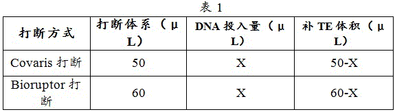
2) FFPE DNA was interrupted using Covaris and blood cell genomic DNA was interrupted using Covaris or Bioruptor.
The Covaris interruption operation is as follows:
a. preparing a Covaris broken tube, marking the serial number of the DNA sample on the tube cover
b. Adding TE Buffer pH 8.0 and sample into the interrupt tube
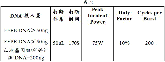
c. Samples were interrupted and the interruption instrument interruption parameters are shown in table 2:
② Bioruptor interruption operation as follows:
a. prepare Bioruptor-cut tube, label DNA sample number on tube cap
b. Adding TE Buffer pH 8.0 and sample into the interrupt tube
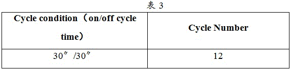
c. Samples were interrupted and the interruption instrument interruption parameters are shown in table 3:
d. the broken DNA is subjected to quality inspection by 2% agarose gel electrophoresis, 50bp DNA Ladder and D2000 DNA Ladder are used, the voltage is 150V, and the electrophoresis band is qualified within 200bp-300 bp.
2.2 end repair
1) End Repair Mix was prepared as per table 4.

2) Referring to table 5, the End Repair reaction program was set up with the hot lid temperature adjusted to 40 ℃.
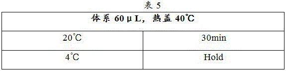
3) The sample was placed in a PCR instrument and the End Repair reaction program was run.
4) The used reagents were returned to the original kit and stored at-20 ℃.
2.3 preparation of Ligation1 Mix
1) Ligation1 Mix was prepared as in table 6.

2.4 purification after repair of the termini
1) Taking out AMPure XP magnetic beads from a refrigerator at 4 ℃, and balancing for 30min at room temperature.
2) 147.5. mu.L (2.5X) of AMPure XP magnetic beads were added to the end-repair sample and mixed well.
3) Standing at room temperature for 10min, transferring to magnetic rack, standing for 5min until the liquid is completely clarified, discarding the supernatant, and paying attention to avoid sucking magnetic beads.
4) 160 μ L of 80% ethanol was added slowly along the tube side walls, left to stand for 30s, and the supernatant was removed using a pipette.
5) Repeating the step 4) once.
6) The residual ethanol was removed by pipetting with a 10. mu.L pipette, and the mixture was left to dry at room temperature for 3 min.
7) Add 30. mu.L Ligation1 Mix to the sample tube, shake and Mix, and centrifuge instantaneously.
2.5、Ligation 1
1) Referring to Table 7, the Ligation1 reaction procedure was set up with the hot lid temperature adjusted to 70 ℃.
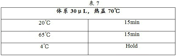
Note: after the process is finished, the sample can be stored at 4 ℃ for no more than 2 h.
2) The Ligation1 reaction program was run.
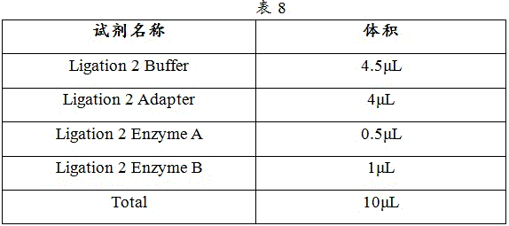
2.6、Ligation 2
1) Ligation 2 Mix was prepared according to Table 8.
2) The PCR reaction tube of Ligation1 was removed from the PCR apparatus, subjected to instantaneous centrifugation, and placed on ice. Each reaction tube is subpackaged with 10 mu L of Ligation 2 Mix, evenly mixed by oscillation and instantaneously centrifuged.
3) The Ligation 2 reaction program was set up with reference to table 9, and the hot lid temperature was adjusted to 70 ℃.

4) The sample was placed in a PCR instrument and the Ligation 2 reaction program was run.
2.7 purification after Adaptor ligation
1) Add 100. mu.L (2.5X) PEG/NaCl to the reacted sample after Ligation 2 and mix well.
2) Standing at room temperature for 10min, transferring to magnetic rack, standing for 5min until the liquid is completely clarified, discarding the supernatant, and paying attention to avoid sucking magnetic beads.
3) 160 μ L of 80% ethanol was added slowly along the tube side walls, left to stand for 30s, and the supernatant was removed using a pipette.
4) Repeating the step 3) once.
5) The residual ethanol was removed by pipetting with a 10. mu.L pipette, and the mixture was left to dry at room temperature for 3 min.
6) Add 20. mu.L of nucleic-Free Water to the sample tube, mix well with shaking, and centrifuge instantaneously.
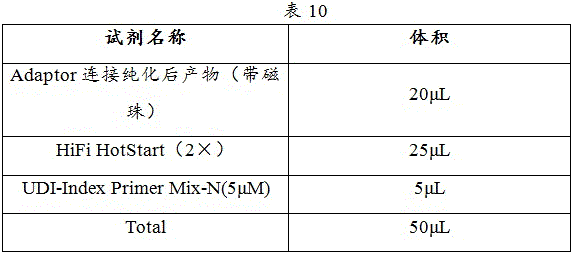
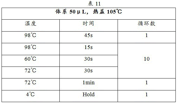
2.8 PCR amplification
1) PCR reaction systems were prepared in PCR tubes according to the system in Table 10.
2) Shaking and mixing evenly, and carrying out instantaneous centrifugation to ensure that all reaction liquid is placed at the bottom of the PCR tube.
PCR amplification procedure is as in table 11:
2.9 library purification and quantification
1) The AMPure XP magnetic beads are taken out of a refrigerator at 4 ℃, and the temperature is balanced for 30 min.
2) The PCR product was removed from the PCR instrument, centrifuged instantaneously, placed on a magnetic stand and allowed to stand for 5min, and the supernatant was transferred to a new tube.
3) Add 65. mu.L (1.3X) of AMPure XP magnetic beads to the PCR product, pipette (range 65. mu.L) down to 20, mix well.
4) Standing at room temperature for 10min, transferring to magnetic rack, standing for 5min until the liquid is completely clarified, discarding the supernatant, and paying attention to avoid sucking magnetic beads.
5) 160 μ L of 80% ethanol was added slowly along the tube side walls, left to stand for 30s, and the supernatant was removed using a pipette.
Note: if the magnetic beads are absorbed when the waste liquid is discarded, standing for 2min, and discarding the supernatant after the magnetic beads are absorbed.
6) Repeating the step 5) once.
7) The residual ethanol was removed by pipetting with a 10. mu.L pipette, and the mixture was left to dry at room temperature for 3 min.
8) Add 32. mu.L TE Buffer PH 8.0 to the sample tube, mix well with shaking, centrifuge instantaneously, incubate for 8min at room temperature.
9) The sample tube was placed on a magnetic rack for 5min until the liquid was completely clear, and 30 μ Ι _ of supernatant was carefully transferred to a new 1.5ml centrifuge tube using a pipettor.
10) The library Qubit was quantified using Qubit 4.0.
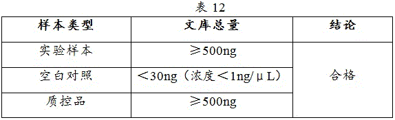
2.10 quality inspection of library
The results are shown in Table 12.
2.11, storing the library, and storing the library in a refrigerator at-20 ℃ in an amplification area.
Step three: and (3) hybridizing the detection probe for detecting Panel of the invention with a DNA library, capturing and sequencing. Specifically, in the present example, 2059 IDT probes were designed and manufactured using the Target Capture Probe Design & Ordering Tool of the website of Integrated DNA Technologies IDT corporation.
Library hybridization procedures were as follows:
1. shaking and uniformly mixing the Human Cot DNA and xGen Universal blocks-TS Mix, and instantaneously centrifuging.
2. Hybridization reaction Mix was prepared as shown in Table 13.
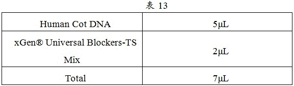
3. Add 7. mu.L Mix and 500ng-1ug DNA library to the centrifuge tube, Mix well with shaking, and centrifuge instantaneously. And drying in a vacuum concentrator for later use.
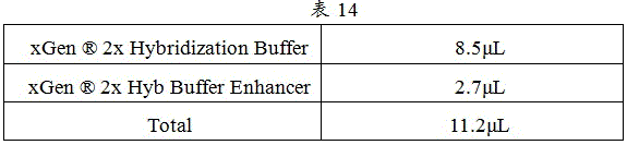
4. XGen according to Table 14 ®2x Hybridization Buffer and xGen ®And (3) oscillating and uniformly mixing the 2x Hyb Buffer Enhancer, and performing instantaneous centrifugation to obtain a hybridization reaction solution.
5. And adding the hybridization reaction solution and 4 mu L of probe into the evaporated sample, and incubating for 10min at 25 ℃ on a constant-temperature mixing machine.
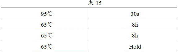
6. And (5) putting the sample into a PCR instrument, clicking the program and starting to operate. PCR procedure as in table 15:
the library elution procedure was as follows:
1. streptavidin magnetic bead cleaning
1) Will xGen®2X Hybridization Buffer and xGen 2X Hyb Buffer Enhancer are shaken and mixed evenly and are subjected to instantaneous centrifugation.
2) The magnetic bead suspension Mix was prepared as in table 16.
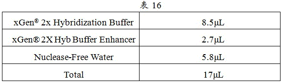
3) Dynabeads M-270 Streptavidin was shaken and mixed well. The amount of Dynabeads M-270 Streptavidin used per library was 50. mu.L.
4) The using amount of each library 1XBead Wash Buffer (nuclear-Free Water and xGen 2X Bead Wash Buffer prepared according to the proportion of 1: 1) is 100 muL, 1X Bead Wash Buffer with the corresponding volume is added into a tube, the mixture is oscillated and mixed evenly (n X100 muL), the mixture is subjected to instantaneous centrifugation and placed in a magnetic frame for 1min, and after the liquid is completely clarified, the supernatant is sucked and discarded by using a pipette.
5) Repeating the step 4) twice.
6) The residual liquid was aspirated with a pipette.
7) The amount of each library bead suspension was 17. mu.L, and a corresponding volume of bead suspension was added to the tube for suspension.
8) A corresponding number of PCR tubes were prepared, and 17. mu.L of a mixture of magnetic beads and a suspension was dispensed into each of the PCR tubes.
9) The PCR tube containing the magnetic bead suspension was placed in a PCR instrument and incubated for 5 min.
2. Streptavidin magnetic bead capture
1) After 4-16h of hybridization reaction, the resuspended streptavidin magnetic beads are added into the hybridization system twice after being mixed evenly. Transfer the sample tube to the running PCR instrument and click on the next step.
2) Incubate at 65 ℃ for 45min, shake and mix every 15min to ensure that the magnetic beads are fully resuspended.
3. Thermal elution
1) The PCR instrument setup program was turned on as per Table 17.

2) The filled containers will contain 1X WashBuffer I (nucleic-Free Water and xGen)®10 XWashbuffer I in a 1:9 ratio) and 1X Stringent Washbuffer (nucleic-Free Water and xGen)®10X Stringent Wash Buffer in a 1:9 ratio) was placed in a PCR apparatus and preheated.
3) After incubation at 65 ℃ for 45min, transferring the hybridization sample captured by the streptavidin magnetic beads into a PCR tube filled with 1X Wash Buffer I and mixing uniformly. The PCR tube was placed on a magnetic stand for 1min, and after the liquid was completely clarified, the supernatant was removed thoroughly with a pipette.
4) Transferring the 1X Stringent Wash Buffer into a PCR tube filled with sample magnetic beads, mixing uniformly, covering the tube cap tightly, and putting the tube cap into a PCR instrument to incubate for 5min at 65 ℃. After this time the PCR tube with the sample is placed on a magnetic rack until clear and the supernatant is removed thoroughly with a pipettor.
5) Repeating the step 4) once.
4. Eluting at normal temperature
1) The PCR tube containing the sample was removed from the magnetic stand, and 150ul of 1 XWash Buffer II (nucleic-Free Water and xGen) was pipetted from the dispensing tube®10x Wash Buffer II in a ratio of 1: 9) into a PCR tube filled with magnetic beads, incubating at room temperature for 2min, uniformly mixing for 30s in a constant-temperature mixer, standing for 30s, and alternately mixing to ensure sufficient mixing. The PCR tube was centrifuged instantaneously and then allowed to stand on a magnetic stand for 1min, and the supernatant was removed thoroughly with a pipette.
2) The PCR tube containing the sample was removed from the magnetic rack, and after transient centrifugation, the magnetic rack was mounted and a small amount of residual Buffer was removed using a pipette.
3) Add 21. mu.L of nucleic-Free Water to the tube and mix well by aspiration.
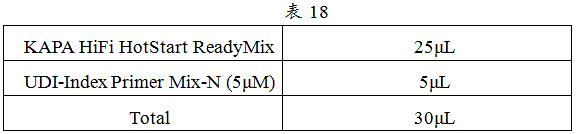
4) KAPA HiFi HotStart ReadyMix, primers FCF (10. mu.M) and FCR (10. mu.M) were thawed 15-20min in advance at 4 ℃.
5) The reaction system was prepared in PCR tubes or centrifuge tubes as in Table 18.
6) And (3) subpackaging the configured MIX into a PCR tube with a labeled sample serial number.
7) Transfer 20. mu.L of the product with magnetic beads to the corresponding PCR tube with a pipette, and mix well.
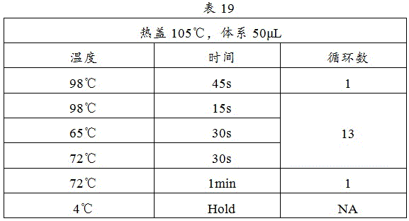
8) The procedure shown in table 19 was started on a PCR instrument:
library purification steps were as follows:
1) and (3) oscillating and uniformly mixing the AMPure XP magnetic beads, sucking 60 mu L of the mixture and adding the mixture into a labeled PCR tube.
2) After PCR amplification, the PCR tube was removed and placed in a magnetic rack for 2min, and after the liquid was completely clarified, the supernatant was transferred to the PCR tube in the magnetic rack.
3) Shaking and mixing, and incubating at room temperature for 10 min.
4) The PCR tube was centrifuged instantaneously and placed on a magnetic stand for 5min, after the liquid was completely clarified, the supernatant was removed (5-10. mu.L of liquid was left).
5) 200 μ L of 80% ethanol was added slowly along the side walls of the PCR tube, left to stand for 30s, and the supernatant was removed using a pipette.
6) Repeating the step 5) once.
7) The PCR tube was removed from the magnetic rack, placed on the magnetic rack after transient centrifugation, and a small amount of residual ethanol was removed using a pipette, taking care not to attract the magnetic beads.
8) And opening a PCR tube cover, and placing the magnetic beads at room temperature for drying, wherein the surfaces of the magnetic beads have no liquid to reflect light, and the magnetic beads cannot be dried excessively.
9) Add 31. mu.L of TE Buffer pH 8.0 to the PCR tube, mix well with shaking, incubate for 5min at room temperature.
10) The PCR tube was centrifuged briefly and placed on a magnetic rack for 1-2min until the liquid was clear, and 30. mu.L of the supernatant was carefully transferred to a new 1.5mL centrifuge tube with a pipette, taking care not to attract the beads.
11) Performing library quality inspection, and performing machine sequencing after the library quality inspection is qualified.
Step four: and (4) performing bioinformatics analysis on the sequencing result to obtain a mutation result of the detection sample. The method can be specifically implemented by the following steps:
and removing the adaptor sequence and the low-quality base sequence introduced in the experiment and sequencing links by using fastp (v0.19.4) software to obtain high-quality sequencing data. Wherein, the required data quality meets Q30 more than or equal to 80%, otherwise, the quality control is judged not to pass.
The above-described data satisfying the requirements were aligned to hg19 (GRCh 37) reference genome using bwa (0.7.17) sequence alignment software, generating BAM format files recording the alignment results. BAM files were then sorted, de-duplicated, and base quality corrected using Samtools (v 1.9) and genoanalysis tk (v 4.1.0) to obtain the final BAM file, and subsequent mutation analysis was performed based on this file.
Wherein, the reference genome comparison rate is not less than 90%, the average sequencing depth of the target area of the control sample is not less than 100, the average sequencing depth of the target area of the tumor sample is not less than 500, and the site proportion of the depth in the target area which is more than the average depth multiplied by 0.2 is not less than 90%.
The samples were analyzed for point mutations, indel mutations using the mutation identification module of genomeanalysttk (v4.1.0). Obtaining 89 variations, filtering clear benign variations by the crowd frequency and the database, and finally obtaining the following three variations to enter an annotation process: variation 1: NM-007294.3 (BRCA1) c.5470-5477 del (p.Ile 1824AspfsTer3); variation 2: NM-001184.3 (ATR) c.117A > G (p.Gln39 =);
variation 3: NM-000546.5 (TP53) c.460G > A (p.Gly154Ser);
the three variants obtained were annotated: a variant annotation module built based on Annovar (v2018.04.16) is used for annotating point mutation, insertion deletion and other type variants, and databases and tools used for annotating comprise ClinVar, LOVD, gnomaD, REVEL and the like. The annotation results are fed back to the medicine for site interpretation and reporting.
Step five: and (5) reading mutation results, and judging the pathogenicity of the mutation. Based on the annotation result of the variation in the step 4, the matched evidence is confirmed and supplemented according to the variation classification standard specified in ACMG genetic variation classification standard and guideline for the three detected non-benign variations, and the classification results of the three variations are obtained according to evidence analysis:
variation 1: NM-007294.3 (BRCA1) c.5470-5477 del (p.Ile1824AspfsTer3) are matched with PM2+ PP5+ PVS1+ PS4, so that the mutation 1 is classified as type 5-pathogenic;
variation 2: NM _001184.3(ATR) c.117a > G (p.gln39=) matched evidence BP4+ BP7, so mutation 2 was classified as class 2-probably benign;
variation 3: NM-000546.5 (TP53): c.460G > A (p.Gly154Ser) did not match the corresponding evidence, so the classification of variation 3 into 3 classes-clinical significance was unclear.
Based on the above interpretation classification results, the variation 1: NM-007294.3 (BRCA1) c.5470-5477 del (p.Ile1824AspfsTer3) was reported as a positive variation and described for evidence, and the detailed report screenshot can be shown in FIG. 1.
Step six: and issuing a detection report according to the site interpretation result.
In the blood sample of the ovarian cancer patient, a pathogenic germ line mutation NM-007294.3 (BRCA1) c.5470-5477 del is detected. Namely, BRCA1 has a pathogenic germline mutation. Meanwhile, the pathogenic germ line variation of BRCA1 is carried, which indicates that the detected person is a patient with HBOC (hereditary breast cancer-ovarian cancer syndrome), and the risk of ovarian cancer is increased, and the risk of the breast cancer, the prostate cancer, the pancreatic cancer, the male breast cancer, the melanoma and the like is also obviously increased; and the syndrome is inherited in an autosomal dominant manner, suggesting that the subject's first-degree relatives have a 50% probability of carrying the pathogenic variation of BRCA 1. A screenshot of a report of a clinically meaningful description or possible benefit associated with a BRCA1 mutation can be found in fig. 2.
Therefore, the technical scheme provided by the invention can be used for mutation detection of various tumors including ovarian cancer and the like, and the detection result can realize effective guidance of accurate treatment.
While preferred embodiments of the present invention have been described, additional variations and modifications in those embodiments may occur to those skilled in the art once they learn of the basic inventive concepts. Therefore, it is intended that the appended claims be interpreted as including preferred embodiments and all such alterations and modifications as fall within the scope of the invention. It will be apparent to those skilled in the art that various changes and modifications may be made in the present invention without departing from the spirit and scope of the invention. Thus, if such modifications and variations of the present invention fall within the scope of the claims of the present invention and their equivalents, the present invention is also intended to include such modifications and variations. Unless otherwise specified, reagents, methods and equipment used in the present invention are conventional methods, and test materials used therein are available from commercial companies, unless otherwise specified.
Claims (9)
1. A gene Panel for detecting various tumors, which is characterized in that the various tumors comprise gynecological tumors, breast cancers, prostate cancers and digestive tract tumors, and the gene Panel comprises hereditary breast cancer ovarian cancer syndrome BRCA related genes, Ringqi syndrome related genes, other genes related to the various tumors and homologous recombination repair pathway key genes; other genes associated with the various tumors include: PTEN, STK11, TP53, BRIP1, RAD51C, RAD51D, ATM, PALB2, CDH1, NBN, CHEK2, BARD1, RAD50, MRE 11.
2. The gene Panel for multiple tumor detection according to claim 1, wherein the gene associated with the hereditary breast cancer ovarian cancer syndrome BRCA comprises: BRCA1, BRCA 2.
3. The gene Panel for detecting multiple tumors of claim 1, wherein said genes associated with the forest syndrome comprise: MLH1, MSH2, MSH6, PMS2, EPCAM.
4. The gene Panel for detecting multiple tumors of claim 1, wherein said homologous recombination repair pathway key genes comprise: ABRAXAS1(FAM175A), ATM, ATR, BAP1, BARD1, BRCA1, BRCA2, BRIP1, C11ORF30(EMSY), CDK12, CHEK1, CHEK2, FACCA, FACCC, FACCD 2, FACCI, FACCL, MRE11, NBN, PALB2, PPP2R2A, PTEN, RAD50, RAD51B, RAD51C, RAD51D, RAD54B, RAD 54L.
5. A probe Panel for detecting multiple tumors, wherein the probe Panel is a detection probe for the BRCA-associated gene of hereditary breast cancer ovarian cancer syndrome, the LINGQI syndrome-associated gene, other genes associated with the multiple tumors and the key genes of homologous recombination repair pathway as claimed in any one of claims 1 to 4.
6. A kit for detecting multiple tumors, comprising at least one dose of the probe Panel of claim 5.
7. Use of the gene Panel according to any one of claims 1 to 4 or the probe Panel according to claim 5 for the preparation of a plurality of tumor detection devices.
8. An apparatus for detecting multiple tumors, comprising:
the sequencing module is used for extracting DNA of a sample to be tested and carrying out high-throughput sequencing to obtain a sequencing result;
the analysis module is used for performing bioinformatics analysis on the high-throughput sequencing result to obtain mutation information of the detection sample;
a comparison module for comparing the mutation information with the gene Panel according to any one of claims 1 to 4 to determine the pathogenicity of the mutation.
9. A method for detecting a plurality of tumors, comprising:
extracting DNA of a detection sample;
constructing a detection sample DNA library;
constructing a detection probe for the gene Panel according to any one of claims 1 to 4;
the detection probe is hybridized and captured with the DNA library and sequenced;
performing bioinformatics analysis on the sequencing result to obtain mutation information of the detection sample;
comparing the mutation information with the gene Panel according to any one of claims 1 to 4 to determine the pathogenicity of the mutation information.
Priority Applications (2)
| Application Number | Priority Date | Filing Date | Title |
|---|---|---|---|
| CN202210339492.1A CN114438218B (en) | 2022-04-01 | 2022-04-01 | Gene Panel for detecting various tumors, kit and application |
| ZA2022/09466A ZA202209466B (en) | 2022-04-01 | 2022-08-24 | A gene panel, kits and applications for detecting multiple tumors |
Applications Claiming Priority (1)
| Application Number | Priority Date | Filing Date | Title |
|---|---|---|---|
| CN202210339492.1A CN114438218B (en) | 2022-04-01 | 2022-04-01 | Gene Panel for detecting various tumors, kit and application |
Publications (2)
| Publication Number | Publication Date |
|---|---|
| CN114438218A true CN114438218A (en) | 2022-05-06 |
| CN114438218B CN114438218B (en) | 2022-08-09 |
Family
ID=81359579
Family Applications (1)
| Application Number | Title | Priority Date | Filing Date |
|---|---|---|---|
| CN202210339492.1A Active CN114438218B (en) | 2022-04-01 | 2022-04-01 | Gene Panel for detecting various tumors, kit and application |
Country Status (2)
| Country | Link |
|---|---|
| CN (1) | CN114438218B (en) |
| ZA (1) | ZA202209466B (en) |
Cited By (1)
| Publication number | Priority date | Publication date | Assignee | Title |
|---|---|---|---|---|
| CN112708676A (en) * | 2020-12-29 | 2021-04-27 | 南京科佰基因科技有限公司 | Standard substance for DNA homologous recombination repair gene detection and preparation method thereof |
Citations (6)
| Publication number | Priority date | Publication date | Assignee | Title |
|---|---|---|---|---|
| US20140011701A1 (en) * | 2011-03-14 | 2014-01-09 | National Research Council Of Canada | Prognostic Marker Sets For Prostate Cancer |
| US20150307947A1 (en) * | 2012-12-04 | 2015-10-29 | Caris Mpi, Inc. | Molecular profiling for cancer |
| WO2018135464A1 (en) * | 2017-01-18 | 2018-07-26 | 大学共同利用機関法人 情報・システム研究機構 | Rapid genetic screening method using next generation sequencer |
| WO2020226333A1 (en) * | 2019-05-03 | 2020-11-12 | 주식회사 디시젠 | Method of predicting cancer prognosis and composition for same |
| CN112831563A (en) * | 2021-02-08 | 2021-05-25 | 复旦大学附属肿瘤医院 | Detection panel and detection kit for hereditary colorectal cancer |
| CN113462784A (en) * | 2021-08-31 | 2021-10-01 | 迈杰转化医学研究(苏州)有限公司 | Method for constructing target set for homologous recombination repair defect detection |
-
2022
- 2022-04-01 CN CN202210339492.1A patent/CN114438218B/en active Active
- 2022-08-24 ZA ZA2022/09466A patent/ZA202209466B/en unknown
Patent Citations (6)
| Publication number | Priority date | Publication date | Assignee | Title |
|---|---|---|---|---|
| US20140011701A1 (en) * | 2011-03-14 | 2014-01-09 | National Research Council Of Canada | Prognostic Marker Sets For Prostate Cancer |
| US20150307947A1 (en) * | 2012-12-04 | 2015-10-29 | Caris Mpi, Inc. | Molecular profiling for cancer |
| WO2018135464A1 (en) * | 2017-01-18 | 2018-07-26 | 大学共同利用機関法人 情報・システム研究機構 | Rapid genetic screening method using next generation sequencer |
| WO2020226333A1 (en) * | 2019-05-03 | 2020-11-12 | 주식회사 디시젠 | Method of predicting cancer prognosis and composition for same |
| CN112831563A (en) * | 2021-02-08 | 2021-05-25 | 复旦大学附属肿瘤医院 | Detection panel and detection kit for hereditary colorectal cancer |
| CN113462784A (en) * | 2021-08-31 | 2021-10-01 | 迈杰转化医学研究(苏州)有限公司 | Method for constructing target set for homologous recombination repair defect detection |
Non-Patent Citations (14)
| Title |
|---|
| 张佳佳等: "同源重组修复通路相关基因在上皮性卵巢癌中的研究进展", 《重庆医学》 * |
| 张佳佳等: "同源重组修复通路相关基因在上皮性卵巢癌中的研究进展", 《重庆医学》, no. 30, 30 October 2018 (2018-10-30), pages 97 - 99 * |
| 张静等: "二代测序在家族遗传性高危胃肠肿瘤筛查中的应用", 《中国肿瘤临床》 * |
| 张静等: "二代测序在家族遗传性高危胃肠肿瘤筛查中的应用", 《中国肿瘤临床》, no. 19, 15 October 2018 (2018-10-15), pages 34 - 38 * |
| 朱珏等: "遗传性卵巢癌综合征相关易感基因的研究进展", 《国际妇产科学杂志》 * |
| 朱珏等: "遗传性卵巢癌综合征相关易感基因的研究进展", 《国际妇产科学杂志》, no. 06, 15 December 2014 (2014-12-15), pages 584 - 586 * |
| 朱耀: "中国前列腺癌患者基因检测专家共识(2020年版)", 《中国癌症杂志》 * |
| 朱耀: "中国前列腺癌患者基因检测专家共识(2020年版)", 《中国癌症杂志》, no. 07, 30 July 2020 (2020-07-30), pages 76 - 85 * |
| 温灏等: "上皮性卵巢癌的遗传易感基因突变与检测", 《中国妇产科临床杂志》 * |
| 温灏等: "上皮性卵巢癌的遗传易感基因突变与检测", 《中国妇产科临床杂志》, no. 04, 15 July 2017 (2017-07-15), pages 6 - 8 * |
| 白莎莎等: "DNA修复及其在肿瘤发生中的作用", 《医学综述》 * |
| 白莎莎等: "DNA修复及其在肿瘤发生中的作用", 《医学综述》, no. 17, 5 September 2016 (2016-09-05), pages 3386 - 3388 * |
| 邱宇凡等: "DNA同源重组修复与乳腺癌的研究进展", 《中国药理学通报》 * |
| 邱宇凡等: "DNA同源重组修复与乳腺癌的研究进展", 《中国药理学通报》, no. 07, 20 June 2016 (2016-06-20), pages 910 - 913 * |
Cited By (2)
| Publication number | Priority date | Publication date | Assignee | Title |
|---|---|---|---|---|
| CN112708676A (en) * | 2020-12-29 | 2021-04-27 | 南京科佰基因科技有限公司 | Standard substance for DNA homologous recombination repair gene detection and preparation method thereof |
| CN112708676B (en) * | 2020-12-29 | 2024-05-14 | 南京科佰基因科技有限公司 | Standard substance for DNA homologous recombination repair gene detection and preparation method thereof |
Also Published As
| Publication number | Publication date |
|---|---|
| CN114438218B (en) | 2022-08-09 |
| ZA202209466B (en) | 2022-12-21 |
Similar Documents
| Publication | Publication Date | Title |
|---|---|---|
| US10998084B2 (en) | Sequencing data analysis method, device and computer-readable medium for microsatellite instability | |
| CN109609647B (en) | Detection Panel for pan-cancer-species targeting, chemotherapy and immune drugs based on next-generation sequencing, detection kit and application thereof | |
| Yohe et al. | Review of clinical next-generation sequencing | |
| CN106755501B (en) | Method for simultaneously detecting microsatellite locus stability and genome change based on next-generation sequencing | |
| Cottrell et al. | Validation of a next-generation sequencing assay for clinical molecular oncology | |
| CN107513565B (en) | A kind of microsatellite instability Sites Combination, detection kit and its application | |
| CN114026646A (en) | System and method for assessing tumor score | |
| US20220336046A1 (en) | Methods and systems for refining copy number variation in a liquid biopsy assay | |
| DeRycke et al. | Targeted sequencing of 36 known or putative colorectal cancer susceptibility genes | |
| Das et al. | Molecular cytogenetics: recent developments and applications in cancer | |
| WO2024138956A1 (en) | Minimal residual disease detection method and apparatus, device, and storage medium | |
| CN114480660A (en) | Gene Panel for detecting pan-cancer species, probe and application | |
| US20220213555A1 (en) | Next generation sequencing-based detection panel for glioma, detection kit, detection method and application thereof | |
| US20230002831A1 (en) | Methods and compositions for analyses of cancer | |
| CN111979329A (en) | Pan-cancer detection panel based on next-generation sequencing technology and its application | |
| CN114250303A (en) | A breast cancer multi-gene detection panel, kit and application thereof | |
| CN114283889A (en) | Method and device for correcting homologous recombination repair defect score | |
| EP4434036A1 (en) | Methods and systems for accurate genotyping of repeat polymorphisms | |
| Chang et al. | Somatic diseases (cancer): amplification-based next-generation sequencing | |
| CN114438218B (en) | Gene Panel for detecting various tumors, kit and application | |
| Ow et al. | Next generation sequencing reveals novel mutations in mismatch repair genes and other cancer predisposition genes in Asian patients with suspected Lynch syndrome | |
| Verner et al. | Validation of the Labcorp Plasma Focus Test to Facilitate Precision Oncology through cell-free DNA genomic profiling of solid tumors | |
| JP2025507673A (en) | Probe sets for liquid biopsy assays | |
| CN114525344A (en) | Kit for detecting or assisting in detecting tumor-related gene variation and application thereof | |
| CN117467762A (en) | Probe composition and kit for breast cancer gene detection |
Legal Events
| Date | Code | Title | Description |
|---|---|---|---|
| PB01 | Publication | ||
| PB01 | Publication | ||
| SE01 | Entry into force of request for substantive examination | ||
| SE01 | Entry into force of request for substantive examination | ||
| GR01 | Patent grant | ||
| GR01 | Patent grant |