CN114283164B - Breast cancer pathological section image segmentation prediction system based on UNet3+ - Google Patents
Breast cancer pathological section image segmentation prediction system based on UNet3+ Download PDFInfo
- Publication number
- CN114283164B CN114283164B CN202210194828.XA CN202210194828A CN114283164B CN 114283164 B CN114283164 B CN 114283164B CN 202210194828 A CN202210194828 A CN 202210194828A CN 114283164 B CN114283164 B CN 114283164B
- Authority
- CN
- China
- Prior art keywords
- unet3
- module
- image
- pathological section
- training
- Prior art date
- Legal status (The legal status is an assumption and is not a legal conclusion. Google has not performed a legal analysis and makes no representation as to the accuracy of the status listed.)
- Active
Links
Images


Landscapes
- Image Processing (AREA)
- Image Analysis (AREA)
Abstract
The invention discloses a UNet3+ -based breast cancer pathological section image segmentation prediction system, which comprises the following specific steps: generating an image block and a corresponding real label based on the Dajin algorithm and the close-spread algorithm; constructing an encoder of UNet3+ based on SeResNext; decoder building of UNet3+ is realized through full-scale hopping connection based on depth separable convolution; training a UNet3+ neural network based on a dynamic random negative sample sampling method; and generating a large slice prediction result such as a pathological slice based on an aliasing dense-paving algorithm. The invention effectively inhibits the false positive phenomenon in the pathological section segmentation task, greatly reduces the parameter quantity and the calculated quantity of the network on the premise of maintaining the segmentation effect, and finishes the pixel-level delineation of cancerous gland and non-gland tumor regions.
Description
Technical Field
The invention relates to the technical field of image processing, in particular to a UNet3+ -based breast cancer pathological section image segmentation prediction system.
Background
Because pathological sections can be scanned into digital pathological sections by a scanner and are stored in a computer with definition comparable to that under a microscope, the workload of a pathologist can be directly reduced by diagnosing the digital pathological sections by using a computer method, and the current segmentation network based on deep learning technology and UNet can realize more accurate segmentation of the digital pathological sections, but the methods generally have the following problems: (a) the network complexity is too high. The split network often contains a huge number of parameters, and a large amount of hardware resources and time are consumed in the training phase. The operating speed and efficiency in the prediction are often also unsatisfactory; (b) false positive phenomenon. Although the cancerous region occupies most of the pathological section for the pathological section image of the advanced malignant tumor, many pathological sections of the early malignant tumor contain only a few cancerous regions — the diagnosis of the early pathological section is the most valuable, and after all, the image of the later section is often very obvious and the later section is very tedious if found. The sparsity of the cancerous region in the pathological section of the early malignant tumor causes the problem of excessive false positive in the prediction result; (c) multi-scale information is not adequately captured. The size and the shape of the focus in pathological sections are often greatly different, and the glandular structure and the cell shape are very important characteristics. This requires that the neural network can capture coarse-grained glandular features and fine-grained cellular features simultaneously. Although the existing segmentation networks fuse multi-scale features to different degrees, the mining of information of different scales is still insufficient. (d) Context information of image block edges is damaged during algorithm reasoning. Since the general close-spread algorithm does not consider the position of the lesion when generating the image block, it is only a simple sliding window. If the boundary of the window contains more important structural information, a simple sliding window will result in the prediction result of the full slice being discontinuous between different image blocks.
Disclosure of Invention
In order to overcome the defects and shortcomings of the prior art, the invention provides a UNet3+ -based breast cancer pathological section image segmentation prediction system, which solves the problems of high network complexity, a large number of false positives, insufficient capture of multi-scale information and discontinuous image blocks when reasoning on a full section of the existing algorithm, and can realize accurate segmentation on a breast cancer full-field pathological section image.
In order to achieve the purpose, the invention adopts the following technical scheme:
a UNet3+ based breast cancer pathological section image segmentation prediction system comprises: the system comprises an image preprocessing module, a UNet3+ encoder construction module, a UNet3+ decoder construction module, a training module and a prediction module;
the image preprocessing module is used for extracting a foreground tissue area based on an Otsu algorithm, filtering a cavity and a background by utilizing a foreground mask image and a slice-level real label and performing and operation, and generating an image block and a real label corresponding to the image block level based on a close-spread algorithm;
the UNet3+ encoder building module is used for building an encoder of UNet3+ based on SeResNext, adding the compression activation module to a residual error module of ResNet, and completing feature map splicing by using a scaling module;
the UNet3+ decoder building module is used for replacing a convolution layer of UNet3+ by using depth separable convolution, and the UNet3+ decoder building is realized through full-scale jump connection;
the training module is used for training a UNet3+ neural network based on a dynamic random negative sample sampling strategy, the dynamic random negative sample sampling strategy generates statistical positive samples and generates a negative sample pool at the initial training stage, and before each round of training, negative samples with the same number as that of the positive samples are obtained by sampling from the negative sample pool according to set random number seeds to form a data set of the round of training;
the prediction module is used for generating a prediction result with the same size as the pathological section image based on an aliasing close-spread algorithm, and the specific steps comprise:
initializing a matrix which has the same shape as the original pathological section image and all pixels are 0 as a map;
designing the moving step length of the sliding window according to the size of the required window and the aliasing proportion of the image block;
generating a sliding window in the original pathological section image;
sliding a sliding window on the pathological section image according to a set step length, adding the coordinates of the sliding window into a data generator, increasing the numerical value of the corresponding position of the generated map progressively, and covering the original map;
generating image blocks to be predicted by a data generator according to a set batch size, predicting the generated images by using a weight-loaded neural network, and correspondingly adding predicted values according to coordinates;
and when the prediction of the whole pathological section image is finished, dividing the generated matrix by the map to obtain a predicted result.
As a preferred technical solution, the image preprocessing module is configured to extract a foreground tissue region based on an oxford algorithm, filter a cavity and a background by performing and operation using a foreground mask image and slice-level real labels, and generate an image block and a real label corresponding to the image block level based on a dense-tiling algorithm, and specifically includes the steps of:
sequentially generating a large positive region marking mask image as the original slice according to the marking file, generating a large tissue mask image as the slice by extracting a foreground region from the original pathological slice image based on the Otsu algorithm, and performing AND operation on the marking mask image and the tissue mask image to obtain a large marking mask image as the original slice with a filtered cavity;
setting a rectangle completely containing the foreground region on the tissue mask image, namely, four edges of the rectangle are tangent to the outermost outline of the tissue;
simultaneously generating a sliding window at the corresponding position of the rectangle at the upper left corner of the original slice and the marking mask image;
respectively storing elements in the original slice and the mask image sliding window as image blocks and corresponding labels;
and sequentially sliding the window to the right and downwards by a set step length until the lower right corner of the rectangle is crossed.
As a preferred technical scheme, the UNet3+ encoder building module is used for building an encoder of UNet3+ based on seresext, and the specific steps are as follows:
building a residual module of a ResNet network, building a compression activation module, and sequentially cascading a global pooling, full connection layer, Relu activation function, full connection layer and sigmoid activation function;
adding a compression activation module behind a residual module of an original ResNet network;
the scaling module splices the feature diagram originally to be output by each stage of the original ResNet network and the feature diagram of the compression activation module;
the feature map before each downsampling of seresext is retained as input for the jump connection.
As a preferred technical solution, the UNet3+ decoder building module is used for replacing a convolution layer of UNet3+ with a depth separable convolution, and realizes decoder building of UNet3+ through a full-scale jump connection, and includes the following specific steps:
building a decoder module for cascading two layers of 3 multiplied by 3 depth separable convolution layers, batch normalization layers and Relu activation functions each time;
and adjusting the feature maps of the UNet3+ encoder and the constructed decoder to the same resolution by using the constructed decoder module to complete splicing.
As a preferred technical solution, the training module is used for training UNet3+ neural network based on a dynamic random negative sample sampling strategy, and includes the specific steps of:
generating a data set mask according to the preprocessed input picture data set;
counting the number of positive samples in the generated data set, subtracting the number of the positive samples from the total number of the samples to obtain the number of pure negative samples, adopting a dynamic random negative sample sampling method if the number of the negative samples is greater than the number of the positive samples, and adopting the data set for direct training if the number of the negative samples is less than the number of the positive samples;
respectively storing the file sets of the positive samples and the negative samples obtained by statistics in a certain form, randomly extracting the negative samples with the same number as the positive samples from the negative samples before training, combining the positive samples with the negative samples after sampling, and finishing the initialization of the training set;
calculating the loss of the network output and the generated real label based on a CE-Dice loss function, and optimizing the parameters of UNet3+ based on an Adam optimizer, wherein the CE-Dice loss function is expressed as follows:

where X represents a single sample, X represents a set of samples used to calculate the loss function,p(x) Andy(x) Respectively representing the model prediction probability and the true label corresponding to the sample x, represents a constant;
and after one iteration is finished, extracting the negative samples with the same number as the positive samples from the generated negative sample file set again, combining the positive samples and the negative samples, and generating a new training set until the training is finished.
As a preferred technical solution, the prediction module is configured to generate a prediction result with a size equal to that of the pathological section image based on an aliasing dense-paving algorithm, and includes:
reading the weight value of the trained UNet3+ neural network;
moving the sliding window of the original pathological section image to the right, if the sliding window does not exceed the boundary, continuously executing the addition of the coordinates of the sliding window into the data generator, and if the sliding window exceeds the boundary of the pathological section image, backtracking the sliding window to the left to enable the right boundary of the window to be overlapped with the right boundary of the pathological section image;
sliding the sliding window downwards, if the sliding window does not exceed the lower boundary of the slice, continuing to execute, adding the coordinates of the sliding window into the data generator, if the sliding window exceeds the boundary, generating prediction data by the data generator according to a set batch size, predicting the generated picture by using a weighted neural network, and correspondingly adding the prediction values according to the coordinates;
and when the whole pathological section image is predicted, dividing the generated matrix by the map to obtain a predicted floating point result.
Compared with the prior art, the invention has the following advantages and beneficial effects:
(1) aiming at the problem that the quantity of parameters of the current segmented network is too large, the improved UNet3+ adopted by the invention is higher in precision and less in quantity of parameters compared with UNet and UNet + + and original UNet3 +;
(2) aiming at the problem of serious false positive prediction in the breast cancer pathological section image segmentation task, the invention provides a method for inhibiting the false positive tendency of network prediction by dynamic sample balance by adopting dynamic random negative sample sampling;
(3) aiming at the problem that the multi-scale information is not sufficiently understood by the current algorithm, UNet3+ adopted by the invention realizes full-scale jump connection based on deep separable convolution, fully captures the multi-scale information and simultaneously reduces the parameter quantity and the calculation quantity of the network.
(4) Aiming at the problems that the image block edge is discontinuous and the edge context information is damaged when the current algorithm is used for reasoning on the full-slice, the aliasing dense-paving algorithm provided by the invention protects the edge context information, greatly improves the visualization effect of the full-slice reasoning, can realize accurate semantic segmentation on the full-view breast cancer pathological slice image, and completes the pixel-level delineation of the cancerous gland and the non-glandular tumor region.
Drawings
Fig. 1 is a schematic flow chart of an implementation of the system for predicting the image segmentation of the pathological section of breast cancer based on UNet3 +;
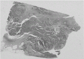
fig. 2(a) is a thumbnail of the original pathological section image in the present embodiment;
FIG. 2(b) is a label mask diagram before the Dajin algorithm is applied in the present embodiment;
FIG. 2(c) is a labeled mask diagram after the Dajin algorithm is applied in the present embodiment;
FIG. 3 is a schematic flow chart illustrating generation of a large prediction result with a trained neural network and pathological section according to the present embodiment;
FIG. 4(a) is a schematic diagram of an original pathological section image and a label in the embodiment;
FIG. 4(b) is a diagram illustrating the UNet prediction result of the embodiment;
FIG. 4(c) is a diagram illustrating the UNet + + prediction result of the embodiment;
FIG. 4(d) is a schematic diagram of UNet3+ prediction result in the embodiment;
FIG. 5 is a schematic diagram of the aliasing tiling algorithm according to this embodiment.
Detailed Description
In order to make the objects, technical solutions and advantages of the present invention more apparent, the present invention is described in further detail below with reference to the accompanying drawings and embodiments. It should be understood that the specific embodiments described herein are merely illustrative of the invention and do not limit the invention.
Examples
The embodiment provides a system for predicting the image segmentation of pathological breast cancer slices based on UNet3+, which comprises: the system comprises an image preprocessing module, a UNet3+ encoder construction module, a UNet3+ decoder construction module, a training module and a prediction module;
in this embodiment, the image preprocessing module is configured to extract a foreground tissue region based on an atrazine algorithm, filter a cavity and a background by performing and operation using a foreground mask image and slice-level real tags, and generate an image block and a real tag corresponding to the image block level based on a dense tiling algorithm;
in this embodiment, the UNet3+ encoder construction module is used for constructing an encoder of UNet3+ based on serernext, adding the compression activation module to a residual error module of ResNet, and completing feature map splicing by using a scaling module;
in the embodiment, the UNet3+ decoder building module is used for replacing a convolution layer of UNet3+ by using depth separable convolution, and the UNet3+ decoder building is realized by full-scale jump connection;
in this embodiment, the training module is configured to train the UNet3+ neural network based on a dynamic random negative sample sampling strategy, where the dynamic random negative sample sampling strategy generates a statistical positive sample and generates a negative sample pool at a training start stage, and samples the negative sample pool according to a set random number seed before each round of training to obtain negative samples with the same number as the positive samples to form a data set for the round of training;
in this embodiment, the prediction module is configured to generate a prediction result with a size equal to that of the pathological section image based on an aliasing dense-paving algorithm, and includes the following specific steps:
initializing a matrix which has the same shape as the original pathological section image and all pixels are 0 as a map;
designing the moving step length of the sliding window according to the size of the required window and the aliasing proportion of the image block;
generating a sliding window in the original pathological section image;
sliding the sliding window on the pathological section image according to a set step length, adding the coordinates of the sliding window into the data generator, increasing the numerical value of the corresponding position of the generated map, and covering the original map;
generating image blocks to be predicted by a data generator according to a set batch size, predicting the generated images by using a weight-loaded neural network, and correspondingly adding predicted values according to coordinates;
and when the prediction of the whole pathological section image is finished, dividing the generated matrix by the map to obtain a predicted result.
As shown in fig. 1, the present embodiment provides an implementation method of a breast cancer pathological section image segmentation prediction system based on UNet3+, including the following steps:
s1, extracting a foreground tissue region based on an Otsu algorithm, and performing AND operation by using a foreground mask image and a slice-level real label to filter a cavity and a background;
and generating large real labels such as pathological sections based on the Dajin algorithm. Generally speaking, the digital pathological section real label can generate a marking mask image of a positive area by reading the coordinates of a marking file, and the size of the mask image is the same as that of the section;
however, since cavities may exist within the gland, they also correspond to positive labeling if the algorithm described above is followed. Considering that the size and the shape of the breast cancer pathological section gland are irregular, the neural network is expected to learn the characteristics of cells, and marking the cavity as positive undoubtedly introduces strong noise to the training of the neural network, so that the cavity is filtered based on the Otsu algorithm, the data volume can be compressed quickly, the background noise can be filtered, and the updated algorithm is as follows:
generating a label mask map of a positive area according to a label file, wherein the size of the mask map is the same as that of a slice, and temporarily storing the mask map;
secondly, extracting a foreground area on the original slice based on an Otsu algorithm, generating a tissue mask image with the same size as the slice, and temporarily storing the mask image;
performing AND operation on the mask graph generated in the step (I) and the mask graph generated in the step (II), taking the returned mask graph as a corrected labeled mask graph, and deleting the mask graph generated in the first step;
as shown in fig. 2(a) -2 (c), the real label generation situations before and after the salix algorithm is applied are shown, and it can be found that many regions originally marked as positive are marked as negative again after the salix algorithm is applied, which significantly reduces noise brought to the neural network by the data set.
S2, preprocessing the neural network input image data set based on the close-spread algorithm, generating image blocks and real labels corresponding to the image block levels based on the close-spread algorithm, and on the basis of the step S1, preprocessing the breast cancer pathological section as follows:
completely containing the foreground region on the mask map generated in the second step of the step S2 by using a rectangle, wherein four sides of the rectangle must be connected with the foreground region, and mapping the rectangle onto the original slice and the mask map generated in the third step of the step S2;
and (II) simultaneously and respectively generating a square sliding window on the two pictures, namely the original slice and the mask image generated in the third step of the step S2 by taking the upper left corner of the rectangle as a starting point, wherein the upper left corner of the sliding window is coincided with the upper left corner of the rectangle. The rectangle should not be too large in size, and is of a shape suitable for processing by a neural network, typically an nth power of 2 or a sum of an nth power of 2 and an mth power of 2;
saving the area in the window in the two pictures as a picture, and adding the picture into the data set;
and (IV) sliding the window to the right by a certain step length, and returning to the step (III) if the boundary is not exceeded. If the boundary is exceeded, jumping to the step (five);
and (V) sliding the window downwards for a certain step length, and if the window exceeds the lower boundary of the slice, jumping to the step (six). Otherwise, translating to the leftmost side of the slice, and returning to the step (five);
and (VI) finishing the pretreatment of the slice.
The breast cancer pathology section dataset used in this example was mainly from the CAMELYON challenge. This race includes CAMELYON16 and CAMELYON 17. Of these, CAMELYON16 contained 111 total pathological sections (WSI) of breast cancer full-field containing positive regions, and CAMELYON17 contained 50 pathological sections of breast cancer metastatic lymph nodes containing positive regions. 161 slices of a two-year challenge are taken as a training set. In addition, CAMELYON also provided 48 slices as test sets, which are also the data sources for the test sets in the experiment, all of which were labeled. Irregular cancerous regions are circled by polygons using ASPP software. Annotations are exported into an XML file in the format of coordinate points. The marking information in the XML file can be conveniently read by utilizing a python interface provided by the ASPP.
When generating the block, the block is sampled 4 times with 1024 × 1024 as the original scale to obtain 256 × 256 blocks. Using the above algorithm, 30847 blocks containing positive regions and 569484 completely negative blocks were generated on 161 sections of the CAMELYON data set, which together constituted the training set of this example.
S3, constructing an encoder of UNet3+ based on SeResNext. The specific operation scheme is as follows:
and (I) defining a basic residual error module and a bottleneck module, wherein the basic residual error module consists of two convolution layers of 3 multiplied by 3 and quick connection, the bottleneck module consists of 1 multiplied by 1 convolution, 3 multiplied by 3 convolution, 1 multiplied by 1 convolution and quick connection respectively, and a ResNet neural network is designed based on the two convolution modules.
And (II) replacing convolution layers in the convolution module in ResNet with packet convolution.
And (III) building a compression activation module, and sequentially cascading a global pooling function, a full connection layer, a Relu activation function, a full connection layer and a sigmoid activation function.
And (IV) adding a compression activation module to the residual module of the ResNet through a scaling module.
And (V) retaining the feature map before each downsampling as the input of the jump connection. Since seresext has been downsampled 5 times in total, its encoder outputs five feature maps in total.
The existing UNet3+ is a VGG-like structure adopting a simple stacked convolution module in the implementation of an encoder, and the method has poor feature extraction capability when being used for application scenes containing high-dimensional complex features, such as cancer slices. The invention builds SeResNext as a UNet encoder, improves the feature representation capability of Unet3+, and the compression activation module introduced aiming at the characteristic of complex features of pathological sections can be considered as channel attention, fully represents the complex features of pathological sections, and is more suitable for processing application scenes of high-dimensional complex features.
S4, realizing full-scale jump connection based on depth separable convolution and constructing a UNet3+ decoder. The specific operation scheme is as follows:
(i) adjusting the signature of the encoder output designed in step S4 to the same size by concatenating two layers of 3 × 3 depth separable convolutional layers;
splicing the five output characteristic diagrams of the encoder after adjustment to serve as a fourth layer of the decoder, wherein the shape of the characteristic diagram corresponds to the original characteristic diagram output by the fourth layer of the encoder, and particularly, the encoder and the decoder share a fifth layer;
splicing the first to third layers of the adjusted output characteristic diagrams of the encoder and the fourth to fifth layers of the adjusted output characteristic diagrams of the decoder to form a third layer of the decoder, wherein the output characteristic diagrams of the third layer of the decoder correspond to the original characteristic diagrams output by the third layer of the encoder;
splicing the first-layer to second-layer adjusted output characteristic diagrams of the encoder and the third-layer to fifth-layer adjusted output characteristic diagrams of the decoder to form a second layer of the decoder, wherein the output characteristic diagrams of the second layer of the decoder correspond to the original characteristic diagrams output by the second layer of the encoder;
splicing the output characteristic diagram of the first layer of the encoder after adjustment with the output characteristic diagram of the decoder after adjustment of two to five layers to be used as the first layer of the decoder, wherein the output characteristic diagram of the first layer of the encoder corresponds to the original characteristic diagram output by the first layer of the encoder;
and (VI) sequentially cascading the output of the first layer of the decoder with a 3 x 3 convolutional layer, a batch normalization layer and a Relu activation function, and then adjusting the output characteristic diagram into a square with the same size as the input image through upsampling.
The prior art discloses technical solutions based on UNet3+, which adopt a conventional convolution module to adjust the shape of the feature diagram, thereby completing full-scale treaty connection. For a 5-layer Unet3+, the input of each layer decoder is a concatenation of 4 signatures, and each convolution module has 2 convolution layers concatenated. A 5-layer Unet3+ would use 4 × 5 × 2=40 convolutional layers to complete the construction of the decoder, which would greatly increase the amount of parameters of the network. The invention completes the construction of the decoder by utilizing the depth separable convolution, and reduces the calculation amount of the UNet3+ decoder in order of magnitude while maintaining the algorithm precision. Under the premise of adopting the same encoder, the parameters and the calculated amount of the UNet3+ provided by the invention are far less than those of UNet3+ and UNet2+, the parameters and the calculated amount are almost equal to those of the original UNet, and the algorithm precision is improved to a different degree compared with the three.
S5, constructing a BCE-Dice loss function and an Adam optimizer. The specific operation steps are as follows:
defining a binary cross entropy loss function, and instantiating the loss function;
defining a confusion matrix calculation function, and instantiating the calculation function;
and (III) defining a Dice coefficient according to the confusion matrix, defining a Dice Loss function according to the coefficient and instantiating the Dice Loss function.
Adding the binary cross entropy loss function and the Dice loss function to obtain a Bce-Dice loss function shown by the following formula:
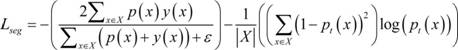
where X represents a single sample, X represents a set of samples used to calculate the loss function,p(x) Andy(x) Respectively representing the model prediction probability and the true label corresponding to the sample x, represents a constant; the former term of the BCE-Dice loss function is the Dice loss function, which is essentially the ratio of twice the number of true positive pixels to the union of all the pixels predicted to be positive and the positive area pixels in the label, and the addition of an offset amount to the Dice coefficient is used in engineering to avoid the denominator of the Dice coefficient being zero. The latter term of the BCE-Dice loss function is the binary cross-entropy loss. For each pixel, the better the prediction result, the closer the BCE loss is to 0; conversely, the worse the prediction, the closer the BCE loss is to 1.
(V) instantiating Adam optimizer, setting initial parameters, and setting specific parameters to be setlr=1e-5, =0.9,
=0.9, =0.9,eps=1e-8,
 = 0.0005. In training, a 2080Ti is used, and the batch size is set to 16.
And S6, training UNet3+ neural network based on dynamic random negative sample sampling. The specific operation steps are as follows:
and (I) counting the number of positive samples in the data set generated in the step S2 according to the mask of the data set generated in the step S2, and subtracting the number of the positive samples from the total number of the samples to obtain the number of pure negative samples. If the number of the negative samples is larger than that of the positive samples, adopting the following dynamic random negative sample sampling method; otherwise, the data set generated in step S2 is used for direct training. Since the positive sample area is almost always smaller than the negative area in the pathological section, the dynamic random negative sample sampling is almost suitable for all the neural network training scenes aiming at the pathological section.
And (II) respectively storing the file sets of the positive samples and the negative samples obtained by statistics in the step (I) in a certain form, randomly extracting the negative samples with the same number as the positive samples from the negative samples before training, combining the positive samples and the negative samples after sampling, and finishing the initialization of the training set.
(III), SeResNext pre-trained on Imagenet weights were loaded onto UNet3+, and all neurons were set to trainable state.
And (IV) starting training at a certain batch size, inputting each batch of data into a neural network to obtain output, calculating the network output based on the CE-Dice loss function designed in the step S5 and the loss of the real label generated in the step S2, and optimizing the parameters of UNet3+ based on an Adam optimizer designed in the step S5.
And (V) after one epoch is finished, extracting the negative samples with the same number as the positive samples from the negative sample file set generated in the step (II), and combining the positive samples and the negative samples to generate a new training set.
And sixthly, verifying the network effect according to the Dice coefficient defined in the step S5 when each epoch is ended, and if the Dice after the epoch is ended is higher than the highest value before, saving the weight at the moment as a temporary final weight.
And (seventhly), after all epochs are finished, saving the temporary final weight from the system memory to a disk to realize permanent saving.
Taking UNet3+ as an example, table 1 below shows that the model training time is shorter, the convergence is faster, and the generalization capability is stronger when dynamic random negative sample sampling is used. acc represents the pixel-level classification precision, fwIoU and mIoU respectively represent the frequency intersection ratio and the average intersection ratio, and Dice is an important index in a medical image segmentation task.
Table 1 table of test results of model

In the field of image processing, the prior art discloses some steps of random negative sample sampling training, and these techniques are generally static, i.e. negative samples are sampled at the beginning of training, and the distribution of positive and negative samples is not changed during the training process. The negative area of the pathological section is large, the number of characteristics is large, and the static sampling method cannot ensure that the neural network can fully learn the characteristics of the non-pathological area. The invention provides dynamic random negative sample sampling aiming at the characteristics of small positive area, large background area and more characteristics of pathological sections, and the negative samples are re-sampled after each epoch. The method can not only enable the neural network to mainly focus on the characteristics of the positive regions, but also ensure that the neural network learns the characteristics of different negative regions. In the application scene of pathological sections, from the aspect of training speed, the dynamic random negative sample sampling method provided by the invention is dynamic, and dynamic random negative sample sampling > static random negative sample sampling > non-sampling. From the aspect of model precision, dynamic random negative sample sampling > no sampling > static random negative sample sampling.
And S7, generating a large prediction result such as a pathological section and the like based on an aliasing dense paving algorithm by adopting a trained neural network. When in prediction, a certain aliasing exists between two adjacent blocks, and the prediction times of each pixel point are recorded by using another map. As shown in fig. 3, the specific steps include:
(I) rereading the weight saved in the step S6 into the Unet3+ network defined in the step S3 and the step S4.
And (II) initializing a matrix which has the same shape as the original slice and all pixels are 0 as a map.
And (III) designing the window size and the step length according to the precision, the speed requirement and the hardware support condition of the inference, wherein the step length is not greater than the window length because all positions of the slice are required to be predicted. When the step size is smaller than the window size, no aliasing exists, and the aliasing cipher paving algorithm degenerates into a general cipher paving algorithm.
And (IV) generating a sliding window at the upper left corner of the original slice.
And (V) adding the coordinates of the sliding window into the data generator, adding 1 to the corresponding position of the map generated in the step (II), and covering the original map.
Moving the sliding window to the right, and jumping to the step (five) if the boundary is not exceeded; and (5) jumping to the step (seven) if the boundary is exceeded.
(VII) the window is traced back to the left, so that the right boundary of the window just overlaps with the right boundary of the slice.
(eighth), the window slides downwards, and if the lower boundary of the slice is not exceeded, the step (fifth) is skipped; and (5) jumping to the step (ten) if the boundary is exceeded.
And (ten) generating prediction data in a certain batch size by using a data generator, predicting the generated picture by using the weighted neural network in the step (one), and correspondingly adding the predicted values according to coordinates.
And (eleven) after the whole slice prediction is finished, dividing the generated matrix by the map to obtain a predicted floating point type result. As shown in fig. 5, a map generation condition representing an aliasing dense-paving algorithm is obtained, and the shadow depth and the number in the map represent the number of times that the pixel point of the region is predicted. Since the context information of the edge position of the picture is more fuzzy, the multiple prediction at the edge position is beneficial to improving the prediction result of the algorithm on the edge position.
And (twelfth), rounding the target by a certain threshold value to obtain a final prediction result.
When UNet, UNet + + and UNet3+ were used for segmentation, as shown in fig. 4(a) to 4(d), respectively, segmentation results were obtained, and it can be seen that UNet already predicted most lesions, but the prediction results were noisy, and false positives were severe. The UNet + + false positive phenomenon is significantly improved over UNet, but noise still remains and the edges are not smooth enough. In contrast, the prediction result of UNet3+ significantly reduces the phenomena of false positives and noise, and the edges are smoother than UNet + +, which greatly improves the baseline network. As shown in table 2 below, several indices also show that UNet3+ performs better than UNet + + and UNet.
Table 2 table of test results of model

Compared with the existing sliding window-based close-spread algorithm, the aliasing close-spread algorithm provided by the invention is specially designed for digital pathological sections, and is optimized by taking the running speed of the algorithm as a target. Existing tiling algorithms typically generate image blocks using a sliding window. If the sliding window is free of aliasing, the structured context information of the image block edges is destroyed, resulting in discontinuity of the boundaries between image blocks. But introducing aliasing at the time of inference is not as convenient as just changing the window step size at the time of training. For pathological slices, the algorithm finally splices the prediction mask images of the image blocks according to the corresponding positions of the image blocks in the slices. If aliasing exists during inference, the prediction result of an aliasing area is difficult to process, so the existing full-field pathological section segmentation algorithm usually adopts a sliding window without aliasing during inference. The aliasing dense-paving algorithm provided by the invention introduces aliasing during reasoning, and records the predicted times of each position by additionally generating a mask image, so that the method can furthest reserve the structured context information in a pathological section and ensure the continuity of slice-level prediction results. Meanwhile, the method well processes the prediction result of the aliasing region by sacrificing a certain storage space to exchange for the algorithm reasoning speed.
The method provided by the embodiment can realize accurate semantic segmentation on the full-field breast cancer pathological section image and draw out a cancerous gland or a non-glandular tumor region.
The above embodiments are preferred embodiments of the present invention, but the present invention is not limited to the above embodiments, and any other changes, modifications, substitutions, combinations, and simplifications which do not depart from the spirit and principle of the present invention should be construed as equivalents thereof, and all such modifications are intended to be included in the scope of the present invention.
Claims (6)
1. A UNet3+ based breast cancer pathological section image segmentation prediction system is characterized by comprising: the system comprises an image preprocessing module, a UNet3+ encoder construction module, a UNet3+ decoder construction module, a training module and a prediction module;
the image preprocessing module is used for extracting a foreground tissue region based on an Otsu algorithm, performing and operation by using a foreground mask image and a slice-level real label to filter a cavity and a background, and generating an image block and a real label corresponding to the image block level based on a close-spread algorithm;
the UNet3+ encoder building module is used for building an encoder of UNet3+ based on SeResNext, adding the compression activation module to a residual error module of ResNet, and completing feature map splicing by using a scaling module;
the UNet3+ decoder building module is used for replacing a convolution layer of UNet3+ by using depth separable convolution, and the UNet3+ decoder building is realized through full-scale jump connection;
the training module is used for training a UNet3+ neural network based on a dynamic random negative sample sampling strategy, the dynamic random negative sample sampling strategy generates statistical positive samples and generates a negative sample pool at the initial training stage, and before each round of training, negative samples with the same number as that of the positive samples are obtained by sampling from the negative sample pool according to set random number seeds to form a data set of the round of training;
the prediction module is used for generating a prediction result with the same size as the pathological section image based on an aliasing close-spread algorithm, and the specific steps comprise:
initializing a matrix which has the same shape as the original pathological section image and all pixels are 0 as a map;
designing the moving step length of the sliding window according to the size of the required window and the aliasing proportion of the image block;
generating a sliding window in the original pathological section image;
sliding the sliding window on the pathological section image according to a set step length, adding the coordinates of the sliding window into a data generator, increasing the numerical value of the corresponding position of the generated map progressively, and covering the original map;
generating image blocks to be predicted by a data generator according to a set batch size, predicting the generated images by using a weight-loaded neural network, and correspondingly adding predicted values according to coordinates;
and when the prediction of the whole pathological section image is finished, dividing the generated matrix by the map to obtain a predicted result.
2. The UNet3+ -based breast cancer pathological section image segmentation prediction system according to claim 1, wherein the image preprocessing module is configured to extract a foreground tissue region based on the Otsu algorithm, filter cavities and background by using a foreground mask image and slice-level real label and operation, and generate image blocks and real labels corresponding to the image block levels based on a dense-paving algorithm, and the specific steps include:
sequentially generating a large positive region marking mask image as the original slice according to the marking file, generating a large tissue mask image as the slice by extracting a foreground region from the original pathological slice image based on the Otsu algorithm, and performing AND operation on the marking mask image and the tissue mask image to obtain a large marking mask image as the original slice with a filtered cavity;
setting a rectangle completely containing the foreground region on the tissue mask image, namely, four edges of the rectangle are tangent to the outermost outline of the tissue;
simultaneously generating a sliding window at the corresponding position of the rectangle at the upper left corner of the original slice and the marking mask image;
respectively storing elements in the original slice and the mask image sliding window as image blocks and corresponding labels;
and sequentially sliding the window to the right and downwards by a set step length until the lower right corner of the rectangle is crossed.
3. The UNet3+ -based breast cancer pathological section image segmentation prediction system according to claim 1, wherein the UNet3+ encoder construction module is configured to construct an encoder of UNet3+ based on SeResNext, by the specific steps of:
building a residual module of a ResNet network, building a compression activation module, and sequentially cascading a global pooling, full connection layer, Relu activation function, full connection layer and sigmoid activation function;
adding a compression activation module behind a residual module of an original ResNet network;
the scaling module splices the feature diagram originally to be output by each stage of the original ResNet network and the feature diagram of the compression activation module;
the feature map before each downsampling of seresext is retained as input for the jump connection.
4. The UNet3+ -based breast cancer pathological section image segmentation prediction system according to claim 1, wherein the UNet3+ decoder building module is configured to replace a convolution layer of UNet3+ by a depth separable convolution, and the UNet3+ decoder building is realized by a full-scale jump connection, and includes the following specific steps:
building a decoder module for cascading two layers of 3 multiplied by 3 depth separable convolution layers, batch normalization layers and Relu activation functions each time;
and adjusting the feature maps of the UNet3+ encoder and the constructed decoder to the same resolution by using the constructed decoder module to complete splicing.
5. The UNet3+ -based breast cancer pathological section image segmentation prediction system according to claim 1, wherein the training module is configured to train UNet3+ neural network based on dynamic random negative sample sampling strategy, and the specific steps include:
generating a data set mask according to the preprocessed input picture data set;
counting the number of positive samples in the generated data set, subtracting the number of the positive samples from the total number of the samples to obtain the number of pure negative samples, adopting a dynamic random negative sample sampling method if the number of the negative samples is greater than the number of the positive samples, and adopting the data set for direct training if the number of the negative samples is less than the number of the positive samples;
respectively storing the file sets of the positive samples and the negative samples obtained by statistics in a certain form, randomly extracting the negative samples with the same number as the positive samples from the negative samples before training, combining the positive samples with the negative samples after sampling, and finishing the initialization of the training set;
calculating the loss of the network output and the generated real label based on a CE-Dice loss function, and optimizing the parameters of UNet3+ based on an Adam optimizer, wherein the CE-Dice loss function is expressed as follows:

where X represents a single sample, X represents a set of samples used to calculate the loss function,p(x) Andy(x) Respectively representing the model prediction probability and the true label corresponding to the sample x, represents a constant;
and after one iteration is finished, extracting the negative samples with the same number as the positive samples from the generated negative sample file set again, combining the positive samples and the negative samples, and generating a new training set until the training is finished.
6. The UNet3+ -based breast cancer pathological section image segmentation prediction system according to claim 1, wherein the prediction module is configured to generate a prediction result with a size equal to that of a pathological section image based on an aliasing tiling algorithm, and the specific steps include:
reading the weight value of the trained UNet3+ neural network;
moving the sliding window of the original pathological section image to the right, if the sliding window does not exceed the boundary, continuously executing the addition of the coordinates of the sliding window into the data generator, and if the sliding window exceeds the boundary of the pathological section image, backtracking the sliding window to the left to enable the right boundary of the window to be overlapped with the right boundary of the pathological section image;
sliding the sliding window downwards, if the sliding window does not exceed the lower boundary of the slice, continuing to execute, adding the coordinates of the sliding window into the data generator, if the sliding window exceeds the boundary, generating prediction data by the data generator according to a set batch size, predicting the generated picture by using a weighted neural network, and correspondingly adding the prediction values according to the coordinates;
and when the whole pathological section image is predicted, dividing the generated matrix by the map to obtain a predicted floating point result.
Priority Applications (1)
| Application Number | Priority Date | Filing Date | Title |
|---|---|---|---|
| CN202210194828.XA CN114283164B (en) | 2022-03-02 | 2022-03-02 | Breast cancer pathological section image segmentation prediction system based on UNet3+ |
Applications Claiming Priority (1)
| Application Number | Priority Date | Filing Date | Title |
|---|---|---|---|
| CN202210194828.XA CN114283164B (en) | 2022-03-02 | 2022-03-02 | Breast cancer pathological section image segmentation prediction system based on UNet3+ |
Publications (2)
| Publication Number | Publication Date |
|---|---|
| CN114283164A CN114283164A (en) | 2022-04-05 |
| CN114283164B true CN114283164B (en) | 2022-06-14 |
Family
ID=80882173
Family Applications (1)
| Application Number | Title | Priority Date | Filing Date |
|---|---|---|---|
| CN202210194828.XA Active CN114283164B (en) | 2022-03-02 | 2022-03-02 | Breast cancer pathological section image segmentation prediction system based on UNet3+ |
Country Status (1)
| Country | Link |
|---|---|
| CN (1) | CN114283164B (en) |
Families Citing this family (8)
| Publication number | Priority date | Publication date | Assignee | Title |
|---|---|---|---|---|
| CN114862763B (en) * | 2022-04-13 | 2024-06-21 | 华南理工大学 | A gastric cancer pathological section image segmentation prediction method based on EfficientNet |
| CN114782355B (en) * | 2022-04-18 | 2024-05-14 | 华南理工大学 | Gastric cancer digital pathological section detection method based on improved VGG16 network |
| CN114863111B (en) * | 2022-05-27 | 2025-08-29 | 深圳大学 | Ultrasound image quantization method based on interactive fusion of Transformer |
| CN115456957B (en) * | 2022-08-19 | 2023-09-01 | 广州大学 | Method for detecting change of remote sensing image by full-scale feature aggregation |
| CN115601542B (en) * | 2022-10-08 | 2023-07-21 | 湖北工业大学 | Image Semantic Segmentation Method, System and Equipment Based on Full-Scale Dense Connections |
| CN116128855A (en) * | 2023-02-22 | 2023-05-16 | 南京工业大学 | Algorithm for detecting tumor protein marker expression level based on pathological image characteristics |
| CN116912214B (en) * | 2023-07-19 | 2024-03-22 | 首都医科大学宣武医院 | Method, apparatus and storage medium for segmenting aneurysm detection image |
| CN117036355B (en) * | 2023-10-10 | 2023-12-15 | 湖南大学 | Encoder and model training method, fault detection method and related equipment |
Citations (3)
| Publication number | Priority date | Publication date | Assignee | Title |
|---|---|---|---|---|
| CN112258456A (en) * | 2020-09-28 | 2021-01-22 | 汕头大学 | Three-dimensional image segmentation method based on convolutional neural network supervision |
| EP3797725A1 (en) * | 2019-09-24 | 2021-03-31 | Medsonic Limited | High intensity focused ultrasound robotic system operating in magnetic resonance environment |
| CN112801970A (en) * | 2021-01-25 | 2021-05-14 | 北京工业大学 | Breast ultrasound image tumor segmentation method |
Family Cites Families (1)
| Publication number | Priority date | Publication date | Assignee | Title |
|---|---|---|---|---|
| EP3474189A1 (en) * | 2017-10-18 | 2019-04-24 | Aptiv Technologies Limited | A device and a method for assigning labels of a plurality of predetermined classes to pixels of an image |
-
2022
- 2022-03-02 CN CN202210194828.XA patent/CN114283164B/en active Active
Patent Citations (3)
| Publication number | Priority date | Publication date | Assignee | Title |
|---|---|---|---|---|
| EP3797725A1 (en) * | 2019-09-24 | 2021-03-31 | Medsonic Limited | High intensity focused ultrasound robotic system operating in magnetic resonance environment |
| CN112258456A (en) * | 2020-09-28 | 2021-01-22 | 汕头大学 | Three-dimensional image segmentation method based on convolutional neural network supervision |
| CN112801970A (en) * | 2021-01-25 | 2021-05-14 | 北京工业大学 | Breast ultrasound image tumor segmentation method |
Non-Patent Citations (2)
| Title |
|---|
| An improved breast cacer nuclei segmentation method based on Unet++;Haonan Wang et al;《2020 6th international Conference on Computing and Artificial Intelligence》;20200423;193-197 * |
| 少样本条件下CT图像三维分割算法及其放疗应用;吴茜 等;《兰州文理学院学报(自然科学版)》;20211231;第35卷(第1期);65-70 * |
Also Published As
| Publication number | Publication date |
|---|---|
| CN114283164A (en) | 2022-04-05 |
Similar Documents
| Publication | Publication Date | Title |
|---|---|---|
| CN114283164B (en) | Breast cancer pathological section image segmentation prediction system based on UNet3+ | |
| CN111210443B (en) | Deformable convolution mixing task cascading semantic segmentation method based on embedding balance | |
| CN111369581B (en) | Image processing method, device, equipment and storage medium | |
| CN105551036B (en) | A kind of training method and device of deep learning network | |
| CN111489357A (en) | Image segmentation method, device, equipment and storage medium | |
| CN111091130A (en) | Real-time image semantic segmentation method and system based on lightweight convolutional neural network | |
| WO2022109922A1 (en) | Image matting implementation method and apparatus, and device and storage medium | |
| CN113378933A (en) | Thyroid ultrasound image classification and segmentation network, training method, device and medium | |
| CN114862763B (en) | A gastric cancer pathological section image segmentation prediction method based on EfficientNet | |
| CN115760875A (en) | Full-field medical picture region segmentation method based on self-supervision learning | |
| CN113807354B (en) | Image semantic segmentation method, device, equipment and storage medium | |
| CN118072001B (en) | Camouflaged target detection method based on scale feature perception and wide-range perception convolution | |
| CN118967706B (en) | An image segmentation method based on medical hyperspectral image segmentation network | |
| CN112164078B (en) | RGB-D multi-scale semantic segmentation method based on encoder-decoder | |
| CN117456376A (en) | Remote sensing satellite image target detection method based on deep learning | |
| CN118941795A (en) | Medical image segmentation method based on fusion of 3D depthwise separable convolution and EAM module | |
| CN114998587B (en) | Semantic segmentation method and system for remote sensing image building | |
| CN110866938A (en) | Full-automatic video moving object segmentation method | |
| CN117437423A (en) | Weakly supervised medical image segmentation method and device based on SAM collaborative learning and cross-layer feature aggregation enhancement | |
| CN119151963A (en) | CT image segmentation method and system based on improved Swin-Unet | |
| CN113850834B (en) | Tumor region microvascular segmentation method and device | |
| CN115713568A (en) | SAR image generation method and system based on generation countermeasure network | |
| CN113192018A (en) | Video recognition method of water wall surface defects based on fast segmentation convolutional neural network | |
| CN115731461B (en) | A method for extracting buildings from optical remote sensing images with multi-layer feature decoupling | |
| CN120451188B (en) | Weak supervision cell nucleus segmentation method based on wavelet differential convolution and region expansion |
Legal Events
| Date | Code | Title | Description |
|---|---|---|---|
| PB01 | Publication | ||
| PB01 | Publication | ||
| SE01 | Entry into force of request for substantive examination | ||
| SE01 | Entry into force of request for substantive examination | ||
| GR01 | Patent grant | ||
| GR01 | Patent grant |