CN113888566A - Target contour curve determining method and device, electronic equipment and storage medium - Google Patents
Target contour curve determining method and device, electronic equipment and storage medium Download PDFInfo
- Publication number
- CN113888566A CN113888566A CN202111150742.9A CN202111150742A CN113888566A CN 113888566 A CN113888566 A CN 113888566A CN 202111150742 A CN202111150742 A CN 202111150742A CN 113888566 A CN113888566 A CN 113888566A
- Authority
- CN
- China
- Prior art keywords
- target
- vertex
- triangular patch
- patch data
- target object
- Prior art date
- Legal status (The legal status is an assumption and is not a legal conclusion. Google has not performed a legal analysis and makes no representation as to the accuracy of the status listed.)
- Granted
Links
Images


Classifications
-
- G—PHYSICS
- G06—COMPUTING OR CALCULATING; COUNTING
- G06T—IMAGE DATA PROCESSING OR GENERATION, IN GENERAL
- G06T7/00—Image analysis
- G06T7/10—Segmentation; Edge detection
- G06T7/12—Edge-based segmentation
-
- G—PHYSICS
- G06—COMPUTING OR CALCULATING; COUNTING
- G06T—IMAGE DATA PROCESSING OR GENERATION, IN GENERAL
- G06T15/00—3D [Three Dimensional] image rendering
- G06T15/005—General purpose rendering architectures
-
- G—PHYSICS
- G06—COMPUTING OR CALCULATING; COUNTING
- G06T—IMAGE DATA PROCESSING OR GENERATION, IN GENERAL
- G06T17/00—Three dimensional [3D] modelling, e.g. data description of 3D objects
-
- G—PHYSICS
- G06—COMPUTING OR CALCULATING; COUNTING
- G06T—IMAGE DATA PROCESSING OR GENERATION, IN GENERAL
- G06T5/00—Image enhancement or restoration
- G06T5/70—Denoising; Smoothing
-
- G—PHYSICS
- G06—COMPUTING OR CALCULATING; COUNTING
- G06T—IMAGE DATA PROCESSING OR GENERATION, IN GENERAL
- G06T7/00—Image analysis
- G06T7/10—Segmentation; Edge detection
- G06T7/136—Segmentation; Edge detection involving thresholding
-
- G—PHYSICS
- G06—COMPUTING OR CALCULATING; COUNTING
- G06T—IMAGE DATA PROCESSING OR GENERATION, IN GENERAL
- G06T2207/00—Indexing scheme for image analysis or image enhancement
- G06T2207/10—Image acquisition modality
- G06T2207/10072—Tomographic images
- G06T2207/10081—Computed x-ray tomography [CT]
-
- G—PHYSICS
- G06—COMPUTING OR CALCULATING; COUNTING
- G06T—IMAGE DATA PROCESSING OR GENERATION, IN GENERAL
- G06T2207/00—Indexing scheme for image analysis or image enhancement
- G06T2207/10—Image acquisition modality
- G06T2207/10072—Tomographic images
- G06T2207/10088—Magnetic resonance imaging [MRI]
-
- G—PHYSICS
- G06—COMPUTING OR CALCULATING; COUNTING
- G06T—IMAGE DATA PROCESSING OR GENERATION, IN GENERAL
- G06T2207/00—Indexing scheme for image analysis or image enhancement
- G06T2207/20—Special algorithmic details
- G06T2207/20112—Image segmentation details
-
- G—PHYSICS
- G06—COMPUTING OR CALCULATING; COUNTING
- G06T—IMAGE DATA PROCESSING OR GENERATION, IN GENERAL
- G06T2207/00—Indexing scheme for image analysis or image enhancement
- G06T2207/20—Special algorithmic details
- G06T2207/20172—Image enhancement details
- G06T2207/20182—Noise reduction or smoothing in the temporal domain; Spatio-temporal filtering
-
- G—PHYSICS
- G06—COMPUTING OR CALCULATING; COUNTING
- G06T—IMAGE DATA PROCESSING OR GENERATION, IN GENERAL
- G06T2207/00—Indexing scheme for image analysis or image enhancement
- G06T2207/30—Subject of image; Context of image processing
- G06T2207/30196—Human being; Person
Landscapes
- Engineering & Computer Science (AREA)
- Physics & Mathematics (AREA)
- General Physics & Mathematics (AREA)
- Theoretical Computer Science (AREA)
- Computer Graphics (AREA)
- Computer Vision & Pattern Recognition (AREA)
- Geometry (AREA)
- Software Systems (AREA)
- Apparatus For Radiation Diagnosis (AREA)
Abstract
The embodiment of the invention discloses a method and a device for determining a target contour curve, electronic equipment and a storage medium. The method comprises the following steps: acquiring a medical image of a target object, and performing image segmentation on the medical image to obtain a part segmentation result of the target object; determining initial triangular patch data of the target object based on the part segmentation result, and smoothing the initial triangular patch data based on vertex coordinates and vertex pixel values of all vertexes in the initial triangular patch data to obtain target triangular patch data of the target object; and rendering the target triangular patch data to obtain a target contour curve of the target object. By the technical scheme of the embodiment of the invention, the time for extracting the contour information is shortened, and the accuracy and the safety of extracting the contour information are improved.
Description
Technical Field
The embodiment of the invention relates to the technical field of image processing, in particular to a method and a device for determining a target contour curve, electronic equipment and a storage medium.
Background
Contour information in dicom data is important reference information for doctors in clinical diagnosis. The doctor analyzes the image characteristics of the organs and the focuses through the contour information of the organs and the focuses in the dicom, determines the space position of the focuses and the pathological characteristics of the focuses, and provides clinical basis for later treatment.
At present, most methods for extracting outline information in dicom in the prior art perform semantic segmentation on dicom based on deep learning to obtain a mask, then extract outline coordinates of the mask by using opencv, store the outline coordinates as a json file, transmit the json file to a front end, and draw the json file according to coordinate points in the json file by the front end.
However, in the implementation process of the prior art, the process of saving and reading the json file is time-consuming, and the contour curve obtained by directly extracting the contour coordinates of the mask has curve superposition, so that the accuracy of contour information acquisition is reduced, the transmission process is easy to illegally acquire by a third party, and the safety of contour information acquisition is reduced.
Disclosure of Invention
The invention provides a method and a device for determining a target contour curve, electronic equipment and a storage medium, which are used for shortening the time for extracting contour information and improving the accuracy and safety of contour information extraction.
In a first aspect, an embodiment of the present invention provides a method for determining a target profile curve, where the method includes:
acquiring a medical image of a target object, and performing image segmentation on the medical image to obtain a part segmentation result of the target object;
determining initial triangular patch data of the target object based on the part segmentation result, and smoothing the initial triangular patch data based on vertex coordinates and vertex pixel values of all vertexes in the initial triangular patch data to obtain target triangular patch data of the target object;
and rendering the target triangular patch data to obtain a target contour curve of the target object.
Optionally, the image segmentation on the medical image to obtain a result of the segmentation of the target object includes:
and acquiring at least one image sequence of the medical image, and inputting each image sequence into a pre-trained part segmentation model to obtain at least one part segmentation result output by the part segmentation model.
Optionally, the determining initial triangular patch data of the target object based on the region segmentation result includes:
and acquiring vertex coordinates of each vertex of at least one voxel corresponding to the part segmentation result, and determining initial triangular surface patch data of the target object based on the vertex coordinates of each vertex of each voxel.
Optionally, after obtaining the target triangular patch data of the target object, the method further includes:
and associating and storing each coordinate corresponding to any triangular patch in the target triangular patch data of the target object in a data file with a preset format.
Optionally, the smoothing processing on the initial triangular patch data based on the vertex coordinates and the vertex pixel values of each vertex in the initial triangular patch data to obtain the target triangular patch data of the target object includes:
determining the coordinates of interpolation points of points to be interpolated based on the vertex coordinates of each vertex in the initial triangular patch data;
vertex pixel values corresponding to a preset number of vertexes are obtained in the preset range of the point to be interpolated, and the pixel value of the point to be interpolated is determined based on each vertex pixel value;
and determining target triangular patch data of the target object based on each interpolation point pixel value and each vertex pixel value.
Optionally, the determining the interpolation point coordinates of the to-be-interpolated point based on the vertex coordinates of each vertex in the initial triangular patch data includes:
determining the adjacent point coordinates of at least one adjacent point corresponding to the current vertex based on the vertex coordinates of the current vertex in the initial triangular patch data, and determining the interpolation point coordinates of at least one point to be interpolated based on the vertex coordinates and any adjacent point coordinates respectively; or,
and determining the point coordinate and the vertex pixel value of at least one adjacent point corresponding to the current vertex based on the point coordinate and the vertex pixel value of the current vertex in the initial triangular patch data, and if the pixel value difference value between the vertex pixel value of the current vertex and the adjacent point pixel value of the current adjacent point is greater than a preset pixel value threshold, determining the point coordinate of the point to be interpolated based on the point coordinate of the current vertex and the point coordinate of the current adjacent point.
Optionally, the rendering the target triangular patch data to obtain a target contour curve of the target object includes:
and inputting the target triangular patch data into a pre-trained data rendering model to obtain a target contour curve output by the rendering model.
In a second aspect, an embodiment of the present invention further provides an apparatus for determining a target profile curve, where the apparatus includes:
the part segmentation result acquisition module is used for acquiring a medical image of a target object and carrying out image segmentation on the medical image to obtain a part segmentation result of the target object;
a target triangular patch data acquisition module, configured to determine initial triangular patch data of the target object based on the part segmentation result, and smooth the initial triangular patch data based on vertex coordinates and vertex pixel values of vertices in the initial triangular patch data to obtain target triangular patch data of the target object;
and the target contour curve acquisition module is used for rendering the target triangular patch data to obtain a target contour curve of the target object.
In a third aspect, an embodiment of the present invention further provides an electronic device, where the electronic device includes:
one or more processors;
a storage device for storing one or more programs,
when executed by the one or more processors, cause the one or more processors to implement a target profile determination method as provided by any of the embodiments of the invention.
In a fourth aspect, the embodiments of the present invention further provide a computer-readable storage medium, on which a computer program is stored, where the computer program, when executed by a processor, implements the target profile curve determination method provided in any embodiment of the present invention.
The technical scheme of the embodiment specifically obtains a part segmentation result of the target object by obtaining a medical image of the target object and performing image segmentation on the medical image; determining initial triangular patch data of the target object based on the part segmentation result, and smoothing the initial triangular patch data based on the vertex coordinates and vertex pixel values of all vertexes in the initial triangular patch data to obtain target triangular patch data of the target object; rendering the target triangular patch data to obtain a target contour curve of the target object; the triangular patch is obtained by reconstructing the segmentation result, so that the safety problem caused by storing the segmentation result as a json file and then transmitting the json file to the front end is avoided, and the safety of contour information extraction is improved; and further, the obtained triangular patch is subjected to smoothing treatment, so that the triangular patch data which is smooth but can not be overlapped is obtained, more accurate contour information can be obtained, the time for extracting the contour information is shortened, and the accuracy and the safety of extracting the contour information are improved.
Drawings
In order to more clearly illustrate the technical solutions of the exemplary embodiments of the present invention, a brief description is given below of the drawings used in describing the embodiments. It should be clear that the described figures are only views of some of the embodiments of the invention to be described, not all, and that for a person skilled in the art, other figures can be derived from these figures without inventive effort.
Fig. 1 is a schematic flow chart of a target contour curve determination method according to an embodiment of the present invention;
FIG. 2 is a schematic flow chart diagram illustrating another method for determining a target profile according to an embodiment of the present invention;
FIG. 3 is a schematic structural diagram of an apparatus for determining a target profile according to an embodiment of the present invention;
fig. 4 is a schematic structural diagram of an electronic device according to an embodiment of the present invention.
Detailed Description
The present invention will be described in further detail with reference to the accompanying drawings and examples. It is to be understood that the specific embodiments described herein are merely illustrative of the invention and are not limiting of the invention. It should be further noted that, for the convenience of description, only some of the structures related to the present invention are shown in the drawings, not all of the structures.
Fig. 1 is a flowchart of a method for determining a target contour curve according to an embodiment of the present invention, which is applicable to a situation of obtaining contour information of a target object; in particular, the method is more suitable for the case of rendering the contour information of the target object based on the triangular patch of the target object. The method may be performed by a target profile determination apparatus, which may be implemented in software and/or hardware.
Before the technical solution of the embodiment of the present invention is introduced, an application scenario of the embodiment of the present invention is introduced exemplarily. Of course, the following application scenarios are only optional application scenarios, and the present embodiment may also be applied to other application scenarios, and the present embodiment does not limit the application scenarios. Specifically, the exemplary application scenarios of the present embodiment include: contour information in dicom data is important reference information for doctors in clinical diagnosis. The doctor analyzes the image characteristics of the organs and the focuses through the contour information of the organs and the focuses in the dicom, determines the space position of the focuses and the pathological characteristics of the focuses, and provides clinical basis for later treatment. At present, most methods for extracting outline information in dicom in the prior art perform semantic segmentation on dicom based on deep learning to obtain a mask, then extract outline coordinates of the mask by using opencv, store the outline coordinates as a json file, transmit the json file to a front end, and draw the json file according to coordinate points in the json file by the front end. However, in the implementation process of the prior art, the process of saving and reading the json file is time-consuming, curve superposition exists in the contour curve obtained by directly extracting the contour coordinates of the mask, the accuracy of contour information acquisition is reduced, the transmission process is easy to illegally acquire by a third party, and the safety of contour information acquisition is reduced.
Based on the above technical problems, in the prior art, a smoothing scheme (such as laplacian smoothing) that combines a plurality of triangular patches into one triangular patch is adopted, but the above smoothing scheme may cause partial overlapping of the generated triangular patches at different positions, so that when contour information is generated, contours may overlap. In order to avoid the above technical problem, in this embodiment, after obtaining a mask based on dicom data, a three-dimensional reconstruction is performed on a mask region to obtain a triangular patch of the organ, and smoothing is performed on each triangular patch based on coordinates of the triangular patch and a pixel value corresponding to the coordinates. Furthermore, in the front-end drawing process, the smooth triangular patch is subjected to three-dimensional rendering by utilizing light projection, so that the contour information of the dicom data of the relevant angle is obtained, the time for extracting the contour information is shortened, and the accuracy and the safety of extracting the contour information are improved.
Based on the technical idea, the technical scheme of the embodiment specifically obtains a medical image of a target object, and performs image segmentation on the medical image to obtain a part segmentation result of the target object; determining initial triangular patch data of the target object based on the part segmentation result, and smoothing the initial triangular patch data based on the vertex coordinates and vertex pixel values of all vertexes in the initial triangular patch data to obtain target triangular patch data of the target object; rendering the target triangular patch data to obtain a target contour curve of the target object; the triangular patch obtained based on the segmentation result is subjected to smoothing processing, so that the triangular patch data which is smooth but can not be overlapped is obtained, more accurate contour information can be obtained, and the accuracy of contour information extraction is improved.
As shown in fig. 1, the method specifically includes the following steps:
s110, acquiring a medical image of the target object, and performing image segmentation on the medical image to obtain a part segmentation result of the target object.
In this embodiment, the target object may be an object scanned by a medical imaging apparatus such as CT or magnetic resonance, the target object may be a human or an animal, and the present embodiment does not limit the object type of the target object. The medical image of the target object may be only a medical image of one portion, or may be a medical image including a plurality of portions. The medical image in this embodiment is a three-dimensional medical image, so that the contour information of the target object corresponding to each cross section can be obtained based on the medical image. Illustratively, the medical image may be a three-dimensional medical image such as a CT image or a magnetic resonance image. Specifically, the method for acquiring the medical image may be obtained based on the shooting result of the medical imaging apparatus, or may be obtained based on reading from a medical image database, and the embodiment does not limit the acquisition manner of the medical image.
Further, after the medical image is acquired, the medical image is subjected to image segmentation to obtain a part segmentation result of the target object. Optionally, the method for obtaining a result of segmenting a target object by performing image segmentation on the medical image may include: at least one image sequence of the medical image is obtained, and each image sequence is input into a pre-trained part segmentation model to obtain at least one part segmentation result output by the part segmentation model.
In particular, at least one image sequence of medical images of the target object is acquired, for example, each CT sequence of CT images; each CT sequence is input to a pre-trained region segmentation model, such as the UNet model, and the region segmentation result output by the model is obtained, which may include the mask of each region.
And S120, determining initial triangular patch data of the target object based on the part segmentation result, and smoothing the initial triangular patch data based on the vertex coordinates and the vertex pixel values of all vertexes in the initial triangular patch data to obtain the target triangular patch data of the target object.
In the embodiment of the present invention, after a region segmentation result of a target object is acquired, initial triangular patch data of the target object is determined based on the region segmentation result. Specifically, the triangular patch data is obtained by reconstructing the segmentation result of each part, namely the mask of each part of the target object, through a Marching Cube algorithm.
Optionally, vertex coordinates of each vertex of at least one voxel corresponding to the part segmentation result are obtained, and initial triangular patch data of the target object is determined based on the vertex coordinates of each vertex of each voxel.
Specifically, the mask of each part is divided into at least one cubic grid, i.e., voxels. Any vertex of any voxel has a corresponding sensitivity value. In other words, each vertex in each cube grid has a corresponding vertex value. From the set relationships it can be easily deduced that: if the vertex value of each vertex of a cube grid is less than a preset vertex threshold, then the cube can be considered to be completely on one side of the surface. Similarly, if the vertex value of each vertex is greater than the preset vertex threshold, then the cube may be considered to be completely on the other side of the surface. If the vertex values of some of the vertices are smaller than the preset vertex threshold value and the vertex values of some of the vertices are larger than the preset vertex threshold value, the whole curved surface intersects with the cube. At this time, linear interpolation is performed on the corresponding edge to find the corresponding vertex position by analyzing the vertex value of each vertex, so that the triangular patch data corresponding to each cube is constructed.
Since the initial triangular patch obtained according to the Marching Cube algorithm is too coarse, the prior art generally performs a blending smoothing operation (such as laplacian smoothing) on similar triangular patches in the obtained initial triangular patch data, that is, combines a plurality of triangular patches into one. However, the above smoothing scheme may cause the generated triangular patches at different positions to partially overlap, i.e., the contours overlap when the contours are generated. Therefore, in order to solve the above technical problem, in the embodiment of the present invention, the initial triangular patch data is smoothed based on the vertex coordinates and the vertex pixel values of the vertices in the initial triangular patch data to obtain the target triangular patch data of the target object, i.e., the smoothed triangular patch data is obtained.
Optionally, the vertex coordinates of the triangular patch may be adjusted by using a vtkwindowedsincpndafilter function to obtain a smoothed triangular patch. Specifically, a sinc function interpolation kernel in vtkvwindowsincsdatafilter is used to adjust the coordinates of each vertex in the triangular patch. The essence is that the vertex coordinates are interpolated so that the more different points have "smooth" points in between to join. The vertex distribution tends to be 'smooth', and the effect is as if the network structure is 'relaxed'.
Optionally, the method for obtaining the target triangular patch data of the target object may include: determining the coordinates of interpolation points of points to be interpolated based on the vertex coordinates of each vertex in the initial triangular patch data; vertex pixel values corresponding to a preset number of vertexes are obtained in a preset range of the point to be interpolated, and the interpolation point pixel value of the point to be interpolated is determined based on each vertex pixel value; and determining target triangular patch data of the target object based on each interpolation point pixel value and each vertex pixel value.
In this embodiment, the point to be interpolated may be an interpolation point determined between two vertices in the triangular patch, and then interpolation is performed at the interpolation point, so as to smooth the triangular patch data of the target object. Optionally, the method for determining the interpolation point coordinates of the to-be-interpolated point based on the vertex coordinates of each vertex in the initial triangular patch data may include: determining the adjacent point coordinates of at least one adjacent point corresponding to the current vertex based on the vertex coordinates of the current vertex in the initial triangular patch data, and determining the interpolation point coordinates of at least one to-be-interpolated point based on the vertex coordinates and any adjacent point coordinates.
Specifically, any vertex of any triangular patch in the initial triangular patch data is taken as a current vertex, each adjacent point of the current vertex is determined, and each point to be interpolated is determined between the current vertex and each adjacent point; further, the points to be interpolated are determined from the vertexes and the adjacent points of each triangular patch in the initial triangular patch data based on the method for determining the points to be interpolated. In other words, to-be-interpolated points are arranged between the vertexes of the initial triangular patch in the embodiment, which has the beneficial effect that the obtained target triangular patch data is more gentle, so that the obtained contour curve is smoother.
Optionally, the method for determining the interpolation point coordinates of the to-be-interpolated point based on the vertex coordinates of each vertex in the initial triangular patch data may further include: and determining the point coordinate and the vertex pixel value of at least one adjacent point corresponding to the current vertex based on the point coordinate and the vertex pixel value of the current vertex in the initial triangular patch data, and if the pixel value difference value between the vertex pixel value of the current vertex and the adjacent point pixel value of the current adjacent point is greater than a preset pixel value threshold, determining the point coordinate of the point to be interpolated based on the point coordinate of the current vertex and the point coordinate of the current adjacent point.
Specifically, any vertex of any triangular patch in the initial triangular patch data is determined as a current vertex, and each adjacency point of the current vertex is determined. Determining the vertex pixel value of the current vertex and the adjacent point pixel value of each adjacent point, and comparing the vertex pixel value of the vertex with the adjacent point pixel value of the adjacent point. And if the pixel value difference value between the vertex pixel value of the current vertex and the adjacent point pixel value of the current adjacent point is larger than a preset pixel value threshold value, determining the point coordinate of the point to be interpolated based on the point coordinate of the current vertex and the point coordinate of the current adjacent point. For example, the pixel value difference may be a pixel value difference obtained by subtracting a vertex pixel value of the current vertex from an adjacent point pixel value of the adjacent point, or a pixel value difference obtained by subtracting the vertex pixel value of the current vertex from the adjacent point pixel value of the adjacent point, which is not limited in this embodiment. Further, the to-be-interpolated point is determined by interpolating the pixels of each vertex and each adjacent point of each triangular patch in the initial triangular patch data based on the above method for determining the to-be-interpolated point. In other words, if the pixel difference value between the vertexes of the initial triangular patch exceeds the preset pixel threshold value in the embodiment, the point to be interpolated is set between the two vertexes, which has the beneficial effect that the target triangular patch data can be obtained more quickly, so that the contour curve can be obtained more quickly.
Further, after the coordinates of the interpolation points of the points to be interpolated are determined, vertex pixel values corresponding to a preset number of vertexes are obtained within a preset range of the points to be interpolated, and the interpolation point pixel values of the points to be interpolated are determined based on the vertex pixel values.
Specifically, the weight of each vertex is determined based on the coordinate of the interpolation point of the current point to be interpolated and the vertex coordinate of each vertex, and the pixel value of the interpolation point of the current point to be interpolated is determined based on the weight of each vertex and the pixel value of each vertex. For example, the interpolation point pixel value of the current interpolation point may be determined based on the following formula:
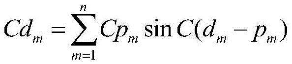
wherein CdmRepresenting the pixel value of an interpolation point of a current point to be interpolated; m represents the number of vertexes in a preset range of the current point to be interpolated; cpmA vertex pixel value representing a vertex; dmRepresenting the coordinates of the current point to be interpolated; p is a radical ofmRepresenting the coordinates of the vertices.
Further, target triangular patch data of the target object is determined based on each interpolation point pixel value and each vertex pixel value.
And S130, rendering the target triangular patch data to obtain a target contour curve of the target object.
In this embodiment, after target triangular patch data of a target object, that is, smoothed triangular patch data, is obtained, rendering processing is performed on the target triangular patch data, so as to obtain a target contour curve of the target object. Optionally, the method for obtaining the target contour curve of the target object may include: and inputting the target triangular patch data into a pre-trained data rendering model to obtain a target contour curve output by the rendering model. In this embodiment, each layer of the cross-section, coronal plane and sagittal plane can be iteratively projected according to the layer thickness of the target triangular patch data, so as to obtain the contour curve of a certain tissue of the corresponding layer.
The technical scheme of the embodiment specifically obtains a part segmentation result of the target object by obtaining a medical image of the target object and performing image segmentation on the medical image; determining initial triangular patch data of the target object based on the part segmentation result, and smoothing the initial triangular patch data based on the vertex coordinates and vertex pixel values of all vertexes in the initial triangular patch data to obtain target triangular patch data of the target object; rendering the target triangular patch data to obtain a target contour curve of the target object; the triangular patch is obtained by reconstructing the segmentation result, so that the safety problem caused by storing the segmentation result as a json file and then transmitting the json file to the front end is avoided, and the safety of contour information extraction is improved; and further, the obtained triangular patch is subjected to smoothing treatment, so that the triangular patch data which is smooth but can not be overlapped is obtained, more accurate contour information can be obtained, the time for extracting the contour information is shortened, and the accuracy and the safety of extracting the contour information are improved.
Fig. 2 is a flowchart of another target profile curve determining method according to an embodiment of the present invention, which is added to the foregoing embodiments to "generate a test analysis report based on the substandard test transaction and using the request parameter of the substandard test transaction, where the test analysis report includes a performance analysis main table and a performance analysis refinement table. "wherein explanations of the same or corresponding terms as those of the above-described embodiments are omitted. Referring to fig. 2, the method for determining a target profile curve provided in this embodiment includes:
s210, acquiring a medical image of the target object, and performing image segmentation on the medical image to obtain a part segmentation result of the target object.
And S220, determining initial triangular patch data of the target object based on the part segmentation result, and smoothing the initial triangular patch data based on the vertex coordinates and the vertex pixel values of all vertexes in the initial triangular patch data to obtain the target triangular patch data of the target object.
And S230, associating and storing each coordinate corresponding to any one triangular patch in the target triangular patch data of the target object in a data file with a preset format.
On the basis of the above embodiments, after the target triangular patch data of the target object is obtained, the target triangular patch data is stored in the data file of the preset format. For example, the data file is stored in a preset format such as a vtp file and an stl file. For example, the present embodiment may be described by taking an example in which the target triangular patch data is stored in a vtp file. Of course, the target triangle patch data may also be stored in a data file with a preset format, such as an stl file, and the data file format for storing the target triangle patch data is not limited in this embodiment, that is, the vertex coordinate data of the target triangle patch data may be obtained.
In some embodiments, after the marching cube algorithm is reconstructed to obtain the initial triangular patch data, the initial triangular patch data is stored in the vtp file, and before the initial triangular patch data is smoothed, the initial triangular patch data is read from the vtp file for processing, and the target triangular patch data obtained after the smoothing processing is stored in the vtp file. In this embodiment, the vtp file is actually an xml format file, and the file mainly stores vertex coordinates of each vertex of the triangular patch and a vertex combination forming one triangular patch. In other words, the vertex coordinates of each vertex stored in the vtp file are stored based on the relationship association between the triangular patches.
And S240, rendering the target triangular patch data to obtain a target contour curve of the target object.
In this embodiment, when rendering the target triangular patch data, the vtp file may be directly read, that is, the point coordinates of all the triangular patches may be read, and the rendering process is performed based on the point coordinates of all the triangular patches, so as to obtain the target contour curve of the target object.
According to the technical scheme of the embodiment, the three-dimensional reconstruction is performed on the mask region obtained through the segmentation to obtain the triangular patch data of each part, and the triangular patch data is subjected to smoothing processing and then stored as the VTP file. Furthermore, when the three-dimensional rendering is carried out by utilizing the ray projection in the front-end drawing process, the outline information of the dicom data of the relevant angle is directly obtained from the VTP file, the whole end-to-end service time is effectively shortened, and meanwhile, the safety of the front-end and back-end interaction processes is improved.
The following is an embodiment of the target contour curve determination apparatus provided in the embodiments of the present invention, which belongs to the same inventive concept as the target contour curve determination methods of the above embodiments, and details which are not described in detail in the embodiments of the target contour curve determination apparatus may refer to the embodiments of the target contour curve determination methods.
Fig. 3 is a schematic structural diagram of a target contour curve determining apparatus according to an embodiment of the present invention, which is applicable to a situation of acquiring contour information of a target object; in particular, the method is more suitable for the case of rendering the contour information of the target object based on the triangular patch of the target object. The specific structure of the target profile curve determination device comprises: a part segmentation result obtaining module 310, a target triangular patch data obtaining module 320, and a target contour curve obtaining module 330; wherein,
a part segmentation result obtaining module 310, configured to obtain a medical image of a target object, and perform image segmentation on the medical image to obtain a part segmentation result of the target object;
a target triangular patch data obtaining module 320, configured to determine initial triangular patch data of the target object based on the part segmentation result, and smooth the initial triangular patch data based on vertex coordinates and vertex pixel values of vertices in the initial triangular patch data to obtain target triangular patch data of the target object;
and a target contour curve obtaining module 330, configured to perform rendering processing on the target triangular patch data to obtain a target contour curve of the target object.
The technical scheme of the embodiment specifically obtains a part segmentation result of a target object by obtaining a medical image of the target object and performing image segmentation on the medical image; determining initial triangular patch data of the target object based on the part segmentation result, and smoothing the initial triangular patch data based on vertex coordinates and vertex pixel values of all vertexes in the initial triangular patch data to obtain target triangular patch data of the target object; rendering the target triangular patch data to obtain a target contour curve of the target object; the triangular patch is obtained by reconstructing the segmentation result, so that the safety problem caused by storing the segmentation result as a json file and then transmitting the json file to the front end is avoided, and the safety of contour information extraction is improved; and further, the obtained triangular patch is subjected to smoothing treatment, so that the triangular patch data which is smooth but can not be overlapped is obtained, more accurate contour information can be obtained, the time for extracting the contour information is shortened, and the accuracy and the safety of extracting the contour information are improved.
On the basis of the above embodiments, the part segmentation result obtaining module 310 includes:
and the part segmentation result acquisition unit is used for acquiring at least one image sequence of the medical image, inputting each image sequence into a pre-trained part segmentation model and obtaining at least one part segmentation result output by the part segmentation model.
On the basis of the foregoing embodiments, the target triangular patch data obtaining module 320 includes:
and an initial triangular patch data acquisition unit configured to acquire vertex coordinates of each vertex of at least one voxel corresponding to the region division result, and to specify initial triangular patch data of the target object based on the vertex coordinates of each vertex of each voxel.
On the basis of the above embodiments, the apparatus further includes:
and the storage module is used for storing each coordinate corresponding to any triangular patch in the target triangular patch data of the target object in a data file with a preset format in an associated manner after the target triangular patch data of the target object is obtained.
On the basis of the foregoing embodiments, the target triangular patch data obtaining module 320 includes:
an interpolation point coordinate determination unit, configured to determine an interpolation point coordinate of a point to be interpolated based on vertex coordinates of each vertex in the initial triangular patch data;
an interpolation point pixel value determining unit, configured to obtain vertex pixel values corresponding to a preset number of vertices in a preset range of the to-be-interpolated point, and determine an interpolation point pixel value of the to-be-interpolated point based on each vertex pixel value;
a target triangular patch data determining unit configured to determine target triangular patch data of the target object based on each of the interpolation point pixel values and each of the vertex pixel values.
On the basis of the above embodiments, the interpolation point coordinate determination unit includes:
a first interpolation point coordinate determining subunit, configured to determine, based on a vertex coordinate of a current vertex in the initial triangular patch data, an adjacency point coordinate of at least one adjacency point corresponding to the current vertex, and determine, based on the vertex coordinate and any adjacency point coordinate, an interpolation point coordinate of at least one to-be-interpolated point; or,
and a second interpolation point coordinate determination subunit, configured to determine, based on the point coordinate and the vertex pixel value of the current vertex in the initial triangular patch data, the point coordinate and the vertex pixel value of at least one adjacent point corresponding to the current vertex, and if a pixel value difference between the vertex pixel value of the current vertex and the adjacent point pixel value of the current adjacent point is greater than a preset pixel value threshold, determine, based on the point coordinate of the current vertex and the point coordinate of the current adjacent point, the point coordinate of the point to be interpolated.
On the basis of the foregoing embodiments, the target profile curve obtaining module 330 includes:
and the target contour curve acquisition unit is used for inputting the target triangular patch data into a pre-trained data rendering model to obtain a target contour curve output by the rendering model.
The target contour curve determining device provided by the embodiment of the invention can execute the target contour curve determining method provided by any embodiment of the invention, and has corresponding functional modules and beneficial effects of the executing method.
It should be noted that, in the embodiment of the target profile determining apparatus, the included units and modules are only divided according to the functional logic, but are not limited to the above division as long as the corresponding functions can be realized; in addition, specific names of the functional units are only for convenience of distinguishing from each other, and are not used for limiting the protection scope of the present invention.
Fig. 4 is a schematic structural diagram of an electronic device according to an embodiment of the present invention. FIG. 4 illustrates a block diagram of an exemplary electronic device 12 suitable for use in implementing embodiments of the present invention. The electronic device 12 shown in fig. 4 is only an example and should not bring any limitation to the function and the scope of use of the embodiment of the present invention.
As shown in FIG. 4, electronic device 12 is embodied in the form of a general purpose computing electronic device. The components of electronic device 12 may include, but are not limited to: one or more processors or processing units 16, a system memory 28, and a bus 18 that couples various system components including the system memory 28 and the processing unit 16.
The system memory 28 may include computer system readable media in the form of volatile memory, such as Random Access Memory (RAM)30 and/or cache memory 32. The electronic device 12 may further include other removable/non-removable, volatile/nonvolatile computer system storage media. By way of example only, storage system 34 may be used to read from and write to non-removable, nonvolatile magnetic media (not shown in FIG. 4, and commonly referred to as a "hard drive"). Although not shown in FIG. 4, a magnetic disk drive for reading from and writing to a removable, nonvolatile magnetic disk (e.g., a "floppy disk") and an optical disk drive for reading from or writing to a removable, nonvolatile optical disk (e.g., a CD-ROM, DVD-ROM, or other optical media) may be provided. In these cases, each drive may be connected to bus 18 by one or more data media interfaces. System memory 28 may include at least one program product having a set (e.g., at least one) of program modules that are configured to carry out the functions of embodiments of the invention.
A program/utility 40 having a set (at least one) of program modules 42 may be stored, for example, in system memory 28, such program modules 42 including, but not limited to, an operating system, one or more application programs, other program modules, and program data, each of which examples or some combination thereof may comprise an implementation of a network environment. Program modules 42 generally carry out the functions and/or methodologies of the described embodiments of the invention.
The processing unit 16 executes various functional applications and sample data acquisition by running a program stored in the system memory 28, for example, to implement the steps of a target contour curve determination method provided in this embodiment, where the target contour curve determination method includes:
acquiring a medical image of a target object, and performing image segmentation on the medical image to obtain a part segmentation result of the target object;
determining initial triangular patch data of the target object based on the part segmentation result, and smoothing the initial triangular patch data based on vertex coordinates and vertex pixel values of all vertexes in the initial triangular patch data to obtain target triangular patch data of the target object;
and rendering the target triangular patch data to obtain a target contour curve of the target object.
Of course, those skilled in the art can understand that the processor may also implement the technical solution of the sample data obtaining method provided in any embodiment of the present invention.
The present embodiment also provides a computer-readable storage medium, on which a computer program is stored, which when executed by a processor implements, for example, the steps of a target contour curve determination method provided by the present embodiment, where the target contour curve determination method includes:
acquiring a medical image of a target object, and performing image segmentation on the medical image to obtain a part segmentation result of the target object;
determining initial triangular patch data of the target object based on the part segmentation result, and smoothing the initial triangular patch data based on vertex coordinates and vertex pixel values of all vertexes in the initial triangular patch data to obtain target triangular patch data of the target object;
and rendering the target triangular patch data to obtain a target contour curve of the target object.
Computer storage media for embodiments of the invention may employ any combination of one or more computer-readable media. The computer readable medium may be a computer readable signal medium or a computer readable storage medium. The computer-readable storage medium may be, for example but not limited to: an electrical, magnetic, optical, electromagnetic, infrared, or semiconductor system, apparatus, or device, or any combination thereof. More specific examples (a non-exhaustive list) of the computer readable storage medium would include the following: an electrical connection having one or more wires, a portable computer diskette, a hard disk, a Random Access Memory (RAM), a read-only memory (ROM), an erasable programmable read-only memory (EPROM or flash memory), an optical fiber, a portable compact disc read-only memory (CD-ROM), an optical storage device, a magnetic storage device, or any suitable combination of the foregoing. In the context of this document, a computer readable storage medium may be any tangible medium that can contain, or store a program for use by or in connection with an instruction execution system, apparatus, or device.
A computer readable signal medium may include a propagated data signal with computer readable program code embodied therein, for example, in baseband or as part of a carrier wave. Such a propagated data signal may take many forms, including, but not limited to, electro-magnetic, optical, or any suitable combination thereof. A computer readable signal medium may also be any computer readable medium that is not a computer readable storage medium and that can communicate, propagate, or transport a program for use by or in connection with an instruction execution system, apparatus, or device.
Program code embodied on a computer readable medium may be transmitted using any appropriate medium, including but not limited to: wireless, wire, fiber optic cable, RF, etc., or any suitable combination of the foregoing.
Computer program code for carrying out operations for aspects of the present invention may be written in any combination of one or more programming languages, including an object oriented programming language such as Java, Smalltalk, C + + or the like and conventional procedural programming languages, such as the "C" programming language or similar programming languages. The program code may execute entirely on the user's computer, partly on the user's computer, as a stand-alone software package, partly on the user's computer and partly on a remote computer or entirely on the remote computer or server. In the case of a remote computer, the remote computer may be connected to the user's computer through any type of network, including a Local Area Network (LAN) or a Wide Area Network (WAN), or the connection may be made to an external computer (for example, through the Internet using an Internet service provider).
It will be understood by those skilled in the art that the modules or steps of the invention described above may be implemented by a general purpose computing device, they may be centralized on a single computing device or distributed across a network of computing devices, and optionally they may be implemented by program code executable by a computing device, such that it may be stored in a memory device and executed by a computing device, or it may be separately fabricated into various integrated circuit modules, or it may be fabricated by fabricating a plurality of modules or steps thereof into a single integrated circuit module. Thus, the present invention is not limited to any specific combination of hardware and software.
It is to be noted that the foregoing is only illustrative of the preferred embodiments of the present invention and the technical principles employed. It will be understood by those skilled in the art that the present invention is not limited to the particular embodiments described herein, but is capable of various obvious changes, rearrangements and substitutions as will now become apparent to those skilled in the art without departing from the scope of the invention. Therefore, although the present invention has been described in greater detail by the above embodiments, the present invention is not limited to the above embodiments, and may include other equivalent embodiments without departing from the spirit of the present invention, and the scope of the present invention is determined by the scope of the appended claims.
Claims (10)
1. A method of determining a target profile curve, comprising:
acquiring a medical image of a target object, and performing image segmentation on the medical image to obtain a part segmentation result of the target object;
determining initial triangular patch data of the target object based on the part segmentation result, and smoothing the initial triangular patch data based on vertex coordinates and vertex pixel values of all vertexes in the initial triangular patch data to obtain target triangular patch data of the target object;
and rendering the target triangular patch data to obtain a target contour curve of the target object.
2. The method according to claim 1, wherein the image segmentation of the medical image to obtain a result of the segmentation of the target object includes:
and acquiring at least one image sequence of the medical image, and inputting each image sequence into a pre-trained part segmentation model to obtain at least one part segmentation result output by the part segmentation model.
3. The method of claim 1, wherein the determining initial triangular patch data of the target object based on the region segmentation result comprises:
and acquiring vertex coordinates of each vertex of at least one voxel corresponding to the part segmentation result, and determining initial triangular surface patch data of the target object based on the vertex coordinates of each vertex of each voxel.
4. The method of claim 1, wherein after obtaining the target triangle patch data of the target object, the method further comprises:
and associating and storing each coordinate corresponding to any triangular patch in the target triangular patch data of the target object in a data file with a preset format.
5. The method of claim 1, wherein smoothing the initial triangular patch data based on vertex coordinates and vertex pixel values of vertices in the initial triangular patch data to obtain target triangular patch data of the target object, comprises:
determining the coordinates of interpolation points of points to be interpolated based on the vertex coordinates of each vertex in the initial triangular patch data;
vertex pixel values corresponding to a preset number of vertexes are obtained in the preset range of the point to be interpolated, and the pixel value of the point to be interpolated is determined based on each vertex pixel value;
and determining target triangular patch data of the target object based on each interpolation point pixel value and each vertex pixel value.
6. The method of claim 1, wherein determining interpolation point coordinates of a point to be interpolated based on vertex coordinates of vertices in the initial triangular patch data comprises:
determining the adjacent point coordinates of at least one adjacent point corresponding to the current vertex based on the vertex coordinates of the current vertex in the initial triangular patch data, and determining the interpolation point coordinates of at least one point to be interpolated based on the vertex coordinates and any adjacent point coordinates respectively; or,
and determining the point coordinate and the vertex pixel value of at least one adjacent point corresponding to the current vertex based on the point coordinate and the vertex pixel value of the current vertex in the initial triangular patch data, and if the pixel value difference value between the vertex pixel value of the current vertex and the adjacent point pixel value of the current adjacent point is greater than a preset pixel value threshold, determining the point coordinate of the point to be interpolated based on the point coordinate of the current vertex and the point coordinate of the current adjacent point.
7. The method of claim 1, wherein the rendering the target triangular patch data to obtain the target contour curve of the target object comprises:
and inputting the target triangular patch data into a pre-trained data rendering model to obtain a target contour curve output by the rendering model.
8. An apparatus for determining a target profile curve, comprising:
the part segmentation result acquisition module is used for acquiring a medical image of a target object and carrying out image segmentation on the medical image to obtain a part segmentation result of the target object;
a target triangular patch data acquisition module, configured to determine initial triangular patch data of the target object based on the part segmentation result, and smooth the initial triangular patch data based on vertex coordinates and vertex pixel values of vertices in the initial triangular patch data to obtain target triangular patch data of the target object;
and the target contour curve acquisition module is used for rendering the target triangular patch data to obtain a target contour curve of the target object.
9. An electronic device, comprising:
one or more processors;
a storage device for storing one or more programs,
when executed by the one or more processors, cause the one or more processors to implement the target profile determination method of any one of claims 1-7.
10. A computer-readable storage medium, on which a computer program is stored which, when being executed by a processor, carries out a method for determining a target profile curve as claimed in any one of claims 1 to 7.
Priority Applications (1)
| Application Number | Priority Date | Filing Date | Title |
|---|---|---|---|
| CN202111150742.9A CN113888566B (en) | 2021-09-29 | 2021-09-29 | Target contour curve determination method and device, electronic equipment and storage medium |
Applications Claiming Priority (1)
| Application Number | Priority Date | Filing Date | Title |
|---|---|---|---|
| CN202111150742.9A CN113888566B (en) | 2021-09-29 | 2021-09-29 | Target contour curve determination method and device, electronic equipment and storage medium |
Publications (2)
| Publication Number | Publication Date |
|---|---|
| CN113888566A true CN113888566A (en) | 2022-01-04 |
| CN113888566B CN113888566B (en) | 2022-05-10 |
Family
ID=79007918
Family Applications (1)
| Application Number | Title | Priority Date | Filing Date |
|---|---|---|---|
| CN202111150742.9A Active CN113888566B (en) | 2021-09-29 | 2021-09-29 | Target contour curve determination method and device, electronic equipment and storage medium |
Country Status (1)
| Country | Link |
|---|---|
| CN (1) | CN113888566B (en) |
Cited By (3)
| Publication number | Priority date | Publication date | Assignee | Title |
|---|---|---|---|---|
| CN114820902A (en) * | 2022-04-26 | 2022-07-29 | 广州柏视医疗科技有限公司 | Method and system for rendering three-dimensional model contour based on point cloud |
| CN116385474A (en) * | 2023-02-27 | 2023-07-04 | 雅客智慧(北京)科技有限公司 | Dental scan model segmentation method, device, and electronic equipment based on deep learning |
| CN117037208A (en) * | 2023-07-19 | 2023-11-10 | 北京卡路里信息技术有限公司 | Image detection method and device |
Citations (13)
| Publication number | Priority date | Publication date | Assignee | Title |
|---|---|---|---|---|
| CN101322652A (en) * | 2008-07-04 | 2008-12-17 | 浙江大学 | A computer simulation calibration biopsy method and device |
| CN101894363A (en) * | 2009-05-20 | 2010-11-24 | 新奥特(北京)视频技术有限公司 | Patched garbage mask border rendering method |
| CN102169588A (en) * | 2010-02-26 | 2011-08-31 | 新奥特(北京)视频技术有限公司 | Method for constructing meteorological curve |
| CN103810752A (en) * | 2014-02-18 | 2014-05-21 | 海信集团有限公司 | Liver segmenting method based on medical image and liver segmenting system thereof |
| CN105912874A (en) * | 2016-04-29 | 2016-08-31 | 青岛大学附属医院 | Liver three-dimensional database system constructed on the basis of DICOM (Digital Imaging and Communications in Medicine) medical image |
| CN106373168A (en) * | 2016-11-24 | 2017-02-01 | 北京三体高创科技有限公司 | Medical image based segmentation and 3D reconstruction method and 3D printing system |
| CN106485642A (en) * | 2016-09-30 | 2017-03-08 | 北京交通大学 | The method of embedded visible watermark in three-dimensional grid model |
| CN108389251A (en) * | 2018-03-21 | 2018-08-10 | 南京大学 | The full convolutional network threedimensional model dividing method of projection based on fusion various visual angles feature |
| CN111127453A (en) * | 2019-12-27 | 2020-05-08 | 苏州影加科技有限公司 | A fully automatic segmentation method of tracheal tree based on differential geometry |
| US20200150624A1 (en) * | 2018-11-09 | 2020-05-14 | Autodesk, Inc. | Conversion of mesh geometry to watertight boundary representation |
| CN111402216A (en) * | 2020-03-10 | 2020-07-10 | 河海大学常州校区 | Three-dimensional broken bone segmentation method and device based on deep learning |
| US11126162B1 (en) * | 2020-09-17 | 2021-09-21 | Shanghai Fusion Tech Co., Ltd. | 3D printing slicing method, apparatus, device, and storage medium |
| WO2021184933A1 (en) * | 2020-03-20 | 2021-09-23 | 华为技术有限公司 | Three-dimensional human body model reconstruction method |
-
2021
- 2021-09-29 CN CN202111150742.9A patent/CN113888566B/en active Active
Patent Citations (13)
| Publication number | Priority date | Publication date | Assignee | Title |
|---|---|---|---|---|
| CN101322652A (en) * | 2008-07-04 | 2008-12-17 | 浙江大学 | A computer simulation calibration biopsy method and device |
| CN101894363A (en) * | 2009-05-20 | 2010-11-24 | 新奥特(北京)视频技术有限公司 | Patched garbage mask border rendering method |
| CN102169588A (en) * | 2010-02-26 | 2011-08-31 | 新奥特(北京)视频技术有限公司 | Method for constructing meteorological curve |
| CN103810752A (en) * | 2014-02-18 | 2014-05-21 | 海信集团有限公司 | Liver segmenting method based on medical image and liver segmenting system thereof |
| CN105912874A (en) * | 2016-04-29 | 2016-08-31 | 青岛大学附属医院 | Liver three-dimensional database system constructed on the basis of DICOM (Digital Imaging and Communications in Medicine) medical image |
| CN106485642A (en) * | 2016-09-30 | 2017-03-08 | 北京交通大学 | The method of embedded visible watermark in three-dimensional grid model |
| CN106373168A (en) * | 2016-11-24 | 2017-02-01 | 北京三体高创科技有限公司 | Medical image based segmentation and 3D reconstruction method and 3D printing system |
| CN108389251A (en) * | 2018-03-21 | 2018-08-10 | 南京大学 | The full convolutional network threedimensional model dividing method of projection based on fusion various visual angles feature |
| US20200150624A1 (en) * | 2018-11-09 | 2020-05-14 | Autodesk, Inc. | Conversion of mesh geometry to watertight boundary representation |
| CN111127453A (en) * | 2019-12-27 | 2020-05-08 | 苏州影加科技有限公司 | A fully automatic segmentation method of tracheal tree based on differential geometry |
| CN111402216A (en) * | 2020-03-10 | 2020-07-10 | 河海大学常州校区 | Three-dimensional broken bone segmentation method and device based on deep learning |
| WO2021184933A1 (en) * | 2020-03-20 | 2021-09-23 | 华为技术有限公司 | Three-dimensional human body model reconstruction method |
| US11126162B1 (en) * | 2020-09-17 | 2021-09-21 | Shanghai Fusion Tech Co., Ltd. | 3D printing slicing method, apparatus, device, and storage medium |
Non-Patent Citations (13)
| Title |
|---|
| MICHAEL HAUBNER等: "Virtual Reality in Medicine–Computer Graphics and Interaction Techniques", 《IEEE TRANSACTIONS ON INFORMATION TECHNOLOGY IN BIOMEDICINE》 * |
| MICHAEL R. KAUS等: "Automated segmentation of the left ventricle in cardiac MRI", 《MEDICAL IMAGE ANALYSIS》 * |
| ZHAOXI PAN等: "Comparison of Medical Image 3D Reconstruction Rendering Methods for Robot-Assisted Surgery", 《2017 2ND INTERNATIONAL CONFERENCE ON ADVANCED ROBOTICS AND MECHATRONICS (ICARM)》 * |
| 刘敏: "基于VTK的医学图像三维重建的研究", 《中国优秀博硕士学位论文全文数据库(硕士) 信息科技辑》 * |
| 孙键: "医学图像可视化中的重建技术方法研究", 《中国优秀博硕士学位论文全文数据库(硕士) 信息科技辑》 * |
| 孟凡文: "面向光栅投影的点云预处理与曲面重构技术研究", 《中国优秀博硕士学位论文全文数据库(博士) 工程科技Ⅰ辑》 * |
| 宋林锐: "基于VR的医学图像智能分析系统研究与应用", 《中国优秀博硕士学位论文全文数据库(硕士) 医药卫生科技辑》 * |
| 张嘉伟等: "基于云计算的医学影像处理与 3D 打印平台", 《软件》 * |
| 张宇: "医学图像三维重建的研究", 《万方在线》 * |
| 徐欣康: "基于层间关联性的切片轮廓数据平滑算法研究", 《中国优秀博硕士学位论文全文数据库(硕士)信息科技辑》 * |
| 戴培山等: "基于CT图像的鼻咽组织重建", 《系统仿真学报》 * |
| 王德远: "基于核磁共振图像的心室分割与三维可视化研究", 《中国优秀博硕士学位论文全文数据库(硕士) 信息科技辑》 * |
| 高卫香: "基于MRI序列图像的分割方法及三维网格剖分模型的构建", 《中国优秀博硕士学位论文全文数据库(硕士) 信息科技辑》 * |
Cited By (5)
| Publication number | Priority date | Publication date | Assignee | Title |
|---|---|---|---|---|
| CN114820902A (en) * | 2022-04-26 | 2022-07-29 | 广州柏视医疗科技有限公司 | Method and system for rendering three-dimensional model contour based on point cloud |
| CN114820902B (en) * | 2022-04-26 | 2023-05-02 | 广州柏视医疗科技有限公司 | Method and system for rendering three-dimensional model contour based on point cloud |
| CN116385474A (en) * | 2023-02-27 | 2023-07-04 | 雅客智慧(北京)科技有限公司 | Dental scan model segmentation method, device, and electronic equipment based on deep learning |
| CN116385474B (en) * | 2023-02-27 | 2024-06-04 | 雅客智慧(北京)科技有限公司 | Tooth scanning model segmentation method and device based on deep learning and electronic equipment |
| CN117037208A (en) * | 2023-07-19 | 2023-11-10 | 北京卡路里信息技术有限公司 | Image detection method and device |
Also Published As
| Publication number | Publication date |
|---|---|
| CN113888566B (en) | 2022-05-10 |
Similar Documents
| Publication | Publication Date | Title |
|---|---|---|
| CN109961491B (en) | Multi-modal image truncation compensation method, device, computer equipment and medium | |
| US20200082571A1 (en) | Method and apparatus for calibrating relative parameters of collector, device and storage medium | |
| CN113160189B (en) | Blood vessel centerline extraction method, device, equipment and storage medium | |
| RU2556428C2 (en) | Method for weakening of bone x-ray images | |
| CN113888566B (en) | Target contour curve determination method and device, electronic equipment and storage medium | |
| CN111340756B (en) | Medical image lesion detection merging method, system, terminal and storage medium | |
| CN112419484A (en) | Three-dimensional blood vessel synthesis method and system, coronary artery analysis system and storage medium | |
| CN110458830A (en) | Image processing method, device, server and storage medium | |
| CN112508835B (en) | A GAN-based modeling method for contrast-free medical image enhancement | |
| US20250148574A1 (en) | Method and apparatus for enhancing pet parameter image, device, and storage medium | |
| CN113781653A (en) | Object model generation method and device, electronic equipment and storage medium | |
| CN113610752A (en) | Mammary gland image registration method, computer device and storage medium | |
| CN111145160A (en) | Method, device, server and medium for determining coronary artery branch where calcified area is located | |
| US11138736B2 (en) | Information processing apparatus and information processing method | |
| CN112150600B (en) | Volume reconstruction image generation method, device and system and storage medium | |
| WO2020168698A1 (en) | Vrds 4d medical image-based vein ai endoscopic analysis method and product | |
| CN111539926B (en) | Image detection method and device | |
| CN109658425B (en) | Lung lobe segmentation method and device, computer equipment and storage medium | |
| CN116993920A (en) | A blood vessel reconstruction method, device, electronic equipment and storage medium | |
| CN113850794B (en) | Image processing method and device | |
| CN113205459B (en) | Motion correction of angiographic images for 3D reconstruction of coronary arteries | |
| CN109799936B (en) | Image generation method, device, equipment and medium | |
| US20240394955A1 (en) | System and Method for Rendering Images | |
| WO2021081839A1 (en) | Vrds 4d-based method for analysis of condition of patient, and related products | |
| CN112530554B (en) | A scanning positioning method, device, storage medium and electronic equipment |
Legal Events
| Date | Code | Title | Description |
|---|---|---|---|
| PB01 | Publication | ||
| PB01 | Publication | ||
| SE01 | Entry into force of request for substantive examination | ||
| SE01 | Entry into force of request for substantive examination | ||
| GR01 | Patent grant | ||
| GR01 | Patent grant |