CN113786262B - Preparation method of dental crown lengthening operation guide plate - Google Patents
Preparation method of dental crown lengthening operation guide plate Download PDFInfo
- Publication number
- CN113786262B CN113786262B CN202110939183.3A CN202110939183A CN113786262B CN 113786262 B CN113786262 B CN 113786262B CN 202110939183 A CN202110939183 A CN 202110939183A CN 113786262 B CN113786262 B CN 113786262B
- Authority
- CN
- China
- Prior art keywords
- image
- guide plate
- data
- dimensional
- pixel
- Prior art date
- Legal status (The legal status is an assumption and is not a legal conclusion. Google has not performed a legal analysis and makes no representation as to the accuracy of the status listed.)
- Active
Links
- 238000002360 preparation method Methods 0.000 title claims abstract description 13
- 238000000034 method Methods 0.000 claims abstract description 53
- 210000000988 bone and bone Anatomy 0.000 claims abstract description 26
- 238000007781 pre-processing Methods 0.000 claims abstract description 9
- 238000012876 topography Methods 0.000 claims abstract description 4
- 238000012545 processing Methods 0.000 claims description 23
- 230000008569 process Effects 0.000 claims description 20
- 238000004519 manufacturing process Methods 0.000 claims description 9
- 238000001914 filtration Methods 0.000 claims description 8
- 238000007639 printing Methods 0.000 claims description 4
- 238000013528 artificial neural network Methods 0.000 claims description 3
- 238000012937 correction Methods 0.000 claims description 3
- 230000002708 enhancing effect Effects 0.000 claims description 3
- 238000007500 overflow downdraw method Methods 0.000 claims description 3
- 230000011218 segmentation Effects 0.000 claims description 3
- 239000007787 solid Substances 0.000 claims description 3
- 238000013461 design Methods 0.000 claims description 2
- 238000012163 sequencing technique Methods 0.000 claims description 2
- 238000003860 storage Methods 0.000 claims description 2
- 238000001356 surgical procedure Methods 0.000 claims 2
- 210000004195 gingiva Anatomy 0.000 description 3
- 230000008439 repair process Effects 0.000 description 3
- 238000010146 3D printing Methods 0.000 description 2
- 230000003239 periodontal effect Effects 0.000 description 2
- 210000001519 tissue Anatomy 0.000 description 2
- 238000009825 accumulation Methods 0.000 description 1
- 210000001909 alveolar process Anatomy 0.000 description 1
- 238000004590 computer program Methods 0.000 description 1
- 238000010276 construction Methods 0.000 description 1
- 230000007547 defect Effects 0.000 description 1
- 208000002925 dental caries Diseases 0.000 description 1
- 230000006872 improvement Effects 0.000 description 1
- 239000000463 material Substances 0.000 description 1
- 238000002844 melting Methods 0.000 description 1
- 230000008018 melting Effects 0.000 description 1
- 230000004048 modification Effects 0.000 description 1
- 238000012986 modification Methods 0.000 description 1
- 230000008092 positive effect Effects 0.000 description 1
- 230000002035 prolonged effect Effects 0.000 description 1
Images


Classifications
-
- A—HUMAN NECESSITIES
- A61—MEDICAL OR VETERINARY SCIENCE; HYGIENE
- A61C—DENTISTRY; APPARATUS OR METHODS FOR ORAL OR DENTAL HYGIENE
- A61C19/00—Dental auxiliary appliances
- A61C19/06—Implements for therapeutic treatment
-
- A—HUMAN NECESSITIES
- A61—MEDICAL OR VETERINARY SCIENCE; HYGIENE
- A61B—DIAGNOSIS; SURGERY; IDENTIFICATION
- A61B6/00—Apparatus or devices for radiation diagnosis; Apparatus or devices for radiation diagnosis combined with radiation therapy equipment
- A61B6/50—Apparatus or devices for radiation diagnosis; Apparatus or devices for radiation diagnosis combined with radiation therapy equipment specially adapted for specific body parts; specially adapted for specific clinical applications
- A61B6/51—Apparatus or devices for radiation diagnosis; Apparatus or devices for radiation diagnosis combined with radiation therapy equipment specially adapted for specific body parts; specially adapted for specific clinical applications for dentistry
- A61B6/512—Intraoral means
-
- A—HUMAN NECESSITIES
- A61—MEDICAL OR VETERINARY SCIENCE; HYGIENE
- A61B—DIAGNOSIS; SURGERY; IDENTIFICATION
- A61B6/00—Apparatus or devices for radiation diagnosis; Apparatus or devices for radiation diagnosis combined with radiation therapy equipment
- A61B6/52—Devices using data or image processing specially adapted for radiation diagnosis
- A61B6/5211—Devices using data or image processing specially adapted for radiation diagnosis involving processing of medical diagnostic data
-
- B—PERFORMING OPERATIONS; TRANSPORTING
- B33—ADDITIVE MANUFACTURING TECHNOLOGY
- B33Y—ADDITIVE MANUFACTURING, i.e. MANUFACTURING OF THREE-DIMENSIONAL [3-D] OBJECTS BY ADDITIVE DEPOSITION, ADDITIVE AGGLOMERATION OR ADDITIVE LAYERING, e.g. BY 3-D PRINTING, STEREOLITHOGRAPHY OR SELECTIVE LASER SINTERING
- B33Y50/00—Data acquisition or data processing for additive manufacturing
-
- B—PERFORMING OPERATIONS; TRANSPORTING
- B33—ADDITIVE MANUFACTURING TECHNOLOGY
- B33Y—ADDITIVE MANUFACTURING, i.e. MANUFACTURING OF THREE-DIMENSIONAL [3-D] OBJECTS BY ADDITIVE DEPOSITION, ADDITIVE AGGLOMERATION OR ADDITIVE LAYERING, e.g. BY 3-D PRINTING, STEREOLITHOGRAPHY OR SELECTIVE LASER SINTERING
- B33Y80/00—Products made by additive manufacturing
Landscapes
- Health & Medical Sciences (AREA)
- Life Sciences & Earth Sciences (AREA)
- Engineering & Computer Science (AREA)
- Medical Informatics (AREA)
- Veterinary Medicine (AREA)
- Public Health (AREA)
- General Health & Medical Sciences (AREA)
- Animal Behavior & Ethology (AREA)
- Dentistry (AREA)
- Radiology & Medical Imaging (AREA)
- Chemical & Material Sciences (AREA)
- Oral & Maxillofacial Surgery (AREA)
- Materials Engineering (AREA)
- Surgery (AREA)
- Physics & Mathematics (AREA)
- Biophysics (AREA)
- High Energy & Nuclear Physics (AREA)
- Manufacturing & Machinery (AREA)
- Nuclear Medicine, Radiotherapy & Molecular Imaging (AREA)
- Optics & Photonics (AREA)
- Pathology (AREA)
- Molecular Biology (AREA)
- Biomedical Technology (AREA)
- Heart & Thoracic Surgery (AREA)
- Epidemiology (AREA)
- Computer Vision & Pattern Recognition (AREA)
- Apparatus For Radiation Diagnosis (AREA)
Abstract
The invention belongs to the technical field of oral clinical medical treatment instruments, and discloses a method for preparing a dental crown lengthening guide plate, which comprises the steps of obtaining CT image data by shooting a CT mode, and carrying out preprocessing and depth analysis on the CT image data so as to determine the three-dimensional shapes of teeth and alveolar bones of a patient; simultaneously, acquiring the three-dimensional shape of the gum of the patient by using an intraoral scanner; determining the bone removal position, the gum incision position and the position of the edge of the dental crown of the alveolar bone based on the obtained three-dimensional shape, wherein the alveolar bone and the gum incision position are marked according to a biological width principle and the self condition of a patient; matching the requirement that the fitting position of the operation guide plate is tightly fitted with the teeth of the patient; depicting the three-dimensional shape of the guide plate on the three-dimensional topography; the guide plate is produced from the three-dimensional shape of the guide plate described. The invention can improve the preparation accuracy of the dental crown lengthening guide plate and provides convenience for a doctor to remove alveolar bone and gum in the dental crown lengthening operation.
Description
Technical Field
The invention belongs to the technical field of oral clinical medical treatment instruments, and particularly relates to a method for preparing a dental crown lengthening operation guide plate.
Background
At present, the crown lengthening operation is to expose more tooth body tissues of healthy teeth under the principle of periodontal biological breadth, and remove a certain alveolar bone and gingiva by an operation method, so that the exposure of the teeth is increased, and the next step of improving the gingiva and restoring the aesthetic appearance of the gingiva shape is carried out. In order to precisely control the amount of gum and alveolar bone removed during the operation, a guide plate is generally used during the operation to assist in the determination.
Normally, the distance from the gingival sulcus bottom to the alveolar ridge crest is a constant distance, namely the biological width is about two millimeters, and in some teeth needing full-crown repair, when the clinical dental crown is too short to prevent the repair from being fixed due to dental folds, root-surface caries and the like, the clinical dental crown of the tooth needs to be prolonged by using a dental crown lengthening operation to ensure that the repair has enough fixation and the dental crown margin is not placed at the bottom of the gingival sulcus bottom to stimulate periodontal tissues.
The method for preparing the guide plate for the dental crown lengthening operation in the prior art reduces the preparation precision of the guide plate for the dental crown lengthening operation, and is inconvenient for a doctor to operate. Meanwhile, the preparation method of the dental crown lengthening operation guide plate in the prior art increases the preparation cost and reduces the working efficiency.
Through the above analysis, the problems and defects of the prior art are as follows:
(1) The method for preparing the guide plate for the dental crown lengthening operation in the prior art reduces the preparation precision of the guide plate for the dental crown lengthening operation, and is inconvenient for a doctor to operate.
(2) The preparation method of the dental crown lengthening operation guide plate in the prior art increases the preparation cost and reduces the working efficiency.
Disclosure of Invention
Aiming at the problems in the prior art, the invention provides a method for preparing a dental crown lengthening operation guide plate.
The present invention is achieved as such, a method for preparing a dental crown lengthening guide, the method comprising:
acquiring CT image data by shooting a CT mode, and performing preprocessing and depth analysis on the CT image data to further determine the three-dimensional shapes of teeth and alveolar bones of a patient; simultaneously, acquiring the three-dimensional shape of the gum of the patient by using an intraoral scanner;
the CT image data preprocessing comprises the following steps:
carrying out image graying processing on the acquired CT image data, and carrying out filtering enhancement on the image; after the CT image data filtering and enhancing are finished, carrying out binarization processing on the image, and accurately positioning teeth and alveolar bones;
determining the bone removal position of the alveolar bone, the gingival incision position and the position of the edge of the dental crown based on the obtained three-dimensional shape, wherein the alveolar bone and the gingival incision position are marked according to a biological width principle and the self condition of a patient;
thirdly, matching the joint position of the operation guide plate with the requirement of tightly jointing the teeth of the patient according to the calibrated gum cut position, alveolar bone removal position and dental crown edge position; depicting the three-dimensional shape of the guide plate on the three-dimensional topography;
fourthly, manufacturing a guide plate according to the three-dimensional shape of the depicted guide plate;
the manufacturing of the guide plate according to the depicted three-dimensional shape of the guide plate comprises: importing the three-dimensional model data of the depicted guide plate into a 3D printer, carrying out layering processing on the entity three-dimensional model data to obtain lamina data, and carrying out reverse solution manufacturing according to the lamina data, wherein the reverse solution manufacturing comprises lamina printing and lamina accumulation to obtain the dental crown lengthening operation guide plate;
the layering processing of the entity three-dimensional model data comprises the following steps:
and slicing the solid three-dimensional model by using a plane vertical to a Z axis as a layering plane by adopting three-dimensional slicing software, carrying out equal-thickness layering along the Z axis of the actual guide plate model, obtaining a plurality of pieces of two-dimensional section information according to the relation between the designed thickness and the layer height, and obtaining the slice data of each layer through the intersection line position relation of the triangular patch data and the two-dimensional section information.
Further, the depth analysis of the CT image data includes:
acquiring depth data of the preprocessed image, segmenting the image, and extracting corresponding image data;
and establishing a corresponding recognition model to perform matching recognition with data in the cloud server, and describing the CT image data.
Further, the specific process of performing image graying processing on the acquired CT image data is as follows:
let the image width in CT color image be w idth Height of h eight Size of image T = w idth ×h eight ;
Respectively multiplying three components of the position of a pixel i in the graying of the image by respective weight by using a weighted average method; then adding to obtain a grayed value of the position of the pixel i;
the image C' of the color image C after the image graying processing is positioned as follows:

wherein, C' i Is the luminance value, R, of the i position of a pixel of a gray scale image i 、G i 、B i The component values of red, green and blue at the position of pixel i of the color image, respectively.
Further, the specific process of performing filtering enhancement on the image is as follows:
determining the initial address of the CT image and the width and the height of the image, scanning pixel points in the image one by one, sequencing pixel values of all elements in the neighborhood of the pixel points from small to large, and assigning the obtained intermediate value to the pixel point corresponding to the current point in the target image;
and repeating the process until all the CT images are processed.
Further, the specific processing process of the image binarization processing is as follows:
carrying out binaryzation on the original image by an improved Bernsen method to obtain a first source image;
calculating a threshold value according to the self-organizing neural network, and carrying out binaryzation on the image to obtain a second source image;
and (5) obtaining a final binary image by using the principle of minimum gray value as an image fusion method.
Further, the specific process of extracting the corresponding image data is as follows:
standardizing the color space of the input image by adopting a Gamma correction method on the preprocessed image;
determining the gradient of each pixel of the image, dividing the image into a plurality of region blocks, determining a gradient histogram in each region, and counting all the gradient histograms;
and (4) combining the corresponding area blocks into a large area block, and connecting the features in the area blocks in series to obtain the features of the image.
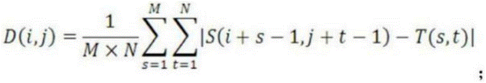
Further, the established recognition model is as follows:

wherein i is more than or equal to 1 and less than or equal to M-M +1,1 and more than or equal to j is more than or equal to N-N +1; d (i, j) is the average absolute difference; (i, j) is the position in the graph and MxN is the size of the sub-graph.
Further, the specific process of segmenting the image is as follows:
selecting an image average gray value, and initializing a threshold;
dividing the gray level image according to a threshold value, and calculating the gray level mean value of the A-type image and the B-type image;
updating the threshold value so that the average of the grays of A plus the average of the grays of B is equal to the threshold value
And repeating the process to enable the threshold value to meet a certain range of numerical values, and performing binarization segmentation on the gray level image by using the determined threshold value.
It is another object of the present invention to provide a computer program product stored on a computer readable medium, comprising a computer readable program for providing a user input interface to implement the crown lengthening guide preparation method when executed on an electronic device.
It is another object of the present invention to provide a computer-readable storage medium storing instructions that, when executed on a computer, cause the computer to perform the crown lengthening guide preparation method.
By combining all the technical schemes, the invention has the advantages and positive effects that: according to the invention, CT image data is obtained by shooting a CT mode, and preprocessing and depth analysis are carried out, so that the preparation accuracy of the dental crown lengthening guide plate can be improved, and convenience is provided for a doctor to carry out dental crown lengthening operation to cut alveolar bone and gum; meanwhile, the guide plate is convenient to remove, and great help is provided for the whole operation.
According to the invention, the guide plate is printed by 3D printing forming, so that the layer-by-layer forming, clear layering and three-dimensional modeling in the process of melting and forming the printing material can be ensured, the construction period can be shortened by adopting the 3D printing forming, the working efficiency is improved, and the cost is saved.
Drawings
In order to more clearly illustrate the technical solutions of the embodiments of the present application, the drawings needed to be used in the embodiments of the present application will be briefly described below, and it is obvious that the drawings described below are only some embodiments of the present application, and it is obvious for those skilled in the art that other drawings can be obtained from the drawings without creative efforts.
Fig. 1 is a flowchart of a method for preparing a dental crown lengthening guide according to an embodiment of the present invention.
Fig. 2 is a flowchart of a method for preprocessing CT image data according to an embodiment of the present invention.
Fig. 3 is a flowchart of a method for depth analysis of CT image data according to an embodiment of the present invention.
Fig. 4 is a flowchart of a method for enhancing filtering of an image according to an embodiment of the present invention.
FIG. 5 is a flow chart of a method for fabricating a guide plate based on a three-dimensional shape of the guide plate depicted in accordance with an embodiment of the present invention.
Detailed Description
In order to make the objects, technical solutions and advantages of the present invention more apparent, the present invention is further described in detail with reference to the following embodiments. It should be understood that the specific embodiments described herein are merely illustrative of the invention and do not limit the invention.
In view of the problems of the prior art, the present invention provides a method for preparing a guide plate for a crown lengthening operation, which is described in detail below with reference to the accompanying drawings.
As shown in fig. 1, a method for preparing a crown lengthening guide according to an embodiment of the present invention includes:
s101, acquiring CT image data by shooting a CT mode, and preprocessing and deeply analyzing the CT image data to further determine the three-dimensional shapes of teeth and alveolar bones of a patient; meanwhile, an intraoral scanner is used for obtaining the three-dimensional shape of the gum of the patient;
s102, determining a bone removal position, a gum incision position and a crown edge position of the alveolar bone based on the obtained three-dimensional shape, wherein the alveolar bone and the gum incision position are marked according to a biological width principle and the self condition of a patient;
s103, matching the requirement that the attaching position of the operation guide plate is closely attached to the teeth of the patient according to the marked gum incision position, alveolar bone removing position and dental crown edge position; depicting the three-dimensional shape of the guide plate on the three-dimensional topography; the guide plate is produced from the three-dimensional shape of the guide plate described.
As shown in fig. 2, the process of preprocessing the CT image data provided by the embodiment of the present invention includes:
s201, carrying out image graying processing on the acquired CT image data, and carrying out filtering enhancement on the image;
and S202, after the CT image data is filtered and enhanced, carrying out binarization processing on the image, and accurately positioning teeth, gums and alveolar bones.
As shown in fig. 3, a specific process of performing depth analysis on CT image data provided by the embodiment of the present invention is as follows:
s301, carrying out depth data acquisition on the preprocessed image, segmenting the image, and extracting corresponding image data;
and S302, establishing a corresponding recognition model to perform matching recognition with data in the cloud server, so as to realize the description of the CT image data.
The specific process of carrying out image graying processing on the acquired CT image data provided by the embodiment of the invention comprises the following steps:
let the image width in CT color image be w idth Height of h eight Size of image T = w idth ×h eight ;
Multiplying three components of the position of a pixel i in the graying of the image by respective weight value by using a weighted average method; then adding to obtain a grayed value of the position of the pixel i;
the image C' of the color image C after the image graying processing is positioned as follows:

wherein, C' i Is the luminance value, R, of the i position of a pixel of a gray scale image i 、G i 、B i The component values of red, green and blue at the position of pixel i of the color image, respectively.
As shown in fig. 4, a specific process of performing filtering enhancement on an image provided by the embodiment of the present invention is as follows:
s401: determining the first address of the CT image and the width and the height of the image, and scanning pixel points in the image one by one;
s402: sorting the pixel values of all elements in the neighborhood from small to large, and assigning the obtained intermediate value to a pixel point corresponding to the current point in the target image;
s403: and repeating the process until all the CT images are processed.
The specific processing process for the binarization processing of the image provided by the embodiment of the invention comprises the following steps:
carrying out binarization on the original image by an improved Bernsen method to obtain a first source image;
calculating a threshold value according to the self-organizing neural network, and carrying out binarization on the image to obtain a second source image;
and (5) obtaining a final binary image by using the principle of minimum gray value as an image fusion method.
The specific process for extracting the corresponding image data provided by the embodiment of the invention is as follows:
carrying out color space standardization on the input image by adopting a Gamma correction method on the preprocessed image;
determining the gradient of each pixel of the image, dividing the image into a plurality of region blocks, determining a gradient histogram in each region, and counting all the gradient histograms;
and (4) combining the corresponding area blocks into a large area block, and connecting the features in the area blocks in series to obtain the features of the image.
The established recognition model provided by the embodiment of the invention is as follows:

wherein i is more than or equal to 1 and less than or equal to M-M +1,1 and more than or equal to j is more than or equal to N-N +1; d (i, j) is the average absolute difference; (i, j) is the position in the graph and MxN is the size of the sub-graph.
The specific process for segmenting the image provided by the embodiment of the invention comprises the following steps:
selecting an image average gray value, and initializing a threshold;
dividing the gray level image according to a threshold value, and calculating the gray level mean value of the A-type image and the B-type image;
updating the threshold value so that the average of the grays of A plus the average of the grays of B is equal to the threshold value
And repeating the process to enable the threshold value to meet a certain range of numerical values, and performing binarization segmentation on the gray level image by using the determined threshold value.
As shown in fig. 5, the manufacturing of the guide plate according to the depicted three-dimensional shape of the guide plate according to the embodiment of the present invention includes:
s501, importing the three-dimensional model data of the depicted guide plate into a 3D printer, and carrying out layering processing on the entity three-dimensional model data to obtain slice data;
and S502, performing reverse manufacturing according to the lamina data, wherein the reverse manufacturing comprises lamina printing and lamina stacking, and obtaining the dental crown lengthening operation guide plate.
The embodiment of the invention provides a method for layering entity three-dimensional model data, which comprises the following steps:
slicing the solid three-dimensional model by using three-dimensional slicing software and taking a plane vertical to the Z axis as a layering plane; carrying out equal-thickness layering processing along the Z axis of the actual guide plate model, and obtaining a plurality of pieces of two-dimensional section information according to the relation between the design thickness and the layer height; and obtaining the slice data of each layer according to the intersection line position relation of the triangular slice data and the two-dimensional section information.
The above description is only for the purpose of illustrating the preferred embodiments of the present invention, and the scope of the present invention is not limited thereto, and any modification, equivalent replacement, and improvement made by those skilled in the art within the technical scope of the present invention disclosed herein, which is within the spirit and principle of the present invention, should be covered by the present invention.
Claims (8)
1. A method for preparing a crown lengthening guide plate, comprising:
acquiring CT image data by shooting a CT mode, and performing preprocessing and depth analysis on the CT image data to further determine the three-dimensional shapes of teeth and alveolar bones of a patient; simultaneously, acquiring the three-dimensional shape of the gum of the patient by using an intraoral scanner;
the CT image data preprocessing comprises the following steps:
carrying out image graying processing on the acquired CT image data, and carrying out filtering enhancement on the image; after the CT image data filtering and enhancing are finished, carrying out binarization processing on the image, and accurately positioning teeth and alveolar bones;
determining the bone removal position of the alveolar bone, the gingival incision position and the position of the edge of the dental crown based on the obtained three-dimensional shape, wherein the alveolar bone and the gingival incision position are marked according to a biological width principle and the self condition of a patient;
thirdly, matching the joint position of the operation guide plate with the requirement of tightly jointing the teeth of the patient according to the calibrated gum cut position, alveolar bone removal position and dental crown edge position; depicting the three-dimensional shape of the guide plate on the three-dimensional topography;
fourthly, manufacturing a guide plate according to the three-dimensional shape of the depicted guide plate;
the manufacturing of the guide plate according to the depicted three-dimensional shape of the guide plate comprises:
importing the three-dimensional model data of the depicted guide plate into a 3D printer, and carrying out layering processing on the entity three-dimensional model data;
the obtained layer data is reversely manufactured according to the layer data, including layer printing and layer stacking, so as to obtain a dental crown lengthening operation guide plate;
the layering processing of the entity three-dimensional model data comprises the following steps:
slicing the solid three-dimensional model by using three-dimensional slicing software and taking a plane vertical to the Z axis as a layering plane, and performing equal-thickness layering along the Z axis of the actual guide plate model; obtaining a plurality of pieces of two-dimensional section information according to the relation between the design thickness and the layer height, and obtaining the layer data of each layer according to the intersection line position relation between the triangular patch data and the two-dimensional section information;
the depth analysis of the CT image data comprises:
acquiring depth data of the preprocessed image, segmenting the image, and extracting corresponding image data;
and establishing a corresponding recognition model to perform matching recognition with data in the cloud server, and describing the CT image data.
2. The method for preparing a guide plate for dental crown lengthening surgery according to claim 1, wherein the acquired CT image data is subjected to image graying by the specific process of:
let the image width in CT color image be w idth Height of h eight Size of image T = w idth ×h eight ;
Respectively multiplying three components of the position of a pixel i in the graying of the image by respective weight by using a weighted average method; then adding to obtain a grayed value of the position of the pixel i;
the image C' of the color image C after the image graying processing is positioned as follows:

wherein, C i ’ Is the luminance value, R, of the i position of a pixel of a gray scale image i 、G i 、B i Are respectively color picturesThe component values of red, green and blue at pixel i position.
3. The method for preparing a crown lengthening guide plate according to claim 1, wherein the image is subjected to filter enhancement by:
determining the initial address of the CT image and the width and the height of the image, scanning pixel points in the image one by one, sequencing pixel values of all elements in the neighborhood of the pixel points from small to large, and assigning the obtained intermediate value to the pixel point corresponding to the current point in the target image;
and repeating the process until all the CT images are processed.
4. The method for preparing the guide plate for the crown lengthening surgery according to claim 1, wherein the binarization processing of the image comprises the following specific processing procedures:
carrying out binarization on the original image by an improved Bernsen method to obtain a first source image;
calculating a threshold value according to the self-organizing neural network, and carrying out binarization on the image to obtain a second source image;
and (5) obtaining a final binary image by taking the principle of minimum gray value as an image fusion method.
5. The method for preparing a crown lengthening guide according to claim 1, wherein the extracting the corresponding image data is performed by:
standardizing the color space of the input image by adopting a Gamma correction method on the preprocessed image;
determining the gradient of each pixel of the image, dividing the image into a plurality of region blocks, determining a gradient histogram in each region, and counting all the gradient histograms;
and (4) combining the corresponding area blocks into a large area block, and connecting the features in the area blocks in series to obtain the features of the image.
6. The method for preparing a crown lengthening guide plate according to claim 1, wherein the established recognition model is:
wherein i is more than or equal to 1 and less than or equal to M-M +1,1 and more than or equal to j is more than or equal to N-N +1; d (I, j) is the average absolute difference; (i, j) is the position in the graph and MxN is the size of the sub-graph.
7. The method for preparing a crown lengthening guide according to claim 1, wherein the image is segmented by:
selecting an image average gray value, and initializing a threshold;
dividing the gray level image according to a threshold value, and calculating the gray level mean value of the A-type image and the B-type image;
updating the threshold value so that the average of the grays of A plus the average of the grays of B is equal to the threshold value
And repeating the process to enable the threshold value to meet a certain range of numerical values, and performing binarization segmentation on the gray level image by using the determined threshold value.
8. A computer-readable storage medium storing instructions that, when executed on a computer, cause the computer to perform a crown lengthening guide preparation method according to claims 1 to 7.
Priority Applications (1)
| Application Number | Priority Date | Filing Date | Title |
|---|---|---|---|
| CN202110939183.3A CN113786262B (en) | 2021-08-16 | 2021-08-16 | Preparation method of dental crown lengthening operation guide plate |
Applications Claiming Priority (1)
| Application Number | Priority Date | Filing Date | Title |
|---|---|---|---|
| CN202110939183.3A CN113786262B (en) | 2021-08-16 | 2021-08-16 | Preparation method of dental crown lengthening operation guide plate |
Publications (2)
| Publication Number | Publication Date |
|---|---|
| CN113786262A CN113786262A (en) | 2021-12-14 |
| CN113786262B true CN113786262B (en) | 2023-02-17 |
Family
ID=79181837
Family Applications (1)
| Application Number | Title | Priority Date | Filing Date |
|---|---|---|---|
| CN202110939183.3A Active CN113786262B (en) | 2021-08-16 | 2021-08-16 | Preparation method of dental crown lengthening operation guide plate |
Country Status (1)
| Country | Link |
|---|---|
| CN (1) | CN113786262B (en) |
Families Citing this family (2)
| Publication number | Priority date | Publication date | Assignee | Title |
|---|---|---|---|---|
| CN114770949B (en) * | 2022-04-15 | 2024-05-24 | 中国人民解放军空军军医大学 | Grating front tooth prepared tooth observation plate and digital preparation method thereof |
| CN115531014B (en) * | 2022-12-01 | 2023-03-03 | 北京大学口腔医学院 | Dental crown lengthening guide plate and manufacturing method thereof |
Citations (7)
| Publication number | Priority date | Publication date | Assignee | Title |
|---|---|---|---|---|
| WO2009010543A1 (en) * | 2007-07-16 | 2009-01-22 | Materialise Dental N.V. | Device for reshaping hard and soft tissues of the jaw and dentition |
| CN106901863A (en) * | 2017-02-23 | 2017-06-30 | 北京大学口腔医学院 | A kind of lengthening surgery guide plate and preparation method thereof |
| EP3560455A1 (en) * | 2010-10-29 | 2019-10-30 | 3Shape A/S | Designing a virtual preparation and a virtual gingival |
| CN111616824A (en) * | 2020-05-07 | 2020-09-04 | 厦门医学院附属口腔医院(厦门市口腔医院) | Periodontal surgery guide plate and preparation method thereof |
| CN111743642A (en) * | 2020-06-29 | 2020-10-09 | 浙江隐齿丽医学技术有限公司 | Shell-shaped dental instrument cutting method, preparation method and electronic equipment |
| TWI715297B (en) * | 2019-11-20 | 2021-01-01 | 洪宗甫 | Dental crown augmentation surgical aid and manufacturing method thereof |
| CN112386350A (en) * | 2020-11-30 | 2021-02-23 | 四川大学 | Digital metal gum wall lifting guide plate and manufacturing method thereof |
-
2021
- 2021-08-16 CN CN202110939183.3A patent/CN113786262B/en active Active
Patent Citations (7)
| Publication number | Priority date | Publication date | Assignee | Title |
|---|---|---|---|---|
| WO2009010543A1 (en) * | 2007-07-16 | 2009-01-22 | Materialise Dental N.V. | Device for reshaping hard and soft tissues of the jaw and dentition |
| EP3560455A1 (en) * | 2010-10-29 | 2019-10-30 | 3Shape A/S | Designing a virtual preparation and a virtual gingival |
| CN106901863A (en) * | 2017-02-23 | 2017-06-30 | 北京大学口腔医学院 | A kind of lengthening surgery guide plate and preparation method thereof |
| TWI715297B (en) * | 2019-11-20 | 2021-01-01 | 洪宗甫 | Dental crown augmentation surgical aid and manufacturing method thereof |
| CN111616824A (en) * | 2020-05-07 | 2020-09-04 | 厦门医学院附属口腔医院(厦门市口腔医院) | Periodontal surgery guide plate and preparation method thereof |
| CN111743642A (en) * | 2020-06-29 | 2020-10-09 | 浙江隐齿丽医学技术有限公司 | Shell-shaped dental instrument cutting method, preparation method and electronic equipment |
| CN112386350A (en) * | 2020-11-30 | 2021-02-23 | 四川大学 | Digital metal gum wall lifting guide plate and manufacturing method thereof |
Also Published As
| Publication number | Publication date |
|---|---|
| CN113786262A (en) | 2021-12-14 |
Similar Documents
| Publication | Publication Date | Title |
|---|---|---|
| US12236607B2 (en) | Automated detection, generation and/or correction of margin lines in digital models | |
| CN111415419B (en) | Method and system for making tooth restoration model based on multi-source image | |
| US20220361992A1 (en) | System and Method for Predicting a Crown and Implant Feature for Dental Implant Planning | |
| Osnes et al. | Full arch precision of six intraoral scanners in vitro | |
| US10748651B2 (en) | Method and system of teeth alignment based on simulating of crown and root movement | |
| EP2673747B1 (en) | Method and analysis system for the geometric analysis of scan data from oral structures | |
| JP2022000211A (en) | Method and device for removing teeth row mesh braces | |
| EP3349680B1 (en) | Method for creating flexible arch model of teeth for use in restorative dentistry | |
| CN112515787B (en) | Three-dimensional dental data analysis method | |
| US20230419631A1 (en) | Guided Implant Surgery Planning System and Method | |
| US20230298272A1 (en) | System and Method for an Automated Surgical Guide Design (SGD) | |
| US12469237B2 (en) | System and method for alignment of volumetric and surface scan images | |
| CN113786262B (en) | Preparation method of dental crown lengthening operation guide plate | |
| EP3797732A1 (en) | A method for digitally designing a denture using an existing denture | |
| US20250017704A1 (en) | System and Method for Visualizing a Dental Condition as a Gradient Color Map | |
| CN117115405B (en) | CBCT-based three-dimensional tooth image slice display method, medium and device | |
| CN117788747A (en) | A three-dimensional reconstruction method of dental crowns using oral scan data and CBCT | |
| CN111292313A (en) | Tooth filling quality evaluation method and device | |
| CN119745549A (en) | Systems, methods, and apparatus for intraoral scanning | |
| US20230051400A1 (en) | System and Method for Fusion of Volumetric and Surface Scan Images | |
| US20250218590A1 (en) | Method and system for processing digital dental impressions | |
| US20230013902A1 (en) | System and Method for Correcting for Distortions of a Diagnostic Image | |
| US20250322521A1 (en) | System and method for determining a dental condition based on the alignment of different image formats | |
| CN119359754B (en) | Methods, devices, computer equipment, and storage media for determining the edge line of dentures | |
| EP4383205A2 (en) | Automated method for generating prothesis from three dimensional scan data, apparatus generating prosthesis from three dimensional scan data and computer readable medium having program for performing the method |
Legal Events
| Date | Code | Title | Description |
|---|---|---|---|
| PB01 | Publication | ||
| PB01 | Publication | ||
| SE01 | Entry into force of request for substantive examination | ||
| SE01 | Entry into force of request for substantive examination | ||
| GR01 | Patent grant | ||
| GR01 | Patent grant |