Background
Continuous delivery of therapeutic agents remains an important technology in modern medical devices. The most important example of such a medical device is an insulin pump, also known as a Continuous Subcutaneous Insulin Infusion (CSII) system. Insulin pumps were developed in the 1980 s and commercialized in the 1990 s to provide a more physiological method of insulin delivery than infrequently injecting insulin by syringe. The importance of improved insulin delivery methods was further recognized after the diabetes control and complications trial (DCTT) was published in 1992, which indicated that intensive insulin treatment significantly reduced the incidence and severity of long-term complications of diabetes. More recently, insulin pumps have been configured to automatically suspend insulin infusion in the event of actual or impending hypoglycemia as determined by algorithms designed to minimize the occurrence, severity, or continuous time of hypoglycemia using input from a continuous glucose monitoring system. The insulin pump is also configured to continuously regulate insulin delivery to maintain glucose levels within a normoglycemic set point or normoglycemic window (or region) as determined by algorithms designed to achieve improved glycemic control using input from a continuous blood glucose monitoring system. Such systems are variously described as an artificial pancreas device, an automatic insulin delivery system, an automatic glucose control system, and a closed loop system, among others. In these cases, both analyte sensing and therapy delivery modalities include two distinct and separable devices that are worn on the body. However, one major obstacle in the use of these techniques by many patients is the use of two separate body devices, as shown in fig. 2, with two devices 205 and 210 located on the user 215. In keeping with the goals of integrated sensing-therapy systems and the need for miniaturized body-worn devices, co-locating both analyte sensing and therapy delivery components into a single wearable device is an active area of development. In view of the foregoing, co-location of sensing and treatment teams presents their own unique challenges — delivering therapeutic agents in the vicinity of an analyte sensing modality often presents key technical challenges such as cross-talk, interference, contamination, and local dilution of the analyte being detected. It is for this reason that the sensing and delivery components are often placed at physically separate locations on the body.
The Continuous Subcutaneous Insulin Infusion (CSII) system is widely used in type 1 diabetic patients. An increasing number of patients with type 2 diabetes are also using the CSII system. There are two general classes of CSII devices-one with a tube for delivering insulin and the other without a tube (or no tube), with a small cannula that protrudes from the device and is inserted directly into tissue. The tube device consists of a programmable electromechanical pump device with an LED display screen and a touch pad for command input. Tubing pumps are typically 6-8cm long, 4-6cm wide and 2-4cm thick and contain a reservoir of insulin that is delivered to the body through a 24-48 inch plastic tube that terminates in an infusion set with a cannula or needle that is inserted into the subcutaneous adipose tissue. The tubeless device (sometimes referred to as a patch pump) consists of an electromechanical pump device that does not have an LED display or touch pad for commands, but rather has wireless functionality to a separate controller consisting of a dedicated medical device or smartphone running a regulated ambulatory medical application. The patch pump is typically 3-5cm long, 2-4cm wide and 2-3cm thick, and also contains a reservoir of insulin that is delivered to the body through a 2-3cm short cannula that protrudes from the bottom of the patch pump and is also inserted into subcutaneous adipose tissue. Current infusion systems configured for delivery of solution phase therapeutics (i.e., insulin) are typically paired with needle and cannula based sensor systems configured for continuous quantification of an analyte (i.e., glucose). While such systems operate in unison and are configured to operate in similar physiological compartments (such as the subcutaneous fat layer of tissue), the two systems are not suitable for co-location within a single body worn device. This is due in part to the challenges associated with inserting two cannulas that are physically attached to a single integrated device. However, the main challenge is due to the lack of isolation when operating two systems in close proximity. That is, in the case of simultaneous operation of an analyte sensor device and a therapy delivery device co-located in a given physiological compartment, undesirable chemical interactions may occur; these undesirable effects are cross-talk, interference, contamination and local dilution, which directly affect the ability of the sensor to quantify the desired analyte with a particular degree of selectivity, sensitivity, stability and response time.
A major obstacle to successful co-location of glucose sensing and insulin infusion is the deleterious effect of preservatives in current insulin formulations on the enzymatic properties of standard commercial glucose sensors. Insulin is easier to dissolve in the horizontal direction than in the vertical direction along the path of least resistance in the subcutaneous adipose tissue layer. These recent findings support the invention described in this application to obtain a single on-body device capable of measuring glucose in the dermis (< 1mm below the skin surface) and simultaneously injecting insulin into the subcutaneous adipose tissue (about 10-40mm below the skin surface). Fig. 11 is an illustration (not to scale) of a patient's skin 40 including epidermis 41, dermis 42, and subcutaneous tissue 43, with an infusion system 45 configured to operate within the subcutaneous tissue 45. The following graphs 1200, 1210, 1220, and 1230 from Jockel et al, fig. 12A-12D, illustrate preferential modes of asymmetric horizontal diffusion of different volumes of insulin injected into tissue. Jockel et al have shown that this preferential diffusion of insulin in the horizontal direction limits the vertical extent of the insulin reservoir to about 4mm, even for large volumes of insulin. Thus, achieving a physical separation of 10mm or more between the microneedle array in the dermis where glucose is measured and the distal end of the cannula where insulin is injected in adipose tissue is sufficient to prevent contamination of the glucose sensor and to allow physical integration of a continuous glucose monitor into the insulin delivery system.
The algorithms of artificial pancreas devices depend to a large extent on principles recognized in control theory and chemical engineering and can be divided into several classes. In the first category, the algorithms of the artificial pancreas apparatus are either mono-hormonal or bi-hormonal. The single hormone system uses only insulin infusion to avoid hyperglycemia and maximize euglycemia. In a single-hormone system, hypoglycemia may be prevented or treated by pausing insulin based on actual readings from a continuous glucose monitoring system or predicted glucose values derived from continuous glucose monitoring data. In the two-hormone system, like the single-hormone system, hyperglycemia is avoided or treated by insulin infusion, but hypoglycemia is prevented or treated by infusing glucagon, which stimulates the liver to produce endogenous glucose. In the second category, optimization of insulin delivery and glycemic control can be achieved by different algorithmic methods, such as Model Predictive Control (MPC), proportional-integral-derivative (PID), or Fuzzy Logic (FL). Finally, artificial pancreases can also be classified according to the degree of automation. Hybrid closed-loop systems require the user to initiate a meal bolus based on a quantitative or qualitative estimate of meal size. In contrast, a fully closed-loop system automatically delivers insulin at meal time, without the need for user intervention in the meal bolus.
Prior art solutions mainly relate to
Both the sensing and delivery systems operate as separable body worn devices operating in the same physiological compartment, although separated to a sufficient degree spatially to avoid interaction between the two systems. The interaction can take many forms-crosstalk, interference, contamination and dilution. Prior art embodiments of analyte sensing modalities include cannula-assisted, subcutaneously implanted wire-based sensors configured to quantify analytes using electrochemical conversion techniques. Prior art embodiments of therapy delivery modalities include cannula-based patch pumps and infusion sets configured to deliver therapy to subcutaneous adipose tissue. Fig. 1A is a prior art needle/cannula based analyte selective sensor 110 having a user interface device 115 and a mobile phone 105 configured for quantifying glucose in subcutaneous adipose tissue. Fig. 1B is a prior art needle/cannula based analyte selective sensor 130 having a user interface device 125 configured for quantifying glucose in subcutaneous adipose tissue. Fig. 1C is a prior art needle/cannula based analyte selective sensor 150 having a user interface device 145 configured for quantifying glucose in subcutaneous adipose tissue. Recent prior art has indicated co-location of sensing and delivery modalities within a single body worn device, albeit characterized by sufficient lateral or spatial separation between the sensing and delivery teams to minimize interaction between the two, even when operating in the same physiological compartment.
Although the power of integrating these two devices appears to be significant, many physiological and technical challenges have hindered the successful resolution of this problem. The biggest hurdle to co-localizing insulin infusion and glucose sensing appears to be preservatives used to stabilize insulin formulations, which can damage the enzymes used in typical glucose sensors.
Drawings
Fig. 1A is a prior art needle/cannula based analyte selective sensor configured for quantifying glucose in subcutaneous adipose tissue.
Fig. 1B is a prior art needle/cannula based analyte selective sensor configured for quantifying glucose in subcutaneous adipose tissue.
Fig. 1C is a prior art needle/cannula based analyte selective sensor configured for quantifying glucose in subcutaneous adipose tissue.
Fig. 2 is a prior art embodiment of an analyte selective sensor device (left) and an infusion system (right), both of which are characterized by extensive spatial separation to avoid unwanted interactions.
Fig. 3 is a prior art needle/cannula based analyte selective sensor configured for quantifying glucose in subcutaneous adipose tissue (left) and a microneedle array based analyte selective sensor configured for quantifying glucose in the dermis (right).
Fig. 4 is a diagrammatic representation (not to scale) of an infusion system configured to operate within subcutaneous tissue (left) and an analyte-selective sensor configured to operate within the dermis (right), in close spatial proximity.
Fig. 5A is a diagrammatic view of a microneedle array-based analyte selective sensor integrated into an infusion set.
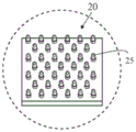
Fig. 5B is a suggestion of integrating a microneedle array-based analyte selective sensor into a patch pump.
Fig. 6A is a suggestion of integrating a microneedle array-based analyte selective sensor into a patch pump.
Fig. 6B is a suggestion of integrating a microneedle array-based analyte selective sensor into a patch pump.
FIG. 6C is an isolated view of circle 6C of FIG. 6B.
Fig. 7 is a block/process flow diagram illustrating the major method steps of an OPEN LOOP (OPEN LOOP) embodiment of the present invention.
Fig. 8 is a block/process flow diagram illustrating the main method steps of a CLOSED LOOP (CLOSED LOOP) embodiment of the present invention.
Fig. 9 is a block/process flow diagram illustrating the input, output and major components of the present invention in an open loop embodiment.
Fig. 10 is a block/process flow diagram illustrating the input, output and major components of the present invention in a closed loop embodiment.
Fig. 11 is a diagram (not to scale) of an infusion system configured to operate within subcutaneous tissue.
Fig. 12A is a graphical representation of graphs relating to insulin depot formation in subcutaneous adipose tissue.
Fig. 12B is a graphical representation of graphs relating to insulin depot formation in subcutaneous adipose tissue.
Fig. 12C is a graphical representation of graphs relating to insulin depot formation in subcutaneous adipose tissue.
Fig. 12D is a graphical representation of graphs relating to insulin depot formation in subcutaneous adipose tissue.
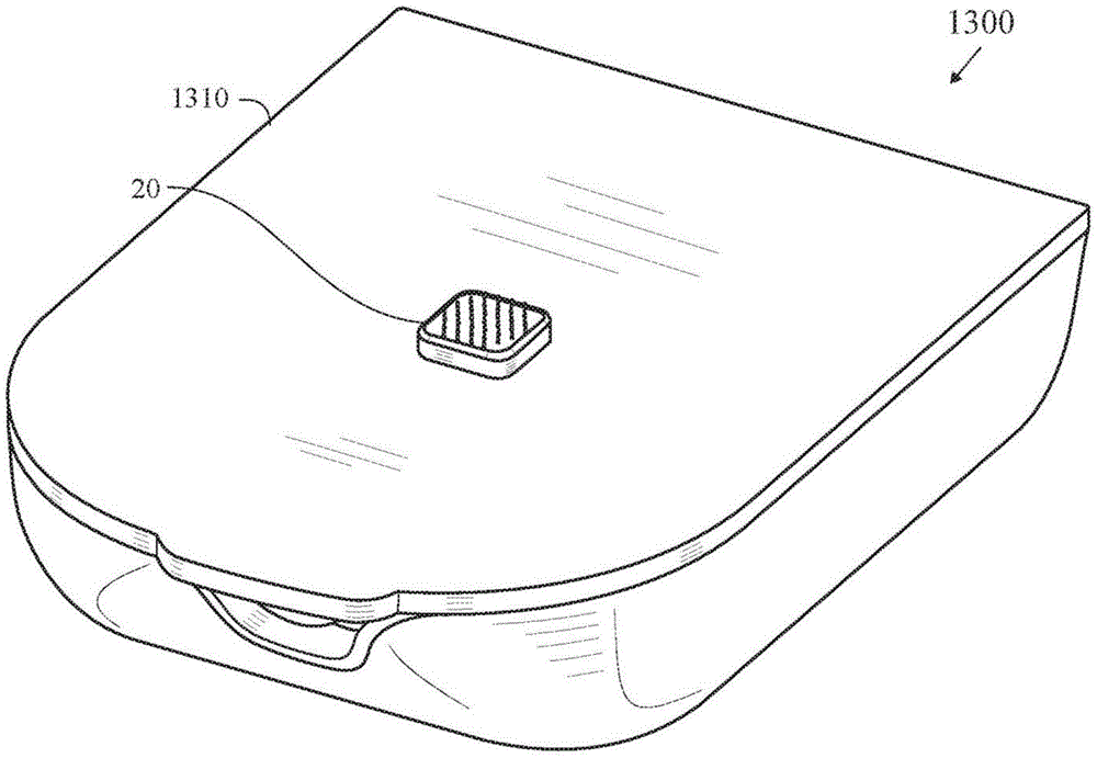
FIG. 13 is a top perspective view of an insulin patch pump with a fully integrated microarray sensor.
FIG. 14 is a bottom perspective view of an insulin patch pump with a fully integrated microarray sensor.
FIG. 15 is a side view of an insulin patch pump with a fully integrated microarray sensor.
FIG. 16 is a bottom perspective view of an insulin patch pump with micro-array sensors connected via a connector.
FIG. 17 is an isolated view of a microarray sensor connected to a connector.
FIG. 18 is a bottom perspective view of an insulin patch pump having a recess for a microarray sensor that has been applied to a patient's skin.
Detailed Description
In recent years, the development of microneedle-based analyte selective sensors has increased and represents a promising capability for minimally invasive quantification of many analytes of interest in physiological fluids (e.g., interstitial fluid, dermal interstitial fluid, blood, serum, and plasma). These devices consist of an array of at least two protrusions on a substrate, each protrusion attached to the substrate at a proximal end and extending between 200 and 2000 microns to a distal end. At least one of the protrusions is configured to feature at least one electrode comprising a metal, semiconductor, or polymer material, which may be further coated with one or more layers of polymer films. In certain embodiments, a recognition element is located on the electrode or within the membrane to impart selective sensing capabilities for endogenous or exogenous chemical substances that occupy the physiological fluid. The chemical may include at least one of a biomarker, a chemical, a biochemical, a metabolite, an electrolyte, an ion, a hormone, a neurotransmitter, a vitamin, a mineral, a drug, a therapeutic, a toxin, an enzyme, a protein, a nucleic acid, DNA, and RNA. The sensing modes may include electrical, chemical, electrochemical (voltammetric, amperometric, potentiometric), optical, fluorescent, colorimetric, absorbance, emission, conductance, impedance, resistance, capacitance. A typical mode of application includes a package that is converted to kinetic energy by a force provided by a user, potential energy stored therein upon a user-initiated actuation motion, and an applicator capable of accelerating the microneedle-based analyte-selective sensor, thereby causing the microneedle assembly of the array to penetrate the stratum corneum and effect sensing within the viable epidermis or dermis to facilitate intradermal analysis of an analyte of interest from a viable physiological medium (interstitial fluid, blood) occupying the viable epidermis and dermis layers. Sensing is achieved in a continuous, quasi-continuous, periodic or single-shot manner. In some embodiments, the sensor device contains a radio configured to relay data, measurements, or readings to connected wireless-enabled devices such as smartphones, smartwatches, and therapy delivery systems. In other embodiments, the device contains at least one electrical contact configured to relay data, measurements, or readings to a mechanically coupled therapy delivery system.
The therapy delivery system is configured to inject a therapy, drug, pharmaceutical, or active ingredient into a physiological compartment of a user. These devices most often contain a reservoir for the treatment, a dispensing mechanism or actuator that controls the amount or dosage of the treatment, a power source (i.e., a battery), and an electronic controller containing an embedded control algorithm programmed into firmware. In some preferred embodiments, these systems feature a radio configured to relay data, measurements, or readings to connected wireless-enabled devices such as smartphones, smartwatches, and continuous analyte monitors. Embodiments of the therapy delivery system include a skin-worn integrated patch pump that integrates both a pump and a cannula for subcutaneous delivery of therapy. In other embodiments, the therapy delivery system contains a non-skin worn pump and a skin-groomed infusion set. Continuous Subcutaneous Insulin Infusion (CSII) and Automatic Insulin Delivery (AID) systems, consisting of an insulin pump, control algorithms and data interfacing methods with a continuous glucose monitor, have been at the forefront of this field development activity due to the potential for closed loop operation aimed at automatically delivering insulin to counteract blood glucose fluctuations and maximize the user's time in normal blood glucose, also known as in-range time.
The control algorithm is designed to regulate the delivery of therapy via the therapy delivery system based on data input provided by the continuous analyte sensor, such that a therapeutic intervention can be automatically delivered, the dosage of which is controlled to counteract the pathophysiological state. While basal rate delivery requires a fixed time rate of therapy delivery, the ability to measure at least one analyte provides an effective feedback method and is therefore suitable for fully autonomous closed-loop therapy. In particular, the analyte sensor may comprise a continuous glucose monitor and the therapy delivery system may constitute a Continuous Subcutaneous Insulin Infusion (CSII) system. The continuous glucose monitor is configured to measure glucose under the skin, and the CSII system includes a pump paired with an infusion set, or otherwise integrated into a skin-adhesive patch (also referred to as a tubeless pump), and configured to deliver a prescribed dose of insulin. A control algorithm resident in the CSII, continuous glucose monitor, or wireless companion device calculates the therapeutic dose required to counteract the pathophysiological state and achieve tight glycemic control, preferably in the normoglycemic range (70-180 mg/dL), based on readings from the continuous glucose monitor. The control algorithm is designed to monitor a controlled process variable (i.e., glucose level through a continuous glucose monitor) and compare it to a reference or set point (i.e., glucose level within the normal blood glucose range). The difference between the actual value and the desired value of the process variable, referred to as the error signal or SP-PV error, is used as feedback to generate a control action to bring the controlled process variable to the same value as the set point. In other words, the main objective of the control algorithm is to minimize the error signal. The system may operate under closed loop control (i.e., control action from the controller relies on feedback from the process in the form of process variable values) or open loop control (i.e., control action from the controller is independent of process output). In various embodiments, no feedback or negative feedback may be employed. The advantage of negative feedback is that an unstable process can be stabilized, sensitivity to parameter changes is reduced, and set point performance is improved. In a preferred embodiment, the control algorithm resides in a memory or processor embedded within the CSII system. Typically, the control algorithm (resident in the processor or memory) receives as input either manually input by the user (such as from a finger stick blood sample) or glucose data flowing from a continuous glucose monitor (typically wireless, but the processor may have a direct electrical connection located in a single device).
An exemplary closed-loop controller architecture is a proportional-integral-derivative (PID) controller. As with other exemplary systems, PID controllers make extensive use of transfer functions, also known as system functions or network functions, which are composed of a mathematical model (i.e., a set of time-or process-dependent equations) of the relationship between system inputs and outputs. Another embodiment of the control algorithm may include at least one of proportional-integral-derivative, model predictive control, fuzzy logic, and safety supervision design (ann. ny acad. sci.2014.4 months: 1311: 102-23).
The present invention teaches devices and methods for sensing one or more analytes using a single body worn device and delivering an accompanying therapeutic intervention or therapeutic interventions in different physiological compartments to avoid problems with cross-talk, interference, contamination and/or dilution that arise when performing two actions in the spatial vicinity. A single body worn device is configured to be easily applied to a wearer's skin and to participate in a sensing routine in the wearer's viable epidermis or dermis. The delivery or infusion of therapeutic interventions is directed to deeper and anatomically separated and distinct layers of subcutaneous adipose tissue. Embodiments may include an open loop system whereby the wearer adjusts the dosage of the therapeutic intervention based on the level of the one or more analytes, and a closed loop system whereby the control algorithm autonomously adjusts the dosage of the one or more therapeutic interventions.
Current needle and cannula based infusion systems configured for delivery of solution phase therapeutics (i.e., insulin) are typically paired with needle and cannula based sensor systems configured for continuous quantification of analytes (i.e., glucose). While such systems operate in unison and are configured to operate in the same physiological compartment (such as the subcutaneous fat layer of tissue), the two systems are not suitable for co-location within a single body worn device. While this is due in part to the insertion-related challenges of physically attaching two cannulae to a single integrated device, the primary challenge is due to the lack of isolation when operating the two systems in close proximity. That is, in the case of simultaneous operation of an analyte sensor apparatus and a therapy delivery apparatus that collectively accommodate a given physiological compartment, undesirable effects may occurThe chemical interaction of inspection; these undesirable effects are cross-talk, interference, contamination and local dilution, which directly affect the ability of the sensor to quantify the desired analyte with a particular degree of selectivity, sensitivity, stability and response time. As an example, liquid formulations of insulin often used in insulin pumps include meta-cresol and methyl paraben as preservatives.1While these two compounds effectively retain insulin activity during long-term storage and after significant temperature fluctuations, these substances are electroactive and interfere with the simultaneous electrochemical detection of glucose. Therefore, in order to accurately measure glucose, the delivery of insulin must be spatially isolated from the sensor; this spatial isolation makes integration of both sensing and therapy modalities impractical for a single body worn device. Moreover, co-locating both analyte sensing and therapy delivery teams into a single body-worn device has presented significant challenges in application due to difficulties associated with the implantation of two cannulas simultaneously or in progress. In summary, integration of both analyte sensors and therapy delivery modalities into a single body-worn device is a strong driver for wearer convenience and simplicity. This is driven by, among other things, the challenges of current automated insulin delivery, which has created considerable promise for more effective management of insulin-dependent diabetes. Today, due to current design implementations, both analyte sensing and therapy delivery modalities are configured to operate in subcutaneous adipose tissue (also referred to as subcutaneous tissue, subdermal tissue, or hypodermal tissue). One key challenge arises if it is desired to co-locate two systems into a single body-worn device — sufficient spatial separation of analyte sensing and therapy delivery modalities so that both can operate in isolation (i.e., neither system is affected by routines executed in the other team). Moreover, although to a lesser extent, co-locating both analyte sensing and therapy delivery systems into a single, body-worn device having an attractive appearance faces certain integration challenges, particularly due to the macroscopic features of both systems and their unique requirements for packaging, control electronics, and hardware.
Body worn analyte sensors, such as continuous glucose monitors, are sensitive electrochemical systems configured to sense one or more analytes in a selective manner with a high degree of accuracy. In many cases, the sensor may be configured to exclude other endogenous analytes from interfering with the detection process, however, perturbations in equilibrium conditions near the sensor (such as those caused by infusion) may cause erroneous readings that do not reflect in situ analyte levels, let alone large amounts of exogenous therapeutic agents may directly interfere with the quantification of the analyte. The present invention addresses the challenge of co-localization of both analyte sensing and therapy delivery modalities in the same on-body device in the same physiological compartment by facilitating separation of analyte sensing and therapy delivery routines in different physiological compartments (skin layers) that are separated laterally rather than laterally. The innovations represent an alternative approach that facilitates the delivery of therapeutic treatments without causing subsequent and undesirable responses in analyte-selective sensors operating in proximity to the therapeutic solution; this is achieved by positioning both the sensing and delivery teams in different physiological compartments, even where both modalities are located near the same lateral space (as within a single body-worn device). Exemplary embodiments of the analyte sensor in the present invention constitute a microneedle or microneedle array configured to sense at least one analyte in a viable epidermal or dermal layer of skin; and a cannula-based infusion set or patch pump configured to deliver at least one of the therapeutic interventions (such as solution phase drugs, pharmacology, biology, or pharmaceuticals) into the subcutaneous adipose tissue layer. In various embodiments, the lateral separation between the two teams (the analyte sensor integrated into the patch pump or cannula-based infusion set) may be in the range of 2mm to 50mm, and most preferably 5mm to 25 mm. It is expected that the two physiological compartments will be sufficiently isolated to mitigate the possible occurrence of cross-talk, interference, contamination and local dilution of the analyte being detected.
Fig. 5A is a diagram of a microneedle array-based analyte selective sensor 20 integrated into an infusion set 500.
Fig. 5B, 6A and 6B illustrate the integration of a microneedle array-based analyte selective sensor 20 into a patch pump 525. Fig. 6C illustrates a microneedle array-based analyte selective sensor 20 and microneedles 25.
The technology disclosed herein juxtaposes an analyte sensor system and a therapy delivery system to operate in different physiological compartments while maintaining minimal spatial separation between the two. This is achieved by dispensing an analyte sensor into the viable epidermis or dermis of the wearer, whereby the system is configured to quantify one or more analytes residing therein. Instead, the therapy delivery system is distributed in the subcutaneous region. The lateral separation of both the sensing and delivery modalities, confining the sensing routine to the viable epidermis or dermis and the delivery routine to the subcutaneous adipose tissue, enables isolation of the two routines, thereby mitigating the potential occurrence of cross-talk, interference, contamination and local dilution of the analyte being detected, should be co-located in a given physiological compartment. In a preferred embodiment of the invention, the system may operate in an open loop paradigm whereby treatment is initiated by the user and guided by measurements from the sensors. Alternatively, the system may feature a control algorithm to autonomously deliver a therapeutic intervention in response to a sensor reading or readings. This paradigm is expected to have profound effects on diabetes management, especially in those undergoing intensive insulin therapy.
Open-loop embodiments of the present invention include systems that integrate a sensor and infusion subsystem and require user action to initiate delivery of a therapeutic intervention. The system is preferably a body worn device capable of incorporating both a sensor and an infusion subsystem to deliver a therapeutic agent in a physically distinct compartment from the area where the analyte is detected. The sensor is preferably a plurality of microneedles having a vertical extent of between 200 and 2000 μm configured to selectively quantify the level of at least one analyte located within the viable epidermis or dermis. Fig. 3 illustrates a micro-needle array sensor 325 associated with a coin 301 and a needle 305. The sensor is designed to measure analytes in a different layer of skin, such as the viable epidermis or dermis. The infusion subsystem is designed to deliver therapeutic agents to different and physically distinct layers of skin, such as subcutaneous adipose tissue. The infusion subsystem is preferably a fluid delivery device configured to infuse a solution-phase therapeutic agent into the subcutaneous adipose layer, circulatory system (vein, artery, or capillary), or muscle tissue via a venous line, hypodermic needle, infusion cannula, or oral delivery route. The therapeutic agent is preferably a solution phase drug, pharmacological, biological or pharmaceutical agent. The sensor may be bonded to the bottom of an insulin infusion cannula set that is adhered to the skin with a medical adhesive and attached to the insulin pump by a hollow plastic tube. Alternatively, the sensor may be bonded to the bottom of an insulin patch pump that is adhered to the skin with a medical adhesive.
Closed-loop embodiments of the present invention include systems that integrate a sensor and infusion subsystem and employ a control algorithm to initiate delivery of a therapeutic intervention. The system is preferably a body worn device with a control algorithm that can incorporate both a sensor and an infusion subsystem to deliver a therapeutic agent in a physically distinct compartment from the area where the analyte is detected. The sensor is preferably a plurality of microneedles having a vertical extent of between 200 and 2000 μm configured to selectively quantify the level of at least one analyte located within the viable epidermis or dermis. The sensor is designed to measure analytes in a different layer of skin, such as the viable epidermis or dermis. The infusion subsystem is designed to deliver therapeutic agents to different and physically distinct layers of skin, such as subcutaneous adipose tissue. The infusion subsystem is preferably a fluid delivery device configured to infuse a solution-phase therapeutic agent into the subcutaneous adipose layer, circulatory system (vein, artery, or capillary), or muscle tissue via a venous line, hypodermic needle, infusion cannula, or oral delivery route. The therapeutic agent is preferably a solution phase drug, pharmacological, biological or pharmaceutical agent. The sensor may be bonded to the bottom of an insulin infusion cannula set that is adhered to the skin with a medical adhesive and attached to the insulin pump by a hollow plastic tube. Alternatively, the sensor may be bonded to the bottom of an insulin patch pump that is adhered to the skin with a medical adhesive. Treatment is defined as the dose distribution of a therapeutic agent in response to an analyte measurement, which can be controlled by an algorithm. The control algorithm is preferably a software or firmware routine that employs one or more mathematical transforms to control the dosage of the therapeutic agent by controlling the amount delivered, the duration of delivery, and/or the frequency of delivery based on input from the user or measurements recorded from the microneedle array analyte selective sensor. The mathematical transform may take additional input, be provided by the user or be autonomously integrated from elsewhere.
Fig. 7 is a block/process flow diagram illustrating the major method steps of an OPEN LOOP (OPEN LOOP) embodiment of the present invention. A method 700 for performing an open loop embodiment begins at block 701 where a microneedle array analyte-selective sensor records measurements of one or more analytes in the viable epidermis or dermis. Circulating levels of viable epidermal or intradermal analytes are quantified by the sensor. Next, at block 702, one or more measurements from the microneedle array analyte selective sensor are displayed to a user. The user receives a reading of the circulating level of the one or more analytes on a display or interface. Alternatively, the user receives notification that the circulating level of the one or more analytes is outside of a predetermined standard or range of values. Next, at block 703, the user adjusts the dosage of one or more therapeutic agents, if necessary. The user manipulates the infusion amount, duration, or frequency of the therapy based on the measurement of the one or more analytes provided by the sensor. Next, at block 704, one or more therapeutic agents are administered into the subcutaneous fat layer, the circulatory system (venous, arterial, or capillary), muscle tissue, or oral delivery route via a therapeutic delivery mechanism. Therapy is delivered to the user via the infusion subsystem and is determined based on the user dose given one or more measurements from the sensor.
Fig. 8 is a block/process flow diagram illustrating the main method steps of a CLOSED LOOP (CLOSED LOOP) embodiment of the present invention. A method 800 for performing a closed loop embodiment begins at block 801 where a microneedle array analyte selective sensor records measurements of one or more analytes in the viable epidermis or dermis. Circulating levels of viable epidermal or intradermal analytes are quantified by the sensor. Next, at block 802, one or more measurements from the microneedle array analyte selective sensor are input into a control algorithm; optionally, the one or more measurements are displayed to a user. The current and optionally past stored measurements are used as one or more inputs to the algorithm. Alternatively, the user also receives a reading of the circulating level of the one or more analytes on a display or interface. Alternatively, the user receives notification that the circulating level of the one or more analytes is outside of a predetermined standard or range of values. Next, at block 803, the control algorithm adjusts the dosage of one or more therapeutic agents, if necessary, based on the programmed mathematical transformation. The algorithm autonomously manipulates the infusion volume, duration, or frequency of the therapy based on the measurement of the one or more analytes provided by the sensor. Next, at block 804, one or more therapeutic agents are administered by a therapeutic delivery mechanism into the subcutaneous fat layer, the circulatory system (venous, arterial, or capillary), muscle tissue, or oral delivery route. The therapy is delivered to the user via an infusion subsystem and is determined based on the dose output by a given algorithm.
The input of circulating levels of one or more analytes within the viable epidermis or dermis is an endogenous or exogenous biochemical, metabolite, drug, pharmacological, biological, or pharmaceutical agent in the viable epidermis or dermis indicative of a particular physiological or metabolic state.
The output is administration of one or more therapeutic agents into the circulatory system (venous, arterial, or capillary), muscle tissue, or oral delivery route. The measurements provided by the sensors are used to initiate the release of therapy by the infusion subsystem. In an open loop embodiment, the delivery of therapy is controlled by the user. In a closed loop embodiment, an algorithm is employed to control the dose, duration and frequency of treatment.
Fig. 9 is a block/process flow diagram 900 illustrating the input, output and major components of the present invention in an open loop embodiment. At block 901, the circulating level of the one or more analytes is within the dermis. At block 902, a sensor measures an analyte. At block 903, the user adjusts the dosage of one or more therapeutic agents, if necessary. The user 903 manipulates the infusion amount, duration, or frequency of the therapy 904 based on the measurement of the one or more analytes provided by the sensor. At block 905, one or more therapeutic agents are administered by a therapeutic delivery mechanism into the subcutaneous fat layer, the circulatory system (venous, arterial, or capillary), muscle tissue, or oral delivery route. Therapy is delivered to the user via the infusion subsystem and is determined based on the user dose given one or more measurements from the sensor.
Fig. 10 is a block/process flow diagram 1000 illustrating the input, output and major components of the present invention in a closed loop embodiment. At block 1001, the circulating level of the one or more analytes is within the dermis. At block 1002, a sensor measures an analyte. The control algorithm 1003 adjusts the dosage of one or more therapeutic agents, if necessary, based on programmed mathematical transformations. The algorithm autonomously manipulates the infusion volume, duration, or frequency of therapy 1004 based on the measurement of one or more analytes provided by the sensor. Next, at block 1005, one or more therapeutic agents are administered by a therapeutic delivery mechanism into the subcutaneous fat layer, the circulatory system (venous, arterial, or capillary), muscle tissue, or oral delivery route. The therapy is delivered to the user via an infusion subsystem and is determined based on the dose output by a given algorithm.
Fig. 13-15 illustrate an insulin patch pump 1300 having a body 1305 with a fully integrated microarray sensor 20 in a bottom surface 1310.
Fig. 16-17 illustrate an alternative embodiment of an insulin patch pump 1300 in which the microarray sensors 20 are connected via a connector 1350 on the bottom surface 1310 of the patch pump 1300.
Fig. 18 illustrates an insulin patch pump 1300 having a recess 1335 in a bottom surface 1310 of the patch pump 1300 for positioning over a microarray sensor (not shown) that has been applied to a patient's skin.