CN113577393A - Bone repair material and preparation method and application thereof - Google Patents
Bone repair material and preparation method and application thereof Download PDFInfo
- Publication number
- CN113577393A CN113577393A CN202110992157.7A CN202110992157A CN113577393A CN 113577393 A CN113577393 A CN 113577393A CN 202110992157 A CN202110992157 A CN 202110992157A CN 113577393 A CN113577393 A CN 113577393A
- Authority
- CN
- China
- Prior art keywords
- bone
- repair material
- decalcified
- matrix
- powder
- Prior art date
- Legal status (The legal status is an assumption and is not a legal conclusion. Google has not performed a legal analysis and makes no representation as to the accuracy of the status listed.)
- Granted
Links
Images


Classifications
-
- A—HUMAN NECESSITIES
- A61—MEDICAL OR VETERINARY SCIENCE; HYGIENE
- A61L—METHODS OR APPARATUS FOR STERILISING MATERIALS OR OBJECTS IN GENERAL; DISINFECTION, STERILISATION OR DEODORISATION OF AIR; CHEMICAL ASPECTS OF BANDAGES, DRESSINGS, ABSORBENT PADS OR SURGICAL ARTICLES; MATERIALS FOR BANDAGES, DRESSINGS, ABSORBENT PADS OR SURGICAL ARTICLES
- A61L27/00—Materials for grafts or prostheses or for coating grafts or prostheses
- A61L27/36—Materials for grafts or prostheses or for coating grafts or prostheses containing ingredients of undetermined constitution or reaction products thereof, e.g. transplant tissue, natural bone, extracellular matrix
- A61L27/3604—Materials for grafts or prostheses or for coating grafts or prostheses containing ingredients of undetermined constitution or reaction products thereof, e.g. transplant tissue, natural bone, extracellular matrix characterised by the human or animal origin of the biological material, e.g. hair, fascia, fish scales, silk, shellac, pericardium, pleura, renal tissue, amniotic membrane, parenchymal tissue, fetal tissue, muscle tissue, fat tissue, enamel
- A61L27/3608—Bone, e.g. demineralised bone matrix [DBM], bone powder
-
- A—HUMAN NECESSITIES
- A61—MEDICAL OR VETERINARY SCIENCE; HYGIENE
- A61L—METHODS OR APPARATUS FOR STERILISING MATERIALS OR OBJECTS IN GENERAL; DISINFECTION, STERILISATION OR DEODORISATION OF AIR; CHEMICAL ASPECTS OF BANDAGES, DRESSINGS, ABSORBENT PADS OR SURGICAL ARTICLES; MATERIALS FOR BANDAGES, DRESSINGS, ABSORBENT PADS OR SURGICAL ARTICLES
- A61L27/00—Materials for grafts or prostheses or for coating grafts or prostheses
- A61L27/14—Macromolecular materials
- A61L27/18—Macromolecular materials obtained otherwise than by reactions only involving carbon-to-carbon unsaturated bonds
-
- A—HUMAN NECESSITIES
- A61—MEDICAL OR VETERINARY SCIENCE; HYGIENE
- A61L—METHODS OR APPARATUS FOR STERILISING MATERIALS OR OBJECTS IN GENERAL; DISINFECTION, STERILISATION OR DEODORISATION OF AIR; CHEMICAL ASPECTS OF BANDAGES, DRESSINGS, ABSORBENT PADS OR SURGICAL ARTICLES; MATERIALS FOR BANDAGES, DRESSINGS, ABSORBENT PADS OR SURGICAL ARTICLES
- A61L27/00—Materials for grafts or prostheses or for coating grafts or prostheses
- A61L27/36—Materials for grafts or prostheses or for coating grafts or prostheses containing ingredients of undetermined constitution or reaction products thereof, e.g. transplant tissue, natural bone, extracellular matrix
- A61L27/3641—Materials for grafts or prostheses or for coating grafts or prostheses containing ingredients of undetermined constitution or reaction products thereof, e.g. transplant tissue, natural bone, extracellular matrix characterised by the site of application in the body
- A61L27/3645—Connective tissue
- A61L27/365—Bones
-
- A—HUMAN NECESSITIES
- A61—MEDICAL OR VETERINARY SCIENCE; HYGIENE
- A61L—METHODS OR APPARATUS FOR STERILISING MATERIALS OR OBJECTS IN GENERAL; DISINFECTION, STERILISATION OR DEODORISATION OF AIR; CHEMICAL ASPECTS OF BANDAGES, DRESSINGS, ABSORBENT PADS OR SURGICAL ARTICLES; MATERIALS FOR BANDAGES, DRESSINGS, ABSORBENT PADS OR SURGICAL ARTICLES
- A61L27/00—Materials for grafts or prostheses or for coating grafts or prostheses
- A61L27/50—Materials characterised by their function or physical properties, e.g. injectable or lubricating compositions, shape-memory materials, surface modified materials
- A61L27/56—Porous materials, e.g. foams or sponges
-
- A—HUMAN NECESSITIES
- A61—MEDICAL OR VETERINARY SCIENCE; HYGIENE
- A61L—METHODS OR APPARATUS FOR STERILISING MATERIALS OR OBJECTS IN GENERAL; DISINFECTION, STERILISATION OR DEODORISATION OF AIR; CHEMICAL ASPECTS OF BANDAGES, DRESSINGS, ABSORBENT PADS OR SURGICAL ARTICLES; MATERIALS FOR BANDAGES, DRESSINGS, ABSORBENT PADS OR SURGICAL ARTICLES
- A61L27/00—Materials for grafts or prostheses or for coating grafts or prostheses
- A61L27/50—Materials characterised by their function or physical properties, e.g. injectable or lubricating compositions, shape-memory materials, surface modified materials
- A61L27/58—Materials at least partially resorbable by the body
-
- B—PERFORMING OPERATIONS; TRANSPORTING
- B33—ADDITIVE MANUFACTURING TECHNOLOGY
- B33Y—ADDITIVE MANUFACTURING, i.e. MANUFACTURING OF THREE-DIMENSIONAL [3-D] OBJECTS BY ADDITIVE DEPOSITION, ADDITIVE AGGLOMERATION OR ADDITIVE LAYERING, e.g. BY 3-D PRINTING, STEREOLITHOGRAPHY OR SELECTIVE LASER SINTERING
- B33Y70/00—Materials specially adapted for additive manufacturing
- B33Y70/10—Composites of different types of material, e.g. mixtures of ceramics and polymers or mixtures of metals and biomaterials
-
- A—HUMAN NECESSITIES
- A61—MEDICAL OR VETERINARY SCIENCE; HYGIENE
- A61L—METHODS OR APPARATUS FOR STERILISING MATERIALS OR OBJECTS IN GENERAL; DISINFECTION, STERILISATION OR DEODORISATION OF AIR; CHEMICAL ASPECTS OF BANDAGES, DRESSINGS, ABSORBENT PADS OR SURGICAL ARTICLES; MATERIALS FOR BANDAGES, DRESSINGS, ABSORBENT PADS OR SURGICAL ARTICLES
- A61L2430/00—Materials or treatment for tissue regeneration
- A61L2430/02—Materials or treatment for tissue regeneration for reconstruction of bones; weight-bearing implants
Landscapes
- Health & Medical Sciences (AREA)
- Chemical & Material Sciences (AREA)
- Life Sciences & Earth Sciences (AREA)
- Engineering & Computer Science (AREA)
- Epidemiology (AREA)
- Oral & Maxillofacial Surgery (AREA)
- Transplantation (AREA)
- Medicinal Chemistry (AREA)
- Dermatology (AREA)
- Animal Behavior & Ethology (AREA)
- General Health & Medical Sciences (AREA)
- Public Health (AREA)
- Veterinary Medicine (AREA)
- Biomedical Technology (AREA)
- Chemical Kinetics & Catalysis (AREA)
- Botany (AREA)
- Orthopedic Medicine & Surgery (AREA)
- Composite Materials (AREA)
- Structural Engineering (AREA)
- Manufacturing & Machinery (AREA)
- Materials Engineering (AREA)
- Civil Engineering (AREA)
- Ceramic Engineering (AREA)
- Vascular Medicine (AREA)
- Dispersion Chemistry (AREA)
- Molecular Biology (AREA)
- Urology & Nephrology (AREA)
- Zoology (AREA)
- Materials For Medical Uses (AREA)
Abstract
The invention discloses a bone repair material, which comprises the raw materials of bone powder, decalcified bone matrix and biodegradable polymer; wherein the mass ratio of the bone meal, the decalcified bone matrix and the biodegradable polymer is 70-75: 5-20: 10-20. The bone repair material has a loose porous three-dimensional structure similar to natural cancellous bone; the bone powder and the decalcified bone matrix have the same source and are uniformly distributed in the bone repair material, so that the bioactivity and biocompatibility of the bone repair material are ensured; meanwhile, the bone repair material has excellent mechanical property, the compression strength can reach 47.3MPa, the compression modulus can reach 0.4GPa, and a growth space and a nutrient substance transmission channel required by cells are provided for bone filling repair. Therefore, the implanted biological scaffold made of the bone repair material has excellent capability of promoting bone repair and regeneration and wide application prospect.
Description
Technical Field
The present invention relates to the field of biomaterials. More particularly, relates to a composite bone repair material and a preparation method and application thereof.
Background
In the selection of bone filling and repairing materials, autologous bone transplantation is known as the 'gold standard' because of the absence of immunological rejection, good biocompatibility and strong bone induction and bone conduction capabilities. On the other hand, the bone supply amount of autologous bones in clinical application is limited, and adverse reaction in a supply area and nerve injury and the like are easy to occur after operation.
The allogeneic bone and the xenogeneic bone have tissue structure and biomechanical property similar to those of the autogenous bone, have certain bone induction and bone conduction capabilities, are fully obtained relative to the autogenous bone, are widely applied clinically, but have the problems of difficult material acquisition, low price and the like. The source of the allogenic bone is rich, but the allogenic bone is not as good as the autologous bone and the allogenic bone in terms of the three-dimensional structure and composition of the bone and the bone repair capability. The artificial bone has the advantages of unlimited sources, capability of being prepared into any shape, convenience in application and the like, is widely researched in recent years, and is different from a natural bone material in that the bone induction, bone conduction capability and biocompatibility of the artificial bone material are inferior to those of the artificial bone material, so that most of the artificial bone material is compounded with bone growth factors or seed cells and the like.
Natural bone tissue is classified into two types, cancellous bone and cortical bone, according to the difference in internal structure. The cancellous bone has a natural loose porous structure, mainly exists in the joint head and the vertebral column cone, has a small proportion, and has good repair and regeneration capacity for repairing various bone defects clinically due to the structural advantages. The cortical bone exists in the parts of the limbs, such as the diaphysis and the like, has larger proportion, has the same components as the cancellous bone and high content of bone active factors, but has very compact structure and very slow degradation, is only used for the auxiliary reinforcement support of certain bone defects clinically, and has very limited application. Therefore, a large amount of cortical bone materials cannot be sufficiently used.
At present, various technologies are used for preparing materials which can be applied to clinic from natural bone powder (mainly from cortical bone) by various processing methods. Compared with traditional tissue engineering porous scaffold preparation methods such as a freeze-drying method, a thermally induced phase separation method, a particle pore-forming method and the like, the 3D printing forming method has the advantage of being capable of being accurately regulated and controlled in aspects such as scaffold appearance, porosity, pore connectivity and the like, and therefore the method is gradually widely researched and applied. Patent CN 107469153A, placing bovine cancellous bone or pig humerus powder in n-butyl alcohol solution, stirring, drying, and mixing with polylactic acid or polylactic acid-glycolic acid copolymer, nano calcium phosphate or calcium phosphate fiber, and toughening agent; 3D printing into the skeleton structure of human or animal body through melting. The content of the bone meal in the formula is 10-20%, the content is low, the beneficial effect of the bone meal is probably not exerted to the maximum, and in addition, the collagen in the bone meal can be denatured by a high-temperature melting printing mode, so that the biological activity of the bone meal and the capability of promoting tissue regeneration and repair are greatly weakened. Therefore, some researchers try to use bone powder as a main formula, mix the bone powder with a binder with good biocompatibility, and print the mixture into a scaffold by 3D printing for bone tissue repair and regeneration. The main body of the stent prepared by the patent CN 106730009A is composed of autologous bone powder and autologous bone glue, a nano calcium phosphate coating is deposited on the surface, and the autologous bone glue is filled in pores of the stent, but the autologous source is very limited, the autologous stent can only be applied by a supplier, and the autologous stent does not have wide popularization and application values. Patent CN 105903078A is prepared by grinding allogeneic bone or xenogeneic bone in a cryomill at low temperature, mixing with collagen and other solutions to obtain a printing material, preparing a scaffold by 3D printing, and performing post-treatment in genipin solution to improve the mechanical properties of the scaffold. However, the applicant finds that the scaffold prepared by using water-soluble natural polymers similar to collagen and the like as the adhesive has generally poor mechanical strength, so that the scaffold is degraded and collapsed too fast in tissue repair and regeneration, and cannot provide sufficient growth space and nutrient transmission channels for cells at the bone defect.
Therefore, there is a need to provide a bone repair material having both a sufficient bone meal content to satisfy bioactivity and excellent mechanical strength.
Disclosure of Invention
The invention aims to provide a bone repair material, which has high bone powder content and can ensure the bioactivity and biocompatibility of the bone repair material by matching with a Decalcified Bone Matrix (DBM) with the same bone tissue source; the biodegradable polymer improves the mechanical properties of the bone repair material.
The invention also aims to provide a preparation method of the bone repair material, which utilizes 3D low-temperature deposition printing to prepare a loose porous three-dimensional net rack similar to the structure of human cancellous bone, and better retains the bioactivity of the bone repair material.
It is a further object of the present invention to provide a use of the bone repair material.
In order to achieve the purpose, the invention adopts the following technical scheme:
a bone repair material comprises bone powder, Decalcified Bone Matrix (DBM) and biodegradable polymer;
wherein the mass ratio of the bone meal, the Decalcified Bone Matrix (DBM) and the biodegradable polymer is 65-75: 5-25: 10-20;
the bone meal and the Decalcified Bone Matrix (DBM) are from the same source.
Preferably, the mass ratio of the bone meal, the Decalcified Bone Matrix (DBM) and the biodegradable polymer is 70-75: 5-20: 10-20.
The bone repair material takes natural bone tissues as a main material, the content (bone powder and Decalcified Bone Matrix (DBM)) of the bone repair material is as high as 80-90 percent, the pore communication rate is 100 percent, the bioactivity of the bone repair material can be ensured, and the bone repair material has excellent bone induction and bone conduction capacities; adding a biodegradable polymer with good biocompatibility to bond and form the bone powder, so that the prepared bone repair material has mechanical strength superior to that of a bone repair material taking water-soluble natural macromolecules such as collagen and the like as a bonding agent, and can provide a growth space and a nutrient substance transmission channel required by cells in bone filling repair; the added Decalcified Bone Matrix (DBM) is dispersed in the bone repair material in the form of particles, has osteoinductive activity, is continuously released in the degradation process of the bone repair material in vivo, and improves the biocompatibility of a microenvironment around the stent.
The bone powder and the Decalcified Bone Matrix (DBM) have the same source, so that the biocompatibility and the bioactivity of the bone repair material are further improved.
Preferably, the bone meal and the Demineralized Bone Matrix (DBM) are both from allogeneic bone, porcine bone or bovine bone;
preferably, the bone meal and the Demineralized Bone Matrix (DBM) are both from allogeneic cortical bone.
Preferably, the bone repair material has exposed bone powder and Demineralized Bone Matrix (DBM) on the surface. The exposed bone powder enables the surface of the bone repair material to have certain roughness, which is beneficial to the attachment and aggregation of cells, the rapid osteoclast and osteogenesis process and the regeneration of bone tissues.
Preferably, the particle size of the bone powder and the Decalcified Bone Matrix (DBM) is 10-75 μm; preferably, the particle size of the bone powder and the Decalcified Bone Matrix (DBM) is 10-50 μm.
The purpose of selecting bone meal with the particle size less than 75 mu m is to realize 3D printing, and if the particle size of the bone meal is too large, a printing needle head is easy to block. Secondly, the smaller the particle size of the bone powder is, the more easily the bone powder is absorbed and degraded and plays a role in bone formation, the natural bone material is selected in the technical scheme, and the bone powder with the minimum particle size range which can be prepared by a grinding method is 10-75 microns.
Preferably, the biodegradable polymer includes, but is not limited to, poly (glycolide-co-lactide) (PLGA), Polycaprolactone (PCL), Polylactide (PLA), Polyhydroxyalkanoate (PHA), and the like.
Further preferably, the lactide content of the poly (glycolide) is 75%, such as PLGA7525, the weight average molecular weight is 10-30 ten thousand g/mol, and the ratio of the mass of the poly (glycolide) to the volume of the 1, 4-dioxane in the 3D printing material is 0.1-0.3 g/mL.
Further preferably, the weight average molecular weight of the polycaprolactone is 4-6 ten thousand g/mol, and the ratio of the mass of the polycaprolactone to the volume of the 1, 4-dioxane in the 3D printing material is 0.2-0.4 g/mL.
Preferably, the bone powder and the Decalcified Bone Matrix (DBM) added to the bone repair material of the present invention are prepared as follows:
bone meal: grinding cortical bone from allogeneic bone or allogeneic bone into powder, degreasing in 10% hydrogen peroxide for 72 hours, cleaning with purified water, and inactivating virus with 75% ethanol for 2 hours; chloroform: removing impure protein from bone powder with methanol mixed solution (volume ratio 3: 1) for 4 hr, alternately cleaning with 75% ethanol and distilled water, removing chloroform and methanol, and lyophilizing at 10Pa for 16 hr to obtain partially deproteinized cortical bone powder containing natural bone hydroxyapatite and collagen as main ingredients.
Demineralized Bone Matrix (DBM): grinding the bone blocks into powder, degreasing, inactivating viruses, removing foreign proteins, decalcifying, cleaning with purified water until pH is neutral, and freeze-drying under low pressure to obtain decalcified bone matrix.
One possible implementation of a Demineralized Bone Matrix (DBM) is: grinding cortical bone from allogeneic bone or allogeneic bone into powder, degreasing in 10% hydrogen peroxide for 72 hours, cleaning with purified water, and inactivating virus with 75% ethanol for 2 hours; chloroform: removing impure protein from bone powder with methanol mixed solution (volume ratio of 3: 1) for 4 hr, alternately cleaning with 75% ethanol and distilled water, removing chloroform and methanol, decalcifying with 0.6M hydrochloric acid at 4 deg.C for 24 hr, cleaning with purified water until pH is neutral, and lyophilizing at 10Pa for 16 hr to obtain Decalcified Bone Matrix (DBM) containing collagen as main component and trace natural bone active component.
The invention also provides a preparation method of the bone repair material, which comprises the following steps:
s1, preparing bone powder and a Decalcified Bone Matrix (DBM), dissolving the bone powder, the Decalcified Bone Matrix (DBM) and a biodegradable polymer in 1, 4-dioxane, and uniformly mixing to obtain a 3D printing material;
and S2, performing low-temperature deposition printing by using 3D printing equipment, after printing is completed, freeze-drying, soaking in dichloromethane, ultrasonically cleaning, and sterilizing to obtain the bone repair material.
Preferably, the parameters of the 3D printing device are set as: the temperature of the spray head is 16.5-18 ℃, the diameter of the spray head is 200-600 mu m, the temperature of the platform is-30 ℃, and the distance between the filaments is 0.5-1.5 mm;
the preparation process of the bone repair material provided by the invention adopts a low-temperature deposition 3D printing technology, and compared with a high-temperature environment, the biological activity of the bone repair material is better kept; meanwhile, the 3D printing technology enables the bone repair material to have good processing performance and adjustability of macro and micro appearance.
Due to the fact that the 1, 4-dioxane completely dissolves the degradable polymer and does not dissolve the bone meal and the Decalcified Bone Matrix (DBM), after the 3D printing material is subjected to freeze drying to remove the solvent, the degradable polymer with a certain thickness is formed on the surface of the material and the surface of macro pores inside the material, and the bone meal and the Decalcified Bone Matrix (DBM) are completely wrapped. By controlling the time and temperature of soaking in dichloromethane, the polymers on the surface layer of the pores are removed, so that the bone powder and the Decalcified Bone Matrix (DBM) are partially exposed on the surface of the material and the surface of the internal pores, and the bone repair effect can be rapidly exerted after implantation.
The soaking process in the dichloromethane specifically comprises the following steps: and (3) soaking the dried material in dichloromethane pre-cooled to-10 to-20 ℃ for 10-15 min, vacuumizing to enable dichloromethane to penetrate from the surface of the material to the inside, centrifuging to remove redundant dichloromethane, and drying in a vacuum oven to completely remove dichloromethane.
The ultrasonic cleaning process may be repeated several times of immersion ultrasonic cleaning using absolute ethanol and purified water to remove the residual dichloromethane solvent.
The gamma ray has no harmful substance residue and can be used as a sterilization mode, but the gamma ray can reduce the mechanical property of the material, the radiation dose needs to be strictly controlled within the range of 25-30 kGy, and the influence on the support mechanics is reduced.
The invention also provides the application of the bone repair material in the aspect of manufacturing an implant biological scaffold. The bone repair material has good bioactivity and mechanical property, and has good application prospect in the aspect of manufacturing an implanted biological scaffold.
The invention has the following beneficial effects:
the bone repair material is prepared by taking bone powder as a main body, taking a biodegradable polymer as a binder, adding Decalcified Bone Matrix (DBM) with the same source as the bone powder, and adopting a 3D low-temperature deposition printing technology in the preparation process, wherein the obtained bone repair material has a loose porous three-dimensional structure similar to a cancellous bone block, the porosity is about 80%, and the through-hole rate is 100%; the bone powder and the Decalcified Bone Matrix (DBM) are uniformly distributed in the bone repair material and are embedded on the surface of the material and the surface of internal pores, so that the bioactivity and biocompatibility of the bone repair material are ensured; the mechanical property of the bone repair material is improved, the compressive strength can reach 47.3MPa, the compressive modulus can reach 0.4GPa, and a growth space and a nutrient transmission channel required by cells are provided for bone filling repair. Therefore, the implanted biological scaffold made of the bone repair material has excellent capability of promoting bone repair and regeneration and wide application prospect.
Drawings
The following describes embodiments of the present invention in further detail with reference to the accompanying drawings.
Fig. 1 shows a photograph of a cubic-shaped bone repair scaffold in test example 1.
Fig. 2 shows a photograph of a cubic-shaped bone repair scaffold in test example 1 at 25-fold magnification.
Fig. 3 shows a 2000-fold magnified photograph of a cubical bone repair scaffold in test example 1.
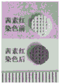
Fig. 4 shows the alizarin red staining result of the cylindrical bone repair scaffold in test example 2.
Fig. 5 shows photographs of in vitro degradation of the cylindrical bone repair scaffold in test example 3.
Fig. 6 shows a scanning electron microscope photograph of the cylindrical bone repair scaffold in test example 3 after 8 weeks of in vitro degradation.
Fig. 7 shows the results of the tissue section of the in vivo animal test in test example 4.
Detailed Description
In order to more clearly illustrate the invention, the invention is further described below with reference to preferred embodiments and the accompanying drawings. Similar parts in the figures are denoted by the same reference numerals. It is to be understood by persons skilled in the art that the following detailed description is illustrative and not restrictive, and is not to be taken as limiting the scope of the invention.
Example 1
The process for preparing the bone repair material is as follows:
(1) preparing bone meal: immersing the allogeneic cortical bone into liquid nitrogen, taking out, putting into a grinder for grinding, grinding into powder, degreasing in 10% hydrogen peroxide for 72 hours, cleaning with purified water, and inactivating viruses with 75% ethanol for 2 hours; chloroform: removing impure protein from bone powder with methanol mixed solution (volume ratio 3: 1) for 4 hr, alternately cleaning with 75% ethanol and distilled water, removing chloroform and methanol, freeze drying under 10pa for 16 hr, and sieving with sieve to obtain allogeneic cortical bone powder with particle size of 10-75 μm.
(2) Demineralized Bone Matrix (DBM): grinding the allogeneic cortical bone into powder, degreasing in 10% hydrogen peroxide for 72 hours, cleaning with purified water, and inactivating viruses with 75% ethanol for 2 hours; chloroform: removing impure protein in bone powder with methanol mixed solution (volume ratio 3: 1) for 4 hr, alternately cleaning with 75% ethanol and distilled water, removing chloroform and methanol, decalcifying with 0.6M hydrochloric acid at 4 deg.C for 24 hr, cleaning with purified water until pH is neutral, freeze drying at 10Pa for 16 hr, and sieving with sieve to obtain Decalcified Bone Matrix (DBM) with particle size of 10-75 μ M.
(3) 1, 4-dioxane is used as a solvent, allogeneic cortical bone powder, allogeneic Decalcified Bone Matrix (DBM), PLGA7525 (lactide content is 75 mol%, weight average molecular weight is 10 ten thousand g/mol) are fully and uniformly mixed according to the proportion of 70%, 10% and 20% by mass percent at room temperature, and the ratio of the PLGA mass to the volume of the 1, 4-dioxane is 0.1g/mL, so that the 3D printing material is obtained.
(4) Adding the 3D printing material into a low-temperature deposition 3D printer, and inputting 3D printing parameters as follows: the temperature of the jet was 18 ℃, the diameter of the jet 400 microns, the platform temperature-30 ℃, the filament spacing 0.8mm, and printing of the material was started.
(5) And (5) drying the printed material in the step (4) in a freeze dryer for 3 days.
(6) And (3) soaking the material obtained in the step (5) in pre-cooled dichloromethane at the temperature of minus 20 ℃ for 10min, vacuumizing to enable dichloromethane to enter the material, then moving the material into a centrifuge, centrifuging at 1000rpm for 3min to remove redundant dichloromethane solvent inside and outside the material, and then drying in a vacuum oven at the temperature of 37 ℃.
(7) And (4) soaking and ultrasonically cleaning the stent obtained in the step (6) for 3 times by using absolute ethyl alcohol, repeatedly soaking and ultrasonically cleaning the stent for 5 times by using purified water, and freeze-drying the stent for 2 days.
(8) And (5) sealing and packaging, and sterilizing by using 25kGy gamma ray to obtain the bone repair material.
Test example 1
A bone repair scaffold having a specific shape was prepared according to the preparation method provided in example 1, and the material of the bone repair scaffold was the bone repair material in example 1.
And performing characterization and performance tests on the bone repair scaffold, wherein the characterization and performance tests comprise a pore structure, mechanical properties, allogenic cortical bone powder distribution in the scaffold, an in-vitro degradation experiment, an in-vivo animal experiment and the like.
The prepared 3D printing cubic allogeneic bone repair scaffold is observed to have regular structure through naked eyes (see figure 1), and the scanning electron microscope is used for observing the surface morphology (see figures 2 and 3), so that the average diameter of macropore pores is 380 mu m, and part of bone powder is exposed on the surface of the scaffold. The porosity of the scaffold was measured to be about 80% by liquid displacement method using absolute ethanol as the displacement liquid. And (3) detecting the connectivity of the bracket holes by micro-CT imaging, wherein the connectivity is 100%. The stent measured a compressive strength of 47.3MPa and a compressive modulus of 0.4 GPa.
Test example 2
Calcium salt in the allogeneic cortical bone powder can generate orange red deposition through alizarin red staining, and the bone powder distribution in the bracket can be observed through the method. Alizarin red staining results (see fig. 4) on cylindrical bone repair scaffolds of 5mm diameter x 1mm height showed that all parts of the scaffold were uniformly stained orange-red, demonstrating that a high proportion of bone meal did not settle during scaffold formation and was uniformly distributed in the scaffold.
Test example 3
In vitro degradation experiments: cylindrical 3D-printed allogeneic bone repair scaffolds 5mm in diameter x 1mm in height were prepared as in example 1, placed in a closed container with sterile PBS to allow complete liquid immersion of the material, incubated with shaking at 37 ℃, and replaced with PBS solution once a day. Taking out the scaffold at 4 th week and 8 th week, repeatedly washing with purified water, soaking, and drying in vacuum drying oven at 40 deg.C for 72 hr.
The result of the macroscopic photograph of the stent (see fig. 5) shows that the stent can still maintain the basic macroscopic three-dimensional structure and the macroporous pores at the 4 th week of degradation, the degradation speed of the stent is gradually accelerated and the mechanical property is gradually weakened at the 8 th week of degradation, and the result of a scanning electron microscope (see fig. 6) shows that the stent is obviously degraded.
Test example 4
In vivo animal experiments:
group A: preparing a cylindrical 3D-printed allograft bone repair scaffold 5mm in diameter × 1mm in height as an experimental group according to the method of example 1; group B: a bone repair scaffold not subjected to the surface treatment operation of step 6 of example 1;
female 5-6 week old SD rats were used as experimental animals, defects of 5mm in diameter were constructed in the skull, and stents were implanted into the defects, and the animals were sacrificed after 2, 4, 8, 12, and 26 weeks.
Observation shows that at 26 weeks, animals have no obvious adverse reaction, no obvious inflammatory reaction at the defect, and tissue section HE staining (see figure 7), and the result shows that more new bone-like tissues are generated in the A group of scaffolds, and the area of the new bone-like tissues is significantly different (P is less than 0.5) compared with the B group. In addition, it was found that less scaffold material remained in the defects of group A than in group B, which provided more adequate growth space for the newly formed bone tissue and did not impede the growth of the newly formed bone tissue. A layer of biodegradable polymer is distributed on the surface of the bone repair bracket in the group B, so that the compatibility of the bone repair bracket and surrounding tissues is not facilitated, and the growth of bone-like tissues is influenced.
Example 2
The process for preparing the bone repair material is as follows:
(1) bone meal: immersing the allogeneic cortical bone into liquid nitrogen, taking out, putting into a grinder for grinding, grinding into bone powder, degreasing in 10% hydrogen peroxide for 72 hours, cleaning with purified water, and inactivating viruses with 75% ethanol for 2 hours; chloroform: removing impure protein from bone powder with methanol mixed solution (volume ratio 3: 1) for 4 hr, alternately cleaning with 75% ethanol and distilled water, removing chloroform and methanol, freeze drying under 10pa for 16 hr, and sieving with sieve to obtain allogeneic cortical bone powder with particle size of 10-75 μm.
(2) Demineralized Bone Matrix (DBM): grinding allogenic cortical bone into bone powder, degreasing in 10% hydrogen peroxide for 72 hours, cleaning with purified water, and inactivating virus with 75% ethanol for 2 hours; chloroform: removing impure protein in bone powder with methanol mixed solution (volume ratio 3: 1) for 4 hr, alternately cleaning with 75% ethanol and distilled water, removing chloroform and methanol, decalcifying with 0.6M hydrochloric acid at 4 deg.C for 24 hr, cleaning with purified water until pH is neutral, freeze drying at 10Pa for 16 hr, and sieving with sieve to obtain Decalcified Bone Matrix (DBM) with particle size of 10-75 μ M.
(3) Taking 1, 4-dioxane as a solvent, and fully and uniformly mixing allogenic cortical bone powder, PCL (molecular weight of 4.5 ten thousand g/mol) and allogenic Decalcified Bone Matrix (DBM) according to the mass percentages of 70%, 10% and 20% at room temperature, wherein the volume ratio of the mass of the PCL to the volume of the 1, 4-dioxane is 0.3g/mL, so as to obtain a 3D printing material;
(4) adding the 3D printing material into a low-temperature deposition 3D printer, and inputting 3D printing parameters as follows: the temperature of the spray head is 18 ℃, the diameter of the spray head is 600 microns, the temperature of the platform is-30 ℃, the distance between wires is 1.2mm, and the printing of the material is started;
(5) putting the printed material in the step (4) into a freeze dryer for drying for 3 days;
(6) soaking the material obtained in the step (5) in pre-cooled dichloromethane at-20 ℃ for 15min, vacuumizing to enable dichloromethane to enter the inside of the bracket, then moving the bracket into a centrifuge, centrifuging at 1000rpm for 3min to remove redundant dichloromethane solvent inside and outside the material, and then drying in a vacuum oven at 37 ℃;
(7) soaking the material obtained in the step (6) in absolute ethyl alcohol, ultrasonically cleaning for 3 times, repeatedly soaking in purified water, ultrasonically cleaning for 5 times, and freeze-drying for 2 days;
(8) sealed, packed and sterilized by 25kGy gamma ray.
The stent measured 14.4MPa compressive strength and 0.12GPa compressive modulus.
It should be understood that the above-mentioned embodiments of the present invention are only examples for clearly illustrating the present invention, and are not intended to limit the embodiments of the present invention, and it will be obvious to those skilled in the art that other variations or modifications may be made on the basis of the above description, and all embodiments may not be exhaustive, and all obvious variations or modifications may be included within the scope of the present invention.
Claims (10)
1. The bone repair material is characterized in that the raw materials comprise bone powder, decalcified bone matrix and biodegradable polymer;
wherein the mass ratio of the bone meal, the decalcified bone matrix and the biodegradable polymer is 70-75: 5-20: 10-20;
the bone powder and the decalcified bone matrix are from the same source.
2. The bone repair material according to claim 1, wherein the bone meal and the decalcified bone matrix are both from allogeneic, porcine or bovine bone;
preferably, the bone meal and the decalcified bone matrix are both from allogeneic cortical bone.
3. The bone repair material of claim 1 wherein the bone repair material has exposed bone powder and demineralized bone matrix on a surface thereof.
4. The bone repair material according to claim 1, wherein the particle size of the bone powder and the decalcified bone matrix is 10 to 75 μm; preferably, the particle size of the bone powder and the decalcified bone matrix is 10-50 μm.
5. Bone repair material according to claim 1, wherein the biodegradable polymer is selected from one or several of the group consisting of poly (glycolide), poly (caprolactone), poly (lactide), poly (hydroxy fatty acid esters).
6. The bone repair material according to claim 1, wherein the decalcified bone matrix is prepared as follows:
grinding the bone blocks into powder, degreasing, inactivating viruses, removing foreign proteins, decalcifying, cleaning with purified water until pH is neutral, and freeze-drying under low pressure to obtain decalcified bone matrix.
7. A method of preparing a bone repair material according to any of claims 1 to 6 comprising the steps of:
s1, preparing bone meal and decalcified bone matrix, dissolving the bone meal, the decalcified bone matrix and a biodegradable polymer in 1, 4-dioxane, and uniformly mixing to obtain a 3D printing material;
and S2, performing low-temperature deposition printing by using 3D printing equipment, after printing is completed, freeze-drying, soaking in dichloromethane, ultrasonically cleaning, and sterilizing to obtain the bone repair material.
8. The manufacturing method according to claim 7, wherein the parameters of the 3D printing apparatus are set to: the temperature of the spray head is 16.5-18 ℃, the diameter of the spray head is 200-600 mu m, the temperature of the platform is-30 ℃, and the distance between the filaments is 0.5-1.5 mm.
9. The preparation method of claim 7, wherein the soaking in dichloromethane in S2 is pre-cooled to-10 to-20 ℃;
preferably, the sterilization mode is gamma rays, and the irradiation dose of the gamma rays is controlled to be 25-30 kGy.
10. Use of a bone repair material according to any one of claims 1 to 6 for the manufacture of an implant bioscaffold.
Priority Applications (1)
| Application Number | Priority Date | Filing Date | Title |
|---|---|---|---|
| CN202110992157.7A CN113577393B (en) | 2021-08-27 | 2021-08-27 | Bone repair material and preparation method and application thereof |
Applications Claiming Priority (1)
| Application Number | Priority Date | Filing Date | Title |
|---|---|---|---|
| CN202110992157.7A CN113577393B (en) | 2021-08-27 | 2021-08-27 | Bone repair material and preparation method and application thereof |
Publications (2)
| Publication Number | Publication Date |
|---|---|
| CN113577393A true CN113577393A (en) | 2021-11-02 |
| CN113577393B CN113577393B (en) | 2023-02-10 |
Family
ID=78239932
Family Applications (1)
| Application Number | Title | Priority Date | Filing Date |
|---|---|---|---|
| CN202110992157.7A Active CN113577393B (en) | 2021-08-27 | 2021-08-27 | Bone repair material and preparation method and application thereof |
Country Status (1)
| Country | Link |
|---|---|
| CN (1) | CN113577393B (en) |
Cited By (2)
| Publication number | Priority date | Publication date | Assignee | Title |
|---|---|---|---|---|
| CN115887771A (en) * | 2022-11-23 | 2023-04-04 | 深圳市光明创博生物制品发展有限公司 | Decalcification method of bone repair material |
| CN118079096A (en) * | 2024-02-28 | 2024-05-28 | 杉纳(秦皇岛)科技有限公司 | Implant material and artificial prosthesis |
Citations (9)
| Publication number | Priority date | Publication date | Assignee | Title |
|---|---|---|---|---|
| CN1403167A (en) * | 2002-09-26 | 2003-03-19 | 东南大学 | Hard tissue repairing material and its prepn |
| US20030065400A1 (en) * | 2001-04-12 | 2003-04-03 | Beam Heather Ann | Method and apparatus for engineered regenrative biostructures such as hydroxyapatite substrates for bone healing applications |
| KR20130037324A (en) * | 2011-10-06 | 2013-04-16 | 주식회사 본셀바이오텍 | Composition and method for 3d printing stacking of scaffold adequate to multi-material for bone regenerating |
| CN105903078A (en) * | 2016-05-18 | 2016-08-31 | 中国人民解放军第三军医大学第三附属医院 | Method for preparing biological scaffold by 3D printing |
| CN108310465A (en) * | 2018-03-21 | 2018-07-24 | 温州生物材料与工程研究所 | A kind of 3D printing bone renovating material and its method for preparing bone repairing support |
| US20180296343A1 (en) * | 2017-04-18 | 2018-10-18 | Warsaw Orthopedic, Inc. | 3-d printing of porous implants |
| CN110665057A (en) * | 2019-11-07 | 2020-01-10 | 上海健康医学院 | A kind of preparation method of hydroxyapatite/PLGA double-layer scaffold |
| CN110743036A (en) * | 2019-10-28 | 2020-02-04 | 上海交通大学医学院附属第九人民医院 | Gallium-containing polycaprolactone/bioglass porous bone repair 3D printing support and application thereof in infectious bone defect repair |
| CN113274551A (en) * | 2021-05-08 | 2021-08-20 | 赵赫凯 | Absorbable bone cement regeneration repair material |
-
2021
- 2021-08-27 CN CN202110992157.7A patent/CN113577393B/en active Active
Patent Citations (10)
| Publication number | Priority date | Publication date | Assignee | Title |
|---|---|---|---|---|
| US20030065400A1 (en) * | 2001-04-12 | 2003-04-03 | Beam Heather Ann | Method and apparatus for engineered regenrative biostructures such as hydroxyapatite substrates for bone healing applications |
| CN1403167A (en) * | 2002-09-26 | 2003-03-19 | 东南大学 | Hard tissue repairing material and its prepn |
| KR20130037324A (en) * | 2011-10-06 | 2013-04-16 | 주식회사 본셀바이오텍 | Composition and method for 3d printing stacking of scaffold adequate to multi-material for bone regenerating |
| CN105903078A (en) * | 2016-05-18 | 2016-08-31 | 中国人民解放军第三军医大学第三附属医院 | Method for preparing biological scaffold by 3D printing |
| US20180296343A1 (en) * | 2017-04-18 | 2018-10-18 | Warsaw Orthopedic, Inc. | 3-d printing of porous implants |
| CN108724712A (en) * | 2017-04-18 | 2018-11-02 | 华沙整形外科股份有限公司 | The 3D printing of porous implant |
| CN108310465A (en) * | 2018-03-21 | 2018-07-24 | 温州生物材料与工程研究所 | A kind of 3D printing bone renovating material and its method for preparing bone repairing support |
| CN110743036A (en) * | 2019-10-28 | 2020-02-04 | 上海交通大学医学院附属第九人民医院 | Gallium-containing polycaprolactone/bioglass porous bone repair 3D printing support and application thereof in infectious bone defect repair |
| CN110665057A (en) * | 2019-11-07 | 2020-01-10 | 上海健康医学院 | A kind of preparation method of hydroxyapatite/PLGA double-layer scaffold |
| CN113274551A (en) * | 2021-05-08 | 2021-08-20 | 赵赫凯 | Absorbable bone cement regeneration repair material |
Non-Patent Citations (2)
| Title |
|---|
| 刘冬 等: "3D打印羟基磷灰石/聚乳酸网状复合物修复颅骨缺损", 《中国组织工程研究》 * |
| 徐达传: "《实用临床骨缺损修复应用解剖学》", 31 January 2000 * |
Cited By (2)
| Publication number | Priority date | Publication date | Assignee | Title |
|---|---|---|---|---|
| CN115887771A (en) * | 2022-11-23 | 2023-04-04 | 深圳市光明创博生物制品发展有限公司 | Decalcification method of bone repair material |
| CN118079096A (en) * | 2024-02-28 | 2024-05-28 | 杉纳(秦皇岛)科技有限公司 | Implant material and artificial prosthesis |
Also Published As
| Publication number | Publication date |
|---|---|
| CN113577393B (en) | 2023-02-10 |
Similar Documents
| Publication | Publication Date | Title |
|---|---|---|
| CN103055352B (en) | Calcium phosphate/collagen composite biologic ceramic material and preparation method thereof | |
| EP3532117B1 (en) | Preparation and applications of 3d bioprinting bioinks for repair of bone defects, based on cellulose nanofibrils hydrogels with natural or synthetic calcium phosphate particles | |
| Schieker et al. | Biomaterials as scaffold for bone tissue engineering | |
| US9925301B2 (en) | Methods of producing and using silk microfibers | |
| EP2517738B1 (en) | A collagen/hydroxyapatite composite scaffold | |
| CN105688274B (en) | A kind of preparation process of polycaprolactone/gelatin electrospinning compound rest | |
| CN101564553B (en) | Humanization active forging bone and preparation method thereof | |
| CN101020082B (en) | Bone repairing material and its prepn process and use | |
| WO2018078130A1 (en) | Preparation and applications of 3d bioprinting bioinks for repair of bone defects, based on cellulose nanofibrils hydrogels with natural or synthetic calcium phosphate particles | |
| Li et al. | A bone regeneration strategy via dual delivery of demineralized bone matrix powder and hypoxia-pretreated bone marrow stromal cells using an injectable self-healing hydrogel | |
| Liu et al. | Development of biodegradable scaffolds for tissue engineering: a perspective on emerging technology | |
| Dou et al. | A hierarchical scaffold with a highly pore-interconnective 3D printed PLGA/n-HA framework and an extracellular matrix like gelatin network filler for bone regeneration | |
| CN104353124B (en) | A 3D printed porous metal scaffold of a composite magnetic nanomaterial and its preparation method | |
| Gundu et al. | Recent developments of biomaterial scaffolds and regenerative approaches for craniomaxillofacial bone tissue engineering | |
| CN113577393B (en) | Bone repair material and preparation method and application thereof | |
| Chen et al. | Nanohydroxyapatite/cellulose nanocrystals/silk fibroin ternary scaffolds for rat calvarial defect regeneration | |
| Rahimnejad et al. | Biomaterials in bone and mineralized tissue engineering using 3D printing and bioprinting technologies | |
| Li et al. | Synthesis and evaluation of BMMSC-seeded BMP-6/nHAG/GMS scaffolds for bone regeneration | |
| US9833543B2 (en) | Implantable scaffolds and methods of use | |
| Liu et al. | Construction and osteogenic effects of 3D-printed porous titanium alloy loaded with VEGF/BMP-2 shell-core microspheres in a sustained-release system | |
| Zhang et al. | Advancing collagen-based biomaterials for oral and craniofacial tissue regeneration | |
| JP2011189052A (en) | Calcium phosphate/biodegradable polymer hybrid material, method for producing same, and implant using the hybrid material | |
| CN114558170B (en) | Growth factor-containing skull repair polyether-ether-ketone material and preparation method thereof | |
| CN113577392A (en) | Artificial periosteum of composite acellular periosteum matrix and preparation method and application thereof | |
| Liu et al. | Improvement of mechanical properties of zein porous scaffold by quenching/electrospun fiber reinforcement |
Legal Events
| Date | Code | Title | Description |
|---|---|---|---|
| PB01 | Publication | ||
| PB01 | Publication | ||
| SE01 | Entry into force of request for substantive examination | ||
| SE01 | Entry into force of request for substantive examination | ||
| GR01 | Patent grant | ||
| GR01 | Patent grant |