CN113555090A - Method and system for constructing scanning knowledge base of image equipment - Google Patents
Method and system for constructing scanning knowledge base of image equipment Download PDFInfo
- Publication number
- CN113555090A CN113555090A CN202110822356.3A CN202110822356A CN113555090A CN 113555090 A CN113555090 A CN 113555090A CN 202110822356 A CN202110822356 A CN 202110822356A CN 113555090 A CN113555090 A CN 113555090A
- Authority
- CN
- China
- Prior art keywords
- scanning
- knowledge base
- module
- branch
- personalized
- Prior art date
- Legal status (The legal status is an assumption and is not a legal conclusion. Google has not performed a legal analysis and makes no representation as to the accuracy of the status listed.)
- Pending
Links
Images


Classifications
-
- G—PHYSICS
- G16—INFORMATION AND COMMUNICATION TECHNOLOGY [ICT] SPECIALLY ADAPTED FOR SPECIFIC APPLICATION FIELDS
- G16H—HEALTHCARE INFORMATICS, i.e. INFORMATION AND COMMUNICATION TECHNOLOGY [ICT] SPECIALLY ADAPTED FOR THE HANDLING OR PROCESSING OF MEDICAL OR HEALTHCARE DATA
- G16H30/00—ICT specially adapted for the handling or processing of medical images
- G16H30/20—ICT specially adapted for the handling or processing of medical images for handling medical images, e.g. DICOM, HL7 or PACS
-
- G—PHYSICS
- G06—COMPUTING OR CALCULATING; COUNTING
- G06F—ELECTRIC DIGITAL DATA PROCESSING
- G06F16/00—Information retrieval; Database structures therefor; File system structures therefor
- G06F16/20—Information retrieval; Database structures therefor; File system structures therefor of structured data, e.g. relational data
- G06F16/23—Updating
-
- G—PHYSICS
- G06—COMPUTING OR CALCULATING; COUNTING
- G06F—ELECTRIC DIGITAL DATA PROCESSING
- G06F16/00—Information retrieval; Database structures therefor; File system structures therefor
- G06F16/20—Information retrieval; Database structures therefor; File system structures therefor of structured data, e.g. relational data
- G06F16/25—Integrating or interfacing systems involving database management systems
-
- G—PHYSICS
- G06—COMPUTING OR CALCULATING; COUNTING
- G06F—ELECTRIC DIGITAL DATA PROCESSING
- G06F16/00—Information retrieval; Database structures therefor; File system structures therefor
- G06F16/50—Information retrieval; Database structures therefor; File system structures therefor of still image data
- G06F16/51—Indexing; Data structures therefor; Storage structures
-
- G—PHYSICS
- G16—INFORMATION AND COMMUNICATION TECHNOLOGY [ICT] SPECIALLY ADAPTED FOR SPECIFIC APPLICATION FIELDS
- G16H—HEALTHCARE INFORMATICS, i.e. INFORMATION AND COMMUNICATION TECHNOLOGY [ICT] SPECIALLY ADAPTED FOR THE HANDLING OR PROCESSING OF MEDICAL OR HEALTHCARE DATA
- G16H10/00—ICT specially adapted for the handling or processing of patient-related medical or healthcare data
- G16H10/20—ICT specially adapted for the handling or processing of patient-related medical or healthcare data for electronic clinical trials or questionnaires
Landscapes
- Engineering & Computer Science (AREA)
- Theoretical Computer Science (AREA)
- Databases & Information Systems (AREA)
- Health & Medical Sciences (AREA)
- Physics & Mathematics (AREA)
- General Engineering & Computer Science (AREA)
- General Physics & Mathematics (AREA)
- Data Mining & Analysis (AREA)
- Medical Informatics (AREA)
- Epidemiology (AREA)
- General Health & Medical Sciences (AREA)
- Primary Health Care (AREA)
- Public Health (AREA)
- Nuclear Medicine, Radiotherapy & Molecular Imaging (AREA)
- Radiology & Medical Imaging (AREA)
- Software Systems (AREA)
- Apparatus For Radiation Diagnosis (AREA)
- Medical Treatment And Welfare Office Work (AREA)
Abstract
The invention provides a method for constructing a scanning knowledge base of image equipment, which comprises the following steps: setting capability parameters for each type of image equipment; defining the capability parameters of a single image device, and storing the capability parameters of the image device; based on the examination part, configuring clinical application items for each type of imaging equipment, setting examination objectives corresponding to the clinical application items for each clinical application item, configuring age groups matched with the examination objectives for each examination objective, and generating a tree structure; wherein, the type of the image equipment, the clinical application project, the examination purpose and the age group are taken as elements to form each branch of the tree structure; and calling the capability parameters of the matched image equipment based on each branch, defining the scanning scheme of the branch and saving the scanning scheme to the general scanning knowledge base. The invention also discloses a system for constructing the scanning knowledge base of the image equipment. The invention improves the scanning quality of the vast primary medical institution, so that the original scanning scheme exceeding the memory capability of a technician can be practically applied in clinic.
Description
Technical Field
The invention relates to the field of medical information, in particular to a method and a system for constructing a scanning knowledge base of an imaging device.
Background
The scanning type range and the scanning precision of the medical imaging equipment are rapidly improved, and the possibility is provided for more accurate imaging diagnosis. On the other hand, because the scanning knowledge base is too large, there are thousands of possible selection schemes in actual business, which greatly exceed the capability of manual memory of technicians, the actually applied precise scanning schemes are few, and most technicians still use the traditional scanning schemes, so that the imaging examination only benefits from speed and precision, which is not consistent with the goal of medical business continuously pursuing personalized medicine.
There are two traditional training approaches for technician scan capability enhancement: the first is the on-site service of training experts of equipment manufacturers, which has four disadvantages: firstly, people who do not participate can not learn; secondly, the emerging cases are not typologically available; thirdly, because the ability level of the trained technician is difficult to be reached by the ordinary technician, the scanning scheme selected by the trained technician is better, and the scanning scheme which is not so good is selected by the trainee when the trainee actually works; fourthly, the scanning skill of a certain manufacturer is difficult to be carried to other machines in the department for use. A second scan capability improvement is to arrange the technician to go to a high-end medical facility for repair. This method also has two disadvantages: firstly, the scanning is good on a machine of a manufacturer, but the scanning is possibly not good on a machine of a department; secondly, the location/disease category of the study is limited by the arrangement of the training institution. Both of these approaches remain effective, but apparently do not address the rapidly growing clinical needs of scanning protocols.
Disclosure of Invention
In view of the above, the present invention provides a method and a system for constructing a scanning knowledge base of an imaging device, which can solve the problems in the prior art that the scanning capability of a technician cannot be rapidly improved and the scanning quality is not high due to the limitation of a training mode.
In order to achieve the purpose, the technical scheme of the invention is realized as follows:
in one aspect, the present invention provides a method for constructing a scanning knowledge base of an image device, including: setting capability parameters for each type of image equipment; wherein, the capability parameters comprise a positioning capability parameter, a scanning capability parameter and a reconstruction capability parameter; defining the capability parameters of a single image device, and storing the capability parameters of the image device; based on the examination part, configuring clinical application items for each type of imaging equipment, setting examination objectives corresponding to the clinical application items for each clinical application item, configuring age groups matched with the examination objectives for each examination objective, and generating a tree structure; wherein, the type of the image equipment, the clinical application project, the examination purpose and the age group are taken as elements to form each branch of the tree structure; and calling the capability parameters of the matched image equipment based on each branch, defining the scanning scheme of the branch and saving the scanning scheme to the general scanning knowledge base.
Preferably, the method further comprises: setting physical characteristics of the patient under the age group, wherein the elements of each branch further comprise the physical characteristics of the patient, and the branch comprising the physical characteristics of the patient is defined as a personalized branch.
Preferably, the method further comprises: and calling the capability parameters of the matched image equipment based on the personalized branch, defining the scanning scheme of the personalized branch and storing the scanning scheme into a personalized scanning knowledge base.
Preferably, the method further comprises: after the doctor inputs the elements of the branches or the personalized branches, the scanning scheme corresponding to the elements is extracted and dynamically pushed to the doctor.
Preferably, the method further comprises: and establishing a cloud sharing platform, and uploading the general scanning knowledge base and the personalized scanning knowledge base to the cloud sharing platform.
Preferably, the cloud sharing platform comprises an image device creating module, a scanning scheme downloading module, a scanning scheme modifying module and a scanning scheme inquiring module.
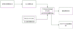
In another aspect, the present invention further provides a system for constructing a scanning knowledge base of an imaging device, including: the device comprises a parameter setting module, a definition module, a branch configuration module and a knowledge base creation module, wherein the parameter setting module is connected with the definition module and is used for setting capability parameters for each type of image equipment; wherein, the capability parameters comprise a positioning capability parameter, a scanning capability parameter and a reconstruction capability parameter; the definition module is respectively connected with the parameter setting module and the knowledge base creating module and is used for defining the capability parameters of the single image equipment and storing the capability parameters of the image equipment; the branch configuration module is connected with the knowledge base creation module and used for configuring clinical application items for each type of imaging equipment based on the examination part, setting examination objectives corresponding to the clinical application items for each clinical application item, configuring age groups matched with the examination objectives for each examination objective and generating a tree structure; wherein, the type of the image equipment, the clinical application project, the examination purpose and the age group are taken as elements to form each branch of the tree structure; and the knowledge base creating module is respectively connected with the branch configuration module and the defining module and used for calling the capability parameters of the image equipment matched with each branch based on each branch, defining the scanning scheme of the branch and storing the scanning scheme into the general scanning knowledge base.
Preferably, the branch configuration module further comprises a feature definition unit for setting physical features of the patient under the age group, the elements of each branch further comprise the physical features of the patient, and the branch comprising the physical features of the patient is defined as a personalized branch.
Preferably, the knowledge base creating module further comprises a personalized knowledge base creating unit, configured to call, based on the personalized branch, the capability parameter of the imaging device matched with the personalized knowledge base, define a scanning scheme of the personalized branch, and store the scanning scheme in the personalized scanning knowledge base.
Preferably, the system further comprises: and the extraction module is connected with the knowledge base creation module and used for extracting the corresponding scanning scheme and dynamically pushing the scanning scheme to the doctor after the doctor inputs the elements of the branches or the personalized branches.
Preferably, the system further comprises: and the shared platform creation module is connected with the knowledge base creation module and used for creating a cloud shared platform and uploading the general scanning knowledge base and the personalized scanning knowledge base to the cloud shared platform.
Preferably, the cloud sharing platform comprises an image device creating module, a scanning scheme downloading module, a scanning scheme modifying module and a scanning scheme inquiring module.
The invention has the technical effects that: 1 the method of the invention sets the capability parameter for each type of image equipment, defines the capability parameter of single equipment,
and storing the capability parameters of the image equipment; matching the capability parameters of the imaging equipment according to the clinical application items, the examination purpose and the age bracket, defining a scanning scheme and storing the scanning scheme in a general scanning knowledge base; because the capability parameters of a single imaging device (such as the manufacturer, the model and the configuration of the imaging device) are defined, the capability characteristics of different devices are considered, and the capability parameters of the corresponding imaging device are matched based on the factors of clinical application items, examination purposes and age groups, the scanning scheme is more accurate, the scanning quality of the vast primary medical institution is improved, the original scanning scheme exceeding the memory capability of a technician can be practically applied in clinic, and the universal significance for improving the quality and the efficiency of the imaging diagnosis service is realized; 2 the method of the invention sets the physical characteristics of the patient under the age group (e.g. unconsciousness, over-fatness, some part built-in)
Metal implant, etc.), the elements of each branch also include the physical characteristics of the patient, the scanning scheme of the personalized branch can be defined and saved to the personalized scanning knowledge base; when the scanning knowledge base is established, the individualized physical condition of a patient is considered, and the application of the original complicated and various individualized scanning scheme which is difficult to remember can be greatly promoted; 3 the method of the invention can also extract the corresponding scanning scheme after the doctor inputs the elements of the branch or the personalized branch
And dynamically pushed to the doctor; the model of the imaging device can be determined according to the clinical application item, the examination purpose and the current operation of the technician
And the most appropriate scanning scheme of the case is dynamically given according to the configuration, so that the scanning quality and the scanning efficiency of a technician are improved; 4 the method of the invention can also create a cloud sharing platform to upload the general scanning knowledge base and the personalized scanning knowledge base to
The cloud sharing platform can create image equipment information, download scanning schemes from the cloud, modify the scanning schemes and query the scanning schemes at the cloud through cloud sharing, so that a scanning knowledge base is updated in a crowd funding mode, the scanning knowledge base is low in maintenance cost, easy to personalize and easy to popularize.
Drawings
The accompanying drawings, which are included to provide a further understanding of the invention and are incorporated in and constitute a part of this application, illustrate embodiment(s) of the invention and together with the description serve to explain the invention without limiting the invention. In the drawings:
fig. 1 is a flowchart illustrating a method for constructing a knowledge base of scanning by an imaging device according to a first embodiment of the present invention;
fig. 2 is a schematic interface diagram illustrating capability parameters set for each type of image device in a method for constructing a scanning knowledge base of an image device according to a first embodiment of the present invention;
fig. 3 is a schematic interface diagram illustrating definition of capability parameters of a single image device in a method for constructing a scanning knowledge base of an image device according to a first embodiment of the present invention;
fig. 4 is a schematic interface diagram illustrating a tree structure formed by taking the type of the imaging device, the clinical application item, the examination purpose, and the age group as elements in the imaging device scanning knowledge base construction method according to a first embodiment of the present invention;
fig. 5 is a schematic interface diagram illustrating a scan protocol defined by inputting device type, clinical application item, examination purpose, age group, and other information in a method for constructing a knowledge base of a scanning device according to a first embodiment of the present invention;
fig. 6 is a schematic interface diagram illustrating a scan plan template defined in a method for constructing a knowledge base of a scanning device according to a first embodiment of the present invention;
fig. 7 is a schematic interface diagram illustrating setting of physical features of a patient in a scanning knowledge base construction method of an imaging device according to a first embodiment of the invention;
fig. 8 is a schematic interface diagram illustrating a scanning protocol defined by adding physical characteristic information of a patient in a method for constructing a knowledge base of scanning by an imaging device according to a first embodiment of the invention;
FIG. 9 is a schematic diagram of a system for constructing a knowledge base of scanning by an imaging device according to a second embodiment of the present invention;
fig. 10 is a schematic diagram illustrating an interface for setting capability parameters for each type of image device in the image device scanning knowledge base construction system according to the second embodiment of the present invention;
fig. 11 is a schematic diagram illustrating an interface for defining capability parameters of a single image device in the image device scanning knowledge base building system according to the second embodiment of the present invention;
fig. 12 is a schematic interface diagram illustrating an imaging device scanning knowledge base building system according to a second embodiment of the present invention, in which the type, clinical application item, examination purpose, and age group of the imaging device form a tree structure;
fig. 13 is a schematic diagram of an interface for inputting information of device type, clinical application item, examination purpose, age group, etc. to define a scanning scheme in the imaging device scanning knowledge base construction system according to the second embodiment of the present invention;
fig. 14 is a schematic diagram illustrating an interface for defining a scan plan template in the imaging device scan knowledge base construction system according to a second embodiment of the present invention;
FIG. 15 is a schematic structural diagram of a system for constructing a knowledge base of scanning by an imaging device according to a third embodiment of the present invention;
FIG. 16 is a schematic diagram of an interface for setting the physical characteristics of a patient in the imaging device scanning knowledge base construction system according to the third embodiment of the invention;
FIG. 17 is a schematic structural diagram of a scanning knowledge base construction system of a fourth imaging device according to the present invention;
fig. 18 is a schematic interface diagram illustrating that the imaging device scans the knowledge base building system to add the physical characteristic information of the patient to define the scanning scheme according to the fourth embodiment of the present invention;
FIG. 19 is a diagram illustrating a scanning knowledge base construction system of a fifth embodiment of the present invention;
fig. 20 is a schematic structural diagram of a scanning knowledge base construction system of a sixth embodiment of the invention.
Detailed Description
The present invention will be described in detail below with reference to the accompanying drawings in conjunction with embodiments.
Example one
Fig. 1 is a flowchart illustrating a method for constructing a knowledge base of scanning by an imaging device according to a first embodiment of the present invention; as shown in fig. 1, the method comprises the steps of:
step S101, setting capability parameters for each type of image equipment; wherein, the capability parameters comprise a positioning capability parameter, a scanning capability parameter and a reconstruction capability parameter;
the imaging device types include CT/MR/DR/MA/XA/PET/ECT and the like. Different types of imaging equipment, for the same examination purpose, are different, and the scanning scheme is related to the equipment type.
Fig. 2 shows an interface schematic diagram of setting capability parameters for each type of imaging device in the imaging device scanning knowledge base construction method according to the first embodiment of the present invention, as shown in fig. 2, the leftmost side of the interface is a device type, such as CT \ MR \ DR \ RF, etc., the imaging device capability parameters are a positioning capability parameter, a scanning capability parameter, and a reconstruction capability parameter, a parameter name, a parameter code, a unit, an input mode, a data type, a limit identifier, etc., are set under each capability parameter, and each capability parameter may be stored in a template form for later invocation.
Step S102, defining the capability parameter of a single image device and storing the capability parameter of the image device;
the manufacturers, models and configurations of the imaging devices of each medical institution are different, so that the capability parameters of each imaging device need to be defined for later calling.
Fig. 3 is a schematic interface diagram illustrating definition of capability parameters of a single image device in a method for constructing a scanning knowledge base of an image device according to a first embodiment of the present invention; as shown in fig. 3, there is image device information in the interface, such as the inspection type, device manufacturer, device model, etc.; the positioning capability parameter, the scanning capability parameter and the reconstruction capability parameter are sequentially arranged below the image equipment information, and corresponding parameter information, such as the row number, the layer number, the KV range value and the like in the scanning capability parameter, is selected and filled according to manufacturer information and equipment model. These parameters may be stored in the form of a template and finally the capability parameters of the device are saved to the system for invocation.
Step S103, based on the examination part, configuring clinical application items for each type of imaging equipment, setting examination objectives corresponding to each clinical application item, configuring age groups matched with each examination objective, and generating a tree structure; wherein, the type of the image equipment, the clinical application project, the examination purpose and the age group are taken as elements to form each branch of the tree structure;
clinical application projects for imaging examinations are usually associated with medical insurance reimbursements from various locations. Such as a CT scan of the chest, an MR scan of the abdomen, etc. The application items of each medical institution are generally similar, but the details are different. Some comprehensive medical institutions have more extensive projects, such as pelvic MR flat scan. There are also professional medical institutions or high-end medical institutions that use relatively detailed scanning schemes. For example, the pelvic cavity MR flat scanning is refined into a plurality of segmentation items such as uterus MR scanning and rectum MR scanning. Regardless of the thickness, the total number of all application items is limited, and does not affect the architecture of the present invention.
The examination purpose is a clinical diagnosis purpose that can be subdivided under the item. For example, the purpose of flat scanning of CT on the chest may be cough to be examined to determine whether lung is infected, or may be lung nodule screening for physical examination, or may be a follow-up examination of lung nodules. The focus of the image is different for different examination purposes, and the optimal scanning scheme is also different. Determining the objective of diagnosis and treatment is crucial for selecting the optimal scanning scheme. The purpose of the examination is only one level below the application project, usually not more than 10 options.
Some tests are aimed at judging whether there is a disease or not; there are cases where the disease species is determined but imaging is required to determine the severity. According to the clinical understanding of the disease of the patient, different application projects can be adopted at different stages, the existing rationality exists, and the thickness method does not conflict. The breadth of the application project, and the breadth of the inspection purpose below, does not affect the method.
The next level of the scanning knowledge base framework is the division of age groups. Different examination purposes, different equipment types, and different methods of classifying patient ages. The age group is related to the development of different tissues to perfect and molt; establishment, improvement and molting of different physiological functions. There are also many imaging examination scanning methods that are independent of the age bracket of the patient, and the age bracket is directly filled in as 0-199.
Fig. 4 is a schematic interface diagram illustrating a tree structure formed by taking the type of the imaging device, the clinical application item, the examination purpose, and the age group as elements in the imaging device scanning knowledge base construction method according to a first embodiment of the present invention; as shown in fig. 4, the leftmost side of the interface is the image device type, and the image device types are arranged in sequence from the right: the examination part (chest), examination items can be chest CT flat scan, chest CT enhancement, pulmonary artery CTA, etc., and the examination purpose under the examination items of chest CT flat scan can be: fever, routine examination, pulmonary nodules, physical examination, and the age groups for which the examination is aimed at fever may be adults (over 14 years), children (7-14 years), toddlers (1-7 years), infants (< 1 year). The device type is CT, the examination item is chest CT flat scan, the examination purpose is fever, and the age group is an adult human factor to form a branch of a tree structure.
And step S104, calling the capability parameters of the matched image equipment based on each branch, defining the scanning scheme of the branch and storing the scanning scheme into a general scanning knowledge base.
Fig. 5 is a schematic interface diagram illustrating a scan protocol defined by inputting device type, clinical application item, examination purpose, age group, and other information in a method for constructing a knowledge base of a scanning device according to a first embodiment of the present invention; as shown in fig. 5, in order to edit the information of the general scanning knowledge base, fill in the name of the scanning knowledge base, the examination item, the template, the examination purpose, and the age group, and after being saved, pop up fig. 6 automatically, fig. 6 shows an interface diagram for defining a scanning scheme template in the construction method of the scanning knowledge base of the imaging device according to the first embodiment of the present invention; as shown in fig. 6, for CT stroke, the scan plan of the branch is defined for routine examination and adults in the age group and is stored in the general scan knowledge base.
Wherein, the method also comprises: setting physical characteristics of the patient under the age group, wherein the elements of each branch further comprise the physical characteristics of the patient, and the branch comprising the physical characteristics of the patient is defined as a personalized branch.
Fig. 7 is a schematic interface diagram illustrating setting of physical features of a patient in a scanning knowledge base construction method of an imaging device according to a first embodiment of the invention; as shown in FIG. 7, the lowest part of the interface has displays: "patient characteristic information", such as special characteristics: inability to breathe spontaneously, irritability, a fat body shape, etc.
Wherein, the method also comprises: and calling the capability parameters of the matched image equipment based on the personalized branch, defining the scanning scheme of the personalized branch and storing the scanning scheme into a personalized scanning knowledge base.
Fig. 8 is a schematic interface diagram illustrating a scanning protocol defined by adding physical characteristic information of a patient in a method for constructing a knowledge base of scanning by an imaging device according to a first embodiment of the invention; as shown in fig. 8, when editing the personalized scan knowledge base information, there is a control for displaying the patient characteristics, and based on the basic information, the patient characteristics are selected, and after storing, a scan scheme defining personalized branches of the knowledge base name GE CT chest flat scan, physical examination, adult, and inability to breathe autonomously is automatically popped up and stored in the personalized scan knowledge base.
There are two types of branches below the age group:
the first branch is a specific scanning scheme corresponding to a certain image equipment manufacturer and model: after determining the clinical application item, the device type, the examination purpose, and the age group, a routine optimized scanning scheme can be given according to the specific device model. This solution is often relatively clear and may be provided entirely by trained technicians/equipment manufacturers. The initial stage of the scanning scheme can be a complete free text, which can be understood by the scanner technician and performed to meet clinical requirements. And the later stage can be marked according to a structured scheme with a RADLEX body semantic tag, such as a sequence, a sequence technical requirement and the like, so that the aim of automatically sending scanning parameters to imaging equipment is fulfilled.
Theoretically, there are how many types of devices there are optimal scanning schemes for the devices. Because the basic capability of the existing image equipment is greatly improved, the scanning schemes of equipment of different manufacturers and models are often highly similar in the face of two-thirds of routine inspection, and the equipment can be reused only by mapping between proper nouns of different enterprises.
The second branch is a specific scanning scheme corresponding to a certain imaging device scene/model when the patient has certain personalized physical characteristics.
In some cases, the patient has a special condition, such as unconsciousness, obesity, a metal implant in a certain part, allergy to certain contrast agents, heart failure, etc., which requires different extreme scanning schemes for different imaging devices. For example, if the patient is unconscious and unable to cooperate with breathing, the chest CT scan can be performed, and for siemens FLASH CT, a 3-second fast scan scheme can be adopted to reduce the breathing motion artifact. Scanning too fast is not the preferred scanning scheme because it still suffers from reduced image quality and is relatively costly to machine.
Unlike the hierarchy of the first branch using "equipment" - "scanning protocol", the hierarchy of "patient condition" - "equipment model" - "scanning protocol" is added in the second branch to form the hierarchy of "patient condition" - "equipment model" - "scanning protocol".
The construction of this hierarchy is entirely by manual enumeration. Because the specifics of each "modality-inspection purpose" combination are different, no general algorithm is available to automatically generate a scene. Devices of different capabilities have different solutions for different scenarios. If a device does not have a special solution for a particular situation, the patient's condition is ignored and the scan is performed directly using the optimal conventional protocol.
Wherein, the method also comprises: after the doctor inputs the elements of the branches or the personalized branches, the scanning scheme corresponding to the elements is extracted and dynamically pushed to the doctor.
Wherein, the method also comprises: and establishing a cloud sharing platform, and uploading the general scanning knowledge base and the personalized scanning knowledge base to the cloud sharing platform.
The cloud sharing platform comprises an image device creating module, a scanning scheme downloading module, a scanning scheme modifying module and a scanning scheme inquiring module.
The cloud sharing platform provides a group of independent accounts for each medical institution. The medical institution can select the type and the model of the image equipment at the cloud sharing platform and edit the capability label in a personalized way. If the cloud does not already have a new type of device, the facility may create the device. The newly created device information includes information such as a manufacturer, a device type, and a model. And after being created, the file becomes an optional drop-down list for sharing in the cloud.
The personalization capability tag of the device is a set of self-defined text tags for the medical institution. Such as "dual energy", "high capacity bulb", "dual syringe", "XX body surface coil", etc. These tags are plain text tags and do not currently need to be mapped to any ontology tags at all. Even if the devices with the same model number are configured to be possibly far away, and the boundary capabilities are greatly different, personalized marking is needed so as to be distinguished at the cloud end and facilitate users with the same configuration to select the device types and schemes.
The medical institution downloads the scanning schemes from the cloud and modifies the scanning schemes in a self-defined mode, and all the scanning schemes are text messages at present and can be understood by a scanning technician. After the medical institution creates the equipment of the hospital from the cloud, the standard scanning scheme which is located at the cloud, created by other medical institutions and related to the model and the personalized label of the equipment can be downloaded to a front-end computer service platform in the hospital, and meanwhile, the personalized scanning scheme related to the patient condition can be downloaded. The medical institution can directly use the schemes and can also edit and upload the schemes to the cloud for sharing.
And providing a query interface (providing a Webservice/WEB API interface and carrying out identity verification through OAuth 2.0) of the third-party information system.
The cloud sharing platform can provide a query interface on a front-end processor of a medical institution. Any one of the RIS systems can query using information such as standardized "clinical application item", "device type", "exam purpose", "device model", "patient demographic information", etc., returning the device model, the standardized scan protocol under the exam purpose, and all possible personalized scan protocol options related to "patient condition". The standardized scanning scheme and the multiple personalized scanning schemes of the equipment use different label prompts, and the specific scanning scheme under the label can be seen by clicking the label, so that a technician is prompted in real time.
The embodiment of the invention sets the capability parameter for each type of image equipment, defines the capability parameter of a single equipment and stores the capability parameter of the image equipment; matching the capability parameters of the imaging equipment according to the clinical application items, the examination purpose and the age bracket, defining a scanning scheme and storing the scanning scheme in a general scanning knowledge base; because the capability parameters of a single imaging device (such as the manufacturer, the model and the configuration of the imaging device) are defined, the capability characteristics of different devices are considered, and the capability parameters of the corresponding imaging device are matched based on the factors of clinical application items, examination purposes and age groups, the scanning scheme is more accurate, the scanning quality of the vast primary medical institution is improved, the original scanning scheme exceeding the memory capability of a technician can be practically applied in clinic, and the universal significance for improving the quality and the efficiency of the imaging diagnosis service is realized; the embodiment of the invention sets the physical characteristics of the patient (such as unconsciousness, over-fatness, metal implant built in a certain part and the like) under the age group, the elements of each branch also comprise the physical characteristics of the patient, and the scanning scheme of the personalized branch can be defined and stored in a personalized scanning knowledge base; when the scanning knowledge base is established, the individualized physical condition of a patient is considered, and the application of the original complicated and various individualized scanning scheme which is difficult to remember can be greatly promoted; the embodiment of the invention can also extract the corresponding scanning scheme and dynamically push the scanning scheme to the doctor after the doctor inputs the elements of the branches or the personalized branches; the most appropriate scanning scheme of the case can be dynamically given according to the clinical application project, the examination purpose, the model and the configuration of the imaging equipment currently operated by the technician, so that the scanning quality and the scanning efficiency of the technician are improved; the embodiment of the invention can also create a cloud sharing platform, upload the general scanning knowledge base and the personalized scanning knowledge base to the cloud sharing platform, create image equipment information, download scanning schemes from the cloud, modify the scanning schemes and inquire the scanning schemes at the cloud through cloud sharing, update the scanning knowledge base in a crowd-funded mode, and has the advantages of low maintenance cost of the scanning knowledge base, easy personalization and easy popularization.
Example two
FIG. 9 is a schematic diagram of a system for constructing a knowledge base of scanning by an imaging device according to a second embodiment of the present invention; as shown in fig. 9, the system includes: parameter setting module 10, definition module 20, branch configuration module 30, knowledge base creation module 40, wherein,
the parameter setting module 10 is connected with the definition module 20 and is used for setting capability parameters for each type of image equipment; wherein, the capability parameters comprise a positioning capability parameter, a scanning capability parameter and a reconstruction capability parameter;
the imaging device types include CT/MR/DR/MA/XA/PET/ECT and the like. Different types of imaging equipment, for the same examination purpose, are different, and the scanning scheme is related to the equipment type.
Fig. 10 shows an interface schematic diagram of setting capability parameters for each type of imaging device in the imaging device scanning knowledge base construction system according to the second embodiment of the present invention, as shown in fig. 10, the leftmost side of the interface is a device type, such as CT \ MR \ DR \ RF, etc., the imaging device capability parameters are positioning capability parameters, scanning capability parameters, and reconstruction capability parameters, a parameter name, a parameter code, a unit, an input mode, a data type, a limit identifier, etc., are set under each capability parameter, and each capability parameter may be stored in a form of a template for later invocation.
A defining module 20, connected to the parameter setting module 10 and the knowledge base creating module 40, respectively, for defining the capability parameters of a single image device and storing the capability parameters of the image device;
the manufacturers, models and configurations of the imaging devices of each medical institution are different, so that the capability parameters of each imaging device need to be defined for later calling.
Fig. 11 is a schematic diagram illustrating an interface for defining capability parameters of a single image device in the image device scanning knowledge base building system according to the second embodiment of the present invention; as shown in fig. 11, there is image device information in the interface, such as the inspection type, device manufacturer, device model, etc.; the positioning capability parameter, the scanning capability parameter and the reconstruction capability parameter are sequentially arranged below the image equipment information, and corresponding parameter information, such as the row number, the layer number, the KV range value and the like in the scanning capability parameter, is selected and filled according to manufacturer information and equipment model. These parameters may be stored in the form of a template and finally the capability parameters of the device are saved to the system for invocation.
A branch configuration module 30, connected to the knowledge base creation module 40, for configuring clinical application items for each type of imaging device based on the examination location, setting an examination purpose corresponding to each clinical application item, and configuring an age group matched with each examination purpose, so as to generate a tree structure; wherein, the type of the image equipment, the clinical application project, the examination purpose and the age group are taken as elements to form each branch of the tree structure;
clinical application projects for imaging examinations are usually associated with medical insurance reimbursements from various locations. Such as a CT scan of the chest, an MR scan of the abdomen, etc. The application items of each medical institution are generally similar, but the details are different. Some comprehensive medical institutions have more extensive projects, such as pelvic MR flat scan. There are also professional medical institutions or high-end medical institutions that use relatively detailed scanning schemes. For example, the pelvic cavity MR flat scanning is refined into a plurality of segmentation items such as uterus MR scanning and rectum MR scanning. Regardless of the thickness, the total number of all application items is limited, and does not affect the architecture of the present invention.
The examination purpose is a clinical diagnosis purpose that can be subdivided under the item. For example, the purpose of flat scanning of CT on the chest may be cough to be examined to determine whether lung is infected, or may be lung nodule screening for physical examination, or may be a follow-up examination of lung nodules. The focus of the image is different for different examination purposes, and the optimal scanning scheme is also different. Determining the objective of diagnosis and treatment is crucial for selecting the optimal scanning scheme. The purpose of the examination is only one level below the application project, usually not more than 10 options.
Some tests are aimed at judging whether there is a disease or not; there are cases where the disease species is determined but imaging is required to determine the severity. According to the clinical understanding of the disease of the patient, different application projects can be adopted at different stages, the existing rationality exists, and the thickness method does not conflict. The breadth of the application project, and the breadth of the inspection purpose below, does not affect the method.
The next level of the scanning knowledge base framework is the division of age groups. Different examination purposes, different equipment types, and different methods of classifying patient ages. The age group is related to the development of different tissues to perfect and molt; establishment, improvement and molting of different physiological functions. There are also many imaging examination scanning methods that are independent of the age bracket of the patient, and the age bracket is directly filled in as 0-199.
Fig. 12 is a schematic interface diagram illustrating an imaging device scanning knowledge base building system according to a second embodiment of the present invention, in which the type, clinical application item, examination purpose, and age group of the imaging device form a tree structure; as shown in fig. 12, the leftmost side of the interface is the image device type, and the image device types are arranged in sequence from the right: the examination part (chest), examination items can be chest CT flat scan, chest CT enhancement, pulmonary artery CTA, etc., and the examination purpose under the examination items of chest CT flat scan can be: fever, routine examination, pulmonary nodules, physical examination, and the age groups for which the examination is aimed at fever may be adults (over 14 years), children (7-14 years), toddlers (1-7 years), infants (< 1 year). The device type is CT, the examination item is chest CT flat scan, the examination purpose is fever, and the age group is an adult human factor to form a branch of a tree structure.
And the knowledge base creating module 40 is respectively connected with the branch configuration module 30 and the defining module 20, and is used for calling the capability parameter of the image device matched with each branch based on each branch, defining the scanning scheme of the branch and storing the scanning scheme into the general scanning knowledge base.
Fig. 13 is a schematic diagram of an interface for inputting information of device type, clinical application item, examination purpose, age group, etc. to define a scanning scheme in the imaging device scanning knowledge base construction system according to the second embodiment of the present invention; as shown in fig. 13, in order to edit the information of the general scanning knowledge base, fill in the name of the scanning knowledge base, the examination items, the templates, the examination purpose, and the age group, and automatically pop up fig. 14 after storage, fig. 14 shows an interface diagram for defining the scanning scheme template in the construction method of the scanning knowledge base of the imaging device according to the first embodiment of the present invention; as shown in fig. 14, for CT stroke, the scan plan of the branch is defined for routine examination and adults in the age group and stored in the general scan knowledge base.
The embodiment of the invention is provided with a parameter setting module, a definition module, a branch configuration module and a knowledge base establishing module, can set capacity parameters for each type of image equipment, defines the capacity parameters of single equipment and stores the capacity parameters of the image equipment; matching the capability parameters of the imaging equipment according to the clinical application items, the examination purpose and the age bracket, defining a scanning scheme and storing the scanning scheme in a general scanning knowledge base; because the capability parameters of a single imaging device (such as the manufacturer, the model and the configuration of the imaging device) are defined, the capability characteristics of different devices are considered, and the capability parameters of the corresponding imaging device are matched based on the factors of clinical application items, examination purposes and age groups, the scanning scheme is more accurate, the scanning quality of the vast primary medical institutions is improved, the original scanning scheme exceeding the memory capability of technicians can be practically applied in clinics, and the universal significance for improving the quality and the efficiency of the imaging diagnosis service is realized.
EXAMPLE III
FIG. 15 is a schematic structural diagram of a system for constructing a knowledge base of scanning by an imaging device according to a third embodiment of the present invention; as shown in fig. 15, the branch configuration module 30 further includes a feature definition unit 302 for setting physical features of the patient under the age group, elements of each branch further include the physical features of the patient, and the branch including the physical features of the patient is defined as a personalized branch.
FIG. 16 is a schematic diagram of an interface for setting the physical characteristics of a patient in the imaging device scanning knowledge base construction system according to the third embodiment of the invention; as shown in fig. 16, the lowermost part of the interface has a display: "patient characteristic information", such as special characteristics: inability to breathe spontaneously, irritability, a fat body shape, etc.
The embodiment of the invention is provided with a characteristic definition unit, the physical characteristics (such as unconsciousness, over-fatness, metal implant built in a certain part and the like) of the patient are set under the age group, the elements of each branch also comprise the physical characteristics of the patient, and the scanning scheme of the personalized branch can be defined and stored in a personalized scanning knowledge base; when the scanning knowledge base is created, the personalized physical condition of the patient is considered, and the application of the original complex and various personalized scanning schemes which are difficult to remember can be greatly promoted.
Example four
FIG. 17 is a schematic structural diagram of a scanning knowledge base construction system of a fourth imaging device according to the present invention; as shown in fig. 17, the knowledge base creating module 40 further includes a personalized knowledge base creating unit 402, configured to define a scanning scheme of the personalized branch based on the capability parameter of the imaging device matched with the personalized branch call, and store the scanning scheme to the personalized scanning knowledge base.
Fig. 18 is a schematic interface diagram illustrating that the imaging device scans the knowledge base building system to add the physical characteristic information of the patient to define the scanning scheme according to the fourth embodiment of the present invention; as shown in fig. 18, when editing the personalized scan knowledge base information, there is a control for displaying the patient characteristics, and based on the basic information, the patient characteristics are selected, and after storing, a scan scheme defining personalized branches of the knowledge base name GE CT chest flat scan, physical examination, adult, and inability to breathe autonomously is automatically popped up and stored in the personalized scan knowledge base.
There are two types of branches below the age group:
the first branch is a specific scanning scheme corresponding to a certain image equipment manufacturer and model: after determining the clinical application item, the device type, the examination purpose, and the age group, a routine optimized scanning scheme can be given according to the specific device model. This solution is often relatively clear and may be provided entirely by trained technicians/equipment manufacturers. The initial stage of the scanning scheme can be a complete free text, which can be understood by the scanner technician and performed to meet clinical requirements. And the later stage can be marked according to a structured scheme with a RADLEX body semantic tag, such as a sequence, a sequence technical requirement and the like, so that the aim of automatically sending scanning parameters to imaging equipment is fulfilled.
Theoretically, there are how many types of devices there are optimal scanning schemes for the devices. Because the basic capability of the existing image equipment is greatly improved, the scanning schemes of equipment of different manufacturers and models are often highly similar in the face of two-thirds of routine inspection, and the equipment can be reused only by mapping between proper nouns of different enterprises.
The second branch is a specific scanning scheme corresponding to a certain imaging device scene/model when the patient has certain personalized physical characteristics.
In some cases, the patient has a special condition, such as unconsciousness, obesity, a metal implant in a certain part, allergy to certain contrast agents, heart failure, etc., which requires different extreme scanning schemes for different imaging devices. For example, if the patient is unconscious and unable to cooperate with breathing, the chest CT scan can be performed, and for siemens FLASH CT, a 3-second fast scan scheme can be adopted to reduce the breathing motion artifact. Scanning too fast is not the preferred scanning scheme because it still suffers from reduced image quality and is relatively costly to machine.
Unlike the hierarchy of the first branch using "equipment" - "scanning protocol", the hierarchy of "patient condition" - "equipment model" - "scanning protocol" is added in the second branch to form the hierarchy of "patient condition" - "equipment model" - "scanning protocol".
The construction of this hierarchy is entirely by manual enumeration. Because the specifics of each "modality-inspection purpose" combination are different, no general algorithm is available to automatically generate a scene. Devices of different capabilities have different solutions for different scenarios. If a device does not have a special solution for a particular situation, the patient's condition is ignored and the scan is performed directly using the optimal conventional protocol.
EXAMPLE five
FIG. 19 is a diagram illustrating a scanning knowledge base construction system of a fifth embodiment of the present invention; as shown in fig. 19, the system further includes: and the extraction module 50 is connected with the knowledge base creation module 40 and is used for extracting the corresponding scanning scheme and dynamically pushing the scanning scheme to the doctor after the doctor inputs the elements of the branches or the personalized branches.
The embodiment of the invention is provided with the extraction module, so that after a doctor inputs the elements of the branches or the personalized branches, the corresponding scanning scheme can be extracted and dynamically pushed to the doctor; the most suitable scanning scheme of the case can be dynamically given according to the clinical application project, the examination purpose, the model and the configuration of the imaging equipment currently operated by the technician, and the scanning quality and the scanning efficiency of the technician are improved.
EXAMPLE six
Fig. 20 is a schematic structural diagram of a system for constructing a scanning knowledge base of an imaging device according to a sixth embodiment of the present invention, and as shown in fig. 20, the system further includes: and the shared platform creating module 60 is connected to the knowledge base creating module 40, and is configured to create a cloud shared platform, and upload the general scanning knowledge base and the personalized scanning knowledge base to the cloud shared platform.
The cloud sharing platform comprises an image device creating module, a scanning scheme downloading module, a scanning scheme modifying module and a scanning scheme inquiring module.
The cloud sharing platform provides a group of independent accounts for each medical institution. The medical institution can select the type and the model of the image equipment at the cloud sharing platform and edit the capability label in a personalized way. If the cloud does not already have a new type of device, the facility may create the device. The newly created device information includes information such as a manufacturer, a device type, and a model. And after being created, the file becomes an optional drop-down list for sharing in the cloud.
The personalization capability tag of the device is a set of self-defined text tags for the medical institution. Such as "dual energy", "high capacity bulb", "dual syringe", "XX body surface coil", etc. These tags are plain text tags and do not currently need to be mapped to any ontology tags at all. Even if the devices with the same model number are configured to be possibly far away, and the boundary capabilities are greatly different, personalized marking is needed so as to be distinguished at the cloud end and facilitate users with the same configuration to select the device types and schemes.
The medical institution downloads the scanning schemes from the cloud and modifies the scanning schemes in a self-defined mode, and all the scanning schemes are text messages at present and can be understood by a scanning technician. After the medical institution creates the equipment of the hospital from the cloud, the standard scanning scheme which is located at the cloud, created by other medical institutions and related to the model and the personalized label of the equipment can be downloaded to a front-end computer service platform in the hospital, and meanwhile, the personalized scanning scheme related to the patient condition can be downloaded. The medical institution can directly use the schemes and can also edit and upload the schemes to the cloud for sharing.
And providing a query interface (providing a Webservice/WEB API interface and carrying out identity verification through OAuth 2.0) of the third-party information system.
The cloud sharing platform can provide a query interface on a front-end processor of a medical institution. Any one of the RIS systems can query using information such as standardized "clinical application item", "device type", "exam purpose", "device model", "patient demographic information", etc., returning the device model, the standardized scan protocol under the exam purpose, and all possible personalized scan protocol options related to "patient condition". The standardized scanning scheme and the multiple personalized scanning schemes of the equipment use different label prompts, and the specific scanning scheme under the label can be seen by clicking the label, so that a technician is prompted in real time.
The embodiment of the invention is provided with the sharing platform creating module which can create a cloud sharing platform, upload the general scanning knowledge base and the personalized scanning knowledge base to the cloud sharing platform, create image equipment information, download scanning schemes from the cloud, modify the scanning schemes and inquire the scanning schemes at the cloud through cloud sharing, update the scanning knowledge base in a crowd-funded mode, and has the advantages of low maintenance cost of the scanning knowledge base, easy personalization and easy popularization.
From the above description, it can be seen that the above-described embodiments of the present invention achieve the following technical effects: the embodiment of the invention can set the capability parameter for each type of image equipment, define the capability parameter of a single equipment and store the capability parameter of the image equipment; matching the capability parameters of the imaging equipment according to the clinical application items, the examination purpose and the age bracket, defining a scanning scheme and storing the scanning scheme in a general scanning knowledge base; because the capability parameters of a single imaging device (such as the manufacturer, the model and the configuration of the imaging device) are defined, the capability characteristics of different devices are considered, and the capability parameters of the corresponding imaging device are matched based on the factors of clinical application items, examination purposes and age groups, the scanning scheme is more accurate, the scanning quality of the vast primary medical institution is improved, the original scanning scheme exceeding the memory capability of a technician can be practically applied in clinic, and the universal significance for improving the quality and the efficiency of the imaging diagnosis service is realized; the embodiment of the invention sets the physical characteristics of the patient (such as unconsciousness, over-fatness, metal implant built in a certain part and the like) under the age group, the elements of each branch also comprise the physical characteristics of the patient, and the scanning scheme of the personalized branch can be defined and stored in a personalized scanning knowledge base; when the scanning knowledge base is established, the individualized physical condition of a patient is considered, and the application of the original complicated and various individualized scanning scheme which is difficult to remember can be greatly promoted; the embodiment of the invention can also extract the corresponding scanning scheme and dynamically push the scanning scheme to the doctor after the doctor inputs the elements of the branches or the personalized branches; the most appropriate scanning scheme of the case can be dynamically given according to the clinical application project, the examination purpose, the model and the configuration of the imaging equipment currently operated by the technician, so that the scanning quality and the scanning efficiency of the technician are improved; the embodiment of the invention can also create a cloud sharing platform, upload the general scanning knowledge base and the personalized scanning knowledge base to the cloud sharing platform, create image equipment information, download scanning schemes from the cloud, modify the scanning schemes and inquire the scanning schemes at the cloud through cloud sharing, update the scanning knowledge base in a crowd-funded mode, and has the advantages of low maintenance cost of the scanning knowledge base, easy personalization and easy popularization.
It will be apparent to those skilled in the art that the modules or steps of the present invention described above may be implemented by a general purpose computing device, they may be centralized on a single computing device or distributed across a network of multiple computing devices, and they may alternatively be implemented by program code executable by a computing device, such that they may be stored in a storage device and executed by a computing device, or fabricated separately as individual integrated circuit modules, or fabricated as a single integrated circuit module from multiple modules or steps. Thus, the present invention is not limited to any specific combination of hardware and software.
The above description is only a preferred embodiment of the present invention and is not intended to limit the present invention, and various modifications and changes may be made by those skilled in the art. Any modification, equivalent replacement, or improvement made within the spirit and principle of the present invention should be included in the protection scope of the present invention.
Claims (12)
1. A method for constructing a scanning knowledge base of image equipment is characterized by comprising the following steps:
setting capability parameters for each type of image equipment; wherein the capability parameters comprise a positioning capability parameter, a scanning capability parameter and a reconstruction capability parameter;
defining the capability parameter of a single image device, and storing the capability parameter of the image device;
configuring clinical application items for each type of imaging equipment based on an examination part, setting an examination purpose corresponding to each clinical application item, configuring an age group matched with each examination purpose, and generating a tree structure; wherein each branch of the tree structure is formed by using the type of the imaging device, the clinical application item, the examination purpose, and the age group as elements;
and calling the capability parameters of the image equipment matched with each branch based on each branch, defining the scanning scheme of the branch and saving the scanning scheme to a general scanning knowledge base.
2. The method as claimed in claim 1, further comprising: setting physical characteristics of a patient under the age group, the elements of each of the branches further including the physical characteristics of the patient, the branches including the physical characteristics of the patient being defined as personalized branches.
3. The method as claimed in claim 2, further comprising: and calling the capability parameters of the image equipment matched with the personalized branch based on the personalized branch, defining the scanning scheme of the personalized branch and storing the scanning scheme into a personalized scanning knowledge base.
4. The method as claimed in claim 3, further comprising: and after the doctor inputs the elements of the branches or the personalized branches, extracting the scanning scheme corresponding to the elements and dynamically pushing the scanning scheme to the doctor.
5. The method as claimed in claim 3, further comprising: and creating a cloud sharing platform, and uploading the general scanning knowledge base and the personalized scanning knowledge base to the cloud sharing platform.
6. The method for building a scanning knowledge base of imaging devices according to claim 5, wherein the cloud sharing platform comprises an imaging device creating module, a scanning scheme downloading module, a scanning scheme modifying module and a scanning scheme querying module.
7. An imaging device scanning knowledge base construction system, the system comprising: a parameter setting module, a definition module, a branch configuration module and a knowledge base creation module, wherein,
the parameter setting module is connected with the definition module and is used for setting capability parameters for each type of image equipment; wherein the capability parameters comprise a positioning capability parameter, a scanning capability parameter and a reconstruction capability parameter;
the definition module is respectively connected with the parameter setting module and the knowledge base creating module, and is used for defining the capability parameters of a single image device and storing the capability parameters of the image device;
the branch configuration module is connected with the knowledge base creation module and used for configuring clinical application items for each type of imaging equipment based on an examination part, setting examination objectives corresponding to each clinical application item, configuring age groups matched with each examination objective and generating a tree structure; wherein each branch of the tree structure is formed by using the type of the imaging device, the clinical application item, the examination purpose, and the age group as elements;
the knowledge base creating module is respectively connected with the branch configuration module and the definition module, and is used for calling the capability parameters of the image equipment matched with each branch, defining the scanning scheme of the branch and storing the scanning scheme into a general scanning knowledge base.
8. The imaging device scanning knowledge base construction system according to claim 7, wherein the branch configuration module further comprises a feature definition unit for setting physical features of patients under the age group, elements of each of the branches further include the physical features of the patients, and the branches including the physical features of the patients are defined as personalized branches.
9. The system for building a knowledge base for scanning of image devices as claimed in claim 8, wherein the knowledge base creation module further comprises a personalized knowledge base creation unit, configured to invoke the capability parameter of the image device matched with the personalized knowledge base based on the personalized branch, define the scanning scheme of the personalized branch, and store the scanning scheme to the personalized scanning knowledge base.
10. The imaging device scanning knowledge base building system of claim 9, further comprising: and the extraction module is connected with the knowledge base creation module and used for extracting the corresponding scanning scheme and dynamically pushing the scanning scheme to the doctor after the doctor inputs the elements of the branches or the personalized branches.
11. The imaging device scanning knowledge base building system of claim 9, further comprising: and the shared platform creation module is connected with the knowledge base creation module and used for creating a cloud shared platform and uploading the general scanning knowledge base and the personalized scanning knowledge base to the cloud shared platform.
12. The imaging device scanning knowledge base building system according to claim 11, wherein the cloud sharing platform includes an imaging device creating module, a scanning scheme downloading module, a scanning scheme modifying module, and a scanning scheme querying module.
Priority Applications (1)
| Application Number | Priority Date | Filing Date | Title |
|---|---|---|---|
| CN202110822356.3A CN113555090A (en) | 2021-07-21 | 2021-07-21 | Method and system for constructing scanning knowledge base of image equipment |
Applications Claiming Priority (1)
| Application Number | Priority Date | Filing Date | Title |
|---|---|---|---|
| CN202110822356.3A CN113555090A (en) | 2021-07-21 | 2021-07-21 | Method and system for constructing scanning knowledge base of image equipment |
Publications (1)
| Publication Number | Publication Date |
|---|---|
| CN113555090A true CN113555090A (en) | 2021-10-26 |
Family
ID=78132262
Family Applications (1)
| Application Number | Title | Priority Date | Filing Date |
|---|---|---|---|
| CN202110822356.3A Pending CN113555090A (en) | 2021-07-21 | 2021-07-21 | Method and system for constructing scanning knowledge base of image equipment |
Country Status (1)
| Country | Link |
|---|---|
| CN (1) | CN113555090A (en) |
Citations (7)
| Publication number | Priority date | Publication date | Assignee | Title |
|---|---|---|---|---|
| CN106844725A (en) * | 2017-02-10 | 2017-06-13 | 深圳前海大造科技有限公司 | A kind of high in the clouds image data base generation and recognition methods |
| US20170206680A1 (en) * | 2014-07-16 | 2017-07-20 | Koninklijke Philips N.V. | Irecon: intelligent image reconstruction system with anticipatory execution |
| CN109431506A (en) * | 2018-12-26 | 2019-03-08 | 上海联影医疗科技有限公司 | Medical image equipment imaging parameters setting method, device, storage medium and equipment |
| CN109871357A (en) * | 2018-12-14 | 2019-06-11 | 深圳壹账通智能科技有限公司 | Project document display method, device, electronic device and storage medium |
| CN110750540A (en) * | 2019-10-18 | 2020-02-04 | 中国人民解放军军事科学院军事医学研究院 | Method for constructing medical service knowledge base, method and system for obtaining medical service semantic model and medium |
| CN110853740A (en) * | 2019-11-07 | 2020-02-28 | 李真林 | System and method for extracting image scanning scheme characteristics from DICOM (digital imaging and communications in medicine) image |
| CN111584043A (en) * | 2019-02-15 | 2020-08-25 | 北京新网医讯技术有限公司 | Intelligent prompting system and method for imaging scanning scheme |
-
2021
- 2021-07-21 CN CN202110822356.3A patent/CN113555090A/en active Pending
Patent Citations (7)
| Publication number | Priority date | Publication date | Assignee | Title |
|---|---|---|---|---|
| US20170206680A1 (en) * | 2014-07-16 | 2017-07-20 | Koninklijke Philips N.V. | Irecon: intelligent image reconstruction system with anticipatory execution |
| CN106844725A (en) * | 2017-02-10 | 2017-06-13 | 深圳前海大造科技有限公司 | A kind of high in the clouds image data base generation and recognition methods |
| CN109871357A (en) * | 2018-12-14 | 2019-06-11 | 深圳壹账通智能科技有限公司 | Project document display method, device, electronic device and storage medium |
| CN109431506A (en) * | 2018-12-26 | 2019-03-08 | 上海联影医疗科技有限公司 | Medical image equipment imaging parameters setting method, device, storage medium and equipment |
| CN111584043A (en) * | 2019-02-15 | 2020-08-25 | 北京新网医讯技术有限公司 | Intelligent prompting system and method for imaging scanning scheme |
| CN110750540A (en) * | 2019-10-18 | 2020-02-04 | 中国人民解放军军事科学院军事医学研究院 | Method for constructing medical service knowledge base, method and system for obtaining medical service semantic model and medium |
| CN110853740A (en) * | 2019-11-07 | 2020-02-28 | 李真林 | System and method for extracting image scanning scheme characteristics from DICOM (digital imaging and communications in medicine) image |
Similar Documents
| Publication | Publication Date | Title |
|---|---|---|
| CN110517238B (en) | AI three-dimensional reconstruction and human-computer interaction visualization network system for CT medical image | |
| CN106407666A (en) | A method, device and system for generating electronic medical record information | |
| US10657220B2 (en) | System and methods for medical reporting | |
| US20180046761A1 (en) | Automated cloud image processing and routing | |
| CN115994902A (en) | Medical image analysis method, electronic equipment and storage medium | |
| US11989878B2 (en) | Enhancing medical imaging workflows using artificial intelligence | |
| US12191037B2 (en) | Medical machine learning system | |
| CN111584043B (en) | Intelligent prompting system and method for imaging scanning scheme | |
| CN110782979A (en) | Medical task allocation method and device, electronic equipment and storage medium | |
| US20190188849A1 (en) | Generating simulated photographic anatomical slices | |
| CN115171835B (en) | Case structured model training method, device and case structured method | |
| CN112700888A (en) | Method and system for generating inquiry flow and readable storage medium | |
| CN113555090A (en) | Method and system for constructing scanning knowledge base of image equipment | |
| KR102371037B1 (en) | Programmable electronic medical record system | |
| US20240395392A1 (en) | Flap prediction system based on volumetric data and foundation models | |
| CN112215969A (en) | User data processing method and device based on virtual reality | |
| CN113728398A (en) | Disease position acquisition method, device, equipment and computer-readable storage medium | |
| CN105830071A (en) | A method and computer program product for management of the distribution of medical reports in clinical applications | |
| JP2003108664A (en) | Finding creation system | |
| CN117393099A (en) | Medical document generation method and device, electronic equipment and storage medium | |
| CN114999613A (en) | Method for providing at least one metadata attribute associated with a medical image | |
| Pappalardo et al. | A platform for real-3d visualization and planning of left atrial appendage occlusion through mixed reality | |
| JP7702765B1 (en) | Medical document creation support system, medical document creation support method, and program | |
| LU509545B1 (en) | An Auxiliary Preoperative TAVR Diagnostic Report System, Storage Medium, and Electronic Device Based on Big Language Model | |
| US20250131998A1 (en) | Methods and devices for a configurable medical information platform |
Legal Events
| Date | Code | Title | Description |
|---|---|---|---|
| PB01 | Publication | ||
| PB01 | Publication | ||
| SE01 | Entry into force of request for substantive examination | ||
| SE01 | Entry into force of request for substantive examination |