CN112680407B - Compound for promoting muscle stem cell differentiation and application thereof - Google Patents
Compound for promoting muscle stem cell differentiation and application thereof Download PDFInfo
- Publication number
- CN112680407B CN112680407B CN201910989528.9A CN201910989528A CN112680407B CN 112680407 B CN112680407 B CN 112680407B CN 201910989528 A CN201910989528 A CN 201910989528A CN 112680407 B CN112680407 B CN 112680407B
- Authority
- CN
- China
- Prior art keywords
- muscle
- mice
- differentiation
- muscle stem
- cells
- Prior art date
- Legal status (The legal status is an assumption and is not a legal conclusion. Google has not performed a legal analysis and makes no representation as to the accuracy of the status listed.)
- Active
Links
Images


Landscapes
- Measuring Or Testing Involving Enzymes Or Micro-Organisms (AREA)
Abstract
Description
技术领域Technical Field
本发明属于细胞生物学和药学领域,更具体地,本发明涉及促进肌肉干细胞分化的化合物及其应用。The present invention belongs to the field of cell biology and pharmacy, and more specifically, the present invention relates to a compound for promoting differentiation of muscle stem cells and application thereof.
背景技术Background Art
强直性肌肉营养不良(myotonic dystrophy,DM)是一种以肌强直,肌无力和肌萎缩为主要特点、多器官受累的显性遗传性疾病。主要分为两个类型:强直性肌营养不良1型(myotonic dystrophy type 1,DM1)和强直性肌营养不良2型(myotonic dystrophy type2,DM2)。Myotonic dystrophy (DM) is a dominant hereditary disease characterized by muscle stiffness, weakness and atrophy, with multiple organ involvement. It is mainly divided into two types: myotonic dystrophy type 1 (DM1) and myotonic dystrophy type 2 (DM2).
1型强直性肌营养不良症(DM1)是最常见的肌肉萎缩症,会出现多系统病变。该病是由于在强直性肌营养不良蛋白激酶(DMPK)基因的3’末端出现了CTG三核苷酸的异常扩增所导致的,但是针对CTG异常扩增产生病变的分子机制还不清楚。DM1一般是远端肌肉表型更严重,较DM2患者,DM1患者的表型比较严重,有些患者随着疾病的发展会出现猝死。本发明只涉及到DM1疾病模型。Myotonic dystrophy type 1 (DM1) is the most common muscular dystrophy, which can cause multi-system lesions. The disease is caused by abnormal amplification of CTG trinucleotides at the 3' end of the myotonic dystrophy protein kinase (DMPK) gene, but the molecular mechanism of abnormal amplification of CTG causing lesions is still unclear. DM1 is generally more severe in distal muscle phenotypes. Compared with DM2 patients, the phenotype of DM1 patients is more serious, and some patients will experience sudden death as the disease progresses. The present invention only relates to the DM1 disease model.
DM1疾病发病的器官组织包括肌肉(骨骼肌)、心脏(心肌)、眼睛、大脑、消化系统(平滑肌)和内分泌系统等。为了进一步加深对疾病的了解,进而找到有效的治愈方法。科学家们已经构建了一些DM1疾病小鼠模型。首先由于DM1疾病的发是因为DMPK基因的3’端出现了CTG序列的异常加长导致的,DMPK的突变RNA无法正常的运送到细胞质内,导致其蛋白水平表达下降,DMPK的mRNA在核内聚集,无法完成RNA到蛋白质的编码,因此最早推测DMPK基因负责疾病的发生。随后有实验室构建了DMPK敲除的小鼠模型,发现DMPK的敲除小鼠不能够很好的模拟疾病的发生,会出现肌肉纤维退化、横截面大小差异变大和肌肉力量减弱等表型,一些更为典型的DM1表型并没有出现。这说明疾病的发生并不是完全由DMPK基因表达异常导致的。The organs and tissues that are affected by DM1 disease include muscles (skeletal muscles), heart (myocardium), eyes, brain, digestive system (smooth muscle) and endocrine system. In order to further deepen the understanding of the disease and find an effective cure. Scientists have constructed some DM1 disease mouse models. First of all, since the onset of DM1 disease is caused by the abnormal extension of the CTG sequence at the 3' end of the DMPK gene, the mutant RNA of DMPK cannot be normally transported to the cytoplasm, resulting in a decrease in its protein expression level. The mRNA of DMPK aggregates in the nucleus and cannot complete the RNA-to-protein coding. Therefore, it was first speculated that the DMPK gene is responsible for the occurrence of the disease. Subsequently, a laboratory constructed a DMPK knockout mouse model and found that the DMPK knockout mouse could not simulate the occurrence of the disease well. It would show phenotypes such as muscle fiber degeneration, increased cross-sectional size differences and weakened muscle strength, and some more typical DM1 phenotypes did not appear. This shows that the occurrence of the disease is not entirely caused by abnormal expression of the DMPK gene.
通过对患者肌肉组织进行检测发现,DMPK基因3’端出现了CTG序列的异常加长影响的不仅仅是DMPK的表达,DMPK基因的上游基因SIX5和下游基因DMWD的表达量也受到影响,发生一定程度的降低。与此同时,还发现突变后的DMPK转录出的CUG重复序列会形成一个颈环结构,可以招募一些RNA剪接蛋白,比如Muscleblind-like家族(MBNL家族,包括MBNL1,MBNL2,MBNL3三个基因)和CUGBP1等,进而在细胞核内形成聚集点。大量剪接蛋白被“无效招募”后,细胞内可正常发挥功能的RNA剪接蛋白量降低,因此在细胞内会堆积存在大量的异常剪接的蛋白,从而加速了疾病的发生和发展。其中肌肉组织的Clcn1等基因的mRNA出现剪切异常,肌肉收缩兴奋异常,导致强直的产生,心脏组织的Ldb3等基因的mRNA剪切异常,出现心律失常等表型。针对这些现象,2000年有两个实验室首先构建了SIX5基因敲除小鼠,在敲除小鼠中出现白内障表型,纯合敲除的表型重于杂合敲除,小鼠不同的遗传背景对疾病的发生也有影响。2004年Sita Reddy等人对SIX5基因敲除表型进行分析,发现SIX5基因敲除公鼠不育,这与DM1疾病男性不育类似。Rochester大学的Charles A.Thornton教授等人在HSA基因表达片段的3’非编码区加上大约250个CTG重复,并以此构建了转基因小鼠模型,他们发现转基因小鼠出现了DM1疾病相关的表型,包括肌强直和肌萎缩等。病理切片也发现肌肉核内移、肌浆块和戒指样肌纤维等。但也有很多表型如心脏传导阻滞和白内障等并没有出现。By testing the patient's muscle tissue, it was found that the abnormal lengthening of the CTG sequence at the 3' end of the DMPK gene not only affected the expression of DMPK, but also the expression of the upstream gene SIX5 and the downstream gene DMWD of the DMPK gene, which were reduced to a certain extent. At the same time, it was also found that the CUG repeat sequence transcribed by the mutated DMPK would form a neck ring structure, which could recruit some RNA splicing proteins, such as the Muscleblind-like family (MBNL family, including three genes MBNL1, MBNL2, and MBNL3) and CUGBP1, and then form aggregation points in the cell nucleus. After a large number of splicing proteins were "ineffectively recruited", the amount of RNA splicing proteins that could function normally in the cell was reduced, so a large number of abnormally spliced proteins would accumulate in the cell, thereby accelerating the occurrence and development of the disease. Among them, the mRNA of genes such as Clcn1 in muscle tissue showed abnormal splicing, abnormal muscle contraction and excitement, leading to the generation of stiffness, and the mRNA splicing of genes such as Ldb3 in heart tissue was abnormal, resulting in phenotypes such as arrhythmia. In response to these phenomena, two laboratories first constructed SIX5 knockout mice in 2000. Cataract phenotypes appeared in the knockout mice. The phenotype of homozygous knockout was more severe than that of heterozygous knockout. Different genetic backgrounds of mice also affected the occurrence of the disease. In 2004, Sita Reddy et al. analyzed the phenotype of SIX5 knockout and found that SIX5 knockout male mice were infertile, which was similar to male infertility in DM1 disease. Professor Charles A. Thornton of the University of Rochester and others added about 250 CTG repeats to the 3' non-coding region of the HSA gene expression fragment and constructed a transgenic mouse model. They found that transgenic mice had DM1 disease-related phenotypes, including myotonia and muscular atrophy. Pathological sections also found muscle nuclear intranslocation, sarcoplasmic masses and ring-like muscle fibers. However, many phenotypes such as heart block and cataracts did not appear.
由于在成熟肌肉中存在大量剪接异常的RNA。2003年Maurice S.Swanson教授的实验室构建了MBNL1敲除小鼠。MBNL1纯合敲除小鼠出现了肌肉强直和白内障等表型,并检测到包括Clcn1、Tnnt2、Tnnt3在内的很多基因存在RNA剪接异常,这都与临床数据类似。肌肉病理切片也观察到很多核内移的现象。2008年Fabian Chen等人又构建了MBNL2敲除小鼠,在小鼠发育过程中观察到佝偻的产生,同时肌肉病理切片也看到核内移,肌肉纤维横截面变小,部分RNA剪接异常等现象。2012年Maurice S.Swanson教授等人发现MBNL2敲除小鼠脑部发育异常,并存在大量RNA剪接异常,这与DM1疾病部分表型类似。2013年MauriceS.Swanson教授实验室又进行了MBNL1和MBNL2共同敲除,突变小鼠的表型甚于单独一个基因突变的表型。2016年Seta Reddy等人对MBNL3敲除小鼠进行分析发现,小鼠的葡萄糖代谢和心脏功能出现异常。Since there are a large number of abnormally spliced RNAs in mature muscles. In 2003, Professor Maurice S. Swanson's laboratory constructed MBNL1 knockout mice. MBNL1 homozygous knockout mice showed phenotypes such as muscle stiffness and cataracts, and many genes including Clcn1, Tnnt2, and Tnnt3 were detected to have abnormal RNA splicing, which is similar to clinical data. Many nuclear intrusion phenomena were also observed in muscle pathological sections. In 2008, Fabian Chen et al. constructed MBNL2 knockout mice again, and observed the occurrence of rickets during mouse development. At the same time, muscle pathological sections also showed nuclear intrusion, smaller muscle fiber cross-sections, and abnormal splicing of some RNAs. In 2012, Professor Maurice S. Swanson et al. found that the brain development of MBNL2 knockout mice was abnormal, and there were a large number of RNA splicing abnormalities, which are similar to some phenotypes of DM1 disease. In 2013, Professor Maurice S. Swanson's laboratory knocked out both MBNL1 and MBNL2, and the phenotype of the mutant mice was worse than that of a single gene mutation. In 2016, Seta Reddy et al. analyzed MBNL3 knockout mice and found that the mice had abnormal glucose metabolism and heart function.
虽然疾病相关的小鼠模型构建了很多,但这些模型只能部分模拟疾病的发生,没有一种模型能够全面展现患者的表征,同时临床数据显示,涉及到的基因并不是完全没有功能蛋白,因此纯合敲除的表型并不能很好的说明疾病发生的实际情况。Although many disease-related mouse models have been constructed, these models can only partially simulate the occurrence of the disease. No model can fully display the patient's manifestations. At the same time, clinical data show that the genes involved are not completely devoid of functional proteins. Therefore, the phenotype of homozygous knockout cannot well explain the actual situation of the disease.
综上,本领域还需要建立更为理想的、贴近临床表征的动物模型,以及基于此类模型筛选获得有用的药物。In summary, this field still needs to establish more ideal animal models that are closer to clinical characteristics, and to screen and obtain useful drugs based on such models.
发明内容Summary of the invention
本发明的目的在于提供促进肌肉干细胞分化的化合物及其应用。The purpose of the present invention is to provide a compound for promoting the differentiation of muscle stem cells and its application.
本发明的目的还在于提供一种制备1型强直性肌营养不良症(DM1)的多基因敲除实验模型、其制备方法及其应用。所述实验模型包括动物模型和细胞模型。The present invention also aims to provide a multi-gene knockout experimental model for myotonic dystrophy type 1 (DM1), a preparation method thereof and its application. The experimental model includes an animal model and a cell model.
在本发明的第一方面,提供式(I)所示母核结构的化合物或其异构体、衍生物、溶剂合物或前体,或它们的药学上可接受的盐的用途,用于制备促进肌肉干细胞分化的组合物,或用于制备缓解或治疗肌肉干细胞分化异常相关疾病的组合物;In the first aspect of the present invention, there is provided a use of a compound having a parent core structure as represented by formula (I) or an isomer, derivative, solvate or precursor thereof, or a pharmaceutically acceptable salt thereof, for preparing a composition for promoting muscle stem cell differentiation, or for preparing a composition for alleviating or treating a disease associated with abnormal muscle stem cell differentiation;
在一个优选例中,所述的溶剂合物为水合物。In a preferred embodiment, the solvate is a hydrate.
在另一优选例中,所述的组合物为药物组合物。In another preferred embodiment, the composition is a pharmaceutical composition.
在另一优选例中,所述的组合物为培养基组合物。In another preferred embodiment, the composition is a culture medium composition.
在另一优选例中,所述肌肉干细胞分化异常包括:肌肉干细胞分化能力下降或不分化。In another preferred embodiment, the abnormal differentiation of muscle stem cells includes: decreased differentiation ability of muscle stem cells or no differentiation.
在另一优选例中,肌肉干细胞分化异常相关疾病包括:肌肉减少症,肌肉衰老或肌肉萎缩症;较佳地,所述的肌肉萎缩症包括1型强直性肌营养不良症。In another preferred embodiment, diseases associated with abnormal muscle stem cell differentiation include: sarcopenia, muscle aging or muscular dystrophy; preferably, the muscular dystrophy includes
在另一优选例中,式(I)所示母核结构的化合物的苯基上,存在至少一个取代基R,R独立地选自:氢、C1-C4烷基、羟基、C2-C4链烯基、C2-C4链炔基、卤素。In another preferred embodiment, there is at least one substituent R on the phenyl group of the compound of the core structure represented by formula (I), and R is independently selected from the group consisting of hydrogen, C1-C4 alkyl, hydroxyl, C2-C4 alkenyl, C2-C4 alkynyl, and halogen.
在另一优选例中,R独立地选自:氢、羟基、C1-C2烷基。In another preferred embodiment, R is independently selected from: hydrogen, hydroxyl, C1-C2 alkyl.
在另一优选例中,所述的化合物为:In another preferred embodiment, the compound is:

在本发明的另一方面,提供一种用于促进肌肉干细胞分化的药物组合物或培养基组合物,其中含有式(I)所示母核结构的化合物或其异构体、衍生物、溶剂合物或前体,或它们的药学上可接受的盐作为活性组分,以及药学或食品学上可接受的载体。In another aspect of the present invention, a pharmaceutical composition or culture medium composition for promoting muscle stem cell differentiation is provided, which contains a compound with a core structure shown in formula (I) or its isomers, derivatives, solvates or precursors, or pharmaceutically acceptable salts thereof as an active ingredient, and a pharmaceutically or food acceptable carrier.
在一个优选例中,式(I)所示母核结构的化合物的苯基上,存在至少一个取代基R,R独立地选自:氢、C1-C4烷基、羟基、C2-C4链烯基、C2-C4链炔基、卤素;较佳地,R独立地选自:氢、羟基、C1-C2烷基;更佳地,所述的化合物为:
在本发明的另一方面,提供一种制备用于促进肌肉干细胞分化或缓解或治疗肌肉干细胞分化异常相关疾病的组合物的方法,包括:将式(I)所示母核结构的化合物或其异构体、衍生物、溶剂合物或前体,或它们的药学上可接受的盐与药学或食品学上可接受的载体混合。In another aspect of the present invention, a method for preparing a composition for promoting muscle stem cell differentiation or alleviating or treating diseases related to abnormal muscle stem cell differentiation is provided, comprising: mixing a compound having a parent core structure shown in formula (I) or its isomers, derivatives, solvates or precursors, or pharmaceutically acceptable salts thereof with a pharmaceutically or food acceptable carrier.
在本发明的另一方面,提供一种促进肌肉干细胞分化的方法,包括:以式(I)所示母核结构的化合物或其异构体、衍生物、溶剂合物或前体,或它们的药学上可接受的盐处理肌肉干细胞。In another aspect of the present invention, a method for promoting muscle stem cell differentiation is provided, comprising: treating muscle stem cells with a compound having a core structure as shown in formula (I) or its isomers, derivatives, solvates or precursors, or pharmaceutically acceptable salts thereof.
在另一优选例中,所述的方法为体外方法。In another preferred embodiment, the method is an in vitro method.
在另一优选例中,所述的方法为不是以治疗为目的的方法。In another preferred embodiment, the method is not a method for therapeutic purposes.
在另一优选例中,所述的方法为促进肌肉干细胞培养物中肌肉干细胞分化的方法。In another preferred embodiment, the method is a method for promoting the differentiation of muscle stem cells in muscle stem cell culture.
在另一优选例中,式(I)所示母核结构的化合物的苯基上,存在至少一个取代基R,R独立地选自:氢、C1-C4烷基、羟基、C2-C4链烯基、C2-C4链炔基、卤素;较佳地,R独立地选自:氢、羟基、C1-C2烷基;更佳地,所述的化合物为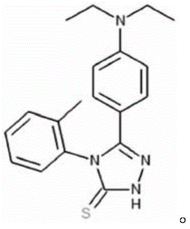
在本发明的另一方面,提供一种制备1型强直性肌营养不良症的动物模型的方法,包括:改造动物的Dmpk,Six5,Mbnl1和Dmwd基因,使这些基因呈现Dmpk+/-,Six5+/-,Mbnl1+/-和Dmwd+/-的表型。In another aspect of the present invention, a method for preparing an animal model of
在一个优选例中,所述的动物包括:啮齿类动物、非人灵长类动物;较佳地,所述的啮齿类动物包括鼠(如小鼠或大鼠)。In a preferred embodiment, the animals include: rodents and non-human primates; preferably, the rodents include mice (such as mice or rats).
在另一优选例中,所述非人灵长类动物包括猕猴或绒猴。In another preferred embodiment, the non-human primate includes a macaque or a marmoset.
在另一优选例中,所述的改造包括:基因敲除;较佳地,包括以CRISPR基因编辑方法进行敲除。In another preferred embodiment, the modification includes: gene knockout; preferably, includes knockout using CRISPR gene editing method.
在另一优选例中,所述的动物来自于孤雄单倍体胚胎干细胞;较佳地,以孤雄单倍体技术结合CRISPR技术,可通过一步法获得呈现Dmpk+/-,Six5+/-,Mbnl1+/-和Dmwd+/-表型的动物模型。In another preferred embodiment, the animal is derived from haploid embryonic stem cells; preferably, by combining haploid technology with CRISPR technology, an animal model exhibiting Dmpk+/-, Six5+/-, Mbnl1+/- and Dmwd+/- phenotypes can be obtained in a one-step method.
在本发明的另一方面,提供一种制备动物细胞模型的方法,包括:改造细胞中的Dmpk,Six5,Mbnl1和Dmwd基因,使这些基因呈现Dmpk+/-,Six5+/-,Mbnl1+/-和Dmwd+/-的表型。In another aspect of the present invention, a method for preparing an animal cell model is provided, comprising: modifying the Dmpk, Six5, Mbnl1 and Dmwd genes in the cells so that these genes present the phenotypes of Dmpk+/-, Six5+/-, Mbnl1+/- and Dmwd+/-.
在一个优选例中,,所述的细胞为肌肉细胞。In a preferred embodiment, the cells are muscle cells.
在另一优选例中,所述的细胞为分离的、离体的细胞。In another preferred embodiment, the cells are separated and isolated cells.
在另一优选例中,所述的细胞包括细胞培养物。In another preferred embodiment, the cell comprises a cell culture.
在本发明的另一方面,提供一种筛选促进肌肉干细胞分化的物质(包括潜在物质)的方法,所述方法包括:(1)用候选物质处理基因组中存在Dmpk+/-,Six5+/-,Mbnl1+/-和Dmwd+/-表型的细胞或细胞培养物;和(2)检测所述存在Dmpk+/-,Six5+/-,Mbnl1+/-和Dmwd+/-表型的细胞的分化情况;若所述候选物质在统计学上促进该细胞的分化,则表明该候选物质是促进肌肉干细胞分化的潜在物质。In another aspect of the present invention, a method for screening substances (including potential substances) that promote muscle stem cell differentiation is provided, the method comprising: (1) treating cells or cell cultures having Dmpk+/-, Six5+/-, Mbnl1+/- and Dmwd+/- phenotypes in their genomes with candidate substances; and (2) detecting the differentiation of cells having Dmpk+/-, Six5+/-, Mbnl1+/- and Dmwd+/- phenotypes; if the candidate substance statistically promotes the differentiation of the cells, it indicates that the candidate substance is a potential substance that promotes muscle stem cell differentiation.
在一个优选例中,步骤(1)包括:在测试组中,将候选物质加入存在Dmpk+/-,Six5+/-,Mbnl1+/-和Dmwd+/-表型的细胞或细胞培养物中;和/或步骤(2)包括:检测测试组的体系中细胞的分化情况,并与对照组比较,其中所述的对照组是不添加所述候选物质的存在Dmpk+/-,Six5+/-,Mbnl1+/-和Dmwd+/-表型的细胞或细胞培养物;如果测试组中所述细胞的分化效率显著高于对照组,则表明该候选物质是促进肌肉干细胞分化的潜在物质。In a preferred embodiment, step (1) includes: in the test group, adding the candidate substance to cells or cell cultures with Dmpk+/-, Six5+/-, Mbnl1+/- and Dmwd+/- phenotypes; and/or step (2) includes: detecting the differentiation of cells in the test group system and comparing it with that of the control group, wherein the control group is cells or cell cultures with Dmpk+/-, Six5+/-, Mbnl1+/- and Dmwd+/- phenotypes to which the candidate substance is not added; if the differentiation efficiency of the cells in the test group is significantly higher than that in the control group, it indicates that the candidate substance is a potential substance for promoting muscle stem cell differentiation.
在另一优选例中,所述的候选物质是小分子化合物,干扰分子,基因编辑试剂,小RNA等。In another preferred embodiment, the candidate substance is a small molecule compound, an interfering molecule, a gene editing reagent, a small RNA, etc.
在本发明的另一方面,提供一种筛选缓解或治疗1型强直性肌营养不良症的物质(包括潜在物质)的方法,包括:(1)用候选物质处理基因组中存在Dmpk+/-,Six5+/-,Mbnl1+/-和Dmwd+/-表型的动物模型;和(2)检测(1)的动物模型所呈现的形态或症状变化;若所述候选物质在统计学上改善动物1型强直性肌营养不良症,则表明该候选物质是缓解或治疗1型强直性肌营养不良症的物质。In another aspect of the present invention, a method for screening substances (including potential substances) for alleviating or treating
本发明的其它方面由于本文的公开内容,对本领域的技术人员而言是显而易见的。Other aspects of the present invention will be apparent to those skilled in the art in view of the disclosure herein.
附图说明BRIEF DESCRIPTION OF THE DRAWINGS
图1、DSM-TKO半克隆小鼠的获得和骨骼肌相关表型分析。a,DSM-TKO半克隆小鼠获得示意图;b,DSM-AGH细胞系;c,小鼠出生后生长曲线。d,DSM-TKO半克隆小鼠出现佝偻表型;e,EMG检测;f,小鼠跑步机实验统计;g,小鼠前爪抓力分析;h,小鼠Rotarod分析;i,抗肌萎缩蛋白染色,肌肉结构完整;j,胫骨前肌H&E组织染色;k,半克隆小鼠肌纤维横截面统计。Figure 1. DSM-TKO semi-cloned mice and skeletal muscle-related phenotype analysis. a, Schematic diagram of DSM-TKO semi-cloned mice; b, DSM-AGH cell line; c, postnatal growth curve of mice. d, DSM-TKO semi-cloned mice showed ricket phenotype; e, EMG detection; f, treadmill test statistics of mice; g, front paw grip analysis of mice; h, Rotarod analysis of mice; i, dystrophin staining, intact muscle structure; j, H&E tissue staining of tibialis anterior muscle; k, cross-sectional statistics of muscle fibers of semi-cloned mice.
图2、DSMD-QKO半克隆小鼠的CDM1呼吸系统相关表型。a,DSMD-QKO-DAH细胞系。b,DSMD-QKO半克隆小鼠部分出生死亡;c,部分新生DSMD-QKO半克隆小鼠肺沉于水底。d,小鼠膈肌H&E染色(P1);e,膈肌肌肉纤维横截面统计;f,膈肌神经肌肉接头染色与统计。Figure 2. CDM1 respiratory system-related phenotypes of DSMD-QKO semi-cloned mice. a, DSMD-QKO-DAH cell line. b, Some DSMD-QKO semi-cloned mice died at birth; c, Some newborn DSMD-QKO semi-cloned mice sank to the bottom of the water. d, H&E staining of mouse diaphragm (P1); e, statistics of diaphragm muscle fiber cross-sections; f, staining and statistics of diaphragm neuromuscular junctions.
图3、CDM1相关的其他表型。a-b,DSMD-QKO半克隆小鼠胎儿在后续哺育过程中生长缓慢。c,DSMD-QKO半克隆小鼠跖骨发育异常(P1)。d,肌肉H&E染色(P2)。e,DSMD-QKO半克隆小鼠发育明显迟缓(P5)。f,DSMD-QKO半克隆小鼠肌肉张力分析。g,心脏H&E染色。Figure 3. Other phenotypes associated with CDM1. a-b, DSMD-QKO semi-cloned mouse fetuses grow slowly during subsequent feeding. c, DSMD-QKO semi-cloned mice have abnormal metatarsal development (P1). d, H&E staining of muscles (P2). e, DSMD-QKO semi-cloned mice have obvious developmental delay (P5). f, muscle tension analysis of DSMD-QKO semi-cloned mice. g, H&E staining of the heart.
图4、DSMD-QKO半克隆小鼠成年后相关表型。a,DSMD-QKO半克隆小鼠出现强直表型。b,小鼠跑步机实验统计;c,小鼠前爪抓力实验统计;d,小鼠Rotarod实验统计;e,抗肌萎缩蛋白染色无差异;f,胫骨前肌H&E染色;g,肌肉纤维横截面统计;h,胫骨前肌ATPase染色及I型肌肉纤维分析;i,白内障表型。Figure 4. Phenotypes of DSMD-QKO semi-cloned mice in adulthood. a. DSMD-QKO semi-cloned mice show ankylosing phenotype. b. Statistics of treadmill test of mice; c. Statistics of forepaw grip test of mice; d. Statistics of Rotarod test of mice; e. No difference in dystrophin staining; f. H&E staining of tibialis anterior muscle; g. Statistics of muscle fiber cross-section; h. ATPase staining of tibialis anterior muscle and analysis of type I muscle fibers; i. Cataract phenotype.
图5、DSM-TKO和DSMD-QKO的MuSCs表型。a,胫骨前肌pax7蛋白免疫荧光。b,胫骨前肌pax7染色后干细胞统计。c,MuSCs体外增殖实验。d,MuSCs体外分化2天。e,MuSCs体外分化2天统计统计。Figure 5. Phenotypes of MuSCs in DSM-TKO and DSMD-QKO. a, Immunofluorescence of Pax7 protein in tibialis anterior muscle. b, Stem cell statistics after Pax7 staining in tibialis anterior muscle. c, MuSCs proliferation assay in vitro. d, MuSCs differentiated in vitro for 2 days. e, MuSCs differentiated in vitro for 2 days.
图6、分化过程中肌肉相关基因的表达情况。a,肌肉相关基因转录组测序热图。b,肌肉分化相关基因的qRT-PCR检测。Figure 6. Expression of muscle-related genes during differentiation. a, Heat map of transcriptome sequencing of muscle-related genes. b, qRT-PCR detection of muscle differentiation-related genes.
图7、MuSCs体外分化系统结合小分子库。a,筛选流程;b,化合物结构;c,小分子化合物促进了DSM-TKO肌肉干细胞的分化。Figure 7. MuSCs in vitro differentiation system combined with small molecule library. a, screening process; b, compound structure; c, small molecule compounds promoted the differentiation of DSM-TKO muscle stem cells.
具体实施方式DETAILED DESCRIPTION
本发明人经过深入的研究,结合孤雄单倍体胚胎干细胞介导的半克隆技术以及基因编辑技术,确定了一些与1型强直性肌营养不良症(DM1)的发生、发展密切相关的基因,它们是Dmpk,Six5,Mbnl1和Dmwd基因,本发明人对它们进行了改造,获得呈现Dmpk+/-,Six5+/-,Mbnl1+/-和Dmwd+/-表型的动物或细胞,所述的动物呈现1型强直性肌营养不良症的典型表型。所述动物模型可用于进行1型强直性肌营养不良症的研究,并可以用于特定药物、基因治疗或其它治疗方案的筛选和测试试验。After in-depth research, the inventors have combined semi-cloning technology mediated by haploid embryonic stem cells and gene editing technology to identify some genes closely related to the occurrence and development of myotonic dystrophy type 1 (DM1), which are Dmpk, Six5, Mbnl1 and Dmwd genes. The inventors have modified them to obtain animals or cells showing Dmpk+/-, Six5+/-, Mbnl1+/- and Dmwd+/- phenotypes, and the animals show a typical phenotype of
利用所构建的细胞模型,本发明人还筛选到一种具有促进肌肉干细胞分化功能的小分子化合物,为开发治疗DM1疾病的药物提供了新的途径。Using the constructed cell model, the inventors also screened a small molecule compound that has the function of promoting muscle stem cell differentiation, providing a new approach for the development of drugs for the treatment of DM1 disease.
模型及其构建Model and its construction
本发明人通过基因水平的遗传学研究,经过反复研究论证,锁定一系列与DM1疾病密切相关的致病基因,通过对所述目标基因的改造来制备DM1实验模型。所述的实验模型包括动物模型或细胞模型等。The inventors have conducted genetic research at the gene level and after repeated research and demonstration, have identified a series of pathogenic genes closely related to DM1 disease, and prepared a DM1 experimental model by modifying the target genes. The experimental model includes an animal model or a cell model.
前期工作中已发现,Dmpk、Six5、Mbnl1和Dmwd这些基因的单独敲除小鼠不能模拟DM1的典型症状。因此本发明人推测DM1是CTG重复引起多个基因表达异常所致的。为了验证这一假设,本发明人利用了孤雄单倍体胚胎干细胞介导的半克隆技术,在单倍体细胞中针对Dmpk、Six5、Mbnl1和Dmwd进行组合敲除,然后通过注入卵母细胞中一步获取携带不同组合突变的杂合小鼠,进而分析表型,获得能模拟DM1的理想小鼠模型。通过表型分析,本发明人发现该模型小鼠能够很好的模拟DM1疾病的相关症状。为了进一步探讨其发病机制,本发明人还分离相应的肌肉干细胞,为相关机制的深入探讨提供工具。It has been found in previous work that mice with single knockout of genes such as Dmpk, Six5, Mbnl1 and Dmwd cannot simulate the typical symptoms of DM1. Therefore, the inventors speculate that DM1 is caused by abnormal expression of multiple genes caused by CTG repeats. In order to verify this hypothesis, the inventors used semi-cloning technology mediated by haploid embryonic stem cells to knock out Dmpk, Six5, Mbnl1 and Dmwd in haploid cells, and then obtained heterozygous mice carrying different combinations of mutations in one step by injecting them into oocytes, and then analyzed the phenotype to obtain an ideal mouse model that can simulate DM1. Through phenotypic analysis, the inventors found that the model mouse can well simulate the relevant symptoms of DM1 disease. In order to further explore its pathogenesis, the inventors also isolated the corresponding muscle stem cells to provide tools for in-depth exploration of related mechanisms.
根据本发明的新发现,本发明提供了制备DM1疾病实验模型的方法。所述方法包括:对实验对象进行基因改造,从而使之存在Dmpk+/-,Six5+/-,Mbnl1+/-和Dmwd+/-的表型。According to the new discovery of the present invention, the present invention provides a method for preparing a DM1 disease experimental model, which comprises: genetically modifying the experimental subject so that the subject has the phenotype of Dmpk+/-, Six5+/-, Mbnl1+/- and Dmwd+/-.
所述制备实验模型的思路可以应用于制备细胞模型和动物模型。所述的细胞模型包括哺乳动物细胞模型或其它真核细胞模型,如人类细胞或小鼠细胞。所述的动物包括但不限于:啮齿类动物(包括大鼠,小鼠,仓鼠等),非人灵长类动物(包括石蟹猴,猕猴,绒猴等)等模式动物。优选地,所述动物为啮齿类动物,包括但并不限于:大鼠、小鼠。应理解,本发明保护的核心在于Dmpk,Six5,Mbnl1和Dmwd基因功能缺失性突变导致DM1疾病这一相关性的模型设计和实现策略,包括但不限于本发明后续实施例记载的方案。The idea of preparing the experimental model can be applied to the preparation of cell models and animal models. The cell model includes a mammalian cell model or other eukaryotic cell models, such as human cells or mouse cells. The animals include but are not limited to: rodents (including rats, mice, hamsters, etc.), non-human primates (including crab monkeys, macaques, marmosets, etc.) and other model animals. Preferably, the animal is a rodent, including but not limited to: rats and mice. It should be understood that the core of the protection of the present invention lies in the model design and implementation strategy of the correlation between Dmpk, Six5, Mbnl1 and Dmwd gene loss-of-function mutations leading to DM1 disease, including but not limited to the schemes recorded in the subsequent embodiments of the present invention.
可使用多种方法在动物或细胞中形成Dmpk+/-,Six5+/-,Mbnl1+/-和Dmwd+/-的表型,包括:通过干细胞打靶(同源重组,转座子定点失活等)进行基因敲除;或通过基因编辑技术(包括CRISPR/cas9(或者spf1),TALEN,锌指核酸酶,Base editing技术等)等。本领域已知的多种使得基因或多肽下调表达或失活的方法也是可用的。A variety of methods can be used to form Dmpk+/-, Six5+/-, Mbnl1+/- and Dmwd+/- phenotypes in animals or cells, including: gene knockout by stem cell targeting (homologous recombination, transposon site-directed inactivation, etc.); or by gene editing technology (including CRISPR/cas9 (or spf1), TALEN, zinc finger nuclease, Base editing technology, etc.), etc. A variety of methods known in the art for downregulating expression or inactivating genes or polypeptides are also available.
作为本发明的优选方式,结合孤雄单倍体胚胎干细胞介导的半克隆技术以及基因编辑技术以形成Dmpk+/-,Six5+/-,Mbnl1+/-和Dmwd+/-的表型。较佳地,利用来自于孤雄单倍体胚胎干细胞的动物结合CRISPR技术,可通过一步法获得呈现Dmpk+/-,Six5+/-,Mbnl1+/-和Dmwd+/-表型的动物模型。在更具体的方式中,采用CRISPR/Cas9系统进行基因编辑,通过选择合适的sgRNA靶位点,可带来高的基因编辑效率。As a preferred embodiment of the present invention, semi-cloning technology mediated by haploid embryonic stem cells and gene editing technology are combined to form the phenotypes of Dmpk+/-, Six5+/-, Mbnl1+/- and Dmwd+/-. Preferably, by combining animals from haploid embryonic stem cells with CRISPR technology, an animal model showing the phenotypes of Dmpk+/-, Six5+/-, Mbnl1+/- and Dmwd+/- can be obtained in a one-step method. In a more specific embodiment, gene editing is performed using the CRISPR/Cas9 system, and high gene editing efficiency can be achieved by selecting appropriate sgRNA target sites.
在本发明的优选实施例中,设计并提供了优选的靶位点。将sgRNA或能形成所述sgRNA的核酸、Cas9 mRNA或能形成所述Cas9 mRNA的核酸共同导入动物(如小鼠)受精卵中,获得经基因编辑的动物(如小鼠)。可以体外转录获得Cas9 mRNA以及sgRNA,也可将能形成所述sgRNA的核酸为核酸构建体或表达载体,或所述的能形成所述Cas9 mRNA的核酸为核酸构建体或表达载体,将这些表达载体导入到细胞内,从而在细胞内形成有活性的sgRNA及Cas9 mRNA。其导入方式,对于受精卵优选显微注射,对于细胞优选电转或脂质体转染,但不排除现有其它有效的导入方法。In a preferred embodiment of the present invention, a preferred target site is designed and provided. SgRNA or a nucleic acid capable of forming the sgRNA, Cas9 mRNA or a nucleic acid capable of forming the Cas9 mRNA are co-introduced into a fertilized egg of an animal (such as a mouse) to obtain a gene-edited animal (such as a mouse). Cas9 mRNA and sgRNA can be obtained by in vitro transcription, or the nucleic acid capable of forming the sgRNA can be a nucleic acid construct or an expression vector, or the nucleic acid capable of forming the Cas9 mRNA is a nucleic acid construct or an expression vector, and these expression vectors are introduced into cells, thereby forming active sgRNA and Cas9 mRNA in the cells. The introduction method is preferably microinjection for fertilized eggs, and preferably electroporation or liposome transfection for cells, but other existing effective introduction methods are not excluded.
作为另一选择方式,也可应用Cre和loxp方法使动物或细胞的基因组中相关基因选择性的敲除,表达降低或失活。As another alternative, the Cre and loxp methods can also be used to selectively knock out, reduce or inactivate the expression of related genes in the genome of animals or cells.
本发明构建的实验模型可以用于特定药物的筛选和测试试验,可作为科学研究和新药评价的有力工具,其稳定性好。The experimental model constructed by the present invention can be used for screening and testing of specific drugs, and can be used as a powerful tool for scientific research and new drug evaluation, and has good stability.
本发明还提供了一种利用本发明的实验模型(包括动物模型或细胞模型)筛选治疗DM1疾病的候选药物或治疗剂的方法,包括将候选物质给予所述的试验模型,观测试验模型是否存在疾病的缓解或改善,若是存在,则该候选物质为潜在的DM1疾病的潜在药物。The present invention also provides a method for screening candidate drugs or therapeutic agents for treating DM1 disease using the experimental model of the present invention (including animal models or cell models), comprising administering a candidate substance to the experimental model and observing whether the experimental model has disease alleviation or improvement. If so, the candidate substance is a potential drug for DM1 disease.
在本发明中,候选药物或治疗剂是指已知具有某种药理学活性或正在被检测的可能具有某种药理学活性的物质,包括但不限于核酸、蛋白、糖类、化学合成的小分子或大分子化合物、细胞、天然产物提取物,及以上物质的复合组分等。候选药物或治疗剂的给药方式可以是口服、静脉注射、腹腔注射、皮下注射、灌胃等。In the present invention, candidate drugs or therapeutic agents refer to substances that are known to have certain pharmacological activities or are being tested and may have certain pharmacological activities, including but not limited to nucleic acids, proteins, carbohydrates, chemically synthesized small molecules or macromolecular compounds, cells, natural product extracts, and composite components of the above substances. The administration of candidate drugs or therapeutic agents can be oral, intravenous, intraperitoneal, subcutaneous, gavage, etc.
基于本发明的方法,本发明还提供了用于制备DM1疾病实验模型的试剂盒,所述试剂盒中包括:使基因组中形成Dmpk+/-,Six5+/-,Mbnl1+/-和Dmwd+/-的表型的试剂。所述的试剂盒中,还可包括说明实施本发明的制备DM1疾病动物模型/细胞模型的方法的使用说明书,以便于本领域技术人员应用。Based on the method of the present invention, the present invention also provides a kit for preparing a DM1 disease experimental model, the kit comprising: reagents for forming the phenotypes of Dmpk+/-, Six5+/-, Mbnl1+/- and Dmwd+/- in the genome. The kit may also include instructions for implementing the method of preparing a DM1 disease animal model/cell model of the present invention, so as to facilitate application by those skilled in the art.
筛药方法Drug screening method
已有工作表明,DM1患者体内分离的肌肉干细胞(MuSCs)分化存在异常,为了验证构建的模型小鼠是否存在MuSCs分化异常,本发明人利用之前建立起来的肌肉干细胞培养方法,首先从DM1小鼠模型中分离和培养MuSCs,随后进行该肌肉干细胞的功能和基因表达评价,分析是否与正常的肌肉干细胞存在差异。接着,本发明人又进行一系列小分子库筛选,该库是英国的LifeArc提供的,尝试找出促进肌肉干细胞分化的小分子化合物。Existing work has shown that there are abnormalities in the differentiation of muscle stem cells (MuSCs) isolated from DM1 patients. In order to verify whether the constructed model mice have abnormal MuSCs differentiation, the inventors used the previously established muscle stem cell culture method to first isolate and culture MuSCs from the DM1 mouse model, and then evaluated the function and gene expression of the muscle stem cells to analyze whether there are differences with normal muscle stem cells. Then, the inventors screened a series of small molecule libraries provided by LifeArc in the UK, trying to find small molecule compounds that promote muscle stem cell differentiation.
在获得了上述的实验模型(包括动物模型或细胞模型)后,可以基于该特征来筛选潜在的促进肌肉干细胞分化的物质。根据原理,所述促进肌肉干细胞分化的物质潜在地也可以用于肌肉减少症,肌肉衰老或肌肉萎缩症;较佳地,所述的肌肉萎缩症包括1型强直性肌营养不良症。可从所述的物质中找到对于改善或治疗疾病真正有用的药物。After obtaining the above experimental model (including animal model or cell model), potential substances that promote muscle stem cell differentiation can be screened based on the characteristics. According to the principle, the substances that promote muscle stem cell differentiation can also potentially be used for sarcopenia, muscle aging or muscular dystrophy; preferably, the muscular dystrophy includes
因此,本发明提供一种筛选促进肌肉干细胞分化的潜在物质的方法,所述方法包括:(1)用候选物质处理基因组中存在Dmpk+/-,Six5+/-,Mbnl1+/-和Dmwd+/-表型的细胞或细胞培养物;和(2)检测所述存在Dmpk+/-,Six5+/-,Mbnl1+/-和Dmwd+/-表型的细胞的分化情况;若所述候选物质在统计学上促进该细胞的分化,则表明该候选物质是促进肌肉干细胞分化的潜在物质。Therefore, the present invention provides a method for screening potential substances that promote muscle stem cell differentiation, the method comprising: (1) treating cells or cell cultures having Dmpk+/-, Six5+/-, Mbnl1+/- and Dmwd+/- phenotypes in their genome with candidate substances; and (2) detecting the differentiation of cells having Dmpk+/-, Six5+/-, Mbnl1+/- and Dmwd+/- phenotypes; if the candidate substance statistically promotes the differentiation of the cells, it indicates that the candidate substance is a potential substance that promotes muscle stem cell differentiation.
在本发明的优选方式中,在进行筛选时,为了更易于观察到细胞分化的变化情况,还可设置对照组,所述的对照组可以是不添加所述候选物质的存在Dmpk+/-,Six5+/-,Mbnl1+/-和Dmwd+/-表型的体系。In a preferred embodiment of the present invention, during screening, a control group may be set up to more easily observe changes in cell differentiation. The control group may be a system with Dmpk+/-, Six5+/-, Mbnl1+/- and Dmwd+/- phenotypes without the addition of the candidate substance.
作为本发明的优选方式,所述的方法还包括:对获得的潜在物质进行进一步的细胞实验和/或动物试验,以进一步选择和确定对于改善或促进肌肉干细胞分化真正有用的物质。As a preferred embodiment of the present invention, the method further comprises: conducting further cell experiments and/or animal experiments on the obtained potential substances to further select and determine substances that are truly useful for improving or promoting muscle stem cell differentiation.
在本发明的实施例中,利用MuSCs体外分化系统,筛选对MuSCs相关疾病和老年肌肉减少症有效的小分子。在本发明人构建的DM1疾病小鼠模型中,本发明人采用杂合敲除DM1相关基因的方式来开展研究,这与临床比较接近。同时本发明人第一次观察小鼠模型MuSCs分化异常,提出了MuSCs体外分化系统可以模拟体内分化的进行,进而利用体外分化系统筛选出对MuSCs分化有促进作用的小分子化合物。这样的分化系统也可以用于其他的疾病的研究。譬如,在年老人群中存在肌肉减少症,骨骼肌是人体运动系统的动力,肌肉的衰老和萎缩是人体衰老的重要标志,非常容易引起骨折以及关节损伤等,同时有文章证明了年老人群的MuSCs分化能力显著下降。因此结合MuSCs体外分化系统,可筛选出促进其MuSCs分化的小分子化合物,进而延缓肌肉衰老。In an embodiment of the present invention, a MuSCs in vitro differentiation system is used to screen small molecules that are effective for MuSCs-related diseases and sarcopenia in the elderly. In the DM1 disease mouse model constructed by the inventors, the inventors used a heterozygous knockout of DM1-related genes to carry out research, which is closer to the clinic. At the same time, the inventors observed abnormal differentiation of MuSCs in the mouse model for the first time, and proposed that the MuSCs in vitro differentiation system can simulate the process of differentiation in vivo, and then use the in vitro differentiation system to screen out small molecule compounds that promote MuSCs differentiation. Such a differentiation system can also be used for the study of other diseases. For example, sarcopenia exists in the elderly population, skeletal muscle is the power of the human motor system, and muscle aging and atrophy are important signs of human aging, which are very likely to cause fractures and joint injuries, etc. At the same time, there are articles that prove that the differentiation ability of MuSCs in the elderly population is significantly reduced. Therefore, combined with the MuSCs in vitro differentiation system, small molecule compounds that promote the differentiation of MuSCs can be screened, thereby delaying muscle aging.
促进肌肉干细胞分化的化合物Compounds that promote muscle stem cell differentiation
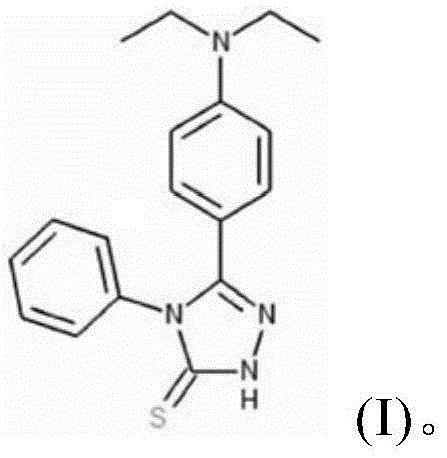
本发明首先提供了一种具有结构式(I)所示的母核结构的化合物:The present invention first provides a compound having a core structure shown in structural formula (I):

其中,R独立地选自:氢、羟基、C1-C4烷基、C2-C4链烯基、C2-C4链炔基、卤素。较佳地,R独立地选自:氢、羟基、C1-C2烷基。Wherein, R is independently selected from: hydrogen, hydroxyl, C1-C4 alkyl, C2-C4 alkenyl, C2-C4 alkynyl, halogen. Preferably, R is independently selected from: hydrogen, hydroxyl, C1-C2 alkyl.
本发明还包括上述具有式(I)所示母核结构的化合物的异构体、溶剂合物、前体,或它们的药学上可接受的盐,只要它们也具有与式(I)所示母核结构的化合物具有相同或基本相同的功能。所述的“药学上可接受的盐”是指化合物与无机酸、有机酸、碱金属或碱土金属等反应生成的盐。这些盐包括(但不限于):(1)与如下无机酸形成的盐:如盐酸、硫酸、硝酸、磷酸;(2)与如下有机酸形成的盐,如乙酸、草酸、丁二酸、酒石酸、甲磺酸、马来酸、或精氨酸。其它的盐包括与碱金属或碱土金属(如钠、钾、钙或镁)形成的盐,以酯、氨基甲酸酯,或其它常规的“前体药物”的形式。化合物具有一个或多个不对称中心。所以,这些化合物可以作为外消旋的混合物、单独的对映异构体、单独的非对映异构体、非对映异构体混合物、顺式或反式异构体存在。The present invention also includes isomers, solvates, precursors, or pharmaceutically acceptable salts of the above-mentioned compounds having the parent core structure shown in formula (I), as long as they also have the same or substantially the same function as the compound having the parent core structure shown in formula (I). The "pharmaceutically acceptable salt" refers to a salt formed by the reaction of a compound with an inorganic acid, an organic acid, an alkali metal or an alkaline earth metal. These salts include (but are not limited to): (1) salts formed with the following inorganic acids: such as hydrochloric acid, sulfuric acid, nitric acid, phosphoric acid; (2) salts formed with the following organic acids, such as acetic acid, oxalic acid, succinic acid, tartaric acid, methanesulfonic acid, maleic acid, or arginine. Other salts include salts formed with alkali metals or alkaline earth metals (such as sodium, potassium, calcium or magnesium) in the form of esters, carbamates, or other conventional "prodrugs". The compounds have one or more asymmetric centers. Therefore, these compounds can exist as racemic mixtures, individual enantiomers, individual diastereomers, diastereoisomer mixtures, cis or trans isomers.
所述的“化合物的前体”指当用适当的方法服用后,该化合物的前体在病人体内进行代谢或化学反应而转变成结构式(I)所示母核结构的一种化合物,或化学结构式(I)所示母核结构的一个化合物所组成的盐或溶液。The "precursor of a compound" refers to a compound having a parent core structure shown in structural formula (I) or a salt or solution composed of a compound having a parent core structure shown in chemical formula (I) which undergoes metabolism or chemical reaction in the patient's body after being taken by an appropriate method.
作为本发明的一种优选方式,所述的化合物为:As a preferred embodiment of the present invention, the compound is:

作为一种上述化合物的异构体,其具有以下的结构:As an isomer of the above compound, it has the following structure:

本领域人员应理解,在得知了本发明化合物的结构以后,可通过多种本领域熟知的方法、利用公知的原料,来获得本发明的化合物,比如化学合成或从生物(如动物或植物)中提取或在提取基础上进行改造的方法,这些方法均包含在本发明中。Those skilled in the art should understand that, after knowing the structure of the compound of the present invention, the compound of the present invention can be obtained by a variety of methods well known in the art, using well-known raw materials, such as chemical synthesis or extraction from organisms (such as animals or plants) or modification based on extraction, and these methods are all included in the present invention.
可以利用公知的方法来合成本发明的化合物;合成的化合物可以进一步通过柱层析法、高效液相色谱法等方式进一步纯化。此外,也可以通过商购的方式获得本发明的化合物。The compounds of the present invention can be synthesized by known methods; the synthesized compounds can be further purified by column chromatography, high performance liquid chromatography, etc. In addition, the compounds of the present invention can also be obtained by commercial means.
本发明人在研究中发现,本发明的式(I)所示母核结构化合物具有显著的促进肌肉干细胞分化的作用。基于本发明人的新发现,本发明提供了式(I)所示的化合物或其异构体、溶剂合物、前体,或它们的药学上可接受的盐的用途,用于制备促进肌肉干细胞分化的组合物,或用于制备缓解或治疗肌肉干细胞分化异常相关疾病的组合物。所述肌肉干细胞分化异常包括:肌肉干细胞分化能力下降或不分化。肌肉干细胞分化异常相关疾病包括:肌肉减少症,肌肉衰老或肌肉萎缩症;较佳地,所述的肌肉萎缩症包括1型强直性肌营养不良症。The inventors have found in their research that the compound with the parent nucleus structure shown in formula (I) of the present invention has a significant effect on promoting the differentiation of muscle stem cells. Based on the new discovery of the inventors, the present invention provides the use of the compound shown in formula (I) or its isomers, solvates, precursors, or pharmaceutically acceptable salts thereof for preparing a composition for promoting the differentiation of muscle stem cells, or for preparing a composition for alleviating or treating diseases related to abnormal differentiation of muscle stem cells. The abnormal differentiation of muscle stem cells includes: decreased differentiation ability of muscle stem cells or no differentiation. Diseases related to abnormal differentiation of muscle stem cells include: sarcopenia, muscle aging or muscular dystrophy; preferably, the muscular dystrophy includes
本发明还提供了一种组合物,含有有效量的式(I)所示母核结构所述的化合物、或其异构体、溶剂合物、前体。The present invention also provides a composition containing an effective amount of the compound of the parent core structure shown in formula (I), or its isomer, solvate, or precursor.
本发明中,所述的“含有”,“具有”或“包括”包括了“包含”、“主要由……构成”、“基本上由……构成”、和“由……构成”;“主要由……构成”、“基本上由……构成”和“由……构成”属于“含有”、“具有”或“包括”的下位概念。In the present invention, the term “contains”, “has” or “includes” includes “comprises”, “mainly consists of”, “substantially consists of”, and “consisting of”; “mainly consists of”, “substantially consists of” and “consisting of” are subordinate concepts of “contains”, “has” or “includes”.
作为一种优选方式,所述的组合物为药物组合物,所述组合物中还含有药学上可接受的盐和/或药学上可接受的载体或赋形剂。As a preferred embodiment, the composition is a pharmaceutical composition, which further contains a pharmaceutically acceptable salt and/or a pharmaceutically acceptable carrier or excipient.
如本文所用,“药学上可接受的”成分是适用于人和/或动物而无过度不良副反应(如毒性、刺激和变态反应)的物质,即有合理的效益/风险比的物质。“药学上可接受的载体”是用于将本发明的化合物传送给动物或人的药学上或食品上可接受的溶剂、悬浮剂或赋形剂。载体可以是液体或固体。As used herein, a "pharmaceutically acceptable" ingredient is a substance that is suitable for use in humans and/or animals without excessive adverse side effects (such as toxicity, irritation, and allergic reactions), i.e., a substance with a reasonable benefit/risk ratio. A "pharmaceutically acceptable carrier" is a pharmaceutically or food-acceptable solvent, suspending agent, or excipient for delivering the compounds of the present invention to animals or humans. The carrier can be a liquid or a solid.
在本发明中,所述的药物组合物含有按照重量比例为0.001-50%的式(I)所示母核结构所示的化合物或其药学上可接受的盐。较佳的,所述的药物组合物含有按照重量比例为0.05-30%的式(I)所示母核结构所示的化合物或其药学上可接受的盐;更佳地,所述的药物组合物含有按照重量比例为0.01-20%的式(I)所示母核结构所示的化合物或其药学上可接受的盐。本领域人员应理解,根据临床实际需求或制药学中的药物设计方式,其它的重量比例也是可行的。In the present invention, the pharmaceutical composition contains 0.001-50% by weight of the compound represented by the parent core structure represented by formula (I) or a pharmaceutically acceptable salt thereof. Preferably, the pharmaceutical composition contains 0.05-30% by weight of the compound represented by the parent core structure represented by formula (I) or a pharmaceutically acceptable salt thereof; more preferably, the pharmaceutical composition contains 0.01-20% by weight of the compound represented by the parent core structure represented by formula (I) or a pharmaceutically acceptable salt thereof. Those skilled in the art should understand that other weight ratios are also feasible according to actual clinical needs or drug design methods in pharmacy.
本发明所述的药物组合物的剂型可以是多种多样的,只要是能够使活性成分有效地到达哺乳动物机体的剂型都是可以的。比如可选自:凝胶剂、气雾剂、片剂、胶囊、粉末、颗粒、糖浆、溶液、或悬浮液。根据本发明的化合物所治疗的疾病类型,本领域人员可以选择方便应用的剂型。从易于制备和给药的立场看,优选的药物组合物是固态组合物,尤其是片剂和固体填充或液体填充的胶囊。本发明的化合物或其药物组合物也可储存在适宜于注射或滴注的消毒器具中。式(I)所示母核结构化合物作为活性成分的有效施用剂量可随给药的模式和待治疗的疾病的严重程度而变化。The dosage form of the pharmaceutical composition of the present invention can be varied, as long as it is a dosage form that can effectively allow the active ingredient to reach the mammalian body. For example, it can be selected from: gel, aerosol, tablet, capsule, powder, granule, syrup, solution, or suspension. According to the type of disease treated by the compound of the present invention, those skilled in the art can choose a dosage form that is convenient for use. From the standpoint of ease of preparation and administration, the preferred pharmaceutical composition is a solid composition, especially tablets and solid-filled or liquid-filled capsules. The compound of the present invention or its pharmaceutical composition can also be stored in a sterile device suitable for injection or instillation. The effective dosage of the parent core structure compound shown in formula (I) as the active ingredient can vary with the mode of administration and the severity of the disease to be treated.
作为另一种优选方式,所述的组合物为培养基组合物,从而可用于在体外/离体状态下培养肌肉干细胞,促进其分化为所需的肌肉干细胞。所述的培养基组合物中的其它成分可以是为细胞提供常规营养成分的物质,这是本领域人员容易获得的。As another preferred embodiment, the composition is a culture medium composition, which can be used to culture muscle stem cells in vitro/ex vivo to promote their differentiation into desired muscle stem cells. Other components in the culture medium composition can be substances that provide conventional nutrients for cells, which are easily available to those skilled in the art.
下面结合具体实施例,进一步阐述本发明。应理解,这些实施例仅用于说明本发明而不用于限制本发明的范围。下列实施例中未注明具体条件的实验方法,通常按照常规条件如J.萨姆布鲁克等编著,分子克隆实验指南,第三版,科学出版社,2002中所述的条件,或按照制造厂商所建议的条件。The present invention is further described below in conjunction with specific examples. It should be understood that these examples are only used to illustrate the present invention and are not intended to limit the scope of the present invention. The experimental methods in the following examples where specific conditions are not specified are usually carried out according to conventional conditions such as those described in J. Sambrook et al., Molecular Cloning Experiment Guide, 3rd edition, Science Press, 2002, or according to the conditions recommended by the manufacturer.
材料和方法Materials and methods
1、骨架质粒与菌株1. Backbone plasmid and strain
pMD19-T vector(Takara);pMD19-T vector (Takara);
px330-mcherry(Addgene);px330-mcherry (Addgene);
DH5α:大肠杆菌(Escherichia coli)。DH5α: Escherichia coli.
2、细胞系2. Cell lines
小鼠胚胎成纤维细胞(mouse embryonic fibroblast,MEF);Mouse embryonic fibroblast (MEF);
孤雄单倍体胚胎干细胞(AG-haESCs)(Yang H,Shi L,Wang BA,Liang D,Zhong C,Liu W et al(2012)Generation of genetically modified mice by oocyte injectionof androgenetic haploid embryonic stem cells.Cell 149:605–617)。Androgenetic haploid embryonic stem cells (AG-haESCs) (Yang H, Shi L, Wang BA, Liang D, Zhong C, Liu W et al (2012) Generation of genetically modified mice by oocyte injection of androgenetic haploid embryonic stem cells. Cell 149: 605–617).
3、抗体3. Antibodies
Anti-Muscleblind-like 1抗体(Abcam,cat.no.ab108519);Anti-Muscleblind-like 1 antibody (Abcam, cat.no.ab108519);
Anti-SIX5抗体(Abcam,cat.no.ab113064);Anti-SIX5 antibody (Abcam, cat. no. ab113064);
Anti-DMWD抗体(Santa cruz,cat.no.sc-167638);Anti-DMWD antibody (Santa cruz, cat. no. sc-167638);
Anti-DMPK抗体(Santa cruz,cat.no.sc-13612)。Anti-DMPK antibody (Santa cruz, cat. no. sc-13612).
4、实验用动物4. Experimental animals
C57/BL6小鼠;C57/BL6 mice;
B6D2F1小鼠:由C57/BL6雌鼠与DBA/2公鼠交配繁育获得;B6D2F1 mice: obtained by mating C57/BL6 female mice with DBA/2 male mice;
ICR小鼠;ICR mice;
IG-DMR敲除小鼠(4.15kb),获自剑桥大学;IG-DMR knockout mice (4.15 kb) were obtained from Cambridge University;
H19 DMR敲除小鼠(3.8kb),获自宾夕法尼亚大学;H19 DMR knockout mice (3.8 kb) were obtained from the University of Pennsylvania;
所有小鼠实验都符合上海生化与细胞研究所实验动物实验管理委员会的规章制度,所有实验小鼠都饲养在上海生化与细胞研究所实验动物平台SPF房间。所有小鼠的饲养周期白天12h,夜晚12h。All mouse experiments were in accordance with the rules and regulations of the Shanghai Institute of Biochemistry and Cell Biology Laboratory Animal Experiment Management Committee, and all experimental mice were kept in the SPF room of the Shanghai Institute of Biochemistry and Cell Biology Laboratory Animal Platform. All mice were kept in a 12-hour day and 12-hour night cycle.
5、小鼠ESCs饲养层细胞的制备5. Preparation of Mouse ESCs Feeder Cells
饲养层细胞用的是灭活后的小鼠胚胎(12.5dpc)成纤维细胞。取12.5dpc的ICR母鼠,颈部脱臼处死后,取出胎儿。PBS清洗胎儿数次,去除胎儿头部和四肢及内脏,剪碎胎儿躯干,加入适量0.05%Trysin-EDTA混匀,置于37℃水浴锅消化10min,消化过程中不时摇晃以免组织沉底阻碍消化的进行,随后终止胰蛋白酶消化,70μm滤网过滤除去未消化大组织块,铺到10cm培养皿中,成纤维细胞长满后用0.05%Trysin-EDTA消化,按1:5比例进行传代。传代2-3次之后,可进行射线辐射灭活,并分管冻存。灭活后的小鼠成纤维细胞可在-80℃冻存一个月,若想长期保存,则需转移到液氮中。The feeder layer cells used were inactivated mouse embryonic fibroblasts (12.5dpc). Take the 12.5dpc ICR mother mouse, dislocate the neck and kill it, and remove the fetus. Wash the fetus several times with PBS, remove the fetal head, limbs and internal organs, cut the fetal trunk into pieces, add an appropriate amount of 0.05% Trysin-EDTA and mix well, place it in a 37℃ water bath for digestion for 10min, shake it from time to time during the digestion process to prevent the tissue from sinking to the bottom and hindering the digestion, then terminate the trypsin digestion, filter out the undigested large tissue blocks with a 70μm filter, spread it into a 10cm culture dish, digest it with 0.05% Trysin-EDTA after the fibroblasts are full, and pass it at a ratio of 1:5. After 2-3 passagings, it can be inactivated by radiation and cryopreserved in separate tubes. The inactivated mouse fibroblasts can be frozen at -80℃ for one month. If you want to store them for a long time, you need to transfer them to liquid nitrogen.
6、MⅡ期卵母细胞的收集6. Collection of MⅡ stage oocytes
对8-10周的B6D2F1母鼠腹腔注射5-6IU PMSG,48h后,注射5-6IU hCG。注射hCG激素14h后可进行取卵。进行取卵操作前先准备好CZB液滴和KSOM液滴培养皿,每个液滴10-20μl。小鼠颈部脱臼处死后,取出输卵管并划出输卵管膨大部的卵团。随后用Hy工作液去除卵丘细胞,在HCZB液滴和CZB液滴清洗数次后可置于CZB液滴培养皿中并短暂培养于恒温37℃的CO2培养箱直至实验结束。8-10 week old B6D2F1 female mice were injected with 5-6 IU PMSG intraperitoneally, and 48 hours later, 5-6 IU hCG was injected. Egg retrieval can be performed 14 hours after the hCG hormone injection. Before the egg retrieval operation, prepare CZB droplets and KSOM droplet culture dishes, each with 10-20 μl. After the mouse was killed by cervical dislocation, the oviduct was removed and the egg mass in the oviduct bulge was cut out. Subsequently, the cumulus cells were removed with Hy working solution, and after washing several times with HCZB droplets and CZB droplets, they could be placed in CZB droplet culture dishes and briefly cultured in a constant temperature 37°C CO2 incubator until the end of the experiment.
7、AG-haESCs的建系、培养和基因操作7. Establishment, culture and genetic manipulation of AG-haESCs
AG-haESCs来自于去核卵母细胞注入断尾精子获得的孤雄单倍体囊胚。在获得囊胚期的单倍体胚胎后,先用TSA消化透明带,随后转移到已经铺就饲养层细胞的96孔板中,每孔放入一个囊胚,孔内事先放入2i培养基。生长4-5d后,0.05%trypsin-EDTA消化传代到96孔板另一个孔中,3-5d后,有可见克隆的孔则继续传代到48孔板中,再到24孔板。在24孔板传代中可把部分细胞进行单倍体检测和分选。FACS分选前,需要对ESCs进行Hoechst33342染色,Hoechst 33342可与双链DNA结合。在FACS分选时,Hoechst 33342可被紫外光激发释放461nm波长的荧光,荧光强度与细胞内Hoechst 33342含量成正比,因此不同染色体倍性的细胞可根据荧光强弱而进行区分。AG-haESCs are derived from the haploid blastocysts obtained by injecting the tail-broken sperm into the enucleated oocytes. After obtaining the haploid embryos in the blastocyst stage, the zona pellucida is first digested with TSA, and then transferred to a 96-well plate with feeder cells, and one blastocyst is placed in each well. 2i culture medium is placed in the wells in advance. After growing for 4-5 days, 0.05% trypsin-EDTA digestion is performed and the cells are passaged to another well of the 96-well plate. After 3-5 days, the wells with visible clones are further passaged to 48-well plates and then to 24-well plates. During the passage in the 24-well plate, some cells can be tested for haploidy and sorted. Before FACS sorting, ESCs need to be stained with Hoechst33342, which can bind to double-stranded DNA. During FACS sorting, Hoechst 33342 can be excited by ultraviolet light to release fluorescence at a wavelength of 461nm. The fluorescence intensity is proportional to the Hoechst 33342 content in the cell. Therefore, cells with different chromosome ploidy can be distinguished based on the fluorescence intensity.
建系后的单倍体,可进行基因操作。After the haploid line is established, genetic manipulation can be performed.
sgRNA质粒的设计:Design of sgRNA plasmid:
px330-SIX5:Six5-sgRNA-f:CACCGTCTGATGGGAACCCCACTA(SEQ ID NO:1);Six5-sgRNA-r:TAGTGGGGTTCCCATCAGAC(SEQ ID NO:2),合成sgRNA序列,插入到px330-mcherry的DBSI酶切位点。px330-SIX5: Six5-sgRNA-f: CACCGTCTGATGGGAACCCCACTA (SEQ ID NO: 1); Six5-sgRNA-r: TAGTGGGGTTCCCATCAGAC (SEQ ID NO: 2), synthesized sgRNA sequence, and inserted into the DBSI restriction site of px330-mcherry.
px330-DMPK:Dmpk-sgRNA-f:CACCGCCCATTGCAGCAAGGCTTA(SEQ ID NO:3);Dmpk-sgRNA-r:AAACTAAGCCTTGCTGCAATGGGC(SEQ ID NO:4),合成sgRNA序列,插入到px330-mcherry的DBSI酶切位点。px330-DMPK: Dmpk-sgRNA-f: CACCGCCCATTGCAGCAAGGCTTA (SEQ ID NO: 3); Dmpk-sgRNA-r: AAACTAAGCCTTGCTGCAATGGGC (SEQ ID NO: 4), synthesized sgRNA sequence, and inserted into the DBSI restriction site of px330-mcherry.
px330-MBNL1:Mbnl1-up-sgRNA-f:CACCGCTGCAACACTATCTGTGAT(SEQ ID NO:5);Mbnl1-up-sgRNA-r:AAACATCACAGATAGTGTTGCAGC(SEQ ID NO:6);Mbnl1-down-sgRNA-f:CACCGGTACATGGTACTCTAAATT(SEQ ID NO:7);Mbnl1-down-sgRNA-r:AAACAATTTAGAGTACCATGTACC(SEQ ID NO:8),合成两段sgRNA序列,插入到px330-mcherry的DBSI酶切位点位点内。px330-MBNL1: Mbnl1-up-sgRNA-f: CACCGCTGCAACACTATCTGTGAT (SEQ ID NO: 5); Mbnl1-up-sgRNA-r: AAACATCACAGATAGTGTTGCAGC (SEQ ID NO: 6); Mbnl1-down-sgRNA-f: CACCGGTACATGGTACTCTAAATT (SEQ ID NO: 7); Mbnl1-down-sgRNA-r: AAACAATTTAGAGTACCATGTACC (SEQ ID NO: 8). Two sgRNA sequences were synthesized and inserted into the DBSI restriction site of px330-mcherry.
px330-DMWD:Dmwd-sgRNA-f:CACCGGCTTCTACAAGCTGCTCCC(SEQ ID NO:9);Dmwd-sgRNA-r:AAACGGGAGCAGCTTGTAGAAGCC(SEQ ID NO:10),合成sgRNA序列,插入到px330-mcherry的DBSI酶切位点位点内。px330-DMWD: Dmwd-sgRNA-f: CACCGGCTTCTACAAGCTGCTCCC (SEQ ID NO: 9); Dmwd-sgRNA-r: AAACGGGAGCAGCTTGTAGAAGCC (SEQ ID NO: 10), synthesized sgRNA sequence, and inserted into the DBSI restriction site of px330-mcherry.
携带靶向目的基因sgRNA的Cas9质粒px330-mcherry可用Lipofectamine 2000转染AG-haESCs,细胞转染后第二天根据转染质粒的荧光和Hoechst33342染色进行FACS分选,收集阳性的单倍体细胞,随后低密度的铺就在6孔板中(每孔不多于10000个细胞),生长6-8d,挑取单个ESCs克隆进行消化,一半传到新的96孔板,一半裂解进行目的基因突变鉴定。确定细胞系基因型后,进行单倍体分选。多基因操作的细胞系,在确定一个基因操作完成后,进行其他基因的操作,如果多基因位于不同染色体,或者同一染色体距离较远时,也可以同时进行多基因突变操作。The Cas9 plasmid px330-mcherry carrying the sgRNA targeting the target gene can be transfected into AG-haESCs using Lipofectamine 2000. The next day after cell transfection, FACS sorting is performed based on the fluorescence of the transfected plasmid and Hoechst33342 staining, and positive haploid cells are collected. Then, they are spread at a low density in a 6-well plate (no more than 10,000 cells per well), grown for 6-8 days, and a single ESCs clone is picked for digestion. Half of them are transferred to a new 96-well plate, and half are lysed for target gene mutation identification. After determining the genotype of the cell line, haploid sorting is performed. For cell lines with multi-gene manipulation, after determining that one gene manipulation is completed, other genes are manipulated. If multiple genes are located on different chromosomes, or if the same chromosome is far away, multi-gene mutation manipulation can also be performed simultaneously.
8、ROSI和ICAHCI实验8. ROSI and ICAHCI experiments
ROSI(Round spermatid injection):即球形精子卵胞浆内显微注射。取成年公鼠的睾丸,剪碎后消化,Hoechst 33342染色后,FACS分选出单倍体的球形精子。提前30minSr2+激活MⅡ期卵母细胞,随后把球形精子细胞核注入到激活的MⅡ期卵母细胞中,每组操作时间不超过30min。操作全部完成后,继续Sr2+激活3h,随后放入KSOM液滴中继续培养,重构胚胎发育到两细胞期后移植到假孕0.5d的ICR母鼠输卵管中(20-25颗重构胚胎/只母鼠),19.5d小鼠发育到期。ROSI (Round spermatid injection): Round spermatid injection. The testicles of adult male mice were cut into pieces and digested. After Hoechst 33342 staining, haploid spherical sperm were sorted out by FACS. Activate MⅡ stage oocytes with Sr 2+ 30 minutes in advance, and then inject the spherical sperm nuclei into the activated MⅡ stage oocytes. The operation time for each group should not exceed 30 minutes. After all the operations were completed, continue Sr 2+ activation for 3 hours, and then continue to culture in KSOM droplets. After the reconstructed embryos developed to the two-cell stage, they were transplanted into the oviducts of ICR female mice with pseudopregnancy of 0.5 days (20-25 reconstructed embryos/female mouse), and the mice matured at 19.5 days.
ICAHCI:提前1天在克隆大小比较合适的AG-haESCs中加入秋水仙素,把细胞阻滞在细胞周期的中期,8-12h消化细胞(80%以上细胞处于中期),加入到操作滴中,选取处于中期的单倍体细胞(无细胞核且细胞较小)注入到卵母细胞内,每组操作时间不超过30min,结束一组后清洗数次置于CZB液滴中,操作完全结束后,重构胚胎Sr2+激活5h,随后放入KSOM液滴中继续培养,重构胚胎发育到两细胞期后移植到假孕0.5天(d)的ICR母鼠输卵管中(30-40颗重构胚胎/只母鼠),19.5d小鼠发育到期。若到期未生,可进行剖腹产。ICAHCI: Colchicine was added to AG-haESCs with a relatively
9、冰冻切片与免疫荧光9. Frozen sections and immunofluorescence
小鼠颈部脱臼后,去除下肢皮肤的覆盖,取出胫骨前肌包埋在OCT中,随后在液氮中速冻15s,保存在-80℃中直至切片。而对于心脏纵切,新取的心脏首先在PBS清洗,并按压去除心脏中的血液,随后在浓度4%的PFA中固定30min,30%蔗糖脱水后,包埋在OCT中,经液氮速冻后保存在-80℃直至切片。切片厚度一般8-10μm。免疫荧光染色的切片首先4%PFA固定,PBS清洗,一抗37℃孵育1h,PBS清洗,二抗37℃孵育1h,然后用DAPI对细胞核标记,封片观察。所有荧光照片在Leica SP8荧光共聚焦显微镜拍摄。对刚出生小鼠膈肌的神经肌肉接头(neuromuscμlar junctions,NMJs)染色,膈肌冰冻切片用偶联Alexa Fluor 594的金环蛇毒素标记乙酰胆碱受体(肌肉纤维),同时用神经丝蛋白H标记神经细胞。After the mouse neck was dislocated, the skin covering the lower limbs was removed, the tibialis anterior muscle was taken out and embedded in OCT, then quickly frozen in liquid nitrogen for 15s and stored at -80℃ until sectioning. For longitudinal sectioning of the heart, the newly taken heart was first washed in PBS, and the blood in the heart was removed by pressing, then fixed in 4% PFA for 30min, dehydrated with 30% sucrose, embedded in OCT, quickly frozen in liquid nitrogen and stored at -80℃ until sectioning. The thickness of the section is generally 8-10μm. The sections for immunofluorescence staining were first fixed with 4% PFA, washed with PBS, incubated with primary antibody at 37℃ for 1h, washed with PBS, incubated with secondary antibody at 37℃ for 1h, and then labeled with DAPI for nuclei and sealed for observation. All fluorescent photos were taken with a Leica SP8 fluorescence confocal microscope. The neuromuscular junctions (NMJs) of the diaphragm of newborn mice were stained. The diaphragm frozen sections were labeled with Alexa Fluor 594-conjugated bungarus toxin to label acetylcholine receptors (muscle fibers), and neurofilament H was used to label neurons.
10、组织学分析10. Histological analysis
对于肌肉、膈肌、小肠和心脏等组织的病理学分析,采用的是伊红美蓝染色方法。肌肉、膈肌和心脏用的是冰冻切片进行染色,小肠用的是石蜡切片进行染色。对于快肌慢肌分析,采用的是ATP酶染色方法。即冰冻切片PH 4.6的酸性环境下孵育5-15min,随后用ATP溶液进行染色,经过1%CaCl2,2%CoCl2,5mM巴比妥酸和1%(NH4)2S处理后,切片进行乙醇梯度脱水,慢肌纤维则显暗灰色,白色为快肌。对于小肠组织切片,新鲜分离的组织,用PBS冲出肠内容物,随后4%PFA 4℃固定过夜,乙醇梯度脱水后包埋在石蜡中而后进行切片,样品和切片均可室温保存,切片厚度5μm。For the pathological analysis of muscle, diaphragm, small intestine and heart, the eosin-methylene blue staining method was used. Muscle, diaphragm and heart were stained with frozen sections, and small intestine was stained with paraffin sections. For the analysis of fast and slow muscles, the ATPase staining method was used. That is, the frozen sections were incubated in an acidic environment of pH 4.6 for 5-15 minutes, and then stained with ATP solution. After being treated with 1% CaCl 2 , 2% CoCl 2 , 5mM barbituric acid and 1% (NH 4 ) 2 S, the sections were dehydrated with ethanol gradient, and slow muscle fibers appeared dark gray, while white was fast muscle. For small intestinal tissue sections, freshly isolated tissues were flushed with PBS to remove intestinal contents, and then fixed with 4% PFA at 4°C overnight. After ethanol gradient dehydration, they were embedded in paraffin and then sectioned. Both samples and sections can be stored at room temperature, and the section thickness is 5μm.
11、小鼠前爪力量检测和Rotarod实验11. Mouse front paw strength test and Rotarod experiment
先把实验小鼠置于匀速运动的滚轴上适应5min,随后调整转轴的速度,5min内从4rpm逐渐增加到40rpm。记录下滚轴掉落的时间,小鼠超过5min还没有掉落的,则记录为300s。正式进行实验前,小鼠提前训练3d。First, the experimental mice were placed on a uniformly moving roller to adapt for 5 minutes, and then the speed of the roller was adjusted, gradually increasing from 4rpm to 40rpm within 5 minutes. The time it took for the roller to fall was recorded. If the mouse did not fall for more than 5 minutes, it was recorded as 300s. Before the formal experiment, the mice were trained for 3 days in advance.
12、小鼠肌电图实验12. Mouse electromyography experiment
通过肌电图实验,可以检测小鼠肌肉是否存在强直表型。实验小鼠在异氟烷麻醉状态下,脱毛膏去除小鼠腹部及腿部的软毛,在小鼠腹部贴上电极片,在腿部插入电极针检测强直电位进行肌电图检测(electromyography,EMG)。Electromyography (EMG) experiments can be used to detect whether the mouse muscles have a tonic phenotype. The experimental mice are anesthetized with isoflurane, and the soft hair on the abdomen and legs of the mice is removed with depilatory cream. Electrodes are attached to the abdomen of the mice, and electrode needles are inserted into the legs to detect tonic potentials for electromyography (EMG).
13、RNA抽提和荧光定量PCR13. RNA extraction and fluorescence quantitative PCR
细胞消化和DPBS清洗后,加入Trizol室温裂解数分钟即可进行RNA抽提。新取的组织(腿部肌肉和心脏)则放入液氮中灭活RNA酶,随后加入Trizol震荡裂解充分进行RNA抽提。在Trizol裂解液中加入氯仿,剧烈震荡后离心,取上清于新的EP管中,加入等量的异戊醇,颠倒混匀后离心去上清,加入75%乙醇洗涤沉淀,离心后弃上清,晾干加ddH2O溶解。After cell digestion and DPBS washing, add Trizol to lyse at room temperature for several minutes to extract RNA. The newly obtained tissues (leg muscles and heart) are placed in liquid nitrogen to inactivate RNase, and then add Trizol to shake and lyse to fully extract RNA. Add chloroform to the Trizol lysis solution, shake vigorously and centrifuge, take the supernatant into a new EP tube, add an equal amount of isoamyl alcohol, invert and mix, centrifuge to remove the supernatant, add 75% ethanol to wash the precipitate, discard the supernatant after centrifugation, dry and add ddH 2 O to dissolve.
取0.5μg RNA进行首先加入反转录试剂盒buffer1去除基因组DNA,随后加入buffer2反转录成cDNA。转录结束后,使用Toyobo公司SYBR Green进行基因表达检测。每个样品重复三次,20μl体系:SYBR Green(10μl),引物(Xμl,5μM),样品(1μl),ddH2O[(9-2X)μl]。PCR程序:94℃变性4min,循环94℃变性10s、60℃退火20s、72℃延伸20s程序40次,72℃延伸4min。GADPH表达量作为基因表达内参。Take 0.5 μg RNA and first add the reverse
14、细胞培养相关试剂14. Cell culture related reagents
ES DMEM(Millipore,cat.no.SLM-220-M).ES DMEM (Millipore, cat. no. SLM-220-M).
DMEM(Thermo Fisher Scientific,cat.no.11965118).DMEM (Thermo Fisher Scientific, cat. no. 11965118).
Opti-MEM(Thermo Fisher Scientific,cat.no.51985042).Opti-MEM (Thermo Fisher Scientific, cat. no. 51985042).
ES FBS(Thermo Fisher Scientific,cat.no.16000044).ES FBS (Thermo Fisher Scientific, cat. no. 16000044).
FBS(Excell,cat.no.FSP500).FBS (Excell, cat. no. FSP500).
Nucleotides(100×)(Millipore,cat.no.ES-008-D).Nucleotides (100×) (Millipore, cat. no. ES-008-D).
Non-Essential Amino Acids(100×)(NEAA;Millipore,cat.no.TMS-001-C).Non-Essential Amino Acids (100×) (NEAA; Millipore, cat. no. TMS-001-C).
L-Glutamine Solution(100×)(Millipore,cat.no.TMS-002-C).L-Glutamine Solution (100×) (Millipore, cat. no. TMS-002-C).
2-Mercaptoethanol(100×)(Millipore,cat.no.ES-007-E).2-Mercaptoethanol (100×) (Millipore, cat. no. ES-007-E).
Penicillin-Streptomycin(Thermo Fisher Scientific,cat.no.15140122).Penicillin-Streptomycin (Thermo Fisher Scientific, cat. no. 15140122).
Leukemia Inhibitory Factor(LIF;Millipore,cat.no.ESG1107).Leukemia Inhibitory Factor (LIF; Millipore, cat. no. ESG1107).
DPBS(Thermo Fisher Scientific,cat.no.14190-144).DPBS (Thermo Fisher Scientific, cat. no. 14190-144).
0.05%Trypsin-EDTA(Thermo Fisher Scientific,cat.no.25300-112).0.05% Trypsin-EDTA (Thermo Fisher Scientific, cat. no. 25300-112).
G418(Sigma,cat.no.A1720).G418 (Sigma, cat. no. A1720).
Demecolcine solution(Sigma,cat.no.D1925).Demecolcine solution (Sigma, cat. no. D1925).
Dimethyl sulfoxide(DMSO;Sigma,cat.no.D8418).Dimethyl sulfoxide (DMSO; Sigma, cat. no. D8418).
RecoveryTM Cell Culture Freezing Medium(Thermo Fisher Scientific,cat.no.12648-010).Recovery TM Cell Culture Freezing Medium (Thermo Fisher Scientific, cat. no. 12648-010).
Lipofectamine 2000(Thermo Fisher Scientific,cat.no.12648-010).Lipofectamine 2000 (Thermo Fisher Scientific, cat. no. 12648-010).
15、小鼠肌肉干细胞的分离和培养15. Isolation and culture of mouse muscle stem cells
取小鼠的肌肉(如胫骨前肌)并剪碎,加入适量0.05%Trysin-EDTA混匀,置于37℃水浴锅消化10min,消化过程中不时摇晃以免组织沉底阻碍消化的进行,随后终止胰蛋白酶消化,铺到10cm培养皿中,肌肉干细胞长满后用0.05%Trysin-EDTA消化,并按适当的比例传代。细胞培养液除了基础的培养液(DMEM+10%FBS+5%Penicillin-Streptomycin)外,添加细胞因子至少包括IL1、IL4、IL13、TNF alpha、IL2和IFN gamma,每种浓度为1ng/ml。浓度之和不低于6ng/ml,较佳地为50-1250ng/ml。Take the muscle of the mouse (such as the tibialis anterior muscle) and cut it into pieces, add an appropriate amount of 0.05% Trysin-EDTA and mix it, place it in a 37°C water bath for digestion for 10 minutes, shake it from time to time during the digestion process to prevent the tissue from sinking to the bottom and hindering the digestion, then terminate the trypsin digestion, spread it into a 10cm culture dish, digest it with 0.05% Trysin-EDTA after the muscle stem cells are full, and pass it in an appropriate proportion. In addition to the basic culture medium (DMEM+10%FBS+5%Penicillin-Streptomycin), the cell culture medium is added with cytokines including at least IL1, IL4, IL13, TNF alpha, IL2 and IFN gamma, each at a concentration of 1ng/ml. The sum of the concentrations is not less than 6ng/ml, preferably 50-1250ng/ml.
实施例1、DSM-TKO杂合突变小鼠的获得和表型分析Example 1. Acquisition and phenotypic analysis of DSM-TKO heterozygous mutant mice
本发明人在得到可以有效支持半克隆小鼠出生的AG-haESCs细胞系后,尝试是否可以利用这一技术一步获得多个基因修饰的小鼠呢?首先利用CRISPR技术构建了单个基因敲除的AG-haESCs细胞系,即ΔDMPK-AGH细胞系(移码突变)。在ΔDMPK-AGH细胞系上进行了SIX5和MBNL1的基因敲除操作(图1a),三基因敲除的细胞系(DSM-AGH细胞系)细胞形态和生长正常(图1b),进行ICAHCI实验后,半克隆小鼠生长也比较正常(图1c)。但是在成年小鼠中出现了佝偻表型(图1d)。对三基因敲除DSM-TKO半克隆小鼠进行EMG实验,检测到强直的出现(图1e);其前爪抓力与对照相比也明显减弱;进行跑步机实验时,其整体的运动能力发生明显的降低;在rotarod试验中,DSM-TKO半克隆小鼠更易掉落(图1f-h),这些都说明DSM-TKO半克隆小鼠肌肉力量减弱,肌肉功能受到了一定的影响。After obtaining the AG-haESCs cell line that can effectively support the birth of semi-cloned mice, the inventors tried to see if this technology could be used to obtain mice with multiple gene modifications in one step. First, a single gene knockout AG-haESCs cell line, namely the ΔDMPK-AGH cell line (frameshift mutation), was constructed using CRISPR technology. Gene knockout operations of SIX5 and MBNL1 were performed on the ΔDMPK-AGH cell line (Figure 1a). The cell morphology and growth of the triple gene knockout cell line (DSM-AGH cell line) were normal (Figure 1b). After the ICAHCI experiment, the growth of semi-cloned mice was also relatively normal (Figure 1c). However, a ricket phenotype appeared in adult mice (Figure 1d). EMG experiments were performed on triple-gene knockout DSM-TKO semi-cloned mice, and the occurrence of spasticity was detected (Figure 1e); their forepaw grip strength was also significantly weakened compared with the control; when conducting treadmill experiments, their overall motor ability was significantly reduced; in the rotarod test, DSM-TKO semi-cloned mice were more likely to fall (Figure 1f-h), all of which indicate that the muscle strength of DSM-TKO semi-cloned mice was weakened and their muscle function was affected to a certain extent.
随后本发明人对小鼠骨骼肌、膈肌、心脏和小肠进行组织学分析。骨骼肌方面,对胫骨前肌切片进行抗肌萎缩蛋白的染色,结果显示肌肉整个结构较完整。对切片进行H&E染色,未发现明显的核内移现象,但是可以观察到肌浆块,核聚集及空泡纤维等表型,同时对肌肉纤维的横截面进行统计,发现肌肉纤维横截面整体都变小(图1i-k),表明肌肉萎缩现象的出现。The inventors then performed histological analysis on mouse skeletal muscle, diaphragm, heart and small intestine. In terms of skeletal muscle, dystrophin staining was performed on tibialis anterior muscle sections, and the results showed that the entire muscle structure was relatively intact. H&E staining of the sections did not reveal obvious nuclear intranslocation, but phenotypes such as sarcoplasmic masses, nuclear aggregation and vacuolar fibers could be observed. At the same time, statistics were performed on the cross-sections of the muscle fibers, and it was found that the cross-sections of the muscle fibers were smaller as a whole (Figure 1i-k), indicating the occurrence of muscle atrophy.
至此,本发明人通过把几个DM1涉及到的基因组合在一起,得到了一个新的DM1疾病小鼠模型,在这个模型中,可以看到骨骼肌,心肌和小肠等均存在一定的异常,与临床结果比较一致。但也有一些表型并没有获得。首先,DSM-TKO半克隆小鼠的强直表型并没有其他几个DM1小鼠模型明显。第二,在肌肉组织学切片分析中,没有观察到明显肌肉纤维核内移现象,但这种现象在临床患者中发生的很普遍(也有患者未发生明显核内移)。第三,未观察到明显的心脏结构和功能上的明显表型,而心脏问题是经典DM1患者猝死的最主要的原因。第四,在小鼠肌肉和心脏组织中不存在大量异常剪接的RNA。这几点说明,这个疾病模型还有其他没有关注的基因依然在其中起重要作用。So far, the inventors have obtained a new DM1 disease mouse model by combining several genes involved in DM1. In this model, it can be seen that there are certain abnormalities in skeletal muscle, myocardium and small intestine, which are consistent with the clinical results. However, some phenotypes are not obtained. First, the ankylosing phenotype of DSM-TKO semi-cloned mice is not as obvious as that of several other DM1 mouse models. Second, in the analysis of muscle histological sections, no obvious muscle fiber nuclear involution was observed, but this phenomenon is very common in clinical patients (some patients do not have obvious nuclear involution). Third, no obvious phenotypes in cardiac structure and function were observed, and cardiac problems are the main cause of sudden death in classic DM1 patients. Fourth, there is no large amount of abnormally spliced RNA in mouse muscle and heart tissue. These points show that there are other genes that have not been paid attention to in this disease model that still play an important role.
实施例2、CDM1小鼠模型的构建Example 2: Construction of CDM1 mouse model
从上述实验可以看出,虽然三基因敲除小鼠出现了DM1疾病的部分表型,但是一些重要的表型并没有模拟出来,包括先天型DM1的一系列表型,成年小鼠中也没有出现心脏功能异常,白内障等表型,这说明可能还有其他基因参与到疾病的发生过程中。From the above experiments, it can be seen that although the triple-gene knockout mice showed some phenotypes of DM1 disease, some important phenotypes were not simulated, including a series of phenotypes of congenital DM1. No abnormal heart function, cataracts and other phenotypes appeared in adult mice, which indicates that other genes may be involved in the occurrence of the disease.
本发明人在DSM-TKO-DAH(DAH指孤雄单倍体干细胞)细胞系上进一步敲除DMWD,获得的DSMD-QKO-DAH细胞系,细胞形态和生长都比较正常(图2a),进行ICAHCI实验时可有效支持半克隆小鼠的出生。但发现大约有20%的小鼠出生后迅速死亡(图2b)。对获得的DSMD-QKO半克隆小鼠进行表型分析发现,存活小鼠的肺脏置于清水中时会浮于水面,而死亡小鼠的肺脏则沉于水底(图2c),这说明死亡小鼠出生后并没进行呼吸。由于先天型DM1的患者很有可能熬过最早的阶段,但是后续中依然存在呼吸的问题,本发明人取刚出生1天的小鼠的膈肌进行切片染色发现,相对于对照小鼠,DSMD-QKO半克隆小鼠的膈肌结构部分区域不太完整(图2d),其肌肉纤维横截面明显变小(图2e)。神经肌肉接头(neuromuscularjunction,NMJ)染色发现,相对于对照,DSMD-QKO半克隆小鼠的成熟接头连接明显变少(图2f)。The inventors further knocked out DMWD on the DSM-TKO-DAH (DAH refers to haploid stem cells) cell line, and obtained the DSMD-QKO-DAH cell line, which has relatively normal cell morphology and growth (Figure 2a), and can effectively support the birth of semi-cloned mice when performing ICAHCI experiments. However, it was found that about 20% of the mice died quickly after birth (Figure 2b). Phenotypic analysis of the obtained DSMD-QKO semi-cloned mice found that the lungs of surviving mice floated on the water surface when placed in clear water, while the lungs of dead mice sank to the bottom of the water (Figure 2c), which shows that the dead mice did not breathe after birth. Since patients with congenital DM1 are likely to survive the earliest stage, but still have breathing problems in the follow-up, the inventors took the diaphragm of mice that were just born one day ago and stained them. It was found that compared with control mice, some areas of the diaphragm structure of DSMD-QKO semi-cloned mice were not complete (Figure 2d), and the cross-section of their muscle fibers was significantly reduced (Figure 2e). Neuromuscular junction (NMJ) staining revealed that the mature junction connections in DSMD-QKO semi-cloned mice were significantly reduced compared with the controls (Figure 2f).
DSMD-QKO半克隆小鼠胎儿在后续哺育过程中,相对于对照小鼠,体重明显较轻,发育迟缓(图3a-b)。对出生1d小鼠进行阿利新蓝和茜素红S染色显示,DSMD-QKO半克隆小鼠的跖骨发育相对迟缓(图3c),一定程度上解释了为什么CDM1疾病新生儿罹患马蹄内翻足,可能是由于胎儿在发育过程中,跖骨钙化较慢,因此在母体怀孕后期可能更容易受到外界影响而发生变形。取出生后2d的小鼠后肢进行HE染色,本发明人发现,DSMD-QKO半克隆小鼠存在很多细胞核位于中央的肌肉纤维(图3d),再次证明DSMD-QKO半克隆小鼠发育迟缓。在半克隆小鼠生长到第五天,本发明人已经可以看到部分DSMD-QKO半克隆小鼠明显与其他小鼠不同,生长较缓慢(图3e)。对此时的新生鼠进行肌肉张力检测,相对于对照,在仰躺条件下,DMSM-QKO半克隆小鼠翻身需要的时间更长(图3f)。在随后的哺育过程中,两周大小的小鼠,依然会存在一定的死亡率,这种突然的死亡与CDM1患者出现的表型也比较类似,疑似是心律传导异常导致的。对小鼠心超实验也表明,部分小鼠确实存在心律传导异常,心脏切片H&E染色也表明其结构异常(图3g)。During the subsequent feeding process, the fetuses of DSMD-QKO semi-cloned mice were significantly lighter and had developmental delays compared to control mice (Figure 3a-b). Alcian blue and Alizarin Red S staining of mice born 1 day ago showed that the metatarsal development of DSMD-QKO semi-cloned mice was relatively slow (Figure 3c), which to some extent explains why newborns with CDM1 disease suffer from clubfoot. It may be because the metatarsal calcification is slow during the development of the fetus, so it may be more susceptible to external influences and deformity in the late pregnancy of the mother. HE staining was performed on the hind limbs of
综上表型情况说明,本发明人得到了先天型DM1疾病小鼠模型。包括骨骼肌,心脏和呼吸系统均存在与临床较类似的表型。同时利用该疾病模型,也得出CDM1呼吸障碍可能是与膈肌结构有关。本发明人观察到DSMD-QKO半克隆小鼠膈肌存在结构不完整,膈肌肌肉纤维力量不足和成熟神经肌肉接头较少等异常,弥补了临床由于样品获取困难而无法深入研究的缺陷。Based on the above phenotypic description, the inventors obtained a congenital DM1 disease mouse model. The skeletal muscle, heart and respiratory system all have phenotypes similar to those in the clinic. At the same time, using this disease model, it was also concluded that CDM1 respiratory disorders may be related to the diaphragm structure. The inventors observed that the diaphragm of the DSMD-QKO semi-cloned mice had structural incompleteness, insufficient diaphragm muscle fiber strength and fewer mature neuromuscular junctions, which made up for the defects of in-depth research in the clinic due to the difficulty in obtaining samples.
在DSMD-QKO成年小鼠中也出现了佝偻表型,EMG实验检测DSMD-QKO半克隆小鼠有强直的出现(图4a)。小鼠前爪抓力明显减弱,同时进行rotarod实验时DSMD-QKO半克隆小鼠更易掉落,跑步机实验也证实其整体的运动能力发生明显的降低(图4b-d)。部分小鼠出现白内障表型(图4i)。随后对小鼠骨骼肌、膈肌、心脏和小肠进行组织学分析。与DSM-TK半克隆小鼠类似。胫骨前肌切片并未出现明显的核内移,但观察到肌浆块,核聚集及空泡纤维等表型(图4f),肌肉纤维横截面变小(图4g),抗肌萎缩蛋白染色显示肌肉结构较正常(图4e)。对其肌肉切片进行ATPase染色,本发明人发现I型肌纤维数量明显增多,而其横截面面积变小(图4h)。Rickets phenotype also appeared in DSMD-QKO adult mice. EMG experiments detected that DSMD-QKO semi-cloned mice had the appearance of stiffness (Figure 4a). The grip of the mouse forepaws was significantly weakened. At the same time, DSMD-QKO semi-cloned mice were more likely to fall during the rotarod experiment. The treadmill experiment also confirmed that their overall motor ability was significantly reduced (Figure 4b-d). Some mice showed cataract phenotypes (Figure 4i). Subsequently, histological analysis of mouse skeletal muscle, diaphragm, heart and small intestine was performed. Similar to DSM-TK semi-cloned mice. Tibialis anterior muscle sections did not show obvious nuclear inward migration, but phenotypes such as sarcoplasmic masses, nuclear aggregation and vacuolar fibers were observed (Figure 4f), the cross-section of muscle fibers became smaller (Figure 4g), and anti-dystrophin staining showed that the muscle structure was relatively normal (Figure 4e). ATPase staining was performed on its muscle sections, and the inventors found that the number of type I muscle fibers increased significantly, while their cross-sectional area decreased (Figure 4h).
在本发明人构建的疾病模型中,小鼠展现了DM1表型,说明DM1疾病不仅仅是一种RNA疾病,包括SIX5,DMPK,DMWD在内的多个基因的异常导致了疾病的发生。In the disease model constructed by the inventors, mice exhibited the DM1 phenotype, indicating that DM1 is not just an RNA disease, but that abnormalities in multiple genes including SIX5, DMPK, and DMWD lead to the occurrence of the disease.
实施例3、小鼠疾病模型的肌肉干细胞(MuSCs)体外分化异常Example 3. Abnormal in vitro differentiation of muscle stem cells (MuSCs) in mouse disease models
进一步地,本发明人检测前述所构建的DM1疾病模型(DSM-TKO,DSMD-QKO)中是否存在MuSCs的异常。Furthermore, the present inventors detected whether there was abnormality of MuSCs in the aforementioned constructed DM1 disease models (DSM-TKO, DSMD-QKO).
首先,获取小鼠胫骨前肌的肌肉组织切片,进行Pax7免疫荧光检测,发现小鼠中的干细胞数量并未减少(图5a-b)。FACS检测结果也表明DSM-TKO半克隆小鼠、DSMD-QKO半克隆小鼠和对照半克隆小鼠肌肉组织中MuSCs数目不存在明显差异。通过FACS分选或者细胞贴壁,本发明人把MuSCs从体内分离出来进行体外培养,从体内分离出的DSM-TKO或者DSMD-QKO的MuSCs与对照半克隆小鼠的MuSCs相比,体外增殖比较正常,不存在显著差异。随后本发明人对MuSCs进行体外分化实验,发现DSM-TKO或者DSMD-QKO的MuSCs分化能力明显减弱,统计结果显示,多于5个核的肌肉纤维比率明显减少,在MuSCs分化过程中其融合能力降低(图5d-e)。First, muscle tissue sections of the tibialis anterior muscle of mice were obtained and Pax7 immunofluorescence detection was performed, and it was found that the number of stem cells in the mice did not decrease (Figure 5a-b). The FACS test results also showed that there was no significant difference in the number of MuSCs in the muscle tissue of DSM-TKO semi-cloned mice, DSMD-QKO semi-cloned mice and control semi-cloned mice. Through FACS sorting or cell adhesion, the inventors separated MuSCs from the body for in vitro culture. Compared with the MuSCs of the control semi-cloned mice, the MuSCs of DSM-TKO or DSMD-QKO isolated from the body had normal in vitro proliferation and no significant difference. Subsequently, the inventors conducted an in vitro differentiation experiment on MuSCs and found that the differentiation ability of MuSCs of DSM-TKO or DSMD-QKO was significantly weakened. The statistical results showed that the ratio of muscle fibers with more than 5 nuclei was significantly reduced, and its fusion ability was reduced during the differentiation process of MuSCs (Figure 5d-e).
为了进一步研究为何存在如此差异?本发明人取分化前的细胞、分化一天和分化两天的细胞进行转录组深度测序。对肌肉发育结构相关的基因进行分析发现,相对于分化一天和分化两天而言,分化之前的三基因或者四基因杂合敲除的MuSCs和对照差异更大(图6a),qPCR对相应基因进行检测得到类似的结果(图6b)。这说明体外培养的杂合敲除的MuSCs干性降低,干性的降低直接导致在分化体系中无法产生足够多的细胞进行后续的肌肉纤维融合,因此本发明人观察到三基因或者四基因杂合敲除的MuSCs分化过程中融合的肌纤维较细,细胞核较少。In order to further study why there is such a difference? The inventors took cells before differentiation, cells differentiated for one day and two days for deep transcriptome sequencing. Analysis of genes related to muscle development structure found that compared with one day and two days of differentiation, the difference between MuSCs with three or four genes heterozygous knockout before differentiation and the control was greater (Figure 6a), and qPCR detection of the corresponding genes obtained similar results (Figure 6b). This shows that the stemness of heterozygous knockout MuSCs cultured in vitro is reduced, and the reduction in stemness directly leads to the inability to produce enough cells in the differentiation system for subsequent muscle fiber fusion. Therefore, the inventors observed that the fused muscle fibers during the differentiation of MuSCs with three or four genes heterozygous knockout were thinner and had fewer nuclei.
2、利用DM1模型小鼠的MuSCs筛选小分子2. Screening of small molecules using MuSCs from DM1 model mice
MuSCs分化能力的异常,一定程度上导致了疾病的发生。因此本发明人利用MuSCs分化体系进行小分子化合物筛选实验,筛选可对缓解甚至治愈疾病的药物。首先,本发明人采用384孔板分化体系进行小分子库的筛选,以DSM-TKO-MuSCs和DSMD-QKO MuSCs杂合敲除细胞系(取小鼠的胫骨前肌,分离得到肌肉干细胞)、对照组细胞系进行试验,培养细胞系并以化合物处理。筛选流程如图7a。The abnormality of MuSCs differentiation ability leads to the occurrence of diseases to a certain extent. Therefore, the inventors used the MuSCs differentiation system to conduct small molecule compound screening experiments to screen drugs that can alleviate or even cure diseases. First, the inventors used a 384-well plate differentiation system to screen the small molecule library, and tested DSM-TKO-MuSCs and DSMD-QKO MuSCs heterozygous knockout cell lines (muscle stem cells were isolated from the tibialis anterior muscle of mice) and control group cell lines, cultured the cell lines and treated with compounds. The screening process is shown in Figure 7a.
本发明人首先把WT,DSM-TKO和DSMD-QKO的肌肉干细胞从小鼠的胫骨前肌分离出来,并以每孔3500个细胞铺入384孔细胞培养皿中,正常培养两天后,加入待测试的化合物,浓度为10uM(化合物用DMSO溶解),培养两天后,用显微镜观察,并对肌肉干细胞的分化情况进行统计分析,包括分析细胞内的细胞核数量。肌肉干细胞分化后,每个细胞内的细胞核数量会发生变化,观测和统计各组细胞的细胞核数量,数量多则表明细胞分化得以促进,也说明细胞分化比较好。The inventors first separated WT, DSM-TKO and DSMD-QKO muscle stem cells from the tibialis anterior muscle of mice, and plated 3500 cells per well into a 384-well cell culture dish. After two days of normal culture, the compound to be tested was added at a concentration of 10uM (the compound was dissolved in DMSO). After two days of culture, the muscle stem cells were observed under a microscope and statistically analyzed for differentiation, including the number of nuclei in the cells. After the muscle stem cells differentiated, the number of nuclei in each cell changed. The number of nuclei in each group of cells was observed and counted. A large number indicated that cell differentiation was promoted, which also indicated that the cell differentiation was better.
本发明人共筛选了将近一万个小分子,得到促进杂合敲除细胞系DSM-TKO-MuSCs和DSMD-QKO MuSCs细胞系分化的17个小分子,其中效果最显著的小分子T5381948(Enamine公司;https://enaminestore.com/catalog)(图7b),结构式如下:The inventors screened nearly 10,000 small molecules and obtained 17 small molecules that promoted the differentiation of the heterozygous knockout cell lines DSM-TKO-MuSCs and DSMD-QKO MuSCs. Among them, the most effective small molecule was T5381948 (Enamine; https://enaminestore.com/catalog) (Figure 7b), and its structural formula is as follows:
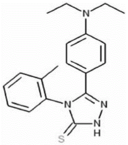
利用上述小分子T5381948处理后的结果如图7c,DSM-TKO肌肉干细胞在经过T5381948处理后,分化较对照组显著更好,其表现为:平均含有大于等于10个细胞核的肌肉细胞的数量极为显著地大于对照组;***,P<0.001。这一结果表明,小分子T5381948能够极为显著地促进肌肉干细胞的分化,有用于治疗肌肉干细胞分化异常相关疾病。The results after treatment with the above-mentioned small molecule T5381948 are shown in Figure 7c. After treatment with T5381948, the differentiation of DSM-TKO muscle stem cells was significantly better than that of the control group, as shown by: the number of muscle cells with an average of 10 or more nuclei was significantly greater than that of the control group; ***, P<0.001. This result shows that the small molecule T5381948 can significantly promote the differentiation of muscle stem cells and is useful for treating diseases related to abnormal differentiation of muscle stem cells.
在本发明提及的所有文献都在本申请中引用作为参考,就如同每一篇文献被单独引用作为参考那样。此外应理解,在阅读了本发明的上述讲授内容之后,本领域技术人员可以对本发明作各种改动或修改,这些等价形式同样落于本申请所附权利要求书所限定的范围。All documents mentioned in the present invention are cited as references in this application, just as each document is cited as reference individually. In addition, it should be understood that after reading the above teachings of the present invention, those skilled in the art can make various changes or modifications to the present invention, and these equivalent forms also fall within the scope defined by the claims attached to this application.
序列表Sequence Listing
<110> 中国科学院上海生命科学研究院<110> Shanghai Institutes for Life Sciences, Chinese Academy of Sciences
<120> 促进肌肉干细胞分化的化合物及其应用<120> Compounds for promoting differentiation of muscle stem cells and their application
<130> 196916<130> 196916
<160> 10<160> 10
<170> SIPOSequenceListing 1.0<170> SIPOSequenceListing 1.0
<210> 1<210> 1
<211> 24<211> 24
<212> DNA<212> DNA
<213> 引物(Primer)<213> Primer
<400> 1<400> 1
caccgtctga tgggaacccc acta 24caccgtctga tgggaacccc acta 24
<210> 2<210> 2
<211> 20<211> 20
<212> DNA<212> DNA
<213> 引物(Primer)<213> Primer
<400> 2<400> 2
tagtggggtt cccatcagac 20
<210> 3<210> 3
<211> 24<211> 24
<212> DNA<212> DNA
<213> 引物(Primer)<213> Primer
<400> 3<400> 3
caccgcccat tgcagcaagg ctta 24caccgcccat tgcagcaagg ctta 24
<210> 4<210> 4
<211> 24<211> 24
<212> DNA<212> DNA
<213> 引物(Primer)<213> Primer
<400> 4<400> 4
aaactaagcc ttgctgcaat gggc 24aaactaagcc ttgctgcaat gggc 24
<210> 5<210> 5
<211> 24<211> 24
<212> DNA<212> DNA
<213> 引物(Primer)<213> Primer
<400> 5<400> 5
caccgctgca acactatctg tgat 24caccgctgca acactatctg tgat 24
<210> 6<210> 6
<211> 24<211> 24
<212> DNA<212> DNA
<213> 引物(Primer)<213> Primer
<400> 6<400> 6
aaacatcaca gatagtgttg cagc 24aaacatcaca gatagtgttg cagc 24
<210> 7<210> 7
<211> 24<211> 24
<212> DNA<212> DNA
<213> 引物(Primer)<213> Primer
<400> 7<400> 7
caccggtaca tggtactcta aatt 24caccggtaca tggtactcta aatt 24
<210> 8<210> 8
<211> 24<211> 24
<212> DNA<212> DNA
<213> 引物(Primer)<213> Primer
<400> 8<400> 8
aaacaattta gagtaccatg tacc 24aaacaattta gagtaccatg tacc 24
<210> 9<210> 9
<211> 24<211> 24
<212> DNA<212> DNA
<213> 引物(Primer)<213> Primer
<400> 9<400> 9
caccggcttc tacaagctgc tccc 24caccggcttc tacaagctgc tccc 24
<210> 10<210> 10
<211> 24<211> 24
<212> DNA<212> DNA
<213> 引物(Primer)<213> Primer
<400> 10<400> 10
aaacgggagc agcttgtaga agcc 24aaacgggagc agcttgtaga agcc 24
Claims (6)

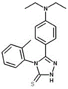
Priority Applications (1)
| Application Number | Priority Date | Filing Date | Title |
|---|---|---|---|
| CN201910989528.9A CN112680407B (en) | 2019-10-17 | 2019-10-17 | Compound for promoting muscle stem cell differentiation and application thereof |
Applications Claiming Priority (1)
| Application Number | Priority Date | Filing Date | Title |
|---|---|---|---|
| CN201910989528.9A CN112680407B (en) | 2019-10-17 | 2019-10-17 | Compound for promoting muscle stem cell differentiation and application thereof |
Publications (2)
| Publication Number | Publication Date |
|---|---|
| CN112680407A CN112680407A (en) | 2021-04-20 |
| CN112680407B true CN112680407B (en) | 2023-03-31 |
Family
ID=75444731
Family Applications (1)
| Application Number | Title | Priority Date | Filing Date |
|---|---|---|---|
| CN201910989528.9A Active CN112680407B (en) | 2019-10-17 | 2019-10-17 | Compound for promoting muscle stem cell differentiation and application thereof |
Country Status (1)
| Country | Link |
|---|---|
| CN (1) | CN112680407B (en) |
Families Citing this family (1)
| Publication number | Priority date | Publication date | Assignee | Title |
|---|---|---|---|---|
| CN119925582B (en) * | 2025-02-11 | 2025-10-03 | 中山大学中山眼科中心 | Application of NSUN2 in muscle injury repair |
Citations (3)
| Publication number | Priority date | Publication date | Assignee | Title |
|---|---|---|---|---|
| CN107847511A (en) * | 2015-07-14 | 2018-03-27 | 国立大学法人大阪大学 | myotonic dystrophy drugs |
| CN108368519A (en) * | 2015-07-02 | 2018-08-03 | 中国科学院上海生命科学研究院 | A kind of lonely male monoploid embryo stem cell and its preparation and application |
| WO2019099823A1 (en) * | 2017-11-17 | 2019-05-23 | Stc.Unm | Therapy for myotonic dystrophy type 1 via genome editing of the dmpk gene |
-
2019
- 2019-10-17 CN CN201910989528.9A patent/CN112680407B/en active Active
Patent Citations (3)
| Publication number | Priority date | Publication date | Assignee | Title |
|---|---|---|---|---|
| CN108368519A (en) * | 2015-07-02 | 2018-08-03 | 中国科学院上海生命科学研究院 | A kind of lonely male monoploid embryo stem cell and its preparation and application |
| CN107847511A (en) * | 2015-07-14 | 2018-03-27 | 国立大学法人大阪大学 | myotonic dystrophy drugs |
| WO2019099823A1 (en) * | 2017-11-17 | 2019-05-23 | Stc.Unm | Therapy for myotonic dystrophy type 1 via genome editing of the dmpk gene |
Non-Patent Citations (3)
| Title |
|---|
| 5-[4-(diethylamino)phenyl]-4-(2-methylphenyl)-4H-1,2,4-triazole-3-thiol,CAS number: 851207-79-9;ENAMINE Ltd.;《Enamine》;20100827;8、11 * |
| Dosage effect of multiple genes accounts for multisystem disorder of myotonic dystrophy type 1;Qi Yin et al.;《cell research》;20191218;第30卷;1-20 * |
| RN 851207-79-9;ACS;《STN registry 数据库》;20050526;8、11 * |
Also Published As
| Publication number | Publication date |
|---|---|
| CN112680407A (en) | 2021-04-20 |
Similar Documents
| Publication | Publication Date | Title |
|---|---|---|
| Braun et al. | Targeted inactivation of the muscle regulatory gene Myf-5 results in abnormal rib development and perinatal death | |
| Surani et al. | Nuclear transplantation in the mouse: heritable differences between parental genomes after activation of the embryonic genome | |
| CN108660161B (en) | Method for preparing chimeric gene-free knockout animal based on CRISPR/Cas9 technology | |
| CN105210981B (en) | Establish the method and its application for the ferret model that can be applied to human diseases research | |
| US20020132346A1 (en) | Use of RNA interference for the creation of lineage specific ES and other undifferentiated cells and production of differentiated cells in vitro by co-culture | |
| CN109640645A (en) | Genetically modified cells, tissues and organs for the treatment of disease | |
| US20060178297A1 (en) | Systems and methods for silencing expression of a gene in a cell and uses thereof | |
| WO1997044450A1 (en) | Regulation of gene expression | |
| US20040180430A1 (en) | Methods of restoring telomere length and extending cell lifespan using nuclear transfer | |
| JP2002537785A (en) | Genetic modification of somatic cells and their use | |
| CA2383790C (en) | Telomere restoration and extension of cell life-span in animals cloned from senescent somatic cells | |
| US20220369608A1 (en) | Method for establishing diabetes disease model dog | |
| Park et al. | Assisted reproductive techniques and genetic manipulation in the common marmoset | |
| JP2002537803A (en) | Embryonic or stem-like cell lines produced by xenogeneic nuclear transfer | |
| CN101985628B (en) | Method for building special microRNA knock-down mouse model of heart | |
| CN105177044B (en) | A method for obtaining a lymphoma mini-pig disease model by knocking out the p53 gene | |
| CN112680407B (en) | Compound for promoting muscle stem cell differentiation and application thereof | |
| US20100122356A1 (en) | Pig model for psoriasis | |
| CN102051380A (en) | Method for establishing arrhythmia animal model | |
| CN107988257B (en) | Carrier, cell and the method for goat cloning efficiency are improved based on the horizontal modification of donorcells DNA methylation | |
| CN113801851B (en) | Somatic cell nuclear transfer method and application thereof | |
| JP2002511234A (en) | Swine nuclear transfer | |
| CN108624621B (en) | The preparation method of the somatic cell clone animal of non-human primates | |
| JP2006522609A (en) | A method for correcting spindle defects associated with somatic cell nuclear transfer in animals | |
| CN104593414B (en) | Method for constructing human epidermolytic palmoplantar keratoderma mouse model |
Legal Events
| Date | Code | Title | Description |
|---|---|---|---|
| PB01 | Publication | ||
| PB01 | Publication | ||
| SE01 | Entry into force of request for substantive examination | ||
| SE01 | Entry into force of request for substantive examination | ||
| GR01 | Patent grant | ||
| GR01 | Patent grant |