Drawings
Fig. 1 is a schematic diagram illustrating a derivation process of a hepatocellular carcinoma-specific marker of the present invention.
FIG. 2 is a diagram showing the derivation process of 2502 Pre-Cancer genomes (Pre-Cancer Genome) that are overexpressed only in hepatocellular carcinoma.
FIG. 3 is a graph showing the results of hierarchical clustering analysis of 737 genes overexpressed in both databases by analyzing Cancer Genome Atlas (Cancer Genome Atlas) hepatocellular carcinoma (TCGA _ LIHC) data and Gene Expression integration (GEO) databases.
Figures 4a-4d are graphs confirming that 10 candidate marker genes exhibited overexpression in both the GSE114564 data cohort and GSE 6764.
Fig. 5a to 5d show the results of analyzing the expression levels of 10 marker genes in non-tumor tissues and tumor tissues of hepatoma cell patients derived from the TCGA _ LIHC data set and ICGC _ LIRI data set, respectively.
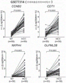
FIGS. 6a-6b show the results of comparative analysis of the expression levels of the above 10 marker genes using the GSE77314 dataset.
FIG. 7 is a graph showing the values of alpha-fetoprotein, which is a marker for hepatocellular carcinoma cancer, in a cohort of independent liver disease patients (771 specimens of 100 patients).
FIGS. 8a-8b are the results of analysis of the expression levels of 10 selected marker genes by enzyme-linked immunosorbent assay (ELISA).
FIGS. 9a-9b are the results of Receiver Operating Characteristic (ROC) curve analysis using enzyme-linked immunosorbent assay values for 10 marker genes.
FIG. 10 is a graph showing the values of alpha-fetoprotein, which is a marker for hepatocellular carcinoma cancer, in a cohort of independent liver disease patients (1148 specimens of 279 patients).
FIGS. 11a-11b are the results of analyzing the expression levels of 10 marker genes by ELISA.
FIGS. 12a-12b are the results of the analysis of the working characteristic curves of the subjects using the ELISA values of the marker genes for hyaluronic acid-mediated motor receptor, neurotonin 4, paralog 1, thrombospondin 4, and ubiquitin-binding enzyme E2T.
FIGS. 13a-13g are graphs confirming the sensitivity, specificity and accuracy of the hyaluronan-mediated motor receptor, neurotrophin 4, paralogous domain 1, thrombospondin 4 and ubiquitin-binding enzyme E2T in cohorts validated with alpha-fetoprotein by enzyme-linked immunosorbent assay analysis.
FIGS. 14a-14d are graphs showing the results of two markers selected from alpha-fetoprotein, hyaluronic acid-mediated motor receptor, neurotrophin 4, paralogous domain 1, thrombospondin 4 and ubiquitin-binding enzyme E2T (combination of alpha-fetoprotein and two markers selected from hyaluronic acid-mediated motor receptor, neurotrophin 4, paralogous domain 1, thrombospondin 4 and ubiquitin-binding enzyme E2T; or combination of hyaluronic acid-mediated motor receptor, neurotrophin 4, paralogous domain 1, thrombospondin 4 and ubiquitin-binding enzyme E2T) or combination of three markers (combination of alpha-fetoprotein and two markers selected from hyaluronic acid-mediated motor receptor, neurotrophin 4, paralogous domain 1, thrombospondin 4 and ubiquitin-binding enzyme E2T; or combination of hyaluronic acid-mediated motor receptor, neurotrophin, neurot, A combination of three markers of neurotrophin 4, paralog 1, thrombospondin 4, and ubiquitin-conjugating enzyme E2T).
Best mode for carrying out the invention
Hereinafter, the present invention will be described in detail by way of examples thereof with reference to the accompanying drawings. However, the following examples are provided as illustrations of the present invention, and when it is judged that a detailed description of a technique or a structure known to those of ordinary skill in the art to which the present invention pertains may unnecessarily obscure the gist of the present invention, a detailed description thereof may be omitted, and the present invention is not limited thereto. The present invention can be variously modified and applied within the scope of the following claims and equivalents to be explained thereby.
Also, the term (terminologies) used in the present specification is a term used for appropriately expressing the preferred embodiment of the present invention, which may be varied according to the intention of a user or operator, or the convention in the art to which the present invention pertains, and the like. Therefore, these terms should be defined based on the contents throughout the specification. In the present invention, the term "includes" or "including" a certain component in a certain portion is not intended to exclude another component but may include another component unless specifically stated to the contrary.
Unless otherwise indicated, nucleic acids are recorded from left to right in the 5'→ 3' direction. Numerical ranges recited in the specification include the numbers used to define the range and include each integer or any non-integer fraction within the defined range.
Unless defined otherwise, all technical and scientific terms used herein have the same meaning as commonly understood by one of ordinary skill in the art to which this invention belongs. Any methods and materials similar or equivalent to those described herein can be used in the practice for testing the present invention, but the preferred materials and methods are described herein.
In the present invention, the term "subject" or "patient" refers to any individual in need of treatment, including humans, apes, monkeys, cows, dogs, guinea pigs, rabbits, chickens, insects, and the like. Also, subjects include any subjects participating in a clinical study trial and not showing any clinical manifestations of the disease or subjects participating in a mechanical study or subjects used as a control group.
In the present invention, the term "specimen (sample)" refers to a biological specimen obtained from a subject or a patient. The source of the biological sample may be a fresh, frozen and/or preserved organ or tissue sample or solid tissue resulting from a biopsy or primer; blood or any blood component; cells at any point in time during pregnancy or development of a subject. In one embodiment of the invention, blood or any blood component is used as the sample.
Unless otherwise defined, all technical terms used herein have the same meaning as commonly understood by one of ordinary skill in the art to which this invention belongs in the related art of the present invention. Also, although preferred methods or samples are described herein, equivalents or equivalents thereof are also included within the scope of the present invention. The contents of all publications mentioned in this specification as references are incorporated herein by reference.
In one aspect, the present invention relates to a biomarker for diagnosing liver cancer, comprising one or more genes selected from the group consisting of alpha-fetoprotein, hyaluronic acid-mediated motility receptor (HMMR), neuroavidin 4(NXPH4, neuroopin 4), paired-homeodomain 1(PITX1, paired-like homeodomain 1), thrombospondin 4(THBS4, thrombospondin 4), and ubiquitin-binding enzyme E2T (UBE2T, ubiquitin-conjugating enzyme E2T) exhibiting specific expression changes against liver cancer or proteins expressed from the above genes. In an embodiment of the present invention, fig. 1 is a schematic diagram illustrating an identification sequence of liver cancer specific biomarkers.
In one example, liver cancer may be hepatocellular carcinoma (HCC), and may be early stage hepatocellular carcinoma or progressive hepatocellular carcinoma.
In one example, the expression of the biomarker genes of the present invention can be specifically increased in liver cancer.
In one aspect, the present invention relates to a composition for diagnosing liver cancer, comprising an agent for measuring the expression amount of one or more biomarker genes selected from the group consisting of alpha-fetoprotein, hyaluronic acid-mediated motor receptor, neurotrophin 4, paralog domain 1, thrombospondin 4, and ubiquitin-binding enzyme E2T, at the mRNA or protein level.
In one example, the composition may comprise a peptide for measuring at the mRNA or protein level selected from the group consisting of alpha-fetoprotein and hyaluronan-mediated motor receptors, alpha-fetoprotein and neurotrophin 4, alpha-fetoprotein and paralogous domain 1, alpha-fetoprotein and thrombospondin 4, alpha-fetoprotein and ubiquitin-binding enzyme E2T, hyaluronic acid-mediated motor receptors and neurotrophin 4, hyaluronic acid-mediated motor receptors and paralogous domain 1, hyaluronic acid-mediated motor receptors and thrombospondin 4, hyaluronic acid-mediated motor receptors and ubiquitin-binding enzyme E2T, neurotrophin 4 and paralogous domain 1, neurotrophin 4 and thrombospondin 4, neurotrophin 4 and ubiquitin-binding enzyme E2T, paralogous domain 1 and thrombospondin 4, paralogous domain 1 and ubiquitin-binding enzyme E2T, a preparation for expression level of at least one biomarker gene set in the group consisting of thrombospondin 4 and ubiquitin-conjugating enzyme E2T.
In one example, the above composition may comprise a peptide selected from the group consisting of alpha-fetoprotein, hyaluronic acid-mediated motor receptor, and neurotrophin 4, measured at the mRNA or protein level; alpha-fetoprotein, hyaluronic acid-mediated motor receptors, and paired homeodomain 1; alpha-fetoprotein, hyaluronic acid-mediated motor receptors, and thrombospondin 4; alpha-fetoprotein, hyaluronic acid-mediated motor receptor and ubiquitin-conjugating enzyme E2T; alpha-fetoprotein, neurotrophin 4, and paralog domain 1; alpha-fetoprotein, neurotrophin 4, and thrombospondin 4; alpha-fetoprotein, neurotrophin 4 and ubiquitin-binding enzyme E2T; alpha-fetoprotein, paralogous domain 1, and thrombospondin 4; alpha-fetoprotein, pairwise homeodomain 1, and ubiquitin-binding enzyme E2T; alpha-fetoprotein, thrombospondin 4, and ubiquitin-conjugating enzyme E2T; a hyaluronic acid-mediated motor receptor, a neurotrophin 4, and a paralog domain 1; a hyaluronic acid-mediated motor receptor, neurotrophin 4 and; a hyaluronic acid-mediated motor receptor, neurotrophin 4, and thrombospondin 4; hyaluronic acid-mediated motor receptors, neurotrophin 4 and ubiquitin-conjugating enzyme E2T; a hyaluronic acid-mediated motor receptor, a paralogous domain 1 and a thrombospondin 4; a hyaluronic acid-mediated motor receptor, paralogous domain 1 and ubiquitin-conjugating enzyme E2T; hyaluronic acid-mediated movement receptor, thrombospondin 4, and ubiquitin-conjugating enzyme E2T; neurotrophin 4, paralog domain 1, and thrombospondin 4; neurotrophin 4, paralog domain 1, and ubiquitin-binding enzyme E2T; and a preparation for expressing the amount of at least one biomarker gene set selected from the group consisting of neurotrophin 4, thrombospondin 4 and ubiquitin-conjugating enzyme E2T.
In one example, the preparation for measuring the expression level of the biomarker gene at the mRNA level may include a nucleic acid sequence of the marker, a nucleic acid sequence complementary to the nucleic acid sequence, a primer pair, a probe, or a primer pair and a probe that specifically recognizes a fragment of the nucleic acid sequence and the complementary sequence, and the measurement may be performed by a method selected from the group consisting of polymerase chain reaction, Real-time fluorescent quantitative reverse transcription polymerase chain reaction (Real-time RT-PCR), reverse transcription polymerase chain reaction, Competitive polymerase chain reaction (Competitive RT-PCR), Nuclease (Nuclease) protection assay (RNase, S1 Nuclease assay), in situ hybridization, nucleic acid microarray, northern blot, or DNA chip.
In one example, the preparation of the above-mentioned organism for measuring the expression amount of the marker gene at the protein level may comprise an antibody, an antibody fragment, an aptamer (aptamer), an avidity multimer (avidity multimer), or a peptidomimetic (peptidomimetics) that specifically recognizes the full-length protein or a fragment thereof of the above-mentioned marker, which can be measured by a method selected from the group consisting of immunoblotting, enzyme linked immunosorbent Assay (enzyme linked immunosorbent Assay), Radioimmunoassay (RIA), radio immunodiffusion (radioimmunodification), immunoelectrophoresis, tissue immunostaining, Immunoprecipitation Assay (Immunoprecipitation Assay), Complement Fixation Assay (complementary hybridization Assay), Fluorescence Activated Cell Sorting (FACS), mass analysis, or protein microarray.
The term "detecting" or "measuring" as used herein refers to quantifying the concentration of an object being detected or measured.
In the present invention, the term "primer" refers to a short nucleic acid sequence having a short free 3 hydroxyl (free 3 hydroxyl group) nucleic acid sequence capable of forming a base pair (base pair) with a complementary template (template), which serves as an origin for template strand replication. The primers can induce DNA synthesis in the presence of reagents for the polymerization reaction (i.e., DN polymerase or reverse transcriptase) and different 4 nucleoside triphosphates in the appropriate buffer and temperature.
In the present invention, the term "probe" refers to a nucleic acid fragment corresponding to several bases to several hundreds of bases capable of specifically binding to mRNA, for example, RNA or DNA, etc. Because of the labeling, the presence or absence of a specific mRNA can be confirmed. The probe can be produced in the form of an oligonucleotide (oligonucleotide) probe, a single-stranded dna (single stranded dna) probe, a double-stranded dna (double stranded dna) probe, an RNA probe, or the like. In the present invention, hybridization is performed using a probe complementary to the above-mentioned alpha-fetoprotein, hyaluronic acid-mediated motor receptor, neurotonin 4, paralog 1, thrombospondin 4, and/or ubiquitin-binding enzyme E2T genes, and the expression level of the above-mentioned genes can be diagnosed by whether or not hybridization is performed. The selection of an appropriate probe and hybridization conditions may be changed based on techniques known in the art, and there is no particular limitation in the present invention.
The primer or probe of the present invention can be chemically synthesized by using a phosphoramidite solid phase support method or other known methods. Such nucleic acid sequences may be modified by a variety of means well known in the art. Non-limiting examples of such variations include methylation, encapsulation, substitution of more than one homolog of the natural nucleotide, and variations between nucleotides, for example, variations to uncharged linkers (e.g., methyl phosphonates, phosphotriesters, phosphoramidates, carbamates, etc.) or charged linkers (e.g., phosphorothioates, phosphorodithioates, etc.).
In the present invention, suitable conditions for hybridizing a probe to a cDNA molecule can be determined in a series of processes by optimization steps. This step is performed by one of ordinary skill in the art through a series of procedures to establish a protocol for use in a research facility. For example, the conditions such as temperature, component concentration, hybridization and washing time, buffer components and their pH, and ionic strength depend on various factors such as the length of the probe, GC amount, and target nucleotide sequence. Detailed conditions for hybridization can be selected from "Joseph Sambrook, et al, Molecular Cloning, A Laboratory Manual, Cold Spring Harbor Laboratory Press, Cold Spring Harbor, N.Y. (2001); and M.L.M.Anderson, nucleic acid hybridization, Springer-Verlag New York Inc.N.Y. (1999) ". For example, high stringency conditions among the above stringent conditions means hybridization at 65 ℃ in 0.5M NaHPO4, 7% Sodium Dodecyl Sulfate (SDS), 1mM EDTA, and washing at 68 ℃ in 0.1 × Standard citrate saline (SSC)/0.1% sodium dodecyl sulfate. Alternatively, high stringency conditions refer to washing at 48 ℃ in 6 × standard saline citrate/0.05% sodium pyrophosphate. Low stringency conditions refer to washing at, for example, 42 ℃ in 0.2X standard saline citrate/0.1% sodium lauryl sulfate.
In the present invention, the term "antibody" is a term well known in the art and refers to a specific protein molecule directed against an antigenic site. For the purpose of the present invention, the antibody is an antibody that specifically binds to a protein expressed in the alpha-fetoprotein, hyaluronic acid-mediated motor receptor, neurotrophin 4, paralog 1, thrombospondin 4, and/or ubiquitin-conjugating enzyme E2T gene, which are markers of the present invention, and can be prepared by a known method. Including partial peptides which can be made from the above proteins. The form of the antibody of the present invention is not particularly limited, and if a polyclonal antibody, a monoclonal antibody or any one having antigen binding property, a part thereof is also included in the antibody of the present invention, and all immunoglobulin antibodies are included. Furthermore, the antibody of the present invention also includes a specific antibody, for example, a humanized antibody.
In one aspect, the present invention relates to a liver cancer diagnostic kit comprising a composition for diagnosing liver cancer.
In one example, the kit may further comprise means and/or reagents for collecting a biological sample from a subject or patient, and means and/or reagents for preparing genomic DNA, cDNA, RNA, or protein from a sample thereof. For example, PCR primers for amplifying the relevant regions of genomic DNA may be included. The kit may contain probes for genetic elements useful in pharmacogenomic analysis. Also, when such a kit is used, labeled oligonucleotides can be used for easy recognition during the analysis.
In one example, the kit may further contain a labeling substance such as DNA polymerase, dNTP (dGTP, dCTP, dATP, and dTTP), a fluorescent substance, and the like.
In the present invention, the term "liver cancer diagnostic kit" refers to a kit comprising the composition for diagnosing liver cancer of the present invention. Therefore, the above expression "liver cancer diagnosis kit" can be used interchangeably or in admixture with "composition for diagnosing liver cancer". In this specification, the term "diagnosing" includes determining a subject's sensitivity to a particular disease or condition (susceptability), determining whether a subject is currently suffering from a particular disease or condition, determining the prognosis of a subject suffering from a particular disease or condition (prognosis) (e.g., identifying pre-metastatic or metastatic cancer symptoms, determining the stage of cancer or the responsiveness of cancer to treatment), or treating indicators (therametrics) (e.g., monitoring the status of a subject to provide information relating to the efficacy of treatment).
In the present invention, the term "diagnostic biomarker, diagnostic biomarker or diagnostic marker (diagnosis marker)" is a substance capable of diagnosing the presence or absence of liver cancer cells or tissues separately from normal cells or tissues, and includes organic biomolecules such as polypeptides or nucleic acids (e.g., mRNA, etc.), lipids, glycolipids, glycoproteins, sugars (monosaccharides, disaccharides, oligosaccharides, etc.), etc., which are expressed in a state increased or decreased in cells or tissues having liver cancer cells as compared with normal cells. For the purpose of the present invention, the biomarker for detecting or diagnosing liver cancer is one or more selected from the group consisting of gene alpha-fetoprotein, hyaluronic acid-mediated motor receptor, neurotrophin 4, paralogous domain 1, thrombospondin 4, and ubiquitin-binding enzyme E2T, and is a gene that specifically increases mRNA expression or protein expression level in liver cancer. These markers include not only genes but also DNA or mRNA complementary to one marker, and preferably a composite marker including two or more of these markers, more preferably selected from the group consisting of movement receptors mediated by alpha-fetoprotein and hyaluronic acid; alpha-fetoprotein and neurotrophin 4; alpha-fetoprotein and paired homeodomain 1; alpha-fetoprotein and thrombospondin 4; alpha-fetoprotein and ubiquitin-conjugating enzyme E2T; hyaluronic acid-mediated motor receptors and neurotrophin 4; a hyaluronic acid-mediated motor receptor and a paralogous domain 1; hyaluronic acid-mediated motor receptors and thrombospondin 4; hyaluronic acid-mediated movement receptor and ubiquitin-conjugating enzyme E2T; neurotrophin 4 and paralog domain 1; neurotrophin 4 and thrombospondin 4; neurotrophin 4 and ubiquitin-conjugating enzyme E2T; a paralogous domain 1 and thrombospondin 4; paralogous domain 1 and ubiquitin-binding enzyme E2T; thrombospondin 4 and ubiquitin-conjugating enzyme E2T; alpha-fetoprotein, hyaluronic acid-mediated motor receptors, and neurotrophin 4; alpha-fetoprotein, hyaluronic acid-mediated motor receptors, and paired homeodomain 1; alpha-fetoprotein, hyaluronic acid-mediated motor receptors, and thrombospondin 4; alpha-fetoprotein, hyaluronic acid-mediated motor receptor and ubiquitin-conjugating enzyme E2T; alpha-fetoprotein, neurotrophin 4, and paralog domain 1; alpha-fetoprotein, neurotrophin 4, and thrombospondin 4; alpha-fetoprotein, neurotrophin 4 and ubiquitin-binding enzyme E2T; alpha-fetoprotein, paralogous domain 1, and thrombospondin 4; alpha-fetoprotein, pairwise homeodomain 1, and ubiquitin-binding enzyme E2T; alpha-fetoprotein, thrombospondin 4, and ubiquitin-conjugating enzyme E2T; a hyaluronic acid-mediated motor receptor, a neurotrophin 4, and a paralog domain 1; a hyaluronic acid-mediated motor receptor, neurotrophin 4 and; a hyaluronic acid-mediated motor receptor, neurotrophin 4, and thrombospondin 4; hyaluronic acid-mediated motor receptors, neurotrophin 4 and ubiquitin-conjugating enzyme E2T; a hyaluronic acid-mediated motor receptor, a paralogous domain 1 and a thrombospondin 4; a hyaluronic acid-mediated motor receptor, paralogous domain 1 and ubiquitin-conjugating enzyme E2T; hyaluronic acid-mediated movement receptor, thrombospondin 4, and ubiquitin-conjugating enzyme E2T; neurotrophin 4, paralog domain 1, and thrombospondin 4; neurotrophin 4, paralog domain 1, and ubiquitin-binding enzyme E2T; and one or more of the group consisting of neurotrophin 4, thrombospondin 4, and ubiquitin-conjugating enzyme E2T.
In one aspect, the present invention relates to a method for screening an anticancer candidate substance, comprising: measuring the expression level of alpha-fetoprotein, hyaluronic acid mediated motor receptor, neurotrophin 4, paralog 1, thrombospondin 4 or ubiquitin-conjugating enzyme E2T genes of the liver cancer cells; administering an anticancer candidate substance to the liver cancer cell and measuring the expression level of alpha-fetoprotein, a hyaluronan-mediated motor receptor, neurotrophin 4, paralog 1, thrombospondin 4, or ubiquitin-conjugating enzyme E2T gene; and (c) judging the candidate anticancer substance to be an effective anticancer substance when the expression level of the gene for alpha-fetoprotein, hyaluronic acid-mediated motor receptor, neurotrophin 4, paralog 1, thrombospondin 4 or ubiquitin-binding enzyme E2T in the step (a) is higher than the expression level of the gene for alpha-fetoprotein, hyaluronic acid-mediated motor receptor, neurotrophin 4, paralog 1, thrombospondin 4 or ubiquitin-binding enzyme E2T in the step (b).
In one aspect, the present invention relates to a method for providing information necessary for diagnosing liver cancer, comprising: measuring the expression level of at least one biomarker gene selected from the group consisting of alpha-fetoprotein, hyaluronic acid-mediated motor receptor, neurotrophin 4, paralogous domain 1, thrombospondin 4, and ubiquitin-conjugating enzyme E2T in a biological sample isolated from a subject; step (b) comparing the results with corresponding results for the corresponding markers in the normal control sample; and (c) determining that the subject is likely to be liver cancer when the expression level of the biomarker gene in the step (a) is greater than the expression level of the biomarker gene in the step (b).
In one example, the method may further comprise the step of distinguishing between early stage liver cancer and advanced liver cancer based on the level of change in expression of the biomarker genes.
In one example, the specific method for measuring the expression level of mRNA of the above biomarker gene or protein thereof is as follows: the expression of the above-mentioned gene can be detected at the mRNA level or the protein level, mRNA or protein can be isolated from a biological sample using a known procedure, and the expression level of the gene can be confirmed by reverse transcription-polymerase chain reaction (RT-PCR) or real time-PCR (real time-PCR).
In one example, the biological sample may include a sample such as tissue, cells, whole blood, serum, plasma, saliva, sputum, cerebrospinal fluid, or urine, and more preferably, whole blood, serum, or plasma.
The present invention will be described in more detail by the following examples. However, the following examples are only for specifically describing the contents of the present invention, and the present invention is not limited thereto.
Detailed Description
Example 1 blood marker screening Using a database
1-1 screening of liver cancer cell specific markers
After selecting 3 independent groups from blood samples (15 Normal Liver patient samples (Normal Liver, NL), 20 Chronic hepatitis patient samples (CH), 10 Cirrhosis patient samples (Liver Cirrhosis, LC), 18 Early hepatocellular carcinoma patient samples (Early HCC, eHCC), and 45 Advanced hepatocellular carcinoma patient samples (Advanced HCC, avHCC)) of an independent Liver disease patient cohort (108 samples of 86 patients), and extracting total RNA using TRIzol reagent, a sequencing Library was prepared using RNA Library Prep Kit for Illumina (Cat # E7420L) and sequenced by Illumina HiSeq 2000 according to the standard method of Illumina corporation. After the entire transcriptome (transcriptome) of the analyzed liver was plotted using STAR and Gencode v.25, the expression profile was replaced with FPKM values, and then the gene types were classified using Gencode v.25 and 12654 signal peptides were derived using SignalP 4.1. All data generated are recorded in the open Omix database GEO. After deriving 2502 Pre-Cancer genomes (Pre-Cancer Genome) that were overexpressed only in hepatocellular carcinoma (fig. 2), 737 genes overexpressed in both databases were analyzed by hierarchical clustering by analyzing Cancer Genome map (Cancer Genome Atlas) hepatocellular carcinoma (TCGA _ LIHC) data and Gene Expression integration (Gene Expression Omnibus) (GEO) database. As a result, the GSE114564 database shows a systematic diagram of 5 sub-classes classified into normal liver, Chronic Hepatitis (CHB), cirrhosis, early hepatocellular carcinoma, and progressive hepatocellular carcinoma, and the TCGA _ LIHC database shows a systematic diagram of 2 sub-classes classified into normal liver and progressive hepatocellular carcinoma (fig. 3). When the expression states of the 737 genes calculated were compared in 2 data sets, it was confirmed that there was a significant difference in the progression to liver cancer or the change in expression in the progressive liver cancer as compared with the normal liver tissue. In order to analyze Gene data having two types, a Gene Set Enrichment Analysis (GSEA) for extracting two types of important Gene sets whose expression values statistically show a significant difference from each other among various Gene sets composed based on biological characteristics was used. As a result of the analysis, it was confirmed that the gene was very closely related to a data set of a LIVER CANCER cohort CHANG _ lever _ CANCER, which is a conventionally known LIVER CANCER cohort (mean agglutination index NES 1.88, NES 1.85). Then, 10 candidate markers CCNB2, CDT1, COCH, CSMD1, HMMR, NXPH4, OLFML2B, PITX1, THBS4, and UBE2T, whose expression specificity was increased only in hepatocellular carcinoma, were selected by confirming that 10 candidate marker genes were overexpressed in both the GSE114564 data cohort and GSE6764 (fig. 4a-4 d).
1-2, TCGA _ LIHC and ICGC _ LIRI are used for differential verification of normal liver and progressive hepatocellular carcinoma
In order to achieve significant increases in biomarkers from the patient's serum, the rate of increase in expression in progressive liver cancer must be statistically significantly higher than that of normal liver, and in published data, it was confirmed that significant differences in expression were exhibited in both cohorts based on sequencing that can accurately measure expression, and analysis and validation of the expression levels of 10 marker genes derived from non-tumor tissue and tumor tissue of liver cancer cell patients from TCGA _ LIHC dataset and ICGC _ LIRI dataset as large-scale cohorts, respectively (fig. 5a-5 d).
1-3 analysis of 50 pairs of matched pairs (matched pairs) in patients with hepatoma cells
The expression levels of the 10 marker genes were compared and analyzed using the GSE77314 dataset, which is the expression value of the genes measured by the sequencing method in the peripheral normal tissues and liver cancer tissues of 50 liver cancer patients in total as a cohort of chinese liver cancer patients, and as a result, it was confirmed that the expression thereof was significantly increased in the liver cancer tissues compared to the normal liver tissues in most of the patients (fig. 6a to 6 b).
Example 2 enzyme-linked immunosorbent assay analysis of the first selected marker
2-1. confirmation of marker expression profiles
Expression levels of 10 marker genes selected in example 1 above were confirmed by ELISA analysis in blood samples (135 samples (Normal Liver, NL) of 16 Normal Liver patients, 65 samples (Chronic hepatitis, CH) of 13 Chronic hepatitis patients, 103 samples (Liver Cirrhosis, LC) of 15 Cirrhosis patients, 227 samples (Early HCC, eHCC) of 35 Early hepatocellular carcinoma patients, and 241 samples (Advanced HCC, avHCC) of 24 progressive hepatocellular carcinoma patients (FIG. 7) of a cohort of Liver disease patients (771 samples of 100 patients) for which the alpha-fetoprotein value as a marker for hepatocellular carcinoma was confirmed (FIGS. 8a-8 b).
As a result, in the case of CCNB2, on average, 0.02ng/ml was measured from normal liver patients (NL), chronic hepatitis patients (CH) showed 0.2029ng/ml, cirrhosis patients (LC) showed 0.43ng/ml, early hepatocellular carcinoma patients (eHCC) showed 0.27ng/ml, and advanced hepatocellular carcinoma patients (avHCC) showed 0.31ng/ml, and thus LC showed the maximum value. In the case of CDT1, 167.7pg/ml, CH 230.8pg/ml, LC 178.2pg/ml, eHCC 103.5pg/ml, and avHCC 146.8pg/ml were averaged from normal liver patients (NL), and thus avHCC showed the maximum value, and overall, no significant difference was shown between diseases. In the case of COCH, 1.724ng/ml, CH showed 12.78ng/ml, LC showed 10.03ng/ml, eHCC showed 6.74ng/ml, and avHCC showed 8.025ng/ml, on average, from normal liver patients (NL), thus showing larger values in all liver diseases and liver cancer stages except normal liver. In the case of CSMD1, on average 14.8ng/ml, CH showed 11.65ng/ml, LC showed 14.48ng/ml, eHCC showed 14.72ng/ml, and avHCC showed 15.66ng/ml from normal liver patients (NL), thus showing similar values overall. In the case of OLFML2B, NL showed 175.2pg/ml on average, CH 658.8pg/ml, LC showed 338.4pg/ml, eehcc showed 284.1pg/ml, and avHCC showed 349.6pg/ml, thus showing larger values in all stages of liver disease and liver cancer, especially in CH, in addition to normal liver. In the case of HMMR, 0.21ng/ml was measured on average from NL, CH showed 0.62ng/ml, LC showed 0.74ng/ml, eHCC showed 1.54ng/ml, and avHCC showed 1.64ng/ml, thus, similar to the sequencing results, the value increased with the progression of the liver disease stage. In the case of NXPH4, NL showed an average of 3.54ng/ml, CH 10.23ng/ml, LC 6.52ng/ml, eHCC 15.02ng/ml, and avHCC 19.83ng/ml, thus slightly higher in CH, but similar to the sequencing results, this value increased with the progression of the liver disease stage. In the case of PITX1, NL showed 2042pg/ml on average, CH showed 1994pg/ml, LC showed 3238pg/ml, eHCC showed 3314pg/ml, and avHCC showed 6135pg/ml, thus showing a lower value than normal in CH, but similar to the sequencing results, this value increased with the progression of the liver disease stage. In the case of THBS4, NL showed 45.36ng/ml on average, CH showed 70.96ng/ml, LC showed 141.8ng/ml, eHCC showed 229.4ng/ml, and avHCC showed 233.6ng/ml, thus the value increased with the progression of the stage of liver disease as the sequencing results. In the case of UBE2T, NL showed 16.14ng/ml on average, CH 319.9ng/ml, LC 426.1ng/ml, eehcc 505.5ng/ml, and avHCC 877.2ng/ml, thus sharply increasing in liver disease by about 20-fold compared to normal liver, and the value increased with the progression of the stage of liver disease.
2-2. receiver operating characteristics (receiver operating characteristics) Curve analysis
The test subject working characteristic curve analysis is carried out on 10 marker genes in the above queue through enzyme-linked immunosorbent assay values.
As a result, CSMD1, HMMR, NXPH4, OPITX1, THBS4 and UBE2T had statistically significant values compared to the reference line, and the analysis of the working characteristic curve of the subject showed that the area under the curve (AUC) values were similar to or greater than that of alpha fetoprotein, indicating that the markers with characteristics and sensitivity were HMMR, NXPH4, PITX1, THBS4 and UBE2T (fig. 9a-9 b).
Example 3 validation of early cancer diagnostic markers HMMR, NXPH4, PITX1, THBS4 and UBE2T
3-1. confirmation of marker expression profiles
Expression levels of 10 marker genes selected in example 1 were confirmed by ELISA analysis in blood samples (222 samples (Normal Liver, NL) of 49 Normal Liver patients, 115 samples (Chronic hepatitis, CH) of 31 Chronic hepatitis patients, 183 samples (Liver Cirrhosis, LC) of 46 Cirrhosis patients, 345 samples (Early HCC, eHCC) of 77 Early hepatocellular carcinoma patients, and 283 samples (Advanced HCC, avHCC) of 64 progressive hepatocellular carcinoma patients (FIG. 10) of an independent Liver disease patient cohort (1148 samples of 279 patients) in which the alpha-fetoprotein number as a Liver cancer marker was confirmed (FIGS. 11a-11 b).
As a result, when the protein expression levels of 5 markers were measured in the validation cohort, respectively, in the case of HMMR, by comparing the results of the normal group with each of the liver disease groups in a total of 230 specimens, a very significant difference was exhibited except for the liver cirrhosis group, and it was confirmed that the expression thereof was specifically high particularly in early liver cancer. In the case of NXPH4, each liver disease stage group showed a greater change in expression compared to the normal group, and PITX1 also showed the same result. In the case of THBS4, its expression was significantly increased except in the cirrhosis group like HMMR, and showed a larger value in the early liver cancer group. In the case of UBE2T, expression was not expressed at all in the normal group, like the test cohort, but increased expression was shown in the liver disease stage group.
3-2. analysis of the characteristic curves of the subjects
Subject working profile analysis was performed on the marker genes HMMR, NXPH4, PITX1, THBS4 and UBE2T in the cohort.
As a result, in the case of HMMR and THBS4, the AUC was 0.856 and the AUC was 0.772, respectively, and it was confirmed that the expression level thereof was higher than the AUC of the conventional marker AFP, which was 0.749 (fig. 12a to 12 b).
3-3 confirmation of expression status of each stage of development of hepatocellular carcinoma
To confirm the sensitivity (sensitivity), specificity (specificity) and accuracy (accuracycacy) of the above HMMR, NXPH4, PITX1, THBS4 and UBE2T as verified by AFP, the above cohort samples were subjected to elisa analysis.
TABLE 1
The results are shown in Table 1 above and FIGS. 13a-13 g. Specifically, the following steps were performed for alpha-fetoprotein and 5 markers: 1) comparison in non-liver cancer samples (normal liver, hepatitis, cirrhosis samples) and liver cancer samples; 2) after comparison between the liver disease samples (hepatitis and cirrhosis samples) and the liver cancer sample, 3) non-liver cancer samples and 4) liver disease samples were specifically analyzed for early stage liver cancer only. In all 4 cases, the sensitivity, specificity and accuracy of the hyaluronan-mediated movement receptor were maximized. When the cut-off value (cut-off value) of their respective markers was determined by the MedCal program, the assay was carried out by determining that 0.8ng/μ l of hyaluronic acid-mediated motor receptor, 7.5ng/μ l of neurotrophin 4, 2475pg/μ l of paralog 1, 90ng/μ l of thrombospondin 4, and 40ng/μ l of ubiquitin-binding enzyme E2T were present, and in the case of samples in which the cut-off value was increased more than each cut-off value, the samples were classified as Positive (Positive) or low Negative (Negative). From the positive rate in the normal liver, 2% of alpha-fetoprotein, 0% of hyaluronic acid-mediated motor receptor, 6% of neurotrophin 4, 23% of paralog 1, 4% of thrombospondin 4, and 0% of ubiquitin-binding enzyme E2T were measured. In the hepatitis group, 19% of alpha-fetoprotein, 19% of hyaluronic acid-mediated motor receptors, 50% of neurotrophin 4, 44% of paralog domain 1, 44% of thrombospondin 4, and 63% of ubiquitin-binding enzyme E2T were measured. In the cirrhosis group, 39% of alpha-fetoprotein, 17% of hyaluronic acid-mediated motor receptors, 48% of neurotrophin 4, 57% of paralogous domain 1, 4% of thrombospondin 4, and 70% of ubiquitin-binding enzyme E2T were determined. In the early liver cancer group, 33% of alpha-fetoprotein, 83% of hyaluronic acid-mediated motor receptors, 64% of neurotrophin 4, 72% of paralog 1, 54% of thrombospondin 4, and 54% of ubiquitin-binding enzyme E2T were measured, and thus the positive rates of the 5 markers measured were significantly higher than those of alpha-fetoprotein as a measurement marker of liver cancer. In the progressive group of liver cancers, 73% of alpha-fetoprotein, 78% of hyaluronic acid-mediated motor receptors, 87% of neurotrophin 4, 89% of paralog 1, 62% of thrombospondin 4, and 67% of ubiquitin-binding enzyme E2T were shown. Then, when comparing the positive rates of alpha-fetoprotein and 5 markers in liver cancer patients, the alpha-fetoprotein was 52%, while the remaining markers showed a high positive rate, particularly when comparing each positive rate in liver cancer patients in which alpha-fetoprotein was negative, in the case of HMMR, a high positive rate of 86% was measured, which is expected to be able to complement liver cancer patients in which no alpha-fetoprotein was measured as positive. In the case of the early liver cancer group, alpha-fetoprotein showed a positive rate of 33%, whereas in the case of HMMR, a high positive rate of 83% was exhibited. And also in a liver cancer patient negative for alpha-fetoprotein, a high positive rate of 85% was exhibited.
Example 4 confirmation of the Combined Effect of early cancer diagnostic markers
For the cohort of the above example 3-1, combinations according to two markers of alpha-fetoprotein, hyaluronic acid-mediated motor receptor, neurotrophin 4, paralogous domain 1, thrombospondin 4 and ubiquitin-binding enzyme E2T (a combination of alpha-fetoprotein and two markers of hyaluronic acid-mediated motor receptor, neurotrophin 4, paralogous domain 1, thrombospondin 4 or ubiquitin-binding enzyme E2T or a combination of hyaluronic acid-mediated motor receptor, neurotrophin 4, paralogous domain 1, thrombospondin 4 and ubiquitin-binding enzyme E2T) or a combination of three markers (a combination of alpha-fetoprotein and two markers of hyaluronic acid-mediated motor receptor, neurotrophin 4, paralogous domain 1, thrombospondin 4 and ubiquitin-binding enzyme E2T or a combination of hyaluronic acid-mediated motor receptor, neurotrophin 4, paralogous domain 1, thrombospondin 4 and ubiquitin-binding enzyme E2T) or a combination of hyaluronic acid-mediated motor receptor, neurotrophin, or ubiquitin-, A combination of three markers of neurotrophin 4, paralog 1, thrombospondin 4 and ubiquitin-binding enzyme E2T).
TABLE 2
The results are shown in table 2 above and in fig. 14a-14 d. Specifically, when 2 markers were combined, the combination of alpha-fetoprotein and the hyaluronan-mediated motor receptor showed a 92% positive rate, and the combination of the hyaluronan-mediated motor receptor and the homeodomain 1 showed a maximum 96% positive rate, in all liver cancer patients. When patients with early stage liver cancer are treated, the combination of alpha-fetoprotein and a hyaluronan-mediated motor receptor shows a 90% positive rate, and the combination of a hyaluronan-mediated motor receptor and the paralog domain 1 shows a maximum 99% positive rate. In the case of combining the three markers, if all liver cancer patients were treated, the combination of alpha-fetoprotein, hyaluronic acid-mediated motor receptor and paralog 1, the combination of hyaluronic acid-mediated motor receptor, neurotonin 4 and paralog 1, and the combination of hyaluronic acid-mediated motor receptor, paralog 1 and ubiquitin-binding enzyme E2T showed a 100% positive rate. When patients with early stage liver cancer were treated, 100% positive rate was observed for the combination of alpha-fetoprotein with hyaluronan-mediated motor receptor and paralog 1, the combination of hyaluronan-mediated motor receptor, neurotrophin 4 and ubiquitin-binding enzyme E2T, and the combination of hyaluronan-mediated motor receptor, paralog 1 and ubiquitin-binding enzyme E2T.
Further, when the subject working characteristics were analyzed for combinations showing a positive rate of 100% among 86 non-liver cancer samples and 132 liver cancer samples, it was confirmed that the area value under the curve was statistically significantly increased for all combinations compared to the conventional alpha-fetoprotein, the combination of the alpha-fetoprotein and the hyaluronic acid-mediated motor receptor was most excellent in the two combinations, and the combination of the alpha-fetoprotein, the hyaluronic acid-mediated motor receptor, and the paralogous domain 1 showed the maximum value in the three combinations. In the diagnostic analysis, the highest hyaluronic acid-mediated motor receptor and the highest paired homeodomain 1 was detected and the odds ratio showed the highest 75.23 in the two combinations for the results of the accuracy analysis. Among the three combinations, as a result of the accuracy analysis, alpha-fetoprotein, hyaluronic acid-mediated motor receptor, and paralog domain 1 showed the highest 90.37%, and the odds ratio also showed the highest 87.04. Further, when the subject working characteristics were analyzed for combinations showing 100% positive rate in each of 86 non-liver cancer samples and 69 early liver cancer samples, it was confirmed that the area value under the curve was statistically significantly increased in all combinations compared to the conventional alpha fetoprotein, and the combination of the hyaluronic acid-mediated motor receptor and the paralogoid domain 1 was most excellent in the two combinations, and the combination of the alpha fetoprotein, the hyaluronic acid-mediated motor receptor, and the paralogoid domain 1 showed the maximum value in the three combinations. In the diagnostic assay, the highest 88.39% was determined for the hyaluronan-mediated motor receptor and the syngenin domain 1 and the highest 64.75% was also shown for the odds ratio in the two combinations for the results of the accuracy assay. Among the three combinations, alpha-fetoprotein, hyaluronic acid-mediated motor receptor, and paralog domain 1 showed the highest 92.75% as a result of the accuracy analysis, and the odds ratio was also determined to be the highest 65.83.