CN112351793A - Immune checkpoint inhibitor co-expression vectors - Google Patents
Immune checkpoint inhibitor co-expression vectors Download PDFInfo
- Publication number
- CN112351793A CN112351793A CN201980044406.0A CN201980044406A CN112351793A CN 112351793 A CN112351793 A CN 112351793A CN 201980044406 A CN201980044406 A CN 201980044406A CN 112351793 A CN112351793 A CN 112351793A
- Authority
- CN
- China
- Prior art keywords
- sequence
- antigen
- vector
- nucleic acid
- acid sequence
- Prior art date
- Legal status (The legal status is an assumption and is not a legal conclusion. Google has not performed a legal analysis and makes no representation as to the accuracy of the status listed.)
- Pending
Links
Images


Classifications
-
- A—HUMAN NECESSITIES
- A61—MEDICAL OR VETERINARY SCIENCE; HYGIENE
- A61K—PREPARATIONS FOR MEDICAL, DENTAL OR TOILETRY PURPOSES
- A61K39/00—Medicinal preparations containing antigens or antibodies
- A61K39/0005—Vertebrate antigens
- A61K39/0011—Cancer antigens
- A61K39/00119—Melanoma antigens
- A61K39/001191—Melan-A/MART
-
- A—HUMAN NECESSITIES
- A61—MEDICAL OR VETERINARY SCIENCE; HYGIENE
- A61K—PREPARATIONS FOR MEDICAL, DENTAL OR TOILETRY PURPOSES
- A61K39/00—Medicinal preparations containing antigens or antibodies
- A61K39/0005—Vertebrate antigens
- A61K39/0011—Cancer antigens
- A61K39/001184—Cancer testis antigens, e.g. SSX, BAGE, GAGE or SAGE
- A61K39/001188—NY-ESO
-
- A—HUMAN NECESSITIES
- A61—MEDICAL OR VETERINARY SCIENCE; HYGIENE
- A61K—PREPARATIONS FOR MEDICAL, DENTAL OR TOILETRY PURPOSES
- A61K39/00—Medicinal preparations containing antigens or antibodies
- A61K39/0005—Vertebrate antigens
- A61K39/0012—Lipids; Lipoproteins
-
- A—HUMAN NECESSITIES
- A61—MEDICAL OR VETERINARY SCIENCE; HYGIENE
- A61K—PREPARATIONS FOR MEDICAL, DENTAL OR TOILETRY PURPOSES
- A61K39/00—Medicinal preparations containing antigens or antibodies
- A61K39/12—Viral antigens
-
- A—HUMAN NECESSITIES
- A61—MEDICAL OR VETERINARY SCIENCE; HYGIENE
- A61K—PREPARATIONS FOR MEDICAL, DENTAL OR TOILETRY PURPOSES
- A61K40/00—Cellular immunotherapy
- A61K40/30—Cellular immunotherapy characterised by the recombinant expression of specific molecules in the cells of the immune system
- A61K40/34—Antigenic peptides
-
- A—HUMAN NECESSITIES
- A61—MEDICAL OR VETERINARY SCIENCE; HYGIENE
- A61K—PREPARATIONS FOR MEDICAL, DENTAL OR TOILETRY PURPOSES
- A61K40/00—Cellular immunotherapy
- A61K40/40—Cellular immunotherapy characterised by antigens that are targeted or presented by cells of the immune system
- A61K40/41—Vertebrate antigens
- A61K40/42—Cancer antigens
- A61K40/4267—Cancer testis antigens, e.g. SSX, BAGE, GAGE or SAGE
- A61K40/4269—NY-ESO
-
- A—HUMAN NECESSITIES
- A61—MEDICAL OR VETERINARY SCIENCE; HYGIENE
- A61K—PREPARATIONS FOR MEDICAL, DENTAL OR TOILETRY PURPOSES
- A61K40/00—Cellular immunotherapy
- A61K40/40—Cellular immunotherapy characterised by antigens that are targeted or presented by cells of the immune system
- A61K40/41—Vertebrate antigens
- A61K40/42—Cancer antigens
- A61K40/4271—Melanoma antigens
- A61K40/4272—Melan-A/MART
-
- A—HUMAN NECESSITIES
- A61—MEDICAL OR VETERINARY SCIENCE; HYGIENE
- A61P—SPECIFIC THERAPEUTIC ACTIVITY OF CHEMICAL COMPOUNDS OR MEDICINAL PREPARATIONS
- A61P31/00—Antiinfectives, i.e. antibiotics, antiseptics, chemotherapeutics
- A61P31/12—Antivirals
- A61P31/14—Antivirals for RNA viruses
- A61P31/16—Antivirals for RNA viruses for influenza or rhinoviruses
-
- A—HUMAN NECESSITIES
- A61—MEDICAL OR VETERINARY SCIENCE; HYGIENE
- A61P—SPECIFIC THERAPEUTIC ACTIVITY OF CHEMICAL COMPOUNDS OR MEDICINAL PREPARATIONS
- A61P31/00—Antiinfectives, i.e. antibiotics, antiseptics, chemotherapeutics
- A61P31/12—Antivirals
- A61P31/20—Antivirals for DNA viruses
-
- A—HUMAN NECESSITIES
- A61—MEDICAL OR VETERINARY SCIENCE; HYGIENE
- A61P—SPECIFIC THERAPEUTIC ACTIVITY OF CHEMICAL COMPOUNDS OR MEDICINAL PREPARATIONS
- A61P35/00—Antineoplastic agents
-
- C—CHEMISTRY; METALLURGY
- C07—ORGANIC CHEMISTRY
- C07K—PEPTIDES
- C07K16/00—Immunoglobulins [IGs], e.g. monoclonal or polyclonal antibodies
- C07K16/18—Immunoglobulins [IGs], e.g. monoclonal or polyclonal antibodies against material from animals or humans
- C07K16/28—Immunoglobulins [IGs], e.g. monoclonal or polyclonal antibodies against material from animals or humans against receptors, cell surface antigens or cell surface determinants
- C07K16/2803—Immunoglobulins [IGs], e.g. monoclonal or polyclonal antibodies against material from animals or humans against receptors, cell surface antigens or cell surface determinants against the immunoglobulin superfamily
- C07K16/2818—Immunoglobulins [IGs], e.g. monoclonal or polyclonal antibodies against material from animals or humans against receptors, cell surface antigens or cell surface determinants against the immunoglobulin superfamily against CD28 or CD152
-
- C—CHEMISTRY; METALLURGY
- C12—BIOCHEMISTRY; BEER; SPIRITS; WINE; VINEGAR; MICROBIOLOGY; ENZYMOLOGY; MUTATION OR GENETIC ENGINEERING
- C12N—MICROORGANISMS OR ENZYMES; COMPOSITIONS THEREOF; PROPAGATING, PRESERVING, OR MAINTAINING MICROORGANISMS; MUTATION OR GENETIC ENGINEERING; CULTURE MEDIA
- C12N15/00—Mutation or genetic engineering; DNA or RNA concerning genetic engineering, vectors, e.g. plasmids, or their isolation, preparation or purification; Use of hosts therefor
- C12N15/09—Recombinant DNA-technology
- C12N15/63—Introduction of foreign genetic material using vectors; Vectors; Use of hosts therefor; Regulation of expression
- C12N15/79—Vectors or expression systems specially adapted for eukaryotic hosts
- C12N15/85—Vectors or expression systems specially adapted for eukaryotic hosts for animal cells
- C12N15/86—Viral vectors
-
- C—CHEMISTRY; METALLURGY
- C12—BIOCHEMISTRY; BEER; SPIRITS; WINE; VINEGAR; MICROBIOLOGY; ENZYMOLOGY; MUTATION OR GENETIC ENGINEERING
- C12Q—MEASURING OR TESTING PROCESSES INVOLVING ENZYMES, NUCLEIC ACIDS OR MICROORGANISMS; COMPOSITIONS OR TEST PAPERS THEREFOR; PROCESSES OF PREPARING SUCH COMPOSITIONS; CONDITION-RESPONSIVE CONTROL IN MICROBIOLOGICAL OR ENZYMOLOGICAL PROCESSES
- C12Q1/00—Measuring or testing processes involving enzymes, nucleic acids or microorganisms; Compositions therefor; Processes of preparing such compositions
- C12Q1/68—Measuring or testing processes involving enzymes, nucleic acids or microorganisms; Compositions therefor; Processes of preparing such compositions involving nucleic acids
- C12Q1/6813—Hybridisation assays
- C12Q1/6827—Hybridisation assays for detection of mutation or polymorphism
-
- A—HUMAN NECESSITIES
- A61—MEDICAL OR VETERINARY SCIENCE; HYGIENE
- A61K—PREPARATIONS FOR MEDICAL, DENTAL OR TOILETRY PURPOSES
- A61K39/00—Medicinal preparations containing antigens or antibodies
- A61K2039/505—Medicinal preparations containing antigens or antibodies comprising antibodies
-
- A—HUMAN NECESSITIES
- A61—MEDICAL OR VETERINARY SCIENCE; HYGIENE
- A61K—PREPARATIONS FOR MEDICAL, DENTAL OR TOILETRY PURPOSES
- A61K39/00—Medicinal preparations containing antigens or antibodies
- A61K2039/57—Medicinal preparations containing antigens or antibodies characterised by the type of response, e.g. Th1, Th2
- A61K2039/572—Medicinal preparations containing antigens or antibodies characterised by the type of response, e.g. Th1, Th2 cytotoxic response
-
- A—HUMAN NECESSITIES
- A61—MEDICAL OR VETERINARY SCIENCE; HYGIENE
- A61K—PREPARATIONS FOR MEDICAL, DENTAL OR TOILETRY PURPOSES
- A61K39/00—Medicinal preparations containing antigens or antibodies
- A61K2039/57—Medicinal preparations containing antigens or antibodies characterised by the type of response, e.g. Th1, Th2
- A61K2039/575—Medicinal preparations containing antigens or antibodies characterised by the type of response, e.g. Th1, Th2 humoral response
-
- A—HUMAN NECESSITIES
- A61—MEDICAL OR VETERINARY SCIENCE; HYGIENE
- A61K—PREPARATIONS FOR MEDICAL, DENTAL OR TOILETRY PURPOSES
- A61K39/00—Medicinal preparations containing antigens or antibodies
- A61K2039/60—Medicinal preparations containing antigens or antibodies characteristics by the carrier linked to the antigen
- A61K2039/6031—Proteins
- A61K2039/6037—Bacterial toxins, e.g. diphteria toxoid [DT], tetanus toxoid [TT]
-
- A—HUMAN NECESSITIES
- A61—MEDICAL OR VETERINARY SCIENCE; HYGIENE
- A61K—PREPARATIONS FOR MEDICAL, DENTAL OR TOILETRY PURPOSES
- A61K39/00—Medicinal preparations containing antigens or antibodies
- A61K2039/60—Medicinal preparations containing antigens or antibodies characteristics by the carrier linked to the antigen
- A61K2039/6031—Proteins
- A61K2039/605—MHC molecules or ligands thereof
-
- A—HUMAN NECESSITIES
- A61—MEDICAL OR VETERINARY SCIENCE; HYGIENE
- A61K—PREPARATIONS FOR MEDICAL, DENTAL OR TOILETRY PURPOSES
- A61K39/00—Medicinal preparations containing antigens or antibodies
- A61K2039/70—Multivalent vaccine
-
- A—HUMAN NECESSITIES
- A61—MEDICAL OR VETERINARY SCIENCE; HYGIENE
- A61K—PREPARATIONS FOR MEDICAL, DENTAL OR TOILETRY PURPOSES
- A61K2121/00—Preparations for use in therapy
-
- A—HUMAN NECESSITIES
- A61—MEDICAL OR VETERINARY SCIENCE; HYGIENE
- A61K—PREPARATIONS FOR MEDICAL, DENTAL OR TOILETRY PURPOSES
- A61K2300/00—Mixtures or combinations of active ingredients, wherein at least one active ingredient is fully defined in groups A61K31/00 - A61K41/00
-
- C—CHEMISTRY; METALLURGY
- C07—ORGANIC CHEMISTRY
- C07K—PEPTIDES
- C07K14/00—Peptides having more than 20 amino acids; Gastrins; Somatostatins; Melanotropins; Derivatives thereof
- C07K14/435—Peptides having more than 20 amino acids; Gastrins; Somatostatins; Melanotropins; Derivatives thereof from animals; from humans
- C07K14/705—Receptors; Cell surface antigens; Cell surface determinants
- C07K14/70503—Immunoglobulin superfamily
- C07K14/70539—MHC-molecules, e.g. HLA-molecules
-
- C—CHEMISTRY; METALLURGY
- C07—ORGANIC CHEMISTRY
- C07K—PEPTIDES
- C07K14/00—Peptides having more than 20 amino acids; Gastrins; Somatostatins; Melanotropins; Derivatives thereof
- C07K14/435—Peptides having more than 20 amino acids; Gastrins; Somatostatins; Melanotropins; Derivatives thereof from animals; from humans
- C07K14/705—Receptors; Cell surface antigens; Cell surface determinants
- C07K14/70596—Molecules with a "CD"-designation not provided for elsewhere
-
- C—CHEMISTRY; METALLURGY
- C12—BIOCHEMISTRY; BEER; SPIRITS; WINE; VINEGAR; MICROBIOLOGY; ENZYMOLOGY; MUTATION OR GENETIC ENGINEERING
- C12N—MICROORGANISMS OR ENZYMES; COMPOSITIONS THEREOF; PROPAGATING, PRESERVING, OR MAINTAINING MICROORGANISMS; MUTATION OR GENETIC ENGINEERING; CULTURE MEDIA
- C12N2710/00—MICROORGANISMS OR ENZYMES; COMPOSITIONS THEREOF; PROPAGATING, PRESERVING, OR MAINTAINING MICROORGANISMS; MUTATION OR GENETIC ENGINEERING; CULTURE MEDIA dsDNA viruses
- C12N2710/00011—Details
- C12N2710/10011—Adenoviridae
- C12N2710/10311—Mastadenovirus, e.g. human or simian adenoviruses
- C12N2710/10341—Use of virus, viral particle or viral elements as a vector
- C12N2710/10343—Use of virus, viral particle or viral elements as a vector viral genome or elements thereof as genetic vector
-
- C—CHEMISTRY; METALLURGY
- C12—BIOCHEMISTRY; BEER; SPIRITS; WINE; VINEGAR; MICROBIOLOGY; ENZYMOLOGY; MUTATION OR GENETIC ENGINEERING
- C12N—MICROORGANISMS OR ENZYMES; COMPOSITIONS THEREOF; PROPAGATING, PRESERVING, OR MAINTAINING MICROORGANISMS; MUTATION OR GENETIC ENGINEERING; CULTURE MEDIA
- C12N2710/00—MICROORGANISMS OR ENZYMES; COMPOSITIONS THEREOF; PROPAGATING, PRESERVING, OR MAINTAINING MICROORGANISMS; MUTATION OR GENETIC ENGINEERING; CULTURE MEDIA dsDNA viruses
- C12N2710/00011—Details
- C12N2710/16011—Herpesviridae
- C12N2710/16111—Cytomegalovirus, e.g. human herpesvirus 5
- C12N2710/16134—Use of virus or viral component as vaccine, e.g. live-attenuated or inactivated virus, VLP, viral protein
-
- C—CHEMISTRY; METALLURGY
- C12—BIOCHEMISTRY; BEER; SPIRITS; WINE; VINEGAR; MICROBIOLOGY; ENZYMOLOGY; MUTATION OR GENETIC ENGINEERING
- C12N—MICROORGANISMS OR ENZYMES; COMPOSITIONS THEREOF; PROPAGATING, PRESERVING, OR MAINTAINING MICROORGANISMS; MUTATION OR GENETIC ENGINEERING; CULTURE MEDIA
- C12N2710/00—MICROORGANISMS OR ENZYMES; COMPOSITIONS THEREOF; PROPAGATING, PRESERVING, OR MAINTAINING MICROORGANISMS; MUTATION OR GENETIC ENGINEERING; CULTURE MEDIA dsDNA viruses
- C12N2710/00011—Details
- C12N2710/16011—Herpesviridae
- C12N2710/16211—Lymphocryptovirus, e.g. human herpesvirus 4, Epstein-Barr Virus
- C12N2710/16234—Use of virus or viral component as vaccine, e.g. live-attenuated or inactivated virus, VLP, viral protein
-
- C—CHEMISTRY; METALLURGY
- C12—BIOCHEMISTRY; BEER; SPIRITS; WINE; VINEGAR; MICROBIOLOGY; ENZYMOLOGY; MUTATION OR GENETIC ENGINEERING
- C12N—MICROORGANISMS OR ENZYMES; COMPOSITIONS THEREOF; PROPAGATING, PRESERVING, OR MAINTAINING MICROORGANISMS; MUTATION OR GENETIC ENGINEERING; CULTURE MEDIA
- C12N2710/00—MICROORGANISMS OR ENZYMES; COMPOSITIONS THEREOF; PROPAGATING, PRESERVING, OR MAINTAINING MICROORGANISMS; MUTATION OR GENETIC ENGINEERING; CULTURE MEDIA dsDNA viruses
- C12N2710/00011—Details
- C12N2710/20011—Papillomaviridae
- C12N2710/20034—Use of virus or viral component as vaccine, e.g. live-attenuated or inactivated virus, VLP, viral protein
-
- C—CHEMISTRY; METALLURGY
- C12—BIOCHEMISTRY; BEER; SPIRITS; WINE; VINEGAR; MICROBIOLOGY; ENZYMOLOGY; MUTATION OR GENETIC ENGINEERING
- C12N—MICROORGANISMS OR ENZYMES; COMPOSITIONS THEREOF; PROPAGATING, PRESERVING, OR MAINTAINING MICROORGANISMS; MUTATION OR GENETIC ENGINEERING; CULTURE MEDIA
- C12N2730/00—Reverse transcribing DNA viruses
- C12N2730/00011—Details
- C12N2730/10011—Hepadnaviridae
- C12N2730/10111—Orthohepadnavirus, e.g. hepatitis B virus
- C12N2730/10134—Use of virus or viral component as vaccine, e.g. live-attenuated or inactivated virus, VLP, viral protein
-
- C—CHEMISTRY; METALLURGY
- C12—BIOCHEMISTRY; BEER; SPIRITS; WINE; VINEGAR; MICROBIOLOGY; ENZYMOLOGY; MUTATION OR GENETIC ENGINEERING
- C12N—MICROORGANISMS OR ENZYMES; COMPOSITIONS THEREOF; PROPAGATING, PRESERVING, OR MAINTAINING MICROORGANISMS; MUTATION OR GENETIC ENGINEERING; CULTURE MEDIA
- C12N2740/00—Reverse transcribing RNA viruses
- C12N2740/00011—Details
- C12N2740/10011—Retroviridae
- C12N2740/14011—Deltaretrovirus, e.g. bovine leukeamia virus
- C12N2740/14034—Use of virus or viral component as vaccine, e.g. live-attenuated or inactivated virus, VLP, viral protein
-
- C—CHEMISTRY; METALLURGY
- C12—BIOCHEMISTRY; BEER; SPIRITS; WINE; VINEGAR; MICROBIOLOGY; ENZYMOLOGY; MUTATION OR GENETIC ENGINEERING
- C12N—MICROORGANISMS OR ENZYMES; COMPOSITIONS THEREOF; PROPAGATING, PRESERVING, OR MAINTAINING MICROORGANISMS; MUTATION OR GENETIC ENGINEERING; CULTURE MEDIA
- C12N2740/00—Reverse transcribing RNA viruses
- C12N2740/00011—Details
- C12N2740/10011—Retroviridae
- C12N2740/16011—Human Immunodeficiency Virus, HIV
- C12N2740/16111—Human Immunodeficiency Virus, HIV concerning HIV env
- C12N2740/16134—Use of virus or viral component as vaccine, e.g. live-attenuated or inactivated virus, VLP, viral protein
-
- C—CHEMISTRY; METALLURGY
- C12—BIOCHEMISTRY; BEER; SPIRITS; WINE; VINEGAR; MICROBIOLOGY; ENZYMOLOGY; MUTATION OR GENETIC ENGINEERING
- C12N—MICROORGANISMS OR ENZYMES; COMPOSITIONS THEREOF; PROPAGATING, PRESERVING, OR MAINTAINING MICROORGANISMS; MUTATION OR GENETIC ENGINEERING; CULTURE MEDIA
- C12N2740/00—Reverse transcribing RNA viruses
- C12N2740/00011—Details
- C12N2740/10011—Retroviridae
- C12N2740/16011—Human Immunodeficiency Virus, HIV
- C12N2740/16211—Human Immunodeficiency Virus, HIV concerning HIV gagpol
- C12N2740/16234—Use of virus or viral component as vaccine, e.g. live-attenuated or inactivated virus, VLP, viral protein
-
- C—CHEMISTRY; METALLURGY
- C12—BIOCHEMISTRY; BEER; SPIRITS; WINE; VINEGAR; MICROBIOLOGY; ENZYMOLOGY; MUTATION OR GENETIC ENGINEERING
- C12N—MICROORGANISMS OR ENZYMES; COMPOSITIONS THEREOF; PROPAGATING, PRESERVING, OR MAINTAINING MICROORGANISMS; MUTATION OR GENETIC ENGINEERING; CULTURE MEDIA
- C12N2740/00—Reverse transcribing RNA viruses
- C12N2740/00011—Details
- C12N2740/10011—Retroviridae
- C12N2740/16011—Human Immunodeficiency Virus, HIV
- C12N2740/16311—Human Immunodeficiency Virus, HIV concerning HIV regulatory proteins
- C12N2740/16334—Use of virus or viral component as vaccine, e.g. live-attenuated or inactivated virus, VLP, viral protein
-
- C—CHEMISTRY; METALLURGY
- C12—BIOCHEMISTRY; BEER; SPIRITS; WINE; VINEGAR; MICROBIOLOGY; ENZYMOLOGY; MUTATION OR GENETIC ENGINEERING
- C12N—MICROORGANISMS OR ENZYMES; COMPOSITIONS THEREOF; PROPAGATING, PRESERVING, OR MAINTAINING MICROORGANISMS; MUTATION OR GENETIC ENGINEERING; CULTURE MEDIA
- C12N2760/00—MICROORGANISMS OR ENZYMES; COMPOSITIONS THEREOF; PROPAGATING, PRESERVING, OR MAINTAINING MICROORGANISMS; MUTATION OR GENETIC ENGINEERING; CULTURE MEDIA ssRNA viruses negative-sense
- C12N2760/00011—Details
- C12N2760/16011—Orthomyxoviridae
- C12N2760/16111—Influenzavirus A, i.e. influenza A virus
- C12N2760/16134—Use of virus or viral component as vaccine, e.g. live-attenuated or inactivated virus, VLP, viral protein
-
- C—CHEMISTRY; METALLURGY
- C12—BIOCHEMISTRY; BEER; SPIRITS; WINE; VINEGAR; MICROBIOLOGY; ENZYMOLOGY; MUTATION OR GENETIC ENGINEERING
- C12N—MICROORGANISMS OR ENZYMES; COMPOSITIONS THEREOF; PROPAGATING, PRESERVING, OR MAINTAINING MICROORGANISMS; MUTATION OR GENETIC ENGINEERING; CULTURE MEDIA
- C12N2770/00—MICROORGANISMS OR ENZYMES; COMPOSITIONS THEREOF; PROPAGATING, PRESERVING, OR MAINTAINING MICROORGANISMS; MUTATION OR GENETIC ENGINEERING; CULTURE MEDIA ssRNA viruses positive-sense
- C12N2770/00011—Details
- C12N2770/24011—Flaviviridae
- C12N2770/24111—Flavivirus, e.g. yellow fever virus, dengue, JEV
- C12N2770/24141—Use of virus, viral particle or viral elements as a vector
- C12N2770/24143—Use of virus, viral particle or viral elements as a vector viral genome or elements thereof as genetic vector
-
- C—CHEMISTRY; METALLURGY
- C12—BIOCHEMISTRY; BEER; SPIRITS; WINE; VINEGAR; MICROBIOLOGY; ENZYMOLOGY; MUTATION OR GENETIC ENGINEERING
- C12N—MICROORGANISMS OR ENZYMES; COMPOSITIONS THEREOF; PROPAGATING, PRESERVING, OR MAINTAINING MICROORGANISMS; MUTATION OR GENETIC ENGINEERING; CULTURE MEDIA
- C12N2770/00—MICROORGANISMS OR ENZYMES; COMPOSITIONS THEREOF; PROPAGATING, PRESERVING, OR MAINTAINING MICROORGANISMS; MUTATION OR GENETIC ENGINEERING; CULTURE MEDIA ssRNA viruses positive-sense
- C12N2770/00011—Details
- C12N2770/24011—Flaviviridae
- C12N2770/24211—Hepacivirus, e.g. hepatitis C virus, hepatitis G virus
- C12N2770/24234—Use of virus or viral component as vaccine, e.g. live-attenuated or inactivated virus, VLP, viral protein
-
- C—CHEMISTRY; METALLURGY
- C12—BIOCHEMISTRY; BEER; SPIRITS; WINE; VINEGAR; MICROBIOLOGY; ENZYMOLOGY; MUTATION OR GENETIC ENGINEERING
- C12N—MICROORGANISMS OR ENZYMES; COMPOSITIONS THEREOF; PROPAGATING, PRESERVING, OR MAINTAINING MICROORGANISMS; MUTATION OR GENETIC ENGINEERING; CULTURE MEDIA
- C12N2770/00—MICROORGANISMS OR ENZYMES; COMPOSITIONS THEREOF; PROPAGATING, PRESERVING, OR MAINTAINING MICROORGANISMS; MUTATION OR GENETIC ENGINEERING; CULTURE MEDIA ssRNA viruses positive-sense
- C12N2770/00011—Details
- C12N2770/36011—Togaviridae
- C12N2770/36111—Alphavirus, e.g. Sindbis virus, VEE, EEE, WEE, Semliki
- C12N2770/36141—Use of virus, viral particle or viral elements as a vector
- C12N2770/36143—Use of virus, viral particle or viral elements as a vector viral genome or elements thereof as genetic vector
Landscapes
- Health & Medical Sciences (AREA)
- Life Sciences & Earth Sciences (AREA)
- Chemical & Material Sciences (AREA)
- General Health & Medical Sciences (AREA)
- Organic Chemistry (AREA)
- Immunology (AREA)
- Animal Behavior & Ethology (AREA)
- Veterinary Medicine (AREA)
- Public Health (AREA)
- Medicinal Chemistry (AREA)
- Genetics & Genomics (AREA)
- Microbiology (AREA)
- Pharmacology & Pharmacy (AREA)
- Epidemiology (AREA)
- Engineering & Computer Science (AREA)
- Virology (AREA)
- Molecular Biology (AREA)
- Zoology (AREA)
- Mycology (AREA)
- Wood Science & Technology (AREA)
- Oncology (AREA)
- Biophysics (AREA)
- Biotechnology (AREA)
- Bioinformatics & Cheminformatics (AREA)
- General Engineering & Computer Science (AREA)
- Biochemistry (AREA)
- Biomedical Technology (AREA)
- Proteomics, Peptides & Aminoacids (AREA)
- Physics & Mathematics (AREA)
- Nuclear Medicine, Radiotherapy & Molecular Imaging (AREA)
- General Chemical & Material Sciences (AREA)
- Chemical Kinetics & Catalysis (AREA)
- Plant Pathology (AREA)
- Communicable Diseases (AREA)
- Toxicology (AREA)
- Cell Biology (AREA)
- Gastroenterology & Hepatology (AREA)
- Analytical Chemistry (AREA)
- Pulmonology (AREA)
- Micro-Organisms Or Cultivation Processes Thereof (AREA)
Abstract
Disclosed herein are vectors comprising an antigen encoding nucleic acid sequence and a co-expression immune modulator. Also disclosed are nucleotides, cells and methods related to the vectors, including their use as vaccines.
Description
Cross Reference to Related Applications
This application claims the benefit of U.S. provisional application No. 62/675,624 filed on 23/5/2018, which is hereby incorporated by reference in its entirety for all purposes.
Sequence listing
This application contains a sequence listing, which has been submitted electronically in ASCII format and is incorporated by reference herein in its entirety. The ASCII copy created on day 22, 5 months, 2019, named GSO _018_ WO _ Sequence _ listing.txt, and is 422,185 bytes in size.
Background
Therapeutic vaccines based on tumor-specific antigens have broad prospects as a new generation of personalized cancer immunotherapy.1–3For example, cancers with a high mutation burden, such as non-small cell lung cancer (NSCLC) and melanoma, are particularly attractive targets for such therapies given the relatively high probability of generating new antigens.4,5Early evidence suggests that vaccination based on neoantigens can elicit T cell responses 6And cell therapies targeting the neoantigen can in some cases cause tumor regression in selected patients.7
In addition to the challenges of current neoantigen prediction methods, the available vector systems that can be used for neoantigen delivery in humans also present certain challenges, many of which are derived from humans. For example, many people have pre-existing immunity to human viruses due to prior natural exposure, and this immunity may be a major obstacle to the delivery of neoantigens for cancer therapy using recombinant human viruses.
The use of immune checkpoint inhibitors has shown great promise in cancer therapy. However, there is still a need for improved delivery methods, particularly in the case of DNA or RNA based cancer vaccines.
Disclosure of Invention
Disclosed herein is a vector system comprising an antigen cassette comprising: (1) at least one antigen-encoding nucleic acid sequence associated with a tumor present in a subject comprising: at least one antigen-encoding nucleic acid sequence, optionally, the at least one antigen-encoding nucleic acid sequence comprises an MHC class I antigen-encoding nucleic acid sequence, each comprising: a. an epitope-encoding nucleic acid sequence, optionally comprising at least one alteration that renders the encoded peptide sequence different from the corresponding peptide sequence encoded by the wild-type nucleic acid sequence, b.optionally, a 5 'linker sequence, and c.optionally, a 3' linker sequence; (2) at least one promoter sequence operably linked to at least one antigen-encoding nucleic acid sequence, (3) optionally, at least one MHC class II antigen-encoding nucleic acid sequence; (4) optionally, at least one GPGPG linker sequence (SEQ ID NO: 56); (5) optionally, at least one polyadenylation sequence; and the vector further comprises a nucleic acid sequence encoding at least one immune modulator, optionally within the cassette, optionally wherein the nucleic acid sequence encoding at least one immune modulator is transcribed at: (1) the same transcript as the at least one antigen-encoding nucleic acid sequence, wherein an Internal Ribosome Entry Sequence (IRES) sequence separates the sequence encoding the at least one immune modulator from the at least one antigen-encoding nucleic acid sequence, or (2) a transcript different from the at least one antigen-encoding nucleic acid sequence, wherein at least one second promoter sequence is operably linked to the sequence encoding the at least one immune modulator.
Also disclosed herein is a chimpanzee adenovirus vector comprising: a. a modified ChAdV68 sequence comprising the sequence of SEQ ID NO:1 with the deletion of E1(nt 577 to 3403) and the deletion of E3(nt 27,125-31, 825); a CMV promoter sequence; a SV40 polyadenylation signal nucleotide sequence; d. a nucleic acid sequence encoding an immune checkpoint inhibitor, and e. an antigen cassette comprising: (1) at least one antigen-encoding nucleic acid sequence derived from a tumor present in a subject, the at least one antigen-encoding nucleic acid sequence comprising: at least 10 tumor-specific and subject-specific MHC class I antigen-encoding nucleic acid sequences linearly linked to each other and each comprising: (A) a nucleic acid sequence encoding an MHC class I epitope with at least one alteration that makes the encoded peptide sequence different from the corresponding peptide sequence encoded by the wild type nucleic acid sequence, wherein the MHC class I epitope-encoding nucleic acid sequence encodes an MHC class I epitope of 7-15 amino acids in length, (B) a 5 'linker sequence, wherein the 5' linker sequence encodes a native N-terminal amino acid sequence of the MHC class I epitope, and wherein the 5 'linker sequence encodes a peptide of at least 3 amino acids in length, (C) a 3' linker sequence, wherein the 3 'linker sequence encodes a native C-terminal sequence of the MHC class I epitope, and wherein the 3' linker sequence encodes a peptide of at least 3 amino acids in length, and wherein each of the MHC class I antigen-encoding nucleic acid sequences encodes a polypeptide of 25 amino acids in length, and wherein each 3 'end of each MHC class I antigen-encoding nucleic acid sequence is linked to the 5' end of the next MHC class I antigen-encoding nucleic acid sequence, with the exception of the final MHC class I antigen-encoding nucleic acid sequence; and (2) at least two MHC class II antigen-encoding nucleic acid sequences comprising: (A) PADRE MHC II-like sequence (SEQ ID NO:48), (B) a tetanus toxin MHC class II sequence (SEQ ID NO:46), (C) a first nucleic acid sequence encoding a gpgpgg amino acid linker sequence linking the PADRE MHC II-like sequence and the tetanus toxin MHC class II sequence, (D) a second nucleic acid sequence encoding a gpgpgg amino acid linker sequence linking the 5 'end of the at least two MHC class II antigen encoding nucleic acid sequences with the at least 10 tumor-specific and subject-specific MHC class I neo-antigen encoding nucleic acid sequences, (E) optionally, a third nucleic acid sequence encoding a gpgpgg amino acid linker sequence at the 3' end of the at least two MHC class II antigen encoding nucleic acid sequences; and wherein the antigen cassette is inserted within the E1 deletion and the CMV promoter sequence is operably linked to the antigen cassette, and wherein the nucleic acid sequence encoding the checkpoint inhibitor is transcribed at: (1) on the same transcript as the at least one antigen-encoding nucleic acid sequence, wherein an Internal Ribosome Entry Sequence (IRES) sequence separates the checkpoint inhibitor-encoding sequence from the at least one antigen-encoding nucleic acid sequence, or (2) on a transcript different from the at least one antigen-encoding nucleic acid sequence, wherein at least one second CMV promoter sequence is operably linked to the sequence encoding at least one immune modulator, or optionally wherein the at least one immune modulator is inserted within the E3 deletion.
In some aspects, the ordered sequence of each element of the vector is described by the formula, which comprises from 5 'to 3':
Pa-(L5b-Nc-L3d)X-(G5e-Uf)Y-G3g-Ah
wherein P comprises at least one promoter sequence operably linked to at least one of the at least one antigen-encoding nucleic acid sequence, wherein a-1, N comprises one of the epitope-encoding nucleic acid sequences with at least one alteration that results in the encoded peptide sequence being different from the corresponding peptide sequence encoded by the wild-type nucleic acid sequence, wherein c-1, L5 comprises the 5 'linker sequence, wherein b-0 or 1, L3 comprises the 3' linker sequence, wherein d-0 or 1, G5 comprises one of the at least one nucleic acid sequences encoding a gpgpgpg amino acid linker, wherein e-0 or 1, G3 comprises one of the at least one nucleic acid sequences encoding a gpg amino acid linker, wherein G-0 or 1, U comprises one of the at least one MHC class II antigen-encoding nucleic acid sequences, wherein f-1, a comprises at least one polyadenylation sequence, wherein h is 0 or 1, X is 2 to 400, wherein for each X the respective Nc is an epitope-encoding nucleic acid sequence, optionally wherein for each X the respective Nc is a different MHC class I epitope-encoding nucleic acid sequence, and Y is 0-2, wherein for each Y the respective Uf is an antigen-encoding nucleic acid sequence, optionally wherein for each Y the respective Uf is a different MHC class II antigen-encoding nucleic acid sequence. In a particular aspect, b 1, d 1, E1, g 1, h 1, X10, Y2, P is a CMV promoter sequence, each N encoding an MHC class I epitope of 7-15 amino acids in length, L5 encoding a native N-terminal amino acid sequence of said MHC class I epitope, and wherein said 5 'linker sequence encodes a peptide of at least 3 amino acids in length, L3 encoding a native C-terminal amino acid sequence of said MHC class I epitope, and wherein said 3' linker sequence encodes a peptide of at least 3 amino acids in length, U being each of a PADRE class II sequence and a tetanus toxoid class II sequence, said vector comprising a modified ChAdV68 sequence comprising the sequence of SEQ ID NO:1 with an E1(nt 577 to 3403) deletion and an E3 (27,125-31,825) deletion, and a neoantigen cassette is inserted within said E1 deletion, and each of the MHC class I antigen-encoding nucleic acid sequences encodes a polypeptide that is 25 amino acids in length.
In some aspects, at least one of the at least one antigen-encoding nucleic acid sequences encodes a polypeptide sequence, or portion thereof, that is presented by MHC class I on the surface of a tumor cell. In some aspects, at least 1, 2, or optionally 3 of the antigen-encoding nucleic acid sequences encode a polypeptide sequence, or portion thereof, that is presented by MHC class I on the surface of a tumor cell.
In some aspects, each antigen-encoding nucleic acid sequence is directly linked to each other. In some aspects, at least one of the at least one antigen-encoding nucleic acid sequences is linked to a different antigen-encoding nucleic acid sequence having a nucleic acid sequence encoding a linker. In some aspects, the linker links two MHC class I sequences or links an MHC class I sequence to an MHC class II sequence. In some aspects, the linker is selected from: (1) consecutive glycine residues of at least 2, 3, 4, 5, 6, 7, 8, 9 or 10 residues in length; (2) consecutive alanine residues of at least 2, 3, 4, 5, 6, 7, 8, 9, or 10 residues in length; (3) two arginine residues (RR); (4) alanine, tyrosine (AAY); (5) a consensus sequence of at least 2, 3, 4, 5, 6, 7, 8, 9, or 10 amino acid residues in length that is efficiently processed by the mammalian proteasome; and (6) one or more native sequences flanking an antigen derived from a homologous protein and having a length of at least 2, 3, 4, 5, 6, 7, 8, 9, 10, 11, 12, 13, 14, 15, 16, 17, 18, 19, 20, or 2-20 amino acid residues. In some aspects, the linker joins two MHC class II sequences or joins an MHC class II sequence to an MHC class I sequence. In some aspects, the linker comprises the sequence GPGPG.
In some aspects, at least one of the at least one antigen-encoding nucleic acid sequence is operably or directly linked to a separate or contiguous sequence that enhances expression, stability, cellular trafficking, processing and presentation, and/or immunogenicity of the at least one antigen-encoding nucleic acid sequence. In some aspects, the separate or consecutive sequences include at least one of: ubiquitin sequences, ubiquitin sequences modified to increase proteasome targeting (e.g., ubiquitin sequences contain a Gly to Ala substitution at position 76), immunoglobulin signal sequences (e.g., IgK), major histocompatibility class I sequences, Lysosomal Associated Membrane Protein (LAMP) -1, human dendritic cell lysosomal associated membrane protein, and major histocompatibility class II sequences; optionally wherein the ubiquitin sequence modified to increase proteasome targeting is a 76.
In some aspects, at least one of the antigen-encoding nucleic acid sequences encodes a polypeptide sequence or portion thereof that has increased binding affinity for its corresponding MHC allele relative to the translated corresponding wild-type nucleic acid sequence. In some aspects, at least one of the antigen-encoding nucleic acid sequences encodes a polypeptide sequence or portion thereof that has increased binding stability to its corresponding MHC allele relative to the translated corresponding wild-type parent nucleic acid sequence. In some aspects, at least one of the antigen-encoding nucleic acid sequences encodes a polypeptide sequence or portion thereof that has an increased likelihood of being presented on its corresponding MHC allele relative to the translated corresponding wild-type nucleic acid sequence.
In some aspects, the at least one alteration comprises a point mutation, a frameshift mutation, a non-frameshift mutation, a deletion mutation, an insertion mutation, a splice variant, a genomic rearrangement, or a proteasome-produced splice antigen.
In some aspects, the tumor is selected from: lung cancer, melanoma, breast cancer, ovarian cancer, prostate cancer, kidney cancer, stomach cancer, colon cancer, testicular cancer, head and neck cancer, pancreatic cancer, brain cancer, B-cell lymphoma, acute myeloid leukemia, chronic lymphocytic leukemia, T-cell lymphocytic leukemia, non-small cell lung cancer, and small cell lung cancer.
In some aspects, expression of each of the at least one antigen-encoding nucleic acid sequences is driven by the at least one promoter.
In some aspects, the at least one antigen-encoding nucleic acid sequence comprises at least 2, 3, 4, 5, 6, 7, 8, 9, or 10 nucleic acid sequences. In some aspects, at least one antigen-encoding nucleic acid sequence comprises at least 11, 12, 13, 14, 15, 16, 17, 18, 19, 20, or up to 400 nucleic acid sequences. In some aspects, at least one antigen-encoding nucleic acid sequence comprises at least 2-400 nucleic acid sequences and wherein at least one of the antigen-encoding nucleic acid sequences encodes a polypeptide sequence or portion thereof presented by MHC I on the surface of a tumor cell. In some aspects, at least one antigen-encoding nucleic acid sequence comprises at least 2-400 nucleic acid sequences, and wherein when administered to the subject and translated, at least one of the antigens is presented on an antigen presenting cell, generating an immune response that targets at least one of the antigens on the surface of the tumor cell. In some aspects, the at least one antigen-encoding nucleic acid sequence comprises at least 2-400 MHC class I and/or MHC class II antigen-encoding nucleic acid sequences, wherein when administered to the subject and translated, at least one of the MHC class I or class II antigens is presented on an antigen-presenting cell resulting in an immune response that targets at least one of the antigens on the surface of the tumor cell, and optionally wherein expression of each of the at least 2-400 MHC class I or MHC class II antigen-encoding nucleic acid sequences is driven by the at least one promoter.
In some aspects, each MHC class I antigen-encoding nucleic acid sequence encodes a polypeptide sequence that is 8 to 35 amino acids in length, optionally 9-17, 9-25, 8, 9, 10, 11, 12, 13, 14, 15, 16, 17, 18, 19, 20, 21, 22, 23, 24, 25, 26, 27, 28, 29, 30, 31, 32, 33, 34, or 35 amino acids in length.
In some aspects, at least one MHC class II antigen-encoding nucleic acid sequence is present. In some aspects, at least one MHC class II antigen-encoding nucleic acid sequence is present and comprises at least one MHC class II neoantigen-encoding nucleic acid sequence comprising at least one alteration that causes the encoded peptide sequence to differ from the corresponding peptide sequence encoded by the wild-type nucleic acid sequence. In some aspects, at least one MHC class II antigen-encoding nucleic acid sequence is 12-20, 12, 13, 14, 15, 16, 17, 18, 19, 20, or 20-40 amino acids in length. In some aspects, at least one MHC class II antigen-encoding nucleic acid sequence is present and comprises at least one universal MHC class II antigen-encoding nucleic acid sequence, optionally wherein the at least one universal sequence comprises at least one of tetanus toxoid and PADRE.
In some aspects, at least one promoter sequence is inducible. In some aspects, at least one promoter sequence is non-inducible. In some aspects, at least one promoter sequence is a CMV, SV40, EF-1, RSV, PGK, HSA, MCK, or EBV promoter sequence.
In some aspects, the antigen cassette further comprises at least one polyadenylation (poly a) sequence operably linked to at least one of the at least one antigen-encoding nucleic acid sequence, optionally wherein the poly a sequence is located 3' to the at least one antigen-encoding nucleic acid sequence. In some aspects, the poly a sequence comprises SV40 or Bovine Growth Hormone (BGH) poly a sequence. In some aspects, the antigen cassette further comprises at least one of: an intron sequence, a woodchuck hepatitis virus post-transcriptional regulatory element (WPRE) sequence, an Internal Ribosome Entry Sequence (IRES) sequence, or a sequence in a 5 'or 3' non-coding region known to enhance nuclear export, stability, or translation efficiency of mRNA operably linked to at least one of the at least one antigen-encoding nucleic acid sequences. In some aspects, the antigen cassette further comprises a reporter gene including, but not limited to, Green Fluorescent Protein (GFP), GFP variant, secreted alkaline phosphatase, luciferase, or luciferase variant.
In some aspects, the at least one immune modulator inhibits an immune checkpoint molecule.
In some aspects, the immunomodulator is an anti-CTLA 4 antibody or antigen-binding fragment thereof, an anti-PD-1 antibody or antigen-binding fragment thereof, an anti-PD-L1 antibody or antigen-binding fragment thereof, an anti-4-1 BB antibody or antigen-binding fragment thereof, or an anti-OX-40 antibody or antigen-binding fragment thereof. In some aspects, the antibody or antigen-binding fragment thereof is a Fab fragment, a Fab' fragment, a single chain fv (scfv), a single domain antibody (sdAb) that is monospecific or multispecific linked together (e.g., a camelid antibody domain), or a full-length single chain antibody (e.g., a full-length IgG having a heavy chain and a light chain linked by a flexible linker). In some aspects, the heavy and light chain sequences of the antibody are contiguous sequences separated by a self-cleaving sequence, such as a 2A or IRES sequence, optionally wherein the self-cleaving sequence has a furin cleavage site sequence 5' to the self-cleaving sequence; or the heavy and light chain sequences of the antibody are linked by a flexible linker, such as consecutive glycine residues. In some aspects, the anti-CTLA 4 antibody comprises SEQ ID NOs: 76-78 and VL CDR1, CDR2 and CDR3 sequences comprising SEQ ID NOs: VH CDR1, CDR2 and CDR3 sequences of 79-81. In some aspects, the anti-CTLA 4 antibody comprises SEQ ID NOs: 21-23, and VL CDR1, CDR2, and CDR3 sequences comprising SEQ ID NOs: VH CDR1, CDR2, and CDR3 sequences of 18-20.
In some aspects, the immunomodulator is a cytokine. In some aspects, the cytokine is at least one of IL-2, IL-7, IL-12, IL-15, or IL-21, or a variant thereof, respectively.
In some aspects, the vector is a chimpanzee adenovirus vector. In some aspects, the vector is a chimpanzee adenovirus vector is a ChAdV68 vector. In some aspects, the vector comprises the sequence set forth in SEQ ID NO:1, or a fragment thereof. In some aspects, the vector comprises the sequence set forth in SEQ ID No. 1, except that the sequence is completely deleted or functionally deleted from at least one gene selected from the group consisting of: 1, chimpanzee adenovirus E1A, E1B, E2A, E2B, E3, E4, L1, L2, L3, L4 and L5 genes, optionally wherein said sequences are completely or functionally deleted: 1, E1A and E1B; (2) E1A, E1B and E3; or (3) E1A, E1B, E3 and E4.
In some aspects, the vector comprises a gene or regulatory sequence obtained from the sequence of SEQ ID No. 1, optionally wherein the gene is selected from the group consisting of: 1, E1A, E1B, E2A, E2B, E3, E4, L1, L2, L3, L4 and L5 genes.
In some aspects, the antigen cassette is inserted into the E1 region, the E3 region, and/or any deleted AdV region that allows for incorporation of the antigen cassette in the vector.
In some aspects, the vector is produced from one of a first generation, a second generation, or a helper-dependent adenovirus vector.
In some aspects, the adenoviral vector comprises one or more deletions between base pair numbers 577 and 3403 or between base pair numbers 456 and 3014 of the sequence set forth in SEQ ID No. 1, and optionally, wherein the vector further comprises one or more deletions between base pair numbers 27,125 and 31,825 or between base pair numbers 27,816 and 31,333 of the sequence set forth in SEQ ID No. 1. In some aspects, the vector further comprises one or more deletions between base pair numbers 3957 and 10346, between base pair numbers 21787 and 23370, and between base pair numbers 33486 and 36193 of the sequence set forth in SEQ ID No. 1.
In some aspects, the vector comprises a + -strand RNA vector. In some aspects, the + -strand RNA vector comprises a 5' 7-methylguanosine (m7g) cap. In some aspects, the + -strand RNA vector is produced by in vitro transcription. In some aspects, the vector self-replicates within a mammalian cell.
In some aspects, the vector comprises a scaffold, wherein the scaffold comprises: (i) at least one promoter nucleotide sequence, and (ii) at least one polyadenylation (poly (a)) sequence. In some aspects, the scaffold comprises at least one nucleotide sequence of an ola virus (Aura virus), a Morgan virus (Fort Morgan virus), a Venezuelan equine encephalitis virus (Venezuelan equivalent encephalitis virus), a Ross River virus (Ross River virus), a Semliki Forest virus (Semliki Forest virus), a Sindbis virus (Sindbis virus), or a maryale virus (mayavirus). In some aspects, the vector backbone comprises at least one nucleotide sequence of venezuelan equine encephalitis virus. In some aspects, the scaffold comprises at least a sequence for non-structural protein mediated amplification, a 26S promoter sequence, a poly (a) sequence, a non-structural protein 1(nsP1) gene, an nsP2 gene, an nsP3 gene, and an nsP4 gene encoded by a nucleotide sequence of an ola virus, morguerburv, venezuelan equine encephalitis virus, ross river virus, semliki forest virus, sindbis virus, or masauu virus. In some aspects, the scaffold comprises at least a sequence for non-structural protein mediated amplification, a 26S promoter sequence, and a poly (a) sequence encoded by a nucleotide sequence of an ola virus, morburg virus, venezuelan equine encephalitis virus, ross river virus, semliki forest virus, sindbis virus, or mayalu virus. In some aspects, the non-structural protein mediated amplification sequence is selected from the group consisting of: an alphavirus 5'UTR, 51-nt CSE, 24-nt CSE, 26S subgenomic promoter sequence, 19-nt CSE, an alphavirus 3' UTR, or a combination thereof.
In some aspects, the backbone does not encode the structural virion proteins capsid E2 and E1. In some aspects, a neoantigen cassette is inserted in place of a structural virion protein within the nucleotide sequence of an ola virus, a morganister virus, a venezuelan equine encephalitis virus, a ross river virus, a semliki forest virus, a sindbis virus, or a mayalu virus.
In some aspects, venezuelan equine encephalitis Virus (VEE) comprises strain TC-83. In some aspects, the Venezuelan equine encephalitis virus comprises a sequence set forth as SEQ ID NO. 3 or SEQ ID NO. 5. In some aspects, venezuelan equine encephalitis virus comprises the sequence of SEQ ID No. 3 or SEQ ID No. 5, further comprising a deletion between base pairs 7544 and 11175. In some aspects, the scaffold is a sequence set forth in SEQ ID NO 6 or SEQ ID NO 7. In some aspects, a neoantigen cassette is inserted in place of the deletion between base pairs 7544 and 11175 shown in the sequence of SEQ ID NO 3 or SEQ ID NO 5.
In some aspects, insertion of the neoantigen cassette provides for transcription of a polycistronic RNA comprising the nsP1-4 gene and at least one antigen-encoding nucleic acid sequence, wherein the nsP1-4 gene and the at least one antigen-encoding nucleic acid sequence are in separate open reading frames.
In some aspects, the at least one promoter nucleotide sequence is a native 26S promoter nucleotide sequence encoded by the backbone. In some aspects, at least one promoter nucleotide sequence is an exogenous RNA promoter. In some aspects, the second promoter nucleotide sequence is a 26S promoter nucleotide sequence. In some aspects, the second promoter nucleotide sequence comprises a plurality of 26S promoter nucleotide sequences, wherein each 26S promoter nucleotide sequence provides for transcription of one or more separate open reading frames.
In some aspects, the vector is an srna vector. In some aspects, the srna vector is a venezuelan equine encephalitis virus srna vector.
In some aspects, at least one antigen-encoding nucleic acid sequence is selected by performing the steps of: obtaining at least one of exome, transcriptome, or whole genome tumor nucleotide sequencing data from the tumor, wherein the tumor nucleotide sequencing data is used to obtain data representative of peptide sequences for each of a set of antigens; inputting the peptide sequence of each antigen into a presentation model to generate a set of numerical likelihoods that each of the antigens is presented by one or more of the MHC alleles on the surface of a tumor cell of the tumor, the set of numerical likelihoods having been identified based at least on the received mass spectral data; and selecting a subset of the set of antigens based on the set of numerical likelihoods to produce a selected set of antigens for use in producing the at least one antigen-encoding nucleic acid sequence.
In some aspects, each of the epitope-encoding nucleic acid sequences is selected by performing the following steps: obtaining at least one of exome, transcriptome, or whole genome tumor nucleotide sequencing data from the tumor, wherein the tumor nucleotide sequencing data is used to obtain data representative of peptide sequences for each of a set of antigens; inputting the peptide sequence of each antigen into a presentation model to generate a set of numerical likelihoods that each of the antigens is presented by one or more of the MHC alleles on the surface of a tumor cell of the tumor, the set of numerical likelihoods having been identified based at least on the received mass spectral data; and selecting a subset of the set of antigens based on the set of numerical likelihoods to produce a selected set of antigens for use in producing at least one antigen-encoding nucleic acid sequence.
In some aspects, the number of antigen pools selected is 2-20.
In some aspects, the presentation model represents a dependency between: the presence of a particular pair of alleles in the MHC allele and a particular amino acid at a particular position in the peptide sequence; and the possibility of presenting such a peptide sequence comprising a specific amino acid at a specific position on the surface of a tumor cell by a specific allele of the pair of MHC alleles.
In some aspects, selecting a set of selected antigens comprises selecting antigens with an increased likelihood of being presented on the surface of a tumor cell relative to unselected antigens based on a presentation model. In some aspects, selecting the set of selected antigens comprises selecting antigens with an increased likelihood of being able to induce a tumor-specific immune response in the subject relative to unselected antigens based on the presentation model. In some aspects, selecting the set of selected antigens comprises selecting antigens with an increased likelihood of being capable of being presented by professional Antigen Presenting Cells (APCs) to naive T cells relative to unselected antigens based on a presentation model, optionally wherein the APCs are Dendritic Cells (DCs). In some aspects, selecting a set of selected antigens comprises selecting antigens with a reduced likelihood of being inhibited via central or peripheral tolerance relative to unselected antigens based on a presentation model. In some aspects, selecting the set of selected antigens comprises selecting antigens that are capable of inducing an autoimmune response in the subject against normal tissue with a reduced likelihood relative to unselected antigens based on the presentation model. In some aspects, exome or transcriptome nucleotide sequencing data is obtained by sequencing tumor tissue. In some aspects, the sequencing is Next Generation Sequencing (NGS) or any massively parallel sequencing method.
In some aspects, the antigen cassette comprises a sequence of linked epitopes formed by adjacent sequences in the antigen cassette. In some aspects, at least one or each attached epitope sequence has an affinity for MHC greater than 500 nM. In some aspects, each joined list bit sequence is non-self. In some aspects, the antigen cassette does not encode a non-therapeutic MHC class I or class II epitope nucleic acid sequence comprising a translated wild-type nucleic acid sequence, wherein the non-therapeutic epitope is predicted to be displayed on an MHC allele of the subject. In some aspects, the predicted non-therapeutic MHC class I or class II epitope sequence is a linked epitope sequence formed by adjacent sequences in an antigen cassette. In some aspects, the prediction is based on a likelihood of presentation generated by inputting the sequence of the non-therapeutic epitope into a presentation model. In some aspects, the order of at least one antigen-encoding nucleic acid sequence in an antigen cassette is determined by a series of steps comprising: 1. generating a collection of candidate antigen cassette sequences corresponding to different orders of the at least one antigen encoding nucleic acid sequence; 2. for each candidate antigen cassette sequence, determining a presentation score based on the presentation of the non-therapeutic epitope in the candidate antigen cassette sequence; selecting candidate cassette sequences that correlate with a presentation score below a predetermined threshold as antigen cassette sequences for an antigen vaccine.
Also disclosed herein is a pharmaceutical composition comprising a vector disclosed herein (e.g., a ChAd-based vector disclosed herein) and a pharmaceutically acceptable carrier. In some aspects, the pharmaceutical composition further comprises an adjuvant. In some aspects, the pharmaceutical composition further comprises an immunomodulator. In some aspects, the immunomodulator is an anti-CTLA 4 antibody or antigen-binding fragment thereof, an anti-PD-1 antibody or antigen-binding fragment thereof, an anti-PD-L1 antibody or antigen-binding fragment thereof, an anti-4-1 BB antibody or antigen-binding fragment thereof, or an anti-OX-40 antibody or antigen-binding fragment thereof.
Also disclosed herein is an isolated nucleotide sequence comprising an antigen cassette disclosed herein and at least one promoter disclosed herein. In some aspects, the isolated nucleotide sequences further comprise a ChAd-based gene. In some aspects, the ChAd-based gene is obtained from the sequence of SEQ ID No. 1, optionally wherein the gene is selected from the group consisting of chimpanzee adenovirus ITR, E1A, E1B, E2A, E2B, E3, E4, L1, L2, L3, L4 and L5 genes of the sequence shown in SEQ ID No. 1, and optionally wherein the nucleotide sequence is cDNA.
Also disclosed herein is an isolated cell comprising an isolated nucleotide sequence disclosed herein, optionally wherein the cell is a CHO, HEK293 or variant thereof, 911, HeLa, a549, LP-293, per.c6 or AE1-2a cell.
Also disclosed herein is a vector comprising the isolated nucleotide sequence disclosed herein.
Also disclosed herein is a kit comprising a vector or composition disclosed herein and instructions for use.
Also disclosed herein is a method for treating a subject having cancer, the method comprising administering to the subject a vector disclosed herein or a pharmaceutical composition disclosed herein. In some aspects, the vector or composition is administered Intramuscularly (IM), Intradermally (ID), or Subcutaneously (SC). In some aspects, the method further comprises administering an immunomodulator to the subject, optionally wherein the immunomodulator is administered prior to, simultaneously with or after administration of the carrier or pharmaceutical composition. In some aspects, the immunomodulator is an anti-CTLA 4 antibody or antigen-binding fragment thereof, an anti-PD-1 antibody or antigen-binding fragment thereof, an anti-PD-L1 antibody or antigen-binding fragment thereof, an anti-4-1 BB antibody or antigen-binding fragment thereof, or an anti-OX-40 antibody or antigen-binding fragment thereof. In some aspects, the immunomodulator is administered Intravenously (IV), Intramuscularly (IM), Intradermally (ID), or Subcutaneously (SC). In some aspects, wherein subcutaneous administration is near the site of administration of the vector or composition or near one or more vector or composition draining lymph nodes.
In some aspects, the method further comprises administering a second vaccine composition to the subject. In some aspects, the second vaccine composition is administered prior to administration of a vector or pharmaceutical composition of any of the above vectors or compositions. In some aspects, the second vaccine composition is administered after administration of a vector or pharmaceutical composition of any of the above vectors or compositions. In some aspects, the second vaccine composition is the same as the carrier or pharmaceutical composition of any of the above-described carriers or compositions. In some aspects, the second vaccine composition is different from the carrier or pharmaceutical composition of any of the above-described carriers or compositions. In some aspects, the second vaccine composition comprises a chimpanzee adenovirus vector. In some aspects, the chimpanzee adenovirus vector is a ChAdV68 vector. In some aspects, the second vaccine composition comprises an srna vector. In some aspects, the srna vector is a venezuelan equine encephalitis virus vector. In some aspects, the chimpanzee adenovirus vector or srna vector comprises a nucleic acid sequence encoding at least one immune modulator. In some aspects, at least one antigen-encoding nucleic acid sequence encoded by a chimpanzee adenovirus vector or a srna vector is identical to at least one antigen-encoding nucleic acid sequence of any of the above vectors. In some aspects, the nucleic acid sequence encoding at least one immunomodulator encoded by the chimpanzee adenovirus vector or the srna vector is the same as at least one immunomodulator of any of the above vectors.
In some aspects, any of the above compositions further comprises a nanoparticle delivery vehicle. In some aspects, the nanoparticle delivery vehicle may be a Lipid Nanoparticle (LNP). In some aspects, the LNP comprises an ionizable amino lipid. In some aspects, the ionizable amino lipid comprises a MC 3-like (dilinoleylmethyl-4-dimethylaminobutyrate) molecule. In some aspects, the nanoparticle delivery vehicle encapsulates a neoantigen expression system.
In some aspects, any of the above compositions further comprises a plurality of LNPs, wherein the LNPs comprise: a novel antigen expression system; a cationic lipid; a non-cationic lipid; and a conjugated lipid that inhibits LNP aggregation, wherein at least about 95% of the LNPs in the plurality of LNPs: has a non-lamellar morphology; or electron dense.
In some aspects, the non-cationic lipid is a mixture of (1) a phospholipid and (2) cholesterol or a cholesterol derivative.
In some aspects, the conjugated lipid that inhibits LNP aggregation is a polyethylene glycol (PEG) -lipid conjugate. In some aspects, the PEG-lipid conjugate is selected from the group consisting of: PEG-diacylglycerol (PEG-DAG) conjugates, PEG dialkoxypropyl (PEG-DAA) conjugates, PEG-phospholipid conjugates, PEG-ceramide (PEG-Cer) conjugates, and mixtures thereof. In some aspects, the PEG-DAA conjugate is a member selected from the group consisting of: PEG-didecyloxypropyl (C) 10) Conjugate, PEG-dilauryloxypropyl (C)12) Conjugate, PEG-dimyristoyloxypropyl (C)14) Conjugate, PEG-dipalmitoyloxypropyl (C)16) Conjugate, PEG-distearoyloxypropyl (C)18) Conjugates and mixtures thereof.
In some aspects, the neoantigen expression system is completely encapsulated in LNP.
In some aspects, the non-lamellar morphology of the LNP comprises an inverted hexagon (H)II) Or a cubic phase structure.
In some aspects, the cationic lipid comprises from about 10 mol% to about 50 mol% of the total lipid present in the LNP. In some aspects, the cationic lipid comprises from about 20 mol% to about 50 mol% of the total lipid present in the LNP. In some aspects, the cationic lipid comprises from about 20 mol% to about 40 mol% of the total lipid present in the LNP.
In some aspects, the non-cationic lipid comprises from about 10 mol% to about 60 mol% of the total lipid present in the LNP. In some aspects, the non-cationic lipid comprises from about 20 mol% to about 55 mol% of the total lipid present in the LNP. In some aspects, the non-cationic lipid comprises from about 25 mol% to about 50 mol% of the total lipid present in the LNP.
In some aspects, the conjugated lipid comprises from about 0.5 mol% to about 20 mol% of the total lipid present in the LNP. In some aspects, the conjugated lipid comprises from about 2 mol% to about 20 mol% of the total lipid present in the LNP. In some aspects, the conjugated lipid comprises from about 1.5 mol% to about 18 mol% of the total lipid present in the LNP.
In some aspects, greater than 95% of the LNPs have a non-lamellar morphology. In some aspects, greater than 95% of the LNPs are electron dense.
In some aspects, any of the above compositions further comprises a plurality of LNPs, wherein the LNPs comprise: a cationic lipid comprising 50 to 65 mole% of the total lipid present in the LNP; a conjugated lipid that inhibits LNP aggregation, which comprises 0.5 to 2 mole% of the total lipid present in LNP; and a non-cationic lipid comprising: a mixture of phospholipids and cholesterol or derivatives thereof, wherein phospholipids comprise from 4 to 10 mol% and cholesterol or derivatives thereof comprise from 30 to 40 mol% of the total lipid present in the LNP; a mixture of phospholipids and cholesterol or derivatives thereof, wherein phospholipids comprise from 3 to 15 mol% and cholesterol or derivatives thereof comprise from 30 to 40 mol% of the total lipid present in the LNP; or up to 49.5 mole% of the total lipid present in the LNP and comprising a mixture of phospholipids and cholesterol or derivatives thereof, wherein cholesterol or derivatives thereof comprises from 30 to 40 mole% of the total lipid present in the LNP.
In some aspects, any of the above compositions further comprises a plurality of LNPs, wherein the LNPs comprise: a cationic lipid comprising 50 to 85 mole% of the total lipid present in the LNP; a conjugated lipid that inhibits LNP aggregation, which comprises 0.5 to 2 mole% of the total lipid present in LNP; and non-cationic lipids which comprise 13 to 49.5 mole% of the total lipid present in the LNP.
In some aspects, the phospholipid comprises Dipalmitoylphosphatidylcholine (DPPC), Distearoylphosphatidylcholine (DSPC), or a mixture thereof.
In some aspects, the conjugated lipid comprises a polyethylene glycol (PEG) -lipid conjugate. In some aspects, the PEG-lipid conjugate comprises a PEG-diacylglycerol (PEG-DAG) conjugate, a PEG-dialkoxypropyl (PEG-DAA) conjugate, or a mixture thereof. In some aspects, the PEG-DAA conjugate comprises a PEG-dimyristoyloxypropyl (PEG-DMA) conjugate, a PEG-distearoyloxypropyl (PEG-DSA) conjugate, or a mixture thereof. In some aspects, the PEG moiety of the conjugate has an average molecular weight of about 2,000 daltons.
In some aspects, the conjugated lipid comprises 1 to 2 mol% of the total lipid present in the LNP.
In some aspects, the LNP comprises a compound having the structure of formula I:

or a pharmaceutically acceptable salt, tautomer, prodrug or stereoisomer thereof, wherein: l is1And L2Each independently represents-O (C ═ O) -, - (C ═ O) O-, -C (═ O) -, -O-, -S (O)x-、-S-S-、-C(=O)S-、-SC(=O)-、-RaC(=O)-、-C(=O)Ra-、-RaC(=O)Ra-、-OC(=O)Ra-、-RaC (═ O) O — or a direct bond; g1Is C1-C2Alkylene, - (C ═ O) -, -O (C ═ O) -, -SC (═ O) -, -RaC (═ O) — or a direct bond; -C (═ O) -, - (C ═ O) O-, -C ═ O) O —, or, -C(=O)S-、-C(=O)Ra-or a direct bond; g is C1-C6An alkylene group; raIs H or C1-C12 alkyl; r1aAnd R1bAt each occurrence independently is: (a) h or C1-C12An alkyl group; or (b) R1aIs H or C1-C12Alkyl, and R1bTogether with the adjacent R with the carbon atom to which it is bonded1bTogether with the carbon atom to which it is bonded form a carbon-carbon double bond; r2aAnd R2bAt each occurrence independently is: (a) h or C1-C12An alkyl group; or (b) R2aIs H or C1-C12Alkyl, and R2bTogether with the adjacent R with the carbon atom to which it is bonded2bTogether with the carbon atom to which it is bonded form a carbon-carbon double bond; r3aAnd R3bAt each occurrence independently is: (a) h or C1-C12An alkyl group; or (b) R3aIs H or C1-C12Alkyl, and R3bTogether with the carbon atom to which it is bonded, forms a carbon-carbon double bond with the adjacent R and the carbon atom to which it is bonded; r4aAnd R4bAt each occurrence independently is: (a) h or C1-C12 alkyl; or (b) R4aIs H or C1-C12 alkyl, and R4bTogether with the adjacent R with the carbon atom to which it is bonded4bTogether with the carbon atom to which it is bonded form a carbon-carbon double bond; r5And R6Each independently is H or methyl; r7Is C4-C20 alkyl; r8And R9Each independently is a C1-C12 alkyl group; or R8And R9Together with the nitrogen atom to which they are attached form a 5-, 6-or 7-membered heterocyclic ring; a. b, c and d are each independently an integer from 1 to 24; and x is 0, 1 or 2.
In some aspects, the LNP comprises a compound having the structure of formula II:
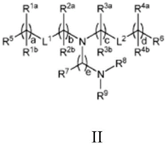
or a pharmaceutically acceptable salt, tautomer, prodrug or stereoisomer thereof, wherein: l is1And L2Each independently is-O (C ═ O) -, - (C ═ O) O-, or a carbon-carbon double bond; r1aAnd R1bIndependently at each occurrence is (a) H or C1-C12Alkyl, or (b) R1aIs H or C1-C12Alkyl, and R1bTogether with the adjacent R with the carbon atom to which it is bonded1bTogether with the carbon atom to which it is bonded form a carbon-carbon double bond; r2aAnd R2bIndependently at each occurrence is (a) H or C1-C12Alkyl, or (b) R2aIs H or C1-C12Alkyl, and R2bTogether with the adjacent R atom to which it is bonded2bTogether with the carbon atom to which it is bonded form a carbon-carbon double bond; r3aAnd R3bIndependently at each occurrence is (a) H or C1-C12Alkyl, or (b) R3aIs H or C1-C12Alkyl, and R3bTogether with the adjacent R with the carbon atom to which it is bonded3bTogether with the carbon atom to which it is bonded form a carbon-carbon double bond; r4aAnd R4bIndependently at each occurrence is (a) H or C1-C12Alkyl, or (b) R4aIs H or C1-C12Alkyl, and R4bTogether with the adjacent R with the carbon atom to which it is bonded4bTogether with the carbon atom to which it is bonded form a carbon-carbon double bond; r5And R6Each independently is methyl or cycloalkyl; r7Independently at each occurrence is H or C 1-C12An alkyl group; r8And R9Each independently is unsubstituted C1-C12 alkyl; or R8And R9Together with the nitrogen atom to which they are attached form a 5-, 6-or 7-membered heterocyclic ring containing one nitrogen atom; a and d are each independently an integer from 0 to 24; b and c are each independently an integer from 1 to 24; and e is 1 or 2, with the proviso that: r1a、R2a、R3aOr R4aAt least one of which is C1-C12 alkyl, or L1Or L2is-O (C ═ O) -or- (C ═ O) O-; and R is1aAnd R1bIs not isopropyl when a is 6 or is not n-butyl when a is 8.
In some aspects, any of the above compositions further comprises one or more excipients comprising a neutral lipid, a steroid, and a polymer conjugated lipid. In some aspects, the neutral lipid comprises at least one of 1, 2-distearoyl-sn-glycero-3-phosphocholine (DSPC), 1, 2-dipalmitoyl-sn-glycero-3-phosphocholine (DPPC), 1, 2-dimyristoyl-sn-glycero-3-phosphocholine (DMPC), 1-palmitoyl-2-oleoyl-sn-glycero-3-phosphocholine (POPC), 1, 2-dioleoyl-sn-glycero-3-phosphocholine (DOPC), and 1, 2-dioleoyl-sn-glycero-3-phosphoethanolamine (DOPE). In some aspects, the neutral lipid is DSPC.
In some aspects, the molar ratio of the compound to neutral lipid is in the range of about 2:1 to about 8: 1.
In some aspects, the steroid is cholesterol. In some aspects, the molar ratio of the compound to cholesterol is in the range of about 2:1 to 1: 1.
In some aspects, the polymer-conjugated lipid is a pegylated lipid. In some aspects, the molar ratio of the compound to pegylated lipid is in the range of about 100:1 to about 25: 1. In some aspects, the pegylated lipid is PEG-DAG, PEG polyethylene (PEG-PE), PEG-succinyl-diacylglycerol (PEG-S-DAG), PEG-cer, or PEG dialkoxypropylcarbamate. In some aspects, the pegylated lipid has the following structure III:

or a pharmaceutically acceptable salt, tautomer, or stereoisomer thereof, wherein: r10And R11Each independently is a straight or branched, saturated or unsaturated alkyl chain containing from 10 to 30 carbon atoms, wherein the alkyl chain is optionally interrupted by one or more ester linkages; and z has an average value in the range of 30 to 60. In some aspects, R10And R11Each independently a straight saturated alkyl chain having from 12 to 16 carbon atoms. In some aspects, the average z is about 45.
In some aspects, the LNPs self-assemble into non-bilayer structures upon mixing with the polyanionic nucleic acid. In some aspects, the diameter of the non-bilayer structure is between 60nm and 120 nm. In some aspects, the diameter of the non-bilayer structure is about 70nm, about 80nm, about 90nm, or about 100 nm. In some aspects, wherein the nanoparticle delivery vehicle has a diameter of about 100 nm.
Also disclosed herein is a method of making a carrier disclosed herein, the method comprising: obtaining a plasmid sequence comprising at least one promoter sequence and an antigen cassette; transfecting the plasmid sequence into one or more host cells; and isolating the vector from the one or more host cells.
In some aspects, separating comprises: lysing the host cell to obtain a cell lysate comprising the vector; and purifying the vector from the cell lysate and optionally also from the medium used to culture the one or more host cells.
In some aspects, the plasmid sequence is generated using one of: DNA recombination or bacterial recombination or whole genome DNA synthesis using amplification of synthetic DNA in bacterial cells. In some aspects, the one or more host cells are at least one of CHO, HEK293 or variants thereof, 911, HeLa, a549, LP-293, per.c6, and AE1-2a cells. In some aspects, purifying the support from the cell lysate involves one or more of chromatographic separation, centrifugation, viral precipitation, and filtration.
Drawings
These and other features, aspects, and advantages of the present invention will become better understood with regard to the following description and accompanying drawings where:
figure 1 shows the development of an in vitro T cell activation assay. This assay is schematically shown, where delivery of a vaccine cassette to antigen presenting cells results in expression, processing and MHC restricted presentation of unique peptide antigens. Reporter T cells engineered to have T cell receptors matching a particular peptide-MHC combination are activated, resulting in luciferase expression.
Figure 2A shows an evaluation of the linker sequence in the short cassette and shows five MHC class I restricted epitopes (epitopes 1 to 5) concatenated in the same position relative to each other, followed by two MHC class II epitopes in general (MHC-II). Various iterations are generated using different linkers. In some cases, the T cell epitopes are directly linked to each other. In other cases, the T cell epitope is flanked on one or both sides by its native sequence. In other iterations, T cell epitopes are linked by non-native sequences AAY, RR and DPP.
Figure 2B shows an assessment of the linker sequence in the short box and shows sequence information about the T cell epitopes embedded in the short box.
Figure 3 shows the evaluation of cell targeting sequences added to a model vaccine cassette. The targeting cassette extends this short cassette design with ubiquitin (Ub), Signal Peptide (SP) and/or Transmembrane (TM) domains, characterized by the close proximity of five markers human T cell epitopes (epitopes 1 to 5) and two mouse T cell epitopes siinfekl (sii) and SPSYAYHQF (a5), and uses a non-natural linker AAY-or T cell epitopes flanked on both sides by natural linkers (25-mer).
Figure 4 shows in vivo evaluation of linker sequences in short cassettes. A) Experimental design for in vivo evaluation of vaccine cassettes using HLA-a2 transgenic mice.
Figure 5A shows an in vivo assessment of the effect on epitope positions in the 21-mer long cassette and shows that the design of the long cassette requires five marker class I epitopes (epitopes 1 to 5) (linker ═ native flanking sequence) contained in the 25-mer native sequence, separated by additional well-known T cell class I epitopes (epitopes 6 to 21) contained in the 25-mer native sequence, and two universal class II epitopes (MHC-II0, where only the relative positions of the class I epitopes are changed.
Figure 5B shows in vivo evaluation of the effect on epitope position in the 21-mer long box and shows sequence information on the T cell epitopes used.
Figure 6A shows the final cassette design of the preclinical IND authorization study (IND-inactivation study) and shows that the design of the final cassette comprises 20 MHC I epitopes (linker ═ native flanking sequence) comprised of 6 non-human primate (NHP) epitopes, 5 human epitopes, 9 murine epitopes, as well as 2 universal MHC class II epitopes comprised in the 25 mer native sequence.
Figure 6B shows the final cassette design of preclinical IND grant study and shows the sequence information of the T cell epitopes used presented on non-human primate, mouse and human derived MHC class I, as well as the sequences of 2 general MHC class II epitopes PADRE and tetanus toxoid.
Fig. 7A shows generation of chadv68.4wtnt. gfp virus after transfection. HEK293A cells were transfected with chadv68.4wtnt. gfp DNA using a calcium phosphate protocol. Virus replication was observed 10 days after transfection and viral plaques were observed for chadv68.4wtnt. gfp using light microscopy (40 × magnification).
Fig. 7B shows generation of chadv68.4wtnt. gfp virus after transfection. HEK293A cells were transfected with chadv68.4wtnt. gfp DNA using a calcium phosphate protocol. Virus replication was observed 10 days after transfection and viral plaques were observed for chadv68.4wtnt. gfp at 40 × magnification using a fluorescence microscope.
Fig. 7C shows generation of chadv68.4wtnt. gfp virus after transfection. HEK293A cells were transfected with chadv68.4wtnt. gfp DNA using a calcium phosphate protocol. Virus replication was observed 10 days after transfection and viral plaques were observed for chadv68.4wtnt. gfp at 100 × magnification using a fluorescence microscope.
Fig. 8A shows generation of chadv68.5wtnt. gfp virus after transfection. HEK293A cells were transfected with chadv68.5wtnt. gfp DNA using lipofectamine (lipofectamine) protocol. Viral replication (plaques) was observed 10 days after transfection. Lysates were prepared and used to reinfect 293A cells in T25 flasks. After 3 days, plaques of the chadv68.5wtnt. gfp virus were observed using an optical microscope (40 × magnification) and photographed.
Fig. 8B shows generation of chadv68.5wtnt. gfp virus after transfection. HEK293A cells were transfected with chadv68.5wtnt. gfp DNA using a lipofectamine protocol. Viral replication (plaques) was observed 10 days after transfection. Lysates were prepared and used to reinfect 293A cells in T25 flasks. After 3 days, plaques of the chadv68.5wtnt. gfp virus were observed using a fluorescence microscope at 40 × magnification and photographed.
Fig. 8C shows generation of chadv68.5wtnt. gfp virus after transfection. HEK293A cells were transfected with chadv68.5wtnt. gfp DNA using a lipofectamine protocol. Viral replication (plaques) was observed 10 days after transfection. Lysates were prepared and used to reinfect 293A cells in T25 flasks. After 3 days, plaques of the chadv68.5wtnt. gfp virus were observed using a fluorescence microscope at 100 × magnification and photographed.
FIG. 9 shows a virus particle production scheme.
FIG. 10 shows an alphavirus-derived VEE self-replicating RNA (srRNA) vector.
FIG. 11 shows reporter gene expression in vivo after C57BL/6J mice were inoculated with VEE-luciferase srRNA. Representative luciferase signal images after immunization of C57BL/6J mice (10 ug per mouse, both side intramuscular injection, MC3 encapsulation) with VEE-luciferase srna at various time points are shown.
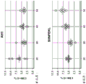
FIG. 12A shows T cell responses measured 14 days after immunization of VEE srRNA formulated with MC3LNP in mice bearing B16-OVA tumors. C57BL/6J mice bearing B16-OVA tumors were injected with 10ug VEE-luciferase srRNA (control), VEE-UbAAY srRNA (Vax), VEE-luciferase srRNA and anti-CTLA-4 (aCTLA-4) or VEE-UbAAY srRNA and anti-CTLA-4 (Vax + aCTLA-4). In addition, all mice were treated with anti-PD 1 mAb starting on day 7. Each group consisted of 8 mice. At 14 days after immunization, mice were sacrificed and spleen and lymph nodes were collected. SIINFEKL specific T cell responses were assessed by IFN- γ ELISPOT and reported as Spot Forming Cell (SFC) number per 106 splenocytes. The line represents the median.
FIG. 12B shows T cell responses measured 14 days after immunization of VEE srRNA formulated with MC3LNP in mice bearing B16-OVA tumors. C57BL/6J mice bearing B16-OVA tumors were injected with 10ug VEE-luciferase srRNA (control), VEE-UbAAY srRNA (Vax), VEE-luciferase srRNA and anti-CTLA-4 (aCTLA-4) or VEE-UbAAY srRNA and anti-CTLA-4 (Vax + aCTLA-4). In addition, all mice were treated with anti-PD 1 mAb starting on day 7. Each group consisted of 8 mice. At 14 days after immunization, mice were sacrificed and spleen and lymph nodes were collected. SIINFEKL-specific T cell responses were assessed by mhc i-pentamer staining, reported as the percentage of pentamer-positive cells to CD 8-positive cells. The line represents the median.
Figure 13A shows antigen-specific T cell responses following heterologous prime/boost immunization in mice bearing B16-OVA tumors. C57BL/6J mice bearing B16-OVA tumors were injected with GFP expressing adenovirus (Ad5-GFP) and boosted with VEE-luciferase srRNA formulated with MC3 LNP (control) or Ad5-UbAAY and boosted with VEE-UbAAY srRNA (Vax). The control and Vax groups were also treated with IgG control mAb. The third group was treated with Ad5-GFP prime/VEE-luciferase srRNA boost in combination with anti-CTLA-4 (aCTLA-4), while the fourth group was treated with Ad5-UbAAY prime/VEE-UbAAY boost in combination with anti-CTLA-4 (Vax + aCTLA-4). In addition, all mice were treated with anti-PD-1 mAb starting on day 21. T cell responses were measured by IFN-. gamma.ELISPOT. At 14 days after immunization with adenovirus, mice were sacrificed and spleen and lymph nodes were collected.
Figure 13B shows antigen-specific T cell responses following heterologous prime/boost immunization in mice bearing B16-OVA tumors. C57BL/6J mice bearing B16-OVA tumors were injected with GFP expressing adenovirus (Ad5-GFP) and boosted with VEE-luciferase srRNA formulated with MC3 LNP (control) or Ad5-UbAAY and boosted with VEE-UbAAY srRNA (Vax). The control and Vax groups were also treated with IgG control mAb. The third group was treated with Ad5-GFP prime/VEE-luciferase srRNA boost in combination with anti-CTLA-4 (aCTLA-4), while the fourth group was treated with Ad5-UbAAY prime/VEE-UbAAY boost in combination with anti-CTLA-4 (Vax + aCTLA-4). In addition, all mice were treated with anti-PD-1 mAb starting on day 21. T cell responses were measured by IFN-. gamma.ELISPOT. Mice were sacrificed 14 days after immunization with adenovirus and 14 days after boosting with srna (day 28 after priming) and spleens and lymph nodes were collected.
Figure 13C shows antigen-specific T cell responses following heterologous prime/boost immunization in mice bearing B16-OVA tumors. C57BL/6J mice bearing B16-OVA tumors were injected with GFP expressing adenovirus (Ad5-GFP) and boosted with VEE-luciferase srRNA formulated with MC3LNP (control) or Ad5-UbAAY and boosted with VEE-UbAAY srRNA (Vax). The control and Vax groups were also treated with IgG control mAb. The third group was treated with Ad5-GFP prime/VEE-luciferase srRNA boost in combination with anti-CTLA-4 (aCTLA-4), while the fourth group was treated with Ad5-UbAAY prime/VEE-UbAAY boost in combination with anti-CTLA-4 (Vax + aCTLA-4). In addition, all mice were treated with anti-PD-1 mAb starting on day 21. T cell responses were measured by MHC class I pentamer staining. At 14 days after immunization with adenovirus, mice were sacrificed and spleen and lymph nodes were collected.
Figure 13D shows antigen-specific T cell responses following heterologous prime/boost immunization in mice bearing B16-OVA tumors. C57BL/6J mice bearing B16-OVA tumors were injected with GFP expressing adenovirus (Ad5-GFP) and boosted with VEE-luciferase srRNA formulated with MC3LNP (control) or Ad5-UbAAY and boosted with VEE-UbAAY srRNA (Vax). The control and Vax groups were also treated with IgG control mAb. The third group was treated with Ad5-GFP prime/VEE-luciferase srRNA boost in combination with anti-CTLA-4 (aCTLA-4), while the fourth group was treated with Ad5-UbAAY prime/VEE-UbAAY boost in combination with anti-CTLA-4 (Vax + aCTLA-4). In addition, all mice were treated with anti-PD-1 mAb starting on day 21. T cell responses were measured by MHC class I pentamer staining. Mice were sacrificed 14 days after immunization with adenovirus and 14 days after boosting with srna (day 28 after priming) and spleens and lymph nodes were collected.
FIG. 14A shows antigen-specific T cell responses following allogeneic prime/boost in mice bearing CT26(Balb/c) tumors. Mice were immunized with Ad5-GFP and boosted 15 days after adenovirus priming with VEE-luciferase srRNA formulated with MC3 LNP (control) or primed with Ad5-UbAAY and boosted with VEE-UbAAY srRNA (Vax). The control and Vax groups were also treated with IgG control mAb. The other group was administered Ad 5-GFP/VEE-luciferase srna combination primed/boosted with anti-PD-1 (aPD1), while the fourth group received Ad5-UbAAY/VEE-UbAAY srna combination primed/boosted with anti-PD-1 mAb (Vax + aPD 1). The response of T cells to AH1 peptide was measured using IFN- γ ELISPOT. At 12 days post-immunization with adenovirus, mice were sacrificed and spleen and lymph nodes were collected.
Figure 14B shows antigen-specific T cell responses following allogeneic prime/boost in mice bearing CT26(Balb/c) tumors. Mice were immunized with Ad5-GFP and boosted 15 days after adenovirus priming with VEE-luciferase srRNA formulated with MC3 LNP (control) or primed with Ad5-UbAAY and boosted with VEE-UbAAY srRNA (Vax). The control and Vax groups were also treated with IgG control mAb. The other group was administered Ad 5-GFP/VEE-luciferase srna combination primed/boosted with anti-PD-1 (aPD1), while the fourth group received Ad5-UbAAY/VEE-UbAAY srna combination primed/boosted with anti-PD-1 mAb (Vax + aPD 1). The response of T cells to AH1 peptide was measured using IFN- γ ELISPOT. Mice were sacrificed 12 days after immunization with adenovirus and 6 days after boosting with srna (day 21 after priming) and spleens and lymph nodes were collected.
Fig. 15 shows that ChAdV68 elicits T cell responses against mouse tumor antigens in mice. Mice were immunized with chadv68.5wtnt. mag25mer and T cell responses to MHC class I epitopes siinfekl (ova) were measured in C57BL/6J female mice and to MHC class I epitopes AH1-a5 in Balb/C mice. Presentation was measured in an ELISpot assay for every 106Mean spot-forming cells (SFC) of individual splenocytes. Error bars indicate standard deviation.
FIG. 16 shows cellular immune responses following a single immunization with ChAdV6, ChAdV + anti-PD-1, srRNA + anti-PD-1, or anti-PD-1 alone in a CT26 tumor model. The production of antigen-specific IFN- γ in splenocytes from 6 mice of each group was measured using ELISpot. The results are presented as every 106Spot-forming cells (SFC) of individual splenocytes. The median of each group is indicated by the horizontal line. P values were determined using Dunnett's multiple comparison test; p<0.0001,**P<0.001,*P<0.05。ChAdV=ChAdV68.5WTnt.MAG25mer;srRNA=VEE-MAG25mer srRNA。
FIG. 17 shows CD8T cell responses following a single immunization with ChAdV6, ChAdV + anti-PD-1, srRNA + anti-PD-1, or anti-PD-1 alone in a CT26 tumor model. ICS was used to measure the production of antigen-specific IFN- γ in CD8T cells and the results were presented as the percentage of antigen-specific CD8T cells to total CD8T cells. The median of each group is indicated by the horizontal line. P values were determined using Dunnett's multiple comparison test; p <0.0001, P <0.001, P < 0.05. ChAdV ═ chadv68.5wtnt. mag25mer; srna ═ VEE-MAG25mer srna.
Fig. 18 shows tumor growth following immunization with ChAdV/srna heterologous prime/boost, srna/ChAdV heterologous prime/boost or srna/srna homologous prime/boost in a CT26 tumor model. Also shown is a comparison to prime/boost immunizations administered with or without anti-PD 1 during prime and boost immunizations. Tumor volumes were measured twice weekly and presented as the mean tumor volume for the first 21 days of the study. 22-28 mice per group were initiated for the study. Error bars represent Standard Error (SEM) of the mean. P values were determined using Dunnett's test; p <0.0001, P <0.001, P < 0.05. ChAdV ═ chadv68.5wtnt. mag25mer; srna ═ VEE-MAG25mer srna.
Fig. 19 shows survival after immunization with ChAdV/srna heterologous prime/boost, srna/ChAdV heterologous prime/boost or srna/srna homologous prime/boost in a CT26 tumor model. Also shown is a comparison to prime/boost immunizations administered with or without anti-PD 1 during prime and boost immunizations. P values were determined using the log rank test; p <0.0001, P <0.001, P < 0.01. ChAdV ═ chadv68.5wtnt. mag25mer; srna ═ VEE-MAG25 mersrna.
Figure 20 shows antigen-specific cellular immune responses measured using ELISpot. Antigen-specific IFN- γ production against six different mamu a 01-restricted epitopes in PBMCs of the VEE-MAG25mer rrna-LNP1(30 μ g) (fig. 20A), VEE-MAG25mer rrna-LNP1(100 μ g) (fig. 20B), or VEE-MAG25mer rrna-LNP2(100 μ g) (fig. 20C) homologous prime/boost or the chadvv 68.5wtnt.mag25mer/VEE-MAG25mer rrna heterologous prime/boost panel (fig. 20D) was measured using ELISpot at 1, 2, 3, 4, 5, 6, 8, 9 or 10 weeks after the first boost (6 rhesus per group). Results were in a stacked bar graph format every 10 for each epitope 6Mean spot-forming cell (SFC) presentation of individual PBMCs. Values for each animal were normalized to the level before exsanguination (week 0).
Figure 21 shows antigen specific cellular immune responses measured using ELISpot. Prior to immunization withAnd measuring antigen-specific IFN- γ production against six different mamu a 01-restricted epitopes in PBMCs after immunization with a chadv68.5wtnt. mag25mer/VEE-MAG25mer srna heterologous prime/boost regimen using ELISpot at 4, 5, 6, 7, 8, 10, 11, 12, 13, 14, 15, 16, 17, 18, 19, 20, 21, 22, 23, or 24 weeks after the initial immunization. Results are in a stacked bar graph format every 10 for each epitope6Mean spot-forming cells (SFC) presentation of individual PBMCs (6 rhesus monkeys per group).
Figure 22 shows antigen specific cellular immune responses measured using ELISpot. Antigen-specific IFN- γ production against six different mamu a 01-restricted epitopes in PBMCs after immunization with VEE-MAG25mer srna LNP2 homologous prime/boost regimen was measured using ELISpot before immunization and at 4, 5, 6, 7, 8, 10, 11, 12, 13, 14 or 15 weeks after initial immunization. Results are in a stacked bar graph format every 10 for each epitope 6Mean spot-forming cells (SFC) presentation of individual PBMCs (6 rhesus monkeys per group).
Figure 23 shows antigen specific cellular immune responses measured using ELISpot. Antigen-specific IFN- γ production against six different mamu a 01-restricted epitopes in PBMCs after immunization with VEE-MAG25mer srna LNP1 homologous prime/boost regimen was measured using ELISpot before immunization and at 4, 5, 6, 7, 8, 10, 11, 12, 13, 14 or 15 weeks after initial immunization. Results are in a stacked bar graph format every 10 for each epitope6Mean spot-forming cells (SFC) presentation of individual PBMCs (6 rhesus monkeys per group).
FIGS. 24A and 24 show exemplary peptide profiles generated from Promega's dynamic range standard.
Figure 25 shows the correlation between EDGE scores and the likelihood of detecting candidate consensus neoantigenic peptides by targeting MS.
FIG. 26 shows the design of an E1/E3 deleted ChAdV68 viral vector using an expression cassette co-expressing a checkpoint inhibitor introduced into the deleted E1 region.
Figure 27 shows in vitro mouse anti-CTLA 4 clone 9D9 antibody expression following 293A cell infection.
FIG. 28A shows a Western blot demonstrating expression of human anti-CTLA-4 IgG1 antibody (ipilimumab) in chAd68-MAG-IRES-IPI (IPI-MAG) and chAd68-GFP-IRES-IPI (IPI-GFP) infected cells. HEK 293A. Cells were infected at an MOI of 1, and cell pellets and supernatant were harvested 48h post infection. anti-CTLA 4 antibody was purified from the supernatant by immunoprecipitation with protein G beads (ThermoFisher). The supernatants of the pellet and immunoprecipitate were analyzed by SDS-PAGE electrophoresis and Western blotting using HRP donkey anti-human IgG antibody and detected by ECL chemiluminescent substrate (ThermoFisher).
FIG. 28B shows a Western blot demonstrating antibody expression of human anti-CTLA-4 IgG2 antibody (tremelimumab) in (1) chAd68-MAG-IRES-TREME and chAd68-M2.2, control virus infected cells. HEK 293A. Cells were infected at an MOI of 1, and cell pellets and supernatant were harvested 48h post infection. anti-CTLA 4 antibody was purified from the supernatant by immunoprecipitation with protein G beads (ThermoFisher). The supernatants of the pellet and immunoprecipitate were analyzed by SDS-PAGE electrophoresis and Western blotting using HRP donkey anti-human IgG antibody and detected by ECL chemiluminescent substrate (ThermoFisher).
Fig. 29 shows the general organization of model epitopes from various species with large antigen cassettes of 30 (L), 40 (XL) or 50 (XXL) epitopes.
Fig. 30 shows ChAd vectors expressing long cassettes as indicated by western blotting as above using anti-class II (PADRE) antibodies recognizing sequences common to all cassettes. HEK293 cells were infected with chAd68 vectors expressing variable size large cassettes (chAd68-50XXL, chAd68-40XL, and chAd 68-30L). Infection was set at an MOI of 0.2. Twenty-four hours post-infection, the proteasome inhibitor MG132 was added to a set of infected wells (indicated by a plus sign). Another set of wells treated with virus were not treated with MG132 (indicated by a minus sign). Uninfected HEK293 cells (293F) were used as negative controls. Forty-eight hours post infection, cell pellets were collected and analyzed by SDS/PAGE electrophoresis and immunoblotting using rabbit anti-class II PADRE antibody. Detection was performed using HRP anti-rabbit antibody and ECL chemiluminescent substrate.
Fig. 31 shows the CD8+ immune response detected by ICS against AH1 (top) and SIINFEKL (bottom) in chAd68 large box immunized mice. Data are expressed as the percentage of IFNg + cells to model epitope to total CD8 cells.
FIG. 32 shows CD8+ responses to LD-AH1+ (top) and Kb-SIINFEKL + (bottom) tetramer following large cassette vaccination with chAd 68. Data are expressed as a percentage of total CD8 cells reactive to the model tetrameric peptide complex. P <0.05, p <0.01, ANOVA by Tukey test. All p values were compared to the MAG 20 antigen cassette.
FIG. 33 shows CD8+ immune responses against AH1 (top) and SIINFEKL (bottom) detected by ICS in alphavirus big-box treated mice. Data are expressed as the percentage of IFNg + cells to model epitope to total CD8 cells. P <0.05, p <0.01, p <0.001, ANOVA by Tukey test. All p values were compared to the MAG 20 antigen cassette.
Figure 34 shows a vaccination strategy for assessing the immunogenicity of antigen cassette-containing vectors in rhesus monkeys. Triangles indicate vaccination with chAd68 at weeks 0 and 32 (1e12 vp/animal). Circles indicate alphavirus vaccination at weeks 0, 4, 12, 20, 28 and 32. Squares indicate administration of anti-CTLA 4 antibody.
FIG. 35 shows the time course of CD8+ anti-epitope response in rhesus monkeys given only chAd-MAG (group 4). Mean SFC/1e6 splenocytes are shown.
Figure 36 shows the time course of CD8+ anti-epitope response in rhesus monkeys given IV delivered chAd-MAG plus anti-CTLA 4 antibody (ipilimumab) (group 5). Mean SFC/1e6 splenocytes are shown.
Figure 37 shows the time course of CD8+ anti-epitope response in rhesus monkeys given SC-delivered chAd-MAG plus anti-CTLA 4 antibody (ipilimumab) (group 6). Mean SFC/1e6 splenocytes are shown.
FIG. 38 shows antigen-specific memory responses generated by the ChAdV68/samRNA vaccine protocol as measured by ELISpot. The results are shown as individual dots, each dot representing one animal. Baseline before immunization (left panel) and memory response 18 months after priming (right panel) are shown.
Figure 39 shows memory cell phenotype analysis of antigen-specific CD8+ T cells using flow cytometry combined tetramer staining and CD45RA/CCR7 co-staining.
Figure 40 shows the distribution of memory cell types in the sum of the four Mamu-a 01 tetramer + CD8+ T cell populations at study month 18. Memory cells were characterized as follows: CD45RA + CCR7+ (naive), CD45RA + CCR7 ═ effector (Teff), CD45RA-CCR7 ═ central memory (Tcm), CD45RA-CCR7 ═ effector memory (Tem).
FIG. 41 shows antigen-specific T cell responses to immunization with low (1.5e6IU, left) and high (1.5e7 IU, right) vector doses of chAd-MAG-CTLA4, chAd-MAG alone, and anti-CTLA 4 o9D9 delivered with chAd-MAG and IP. Responses were measured by IFN γ ELISpot and expressed as spot-forming cells per mouse of 6 splenocytes per 1e (n-8 per group). Bars represent median.
FIG. 42 shows antigen-specific T cell responses to immunization with low (1.5e6IU, left) and high (1.5e7 IU, right) vector doses of chAd-MAG-CTLA4, chAd-MAG alone, and anti-CTLA 4 o9D9 delivered with chAd-MAG and IP. The response was measured by intracellular staining (ICS) and expressed as IFN γ per mouse+Cell occupancy Total CD8+Percentage of cells (n-8 per group). Bars represent median.
FIG. 43 shows anti-CTLA 4 antibody levels in serum of mice immunized with anti-CTLA 4 o9D9 delivered with chAd-MAG-CTLA4 or chAd-MAG plus IP. Electrochemiluminescence (ECL), mean and standard deviation for each time point and group (n-8 per group). Black arrows represent time points of anti-CTLA 4 administration in groups 3 and 4. Mice were immunized on day 0 with ChAd-MAG and ChAd-MAG-aCTLA 4. The dotted line represents the maximum limit of the assay. Both groups that delivered anti-CTLA 4 mAb systemically (IP) were at the highest limit of the assay at all time points measured post-immunization.
Figure 44 shows the frequency of CD8+ T cells recognizing the CT26 tumor antigen AH1 in mice with CT26 tumor. P values were determined using a one-way ANOVA and Tukey multiple comparison test. P <0.001, P < 0.05. ChAdV ═ chadv68.5wtnt. mag25mer; CTLA 4-anti-CTLA 4 antibody, clone 9D 9.
Detailed Description
I. Definition of
In general, the terms used in the claims and the specification are intended to be interpreted to have ordinary meanings as understood by those of ordinary skill in the art. For clarity, certain terms are defined below. The definitions provided should be used if there is a conflict between ordinary meaning and the definitions provided.
As used herein, the term "antigen" is a substance that induces an immune response. The antigen may be a neoantigen. An antigen may be a "consensus antigen," which is an antigen found in a particular population (e.g., a particular population of cancer patients).
As used herein, the term "neoantigen" is an antigen having at least one alteration that makes it different from a corresponding wild-type antigen, e.g., via a mutation in a tumor cell or a post-translational modification specific for a tumor cell. The neoantigen may comprise a polypeptide sequence or a nucleotide sequence. Mutations may include frameshift or non-frameshift indels, missense or nonsense substitutions, splice site alterations, genomic rearrangements or gene fusions, or any genomic or expression alteration that produces a neoORF. Mutations may also include splice variants. Post-translational modifications specific for tumor cells may include aberrant phosphorylation. Post-translational modifications specific for tumor cells may also include proteasomally produced splicing antigens. See, Liepe et al, A large fraction of HLA class I ligands area proteins-generated specific peptides; science.2016, 10 months and 21 days; 354(6310):354-358. Such consensus neoantigens are useful for inducing an immune response in a subject by administration. The subject can be identified for administration by using a variety of diagnostic methods, such as the patient selection methods described further below.
As used herein, the term "tumor antigen" is an antigen that is present in a tumor cell or tissue of a subject but not in a corresponding normal cell or tissue of the subject or that is derived from a polypeptide that is known or has been found to have altered expression in a tumor cell or cancer tissue as compared to a normal cell or tissue.
As used herein, the term "antigen-based vaccine" is a vaccine composition based on one or more antigens (e.g., multiple antigens). Vaccines can be nucleotide-based (e.g., virus-based, RNA-based, or DNA-based), protein-based (e.g., peptide-based), or a combination thereof.
As used herein, the term "candidate antigen" is a mutation or other aberration that produces a sequence that can represent an antigen.
As used herein, the term "coding region" is the portion of a gene that encodes a protein.
As used herein, the term "coding mutation" is a mutation that occurs in a coding region.
As used herein, the term "ORF" means an open reading frame.
As used herein, the term "NEO-ORF" is a tumor-specific ORF that results from a mutation or other aberration, such as splicing.
As used herein, the term "missense mutation" is a mutation that causes the substitution of one amino acid by another.
As used herein, the term "nonsense mutation" is a mutation that causes an amino acid to be replaced by a stop codon or causes removal of a typical start codon.
As used herein, the term "frameshift mutation" is a mutation that causes a change in the framework of a protein.
As used herein, the term "indel" is an insertion or deletion of one or more nucleic acids.
As used herein, the term "percent identity," in the context of two or more nucleic acid or polypeptide sequences, refers to a specified percentage of nucleotides or amino acid residues of the two or more sequences or subsequences that are the same when compared and aligned for maximum correspondence, as measured using one of the sequence comparison algorithms described below (e.g., BLASTP and BLASTN, or other algorithms available to the skilled artisan) or by visual inspection. Depending on the application, the "identity" percentage can be present over the region of the sequences being compared, for example over the functional domain, or over the entire length of the two sequences to be compared.
With respect to sequence comparison, typically one sequence serves as a reference sequence to which test sequences are compared. When using a sequence comparison algorithm, the test sequence and the reference sequence are entered into a computer, subsequence coordinates are designated as necessary, and sequence algorithm program parameters are designated. The sequence comparison algorithm then calculates the percent sequence identity of the test sequence relative to the reference sequence based on the specified program parameters. Alternatively, sequence similarity or dissimilarity can be determined by combining the presence or absence of specific nucleotides at selected sequence positions (e.g., sequence motifs), or specific amino acids for the translated sequence.
Optimal sequence alignment for comparison can be achieved, for example, by the local homology algorithm of Smith and Waterman, adv.appl.math.2:482 (1981); homology alignment algorithms of Needleman and Wunsch, J.mol.biol.48:443 (1970); the similarity search method of Pearson and Lipman, Proc.Nat' l.Acad.Sci.USA 85:2444 (1988); computerized implementation of these algorithms (GAP, BESTFIT, FASTA and TFASTA in the Wisconsin Genetics software package; Genetics Computer Group,575 Science Dr., Madison, Wis.); or by visual inspection (see generally Ausubel et al, infra).
An example of an algorithm suitable for determining sequence identity and percent sequence similarity is the BLAST algorithm described in Altschul et al, J.mol.biol.215: 403-. Software for performing BLAST analysis is publicly available through the National Center for Biotechnology Information.
As used herein, the term "no termination or read-through" is a mutation that causes the removal of the native stop codon.
As used herein, the term "epitope" is the specific portion of an antigen that is normally bound by an antibody or T cell receptor.
As used herein, the term "immunogenicity" is the ability to elicit an immune response, e.g., by T cells, B cells, or both.
As used herein, the terms "HLA binding affinity", "MHC binding affinity" means the binding affinity between a specific antigen and a specific MHC allele.
As used herein, the term "bait" is a nucleic acid probe used to enrich a sample for a specific sequence of DNA or RNA.
As used herein, the term "variant" is the difference between a subject's nucleic acid and a reference human genome used as a control.
As used herein, the term "variant call" is an algorithmic determination of the presence of variants typically determined by sequencing.
As used herein, the term "polymorphism" is a germline variant, i.e. a variant found in all DNA-carrying cells of an individual.
As used herein, the term "somatic variant" is a variant produced in a non-germline cell of an individual.
As used herein, the term "allele" is a form of a gene, or a form of a gene sequence, or a form of a protein.
As used herein, the term "HLA type" is the complement of an HLA gene allele.
As used herein, the term "nonsense-mediated decay" or "NMD" is the degradation of mRNA by a cell due to a premature stop codon.
As used herein, the term "trunk mutation" is a mutation originating in the early stages of tumor development and present in most tumor cells.
As used herein, the term "subcloning mutation" is a mutation originating in a later stage of tumor development and present only in a subset of tumor cells.
As used herein, the term "exome" is a subset of the genome that encodes a protein. An exome may be a collective exon of a genome.
As used herein, the term "logistic regression" is a regression model from statistical binary data in which the logic of the probability that a dependent variable equals 1 is modeled as a linear function of the dependent variable.
As used herein, the term "neural network" is a machine learning model for classification or regression that consists of a multi-layered linear transformation followed by element-wise nonlinearities that are typically trained via stochastic gradient descent and back propagation.
As used herein, the term "proteome" is a collection of all proteins expressed and/or translated by a cell, group of cells, or individual.
As used herein, the term "pepset" is a collection of all peptides presented by MHC-I or MHC-II on the cell surface. A pepset may refer to a property of a cell or collection of cells (e.g., a tumor pepset, meaning the union of the pepsets of all cells that make up a tumor).
As used herein, the term "ELISPOT" means an enzyme-linked immunosorbent spot assay, which is a commonly used method for monitoring immune responses in humans and animals.
As used herein, the term "dextran peptide multimer" is a dextran-based peptide-MHC multimer used for antigen-specific T cell staining in flow cytometry.
As used herein, the term "tolerance or immunological tolerance" is a state of immunological nonresponsiveness to one or more antigens (e.g., autoantigens).
As used herein, the term "central tolerance" is the tolerance suffered in the thymus by the deletion of autoreactive T cell clones or by promoting differentiation of autoreactive T cell clones into immunosuppressive regulatory T cells (tregs).
As used herein, the term "peripheral tolerance" is the tolerance that is experienced peripherally by downregulating or not activating autoreactive T cells that are subjected to central tolerance or promoting the differentiation of these T cells into tregs.
The term "sample" may include obtaining a single cell or a plurality of cells or cell fragments or aliquots of body fluid from a subject by means including venipuncture, excretion, ejaculation, massage, biopsy, needle aspiration, lavage of the sample, scraping, surgical incision or intervention, or other means known in the art.
The term "subject" encompasses a human or non-human, whether in vivo, ex vivo or in vitro, male or female cell, tissue or organism. The term subject includes mammals including humans.
The term "mammal" encompasses humans and non-humans and includes, but is not limited to, humans, non-human primates, canines, felines, murines, bovines, equines, and porcines.
The term "clinical factor" refers to a measure of the condition of a subject, such as disease activity or severity. "clinical factors" encompass all markers of the health condition of a subject, including non-sample markers, and/or other characteristics of the subject, such as, but not limited to, age and gender. A clinical factor may be a score, value, or set of values that may be obtained from evaluating a sample (or population of samples) from a subject or a subject under defined conditions. Clinical factors may also be predicted from markers and/or other parameters (e.g., gene expression surrogates). Clinical factors may include tumor type, tumor subtype and smoking history.
The term "tumor-derived antigen-encoding nucleic acid sequence" refers to a nucleic acid sequence that is directly extracted from a tumor, e.g., via RT-PCR; or sequence data obtained by tumor sequencing, and then synthesizing the nucleic acid sequence using the sequencing data, e.g., via various synthetic or PCR-based methods known in the art.
The term "alphavirus" refers to a member of the Togaviridae family (Togaviridae) and is a plus-sense single-stranded RNA virus. Alphaviruses are generally classified as old world, such as sindbis, ross river, mayalu, Chikungunya (Chikungunya) and semliki forest viruses, or new world, such as eastern equine encephalitis, ola, morguerburg or venezuelan equine encephalitis and its derivative strains TC-83. Alphaviruses are typically self-replicating RNA viruses.
The term "alphavirus backbone" refers to the minimal sequence of an alphavirus that allows the viral genome to replicate itself. The minimal sequences may include conserved sequences for non-structural protein mediated amplification, the non-structural protein 1(nsP1) gene, the nsP2 gene, the nsP3 gene, the nsP4 gene, and poly a sequences, as well as sequences for subgenomic viral RNA expression, including the 26S promoter element.
The term "sequence for non-structural protein mediated amplification" includes the conserved sequence element of alphavirus (CSE) well known to those skilled in the art. CSE includes, but is not limited to, alphavirus 5'UTR, 51-nt CSE, 24-nt CSE or other 26S subgenomic promoter sequences, 19-nt CSE and alphavirus 3' UTR.
The term "RNA polymerase" includes polymerases that catalyze the production of RNA polynucleotides from DNA templates. RNA polymerases include, but are not limited to, phage-derived polymerases, including T3, T7, and SP 6.
The term "lipid" includes hydrophobic and/or amphiphilic molecules. The lipids may be cationic, anionic or neutral. Lipids may be of synthetic or natural origin, and in some cases are biodegradable. Lipids may include cholesterol, phospholipids, lipid conjugates including, but not limited to, polyethylene glycol (PEG) conjugates (pegylated lipids), waxes, oils, glycerides, fats, and fat-soluble vitamins. Lipids may also include dilinoleylmethyl-4-dimethylaminobutyrate ester (MC3) and MC 3-like molecules.
The term "lipid nanoparticle" or "LNP" includes vesicle-like structures formed around an aqueous interior using a lipid-containing membrane, also known as liposomes. Lipid nanoparticles include lipid-based compositions having a solid lipid core stabilized by a surfactant. The core lipid may be a fatty acid, an acylglycerol, a wax, and mixtures of these surfactants. Biomembrane lipids, such as phospholipids, sphingomyelin, bile salts (sodium taurocholate) and sterols (cholesterol), may be used as stabilizers. Lipid nanoparticles can be formed using a defined ratio of different lipid molecules, including (but not limited to) a defined ratio of one or more cationic, anionic or neutral lipids. The lipid nanoparticles can encapsulate molecules within an outer membrane shell, and can then be contacted with a target cell to deliver the encapsulated molecules to the host cell cytosol. The lipid nanoparticles may be modified or functionalized with non-lipid molecules, including on their surface. The lipid nanoparticles may be monolayer (monolayer) or multilayer (multilayer). The lipid nanoparticles may be complexed with nucleic acids. The monolayer of lipid nanoparticles can be complexed with nucleic acids, wherein the nucleic acids are in the aqueous interior. The multilamellar lipid nanoparticles can be complexed with nucleic acids, wherein the nucleic acids are within the aqueous interior, or formed or sandwiched therebetween.
Abbreviations: MHC: a major histocompatibility complex; HLA: human leukocyte antigens or human MHC loci; and (3) NGS: sequencing the next generation; PPV: positive predictive value; TSNA: a tumor-specific neoantigen; FFPE: formalin fixation and paraffin embedding; NMD: nonsense-mediated decay; NSCLC: non-small cell lung cancer; DC: a dendritic cell.
As used in this specification and the appended claims, the singular forms "a", "an", and "the" include plural referents unless the context clearly dictates otherwise.
Unless otherwise indicated or apparent from the context, as used herein, the term "about" is to be understood as being within the normal tolerance of the art, e.g., within 2 standard deviations of the mean. About can be understood to be within 10%, 9%, 8%, 7%, 6%, 5%, 4%, 3%, 2%, 1%, 0.5%, 0.1%, 0.05% or 0.01% of the stated value. All numerical values provided herein are modified by the term about, unless the context clearly dictates otherwise.
Any terms not directly defined herein should be understood to have the meanings commonly associated therewith as understood in the art of the present invention. Certain terms are discussed herein in order to provide additional guidance to the practitioner regarding the compositions, devices, methods, etc., and making or using thereof, of the various aspects of the invention. It should be understood that the same thing can be represented in more than one way. Thus, alternative phraseology and synonyms may be used for any one or more of the terms discussed herein. It is irrelevant whether terminology is set forth or discussed herein. Synonyms or substitutable methods, materials, etc. are provided. Recitation of one or more synonyms or equivalents does not exclude the use of other synonyms or equivalents unless explicitly stated otherwise. Examples, including use of the term examples, are for illustrative purposes only and are not intended to limit the scope or meaning of aspects of the present invention herein.
All references, issued patents and patent applications cited within the text of the specification are hereby incorporated by reference in their entirety for all purposes.
Method for identifying antigens
Methods for identifying common antigens (e.g., neoantigens) include methods of identifying antigens from a tumor in a subject that may be presented on the cell surface of a tumor or immune cell, including professional antigen presenting cells such as dendritic cells, and/or that may be immunogenic. For example, one such method may include the steps of: obtaining at least one of exome, transcriptome, or whole genome tumor nucleotide sequencing and/or expression data from tumor cells of a subject, wherein the tumor nucleotide sequencing and/or expression data is used to obtain data representative of peptide sequences for each of a collection of antigens (e.g., in the case of neoantigens, wherein the peptide sequence of each neoantigen comprises at least one alteration that makes it different from a corresponding wild-type peptide sequence, or in the case of a consensus antigen without mutations, wherein the peptide is derived from any polypeptide known or found to have altered expression in tumor cells or cancer tissue as compared to normal cells or tissue); inputting the peptide sequence of each antigen into one or more presentation models to generate a set of numerical likelihoods that the antigens are each presented by one or more MHC alleles on the tumor cell surface of a tumor cell or cells present in a tumor of the subject, the set of numerical likelihoods having been identified based at least on the received mass spectrometry data; and selecting a subset of the set of antigens based on the set of numerical likelihoods to produce a set of selected antigens.
The presentation model may comprise a statistical regression or machine learning (e.g. deep learning) model trained on a reference data set (also referred to as a training data set) comprising a respective set of markers, wherein the reference data set is obtained from each of a plurality of different subjects, wherein optionally some subjects may have a tumor, and wherein the reference data set comprises at least one of: data representing exome nucleotide sequences from tumor tissue, data representing exome nucleotide sequences from normal tissue, data representing transcriptome nucleotide sequences from tumor tissue, data representing proteome sequences from tumor tissue and data representing MHC pepset sequences from tumor tissue, and data representing MHC pepset sequences from normal tissue. Reference data may additionally include mass spectral data, sequencing data, RNA sequencing data, expression analysis data and proteomic data, and T cell analysis (e.g., ELISPOT) of single allele cell lines engineered to express the predetermined MCH allele and subsequently exposed to synthetic proteins, normal and tumor human cell lines, as well as fresh and frozen raw samples. In certain aspects, the reference data set includes reference data of each form.
The presentation model may comprise a feature set derived at least in part from a reference data set, and wherein the feature set comprises at least one of an allele-dependent feature and an allele-independent feature. In certain aspects, each feature is included.
The methods for identifying consensus antigens also include methods for generating an output for constructing a personalized cancer vaccine by identifying one or more antigens likely to be presented on the surface of a tumor cell from one or more tumor cells of a subject. For example, one such method may include the steps of: obtaining at least one of exome, transcriptome, or whole genome nucleotide sequencing and/or expression data from tumor cells and normal cells of the subject, wherein the nucleotide sequencing and/or expression data is used to obtain data representative of peptide sequences for each of a collection of antigens identified by comparing the nucleotide sequencing and/or expression data from the tumor cells with the nucleotide sequencing and/or expression data from the normal cells (e.g., in the case of neoantigens, wherein the peptide sequence of each neoantigen comprises at least one alteration that makes it different from a corresponding wild-type peptide sequence, or in the case of a consensus antigen without mutations, wherein the peptide is derived from any polypeptide known or found to have altered expression in tumor cells or cancer tissues as compared to normal cells or tissues); encoding the peptide sequence of each antigen into a respective numerical vector, each numerical vector comprising information about a plurality of amino acids that make up the peptide sequence and a set of amino acid positions in the peptide sequence; inputting, using a computer processor, the numerical vector into a deep learning presentation model to generate a set of presentation possibilities for the set of antigens, each presentation possibility in the set representing a likelihood that one or more MHC class II alleles present a respective antigen on the surface of a tumor cell of the subject, i.e., a deep learning presentation model; selecting a subset of the set of antigens based on the set of presentation possibilities to generate a selected set of antigens; and generating an output for constructing a personalized cancer vaccine based on the selected set of antigens.
Specific methods for identifying antigens, including neoantigens, are known to those skilled in the art, for example, as described in more detail in International patent application publications WO/2017/106638, WO/2018/195357, and WO/2018/208856, which are incorporated herein by reference in their entirety for all purposes.
Disclosed herein are methods of treating a subject having a tumor, comprising the steps of performing any of the antigen identification methods described herein, and further comprising obtaining a tumor vaccine comprising a selected set of antigens, and administering the tumor vaccine to the subject.
The methods disclosed herein can further comprise identifying one or more T cells that are antigen-specific for at least one antigen in the subset. In some embodiments, identifying comprises co-culturing the one or more T cells with the one or more antigens in the subset under conditions that expand the one or more antigen-specific T cells. In other embodiments, identifying comprises contacting one or more T cells with a tetramer comprising one or more antigens in the subset under conditions that allow binding between the T cells and the tetramer. In other embodiments, the methods disclosed herein can further comprise identifying one or more T Cell Receptors (TCRs) of the one or more identified T cells. In certain embodiments, identifying one or more T cell receptors comprises sequencing the T cell receptor sequences of one or more identified T cells. The methods disclosed herein may further comprise genetically engineering a plurality of T cells to express at least one of one or more identified T cell receptors; culturing the plurality of T cells under conditions that expand the plurality of T cells; and injecting the expanded T cells into the subject. In some embodiments, genetically engineering the plurality of T cells to express at least one of the one or more identified T cell receptors comprises cloning T cell receptor sequences of the one or more identified T cells into an expression vector; and transfecting each of a plurality of T cells with the expression vector. In some embodiments, the methods disclosed herein further comprise culturing the one or more identified T cells under conditions that expand the one or more identified T cells; and injecting the expanded T cells into the subject.
Also disclosed herein is an isolated T cell having antigenic specificity for at least one selected antigen in a subset.
Also disclosed herein is a method for manufacturing a tumor vaccine comprising the steps of: obtaining at least one of exome, transcriptome, or whole genome tumor nucleotide sequencing and/or expression data from tumor cells of the subject, wherein the tumor nucleotide sequencing and/or expression data is used to obtain data representative of peptide sequences for each of a collection of antigens (e.g., in the case of neoantigens, wherein the peptide sequence of each neoantigen comprises at least one alteration that makes it different from a corresponding wild-type peptide sequence, or in the case of a consensus antigen without mutations, wherein the peptide is derived from any polypeptide known or found to have altered expression in tumor cells or cancer tissue as compared to normal cells or tissue); inputting the peptide sequence of each antigen into one or more presentation models to generate a set of numerical likelihoods that each of the antigens is presented by one or more MHC alleles on a tumor cell surface of a tumor cell of the subject, the set of numerical likelihoods having been identified based at least on the received mass spectrometry data; and selecting a subset of the set of antigens based on the set of numerical likelihoods to produce a selected set of antigens; and producing or having produced a tumor vaccine comprising the selected antigen set.
Also disclosed herein is a tumor vaccine comprising a selected set of antigens selected by performing a method comprising: obtaining at least one of exome, transcriptome, or whole genome tumor nucleotide sequencing and/or expression data from tumor cells of the subject, wherein the tumor nucleotide sequencing and/or expression data is used to obtain data representative of peptide sequences for each of a collection of antigens (e.g., in the case of neoantigens, wherein the peptide sequence of each neoantigen comprises at least one alteration that makes it different from a corresponding wild-type peptide sequence, or in the case of a consensus antigen without mutations, wherein the peptide is derived from any polypeptide known or found to have altered expression in tumor cells or cancer tissue as compared to normal cells or tissue); inputting the peptide sequence of each antigen into one or more presentation models to generate a set of numerical likelihoods that each of the antigens is presented by one or more MHC alleles on the tumor cell surface of a tumor cell of the subject, the set of numerical likelihoods having been identified based at least on the received mass spectrometry data; and selecting a subset of the set of antigens based on the set of numerical likelihoods to produce a selected set of antigens; and producing or having produced a tumor vaccine comprising the selected antigen set.
The tumor vaccine may comprise one or more of a nucleotide sequence, a polypeptide sequence, RNA, DNA, a cell, a plasmid, or a vector.
A tumor vaccine may include one or more antigens presented on the surface of tumor cells.
A tumor vaccine can include one or more antigens that are immunogenic in a subject.
A tumor vaccine may not include one or more antigens that induce an autoimmune response against normal tissue in a subject.
The tumor vaccine may include an adjuvant.
The tumor vaccine may include an excipient.
The methods disclosed herein can further comprise selecting antigens with an increased likelihood of being presented on the surface of the tumor cell relative to unselected antigens based on the model of presentation.
The methods disclosed herein can further comprise selecting an antigen with an increased likelihood of being able to induce a tumor-specific immune response in the subject relative to an unselected antigen based on the presentation model.
The methods disclosed herein can further comprise selecting an antigen with an increased likelihood of being capable of being presented by a professional Antigen Presenting Cell (APC) to the naive T cell, relative to an unselected antigen, based on the presentation model, optionally wherein the APC is a Dendritic Cell (DC).
The methods disclosed herein can further comprise selecting antigens with a reduced likelihood of being inhibited via central or peripheral tolerance relative to unselected antigens based on the presentation model.
The methods disclosed herein can further comprise selecting an antigen with a reduced likelihood of being able to induce an autoimmune response against normal tissue in a subject relative to an unselected antigen based on the presentation model.
Exome or transcriptome nucleotide sequencing and/or expression data may be obtained by sequencing tumor tissue.
The sequencing may be Next Generation Sequencing (NGS) or any massively parallel sequencing method.
The set of numerical likelihoods may be further identified by at least a MHC-allele interaction signature comprising at least one of: predicted affinity of the MHC allele for binding to an antigen-encoding peptide; stability of the predicted antigen-encoding peptide-MHC complex; the sequence and length of the antigen-encoding peptide; the probability of presenting antigen-encoding peptides with similar sequences in cells from other individuals expressing a particular MHC allele as assessed by mass spectrometry proteomics or other means; the level of expression of a particular MHC allele in the subject in question (e.g., as measured by RNA-seq or mass spectrometry); a probability of independence of an overall neoantigen-encoding peptide sequence presented by a particular MHC allele in a different other subject expressing the particular MHC allele; probability of independence of the overall neoantigen-encoding peptide sequences presented by MHC alleles in the same family of molecules (e.g., HLA-A, HLA-B, HLA-C, HLA-DQ, HLA-DR, HLA-DP) in other different subjects.
The set of numerical likelihoods is further identified by at least an MHC-allele non-interaction signature comprising at least one of: flanking within its source protein sequence the C-terminal and N-terminal sequences of the peptide encoding the novel antigen; the presence of a protease cleavage motif in the neoantigen-encoding peptide, optionally weighted according to the expression of the corresponding protease in the tumor cell (as measured by RNA-seq or mass spectrometry); the turnover rate of the source protein as measured in the appropriate cell type; the length of the source protein, optionally taking into account the specific splice variant ("isoform") that is most highly expressed in tumor cells, as measured by RNA-seq or proteomic mass spectrometry, or as predicted by the annotation of germline or somatic splicing mutations detected in DNA or RNA sequence data; the expression level of proteasome, immunoproteasome, thymoproteasome or other proteases in tumor cells (which can be measured by RNA-seq, proteomic mass spectrometry, or immunohistochemistry); expression of a source gene encoding a peptide of the neoantigen (e.g., as measured by RNA-seq or mass spectrometry); typical tissue-specific expression of the source gene of the neoantigen-encoding peptide during various phases of the cell cycle; a comprehensive list of characteristics of the source protein and/or its domains, as can be seen for example in uniProt or PDB http:// www.rcsb.org/PDB/home/home.do; features describing the domain characteristics of the source protein containing the peptide, such as: secondary or tertiary structure (e.g., alpha helix versus beta sheet); alternative splicing; the probability of presenting the peptide from the source protein of the peptide encoded by the neoantigen in question in a different subject; probability that a peptide will not be detected or over-represented by mass spectrometry due to technical bias; expression of various gene modules/pathways that provide information about tumor cell, stroma, or Tumor Infiltrating Lymphocyte (TIL) status as measured by RNASeq (which does not require a peptide-containing source protein); the copy number of the source gene of the peptide encoding the neoantigen in the tumor cell; the probability of binding of the peptide to TAP or the measured or predicted binding affinity of the peptide to TAP; the level of expression of TAP in tumor cells (which can be measured by RNA-seq, proteomic mass spectrometry, immunohistochemistry); the presence or absence of a tumor mutation, including (but not limited to): cancer driver genes (e.g., EGFR, KRAS, ALK, RET, ROS1, TP53, CDKN2A, CDKN2B, NTRK1, NTRK2, NTRK3) and genes encoding proteins involved in antigen presentation mechanisms (e.g., B2M, HLA-A, HLA-B, HLA-C, TAP-1, TAP-2, TAPBP, CALR, CNX, ERP57, HLA-DM, HLA-DMA, HLA-DMB, HLA-DO, HLA-DOA, HLA-DOB, HLA-DP, HLA-DPA1, HLA-DPB1, HLA-DQ, HLA-DQA1, HLA-DQA2, HLA-DQB1, HLA-DQB2, HLA-DR, HLA-DRA, HLA-46DRB 48, HLA-DRB 585, HLA-DRB4, HLA-DRB5 or genes encoding proteins in the immune set) are known. Presenting peptides that are dependent on components of the antigen presentation mechanism that undergo loss-of-function mutations in the tumor with a reduced probability of presentation; the presence or absence of functional germline polymorphisms including (but not limited to): in a gene encoding a protein involved in an antigen presentation mechanism (e.g., any one of B2M, HLA-A, HLA-B, HLA-C, TAP-1, TAP-2, TAPBP, CALR, CNX, ERP57, HLA-DM, HLA-DMA, HLA-DMB, HLA-DO, HLA-DOA, HLA-DOB, HLA-DP, HLA-DPA1, HLA-DPB1, HLA-DQ, HLA-DQA1, HLA-DQA2, HLA-DQB1, HLA-DQB2, HLA-DR, HLA-DRA, HLA-DRB1, HLA-DRB3, HLA-DRB4, HLA-DRB5, or a gene encoding a proteasome or an immunoproteasome component); tumor type (e.g., NSCLC, melanoma); clinical tumor subtypes (e.g., squamous lung cancer versus non-squamous lung cancer); history of smoking; the typical expression of the source gene for this peptide in the relevant tumor type or clinical subtype, optionally stratified by driving mutations.
The at least one alteration may be a frameshift or non-frameshift indel, a missense or nonsense substitution, a splice site alteration, a genomic rearrangement or gene fusion, or any genomic or expression alteration that produces a neoORF.
The tumor cell may be selected from: lung cancer, melanoma, breast cancer, ovarian cancer, prostate cancer, kidney cancer, stomach cancer, colon cancer, testicular cancer, head and neck cancer, pancreatic cancer, brain cancer, B-cell lymphoma, acute myeloid leukemia, chronic lymphocytic leukemia, T-cell lymphocytic leukemia, non-small cell lung cancer, and small cell lung cancer.
The methods disclosed herein can further comprise obtaining a tumor vaccine comprising the selected set of neoantigens or a subset thereof, optionally further comprising administering the tumor vaccine to the subject.
When in polypeptide form, at least one neoantigen in the selected set of neoantigens can include at least one of: for MHC class I polypeptides of 8-15, 8, 9, 10, 11, 12, 13, 14 or 15 amino acids in length, for MHC class II polypeptides of 6-30, 6, 7, 8, 9, 10, 11, 12, 13, 14, 15, 16, 17, 18, 19, 20, 21, 22, 23, 24, 25, 26, 27, 28, 29 or 30 amino acids in length, the binding affinity to MHC is an IC50 value of less than 1000nM, a sequence motif that promotes proteasome cleavage is present within or near the polypeptide in the parent protein sequence, and a sequence motif that promotes TAP transport is present. For MHC class II, sequence motifs exist within or near the peptide that facilitate HLA binding catalyzed by extracellular or lysosomal proteases (e.g., cathepsins) cleavage or HLA-DM.
Disclosed herein are methods for identifying one or more neoantigens likely to be presented on a tumor cell surface of a tumor cell, comprising performing the steps of: receiving mass spectral data comprising data relating to a plurality of isolated peptides eluted from a Major Histocompatibility Complex (MHC) derived from a plurality of fresh or frozen tumor samples; obtaining a training data set by at least identifying a set of training peptide sequences present in the tumor sample and presented on one or more MHC alleles associated with each training peptide sequence; obtaining a training protein sequence set based on the training peptide sequence; and training a set of numerical parameters of a presentation model using the training protein sequence and the training peptide sequence, the presentation model providing a plurality of numerical likelihoods that a peptide sequence from a tumor cell is presented by one or more MHC alleles on the surface of the tumor cell.
The presentation model may represent a dependency between: the presence of a particular pair of alleles in the MHC allele and a particular amino acid at a particular position in the peptide sequence; and the possibility of presenting such a peptide sequence comprising a specific amino acid at a specific position on the surface of a tumor cell by a specific allele of the pair of MHC alleles.
The methods disclosed herein can further comprise selecting a subset of neoantigens, wherein the subset of neoantigens is selected for an increased likelihood of presentation on the surface of the tumor cell relative to each of the one or more different tumor neoantigens.
The methods disclosed herein can further comprise selecting a subset of neoantigens, wherein the subset of neoantigens is selected for an increased likelihood of being able to induce a tumor-specific immune response in the subject relative to each of the one or more different tumor neoantigens.
The methods disclosed herein can further comprise selecting a subset of neoantigens, wherein the subset of neoantigens is selected for an increased likelihood that the neoantigen can be presented by a professional Antigen Presenting Cell (APC) to the naive T cell relative to each of the one or more different tumor neoantigens, optionally wherein the APC is a Dendritic Cell (DC).
The methods disclosed herein can further comprise selecting a subset of neoantigens, wherein the subset of neoantigens is selected for a reduced likelihood of suppression of each via central or peripheral tolerance relative to one or more different tumor neoantigens.
The methods disclosed herein can further comprise selecting a subset of neoantigens, wherein the subset of neoantigens is selected for a reduced likelihood of being able to induce an autoimmune response against normal tissue in the subject relative to each of the one or more different tumor neoantigens.
The methods disclosed herein can further comprise selecting a subset of neoantigens, wherein the subset of neoantigens is selected for a reduced likelihood that each will be differentially post-translationally modified in the tumor cell relative to the APC, optionally wherein the APC is a Dendritic Cell (DC).
The practice of the methods herein will employ, unless otherwise indicated, conventional methods of protein chemistry, biochemistry, recombinant DNA technology and pharmacology within the skill of the art. These techniques are explained fully in the literature. See, e.g., T.E.Creighton, Proteins: Structures and Molecular Properties (W.H.Freeman and Company, 1993); l. lehninger, Biochemistry (Worth Publishers, inc., current edition); sambrook et al, Molecular Cloning: A Laboratory Manual (2 nd edition, 1989); methods In Enzymology (s.Colowick and N.Kaplan eds., Academic Press, Inc.); remington's Pharmaceutical Sciences, 18 th edition (Easton, Pennsylvania: Mack Publishing Company, 1990); carey and Sundberg Advanced Organic Chemistry 3 rd edition (Plenum Press), volumes A and B (1992).
Identification of tumor-specific mutations in neoantigens
Also disclosed herein are methods for identifying certain mutations (e.g., variants or alleles present in cancer cells). In particular, these mutations may be present in the genome, transcriptome, proteome, or exome of cancer cells of a subject having cancer, but not in normal tissues of the subject. Methods for identifying multimers of neoantigens (including consensus neoantigens) of tumor specific types are known to those skilled in the art, for example, as described in more detail in International patent application publications WO/2017/106638, WO/2018/195357, and WO/2018/208856, which are incorporated herein by reference in their entirety for all purposes.
A genetic mutation in a tumor is considered useful for immune targeting of the tumor if it causes a change in the amino acid sequence of a protein characteristic of the tumor. Useful mutations include: (1) non-synonymous mutations, resulting in amino acid differences in the protein; (2) read-through mutations, in which the stop codon is modified or deleted, result in the translation of longer proteins with novel tumor-specific sequences at the C-terminus; (3) splice site mutations resulting in the inclusion of introns in the mature mRNA and thus in the characteristic tumor-specific protein sequence; (4) chromosomal rearrangements, producing chimeric proteins with tumor-specific sequences at the junctions of the 2 proteins (i.e., gene fusions); (5) frame-shift mutations or deletions result in new open reading frames with novel tumor-specific protein sequences. Mutations may also include one or more of a non-frameshift indel, a missense or nonsense substitution, a splice site alteration, a genomic rearrangement or gene fusion, or any genomic or expression alteration that produces a neoORF.
Peptides or mutant polypeptides having mutations, resulting from, for example, splice sites, frameshifts, readthrough, or gene fusion mutations in tumor cells, can be identified by sequencing DNA, RNA, or proteins in tumor and normal cells.
Mutations may also include previously identified tumor-specific mutations. Known tumor mutations can be found in the cancer somatic mutation catalogue (COSMIC) database.
Various methods are available for detecting the presence of a particular mutation or allele in the DNA or RNA of an individual. Advances in the art have provided accurate, easy and inexpensive large-scale SNP genotyping. For example, several techniques have been described, including Dynamic Allele Specific Hybridization (DASH), Microplate Array Diagonal Gel Electrophoresis (MADGE), pyrosequencing, oligonucleotide-specific ligation, TaqMan systems, and various DNA "chip" techniques, such as Affymetrix SNP chips. These methods utilize amplification of the target gene region, typically by PCR. Still other methods are based on the generation of small signal molecules by invasive cleavage followed by mass spectrometry or immobilized padlock probes and rolling circle amplification. Several methods known in the art for detecting specific mutations are summarized below.
The PCR-based detection means may comprise multiplex amplification of multiple markers simultaneously. For example, the selection of PCR primers to produce PCR products that do not overlap in size and that can be analyzed simultaneously is well known in the art. Alternatively, different markers can be amplified with differentially labeled and thus differentially detectable primers each. Of course, hybridization-based detection means allow for differential detection of multiple PCR products in a sample. Other techniques are known in the art to allow for multiplexed analysis of multiple markers.
Several methods have been developed to facilitate the analysis of single nucleotide polymorphisms in genomic DNA or cellular RNA. For example, single base polymorphisms can be detected by using specialized exonuclease resistant nucleotides, as disclosed, for example, in Mundy, c.r. (U.S. Pat. No. 4,656,127). According to this method, a primer complementary to an allelic sequence immediately 3' to the polymorphic site is allowed to hybridize to a target molecule obtained from a particular animal or human. If the polymorphic site on the target molecule contains a nucleotide that is complementary to the particular exonuclease resistant nucleotide derivative present, that derivative will be incorporated at the end of the hybridizing primer. This incorporation makes the primer resistant to exonuclease, allowing its detection. Since the identity of the exonuclease resistant derivative of the sample is known, the finding that the primer is already resistant to exonuclease reveals that the nucleotides present in the polymorphic site of the target molecule are complementary to the nucleotide derivative used in the reaction. This approach has the advantage that it does not require the determination of large amounts of irrelevant sequence data.
Solution-based methods can be used to determine the identity of the nucleotide of the polymorphic site. Cohen, D.et al (French patent 2,650,840; PCT application No. WO 91/02087). For example, in the Mundy method of U.S. Pat. No. 4,656,127, a primer complementary to the sequence of the allele immediately 3' to the polymorphic site is used. The method uses a labeled dideoxynucleotide derivative to determine the identity of the nucleotide at the site, which will be incorporated onto the end of the primer if it is complementary to the nucleotide at the polymorphic site.
An alternative method, known as genetic profiling or GBA, is described by Goelet, p. et al (PCT application No. 92/15712). The method of Goelet, P. et al uses a mixture of a labeled terminator and a primer complementary to the polymorphic site 3' sequence. The incorporated labeled terminator is thus determined by and complementary to the nucleotide present in the polymorphic site of the target molecule being evaluated. In contrast to the method of Cohen et al (French patent 2,650,840; PCT application No. WO 91/02087), the method of Goelet, P.et al may be a heterogeneous assay in which primers or target molecules are immobilized on a solid phase.
Several primer-guided nucleotide incorporation procedures for the analysis of polymorphic sites in DNA have been described (Komher, J.S. et al, Nucl.acids. Res.17:7779-7784 (1989); Sokolov, B.P., Nucl.acids Res.18:3671 (1990); Syvanen, A.C. et al, Genomics 8:684-692 (1990); Kuppuswamy, M.N. et al, Proc. Natl.Acad.Sci. (U.S.A.)88:1143-1147 (1991); Prezant, T.R. et al, Hum.Mutah.1: 159-164 (1992); Ugozzoli, L. et al, GATA 9:107-112 (1992); Anyren, P. Biochem.171: 175-171 (1993)). These methods differ from GBA in that they utilize the incorporation of labeled deoxynucleotides to distinguish the bases at multiple sites. In this format, polymorphisms occurring in manipulation of the same nucleotide can produce a signal proportional to the length of the manipulation, since the signal is proportional to the number of deoxynucleotides incorporated (Syvanen, A. -C. et al, Amer.J.hum.Genet.52:46-59 (1993)).
Many protocols obtain sequence information directly from millions of individual DNA or RNA molecules in parallel. Real-time single molecule sequencing-by-synthesis techniques rely on the detection of fluorescent nucleotides because they are incorporated into the nascent strand of DNA complementary to the template being sequenced. In one method, oligonucleotides 30-50 bases in length are covalently anchored at the 5' end to a glass cover slip. These anchor chains perform two functions. First, if the template is configured to have a capture tail complementary to the surface-bound oligonucleotide, it serves as a capture site for the target template strand. They also serve as primers for template-directed primer extension, forming the basis for sequence reading. The capture primer serves as a fixation site for sequence determination using multiple cycles of synthesizing, detecting, and chemically cleaving the dye linker to remove the dye. Each cycle consisted of: polymerase/labeled nucleotide mixture is added, washed, imaged and dye cleaved. In an alternative method, the polymerase is modified with a fluorescent donor molecule and immobilized on a slide, and each nucleotide is color-coded with an acceptor fluorescent moiety linked to a gamma-phosphate. The system detects the interaction between the fluorescently labeled polymerase and the fluorescently modified nucleotides as the nucleotides are incorporated into the de novo strand. Other sequencing-by-synthesis techniques also exist.
Mutations can be identified using any suitable sequencing-by-synthesis platform. As mentioned above, four major sequencing-by-synthesis platforms are currently available: genome sequencer from Roche/454 Life Sciences, 1G analyzer from Illumina/Solexa, SOLID system from Applied BioSystems, and Heliscope system from Helicos Biosciences. Sequencing-by-synthesis platforms have also been described by Pacific BioSciences and VisiGen Biotechnologies. In some embodiments, a plurality of nucleic acid molecules to be sequenced are bound to a support (e.g., a solid support). To immobilize the nucleic acids on the support, capture sequences/universal priming sites may be added at the 3 'and/or 5' end of the template. The nucleic acid may be bound to the support by hybridizing the capture sequence to a complementary sequence covalently attached to the support. The capture sequence (also referred to as universal capture sequence) is a nucleic acid sequence complementary to a sequence attached to the support, which can double as a universal primer.
As an alternative to capture sequences, one member of a coupled pair (e.g., an antibody/antigen, receptor/ligand, or avidin-biotin pair as described, for example, in U.S. patent application No. 2006/0252077) can be attached to each fragment and captured on a surface coated with the corresponding second member of the coupled pair.
After capture, the sequence can be analyzed, e.g., by single molecule detection/sequencing, e.g., as described in the examples and U.S. patent No. 7,283,337, including template-dependent sequencing-by-synthesis. In sequencing-by-synthesis, a surface-bound molecule is exposed to a plurality of labeled nucleotide triphosphates in the presence of a polymerase. The sequence of the template is determined by the order of the labeled nucleotides incorporated into the 3' end of the growing strand. This may be done in real time or may be done in a step and repeat mode. For real-time analysis, different optical labels can be incorporated into each nucleotide and multiple lasers can be used to stimulate the incorporated nucleotides.
Sequencing may also include other massively parallel sequencing or Next Generation Sequencing (NGS) techniques and platforms. Additional examples of massively parallel sequencing techniques and platforms are Illumina HiSeq or MiSeq, Thermo PGM or Proton, Pac Bio RS II or sequence, Gene Reader and Oxford Nanopore MinION by Qiagen. Other similar current massively parallel sequencing techniques, as well as progeny of these techniques, can be used.
Any cell type or tissue can be used to obtain a nucleic acid sample for use in the methods described herein. For example, a DNA or RNA sample may be obtained from a tumor or a bodily fluid, such as blood or saliva obtained by known techniques (e.g., venipuncture). Alternatively, nucleic acid testing may be performed on dry samples (e.g., hair or skin). In addition, a sample can be obtained from the tumor for sequencing and another sample can be obtained from normal tissue for sequencing, where the normal tissue has the same tissue type as the tumor. A sample can be obtained from the tumor for sequencing and another sample can be obtained from normal tissue for sequencing, where the normal tissue has a different tissue type relative to the tumor.
The tumor may comprise one or more of lung cancer, melanoma, breast cancer, ovarian cancer, prostate cancer, kidney cancer, stomach cancer, colon cancer, testicular cancer, head and neck cancer, pancreatic cancer, brain cancer, B-cell lymphoma, acute myeloid leukemia, chronic lymphocytic leukemia, T-cell lymphocytic leukemia, non-small cell lung cancer, and small cell lung cancer.
Alternatively, protein mass spectrometry can be used to identify or verify the presence of mutant peptides that bind to MHC proteins on tumor cells. Peptides can be acid eluted from tumor cells or from HLA molecules immunoprecipitated from tumors and then identified using mass spectrometry.
Antigen IV
The antigen may comprise a nucleotide or a polypeptide. For example, an antigen can be an RNA sequence that encodes a polypeptide sequence. Antigens useful in vaccines can thus include nucleotide sequences or polypeptide sequences.
Disclosed herein are isolated peptides comprising tumor-specific mutations identified by the methods disclosed herein, peptides comprising known tumor-specific mutations, and mutant polypeptides or fragments thereof identified by the methods disclosed herein. Neoantigenic peptides can be described in the context of their coding sequences, where the neoantigen includes a nucleotide sequence (e.g., DNA or RNA) that encodes a related polypeptide sequence.
Also disclosed herein are peptides derived from any polypeptide known or found to have altered expression in tumor cells or cancer tissue as compared to normal cells or tissue, e.g., any polypeptide known or found to be aberrantly expressed in tumor cells or cancer tissue as compared to normal cells or tissue. Suitable polypeptides from which antigenic peptides are available can be found, for example, in the COSMIC database. Cosinc curates comprehensive information about somatic mutations in human cancers. The peptide contains tumor specific mutations.
The one or more polypeptides encoded by the antigenic nucleotide sequence may comprise at least one of: for MHC class I peptides 8-15, 8, 9, 10, 11, 12, 13, 14 or 15 amino acids in length, the binding affinity to MHC is less than 1000nM with an IC50 value, a sequence motif promoting proteasome cleavage is present within or near the peptide, and a sequence motif promoting TAP transport is present. For MHC class II peptides of 6-30, 6, 7, 8, 9, 10, 11, 12, 13, 14, 15, 16, 17, 18, 19, 20, 21, 22, 23, 24, 25, 26, 27, 28, 29 or 30 amino acids in length, sequence motifs are present within or adjacent to the peptide which promote HLA binding by extracellular or lysosomal proteases (e.g. cathepsins) cleavage or HLA-DM catalysis.
One or more antigens may be presented on the surface of the tumor.
The one or more antigens may be immunogenic in a subject having a tumor, e.g., capable of eliciting a T cell response or a B cell response in the subject.
In the context of vaccine production, one or more antigens that induce an autoimmune response in a subject may not be considered for a subject with a tumor.
The size of the at least one antigenic peptide molecule can include, but is not limited to, about 5, about 6, about 7, about 8, about 9, about 10, about 11, about 12, about 13, about 14, about 15, about 16, about 17, about 18, about 19, about 20, about 21, about 22, about 23, about 24, about 25, about 26, about 27, about 28, about 29, about 30, about 31, about 32, about 33, about 34, about 35, about 36, about 37, about 38, about 39, about 40, about 41, about 42, about 43, about 44, about 45, about 46, about 47, about 48, about 49, about 50, about 60, about 70, about 80, about 90, about 100, about 110, about 120 or more amino molecule residues, and any range derivable therein. In a specific embodiment, the antigenic peptide molecule is equal to or less than 50 amino acids.
Antigenic peptides and polypeptides may be: for MHC class I, 15 residues or less in length and typically consists of about 8 to about 11 residues, particularly 9 or 10 residues; for MHC class II, 6-30 residues (inclusive).
Longer peptides can be designed in several ways if desired. In one instance, where the likelihood of presentation of a peptide on an HLA allele is predicted or known, a longer peptide may consist of any one of: (1) (ii) individually presented peptides having 2-5 amino acids extended towards the N-and C-terminus of each respective gene product; (2) concatenation of some or all of the presented peptides with respective extension sequences. In another case, when sequencing reveals long (>10 residues) new epitope sequences present in the tumor (e.g. due to frameshifting, readthrough or intron inclusion leading to novel peptide sequences), the longer peptide will consist of: (3) whole stretches of novel tumor-specific amino acids, thus bypassing the need to select the shorter peptides for strongest HLA presentation based on calculation or in vitro testing. In both cases, the use of longer peptides allows the patient cells to undergo endogenous processing and may lead to more efficient antigen presentation and induce T cell responses.
Antigenic peptides and polypeptides can be presented on HLA proteins. In some aspects, antigenic peptides and polypeptides are presented on HLA proteins with greater affinity than wild-type peptides. In some aspects, the IC50 of the antigenic peptide or polypeptide can be at least less than 5000nM, at least less than 1000nM, at least less than 500nM, at least less than 250nM, at least less than 200nM, at least less than 150nM, at least less than 100nM, at least less than 50nM, or less.
In some aspects, the antigenic peptides and polypeptides do not induce an autoimmune response and/or elicit immune tolerance when administered to a subject.
Also provided are compositions comprising at least two or more antigenic peptides. In some embodiments, the composition contains at least two different peptides. The at least two different peptides may be derived from the same polypeptide. By different polypeptide is meant that the peptide varies according to length, amino acid sequence, or both. The peptide is derived from any polypeptide known or found to contain a tumor-specific mutation, or the peptide is derived from any polypeptide known or found to have altered expression in tumor cells or cancer tissue as compared to normal cells or tissue, e.g., any polypeptide known or found to be aberrantly expressed in tumor cells or cancer tissue as compared to normal cells or tissue. Suitable polypeptides from which antigenic peptides are available can be found, for example, in the COSMIC database or the AACR Genomics Evaluation Neopalasia Information Exchange (GENIE) database. Cosinc curates comprehensive information about somatic mutations in human cancers. AACR GENIE summarize and correlate clinical grade cancer genomic data with clinical outcomes of thousands of cancer patients. The peptide contains tumor specific mutations. In some aspects, the tumor-specific mutation is a driver mutation for a particular cancer type.
Antigenic peptides and polypeptides having a desired activity or property can be modified to provide certain desired attributes, such as improved pharmacological profiles, while increasing or at least retaining substantially all of the biological activity of the unmodified peptide to bind to a desired MHC molecule and activate an appropriate T cell. For example, antigenic peptides and polypeptides may be subject to various changes, such as conservative or non-conservative substitutions, where such changes may provide certain advantages in their use, such as improved MHC binding, stability or presentation. Conservative substitution means that an amino acid residue is substituted with another amino acid residue that is biologically and/or chemically similar, e.g., one hydrophobic residue for another, or one polar residue for another. Substitutions include combinations such as Gly, Ala; val, Ile, Leu, Met; asp and Glu; asn, Gln; ser, Thr; lys, Arg; and Phe, Tyr. The effect of single amino acid substitutions can also be probed using D-amino acids. Such modifications can be carried out using well known procedures for peptide synthesis, such as, for example, Merrifield, Science 232:341-347(1986), Barany and Merrifield, The Peptides, Gross and Meienhofer (N.Y., Academic Press), pp.1-284 (1979); and Stewart and Young, Solid Phase Peptide Synthesis, (Rockford, Ill., Pierce), 2 nd edition (1984).
Modification of peptides and polypeptides with various amino acid mimetics or unnatural amino acids can be particularly useful in improving the in vivo stability of the peptides and polypeptides. Stability can be analyzed in a number of ways. For example, peptidases and various biological media (e.g., human plasma and serum) have been used to test stability. See, e.g., Verhoef et al, Eur.J. drug method Pharmacokin.11:291-302 (1986). The half-life of the peptide can be conveniently determined using a 25% human serum (v/v) assay. The protocol is generally as follows. Pooled human serum (type AB, not heat-activated) was degreased by centrifugation prior to use. Serum was then diluted to 25% with RPMI tissue culture medium and used to test peptide stability. At predetermined time intervals, a small amount of the reaction solution was removed and added to 6% trichloroacetic acid or aqueous ethanol. The turbid reaction sample was cooled (4 ℃) for 15 minutes and then spun to pellet the precipitated serum proteins. The presence of the peptide was then determined by reverse phase HPLC using stability specific chromatographic conditions.
Peptides and polypeptides may be modified to provide desired attributes in addition to improved serum half-life. For example, the ability of a peptide to induce CTL activity may be enhanced by linking to a sequence containing at least one epitope capable of inducing a T helper cell response. The immunogenic peptide/T helper cell conjugate may be linked by a spacer molecule. The spacer is typically composed of a relatively small neutral molecule, such as an amino acid or amino acid mimetic, which is substantially uncharged under physiological conditions. The spacer is typically selected from other neutral spacers such as Ala, Gly, or non-polar or neutral polar amino acids. It will be appreciated that the optionally present spacer need not consist of identical residues and may therefore be a hetero-oligomer or homo-oligomer. When present, the spacer will typically be at least one or two residues, more typically three to six residues. Alternatively, the peptide may be linked to the T helper peptide without a spacer.
The antigenic peptide may be linked to the T helper peptide directly or via a spacer at the amino or carboxy terminus of the peptide. The amino terminus of the antigenic peptide or the T helper peptide may be acylated. Exemplary T helper peptides include tetanus toxoid 830-.
The protein or peptide can be made by any technique known to those skilled in the art, including expression of the protein, polypeptide or peptide by standard molecular biology techniques; isolating a protein or peptide from a natural source; or chemically synthesized proteins or peptides. Nucleotide and protein, polypeptide and peptide sequences corresponding to various genes have been previously disclosed and can be found in computerized databases known to those of ordinary skill in the art. One such database is the Genbank and GenPept databases of the National center for Biotechnology information located at the National Institutes of Health website. The coding regions of known genes may be amplified and/or expressed using techniques disclosed herein or as would be known to one of ordinary skill in the art. Alternatively, various commercially available formulations of proteins, polypeptides and peptides are known to those skilled in the art.
In another aspect, an antigen includes a nucleic acid (e.g., a polynucleotide) encoding an antigenic peptide or a portion thereof. The polynucleotide may be, for example, a DNA, cDNA, PNA, CNA, RNA (e.g., mRNA), single and/or double stranded, or native or stable form of a polynucleotide, such as a polynucleotide having a phosphorothioate backbone, or a combination thereof, and may or may not contain introns. Another aspect provides an expression vector capable of expressing a polypeptide or a portion thereof. Expression vectors for different cell types are well known in the art and can be selected without undue experimentation. Generally, the DNA is inserted into an expression vector, such as a plasmid, in the proper orientation and expressed in the correct reading frame. If desired, the DNA may be linked to appropriate transcription and translation regulatory control nucleotide sequences recognized by the desired host, and such controls are typically used in expression vectors. The vector is then introduced into the host via standard techniques. Guidance can be found, for example, in Sambrook et al (1989) Molecular Cloning, A Laboratory Manual, Cold Spring Harbor Laboratory, Cold Spring Harbor, N.Y..
Vaccine compositions
Also disclosed herein is an immunogenic composition, e.g., a vaccine composition, capable of eliciting a specific immune response, e.g., a tumor-specific immune response. Vaccine compositions typically comprise one or more antigens selected, for example, using the methods described herein. Vaccine compositions may also be referred to as vaccines.
The vaccine may contain 1 to 30 peptides; 2. 3, 4, 5, 6, 7, 8, 9, 10, 11, 12, 13, 14, 15, 16, 17, 18, 19, 20, 21, 22, 23, 24, 25, 26, 27, 28, 29, or 30 different peptides; 6. 7, 8, 9, 10, 11, 12, 13, or 14 different peptides; or 12, 13 or 14 different peptides. The peptide may include post-translational modifications. The vaccine may contain 1 to 100 or more nucleotide sequences; 2. 3, 4, 5, 6, 7, 8, 9, 10, 11, 12, 13, 14, 15, 16, 17, 18, 19, 20, 21, 22, 23, 24, 25, 26, 27, 28, 29, 30, 31, 32, 33, 34, 35, 36, 37, 38, 39, 40, 41, 42, 43, 44, 45, 46, 47, 48, 49, 50, 51, 52, 53, 54, 55, 56, 57, 58, 59, 60, 61, 62, 63, 64, 65, 66, 67, 68, 69, 70, 71, 72, 73, 74, 75, 76, 77, 78, 79, 80, 81, 82, 83, 84, 85, 86, 87, 88, 89, 90, 91, 92, 93, 94, 95, 96, 97, 98, 99, 100 or more different nucleotide sequences; 6. 7, 8, 9, 10, 11, 12, 13, or 14 different nucleotide sequences; or 12, 13 or 14 different nucleotide sequences. The vaccine may contain 1 to 30 antigen sequences; 2. 3, 4, 5, 6, 7, 8, 9, 10, 11, 12, 13, 14, 15, 16, 17, 18, 19, 20, 21, 22, 23, 24, 25, 26, 27, 28, 29, 30, 31, 32, 33, 34, 35, 36, 37, 38, 39, 40, 41, 42, 43, 44, 45, 46, 47, 48, 49, 50, 51, 52, 53, 54, 55, 56, 57, 58, 59, 60, 61, 62, 63, 64, 65, 66, 67, 68, 69, 70, 71, 72, 73, 74, 75, 76, 77, 78, 79, 80, 81, 82, 83, 84, 85, 86, 87, 88, 89, 90, 91, 92, 93, 94, 95, 96, 97, 98, 99, 100 or more different antigenic sequences; 6. 7, 8, 9, 10, 11, 12, 13 or 14 different antigen sequences; or 12, 13 or 14 different antigen sequences.
In one embodiment, the different peptides and/or polypeptides or their encoding nucleotide sequences are selected such that the peptides and/or polypeptides are capable of associating with different MHC molecules (e.g. different MHC class I molecules and/or different MHC class II molecules). In some aspects, a vaccine composition comprises a coding sequence for a peptide and/or polypeptide capable of associating with a most frequently occurring MHC class I molecule and/or a different MHC class II molecule. Thus, the vaccine composition may comprise different fragments capable of associating with at least 2 preferred, at least 3 preferred, or at least 4 preferred MHC class I molecules and/or different MHC class II molecules.
The vaccine composition is capable of eliciting a specific cytotoxic T cell response and/or a specific helper T cell response.
The vaccine composition may additionally comprise an adjuvant and/or a carrier. Examples of useful adjuvants and carriers are given below. The composition can be associated with a carrier, such as a protein or antigen presenting cell, such as a Dendritic Cell (DC) capable of presenting the peptide to a T cell.
An adjuvant is any substance that is mixed into a vaccine composition to increase or otherwise modify the immune response to an antigen. The carrier may be a backbone structure, such as a polypeptide or polysaccharide capable of associating with an antigen. Optionally, the adjuvant is covalently or non-covalently bound.
The ability of an adjuvant to enhance the immune response to an antigen is often manifested as a significant or substantial increase in immune-mediated responses or a decrease in disease symptoms. For example, an increase in humoral immunity is typically manifested as a significant increase in the titer of antibodies produced against an antigen, and an increase in T cell activity is typically manifested as an increase in cell proliferation, or cytotoxicity, or cytokine secretion. Adjuvants may also alter the immune response, for example by changing the primary humoral or Th response to a primary cellular or Th response.
Suitable adjuvants include, but are not limited to 1018 ISS, alum, aluminium salts, Amplivax, AS15, BCG, CP-870,893, CpG7909, CyaA, dSLIM, GM-CSF, IC30, IC31, Imiquimod (Imiquimod), ImuFact IMP321, IS Patch, ISS, OMISCATRIX, JuvImmune, LipoVac, MF59, monophosphoryl lipid A, Montanide IMS 1312, Montanide ISA 206, Montanide ISA 50V, Montanide ISA-51, OK-432, OM-174, OM-197-MP-EC, ONTAK, PepTel vector systems, PLG microparticles, resiquimod (resiquimod), SRL172, viral and other virus-like particles, YF-D, VEGF capture, R848, beta-glucan, Pam3Cys, saponin derived from Aquiques, and other cell wall extracts derived from Bacillus subtilis, Bioquiz exciters, and other adjuvants such AS Bioquifoe.S, Bioquiz, USA, and Bioquiz. Adjuvants such as incomplete Freund's adjuvant or GM-CSF are useful. Several immunoadjuvants specific for dendritic cells (e.g., MF59) and their preparation have been previously described (Dupuis M et al, Cell Immunol.1998; 186(1): 18-27; Allison A C; Dev Biol stand.1998; 92: 3-11). Cytokines may also be used. Several cytokines have been directly linked to: effective antigen presenting cells that affect dendritic cell migration to lymphoid tissues (e.g., TNF-. alpha.), accelerate dendritic cell maturation to T-lymphocytes (e.g., GM-CSF, IL-1, and IL-4) (U.S. Pat. No. 5,849,589, which is expressly incorporated herein by reference in its entirety), and serve as immune adjuvants (e.g., IL-12) (Gabrilovich D I et al, J Immunother Emphasis Tumor Immunol.1996(6): 414-418).
CpG immunostimulatory oligonucleotides have also been reported to enhance the effect of adjuvants in vaccine environments. Other TLR binding molecules, such as TLR 7, TLR 8 and/or TLR 9 that bind RNA, can also be used.
Other examples of useful adjuvants include, but are not limited to, chemically modified CpG (e.g., CpR, Idera), poly (I: C) (e.g., poly I: CI2U), non-CpG bacterial DNA or RNA, and immunologically active small molecules and antibodies, such as cyclophosphamide, sunitinib (sunitinib), bevacizumab (bevacizumab), celecoxib (celebrex), NCX-4016, sildenafil (sildenafil), tadalafil (tadalafil), vardenafil (vardenafil), sorafenib (sorafib), XL-999, CP-547632, palozoanilide (pazopanib), ZD2171, AZD2171, ipilimumab (ipilimumab), tremelimumab (tremelimumab), and SC58175, which may act therapeutically and/or as an adjuvant. The amounts and concentrations of adjuvants and additives can be readily determined by one skilled in the art without undue experimentation. Additional adjuvants include colony stimulating factors, such as granulocyte macrophage colony stimulating factor (GM-CSF, sargramostim).
The vaccine composition may comprise more than one different adjuvant. Furthermore, the therapeutic composition may comprise any adjuvant substance, including any one of the above or a combination thereof. It is further contemplated that the vaccine and adjuvant may be administered separately, together or in any suitable order.
The carrier (or excipient) may be present independently of the adjuvant. The function of the carrier may be, for example, to increase the molecular weight of a particular mutant to increase activity or immunogenicity, to confer stability, to increase biological activity or to increase serum half-life. In addition, the carrier may assist in presenting the peptide to T cells. The carrier may be any suitable carrier known to those skilled in the art, such as a protein or antigen presenting cell. The carrier protein may be, but is not limited to, keyhole limpet hemocyanin, a serum protein (e.g. transferrin), bovine serum albumin, human serum albumin, thyroglobulin or ovalbumin, an immunoglobulin or a hormone, such as insulin or palmitic acid. For use in human immunization, the carrier is typically a physiologically acceptable carrier, which is human acceptable and safe. However, tetanus toxoid and/or diphtheria toxoid are suitable carriers. Alternatively, the carrier may be dextran, such as agarose.
Cytotoxic T Cells (CTLs) recognize antigens in the form of peptides bound to MHC molecules, rather than the intact foreign antigen itself. The MHC molecule itself is located on the cell surface of the antigen presenting cell. Thus, if a trimeric complex of peptide antigen, MHC molecule and APC is present, it is possible to activate CTLs. Accordingly, if not only the peptide is used to activate CTLs, but also if APCs having corresponding MHC molecules are additionally added, the immune response can be boosted. Thus, in some embodiments, the vaccine composition additionally contains at least one antigen presenting cell.
Antigens may also be included in viral vector-based vaccine platforms such as vaccinia, avipox, self-replicating alphaviruses, maraba virus (maravirous), Adenoviruses (see, e.g., Tatsis et al, Adenoviruses, Molecular Therapy (2004)10, 616-membered), or lentiviruses, including, but not limited to, second, third, or hybrid second/third generation lentiviruses, as well as any generation of recombinant lentiviruses designed to target specific cell types or receptors (see, e.g., Hu et al, Immunization depleted by viral Vectors for Cancer and Infectious Diseases, Immunol Rev. (2015) 239(1), (45-61; Sakuma et al, Lentiviral Vectors: basic transfer, Biochem J (3) 3-20; coding et al, (18-membered) viruses, (443, Zuricemia 682; Zuricemia, Zuricosu et al, self-activating leaving Vector for Safe and Efficient In Vivo Gene Delivery, J.Virol (1998)72(12): 9873-. Depending on the packaging capacity of the viral vector-based vaccine platform described above, this approach may deliver one or more nucleotide sequences encoding one or more neo-antigenic peptides. The sequence may be flanked by non-mutated sequences, may be separated by linkers or may be preceded by one or more sequences Targeting subcellular compartments (see, e.g., Gros et al, productive identification of biochemical analysis in the experimental clones in the molecular patches of molecular tissues, Nat Med. (2016) (22 (4): 433-8; Stronen et al, Targeting of Cancer biochemical with dominant-derivative T1 receptors, Science (2016) (352) (6291): 1337-41; Lu et al, Efficient identification of mutated cells registered by T cells (connected with viral polypeptides) with minor molecules (2014) (201413). Upon introduction into a host, the infected cells express the antigen, thereby eliciting a host immune (e.g., CTL) response against the peptide. Vaccinia vectors and methods for use in immunization protocols are described, for example, in U.S. Pat. No. 4,722,848. Another vector is Bacillus Calmette Guerin (BCG). BCG vectors are described in Stover et al (Nature 351:456-460 (1991)). Various other vaccine vectors for therapeutic administration or immunization of antigens, such as Salmonella typhi (Salmonella typhi) vectors and the like, will be apparent to those skilled in the art in view of the description herein.
V.A. antigen kit
Methods for selecting one or more antigens, cloning and constructing "cassettes" and their insertion into viral vectors are within the skill in the art in view of the teachings provided herein. By "antigen cassette" is meant the combination of the selected antigen or antigens with other regulatory elements necessary to transcribe the antigen and express the transcript. The antigen or antigens may be operably linked to the regulatory element in a manner that allows for transcription. Such components include conventional regulatory elements that can drive the expression of antigens in cells transfected with viral vectors. Thus, the antigen cassette may also contain a selected promoter linked to the antigen and located within selected viral sequences of the recombinant vector along with other optional regulatory elements.
Useful promoters may be constitutive promoters or regulated (inducible) promoters, which will be able to control the amount of antigen to be expressed. For example, a desirable promoter is that of the cytomegalovirus immediate early promoter/enhancer [ see, e.g., Boshart et al, Cell,41:521- "530 (1985) ]. Another desirable promoter includes the Laus sarcoma (Rous sarcoma) virus LTR promoter/enhancer. Another promoter/enhancer sequence is the chicken cytoplasmic β -actin promoter [ T.A.Kost et al, Nucl.acids Res.,11(23):8287(1983) ]. Other suitable or desirable promoters may be selected by those skilled in the art.
The antigen cassette may also include nucleic acid sequences heterologous to the viral vector sequences, including sequences that provide an effective polyadenylation signal (poly (a), poly-a, or pA) for the transcript, and introns with functional splice donor and acceptor sites. The common poly-A sequence used in the exemplary vectors of the present invention is derived from the papovavirus SV-40. The poly-a sequence can generally be inserted into the cassette after the antigen-based sequence and before the viral vector sequence. The common intron sequence may also be derived from SV-40 and is referred to as the SV-40T intron sequence. The antigen cassette may also contain such introns between the promoter/enhancer sequence and the antigen. The selection of these and other common vector elements is conventional [ see, e.g., Sambrook et al, "Molecular cloning. A Laboratory Manual", 2 nd edition, Cold Spring Harbor Laboratory, New York (1989) and references cited therein ] and many such sequences are available from commercial and industrial sources as well as Genbank.
The antigen cassette may have one or more antigens. For example, a given cassette can include 1-10, 1-20, 1-30, 10-20, 15-25, 15-20, 1, 2, 3, 4, 5, 6, 7, 8, 9, 10, 11, 12, 13, 14, 15, 16, 17, 18, 19, 20 or more antigens. The antigens may be linked directly to each other. The antigens may also be linked to each other with a linker. The antigens may be in any orientation relative to each other, including N to C or C to N.
As described above, the antigen cassette may be located at any selectable deletion site in the viral vector, such as a selectable site where the E1 gene region is deleted or the E3 gene region is deleted.
The antigen cassette can be described using the following formula to describe the ordered sequence of each element from 5 'to 3':
(Pa-(L5b-Nc-L3d)X)Z-(P2h-(G5e-Uf)Y)W-G3g
wherein P and P2 comprise a promoter nucleotide sequence, N comprises an MHC class I epitope-encoding nucleic acid sequence, L5 comprises a 5 'linker sequence, L3 comprises a 3' linker sequence, G5 comprises a nucleic acid sequence encoding an amino acid linker, G3 comprises one of at least one nucleic acid sequence encoding an amino acid linker, U comprises an MHC class II antigen-encoding nucleic acid sequence, wherein for each X the corresponding Nc is the epitope-encoding nucleic acid sequence, and wherein for each Y the corresponding Uf is the antigen-encoding nucleic acid sequence. The compositions and ordered sequences may be further defined by selecting the number of elements present, for example where a is 0 or 1, where b is 0 or 1, where c is 1, where d is 0 or 1, where e is 0 or 1, where f is 1, where g is 0 or 1, where h is 0 or 1, X is 1 to 400, Y is 0, 1, 2, 3, 4 or 5, Z is 1 to 400, and W is 0, 1, 2, 3, 4 or 5.
In one example, the elements present include where a ═ 0, b ═ 1, d ═ 1, e ═ 1, g ═ 1, h ═ 0, X ═ 10, Y ═ 2, Z ═ 1, and W ═ 1, describing a promoter in which no other promoter is present (i.e. only the promoter nucleotide sequence provided by the vector backbone (e.g. viral vector backbone such as alphavirus backbone) is present), 20 MHC class I epitopes are present, a 5' linker is present for each N, a 3' linker is present for each N, 2 MHC class II epitopes are present, linkers are present linking two MHC class II epitopes, a linker is present linking the 5' ends of two MHC class II epitopes to the 3' end of the last MHC class I epitope, and a linker is present linking the 3' ends of two MHC class II epitopes to the vector backbone (e.g. viral vector backbone such as alphavirus backbone). Examples of linking the 3' end of the antigen cassette to a vector backbone (e.g., a viral vector backbone, such as an alphavirus backbone) include a 3' UTR element, e.g., 3'19-nt CSE, provided directly linked to a vector backbone (e.g., a viral vector backbone, such as an alphavirus backbone). Examples of linking the 5 'end of the antigen cassette to a vector backbone (e.g., a viral vector backbone such as an alphavirus backbone) include direct linkage to a 26S promoter sequence, an alphavirus 5' UTR, 51 nt CSE, or 24 nt CSE.
Other examples include: wherein a-1 describes a promoter in which a promoter nucleotide sequence different from that provided by a vector backbone (e.g., a viral vector backbone such as an alphavirus backbone) is present; wherein a is 1 and Z is greater than 1, wherein there are a plurality of promoters other than the promoter nucleotide sequence provided by the RNA vector backbone (e.g., a viral vector backbone such as an alphavirus backbone), each driving expression of 1 or more different MHC class I epitope-encoding nucleic acid sequences; wherein h-1 describes the presence of a separate promoter to drive expression of an MHC class II antigen-encoding nucleic acid sequence; and wherein g-0 describes that the MHC class II antigen-encoding nucleic acid sequence, if present, is directly linked to a vector backbone (e.g. a viral vector backbone such as an alphavirus backbone).
Other examples include where each MHC class I epitope present may have a 5 'linker, a 3' linker, neither, or both. In instances where more than one MHC class I epitope is present in the same antigen cassette, some MHC class I epitopes may have both a 5 'linker and a 3' linker, while other MHC class I epitopes may have a 5 'linker, a 3' linker, or neither. In other examples where more than one MHC class I epitope is present in the same antigen cassette, some MHC class I epitopes may have a 5 'linker or a 3' linker, while other MHC class I epitopes may have a 5 'linker, a 3' linker, or neither.
In instances where more than one MHC class II epitope is present in the same antigen cassette, some MHC class II epitopes may have both a 5 'linker and a 3' linker, while other MHC class II epitopes may have a 5 'linker, a 3' linker, or neither. In other examples where more than one MHC class II epitope is present in the same antigen cassette, some MHC class II epitopes may have a 5 'linker or a 3' linker, while other MHC class II epitopes may have a 5 'linker, a 3' linker, or neither.
The promoter nucleotide sequence P and/or P2 may be identical to the promoter nucleotide sequence provided by the vector backbone (e.g., a viral vector backbone such as an alphavirus backbone). For example, the promoter sequences provided by the vector backbone (e.g., a viral backbone such as an alphavirus backbone) Pn and P2 can each comprise a 26S subgenomic promoter. The promoter nucleotide sequence P and/or P2 may be different from the promoter nucleotide sequence provided by the vector backbone (e.g., a viral vector backbone such as an alphavirus backbone) or may be different from each other.
The 5' linker L5 may be a native sequence or a non-native sequence. Non-natural sequences include, but are not limited to, AAY, RR, and DPP. The 3' linker L3 may also be a native sequence or a non-native sequence. Additionally, L5 and L3 may both be native sequences, may both be non-native sequences, or one may be native and the other may be non-native. For each X, the amino acid linker can be 2, 3, 4, 5, 6, 7, 8, 9, 10, 11, 12, 13, 14, 15, 16, 17, 18, 19, 20, 21, 22, 23, 24, 25, 26, 27, 28, 29, 30, 31, 32, 33, 34, 35, 36, 37, 38, 39, 40, 41, 42, 43, 44, 45, 46, 47, 48, 49, 50, 51, 52, 53, 54, 55, 56, 57, 58, 59, 60, 61, 62, 63, 64, 65, 66, 67, 68, 69, 70, 71, 72, 73, 74, 75, 76, 77, 78, 79, 80, 81, 82, 83, 84, 85, 86, 87, 88, 89, 90, 91, 92, 93, 94, 95, 96, 97, 98, 99, 100 or more amino acids in length. The amino acid linker can also be at least 3, at least 4, at least 5, at least 6, at least 7, at least 8, at least 9, at least 10, at least 11, at least 12, at least 13, at least 14, at least 15, at least 16, at least 17, at least 18, at least 19, at least 20, at least 21, at least 22, at least 23, at least 24, at least 25, at least 26, at least 27, at least 28, at least 29, or at least 30 amino acids in length for each X.
For each Y, the amino acid linker G5 may be 2, 3, 4, 5, 6, 7, 8, 9, 10, 11, 12, 13, 14, 15, 16, 17, 18, 19, 20, 21, 22, 23, 24, 25, 26, 27, 28, 29, 30, 31, 32, 33, 34, 35, 36, 37, 38, 39, 40, 41, 42, 43, 44, 45, 46, 47, 48, 49, 50, 51, 52, 53, 54, 55, 56, 57, 58, 59, 60, 61, 62, 63, 64, 65, 66, 67, 68, 69, 70, 71, 72, 73, 74, 75, 76, 77, 78, 79, 80, 81, 82, 83, 84, 85, 86, 87, 88, 89, 90, 91, 92, 93, 94, 95, 96, 97, 98, 99, 100 or more amino acids in length. The amino acid linker can also be at least 3, at least 4, at least 5, at least 6, at least 7, at least 8, at least 9, at least 10, at least 11, at least 12, at least 13, at least 14, at least 15, at least 16, at least 17, at least 18, at least 19, at least 20, at least 21, at least 22, at least 23, at least 24, at least 25, at least 26, at least 27, at least 28, at least 29, or at least 30 amino acids in length for each Y.
The amino acid linker G3 may be 2, 3, 4, 5, 6, 7, 8, 9, 10, 11, 12, 13, 14, 15, 16, 17, 18, 19, 20, 21, 22, 23, 24, 25, 26, 27, 28, 29, 30, 31, 32, 33, 34, 35, 36, 37, 38, 39, 40, 41, 42, 43, 44, 45, 46, 47, 48, 49, 50, 51, 52, 53, 54, 55, 56, 57, 58, 59, 60, 61, 62, 63, 64, 65, 66, 67, 68, 69, 70, 71, 72, 73, 74, 75, 76, 77, 78, 79, 80, 81, 82, 83, 84, 85, 86, 87, 88, 89, 90, 91, 92, 93, 94, 95, 96, 97, 98, 99, 100 or more amino acids in length. G3 may also be at least 3, at least 4, at least 5, at least 6, at least 7, at least 8, at least 9, at least 10, at least 11, at least 12, at least 13, at least 14, at least 15, at least 16, at least 17, at least 18, at least 19, at least 20, at least 21, at least 22, at least 23, at least 24, at least 25, at least 26, at least 27, at least 28, at least 29, or at least 30 amino acids in length.
For each X, each N may encode an MHC class I epitope of 7-15 amino acids in length. For each X, each N may also encode an MHC class I epitope of 5, 6, 7, 8, 9, 10, 11, 12, 13, 14, 15, 16, 17, 18, 19, 20, 21, 22, 23, 24, 25, 26, 27, 28, 29, or 30 amino acids in length. Each N may also encode, for each X, an MHC class I epitope of at least 5, at least 6, at least 7, at least 8, at least 9, at least 10, at least 11, at least 12, at least 13, at least 14, at least 15, at least 16, at least 17, at least 18, at least 19, at least 20, at least 21, at least 22, at least 23, at least 24, at least 25, at least 26, at least 27, at least 28, at least 29, or at least 30 amino acids in length.
V.b. immune checkpoint
A vector described herein, e.g., a C68 vector described herein or an alphavirus vector described herein, can comprise a nucleic acid encoding at least one antigen, and the same or another vector can comprise a nucleic acid encoding at least one immune modulator (e.g., an antibody, e.g., an scFv) that binds to and blocks the activity of an immune checkpoint molecule. The vector may comprise an antigen cassette and one or more nucleic acid molecules encoding checkpoint inhibitors.
Illustrative immune checkpoint molecules that can be targeted for blocking or inhibition include, but are not limited to, CTLA-4, 4-1BB (CD137), 4-1BBL (CD137L), PDL1, PDL2, PD1, B7-H3, B7-H4, BTLA, HVEM, TIM3, GAL9, LAG3, TIM3, B7H3, B7H4, VISTA, KIR, 2B4 (molecules belonging to the CD2 family and expressed on all NK, γ δ and memory CD8+ (α β) T cells), CD160 (also known as BY55) and CGEN-15049. Immune checkpoint inhibitors include antibodies or antigen binding fragments thereof, or other binding proteins, that bind to and block or inhibit the activity of one or more of the following: CTLA-4, PDL1, PDL2, PD1, B7-H3, B7-H4, BTLA, HVEM, TIM3, GAL9, LAG3, TIM3, B7H3, B7H4, VISTA, KIR, 2B4, CD160, and CGEN-15049. Illustrative immune checkpoint inhibitors include tremelimumab (CTLA-4 blocking antibody), anti-OX 40, PD-L1 monoclonal antibody (anti-B7-H1; MEDI4736), ipilimumab, MK-3475(PD-1 blocking agent), nivolumab (Nivolumab) (anti-PD 1 antibody), CT-011 (anti-PD 1 antibody), BY55 monoclonal antibody, AMP224 (anti-PDL 1 antibody), BMS-936559 (anti-PDL 1 antibody), MPLDL3280A (anti-PDL 1 antibody), MSB0010718C (anti-PDL 1 antibody), and Yervoy/ipilimumab (anti-CTLA-4 checkpoint inhibitor). Antibody coding sequences can be engineered into vectors such as C68 using ordinary skill in the art. Exemplary methods are described in Fang et al, Stable antibody expression at therapeutic levels using the 2A peptide nat biotechnol.2005, month 5; 23(5) 584-90 in 17 days 4/2005; which is incorporated herein by reference for all purposes.
Immune modulators (e.g., checkpoint inhibitor antibodies, such as anti-CTLA 4 antibodies or anti-PD 1 antibodies) encoded in the same vector system as the antigen-encoding cassette may also be encoded such that the nucleic acid sequence encoding the immune modulator is transcribed as part of the same transcript as the antigen-encoding nucleic acid sequence. Other elements that allow translation of both the antigen and the immune modulator may be incorporated into the nucleic acid sequence cassette. For example, Internal Ribosome Entry Sequence (IRES) sequences can be used to isolate sequences encoding antigens and immune modulators, thereby allowing separate translation of the antigens and immune modulators. In another example, a sequence encoding a self-cleaving 2A peptide can be incorporated between the antigen and the immunomodulator, thereby allowing translation of both the antigen and the immunomodulator as part of the same protein, followed by cleavage of the 2A peptide; separate proteins for antigens and immune modulators are produced. These examples are not meant to be limiting, and it is also understood that multiple elements may be combined to facilitate co-expression of both antigen and immune modulators, for example using both IRES sequences and 2A peptide coding sequences. In addition, the furin cleavage site coding sequence may be incorporated 5' to the 2A peptide coding sequence. The furin cleavage site allows for removal of the 2A peptide residue from after cleavage.
In the example where the antigen and the immune modulator are encoded on the same transcript, the order of the antigen and the immune modulator may be in any order. For example, where an IRES sequence is used to separate an antigen and an immunomodulator, the order from 5 'to 3' may be in the antigen-IRES-immunomodulator orientation or the immunomodulator-IRES-antigen orientation.
In addition, an immune modulator encoded in the same vector system as the antigen-encoding cassette may also be encoded such that the nucleic acid sequence encoding the immune modulator is transcribed on a different transcript than the antigen-encoding nucleic acid sequence. For example, separate promoters may be incorporated to independently drive transcription of immune modulator and antigen-encoding nucleic acid sequences. The individual promoters may be the same or different promoters, and each may be an inducible or constitutive promoter. Exemplary promoter sequences include, but are not limited to, CMV, SV40, EF-1, RSV, PGK, MCK, HSA, and EBV promoter sequences. In another example, the antigen-coding cassette and the nucleic acid sequence encoding the immune modulator may be inserted into different regions of the same viral vector, including deletion regions, such that each is transcribed independently. In one example, a vector was designed in which the expression cassette was introduced into the deleted E1 region and an immune checkpoint inhibitor was introduced into the deleted E3 region in the E1/E3 deleted ChAdV68 viral vector.
Additional considerations for v.c. vaccine design and manufacture
V.c.1. determination of peptide pool covering all tumor subclones
Trunk peptide, meaning peptide composed of all or most of the tumorThose peptides which are presented clonally may be preferably included in a vaccine.53Optionally, if there are no torso peptides predicted to be presented with high probability and to be immunogenic, or if the number of torso peptides predicted to be presented with high probability and to be immunogenic is small enough that additional non-torso peptides can be included in the vaccine, the other peptides can be prioritized by estimating the number and identity of tumor subclones and selecting the peptides to maximize the number of tumor subclones covered by the vaccine.54
V.C.2. antigen prioritization
After applying all the above antigen filters, more candidate antigens can still be included in the vaccine than the vaccine technology can support. In addition, uncertainties regarding various aspects of antigen analysis may be retained, and there may be tradeoffs between different properties of candidate vaccine antigens. Thus, an integrated multidimensional model can be considered instead of a predetermined filter in each step of the selection process, placing candidate antigens in a space with at least the following axis and optimizing the selection using an integration approach.
1. Risk of autoimmunity or tolerance (risk of germline) (lower risk of autoimmunity is generally preferred)
2. Probability of sequencing artifacts (lower probability of artifacts is generally preferred)
3. Probability of immunogenicity (probability of immunogenicity is higher usually preferred)
4. Probability of presentation (higher probability of presentation is generally preferred)
5. Gene expression (higher expression is generally preferred)
Coverage of HLA genes (greater number of HLA molecules involved in presentation of antigen pool reduces the probability of tumor evading immune attack via down-regulation or mutation of HLA molecules)
HLA class coverage (simultaneous coverage of HLA-I and HLA-II may increase the probability of response to treatment and decrease the probability of tumor escape)
In addition, optionally, if the antigen is predicted to be presented by a missing or inactivated HLA allele in all or part of the tumor of the patient, the antigen may be nominated (e.g., excluded) from the vaccination. Loss of HLA alleles can occur through somatic mutations, loss of heterozygosity, or homozygous deletion of a locus. Methods for detecting somatic mutations in HLA alleles are well known in the art, for example (Shukla et al, 2015). Methods for detecting LOH and homozygous deletions (including HLA loci) in somatic cells are also well described. (Carter et al, 2012; McGranahan et al, 2017; Van Loo et al, 2010). The priority of the antigen may also be reduced if the mass spectral data indicates that the predicted antigen is not presented by the predicted HLA allele.
Alphavirus V.D
V.d.1. alphavirus biology
Alphaviruses are members of the togaviridae family and are plus-sense single-stranded RNA viruses. Members are generally classified as old world, such as sindbis, ross, mayalo, chikungunya and semliki forest viruses, or new world, such as eastern equine encephalitis, ola, morguefort, or venezuelan equine encephalitis and its derivative strain TC-83(Strauss Microbrial Review 1994). The native alphavirus genome is typically about 12kb long, with the first two thirds containing genes encoding non-structural proteins (nsP) that form an RNA replication complex for the viral genome to self-replicate, and the last third containing a subgenomic expression cassette encoding structural proteins for virion production (Frolov RNA 2001).
The model life cycle of alphaviruses involves several different steps (Strauss Microbrial Review 1994, Jose Future Microbiol 2009). After attachment of the virus to the host cell, the virion fuses with the membrane within the endocytic compartment, resulting in the eventual release of genomic RNA into the cytosol. Genomic RNA, oriented in the plus strand and comprising a 5 'methyl guanylate cap and a 3' poly a tail, is translated to produce the nonstructural protein nsP1-4, which forms a replication complex. In the early stages of infection, the positive strand is then replicated by the complex into a negative strand template. In the current model, the replication complex is further processed as the infection progresses, such that the resulting processed complex is converted to transcribe the negative strand into a full-length positive-stranded genomic RNA and a 26S subgenomic positive-stranded RNA containing the structural gene. Several Conserved Sequence Elements (CSEs) of alphaviruses have been identified as likely to play a role in various steps of RNA replication, including: complementary sequences of the 5'UTR in positive strand RNA replication of the negative strand template, 51-nt CSE in negative strand synthesis replication of the genomic template, 24-nt CSE in the junction region between nsP and 26S RNA in subgenomic RNA transcription of the negative strand, and 3'19-nt CSE in negative strand synthesis of the positive strand template.
After replication of the various RNA species, the virions are then typically assembled in the natural life cycle of the virus. The 26S RNA is translated and the resulting protein is further processed to produce structural proteins, including capsid proteins, glycoproteins E1 and E2, and two small polypeptides E3 and 6K (Strauss 1994). Encapsidation of viral RNA occurs, capsid proteins are usually specific only for the packaged genomic RNA, and then virions assemble and bud on the membrane surface.
V.D.2. alphavirus as delivery vehicle
Alphaviruses (including alphavirus sequences, features, and other elements) can be used to generate alphavirus-based delivery vectors (also known as alphavirus vectors, alphavirus viral vectors, alphavirus vaccine vectors, self-replicating rna (srna) vectors, or self-amplifying rna (samrna) vectors). Alphaviruses have previously been engineered for use as expression vector systems (Pushko 1997, Rheme 2004). Alphaviruses offer several advantages, particularly in vaccine environments where heterologous antigen expression may be desirable. Due to the ability to self-replicate in the host cytosol, alphavirus vectors are generally capable of producing high copy numbers of expression cassettes within the cell, resulting in high levels of heterologous antigen production. In addition, the vector is typically transient, allowing for improved biosafety and reduced induction of immune tolerance to the vector. The public also generally lacks pre-existing immunity to alphavirus vectors, as compared to other standard viral vectors (e.g., human adenovirus). Alphavirus-based vectors also generally result in a cytotoxic response to infected cells. To some extent, cytotoxicity can be important in a vaccine setting for proper illicit priming of an immune response to an expressed heterologous antigen. However, the degree of cytotoxicity required can be a balancing effect, and thus several attenuated alphaviruses have been developed, including the TC-83 strain of VEE. Thus, examples of antigen expression vectors described herein may utilize an alphavirus backbone that allows for high levels of antigen expression, elicits a robust immune response to the antigen, does not elicit an immune response to the vector itself, and may be used in a safe manner. Furthermore, the antigen expression cassette may be designed to elicit different levels of immune response via alphavirus sequences (including but not limited to sequences derived from VEE or its attenuated derivative TC-83) that optimize vector use.
Several expression vector design strategies have been engineered using alphavirus sequences (Pushko 1997). In one strategy, alphavirus vector design involves insertion of a second copy of the 26S promoter sequence element downstream of the structural protein gene, followed by a heterologous gene (Frolov 1993). Thus, in addition to the native non-structural and structural proteins, additional subgenomic RNAs are produced that express heterologous proteins. In this system, all elements for the production of infectious viral particles are present, and therefore infection with repeated rounds of expression of the vector in uninfected cells may occur.
Another expression vector design utilizes the helper virus system (Pushko 1997). In this strategy, the structural protein is replaced by a heterologous gene. Thus, the 26S subgenomic RNA provides for expression of heterologous proteins following self-replication of viral RNA mediated by the still intact non-structural gene. Traditionally, additional vectors expressing structural proteins are then supplied in trans, e.g., by co-transfection of cell lines, to produce infectious virus. The system is described in detail in USPN 8,093,021, which is incorporated herein by reference in its entirety for all purposes. The helper vector system provides the benefit of limiting the possibility of forming infectious particles, thus improving biosafety. In addition, helper vector systems reduce the total vector length, potentially increasing replication and expression efficiency. Thus, the examples of antigen expression vectors described herein can utilize an alphavirus backbone in which the structural proteins are replaced by antigen cassettes, the resulting vectors reducing biosafety issues while facilitating efficient expression due to the reduced overall expression vector size.
V.D.3. in vitro alphavirus production
Typically, the alphavirus delivery vector is a positive sense RNA polynucleotide. A suitable technique for producing RNA that is well known in the art is in vitro transcription of IVT. In this technique, a DNA template of the desired vector is first generated by techniques well known to those skilled in the art, including standard molecular biology techniques such as cloning, restriction digestion, ligation, gene synthesis, and Polymerase Chain Response (PCR). The DNA template contains an RNA polymerase promoter at the 5' end of the sequence desired to be transcribed into RNA. Promoters include, but are not limited to, phage polymerase promoters such as T3, T7, or SP 6. The DNA template is then incubated with an appropriate RNA polymerase, buffer and Nucleotides (NTPs). The resulting RNA polynucleotide can optionally be further modified, including (but not limited to) the addition of a 5 'cap structure, such as 7-methylguanosine or a related structure, and optionally modifying the 3' end to include a poly a tail. The RNA can then be purified using techniques well known in the art, such as phenol-chloroform extraction.
V.d.4. delivery via lipid nanoparticles
An important aspect to consider in vaccine vector design is immunity against the vector itself (Riley 2017). This may be in the form of pre-existing immunity to the vector itself (e.g., certain human adenovirus systems), or in the form of immunity to the vector following vaccine administration. The latter is an important consideration if multiple administrations of the same vaccine are performed (e.g., separate prime and boost doses), or if different antigen cassettes are delivered using the same vaccine vector system.
In the case of alphavirus vectors, the standard delivery method is the previously discussed helper virus system, which provides the capsid, E1 and E2 proteins in trans to produce infectious virions. However, it is important to note that the E1 and E2 proteins are often the primary targets of neutralizing antibodies (Strauss 1994). Thus, if the neutralizing antibody targets infectious particles, the efficacy of using an alphavirus vector to deliver the antigen of interest to the target cell may be reduced.
An alternative to virion-mediated gene delivery is the use of nanomaterials to deliver expression vectors (Riley 2017). Importantly, the nanomaterial vehicle can be made of non-immunogenic materials and generally avoids eliciting immunity to the delivery vehicle itself. These materials may include, but are not limited to, lipids, inorganic nanomaterials, and other polymeric materials. The lipids may be cationic, anionic or neutral. The materials may be of synthetic or natural origin, and in some cases biodegradable. Lipids may include fats, cholesterol, phospholipids, lipid conjugates, including but not limited to polyethylene glycol (PEG) conjugates (pegylated lipids), waxes, oils, glycerides, and fat-soluble vitamins.
Lipid Nanoparticles (LNPs) are attractive delivery systems because the amphiphilicity of lipids enables the formation of membrane and vesicle-like structures (Riley 2017). Generally, these vesicles deliver the expression vector by absorbing into the membrane of the target cell and releasing the nucleic acid into the cytosol. Additionally, LNPs can be further modified or functionalized to facilitate targeting to specific cell types. Another consideration in LNP design is the balance between targeting efficiency and cytotoxicity. Lipid compositions generally comprise a defined mixture of cationic, neutral, anionic and amphoteric lipids. In some cases, certain lipids are included to prevent LNP aggregation, to prevent lipid oxidation, or to provide functional chemical groups that facilitate attachment of additional moieties. Lipid compositions can affect overall LNP size and stability. In one example, the lipid composition comprises dilinoleylmethyl-4-dimethylaminobutyrate (MC3) or MC 3-like molecules. MC3 and MC 3-like lipid compositions can be formulated to include one or more other lipids, such as PEG or PEG-conjugated lipids, sterols, or neutral lipids.
Nucleic acid vectors (e.g., expression vectors) that are directly exposed to serum can have several undesirable consequences, including degradation of the nucleic acid by serum nucleases or off-target stimulation of the immune system by free nucleic acids. Thus, encapsulation of alphavirus vectors can be used to avoid degradation, while also avoiding potential off-target effects. In certain examples, the alphavirus vector is fully encapsulated within the delivery vehicle, e.g., within the aqueous interior of the LNP. Encapsulation of alphavirus vectors within LNPs can be performed by techniques well known to those skilled in the art, such as microfluidic mixing and droplet generation on a microfluidic droplet generation device. These devices include, but are not limited to, standard T-junction devices or flow focusing devices. In one example, a desired lipid formulation (e.g., a composition comprising MC3 or MC 3-like) is provided to a droplet generation apparatus in parallel with an alphavirus delivery vector and other desired agent, such that the delivery vector and desired agent are completely encapsulated within MC 3-or MC 3-like-based LNPs. In one example, the droplet generation device can control the size range and size distribution of the LNPs produced. For example, the size of the LNP can be in the range of 1 to 1000 nanometers in diameter, such as 1, 10, 50, 100, 500, or 1000 nanometers. After droplet generation, the delivery vehicle encapsulating the expression vector may be further treated or modified to prepare it for administration.
Chimpanzee adenovirus (ChAd)
V.e.1. delivery of viruses with chimpanzee adenoviruses
Vaccine compositions for delivery of one or more antigens (e.g., via an antigen cassette, and including one or more neo-antigens) can be produced by providing chimpanzee-derived adenoviral nucleotide sequences, various novel vectors, and cell lines expressing chimpanzee adenovirus genes. The nucleotide sequence of the chimpanzee C68 adenovirus (also referred to herein as ChAdV68) can be used in vaccine compositions for antigen delivery (see SEQ ID NO: 1). The use of vectors derived from the C68 adenovirus is described in further detail in USPN 6,083,716, which is incorporated herein by reference in its entirety for all purposes.
In another aspect, provided herein is a recombinant adenovirus comprising a DNA sequence of a chimpanzee adenovirus, such as C68, and an antigen cassette operably linked to regulatory sequences that direct its expression. The recombinant virus is capable of infecting a mammalian cell, preferably a human cell, and of expressing the antigen cassette product in the cell. In such vectors, the native chimpanzee E1 gene and/or the E3 gene and/or the E4 gene may be deleted. An antigen cassette may be inserted into any of these gene deletion sites. The antigen kit may include an antigen against which an activated immune response is desired.
In another aspect, provided herein is a mammalian cell infected with a chimpanzee adenovirus, such as C68.
In another aspect, a novel mammalian cell line is provided which expresses a chimpanzee adenovirus gene (e.g., from C68) or a functional fragment thereof.
In another aspect, provided herein is a method for delivering an antigen cassette into a mammalian cell, comprising the steps of: introducing into the cell an effective amount of a chimpanzee adenovirus, e.g., C68, that has been engineered to express the antigen cassette.
Another aspect provides a method for eliciting an immune response in a mammalian host to treat cancer. The method may comprise the steps of: administering to a host an effective amount of a recombinant chimpanzee adenovirus, such as C68, comprising an antigen cassette encoding one or more antigens from a tumor targeted by an immune response.
Also disclosed is a non-simian mammalian cell that expresses a chimpanzee adenovirus gene obtained from the sequence of SEQ ID NO: 1. The gene may be selected from: adenoviruses E1A, E1B, E2A, E2B, E3, E4, L1, L2, L3, L4 and L5 of SEQ ID NO. 1.
Also disclosed is a nucleic acid molecule comprising a chimpanzee adenovirus DNA sequence comprising a gene obtained from the sequence of SEQ ID No. 1. The gene may be selected from: 1, E1A, E1B, E2A, E2B, E3, E4, L1, L2, L3, L4 and L5 genes. In some aspects, the nucleic acid molecule comprises SEQ ID NO 1. In some aspects, the nucleic acid molecule comprises the sequence of SEQ ID No. 1, lacking at least one gene selected from the group consisting of: 1, E1A, E1B, E2A, E2B, E3, E4, L1, L2, L3, L4 and L5 genes.
Also disclosed is a vector comprising a chimpanzee adenovirus DNA sequence obtained from SEQ ID No. 1 and an antigen cassette operably linked to one or more control sequences which direct expression of the cassette in a heterologous host cell, optionally wherein the chimpanzee adenovirus DNA sequence comprises at least the cis-elements necessary for replication and virion encapsidation, which cis-elements flank the antigen cassette and the control sequences. In some aspects, the chimpanzee adenovirus DNA sequence comprises a gene selected from the group consisting of: 1, E1A, E1B, E2A, E2B, E3, E4, L1, L2, L3, L4 and L5 gene sequences. In some aspects, the vector may lack the E1A and/or E1B genes.
Also disclosed herein are host cells transfected with the vectors disclosed herein, e.g., a C68 vector engineered to express an antigen cassette. Also disclosed herein are human cells expressing a selected gene introduced therein via introduction of a vector disclosed herein into the cells.
Also disclosed herein is a method for delivering an antigen cassette to a mammalian cell comprising introducing into the cell an effective amount of a vector disclosed herein, e.g., a C68 vector engineered to express the antigen cassette.
Also disclosed herein is a method for producing an antigen comprising introducing a vector disclosed herein into a mammalian cell, culturing the cell under suitable conditions, and producing the antigen.
Complementary cell line expressing E1
To produce a recombinant chimpanzee adenovirus (Ad) deleted of any of the genes described herein, the function of the deleted gene region, if essential for viral replication and infectivity, can be supplied to the recombinant virus by a helper virus or cell line (i.e., a complementing or packaging cell line). For example, to generate replication-defective chimpanzee adenovirus vectors, cell lines expressing the E1 gene product of human or chimpanzee adenovirus can be used; such cell lines may include HEK293 or variants thereof. Protocols for generating cell lines expressing chimpanzee E1 gene products (examples 3 and 4 of USPN 6,083,716) can be followed to generate cell lines expressing any selected chimpanzee adenovirus gene.
AAV-enhanced assays can be used to identify cell lines expressing chimpanzee adenovirus E1. This assay can be used to identify E1 function in cell lines prepared by using, for example, the E1 gene from other uncharacterized adenoviruses of other species. This analysis is described in example 4B of USPN 6,083,716.
A selected chimpanzee adenovirus gene (e.g., E1) can be under the transcriptional control of a promoter for expression in a selected parental cell line. Inducible or constitutive promoters can be used for this purpose. Included among inducible promoters are the sheep metallothionein promoter, inducible by zinc, or the Mouse Mammary Tumor Virus (MMTV) promoter, inducible by glucocorticoids, particularly dexamethasone (dexamethasone). Other inducible promoters, such as those identified in International patent application WO95/13392, which is incorporated herein by reference, may also be used to generate packaging cell lines. In addition, constitutive promoters which control chimpanzee adenovirus gene expression can also be used.
The parent cell may be selected to produce a novel cell line expressing any desired C68 gene. Such parent cell lines may be, but are not limited to, HeLa [ ATCC accession number CCL 2], A549[ ATCC accession number CCL 185], KB [ CCL 17], Detroit [ e.g., Detroit 510, CCL 72] and WI-38[ CCL 75] cells. Other suitable parental cell lines may be obtained from other sources. The parental cell line may comprise CHO, HEK293 or variants thereof, 911, HeLa, A549, LP-293, PER. C6 or AE1-2 a.
Cell lines expressing E1 can be used to generate recombinant chimpanzee adenovirus E1 deleted vectors. Cell lines expressing one or more other chimpanzee adenovirus gene products constructed using essentially the same procedures can be used to generate recombinant chimpanzee adenovirus vectors that are deleted for the genes encoding those products. In addition, cell lines expressing other human Ad E1 gene products can also be used to generate chimpanzee recombinant Ad.
V.E.3. recombinant virions as vectors
The compositions disclosed herein can comprise a viral vector that delivers at least one antigen to a cell. Such vectors comprise a chimpanzee adenovirus DNA sequence, such as C68, and an antigen cassette operably linked to regulatory sequences that direct expression of the cassette. The C68 vector is capable of expressing the cassette in infected mammalian cells. The C68 vector may be functionally deleted for one or more viral genes. The antigen cassette comprises at least one antigen under the control of one or more regulatory sequences, such as a promoter. The optional helper virus and/or packaging cell line can supply any necessary products of the deleted adenovirus gene to the chimpanzee viral vector.
The term "functional deletion" means that a sufficient amount of a gene region is removed or otherwise altered (e.g., by mutation or modification) such that the gene region is no longer capable of producing a functional product of expression of one or more genes. Mutations or modifications that can result in a functional deletion include, but are not limited to, nonsense mutations, such as the introduction of a premature stop codon and the removal of canonical and atypical start codons, mutations that alter mRNA splicing or other transcriptional processing, or combinations thereof. If desired, the entire gene region may be removed.
Modifications of the nucleic acid sequences forming the vectors disclosed herein, including sequence deletions, insertions, and other mutations, can be made using standard molecular biology techniques and are within the scope of the invention.
Construction of V.E.4. viral plasmid vector
Chimpanzee adenovirus C68 vectors useful in the invention include recombinant defective adenoviruses, i.e., chimpanzee adenovirus sequences functionally deleted in the E1a or E1b genes and optionally carrying other mutations, such as temperature sensitive mutations or deletions in other genes. It is contemplated that these chimpanzee sequences may also be used to form hybridization vectors from other adenoviral and/or adeno-associated viral sequences. Homologous adenoviral vectors prepared from human adenoviruses are described in the open literature [ see, e.g., Kozarsky I and II, cited above, and references cited therein, U.S. patent No. 5,240,846 ].
In constructing chimpanzee adenovirus C68 vectors that can be used to deliver antigen cassettes to human (or other mammalian) cells, a series of adenoviral nucleic acid sequences can be employed in the vector. Vectors comprising the minimal chimpanzee C68 adenoviral sequences can be used in conjunction with helper viruses to produce infectious recombinant viral particles. Helper viruses provide the essential gene products required for viral infectivity and reproduction of minimal chimpanzee adenovirus vectors. When only one or more selected deletions of chimpanzee adenovirus genes are produced in an additional functional viral vector, the deleted gene products can be supplied during viral vector production by propagating the virus in a selected packaging cell line that provides the gene function deleted in trans.
V.E.5. recombinant Min adenovirus
The smallest chimpanzee Ad C68 virus is a virion containing only the adenoviral cis-elements necessary for replication and virion encapsidation. That is, the vector contains the cis-acting 5' and 3' Inverted Terminal Repeat (ITR) sequences of the adenovirus, which serve as origins of replication, and the native 5' packaging/enhancer domain, which contains the sequences necessary for packaging the linear Ad genome and the enhancer element of the E1 promoter. See, for example, the techniques for making "minimal" human Ad vectors described in International patent application WO96/13597 and incorporated herein by reference.
V.E.6. other defective adenoviruses
Recombinant replication deficient adenoviruses may also contain more than the minimal chimpanzee adenovirus sequences. These other Ad vectors can be characterized by deletion of various portions of the viral gene region and by infectious viral particles optionally formed using helper viruses and/or packaging cell lines.
As an example, a suitable vector may be formed by deleting all or sufficient portions of the C68 adenovirus immediate early gene E1a and delayed early gene E1b to eliminate its normal biological function. The replication-deficient E1-deficient virus is capable of replicating and producing infectious virus when grown on chimpanzee adenovirus-transformed complementing cell lines containing the functional adenovirus E1a and E1b genes that provide the corresponding gene products in trans. Based on homology to known adenovirus sequences, it is expected that the resulting recombinant chimpanzee adenoviruses, like the human recombinant E1 deletion adenoviruses in the art, are capable of infecting many cell types and expressing antigens, but are unable to replicate in most cells that do not carry DNA from the chimpanzee E1 region unless the cells are infected at a very high infection rate.
As another example, all or a portion of the C68 adenovirus delayed early gene E3 can be eliminated from chimpanzee adenovirus sequences that form part of the recombinant virus.
Chimpanzee adenovirus C68 vectors with a deletion in the E4 gene can also be constructed. Another vector may contain a deletion in the delayed early gene E2 a.
Deletions can also be obtained in any of the late genes L1 to L5 of the chimpanzee C68 adenovirus genome. Similarly, deletions in the intermediate genes IX and IVa2 may be used for some purposes. Other deletions may be obtained in other structural or non-structural adenovirus genes.
The above deletions may be used alone, i.e., the adenoviral sequence may contain only the E1 deletion. Alternatively, deletions of the entire gene or portions thereof effective to disrupt or reduce its biological activity may be used in any combination. For example, in one exemplary vector, the adenoviral C68 sequence can be deleted for the E1 gene and the E4 gene, or the E1, E2a and E3 genes, or the E1 and E3 genes, or the E1, E2a and E4 genes, and so forth, with or without deletion of E3. As discussed above, these deletions can be used in combination with other mutations (e.g., temperature sensitive mutations) to achieve the desired results.
A cassette comprising an antigen is optionally inserted into any of the deletion regions of the chimpanzee C68Ad virus. Alternatively, if desired, a cassette may be inserted into an existing gene region to disrupt the function of that region.
V.E.7. helper virus
Depending on the chimpanzee adenovirus gene content of the viral vector used to carry the antigen cassette, helper adenovirus or non-replicating viral fragments can be used to provide sufficient chimpanzee adenovirus gene sequences to produce infectious recombinant viral particles containing the cassette.
Useful helper viruses contain selected adenoviral gene sequences that are not present in the adenoviral vector construct and/or are not expressed by the packaging cell line transfected with the vector. The helper virus may be replication-defective and contain, in addition to the sequences described above, a variety of adenoviral genes. Helper viruses may be used in combination with the E1 expressing cell lines described herein.
For C68, a "helper" virus may be a fragment formed by shearing the C end of the C68 genome with SspI, which removes about 1300bp from the left end of the virus. This sheared virus was then co-transfected with plasmid DNA into a cell line expressing E1, thereby forming a recombinant virus by homologous recombination with the C68 sequence in the plasmid.
Helper viruses can also form polycationic conjugates, such as Wu et al, J.biol.chem.,264:16985-16987 (1989); fisher and J.M.Wilson, biochem.J.,299:49 (1/4 of 1994). The helper virus may optionally contain a reporter gene. Many such reporter genes are known in the art. Unlike the antigen cassette on an adenoviral vector, the presence of a reporter gene on the helper virus allows for independent monitoring of the Ad vector and helper virus. This second reporter gene, when used for purification, enables isolation of the resulting recombinant virus from the helper virus.
V.e.8. assembly of viral particles and infection of cell lines
The assembly of selected adenoviral DNA sequences, antigen cassettes and other vector elements into various intermediate plasmids and shuttle vectors, and the production of recombinant viral particles using the plasmids and vectors, can be accomplished using conventional techniques. These include conventional cDNA cloning techniques, in vitro recombinant techniques (e.g., Gibson assembly), use of overlapping oligonucleotide sequences of the adenoviral genome, polymerase chain reaction, and any suitable method of providing the desired nucleotide sequence. Standard transfection and co-transfection techniques, such as the CaPO4 precipitation technique or liposome-mediated transfection methods, such as lipofectamine, are used. Other conventional methods employed include homologous recombination of viral genomes, plaque of viruses in agar overlays, methods of measuring signal generation, and the like.
For example, after construction and assembly of the desired viral vector containing the antigen cassette, the vector can be transfected in vitro into a packaging cell line in the presence of a helper virus. Homologous recombination occurs between the helper and vector sequences, which allows replication and packaging of the adenoviral antigen sequences in the vector into the virion capsid, thereby producing a recombinant viral vector particle.
The resulting recombinant chimpanzee C68 adenovirus can be used to transfer the antigen cassette into selected cells. In vivo experiments using recombinant viruses grown in packaging cell lines, the E1-deleted recombinant chimpanzee adenovirus exhibited utility in transferring the cassette to non-chimpanzee (preferably human) cells.
Use of recombinant viral vectors
The resulting recombinant chimpanzee C68 adenovirus containing the antigen cassette (generated by the cooperation of the adenoviral vector and either a helper virus or an adenoviral vector and a packaging cell line, as described above) thus provides an effective gene transfer vehicle that can deliver antigen to a subject either in vivo or ex vivo.
The above recombinant vectors are administered to humans according to the disclosed gene therapy. The chimpanzee viral vector carrying the antigen cassette can be administered to a patient, preferably suspended in a biocompatible solution or a pharmaceutically acceptable delivery vehicle. Suitable vehicles include sterile saline. Other aqueous and non-aqueous isotonic sterile injection solutions and aqueous and non-aqueous sterile suspensions known as pharmaceutically acceptable carriers and well known to those skilled in the art may be used for this purpose.
The chimpanzee adenoviral vector is administered in an amount sufficient to transduce human cells and provide sufficient levels of antigen transfer and expression to provide therapeutic benefit without undue adverse effects or with medically acceptable physiological effects, as can be determined by one skilled in the art of medicine. Conventional and pharmaceutically acceptable routes of administration include, but are not limited to, direct delivery to the liver, intranasal, intravenous, intramuscular, subcutaneous, intradermal, oral and other parenteral routes of administration. The routes of administration can be combined, if desired.
The dosage of the viral vector will depend primarily on factors such as: the condition treated, the age, weight and health of the patient, and thus may vary from patient to patient. The dosage will be adjusted to balance the therapeutic benefit with any side effects, and these dosages may vary depending on the therapeutic application in which the recombinant vector is employed. Antigen expression levels can be monitored to determine dose administration frequency.
The recombinant replication-defective adenovirus may be administered in a "pharmaceutically effective amount," that is, an amount of recombinant adenovirus that is effective to transfect the desired cells and provide sufficient expression levels of the selected gene to provide the benefit of the vaccine (i.e., some measurable level of protective immunity) in the route of administration. The C68 vector containing the antigen cassette may be co-administered with an adjuvant. The adjuvant may be separate from or encoded within the carrier (e.g. alum), particularly where the adjuvant is a protein. Adjuvants are well known in the art.
Conventional and pharmaceutically acceptable routes of administration include, but are not limited to, intranasal, intramuscular, intratracheal, subcutaneous, intradermal, rectal, oral and other parenteral routes of administration. If desired, the route of administration can be combined or adjusted, depending on the immunogen or disease. For example, in rabies prevention, subcutaneous, intratracheal and intranasal routes are preferred. The route of administration will depend primarily on the nature of the disease being treated.
The level of immunity to the antigen can be monitored to determine if an enhancer is required. For example, after assessing antibody titers in serum, an optional boost may be required.
Methods of treatment and manufacture
Also provided is a method of vaccinating against a tumor, treating and or alleviating a symptom of cancer in a subject by inducing a tumor-specific immune response in the subject by administering one or more antigens (e.g., a plurality of antigens identified using the methods disclosed herein) to the subject.
In some aspects, the subject has been diagnosed with or at risk of developing cancer. The subject may be a human, dog, cat, horse or any animal in need of a tumor-specific immune response. The tumor can be any solid tumor, such as breast tumor, ovarian tumor, prostate tumor, lung tumor, kidney tumor, stomach tumor, colon tumor, testicular tumor, head and neck tumor, pancreatic tumor, brain tumor, melanoma, and other tissue organ tumors, as well as hematological tumors, such as lymphomas and leukemias, including acute myelogenous leukemia, chronic lymphocytic leukemia, T-cell lymphocytic leukemia, and B-cell lymphoma.
The antigen may be administered in an amount sufficient to induce a CTL response.
The antigen may be administered alone or in combination with other therapeutic agents. The therapeutic agent is, for example, a chemotherapeutic agent, radiation, or immunotherapy. Any suitable therapeutic treatment may be administered for a particular cancer.
In addition, the subject may be further administered an anti-immunosuppressive/immunostimulatory agent, such as a checkpoint inhibitor. For example, the subject may be further administered an anti-CTLA antibody or anti-PD-1 or anti-PD-L1. Blockade of CTLA-4 or PD-L1 by antibodies can enhance the immune response of patients to cancer cells. In particular, CTLA-4 blockade has been shown to be effective when following a vaccination regimen.
The optimal amount and optimal dosing regimen of each antigen included in the vaccine composition can be determined. For example, antigens or variants thereof can be prepared for intravenous (i.v.) injection, subcutaneous (s.c.) injection, intradermal (i.d.) injection, intraperitoneal (i.p.) injection, intramuscular (i.m.) injection, or the like. Injection methods include s.c., i.d., i.p., i.m., and i.v. Methods of DNA or RNA injection include i.d., i.m., s.c., i.p., and i.v. Other methods of administration of vaccine compositions are known to those skilled in the art.
Vaccines can be compiled such that the selection, quantity, and/or amount of antigen present in the composition is tissue, cancer, and/or patient specific. For example, the precise selection of a peptide may be guided by the expression pattern of the parent protein in a given tissue, or by the mutation status of the patient. The choice may depend on the particular type of cancer, disease state, earlier treatment regimen, patient immune status, and, of course, the patient's HLA haplotype. In addition, vaccines may contain individualized components according to the individual needs of a particular patient. Examples include altering the selection of antigens based on their expression in a particular patient or adjusting a second treatment after a first round or treatment regimen.
Patients administered with the antigen vaccine can be identified by using various diagnostic methods, such as the patient selection methods described further below. Patient selection may involve identifying mutations or expression patterns of one or more genes. In some cases, patient selection involves identifying the patient's haplotype. The individual patient selection methods may be performed in parallel, for example, sequencing diagnostics may identify both mutations and haplotypes of a patient. Multiple patient selection methods can be performed in sequence, e.g., one diagnostic test identifies the mutation and another diagnostic test identifies the patient's haplotype, and wherein each test can be the same (e.g., high throughput sequencing) or different (e.g., one high throughput sequencing and another Sanger sequencing) diagnostic method.
For compositions to be used as cancer vaccines, antigens with similar normal self-peptides that are expressed in large amounts in normal tissues may be avoided or present in low amounts in the compositions described herein. On the other hand, if the patient's tumor is known to express a large amount of a particular antigen, the corresponding pharmaceutical composition for treating such cancer may be present in large amounts and/or may comprise more than one antigen specific for this particular antigen or for the pathway of this antigen.
A composition comprising an antigen can be administered to an individual already having cancer. In therapeutic applications, the compositions are administered to a patient in an amount sufficient to elicit an effective CTL response against the tumor antigen and to cure or at least partially arrest the symptoms and/or complications. An amount sufficient to achieve this goal is defined as a "therapeutically effective dose". Amounts effective for such use will depend, for example, on the composition, the mode of administration, the stage and severity of the condition being treated, the weight and general health of the patient, and the judgment of the prescribing physician. It should be borne in mind that the compositions are generally useful in severe disease states, that is, life-threatening or potentially life-threatening situations, particularly when the cancer has metastasized. In these cases, the attending physician may and may feel the need to administer a substantial excess of these compositions in view of the minimization of foreign matter and the relative non-toxicity of the antigen.
For therapeutic use, administration can be initiated upon detection or surgical removal of the tumor. This is followed by a booster dose until the symptoms are at least substantially reduced and for a period of time thereafter.
Pharmaceutical compositions for therapeutic treatment (e.g. vaccine compositions) are intended for parenteral, topical, nasal, oral or topical administration. The pharmaceutical composition may be administered parenterally, for example intravenously, subcutaneously, intradermally or intramuscularly. The composition may be administered at the site of surgical resection to induce a local immune response against the tumor. Disclosed herein are compositions for parenteral administration comprising a solution of an antigen and a vaccine composition dissolved or suspended in an acceptable carrier (e.g., an aqueous carrier). A variety of aqueous carriers can be used, such as water, buffered water, 0.9% saline, 0.3% glycine, hyaluronic acid, and the like. These compositions may be sterilized by conventional, well-known sterilization techniques, or may be sterile filtered. The resulting aqueous solution may be packaged for use as is or lyophilized, the lyophilized formulation being combined with a sterile solution prior to administration. The compositions may contain pharmaceutically acceptable auxiliary substances as required to approximate physiological conditions, such as pH adjusting and buffering agents, tonicity adjusting agents, wetting agents and the like, for example, sodium acetate, sodium lactate, sodium chloride, potassium chloride, calcium chloride, sorbitan monolaurate, triethanolamine oleate, and the like.
The antigen may also be administered via liposomes which target it to a particular cellular tissue, such as lymphoid tissue. Liposomes can also be used to increase half-life. Liposomes include emulsions, foams, micelles, insoluble monolayers, liquid crystals, phospholipid dispersions, lamellar layers, and the like. In these formulations, the antigen to be delivered is incorporated as part of a liposome alone or with molecules that bind to receptors that are ubiquitous in, for example, lymphocytes (e.g., monoclonal antibodies that bind to the CD45 antigen) or with other therapeutic or immunogenic compositions. Thus, liposomes filled with the desired antigen can be directed to the site of the lymphocyte where they then deliver the selected therapeutic/immunogenic composition. Liposomes can be formed from standard vesicle-forming lipids, which generally include neutral and negatively charged phospholipids and a sterol (e.g., cholesterol). The choice of lipid is generally guided by considerations such as liposome size, acid lability and stability of the liposome in the bloodstream. Various methods can be used to prepare liposomes, such as, for example, Szoka et al, ann.rev.biophysis.bioeng.9; 467 (1980); U.S. Pat. nos. 4,235,871, 4,501,728, 4,837,028, and 5,019,369.
For targeting immune cells, the ligand to be incorporated into the liposome may include, for example, an antibody or fragment thereof specific for a cell surface determinant of a desired immune system cell. Liposomal suspensions can be administered intravenously, topically, etc. at dosages that vary depending upon, among other things, the mode of administration, the peptide being delivered, and the stage of the disease being treated.
Nucleic acids encoding the peptides and optionally one or more of the peptides described herein can also be administered to a patient for therapeutic or immunological purposes. Various methods are conveniently used to deliver the nucleic acid to the patient. For example, nucleic acids may be delivered directly in "naked DNA" form. Such methods are described, for example, in Wolff et al, Science 247: 1465-. Nucleic acids can also be administered using ballistic delivery, as described, for example, in U.S. patent No. 5,204,253. Particles comprising only DNA may be administered. Alternatively, the DNA may adhere to particles, such as gold particles. Methods for delivering nucleic acid sequences, with or without electroporation, can include viral vectors, mRNA vectors, and DNA vectors.
Nucleic acids can also be delivered complexed with cationic compounds (e.g., cationic lipids). Lipid-mediated gene delivery methods are described, for example, in 9618372WOAWO 96/18372; 9324640WOAWO 93/24640; mannino and Gould-Fogerite, BioTechniques 6(7):682-691 (1988); U.S. patent No. 5,279,833; rose U.S. patent No. 5,279,833; 9106309WOAWO 91/06309; and Felgner et al, Proc.Natl.Acad.Sci.USA 84: 7413-.
Antigens may also be included in viral vector-based vaccine platforms such as vaccinia, avipox, self-replicating alphaviruses, malaba viruses, Adenoviruses (see, e.g., Tatsis et al, Adenoviruses, Molecular Therapy (2004)10,616-629) or lentiviruses, including, but not limited to, second, third or hybrid second/third generation lentiviruses and any generation of recombinant lentiviruses designed to target specific cell types or receptors (see, e.g., Hu et al, Immunization deleted by lentivirus Vectors for Cancer and infection Diseases, Immunol Rev. (2011)239(1): 45-61; Sakuma et al, Lentiviral Vectors: basic to translation, Biochem J3 (443), zezer 603-18; coding et al, coding of protein 682-682. Zones et al, (Zuricin) 2, self-activating leaving Vector for Safe and Efficient In Vivo Gene Delivery, J.Virol (1998)72(12): 9873-. Depending on the packaging capacity of the viral vector-based vaccine platform described above, such methods may deliver one or more nucleotide sequences encoding one or more antigenic peptides. The sequence may be flanked by non-mutated sequences, may be separated by linkers or may be preceded by one or more sequences Targeting subcellular compartments (see, e.g., Gros et al, productive identification of biochemical analysis in the experimental clones in the molecular patches of molecular tissues, Nat Med. (2016) (22 (4): 433-8; Stronen et al, Targeting of Cancer biochemical with dominant-derivative T1 receptors, Science (2016) (352) (6291): 1337-41; Lu et al, Efficient identification of mutated cells registered by T cells (connected with viral polypeptides) with minor molecules (2014) (201413). Upon introduction into the host, the infected cells express the antigen, thereby eliciting a host immune (e.g., CTL) response against the peptide. Vaccinia vectors and methods useful in immunization protocols are described, for example, in U.S. Pat. No. 4,722,848. Another vector is BCG (BCG vaccine). BCG vectors are described in Stover et al (Nature 351:456-460 (1991)). Various other vaccine vectors, such as salmonella typhi vectors and the like, useful for therapeutic administration or immunization of antigens will be apparent to those skilled in the art in view of the description herein.
The means of administering the nucleic acid uses a pocket-sized gene construct encoding one or more epitopes. To generate a DNA sequence encoding a CTL epitope (mini gene) selected for expression in human cells, the amino acid sequence of the epitope is reverse translated. A human codon usage table was used to guide the codon usage for each amino acid. These epitope-encoding DNA sequences are directly contiguous, resulting in a contiguous polypeptide sequence. To optimize expression and/or immunogenicity, additional elements may be incorporated into the pocket-sized gene design. Examples of amino acid sequences that can be reverse translated and included in the mini-gene sequence include: helper T lymphocytes, epitopes, leader (signal) sequences and endoplasmic reticulum retention signals. In addition, MHC presentation of CTL epitopes can be improved by including synthetic (e.g., poly-alanine) or naturally occurring flanking sequences adjacent to the CTL epitope. The mini gene sequences are converted to DNA by assembling oligonucleotides encoding the positive and negative strands of the mini gene. Overlapping oligonucleotides (30-100 bases long) are synthesized, phosphorylated, purified and ligated under appropriate conditions using well-known techniques. The ends of the oligonucleotides were ligated using T4DNA ligase. Such synthetic pocket genes encoding CTL epitope polypeptides can then be cloned into desired expression vectors.
Purified plasmid DNA can be prepared for injection using a variety of formulations. The simplest of these is the reconstitution of lyophilized DNA in sterile Phosphate Buffered Saline (PBS). Various methods have been described and new techniques may be used. As indicated above, the nucleic acids are preferably formulated with cationic lipids. In addition, glycolipids, fusogenic liposomes, peptides and compounds, collectively referred to as protective, interactive, non-condensing (PINC), can also be complexed with purified plasmid DNA to affect variables such as: stability, intramuscular dispersion or transport to specific organs or cell types.
Also disclosed is a method of making a tumor vaccine comprising the steps of performing the method disclosed herein; and generating a tumor vaccine comprising a plurality of antigens or a subset of the plurality of antigens.
The antigens disclosed herein can be made using methods known in the art. For example, a method of producing an antigen or vector disclosed herein (e.g., a vector comprising at least one sequence encoding one or more antigens) can comprise culturing a host cell under conditions suitable for expression of the antigen or vector, wherein the host cell comprises at least one polynucleotide encoding the antigen or vector; and purifying the antigen or vector. Standard purification methods include chromatographic techniques, electrophoresis, immunization, precipitation, dialysis, filtration, concentration and chromatofocusing techniques.
The host cell may include Chinese Hamster Ovary (CHO) cells, NS0 cells, yeast, or HEK293 cells. A host cell can be transformed with one or more polynucleotides comprising at least one nucleic acid sequence encoding an antigen or vector disclosed herein, optionally wherein the isolated polynucleotide further comprises a promoter sequence operably linked to the at least one nucleic acid sequence encoding the antigen or vector. In certain embodiments, the isolated polynucleotide may be a cDNA.
Antigen use and administration
The subject may be administered one or more antigens using a vaccination regimen. A prime vaccine and a boost vaccine may be used for administration to a subject. The priming vaccine may be based on C68 (e.g. the sequence shown in SEQ ID NO:1 or 2) or srRNA (e.g. the sequence shown in SEQ ID NO:3 or 4) and the boosting vaccine may be based on C68 (e.g. the sequence shown in SEQ ID NO:1 or 2) or srRNA (e.g. the sequence shown in SEQ ID NO:3 or 4). Each vector typically includes a cassette containing an antigen. A cassette may include about 20 antigens separated by a spacer (e.g., a native sequence that typically surrounds each antigen) or other non-native spacer sequence (e.g., AAY). The cassette may also include MHCII antigens such as tetanus toxoid antigen and PADRE antigen, which may be considered to be generic class II antigens. The cassette may also include a targeting sequence, such as a ubiquitin targeting sequence. In addition, each vaccine dose can be administered to the subject in conjunction with (e.g., simultaneously, prior to, or after) a checkpoint inhibitor (CPI). CPI may include those that inhibit CTLA4, PD1, and/or PDL1, such as an antibody or antigen-binding portion thereof. These antibodies may include tremelimumab or Devolumab.
The primary vaccine can be injected (e.g., intramuscularly) into the subject. A two-sided injection of each dose may be used. For example, one or more injections of ChAdV68(C68) may be used (e.g., total dose 1X 10)12Individual virus particles); one or more self-replicating RNA (srna) injections can be used with low vaccine doses selected from the range of 0.001 to 1ug RNA, particularly 0.1 or 1 ug; alternatively, one or more srna injections with high vaccine doses selected from the range of 1 to 100ug RNA, especially 10 or 100ug, can be used.
A booster (booster) of the vaccine may be injected (e.g., intramuscularly) after the primary immunization. The booster vaccine may be administered about every 1, 2, 3, 4, 5, 6, 7, 8, 9, or 10 weeks, e.g., every 4 and/or 8 weeks, after priming. A two-sided injection of each dose may be used. For example, one or more injections of ChAdV68(C68) may be used (e.g., total dose 1X 10)12Individual virus particles); one or more self-replicating RNA (srna) injections can be used with low vaccine doses selected from the range of 0.001 to 1ug RNA, particularly 0.1 or 1 ug; alternatively, one or more srna injections with high vaccine doses selected from the range of 1 to 100ug RNA, especially 10 or 100ug, can be used.
anti-CTLA-4 (e.g., tremelimumab) may also be administered to the subject. For example, anti-CTLA 4 can be administered subcutaneously near the site of intramuscular vaccine injection (ChAdV68 prime or srna low dose) to ensure drainage to the same lymph node. Tremelimumab is a selective human IgG2mAb inhibitor of CTLA-4. The subcutaneous dose of the anti-CTLA-4 (tremelimumab) of interest is generally 70-75mg (especially 75mg), and the dose range is, for example, 1-100mg or 5-420 mg.
In certain instances, an anti-PD-L1 antibody, such as de wagulumab (MEDI4736), may be used. Devolumab is a selective, high affinity human IgG1 mAb that blocks the binding of PD-L1 to PD-1 and CD 80. Devolumab is typically administered i.v. at 20mg/kg every 4 weeks.
Immune monitoring can be performed before, during, and/or after vaccine administration. Such monitoring may inform safety and efficacy, among other parameters.
For immune monitoring PBMCs are usually used. PBMCs may be isolated prior to primary immunization and after primary immunization (e.g., 4 weeks and 8 weeks). PBMCs may be collected just prior to the booster vaccination and after each booster vaccination (e.g. 4 and 8 weeks).
T cell responses can be assessed as part of an immune monitoring protocol. T cell responses can be measured using one or more methods known in the art, such as ELISpot, intracellular cytokine staining, cytokine secretion and cell surface capture, T cell proliferation, MHC multimer staining, or by cytotoxicity assays. T cell responses to epitopes encoded in the vaccine can be monitored from PBMCs by measuring the induction of cytokines (e.g., IFN- γ) using ELISpot assays. Specific CD4 or CD8T cellular responses to epitopes encoded in vaccines can be monitored from PBMCs by measuring induction of intracellular or extracellular captured cytokines (e.g., IFN- γ) using flow cytometry. Specific CD4 or CD8T cell responses to epitopes encoded in the vaccine can be monitored from PBMCs by measuring T cell populations expressing T cell receptors specific for epitope/MHC class I complexes using MHC multimer staining. Specific CD4 or CD8T cell responses against epitopes encoded in the vaccine can be monitored from PBMCs by measuring ex vivo expansion of T cell populations following 3H-thymidine, bromodeoxyuridine, and carboxyfluorescein-diacetate-succinimidyl ester (CFSE) incorporation. The antigen recognition ability and lytic activity of PBMC-derived T cells specific for epitopes encoded in vaccines can be functionally assessed by chromium release assays or alternative colorimetric cytotoxicity assays.
Identification of antigen
Identification of antigen candidates
Research approaches for NGS analysis of tumor and normal exome and transcriptome have been described and applied to the space of antigen identification.6,14,15Certain optimizations that provide greater sensitivity and specificity for antigen identification in a clinical setting may be considered. These optimizations can be divided into two areas, those related to laboratory methods and those related to NGS data analysis. Examples of optimizations are known to those skilled in the art, such as the methods described in more detail in International patent application publications WO/2017/106638, WO/2018/195357, and WO/2018/208856, which are incorporated herein by reference in their entirety for all purposes.
Isolation and detection of hla peptides
After lysis and lysis of the tissue sample, separation of HLA-peptide molecules was performed using classical Immunoprecipitation (IP) methods (55-58). The clarified lysate was used for HLA-specific IP.
Immunoprecipitation was performed using antibodies conjugated to beads, where the antibodies were specific for HLA molecules. Pan-class I CR antibodies were used for pan-class I HLA immunoprecipitation, and HLA-DR antibodies were used for class II HLA-DR. During overnight incubation, the antibody was covalently linked to NHS-sepharose beads. After covalent attachment, the beads were washed and aliquoted for IP. (59,60) immunoprecipitation may also be performed with antibodies that are not covalently bound to the beads. This is typically done using agarose or magnetic beads coated with protein a and/or protein G to immobilize the antibody on the column. Some antibodies that can be used to selectively enrich for MHC/peptide complexes are listed below.
| Name of antibody | Specificity of |
| W6/32 | HLA class I-A, B, C |
| L243 | Class II-HLA-DR |
| Tu36 | Class II-HLA-DR |
| LN3 | Class II-HLA-DR |
| Tu39 | Class II-HLA-DR, DP, DQ |
The clarified tissue lysate was added to antibody beads for immunoprecipitation. After immunoprecipitation, beads were removed from the lysates and the lysates were stored for additional experiments, including additional IP. The IP beads are washed to remove non-specific binding and the HLA/peptide complexes are eluted from the beads using standard techniques. Protein fractions were removed from the peptides using molecular weight spin columns or C18 fractionation. The resulting peptide was evaporated by SpeedVac to dryness and, in some cases, stored at-20C prior to MS analysis.
The dried peptides were reconstituted in HPLC buffer suitable for reverse phase chromatography and loaded onto a C-18 microcapillary HPLC column for gradient elution in a Fusion Lumos mass spectrometer (Thermo). MS1 spectra of peptide mass/charge (m/z) were collected at high resolution in an Orbitrap detector, followed by MS2 low resolution scans in an ion trap detector after HCD fragmentation of selected ions. In addition, MS2 spectra can be obtained using CID or ETD fragmentation methods or any combination of the three techniques to achieve greater amino acid coverage of the peptide. The MS2 spectra can also be measured in an Orbitrap detector with high resolution mass accuracy.
Protein database searches were performed using Comet (61,62) on MS2 spectra from each analysis and peptide identification was scored using Percolator (63-65). Other sequencing is performed using PEAKS studio (Bioinformatics Solutions Inc.), and other search engines or sequencing methods, including spectral matching and de novo sequencing, can be used (97).
VIII.B.1. MS detection Limit study supporting comprehensive HLA peptide sequencing
The detection limit was determined using peptide YVYVADVAAK, using different amounts of peptide loaded on the LC column. The amounts of peptide tested were 1pmol, 100fmol, 10fmol, 1fmol and 100 amol. (Table 1) the results are shown in FIG. 1F. These results indicate that the lowest detection limit (LoD) is in the Evermol range (10)-18) In (5), the dynamic range spans five orders of magnitude, and the signal-to-noise ratio is sufficient in the low femtomolar range (10)-15) And (5) sequencing. Mass spectrometry can be used in conjunction with the prediction algorithms described herein to validate HLA presentation. For example, mass spectrometry can be used to validate epitope candidates generated by the EDGE prediction model (deep learning models trained on HLA provide peptides sequenced by MS/MS as described in international patent application publications WO/2017/106638, WO/2018/195357, and WO/2018/208856, which are incorporated herein by reference in their entirety for all purposes). Figure 25 shows an example of the correlation between EDGE scores and the probability of detection of candidate consensus neoantigenic peptides by targeted MS.
TABLE 1
| Peptide m/z | Loaded on the column | Copy number in 1e9 cells/cell |
| 566.830 | |
600 |
| 562.823 | |
60 |
| 559.816 | |
6 |
| 556.810 | 1fmol | 0.6 |
| 553.802 | 100amol | 0.06 |
IX. presentation model
The presentation model can be used to identify the likelihood of peptide presentation in a patient. Various presentation models are known to those skilled in the art, such as those described in more detail in international patent application publications WO/2017/106638, WO/2018/195357, WO/2018/208856, WO2016187508, and US patent application US20110293637, which are incorporated herein by reference in their entirety for all purposes.
X. training module
The training module can be used to construct one or more presentation models based on the training dataset that yield a likelihood of whether the peptide sequence will be presented by an MHC allele associated with the peptide sequence. Various training modules are known to those skilled in the art, such as the presentation models described in more detail in International patent application publications WO/2017/106638, WO/2018/195357, and WO/2018/208856, which are incorporated herein by reference in their entirety for all purposes. The training module can construct a presentation model to predict presentation likelihood based on the independent alleles. The training module can construct a presentation model to predict the likelihood of presentation of a peptide in a multiallelic setting where there are two or more MHC alleles.
XI prediction module
A prediction module can be used to accept sequence data and select candidate antigens in the sequence data using a presentation model. In particular, the sequence data may be DNA sequences, RNA sequences and/or protein sequences extracted from tumor tissue cells of the patient. The prediction module can identify candidate neoantigens in the mutant peptide sequence by comparing sequence data extracted from normal tissue cells of the patient with sequence data extracted from tumor tissue cells of the patient to identify portions containing one or more mutations. The prediction module can identify candidate antigens that have altered expression in tumor cells or cancer tissue as compared to normal cells or tissue by comparing sequence data extracted from normal tissue cells of the patient to sequence data extracted from tumor tissue cells of the patient to identify inappropriately expressed candidate antigens.
The presentation module can apply one or more presentation models to the processed peptide sequence to estimate a likelihood of presentation of the peptide sequence. In particular, the prediction module may select one or more candidate antigen peptide sequences likely to be presented on a tumor HLA molecule by applying a presentation model to the candidate antigens. In one embodiment, the presentation module selects candidate antigen sequences for which the estimated likelihood of presentation exceeds a predetermined threshold. In another embodiment, the presentation model selects the N candidate antigen sequences with the highest estimated presentation potential (where N is typically the maximum number of epitopes that can be delivered in the vaccine). A vaccine comprising a candidate antigen selected for a given patient may be injected into the patient to induce an immune response.
XI.B. Box design Module
Overview of XI.B.1
The cassette design module may be used to generate vaccine cassette sequences based on candidate peptides selected for injection into a patient. Various cartridge design modules are known to those skilled in the art, such as the cartridge design modules described in more detail in international patent application publications WO/2017/106638, WO/2018/195357 and WO/2018/208856, which are incorporated herein by reference in their entirety for all purposes.
A set of therapeutic epitopes can be generated based on selected peptides associated with a likelihood of presentation exceeding a predetermined threshold as determined by a prediction module, wherein the likelihood of presentation is determined by a presentation model. However, it will be appreciated that in other embodiments, the set of therapeutic epitopes may be generated based on any one or more of a variety of methods (alone or in combination), for example based on the binding affinity or predicted binding affinity for a class I or class II HLA allele of a patient, the binding stability or predicted binding stability for a class I or class II HLA allele of a patient, random sampling, and the like.
The therapeutic epitope may correspond to the peptide of choice itself. In addition to the selected peptide, the therapeutic epitope may also include C-terminal and/or N-terminal flanking sequences. The N-terminal and C-terminal flanking sequences may be native N-terminal and C-terminal flanking sequences of a therapeutic vaccine epitope in the context of the source protein. The therapeutic epitope may represent a fixed length epitope. A therapeutic epitope may refer to an epitope of variable length, wherein the length of the epitope may vary depending on, for example, the length of the C-or N-flanking sequence. For example, the C-terminal flanking sequence and the N-terminal flanking sequence may each have varying lengths of 2-5 residues, thereby yielding 16 possible epitope selections.
The cassette design module can also generate cassette sequences by taking into account the presentation of junction epitopes that span the junction between a pair of therapeutic epitopes in the cassette. The binding epitope is a novel non-self but unrelated epitope sequence generated in the cassette due to the process of concatenating the therapeutic epitope and the linker sequence in the cassette. The novel sequence of the junction epitope is different from the therapeutic epitope of the cassette itself.
The cassette design module can generate cassette sequences that reduce the likelihood of presenting a binding epitope in a patient. In particular, when the cassette is injected into a patient, it is possible that the junction epitopes are presented by the patient's HLA class I or HLA class II alleles and are classifiedRespectively, CD8 or CD 4T cell responses. Such responses are often undesirable because T cell responses to the junctional epitope have no therapeutic benefit and may attenuate the immune response to the selected therapeutic epitope in the cassette by antigen competition.76
The cassette design module can iterate through one or more candidate cassettes and determine cassette sequences for which the presentation score of the junction epitope associated with the cassette sequence is below a numerical threshold. A binding epitope presentation score is an amount associated with the likelihood of presentation of a binding epitope in the cassette, and a higher binding epitope presentation score value indicates a higher likelihood that the binding epitope of the cassette will be presented by HLA class I or HLA class II or both.
In one embodiment, the cassette design module may determine the cassette sequence of the candidate cassette sequences that is associated with the lowest junction epitope presentation score.
The cassette design module can iterate through one or more candidate cassette sequences, determine a junction epitope presentation score for the candidate cassette, and identify the best cassette sequence associated with a junction epitope presentation score below a threshold.
The cassette design module may further examine the one or more candidate cassette sequences to identify whether any of the junction epitopes in the candidate cassette sequences are self epitopes of a given patient for whom the vaccine is designed to be used. To accomplish this, the cassette design module checks the junction epitopes against a known database such as BLAST. In one embodiment, the cassette design module may be configured to design cassettes that avoid binding self epitopes.
The cassette design module may perform a brute force approach and iterate through all or most of the possible candidate cassette sequences to select the sequence with the smallest junctional epitope presentation score. However, the number of such candidate cassettes may be extremely large due to the increased vaccine capacity. For example, for a vaccine capacity of 20 epitopes, the cassette design module must iterate about 1018The single possible candidate cassette, the cassette with the lowest binding site epitope presentation score can be determined. This determination may be computationally burdensome (in terms of what is done) for the cassette design module to complete in a reasonable amount of time to generate a vaccine for a patient Required computational processing resources) and sometimes difficult to process. In addition, it may be even more cumbersome to consider the possible binding epitopes of each candidate cassette. Thus, the box design module may select a box sequence based on a number of candidate boxes that is significantly less iterative than the number of candidate box sequences in a brute force approach.
The cassette design module may generate a randomly or at least pseudo-randomly generated subset of candidate cassettes and select the candidate cassettes associated with a junctional epitope presentation score below a predetermined threshold as the cassette sequence. In addition, the cassette design module may select the candidate cassette from the panel with the lowest splice point epitope presentation score as the cassette sequence. For example, the cassette design module may generate about 1 million subsets of candidate cassettes for the 20 selected epitope sets, and select the candidate cassette with the smallest junctional epitope presentation score. Although generating a subset of random box sequences and selecting box sequences from the subset that have a low junctional epitope presentation score may not be as good as a brute force approach, it requires significantly less computational resources, thereby making its implementation technically feasible. In addition, performing a brute force approach may only result in a minor or even negligible improvement in the junctional epitope presentation score relative to such more efficient techniques, and thus, a brute force approach is not worthwhile to implement from a resource allocation perspective. The cassette design module may determine an improved cassette configuration by formulating the epitope sequence of the cassette with an asymmetric Traveling Salesman Problem (TSP). In view of the list of nodes and the distance between each pair of nodes, the TSP determines a sequence of nodes associated with the shortest total distance to visit each node exactly once and return to the original node. For example, based on cities A, B and C and knowing the distance between each other, the TSP solution produces a closed city sequence for which the total distance traveled exactly once to visit each city is the shortest among the possible routes. The asymmetric form of the TSP determines the optimal sequence of nodes when the distance between a pair of nodes is asymmetric. For example, the "distance" traveled from node a to node B may be different from the "distance" traveled from node B to node a. By addressing the improved optimal cassette using an asymmetric TSP, the cassette design module can look for cassette sequences that reduce the presentation score of all junctions between epitopes of the cassette. Asymmetric TSP solutions indicate therapeutic epitope sequences corresponding to the order in which epitopes should be concatenated in a cassette to minimize the junctional epitope presentation score of all junctional sites of the cassette. Cassette sequences determined by this method can yield sequences with significantly fewer junctional epitope presentations than random sampling methods, while potentially requiring significantly less computational resources, especially when the number of candidate cassette sequences generated is large. Illustrative examples of different computational methods and comparisons for optimizing cassette designs are described in more detail in international patent application publications WO/2017/106638, WO/2018/195357, and WO/2018/208856, which are incorporated herein by reference in their entirety for all purposes.
Xiii. example computer
A computer may be used for any of the computing methods described herein. Those skilled in the art will recognize that computers may have different architectures. Examples of computers are known to those skilled in the art, such as the computers described in more detail in International patent application publications WO/2017/106638, WO/2018/195357, and WO/2018/208856, which are incorporated herein by reference in their entirety for all purposes.
XIV. antigen delivery vehicle example
The following are examples of specific embodiments for carrying out the invention. The examples are provided for illustrative purposes only and are not intended to limit the scope of the present invention in any way. Efforts have been made to ensure accuracy with respect to numbers used (e.g., amounts, temperature, etc.), but some experimental error and deviation should, of course, be allowed for.
Unless otherwise indicated, the present invention will be carried out using conventional methods of protein chemistry, biochemistry, recombinant DNA technology and pharmacology within the skill of the art. Said techniques are explained fully in the literature. See, e.g., T.E.Creighton, Proteins: Structures and Molecular Properties (W.H.Freeman and Company, 1993); l. lehninger, Biochemistry (Worth Publishers, inc., current edition); sambrook et al, Molecular Cloning: A Laboratory Manual (2 nd edition, 1989); methods In Enzymology (s.Colowick and N.Kaplan eds., Academic Press, Inc.); remington's Pharmaceutical Sciences, 18 th edition (Easton, Pennsylvania: Mack Publishing Company, 1990); carey and Sundberg Advanced Organic Chemistry 3 rd edition (Plenum Press) volumes A and B (1992).
Novel antigen cassette design
Multiple MHC class I-restricted tumor-specific neo-antigens (TSNAs) that stimulate corresponding cellular immune responses can be delivered by vaccination. In one example, the vaccine cassette is engineered to encode multiple epitopes as a single gene product, wherein the epitopes are embedded within their native surrounding peptide sequences or separated by non-native linker sequences. Several design parameters were identified that could potentially affect antigen processing and presentation and thus the magnitude and breadth of TSNA-specific CD8T cellular responses. In this example, several model boxes were designed and constructed to evaluate: (1) whether a stable T cell response can be generated against multiple epitopes incorporated into a single expression cassette; (2) what allows the optimal linker to be placed between TSNAs within the expression cassette, resulting in optimal processing and presentation of all epitopes; (3) whether the relative position of the epitope within the cassette affects T cell response; (4) whether the number of epitopes within the cassette affects the magnitude or quality of the T cell response to the individual epitopes; (5) whether addition of a cell targeting sequence improves T cell response.
Two reads were generated to assess antigen presentation and T cell response specific for the marker epitope within the model cassette: (1) in vitro cell-based screening, which allows assessment of antigen presentation as measured by activation of specially engineered reporter T cells (Aarnoudse et al, 2002; Nagai et al, 2012); and (2) in vivo analysis of immunogenicity post vaccination by their corresponding epitope-specific T cell responses to human epitopes derived from the assessment kit using HLA-A2 transgenic mice (Vitiello et al, 1991) (Cornet et al, 2006; Depla et al, 2008; Ishioka et al, 1999).
Evaluation of antigen cassette design
Method and material of XIV.B.1
TCR and cassette design and cloning
When presented by a × 0201, the selected TCRs recognize the peptides NLVPMVATV (PDB #5D2N), CLGGLLTMV (PDB #3REV), GILGFVFTL (PDB #1OGA), LLFGYPVYV (PDB #1AO 7). Transfer vectors were constructed containing a 2A peptide linked TCR subunit (β followed by α), EMCV IRES and a 2A-linked CD8 subunit (β followed by α and puromycin resistance genes). Open reading frame sequences were codon optimized and synthesized by GeneArt.
Cell line generation for in vitro epitope processing and presentation studies
Peptides were purchased from ProImmune or Genscript and diluted to 10mg/mL in water/DMSO (2:8, v/v) containing 10mM tris (2-carboxyethyl) phosphine (TCEP). Unless otherwise indicated, cell culture media and supplements were from Gibco. Heat-inactivated fetal bovine serum (FBShi) was from Seradigm. QUANTI-Luc substrate, Geohomycin (Zeocin) and puromycin were from InvivoGen. Jurkat-Lucia NFAT cells (InvivoGen) were maintained in RPMI 1640 supplemented with 10% FBShi, sodium pyruvate, and 100. mu.g/mL gemithromycin. Immediately after transduction, these cells received an additional 0.3. mu.g/mL puromycin. T2 cells (ATCC CRL-1992) were cultured in Iscove's Medium (IMDM) plus 20% FBShi. U-87MG (ATCC HTB-14) cells were maintained in MEM eagle Medium (MEM Eagles Medium) supplemented with 10% FBShi.
Jurkat-Lucia NFAT cells contain a NFAT-inducible Lucia reporter construct. The Lucia gene, when activated by engaging the T Cell Receptor (TCR), secretes luciferase using coelenterazine into the medium. This luciferase can be measured using QUANTI-Luc luciferase assay reagent. Jurkat-Lucia cells were transduced with lentivirus to express antigen-specific TCR. HIV-derived lentiviral transfer vectors were obtained from GeneCopoeia, and lentiviral support plasmids (support plasmids) expressing VSV-G (pCMV-VsvG), Rev (pRSV-Rev) and Gag-pol (pCgpV) were obtained from Cell Design Labs.
Lentiviruses were prepared by transfecting 50-80% confluent HEK293 cells in T75 flasks with lipofectamine 2000(Thermo Fisher) using 40. mu.l of lipofectamine and 20. mu.g of a DNA mixture (4: 2:1:1 by weight of the transfer plasmid: pCgpV: pRSV-Rev: pCMV-VsvG). 8-10mL of virus-containing medium was concentrated using the Lenti-X system (Clontech) and the virus was resuspended in 100-. This volume was used to cover an equal volume of Jurkat-Lucia cells (5X 10E 4-1X 10E6 cells were used in different experiments). After culturing in a medium containing 0.3. mu.g/ml puromycin, the cells were sorted to obtain clonality. These Jurkat-Lucia TCR clones were tested for activity and selectivity using peptide loaded T2 cells.
In vitro epitope processing and presentation assays
Antigen recognition was routinely checked by TCR using T2 cells. T2 cells lack a peptide transporter for antigen processing (TAP-deficient) and are unable to load endogenous peptides in the endoplasmic reticulum for presentation on MHC. However, T2 cells can be easily loaded with exogenous peptides. Five marker peptides (NLVPMVATV, CLGGLLTMV, GLCTLVAML, LLFGYPVYV, GILGFVFTL) and two unrelated peptides (WLSLLVPFV, FLLTRICT) were loaded onto T2 cells. Briefly, T2 cells were counted and diluted to 1 × 10⁶ cells/ml with IMDM plus 1% FBShi. Peptides were added to generate 10. mu.g of peptide per 1X 10⁶ cells. The cells were then incubated at 37 ℃ for 90 minutes. Cells were washed twice with IMDM plus 20% FBShi, diluted to 5 × 10E5 cells/ml and 100 μ Ι _ plated into 96-well Costar tissue culture plates. Jurkat-Lucia TCR clones were counted and diluted to 5 × 10E5 cells/ml in RPMI 1640 plus 10% FBShi and 100 μ L was added to T2 cells. The plates were incubated overnight at 37 ℃ and 5% CO 2. The plates were then centrifuged at 400g for 3 minutes and 20 μ L of the supernatant was transferred to a white flat-bottom Greiner plate. QUANTI-Luc substrate was prepared according to the instructions and added at 50. mu.L per well. Luciferase expression was read on Molecular Devices SpectraMax iE3 x.
To test for marker epitope presentation of the adenoviral cassette, U-87MG cells were used as surrogate Antigen Presenting Cells (APCs) and transduced with adenoviral vectors. U-87MG cells were harvested and plated at 5X 10E5 cells/100. mu.l in medium in 96-well Costar tissue culture plates. The plates were incubated at 37 ℃ for about 2 hours. Adenovirus cassettes were diluted with MEM plus 10% FBShi to MOI of 100, 50, 10, 5, 1 and 0 and added to U-87MG cells at 5 μ Ι per well. The plates were then incubated at 37 ℃ for about 2 hours. Jurkat-Lucia TCR clones were counted and diluted to 5 × 10E5 cells/ml in RPMI plus 10% FBShi and added to U-87MG cells at 100. mu.L per well. Next, the plates were incubated at 37 ℃ and 5% CO2 for about 24 hours. The plates were centrifuged at 400g for 3 minutes and 20. mu.L of the supernatant was transferred to a white flat-bottom Greiner plate. QUANTI-Luc substrate was prepared according to the instructions and added at 50. mu.L per well. Luciferase expression was read on Molecular Devices SpectraMax iE3 x.
Mouse strain for immunogenicity studies
Transgenic HLA-A2.1(HLA-A2Tg) mice were obtained from Taconic Labs, Inc. These mice harbor a transgene consisting of a chimeric class I molecule comprising the human HLA-A2.1 leader, the α 1 and α 2 domains, and the murine H2-Kb α 3, transmembrane and cytoplasmic domains (Vitiello et al, 1991). The mice used in the study were first generation offspring of wild type BALB/cAnNTac females and homozygous HLA-A2.1Tg males based on a C57Bl/6 background (F1).
Adenoviral vector (Ad5v) immunization
Immunization of HLA-A2Tg mice with two-sided intramuscular injection into the tibialis anterior 1X 10¹⁰To 1X 10⁶An adenoviral vector virion. Immune responses were measured 12 days after immunization.
Lymphocyte isolation
Lymphocytes were isolated from the spleen and lymph nodes of freshly harvested immunized mice. Tissues were dissociated in RPMI (complete RPMI) containing 10% fetal bovine serum with penicillin and streptomycin using a gentlemecs tissue dissociator according to the manufacturer's instructions.
Ex vivo enzyme-linked immunospot (ELISPOT) analysis ELISPOT analysis is performed according to ELISPOT unifying guidelines (Janetzki et al, 2015) using the mouse IFNg ELISPOT plus kit (MABTECH). Will be 1 × 10⁵Individual splenocytes were incubated with 10uM of the indicated peptide for 16 hours in 96-well plates coated with IFNg antibody. The spots were developed using alkaline phosphatase. The reaction was timed for 10 minutes and quenched by running tap water through the plate. Spots were counted using the AID vSpot reader spectrogram. For ELISPOT analysis, saturation was measured>50% of the wells were recorded as "too many to count". Will replicate deviations in the hole>10% of the samples were excluded from the analysis. Then, use Spot counts were corrected for aperture confluence by the following equation: spot count +2 × (spot count × degree of confluence%/[ 100% -degree of confluence%)]). Negative background was corrected by subtracting the spot count in negative peptide-stimulated wells from antigen-stimulated wells. Finally, the wells marked too many to count were set to the highest observed correction, rounded to the nearest percentage.
Ex vivo Intracellular Cytokine Staining (ICS) and flow cytometry analysis
The density is 2-5 x 10⁶Freshly isolated lymphocytes per mL were incubated with 10uM of the indicated peptide for 2 hours. After two hours, brefeldin A was added to a concentration of 5ug/ml and the cells were incubated with the stimulating agent for an additional 4 hours. After stimulation, live cells were labeled with the immortable viability dye, eFluor780, according to the manufacturer's protocol and stained with anti-CD 8 APC (clone 53-6.7, BioLegend) diluted 1: 400. For intracellular staining, 1:100 dilution of anti-IFNg PE (clone XMG1.2, BioLegend) was used. Samples were collected on an Attune NxT flow cytometer (Thermo Scientific). FlowJo was used to map and analyze flow cytometry data. To assess the extent of antigen-specific responses, the percentage of CD8+ cells responding to IFNg + of each peptide stimulator and the total IFNg + cell number/1 × 10 were calculated 6And (4) living cells.
In vitro evaluation of antigen cassette design
As an example of antigen cassette design evaluation, an in vitro cell-based assay was developed to assess whether selected human epitopes within a model vaccine cassette are expressed, processed and presented by antigen presenting cells (fig. 1). Upon identification, Jurkat-Lucia reporter T cells engineered to express one of five TCRs specific for a well-characterized peptide-HLA combination become activated and translocate the activated T Nuclear Factor (NFAT) into the nucleus, resulting in transcriptional activation of the luciferase reporter. Antigen stimulation of the CD8T cell line was individually reported by bioluminescence quantification.
Individual Jurkat-Lucia reporter strains were improved by transducing lentiviruses with expression constructs comprising antigen-specific TCR β and TCR α chains separated by P2A ribosome skipping sequences (skip sequences) to ensure equimolar amounts of translation products (Banu et al, 2014). The addition of a second CD8 β -P2A-CD8 α element to the lentiviral construct provides for the expression of CD8 co-receptor that the parental reporter cell line lacks, since CD8 on the cell surface is critical for binding affinity to the target pMHC molecule and enhances signaling by engaging its cytoplasmic tail (Lyons et al, 2006; Yachi et al, 2006).
Following lentiviral transduction, Jurkat-Lucia reporter cells were expanded under puromycin selection, subjected to single cell Fluorescence Assisted Cell Sorting (FACS) and tested for luciferase expression of the monoclonal population. This resulted in stably transduced reporter cell lines directed against specific peptide antigens 1, 2, 4 and 5 with functional cellular responses. (Table 2).
Table 2: development of in vitro T cell activation assays. Peptide-specific T cell recognition as measured by induction of luciferase is indicative of efficient processing and presentation of vaccine cassette antigens.
| Short box | |
| Epitope | AAY |
| 1 | 24.5±0.5 |
| 2 | 11.3±0.4 |
| 3* | n/a |
| 4 | 26.1±3.1 |
| 5 | 46.3±1.9 |
Reporter T cells against epitope 3 that have not yet been generated
In another example, for a series of short cassettes, all marker epitopes were incorporated in the same location (fig. 2A) and only the linkers separating HLA-a x 0201 restriction epitopes (fig. 2B) were varied. Reporter T cells were individually mixed with U-87 Antigen Presenting Cells (APCs) infected with adenovirus constructs expressing these short cassettes, and luciferase expression was measured relative to uninfected controls. Efficient processing and presentation of multiple antigens is demonstrated by matching all four antigens in the reporter T cell recognition model box. The magnitude of the T cell response largely follows a similar trend for native and AAY-linkers. Antigens released from the RR-linker based cassettes showed lower luciferase induction (table 3). DPP-linker-made vaccine cassettes designed to disrupt antigen processing resulted in low epitope presentation (table 3).
Table 3: evaluation of linker sequences in short boxes. Luciferase induction in the in vitro T cell activation assay indicates that all linkers, except for the DPP-based cassette, contribute to efficient release of cassette antigen. T cell epitope (no linker) only 9AA, native linker side 17AA, native linker side 25AA, non-native linker AAY, RR, DPP

Reporter T cells against epitope 3 that have not yet been generated
In another example, an additional series of short cassettes are constructed that contain, in addition to human and mouse epitopes, targeting sequences such as ubiquitin (Ub), MHC and Ig-kappa Signal Peptide (SP) and/or MHC Transmembrane (TM) motifs located at the N-or C-terminus of the cassette. (FIG. 3). When delivered to U-87APC by an adenoviral vector, the reporter T cell again displays efficient processing and presentation of multiple cassette-derived antigens. However, the various targeting characteristics had no substantial effect on the magnitude of the T cell response (table 4).
Table 4: evaluation of cell targeting sequences added to model vaccine cassettes. In vitro T cell activation analysis was used to demonstrate that the four HLA-a x 0201 restriction marker epitopes were efficiently released from the model box and that the targeting sequence did not substantially improve T cell recognition and activation.
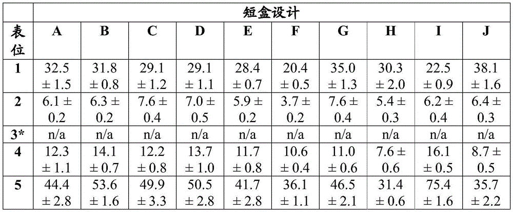
Reporter T cells against epitope 3 that have not yet been generated
In vivo evaluation of antigen cassette design
As another example of antigen cassette design evaluation, the vaccine cassette was designed to contain 5 well-characterized human MHC class I epitopes known to stimulate CD8T cells in an HLA-a x 02:01 restricted manner (fig. 2A, 3, 5A). To assess immunogenicity in vivo, vaccine cassettes containing these marker epitopes were incorporated into adenoviral vectors and used to infect HLA-a2 transgenic mice (fig. 4). The mouse model carries a transgene consisting in part of human HLA-a 0201 and mouse H2-Kb, thus encoding a chimeric class I MHC molecule consisting of a human HLA-a2.1 leader, α 1 and α 2 domains linked to murine α 3, a transmembrane and cytoplasmic H2-Kb domain (Vitiello et al, 1991). The chimeric molecules allow HLA-a 02:01 restricted antigen presentation while maintaining species-matched interaction of CD8 co-receptors with the alpha 3 domain on MHC.
For short cassettes, all marker epitopes generate T cell responses to a degree about 10-50 times stronger than that normally reported, as determined by IFN-. gamma.ELISPOT (Cornet et al, 2006; Depla et al, 2008; Ishioka et al, 1999). Among all linkers evaluated, 25-mer sequence concatemers, each containing a very small epitope flanked by native amino acid sequences, produced the largest and most extensive T cell responses (table 5). Intracellular Cytokine Staining (ICS) and flow cytometry analysis revealed that the antigen-specific T cell response was derived from CD8T cells.
Table 5: in vivo evaluation of linker sequences in short boxes. ELISPOT data indicate that HLA-a2 transgenic mice generated T cell responses against all MHC class I restricted epitopes in the cassette 17 days after infection with 1e11 adenovirus virions.

In another example, a series of long vaccine cassettes were constructed and incorporated into adenoviral vectors containing an additional 16 HLA-a 02:01, a 03:01, and B44: 05 epitopes with known CD8T cellular reactivity next to the original 5 marker epitopes (fig. 5A, B). The dimensions of these long cassettes closely mimic the final clinical cassette design, and only the positions of the epitopes relative to each other are different. For the long and short vaccine cassettes, the CD8T cell responses were comparable in magnitude and breadth, demonstrating that (a) adding more epitopes did not substantially affect the magnitude of the immune response against the original set of epitopes, and (b) the location of the epitopes in the cassette did not substantially affect the subsequent T cell response against it (table 6).
Table 6: in vivo assessment of the effect of epitope position in long boxes. ELISPOT data indicate that for long versus short vaccine cassettes, HLA-a2 transgenic mice produced comparable magnitude of T cell responses 17 days after infection with 5e10 adenovirus virions.

Suspected technical errors caused a lack of T cell responses.
Antigen cassette design for immunogenicity and toxicology studies
Overall, the results of the studies on model box evaluation (fig. 2-5, tables 2-6) demonstrate that for the model vaccine box, strong immunogenicity is achieved when the "string of beads" method is used to encode about 20 epitopes in the context of adenovirus-based vectors. The epitopes are assembled by concatenating 25-mer sequences, each embedded in a very small CD8T cell epitope (e.g., 9 amino acid residues) flanked on both sides by its native, surrounding peptide sequence (e.g., 8 amino acid residues on each side). As used herein, a "native" or "native" flanking sequence refers to an N-terminal and/or C-terminal flanking sequence of a given epitope in the naturally occurring environment of the epitope within its source protein. For example, the HCMV pp65 MHC I epitope NLVPMVATV is flanked on its 5 'end by the native 5' sequence WQAGILAR and on its 3 'end by the native 3' sequence QGQNLKYQ, thereby generating the 25-mer peptide WQAGILARNLVPMVATVQGQNLKYQ found within the HCMV pp65 source protein. Native or native sequences may also refer to nucleotide sequences encoding epitopes flanked by native flanking sequences. Each 25-mer sequence is directly linked to the following 25-mer sequence. In the example of a very small CD8T cell epitope of greater or less than 9 amino acids, the flanking peptide lengths may be adjusted so that the overall length remains a 25-mer peptide sequence. For example, a 10 amino acid CD8T cell epitope may be flanked by an 8 amino acid sequence and 7 amino acids. The concatemer is followed by two universal MHC class II epitopes which are included to stimulate CD 4T helper cells and improve the overall in vivo immunogenicity of the vaccine cassette antigen. (Alexander et al, 1994; Panina-Bordgunon et al, 1989) the class II epitope was linked to the final class I epitope by a GPGPG amino acid linker (SEQ ID NO: 56). The two class II epitopes are also linked to each other by a gpgpgpg amino acid linker and flanked on the C-terminus by a gpgpgpg amino acid linker. Neither the position nor the number of epitopes appears to substantially affect T cell recognition or response. The targeting sequence also does not appear to substantially affect the immunogenicity of the cassette-derived antigen.
As another example, based on in vitro and in vivo data obtained with model cassettes (fig. 2-5, tables 2-6), a cassette design was generated that alternates well-characterized T cell epitopes known to be immunogenic in non-human primates (NHPs), mice and humans. The 20 epitopes fully embedded in the native 25-mer sequence were followed by two universal MHC class II epitopes present in all the model cassettes evaluated (figure 6). This cassette design was used to study immunogenicity as well as pharmacological and toxicological studies in multiple species.
Antigen cassette design and evaluation for 30, 40 and 50 antigens
Large antigen cassettes with 30(L), 40(XL) or 50(XXL) epitopes, each 25 amino acids in length, were designed. These epitopes are a mixture of human, NHP and mouse epitopes to mimic disease antigens, including tumor antigens. Fig. 29 shows the general organization of epitopes from various species. Tables 37, 38 and 39 describe model antigens for human, primate and mouse model epitopes, respectively. Table 37, table 38 and table 39 each describe the position, name, minimum epitope description and MHC classification of the epitope.
These cassettes were cloned into chAd68 and srna vaccine vectors as described to assess the efficacy of longer polyepitope cassettes. Fig. 30 shows that each large antigen cassette was expressed by the ChAdV vector, as indicated by at least one major band of expected size by western blotting.
Mice were immunized as described to assess the efficacy of the large cassette. For epitopes AH1 (upper panel) and SINNFEKL (lower panel), T cell responses were analyzed by ICS and tetramer staining after immunization with chAd68 vector (fig. 31/table 40 and fig. 32/table 41, respectively) and by ICS after immunization with srna vector (fig. 33/table 42). Immunization with chAd68 and srna vaccine vectors expressing 30(L), 40(XL), or 50(XXL) epitopes induced a CD8+ immune response to model disease epitopes.
TABLE 37 human epitopes in the big Box
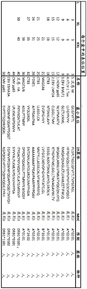
TABLE 38 NHP epitopes in the big Box
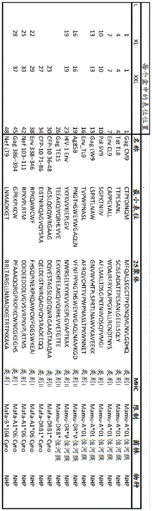
TABLE 39 mouse epitopes in the big Box
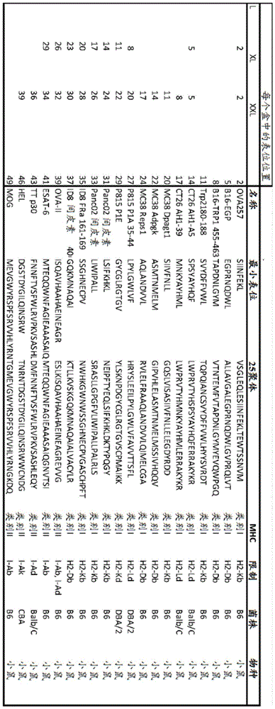
Table 40: mean IFNg + cells in ChAd big box treated mice in response to AH1 and SIINFEKL peptide. Data are expressed as a percentage of total CD8 cells. Shown are the mean and standard deviation for each group and the p-value by ANOVA and Tukey test. All p values were compared to the MAG 20 antigen cassette.
| # antigen | Antigens | Mean value of | Standard deviation of | | N | |
| 20 | SIINFEKL | 5.308 | 0.660 | n/a | 8 | |
| 30 | SIINFEKL | 4.119 | 1.019 | 0.978 | 8 | |
| 40 | SIINFEKL | 6.324 | 0.954 | 0.986 | 8 | |
| 50 | SIINFEKL | 8.169 | 1.469 | 0.751 | 8 | |
| 20 | AH1 | 6.405 | 2.664 | n/a | 8 | |
| 30 | AH1 | 4.373 | 1.442 | 0.093 | 8 | |
| 40 | AH1 | 4.126 | 1.135 | 0.050 | 8 | |
| 50 | AH1 | 4.216 | 0.808 | 0.063 | 8 |
Table 41: mean tetramer + cells for AH1 and SIINFEKL antigens in ChAd large box treated mice. Data are expressed as a percentage of total CD8 cells. Shown are the mean and standard deviation for each group and the p-value by ANOVA and Tukey test. All p values were compared to the MAG 20 antigen cassette.
| # antigen | Antigens | Mean value of | Standard deviation of | | N | |
| 20 | SIINFEKL | 10.314 | 2.384 | n/a | 8 | |
| 30 | SIINFEKL | 4.551 | 2.370 | 0.003 | 8 | |
| 40 | SIINFEKL | 5.186 | 3.254 | 0.009 | 8 | |
| 50 | SIINFEKL | 14.113 | 3.660 | 0.072 | 8 | |
| 20 | AH1 | 6.864 | 2.207 | n/a | 8 | |
| 30 | AH1 | 4.713 | 0.922 | 0.036 | 8 | |
| 40 | AH1 | 5.393 | 1.452 | 0.223 | 8 | |
| 50 | AH1 | 5.860 | 1.041 | 0.543 | 8 |
Table 42: mean IFNg + cells in SAM big box treated mice in response to AH1 and SIINFEKL peptide. Data are expressed as a percentage of total CD8 cells. Shown are the mean and standard deviation for each group and the p-value by ANOVA and Tukey test. All p values were compared to the MAG 20 antigen cassette.


ChAd antigen cassette delivery vectors
Construction of XV.A.ChAd antigen cassette delivery vectors
In one example, chimpanzee adenovirus (ChAd) is engineered into a delivery vector for an antigen cassette. In another example, the full-length ChAdV68 vector was synthesized based on AC _000011.1 (sequence 2 from U.S. Pat. No. 2, 6083716) that lacks the E1(nt 457 to 3014) and E3(nt 27,816) sequences 31,332). The reporter gene under the control of the CMV promoter/enhancer was inserted in place of the deleted E1 sequence. Transfection of this clone into HEK293 cells did not produce infectious virus. To determine the sequence of the wild-type C68 virus, isolate VR-594 was obtained from the ATCC, passaged, and then sequenced independently (SEQ ID NO: 10). When the AC _000011.1 sequence was compared to the ATCC VR-594 sequence of the wild-type ChAdV68 virus (SEQ ID NO:10), a 6 nucleotide difference was identified. In one example, a modified ChAdV68 vector (ChAdV68.5WNTt SEQ ID NO:1) was generated based on AC _000011.1 in which the corresponding ATCC VR-594 nucleotide was substituted at five positions.
In another example, a modified ChAdV68 vector was generated based on AC _000011.1 that deleted the E1(nt 577-3403) and E3(nt 27,816-31,332) sequences with the corresponding ATCC VR-594 nucleotides substituted at the four positions. The deleted E1 sequence was replaced by either a GFP reporter (ChAdV68.4WNT. GFP; SEQ ID NO:11) or a model neoantigen cassette (ChAdV68.4WNT. MAG25mer; SEQ ID NO:12) inserted under the control of the CMV promoter/enhancer.
In another example, a modified ChAdV68 vector was generated based on AC _000011.1 that deleted the E1(nt 577-3403) and E3(nt 27,125-31,825) sequences and the corresponding ATCC VR-594 nucleotides were substituted at five positions. The deleted E1 sequence was replaced by the insertion of a GFP reporter protein (ChAdV68.5WNT. GFP; SEQ ID NO:13) or a model neoantigen cassette (ChAdV68.5WNT. MAG25mer; SEQ ID NO:2) under the control of the CMV promoter/enhancer.
The relevant vectors are described below:
the full-length ChAdVC68 sequence "ChAdV68.5WTnt" (SEQ ID NO: 1); the corresponding AC _000011.1 sequence with ATCC VR-594 nucleotide substituted in the five positions.
-ATCC VR-594C68(SEQ ID NO: 10); independent sequencing; full length C68
ChAdV68.4WNT.GFP (SEQ ID NO: 11); the E1(nt 577-3403) and E3(nt 27,816-31,332) sequences are deleted from AC _ 000011.1; the corresponding ATCC VR-594 nucleotide is substituted at four positions; insertion of GFP reporter protein under the control of CMV promoter/enhancer instead of deleted E1
-ChAdV68.4WNT.MAG25mer (SEQ ID NO: 12); the E1(nt 577-3403) and E3(nt 27,816-31,332) sequences are deleted from AC _ 000011.1; the corresponding ATCC VR-594 nucleotide is substituted at four positions; insertion of a model neoantigen cassette under the control of the CMV promoter/enhancer instead of deleted E1
ChAdV68.5WTnt. GFP (SEQ ID NO: 13); the AC _000011.1 lacks the E1(nt 577-3403) and E3(nt 27,125-31,825) sequences; the corresponding ATCC VR-594 nucleotide is substituted at five positions; insertion of GFP reporter protein under the control of CMV promoter/enhancer instead of deleted E1
Xv.b.chad antigen cassette delivery vector assay
XV.b.1.chad vector evaluation methods and materials
Transfection of HEK293A cells with Lipochromide
Using the following protocol, DNA of the ChAdV68 constructs (chadv68.4wtnt.gfp, chadv68.5wtnt.gfp, chadv68.4wtnt.mag25mer, and chadv68.5wtnt.mag25mer) was prepared and transfected into HEK293A cells.
10ug of plasmid DNA was digested with PacI to release the viral genome. Then, for longer DNA fragments, the DNA was purified using GeneJet DNA purification mini column (DNA clean Micro columns; Thermo Fisher) according to the manufacturer's instructions and eluted in 20ul of preheated water; the column was kept at 37 ℃ for 0.5-1 hour prior to the elution step.
HEK293A was added at 10 before transfection6Cell density per cell/well was introduced into 6-well plates and maintained for 14-18 hours. Cells were covered with 1ml of fresh medium (DMEM-10% hiFBS with penicillin/streptomycin and glutamic acid) per well. 1-2ug of purified DNA per well was used in two transfections with microliter volumes (2-4ul) of lipofectamine 2000 according to the manufacturer's protocol. 0.5ml of OPTI-MEM medium containing the transfection mixture was added to 1ml of standard growth medium in each well and kept on the cells overnight.
The transfected cell cultures were incubated at 37 ℃ for at least 5-7 days. If no viral plaques were seen on day 7 post-transfection, cells were isolated at 1:4 or 1:6 and incubated at 37 ℃ to monitor plaque production. Alternatively, transfected cells were collected and subjected to 3 cycles of freezing and thawing, and HEK293A cells were infected with cell lysate and incubated until viral plaques were observed.
Transfection of ChAdV68 into HEK293A cells Using calcium phosphate and Generation of third Generation Virus stocks
Using the following protocol, DNA of the ChAdV68 constructs (chadv68.4wtnt.gfp, ChAdV68.5 wtnt.gfp, chadv68.4wtnt.mag25mer, chadv68.5wtnt.mag25mer) was prepared and transfected into HEK293A cells.
One day prior to transfection, HEK293A cells were plated at 106Individual cells/well were seeded in 6-well plates in 5% BS/DMEM/1XP/S, 1 XGlutamax. Two wells were required for each transfection. Two to four hours prior to transfection, the medium was changed to fresh medium. ChAdV68.4Wnt.G using PacIFP plasmid was linearized. The linearized DNA was then extracted with phenol chloroform and precipitated using one-tenth volume of 3M sodium acetate pH 5.3 and two volumes of 100% ethanol. The precipitated DNA was pelleted by centrifugation at 12,000Xg for 5 minutes, and then washed 1 time with 70% ethanol. The pellet was air dried and resuspended in 50. mu.L of sterile water. Using NanoDropTM(ThermoFisher) the DNA concentration was determined and the volume was adjusted to 5. mu.g DNA/50. mu.L.
169 μ L of sterile water was added to the microcentrifuge tube. Then 5. mu.L of 2M CaCl2Add to water and mix gently by pipette. Add 50. mu.L of DNA dropwise to CaCl2In aqueous solution. Then 26. mu.l of 2M CaCl were added2And gently mixed by pipetting twice through a micropipette. This final solution should be prepared from 5. mu.g DNA in 250. mu.L of 0.25M CaCl2Is prepared from (1). A second tube containing 250. mu.L of 2XHBS (Hepes buffer) was then prepared. A 2mL sterile pipette connected to Pipet-Aid air was used to slowly bubble through the 2XHBS solution. Simultaneously, dropwise adding into 0.25M CaCl 2DNA solution in solution. After the addition of the final DNA droplet, bubbling was continued for about 5 seconds. The solution was then incubated at room temperature for 20 minutes and then added to 293A cells. 250 μ L of DNA/calcium phosphate solution was added dropwise to the previous day at 106Individual cells/well were seeded in 293A cell monolayers in 6-well plates. The cells were returned to the incubator and incubated overnight. The medium was changed after 24 hours. After 72 hours, cells were dispensed 1:6 into 6-well plates. The cell monolayer was monitored daily by optical microscopy for signs of cytopathic effect (CPE). 7-10 days after transfection, viral plaques were observed and cell monolayers were collected by pipetting the medium in the wells to raise the cells. The collected cells and medium were transferred to a 50mL centrifuge tube, followed by three rounds of freeze thawing (at-80 ℃ and 37 ℃). The subsequent lysate, referred to as primary virus stock, was clarified by full speed centrifugation on a desk top (bench top) centrifuge (4300Xg) and used a portion of the lysate (10-50%) to infect 293A cells in a T25 flask. Infected cells were incubated for 48 hours, and then cells and medium were collected under complete CPE. Collecting the cells again, freezing, thawing, and clarifying, and then using the same Infection of second Generation Virus stocks Each flask was inoculated with 1.5X 10⁷Individual cell T150 flasks. Once complete CPE was achieved at 72 hours, the media and cells were collected and processed in the same manner as the previous virus stock to produce a third generation stock.
Production in 293F cells
At 8% CO2In an incubator of 293 FreeStyleTMProduction of ChAdV68 virus was performed in 293F cells grown in (ThermoFisher) medium. On the day of infection, cells were diluted to 106cells/mL, 98% viability, and 400mL were used in 1L shake flasks (Corning) for each manufacturing run. Use of target MOI for Each infection>3.3 of 4mL third generation virus stock. Cells were incubated for 48-72 hours until viability was measured by trypan blue<70 percent. The infected cells were then collected by centrifugation in a full speed desktop centrifuge and washed in 1XPBS, recentrifuged and then resuspended in 20mL of 10mM Tris pH 7.4. The cell pellets were lysed by freeze thawing 3 times and clarified by centrifugation at 4,300Xg for 5 minutes.
Purification by CsCl centrifugation
Viral DNA was purified by CsCl centrifugation. Two discrete gradient operations are performed. The first time is to purify the virus from the cellular components and the second time is to further optimize the separation from the cellular components and separate the defective particles from the infectious particles.
10mL of 1.2(26.8g CsCl dissolved in 92mL of 10mM Tris pH 8.0) CsCl was added to the hetero-isomorphous polymer tube. Then, using a pipette, deliver to the bottom of the tube, carefully add 8mL of 1.4CsCl (53g CsCl dissolved in 87mL of 10mM Tris pH 8.0). Clarified virus was carefully layered on top of the 1.2 layers. If necessary, 10mM Tris was added to equilibrate the tubes. The tubes were then placed in an SW-32Ti spinner and centrifuged at 10 ℃ for 2 hours and 30 minutes. The tubes were then moved to a laminar flow cabinet and viral strips aspirated using an 18 gauge needle and a 10mL syringe. Removal of contaminating host cell DNA and proteins should be avoided. The viral bands were then diluted at least 2-fold with 10mM Tris pH 8.0 and plated on a discontinuous gradient as described above. Operate as previously describedWith the exception that this was done overnight at this time. The next day, the viral bands were carefully aspirated to avoid aspiration of any defective virion bands. Then Slide-A-LyzerT was usedMThe cassette (Pierce) was dialyzed against ARM buffer (20mM Tris pH 8.0, 25mM NaCl, 2.5% glycerol). This was done 3 times, each time with buffer exchange for 1 hour. The virus was then aliquoted for storage at-80 ℃.
Virus analysis
Based on 1.1 × 10¹²The extinction coefficient of individual Virus Particles (VP) corresponds to an absorbance value of 1 at OD260nm, and VP concentration was determined by using OD260 analysis. Two dilutions (1:5 and 1:10) of adenovirus were prepared in virus lysis buffer (0.1% SDS, 10mM Tris pH 7.4, 1mM EDTA). The OD of the two dilutions was measured in duplicate and multiplied by the dilution factor multiplied by the OD260 value by 1.1 × 10¹²VP to measure VP concentration per ml.
Infectious Unit (IU) titers were calculated using a limiting dilution analysis of the virus stock. Virus was initially 100-fold diluted in DMEM/5% NS/1X PS, followed by 10-fold dilution to 1X10⁻⁷. Then, 100 μ Ι _ of these dilutions were added to 293A cells seeded at 3e5 cells/well in 24-well plates at least one hour before. This procedure was performed in duplicate. Plates were incubated in a CO2 (5%) incubator for 48 hours at 37 ℃. The cells were then washed with 1XPBS and then fixed with 100% cold methanol (-20 ℃). The plates were then incubated at-20 ℃ for a minimum of 20 minutes. Each well was washed with 1XPBS and then blocked in 1 XPPBS/0.1% BSA for 1 hour at room temperature. Rabbit anti-Ad antibody (Abcam, Cambridge, MA) was added at 1:8,000 dilution (0.25 ml per well) in blocking buffer and incubated for 1 hour at room temperature. Each well was washed 4 times with 0.5mL PBS per well. A1000-fold dilution of HRP-conjugated goat anti-rabbit antibody (Bethy Labs, Montgomery Texas) was added to each well and incubated for 1 hour, followed by a final wash round. 5 PBS washes were performed and used with 0.01% H 2O2The plate was developed with Diaminobenzidine tetrahydrochloride (DAB) substrate (0.67mg/mL DAB in 50mM Tris pH 7.5, 150mM NaCl) in Tris buffered saline. The wells were allowed to develop for 5 minutes, thenAnd (6) counting. Cells were counted at 10X using dilutions that produced 4-40 stained cells per field. The field of view used was 0.32mm2A grid, corresponding to 625 grids per field of view on a 24-well plate. The number of infectious virus per ml can be determined by multiplying the number of stained cells in each grid by the number of grids per field of view by a dilution factor of 10. Similarly, when working with GFP-expressing cells, fluorescence rather than capsid staining can be used to determine the number of GFP-expressing virions per ml.
Immunization
Injecting C57BL/6J female mouse and Balb/C female mouse with 1 × 10 by intramuscular injection at two sides8One ChAdV68.5WNTt. MAG25mer Virion (VP) in a volume of 100uL (50 uL per leg).
Spleen cell dissociation
The spleen and lymph nodes of each mouse were pooled in 3mL of complete RPMI (RPMI, 10% FBS, penicillin/streptomycin). Mechanical dissociation was performed using a genetlemecs acs dissociator (Miltenyi Biotec) following the manufacturer's protocol. Dissociated cells were filtered through a 40 micron filter and lysed with ACK buffer (150mM NH) 4Cl、10mM KHCO3、0.1mM Na2EDTA) lysed erythrocytes. The cells were filtered again through a 30 micron filter and then resuspended in complete RPMI. Cells were counted on an Attune NxT flow cytometer (Thermo Fisher) using propidium iodide staining to exclude dead and apoptotic cells. The cells are then adjusted to the appropriate viable cell concentration for subsequent analysis.
Ex vivo enzyme-linked immunospot (ELISPOT) assay
ELISPOT analysis was performed using the mouse IFNg ELISPOtPLUS kit (MABTECH) according to the ELISPOT consensus criterion (DOI: 10.1038/nprot.2015.068). Will be 5X 10⁴Individual splenocytes were incubated with 10uM of the indicated peptide for 16 hours in 96-well plates coated with IFNg antibody. The spots were developed using alkaline phosphatase. The reaction was timed for 10 minutes and terminated by running tap water through the plate. Spots were counted using the AID vSpot reader spectrogram. For ELISPOT analysis, saturation was measured>50% of the wells were recorded as "too many to count". Bias of holes to be replicatedDifference (D)>10% of the samples were excluded from the analysis. The spot count was then corrected for the degree of well confluence using the following formula: spot count +2 × (spot count × degree of confluence%/[ 100% -degree of confluence%)]). Negative background was corrected by subtracting the spot count in negative peptide-stimulated wells from antigen-stimulated wells. Finally, the wells marked too many to count were set to the highest observed correction, rounded to the nearest percentage.
Xv.b.2. manufacture of ChAdV68 virus delivery particles following DNA transfection
In one example, chadv68.4wtnt. gfp (fig. 7) and chadv68.5wtnt. gfp (fig. 8) DNA were transfected into HEK293A cells and virus replication (viral plaques) was observed 7-10 days after transfection. The ChAdV68 virus plaques were observed using light microscopy (FIGS. 7A and 8A) and fluorescence microscopy (FIGS. 7B-C and 8B-C). GFP indicates the production of a toxigenic ChAdV68 virus delivery particle.
XV.B.3.ChAdV68 Virus delivery particle amplification
In one example, the chadv68.4wtnt.gfp, chadv68.5wtnt.gfp and chadv68.5wtnt.mag25mer viruses were amplified in HEK293F cells and 18 days after transfection, purified virus stock was prepared (fig. 9). The number of virus particles in the purified ChAdV68 virus stock was quantified and compared to adenovirus type 5 (Ad5) and ChAdVY25 (closely related ChAdV; Dicks,2012, PloS ONE 7, e40385) virus stocks made using the same protocol. The ChAdV68 virus titers were comparable to Ad5 and ChAdVY25 (table 7).
TABLE 7 Generation of adenovirus vectors in 293F suspension cells
| Construct | Mean VP/cell +/-SD |
| Ad 5-vector (multiple vectors) | 2.96e4+/-2.26e4 |
| Ad5-GFP | 3.89e4 |
| chAdY25-GFP | 1.75e3+/-6.03e1 |
| ChAdV68.4WTnt.GFP | 1.2e4+/-6.5e3 |
| ChAdV68.5WTnt.GFP | 1.8e3 |
| ChAdV68.5WTnt.MAG25mer | 1.39e3+/-1.1e3 |
SD is only reported in the case of performing multiple manufacturing operations
Assessment of immunogenicity in XV.B.4. tumor model
The C68 vector expressing the mouse tumor antigen was evaluated in a mouse immunogenicity study to confirm that the C68 vector elicited T cell responses. T-cell responses to the class I MHC epitope SIINFEKL were measured in C57BL/6J female mice and to the class I MHC epitope AH1-A5(Slansky et al, 2000, Immunity13:529-538) in Balb/C mice. As shown in fig. 15, a stronger T cell response was measured relative to the control after immunization of mice with chadv68.5wtnt. When C57BL/6J or Balb/C mice were immunized with ChAdV68.5WNT. MAG25mer, every 10 days after immunization were observed separately in ELISpot analysis6Average cellular immune response of 8957 or 4019 Spot Forming Cells (SFC) per splenocyte.
Tumor infiltrating lymphocytes were also evaluated in the CT26 tumor model, which evaluated co-administration of ChAdV and anti-CTLA 4 antibodies. Mice were implanted with CT26 tumor cells and 7 days post-implantation, immunized with the ChAdV vaccine and treated with anti-CTLA 4 antibody (clone 9D9) or IgG as controls. Tumor infiltrating lymphocytes were analyzed 12 days after immunization. Tumors from each mouse were dissociated using a genetlemecs dis (Miltenyi Biotec) and a mouse tumor dissociation kit (Miltenyi Biotec). Dissociated cells were filtered through a 30 micron filter and resuspended in complete RPMI. Cells were counted on an Attune NxT flow cytometer (Thermo Fisher) using propidium iodide staining to exclude dead and apoptotic cells. The cells are then adjusted to the appropriate viable cell concentration for subsequent analysis. Antigen-specific cells were identified by MHC-tetramer complexes and co-stained with anti-CD 8 and viability markers. Tumors were collected 12 days after priming.
Antigen-specific CD8+ T cells within the tumors accounted for 3.3%, 2.2%, or 8.1% of the median total viable cell population in the ChAdV, anti-CTLA 4, and ChAdV + anti-CTLA 4 treated groups, respectively (fig. 44 and table 36). The combination of anti-CTLA treatment with active ChAdV immunization resulted in a statistically significant increase in antigen-specific CD8+ T cell frequency compared to ChAdV alone and anti-CTLA 4 alone, suggesting that anti-CTLA 4 when co-administered with chAd68 vaccine increased the number of infiltrating T cells in the tumor.
Tetramer + frequency of infiltrating CD8T cells in Table 36-CT26 tumors

Xvi. alphavirus antigen cassette delivery vehicle
Materials and methods for assessing xvi.a. alphavirus delivery vectors
In vitro transcription to generate RNA
For the in vitro test: plasmid DNA was linearized by restriction digestion with PmeI, column purified following the manufacturer's protocol (GeneJet DNA purification kit, Thermo) and used as template. According to the manufacturer's protocol, a RiboMAX Large Scale RNA production System (Promega) was used, using m7G cap analogue (Promega) was transcribed in vitro. mRNA was purified using RNeasy kit (Qiagen) according to the manufacturer's protocol.
For in vivo studies: RNA was generated and purified by TriLInk Biotechnologies and capped with enzymic Cap 1.
RNA transfection
Approximately 16 hours prior to transfection, HEK293A cells were seeded at 6e4 cells/well for 96 wells and 2e5 cells/well for 24 wells. Cells were transfected with mRNA using MessengerMAX lipofectamine (Invitrogen) and following the manufacturer's protocol. For 96 wells, 0.15uL of lipid staining amine and 10ng of mRNA per well was used, and for 24 wells, 0.75uL of lipid staining amine and 150ng of mRNA per well was used. GFP-expressing mRNA (TriLink Biotechnologies) was used as a transfection control.
Luciferase assay
Luciferase reporter assays were performed in triplicate in white-walled 96-well plates under each condition using the ONE-Glo luciferase assay (Promega), following the manufacturer's protocol. Luminescence was measured using SpectraMax.
qRT-PCR
At 2 hours post-transfection, transfected cells were rinsed with fresh medium and the medium was changed to remove any untransfected mRNA. Then, at various time points, cells were collected in RLT plus lysis buffer (Qiagen), homogenized using qiasrededer (Qiagen), and RNA extracted using RNeasy kit (Qiagen), all following the manufacturer's protocol. Total RNA was quantified using a Nanodrop (thermo scientific). According to the manufacturer's scheme, at qTower3qRT-PCR was performed on (Analytik Jena) using a Quantitect Probe One-Step RT-PCR kit (Probe One-Step RT-PCR kit; Qiagen) using 20ng of total RNA per reaction. For each probe, each sample was run in triplicate. Actin or GusB was used as reference gene. Custom primers/probes were generated from IDT (table 8).
TABLE 8.qPCR primers/probes

B16-OVA tumor model
Injection of 10 into the lower left flank of C57BL/6J mice5Fine B16-OVACell/animal. Tumors were allowed to grow for 3 days prior to immunization.
CT26 tumor model
Injection of 10 into the lower left flank of Balb/c mice6Individual CT26 cells/animal. Tumors were allowed to grow for 7 days prior to immunization.
Immunization
For the srna vaccine, mice were injected with 10ug of RNA in a volume of 100uL (50 uL per leg) by two-sided intramuscular injection. For the Ad5 vaccine, mice were injected with a 5X 10 injection by bilateral intramuscular injection10Individual Virus Particles (VP), volume 100uL (50 uL per leg). Animals were injected 2 times weekly with 250ug doses of anti-CTLA-4 (clone 9D9, BioXcell), anti-PD-1 (clone RMP1-14, BioXcell) or anti-IgG (clone MPC-11, BioXcell) via intraperitoneal injection.
In vivo bioluminescence imaging
At each time point, mice were injected with 150mg/kg fluorescein substrate via intraperitoneal injection and bioluminescence was measured 10-15 minutes after injection using the IVIS in vivo imaging system (PerkinElmer).
Spleen cell dissociation
The spleen and lymph nodes of each mouse were pooled in 3mL of complete RPMI (RPMI, 10% FBS, penicillin/streptomycin). Mechanical dissociation was performed using a genetlemecs acs dissociator (Miltenyi Biotec) following the manufacturer's protocol. Dissociated cells were filtered through a 40 micron filter and lysed with ACK buffer (150mM NH) 4Cl、10mM KHCO3、0.1mM Na2EDTA) lysed erythrocytes. The cells were filtered again through a 30 micron filter and then resuspended in complete RPMI. Cells were counted on an Attune NxT flow cytometer (Thermo Fisher) using propidium iodide staining to exclude dead and apoptotic cells. The cells are then adjusted to the appropriate viable cell concentration for subsequent analysis.
Ex vivo enzyme-linked immunospot (ELISPOT) assay
ELISPOT analysis was performed using the mouse IFNg ELISPOtPLUS kit (MABTECH) according to the ELISPOT consensus criterion (DOI: 10.1038/nprot.2015.068). Will be 5X 10⁴Individual splenocytes were plated with IFNg antibody in 96-well plates with 10uM of the indicated peptideIncubated for 16 hours. The spots were developed using alkaline phosphatase. The reaction was timed for 10 minutes and terminated by running tap water through the plate. Spots were counted using the AID vSpot reader spectrogram. For ELISPOT analysis, saturation was measured>50% of the wells were recorded as "too many to count". Will replicate deviations in the hole>10% of the samples were excluded from the analysis. The spot count was then corrected for the degree of well confluence using the following formula: spot count +2 × (spot count × degree of confluence%/[ 100% -degree of confluence%)]). Negative background was corrected by subtracting the spot count in negative peptide-stimulated wells from antigen-stimulated wells. Finally, the wells marked too many to count were set to the highest observed correction, rounded to the nearest percentage.
XVI.B. alphavirus vectors
XVI.B.1. alphavirus vector in vitro evaluation
In one embodiment of the invention, the RNA alphavirus backbone for the antigen expression system is generated from a self-replicating RNA (srRNA) vector based on Venezuelan Equine Encephalitis (VEE) (Kinney,1986, Virology 152: 400-. In one example, the sequence encoding the VEE structural protein located 3' to the 26S subgenomic promoter was deleted (VEE sequence 7544 to 11,175 deletion; numbering based on Kinney et al 1986; SEQ ID NO:6) and replaced with an antigenic sequence (SEQ ID NO:14 and SEQ ID NO:4) or a luciferase reporter protein (e.g., VEE-luciferase, SEQ ID NO:15) (FIG. 10). RNA was transcribed in vitro from srna DNA vectors, transfected into HEK293A cells and luciferase reporter protein expression was measured. In addition, luciferase-encoding (non-replicative) mRNA was transfected for comparison. When comparing the 23 hour measurements to the 2 hour measurements, an approximately 30,000-fold increase in srna reporter signal was observed for VEE-luciferase srna (table 9). In contrast, mRNA reporter genes exhibited less than a 10-fold increase in signal over the same time period (table 9).
TABLE 9 luciferase expression from VEE self-replicating vectors increases over time. HEK293A cells were transfected with 10ng VEE-luciferase srRNA or 10ng non-replicating luciferase mRNA per well (TriLink L-6307) in 96 wells. Luminescence was measured at various times post-transfection. Luciferase expression is reported in Relative Luminescence Units (RLU). Each data point is the mean of 3 transfected wells +/-SD.
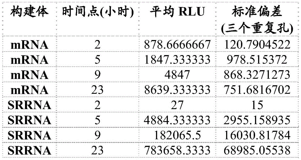
In another example, rrna replication is determined directly by measuring the RNA content after transfection of rrna encoding luciferase (VEE-luciferase) or a multi-epitope cassette (VEE-MAG25mer) using quantitative reverse transcription polymerase chain reaction (qRT-PCR). An approximately 150-fold increase in RNA was observed for VEE-luciferase srRNA (Table 10), while a 30-50 fold increase in RNA was observed for VEE-MAG25mer srRNA (Table 11). These data confirm that the VEE srna vector replicates when transfected into cells.
Table 10 direct measurement of RNA replication in VEE-luciferase srna transfected cells. HEK293A cells were transfected with VEE-luciferase srna (150 ng/well, 24 wells) and RNA levels were quantified by qRT-PCR at various times after transfection. Each measurement was normalized based on the actin reference gene and presented as fold change relative to the 2 hour time point.

TABLE 11 direct measurement of RNA replication in VEE-MAG25mer srRNA transfected cells. HEK293 cells were transfected with VEE-MAG25mer srna (150 ng/well, 24 wells) and RNA levels were quantified by qRT-PCR at various times after transfection. Each measurement was normalized based on the GusB reference gene and presented fold change relative to the 2 hour time point. The different lines on the figure represent 2 different qPCR primer/probe sets, both of which detect epitope box regions of srna.
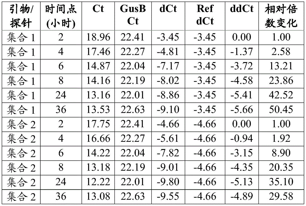
In vivo evaluation of the alphavirus vectors
In another example, VEE-luciferase reporter protein expression is assessed in vivo. Mice were injected with 10ug of VEE-luciferase srna encapsulated in lipid nanoparticles (MC3) and imaged at 24 and 48 hours, and 7 and 14 days post injection to determine bioluminescent signals. Luciferase signal was detected 24 hours after injection and increased over time, with a peak occurring 7 days after srna injection (fig. 11).
XVI.B.3. alphavirus vector tumor model evaluation
In one embodiment, to determine whether the VEE srRNA vector directs an antigen-specific immune response in vivo, a VEE srRNA vector (VEE-UbAAY, SEQ ID NO:14) was generated that expresses 2 different MHC class I mouse tumor epitopes SIINFEKL and AH1-A5 (Slansky et al, 2000, Immunity 13: 529-. The SFL (SIINFEKL) epitope was expressed using the B16-OVA melanoma cell line, and the AH1-A5 (SPSYAYHQF; Slansky et al, 2000, Immunity) epitope induced T cells to target the relevant epitope expressed by the CT26 colon cancer cell line (AH 1/SPSYVYHQF; Huang et al, 1996, Proc Natl Acad Sci USA 93: 9730-. In one example, for in vivo studies VEE-UbAAY srna was generated and encapsulated in lipid nanoparticles (MC3) by in vitro transcription using T7 polymerase (TriLink Biotechnologies).
Two weeks after immunization of mice bearing B16-OVA tumors with VEE-UbAAY srna formulated at MC3, a strong SFL-targeted antigen-specific T cell response was observed relative to the control. In one example, every 10 is measured in an ELISpot assay after stimulation with SFL peptide63835 (median) spot-forming cells (SFC) from splenocytes (fig. 12A, table 12) and 1.8% (median) CD8T cells were SFL antigen specific as measured by pentamer staining (fig. 12B, table 12). In another example, co-administration of anti-CTLA-4 monoclonal antibodies (mabs) with VEE srna vaccines caused a modest increase in overall T cell response, and every 10 was measured in ELISpot assays64794.5 (median) SFCs of individual splenocytes (FIG. 12A, Table 12).
TABLE 12 results of ELISPOT and MHCI-pentamer staining analysis 14 days after VEE srRNA immunization in C57BL/6J mice bearing B16-OVA tumors.

Note that the results obtained from mouse # 6 in the Vax group were excluded from the analysis due to the large variation between the three replicate wells.
In another embodiment, to reflect the clinical approach, heterologous prime/boost immunizations were performed in B16-OVA and CT26 mouse tumor models, in which tumor-bearing mice were first immunized with an adenoviral vector expressing the same antigen cassette (Ad5-UbAAY), followed by 14 days after Ad5-UbAAY priming by VEE-UbAAY srRNA vaccine. In one example, an antigen-specific immune response is induced by the Ad5-UbAAY vaccine, whereby every 10 is measured in an ELISpot assay 6Individual splenocytes 7330 (median) SFC (fig. 13A, table 13) and 2.9% (median) CD8T cells targeted SFL antigen as measured by pentamer staining (fig. 13C, table 13). In another example, T cell responses were maintained in the B16-OVA model 2 weeks after VEE-UbAAY srRNA boosting, and every 10 were measured in the ELISpot assay6Individual splenocytes 3960 (median) SFL-specific SFC (fig. 13B, table 13) and 3.1% (median) CD8T cells targeted SFL antigen as measured by pentamer staining (fig. 13D, table 13).
TABLE 13 immunological monitoring of B16-OVA mice following heterologous prime/boost immunization with Ad5 vaccine prime and srRNA boost.
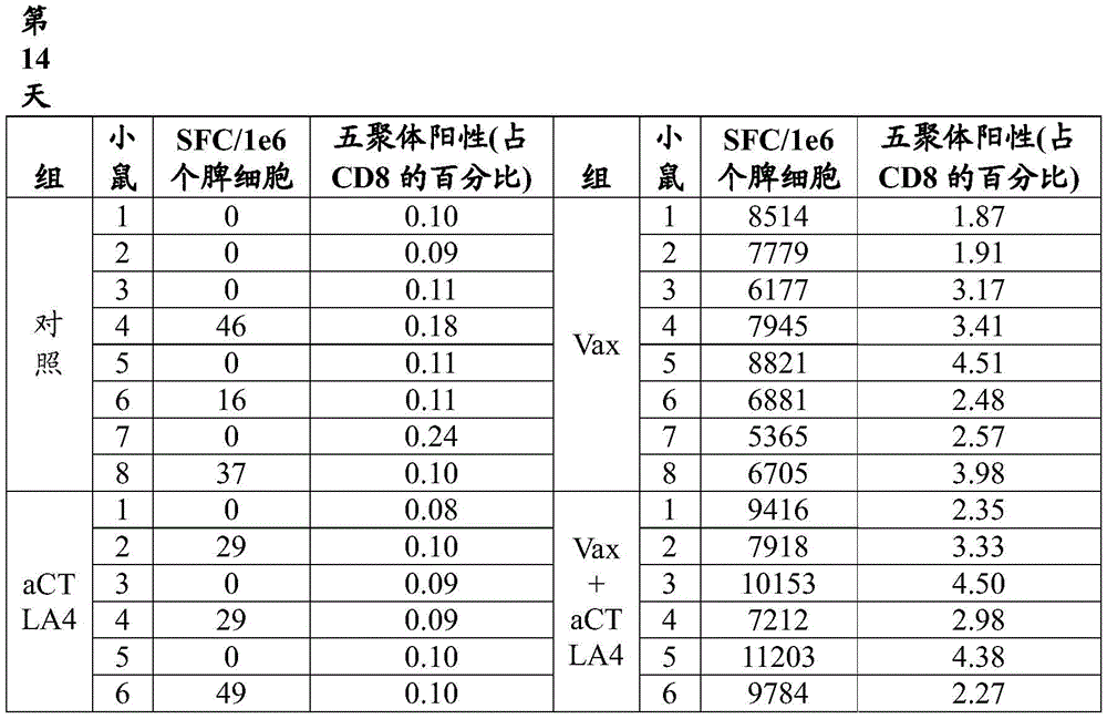
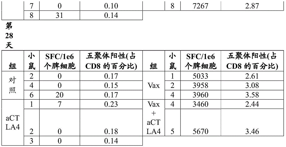
In another embodiment, similar results are observed in the CT26 mouse model after priming for Ad5-UbAAY and boosting for VEE-UbAAY srRNA. In one example, AH1 antigen specific responses were observed after Ad5-UbAAY priming (day 14) and measured every 10 in an ELISpot assay6An average of 5187 SFCs per splenocyte (FIG. 14A, Table 14) and at VEEMeasured every 10 in the ELISpot assay after UbAAY srRNA boosting (day 28)6An average of 3799 SFCs per splenocyte (figure 14B, table 14).
TABLE 14 immunological monitoring following allogeneic prime/boost in the CT26 tumor mouse model.

ChAdV/srRNA Combined tumor model evaluation
Various dosing regimens using ChAdV68 and self-replicating rna (srna) were evaluated in a murine CT26 tumor model.
XVII. A ChAdV/srRNA combined tumor model evaluation method and material
Tumor injection
Balb/c mice were injected with the CT26 tumor cell line. 7 days after tumor cell injection, mice were randomized into different study groups (28-40 mice/group) and treatment was initiated. Injection of 10 into the lower left flank of Balb/c mice6Individual CT26 cells/animal. Tumors were allowed to grow for 7 days prior to immunization. The study groups are described in detail in table 15.
TABLE 15-ChAdV/srRNA Combined tumor model evaluation study group

Immunization
For the srRNA vaccine, mice were injected with 10ug VEE-MAG25mer srRNA by two-sided intramuscular injection in a volume of 100uL (50. mu.L per leg). For the C68 vaccine, mice were injected 1X 10 by two-sided intramuscular injection11One ChAdV68.5WNTt. MAG25mer Virion (VP) in a volume of 100. mu.L (50. mu.L per leg). Animals were injected 2 times weekly with 250ug doses of anti-PD-1 (clone RMP1-14, BioXcell) or anti-IgG (clone MPC-11, BioXcell) via intraperitoneal injection.
Spleen cell dissociation
Spleen and lymph nodes from each mouse were pooled into 3mL of complete RPMI (RPMI, 10% FBS, penicillin/streptomycin). Mechanical dissociation was performed using a genetlemecs acs dissociator (Miltenyi Biotec) following the manufacturer's protocol. Dissociated cells were filtered through a 40 micron filter and lysed with ACK lysis buffer (150mM NH4Cl、10mM KHCO3、0.1mM Na2EDTA) lysed erythrocytes. The cells were filtered again through a 30 micron filter and then resuspended in complete RPMI. Cells were counted on an Attune NxT flow cytometer (Thermo Fisher) using propidium iodide staining to exclude dead and apoptotic cells. The cells are then adjusted to the appropriate viable cell concentration for subsequent analysis.
Ex vivo enzyme-linked immunospot (ELISPOT) assay
ELISPOT analysis was performed using the mouse IFNg ELISPOtPLUS kit (MABTECH) according to the ELISPOT consensus criterion (DOI: 10.1038/nprot.2015.068). Will be 5X 10⁴Individual splenocytes were incubated with 10uM of the indicated peptide for 16 hours in 96-well plates coated with IFNg antibody. The spots were developed using alkaline phosphatase. The reaction was timed for 10 minutes and terminated by running tap water through the plate. Spots were counted using the AID vSpot reader spectrogram. For ELISPOT analysis, saturation was measured>50% of the wells were recorded as "too many to count". Will replicate deviations in the hole>10% of the samples were excluded from the analysis. The spot count was then corrected for the degree of well confluence using the following formula: spot count +2 × (spot count × degree of confluence%/[ 100% -degree of confluence%) ]). Negative background was corrected by subtracting the spot count in negative peptide-stimulated wells from antigen-stimulated wells. Finally, the wells marked too many to count were set to the highest observed correction, rounded to the nearest percentage.
Combined assessment of ChAdV/srRNA in the XVII. B CT26 tumor model
Immunogenicity and efficacy of either the chadv68.5wtnt. mag25mer/VEE-MAG25mer srna heterologous prime/boost or the VEE-MAG25mer srna homologous prime/boost vaccines were evaluated in a CT26 mouse tumor model. Balb/c mice were injected with the CT26 tumor cell line. 7 days after tumor cell injection, mice were randomized into different study groups and treatment was initiated. The study groups are described in detail in table 15 and more coarsely in table 16.
TABLE 16 Primary/boost groups

Spleens were collected for immune monitoring 14 days after primary immunization vaccination. Tumor and body weight measurements were taken twice a week and survival was monitored. A strong immune response relative to the control was observed in all active vaccine groups.
14 days after the first immunization, every 10 th day was observed in the ELISpot analysis in mice immunized with ChAdV68.5WNT. MAG25mer (ChAdV/group 3), ChAdV68.5WNT. MAG25mer + anti-PD-1 (ChAdV + PD-1/group 4), VEE-MAG25mer srRNA (srRNA/median of group 5 and group 7 combinations) or VEE-MAG25mer srRNA + anti-PD-1 (srRNA + PD-1/median of group 6 and group 8 combinations), respectively 6Median cellular immune responses of 10,630, 12,976, 3319, or 3745 Spot Forming Cells (SFC) per splenocyte (fig. 16 and table 17). In contrast, vaccine control (group 1) or combination of vaccine control and anti-PD-1 (group 2), respectively, exhibited every 106Median cellular immune response of 296 or 285 SFCs per splenocyte.
TABLE 17 cellular immune response in CT26 tumor model

Consistent with ELISpot data, 5.6%, 7.8%, 1.8%, or 1.9% of CD8T cells (median) in Intracellular Cytokine Staining (ICS) analysis were immunized in mice vaccinated with chadv68.5wtnt.mag25mer + anti-PD-1 (chadvj + PD-1/4 th group), VEE-MAG25mer srna (median of srna/combination of 5 th and 7 th groups) or VEE-MAG25mer srna + anti-PD-1 (median of srna + PD-1/combination of 6 th and 8 th groups), respectively, 14 days after the first immunization (fig. 17 and table 18). Mice immunized with the vaccine control or the combination of the vaccine control and anti-PD-1 showed antigen-specific CD8 responses of 0.2% and 0.1%, respectively.
CD8T cellular responses in the tumor model of Table 18-CT26
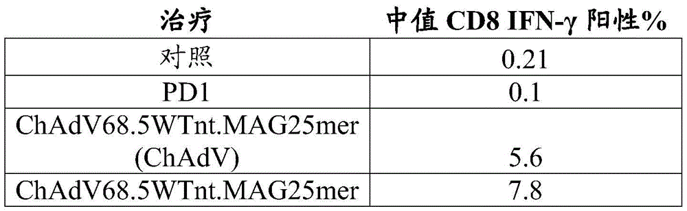

Tumor growth was measured in all groups in the CT26 colon tumor model, and by 21 days after treatment initiation (28 days after injection of CT-26 tumor cells), tumor growth occurred. 21 days after the start of treatment, based on the larger tumor size: ( >2500mm3) The mice were sacrificed; therefore, only the first 21 days are presented to avoid analytical bias. ChAdV68.5WTnt.MAG25mer prime/VEE-MAG 25mer srRNA boost (group 3), ChAdV68.5WTnt.MAG25mer prime/VEE-MAG 25mer srRNA boost + anti-PD-1 (group 4), VEE-MAG25mer srRNA prime/ChAdV68.5WTnt.MAG 25mer boost (group 5), VEE-MAG25mer srRNA prime/ChAdV68.5WTWTnt.MAG25mer boost + anti-PD-1 (group 6), VEE-MAG25mer srRNA prime/VEE-MAG 25mer srRNA boost (group 7) and VEE-MAG25mer srRNA prime/VEE-MAG 25mer srRNA boost + anti-PD-1 (group 8) mean tumor volumes at 21 days were 1129, 112848, 2142, 198 and 1606mm, respectively3(FIG. 18 and Table 19). The mean tumor volume of vaccine control or combination of vaccine control and anti-PD-1 was 2361 or 2067mm, respectively3. Based on these data, vaccine treatment with ChAdV68.5WTnt.MAG25mer/VEE-MAG25mer srRNA (group 3), ChAdV68.5WTnt.MAG25mer/VEE-MAG25mer srRNA + anti-PD-1 (group 4), VEE-MAG25mer srRNA/ChAdV68.5WTnt.MAG25mer + anti-PD-1 (group 6) and VEE-MAG25mer srRNA/VEE-MAG25mer srRNA + anti-PD-1 (group 8) caused a reduction in tumor growth at 21 days, which is significantly different from the control (group 1).
TABLE 19 tumor size in CT26 model measured on day 21


In the CT-26 tumor model, survival was monitored for 35 days (42 days after injection of CT-26 tumor cells) after initiation of treatment. An increase in survival was observed after vaccination of mice with 4 combinations of tests. After vaccination, the combination of anti-PD-1 and chadv68.5wtnt.mag25mer srna boost immunization with chadv68.5wtnt.mag25mer srna (group 4; P <0.0001 relative to control group 1), VEE-MAG25mer srna boost/VEE-MAG 25mer srna boost combination with anti-PD-1 (group 8; P ═ 0.0006 relative to control group 1), chadv68.5wtnt.mag25mer prime/VEE-MAG 25mer srna boost (group 3; P ═ 0.0003 relative to control group 1) and VEE-MAG25mer srna boost/chadv68.5wtnt.mag25mer srna boost combination with anti-PD-1 (group 6; P ═ 0.0016 relative to control group 1) were 64%, 46%, 41% and 36%, respectively, for survival (fig. 19 and fig. 20). Survival rates for the remaining treatment groups [ VEE-MAG25mer srna prime/chadv68.5wtnt.mag25mer boost (group 5), VEE-MAG25mer srna prime/VEE-MAG 25mer srna boost (group 7) and anti-PD-1 alone (group 2) ] were not significantly different from control group 1 (< 14%).
TABLE 20 survival in CT26 model
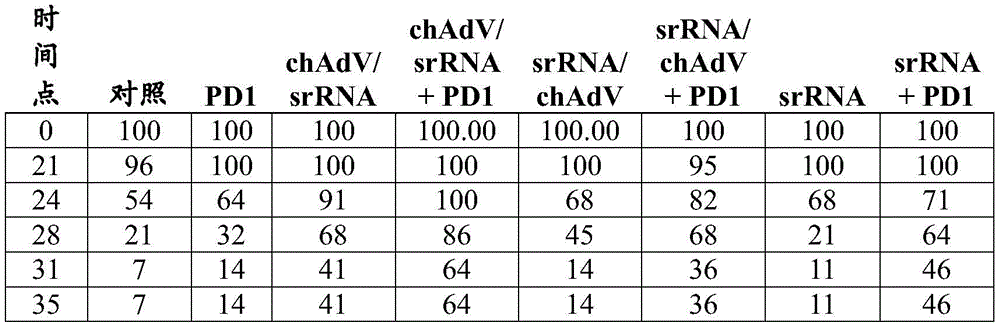
In summary, both chadv68.5wtnt. mag25mer and VEE-MAG25mer srna elicited strong T cell responses against the mouse tumor antigen encoded by the vaccine relative to controls. Survival was improved by administering to tumor bearing mice a combination of either ChAdV68.5WNT.MAG25mer prime and VEE-MAG25mer srRNA boost with or without co-administration of anti-PD-1, VEE-MAG25mer srRNA prime and ChAdV68.5WNT.MAG25mer boost with anti-PD-1, or VEE-MAG25mer srRNA homoprime boost with anti-PD-1.
XVIII non-human primate study
Various dosing regimens using ChAdV68 and self-replicating rna (srna) were evaluated in non-human primates (NHPs).
Materials and methods
The study (vaccine priming) was initiated by intramuscular Injection of (IM) priming vaccine in each NHP. One or more booster vaccines (vaccine boosts) are also injected intramuscularly in each NHP. Each dose of bilateral injection was administered according to the groups summarized in the table and summarized below.
Immunization
Immune monitoring
At the indicated times after primary immunization, PBMCs were isolated using Lymphocyte Separation Medium (LSM; MP Biomedicals) and Leucosep Separation tubes (Greiner Bio-One) and resuspended in RPMI containing 10% FBS and penicillin/streptomycin. Cells were counted on an Attune NxT flow cytometer (Thermo Fisher) using propidium iodide staining to exclude dead and apoptotic cells. The cells are then adjusted to the appropriate viable cell concentration for subsequent analysis. For each monkey in the study, T cell responses were measured using ELISpot or flow cytometry methods. T cell responses against class I epitopes of 6 different rhesus macau Mamu-a 01 encoded in the vaccine were monitored in PBMCs by measuring induction of cytokines such as IFN- γ using an ex vivo enzyme linked immunospot (ELISpot) assay. ELISpot analysis was performed using the monkey IFNg ELISpot PLUS kit (MABTECH) according to the ELISPOT consensus criterion { DOI:10.1038/nprot.2015.068 }. 200,000 PBMCs were incubated with 10. mu.M of the indicated peptides for 16 hours in 96-well plates coated with IFNg antibody. The spots were developed using alkaline phosphatase. The reaction was timed for 10 minutes and terminated by running tap water through the plate. Spots were counted using the AID vSpot reader spectrogram. For ELISPOT analysis, wells with > 50% saturation were recorded as "too many to count". Samples with > 10% deviation of replicate wells were excluded from the analysis. The spot count was then corrected for the degree of well confluence using the following formula: spot count +2 × (spot count × degree of confluence%/[ 100% -degree of confluence ]). Negative background was corrected by subtracting the spot count in negative peptide-stimulated wells from antigen-stimulated wells. Finally, the wells marked too many to count were set to the highest observed correction, rounded to the nearest percentage.
Specific CD4 and CD8T cellular responses against class I epitopes of 6 different rhesus macau Mamu-a 01 encoded in the vaccine were monitored in PBMCs by measuring induction of intracellular cytokines such as IFN- γ using flow cytometry. The results obtained from both methods indicate that the cytokine directed against the epitope is induced in an antigen-specific manner.
Immunogenicity in rhesus monkeys
The study was designed to (a) evaluate the immunogenicity and primary safety of the combination of VEE-MAG25mer srna and chadv68.5wtnt. mag25mer at 30 μ g and 100 μ g doses as a homologous prime/boost or heterologous prime/boost; (b) comparing the immune response of VEE-MAG25mer srna in lipid nanoparticles using LNP1 with LNP 2; (c) kinetics of T cell response to VEE-MAG25mer srna and chadv68.5wtnt. mag25mer immunization were assessed.
The study groups were performed in Mamu-a × 01 indian rhesus monkeys to demonstrate immunogenicity. The selected antigens used in this study were recognized only in rhesus monkeys, particularly those with a Mamu-a × 01MHC class I haplotype. Mamu-a × 01 indian rhesus monkeys were randomly assigned to different study groups (6 rhesus monkeys per group) and given a chadv68.5wtnt. mag25mer or VEE-MAG25mer srna vector encoding a model antigen comprising multiple Mamu-a × 01 restriction epitopes by two-sided IM injection. The study groups are as follows.
Table 21: non-GLP immunogenicity studies in Indian rhesus monkeys

PBMCs were collected for immune monitoring before immunization and at weeks 1, 2, 3, 4, 5, 6, 8, 9 and 10 after the initial immunization.
Results
Antigen-specific cellular immune responses in Peripheral Blood Mononuclear Cells (PBMCs) were measured against six different Mamu-a × 01 restricted epitopes before immunization and at weeks 1, 2, 3, 4, 5, 6, 8, 9 and 10 after the initial immunization. As described in Table 21, animals received boosts of VEE-MAG25mer srRNA at doses of 30 μ g or 100 μ g at weeks 4 and 8 and formulated with LNP1 or LNP 2. The combined immune response for all six epitopes was plotted for each immunodominant time point (FIGS. 20A-D and tables 22-25).
At 1, 2, 3, 4, 5, 6, 8, 9 or 10 weeks after initial immunization with the initial VEE-MAG25mer srRNA-LNP1(30 μ g), respectively at every 10 weeks6Combined antigen-specific immune responses were observed at all measurements of 170, 14, 15, 11, 7, 8, 14, 17, 12 SFCs (six combined epitopes) in one PBMC (fig. 20A). At 1, 2, 3, 4, 5, 6, 8, 9 or 10 weeks after initial immunization with the initial VEE-MAG25mer srRNA-LNP1 (100. mu.g), respectively at every 10 weeks 6Combined antigen-specific immune responses were observed for all measurements of 108, -3, 14, 1, 37, 4, 105, 17, 25 SFCs (six combined epitopes) in individual PBMCs (fig. 20B). At 1, 2, 3, 4, 5, 6, 8, 9 or 10 weeks after initial immunization with the initial VEE-MAG25mer srRNA-LNP2 (100. mu.g), respectively at every 10 weeks6Combined antigen-specific immune responses were observed for all measurements of-17, 38, 14, -2, 87, 21, 104, 129, 89 SFCs (six combined epitopes) in one PBMC (figure 20C). Negative values are the result of normalizing the pre-exsanguination values for each epitope/animal.
1, 2, 3, 4, 5, 6, 8, 9 or 10 weeks after initial immunization of the initial ChAdV68.5WNT. MAG25mer, respectively every 10 weeks6All measurement junctions of 1218, 1784, 1866, 973, 1813, 747, 797, 1249 and 547 SFCs (six combinatorial epitopes) in one PBMCCombined antigen-specific immune responses were observed below the fruits (fig. 20D). The immune response showed the expected characteristics, with the peak of the immune response measured at about 2-3 weeks after the initial immunization followed by a contraction of the immune response after 4 weeks. At 5 weeks after initial immunization with ChAdV68.5WNT. MAG25mer (i.e., at 1 week after first boosting with VEE-MAG25mer srRNA), the combined antigen-specific cellular immune response was measured as every 10 th week 61813 SFCs (six combined epitopes) in one PBMC. The immune response measured at week 1 (week 5) after the first boost with VEE-MAG25mer srna was comparable to the peak immune response measured at the initial immunization with chadv68.5wtnt. mag25mer (week 3) (fig. 20D). At 9 weeks after initial immunization with ChAdV68.5WNT. MAG25mer (i.e., at 1 week after second booster immunization with VEE-MAG25mer srRNA), a combined antigen-specific cellular immune response was measured as every 10 th week61249 SFCs (six combined epitopes) in one PBMC. The immune response measured at week 1 (week 9) after the second boost with VEE-MAG25mer srna was about 2-fold higher than the immune response measured before the boost immunization (fig. 20D).
Table 22: each 10 of each epitope of VEE-MAG25mer srRNA-LNP1 (30. mu.g)6Mean spot-forming cells (SFC). + -. SEM in individual PBMCs (group 1)
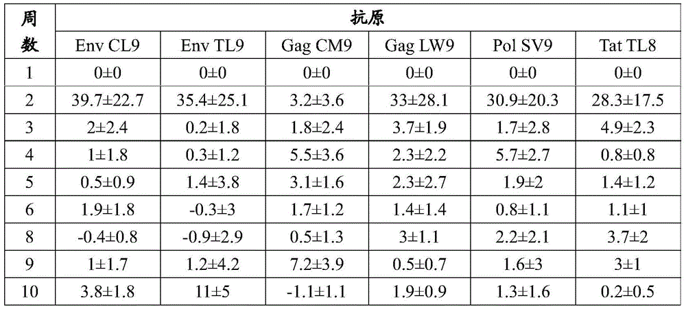
Table 23: each 10 of each epitope of VEE-MAG25mer srRNA-LNP1 (100. mu.g)6Mean spot-forming cells (SFC). + -. SEM in individual PBMCs (group 2)
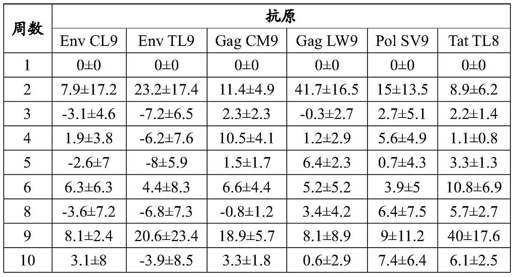
Table 24: each 10 of each epitope of VEE-MAG25mer srRNA-LNP2 (100. mu.g)6Mean spot-forming cells (SFC). + -. SEM in individual PBMCs (group 3)


Table 25: every 10 of each epitope primed by ChAdV68.5WNT.MAG25mer6Mean spot-forming cells (SFC). + -. SEM in individual PBMCs

non-GLP RNA dose range study (higher dose) in Indian rhesus monkeys
The study was designed to (a) assess the immunogenicity of a 300 μ g dose of VEE-MAG25mer srna in combination with chadv68.5wtnt.mag25mer as a homologous prime/boost or heterologous prime/boost; (b) comparing the immune response of a 300 μ g dose of VEE-MAG25mer srna in lipid nanoparticles using LNP1 and LNP 2; and (c) assessing the kinetics of T cell responses to VEE-MAG25mer srna and chadv68.5wtnt. mag25mer immunizations.
The study groups were performed in Mamu-a × 01 indian rhesus monkeys to demonstrate immunogenicity. Vaccine immunogenicity in non-human primate species, such as rhesus monkeys, is the best predictor of vaccine efficacy in humans. Furthermore, the selected antigens used in this study were recognized only in rhesus monkeys, in particular with the Mamu-a × 01MHC class I haplotype. Mamu-a 01 indian rhesus monkeys were randomly assigned to different study groups (6 rhesus monkeys per group) and given a bilateral IM injection with either chadv68.5wtnt. mag25mer or VEE-MAG25mer srna encoding a model antigen including multiple Mamu-a 01 restriction antigens. The study groups are as follows.
PBMCs were collected prior to immunization and at weeks 4, 5, 6, 7, 8, 10, 11, 12, 13, 14, 15, 16, 17, 18, 19, 20, 21, 22, 23 or 24 after the initial immunization for immune monitoring (heterologous prime/boost) on group 1. PBMCs were collected prior to immunization and at weeks 4, 5, 7, 8, 10, 11, 12, 13, 14 or 15 after the initial immunization for immune monitoring (heterologous prime/boost) for groups 2 and 3.
Table 26: non-GLP immunogenicity studies in Indian rhesus monkeys

Results
Mamu-a 01 indian rhesus monkeys were immunized with chadv68.5-wtnt. Antigen-specific cellular immune responses in Peripheral Blood Mononuclear Cells (PBMCs) were measured against six different Mamu-a × 01 restricted epitopes before immunization and at 4, 5, 6, 7, 8, 10, 11, 12, 13, 14, 15, 16, 17, 18, 19, 20, 21, 22, 23 or 24 weeks after the initial immunization (fig. 21 and table 27). At weeks 4, 12 and 20, animals were boosted with VEE-MAG25mer srna using LNP2 formulation. Combined antigen specific immune responses measured every 10, 5, 6, 7, 8, 10, 11, 12, 13, 14, 15, 16, 17, 18, 19, 20, 21, 22, 23 or 24 weeks after initial immunization with chadv68.5wtnt61750, 4225, 1100, 2529, 3218, 1915, 1708, 1561, 5077, 4543, 4920, 5820, 3395, 2728, 1996, 1465, 4730, 2984, 2828 or 3043 SFCs (six combined epitopes) out of PBMCs (figure 21). The immune response measured at week 1 (week 13) after the second boost with VEE-MAG25mer srRNA was about 3-fold greater than the immune response measured before the boost (week 12). The immune response measured at week 1 (week 21) after the third boost with VEE-MAG25mer srna was about 3-fold higher than the immune response measured before the boost (week 20), similar to the response observed at the second boost.
Two different LNP formulations (LNP1 and LNP2) were also used to immunize Mamu-a × 01 indian rhesus monkeys with VEE-MAG25mer srna. Antigen-specific cellular immune responses in Peripheral Blood Mononuclear Cells (PBMCs) were measured against six different Mamu-a × 01 restricted epitopes before immunization and at weeks 4, 5, 6, 7, 8, 10, 11, 12, 13, 14 or 15 after the initial immunization (fig. 22 and 23, tables 28 and 29). Animals were dosed with LNP1 or LNP2 at weeks 4 and 12, respectivelyReceived a boost using VEE-MAG25mer srRNA. Combined antigen-specific immune responses were measured as 168, 204, 103, 126, 140, 145, 330, 203 and 162 SFCs (six combined epitopes) per 106 PBMCs at 4, 5, 7, 8, 10, 11, 13, 14, 15 weeks after immunization with VEE-MAG25mer rrna-LNP2 (figure 22). The combined antigen-specific immune response was measured every 10 weeks after immunization with VEE-MAG25mer srRNA-LNP1 at 4, 5, 7, 8, 10, 11, 12, 13, 14, 15 weeks6189, 185, 349, 437, 492, 570, 233, 886, 369 and 381 SFCs (six combined epitopes) out of the PBMCs (fig. 23).
Table 27: every 10 of each epitope initially immunized with ChAdV68.5WNT.MAG25mer 6Mean spot-forming cells (SFC). + -. SEM in individual PBMCs (group 1)


Table 28: each 10 th epitope of each of the primary immunizations with VEE-MAG25mer srRNA-LNP2 (300. mu.g)6Mean spot-forming cells (SFC). + -. SEM in individual PBMCs (group 2)
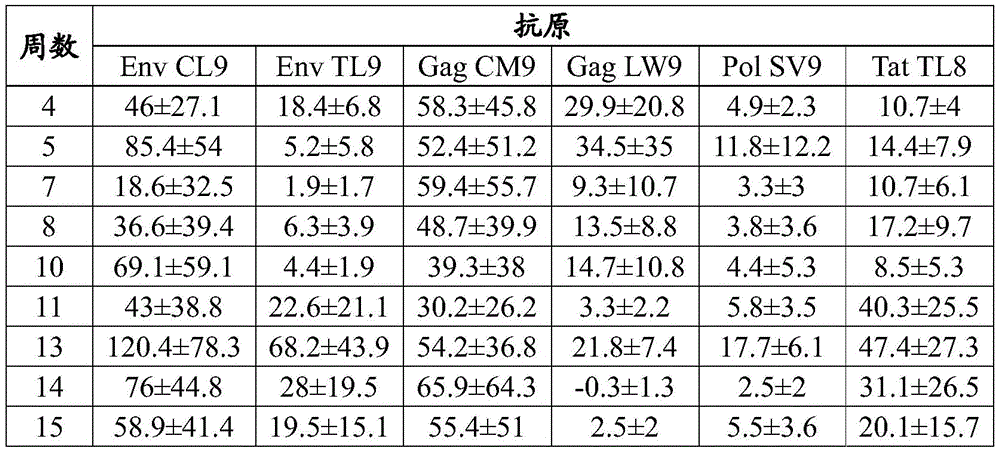
Table 29: each 10 th epitope of each of the primary immunizations with VEE-MAG25mer srRNA-LNP1 (300. mu.g)6Mean spot-forming cells (SFC). + -. SEM in individual PBMCs (group 3)
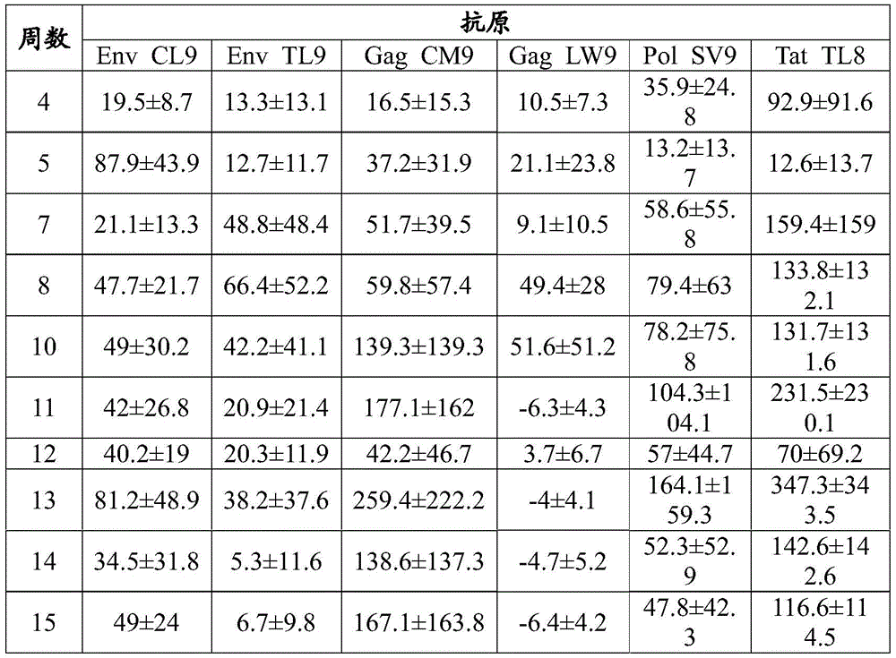
srRNA dose Range study
In one embodiment of the invention, srna dose range studies can be performed in mamu a01 indian rhesus monkeys to identify which srna dose can be used for NHP immunogenicity studies. In one example, an srna vector encoding a model antigen comprising multiple Mamu a01 restriction epitopes can be administered to Mamu a01 indian rhesus monkey by IM injection. In another example, anti-CTLA-4 monoclonal antibodies can be SC administered near the site of IM vaccine injection to target vaccine draining lymph nodes in a group of animals. PBMCs can be collected every two weeks after the initial vaccination for immune monitoring. The study groups are described below (table 30).
Table 30: non-GLP RNA dose range study in Indian rhesus monkeys
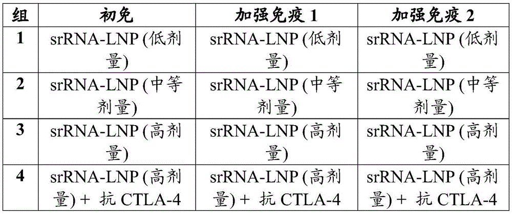
Dose range of srna determined at high dose ≤ 300 μ g.
Immunogenicity studies in indian rhesus monkeys
Vaccine studies were performed in mamu a01 indian rhesus monkey (NHP) to demonstrate immunogenicity using antigen vectors. Figure 34 shows a vaccination strategy. Three groups of NHPs were immunized with chadv68.5-wtnt. mag25mer with the checkpoint inhibitor anti-CTLA-4 antibody ipilimumab (groups 5 and 6) or without checkpoint inhibitor (group 4). The antibody was administered intravenously (group 5) or subcutaneously (group 6). Triangles indicate vaccination with chAd68 (1e12 vp/animal) at weeks 0 and 32. Circles indicate alphavirus vaccination at weeks 0, 4, 12, 20, 28 and 32.
The time course of the CD8+ anti-epitope response in immunized NHPs is shown for chAd-MAG immunization alone (fig. 35 and table 31A), checkpoint inhibitors for chAd-MAG immunization and IV delivery (fig. 36 and table 31B), and checkpoint inhibitors for chAd-MAG immunization and SC delivery (fig. 37 and table 31C). The results indicate that the chAd68 vector efficiently elicits CD8+ responses in primates, that the alphavirus vector efficiently enhances the elicited response of the chAd68 vaccine, that both the elicitation and the booster response are enhanced by checkpoint inhibitors delivered IV or SC, and that the chAd vector is re-administered after vaccination to effectively enhance the immune response.
Table 31A: CD8+ anti-epitope response in rhesus monkeys given chAd-MAG (group 4). Mean SFC/1e6 splenocytes +/-standard error are shown
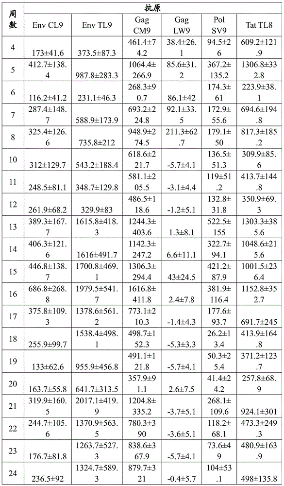
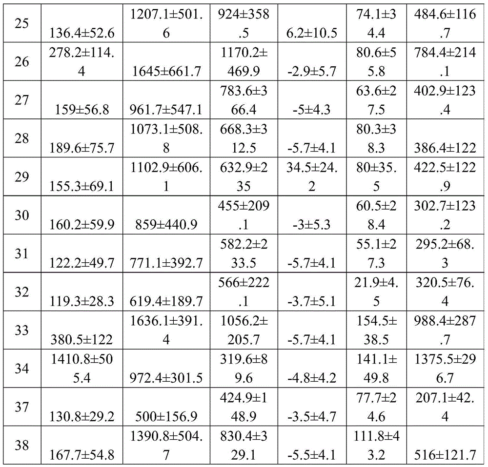
Table 31B: CD8+ anti-epitope response in rhesus monkeys given chAd-MAG plus IV delivered anti-CTLA 4 antibody (ipilimumab) (group 5). Mean SFC/1e6 splenocytes +/-standard error are shown
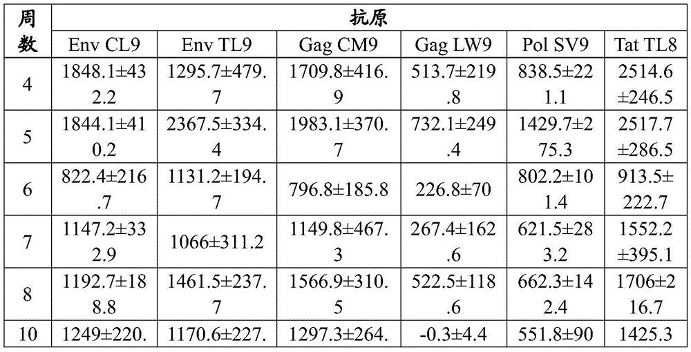


Table 31C: CD8+ anti-epitope response in rhesus monkeys given chAd-MAG plus SC-delivered anti-CTLA 4 antibody (ipilimumab) (group 6). Mean SFC/1e6 splenocytes +/-standard error are shown

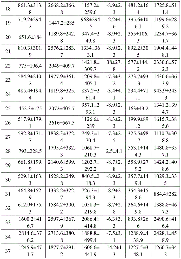
Memory phenotype analysis in Indian rhesus monkeys
Rhesus monkeys were immunized with a chadv68.5wtnt.mag25mer/VEE-MAG25mer srna heterologous prime/boost regimen with or without anti-CTLA 4 and boosted again with chadv68.5wtnt.mag25mer. Each group was evaluated 11 months after the final administration of ChAdV68 (study month 18). ELISpot was performed as described. Fig. 38 and table 43 show cellular responses to six different Mamu-a 01 restricted epitopes measured by ELISpot before immunization (left panel) and after 18 months (right panel). Detection of a response to the restriction epitope indicates that the ChAdV68/samRNA vaccine regimen generates an antigen-specific memory response.
To assess memory, CD8+ T cells recognizing 4 different rhesus macau Mamu-a 01 class I epitopes encoded in the vaccine were monitored using a two-color Mamu-a 01 tetramer marker, each antigen represented by a unique double-positive combination, and allowed the identification of all 4 antigen-specific populations in a single sample. Memory cell phenotype analysis was performed by co-staining with the cell surface markers CD45RA and CCR 7. Fig. 39 and table 44 show the results of combined tetramer staining and CD45RA/CCR7 co-staining for memory T cells recognizing four different Mamu-a × 01 restriction epitopes. T cell phenotypes were also assessed by flow cytometry. Figure 40 shows the distribution of memory cell types within the four Mamu-a 01 tetramer + CD8+ T cell populations at study month 18. Memory cells were characterized as follows: CD45RA + CCR7+ (naive), CD45RA + CCR7 ═ effector (Teff), CD45RA-CCR7 ═ central memory (Tcm), CD45RA-CCR7 ═ effector memory (Tem). Taken together, these results indicate that a memory response was detected at least one year after the last booster immunization, indicating a long lasting immunity, including effector, central memory and effector memory groups.
TABLE 43 Primary and memory assessment time points (18 months) 10/animal 6Mean Spot Forming Cells (SFC) of PBMC.


ND is undetermined due to technical exclusions
TABLE 44 percentage of positive Mamu-A01 tetramer in live CD8+ cells
| Animal(s) production | Tat TL8 | Gag CM9 | Env | Env CL9 | |
| 1 | 0.42 | 0.11 | 0.19 | 0.013 | |
| 2 | 0.36 | 0.048 | 0.033 | 0.00834 | |
| 3 | 0.97 | 0.051 | 0.35 | 0.048 | |
| 4 | 0.46 | 0.083 | 0.17 | 0.028 | |
| 5 | 0.77 | 0.45 | 0.14 | 0.2 | |
| 6 | 0.71 | 0.16 | 0.17 | 0.04 |
Co-expression of anti-CTLA 4 immune checkpoint inhibitors
Vectors co-expressing an antigen and an immune checkpoint inhibitor are engineered.
Materials and methods
In one example, chimpanzee adenovirus viral vectors are designed to express both a model antigen and an immune checkpoint inhibitor anti-CTLA 4.
Carrier design
An E1/E3 deleted ChAdV68 viral vector was designed in which the expression cassette was introduced into the deleted E1 region from 5 'to 3' in the following format (see fig. 26): [ CMV-model antigen/GFP-IRES-anti-CTLA 4-SV40 ]. Cassette expression is driven by the Cytomegalovirus (CMV) promoter located 5 'to the model antigen cassette (or GFP reporter) and the SV-40 polyadenylation signal 3' to the anti-CTLA 4 antibody. The model antigen cassette is the MAG25mer cassette described in section XIV.B.4 (SEQ ID NO: 34) above. The antigen cassette (or GFP reporter gene) and anti-CTLA 4 antibody are separated by an IRES sequence, which enables translation of the model antigen and anti-CTLA 4 antibody, respectively, from the same transcript. The anti-CTLA 4 antibody nucleotide sequence is based on the anti-CTLA 4 clone 9D9 sequence, which is available on Genbank (NCBI ID LQ222660.1 and LQ222658.1) and is further described in PCT publication WO2016025642, the entire teachings of which are incorporated herein by reference. The anti-CTLA 4 antibody expression cassette was designed in the following format: [ full-length heavy chain-furin cleavage site-T2A site-full-length light chain ], as described below (Fang J, Qian JJ, Yi S, Harding TC, Tu GH, Van Roey M, Jooss K. (2005.) Stable expression at therapeutic levels using the 2A peptide. Nat Biotechnol.23(5):584-90), wherein the T2A site is the Thosea asigna virus 2A peptide. The variable region was appended to the constant region of mouse IgG2b, which corresponds to the original isotype of 9D 9. The nucleotide sequence was also codon optimized for expression relative to the Genbank sequence.
Two versions of GFP reporter expression constructs were prepared: one with the antibody leader sequence using the NCBI sequence (g9D9, SEQ ID NO:60) and the other with a leader sequence based on the sequence predicted to be presented in the original mouse 9D9 hybridoma by the IgBLAST tool (o9D9, SEQ ID NO: 61).
Model antigen expression constructs were made using the "o 9D 9" antibody leader sequence described above. The full-length sequence of the chimpanzee C68 adenovirus construct expressing the model antigen and anti-CTLA 4 ("chAd-MAG-CTLA 4") is described below (SEQ ID NO: 57).
Additional checkpoint immune checkpoint inhibitor co-expression vectors were generated: a chimpanzee C68 adenovirus construct expressing a GFP reporter gene or model antigen cassette and sequences encoding anti-CTLA 4 antibody ipilimumab (chAd68-GFP-IRES-IPI "IPI-GFP" and chAd68-MAG-IRES-IPI "IPI-MAG"; SEQ ID NOS: 70 and 71, respectively); a chimpanzee C68 adenovirus construct (chAd 68-MAG-IRES-TREME; SEQ ID NO:72) expressing the sequence of the model antigen cassette and encoding anti-CTLA 4 antibody tremelimumab.
Vector production
anti-CTLA 4 expressing viral vectors were generated by transfecting PacI digested pA68-MAG-o9D9 (plasmid containing ChAdV68 vector expressing model antigen cassette and anti-CTLA 4 version o9D 9), pA68-GFP-IRES-o9D9 (plasmid containing ChAdV68 vector expressing GFP reporter gene and anti-CTLA 4 version o9D 9) and pA68-GFP-IRES-g9D9 (plasmid containing ChAdV68 vector expressing GFP reporter gene and anti-CTLA 4 version g9D 9) into 293A cells using Fugene 6 (Promega). Viral vectors were amplified in 293 cells and then produced on a large scale (400mL scale) in 293F suspension cells. 48 hours post infection, cells were harvested, lysed and virus purified by two rounds of CsCl gradient. The virus was dialyzed into 20mM Tris pH 8.0, 25mM NaCl and 2.5% glycerol. The purified viral vectors were aliquoted and stored at-80 ℃. The infectious unit titer of the purified viral vector was determined using an anti-capsid assay. For in vitro and in vivo experiments, the dose was based on IU titers.
In vitro expression
293F cells were infected with the following viral vectors at an MOI of 1. After infection, the supernatant was collected and the antibody recovered using protein a beads. The antibodies were eluted from the beads and separated on a 4-20% SDS-PAGE gel. The gels were subjected to western blot analysis using goat anti-mouse HRP conjugated antibody (Millipore, AP124P) diluted 1:2,500 in TBST-5% dry milk, followed by chemiluminescent detection reagent (thermoldissher, ECL Plus).
In vivo evaluation
C56F1 mice were administered 1.5e7 or 1.5e6IU of the following viral vectors delivered to the tibialis anterior by bilateral Intramuscular (IM) injection. Mice co-administered with anti-CTLA 4 were delivered antibodies at 250ug, 2 x/week by Intraperitoneal (IP) administration. The groups, doses, routes and times of injection are described below. Antigen-specific T cells were measured by ELISpot and intracellular cytokine staining. Sera were obtained at various time points and anti-CTLA 4 concentrations were measured by ELISA.
Results
Viruses were generated and quantified as described above. The titer data for chAd68-o9D9 batch CS110617E used in the in vivo study is shown below:
table 32: titer data against CTLA4 clinical cassette virus (chAd68-MAG-o9D9)
| Measurement of | Results |
| Infectious unit titer | 3.06e8IU/mL |
| Viral particle titer | 3e12VP/mL |
anti-CTLA 4 in vitro expression
293F cells were infected with the following viral vectors: ChAdVC68-MAG25mer-IRES-o9D9 ("5 WT-MAG-o9D 9"), ChAdVC68-GFP-IRES-o9D9 and ChAdVC68-GFP-IRES-g9D 9. Supernatants were collected at 7, 20, 30 and 48 hours post infection and analyzed by Western after antibody recovery with protein a beads. Commercial 9D9 antibody was used as a positive control ("(+) ctrl"). Supernatants from viral vectors encoding only GFP but not anti-CTLA 9D9 antibodies were used as negative controls ("(-) ctrl"). As shown in figure 27, starting 20 hours post-infection, there were bands associated with the heavy and light chains of the 9D9 anti-CTLA 4 positive control in the supernatant of the three vectors tested, while no protein band was detected in the negative control lane, indicating that each vector expresses the desired anti-CTLA 4 antibody. No protein was observed in the lane of cell lysate alone (i.e., the sample without antibody recovery using protein a beads) (lane 2), demonstrating that detection of expressed antibody requires antibody recovery.
Next, the expression of chAd68-GFP-IRES-IPI and chAd68-MAG-IRES-IPI in vitro was evaluated. Fig. 28A demonstrates the expression of the heavy and light chains of ipilimumab. Next, the in vitro expression of chAd68-MAG-IRES-TREME was assessed. Fig. 28B demonstrates the expression of the heavy and light chains of tremelimumab.
In vivo evaluation of vectors co-expressing anti-CTLA 4
Cell antigen-specific immune responses and anti-CTLA 4 expression in serum of chaadv 68-MAG25mer-IRES-o9D9 ("chAd-MAG-CTLA 4") co-expressing a model antigen cassette (MAG) and anti-CTLA 4 antibody (o9D9) were evaluated in mice. The co-expression vector treatments were compared to mice receiving either vectors expressing the same model antigen cassette but not expressing anti-CTLA 4 antibody ("chAd-MAG") or mice expressing the same model antigen cassette but not expressing anti-CTLA 4 antibody and co-administered 9D9 anti-CTLA 4 antibody (purchased from BioXcell) at the same time. chAd virus is administered by the IM route at 1.5e7 or 1.5e6 IU. Co-administered anti-CTLA 4 antibody was IP injected at a dose of 250 ug. The following table details the various groups:
TABLE 33 in vivo evaluation design of chAd-MAG-CTLA4 immunogenicity and expression

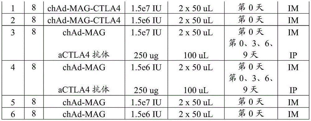
Antigen-specific T cells against MHC class I epitope AH1-a5 were measured by ELISpot and intracellular cytokine staining 12 days after vaccine administration. As shown in FIG. 41, the antigen-specific immune responses of chAd-MAG against chAd-MAG-CTLA4, chAd-MAG alone, and co-administered IP-delivered anti-CTLA 4 antibody were equivalent as measured by ELISpot at day 12 post-immunization. The number of antigen-specific T cells was similar for chAd-MAG-CTLA4, chAd-MAG + anti-CTLA 4 and chAd-MAG as measured by IFN γ -Elispot, with median values of 11578, 12194 and 12945 SFC/1e6 splenocytes for the low dose of 1.5e6IU (FIG. 41 left panel), and 18933, 17992 and 18766 SFC/1e6 splenocytes for the high dose of 1.5e7 IU (FIG. 41 right panel).
As shown in FIG. 42, these three treatments also produced a significant amount of CD8 against MHC class I epitope AH1-A5+IFNγ+Antigen-specific T cells, as measured by ICS, with median values of 3.7, 3.5 and 3.9IFN γ at 1.5e6IU+(CD8+Of the graph (fig. 42 left), and 8.4, 7.3, and 8.2 at 1.5e7 IU (fig. 42 right).
No statistically significant differences were found between groups for ELISpot or ICS analysis as determined by ANOVA. The results demonstrate that the chAd-MAG-a ctla4 vaccine is functional in driving antigen-specific T cell responses in mice, and is at least equivalent by these assays.
The amount of anti-CTLA 4 antibody in mouse sera was assessed by a meso-scale discovery (MSD) ELISA on days 1, 2, 3, 6, and 12 after immunization with chaD-MAG-CTLA4 (groups 1 and 2) or chaD-MAG co-administered with IP-delivered anti-CTLA 4 antibody. For groups 3 and 4, anti-CTLA 4 mAb (250 μ g, IP) was administered on days 0, 3, 6, and 9.
As shown in FIG. 43 and Table 34, anti-CTLA 4 expression was detected in the serum of mice immunized with low (1.5e6IU) and high (1.5e7 IU) doses of chAd-MAG-CTLA 4. In the absence of additional immunization, expression peaked at day 2 post-immunization and remained above baseline (day 0) until final measurements were taken at day 12. Dose responses of expression were observed, with higher levels detected at higher doses of chAd-MAG-CTLA4 at all time points. Serum levels of anti-CTLA 4 were significantly lower than IP-delivered anti-CTLA 4 mAb, which was greater than the maximum range determined at all time points after administration. These results indicate that the ChAd-MAG-a CTLA4 vaccine expressed low, but sustained expression in a dose-dependent manner relative to repeated IP administration of anti-CTLA 4 antibody.
Table 34: anti-CTLA 4 antibody levels in mouse serum

Average ECL values for each group and time point.
Sequence of


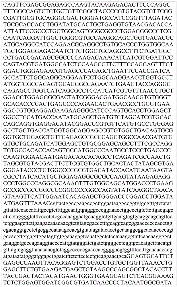
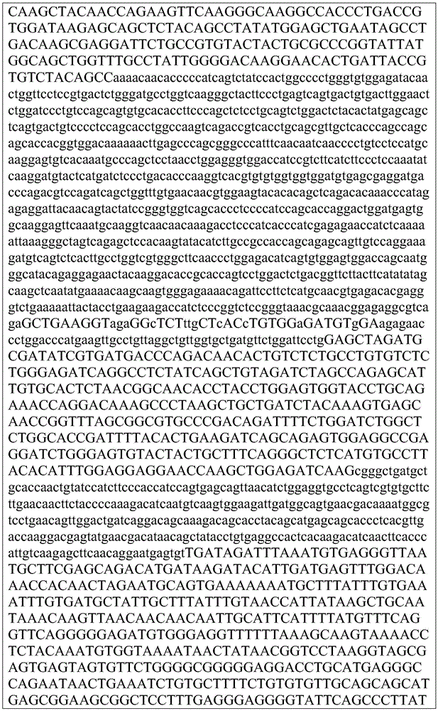


XX. Co-expression tumor model evaluation of anti-CTLA 4 immune checkpoint inhibitors
Various dosing regimens using ChAdV68 with or without co-expression of anti-CTLA 4 and optionally in combination with self-replicating rna (srna) were evaluated in murine CT26 or B16-OVA tumor models.
Method and material
Tumor injection
For the B16-OVA tumor model, 10 injections were made in the lower left flank of Balb/c mice5B16-OVA cells/animal. Prior to immunizationTumors were allowed to grow for 3 days.
For the CT26 tumor model, 10 was injected in the lower left flank of Balb/c mice6Individual CT26 cells/animal. Tumors were allowed to grow for 7 days prior to immunization.
Immunization
Mice were immunized with 9D9 anti-CTLA 4 antibody (purchased from BioXcell) co-administered simultaneously with either a ChAdVC68-MAG25mer-IRES-o9D9 co-expressing a model antigen cassette (MAG) and an anti-CTLA 4 antibody (o9D9), a vector expressing the same model antigen cassette but not expressing anti-CTLA 4 antibody, and/or a vector expressing the same model antigen cassette but not expressing anti-CTLA 4 antibody. In multiple study groups, mice received booster immunizations with alphavirus vaccine vectors expressing the same model antigen.
Spleen cell dissociation
The spleen and lymph nodes of each mouse were pooled in 3mL of complete RPMI (RPMI, 10% FBS, penicillin/streptomycin). Mechanical dissociation was performed using a genetlemecs acs dissociator (Miltenyi Biotec) following the manufacturer's protocol. Dissociated cells were filtered through a 40 micron filter and lysed with ACK buffer (150mM NH)4Cl、10mM KHCO3、0.1mM Na2EDTA) lysed erythrocytes. The cells were filtered again through a 30 micron filter and then resuspended in complete RPMI. Cells were counted on an Attune NxT flow cytometer (Thermo Fisher) using propidium iodide staining to exclude dead and apoptotic cells. The cells are then adjusted to the appropriate viable cell concentration for subsequent analysis.
Ex vivo enzyme-linked immunospot (ELISPOT) assay
ELISPOT analysis was performed using the mouse IFNg ELISPOtPLUS kit (MABTECH) according to the ELISPOT consensus criterion (DOI: 10.1038/nprot.2015.068). Will be 5X 10⁴Individual splenocytes were incubated with 10uM of the indicated peptide for 16 hours in 96-well plates coated with IFNg antibody. The spots were developed using alkaline phosphatase. The reaction was timed for 10 minutes and terminated by running tap water through the plate. Spots were counted using the AID vSpot reader spectrogram. For ELISPOT analysis, saturation was measured >50% of the wells were recorded as "too many to count". Will replicate deviations in the hole>10% of the samples were excluded from the analysis. The spot count was then corrected for the degree of well confluence using the following formula: spot count +2 × (spot count × degree of confluence%/[ 100% -degree of confluence%)]). Negative background was corrected by subtracting the spot count in negative peptide-stimulated wells from antigen-stimulated wells. Finally, the wells marked too many to count were set to the highest observed correction, rounded to the nearest percentage.
Results
The cellular antigen-specific immune response of mice in various study groups was assessed by ELISpot and ICS, anti-CTLA 4 antibody levels in serum, tumor growth and survival. Immunization with ChAdVC68-MAG25mer-IRES-o9D9 showed improved immune response.
Co-expression of immune checkpoint inhibitors
In one example, the viral vector is designed to express both a model antigen and an immune checkpoint inhibitor. The viral vector co-expressing the immune checkpoint inhibitor is compared to a vector expressing the same model antigen cassette but not expressing the immune checkpoint inhibitor and/or a vector expressing the same model antigen cassette but not expressing the immune checkpoint inhibitor while co-administering the immune checkpoint inhibitor. Other immune modulators were also tested in the same manner.
Carrier design
In one example, an E1/E3 deleted ChAdV68 viral vector was designed in which the expression cassette was introduced into the deleted E1 region from 5 'to 3' in the following format: [ CMV-model antigen/GFP-IRES-immune checkpoint inhibitor-SV 40 ]. Cassette expression is driven by the Cytomegalovirus (CMV) promoter located 5 'to the model antigen cassette (or GFP reporter) and the SV-40 polyadenylation signal 3' to the immune checkpoint inhibitor. The model antigen cassette is the MAG25mer cassette described in section XIV.B.4 above (SEQ ID NO: 34). The antigen cassette (or GFP reporter) and the immune checkpoint inhibitor are separated by an IRES sequence, which enables translation of the model antigen separately, and are separated from the same transcript by the IRES sequence. For an anti-CTLA 4 antibody or antigen-binding fragment thereof, an anti-PD-1 antibody or antigen-binding fragment thereof, an anti-PD-L1 antibody or antigen-binding fragment thereof, an anti-4-1 BB antibody or antigen-binding fragment thereof, or an anti-OX-40 antibody or antigen-binding fragment thereof, the immune checkpoint inhibitor or immunomodulator is based on available sequences, sequences obtained by peptide sequencing of available antibodies and subsequent design of nucleotide sequences for expression, or sequences obtained by BCR sequencing of hybridomas. Useful immune checkpoint inhibitors and immune modulators are described in table 35 below. The sequences are also modified for expression, e.g., codon optimized or engineered for introduction into a cloning vector. Antibody-encoding nucleotide sequences were designed according to the following format: [ heavy chain-furin cleavage site-T2A site-light chain ], as described below (Fang J, Qian JJ, Yi S, Harding TC, Tu GH, Van Roey M, Jooss K. (2005). Stable antibody expression at therapeutic levels using the 2A peptide. Nat Biotechnol.23(5):584-90), wherein the T2A site is the Leuconostoc 2A peptide. The variable region is appended to the constant region of the mouse Ig isotype.
Table 35: useful immune checkpoint inhibitors
| Target | Cloning | Source |
| 4-1BB | 3H3 | Bio X Cell BE0239 |
| 4-1BB | LOB12.3 | Bio X Cell BE0169 |
| OX40 | OX86 | Bio X Cell BE0031 |
| CTLA-4 | 9H10 | Bio X Cell BE0131 |
| PD-1 | RMP1-14 | Bio X Cell BE0146 |
In another example, an E1/E3 deleted ChAdV68 viral vector was designed as described above, wherein the expression cassette was introduced into the deleted E1 region and an immune checkpoint inhibitor was introduced into the deleted E3 region.
In another example, an alphavirus vector with a deletion of a viral structural protein is designed, in which an expression cassette is introduced into the deleted region from 5 'to 3' in the following format: [ model-antigen/GFP-IRES-immune checkpoint inhibitor ]. The various immune checkpoint inhibitors tested in the alphavirus vectors were identical to those tested in the adenovirus vectors described above.
Vector production
The immune checkpoint inhibitor expression viral vectors were generated by transfecting linearized vectors into 293A cells using Fugene 6 (Promega). The viral vectors were subjected to amplification in 293 cells and then mass produced in 293F suspension cells (400mL scale). 48 hours post infection, cells were harvested, lysed and virus purified by two rounds of CsCl gradient. The virus was dialyzed into 20mM Tris pH 8.0, 25mM NaCl and 2.5% glycerol. The purified viral vectors were aliquoted and stored at-80 ℃. The infectious unit titer of the purified viral vector was determined using an anti-capsid assay. For in vitro and in vivo experiments, the administration was based on IU titers.
In vitro expression
293F cells were infected with various viral vectors at an MOI of 1. After infection, the supernatant was collected and the antibody recovered using protein a beads. The antibodies were eluted from the beads and separated on a 4-20% SDS-PAGE gel. Western blot analysis of the gels was performed using goat anti-mouse HRP conjugated antibody (Millipore, AP124P) diluted 1:2,500 in TBST-5% dry milk, followed by chemiluminescent detection reagent (thermoldissher, ECL Plus).
In vivo evaluation
Mice were immunized with various viral vectors delivered to the tibialis anterior by bilateral Intramuscular (IM) injection. Co-administering an immune checkpoint inhibitor to mice in a selected group that receive a viral vector that does not express an immune checkpoint inhibitor. Antigen-specific T cells were measured by ELISpot and intracellular cytokine staining. Sera were obtained at various time points and the concentration of immune checkpoint inhibitors was measured by ELISA.
Results
A variety of viral vectors were generated and quantified, including those co-expressing immune checkpoint inhibitors. Viral vectors encoding immune checkpoint inhibitors express immune checkpoint inhibitors at detectable levels in vitro and in serum.
Multiple viral vectors co-expressing an immune checkpoint inhibitor result in immune activation equal to or greater than that achieved by vectors without the checkpoint inhibitor.
Certain sequences
The vectors, cassettes and antibodies referred to herein are described below and are referred to by SEQ ID NO.
Reference to the literature
1.Desrichard,A.,Snyder,A.&Chan,T.A.Cancer Neoantigens and Applications for Immunotherapy.Clin.Cancer Res.Off.J.Am.Assoc.Cancer Res.(2015).doi:10.1158/1078-0432.CCR-14-3175
2.Schumacher,T.N.&Schreiber,R.D.Neoantigens in cancer immunotherapy.Science 348,69-74(2015).
3.Gubin,M.M.,Artyomov,M.N.,Mardis,E.R.&Schreiber,R.D.Tumor neoantigens:building a framework for personalized cancer immunotherapy.J.Clin.Invest.125,3413-3421(2015).
4.Rizvi,N.A.et al.Cancer immunology.Mutational landscape determines sensitivity to PD-1 blockade in non-small cell lung cancer.Science 348,124-128(2015).
5.Snyder,A.et al.Genetic basis for clinical response to CTLA-4 blockade in melanoma.N.Engl.J.Med.371,2189-2199(2014).
6.Carreno,B.M.et al.Cancer immunotherapy.A dendritic cell vaccine increases the breadth and diversity of melanoma neoantigen-specific T cells.Science 348,803-808(2015).
7.Tran,E.et al.Cancer immunotherapy based on mutation-specific CD4+ T cells in a patient with epithelial cancer.Science 344,641-645(2014).
8.Hacohen,N.&Wu,C.J.-Y.United States Patent Application:20110293637-COMPOSITIONS AND METHODS OF IDENTIFYING TUMOR SPECIFIC NEOANTIGENS.(A1).at<http://appft1.uspto.gov/netacgi/nph-ParserSect1=PTO1&Sect2=HITOFF&d=PG01&p=1&u=/netahtml/PTO/srchnum.html&r=1&f=G&l=50&s1=20110293637.PGNR.>
9.Lundegaard,C.,Hoof,I.,Lund,O.&Nielsen,M.State of the art and challenges in sequence based T-cell epitope prediction.Immunome Res.6 Suppl 2,S3(2010).
10.Yadav,M.et al.Predicting immunogenic tumour mutations by combining mass spectrometry and exome sequencing.Nature 515,572-576(2014).
11.Bassani-Sternberg,M.,Pletscher-Frankild,S.,Jensen,L.J.&Mann,M.Mass spectrometry of human leukocyte antigen class I peptidomes reveals strong effects of protein abundance and turnover on antigen presentation.Mol.Cell.Proteomics MCP 14,658-673(2015).
12.Van Allen,E.M.et al.Genomic correlates of response to CTLA-4 blockade in metastatic melanoma.Science 350,207-211(2015).
13.Yoshida,K.&Ogawa,S.Splicing factor mutations and cancer.Wiley Interdiscip.Rev.RNA 5,445-459(2014).
14.Cancer Genome Atlas Research Network.Comprehensive molecular profiling of lung adenocarcinoma.Nature 511,543-550(2014).
15.Rajasagi,M.et al.Systematic identification of personal tumor-specific neoantigens in chronic lymphocytic leukemia.Blood 124,453-462(2014).
16.Downing,S.R.et al.United States Patent Application:0120208706-OPTIMIZATION OF MULTIGENE ANALYSIS OF TUMOR SAMPLES.(A1).at<http://appft1.uspto.gov/netacgi/nph-ParserSect1=PTO1&Sect2=HITOFF&d=PG01&p=1&u=/netahtml/PTO/srchnum.html&r=1&f=G&l=50&s1=20120208706.PGNR.>
17.Target Capture for NextGen Sequencing-IDT.at<http://www.idtdna.com/pages/products/nextgen/target-capture>
18.Shukla,S.A.et al.Comprehensive analysis of cancer-associated somatic mutations in class I HLA genes.Nat.Biotechnol.33,1152-1158(2015).
19.Cieslik,M.et al.The use of exome capture RNA-seq for highly degraded RNA with application to clinical cancer sequencing.Genome Res.25,1372-1381(2015).
20.Bodini,M.et al.The hidden genomic landscape of acute myeloid leukemia:subclonal structure revealed by undetected mutations.Blood 125,600-605(2015).
21.Saunders,C.T.et al.Strelka:accurate somatic small-variant calling from sequenced tumor-normal sample pairs.Bioinforma.Oxf.Engl.28,1811-1817(2012).
22.Cibulskis,K.et al.Sensitive detection of somatic point mutations in impure and heterogeneous cancer samples.Nat.Biotechnol.31,213-219(2013).
23.Wilkerson,M.D.et al.Integrated RNA and DNA sequencing improves mutation detection in low purity tumors.Nucleic Acids Res.42,e107(2014).
24.Mose,L.E.,Wilkerson,M.D.,Hayes,D.N.,Perou,C.M.&Parker,J.S.ABRA:improved coding indel detection via assembly-based realignment.Bioinforma.Oxf.Engl.30,2813-2815(2014).
25.Ye,K.,Schulz,M.H.,Long,Q.,Apweiler,R.&Ning,Z.Pindel:a pattern growth approach to detect break points of large deletions and medium sized insertions from paired-end short reads.Bioinforma.Oxf.Engl.25,2865-2871(2009).
26.Lam,H.Y.K.et al.Nucleotide-resolution analysis of structural variants using BreakSeq and a breakpoint library.Nat.Biotechnol.28,47-55(2010).
27.Frampton,G.M.et al.Development and validation of a clinical cancer genomic profiling test based on massively parallel DNA sequencing.Nat.Biotechnol.31,1023-1031(2013).
28.Boegel,S.et al.HLA typing from RNA-Seq sequence reads.Genome Med.4,102(2012).
29.Liu,C.et al.ATHLATES:accurate typing of human leukocyte antigen through exome sequencing.Nucleic Acids Res.41,e142(2013).
30.Mayor,N.P.et al.HLA Typing for the Next Generation.PloS One 10,e0127153(2015).
31.Roy,C.K.,Olson,S.,Graveley,B.R.,Zamore,P.D.&Moore,M.J.Assessing long-distance RNA sequence connectivity via RNA-templated DNA-DNA ligation,eLife 4,(2015).
32.Song,L.&Florea,L.CLASS:constrained transcript assembly of RNA-seq reads.BMC Bioinformatics 14 Suppl 5,S14(2013).
33.Maretty,L.,Sibbesen,J.A.&Krogh,A.Bayesian transcriptome assembly.Genome Biol.15,501(2014).
34.Pertea,M.et al.StringTie enables improved reconstruction of a transcriptome from RNA-seq reads.Nat.Biotechnol.33,290-295(2015).
35.Roberts,A.,Pimentel,H.,Trapnell,C.&Pachter,L.Identification of novel transcripts in annotated genomes using RNA-Seq.Bioinforma.Oxf.Engl.(2011).doi:10.1093/bioinformatics/btr355
36.Vitting-Seerup,K.,Porse,B.T.,Sandelin,A.&Waage,J.spliceR:an R package for classification of alternative splicing and prediction of coding potential from RNA-seq data.BMC Bioinformatics 15,81(2014).
37.Rivas,M.A.et al.Human genomics.Effect of predicted protein-truncating genetic variants on the human transcriptome.Science 348,666-669(2015).
38.Skelly,D.A.,Johansson,M.,Madeoy,J.,Wakefield,J.&Akey,J.M.A powerful and flexible statistical framework for testing hypotheses of allele-specific gene expression from RNA-seq data.Genome Res.21,1728-1737(2011).
39.Anders,S.,Pyl,P.T.&Huber,W.HTSeq--a Python framework to work with high-throughput sequencing data.Bioinforma.Oxf.Engl.31,166-169(2015).
40.Furney,S.J.et al.SF3B1 mutations are associated with alternative splicing in uveal melanoma.Cancer Discov.(2013).doi:10.1158/2159-8290.CD-13-0330
41.Zhou,Q.et al.A chemical genetics approach for the functional assessment of novel cancer genes.Cancer Res.(2015).doi:10.1158/0008-5472.CAN-14-2930
42.Maguire,S.L.et al.SF3B1 mutations constitute a novel therapeutic target in breast cancer,J.Pathol.235,571-580(2015).
43.Carithers,L.J.et al.A Novel Approach to High-Quality Postmortem Tissue Procurement:The GTEx Project.Biopreservation Biobanking 13,311-319(2015).
44.Xu,G.et al.RNA CoMPASS:a dual approach for pathogen and host transcriptome analysis of RNA-seq datasets.PloS One9,e89445(2014).
45.Andreatta,M.&Nielsen,M.Gapped sequence alignment using artificial neural networks:application to the MHC class I system.Bioinforma.Oxf.Engl.(2015).doi:10.1093/bioinformatics/btv639
46. K.W.,Rasmussen,M.,Buus,S.&Nielsen,M.NetMHCstab-predicting stability of peptide-MHC-I complexes;impacts for cytotoxic T lymphocyte epitope discovery.Immunology 141,18-26(2014).
47.Larsen,M.V.et al.An integrative approach to CTL epitope prediction:a combined algorithm integrating MHC class I binding,TAP transport efficiency,and proteasomal cleavage predictions.Eur.J.Immunol.35,2295-2303(2005).
48.Nielsen,M.,Lundegaard,C.,Lund,O.& C.The role of the proteasome in generating cytotoxic T-cell epitopes:insights obtained from improved predictions of
 proteasomal cleavage.Immunogenetics 57,33-41(2005).
49.Boisvert,F.-M.et al.A Quantitative Spatial Proteomics Analysis of Proteome Turnover in Human Cells.Mol.Cell.Proteomics 11,M111.011429-M111.011429(2012).
50.Duan,F.et al.Genomic and bioinformatic profiling of mutational neoepitopes reveals new rules to predict anticancer immunogenicity.J.Exp.Med.211,2231-2248(2014).
51.Janeway’s Immunobiology:9780815345312:Medicine&Health Science Books @Amazon.com.at<http://www.amazon.com/Janeways-Immunobiology-Kenneth-Murphy/dp/0815345313>
52.Calis,J.J.A.et al.Properties of MHC Class I Presented Peptides That Enhance Immunogenicity.PLoS Comput.Biol.9,e1003266(2013).
53.Zhang,J.et al.Intratumor heterogeneity in localized lung adenocarcinomas delineated by multiregion sequencing.Science 346,256-259(2014)
54.Walter,M.J.et al.Clonal architecture of secondary acute mveloid leukemia.N.Engl.J.Med.366,1090-1098(2012).
55.Hunt DF,Henderson RA,Shabanowitz J,Sakaguchi K,Michel H,Sevilir N,Cox AL,Appella E,Engelhard VH.Characterization of peptides bound to the class I MHC molecule HLA-A2.1 by mass spectrometry.Science 1992.255:1261-1263.
56.Zarling AL,Polefrone JM,Evans AM,Mikesh LM,Shabanowitz J,Lewis ST,Engelhard VH,Hunt DF.Identification of class I MHC-associated phosphopeptides as targets for cancer immunotherapy.Proc Natl Acad Sci U S A.2006 Oct 3;103(40):14889-94.
57.Bassani-Sternberg M,Pletscher-Frankild S,Jensen LJ,Mann M.Mass spectrometry of human leukocyte antigen class I peptidomes reveals strong effects of protein abundance and turnover on antigen presentation.Mol Cell Proteomics.2015 Mar;14(3):658-73.doi:10.1074/mcp.M114.042812.
58.Abelin JG,Trantham PD,Penny SA,Patterson AM,Ward ST,Hildebrand WH,Cobbold M,Bai DL,Shabanowitz J,Hunt DF.Complementary IMAC enrichment methods for HLA-associated phosphopeptide identification by mass spectrometry.Nat Protoc.2015 Sep;10(9):1308-18.doi:10.1038/nprot.2015.086.Epub 2015 Aug 6
59.Barnstable CJ,Bodmer WF,Brown G,Galfre G,Milstein C,Williams AF,Ziegler A.Production of monoclonal antibodies to group A erythrocytes,HLA and other human cell surface antigens-new tools for genetic analysis.Cell.1978 May;14(1):9-20.
60.Goldman JM,Hibbin J,Kearney L,Orchard K,Th′ng KH.HLA-DR monoclonal antibodies inhibit the proliferation of normal and chronic granulocytic leukaemia myeloid progenitor cells.Br J Haematol.1982 Nov;52(3):411-20.
61.Eng JK,Jahan TA,Hoopmann MR.Comet:an open-source MS/MS sequence database search tool.Proteomics.2013 Jan;13(1):22-4.doi:10.1002/pmic.201200439.Epub 2012 Dec 4.
62.Eng JK,Hoopmann MR,Jahan TA,Egertson JD,Noble WS,MacCoss MJ.A deeper look into Comet--implementation and features.J Am Soc Mass Spectrom.2015 Nov;26(11):1865-74.doi:10.1007/s13361-015-1179-x.Epub 2015 Jun 27.
63.Lukas Jesse Canterbury,Jason Weston,William Stafford Noble and Michael J.MacCoss.Semi-supervised learning for peptide identification from shotgun proteomics datasets.Nature Methods 4:923-925,November 2007
64.Lukas John D.Storey,Michael J.MacCoss and William Stafford Noble.Assigning confidence measures to peptides identified by tandem mass spectrometry.Journal of Proteome Research,7(1):29-34,January 2008
65.Lukas John D.Storey and William Stafford Noble.Nonparametric estimation of posterior error probabilities associated with peptides identified by tandem mass spectrometry.Bioinformatics,24(16):i42-i48,August 2008
66.Kinney RM,BJ Johnson,VL Brown,DW Trent.Nucleotide Sequence of the 26 S mRNA of the Virulent Trinidad Donkey Strain of Venezuelan Equine Encephalitis Virus and Deduced Sequence of the Encoded Structural Proteins.Virology 152(2),400-413.1986 Jul 30.
67.Jill E Slansky,Frédérique M Rattis,Lisa F Boyd,Tarek Fahmy,Elizabeth M Jaffee,Jonathan P Schneck,David H Margulies,Drew M Pardoll.Enhanced Antigen-Specific Antitumor Immunity with Altered Peptide Ligands that Stabilize the MHC-Peptide-TCR Complex.Immunity,Volume 13, Issue 4,1 October 2000,Pages 529-538.
68.A Y Huang,P H Gulden,A S Woods,M C Thomas,C D Tong,W Wang,V H Engelhard,G Pasternack,R Cotter,D Hunt,D M Pardoll,and E M Jaffee.The immunodominant major histocompatibility complex class I-restricted antigen of a murine colon tumor derives from an endogenous retroviral gene product.Proc Natl Acad Sci U S A.;93(18):9730-9735,1996 Sep 3.
69.JOHNSON,BARBARA J.B.,RICHARD M.KINNEY,CRYSTLE L.KOST AND DENNIS W.TRENT.Molecular Determinants of Alphavirus Neurovirulence:Nucleotide and Deduced Protein Sequence Changes during Attenuation of Venezuelan Equine Encephalitis Virus.J Gen Virol 67:1951-1960,1986.
70.Aarnoudse,C.A.,Krüse,M.,Konopitzky,R.,Brouwenstijn,N.,and Schrier,P.I.(2002).TCR reconstitution in Jurkat reporter cells facilitates the identification of novel tumor antigens by cDNA expression cloning.Int J Cancer 99,7-13.
71.Alexander,J.,Sidney,J.,Southwood,S.,Ruppert,J.,Oseroff,C.,Maewal,A.,Snoke,K.,Serra,H.M.,Kubo,R.T.,and Sette,A.(1994).Development of high potency universal DR-restricted helper epitopes by modification of high affinity DR-blocking peptides.Immunity 1,751-761.
72.Banu,N.,Chia,A.,Ho,Z.Z.,Garcia,A.T.,Paravasivam,K.,Grotenbreg,G.M.,Bertoletti,A.,and Gehring,A.J.(2014).Building and optimizing a virus-specific T cell receptor library for targeted immunotherapy in viral infections.Scientific Reports 4,4166.
73.Cornet,S.,Miconnet,I.,Menez,J.,Lemonnier,F.,and Kosmatopoulos,K.(2006).Optimal organization of a polypeptide-based candidate cancer vaccine composed of cryptic tumor peptides with enhanced immunogenicity.Vaccine 24,2102-2109.
74.Depla,E.,van der Aa,A.,Livingston,B.D.,Crimi,C.,Allosery,K.,de Brabandere,V.,Krakover,J.,Murthy,S.,Huang,M.,Power,S.,et al.(2008).Rational design of a multiepitope vaccine encoding T-lymphocyte epitopes for treatment of chronic hepatitis B virus infections.Journal of Virology 82,435-450.
75.Ishioka,G.Y.,Fikes,J.,Hermanson,G.,Livingston,B.,Crimi,C.,Qin,M.,del Guercio,M.F.,Oseroff,C.,Dahlberg,C.,Alexander,J.,et al.(1999).Utilization of MHC class I transgenic mice for development of minigene DNA vaccines encoding multiple HLA-restricted CTL epitopes.J Immunol 162,3915-3925.
76.Janetzki,S.,Price,L.,Schroeder,H.,Britten,C.M.,Welters,M.J.P.,and Hoos,A.(2015).Guidelines for the automated evaluation of Elispot assays.Nat Protoc 10,1098-1115.
77.Lyons,G.E.,Moore,T.,Brasic,N.,Li,M.,Roszkowski,J.J.,and Nishimura,M.I.(2006).Influence of human CD8 on antigen recognition by T-cell receptor-transduced cells.Cancer Res 66,11455-11461.
78.Nagai,K.,Ochi,T.,Fujiwara,H.,An,J.,Shirakata,T.,Mineno,J.,Kuzushima,K.,Shiku,H.,Melenhorst,J.J.,Gostick,E.,et al.(2012).Aurora kinase A-specific T-cell receptor gene transfer redirects T lymphocytes to display effective antileukemia reactivity.Blood 119,368-376.
79.Panina-Bordignon,P.,Tan,A.,Termijtelen,A.,Demotz,S.,Corradin,G.,and Lanzavecchia,A.(1989).Universally immunogenic T cell epitopes:promiscuous binding to human MHC class II and promiscuous recognition by T cells.Eur J Immunol 19,2237-2242.
80.Vitiello,A.,Marchesini,D.,Furze,J.,Sherman,L.A.,and Chesnut,R.W.(1991).Analysis of the HLA-restricted influenza-specific cytotoxic T lymphocyte response in transgenic mice carrying a chimeric human-mouse class I major histocompatibility complex.J Exp Med 173,1007-1015.
81.Yachi,P.P.,Ampudia,J.,Zal,T.,and Gascoigne,N.R.J.(2006).Altered peptide ligands induce delayed CD8-T cell receptor interaction--a role for CD8 in distinguishing antigen quality.Immunity 25,203-211.
82.Pushko P,Parker M,Ludwig GV,Davis NL,Johnston RE,Smith JF.Replicon-helper systems from attenuated Venezuelan equine encephalitis virus:expression of heterologous genes in vitro and immunization against heterologous pathogens in vivo.Virology.1997 Dec 22;239(2):389-401.
83.Strauss,JH and E G Strauss.The alphaviruses:gene expression,replication,and evolution.Microbiol Rev.1994 Sep;58(3):491-562.
84. C,Ehrengruber MU,Grandgirard D.Alphaviral cytotoxicity and its implication in vector development.Exp Physiol.2005 Jan;90(1):45-52.Epub 2004
 Nov 12.
85.Riley,Michael K.II,and Wilfred Vermerris.Recent Advances in Nanomaterials for Gene Delivery-A Review.Nanomaterials 2017,7(5),94.
86.Frolov I,Hardy R,Rice CM.Cis-acting RNA elements at the 5′end of Sindbis virus genome RNA regulate minus-and plus-strand RNA synthesis.RNA.2001 Nov;7(11):1638-51.
87.Jose J,Snyder JE,Kuhn RJ.A structural and functional perspective of alphavirus replication and assembly.Future Microbiol.2009 Sep;4(7):837-56.
88.Bo Li and C.olin N.Dewey.RSEM:accurate transcript quantification from RNA-Seq data with or without a referenfe genome.BMC Bioinformatics,12:323,August 2011
89.Hillary Pearson,Tariq Daouda,Diana Paola Granados,Chantal Durette,Eric Bonneil,Mathieu Courcelles,Anja Rodenbrock,Jean-Philippe Laverdure,Caroline Sylvie Mader,Sébastien Lemieux,Pierre Thibault,and Claude Perreault.MHC class I-associated peptides derive from selective regions of the human genome.The Journal of Clinical Investigation,2016,
90.Juliane Liepe,Fabio Marino,John Sidney,Anita Jeko,Daniel E.Bunting,Alessandro Sette,Peter M.Kloetzel,Michael P.H.Stumpf,Albert J.R.Heck,Michele Mishto.A large fraction of HLA class I ligands are proteasome-generated spliced peptides.Science,21,October 2016.
91.Mommen GP.,Marino,F.,Meiring HD.,Poelen,MC.,van Gaans-van den Brink,JA.,Mohammed S.,Heck AJ.,and van Els CA.Sampling From the Proteome to the Human Leukocyte Antigen-DR(HLA-DR)Ligandome Proceeds Via High Specificity.Mol Cell Proteomics 15(4):1412-1423,April 2016.
92.Sebastian Kreiter,Mathias Vormehr,Niels van de Roemer,Mustafa Diken,Martin Jan Diekmann,Sebastian Boegel,Barbara
 Fulvia Vascotto,John C.Castle,Arbel D.Tadmor,Stephen P.Schoenberger,Christoph Huber,
 Türeci,and Ugur Sahin.Mutant MHC class II epitopes drive therapeutic immune responses to caner.Nature 520,692-696,April 2015.
93.Tran E.,Turcotte S.,Gros A.,Robbins P.F.,Lu Y.C.,Dudley M.E.,Wunderlich J.R.,Somerville R.P.,Hogan K.,Hinrichs C.S.,Parkhurst M.R.,Yang J.C.,Rosenberg S.A.Cancer immunotherapy based on mutation-specific CD4+ T cells in a patient with epithelial cancer.Science 344(6184)641-645,May 2014.
94.Andreatta M.,Karosiene E.,Rasmussen M.,Stryhn A.,Buus S.,Nielsen M.Accurate pan-specific prediction of peptide-MHC class II binding affinity with improved binding core identification.Immunogenetics 67(11-12)641-650,November 2015.
95.Nielsen,M.,Lund,O.NN-align.An artificial neural network-based alignment algorithm for MHC class II peptide binding prediction.BMC Bioinformatics 10:296,September 2009.
96.Nielsen,M.,Lundegaard,C.,Lund,O.Prediction of MHC class II binding affinity using SMM-align,a novel stabilization matrix alignment method.BMC Bioinformatics 8:238,July 2007.
97.Zhang,J.,et al.PEAKS DB:de novo sequencing assisted database search for sensitive and accurate peptide identification.Molecular&Cellular Proteomics.11(4):1-8.1/2/2012.
98.Jensen,Kamilla Kjaergaard,et al.“Improved Methods for Prediting Peptide Binding Affinity to MHC Class II Molecules.”Immunology,2018,doi:10.1111/imm.12889.
99.Carter,S.L.,Cibulskis,K.,Helman,E.,McKenna,A.,Shen,H.,Zack,T.,Laird,P.W.,Onofrio,R.C.,Winckler,W.,Weir,B.A.,et al.(2012).Absolute quantification of somatic DNA alterations in human cancer.Nat.Biotechnol.30,413-421
100.McGranahan,N.,Rosenthal,R.,Hiley,C.T.,Rowan,A.J.,Watkins,T.B.K.,Wilson,G.A.,Birkbak,N.J.,Veeriah,S.,Van Loo,P.,Herrero,J.,et al.(2017).Allele-Specific HLA Loss and Immune Escape in Lung Cancer Evolution.Cell 171,1259-1271.e11.
101.Shukla,S.A.,Rooney,M.S.,Rajasagi,M.,Tiao,G.,Dixon,P.M.,Lawrence,M.S.,Stevens,J.,Lane,W.J.,Dellagatta,J.L.,Steelman,S.,et al.(2015).Comprehensive analysis of cancer-associated somatic mutations in class I HLA genes.Nat.Biotechnol.33,1152-1158.
102.Van Loo,P.,Nordgard,S.H., O.C.,Russnes,H.G.,Rye,I.H.,Sun,W.,Weigman,V.J.,Marynen,P.,Zetterberg,A.,Naume,B.,et al.(2010).Allele-specific copy number analysis of tumors.Proc.Natl.Acad.Sci.U.S.A.107,16910-16915.
103.Van Loo,P.,Nordgard,S.H., O.C.,Russnes,H.G.,Rye,I.H.,Sun,W.,Weigman,V.J.,Marynen,P.,Zetterberg,A.,Naume,B.,et al.(2010).Allele-specific copy number analysis of tumors.Proc.Natl.Acad.Sci.U.S.A.107,16910-16915.


Claims (94)
Applications Claiming Priority (3)
| Application Number | Priority Date | Filing Date | Title |
|---|---|---|---|
| US201862675624P | 2018-05-23 | 2018-05-23 | |
| US62/675,624 | 2018-05-23 | ||
| PCT/US2019/033828 WO2019226939A1 (en) | 2018-05-23 | 2019-05-23 | Immune checkpoint inhibitor co-expression vectors |
Publications (1)
| Publication Number | Publication Date |
|---|---|
| CN112351793A true CN112351793A (en) | 2021-02-09 |
Family
ID=68617211
Family Applications (1)
| Application Number | Title | Priority Date | Filing Date |
|---|---|---|---|
| CN201980044406.0A Pending CN112351793A (en) | 2018-05-23 | 2019-05-23 | Immune checkpoint inhibitor co-expression vectors |
Country Status (9)
| Country | Link |
|---|---|
| US (2) | US20210213122A1 (en) |
| EP (1) | EP3796930A4 (en) |
| JP (1) | JP2021524247A (en) |
| KR (1) | KR20210013589A (en) |
| CN (1) | CN112351793A (en) |
| AU (1) | AU2019275070A1 (en) |
| CA (1) | CA3099642A1 (en) |
| TW (1) | TW202000906A (en) |
| WO (1) | WO2019226939A1 (en) |
Families Citing this family (15)
| Publication number | Priority date | Publication date | Assignee | Title |
|---|---|---|---|---|
| EP4414990A3 (en) | 2013-01-17 | 2024-11-06 | Personalis, Inc. | Methods and systems for genetic analysis |
| WO2016070131A1 (en) | 2014-10-30 | 2016-05-06 | Personalis, Inc. | Methods for using mosaicism in nucleic acids sampled distal to their origin |
| IL315224A (en) | 2017-05-08 | 2024-10-01 | Gritstone Bio Inc | Alphavirus neoantigen vectors |
| IL280890B2 (en) * | 2018-08-17 | 2025-10-01 | Gritstone Bio Inc | Antigen-binding proteins targeting shared antigens |
| WO2020097393A1 (en) * | 2018-11-07 | 2020-05-14 | Gritstone Oncology, Inc. | Alphavirus neoantigen vectors and interferon inhibitors |
| TWI852977B (en) | 2019-01-10 | 2024-08-21 | 美商健生生物科技公司 | Prostate neoantigens and their uses |
| IL288283B2 (en) | 2019-05-30 | 2025-05-01 | Gritstone Bio Inc | Modified adenoviruses |
| WO2021092066A1 (en) | 2019-11-05 | 2021-05-14 | Personalis, Inc. | Estimating tumor purity from single samples |
| US12018289B2 (en) | 2019-11-18 | 2024-06-25 | Janssen Biotech, Inc. | Vaccines based on mutant CALR and JAK2 and their uses |
| EP4168569A4 (en) * | 2020-06-18 | 2024-08-07 | Personalis, Inc. | MACHINE LEARNING TECHNIQUES FOR PREDICTING SURFACE-MOUNTED PEPTIDES |
| EP4175664A2 (en) | 2020-07-06 | 2023-05-10 | Janssen Biotech, Inc. | Prostate neoantigens and their uses |
| EP4192496A4 (en) | 2020-08-06 | 2025-01-01 | Gritstone bio, Inc. | MULTIPLE EPITOPE VACCINE CASSETTES |
| EP4329780B1 (en) | 2021-04-29 | 2025-11-26 | Yeda Research and Development Co. Ltd | T cell receptors directed against ras-derived recurrent neoantigens and methods of identifying same |
| EP4395817A4 (en) * | 2021-09-02 | 2025-10-15 | Elixirgen Therapeutics Inc | TEMPERATURE-CONTROLLED RNA IMMUNOTHERAPEUTIC AGAINST CANCER |
| WO2023059654A1 (en) | 2021-10-05 | 2023-04-13 | Personalis, Inc. | Customized assays for personalized cancer monitoring |
Citations (7)
| Publication number | Priority date | Publication date | Assignee | Title |
|---|---|---|---|---|
| WO1997041241A1 (en) * | 1996-04-25 | 1997-11-06 | GESELLSCHAFT FüR BIOTECHNOLOGISCHE FORSCHUNG MBH (GBF) | Polycistronic expression constructs, their use and vaccines |
| US20140271724A1 (en) * | 2013-03-14 | 2014-09-18 | The Wistar Institute Of Anatomy And Biology | Methods and compositions for treating or preventing cancer |
| CN106456724A (en) * | 2013-12-20 | 2017-02-22 | 博德研究所 | Combination therapy with neoantigen vaccine |
| WO2017152042A2 (en) * | 2016-03-04 | 2017-09-08 | New York University | Virus vectors expressing multiple epitopes of tumor associated antigens for inducing antitumor immunity |
| WO2017192924A1 (en) * | 2016-05-04 | 2017-11-09 | Fred Hutchinson Cancer Research Center | Cell-based neoantigen vaccines and uses thereof |
| WO2018161092A1 (en) * | 2017-03-03 | 2018-09-07 | New York University | Induction and enhancement of antitumor immunity involving virus vectors expressing multiple epitopes of tumor associated antigens and immune checkpoint inhibitors or proteins |
| CN110545810A (en) * | 2016-11-23 | 2019-12-06 | 磨石肿瘤生物技术公司 | Viral delivery of novel antigens |
Family Cites Families (6)
| Publication number | Priority date | Publication date | Assignee | Title |
|---|---|---|---|---|
| US7557200B2 (en) * | 2001-02-01 | 2009-07-07 | Johns Hopkins University | Superior molecular vaccine based on self-replicating RNA, suicidal DNA or naked DNA vector, that links antigen with polypeptide that promotes antigen presentation |
| CA2453880A1 (en) * | 2001-07-25 | 2003-02-06 | New York University | Use of glycosylceramides as adjuvants for vaccines against infections and cancer |
| WO2004031210A2 (en) * | 2002-10-03 | 2004-04-15 | Epimmune Inc. | Optimized multi-epitope constructs and uses thereof |
| CA2960096A1 (en) * | 2014-09-03 | 2016-03-10 | Bavarian Nordic A/S | Methods and compositions for enhancing immune responses |
| WO2017066256A2 (en) * | 2015-10-12 | 2017-04-20 | Nantomics, Llc | Compositions and methods for viral cancer neoepitopes |
| US20190046648A1 (en) * | 2016-03-03 | 2019-02-14 | Cue Biopharma, Inc. | T-cell modulatory multimeric polypeptides and methods of use thereof |
-
2019
- 2019-05-23 CN CN201980044406.0A patent/CN112351793A/en active Pending
- 2019-05-23 WO PCT/US2019/033828 patent/WO2019226939A1/en not_active Ceased
- 2019-05-23 JP JP2020565264A patent/JP2021524247A/en not_active Withdrawn
- 2019-05-23 US US17/058,113 patent/US20210213122A1/en not_active Abandoned
- 2019-05-23 AU AU2019275070A patent/AU2019275070A1/en not_active Abandoned
- 2019-05-23 TW TW108117922A patent/TW202000906A/en unknown
- 2019-05-23 EP EP19808512.8A patent/EP3796930A4/en active Pending
- 2019-05-23 CA CA3099642A patent/CA3099642A1/en active Pending
- 2019-05-23 KR KR1020207036367A patent/KR20210013589A/en not_active Ceased
-
2024
- 2024-01-04 US US18/404,681 patent/US20250025544A1/en active Pending
Patent Citations (7)
| Publication number | Priority date | Publication date | Assignee | Title |
|---|---|---|---|---|
| WO1997041241A1 (en) * | 1996-04-25 | 1997-11-06 | GESELLSCHAFT FüR BIOTECHNOLOGISCHE FORSCHUNG MBH (GBF) | Polycistronic expression constructs, their use and vaccines |
| US20140271724A1 (en) * | 2013-03-14 | 2014-09-18 | The Wistar Institute Of Anatomy And Biology | Methods and compositions for treating or preventing cancer |
| CN106456724A (en) * | 2013-12-20 | 2017-02-22 | 博德研究所 | Combination therapy with neoantigen vaccine |
| WO2017152042A2 (en) * | 2016-03-04 | 2017-09-08 | New York University | Virus vectors expressing multiple epitopes of tumor associated antigens for inducing antitumor immunity |
| WO2017192924A1 (en) * | 2016-05-04 | 2017-11-09 | Fred Hutchinson Cancer Research Center | Cell-based neoantigen vaccines and uses thereof |
| CN110545810A (en) * | 2016-11-23 | 2019-12-06 | 磨石肿瘤生物技术公司 | Viral delivery of novel antigens |
| WO2018161092A1 (en) * | 2017-03-03 | 2018-09-07 | New York University | Induction and enhancement of antitumor immunity involving virus vectors expressing multiple epitopes of tumor associated antigens and immune checkpoint inhibitors or proteins |
Non-Patent Citations (1)
| Title |
|---|
| N TATSIS等: "Chimpanzee-origin adenovirus vectors as vaccine carriers", GENETHERAPY, vol. 13, pages 421 - 429 * |
Also Published As
| Publication number | Publication date |
|---|---|
| KR20210013589A (en) | 2021-02-04 |
| TW202000906A (en) | 2020-01-01 |
| EP3796930A1 (en) | 2021-03-31 |
| JP2021524247A (en) | 2021-09-13 |
| AU2019275070A1 (en) | 2021-01-14 |
| WO2019226939A8 (en) | 2020-11-19 |
| WO2019226939A1 (en) | 2019-11-28 |
| EP3796930A4 (en) | 2022-05-04 |
| US20210213122A1 (en) | 2021-07-15 |
| US20250025544A1 (en) | 2025-01-23 |
| CA3099642A1 (en) | 2019-11-28 |
Similar Documents
| Publication | Publication Date | Title |
|---|---|---|
| US20250025544A1 (en) | Immune checkpoint inhibitor co-expression vectors | |
| US20220226453A1 (en) | Alphavirus Antigen Vectors | |
| US20210196806A1 (en) | Shared antigens | |
| US20220125919A1 (en) | Alphavirus neoantigen vectors and interferon inhibitors | |
| US20230020089A1 (en) | Shared neoantigen vaccines | |
| WO2018098362A1 (en) | Viral delivery of neoantigens | |
| CN114072516A (en) | Modified adenoviruses | |
| US20220265812A1 (en) | Hiv antigens and mhc complexes | |
| US20250276049A1 (en) | Egfr vaccine cassettes | |
| JP2024535855A (en) | KRAS neoantigen therapy | |
| US20240216501A1 (en) | Neoantigen adjuvant and maintenance therapy | |
| US20250255946A1 (en) | Low-dose neoantigen vaccine therapy | |
| TWI910091B (en) | Viral delivery of neoantigens | |
| CN120583957A (en) | CTA vaccine box | |
| CN118302195A (en) | Pan-coronavirus vaccine |
Legal Events
| Date | Code | Title | Description |
|---|---|---|---|
| PB01 | Publication | ||
| PB01 | Publication | ||
| SE01 | Entry into force of request for substantive examination | ||
| SE01 | Entry into force of request for substantive examination | ||
| CB02 | Change of applicant information |
Address after: California, USA Applicant after: Millstone biological Co. Address before: California, USA Applicant before: Gritstone Oncology, Inc. |
|
| CB02 | Change of applicant information | ||
| WD01 | Invention patent application deemed withdrawn after publication |
Application publication date: 20210209 |
|
| WD01 | Invention patent application deemed withdrawn after publication |