CN112041659A - Microfluidic devices, systems, infrastructure, uses thereof, and methods of using the same for genetic engineering - Google Patents
Microfluidic devices, systems, infrastructure, uses thereof, and methods of using the same for genetic engineering Download PDFInfo
- Publication number
- CN112041659A CN112041659A CN201880092246.2A CN201880092246A CN112041659A CN 112041659 A CN112041659 A CN 112041659A CN 201880092246 A CN201880092246 A CN 201880092246A CN 112041659 A CN112041659 A CN 112041659A
- Authority
- CN
- China
- Prior art keywords
- microfluidic device
- droplet
- plate
- electrode
- composition
- Prior art date
- Legal status (The legal status is an assumption and is not a legal conclusion. Google has not performed a legal analysis and makes no representation as to the accuracy of the status listed.)
- Pending
Links
Images


Classifications
-
- C—CHEMISTRY; METALLURGY
- C12—BIOCHEMISTRY; BEER; SPIRITS; WINE; VINEGAR; MICROBIOLOGY; ENZYMOLOGY; MUTATION OR GENETIC ENGINEERING
- C12P—FERMENTATION OR ENZYME-USING PROCESSES TO SYNTHESISE A DESIRED CHEMICAL COMPOUND OR COMPOSITION OR TO SEPARATE OPTICAL ISOMERS FROM A RACEMIC MIXTURE
- C12P21/00—Preparation of peptides or proteins
-
- B—PERFORMING OPERATIONS; TRANSPORTING
- B01—PHYSICAL OR CHEMICAL PROCESSES OR APPARATUS IN GENERAL
- B01L—CHEMICAL OR PHYSICAL LABORATORY APPARATUS FOR GENERAL USE
- B01L3/00—Containers or dishes for laboratory use, e.g. laboratory glassware; Droppers
- B01L3/50—Containers for the purpose of retaining a material to be analysed, e.g. test tubes
- B01L3/502—Containers for the purpose of retaining a material to be analysed, e.g. test tubes with fluid transport, e.g. in multi-compartment structures
- B01L3/5027—Containers for the purpose of retaining a material to be analysed, e.g. test tubes with fluid transport, e.g. in multi-compartment structures by integrated microfluidic structures, i.e. dimensions of channels and chambers are such that surface tension forces are important, e.g. lab-on-a-chip
- B01L3/502769—Containers for the purpose of retaining a material to be analysed, e.g. test tubes with fluid transport, e.g. in multi-compartment structures by integrated microfluidic structures, i.e. dimensions of channels and chambers are such that surface tension forces are important, e.g. lab-on-a-chip characterised by multiphase flow arrangements
- B01L3/502784—Containers for the purpose of retaining a material to be analysed, e.g. test tubes with fluid transport, e.g. in multi-compartment structures by integrated microfluidic structures, i.e. dimensions of channels and chambers are such that surface tension forces are important, e.g. lab-on-a-chip characterised by multiphase flow arrangements specially adapted for droplet or plug flow, e.g. digital microfluidics
- B01L3/502792—Containers for the purpose of retaining a material to be analysed, e.g. test tubes with fluid transport, e.g. in multi-compartment structures by integrated microfluidic structures, i.e. dimensions of channels and chambers are such that surface tension forces are important, e.g. lab-on-a-chip characterised by multiphase flow arrangements specially adapted for droplet or plug flow, e.g. digital microfluidics for moving individual droplets on a plate, e.g. by locally altering surface tension
-
- B—PERFORMING OPERATIONS; TRANSPORTING
- B01—PHYSICAL OR CHEMICAL PROCESSES OR APPARATUS IN GENERAL
- B01L—CHEMICAL OR PHYSICAL LABORATORY APPARATUS FOR GENERAL USE
- B01L3/00—Containers or dishes for laboratory use, e.g. laboratory glassware; Droppers
- B01L3/50—Containers for the purpose of retaining a material to be analysed, e.g. test tubes
- B01L3/502—Containers for the purpose of retaining a material to be analysed, e.g. test tubes with fluid transport, e.g. in multi-compartment structures
- B01L3/5027—Containers for the purpose of retaining a material to be analysed, e.g. test tubes with fluid transport, e.g. in multi-compartment structures by integrated microfluidic structures, i.e. dimensions of channels and chambers are such that surface tension forces are important, e.g. lab-on-a-chip
- B01L3/502715—Containers for the purpose of retaining a material to be analysed, e.g. test tubes with fluid transport, e.g. in multi-compartment structures by integrated microfluidic structures, i.e. dimensions of channels and chambers are such that surface tension forces are important, e.g. lab-on-a-chip characterised by interfacing components, e.g. fluidic, electrical, optical or mechanical interfaces
-
- B—PERFORMING OPERATIONS; TRANSPORTING
- B01—PHYSICAL OR CHEMICAL PROCESSES OR APPARATUS IN GENERAL
- B01L—CHEMICAL OR PHYSICAL LABORATORY APPARATUS FOR GENERAL USE
- B01L3/00—Containers or dishes for laboratory use, e.g. laboratory glassware; Droppers
- B01L3/50—Containers for the purpose of retaining a material to be analysed, e.g. test tubes
- B01L3/502—Containers for the purpose of retaining a material to be analysed, e.g. test tubes with fluid transport, e.g. in multi-compartment structures
- B01L3/5027—Containers for the purpose of retaining a material to be analysed, e.g. test tubes with fluid transport, e.g. in multi-compartment structures by integrated microfluidic structures, i.e. dimensions of channels and chambers are such that surface tension forces are important, e.g. lab-on-a-chip
- B01L3/50273—Containers for the purpose of retaining a material to be analysed, e.g. test tubes with fluid transport, e.g. in multi-compartment structures by integrated microfluidic structures, i.e. dimensions of channels and chambers are such that surface tension forces are important, e.g. lab-on-a-chip characterised by the means or forces applied to move the fluids
-
- C—CHEMISTRY; METALLURGY
- C12—BIOCHEMISTRY; BEER; SPIRITS; WINE; VINEGAR; MICROBIOLOGY; ENZYMOLOGY; MUTATION OR GENETIC ENGINEERING
- C12M—APPARATUS FOR ENZYMOLOGY OR MICROBIOLOGY; APPARATUS FOR CULTURING MICROORGANISMS FOR PRODUCING BIOMASS, FOR GROWING CELLS OR FOR OBTAINING FERMENTATION OR METABOLIC PRODUCTS, i.e. BIOREACTORS OR FERMENTERS
- C12M23/00—Constructional details, e.g. recesses, hinges
- C12M23/02—Form or structure of the vessel
- C12M23/16—Microfluidic devices; Capillary tubes
-
- C—CHEMISTRY; METALLURGY
- C12—BIOCHEMISTRY; BEER; SPIRITS; WINE; VINEGAR; MICROBIOLOGY; ENZYMOLOGY; MUTATION OR GENETIC ENGINEERING
- C12M—APPARATUS FOR ENZYMOLOGY OR MICROBIOLOGY; APPARATUS FOR CULTURING MICROORGANISMS FOR PRODUCING BIOMASS, FOR GROWING CELLS OR FOR OBTAINING FERMENTATION OR METABOLIC PRODUCTS, i.e. BIOREACTORS OR FERMENTERS
- C12M41/00—Means for regulation, monitoring, measurement or control, e.g. flow regulation
- C12M41/30—Means for regulation, monitoring, measurement or control, e.g. flow regulation of concentration
- C12M41/36—Means for regulation, monitoring, measurement or control, e.g. flow regulation of concentration of biomass, e.g. colony counters or by turbidity measurements
-
- C—CHEMISTRY; METALLURGY
- C12—BIOCHEMISTRY; BEER; SPIRITS; WINE; VINEGAR; MICROBIOLOGY; ENZYMOLOGY; MUTATION OR GENETIC ENGINEERING
- C12N—MICROORGANISMS OR ENZYMES; COMPOSITIONS THEREOF; PROPAGATING, PRESERVING, OR MAINTAINING MICROORGANISMS; MUTATION OR GENETIC ENGINEERING; CULTURE MEDIA
- C12N15/00—Mutation or genetic engineering; DNA or RNA concerning genetic engineering, vectors, e.g. plasmids, or their isolation, preparation or purification; Use of hosts therefor
- C12N15/09—Recombinant DNA-technology
- C12N15/11—DNA or RNA fragments; Modified forms thereof; Non-coding nucleic acids having a biological activity
- C12N15/111—General methods applicable to biologically active non-coding nucleic acids
-
- C—CHEMISTRY; METALLURGY
- C12—BIOCHEMISTRY; BEER; SPIRITS; WINE; VINEGAR; MICROBIOLOGY; ENZYMOLOGY; MUTATION OR GENETIC ENGINEERING
- C12N—MICROORGANISMS OR ENZYMES; COMPOSITIONS THEREOF; PROPAGATING, PRESERVING, OR MAINTAINING MICROORGANISMS; MUTATION OR GENETIC ENGINEERING; CULTURE MEDIA
- C12N15/00—Mutation or genetic engineering; DNA or RNA concerning genetic engineering, vectors, e.g. plasmids, or their isolation, preparation or purification; Use of hosts therefor
- C12N15/09—Recombinant DNA-technology
- C12N15/63—Introduction of foreign genetic material using vectors; Vectors; Use of hosts therefor; Regulation of expression
- C12N15/79—Vectors or expression systems specially adapted for eukaryotic hosts
- C12N15/85—Vectors or expression systems specially adapted for eukaryotic hosts for animal cells
-
- C—CHEMISTRY; METALLURGY
- C12—BIOCHEMISTRY; BEER; SPIRITS; WINE; VINEGAR; MICROBIOLOGY; ENZYMOLOGY; MUTATION OR GENETIC ENGINEERING
- C12N—MICROORGANISMS OR ENZYMES; COMPOSITIONS THEREOF; PROPAGATING, PRESERVING, OR MAINTAINING MICROORGANISMS; MUTATION OR GENETIC ENGINEERING; CULTURE MEDIA
- C12N15/00—Mutation or genetic engineering; DNA or RNA concerning genetic engineering, vectors, e.g. plasmids, or their isolation, preparation or purification; Use of hosts therefor
- C12N15/09—Recombinant DNA-technology
- C12N15/87—Introduction of foreign genetic material using processes not otherwise provided for, e.g. co-transformation
- C12N15/88—Introduction of foreign genetic material using processes not otherwise provided for, e.g. co-transformation using microencapsulation, e.g. using amphiphile liposome vesicle
-
- C—CHEMISTRY; METALLURGY
- C12—BIOCHEMISTRY; BEER; SPIRITS; WINE; VINEGAR; MICROBIOLOGY; ENZYMOLOGY; MUTATION OR GENETIC ENGINEERING
- C12Q—MEASURING OR TESTING PROCESSES INVOLVING ENZYMES, NUCLEIC ACIDS OR MICROORGANISMS; COMPOSITIONS OR TEST PAPERS THEREFOR; PROCESSES OF PREPARING SUCH COMPOSITIONS; CONDITION-RESPONSIVE CONTROL IN MICROBIOLOGICAL OR ENZYMOLOGICAL PROCESSES
- C12Q1/00—Measuring or testing processes involving enzymes, nucleic acids or microorganisms; Compositions therefor; Processes of preparing such compositions
- C12Q1/02—Measuring or testing processes involving enzymes, nucleic acids or microorganisms; Compositions therefor; Processes of preparing such compositions involving viable microorganisms
- C12Q1/04—Determining presence or kind of microorganism; Use of selective media for testing antibiotics or bacteriocides; Compositions containing a chemical indicator therefor
- C12Q1/06—Quantitative determination
-
- C—CHEMISTRY; METALLURGY
- C12—BIOCHEMISTRY; BEER; SPIRITS; WINE; VINEGAR; MICROBIOLOGY; ENZYMOLOGY; MUTATION OR GENETIC ENGINEERING
- C12Q—MEASURING OR TESTING PROCESSES INVOLVING ENZYMES, NUCLEIC ACIDS OR MICROORGANISMS; COMPOSITIONS OR TEST PAPERS THEREFOR; PROCESSES OF PREPARING SUCH COMPOSITIONS; CONDITION-RESPONSIVE CONTROL IN MICROBIOLOGICAL OR ENZYMOLOGICAL PROCESSES
- C12Q1/00—Measuring or testing processes involving enzymes, nucleic acids or microorganisms; Compositions therefor; Processes of preparing such compositions
- C12Q1/34—Measuring or testing processes involving enzymes, nucleic acids or microorganisms; Compositions therefor; Processes of preparing such compositions involving hydrolase
-
- G—PHYSICS
- G02—OPTICS
- G02B—OPTICAL ELEMENTS, SYSTEMS OR APPARATUS
- G02B26/00—Optical devices or arrangements for the control of light using movable or deformable optical elements
- G02B26/004—Optical devices or arrangements for the control of light using movable or deformable optical elements based on a displacement or a deformation of a fluid
- G02B26/005—Optical devices or arrangements for the control of light using movable or deformable optical elements based on a displacement or a deformation of a fluid based on electrowetting
-
- B—PERFORMING OPERATIONS; TRANSPORTING
- B01—PHYSICAL OR CHEMICAL PROCESSES OR APPARATUS IN GENERAL
- B01L—CHEMICAL OR PHYSICAL LABORATORY APPARATUS FOR GENERAL USE
- B01L2200/00—Solutions for specific problems relating to chemical or physical laboratory apparatus
- B01L2200/02—Adapting objects or devices to another
- B01L2200/026—Fluid interfacing between devices or objects, e.g. connectors, inlet details
- B01L2200/027—Fluid interfacing between devices or objects, e.g. connectors, inlet details for microfluidic devices
-
- B—PERFORMING OPERATIONS; TRANSPORTING
- B01—PHYSICAL OR CHEMICAL PROCESSES OR APPARATUS IN GENERAL
- B01L—CHEMICAL OR PHYSICAL LABORATORY APPARATUS FOR GENERAL USE
- B01L2200/00—Solutions for specific problems relating to chemical or physical laboratory apparatus
- B01L2200/02—Adapting objects or devices to another
- B01L2200/028—Modular arrangements
-
- B—PERFORMING OPERATIONS; TRANSPORTING
- B01—PHYSICAL OR CHEMICAL PROCESSES OR APPARATUS IN GENERAL
- B01L—CHEMICAL OR PHYSICAL LABORATORY APPARATUS FOR GENERAL USE
- B01L2200/00—Solutions for specific problems relating to chemical or physical laboratory apparatus
- B01L2200/06—Fluid handling related problems
-
- B—PERFORMING OPERATIONS; TRANSPORTING
- B01—PHYSICAL OR CHEMICAL PROCESSES OR APPARATUS IN GENERAL
- B01L—CHEMICAL OR PHYSICAL LABORATORY APPARATUS FOR GENERAL USE
- B01L2400/00—Moving or stopping fluids
- B01L2400/04—Moving fluids with specific forces or mechanical means
- B01L2400/0403—Moving fluids with specific forces or mechanical means specific forces
- B01L2400/0415—Moving fluids with specific forces or mechanical means specific forces electrical forces, e.g. electrokinetic
- B01L2400/0427—Electrowetting
-
- C—CHEMISTRY; METALLURGY
- C12—BIOCHEMISTRY; BEER; SPIRITS; WINE; VINEGAR; MICROBIOLOGY; ENZYMOLOGY; MUTATION OR GENETIC ENGINEERING
- C12N—MICROORGANISMS OR ENZYMES; COMPOSITIONS THEREOF; PROPAGATING, PRESERVING, OR MAINTAINING MICROORGANISMS; MUTATION OR GENETIC ENGINEERING; CULTURE MEDIA
- C12N13/00—Treatment of microorganisms or enzymes with electrical or wave energy, e.g. magnetism, sonic waves
-
- C—CHEMISTRY; METALLURGY
- C12—BIOCHEMISTRY; BEER; SPIRITS; WINE; VINEGAR; MICROBIOLOGY; ENZYMOLOGY; MUTATION OR GENETIC ENGINEERING
- C12N—MICROORGANISMS OR ENZYMES; COMPOSITIONS THEREOF; PROPAGATING, PRESERVING, OR MAINTAINING MICROORGANISMS; MUTATION OR GENETIC ENGINEERING; CULTURE MEDIA
- C12N2310/00—Structure or type of the nucleic acid
- C12N2310/10—Type of nucleic acid
- C12N2310/20—Type of nucleic acid involving clustered regularly interspaced short palindromic repeats [CRISPR]
-
- C—CHEMISTRY; METALLURGY
- C12—BIOCHEMISTRY; BEER; SPIRITS; WINE; VINEGAR; MICROBIOLOGY; ENZYMOLOGY; MUTATION OR GENETIC ENGINEERING
- C12N—MICROORGANISMS OR ENZYMES; COMPOSITIONS THEREOF; PROPAGATING, PRESERVING, OR MAINTAINING MICROORGANISMS; MUTATION OR GENETIC ENGINEERING; CULTURE MEDIA
- C12N9/00—Enzymes; Proenzymes; Compositions thereof; Processes for preparing, activating, inhibiting, separating or purifying enzymes
- C12N9/14—Hydrolases (3)
- C12N9/16—Hydrolases (3) acting on ester bonds (3.1)
- C12N9/22—Ribonucleases [RNase]; Deoxyribonucleases [DNase]
Landscapes
- Chemical & Material Sciences (AREA)
- Health & Medical Sciences (AREA)
- Life Sciences & Earth Sciences (AREA)
- Engineering & Computer Science (AREA)
- Organic Chemistry (AREA)
- Genetics & Genomics (AREA)
- Wood Science & Technology (AREA)
- Zoology (AREA)
- Bioinformatics & Cheminformatics (AREA)
- General Health & Medical Sciences (AREA)
- Biotechnology (AREA)
- General Engineering & Computer Science (AREA)
- Biomedical Technology (AREA)
- Microbiology (AREA)
- Biochemistry (AREA)
- Physics & Mathematics (AREA)
- Molecular Biology (AREA)
- Analytical Chemistry (AREA)
- Dispersion Chemistry (AREA)
- Clinical Laboratory Science (AREA)
- Chemical Kinetics & Catalysis (AREA)
- Biophysics (AREA)
- Proteomics, Peptides & Aminoacids (AREA)
- Hematology (AREA)
- Plant Pathology (AREA)
- Immunology (AREA)
- Sustainable Development (AREA)
- Toxicology (AREA)
- Optics & Photonics (AREA)
- General Physics & Mathematics (AREA)
- General Chemical & Material Sciences (AREA)
- Apparatus Associated With Microorganisms And Enzymes (AREA)
- Measuring Or Testing Involving Enzymes Or Micro-Organisms (AREA)
Abstract
Description
相关申请的交叉引用CROSS-REFERENCE TO RELATED APPLICATIONS
本公开要求2018年2月6日提交的美国临时申请第62/627,022号和2018年7月4日提交的美国临时申请第62/693,998号的优先权的权益。这些文件在此通过引用以其整体并入。The present disclosure claims the benefit of priority from US Provisional Application No. 62/627,022, filed on February 6, 2018, and US Provisional Application No. 62/693,998, filed on July 4, 2018. These documents are hereby incorporated by reference in their entirety.
技术领域technical field
本主题涉及用于控制和操纵微流体装置中的液滴的系统和方法。The present subject matter relates to systems and methods for controlling and manipulating droplets in microfluidic devices.
背景技术Background technique
数字微流体(DMF)提供了一种在电极的阵列上操纵nL-μL体积的液体的方法。通过向电极施加电位,这些离散的液滴可以被并行控制、传输、混合、反应和分析。典型地,自动化系统与DMF装置相连接,该DMF装置使用由用户编写的一组标准基本指令来执行液滴操作。Digital microfluidics (DMF) provides a method for manipulating nL-μL volumes of liquids on arrays of electrodes. By applying a potential to the electrodes, these discrete droplets can be controlled, transported, mixed, reacted and analyzed in parallel. Typically, an automated system is connected to a DMF device that performs droplet operations using a standard set of basic instructions written by a user.
电容反馈系统与数字微流体的集成使用电子电路来感测和监测装置上的液滴。然而,这些方法的缺点是这些系统不能检测单个液滴失败。如果检测到失败,则这些系统需要针对装置上的所有液滴在目标电极上重新施加电位,因为不知道装置上的哪个液滴在操作中发生了失败。这不是一种有利的解决方案,因为电极的过度激活会降低电介质的完整性,并且导致表面易于生物污染。此外,这些系统只能感测液滴,但需要外部检测器(例如,孔板读取器)进行生物分析。The integration of capacitive feedback systems with digital microfluidics uses electronic circuitry to sense and monitor droplets on the device. A disadvantage of these methods, however, is that these systems cannot detect individual droplet failures. If a failure is detected, these systems need to reapply the potential on the target electrode for all droplets on the device, since it is not known which droplet on the device failed in operation. This is not an advantageous solution because over-activation of the electrodes reduces the integrity of the dielectric and results in a surface prone to biofouling. Furthermore, these systems can only sense droplets, but require external detectors (eg, plate readers) for biological analysis.
发明内容SUMMARY OF THE INVENTION
根据一个实例,提供了一种用于跟踪数字微流体装置上的液滴运动的基于图像的系统。该基于图像的系统包含计算机视觉系统,用于捕获数字微流体装置的一个或多个电极上的至少一个液滴的图像;控制单元,其被配置为操纵数字微流体装置的一个或多个电极上的至少一个液滴;和界面单元,其被电耦合至计算机视觉系统并被电耦合至控制单元。界面单元被配置为:指导控制单元以操纵数字微流体装置的一个或多个电极上的至少一个液滴;接收数字微流体装置的一个或多个电极上的至少一个液滴的图像,该图像由计算机视觉系统捕获;以及基于由计算机视觉系统捕获的图像,确定数字微流体装置的一个或多个电极上的至少一个液滴的位置。According to one example, an image-based system for tracking droplet motion on a digital microfluidic device is provided. The image-based system includes a computer vision system for capturing an image of at least one droplet on one or more electrodes of the digital microfluidic device; and a control unit configured to manipulate the one or more electrodes of the digital microfluidic device at least one droplet on; and an interface unit electrically coupled to the computer vision system and electrically coupled to the control unit. The interface unit is configured to: instruct the control unit to manipulate the at least one droplet on the one or more electrodes of the digital microfluidic device; receive an image of the at least one droplet on the one or more electrodes of the digital microfluidic device, the image captured by a computer vision system; and determining a position of at least one droplet on one or more electrodes of the digital microfluidic device based on the images captured by the computer vision system.
根据一个实例,提供了一种微流体装置,包含:光密度(OD)读取器,其中光密度读取器包括发光源和传感器,以能够监测在该装置中培养的细菌培养物的样品的光密度。According to one example, there is provided a microfluidic device comprising: an optical density (OD) reader, wherein the optical density reader includes a light emitting source and a sensor to enable monitoring of samples of bacterial cultures grown in the device for Optical density.
根据一个实例,提供了一种微流体装置,包含:According to one example, a microfluidic device is provided, comprising:
用于混合细菌培养物的培养区域;和a culture area for mixing bacterial cultures; and
用于测量细菌培养物的样品的酶活性的测定区域,所述测定区域包括光密度读取器,其中所述光密度读取器包括能够监测细菌培养物的样品的光密度的发光源和传感器。An assay area for measuring the enzymatic activity of a sample of bacterial culture, the assay area including an optical density reader, wherein the optical density reader includes a luminescent source and a sensor capable of monitoring the optical density of the sample of bacterial culture .
根据一个实例,提供了一种微流体装置,包含:According to one example, a microfluidic device is provided, comprising:
用于混合细菌培养物的培养区域;a culture area for mixing bacterial cultures;
用于储存用于诱导细菌培养的试剂的至少一个储器;at least one reservoir for storing reagents for inducing bacterial culture;
用于排放细菌培养物的废物的废物区域;和Waste areas used to discharge waste from bacterial cultures; and
用于测量细菌培养物的样品的酶活性的测定区域,所述测定区域包括光密度读取器,其中所述光密度读取器包括能够监测细菌培养物的样品的光密度的发光源和传感器。An assay area for measuring the enzymatic activity of a sample of bacterial culture, the assay area including an optical density reader, wherein the optical density reader includes a luminescent source and a sensor capable of monitoring the optical density of the sample of bacterial culture .
根据一个实例,提供了一种在微流体系统中诱导细菌培养物的方法,包含:According to one example, there is provided a method of inducing a bacterial culture in a microfluidic system, comprising:
诱导细菌培养物;inducing bacterial cultures;
在微阵列中进行细菌培养物的至少一次孵育;at least one incubation of the bacterial culture in the microarray;
淬灭孵育的细菌培养物;和quenching the incubated bacterial culture; and
读取淬灭的细菌培养物的样品的光密度。The optical density of a sample of the quenched bacterial culture was read.
根据一个实例,提供了一种在微流体系统中诱导细菌培养物的方法,包含:According to one example, there is provided a method of inducing a bacterial culture in a microfluidic system, comprising:
诱导所述细菌培养物;inducing the bacterial culture;
在微阵列中进行所述细菌培养物的两次孵育,其中所述两次孵育在不同的时间进行;淬灭孵育的细菌培养物;和performing two incubations of the bacterial culture in a microarray, wherein the two incubations are performed at different times; quenching the incubated bacterial culture; and
读取淬灭的细菌培养物的样品的光密度。The optical density of a sample of the quenched bacterial culture was read.
根据一个实例,提供了一种用于自动化和跟踪数字微流体装置上的液滴运动的基于图像的系统,包含:According to one example, an image-based system for automating and tracking droplet motion on a digital microfluidic device is provided, comprising:
计算机视觉系统,用于获取用于检测数字微流体装置上的液滴的图像;a computer vision system for acquiring images for detecting droplets on a digital microfluidic device;
控制单元,用于操纵数字微流体装置中的液滴;以及a control unit for manipulating droplets in a digital microfluidic device; and
图形用户界面,用于对液滴操作进行编程、跟踪液滴运动和可视化当前液滴操作。Graphical user interface for programming droplet operations, tracking droplet motion, and visualizing current droplet operations.
根据一个实例,提供了一种用于操作AIMS的方法,包括:According to one example, a method for operating an AIMS is provided, comprising:
将装置插入到OD读取器中;Insert the device into the OD reader;
将试剂装载到装置上;和loading reagents onto the device; and
输入一系列所需的液滴运动步骤,使得由AIMS进行诱导(以及细胞培养和分析)。A series of desired droplet motion steps were entered to allow induction (as well as cell culture and analysis) by AIMS.
根据一个实例,提供了一种用于操作基于图像的反馈系统的方法,包括:According to one example, a method for operating an image-based feedback system is provided, comprising:
将液滴置于第一电极上;placing the droplet on the first electrode;
向第二电极施加电位;applying a potential to the second electrode;
在致动后捕获帧;capture frames after actuation;
通过从灰度图像和参考图像(即,没有分配的液滴)中获取差异来创建差异帧;Create a difference frame by taking the difference from a grayscale image and a reference image (i.e., no droplets dispensed);
从差异帧创建二值化的帧;Create binarized frames from difference frames;
通过霍夫变换从该帧中检测圆;和Detect circles from the frame by the Hough transform; and
根据被致动的液滴的位置和用户定义的检测盒返回成功的或不成功的结果。A successful or unsuccessful result is returned depending on the position of the actuated droplet and a user-defined detection cartridge.
根据一个实例,提供了一种用于操作数字微流体装置的方法,包括:According to one example, there is provided a method for operating a digital microfluidic device, comprising:
移动数字微流体装置中的液滴以获取液滴的光密度(OD)读数。Move the droplet in the digital microfluidic device to obtain optical density (OD) readings of the droplet.
根据一个实例,提供了一种用于构建数字微流体(DMF)装置的方法,包括:According to one example, a method for constructing a digital microfluidic (DMF) device is provided, comprising:
绘制DMF装置的设计;Drawing the design of the DMF installation;
印刷DMF装置的光掩模;Photomasks for printing DMF devices;
形成底板和顶板,其中底板和顶板由基底形成;forming a bottom plate and a top plate, wherein the bottom plate and the top plate are formed from the base;
压印透明掩模设计铬基底以形成底板,使得基底涂覆有光致抗蚀剂材料;imprinting a transparent mask design chrome substrate to form a backplane such that the substrate is coated with a photoresist material;
冲洗涂覆的基底,并在气流下对其进行干燥,并对其进行烘烤;Rinse the coated substrate, dry it under air flow, and bake it;
蚀刻基底的暴露的铬,冲洗基底并在气流下干燥基底;和Etching the exposed chromium of the substrate, rinsing the substrate and drying the substrate under a gas stream; and
通过将顶板连接到底板来组装装置。The device is assembled by connecting the top plate to the bottom plate.
根据一个实例,提供了一种微流体装置,包括:According to one example, a microfluidic device is provided, comprising:
包括至少一个亲水性位点的第一板。A first plate comprising at least one hydrophilic site.
根据一个实例,提供了一种微流体装置,包括:According to one example, a microfluidic device is provided, comprising:
板组件,所述板组件包括通过分离材料彼此分离的第一板和第二板;a plate assembly including a first plate and a second plate separated from each other by a separation material;
其中第一板包括至少一个亲水性位点。wherein the first plate includes at least one hydrophilic site.
根据一个实例,提供了一种用于在微流体装置上进行组合物的分析的方法,该微流体装置包括具有第一板和第二板的板组件,该方法包括:According to one example, there is provided a method for performing analysis of a composition on a microfluidic device comprising a plate assembly having a first plate and a second plate, the method comprising:
在微流体装置的第二板上分配组合物;Dispense the composition on the second plate of the microfluidic device;
通过利用重力将组合物从第二板输送到第一板,使得组合物从第二板转移到第一板;和The composition is transferred from the second plate to the first plate by utilizing gravity to convey the composition from the second plate to the first plate; and
分析或处理第一板上的组合物。The composition on the first plate is analyzed or processed.
根据一个实例,提供了一种微流体装置。该微流体装置包含:第一板,该第一板包含:用于维持细胞培养物的细胞培养区域;光密度读取器,用于测量细胞培养物的至少一部分的光密度;位于细胞培养区域和光密度读取器之间的亲水性位点,所述亲水性位点用于将细胞培养物的至少一部分呈现给光密度读取器;和包括电极的第二板,当被致动时,该第二板控制细胞培养物的至少一部分向待由光密度读取器测量的亲水性位点的移动。According to one example, a microfluidic device is provided. The microfluidic device comprises: a first plate comprising: a cell culture area for maintaining a cell culture; an optical density reader for measuring the optical density of at least a portion of the cell culture; located in the cell culture area a hydrophilic site between the optical density reader and the hydrophilic site for presenting at least a portion of the cell culture to the optical density reader; and a second plate comprising electrodes, when actuated , the second plate controls the movement of at least a portion of the cell culture to the hydrophilic site to be measured by the optical density reader.
根据一个实例,提供了一种微流体装置。该微流体装置包含第一板,该第一板包括:用于维持细胞培养物的细胞培养区域;用于储存试剂以诱导细胞培养物的至少一部分的储器;以及位于细胞培养区域和储器之间的亲水性位点,用于混合细胞培养物的至少一部分和试剂的至少一部分以诱导细胞培养物的至少一部分;和与第一板间隔开的第二板,该第二板包括电极,当被致动时,该电极控制细胞培养物的至少一部分和试剂的至少一部分向亲水性位点的移动。According to one example, a microfluidic device is provided. The microfluidic device includes a first plate comprising: a cell culture region for maintaining a cell culture; a reservoir for storing reagents to induce at least a portion of the cell culture; and a cell culture region and a reservoir located in the cell culture region a hydrophilic site therebetween for mixing at least a portion of the cell culture and at least a portion of the reagent to induce at least a portion of the cell culture; and a second plate spaced apart from the first plate, the second plate including electrodes , when actuated, the electrode controls the movement of at least a portion of the cell culture and at least a portion of the reagent to the hydrophilic site.
根据一个实例,提供了通过微流体装置上的细胞培养物中的细胞诱导蛋白表达的方法。该微流体装置包含具有第一板和第二板的板组件。该方法包含监测至少一部分细胞培养物的光密度;当组合物的至少一部分的光密度达到阈值光密度时,将细胞培养物的至少一部分移动到微流体装置的亲水性位点;以及将诱导剂与微流体装置的亲水性位点处的至少一部分细胞培养物结合,以通过微流体装置的亲水性位点处的细胞培养物中的细胞来诱导蛋白表达。将所述细胞培养物的至少一部分移动到所述疏水性位点包含顺序地致动所述第二板的电极,以控制所述细胞培养物的至少一部分向所述亲水性位点的运动。According to one example, a method of inducing protein expression by cells in cell culture on a microfluidic device is provided. The microfluidic device includes a plate assembly having a first plate and a second plate. The method comprises monitoring the optical density of at least a portion of the cell culture; when the optical density of at least a portion of the composition reaches a threshold optical density, moving at least a portion of the cell culture to a hydrophilic site of the microfluidic device; and inducing The agent binds to at least a portion of the cell culture at the hydrophilic site of the microfluidic device to induce protein expression by cells in the cell culture at the hydrophilic site of the microfluidic device. Moving at least a portion of the cell culture to the hydrophobic site comprises sequentially actuating electrodes of the second plate to control movement of at least a portion of the cell culture to the hydrophilic site .
附图说明Description of drawings
以下附图作为非限制性的实例呈现。The following figures are presented as non-limiting examples.
图1是根据一个实例的基于图像的DMF反馈系统的示意图。1 is a schematic diagram of an image-based DMF feedback system according to one example.
图2示出了根据一个实例的自动化的诱导微流体系统(AIMS)的3D外壳的制造。Figure 2 illustrates the fabrication of a 3D enclosure for an automated induced microfluidic system (AIMS) according to one example.
图3示出了根据一个实例的电路图,其示出了连接至弹簧针的一个输出端的连通性。Figure 3 shows a circuit diagram showing connectivity to one output of a pogo pin, according to one example.
图4A和图4B示出了根据一个实例的示意图,其示出了用成像反馈系统测试的致动方案。4A and 4B show schematic diagrams illustrating an actuation scheme tested with an imaging feedback system, according to one example.
图5示出了根据一个实例的包含不同尺寸电极的装置。5 illustrates a device including electrodes of different sizes, according to one example.
图6示出了根据一个实例的由具有BGL1的pET16b主链组成的pET_BGL1的质粒图。Figure 6 shows a plasmid map of pET_BGL1 consisting of a pET16b backbone with BGL1, according to one example.
图7示出了来自嗜热菌的β-葡萄糖苷酶(BGL)的序列(SEQ ID NO:1)。Figure 7 shows the sequence of β-glucosidase (BGL) from thermophilus (SEQ ID NO: 1).
图8示出了根据一个实例的基于图像的反馈系统的算法。8 illustrates an algorithm for an image-based feedback system according to one example.
图9是根据一个实例的流程图,其总结了用于管理基于图像的反馈系统的算法。9 is a flowchart summarizing an algorithm for managing an image-based feedback system, according to one example.
图10A示出了用白色背景包围所测量的角度的相机的设置。Figure 10A shows a camera setup with a white background surrounding the measured angle.
图10B示出了一组图像,其示出了在不同光强度(照度)下作为相机角度(°)的函数的液滴检测的成功。Figure 10B shows a set of images showing the success of drop detection as a function of camera angle (°) at different light intensities (illuminance).
图11示出了根据一个实例的电极尺寸和液滴半径对液滴检测的影响。Figure 11 shows the effect of electrode size and droplet radius on droplet detection according to one example.
图12示出了根据一个实例的多路分配,其示出了单个液滴分配失败的检测。Figure 12 illustrates multiplexing according to one example showing detection of a single droplet dispensing failure.
图13示出了根据一个实例在没有反馈的情况下,液滴运动对DMF装置的影响。Figure 13 shows the effect of droplet motion on a DMF device without feedback, according to one example.
图14示出了酶测定的化学方案。Figure 14 shows the chemical scheme of the enzyme assay.
图15示出了描绘作为时间的函数的平均蓝色通道像素强度的曲线。Figure 15 shows a graph depicting mean blue channel pixel intensity as a function of time.
图16示出了根据一个实例,每30min收集一次作为时间的函数的吸光度读数的芯片外酶测定。Figure 16 shows an off-chip enzyme assay in which absorbance readings as a function of time are collected every 30 min, according to one example.
图17示出了根据一个实例的AIMS装置的布局。Figure 17 shows the layout of an AIMS device according to one example.
图18示出了根据一个实例的在AIMS上的细菌生长与宏观尺度培养物的比较。Figure 18 shows a comparison of bacterial growth on AIMS to macroscale cultures, according to one example.
图19示出了根据一个实例的使用AIMS的自动化的诱导。Figure 19 shows automated induction using AIMS according to one example.
图20A和图20B示出了根据一个实例的用于DMF的自动化系统。20A and 20B illustrate an automated system for DMF according to one example.
图21A示出了根据一个实例的来自AIMS的电影的图像,其示出了自动化的培养、诱导和蛋白分析的步骤。Figure 21A shows an image of a movie from AIMS showing the steps of automated culture, induction and protein analysis, according to one example.
图21B示出了根据一个实例的使用AIMS和宏观尺度培养物的异丙基β-D-1-硫代半乳糖苷(IPTG)的剂量-响应曲线的比较。21B shows a comparison of dose-response curves for isopropyl β-D-1-thiogalactoside (IPTG) using AIMS and macroscale cultures, according to one example.
图21C示出了根据一个实例的三种酶相对于最低酶(BGL1)的活性速率的比较。Figure 21C shows a comparison of the activity rates of the three enzymes relative to the lowest enzyme (BGL1) according to an example.
图21D示出了根据一个实例的在AIMS上在6h内最高活性酶的诱导曲线。Figure 21D shows the induction curve of the highest active enzyme over 6 h on AIMS according to an example.
图22A示出了根据一个实例的所提议的电路的模拟输出。Figure 22A shows the analog output of the proposed circuit according to one example.
图22B示出了根据一个实例的示意图,其示出了荧光检测与AIMS的在线整合。Figure 22B shows a schematic diagram illustrating the online integration of fluorescence detection with AIMS, according to one example.
图23A示出了根据一个实例的薄膜晶体管(TFT)-DMF装置的侧视图。23A shows a side view of a thin film transistor (TFT)-DMF device according to one example.
图23B示出了根据一个实例的所制造的TFT-DMF装置的图像。23B shows an image of a fabricated TFT-DMF device according to an example.
图23C示出了根据一个实例的3×3晶体管的测量的l-V曲线。Figure 23C shows the measured l-V curves of a 3x3 transistor according to one example.
图23D示出了根据一个实例的用于析因实验的TFT装置的示意图。23D shows a schematic diagram of a TFT device used for factorial experiments, according to one example.
图24示出了根据一个实例的聚合酶链反应(PCR)产物的凝胶电泳,所述聚合酶链反应产物源自含有合成插入物红色荧光蛋白(RFP)、BGL1、BGL2和BGL3的pET16b载体的扩增。Figure 24 shows gel electrophoresis of polymerase chain reaction (PCR) products derived from pET16b vector containing synthetic inserts red fluorescent protein (RFP), BGL1, BGL2, and BGL3, according to one example expansion.
图25是根据一个实例的质粒的示意图。Figure 25 is a schematic diagram of a plasmid according to an example.
图26是根据一个实例,在正常培养条件下在有(红色)和没有(蓝色)0.05%的Pluronics F-68的情况下培养的BL21大肠杆菌的生长曲线。Figure 26 is a growth curve of BL21 E. coli cultured with (red) and without (blue) 0.05% Pluronics F-68 under normal culture conditions, according to one example.
图27示出了根据一个实例,在孔板中进行的发现高度活性的BGL的表达优化测定。Figure 27 shows an expression optimization assay performed in a well plate to find highly active BGLs, according to one example.
图28A示出了根据一个实例的函数发生器和放大器、控制板、Arduino Uno、弹簧针板和具有DMF装置的光密度(OD)读取器之间的关系。28A shows the relationship between a function generator and an amplifier, a control board, an Arduino Uno, a pogo pin board, and an optical density (OD) reader with a DMF device, according to one example.
图28B示出了根据一个实例的函数发生器和放大器、控制板、Arduino Uno、弹簧针板和具有DMF装置的OD读取器之间的关系。Figure 28B shows the relationship between a function generator and amplifier, a control board, an Arduino Uno, a pogo pin board, and an OD reader with a DMF device, according to one example.
图28C示出了根据一个实例的DMF装置的示意图。28C shows a schematic diagram of a DMF device according to one example.
图28D示出了根据一个实例的DMF装置的示意图。28D shows a schematic diagram of a DMF device according to one example.
图29示出了根据一个实例的使用AIMS的液滴操作的序列。Figure 29 shows a sequence of droplet operations using AIMS, according to one example.
图30A示出了根据一个实例的使用AIMS的液滴操作的序列。30A shows a sequence of droplet operations using AIMS, according to one example.
图30B示出了根据一个实例的常规和微流体诱导方案的比较。Figure 30B shows a comparison of conventional and microfluidic induction protocols according to one example.
图31A至图31D示出了根据实例的AIMS的表征。31A-31D illustrate characterization of an AIMS according to an example.
图32A至图32C示出了根据一个实例的诱导剂浓度优化。32A-32C illustrate inducer concentration optimization according to one example.
图33A至图33D示出了根据一个实例的发现高度活性的BGL的表达优化(单点和多点)测定。Figures 33A-33D show expression optimization (single and multi-point) assays that found highly active BGLs according to one example.
图34示出了根据一个实例的数字微流体装置的俯视示意图。34 shows a schematic top view of a digital microfluidic device according to one example.
图35示出了根据一个实例的示出了在顶板上培养的贴壁细胞的示意图。Figure 35 shows a schematic diagram showing adherent cells cultured on the top plate, according to one example.
图36示出了根据一个实例的细胞水平上的逐步CRISPR-Cas9敲除过程。Figure 36 shows a stepwise CRISPR-Cas9 knockout process at the cellular level according to one example.
图37A示出了根据一个实例的示意图,其示出了用于分析转染的成像管道。Figure 37A shows a schematic diagram illustrating an imaging pipeline for analyzing transfection, according to one example.
图37B示出了根据一个实例,在孔板形式中和在DMF装置上转染了mCherry的NCI-H1299细胞的显微图像。Figure 37B shows microscopic images of NCI-H1299 cells transfected with mCherry in a well plate format and on a DMF device, according to one example.
图37C示出了根据一个实例的来自补充电影的视频序列,其描绘了脂质和DNA的混合以及在亲水性斑点上的被动分配程序。Figure 37C shows a video sequence from a supplementary movie depicting the mixing of lipids and DNA and the passive dispensing procedure on hydrophilic spots, according to one example.
图37D示出了根据一个实例的图,其示出了用于在装置上转染的脂质复合物与培养基比率的优化。Figure 37D shows a graph showing optimization of the lipoplex to medium ratio for transfection on the device, according to one example.
图37E示出了根据一个实例,在孔板中和在DMF装置上的mCherry质粒的转染效率的图。Figure 37E shows a graph of the transfection efficiency of mCherry plasmids in well plates and on DMF devices, according to one example.
图38A示出了根据一个实例的示出了用于分析敲除的成像管道的示意图。38A shows a schematic diagram showing an imaging pipeline for analyzing knockout, according to one example.
图38B示出了根据一个实例的由CellProfiler处理以评估eGFP敲除效率的图像集(Hoechst、GFP、重叠)。Figure 38B shows a set of images (Hoechst, GFP, overlay) processed by CellProfiler to assess eGFP knockout efficiency, according to one example.
图38C示出了根据一个实施的所使用的pCRISPR质粒的质粒图,其示出了在NCI-H1299和eGFP的sgRNA靶区域中的转基因整合。Figure 38C shows a plasmid map of the pCRISPR plasmid used showing transgene integration in the sgRNA target regions of NCI-H1299 and eGFP, according to one implementation.
图38D示出了根据一个实例的与微观尺度相比,在孔板中敲除GFP的图。Figure 38D shows a graph of GFP knockdown in a well plate compared to microscale, according to one example.
图39A示出了根据一个实例的导致最终细胞增殖的Ras途径中的信号转导。Figure 39A shows signal transduction in the Ras pathway leading to eventual cell proliferation, according to one example.
图39B示出了根据一个实例的在索拉非尼抑制剂(在DMSO中为0μM和120μM)和在靶向RAF1和eGFP(对照)的导向物的情况下H1299细胞的显微图像。39B shows microscopic images of H1299 cells in the presence of inhibitors of sorafenib (0 μM and 120 μM in DMSO) and with targets targeting RAF1 and eGFP (control), according to one example.
图39C和39D示出了根据一个实例,(c)芯片上和(d)芯片外剂量-响应曲线,用于在有和没有靶向Raf-1的不同浓度索拉非尼的个体导向物情况下转染的H1299细胞。Figures 39C and 39D show (c) on-chip and (d) off-chip dose-response curves for individual guide cases with and without different concentrations of sorafenib targeting Raf-1, according to one example Transfected H1299 cells.
图40示出了根据一个实例的代表为所有sgRNA设计的模板的sgRNA序列(SEQ IDNO:2)。Figure 40 shows sgRNA sequences (SEQ ID NO: 2) representing templates designed for all sgRNAs, according to one example.
图41示出了根据一个实例的合成的CRISPR导向物的PCR产物的凝胶电泳图像,产生g-嵌段。Figure 41 shows gel electrophoresis images of PCR products of synthetic CRISPR guides, resulting in g-blocks, according to one example.
图42示出了根据一个实例的示意图,其示出了将CRISPR导向物插入到Cas9载体主链的程序。Figure 42 shows a schematic diagram illustrating a procedure for inserting a CRISPR guide into a Cas9 vector backbone, according to one example.
图43是根据一个实例的DMF装置和顶板制造的示意图。43 is a schematic diagram of a DMF device and top plate fabrication according to one example.
图44示出了根据一个实例的微流体自动化系统。44 shows a microfluidic automation system according to one example.
图45A示出了根据一个实例的带有盖子以防止液滴的蒸发的细胞加湿室。Figure 45A shows a cell humidification chamber with a lid to prevent evaporation of droplets, according to one example.
图45B示出了根据一个实例的为数字微流体装置定制的显微镜支架,其具有用于荧光显微镜的不透明盖。45B shows a microscope stand customized for a digital microfluidic device with an opaque cover for fluorescence microscopy, according to one example.
图46A示出了根据一个实例的具有方形电极的芯片配置和电极设计的优化。46A illustrates chip configuration with square electrodes and optimization of electrode design according to one example.
图46B示出了根据一个实例的有助于液滴移动的叉指状电极。Figure 46B shows interdigitated electrodes that facilitate droplet movement, according to one example.
图47示出了根据一个实例的使用液体培养基中脂质复合物的各种稀释物的芯片上转染的优化。Figure 47 shows optimization of on-chip transfection using various dilutions of lipoplexes in liquid medium, according to one example.
图48示出了根据一个实例的蛋白印迹,其示出了Cas9蛋白水平,比较了进入NCI-H1299细胞的Cas9的不同起始材料。Figure 48 shows a Western blot showing Cas9 protein levels comparing different starting materials for Cas9 entering NCI-H1299 cells, according to one example.
图49示出了根据一个实例的All_in_one_CRISPR/Cas9_LacZ(pCRISPR)和mCherry2-N1两者的转染效率的图。Figure 49 shows a graph of the transfection efficiency of both All_in_one_CRISPR/Cas9_LacZ (pCRISPR) and mCherry2-N1 according to an example.
图50示出了根据一个实例的示出了细胞生存力随时间的进展的图。Figure 50 shows a graph showing the progression of cell viability over time, according to one example.
图51示出了根据一个实例的芯片上H1299细胞的显微图像。Figure 51 shows microscopic images of H1299 cells on a chip according to one example.
图52示出了根据一个实例的示出了H1299细胞的绝对荧光和形态的原始数据。Figure 52 shows raw data showing absolute fluorescence and morphology of H1299 cells, according to one example.
具体实施方式Detailed ways
在理解本公开的范围时,如本文所使用的术语“包括”及其派生词旨在是开放式术语,其指定所述特征、元件、组件、组、整数和/或步骤的存在,但是不排除其他未陈述的特征、元件、组件、组、整数和/或步骤的存在。前述内容也适用于具有类似含义的词语,诸如术语“包含”、“具有”及其派生词。最后,如本文所使用的程度术语诸如“基本上”、“约”和“近似”是指所修饰的术语的合理偏差量,使得最终结果不会显著改变。这些程度术语应该被解释为包含所修饰的术语的至少±10%的偏差,条件是这种偏差不会否定它所修饰的词语的含义。In understanding the scope of the present disclosure, the term "comprising" and its derivatives as used herein are intended to be open-ended terms that specify the presence of said features, elements, components, groups, integers and/or steps, but not The presence of other unstated features, elements, components, groups, integers and/or steps is excluded. The foregoing also applies to words of similar import, such as the terms "comprising", "having" and derivatives thereof. Finally, terms of degree such as "substantially", "about" and "approximately" as used herein refer to a reasonable amount of deviation of the modified term such that the end result is not significantly changed. These terms of degree should be construed to contain a deviation of at least ±10% of the term they modify, provided that such deviation does not negate the meaning of the word it modifies.
如在本说明书和所附权利要求中所使用的,单数形式“一(a)”、“一(an)”和“该(the)”包含复数指代物,除非上下文清楚地另有规定。因此,例如,含有“化合物”的组合物包含两种或更多种化合物的混合物。还应该注意的是,术语“或”通常以其包含“和/或”的含义使用,除非上下文清楚地另有规定。As used in this specification and the appended claims, the singular forms "a (a)," "an (an)," and "the (the)" include plural referents unless the context clearly dictates otherwise. Thus, for example, a "compound" containing composition includes a mixture of two or more compounds. It should also be noted that the term "or" is generally employed in its sense including "and/or" unless the context clearly dictates otherwise.
如本领域技术人员所理解的,在特定部分中描述的定义和实施例旨在适用于本文描述的其他实施例,其适合于这些实施例。Definitions and embodiments described in particular sections are intended to be applicable to other embodiments described herein, as are suitable for such embodiments, as understood by those skilled in the art.
例如,微流体装置进一步包含吸光度读取电极,该吸光度读取电极包括透明区段,使得光密度读取器测量沉积在吸光度读取电极上的组合物的样品。For example, the microfluidic device further comprises an absorbance read electrode comprising a transparent section such that the optical density reader measures a sample of the composition deposited on the absorbance read electrode.
例如,透明区段位于吸光度读取电极的中间、中心或边缘。For example, the transparent section is located in the middle, center or edge of the absorbance reading electrode.
例如,发光源被放置在吸光度读取电极上方,并且传感器被放置在吸光度读取电极上,用于监测细菌培养物的样品的光密度。For example, a light emitting source is placed over an absorbance read electrode and a sensor is placed on the absorbance read electrode for monitoring the optical density of a sample of the bacterial culture.
例如,发光源被放置在吸光度读取电极的透明窗口上方,并且传感器被放置在透明窗口下方,用于读取由发光源发出的通过的光的强度。For example, a light emitting source is placed over the transparent window of the absorbance reading electrode, and a sensor is placed below the transparent window for reading the intensity of the passing light emitted by the light emitting source.
例如,吸光度读取电极包括约2.25mm的宽度和约2.25mm的长度。For example, the absorbance reading electrode includes a width of about 2.25 mm and a length of about 2.25 mm.
例如,透明区段包括约0.75mm的宽度和约0.75mm的长度。For example, the transparent section includes a width of about 0.75mm and a length of about 0.75mm.
例如,发光源包括600nm的发光源。For example, the light emitting source includes a 600 nm light emitting source.
例如,传感器是光电二极管传感器。For example, the sensor is a photodiode sensor.
例如,在微流体系统中诱导组合物的方法进一步包含监测组合物的光密度以在最佳值下对其进行诱导。For example, the method of inducing a composition in a microfluidic system further comprises monitoring the optical density of the composition to induce it at an optimum value.
例如,该方法进一步包含监测组合物的光密度,以在期望的时间对其进行诱导。For example, the method further comprises monitoring the optical density of the composition to induce it at a desired time.
例如,计算机视觉系统检测至少一个液滴的尺寸和/或数字微流体装置上的单个液滴分配和运动失败。For example, the computer vision system detects the size of at least one droplet and/or individual droplet dispensing and motion failures on the digital microfluidic device.
例如,控制单元感测数字微流体装置的电极上的至少一个液滴。For example, the control unit senses at least one droplet on an electrode of the digital microfluidic device.
例如,控制单元通过向电极施加电位来控制数字微流体装置的电极上的至少一个液滴。For example, the control unit controls at least one droplet on an electrode of the digital microfluidic device by applying a potential to the electrode.
例如,控制单元感测电极上的至少一个液滴,并且如果该液滴不存在于该电极上,则在该电极处重新施加电位。For example, the control unit senses at least one droplet on the electrode and reapplies the potential at the electrode if the droplet is not present on the electrode.
例如,用户可以通过界面向控制单元提供一组指令,用于在数字微流体装置上分配、移动、分裂和混合液滴。For example, the user can provide a set of instructions to the control unit through the interface for dispensing, moving, splitting and mixing droplets on the digital microfluidic device.
例如,用户通过界面构建对应于数字微流体装置的装置网格的网格。For example, the user constructs a grid corresponding to the device grid of the digital microfluidic device through the interface.
例如,用户通过界面在网格上生成液滴操作的序列。For example, the user generates a sequence of droplet operations on the grid through the interface.
例如,用户通过界面将液滴操作的序列导入数字微流体装置,使得界面向控制单元提供一组指令,用于在数字微流体装置的装置网格上执行液滴操作的相同序列。For example, a user imports a sequence of droplet operations into the digital microfluidic device through the interface such that the interface provides a set of instructions to the control unit for performing the same sequence of droplet operations on the device grid of the digital microfluidic device.
例如,计算机视觉系统在数字微流体装置的装置网格上监测液滴操作的相同序列,并向界面提供反馈。For example, a computer vision system monitors the same sequence of droplet operations on the device grid of a digital microfluidic device and provides feedback to the interface.
例如,反馈包括图像数据和/或视频数据中的至少一种。For example, the feedback includes at least one of image data and/or video data.
例如,该界面是图形用户界面。For example, the interface is a graphical user interface.
例如,控制单元通过以下来检测至少一个液滴是否位于目标电极:For example, the control unit detects whether at least one droplet is located at the target electrode by:
指示计算机视觉以捕捉电极源上的至少一个液滴的位置的帧;a frame indicating the computer vision to capture the position of the at least one droplet on the electrode source;
通过从所述帧中减去参考图像来确定差异图像,以识别所述至少一个液滴的边界;determining a difference image by subtracting a reference image from the frame to identify the boundary of the at least one droplet;
检测所述至少一个液滴是否在差异图像上的目标电极上。It is detected whether the at least one droplet is on the target electrode on the difference image.
例如,如果在目标电极上没有检测到至少一个液滴,则控制单元通过以下来启动反馈过程:For example, if at least one droplet is not detected on the target electrode, the control unit initiates the feedback process by:
致动所述至少一个液滴的源电极;actuating the source electrode of the at least one droplet;
致动所述至少一个液滴的目标电极;actuating a target electrode of the at least one droplet;
暂停预定的时间量;Suspend a predetermined amount of time;
关闭源电极;turn off the source electrode;
将电极上的电压递增预定的电压量;和incrementing the voltage on the electrodes by a predetermined voltage amount; and
关闭目标电极。Turn off the target electrode.
例如,控制单元检测至少一个液滴是否位于目的地。For example, the control unit detects whether at least one droplet is located at the destination.
例如,该方法进一步包含将诱导剂添加到数字微流体装置中的液滴中。For example, the method further includes adding an inducer to the droplets in the digital microfluidic device.
例如,该方法进一步包含在数字微流体装置中孵育液滴。For example, the method further comprises incubating the droplets in the digital microfluidic device.
例如,该方法进一步包含将基底浸入用于介电底漆的硅烷组合物中;和任选地冲洗基底并在气流下干燥。For example, the method further comprises dipping the substrate into a silane composition for a dielectric primer; and optionally rinsing and drying the substrate under an air stream.
例如,该方法进一步包含向基底中添加聚合物涂层。For example, the method further includes adding a polymer coating to the substrate.
例如,该方法进一步包含在基底上沉积介电涂层;和任选地用疏水性涂层涂覆基底。For example, the method further comprises depositing a dielectric coating on the substrate; and optionally coating the substrate with a hydrophobic coating.
例如,顶板包括由氧化铟锡(ITO)或任何金属涂覆的基底形成的接地电极。For example, the top plate includes a ground electrode formed of indium tin oxide (ITO) or any metal coated substrate.
例如,该方法进一步包含在氧化铟锡上旋涂FluoroPel或疏水基涂层。For example, the method further comprises spin-coating FluoroPel or a hydrophobic-based coating on the indium tin oxide.
例如,通过浸泡在由去离子水、含水氢氧化铵和过氧化氢组成的RCA溶液中来清洁ITO。For example, ITO is cleaned by soaking in an RCA solution consisting of deionized water, aqueous ammonium hydroxide, and hydrogen peroxide.
例如,在冲洗、干燥和脱水之后,基底用光致抗蚀剂旋涂;并且任选地烘烤。For example, after rinsing, drying, and dewatering, the substrate is spin-coated with photoresist; and optionally baked.
例如,通过具有六个1.75mm直径的圆形特征的阵列的光掩模来曝光基底;并且任选地,在冲洗、风干和脱水之后,然后将顶板泛光曝光,旋涂特氟隆,并且后烘烤。For example, the substrate is exposed through a photomask having an array of six 1.75 mm diameter circular features; and optionally, after rinsing, air drying, and dehydration, the top plate is then flood exposed, spin-coated Teflon, and Post bake.
例如,在进行冷却之后,在搅拌下将基底浸入丙酮中,直到在图案化的位置上的特氟隆-AF被剥离;任选地,在用去离子水冲洗并且在氮气流下干燥后,将AZ300T剥离剂的液滴放置在斑点上,并将基底放置在一边,随后用去离子水冲洗并风干;和任选地后烘烤,随后回流特氟隆-AF。For example, after cooling, the substrate is immersed in acetone with stirring until the Teflon-AF at the patterned sites is stripped; optionally, after rinsing with deionized water and drying under a stream of nitrogen, the A drop of AZ300T stripper was placed on the spot and the substrate was set aside, then rinsed with deionized water and air dried; and optionally post-baked, followed by reflow of Teflon-AF.
例如,基底包括玻璃、纸、硅或基于半导体的元件。For example, substrates include glass, paper, silicon, or semiconductor-based components.
例如,第一板包括由电绝缘基底支撑的电极层。For example, the first plate includes an electrode layer supported by an electrically insulating substrate.
例如,电极由氧化铟锡(ITO)或任何金属涂覆的玻璃基底形成。For example, the electrodes are formed from indium tin oxide (ITO) or any metal coated glass substrate.
例如,第一板是顶板。For example, the first plate is the top plate.
例如,第一板是可拆卸的。For example, the first plate is removable.
例如,至少一个亲水性位点被配置成用于分配用于培养的组合物。For example, at least one hydrophilic site is configured for dispensing the composition for culturing.
例如,至少一个亲水性位点用电极制造并且用于细胞感测。For example, at least one hydrophilic site is fabricated with electrodes and used for cell sensing.
例如,第一板包括由涂覆有氧化铟锡(ITO)的玻璃基底形成的电极。For example, the first plate includes electrodes formed from a glass substrate coated with indium tin oxide (ITO).
例如,顶板用于在亲水性斑点上培养细胞。For example, the top plate is used to grow cells on hydrophilic spots.
例如,顶板用于整合微流体装置上用于转化或转染实验的其他电极。For example, the top plate is used to integrate other electrodes on the microfluidic device for transformation or transfection experiments.
例如,第一板用于交换微流体装置上的试剂。For example, the first plate is used to exchange reagents on the microfluidic device.
例如,当在微流体装置上交换液体时,第一板可以容纳磁珠。For example, the first plate can hold magnetic beads when exchanging liquids on a microfluidic device.
例如,第一板是顶板,并且第二板是底板。For example, the first plate is the top plate and the second plate is the bottom plate.
例如,第一板包括至少六个亲水性位点。For example, the first plate includes at least six hydrophilic sites.
例如,至少一个亲水性位点包括约1.5mm的直径。For example, the at least one hydrophilic site includes a diameter of about 1.5 mm.
例如,至少一个亲水性位点包括约1mm至约2mm的直径。For example, the at least one hydrophilic site includes a diameter of about 1 mm to about 2 mm.
例如,至少一个亲水性位点包括约0.1mm至约5mm的直径。For example, the at least one hydrophilic site includes a diameter of about 0.1 mm to about 5 mm.
例如,第二板包括用于操纵液滴的电极,并且电极包括电介质和/或疏水层。For example, the second plate includes electrodes for manipulating droplets, and the electrodes include dielectric and/or hydrophobic layers.
例如,第二板的电极是金属图案化的。For example, the electrodes of the second plate are metal patterned.
例如,第二板包括在电绝缘基底上形成的电极,该电极涂覆有具有疏水表面的介电层。For example, the second plate includes electrodes formed on an electrically insulating substrate coated with a dielectric layer having a hydrophobic surface.
例如,分离材料是约5μm至约240μm的间隔物。For example, the separation material is a spacer of about 5 μm to about 240 μm.
例如,分离材料是约100μm至约180μm的间隔物。For example, the separation material is a spacer of about 100 μm to about 180 μm.
例如,分离材料是约130μm至约150μm的间隔物。For example, the separation material is a spacer of about 130 μm to about 150 μm.
例如,分离材料包括介电间隔物,以形成用于支撑和输送液滴和/或向再填充储器递送流体的内部通道。For example, the separation material includes dielectric spacers to form internal channels for supporting and transporting droplets and/or delivering fluids to the refill reservoir.
例如,处理组合物包括以下之一:将组合物与另一种物质混合、稀释组合物、孵育组合物、培养组合物、对组合物进行敲除实验和对组合物进行转染实验。For example, treating the composition includes one of: mixing the composition with another substance, diluting the composition, incubating the composition, culturing the composition, subjecting the composition to knockout experiments, and subjecting the composition to transfection experiments.
例如,该方法进一步包含分析或处理第一板的亲水性位点上的组合物。For example, the method further comprises analyzing or treating the composition on the hydrophilic sites of the first plate.
例如,该方法进一步包含监测微流体装置上的组合物。For example, the method further comprises monitoring the composition on the microfluidic device.
例如,监测微流体装置上的组合物是通过显微镜进行的。For example, monitoring the composition on a microfluidic device is performed by microscopy.
例如,监测微流体装置上的组合物通过拍摄组合物的图像并在计算装置上分析所述图像来进行。For example, monitoring the composition on a microfluidic device is performed by taking an image of the composition and analyzing the image on a computing device.
例如,分析图像包括以下中的至少一种:图像裁剪、识别组合物中的单个和重叠细胞、计算细胞的总数、测量细胞的大小和形状、创建细胞的二进制图像以及比较敲除的细胞和未敲除的细胞。For example, analyzing the image includes at least one of: cropping the image, identifying single and overlapping cells in the composition, counting the total number of cells, measuring the size and shape of the cells, creating a binary image of the cells, and comparing knocked-out cells to untouched cells knockout cells.
例如,该方法可以用于基因编辑和分析。For example, the method can be used for gene editing and analysis.
例如,该组合物包括细菌培养物和/或基因。For example, the composition includes bacterial cultures and/or genes.
例如,该方法可以通过使用本文所述的微流体装置来实施。For example, the method can be implemented using the microfluidic devices described herein.
例如,该方法包含用本文所述的微流体装置进行基因编辑测定。For example, the method comprises performing a gene editing assay with the microfluidic device described herein.
例如,使用该装置的方法包含进行基因转染和/或敲除程序。For example, methods of using the device include performing gene transfection and/or knockout procedures.
例如,使用该装置的方法包含用所述装置编辑癌细胞。For example, a method of using the device comprises editing cancer cells with the device.
以下给出的实例是非限制性的并且用于更好地举例说明本公开的过程。The examples given below are non-limiting and serve to better illustrate the process of the present disclosure.
例如,该装置可以进一步包括吸光度读取电极,该吸光度读取电极包括透明区段,使得光密度读取器测量沉积在吸光度读取电极上的组合物的样品。For example, the device may further comprise an absorbance read electrode comprising a transparent section such that the optical density reader measures a sample of the composition deposited on the absorbance read electrode.
例如,透明区段位于吸光度读数电极的中间、中心或边缘。For example, the transparent section is located in the middle, center or edge of the absorbance reading electrode.
例如,发光源可以放置在吸光度读取电极上方,并且传感器放置在吸光度读取电极上,用于监测细菌培养物的样品的光密度。For example, a light source can be placed over the absorbance read electrode and a sensor placed on the absorbance read electrode for monitoring the optical density of a sample of the bacterial culture.
例如,发光源可以放置在吸光度读取电极的透明窗口上方,并且传感器放置在透明窗口下方,用于读取由发光源发出的通过的光的强度。For example, a light emitting source may be placed over the transparent window of the absorbance reading electrode, and a sensor placed below the transparent window for reading the intensity of the passing light emitted by the light emitting source.
例如,吸光度读取电极可以包括约1mm至约3mm的宽度和约1mm至约3mm的长度。For example, the absorbance reading electrode may include a width of about 1 mm to about 3 mm and a length of about 1 mm to about 3 mm.
例如,吸光度读取电极可以包括约2.25mm的宽度和约2.25mm的长度。For example, the absorbance reading electrode may include a width of about 2.25 mm and a length of about 2.25 mm.
例如,透明区段可以包括约0.5mm至约1.5mm的宽度和约0.5mm至约1.5mm的长度。For example, the transparent section may include a width of about 0.5 mm to about 1.5 mm and a length of about 0.5 mm to about 1.5 mm.
例如,透明区段可以包括约0.75mm的宽度和约0.75mm的长度。For example, the transparent section may comprise a width of about 0.75mm and a length of about 0.75mm.
例如,发光源可以包括600nm的发光源。For example, the light emitting source may comprise a 600 nm light emitting source.
例如,发光源可以包括500nm至700nm的发光源。For example, the light emitting source may include a light emitting source of 500 nm to 700 nm.
例如,传感器可以是光电二极管传感器。For example, the sensor may be a photodiode sensor.
例如,该方法可以进一步包括监测组合物的光密度,以在最佳值下对其进行诱导。For example, the method may further comprise monitoring the optical density of the composition to induce it at an optimum value.
例如,该方法可以进一步包括监测组合物的光密度以在期望的时间对其进行诱导。For example, the method can further comprise monitoring the optical density of the composition to induce it at a desired time.
例如,计算机视觉系统可以检测至少一个液滴的尺寸和/或数字微流体装置上的单个液滴分配和运动失败。For example, a computer vision system can detect at least one droplet size and/or individual droplet dispensing and motion failures on a digital microfluidic device.
例如,控制单元可以感测数字微流体装置的电极上的至少一个液滴。For example, the control unit may sense at least one droplet on an electrode of the digital microfluidic device.
例如,控制单元可以通过向电极施加电位来控制数字微流体装置的电极上的至少一个液滴。For example, the control unit may control at least one droplet on an electrode of the digital microfluidic device by applying a potential to the electrode.
例如,控制单元可以感测电极上的至少一个液滴,并且如果液滴不存在于电极上,则在电极处重新施加电位。For example, the control unit may sense at least one droplet on the electrode and reapply the potential at the electrode if the droplet is not present on the electrode.
例如,用户可以通过界面向控制单元提供一组指令,用于在数字微流体装置上分配、移动、分裂和混合液滴。For example, the user can provide a set of instructions to the control unit through the interface for dispensing, moving, splitting and mixing droplets on the digital microfluidic device.
例如,用户通过界面可以构建与数字微流体装置的装置网格相对应的网格。For example, through the interface, the user can construct a grid that corresponds to the device grid of the digital microfluidic device.
例如,用户通过界面可以在网格上生成液滴操作的序列。For example, a user interface can generate a sequence of droplet operations on a grid.
例如,用户通过界面可以将液滴操作的序列导入数字微流体装置,使得界面向控制单元提供一组指令,用于在数字微流体装置的装置网格上执行相同序列的液滴操作。For example, a user can import a sequence of droplet operations into a digital microfluidic device through an interface such that the interface provides a set of instructions to the control unit for performing the same sequence of droplet operations on the device grid of the digital microfluidic device.
例如,计算机视觉系统可以监测数字微流体装置的装置网格上的相同序列的液滴操作,并向界面提供反馈。For example, a computer vision system can monitor the same sequence of droplet operations on the device grid of a digital microfluidic device and provide feedback to the interface.
例如,反馈可以包括图像数据和/或视频数据中的至少一种。For example, the feedback may include at least one of image data and/or video data.
例如,界面可以是图形用户界面。For example, the interface may be a graphical user interface.
例如,控制单元可以通过以下来检测至少一个液滴是否位于目标电极:For example, the control unit may detect whether at least one droplet is located at the target electrode by:
指示计算机视觉以捕捉电极源上的至少一个液滴的位置的帧;a frame indicating the computer vision to capture the position of the at least one droplet on the electrode source;
通过从所述帧中减去参考图像来确定差异图像,以识别所述至少一个液滴的边界;以及determining a difference image by subtracting a reference image from the frame to identify the boundary of the at least one droplet; and
检测所述至少一个液滴是否在差异图像上的目标电极上。It is detected whether the at least one droplet is on the target electrode on the difference image.
例如,如果在目标电极上没有检测到至少一个液滴,则控制单元可以通过以下来启动反馈过程:For example, if at least one droplet is not detected on the target electrode, the control unit can initiate the feedback process by:
致动所述至少一个液滴的源电极;actuating the source electrode of the at least one droplet;
致动所述至少一个液滴的目标电极;actuating a target electrode of the at least one droplet;
暂停预定的时间量;Suspend a predetermined amount of time;
关闭源电极;turn off the source electrode;
将电极上的电压递增预定的电压量;和incrementing the voltage on the electrodes by a predetermined voltage amount; and
关闭目标电极。Turn off the target electrode.
例如,控制单元可以检测至少一个液滴是否位于目的地。For example, the control unit may detect whether at least one droplet is located at the destination.
例如,该方法可以进一步包括将诱导剂添加到数字微流体装置中的液滴中。For example, the method can further include adding an inducer to the droplets in the digital microfluidic device.
例如,该方法可以进一步包括在数字微流体装置中孵育液滴。For example, the method can further comprise incubating the droplets in the digital microfluidic device.
例如,该方法可以进一步包括将基底浸入用于介电底漆的硅烷组合物中;和任选地冲洗基底并在气流下干燥。For example, the method may further include dipping the substrate in a silane composition for a dielectric primer; and optionally rinsing and drying the substrate under an air stream.
例如,该方法可以进一步包括向基底中添加聚合物涂层。For example, the method may further include adding a polymer coating to the substrate.
例如,该方法可以进一步包括在基底上沉积介电涂层;和任选地用疏水性涂层涂覆基底。For example, the method can further include depositing a dielectric coating on the substrate; and optionally coating the substrate with a hydrophobic coating.
例如,顶板可以包括由氧化铟锡(ITO)或任何金属涂覆的基底形成的接地电极。For example, the top plate may include a ground electrode formed of indium tin oxide (ITO) or any metal coated substrate.
例如,该方法可以进一步包括在氧化铟锡上旋涂FluoroPel或疏水基涂层。For example, the method may further include spin-coating FluoroPel or a hydrophobic-based coating on the indium tin oxide.
例如,可以通过浸入到由去离子水、含水氢氧化铵和过氧化氢组成的RCA溶液中来清洁ITO。For example, ITO can be cleaned by immersion in an RCA solution consisting of deionized water, aqueous ammonium hydroxide, and hydrogen peroxide.
例如,在冲洗、干燥和脱水之后,基底可以用光致抗蚀剂旋涂;并且任选地烘烤。For example, after rinsing, drying, and dewatering, the substrate can be spin-coated with photoresist; and optionally baked.
例如,可以通过具有六个1.75mm直径的圆形特征的阵列的光掩模来曝光基底;并且任选地,在冲洗、风干和脱水之后,将顶板泛光曝光,旋涂特氟隆,并且后烘烤。For example, the substrate can be exposed through a photomask having an array of six 1.75 mm diameter circular features; and optionally, after rinsing, air drying, and dehydration, the top plate is flood exposed, spin-coated Teflon, and Post bake.
例如,在进行冷却后,可以在搅拌下将基底浸入丙酮中,直到在图案化的位置上的特氟隆-AF被剥离;任选地,在用去离子水冲洗并在氮气流下干燥后,将AZ300T剥离剂的液滴放置在斑点上,并将基底放置在一边,随后用去离子水冲洗并风干;和任选地后烘烤,随后回流特氟隆-AF。For example, after cooling, the substrate can be immersed in acetone with agitation until the Teflon-AF at the patterned locations is stripped; optionally, after rinsing with deionized water and drying under a stream of nitrogen, A drop of AZ300T release agent is placed on the spot and the substrate is set aside, then rinsed with deionized water and air-dried; and optionally post-baked, followed by reflow of Teflon-AF.
例如,基底可以包括玻璃、纸、硅或基于半导体的元件。For example, the substrate may comprise glass, paper, silicon, or semiconductor-based elements.
例如,第一板可以包括由电绝缘基底支撑的电极层。For example, the first plate may include an electrode layer supported by an electrically insulating substrate.
例如,电极可以由氧化铟锡(ITO)或任何金属涂覆的玻璃基底形成。For example, the electrodes may be formed from indium tin oxide (ITO) or any metal coated glass substrate.
例如,第一板可以是顶板。For example, the first plate may be a top plate.
例如,第一板可以是可拆卸的。For example, the first plate may be removable.
例如,至少一个亲水性位点可以被配置成用于分配用于培养的组合物。For example, at least one hydrophilic site can be configured for dispensing the composition for culturing.
例如,至少一个亲水性位点可以用电极制造并用于细胞感测。For example, at least one hydrophilic site can be fabricated with electrodes and used for cell sensing.
例如,第一板可以包括由涂覆有氧化铟锡(ITO)的玻璃基底形成的电极。For example, the first plate may include electrodes formed from a glass substrate coated with indium tin oxide (ITO).
例如,顶板可以用于在亲水性斑点上培养细胞。For example, the top plate can be used to grow cells on hydrophilic spots.
例如,顶板可以用于整合在微流体装置上用于转化或转染实验的其他电极。For example, the top plate can be used to integrate other electrodes on a microfluidic device for transformation or transfection experiments.
例如,第一板可以用于交换微流体装置上的试剂。For example, the first plate can be used to exchange reagents on the microfluidic device.
例如,当在微流体装置上交换液体时,第一板可以容纳磁珠。For example, the first plate can hold magnetic beads when exchanging liquids on a microfluidic device.
例如,第一板可以是顶板,并且第二板可以是底板。For example, the first plate can be the top plate and the second plate can be the bottom plate.
例如,第一板可以包括至少六个亲水性位点。For example, the first plate can include at least six hydrophilic sites.
例如,所述至少一个亲水性位点可以包括约1.5mm的直径。For example, the at least one hydrophilic site may comprise a diameter of about 1.5 mm.
例如,所述至少一个亲水性位点可以包括约1mm至约2mm的直径。For example, the at least one hydrophilic site may comprise a diameter of about 1 mm to about 2 mm.
例如,所述至少一个亲水性位点可以包括约0.1mm至约5mm的直径。For example, the at least one hydrophilic site may comprise a diameter of about 0.1 mm to about 5 mm.
例如,第二板可以包括用于操纵液滴的电极,并且其中电极包括介电层和/或疏水层。For example, the second plate may include electrodes for manipulating droplets, and wherein the electrodes include a dielectric layer and/or a hydrophobic layer.
例如,第二板可以包括在电绝缘基底上形成的电极,该电极涂覆有具有疏水表面的介电层。For example, the second plate may include electrodes formed on an electrically insulating substrate coated with a dielectric layer having a hydrophobic surface.
例如,分离材料可以是约5μm至约240μm的间隔物。For example, the separation material can be a spacer of about 5 μm to about 240 μm.
例如,分离材料可以是约100μm至约180μm的间隔物。For example, the separation material can be about 100 μm to about 180 μm spacers.
例如,分离材料可以是约130μm至约150μm的间隔物。For example, the separation material can be about 130 μm to about 150 μm spacers.
例如,分离材料可以包括介电间隔物,以形成用于支撑和输送液滴和/或向再填充储器递送流体的内部通道。For example, the separation material may include dielectric spacers to form internal channels for supporting and transporting droplets and/or delivering fluids to the refill reservoir.
例如,处理组合物可以包括以下之一:将组合物与另一种物质混合、稀释组合物、孵育组合物、培养组合物、对组合物进行敲除实验和对组合物进行转染实验。For example, treating the composition can include one of: mixing the composition with another substance, diluting the composition, incubating the composition, culturing the composition, subjecting the composition to knockout experiments, and subjecting the composition to transfection experiments.
例如,该方法可以进一步包括分析或处理第一板的亲水性位点上的组合物。For example, the method may further comprise analyzing or treating the composition on the hydrophilic sites of the first plate.
例如,该方法可以进一步包括监测微流体装置上的组合物。For example, the method can further comprise monitoring the composition on the microfluidic device.
例如,监测微流体装置上的组合物可以通过显微镜来进行。For example, monitoring the composition on a microfluidic device can be performed by microscopy.
例如,监测微流体装置上的组合物可以通过拍摄组合物的图像并在计算装置上分析所述图像来进行。For example, monitoring the composition on a microfluidic device can be performed by taking an image of the composition and analyzing the image on a computing device.
例如,分析图像可以包括以下中的至少一种:图像裁剪、识别组合物中的单个和重叠细胞、计算细胞的总数、测量细胞的大小和形状、创建细胞的二进制图像以及比较敲除的细胞和未敲除的细胞。For example, analyzing the image may include at least one of: cropping the image, identifying single and overlapping cells in the composition, counting the total number of cells, measuring the size and shape of the cells, creating a binary image of the cells, and comparing knockout cells with Unknocked out cells.
例如,上述方法可以用于基因编辑和分析。For example, the methods described above can be used for gene editing and analysis.
例如,组合物可以包括细菌培养物和/或基因。For example, the composition may include bacterial cultures and/or genes.
例如,上述方法可以通过使用微流体装置来实施。For example, the methods described above can be implemented by using a microfluidic device.
例如,提供了一种使用本公开的装置的方法,包括用所述装置进行基因编辑测定。For example, a method of using a device of the present disclosure is provided, comprising performing a gene editing assay with the device.
例如,提供了一种使用本公开的装置的方法,包括进行基因转染和/或敲除程序。For example, a method of using the device of the present disclosure is provided, comprising performing gene transfection and/or knockout procedures.
例如,提供了一种使用本公开的装置的方法,包括用所述装置编辑癌细胞。For example, a method of using the device of the present disclosure is provided, comprising editing cancer cells with the device.
例如,提供了本公开的装置用于基因编辑和/或分析的用途。For example, use of the devices of the present disclosure for gene editing and/or analysis is provided.
用于数字微流体的基于图像的反馈和分析系统Image-based feedback and analysis system for digital microfluidics
提供了一种用于数字微流体(DMF)装置的反馈系统和方法,其依赖于成像技术,该成像技术将允许在线检测液滴,而不需要重新激活所有目标电极。例如,该系统由具有CMOS相机系统和变焦镜头的集成电子器件组成,用于获取将用于检测装置上的液滴的图像。还创建了一种算法,该算法使用霍夫变换来检测各种液滴尺寸,并检测装置上的单个液滴分配和运动失败。A feedback system and method for a digital microfluidic (DMF) device is provided that relies on imaging techniques that will allow in-line detection of droplets without requiring reactivation of all target electrodes. For example, the system consists of integrated electronics with a CMOS camera system and zoom lens to acquire images that will be used to detect droplets on the device. An algorithm was also created that uses the Hough transform to detect various droplet sizes and to detect individual droplet dispensing and motion failures on the device.
数字微流体(DMF)是一种技术,其提供了一种在电极的阵列上操纵nL-μL体积的液体的方法。通过向电极施加电位,这些离散的液滴可以被并行控制,并且可以被输送、混合、反应和分析。典型地,自动化系统与使用由用户编写的一组基本指令来执行液滴操作的DMF装置相连接。此处,提供了用于DMF的第一反馈系统和方法,其依赖于成像技术,该成像技术将允许在线检测液滴,而不需要重新激活所有目标电极。Digital microfluidics (DMF) is a technology that provides a method to manipulate nL-μL volumes of liquids on an array of electrodes. By applying a potential to the electrodes, these discrete droplets can be controlled in parallel and can be transported, mixed, reacted and analyzed. Typically, an automated system is connected to a DMF device that performs droplet operations using a set of basic instructions written by a user. Here, a first feedback system and method for DMF is provided that relies on imaging techniques that will allow in-line detection of droplets without requiring reactivation of all target electrodes.
例如,反馈系统由具有CMOS相机和变焦镜头的集成电子器件组成,用于获取将用于检测装置上的液滴的图像。该系统可以包含计算机程序,该计算机程序使用霍夫变换来检测各种液滴尺寸,并且检测装置上的单个液滴分配和运动失败。作为第一个测试,该反馈系统用于测试在基于细胞的测定中使用的各种液体的液滴运动,并且优化不同的反馈致动方案,以提高液滴运动的保真度。该系统还应用于比色酶测定,以显示其能够进行生物分析。总的来说,这种为DMF集成成像和反馈系统的方法可以为自动化生物测定分析提供平台。For example, the feedback system consists of integrated electronics with a CMOS camera and zoom lens to acquire images that will be used to detect droplets on the device. The system may contain a computer program that uses the Hough transform to detect various droplet sizes and individual droplet dispensing and motion failures on the detection device. As a first test, this feedback system was used to test the droplet motion of various liquids used in cell-based assays, and different feedback actuation schemes were optimized to improve the fidelity of droplet motion. The system has also been applied in colorimetric enzyme assays to show its ability to perform biological analysis. Overall, this approach to integrating imaging and feedback systems for DMF could provide a platform for automated bioassay analysis.
数字微流体(DMF)能够通过施加电位来操纵电极阵列表面上的液滴。(参见K.Choi,A.H.Ng,R.Fobel and A.R.Wheeler,Annu Rev Anal Chem(Palo Alto Calif),2012,5,413-440;E.Samiei,M.Tabrizian and M.Hoorfar,Lab Chip,2016,16,2376-239)。DMF系统已知为大范围的体积(pL-μL范围)提供了一种操纵液滴的方法,并且每个液滴都可以被输送、混合、反应和分析。其已经成为自然的适合集成流体处理以用于需要多路复用的广泛应用,诸如合成生物学(参见P.C.Gach,S.C.Shih,J.Sustarich,J.D.Keasling,N.J.Hillson,P.D.Adams and A.K.Singh,ACS Synth Biol,2016,5,426-433;S.C.C.Shih,G.Goyal,P.W.Kim,N.Koutsoubelis,J.D.Keasling,P.D.Adams,N.J.Hillson andA.K.Singh,ACS Synth Biol,2015,10,1151-1164)和临床诊断。(参见S.Kalsi,M.Valiadi,M.N.Tsaloglou,L.Parry-Jones,A.Jacobs,R.Watson,C.Turner,R.Amos,B.Hadwen,J.Buse,C.Brown,M.Sutton and H.Morgan,Lab Chip,2015,15,3065-3075;A.H.Ng,M.Lee,K.Choi,A.T.Fischer,J.M.Robinson and A.R.Wheeler,Clin Chem,2015,61,420-429)。数字微流体的一个主要优势是其非常适合集成自动化系统(参见M.D.M.Dryden,R.Fobel,C.Fobel and A.R.Wheeler,Anal Chem,2017,89,4330-4338;S.C.C.Shih,I.Barbulovic-Nad,X.Yang,R.Fobel and A.R.Wheeler,Biosens.Bioelectron.,2013,42,314-320)和外部检测器或内部在线检测器(参见X.Zeng,K.Zhang,J.Pan,G.Chen,A.Q.Liu,S.K.Fan andJ.Zhou,Lab Chip,2013,13,2714-2720;L.Lin,R.D.Evans,N.M.Jokerst and R.B.Fair,IEEE Sens.J.,2008,8,628-635),用于离线生物分析(参见L.Malic,T.Veres andM.Tabrizian,Lab Chip,2009,9,473-475;S.H.Au,S.C.C.Shih and A.R.Wheeler,Biomed.Microdevices,2011,13,41-50)。Digital microfluidics (DMF) enables the manipulation of droplets on the surface of an electrode array by applying an electrical potential. (See K. Choi, A. H. Ng, R. Fobel and A. R. Wheeler, Annu Rev Anal Chem (Palo Alto Calif), 2012, 5, 413-440; E. Samiei, M. Tabrizian and M. Hoorfar, Lab Chip, 2016, 16 , 2376-239). DMF systems are known to provide a way to manipulate droplets over a wide range of volumes (pL-μL range), and each droplet can be delivered, mixed, reacted, and analyzed. It has become a natural fit for integrating fluid processing for a wide range of applications requiring multiplexing, such as synthetic biology (see P.C.Gach, S.C.Shih, J.Sustarich, J.D.Keasling, N.J.Hillson, P.D.Adams and A.K.Singh, ACS Synth Biol, 2016, 5, 426-433; S.C.C. Shih, G. Goyal, P.W. Kim, N. Koutsoubelis, J.D. Keasling, P.D. Adams, N.J. Hillson and A.K. Singh, ACS Synth Biol, 2015, 10, 1151-1164) and clinical diagnosis. (See S. Kalsi, M. Valiadi, M. N. Tsaloglou, L. Parry-Jones, A. Jacobs, R. Watson, C. Turner, R. Amos, B. Hadwen, J. Buse, C. Brown, M. Sutton and H. Morgan, Lab Chip, 2015, 15, 3065-3075; A. H. Ng, M. Lee, K. Choi, A. T. Fischer, J. M. Robinson and A. R. Wheeler, Clin Chem, 2015, 61, 420-429). A major advantage of digital microfluidics is that it is well suited for integrating automated systems (see M.D.M.Dryden, R.Fobel, C.Fobel and A.R.Wheeler, Anal Chem, 2017, 89, 4330-4338; S.C.C.Shih, I.Barbulovic-Nad, X. Yang, R. Fobel and A. R. Wheeler, Biosens. Bioelectron., 2013, 42, 314-320) and external detectors or internal online detectors (see X. Zeng, K. Zhang, J. Pan, G. Chen, A.Q. Liu, S.K.Fan and J.Zhou, Lab Chip, 2013, 13, 2714-2720; L.Lin, R.D.Evans, N.M.Jokerst and R.B.Fair, IEEE Sens.J., 2008, 8, 628-635), for offline bioanalysis (See L. Malic, T. Veres and M. Tabrizian, Lab Chip, 2009, 9, 473-475; S. H. Au, S. C. C. Shih and A. R. Wheeler, Biomed. Microdevices, 2011, 13, 41-50).
通常,自动化系统与DMF装置连接,DMF装置接受由用户编写的一组标准基本指令来执行液滴操作。例如,用户对一组指令进行编程以分配和移动液滴,并且与其他液滴混合用于分析。理想的结果是每组指令将等同于液滴操作(例如,混合、分配、分流)。然而,由于表面的不均匀性或液滴的内含物,每次施加电位都不容易地转化为装置上的运动。当液滴成分含有细胞或蛋白时,这种行为被加剧,因为其倾向于‘生物污染’表面,并使装置在几次致动后变得无用。(参见S.H.Au,P.Kumar and A.R.Wheeler,Langmuir,2011,27,8586-8594;S.L.Freire and B.Tanner,Langmuir,2013,29,9024-9030)。Typically, an automated system interfaces with a DMF device that accepts a standard set of basic instructions written by the user to perform droplet operations. For example, the user programs a set of instructions to dispense and move droplets and mix with other droplets for analysis. The ideal result would be that each set of instructions would be equivalent to a droplet operation (eg, mix, dispense, split). However, each application of a potential does not easily translate into motion on the device due to surface inhomogeneities or droplet inclusions. This behavior is exacerbated when the droplet composition contains cells or proteins, which tend to 'biofoul' the surface and render the device useless after a few actuations. (See S.H.Au, P.Kumar and A.R.Wheeler, Langmuir, 2011, 27, 8586-8594; S.L. Freire and B. Tanner, Langmuir, 2013, 29, 9024-9030).
可以减轻这些问题的一种解决方案是使用控制反馈系统,因为其提供了一种‘感测’电极上的液滴的方法。(参见H.Ren,R.B.Fair and M.G.Pollack,Sens and Act B.,2004,319-327;J.Gong and C.J.Kim,Lab Chip,2008,8,898-906;S.C.C.Shih,R.Fobel,P.Kumar and A.R.Wheeler,Lab Chip,2011,11,535-540;J.Gao,X.Liu,T.Chen,P.I.Mak,Y.Du,M.I.Vai,B.Lin and R.P.Martins,Lab Chip,2013,13,443-451;S.Sadeghi,H.Ding,G.J.Shah,S.Chen,P.Y.Keng,C.J.Kim and R.M.van Dam,Anal.Chem.,2012,84,1915-1923)。通过感测电极上的液滴,如果在该电极上不存在液滴,则可以执行控制算法以在目标电极处重新施加电位。这可以被重复,直到液滴完成所需的操作。目前,用于感测DMF装置上的液滴的常用方案是使用电容感测,因为DMF装置的配置可以用电阻器和电容器进行机电建模。(参见D.Chatterjee,H.Shepherd and R.L.Garrell,Lab Chip,2009,9,1219-1229)。在文献中已经存在几篇论文,其描述了电容反馈系统与数字微流体的集成。Ren等人(参见H.Ren,R.B.Fair and M.G.Pollack,Sens and Act B.,2004,319-327)以及Gong和Kim(参见J.Gong and C.J.Kim,Lab Chip,2008,8,898-906)已经使用了环形振荡器电路,该电路使用所施加的信号中的频率变化来监测液滴分配。Shih等人(参见S.C.C.Shih,R.Fobel,P.Kumar and A.R.Wheeler,Lab Chip,2011,11,535-540)已经使用了简单的电阻器和电容器电路来输出电压值,该电压值将被用于监测液滴运动。Gao等人(参见J.Gao,X.Liu,T.Chen,P.I.Mak,Y.Du,M.I.Vai,B.Lin and R.P.Martins,Lab Chip,2013,13,443-451)已经实现了模糊控制算法,该算法将计算优化的电极充电时间和装置上的液滴的实时监测。这些方法使用电子电路来感测和监测装置上的液滴。然而,这些方法的缺点是这些系统不能检测单个液滴失败。如果检测到失败,则这些系统需要针对装置上的所有液滴在目标电极上重新施加电位,因为不知道装置上的哪个液滴在操作中发生了失败。这不是一种有利的解决方案,因为电极的过度激活会降低电介质的完整性,并且导致表面易于生物污染。此外,这些系统只能感测液滴,但需要外部检测器(例如孔板读取器)(参见A.H.Ng,B.B.Li,M.D.Chamberlain and A.R.Wheeler,Annu Rev Biomed Eng,2015,17,91-112;I.Barbulovic-Nad,S.H.Au and A.R.Wheeler,Lab on a Chip,2010,10,1536-1542)用于生物分析。One solution that can alleviate these problems is to use a control feedback system, as it provides a way to 'sense' droplets on the electrodes. (See H.Ren, R.B.Fair and M.G.Pollack, Sens and Act B., 2004, 319-327; J. Gong and C.J. Kim, Lab Chip, 2008, 8, 898-906; S.C.C.Shih, R.Fobel, P.Kumar and A.R.Wheeler,Lab Chip,2011,11,535-540;J.Gao,X.Liu,T.Chen,P.I.Mak,Y.Du,M.I.Vai,B.Lin and R.P.Martins,Lab Chip,2013,13,443-451 ; S. Sadeghi, H. Ding, G. J. Shah, S. Chen, P. Y. Keng, C. J. Kim and R. M. van Dam, Anal. Chem., 2012, 84, 1915-1923). By sensing a droplet on an electrode, if no droplet is present on that electrode, a control algorithm can be executed to reapply the potential at the target electrode. This can be repeated until the droplet completes the desired operation. Currently, a common approach for sensing droplets on DMF devices is to use capacitive sensing, since the configuration of DMF devices can be modeled electromechanically with resistors and capacitors. (See D. Chatterjee, H. Shepherd and R.L. Garrell, Lab Chip, 2009, 9, 1219-1229). Several papers already exist in the literature describing the integration of capacitive feedback systems with digital microfluidics. Ren et al. (see H. Ren, R.B. Fair and M.G. Pollack, Sens and Act B., 2004, 319-327) and Gong and Kim (see J. Gong and C.J. Kim, Lab Chip, 2008, 8, 898-906) have A ring oscillator circuit is used that uses frequency changes in the applied signal to monitor droplet dispensing. Shih et al. (see S.C.C.Shih, R.Fobel, P.Kumar and A.R.Wheeler, Lab Chip, 2011, 11, 535-540) have used a simple resistor and capacitor circuit to output a voltage value that will be used for Monitor droplet movement. Gao et al. (see J.Gao, X.Liu, T.Chen, P.I.Mak, Y.Du, M.I.Vai, B.Lin and R.P.Martins, Lab Chip, 2013, 13, 443-451) have implemented a fuzzy control algorithm, The algorithm will calculate optimized electrode charging times and real-time monitoring of droplets on the device. These methods use electronic circuits to sense and monitor droplets on the device. A disadvantage of these methods, however, is that these systems cannot detect individual droplet failures. If a failure is detected, these systems need to reapply the potential on the target electrode for all droplets on the device, since it is not known which droplet on the device failed in operation. This is not an advantageous solution because over-activation of the electrodes reduces the integrity of the dielectric and results in a surface prone to biofouling. Furthermore, these systems can only sense droplets, but require an external detector (eg a plate reader) (see A.H.Ng, B.B.Li, M.D.Chamberlain and A.R.Wheeler, Annu Rev Biomed Eng, 2015, 17, 91-112 ; I. Barbulovic-Nad, S.H.Au and A.R.Wheeler, Lab on a Chip, 2010, 10, 1536-1542) for bioanalysis.
作为这些不同技术的替代,本文公开了一种基于基于图像的技术的反馈和分析数字微流体系统。存在使用液滴跟踪软件的报告,该软件跟踪液滴位置,但不提供DMF装置上液滴的反馈和分析。(参见A.S.Basu,Lab Chip,2013,13,1892-1901)。在本文中,描述了一种系统,该系统包括具有聚焦变焦镜头的相机,以监测单个液滴的运动。该系统被应用于(1)显示多路液滴分配和液滴检测失败的单独监测,(2)致动一系列对生物测定有用的流体,以及(3)验证该基于图像的系统可以用于使用比色像素检测来分析酶测定。此外,提供了用于新系统的装配和操作细节。该系统对科学家在他们自己的生物应用中采用DMF是有用的。As an alternative to these different techniques, a feedback and analysis digital microfluidic system based on image-based techniques is disclosed herein. There are reports using droplet tracking software that tracks droplet position but does not provide feedback and analysis of droplets on DMF devices. (See A.S. Basu, Lab Chip, 2013, 13, 1892-1901). In this paper, a system is described that includes a camera with a focusing zoom lens to monitor the movement of individual droplets. The system was applied to (1) show individual monitoring of multiplex droplet dispensing and droplet detection failures, (2) actuate a series of fluids useful for bioassays, and (3) verify that the image-based system can be used for The enzymatic assay was analyzed using colorimetric pixel detection. In addition, assembly and operation details for the new system are provided. This system is useful for scientists to employ DMF in their own biological applications.
基于图像的自动化的反馈系统Image-based automated feedback system
反馈系统及其设置在图1示出。数字微流体装置被连接至具有3D打印基座平台(参见图2)的弹簧针控制板,该平台将电位递送至装置用于液滴运动。The feedback system and its setup are shown in Figure 1. The digital microfluidic device was connected to a pogo pin control board with a 3D printed base platform (see Figure 2) that delivered electrical potential to the device for droplet motion.
图1示出了基于图像的DMF反馈系统的示意图。例如,反馈系统可以由计算机视觉系统(例如相机)3、图形用户界面(GUI)5、微控制器(例如Arduino)7、函数发生器和放大器9、开关控制板11以及弹簧针板和DMF装置13组成。例如,弹簧针板可以基于3D打印来控制被施加至DMF装置的电位的施加。用户可以对图形用户界面5进行编程,以递送一系列液滴致动并获取图像,从而管理用于向DMF装置顺序施加电位的控制逻辑。Figure 1 shows a schematic diagram of an image-based DMF feedback system. For example, the feedback system may consist of a computer vision system (eg, a camera) 3, a graphical user interface (GUI) 5, a microcontroller (eg, an Arduino) 7, a function generator and
图2描述了根据一个实例的自动化的诱导微流体系统(AIMS)的制造。其由四层(从上到下)组成:层1(1331),用于容纳LEF(1330);层2(1333),用于支撑弹簧针板,该弹簧针板将电位施加至装置;层3(1335)用于将装置支撑在适当的位置;和层4(1337),用于将传感器定位在装置的正下方。Figure 2 depicts the fabrication of an automated induced microfluidic system (AIMS) according to one example. It consists of four layers (from top to bottom): Layer 1 (1331), which houses the LEF (1330); Layer 2 (1333), which supports the pogo pin plate, which applies the potential to the device; Layer 2 (1333) 3 (1335) is used to hold the device in place; and layer 4 (1337) is used to position the sensor directly below the device.
例如,弹簧针板可以由2.5mm厚的板(由Gold Phoenix,Mississauga,ON印刷)组成,该板具有将连接至数字微流体装置的表面安装的弹簧针。这些弹簧针板(通过带状电缆)被连接至三个控制板(由Gold Phoenix,Mississauga,ON印刷),在每个板上容纳80个固态开关。连接至弹簧针的典型输出端被配置为指定两种状态:接地和高压。每个开关都由I/O扩展器和逆变器控制,该I/O扩展器用于通过来自Arduino的I2C连接将5V功率(即逻辑高)递送至开关,该逆变器将逻辑低(即接地电压)自动地递送至同一输出端的开关,以防止电源和地之间的任何短路(参见图3)。For example, a pogo pin plate may consist of a 2.5 mm thick plate (printed by Gold Phoenix, Mississauga, ON) with surface mounted pogo pins to be connected to a digital microfluidic device. These pogo pin boards were connected (via ribbon cables) to three control boards (printed by Gold Phoenix, Mississauga, ON),
重复该连接方案,以允许数字微流体装置上的104个输出端。Arduino Uno微控制器和高压放大器(Trek Ltd.,PZD700A)被连接至控制板和函数发生器(AlliedElectronics,33210A Agilent),并且通过USB连接至被连接至计算机。反馈成像系统的主要部件是个3.0MP CMOS彩色USB相机(Edmund Optics,EO-3112C),该相机被连接至10X C型安装的近距变焦镜头(Edmund Optics,54363)。在相机和装置周围配置了额外的照明设置。该设置由150W的光纤照明器(Edmund Optics,38939)和23”的半刚性双分支(EdmundOptics,54212)组成,该分支指向自制的背景。为了获取图像,调整来自光纤的光的强度,并将相机从垂直中心旋转约5°,以增强液滴的轮廓。采集了高分辨率图像(2048x 1536像素),并用于液滴分析和检测。This connection scheme was repeated to allow 104 outputs on the digital microfluidic device. An Arduino Uno microcontroller and high voltage amplifier (Trek Ltd., PZD700A) were connected to the control board and function generator (Allied Electronics, 33210A Agilent) and were connected to a computer via USB. The main component of the feedback imaging system is a 3.0MP CMOS color USB camera (Edmund Optics, EO-3112C) connected to a 10X C-mount close-up zoom lens (Edmund Optics, 54363). Additional lighting setups were configured around the camera and rig. The setup consisted of a 150W fiber optic illuminator (Edmund Optics, 38939) and a 23" semi-rigid dual branch (Edmund Optics, 54212) pointed at a home-made background. To acquire images, the intensity of the light from the fiber was adjusted and the The camera was rotated about 5° from the vertical center to enhance the contour of the droplet. High-resolution images (2048 x 1536 pixels) were acquired and used for droplet analysis and detection.
参考图3,示出了显示连接至弹簧针的一个输出端的连通性的电路图。该软件使用I2C通信协议以将用户可配置的高(5V)和低(0V)信号递送至Arduino(未示出)。数据(SDA)和时钟(SCL)信号通过地址AD0和AD1被递送至Maxim I/O扩展器,并且扩展器的输出端被连接至PhotoMOS开关和逆变器。每个开关含有两个光电二极管,其将被用于递送两种逻辑状态:高(即约100V)和低(即0V)。逆变器用于防止开关的输出端的任何短路。开关的输出端被连接至装有104个弹簧加载的针的弹簧针板。Referring to Figure 3, a circuit diagram showing the connectivity to one output of the pogo pin is shown. The software uses the I2C communication protocol to deliver user-configurable high (5V) and low (0V) signals to the Arduino (not shown). The data (SDA) and clock (SCL) signals are delivered to the Maxim I/O expander through addresses AD0 and AD1, and the output of the expander is connected to the PhotoMOS switch and inverter. Each switch contains two photodiodes which will be used to deliver two logic states: high (ie about 100V) and low (ie 0V). The inverter is used to prevent any short circuit at the output of the switch. The output of the switch is connected to a pogo pin board containing 104 spring loaded pins.
反馈软件设置Feedback software settings
Arduino系统/控制器由使用MATLAB的内部软件控制,该软件可以进行图像采集和处理、计算机视觉、仪器控制和Arduino支持工具箱以用于执行。例如,为了启用反馈系统,这可以包含配置软件的三个部分:(1)DMF网格配置,(2)序列生成,以及(3)反馈和分析设置。在DMF网格配置中,用户可以通过输入指定行数和列数的网格并选择网格上的正方形以匹配用户装置设计,来创建匹配其装置设计的自己的设计。接下来,用户将输入与弹簧针板和开关上的连接相匹配的‘电极编号’。The Arduino system/controller is controlled by in-house software using MATLAB for image acquisition and processing, computer vision, instrument control and Arduino support toolbox for execution. For example, to enable the feedback system, this can involve configuring three parts of the software: (1) DMF mesh configuration, (2) sequence generation, and (3) feedback and analysis settings. In a DMF grid configuration, users can create their own designs that match their device designs by entering a grid specifying the number of rows and columns and selecting squares on the grid to match the user's device design. Next, the user will enter the 'electrode number' that matches the connection on the pogo pin plate and switch.
所得的DMF设计网格可以保存以备将来使用。在序列生成中,用户具有能够实现实时控制(即按需致动)或序列激活的控制(即用户创建自己的序列)的能力。为了实时控制,用户可以点击电极,以能够实现向电极实时施加电位。对于序列激活的控制,用户可以通过点击电极按钮来创建序列,并通过启用‘空格’键来保存选择。这可以被重复,保存以备将来使用,并且在用户准备致动时被激活。对于任一致动方法,用户将输入电压、时间和频率的值,这些值是致动装置上的液滴所需的参数。The resulting DMF design grid can be saved for future use. In sequence generation, the user has the ability to implement real-time control (ie actuation on demand) or sequence activated control (ie the user creates his own sequence). For real-time control, the user can click on the electrodes to enable real-time application of potentials to the electrodes. For sequence-activated controls, the user can create a sequence by clicking on the electrode button and save the selection by enabling the 'space' key. This can be repeated, saved for future use, and activated when the user is ready to actuate. For either actuation method, the user will enter values for voltage, time, and frequency, which are the parameters needed to actuate the droplets on the device.
在反馈和分析设置中,需要各种参数来启用反馈系统(参见视频)。简而言之,用户将创建用于存储电极的坐标的可视化网格。用户将输入电极尺寸(以像素为单位)、半径尺寸(即通常电极尺寸的一半)、检测盒(即检测的区域)、基准时间(即一个脉冲的持续时间)、校正时间(即一次校正的持续时间)、基准电压(即施加至电极的初始电压)和颠簸电压(即增量电压)的值。使用该系统,获取并分析图像,以检查液滴是否在目标电极上。此外,创建了一个程序来获取液滴的图像,该程序将自动地计算用于生物分析的像素化的RGB通道值。In the feedback and analysis settings, various parameters are required to enable the feedback system (see video). In short, the user will create a visualization grid that stores the coordinates of the electrodes. The user will enter the electrode size (in pixels), the radius size (i.e. usually half the size of the electrode), the detection box (i.e. the area to be detected), the reference time (i.e. the duration of one pulse), the calibration time (i.e. the one-time calibration duration), the reference voltage (ie the initial voltage applied to the electrodes) and the bump voltage (ie the delta voltage). Using this system, images are acquired and analyzed to check if the droplet is on the target electrode. Additionally, a program was created to acquire images of droplets that would automatically calculate pixelated RGB channel values for biological analysis.
液滴分配和运动Droplet distribution and movement
例如,在其中没有反馈的系统中,通过将电位(约150VRMS;10kHz)施加至储器电极来开始液滴分配;然后迭代地施加到三个相邻的电极上,以从储液器中挤出液体。为了‘分配’液滴,将电位同时施加至储器和第三个相邻的电极。类似地,通过将电位施加到期望的电极并迭代地施加到相邻的电极来开始液滴运动。这使得用户能够对液滴运动的次数(ND)进行编程,并记录成功的液滴运动的次数。为了评估反馈系统,测试了四种致动方案以确定液滴操纵的保真度:(1)正常,(2)颠簸,(3)校正,(4)颠簸和校正(图4A)。For example, in a system where there is no feedback, droplet dispensing is initiated by applying a potential (approximately 150V RMS ; 10kHz) to the reservoir electrode; then iteratively applied to three adjacent electrodes to remove water from the reservoir Squeeze out the liquid. To 'distribute' the droplet, a potential was applied to both the reservoir and the third adjacent electrode. Similarly, droplet motion is initiated by applying a potential to the desired electrode and iteratively to adjacent electrodes. This enables the user to program the number of drop movements (N D ) and record the number of successful drop movements. To evaluate the feedback system, four actuation schemes were tested to determine the fidelity of droplet manipulation: (1) normal, (2) bumped, (3) corrected, (4) bumped and corrected (Fig. 4A).
在正常方案中,如果存在液滴运动的失败,则向目标电极(Y)再次施加参考电位。在颠簸方案中,在反馈系统的设置期间,目标电极(Y)以用户设置的增量(即颠簸电压)以更高的电位被重新致动。如果液滴运动没有进行到Y,则重复该过程,直到电压达到250VRMS的极限。在校正方案中,两个电极-源极(X)和目标电极(Y)-用相同的外加电压致动。如果存在液滴运动的失败,则该方案将(1)在用户指定的持续时间(即校正时间)内同时致动X电极和Y电极,以及(2)关闭X电极,同时在额外的校正时间内保持Y电极接通。在颠簸和校正组合方案中,程序将以校正方案开始,并且在校正方案结束时增加电极Y上的电压(增量为颠簸电压)。对于这些方案,测量每个运动的液滴速度,其是电极的尺寸(D)与用户设定的一个脉冲的基准时间(TD)之间的比率(即,V=D/TD)。In the normal scheme, if there is a failure of droplet motion, the reference potential is reapplied to the target electrode (Y). In the jolt scheme, during setup of the feedback system, the target electrode (Y) is re-actuated at a higher potential in increments set by the user (ie, jolt voltage). If the droplet motion did not progress to Y, the process was repeated until the voltage reached the limit of 250 V RMS . In the calibration scheme, both electrodes - source (X) and target (Y) - are actuated with the same applied voltage. If there is a failure of droplet motion, the scheme will (1) actuate both the X and Y electrodes for a user-specified duration (ie, the correction time), and (2) turn off the X electrode, while at the additional correction time Keep the Y electrode on. In a combined bump and correction scheme, the procedure will start with the correction scheme and increase the voltage on electrode Y (in increments of the bump voltage) at the end of the correction scheme. For these schemes, the droplet velocity for each movement is measured, which is the ratio between the size of the electrode ( D ) and the user-set reference time (TD) for one pulse (ie, V= D /TD).
在反馈模式下,分配和运动遵循类似的过程,其中额外的时间用于分析图像(TI)。检查图像的时间通常为约500ms。因此,液滴速度被计算为V=D*ND/(NA x(TI+TD)),其中NA为电极致动的次数。例如,实验可以在图5所示的装置上进行。例如,用间隙高度为70∝m的装置1(参图5)进行实验。In feedback mode, assignments and movements follow a similar process, with additional time spent analyzing the images (T I ). The time to examine an image is typically about 500ms. Therefore, the droplet velocity is calculated as V=D*N D /(NA x(T I + T D )), where NA is the number of electrode actuations. For example, experiments can be performed on the apparatus shown in FIG. 5 . For example, experiments were performed with device 1 (see Fig. 5 ) with a gap height of 70∝m.
参考图4A,示意图示出了用成像反馈系统测试的致动方案。在正常方案中,在不增加电压的情况下,向目的地施加额外的150VRMS电位。在颠簸方案中,对于每个致动周期,电压增加10VRMS(或由用户设定)。在校正方案中,源电极和目标电极都在150VRMS的参考电位下被激活。测试颠簸和校正(未示出)的组合,其从校正方案开始,并且然后在校正方案(未示出)结束时将参考电压(150VRMS)增加10VRMS到目标电极Y。参考图4B,示意图示出了使用具有高度粘性的生物液体的颠簸方案经常证明的‘拉回’问题。Referring to Figure 4A, a schematic diagram shows an actuation scheme tested with an imaging feedback system. In the normal scheme, an additional 150V RMS potential is applied to the destination without increasing the voltage. In the jolt scheme, the voltage is increased by 10V RMS (or set by the user) for each actuation cycle. In the calibration scheme, both source and target electrodes were activated at a reference potential of 150 V RMS . A combination of bump and correction (not shown) was tested, starting with a correction scheme and then adding a reference voltage ( 150VRMS ) to the target electrode Y by 10VRMS at the end of the correction scheme (not shown). Referring to Figure 4B, a schematic diagram illustrates the 'pull-back' problem often demonstrated using a jolt scheme with a highly viscous biological fluid.
β-葡萄糖苷酶的酶促测定Enzymatic assay for β-glucosidase
参考图6,示出了由具有BGL1的pET16b主链组成的pET_BGL1的质粒图。该质粒中的其他部分由具有ColE1复制起点和氨苄青霉素抗性的T7启动子和终止子组成。Referring to Figure 6, a plasmid map of pET_BGL1 consisting of the pET16b backbone with BGL1 is shown. The rest of this plasmid consists of a T7 promoter and terminator with a ColE1 origin of replication and ampicillin resistance.
芯片上测定由装载到DMF装置的储器上的三种不同的溶液组成。首先,使用15kHz的230VRMS的起始电压分配细胞裂解液的单位液滴,并将其致动到四个测定混合区域中的每一个(对于DMF设计,参见图5)。裂解液由BL21(DE3)的菌落制备,该菌落用含有β-葡萄糖苷酶(BGL)基因的质粒转化(分别参见图6和图7查看质粒图和序列(SEQ ID NO:1)),该基因在37℃生长并在0.4O.D下诱导(约1.75h,从0.1O.D开始)。通过将含有基底的液滴添加到细胞裂解液的液滴中来开始测定。基底溶液含有pH 7.0的50mM的柠檬酸钠和4mM的4-硝基苯基β-D-吡喃葡萄糖苷(MUG)。反应在不同的时间(0min、40min、80min和120min)孵育,并且通过在装置上的测定区域上添加0.3M的甘氨酸-NaOH的单位液滴而停止。溶液含有0.05%最终浓度的F-68Pluronics。在反馈控制下,使用间隙高度为280∝m的三种不同装置进行三次重复试验。在添加甘氨酸-NaOH液滴后,使用成像反馈系统获得液滴的蓝色通道像素强度,并且随时间绘制。The on-chip assay consisted of three different solutions loaded onto the reservoir of the DMF device. First, a unit drop of cell lysate was dispensed using a starting voltage of 230 V RMS at 15 kHz and actuated to each of the four assay mixing zones (see Figure 5 for DMF design). Lysates were prepared from colonies of BL21 (DE3) transformed with a plasmid containing the beta-glucosidase (BGL) gene (see Figure 6 and Figure 7 for plasmid map and sequence (SEQ ID NO: 1), respectively), which Genes were grown at 37°C and induced at 0.4OD (about 1.75h, starting at 0.1OD). The assay is started by adding the droplet containing the substrate to the droplet of cell lysate. The base solution contained 50 mM sodium citrate and 4 mM 4-nitrophenyl β-D-glucopyranoside (MUG) at pH 7.0. Reactions were incubated at different times (0 min, 40 min, 80 min and 120 min) and stopped by adding a unit drop of 0.3M Glycine-NaOH over the assay area on the device. The solution contained F-68 Pluronics at a final concentration of 0.05%. Under feedback control, three replicates were performed using three different devices with a gap height of 280∝m. After addition of glycine-NaOH droplets, the blue channel pixel intensities of the droplets were obtained using an imaging feedback system and plotted over time.
基于图像的反馈系统Image-Based Feedback System
参考图8,公开了基于图像的反馈系统的算法。如所示出的,液滴停留在x电极上,并且自动化系统将电位施加至y电极。在致动后捕获帧。通过从灰度图像和参考图像(即没有分配的液滴)中获取差异来创建差异帧。从差异帧创建二值化的帧。从这一帧,霍夫变换允许检测圆,并返回成功的结果或不成功的结果,这取决于致动的液滴的位置和用户定义的检测框。Referring to Figure 8, an algorithm for an image-based feedback system is disclosed. As shown, the droplet rests on the x-electrode, and the automated system applies a potential to the y-electrode. Frames are captured after actuation. A difference frame is created by taking the difference from a grayscale image and a reference image (ie, a droplet without dispensing). Create binarized frames from difference frames. From this frame, the Hough transform allows detection of circles and returns a successful or unsuccessful result, depending on the position of the actuated droplet and a user-defined detection frame.
可以编写定制的MATLAB程序(Mathworks,Natick,MA)以实现新的成像和分析反馈系统。为了设置反馈系统,采集了参考图像,除了在储器上,在电极路径上没有可见的液滴。获取该参考图像用于液滴的边缘检测和用于液滴检测的减法技术(在这些研究中类似地使用的一种方法)(参见A.S.Basu,Lab Chip,2013,13,1892-1901;M.A.Alyassin,S.Moon,H.O.Keles,F.Manzur,R.L.Lin,E.Haeggstrom,D.R.Kuritzkes and U.Demirci,Lab Chip,2009,9,3364-3369)。为了检测液滴位置,每500ms执行四次操作,以确定液滴是从储器分配,还是成功地移动到目标电极上(图8)。目标电极是具有施加的电位的任何电极(即,储器或致动电极)。操作(1)获取捕获帧,该帧显示源电极(显示为‘x’)和目标电极(显示为‘y’)上的液滴。操作(2)通过从灰度图像中减去参考图像(从设置中获取)来计算差异图像,使得其识别液滴边界。操作(3)将差异图像二值化(即,将图像数字化为1和0),这将微弱的液滴边界增强成更强的液滴边界,类似于强度阈值或最大计算。(参见J.Canny,IEEE TransPattern Anal Mach Intell,1986,8,679-698;D.Ziou and S.Tabbone,InternationalJournal of Pattern Recognition and Image Analysis,1998,8,537-559)。操作(4)使用霍夫变换(参见M.Smereka and I.Dul,Int.J.Appl.Math.Comput.Sci.,2008,18,85-91;M.Girault,H.Kim,H.Arakawa,K.Matsuura,M.Odaka,A.Hattori,H.Terazono andK.Yasuda,Sci Rep,2017,7,40072;H.N.Joensson,M.Uhlen and H.A.Svahn,Lab Chip,2011,11,1305-1310),以检测目标电极处的圆(即液滴的形状),并返回成功的结果或不成功的结果。不成功的液滴运动将使程序能够启动四个致动方案之一(在方法中描述)到达目标电极‘Y’,同时成功的液滴运动继续到序列中的下一个液滴运动事件。由于两个电极(储器和第三个相邻的电极)被同时致动以用于分配,所以仅考虑致动(而不是储器)用于检测分配的液滴。图9给出了显示反馈和分析步骤的控制逻辑流程图。Custom MATLAB programs (Mathworks, Natick, MA) can be written to implement new imaging and analysis feedback systems. To set up the feedback system, reference images were acquired, with no visible droplets in the electrode path except on the reservoir. This reference image was acquired for edge detection of droplets and subtraction techniques for droplet detection (a method similarly used in these studies) (see A.S. Basu, Lab Chip, 2013, 13, 1892-1901; M.A. Alyassin, S. Moon, H. O. Keles, F. Manzur, R. L. Lin, E. Haeggstrom, D. R. Kuritzkes and U. Demirci, Lab Chip, 2009, 9, 3364-3369). To detect droplet position, four operations were performed every 500 ms to determine whether the droplet was dispensed from the reservoir or successfully moved onto the target electrode (Figure 8). A target electrode is any electrode with an applied potential (ie, a reservoir or actuation electrode). Operation (1) acquires a capture frame showing droplets on the source electrode (shown as 'x') and the target electrode (shown as 'y'). Operation (2) computes the difference image by subtracting the reference image (obtained from the setup) from the grayscale image so that it identifies droplet boundaries. Operation (3) binarizes the difference image (ie, digitizes the image into 1s and 0s), which enhances weak droplet boundaries into stronger droplet boundaries, similar to intensity thresholding or maximum calculations. (See J. Canny, IEEE TransPattern Anal Mach Intell, 1986, 8, 679-698; D. Ziou and S. Tabbone, International Journal of Pattern Recognition and Image Analysis, 1998, 8, 537-559). Operation (4) uses the Hough transform (see M. Smereka and I. Dul, Int. J. Appl. Math. Comput. Sci., 2008, 18, 85-91; M. Girault, H. Kim, H. Arakawa , K. Matsuura, M. Odaka, A. Hattori, H. Terazono and K. Yasuda, Sci Rep, 2017, 7, 40072; H. N. Joensson, M. Uhlen and H. A. Svahn, Lab Chip, 2011, 11, 1305-1310) , to detect the circle (i.e. the shape of the drop) at the target electrode, and return a successful or unsuccessful result. Unsuccessful drop motion will enable the program to initiate one of four actuation schemes (described in Methods) to the target electrode 'Y', while successful drop motion continues to the next drop motion event in the sequence. Since both electrodes (the reservoir and the third adjacent electrode) are actuated simultaneously for dispensing, only actuation (and not the reservoir) is considered for detection of dispensed droplets. Figure 9 presents a control logic flow diagram showing the feedback and analysis steps.
参考图9,示出了流程图,其总结了根据一个实例的用于管理基于图像的反馈系统的算法。液滴由15kHz的150VRMS AC信号致动。如果液滴没有移动到目标电极(显示为Y),则成像反馈系统启动。致动方法是将液滴移动到Y上的反馈方案(参见方法)。作为实例,示意图示出了用于颠簸和校正致动方案的程序。根据在程序设置开始时的用户选择,该方法可以被切换到仅颠簸或校正。如果液滴运动失败,则算法将继续执行致动方案,直到电压超过250VRMS,或者如果液滴已经移动到电极Y。如果液滴运动是成功的,则算法将继续执行液滴运动序列,除非该序列完成。Referring to FIG. 9, a flowchart is shown that summarizes an algorithm for managing an image-based feedback system according to one example. Droplets were actuated by a 150V RMS AC signal at 15kHz. If the droplet does not move to the target electrode (shown as Y), the imaging feedback system is activated. The actuation method is a feedback scheme that moves the droplet onto Y (see Methods). As an example, the schematic shows the procedure for the pitch and correct actuation scheme. Depending on the user selection at the beginning of the program setup, the method can be switched to bump only or correction. If the droplet motion fails, the algorithm will continue to execute the actuation protocol until the voltage exceeds 250VRMS , or if the droplet has moved to electrode Y. If the drop motion is successful, the algorithm will continue with the drop motion sequence unless the sequence is complete.
反馈系统的表征Characterization of Feedback Systems
图10A示出了用白色背景包围测量的角度的相机的设置。图10B示出了一组图像,其示出了在不同的光强度(照度)下作为相机角度(°)的函数的液滴检测的成功。将液滴放置在源电极(被标记为s)处,并且致动到目标电极(被标记为d),以确定图像软件是否可以检测到液滴。对于每个角度和光强度,示出了两幅图像(圆检测-左和原始-右)。Figure 10A shows a camera setup with a white background surrounding the measured angle. Figure 10B shows a set of images showing the success of drop detection as a function of camera angle (°) at different light intensities (illuminance). A drop was placed at the source electrode (labeled s) and actuated to the target electrode (labeled d) to determine if the image software could detect the drop. For each angle and light intensity, two images are shown (circle detection - left and original - right).
在初始实验中,观察到使用成像软件的液滴检测功效在装置的不同区域上不一致(即检测到约40%的液滴)。这可能是由于来自环境的光照和相机相对于装置的对准,这可能导致假阳性或假阴性。为了缓解这种情况,设计了外部背景(参见图10A),该外部背景保持装置周围的均匀照明。该外部背景由带有双分支光纤照明器的白色盒子组成,以将光线引导到盒子中。在这种修改之后,照明系统的特征在于检查照明强度和相机的对准,并且使用检测软件确定其对液滴检测的影响(参见图10B)。在这些实验中,收集了一系列测试图像,在参比电极上含有液滴,并将其移动到相邻的电极。基于该结果,在测试的角度和光强度下没有观察到液滴检测中的误差,证明了成像算法的功效。尽管获得了很高的检测成功率,但由于获得了液滴和装置上的电极之间的最佳对比度,所以选择了5°的相机角度。In initial experiments, droplet detection efficacy using the imaging software was observed to be inconsistent across different regions of the device (ie, about 40% of droplets were detected). This may be due to lighting from the environment and the alignment of the camera relative to the device, which can lead to false positives or false negatives. To mitigate this, an external background (see Figure 10A) was designed that maintains uniform illumination around the device. This exterior backdrop consists of a white box with dual branch fiber optic illuminators to direct light into the box. After this modification, the illumination system was characterized by examining the illumination intensity and camera alignment, and using inspection software to determine its effect on drop detection (see Figure 10B). In these experiments, a series of test images were collected containing a droplet on a reference electrode and moved to an adjacent electrode. Based on this result, no errors in droplet detection were observed at the angles and light intensities tested, demonstrating the efficacy of the imaging algorithm. Although a high detection success rate was obtained, a camera angle of 5° was chosen due to obtaining the best contrast between the droplet and the electrodes on the device.
接下来,进行实验以评估半径尺寸参数和电极的尺寸对液滴检测的影响。在本文中,使用含有不同尺寸电极的装置#1(参见图5),并且系统地改变检测的盒尺寸,以确定是否可以通过成像反馈系统检测到液滴。使用1mm、1.5mm、2mm、2.5mm和3mm的电极尺寸,分别容纳70nL、157.5nL、280nL、437.5nL和630nL(对于70∝m间隔物)的液体体积,覆盖电极的区域。对于每个电极尺寸,系统地改变检测盒的尺寸(以像素为单位),并且然后执行图像检测软件以确定是否成功地检测到液滴。这是程序中的重要特征,以确保可以检测到一定范围的液滴体积,尤其是在其中液滴被合并在一起的情况下。Next, experiments were performed to evaluate the effect of the radius size parameter and the size of the electrodes on droplet detection. In this paper, device #1 (see Figure 5) containing electrodes of different sizes was used, and the detected cartridge size was systematically varied to determine whether droplets could be detected by the imaging feedback system. Electrode sizes of 1 mm, 1.5 mm, 2 mm, 2.5 mm and 3 mm were used to accommodate liquid volumes of 70 nL, 157.5 nL, 280 nL, 437.5 nL and 630 nL (for 70∝m spacers), respectively, covering the area of the electrodes. For each electrode size, the size of the detection cartridge (in pixels) was systematically changed, and the image detection software was then executed to determine whether a drop was successfully detected. This is an important feature in the procedure to ensure that a range of droplet volumes can be detected, especially in cases where droplets are merged together.
图11示出了电极尺寸和液滴半径对液滴检测的影响。与较大的电极尺寸(3mm)相比,较小的电极尺寸(1mm)具有较小的成功液滴检测的范围。图中的插图示出了成功的液滴检测的图像视图。中间的线示出了当使用电极尺寸一半的半径时的情况。Figure 11 shows the effect of electrode size and droplet radius on droplet detection. The smaller electrode size (1 mm) has a smaller range of successful drop detection than the larger electrode size (3 mm). The inset in the figure shows an image view of successful droplet detection. The middle line shows the situation when a radius of half the electrode size is used.
如图11所示,与较大的电极尺寸(例如3mm)相比,较小的电极尺寸(例如1mm)具有用于成功的液滴检测的较小的范围。如果检测盒的尺寸在上限和下限内选择(即以绿色区域显示),则可以避免假阳性(即,当不存在液滴时,‘检测到’液滴)或假阴性(即,存在液滴但未检测到液滴)。理想的检测盒尺寸是电极尺寸的一半,因为获得了100%成功的液滴检测。As shown in Figure 11, a smaller electrode size (eg, 1 mm) has a smaller range for successful drop detection than a larger electrode size (eg, 3 mm). If the size of the detection cartridge is chosen within the upper and lower limits (i.e. shown in the green area), either false positives (i.e. droplets are 'detected' when not present) or false negatives (i.e. droplets are present) can be avoided but no droplets were detected). The ideal detection cartridge size is half the size of the electrode, as 100% successful drop detection is obtained.
在感测液滴位置后,对反馈系统进行编程以在目标电极上重复施加电位。然而,在使用这种典型方案进行检测后,经常观察到失败,特别是对于具有蛋白的液体(50个已编程的运动中约10%是成功的)。因此,这使得能够通过计算完成的液滴运动步骤的数量和在遇到失败后所需的反馈致动的次数来评估不同的致动方案。After sensing the droplet position, the feedback system is programmed to repeatedly apply a potential across the target electrode. However, after testing using this typical protocol, failures were frequently observed, especially for fluids with protein (about 10% of 50 programmed movements were successful). Thus, this enables the evaluation of different actuation schemes by calculating the number of completed droplet motion steps and the number of feedback actuations required after encountering a failure.
一些团体已经引入了升级的硬件解决方案(参见N.Rajabi and A.Dolatabadi,Colloid Surf A-Physicochem Eng Asp,2010,365,230-236;D.Brassard,L.Malic,F.Normandin,M.Tabrizian and T.Veres,Lab Chip,2008,8,1342-1349),其中一些已经引入了电极驱动电压的升高(参见C.Dong,T.Chen,J.Gao,Y.Jia,P.I.Mak,M.I.Vai andR.P.Martins,Microfluid Nanofluid,2015,18,673-683;T.Chen,C.Dong,J.Gao,Y.Jia,P.I.Mak,M.I.Vai and R.P.Martins,AIP Adv,2014,4,047129)以改善液滴运动。在本文中,评估了可以用于移动阻止运动的液滴的多种致动方案。测试了三种不同的方案:颠簸、校正、颠簸和校正(如方法中所述),并且使用由含有10%FBS的RPMI 1640组成的完整细胞培养基将其与常规方案(即重新施加相同幅度的电位)进行比较。其他类型的液体没有被测试,因为根据观察结果(参见下一节)和其他研究所显示的,反馈感测通常不用于没有蛋白的液体(参见S.C.C.Shih,R.Fobel,P.Kumar and A.R.Wheeler,Lab Chip,2011,11,535-540;J.Gao,X.Liu,T.Chen,P.I.Mak,Y.Du,M.I.Vai,B.Lin and R.P.Martins,Lab Chip,2013,13,443-451)。在下面的表1中,颠簸方案将每个周期的电位暂时提高了10VRMS,并且成功地将液滴移动约16%的时间。Several groups have introduced upgraded hardware solutions (see N. Rajabi and A. Dolatabadi, Colloid Surf A-Physicochem Eng Asp, 2010, 365, 230-236; D. Brassard, L. Malic, F. Normandin, M. Tabrizian and T. Veres, Lab Chip, 2008, 8, 1342-1349), some of which have introduced an increase in electrode drive voltage (see C. Dong, T. Chen, J. Gao, Y. Jia, PIMak, MIVai and R. P. Martins, Microfluid Nanofluid, 2015, 18, 673-683; T. Chen, C. Dong, J. Gao, Y. Jia, PIMak, MIVai and RPMartins, AIP Adv, 2014, 4, 047129) to improve droplet motion. In this paper, various actuation schemes that can be used to move droplets that resist motion are evaluated. Three different protocols were tested: bump, correction, bump and correction (as described in Methods) and were reapplied to the same magnitude as the conventional protocol (i.e., reapplied using complete cell culture medium consisting of RPMI 1640 with 10% FBS). potential) for comparison. Other types of fluids were not tested because feedback sensing is generally not used in fluids without proteins (see SCC Shih, R. Fobel, P. Kumar and ARWheeler, Lab Chip, 2011, 11, 535-540; J. Gao, X. Liu, T. Chen, PIMak, Y. Du, MIVai, B. Lin and RPMartins, Lab Chip, 2013, 13, 443-451). In Table 1 below, the bumping protocol temporarily increased the potential by 10 V RMS per cycle and successfully moved the droplet about 16% of the time.
表1不同的反馈致动方案的比较Table 1 Comparison of different feedback actuation schemes

然而,这种致动方案经常会损害电介质,导致在高电压下电解,这使得该装置无用。此外,电位的增加导致液滴移动到目标电极,但是在目标电极上施加增加的电压后,液滴经常会‘拉回’到源电极(图4B)。不同的切换方案可以缓解这种‘拉回’问题-具体地说,接通源电极和目标电极两者将使得能够与目标电极重叠,同时防止液滴‘拉回’到源电极。数据验证了假设-与颠簸方案相比,在启动校正致动方案后,观察到成功的液滴运动的显著增加-16%/100%。在大多数情况下,当初始液滴运动失败时,通常只需要一次校正致动,而由于拉回问题,每次失败的液滴运动需要两次颠簸致动。However, this actuation scheme often damages the dielectric, resulting in electrolysis at high voltages, which renders the device useless. Furthermore, the increase in potential caused the droplet to move to the target electrode, but after applying an increased voltage across the target electrode, the droplet often 'pulled back' to the source electrode (Fig. 4B). Different switching schemes can alleviate this 'pullback' problem - specifically, turning on both the source and target electrodes will enable overlap with the target electrode while preventing the droplet from 'pulling back' to the source electrode. The data validated the hypothesis - a significant increase in successful droplet motion - 16%/100% was observed after initiating the corrective actuation protocol compared to the bump protocol. In most cases, only one corrective actuation is usually required when initial drop motion fails, while two bump actuations are required for each failed drop motion due to pullback issues.
对于完成,测试了颠簸和校正的组合,并且观察到关于仅使用校正方案类似的成功完成率(100%)。平均而言,通常只需要一次颠簸和校正致动,因为颠簸与校正结合使用。这表明,带反馈的校正方案最有利于移动粘度与DMF装置上的完整细胞培养基相似的液体,因为它防止了‘拉回’问题,并且避免了电介质的任何降解。For completion, a combination of bumps and corrections was tested and a similar successful completion rate (100%) was observed for using only the correction scheme. On average, only one bump and correction actuation is usually required because the bump is used in conjunction with the correction. This suggests that the correction scheme with feedback is most beneficial for moving liquids with a viscosity similar to that of complete cell culture medium on DMF devices, as it prevents the 'pull-back' problem and avoids any degradation of the dielectric.
液滴分配和运动Droplet distribution and movement
液滴分配是通常在数字微流体装置上进行的操作。如果分配协议产生具有用户指定体积的单位液滴,则分配被定义为成功。若干项研究已经检查了液滴分配,并且已经表征了液滴分配的机理。(参见H.Ren,R.B.Fair and M.G.Pollack,Sens and Act B.,2004,319-327;J.Gong and C.J.Kim,Lab Chip,2008,8,898-906;K.S.Elvira,R.Leatherbarrow,J.Edel and A.Demello,Biomicrofluidics,2012,6,22003-2200310)。这些小组研究分配的液滴的体积变化,并且通过电容感测和反馈控制来校正体积的变化。(参见H.Ren,R.B.Fair and M.G.Pollack,Sens and Act B.,2004,319-327;J.Gong andC.J.Kim,Lab Chip,2008,8,898-906)。不幸的是,这些系统主要集中于从储器重复分配液滴-即在序列期间连续地分配一个液滴-和研究分配的液滴的体积的变化。其系统的缺点是其不能检测单个的分配失败,只能检测存在的体积是否有变化。Droplet dispensing is an operation commonly performed on digital microfluidic devices. Dispensing was defined as successful if the dispensing protocol produced a unit droplet with a user-specified volume. Several studies have examined droplet distribution and the mechanism of droplet distribution has been characterized. (See H.Ren, R.B.Fair and M.G.Pollack, Sens and Act B., 2004, 319-327; J. Gong and C.J. Kim, Lab Chip, 2008, 8, 898-906; K.S. Elvira, R. Leatherbarrow, J. Edel and A. Demello, Biomicrofluidics, 2012, 6, 22003-2200310). The groups studied the volume changes of the dispensed droplets and corrected for the volume changes through capacitive sensing and feedback control. (See H. Ren, R.B. Fair and M.G. Pollack, Sens and Act B., 2004, 319-327; J. Gong and C.J. Kim, Lab Chip, 2008, 8, 898-906). Unfortunately, these systems have primarily focused on repetitively dispensing droplets from the reservoir - ie, dispensing one droplet continuously during the sequence - and studying changes in the volume of the dispensed droplet. The disadvantage of its system is that it cannot detect individual dispensing failures, only if there is a change in the volume present.
为了充分地利用数字微流体的优势,需要能够在一个序列期间进行多重分配,即液滴的平行分配。将成像反馈控制应用于多路分配可以能够实现单个分配失败的检测。To take full advantage of digital microfluidics, it is necessary to be able to perform multiple dispensing, ie parallel dispensing of droplets, during a sequence. Applying imaging feedback control to multiplexing may enable detection of individual dispensing failures.
参考图12,示出了显示检测到单个液滴分配失败的多路分配。第1行到第4行被同时分配。第2行到第4行示出了分配的成功,而在第1行中观察到失败。两个额外的电位施加(#1和#2)仅施加到第1行,而第2行-第4行上的液滴继续编程序列。Referring to Figure 12, a multiplexed dispensing is shown showing detection of a single droplet dispensing failure.
如图12所示,三个含有水的液滴和一个含有LB培养基的液滴按照典型的用于分配的致动程序(在方法中描述)并行分配。在第2行-第4行中,分配是成功的,因为在检测盒(即目标电极)中观察到液滴,而在第1行中,分配失败,并且需要感测和反馈来完成液滴分配过程。进行了三次重复试验,并且每一次试验都表明,对于粘性液体,特别是对于含有蛋白的液体(例如,LB培养基),在最初施加电位的情况下,液滴分配方案通常不能产生单位液滴。这表明需要感测和反馈来分配含有蛋白的液体。例如,对分配的液滴的单独检测对于生物测定变得重要,因为其只对失败的液滴运动重新施加电位,而不对具有成功液滴运动的电极过度施加电位。这将最小化生物污染,因为更多的致动降低了液滴的接触角。(参见S.H.Au,P.Kumar and A.R.Wheeler,Langmuir,2011,27,8586-8594)。此外,过度致动将增加介电层的退化,这将降低装置的寿命。(参见C.Dong,T.Chen,J.Gao,Y.Jia,P.I.Mak,M.I.Vai and R.P.Martins,Microfluid Nanofluid,2015,18,673-683)。As shown in Figure 12, three droplets containing water and one droplet containing LB medium were dispensed in parallel following a typical actuation procedure for dispensing (described in Methods). In rows 2-4, the dispense is successful because the drop is observed in the detection cartridge (i.e. the target electrode), while in
除了液滴分配,基于图像的反馈系统还通过评估四种常用于生物测定的液体的液滴运动得到验证:去离子水、PBS、含大肠杆菌的LB培养基(O.D.1.5)和含10%FBS的RPMI。在测试中,液滴通过由10个电极组成的线性装置被致动,并重复五次,产生总共50次运动。改变致动基准时间(TD-100ms、500ms、1000ms、1500ms),并且测量50个步骤中成功的液滴运动的次数。In addition to droplet dispensing, the image-based feedback system was also validated by evaluating droplet motion for four liquids commonly used in bioassays: deionized water, PBS, LB medium with E. coli (OD1.5) and 10% FBS's RPMI. In the test, the droplet was actuated through a linear device consisting of 10 electrodes and repeated five times, resulting in a total of 50 movements. The actuation reference time was varied (TD - 100ms, 500ms, 1000ms, 1500ms ) and the number of successful droplet movements in 50 steps was measured.
参考图13,示出了在没有反馈的情况下液滴运动对DMF装置的影响。在10个电极上以不同的速度(即不同的基准时间-TD-100ms、500ms、1000ms、1500ms)测试了四种液体:去离子水、PBS、含10%FBS的RPMI(完全细胞培养基)和LB培养基(O.D=1.5),并重复五次,以给出总共50次致动。误差棒是来自三次重复试验的+/-一个标准偏差。表1.1说明了显示具有反馈的液体的速度的表格。Referring to Figure 13, the effect of droplet motion on the DMF device without feedback is shown. Four liquids were tested on 10 electrodes at different speeds (i.e. different base times - TD - 100ms, 500ms, 1000ms, 1500ms): deionized water, PBS, RPMI (complete cell culture medium) with 10% FBS and LB medium (O.D=1.5) and repeated five times to give a total of 50 actuations. Error bars are +/- one standard deviation from triplicate experiments. Table 1.1 illustrates a table showing the velocity of a liquid with feedback.
表1.1-具有反馈的液体的速度Table 1.1 - Velocities of liquids with feedback

如图13所示,成功运动的次数高度依赖于TD。具体而言,在没有反馈的情况下,在单次施加电位时,对于非水液体,较高的速度(或快速的基准时间:100ms或500ms)通常会导致差的液滴运动。此外,由于溶液的非均质混合物,对于含有蛋白的液体(例如,含10%FBS的RPMI和含大肠杆菌的LB培养基)在较低的速度(1.65mm/s和2.48mm/s)下存在高的成功的可变性。这对于数字微流体来说是有问题的,因为在低速度(≤5mm/s)下,对于富含蛋白的液体,液滴输送效率变化很大,并且因此取决于完成的机会。然而,通过基于图像的反馈系统,观察到速度的改善(即,液滴到达目的地的速度更快),并且更重要的是,观察到成功的液滴运动的次数增加。如表1.1所示,对于富含蛋白的液体,在约2.5mm/s的平均速度和2-3倍的速度增加(与无反馈相比)的情况下,获得了完美的液滴运动保真度(在50次运动中)。此外,100ms的快速基准时间有利于移动不含蛋白的液滴(例如,PBS和H2O),而500ms有利于富含蛋白的液体(例如,具有FBS和LB培养基的RPMI)。与先前的研究相比,这是一个类似的观察结果,在先前的研究中,快速基准时间不足以解释液体的粘度,并且慢速基准时间会加剧表面污染。(参见S.C.C.Shih,R.Fobel,P.Kumar and A.R.Wheeler,Lab Chip,2011,11,535-540;J.Gao,X.Liu,T.Chen,P.I.Mak,Y.Du,M.I.Vai,B.Lin and R.P.Martins,LabChip,2013,13,443-451)。因此,这清楚地表明需要一种用于移动富含蛋白的液体的基于图像的反馈系统,该系统将自动地优化移动这些类型的液体的基准时间。As shown in Figure 13, the number of successful movements was highly dependent on TD . Specifically, in the absence of feedback, higher velocities (or fast reference times: 100ms or 500ms) typically result in poor droplet motion for non-aqueous liquids with a single potential application. Furthermore, due to the heterogeneous mixture of solutions, for protein-containing liquids (eg, RPMI with 10% FBS and LB medium with E. coli) at lower velocities (1.65 mm/s and 2.48 mm/s) There is a high variability of success. This is problematic for digital microfluidics because at low velocities (≤5 mm/s), droplet delivery efficiency varies widely for protein-rich liquids and is therefore dependent on the chance of completion. However, with the image-based feedback system, an improvement in velocity was observed (ie, the droplet reached its destination faster) and, more importantly, an increase in the number of successful droplet movements was observed. As shown in Table 1.1, for protein-rich liquids, perfect droplet motion fidelity was obtained at an average velocity of about 2.5 mm/s and a 2-3x increase in velocity (compared to no feedback) degrees (in 50 movements). In addition, a fast baseline time of 100ms is beneficial for moving protein-free droplets (eg, PBS and H2O ), while 500ms is beneficial for protein-rich liquids (eg, RPMI with FBS and LB medium). This is a similar observation compared to previous studies in which fast benchmark times were insufficient to explain the viscosity of liquids and where slow benchmark times exacerbated surface contamination. (See SCC Shih, R. Fobel, P. Kumar and ARWheeler, Lab Chip, 2011, 11, 535-540; J. Gao, X. Liu, T. Chen, PIMak, Y. Du, MIVai, B. Lin and RPMartins, Lab Chip , 2013, 13, 443-451). Thus, this clearly demonstrates the need for an image-based feedback system for moving protein-rich liquids that will automatically optimize the baseline time for moving these types of liquids.
β-葡萄糖苷酶的酶促测定Enzymatic assay for β-glucosidase
参考图14,示出了酶促测定的化学方案。参考图15,示出了描绘作为时间的函数的平均蓝色通道像素强度的曲线。使用基于图像的反馈系统,每隔40min在装置#2上收集平均蓝色通道像素强度。插图示出了不同时间间隔的一系列帧,其描绘了酶测定和液滴分析的位置(红色框)。每个实验在不同的装置上重复三次,并且误差棒为±SD。Referring to Figure 14, the chemical scheme of the enzymatic assay is shown. Referring to FIG. 15, a graph depicting the average blue channel pixel intensity as a function of time is shown. Using an image-based feedback system, average blue channel pixel intensities were collected on
为了证明基于图像的反馈系统的适用性,研究了用于生产生物燃料的β-葡萄糖苷酶的活性。纤维素作为一种可再生能源具有巨大的潜力,并且由β-葡萄糖苷酶完成的酶促水解是用于生产燃料的有前途的绿色替代方案。(参见H.Teugjas和P.Valjamae,Biotechnol Biofuels,2013,6,105)。用于分析β-葡萄糖苷酶的动力学的典型模型是使用生色模型基底对硝基苯基-β-葡萄糖苷(pNPG),其将在水解时产生葡萄糖和对硝基苯酚(图14)。对硝基苯酚(pNP)的释放产生黄色产物,该黄色产物可以通过基于图像的反馈系统被监测。To demonstrate the applicability of the image-based feedback system, the activity of β-glucosidase for biofuel production was investigated. Cellulose has great potential as a renewable energy source, and enzymatic hydrolysis by β-glucosidases is a promising green alternative for the production of fuels. (See H. Teugjas and P. Valjamae, Biotechnol Biofuels, 2013, 6, 105). A typical model for analyzing the kinetics of β-glucosidase is to use the chromogenic model substrate p-nitrophenyl-β-glucoside (pNPG), which will yield glucose and p-nitrophenol upon hydrolysis (Figure 14) . The release of p-nitrophenol (pNP) produces a yellow product that can be monitored by an image-based feedback system.
一些小组已经通过捕获图像并将其用作强度阈值或者将从视频捕获的图像与标准图像进行比较,将图像处理技术结合到液滴上。(参见M.Girault,H.Kim,H.Arakawa,K.Matsuura,M.Odaka,A.Hattori,H.Terazono and K.Yasuda,Sci Rep,2017,7,40072;H.Kim,H.Terazono,Y.Nakamura,K.Sakai,A.Hattori,M.Odaka,M.Girault,T.Arao,K.Nishio,Y.Miyagi and K.Yasuda,PLoS One,2014,9,e104372;E.Zang,S.Brandes,M.Tovar,K.Martin,F.Mech,P.Horbert,T.Henkel,M.T.Figge and M.Roth,Lab Chip,2013,13,3707-3713;A.M.Esmaeel,A.B.Sharkawy,T.EIMelegy and M.Abdelgawad,18thInternational Conference on Miniaturized Systems for Chemistry and LifeSciences,San Antonio,TX,USA,2014,2339-2341)。对于动力学分析,使用了不同的方法(参见P.A.Wijethunga,Y.S.Nanayakkara,P.Kunchala,D.W.Armstrong and H.Moon,AnalChem,2011,83,1658-1664)来测量酶的活性。使用装置#2(参见图5),自动化的反馈系统被用于分配并将基底和裂解液移动到装置上的混合和检测区域,并在不同的时间间隔计算液滴内部的相关区域(ROI)的RGB分布,而没有任何外部光学检测器(例如孔板读取器或光纤)(图15)。使用MATLAB程序colour_analysis.m,选择覆盖25%液滴的ROI,并对每个颜色通道:红色、绿色和蓝色的像素强度进行平均。正如所预期的,红色通道和绿色通道在pNP黄色产产物的像素分析中没有显示出任何显著差异(数据未示出)。从蓝色通道,如图15所示,该图描绘了作为时间的函数的黄色的变化,其示出了与反馈控制混合的反应液滴中的pNP产物的蓝色通道像素强度的差异。在没有反馈的初始实验中,由于大的间隙高度(约280∝m),移动和分配含有裂解液和基底的液滴是困难的,这导致在95%的时间内实验失败。然而,利用反馈系统,当以完美的保真度将液滴移动至目标电极时,液滴以>99%的成功率被分配。另外,液滴被合并,并且该液滴以相同的保真度被检测。这种高的成功率是由于反馈系统能够校正单个液滴操作失败,同时致动成功移动到目的地的液滴的能力。使用基于图像的反馈方法允许移动和分配富含蛋白的液体,并且分析酶促测定的产物。Some groups have incorporated image processing techniques onto droplets by capturing images and using them as intensity thresholds or by comparing images captured from video to standard images. (See M. Girault, H. Kim, H. Arakawa, K. Matsuura, M. Odaka, A. Hattori, H. Terazono and K. Yasuda, Sci Rep, 2017, 7, 40072; H. Kim, H. Terazono , Y. Nakamura, K. Sakai, A. Hattori, M. Odaka, M. Girault, T. Arao, K. Nishio, Y. Miyagi and K. Yasuda, PLoS One, 2014, 9, e104372; E. Zang, S. Brandes, M. Tovar, K. Martin, F. Mech, P. Horbert, T. Henkel, M. T. Figge and M. Roth, Lab Chip, 2013, 13, 3707-3713; A.M. Esmaeel, A.B. Sharkawy, T. EIMelegy and M. Abdelgawad, 18th International Conference on Miniaturized Systems for Chemistry and LifeSciences, San Antonio, TX, USA, 2014, 2339-2341). For kinetic analysis, different methods (see P.A. Wijethunga, Y.S. Nanayakkara, P. Kunchala, D.W. Armstrong and H. Moon, AnalChem, 2011, 83, 1658-1664) were used to measure the activity of the enzymes. Using device #2 (see Figure 5), an automated feedback system was used to dispense and move substrate and lysate to mixing and detection areas on the device, and at various time intervals to calculate the region of interest (ROI) inside the droplet RGB distribution without any external optical detectors (such as well plate readers or optical fibers) (Figure 15). Using the MATLAB program colour_analysis.m, ROIs covering 25% of the droplets were selected and the pixel intensities of each colour channel: red, green and blue were averaged. As expected, the red and green channels did not show any significant differences in pixel analysis of the pNP yellow product (data not shown). From the blue channel, as shown in Figure 15, which depicts the change in yellow as a function of time, showing the difference in blue channel pixel intensity of pNP product in reaction droplets mixed with feedback control. In initial experiments without feedback, it was difficult to move and dispense droplets containing lysate and substrate due to the large gap height (~280∝m), which resulted in experimental failure 95% of the time. However, using the feedback system, the droplets were dispensed with >99% success when the droplets were moved to the target electrode with perfect fidelity. Additionally, the droplets are merged and the droplets are detected with the same fidelity. This high success rate is due to the ability of the feedback system to correct for individual droplet manipulation failures while simultaneously actuating droplets that successfully move to their destination. The use of image-based feedback methods allows for movement and dispensing of protein-rich liquids and analysis of the products of enzymatic assays.
在相同的实验中,可以提取一级速率常数,并且将其与芯片外反应进行比较。从基于图像的反馈系统生成的提取值是kDMF=0.167h-1并且来自芯片外实验的速率常数是k板=0.504h-1(图16)。注意到速率常数有一些差异,因为使用不同的光学装置(相机/孔板读取器)来分析pNP产物。将来,建议将透镜和滤光器集成到相机设置中可以更接近地估计孔板反应速率常数。然而,建议发现酶之间的相对活性或任何需要富含蛋白的液体的自动化的混合的应用将非常适合于基于图像的反馈系统。In the same experiment, first-order rate constants can be extracted and compared to off-chip reactions. The extracted value generated from the image-based feedback system was k DMF = 0.167h -1 and the rate constant from the off-chip experiment was k plate = 0.504h -1 ( FIG. 16 ). Some differences in rate constants were noted because different optical setups (camera/plate reader) were used to analyze the pNP products. In the future, it is suggested that the integration of lenses and filters into the camera setup could provide a closer estimate of the plate reaction rate constant. However, it is suggested that discovering relative activities between enzymes or any application requiring automated mixing of protein-rich liquids would be well suited for image-based feedback systems.
参考图16,示出了每30min收集作为时间的函数的吸光度读数的芯片外酶促测定。在96孔板的孔中混合含有等体积的裂解液、酶和基底的九个反应。每隔30min,用甘氨酸-NaOH溶液终止反应,并且从产生黄色的pNP的产物形成中获得吸光度测量值。每个实验重复三次,并且误差棒为+/-1SD。Referring to Figure 16, an off-chip enzymatic assay is shown collecting absorbance readings as a function of time every 30 min. Nine reactions containing equal volumes of lysate, enzyme, and substrate were mixed in wells of a 96-well plate. Every 30 min, the reaction was stopped with a glycine-NaOH solution and absorbance measurements were obtained from the product formation that yielded yellow pNPs. Each experiment was repeated three times and error bars are +/- 1 SD.
展示了一种自动化的基于图像的反馈系统,以用于在数字微流体装置上移动和分配生物流体。基于图像的反馈系统使用一种带有霍夫变换的参考和减法技术来可视化装置上的液滴。对基于图像的反馈系统进行表征,并且确定最佳的相机角度、光照强度、检测的半径和校正方法,以实现液滴检测的高成功率。此外,该系统能够检测单个液滴分配和运动失败,并且在继续装置上的其他液滴操作的同时实现反馈。为了示出该系统的实用性,其用于进行酶促测定,该酶促测定使用基于图像的算法来分析酶产物,而不需要任何其他外部检测器。基于图像的反馈和分析系统是一种自动化解决方案,用于性能超过市场上其他技术的多路生物测定。An automated image-based feedback system is demonstrated for moving and dispensing biological fluids on a digital microfluidic device. The image-based feedback system uses a reference and subtraction technique with a Hough transform to visualize droplets on the device. The image-based feedback system is characterized and the optimal camera angle, light intensity, detection radius, and correction method are determined to achieve a high success rate of drop detection. Additionally, the system is able to detect individual droplet dispensing and motion failures and enable feedback while continuing other droplet operations on the device. To illustrate the utility of this system for performing enzymatic assays that use image-based algorithms to analyze enzyme products without the need for any other external detectors. The image-based feedback and analysis system is an automated solution for multiplexed bioassays that outperform other technologies on the market.
用于合成生物学的自动化的诱导微流体系统Automated inductive microfluidic systems for synthetic biology
合成生物学已经成为一种为各种应用创建有用的生物系统的手段。构建这样的生物系统可能是一个广泛的操作,并且通常是通过反复试验的过程。在合成生物学中常用的一个过程是诱导。诱导使用化学诱导剂IPTG来表达高水平的相关的蛋白。尽管需要在人工添加IPTG之前的若干个小时内频繁地检查生长的培养物的密度,但是常规的方案仍然被广泛使用。在本文中,使用数字微流体,对于合成生物学开发了自动化诱导系统,而无需频繁监测培养物。Synthetic biology has emerged as a means of creating useful biological systems for a variety of applications. Building such biological systems can be an extensive operation, and is often a process of trial and error. One process commonly used in synthetic biology is induction. Induction uses the chemical inducer IPTG to express high levels of the relevant proteins. Although the density of growing cultures needs to be checked frequently for several hours prior to manual addition of IPTG, the conventional protocol is still widely used. In this paper, using digital microfluidics, an automated induction system was developed for synthetic biology without the need for frequent monitoring of cultures.
合成生物学使用设计/测试/构建工作流程来工程化新的生物系统。设计新的生物系统的进展已经主要受到缺乏物理自动化系统来加速这种工程周期的阻碍。然而,自动化的最新进展已经允许提高过程的速度和产量(参见Linshiz,Gregory et al."PR-PR:cross-platform laboratory automation system."ACS synthetic biology 3.8(2014):515-524)。一种有前途的技术,即数字微流体(DMF),已经在自动化合成生物学中显示出很有前途的结果,其中例如DNA组装的普通实验(参见Gach,Philip C et al."A dropletmicrofluidic platform for automating genetic engineering."ACS syntheticbiology 5.5(2016):426-433)在没有人工干预的情况下被自动化。合成生物学中的常见步骤是诱导,其使用合成分子IPTG以在宿主细菌大肠杆菌中诱导相关的蛋白的高表达。该方案需要手动地检查生长培养物的光密度(OD)以确定诱导表达的最佳时间。尽管需要时间和注意力,但常规的方案适合于能够单独诱导表达的较新的自动化的诱导培养基(参见Grabski,Anthony,Mark Mehler,and D.Drott."Unattended high-density cell growthand induction of protein expression with the Overnight Express AutoinductionSystem."InNovations 17(2003):3-8)。作为一种替代方案,对细菌培养物的OD测量进行自动化并且添加IPTG将为研究人员轻松诱导其培养物提供便利。在本文中,报道了蛋白表达的自动化诱导的基于DMF的平台的创建。该系统(被称为AIMS)能够监测细菌培养物的OD,以便在所需的时间诱导蛋白表达;并且进行酶促测定以评估蛋白表达。Synthetic biology uses a design/test/build workflow to engineer new biological systems. Progress in designing new biological systems has been largely hampered by the lack of physical automation systems to accelerate this engineering cycle. However, recent advances in automation have allowed to increase the speed and yield of the process (see Linshiz, Gregory et al. "PR-PR: cross-platform laboratory automation system." ACS synthetic biology 3.8 (2014):515-524). One promising technique, digital microfluidics (DMF), has shown promising results in automated synthetic biology, where, for example, common experiments in DNA assembly (see Gach, Philip C et al. "A droplet microfluidic platform" for automating genetic engineering." ACS syntheticbiology 5.5(2016):426-433) was automated without human intervention. A common procedure in synthetic biology is induction, which uses the synthetic molecule IPTG to induce high expression of the relevant protein in the host bacterium E. coli. This protocol entails manually checking the optical density (OD) of growing cultures to determine the optimal time to induce expression. Although requiring time and attention, conventional protocols are suitable for newer automated induction media capable of inducing expression alone (see Grabski, Anthony, Mark Mehler, and D. Drott. "Unattended high-density cell growth and induction of protein" expression with the Overnight Express Autoinduction System." InNovations 17(2003):3-8). As an alternative, automating OD measurements of bacterial cultures and adding IPTG would facilitate easy induction of their cultures by researchers. In this article, the creation of a DMF-based platform for automated induction of protein expression is reported. This system (referred to as AIMS) is capable of monitoring the OD of bacterial cultures to induce protein expression at the desired time; and performing enzymatic assays to assess protein expression.
通过光刻法来制造DMF装置。沉积一层7∝m的聚对二甲苯-C作为电介质,并且在使用前用疏水性Fluoropel PFC1601V涂覆装置。DMF devices are fabricated by photolithography. A 7∝m layer of Parylene-C was deposited as the dielectric and the device was coated with hydrophobic Fluoropel PFC1601V prior to use.
参考图17,示出了根据一个实例的自动化的诱导微流体系统(AIMS)装置的布局。例如,该装置可以包含用于细菌培养、孵育和分配试剂的区域。LED和光传感器之间的对准允许通过液滴的芯片上样品的吸光度读数。Referring to Figure 17, a layout of an automated induced microfluidic system (AIMS) device is shown according to one example. For example, the device may contain areas for bacterial culture, incubation and distribution of reagents. The alignment between the LED and the light sensor allows for absorbance readings of the on-chip sample through the droplet.
参考图17,该装置包含LB储器51、IPTG储器52、测定试剂储器53、废物区域54、测定区域55、吸光度读取电极57和培养区域56。例如,在吸光度读取区域,在读取电极的顶部存在LED 58;在电极的底部存在光电二极管59,用于检测和读取读取电极上材料(或液滴)的光密度(OD)和/或吸光度。LED和光传感器之间的对准允许通过液滴的芯片上样品的吸光度读数。Referring to FIG. 17 , the device includes a
例如,DMF设计50含有专用于细菌培养物的混合的区域、孵育区域和6个用于分配试剂的储器(参见图17)。例如,吸光度窗口被集成为吸光度读取电极的中心的透明区段。例如,整个系统集成了放置在吸光度窗口上方的600nm发光LED和用于读取通过样品的光的强度的光传感器。For example, the
对于诱导实验,将大肠杆菌的过夜培养物在含有0.05%的Pluronics F-68表面活性剂的LB培养基中稀释至OD 0.1。将20μL放入到芯片的培养区域中(图1)。培养物通过将封闭的装置置于37℃的培养箱中被生长,直到其达到0.4的OD。该阈值OD触发了IPTG浓度降低的五个子液滴的诱导。在分析前,将诱导的液滴在五个测定区域(图1)中保持孵育四小时。For induction experiments, overnight cultures of E. coli were diluted to OD 0.1 in LB medium containing 0.05% Pluronics F-68 surfactant.
图18示出了在AIMS上的细菌生长与宏观尺度培养物的比较。宏观尺度培养物是人工生成的,而微观尺度培养物是在具有混合和光密度(OD)读数的AIMS上自动进行的。Figure 18 shows bacterial growth on AIMS compared to macroscale cultures. Macroscale cultures were artificially generated, while microscale cultures were automated on AIMS with mixing and optical density (OD) readings.
AIMS准确地读取光密度的能力可以通过使用已知OD的培养物的稀释液生成标准曲线并在系统上自动读取(数据未示出)来验证。然后,通过跟踪在装置上混合五小时的培养物的OD来生成生长曲线(图18)。为了比较,还从宏观尺度培养物上的手动OD读数中创建了生长曲线。AIMS能够跟踪OD随时间的增加,其趋势类似于宏观尺度。如先前在小尺度细菌培养物上所观察到的,微观尺度培养物达到较低的最终密度(参见Au,Sam H.,SteveC.C.Shih,and Aaron R.Wheeler."Integrated microbioreactor for culture andanalysis of bacteria,algae and yeast."Biomedical microdevices 13.1(2011):41-50)。The ability of the AIMS to accurately read optical density can be verified by generating a standard curve using dilutions of cultures of known OD and reading it automatically on the system (data not shown). Growth curves were then generated by tracking the OD of the cultures mixed on the device for five hours (Figure 18). For comparison, growth curves were also created from manual OD readings on macroscale cultures. AIMS was able to track the increase in OD over time with a trend similar to that on a macroscopic scale. Microscale cultures reached lower final densities as previously observed on small scale bacterial cultures (see Au, Sam H., Steve C.C. Shih, and Aaron R. Wheeler." Integrated microbioreactor for culture and analysis of bacteria, algae and yeast." Biomedical microdevices 13.1(2011):41-50).
AIMS也能够在达到一定密度时诱导培养物。这通过诱导插入到pET16b质粒的红色荧光蛋白(RFP)基因来证明。在该实验中,在诱导后将单个液滴混合并分裂,以获得四种不同的IPTG浓度和非诱导的培养物的液滴。自动化的诱导是成功的,其中诱导的液滴相对于非诱导的液滴显示出增加的荧光水平(图19)。图19示出了根据一个实例的使用AIMS的自动化的诱导。随着IPTG浓度的降低,培养物被生长和诱导,并且液滴被扫描用于RFP表达。AIMS is also able to induce cultures when a certain density is reached. This was demonstrated by induction of the red fluorescent protein (RFP) gene inserted into the pET16b plasmid. In this experiment, individual droplets were mixed and split after induction to obtain droplets of four different IPTG concentrations and non-induced cultures. Automated induction was successful, with induced droplets showing increased fluorescence levels relative to non-induced droplets (Figure 19). Figure 19 shows automated induction using AIMS according to one example. With decreasing IPTG concentration, cultures were grown and induced, and droplets were scanned for RFP expression.
在这项工作中,利用DMF技术,为细菌培养、诱导和后续酶测定的自动化创建了一个系统。利用在生长的培养物上的芯片上的光密度(OD)读数使这个过程自动化,并且诱导是在阈值OD自动触发的。这将使AIMS能够进行自动化的生长、诱导和分析,以促进用于合成生物学家的诱导过程。In this work, using DMF technology, a system was created for the automation of bacterial culture, induction, and subsequent enzymatic assays. This process is automated using on-chip optical density (OD) readings on growing cultures, and induction is automatically triggered at a threshold OD. This will enable AIMS to automate growth, induction and analysis to facilitate induction processes for synthetic biologists.
将提供一种新的自动化工具以快速找到适合于蛋白生产的条件的自动化的诱导微流体系统Will provide a new automated tool to rapidly find conditions suitable for protein production in an automated inducible microfluidic system
几乎所有(如果不是全部)合成生物学应用都需要诱导,其在存在化学诱导剂的情况下调节基因表达。这在菌株优化的情况下可能是有用的,其遵循设计-构建-测试-学习(DBTL)的典型迭代工程化工作流程,以同时研究生物系统,同时产生有价值的产物(例如,用于疾病的治疗剂(参见Lienert,F.,Lohmueller,J.J.,Garg,A.,and Silver,P.A.(2014)Synthetic biology in mammalian cells:next generation research tools andtherapeutics,Nat.Rev.Mol.Cell Biol.15,95-107;Slomovic,S.,Pardee,K.,andCollins,J.J.(2015)Synthetic biology devices for in vitro and in vivodiagnostics,Proc.Natl.Acad.Sci.U.S.A.112,14429-14435;DeLoache,W.C.,Russ,Z.N.,Narcross,L.,Gonzales,A.M.,Martin,V.J.,and Dueber,J.E.(2015)An enzyme-coupledbiosensor enables(S)-reticuline production in yeast from glucose,Nat.Chem.Biol.11,465-471)或用于绿色能源的生物化学品(参见Zhang,F.,Carothers,J.M.,and Keasling,J.D.(2012)Design of a dynamic sensor-regulator system forproduction of chemicals and fuels derived from fatty acids,Nat.Biotechnol.30,354-359;Beller,H.R.,Lee,T.S.,and Katz,L.(2015)Natural products as biofuelsand bio-based chemicals:fatty acids and isoprenoids,Nat.Prod.Rep.32,1508-1526))。活菌株的工程化依赖于DNA部分的特性来获得最佳的蛋白表达和生产率。因此,许多小组已经花了大量时间来表征哪些条件适合于蛋白表达。Almost all, if not all, synthetic biology applications require induction, which modulates gene expression in the presence of chemical inducers. This may be useful in the context of strain optimization, which follows a typical iterative engineering workflow of design-build-test-learn (DBTL) to simultaneously study biological systems while producing valuable products (e.g., for use in disease (see Lienert, F., Lohmueller, J.J., Garg, A., and Silver, P.A. (2014) Synthetic biology in mammalian cells: next generation research tools and therapeutics, Nat.Rev.Mol.Cell Biol.15,95 -107; Slomovic, S., Pardee, K., and Collins, J.J. (2015) Synthetic biology devices for in vitro and in vivodiagnostics, Proc.Natl.Acad.Sci.U.S.A.112,14429-14435;DeLoache,W.C.,Russ, Z.N., Narcross, L., Gonzales, A.M., Martin, V.J., and Dueber, J.E. (2015) An enzyme-coupledbiosensor enables(S)-reticuline production in yeast from glucose, Nat.Chem.Biol.11, 465-471) or with Biochemicals for green energy (see Zhang, F., Carothers, J.M., and Keasling, J.D. (2012) Design of a dynamic sensor-regulator system for production of chemicals and fuels derived from fatty acids, Nat. Biotechnol. 30, 354-359 ; Beller, H.R., Lee, T.S., and Katz, L. (2015) Natural products as biofuels and bio-based chemicals: fatty acids and isoprenoids, Nat. Prod. Rep. 32, 1508-1526)). Engineering of live strains relies on the properties of the DNA moiety for optimal protein expression and productivity. Therefore, many groups have spent considerable time characterizing which conditions are suitable for protein expression.
目标是开发一种自动化的诱导微流体系统,该系统将提供一种新的自动化的工具来快速地找到适合于蛋白生产的条件。新的方法可以依赖于数字微流体用于处理和递送小体积的试剂,这些试剂将被集成到台式仪器中,该仪器将控制流体的操纵以及细胞和蛋白的分析。这项工作将在两个特定的目标下进行:1)以将电子和检测系统小型化为台式仪器(大小类似于孔板读取器),和2)以开发能够进行析因实验的装置,其能够测试33个条件。The goal is to develop an automated inducible microfluidic system that will provide a new automated tool to rapidly find conditions suitable for protein production. New approaches can rely on digital microfluidics for processing and delivering small volumes of reagents that will be integrated into benchtop instruments that will control the manipulation of fluids and the analysis of cells and proteins. This work will be undertaken under two specific goals: 1) to miniaturize electronics and detection systems into benchtop instruments (similar in size to a well plate reader), and 2) to develop devices capable of performing factorial experiments, It is capable of testing 33 conditions.
通过诱导在宿主生物中表达重组基因可能是大量手工和劳动密集型的程序。为了加速该过程,描述了自动化的诱导微流体系统的当前主题-其被称为AIMS。该系统由台式平台组成,该平台将含有具有集成的吸光度和荧光读取器的电子装置,以能够实现样品的光密度(OD)的实时监测,并与微流体装置上的细胞培养物的半连续混合相协调。微流体装置将被放置在系统的顶部上,并且其将负责培养细胞并测量细菌培养物的OD。此外,该平台提供了大肠杆菌中调节的蛋白表达的分析,而不需要标准化的孔板。Expression of recombinant genes in a host organism by induction can be a largely manual and labor-intensive procedure. To speed up the process, the current topic of automated induced microfluidic systems - known as AIMS - is described. The system consists of a benchtop platform that will contain electronics with integrated absorbance and fluorescence readers to enable real-time monitoring of the optical density (OD) of the sample, in parallel with the half-measurement of cell cultures on the microfluidic device. Continuous mixing is coordinated. A microfluidic device will be placed on top of the system and it will be responsible for growing the cells and measuring the OD of the bacterial culture. Furthermore, this platform provides analysis of regulated protein expression in E. coli without the need for standardized well plates.
该系统提供了极大的便利,而无需用户在特定的时间物理地监测培养物或手动地添加诱导剂。进行了一些初步工作(参见下文),其中进行自动化的诱导优化测定。所提出的系统将是第一个自动化的诱导系统。据信,该平台可用于合成生物学或分子生物学应用,这些应用需要调节和分析用于菌株优化的异源基因的表达。This system provides great convenience without requiring the user to physically monitor the culture or manually add inducers at specific times. Some preliminary work was performed (see below) in which an automated induction optimization assay was performed. The proposed system will be the first automated induction system. It is believed that this platform can be used in synthetic biology or molecular biology applications that require regulation and analysis of the expression of heterologous genes for strain optimization.
用于数字微流体(DMF)的自动化系统的开发。开发了一种基于图像的自动化反馈系统,该系统能够操纵和跟踪电极的阵列上的液滴,以确保高保真度的液滴运动(参见Vo,P.Q.N.,Husser,M.C.,Ahmadi,F.,Sinha,H.,and Shih,S.C.C.(2017)Image-basedfeedback and analysis system for digital microfluidics,Lab Chip 17,3437-3446)。如图20A所描绘的,硬件由将封装在3D打印盒中的固态继电器组成。该外壳将被直接连接至用于操纵液滴的装置,而无需泵或管道。图20B示出了软件界面,该软件界面将允许用户上传他们自己的装置设计,用致动和电压要求的开/关时间来编程液滴操作,使用反馈来跟踪液滴运动,并且可视化当前的液滴操纵。在这种软件和硬件中内置的先进技术将能够控制和跟踪微流体装置上的约100个液滴,从而为自动化的诱导微流体系统(AIMS)做准备。Development of an automated system for digital microfluidics (DMF). developed an automated image-based feedback system capable of manipulating and tracking droplets on an array of electrodes to ensure high-fidelity droplet motion (see Vo, P.Q.N., Husser, M.C., Ahmadi, F., Sinha , H., and Shih, S.C.C. (2017) Image-based feedback and analysis system for digital microfluidics,
图21A示出了来自自动化的诱导微流体系统(AIMS)的电影的图像,其示出了自动培养、诱导和蛋白分析的步骤。图21B示出了使用AIMS和宏观尺度培养物的IPTG的剂量-响应曲线的比较。图21C示出了三种酶相对于最低酶(BGL1)的活性速率的比较。图21D示出了在AIMS上在6h内最高活性酶的诱导曲线。Figure 21A shows an image of a movie from an automated induction microfluidic system (AIMS) showing the steps of automated incubation, induction and protein analysis. Figure 21B shows a comparison of dose-response curves for IPTG using AIMS and macroscale cultures. Figure 21C shows a comparison of the activity rates of the three enzymes relative to the lowest enzyme (BGL1). Figure 21D shows the induction curve of the highest active enzyme over 6 h on AIMS.
DMF上的自动诱导测定。使用上述自动化设置,在数字和通道(“混合”)微流体装置上实施使用用不同质粒培养的大肠杆菌细胞的自动诱导测定(参见Husser,M.,Vo,P.Q.N.,Sinha,H.,Ahmadi,F.,and Shih,S.C.C.(2018)An automated induction microfluidicssystem(AIMS)for synthetic biology,Lab Chip,In press)。该装置的通道部分用于自动将流体递送(和再填充)到储器中。数字部分用于执行自动培养、诱导和分析。图21A示出了来自电影的图像的序列,其描绘了在装置上从培养到诱导到蛋白分析的自动诱导测定的步骤。用在T7启动子的下游携带红色荧光蛋白(RFP)基因的IPTG诱导表达载体测试该系统。如图21B所示,是来自宏观尺度和微流体实验的剂量-响应曲线的相似性。为了进一步示出AIMS的多功能性,该系统被用于测试和分析适合于一组酶的蛋白表达的条件,这些酶用于分解生物质以用于生物燃料的生产。如图21C中所描绘的,是使用外部台式扫描孔板读取器在装置上直接测量的酶促测定的荧光强度曲线。最活跃的酶的活性被进一步优化(即BGL3),以确定BGL3表达的最佳诱导后孵育期(即裂解前)。如图21D所示,与立即诱导和裂解(0h)相比,在诱导和孵育6h后,BGL3示出了更高的表达(高至少三倍)。Automated induction assay on DMF. Automated induction assays using E. coli cells cultured with different plasmids (see Husser, M., Vo, P.Q.N., Sinha, H., Ahmadi, F., and Shih, S.C.C. (2018) An automated induction microfluidics system (AIMS) for synthetic biology, Lab Chip, In press). The channel portion of the device is used to automatically deliver (and refill) fluid into the reservoir. The digital section is used to perform automated culture, induction, and analysis. Figure 21A shows a sequence of images from a movie depicting the steps of an automated induction assay on the device from culture to induction to protein analysis. The system was tested with an IPTG-inducible expression vector carrying the red fluorescent protein (RFP) gene downstream of the T7 promoter. As shown in Figure 21B is the similarity of dose-response curves from macroscale and microfluidic experiments. To further illustrate the versatility of AIMS, the system was used to test and analyze conditions suitable for protein expression of a set of enzymes used to break down biomass for biofuel production. Depicted in Figure 21C is the fluorescence intensity curve of the enzymatic assay measured directly on the device using an external benchtop scanning plate reader. The activity of the most active enzyme was further optimized (ie, BGL3) to determine the optimal post-induction incubation period (ie, before lysis) for BGL3 expression. As shown in Figure 21D, BGL3 showed higher expression (at least three-fold higher) after induction and incubation for 6 h compared to immediate induction and lysis (Oh).
当前/现有技术的比较。当前的小规模和机器人技术不具有AIMS的自动化和集成能力。AIMS将自动化蛋白表达和分析所需的所有步骤-降低成本和提高速度(参见表2)。AIMS还具有低试剂消耗、细胞培养、诱导和蛋白表达分析的自动化的益处。具有显著特征的数字微流体(诸如液滴)的使用被容易地单独控制(通过施加电位),而不需要通道、泵、阀或机械混合器。所有这些不同的过程都通过任何实验室都负担得起的简单且紧凑的设计被容易地实现。Comparison of current/existing technologies. Current small scale and robotics technologies do not have the automation and integration capabilities of AIMS. AIMS will automate all steps required for protein expression and analysis - reducing cost and increasing speed (see Table 2). AIMS also has the benefit of low reagent consumption, automation of cell culture, induction and protein expression analysis. The use of digital microfluidics with remarkable features, such as droplets, is easily controlled individually (by applying a potential) without the need for channels, pumps, valves or mechanical mixers. All these different processes are easily implemented in a simple and compact design that any laboratory can afford.
表2-当前技术与所提出的技术的比较Table 2 - Comparison of current technology with the proposed technology

1培养、诱导和测试27个条件的时间;对于手动和机器人,速度估计由Zymergen给出。 1 Time to culture, induce and test 27 conditions; speed estimates are given by Zymergen for both manual and robotic.
2用于27个条件(手动和机器人)的成本估算,其由Zymergen和Hyacynth给出。 2 Cost estimates for 27 conditions (manual and robotic) given by Zymergen and Hyacynth.
目标是开发一种用于菌株优化和合成生物学应用的小型化自动化的诱导微流体系统。该第I阶段的研究将按照以下两个目标进行。The goal is to develop a miniaturized automated inducible microfluidic system for strain optimization and synthetic biology applications. This Phase I study will be conducted with the following two objectives.
具体目标1:将AIMS包装到台式仪器中Specific Goal 1: Package AIMS into a benchtop instrument
创新:这将是第一个能够进行细胞培养、诱导和分析的台式系统。Innovation: This will be the first benchtop system capable of cell culture, induction and analysis.
里程碑:能够自动培养、诱导和分析,其中性能与初步结果相同(即酶活性增加6倍)。Milestone: Ability to automate culture, induction and analysis with the same performance as preliminary results (ie 6-fold increase in enzymatic activity).
具体目标2:用于菌株优化的条件的析因测试。Target 2: Factorial testing of conditions for strain optimization.
创新:扩大在AIMS上进行的测试。Innovation: Expanding testing at AIMS.
里程碑:使用范围为100-300nL的样品分析33(27)个条件,以发现与对照相比具有>5倍活性的酶。Milestone: 33 (27) conditions were analyzed using samples ranging from 100-300 nL to find enzymes with >5-fold activity compared to controls.
具体目标1:将AIMS包装到台式仪器中。最近设计了一个原理验证系统以确定适合于高酶活性的条件,所述系统能够使用一系列测试进行培养、诱导和蛋白表达分析。通过放大和荧光检测产生低压AC信号用于离线仪器。Specific Goal 1: Package AIMS into a benchtop instrument. A proof-of-principle system was recently designed to determine conditions suitable for high enzymatic activity, which enables culture, induction, and protein expression analysis using a battery of assays. Low-voltage AC signals are generated by amplification and fluorescence detection for off-line instrumentation.
例如,为了使在数字微流体装置上的液滴运动自动化,可以使用函数发生器和放大器。然而,这两个部件体积庞大,并且是连接至激活电极所需的控制板的外部部件。建议构建一个由FET组成的正弦波发生器和放大器,其将仅占用5”x 5”的小空间。新系统将由微控制器和数模转换器组成,该数模转换器带有低通滤波器,以充当函数发生器。来自新系统的输出信号将通过电流镜被连接至差分放大器,其然后通过滤波级以消除高频信号。在最近的工作中,在LTSPICE中进行了模拟,并且显示该电路的输出可以是最大400Vpp(约140Vrms)的带宽(0.5-20kHz),这对于合成生物学应用是足够的(图22A)。图22A示出了所提出的电路的模拟的输出。For example, to automate droplet motion on digital microfluidic devices, function generators and amplifiers can be used. However, these two components are bulky and are external components connected to the control board required to activate the electrodes. It is recommended to build a sine wave generator and amplifier consisting of FETs that will take up only a small space of 5" x 5". The new system will consist of a microcontroller and a digital-to-analog converter with a low-pass filter to act as a function generator. The output signal from the new system will be connected through a current mirror to a differential amplifier, which will then pass through a filtering stage to remove high frequency signals. In recent work, simulations were performed in LTSPICE and showed that the output of this circuit can be a bandwidth (0.5-20kHz) of up to 400V pp (~140V rms ), which is sufficient for synthetic biology applications (Figure 22A) . Figure 22A shows the simulated output of the proposed circuit.
通过/不通过的决定点是能够达到上述规格。然而,如果这是不可实现的,如果该设计能够提供1)100Vpp(约35Vrms)的降低的电压,2)到0-1kHz的降低的带宽,3)产生方波,因为与正弦波产生相比,其只需要最小滤波的整流,以及4)使用IC(代替FET)用于放大级(例如,Apex PA94 IC),仍可以继续进行,即使其与使用FET相比成本较高。The pass/fail decision point is being able to meet the above specifications. However, if this is not achievable, if the design can provide 1) a reduced voltage of 100V pp (about 35V rms ), 2) a reduced bandwidth to 0-1kHz, 3) a square wave is generated, as is generated with a sine wave In contrast, it requires only minimally filtered rectification, and 4) the use of ICs (instead of FETs) for the amplification stage (eg, Apex PA94 ICs) can still proceed, even though it is more expensive than using FETs.
生物和化学测定通常产生需要检测的输出(例如荧光)。存在有用的方法,在该方法中可以将检测器从流体学中分离出来(例如,与光学板读取器耦合的数字微流体(参见Barbulovic-Nad,I.,Au,S.H.,and Wheeler,A.R.(2010)A microfluidic platform forcomplete mammalian cell culture,Lab Chip 10,1536-1542;Ng,A.H.,Choi,K.,Luoma,R.P.,Robinson,J.M.,and Wheeler,A.R.(2012)Digital microfluidic magneticseparation for particle-based immunoassays,Anal.Chem.84,8805-8812)或成像设置(参见Malic,L.,Veres,T.,and Tabrizian,M.(2009)Two-dimensional droplet-basedsurface plasmon resonance imaging using electrowetting-on-dielectricmicrofluidics,Lab Chip 9,473-475;Malic,L.,Veres,T.,and Tabrizian,M.(2009)Biochip functionalization using electrowetting-on-dielectric digitalmicrofluidics for surface plasmon resonance imaging detection of DNAhybridization,Biosens Bioelectron 24,2218-2224))。但这些需要不适合于市场需求的外部设备。建议开发一种与AIMS集成的用于检测的微型装置-使用LED用于激发源,其中人造的光纤连接器被连接至光电倍增管,该光电倍增管可以容易地与装置连接。Biological and chemical assays often produce outputs (eg, fluorescence) that need to be detected. There are useful methods in which detectors can be separated from fluidics (eg, digital microfluidics coupled to optical plate readers (see Barbulovic-Nad, I., Au, S.H., and Wheeler, A.R. (2010) A microfluidic platform for complete mammalian cell culture,
图22B示出了显示荧光检测与AIMS在线整合的示意图。如图22B所示,将构造一种光纤连接器,其可以使用真空被直接放置在装置的下方(或上方)。例如,可能需要可靠地收集来自液滴的荧光发射的光。该部件的通过/不通过的判定点是允许光纤电缆使用透明窗口直接地读取液滴的输出,以提供10pM的检测极限(LOD)。在最初的工作中,将演示一种用于测量含有荧光素的标准溶液的液滴的原理证明,以表征LOD、动态范围和灵敏度,并且然后转向使用β-葡萄糖苷酶(即BGL)作为模型系统检测酶活性。如果LOD>10pM,则光纤可以设计在与装置相同的平面上(没有真空)或使用激光光源(代替LED),但这可能会增加复杂性和成本。如果系统具有LOD>10pM,则可能无法继续,因为这是现成检测器的典型检测极限。Figure 22B shows a schematic diagram showing the online integration of fluorescence detection with AIMS. As shown in Figure 22B, a fiber optic connector will be constructed that can be placed directly below (or above) the device using a vacuum. For example, it may be desirable to reliably collect fluorescently emitted light from droplets. The pass/fail decision point for this part is to allow the fiber optic cable to read the droplet output directly using a transparent window to provide a limit of detection (LOD) of 10 pM. In the initial work, a proof-of-principle for measuring droplets of standard solutions containing fluorescein to characterize LOD, dynamic range, and sensitivity will be demonstrated, and will then move to the use of beta-glucosidase (i.e., BGL) as a model Systematic detection of enzymatic activity. If the LOD is >10 pM, the fiber can be designed in the same plane as the device (no vacuum) or use a laser light source (instead of LEDs), but this may add complexity and cost. If the system has an LOD > 10 pM, it may not proceed as this is the typical detection limit of off-the-shelf detectors.
具体目标#1的里程碑包含实施自动化培养、诱导和分析,其性能与初步结果相同(即测试的BGL的酶活性增加了6倍),其中样品液滴的重复分析范围为100-300nL体积。The milestones for
具体目标2:用于菌株优化的条件的析因测试。在对用于合成生物学的DMF装置的析因测试的预期中,开发了基于有源矩阵阵列的方法以增加电极的密度。(参见Lau,P.H.,Takei,K.,Wang,C.,Ju,Y.,Kim,J.,Yu,Z.,Takahashi,T.,Cho,G.,and Javey,A.(2013)Fully printed,high performance carbon nanotube thin-film transistors onflexible substrates,Nano Lett13,3864-3869)。作为原理证明,制造了一系列3×3有源矩阵电极(对于层,参见图23A,并且对于TFT-DMF装置的图像,参见图23B),用于自动化DNA组装和转化(未公开的数据)。Target 2: Factorial testing of conditions for strain optimization. In anticipation of factorial testing of DMF devices for synthetic biology, methods based on active matrix arrays were developed to increase the density of electrodes. (See Lau, P.H., Takei, K., Wang, C., Ju, Y., Kim, J., Yu, Z., Takahashi, T., Cho, G., and Javey, A. (2013) Fully printed, high performance carbon nanotube thin-film transistors on flexible substrates, Nano Lett 13, 3864-3869). As a proof of principle, a series of 3x3 active matrix electrodes were fabricated (see Figure 23A for layers, and Figure 23B for images of TFT-DMF devices) for automated DNA assembly and transformation (unpublished data) .
图23A示出了TFT-DMF装置的侧视图。图23B示出了制造的TFT-DMF装置的图像。图23C示出了3×3晶体管的测量的I-V曲线。图23D示出了用于析因实验的TFT装置的示意图。Figure 23A shows a side view of a TFT-DMF device. Figure 23B shows an image of the fabricated TFT-DMF device. Figure 23C shows the measured I-V curves for a 3x3 transistor. Figure 23D shows a schematic diagram of the TFT setup used for the factorial experiments.
在室温和环境空气下测量的该装置的电性能在图23C中示出。为此,该平台被扩展到20×20的矩阵区域,使得可以使用AIMS进行析因分析。如图23D所示,存在三个培养区域,该培养区域将通向吸光度读取电极以监测OD。此外,将存在四个额外的储器,其将容纳新鲜的培养基、诱导剂(即IPTG)和测定试剂(例如,终止溶液和缓冲液)。为了显示该装置的性能,测试了将对蛋白表达具有影响的三个变量(每个变量有三个条件):诱导剂浓度(0.25、0.5、1∝m)、诱导后的孵育时间(4h、6h和8h)和OD诱导(0.4、0.5或0.6)。这将使27种不同的条件能够在AIMS上被并行测试。该新装置的通过/不通过的判定点将包含:1)TFT-DMF装置的驱动电压<25Vrms,2)具有至少为10-6A的漏极电流,以确保TFT接通,以及3)I接通/I断开为>107,使得存在更小的漏电流和更大的栅极控制。如果驱动电压为约30Vrms(但不超过,否则装置会击穿),或者I接通/I断开比率为106,则继续进行的额外的风险级别将是可接受的。例如,漏极电流可以是10-6A,以确保完全工作的晶体管。The electrical properties of the device measured at room temperature and ambient air are shown in Figure 23C. To this end, the platform was extended to a 20 × 20 matrix area, making it possible to perform factorial analysis using AIMS. As shown in Figure 23D, there are three culture areas that will lead to absorbance reading electrodes to monitor OD. In addition, there will be four additional reservoirs that will hold fresh medium, inducer (ie IPTG) and assay reagents (eg stop solution and buffer). To show the performance of the device, three variables (three conditions for each variable) that will have an effect on protein expression were tested: inducer concentration (0.25, 0.5, 1∝m), incubation time after induction (4h, 6h) and 8h) and OD induction (0.4, 0.5 or 0.6). This will enable 27 different conditions to be tested on AIMS in parallel. The pass/fail decision points for this new device will include: 1) a drive voltage <25V rms for the TFT-DMF device, 2) a drain current of at least 10-6 A to ensure the TFT is on, and 3) Ion / Ioff is > 107 so that there is less leakage current and greater gate control. If the drive voltage is about 30V rms (but not above, otherwise the device will break down), or the Ion /Ioff ratio is 106 , the additional level of risk to proceed would be acceptable. For example, the drain current may be 10-6 A to ensure a fully functioning transistor.
具体目标#2的里程碑是能够使用在100-300nL范围内的样品分析33(27)个条件,以发现具有>5倍活性的BGL酶。The milestone of
用于合成生物学的自动化的诱导微流体系统(AIMS)Automated Induced Microfluidics System (AIMS) for Synthetic Biology
自动化的诱导微流体系统(AIMS)。AIMS是一个能够在数字微流体装置上自动化异源基因表达的诱导的系统。整个过程由AIMS自动完成,其包含细菌细胞培养、OD读数、添加诱导剂、孵育和进行酶促测定。具体来说,AIMS经常检查混合在装置上的组合物(诸如细菌培养物)的OD。然后,其向培养物中加入诱导剂,使得操作在达到一定的OD值时进行。在诱导后,可以通过连续混合若干种试剂来进行酶促测定(或其他生物测定),并且通过荧光进行分析。本主题消除了手动干预的需要:监测细胞培养密度、添加诱导剂或混合用于酶促测定的试剂,这是分子生物学家经常需要的步骤。其还引入了减少的实验规模,其中试剂的使用被最小化,并且可以容易地包含高通量的多重实验。AIMS相对于市场上销售的自动诱导培养基具有优势,因为任何诱导或蛋白表达策略都可以实施,并具有自动化的额外优势。用于AIMS的应用可在合成生物学中找到,或在任何需要监测细菌生长、诱导或在受控条件下测试各种蛋白的活性或表达的生物学实验中找到。Automated Induced Microfluidics System (AIMS). AIMS is a system capable of automating the induction of heterologous gene expression on a digital microfluidic device. The entire process is automated by AIMS, which includes bacterial cell culture, OD readings, addition of inducers, incubation and enzymatic assays. In particular, AIMS often checks the OD of compositions (such as bacterial cultures) mixed on the device. It then adds an inducer to the culture so that the operation takes place when a certain OD value is reached. After induction, enzymatic assays (or other biological assays) can be performed by mixing several reagents in succession, and analyzed by fluorescence. This topic eliminates the need for manual intervention: monitoring cell culture density, adding inducers, or mixing reagents for enzymatic assays, steps frequently required by molecular biologists. It also introduces a reduced experimental scale, where the use of reagents is minimized, and can easily accommodate high-throughput multiplex experiments. AIMS has advantages over commercially available auto-induction media because any induction or protein expression strategy can be implemented with the added advantage of automation. Applications for AIMS can be found in synthetic biology, or in any biological experiment that requires monitoring bacterial growth, induction, or testing the activity or expression of various proteins under controlled conditions.
通过诱导在宿主生物中表达重组基因可能是大量手工和劳动密集型的程序。已经开发了若干种方法来简化该协议,但是没有一种方法完全取代传统的基于IPTG的诱导。为了简化该过程,描述了基于数字微流体的自动诱导平台的开发。该系统由600nm的LED和光传感器组成,以能够实时监测样品的光密度(OD),并与细菌培养物的半连续混合相协调。设计了一种手持装置作为微生物反应器来培养细胞和测量细菌培养物的OD。此外,其还作为用于分析大肠杆菌中调节的蛋白表达的平台,而不需要标准化的孔板或基于移液的平台。Expression of recombinant genes in a host organism by induction can be a largely manual and labor-intensive procedure. Several methods have been developed to simplify this protocol, but none completely replace traditional IPTG-based induction. To simplify the process, the development of a digital microfluidics-based automated induction platform is described. The system consists of 600nm LEDs and light sensors to enable real-time monitoring of the optical density (OD) of the sample, coordinated with semi-continuous mixing of bacterial cultures. A hand-held device was designed as a microreactor to grow cells and measure the OD of bacterial cultures. In addition, it serves as a platform for analyzing regulated protein expression in E. coli without the need for standardized well plates or pipetting-based platforms.
在本文中,首次报道了一种系统,该系统提供了极大的便利,而无需用户在特定时间物理监测培养物或手动添加诱导剂。该系统的特征在于观察自动诱导型系统所需的若干个参数(电极设计、间隙高度和生长率)。作为第一步,使用RFP报告基因进行自动化的诱导优化测定,以鉴定适合于该系统的条件。接下来,该系统被用于鉴定可能是生物质水解的合适候选物的活性嗜热β-葡萄糖苷酶。总体而言,该平台可用于需要调节和分析异源基因的表达以用于菌株优化的合成生物学应用。In this paper, for the first time, a system is reported that provides great convenience without the need for the user to physically monitor cultures at specific times or manually add inducers. The system was characterized by several parameters (electrode design, gap height and growth rate) required to observe an auto-inducible system. As a first step, an automated induction optimization assay was performed using an RFP reporter gene to identify conditions suitable for this system. Next, this system was used to identify active thermophilic β-glucosidases that might be suitable candidates for biomass hydrolysis. Overall, this platform can be used for synthetic biology applications that require regulation and analysis of the expression of heterologous genes for strain optimization.
利用合成生物学,可以在活细胞中工程化若干个关键的生物学功能,以产生有价值的产品,诸如用于疾病的治疗剂(参见Lienert,F.,Lohmueller,J.J.,Garg,A.,andSilver,P.A.(2014)Synthetic biology in mammalian cells:next generationresearch tools and therapeutics,Nat.Rev.Mol.Cell Biol.15,95-107;Slomovic,S.,Pardee,K.,and Collins,J.J.(2015)Synthetic biology devices for in vitro and invivo diagnostics,Proc.Natl.Acad.Sci.U.S.A.112,14429-14435;DeLoache,W.C.,Russ,Z.N.,Narcross,L.,Gonzales,A.M.,Martin,V.J.,and Dueber,J.E.(2015)An enzyme-coupled biosensor enables(S)-reticuline production in yeast from glucose,Nat.Chem.Biol.11,465-471),或用于绿色能源的生物化学品(参见Zhang,F.,Carothers,J.M.,and Keasling,J.D.(2012)Design of a dynamic sensor-regulator system forproduction of chemicals and fuels derived from fatty acids,Nat.Biotechnol.30,354-359;Beller,H.R.,Lee,T.S.,and Katz,L.(2015)Natural products as biofuelsand bio-based chemicals:fatty acids and isoprenoids,Nat.Prod.Rep.32,1508-1526)。其遵循典型的设计-构建-测试-学习(DBTL)的迭代工程化工作流程,以同时研究生物系统,同时通过合理设计和从各种来源组装DNA来创建这些有用的技术。尽管近年来合成生物学领域已经迅速地发展,但由于难以预测各种DNA部分(即表达构建体)和测定条件的综合效应,某些技术挑战仍然存在,如菌株的开发。(参见Klein-Marcuschamer,D.,Santos,C.N.,Yu,H.,and Stephanopoulos,G.(2009)Mutagenesis of the bacterial RNApolymerase alpha subunit for improvement of complex phenotypes,Appl.Environ.Microbiol.75,2705-2711;Wang,H.H.,Isaacs,F.J.,Carr,P.A.,Sun,Z.Z.,Xu,G.,Forest,C.R.,and Church,G.M.(2009)Programming cells by multiplex genomeengineering and accelerated evolution,Nature 460,894-898)。活菌株的工程化依赖于遗传部分的表征来获得最佳的蛋白表达和生产率。因此,许多研究小组已经投入了大量时间,通过筛选DNA部分赋予改善的表型的能力来表征DNA部分。例如,许多启动子文库(通过诱变设计)已经被测试以调节转录速率和改善整体蛋白表达。(参见Anderson,J.C.,Dueber,J.E.,Leguia,M.,Wu,G.C.,Goler,J.A.,Arkin,A.P.,and Keasling,J.D.(2010)BgIBricks:A flexible standard for biological part assembly,J Biol.Eng.4,1;Davis,J.H.,Rubin,A.J.,and Sauer,R.T.(2011)Design,construction andcharacterization of a set of insulated bacterial promoters,Nucleic AcidsRes.39,1131-1141;Mutalik,V.K.,Guimaraes,J.C.,Cambray,G.,Lam,C.,Christoffersen,M.J.,Mai,Q.A.,Tran,A.B.,Pauli,M.,Keasling,J.D.,Arkin,A.P.,andEndy,D.(2013)Precise and reliable gene expression via standard transcriptionand translation initiation elements,Nat.Methods 10,354-360;Balzer,S.,Kucharova,V.,Megerle,J.,Lale,R.,Brautaset,T.,and Valla,S.(2013)A comparativeanalysis of the properties of regulated promoter systems commonly used forrecombinant gene expression in Escherichia coli,Microb.Cell Fact.12,26)。此外,已经在大肠杆菌和其他类型的细菌中设计了若干种诱导型启动子,其能够独立控制下游基因的表达。(参见Baneyx,F.(1999)Recombinant protein expression in Escherichiacoli,Curr.Opin.Biotechnol.10,411-421;Jonasson,P.,Liljeqvist,S.,Nygren,P.A.,and Stahl,S.(2002)Genetic design for facilitated production and recovery ofrecombinant proteins in Escherichia coli,Biotechnol.Appl.Biochem.35,91-105;Guzman,L.M.,Belin,D.,Carson,M.J.,and Beckwith,J.(1995)Tight regulation,modulation,and high-level expression by vectors containing the arabinose PBADpromoter,J.Bacteriol.177,4121-4130)。此外,商业上可获得的系统(如pET表达系统)通常用于控制重组基因在大肠杆菌中的表达。该系统由被乳糖操纵子控制的T7启动子组成,该启动子在诱导剂的存在下允许基因表达(参见Sorensen,H.P.,and Mortensen,K.K.(2005)Advanced genetic strategies for recombinant protein expression inEscherichia coli,J.Biotechnol.115,113-128;Studier,F.W.,Rosenberg,A.H.,Dunn,J.J.,and Dubendorff,J.W.(1990)Use of T7 RNA polymerase to direct expressionof cloned genes,Methods Enzymol.185,60-89),(例如IPTG(参见Tegel,H.,Ottosson,J.,and Hober,S.(2011)Enhancing the protein production levels in Escherichiacoli with a strong promoter,FEBS J.278,729-739;Jensen,P.R.,Westerhoff,H.V.,and Michelsen,O.(1993)The use of lac-type promoters in control analysis,Eur.J.Biochem.211,181-191))。利用诱导用于菌株优化的目的通常包含将具有所需的外源构建体的细胞的培养物生长至最佳光密度(OD),随后加入诱导剂。细胞在诱导剂的存在下生长后被收获,并测试所需的产量,通常是所选蛋白的表达。除了诱导剂的高成本外,这是一个人工和劳动密集型的过程,需要频繁优化表达条件,诸如诱导剂浓度和生长条件,以达到蛋白表达的最佳水平。因此,对更加简化的和自动化的方案的需求将(1)消除持续监测细胞生长的需求,(2)在适当的时间主动地诱导靶基因的表达以获得期望的表达水平,以及(3)允许更快地筛选影响重组蛋白表达的参数以快速地通知迭代的菌株优化努力。Using synthetic biology, several key biological functions can be engineered in living cells to yield valuable products, such as therapeutics for disease (see Lienert, F., Lohmueller, J.J., Garg, A., and Silver, P.A. (2014) Synthetic biology in mammalian cells: next generation research tools and therapeutics, Nat. Rev. Mol. Cell Biol. 15, 95-107; Slomovic, S., Pardee, K., and Collins, J.J. (2015) Synthetic biology devices for in vitro and invivo diagnostics, Proc. Natl. Acad. Sci. U.S.A. 112, 14429-14435; DeLoache, W.C., Russ, Z.N., Narcross, L., Gonzales, A.M., Martin, V.J., and Dueber, J.E. (2015) An enzyme-coupled biosensor enables(S)-reticuline production in yeast from glucose, Nat.Chem.Biol.11, 465-471), or biochemicals for green energy (see Zhang, F., Carothers, J.M. , and Keasling, J.D. (2012) Design of a dynamic sensor-regulator system for production of chemicals and fuels derived from fatty acids, Nat. Biotechnol. 30, 354-359; Beller, H.R., Lee, T.S., and Katz, L. (2015) Natural products as biofuels and bio-based chemicals: fatty acids and isoprenoids, Nat. Prod. Rep. 32, 1508-1526). It follows a typical design-build-test-learn (DBTL) iterative engineering workflow to simultaneously study biological systems while creating these useful technologies through rational design and assembly of DNA from various sources. Although the field of synthetic biology has grown rapidly in recent years, certain technical challenges, such as strain development, remain due to the difficulty in predicting the combined effects of various DNA moieties (ie, expression constructs) and assay conditions. (See Klein-Marcuschamer, D., Santos, C.N., Yu, H., and Stephanopoulos, G. (2009) Mutagenesis of the bacterial RNApolymerase alpha subunit for improvement of complex phenotypes, Appl.Environ.Microbiol.75,2705-2711 Wang, H.H., Isaacs, F.J., Carr, P.A., Sun, Z.Z., Xu, G., Forest, C.R., and Church, G.M. (2009) Programming cells by multiplex genome engineering and accelerated evolution, Nature 460, 894-898). Engineering of live strains relies on the characterization of genetic components for optimal protein expression and productivity. Therefore, many research groups have devoted considerable time to characterizing DNA parts by screening them for their ability to confer improved phenotypes. For example, a number of promoter libraries (designed by mutagenesis) have been tested to modulate transcription rates and improve overall protein expression. (See Anderson, J.C., Dueber, J.E., Leguia, M., Wu, G.C., Goler, J.A., Arkin, A.P., and Keasling, J.D. (2010) BgIBricks: A flexible standard for biological part assembly, J Biol.Eng.4 , 1; Davis, J.H., Rubin, A.J., and Sauer, R.T. (2011) Design, construction and characterization of a set of insulated bacterial promoters, Nucleic Acids Res.39, 1131-1141; Mutalik, V.K., Guimaraes, J.C., Cambray, G ., Lam, C., Christoffersen, M.J., Mai, Q.A., Tran, A.B., Pauli, M., Keasling, J.D., Arkin, A.P., and Endy, D. (2013) Precise and reliable gene expression via standard transcription and translation initiation elements , Nat.
使基因的表达自动化的一种常见做法是使用自动或自诱导系统。(参见Grabski,A.,Mehler,M.,and Drott,D.(2003)Unattended high-density cell growth andinduction of protein expression with the Overnight ExpressTM AutoinductionSystem,InNovations 17,3-8;Studier,F.W.(2005)Protein production by auto-induction in high-density shaking cultures,Protein Expr.Purif.41,207-234;Tsao,C.Y.,Hooshangi,S.,Wu,H.C.,Valdes,J.J.,and Bentley,W.E.(2010)Autonomousinduction of recombinant proteins by minimally rewiring native quorum sensingregulon of E.coli,Metab.Eng.12,291-297;Nocadello,S.,and Swennen,E.F.(2012)Thenew pLAI(lux regulon based auto-inducible)expression system for recombinantprotein production in Escherichia coli,Microb.Cell Fact.11,3;Briand,L,Marcion,G.,Kriznik,A.,Heydel,J.M.,Artur,Y.,Garrido,C.,Seigneuric,R.,andNeiers,F.(2016)A self-inducible heterologous protein expression system inEscherichia coli,Sci.Rep.6,33037;Grabski,A.,Mehler,M.,and Drott,D.(2005)TheOvernight Express Autoinduction System:High-density cell growth and proteinexpression while you sleep,Nat.Methods 2,233-235)。自动化的诱导系统允许培养物在诱导重组蛋白之前增加密度,因为这些系统在生长期间由内源性或诱导的代谢变化调节。与基于IPTG的手动诱导方法相反,自动化的诱导系统不需要监测培养物密度并减少污染的机会。尽管对诱导方案进行了改进,但自动化的诱导方案消除了控制的能力,即不知道诱导蛋白表达的细胞密度和营养源的相对量。与标准方法相比,无法使用自动化的诱导来控制这些因素通常在每体积的培养物中产生更高水平的目标蛋白,这可能会导致高代谢负荷并抑制细胞代谢和生长,并且因此对蛋白表达的结果至关重要。(参见Faust,G.,Stand,A.,and Weuster-Botz,D.(2015)IPTG can replace lactose in auto-induction mediato enhance protein expression in batch-cultured Escherichia coli,Eng.LifeSci.15,824-829)。此外,自动化的诱导系统不会优化或提供蛋白表达的分析。因此,允许诱导的时间和次数的灵活性,同时提供自动化以监测细胞密度和筛选/分析影响重组蛋白表达的不同参数的技术可能是用于控制和提高蛋白产量的合适的替代方案。A common practice to automate the expression of genes is to use automated or self-inducing systems. (See Grabski, A., Mehler, M., and Drott, D. (2003) Unattended high-density cell growth and induction of protein expression with the Overnight Express TM Autoinduction System,
最近,一种被称为微流体的技术已经被开发,以将化学和生物过程小型化到手持装置上。微流体具有许多优点:体积减小(与台式技术相比是1000倍)、高通量处理和自动化流体过程的潜力。它已被应用于许多应用,诸如基于细胞的监测、护理点诊断和合成生物学(参见Huang,H.,and Densmore,D.(2014)Integration of microfluidics into thesynthetic biology design flow,Lab Chip 14,3459-3474;Linshiz,G.,Jensen,E.,Stawski,N.,Bi,C.,Elsbree,N.,Jiao,H.,Kim,J.,Mathies,R.,Keasling,J.D.,andHillson,N.J.(2016)End-to-end automated microfluidic platform for syntheticbiology:from design to functional analysis,J.Biol.Eng.10,3;Luke,C.S.,Selimkhanov,J.,Baumgart,L.,Cohen,S.E.,Golden,S.S.,Cookson,N.A.,and Hasty,J.(2016)A microfluidic platform for long-Term monitoring of algae in a dynamicenvironment,ACS Synth.Biol.5,8-14;Nayak,S.,Sridhara,A.,Melo,R.,Richer,L.,Chee,N.H.,Kim,J.,Linder,V.,Steinmiller,D.,Sia,S.K.,and Gomes-Solecki,M.(2016)Microfluidics-based point-of-care test for serodiagnosis of Lyme Disease,Sci.Rep.6,35069;Kong,D.S.,Thorsen,T.A.,Babb,J.,Wick,S.T.,Gam,J.J.,Weiss,R.,and Carr,P.A.(2017)Open-source,community-driven microfluidics withMetafluidics,Nat.Biotechnol.35,523-529)。传统上,这些装置在微米大小的通道内具有∝L流体流。微通道的替代物是数字微流体(DMF)(参见Jebrail,M.J.,Bartsch,M.S.,andPatel,K.D.(2012)Digital microfluidics:a versatile tool for applications inchemistry,biology and medicine,Lab Chip 12,2452-2463;Samiei,E.,Tabrizian,M.,and Hoorfar,M.(2016)A review of digital microfluidics as portable platformsfor lab-on a-chip applications,Lab Chip 16,2376-2396;Choi,K.,Ng,A.H.,Fobel,R.,and Wheeler,A.R.(2012)Digital microfluidics,Annu.Rev.Anal.Chem.(Palo AltoCalif.)5,413-440),其使用在芯片上制造的电极阵列,使得可以在装置上操纵nL(或pL范围)体积液滴。DMF的多功能性能够控制液滴(分配、分裂、合并和移动液滴操作),并且因此非常适合与合成生物学相关的自动化流体处理操作,因为其具有将DBTL循环整合和自动化为连贯的整体的能力。(参见Ben Yehezkel,T.,Rival,A.,Raz,O.,Cohen,R.,Marx,Z.,Camara,M.,Dubern,J.F.,Koch,B.,Heeb,S.,Krasnogor,N.,Delattre,C.,and Shapiro,E.(2016)Synthesis and cell-free cloning of DNA libraries using programmablemicrofluidics,Nucleic Acids Res.44,e35;Gach,P.C.,Shih,S.C.,Sustarich,J.,Keasling,J.D.,Hillson,N.J.,Adams,P.D.,and Singh,A.K.(2016)A DropletMicrofluidic Platform for Automating Genetic Engineering,ACS Synth.Biol.5,426-433;Shih,S.C.C.,Goyal,G.,Kim,P.W.,Koutsoubelis,N.,Keasling,J.D.,Adams,P.D.,Hillson,N.J.,and Singh,A.K.(2015)A versatile microfluidic device forautomating synthetic biology,ACS Synth.Biol.10,1151-1164)。More recently, a technology known as microfluidics has been developed to miniaturize chemical and biological processes onto hand-held devices. Microfluidics has many advantages: volume reduction (1000-fold compared to benchtop technologies), high-throughput processing, and the potential to automate fluidic processes. It has been applied to many applications such as cell-based monitoring, point-of-care diagnostics and synthetic biology (see Huang, H., and Densmore, D. (2014) Integration of microfluidics into the synthetic biology design flow,
在本文中,第一个自动化的诱导微流体系统(AIMS)已经被设计用于合成生物学,以提供将优化和分析影响蛋白的表达的参数的平台。该系统包含三个部件:(1)用于培养和诱导生物细胞并分析蛋白表达的DMF平台,(2)用于驱动DMF装置上液滴运动的自动化系统,以及(3)用于监测细胞的光密度(OD)的吸光度读取器。这项新技术是自动化的,使得细胞培养、OD监测和测量、诱导和测试蛋白表达都在芯片上进行,而无需人工干预。该系统还为基因表达方案提供了额外的优势,因为其最小化交叉污染的机会,对实验条件提供了更好的控制,允许同时诱导额外的培养物,并且通过最小化诱导所需的体积来降低诱导剂(如IPTG)的显著成本。尽管AIMS是为基于IPTG的诱导而构建的,以促进OD监测,但其也可以用于其他诱导型系统(参见Choi,Y.J.,Morel,L.,Le Francois,T.,Bourque,D.,Bourget,L.,Groleau,D.,Massie,B.,and Miguez,C.B.(2010)Novel,versatile,and tightlyregulated expression system for Escherichia coli strains,Appl.Environ.Microbiol.76,5058-5066)或自动诱导型表达系统(参见Nocadello,S.,andSwennen,E.F.(2012)The new pLAI(lux regulon based auto-inducible)expressionsystem for recombinant protein production in Escherichia coli,Microb.CellFact.11,3)(例如,自动化所有流体操作以控制蛋白表达的条件,而不需要诱导剂)。下面,描述了自动化的工作流程的原理验证实现,以测试各种诱导条件,以确定红色荧光蛋白(RFP)基因的蛋白表达的水平。AIMS的实用性和多功能性也通过测试来自玫瑰色热微菌、嗜热菌和海洋红嗜热盐菌的关键β-葡萄糖苷酶(BGL)基因的活性被证明(参见Gladden,J.M.,Park,J.I.,Bergmann,J.,Reyes-Ortiz,V.,D'Haeseleer,P.,Quirino,B.F.,Sale,K.L.,Simmons,B.A.,and Singer,S.W.(2014)Discovery and characterization of ionicliquid-tolerant thermophilic cellulases from a switchgrass-adapted microbialcommunity,Biotechnol.Biofuels 7,15),其可用于生物燃料生产的生物质水解。In this paper, the first automated inducible microfluidic system (AIMS) has been designed for synthetic biology to provide a platform that will optimize and analyze parameters affecting the expression of proteins. The system consists of three components: (1) a DMF platform for culturing and inducing biological cells and analyzing protein expression, (2) an automated system for driving the movement of droplets on the DMF device, and (3) a system for monitoring the cells Absorbance Reader for Optical Density (OD). This new technology is automated, enabling cell culture, OD monitoring and measurement, induction and testing of protein expression all on-chip without manual intervention. The system also offers additional advantages for gene expression protocols as it minimizes the chance of cross-contamination, provides better control over experimental conditions, allows for simultaneous induction of additional cultures, and minimizes the volume required for induction by minimizing the volume required for induction. Significant cost reduction for inducers such as IPTG. Although AIMS was constructed for IPTG-based induction to facilitate OD monitoring, it can also be used in other inducible systems (see Choi, Y.J., Morel, L., Le Francois, T., Bourque, D., Bourget , L., Groleau, D., Massie, B., and Miguez, C.B. (2010) Novel, versatile, and tightly regulated expression system for Escherichia coli strains, Appl.Environ.Microbiol.76,5058-5066) or auto-inducible Expression systems (see Nocadello, S., and Swennen, E.F. (2012) The new pLAI (lux regulon based auto-inducible) expression system for recombinant protein production in Escherichia coli, Microb. Cell Fact. 11, 3) (eg, automates all fluid manipulations to control protein expression conditions without the need for inducers). Below, a proof-of-principle implementation of an automated workflow is described to test various induction conditions to determine the level of protein expression of the red fluorescent protein (RFP) gene. The utility and versatility of AIMS was also demonstrated by testing the activity of key β-glucosidase (BGL) genes from P. , J.I., Bergmann, J., Reyes-Ortiz, V., D'Haeseleer, P., Quirino, B.F., Sale, K.L., Simmons, B.A., and Singer, S.W. (2014) Discovery and characterization of ionicliquid-tolerant thermophilic cellulases from a switchgrass-adapted microbial community, Biotechnol.
材料和方法Materials and methods
试剂和材料Reagents and Materials
除非另有说明,否则所有通用试剂均购自Sigma。大肠杆菌DH5α和BL21(DE3)菌株和最初的pET16b载体由Vincent Martin博士慷慨捐赠。用于本研究的菌株和质粒在表3中示出(质粒也可从Addgene和ACS合成生物学注册处获得)。Miniprep试剂盒(类别号BS413)和凝胶提取试剂盒(类别号BS354)购自BioBasic(Amherst,NY)。β-葡萄糖苷酶基底4-甲基伞形酮基β-D-吡喃葡萄糖苷(MUG)购自Carbosynth(类别号EM05983,San Diego,CA)。All generic reagents were purchased from Sigma unless otherwise stated. E. coli DH5α and BL21(DE3) strains and the original pET16b vector were generously donated by Dr. Vincent Martin. The strains and plasmids used in this study are shown in Table 3 (plasmids are also available from Addgene and the ACS Synthetic Biology Registry). Miniprep Kit (Cat. No. BS413) and Gel Extraction Kit (Cat. No. BS354) were purchased from BioBasic (Amherst, NY). Beta-glucosidase substrate 4-methylumbelliferone beta-D-glucopyranoside (MUG) was purchased from Carbosynth (Cat. No. EM05983, San Diego, CA).
表3-在本研究中使用的菌株和质粒Table 3 - Strains and plasmids used in this study
微流体装置制造试剂和供应品包含在来自Telic(Valencia,CA)的载玻片上涂覆有S1811光致抗蚀剂的铬、涂覆有氧化铟锡(ITO)的载玻片、Rs=15-25Ω(类别号CG-61IN-S207,Delta Technologies,Loveland CO)、来自Cytonix LLC(Beltsville,MD)的FluoroPel PFC1601V、来自Rohm and Haas(Marlborough,MA)的MF-321正性光致抗蚀剂显影剂、来自OM Group(Cleveland,OH)的CR-4铬蚀刻剂和来自AZ Electronic Materials(Somerville,NJ)的AZ-300T光致抗蚀剂剥离剂。用于装置制造的透明掩模是从CADArt(Bandon,OR)印刷的,并且用于3D打印的聚乳酸(PLA)材料购自3Dshop(Mississauga,ON,Canada)。Microfluidic device fabrication reagents and supplies included chromium, indium tin oxide (ITO) coated glass slides, R s = 15-25Ω (category number CG-61IN-S207, Delta Technologies, Loveland CO), FluoroPel PFC1601V from Cytonix LLC (Beltsville, MD), MF-321 positive photoresist from Rohm and Haas (Marlborough, MA) Developer, CR-4 Chromium Etchant from OM Group (Cleveland, OH), and AZ-300T Photoresist Stripper from AZ Electronic Materials (Somerville, NJ). Transparent masks for device fabrication were printed from CADArt (Bandon, OR) and polylactic acid (PLA) material for 3D printing was purchased from 3Dshop (Mississauga, ON, Canada).
装置设计、制造和组装Device design, fabrication and assembly
两种数字微流体装置几何图形被用于本研究,其是使用Autocad制作的。设计#1由具有一个储器电极的线性电极阵列组成,并且设计#2由被20∝m间隙隔开的驱动电极组成;在图5中列出了电极图案和尺寸。Two digital microfluidic device geometries were used for this study, which were fabricated using Autocad.
装置制造遵循的程序如下。简而言之,使用光刻、显影、蚀刻和剥离方法对铬基底进行图案化。在图案化后,用聚对二甲苯-C(约5∝m)和FluoroPel 1601V(180nm)涂覆。通过在气相沉积仪器(Specialty Coating Systems,Indianapolis,IN)中蒸发15g的聚对二甲苯C二聚体来施加聚对二甲苯,并且将疏水性的FluoroPel 1601V(Cytonix,Beltsville,MD,USA)旋涂(1500rpm,30s)并在热板(180℃,10min)上后烘烤。未图案化的顶板是通过用FluoroPel 1601V旋涂ITO形成的(与底部基底一样)。The procedure followed for device fabrication is as follows. Briefly, a chromium substrate was patterned using photolithography, development, etching and lift-off methods. After patterning, it was coated with Parylene-C (about 5∝m) and FluoroPel 1601V (180 nm). Parylene was applied by evaporating 15 g of Parylene C dimer in a vapor deposition apparatus (Specialty Coating Systems, Indianapolis, IN), and the hydrophobic FluoroPel 1601V (Cytonix, Beltsville, MD, USA) was spun Coat (1500rpm, 30s) and post bake on a hot plate (180°C, 10min). The unpatterned top plate was formed by spin coating ITO with FluoroPel 1601V (as was the bottom substrate).
装置用ITO顶板和图案化的底板组装而成,所述顶板和底板被由一个或四个双面胶带(分别为70∝m或280∝m)形成的间隔物隔开。液滴被夹在这两个板之间,并且通过在这两个板之间施加电位而被致动。每个电极被连接至与弹簧针连接器连接的接触垫(为简单起见,在图5中未示出)。使用自动化的成像反馈系统来管理液滴运动。使用移液器将所有试剂手动地装入储器中。The device was assembled with an ITO top plate and a patterned bottom plate separated by spacers formed by one or four double-sided tapes (70∝m or 280∝m, respectively). The droplet is sandwiched between the two plates and is actuated by applying an electrical potential between the two plates. Each electrode is connected to a contact pad connected to a pogo pin connector (not shown in Figure 5 for simplicity). Use an automated imaging feedback system to manage droplet motion. All reagents were manually loaded into the reservoirs using a pipette.
分子克隆Molecular Cloning
嗜热菌β-葡萄糖苷酶(BGL1)的基因序列从NCBI获得(GenBank登记号WP_041425608.1),并且由Gen9(现为Ginko Bioworks的一部分)在pGm9-2主链(BGL1的序列)中合成。用引入5'Xbal和3'BamHI限制性位点的引物(如下所示)通过PCR扩增该基因。The gene sequence for thermophilic β-glucosidase (BGL1) was obtained from NCBI (GenBank Accession No. WP_041425608.1) and synthesized in the pGm9-2 backbone (sequence of BGL1) by Gen9 (now part of Ginko Bioworks) . The gene was amplified by PCR with primers (shown below) that introduced 5'Xbal and 3'BamHI restriction sites.
正向:Forward:
TGACTGACTCTAGAAATAATTTTGTTTAACTTTAAGAAGGAGATATACCATGGACCCGTATGAAGATCCGC-3'(SEQ ID NO:3)TGACTGACTCTAGAAATAATTTTGTTTAACTTTAAGAAGGAGATATACCATGGACCCGTATGAAGATCCGC-3' (SEQ ID NO: 3)
反向:Reverse:
5'-GCATGCATGGATCCCTACAGGGTCAGACCATGACCG-3'(SEQ ID NO:4)5'-GCATGCATGGATCCCTACAGGGTCAGACCATGACCG-3' (SEQ ID NO: 4)
单个PCR反应由10μL的5X Phusion缓冲液、1μL的二甲基亚砜(DMSO)、20ng的模板DNA、单个dNTP和引物(最终浓度为200μM和0.5μM)和高达50μL的蒸馏水组成。使用以下PCR热循环条件:在98℃下初始变性30s,随后在98℃下35个变性循环10s,在55℃下退火30s,并且在72℃下延伸30s/kb,以及在72℃下10min的最后延伸步骤。将PCR产物装入TAE缓冲液中的0.8%琼脂糖凝胶中,并在130V下溶解30min。使用凝胶提取试剂盒提取相应的条带。然后使用Xbal和BamHI限制性酶消化该基因,并将其连接到线性化的pET16b载体骨架中(参见质粒图-图6)。A single PCR reaction consisted of 10 μL of 5X Phusion buffer, 1 μL of dimethyl sulfoxide (DMSO), 20 ng of template DNA, individual dNTPs and primers (200 μM and 0.5 μM final concentrations), and up to 50 μL of distilled water. The following PCR thermocycling conditions were used: initial denaturation at 98 °C for 30 s, followed by 35 denaturation cycles at 98 °C for 10 s, annealing at 55 °C for 30 s, and extension at 72 °C for 30 s/kb, and 10 min at 72 °C. The final extension step. The PCR product was loaded into a 0.8% agarose gel in TAE buffer and dissolved at 130V for 30 min. Extract the corresponding bands using a gel extraction kit. The gene was then digested with Xbal and BamHI restriction enzymes and ligated into the linearized pET16b vector backbone (see plasmid map - Figure 6).
将连接产物转化到化学感受态大肠杆菌DH5α细胞中,并且置于含有100μg/mL氨苄青霉素(Amp)的LB板上。为了转化,将100∝L解冻的感受态细胞与100ng的连接产物在冰上混合。将该混合物在42℃下热震60s,之后将细胞置于冰上1min以用于回收。向转化混合物中加入900∝L的LB,并且细胞在37℃下孵育1h。将200∝L的该混合物放置到选择性培养基上。第二天,将单个菌落接种在5mL的LB Amp培养基中过夜,并且使用BioBasic miniprep试剂盒提取质粒。最后,通过用Xbal和BamHI消化2μg的质粒并在0.8%琼脂糖凝胶上运行产物以寻找正确的插入带大小来验证基因的正确插入。The ligation product was transformed into chemically competent E. coli DH5α cells and plated on LB plates containing 100 μg/mL ampicillin (Amp). For transformation, 100∝L thawed competent cells were mixed with 100ng of ligation product on ice. The mixture was heat-shocked at 42°C for 60 s, after which the cells were placed on ice for 1 min for recovery. 900∝L of LB was added to the transformation mixture and cells were incubated for 1 h at 37°
蛋白表达Protein
首先将含有克隆的BGL1基因的质粒转化到大肠杆菌BL21(DE3)中以用于重组表达。转化的细胞在5mL的预培养物中接种过夜。第二天,将培养物在100mL的起始培养物中稀释至0.05的OD,并在37℃下在200rpm的摇动的情况下生长。当达到0.4的OD时,通过添加1mM的IPTG来诱导BGL1基因的表达,并且在相同的生长条件下进行诱导8小时。最终诱导的培养物以4000rpm离心5min,并且弃去上清液。每50mL的初始培养物将细胞沉淀重新悬浮在2mL的裂解溶液中。裂解溶液包括1mg/mL的溶菌酶、25U/ml的苯并酶和1mM的苯基甲磺酰氟(PMSF)。裂解在室温下进行30min,并且裂解液在pH 7下在含有50mM的柠檬酸钠的测定缓冲液中稀释100倍,并且在测定之前在4℃下储存。The plasmid containing the cloned BGL1 gene was first transformed into E. coli BL21 (DE3) for recombinant expression. Transformed cells were seeded overnight in 5 mL of preculture. The next day, the cultures were diluted to an OD of 0.05 in 100 mL of the starter culture and grown at 37°C with shaking at 200 rpm. When an OD of 0.4 was reached, BGL1 gene expression was induced by adding 1 mM IPTG, and induction was performed for 8 hours under the same growth conditions. The final induced culture was centrifuged at 4000 rpm for 5 min and the supernatant was discarded. The cell pellet was resuspended in 2 mL of lysis solution per 50 mL of initial culture. The lysis solution included 1 mg/mL lysozyme, 25 U/ml benzoase, and 1 mM phenylmethanesulfonyl fluoride (PMSF). Lysis was performed at room temperature for 30 min and lysates were diluted 100-fold in assay buffer containing 50 mM sodium citrate at
BGL芯片外检测BGL off-chip detection
在测定中,九个反应由等体积的细胞裂解液和溶解在测定缓冲液中的4mM的对硝基苯基-β-D-吡喃葡萄糖苷(pNPG)组成。每隔30min,将来自反应的134μL加入到在透明平底孔板中的67μL的300mM的甘氨酸-NaOH溶液中,以停止反应。在TECAN infinite M200平板读取器上停止每个反应后,立即获得405nm处的吸光度,所述读取器具有以下设置:9nm带宽、每孔单次读数、每次读数25次闪烁和0ms的稳定时间。吸光度单位>4的反应被稀释,并且根据稀释的样品计算最终吸光度。该测定被重复三次,并且将来自含有空的pET16b质粒的转化的培养物的裂解液用作阴性对照。In the assay, nine reactions consisted of equal volumes of cell lysate and 4 mM of p-nitrophenyl-beta-D-glucopyranoside (pNPG) dissolved in assay buffer. Every 30 min, 134 μL from the reaction was added to 67 μL of 300 mM Glycine-NaOH solution in a clear flat bottom plate to stop the reaction. Absorbance at 405 nm was obtained immediately after stopping each reaction on a TECAN infinite M200 plate reader with the following settings: 9 nm bandwidth, single reading per well, 25 flashes per reading, and 0 ms stabilization time. Reactions with absorbance units >4 were diluted and the final absorbance was calculated from the diluted samples. The assay was repeated three times and lysates from transformed cultures containing the empty pET16b plasmid were used as negative controls.
质粒制备和转化Plasmid preparation and transformation
用于报告红色荧光蛋白(RFP)的基因序列从iGEM注册处(BBa_E1010)获得,并且β-葡萄糖苷酶基因(BGL)从玫瑰色热微菌(BGL1,GenBank登记号YP_002522957.1)、嗜热菌(BGL2,GenBank登记号WP_041425608.1)和海洋红嗜热盐菌DSM4252(BGL3,GenBank登记号WP_012844561.1)获得。BGL1由IDT(Coralville,IA)作为线性DNA片段合成,并且BGL2和BGL3由Gen9(现在的Ginko Bioworks)合成。这些基因用于通过PCR的扩增(对于引物序列,参见表4)。单个PCR反应由10μL的5X Phusion缓冲液、1μL的二甲基亚砜(DMSO)、20ng的模板DNA、单个dNTP和引物(最终浓度分别为200μM和0.5μM)、0.5μL的Phusion聚合酶和高达50μL的蒸馏水组成。使用以下PCR热循环条件:在98℃下初始变性30s,随后在98℃下35个循环变性10s,在55℃下退火30s,并且在72℃下延伸30s/kb,以及在72℃下10min的最后延伸步骤。将PCR产物装入在TAE缓冲液中的0.8%琼脂糖凝胶中,并在130V下溶解30min。使用凝胶提取试剂盒从凝胶中提取相应的条带(图24)。The gene sequence for reporter red fluorescent protein (RFP) was obtained from the iGEM registry (BBa_E1010), and the β-glucosidase gene (BGL) was Bacteria (BGL2, GenBank Accession No. WP_041425608.1) and Rhodophyta marine DSM4252 (BGL3, GenBank Accession No. WP_012844561.1). BGL1 was synthesized by IDT (Coralville, IA) as a linear DNA fragment, and BGL2 and BGL3 were synthesized by Gen9 (now Ginko Bioworks). These genes were used for amplification by PCR (see Table 4 for primer sequences). A single PCR reaction consists of 10 μL of 5X Phusion buffer, 1 μL of dimethyl sulfoxide (DMSO), 20 ng of template DNA, individual dNTPs and primers (200 μM and 0.5 μM final concentrations, respectively), 0.5 μL of Phusion polymerase and up to 50 μL of distilled water. The following PCR thermocycling conditions were used: initial denaturation at 98 °C for 30 s, followed by 35 cycles of denaturation at 98 °C for 10 s, annealing at 55 °C for 30 s, and extension at 72 °C for 30 s/kb, and 10 min at 72 °C. The final extension step. The PCR product was loaded into a 0.8% agarose gel in TAE buffer and solubilized at 130V for 30 min. The corresponding bands were extracted from the gel using a gel extraction kit (Figure 24).
表4-引物序列Table 4 - Primer sequences
图24示出了从含有合成插入物RFP、BGL1、BGL2和BGL3的pET16b载体的扩增中获得的PCR产物的凝胶电泳。箭头显示了每个PCR产物的预期重量的带,其分别为678bp(RFP)、2520bp(BGL1)、1761bp(BGL2)和1359bp(BGL3)。Figure 24 shows gel electrophoresis of PCR products obtained from amplification of pET16b vector containing synthetic inserts RFP, BGL1, BGL2 and BGL3. Arrows show the expected weight bands for each PCR product, which are 678 bp (RFP), 2520 bp (BGL1), 1761 bp (BGL2) and 1359 bp (BGL3), respectively.
回收的DNA使用Xbal和BamHI限制性酶(Thermo,Waltham,MA)在37℃下消化4小时,并且在室温下使用T4连接酶(Thermo,Waltham,MA)连接到含有T7启动子和lacl编码序列的pET16b表达载体中1小时(对于质粒图,参见图25)。图25示出了在研究中使用的质粒的示意图:BGL和RFP被插入到T7启动子的下游。为了转化,将100∝L解冻的感受态细胞与100ng的连接产物混合并置于冰上。将该混合物在42℃下热震45s,之后将细胞置于冰上1min以用于回收。向每个转化混合物中加入900∝L的LB培养基,并将细胞在37℃下孵育1h。将200∝L的最终混合物置于含有100μg/mL的氨苄青霉素的选择性LB琼脂板上,并且在37℃下孵育过夜。第二天挑选单个菌落,并且接种到5mL的LB Amp中过夜。使用miniprep试剂盒从大肠杆菌中提取含有RFP和BGL基因的质粒,并且用Xbal和BamHI消化,并在凝胶上验证以确保基因的正确插入。The recovered DNA was digested with Xbal and BamHI restriction enzymes (Thermo, Waltham, MA) for 4 hours at 37°C, and ligated to a sequence containing the T7 promoter and lacl coding sequence using T4 ligase (Thermo, Waltham, MA) at room temperature pET16b expression vector for 1 hr (for plasmid map, see Figure 25). Figure 25 shows a schematic of the plasmids used in the study: BGL and RFP were inserted downstream of the T7 promoter. For transformation, 100∝L of thawed competent cells were mixed with 100ng of ligation product and placed on ice. The mixture was heat-shocked at 42°C for 45 s, after which the cells were placed on ice for 1 min for recovery. 900∝L of LB medium was added to each transformation mixture and cells were incubated at 37°C for 1 h. 200∝L of the final mixture was plated on selective LB agar plates containing 100 μg/mL ampicillin and incubated overnight at 37°C. Single colonies were picked the next day and inoculated into 5 mL of LB Amp overnight. Plasmids containing the RFP and BGL genes were extracted from E. coli using the miniprep kit, digested with Xbal and BamHI, and verified on a gel to ensure correct insertion of the genes.
常规的台式培养、诱导和表达Routine benchtop culture, induction and expression
用含有用于诱导的克隆基因的表达载体转化化学感受态大肠杆菌BL21(DE3)细胞。将来自单个菌落的培养物在5mL的含100μg/mL的氨苄青霉素(Amp)的LB培养基中以200rpm振荡在37℃恒定温度下生长过夜。将这些稀释至OD 0.1的起始培养物,并且在相同条件下生长,直到其达到0.4的OD。在Varian Cary 50Bio UV-vis分光光度计(AgilentTechnologies,Santa Clara,CA)上的微量离心管中定期测量600nm处的光密度。为了启动基因表达,通过在OD 0.4处加入1mM的IPTG来诱导培养物,并且在相同条件下孵育4h。然后将诱导的培养物收集在微量离心管中,并且储存在-20℃以备后用。Chemically competent E. coli BL21(DE3) cells were transformed with the expression vector containing the cloned gene for induction. Cultures from a single colony were grown overnight at a constant temperature of 37°C in 5 mL of LB medium containing 100 μg/mL ampicillin (Amp) with shaking at 200 rpm. These were diluted to a starting culture of OD 0.1 and grown under the same conditions until it reached an OD of 0.4. Optical density at 600 nm was measured periodically in microcentrifuge tubes on a Varian Cary 50Bio UV-vis spectrophotometer (Agilent Technologies, Santa Clara, CA). To initiate gene expression, cultures were induced by adding 1 mM IPTG at OD 0.4 and incubated under the same conditions for 4 h. The induced cultures were then collected in microcentrifuge tubes and stored at -20°C for later use.
为了获得宏观尺度的生长曲线,通过在选择性培养基中将携带空的pET16b载体的过夜培养物稀释至OD 0.1来开始150mL的培养。宏观尺度培养物在37℃下以200rpm的振荡孵育。每30min取出烧瓶,以测量一式三份1mL样品的光密度。在Varian Cary50分光光度计上,在600nm处测量OD。进行实验直到OD达到平稳,并且绘制宏观尺度培养物的生长曲线。由于Pluronics F-68中的细胞是在微流体上培养的,还测试了Pluronics F-68对细菌生长的影响,并且没有发现对其生长的有害影响(图26)。图26示出了在正常培养条件下在有(红色)和没有(蓝色)0.05%Pluronics F-68的情况下培养的BL21大肠杆菌的生长曲线。To obtain a macroscale growth curve, start a 150 mL culture by diluting an overnight culture carrying an empty pET16b vector to OD 0.1 in selective medium. Macroscale cultures were incubated at 37°C with shaking at 200 rpm. The flasks were removed every 30 min to measure the optical density of
对于在宏观尺度中进行的诱导剂浓度优化,通过过夜接种在OD 0.1处制备具有RFP质粒的起始培养物。培养物在37℃下在振荡的情况下生长,并且在达到OD 0.4时诱导。在200∝M诱导45mL的培养物,并用新鲜的培养基稀释,以产生以下IPTG浓度:200∝M、133.3∝M、88.9∝M、59.3∝M、40∝M、26.7∝M、17.8∝M和11.9∝M。这些亚培养物与未诱导的对照一起以一式三份制备,并且在37℃下诱导和振荡4小时。在诱导后,将200∝L的每种培养物加载到96孔板上,并且在TECAN Infinite M200板读取器(Mannedorf,Switzerland)上用582nm激发测量612nm处的荧光,其中设置为:增益为75,25次闪光和20∝s积分时间。荧光强度随着IPTG浓度的增加以对数标度绘制,以生成剂量-响应曲线。For inducer concentration optimization in the macroscale, starter cultures with RFP plasmids were prepared by overnight inoculation at OD 0.1. Cultures were grown with shaking at 37°C and induced when OD 0.4 was reached. 45 mL of culture was induced at 200∝M and diluted with fresh medium to yield the following IPTG concentrations: 200∝M, 133.3∝M, 88.9∝M, 59.3∝M, 40∝M, 26.7∝M, 17.8∝M and 11.9∝M. These subcultures were prepared in triplicate along with uninduced controls and induced and shaken for 4 hours at 37°C. After induction, 200∝L of each culture was loaded onto a 96-well plate and fluorescence at 612 nm was measured on a TECAN Infinite M200 plate reader (Mannedorf, Switzerland) with excitation at 582 nm, with settings: gain of 75, 25 flashes and 20∝s integration time. Fluorescence intensity was plotted on a logarithmic scale with increasing IPTG concentration to generate dose-response curves.
微流体装置制造Microfluidic device fabrication
装置是使用AutoCAD 2016(Autodesk,San Rafael,CA)设计的,并且在ConcordiaSilicon Microfabrication Lab(ConSIM)制造。制作程序遵循先前的方案(参见Shih,S.C.C.,Gach,P.C.,Sustarich,J.,Simmons,B.A.,Adams,P.D.,Singh,S.,and Singh,A.K.(2015)A droplet-to-digital(D2D)microfluidic device for single cell assays,LabChip 15,225-236),使用由CAD/Art sevices印刷的高分辨率25,400dpi透明掩模。简而言之,预先涂覆有S1811光致抗蚀剂的玻璃基底(Telic,Valencia,CA)在Quintel Q-4000掩模对准器(Neutronix Quintel,Morgan Hill,CA)上被暴露于UV持续8s,以压印透明掩模设计。这些是在MF-321中在振荡和用去离子水冲洗的情况下被显影2min。然后将显影的载玻片在115℃下烘烤1min,然后在CR-4铬蚀刻剂中蚀刻,直到图案清晰可见。然后在AZ-300T剥离剂中去除剩余的光致抗蚀剂2min。在用去离子水冲洗并干燥后,将包括去离子水、2-丙醇和甲基丙烯酸(三甲氧基甲硅烷基)-丙酯(50:50:1)的硅烷溶液加入到pyrex皿中的装置中15min。在SCS Labcoter 2PDS 2010(Specialty Coating Systems,Indianapolis,IN)中,用聚对二甲苯-C(7.2∝m)对装置涂覆介电涂层底漆,并且在Laurell旋转涂布机(NorthWales,PA)中用Fluoropel PFC1601V(Cytonix,Beltsville,MD)进行涂覆,该涂布机以500rpm/s的加速度设置为1500rpm持续30s,随后在180℃下烘烤10min。The device was designed using AutoCAD 2016 (Autodesk, San Rafael, CA) and fabricated at the Concordia Silicon Microfabrication Lab (ConSIM). The fabrication procedure followed the previous protocol (see Shih, S.C.C., Gach, P.C., Sustarich, J., Simmons, B.A., Adams, P.D., Singh, S., and Singh, A.K. (2015) A droplet-to-digital (D2D) microfluidic device for single cell assays,
自动化的诱导微流体系统(AIMS)Automated Induced Microfluidics System (AIMS)
参考图28A和图28B,示出了自动化的诱导微流体系统(AIMS)的实施例。参考图28A,该示意图示出了函数发生器和放大器、承载用于高电压的固态开关的控制板、ArduinoUno、弹簧针板和具有DMF装置的光密度(OD)读取器之间的关系。低电压信号(5V DC)被递送到Arduino以激活控制板上的开关,从而通过弹簧针将高电压(约100VRMS)递送到DMF装置。为了使得细胞培养、诱导和蛋白表达的分析自动化,用户通过点击图形用户界面以启动液滴运动而对液滴运动序列进行编程。Referring to Figures 28A and 28B, an embodiment of an automated induced microfluidic system (AIMS) is shown. Referring to Figure 28A, this schematic shows the relationship between a function generator and amplifier, a control board carrying a solid state switch for high voltage, an Arduino Uno, a pogo pin board, and an optical density (OD) reader with a DMF device. A low voltage signal (5V DC) is delivered to the Arduino to activate a switch on the control board, delivering a high voltage (about 100VRMS) to the DMF device through the pogo pins. To automate the analysis of cell culture, induction, and protein expression, the user programs the droplet motion sequence by clicking on the graphical user interface to initiate droplet motion.
参考图28A,示出了该装置的示意图。带有四个方形电极(每个4.5×4.5mm)的细胞培养区域被用于半连续混合母培养液滴。为了监测OD,母液滴被延伸到吸光度读数电极(左展开图)。如果OD读数超过阈值,则分配IPTG的液滴并与子液滴混合。接下来,这将启动两个程序中的一个:浓度或时间进程,这将启动液滴运动序列,并开始在测定区域中孵育。Referring to Figure 28A, a schematic diagram of the device is shown. A cell culture area with four square electrodes (4.5 x 4.5 mm each) was used for semi-continuous mixing of parent culture droplets. To monitor OD, the mother droplet was extended to an absorbance reading electrode (expansion left). If the OD reading exceeds the threshold, a droplet of IPTG is dispensed and mixed with sub-droplets. Next, this initiates one of two programs: concentration or time course, which initiates the droplet motion sequence and begins incubation in the assay area.
图28B还示出了函数发生器和放大器、控制板、Arduino Uno、弹簧针板和具有DMF装置的OD读取器之间的关系。Figure 28B also shows the relationship between the function generator and amplifier, control board, Arduino Uno, pogo pin board and OD reader with DMF device.
如图28A所描绘的,AIMS由带有600nm LED(Digikey,Cat no.1497-1021-ND,Winnipeg,MB)的3D打印顶盖和底部支架(对于顶部和底部支架制造,参见SI)组成,底部支架含有发光度传感器(TSL2561,Adafruit,New York,NY)。为了测量光密度,装置被放置在底部支架中的槽中,该槽位于LED下方约8mm和照度传感器上方约4mm。在装置和底部支架上设计有对准标记,用于将装置上的吸光度窗口与照度传感器对准,以将照度测量值的波动最小化。照度传感器被编程(代码可在Github-www.github.com/shihmicrolab/AIMS上获得),并使用Arduino Uno控制器进行管理,该控制器被连接至图形用户界面以显示所测量的发光度值。As depicted in Figure 28A, the AIMS consists of a 3D printed top cover and bottom bracket (see SI for top and bottom bracket fabrication) with 600nm LEDs (Digikey, Cat no. 1497-1021-ND, Winnipeg, MB), The bottom bracket contains a luminosity sensor (TSL2561, Adafruit, New York, NY). To measure optical density, the device was placed in a slot in the bottom bracket, which was approximately 8mm below the LED and approximately 4mm above the illuminance sensor. Alignment marks are designed on the device and bottom bracket to align the absorbance window on the device with the illuminance sensor to minimize fluctuations in illuminance measurements. The illuminance sensor was programmed (code available on Github-www.github.com/shihmicrolab/AIMS) and managed using an Arduino Uno controller, which was connected to a graphical user interface to display the measured luminosity values.
图28C示出了DMF装置的示意图。图28D示出了DMF装置的示意图。表4.1示出了根据一个实例的用于制造控制系统的电子部件的实例。Figure 28C shows a schematic diagram of a DMF device. Figure 28D shows a schematic diagram of a DMF device. Table 4.1 shows an example of an electronic component used to manufacture a control system according to one example.
表4.1-用于制造控制系统的电子部件的实例Table 4.1 - Examples of electronic components used to manufacture control systems
装置上的液滴运动使用自动化的控制系统进行管理(可在GitHub上获得的硬件和软件;表S3为BOM清单)。(参见Vo,P.Q.N.,Husser,M.,Ahmadi,F.,Sinha,H.,and Shih,S.C.C.(2017)Image-based feedback and analysis system for digitalmicrofluidics,Lab Chip 17,3437-3446)。其由定制的MATLAB(Natlick,MA)程序组成,该程序与控制高压继电器(AQW216 Panasonic,Digikey,Winnipeg,MB)的网络状态的ArduinoUNO连接。控制板被连接至函数发生器(33201A Agilent,Allied Electronics,Ottawa,ON)和高压放大器(PZD-700A,Trek Inc.,Lockport,NY),该放大器将130-270VRMS正弦信号递送到配对的弹簧针板上。具体来说,继电器的输入端被连接至函数发生器/放大器组合,并且输出端被连接至弹簧针板。控制单个开关的逻辑是通过使用I/O扩展器(Maxim 7300,Digikey,Winnipeg,MB)的I2C通信协议来完成的。在实践中,用户将装置插入到OD读取器中,将试剂装载到装置上,并且然后输入一系列所需的液滴运动步骤,使得诱导(以及细胞培养和分析)将由AIMS自动进行。在表4.1中包含可以用于制造微流体控制系统的部件的列表。Droplet motion on the device was managed using an automated control system (hardware and software available on GitHub; Table S3 for the BOM list). (See Vo, PQN, Husser, M., Ahmadi, F., Sinha, H., and Shih, SCC (2017) Image-based feedback and analysis system for digitalmicrofluidics,
微流体自动化的培养、诱导、表达Microfluidics automated culture, induction, and expression
用于常规台式实验的上述方案适用于在微流体装置上使用的体积,并且补充有0.05%的Pluronics F-68。Pluronics添加剂是必要的,因为其防止任何蛋白或细胞吸附在DMF装置上。(参见Au,S.H.,Kumar,P.,and Wheeler,A.R.(2011)A new angle on pluronicadditives:advancing droplets and understanding in digital microfluidics,Langmuir 27,8586-8594;Shih,S.C.C.,Barbulovic-Nad,I.,Yang,X.,Fobel,R.,andWheeler,A.R.(2013)Digital microfluidics with impedance sensing for integratedcell culture and analysis,Biosens.Bioelectron.42,314-320;Shih,S.C.C.,Mufti,N.S.,Chamberlain,M.D.,Kim,J.,and Wheeler,A.R.(2014)A droplet-based screen forwavelength-dependent lipid production in algae,Energy Environ.Sci.7,2366-2375)。在实验之前,装置(图28B)被插入到AIMS设置的OD读取器和弹簧针界面之间。将含有带有细胞的培养基的液滴加载到母体培养区域上,并且将底板与ITO顶板配对以用于接地,以完成装置配置。在实验期间,该设置被放置在培养箱中,以将系统温度保持在37℃,并使用开放的水容器,以提供湿度并防止装置上的液滴蒸发。The above protocol for routine benchtop experiments was adapted to the volume used on the microfluidic device and was supplemented with 0.05% Pluronics F-68. The Pluronics additive is necessary because it prevents any proteins or cells from adsorbing to the DMF device. (See Au, S.H., Kumar, P., and Wheeler, A.R. (2011) A new angle on pluronicadditives: advancing droplets and understanding in digital microfluidics, Langmuir 27, 8586-8594; Shih, S.C.C., Barbulovic-Nad, I., Yang, X., Fobel, R., and Wheeler, A.R. (2013) Digital microfluidics with impedance sensing for integrated cell culture and analysis, Biosens. Bioelectron. 42, 314-320; Shih, S.C.C., Mufti, N.S., Chamberlain, M.D., Kim, J ., and Wheeler, A.R. (2014) A droplet-based screen for wavelength-dependent lipid production in algae, Energy Environ. Sci. 7, 2366-2375). Before the experiment, the device (FIG. 28B) was inserted between the OD reader and the pogo pin interface of the AIMS setup. A droplet containing medium with cells was loaded onto the parent culture area and the bottom plate was paired with the ITO top plate for grounding to complete the device configuration. During the experiments, the setup was placed in an incubator to keep the system temperature at 37 °C, and an open water container was used to provide humidity and prevent evaporation of droplets on the device.
为了生成生长曲线,通过用含有0.05%的Pluronic F-68的新鲜培养基稀释过夜培养物至低OD(约0.1)来初始化母培养物。将14∝L的该培养物装载到DMF装置的培养区域上,并且以每45s一次致动的频率(具有700ms的致动时间)半连续混合,以确保母培养物中均匀的细胞密度(参见图30A-混合)。To generate growth curves, parent cultures were initialized by diluting overnight cultures to low OD (about 0.1) with fresh medium containing 0.05% Pluronic F-68. 14∝L of this culture was loaded onto the culture area of the DMF device and mixed semi-continuously at a frequency of one actuation every 45s (with an actuation time of 700ms) to ensure uniform cell density in the parent culture (see Figure 30A - Mixed).
参考图29,示出了根据一个实例的使用AIMS的液滴操作的序列。在“细菌培养”中,母液滴通过将垂直和水平方向互换的AIMS混合。将母液滴延伸并致动到吸光度窗口,以测量培养物的OD。在“IPTG诱导”中,将IPTG的液滴分配并与母培养物液滴混合。然后分配五个子液滴,并在五个测定区域中孵育。在“单点诱导测定”中,BGL测定由将诱导的培养物与裂解溶液连续混合、与MUG基底一起孵育、随后加入终止溶液组成。Referring to Figure 29, a sequence of droplet operations using AIMS is shown, according to one example. In "bacterial culture", mother droplets are mixed by AIMS that reverses the vertical and horizontal orientations. The mother droplet was extended and actuated to the absorbance window to measure the OD of the culture. In "IPTG induction", droplets of IPTG were dispensed and mixed with droplets of parent culture. Five sub-droplets were then dispensed and incubated in the five assay areas. In the "single-point induction assay", the BGL assay consists of sequential mixing of the induced culture with lysis solution, incubation with MUG substrate, and subsequent addition of stop solution.
图30A和30B示出了常规和微流体诱导方案的比较。常规的方案使用大体积(约mL)来开始细胞培养,并且经常需要手动监测OD。一旦培养物达到阈值OD,用户将诱导剂(例如IPTG)的等分试样移液到培养物中,并继续培养,直到准备好用于生物测定。通常,用户需要另一个用于生物测定的液体处理平台(例如孔板)。AIMS方案仅要求初始移液步骤(试剂、培养基中的细胞、诱导剂),而所有其他诱导和测定步骤都是自动化的。“诱导剂浓度”程序被用于优化IPTG浓度,并且“表达优化”程序用于筛选不同的酶(单点诱导)和最高活性酶的表达条件(多点诱导)。Figures 30A and 30B show a comparison of conventional and microfluidic induction protocols. Conventional protocols use large volumes (approximately mL) to start cell cultures and often require manual monitoring of OD. Once the culture reaches the threshold OD, the user pipettes an aliquot of the inducer (eg IPTG) into the culture and continues to grow until ready for bioassays. Often, users need another liquid handling platform (eg, a well plate) for bioassays. The AIMS protocol requires only the initial pipetting steps (reagents, cells in medium, inducers), while all other induction and assay steps are automated. The "Inducer Concentration" program was used to optimize IPTG concentration, and the "Expression Optimization" program was used to screen for different enzymes (single-site induction) and expression conditions for the most active enzyme (multi-site induction).
使用发光度传感器从装置上的吸光度窗口进行照度测量(照度)。在每次读取样品前,获取空白(即LB培养基的液滴并且无细胞)值,以使用以下等式计算OD:Illuminance measurements (illuminance) were made from an absorbance window on the device using a luminescence sensor. Before each sample read, a blank (i.e. droplet of LB medium and no cells) values were obtained to calculate OD using the following equation:
其中A是以OD计的测量的吸光度,I0是空白光强度值,并且I是来自样品的光强度读数。将OD值除以0.028,以校正在280μm高度间隙上读数的路径长度。where A is the measured absorbance in OD, I0 is the blank light intensity value, and I is the light intensity reading from the sample. Divide the OD value by 0.028 to correct for the path length read over the 280 μm height gap.
在细胞生长期间,于是需要诱导来启动蛋白表达。诱导程序从将含有细菌的母液滴致动到吸光度窗口开始,以测量OD(参见图30A-OD读数)。如果计算的OD低于0.4的阈值OD,则母培养物将返回到混合区域,并且继续混合10min,直到下一个OD读数。然而,如果OD达到阈值,则控制系统将触发诱导程序,以通过分配IPTG的液滴以与培养物混合而开始。这将启动两个程序之一:诱导剂浓度或表达优化程序。During cell growth, induction is then required to initiate protein expression. The induction procedure begins with actuation of the bacteria-containing mother droplets to an absorbance window to measure OD (see Figure 30A-OD readings). If the calculated OD is below the threshold OD of 0.4, the parent culture will be returned to the mixing zone and mixing will continue for 10 min until the next OD reading. However, if the OD reaches a threshold, the control system will trigger the induction program to begin by dispensing droplets of IPTG to mix with the culture. This will initiate one of two programs: the inducer concentration or the expression optimization program.
在诱导剂浓度优化程序中,从培养区域分配含有转化的RFP细胞的1.42∝L的三个单位液滴,并且与0.3∝L的3.24mM的IPTG混合。该液滴被致动到空的储器中,并且一个子液滴从该储器中分离出来并被致动到培养区域。然后将来自母培养物的另一个单位液滴混合到储器中,并再次分离以生成2:1的IPTG系列稀释液。在每次分离后,液滴被驱动至其各自的测定点。为了评估IPTG浓度对基因表达的影响,在四小时后,通过将该装置放置在孔板盖的顶部,并且然后插入到CLARIOStar板读取器(BGM labtech,Ortenberg,Germany)中来评估RFP表达,以使用扫描矩阵=30×30,扫描宽度=6mm,聚焦高度=7.2mm,并且增益=2905的孔扫描程序,在582nm激发下测量612nm处的荧光发射。In the inducer concentration optimization procedure, 1.42∝L of three unit droplets containing transformed RFP cells were dispensed from the culture area and mixed with 0.3∝L of 3.24 mM IPTG. The droplet is actuated into an empty reservoir, and a sub-droplet is separated from the reservoir and actuated to the culture area. Another unit droplet from the parent culture was then mixed into the reservoir and separated again to generate a 2:1 serial dilution of IPTG. After each separation, the droplets are driven to their respective measurement points. To assess the effect of IPTG concentration on gene expression, RFP expression was assessed after four hours by placing the device on top of the well plate lid and then inserting it into a CLARIOStar plate reader (BGM labtech, Ortenberg, Germany), Fluorescence emission at 612 nm was measured at 582 nm excitation with a well scan program using scan matrix = 30 x 30, scan width = 6 mm, focus height = 7.2 mm, and gain = 2905.
在表达优化程序中,进行了两次测定(单点和多点)以显示系统的实用性并鉴定高度活性的BGL酶。在单点诱导中,将11mM的IPTG的2∝L液滴与单个培养液滴混合,然后将其返回到培养物混合区域中以混合和诱导整个培养物(图30A,诱导)。将五个诱导的子液滴分配并致动到其各自的孵育点(图29,孵育)。在孵育四小时后,将现场的每个液滴与1.42∝L的1X裂解液液滴混合,以裂解细胞来分析BGL酶(图30A,裂解)。在室温下裂解10min后,将含有150mM柠檬酸钠和6mM的MUG的1.42∝L的液滴加入到每个测定区域中,并且孵育不同的持续时间(0min、15min、30min、45min和60min)。通过添加0.4M的甘氨酸-NaOH的1.42∝L液滴来停止反应(图30A,-停止并读取荧光)。为了评估BGL活性,将该装置放置在孔板盖上,并且放置到孔板读取器中,以测量在368nm激发时在449nm处的荧光强度,其中除了4.0mm的聚焦高度和664的增益之外,设置与诱导剂浓度程序相同。对每个液滴的荧光强度进行分析。In the expression optimization program, two assays (single and multipoint) were performed to show the utility of the system and to identify highly active BGL enzymes. In single-site induction, a 2∝L drop of 11 mM IPTG was mixed with a single culture drop, which was then returned to the culture mixing area to mix and induce the entire culture (FIG. 30A, induction). Five induced sub-droplets were dispensed and actuated to their respective incubation points (FIG. 29, Incubation). After four hours of incubation, cells were lysed for analysis of BGL enzymes by mixing each droplet in the field with a 1.42∝L droplet of 1X Lysis Buffer (Figure 30A, Lysis). After lysis for 10 min at room temperature, a 1.42∝L droplet containing 150 mM sodium citrate and 6 mM MUG was added to each assay area and incubated for different durations (0 min, 15 min, 30 min, 45 min and 60 min). The reaction was stopped by adding a 1.42∝L drop of 0.4M Glycine-NaOH (FIG. 30A, - stop and read fluorescence). To assess BGL activity, the device was placed on a well plate lid and placed in a well plate reader to measure the fluorescence intensity at 449 nm upon excitation at 368 nm, with the exception of a focus height of 4.0 mm and a gain of 664 Otherwise, the settings are the same as the inducer concentration program. The fluorescence intensity of each droplet was analyzed.
在多点诱导测定中,以与在单点程序中相同的体积和浓度生长和诱导低OD(约0.1)的培养物。在诱导后,在孵育0h、2h、3h、5h和6h后,裂解并测定五种亚培养物(图30A-多点诱导测定)。裂解进行10min,并且在淬灭和荧光读数之前,每个液滴用MUG孵育30min。对于荧光测量使用与单点诱导测定中相同的设置。In the multi-point induction assay, cultures with low OD (about 0.1) were grown and induced at the same volume and concentration as in the single-point procedure. Following induction, five subcultures were lysed and assayed after 0h, 2h, 3h, 5h and 6h of incubation (Fig. 30A - Multipoint induction assay). Lysis was performed for 10 min and each droplet was incubated with MUG for 30 min before quenching and fluorescence reading. Use the same settings as in the single-point induction assay for fluorescence measurements.
参考图27,示出了在孔板中进行的发现高度活性的BGL的表达优化测定。通过60min的荧光强度(ex=369nm和em=449nm)来测量三种不同的BGL在2mM的MUG存在下的活性。Referring to Figure 27, there is shown an expression optimization assay performed in a well plate that found highly active BGL. The activity of the three different BGLs in the presence of 2 mM MUG was measured by fluorescence intensity ( ex = 369 nm and em = 449 nm) at 60 min.
结果和讨论Results and discussion
AIMS的表征Characterization of AIMS
广泛的合成生物学应用(诸如菌株优化)需要使用诱导。一个实例是研究影响大肠杆菌或酵母中重组蛋白表达的生物部分或工具,以提高蛋白产量或了解基因表达的模式。(参见Balzer,S.,Kucharova,V.,Megerle,J.,Lale,R.,Brautaset,T.,and Valla,S.(2013)A comparative analysis of the properties of regulated promoter systemscommonly used for recombinant gene expression in Escherichia coli,Microb.CellFact.12,26;Haynes,K.A.,Ceroni,F.,Flicker,D.,Younger,A.,and Silver,P.A.(2012)Asensitive switch for visualizing natural gene silencing in single cells,ACSSynth.Biol.1,99-106;Oishi,K.,and Klavins,E.(2014)Framework for engineeringfinite state machines in gene regulatory networks,ACS Synth.Biol.3,652-665;Markley,A.L.,Begemann,M.B.,Clarke,R.E.,Gordon,G.C.,and Pfleger,B.F.(2015)Synthetic biology toolbox for controlling gene expression in thecyanobacterium Synechococcus sp.strain PCC 7002,ACS Synth.Biol.4,595-603;Redden,H.,Morse,N.,and Alper,H.S.(2015)The synthetic biology toolbox fortuning gene expression in yeast,FEMS Yeast Res.15,1-10)。通常,诱导遵循手动程序,其中持续监测细胞密度,并在特定时间点手动添加诱导剂。在本文中,介绍了第一个使用数字微流体的自动化的诱导系统,该系统能够在没有这些人工步骤的情况下进行培养、诱导和蛋白分析(图30)。该系统被称为AIMS,以其功能命名为‘自动化的诱导微流体系统’。A wide range of synthetic biology applications, such as strain optimization, require the use of induction. An example is the study of biological moieties or tools that affect recombinant protein expression in E. coli or yeast to increase protein yield or understand patterns of gene expression. (See Balzer, S., Kucharova, V., Megerle, J., Lale, R., Brautaset, T., and Valla, S. (2013) A comparative analysis of the properties of regulated promoter systems commonly used for recombinant gene expression in Escherichia coli, Microb. CellFact. 12, 26; Haynes, K.A., Ceroni, F., Flicker, D., Younger, A., and Silver, P.A. (2012) Asensitive switch for visualizing natural gene silencing in single cells, ACSSynth . Biol. 1, 99-106; Oishi, K., and Klavins, E. (2014) Framework for engineeringfinite state machines in gene regulatory networks, ACS Synth. Biol. 3, 652-665; Markley, A.L., Begemann, M.B., Clarke , R.E., Gordon, G.C., and Pfleger, B.F. (2015) Synthetic biology toolbox for controlling gene expression in thecyanobacterium Synechococcus sp.strain PCC 7002, ACS Synth.Biol.4,595-603; Redden, H., Morse, N., and Alper, H.S. (2015) The synthetic biology toolbox fortuning gene expression in yeast, FEMS Yeast Res. 15, 1-10). Typically, induction follows a manual procedure in which cell density is continuously monitored and inducers are added manually at specific time points. In this paper, the first automated induction system using digital microfluidics is introduced that enables culture, induction, and protein analysis without these manual steps (Figure 30). The system is called AIMS, named for its function as 'Automated Induced Microfluidics System'.
图30B示出了常规和微流体诱导方案的比较。常规的方案使用大体积(约mL)来开始细胞培养,并且经常需要手动监测OD。一旦培养物达到阈值OD,用户将诱导剂(例如IPTG)的等分试样移液到培养物中,并继续培养,直到准备好用于生物测定。通常,用户需要另一个用于生物测定的液体处理平台(例如孔板)。AIMS方案仅要求初始移液步骤(试剂、培养基中的细胞、诱导剂),而所有其他诱导和测定步骤都是自动化的。‘诱导剂浓度’程序用于优化IPTG浓度,并且‘表达优化’程序用于筛选不同的酶(单点诱导)和最高活性酶的表达条件(多点诱导)。AIMS协议中的数字指的是图29中描述的步骤。Figure 30B shows a comparison of conventional and microfluidic induction protocols. Conventional protocols use large volumes (approximately mL) to start cell cultures and often require manual monitoring of OD. Once the culture reaches the threshold OD, the user pipettes an aliquot of the inducer (eg IPTG) into the culture and continues to grow until ready for bioassays. Often, users need another liquid handling platform (eg, a well plate) for bioassays. The AIMS protocol requires only the initial pipetting steps (reagents, cells in medium, inducers), while all other induction and assay steps are automated. The 'Inducer Concentration' program was used to optimize IPTG concentration, and the 'Expression Optimization' program was used to screen for different enzymes (single site induction) and expression conditions for the most active enzyme (multisite induction). The numbers in the AIMS protocol refer to the steps described in Figure 29.
AIMS的主要功能是使诱导自动化,这需要初始细胞培养。如图28C所示,该装置被设计成使得细胞培养在含有培养基和细胞(起始OD为0.1)的20∝L液滴中进行,这被称为‘母培养物’。在最初的实验中,母培养物被连续地混合以确保气体和营养物的均匀分布,并且尤其是细胞本身。(参见Takahashi,C.N.,Miller,A.W.,Ekness,F.,Dunham,M.J.,andKlavins,E.(2015)A low cost,customizable turbidostat for use in syntheticcircuit characterization,ACS Synth.Biol.4,32-38;Al Taweel,A.M.,Shah,Q.,andAufderheide,B.(2012)Effect of Mixing on Microorganism Growth in LoopBioreactors,Int.J.Chem.Eng.2012,12)。然而,在培养两小时后观察到生物污染,这不足以达到用于诱导的OD。在其他地方已经报道(参见Paik,P.,Pamula,V.K.,and Fair,R.B.(2003)Rapid droplet mixers for digital microfluidic systems,Lab Chip 3,253-259;Au,S.H.,Shih,S.C.C.,and Wheeler,A.R.(2011)Integrated microbioreactor forculture and analysis of bacteria,algae and yeast,Biomed.Microdevices 13,41-50;Lu,H.W.,Bottausci,F.,Fowler,J.D.,Bertozzi,A.L.,Meinhart,C.,and Kim,C.J.(2008)A study of EWOD-driven droplets by PIV investigation,Lab Chip 8,456-461)液滴可以被半连续地混合,这可以减少生物污染,(参见Au,S.H.,Kumar,P.,andWheeler,A.R.(2011)A new angle on pluronic additives:advancing droplets andunderstanding in digital microfluidics,Langmuir 27,8586-8594),其中液滴内容物以比单独使用基于阵列的形式的电极的扩散速率快10-50倍的速率混合(参见Paik,P.,Pamula,V.K.,and Fair,R.B.(2003)Rapid droplet mixers for digital microfluidicsystems,Lab Chip 3,253-259)。如图29所示,混合步骤由在水平和垂直方向上移动母培养物的四个运动的序列组成。存在在更复杂的重排(例如,8字形)中移动液滴的可能性(参见Paik,P.,Pamula,V.K.,and Fair,R.B.(2003)Rapid droplet mixers for digitalmicrofluidic systems,Lab Chip 3,253-259)或使液滴共振(参见Lee,C.P.,Chen,H.C.,and Lai,M.F.(2012)Electrowetting on dielectric driven droplet resonance andmixing enhancement in parallel-plate configuration,Biomicrofluidics 6,12814-128148)。然而,这些需要更多次的致动或者允许液滴静止,这可能导致装置上更快的生物污染。(参见Au,S.H.,Kumar,P.,and Wheeler,A.R.(2011)A new angle on pluronicadditives:advancing droplets and understanding in digital microfluidics,Langmuir 27,8586-8594)。最初尝试了<700ms的更快致动时间,但是液滴不会移动至激活的电极,或者更慢的致动时间,但是液滴会使表面生物污染,防止进一步的液滴运动。当液滴移动时,在0.7s(和每45s混合频率)达到平衡,同时防止任何生物污染。此外,简单的水平和垂直运动对于诱导和分析是足够的,因为其提供了细胞在液滴中的均匀分布。The main function of AIMS is to automate induction, which requires initial cell culture. As shown in Figure 28C, the device was designed so that cell cultures were carried out in 20∝L droplets containing medium and cells (starting OD of 0.1), referred to as 'mother cultures'. In the initial experiments, the parent cultures were continuously mixed to ensure an even distribution of gases and nutrients, and especially the cells themselves. (See Takahashi, C.N., Miller, A.W., Ekness, F., Dunham, M.J., and Klavins, E. (2015) A low cost, customizable turbidostat for use in synthetic circuit characterization, ACS Synth. Biol. 4, 32-38; Al Taweel, A.M., Shah, Q., and Aufderheide, B. (2012) Effect of Mixing on Microorganism Growth in Loop Bioreactors, Int. J. Chem. Eng. 2012, 12). However, biofouling was observed after two hours of culture, which was not enough to reach the OD for induction. It has been reported elsewhere (see Paik, P., Pamula, V.K., and Fair, R.B. (2003) Rapid droplet mixers for digital microfluidic systems,
图31A、31B、31C和31D示出了AIMS的表征。在图31A中,示出了在该研究中测试的不同吸光度窗口的示意图。在图31B中,示出了在分光光度计中测量不同OD的细菌培养物的校准曲线。用AIMS系统验证了相同的样品。在图31C中,示出了一条曲线,该曲线示出了对于给定的间隔物间高度(在顶板与底板之间)的检测极限。通过使用空白样品(即没有细胞的培养基)的AIMS测量OD并加上三倍的标准偏差来计算检测极限。在图31D中,示出了在台式或使用AIMS的细菌的代表性生长曲线。台式测量使用孔板读取器进行,并且在烧瓶中生长,而微观尺度测量在AIMS上进行。箭头指示诱导点(OD=0.4)。对于(B-D),误差棒代表一式三份的±1标准偏差。Figures 31A, 31B, 31C and 31D show the characterization of AIMS. In Figure 31A, a schematic representation of the different absorbance windows tested in this study is shown. In Figure 31B, a calibration curve for bacterial cultures of different ODs measured in a spectrophotometer is shown. The same samples were validated with the AIMS system. In Figure 31C, a curve is shown showing the limit of detection for a given spacer height (between the top and bottom plates). Detection limits were calculated by measuring OD using AIMS of a blank sample (ie, medium without cells) and adding three times the standard deviation. In Figure 31D, representative growth curves of bacteria on the benchtop or using AIMS are shown. Benchtop measurements were performed using a plate reader and grown in flasks, while microscale measurements were performed on AIMS. Arrows indicate induction points (OD=0.4). For (B-D), error bars represent ±1 standard deviation of triplicates.
接下来,为了便于吸光度测量,各种不同形状的电极用于细胞密度分析。如图31A所示,测试了用于测量OD的七个不同的透明窗口。存在用于确定最佳电极的两个标准:1)液滴可靠地移动到电极上,和2)可以精确地测量的OD测量的范围(即分辨率)。为了测试液滴运动,将来自母培养物的液滴分配并致动到透明电极。大多数被评估的电极(2-7)不阻碍液滴运动,因为液滴可靠地在窗口上移动。然而,对于电极1(即由1.125mm组成的窗口),液滴的运动是缓慢的或者不在窗口上移动。该电极被设计成具有正方形电极的面积的1/2的透明区域,这是不利的,因为当电极面积减小时,移动液滴所需的电动力较弱。(参见Abdelgawad,M.,Park,P.,and Wheeler,A.R.(2009)Optimization of device geometryin single-plate digital microfluidics,J.Appl.Phys.105,094506;Zeng,J.,andKorsmeyer,T.(2004)Principles of droplet electrohydrodynamics for lab-on-a-chip,Lab Chip 4,265-277)。总的来说,通过将母培养物延伸到电极上而不是分配,观察到在吸光度电极的顶部上的最可靠的运动(图30A-OD读数)。接下来,测试了可以用窗口2-7观察到的OD测量值的范围。用不同的OD(由Varian Cary 50Bio UV-vis分光光度计确认)来形成细菌培养物的稀释液,并用AIMS测量其OD。如图31B所示,绘制了多个OD样品的验证结果。星形阵列窗口(电极7)没有给出预期的线性范围值,这在中心(电极6)和间隔(电极5)阵列的情况下也观察到。这很可能是由于中央透明窗口太小,无法进行可重复测量。(参见Au,S.H.,Shih,S.C.C.,and Wheeler,A.R.(2011)Integrated microbioreactor for cultureand analysis of bacteria,algae and yeast,Biomed.Microdevices 13,41-50)。然而,使用中间正方形电极(电极3和4)在线性、分辨率和准确度方面显示出有利的结果。下面的表5示出了结果的总结,并且虽然使用中间电极的策略在当前设计中运行良好,但未来的可能性包含集成光纤(参见Choi,K.,Mudrik,J.M.,and Wheeler,A.R.(2015)A guidinglight:spectroscopy on digital microfluidic devices using in-plane opticalfibre waveguides,Anal.Bioanal.Chem.407,7467-7475)或波导(参见Ceyssens,F.,Witters,D.,Grimbergen,T.V.,Knez,K.,Lammertyn,J.,and Puers,R.(2013)Integratingoptical waveguides in electrowetting-on-dielectric digital microfluidicchips,Sens.Actuators,B 181,166-171),以提高测量的灵敏度。Next, to facilitate absorbance measurements, electrodes of various shapes were used for cell density analysis. As shown in Figure 31A, seven different transparent windows were tested for measuring OD. There are two criteria for determining the optimal electrode: 1) the droplet moves reliably onto the electrode, and 2) the range (ie resolution) of the OD measurement that can be accurately measured. To test droplet motion, droplets from the mother culture were dispensed and actuated to transparent electrodes. Most of the electrodes evaluated (2-7) did not impede droplet motion as the droplet moved reliably over the window. However, for electrode 1 (ie, the window consisting of 1.125 mm), the movement of the droplet is slow or does not move over the window. The electrode is designed to have a transparent area that is 1/2 the area of a square electrode, which is disadvantageous because the electromotive force required to move the droplet is weaker when the electrode area is reduced. (See Abdelgawad, M., Park, P., and Wheeler, A.R. (2009) Optimization of device geometry in single-plate digital microfluidics, J. Appl. Phys. 105, 094506; Zeng, J., and Korsmeyer, T. (2004 ) Principles of droplet electrohydrodynamics for lab-on-a-chip,
表5-用于吸光度测量的不同透明电极的标准比较Table 5 - Standard Comparison of Different Transparent Electrodes for Absorbance Measurements
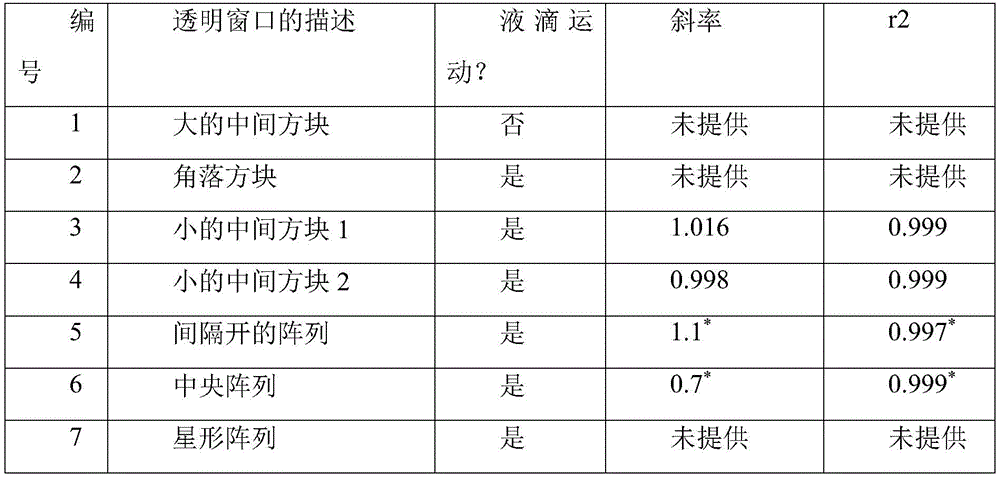
带下划线的文本-用于AIMS的选定的吸光度窗口;*这些值是从标准曲线的线性部分获得的。 Underlined text - selected absorbance window for AIMS; * these values were obtained from the linear portion of the standard curve.
使用数字微流体用于自动诱导的优点是用于吸光度测量的垂直路径长度可以容易地被调整。理想地,路径长度越大,在低吸光度下测量越灵敏(由于朗伯定律)。在本文中,测试了三种不同的间隙高度,并且测量了使用AIMS进行OD测量的检测极限。最初,尝试在装置中的顶板与底板之间的<140∝m的小间隔物厚度,因为其是通常用于DMF装置上的生物测定的间隙高度的范围。(参见Shih,S.C.C.,Goyal,G.,Kim,P.W.,Koutsoubelis,N.,Keasling,J.D.,Adams,P.D.,Hillson,N.J.,and Singh,A.K.(2015)A versatilemicrofluidic device for automating synthetic biology,ACS Synth.Biol.10,1151-1164;Shih,S.C.C.,Gach,P.C.,Sustarich,J.,Simmons,B.A.,Adams,P.D.,Singh,S.,andSingh,A.K.(2015)A droplet-to-digital(D2D)microfluidic device for single cellassays,Lab Chip 15,225-236)。然而,在这些较低的间隙高度下,可能无法实现灵敏且可重复的OD测量。这导致尝试更大的高度(210∝m、280∝m和350∝m)以通过测量仅含有培养基的液滴的OD来确定检测极限。如所预期的,350∝m的间隙高度给出了最低检测极限,0.029OD单位(图31C)。然而,在装置上的这些间隙高度处通常观察到的问题是分配的可靠性。事实上,对于带有细胞的培养基的液滴分配和从储器中的重复分配几乎是不可能的。也尝试了如其他人所提出的增加电压以改善液滴运动和分配((参见Chen,T.,Dong,C.,Gao,J.,Jia,Y.,Mak,P.I.,Vai,M.I.,and Martins,R.P.2014)。Natural discharge afterpulse and cooperative electrodes to enhance droplet velocity in digitalmicrofluidics,AIP Adv.4,047129;Chen,C.H.,Tsai,S.L,Chen,M.K.,and Jang,L.S.(2011)Effects of gap height,applied frequency,and fluid conductivity onminimum actuation voltage of electrowetting-on-dielectric and liquiddielectrophoresis,Sens.Actuators,B 159,321-327)),但这经常导致装置上的电解(即介电击穿)。因此,280∝m的间隔物被用于本文所报告的工作中,因为其给出了适当的检测极限,并且在液滴分配和运动方面是可重复的。The advantage of using digital microfluidics for automated induction is that the vertical path length for absorbance measurements can be easily adjusted. Ideally, the larger the path length, the more sensitive the measurement will be at low absorbance (due to Lambert's law). In this paper, three different gap heights were tested and the detection limit of OD measurements using AIMS was measured. Initially, a small spacer thickness of <140∝m between the top and bottom plates in the device was attempted, as this is the range of gap heights commonly used for bioassays on DMF devices. (See Shih, S.C.C., Goyal, G., Kim, P.W., Koutsoubelis, N., Keasling, J.D., Adams, P.D., Hillson, N.J., and Singh, A.K. (2015) A versatilemicrofluidic device for automating synthetic biology, ACS Synth. Biol. 10, 1151-1164; Shih, S.C.C., Gach, P.C., Sustarich, J., Simmons, B.A., Adams, P.D., Singh, S., and Singh, A.K. (2015) A droplet-to-digital (D2D) microfluidic device for single cell assays,
为了确保在适当的时间诱导,将AIMS上的细菌生长率与由常规方法培养的细菌生长率进行比较。如方法部分中所述,两个系统的培养条件相似。如图31D所示,细菌的生长在曲线的指数区域具有相似的趋势,但是在倍增时间上显示出显著差异,其中对于常规培养和AIMS培养分别为36.80±0.36min和72.88±2.30min(双尾配对t检验;P值=0.018)。观察到固定相的差异,并且推测微观尺度和宏观尺度系统之间的这种相的变化可能是由许多因素引起的。最可能的因素是混合效率,因为在微流体装置上存在半连续混合,而在宏观尺度上存在连续混合。混合的差异可以导致培养物中溶解的气体和营养物的差异,这可以使细菌细胞比预期更快地进入稳定期。此外,与宏观尺度(280∝m/1cm)相比,微观尺度中较短的路径长度也可以导致OD测量值的变化。虽然观察到在固定相中的差异,但诱导发生在早期指数期(约0.3-0.4OD),这在两个平台中是相似的。To ensure induction at the appropriate time, bacterial growth rates on AIMS were compared to bacterial growth rates cultured by conventional methods. Culture conditions were similar for both systems, as described in the Methods section. As shown in Figure 31D, bacterial growth had a similar trend in the exponential region of the curve, but showed significant differences in doubling times, which were 36.80 ± 0.36 min and 72.88 ± 2.30 min for conventional and AIMS cultures, respectively (two-tailed Paired t-test; P value = 0.018). Differences in stationary phases were observed, and it was speculated that this phase variation between microscale and macroscale systems could be caused by a number of factors. The most likely factor is mixing efficiency, as there is semi-continuous mixing on microfluidic devices and continuous mixing on the macroscopic scale. Differences in mixing can lead to differences in dissolved gases and nutrients in the culture, which can cause bacterial cells to enter stationary phase sooner than expected. Furthermore, shorter path lengths in the microscale compared to the macroscale (280∝m/1cm) can also lead to variations in OD measurements. Although differences in stationary phase were observed, induction occurred in the early exponential phase (approximately 0.3-0.4 OD), which was similar in both platforms.
诱导剂浓度优化-监测基因表达Inducer Concentration Optimization - Monitoring Gene Expression
参考图32A,示出了使用AIMS和在宏观尺度培养物中IPTG的剂量-响应曲线的比较。误差棒代表一式三份的±1标准偏差。参考图32B,示出了通过荧光扫描在诱导的和非诱导的培养物的液滴上检测到的RFP信号。用582nm的激发波长和612nm的发射波长测量荧光(参考用于特定孔板设置的方法)。参考图32C,示出了显示装置上含有用IPTG诱发的液滴的五个区域的图片。放大的插图示出了在测定区域中含有表达RFP的细胞的液滴。Referring to Figure 32A, a comparison of dose-response curves using AIMS and IPTG in macroscale cultures is shown. Error bars represent ±1 standard deviation of triplicates. Referring to Figure 32B, RFP signals detected by fluorescence scanning on droplets of induced and non-induced cultures are shown. Fluorescence was measured with an excitation wavelength of 582 nm and an emission wavelength of 612 nm (refer to the method for the specific well plate setup). Referring to Figure 32C, a picture of five regions on a display device containing droplets induced with IPTG is shown. The enlarged inset shows a droplet containing RFP-expressing cells in the assay area.
AIMS的关键优势是在同一装置上直接诱导后分析蛋白表达的潜力。为了用AIMS来说明这一点,用在T7启动子下游携带红色荧光蛋白(RFP)基因的IPTG诱导型表达载体来测试该系统。细菌细胞被培养直到OD 0.4,并且使用不同的IPTG浓度(在芯片上生成)诱导,以评估用于诱导的最佳浓度(图32A)。如所示出的,宏观尺度和微流体装置中的剂量-响应曲线均遵循S形曲线轮廓(即希尔函数),其中在高于200∝M的IPTG浓度下在四小时后具有最高的蛋白产量。在较低的IPTG浓度下(通常<30∝M),蛋白产量不变(即基础水平),这在这些浓度下是可预期的。观察到曲线形状的一些差异,特别是其陡度的差异。考虑到两种系统之间的显著差异(在体积、电场致动、光学检测器、样品的混合效率等方面),这不是令人惊讶的。然而,这可以通过整合‘灵敏度调谐器’(参见Cambridge,U.o.(2009)InternationalGenetically Engineered Machine(iGEM))或将多个蛋白结合结构61或转录级联系统(参见Hooshangi,S.,Thiberge,S.,and Weiss,R.(2005)Ultrasensitivity and noisepropagation in a synthetic transcriptional cascade,Proc.Natl.Acad.Sci.U.S.A.102,3581-3586)添加到细胞中来改善,所述细胞将调节有效的结合协同性,并改善多个转录因子与相同启动子的协同结合,用于转录调节的基因表达。尽管有这些差异,但该系统能够自动诱导和监测基因表达,这可以被扩展到其他类型的诱导测定(参见表达优化部分)。A key advantage of AIMS is the potential to analyze protein expression after direct induction on the same device. To illustrate this with AIMS, the system was tested with an IPTG-inducible expression vector carrying the red fluorescent protein (RFP) gene downstream of the T7 promoter. Bacterial cells were cultured until OD 0.4 and induced using different IPTG concentrations (generated on-chip) to assess the optimal concentration for induction (Figure 32A). As shown, the dose-response curves in both macroscale and microfluidic devices follow a sigmoid profile (ie, a Hill function), with the highest protein after four hours at IPTG concentrations above 200∝M Yield. At lower IPTG concentrations (usually <30∝M), protein production was unchanged (ie, basal levels), as would be expected at these concentrations. Some differences in the shape of the curves are observed, especially in their steepness. This is not surprising given the significant differences between the two systems (in terms of volume, electric field actuation, optical detector, mixing efficiency of the sample, etc.). However, this can be achieved by integrating 'sensitivity tuners' (see Cambridge, Uo (2009) International Genetically Engineered Machine (iGEM)) or by combining multiple protein binding structures61 or transcriptional cascades (see Hooshangi , S., Thiberge, S. , and Weiss, R. (2005) Ultrasensitivity and noisepropagation in a synthetic transcriptional cascade, Proc. Natl. Acad. Sci. USA 102, 3581-3586) was improved by adding to cells that would modulate efficient binding cooperativity, And improve the cooperative binding of multiple transcription factors to the same promoter for transcriptionally regulated gene expression. Despite these differences, the system is capable of automatic induction and monitoring of gene expression, which can be extended to other types of induction assays (see Expression Optimization section).
由于荧光被用作蛋白生产的读数,光学板读取器被用于分析,因为该装置可以容易地与离线检测器集成。(参见Au,S.H.,Shih,S.C.C.,and Wheeler,A.R.(2011)Integrated microbioreactor for culture and analysis of bacteria,algae andyeast,Biomed.Microdevices 13,41-50;Barbulovic-Nad,I.,Au,S.H.,and Wheeler,A.R.(2010)A microfluidic platform for complete mammalian cell culture,Lab Chip10,1536-1542)。使用这些光学检测器,只能检测到液滴区域,并且因此不存在其他荧光信号干扰所需信号的风险。此外,该读数是该过程的最后一步,并且因此只需要将装置转移到板读取器中,即不需要额外的移液步骤或流体处理步骤。如图32B所示,可以通过孔板软件来选择液滴,并且可以清楚地区分液滴及其周围区域以及低荧光(无IPTG)和高度荧光液滴(200∝M的IPTG)之间的差异。这表明该装置可与外部检测器兼容,并且可以被用作终点荧光检测的替代方案。将来,建议集成在线荧光检测器(参见Sista,R.,Hua,Z.,Thwar,P.,Sudarsan,A.,Srinivasan,V.,Eckhardt,A.,Pollack,M.,and Pamula,V.(2008)Development of a digital microfluidic platform for point of care testing,LabChip 8,2091-2104)或需要诱导和使用荧光的吸光度作为读数的其他类型的测定的变体-例如遗传元件筛选(参见Song,Y.,Nikoloff,J.M.,Fu,G.,Chen,J.,Li,Q.,Xie,N.,Zheng,P.,Sun,J.,and Zhang,D.(2016)Promoter Screening from Bacillus subtilis inVarious Conditions Hunting for Synthetic Biology and Industrial Applications,PLoS One 11,e0158447;Stanton,B.C.,Nielsen,A.A.,Tamsir,A.,Clancy,K.,Peterson,T.,and Voigt,C.A.(2014)Genomic mining of prokaryotic repressors fororthogonal logic gates,Nat.Chem.Biol.10,99-105)和/或调节基因表达(参见Markley,A.L.,Begemann,M.B.,Clarke,R.E.,Gordon,G.C.,and Pfleger,B.F.(2015)Syntheticbiology toolbox for controlling gene expression in the cyanobacteriumSynechococcus sp.strain PCC 7002,ACS Synth.Biol.4,595-603;Ang,J.,Harris,E.,Hussey,B.J.,Kil,R.,and McMillen,D.R.(2013)Tuning response curves forsynthetic biology,ACS Synth.Biol.2,547-567)。Since fluorescence is used as a readout for protein production, an optical plate reader is used for the analysis, as the device can be easily integrated with off-line detectors. (See Au, S.H., Shih, S.C.C., and Wheeler, A.R. (2011) Integrated microbioreactor for culture and analysis of bacteria, algae andyeast, Biomed. Microdevices 13, 41-50; Barbulovic-Nad, I., Au, S.H., and Wheeler, A.R. (2010) A microfluidic platform for complete mammalian cell culture,
如图32C所示,该方法是以5重形式进行的,但是在将来,建议有可能将AIMS扩展到甚至更高的多路复用水平,特别是利用‘混合’微流体技术的报道,这可以增加1000个样品的通量和分析。(参见Shih,S.C.C.,Gach,P.C.,Sustarich,J.,Simmons,B.A.,Adams,P.D.,Singh,S.,and Singh,A.K.(2015)A droplet-to-digital(D2D)microfluidic device forsingle cell assays,Lab Chip 15,225-236;Heinemann,J.,Deng,K.,Shih,S.C.C.,Gao,J.,Adams,P.D.,Singh,A.K.,and Northen,T.R.(2017)On-chip integration of dropletmicrofluidics and nanostructure-initiator mass spectrometry for enzymescreening,Lab Chip 17,323-331)。此外,本文报告的方法与小规模方法相比使得细菌培养体积减少了10,000倍(在微观尺度中的15μL/批量尺度中的150mL),并且测定体积减少了至少40倍(与在96孔板中的200μL相比,在装置上为5μL)。该系统还能够在没有干预的情况下实现自动诱导和基因表达分析。建议本文描述的新方法对于涉及珍贵和昂贵试剂的应用以及需要多种稀释或条件的诱导测定可能是特别有用的(对于成本、人工干预和时间的详细比较,参见下表6)。As shown in Figure 32C, the method was performed in a 5-plex format, but in the future, it is suggested that it may be possible to extend AIMS to even higher levels of multiplexing, especially reports utilizing 'hybrid' microfluidics, which The throughput and analysis of up to 1000 samples can be increased. (See Shih, S.C.C., Gach, P.C., Sustarich, J., Simmons, B.A., Adams, P.D., Singh, S., and Singh, A.K. (2015) A droplet-to-digital (D2D) microfluidic device for single cell assays,
表达优化-筛选活性BGL酶Expression optimization - screening for active BGL enzymes
参考图33A、33B、33C和33D,示出了发现高度活性的BGL的表达优化(单点和多点)测定。参考图33A,示出了显示通过β-葡萄糖苷酶(BGL)将4-甲基伞形酮基β-D-吡喃葡萄糖苷(MUG)酶促水解为4-甲基伞形酮(MUF)的示意图。参考图33B,示出了在2mM的MUG的存在下,通过60min的荧光强度(ex=369nm和em=449nm)测量的三种不同的BGL的活性。参考图33C,示出了三种酶相对于最低酶(BGL1)的活性速率的比较。参考图33D,示出了在AIMS上在6h内BGL3的诱导概况。对于(B-D),误差棒代表一式三份的±1标准偏差。Referring to Figures 33A, 33B, 33C and 33D, expression optimization (single and multi-point) assays of BGLs found to be highly active are shown. Referring to Figure 33A, there is shown the enzymatic hydrolysis of 4-methylumbelliferone β-D-glucopyranoside (MUG) to 4-methylumbelliferone (MUF) by β-glucosidase (BGL). ) schematic diagram. Referring to Figure 33B, the activities of three different BGLs measured by fluorescence intensities ( ex = 369 nm and em = 449 nm) at 60 min in the presence of 2 mM of MUG are shown. Referring to Figure 33C, a comparison of the activity rates of the three enzymes relative to the lowest enzyme (BGL1) is shown. Referring to Figure 33D, the induction profile of BGL3 over 6 h on AIMS is shown. For (BD), error bars represent ±1 standard deviation of triplicates.
鉴于AIMS的多功能性,它被设计成分析更复杂的生物系统的蛋白表达。人们对发现酶来分解大的糖聚合物(由己糖和戊糖组成)的兴趣激增,这些糖聚合物可以被发酵成生物燃料,作为汽油、柴油和喷气燃料的潜在替代品。(参见Steen,E.J.,Kang,Y.,Bokinsky,G.,Hu,Z.,Schirmer,A.,McClure,A.,Del Cardayre,S.B.,and Keasling,J.D.(2010)Microbial production of fatty-acid-derived fuels and chemicals from plantbiomass,Nature 463,559-562;Peralta-Yahya,P.P.,and Keasling,J.D.(2010)Advancedbiofuel production in microbes,Biotechnol.J.5,147-162;Nakayama,S.,Kiyoshi,K.,Kadokura,T.,and Nakazato,A.(2011)Butanol production from crystallinecellulose by cocultured Clostridium thermocellum and Clostridiumsaccharoperbutylacetonicum N1-4,Appl.Environ.Microbiol.77,6470-6475)。一组酶(β-葡萄糖苷酶(BGL))由于其水解纤维素以产生葡萄糖的能力,近年来已经引起了相当大的关注。通常,BGL活性首先使用人工基底诸如4-甲基伞形酮基β-D-吡喃葡萄糖苷(MUG)来测量。因此,AIMS被用于研究基于人工基底MUG的三种BGL的催化活性(对于化学方案,参见图33A)。首先,三个试剂储器专用于分配多种试剂(基底、裂解溶液和终止溶液),并且32个致动电极用于移动和混合试剂与诱导的培养物,且五个测定区域用于测量装置上的酶活性。在37℃诱导四小时后,将细胞裂解并与含有荧光基底MUG的液滴混合。在本文中,随时间变化的荧光被用作酶活性的读数。对于未来的工作,建议依赖荧光的许多其他可能的探针或蛋白与AIMS兼容。Given the versatility of AIMS, it was designed to analyze protein expression in more complex biological systems. There has been a surge of interest in discovering enzymes to break down large sugar polymers (composed of hexose and pentose sugars) that can be fermented into biofuels as potential replacements for gasoline, diesel and jet fuel. (See Steen, E.J., Kang, Y., Bokinsky, G., Hu, Z., Schirmer, A., McClure, A., Del Cardayre, S.B., and Keasling, J.D. (2010) Microbial production of fatty-acid- derived fuels and chemicals from plantbiomass, Nature 463, 559-562; Peralta-Yahya, P.P., and Keasling, J.D. (2010) Advanced biofuel production in microbes, Biotechnol. J. 5, 147-162; Nakayama, S., Kiyoshi, K., Kadokura, T., and Nakazato, A. (2011) Butanol production from crystallinecellulose by cocultured Clostridium thermocellum and Clostridium saccharoperbutylacetonicum N1-4, Appl. Environ. Microbiol. 77, 6470-6475). A group of enzymes, beta-glucosidases (BGLs), have attracted considerable attention in recent years due to their ability to hydrolyze cellulose to produce glucose. Typically, BGL activity is first measured using artificial substrates such as 4-methylumbelliferyl β-D-glucopyranoside (MUG). Therefore, AIMS was used to study the catalytic activity of three BGLs based on the artificial substrate MUG (for chemical scheme, see Figure 33A). First, three reagent reservoirs are dedicated to dispensing multiple reagents (substrate, lysis solution, and stop solution), and 32 actuation electrodes are used to move and mix reagents and induced cultures, and five assay areas are used to measure the device on the enzymatic activity. After four hours of induction at 37°C, cells were lysed and mixed with droplets containing the fluorescent substrate MUG. In this paper, time-varying fluorescence is used as a readout of enzymatic activity. For future work, many other possible probes or proteins that rely on fluorescence are suggested to be compatible with AIMS.
使用台式扫描孔板读取器在装置上直接测量酶促测定的荧光强度,并且酶活性曲线在图33B中示出。如所预期的,随着时间的推移,对于三种不同的BGL酶测得的荧光增加,而在阴性对照(即不含任何BGL的‘空’质粒)中观察到很少的活性或没有活性。具体来说,在单点诱导测定中,通过荧光测量的活性的速率对于BGL1和BGL2几乎相同,但是对于BGL3显著更高。事实上,与其他两个BGL相比,对于BGL3,这一速率高至少六倍(图33C)。为了进一步优化BGL3的活性,进行了多点诱导测定以确定BGL3表达的最佳诱导后孵育期(即裂解前)。The fluorescence intensity of the enzymatic assay was measured directly on the device using a benchtop scanning well plate reader, and the enzymatic activity curve is shown in Figure 33B. As expected, the measured fluorescence increased over time for the three different BGL enzymes, while little or no activity was observed in the negative control (ie, the 'empty' plasmid without any BGL) . Specifically, in the single-point induction assay, the rate of activity measured by fluorescence was almost the same for BGL1 and BGL2, but significantly higher for BGL3. In fact, this rate was at least six-fold higher for BGL3 compared to the other two BGLs (Figure 33C). To further optimize the activity of BGL3, a multi-point induction assay was performed to determine the optimal post-induction incubation period (ie, before lysis) for BGL3 expression.
如图33C所示,与立即诱导和裂解(0h)相比,在诱导和孵育6h后,BGL3显示出最高表达(至少高三倍)。这是预期的,因为诱导后孵育时间的效果会影响重组蛋白在大肠杆菌中的整体折叠、积累和生产率,并且因此较长的孵育时间(>1h)是更有利的。(参见Shin,C.S.,Hong,M.S.,Bae,C.S.,and Lee,J.(1997)Enhanced production of human mini-proinsulin in fed-batch cultures at high cell density of Escherichia coliBL21(DE3)[pET-3aT2M2],Biotechnol.Prog.13,249-257)。关于BGL3的高活性(与其他测试的BGL相比),目前还不太清楚,然而,一些小组已经假设,较高的盐浓度(以及在中性pH 7.0时)将诱导较高的酶活性和耐热生物如海洋红嗜热盐菌的更快生长。(参见Gladden,J.M.,Park,J.I.,Bergmann,J.,Reyes-Ortiz,V.,D'Haeseleer,P.,Quirino,B.F.,Sale,K.L,Simmons,B.A.,and Singer,S.W.(2014)Discovery and characterization of ionicliquid-tolerant thermophilic cellulases from a switchgrass-adapted microbialcommunity,Biotechnol.Biofuels 7,15;Bjornsdottir,S.H.,Blondal,T.,Hreggvidsson,G.O.,Eggertsson,G.,Petursdottir,S.,Hjorleifsdottir,S.,Thorbjarnardottir,S.H.,and Kristjansson,J.K.(2006)Rhodothermus marinus:physiology and molecularbiology,Extremophiles 10,1-16)。此外,这些生物体通常生活在恶劣的环境中,并且需要不断保持其高水平的热稳定性和酶活性。因此,这些酶可以在标准环境中(即在室温、恒定的pH等)保持其功能和活性并不令人惊讶。无论如何,这些结果证实了AIMS能够使诱导自动化并发现用于生物质水解的可能候选物的酶。建议本文描述的系统可用于测试各种酶,以鉴定更多的用于生物燃料生产和合成生物学应用的候选物。As shown in Figure 33C, BGL3 showed the highest expression (at least three-fold higher) after induction and incubation for 6 h compared to immediate induction and lysis (Oh). This is expected as the effect of post-induction incubation time affects the overall folding, accumulation and productivity of recombinant proteins in E. coli, and therefore longer incubation times (>1 h) are more favorable. (See Shin, C.S., Hong, M.S., Bae, C.S., and Lee, J. (1997) Enhanced production of human mini-proinsulin in fed-batch cultures at high cell density of Escherichia coliBL21(DE3)[pET-3aT2M2], Biotechnol. Prog. 13, 249-257). Little is known about the high activity of BGL3 (compared to other BGLs tested), however, some groups have hypothesized that higher salt concentrations (and at neutral pH 7.0) will induce higher enzymatic activity and Faster growth of heat-resistant organisms such as Rhodotortis marine. (See Gladden, J.M., Park, J.I., Bergmann, J., Reyes-Ortiz, V., D'Haeseleer, P., Quirino, B.F., Sale, K.L, Simmons, B.A., and Singer, S.W. (2014) Discovery and Characterization of ionicliquid-tolerant thermophilic cellulases from a switchgrass-adapted microbial community, Biotechnol.
第一个自动化的诱导微流体平台被提出以使用数字微流体来监测用于合成生物学应用的基因表达。AIMS能够实现1)装置上的OD读数,2)在线细菌培养和液滴形式的诱导,以及3)酶表达和活性的分析。该系统的特征在于优化用于细菌细胞培养的OD测量和生长条件。AIMS具有0.035OD单位的检测极限,并且能够在五小时内在没有人工干预的情况下在微观尺度上监测细菌生长。此外,使用不同的IPTG浓度来测试在pET表达载体中Rfgene的诱导,以生成剂量-响应曲线,并将其与宏观尺度实验进行比较,并发现其超灵敏度的差异。最后,在自动的诱导后,使用AIMS以直接在装置上测量三种BGL酶的活性,并在不同诱导后孵育条件下优化最高活性酶以优化终点活性。这些结果表明使用数字微流体来自动化诱导和分析酶活性的巨大潜力。预计朝向在线荧光和吸光度检测的进一步发展将使这种技术成为一种用于监测和分析用于合成生物学应用的蛋白表达的有吸引力的解决方案。The first automated inducible microfluidics platform was proposed to use digital microfluidics to monitor gene expression for synthetic biology applications. AIMS enables 1) on-device OD reading, 2) on-line bacterial culture and induction in droplet format, and 3) analysis of enzyme expression and activity. This system is characterized by optimized OD measurement and growth conditions for bacterial cell culture. AIMS has a detection limit of 0.035 OD units and is capable of monitoring bacterial growth at the microscopic scale within five hours without human intervention. Furthermore, induction of Rfgene in pET expression vectors was tested using different IPTG concentrations to generate dose-response curves, which were compared to macroscale experiments and differences in their supersensitivity were found. Finally, after automated induction, AIMS was used to measure the activity of the three BGL enzymes directly on the device, and the highest active enzyme was optimized under different post-induction incubation conditions to optimize end-point activity. These results demonstrate the great potential of using digital microfluidics to automate the induction and analysis of enzymatic activity. Further developments towards online fluorescence and absorbance detection are expected to make this technique an attractive solution for monitoring and analyzing protein expression for synthetic biology applications.
补充信息在下面示出并且包含:3D外壳的制造程序的描述,带有显示AIMS的多层的图形、宏观尺度和AIMS之间的比较表(表6),以及用于自动化系统的电子部件的材料列表清单。Supplementary information is shown below and contains: a description of the fabrication procedure for the 3D enclosure, with graphics showing multiple layers of AIMS, a comparison table between macro-scale and AIMS (Table 6), and a description of the electronic components for the automation system List of materials.
3D外壳的制造程序的描述,带有显示AIMS的多层的图形:图2示出了AIMS的3D外壳的制造。其由四层组成(从上到下):第1层(以绿色显示)用于固定LED,第2层(以蓝色显示)用于支撑将向装置施加电位的弹簧针板,第3层(以橙色显示)用于将装置支撑在适当的位置,以及第4层(以红色显示)用于将传感器定位在装置正下方。Description of the fabrication procedure of the 3D enclosure, with a graphic showing multiple layers of the AIMS: Figure 2 shows the fabrication of the 3D enclosure of the AIMS. It consists of four layers (from top to bottom): Layer 1 (shown in green) holds the LEDs, Layer 2 (shown in blue) holds the pogo pin board that will apply a potential to the device, and Layer 3 (shown in orange) is used to hold the unit in place, and layer 4 (shown in red) is used to position the sensor directly below the unit.
用于自动化系统的电子部件的材料列表清单Bill of materials for electronic components for automation systems

对于成本,仅显示试剂的成本(左)以及试剂和装置的成本(右)。For cost, only the cost of reagents (left) and the cost of reagents and devices (right) are shown.
5种不同条件的细节:Details for 5 different conditions:
在装置上:On the device:
20μL_LB价格为$7.5/L-->$0.0001520μL_LB price is $7.5/L-->$0.00015
20μL 1mM IPTG价格为$32/g-->4.8x106 g-->$0.00015420μL 1mM IPTG is $32/g-->4.8x10 6 g-->$0.000154
5x6=30μL 2mM MUG价格为$400/g-->20x106 g-->$0,0085x6=30μL 2mM MUG at $400/g-->20x10 6 g-->$0,008
装置基底=$4.50Device Base = $4.50
总计:0.00015+0.000154+0.008=$4.51Total: 0.00015+0.000154+0.008=$4.51
宏观尺度:Macro scale:
150mL LB价格为$7.5/L-->$1.125150mL LB is $7.5/L-->$1.125
150mL 1mM IPTG价格为$32/g-->36mg-->$1.152150mL 1mM IPTG is $32/g-->36mg-->$1.152
5x200=1000μL 2mM MUG价格为$400/g-->0.677mg-->$0.275x200=1000μL 2mM MUG at $400/g-->0.677mg-->$0.27
孔板=$5.50Orifice Plate = $5.50
总计1.125+1.152+0.27+$5.50=$8.05Total 1.125+1.152+0.27+$5.50=$8.05
潜在扩大的100种不同条件的估计细节:Estimated details for potentially expanded 100 different conditions:
在装置上:On the device:
每个储器20μL*4种培养物*储器5次灌装-400μL LB价格为$7.5/L-$0.00320µL per reservoir * 4 cultures * 5 fills in reservoir - 400µL LB price is $7.5/L - $0.003
15μL以分配4个液滴-14.3mM IPTG,价格为$32/g-5.11x10-5 g-$0.0016415μL to dispense 4 droplets - 14.3mM IPTG at $32/g - 5.11x10⁻⁵ g - $0.00164
100个条件x 1.5μL=150μL,在6mM MUG价格为$400/g-3x10-4 g-$0.12对于添加剂,假设与MUG-$0.12价格类似(且相同体积)100 conditions x 1.5 μL = 150 μL at 6mM MUG at $400/g - 3x10⁻⁴ g - $0.12 for additive, assuming similar price (and same volume) as MUG - $0.12
装置基底=$4.50Device Base = $4.50
总计:0.003+0.00164+0.12+0.12+4.50=$4.74Total: 0.003+0.00164+0.12+0.12+4.50=$4.74
宏观尺度:Macro scale:
4种培养物x 150mL=600mL LB价格为$7.5/L-$4.54 cultures x 150mL = 600mL LB is $7.5/L-$4.5
每孔约6μL*100个条件=600μL 1M IPTG价格为$32/g-144mg-$4.608About 6μL*100 conditions per well = 600μL 1M IPTG price is $32/g-144mg-$4.608
100个条件x 90μL=9mL 4mM MUG价格为$400/g-12.186mg-$4.86100 conditions x 90μL = 9mL 4mM MUG at $400/g - 12.186mg - $4.86
对于添加剂,假设与MUG-$4.86价格相似(且相同体积)For additives, assume similar price (and same volume) as MUG - $4.86
孔板=$5.50Orifice Plate = $5.50
总计:4.5+4.608+4.86+4.86+5.50=$24.33Total: 4.5+4.608+4.86+4.86+5.50=$24.33
潜在放大的1000种不同条件的估计细节:Estimated details of 1000 different conditions for potential zoom in:
在装置上-将使用10个装置:On installations - 10 installations will be used:
10个装置x 400μL每个装置=4mL LB价格为$7.5/L-$0.0310 devices x 400μL each device = 4mL LB price is $7.5/L - $0.03
10个装置x 15μL每个装置=150μL 1M IPTG价格为$32/g-5.1 1x10⁻⁴ g-$0.01641000个条件x 1.5μL=1.5mL 6mM MUG价格为$400/g-->3x10-3 g-->$1.210 devices x 15µL each = 150µL 1M IPTG at $32/g - 5.1 1x10⁻⁴ g - $0.01641000 conditions x 1.5µL = 1.5mL 6mM MUG at $400/g --> 3x10⁻³ g --> $1.2
对于添加剂,假定价格(和体积)与MUG-$1.2相似For additives, price (and volume) is assumed to be similar to MUG-$1.2
装置基底=$4.50*10=$45Device base = $4.50*10 = $45
总计:0.03+0.0164+1.2+1.2+0.45=$47.45Total: 0.03+0.0164+1.2+1.2+0.45=$47.45
宏观尺度:Macro scale:
4种培养物x 150mL=600mL LB价格为$7.5/L-$4.54 cultures x 150mL = 600mL LB is $7.5/L-$4.5
每孔约6μL*1000个条件=6000μL 1M IPTG价格为$32/g-1.44g-$46.08About 6μL*1000 conditions per well = 6000μL 1M IPTG price is $32/g-1.44g-$46.08
1000个条件x每孔90μL=90mL 4mM MUG价格为$400/g-->121.86mg-->$48.6对于添加剂,假定价格(和体积)与MUG-$48.6相似1000 conditions x 90 μL per well = 90 mL 4mM MUG price is $400/g --> 121.86mg --> $48.6 For additives, assuming similar price (and volume) to MUG - $48.6
孔板=$5.50*11=$60.5Orifice plate = $5.50*11 = $60.5
总计:45+46.08+48.6+48.6+60.5=$248.78Total: 45+46.08+48.6+48.6+60.5=$248.78
下面显示了用于测试5个条件的每个步骤的总结:A summary of each step used to test the 5 conditions is shown below:
宏观尺度:Macro scale:
-为了制备起始培养物,将转化的大肠杆菌BL21(DE3)细胞在LB Amp中的过夜培养物在150mL的新鲜培养基中稀释至OD 0.1(2min;每个烧瓶1个移液步骤)。- To prepare starter cultures, an overnight culture of transformed E. coli BL21(DE3) cells in LB Amp was diluted to OD 0.1 in 150 mL of fresh medium (2 min; 1 pipetting step per flask).
-进行频繁的OD读数以监测生长,并且包含取1mL的培养物样品,并针对600nm的LB空白测量OD(10min;每个读数1个移液步骤和空白1个移液步骤)。- Perform frequent OD readings to monitor growth and involves taking 1 mL samples of the culture and measuring OD against LB blanks at 600 nm (10 min; 1 pipetting step per reading and 1 pipetting step for blanks).
-通过向培养瓶中加入150μL的1M的IPTG进行诱导(0.5min;每个烧瓶1个移液步骤)。- Induction by adding 150 μL of 1 M IPTG to the flasks (0.5 min; 1 pipetting step per flask).
-诱导的培养物在诱导后的不同时间通过从生长烧瓶中取出1mL样品被取样并检查OD(10min;每个烧瓶5个移液步骤)。- The induced cultures were sampled at various times after induction by taking 1 mL samples from the growth flasks and checked for OD (10 min; 5 pipetting steps per flask).
-通过向每个样品中加入1mL的裂解溶液并在室温下放置15min来进行裂解(2min的动手时间;每个样品1个移液步骤)。- Lysis was performed by adding 1 mL of lysis solution to each sample and leaving for 15 min at room temperature (2 min hands-on time; 1 pipetting step per sample).
-通过向96孔板的单个孔中加入50μL的裂解液和130μL的基底溶液来开始测定(10min;每个样品2个移液步骤)。通过添加20μL的终止溶液(每个样品1个移液步骤)终止。- Start the assay by adding 50 μL of lysate and 130 μL of base solution to a single well of a 96-well plate (10 min; 2 pipetting steps per sample). Stop by adding 20 μL of stop solution (1 pipetting step per sample).
AIMS:AIMS:
-准备装置-用EtOH洗涤并干燥(10min)。- Prepare the device - Wash with EtOH and dry (10 min).
-为了制备起始培养物,将转化的大肠杆菌BL21(DE3)细胞在LB Amp中的过夜培养物在含有0.05%Pluronics F-68的1mL的新鲜培养基中稀释至OD 0.1(1min;3个移液步骤)。- To prepare the starter culture, an overnight culture of transformed E. coli BL21(DE3) cells in LB Amp was diluted to OD 0.1 in 1 mL of fresh medium containing 0.05% Pluronics F-68 (1 min; 3 pipetting steps).
-在开始实验之前,将起始培养物的液滴、LB和IPTG移液到装置上(1min;3个移液步骤)。- Before starting the experiment, pipette droplets of starter culture, LB and IPTG onto the device (1 min; 3 pipetting steps).
-诱导的培养物的所有后续OD读数和取样都是自动化的并且不需要移液。(5min安装软件)。- All subsequent OD readings and sampling of induced cultures are automated and do not require pipetting. (5min to install the software).
-在测定准备过程中,将裂解溶液的液滴、基底溶液和终止溶液移液到装置上,并且致动到其储器(1min;3个移液步骤)。- During assay preparation, droplets of lysis solution, base solution and stop solution were pipetted onto the device and actuated to its reservoir (1 min; 3 pipetting steps).
-用于测定的所有混合步骤都是自动化的并且不需要手动移液步骤。- All mixing steps for the assay are automated and manual pipetting steps are not required.
对于100个条件,估计有4种不同的培养物,其在宏观尺度和微观尺度上用5种不同的IPTG浓度和5种添加剂浓度进行询问。对于宏观尺度,培养物开始于烧瓶中,并且然后被等分到96孔板中。对于芯片,培养物、用于稀释的缓冲液、裂解液、基底和终止溶液需要重新填充储器,因此需要更高数量的移液步骤。For the 100 conditions, 4 different cultures were estimated, which were interrogated with 5 different IPTG concentrations and 5 additive concentrations at the macro- and micro-scale. For the macro scale, cultures were started in flasks and then aliquoted into 96-well plates. For chips, the culture, buffer for dilution, lysate, substrate, and stop solution need to refill the reservoirs, thus requiring a higher number of pipetting steps.
对于1000个条件,估计有4种不同的培养物,其用5种不同的IPTG浓度和50种添加剂浓度进行询问。移液步骤从100个条件开始线性缩放,而手动操作时间通常被当芯片已经被线性缩放时大3倍。For 1000 conditions, 4 different cultures were estimated, which were interrogated with 5 different IPTG concentrations and 50 additive concentrations. Pipetting steps scale linearly from 100 conditions, while hands-on time is typically 3 times larger when the chip has been linearly scaled.
在表6.1和6.2中提供了有关芯片的条件的更多信息。根据一些实例,表6.1示出了芯片上的操作条件。根据其他实例,表6.1还示出了芯片上的操作条件。More information on the conditions of the chips is provided in Tables 6.1 and 6.2. According to some examples, Table 6.1 shows the operating conditions on the chip. According to other examples, Table 6.1 also shows the operating conditions on the chip.
表6.1-根据一些实例的操作条件Table 6.1 - Operating conditions according to some examples
表6.1-根据一些实例的操作条件Table 6.1 - Operating conditions according to some examples

表6.2-根据一些实例的操作条件Table 6.2 - Operating conditions according to some examples
表6.2-根据一些实例的操作条件Table 6.2 - Operating conditions according to some examples

根据另一个实例,每个步骤的总结如下所示:According to another example, the summary of each step is as follows:
宏观尺度:Macro scale:
为了制备起始培养物,将转化的大肠杆菌BL21(DE3)细胞在LB Amp中的过夜培养物在150mL的新鲜培养基中稀释至OD 0.1(2min;每个烧瓶1个移液步骤)。To prepare starter cultures, overnight cultures of transformed E. coli BL21(DE3) cells in LB Amp were diluted to OD 0.1 in 150 mL of fresh medium (2 min; 1 pipetting step per flask).
经常读取OD读数以监测生长,并且包含取1mL的培养物样品,并针对600nm的LB空白测量OD(10min;每个读数1个移液步骤,并且空白1个移液步骤)。OD readings were taken frequently to monitor growth and consisted of taking a 1 mL sample of the culture and measuring OD against a LB blank at 600 nm (10 min; 1 pipetting step per reading and 1 pipetting step for blank).
通过向培养瓶中加入150mL的1mM的IPTG来进行诱导(0.5min;每个烧瓶1个移液步骤)。Induction was performed by adding 150 mL of 1 mM IPTG to the flasks (0.5 min; 1 pipetting step per flask).
诱导的培养物在诱导后的不同时间通过从生长烧瓶中取出1mL样品被取样并检查OD(10min;每个烧瓶5个移液步骤)。Induced cultures were sampled at various times after induction by removing 1 mL samples from the growth flasks and checked for OD (10 min; 5 pipetting steps per flask).
通过向每个样品中加入1mL的裂解溶液并在室温下放置15min来进行裂解(2min的动手时间;每个样品1个移液步骤)。Lysis was performed by adding 1 mL of lysis solution to each sample and leaving for 15 min at room temperature (2 min hands-on time; 1 pipetting step per sample).
通过向96孔板的单个孔中加入50μL的裂解液和100μL的基底溶液来开始测定(10min;每个样品2个移液步骤)。通过添加50μL的终止溶液(每个样品1个移液步骤)终止。The assay was started by adding 50 μL of lysate and 100 μL of base solution to a single well of a 96-well plate (10 min; 2 pipetting steps per sample). Stop by adding 50 μL of stop solution (1 pipetting step per sample).
AIMS:AIMS:
准备装置-用EtOH洗涤并干燥(10min)。Prepare device - wash with EtOH and dry (10 min).
为了制备起始培养物,将转化的大肠杆菌BL21(DE3)细胞在LB Amp中的过夜培养物在含有0.05%Pluronics F-68的1mL的新鲜培养基中稀释至OD 0.1(1min;3个移液步骤)。在开始实验之前,将起始培养物的液滴、LB和IPTG移液到装置上(1min;3个移液步骤)。To prepare starter cultures, overnight cultures of transformed E. coli BL21(DE3) cells in LB Amp were diluted to OD 0.1 in 1 mL of fresh medium containing 0.05% Pluronics F-68 (1 min; 3 pipettes) liquid step). Drops of starter cultures, LB and IPTG were pipetted onto the device (1 min; 3 pipetting steps) before starting the experiment.
诱导的培养物的所有后续OD读数和取样都是自动化的并且不需要移液。(5min安装软件)All subsequent OD readings and sampling of induced cultures were automated and did not require pipetting. (5min to install the software)
在测定准备过程中,将裂解溶液的液滴、基底溶液和终止溶液移液到装置上,并致动到其储器(1min;3个移液步骤)。During assay preparation, droplets of lysis solution, base solution and stop solution were pipetted onto the device and actuated to its reservoir (1 min; 3 pipetting steps).
用于测定的所有混合步骤都是自动化的并且不需要手动移液步骤。All mixing steps for the assay are automated and manual pipetting steps are not required.
用于破译癌症基因的自动化微流体基因编辑平台Automated microfluidic gene editing platform for deciphering cancer genes
基因编辑技术(诸如RNA导向的核酸内切酶系统)对于表型筛选正在变得越来越受欢迎。这样的筛选通常以阵列或集合的形式进行。近年来,对寻找用于进行这些基因编辑测定的新的技术方法已经存在极大的兴趣。据报道,这是第一个可以使得哺乳动物细胞中排列的基因编辑自动化的数字微流体方法。具体而言,该方法可用于培养肺癌细胞长达六天,以及实施自动化的基因转染和敲除程序。此外,在这些程序期间,还设计并实施了用于分析荧光标记的细胞的标准化成像管道。进行了用于询问MAPK/ERK途径的基因编辑测定,以显示该平台的效用,并确定敲除肺癌细胞中RAF1基因的效果。除了基因敲除,细胞还用抑制剂索拉非尼甲苯磺酸酯处理,以确定酶抑制的效果。与仅用抑制剂处理的细胞相比,装置上酶抑制和导向靶向的结合导致实现半抑制效应(IC50)的较低药物浓度,证实肺癌细胞在装置上被成功地编辑。建议该系统可用于其他类型的基因编辑测定以及与个性化医学相关的应用。Gene editing techniques, such as RNA-guided endonuclease systems, are becoming increasingly popular for phenotypic screening. Such screening is usually performed in the form of an array or collection. In recent years, there has been great interest in finding new technical methods for performing these gene editing assays. This is reported to be the first digital microfluidic method to automate gene editing arranged in mammalian cells. Specifically, the method can be used to culture lung cancer cells for up to six days, as well as to implement automated gene transfection and knockout procedures. In addition, during these procedures, a standardized imaging pipeline for analyzing fluorescently labeled cells was designed and implemented. Gene editing assays to interrogate the MAPK/ERK pathway were performed to show the utility of this platform and to determine the effect of knocking out the RAF1 gene in lung cancer cells. In addition to the gene knockout, cells were treated with the inhibitor sorafenib tosylate to determine the effect of enzyme inhibition. The combination of enzymatic inhibition and targeted targeting on the device resulted in lower drug concentrations achieving a semi-inhibitory effect (IC50) compared to cells treated with the inhibitor alone, confirming that lung cancer cells were successfully edited on the device. It is suggested that the system can be used for other types of gene editing assays and applications related to personalized medicine.
最近在癌症表征方面的努力正转向更加个性化的方法,而不是基于化学灵敏度实验的分级分类。(参见A.A.Friedman,A.Letai,D.E.Fisher and K.T.Flaherty,Nat RevCancer,2015,15,747-756)。癌症是一种异质性疾病,其基因组成差异很大,并且依赖于不同的生存途径,这导致对不同抗癌药剂的广泛潜在反应。(参见J.Barretina,G.Caponigro,N.Stransky,K.Venkatesan,A.A.Margolin,S.Kim,C.J.Wilson,J.Lehar,G.V.Kryukov,D.Sonkin,A.Reddy,M.Liu,L.Murray,M.F.Berger,J.E.Monahan,P.Morais,J.Meltzer,A.Korejwa,J.Jane-Valbuena,F.A.Mapa,J.Thibault,E.Bric-Furlong,P.Raman,A.Shipway,I.H.Engels,J.Cheng,G.K.Yu,J.Yu,P.Aspesi,Jr.,M.de Silva,K.Jagtap,M.D.Jones,L.Wang,C.Hatton,E.Palescandolo,S.Gupta,S.Mahan,C.Sougnez,R.C.Onofrio,T.Liefeld,L.MacConaill,W.Winckler,M.Reich,N.Li,J.P.Mesirov,S.B.Gabriel,G.Getz,K.Ardlie,V.Chan,V.E.Myer,B.L.Weber,J.Porter,M.Warmuth,P.Finan,J.L.Harris,M.Meyerson,T.R.Golub,M.P.Morrissey,W.R.Sellers,R.Schlegeland L.A.Garraway,Nature,2012,483,603-607;M.J.Garnett,E.J.Edelman,S.J.Heidorn,C.D.Greenman,A.Dastur,K.W.Lau,P.Greninger,I.R.Thompson,X.Luo,J.Soares,Q.Liu,F.lorio,D.Surdez,L.Chen,R.J.Milano,G.R.Bignell,A.T.Tam,H.Davies,J.A.Stevenson,S.Barthorpe,S.R.Lutz,F.Kogera,K.Lawrence,A.McLaren-Douglas,X.Mitropoulos,T.Mironenko,H.Thi,L.Richardson,W.Zhou,F.Jewitt,T.Zhang,P.O'Brien,J.L.Boisvert,S.Price,W.Hur,W.Yang,X.Deng,A.Butler,H.G.Choi,J.W.Chang,J.Baselga,I.Stamenkovic,J.A.Engelman,S.V.Sharma,O.Delattre,J.Saez-Rodriguez,N.S.Gray,J.Settleman,P.A.Futreal,D.A.Haber,M.R.Stratton,S.Ramaswamy,U.McDermott and C.H.Benes,Nature,2012,483,570-575)。一种已经迅速引起兴趣的方法是使用基于CRISPR的筛选来系统地鉴定哺乳动物细胞的存活和增殖所需的基因。(参见J.Barretina,G.Caponigro,N.Stransky,K.Venkatesan,A.A.Margolin,S.Kim,C.J.Wilson,J.Lehar,G.V.Kryukov,D.Sonkin,A.Reddy,M.Liu,L.Murray,M.F.Berger,J.E.Monahan,P.Morais,J.Meltzer,A.Korejwa,J.Jane-Valbuena,F.A.Mapa,J.Thibault,E.Bric-Furlong,P.Raman,A.Shipway,I.H.Engels,J.Cheng,G.K.Yu,J.Yu,P.Aspesi,Jr.,M.de Silva,K.Jagtap,M.D.Jones,L.Wang,C.Hatton,E.Palescandolo,S.Gupta,S.Mahan,C.Sougnez,R.C.Onofrio,T.Liefeld,L.MacConaill,W.Winckler,M.Reich,N.Li,J.P.Mesirov,S.B.Gabriel,G.Getz,K.Ardlie,V.Chan,V.E.Myer,B.L.Weber,J.Porter,M.Warmuth,P.Finan,J.L.Harris,M.Meyerson,T.R.Golub,M.P.Morrissey,W.R.Sellers,R.Schlegel and L.A.Garraway,Nature,2012,483,603-607;M.J.Garnett,E.J.Edelman,S.J.Heidorn,C.D.Greenman,A.Dastur,K.W.Lau,P.Greninger,I.R.Thompson,X.Luo,J.Soares,Q.Liu,F.lorio,D.Surdez,L.Chen,R.J.Milano,G.R.Bignell,A.T.Tam,H.Davies,J.A.Stevenson,S.Barthorpe,S.R.Lutz,F.Kogera,K.Lawrence,A.McLaren-Douglas,X.Mitropoulos,T.Mironenko,H.Thi,L.Richardson,W.Zhou,F.Jewitt,T.Zhang,P.O'Brien,J.L.Boisvert,S.Price,W.Hur,W.Yang,X.Deng,A.Butler,H.G.Choi,J.W.Chang,J.Baselga,I.Stamenkovic,J.A.Engelman,S.V.Sharma,O.Delattre,J.Saez-Rodriguez,N.S.Gray,J.Settleman,P.A.Futreal,D.A.Haber,M.R.Stratton,S.Ramaswamy,U.McDermott and C.H.Benes,Nature,2012,483,570-575;T.Wang,K.Birsoy,N.W.Hughes,K.M.Krupczak,Y.Post,J.J.Wei,E.S.Lander and D.M.Sabatini,Science,2015,350,1096-1 101;T.Wang,J.J.Wei,D.M.Sabatini and E.S.Lander,Science,2014,343,80-84;O.Shalem,N.E.Sanjana,E.Hartenian,X.Shi,D.A.Scott,T.Mikkelson,D.Heckl,B.L.Ebert,D.E.Root,J.G.Doench and F.Zhang,Science,2014,343,84-87;N.E.Sanjana,O.Shalem and F.Zhang,Nat Methods,2014,11,783-784;H.Koike-Yusa,Y.Li,E.P.Tan,C.Velasco-Herrera Mdel and K.Yusa,Nat Biotechnol,2014,32,267-273;L.A.Gilbert,M.A.Horlbeck,B.Adamson,J.E.Villalta,Y.Chen,E.H.Whitehead,C.Guimaraes,B.Panning,H.L.Ploegh,M.C.Bassik,L.S.Qi,M.Kampmannand J.S.Weissman,Cell,2014,159,647-661)。这样的方法能够实现基因的完全和永久失活,并且可以提供对疾病的遗传基础的深入了解,且导致新的药物靶的鉴定。5、10-13若干个小组已经报道了通过转染质粒DNA14或通过使用慢病毒或其他逆转录病毒15稳定递送到细胞中而成功编辑培养细胞中的内源基因。这些系统含有Cas9,其可以通过互补于靶DNA的单个导向RNA被靶向到基因组中的特定位置,并且可以被用于功能缺失筛选,旨在鉴定癌症治疗的潜在药物靶。(参见T.Wang,J.J.Wei,D.M.Sabatini and E.S.Lander,Science,2014,343,80-84;N.E.Sanjana,O.Shalem and F.Zhang,Nat Methods,2014,11,783-784;O.Shalem,N.E.Sanjana,E.Hartenian,X.Shi,D.A.Scott,T.S.Mikkelsen,D.Heckl,B.L.Ebert,D.E.Root,J.G.Doench and F.Zhang,Science,2014,343,84-87;L.Cong,F.A.Ran,D.Cox,S.Lin,R.Barretto,N.Habib,P.D.Hsu,X.Wu,W.Jiang,L.A.Marraffiniand F.Zhang,Science,2013,339,819-823;F.A.Ran,P.D.Hsu,J.Wright,V.Agarwala,D.A.Scott and F.Zhang,Nat Protoc,2013,8,2281-2308;P.S.Choi and M.Meyerson,NatCommun,2014,5,3728;S.Konermann,M.D.Brigham,A.E.Trevino,J.Joung,O.O.Abudayyeh,C.Barcena,P.D.Hsu,N.Habib,J.S.Gootenberg,H.Nishimasu,O.Nureki and F.Zhang,Nature,2015,517,583-U332;S.Chen,N.E.Sanjana,K.Zheng,O.Shalem,K.Lee,X.Shi,D.A.Scott,J.Song,J.Q.Pan,R.Weissleder,H.Lee,F.Zhang and P.A.Sharp,Cell,2015,160,1246-1260;R.J.Platt,S.Chen,Y.Zhou,M.J.Yim,L.Swiech,H.R.Kempton,J.E.Dahlman,O.Parnas,T.M.Eisenhaure,M.Jovanovic,D.B.Graham,S.Jhunjhunwala,M.Heidenreich,R.J.Xavier,R.Langer,D.G.Anderson,N.Hacohen,A.Regev,G.Feng,P.A.Sharp and F.Zhang,Cell,2014,159,440-455)。Recent efforts in cancer characterization are moving toward more personalized approaches, rather than graded classification based on chemical sensitivity experiments. (See AAFriedman, A. Letai, DE Fisher and KT Flaherty, Nat Rev Cancer, 2015, 15, 747-756). Cancer is a heterogeneous disease with widely differing genetic makeup and reliance on distinct survival pathways, which lead to a wide range of potential responses to different anticancer agents. (See J. Barretina, G. Caponigro, N. Stransky, K. Venkatesan, AA Margolin, S. Kim, CJ Wilson, J. Lehar, GV Kryukov, D. Sonkin, A. Reddy, M. Liu, L. Murray, MF Berger, JEMonahan,P.Morais,J.Meltzer,A.Korejwa,J.Jane-Valbuena,FAMapa,J.Thibault,E.Bric-Furlong,P.Raman,A.Shipway,IHEngels,J.Cheng,GKYu,J. Yu,P.Aspesi,Jr.,M.de Silva,K.Jagtap,MDJones,L.Wang,C.Hatton,E.Palescandolo,S.Gupta,S.Mahan,C.Sougnez,RCOnofrio,T.Liefeld, L.MacConaill,W.Winckler,M.Reich,N.Li,JPMesirov,SBGabriel,G.Getz,K.Ardlie,V.Chan,VEMyer,BLWeber,J.Porter,M.Warmuth,P.Finan,JLHarris, M. Meyerson, TR Golub, MP Morrissey, WRSellers, R. Schlegeland, LA Garraway, Nature, 2012, 483, 603-607; MJ Garnett, EJ Edelman, SJ Heidorn, CD Greenman, A. Dastur, KW Lau, P. Greninger, IRThompson, X. Luo, J. Soares , Q.Liu,F.lorio,D.Surdez,L.Chen,RJMilano,GRBignell,ATTam,H.Davies,JAStevenson,S.Barthorpe,SRLutz,F.Kogera,K.Lawrence,A.McLaren-Douglas,X .Mitropoulos,T.Mironenko,H.Thi,L.Richardson,W.Zhou,F.Jewitt,T.Zhang,P.O'Brien,JLBoisvert,S.Price,W.Hur,W.Yang,X.Deng , A. Butler, HG Choi, JW Chang, J. Baselga, I. Stamenkovic , JA Engelman, SV Sharma, O. Delattre, J. Saez-Rodriguez, NSGray, J. Settleman, PAFutreal, DA Haber, MR Stratton, S. Ramaswamy, U. McDermott and CHBenes, Nature, 2012, 483, 570-575). One approach that has rapidly gained interest is the use of CRISPR-based screens to systematically identify genes required for the survival and proliferation of mammalian cells. (See J. Barretina, G. Caponigro, N. Stransky, K. Venkatesan, AA Margolin, S. Kim, CJ Wilson, J. Lehar, GV Kryukov, D. Sonkin, A. Reddy, M. Liu, L. Murray, MF Berger, JEMonahan,P.Morais,J.Meltzer,A.Korejwa,J.Jane-Valbuena,FAMapa,J.Thibault,E.Bric-Furlong,P.Raman,A.Shipway,IHEngels,J.Cheng,GKYu,J. Yu,P.Aspesi,Jr.,M.de Silva,K.Jagtap,MDJones,L.Wang,C.Hatton,E.Palescandolo,S.Gupta,S.Mahan,C.Sougnez,RCOnofrio,T.Liefeld, L.MacConaill,W.Winckler,M.Reich,N.Li,JPMesirov,SBGabriel,G.Getz,K.Ardlie,V.Chan,VEMyer,BLWeber,J.Porter,M.Warmuth,P.Finan,JLHarris, M. Meyerson, TR Golub, MP Morrissey, WRSellers, R. Schlegel and LA Garraway, Nature, 2012, 483, 603-607; MJ Garnett, EJ Edelman, SJ Heidorn, CD Greenman, A. Dastur, KW Lau, P. Greninger, IRThompson, X. Luo, J. Soares,Q.Liu,F.lorio,D.Surdez,L.Chen,RJMilano,GRBignell,ATTam,H.Davies,JAStevenson,S.Barthorpe,SRLutz,F.Kogera,K.Lawrence,A.McLaren-Douglas, X.Mitropoulos,T.Mironenko,H.Thi,L.Richardson,W.Zhou,F.Jewitt,T.Zhang,P.O'Brien,JLBoisvert,S.Price,W.Hur,W.Yang,X. Deng, A. Butler, HG Choi, JW Chang, J. Baselga, I. Stamenkovi c, JA Engelman, SV Sharma, O. Delattre, J. Saez-Rodriguez, NSGray, J. Settleman, PAFutreal, DA Haber, MR Stratton, S. Ramaswamy, U. McDermott and CHBenes, Nature, 2012, 483, 570-575; T. Wang, K.Birsoy,NWHughes,KMKrupczak,Y.Post,JJWei,ESLander and DMSabatini,Science,2015,350,1096-1 101;T.Wang,JJWei,DMSabatini and ESLander,Science,2014,343,80-84;O .Shalem,NESanjana,E.Hartenian,X.Shi,DAScott,T.Mikkelson,D.Heckl,BLEbert,DERoot,JGDoench and F.Zhang,Science,2014,343,84-87;NESanjana,O.Shalem and F . Zhang, Nat Methods, 2014, 11, 783-784; H. Koike-Yusa, Y. Li, EPTan, C. Velasco-Herrera Mdel and K. Yusa, Nat Biotechnol, 2014, 32, 267-273; LA Gilbert, MA Horlbeck, B. Adamson, JEVillalta, Y. Chen, EH Whitehead, C. Guimaraes, B. Panning, HLPloegh, MC Bassik, LS Qi, M. Kampmann and JS Weissman, Cell, 2014, 159, 647-661). Such methods enable complete and permanent inactivation of genes and can provide insight into the genetic basis of disease and lead to the identification of new drug targets. 5, 10-13 Several groups have reported successful editing of endogenous genes in cultured cells by transfection of plasmid DNA 14 or by stable delivery into cells using lentiviruses or other retroviruses 15 . These systems contain Cas9, which can be targeted to specific locations in the genome by a single guide RNA complementary to target DNA, and can be used in loss-of-function screens aimed at identifying potential drug targets for cancer therapy. (See T. Wang, JJ Wei, DM Sabatini and ESLander, Science, 2014, 343, 80-84; NE Sanjana, O. Shalem and F. Zhang, Nat Methods, 2014, 11, 783-784; O. Shalem, NE Sanjana, E. Hartenian , X.Shi,DAScott,TSMikkelsen,D.Heckl,BLEbert,DERoot,JGDoench and F.Zhang,Science,2014,343,84-87; L.Cong,FARan,D.Cox,S.Lin,R.Barretto , N.Habib,PDHsu,X.Wu,W.Jiang,LAMarraffiniand,F.Zhang,Science,2013,339,819-823; FARan,PDHsu,J.Wright,V.Agarwala,DAScott and F.Zhang,Nat Protoc,2013 , 8, 2281-2308; PS Choi and M. Meyerson, NatCommun, 2014, 5, 3728; S. Konermann, MDBrigham, AETrevino, J.Joung, OOAbudayyeh, C.Barcena, PDHsu, N.Habib, JSGootenberg, H.Nishimasu , O.Nureki and F.Zhang,Nature,2015,517,583-U332; S.Chen,NESanjana,K.Zheng,O.Shalem,K.Lee,X.Shi,DAScott,J.Song,JQPan,R.Weissleder , H.Lee,F.Zhang and PASharp,Cell,2015,160,1246-1260;RJPlatt,S.Chen,Y.Zhou,MJYim,L.Swiech,HRKempton,JEDahlman,O.Parnas,TMEisenhaure,M.Jovanovic , DB Graham, S. Jhunjhunwala, M. Heidenreich, RJ Xavier, R. Langer, DG Anderson, N. Hacohen, A. Regev, G. Feng, PA Sharp and F. Zhang, Cell, 2014, 159, 440-455).
这些功能丧失扰动的最常见的形式是体外‘集合’筛选(参见T.Wang,J.J.Wei,D.M.Sabatini and E.S.Lander,Science,2014,343,80-84;O.Shalem,N.E.Sanjana,E.Hartenian,X.Shi,D.A.Scott,T.S.Mikkelsen,D.Heckl,B.L.Ebert,D.E.Root,J.G.Doench and F.Zhang,Science,2014,343,84-87;S.Konermann,M.D.Brigham,A.E.Trevino,J.Joung,O.O.Abudayyeh,C.Barcena,P.D.Hsu,N.Habib,J.S.Gootenberg,H.Nishimasu,O.Nureki and F.Zhang,Nature,2015,517,583-U332),其依赖于Cas9核酸酶的递送和通过转染或转导进入细胞的导向RNA(sgRNA)的‘池’。集合的文库使筛选能够同时评估在表型读数中多个基因座敲除数百至数千个单个基因的效果,诸如增殖或转移测定。尽管这样的进展为药物靶的鉴定和验证提供了新的机会,但以集合形式对结果的解释依赖于之后相对于之前的导向RNA的差异表达(如通过下一代测序所评估的),并且依赖于多个导向RNA的富集作为靶相关性的验证。(参见O.Shalem,N.E.Sanjana,E.Hartenian,X.Shi,D.A.Scott,T.S.Mikkelsen,D.Heckl,B.L.Ebert,D.E.Root,J.G.Doench and F.Zhang,Science,2014,343,84-87;S.Chen,N.E.Sanjana,K.Zheng,O.Shalem,K.Lee,X.Shi,D.A.Scott,J.Song,J.Q.Pan,R.Weissleder,H.Lee,F.Zhang and P.A.Sharp,Cell,2015,160,1246-1260)。此外,种群动态的复杂性,每个细胞与许多其他细胞竞争,可能导致偏差,这导致一些扰动相对于一些其他扰动的更高的相对丰度。‘集合’筛选的替代方案是实施‘阵列’筛选,其中细胞仅被一个已知的基因靶进行遗传干扰。(参见P.D.Hsu,D.A.Scott,J.A.Weinstein,F.A.Ran,S.Konermann,V.Agarwala,Y.Li,E.J.Fine,X.Wu,O.Shalem,T.J.Cradick,L.A.Marraffini,G.Bao and F.Zhang,Nat Biotechnol,2013,31,827-832;J.G.Doench,N.Fusi,M.Sullender,M.Hegde,E.W.Vaimberg,K.F.Donovan,I.Smith,Z.Tothova,C.Wilen,R.Orchard,H.W.Virgin,J.Listgarten and D.E.Root,NatureBiotechnology,2016,34,184-+)。这有可能使更广泛的细胞表型的使用得到研究。(参见B.Neumann,M.Held,U.Liebel,H.Erfle,P.Rogers,R.Pepperkok and J.Ellenberg,NatMethods,2006,3,385-390;J.Moffat,D.A.Grueneberg,X.Yang,S.Y.Kim,A.M.Kloepfer,G.Hinkle,B.Piqani,T.M.Eisenhaure,B.Luo,J.K.Grenier,A.E.Carpenter,S.Y.Foo,S.A.Stewart,B.R.Stockwell,N.Hacohen,W.C.Hahn,E.S.Lander,D.M.Sabatini andD.E.Root,Cell,2006,124,1283-1298;S.A.Hasson,L.A.Kane,K.Yamano,C.H.Huang,D.A.Sliter,E.Buehler,C.Wang,S.M.Heman-Ackah,T.Hessa,R.Guha,S.E.Martin andR.J.Youle,Nature,2013,504,291-295)。阵列实验的局限性是相关的成本(通常比集合的文库贵一个数量级(参见O.Shalem,N.E.Sanjana and F.Zhang,Nat Rev Genet,2015,16,299-311)),因为其需要使用自动化用于处理板的特殊设施和效率低下的工作流程,其包含劳动强度大的准备工作,以建立和生产单独的导向文库并将样品转移到其他平台用于分析。因此,自动化和集成的平台(其将培养细胞数天,能够实现哺乳动物细胞和试剂的有效处理,表达针对细胞中单个基因或基因座的基因编辑机制,并且测定细胞表型)对于这些阵列型实验将是有益的以节省总体成本并改善工作流程,其使扰动与测量之间的时间范围最小化。The most common form of these loss-of-function perturbations is an in vitro 'ensemble' screen (see T. Wang, J.J. Wei, D.M. Sabatini and E.S. Lander, Science, 2014, 343, 80-84; O. Shalem, N.E. Sanjana, E. Hartenian , X.Shi,D.A.Scott,T.S.Mikkelsen,D.Heckl,B.L.Ebert,D.E.Root,J.G.Doench and F.Zhang,Science,2014,343,84-87;S.Konermann,M.D.Brigham,A.E.Trevino,J. Joung, O.O. Abudayyeh, C. Barcena, P.D. Hsu, N. Habib, J.S. Gootenberg, H. Nishimasu, O. Nureki and F. Zhang, Nature, 2015, 517, 583-U332), which relies on the delivery and passage of Cas9 nucleases A 'pool' of guide RNAs (sgRNAs) that are transfected or transduced into cells. Pooled libraries enable screening to simultaneously assess the effect of knocking out hundreds to thousands of individual genes at multiple loci in phenotypic reads, such as proliferation or metastasis assays. While such advances provide new opportunities for the identification and validation of drug targets, interpretation of results in an aggregated format relies on later differential expression relative to prior guide RNAs (as assessed by next-generation sequencing), and relies on Enrichment for multiple guide RNAs as validation of target relevance. (See O.Shalem, N.E.Sanjana, E.Hartenian, X.Shi, D.A.Scott, T.S.Mikkelsen, D.Heckl, B.L.Ebert, D.E.Root, J.G.Doench and F.Zhang, Science, 2014, 343, 84-87; S.Chen,N.E.Sanjana,K.Zheng,O.Shalem,K.Lee,X.Shi,D.A.Scott,J.Song,J.Q.Pan,R.Weissleder,H.Lee,F.Zhang and P.A.Sharp,Cell, 2015, 160, 1246-1260). Furthermore, the complexity of population dynamics, with each cell competing with many other cells, can lead to biases that result in higher relative abundances of some perturbations relative to some others. An alternative to 'panel' screening is to perform 'array' screening, in which cells are genetically perturbed by only one known gene target. (See P.D.Hsu, D.A.Scott, J.A.Weinstein, F.A.Ran, S.Konermann, V.Agarwala, Y.Li, E.J.Fine, X.Wu, O.Shalem, T.J.Cradick, L.A.Marraffini, G.Bao and F.Zhang , Nat Biotechnol, 2013, 31, 827-832; J. G. Doench, N. Fusi, M. Sullender, M. Hegde, E. W. Vaimberg, K. F. Donovan, I. Smith, Z. Tothova, C. Wilen, R. Orchard, H.W. Virgin, J. Listgarten and D.E. Root, Nature Biotechnology, 2016, 34, 184-+). This has the potential to enable the use of a wider range of cellular phenotypes to be studied. (See B. Neumann, M. Held, U. Liebel, H. Erfle, P. Rogers, R. Pepperkok and J. Ellenberg, Nat Methods, 2006, 3, 385-390; J. Moffat, D.A. Grueneberg, X. Yang, S.Y. Kim,A.M.Kloepfer,G.Hinkle,B.Piqani,T.M.Eisenhaure,B.Luo,J.K.Grenier,A.E.Carpenter,S.Y.Foo,S.A.Stewart,B.R.Stockwell,N.Hacohen,W.C.Hahn,E.S.Lander,D.M.Sabatini andD.E Root, Cell, 2006, 124, 1283-1298; S.E. Martin and R.J. Youle, Nature, 2013, 504, 291-295). A limitation of array experiments is the associated cost (usually an order of magnitude more expensive than pooled libraries (see O. Shalem, N.E. Sanjana and F. Zhang, Nat Rev Genet, 2015, 16, 299-311)) because it requires the use of automation for Special facilities for handling plates and inefficient workflows that involve labor-intensive preparations to build and produce individual targeting libraries and transfer samples to other platforms for analysis. Thus, automated and integrated platforms that will culture cells for days, enable efficient processing of mammalian cells and reagents, express gene editing machinery targeting individual genes or loci in cells, and determine cell phenotypes are useful for these array types. Experiments would be beneficial to save overall cost and improve workflow that minimizes the time frame between perturbation and measurement.
阵列文库通常在多孔板中生成,其中每个孔含有病毒或载体,或带有靶向特定基因的导向物的试剂。用于这些类型的实验的工具(诸如自动化的机器人与流式细胞术的结合)可以提供对由单一扰动引起的复杂表型的探索。尽管其在减少细胞死亡或限制与编辑相关的非靶诱变方面具有突出的特征(参见L.A.Lonowski,Y.Narimatsu,A.Riaz,C.E.Delay,Z.Yang,F.Niola,K.Duda,E.A.Ober,H.Clausen,H.H.Wandall,S.H.Hansen,E.P.Bennett and M.Frodin,Nature Protocols,2017,12,581-603;P.D.Hsu,D.A.Scott,J.A.Weinstein,F.A.Ran,S.Konermann,V.Agarwala,Y.Q.Li,E.J.Fine,X.B.Wu,O.Shalem,T.J.Cradick,L.A.Marraffini,G.Bao and F.Zhang,Nature Biotechnology,2013,31,827-+),但这些技术具有三个关键限制。首先,可获得的液体处理技术、数据采集装置和数据存储/处理系统传统上都很昂贵,并且具有大的占地面积,其远远超出了许多实验室的预算范围。此外,编程软件包在实验室之间没有被标准化,这经常阻碍跨学科科学家和研究人员使用机器人,因为通常需要更多的时间和精力来指导机器人执行任务。第二,用于细胞培养和样品制备的液体处理器具有多种可变性来源(尤其是在nL体积下),这可能导致与基因编辑过程相关的意外扰动-例如,不同的体积可能改变细胞生长,从而导致在板的孔中细胞的数量不相等。在测量与细胞密度有关的转染和敲除效率方面,这可能会引起下游分析的可变性问题。第三,对于流式细胞术,并且尤其是对于如何分析和报告流量数据,在测定和仪器设置方面缺乏标准化。因此,这些方法可能给基因编辑的已经复杂的程序带来额外的挑战。Array libraries are typically generated in multi-well plates, where each well contains a virus or vector, or reagents with targets targeting specific genes. Tools for these types of experiments, such as automated robotics combined with flow cytometry, can provide exploration of complex phenotypes caused by a single perturbation. Although it has outstanding features in reducing cell death or limiting off-target mutagenesis associated with editing (see L.A. Lonowski, Y. Narimatsu, A. Riaz, C.E. Delay, Z. Yang, F. Niola, K. Duda, E.A. Ober,H.Clausen,H.H.Wandall,S.H.Hansen,E.P.Bennett and M.Frodin,Nature Protocols,2017,12,581-603;P.D.Hsu,D.A.Scott,J.A.Weinstein,F.A.Ran,S.Konermann,V.Agarwala,Y.Q.Li , E.J.Fine, X.B.Wu, O.Shalem, T.J.Cradick, L.A.Marraffini, G.Bao and F.Zhang, Nature Biotechnology, 2013, 31, 827-+), but these techniques have three key limitations. First, available liquid handling technology, data acquisition devices, and data storage/processing systems are traditionally expensive and have large footprints that are well beyond the budget of many laboratories. In addition, programming packages are not standardized across laboratories, which often hinders scientists and researchers from using robots across disciplines, as more time and effort is often required to direct robots to perform tasks. Second, liquid handlers for cell culture and sample preparation have multiple sources of variability (especially at nL volumes), which can lead to unexpected perturbations associated with the gene editing process - for example, different volumes may alter cell growth , resulting in unequal numbers of cells in the wells of the plate. This may raise questions of variability in downstream analyses in terms of measuring transfection and knockout efficiency in relation to cell density. Third, for flow cytometry, and especially for how flow data is analyzed and reported, there is a lack of standardization in assay and instrument settings. Thus, these methods may present additional challenges to the already complex procedure of gene editing.
缓解上述挑战的策略是使用基于流动的微流体和荧光显微技术(参见M.R.Bennett,W.L.Pang,N.A.Ostroff,B.L.Baumgartner,S.Nayak,L.S.Tsimring andJ.Hasty,Nature,2008,454,1 119-1122;T.A.Moore and E.W.Young,Biomicrofluidics,2016,10,044105;P.Paie,F.Bragheri,D.Di Carlo and R.Osellame,Microsyst Nanoeng,2017,3)。这些微型装置和光学技术的发展和成熟有利于用于基于细胞的测定和基因组学。(参见S.H.Au,B.D.Storey,J.C.Moore,Q.Tang,Y.L.Chen,S.Javaid,A.F.Sarioglu,R.Sullivan,M.W.Madden,R.O'Keefe,D.A.Haber,S.Maheswaran,D.M.Langenau,S.L.Stottand M.Toner,Proc Natl Acad Sci U S A,2016,1 13,4947-4952;S.Upadhyaya andP.R.Selvaganapathy,Lab Chip,2010,10,341-348;J.T.Nevill,R.Cooper,M.Dueck,D.N.Breslauer and L.P.Lee,Lab Chip,2007,7,1689-1695;F.Lan,B.Demaree,N.Ahmedand A.R.Abate,Nat Biotechnol,2017,35,640-646;M.Marimuthu,N.Rousset,A.St-Georges-Robillard,M.A.Lateef,M.Ferland,A.M.Mes-Masson and T.Gervais,Lab Chip,2018,18,304-314;G.Linshiz,N.Stawski,G.Goyal,C.Bi,S.Poust,M.Sharma,V.Mutalik,J.D.Keasling and N.J.Hillson,ACS Synth Biol,2014,3,515-524;C.Pak,N.S.Callander,E.W.K.Young,B.Titz,K.Kim,S.Saha,K.Chng,F.Asimakopoulos,D.J.Beebe and S.Miyamoto,Integr Biol-Uk,2015,7,643-654)。微流体允许在相互连接的微米级尺寸的通道中操纵纳升(或更小)规模的小体积液体,并且能够实现化学刺激物向细胞的自动递送。产生的细胞反应可以用荧光报告物或荧光标记技术成像。对于基因编辑测定,这包含将Cas9递送到细胞中,并且通过荧光报告物或使用流式细胞术技术对其进行可视化,以确定Cas9是否已经被递送到细胞中。(参见X.Han,Z.Liu,M.C.Jo,K.Zhang,Y.Li,Z.Zeng,N.Li,Y.Zu and L.Qin,Sci Adv,2015,1,e1500454;X.Han,Z.Liu,L.Zhao,F.Wang,Y.Yu,J.Yang,R.Chen and L.Qin,Angew Chem Int Ed Engl,2016,55,8561-8565)。这些方法为基因编辑提供了激动人心的新框架,但没有将两个关键步骤纳入到基因编辑过程中。首先,基于流动的微流体的系列特性在递送基因编辑过程所必需的许多试剂(即脂质、DNA、培养基、药物等……)方面提出了挑战。事实上,阀可以被集成到基于PDMS的微型装置中,但是这对于设置(就管道的对准和插入而言)和操作来说可能非常复杂。(参见R.Gomez-Sjoberg,A.A.Leyrat,D.M.Pirone,C.S.Chen and S.R.Quake,Analytical Chemistry,2007,79,8557-8563;W.Gu,X.Y.Zhu,N.Futai,B.S.Cho and S.Takayama,P Natl Acad SciUSA,2004,101,15861-15866)。其次,基因编辑中的两个关键步骤-细胞培养和分析已经在芯片外进行-即细胞已经在烧瓶中培养并通过流式细胞术进行分析。可以使所有步骤自动化的标准化的自动化基因编辑平台将改善工作流程。A strategy to alleviate the above challenges is to use flow-based microfluidics and fluorescence microscopy (see M.R.Bennett, W.L.Pang, N.A.Ostroff, B.L.Baumgartner, S.Nayak, L.S.Tsimring and J.Hasty, Nature, 2008, 454, 1 119- 1122; T.A. Moore and E.W. Young, Biomicrofluidics, 2016, 10, 044105; P. Paie, F. Bragheri, D. Di Carlo and R. Osellame, Microsyst Nanoeng, 2017, 3). The development and maturation of these miniature devices and optical technologies has been beneficial for use in cell-based assays and genomics. (See S.H.Au, B.D.Storey, J.C.Moore, Q.Tang, Y.L.Chen, S.Javaid, A.F.Sarioglu, R.Sullivan, M.W.Madden, R.O'Keefe, D.A.Haber, S.Maheswaran, D.M.Langenau, S.L.Stottand M. Toner, Proc Natl Acad Sci U S A, 2016, 1 13, 4947-4952; S. Upadhyaya and P. R. Selvaganapathy, Lab Chip, 2010, 10, 341-348; J. T. Nevill, R. Cooper, M. Dueck, D.N. Breslauer and L.P.Lee, Lab Chip, 2007, 7, 1689-1695; F. Lan, B. Demaree, N. Ahmed and A. R. Abate, Nat Biotechnol, 2017, 35, 640-646; M. Marimuthu, N. Rousset, A. St- Georges-Robillard, M.A. Lateef, M. Ferland, A.M. Mes-Masson and T. Gervais, Lab Chip, 2018, 18, 304-314; G. Linshiz, N. Stawski, G. Goyal, C. Bi, S. Poust, M .Sharma,V.Mutalik,J.D.Keasling and N.J.Hillson,ACS Synth Biol,2014,3,515-524;C.Pak,N.S.Callander,E.W.K.Young,B.Titz,K.Kim,S.Saha,K.Chng,F . Asimakopoulos, D.J. Beebe and S. Miyamoto, Integr Biol-Uk, 2015, 7, 643-654). Microfluidics allows the manipulation of small volumes of liquids on the nanoliter (or smaller) scale in interconnected micrometer-sized channels and enables automated delivery of chemical stimuli to cells. The resulting cellular responses can be imaged with fluorescent reporters or fluorescent labeling techniques. For gene editing assays, this involves delivering Cas9 into cells and visualizing it by fluorescent reporters or using flow cytometry techniques to determine whether Cas9 has been delivered into cells. (See X.Han, Z.Liu, M.C.Jo, K.Zhang, Y.Li, Z.Zeng, N.Li, Y.Zu and L.Qin, Sci Adv, 2015, 1, e1500454; X.Han, Z. Liu, L. Zhao, F. Wang, Y. Yu, J. Yang, R. Chen and L. Qin, Angew Chem Int Ed Engl, 2016, 55, 8561-8565). These approaches provide an exciting new framework for gene editing, but do not incorporate two key steps into the gene editing process. First, the series properties of flow-based microfluidics present challenges in delivering the many reagents (i.e. lipids, DNA, media, drugs, etc.) necessary for the gene editing process. In fact, valves can be integrated into PDMS-based microdevices, but this can be very complicated for setup (in terms of alignment and insertion of tubing) and operation. (See R. Gomez-Sjoberg, A.A. Leyrat, D.M. Pirone, C.S.Chen and S.R.Quake, Analytical Chemistry, 2007, 79, 8557-8563; W.Gu, X.Y.Zhu, N.Futai, B.S.Cho and S.Takayama, P. Natl Acad SciUSA, 2004, 101, 15861-15866). Second, two key steps in gene editing—cell culture and analysis—have been performed off-chip—that is, cells have been grown in flasks and analyzed by flow cytometry. A standardized automated gene editing platform that can automate all steps will improve workflow.
为了应对上述挑战,本文报道了一种新的用于基因编辑(其被称为微流体自动化的CRISPR-Cas9编辑(ACE))的基于液滴的方法,其可以使用于基因编辑-培养、递送和分析的所有步骤自动化。在这项工作中,报告了应用ACE来评估良好表征的丝裂原活化的蛋白激酶或细胞外信号调节激酶(MAPK/ERK)途径(参见A.B.Vojtek and C.J.Der,J Biol Chem,1998,273,19925-19928;J.G.Paez,P.A.Janne,J.C.Lee,S.Tracy,H.Greulich,S.Gabriel,P.Herman,F.J.Kaye,N.Lindeman,T.J.Boggon,K.Naoki,H.Sasaki,Y.Fujii,M.J.Eck,W.R.Sellers,B.E.Johnson and M.Meyerson,Science,2004,304,1497-1500)并且包含在有和没有Raf-1抑制剂索拉非尼甲苯磺酸酯的情况下Raf-1基因的下游编辑。结果概括了有关该途径及其对细胞生存力的影响的已知信息,但本文介绍的技术表明,可以用开源自动化系统结合标准化的管道进行从细胞培养到分析的自动化的基因编辑工作流程,以分析转染的/敲除的荧光细胞。这些结果是同类中的第一个,并且用作未来可能的实例-一种探测其他类型的癌症的新技术,并且用作与需要自动化的细胞培养、转染、CRISPR-Cas9编辑和药物抑制的个性化的药物相关的离体应用的平台。To address the above challenges, this paper reports a new droplet-based approach for gene editing, termed microfluidic automated CRISPR-Cas9 editing (ACE), which can be used for gene editing-culturing, delivery and automation of all steps of the analysis. In this work, the application of ACE to assess the well-characterized mitogen-activated protein kinase or extracellular signal-regulated kinase (MAPK/ERK) pathway is reported (see A.B. Vojtek and C.J. Der, J Biol Chem, 1998, 273, 19925-19928; J.G.Paez, P.A.Janne, J.C.Lee, S.Tracy, H.Greulich, S.Gabriel, P.Herman, F.J.Kaye, N.Lindeman, T.J.Boggon, K.Naoki, H.Sasaki, Y.Fujii , M.J.Eck, W.R.Sellers, B.E.Johnson and M.Meyerson, Science, 2004, 304, 1497-1500) and contains the Raf-1 gene in the presence and absence of the Raf-1 inhibitor sorafenib tosylate downstream editor. The results recapitulate what is known about this pathway and its impact on cell viability, but the techniques presented here demonstrate that an automated gene editing workflow from cell culture to analysis can be performed using open source automated systems combined with standardized pipelines to Transfected/knockout fluorescent cells were analyzed. These results are the first of their kind and serve as possible future examples - a new technique for probing other types of cancer, and for use in conjunction with cell culture, transfection, CRISPR-Cas9 editing and drug inhibition that require automation A platform for personalized medicine-related ex vivo applications.
材料和方法Materials and methods
在补充信息中描述了装置制造和装配、自动化设置和操作。Device fabrication and assembly, automated setup and operation are described in Supplementary Information.
试剂和材料Reagents and Materials
微流体装置制造试剂和供应品包含来自Telic(Valencia,CA)的具有S1811光致抗蚀剂的铬涂覆的玻璃载片、涂覆有氧化铟锡(ITO)的玻璃载片、Rs=15-25Ω(登记号CG-61IN-S207,Delta Technologies,Loveland CO)、来自Cytonix LLC(Beltsville,MD)的FluoroPel PFC1601V、来自Rohm and Haas(Marlborough,MA)的MF-321正性光致抗蚀剂显影剂、来自OM Group(Cleveland,OH)的CR-4铬蚀刻剂、来自AZ Electronic Materials(Somerville,NJ)的AZ-300T光致抗蚀剂剥离剂、来自DuPont Fluoroproducts(Wilmington,DE)的DuPont AF。用于装置制造的透明掩模是从CADArt(Bandon,OR)印刷的,并且用于3D打印的聚乳酸(PLA)材料购自3Dshop(Mississauga,ON,Canada)。用于组织培养的通用化学品购自Wisent Bio Products(Saint-Bruno,QC,Canada)。InvitrogenLipofectamine 3000转染试剂购自Thermo Fisher Scientific(Waltham,MA)。除非另有说明,否则一般用途的化学品和试剂盒购自Sigma-Aldrich(St.Louis,MO)。用于本研究的质粒购自Addgene或捐赠(参见表7),并且引物购自Invitrogen(Waltham,MA),且基因(438bp)由IDT(Coralville,IA)合成。索拉非尼甲苯磺酸酯购自Selleckchem(Houston,TX)。Microfluidic device fabrication reagents and supplies included chromium coated glass slides with S1811 photoresist, indium tin oxide (ITO) coated glass slides, R s = from Telic (Valencia, CA) 15-25Ω (Accession No. CG-61IN-S207, Delta Technologies, Loveland CO), FluoroPel PFC1601V from Cytonix LLC (Beltsville, MD), MF-321 positive photoresist from Rohm and Haas (Marlborough, MA) Developer, CR-4 Chromium Etchant from OM Group (Cleveland, OH), AZ-300T Photoresist Stripper from AZ Electronic Materials (Somerville, NJ), AZ-300T Photoresist Stripper from DuPont Fluoroproducts (Wilmington, DE) DuPont AF. Transparent masks for device fabrication were printed from CADArt (Bandon, OR) and polylactic acid (PLA) material for 3D printing was purchased from 3Dshop (Mississauga, ON, Canada). General chemicals for tissue culture were purchased from Wisent Bio Products (Saint-Bruno, QC, Canada). Invitrogen Lipofectamine 3000 Transfection Reagent was purchased from Thermo Fisher Scientific (Waltham, MA). General purpose chemicals and kits were purchased from Sigma-Aldrich (St. Louis, MO) unless otherwise noted. Plasmids used in this study were purchased from Addgene or donated (see Table 7), and primers were purchased from Invitrogen (Waltham, MA), and the gene (438 bp) was synthesized by IDT (Coralville, IA). Sorafenib tosylate was purchased from Selleckchem (Houston, TX).
质粒构建和纯化Plasmid construction and purification
CRISPR导向RNA(gRNA)由IDT Technologies在通过Benchling在线平台(https://benchling.com/)设计后合成(图40-参见(SEQ ID NO:2)),并进行PCR扩增,以产生两侧带有Esp3l型IIS限制性位点的g-嵌段(对于引物,参见表8)。单个PCR反应由10μL的5XPhusion缓冲液、1μL的二甲基亚砜(DMSO)、20ng的模板DNA、单个dNTP和引物(最终浓度分别为200μM和0.5μM)、0.5μL的Phusion聚合酶和最高50μL的蒸馏水组成。使用以下PCR热循环条件:在98℃下初始变性30s,随后在98℃下35个循环变性10s,在55℃下退火30s,并且在72℃下延伸30s/kb,以及在72℃下10min的最终延伸步骤。将PCR产物装载到在TAE缓冲液中的0.8%琼脂糖凝胶中,并在130V下溶解30min。来自凝胶的相应条带(图41)使用来自BioBasic(Markham,ON,Canada)的凝胶提取试剂盒提取。一步gRNA克隆法改编自Findlay等人的方案。(参见S.D.Findlay,K.M.Vincent,J.R.Berman and L.M.Postovit,Plos One,2016,11)。通过限制性消化/连接将gRNA装配到含有位于LacZα基因片段的3'和5'末端的Esp3l切割位点的All_in_one_CRISPR/Cas9_LacZ主链中。单个反应由25ng的g-嵌段(10ng/μL)、75ng的All_in_one_CRISPR/Cas9_LacZ1μL_BsmBI(10U/μL)、1μL的T4连接酶(ThermoFisher,Waltham,MA)、2μL的T4缓冲液和无核酸酶的水组成,总共20μL。混合物在热循环仪中在37℃孵育5min、在16℃下孵育10min、在37℃下孵育15min并且在80℃下孵育5min。将组装的产物热激转化到LacZα缺陷型DH5α大肠杆菌菌株中。转化的产物在LB/S-Gal琼脂混合物上生长,并且通过菌落的颜色偏差来区分组装的产物-蓝色菌落含有S-Gal水解所需的LacZα片段,而白色菌落具有g-嵌段插入物(即没有LacZα基因)。挑选白色菌落并生长过夜,然后进行DNA纯化,并发送由Eurofins Genomics(Toronto,ON,Canada)进行测序(对于程序的示意图,参见图42)。所有构建的质粒都存放在在线Addgene库(Cambridge,MA)中。CRISPR guide RNAs (gRNAs) were synthesized by IDT Technologies after being designed through the Benchling online platform (https://benchling.com/) (Figure 40—see (SEQ ID NO: 2)) and subjected to PCR amplification to generate two CRISPR guide RNAs (gRNAs). g-block flanked by Esp31-type IIS restriction sites (for primers, see Table 8). A single PCR reaction consists of 10 μL of 5XPhusion buffer, 1 μL of dimethyl sulfoxide (DMSO), 20 ng of template DNA, a single dNTP and primer (final concentrations of 200 μM and 0.5 μM, respectively), 0.5 μL of Phusion polymerase and up to 50 μL of distilled water. The following PCR thermocycling conditions were used: initial denaturation at 98 °C for 30 s, followed by 35 cycles of denaturation at 98 °C for 10 s, annealing at 55 °C for 30 s, and extension at 72 °C for 30 s/kb, and 10 min at 72 °C. Final extension step. The PCR product was loaded into a 0.8% agarose gel in TAE buffer and solubilized at 130V for 30 min. The corresponding bands from the gel (Figure 41) were extracted using a gel extraction kit from BioBasic (Markham, ON, Canada). The one-step gRNA cloning method was adapted from the protocol of Findlay et al. (See S.D. Findlay, K.M. Vincent, J.R. Berman and L.M. Postovit, Plos One, 2016, 11). The gRNA was assembled by restriction digestion/ligation into the All_in_one_CRISPR/Cas9_LacZ backbone containing Esp3l cleavage sites at the 3' and 5' ends of the LacZα gene fragment. A single reaction consisted of 25 ng of g-block (10 ng/μL), 75 ng of All_in_one_CRISPR/Cas9_LacZ, 1 μL_BsmBI (10 U/μL), 1 μL of T4 ligase (ThermoFisher, Waltham, MA), 2 μL of T4 buffer, and nuclease-free water composition, a total of 20 μL. The mixture was incubated in a thermocycler at 37°C for 5 min, 16°C for 10 min, 37°C for 15 min and 80°C for 5 min. The assembled product was heat shock transformed into a LacZα-deficient DH5α E. coli strain. Transformed products were grown on LB/S-Gal agar mixtures and assembled products were distinguished by the color deviation of the colonies - blue colonies contained the LacZα fragment required for S-Gal hydrolysis, while white colonies had g-block inserts (ie no LacZα gene). White colonies were picked and grown overnight before DNA purification and sent for sequencing by Eurofins Genomics (Toronto, ON, Canada) (see Figure 42 for a schematic of the procedure). All constructed plasmids were deposited in the online Addgene library (Cambridge, MA).
宏观尺度的细胞培养、转染和敲除Macro-scale cell culture, transfection and knockout
人肺鳞状细胞癌双标记的稳定NCI-H1299细胞系购自Genecopoeia,Inc(SL001,Rockville,MD)。H1299细胞在含有10%胎牛血清且无抗生素的RPMI 1640中在37℃在5%CO2的情况下在培养箱中生长。Human lung squamous cell carcinoma double labeled stable NCI-H1299 cell line was purchased from Genecopoeia, Inc (SL001, Rockville, MD). H1299 cells were grown in RPMI 1640 with 10% fetal bovine serum and no antibiotics at 37 °C in 5% CO in an incubator.
对于宏观尺度转染实验,在转染前一天(第0天)接种细胞(1.0×10⁵个细胞/mL),以在24孔板中达到70-80%的汇合。在第1天,将500ng/μL的DNA与1μL的P3000试剂在25μL的Opti-MEM中预混合,并且加入到1.5μL的Lipofectamine 3000(其在25μL的Opti-MEM中预混合)中。然后将脂质与DNA在室温下孵育10min,以形成脂质-DNA复合物。将复合物移液到含有粘附细胞的每个单独的孔中。在孵育后的第2天,通过抽吸除去带有DNA的脂质复合物,并且将新鲜的完全培养基重新补充到孔中。细胞用Hoechst 33342染色,并在第3天孵育30min。细胞用20倍物镜在奥林巴斯IX73倒置显微镜(Olympus Canada,Mississauga,ON,Canada)上成像,该显微镜具有荧光成像能力(Hoechst:


对于敲除实验,细胞接种遵循转染实验中描述的步骤。对于转染(第1天),将600ng/μL的组装pCRISPR质粒(带有插入的sgRNA)与上述相同的试剂组合物(孔中脂质复合物与培养基的比率为1:10)混合。在第3天保持细胞(即用新鲜培养基替换)后,在第4天,通过用200μL的PBS洗涤细胞并用150μL的0.25%胰蛋白酶-EDTA去除细胞,在新的24孔板中以1:4的比率再培养细胞。在第5天进一步维护后,在第6天,用1μM的Hoechst 33342对细胞进行染色,并使用相同的显微镜(和滤波器)进行成像,用于使用CellProfiler敲除管道进行敲除分析。数据在P<0.05时使用学生t检验进行统计显著性测试。For knockout experiments, cell seeding followed the steps described in transfection experiments. For transfection (day 1), 600 ng/μL of assembled pCRISPR plasmid (with inserted sgRNA) was mixed with the same reagent composition as above (1:10 ratio of lipid complex to medium in well). After maintaining the cells on day 3 (i.e. replacing with fresh medium), on
微流体细胞培养、转染和敲除Microfluidic cell culture, transfection and knockout
DMF用于使得基因编辑(包含细胞接种、培养、脂质转染、试剂递送、染色、洗涤和药物抑制)所需的方案自动化(对于制造程序,参见图43,对于自动化系统,参见图44;补充视频)。在所有的液滴操纵步骤中,装置以标准配置定向,其中顶板在顶部,而在所有的孵育步骤中,装置被倒置,其中顶板在底部并且在3D打印的加湿室中(图45A)。在将细胞接种到DMF装置上之前(第0天),细胞培养物在T-75烧瓶中生长,并用PBS冲洗,胰蛋白酶化并悬浮在10mL的完全培养基中。在以1000×g离心5min后,将细胞沉淀悬浮在2mL的完全培养基中(并补充有0.05%w/v的Pluronics F-68),使得细胞的初始浓度为约1.5×10⁶个细胞/mL。DMF is used to automate the protocols required for gene editing (including cell seeding, culture, lipofection, reagent delivery, staining, washing, and drug inhibition) (see Figure 43 for manufacturing procedures, and Figure 44 for automated systems; Supplementary video). In all droplet manipulation steps, the device was oriented in the standard configuration with the top plate on top, while in all incubation steps the device was inverted with the top plate on the bottom and in the 3D printed humidification chamber (Figure 45A). Cell cultures were grown in T-75 flasks, rinsed with PBS, trypsinized and suspended in 10 mL of complete medium prior to seeding cells onto DMF devices (day 0). After centrifugation at 1000 x g for 5 min, the cell pellet was suspended in 2 mL of complete medium (supplemented with 0.05% w/v Pluronics F-68) so that the initial concentration of cells was approximately 1.5 x 10 cells/ mL.
为了接种和培养细胞(第0天),将培养基中的2μL的细胞以1.75×106个细胞/mL移液到ITO的边缘上,并从储器中主动地分配成690nL的单位液滴。这些液滴依次被动地分配在每个空的剥离斑点上,从而在亲水性位点上形成285nL的液滴。来自斑点的多余的液体被致动到废液储器中,并用KimWipe移除。将该装置倒置,并在37℃的培养箱中在5%CO2的情况下孵育过夜,从而允许细胞粘附在亲水性斑点上。在接下来的7个步骤中,将一系列转染试剂混合以形成脂质复合物,并在第1天(通过被动分配)递送到每个含有细胞的亲水性位点。(1)将1μL的Lipofectamine稀释在25μL的Opti-MEM中并预混合,并且将2μL添加到储器中。(2)将500ng/μL的待插入的质粒DNA和稀释在25μL的Opti-MEM中的1μL的P3000试剂也加入到另一个储器中。(3)两种试剂被主动地分配(每种360nL),合并并且使用2×2电极以正方形构型混合,并孵育10min以形成脂质复合物。(4)通过与690nL单位的Opti-MEM液滴结合,以1:1的比率稀释脂质复合物。(5)在混合后,通过被动分配6×285nL将复合物递送至细胞,并孵育24h过夜。(6)通过被动分配6×285nL的新鲜完全培养基来除去细胞上的脂质复合物。(7)在24h后,将液体培养基中的6×285nL的1μM Hoechst染色剂被动地分配到每个孔中,并获得荧光图像以测量转染效率。在转染优化实验中,通过进行连续稀释来改变步骤4中的脂质:培养基的比率-通过将含有1:1稀释的复合DNA的初始液滴分成两个子液滴(每个360nL)并将其与液体培养基的单位液滴(690nL)混合。通过显微镜在装置上监测mCherry转染效率,将装置安装在定制的3D打印的显微镜支架上(图45B)。荧光图像使用CellProfiler转染管道进一步分析。For seeding and culturing cells (day 0), 2 μL of cells in medium were pipetted at 1.75 x 10 cells/mL onto the rim of ITO and actively dispensed from the reservoir into unit droplets of 690 nL . These droplets were passively distributed on each empty exfoliation spot in turn, resulting in a droplet of 285 nL on the hydrophilic site. Excess liquid from the spot was actuated into a waste reservoir and removed with a KimWipe. Invert the device and incubate overnight in a 37 °C incubator in the presence of 5% CO to allow cells to adhere to the hydrophilic spots. In the next 7 steps, a series of transfection reagents were mixed to form lipoplexes and delivered to each cell-containing hydrophilic site on day 1 (by passive distribution). (1) 1 μL of Lipofectamine was diluted in 25 μL of Opti-MEM and premixed, and 2 μL was added to the reservoir. (2) 500 ng/μL of plasmid DNA to be inserted and 1 μL of P3000 reagent diluted in 25 μL of Opti-MEM were also added to another reservoir. (3) The two reagents were actively dispensed (360 nL each), combined and mixed in a square configuration using 2x2 electrodes, and incubated for 10 min to form lipoplexes. (4) Dilute the lipoplexes at a ratio of 1:1 by combining with 690 nL units of Opti-MEM droplets. (5) After mixing, complexes were delivered to cells by passive dispensing of 6 x 285 nL and incubated overnight for 24 h. (6) Remove lipid complexes from cells by passively dispensing 6 x 285 nL of fresh complete medium. (7) After 24 h, 6×285 nL of 1 μM Hoechst stain in liquid medium was passively dispensed into each well, and fluorescence images were obtained to measure transfection efficiency. In transfection optimization experiments, the lipid:medium ratio in
为了评估GFP敲除效率,将2μL的细胞(约1.75×10⁶个细胞/mL)移液到储器上,并且将单位液滴致动到空的剥离斑点。在过夜孵育后,在用于转染的步骤(步骤1-6)之后,用600ng/μL的pCRISPR(带有插入的sgRNA)转染粘附的细胞。通过每天被动地将新鲜培养基(6×285nL)分配到每个细胞培养位点,细胞被维持直到第5天。通过使用显微镜和将装置安装在定制的3D打印的显微镜支架上,在装置上监测GFP敲除,以确保在图像采集期间细胞健康。在第5天,通过在每个孔中被动地分配单位液滴,用PBS冲洗微孔,随后用0.25%的胰蛋白酶-EDTA冲洗微孔。在37℃孵育5min后,将顶板从底板上拆下,并且将100μL的完整培养基直接移液到每个亲水性斑点,并转移至96孔板的单个孔中,并孵育2天。在第6天,将液体培养基中的1μM Hoechst染色剂添加到每个孔中,并采集荧光图像,以使用定制的CellProfiler敲除效率管道测量敲除效率。To assess GFP knockout efficiency, 2 μL of cells (approximately 1.75×10 6 cells/mL) were pipetted onto the reservoir and unit droplets were actuated to empty stripped spots. After overnight incubation, adherent cells were transfected with 600 ng/μL of pCRISPR (with inserted sgRNA) following the steps used for transfection (steps 1-6). Cells were maintained until
细胞成像和CellProfiler管道Cell Imaging and the CellProfiler Pipeline
使用倒置的奥林巴斯显微镜来分析带有染色的细胞和荧光细胞的顶板。典型地,图像是使用带有HC ImageLive软件的Hamamatsu数码相机(C1140-42U型)获取的。图像通常使用UV(250ms曝光时间)、GFP(500ms)或mCherry滤波器组(1000ms)来采集。The top panel with stained and fluorescent cells was analyzed using an inverted Olympus microscope. Typically, images were acquired using a Hamamatsu digital camera (model C1140-42U) with HC ImageLive software. Images were typically acquired using UV (250ms exposure time), GFP (500ms) or mCherry filter banks (1000ms).
使用开源CellProfiler 2.2.0r9969F42软件包(http:// www.cellprofiler.org/)分析来自显微镜的图像。(参见A.E.Carpenter,T.R.Jones,M.R.Lamprecht,C.Clarke,I.H.Kang,O.Friman,D.A.Guertin,J.H.Chang,R.A.Lindquist,J.Moffat,P.Golland and D.M.Sabatini,Genome Biol,2006,7)。开发了定制的管道,其包含图像裁剪、从Hoechst染色和mCherry荧光图像中鉴定单个和重叠的细胞、计算细胞的总数、测量细胞的大小和形状、创建细胞的二进制图像(即黑白图像),以及比较敲除的细胞和未敲除的细胞(UV和GFP通道)。对于转染分析,将管道分为四个模块。在模块1中,软件被指示用高斯滤波器(σ=1)平滑Hoechst染色的图像,并使用Otsu全局阈值化方法来检测直径为20-100像素单位的对象(两类,阈值校正因子=0.8)。相邻的像素被分组为对象,并且不期望的成块对象(即,两个紧密重叠的对象)使用强度分离被去块。在模块2中,软件被指示对mCherry图像进行阈值化,以选择具有质粒的细胞(阈值校正因子=1),并将图像二值化为具有黑色区域(对应于mCherry阴性)和白色区域(mCherry阳性)。在模块3中,软件被指示重叠来自模块1和模块2的图像,其中来自模块2的图像用作在模块1中被鉴定的细胞核的掩模。保留与mCherry阳性区域(模块2)重叠的所有细胞核染色的细胞(来自模块1),并计数,这得到转染的细胞的总数。在模块4中,使用等式1:Images from microscopes were analyzed using the open source CellProfiler 2.2.0r9969F42 software package (http://www.cellprofiler.org/ ) . (See AE Carpenter, TR Jones, MRLamprecht, C. Clarke, IHKang, O. Friman, DA Guertin, JH Chang, RALindquist, J. Moffat, P. Golland and DM Sabatini, Genome Biol, 2006, 7). A custom pipeline was developed that included image cropping, identification of single and overlapping cells from Hoechst staining and mCherry fluorescence images, counting the total number of cells, measuring cell size and shape, creating binary images of cells (i.e. black and white images), and Knockout cells were compared to non-knockout cells (UV and GFP channels). For transfection analysis, the pipeline is divided into four modules. In
效率(%)=[重叠的细胞核/总细胞核]×100%Efficiency (%) = [overlapping nuclei/total nuclei] x 100%
结果对应于mCherry阳性细胞核(即转染的细胞)/细胞核的总数的比例。使用相同的管道从阴性对照细胞(即未转染的细胞)进一步校正每个数据点。Results correspond to the ratio of mCherry positive nuclei (ie transfected cells)/total number of nuclei. Each data point was further corrected from negative control cells (ie, untransfected cells) using the same pipeline.
对于敲除管道,创建了四个类似的模块来分析敲除效率。在模块1中,软件遵循转染管道的说明。在模块2中,使用Otsu方法对GFP图像进行阈值化(两类,0.65阈值校正因子)。模块3由将图像与来自模块2的图像重叠(作为用于来自模块1的图像的掩模)组成。与GFP阳性细胞(其总像素的90%)重叠的细胞核染色的细胞不被认为是敲除细胞。模块4遵循等式1-来自模块3中的敲除的细胞总数除以从模块1中获得的细胞总数,以获得敲除效率。For the knockout pipeline, four similar modules were created to analyze knockout efficiency. In
MAPK/ERK途径实验MAPK/ERK pathway experiment
MAPK/ERK途径实验由两个关键部分组成:Raf1的CRISPR-Cas9的基因组破坏和使用索拉非尼甲苯磺酸酯的药物抑制。在宏观尺度中,0.75×105个细胞/mL的H1299细胞在第0天接种在24孔板中。在第1天,将600ng的靶向eGFP(对照)或RAF1的pCRISPR质粒施加到含有细胞的孔中。在第3天,加入不同浓度的药物条件:0μM、7.5μM、15μM、30μM、60μM、120μM,其在完全培养基中稀释。第5天,向细胞中加入稀释在250μL的新鲜无血清培养基中的5μM的钙黄绿素-AM紫染色剂(

类似地,在微观尺度上,遵循用于接种细胞的转染方案和用于转染含有靶向eGFP或Raf-1的sgRNA的pCRISPR质粒的7步方案。标准步骤7被步骤7a和步骤7b代替。在步骤7a中,将在完全培养基中的索拉非尼甲苯磺酸酯主动地分配成单位液滴,并且然后在液体培养基中稀释以形成六种不同的浓度(0μM、7.5μM、15μM、30μM、60μM、120μM),其中一种液滴(0.7μL)用于被动分配到每个亲水性斑点上,并且另一种液滴用于将来的稀释。在用药物询问所有的细胞后,将其孵育两天。在步骤7b中,将6个5μM钙黄绿素-AM紫染色剂的单位液滴被动地分配到细胞中,并且孵育30min,其中使用单模块成像管道拍摄图像以计数细胞。用高斯滤波器(σ=1)平滑钙黄绿素染色的图像,并使用Otsu全局阈值化方法来检测直径为20-100像素单位的对象(两类,阈值校正因子=1.25)。相邻的像素被分组为对象,并且不期望的成块对象(即,两个紧密重叠的对象)使用强度分离被去块。将计数的细胞相对于对照(即在没有药物的情况下询问的细胞)进行标准化。所有曲线都用sigmoid函数拟合,并在线性区域中使用F检验来探测统计显著性。Similarly, at the microscopic scale, the transfection protocol for seeding cells and the 7-step protocol for transfection of pCRISPR plasmids containing sgRNAs targeting eGFP or Raf-1 were followed.
结果和讨论Results and discussion
用于基因编辑的数字微流体平台Digital Microfluidics Platform for Gene Editing
存在非常广泛的使用基因编辑技术的应用,特别是那些涉及沉默基因或开发与疾病相关的基因疗法技术的应用。(参见Y.Zhang,X.Zhan,S.Peng,Y.Cai,Y.S.Zhang,Y.Liu,Z.Wang,Y.Yu,Y.Wang,Q.Shi,X.Zeng,K.Yuan,N.Zhou,R.Joshi,M.Zhang,Z.Zhang andW.Min,Nanomedicine,2018,DOI:10.1016/j.nano.2018.04.010;A.Sharei,J.Zoldan,A.Adamo,W.Y.Sim,N.Cho,E.Jackson,S.Mao,S.Schneider,M.J.Han,A.Lytton-Jean,P.A.Basto,S.Jhunjhunwala,J.Lee,D.A.Heller,J.W.Kang,G.C.Hartoularos,K.S.Kim,D.G.Anderson,R.Langer and K.F.Jensen,Proc Natl Acad Sci U S A,2013,110,2082-2087;K.A.Whitehead,R.Langer and D.G.Anderson,Nat Rev Drug Discov,2009,8,129-138)。这样的应用将受益于一种能够在一个平台上整合基因编辑过程的小型化自动化技术。在本文中,介绍了一种自动化的基于CRISPR的微流体平台,其能够培养、编辑和分析细胞。根据该平台的功能,该平台被称为“ACE”-(自动化的CRISPR编辑)。There is a very wide range of applications using gene editing techniques, especially those involving silencing genes or developing disease-related gene therapy techniques. (See Y.Zhang, X.Zhan, S.Peng, Y.Cai, Y.S.Zhang, Y.Liu, Z.Wang, Y.Yu, Y.Wang, Q.Shi, X.Zeng, K.Yuan, N. .Zhou, R.Joshi, M.Zhang, Z.Zhang and W.Min, Nanomedicine, 2018, DOI: 10.1016/j.nano.2018.04.010; A.Sharei, J.Zoldan, A.Adamo, W.Y.Sim,N .Cho,E.Jackson,S.Mao,S.Schneider,M.J.Han,A.Lytton-Jean,P.A.Basto,S.Jhunjhunwala,J.Lee,D.A.Heller,J.W.Kang,G.C.Hartoularos,K.S.Kim,D.G.Anderson, R. Langer and K. F. Jensen, Proc Natl Acad Sci US A, 2013, 110, 2082-2087; K. A. Whitehead, R. Langer and D. G. Anderson, Nat Rev Drug Discov, 2009, 8, 129-138). Such applications would benefit from a miniaturized automated technology capable of integrating the gene editing process on a single platform. In this article, an automated CRISPR-based microfluidic platform that enables the cultivation, editing, and analysis of cells is introduced. Based on the capabilities of the platform, the platform is called "ACE" - (Automated CRISPR Editing).
开发了ACE平台以使与基因编辑有关的过程自动化并解决当前技术中评估与癌症途径有关的基因的限制。ACE主要依赖于数字微流体(DMF),该数字微流体将通过其通用的液体处理操作(分配、合并、混合和分离液滴)使基因编辑过程自动化。这项工作建立在若干个DMF和细胞培养研究的基础上,这些研究已经建立了原理证明方案。(参见I.A.Eydelnant,U.Uddayasankar,B.Li,M.W.Liao and A.R.Wheeler,Lab Chip,2012,12,750-757;A.H.Ng,B.B.Li,M.D.Chamberlain and A.R.Wheeler,Annu Rev Biomed Eng,2015,17,91-1 12;A.P.Aijian and R.L.Garrell,Jala-J Lab Autom,2015,20,283-295;D.Witters,N.Vergauwe,S.Vermeir,F.Ceyssens,S.Liekens,R.Puers and J.Lammertyn,Lab on a Chip,2011,11,2790-2794)。据了解,这是第一个基于DMF的技术,其能够对肺癌细胞进行细胞培养、基因编辑和图像分析,如图34、35和36所示。The ACE platform was developed to automate processes related to gene editing and address the limitations of current technologies in assessing genes involved in cancer pathways. ACE relies primarily on digital microfluidics (DMF), which will automate the gene editing process through its universal liquid handling operations (dispensing, merging, mixing, and separating droplets). This work builds on several DMF and cell culture studies that have established proof-of-principle protocols. (See I.A.Eydelnant, U.Uddayasankar, B.Li, M.W.Liao and A.R.Wheeler, Lab Chip, 2012, 12, 750-757; A.H.Ng, B.B.Li, M.D.Chamberlain and A.R.Wheeler, Annu Rev Biomed Eng, 2015,17,91 -1 12; A.P.Aijian and R.L.Garrell, Jala-J Lab Autom, 2015, 20, 283-295; D.Witters, N.Vergauwe, S.Vermeir, F.Ceyssens, S.Liekens, R.Puers and J.Lammertyn, Lab on a Chip, 2011, 11, 2790-2794). It is understood that this is the first DMF-based technology that enables cell culture, gene editing and image analysis of lung cancer cells, as shown in Figures 34, 35 and 36.
具体而言,该平台被定制为以多合一pCRISPR质粒形式快速地递送单导向的RNA(sgRNA),以有效地敲除肺癌细胞中的靶基因。该装置定制有用于保存脂质介导的转染所必需的试剂的储器和用于孵育的指定区域,以及用于容纳细胞接种、维持和转染的细胞培养区域(图34)。可以使用基于显微镜的成像分析工作流程在同一装置上对基因组破坏进行表型评估,从而通过使用各种荧光染料监测荧光蛋白表达和细胞生存力来确定质粒的递送效率。该装置由两个平行板组成,该两个平行板由140μm的间隔物隔开。底板由带有介电层和疏水层的金属图案化的电极组成,并且用于操纵含有用于基因编辑的成分的液滴。在这项工作中使用DMF的主要原因之一是液滴的个体可寻址性,这允许在装置上进行受控的自动化的液体处理。然而,DMF的一个持续挑战是装置上液滴运动的再现性,特别是对于高粘度的液体(例如,完整的细胞培养基)。为了缓解这一挑战,有研究引入化学添加剂或不混溶的流体来延长液滴运动。(参见S.H.Au,P.Kumar and A.R.Wheeler,Langmuir,2011,27,8586-8594;D.F.do Nascimento,L.R.Arriaga,M.Eggersdorfer,R.Ziblat,F.Marques Mde,F.Reynaud,S.A.Koehler and D.A.Weitz,Langmuir,2016,32,5350-5355;V.N.Luk,G.C.H.Mo and A.R.Wheeler,Langmuir,2008,24,6382-6389)。在该研究中,最初观察到的主要挑战之一是富含蛋白的溶液(例如悬浮细胞)的液滴运动在培养和维持两天后难以移动(对于设计,参见图46A和46B)。鉴于通常在超过两天可观察到典型的基因编辑表型读数,因此这是有问题的。先前的工作已经表明,改变电极形状可以增强液滴的驱动力。(参见J.F.Chen,Y.H.Yu,J.Li,Y.J.Lai and J.Zhou,Appl Phys Lett,2012,101;L.S.Jang,C.Y.Hsu and C.H.Chen,Biomed Microdevices,2009,11,1029-1036)。在本文中,电极设计已经被修改,使得电极之间的边界交错,并且在液滴中已经添加了化学添加剂。观察到液滴运动得到改善,并且细胞培养和维持所需的所有液滴运动(总共约300次运动,持续五天)以及基因编辑测定均已完成。如从其他研究中所描述的,这种改进的主要原因可能是由于液滴在相邻电极上的重叠,这增加了施加在液滴上的力,并且从而增加了液滴运动的速度。(参见N.Rajabi and A.Dolatabadi,Proceedings of the Asme InternationalMechanical Engineering Congress and Exposition,Vol 13,Pts a and B,2009,1015-1020)。这将最小化液滴在激活的电极上的时间,这可以最小化疏水表面上的生物污染,并且能够在装置上进行更多的致动。Specifically, the platform was tailored to rapidly deliver single guide RNAs (sgRNAs) in an all-in-one pCRISPR plasmid format to efficiently knock out target genes in lung cancer cells. The device was customized with reservoirs for holding reagents necessary for lipid-mediated transfection and designated areas for incubation, as well as cell culture areas for accommodating cell seeding, maintenance, and transfection (Figure 34). Genome disruption can be phenotypically assessed on the same device using a microscope-based imaging analysis workflow to determine plasmid delivery efficiency by monitoring fluorescent protein expression and cell viability using various fluorescent dyes. The device consisted of two parallel plates separated by a spacer of 140 μm. The bottom plate consists of metal-patterned electrodes with dielectric and hydrophobic layers, and is used to manipulate droplets containing components for gene editing. One of the main reasons for using DMF in this work is the individual addressability of droplets, which allows for controlled, automated liquid handling on the device. However, an ongoing challenge for DMF is the reproducibility of droplet motion on the device, especially for highly viscous liquids (eg, complete cell culture medium). To alleviate this challenge, chemical additives or immiscible fluids have been introduced to prolong droplet motion. (See S.H.Au, P.Kumar and A.R.Wheeler, Langmuir, 2011, 27, 8586-8594; D.F.do Nascimento, L.R.Arriaga, M.Eggersdorfer, R.Ziblat, F.Marques Mde, F.Reynaud, S.A.Koehler and D.A. Weitz, Langmuir, 2016, 32, 5350-5355; V.N. Luk, G.C.H.Mo and A.R. Wheeler, Langmuir, 2008, 24, 6382-6389). In this study, one of the main challenges initially observed was that the droplet motion of protein-rich solutions such as suspension cells was difficult to move after two days of incubation and maintenance (see Figures 46A and 46B for design). This is problematic given that typical gene editing phenotypic reads are typically observed over two days. Previous work has shown that changing the electrode shape can enhance the driving force of the droplet. (See J. F. Chen, Y. H. Yu, J. Li, Y. J. Lai and J. Zhou, Appl Phys Lett, 2012, 101; L. S. Jang, C. Y. Hsu and C. H. Chen, Biomed Microdevices, 2009, 11, 1029-1036). In this paper, the electrode design has been modified so that the boundaries between the electrodes are staggered, and chemical additives have been added to the droplets. Improved droplet motion was observed, and all droplet motions required for cell culture and maintenance (~300 motions in total for five days) and gene editing assays were completed. As described from other studies, the main reason for this improvement may be due to the overlap of the droplets on adjacent electrodes, which increases the force exerted on the droplet, and thus the velocity of the droplet motion. (See N. Rajabi and A. Dolatabadi, Proceedings of the Asme International Mechanical Engineering Congress and Exposition, Vol 13, Pts a and B, 2009, 1015-1020). This will minimize the time the droplet is on the activated electrode, which can minimize biofouling on hydrophobic surfaces and enable more actuation on the device.
顶板负责贴壁细胞培养,并依赖于六个1.5mm直径的亲水性位点的微加工。通常,悬浮的细胞通过施加电位来操纵。当移动穿过亲水性斑点时,一部分液滴保持固定在亲水性斑点上,并且将作为细胞培养微血管-这种操作被称为“被动分配”(图34,插图)。(参见I.A.Eydelnant,U.Uddayasankar,B.Y.Li,M.W.Liao and A.R.Wheeler,Lab on a Chip,2012,12,750-757)。将细胞递送到这些亲水性斑点将使细胞能够以颠倒的构型(即,顶板在底部,参见S.C.C.Shih,I.Barbulovic-Nad,X.N.Yang,R.Fobel and A.R.Wheeler,Biosensors&Bioelectronics,2013,42,314-320;S.Srigunapalan,I.A.Eydelnant,C.A.Simmons and A.R.Wheeler,Lab Chip,2012,12,369-375;I.A.Eydelnant,B.B.Li andA.R.Wheeler,Nat Commun,2014,5;and S.Srigunapalan,I.A.Eydelnant,C.A.Simmonsand A.R.Wheeler,Lab on a Chip,2012,12,369-375)粘附、扩散和增殖。为了防止蒸发,装置在3D打印的加湿室中孵育(图45A)。在细胞被固定后,将装置翻转到其标准配置,并且在指定的时间,用基于CRIPSR的质粒转染细胞,该质粒与脂质囊泡复合,以用于有效地将外源物质递送至细胞。如图35所示,当细胞共表达Cas9和sgRNA时,使用该方法在单个细胞中成功地进行基因编辑,所述Cas9和sgRNA组装成核糖核蛋白(RNP)复合物并被递送至细胞核用于靶向切割。该复合物将使用设计的sgRNA寻找与种子序列互补的靶序列,并且将切割靶DNA,这导致双链断裂并理想地导致敲除。对于下游分析,细胞被孵育并用通过被动分配在液体培养基中递送的荧光染料标记,以确定转染和基因敲除的效率。使用定制的3D打印的显微镜支架(图45B),含有细胞的顶板的图像(在不拆卸装置的情况下)被捕获,其可以通过CellProfiler分析以计算转染的细胞或敲除的细胞占细胞总数的百分比。(参见A.E.Carpenter,T.R.Jones,M.R.Lamprecht,C.Clarke,I.H.Kang,O.Friman,D.A.Guertin,J.H.Chang,R.A.Lindquist,J.Moffat,P.Golland and D.M.Sabatini,Genome Biol,2006,7)。此前已经存在用DMF培养贴壁细胞的五项其他研究,但这是首次在这样的平台上培养、编辑和分析肺癌细胞。使用被动分配技术,在亲水性斑点上测试肺癌细胞的繁殖性和生存力。需要大量的反复试验以确保细胞健康并生长,以使得能够进行基因编辑。诸如细胞接种密度和微孔培养体积等的因素对于在装置上维持细胞生存力和形态是至关重要的。细胞以1-2×10⁶个细胞/mL之间的密度接种,并通过每24h更换一次培养基来维持五天,以维持具有适当形态的活肺癌细胞。取决于测定,改变接种密度以确保细胞为实验做好准备。例如,对于转染优化,细胞需要70-80%汇合以确保最佳转染,并且因此细胞以更高的密度-1.75×106个细胞/mL接种(对于基因编辑测定时间表,参见图36)。对于较长期的实验(诸如需要5-6天的敲除实验),细胞以较低的密度接种,以达到基因编辑所需的汇合。在>1.5×106个细胞/mL的较高密度下,细胞迅速地达到汇合,导致细胞在终点敲除效率测量之前衰老。The top plate is responsible for adherent cell culture and relies on microfabrication of six 1.5 mm diameter hydrophilic sites. Typically, cells in suspension are manipulated by applying electrical potentials. When moving across the hydrophilic spot, a portion of the droplet remains immobilized on the hydrophilic spot and will act as cell culture microvessels - an operation known as "passive dispensing" (Figure 34, inset). (See IAEydelnant, U. Uddayasankar, BYLi, MWLiao and ARWheeler, Lab on a Chip, 2012, 12, 750-757). Delivery of cells to these hydrophilic spots will enable cells to be in an upside-down configuration (ie, top plate on bottom, see SCC Shih, I. Barbulovic-Nad, XNYang, R. Fobel and ARWheeler, Biosensors & Bioelectronics, 2013, 42, 314-320; S. Srigunapalan, IAEydelnant, CASimmons and ARWheeler, Lab Chip, 2012, 12, 369-375; IAEydelnant, BBLi and A.R. Wheeler, Nat Commun, 2014, 5; and S. Srigunapalan, IAEydelnant, CASimmons and ARWheeler, Lab on a Chip, 2012, 12, 369-375) Adhesion, Spread and Proliferation. To prevent evaporation, the devices were incubated in a 3D printed humidified chamber (Figure 45A). After the cells were fixed, the device was flipped to its standard configuration, and at the indicated times, the cells were transfected with a CRIPSR-based plasmid complexed with lipid vesicles for efficient delivery of foreign substances to the cells . As shown in Figure 35, gene editing was successfully performed in a single cell using this approach when cells co-expressed Cas9 and sgRNA, which were assembled into a ribonucleoprotein (RNP) complex and delivered to the nucleus for Targeted cleavage. This complex will seek a target sequence complementary to the seed sequence using the designed sgRNA, and will cleave the target DNA, which results in double-strand breaks and ideally in knockout. For downstream analysis, cells were incubated and labeled with fluorescent dyes delivered in liquid medium by passive distribution to determine the efficiency of transfection and gene knockout. Using a custom-made 3D-printed microscope stand (Figure 45B), an image of the top plate containing cells (without disassembling the device) was captured, which could be analyzed by CellProfiler to calculate transfected or knocked-out cells as a percentage of total cells percentage. (See AE Carpenter, TR Jones, MRLamprecht, C. Clarke, IHKang, O. Friman, DA Guertin, JH Chang, RALindquist, J. Moffat, P. Golland and DM Sabatini, Genome Biol, 2006, 7). Five other studies of culturing adherent cells with DMF have existed before, but this is the first time that lung cancer cells have been grown, edited and analyzed on such a platform. Lung cancer cells were tested for reproduction and viability on hydrophilic spots using passive dispensing techniques. A lot of trial and error is required to ensure the cells are healthy and growing to enable gene editing. Factors such as cell seeding density and microwell culture volume are critical to maintaining cell viability and morphology on the device. Cells were seeded at a density between 1-2 x 10⁶ cells/mL and maintained for five days by changing the medium every 24h to maintain viable lung cancer cells with appropriate morphology. Depending on the assay, vary the seeding density to ensure cells are ready for the experiment. For example, for transfection optimization, cells need to be 70-80% confluent to ensure optimal transfection, and thus cells are seeded at a higher density - 1.75 x 10 cells/mL (for gene editing assay schedule, see Figure 36 ). For longer term experiments (such as knockout experiments requiring 5-6 days), cells are seeded at lower densities to reach the confluency required for gene editing. At higher densities >1.5 x 10⁶ cells/mL, cells rapidly reached confluency, causing cells to senesce prior to endpoint knockout efficiency measurements.
优化基因编辑-转染和敲除Optimizing Gene Editing - Transfection and Knockout
数字微流体的优势之一是其与外部设备的兼容性以及可与用于细胞分析的显微技术的顺从性。(参见S.H.Au,P.Kumar and A.R.Wheeler,Langmuir,2011,27,8586-8594;P.T.Kumar,K.Vriens,M.Cornaglia,M.Gijs,T.Kokalj,K.Thevissen,A.Geeraerd,B.P.A.Cammue,R.Puers and J.Lammertyn,Lab on a Chip,2015,15,1852-1860;B.F.Bender,A.P.Aijian and R.L.Garrell,Lab on a Chip,2016,16,1505-1513;M.C.Husser,P.Q.N.Vo,H.Sinha,F.Ahmadi and S.C.C.Shih,ACS Synth Biol,2018,7,933-944)。在这项研究中,显微成像用于分析肺癌细胞在DMF平台上的转染和基因敲除。基于荧光的成像通过用荧光染料染色或通过整合荧光蛋白和使用报告基因(例如,mCherry、GFP)来实现,这也可以有助于揭示关于细胞状态、表型的信息,并且可能提供一些关于基因表达的有价值的见解。如图37A所示,对显示荧光标记的细胞的两幅图像(使用UV和mCherry滤波器)进行计数、阈值化和重叠,以测量转染效率。将顶板定位在底部的简单性(使得顶板与物镜相邻)是数字微流体独有的,因为不需要可能干扰成像的移动部件或管道。图37B示出了代表性的图像,其显示了在DMF装置上的亲水性斑点上生长的两个重叠的荧光标记的图像,以及为了比较,示出了在标准24孔板上生长的肺癌细胞的重叠的图像。如图所示,培养的细胞的形态在两个表面上是相似的。One of the advantages of digital microfluidics is its compatibility with external equipment and the compliance with microscopy techniques used for cell analysis. (See S.H.Au, P.Kumar and A.R.Wheeler, Langmuir, 2011, 27, 8586-8594; P.T.Kumar, K.Vriens, M.Cornaglia, M.Gijs, T.Kokalj, K.Thevissen, A.Geeraerd, B.P.A. Cammue,R.Puers and J.Lammertyn,Lab on a Chip,2015,15,1852-1860;B.F.Bender,A.P.Aijian and R.L.Garrell,Lab on a Chip,2016,16,1505-1513;M.C.Husser,P.Q.N.Vo , H. Sinha, F. Ahmadi and S.C.C. Shih, ACS Synth Biol, 2018, 7, 933-944). In this study, microscopic imaging was used to analyze the transfection and gene knockout of lung cancer cells on the DMF platform. Fluorescence-based imaging is achieved by staining with fluorescent dyes or by integrating fluorescent proteins and using reporter genes (eg, mCherry, GFP), which can also help reveal information about cell state, phenotype, and potentially provide some insight into genes Valuable insights expressed. As shown in Figure 37A, two images (using UV and mCherry filters) showing fluorescently labeled cells were counted, thresholded, and overlapped to measure transfection efficiency. The simplicity of positioning the top plate on the bottom (so that the top plate is adjacent to the objective) is unique to digital microfluidics as there are no moving parts or tubing that could interfere with imaging. Figure 37B shows representative images showing two overlapping fluorescently labeled images grown on a hydrophilic spot on a DMF device and, for comparison, lung cancer grown on a standard 24-well plate Overlaid images of cells. As shown, the morphology of the cultured cells was similar on both surfaces.
对于基因编辑测定,转染通常是一个必要的程序,并且将sgRNA和Cas9成功地递送到细胞中对于在靶DNA上产生双链断裂是至关重要的。(参见F.A.Ran,P.D.Hsu,J.Wright,V.Agarwala,D.A.Scott and F.Zhang,Nature Protocols,2013,8,2281-2308)。脂质介导的转染由于易于使用及其在市场上的试剂可获得性而仍然很受欢迎,并且通常比电穿孔技术危害更小。(参见T.K.Kim and J.H.Eberwine,Anal Bioanal Chem,2010,397,3173-3178;S.L.Li,Curr Gene Ther,2004,4,309-316)。影响阳离子脂质介导的转染的因素之一是与阴离子核酸或负过电荷的蛋白组装的脂质的生物利用度,其可以被有效地导向并被大部分靶细胞吞噬。脂质试剂和核酸的浓度可以用于最大化转染效率,同时最小化细胞毒性。为了验证用于核酸的转染的平台,通过包封mCherry质粒并将其递送至芯片上的细胞以优化转染并测量递送效率来生成脂质-DNA复合物。在图37C中描绘了实验的一部分。简而言之,稀释的脂质和DNA的液滴被分配、合并、混合和培养。复合的DNA-脂质的液滴被分开,并且一个液滴用于被动分配以转染细胞,而另一个液滴用于芯片上的进一步稀释。培养基中脂质复合物的稀释度从1:1至1:10变化,并且确定当1:1的比率被递送至芯片上的细胞时,转染效率最高(约65%)。芯片外制造商的协议建议1:10的比率作为最佳比率(参见L.Technologies,Journal,2013),然而,当在芯片上执行该比率时,观察到低效率(约15%)(图37D)。更高的比率(>1:10)另外在孔板中进行,但是观察到该比率表现出细胞毒性效应。据推测,恶化的迹象可能是由于较大量的脂质的存在,由于形成更高电荷比率的复合物的可能性增加,这些脂质可能对细胞造成毒性。(参见H.T.Lv,S.B.Zhang,B.Wang,S.H.Cuiand J.Yan,J Control Release,2006,114,100-109)。当在装置上时,较高的比率是优选的,因为较低的体积和细胞密度需要较高的脂质复合物与培养基的比率以用于发生转染。如图37D(插图的图像)和补充的图47所示,1:1比率的细胞的形态与装置上的1:10(和其他比率)非常相似,并且没有显示任何细胞脱离或毒性的迹象。接下来,使用每个平台的最佳比率(在孔板中为1:10;在装置上为1:1),评估转染后24h至48h的转染效率。如图37E所示,使用该装置成功地递送编码mCherry至H1299细胞的质粒,其中与在24h后约45.7%±5.9(P<0.05)相比,转染效率在48h后最高,显示为约74.7%±6.8。还比较了带有孔板的芯片上技术,并且没有观察到其效率的显著差异(P>0.05),这表明DMF是转染的合适的替代平台。For gene editing assays, transfection is often a necessary procedure, and successful delivery of sgRNA and Cas9 into cells is critical for generating double-strand breaks in target DNA. (See F.A. Ran, P.D. Hsu, J. Wright, V. Agarwala, D.A. Scott and F. Zhang, Nature Protocols, 2013, 8, 2281-2308). Lipid-mediated transfection remains popular due to its ease of use and availability of reagents on the market, and is generally less harmful than electroporation techniques. (See T.K. Kim and J.H. Eberwine, Anal Bioanal Chem, 2010, 397, 3173-3178; S.L. Li, Curr Gene Ther, 2004, 4, 309-316). One of the factors affecting cationic lipid-mediated transfection is the bioavailability of lipids assembled with anionic nucleic acids or negatively overcharged proteins, which can be efficiently directed and phagocytosed by most target cells. Concentrations of lipid reagents and nucleic acids can be used to maximize transfection efficiency while minimizing cytotoxicity. To validate the platform for transfection of nucleic acids, lipid-DNA complexes were generated by encapsulating and delivering mCherry plasmids to cells on a chip to optimize transfection and measure delivery efficiency. A portion of the experiment is depicted in Figure 37C. Briefly, droplets of diluted lipids and DNA are dispensed, combined, mixed and incubated. Droplets of complexed DNA-lipid are separated and one droplet is used for passive dispensing to transfect cells, while the other droplet is used for further dilution on the chip. The dilution of the lipoplexes in the medium varied from 1:1 to 1:10, and it was determined that transfection efficiency was highest (about 65%) when a 1:1 ratio was delivered to cells on a chip. The off-chip manufacturer's protocol recommends a ratio of 1:10 as the optimal ratio (see L.Technologies, Journal, 2013), however, when this ratio is performed on-chip, low efficiency (~15%) is observed (Figure 37D ). Higher ratios (>1:10) were additionally performed in well plates, but were observed to exhibit cytotoxic effects. It has been speculated that signs of deterioration may be due to the presence of larger amounts of lipids that may be toxic to cells due to an increased likelihood of forming higher charge ratio complexes. (See H.T.Lv, S.B. Zhang, B. Wang, S.H. Cui and J. Yan, J Control Release, 2006, 114, 100-109). When on devices, higher ratios are preferred because lower volumes and cell densities require higher ratios of lipoplexes to medium for transfection to occur. As shown in Figure 37D (image of the inset) and Supplementary Figure 47, the morphology of the cells at the 1:1 ratio was very similar to the 1:10 (and other ratios) on the device and did not show any signs of cell detachment or toxicity. Next, using the optimal ratio for each platform (1:10 in the plate; 1:1 on the device), the transfection efficiency was assessed from 24h to 48h post-transfection. As shown in Figure 37E, the plasmid encoding mCherry was successfully delivered to H1299 cells using this device, where the transfection efficiency was highest after 48h, shown to be about 74.7%, compared to about 45.7% ± 5.9 after 24h (P<0.05) %±6.8. On-chip technologies with well plates were also compared and no significant differences in their efficiency were observed (P>0.05), suggesting that DMF is a suitable alternative platform for transfection.
为了测试ACE平台实现内源性基因靶的敲除的功效,使用了在AAVS1携带位点处稳定地表达增强的GFP(eGFP)的H1299细胞,其中不存在已知的由插入的DNA片段引起的对细胞的不利影响。(参见M.Sadelain,E.P.Papapetrou and F.D.Bushman,Nat Rev Cancer,2011,12,51-58)。这允许使用GFP荧光来监测平台在产生CRISPR介导的基因组编辑中的成功的基因敲除的简单表型读数。最初,进行三个实验以用于测试转染Cas9的起始材料:(1)直接转染Cas9蛋白,(2)共转染仅编码Cas9和靶向GFP的sgRNA的质粒,和(3)转染同时含有Cas9和SGRna的多合一pCRISPR质粒。如图48所示,转染多合一pCRISPR质粒能够在24h内实现高水平的Cas9表达,而蛋白转染在24h时显示较低的水平。在Cas9蛋白转染的细胞中,Cas9蛋白的水平在第一个测量的时间点4h达到峰值,然后迅速降低,并且在24h后在印迹中几乎检测不到。在实现多合一pCRISPR质粒的有利表达模式后,由于三个原因而选择这种形式:(1)与RNA和蛋白相比,质粒DNA更稳定;(2)与共转染相比,对于用一种可以共表达sgRNA和Cas9蛋白两者的质粒转染细胞来说,通常是更成功的;(3)这样的质粒易于重新设计(图42、43)。对于概念验证敲除实验,靶向eGFP,并且使用类似于转染管道的管道来分析敲除(图38A)。简而言之,Hoechst染色的图像和GFP图像(图38B)通过鉴定细胞核并对GFP区域进行阈值化来处理-使这些图像重叠将突出所有不与GFP阳性区域重叠的细胞核,从而被计数为表现出GFP敲除的细胞。将敲除的细胞核的数量与细胞核的总数进行比较,以用于计算GFP敲除效率。设计并组装了三个含有靶向GFP中不同基因座的sgRNA的pCRISPR质粒:上游(sg_12)、中间(sg_497)、下游(sg_683),其中数字代表用于靶向的碱基对的位置(图38C)。用较大的pCRISPR质粒(约10.5kb)转染细胞,其中报道的转染效率类似于约5kb的mCherry质粒(约60%/70%,如图49所示),并且在第6天观察到敲除。如图38D所示,观察到芯片上的约35%的平均效率,其与孔板实验的约39%相当(P>0.05)。通过分析三个不同的基因座,观察到使用两种技术的中间和下游基因座的敲除效率非常相似。然而,观察到在上游基因座敲除效率之间存在差异(32.8%/47.7%)。据推测,这种变化是由于使用孔板用于细胞培养,其中向孔中加入培养基(或任何试剂)可以导致细胞的不均匀分布、附着和生长。(参见B.K.Lundholt,K.M.Scudder and L.Pagliaro,J Biomol Screen,2003,8,566-570)。这可能导致使用管道计数细胞的高度变化,尤其是在敲除之后。然而,观察到当使用DMF时,在基因座没有差异(对于sg_12为32.8%,对于sg_497为38.5%,并且对于sg_683为32.6%),这被认为归因于装置上细胞培养的同质性和可重复性。(参见S.Srigunapalan,I.A.Eydelnant,C.A.Simmons and A.R.Wheeler,Lab Chip,2012,12,369-375)。因此,这证明了DMF与基因编辑相关的敲除测定的兼容性。To test the efficacy of the ACE platform to achieve knockdown of endogenous gene targets, H1299 cells stably expressing enhanced GFP (eGFP) at the AAVS1-carrying site in the absence of known causative factors caused by inserted DNA fragments were used. adverse effects on cells. (See M. Sadelain, E.P. Papapetrou and F.D. Bushman, Nat Rev Cancer, 2011, 12, 51-58). This allows simple phenotypic readouts to monitor the platform's successful gene knockout in generating CRISPR-mediated genome editing using GFP fluorescence. Initially, three experiments were performed to test the starting material for transfection of Cas9: (1) direct transfection of Cas9 protein, (2) co-transfection of a plasmid encoding only Cas9 and GFP-targeting sgRNA, and (3) transfection of The all-in-one pCRISPR plasmid containing both Cas9 and SGRna was transfected. As shown in Figure 48, transfection of the all-in-one pCRISPR plasmid could achieve high levels of Cas9 expression within 24h, while protein transfection showed lower levels at 24h. In Cas9 protein-transfected cells, the level of Cas9 protein peaked at 4 h at the first measured time point, then decreased rapidly, and was barely detectable in the blot after 24 h. After realizing the favorable expression pattern of the all-in-one pCRISPR plasmid, this format was chosen for three reasons: (1) plasmid DNA is more stable compared to RNA and protein; (2) compared to co-transfection, Transfection of cells with a plasmid that can co-express both the sgRNA and Cas9 protein is generally more successful; (3) such plasmids are easy to redesign (Figures 42, 43). For proof-of-concept knockout experiments, eGFP was targeted and knockouts were analyzed using a pipeline similar to the transfection pipeline (Figure 38A). Briefly, Hoechst-stained images and GFP images (Figure 38B) were processed by identifying nuclei and thresholding GFP regions - overlapping these images would highlight all nuclei that did not overlap with GFP-positive regions, and thus were counted as expressing GFP knockout cells. The number of knockout nuclei was compared to the total number of nuclei for calculation of GFP knockout efficiency. Three pCRISPR plasmids containing sgRNAs targeting different loci in GFP were designed and assembled: upstream (sg_12), middle (sg_497), downstream (sg_683), where the numbers represent the positions of the base pairs used for targeting (Fig. 38C). Cells were transfected with the larger pCRISPR plasmid (~10.5kb), where the reported transfection efficiency was similar to the ~5kb mCherry plasmid (~60%/70%, as shown in Figure 49), and was observed on
评估MAPK/ERK途径Assess the MAPK/ERK pathway
为了评估使用该平台进行基因编辑的潜力,通过研究细胞信号传导通路来探索基因功能与细胞表型之间的关系。细胞信号传导是驱动各种细胞活动(诸如蛋白合成、细胞生长和细胞衰老)的复杂的过程,这对于理解肿瘤细胞的行为和进展具有重要意义。(参见C.J.Marshall,Cell,1995,80,179-185)。具体而言,MAPK/ERK(或也被称为RAS-RAF-MEK-ERK)途径是一种高度保守的信号传导级联,其在调节细胞命运决定中起着关键作用,并且在人类癌症中经常上调。(参见V.Gray-Schopfer,C.Wellbrock and R.Marais,Nature,2007,445,851-857;A.A.Samatar and P.I.Poulikakos,Nat Rev Drug Discov,2014,13,928-+)。该途径在图39A中描绘,其中酪氨酸受体激酶通过丝裂原激活而用来向单个细胞传递细胞外信号传导。RAS和RAF基因是MAPK/ERK激酶信号传导级联的上游成分,并且因此是细胞增殖中的节点,将其标记为有效的癌基因和疗法的天然靶。通常,RAS蛋白激酶被磷酸化和激活,并且所得的RAS-GTP将与质膜中的RAF复合。后续事件的顺序仍在很大程度上是未知的,但一系列磷酸化和去磷酸化能实现Raf蛋白激酶的二聚化,用于RAF的催化活化。(参见C.Wellbrock,M.Karasarides and R.Marais,Nat Rev Mol Cell Bio,2004,5,875-885;C.K.Weber,J.R.Slupsky,H.A.Kalmes and U.R.Rapp,Cancer Research,2001,61,3595-3598)。一旦被激活,RAF激酶激活各种控制细胞增殖的效应蛋白。已经研究了RAF蛋白以用于表征人类癌症,特别是RAF1(也被称为c-RAF)是第一个被鉴定为致癌基因的同种型,但有趣的是RAF1的突变在人类癌症中很少见。(参见V.Emuss,M.Garnett,C.Mason,R.Marais and C.G.Project,Cancer Research,2005,65,9719-9726)。围绕RAF1的确切作用的不确定性已经促使人们对研究破坏其编码基因的影响的兴趣。这是通过CRISPR介导的敲除在基因水平上调节RAF1蛋白表达,以及通过使用蛋白抑制剂索拉非尼甲苯磺酸酯的酶抑制在蛋白水平上调节RAF1蛋白表达而开始的。(参见S.Wilhelm,C.Carter,M.Lynch,T.Lowinger,J.Dumas,R.A.Smith,B.Schwartz,R.Simantov and S.Kelley,Nat Rev DrugDiscov,2006,5,835-844)。To evaluate the potential of gene editing using this platform, the relationship between gene function and cellular phenotype was explored by studying cell signaling pathways. Cell signaling is a complex process that drives various cellular activities such as protein synthesis, cell growth, and cellular senescence, which has important implications for understanding tumor cell behavior and progression. (See C.J. Marshall, Cell, 1995, 80, 179-185). Specifically, the MAPK/ERK (or also known as RAS-RAF-MEK-ERK) pathway is a highly conserved signaling cascade that plays a pivotal role in regulating cell fate decisions and is important in human cancers frequently raised. (See V. Gray-Schopfer, C. Wellbrock and R. Marais, Nature, 2007, 445, 851-857; A.A. Samatar and P.I. Poulikakos, Nat Rev Drug Discov, 2014, 13, 928-+). This pathway is depicted in Figure 39A, where tyrosine receptor kinases are used to deliver extracellular signaling to individual cells through mitogen activation. The RAS and RAF genes are upstream components of the MAPK/ERK kinase signaling cascade and thus are nodes in cell proliferation, marking them as natural targets for effective oncogenes and therapies. Normally, RAS protein kinase is phosphorylated and activated, and the resulting RAS-GTP will complex with RAF in the plasma membrane. The sequence of subsequent events is still largely unknown, but a series of phosphorylation and dephosphorylation enables dimerization of the Raf protein kinase for catalytic activation of RAF. (See C. Wellbrock, M. Karasarides and R. Marais, Nat Rev Mol Cell Bio, 2004, 5, 875-885; C.K. Weber, J.R. Slupsky, H.A. Kalmes and U.R. Rapp, Cancer Research, 2001, 61, 3595-3598). Once activated, RAF kinases activate various effector proteins that control cell proliferation. RAF proteins have been studied for characterizing human cancers, in particular RAF1 (also known as c-RAF) was the first isoform identified as an oncogene, but it is interesting that mutations in RAF1 are very common in human cancers. rare. (See V. Emuss, M. Garnett, C. Mason, R. Marais and C. G. Project, Cancer Research, 2005, 65, 9719-9726). Uncertainty surrounding the exact role of RAF1 has prompted interest in studying the effects of disrupting the gene that encodes it. This was initiated by the regulation of RAF1 protein expression at the gene level by CRISPR-mediated knockout and at the protein level by enzymatic inhibition with the protein inhibitor sorafenib tosylate. (See S. Wilhelm, C. Carter, M. Lynch, T. Lowinger, J. Dumas, R. A. Smith, B. Schwartz, R. Simantov and S. Kelley, Nat Rev DrugDiscov, 2006, 5, 835-844).
为了评估基因组编辑和药物抑制的耦合效应,转染具有靶向RAF1的pCRISPR或对照sgRNA的H1299细胞,并在第2天加入15μM的索拉非尼甲苯磺酸酯。在为期7天的实验中的第4天,具有RAF1基因编辑的细胞显示出约50%的最低生存力(图50)。然而,在第4天之后,细胞生存力水平开始增加,而用pCRISPR和索拉非尼两者询问的细胞在第4天之后保持低的基础生存力水平(约25%)。这可能是由于转染后细胞群体的异质性和由单一导向RNA引起的敲除或脱靶效应。将Cas9酶进化为更通用的酶(参见J.H.Hu,S.M.Miller,M.H.Geurts,W.X.Tang,L.W.Chen,N.Sun,C.M.Zeina,X.Gao,H.A.Rees,Z.Lin and D.R.Liu,Nature,2018,556,57-+)或者使用其他类型的RNA导向的核酸内切酶(参见B.Zetsche,J.S.Gootenberg,O.O.Abudayyeh,I.M.Slaymaker,K.S.Makarova,P.Essletzbichler,S.E.Volz,J.Joung,J.van der Oost,A.Regev,E.V.Koonin and F.Zhang,Cell,2015,163,759-771)也许可以缓解这些较低的基础水平和效率。To assess the coupled effects of genome editing and drug inhibition, H1299 cells with either pCRISPR or control sgRNA targeting RAF1 were transfected and 15 μM of sorafenib tosylate was added on
为了验证通过基因组编辑和酶抑制靶向RAF1的效果,H1299细胞在ACE平台上按照用于测量转染和敲除效率的程序进行培养、编辑、测定和分析。使用标准化的成像管道来分析在有和没有靶向RAF1的pCRISPR的情况下转染的并用索拉非尼抑制剂治疗的肺癌细胞的图像(图39B、图51)。图39C(使用ACE)示出了索拉非尼甲苯磺酸酯的剂量-响应曲线,说明了经编辑的H1299细胞的细胞生存力。检查了在有和没有CRISPR介导的RAF1靶向的情况下RAF蛋白激酶抑制剂索拉非尼甲苯磺酸酯的效应。对于在CRISPR介导的RAF1靶向的情况下,编辑的H1299细胞在索拉非尼治疗后显示出线性微摩尔范围(约7-35μM)的灵敏度(类似于先前的研究,参见M.Zheng,H.J.Xu,X.H.Liao,C.P.Chen,A.L.Zhang,W.X.Lu,L.Wang,D.Y.Yang,J.C.Wang,H.K.Liu,X.Z.Zhou and K.P.Lu,Oncotarget,2017,8,29771-29784)。此外,与对照相比,细胞的生存力下降。具体而言,基于sigmoid等式拟合的剂量-响应曲线显示,对于对照,达到半最大生存力水平(IC50)的抑制性索拉非尼浓度为7.54μM,而当使用靶向RAF1的pCRISPR时,存在约1.8倍的降低(13.2μM)。F-检验显示在曲线的线性区域(2.5-50μM)中这两条曲线之间的浓度的显著差异(P<0.05)。这些芯片上的结果表明,添加靶向RAF1的单个导向RNA显示出较低的剂量水平以降低细胞生存力。这些结果也使用孔板来验证,并且通过荧光孔板测量和显微镜图像观察到类似的结果(图39D;参见图52中的原始数据的实例)。此外,这是第一次在DMF平台上展示基因编辑。编辑癌细胞中的基因和检测表型反应的能力突出了ACE平台利用基因编辑技术来研究其他途径的潜力。To verify the effect of targeting RAF1 by genome editing and enzymatic inhibition, H1299 cells were cultured, edited, assayed, and analyzed on the ACE platform following the procedures used to measure transfection and knockout efficiencies. A standardized imaging pipeline was used to analyze images of lung cancer cells transfected with and without pCRISPR targeting RAF1 and treated with a sorafenib inhibitor (FIG. 39B, FIG. 51). Figure 39C (using ACE) shows dose-response curves of sorafenib tosylate illustrating cell viability of edited H1299 cells. The effects of the RAF protein kinase inhibitor sorafenib tosylate with and without CRISPR-mediated targeting of RAF1 were examined. For the case of CRISPR-mediated RAF1 targeting, edited H1299 cells showed sensitivity in the linear micromolar range (~7-35 μM) following sorafenib treatment (similar to previous studies, see M. Zheng, HJXu, XHLiao, CPChen, ALZhang, WXLu, L. Wang, DYYang, JCWang, HKLiu, XZZhou and KPLu, Oncotarget, 2017, 8, 29771-29784). In addition, the viability of the cells decreased compared to controls. Specifically, dose-response curves fitted based on the sigmoid equation showed that the inhibitory sorafenib concentration that reached half-maximal viability level ( IC50 ) was 7.54 μM for the control, whereas when pCRISPR targeting RAF1 was used , there was an approximately 1.8-fold decrease (13.2 μM). The F-test showed a significant difference (P<0.05) in the concentrations between the two curves in the linear region of the curve (2.5-50 μM). These on-chip results indicated that addition of a single guide RNA targeting RAF1 showed lower dose levels to reduce cell viability. These results were also validated using well plates, and similar results were observed by fluorescence well plate measurements and microscope images (FIG. 39D; see example of raw data in FIG. 52). Furthermore, this is the first time that gene editing has been demonstrated on the DMF platform. The ability to edit genes in cancer cells and detect phenotypic responses highlights the potential of the ACE platform to use gene editing technology to study other pathways.
呈现了利用数字微流体的自动化的基因编辑以及应用于破译癌症基因的首次展示。基因编辑与DMF的整合以转染和敲除效率为特征。首次开发了一种新的标准化的成像管道来分析转染的细胞和敲除的细胞。进行了针对MAPK/ERK途径中RAF1基因的基因编辑测定以证明DMF培养的肺癌细胞的功能,并且强调标准化的成像管道平台。本文介绍的自动化、DMF和基因编辑的结合为可以潜在地分析广泛的癌症基因的未来研究提供了基础。Presented the first demonstration of automated gene editing using digital microfluidics and its application to deciphering cancer genes. The integration of gene editing with DMF is characterized by transfection and knockout efficiency. For the first time, a new standardized imaging pipeline has been developed to analyze transfected and knockout cells. Gene editing assays targeting the RAF1 gene in the MAPK/ERK pathway were performed to demonstrate the function of DMF-cultured lung cancer cells and to emphasize a standardized imaging pipeline platform. The combination of automation, DMF, and gene editing presented here provides the basis for future studies that can potentially analyze a broad range of cancer genes.
装置制造和组装Device fabrication and assembly
数字微流体装置按照先前描述的方法制造(图43)。(参见P.Q.N.Vo,M.C.Husser,F.Ahmadi,H.Sinha and S.C.C.Shih,Lab Chip,2017,17,3437-3446;M.C.Husser,P.Q.N.Vo,H.Sinha,F.Ahmadi and S.C.C.Shih,ACS Synth Biol,2018,7,933-944)。简而言之,设计是使用AutoCAD 2015(Autodesk,San Rafael,CA)绘制的,并且然后由CAD/ArtServices Inc(Bandon,OR)以高分辨率(20,000dpi)印刷光掩模。在Concordia SiliconMicrofabrication Lab(ConSIM)中,承载图案化的电极的底板通过标准光刻技术形成。涂覆有光致抗蚀剂的铬基底通过光掩模(7s,42.4mW/cm2)进行UV曝光,以压印透明的掩模设计。然后在MF-321正性光致抗蚀剂显影剂中显影基底(2min,摇动),用去离子水冲洗,在氮气流下干燥,并在115℃下烘烤1min。然后使用CR-4铬蚀刻剂蚀刻暴露的铬(3min),并且然后用去离子水冲洗基底,并在氮气流下干燥。最后,将装置浸入AZ300T光致抗蚀剂剥离剂中(3min)以除去任何残留的光致抗蚀剂,然后冲洗和在氮气流下干燥。一旦图案化步骤完成,将基底浸入到由去离子水、异丙醇和甲基丙烯酸3-(三甲氧基甲硅烷基)丙酯(50:50:1)组成的硅烷溶液中,以用于在15min内涂覆介电底漆。用异丙醇、去离子水冲洗基底,并且然后在氮气流下干燥。在添加聚合物涂层以完成该过程之前,将热胶带添加到接触垫的顶部,以便于随后从接触垫移除聚合物涂层,并且允许用于液滴致动的电接触。聚对二甲苯-C被用作电介质,其通过化学气相沉积在SCS Labcoter 2PDS2010(Specialty Coating Systems,Indianapolis,IN)中沉积,以获得7μm的均匀的最终厚度。FluoroPel PFC1601V被用作疏水涂层,并在Laurell旋转涂布机中以1500rpm旋涂30s,随后在热板上后烘烤(180℃,10min)。Digital microfluidic devices were fabricated as previously described (Figure 43). (See PQNVo, MCHusser, F. Ahmadi, H. Sinha and SCC Shih, Lab Chip, 2017, 17, 3437-3446; MCHusser, PQNVo, H. Sinha, F. Ahmadi and SCC Shih, ACS Synth Biol, 2018, 7, 933-944 ). Briefly, designs were drawn using AutoCAD 2015 (Autodesk, San Rafael, CA) and photomasks were then printed at high resolution (20,000 dpi) by CAD/ArtServices Inc (Bandon, OR). In the Concordia Silicon Microfabrication Lab (ConSIM), the substrate carrying the patterned electrodes is formed by standard photolithographic techniques. The photoresist-coated chromium substrate was UV exposed through a photomask (7s, 42.4 mW/cm 2 ) to imprint a transparent mask design. The substrates were then developed in MF-321 positive photoresist developer (2 min, shaking), rinsed with deionized water, dried under nitrogen flow, and baked at 115°C for 1 min. The exposed chromium was then etched using CR-4 chromium etchant (3 min), and the substrate was then rinsed with deionized water and dried under a stream of nitrogen. Finally, the device was immersed in AZ300T photoresist stripper (3 min) to remove any residual photoresist, then rinsed and dried under a stream of nitrogen. Once the patterning step is complete, the substrate is immersed in a silane solution consisting of deionized water, isopropanol, and 3-(trimethoxysilyl)propyl methacrylate (50:50:1) for use in Apply dielectric primer within 15 minutes. The substrate was rinsed with isopropanol, deionized water, and then dried under a stream of nitrogen. Before adding the polymer coating to complete the process, thermal tape was added to the top of the contact pads to facilitate subsequent removal of the polymer coating from the contact pads and allow electrical contact for droplet actuation. Parylene-C was used as the dielectric, which was deposited by chemical vapor deposition in a SCS Labcoter 2PDS2010 (Specialty Coating Systems, Indianapolis, IN) to obtain a uniform final thickness of 7 μm. FluoroPel PFC1601V was used as the hydrophobic coating and spun in a Laurell spin coater at 1500 rpm for 30 s, followed by a post-bake on a hot plate (180° C., 10 min).
DMF顶板由连续的接地电极组成,该接地电极由涂覆有氧化铟锡(ITO)的玻璃基底形成。对于典型的接地板,使用与在底板制造程序中所述相同的程序,用FluoroPelPFC1601V对ITO进行旋涂。用于芯片上组织培养的带有一系列亲水性斑点(即,暴露的ITO的圆形区域)的ITO使用碳氟化合物剥离程序(遵循先前描述的程序)进行微加工。(参见A.H.C.Ng,M.D.Chamberlain,H.Situ,V.Lee and A.R.Wheeler,Nat Commun,2015,6.7513;S.C.C.Shih,I.Barbulovic-Nad,X.Yang,R.Fobel and A.R.Wheeler,BiosensBioelectron,2013,42,314-320)。在80℃在加热板上,通过浸入到由去离子水、28%的含水氢氧化铵、30%的过氧化氢(5:1:1v/v/v)组成的RCA溶液中30min来清洁ITO。在冲洗、干燥和脱水(在95℃下2min)之后,用Shipley S1811光致抗蚀剂旋涂基底(10s,500rpm,ACL=100rpm和60s,3000rpm,ACL=500rpm),并且在95℃下烘烤2min。载玻片使用Cuter’s Mate(Creator’s Stained Glass,Victoria,BC)切割成所需的大小(即:50×75mm),并在氮气流下通风。通过具有六个1.75mm直径圆形特征的阵列的光掩模来曝光基底(10s,42.4mW/cm2),并在MF-321中显影(3min)。在冲洗、风干和脱水(1min,95℃)之后,然后将顶板泛光暴露(10s,42.4mW/cm2),在FC-40中用1%特氟隆旋涂(10s,500rpm,ACL=100rpm和60s,3000rpm,ACL=500),并在热板上后烘烤(165℃,10min)。在允许在铝箔上冷却2min后,在温和的搅拌下将基底浸入丙酮中10-15s,直到在图案化的位置上的特氟隆-AF被剥离。在用去离子水冲洗并在氮气流下干燥后,将AZ300T剥离剂的液滴轻轻地放置在斑点上,并且将基底静置1min,随后用去离子水冲洗并风干。后烘烤随后在每种温度下将特氟龙-AF在165℃、210℃和300℃下回流5min。The DMF top plate consists of a continuous ground electrode formed from a glass substrate coated with indium tin oxide (ITO). For a typical ground plate, ITO was spin-coated with FluoroPelPFC1601V using the same procedure as described in the Baseplate Fabrication Procedure. ITO with a series of hydrophilic spots (ie, circular areas of exposed ITO) for tissue culture on a chip was microfabricated using a fluorocarbon stripping procedure (following the procedure described previously). (See AHCNg, MDChamberlain, H. Situ, V. Lee and ARWheeler, Nat Commun, 2015, 6.7513; SCC Shih, I. Barbulovic-Nad, X. Yang, R. Fobel and ARWheeler, Biosens Bioelectron, 2013, 42, 314-320). ITO was cleaned by immersion in an RCA solution consisting of deionized water, 28% aqueous ammonium hydroxide, 30% hydrogen peroxide (5:1:1 v/v/v) at 80°C for 30 min on a hot plate . After rinsing, drying and dewatering (2 min at 95°C), the substrate was spin-coated with Shipley S1811 photoresist (10s, 500rpm, ACL=100rpm and 60s, 3000rpm, ACL=500rpm) and baked at 95°C Bake for 2 minutes. Slides were cut to the desired size (ie: 50 x 75 mm) using Cuter's Mate (Creator's Stained Glass, Victoria, BC) and ventilated under nitrogen flow. The substrate was exposed (10 s, 42.4 mW/cm 2 ) through a photomask with an array of six 1.75 mm diameter circular features and developed in MF-321 (3 min). After rinsing, air drying and dehydration (1 min, 95°C), the top plate was then flood exposed (10 s, 42.4 mW/cm 2 ), spin-coated in FC-40 with 1% Teflon (10 s, 500 rpm, ACL= 100rpm and 60s, 3000rpm, ACL=500) and post-bake on hot plate (165°C, 10min). After allowing to cool on the aluminum foil for 2 min, the substrates were immersed in acetone for 10-15 s with gentle stirring until the Teflon-AF on the patterned sites was peeled off. After rinsing with deionized water and drying under a stream of nitrogen, droplets of AZ300T stripper were gently placed on the spot and the substrate was left to stand for 1 min, then rinsed with deionized water and air dried. Post bake followed by reflux of Teflon-AF at 165°C, 210°C and 300°C for 5 min at each temperature.
通过将两层双面胶带堆叠至约140μm的间隙高度而将完整的装置与连续接地的ITO顶板和带有铬电极的底板组装在一起。小心地进行ITO顶板在底板上方的对准,使得顶板的边缘与底板图案的储器电极的外边缘相邻(参见图34)。而且,每个25mm×75mm的顶板与需要虚拟微孔的电极大致对准。The complete device was assembled with a continuously grounded ITO top plate and a bottom plate with chrome electrodes by stacking two layers of double-sided tape to a gap height of about 140 μm. The alignment of the ITO top plate over the bottom plate was carefully performed so that the edge of the top plate was adjacent to the outer edges of the reservoir electrodes of the bottom plate pattern (see Figure 34). Also, each 25mm x 75mm top plate was roughly aligned with the electrodes requiring virtual microwells.
自动化设置和装置操作Automated setup and device operation
自动化系统(图44)由用来控制Arduino Uno微控制器(Adafruit,New York,USA)的MATLAB(Natlick,MA)程序组成。130-270VRMS的驱动输入电位是通过由PZD-700A放大器(Trek Inc.,Lockport,NY)放大来自在10kHz下工作的函数发生器(AgilentTechnologies,Santa Clara,CA)的输出的正弦波而生成的,并且被递送至PCB控制板。Arduino控制被焊接到PCB控制板上的高压继电器(AQW216 Panasonic,Digikey,Winnipeg,MB)的状态。单个固态开关的逻辑状态由I/O扩展器(Maxim 7300,Digikey,Winnipeg,MB)通过I2C通信协议来控制。该控制板与弹簧针界面(104针)匹配,其中每个开关将高电压电位(或接地)信号递送到DMF装置上的接触垫。参见GitHub注册表(https://github.com/shihmicrolab/Automation)以组装硬件并安装开源软件程序来执行自动化系统。The automation system (Figure 44) consisted of a MATLAB (Natlick, MA) program used to control an Arduino Uno microcontroller (Adafruit, New York, USA). A drive input potential of 130-270V RMS was generated by amplifying a sine wave from the output of a function generator (Agilent Technologies, Santa Clara, CA) operating at 10kHz by a PZD-700A amplifier (Trek Inc., Lockport, NY). , and delivered to the PCB control board. The Arduino controls the state of a high voltage relay (AQW216 Panasonic, Digikey, Winnipeg, MB) soldered to the PCB control board. The logic state of a single solid state switch is controlled by an I/O expander (
为了开始基因编辑实验,通过将一滴液体移液到储器电极的外边缘上并靠近底板与顶板之间的间隙,并且致动储器电极,来实现试剂装载。一旦进入储器,液滴然后通过连续致动底板上的相邻电极而被主动地分配、移动、混合或合并。在三个电极上实现主动分配,并且产生直径尺寸与电极相同的液滴(即单位液滴)。为了开始被动分配,通过将主动分配的液滴移动到空的剥离斑点上来实现。有时,该斑点上的内容物可能被新的源液滴的内容物取代。一般来说,所有含有蛋白的液滴都补充有0.05%Pluronics F-68。通过将废液和未使用的流体递送到储器中而将其除去,并且使用KimWipes(Kimberly-Clark,Irving,TX)将其除去。To begin the gene editing experiment, reagent loading was achieved by pipetting a drop of liquid onto the outer edge of the reservoir electrode near the gap between the bottom and top plates, and actuating the reservoir electrode. Once in the reservoir, the droplets are then actively dispensed, moved, mixed or merged by continuously actuating adjacent electrodes on the bottom plate. Active dispensing is achieved on three electrodes and produces droplets of the same diameter size as the electrodes (ie, unit droplets). To initiate passive dispensing, this is achieved by moving an actively dispensed droplet over an empty peeling spot. Occasionally, the contents of the spot may be replaced by the contents of a new source droplet. In general, all protein-containing droplets were supplemented with 0.05% Pluronics F-68. Waste and unused fluids were removed by transferring them to a reservoir and using KimWipes (Kimberly-Clark, Irving, TX).
表7-在本研究中使用的细胞和质粒Table 7 - Cells and plasmids used in this study


表8-引物序列Table 8 - Primer sequences

图形标题figure title
参考图34、35和36-数字微流体自动化的基因编辑测定。图34:用于细胞培养、转染、基因编辑和分析的数字微流体装置的俯视示意图。图35:示出了在顶板上的贴壁细胞培养的侧视示意图。细胞使用脂质介导的质粒的递送来转染并且然后通过成像技术测量敲除。图36:在细胞水平上的逐步CRISPR-Cas9敲除过程。(1)DNA-脂质复合物的组装,(2)内吞作用,(3)内体逃逸,(4)Cas9和sgRNA的转导,(5)Cas9 mRNA的翻译,(6)Cas9核糖核蛋白的组装,(7)核定位,(8)双链断裂,(9)通过非同源末端连接的DNA修复和随后的插入缺失对基因组的破坏。(c)显示芯片上自动化的基因编辑的过程的时间表。Reference is made to Figures 34, 35 and 36 - Digital Microfluidics Automated Gene Editing Assays. Figure 34: Top view schematic of a digital microfluidic device for cell culture, transfection, gene editing and analysis. Figure 35: Schematic side view showing adherent cell culture on top plate. Cells were transfected using lipid-mediated delivery of plasmids and knockout was then measured by imaging techniques. Figure 36: Stepwise CRISPR-Cas9 knockout process at the cellular level. (1) Assembly of DNA-lipid complex, (2) Endocytosis, (3) Endosomal escape, (4) Transduction of Cas9 and sgRNA, (5) Translation of Cas9 mRNA, (6) Cas9 ribonucleus Assembly of proteins, (7) nuclear localization, (8) double-strand breaks, (9) DNA repair by non-homologous end joining and subsequent indels, disruption of the genome. (c) Timeline showing the process of on-chip automated gene editing.
参考图37A、37B、37C、37D和37E-脂质介导的转染实验。图37A:示出了用于分析转染的成像管道的示意图。图37B:以孔板形式和在DMF装置上mCherry转染的NCI-H1299细胞的显微图像。图37C:来自补充电影的视频序列,其描绘了脂质和DNA的混合以及在亲水性斑点上的被动分配程序。帧(i)示出了从单独的储器中分配含有DNA和脂质的液滴,并合并两个单位液滴。帧(ii)在2×2电极阵列上显示了DNA和脂质的混合。帧(iii)示出了孵育复合物10min。帧(iv)示出了通过分配液体培养基的液滴来制备稀释液。帧(v)示出了培养基中脂质复合物的1:1稀释。帧(vi)示出了稀释的脂质在细胞培养斑点上的被动分配。图37D:示出用于在装置上转染的脂质复合物与培养基比率的优化的图。图37E:在孔板中和在DMF装置上mCherry质粒的转染效率的图。所有图均示出了具有±1标准偏差,n=3和*P<0.05的误差棒。Refer to Figures 37A, 37B, 37C, 37D and 37E - lipid-mediated transfection experiments. Figure 37A: Schematic showing the imaging pipeline used to analyze transfection. Figure 37B: Microscopic images of mCherry-transfected NCI-H1299 cells in well plate format and on DMF devices. Figure 37C: Video sequence from Supplementary Movie depicting mixing of lipids and DNA and passive dispensing procedure on hydrophilic spots. Frame (i) shows the dispensing of droplets containing DNA and lipids from separate reservoirs and the merging of two unit droplets. Frame (ii) shows the mixing of DNA and lipids on a 2x2 electrode array. Frame (iii) shows incubation of complexes for 10 min. Frame (iv) shows the preparation of the dilution by dispensing droplets of liquid medium. Frame (v) shows a 1:1 dilution of the lipoplexes in the medium. Frame (vi) shows passive distribution of diluted lipids on cell culture spots. Figure 37D: Graph showing optimization of lipoplex to medium ratio for transfection on device. Figure 37E: Graph of transfection efficiency of mCherry plasmids in well plates and on DMF devices. All graphs show error bars with ±1 standard deviation, n=3 and *P<0.05.
参见图38A、38B、38C和38D-稳定整合的eGFP的敲除。图38A:示出了用于分析敲除的成像管道的示意图。图38B:由CellProfiler处理以评估eGFP敲除效率的图像集(Hoechst、GFP、重叠)。图38C:使用的pCRISPR质粒的质粒图,其示出了转基因整合在eGFP的NCI-H1299和sgRNA靶区域中。图38D:示出了与微观尺寸相比在孔板中GFP的敲除的图。误差棒为±1标准偏差,其中n=3且*P<0.05。See Figures 38A, 38B, 38C and 38D - Knockout of stably integrated eGFP. Figure 38A: Schematic showing the imaging pipeline used to analyze knockout. Figure 38B: Image set processed by CellProfiler to assess eGFP knockout efficiency (Hoechst, GFP, overlay). Figure 38C: Plasmid map of the pCRISPR plasmid used showing the integration of the transgene in the NCI-H1299 and sgRNA target regions of eGFP. Figure 38D: Graph showing knockdown of GFP in well plates compared to microscopic size. Error bars are ±1 standard deviation with n=3 and *P<0.05.
参考图39A、39B、39C和39D-MAPK/ERK途径中癌症基因的鉴定。图39A:示出了导致最终细胞增殖的Ras途径中的信号转导的卡通。在图中显示了使用sgRNA和添加的药物(即索拉非尼)的靶向的基因。图39B:含有索拉非尼抑制剂(在DMSO中为0μM和120μM)和靶向RAF1和eGFP(对照)的导向物的H1299细胞的显微图像。图39C:芯片上和图39D:在有和没有针对不同浓度的索拉非尼靶向Raf-1的单独的导向物的情况下转染的H1299细胞的芯片外剂量-响应曲线。参考图40,sgRNA序列(SEQ ID NO:2)代表为所有sgRNA设计的模板。其由U6启动子、可变种子序列、dCas9手柄和化脓性链球菌终止子组成。种子序列根据靶区域而变化(参见表7)。所有八个构建体都是由整合的DNA Technologies,Inc.(Coralville,IA)合成的。See Figures 39A, 39B, 39C and 39D - Identification of Cancer Genes in the MAPK/ERK Pathway. Figure 39A: Cartoon showing signal transduction in the Ras pathway leading to eventual cell proliferation. Targeted genes using sgRNA and added drug (ie Sorafenib) are shown in the figure. Figure 39B: Microscopic images of H1299 cells containing Sorafenib inhibitor (0 μM and 120 μM in DMSO) and targets targeting RAF1 and eGFP (control). Figure 39C: On-chip and Figure 39D: Off-chip dose-response curves of H1299 cells transfected with and without individual targets targeting Raf-1 for different concentrations of sorafenib. Referring to Figure 40, the sgRNA sequence (SEQ ID NO: 2) represents the template designed for all sgRNAs. It consists of U6 promoter, variable seed sequence, dCas9 handle and S. pyogenes terminator. The seed sequence varied according to the target region (see Table 7). All eight constructs were synthesized by Integrative DNA Technologies, Inc. (Coralville, IA).
参考图41-合成的CRISPR导向物的PCR产物的凝胶电泳图像,产生g-嵌段。将PCR产物装入在TAE缓冲液中的0.8%琼脂糖凝胶中,并在130V下溶解30min。这些代表了两侧带有BsmBl切割位点的g-嵌段,准备插入到pCRISPR主链中。(1)KRAS_5608;(2)KRAS_41162;(3)RAF1_94;(4)RAF1_253;(5)RAF1_64486;(6)EGFP_191;(7)EGFP_314;(8)EGFP_369;(9)EGFP_497;(10)EGFP_683。Referring to Figure 41 - Gel electrophoresis images of PCR products of synthetic CRISPR guides, g-blocks were generated. The PCR product was loaded into a 0.8% agarose gel in TAE buffer and solubilized at 130V for 30 min. These represent g-blocks flanked by BsmBl cleavage sites, ready for insertion into the pCRISPR backbone. (1) KRAS_5608; (2) KRAS_41162; (3) RAF1_94; (4) RAF1_253; (5) RAF1_64486; (6) EGFP_191; (7) EGFP_314; (8) EGFP_369; (9) EGFP_497; (10) EGFP_683.
参见图42-蓝色/白色筛选。示出了将CRISPR导向物插入到Cas9载体主链中的程序的示意图。使用了为蓝色-白色筛选定制的多合一pCRISPR模板。LacZα开放阅读框(对功能性β-半乳糖苷酶表达的Δ(lacZ)M15必需的补充)被插入在两个BsmBl侧翼位点之间。将含有多合一pCRISPR模板、限制性酶、g-嵌段和T4 DNA连接酶的一锅组装反应放入到热循环仪中,并将产物转化到大肠杆菌中。将细胞接种在带有S-Gal的LB琼脂上,S-Gal是无色的基底,其会被β-半乳糖苷酶水解并产生蓝色细菌菌落。用相关的重组载体转化的细胞将是白色,而用非重组载体转化的细胞将是蓝色。See Figure 42 - Blue/White Screen. A schematic diagram of the procedure for inserting CRISPR guides into the Cas9 vector backbone is shown. An all-in-one pCRISPR template customized for blue-white screening was used. The LacZα open reading frame (a necessary complement to Δ(lacZ)M15 for functional β-galactosidase expression) was inserted between the two BsmB1 flanking sites. The one-pot assembly reaction containing the all-in-one pCRISPR template, restriction enzymes, g-block, and T4 DNA ligase was placed in a thermal cycler and the product was transformed into E. coli. Cells are plated on LB agar with S-Gal, a colorless substrate that is hydrolyzed by β-galactosidase and produces blue bacterial colonies. Cells transformed with the relevant recombinant vector will be white, while cells transformed with a non-recombinant vector will be blue.
参考图43-DMF装置和顶板制造的示意图。底板制造遵循光刻程序(左),并且顶板制造遵循标准剥离程序(右)。Referring to Figure 43 - Schematic of DMF device and top plate fabrication. Bottom plate fabrication follows a photolithographic procedure (left), and top plate fabrication follows a standard lift-off procedure (right).
参考图44-用于基因编辑的微流体自动化系统。自动化系统由与Arduino Uno微控制器连接的定制的MATLAB程序组成。Arduino在开关控制板上控制高压继电器的状态。正弦波由在10kHz工作的函数发生器生成,并且使用高压放大器放大,从而向控制板产生130-270VRMS的驱动输入电位。对单个开关的状态的控制是通过使用I/O扩展器的I2C通信协议来完成的。控制板与弹簧针板配合,其中每个开关都连接到单独的弹簧针,与接触垫接触。该装置通过CMOS相机进行实时成像。Refer to Figure 44 - Microfluidic Automation System for Gene Editing. The automation system consists of a custom-made MATLAB program interfaced with an Arduino Uno microcontroller. The Arduino controls the state of the high voltage relay on the switch control board. The sine wave was generated by a function generator operating at 10kHz and amplified using a high voltage amplifier to produce a drive input potential of 130-270V RMS to the control board. Control of the state of the individual switches is accomplished using the I/O expander's I 2 C communication protocol. The control board mates with the pogo pin board, where each switch is connected to a separate pogo pin, making contact with the contact pads. The device is imaged in real time by a CMOS camera.
参考图45A和45B-用于成像的3D打印的加湿室和显微镜支架。图45A:细胞加湿室,其带有盖子以防止液滴的蒸发。该设计包含位于水储器上方的架子,装置被放置在该架子上;和盖子,以防止蒸发并使湿度饱和。图45B:为数字微流体装置定制的显微镜支架,带有用于荧光显微镜的不透明盖。Referring to Figures 45A and 45B - 3D printed humidification chamber and microscope stand for imaging. Figure 45A: Cell humidification chamber with lid to prevent evaporation of droplets. The design includes a shelf above the water reservoir on which the unit is placed; and a lid to prevent evaporation and saturate the humidity. Figure 45B: Microscope stand customized for digital microfluidic device with opaque lid for fluorescence microscopy.
参考图46A和46B-芯片配置和电极设计的优化。图46A:第一设计示出了具有方形电极的配置。图46B:当前的设计被修改为具有叉指状电极,以促进液滴运动。Referring to Figures 46A and 46B - Optimization of Chip Configuration and Electrode Design. Figure 46A: The first design shows a configuration with square electrodes. Figure 46B: The current design is modified to have interdigitated electrodes to facilitate droplet motion.
参考图47-使用液体培养基中不同稀释度的脂质复合物优化芯片上转染。重叠的eGFP和mCherry图像显示了一系列不同比率(1:10、1:8、1:6、1:4、1:2、1:1)的经验转染效率。1:1的比率显示出最高的转染效率。比例尺=0.5mm。Referring to Figure 47 - Optimization of on-chip transfection using different dilutions of lipoplexes in liquid medium. Overlaid eGFP and mCherry images show empirical transfection efficiencies for a range of different ratios (1:10, 1:8, 1:6, 1:4, 1:2, 1:1). A 1:1 ratio showed the highest transfection efficiency. Scale bar = 0.5mm.
参考图48-示出了Cas9蛋白水平的蛋白印迹,其比较了进入NCI-H1299细胞中的Cas9的不同起始材料。使用三种不同的起始材料(DNA和蛋白)进行脂质介导的转染,并且在三个不同的时间点(4h、24h和72h)收集裂解液。泳道(1)示出了纯Cas9蛋白,以评估RNP复合物的转染。泳道(2)示出Cas9表达质粒pCas9,以评估pCas9与sgRNA质粒的共转染。泳道(3)示出了pCRISPR多合一质粒(Cas9和sgRNA)的转染。用mCherry2-N1质粒转染阴性对照,并在24h后收集裂解液。Cas9的预期的蛋白大小为160kDa,其以红色突出显示。Referring to Figure 48 - Western blots of Cas9 protein levels are shown comparing different starting materials for Cas9 into NCI-H1299 cells. Lipid-mediated transfection was performed using three different starting materials (DNA and protein) and lysates were collected at three different time points (4h, 24h and 72h). Lane (1) shows pure Cas9 protein to assess transfection of RNP complexes. Lane (2) shows the Cas9 expression plasmid pCas9 to assess co-transfection of pCas9 with the sgRNA plasmid. Lane (3) shows transfection of pCRISPR all-in-one plasmid (Cas9 and sgRNA). Negative controls were transfected with mCherry2-N1 plasmid, and lysates were collected after 24 h. The expected protein size of Cas9 is 160 kDa, which is highlighted in red.
参考图49-All_in_one_CRISPR/Cas9_LacZ(pCRISPR)和mCherry2-N1两者的转染效率的图。pCRISPR在SV40启动子下具有报告mCherry基因,而CMV启动子被用于mCherry质粒。对于转染,使用1:10的脂质复合物与培养基的比率。在48h后拍摄转染的H1299细胞的图像,并使用标准化的转染管线进行处理。Reference Figure 49 - Graph of transfection efficiency of both All_in_one_CRISPR/Cas9_LacZ (pCRISPR) and mCherry2-N1. pCRISPR has the reporter mCherry gene under the SV40 promoter, while the CMV promoter was used for the mCherry plasmid. For transfection, a 1:10 ratio of lipoplex to medium was used. Images of transfected H1299 cells were taken after 48 h and processed using standardized transfection pipelines.
参考图50-示出了细胞生存力随时间的图。通过在7天内获得荧光测量值来测试四个条件,以评估增殖。在第0天用靶向RAF1的sgRNA或加扰的sgRNA转染细胞。在转染48h后,将药物索拉非尼甲苯磺酸酯或DMSO加入到导向物中。所有读数以一式三份获取并且误差棒代表±1标准偏差。Referring to Figure 50 - a graph of cell viability over time is shown. Four conditions were tested to assess proliferation by obtaining fluorescence measurements over 7 days. Cells were transfected on
参考图51-芯片上H1299细胞的显微图像。每个图像显示了用酶抑制剂索拉非尼甲苯磺酸酯治疗的病症。图像是在第5天拍摄的。比例尺=0.5mm。Refer to Figure 51 - Microscopic images of H1299 cells on a chip. Each image shows a condition treated with the enzyme inhibitor sorafenib tosylate. Images were taken on
参考图52-示出了绝对荧光和H1299细胞的形态的原始数据。测试四个条件,并且在第5天使用GFP滤波器组捕获显微镜荧光图像。Referring to Figure 52 - raw data showing absolute fluorescence and morphology of H1299 cells. Four conditions were tested and microscope fluorescence images were captured on
本公开的段落[0022]至[000508]的实施例在本公开中以这种方式呈现,以便证明当可应用时,可以进行实施例的每种组合。因此,这些实施例已经在说明书中以等同于对依赖于任何前述权利要求的所有实施例做出从属权利要求的方式呈现(覆盖先前呈现的实施例),从而证明其可以以所有可能的方式组合在一起。例如,段落[0022]至[000508]的实施例和段落[0005]至[0021]的技术之间的所有可能的组合,当适用时,在此由本公开覆盖。The embodiments of paragraphs [0022] to [000508] of the present disclosure are presented in this disclosure in such a manner as to demonstrate that each combination of embodiments may be made when applicable. Accordingly, these embodiments have been presented in the specification in a manner equivalent to making dependent claims (covering previously presented embodiments) to all embodiments dependent on any preceding claim, thereby demonstrating that they can be combined in all possible ways together. For example, all possible combinations between the embodiments of paragraphs [0022] to [000508] and the techniques of paragraphs [0005] to [0021], where applicable, are covered herein by this disclosure.
序列表 sequence listing
<110> 瓦罗贝克两合公司<110> varrobeek gmbh & co. kg
<120> 微流体装置、系统、基础设施、其用途以及使用其用于基因工程改造的方法<120> Microfluidic devices, systems, infrastructures, uses thereof, and methods of using the same for genetic engineering
<130> P20115360WP<130> P20115360WP
<150> 62/627,022<150> 62/627,022
<151> 2018-02-06<151> 2018-02-06
<150> 62/693,998<150> 62/693,998
<151> 2018-07-04<151> 2018-07-04
<160> 22<160> 22
<170> PatentIn version 3.5<170> PatentIn version 3.5
<210> 1<210> 1
<211> 2520<211> 2520
<212> DNA<212> DNA
<213> Thermobaculum terrenum<213> Thermobaculum terrenum
<400> 1<400> 1
atgaaacatc tggttaccac actgctgttt ggtctggttt ttgcagcagc agttcgtgca 60atgaaacatc tggttaccac actgctgttt ggtctggttt ttgcagcagc agttcgtgca 60
cagagcagcc gtgttgatag cctgctggca gcaatgaccc tggcagaaaa aatcagcctg 120cagagcagcc gtgttgatag cctgctggca gcaatgaccc tggcagaaaa aatcagcctg 120
ctgcatggtg caattgatcc gaccggtgaa gccggtgcag gttatgttcc gggtgttccg 180ctgcatggtg caattgatcc gaccggtgaa gccggtgcag gttatgttcc gggtgttccg 180
cgtctgggta ttccggcaat gcgtctgacc gatggtccgg caggtattcg tacccgtcat 240cgtctgggta ttccggcaat gcgtctgacc gatggtccgg caggtattcg tacccgtcat 240
ccggcaaccg cactgccagc accggttgca ctggcagcca gctttgatcc ggaactggca 300ccggcaaccg cactgccagc accggttgca ctggcagcca gctttgatcc ggaactggca 300
tatcgttatg gtcatgttat gggtattgaa ggtcgtgcac gtcgtcatga agttctgctg 360tatcgttatg gtcatgttat gggtattgaa ggtcgtgcac gtcgtcatga agttctgctg 360
agcccgatgg ttaatattgt tcgtgttccg gaagcaggtc gtaactttga aacctttagc 420agcccgatgg ttaatattgt tcgtgttccg gaagcaggtc gtaactttga aacctttagc 420
gaagatccgc tgctgagcgc agaaatggtt gcagccgaag tgcgtggtat tcaggatgca 480gaagatccgc tgctgagcgc agaaatggtt gcagccgaag tgcgtggtat tcaggatgca 480
ggtatgatgg caaccgttaa acattatgtg gccaacaact ttgagaatga tcgtatgcgt 540ggtatgatgg caaccgttaa acattatgtg gccaacaact ttgagaatga tcgtatgcgt 540
gttaatgtgg ttgttgatga acgtaccctg cgcgaaattt atctgcctgg ttttgaagca 600gttaatgtgg ttgttgatga acgtaccctg cgcgaaattt atctgcctgg ttttgaagca 600
gccattaaag cgggtgcagc agccgttatg tgtgcatata atcgtgtgaa tggtccgtat 660gccattaaag cgggtgcagc agccgttatg tgtgcatata atcgtgtgaa tggtccgtat 660
gcctgtgata atgaaatgct gctgaccgat attctgcgtg atgaatgggg ctttgaaggt 720gcctgtgata atgaaatgct gctgaccgat attctgcgtg atgaatgggg ctttgaaggt 720
tgggttatga ccgattggtt tgcaggtcat agcctggaaa gcctggttcg tggtctggat 780tgggttatga ccgattggtt tgcaggtcat agcctggaaa gcctggttcg tggtctggat 780
caagaaatgc ctggttatac cattccgttt agcagtccgg atatgccgct ggcaccggca 840caagaaatgc ctggttatac cattccgttt agcagtccgg atatgccgct ggcaccggca 840
gtttttgccg atagcctgtt agccgcagtt gaaagcggtc gtattgatga agcctatgtt 900gtttttgccg atagcctgtt agccgcagtt gaaagcggtc gtattgatga agcctatgtt 900
gatcgtgcag ttcgtcgtat tctggttcag atggaacagt ttggcctgct ggatggcgaa 960gatcgtgcag ttcgtcgtat tctggttcag atggaacagt ttggcctgct ggatggcgaa 960
agcacccctc cggaaattaa cattgaagca catgcagccg ttgcacgtga agttgcagaa 1020agcacccctc cggaaattaa cattgaagca catgcagccg ttgcacgtga agttgcagaa 1020
gcaggcgctg ttctgctgcg taatgaaaat cagaccctgc cgctgagcga acgtgatctg 1080gcaggcgctg ttctgctgcg taatgaaaat cagaccctgc cgctgagcga acgtgatctg 1080
cagcatctgg ttgttattgg tccgaccgca acacgtccgc tgattggtgg tggcggtagc 1140cagcatctgg ttgttattgg tccgaccgca acacgtccgc tgattggtgg tggcggtagc 1140
agtcgtgttc agccgtttcg taccacaagc actctggcag cactgcaaga actggcaggt 1200agtcgtgttc agccgtttcg taccacaagc actctggcag cactgcaaga actggcaggt 1200
ccgcaggcac agattcgtta tgtgccaggt attgatctgg atggtatgcc ggttccgagc 1260ccgcaggcac agattcgtta tgtgccaggt attgatctgg atggtatgcc ggttccgagc 1260
agcgcactgc gtacaccgga tggtcagcct ggtctgctgc gccagggtgc agatggtaca 1320agcgcactgc gtacaccgga tggtcagcct ggtctgctgc gccagggtgc agatggtaca 1320
acccaggttg atgcacagct ggattttacc ggtgaacgtg cactgcctcc gggtagccag 1380acccaggttg atgcacagct ggattttacc ggtgaacgtg cactgcctcc gggtagccag 1380
tggacctgga ccggtacact gaccgcaccg accgcaggcg tttatgaact gaaactgcag 1440tggacctgga ccggtacact gaccgcaccg accgcaggcg tttatgaact gaaactgcag 1440
acagccggtg gtgttggcac cctgagcatt gatggccagc cggttctgcg taccggtatg 1500acagccggtg gtgttggcac cctgagcatt gatggccagc cggttctgcg taccggtatg 1500
ttttttagtg atgcaagcct gattccgaca gcagatagtc tggaaaatgc aacctatcgt 1560ttttttagtg atgcaagcct gattccgaca gcagatagtc tggaaaatgc aacctatcgt 1560
attgaactgc aggcaggcca gcagctgagc ctgaccgttc agattagcgg tcaggtgccg 1620attgaactgc aggcaggcca gcagctgagc ctgaccgttc agattagcgg tcaggtgccg 1620
agcctgccgt ttctgcctgc cggtcagaat ccggttcagg ttcgtctggc atgggttaca 1680agcctgccgt ttctgcctgc cggtcagaat ccggttcagg ttcgtctggc atgggttaca 1680
ccggaacgtc gtgcagcctt tctggaagaa gcagcggaag cagcacgtgc agcacatgcc 1740ccggaacgtc gtgcagcctt tctggaagaa gcagcggaag cagcacgtgc agcacatgcc 1740
gcaattgttt ttgtttatga agagggcacc gaaggtcgcg atcgtgaaac cctggcactg 1800gcaattgttt ttgtttatga agagggcacc gaaggtcgcg atcgtgaaac cctggcactg 1800
cgtccggatc aggatgccct ggttgaagca gttgccgcag caaatccgcg taccaccgtt 1860cgtccggatc aggatgccct ggttgaagca gttgccgcag caaatccgcg taccaccgtt 1860
gttatgaatg ttggtgcacc gacactgatg ccgtgggcag aacgtgttgg tgccattctg 1920gttatgaatg ttggtgcacc gacactgatg ccgtgggcag aacgtgttgg tgccattctg 1920
ctgatgtggt atccgggtca agaaggtggt tgggccaccg cagatgtgct gctgggtcgt 1980ctgatgtggt atccgggtca agaaggtggt tgggccaccg cagatgtgct gctgggtcgt 1980
gcaaatccgg caggccgtct gccggttacc tttccgcgtc gtgccgaaga tgctccgacc 2040gcaaatccgg caggccgtct gccggttacc tttccgcgtc gtgccgaaga tgctccgacc 2040
gccagtccgg aacgttatcc tggtgttgat ctgacagcac gttatgatga aggcattttt 2100gccagtccgg aacgttatcc tggtgttgat ctgacagcac gttatgatga aggcattttt 2100
gttggttatc gttggtatga tgcccagcag attgaaccgc tgtttccgtt tggtcatggt 2160gttggttatc gttggtatga tgcccagcag attgaaccgc tgtttccgtt tggtcatggt 2160
ctgagctata ccacctttgc ctatgaaaat ctgcgtgttg aacctgatgg tgatggtttt 2220ctgagctata ccacctttgc ctatgaaaat ctgcgtgttg aacctgatgg tgatggtttt 2220
gttgttcgtt ttgtggtgcg taataccggt gatcgcgcag gtagtgatgt tccgcaggtt 2280gttgttcgtt ttgtggtgcg taataccggt gatcgcgcag gtagtgatgt tccgcaggtt 2280
tatctgggtc cgcctgaaaa tccgcctgtt ccgatggccg ttcgtcagct ggttggtttt 2340tatctgggtc cgcctgaaaa tccgcctgtt ccgatggccg ttcgtcagct ggttggtttt 2340
cgtcgtgtta cactggcacc tggtgaagca caagaagtta ccgttcgtat cgatggtcgt 2400cgtcgtgtta cactggcacc tggtgaagca caagaagtta ccgttcgtat cgatggtcgt 2400
gccctgagct attggagcgt tgaagatcat gcctgggtta aagcaaccgg tcgtcgtacc 2460gccctgagct attggagcgt tgaagatcat gcctgggtta aagcaaccgg tcgtcgtacc 2460
ctgtatgtgg gtgcaagcgc acgcgatctg cgcctgcaga ccgaaattga tgttcagtag 2520ctgtatgtgg gtgcaagcgc acgcgatctg cgcctgcaga ccgaaattga tgttcagtag 2520
<210> 2<210> 2
<211> 438<211> 438
<212> DNA<212> DNA
<213> Artificial Sequence<213> Artificial Sequence
<220><220>
<223> 合成的<223> Synthetic
<220><220>
<221> misc_feature<221> misc_feature
<222> (336)..(356)<222> (336)..(356)
<223> n is a, c, g, or t<223> n is a, c, g, or t
<400> 2<400> 2
attccccagt ggaaagacgc gcaggcaaaa cgcaccacgt gacggagcgt gaccgcgcgc 60attccccagt ggaaagacgc gcaggcaaaa cgcaccacgt gacggagcgt gaccgcgcgc 60
cgagcgcgcg ccaaggtcgg gcaggaagag ggcctatttc ccatgattcc ttcatatttg 120cgagcgcgcg ccaaggtcgg gcaggaagag ggcctatttc ccatgattcc ttcatatttg 120
catatacgat acaaggctgt tagagagata attagaatta atttgactgt aaacacaaag 180catatacgat acaaggctgt tagagagata attagaatta atttgactgt aaacacaaag 180
atattagtac aaaatacgtg acgtagaaag taataatttc ttgggtagtt tgcagtttta 240atattagtac aaaatacgtg acgtagaaag taataatttc ttgggtagtt tgcagtttta 240
aaattatgtt ttaaaatgga ctatcatatg cttaccgtaa cttgaaagta tttcgatttc 300aaattatgtt ttaaaatgga ctatcatatg cttaccgtaa cttgaaagta tttcgatttc 300
ttgggtttat atatcttgtg gaaaggacga ggatcnnnnn nnnnnnnnnn nnnnnngttt 360ttgggtttat atatcttgtg gaaaggacga ggatcnnnnn nnnnnnnnnn nnnnnngttt 360
tagagctaga aatagcaagt taaaataagg ctagtccgtt atcaacttga aaaagtggca 420tagagctaga aatagcaagt taaaataagg ctagtccgtt atcaacttga aaaagtggca 420
ccgagtcggt gctttttt 438
<210> 3<210> 3
<211> 71<211> 71
<212> DNA<212> DNA
<213> Artificial Sequence<213> Artificial Sequence
<220><220>
<223> 合成的<223> Synthetic
<400> 3<400> 3
tgactgactc tagaaataat tttgtttaac tttaagaagg agatatacca tggacccgta 60tgactgactc tagaaataat tttgtttaac tttaagaagg agatatacca tggacccgta 60
tgaagatccg c 71tgaagatccg c 71
<210> 4<210> 4
<211> 36<211> 36
<212> DNA<212> DNA
<213> Artificial Sequence<213> Artificial Sequence
<220><220>
<223> 合成的<223> Synthetic
<400> 4<400> 4
gcatgcatgg atccctacag ggtcagacca tgaccg 36gcatgcatgg atccctacag ggtcagacca tgaccg 36
<210> 5<210> 5
<211> 69<211> 69
<212> DNA<212> DNA
<213> Artificial Sequence<213> Artificial Sequence
<220><220>
<223> 合成的<223> Synthetic
<400> 5<400> 5
tgactgactc tagaaataat tttgtttaac tttaagaagg agatatacca tggcttcctc 60tgactgactc tagaaataat tttgtttaac tttaagaagg agatatacca tggcttcctc 60
cgaagacgt 69cgaagacgt 69
<210> 6<210> 6
<211> 34<211> 34
<212> DNA<212> DNA
<213> Artificial Sequence<213> Artificial Sequence
<220><220>
<223> 合成的<223> Synthetic
<400> 6<400> 6
gcatgcatgg atccttaagc accggtggag tgac 34gcatgcatgg atccttaagc accggtggag tgac 34
<210> 7<210> 7
<211> 74<211> 74
<212> DNA<212> DNA
<213> Artificial Sequence<213> Artificial Sequence
<220><220>
<223> 合成的<223> Synthetic
<400> 7<400> 7
tgactgactc tagaaataat tttgtttaac tttaagaagg agatatacca tgaaacatct 60tgactgactc tagaaataat tttgtttaac tttaagaagg agatatacca tgaaacatct 60
ggttaccaca ctgc 74ggttaccaca ctgc 74
<210> 8<210> 8
<211> 40<211> 40
<212> DNA<212> DNA
<213> Artificial Sequence<213> Artificial Sequence
<220><220>
<223> 合成的<223> Synthetic
<400> 8<400> 8
gcatgcatgg atccctactg aacatcaatt tcggtctgca 40gcatgcatgg atccctactg aacatcaatt tcggtctgca 40
<210> 9<210> 9
<211> 71<211> 71
<212> DNA<212> DNA
<213> Artificial Sequence<213> Artificial Sequence
<220><220>
<223> 合成的<223> Synthetic
<400> 9<400> 9
tgactgactc tagaaataat tttgtttaac tttaagaagg agatatacca tggacccgta 60tgactgactc tagaaataat tttgtttaac tttaagaagg agatatacca tggacccgta 60
tgaagatccg c 71tgaagatccg c 71
<210> 10<210> 10
<211> 36<211> 36
<212> DNA<212> DNA
<213> Artificial Sequence<213> Artificial Sequence
<220><220>
<223> 合成的<223> Synthetic
<400> 10<400> 10
gcatgcatgg atccctacag ggtcagacca tgaccg 36gcatgcatgg atccctacag ggtcagacca tgaccg 36
<210> 11<210> 11
<211> 67<211> 67
<212> DNA<212> DNA
<213> Artificial Sequence<213> Artificial Sequence
<220><220>
<223> 合成的<223> Synthetic
<400> 11<400> 11
tgactgactc tagaaataat tttgtttaac tttaagaagg agatatacca tgagcgtagc 60tgactgactc tagaaataat tttgtttaac tttaagaagg agatatacca tgagcgtagc 60
gcggttt 67gcggttt 67
<210> 12<210> 12
<211> 40<211> 40
<212> DNA<212> DNA
<213> Artificial Sequence<213> Artificial Sequence
<220><220>
<223> 合成的<223> Synthetic
<400> 12<400> 12
gcatgcatgg atccctaacc ttccaccaaa gcatttcttg 40gcatgcatgg atccctaacc ttccaccaaa gcatttcttg 40
<210> 13<210> 13
<211> 21<211> 21
<212> DNA<212> DNA
<213> Artificial Sequence<213> Artificial Sequence
<220><220>
<223> 合成的<223> Synthetic
<400> 13<400> 13
actgcacgcc gtaggtcagg g 21actgcacgcc gtaggtcagg g 21
<210> 14<210> 14
<211> 21<211> 21
<212> DNA<212> DNA
<213> Artificial Sequence<213> Artificial Sequence
<220><220>
<223> 合成的<223> Synthetic
<400> 14<400> 14
gcaactacaa gacccgcgcc g 21gcaactacaa gacccgcgcc g 21
<210> 15<210> 15
<211> 21<211> 21
<212> DNA<212> DNA
<213> Artificial Sequence<213> Artificial Sequence
<220><220>
<223> 合成的<223> Synthetic
<400> 15<400> 15
tcgatgccct tcagctcgat g 21tcgatgccct tcagctcgat g 21
<210> 16<210> 16
<211> 21<211> 21
<212> DNA<212> DNA
<213> Artificial Sequence<213> Artificial Sequence
<220><220>
<223> 合成的<223> Synthetic
<400> 16<400> 16
tcaagatccg ccacaacatc g 21tcaagatccg ccacaacatc g 21
<210> 17<210> 17
<211> 21<211> 21
<212> DNA<212> DNA
<213> Artificial Sequence<213> Artificial Sequence
<220><220>
<223> 合成的<223> Synthetic
<400> 17<400> 17
ccatgccgag agtgatcccg g 21ccatgccgag agtgatcccg g 21
<210> 18<210> 18
<211> 21<211> 21
<212> DNA<212> DNA
<213> Artificial Sequence<213> Artificial Sequence
<220><220>
<223> 合成的<223> Synthetic
<400> 18<400> 18
gccgcccgag agtcttaatc g 21gccgcccgag agtcttaatc g 21
<210> 19<210> 19
<211> 20<211> 20
<212> DNA<212> DNA
<213> Artificial Sequence<213> Artificial Sequence
<220><220>
<223> 合成的<223> Synthetic
<400> 19<400> 19
gggcgaggag ctgttcaccg 20
<210> 20<210> 20
<211> 40<211> 40
<212> DNA<212> DNA
<213> Artificial Sequence<213> Artificial Sequence
<220><220>
<223> 合成的<223> Synthetic
<400> 20<400> 20
atatatcgtc tcgaattgaa agtatttcga tttcttgggt 40atatatcgtc tcgaattgaa agtatttcga tttcttgggt 40
<210> 21<210> 21
<211> 82<211> 82
<212> DNA<212> DNA
<213> Artificial Sequence<213> Artificial Sequence
<220><220>
<223> 合成的<223> Synthetic
<400> 21<400> 21
ataattcgtc tctagcgcaa aacgcctaac cctaagcaga ttcttcatgc aattgtgtct 60ataattcgtc tctagcgcaa aacgcctaac cctaagcaga ttcttcatgc aattgtgtct 60
agaaaaaagc accgactcgg tg 82agaaaaaagc accgactcgg tg 82
<210> 22<210> 22
<211> 18<211> 18
<212> DNA<212> DNA
<213> Artificial Sequence<213> Artificial Sequence
<220><220>
<223> 合成的<223> Synthetic
<400> 22<400> 22
atttaggtga cactatag 18atttaggtga cactatag 18
1818
Claims (87)
Applications Claiming Priority (5)
| Application Number | Priority Date | Filing Date | Title |
|---|---|---|---|
| US201862627022P | 2018-02-06 | 2018-02-06 | |
| US62/627,022 | 2018-02-06 | ||
| US201862693998P | 2018-07-04 | 2018-07-04 | |
| US62/693,998 | 2018-07-04 | ||
| PCT/CA2018/051063 WO2019153067A1 (en) | 2018-02-06 | 2018-09-04 | Microfluidic devices, systems, infrastructures, uses thereof and methods for genetic engineering using same |
Publications (1)
| Publication Number | Publication Date |
|---|---|
| CN112041659A true CN112041659A (en) | 2020-12-04 |
Family
ID=67548711
Family Applications (1)
| Application Number | Title | Priority Date | Filing Date |
|---|---|---|---|
| CN201880092246.2A Pending CN112041659A (en) | 2018-02-06 | 2018-09-04 | Microfluidic devices, systems, infrastructure, uses thereof, and methods of using the same for genetic engineering |
Country Status (3)
| Country | Link |
|---|---|
| US (1) | US20200001302A1 (en) |
| CN (1) | CN112041659A (en) |
| WO (1) | WO2019153067A1 (en) |
Cited By (4)
| Publication number | Priority date | Publication date | Assignee | Title |
|---|---|---|---|---|
| CN109923203A (en) * | 2016-09-30 | 2019-06-21 | 多伦多大学管理委员会 | For identifying and targeting the individual cells in heterogeneous population with the system of selective extraction cellular content |
| WO2023240854A1 (en) * | 2022-06-17 | 2023-12-21 | 深圳安侣医学科技有限公司 | Hemoglobin analysis method and system based on microscopically-magnified digital image |
| CN118086047A (en) * | 2024-03-22 | 2024-05-28 | 追光生物科技(深圳)有限公司 | A cell transfection chip, cell culture box, automated cell transfection device, system and method based on digital microfluidics technology |
| TWI913550B (en) | 2022-07-29 | 2026-02-01 | 日商斯庫林集團股份有限公司 | Flow path chip, dielectrophoresis apparatus, and control voltage correction method |
Families Citing this family (13)
| Publication number | Priority date | Publication date | Assignee | Title |
|---|---|---|---|---|
| WO2019117937A1 (en) * | 2017-12-15 | 2019-06-20 | Hewlett-Packard Development Company, L.P. | Fluidic ejection controllers with selectively removable ejection boards |
| AU2021210824A1 (en) | 2020-01-22 | 2022-07-21 | Nicoya Lifesciences, Inc. | Digital microfluidic systems, cartridges, and methods including integrated refractive index sensing |
| GB202002077D0 (en) * | 2020-02-14 | 2020-04-01 | Nuclera Nucleics Ltd | Methods for cell-free protein expression |
| US11821018B2 (en) | 2020-02-14 | 2023-11-21 | Nuclera Ltd. | Methods for cell-free protein expression |
| CN112175824B (en) * | 2020-09-17 | 2022-05-27 | 厦门德运芯准科技有限公司 | Full-automatic single cell capturing chip based on digital microfluidic technology and application thereof |
| CN112501020B (en) * | 2020-12-04 | 2022-09-06 | 陕西融光云生物科技有限公司 | Biological tissue culture system based on micro-fluidic chip and implementation operation method thereof |
| CN113433153B (en) * | 2021-05-18 | 2023-04-25 | 中国工程物理研究院材料研究所 | Detection device and method for gradient deformation sample dispersion strengthening phase |
| US20240425884A1 (en) * | 2021-09-07 | 2024-12-26 | University Of Florida Research Foundation, Incorporated | Efficient high-throughput electroporation for ev and exosome cargo loading |
| WO2023038630A1 (en) * | 2021-09-10 | 2023-03-16 | Hewlett-Packard Development Company, L.P. | Interface devices with electrodes for digital microfluidics |
| US12442768B2 (en) * | 2022-09-14 | 2025-10-14 | The Trustees Of The University Of Pennsylvania | Photostimulation device and methods of using the same |
| WO2024081284A1 (en) | 2022-10-11 | 2024-04-18 | Xilis, Inc. | Integrated microfluidic system for generation of microorganospheres (mos) |
| GB2629179B (en) | 2023-04-19 | 2025-06-04 | Nuclera Ltd | System and method for detecting and reporting errors in a digital microfluidic experiment |
| CN117511879B (en) * | 2024-01-04 | 2024-05-03 | 北京理工大学 | Method for integrally realizing exosome enrichment and micromolecule extraction based on microfluidic chip |
Citations (5)
| Publication number | Priority date | Publication date | Assignee | Title |
|---|---|---|---|---|
| CN102015998A (en) * | 2008-02-01 | 2011-04-13 | 加利福尼亚大学董事会 | Microfluidic imaging cytometry |
| CN102803507A (en) * | 2009-06-12 | 2012-11-28 | 精密公司 | Compositions and methods for dehydrated storage of on-board reagents in microfluidic devices |
| CN104345153A (en) * | 2013-07-30 | 2015-02-11 | 香港中文大学 | Microarray substrate, microarray, microfluidic system and preparation method thereof |
| CN106754317A (en) * | 2017-03-10 | 2017-05-31 | 中南大学 | A kind of Microfluidic cell drug concentration gradient maker |
| GB201721344D0 (en) * | 2017-12-19 | 2018-01-31 | Sphere Fluidics Ltd | Methods for preforming biological reactions |
Family Cites Families (8)
| Publication number | Priority date | Publication date | Assignee | Title |
|---|---|---|---|---|
| US20040231987A1 (en) * | 2001-11-26 | 2004-11-25 | Keck Graduate Institute | Method, apparatus and article for microfluidic control via electrowetting, for chemical, biochemical and biological assays and the like |
| US7815871B2 (en) * | 2006-04-18 | 2010-10-19 | Advanced Liquid Logic, Inc. | Droplet microactuator system |
| US7822510B2 (en) * | 2006-05-09 | 2010-10-26 | Advanced Liquid Logic, Inc. | Systems, methods, and products for graphically illustrating and controlling a droplet actuator |
| US8367370B2 (en) * | 2008-02-11 | 2013-02-05 | Wheeler Aaron R | Droplet-based cell culture and cell assays using digital microfluidics |
| US8364315B2 (en) * | 2008-08-13 | 2013-01-29 | Advanced Liquid Logic Inc. | Methods, systems, and products for conducting droplet operations |
| US8940147B1 (en) * | 2011-04-25 | 2015-01-27 | Sandia Corporation | Microfluidic hubs, systems, and methods for interface fluidic modules |
| US8697359B1 (en) * | 2012-12-12 | 2014-04-15 | The Broad Institute, Inc. | CRISPR-Cas systems and methods for altering expression of gene products |
| WO2018204913A1 (en) * | 2017-05-05 | 2018-11-08 | Tract Pharmaceuticals, Inc. | Inflammatory bowel disease stem cells, agents which target ibd stem cells, and uses related thereto |
-
2018
- 2018-09-04 CN CN201880092246.2A patent/CN112041659A/en active Pending
- 2018-09-04 WO PCT/CA2018/051063 patent/WO2019153067A1/en not_active Ceased
-
2019
- 2019-07-03 US US16/502,859 patent/US20200001302A1/en not_active Abandoned
Patent Citations (5)
| Publication number | Priority date | Publication date | Assignee | Title |
|---|---|---|---|---|
| CN102015998A (en) * | 2008-02-01 | 2011-04-13 | 加利福尼亚大学董事会 | Microfluidic imaging cytometry |
| CN102803507A (en) * | 2009-06-12 | 2012-11-28 | 精密公司 | Compositions and methods for dehydrated storage of on-board reagents in microfluidic devices |
| CN104345153A (en) * | 2013-07-30 | 2015-02-11 | 香港中文大学 | Microarray substrate, microarray, microfluidic system and preparation method thereof |
| CN106754317A (en) * | 2017-03-10 | 2017-05-31 | 中南大学 | A kind of Microfluidic cell drug concentration gradient maker |
| GB201721344D0 (en) * | 2017-12-19 | 2018-01-31 | Sphere Fluidics Ltd | Methods for preforming biological reactions |
Cited By (5)
| Publication number | Priority date | Publication date | Assignee | Title |
|---|---|---|---|---|
| CN109923203A (en) * | 2016-09-30 | 2019-06-21 | 多伦多大学管理委员会 | For identifying and targeting the individual cells in heterogeneous population with the system of selective extraction cellular content |
| WO2023240854A1 (en) * | 2022-06-17 | 2023-12-21 | 深圳安侣医学科技有限公司 | Hemoglobin analysis method and system based on microscopically-magnified digital image |
| TWI913550B (en) | 2022-07-29 | 2026-02-01 | 日商斯庫林集團股份有限公司 | Flow path chip, dielectrophoresis apparatus, and control voltage correction method |
| CN118086047A (en) * | 2024-03-22 | 2024-05-28 | 追光生物科技(深圳)有限公司 | A cell transfection chip, cell culture box, automated cell transfection device, system and method based on digital microfluidics technology |
| CN118086047B (en) * | 2024-03-22 | 2024-12-06 | 追光生物科技(深圳)有限公司 | Cell transfection chip, cell incubator, automatic cell transfection device, system and method based on digital microfluidic technology |
Also Published As
| Publication number | Publication date |
|---|---|
| WO2019153067A1 (en) | 2019-08-15 |
| US20200001302A1 (en) | 2020-01-02 |
Similar Documents
| Publication | Publication Date | Title |
|---|---|---|
| CN112041659A (en) | Microfluidic devices, systems, infrastructure, uses thereof, and methods of using the same for genetic engineering | |
| Valihrach et al. | Platforms for single-cell collection and analysis | |
| Shih et al. | A versatile microfluidic device for automating synthetic biology | |
| Luo et al. | Microfluidic single-cell manipulation and analysis: Methods and applications | |
| Sinha et al. | An automated microfluidic gene-editing platform for deciphering cancer genes | |
| He et al. | Digital microfluidics for manipulation and analysis of a single cell | |
| Husser et al. | An automated induction microfluidics system for synthetic biology | |
| JP2021509803A (en) | Automatic cell processing methods, modules, equipment, and systems, including flow-through electroporation devices | |
| Wei et al. | Nanoliter quantitative high-throughput screening with large-scale tunable gradients based on a microfluidic droplet robot under unilateral dispersion mode | |
| Guo et al. | Construction of incoherent feedforward loop circuits in a cell-free system and in cells | |
| Jogia et al. | Droplet microarray based on superhydrophobic-superhydrophilic patterns for single cell analysis | |
| Bian et al. | High-throughput in situ cell electroporation microsystem for parallel delivery of single guide RNAs into mammalian cells | |
| Moazami et al. | Integration of world-to-chip interfaces with digital microfluidics for bacterial transformation and enzymatic assays | |
| Merrin | Frontiers in microfluidics, a teaching resource review | |
| Jin et al. | Programmable static droplet array for the analysis of cell–cell communication in a confined microenvironment | |
| US20240401026A1 (en) | System and method for single cell phenotypical profiling and deterministic nanoliter-droplet encapsulation and deterministic droplet consortia assemblies | |
| Chen et al. | Arbitrarily accessible 3D microfluidic device for combinatorial high-throughput drug screening | |
| Pinheiro et al. | Interlaboratory reproducibility of droplet digital polymerase chain reaction using a new DNA reference material format | |
| Ramalingam et al. | Fluidic logic used in a systems approach to enable integrated single-cell functional analysis | |
| Huang et al. | High-throughput microfluidics for the screening of yeast libraries | |
| Ajikumar et al. | Microfluidic technologies in advancing cancer research | |
| Cao et al. | An Automation Workflow for High‐Throughput Manufacturing and Analysis of Scaffold‐Supported 3D Tissue Arrays | |
| Coelho et al. | Hybrid digital-droplet microfluidic chip for applications in droplet digital nucleic acid amplification: design, fabrication and characterization | |
| Sun et al. | Building dynamic cellular machineries in droplet-based artificial cells with single-droplet tracking and analysis | |
| Zhou et al. | Openly accessible microfluidic liquid handlers for automated high-throughput nanoliter cell culture |
Legal Events
| Date | Code | Title | Description |
|---|---|---|---|
| PB01 | Publication | ||
| PB01 | Publication | ||
| SE01 | Entry into force of request for substantive examination | ||
| SE01 | Entry into force of request for substantive examination | ||
| WD01 | Invention patent application deemed withdrawn after publication |
Application publication date: 20201204 |
|
| WD01 | Invention patent application deemed withdrawn after publication |