CN111787975A - stimulation of the nerves supplying the spleen - Google Patents
stimulation of the nerves supplying the spleen Download PDFInfo
- Publication number
- CN111787975A CN111787975A CN201880089864.1A CN201880089864A CN111787975A CN 111787975 A CN111787975 A CN 111787975A CN 201880089864 A CN201880089864 A CN 201880089864A CN 111787975 A CN111787975 A CN 111787975A
- Authority
- CN
- China
- Prior art keywords
- nerve
- electrode
- spleen
- electrical signal
- neural
- Prior art date
- Legal status (The legal status is an assumption and is not a legal conclusion. Google has not performed a legal analysis and makes no representation as to the accuracy of the status listed.)
- Granted
Links
Images


Classifications
-
- A—HUMAN NECESSITIES
- A61—MEDICAL OR VETERINARY SCIENCE; HYGIENE
- A61N—ELECTROTHERAPY; MAGNETOTHERAPY; RADIATION THERAPY; ULTRASOUND THERAPY
- A61N1/00—Electrotherapy; Circuits therefor
- A61N1/18—Applying electric currents by contact electrodes
- A61N1/32—Applying electric currents by contact electrodes alternating or intermittent currents
- A61N1/36—Applying electric currents by contact electrodes alternating or intermittent currents for stimulation
- A61N1/3605—Implantable neurostimulators for stimulating central or peripheral nerve system
- A61N1/3606—Implantable neurostimulators for stimulating central or peripheral nerve system adapted for a particular treatment
- A61N1/36121—Production of neurotransmitters; Modulation of genes expression
-
- A—HUMAN NECESSITIES
- A61—MEDICAL OR VETERINARY SCIENCE; HYGIENE
- A61B—DIAGNOSIS; SURGERY; IDENTIFICATION
- A61B5/00—Measuring for diagnostic purposes; Identification of persons
- A61B5/02—Detecting, measuring or recording for evaluating the cardiovascular system, e.g. pulse, heart rate, blood pressure or blood flow
- A61B5/026—Measuring blood flow
- A61B5/0265—Measuring blood flow using electromagnetic means, e.g. electromagnetic flowmeter
- A61B5/027—Measuring blood flow using electromagnetic means, e.g. electromagnetic flowmeter using catheters
-
- A—HUMAN NECESSITIES
- A61—MEDICAL OR VETERINARY SCIENCE; HYGIENE
- A61B—DIAGNOSIS; SURGERY; IDENTIFICATION
- A61B5/00—Measuring for diagnostic purposes; Identification of persons
- A61B5/41—Detecting, measuring or recording for evaluating the immune or lymphatic systems
- A61B5/414—Evaluating particular organs or parts of the immune or lymphatic systems
- A61B5/416—Evaluating particular organs or parts of the immune or lymphatic systems the spleen
-
- A—HUMAN NECESSITIES
- A61—MEDICAL OR VETERINARY SCIENCE; HYGIENE
- A61N—ELECTROTHERAPY; MAGNETOTHERAPY; RADIATION THERAPY; ULTRASOUND THERAPY
- A61N1/00—Electrotherapy; Circuits therefor
- A61N1/02—Details
- A61N1/04—Electrodes
- A61N1/05—Electrodes for implantation or insertion into the body, e.g. heart electrode
- A61N1/0551—Spinal or peripheral nerve electrodes
- A61N1/0556—Cuff electrodes
-
- A—HUMAN NECESSITIES
- A61—MEDICAL OR VETERINARY SCIENCE; HYGIENE
- A61N—ELECTROTHERAPY; MAGNETOTHERAPY; RADIATION THERAPY; ULTRASOUND THERAPY
- A61N1/00—Electrotherapy; Circuits therefor
- A61N1/18—Applying electric currents by contact electrodes
- A61N1/32—Applying electric currents by contact electrodes alternating or intermittent currents
- A61N1/36—Applying electric currents by contact electrodes alternating or intermittent currents for stimulation
- A61N1/3605—Implantable neurostimulators for stimulating central or peripheral nerve system
- A61N1/36053—Implantable neurostimulators for stimulating central or peripheral nerve system adapted for vagal stimulation
-
- A—HUMAN NECESSITIES
- A61—MEDICAL OR VETERINARY SCIENCE; HYGIENE
- A61N—ELECTROTHERAPY; MAGNETOTHERAPY; RADIATION THERAPY; ULTRASOUND THERAPY
- A61N1/00—Electrotherapy; Circuits therefor
- A61N1/18—Applying electric currents by contact electrodes
- A61N1/32—Applying electric currents by contact electrodes alternating or intermittent currents
- A61N1/36—Applying electric currents by contact electrodes alternating or intermittent currents for stimulation
- A61N1/3605—Implantable neurostimulators for stimulating central or peripheral nerve system
- A61N1/36128—Control systems
- A61N1/36135—Control systems using physiological parameters
- A61N1/36139—Control systems using physiological parameters with automatic adjustment
-
- A—HUMAN NECESSITIES
- A61—MEDICAL OR VETERINARY SCIENCE; HYGIENE
- A61N—ELECTROTHERAPY; MAGNETOTHERAPY; RADIATION THERAPY; ULTRASOUND THERAPY
- A61N1/00—Electrotherapy; Circuits therefor
- A61N1/18—Applying electric currents by contact electrodes
- A61N1/32—Applying electric currents by contact electrodes alternating or intermittent currents
- A61N1/36—Applying electric currents by contact electrodes alternating or intermittent currents for stimulation
- A61N1/3605—Implantable neurostimulators for stimulating central or peripheral nerve system
- A61N1/36128—Control systems
- A61N1/36146—Control systems specified by the stimulation parameters
- A61N1/36167—Timing, e.g. stimulation onset
- A61N1/36178—Burst or pulse train parameters
-
- A—HUMAN NECESSITIES
- A61—MEDICAL OR VETERINARY SCIENCE; HYGIENE
- A61B—DIAGNOSIS; SURGERY; IDENTIFICATION
- A61B5/00—Measuring for diagnostic purposes; Identification of persons
- A61B5/01—Measuring temperature of body parts ; Diagnostic temperature sensing, e.g. for malignant or inflamed tissue
-
- A—HUMAN NECESSITIES
- A61—MEDICAL OR VETERINARY SCIENCE; HYGIENE
- A61B—DIAGNOSIS; SURGERY; IDENTIFICATION
- A61B5/00—Measuring for diagnostic purposes; Identification of persons
- A61B5/02—Detecting, measuring or recording for evaluating the cardiovascular system, e.g. pulse, heart rate, blood pressure or blood flow
- A61B5/021—Measuring pressure in heart or blood vessels
-
- A—HUMAN NECESSITIES
- A61—MEDICAL OR VETERINARY SCIENCE; HYGIENE
- A61B—DIAGNOSIS; SURGERY; IDENTIFICATION
- A61B5/00—Measuring for diagnostic purposes; Identification of persons
- A61B5/02—Detecting, measuring or recording for evaluating the cardiovascular system, e.g. pulse, heart rate, blood pressure or blood flow
- A61B5/024—Measuring pulse rate or heart rate
-
- A—HUMAN NECESSITIES
- A61—MEDICAL OR VETERINARY SCIENCE; HYGIENE
- A61B—DIAGNOSIS; SURGERY; IDENTIFICATION
- A61B5/00—Measuring for diagnostic purposes; Identification of persons
- A61B5/02—Detecting, measuring or recording for evaluating the cardiovascular system, e.g. pulse, heart rate, blood pressure or blood flow
- A61B5/026—Measuring blood flow
-
- A—HUMAN NECESSITIES
- A61—MEDICAL OR VETERINARY SCIENCE; HYGIENE
- A61B—DIAGNOSIS; SURGERY; IDENTIFICATION
- A61B5/00—Measuring for diagnostic purposes; Identification of persons
- A61B5/05—Detecting, measuring or recording for diagnosis by means of electric currents or magnetic fields; Measuring using microwaves or radio waves
- A61B5/053—Measuring electrical impedance or conductance of a portion of the body
-
- A—HUMAN NECESSITIES
- A61—MEDICAL OR VETERINARY SCIENCE; HYGIENE
- A61B—DIAGNOSIS; SURGERY; IDENTIFICATION
- A61B5/00—Measuring for diagnostic purposes; Identification of persons
- A61B5/08—Measuring devices for evaluating the respiratory organs
- A61B5/0816—Measuring devices for examining respiratory frequency
-
- A—HUMAN NECESSITIES
- A61—MEDICAL OR VETERINARY SCIENCE; HYGIENE
- A61B—DIAGNOSIS; SURGERY; IDENTIFICATION
- A61B5/00—Measuring for diagnostic purposes; Identification of persons
- A61B5/08—Measuring devices for evaluating the respiratory organs
- A61B5/083—Measuring rate of metabolism by using breath test, e.g. measuring rate of oxygen consumption
- A61B5/0836—Measuring rate of CO2 production
-
- A—HUMAN NECESSITIES
- A61—MEDICAL OR VETERINARY SCIENCE; HYGIENE
- A61B—DIAGNOSIS; SURGERY; IDENTIFICATION
- A61B5/00—Measuring for diagnostic purposes; Identification of persons
- A61B5/08—Measuring devices for evaluating the respiratory organs
- A61B5/087—Measuring breath flow
-
- A—HUMAN NECESSITIES
- A61—MEDICAL OR VETERINARY SCIENCE; HYGIENE
- A61B—DIAGNOSIS; SURGERY; IDENTIFICATION
- A61B5/00—Measuring for diagnostic purposes; Identification of persons
- A61B5/145—Measuring characteristics of blood in vivo, e.g. gas concentration or pH-value ; Measuring characteristics of body fluids or tissues, e.g. interstitial fluid or cerebral tissue
- A61B5/14532—Measuring characteristics of blood in vivo, e.g. gas concentration or pH-value ; Measuring characteristics of body fluids or tissues, e.g. interstitial fluid or cerebral tissue for measuring glucose, e.g. by tissue impedance measurement
-
- A—HUMAN NECESSITIES
- A61—MEDICAL OR VETERINARY SCIENCE; HYGIENE
- A61B—DIAGNOSIS; SURGERY; IDENTIFICATION
- A61B5/00—Measuring for diagnostic purposes; Identification of persons
- A61B5/145—Measuring characteristics of blood in vivo, e.g. gas concentration or pH-value ; Measuring characteristics of body fluids or tissues, e.g. interstitial fluid or cerebral tissue
- A61B5/14539—Measuring characteristics of blood in vivo, e.g. gas concentration or pH-value ; Measuring characteristics of body fluids or tissues, e.g. interstitial fluid or cerebral tissue for measuring pH
-
- A—HUMAN NECESSITIES
- A61—MEDICAL OR VETERINARY SCIENCE; HYGIENE
- A61B—DIAGNOSIS; SURGERY; IDENTIFICATION
- A61B5/00—Measuring for diagnostic purposes; Identification of persons
- A61B5/145—Measuring characteristics of blood in vivo, e.g. gas concentration or pH-value ; Measuring characteristics of body fluids or tissues, e.g. interstitial fluid or cerebral tissue
- A61B5/14542—Measuring characteristics of blood in vivo, e.g. gas concentration or pH-value ; Measuring characteristics of body fluids or tissues, e.g. interstitial fluid or cerebral tissue for measuring blood gases
-
- A—HUMAN NECESSITIES
- A61—MEDICAL OR VETERINARY SCIENCE; HYGIENE
- A61B—DIAGNOSIS; SURGERY; IDENTIFICATION
- A61B5/00—Measuring for diagnostic purposes; Identification of persons
- A61B5/24—Detecting, measuring or recording bioelectric or biomagnetic signals of the body or parts thereof
- A61B5/316—Modalities, i.e. specific diagnostic methods
- A61B5/318—Heart-related electrical modalities, e.g. electrocardiography [ECG]
Landscapes
- Health & Medical Sciences (AREA)
- Life Sciences & Earth Sciences (AREA)
- Neurology (AREA)
- Animal Behavior & Ethology (AREA)
- Public Health (AREA)
- Veterinary Medicine (AREA)
- Biomedical Technology (AREA)
- Engineering & Computer Science (AREA)
- General Health & Medical Sciences (AREA)
- Radiology & Medical Imaging (AREA)
- Nuclear Medicine, Radiotherapy & Molecular Imaging (AREA)
- Neurosurgery (AREA)
- Biophysics (AREA)
- Heart & Thoracic Surgery (AREA)
- Physics & Mathematics (AREA)
- Physiology (AREA)
- Genetics & Genomics (AREA)
- Cardiology (AREA)
- Medical Informatics (AREA)
- Pathology (AREA)
- Molecular Biology (AREA)
- Surgery (AREA)
- Orthopedic Medicine & Surgery (AREA)
- Vascular Medicine (AREA)
- Immunology (AREA)
- Electromagnetism (AREA)
- Hematology (AREA)
- Electrotherapy Devices (AREA)
- Medicines That Contain Protein Lipid Enzymes And Other Medicines (AREA)
Abstract
Description
技术领域technical field
本发明涉及供应脾脏的神经的神经调节,更具体地涉及刺激神经中的神经活动的装置、系统和方法,且甚至更具体地涉及信号参数和电极设计。本发明还涉及刺激神经中的神经活动用于治疗炎性病症的装置、系统和方法。The present invention relates to neuromodulation of nerves supplying the spleen, more particularly to devices, systems and methods for stimulating neural activity in nerves, and even more particularly to signal parameters and electrode design. The present invention also relates to devices, systems and methods for stimulating neural activity in nerves for the treatment of inflammatory disorders.
背景技术Background technique
炎症在宿主防御和免疫介导的疾病的进展中发挥根本作用(综述于[1]中)。炎性应答通过化学介体(例如细胞因子和前列腺素)和炎性细胞(例如白细胞)响应于损伤和/或感染而开始。受控的炎性应答是有益的,例如,在消除有害病原体和开始修复受损组织以提供针对感染的保护中是有益的。然而,如果炎性应答失调则可能变得有害,导致各种炎性病症,诸如类风湿性关节炎、骨关节炎、哮喘、过敏、败血性休克综合征、动脉粥样硬化和炎性肠病、克罗恩氏病、溃疡性结肠炎和由慢性炎症介导的其他临床病况。Inflammation plays a fundamental role in host defense and the progression of immune-mediated diseases (reviewed in [1]). Inflammatory responses are initiated in response to injury and/or infection through chemical mediators (eg, cytokines and prostaglandins) and inflammatory cells (eg, leukocytes). A controlled inflammatory response is beneficial, for example, in eliminating harmful pathogens and initiating repair of damaged tissue to provide protection against infection. However, the inflammatory response can become detrimental if dysregulated, leading to various inflammatory conditions such as rheumatoid arthritis, osteoarthritis, asthma, allergies, septic shock syndrome, atherosclerosis and inflammatory bowel disease , Crohn's disease, ulcerative colitis, and other clinical conditions mediated by chronic inflammation.
脾脏含有机体的单核细胞群体的一半,使该器官成为炎症中的主要贡献者。已知该器官被不同神经分支神经支配(综述于[2]中)。自从Dale从脾脏分离乙酰胆碱(ACh)以来,脾脏的副交感神经支配是一个有争议的问题[2]。Buij和同事们已经提出啮齿动物中的脾脏的副交感神经支配[3,4],但人与该神经的相关性是未知的。提出脾脏神经支配的传统观点是98%交感神经的,如神经解剖学和神经化学证据所证明[2]。The spleen contains half of the body's monocyte population, making this organ a major contributor to inflammation. This organ is known to be innervated by different nerve branches (reviewed in [2]). Parasympathetic innervation of the spleen has been a controversial issue since Dale isolated acetylcholine (ACh) from the spleen [2]. Parasympathetic innervation of the spleen in rodents has been suggested by Buij and colleagues [3,4], but the relevance of this nerve in humans is unknown. The traditional view that the innervation of the spleen is 98% sympathetic, as evidenced by neuroanatomical and neurochemical evidence [2].
从功能观点来看,迷走神经刺激(综述于[5]中)以及脾动脉周围的神经丛(在本文中称为脾动脉神经)抑制小鼠中的LPS-诱导的TNF释放[6]。根据Tracey和同事们,脾动脉神经活动直接受源自迷走神经的传出分支的胆碱能抗炎途径(CAP)控制[5]。尽管炎性声调(inflammatory tone)和炎性反射的迷走神经调节已经受到广泛关注,但其他人已经争论迷走神经和脾动脉神经之间的联系。一些作者已经显示,小鼠中的脾动脉神经的去神经支配导致CAP的抑制[6]。然而,Martelli等人已经通过显示脾动脉神经不是直接连接至迷走神经[7]而是作为控制脾动脉神经活动的较大内脏神经的独立分支[8,9]而出现,对该观点提出了挑战。这些作者还反驳炎性标志物的神经传感是体液性的而非神经性的观点[10]。此外,争论炎性反射应答的传出臂是交感还是副交感的。From a functional point of view, stimulation of the vagus nerve (reviewed in [5]) and the plexus surrounding the splenic artery (referred to herein as the splenic artery nerve) inhibit LPS-induced TNF release in mice [6]. According to Tracey and colleagues, splenic artery neural activity is directly controlled by the cholinergic anti-inflammatory pathway (CAP) derived from the efferent branches of the vagus nerve [5]. Although vagal modulation of inflammatory tone and inflammatory reflexes has received extensive attention, others have argued for a connection between the vagus and splenic artery nerves. Several authors have shown that denervation of the splenic artery nerve in mice results in inhibition of CAP [6]. However, Martelli et al. have challenged this notion by showing that the splenic artery nerve is not directly connected to the vagus nerve [7] but emerges as a separate branch of the larger splanchnic nerve that controls splenic artery nerve activity [8,9]. These authors also counter the notion that neural sensing of inflammatory markers is humoral rather than neural [10]. Furthermore, it is debated whether the efferent arm of the inflammatory response is sympathetic or parasympathetic.
在临床试验中,已显示迷走神经的电刺激减轻类风湿性关节炎的症状[11]。然而,存在如下担忧:由于迷走神经由对大部分器官(包括心脏、肝脏和胃肠道)神经支配的几个簇构成,迷走神经的刺激可以产生不期望的、非特异性的CNS作用。In clinical trials, electrical stimulation of the vagus nerve has been shown to reduce symptoms of rheumatoid arthritis [11]. However, there is concern that because the vagus nerve consists of several clusters that innervate most organs, including the heart, liver and gastrointestinal tract, stimulation of the vagus nerve may produce undesired, nonspecific CNS effects.
脾神经的电刺激与脾脏的血管应答相关[12]。参考文献[6, 13, 14, 15]描述了电刺激脾动脉神经用于治疗炎性病症。然而,这些参考文献中用于引起神经活动刺激的电参数可能导致偏离目标效应,诸如脾动脉和静脉血流的变化以及全身性动脉血压和心率的变化。Electrical stimulation of the splenic nerve is associated with vascular responses in the spleen [12]. References [6, 13, 14, 15] describe electrical stimulation of the splenic artery nerve for the treatment of inflammatory disorders. However, the electrical parameters used in these references to induce stimulation of neural activity may lead to off-target effects such as changes in splenic artery and venous blood flow and changes in systemic arterial blood pressure and heart rate.
因此,需要刺激供应脾脏的神经中的神经活动用于治疗炎性病症、包括自身免疫性病症(例如类风湿性关节炎、骨关节炎、银屑病性关节炎、脊柱关节炎、强直性脊柱炎、银屑病、狼疮、多发性硬化症、炎性肠病、克罗恩氏病和溃疡性结肠炎)和败血症的进一步和改进的方式。Accordingly, there is a need to stimulate neural activity in the nerves supplying the spleen for the treatment of inflammatory disorders, including autoimmune disorders (eg, rheumatoid arthritis, osteoarthritis, psoriatic arthritis, spondyloarthritis, ankylosing spine inflammatory bowel disease, psoriasis, lupus, multiple sclerosis, inflammatory bowel disease, Crohn's disease and ulcerative colitis) and sepsis.
发明概述SUMMARY OF THE INVENTION
发明人发现,可以通过测量脾脏、脾动脉或脾静脉内的血流变化来优化用于供应脾脏的神经的神经刺激的刺激参数,其中所述神经与神经血管束(例如脾动脉神经)缔合。具体而言,发明人发现,脾动脉神经的神经生理学特征可用于确定最佳的刺激模式,所述最佳的刺激模式增加免疫抑制作用,同时降低可能的全身作用(例如全身动脉血压和心率的变化)。发明人还使用组织学和电生理学数据发现用于脾动脉神经的电刺激的最佳电极和神经接口设计。The inventors have discovered that stimulation parameters for nerve stimulation of nerves supplying the spleen that are associated with neurovascular bundles (eg, splenic artery nerves) can be optimized by measuring changes in blood flow within the spleen, splenic artery or splenic vein . Specifically, the inventors discovered that neurophysiological characterization of the splenic arterial nerve can be used to determine optimal stimulation patterns that increase immunosuppressive effects while reducing possible systemic effects (eg, changes in systemic arterial blood pressure and heart rate) Variety). The inventors also used histological and electrophysiological data to discover optimal electrode and neural interface designs for electrical stimulation of the splenic artery nerve.
因此,本发明提供了用于刺激供应脾脏的神经的神经活动的系统,其中所述神经与神经血管束(例如脾动脉神经)缔合。所述系统包括与所述神经信号传导接触的至少一个电极,以及与所述至少一个电极电耦合的至少一个控制器。所述至少一个控制器被配置为控制至少一个电极的操作以将电信号施加至所述神经。所述电信号或者具有≤300 Hz的频率并且以周期性开-关模式施加,或者具有≤50 Hz的频率并且连续施加。开-关模式刺激也可以被称为爆发刺激(burst stimulation)或周期性刺激。Accordingly, the present invention provides a system for stimulating neural activity of nerves supplying the spleen, wherein the nerves are associated with neurovascular bundles (eg, splenic artery nerves). The system includes at least one electrode in conductive contact with the nerve signal, and at least one controller electrically coupled to the at least one electrode. The at least one controller is configured to control operation of at least one electrode to apply an electrical signal to the nerve. The electrical signal either has a frequency ≤300 Hz and is applied in a periodic on-off pattern, or has a frequency ≤50 Hz and is applied continuously. On-off mode stimulation may also be referred to as burst stimulation or periodic stimulation.
本发明还提供了用于治疗受试者中的炎性病症的方法。所述方法包括:提供本发明的系统,将至少一个电极定位成与供应脾脏的神经信号传导接触,其中所述神经与神经血管束(例如脾动脉神经)缔合,和用至少一个控制器控制至少一个电极的操作,以将电信号施加至所述神经以刺激神经活动。The present invention also provides methods for treating an inflammatory disorder in a subject. The method includes: providing the system of the present invention, positioning at least one electrode in signaling contact with a nerve supplying the spleen, wherein the nerve is associated with a neurovascular bundle (eg, splenic artery nerve), and controlling with at least one controller Operation of at least one electrode to apply an electrical signal to the nerve to stimulate neural activity.
本发明还提供了可逆地刺激供应脾脏的神经中的神经活动的方法,其中所述神经与神经血管束(例如脾动脉神经)缔合。所述方法包括:提供本发明的系统,将至少一个电极定位成与神经信号传导接触,和用至少一个控制器控制至少一个电极的操作,以将电信号施加至所述神经以刺激神经活动。The present invention also provides methods of reversibly stimulating neural activity in nerves supplying the spleen, wherein the nerves are associated with neurovascular bundles (eg, splenic artery nerves). The method includes providing a system of the present invention, positioning at least one electrode in contact with nerve signaling, and controlling operation of the at least one electrode with at least one controller to apply electrical signals to the nerve to stimulate neural activity.
本发明还提供了用于确定神经接口是否正确地放置为与供应脾脏的神经信号传导接触的方法,其中所述神经与神经血管束(例如脾动脉神经)缔合。所述方法包括:提供本发明的系统,将神经接口定位成与神经信号传导接触,用至少一个控制器控制至少一个电极的操作,以将电信号施加至所述神经,确定已经检测到脾脏、脾动脉、脾静脉中的血液流速的变化,脾脏体积的减少,神经中的神经活动的增加或至少一个电极的阻抗的变化,和向操作者指示神经接口已经正确地放置为与神经信号传导接触。The present invention also provides methods for determining whether a neural interface is properly placed in signaling contact with nerves supplying the spleen, wherein the nerves are associated with neurovascular bundles (eg, splenic artery nerves). The method comprises: providing a system of the present invention, positioning a neural interface in contact with nerve signaling, controlling operation of at least one electrode with at least one controller to apply an electrical signal to the nerve, determining that the spleen has been detected, Changes in blood flow rate in the splenic artery, splenic vein, decrease in spleen volume, increase in neural activity in the nerve or change in impedance of at least one electrode, and an indication to the operator that the neural interface has been properly placed in contact with nerve signaling .
换句话说,本发明提供了用于不同功能的刺激参数。更具体地,连续刺激可用于诱导脾脉管系统内的血流变化,其可以进行检测并且手术台上或围手术用作成功的电极放置、成功的目标(神经)接合和/或振幅确定的指标;且爆发或低频连续刺激(≤1Hz)可用作优选的治疗范例,由此避免这种血流变化和/或其他可能的全身性心血管影响,同时维持作为治疗的效力。In other words, the present invention provides stimulation parameters for different functions. More specifically, continuous stimulation can be used to induce changes in blood flow within the splenic vasculature, which can be detected and used on the operating table or perioperatively as a predictor of successful electrode placement, successful target (nerve) engagement, and/or amplitude determination. and burst or low frequency continuous stimulation (≤1 Hz) can be used as the preferred therapeutic paradigm, thereby avoiding such blood flow changes and/or other possible systemic cardiovascular effects, while maintaining efficacy as a therapy.
因此,电信号或者具有≤300 Hz的频率并且以开-关模式施加用于治疗,或者具有连续施加的> 1Hz且≤50 Hz的频率用于指示电极放置、目标(神经)接合和/或振幅确定。鉴于这些不同的功能,连续施加可以首先用于指示电极放置、目标(神经)接合和/或振幅确定,且然后一旦已经确定电极放置、目标(神经)接合和/或振幅,就可以使用爆发开-关模式刺激。Thus, the electrical signal either has a frequency ≤300 Hz and is applied in an on-off mode for therapy, or has a frequency >1 Hz and ≤50 Hz applied continuously for electrode placement, target (nerve) engagement and/or amplitude Sure. Given these different functions, continuous application can first be used to indicate electrode placement, target (nerve) engagement, and/or amplitude determination, and then once electrode placement, target (nerve) engagement, and/or amplitude have been determined, burst opening can be used - Off mode stimulation.
例如,连续刺激≤10 Hz可用于效力和/或治疗;和/或连续刺激≤30Hz和≥5 Hz可用于血流检测;和/或爆发刺激≥10Hz(任选地≥5Hz),或连续低频(≤1Hz)可用于效力和/或治疗。For example, continuous stimulation ≤ 10 Hz can be used for efficacy and/or therapy; and/or continuous stimulation ≤ 30 Hz and ≥ 5 Hz can be used for blood flow detection; and/or burst stimulation ≥ 10 Hz (optionally ≥ 5 Hz), or continuous low frequency (≤1 Hz) can be used for efficacy and/or therapy.
本发明还提供了用于治疗受试者中的炎性病症的计算机实现的方法。所述方法包括控制本发明的系统的至少一个电极的操作以将信号施加至供应脾脏的神经,其中所述神经与神经血管束(例如脾动脉神经)缔合以刺激神经活动。The present invention also provides computer-implemented methods for treating an inflammatory disorder in a subject. The method includes controlling the operation of at least one electrode of the system of the present invention to apply a signal to a nerve supplying the spleen, wherein the nerve associates with a neurovascular bundle (eg, splenic artery nerve) to stimulate neural activity.
本发明还提供了可逆地刺激供应脾脏的神经中的神经活动的计算机实现的方法,其中所述神经与神经血管束(例如脾动脉神经)缔合。所述方法包括控制本发明的系统的至少一个电极的操作以将信号施加至神经以刺激神经活动。The present invention also provides a computer-implemented method of reversibly stimulating neural activity in nerves supplying the spleen, wherein the nerves are associated with neurovascular bundles (eg, splenic artery nerves). The method includes controlling the operation of at least one electrode of the system of the present invention to apply a signal to a nerve to stimulate neural activity.
本发明还提供了确定神经接口是否正确地放置为与供应脾脏的神经信号传导接触的计算机实现的方法,其中所述神经与神经血管束(例如脾动脉神经)缔合。所述方法包括:控制本发明的系统的至少一个电极的操作以将电信号施加至神经,确定已经检测到脾脏、脾动脉、脾静脉中的血液流速的变化,脾脏体积的减少,神经中的神经活动的增加或至少一个电极的阻抗的变化,和向操作者指示神经接口已经正确地放置为与神经信号传导接触。The present invention also provides a computer-implemented method of determining whether a neural interface is properly placed into contact with nerve signaling supplying the spleen, wherein the nerve is associated with a neurovascular bundle (eg, the splenic artery nerve). The method includes: controlling the operation of at least one electrode of the system of the present invention to apply an electrical signal to the nerve, determining that a change in blood flow rate in the spleen, splenic artery, splenic vein, a decrease in spleen volume, a change in the nerve has been detected An increase in neural activity or a change in the impedance of at least one electrode, and an indication to the operator that the neural interface has been properly placed in contact with neural signaling.
本发明还提供了用于治疗受试者中的炎性病症的神经刺激性电信号,其中所述电信号是根据本发明的任何电信号的电信号。The present invention also provides a neurostimulatory electrical signal for use in the treatment of an inflammatory disorder in a subject, wherein the electrical signal is an electrical signal of any electrical signal according to the present invention.
本发明还提供了修饰的供应脾脏的神经,其中所述神经与神经血管束(例如脾动脉神经)缔合,本发明的系统与所述神经血管束(例如脾动脉神经)信号传导接触,其中至少一个电极与所述神经信号传导接触,并且因此可以将所述神经与其自然状态的神经区分开,且其中所述神经位于患有炎性病症或处于炎性病症的风险中的受试者中。The present invention also provides modified nerves supplying the spleen, wherein the nerve is associated with a neurovascular bundle (eg, splenic artery nerve), and the system of the present invention is in signaling contact with the neurovascular bundle (eg, splenic artery nerve), wherein At least one electrode is in signaling contact with the nerve, and thus can distinguish the nerve from its natural state, and wherein the nerve is located in a subject suffering from or at risk of an inflammatory disorder .
本发明还提供了修饰的供应脾脏的神经,其中所述神经与由神经膜包围的神经血管束(例如脾动脉神经)缔合,所述修饰的神经包含钾和钠离子的分布,所述钾和钠离子可跨越神经膜移动,以改变神经的电膜电位,从而沿着正常状态下的神经传播动作电位;其中对所述神经的至少一部分进行暂时外部电场的施加,所述暂时外部电场修改神经内钾和钠离子的浓度,引起神经膜的去极化,由此,在破坏的状态下,暂时从头产生跨越该部分的动作电位;其中一旦外部电场被移除,所述神经就返回至其正常状态。The present invention also provides modified nerves supplying the spleen, wherein the nerves are associated with neurovascular bundles (eg, splenic arterial nerves) surrounded by neurites, the modified nerves comprising a distribution of potassium and sodium ions, the potassium and sodium ions can move across nerve membranes to alter the electrical membrane potential of the nerve to propagate action potentials along the nerve in a normal state; wherein application of a temporal external electric field to at least a portion of the nerve modifies the The concentration of potassium and sodium ions within the nerve, causing depolarization of the nerve membrane, whereby, in a state of destruction, temporarily de novo action potentials across the part; wherein once the external electric field is removed, the nerve returns to its normal state.
本发明还提供了可通过根据本发明的方法刺激神经的神经活动而获得的修饰的供应脾脏的神经,其中所述神经与神经血管束(例如脾动脉神经)缔合。The present invention also provides modified nerves supplying the spleen obtainable by stimulating the neural activity of nerves in association with neurovascular bundles (eg, splenic artery nerves) obtainable by the methods according to the present invention.
本发明还提供了适合于在多个脾动脉神经周围放置的神经接口,其中所述神经接口完全绕开多个脾动脉神经并且包括至少一个电极。The present invention also provides a neural interface suitable for placement around a plurality of splenic artery nerves, wherein the neural interface completely bypasses the plurality of splenic artery nerves and includes at least one electrode.
本发明还提供了适合于在至少一个脾动脉神经和脾动脉周围放置的神经接口,其中所述神经接口使脾动脉和至少一个脾神经绕开至少50%、优选至少75%,并且包括至少一个电极。The present invention also provides a neural interface suitable for placement around at least one splenic artery nerve and the splenic artery, wherein the neural interface bypasses the splenic artery and the at least one splenic nerve by at least 50%, preferably at least 75%, and includes at least one electrode.
本发明还提供了控制本发明的系统的方法,所述系统被放置为与供应脾脏的神经信号传导接触,其中所述神经与神经血管束(例如脾动脉神经)缔合,所述方法包括向所述系统发送控制指令的步骤,所述系统响应于所述控制指令而向神经施加信号。The present invention also provides a method of controlling a system of the present invention, the system being placed in signalling contact with a nerve supplying the spleen, wherein the nerve is associated with a neurovascular bundle (eg, the splenic artery nerve), the method comprising sending to the spleen The step of the system sending control commands, the system applying signals to the nerve in response to the control commands.
发明详述Detailed description of the invention
围绕脾脏的神经nerves surrounding the spleen
脾脏的神经支配主要是交感神经的或去甲肾上腺素能的,其中肽神经元可能代表剩余神经元的主体。传统上认为人脾脏仅由围绕脾动脉的脾神经丛(plexus)神经支配。脾动脉被神经组织覆盖,该神经组织源自腹腔神经丛,并作为脾神经丛与脾动脉一直继续至脾脏。脾神经丛在门处进入脾脏,在此处脾动脉在末梢分支处分叉,并且脾神经丛与这些分支一起继续进入脾脏的薄壁组织。Innervation of the spleen is predominantly sympathetic or noradrenergic, with peptide neurons likely representing the bulk of the remaining neurons. Traditionally, the human spleen was thought to be innervated only by the splenic plexus surrounding the splenic artery. The splenic artery is covered by nerve tissue that originates from the celiac plexus and continues to the spleen as the splenic plexus and the splenic artery. The splenic plexus enters the spleen at the hilum, where the splenic artery bifurcates at peripheral branches, and with these branches, the splenic plexus continues into the parenchyma of the spleen.
脾神经丛包括几个神经簇(fascicle),其从腹腔动脉至脾脏绕开主要脾动脉,每个神经簇都包含一小束神经纤维。绕开脾神经的神经簇(或称为动脉周围神经簇)在本文中称为脾动脉神经。The splenic plexus includes several fascicles that bypass the main splenic artery from the celiac artery to the spleen, each fascicle containing a small bundle of nerve fibers. The nerve clusters (or periarterial nerve clusters) that bypass the splenic nerve are referred to herein as the splenic arterial nerves.
本发明涉及将电信号施加至供应脾脏的神经,且由此调节供应脾脏的神经的神经活动,其中所述神经与神经血管束缔合。优选地,所述神经是脾动脉神经。The present invention relates to the application of electrical signals to the nerves supplying the spleen, and thereby modulating the neural activity of the nerves supplying the spleen, wherein the nerves are associated with neurovascular bundles. Preferably, the nerve is the splenic artery nerve.
在一些实施方案中,所述神经是交感神经。In some embodiments, the nerve is a sympathetic nerve.
在一些实施方案中,本发明可以涉及将信号施加至一个脾动脉神经。在其他实施方案中,本发明可以涉及多个(即一束)脾动脉神经。In some embodiments, the present invention may involve applying a signal to a splenic artery nerve. In other embodiments, the present invention may involve multiple (ie, a bundle) of splenic artery nerves.
在其他实施方案中,本发明可以涉及将电信号施加至至少一个脾动脉神经和脾动脉。在其他实施方案中,本发明可以涉及将电信号施加至所有脾动脉神经和脾动脉。In other embodiments, the present invention may involve applying electrical signals to at least one of the splenic artery nerves and the splenic artery. In other embodiments, the present invention may involve applying electrical signals to all splenic nerves and splenic arteries.
供应脾脏的神经的刺激stimulation of the nerves supplying the spleen
本发明涉及将电信号施加至供应脾脏的神经以刺激神经中的神经活动,其中所述神经与神经血管束(例如脾动脉神经)缔合。刺激是指神经的至少一部分的信号传导活动与神经的该部分中的基线神经活动相比增加,其中基线神经活动是在任何干预之前受试者中的神经的信号传导活动。换句话说,刺激导致产生神经活动,这增加神经的该部分中的总神经活动。The present invention relates to the application of electrical signals to nerves supplying the spleen to stimulate neural activity in the nerves associated with neurovascular bundles (eg, splenic artery nerves). Stimulation refers to an increase in signaling activity of at least a portion of a nerve as compared to baseline neural activity in that portion of the nerve, wherein the baseline neural activity is the signaling activity of the nerve in the subject prior to any intervention. In other words, stimulation results in the generation of neural activity, which increases the total neural activity in that portion of the nerve.
神经的“神经活动”是指神经的信号传导活动,例如神经中的动作电位的振幅、频率和/或模式。如本文中在神经中的动作电位的上下文中所使用的术语“模式”意欲包括以下中的一种或多种:神经或其中神经元的亚组(例如簇(fascicules))中的局部场电位、复合动作电位、聚集动作电位以及动作电位的振幅、频率、曲线下面积和其他模式。"Neural activity" of a nerve refers to the signaling activity of the nerve, such as the amplitude, frequency, and/or pattern of action potentials in the nerve. The term "pattern" as used herein in the context of action potentials in nerves is intended to include one or more of the following: local field potentials in nerves or subgroups of neurons therein (eg, fascicules) , compound action potentials, aggregated action potentials, and amplitude, frequency, area under the curve, and other patterns of action potentials.
刺激通常涉及增加神经活动,例如在所述的至少一部分中产生超过刺激点的动作电位。在沿着轴突的任一点,发挥功能的神经将具有钾和钠离子跨越神经膜的分布。在沿着轴突的一点的分布决定轴突在该点处的电膜电位,其进而影响钾和钠离子在相邻点处的分布,其进而决定轴突在该点处的电膜电位,等等。这是在其正常状态下操作的神经,其中动作电位沿着轴突从点传播到相邻点,并且其可以使用常规实验观察到。Stimulation typically involves increasing neural activity, such as generating action potentials in at least a portion of the site beyond the point of stimulation. At any point along the axon, a functioning nerve will have a distribution of potassium and sodium ions across the neural membrane. The distribution at a point along the axon determines the axon's electrical membrane potential at that point, which in turn affects the distribution of potassium and sodium ions at adjacent points, which in turn determines the axon's electrical membrane potential at that point, and many more. This is a nerve operating in its normal state, where action potentials propagate along the axon from point to adjacent point, and which can be observed using routine experiments.
表征神经活动的刺激的一种方式是钾和钠离子在轴突中的一个或多个点处的分布,其不是凭借作为传播动作电位的结果的在邻近一个或多个神经点处的电膜电位而产生,而是凭借暂时外部电场的施加而产生。暂时外部电场人为地修改钾和钠离子在神经中的点内的分布,引起否则将不会发生的神经膜的去极化。由暂时外部电场引起的神经膜的去极化产生跨越该点的从头动作电位。这是在破坏状态下操作的神经,其可以通过钾和钠离子在轴突中的点(已被刺激的点)处的分布来观察到,所述点具有不受相邻点的电膜电位影响或不由其决定的电膜电位。One way of characterizing stimulation of neural activity is the distribution of potassium and sodium ions at one or more points in an axon, not by virtue of electrical membranes at adjacent one or more neural points as a result of propagating action potentials potential, but by the application of a temporary external electric field. Temporary external electric fields artificially modify the distribution of potassium and sodium ions within the point in the nerve, causing depolarization of the nerve membrane that would not otherwise occur. Depolarization of the neural membrane caused by a transient external electric field generates a de novo action potential across this point. This is a nerve operating in a disrupted state, which can be observed by the distribution of potassium and sodium ions at points in the axon (points that have been stimulated) that have electrical membrane potentials that are not affected by adjacent points Electromembrane potential that affects or is not determined by it.
神经活动的刺激因此被理解为增加神经活动持续超过信号施加点。因此,修饰信号施加点处的神经,因为神经膜通过电场可逆地去极化,使得产生从头动作电位并通过修饰的神经传播。因此,修饰信号施加点处的神经,因为产生从头动作电位。Stimulation of neural activity is thus understood as increasing neural activity continuously beyond the point of application of the signal. Therefore, the nerve at the point of application of the modified signal is modified, as the nerve membrane is reversibly depolarized by the electric field, so that de novo action potentials are generated and propagated through the modified nerve. Therefore, the nerve at the point of application of the modified signal is modified as de novo action potentials are generated.
当所述信号是电信号时,该刺激基于电流(例如,带电颗粒,其可以是例如,与神经信号传导接触的电极中的一个或多个电子,或者神经外部或神经内部的一个或多个离子)对离子跨越神经膜的分布的影响。When the signal is an electrical signal, the stimulation is based on an electrical current (eg, a charged particle, which may be, for example, one or more electrons in an electrode in contact with nerve signaling, or one or more outside or inside the nerve) ions) on the distribution of ions across neural membranes.
神经活动的刺激涵盖神经中的神经活动的完全刺激 - 即其中在整个神经中的总神经活动增加的实施方案。Stimulation of neural activity encompasses complete stimulation of neural activity in the nerve - ie embodiments in which the total neural activity in the entire nerve is increased.
神经活动的刺激可以是部分刺激。部分刺激可以使得整个神经的总信号传导活动部分增加,或者神经的神经纤维的子集的总信号传导活动完全增加(即,神经的纤维的该子集中没有神经活动),或者与神经纤维的子集中的基线神经活动相比,神经的神经纤维的该子集的总信号传导部分增加。例如,神经活动增加≤5%、≤10%、≤15%、≤20%、≤25%、≤30%、≤35%、≤40%、≤45%、≤50%、≤60%、≤70%、≤80%、≤90%或≤95%,或神经的神经纤维的子集中的神经活动增加。神经活动可以通过本领域中已知的方法来测量,例如,通过轴突传播的动作电位的数目和/或反映动作电位的总和活动的局部场电位的振幅来测量。The stimulation of neural activity may be partial stimulation. Partial stimulation can result in a partial increase in the total signaling activity of the entire nerve, or a complete increase in the total signaling activity of a subset of nerve fibers of the nerve (ie, no neural activity in the subset of fibers of the nerve), or with a subset of nerve fibers. The total signaling fraction of this subset of nerve fibers increased compared to the concentrated baseline neural activity. For example, increased neural activity ≤5%, ≤10%, ≤15%, ≤20%, ≤25%, ≤30%, ≤35%, ≤40%, ≤45%, ≤50%, ≤60%, ≤ Increased neural activity in 70%, ≤80%, ≤90%, or ≤95%, or a subset of nerve fibers of the nerve. Neural activity can be measured by methods known in the art, eg, by the number of action potentials propagating through axons and/or the amplitude of local field potentials reflecting the summed activity of the action potentials.
神经活动的刺激可以是动作电位模式的改变。应当理解,可以调节动作电位模式,而不一定改变整体频率或振幅。例如,神经活动的刺激可以是(至少部分)校正的。如本文所用,“校正的”用于意指调节的神经活动朝向健康受试者中的神经活动的模式改变神经活动,并且这被称为轴突调节疗法。也就是说,在停止信号施加后,神经中的神经活动与信号施加前相比更紧密地类似于(理想地,基本上完全类似于)健康受试者中观察到的神经中的动作电位模式。这种校正刺激可以是如本文所定义的任何刺激。The stimulation of neural activity can be a change in action potential patterns. It will be appreciated that the action potential pattern can be adjusted without necessarily changing the overall frequency or amplitude. For example, stimulation of neural activity may be (at least partially) corrected. As used herein, "corrected" is used to mean that the modulated neural activity changes neural activity toward the pattern of neural activity in healthy subjects, and this is referred to as axon modulation therapy. That is, after signal application is stopped, neural activity in the nerve more closely resembles (ideally, substantially completely resembles) the pattern of action potentials in the nerve observed in healthy subjects than before the signal application. . This corrective stimulus can be any stimulus as defined herein.
例如,信号的施加可导致对神经活动的增加,并且在停止信号施加后,神经中的动作电位模式类似于在健康受试者中观察到的动作电位模式。通过进一步实例的方式,信号的施加可以导致类似于在健康受试者中观察到的动作电位模式的神经活动,并且在停止信号后,神经中的动作电位模式保持在健康受试者中观察到的动作电位模式。For example, the application of a signal can lead to an increase in neural activity, and after the application of the signal is stopped, the pattern of action potentials in the nerve resembles that observed in healthy subjects. By way of further example, application of a signal can result in neural activity similar to the pattern of action potentials observed in healthy subjects, and after cessation of the signal, the pattern of action potentials in the nerve remains as observed in healthy subjects action potential pattern.
神经活动的刺激可以包括以各种其他方式改变神经活动,例如增加基线神经活动的具体部分和/或刺激新活动要素,例如:以特定时间间隔,以特定频带,根据特定模式等等。Stimulation of neural activity may include altering neural activity in various other ways, such as increasing specific portions of baseline neural activity and/or stimulating new activity elements, eg, at specific time intervals, in specific frequency bands, according to specific patterns, and the like.
本发明的一个优点在于神经活动的刺激是可逆的。因此,神经活动的调节不是永久的。例如,停止信号的施加后,神经中的神经活动在1-60秒内、或在1-60分钟内、或在1-24小时内(例如在1-12小时、1-6小时、1-4小时、1-2小时内)或在1-7天(例如,1-4天、1-2天)内基本上返回至基线神经活动。在可逆刺激的一些情况下,神经活动基本上完全返回至基线神经活动。也就是说,停止信号的施加后的神经活动与施加信号前的神经活动基本上相同。因此,所述神经或所述神经的一部分已经重新获得其正常生理能力以传播动作电位。One advantage of the present invention is that stimulation of neural activity is reversible. Therefore, the modulation of neural activity is not permanent. For example, within 1-60 seconds, or within 1-60 minutes, or within 1-24 hours (eg, within 1-12 hours, 1-6 hours, 1- Substantially return to baseline neural activity within 4 hours, 1-2 hours) or within 1-7 days (eg, 1-4 days, 1-2 days). In some cases of reversible stimulation, neural activity returns substantially completely to baseline neural activity. That is, the neural activity after the application of the stop signal is substantially the same as the neural activity before the application of the signal. Thus, the nerve or a portion of the nerve has regained its normal physiological ability to propagate action potentials.
在其他实施方案中,神经活动的刺激可以是基本上持续的。如本文所用,“持续的”用于意指神经活动具有延长的效果。例如,在停止信号的施加后,神经中的神经活动保持与当施加信号时基本上相同 - 即信号施加期间和之后的神经活动是基本上相同的。可逆的调节是优选的。In other embodiments, stimulation of neural activity can be substantially continuous. As used herein, "sustained" is used to mean that neural activity has a prolonged effect. For example, after the application of the signal is stopped, the neural activity in the nerve remains substantially the same as when the signal was applied - that is, the neural activity during and after the application of the signal is substantially the same. Reversible modulation is preferred.
电信号的合适形式Suitable form of electrical signal
本发明使用经由至少一个电极施加的电信号,所述电极放置为与供应脾脏的神经信号传导接触,其中所述神经与神经血管束(例如脾动脉神经)缔合。如本文所用,“信号传导接触”是其中经由至少一个电极施加的电信号的至少一部分在神经处被接收的情况。The present invention uses an electrical signal applied via at least one electrode placed in signaling contact with nerves supplying the spleen, wherein the nerves are associated with neurovascular bundles (eg, splenic artery nerves). As used herein, a "signaling contact" is a situation in which at least a portion of an electrical signal applied via at least one electrode is received at a nerve.
根据本发明施加的电信号理想地是非破坏性的。如本文所用,“非破坏性信号”是这样的信号,当施加时,其不会不可逆地损伤神经的潜在的神经信号传导能力。也就是说,即使该传导实际上由于非破坏性信号的施加而被人工刺激,当信号的施加停止时,非破坏性信号的施加维持向其施加信号的神经或其纤维或其他神经组织传导动作电位的能力。The electrical signals applied in accordance with the present invention are ideally non-destructive. As used herein, a "non-destructive signal" is a signal that, when applied, does not irreversibly damage the underlying neural signaling capabilities of the nerve. That is, even if the conduction is actually artificially stimulated by the application of the non-destructive signal, when the application of the signal ceases, the application of the non-destructive signal maintains the conduction action of the nerve or its fibers or other neural tissue to which the signal is applied potential capability.
根据本发明施加的电信号可以是电压或电流波形。The electrical signal applied in accordance with the present invention may be a voltage or current waveform.
所述电信号可以通过一个或多个电信号参数来表征。所述电信号参数包括波形、频率和振幅。The electrical signal may be characterized by one or more electrical signal parameters. The electrical signal parameters include waveform, frequency and amplitude.
可替代地或另外地,所述电信号可以通过将电信号施加至神经的模式来表征。施加的模式是指将电信号施加至神经的时机。施加的模式可以是连续施加或周期性施加,和/或阵发性施加。Alternatively or additionally, the electrical signal may be characterized by a pattern of application of the electrical signal to the nerve. The pattern of application refers to the timing at which the electrical signal is applied to the nerve. The mode of application may be continuous application or periodic application, and/or episodic application.
阵发性施加是指其中整个一天持续不连续的事件数将电信号施加至神经的情况。每个事件可以由电信号的设置持续时间或设置的迭代数来定义。Paroxysmal application refers to a situation in which an electrical signal is applied to a nerve in discrete numbers of events that continue throughout the day. Each event can be defined by a set duration of the electrical signal or by a set number of iterations.
连续施加是指其中以连续方式将电信号施加至神经的情况。在连续和阵发性施加电信号的情况下,这意味着对于每个施加事件都以连续方式施加信号。在其中所述电信号是一系列脉冲的实施方案中,那些脉冲之间(即在脉冲宽度和相位持续时间之间)的间隙并不意味着没有连续施加信号。Continuous application refers to the situation in which the electrical signal is applied to the nerve in a continuous manner. In the case of continuous and episodic application of the electrical signal, this means that the signal is applied in a continuous manner for each application event. In embodiments in which the electrical signal is a series of pulses, the gaps between those pulses (ie, between the pulse width and the phase duration) do not mean that the signal is not applied continuously.
周期性施加是指以重复模式(例如,开-关模式)将电信号施加至神经的情况。在周期性和阵发性施加电信号的情况下,这意味着对于每个施加事件都以周期性方式施加信号。Periodic application refers to applying an electrical signal to the nerve in a repeating pattern (eg, an on-off pattern). In the case of periodic and paroxysmal application of the electrical signal, this means that the signal is applied in a periodic manner for each application event.
发明人发现用于刺激供应脾脏的神经中的神经活动的优选的电信号参数和信号施加的模式,其中所述神经与神经血管束(例如脾动脉神经)缔合,这导致增加免疫抑制作用,同时减少当刺激脾动脉神经中的神经活动时可能的全身性作用。优选的信号参数和施加模式将在下面详细讨论。The inventors discovered preferred electrical signal parameters and patterns of signal application for stimulating neural activity in the nerves supplying the spleen, wherein the nerves are associated with neurovascular bundles (eg, splenic artery nerves), which lead to increased immunosuppressive effects, Also reduces possible systemic effects when nerve activity in the splenic artery nerve is stimulated. Preferred signal parameters and application modes are discussed in detail below.
波形waveform
供应脾脏的神经(其中所述神经与神经血管束(例如脾动脉神经)缔合)的调节(例如刺激)可以使用电信号来实现,所述电信号用于复制神经的正常神经活动。因此,电信号的波形包括一个或多个脉冲串,每个脉冲串具有限定的脉冲宽度。所述脉冲优选地是矩形脉冲。然而,本发明也可以使用其他脉冲波形,诸如锯齿形、正弦形、三角形、梯形、准梯形或复合波形。Modulation (eg, stimulation) of the nerves supplying the spleen where the nerves are associated with neurovascular bundles (eg, splenic artery nerves) can be accomplished using electrical signals that replicate the normal neural activity of the nerves. Thus, the waveform of the electrical signal includes one or more pulse trains, each pulse train having a defined pulse width. The pulses are preferably rectangular pulses. However, other pulse waveforms, such as sawtooth, sinusoidal, triangular, trapezoidal, quasi trapezoidal, or complex waveforms, may also be used with the present invention.
所述脉冲本质上可以是双相的。术语“双相的”是指随着时间对神经施加正电荷和负电荷(阳极相和阴极相)的脉冲。所述双相脉冲优选地是电荷平衡的。The pulses may be biphasic in nature. The term "biphasic" refers to pulses that apply positive and negative charges (anodic and cathodic) to the nerve over time. The biphasic pulses are preferably charge balanced.
所述脉冲可以是电荷平衡的。电荷平衡的脉冲是指在脉冲时段内向神经施加等量(或其附近)正电荷和负电荷的脉冲。The pulses may be charge balanced. A charge-balanced pulse refers to a pulse that applies equal (or near) positive and negative charges to the nerve during the pulse period.
所述脉冲可以是对称的或不对称的。对称脉冲是这样的脉冲,其中当向神经施加正电荷时的波形与当向神经施加负电荷时的波形是对称的。不对称脉冲是这样的脉冲,其中当向神经施加正电荷时的波形与当向神经施加负电荷时的波形是不对称的。The pulses can be symmetrical or asymmetrical. Symmetrical pulses are pulses in which the waveform when a positive charge is applied to the nerve is symmetrical with the waveform when a negative charge is applied to the nerve. Asymmetric pulses are pulses in which the waveform when a positive charge is applied to the nerve is asymmetrical to the waveform when a negative charge is applied to the nerve.
所述脉冲可以具有250至1000 µs、优选400至1000 µs的(每相的)脉冲宽度(在双相脉冲的情况下,这可以适用于脉冲的正相和负相两者)。例如,所述脉冲宽度可以是≤500µs,≤600 µs,≤700 µs,≤800 µs,≤900 µs或≤1000 µs。另外地或可替代地,所述脉冲宽度可以是≥ 400 µs,≥ 500 µs,≥ 600 µs,≥ 700 µs,≥ 800 µs,或≥ 900 µs。上述上限和下限的任何组合也是可能的。所述脉冲宽度可以另外受频率限制。The pulses may have a pulse width (per phase) of 250 to 1000 µs, preferably 400 to 1000 µs (in the case of biphasic pulses, this may apply to both the positive and negative phases of the pulse). For example, the pulse width may be ≤500 µs, ≤600 µs, ≤700 µs, ≤800 µs, ≤900 µs or ≤1000 µs. Additionally or alternatively, the pulse width may be ≥ 400 μs, ≥ 500 μs, ≥ 600 μs, ≥ 700 μs, ≥ 800 μs, or ≥ 900 μs. Any combination of the above upper and lower limits is also possible. The pulse width may additionally be frequency limited.
脉冲宽度是指波形的初始相位的宽度(或时间持续时间)。在其中脉冲包含作为初始相的第一相和作为恢复相的第二相(例如阳极相和/或阴极相)的情况下,脉冲宽度是指第一相的宽度(或持续时间)。Pulse width refers to the width (or time duration) of the initial phase of the waveform. In the case where the pulse includes a first phase as an initial phase and a second phase (eg, an anode phase and/or a cathode phase) as a recovery phase, the pulse width refers to the width (or duration) of the first phase.
如果双相脉冲不对称,但保持电荷平衡,则相反相的面积必须相等。可以减小振幅(参见下文),但需要扩展脉冲宽度以确保匹配曲线下面积。If the biphasic pulses are not symmetrical, but maintain charge balance, the areas of the opposite phases must be equal. The amplitude can be reduced (see below), but the pulse width needs to be extended to ensure matching area under the curve.
在一个示例性实施方案中,所述波形是具有双相、不对称、电荷平衡的矩形脉冲的脉冲串。In an exemplary embodiment, the waveform is a pulse train with biphasic, asymmetric, charge-balanced rectangular pulses.
振幅amplitude
为了本发明的目的,在本文中以每相的电荷密度的方面来指代振幅。通过电信号施加至神经的每相的电荷密度被定义为电流在一相上(例如,在电荷平衡的双相脉冲的情况下,在双相脉冲的一相上)的积分。因此,通过电信号施加至神经的每相的电荷密度是至少一个电极和神经之间的每单位接触区域的每相的电荷,并且也是信号波形的一相上的电流密度的积分。换句话说,通过电信号施加至神经的每相的电荷密度是通过电信号施加至神经的每相的电荷除以至少一个电极(通常是阴极)和神经之间的接触面积。For the purposes of the present invention, amplitude is referred to herein in terms of charge density per phase. The charge density per phase applied to the nerve by the electrical signal is defined as the integral of the current over one phase (eg, over one phase of the biphasic pulse in the case of a charge-balanced biphasic pulse). Thus, the charge density per phase applied to the nerve by the electrical signal is the charge per phase per unit contact area between the at least one electrode and the nerve, and is also the integral of the current density on a phase of the signal waveform. In other words, the charge density per phase applied to the nerve by the electrical signal is the charge per phase applied to the nerve by the electrical signal divided by the contact area between at least one electrode (usually the cathode) and the nerve.
本发明所需的每相的电荷密度代表刺激供应脾脏的神经中的神经活动所需的能量量,其中所述神经与神经血管束(例如脾动脉神经)缔合以增加免疫抑制作用。The charge density per phase required by the present invention represents the amount of energy required to stimulate neural activity in the nerves supplying the spleen that associate with neurovascular bundles (eg, splenic artery nerves) to increase immunosuppressive effects.
发明人发现刺激猪脾动脉神经中的神经活动所需的每相的电荷密度为5 µC至150µC/cm2/相,并且在一些情况下,使用血管外袖套,为5 µC至180 µC/cm2/相(值可能受电极设计的轻微影响)。例如,通过电信号施加的每相的电荷密度可以是≤ 10 µC/cm2/相、≤ 15µC/cm2/相、≤ 20 µC/cm2/相、≤ 25 µC/cm2/相、≤ 30 µC/cm2/相、≤ 40 µC/cm2/相、≤ 50µC/cm2/相、≤ 75 µC/cm2/相、≤ 100 µC/cm2/相、≤ 125 µC/cm2/相、≤ 150 µC/cm2/相或≤ 180 µC/cm2/相。另外地或可替代地,通过电信号施加的每相的电荷密度可以是≥ 5 µC/cm2/相、≥ 10 µC/cm2/相、≥ 15 µC/cm2/相、≥ 20 µC/cm2/相、≥ 25 µC/cm2/相、≥ 30µC/cm2/相、≥ 40 µC/cm2/相、≥ 50 µC/cm2/相、≥ 75 µC/cm2/相、≥ 100 µC/cm2/相、≥125 µC/cm2/相或≥ 150 µC/cm2/相。上述上限和下限的任何组合也是可能的。The inventors have found that the charge density per phase required to stimulate neural activity in the porcine splenic artery nerve ranges from 5 µC to 150 µC/cm 2 /phase, and in some cases, using an extravascular cuff, from 5 µC to 180 µC/phase. cm 2 /phase (values may be slightly affected by electrode design). For example, the charge density per phase applied by the electrical signal may be ≤ 10 µC/cm 2 /phase, ≤ 15 µC/cm 2 /phase, ≤ 20 µC/cm 2 /phase, ≤ 25 µC/cm 2 /phase, ≤ 30 µC/cm 2 /phase, ≤ 40 µC/cm 2 /phase, ≤ 50 µC/cm 2 /phase, ≤ 75 µC/cm 2 /phase, ≤ 100 µC/cm 2 /phase, ≤ 125 µC/cm 2 / phase, ≤ 150 µC/cm 2 /phase or ≤ 180 µC/cm 2 /phase. Additionally or alternatively, the charge density per phase applied by the electrical signal may be ≥ 5 μC/cm 2 /phase, ≥ 10 μC/cm 2 /phase, ≥ 15 μC/cm 2 /phase, ≥ 20 μC/ cm 2 /phase, ≥ 25 µC/cm 2 /phase, ≥ 30 µC/cm 2 /phase, ≥ 40 µC/cm 2 /phase, ≥ 50 µC/cm 2 /phase, ≥ 75 µC/cm 2 /phase, ≥ 100 µC/cm 2 /phase, ≥125 µC/cm 2 /phase or ≥ 150 µC/cm 2 /phase. Any combination of the above upper and lower limits is also possible.
发明人进一步发现,刺激人脾动脉神经中的神经活动所需的每相的电荷密度的指示估算值为近似70-1300 µC/cm2。例如,通过电信号施加的每相的电荷密度可以是≤ 80 µC/cm2/相、≤ 140 µC/cm2/相、≤ 170 µC/cm2/相、≤ 230 µC/cm2/相、≤ 250 µC/cm2/相、≤300 µC/cm2/相、≤ 350 µC/cm2/相、≤ 400 µC/cm2/相、≤ 450 µC/cm2/相、≤500µC/cm2/相、≤ 1100 µC/cm2/相或≤ 1300µC/cm2/相。另外地或可替代地,通过电信号施加的每相的电荷密度可以是≥ 70 µC/cm2/相、≥ 140 µC/cm2/相、≥ 170 µC/cm2/相、≥ 230 µC/cm2/相、≥ 250 µC/cm2/相、≥ 300 µC/cm2/相、≥ 350 µC/cm2/相、≥ 400 µC/cm2/相、≥ 450µC/cm2/相、≥ 500 µC/cm2/相、≥ 1100 µC/cm2/相或≥ 1300 µC/cm2/相。上限和下限的任何组合也是可能的。除了信号的频率、信号的施加模式以及至少一个电极和神经之间的接触区域,在任何给定时间段内通过电信号施加至神经的总电荷是信号的每相的电荷密度的结果。本文进一步讨论信号的频率、信号的施加模式以及至少一个电极和神经之间的接触区域。The inventors have further found that an indicative estimate of the charge density per phase required to stimulate neural activity in the human splenic artery nerve is approximately 70-1300 µC/cm 2 . For example, the charge density per phase applied by the electrical signal may be ≤ 80 µC/cm 2 /phase, ≤ 140 µC/cm 2 /phase, ≤ 170 µC/cm 2 /phase, ≤ 230 µC/cm 2 /phase, ≤ 250 µC/cm 2 /phase, ≤300 µC/cm 2 /phase, ≤ 350 µC/cm 2 /phase, ≤ 400 µC/cm 2 /phase, ≤ 450 µC/cm 2 /phase, ≤500 µC/cm 2 /phase, ≤ 1100 µC/cm 2 /phase or ≤ 1300µC/cm 2 /phase. Additionally or alternatively, the charge density per phase applied by the electrical signal may be ≥ 70 μC/cm 2 /phase, ≥ 140 μC/cm 2 /phase, ≥ 170 μC/cm 2 /phase, ≥ 230 μC/ cm 2 /phase, ≥ 250 µC/cm 2 /phase, ≥ 300 µC/cm 2 /phase, ≥ 350 µC/cm 2 /phase, ≥ 400 µC/cm 2 /phase, ≥ 450 µC/cm 2 /phase, ≥ 500 µC/cm 2 /phase, ≥ 1100 µC/cm 2 /phase or ≥ 1300 µC/cm 2 /phase. Any combination of upper and lower limits is also possible. In addition to the frequency of the signal, the mode of application of the signal, and the area of contact between the at least one electrode and the nerve, the total charge applied to the nerve by the electrical signal over any given time period is the result of the charge density of each phase of the signal. The frequency of the signal, the mode of application of the signal, and the area of contact between the at least one electrode and the nerve are discussed further herein.
技术人员将理解,实现期望神经活动的刺激所必需的施加的电信号的振幅将取决于电极的定位和相关的电生理特征(例如阻抗)。确定用于在给定受试者内实现期望神经活动的调节的适当电流振幅在技术人员的能力之内。The skilled artisan will understand that the amplitude of the applied electrical signal necessary to achieve stimulation of the desired neural activity will depend on the positioning of the electrodes and associated electrophysiological characteristics (eg, impedance). Determining the appropriate current amplitude for achieving the desired modulation of neural activity in a given subject is within the skill of the artisan.
在本领域中当然将理解,施加至神经的电信号将在临床安全边缘内(例如,适合于维持神经信号传导功能,适合于维持神经完整性,以及适合于维持受试者的安全性)。通常通过临床前研究确定临床安全边缘内的电参数。It will of course be understood in the art that the electrical signal applied to the nerve will be within the margin of clinical safety (eg, suitable for maintaining nerve signaling function, suitable for maintaining nerve integrity, and suitable for maintaining subject safety). Electrical parameters within the margin of clinical safety are typically determined by preclinical studies.
阵发性施加paroxysmal application
阵发性施加是指在整个一天中持续不连续的事件次数将电信号施加至神经的情况。根据本发明的电信号每天施加最多达最多六个事件。例如,每天信号施加的事件次数可以是一、二、三、四、五或六。Paroxysmal application refers to the application of an electrical signal to a nerve for a discrete number of events throughout the day. The electrical signal according to the invention is applied up to a maximum of six events per day. For example, the number of events per day of signal application may be one, two, three, four, five or six.
所述电信号可以每2至3个小时阵发性施加。例如,所述电信号可以每2小时、2小时15分钟、2小时30分钟、2小时45分钟、3小时阵发性施加一次。The electrical signal may be applied in bursts every 2 to 3 hours. For example, the electrical signal may be applied paroxysmal every 2 hours, 2
每个事件可以通过电信号的设置持续时间或设置的迭代次数来定义。在一些实施方案中,每个事件包括向所述神经施加50至10000、例如60至3000个电信号脉冲,100至2400个电信号脉冲,200至1200个电信号脉冲,400至600个电信号脉冲等。例如,每个事件可以包括施加≤400、≤800、≤1200、≤1600、≤2000、≤2400、≤3000或≤10000个电信号脉冲。在另一个实例中,每个事件可以包括施加≤200、≤400、≤600、≤800、≤1000或≤1200个电信号脉冲。在一个进一步实例中,每个事件可以包括施加≤400、≤425、≤450、≤475、≤500、≤525、≤550、≤575或≤600个电信号脉冲。Each event can be defined by a set duration of the electrical signal or by a set number of iterations. In some embodiments, each event comprises applying 50 to 10000, eg, 60 to 3000 electrical signal pulses, 100 to 2400 electrical signal pulses, 200 to 1200 electrical signal pulses, 400 to 600 electrical signal pulses to the nerve pulse, etc. For example, each event may include applying ≤400, ≤800, ≤1200, ≤1600, ≤2000, ≤2400, ≤3000, or ≤10000 electrical signal pulses. In another example, each event may include applying ≤200, ≤400, ≤600, ≤800, ≤1000, or ≤1200 electrical signal pulses. In a further example, each event may include applying ≤400, ≤425, ≤450, ≤475, ≤500, ≤525, ≤550, ≤575, or ≤600 electrical signal pulses.
在其他实施方案中,每个事件包括20至40个阵发性模式迭代。例如,每个事件包括施加20、25、30、35或40个阵发性模式迭代,或它们之间的任何数量。频率越高,迭代数越少。In other embodiments, each event includes 20 to 40 iterations of the paroxysmal pattern. For example, each event includes applying 20, 25, 30, 35 or 40 iterations of the paroxysmal pattern, or any number in between. The higher the frequency, the fewer iterations.
在一些实施方案中,所述事件可以基于受试者的睡眠-苏醒周期,特别是所述事件可以是在受试者睡着时。在一些此类实施方案中,可以在10 pm和6 am之间施加事件。可以经由已知方法通过检测受试者的昼夜节律相位标志物(例如皮质醇水平、褪黑激素水平或核心体温)和/或用于检测受试者的运动的检测器来测量睡眠-苏醒周期。In some embodiments, the event may be based on the subject's sleep-wake cycle, in particular the event may be while the subject is asleep. In some such embodiments, the event can be applied between 10 pm and 6 am. The sleep-wake cycle can be measured via known methods by detecting circadian phase markers (eg, cortisol levels, melatonin levels, or core body temperature) in the subject and/or detectors for detecting movement in the subject .
周期性施加Periodic application
周期性施加是指其中电信号以重复模式施加至神经的情况。优选的重复模式是开-关模式,其中施加信号第一持续时间,本文中称为“开”持续时间,然后停止第二持续时间,本文中称为“关”持续时间,然后再次施加第一持续时间,然后再次停止第二持续时间,等。Periodic application refers to the situation in which the electrical signal is applied to the nerve in a repeating pattern. A preferred repeating pattern is an on-off pattern in which the signal is applied for a first duration, referred to herein as the "on" duration, then stopped for a second duration, referred to herein as the "off" duration, and then the first duration is applied again duration, then stop again for a second duration, etc.
周期性开-关模式优选具有0.1至10s的开持续时间和0.5至30s的关持续时间。例如,所述开持续时间(是指将特定频率和振幅的脉冲递送至神经期间的时间)可以是≤0.2s、≤0.5 s、≤1 s、≤2 s、≤5 s或≤ 10 s。可替代地或另外地,所述开持续时间可以是≥0.1 s、≥0.2 s、≥0.5 s、≥1 s、≥2 s或≥5 s。所述开持续时间的上述上限和下限的任何组合也是可能的。例如,所述关持续时间(是指开时段之间的时间,在此期间没有向神经递送脉冲)可以是≤1 s、≤3 s、≤5 s、≤10 s、≤15 s、≤20 s、≤25 s或≤30 s。可替代地或另外地,所述关持续时间可以是≥0.5 s、≥1 s、≥2 s、≥5 s、≥10 s、≥15 s、≥20 s或≤25 s。所述关持续时间的上述上限和下限的任何组合也是可能的。The periodic on-off pattern preferably has an on duration of 0.1 to 10 s and an off duration of 0.5 to 30 s. For example, the on-duration (referring to the time during which a pulse of a particular frequency and amplitude is delivered to the nerve) may be ≤0.2 s, ≤0.5 s, ≤1 s, ≤2 s, ≤5 s, or ≤10 s. Alternatively or additionally, the on duration may be > 0.1 s, > 0.2 s, > 0.5 s, > 1 s, > 2 s or > 5 s. Any combination of the above upper and lower limits for the on duration is also possible. For example, the off duration (referring to the time between on periods during which no pulse is delivered to the nerve) may be ≤1 s, ≤3 s, ≤5 s, ≤10 s, ≤15 s, ≤20 s, ≤25 s or ≤30 s. Alternatively or additionally, the off duration may be > 0.5 s, > 1 s, > 2 s, > 5 s, > 10 s, > 15 s, > 20 s or < 25 s. Any combination of the above upper and lower limits for the off duration is also possible.
在一个示例性实施方案中,周期性开-关模式具有0.5s开的开持续时间,和4.5sec关。在另一个实例中,周期性开-关模式具有0.5s开的开持续时间,和最多达10Hz脉冲的5 sec关。对于高于10 Hz(例如30 Hz)的频率,示例性的周期性开-关模式具有0.1s的开持续时间或3s的关持续时间。换句话说,开持续时间与关持续时间的比率可以是1:5,进一步优选地,其中所述比率是1:6、1:7、1:8、1:9、1:10、1:20或1:30。对于最高达10Hz的脉冲频率,开持续时间与关持续时间的比率可以是1:10,且对于高于10Hz的脉冲频率,开持续时间与关持续时间的比率可以是1:30。In one exemplary embodiment, the periodic on-off mode has an on duration of 0.5s on, and 4.5sec off. In another example, a periodic on-off pattern has an on duration of 0.5 s on, and a 5 sec off of up to a 10 Hz pulse. For frequencies above 10 Hz (eg, 30 Hz), an exemplary periodic on-off pattern has an on duration of 0.1 s or an off duration of 3 s. In other words, the ratio of ON duration to OFF duration may be 1:5, further preferably, wherein the ratio is 1:6, 1:7, 1:8, 1:9, 1:10, 1:1 20 or 1:30. For pulse frequencies up to 10 Hz, the ratio of on-duration to off-duration may be 1:10, and for pulse frequencies above 10 Hz, the ratio of on-duration to off-duration may be 1:30.
在周期性和阵发性施加电信号的情况下,这意味着对于每个施加事件以周期性方式施加信号。In the case of periodic and paroxysmal application of the electrical signal, this means that the signal is applied in a periodic manner for each application event.
周期性施加也可以被称为占空比施加。占空比代表在周期性模式的循环中将信号施加至神经的时间百分比。例如,20%的占空比可以代表具有2 s的开持续时间和10 s的关持续时间的周期性模式。或者,20%的占空比可以代表具有1 s的开持续时间和5 s的关持续时间的周期性模式。换句话说,周期性施加也可以被称为开-关模式刺激或爆发刺激。Periodic application may also be referred to as duty cycle application. The duty cycle represents the percentage of time that the signal is applied to the nerve during the cycle of the periodic pattern. For example, a 20% duty cycle may represent a periodic pattern with an on duration of 2 s and an off duration of 10 s. Alternatively, a 20% duty cycle could represent a periodic pattern with an on duration of 1 s and an off duration of 5 s. In other words, periodic application may also be referred to as on-off pattern stimulation or burst stimulation.
适用于本发明的占空比为0.1%至100%。Duty cycles suitable for use in the present invention are from 0.1% to 100%.
频率frequency
频率被定义为电波形的相位持续时间的倒数(即1/相)。Frequency is defined as the inverse of the phase duration of the electrical waveform (
发明人已经发现用于刺激供应脾脏的神经的优选频率,其中神经与神经血管束(例如脾动脉神经)缔合。具体而言,发明人已经发现对于其中周期性施加电信号的实施方案以及对于其中连续施加电信号的实施方案的优选频率。The inventors have discovered a preferred frequency for stimulating the nerves supplying the spleen, where the nerves are associated with neurovascular bundles (eg, the splenic artery nerve). In particular, the inventors have discovered preferred frequencies for embodiments in which the electrical signal is applied periodically and for embodiments in which the electrical signal is applied continuously.
如前所示,其中周期性施加电信号的实施方案和其中连续施加电信号的实施方案使用不同的刺激参数提供不同的功能。可以使用连续刺激来诱导脾血管内的血流变化,其可以被检测到并在手术台上或围手术期用作成功电极放置和/或振幅确定的指标;并且可以将周期性刺激用作优选的治疗范例,由此在保持治疗效力的同时最小化或避免了这种血流变化和/或其他可能的全身性心血管作用。As previously indicated, the embodiments in which the electrical signal is applied periodically and the embodiment in which the electrical signal is applied continuously use different stimulation parameters to provide different functions. Continuous stimulation can be used to induce changes in blood flow within the splenic vessels, which can be detected and used on the operating table or perioperatively as an indicator of successful electrode placement and/or amplitude determination; and periodic stimulation can be used for optimal treatment paradigm, thereby minimizing or avoiding such blood flow changes and/or other possible systemic cardiovascular effects while maintaining therapeutic efficacy.
在其中周期性施加电信号的实施方案中,所述电信号具有≤300Hz、优选≤50Hz、更优选≤10Hz的频率。例如,所述电信号的频率可以是≤50Hz、≤100Hz、≤150Hz、≤200Hz、≤250Hz或≤300Hz。在其他实例中,所述电信号的频率可以是≤10 Hz、≤15 Hz、≤20 Hz、≤25 Hz、≤30 Hz、≤35 Hz、≤40 Hz、≤45 Hz或≤50 Hz。在进一步实例中,所述频率可以是≤1 Hz、≤2 Hz、≤5 Hz或≤10 Hz。另外地或可替代地,所述电信号的频率可以是≥10Hz、≥15 Hz、≥20 Hz、≥25 Hz、≥30 Hz、≥35 H、≥40 Hz、≥45 Hz或≥50 Hz。在其他实例中,所述电信号的频率可以是≥0.1 Hz、≥0.2 Hz、≥0.5 Hz、≥1 Hz、≥2 Hz或≥5 Hz。上述上限和下限的任何组合也是可能的。In embodiments in which the electrical signal is applied periodically, the electrical signal has a frequency of < 300 Hz, preferably < 50 Hz, more preferably < 10 Hz. For example, the frequency of the electrical signal may be ≤50 Hz, ≤100 Hz, ≤150 Hz, ≤200 Hz, ≤250 Hz or ≤300 Hz. In other examples, the frequency of the electrical signal may be ≤10 Hz, ≤15 Hz, ≤20 Hz, ≤25 Hz, ≤30 Hz, ≤35 Hz, ≤40 Hz, ≤45 Hz, or ≤50 Hz. In further examples, the frequency may be < 1 Hz, < 2 Hz, < 5 Hz, or < 10 Hz. Additionally or alternatively, the frequency of the electrical signal may be > 10 Hz, > 15 Hz, > 20 Hz, > 25 Hz, > 30 Hz, > 35 H, > 40 Hz, > 45 Hz or > 50 Hz. In other examples, the frequency of the electrical signal may be > 0.1 Hz, > 0.2 Hz, > 0.5 Hz, > 1 Hz, > 2 Hz, or > 5 Hz. Any combination of the above upper and lower limits is also possible.
在其中连续施加电信号的实施方案中,所述电信号具有≤50Hz、优选≤10Hz、更优选≤2Hz、甚至更优选≤1Hz的频率。例如,所述频率可以是≤1 Hz、≤2 Hz、≤5 Hz或≤10Hz。在其他实例中,所述频率可以是≤0.1 Hz、≤0.2 Hz、≤0.3 Hz、≤0.4 Hz、≤0.5 Hz、≤0.6 Hz、≤0.7 Hz、≤0.8 Hz或≤0.9 Hz。另外地或可替代地,所述电信号的频率可以是≥0.1 Hz、≥0.2 Hz、≥0.5 Hz、≥1 Hz、≥2 Hz或≥5 Hz。上述上限和下限的任何组合也是可能的。In embodiments in which the electrical signal is applied continuously, the electrical signal has a frequency of < 50 Hz, preferably < 10 Hz, more preferably < 2 Hz, even more preferably < 1 Hz. For example, the frequency may be ≤1 Hz, ≤2 Hz, ≤5 Hz, or ≤10 Hz. In other examples, the frequency may be ≤0.1 Hz, ≤0.2 Hz, ≤0.3 Hz, ≤0.4 Hz, ≤0.5 Hz, ≤0.6 Hz, ≤0.7 Hz, ≤0.8 Hz, or ≤0.9 Hz. Additionally or alternatively, the frequency of the electrical signal may be > 0.1 Hz, > 0.2 Hz, > 0.5 Hz, > 1 Hz, > 2 Hz or > 5 Hz. Any combination of the above upper and lower limits is also possible.
在信号波形包括脉冲串的情况下,根据上述频率以一定间隔将脉冲施加至神经。例如,50 Hz的频率导致每秒向神经施加50个脉冲。In the case where the signal waveform includes a pulse train, the pulses are applied to the nerve at intervals according to the above-mentioned frequency. For example, a frequency of 50 Hz results in 50 pulses per second being applied to the nerve.
电极和神经接口设计Electrode and Neural Interface Design
电信号经由至少一个与神经信号传导接触的电极施加至供应脾脏的神经,其中所述神经与神经血管束(例如脾动脉神经)缔合。至少一个电极可以位于神经接口10上。The electrical signal is applied to the nerve supplying the spleen via at least one electrode in signal conducting contact with the nerve, wherein the nerve is associated with the neurovascular bundle (eg, the splenic artery nerve). At least one electrode may be located on the
在一些实施方案中,所述电极和/或神经接口被配置用于在至少一个脾动脉神经周围和/或脾动脉周围放置。在此类实施方案中,所述神经接口可以是袖套型接口,但可以使用部分或完全绕过神经的其他接口。In some embodiments, the electrodes and/or neural interface are configured for placement around at least one splenic artery nerve and/or around the splenic artery. In such embodiments, the neural interface may be a cuff-type interface, although other interfaces that partially or completely bypass the nerve may be used.
在其他实施方案中,神经接口10被配置用于在至少一个脾动脉神经上和/或脾动脉上放置。在此类实施方案中,神经接口10可以是补片或夹子型接口。In other embodiments, the
在其他实施方案中,神经接口10被配置用于在脾动脉中放置。在此类实施方案中,所述神经接口可以是导管或探头型接口。In other embodiments, the
在其他实施方案中,神经接口10被配置用于在至少一个脾动脉神经中放置。在此类实施方案中,所述神经接口可以是引脚型接口。In other embodiments, the
所述神经接口包括至少一个电极。所述电极可以由高电荷容量材料、诸如铂黑、氧化铱、氮化钛、钽、聚(乙烯二氧噻吩)及其合适的组合制成,或者用高电荷容量材料、诸如铂黑、氧化铱、氮化钛、钽、聚(乙烯二氧噻吩)及其合适的组合部分或全部涂覆。The neural interface includes at least one electrode. The electrodes may be made of high charge capacity materials such as platinum black, iridium oxide, titanium nitride, tantalum, poly(ethylenedioxythiophene), and suitable combinations thereof, or alternatively Iridium, titanium nitride, tantalum, poly(ethylenedioxythiophene), and suitable combinations thereof are partially or fully coated.
至少一个电极可以是柔性的扁平接口电极,特别是在其中神经接口被配置为在至少一个脾动脉神经和/或脾动脉上或周围放置以绕开神经和/或脾动脉(当神经接口10被固定在神经上)的实施方案中。然而,其他电极类型也适用于本发明中。At least one electrode may be a flexible flat interface electrode, particularly where the neural interface is configured to be placed on or around at least one splenic artery nerve and/or splenic artery to bypass the nerve and/or splenic artery (when the
适用于本发明的其他电极类型包括袖套电极(例如,螺旋(spiral)袖套、螺旋(helical)袖套或扁平接口);半袖套电极;网格,线性杆状引线,桨式引线或圆盘接触电极(包括多盘接触电极);钩形电极;吊索电极;簇内电极;玻璃吸电极;桨式电极;和经皮圆柱电极。Other electrode types suitable for use in the present invention include cuff electrodes (eg, spiral cuff, helical cuff, or flat interface); half cuff electrodes; mesh, linear rod leads, paddle leads, or circular Disk contact electrodes (including multi-disc contact electrodes); hook electrodes; sling electrodes; intracluster electrodes; glass suction electrodes; paddle electrodes; and percutaneous cylindrical electrodes.
至少一个电极可以包括第一电极11和第二电极12,在本文中被称为双极电极配置。图1显示示例性双极电极配置的示意图,其中将电极放置为与至少一个脾动脉神经和/或脾动脉信号传导接触。如本文其他地方所解释,可以通过在神经和/或动脉周围(即,部分或完全绕过)、在神经和/或动脉上、或在脾神经中或在动脉中放置电极来实现合适的信号传导接触。At least one electrode may include a
如图1中所示,第一电极11和第二电极12沿着神经的纵轴定位。可以将电信号施加至电极,使得第一电极11是阳极,且第二电极12是阴极。或者,第一电极11可以是阴极,且第二电极12可以是阳极。As shown in Figure 1, the
在其他实施方案中,至少一个电极可以包括第一电极、第二电极和第三电极,在本文中被称为三极电极配置。In other embodiments, the at least one electrode may include a first electrode, a second electrode, and a third electrode, referred to herein as a tripolar electrode configuration.
如同双极配置一样,第一、第二和第三电极可以沿着神经的纵轴定位,并且在一个实例中,第二电极可以在第一电极11和第三电极13之间定位。As with the bipolar configuration, the first, second, and third electrodes can be positioned along the longitudinal axis of the nerve, and in one example, the second electrode can be positioned between the
电极可以通过非导电生物相容性材料至少部分地彼此绝缘。为此,神经接口可以包括不导电的生物相容性材料,当使用该装置时,所述不导电的生物相容性材料沿着神经横向间隔开。The electrodes may be at least partially insulated from each other by a non-conductive biocompatible material. To this end, the neural interface may comprise a non-conductive biocompatible material that is spaced laterally along the nerve when the device is in use.
发明人已经发现用于将电信号施加至至少一个脾动脉神经的优选电极大小。所述电极的总表面积可以为0.1-0.3mm2。优选地,所述电极的总表面积小于0.2 cm2。The inventors have discovered preferred electrode sizes for applying electrical signals to at least one splenic artery nerve. The total surface area of the electrodes may be 0.1-0.3 mm 2 . Preferably, the total surface area of the electrodes is less than 0.2 cm 2 .
在优选的电极配置中,第一电极11和第二电极12各自的宽度可以为1至4mm。例如,所述宽度可以为1mm至3mm,或2mm至4mm,或2mm至3mm。In a preferred electrode configuration, the width of each of the
控制器controller
参考图1,可以包括神经接口的本发明的系统50还可以包括至少一个控制器,例如微处理器60,其电耦合至神经接口10的至少一个电极并且被配置为控制至少一个电极的操作。至少一个控制器可以负责触发通过至少一个电极递送至神经的信号的开始和/或结束。任选地,至少一个控制器还可以负责产生和/或控制信号参数。1 , a
至少一个控制器可以被配置为以开环方式操作,其中将预先定义的信号(如上所述)以预先定义的施加模式(也如上所述)在有或没有外部触发器的情况下且在没有任何控制或反馈机制的情况下递送至神经。或者,至少一个控制器可以被配置为以闭环方式操作,其中基于控制或反馈机制来施加信号。At least one controller may be configured to operate in an open-loop manner, wherein a predefined signal (as described above) is applied in a predefined application pattern (as also described above) with or without an external trigger and without delivered to the nerve without any control or feedback mechanism. Alternatively, the at least one controller may be configured to operate in a closed-loop manner, wherein the signal is applied based on a control or feedback mechanism.
至少一个控制器优选地被构建为在使用中在系统50中产生不依赖于任何输入的预先配置和/或操作者可选择的信号。预先配置和/或操作者可选择的信号可以是先前描述的电信号中的任一种。在其他实施方案中,至少一个控制器响应于外部信号,更优选地,响应于涉及受试者的一种或多种生理参数的信息(例如,数据),但仍然在先前描述的信号的范围内。The at least one controller is preferably constructed to, in use, generate pre-configured and/or operator-selectable signals in the
至少一个控制器可以是系统50中的微处理器60,其适合于植入受试者中。At least one controller may be
可替代地或另外地,至少一个控制器可以是受试者外部的控制器。Alternatively or additionally, the at least one controller may be a controller external to the subject.
至少一个控制器可以在接收由操作者(诸如,医师或其中植入装置106的受试者)产生的信号后被触发。为此,系统50可以额外包括外部系统80,所述外部系统80包括控制器101。下面参考图2描述这种系统的实例。At least one controller may be triggered upon receipt of a signal generated by an operator, such as a physician or a subject in which
较宽系统100的外部系统80在系统50的外部并且在受试者的外部,并且包括控制器101。控制器101可用于控制系统50和/或为系统50外部供电。为此,控制器101可以包括供电单元102和/或编程单元103。外部系统80可以进一步包括电力传输天线104和数据传输天线105,如下文进一步描述。
至少一个控制器,包括微处理器60和控制器101,可以是连接至携带可执行的计算机程序的存储器(即,非暂时性计算机可读存储介质)的处理器,所述可执行的计算机程序包含代码部分,所述代码部分当被加载并在处理器上运行时引起处理器至少控制至少一个电极的操作。通过控制,所述操作意指至少一个控制器引起至少一个电极使用先前描述的任何信号参数和施加模式将电信号施加至神经。At least one controller, including
神经刺激系统nervous system
除了神经接口10和至少一个控制器60以外,系统50可以包括信号发生器113,其被配置为响应于来自至少一个控制器的控制操作而将上述电信号递送至至少一个电极。所述信号发生器可以包括至少一个电流或电压源。In addition to the
信号发生器113可以电耦合至至少一个控制器和至少一个电极。在一些实施方案中,至少一个电极可以经由电引线107耦合至信号发生器113。在一些实施方案中,所述电引线可以耦合至先前描述的互连器。或者,信号发生器113可以与至少一个电极直接集成而没有引线。在任何情况下,系统50可以包括装置106,其可以被植入受试者中,并且可以包括DC电流阻断输出电路(或AC电流阻断输出电路),其任选地基于电容器和/或电感器,基于所有输出通道(例如,输出至至少一个电极或生理传感器111)。The
除了神经接口10、至少一个电极、至少一个控制器和信号发生器113以外,系统50可以包括以下组件中的一个或多个:可植入收发器110;电源112;存储器114(否则称为非暂时性计算机可读存储装置);生理传感器111;和生理数据处理模块115。生理传感器111和生理数据处理模块115在本文中称为检测器。In addition to
系统50的各种组件优选地是单个物理装置的部分,其或者共有公共壳体,或者是通过电引线连接的互连组件的物理分离的集合,如图2中所示。然而,作为替代方案,本发明可以使用其中组件在物理上分离并且无线通信的系统。因此,例如,所述至少一个电极和可植入装置(例如,可植入装置106)可以是统一装置的部分,或者可以一起形成系统(例如,系统50)。在两种情况下,还可以存在另外的组件以形成较宽的系统(例如系统100)。The various components of
例如,在一些实施方案中,所述可植入装置106中可以含有以下组件中的一个或多个:电源112;存储器114;和生理数据处理模块115。For example, in some embodiments, the
电源112可以包括电流源和/或电压源,用于为信号发生器113提供电力。电源112还可以为可植入装置106和/或系统50的其他组件(诸如微处理器60、存储器114和可植入收发器110)提供电力。电源112可以包括电池,所述电池可以是可充电的。The
应理解,在可植入装置中的电力的可用性受到限制,并且本发明已在考虑到这种约束的情况下设计。所述可植入装置106和/或系统50可以通过感应供电或可再充电电源供电。It will be appreciated that the availability of power in implantable devices is limited and the present invention has been designed with this constraint in mind. The
存储器114可以存储电力数据以及涉及一种或多种生理参数的数据。例如,存储器114可以存储涉及一种或多种信号的数据,所述信号指示通过检测器(例如,经由生理传感器111)检测到的一种或多种生理参数,和/或经由生理数据处理模块115测定的一种或多种相应的生理参数。另外或可替代地,存储器114可以存储电力数据以及涉及经由可植入收发器110来自外部系统80的一种或多种生理参数的数据。为此,可植入收发器110可以形成较宽系统100的通信子系统的一部分,如下面进一步讨论。
生理数据处理模块115被配置为处理指示通过生理传感器111检测到的一种或多种生理参数的一种或多种信号,以测定一种或多种相应的生理参数。生理数据处理模块115可以被配置为减小涉及一种或多种生理参数的数据的大小,用于存储于存储器114中和/或用于经由可植入收发器110传输至外部系统。可植入收发器110可以包括一个或多个天线。所述可植入收发器100可以使用任何合适的信号传导方法,诸如RF、无线、红外等等,用于将信号传输至体外,例如传输至系统50是其一部分的较宽系统100。Physiological
可替代地或另外地,生理数据处理模块115可以被配置为处理指示一种或多种生理参数的信号和/或处理测定的一种或多种生理参数,以确定受试者中的疾病的演变。在这种情况下,系统50,尤其是可植入装置106,将包括基于所述受试者的一种或多种生理参数和确定的受试者中的疾病的演变来校准和调谐信号参数的能力。Alternatively or additionally, the physiological
生理数据处理模块115和至少一个生理传感器111可以形成生理传感器子系统,本文中也称为检测器,作为系统50的一部分、可植入装置106的一部分或在所述系统的外部。The physiological
可能存在至少一个检测器,其被配置为检测与炎性病症的治疗相关的一种或多种生理参数。例如,促炎细胞因子和趋化因子的减少,抗炎细胞因子(例如IL-10)和/或分解介体(诸如resolvins、脂氧素、类二十烷酸、maresins和保护素)的增加,儿茶酚胺类和乙酰胆碱的增加,血液学和细胞计数的变化;诸如免疫细胞群或免疫细胞表面共刺激分子的变化,参与炎症级联的因子的减少和/或免疫应答介体的减少,如下面进一步讨论。例如,所述检测器被配置用于使用电子、RF或光学(可见,红外)生物化学传感器检测生物分子浓度。There may be at least one detector configured to detect one or more physiological parameters associated with the treatment of the inflammatory disorder. For example, reductions in pro-inflammatory cytokines and chemokines, increases in anti-inflammatory cytokines (eg, IL-10) and/or catabolic mediators (such as resolvins, lipoxins, eicosanoids, maresins, and protectins) , increases in catecholamines and acetylcholine, changes in hematology and cell counts; such as changes in immune cell populations or in co-stimulatory molecules on the surface of immune cells, reductions in factors involved in the inflammatory cascade and/or reductions in immune response mediators, as follows further discussion. For example, the detector is configured to detect biomolecule concentrations using electronic, RF or optical (visible, infrared) biochemical sensors.
可能存在至少一个检测器,其被配置为检测其他生理参数,诸如脾脏中的血液流速,脾动脉中的血液流速,脾静脉中的血液流速,脾脏体积,至少一个脾动脉神经中的神经活动或至少一个电极的阻抗。There may be at least one detector configured to detect other physiological parameters such as blood flow rate in the spleen, blood flow rate in the splenic artery, blood flow rate in the splenic vein, spleen volume, neural activity in at least one splenic artery nerve or Impedance of at least one electrode.
例如,所述检测器可以被配置用于使用在动脉或静脉内或周围的血管内或周围流管来检测血流。或者,检测器可以使用电阻抗断层摄影术、电阻抗、刺激器电压依从性、多普勒流、脾组织灌注、超声、应变测量或压力来检测脾动脉收缩和血流变化。For example, the detector may be configured to detect blood flow using an intravascular or peripheral flow conduit in or around an artery or vein. Alternatively, the detector may detect splenic artery constriction and blood flow changes using electrical impedance tomography, electrical impedance, stimulator voltage compliance, Doppler flow, splenic tissue perfusion, ultrasound, strain measurements, or pressure.
在其他实例中,所述检测器可以被配置用于使用电传感器来检测至少一个脾动脉神经的神经活动。当检测器被配置用于检测单个脾动脉神经的神经活动时,所述检测器可以检测动作电位。当检测器被配置用于检测多个脾动脉神经的神经活动时,检测器可以检测复合动作电位。In other examples, the detector may be configured to detect neural activity of at least one splenic artery nerve using electrical sensors. When the detector is configured to detect neural activity of a single splenic artery nerve, the detector can detect action potentials. When the detector is configured to detect neural activity of the plurality of splenic artery nerves, the detector can detect compound action potentials.
在进一步实例中,所述检测器可以被配置用于使用超声来检测脾脏体积。In a further example, the detector may be configured to detect spleen volume using ultrasound.
在其他实例中,所述检测器可以被配置为使用阻抗计、优选低电流AC(例如1kHz)阻抗计来检测至少一个电极的阻抗。具体而言,所述检测器可以检测至少一个电极和地之间,和/或至少一个电极中的电极对(其中存在多个电极)之间的阻抗。在此类实例中,至少一个电极适合于放置在神经上或周围。In other examples, the detector may be configured to detect the impedance of the at least one electrode using an impedance meter, preferably a low current AC (eg, 1 kHz) impedance meter. In particular, the detector may detect impedance between at least one electrode and ground, and/or between a pair of electrodes in at least one of the electrodes (where a plurality of electrodes are present). In such instances, at least one electrode is suitable for placement on or around the nerve.
在其他实例中,所述检测器可以配置用于使用加速度计检测受试者的运动。加速度计通过确定受试者是否躺着,即是否存在其中受试者维持基本上躺着位置的延长时段(例如,>70 min),来确定受试者何时睡着。该确定基于加速度计经历和测量的方向和加速。In other examples, the detector may be configured to detect movement of the subject using an accelerometer. The accelerometer determines when the subject is asleep by determining whether the subject is lying down, ie, whether there is an extended period (eg, >70 min) in which the subject maintains a substantially lying position. This determination is based on the orientation and acceleration experienced and measured by the accelerometer.
通过检测器测定的生理参数可以用于触发微处理器60以使用至少一个电极将上述种类的信号递送至神经。在接收指示从生理传感器111接收的生理参数的信号后,生理数据处理器115可以通过根据本领域中已知的技术计算来测定受试者的生理参数和疾病的演变。例如,如果检测到指示循环中过量的细胞因子(例如TNF)浓度的信号,则处理器可以触发抑制相应信号传导分子的分泌的信号的递送,如本文中其他地方所述。Physiological parameters determined by the detectors can be used to trigger the
存储器114可以存储涉及一种或多种生理参数的正常水平的生理数据。数据可以对其中植入系统50的受试者特异性的,并且可以从本领域中已知的各种测试收集。在接收指示从生理传感器111接收的生理参数的信号后,或者另外周期性地或在来自生理传感器111的需求后,生理数据处理器115可以比较由从生理传感器111接收的信号测定的生理参数与存储器114中存储的涉及生理参数的正常水平的数据,并确定接收的信号是否指示特定生理参数不足或过量,且因此指示受试者中的疾病的演变。
系统50和/或可植入装置106可以被配置为使得如果通过生理数据处理器115测定的生理参数水平不足或过量并且当通过生理数据处理器115测定的生理参数水平不足或过量时,生理数据处理器115以本文其他地方所述的方式触发通过至少一个电极将信号递送至神经。例如,如果测定指示生理参数和/或疾病中的任一种的恶化的生理参数,则生理数据处理器115可以触信号的递送,所述信号抑制相应的生物化学物的分泌,如本文其他地方所述。上面描述与本发明相关的特定生理参数。当通过生理数据处理器115接收指示这些生理参数中的一种或多种的一种或多种信号时,可以经由至少一个电极将信号施加至神经。The
在一些实施方案中,控制器101可以被配置为对系统50的操作进行调整。例如,其可以经由通信子系统(在下文进一步讨论)传输涉及从脾脏分泌的信号传导分子的正常水平的生理参数数据。所述数据可以是其中植入装置的受试者特异性的。控制器101还可以被配置为对电源112、信号发生器113以及处理元件60、115和/或电极的操作进行调整,以便调谐通过神经接口10递送至神经的信号。In some embodiments,
作为系统50和/或可植入装置106响应于受试者的生理参数的能力的替代或除了系统50和/或可植入装置106响应于受试者的生理参数的能力以外,可以在接收由操作者(例如,医师或其中植入系统50的受试者)产生的信号后触发微处理器60。为此,所述系统50可以是较宽系统100的一部分,所述较宽系统100包括外部系统80和控制器101,如下面进一步描述。As an alternative to or in addition to the ability of
超越神经刺激系统Beyond neurostimulation systems
神经刺激系统50可以是较宽系统100的一部分,所述较宽系统100包括许多子系统,例如外部系统80,参见图2。外部系统80可以用于通过人皮肤和下面组织为神经刺激系统50供电和编程。
除了控制器101以外,外部子系统80可以包括以下中的一种或多种:供电单元102,其用于将用于为可植入装置106供电的电源112的电池无线再充电;和,编程单元103,其被配置为与可植入收发器110通信。所述编程单元103和所述可植入收发器110可以形成通信子系统。在一些实施方案中,供电单元102与编程单元103一起被容纳。在其他实施方案中,它们可以被容纳在分开的装置中。In addition to the
所述外部子系统80还可以包括以下中的一种或多种:电力传输天线104;和数据传输天线105。电力传输天线104可以被配置为用于以低频(例如,从30 kHz至10 MHz)传输电磁场。数据传输天线105可以被配置为传输数据用于为可植入装置106编程或重新编程,并且除了电力传输天线104以外,所述数据传输天线105也可以用于以高频(例如,1 MHz至10GHz)传输电磁场。在电力传输天线104的操作期间,皮肤中的温度将不增加超过周围组织大于2摄氏度。可植入收发器110的至少一个天线可以被配置为从由电力传输天线104产生的外部电磁场接收电力,其可以用于为电源112的可再充电电池充电。The
电力传输天线104、数据传输天线105和可植入收发器110的至少一个天线具有某些特性,诸如谐振频率和品质因数(Q)。天线的一种实施方式是具有或不具有铁氧体磁心的线圈,其形成具有定义电感的电感器。该电感器可以与谐振电容器和电阻损耗耦合以形成谐振电路。频率被设置为匹配由电力传输天线105产生的电磁场的频率。可植入收发器110的至少一个天线的第二天线可以在系统50中用于从/向外部系统80接收和传输数据。如果在系统50中使用多于一个天线,则这些天线彼此旋转30度,以在与电力传输天线104稍微错位期间实现更好程度的电力传输效率。The
外部系统80可以包括一个或多个外部身体穿戴的生理传感器121(未显示),以检测指示一种或多种生理参数的信号。所述信号可以经由可植入收发器110的至少一个天线传输至所述系统50。可替代地或另外地,所述信号可以经由可植入收发器110的至少一个天线传输至外部系统50,且然后传输至系统50。与指示通过植入的生理传感器111检测的一种或多种生理参数的信号一样,指示通过所述外部传感器121检测的一种或多种生理参数的信号可以由生理数据处理模块115处理以测定一种或多种生理参数和/或存储在存储器114中以闭环方式操作系统50。除了经由从植入的生理传感器111接受的信号测定的生理参数以外或可替代地,还可以使用经由从外部传感器121接受的信号测定的受试者的生理参数。
例如,在一个具体实施方案中,可植入装置外部的检测器可以包括非侵入性血流监测器,诸如超声流量计和/或非侵入性血压监测器,并且测定生理参数的变化,特别是上述生理参数。如上所解释,响应于这些生理参数中的一种或多种的测定,所述检测器可以通过至少一个电极触发信号向脾动脉神经的递送,或者可以修改所递送的信号或将来通过至少一个电极递送至神经的信号的参数。For example, in one embodiment, detectors external to the implantable device may include non-invasive blood flow monitors, such as ultrasonic flow meters and/or non-invasive blood pressure monitors, and measure changes in physiological parameters, particularly the above physiological parameters. As explained above, in response to the determination of one or more of these physiological parameters, the detector may trigger the delivery of a signal to the splenic artery nerve via the at least one electrode, or may modify the delivered signal or pass the at least one electrode in the future Parameters of signals delivered to nerves.
所述系统100可以包括安全保护特征,所述安全保护特征在以下示例性事件中中断对神经的电刺激:系统50的异常操作(例如,过压);来自植入的生理传感器111的异常读出值(例如,超过2摄氏度的温度增加或电极-组织接口处的电阻抗过高或过低);来自外部身体穿戴的生理传感器121 (未显示)的异常读出值;或由操作者(例如医师或受试者)检测到的对刺激的异常应答。安全预防措施特征可以经由控制器101实施并且通信至系统50,或者在系统50内内部实施。The
所述外部系统80可以包括致动器120(未显示),在被操作者(例如医师或受试者)按压后,所述致动器60将经由控制器101和相应的通信子系统递送信号,以触发系统50的微处理器113,以通过至少一个电极将信号递送至神经。The
外部系统80可以包括用于微控制器60或控制器101的显示器109,以提醒操作者(例如医师或受试者)系统或受试者的状态。显示器109可以是监视器,诸如LED监视器,或者可以是视觉指示器,诸如LED。
本发明的系统100,包括外部系统80,但在特定系统50中,优选由生物稳定和生物相容的材料制成或用生物稳定和生物相容的材料涂覆。这意味着所述系统既受保护免于由于暴露于身体组织而导致的损害,又使所述系统引发宿主的不利反应(其可能最终导致排斥)的风险最小化。用于制造或涂覆所述系统的材料应当理想地抵抗生物膜的形成。合适的材料包括但不限于聚(对二甲苯)聚合物(称为Parylenes)和聚四氟乙烯。The
本发明的可植入装置106将通常重量小于50 g。
确定神经接口的正确放置Determining the correct placement of the neural interface
本发明还提供了用于确定神经接口10是否正确地放置为与供应脾脏的神经信号传导接触的方法,其中所述神经与神经血管束(例如脾动脉神经)缔合。所述方法包括以下步骤:The present invention also provides a method for determining whether the
A. 提供本发明的系统;A. Provide the system of the present invention;
B.将电极和/或神经接口定位成与所述神经信号传导接触;B. Positioning electrodes and/or neural interfaces in contact with said neural signaling;
C.用至少一个控制器控制至少一个电极的操作,以将电信号施加至所述神经;C. controlling the operation of at least one electrode with at least one controller to apply an electrical signal to the nerve;
D.确定已经检测到脾脏、脾动脉、脾静脉中的血液流速的变化,脾脏体积的减少,脾脏灌注的减少,全身性动脉血压和心率的变化,电阻抗或电压依从性的减少,神经中的神经活动的增加或至少一个电极的阻抗的变化;D. Determine that changes in blood flow velocity in the spleen, splenic artery, splenic vein, reduction in spleen volume, reduction in splenic perfusion, changes in systemic arterial blood pressure and heart rate, reduction in electrical impedance or voltage compliance, in nerve an increase in neural activity or a change in the impedance of at least one electrode;
E.向操作者指示神经接口已经正确地放置为与所述神经信号传导接触。E. Indicate to the operator that the neural interface has been properly placed into contact with the neural signaling.
参考该方法的步骤A,所提供的本发明的系统可以包括具有至少一个电极、至少一个控制器和至少一个检测器的神经接口。也可以提供本文描述的系统的任何其他特征。Referring to step A of the method, the provided system of the present invention may include a neural interface having at least one electrode, at least one controller, and at least one detector. Any other features of the systems described herein may also be provided.
在步骤B中,所述电极和/或神经接口优选地定位成与所述神经信号传导接触。在该方法的一些实施方案中,可以省略步骤B。例如,当确定先前植入的电极和/或神经接口是否已经随着时间移动时,可以省略步骤B。In step B, the electrodes and/or neural interface are preferably positioned in contact with the neural signaling. In some embodiments of the method, step B may be omitted. For example, step B may be omitted when determining whether previously implanted electrodes and/or neural interfaces have moved over time.
在步骤C中,所述至少一个控制器控制至少一个电极的操作以将电信号施加至神经。所述电信号可以类似于用于刺激上述神经活动的电信号,因为它可以具有相同的波形,并且还将每相相同的电荷密度施加至神经。然而,施加至神经的总电荷优选较高。这可以通过使用连续信号施加而不是周期性信号施加和/或通过使用比上面针对连续信号施加所描述的频率更高的频率来实现。In step C, the at least one controller controls the operation of the at least one electrode to apply an electrical signal to the nerve. The electrical signal can be similar to the electrical signal used to stimulate the neural activity described above in that it can have the same waveform and also apply the same charge density to the nerve per phase. However, the overall charge applied to the nerve is preferably higher. This can be achieved by using continuous signal application rather than periodic signal application and/or by using higher frequencies than those described above for continuous signal application.
具体而言,可以以先前针对周期性信号施加所描述的速率的频率连续施加信号。因此,所述电信号可以具有≤300Hz、优选≤50Hz,更优选≤10Hz的频率。例如,连续刺激≤10Hz可用于效力和/或治疗;和/或连续刺激≤30Hz和≥5Hz可用于脾动脉血流变化检测;和/或周期性刺激≥10Hz(或任选地≥5Hz)可用于效力和/或治疗。例如,所述电信号的频率可以是≤50Hz、≤100Hz、≤150Hz、≤200Hz、≤250Hz或≤300Hz。在其他实例中,所述电信号的频率可以是≤10 Hz、≤15 Hz、≤20 Hz、≤25 Hz、≤30 Hz、≤35 Hz、≤40 Hz、≤45 Hz或≤50 Hz。在进一步实例中,所述频率可以是≤1 Hz、≤2 Hz、≤5 Hz或≤10 Hz。另外地或可替代地,所述电信号的频率可以是≥10 Hz、≥15 Hz、≥20 Hz、≥25 Hz、≥30 Hz、≥35 Hz、≥40 Hz、≥45 Hz或≥50 Hz。在其他实例中,所述电信号的频率可以是≥0.1 Hz、≥0.2Hz、≥0.5 Hz、≥1 Hz、≥2 Hz或≥5 Hz。上述上限和下限的任何组合也是可能的。In particular, the signal may be applied continuously at a frequency at the rate previously described for the periodic signal application. Thus, the electrical signal may have a frequency of ≦300 Hz, preferably ≦50 Hz, more preferably ≦10 Hz. For example, continuous stimulation ≤ 10 Hz can be used for efficacy and/or therapy; and/or continuous stimulation ≤ 30 Hz and ≥ 5 Hz can be used for splenic artery blood flow change detection; and/or periodic stimulation ≥ 10 Hz (or optionally ≥ 5 Hz) can be used for efficacy and/or treatment. For example, the frequency of the electrical signal may be ≤50 Hz, ≤100 Hz, ≤150 Hz, ≤200 Hz, ≤250 Hz or ≤300 Hz. In other examples, the frequency of the electrical signal may be ≤10 Hz, ≤15 Hz, ≤20 Hz, ≤25 Hz, ≤30 Hz, ≤35 Hz, ≤40 Hz, ≤45 Hz, or ≤50 Hz. In further examples, the frequency may be < 1 Hz, < 2 Hz, < 5 Hz, or < 10 Hz. Additionally or alternatively, the frequency of the electrical signal may be ≥10 Hz, ≥15 Hz, ≥20 Hz, ≥25 Hz, ≥30 Hz, ≥35 Hz, ≥40 Hz, ≥45 Hz or ≥50 Hz . In other examples, the frequency of the electrical signal may be > 0.1 Hz, > 0.2 Hz, > 0.5 Hz, > 1 Hz, > 2 Hz, or > 5 Hz. Any combination of the above upper and lower limits is also possible.
可以针对先前描述的施加事件之一连续施加电信号(例如400至600个脉冲)。或者,可以持续由设置持续时间限定的事件连续施加电信号。优选地,用于施加电信号的设置持续时间是≤3小时。例如,所述设置持续时间可以是≤ 1 min、≤ 2 min、≤ 5 min、≤ 10min、≤ 20 min、≤ 30 min、≤ 1 h、≤ 2 h或≤ 3 h。另外地或可替代地,设置持续时间可以是≥ 1 min、≥ 2 min、≥ 5 min、≥ 10 min、≥ 20 min、≥ 30 min、≥ 1 h或≥ 2 h。上述上限和下限的任何组合也是可能的。The electrical signal may be applied continuously (eg, 400 to 600 pulses) for one of the previously described application events. Alternatively, the electrical signal may be applied continuously for an event defined by a set duration. Preferably, the set duration for applying the electrical signal is < 3 hours. For example, the set duration may be ≤ 1 min, ≤ 2 min, ≤ 5 min, ≤ 10 min, ≤ 20 min, ≤ 30 min, ≤ 1 h, ≤ 2 h or ≤ 3 h. Additionally or alternatively, the set duration may be ≥ 1 min, ≥ 2 min, ≥ 5 min, ≥ 10 min, ≥ 20 min, ≥ 30 min, ≥ 1 h or ≥ 2 h. Any combination of the above upper and lower limits is also possible.
在步骤D中,所述至少一个检测器可以检测以下中的一种或多种:脾脏中的血液流速,脾动脉中的血液流速,脾静脉中的血液流速,脾脏体积,神经中的神经活动或至少一个电极的阻抗。上面描述了用于检测这些生理参数的合适的检测器。In step D, the at least one detector may detect one or more of the following: blood flow rate in the spleen, blood flow rate in the splenic artery, blood flow rate in the splenic vein, spleen volume, neural activity in the nerves or the impedance of at least one electrode. Suitable detectors for detecting these physiological parameters are described above.
当检测器被配置为检测脾脏中的血液流速、脾动脉中的血液流速和/或脾静脉中的血液流速时,当检测到的血流与基线血流不同时,可以确定电极和/或神经接口被正确地放置为与所述神经信号传导接触。When the detector is configured to detect blood flow in the spleen, blood flow in the splenic artery, and/or blood flow in the splenic vein, the electrodes and/or nerve can be determined when the detected blood flow is different from the baseline blood flow The interface is properly placed in contact with the nerve signaling.
当检测器被配置为检测神经中的神经活动时,当检测到的神经活动高于基线神经活动时,可以确定电极和/或神经接口被正确地放置为与所述神经信号传导接触。When the detector is configured to detect neural activity in a nerve, when the detected neural activity is higher than baseline neural activity, it can be determined that the electrodes and/or neural interface are properly placed in contact with the neural signaling.
当检测器被配置为检测脾脏体积时,当检测到的脾脏体积低于基线脾脏体积时(换句话说,当脾脏已经收缩时),可以确定电极和/或神经接口被正确地放置为与所述神经信号传导接触。When the detector is configured to detect the spleen volume, when the detected spleen volume is lower than the baseline spleen volume (in other words, when the spleen has contracted), it can be determined that the electrodes and/or neural interface are correctly placed to match the desired spleen volume. described nerve signaling contacts.
当检测器被配置为检测至少一个电极的阻抗时,当检测到的阻抗不同于基线阻抗时,可以确定电极和/或神经接口被正确地放置为与所述神经信号传导接触。检测的阻抗是在将信号施加至神经的同时所测量的至少一个电极的第一电极和第二电极之间的阻抗。“基线阻抗”是在将信号施加至神经之前在第一电极和第二电极之间的阻抗。When the detector is configured to detect the impedance of at least one electrode, when the detected impedance is different from the baseline impedance, it can be determined that the electrode and/or the neural interface is properly placed in contact with the neural signaling. The detected impedance is the impedance between the first electrode and the second electrode of the at least one electrode measured while the signal is being applied to the nerve. The "baseline impedance" is the impedance between the first electrode and the second electrode before the signal is applied to the nerve.
根据步骤E,一旦至少一个控制器已经确立神经接口已经被正确地放置在神经上,则至少一个控制器可以向操作者指示这一点。对操作者的指示可以采取在系统的显示器109上的通知、闪烁LED等的形式。According to step E, once the at least one controller has established that the neural interface has been correctly placed on the nerve, the at least one controller may indicate this to the operator. Indications to the operator may take the form of notifications on the system's
如果检测器确定电极和/或神经接口未正确地放置为与神经信号传导接触,则至少一个控制器可以经由显示器109向操作者指示神经接口未正确地放置为与神经信号传导接触。If the detector determines that the electrodes and/or the neural interface are not properly placed in contact with the neural signaling, the at least one controller may indicate to the operator via the
如果神经接口没有正确地放置,则可以重复步骤B至E,直至神经接口被正确地放置为与神经信号传导接触。If the neural interface is not properly placed, steps B through E can be repeated until the neural interface is properly placed in contact with nerve signaling.
该方法的步骤C、D和E可以由至少一个控制器执行。所述至少一个控制器包括连接至存储器(即,非暂时性计算机可读存储介质)的处理器,所述存储器(即,非暂时性计算机可读存储介质)携带可执行的计算机程序,所述可执行的计算机程序包含代码部分,所述代码部分当在所述处理器上加载并运行时引起所述处理器执行步骤C、D和E。Steps C, D and E of the method may be performed by at least one controller. The at least one controller includes a processor coupled to a memory (ie, a non-transitory computer-readable storage medium) carrying an executable computer program, the An executable computer program contains code portions which, when loaded and run on the processor, cause the processor to perform steps C, D and E.
用于确定神经接口的放置的方法还可以用于在患者的一生中在手术后跟踪本发明的系统(即,被植入受试者中的系统)的成功(例如,使用外部经皮脾血流测量或全身性动脉血压作为成功神经-电极交插的标志)。因此,除了确保正确放置的方式以外,该方法还提供了确定合适效力的方式(在其中细胞因子读出将不可能的情况下)。The method for determining the placement of the neural interface can also be used to track the success of the system of the invention (ie, the system implanted in the subject) after surgery over the life of the patient (eg, using external percutaneous splenic blood). flow measurement or systemic arterial blood pressure as markers of successful nerve-electrode interleaving). Thus, in addition to a way to ensure proper placement, this method also provides a way to determine appropriate potency (in situations where cytokine readout would not be possible).
炎性病症Inflammatory disorders
本发明可用于治疗与促炎和抗炎细胞因子概况的失衡(与生理内稳态状态相比)相关的病况,诸如炎性病症(例如慢性炎性病症)。The present invention can be used to treat conditions, such as inflammatory disorders (eg, chronic inflammatory disorders), associated with an imbalance in pro- and anti-inflammatory cytokine profiles (as compared to physiological homeostasis).
炎性病症的典型特征在于与正常生理内稳态状态相比,促炎和抗炎细胞因子概况的失衡,例如,与正常生理内稳态状态相比,促炎细胞因子水平增加和/或抗炎细胞因子水平降低。Inflammatory disorders are typically characterized by an imbalance of pro- and anti-inflammatory cytokine profiles compared to normal physiological homeostasis, eg, increased levels of pro-inflammatory cytokines and/or anti-inflammatory cytokines compared to normal physiological homeostasis. Reduced levels of inflammatory cytokines.
因此,本发明可用于治疗患有炎性病症或处于发展炎性病症的风险中的受试者。本发明可以通过减少炎症来治疗或改善炎性病症的作用。这可以通过经由可逆地电刺激供应脾脏的神经来减少来自脾脏的促炎细胞因子的产生和释放,和/或增加来自脾脏的抗炎细胞因子和/或分解介体(例如pro-resolvins)的产生和释放来实现,其中所述神经与神经血管束缔合。Accordingly, the present invention can be used to treat subjects suffering from or at risk of developing an inflammatory disorder. The present invention may treat or ameliorate the effects of inflammatory disorders by reducing inflammation. This may reduce the production and release of pro-inflammatory cytokines from the spleen and/or increase the production of anti-inflammatory cytokines and/or lytic mediators (eg, pro-resolvins) from the spleen via reversible electrical stimulation of the nerves supplying the spleen Generation and release are achieved wherein the nerves are associated with neurovascular bundles.
炎性病症包括自身免疫性病症,诸如关节炎(例如类风湿性关节炎、骨关节炎、银屑病关节炎),格雷夫氏病,重症肌无力,甲状腺炎,系统性红斑狼疮,古德帕斯彻氏综合征,白塞氏综合征,同种异体移植物排斥,移植物抗宿主疾病,强直性脊柱炎,伯杰氏病,包括I型糖尿病的糖尿病,瑞氏综合征,脊柱关节病银屑病,多发性硬化症,炎性肠病,克罗恩氏病,艾迪生氏病,自身免疫介导的脱发(例如斑秃)和溃疡性结肠炎。Inflammatory disorders include autoimmune disorders such as arthritis (eg, rheumatoid arthritis, osteoarthritis, psoriatic arthritis), Grave's disease, myasthenia gravis, thyroiditis, systemic lupus erythematosus, Goode Pascher's syndrome, Behcet's syndrome, allograft rejection, graft-versus-host disease, ankylosing spondylitis, Berger's disease,
炎性病症的某些实例包括涉及胃肠道和相关组织的疾病,诸如阑尾炎,消化性、胃和十二指肠溃疡,腹膜炎,胰腺炎,溃疡性,伪膜性,急性和缺血性结肠炎,炎性肠病,憩室炎,胆管炎,胆囊炎,克罗恩氏病,惠普耳氏病,肝炎,腹部梗阻,肠扭结,术后肠梗阻,肠梗阻,腹腔疾病,牙周病,恶性贫血,阿米巴病和肠炎。Some examples of inflammatory disorders include diseases involving the gastrointestinal tract and related tissues, such as appendicitis, peptic, gastric and duodenal ulcers, peritonitis, pancreatitis, ulcerative, pseudomembranous, acute and ischemic colon inflammation, inflammatory bowel disease, diverticulitis, cholangitis, cholecystitis, Crohn's disease, Whipple's disease, hepatitis, abdominal obstruction, volvulus, postoperative ileus, intestinal obstruction, celiac disease, periodontal disease, Pernicious anemia, amebiasis and enteritis.
进一步实例包括全身或局部炎性疾病和病况,诸如哮喘,过敏,过敏性休克,免疫复合物疾病,败血症,败血病,内毒素性休克,嗜酸性肉芽肿,肉芽肿病,器官缺血,再灌注损伤,器官坏死,枯草热,恶病质,氧过多(hyperexia),败血性流产,HIV感染,疱疹感染,器官移植排斥,弥散性菌血症,登革热,疟疾和结节病。Further examples include systemic or local inflammatory diseases and conditions such as asthma, allergy, anaphylactic shock, immune complex disease, sepsis, sepsis, endotoxic shock, eosinophilic granuloma, granulomatous disease, organ ischemia, Reperfusion injury, organ necrosis, hay fever, cachexia, hyperexia, septic abortion, HIV infection, herpes infection, organ transplant rejection, disseminated bacteremia, dengue fever, malaria and sarcoidosis.
其他实例包括涉及泌尿系统和相关组织的疾病,诸如包括以下的疾病:附睾炎,阴道炎,睾丸炎,尿路感染,肾结石,前列腺炎,尿道炎,盆腔炎性肠病,造影剂诱发的肾病,再灌注性肾损伤,急性肾损伤,感染肾结石,疱疹感染和念珠菌病。Other examples include disorders involving the urinary system and related tissues, such as disorders including epididymitis, vaginitis, orchitis, urinary tract infections, kidney stones, prostatitis, urethritis, pelvic inflammatory bowel disease, contrast induced Nephropathy, reperfusion kidney injury, acute kidney injury, infected kidney stones, herpes infection and candidiasis.
其他实例包括涉及呼吸系统和相关组织,诸如支气管炎,哮喘,枯草热,呼吸机相关的肺损伤,囊性纤维化,成人呼吸窘迫综合征,肺炎,肺泡炎,会厌炎,鼻炎,失弛缓性,呼吸道合胞病毒,咽炎,鼻窦炎,肺炎,肺泡炎,流感,肺栓塞,棘球蚴和/或细支气管炎。Other examples include those involving the respiratory system and related tissues, such as bronchitis, asthma, hay fever, ventilator-associated lung injury, cystic fibrosis, adult respiratory distress syndrome, pneumonia, alveolitis, epiglottitis, rhinitis, achalasia , respiratory syncytial virus, pharyngitis, sinusitis, pneumonia, alveolitis, influenza, pulmonary embolism, hydatid and/or bronchiolitis.
进一步实例是皮肤病学和皮肤病况(诸如烧伤,皮炎,皮肌炎,烧伤,蜂窝组织炎,脓肿,接触性皮炎,皮肌炎,疣,风疹,晒伤,荨麻疹疣,和风疹);涉及心血管系统和相关组织的疾病(诸如心肌梗塞,心脏填塞,血管炎,主动脉夹层,冠状动脉疾病,外周血管疾病,主动脉腹动脉瘤,血管炎,心内膜炎,动脉炎,动脉粥样硬化,血栓性静脉炎,心包炎,心肌炎,心肌性缺血,充血性心力衰竭,结节性动脉周围炎和风湿热,丝虫病血栓性静脉炎,深静脉血栓形成);以及各种癌症、肿瘤和增生性病症(诸如霍奇金氏病),医院感染;以及在任何情况下,对任何原发性疾病的炎性或免疫宿主应答。Further examples are dermatology and skin conditions (such as burns, dermatitis, dermatomyositis, burns, cellulitis, abscesses, contact dermatitis, dermatomyositis, warts, rubella, sunburn, urticarial warts, and rubella); Diseases involving the cardiovascular system and related tissues (such as myocardial infarction, cardiac tamponade, vasculitis, aortic dissection, coronary artery disease, peripheral vascular disease, abdominal aortic aneurysm, vasculitis, endocarditis, arteritis, arterial atherosclerosis, thrombophlebitis, pericarditis, myocarditis, myocardial ischemia, congestive heart failure, periarteritis nodosa and rheumatic fever, filariasis thrombophlebitis, deep vein thrombosis); and each cancers, tumors and proliferative disorders (such as Hodgkin's disease), nosocomial infections; and in any case, inflammatory or immune host responses to any primary disease.
炎性病症的其他实例包括涉及中枢或周围神经系统及相关组织的疾病,诸如阿尔茨海默氏病,抑郁症,多发性硬化症,脑梗塞,脑栓塞,颈动脉疾病,脑震荡,硬脑膜下血肿,硬膜外血肿,短暂性脑缺血发作,颞动脉炎,无放射学发现的脊髓损伤(SCIWORA),脊髓压迫,脑膜炎,脑炎,心脏骤停,Guillain-Barre,脊髓损伤,脑静脉血栓形成和麻痹。Other examples of inflammatory disorders include diseases involving the central or peripheral nervous system and related tissues, such as Alzheimer's disease, depression, multiple sclerosis, cerebral infarction, cerebral embolism, carotid artery disease, concussion, dura mater Inferior hematoma, epidural hematoma, transient ischemic attack, temporal arteritis, spinal cord injury without radiographic findings (SCIWORA), spinal cord compression, meningitis, encephalitis, cardiac arrest, Guillain-Barre, spinal cord injury, Cerebral venous thrombosis and paralysis.
炎性病症还包括与免疫或炎性应答(即,急性炎性事件)相关的病况,包括对神经或其他组织的损伤以及与神经或其他组织相关的疼痛。损伤可能是由于物理、化学或机械创伤。损伤的非限制性实例包括急性损伤,烧伤,鞭打,肌肉骨骼扭伤和术后手术并发症,诸如DVT,心律不齐,呼吸机相关的肺损伤和术后肠梗阻。Inflammatory disorders also include conditions associated with an immune or inflammatory response (ie, an acute inflammatory event), including damage to nerves or other tissues and pain associated with nerves or other tissues. Injuries may be due to physical, chemical or mechanical trauma. Non-limiting examples of injuries include acute injuries, burns, whiplash, musculoskeletal sprains, and postoperative surgical complications such as DVT, arrhythmias, ventilator-related lung injury, and postoperative ileus.
与特定器官(诸如眼睛或耳朵)相关的病况也可以包括免疫或炎性应答,诸如结膜炎,虹膜炎,青光眼,表层巩膜炎,急性视网膜阻塞,眼球破裂,中耳炎,外耳炎,葡萄膜炎和美尼尔氏病。Conditions associated with specific organs, such as the eye or ear, can also include immune or inflammatory responses, such as conjunctivitis, iritis, glaucoma, episcleritis, acute retinal occlusion, rupture of the globe, otitis media, otitis externa, uveitis and beauty. Neil's disease.
炎性病症的另一个实例是术后肠梗阻(POI)。绝大多数经历腹部手术的患者都经历POI。POI的特征在于沿着胃肠道的胃肠道(GI)功能暂时受损,以及患者的疼痛和不适,和住院费用增加。Another example of an inflammatory disorder is postoperative ileus (POI). The vast majority of patients undergoing abdominal surgery experience POI. POI is characterized by temporary impairment of gastrointestinal (GI) function along the gastrointestinal tract, as well as patient pain and discomfort, and increased hospitalization costs.
胃肠功能受损不仅限于手术部位,例如,经历剖腹手术的患者可能经历结肠或瘤胃功能障碍。POI至少部分由促炎性细胞因子的水平升高和手术部位处白细胞的浸润所介导。响应于炎症而活化的神经抑制途径促成远离手术部位的继发性胃肠器官的麻痹。因此,如本文教导的神经活动的刺激可以有效地治疗或预防POI。Impaired gastrointestinal function is not limited to the surgical site, for example, patients undergoing laparotomy may experience colon or rumen dysfunction. POI is mediated, at least in part, by elevated levels of proinflammatory cytokines and infiltration of leukocytes at the surgical site. Neuroinhibitory pathways activated in response to inflammation contribute to paralysis of secondary gastrointestinal organs remote from the surgical site. Thus, stimulation of neural activity as taught herein can effectively treat or prevent POI.
本发明特别用于治疗自身免疫性病症(例如类风湿性关节炎,骨关节炎,银屑病关节炎,脊柱关节炎,强直性脊柱炎,银屑病,系统性红斑狼疮(SLE),多发性硬化症,炎性肠病,克罗恩氏病和溃疡性结肠炎)以及败血症。The present invention is particularly useful in the treatment of autoimmune disorders (eg, rheumatoid arthritis, osteoarthritis, psoriatic arthritis, spondyloarthritis, ankylosing spondylitis, psoriasis, systemic lupus erythematosus (SLE), multiple sclerosis, inflammatory bowel disease, Crohn's disease and ulcerative colitis) and sepsis.
本发明对于治疗B细胞介导的自身免疫性病症(例如系统性红斑狼疮(SLE)和类风湿性关节炎(RA))特别有用。The present invention is particularly useful for the treatment of B cell mediated autoimmune disorders such as systemic lupus erythematosus (SLE) and rheumatoid arthritis (RA).
本发明对于治疗与细菌感染相关的炎性病况特别有用。例如,本发明对于治疗由大肠杆菌、金黄色葡萄球菌、肺炎球菌、流感嗜血杆菌、脑膜炎奈瑟氏菌、肺炎链球菌、耐甲氧西林的金黄色葡萄球菌(MRSA)、克雷伯氏菌或肠杆菌感染引起或恶化的炎性病况特别有用。The present invention is particularly useful for treating inflammatory conditions associated with bacterial infections. For example, the present invention is useful for the treatment of diseases caused by Escherichia coli, Staphylococcus aureus, Pneumococcus, Haemophilus influenzae, Neisseria meningitidis, Streptococcus pneumoniae, Methicillin-resistant Staphylococcus aureus (MRSA), Klebsiella Inflammatory conditions caused or exacerbated by bacterial or Enterobacterial infections are particularly useful.
炎性病症的治疗Treatment of Inflammatory Conditions
炎性病症的治疗可以以各种方式评价,但通常涉及确定受试者的一种或多种生理参数的改善。Treatment of an inflammatory disorder can be assessed in a variety of ways, but generally involves determining an improvement in one or more physiological parameters of a subject.
本发明的有用的生理参数可以是由以下组成的组中的一种或多种:促炎细胞因子的水平,抗炎细胞因子和/或分解介体的水平,儿茶酚胺的水平,免疫细胞群的水平,红细胞的水平,免疫细胞表面共刺激分子的水平,参与炎症级联的因子的水平,免疫应答介体的水平以及临床观察和评分系统(例如疾病活动评分或其他适当的方法)。Physiological parameters useful in the present invention may be one or more of the group consisting of: levels of pro-inflammatory cytokines, levels of anti-inflammatory cytokines and/or catabolic mediators, levels of catecholamines, levels of immune cell populations levels, levels of red blood cells, levels of co-stimulatory molecules on the surface of immune cells, levels of factors involved in the inflammatory cascade, levels of mediators of the immune response, and clinical observation and scoring systems (such as disease activity scores or other appropriate methods).
如本文所用,“确定的生理参数的改善”用于意指对于任何给定的生理参数,改善是受试者中该参数的值朝向正常值或该值的正常范围(即朝向健康受试者中的预期值)的变化。如本文所用,“确定的生理参数的恶化”用于意指对于任何给定的生理参数,恶化是受试者中该参数的值远离正常值或该值的正常范围(即远离健康受试者中的预期值)的变化。As used herein, "improvement in a determined physiological parameter" is used to mean that, for any given physiological parameter, the improvement is the value of that parameter in a subject towards a normal value or a normal range of that value (ie towards a healthy subject) change in expected value). As used herein, "deterioration of a determined physiological parameter" is used to mean that, for any given physiological parameter, a deterioration is a deviation of the value of that parameter in a subject from a normal value or a normal range of that value (ie, away from a healthy subject) change in expected value).
根据本发明的确定的生理参数的改善通过由以下组成的组中的一种或多种指示:促炎细胞因子的减少,抗炎细胞因子和/或分解介体的增加,儿茶酚胺的增加,红细胞的水平,免疫细胞群的变化,免疫细胞表面共刺激分子的变化,参与炎症级联的因子的减少以及免疫应答介体的水平的变化。本发明可能没有导致所有这些参数的变化。An improvement in a physiological parameter determined according to the present invention is indicated by one or more of the group consisting of a decrease in pro-inflammatory cytokines, an increase in anti-inflammatory cytokines and/or catabolic mediators, an increase in catecholamines, red blood cells levels, changes in immune cell populations, changes in co-stimulatory molecules on the surface of immune cells, reductions in factors involved in the inflammatory cascade, and changes in the levels of immune response mediators. The present invention may not result in changes in all of these parameters.
通过刺激脾动脉神经,脾脏可以:(a)与基线分泌相比,减少促炎细胞因子的分泌;和/或(b)与基线分泌相比,增加抗炎细胞因子和/或分解介体的分泌。例如,促炎细胞因子分泌的减少可以是:≤5%、≤10%、≤15%、≤20%、≤25%、≤30%、≤35%、≤40%、≤45%、≤50%、≤60%、≤70%、≤80%、≤90%或≤95%。抗炎细胞因子分泌的增加可以是:≤5%、≤10%、≤15%、≤20%、≤25%、≤30%、≤35%、≤40%、≤45%、≤50%、≤60%、≤70%、≤80%、≤90%、≤95%、≤100%、≤150% 、≤200%或≤500-1000%。By stimulating the splenic artery nerve, the spleen can: (a) reduce the secretion of pro-inflammatory cytokines compared to baseline secretion; and/or (b) increase the secretion of anti-inflammatory cytokines and/or lytic mediators compared to baseline secretion secretion. For example, the reduction in proinflammatory cytokine secretion can be: ≤5%, ≤10%, ≤15%, ≤20%, ≤25%, ≤30%, ≤35%, ≤40%, ≤45%, ≤50 %, ≤60%, ≤70%, ≤80%, ≤90% or ≤95%. The increase in anti-inflammatory cytokine secretion can be: ≤5%, ≤10%, ≤15%, ≤20%, ≤25%, ≤30%, ≤35%, ≤40%, ≤45%, ≤50%, ≤60%, ≤70%, ≤80%, ≤90%, ≤95%, ≤100%, ≤150%, ≤200% or ≤500-1000%.
一旦细胞因子被分泌至循环中,其在循环中的浓度被稀释。脾动脉神经的刺激可以导致:(a)血浆或血清中的促炎细胞因子的水平降低≤5%、≤10%、≤15%、≤20%、≤25%、≤30%、≤35%、≤40%、≤45%、≤50%、≤60%、≤70%、≤80%、≤90%或≤95%;和/或(b)血浆或血清中的抗炎细胞因子和/或分解介体的水平增加≤5%、≤10%、≤15%、≤20%、≤25%、≤30%、≤35%、≤40%、≤45%、≤50%、≤60%、≤70%、≤80%、≤90%、≤95%、≤100%、≤150%或≤200%。优选地,测量血清中的水平。Once the cytokine is secreted into the circulation, its concentration in the circulation is diluted. Stimulation of the splenic artery nerve can result in: (a) reductions in plasma or serum levels of proinflammatory cytokines ≤5%, ≤10%, ≤15%, ≤20%, ≤25%, ≤30%, ≤35% , ≤40%, ≤45%, ≤50%, ≤60%, ≤70%, ≤80%, ≤90%, or ≤95%; and/or (b) anti-inflammatory cytokines in plasma or serum and/or Or the level of decomposition mediator increased ≤5%, ≤10%, ≤15%, ≤20%, ≤25%, ≤30%, ≤35%, ≤40%, ≤45%, ≤50%, ≤60% , ≤70%, ≤80%, ≤90%, ≤95%, ≤100%, ≤150% or ≤200%. Preferably, the level in serum is measured.
通过刺激脾动脉神经,儿茶酚胺(例如去甲肾上腺素或肾上腺素)的水平,例如其在脾脏或脾静脉中的水平可以增加,例如:≤5%、≤10%、≤15%、≤20%、≤25%、≤30%、≤35%、≤40%、≤45%、≤50%、≤60%、≤70%、≤80%、≤90%、≤95%、≤100%、≤150%或≤200%。By stimulating the splenic artery nerve, the level of catecholamines (eg norepinephrine or epinephrine), eg in the spleen or splenic vein can be increased, eg: ≤5%, ≤10%, ≤15%, ≤20% , ≤25%, ≤30%, ≤35%, ≤40%, ≤45%, ≤50%, ≤60%, ≤70%, ≤80%, ≤90%, ≤95%, ≤100%, ≤ 150% or ≤200%.
例如,发明人发现刺激脾动脉神经可以使血清中的促炎细胞因子(例如TNFα)的水平降低30%-60%。For example, the inventors have discovered that stimulation of the splenic artery nerve can reduce serum levels of pro-inflammatory cytokines (eg, TNFα) by 30%-60%.
促炎细胞因子是本领域中已知的。这些的实例包括肿瘤坏死因子(TNF;也称为TNFα或恶病质素),白介素(IL)-1α、IL-1β、IL-2;IL-5、IL-6、IL-8、IL-15、IL-18、干扰素γ(IFN-γ);血小板活化因子(PAF)、血栓烷;可溶性粘附分子;血管活性神经肽;磷脂酶A2;纤溶酶原激活物抑制剂(PAI-1);自由基生成;新蝶呤;CD14;前列环素;嗜中性粒细胞弹性蛋白酶;蛋白激酶;单核细胞趋化蛋白1和2(MCP-1、MCP-2);巨噬细胞迁移抑制因子(MIF)、高迁移率族盒蛋白1(HMGB-1)和其他已知因子。Proinflammatory cytokines are known in the art. Examples of these include tumor necrosis factor (TNF; also known as TNFα or cachexia), interleukin (IL)-1α, IL-1β, IL-2; IL-5, IL-6, IL-8, IL-15, IL-18, interferon gamma (IFN-γ); platelet activating factor (PAF), thromboxane; soluble adhesion molecule; vasoactive neuropeptide; phospholipase A2; plasminogen activator inhibitor (PAI-1) ; free radical generation; neopterin; CD14; prostacyclin; neutrophil elastase; protein kinases;
抗炎细胞因子也是本领域中已知的。这些的实例包括IL-4、IL-10、IL-17、IL-13、IL-1α和TNFα受体。Anti-inflammatory cytokines are also known in the art. Examples of these include IL-4, IL-10, IL-17, IL-13, IL- l[alpha] and TNF[alpha] receptors.
促分解介体也是本领域中已知的。具体而言,专门的促分解介体是在经由几种酶(例如脂氧合酶、环氧化酶、细胞色素P450等)的作用代谢多不饱和脂肪酸期间产生的一类分子。它们协调急性和慢性炎症的分解。实例包括脂氧素、Resolvins、保护素和Maresins。Dissolution-promoting mediators are also known in the art. Specifically, specialized catabolic mediators are a class of molecules produced during the metabolism of polyunsaturated fatty acids via the action of several enzymes (eg, lipoxygenase, cyclooxygenase, cytochrome P450, etc.). They coordinate the breakdown of acute and chronic inflammation. Examples include Lipoxins, Resolvins, Protectins and Maresins.
将认识到,在某些情况下,促炎细胞因子中的一些可以充当抗炎细胞因子,且反之亦然。此类细胞因子通常被称为多效性细胞因子。It will be appreciated that some of the pro-inflammatory cytokines can act as anti-inflammatory cytokines, and vice versa, under certain circumstances. Such cytokines are often referred to as pleiotropic cytokines.
参与免疫应答的因子可能是本发明有用的可测量参数,例如,TGF、PDGF、VEGF、EGF、FGF、I-CAM、一氧化氮。Factors involved in the immune response may be measurable parameters useful in the present invention, eg, TGF, PDGF, VEGF, EGF, FGF, I-CAM, nitric oxide.
趋化因子可能是本发明有用的可测量参数,诸如6cKine和MIP3β,以及趋化因子受体,包括CCR7受体。Chemokines may be useful measurable parameters in the present invention, such as 6cKine and MIP3[beta], as well as chemokine receptors, including the CCR7 receptor.
免疫细胞群(朗格汉斯细胞、树突细胞、淋巴细胞、单核细胞、巨噬细胞)或免疫细胞表面共刺激分子(主要组织相容性、CD80、CD86、CD28、CD40)的变化也可能是本发明有用的可测量参数。根据本发明将信号施加至神经可以引起循环或组织特异性(例如在类风湿性关节炎的情况下为关节特异性)白细胞(包括单核细胞和巨噬细胞、淋巴细胞、嗜中性粒细胞等)的总计数的减少。Changes in immune cell populations (Langerhans cells, dendritic cells, lymphocytes, monocytes, macrophages) or immune cell surface co-stimulatory molecules (major histocompatibility, CD80, CD86, CD28, CD40) also May be a measurable parameter useful in the present invention. Application of signals to nerves in accordance with the present invention can induce circulating or tissue-specific (eg, joint-specific in the case of rheumatoid arthritis) leukocytes (including monocytes and macrophages, lymphocytes, neutrophils) etc.) reduction in the total count.
参与炎性级联的因子也可能是本发明有用的可测量参数。例如,信号转导级联包括因子诸如NFκB、Egr-1、Smads、toll-样受体和MAP激酶。Factors involved in the inflammatory cascade may also be measurable parameters useful in the present invention. For example, signal transduction cascades include factors such as NFκB, Egr-1, Smads, toll-like receptors, and MAP kinases.
评价这些生理参数的方法是本领域中已知的。可以在调节神经中的神经活动之前、期间和/或之后进行任何可测量参数的检测。Methods for evaluating these physiological parameters are known in the art. Detection of any measurable parameter can be performed before, during, and/or after the modulation of neural activity in the nerve.
例如,可以直接检测(例如通过ELISA)细胞因子、趋化因子或儿茶酚胺(例如去甲肾上腺素或肾上腺素)。或者,编码本文描述的多肽的核酸、诸如多核糖核苷酸的存在或数量可以充当该多肽的存在或数量的量度。因此,应当理解的是,检测多肽的存在或数量将包括检测编码该多肽的多核苷酸的存在或数量。专门的促分解介体也可以使用本领域中描述和可得的方法直接测量和定量。此类方法包括液相色谱法和串联质谱法。For example, cytokines, chemokines or catecholamines (eg norepinephrine or epinephrine) can be detected directly (eg by ELISA). Alternatively, the presence or amount of a nucleic acid, such as a polyribonucleotide, encoding a polypeptide described herein can serve as a measure of the presence or amount of the polypeptide. Thus, it should be understood that detecting the presence or amount of a polypeptide will include detecting the presence or amount of a polynucleotide encoding the polypeptide. Specific catalyzing mediators can also be directly measured and quantified using methods described and available in the art. Such methods include liquid chromatography and tandem mass spectrometry.
可以在活体样品、诸如尿液或血浆中测量生物分子(例如细胞因子和促分解介体)的定量变化。生物分子的检测可以直接在取自受试者的样品上进行,或者可以在取自受试者和进行分析之间处理样品。例如,血液样品可以通过如下处理:添加抗凝剂(例如EDTA),随后除去细胞和细胞碎片,留下含有相关分子(例如细胞因子和促分解介体)的血浆用于分析。或者,可以允许血液样品凝结,随后除去细胞和各种凝血因子,留下含有相关分子(例如细胞因子和促分解介体)的血清用于分析。Quantitative changes in biomolecules (eg, cytokines and lytic mediators) can be measured in living samples such as urine or plasma. Detection of biomolecules can be performed directly on a sample taken from the subject, or the sample can be processed between taking from the subject and performing the analysis. For example, a blood sample can be processed by adding an anticoagulant (eg, EDTA) followed by removal of cells and cellular debris, leaving plasma containing relevant molecules (eg, cytokines and lytic mediators) for analysis. Alternatively, the blood sample can be allowed to clot, followed by removal of cells and various coagulation factors, leaving serum containing relevant molecules (eg, cytokines and pro-lytic mediators) for analysis.
在其中受试者睡着的同时施加信号的实施方案中,本发明可以涉及确定受试者的昼夜节律相位标志物,诸如皮质醇(或其代谢物)的水平,褪黑激素(或其代谢物)的水平或核心体温。可以在血液(例如血浆或血清)、唾液或尿液中测量皮质醇或褪黑激素水平。确定这些标志物的水平的方法是本领域中已知的,例如通过酶联免疫吸附测定(ELISA)或放射免疫测定。如果受试者的昼夜节律相位标志物的测量指示炎性标志物的昼夜节律振荡(其可以通过用本发明的系统施加信号来有利地调节),则在深夜/清晨(当皮质醇水平开始升高时)根据受试者的昼夜节律以适当的周期性施加该信号可以是适当的。In embodiments wherein the signal is applied while the subject is asleep, the invention may involve determining the subject's circadian phase markers, such as levels of cortisol (or its metabolites), melatonin (or its metabolism) substance) level or core body temperature. Cortisol or melatonin levels can be measured in blood (eg, plasma or serum), saliva, or urine. Methods of determining the levels of these markers are known in the art, eg, by enzyme-linked immunosorbent assay (ELISA) or radioimmunoassay. If the subject's measurement of circadian phase markers is indicative of circadian oscillations of inflammatory markers (which can be favorably modulated by applying signals with the system of the invention), then in the late night/early morning (when cortisol levels begin to rise) high) it may be appropriate to apply the signal with appropriate periodicity according to the subject's circadian rhythm.
如本文所用,如果参数没有从该受试者或当没有进行干预时的受试者表现出的该参数的正常值或该值的正常范围改变(响应于神经调节)(即,其没有偏离该参数的基线值),则生理参数不受脾神经活动的调节(例如,刺激)的影响。这种生理参数可以是动脉压、心率或葡萄糖代谢。用于确定任何这些生理参数的变化的合适方法将被技术人员所理解。As used herein, if a parameter does not change (in response to neuromodulation) from the normal value of the parameter or the normal range of the value exhibited by the subject or the subject when no intervention is performed (ie, it does not deviate from the baseline value of the parameter), the physiological parameter is not affected by modulation (eg, stimulation) of splenic nerve activity. Such physiological parameters can be arterial pressure, heart rate or glucose metabolism. Suitable methods for determining changes in any of these physiological parameters will be understood by the skilled artisan.
技术人员将理解,受试者中任何神经活动或生理参数的基线不需要是固定或特定的值,而是可以在正常范围内波动,或者可以是具有相关误差和置信区间的平均值。用于确定基线值的合适方法对于技术人员是众所周知的。The skilled artisan will understand that the baseline for any neural activity or physiological parameter in a subject need not be a fixed or specific value, but may fluctuate within a normal range, or may be an average with associated errors and confidence intervals. Suitable methods for determining baseline values are well known to the skilled person.
如本文所用,当确定在检测时由受试者表现出的该参数的值时,在受试者中确定生理参数。检测器(例如生理传感器子系统、生理数据处理模块、生理传感器等)是能够做出这种确定的任何元件。As used herein, a physiological parameter is determined in a subject when determining the value of that parameter exhibited by the subject at the time of detection. A detector (eg, a physiological sensor subsystem, a physiological data processing module, a physiological sensor, etc.) is any element capable of making such a determination.
因此,在某些实施方案中,本发明进一步包括测定受试者的一种或多种生理参数的步骤,其中仅当测定的生理参数满足或超过预定义的阈值时才施加信号。在其中测定所述受试者的多于一种生理参数的此类实施方案中,当测定的生理参数中的任一种满足或超过其阈值时,或者仅当测定的生理参数的全部都满足或超过其阈值时,可以施加信号。在其中通过本发明的系统施加信号的某些实施方案中,所述系统进一步包括至少一个检测器,其经配置以测定所述受试者的一种或多种生理参数。Accordingly, in certain embodiments, the present invention further comprises the step of determining one or more physiological parameters of the subject, wherein a signal is applied only when the determined physiological parameter meets or exceeds a predefined threshold. In such embodiments in which more than one physiological parameter of the subject is determined, when any one of the determined physiological parameters meets or exceeds its threshold, or only if all of the determined physiological parameters meet or when its threshold is exceeded, a signal can be applied. In certain embodiments in which a signal is applied by the system of the present invention, the system further comprises at least one detector configured to determine one or more physiological parameters of the subject.
在某些实施方案中,生理参数是受试者的神经中的动作电位或动作电位模式,其中所述动作电位或动作电位模式与待治疗的病况相关。In certain embodiments, the physiological parameter is an action potential or action potential pattern in the subject's nerve, wherein the action potential or action potential pattern is related to the condition to be treated.
应当理解的是,可以在并行实施方案中测定任何两种生理参数,耦合所述控制器以检测受试者中的动作电位耐受性的模式。It will be appreciated that any two physiological parameters may be determined in a parallel embodiment, coupled to the controller to detect patterns of action potential tolerance in a subject.
生理参数的预定义的阈值是必须由受试者或施加指定干预之前的受试者表现出的该参数的最小(或最大)值。对于任何给定参数,阈值可以被定义为指示病理状态或疾病状态的值。所述阈值可以被定义为指示病理状态或疾病状态的开始的值。因此,根据预定义的阈值,本发明可以用作治疗。或者,所述阈值可以被定义为指示受试者的生理状态(受试者为例如睡眠的、餐后的或运动的)的值。任何给定生理参数的适当值将由技术人员简单地确定(例如,参考医疗实践标准)。A predefined threshold for a physiological parameter is the minimum (or maximum) value of that parameter that must be exhibited by the subject or subject prior to application of the specified intervention. For any given parameter, a threshold can be defined as a value indicative of a pathological state or disease state. The threshold may be defined as a value indicative of the onset of a pathological state or disease state. Therefore, according to predefined thresholds, the present invention can be used as a therapy. Alternatively, the threshold may be defined as a value indicative of the physiological state of the subject (eg, whether the subject is sleeping, postprandial, or exercising). Appropriate values for any given physiological parameter will simply be determined by the skilled artisan (eg, with reference to standards of medical practice).
如果受试者表现出的值超过阈值(也就是说,表现出的值比预定义的阈值更大的偏离该生理参数的正常或健康值),则超过给定生理参数的这种阈值。Such a threshold for a given physiological parameter is exceeded if the subject exhibits a value that exceeds a threshold (that is, exhibits a value that deviates from a normal or healthy value for that physiological parameter by more than a predefined threshold).
除了具有植入物之外,本发明的受试者还可以接受用于其病况的药物。例如,具有根据本发明的植入物的受试者可以接受抗炎药物(其通常将继续接受植入物前发生的用药)。此类药物包括非甾体抗炎药(NSAID)、类固醇、5ASAs、改变疾病的抗炎药(DMARD)(诸如硫唑嘌呤、甲氨蝶呤和环孢菌素),生物药物(如英夫利昔单抗和阿达木单抗)以及新的口服DMARD、如Jak抑制剂。因此,本发明提供了这些药物与本发明的系统组合的用途。In addition to having implants, subjects of the present invention may also receive medications for their conditions. For example, a subject with an implant according to the invention may receive an anti-inflammatory drug (which will generally continue to receive the drug that occurred prior to the implant). Such drugs include non-steroidal anti-inflammatory drugs (NSAIDs), steroids, 5ASAs, disease-modifying anti-inflammatory drugs (DMARDs) (such as azathioprine, methotrexate, and cyclosporine), biological drugs (such as infliximab) xiximab and adalimumab) and new oral DMARDs such as Jak inhibitors. Accordingly, the present invention provides the use of these medicaments in combination with the system of the present invention.
通用Universal
本文所述的方法可以通过软件以有形存储介质上的机器可读形式(例如,以包含计算机程序代码装置的计算机程序的形式,当该程序在计算机上运行时并且在该计算机程序可以在计算机可读介质上体现的情况下,所述计算机程序代码装置适于执行本文描述的任何方法的所有步骤)执行。有形(或非暂时性)存储介质的实例包括磁盘、拇指驱动器、存储卡等,并且不包括传播的信号。该软件可以适合于在并行处理器或串行处理器上执行,使得可以以任何合适的顺序或同时实施方法步骤。这承认固件和软件可以是有价值的、可单独交易的商品。它意欲涵盖在“哑”或标准硬件上运行或控制“哑”或标准硬件以实施期望的功能的软件。它还意欲涵盖“描述”或定义硬件的配置的软件,诸如HDL(硬件描述语言)软件,如用于设计硅芯片,或用于配置通用可编程芯片,以实施期望的功能。The methods described herein can be implemented by software in machine-readable form on a tangible storage medium (eg, in the form of a computer program containing computer program code means when the program is run on a computer and where the computer program can be executed on a computer) When embodied on a reading medium, the computer program code means are adapted to perform all steps of any method described herein). Examples of tangible (or non-transitory) storage media include magnetic disks, thumb drives, memory cards, etc., and do not include propagated signals. The software may be adapted to execute on parallel processors or serial processors, such that method steps may be performed in any suitable order or simultaneously. This acknowledges that firmware and software can be valuable, separately tradable commodities. It is intended to cover software that runs on or controls "dumb" or standard hardware to implement the desired functionality. It is also intended to cover software that "describes" or defines the configuration of hardware, such as HDL (hardware description language) software, as used to design silicon chips, or to configure general purpose programmable chips to implement desired functions.
本领域技术人员将认识到,用于存储程序指令的存储设备可以跨网络分布。例如,远程计算机可以存储描述为软件的过程的实例。本地或终端计算机可以访问远程计算机并下载部分或全部软件以运行程序。或者,本地计算机可以根据需要下载软件的片段,或者在本地终端处执行一些软件指令,并且在远程计算机(或计算机网络)处执行一些软件指令。本领域技术人员还将认识到,通过利用本领域技术人员已知的常规技术,全部或部分软件指令可以通过专用电路、诸如DSP、可编程逻辑阵列等实施。Those skilled in the art will recognize that storage devices used to store program instructions may be distributed across a network. For example, a remote computer may store an instance of a process described as software. A local or terminal computer can access a remote computer and download some or all of the software to run the program. Alternatively, the local computer may download pieces of software as needed, or execute some software instructions at the local terminal and some software instructions at the remote computer (or computer network). Those skilled in the art will also recognize that all or part of the software instructions may be implemented by special purpose circuits, such as DSPs, programmable logic arrays, and the like, using conventional techniques known to those skilled in the art.
除非另有说明,否则如本文所述的每个实施方案可以与如本文所述的另一个实施方案组合。术语“包含”涵盖“包括”以及“由……组成”,例如“包含”X的组合物可以仅由X组成,或者可以包括额外物质,例如X + Y。Unless otherwise indicated, each embodiment as described herein may be combined with another embodiment as described herein. The term "comprising" encompasses "including" as well as "consisting of," eg, a composition "comprising" X may consist of X only, or may include additional substances, eg, X+Y.
应当理解的是,上述益处和优点可以涉及一个实施方案或可以涉及几个实施方案。实施方案不限于解决任何或所有所述问题的那些或具有任何或所有所述益处和优点的那些。It should be appreciated that the above benefits and advantages may relate to one embodiment or may relate to several embodiments. Embodiments are not limited to those that solve any or all of the stated problems or those that have any or all of the stated benefits and advantages.
应当理解的是,优选实施方案的以上描述仅通过实例的方式给出,并且本领域技术人员可以做出各种修改。尽管上面已经以某种程度的特殊性或参考一个或多个单独的实施方案描述了各个实施方案,但本领域技术人员可以在不脱离本发明的范围的情况下对公开的实施方案进行多种改变。It should be understood that the foregoing description of the preferred embodiments has been given by way of example only and that various modifications may be made by those skilled in the art. Although various embodiments have been described above with a certain degree of particularity or with reference to one or more separate embodiments, those skilled in the art can make various changes to the disclosed embodiments without departing from the scope of the invention Change.
附图的简要说明Brief Description of Drawings
本发明的实施方案将通过实例的方式参考以下附图进行描述,在所述附图中:Embodiments of the present invention will be described, by way of example, with reference to the following drawings, in which:
图1举例说明神经刺激系统。Figure 1 illustrates a neurostimulation system.
图2举例说明包括神经刺激系统的较宽的系统。Figure 2 illustrates a broader system including a neurostimulation system.
图3显示猪脾神经的组织学和电生理学特征。图3A是用甲苯胺蓝染色的SpA(脾动脉)/SpN(脾动脉神经)的半薄切片(0.5 µm厚度)的显微照片。图像中无法观察到髓鞘轴突。图3B代表当用动脉周围袖套(围绕整个SpN神经丛)或用少数簇的SpN束周围的小袖套以1Hz刺激时从由动脉解剖出的数簇动脉周围脾神经记录的诱发的复合动作电位(eCAP)的迹线。迹线是10次应答的平均值。图3C显示eCAP的不同组分的传导速度的范围。图3D和3E显示通过刺激整个神经丛(图3D)或少量解剖的簇(图3E)而获得的SpN的强度-持续时间曲线。所述图还显示在不同的刺激振幅下获得阈值eCAP的相对电荷密度。所有刺激都以1 Hz进行,以限制神经中的刺激指示的动作电位传导减慢。Figure 3 shows histological and electrophysiological characteristics of porcine splenic nerves. Figure 3A is a photomicrograph of a semi-thin section (0.5 µm thickness) of SpA (splenic artery)/SpN (splenic artery nerve) stained with toluidine blue. Myelinated axons cannot be seen in the images. Figure 3B represents evoked compound action potentials recorded from clusters of periarterial splenic nerves dissected from arteries when stimulated at 1 Hz with a periarterial cuff (surrounding the entire SpN plexus) or with a small cuff around a few clusters of SpN bundles (eCAP) traces. Traces are the average of 10 responses. Figure 3C shows the range of conduction velocities for different components of eCAP. Figures 3D and 3E show intensity-duration curves of SpNs obtained by stimulating the entire plexus (Figure 3D) or a few dissected clusters (Figure 3E). The graph also shows relative charge densities for obtaining threshold eCAP at different stimulation amplitudes. All stimulations were performed at 1 Hz to limit the slowing of action potential conduction in the nerve as indicated by the stimulation.
图4显示由SpN刺激引起的mSpA BF、mSpV BF、sMABP和HR的瞬时变化,这是刺激强度依赖性的。图4A显示在不同的电流振幅(在3.5和20 mA之间)SpN神经丛的1分钟刺激(对称方形双相脉冲,在10 Hz下400 µs PW)期间的mSpA BF的平均(n = 8)变化(相对于刺激的开始,从-30至+180 s)。图4B显示在不同电流振幅下的SpN神经丛的1分钟刺激(对称方形双相脉冲,在10 Hz下400 µs PW)期间达到的mSpA BF的最大降低。每行代表测试的动物。图4C显示在不同电流振幅和两种不同PW(400(黑色圆形)和200(黑色正方形)µs)的情况下,SpN神经丛的1分钟刺激(对称双相脉冲,在10 Hz下400 µs或200 µs PW)期间达到的mSpA BF的平均(n ≥ 3)最大降低。图4D显示在不同电流振幅(3.5至12 mA)下SpN神经丛的1分钟刺激(对称双相脉冲,10 Hz下400 µs PW)期间的mSpV BF的变化(相对于刺激开始,从-30至+180s)。图4E显示在不同电流振幅(3.5至20 mA)下SpN神经丛的1分钟刺激(对称双相脉冲,在10Hz下400 µs PW)期间的sMABP和HR的平均(n=3)变化(相对于刺激开始,从-30至+180 s)。图4F和4G概述在不同电流振幅下SpN神经丛(图4F)和一些解剖的SpN簇(图4G)的1分钟刺激(对称双相脉冲,在10 Hz下400 µs PW)期间的mSpA BF、sMABP、HR和RR的平均(n=3)最大变化。两个图都显示记录的eCAP(表示为相对于最大应答的%)的振幅(测量为应答的曲线下面积)。SpA BF变化表示为以%计的距基线的最大减少,HR变化表示为每分钟心跳数(bpm),sMABP变化表示为mmHg,RR变化表示为每分钟呼吸数(bpm)。两个图还报告相对于所用刺激振幅的每相的电荷密度。Figure 4 shows transient changes in mSpA BF, mSpV BF, sMABP and HR induced by SpN stimulation, which are stimulation intensity dependent. Figure 4A shows the mean (n = 8) of mSpA BF during 1 min stimulation (symmetric square biphasic pulses, 400 µs PW at 10 Hz) of the SpN plexus at different current amplitudes (between 3.5 and 20 mA) Variation (from -30 to +180 s relative to the onset of stimulation). Figure 4B shows the maximal decrease in mSpA BF achieved during 1 min stimulation of the SpN plexus at different current amplitudes (symmetric square biphasic pulses, 400 µs PW at 10 Hz). Each row represents the animal tested. Figure 4C shows 1 min stimulation of the SpN plexus (symmetric biphasic pulses, 400 µs at 10 Hz) at different current amplitudes and two different PWs (400 (black circles) and 200 (black squares) µs) or 200 µs PW) for the mean (n ≥ 3) maximum reduction in mSpA BF achieved. Figure 4D shows the change in mSpV BF (relative to the onset of stimulation, from -30 to +180s). Figure 4E shows the mean (n=3) changes in sMABP and HR during 1-min stimulation (symmetric biphasic pulses, 400 µs PW at 10 Hz) of the SpN plexus at different current amplitudes (3.5 to 20 mA) (vs. Stimulus onset, from -30 to +180 s). Figures 4F and 4G summarize mSpA BF, BF, BF during 1 min of stimulation (symmetric biphasic pulses, 400 µs PW at 10 Hz) of SpN plexus (Figure 4F) and some dissected SpN clusters (Figure 4G) at different current amplitudes. Mean (n=3) maximum changes in sMABP, HR and RR. Both graphs show the amplitude of the recorded eCAP (expressed as % relative to the maximal response) (measured as the area under the curve of the response). SpA BF change was expressed as % of maximum decrease from baseline, HR change was expressed as beats per minute (bpm), sMABP change was expressed as mmHg, and RR change was expressed as breaths per minute (bpm). Both plots also report the charge density per phase relative to the stimulus amplitude used.
图5显示在SpN刺激期间的mSpA BF、mSpV BF、sMABP和HR的变化是频率依赖性的。图5A显示在不同频率(0.25至100 Hz)下SpN神经丛的1分钟刺激(对称双相脉冲,在约36.9µC/cm2/相下400 µs PW)期间的mSpA BF的平均(n=3)变化(相对于刺激,从-30至+180 s)。图5B显示在不同频率(0.25至100 Hz)下SpN神经丛的1分钟刺激(对称双相脉冲,在约36.9µC/cm2/相下400 µs PW)期间观察到的mSpA BF的平均(n = 3)最大减少。在图5C至5D中,所述图显示在不同频率(0.25至100 Hz)下SpN神经丛的1分钟刺激(对称双相脉冲,在约36.9µC/cm2/相下400 µs PW)期间的mSpV BF、sMABP、HR的变化(表示为相对于刺激前基线的%)。图5A中的数据表示为平均值±s.d.。在图5A和5C至5D中,方框代表刺激时间窗口。Figure 5 shows that changes in mSpA BF, mSpV BF, sMABP and HR during SpN stimulation are frequency dependent. Figure 5A shows the mean (n=3) of mSpA BF during 1 min stimulation of the SpN plexus (symmetric biphasic pulse, 400 µs PW at ~36.9 µC/cm2/phase) at different frequencies (0.25 to 100 Hz) Variation (relative to stimulus, from -30 to +180 s). Figure 5B shows the mean (n = 3) Maximum reduction. In Figures 5C to 5D, the graphs show the effects of 1 min stimulation of the SpN plexus (symmetric biphasic pulses, 400 µs PW at ~36.9 µC/cm 2 /phase) at different frequencies (0.25 to 100 Hz) Changes in mSpV BF, sMABP, HR (expressed as % relative to pre-stimulation baseline). Data in Figure 5A are presented as mean ± sd. In Figures 5A and 5C to 5D, the boxes represent stimulation time windows.
图6显示在不同频率下少量解剖的SpN簇的局部和全身作用。具体而言,图6显示与用不同频率刺激从动脉解剖的少量SpN簇相关的局部和全身变化的代表性实验记录。显示来自代表性实验的HR、sMABP、刺激输入、eCAP、SpA BF原始数据和mSpA BF数据,其中频率范围为3至300 Hz。Figure 6 shows the local and systemic effects of a few dissected SpN clusters at different frequencies. Specifically, Figure 6 shows representative experimental recordings of local and systemic changes associated with stimulation of a small number of SpN clusters dissected from arteries with different frequencies. HR, sMABP, stimulus input, eCAP, SpA BF raw data, and mSpA BF data from representative experiments are shown with frequencies ranging from 3 to 300 Hz.
图7显示经由手术中脾超声波扫描术监测的SpA血流变化。图7的图像在SpN刺激期间从两只不同的动物获得。注意,与刺激前和刺激后(分别为顶部和底部小图)相比,刺激期间的多普勒迹线(中间小图)降低。Figure 7 shows SpA blood flow changes monitored via intraoperative splenic sonography. The images of Figure 7 were obtained from two different animals during SpN stimulation. Note the decrease in Doppler traces (middle panels) during stimulation compared to pre-stimulation and post-stimulation (top and bottom panels, respectively).
图8显示SpN eCAP振幅和传导速度的活动依赖性变化。图8A显示在以不同频率(从左到右,1、10和30 Hz)刺激1分钟期间从SpN记录的eCAP。每个图像都显示所有诱发的应答的叠加。对于1 Hz刺激,有60个应答叠加;对于10和30 Hz,每条迹线代表5个连续应答的平均值。注意,在10和30 Hz的应答随着时间向右移位,并且振幅随着时间减小。图8B显示以5或10个脉冲爆发(间隔5或10 s的关时段),以10 Hz递送的1分钟刺激期间从SpN记录的eCAP。每个图像都显示所有诱发的应答的叠加。注意,应答既不向右移位,也不随着时间振幅减小。图8C显示不同刺激范例的每个记录的eCAP的曲线下面积(AUC)的定量。具体而言,图8C显示以不同范例递送的1至60个脉冲的值的比较。Figure 8 shows activity-dependent changes in SpN eCAP amplitude and conduction velocity. Figure 8A shows eCAP recorded from SpN during 1 min of stimulation at different frequencies (from left to right, 1, 10 and 30 Hz). Each image shows an overlay of all evoked responses. For 1 Hz stimulation, 60 responses were superimposed; for 10 and 30 Hz, each trace represents the average of 5 consecutive responses. Note that the responses at 10 and 30 Hz shifted to the right with time, and the amplitude decreased with time. Figure 8B shows eCAP recorded from SpNs during 1 min of stimulation delivered at 10 Hz in bursts of 5 or 10 pulses (with off periods of 5 or 10 s spaced apart). Each image shows an overlay of all evoked responses. Note that the responses are neither shifted to the right nor decreased in amplitude over time. Figure 8C shows quantification of the area under the curve (AUC) for each recorded eCAP for different stimulation paradigms. Specifically, Figure 8C shows a comparison of the values of 1 to 60 pulses delivered with different paradigms.
图8D显示在具有图8A-C中所示的不同刺激模式的600个连续脉冲上的eCAP潜伏期(表示为相比于第一应答的潜伏期的%)。数据显示为平均值(N≥3)。虚线代表95%置信区间。图8E显示具有图8A-C中所示的不同刺激模式的600个连续脉冲上的eCAP振幅(表示为相对于第一应答的振幅的%)。数据显示为平均值(N≥3)。最小二乘回归曲线针对潜伏期(如图8D中所示)和振幅数据(如图8E中所示)进行拟合。虚线代表95%置信区间。图8F显示使用以10Hz(呈黑色)、1 Hz(浅灰色)或爆发刺激(10 Hz,每10秒5个脉冲,呈灰色)递送的400 µs PW和12 mA(对称,双相方波)对猪脾神经血管束(NVB)进行60s刺激期间的SpA mBF(圆形)和sMABP(三角形)的变化。数据来自同一只动物内的代表性刺激。图8G显示在以图8F中所示的不同刺激模式递送的60s刺激期间记录的SpA mBF和sMABP最大变化。值表示为相对于在10Hz获得的最大变化的百分比。数据显示为平均值(N=4) ± s.d.。使用单向ANOVA和针对多种比较的Tukey事后校正进行图8C中的统计分析。*,P≤0.05;**,P≤0.005;***,P≤0.001;****,P≤0.0001。Figure 8D shows eCAP latency (expressed as % of latency compared to the first response) over 600 consecutive pulses with different stimulation patterns shown in Figures 8A-C. Data are shown as mean values (N≥3). Dashed lines represent 95% confidence intervals. Figure 8E shows the eCAP amplitude (expressed as % of amplitude relative to the first response) over 600 consecutive pulses with the different stimulation patterns shown in Figures 8A-C. Data are shown as mean values (N≥3). Least squares regression curves were fitted to the latency (as shown in Figure 8D) and amplitude data (as shown in Figure 8E). Dashed lines represent 95% confidence intervals. Figure 8F shows pairs of 400 µs PW and 12 mA (symmetric, biphasic square wave) delivered at 10 Hz (in black), 1 Hz (light grey), or burst stimulation (10 Hz, 5 pulses every 10 seconds, in grey) Changes in SpA mBF (circles) and sMABP (triangles) during 60 s stimulation of the porcine splenic neurovascular bundle (NVB). Data are from representative stimuli within the same animal. Figure 8G shows SpA mBF and sMABP maximal changes recorded during 60 s stimulation delivered in the different stimulation modes shown in Figure 8F. Values are expressed as a percentage relative to the maximum change obtained at 10 Hz. Data are shown as mean (N=4) ± s.d.. Statistical analysis in Figure 8C was performed using one-way ANOVA with Tukey's post hoc correction for multiple comparisons. *, P≤0.05; **, P≤0.005; ***, P≤0.001; ****, P≤0.0001.
图9显示爆发和1 Hz刺激产生的mSpA BF的变化最小。具体而言,图9显示mSpA BF的最大变化,其表示为相对于用以10 Hz递送的双相、对称脉冲在60 s刺激期间获得的变化的%(呈黑色)。比较以相同电流振幅递送的不同刺激范例:用对称或不对称双相脉冲的连续10 Hz,连续1 Hz和爆发刺激(每5 s以10 Hz 5个脉冲)。Figure 9 shows that burst and 1 Hz stimulation produced minimal changes in mSpA BF. Specifically, Figure 9 shows the maximum change in mSpA BF expressed as % (in black) relative to the change obtained during 60 s stimulation with a biphasic, symmetric pulse delivered at 10 Hz. Different stimulation paradigms delivered at the same current amplitude were compared: continuous 10 Hz, continuous 1 Hz and burst stimulation (5 pulses at 10 Hz every 5 s) with symmetric or asymmetric biphasic pulses.
图10是猪左腹部的示意图,其突出显示脾神经丛(脾、神经、动脉和静脉)的解剖特征。显示在动脉周围SpN刺激的实验期间的袖套放置的位置。神经以黑色代表,且动脉和静脉以灰色代表。Figure 10 is a schematic illustration of the left abdomen of a pig highlighting the anatomical features of the splenic plexus (spleen, nerves, arteries and veins). The position of the cuff placement during the experiment of SpN stimulation around the artery is shown. Nerves are represented in black, and arteries and veins are represented in grey.
图11显示,猪中SpN的刺激引起体内LPS诱导的细胞因子产生的减少。图11A和11B显示从通过终末麻醉的用0.25 μg大肠杆菌LPS施用的猪收集的血浆直接测量的TNFα和IL-6的动态变化。图11C和11E显示在不同组中LPS施用后的血浆中测量的TNFα和IL-6的峰值。图11D和11F显示TNFα和IL-6两者的曲线下面积(AUC)的定量。数据表示为平均值(假性:N=6,地塞米松:N=2,LVNS:N=5,eLVNS:N=5,SpNS:N=6) +/- SD。Figure 11 shows that stimulation of SpN in pigs results in a reduction in LPS-induced cytokine production in vivo. Figures 11A and 11B show the dynamic changes in TNF[alpha] and IL-6 measured directly from plasma collected by terminally anesthetized pigs administered with 0.25 [mu]g of E. coli LPS. Figures 11C and 11E show the peak values of TNF[alpha] and IL-6 measured in plasma after LPS administration in different groups. Figures 11D and 11F show quantification of the area under the curve (AUC) for both TNF[alpha] and IL-6. Data are presented as mean (sham: N=6, dexamethasone: N=2, LVNS: N=5, eLVNS: N=5, SpNS: N=6) +/- SD.
图12显示SpN沿着主要SpA以及短胃和网膜动脉的解剖学和组织学分析。图12A是脾神经解剖学的示意图,其突出显示(虚线)其中进行组织学分析的区域。图12B至12D显示在不同水平的SpN切片,主要是脾动脉(图12B)、短胃(SG)动脉(图12C)和胃网膜(GEP)动脉(图12D),其用苏木精和曙红(H&E)染色。图12C和图12D中的神经由箭头指示。在图12D中,插图显示一个神经簇的高放大截图。图12E显示箱形图,其报告在不同位置(上小图)的SpN簇的数量的定量以及相同簇在不同位置中的平均直径分布(下小图)。图12F显示在不同位置的簇的数量及其相对平均直径。Figure 12 shows anatomical and histological analysis of SpN along major SpA and short gastric and omental arteries. Figure 12A is a schematic representation of splenic neuroanatomy, highlighting (dotted line) the region in which histological analysis was performed. Figures 12B to 12D show SpN sections at different levels, mainly the splenic artery (Figure 12B), the short gastric (SG) artery (Figure 12C), and the gastroepiploic (GEP) artery (Figure 12D), which were treated with hematoxylin and Eosin (H&E) staining. Nerves in Figures 12C and 12D are indicated by arrows. In Figure 12D, the inset shows a high-magnification screenshot of a neural cluster. Figure 12E shows a box plot reporting quantification of the number of SpN clusters in different locations (upper panel) and the mean diameter distribution of the same clusters in different locations (lower panel). Figure 12F shows the number of clusters and their relative mean diameters at different locations.
图13显示人脾神经是含有缓慢传导轴突的动脉周围簇的神经丛。图13包括以下子切片:A)从供体新鲜分离的含有SpA、SpN、结缔组织、胰腺部分和淋巴结的人脾脾神经血管束(NVB)。将两个小的袖套电极(直径为650 µm)放置在选择的少数解剖的簇上。制备的示意图指示刺激和记录袖套的位置(a和b)。虚线指示其中取B和C中显示的部分的区域;(B)用苏木精和曙红(H&E)染色的人NVB的切片。SpN簇被环绕;(C)被分离用于电生理学研究的刺激的簇的切片。切片用H&E染色,并且显示神经簇(环绕)和脂肪/结缔组织;(D)当在刺激和记录袖套之间压伤神经之前(上小图)和之后(下小图)在1 Hz和400 µs PW下对人SpN施加单极、单相刺激时记录的eCAP。左方框指示刺激伪影,而右侧较大者指示其中应当观察到eCAP的区域,其中箭头指示eCAP;(E)人SpN的募集曲线定量eCAP振幅(表示为最大应答的%) vs刺激振幅。每个点代表以1 Hz和400 µs PW递送的8个连续的单极、单相脉冲的平均振幅;(F)从人、猪(猪)和大鼠SpN记录的所有eCAP组分的传导速度;(G)通过刺激解剖的簇获得的人SpN的强度-持续时间关系(黑色圆形)。数据代表在测试的不同PW触发可检测eCAP所需的最小电流。所述图还显示不同刺激的相应电荷密度(黑色三角形)(被称为右Y轴)。针对强度-持续时间和电荷密度数据绘制最小二乘回归曲线;和(H)以不同的PW刺激三种不同种类的SpN所需的电荷密度。数据用线性回归拟合。比例尺:B = 2 mm;C = 100 µm。Figure 13 shows that the human splenic nerve is a plexus containing periarterial clusters of slowly conducting axons. Figure 13 includes the following subsections: A) Human spleen-splenic neurovascular bundle (NVB) containing SpA, SpN, connective tissue, pancreatic parts and lymph nodes freshly isolated from the donor. Two small cuff electrodes (650 µm in diameter) were placed on the selected few dissected clusters. The prepared schematics indicate the location of the stimulation and recording cuffs (a and b). Dashed lines indicate areas where the sections shown in B and C were taken; (B) Sections of human NVB stained with hematoxylin and eosin (H&E). SpN clusters are surrounded; (C) Sections of stimulated clusters isolated for electrophysiological studies. Sections were stained with H&E and showed nerve clusters (surrounding) and fat/connective tissue; (D) when nerves were crushed between stimulation and recording cuffs before (upper panel) and after (lower panel) at 1 Hz and eCAP recorded during monopolar, monophasic stimulation of human SpNs at 400 µs PW. Left box indicates stimulation artifact, while larger right indicates the area where eCAP should be observed, with arrows indicating eCAP; (E) Recruitment curves for human SpN quantify eCAP amplitude (expressed as % of maximal response) vs stimulation amplitude . Each point represents the average amplitude of 8 consecutive monopolar, monophasic pulses delivered at 1 Hz and 400 µs PW; (F) Conduction velocities of all eCAP components recorded from human, porcine (pig) and rat SpNs ; (G) Intensity-duration relationship of human SpN obtained by stimulating dissected clusters (black circles). The data represent the minimum current required to trigger a detectable eCAP at the different PWs tested. The graph also shows the corresponding charge densities (black triangles) for the different stimuli (referred to as the right Y-axis). Least squares regression curves were plotted against intensity-duration and charge density data; and (H) Charge densities required to stimulate three different species of SpN with different PWs. The data were fitted with linear regression. Scale bar: B = 2 mm; C = 100 µm.
图14显示A)具有缝线的人脾样品的实例,所述缝线指示接近腹腔的近端,(B)用于组织学的块中的组织切片的概念性表示,(C)来自所述块之一的苏木精和曙红(H&E)染色的载片,和(D)用于组织形态计量学估算的方法。Figure 14 shows A) an example of a human spleen sample with sutures indicating proximal access to the abdominal cavity, (B) conceptual representation of tissue sections in blocks for histology, (C) from the A hematoxylin and eosin (H&E) stained slide of one of the blocks, and (D) the method used for histomorphometric estimation.
图15显示脾神经血管束(NVB)的近端、中间和远端部分的(左)簇直径、(中间)外膜周围分布的簇(脾外侧动脉壁)以及(右)簇相比于距外膜的距离的百分比。Figure 15 shows (left) cluster diameter, (middle) periadventitial distribution of clusters (lateral splenic artery wall), and (right) clusters of proximal, middle, and distal portions of splenic neurovascular bundle (NVB) compared to distance Percentage of the distance to the adventitia.
图16显示来自猪脾神经血管束刺激的体内数据;(A)群体募集曲线,(B)强度-持续时间曲线。Figure 16 shows in vivo data from porcine spleen neurovascular bundle stimulation; (A) population recruitment curves, (B) intensity-duration curves.
图17显示(A)来自猪中的计算机芯片上建模的募集曲线,其中x-轴代表在400 us脉冲下的电荷注入估算值,(B)与反映刺激振幅的x-轴相同,(C)来自人中的计算机芯片上建模的募集曲线,其中x-轴代表在400 us(蓝色)和1ms脉冲(红色)下的电荷注入估算值,(D)与反映刺激振幅(mA)的x-轴相同。Figure 17 shows (A) recruitment curves from in silico modeling in pigs, where the x-axis represents estimates of charge injection at 400 us pulses, (B) is the same as the x-axis reflecting stimulation amplitude, (C) ) from in silico modeling of recruitment curves in humans, where the x-axis represents estimates of charge injection at 400 us (blue) and 1 ms pulses (red), (D) vs. reflecting stimulus amplitude (mA) The x-axis is the same.
用于实施本发明的模式Modes for Carrying out the Invention
猪数据Pig data
猪中的脾动脉神经的电刺激Electrical stimulation of the splenic artery nerve in pigs
材料和方法Materials and methods
总共8头猪(体重在40-50 Kg之间)用于脾神经的组织学和电生理学表征。A total of 8 pigs (body weight between 40-50 Kg) were used for histological and electrophysiological characterization of splenic nerves.
在实验当天,用通过肌肉内注射施用的氯胺酮(1.5 mg/kg)和咪达唑仑(0.5 mg/kg)使动物镇静。将静脉内导管置于一个耳静脉中,并通过静脉内施用的丙泊酚(2 mg/Kg)诱导麻醉。放置气管内管,并用七氟醚吸入剂与芬太尼的连续速率输注(CRI)(0.2 µg/Kg/min)的组合维持麻醉。On the day of the experiment, animals were sedated with ketamine (1.5 mg/kg) and midazolam (0.5 mg/kg) administered by intramuscular injection. An intravenous catheter was placed in one ear vein, and anesthesia was induced by intravenously administered propofol (2 mg/Kg). An endotracheal tube was placed and anesthesia was maintained with a combination of sevoflurane inhalation and continuous rate infusion (CRI) of fentanyl (0.2 µg/Kg/min).
在诱导全身麻醉后,将动物置于背侧卧位,用于在超声波扫描术引导下放置双侧留置颈静脉导管和一根股动脉导管。然后将经历SpN袖套植入的动物重新置于右侧卧位。After induction of general anesthesia, animals were placed in a dorsal decubitus position for the placement of bilateral indwelling jugular catheters and a femoral artery catheter under ultrasonography guidance. Animals undergoing SpN cuff implantation were then repositioned in the right lateral decubitus position.
SpN袖套植入的手术方法如下。支撑胸腰连接部并使用沙袋将其稍微抬高。适当的手术准备(修剪并用葡萄糖酸氯己定和乙醇无菌擦洗)后,将左侧腹无菌覆盖,暴露以倒数第二根肋骨为中心的20x25 cm区域。使用单极电烙术在倒数第二个肋间间隙制成15 cm皮肤切口。继续通过皮下组织和肋间肌肉组织进行切口,直到暴露腹膜。在腹膜后放置两个Finochietto肋骨牵开器,注意接合肋骨。在接下来的几分钟内,逐渐打开牵开器,导致左侧腹部暴露,测量近似10x8 cm。牵开器刀片用浸在羧甲基纤维素(CMC)中的纱布海绵覆盖。纵向切开腹膜并将其缝合至覆盖牵开器刀片的皮肤(Vicryl 2-0;Ford互锁缝合模式),以使操作期间脾破裂的风险最小化。使用仔细的数字操作,将脾脏取出,并沿着其内脏表面鉴定脾动脉(SpA)。在脾脏的中部,靠近SpA分支入左胃网膜动脉,仔细切开SpA的短区段,没有围绕的软组织,用于放置1 mm超声流量探头(Transonic)。探头放置后,将脾脏重新放置入腹部。The surgical procedure for SpN cuff implantation is as follows. Support the thoracolumbar junction and use the sandbag to raise it slightly. After proper surgical preparation (trimmed and sterile scrubbed with chlorhexidine gluconate and ethanol), the left flank was aseptically covered, exposing a 20x25 cm area centered on the penultimate rib. A 15 cm skin incision was made in the penultimate intercostal space using monopolar electrocautery. Continue making incisions through the subcutaneous tissue and intercostal musculature until the peritoneum is exposed. Two Finochietto rib retractors are placed retroperitoneally, taking care to engage the ribs. Over the next few minutes, the retractor was gradually opened, resulting in exposure of the left abdomen, measuring approximately 10x8 cm. The retractor blade was covered with a gauze sponge soaked in carboxymethyl cellulose (CMC). The peritoneum was cut longitudinally and sutured to the skin covering the retractor blades (Vicryl 2-0; Ford interlocking suture pattern) to minimize the risk of spleen rupture during the procedure. Using careful digital manipulation, the spleen was removed and the splenic artery (SpA) was identified along its visceral surface. In the middle of the spleen, near the branch of SpA into the left gastroepiploic artery, a short segment of SpA, without surrounding soft tissue, was carefully incised for placement of a 1 mm ultrasonic flow probe (Transonic). After the probe is placed, the spleen is repositioned into the abdomen.
通过将脾内脏基部向操作者稍微旋转,并在脾脏上轻柔地放置腹侧牵引,使用Metzembaum剪刀切开脾门处的脾脏韧带,暴露SpA。以其起点的背向方向(即,腹腔动脉分叉成左胃动脉(LGA)和SpA)追踪动脉。在该分叉的紧接远端,通过使用Metzenbaum剪刀的钝头解剖沿着周围分离动脉周围SpN网络完整的SpA的近似1 cm区段。将弯曲的混合器动脉钳从尾部到颅部插入动脉下方,使用直的Microdissection钳抓住引入手术区域的2.5 mm直径的CorTec袖套的一瓣。通过反转混合器钳的运动将袖套放置在SpA和完整的动脉周围SpN网络周围,注意当正确放置时并置袖套的两个瓣。然后释放脾脏和动脉上的张力。测试SpA血流读数,且最后将肋骨牵开器部分关闭,并且暴露的切口用盐水浸渍的纱布海绵覆盖。Expose the SpA by incising the splenic ligament at the hilum using Metzembaum scissors by slightly rotating the base of the spleen viscera towards the operator and placing a gentle ventral traction on the spleen. The artery is traced in the dorsal direction of its origin (ie, the celiac artery bifurcates into the left gastric artery (LGA) and SpA). Immediately distal to this bifurcation, approximately 1 cm segments of the intact SpA around the arterial periarterial SpN network were isolated by blunt dissection using Metzenbaum scissors along the periphery. A curved mixer arterial forceps was inserted caudally to cranial below the artery, and straight Microdissection forceps were used to grasp one flap of the 2.5 mm diameter CorTec cuff introduced into the surgical field. Place the cuff around the SpA and the intact periarterial SpN network by reversing the motion of the mixer forceps, taking care to juxtapose the two flaps of the cuff when placed correctly. The tension on the spleen and arteries is then released. SpA blood flow readings were tested and finally the rib retractor was partially closed and the exposed incision was covered with saline impregnated gauze sponge.
电生理实验通常需要解剖和袖套(使用500 µm直径的双极或三极CorTec袖套)在刺激袖套的远端(更靠近脾脏)几厘米处的一个或几个离散的SpN簇,以在刺激整个SpN神经丛或少数簇期间使得能够记录诱发的复合动作电位(eCAP)(参见图3)。而且,在刺激部位的上游或下游进行阻断神经信号传导的不同组合(例如,使用局部麻醉的局部施用或SpN簇的横切)。Electrophysiological experiments often require dissection and cuffs (using 500 µm diameter bipolar or tripolar CorTec cuffs) to stimulate one or a few discrete SpN clusters a few centimeters distal to the cuff (closer to the spleen) to Evoked compound action potentials (eCAPs) were recorded during stimulation of the entire SpN plexus or a few clusters (see Figure 3). Also, different combinations of blocking nerve signaling (eg, local administration with local anesthesia or transection of SpN clusters) are performed upstream or downstream of the stimulation site.
使用1800 2-通道微电极AC放大器(A-M系统)对记录的eCAP进行放大和过滤(100-1000 Hz)。使用示波器连续监测神经活动,并使用16通道PowerLab (AD Instruments)采集系统和LabChart 8软件使用20 kHz的采样率记录至计算机。通常对eCAP进行平均化(8-10个脉冲),并对平均应答的峰间距或曲线下面积(AUC)进行定量。从刺激和记录部位之间的距离和eCAP信号的潜伏期计算SpN的eCAP组分的传导速度。Recorded eCAPs were amplified and filtered (100-1000 Hz) using an 1800 2-channel microelectrode AC amplifier (A-M Systems). Neural activity was continuously monitored using an oscilloscope and recorded to a computer using a 16-channel PowerLab (AD Instruments) acquisition system and
在整个手术中监测心电图(ECG)、心率(HR)、动脉血压、呼吸频率(RR)、脉搏血氧定量法、二氧化碳图、呼吸量测定法。用鼻内探针连续记录体温。在整个实验中分析动脉血气,以监测pH、葡萄糖、pO2和pCO2、K+水平。在记录纸上记录所有生理参数以及使用的七氟醚的水平(每5-10分钟)。生理数据也使用Powerlab采集系统和LabChart软件进行数字化。所有参数通常以0.1至2 kHz的频率采样。Electrocardiogram (ECG), heart rate (HR), arterial blood pressure, respiratory rate (RR), pulse oximetry, capnography, spirometry were monitored throughout the procedure. Body temperature was continuously recorded with an intranasal probe. Arterial blood gases were analyzed throughout the experiment to monitor pH, glucose, pO2 and pCO2, K+ levels. All physiological parameters and levels of sevoflurane used (every 5-10 min) were recorded on recording paper. Physiological data were also digitized using the Powerlab acquisition system and LabChart software. All parameters are typically sampled at a frequency of 0.1 to 2 kHz.
麻醉的深度通过眼睑反射、角膜反射、腹中眼球位置和颌音来评价。The depth of anesthesia was assessed by eyelid reflex, corneal reflex, intra-abdominal eye position and jaw sound.
此外,生理参数以及双光谱指数监测系统(30和60之间的水平)用于调整麻醉水平。在一些情况下,使用大剂量的丙泊酚。In addition, physiological parameters as well as a dual spectral index monitoring system (levels between 30 and 60) were used to adjust the level of anesthesia. In some cases, high doses of propofol are used.
在一些情况下,脾脏的手术中超声波扫描术用于实时监测SpN刺激期间SpA血流变化。对于该程序,使用手术中探头(i12L-RS线性手术中换能器4-10 MHz,29x10mm足迹,25mm视野;GE Vivid-i)。In some cases, intraoperative sonography of the spleen is used to monitor SpA blood flow changes in real time during SpN stimulation. For this procedure, an intraoperative probe (i12L-RS linear intraoperative transducer 4-10 MHz, 29x10mm footprint, 25mm field of view; GE Vivid-i) was used.
SpA血流变化通过彩色多普勒和连续波谱追踪法评价。彩色多普勒鉴定脾门远端2-3 cm处的脾脏实质内的SpA后,通过将窗口光标引导至SpA腔的中心来获得SpA流的连续波谱追踪。获得代表性信号后,在开始SpN刺激的同时将超声波扫描术探头和光标窗口留在原处。Changes in SpA blood flow were evaluated by color Doppler and continuous-wave spectroscopy. After color Doppler identification of SpA within the splenic parenchyma 2-3 cm distal to the splenic hilum, continuous-wave spectral tracking of SpA flow was obtained by guiding the window cursor to the center of the SpA lumen. After obtaining a representative signal, the sonography probe and cursor window were left in place while SpN stimulation was initiated.
所有统计分析都用市售统计软件(JMP Pro 13.0.0或GraphPad Prism 5.0)进行。All statistical analyses were performed with commercially available statistical software (JMP Pro 13.0.0 or GraphPad Prism 5.0).
结果result
记录用动脉周围的袖套SpN刺激整个SpN神经丛或用较小袖套刺激少数簇期间生成的eCAP,生成eCAP,其特定的潜伏期取决于刺激部位和记录部位之间的距离(图3B)。eCAP的不同组分的传导速度范围显示于图3C中。整个神经丛或少数簇的刺激生成平均速度低于1 m/s的eCAP(图3C)。该传导速度与以下表征数据中的组织学发现一致,所述表征数据描述SpN为无髓鞘神经。激发刺激整个神经丛或少数簇的eCAP所必需的电流振幅和脉冲持续时间之间的关系显示于图3D和3E中(分别)。当用动脉周围的袖套刺激整个神经丛时,发现神经应答的阈值在7.692和15.58 µC/cm2/相之间。当用较小袖套刺激少数解剖簇时,发现阈值在5.796和11.594 µC/cm2/相之间。在这两种情况下,eCAP记录的电流密度的阈值均在较短的脉冲宽度(PW)下较低。Recording of eCAPs generated during stimulation of the entire SpN plexus with a periarterial cuff SpN or stimulation of a few clusters with a smaller cuff generated eCAPs with specific latencies that depended on the distance between the stimulation site and the recording site (Figure 3B). The conduction velocity ranges for the different components of eCAP are shown in Figure 3C. Stimulation of the entire plexus or a few clusters generated eCAPs with average velocities below 1 m/s (Figure 3C). This conduction velocity is consistent with the histological findings in the following characterization data describing SpN as an unmyelinated nerve. The relationship between current amplitude and pulse duration necessary to excite eCAP that stimulates the entire plexus or a few clusters is shown in Figures 3D and 3E (respectively). When the entire plexus was stimulated with a periarterial cuff, the threshold for neural responses was found to be between 7.692 and 15.58 µC/cm 2 /phase. When a small number of anatomical clusters were stimulated with the smaller cuff, the threshold was found to be between 5.796 and 11.594 µC/cm 2 /phase. In both cases, the thresholds of current densities recorded by eCAP were lower at shorter pulse widths (PW).
在10 Hz和高于特定电流阈值的400 µs PW下SpN双相刺激1分钟一致地引起远端SpA内的瞬时血流减少,如经由血管周围流量探头所测量。递送的电流和流量减少之间存在明显的剂量-应答关系:振幅越高,观察到的血液流量减少越强烈(图4A)。在约4.5 mA(PW为400 µs)和约12 mA(脉冲宽度为200 µs))观察到血流变化阈值(被定义为与刺激前基线相比,平均SpA血流(mSpA BF)改变5%)(图10B和10C)。当计算引起血流变化的阈值的每相的电荷密度时,该值非常相似:在400 µs时为约13.8 µC/cm2/相,且在200 µs时为约18.46 µC/cm2/相。用12 mA和400 µs PW(36.9 µC/cm2/相)刺激引起SpA的平均最大BF比基线值降低约40%。SpN biphasic stimulation for 1 min at 10 Hz and 400 µs PW above a specific current threshold consistently caused a transient reduction in blood flow within the distal SpA, as measured via a perivascular flow probe. There was a clear dose-response relationship between the delivered current and flow reduction: the higher the amplitude, the stronger the observed reduction in blood flow (Figure 4A). Threshold blood flow (defined as a 5% change in mean SpA blood flow (mSpA BF) compared to pre-stimulation baseline) was observed at approximately 4.5 mA (PW of 400 µs) and approximately 12 mA (pulse width of 200 µs) (FIGS. 10B and 10C). When calculating the charge density per phase that causes the threshold for changes in blood flow, the values are very similar: about 13.8 µC/cm 2 /phase at 400 µs and about 18.46 µC/cm 2 /phase at 200 µs. Stimulation with 12 mA and 400 µs PW (36.9 µC/cm 2 /phase) caused an approximately 40% decrease in the mean maximal BF of SpA compared to baseline values.
平行地,通过使用置于脾基部处的多普勒流量探头记录脾静脉(SpV)内的血流的记录,在该处静脉离开脾门。有趣的是,刺激(对称双相脉冲,400 Hz,10 Hz,持续1分钟)引起平均SpV血流(mSpV BF)的增加,这是电流振幅依赖性的。当与基线mSpV BF相比时,用12mA和400 µs PW(36.9 µC/cm2/相)的刺激引起约200%的最大增加。mSpA BF的短暂降低也伴随着全身平均动脉血压(sMABP)的短暂增加。从基线的这种增加(平均在1-6 mmHg之间)再次与刺激强度相关(图4E)。用刺激观察到一致的sMABP变化,引起SpA流量下降20-30%。相比之下,HR仅受到最小影响(<3 bpm变化,或者增加或者减少),但只有在高刺激振幅(> 45 µC/cm2/相,引起3-10 bpm变化)时才更一致(图4G)。在测试的条件下,SpN刺激不影响呼吸率(RR)。In parallel, recordings of blood flow within the splenic vein (SpV), where the vein leaves the splenic hilum, were recorded by using a Doppler flow probe placed at the base of the spleen. Interestingly, stimulation (symmetric biphasic pulses, 400 Hz, 10 Hz for 1 min) caused an increase in mean SpV blood flow (mSpV BF) that was current amplitude-dependent. Stimulation with 12 mA and 400 µs PW (36.9 µC/cm 2 /phase) caused a maximal increase of approximately 200% when compared to baseline mSpV BF. A transient decrease in mSpA BF was also accompanied by a transient increase in systemic mean arterial blood pressure (sMABP). This increase from baseline (averaged between 1-6 mmHg) again correlated with stimulus intensity (Fig. 4E). Consistent sMABP changes were observed with stimulation, causing a 20-30% decrease in SpA flux. In contrast, HR was only minimally affected (<3 bpm change, either increasing or decreasing), but was more consistent only at high stimulus amplitudes (>45 µC/ cm2 /phase, causing 3-10 bpm change) ( Figure 4G). SpN stimulation did not affect respiration rate (RR) under the conditions tested.
在以不同电流振幅(1-50 mA,对应于3.076 – 153.8 µC/cm2/相)刺激1分钟(对称双相脉冲,10 Hz,400 µs PW)期间观察到的mSpA BF、sMABP、HR、RR的变化概述于图4F中。在图4F中,可以观察到这些变化的振幅与来自SpN的eCAP的记录(黑线和圆形)如何相关。募集的纤维数越高(测量为相比于最大记录应答的eCAP%),mSpA BF的降低和其他相关变化越强。Observed mSpA BF, sMABP, HR, Changes in RR are summarized in Figure 4F. In Figure 4F, it can be observed how the amplitude of these changes correlates with the recordings of eCAP from SpN (black lines and circles). The higher the number of fibers recruited (measured as % of eCAP compared to the maximal recorded response), the stronger the decrease in mSpA BF and other associated changes.
从SpA解剖的离散SpN束的直接刺激(使用500 µm直径袖套)激发mSpA BF、sMABP和HR的类似变化。发生在1分钟期间(对称双相脉冲,1 Hz,400 µs PW)和不同的电流振幅(0.1-2.5 mA,对应于3.86 – 96.61 µC/cm2/相)的这些变化概述于图4G中。甚至在这种情况下,相关的变化也取决于通过刺激募集的纤维的比例(以黑色显示的eCAP)。在约153 µC/cm2/相(当刺激整个神经丛时)和在约70 µC/cm2/相时,获得最大eCAP(且因此获得最大变化)。如所预期,由于刺激的纤维总数较少且频率较低,当刺激少数簇时的变化振幅低于当刺激整个神经丛时获得的那些。Direct stimulation of discrete SpN bundles dissected from SpA (using a 500 µm diameter cuff) stimulated similar changes in mSpA BF, sMABP and HR. These changes occurring over a 1-minute period (symmetric biphasic pulse, 1 Hz, 400 µs PW) and different current amplitudes (0.1-2.5 mA, corresponding to 3.86 – 96.61 µC/ cm2 /phase) are summarized in Figure 4G. Even in this case, the associated changes depended on the proportion of fibers recruited by stimulation (eCAP shown in black). Maximum eCAP (and therefore maximum change) was obtained at about 153 μC/cm 2 /phase (when stimulating the entire plexus) and at about 70 μC/cm 2 /phase. As expected, the amplitudes of changes when stimulating a few clusters were lower than those obtained when stimulating the entire plexus due to the smaller total number of fibers stimulated and the lower frequency.
mSpA中的血流变化也受到不同刺激频率的影响。当以不同的频率(0.25和100 Hz之间)进行刺激(对称双相脉冲,在约36.9 µC/cm2/相,400 µs PW,持续1分钟)时,30-50 Hz可靠地引起SpA中的最强血流减少(图5A)。在50 Hz以上(70和100 Hz之间),BF的减小实际上较小,在用10 Hz刺激获得的减小范围内(图5B)。还发现mSpV BF、sMABP和HR的变化取决于施加的刺激的频率。在30和50 Hz之间再次观察到最强的效果(图5C至5D)。Changes in blood flow in mSpA were also affected by different stimulation frequencies. When stimulated at different frequencies (between 0.25 and 100 Hz) (symmetrical biphasic pulses at ~36.9 µC/cm2/phase, 400 µs PW for 1 min), 30-50 Hz reliably evoked stimuli in SpA The strongest blood flow was reduced (Fig. 5A). Above 50 Hz (between 70 and 100 Hz), the reduction in BF was actually smaller, within the range of the reduction obtained with stimulation at 10 Hz (Fig. 5B). Changes in mSpV BF, sMABP and HR were also found to depend on the frequency of the applied stimulation. The strongest effect was again observed between 30 and 50 Hz (Figures 5C to 5D).
当仅最大程度地(约70 µC/cm2/相)刺激从动脉解剖的少数簇时,再一次观察到这一点。在较低频率(1 Hz和以下),已经发生mSpA BF的更强降低,这是因为与频率分析期间用于整个神经丛的刺激振幅相比,神经纤维的募集更高。然而,一致地,在30 – 50 Hz之间观察到最大的降低(图5D)。This was again observed when only a few clusters dissected from the artery were stimulated maximally (~70 µC/ cm2 /phase). At lower frequencies (1 Hz and below), a stronger reduction in mSpA BF has occurred due to higher recruitment of nerve fibers compared to stimulation amplitudes for the entire plexus during frequency analysis. Consistently, however, the largest reduction was observed between 30 – 50 Hz (Fig. 5D).
为了进一步证实观察到的SpA BF的变化是由于直接的神经元活化(而不是刺激平滑肌),在植入的SpN袖套(动脉周围袖套或解剖簇的袖套)周围局部施加利多卡因(2%利多卡因盐酸溶液)。利多卡因是快速电压门控的Na+通道的特异性阻断剂。利多卡因能够阻断SpA BF的变化。此外,能够将BF降低最多达80%的SpA的机械阻塞,不引起sMABP或HR的任何变化。此外,SpN的中央末端(靠近袖套)的横切并未消除对SpA血流、sMABP和HR的刺激作用。同样,在GEP和SG区段内的SpN的横切也不阻止这些变化。有趣的是,仅当切断SpN的外围末端(袖套的远端)时,所有这些作用才被消除。所有这些数据表明,SpA BF和SpV BF的变化是神经元驱动的,并且与SpA的收缩以及脾脏囊的收缩相关。另一方面,sMABP和HR的变化可能不是由于向大脑的神经元通路的活化,而是由于从脾脏向心脏的血液流出增加。To further confirm that the observed changes in SpA BF were due to direct neuronal activation (rather than stimulation of smooth muscle), lidocaine ( 2% lidocaine hydrochloric acid solution). Lidocaine is a specific blocker of fast voltage-gated Na+ channels. Lidocaine can block SpA BF changes. Furthermore, mechanical blockade of SpA, capable of reducing BF by up to 80%, did not cause any changes in sMABP or HR. Furthermore, transection of the central terminus of SpN (near the cuff) did not abrogate the stimulatory effects on SpA blood flow, sMABP and HR. Likewise, transection of SpNs within the GEP and SG segments did not prevent these changes. Interestingly, all these effects were abolished only when the peripheral end of the SpN (the distal end of the cuff) was severed. All these data suggest that changes in SpA BF and SpV BF are neuron-driven and correlate with the contraction of SpA as well as the contraction of the spleen sac. On the other hand, changes in sMABP and HR may not be due to activation of neuronal pathways to the brain, but rather to increased blood outflow from the spleen to the heart.
在少数动物中,还使用脾门处的手术中超声波扫描术来监测刺激期间的SpA的血流变化。在通过彩色多普勒鉴定SpA之后,将BF的变化作为多普勒信号进行监测,如图7中所示。在10 Hz的刺激期间,可以容易地观察到BF的降低,如流量迹线的振幅和形状变化所指示。In a few animals, intraoperative ultrasound scanning at the splenic hilum was also used to monitor SpA blood flow changes during stimulation. After identification of SpA by color Doppler, changes in BF were monitored as Doppler signals, as shown in FIG. 7 . During stimulation at 10 Hz, a decrease in BF could be easily observed, as indicated by changes in amplitude and shape of the flow traces.
讨论discuss
脾神经刺激与mSpA BF和mSpV BF的瞬时局部变化以及脾收缩相关。这些变化是由于SpN的直接活化,而不是由于SpA的平滑肌的直接刺激。SpN刺激期间的脾脏收缩先前也已经在其他物种中报道[16]。动物之间观察到的mSpA BF的变化非常一致。该变异可能主要是由于不同动物中的SpN神经丛周围的袖套的适应不同。SpA BF的变化可以经由非侵袭性超声容易地监测,且因此在临床环境中也可用作标志物以评价SpN的有效刺激。Splenic nerve stimulation was associated with transient local changes in mSpA BF and mSpV BF as well as splenic contraction. These changes were due to direct activation of SpN, rather than direct stimulation of smooth muscle by SpA. Spleen contraction during SpN stimulation has also been previously reported in other species [16]. The observed changes in mSpA BF between animals were very consistent. This variation may be mainly due to different adaptations of the cuff around the SpN plexus in different animals. Changes in SpA BF can be easily monitored via non-invasive ultrasound, and thus can also be used as a marker in a clinical setting to assess effective stimulation of SpN.
SpN刺激期间观察到的瞬时变化显示为振幅和频率依赖性的。在以不同电流振幅刺激一分钟期间,在测试的最高电流振幅处观察到最强的mSpA BF降低,其也对应于记录的eCAP的峰值。当刺激整个SpN神经丛(用动脉周围袖套)时或当仅刺激置于较小袖套内的少数簇时,情况就是如此。从SpN神经丛和从SpN簇获得最大eCAP所需的总电荷密度的差异可以通过用使用的2.5 mm袖套部分覆盖神经丛来解释。实际上,在大多数猪中,该袖套仅导致270-300度的圆周覆盖范围。当仅将从动脉解剖的SpN的少数簇袖套时,覆盖范围几乎是全部。因此,为了限制获得SpN簇的最佳募集所需的电荷密度,将需要动脉的最佳圆周覆盖范围。The transient changes observed during SpN stimulation were shown to be amplitude and frequency dependent. During one minute of stimulation at different current amplitudes, the strongest mSpA BF reduction was observed at the highest current amplitude tested, which also corresponds to the peak of the recorded eCAP. This was the case when stimulating the entire SpN plexus (with a periarterial cuff) or when stimulating only a few clusters placed within a smaller cuff. The difference in total charge density required to obtain maximal eCAP from the SpN plexus and from the SpN cluster can be explained by partially covering the plexus with the 2.5 mm cuff used. In fact, in most pigs, this cuff results in only 270-300 degrees of circumferential coverage. When only a few clusters of SpNs dissected from the artery were cuffed, coverage was nearly complete. Therefore, to limit the charge density required to obtain optimal recruitment of SpN clusters, optimal circumferential coverage of the artery will be required.
在30和50 Hz之间的频率下观察到最强的变化(在mSpA BF和sMABP中)。尽管递送的脉冲总数可以是确定此变化的振幅的重要因素,但的确,当比较用以不同频率递送的相同数量的脉冲发生的变化时,30-50 Hz范围仍然引起最强烈的变化。这可以用先前报道的数据解释,该数据显示在30 Hz观察到来自猫脾脏的NA的最大释放[17, 18]。NA的更高释放可以解释在该刺激范围内观察到的变化的更高振幅。The strongest changes (in mSpA BF and sMABP) were observed at frequencies between 30 and 50 Hz. Although the total number of pulses delivered can be an important factor in determining the amplitude of this change, it is true that the 30-50 Hz range still caused the strongest change when comparing the changes that occurred with the same number of pulses delivered at different frequencies. This can be explained by previously reported data showing that the maximal release of NA from cat spleen was observed at 30 Hz [17, 18]. The higher release of NA could explain the higher amplitude of the changes observed in this stimulus range.
信号参数的优化Optimization of signal parameters
材料和方法Materials and methods
为了开发优化的刺激范例,发明人使用上述材料和方法在上述猪中测试几种信号参数设置。To develop an optimized stimulation paradigm, the inventors tested several signal parameter settings in the aforementioned pigs using the aforementioned materials and methods.
参数的优化集中在减少偏离目标的影响和增加SpN应答的效率。具体而言,由于由SpN刺激引起的全身性变化与SpA的局部收缩和脾脏的收缩相关,因此能够使这些变化最小化的参数可以代表在慢性研究和临床研究中转移的的最佳范例,其中全身性影响(例如sMABP和HR的变化)并不理想。Optimization of parameters focused on reducing off-target effects and increasing the efficiency of SpN responses. Specifically, since systemic changes induced by SpN stimulation correlate with local contraction of SpA and contraction of the spleen, parameters that minimize these changes may represent the best paradigm for metastatic disease in chronic and clinical studies, where Systemic effects such as changes in sMABP and HR are not ideal.
结果result
发现某些频率的SpN的重复刺激引起神经纤维中的疲劳。该作用由以下两种特征组成:i) SpN eCAP振幅的降低和ii)SpN传导速度的减慢。这两种特征均在高于1 Hz的频率下观察到,其中疲劳效应随着频率增加而增加。实际上,以10 Hz连续刺激SpN 1分钟引起记录的应答的适应,导致eCAP的振幅随着时间减小(图8A),以及eCAP记录峰各自的传导速度降低。在较高的频率下,该效应更强(以振幅计)并且更快。例如,以30 Hz连续刺激SpN 1分钟引起eCAP振幅(图8A,8B)以及传导速度的更快和更强的降低。刺激60 s后,10 Hz(总共600个脉冲)脉冲引起eCAP最大振幅降低约60%,而30 Hz脉冲(总共1800脉冲)导致eCAP最大振幅降低约80%(图8C,8E)。相反,当以1 Hz刺激SpN 1分钟时,eCAP随着时间的降低(图8A和8C)非常小,并且没有观察到传导速度的显著降低。当比较相同脉冲数下eCAP振幅的降低时,10Hz和30 Hz仍然产生更快和更强的降低(图8C)。通过周期性地关闭和打开刺激,可以减少随着SpN的重复刺激的这种疲劳效应。当用爆发范例刺激SpN时,例如每5 s给出以10 Hz递送的5个脉冲,消除eCAP和传导速度的降低(图8C至8F)。Repeated stimulation of SpN at certain frequencies was found to induce fatigue in nerve fibers. This effect consists of two features: i) a decrease in SpN eCAP amplitude and ii) a slowdown in SpN conduction velocity. Both features are observed at frequencies above 1 Hz, where the fatigue effect increases with frequency. Indeed, continuous stimulation of the SpN at 10 Hz for 1 min caused adaptation of the recorded responses, resulting in a decrease in the amplitude of the eCAP over time (Fig. 8A), as well as a decrease in the conduction velocity of the respective peaks of the eCAP recordings. At higher frequencies, the effect is stronger (in amplitude) and faster. For example, continuous stimulation of SpN at 30 Hz for 1 min resulted in faster and stronger reductions in eCAP amplitude (Fig. 8A, 8B) as well as conduction velocity. 60 s after stimulation, 10 Hz (600 pulses in total) pulses caused a ~60% reduction in eCAP maximum amplitude, while 30 Hz pulses (1800 pulses in total) caused ~80% reduction in eCAP maximum amplitude (Figure 8C, 8E). In contrast, when SpN was stimulated at 1 Hz for 1 min, the decrease in eCAP over time (Figures 8A and 8C) was very small and no significant decrease in conduction velocity was observed. When comparing the reduction in eCAP amplitude at the same number of pulses, 10 Hz and 30 Hz still produced faster and stronger reductions (Fig. 8C). This fatigue effect with repeated stimulation of the SpN can be reduced by periodically turning stimulation off and on. When the SpN was stimulated with a burst paradigm, eg, 5 pulses delivered at 10 Hz every 5 s, the decrease in eCAP and conduction velocity was abolished (Figures 8C to 8F).
另外,当以10和30 Hz连续刺激时,在前5-20个脉冲内观察到eCAP振幅的迅速增加。该相位先于振幅和速度的连续降低(图8C和8E)。Additionally, a rapid increase in eCAP amplitude was observed within the first 5–20 pulses when continuously stimulated at 10 and 30 Hz. This phase precedes a continuous decrease in amplitude and velocity (FIGS. 8C and 8E).
如前所述,刺激的频率不仅影响神经的应答,而且其还不同地影响生理学。在30和50 Hz之间的频率引起mSpA BF的最强变化,其驱动sMABP的变化。相比之下,≤1 Hz的频率引起mSpA BF的较小变化。1和30 Hz之间的频率引起振幅渐增的mSpA BF的变化。通过选择高电流振幅(以募集大多数SpN纤维),10Hz、双相、对称的60s刺激足以引起mSpA BF的至少50%最大降低。用相同的双相对称脉冲和电流振幅、但以1 Hz频率刺激SpN引起mSpA BF比用10 Hz刺激生成的mSpA BF降低降低约40%以下(降低约70%至约50%)(图9,图8F和8G)。As mentioned earlier, the frequency of stimulation not only affects the neural response, but it also affects physiology differently. Frequencies between 30 and 50 Hz caused the strongest changes in mSpA BF, which drove changes in sMABP. In contrast, frequencies ≤1 Hz caused smaller changes in mSpA BF. Frequencies between 1 and 30 Hz induced changes in mSpA BF with increasing amplitude. By choosing high current amplitudes (to recruit most SpN fibers), 10 Hz, biphasic, symmetrical 60 s stimulation was sufficient to induce at least a 50% maximal reduction in mSpA BF. Stimulation of SpN with the same biphasic symmetric pulse and current amplitude, but at a frequency of 1 Hz, resulted in a decrease in mSpA BF of less than ~40% (~70% to ~50% reduction) compared to the reduction in mSpA BF generated with 10 Hz stimulation (Figure 9, 8F and 8G).
当使用双相脉冲和相同的电流振幅施加爆发刺激(每5 s以10 Hz的5个脉冲)时,观察到相同的结果(图9)。重要的是,当作为双相、不对称脉冲(和相同的电流振幅)施加刺激时,当与它们各自的双相、对称范例相比时,每个刺激范例都导致mSpA BF的较低降低(图9)。甚至在这种情况下,1 Hz和爆发刺激也产生mSpA BF的最低变化。The same results were observed when burst stimulation (5 pulses at 10 Hz every 5 s) was applied using biphasic pulses and the same current amplitude (Fig. 9). Importantly, when the stimulation was applied as a biphasic, asymmetric pulse (and the same current amplitude), each stimulation paradigm resulted in a lower reduction in mSpA BF when compared to their respective biphasic, symmetrical paradigms ( Figure 9). Even in this case, 1 Hz and burst stimulation produced the lowest changes in mSpA BF.
在本文中描述了在没有‘神经疲劳’的情况下提供优化的神经活化的机会空间,同时提供了有效刺激生理变化(高频率)或避免它们(低频率或爆发频率)的选项,这取决于治疗的目标概况。在手术期间,使用血流变化来诱导容易可视化的目标接合概况,以证明装置和疗法被适当放置且振幅合适,可能非常有效的。然后,转向爆发或低频率刺激将确保对于脾脏接合的效力理想的神经接合,同时避免在清醒患者中血流持续改变的临床后果效应。Described herein is the opportunity space to provide optimized neural activation without 'nerve fatigue', while providing options to effectively stimulate physiological changes (high frequency) or avoid them (low frequency or burst frequency), depending on Target profile of treatment. During surgery, it can be very effective to use changes in blood flow to induce an easily visualized target engagement profile to demonstrate that the device and therapy are properly placed and of the right amplitude. Then, turning to burst or low-frequency stimulation would ensure an efficient neural coaptation for splenic coaptation, while avoiding the clinically consequential effects of persistent changes in blood flow in awake patients.
讨论discuss
原则上,最佳刺激(和因此效力)的参数应当:i)生成神经的非常一致且维持的振幅应答;ii)在尽可能最短的时间窗口内递送如所需一样多的脉冲(以获得治疗效果),以便降低患者的能量需求和不适;和iii)具有最低范围的脱离目标效应。SpN在高于1 Hz的频率的刺激显示应答(eCAP)振幅和传导速度中明显的活动依赖性的变化。先前已经在啮齿动物和人两者中的其他无髓鞘神经中观察到这种效应[19,20,21,22]。在以0.25 Hz定期刺激期间,调节以不同的刺激间间隔间歇地插入的脉冲,无髓鞘的纤维显示传导速度的进行性降低,这取决于插入的刺激的数量[20]。用SpN的刺激观察到相同的效果,其中重复刺激引起传导速度的降低以及eCPA振幅的降低。这种效应先前被称为动作电位传导的“低常状态”。In principle, parameters for optimal stimulation (and thus efficacy) should: i) generate a very consistent and sustained amplitude response of the nerve; ii) deliver as many pulses as needed (to obtain therapy) in the shortest possible time window effect) in order to reduce energy demands and discomfort of the patient; and iii) have a minimal range of off-target effects. SpNs showed marked activity-dependent changes in response (eCAP) amplitude and conduction velocity to stimulation at frequencies above 1 Hz. This effect has been previously observed in other unmyelinated nerves in both rodents and humans [19,20,21,22]. During periodic stimulation at 0.25 Hz, modulating pulses inserted intermittently at different inter-stimulus intervals, unmyelinated fibers showed a progressive decrease in conduction velocity, depending on the number of stimuli inserted [20]. The same effect was observed with stimulation of SpN, where repeated stimulation caused a decrease in conduction velocity as well as a decrease in eCPA amplitude. This effect was previously referred to as the "low normal state" of action potential conduction.
当以10 Hz连续刺激SpN时,已经观察到短时段的增加应答,随后为应答相的减慢和降低。之前也已经描述了该其他时段,并称为“超常状态”。动作电位传导的超常状态和低常状态可能是由于当在短时间窗(彼此之间小于1000 ms)内递送脉冲时膜的去极化后和随后的超极化时段[20,21,22]。当记录eCAP时,引起电流活化阈值的增加的轴突中的该膜变化导致记录信号的振幅降低。这不仅仅是传导速度减慢的影响:记录的eCAP转变至更长的潜伏期和振幅减小而总宽度没有扩展。实际上,随着时间推移,测量的AUC较小。然而,观察到的eCAP振幅的降低仍然可能是由于动作电位的去同步化,其然后当记录复合应答时彼此抵消。单个单元记录对于证明SpN确实经历此活动依赖性变化将是理想的。When the SpN was stimulated continuously at 10 Hz, a brief period of increased response was already observed, followed by a slowdown and decrease in the response phase. This other period has also been described before and is referred to as the "supernormal state". The supernormal and hyponormal states of action potential conduction may be due to post-depolarization and subsequent hyperpolarization periods of the membrane when pulses are delivered within short time windows (less than 1000 ms of each other) [20,21,22] . This membrane change in axons that causes an increase in the current activation threshold results in a decrease in the amplitude of the recorded signal when eCAP is recorded. It is not just the effect of slowing conduction velocity: the recorded eCAP transitions to longer latencies and decreases in amplitude without expansion in overall width. In fact, the measured AUC is smaller over time. However, it is still possible that the observed decrease in eCAP amplitude was due to desynchronization of action potentials, which then canceled each other out when compound responses were recorded. Single-unit recordings would be ideal to demonstrate that SpNs indeed undergo this activity-dependent change.
此处已显示,低频刺激(1 Hz或更低)或爆发刺激(以10 Hz的5个脉冲,每5 s)引起SpN疲劳有限或无疲劳,以及引起最低的脱离目标效应。迄今为止,以双相、不对称脉冲递送的这两种范例代表SpN用于治疗免疫介导的病况(例如炎性病症)的最佳刺激模式。高于1Hz的频率诱导血流和压力的变化,并且在电极定位期间可能有用于鉴定神经-目标接合。It has been shown here that low-frequency stimulation (1 Hz or less) or burst stimulation (5 pulses at 10 Hz, every 5 s) induces limited or no fatigue in the SpN, as well as minimal off-target effects. To date, these two paradigms, delivered in biphasic, asymmetric pulses, represent the optimal stimulation mode of SpN for the treatment of immune-mediated conditions such as inflammatory disorders. Frequencies above 1 Hz induce changes in blood flow and pressure, and may be useful in identifying nerve-target junctions during electrode positioning.
连续电刺激在体内LPS猪模型中的作用Effects of continuous electrical stimulation in an in vivo porcine model of LPS
材料和方法Materials and methods
动物animal
总共23头猪(重量65-70 Kg)用于研究的该部分。A total of 23 pigs (weight 65-70 Kg) were used for this part of the study.
一般设计general design
将猪终端麻醉并分为以下五组:假性(植入电极但未刺激),地塞米松(接近SpN,且然后在-2和0h向动物注射地塞米松),LVNS(将猪在颈LVN上植入),eLVNS(将猪在颈LVN上植入,将其结扎并在袖套电极的远端(相对于心脏)切断,并且仅刺激传出桩)和SpNS(将猪在动脉周围SpN上植入)。Pigs were terminally anesthetized and divided into the following five groups: sham (electrodes implanted but not stimulated), dexamethasone (close to SpN, and animals were then injected with dexamethasone at -2 and 0 h), LVNS (pigs were placed in the neck Implanted on the LVN), eLVNS (pigs were implanted on the neck LVN, ligated and severed distal to the cuff electrode (relative to the heart), and only the efferent posts were stimulated) and SpNS (pigs were implanted around the artery) implanted on SpN).
LVNS、eLVNS和SpNS猪的植入装置在1 Hz下从-2h至+1h连续刺激(相对于LPS的注射)。在0 h,以0.25 µg/Kg的剂量将大肠杆菌来源的LPS施用于所有组。地塞米松用作阳性对照。Implanted devices in LVNS, eLVNS and SpNS pigs were continuously stimulated at 1 Hz from -2h to +1h (relative to the injection of LPS). At 0 h, E. coli-derived LPS was administered to all groups at a dose of 0.25 µg/Kg. Dexamethasone was used as a positive control.
在LPS注射前2 h(基线),收集外周静脉血,且然后每半小时收集外周静脉血,直至注射后4小时。对于所有这些时间点,通过市售ELISA进行细胞因子分析(TNFα和IL 6)。Peripheral venous blood was collected 2 h before LPS injection (baseline) and then every half hour until 4 hours after injection. For all these time points, cytokine analysis (TNFα and IL 6) was performed by commercial ELISA.
结果result
细胞因子定量。对于所有组,与基线(LPS注射前)相比,LPS注射导致所有注射后样品中的TNFα水平显著增加,其中在注射后近似1小时观察到峰值应答(图11A)。对于IL-6观察到类似的趋势。在注射后近似2.5小时,IL-6应答达到峰值。Cytokine quantification. For all groups, LPS injection resulted in a significant increase in TNFα levels in all post-injection samples compared to baseline (pre-LPS injection), with peak responses observed approximately 1 hour post-injection (FIG. 11A). A similar trend was observed for IL-6. The IL-6 response peaked approximately 2.5 hours after injection.
通过计算注射后-2h至+4h之间的曲线下面积(AUC),比较各组之间的细胞因子水平。当与假性相比时,SpN以及LVNS和eLVNS组中在1.5小时时TNFα水平降低约30%。对于IL-6,SpNS组中的峰值应答值(图11E)和AUC(图11F)均降低。与假性对照相比,对于LVNS和eLVNS组也观察到类似的降低。Cytokine levels were compared between groups by calculating the area under the curve (AUC) between -2h and +4h post-injection. When compared to sham, TNFα levels were reduced by approximately 30% at 1.5 hours in the SpN and LVNS and eLVNS groups. For IL-6, both peak response values (FIG. 11E) and AUC (FIG. 11F) were decreased in the SpNS group. Similar reductions were also observed for the LVNS and eLVNS groups compared to the sham controls.
讨论discuss
在体内施用LPS以模拟炎性应答,提供了良好的模型来测试SpN刺激的效力。在65-70kg猪中施用LPS(0.25 µg/Kg)引起所有测试动物的血液中细胞因子(TNFα和IL-6)的上调。具体而言,TNFα在注射后1h达到约5 ng/ml的峰值,而IL-6在LPS注射后2.5h达到约0.5ng/ml的峰值。Administration of LPS in vivo to mimic the inflammatory response provides a good model to test the efficacy of SpN stimulation. Administration of LPS (0.25 µg/Kg) in 65-70 kg pigs caused up-regulation of cytokines (TNFα and IL-6) in the blood of all animals tested. Specifically, TNFα peaked at about 5 ng/ml at 1 h after injection, while IL-6 peaked at about 0.5 ng/ml at 2.5 h after LPS injection.
因此,该模型提供了SpN刺激能够调节对炎性刺激的应答的证据,并且显示SpN的延长刺激降低促炎细胞因子的水平,尤其是如通过IL-6的降低所见。这对于减少受试者中的炎性应答可能是有益的,尤其是根据最近的显示迷走神经刺激在自身免疫性病症的治疗中是有益的证据。Thus, this model provides evidence that SpN stimulation can modulate responses to inflammatory stimuli, and shows that prolonged stimulation of SpN reduces levels of proinflammatory cytokines, especially as seen by reductions in IL-6. This may be beneficial in reducing the inflammatory response in a subject, especially in light of recent evidence showing that vagal nerve stimulation is beneficial in the treatment of autoimmune disorders.
概述Overview
总之,发明人发现,在体内LPS动物模型中,对供应脾脏的神经、且特别是脾动脉神经的神经刺激显示促存活作用。因此,脾神经的神经活动的刺激对于治疗炎性病症特别有用。In conclusion, the inventors found that nerve stimulation of the nerves supplying the spleen, and in particular the splenic artery nerve, exhibited pro-survival effects in an in vivo LPS animal model. Therefore, stimulation of neural activity of the splenic nerve is particularly useful for the treatment of inflammatory disorders.
猪中的脾动脉神经的表征Characterization of the splenic artery nerve in pigs
材料和方法Materials and methods
在安乐死的1小时内,在12只雌性猪尸体(体型22至120 kg)中进行脾脏及相关器官的总体解剖学研究。进行以下测量:脾脏的长度和宽度;腹腔动脉的长度(从主动脉到进入左胃和脾动脉的分支);脾动脉(SpA)的长度(从腹腔动脉的分支到进入脾实质);在腹腔动脉远端1 cm和脾门处测量的SpA直径;胰腺至脾脏的距离;胰腺至脾淋巴结的距离。此外,记录腹部迷走神经分支、腹腔神经节、内脏神经和脾神经的数量和过程。处理具有相关脾神经的SpA,用于苏木和曙红(H&E)组织学。Gross anatomical studies of the spleen and related organs were performed in 12 female pig carcasses (body size 22 to 120 kg) within 1 hour of euthanasia. The following measurements were made: length and width of the spleen; length of the celiac artery (from the aorta to branches into the left gastric and splenic arteries); length of the splenic artery (SpA) (from the branch of the celiac artery to into the splenic parenchyma); SpA diameter measured at 1 cm distal to the artery and at the hilum of the spleen; distance from pancreas to spleen; distance from pancreas to splenic lymph nodes. In addition, the number and course of abdominal vagal branches, celiac ganglia, splanchnic nerves, and splenic nerves were recorded. SpA with associated splenic nerves was processed for hematoxylin and eosin (H&E) histology.
从12只雌性猪尸体(体重22 kg,n=6;体重45 kg,n=6)收获具有完整脉管和神经支配的脾脏。安乐死的1小时内收获所有组织,并立即固定在10%中性缓冲的福尔马林中。将具有完整血管周围神经元网络的SpA从腹腔动脉分叉处的起点到脾门每隔5 mm进行切片。这产生5个切片,被定义为分叉;近端SpA;中间SpA;远端SpA和门位置。近端SpA切片对应于上面讨论的以下电刺激研究中的袖套放置的位置。Spleens with intact vasculature and innervation were harvested from 12 female pig carcasses (22 kg body weight, n=6; 45 kg body weight, n=6). All tissues were harvested within 1 h of euthanasia and immediately fixed in 10% neutral buffered formalin. SpA with an intact perivascular neuronal network was sectioned every 5 mm from the origin at the bifurcation of the celiac artery to the hilum of the spleen. This yielded 5 slices, defined as bifurcation; proximal SpA; intermediate SpA; distal SpA and gate position. The proximal SpA slice corresponds to the location of the cuff placement in the following electrical stimulation studies discussed above.
在这五个位置中的每个,处理切片用于常规H&E染色。还处理近端、中间和远端SpA切片,用于免疫组织化学,并用于半薄切片,并用四氧化锇和甲苯胺蓝染色。At each of these five positions, sections were processed for routine H&E staining. Proximal, intermediate and distal SpA sections were also processed for immunohistochemistry and for semi-thin sections and stained with osmium tetroxide and toluidine blue.
H&E染色的切片的数字图像以2x放大倍数获取,并且适当的软件(图像J 1.50i)用于组织形态计量学分析,如下所详述。通过使用ROI管理器功能手动选择每个单个神经簇后,对动脉周围神经簇的数量进行计数,并且通过测量最小费雷特直径(µm)评价簇大小。Digital images of H&E stained sections were acquired at 2x magnification and appropriate software (Image J 1.50i) was used for histomorphometric analysis, as detailed below. After manually selecting each individual nerve cluster by using the ROI manager function, the number of periarterial nerve clusters was counted and cluster size was assessed by measuring the minimum Feret diameter (µm).
计算总神经面积(以µm2计),并通过评价鉴定簇的动脉周长的百分比(将360度分布定义为100%)来定量动脉周围簇分布。通过绘制从每个簇至动脉壁的尽可能最短的垂直线,来测量每个簇至动脉外壁的距离。在近端、中间和远端SpA位置测量脾动脉的外径和内径。Total nerve area (in µm ) was calculated, and periarterial cluster distribution was quantified by evaluating the percentage of arterial perimeter that identified clusters (360 degree distribution was defined as 100%). The distance from each cluster to the outer wall of the artery was measured by drawing the shortest possible vertical line from each cluster to the arterial wall. The outer and inner diameters of the splenic arteries were measured at proximal, intermediate and distal SpA locations.
用酪氨酸羟化酶(TH)和乙酰胆碱转移酶(ChAT)的双重染色用于评价神经元表型。通过用神经丝200(NF200)和核染剂4',6'-二脒基-2-苯基吲哚(DAPI)复染,NF200-TH双阳性神经被认为是交感神经,而NF200-ChAT双阳性物被认为是副交感神经。为了确定传出神经相比于传入神经的比例,将相同位置用传出标志物TH和传入标志物降钙素基因相关肽(GCRP)双重染色。从每条神经以20x放大倍数随机捕获两个不同的数字图像,并使用适当的软件(AxioVision LE64)生成伪彩色复合物。Double staining with tyrosine hydroxylase (TH) and acetylcholine transferase (ChAT) was used to assess neuronal phenotype. By counterstaining with neurofilament 200 (NF200) and the nuclear stain 4',6'-diamidino-2-phenylindole (DAPI), NF200-TH double-positive nerves were considered sympathetic, while NF200-ChAT Double positives are considered parasympathetic. To determine the ratio of efferent to afferent nerves, the same positions were double stained with the efferent marker TH and the afferent marker calcitonin gene-related peptide (GCRP). Two different digital images were randomly captured at 20x magnification from each nerve and pseudocolor composites were generated using the appropriate software (AxioVision LE64).
通过免疫荧光染色以及从半薄切片评价SpN轴突的髓鞘形成。SpA和SpN的不同部分用针对神经丝、β-III微管蛋白和髓鞘碱性蛋白(MBP)的抗体染色。使用如上所述的适当软件生成伪彩色的复合图像。半薄切片用锇和甲苯胺蓝染色。以100x放大倍数获取数字图像,并在100 x 100 µm的区域中手动计数有髓鞘和无髓鞘的轴突的数量。每个神经重复该程序3次,并将这些的平均值用于进一步分析。此外,该程序用于推导轴突密度(轴突数/mm2)。Myelination of SpN axons was assessed by immunofluorescence staining and from semi-thin sections. Different parts of SpA and SpN were stained with antibodies against neurofilament, β-III tubulin and myelin basic protein (MBP). False-color composite images were generated using appropriate software as described above. Semi-thin sections were stained with osmium and toluidine blue. Acquire digital images at 100x magnification and manually count the number of myelinated and unmyelinated axons in a 100 x 100 µm area. This procedure was repeated 3 times per nerve and the average of these was used for further analysis. In addition, the procedure was used to derive axonal density (number of axons/mm 2 ).
所有统计分析都用市售的统计软件(JMP Pro 13.0.0)进行。由于非正态分布,使用Wilcoxon秩-总和检验在不同的猪大小和SpA位置之间比较所有组织形态计量测量值。统计学显著性被定义为P <0.05。All statistical analyses were performed with commercially available statistical software (JMP Pro 13.0.0). Due to non-normal distribution, all histomorphometric measurements were compared between different pig sizes and SpA locations using the Wilcoxon rank-sum test. Statistical significance was defined as P < 0.05.
结果result
神经血管结构仅沿着内脏表面进入和离开脾脏。具体地,主动脉的第一主要腹分支,腹腔动脉,分叉成肝动脉,SpA和LGA(图10)。SpA在门处进入脾脏,其位于脾基部远端的几厘米处。在门处,SpA立即分叉成朝向脾基部行进的一个背侧分支,以及沿着内脏表面朝向脾尖运行的一条腹侧分支。左胃网膜动脉从该腹侧SpA分支产生,该腹侧SpA分支近似在脾脏的中部和远端1/3之间的转换处。Neurovascular structures enter and leave the spleen only along the visceral surface. Specifically, the first major abdominal branch of the aorta, the celiac artery, bifurcates into the hepatic arteries, SpA and LGA (Figure 10). SpA enters the spleen at the hilum, which is a few centimeters distal to the base of the spleen. At the hilum, SpA immediately bifurcates into a dorsal branch running towards the base of the spleen, and a ventral branch running along the visceral surface towards the spleen apex. The left gastroepiploic artery arises from this ventral SpA branch, which approximates the transition between the medial and distal 1/3 of the spleen.
在脾基部,背侧SpA分支分成几个较小的动脉,被鉴定为短胃动脉,其朝向胃的较大弯曲处行进。尽管这些动脉被认为是SpA的末端分支,但它们能够通过具有LGA分支和左侧胃网膜动脉的吻合处提供供应脾脏的旁系血液。SpV沿着脾脏的内脏表面(从尖到门)与SpA平行运行。离开脾门后,SpV紧密附着于SpA行进短距离,直到它沿着中间方向行进至排入肝门静脉,其进而排入尾腔静脉。这留下小空间,在其中,动脉和静脉被几毫米的软组织隔开运行。腹腔动脉分叉成SpA和LGA的紧接远端的该区域,已被鉴定为用于以下功能研究的最佳接口点。在该位置,SpA直径在30 kg动物中为1.5 – 3 mm;在60 kg动物中为2 – 4mm;且在110 kg动物中为5 – 8 mm。At the base of the spleen, the dorsal SpA branch divides into several smaller arteries, identified as short gastric arteries, which run toward the larger curvature of the stomach. Although these arteries are considered terminal branches of SpA, they are capable of supplying collateral blood to the spleen through an anastomosis with LGA branches and the left gastroepiploic artery. SpV runs parallel to SpA along the visceral surface of the spleen (from tip to door). After leaving the splenic hilum, SpV travels a short distance tightly attached to SpA until it travels in a medial direction to drain into the hepatic portal vein, which in turn drains into the caudal vena cava. This leaves small spaces in which arteries and veins run separated by a few millimeters of soft tissue. This region immediately distal to the bifurcation of the celiac artery into SpA and LGA has been identified as the optimal interface point for the following functional studies. At this location, SpA diameters were 1.5 - 3 mm in 30 kg animals; 2 - 4 mm in 60 kg animals; and 5 - 8 mm in 110 kg animals.
SpN由沿着SpA朝向脾门运行的纤维的神经丛组成。难以确立这些神经的起源,尽管可以看到由CG产生的纤维,所述CG位于在腹腔动脉分叉成SpA和LGA的紧接尾端。来自先前主要在啮齿动物中进行的研究的数据确立,大多数SpN源自腹腔和肾上神经节。这尚未在大型动物物种中得到证明。SpN consists of a plexus of fibers running along SpA towards the splenic hilum. The origin of these nerves is difficult to establish, although fibers can be seen from the CG located immediately caudal to the bifurcation of the celiac artery into SpA and LGA. Data from previous studies conducted mainly in rodents established that most SpNs originate from the celiac and suprarenal ganglia. This has not been demonstrated in large animal species.
还已经假设通过腹腔神经节,迷走神经(VN)和SpN之间存在直接连接。看到膈下VN穿过腹腔动脉的侧面,并朝向CG和左肾上腺行进。还看到来自膈下VN的一些纤维与SpN神经丛内的纤维合并。尽管这些纤维可能继续使胰腺神经支配,但基于这些观察,不能排除VN和脾脏之间的直接连接。在啮齿动物中,已经表明在VN和SpN之间以及经由CG在内脏神经和SpN之间存在直接连接。在猪中,看到内脏神经的一部分朝向肾上腺和CG运行。It has also been postulated that there is a direct connection between the vagus nerve (VN) and the SpN through the celiac ganglia. The subphrenic VN is seen laterally traversing the celiac artery and progressing toward the CG and left adrenal gland. Some fibers from the subphrenic VN were also seen to merge with fibers within the SpN plexus. Although these fibers may continue to innervate the pancreas, based on these observations, a direct connection between the VN and the spleen cannot be ruled out. In rodents, direct connections have been shown between VN and SpN and between splanchnic nerves and SpN via CG. In pigs, a portion of the splanchnic nerve is seen running towards the adrenal gland and the CG.
在啮齿动物物种中,除了动脉周围的SpN,已经描述了其他神经对脾脏神经支配;更具体地,已经在大鼠和小鼠的胃-脾韧带内描述了顶端神经。这是一种交感神经(TH+),其可能起源于椎旁交感神经,并朝向胃脾韧带内的脾尖运行。In rodent species, in addition to the periarterial SpN, other nerves have been described to innervate the spleen; more specifically, apical nerves have been described within the gastro-splenoid ligament in rats and mice. This is a sympathetic nerve (TH+) that probably originates from the paravertebral sympathetic nerve and runs towards the spleen apex within the gastrosplenic ligament.
所有组织学测量值都呈现于表1中。SpN-SpA距离是在45 kg猪中显著大于22 kg猪的唯一测量值(在中间SpA和远端SpA位置;P <0.001);因此,对于所有其他测量值,将来自所有猪的数据组合用于统计分析。从近端到远端,沿着SpA的动脉周围神经簇的数目减少;与所有其他位置相比,在分叉处的簇统计学显著更多(P < 0.0001)。在脾门处,神经簇显著大于在其他位置(P <0.0001)。SpA外径在近端SpA位置处显著大于中间和远端SpA位置(分别为P = 0.0162和P = 0.0158)。SpN/SpA距离也从近端至远端减小;在45 kg猪中,分叉处的距离显著大于所有其他位置(P <0.001)。同样在45 kg猪中,在门处的SpN/SpA距离显著大于近端、中间和远端SpA位置(P <0.008)。All histological measurements are presented in Table 1. SpN-SpA distance was the only measure in 45 kg pigs that was significantly greater than 22 kg pigs (at intermediate SpA and distal SpA locations; P < 0.001); therefore, for all other measurements, data from all pigs were combined using for statistical analysis. The number of periarterial nerve clusters decreased along SpA from proximal to distal; clusters were statistically significantly more at bifurcations compared to all other locations (P < 0.0001). At the splenic hilum, the nerve clusters were significantly larger than at other locations (P < 0.0001). SpA outer diameter was significantly larger at proximal SpA locations than at intermediate and distal SpA locations (P = 0.0162 and P = 0.0158, respectively). The SpN/SpA distance also decreased from proximal to distal; in 45 kg pigs, the distance at the bifurcation was significantly greater than at all other locations (P < 0.001). Also in 45 kg pigs, the SpN/SpA distance at the gate was significantly greater than the proximal, middle and distal SpA locations (P < 0.008).

表1:12只雌性猪中SpN和SpA的组织学测量。¥45 kg猪中显著大于22kg猪。*显著不同于所有其他位置。∆显著不同于中间和远端SpA。显著性P <0.05。N/A:不可用。Table 1: Histological measurements of SpN and SpA in 12 female pigs. ¥ 45 kg pigs were significantly larger than 22 kg pigs. * Significantly different from all other locations. ∆ is significantly different from intermediate and distal SpA. Significant P < 0.05. N/A: Not available.
与中间和远端SpA位置相比,近端SpA处的圆周SpN分布显著更高(分别为P = 0.02和P = 0.15)。同样,在近端位置处的SpA周围,簇更均匀地圆周分布,而在中间和远端SpA处,分布模式更加双峰,其中簇在动脉的相对侧簇集。Circumferential SpN distribution was significantly higher at proximal SpA compared with intermediate and distal SpA locations (P = 0.02 and P = 0.15, respectively). Likewise, around the SpA at the proximal location, the clusters were more evenly distributed circumferentially, while at the intermediate and distal SpA, the distribution pattern was more bimodal, where the clusters clustered on opposite sides of the artery.
在猪中,沿着胃脾韧带内的短胃和胃-网膜动脉发现神经(图12)。这些神经似乎是主要的动脉周围SpN神经丛的连续体,并且朝向(或从)胃运行。在该位置,进行免疫组织化学分析,并且发现在任何位置处的SpN都是TH+和ChAT-。有趣的是,沿着主要SpA神经,鉴定对降钙素基因相关肽(CGRP)阳性的纤维,通常用作传入神经元标志物。In pigs, nerves were found along the short gastric and gastro-omental arteries within the gastrosplenic ligament (Figure 12). These nerves appear to be a continuum of the major periarterial SpN plexus and run towards (or from) the stomach. At this location, immunohistochemical analysis was performed and SpN at any location was found to be TH+ and ChAT-. Interestingly, along the main SpA nerve, fibers positive for calcitonin gene-related peptide (CGRP), commonly used as afferent neuron markers, were identified.
在这两个区域中观察到的神经簇的数量和簇大小与沿着主要SpA观察到的那些相比小得多。在45-50 Kg养殖猪中沿着主要SpA以及沿着其他不同解剖位置的神经簇的数量和相对直径的定量显示于图12E和12F中。The number and cluster sizes of neural clusters observed in these two regions were much smaller compared to those observed along the main SpA. Quantification of the number and relative diameter of nerve clusters along the major SpA and along other different anatomical locations in 45-50 Kg farmed pigs is shown in Figures 12E and 12F.
进一步的组织化学和免疫组织化学分析显示,SpN由> 99.9%的无髓鞘纤维构成。实际上,半薄切片的甲苯胺蓝染色没有显示有髓鞘轴突。与此相一致,针对髓鞘碱性蛋白(MBP)的染色揭示非常少量的阳性轴突(<0.01%)。两种评价髓鞘形成的技术均揭示SpN的研究切片中几乎完全没有髓鞘质,如图9中所举例说明。Further histochemical and immunohistochemical analysis revealed that SpNs consisted of >99.9% unmyelinated fibers. Indeed, toluidine blue staining of semi-thin sections did not reveal myelinated axons. Consistent with this, staining for myelin basic protein (MBP) revealed very few positive axons (<0.01%). Both techniques for assessing myelination revealed an almost complete absence of myelin in the studied sections of SpN, as exemplified in FIG. 9 .
讨论discuss
此处进行的组织学分析显示,SpN构成沿着主要SpA以及短胃和胃网膜动脉的神经血管神经丛。簇的数量出乎意料地高。考虑到SpN轴突的平均大小(直径约2 µm),可以计算SpN神经丛在主要SpA的水平(中间切片)应当含有(最多)总共约150K轴突。这些轴突的一部分将对SpA内皮神经支配,而这些轴突的部分将反而进入脾脏,并在白浆和红浆之间以及白浆内的边缘区域水平与平滑肌或免疫细胞形成突触连接,如先前在其他物种中所描述[23,24,25,26,27]。如果考虑假设靶向体内的几个器官的人迷走神经(其具有与猪迷走神经相同的大小)含有约100k轴突,则轴突的数目看起来很高。SpN中的大量的轴突可能与猪中的脾脏大小(其具有比人的脾脏大近似2-3倍的体积)以及假设神经支配SpN的动脉长度相关。人SpN中的簇和轴突的数目可能不同。The histological analysis performed here revealed that SpN constituted the neurovascular plexus along the major SpA as well as the short gastric and gastroepiploic arteries. The number of clusters is unexpectedly high. Considering the average size of SpN axons (about 2 µm in diameter), it can be calculated that the SpN plexus should contain (at most) a total of about 150K axons at the level of the main SpA (middle slice). Parts of these axons will innervate the SpA endothelium, and parts of these axons will instead enter the spleen and form synaptic connections with smooth muscle or immune cells at the level of the border between the white and red plasma and within the white plasma, as previously described in other species [23,24,25,26,27]. The number of axons appears high if one considers that the human vagus nerve (which has the same size as the porcine vagus nerve), hypothetically targeting several organs in the body, contains about 100k axons. The large number of axons in the SpN may be related to the size of the spleen in pigs (which has a volume approximately 2-3 times larger than the human spleen) and the length of the arteries presumably innervating the SpN. The number of clusters and axons may vary in human SpNs.
与人脾脏相比,猪(和其他哺乳动物,诸如狗)的脾脏也被认为含有较高比例的平滑肌细胞[28]。然而,几篇论文也已经显示,在应激条件(诸如呼吸暂停和体育锻炼)下,人脾脏能够收缩[29,30]。Pig (and other mammalian, such as dog) spleens are also thought to contain a higher proportion of smooth muscle cells compared to human spleens [28]. However, several papers have also shown that the human spleen can shrink under stressful conditions such as apnea and physical exercise [29,30].
猪和人之间的脾动脉和静脉的脉管组织略微不同。在猪中,SpA和SpV密切近似朝向或离开脾脏运行。而且,SpV和SpA不像人中观察到的那些那样呈现环或卷绕。因此,只有接近于腹腔动脉的三叉点的SpA的短(近似1-1.5 cm)区段更好地与SpV分离。在上述刺激研究中,选择动脉的该区段作为最佳干预点。实际上,在该位置处接近神经血管束更安全,因此减少在解剖期间损害神经以及动脉和静脉的机会。The vascular organization of the splenic arteries and veins differs slightly between pigs and humans. In pigs, SpA and SpV run in close proximity toward or away from the spleen. Also, SpV and SpA do not exhibit rings or coils like those observed in humans. Therefore, only the short (approximately 1-1.5 cm) segment of SpA close to the trigeminal point of the celiac artery is better separated from SpV. In the stimulation study described above, this segment of the artery was chosen as the optimal intervention point. In fact, it is safer to access the neurovascular bundle at this location, thus reducing the chance of damaging nerves as well as arteries and veins during dissection.
人数据human data
人脾神经的电生理特征:Electrophysiological characteristics of human splenic nerve:
材料和方法Materials and methods
人SpN样本Human SpN samples
将来自供体患者的含有脾神经血管束NVB的一个新鲜收获的组织保存在冰上的适合器官移植的溶液中用于运输。在到达后,将样本置于解剖显微镜下的冰冷克雷布氏溶液中,并且每个样品最少一个离散的SpN簇与SpA小心分离,且随后用分开近似10 mm放置的两个双极圆周袖套电极(0.65 mm直径,5.5 mm长度;CorTec GmbH)仪器化,以激发和记录CAP。在所有植入中,簇电极覆盖率估计为100%。One freshly harvested tissue from the donor patient containing the splenic neurovascular bundle NVB was kept on ice in a solution suitable for organ transplantation for transport. Upon arrival, samples were placed in ice-cold Kreb's solution under a dissecting microscope and at least one discrete SpN cluster per sample was carefully separated from SpA, and subsequently with two bipolar circular sleeves placed approximately 10 mm apart A set of electrodes (0.65 mm diameter, 5.5 mm length; CorTec GmbH) was instrumented to excite and record the CAP. Cluster electrode coverage was estimated to be 100% in all implants.
记录Record
使用示波器连续监测神经活动,并经由1401数字采集系统和Spike2软件(CambridgeElectronic Design Ltd)进行数字记录,其中采样率设置为20 kHz。将激发的CAP平均化(8个脉冲),并且定量平均应答的峰间振幅。从测量的刺激部位和记录部位之间的距离和eCAP信号的潜伏期(从刺激伪影的峰到eCAP的峰测量)计算eCAP组分的传导速度。Neural activity was continuously monitored using an oscilloscope and digitally recorded via a 1401 digital acquisition system and Spike2 software (Cambridge Electronic Design Ltd) with the sampling rate set to 20 kHz. The challenged CAPs were averaged (8 pulses), and the peak-to-peak amplitude of the mean response was quantified. The conduction velocity of the eCAP component was calculated from the measured distance between the stimulation site and the recording site and the latency of the eCAP signal (measured from the peak of the stimulation artifact to the peak of the eCAP).
结果result
与猪样品相比,人SpA呈现更复杂的过程,如先前所述(Michels 1942)。此外,将脾NVB嵌入大量结缔组织和脂肪中(图13A),使得从结构的整个圆周进行记录有挑战性。然而,使用解剖显微镜,几个神经簇是可见的,且后来通过样本的组织学切片原样证实(图13B)。在用刺激和记录袖套电极对这些簇中的一些仪器化(图13A,上下图)后,刺激生成清晰的eCAP(图13D,上迹线)。为了在实验结束时证实记录的有效性,将所述簇在刺激电极和记录电极之间压碎,并且尝试重新记录(图13D,下迹线)。当在特定的脉冲持续时间(例如100、200、400、800和1000μs;PW)施加刺激并增加振幅时,获得典型的募集曲线(图13E)。Compared to pig samples, human SpA presents a more complex process, as previously described (Michels 1942). Furthermore, the splenic NVB was embedded in a large amount of connective tissue and fat (Figure 13A), making recording from the entire circumference of the structure challenging. However, using a dissecting microscope, several neural clusters were visible and were later confirmed as-is by histological sections of the samples (Figure 13B). After instrumentation of some of these clusters with stimulation and recording cuff electrodes (Fig. 13A, upper and lower panels), stimulation produced clear eCAPs (Fig. 13D, upper traces). To confirm the validity of the recording at the end of the experiment, the clusters were crushed between the stimulating and recording electrodes and re-recording was attempted (Fig. 13D, lower trace). Typical recruitment curves were obtained when stimulation was applied at specific pulse durations (eg, 100, 200, 400, 800 and 1000 μs; PW) and the amplitude was increased ( FIG. 13E ).
计算的传导速度表明无髓鞘纤维的典型值,其中与猪(0.7 m/s)和大鼠(0.72 m/s) SpN相比,范围和平均传导速度为0.49 m/s (图13F)。另外,人SpN的eCAP记录显示神经募集的电流振幅和脉冲持续时间之间的典型的强度-持续时间关系(图13G)。eCAP阈值记录的计算的电荷密度值的线性回归显示,斜率显著不同于零(P = 0.0084),其中最低PW(100µs)要求13.44 µC/cm2,且最长PW (2000 µs)要求14.7 µC/cm2。重要的是,发现人SpN簇的电荷密度的斜率与猪簇的电荷密度的斜率相似(图13H)。另外,解剖的人簇的神经活化的电荷密度要求比在任何PW下活化猪SpN簇所需的电荷密度高1.5-2倍(图13H)。Calculated conduction velocities indicated typical values for unmyelinated fibers, with range and mean conduction velocities of 0.49 m/s compared to porcine (0.7 m/s) and rat (0.72 m/s) SpN (Figure 13F). In addition, eCAP recordings of human SpNs showed a typical intensity-duration relationship between current amplitude and pulse duration for neural recruitment (FIG. 13G). Linear regression of the calculated charge density values recorded at the eCAP threshold showed that the slopes were significantly different from zero (P = 0.0084), with the lowest PW (100 µs) requiring 13.44 µC/cm 2 and the longest PW (2000 µs) requiring 14.7 µC/cm cm 2 . Importantly, the slope of the charge density of the human SpN cluster was found to be similar to that of the pig cluster (Figure 13H). Additionally, the charge density requirements for neural activation of dissected human clusters were 1.5-2-fold higher than those required to activate porcine SpN clusters at any PW (Figure 13H).
讨论discuss
人SpN具有与其他哺乳动物(猪和啮齿动物)相似的解剖、形态学和电生理学特征。人SpN由无髓鞘的轴突构成,如传导速度所证实。因此,适当地假设在猪中优化的刺激参数(频率和波形)也将适合于人脾神经。然而,需要从整个NVB计算电荷的要求。Human SpN has anatomical, morphological and electrophysiological features similar to other mammals (pigs and rodents). Human SpNs consist of unmyelinated axons, as evidenced by conduction velocity. Therefore, it is appropriate to assume that stimulation parameters (frequency and waveform) optimized in pigs will also be suitable for human splenic nerves. However, there is a requirement to calculate the charge from the entire NVB.
人脾脏解剖的组织形态计量学表征Histomorphometric characterization of human spleen dissection
该研究的目标是发展对人脾脏解剖学的理解,并使用组织学估算脾神经血管束(NVB)的近似值(参见表2)。对从移植患者接受的脾组织进行该研究。计算管腔直径、动脉壁、簇直径(平均费雷特直径)以及每个簇距外膜(外侧脾动脉壁)的近似距离的组织形态计量学估算值。The goal of this study was to develop an understanding of the anatomy of the human spleen and to estimate the approximation of the splenic neurovascular bundle (NVB) using histology (see Table 2). The study was performed on spleen tissue received from transplant patients. Histomorphometric estimates of lumen diameter, arterial wall, cluster diameter (mean Feret diameter), and approximate distance of each cluster from the adventitia (lateral splenic artery wall) were calculated.
材料和方法:Materials and methods:
由Addenbrooke医院(Cambridge, UK)的移植患者提供五份人脾NVB。切除后尽可能快地将组织浸入10%中性缓冲的福尔马林(NBF)中。拍摄组织的照片,其中存在尺子用于大体测量(参见图14A)。将样品分成0.5cm-1.5cm的连续块用于组织学检查(参见图14B)。保留动脉周围的组织以包含在该块中。将切片包埋并切片,使得每次都对每个块的相同面(即,脾脏的近端或远端)进行采样。切片通常为4-5 um厚,并用苏木精和曙红染剂(H&E)染色(参见图14C)。最后,由病理学家进行组织的质量检查,并且以x20扫描载玻片。应当注意,根据文献,占组织收缩的10%。然而,动脉直径代表零压力。在从移植患者接受的所有样品中都注意到大量的脂肪组织,并且发现所述簇被掩埋在厚脂肪组织层中。Five human spleen NVBs were provided by transplant patients at Addenbrooke's Hospital (Cambridge, UK). Immerse the tissue in 10% neutral buffered formalin (NBF) as soon as possible after excision. Photographs of the tissue were taken with a ruler present for gross measurements (see Figure 14A). Samples were divided into 0.5cm-1.5cm contiguous blocks for histological examination (see Figure 14B). The tissue surrounding the artery is preserved for inclusion in the block. Sections were embedded and sectioned so that the same face of each block (ie, proximal or distal end of the spleen) was sampled each time. Sections are typically 4-5 um thick and stained with hematoxylin and eosin stain (H&E) (see Figure 14C). Finally, tissue quality checks were performed by a pathologist and slides were scanned at x20. It should be noted that, according to the literature, accounts for 10% of tissue shrinkage. However, the arterial diameter represents zero pressure. Large amounts of adipose tissue were noted in all samples received from transplant patients, and the clusters were found to be buried in a thick layer of adipose tissue.
表2. 人脾神经血管束的估算范围(~7mm至10mm)Table 2. Estimated range of human splenic neurovascular bundles (~7mm to 10mm)

为了定量目的,将脾组织分为三个部分:近端、中间和远端。这些部分各自由几个部分组成。近端靠近图14A中用缝线和销钉指示的腹腔,而远端靠近脾脏。这些中的两者不可能是神经接口放置的干预部位。具有环的中间部分可能是干预部位。For quantitative purposes, spleen tissue was divided into three sections: proximal, middle, and distal. Each of these parts consists of several parts. The proximal end is near the abdominal cavity indicated with sutures and pins in Figure 14A, while the distal end is near the spleen. Two of these are unlikely to be intervention sites for neural interface placement. The middle part with the ring may be the intervention site.
总之,如图15中所示,簇直径在20-400um的范围内。对于簇分布,发现神经纤维的近似一半在0-1mm区域内,30%在1-2mm内,15%在2-3mm内,并且剩余的在约3-4mm区域内。In summary, as shown in Figure 15, the cluster diameters are in the range of 20-400um. For cluster distribution, approximately half of the nerve fibers were found within the 0-1 mm area, 30% within 1-2 mm, 15% within 2-3 mm, and the remainder within about 3-4 mm area.
从猪至人脾神经血管束的转化电荷要求Transforming charge requirements from pig to human splenic neurovascular bundles
材料和方法:Materials and methods:
使用来自猪和人脾脏组织学的组织学数据创建3D Finite Element Model计算机模拟。这基本上由脾动脉(管腔+动脉壁)和血管外组织构成。‘血管外组织’由‘脂肪组织’和‘结缔组织’构成,其中神经嵌入组织中。对于猪,使用在Cortec袖套(代表体内袖套)中具有分裂的模型。对于人模型,使用具有三臂结构的袖套。使用的袖套的直径为9mm。3D Finite Element Model computer simulations were created using histological data from porcine and human spleen histology. This essentially consists of the splenic artery (lumen + arterial wall) and extravascular tissue. 'Extravascular tissue' consists of 'adipose tissue' and 'connective tissue' in which nerves are embedded. For pigs, a model with a split in the Cortec cuff (representing an in vivo cuff) was used. For the human model, use a cuff with a three-arm structure. The diameter of the sleeve used was 9 mm.
考虑到猪和人组织学之间的差异:猪中的簇在动脉周围均匀分布并且紧密相邻,而人中的簇看起来更分散;并且b)猪中的组织学表明血管外可忽略不计的脂肪组织,与人中的显著量相反。Take into account the differences between porcine and human histology: clusters in pigs are evenly distributed around the artery and in close proximity, whereas clusters in humans appear to be more dispersed; and b) histology in pigs shows negligible extravascularity adipose tissue, in contrast to the significant amount in humans.
为了将刺激参数的估算值从猪转化为人,在以下两个阶段中进行建模:To convert estimates of stimulation parameters from pigs to humans, modeling was performed in the following two stages:
阶段(a):在Sim4Life模拟工具中开发3D Finite Element Models (FEM)。Phase (a): 3D Finite Element Models (FEM) are developed in the Sim4Life simulation tool.
Sim4Life用于开发代表性的神经和动脉模型(基于组织学和图像定量),袖套和电极(由CAD定义的规格)以及3D电压场。Sim4Life was used to develop representative nerve and arterial models (based on histology and image quantification), cuffs and electrodes (specifications defined by CAD), and 3D voltage fields.
阶段(b):在同一工具中分析FEM解决方案。Sim4Life用于使用Sundt神经模型沿着轴突插入电压[31],并且轴突模拟估算强度-持续时间和群体募集曲线。Stage (b): Analyze the FEM solution in the same tool. Sim4Life was used to interpolate voltages along axons using the Sundt neural model [31], and axonal simulations estimated intensity-duration and population recruitment curves.
结果result
图16A代表来自五只动物的猪脾神经血管束的体内短期数据。在<50 mA、400us和10 Hz下,对于电荷要求,来自五只动物的范围被估算为近似20-160 uC/cm2。对于以灰色代表的第三只动物,电荷要求在30 mA、400us和10 Hz下为近似100 uC/cm2,这与计算机芯片上的模拟数据良好相关(参见图17A)。使用计算机芯片上相比于体内的相关性作为猪中的计算模型的验证,使用两个脉冲宽度的组织学切片将电荷要求转化为人脾神经血管束。数据呈现于图17C-D和表3中。Figure 16A represents in vivo short-term data of porcine splenic neurovascular bundles from five animals. The range from five animals was estimated to be approximately 20-160 uC/ cm2 for charge requirements at <50 mA, 400us and 10 Hz. For the third animal represented in grey, the charge requirement is approximately 100 uC/ cm2 at 30 mA, 400us and 10 Hz, which correlates well with simulated data on a computer chip (see Figure 17A). Using on-chip versus in vivo correlations as validation of a computational model in pigs, charge requirements were converted to human splenic neurovascular bundles using histological sections of two pulse widths. The data are presented in Figures 17C-D and Table 3.
表3:人模型对于两个脉冲宽度(即400 us和1ms脉冲)的电荷估算值Table 3: Charge estimates for the human model for two pulse widths (
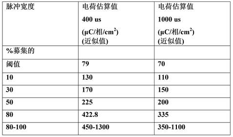
据估计,在人急性模型中募集100%的电荷要求可能在近似80-1300 µC/cm2 (使用400uS脉冲宽度,12 mm2表面积)和70-1100 µC/cm2(使用1ms脉冲宽度)之间变化。在350 µC/cm2下指示近似70%的募集。额外的30%募集要求电荷要求超过由可植入装置可能容纳的指数增加。例如,可以看到100%的募集可能在70-1300 µC/cm2之间变化,对于80%募集在70-450 µC/cm2之间变化,对于50%募集在70-250 µC/cm2之间变化,且对于30%募集在70-170 µC/cm2之间变化。It is estimated that the charge requirement for 100% recruitment in a human acute model may be somewhere between approximately 80-1300 µC/ cm2 (using a 400uS pulse width, 12 mm2 surface area) and 70-1100 µC/ cm2 (using a 1ms pulse width) change between. Approximately 70% recruitment is indicated at 350 µC /cm. An additional 30% recruitment requires an exponential increase in charge requirements beyond what could be accommodated by the implantable device. For example, it can be seen that 100% recruitment may vary between 70-1300 µC/ cm2 , 70-450 µC/ cm2 for 80% recruitment, and 70-250 µC/cm2 for 50% recruitment. varies between 70 and 170 µC/cm 2 for 30% recruitment.
讨论discuss
与猪相比,人中的神经纤维更分散。如组织学概况分析所指示的在脾动脉周围散布的簇的范围可以在近似1-3mm的范围内。组织形态计量学数据进一步用于优化刺激参数,并且使用计算建模工具将电荷要求从猪转化至人。使用Sundt c-纤维模型,表明对于100%募集而言,人的电荷要求在近似70-1000 µC/cm2的范围内。Compared to pigs, nerve fibers are more dispersed in humans. The extent of clusters scattered around the splenic artery as indicated by histological profiling can be in the range of approximately 1-3 mm. Histomorphometric data were further used to optimize stimulation parameters and computational modeling tools were used to translate charge requirements from pigs to humans. Using the Sundt c-fiber model, it was shown that for 100% recruitment, the human charge requirement is in the range of approximately 70-1000 µC/ cm2 .
参考文献references
Claims (45)
Applications Claiming Priority (3)
| Application Number | Priority Date | Filing Date | Title |
|---|---|---|---|
| US201762608392P | 2017-12-20 | 2017-12-20 | |
| US62/608392 | 2017-12-20 | ||
| PCT/GB2018/053725 WO2019122903A2 (en) | 2017-12-20 | 2018-12-20 | Stimulation of a nerve supplying the spleen |
Publications (2)
| Publication Number | Publication Date |
|---|---|
| CN111787975A true CN111787975A (en) | 2020-10-16 |
| CN111787975B CN111787975B (en) | 2024-06-07 |
Family
ID=64949329
Family Applications (1)
| Application Number | Title | Priority Date | Filing Date |
|---|---|---|---|
| CN201880089864.1A Active CN111787975B (en) | 2017-12-20 | 2018-12-20 | Stimulation of nerves supplying the spleen |
Country Status (6)
| Country | Link |
|---|---|
| US (3) | US11446502B2 (en) |
| EP (2) | EP4461213A3 (en) |
| JP (2) | JP2021507745A (en) |
| CN (1) | CN111787975B (en) |
| ES (1) | ES2988484T3 (en) |
| WO (1) | WO2019122903A2 (en) |
Families Citing this family (12)
| Publication number | Priority date | Publication date | Assignee | Title |
|---|---|---|---|---|
| AU2013230776B2 (en) | 2012-03-08 | 2015-12-03 | Medtronic Af Luxembourg S.A.R.L. | Immune system neuromodulation and associated systems and methods |
| WO2016090175A1 (en) | 2014-12-03 | 2016-06-09 | Metavention, Inc. | Systems and methods for modulating nerves or other tissue |
| US10524859B2 (en) | 2016-06-07 | 2020-01-07 | Metavention, Inc. | Therapeutic tissue modulation devices and methods |
| US11446502B2 (en) * | 2017-12-20 | 2022-09-20 | Galvani Bioelectronics Limited | Stimulation of a nerve supplying the spleen |
| BR112020021286A2 (en) * | 2018-04-19 | 2021-01-26 | Iota Biosciences, Inc. | implants using ultrasonic communication to modulate splenic nerve activity |
| JP7540835B2 (en) | 2018-04-19 | 2024-08-27 | イオタ バイオサイエンシズ, インコーポレイテッド | Implants using ultrasonic communication for neural sensing and stimulation |
| JP7577646B2 (en) | 2018-08-29 | 2024-11-05 | イオタ バイオサイエンシズ, インコーポレイテッド | IMPLANTABLE CLOSED-LOOP NEUROMODULATION DEVICES, SYSTEMS, AND METHODS |
| JP7570172B2 (en) | 2018-12-07 | 2024-10-21 | アヴェント インベストメント,エルエルシー | Apparatus and method for selectively and reversibly modulating nervous system structures to inhibit pain |
| MX2022004584A (en) | 2019-10-17 | 2022-09-26 | Iota Biosciences Inc | Devices and methods for modulating immune system activity in a cancer patient and treating cancer. |
| US20230061046A1 (en) * | 2020-01-22 | 2023-03-02 | Regents Of The University Of Minnesota | Systems and methods for responsive ultrasound stimulation for immuno-modulation treatment |
| US11642531B2 (en) * | 2020-10-09 | 2023-05-09 | Medtronic, Inc. | Sensing cardiac signals with leads implanted in epidural space |
| WO2022122833A1 (en) * | 2020-12-09 | 2022-06-16 | Galvani Bioelectronics Limited | Physiological effects of activation of the alox15 pathway |
Citations (15)
| Publication number | Priority date | Publication date | Assignee | Title |
|---|---|---|---|---|
| US20050075702A1 (en) * | 2003-10-01 | 2005-04-07 | Medtronic, Inc. | Device and method for inhibiting release of pro-inflammatory mediator |
| US20060100668A1 (en) * | 2001-08-31 | 2006-05-11 | Biocontrol Medical Ltd. | Selective nerve fiber stimulation |
| US20100125304A1 (en) * | 2008-11-18 | 2010-05-20 | Faltys Michael A | Devices and methods for optimizing electrode placement for anti-inflamatory stimulation |
| US20120143279A1 (en) * | 2009-03-24 | 2012-06-07 | Leon Ekchian | Methods and kits for treating appetite suppressing disorders and disorders with an increased metabolic rate |
| US8504161B1 (en) * | 2012-08-07 | 2013-08-06 | Medtronic, Inc. | Modulate vagal signals to reduce inflammation |
| WO2014153223A1 (en) * | 2013-03-14 | 2014-09-25 | Perryman Laura Tyler | Treating inflammation, chronic pain and other disorders with neuromodulation |
| US20150005680A1 (en) * | 2011-02-23 | 2015-01-01 | John D. LIPANI | System and Methods for Diagnosis and Treatment of Discogenic Lower Back Pain |
| JP2015502820A (en) * | 2011-12-09 | 2015-01-29 | メタベンション インコーポレイテッド | Therapeutic neuromodulation of the liver system |
| US20150127068A1 (en) * | 2013-11-04 | 2015-05-07 | ElectroCore, LLC | Nerve stimulator system |
| US20160015988A1 (en) * | 2012-07-17 | 2016-01-21 | Laura Tyler Perryman | Treating inflammation, chronic pain and other disorders with neuromodulation |
| US20160067497A1 (en) * | 2009-11-17 | 2016-03-10 | Setpoint Medical Corporation | Closed-loop vagus nerve stimulation |
| US20160114165A1 (en) * | 2014-10-24 | 2016-04-28 | Jacob A. Levine | Systems and methods for stimulating and/or monitoring loci in the brain to treat inflammation and to enhance vagus nerve stimulation |
| WO2016170510A1 (en) * | 2015-04-24 | 2016-10-27 | Glaxosmithkline Intellectual Property Development Limited | Neuromodulation device |
| US20170113046A1 (en) * | 2014-06-09 | 2017-04-27 | The Regents Of The University Of California | Systems and methods for restoring cognitive function |
| CN107427678A (en) * | 2014-11-03 | 2017-12-01 | 加尔瓦尼生物电子有限公司 | neuromodulation device |
Family Cites Families (34)
| Publication number | Priority date | Publication date | Assignee | Title |
|---|---|---|---|---|
| CA2338805A1 (en) | 1998-07-27 | 2000-02-10 | J.Thomas Mortimer | Device and method for production of visual sensations by optic nerve stimulation |
| US20090118780A1 (en) | 2001-07-23 | 2009-05-07 | Dilorenzo Daniel John | Method and apparatus for conformal electrodes for autonomic neuromodulation for the treatment of obesity and other conditions |
| US7904176B2 (en) | 2006-09-07 | 2011-03-08 | Bio Control Medical (B.C.M.) Ltd. | Techniques for reducing pain associated with nerve stimulation |
| US6745079B2 (en) * | 2001-11-07 | 2004-06-01 | Medtronic, Inc. | Electrical tissue stimulation apparatus and method |
| US8986297B2 (en) | 2002-08-21 | 2015-03-24 | Resect Medical, Inc. | Thermal hemostasis and/or coagulation of tissue |
| US7742818B2 (en) | 2003-05-19 | 2010-06-22 | Medtronic, Inc. | Gastro-electric stimulation for increasing the acidity of gastric secretions or increasing the amounts thereof |
| US7418292B2 (en) | 2003-10-01 | 2008-08-26 | Medtronic, Inc. | Device and method for attenuating an immune response |
| CA2593079C (en) * | 2004-12-27 | 2014-08-19 | North Shore-Long Island Jewish Research Institute | Treating inflammatory disorders by electrical vagus nerve stimulation |
| US8831739B2 (en) | 2005-06-02 | 2014-09-09 | Huntington Medical Research Institutes | Microelectrode array for chronic deep-brain microstimulation for recording |
| US20080300645A1 (en) * | 2007-05-31 | 2008-12-04 | Pacesetter, Inc. | Treatment of cardiomyopathy, heart failure and cardiac ischemia using stimulation induced secretion of glp-1 |
| US20080300648A1 (en) * | 2007-05-31 | 2008-12-04 | Pacesetter, Inc. | Treatment of cardiomyopathy, heart failure and cardiac ischemia using stimulation induced secretion of glp-1 |
| WO2009026382A1 (en) | 2007-08-20 | 2009-02-26 | Kopell, Brian, H. | Systems and methods for treating neurological disorders by light stimulation |
| US9662490B2 (en) * | 2008-03-31 | 2017-05-30 | The Feinstein Institute For Medical Research | Methods and systems for reducing inflammation by neuromodulation and administration of an anti-inflammatory drug |
| US8996116B2 (en) | 2009-10-30 | 2015-03-31 | Setpoint Medical Corporation | Modulation of the cholinergic anti-inflammatory pathway to treat pain or addiction |
| EP2515996B1 (en) * | 2009-12-23 | 2019-09-18 | Setpoint Medical Corporation | Neural stimulation devices and systems for treatment of chronic inflammation |
| CA2797701C (en) | 2010-04-27 | 2022-06-21 | Rhode Island Hospital | Method of identifying and treating chronic pain of peripheral origin related to peripheral nerve damage |
| WO2011159527A2 (en) * | 2010-06-16 | 2011-12-22 | Cardiac Pacemakers, Inc. | Automatic neural stimulation titration sweep |
| MX2013006546A (en) * | 2010-12-17 | 2013-09-05 | Neural Diabetes Llc | Method, system and apparatus for control of pancreatic beta cell function to improve glucose homeostatis and insulin production. |
| EP2760537A4 (en) | 2011-09-30 | 2015-06-03 | Adi Mashiach | Device and method for modulating nerves using parallel electric fields |
| AU2013230776B2 (en) | 2012-03-08 | 2015-12-03 | Medtronic Af Luxembourg S.A.R.L. | Immune system neuromodulation and associated systems and methods |
| EP2861296A1 (en) | 2012-06-15 | 2015-04-22 | Case Western Reserve University | Therapy delivery devices and methods for non-damaging neural tissue conduction block |
| EP2931361B1 (en) | 2012-12-14 | 2023-07-05 | Case Western Reserve University | Population based encoding of neural information |
| WO2014199239A2 (en) * | 2013-03-15 | 2014-12-18 | The Hospital For Sick Children | Methods relating to the use of remote ischemic conditioning |
| US9610444B2 (en) | 2013-03-15 | 2017-04-04 | Pacesetter, Inc. | Erythropoeitin production by electrical stimulation |
| ES2644290T3 (en) | 2013-05-15 | 2017-11-28 | Koc Universitesi | System to decrease the blood flow of an artery of an objective organ with an electrical stimulation |
| WO2014197625A1 (en) | 2013-06-05 | 2014-12-11 | Metavention, Inc. | Modulation of targeted nerve fibers |
| US9750938B2 (en) | 2014-03-11 | 2017-09-05 | Cardiac Pacemakers, Inc. | Visualization of biological environment impact on implanted therapy |
| US9872980B2 (en) * | 2015-04-15 | 2018-01-23 | Pacesetter, Inc. | Systems and methods for monitoring charge density of electrical stimulation |
| US20170100605A1 (en) * | 2015-05-15 | 2017-04-13 | Ohio State Innovation Foundation | Systems and methods of improving an immune disorder |
| EP4279107B1 (en) | 2015-12-14 | 2025-11-12 | Stimdia Medical Inc. | Electrical stimulation for preservation and restoration of diaphragm function |
| WO2017156039A1 (en) | 2016-03-09 | 2017-09-14 | CARDIONOMIC, Inc. | Cardiac contractility neurostimulation systems and methods |
| EP3538209A1 (en) | 2016-11-08 | 2019-09-18 | Galvani Bioelectronics Limited | Treatment of inflammatory disorders |
| US11633601B2 (en) * | 2017-12-20 | 2023-04-25 | Galvani Bioelectronics Limited | Treatment of inflammatory disorders |
| US11446502B2 (en) * | 2017-12-20 | 2022-09-20 | Galvani Bioelectronics Limited | Stimulation of a nerve supplying the spleen |
-
2018
- 2018-12-20 US US16/955,264 patent/US11446502B2/en active Active
- 2018-12-20 CN CN201880089864.1A patent/CN111787975B/en active Active
- 2018-12-20 ES ES18829943T patent/ES2988484T3/en active Active
- 2018-12-20 JP JP2020533701A patent/JP2021507745A/en active Pending
- 2018-12-20 WO PCT/GB2018/053725 patent/WO2019122903A2/en not_active Ceased
- 2018-12-20 EP EP24190124.8A patent/EP4461213A3/en active Pending
- 2018-12-20 EP EP18829943.2A patent/EP3727566B1/en active Active
-
2022
- 2022-07-25 US US17/872,319 patent/US12296171B2/en active Active
-
2024
- 2024-09-13 JP JP2024159164A patent/JP2025003995A/en active Pending
-
2025
- 2025-04-07 US US19/172,332 patent/US20250235702A1/en active Pending
Patent Citations (15)
| Publication number | Priority date | Publication date | Assignee | Title |
|---|---|---|---|---|
| US20060100668A1 (en) * | 2001-08-31 | 2006-05-11 | Biocontrol Medical Ltd. | Selective nerve fiber stimulation |
| US20050075702A1 (en) * | 2003-10-01 | 2005-04-07 | Medtronic, Inc. | Device and method for inhibiting release of pro-inflammatory mediator |
| US20100125304A1 (en) * | 2008-11-18 | 2010-05-20 | Faltys Michael A | Devices and methods for optimizing electrode placement for anti-inflamatory stimulation |
| US20120143279A1 (en) * | 2009-03-24 | 2012-06-07 | Leon Ekchian | Methods and kits for treating appetite suppressing disorders and disorders with an increased metabolic rate |
| US20160067497A1 (en) * | 2009-11-17 | 2016-03-10 | Setpoint Medical Corporation | Closed-loop vagus nerve stimulation |
| US20150005680A1 (en) * | 2011-02-23 | 2015-01-01 | John D. LIPANI | System and Methods for Diagnosis and Treatment of Discogenic Lower Back Pain |
| JP2015502820A (en) * | 2011-12-09 | 2015-01-29 | メタベンション インコーポレイテッド | Therapeutic neuromodulation of the liver system |
| US20160015988A1 (en) * | 2012-07-17 | 2016-01-21 | Laura Tyler Perryman | Treating inflammation, chronic pain and other disorders with neuromodulation |
| US8504161B1 (en) * | 2012-08-07 | 2013-08-06 | Medtronic, Inc. | Modulate vagal signals to reduce inflammation |
| WO2014153223A1 (en) * | 2013-03-14 | 2014-09-25 | Perryman Laura Tyler | Treating inflammation, chronic pain and other disorders with neuromodulation |
| US20150127068A1 (en) * | 2013-11-04 | 2015-05-07 | ElectroCore, LLC | Nerve stimulator system |
| US20170113046A1 (en) * | 2014-06-09 | 2017-04-27 | The Regents Of The University Of California | Systems and methods for restoring cognitive function |
| US20160114165A1 (en) * | 2014-10-24 | 2016-04-28 | Jacob A. Levine | Systems and methods for stimulating and/or monitoring loci in the brain to treat inflammation and to enhance vagus nerve stimulation |
| CN107427678A (en) * | 2014-11-03 | 2017-12-01 | 加尔瓦尼生物电子有限公司 | neuromodulation device |
| WO2016170510A1 (en) * | 2015-04-24 | 2016-10-27 | Glaxosmithkline Intellectual Property Development Limited | Neuromodulation device |
Also Published As
| Publication number | Publication date |
|---|---|
| WO2019122903A2 (en) | 2019-06-27 |
| CN111787975B (en) | 2024-06-07 |
| EP3727566A2 (en) | 2020-10-28 |
| EP3727566B1 (en) | 2024-08-28 |
| US12296171B2 (en) | 2025-05-13 |
| WO2019122903A3 (en) | 2019-08-08 |
| US20220401734A1 (en) | 2022-12-22 |
| US11446502B2 (en) | 2022-09-20 |
| JP2021507745A (en) | 2021-02-25 |
| ES2988484T3 (en) | 2024-11-20 |
| US20200391035A1 (en) | 2020-12-17 |
| EP4461213A2 (en) | 2024-11-13 |
| EP4461213A3 (en) | 2025-01-08 |
| US20250235702A1 (en) | 2025-07-24 |
| JP2025003995A (en) | 2025-01-14 |
Similar Documents
| Publication | Publication Date | Title |
|---|---|---|
| US12161869B2 (en) | Treatment of disorders associated with inflammation | |
| US20250235702A1 (en) | Stimulation of a nerve supplying the spleen | |
| US20220305259A1 (en) | System for the Treatment of Disorders Associated with Inflammation | |
| US12083340B2 (en) | Treatment of acute medical conditions | |
| US20220409887A1 (en) | Stimulation of a Nerve Supplying the Spleen | |
| US11452867B2 (en) | Treatment of acute medical conditions |
Legal Events
| Date | Code | Title | Description |
|---|---|---|---|
| PB01 | Publication | ||
| PB01 | Publication | ||
| SE01 | Entry into force of request for substantive examination | ||
| SE01 | Entry into force of request for substantive examination | ||
| GR01 | Patent grant | ||
| CP03 | Change of name, title or address | ||
| CP03 | Change of name, title or address |
Address after: Steve Niki Patentee after: GLAXOSMITHKLINE INTELLECTUAL PROPERTY DEVELOPMENT LTD. Country or region after: Britain Address before: The Middlesex County Patentee before: GLAXOSMITHKLINE INTELLECTUAL PROPERTY DEVELOPMENT LTD. Country or region before: Britain |