CN111388187A - An ophthalmic docking device and docking method - Google Patents
An ophthalmic docking device and docking method Download PDFInfo
- Publication number
- CN111388187A CN111388187A CN202010344150.XA CN202010344150A CN111388187A CN 111388187 A CN111388187 A CN 111388187A CN 202010344150 A CN202010344150 A CN 202010344150A CN 111388187 A CN111388187 A CN 111388187A
- Authority
- CN
- China
- Prior art keywords
- ophthalmic
- docking
- tissue
- docking device
- optical coherence
- Prior art date
- Legal status (The legal status is an assumption and is not a legal conclusion. Google has not performed a legal analysis and makes no representation as to the accuracy of the status listed.)
- Granted
Links
Images


Classifications
-
- A—HUMAN NECESSITIES
- A61—MEDICAL OR VETERINARY SCIENCE; HYGIENE
- A61F—FILTERS IMPLANTABLE INTO BLOOD VESSELS; PROSTHESES; DEVICES PROVIDING PATENCY TO, OR PREVENTING COLLAPSING OF, TUBULAR STRUCTURES OF THE BODY, e.g. STENTS; ORTHOPAEDIC, NURSING OR CONTRACEPTIVE DEVICES; FOMENTATION; TREATMENT OR PROTECTION OF EYES OR EARS; BANDAGES, DRESSINGS OR ABSORBENT PADS; FIRST-AID KITS
- A61F9/00—Methods or devices for treatment of the eyes; Devices for putting in contact-lenses; Devices to correct squinting; Apparatus to guide the blind; Protective devices for the eyes, carried on the body or in the hand
-
- A—HUMAN NECESSITIES
- A61—MEDICAL OR VETERINARY SCIENCE; HYGIENE
- A61B—DIAGNOSIS; SURGERY; IDENTIFICATION
- A61B3/00—Apparatus for testing the eyes; Instruments for examining the eyes
- A61B3/10—Objective types, i.e. instruments for examining the eyes independent of the patients' perceptions or reactions
- A61B3/102—Objective types, i.e. instruments for examining the eyes independent of the patients' perceptions or reactions for optical coherence tomography [OCT]
-
- A—HUMAN NECESSITIES
- A61—MEDICAL OR VETERINARY SCIENCE; HYGIENE
- A61B—DIAGNOSIS; SURGERY; IDENTIFICATION
- A61B3/00—Apparatus for testing the eyes; Instruments for examining the eyes
- A61B3/10—Objective types, i.e. instruments for examining the eyes independent of the patients' perceptions or reactions
- A61B3/14—Arrangements specially adapted for eye photography
Landscapes
- Health & Medical Sciences (AREA)
- Life Sciences & Earth Sciences (AREA)
- Animal Behavior & Ethology (AREA)
- Veterinary Medicine (AREA)
- Ophthalmology & Optometry (AREA)
- Engineering & Computer Science (AREA)
- Biomedical Technology (AREA)
- Heart & Thoracic Surgery (AREA)
- Public Health (AREA)
- General Health & Medical Sciences (AREA)
- Surgery (AREA)
- Physics & Mathematics (AREA)
- Molecular Biology (AREA)
- Medical Informatics (AREA)
- Biophysics (AREA)
- Vascular Medicine (AREA)
- Nuclear Medicine, Radiotherapy & Molecular Imaging (AREA)
- Radiology & Medical Imaging (AREA)
- Eye Examination Apparatus (AREA)
- Investigating Or Analysing Materials By Optical Means (AREA)
Abstract
本发明提供的眼科对接装置包括:机械臂、对接接口、移动台架、光源单元、光学相干断层扫描成像单元、数据分析显示单元、计算机处理与控制单元,光学相干断层扫描成像单元实时采集眼科组织图像以及机械臂与眼科组织之间的垂直距离的信号,数据分析显示单元显示采集并处理所述眼科组织图像,计算机处理与控制单元分析眼科组织图像确定所述眼科组织的位置和方向,并根据所述眼科组织的位置和方向控制机械臂与眼科组织之间的垂直距离,本发明提供的眼科对接装置,能够实现在手术期间对光束的精准聚焦和定位,并将其以直观方式向眼外科医生呈现的成像系统的引导眼科对接装置,提高眼外科手术的质量和安全性。另外,本发明还提供了眼科对接装置的对接方法。
The ophthalmic docking device provided by the present invention includes: a robotic arm, a docking interface, a mobile gantry, a light source unit, an optical coherence tomography imaging unit, a data analysis and display unit, a computer processing and control unit, and the optical coherence tomography imaging unit collects ophthalmic tissue in real time The image and the signal of the vertical distance between the robotic arm and the ophthalmic tissue, the data analysis and display unit displays the acquisition and processing of the ophthalmic tissue image, and the computer processing and control unit analyzes the ophthalmic tissue image to determine the position and direction of the ophthalmic tissue, and according to The position and direction of the ophthalmic tissue control the vertical distance between the robotic arm and the ophthalmic tissue. The ophthalmic docking device provided by the present invention can realize precise focusing and positioning of the light beam during the operation, and transmit it to the ophthalmic surgeon in an intuitive way. Physician presents a guided ophthalmic docking device for imaging systems that improves the quality and safety of ophthalmic surgery. In addition, the present invention also provides a docking method for an ophthalmic docking device.
Description
技术领域technical field
本发明涉及医疗器械技术领域,特别涉及一种眼科对接装置及对接方法。The invention relates to the technical field of medical devices, in particular to an ophthalmic docking device and a docking method.
背景技术Background technique
多年来已经开发了用于眼外科手术的多种先进的外科激光系统,从而靶向角膜、晶状体、视网膜和其他眼结构的各个部分。这些外科系统中很多眼外科系统包括对接单元、或患者接口,其与手术眼形成接触,建立良好控制的对接来提高外科手术的精度。眼科程序的精度能够通过增大对接单元与手术靶的对准的精度而提高。其中角膜是不受阻挡且可见的角膜手术中,患者接口与角膜的对准能由外科医生以相对直接的方式执行。然而,由于晶状体位于眼的内部,白内障手术由于若干原因向患者接口的对准和对接更为困难。即使外科医生给予引导和口头指示,患眼与眼外科系统对准还是很困难。另外,眼的可视结构(例如瞳孔)偏心和倾斜,可能造成位移和倾斜。此外,随着对接单元降低至眼,其向眼施加压力,从而可能造成眼的额外位移和倾斜。A variety of advanced surgical laser systems have been developed over the years for use in ocular surgery to target various parts of the cornea, lens, retina and other ocular structures. Many of these surgical systems include a docking unit, or patient interface, that makes contact with the operating eye, establishing a well-controlled docking to improve surgical precision. The accuracy of the ophthalmic procedure can be improved by increasing the accuracy of the alignment of the docking unit with the surgical target. In corneal surgery where the cornea is unobstructed and visible, alignment of the patient interface to the cornea can be performed by the surgeon in a relatively straightforward manner. However, since the lens is located inside the eye, alignment and docking to the patient interface for cataract surgery is more difficult for several reasons. Even with guidance and verbal instructions from the surgeon, alignment of the affected eye with the ophthalmic surgical system is difficult. Additionally, the visual structures of the eye, such as the pupil, are eccentric and tilted, potentially causing displacement and tilting. Furthermore, as the docking unit is lowered to the eye, it applies pressure to the eye, potentially causing additional displacement and tilting of the eye.
眼外科手术的精度和控制实质上受这些对接和固定步骤的精度的影响,并且由此提高对接程序的精度能够提高整个眼外科手术的精度。因此,需要提出一种能够精度控制眼科对接的装置是迫切需要的。The accuracy and control of eye surgery is substantially affected by the accuracy of these docking and securing steps, and thus improving the accuracy of the docking procedure can improve the accuracy of the overall eye surgery. Therefore, it is urgent to propose a device that can precisely control ophthalmic docking.
发明内容SUMMARY OF THE INVENTION
有鉴如此,有必要针对现有技术存在的缺陷,提供一种能够精度控制眼科对接的眼科对接装置。In view of this, it is necessary to provide an ophthalmic docking device capable of precisely controlling ophthalmic docking in view of the defects in the prior art.
为实现上述目的,本发明采用下述技术方案:To achieve the above object, the present invention adopts the following technical solutions:
一种眼科对接装置,包括:机械臂、对接接口、移动台架、光源单元、光学相干断层扫描成像单元、数据分析显示单元、计算机处理与控制单元,所述光源单元连接所述光学相干断层扫描成像单元,其中:An ophthalmic docking device, comprising: a robotic arm, a docking interface, a mobile gantry, a light source unit, an optical coherence tomography imaging unit, a data analysis and display unit, a computer processing and control unit, the light source unit is connected to the optical coherence tomography unit Imaging unit, which:
所述机械臂的两端分别固定连接有所述对接接口及所述光学相干断层扫描成像单元,所述对接接口与固定于所述移动台架上的眼科组织接触,所述光学相干断层扫描成像单元用于实时采集眼科组织图像以及所述机械臂与所述眼科组织之间的垂直距离的信号;Two ends of the robotic arm are respectively fixedly connected with the docking interface and the optical coherence tomography imaging unit, the docking interface is in contact with the ophthalmic tissue fixed on the mobile gantry, and the optical coherence tomography imaging unit The unit is used for real-time acquisition of ophthalmic tissue images and signals of the vertical distance between the robotic arm and the ophthalmic tissue;
所述的数据分析显示单元通过信号通路连接于所述光学相干断层扫描成像单元,所述的数据分析显示单元用于显示采集并处理所述眼科组织图像;The data analysis and display unit is connected to the optical coherence tomography imaging unit through a signal path, and the data analysis and display unit is used for displaying, collecting and processing the ophthalmic tissue image;
所述的计算机处理与控制单元通过信号通路连接于所述数据分析显示单元,所述的计算机处理与控制单元通过分析所述眼科组织图像确定所述眼科组织的位置和方向,所述的计算机处理与控制单元还用于根据所述眼科组织的位置和方向控制所述机械臂与所述眼科组织之间的垂直距离。The computer processing and control unit is connected to the data analysis and display unit through a signal path, the computer processing and control unit determines the position and direction of the ophthalmic tissue by analyzing the ophthalmic tissue image, and the computer processing The and control unit is further configured to control the vertical distance between the robotic arm and the ophthalmic tissue according to the position and direction of the ophthalmic tissue.
在一些较佳的实施例中,所述机械臂包括依次铰接的支撑臂、连接臂和自由端,所述支撑臂与所述光学相干断层扫描成像单元固定连接,所述自由端与所述对接接口固定连接,所述自由端与所述眼科组织之间的垂直距离可调节。In some preferred embodiments, the robotic arm includes a support arm, a connecting arm and a free end that are hinged in sequence, the support arm is fixedly connected to the optical coherence tomography imaging unit, and the free end is connected to the docking unit The interface is fixedly connected, and the vertical distance between the free end and the ophthalmic tissue is adjustable.
在一些较佳的实施例中,所述自由端上设置有XYZ精度调节器,所述XYZ精度调节器可调节所述自由端与所述眼科组织之间的XYZ三维距离。In some preferred embodiments, an XYZ precision adjuster is provided on the free end, and the XYZ precision adjuster can adjust the XYZ three-dimensional distance between the free end and the ophthalmic tissue.
在一些较佳的实施例中,所述对接接口包括与所述自由端连接且用于聚焦光束的反射透镜、与所述反射透镜连接的且用于眼科对接的负压环和设置于所述负压环内的油管,所述对接接口通过所述油管内的液体与所述眼科组织接触。In some preferred embodiments, the docking interface comprises a reflective lens connected with the free end and used for focusing light beams, a negative pressure ring connected with the reflective lens and used for ophthalmic docking, and a negative pressure ring provided on the The oil pipe in the negative pressure ring, the butt interface is in contact with the ophthalmic tissue through the liquid in the oil pipe.
在一些较佳的实施例中,所述负压环的中心、反射透镜的中心和眼科组织的中心,处于同一水平线。In some preferred embodiments, the center of the negative pressure ring, the center of the reflective lens and the center of the ophthalmic tissue are on the same horizontal line.
在一些较佳的实施例中,所述移动台架设置有XYZ调节器,所述XYZ调节器用于调节所述自由端与所述眼科组织之间的XYZ三维距离。In some preferred embodiments, the moving gantry is provided with an XYZ adjuster, and the XYZ adjuster is used to adjust the XYZ three-dimensional distance between the free end and the ophthalmic tissue.
在一些较佳的实施例中,所述的眼科组织为角膜、角膜缘、瞳孔、虹膜、晶状体、睫状肌、玻璃体或视网膜中的任意一种。In some preferred embodiments, the ophthalmic tissue is any one of cornea, limbus, pupil, iris, lens, ciliary muscle, vitreous or retina.
在一些较佳的实施例中,所述光源单元为可见光光源。In some preferred embodiments, the light source unit is a visible light source.
另外,本发明还提供了一种所述的眼科对接装置的对接方法,包括下述步骤:In addition, the present invention also provides a docking method for the ophthalmic docking device, comprising the following steps:
调整所述的移动台架使所述对接接口与所述眼科组织对准;adjusting the mobile gantry to align the docking interface with the ophthalmic tissue;
所述光学相干断层扫描成像单元采集眼科组织的内眼结构和前眼结构的图像;The optical coherence tomography imaging unit acquires images of the inner eye structure and the anterior eye structure of the ophthalmic tissue;
所述数据分析显示单元从所述内眼结构的图像确定内眼结构定向的深度域以及基于所述前眼结构的图像确定所述前眼结构的位置;The data analysis display unit determines a depth domain of the orientation of the inner ocular structure from the image of the inner ocular structure and determines the position of the anterior ocular structure based on the image of the anterior ocular structure;
所述计算机处理与控制单元根据实时的所确定的眼组织形状改变信息进一步调整所述对接接口与眼科组织的对准,所述机械臂根据所述计算机处理与控制单元的指令,基于确定的位置和定向移动所述对接接口,使得所述的移动台架使所述对接接口与所述眼科组织对准。The computer processing and control unit further adjusts the alignment of the docking interface with the ophthalmic tissue according to the real-time determined eye tissue shape change information, and the robotic arm is based on the determined position according to instructions from the computer processing and control unit and directionally moving the docking interface such that the moving gantry aligns the docking interface with the ophthalmic tissue.
本发明采用上述技术方案的优点是:The advantages of the present invention adopting the above technical solutions are:
本发明提供的眼科对接装置包括:机械臂、对接接口、移动台架、光源单元、光学相干断层扫描成像单元、数据分析显示单元、计算机处理与控制单元,所述光学相干断层扫描成像单元用于实时采集眼科组织图像以及所述机械臂与所述眼科组织之间的垂直距离的信号,所述的数据分析显示单元用于显示采集并处理所述眼科组织图像,所述的计算机处理与控制单元通过分析所述眼科组织图像确定所述眼科组织的位置和方向,所述的计算机处理与控制单元还用于根据所述眼科组织的位置和方向控制所述机械臂与所述眼科组织之间的垂直距离,本发明提供的眼科对接装置,能够实现在手术期间对光束的精准聚焦和定位,并将其以直观方式向眼外科医生呈现的成像系统的引导眼科对接装置,提高眼外科手术的质量和安全性。The ophthalmic docking device provided by the present invention includes: a robotic arm, a docking interface, a mobile gantry, a light source unit, an optical coherence tomography imaging unit, a data analysis and display unit, a computer processing and control unit, and the optical coherence tomography imaging unit is used for Real-time acquisition of ophthalmic tissue images and signals of the vertical distance between the robotic arm and the ophthalmic tissue, the data analysis and display unit is used to display, collect and process the ophthalmic tissue images, and the computer processing and control unit The position and direction of the ophthalmic tissue are determined by analyzing the image of the ophthalmic tissue, and the computer processing and control unit is further configured to control the mechanical arm and the ophthalmic tissue according to the position and direction of the ophthalmic tissue. Vertical distance, the ophthalmic docking device provided by the present invention can realize the precise focusing and positioning of the light beam during the operation, and present it to the ophthalmic surgeon in an intuitive manner. The ophthalmic docking device of the imaging system improves the quality of ophthalmic surgery. and security.
附图说明Description of drawings
为了更清楚地说明本发明实施例或现有技术中的技术方案,下面将对实施例或现有技术描述中所需要使用的附图作简单地介绍,显而易见地,下面描述中的附图仅仅是本发明的一些实施例,对于本领域普通技术人员来讲,在不付出创造性劳动的前提下,还可以根据这些附图获得其它的附图。In order to explain the embodiments of the present invention or the technical solutions in the prior art more clearly, the following briefly introduces the accompanying drawings that need to be used in the description of the embodiments or the prior art. Obviously, the accompanying drawings in the following description are only These are some embodiments of the present invention, and for those of ordinary skill in the art, other drawings can also be obtained from these drawings without creative effort.
图1为本发明实施例1提供的眼科对接装置的结构示意图。FIG. 1 is a schematic structural diagram of an ophthalmic docking device provided in Embodiment 1 of the present invention.
图2为本发明实施例2提供的一种眼科对接装置的对接方法的步骤流程图。FIG. 2 is a flowchart of steps of a docking method for an ophthalmic docking device according to
具体实施方式Detailed ways
下面将结合本发明实施例中的附图,对本发明实施例中的技术方案进行清楚、完整地描述,显然,所描述的实施例仅仅是本发明一部分实施例,而不是全部的实施例。基于本发明中的实施例,本领域普通技术人员在没有做出创造性劳动前提下所获得的所有其它实施例,都属于本发明保护的范围。The technical solutions in the embodiments of the present invention will be clearly and completely described below with reference to the accompanying drawings in the embodiments of the present invention. Obviously, the described embodiments are only a part of the embodiments of the present invention, but not all of the embodiments. Based on the embodiments of the present invention, all other embodiments obtained by those of ordinary skill in the art without creative efforts shall fall within the protection scope of the present invention.
实施例1Example 1
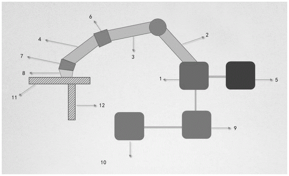
请参阅图1,为本发明实施例1提供的一种眼科对接装置,包括:机械臂、对接接口、移动台架11、光源单元5、光学相干断层扫描成像单元1、数据分析显示单元10、计算机处理与控制单元9。Referring to FIG. 1, an ophthalmic docking device provided in Embodiment 1 of the present invention includes: a robotic arm, a docking interface, a
所述机械臂的两端分别固定连接有所述对接接口及所述光学相干断层扫描成像单元1,所述对接接口与固定于所述移动台架11上的眼科组织接触,所述光学相干断层扫描成像单元1用于实时采集眼科组织图像以及所述机械臂与所述眼科组织之间的垂直距离的信号。Both ends of the robotic arm are respectively fixedly connected with the docking interface and the optical coherence tomography imaging unit 1, the docking interface is in contact with the ophthalmic tissue fixed on the
所述的数据分析显示单元10通过信号通路连接于所述光学相干断层扫描成像单元1,所述的数据分析显示单元10用于显示采集并处理所述眼科组织图像。The data analysis and
所述的计算机处理与控制单元9通过信号通路连接于所述数据分析显示单元10,所述的计算机处理与控制单元9通过分析所述眼科组织图像确定所述眼科组织的位置和方向,所述的计算机处理与控制单元9还用于根据所述眼科组织的位置和方向控制所述机械臂与所述眼科组织之间的垂直距离。The computer processing and
以下详细说明各个部件的结构及连接关系。The structure and connection relationship of each component will be described in detail below.
在一些实施例中,所述机械臂包括依次铰接的支撑臂2、连接臂3和自由端4。所述支撑臂2与所述光学相干断层扫描成像单元1固定连接,所述自由端4与所述对接接口固定连接,所述自由端4与所述眼科组织之间的垂直距离可调节。In some embodiments, the robotic arm includes a
进一步地,所述自由端4上设置有XYZ精度调节器6,所述XYZ精度调节器6可调节所述自由端4与所述眼科组织之间的XYZ三维距离。Further, an XYZ precision adjuster 6 is provided on the
由于在自由端4上设置XYZ精度调节器6,通过调节所述XYZ精度调节器6在XYZ的方向,可以精度调节机械臂的自由端4与眼科组织之间的XYZ三维距离。Since the XYZ precision adjuster 6 is provided on the
可以理解,本发明实施例提供的机械臂能够基于确定的位置和定向移动所述对接接口,对接至眼并且能够精度调节机械臂的自由端4与眼组织之间的垂直距离。It can be understood that the robotic arm provided by the embodiment of the present invention can move the docking interface based on the determined position and orientation, dock to the eye, and can precisely adjust the vertical distance between the
在一些实施例中,所述对接接口包括与所述自由端4连接且用于聚焦光束的反射透镜7、与所述反射透镜7连接的且用于眼科对接的负压环8和设置于所述负压环8内的油管(图未示),所述对接接口通过所述油管内的液体与所述眼科组织接触。In some embodiments, the docking interface includes a
进一步地,所述负压环8的中心、反射透镜7的中心和眼科组织的中心,处于同一水平线。Further, the center of the
在一些实施例中,所述移动台架11设置有XYZ调节器12,所述XYZ调节器12可调节所述移动台架11的方位,以调节所述自由端4与所述眼科组织之间的XYZ三维距离。In some embodiments, the moving
可以理解,由于采用XYZ调节器12操作移动台架11的移动,实现反射透镜7和对接接口的位置调节,减小并最终消除眼的移位或偏差。It can be understood that, since the
在一些实施例中,所述的光学相干断层扫描成像单元1包括OCT光源、第一XYZ轴三向振镜、XYZ轴三向扫描透镜。In some embodiments, the optical coherence tomography imaging unit 1 includes an OCT light source, a first XYZ axis three-direction galvanometer, and an XYZ axis three-direction scanning lens.
更进一步地,所述的光学相干断层扫描成像单元1的成像深度达到8mm;每秒扫描帧数为100帧;扫描次数为20万次/秒;回撤速度20mm/s;波长820-880nm;系统灵敏度6dB/3mm-20dB/3mm;最大功率2.5mW-3.0mW。Further, the imaging depth of the optical coherence tomography imaging unit 1 reaches 8mm; the number of scanning frames per second is 100; the number of scans is 200,000 times/second; the retraction speed is 20mm/s; the wavelength is 820-880nm; System sensitivity 6dB/3mm-20dB/3mm; maximum power 2.5mW-3.0mW.
可以理解,本发明的光学相干断层扫描成像单元1不但能够生成所述眼组织的内眼结构和前眼结构的图像,而且能够在对接期间提供眼组织实时图像,并同时提供对接程序的基本实况的眼组织图像。It can be understood that the optical coherence tomography imaging unit 1 of the present invention can not only generate images of the inner ocular structure and anterior ocular structure of the ocular tissue, but also provide real-time images of the ocular tissue during docking, and at the same time provide the basic live of the docking procedure image of eye tissue.
在一些实施例中,所述的眼组织为角膜、角膜缘、瞳孔、虹膜、晶状体、睫状肌、玻璃体或视网膜中的任意一个。In some embodiments, the ocular tissue is any one of the cornea, limbus, pupil, iris, lens, ciliary muscle, vitreous or retina.
在一些实施例中,数据分析显示单元10为LED显示器。In some embodiments, the data
在一些实施例中,计算机处理与控制单元9为大功率数据处理计算机。In some embodiments, the computer processing and
在一些实施例中,所述光源单元5为可见光光源系统,用于指示装置的光路设置。In some embodiments, the
本发明提供的眼科对接装置,所述光学相干断层扫描成像单元用于实时采集眼科组织图像以及所述机械臂与所述眼科组织之间的垂直距离的信号,所述的数据分析显示单元用于显示采集并处理所述眼科组织图像,所述的计算机处理与控制单元通过分析所述眼科组织图像确定所述眼科组织的位置和方向,所述的计算机处理与控制单元还用于根据所述眼科组织的位置和方向控制所述机械臂与所述眼科组织之间的垂直距离,本发明提供的眼科对接装置,能够实现在手术期间对光束的精准聚焦和定位,并将其以直观方式向眼外科医生呈现的成像系统的引导眼科对接装置,提高眼外科手术的质量和安全性。In the ophthalmic docking device provided by the present invention, the optical coherence tomography imaging unit is used for real-time acquisition of ophthalmic tissue images and signals of the vertical distance between the robotic arm and the ophthalmic tissue, and the data analysis and display unit is used for Display, collect and process the ophthalmic tissue image, the computer processing and control unit determines the position and orientation of the ophthalmic tissue by analyzing the ophthalmic tissue image, and the computer processing and control unit is also used for according to the ophthalmic tissue image. The position and direction of the tissue control the vertical distance between the robotic arm and the ophthalmic tissue. The ophthalmic docking device provided by the present invention can realize precise focusing and positioning of the light beam during surgery, and direct it to the eye in an intuitive way. Surgeon-presented ophthalmic docking device for imaging systems that improves the quality and safety of ophthalmic surgery.
此外,本发明提供的眼科对接装置采用非接触式、浸润式的对接接口,通过液体接触角膜而不是锥镜直接接触角膜,角膜挤压小,从而避免了接触式患者接口会产生角膜皱褶会产生不规则散射损伤角膜组织或者刺激虹膜使瞳孔收缩。In addition, the ophthalmic docking device provided by the present invention adopts a non-contact and infiltrating docking interface, and the liquid contacts the cornea instead of the cone lens directly. Irregular scattering damages the corneal tissue or stimulates the iris to constrict the pupil.
实施例2Example 2
请参阅图2,为本发明实施例2提供的一种眼科对接装置的对接方法,包括下述步骤:Please refer to FIG. 2, which is a docking method for an ophthalmic docking device provided in
步骤S110:调整所述的移动台架11使所述对接接口与所述眼科组织对准。Step S110: Adjust the
步骤S120:所述光学相干断层扫描成像单元1采集眼科组织的内眼结构和前眼结构的图像。Step S120 : the optical coherence tomography imaging unit 1 acquires images of the inner eye structure and the anterior eye structure of the ophthalmic tissue.
由于所述的光学相干断层扫描成像单元1连接于机械臂上,一方面可以用于实时采集眼组织图像外,还可以采集机械臂的自由端4与眼组织之间的垂直距离的信号。Since the optical coherence tomography imaging unit 1 is connected to the robotic arm, on the one hand, it can be used for real-time acquisition of ocular tissue images, and can also acquire signals of the vertical distance between the
可以理解,光学相干断层扫描成像单元1不仅能够在对接期间提供眼组织实时图像,而是能够提供对接程序的基本实况的眼组织图像,实时反馈提供所确定的眼组织形状改变信息,能够向外科医生递送即时信息,以利于医生更好做出决策。It can be understood that the optical coherence tomography imaging unit 1 can not only provide a real-time image of the eye tissue during the docking process, but can also provide a basic live eye tissue image of the docking procedure, and provide real-time feedback to provide the determined shape change information of the eye tissue, which can be reported to the surgeon. Physicians deliver instant information for better decision-making.
步骤S130:所述数据分析显示单元10从所述内眼结构的图像确定内眼结构定向的深度域以及基于所述前眼结构的图像确定所述前眼结构的位置。Step S130 : the data analysis and
步骤S140:所述计算机处理与控制单元9根据实时的所确定的眼组织形状改变信息进一步调整所述对接接口与眼科组织的对准;所述机械臂根据所述计算机处理与控制单元9的指令,调整所述机械臂与所述移动台架11之间相对位置和方向,使所述对接接口与所述眼科组织对准。Step S140: the computer processing and
具体地,所述计算机处理与控制单元9根据实时的所确定的眼组织形状改变信息进一步调整所述对接接口与眼科组织的对准,所述机械臂根据所述计算机处理与控制单元9的指令,通过调节所述XYZ精度调节器6在XYZ的方向,可以精度调节机械臂的自由端4与眼科组织之间的XYZ三维距离。Specifically, the computer processing and
与此同时,所述移动台架11设置有XYZ调节器12,所述XYZ调节器12可调节所述移动台架11的方位,以调节所述自由端4与所述眼科组织之间的XYZ三维距离。At the same time, the moving
可以理解,对接接口可辅助患者接口与眼的对接,在对接之前光学相干断层扫描成像单元1生成眼组织的图像并结合目标位置和定向提供移位和倾斜,并且可在对接期间根据实时成像眼组织的成像系统而进一步完善患者接口与眼的对接。It will be appreciated that the docking interface may assist in the docking of the patient interface with the eye, the optical coherence tomography imaging unit 1 generates an image of the eye tissue prior to docking and provides displacement and tilt in conjunction with the target position and orientation, and may image the eye in real time during docking. The imaging system of the tissue further improves the connection between the patient interface and the eye.
本发明提供的眼科对接装置方法,能够实现在手术期间对光束的精准聚焦和定位,并将其以直观方式向眼外科医生呈现的成像系统的引导眼科对接装置,提高眼外科手术的质量和安全性。The ophthalmic docking device method provided by the invention can realize the precise focusing and positioning of the light beam during the operation, and present it to the ophthalmic surgeon in an intuitive way to guide the ophthalmic docking device of the imaging system, thereby improving the quality and safety of ophthalmic surgery. sex.
此外,本发明提供的眼科对接装置采用非接触式、浸润式的对接接口,通过液体接触角膜而不是锥镜直接接触角膜,角膜挤压小,从而避免了接触式患者接口会产生角膜皱褶会产生不规则散射损伤角膜组织或者刺激虹膜使瞳孔收缩。In addition, the ophthalmic docking device provided by the present invention adopts a non-contact and infiltrating docking interface, and the liquid contacts the cornea instead of the cone lens directly. Irregular scattering damages the corneal tissue or stimulates the iris to constrict the pupil.
可以理解,以上所述实施例的各技术特征可以进行任意的组合,为使描述简洁,未对上述实施例中的各个技术特征所有可能的组合都进行描述,然而,只要这些技术特征的组合不存在矛盾,都应当认为是本说明书记载的范围。It can be understood that the technical features of the above-described embodiments can be combined arbitrarily. For the sake of brevity, all possible combinations of the technical features in the above-described embodiments are not described. However, as long as the combinations of these technical features do not If there is any contradiction, it should be regarded as the scope of the description in this specification.
当然本发明的眼科对接装置正极材料还可具有多种变换及改型,并不局限于上述实施方式的具体结构。总之,本发明的保护范围应包括那些对于本领域普通技术人员来说显而易见的变换或替代以及改型。Of course, the positive electrode material of the ophthalmic docking device of the present invention can also have various transformations and modifications, and is not limited to the specific structure of the above embodiment. In a word, the protection scope of the present invention should include those changes or substitutions and modifications that are obvious to those of ordinary skill in the art.
Claims (9)
Priority Applications (1)
| Application Number | Priority Date | Filing Date | Title |
|---|---|---|---|
| CN202010344150.XA CN111388187B (en) | 2020-04-27 | 2020-04-27 | Ophthalmic docking device and docking method |
Applications Claiming Priority (1)
| Application Number | Priority Date | Filing Date | Title |
|---|---|---|---|
| CN202010344150.XA CN111388187B (en) | 2020-04-27 | 2020-04-27 | Ophthalmic docking device and docking method |
Publications (2)
| Publication Number | Publication Date |
|---|---|
| CN111388187A true CN111388187A (en) | 2020-07-10 |
| CN111388187B CN111388187B (en) | 2023-10-20 |
Family
ID=71411652
Family Applications (1)
| Application Number | Title | Priority Date | Filing Date |
|---|---|---|---|
| CN202010344150.XA Active CN111388187B (en) | 2020-04-27 | 2020-04-27 | Ophthalmic docking device and docking method |
Country Status (1)
| Country | Link |
|---|---|
| CN (1) | CN111388187B (en) |
Cited By (3)
| Publication number | Priority date | Publication date | Assignee | Title |
|---|---|---|---|---|
| CN112587305A (en) * | 2021-03-04 | 2021-04-02 | 季华实验室 | Ophthalmic docking device and method |
| CN113974553A (en) * | 2021-12-28 | 2022-01-28 | 广东麦特维逊医学研究发展有限公司 | Eye pressure measuring device and working method thereof |
| WO2025051990A1 (en) * | 2023-09-07 | 2025-03-13 | Technolas Perfect Vision Gmbh | Laser system having an orientation analysis system for measuring the orientation of the patient |
Citations (6)
| Publication number | Priority date | Publication date | Assignee | Title |
|---|---|---|---|---|
| US20110304819A1 (en) * | 2010-06-14 | 2011-12-15 | Adam Juhasz | Image-Guided Docking for Ophthalmic Surgical Systems |
| CN103957774A (en) * | 2011-08-26 | 2014-07-30 | 爱尔康手术激光股份有限公司 | Imaging-based guidance system for ophthalmic docking using a location-orientation analysis |
| JP2017176840A (en) * | 2017-04-27 | 2017-10-05 | 株式会社トプコン | Ophthalmologic imaging apparatus |
| CN107529981A (en) * | 2015-06-15 | 2018-01-02 | 诺华股份有限公司 | Tracking system for surgical operation optical coherence tomography |
| CN109008942A (en) * | 2018-09-15 | 2018-12-18 | 中山大学中山眼科中心 | A kind of full optics of the eye coherence tomography device and imaging method based on slit-lamp platform |
| CN110167423A (en) * | 2016-11-07 | 2019-08-23 | 卡尔蔡司医疗技术股份公司 | Method and ophthalmology self-checking unit for eyes self-test |
-
2020
- 2020-04-27 CN CN202010344150.XA patent/CN111388187B/en active Active
Patent Citations (6)
| Publication number | Priority date | Publication date | Assignee | Title |
|---|---|---|---|---|
| US20110304819A1 (en) * | 2010-06-14 | 2011-12-15 | Adam Juhasz | Image-Guided Docking for Ophthalmic Surgical Systems |
| CN103957774A (en) * | 2011-08-26 | 2014-07-30 | 爱尔康手术激光股份有限公司 | Imaging-based guidance system for ophthalmic docking using a location-orientation analysis |
| CN107529981A (en) * | 2015-06-15 | 2018-01-02 | 诺华股份有限公司 | Tracking system for surgical operation optical coherence tomography |
| CN110167423A (en) * | 2016-11-07 | 2019-08-23 | 卡尔蔡司医疗技术股份公司 | Method and ophthalmology self-checking unit for eyes self-test |
| JP2017176840A (en) * | 2017-04-27 | 2017-10-05 | 株式会社トプコン | Ophthalmologic imaging apparatus |
| CN109008942A (en) * | 2018-09-15 | 2018-12-18 | 中山大学中山眼科中心 | A kind of full optics of the eye coherence tomography device and imaging method based on slit-lamp platform |
Cited By (5)
| Publication number | Priority date | Publication date | Assignee | Title |
|---|---|---|---|---|
| CN112587305A (en) * | 2021-03-04 | 2021-04-02 | 季华实验室 | Ophthalmic docking device and method |
| CN112587305B (en) * | 2021-03-04 | 2021-06-18 | 季华实验室 | Ophthalmic docking device and method |
| CN113974553A (en) * | 2021-12-28 | 2022-01-28 | 广东麦特维逊医学研究发展有限公司 | Eye pressure measuring device and working method thereof |
| CN113974553B (en) * | 2021-12-28 | 2022-04-01 | 广东麦特维逊医学研究发展有限公司 | Eye pressure measuring device and working method thereof |
| WO2025051990A1 (en) * | 2023-09-07 | 2025-03-13 | Technolas Perfect Vision Gmbh | Laser system having an orientation analysis system for measuring the orientation of the patient |
Also Published As
| Publication number | Publication date |
|---|---|
| CN111388187B (en) | 2023-10-20 |
Similar Documents
| Publication | Publication Date | Title |
|---|---|---|
| US12178754B2 (en) | Full depth laser ophthalmic surgical system, methods of calibrating the surgical system and treatment methods using the same | |
| JP5918241B2 (en) | Ophthalmic system and method for aligning an eye with an ophthalmic system | |
| JP4950077B2 (en) | Device for performing predetermined contact alignment of a patient's eye and laser system during eye surgery, and aligning the patient's eye with a surgical laser system and facilitating their physical engagement device | |
| JP4080379B2 (en) | Ophthalmic laser equipment | |
| US7258686B2 (en) | Corneal surgery apparatus | |
| CN111388187B (en) | Ophthalmic docking device and docking method | |
| TWI631926B (en) | Centering technique for a cutting laser for refractive ophthalmic surgery | |
| CN113974965B (en) | Laser capsulorhexis device | |
| JP6524609B2 (en) | Ophthalmic laser surgery device | |
| US6585724B2 (en) | Ophthalmic surgery apparatus | |
| CN112587306B (en) | An ophthalmic laser treatment system | |
| JP2003245300A (en) | Ophthalmic equipment | |
| CN111407508A (en) | An ophthalmic cutting system and an ophthalmic cutting method | |
| US20200330268A1 (en) | Ophthalmic Device For Treating an Eye | |
| WO2025242204A1 (en) | All-solid corneal surgery system guided by ssoct monitoring | |
| US20230397810A1 (en) | Ophthalmic observation apparatus, method of controlling the same, and recording medium | |
| JP2025513142A (en) | Ocular docking for ophthalmic laser treatments. | |
| US11013407B2 (en) | Intraocular pressure measurement for an eye docked to a laser system | |
| CN213465585U (en) | Ophthalmic docking device | |
| JP2016193096A (en) | Ophthalmic laser treatment device and ophthalmic surgery control program | |
| CN113974964B (en) | Laser capsulorhexis device and working method thereof | |
| CN111407507B (en) | Eye tissue cutting device | |
| CN223489703U (en) | Digital photographing system of direct ophthalmoscope | |
| CN111671389A (en) | Ophthalmic measurement system based on reflection switching | |
| CN108420400A (en) | A kind of image guiding fundus camera |
Legal Events
| Date | Code | Title | Description |
|---|---|---|---|
| PB01 | Publication | ||
| PB01 | Publication | ||
| SE01 | Entry into force of request for substantive examination | ||
| SE01 | Entry into force of request for substantive examination | ||
| GR01 | Patent grant | ||
| GR01 | Patent grant |