CN111247592A - System and method for quantifying tissue over time - Google Patents
System and method for quantifying tissue over time Download PDFInfo
- Publication number
- CN111247592A CN111247592A CN201880068341.9A CN201880068341A CN111247592A CN 111247592 A CN111247592 A CN 111247592A CN 201880068341 A CN201880068341 A CN 201880068341A CN 111247592 A CN111247592 A CN 111247592A
- Authority
- CN
- China
- Prior art keywords
- image
- breast
- parameters
- native
- images
- Prior art date
- Legal status (The legal status is an assumption and is not a legal conclusion. Google has not performed a legal analysis and makes no representation as to the accuracy of the status listed.)
- Granted
Links
Images


Classifications
-
- G—PHYSICS
- G16—INFORMATION AND COMMUNICATION TECHNOLOGY [ICT] SPECIALLY ADAPTED FOR SPECIFIC APPLICATION FIELDS
- G16H—HEALTHCARE INFORMATICS, i.e. INFORMATION AND COMMUNICATION TECHNOLOGY [ICT] SPECIALLY ADAPTED FOR THE HANDLING OR PROCESSING OF MEDICAL OR HEALTHCARE DATA
- G16H30/00—ICT specially adapted for the handling or processing of medical images
- G16H30/40—ICT specially adapted for the handling or processing of medical images for processing medical images, e.g. editing
-
- A—HUMAN NECESSITIES
- A61—MEDICAL OR VETERINARY SCIENCE; HYGIENE
- A61B—DIAGNOSIS; SURGERY; IDENTIFICATION
- A61B5/00—Measuring for diagnostic purposes; Identification of persons
- A61B5/0033—Features or image-related aspects of imaging apparatus, e.g. for MRI, optical tomography or impedance tomography apparatus; Arrangements of imaging apparatus in a room
- A61B5/004—Features or image-related aspects of imaging apparatus, e.g. for MRI, optical tomography or impedance tomography apparatus; Arrangements of imaging apparatus in a room adapted for image acquisition of a particular organ or body part
-
- A—HUMAN NECESSITIES
- A61—MEDICAL OR VETERINARY SCIENCE; HYGIENE
- A61B—DIAGNOSIS; SURGERY; IDENTIFICATION
- A61B5/00—Measuring for diagnostic purposes; Identification of persons
- A61B5/43—Detecting, measuring or recording for evaluating the reproductive systems
- A61B5/4306—Detecting, measuring or recording for evaluating the reproductive systems for evaluating the female reproductive systems, e.g. gynaecological evaluations
- A61B5/4312—Breast evaluation or disorder diagnosis
-
- A—HUMAN NECESSITIES
- A61—MEDICAL OR VETERINARY SCIENCE; HYGIENE
- A61B—DIAGNOSIS; SURGERY; IDENTIFICATION
- A61B5/00—Measuring for diagnostic purposes; Identification of persons
- A61B5/72—Signal processing specially adapted for physiological signals or for diagnostic purposes
- A61B5/7271—Specific aspects of physiological measurement analysis
- A61B5/7275—Determining trends in physiological measurement data; Predicting development of a medical condition based on physiological measurements, e.g. determining a risk factor
-
- A—HUMAN NECESSITIES
- A61—MEDICAL OR VETERINARY SCIENCE; HYGIENE
- A61B—DIAGNOSIS; SURGERY; IDENTIFICATION
- A61B6/00—Apparatus or devices for radiation diagnosis; Apparatus or devices for radiation diagnosis combined with radiation therapy equipment
- A61B6/50—Apparatus or devices for radiation diagnosis; Apparatus or devices for radiation diagnosis combined with radiation therapy equipment specially adapted for specific body parts; specially adapted for specific clinical applications
- A61B6/502—Apparatus or devices for radiation diagnosis; Apparatus or devices for radiation diagnosis combined with radiation therapy equipment specially adapted for specific body parts; specially adapted for specific clinical applications for diagnosis of breast, i.e. mammography
-
- A—HUMAN NECESSITIES
- A61—MEDICAL OR VETERINARY SCIENCE; HYGIENE
- A61B—DIAGNOSIS; SURGERY; IDENTIFICATION
- A61B6/00—Apparatus or devices for radiation diagnosis; Apparatus or devices for radiation diagnosis combined with radiation therapy equipment
- A61B6/52—Devices using data or image processing specially adapted for radiation diagnosis
- A61B6/5211—Devices using data or image processing specially adapted for radiation diagnosis involving processing of medical diagnostic data
- A61B6/5217—Devices using data or image processing specially adapted for radiation diagnosis involving processing of medical diagnostic data extracting a diagnostic or physiological parameter from medical diagnostic data
-
- G—PHYSICS
- G06—COMPUTING OR CALCULATING; COUNTING
- G06T—IMAGE DATA PROCESSING OR GENERATION, IN GENERAL
- G06T7/00—Image analysis
- G06T7/0002—Inspection of images, e.g. flaw detection
- G06T7/0012—Biomedical image inspection
-
- G—PHYSICS
- G06—COMPUTING OR CALCULATING; COUNTING
- G06T—IMAGE DATA PROCESSING OR GENERATION, IN GENERAL
- G06T7/00—Image analysis
- G06T7/60—Analysis of geometric attributes
- G06T7/62—Analysis of geometric attributes of area, perimeter, diameter or volume
-
- G—PHYSICS
- G16—INFORMATION AND COMMUNICATION TECHNOLOGY [ICT] SPECIALLY ADAPTED FOR SPECIFIC APPLICATION FIELDS
- G16H—HEALTHCARE INFORMATICS, i.e. INFORMATION AND COMMUNICATION TECHNOLOGY [ICT] SPECIALLY ADAPTED FOR THE HANDLING OR PROCESSING OF MEDICAL OR HEALTHCARE DATA
- G16H50/00—ICT specially adapted for medical diagnosis, medical simulation or medical data mining; ICT specially adapted for detecting, monitoring or modelling epidemics or pandemics
- G16H50/20—ICT specially adapted for medical diagnosis, medical simulation or medical data mining; ICT specially adapted for detecting, monitoring or modelling epidemics or pandemics for computer-aided diagnosis, e.g. based on medical expert systems
-
- A—HUMAN NECESSITIES
- A61—MEDICAL OR VETERINARY SCIENCE; HYGIENE
- A61B—DIAGNOSIS; SURGERY; IDENTIFICATION
- A61B6/00—Apparatus or devices for radiation diagnosis; Apparatus or devices for radiation diagnosis combined with radiation therapy equipment
- A61B6/02—Arrangements for diagnosis sequentially in different planes; Stereoscopic radiation diagnosis
- A61B6/025—Tomosynthesis
-
- G—PHYSICS
- G06—COMPUTING OR CALCULATING; COUNTING
- G06T—IMAGE DATA PROCESSING OR GENERATION, IN GENERAL
- G06T2207/00—Indexing scheme for image analysis or image enhancement
- G06T2207/30—Subject of image; Context of image processing
- G06T2207/30004—Biomedical image processing
- G06T2207/30068—Mammography; Breast
-
- G—PHYSICS
- G06—COMPUTING OR CALCULATING; COUNTING
- G06T—IMAGE DATA PROCESSING OR GENERATION, IN GENERAL
- G06T2207/00—Indexing scheme for image analysis or image enhancement
- G06T2207/30—Subject of image; Context of image processing
- G06T2207/30168—Image quality inspection
-
- G—PHYSICS
- G06—COMPUTING OR CALCULATING; COUNTING
- G06T—IMAGE DATA PROCESSING OR GENERATION, IN GENERAL
- G06T2207/00—Indexing scheme for image analysis or image enhancement
- G06T2207/30—Subject of image; Context of image processing
- G06T2207/30204—Marker
Landscapes
- Health & Medical Sciences (AREA)
- Engineering & Computer Science (AREA)
- Life Sciences & Earth Sciences (AREA)
- Medical Informatics (AREA)
- Public Health (AREA)
- General Health & Medical Sciences (AREA)
- Biomedical Technology (AREA)
- Physics & Mathematics (AREA)
- Nuclear Medicine, Radiotherapy & Molecular Imaging (AREA)
- Radiology & Medical Imaging (AREA)
- Pathology (AREA)
- Molecular Biology (AREA)
- Veterinary Medicine (AREA)
- Animal Behavior & Ethology (AREA)
- Surgery (AREA)
- Biophysics (AREA)
- Heart & Thoracic Surgery (AREA)
- Computer Vision & Pattern Recognition (AREA)
- Primary Health Care (AREA)
- Epidemiology (AREA)
- General Physics & Mathematics (AREA)
- Theoretical Computer Science (AREA)
- High Energy & Nuclear Physics (AREA)
- Data Mining & Analysis (AREA)
- Databases & Information Systems (AREA)
- Physiology (AREA)
- Optics & Photonics (AREA)
- Quality & Reliability (AREA)
- Artificial Intelligence (AREA)
- Psychiatry (AREA)
- Signal Processing (AREA)
- Geometry (AREA)
- Gynecology & Obstetrics (AREA)
- Reproductive Health (AREA)
- Oral & Maxillofacial Surgery (AREA)
- Dentistry (AREA)
- Apparatus For Radiation Diagnosis (AREA)
Abstract
Description
发明领域Field of Invention
本发明大体上涉及一种用于验证图像参数的准确度的系统和方法,尤其是用于验证医学领域的图像的图像参数的准确度的系统和方法。The present invention generally relates to a system and method for verifying the accuracy of image parameters, in particular for verifying the accuracy of image parameters for images in the medical field.
背景background
乳腺癌是40岁以上女性最常见的癌症形式。术语“乳腺癌”描述的是若干种不同类型的疾病。大多数乳腺癌是浸润性导管癌(IDC),一小部分是浸润性小叶癌(ILC),以及少数是小管癌、粘液性(胶质)癌、具有髓质特征的癌(与BRCA1基因突变有关)或浸润性乳头状癌。罕见形式的乳腺癌包括炎性乳腺癌、乳房和/或乳头的佩吉特病和化生乳腺癌。某些形式的乳腺癌在年轻女性中更为普遍(例如,具有髓质特征的癌,其他形式在老年女性中更为普遍(例如,胶质和浸润性乳头状癌)。这里使用的术语“乳腺癌”是指所有形式的所述疾病。Breast cancer is the most common form of cancer in women over the age of 40. The term "breast cancer" describes several different types of disease. Most breast cancers are invasive ductal carcinomas (IDCs), a small percentage are invasive lobular carcinomas (ILCs), and a minority are tubular, mucinous (glial) carcinomas, carcinomas with medullary features (associated with mutations in the BRCA1 gene) related) or invasive papillary carcinoma. Rare forms of breast cancer include inflammatory breast cancer, Paget's disease of the breast and/or nipple, and metaplastic breast cancer. Some forms of breast cancer are more prevalent in younger women (eg, carcinomas with medullary features) and other forms are more prevalent in older women (eg, glial and invasive papillary carcinomas). The term used here" Breast cancer" refers to all forms of the disease.
肿瘤的形式和结构各不相同。偶尔有不明显的肿瘤,并且也不是所有的肿瘤都是恶性的。癌症的细胞结构也各不相同,范围有管状、片状或指状分支。Tumors vary in form and structure. Occasionally, there are obscure tumors, and not all tumors are malignant. Cancers also vary in their cellular structure, ranging from tubular, sheet-like, or finger-like branches.
人们普遍认为乳腺癌的早期发现改善患者的预后。乳房筛查需要通常使用乳腺X线摄影对无症状人群进行成像以检测乳腺癌,最好是在早期阶段。在筛查乳腺X线摄影期间,乳房被使用乳腺X线摄影单元的板挤压。在乳腺X线摄影过程期间,拍摄四张标准图像——头尾位(CC)和内外斜位(MLO)。诊断性乳腺X线摄片专门用于在筛查乳腺X线摄片中发现乳房症状、变化、或异常的患者以及有乳腺癌的个人和/或家族史的患者。对于诊断性乳腺X线摄影,可以拍摄附加视图,包括特定关注区域的几何放大和点压缩视图。It is widely believed that early detection of breast cancer improves patient outcomes. Breast screening involves imaging asymptomatic people, often with mammography, to detect breast cancer, preferably at an early stage. During screening mammography, the breast is squeezed using the plates of the mammography unit. During the mammography procedure, four standard images - cranio-caudal (CC) and medial-lateral oblique (MLO) are taken. Diagnostic mammography is reserved for patients with breast symptoms, changes, or abnormalities found on screening mammography and for patients with a personal and/or family history of breast cancer. For diagnostic mammography, additional views can be taken, including geometrically magnified and point-compressed views of specific regions of interest.
乳腺X线摄片由放射科医生、放射技师和/或临床医生(在此统称为“读取者(reader)”或“多个读取者(readers)”)读取一次(单次读取)或两次(双重读取)。双重读取大大提高了程序的灵敏度的质量,然而这是劳动密集型、费用高,并且通常要受医疗或政府当局的授权。Mammograms are read once (single read) by radiologists, radiographers and/or clinicians (collectively referred to herein as "readers" or "readers"). ) or twice (double read). Dual reading greatly improves the quality of the sensitivity of the procedure, however it is labor-intensive, expensive, and often subject to authorization by medical or government authorities.
X射线穿透是患者或身体部位厚度的指数递减函数:乳房越厚,需要的剂量越大。为了提高对比度,X射线能量(kV)降低,然而,这需要增加剂量,因为更多的X射线被吸收。较厚的乳房将需要较大的剂量。X-ray penetration is an exponentially decreasing function of the thickness of the patient or body part: the thicker the breast, the greater the dose required. To improve contrast, the X-ray energy (kV) is reduced, however, this requires an increase in dose as more X-rays are absorbed. Thicker breasts will require larger doses.
此外,图像质量是复杂的,其中在用于获得软组织图像的方法中在步骤链的每个阶段都有可变性,并且成像链的每个元素之间相互作用;包括成像设备的物理特性、图像采集技术因素、固有的受试者特性、操作者的技能、施加到图像的任何处理的效果、图像显示的方法、以及涉及用于诊断决策制定的图像可视化和解释的心理测量因素。Furthermore, image quality is complex, with variability at each stage of the chain of steps in the method used to obtain soft tissue images, and interactions between each element of the imaging chain; including the physical properties of the imaging device, the image Acquisition technique factors, inherent subject characteristics, operator skill, effects of any processing applied to the images, method of image display, and psychometric factors involved in image visualization and interpretation for diagnostic decision making.
用于筛查目的的图像的有用性取决于其准确度,并且如果在一段时间内对乳房成分进行准确的比较,则筛查可以变得更有效。用于准确的比较的可靠自动化手段将特别有效。The usefulness of images for screening purposes depends on their accuracy, and screening can become more effective if accurate comparisons of breast composition are made over time. Reliable automated means for accurate comparisons would be particularly effective.
短语“乳房成分”用于指乳房密度以及与整个乳房的大小相比在乳房中“致密”组织的比例。“致密”组织是指包括纤维组织和腺体组织的组织。The phrase "breast composition" is used to refer to breast density and the proportion of "dense" tissue in the breast compared to the size of the entire breast. "Dense" tissue refers to tissue that includes fibrous tissue and glandular tissue.
“参数”是指一组物理特性中的任何一个,其值确定成像对象(即,乳房)的特征。A "parameter" refers to any one of a set of physical properties whose values determine the characteristics of an imaged subject (ie, the breast).
致密的乳房组织被认为是乳腺癌的危险因素:研究估计,与几乎完全脂肪质的乳房相比,极致密的乳房代表着患乳腺癌的风险增加了4-6倍,并且在平均风险人群中致密的乳房组织为归因风险的占比高达30%-40%。然而,乳房密度增加乳腺癌风险的机制尚不清楚,且确切的因果关系是持续研究的主题,包括考虑病变或其他感兴趣区域的局部微环境。Dense breast tissue is considered a risk factor for breast cancer: studies estimate that extremely dense breasts represent a 4-6 times increased risk of breast cancer compared to breasts that are almost entirely fatty, and are at average risk in the population Dense breast tissue accounts for up to 30%-40% of attributable risk. However, the mechanism by which breast density increases breast cancer risk is unclear, and the exact causal relationship is the subject of ongoing research, including consideration of the local microenvironment of lesions or other regions of interest.
乳房密度越来越多地被纳入常规乳腺X线摄影报告中,其中致密组织的发病率(比例)由读取者的视觉解读确定。在基于乳腺X线摄片的乳房密度的读取者任务中存在固有的主观性程度,并且读取者间的一致率是可变的,其中在致密乳房和分散性纤维腺状不均匀致密乳房中报告的一致率最低。Breast density is increasingly being incorporated into routine mammography reports, where the prevalence (proportion) of dense tissue is determined by the reader's visual interpretation. There is an inherent degree of subjectivity in the reader task of mammography-based breast density, and the rate of agreement between readers is variable, with dense breasts and diffuse fibroglandular inhomogeneous dense breasts The lowest rate of agreement was reported in .
包括对乳房的正确定位:没有任何混杂的物质,诸如其他身体部位;以及足够的压缩压力。This includes correct positioning of the breasts: the absence of any contamination, such as other body parts; and adequate compression pressure.
需要压缩乳房以便:保持乳房不动(有助于防止运动模糊)。使乳房固定,以避免图像不清晰;展平乳房,以使组织重叠最小化,从而更好观察;最小化X射线必须穿透的组织厚度和散射辐射量(散射会降低图像质量);以及减少所需的辐射剂量(平均有效剂量估计为0.4毫西弗)。乳房不能被过度压缩,因为这可能会给受试者带来不适。Breast compression is required to: Keep the breast still (helps prevent motion blur). Immobilize the breast to avoid unsharp images; flatten the breast to minimize tissue overlap for better viewing; minimize the thickness of tissue that X-rays must penetrate and the amount of scattered radiation (scattering can degrade image quality); and reduce Radiation dose required (average effective dose estimated to be 0.4 mSv). The breasts cannot be over-compressed as this may cause discomfort to the subject.
最优地,所得的图像具有足够的空间分辨率(细节)以对小结构(包括微钙化)成像,并具有足够的对比度以使软组织肿块和其他病变变得明显。Optimally, the resulting images have sufficient spatial resolution (detail) to image small structures, including microcalcifications, and sufficient contrast to make soft tissue masses and other lesions apparent.
X射线图像的准确度还可能受到患者的解剖学结构和定位的影响,例如,患者的身高、大小、体重和形状、乳房的大小和形状、乳房组织成分、表面特征和其他人工制品、畸形体型(诸如,脊柱后凸)、以及每个患者感觉和耐受的不适程度。The accuracy of an X-ray image may also be affected by the patient's anatomy and positioning, for example, the patient's height, size, weight and shape, breast size and shape, breast tissue composition, surface features and other artifacts, deformities (such as kyphosis), and the degree of discomfort that each patient feels and tolerates.
确定图像的质量以及从而确定哪些图像最适合用于筛查和诊断目的,这取决于放射摄影技术人员的技能,这些技术人员可能受内部标准、或地方标准、区域标准或国家标准的指导。如果放射摄影技术人员认为图像不是“可接受的”,则他/她可以确定有必要获取附加的替换图像或原始图像的变型。Determining the quality of images, and thus which images are best for screening and diagnostic purposes, depends on the skills of the radiographic technician, who may be guided by internal standards, or local, regional, or national standards. If the radiographer does not consider the image "acceptable," he/she may determine that it is necessary to obtain additional replacement images or variations of the original images.
一旦获取并“接受”了所有必要的视图,图像就被发送给临床医生进行筛查或诊断解读。优选地,在准备用于解读时,来自相同患者的其他先前乳腺X线摄影图像也被发送到临床工作站以进行同时查看。临床医生还将受益于具有来自其他诊断成像研究的乳房的相关图像,包括当前的和先前的,来自诸如但不限于以下模式(modality):超声波;磁共振成像(MRI);正电子放射乳腺X线摄影(PEM);乳房专用伽马成像(BSGI);以及对比度增强的光谱乳腺X线摄影(CESM)。Once all necessary views have been acquired and "accepted," the images are sent to clinicians for screening or diagnostic interpretation. Preferably, other previous mammographic images from the same patient are also sent to the clinical workstation for simultaneous viewing in preparation for interpretation. Clinicians will also benefit from having relevant images of the breast from other diagnostic imaging studies, including current and previous, from modalities such as, but not limited to: Ultrasound; Magnetic Resonance Imaging (MRI); Positron Emission Mammography Line Photography (PEM); Breast Specific Gamma Imaging (BSGI); and Contrast-Enhanced Spectral Mammography (CESM).
初步筛选程序的准确度很重要,并且包括技术成像参数和物理参数。The accuracy of the initial screening procedure is important and includes both technical imaging parameters and physical parameters.
最近,诸如
然而,正如
乳房体积=乳房投影面积x乳房厚度如果不验证复制成像参数,这样的值可能不会简单地倾斜。Breast volume = breast projected area x breast thickness Such values may not simply be skewed without verifying the replicated imaging parameters.
此外,应该以+/-5mm的准确度记录乳房厚度,以便确定用于成像程序的正确辐射剂量。然而,随着成像装置老化和/或没有得到充分维护,在实践中甚至更大的变化被发现。In addition, breast thickness should be recorded with an accuracy of +/- 5mm in order to determine the correct radiation dose for the imaging procedure. However, as imaging devices age and/or are not adequately maintained, even greater changes are found in practice.
其他参数影响密度计算的准确度。例如,最佳的情况是,与被成像的没有胸壁和脂肪层的乳房的图像相比,乳房的图像将包括整个胸壁及其脂肪层和相对较低的整体乳房密度。实际上,乳房在压缩板之间的定位有很大的变化。由于只有位于压缩板之间的乳房部分被成像,因此这种定位误差可能会对前面提到的体积测量和乳房密度的估计产生显著影响。Other parameters affect the accuracy of density calculations. For example, optimally, an image of the breast will include the entire chest wall and its fat layer and a relatively low overall breast density compared to an image of the breast imaged without the chest wall and fat layer. In fact, the positioning of the breasts varies considerably between compression plates. Since only the portion of the breast located between the compression plates is imaged, this positioning error can have a significant impact on the aforementioned volume measurement and estimation of breast density.
因此,通过正确的定位,例如根据国际专利申请PCT/IB2017/054382中描述的方法,乳房的投影面积、从胸壁到乳头的距离以及其他图像测量结果可以帮助确定图像质量。Thus, with proper positioning, eg according to the method described in International Patent Application PCT/IB2017/054382, the projected area of the breast, the distance from the chest wall to the nipple, and other image measurements can help determine image quality.
另一考虑因素是,乳房成分会随着女性的一生而变化,而且变化的方式是动态且复杂的。青春期的解剖学结构的快速发育和首次足月妊娠的分化完成,随后在生育期开始逐渐腺体退化和结构去分化,并在绝经期加速。随着年龄的增长,实质组织逐渐退化,导致乳房密度降低。Another consideration is that breast composition changes over a woman's life in a dynamic and complex manner. The rapid development of anatomical structures during puberty and the differentiation of first full-term pregnancy is completed, followed by gradual glandular involution and structural dedifferentiation that begins during reproductive years, and is accelerated during menopause. With age, the parenchymal tissue gradually degenerates, resulting in decreased breast density.
另一复杂因素是,即使在同年龄组中,女性之间乳房成分的变化程度和速度也有很大差异。腺组织(此处指“致密”组织)包括排列在导管系统中的上皮细胞以及提供结缔组织框架以支持上皮细胞的基质元素。脂肪组织不均匀地散布在乳腺小叶之间。在未产女性(未分娩的女性)中,1型小叶在一生中仍然是主要的结构,而2型小叶在早期以中等数量存在,早在23岁就开始减少。在经产女性(已分娩的女性)中,3型小叶在40岁之前一直是主要的结构,此后乳房逐渐退化为2型和1型小叶。绝经期乳腺实质的退化加快,内源性雌激素和孕酮的丧失通过细胞凋亡刺激腺上皮的退化,留下导管组织岛。同时有淋巴管的丧失,而基质被脂肪取代。Another complicating factor is that the degree and speed of change in breast composition varies widely between women, even within the same age group. Glandular tissue (referred to herein as "dense" tissue) includes epithelial cells lining the ductal system and stromal elements that provide a connective tissue framework to support the epithelial cells. Adipose tissue is unevenly distributed between the mammary lobules. In nulliparous women (women who have not given birth),
乳房成分也可以响应于药物和/或饮食而改变。例如,当女性使用荷尔蒙替代疗法时,乳房成分可能会改变。随着女性遵循特定的饮食习惯,乳房成分也可能发生变化,而且有越来越多的证据表明代谢综合征对乳房成分的影响(代谢活动也与细胞异型性的存在有关,这将使患者面临更高的患恶性肿瘤的风险)。绝经后暴露于外源激素对组织成分有可预测的影响,这取决于与雌激素受体的相互作用。尽管用外源性雌激素替代荷尔蒙增加了乳房的乳腺X线摄影密度,但对乳房中雌激素受体具有拮抗作用的选择性雌激素受体调节剂(例如,

随着时间的推移,乳房成分的变化在癌症的检测和对癌症发展倾向的理解方面变得越来越重要,尤其是在乳腺癌预防和护理中,需要更多的监测和更少的手术。Over time, changes in breast composition have become increasingly important in cancer detection and understanding of the predisposition to cancer development, especially in breast cancer prevention and care, requiring more monitoring and fewer surgeries.
人们还普遍认为,乳房成分没有随时间变化的女性,尤其是致密组织的比例没有降低的女性,患乳腺癌的风险更大。It is also widely believed that women whose breast composition does not change over time, especially those who do not have a reduction in the proportion of dense tissue, are at greater risk of breast cancer.
提高乳腺X线摄影的筛查和诊断的准确度需要(连同标准乳腺X线摄影)补充、辅助使用三维(3D)模式。Improving the accuracy of screening and diagnosis of mammography requires (along with standard mammography) complementary, complementary use of three-dimensional (3D) modalities.
断层合成的“伪3D”图像有助于量化,然而,它们也显示致密组织的切片之间的“模糊”,需要准确测量乳房厚度,并且依赖于良好的定位。Tomosynthetic 'pseudo-3D' images are helpful for quantification, however, they also show 'blur' between slices of dense tissue, require accurate measurement of breast thickness, and rely on good localization.
背景实质增强(BPE)是指在施用造影材料/造影剂后在乳房的图像中纤维腺组织的正常增强。BPE经常在磁共振成像(MRI)中被观察到。BPE也在对比增强数字乳腺X线摄影(CEDM)和分子乳房成像(MBI)上进行了报告。Background parenchymal enhancement (BPE) refers to the normal enhancement of fibroglandular tissue in an image of the breast after administration of contrast material/contrast agent. BPE is frequently observed on magnetic resonance imaging (MRI). BPE has also been reported on contrast-enhanced digital mammography (CEDM) and molecular breast imaging (MBI).
MR乳房成像通过其横截面、乳房组织的三维覆盖、以及纤维腺组织和脂肪组织之间的高对比度来提供乳房密度的定量确定。乳房密度的MRI评估可以通过分割技术进行细化,该分割技术去除脂肪组织并量化纤维腺组织的量。对MRI的实质评估也受益于其增强的生理参数,因为MRI允许对乳房实质进行定量分析和生理评估,并且受到纤维腺组织的密度及其血管分布的影响。MR breast imaging provides quantitative determination of breast density through its cross-section, three-dimensional coverage of breast tissue, and high contrast between fibroglandular and adipose tissue. MRI assessment of breast density can be refined by segmentation techniques that remove adipose tissue and quantify the amount of fibroglandular tissue. Parenchymal assessment of MRI also benefits from its enhanced physiological parameters, as MRI allows quantitative analysis and physiological assessment of breast parenchyma and is influenced by the density of fibroglandular tissue and its vascularity.
BPE可能影响解释和检测的准确度,并且也与乳腺癌风险相关联,但是这种相关性仍是持续研究的主题:一些研究(Hambly等人(2011年)和DeMartini等人(2012年))发现,随着BPE的增加,乳腺癌的发病率没有增加,而其他研究(King等人(2011年))发现,在中度或显著BPE的情况下,乳腺癌的比值比(odds ratio)显著增加。BPE may affect the accuracy of interpretation and detection and is also associated with breast cancer risk, but this association remains the subject of ongoing research: several studies (Hambly et al. (2011) and DeMartini et al. (2012)) found no increase in breast cancer incidence with increasing BPE, whereas other studies (King et al (2011)) found a significant odds ratio for breast cancer in the presence of moderate or significant BPE Increase.
BPE的发生还与对旨在阻止乳房细胞增殖的生理参数和化学预防疗法的影响更为敏感有关。The occurrence of BPE is also associated with greater sensitivity to the effects of physiological parameters and chemopreventive therapies aimed at preventing breast cell proliferation.
视觉乳房组织评估方案,诸如美国放射学会乳腺成像报告和数据系统
几项研究表明,BPE进一步代表了生理激素的增强,反映了乳房成分和血管的荷尔蒙相关的变化:例如,BPE的波动已被证明贯穿整个月经周期(在月经周期的后半段黄体期,当乳房细胞增殖达到其最高水平时,增强程度最高)。BPE也已被证明反映雌激素介导的血管通透性的变化,服用雌激素替代疗法的女性BPE增加,而服用抗雌激素药物和绝经后患者的BPE减少。Several studies have shown that BPE further represents a physiological hormonal enhancement, reflecting hormone-related changes in breast composition and blood vessels: for example, fluctuations in BPE have been shown to persist throughout the menstrual cycle (during the second half of the menstrual cycle, the luteal phase, when Enhancement is highest when breast cells proliferate to their highest levels). BPE has also been shown to reflect estrogen-mediated changes in vascular permeability, with an increase in BPE in women taking estrogen replacement therapy and a decrease in BPE in antiestrogen and postmenopausal patients.
然而,乳腺MRI的特异性是可变的,并且其在BPE高的情况下的功效是未知的。MRI是昂贵的——估计是乳腺X线摄影的十倍。此外,MRI与显著的“假阳性”率相关,相比而言,乳腺X线摄影具有可容忍的假阳性召回率,并且越来越多地受到质量措施和改进的质量保证。However, the specificity of breast MRI is variable, and its efficacy in the setting of high BPE is unknown. MRI is expensive—estimated to be ten times more expensive than mammography. Furthermore, MRI is associated with a significant 'false positive' rate, in contrast to mammography, which has a tolerable false positive recall and is increasingly subject to quality measures and improved quality assurance.
分析X射线以确定乳房密度和预测BPE的方法将同时表明MRI是否可能有效(例如,合适、成本有效和“患者友好”)、其他辅助方式是否更合适,并提供评估患者的乳腺癌风险的重要方法,例如,作为乳腺癌风险模型的不可或缺的特征。Methods for analyzing X-rays to determine breast density and predict BPE will simultaneously indicate whether MRI is likely to be effective (eg, appropriate, cost-effective, and "patient-friendly"), whether other adjuvant modalities are more appropriate, and provide important insights into assessing a patient's breast cancer risk. Methods, for example, serve as an integral feature of breast cancer risk models.
基于对乳腺X线摄片导出的组织成分图的定量分析来预测BPE的方法和使用BPE作为图像质量和疾病风险的预测参数将是有价值的。Methods to predict BPE based on quantitative analysis of mammogram-derived histological profiles and the use of BPE as a predictive parameter for image quality and disease risk would be of value.
计算机辅助检测(CAD)是一种过程,通过这种过程,模式识别软件识别并标记图像上的可疑特征,以便使可疑特征引起读取者的注意;或者一旦读取者识别出可疑特征就辅助读取者。例如,读取者可以首先在没有CAD的情况下检查图像,然后激活CAD软件,并在发布其观察结果和最终报告之前重新评估CAD标记的区域。Computer Aided Detection (CAD) is a process by which pattern recognition software identifies and marks suspicious features on an image in order to bring the suspicious feature to the reader's attention; or assist the reader once the suspicious feature is identified reader. For example, a reader can first examine the image without CAD, then activate the CAD software and re-evaluate the CAD-marked areas before publishing their observations and final report.
CAD还具有通过提高对疾病的检测并降低假阴性率(例如,由于视觉疏忽)来提高工作流程效率的潜力。使用计算机而不是第二个人类观察者的优势在于不会增加对读取者或临床资源的需求,且不会对召回率和工作效率产生负面影响。CAD also has the potential to improve workflow efficiency by improving detection of disease and reducing false-negative rates (eg, due to visual oversight). The advantage of using a computer instead of a second human observer is that it does not increase demands on readers or clinical resources, and does not negatively impact recall and productivity.
在乳腺X线摄影中,CAD算法针对特征(诸如微钙化和肿块、刺状和非刺状、结构变形和不对称)来通过处理2D乳腺X线摄片、3D断层合成扫描的中心投影(或更多)、或3D断层合成重构的切片(或多个切片)搜索数字格式的图像,诸如通过全视场数字乳腺X线摄影(FFDM)和原子合成术获取的图像。In mammography, CAD algorithms target features such as microcalcifications and masses, spinous and non-spiky, structural distortions and asymmetries by processing 2D mammograms, central projections of 3D tomosynthesis scans (or more), or 3D tomosynthesis reconstructed slice (or slices) to search for images in digital formats, such as those acquired by full-field digital mammography (FFDM) and atomic synthesis.
然而,在实践中,CAD系统并不标记所有可操作的发现,并且在读取者从其CAD前的审查中所关心的特征或感兴趣区域(ROI)上没有CAD标记可能阻止进一步的评估。在这种情况下,假阴性报告将是解释错误而不是视觉感知错误的结果。In practice, however, CAD systems do not mark all actionable findings, and the absence of CAD markings on features or regions of interest (ROI) of interest to readers from their pre-CAD review may prevent further evaluation. In this case, a false negative report would be the result of an interpretation error rather than a visual perception error.
此外,CAD算法标记符合算法要求的特征,而不仅仅是标记那些读取者认为值得进一步研究的特征,即假CAD标记。Furthermore, the CAD algorithm flags features that meet the algorithm's requirements, not just those features that the reader deems worthy of further study, i.e. fake CAD markers.
因此,在实践中,CAD生成比真正的CAD标记更多的假CAD标记,并且确定CAD标记是否值得进一步评估仍然是读取者的责任。Therefore, in practice, CAD generates more fake CAD marks than real CAD marks, and it remains the reader's responsibility to determine whether a CAD mark is worth further evaluation.
已经有若干种方法被设计来降低假阳性的发生率。通常,这种方法提供了根据预定标准对CAD标记进行分类的措施。例如,标记可以根据生成的列表在图像数据集中临时地和/或顺序地显示;和/或者显示它们在列表上相应位置的指示。许多方法允许将显示标准不断添加到列表中。Several methods have been designed to reduce the incidence of false positives. Generally, this method provides a measure of classifying CAD marks according to predetermined criteria. For example, the markers may be displayed temporarily and/or sequentially in the image dataset according to the generated list; and/or an indication of their corresponding position on the list. Many methods allow display criteria to be continuously added to the list.
降低假阳性发生率的其他方法需要交互地显示CAD结果连同被成像的组织中异常的可能性的指示。基于组织位置,标记的对象选择性地与异常的统计发生率/概率相关,并且评级被转换成概率度量。例如,计算机接收并处理乳房的数字形式的图像(例如,X射线乳腺X线摄片图像),以生成由于图像移位、图像旋转、和图像反转而不同的改变版本或第二版本。每个版本都使用基本的CAD算法进行单独处理,以生成相应的CAD检测集。然后,将这些CAD检测集进行比较,以生成总体的CAD检测集,从而降低假阳性率。Other methods of reducing the incidence of false positives entail interactively displaying CAD results along with an indication of the likelihood of abnormalities in the tissue being imaged. Based on tissue location, marked objects are selectively associated with the statistical incidence/probability of abnormalities, and ratings are converted into probabilistic measures. For example, a computer receives and processes an image of the breast in digital form (eg, an X-ray mammography image) to generate an altered or second version that differs due to image shifting, image rotation, and image inversion. Each version is individually processed using the underlying CAD algorithm to generate the corresponding CAD detection set. These CAD detection sets are then compared to generate an overall CAD detection set that reduces false positive rates.
在临床环境中,CAD是以整理内部数据库和完善内部算法为基础的,通常通过读取者的研究进行补充。该技术被应用于单个图像并关于其进行报告。通常在医学数字成像和通信(DICOM)的CAD结构化报告(CAD SR)中的报告,包含潜在标记和较高概率标记。In the clinical setting, CAD is based on collating internal databases and refining internal algorithms, often supplemented by reader research. The technique is applied to and reported on a single image. Reports, typically in CAD Structured Reports (CAD SR) of Digital Imaging and Communications in Medicine (DICOM), contain latent markers and higher probability markers.
因此,CAD解决方案结合了相关性概率、工作流程效率和其他参数,诸如标记特征的大小和颜色。Therefore, the CAD solution incorporates correlation probability, workflow efficiency and other parameters such as size and color of marked features.
然而,CAD的临床实用性还远未明确。根据一些报告,生成了如此多的假阳性——包括在最佳压缩的清晰图像上——以至于读取者完全忽略了所有标记。如上所述,用于降低假阳性的交互式方法提供了纠正这种情况的技术措施,但是最终依赖于读取者在看到任何结果之前搜索潜在的标记,这本身会使读取者沮丧并且适得其反。However, the clinical utility of CAD is far from clear. According to some reports, so many false positives were generated—including on the best compressed sharp images—that readers ignored all markers entirely. As mentioned above, interactive methods for reducing false positives offer technical measures to correct this situation, but ultimately rely on the reader to search for potential markers before seeing any results, which in itself can be frustrating for the reader and backfires.
此外,尽管在图像的一部分而不是另一部分中的CAD标记可能指示技术成像参数,诸如模糊或不良压缩,而不是感兴趣的特征(例如癌症),但是诸如CAD标记的分布的重要性的参数目前还没有被确定。Furthermore, although CAD markers in one part of an image but not another may indicate technical imaging parameters, such as blur or poor compression, rather than features of interest (eg cancer), parameters such as the importance of the distribution of CAD markers currently It hasn't been determined yet.
本发明的优点在于,它可以应用于CAD,以确定图像参数的有效性,从而提高CAD标记的准确性,并能够随时间实现准确的比较,以评估被成像的对象的成分随时间的变化;并告知(诊断)风险模型。The present invention has the advantage that it can be applied to CAD to determine the validity of image parameters, thereby improving the accuracy of CAD markings, and enabling accurate comparisons over time to assess changes in the composition of the imaged object over time; And inform the (diagnostic) risk model.
发明概述SUMMARY OF THE INVENTION
根据本发明的第一方面,存在用于验证来自源对象的一个或更多个图像的一个或更多个原生图像参数的方法,其中:用参考数据分析来自图像的一个或更多个原生参数,以确定原生参数是否可信,其特征在于,将图像定量地变换成组织成分图。这可能是该方法的第一步骤(步骤1)或第一步骤的开始。According to a first aspect of the present invention there is a method for validating one or more native image parameters from one or more images of a source object, wherein: the one or more native parameters from the images are analysed with reference data , to determine whether the native parameters are plausible, characterized by quantitatively transforming the image into a tissue composition map. This may be the first step of the method (step 1) or the beginning of the first step.
根据本发明的第二方面,存在用于实现本发明的方法的系统。该系统包括用于验证原生图像参数的装置,该装置包括:利用源对象的一个或更多个图像的设备、用参考数据分析一个或更多个原生参数以确定原生参数是否可信的设备、以及将图像定量地变换成组织成分图的设备。According to a second aspect of the present invention there is a system for implementing the method of the present invention. The system includes means for verifying native image parameters, the apparatus comprising: a device utilizing one or more images of a source object, a device analyzing the one or more native parameters with reference data to determine whether the native parameters are authentic, And a device for quantitatively transforming the image into a tissue composition map.
有利地,提高了作为图像质量和/或数据完整性的指标的原生图像参数的准确度。Advantageously, the accuracy of native image parameters as an indicator of image quality and/or data integrity is improved.
参考数据可以包括表征图像的统计信息。The reference data may include statistical information characterizing the image.
图像可以是源图像。图像可以具有原始格式或经处理的格式。图像可以是从源图像导出的导出图像。导出图像的示例是分割区域图或组织成分图。其它类型的导出图像也是可接受的。The image can be the source image. Images can be in raw or processed format. An image can be an exported image derived from a source image. Examples of exported images are segmented area maps or tissue composition maps. Other types of exported images are also acceptable.
原生参数可以是通过来自从源图像导出的组织成分图的值获得的。Native parameters may be obtained from values from a tissue map derived from a source image.
参考数据可以包括从表征源对象的图像导出的信息。参考数据可以包括通过观察源对象而被确定的信息。Reference data may include information derived from images characterizing the source object. Reference data may include information determined by observing the source object.
可以从图像中直接提取一个或更多个原生参数。优选地,直接提取的原生参数包括来自源图像的像素值。One or more native parameters can be extracted directly from the image. Preferably, the directly extracted native parameters include pixel values from the source image.
优选地,其可信度被确定的原生参数是乳房厚度,该乳房厚度是获得图像时被压缩了的乳房的厚度。可以从图像中通过图像乳房的像素值获得可信的压缩的乳房厚度。如此获得的压缩的乳房厚度可以向用户指示为“真实”乳房厚度。“真实”乳房厚度可用于进一步的计算和/或关联,以确定其他原生参数的可信度。Preferably, the native parameter whose confidence is determined is the breast thickness, which is the thickness of the compressed breast when the image was obtained. The credible compressed breast thickness can be obtained from the image by the pixel values of the imaged breast. The compressed breast thickness thus obtained may be indicated to the user as the "true" breast thickness. The "true" breast thickness can be used for further calculations and/or correlations to determine confidence in other native parameters.
直接提取的原生参数可以包括记录在图像中的信息,包括患者年龄、压缩的乳房厚度、压缩设备类型、压缩力、应用的图像处理、或植入物的存在。Directly extracted native parameters may include information recorded in the image, including patient age, compressed breast thickness, compression device type, compression force, image processing applied, or presence of implants.
优选地,一个或更多个原生参数直接与参考数据进行比较。优选地,原生参数直接与参考数据进行比较。Preferably, one or more native parameters are compared directly with reference data. Preferably, native parameters are compared directly with reference data.
参考数据可以是内部的(即,来自相同源图像)。参考数据可以是外部的(即,来自其他图像或来自从图像或源对象导出的统计数据)。参考数据可以是内部的和外部的。Reference data may be internal (ie, from the same source image). Reference data can be external (ie, from other images or from statistics derived from images or source objects). Reference data can be internal and external.
优选地,使用源对象的附加源图像。有利地,提高了来自源图像中的源对象的作为图像质量和/或完整性的指标的原生图像参数的准确度。对于附加源图像中的相同源对象,也提高了质量和/或完整性。Preferably, an additional source image of the source object is used. Advantageously, the accuracy of native image parameters from source objects in the source image as an indicator of image quality and/or integrity is improved. Quality and/or integrity is also improved for the same source objects in additional source images.
优选地,原生参数是从图像的一个或更多个区域导出的。Preferably, native parameters are derived from one or more regions of the image.
参考数据可以从一个或更多个原生参数相同的图像中导出。Reference data can be derived from one or more images with the same native parameters.
每个图像的至少一个原生图像参数可以被用于确保源对象的相同部分被比较。优选地,在多个图像中使用乳房的投影面积和胸壁到乳头的距离,以确保乳房的相同部分被比较。At least one native image parameter of each image can be used to ensure that identical parts of the source objects are compared. Preferably, the projected area of the breast and the distance from the chest wall to the nipple are used in multiple images to ensure that the same portion of the breast is compared.
参考数据和原生参数可以从可以是源图像的单个单独图像中导出。Reference data and native parameters can be derived from a single separate image which can be the source image.
参考数据可以不是从一个或更多个原生参数相同的图像中获得的。可以从附加图像中获得一个或更多个原生参数。The reference data may not be obtained from one or more images with the same native parameters. One or more native parameters can be obtained from the attached image.
图像或一些图像可以基本上同时获得。图像或一些图像可以在基本不同的时间被获得。有利地,对于随时间在图像或附加图像中获取的相同源对象,提高了图像参数的准确度。The image or images may be acquired substantially simultaneously. The image or images may be acquired at substantially different times. Advantageously, for the same source object acquired in an image or additional images over time, the accuracy of the image parameters is improved.
该方法可以具有优选地在第一步骤之后的第二步骤(步骤2)。第二步骤可以具有两个并行的部分(步骤2A)和(步骤2A)。这两个并行的部分可以独立执行。该方法可以包括步骤2A,而没有步骤2B,反之亦然。The method may have a second step (step 2), preferably after the first step. The second step may have two parallel parts (step 2A) and (step 2A). The two parallel parts can be executed independently. The method may include step 2A without step 2B, or vice versa.
优选地,原生参数是通过来自具有原始格式或经处理的格式的源图像的像素值获得的或者原生参数是通过来自从源图像导出的组织成分图的值来获得的。Preferably, the native parameters are obtained from pixel values from a source image in raw or processed format or from values from a histogram derived from the source image.
在该方法的第二步骤中,优选地是在步骤2A部分中,相对于参考数据单独确定一个或更多个原生参数的可信度。原生参数可以是基于第一图像或可以是源图像的其他选定图像而测量、估计、和/或计算的乳房厚度。然后,第一图像或选定图像可以用作基准图像。In a second step of the method, preferably in part 2A of the method, the confidence of one or more native parameters is determined individually with respect to the reference data. The native parameter may be breast thickness measured, estimated, and/or calculated based on the first image or other selected images, which may be the source image. Then, the first image or the selected image can be used as the reference image.
在步骤2A中,其可信度被确定的原生参数可以是从图像或源图像通过被成像的乳房的像素值获得的乳房厚度。优选地,该步骤包括基于基准第一源图像测量和/或估计乳房厚度。In step 2A, the native parameter whose confidence is determined may be the breast thickness obtained from the image or source image through the pixel values of the breast being imaged. Preferably, this step comprises measuring and/or estimating breast thickness based on the reference first source image.
优选地,基于来自图像的组织成分图的值并利用组织成分图作为基础图像来测量和/或估计乳房厚度。Preferably, the breast thickness is measured and/or estimated based on the values of the histogram from the image and using the histogram as the base image.
优选地,这还包括基于恒定体积或临床观察来解决或补偿乳房厚度的误差。Preferably, this also includes accounting for or compensating for errors in breast thickness based on constant volume or clinical observations.
在第二步骤中,且优选是在步骤2B部分中,确定集体组中所有原生参数的可信度。可以从图像集合中估计一个或更多个图像原生参数的可信度。以这种方式,可以识别不可信的图像。图像集合可以包括相同源对象的不同视图。In a second step, and preferably in part 2B of step, the confidence levels of all native parameters in the collective set are determined. The confidence of one or more image-native parameters can be estimated from a collection of images. In this way, untrustworthy images can be identified. Image collections can include different views of the same source object.
可以相对于参考数据来确定集体组内参数的可信度。集体组内参数的可信度可以由集体组中原生参数的关系来确定。Confidence of parameters within a collective group can be determined relative to reference data. The confidence of parameters within a collective group can be determined by the relationship of the native parameters within the collective group.
集体组原生参数可以是多个图像中的被成像的乳房的乳房厚度。优选地,根据与该组中的具有被成像的乳房的另一视图的源图像中的乳房厚度的比较来估计在该组中任何乳房厚度的可信度。一些图像可以是CC视图,而一些图像可以是MLO视图,以及一些图像可以具有其他不同的视图。可以比较这些不同视图中被成像的乳房的乳房厚度。The collective group native parameter may be the breast thickness of the imaged breast in the plurality of images. Preferably, the confidence of any breast thickness in the group is estimated from a comparison with breast thickness in a source image in the group with another view of the breast being imaged. Some images may be CC views, while some images may be MLO views, and some images may have other different views. The breast thicknesses of the breasts imaged in these different views can be compared.
依赖于一个或更多个可变特征的不可信的原生参数可以被修改。可以使用单个步骤或迭代方法来修改原生参数。Untrusted native parameters that depend on one or more variable features can be modified. Native parameters can be modified using a single step or an iterative approach.
优选地,通过朝向可信度调整原生参数,针对依赖于图像或源图像或导出图的可变特征的被确定为不可信的任何原生参数计算经调整的图像参数。Preferably, the adjusted image parameters are calculated for any native parameters that are determined to be untrustworthy depending on the variable characteristics of the image or source image or derived map by adjusting the native parameters towards confidence.
可以在第一步骤之后计算经调整的图像参数。Adjusted image parameters may be calculated after the first step.
在第二步骤确定不可信的原生图像参数之后,可以计算经调整的图像参数。可以在步骤2A或步骤2B或两者中确定不可信的图像参数之后计算经调整的图像参数。在这种情况下,可以在表示为步骤3的步骤中计算经调整的图像参数。然而,步骤3不一定是在第三个被执行。After the second step determines the untrusted native image parameters, the adjusted image parameters can be calculated. The adjusted image parameters may be calculated after the untrustworthy image parameters are determined in step 2A or step 2B or both. In this case, the adjusted image parameters can be calculated in a step denoted
可变特征可以是压缩的厚度和/或乳房体积。Variable characteristics may be compressed thickness and/or breast volume.
依赖于可变特征的原生参数可以是压缩的乳房厚度或乳房体积或两者。The native parameter that depends on the variable characteristic may be the compressed breast thickness or breast volume or both.
优选地,基于源对象的特定特征,将原生参数朝向可信度调整。Preferably, native parameters are adjusted towards confidence based on specific characteristics of the source object.
优选地,用于确定调整的量和方向的信息包括密度乳房密度或乳房定位。优选地,乳房密度是从第一图像或其他选定图像或基准图像获得的。乳房密度可以是从分割区域图和/或组织成分图确定的。乳房定位可以是从源对象(即,乳房)图像的测量导出的,或者从临床观察导出的或从图像的DICOM报头中的信息导出的。Preferably, the information used to determine the amount and direction of adjustment includes density breast density or breast positioning. Preferably, the breast density is obtained from the first image or other selected or reference images. Breast density may be determined from a segmented area map and/or a tissue composition map. Breast localization may be derived from measurements of the source object (ie, breast) image, or from clinical observations or from information in the DICOM header of the image.
优选地,基于技术图像特征,将原生参数朝向可信度进行调整。技术图像特征可以包括在获得源图像时压缩乳房的桨式(paddle type)或源图像的图像处理方法。Preferably, native parameters are adjusted towards credibility based on technical image features. Technical image features may include a paddle type that compresses the breast when the source image is obtained or an image processing method of the source image.
评估经调整的图像参数的可信度,并且可以再次迭代地调整原生参数以重新计算经调整的图像参数。The plausibility of the adjusted image parameters is evaluated, and the native parameters can be iteratively adjusted again to recalculate the adjusted image parameters.
误差阈值可以是基于特定于源对象或技术特征的信息的预选的原生图像参数。在预选的误差阈值以上,该方法可以包括基于乳房厚度调整压缩。The error threshold may be a pre-selected native image parameter based on information specific to the source object or technical characteristics. Above a preselected error threshold, the method may include adjusting compression based on breast thickness.
优选地,基于经调整的乳房厚度来调整压缩。Preferably, the compression is adjusted based on the adjusted breast thickness.
可以基于特定于源对象或技术特征的信息来调整与来自一个或更多个图像的乳房厚度相关联的压缩值。压缩值也可以根据报告的或记录的临床观察来进行调整。也可以确定乳房的乳房厚度的值。乳房厚度可以通过利用压缩值调整乳房厚度来被重新计算。The compression value associated with breast thickness from one or more images may be adjusted based on information specific to the source object or technical characteristics. Compression values can also be adjusted based on reported or recorded clinical observations. The value of the breast thickness of the breast can also be determined. The breast thickness can be recalculated by adjusting the breast thickness with the compression value.
压缩的乳房厚度可以是在不同时间获得的多个图像中的至少一个图像中被确定为不可信的原生图像参数。通过其中假设乳房体积恒定的计算,可将压缩的乳房厚度朝向可信度调整。The compressed breast thickness may be a native image parameter determined to be implausible in at least one of the plurality of images obtained at different times. Compressed breast thickness can be adjusted towards confidence by calculations in which the breast volume is assumed to be constant.
在获得每个源图像时,可以使用平均乳房体积来计算目标乳房体积,并且乳房厚度被调整以实现该目标乳房体积。在获得图像时报告的或记录的乳房体积可以与目标乳房体积进行比较,以确定报告的乳房体积和/或经调整的乳房厚度是否可信。As each source image is obtained, a target breast volume can be calculated using the average breast volume, and breast thickness adjusted to achieve the target breast volume. The reported or recorded breast volume at the time of obtaining the image can be compared to the target breast volume to determine whether the reported breast volume and/or adjusted breast thickness is plausible.
有些不可信的参数是无法被修改的。例如,不可信的参数可能不会被修改,因为它依赖于图像的整体属性或特征。不可信的参数的准确度可以被估计,并且优选地基于准确度的估计进行加权。可以为不可信的参数估计置信度或置信水平。可以参考参数的背景(context)来估计置信度。Some untrusted parameters cannot be modified. For example, an untrusted parameter may not be modified because it depends on the overall properties or characteristics of the image. The accuracy of the implausible parameters can be estimated, and preferably weighted based on the estimate of accuracy. Confidence or confidence levels can be estimated for untrusted parameters. The confidence can be estimated with reference to the context of the parameters.
分配参数置信水平/权重的步骤可以与其中关于被确定为不可信的任何原生参数计算经调整的图像参数的步骤并行发生。分配经调整的图像参数的步骤可以针对被确定为不可信的任何原生参数来进行计算,可以直接在第一步骤之后进行,或者可以在第二步骤、步骤2A或步骤2B之后进行。The step of assigning parameter confidence levels/weights may occur in parallel with the step in which adjusted image parameters are calculated with respect to any native parameters determined to be untrustworthy. The step of assigning the adjusted image parameters may be calculated for any native parameters determined to be implausible, may be performed directly after the first step, or may be performed after the second step, step 2A or step 2B.
分配参数置信水平/权重的步骤可以与将不可信的参数朝向可信度调整的步骤(即,步骤3)并行发生。因此,分配参数置信水平/权重的步骤可以被指定为步骤4,即使它不一定第四个出现。The step of assigning parameter confidence levels/weights may occur in parallel with the step of adjusting untrustworthy parameters towards confidence (ie, step 3). Therefore, the step of assigning parameter confidence levels/weights can be designated as step 4, even though it does not necessarily appear fourth.
在分配参数置信水平/权重的步骤的第一部分,即步骤4A中,可以通过将权重分配给依赖于源对象的整体属性而被确定为不可信的任何原生参数来计算加权的图像参数。整体属性可以是异物、癌症的存在、预测的BPE类别或乳房动脉钙化。In the first part of the step of assigning parameter confidence levels/weights, step 4A, weighted image parameters may be calculated by assigning weights to any native parameters that are determined to be untrustworthy depending on the overall properties of the source object. Global attributes can be foreign bodies, presence of cancer, predicted BPE category, or breast arterial calcification.
权重可以通过被确定为不可信的原生参数的置信水平和预测的BPE类别之间的预选相关性来被确定。The weights may be determined by a preselected correlation between the confidence level of the native parameter determined to be untrustworthy and the predicted BPE category.
基于回归模型,从乳房组织成分图导出的预测的BPE测量可被分类成有序BPE类别。优选地,乳房成分图通过处理乳腺X线摄片源图像以生成密度图作为标准化基础图像来确定。Based on the regression model, the predicted BPE measures derived from the breast tissue composition map can be classified into ordinal BPE categories. Preferably, the breast composition map is determined by processing mammography source images to generate a density map as the normalized base image.
可以处理在不同时间获得的源对象的图像,以便提高时间BPE预测的准确度。优选地,通过处理乳腺X线摄片源图像以生成密度图作为标准化基础图像来确定BPE类别。Images of source objects acquired at different times can be processed in order to improve the accuracy of temporal BPE predictions. Preferably, the BPE category is determined by processing mammography source images to generate density maps as normalized base images.
可以处理在不同时间获得的一系列图像或图像集合,以获得随时间变化的BPE类别。随时间变化获得的BPE类别可以与随时间变化的BPE类别的预选相关性、算法或模型进行比较或相关,以提高时间BPE预测的准确度。A series of images or collections of images acquired at different times can be processed to obtain time-varying BPE categories. The BPE categories obtained over time can be compared or correlated with preselected correlations, algorithms or models of BPE categories over time to improve the accuracy of temporal BPE predictions.
纹理特征可以从密度图中的感兴趣区域提取,并用于回归模型中。回归模型可以是比例比值模型,以计算纹理特征落入任何一个有序BPE类别的可能性。Texture features can be extracted from regions of interest in density maps and used in regression models. The regression model can be a proportional ratio model to calculate the likelihood of texture features falling into any one of the ordered BPE categories.
乳房组织成分图中的组织模式的分形维数可用于导出与乳房组织的结构的复杂性相关的BPE分类类别类。The fractal dimension of the tissue patterns in the breast tissue composition map can be used to derive BPE classification classes related to the structural complexity of breast tissue.
加权的图像参数可以通过给依赖于基本图像特征的被确定为不可信的任何原生参数分配权重来进行计算。这可以是步骤4的变型,或者可以是步骤4的附加步骤4B部分。Weighted image parameters may be computed by assigning weights to any native parameters that are determined to be untrustworthy depending on the underlying image characteristics. This can be a variation of step 4, or it can be an additional step 4B part of step 4.
基本图像特征通过用于获得图像的设备的物理特性或限制以及通过控制图像的获取的物理原理被赋予图像。基本图像特征可以是运动模糊、图像噪声或图像对比度。Basic image characteristics are imparted to the image by the physical properties or limitations of the equipment used to acquire the image and by the physical principles that govern the acquisition of the image. Basic image features can be motion blur, image noise, or image contrast.
优选地,检查图像参数是否该图像参数用于多元测量(multivariate measure)。如果图像参数用于多元测量,则可以使用至少一个经调整的图像参数来重新生成多元测量。图像参数和/或图像图可以使用具有多个输入测量的算法或模型来确定。Preferably, it is checked whether the image parameter is used for a multivariate measure. If the image parameters are used for the multivariate measurement, the multivariate measurement can be regenerated using the at least one adjusted image parameter. Image parameters and/or image maps may be determined using an algorithm or model with multiple input measurements.
优选地,用于多元测量的输入测量包括体积乳房密度、预测的BPE分类、乳房密度随时间的变化、患者剂量、乳房动脉钙化测量或分数、CAD标记、和/或疾病风险。Preferably, the input measures for the multivariate measure include volumetric breast density, predicted BPE classification, breast density change over time, patient dose, breast arterial calcification measurements or scores, CAD markers, and/or disease risk.
步骤3可以与步骤4并行发生。步骤2A和步骤2B也可以与步骤3和步骤4并行发生。
在另一步骤中方法使用至少一个经调整的图像参数来重新生成在多元测量中使用的至少一个图像参数。这个步骤可以被称为步骤5。步骤5可以在步骤1、2A、2B、3、4A或4B之后进行。In a further step the method uses the at least one adjusted image parameter to regenerate at least one image parameter used in the multivariate measurement. This step may be referred to as
在步骤5中,可以使用来自步骤3和/或步骤4的经调整的或加权的图像参数来重新计算或重新生成使用具有多个输入测量的模型的算法确定的图像参数和图像图。In
可以使用至少一个加权的图像参数来重新生成多元测量中使用的至少一个图像参数。The at least one image parameter used in the multivariate measurement may be regenerated using the at least one weighted image parameter.
可以计算源对象的成分随时间的变化,以告知风险模型。Changes in the composition of the source object over time can be calculated to inform risk models.
计算机辅助检测被使用模式识别应用于图像,以识别和CAD标记图像上的特征。标记特征可以被放置在源图像或从源图像导出的图像上,以帮助人们在源对象中搜索感兴趣的特征。Computer-aided inspection is applied to images using pattern recognition to identify and CAD mark features on the image. Marker features can be placed on source images or images derived from source images to help people search for features of interest in source objects.
CAD标记的特征可以与被确定为可信的参数相结合,以确定总体风险评分。Features of CAD markers can be combined with parameters determined to be credible to determine an overall risk score.
总体风险评分可包括CAD标记的特征的量和类型之间差异的比较。总体风险评分可以包括同时期图像上的CAD标记的特征之间的差异的比较。The overall risk score may include a comparison of differences between the amounts and types of CAD-marked features. The overall risk score may include a comparison of differences between features of CAD markers on contemporaneous images.
优选地,图像参数和利用图像参数的模型和/或算法的输出被输出给用户以用于解释和应用。图像参数和模型和/或算法的输出也可以被存储以供将来使用。可以使用远程电子输出和储存装置。Preferably, the image parameters and the output of the model and/or algorithm utilizing the image parameters are output to the user for interpretation and application. Image parameters and outputs of models and/or algorithms may also be stored for future use. Remote electronic output and storage devices may be used.
本发明涉及用于验证图像参数的准确度的系统和方法。The present invention relates to systems and methods for verifying the accuracy of image parameters.
有利地,用于验证一个或更多个原生图像参数的方法可以被用于确定图像参数的准确度。优选地,来自图像的至少一个参数(诸如,乳房的X射线图像)和图像的至少一个方面(例如,被成像的对象的一部分的定位)被用于确定图像参数的准确度。Advantageously, a method for validating one or more native image parameters may be used to determine the accuracy of the image parameters. Preferably, at least one parameter from the image (such as an X-ray image of the breast) and at least one aspect of the image (eg the location of a portion of the object being imaged) are used to determine the accuracy of the image parameters.
步骤(a)可以包括确保图像对象的相同部分被比较。优选地,在步骤(a)中,通过使用图像配准技术将相对应区域彼此叠加来比较图像的部分。Step (a) may include ensuring that identical parts of the image objects are compared. Preferably, in step (a), parts of the images are compared by superimposing corresponding regions on each other using image registration techniques.
步骤(b)可以包括基于基准校正图像来测量/估计乳房厚度。优选地,在步骤(b)中,使用恒定乳房体积生成基准校正图像。基准校正图像可以通过对多个图像进行平均来被计算出。Step (b) may include measuring/estimating breast thickness based on the fiducial corrected image. Preferably, in step (b), the fiducial corrected image is generated using a constant breast volume. The reference corrected image can be calculated by averaging a plurality of images.
步骤(c)可以包括解决或补偿乳房厚度的误差(基于体积(例如恒定体积)和/或临床观察)。从不同角度的断层合成投影可以被用于提高乳房厚度估计。Step (c) may include accounting for or compensating for errors in breast thickness (based on volume (eg constant volume) and/or clinical observations). Tomosynthesis projections from different angles can be used to improve breast thickness estimation.
步骤(d)可以包括高于误差阈值,基于厚度调整压缩。Step (d) may include adjusting the compression based on the thickness above the error threshold.
步骤(e)可以包括确定乳房的厚度值。Step (e) may comprise determining a thickness value of the breast.
步骤(f)可以包括计算被成像的对象的成分随时间的变化,以告知风险模型。Step (f) may include calculating changes in the composition of the imaged object over time to inform the risk model.
可以计算BPE分类/类别。优选地,使用分形维数计算BPE分类/类别。优选地,为了准确和可靠地确定BPE,使用后向选择、学习矢量量化模型、递归特征消除、Boruta算法和最小绝对收缩和选择算子中的一者来执行特征选择,以降低建模的维数和复杂性。BPE classification/class can be calculated. Preferably, fractal dimensions are used to calculate BPE classes/classes. Preferably, to accurately and reliably determine BPE, feature selection is performed using one of backward selection, learned vector quantization models, recursive feature elimination, Boruta's algorithm, and least absolute shrinkage and selection operators to reduce the dimensionality of the modeling number and complexity.
根据本发明的方面,存在用于验证来自源对象的图像的原生参数的方法,其中:用参考数据分析来自图像的一个或更多个原生参数,以确定原生参数是否可信。优选地,图像是源图像或导出的图像。According to an aspect of the invention, there is a method for verifying native parameters of an image from a source object, wherein: one or more native parameters from the image are analyzed with reference data to determine whether the native parameters are authentic. Preferably, the image is a source image or an exported image.
因此,根据本发明,存在一种方法,该方法使用源图像中的被成像的源对象的一个或更多个参数,将该参数与已知的、统计推断的和/或观察的对象特征进行比较,以确定图像参数的有效性,并且随后根据参数与源对象的关系来调整有效性小于预选限制的一个或更多个参数。优选地,该方法包括确定关系是否可信。优选地,该方法包括类似地评估从两个或更多个源导出的图像参数的有效性,或者在不同取向上或在不同时间点获取的相同源对象的导出的图像的图像参数的有效性,以用于它们的集体评估,由此根据比较分析确定参数有效性。具有有限有效性的一个或更多个参数可以根据与源对象的可信的比较关系来进行调整。Thus, in accordance with the present invention, there is a method that uses one or more parameters of an imaged source object in a source image, correlating the parameters with known, statistically inferred and/or observed object characteristics A comparison is made to determine the validity of the image parameters, and then one or more parameters whose validity is less than the preselected limit are adjusted according to the parameter's relationship to the source object. Preferably, the method includes determining whether the relationship is authentic. Preferably, the method comprises similarly evaluating the validity of image parameters derived from two or more sources, or of derived images of the same source object acquired at different orientations or at different points in time , for their collective evaluation, thereby determining parameter validity based on comparative analysis. One or more parameters with limited validity may be adjusted based on a trusted comparison relationship with the source object.
有利地,该方法确定图像参数的准确度。优选地,来自图像的至少一个参数(诸如,乳房的X射线图像)和图像的至少一个方面(例如,被成像的对象的一部分的定位)被用于确定图像参数的准确度。Advantageously, the method determines the accuracy of the image parameters. Preferably, at least one parameter from the image (such as an X-ray image of the breast) and at least one aspect of the image (eg the location of the portion of the object being imaged) are used to determine the accuracy of the image parameters.
根据本发明的方面,存在用于验证图像的原生参数的准确度的方法,包括:获得源对象的一个或更多个图像,分析图像以获得源对象的原生参数,以及结合参考数据分析原生参数以确定原生参数是单独可信还是集体可信。According to an aspect of the invention, there is a method for verifying the accuracy of native parameters of an image, comprising: obtaining one or more images of a source object, analyzing the images to obtain native parameters of the source object, and analyzing the native parameters in conjunction with reference data To determine whether the native parameters are individually or collectively trusted.
当获得源的多于一个的图像时,优选地,这些图像例如都是在基本上相同的时间获得的,或者在同一周的同一天获得的。When more than one image of a source is acquired, preferably, the images are all acquired at substantially the same time, or on the same day of the same week, for example.
可替代地,当获得源图像的多于一个的图像时,每个图像可以是在基本不同的时间获得的,例如在相隔一个月或一年或更长时间获得的。Alternatively, when more than one image of the source image is obtained, each image may be obtained at substantially different times, such as a month or a year or more apart.
在一个或更多个原生参数单独不可信的情况下,则优选地,该方法包括确定单独不可信的原生参数是否是可修改的。Where one or more of the native parameters are individually untrustworthy, then preferably the method includes determining whether the individually untrustworthy native parameters are modifiable.
在其中至少一些图像参数是集体不可信的情况下,则优选地,该方法包括确定集体不可信的原生参数是否是可修改的。In the case where at least some of the image parameters are collectively untrustworthy, then preferably the method includes determining whether the collectively untrustworthy native parameters are modifiable.
优选地,在确定任何不可信的参数是否是可修改的之前,该方法包括确定原生参数是否是单独可信的或者原生参数是否是可集体确定的。Preferably, before determining whether any untrusted parameters are modifiable, the method includes determining whether the native parameters are individually trusted or whether the native parameters are collectively determinable.
优选地,将不可信的参数朝向可信度调整的步骤与分配参数置信度和/或权重的步骤并行执行。Preferably, the step of adjusting untrustworthy parameters towards confidence is performed in parallel with the step of assigning parameter confidence and/or weights.
优选地,该方法包括确定原生参数是否被用于多元测量,并且如果是,则在存储结果和向用户提供输出结果之前计算单独测量和集体测量。Preferably, the method includes determining whether native parameters are used for multivariate measurements, and if so, calculating individual and collective measurements prior to storing the results and providing the output results to the user.
优选地,确定原生参数是否被用于多元测量的步骤跟随在将不可信的参数朝向可信度调整的步骤之后。优选地,确定原生参数是否被用于多元测量的步骤根随在分配原生参数置信水平或权重之后。Preferably, the step of determining whether native parameters are used for the multivariate measurement is followed by the step of adjusting untrustworthy parameters towards confidence. Preferably, the step of determining whether a native parameter is used for the multivariate measurement follows the assignment of a native parameter confidence level or weight.
优选地,在确定至少一些不可信的参数是可修改的之后,优选地紧接着之后,跟随将不可信的参数朝向可信度调整的步骤。Preferably, after determining that at least some of the untrustworthy parameters are modifiable, preferably immediately following, a step of adjusting the untrustworthy parameters towards the confidence level follows.
优选地,在确定至少一些原生参数是单独可信的或集体可信的之后,优选地紧接着之后,跟随确定原生参数是否被用于多元测量的步骤。Preferably, the step of determining whether the native parameters are used for the multivariate measurement follows, preferably immediately following the determination that at least some of the native parameters are individually or collectively credible.
优选地,将不可信的参数朝向可信度调整的步骤和/或分配参数置信水平/权重的步骤发生在确定不可信的参数是否是可修改的步骤和确定参数是否被用于多元测量的步骤之间。Preferably, the step of adjusting the untrustworthy parameter towards confidence and/or the step of assigning the parameter confidence level/weight occurs at the step of determining whether the untrustworthy parameter is modifiable and the step of determining whether the parameter is used for multivariate measurements between.
因此,通过分析图像获得的所有原生参数被检查以确定它们是否被用于多元测量,但是被发现不可信的原生参数首先朝向可信度进行调整或者被分配了置信水平/权重。Therefore, all native parameters obtained by analyzing the images were checked to determine if they were used for multivariate measurements, but native parameters found to be untrustworthy were first adjusted towards confidence or assigned a confidence level/weight.
优选地,用于验证图像的原生参数的准确度的方法包括:获得源对象的一个或更多个图像,分析图像以获得源对象的原生参数,并结合参考数据分析原生参数以确定原生参数是单独可信还是集体可信,然后检查原生参数是否单独可信和/或检查原生参数是否集体可信,然后检查被确定为不可信的原生参数是否是可修改的,并且将可修改的参数朝向可信度调整,并且为不可修改的参数分配置信度或加权水平,检查源对象的所有获得的原生参数以确定是否有任何参数被用于多元测量,并且计算在多元测量中使用的那些参数的单独测量和集体测量,以及然后存储和输出在执行该方法的过程中确定的信息。Preferably, the method for verifying the accuracy of native parameters of an image comprises obtaining one or more images of a source object, analyzing the images to obtain native parameters of the source object, and analyzing the native parameters in conjunction with reference data to determine whether the native parameters are Individually trusted or collectively trusted, then check whether native parameters are individually trusted and/or check whether native parameters are collectively trusted, then check whether the native parameters determined to be untrusted are modifiable, and direct the modifiable parameters towards the modifiable parameters. Reliability adjustment, and assigning reliability or weighting levels to non-modifiable parameter assignments, examines all acquired native parameters of the source object to determine if any parameters are used in the multivariate measurement, and calculates the individual differences of those parameters used in the multivariate measurement Measurements and collective measurements, and then storing and outputting information determined during the execution of the method.
具体而言,本发明使用参数对被成像的对象的组织和组织特征进行分类,以便评估被成像的对象的成分随时间的变化,并告知(诊断)风险模型。因此,本方法的优点在于,对图像参数有效性的改进可以提高对被成像的对象的成分的估计的准确度,且尤其是提供了对被成像的对象的成分随时间的变化的估计的准确度,进而提高(诊断)风险估计。In particular, the present invention uses parameters to classify the tissue and tissue characteristics of an imaged subject in order to assess changes in the composition of the imaged subject over time and inform a (diagnostic) risk model. Therefore, the advantage of the present method is that the improvement in the validity of the image parameters can increase the accuracy of the estimation of the composition of the imaged object, and in particular provides an accurate estimation of the change of the composition of the imaged object over time degree, thereby improving the (diagnostic) risk estimate.
现在将仅通过示例并参考附图描述本发明,其中:The invention will now be described by way of example only and with reference to the accompanying drawings, wherein:
附图简述Brief Description of Drawings
图1示出了图像参数准确度评估和改进的方法;Fig. 1 shows the method of image parameter accuracy evaluation and improvement;
图2示出了图像在早期“时间T”和后期“时间T+1”的投影面积之间的差异。Figure 2 shows the difference between the projected areas of an image at an early "time T" and a later "time T+1".
图3示出了在后期“T+1”的图像具有与在早期“T”相同的投影面积,但报告的乳房厚度不同。Figure 3 shows that the image at later stage "T+1" has the same projected area as at earlier stage "T", but the reported breast thickness is different.
图4示出了作为关于CC(a和b)和MLO(c和d)的内侧乳房中的最大化矩形的感兴趣区域。Figure 4 shows the region of interest as a maximized rectangle in the medial breast with respect to CC (a and b) and MLO (c and d).
图5示出了关于目标(真实)类别和输出(预测)类别的混淆矩阵。使用表1中列出的所有纹理特征来预测BPE分类。Figure 5 shows the confusion matrix for the target (true) class and the output (predicted) class. All texture features listed in Table 1 are used to predict BPE classification.
图6示出了从不同的二值图像中提取分形维数的示例。Figure 6 shows an example of fractal dimension extraction from different binary images.
图7示出了从分形维数进行BPE预测的混淆矩阵。整体准确度为70.3%,与图5中使用所有纹理特征的整体准确度94.6%相比略有下降。Figure 7 shows the confusion matrix for BPE prediction from fractal dimension. The overall accuracy is 70.3%, a slight drop from the overall accuracy of 94.6% in Figure 5 using all texture features.
图8示出了仅来自VBD的BPE预测的混淆矩阵。整体准确度为37.8%,相比之下,使用图5中的分形维数的整体准确度为70.3%,使用图5中的纹理5特征的完整集合的整体准确度为94.6%。Figure 8 shows the confusion matrix for BPE prediction from VBD only. The overall accuracy is 37.8%, compared to 70.3% using the fractal dimension in Figure 5 and 94.6% using the full set of
图9:示出了具有明显VBD和纹理特征的两个乳房的示例:左侧乳房非常致密,但其BPE分类最小;右侧乳房多脂,但其BPE读数是中度。Figure 9: Shows an example of two breasts with distinct VBD and texture features: the left breast is very dense, but has the smallest BPE classification; the right breast is fatty, but its BPE reading is moderate.
图10示出了按重要性排序的学习矢量量化报告特征。Figure 10 shows the learning vector quantization report features sorted by importance.
图11示出了其中CAD系统标记了放射科医师可能需要再次查看的一组可疑区域的示例。Figure 11 shows an example where the CAD system has marked a set of suspicious areas that the radiologist may need to review again.
发明的详细描述Detailed description of the invention
在图1的说明性实施例中,用于图像参数验证的方法包括以下关键步骤。In the illustrative embodiment of FIG. 1, the method for image parameter verification includes the following key steps.
步骤1:step 1:
至少一个参数从被成像的源对象的一个或更多个图像(即乳房在乳腺X线摄片中的X射线图像)中的每个图像中导出。该至少一个参数从来自每个图像(即,乳房的X射线图像)的一个或更多个区域导出。这些图像在本文被称为“源”图像。At least one parameter is derived from each of the one or more images of the source object being imaged (ie, X-ray images of the breast in a mammogram). The at least one parameter is derived from one or more regions from each image (ie, an X-ray image of the breast). These images are referred to herein as "source" images.
从源图像导出的参数在本文被称为“原生”参数。例如,来自乳腺X线摄片的原生参数可以包括图像的方面,或者可以直接被提取的图像特征。可直接提取的原生参数包括图像像素值和来自乳腺X线摄片的DICOM报头的信息。DICOM报头中的信息通常包括患者年龄、压缩的乳房厚度、压缩设备类型、压缩力、应用的图像处理、植入物的存在、解剖视图(例如,CC、MLO)、和采集技术因素(例如,kVp、mAs、阳极/过滤器组合)。Parameters derived from source images are referred to herein as "native" parameters. For example, native parameters from mammography can include aspects of the image, or image features that can be extracted directly. Native parameters that can be directly extracted include image pixel values and information from the DICOM header of the mammogram. Information in the DICOM header typically includes patient age, compressed breast thickness, compression device type, compression force, image processing applied, presence of implants, anatomical view (eg, CC, MLO), and acquisition technique factors (eg, kVp, mAs, anode/filter combination).
原生参数也可能依赖于间接测量,或使用一种或更多种方法和算法的估计。间接测量的原生参数的示例包括:组织成分、乳房体积、纹理描述符、图像对比度和噪声的测量、图像中任何异物的存在和/或位置(例如,其他身体部位、活检夹、疤痕标记等)、运动模糊的检测,乳房定位的测量和评分,BPE的预测、以及与可能包括癌症、良性发现、和动脉钙化的病变相关的检测、分类和评分。Native parameters may also rely on indirect measurements, or estimates using one or more methods and algorithms. Examples of indirectly measured native parameters include: tissue composition, breast volume, texture descriptors, measurements of image contrast and noise, presence and/or location of any foreign bodies in the image (eg, other body parts, biopsy clips, scar markers, etc.) , detection of motion blur, measurement and scoring of breast localization, prediction of BPE, and detection, classification, and scoring associated with lesions that may include cancer, benign findings, and arterial calcifications.
射线照相图像可以定量地变换成指示器官组织的总量的组织成分图。可以生成钙化图,指示钙化组织在组织成分图中的定位。使用钙化组织在钙化图中的定位,可以从组织成分图生成无钙化组织成分图。可以生成血管在组织成分图中的定位的血管图。血管图可以与钙化图结合,以生成指示钙化血管在组织成分图中的定位的血管钙化图。Radiographic images can be quantitatively transformed into a histological map indicating the total amount of organ tissue. A calcification map can be generated indicating the localization of calcified tissue in the tissue composition map. Using the localization of calcified tissue in the calcification map, a calcification-free tissue component map can be generated from the tissue component map. A vascular map of the location of the blood vessels in the tissue composition map can be generated. The vascular map can be combined with the calcification map to generate a vascular calcification map that indicates the location of calcified blood vessels in the tissue composition map.
组织成分图包括与图中定位的相应定量值相关联的器官组织的总量的定量值。从血管图生成器官的血管钙化的量化测量。血管钙化的位置和/或量可用于疾病风险预测和分层。The tissue composition map includes quantitative values for the total amount of organ tissue associated with the corresponding quantitative values located in the map. Quantitative measures of vascular calcification of organs were generated from vascular maps. The location and/or amount of vascular calcification can be used for disease risk prediction and stratification.
因此,钙化密度和/或质量可以使用来自组织成分图的组织成分信息结合使用分割算法生成的血管图来测量。Thus, calcification density and/or mass can be measured using tissue composition information from the tissue composition map in conjunction with a vascular map generated using a segmentation algorithm.
步骤2:Step 2:
在第二步骤中,根据来自单独源图像的原生参数与参考数据的比较来针对原生参数的可信度评估原生参数。术语可信度在本文是指原生参数的准确度。In a second step, native parameters are evaluated for their confidence based on comparison of native parameters from individual source images with reference data. The term confidence is used herein to refer to the accuracy of the native parameter.
一些参考数据可以是内部的,其中给定的原生参数与来自相同图像的一个或更多个原生参数进行比较。在内部比较中,给定的原生参数与来自相同图像的一个或更多个其他原生参数进行比较。Some reference data may be internal, where a given native parameter is compared to one or more native parameters from the same image. In an internal comparison, a given native parameter is compared with one or more other native parameters from the same image.
其他参考数据是外部的,其中给定的原生图像参数与不是从相同图像获得的一个或更多个原生参数进行比较。例如,给定的原生参数可以例如以统计方式与先前测量的图像和对象特性进行比较,以参考适当的约束。此外,观察数据,诸如从技术专家收集的关于患者身体习性、总体健康状况、先前发现、和/或家族史记录,可以被应用于确定参数可信度。Other reference data is external, where a given native image parameter is compared to one or more native parameters not obtained from the same image. For example, given native parameters can be compared, eg, statistically, with previously measured image and object properties to reference appropriate constraints. Additionally, observational data, such as collected from technologists regarding patient physical habits, general health, previous findings, and/or family history records, can be used to determine parameter confidence.
内部和外部数据都可用以导出一个或更多个参考值,针对该一个或更多个参考值来评估参数可信度。Both internal and external data can be used to derive one or more reference values against which parameter confidence is assessed.
进行两种类型的可信度估计。对给定参数是否可信的第一种类型的估计是基于单独图像进行评估(步骤2a)。第二种类型是使用图像集合进行评估。(步骤2b)。Two types of confidence estimates are performed. The first type of estimation of whether a given parameter is plausible is based on the evaluation of individual images (step 2a). The second type is to use a collection of images for evaluation. (step 2b).
步骤2AStep 2A
在第一种类型的估计(步骤2a)的示例中,给定所应用的成像技术因素,通过预期的被成像的对象像素值根据内部比较来估计可信的乳房压缩厚度。In an example of the first type of estimation (step 2a), the plausible breast compression thickness is estimated from internal comparisons from the expected imaged object pixel values, given the imaging technique factors applied.
步骤2BStep 2B
然后,在第二种估计(步骤2b)中,根据与从被成像的对象的其他视图确定的压缩的厚度的集合的比较来估计可信的乳房厚度。Then, in a second estimation (step 2b), a plausible breast thickness is estimated from a comparison with a set of compressed thicknesses determined from other views of the imaged object.
步骤3:Step 3:
在第三步骤中,由于参数依赖于被成像的源对象的图像的一个或更多个可变特征(例如,压缩的乳房厚度、乳房体积)而具有可以被有效地修改的值或分数的不可信的原生图像参数,基于被成像的源对象(例如,最可信的图像上的密度、乳房定位)和技术特性(例如,所采用的桨式和图像处理)被朝向可信的值进行调整。这可以单个步骤完成,或者可以以迭代过程完成。在迭代过程中,对图像参数进行小的调整,评估该经调整的参数的可信度,并且如果该参数继续是不可信的,则可以进行进一步的调整。经调整的图像参数在本文是指其原始值已经被修改的图像参数。In a third step, the inability to have a value or fraction that can be effectively modified due to the parameter being dependent on one or more variable features of the image of the source object being imaged (eg, compressed breast thickness, breast volume) Trusted native image parameters, adjusted towards trustworthy values based on the source object being imaged (e.g. density on the most trustworthy image, breast positioning) and technical characteristics (e.g. paddles and image processing employed) . This can be done in a single step, or it can be done in an iterative process. In an iterative process, small adjustments are made to the image parameters, the plausibility of the adjusted parameter is assessed, and if the parameter continues to be implausible, further adjustments can be made. An adjusted image parameter refers herein to an image parameter whose original value has been modified.
步骤4:Step 4:
在第四步骤中,由于依赖于被成像的源对象的整体属性(例如,异物、癌症的存在、预测的BPE类别和乳房动脉钙化)或基本图像特征(例如,运动模糊、图像噪声和图像对比度)而具有不可以被有效地修改的值或分数的不可信的原生图像参数被分配权重或参数置信水平,该权重或参数置信水平可用于估计每个原生图像参数的相对准确度或有效性。例如,在具有大量运动模糊的图像中,癌症或乳房动脉钙化的存在的置信度可能是低的。类似地,如果存在遮蔽了大部分乳房组织的大的异物(诸如植入物),则运动模糊的检测的置信度可能会被降低。In a fourth step, the underlying image features (e.g., motion blur, image noise, and image contrast) depend on global properties of the source object being imaged (e.g., foreign body, presence of cancer, predicted BPE category, and breast arterial calcifications). ) while untrusted native image parameters with values or scores that cannot be effectively modified are assigned weights or parameter confidence levels that can be used to estimate the relative accuracy or validity of each native image parameter. For example, in an image with a lot of motion blur, the confidence in the presence of cancer or breast arterial calcification may be low. Similarly, the confidence in the detection of motion blur may be reduced if there is a large foreign body (such as an implant) obscuring most of the breast tissue.
步骤5:Step 5:
在第五步骤中,使用具有多输入测量[例如,VBD、预测的BPE类别、密度随时间的变化、患者剂量、CAD标记、疾病风险]的算法或模型来确定的图像参数,使用经调整的或加权的图像参数来被重新计算或重新生成,目的是与使用原生图像参数相比结果具有更高的准确度。In a fifth step, image parameters determined using an algorithm or model with multiple input measures [eg, VBD, predicted BPE class, density change over time, patient dose, CAD markers, disease risk], using adjusted or weighted image parameters to be recalculated or regenerated with the aim of having a higher accuracy of the results than using native image parameters.
步骤6:Step 6:
在第六步骤中,图像参数以及依赖于其使用的模型和算法的输出被输出给用户,以用于它们的解释和应用,并且还被存储以供它们将来使用。In a sixth step, the image parameters and the outputs of the models and algorithms that depend on their use are output to the user for their interpretation and application, and are also stored for their future use.
上述方法的有利应用是首先通过确保在不同的源图像中被成像的对象的相同部分被比较来获得准确的时间数据。例如,为了确保被成像的对象的相同部分被比较,在乳腺X线摄影中,使用乳房的投影面积,以及诸如胸壁到乳头距离的参数,和/或邻近特征的选定区域,例如最接近乳头的1-5cm2的区域。An advantageous application of the above method is to obtain accurate temporal data first by ensuring that the same parts of the object being imaged in different source images are compared. For example, to ensure that the same portion of an object being imaged is compared, in mammography, the projected area of the breast is used, along with parameters such as chest wall-to-nipple distance, and/or selected areas of adjacent features, such as those closest to the nipple the area of 1-5cm2.
一种确保被成像的对象的相同部分被比较的方法包括使用图像配准技术(本领域技术人员已知的多种可变形图像配准方法中的任何一种)将相对应的区域彼此叠加。One method of ensuring that the same parts of the imaged objects are compared involves superimposing corresponding regions on each other using image registration techniques (any of a variety of deformable image registration methods known to those skilled in the art).
更困难的是如何解决或补偿每个源图像中乳房厚度的误差。在实施例中,随着时间的推移,对女性的乳房特性做出某些假设,诸如在一年期间,乳房体积应该保持不变,或者乳房体积由于体重增加或体重减轻而变化。More difficult is how to account for or compensate for errors in breast thickness in each source image. In an embodiment, certain assumptions are made about a woman's breast characteristics over time, such as breast volume should remain constant over a year, or breast volume should change due to weight gain or weight loss.
根据本发明,女性的乳房特性随时间推移的改变可以从乳房体积的主要变化(即,超出了会产生的误差)和/或通过将临床观察输入到算法中(例如,“女性减掉了x磅/千克”)、或通过用户以某种方式输入(“胸罩尺寸从x变为y”)进行推理。In accordance with the present invention, changes in a woman's breast characteristics over time may vary from major changes in breast volume (ie, beyond the error that would arise) and/or by inputting clinical observations into the algorithm (eg, "The woman lost x lb/kg"), or inference from user input in some way ("bra size changed from x to y").
在推理女性的乳房特性随时间的变化的情形下,新的度量被计算以便校正乳房厚度。例如:如果第2年的投影面积与第1年相同或相似,则假设乳房大小没有显著变化。因此,乳房厚度应该相同,以获得相同的总体乳房体积。可替代地,例如+/-10%的投影面积的变化可能指示需要更多或更少的乳房压缩,以便调整乳房厚度(无论是更大还是更小),以达到相同的总乳房体积。In the case of reasoning about changes in a woman's breast characteristics over time, a new metric is calculated to correct for breast thickness. Example: If the projected area in
参考图2和图3,示出了如何解决或补偿一个源图像或另一个源图像中的乳房厚度的误差的示例。在图2中,在早期T,源图像中的源对象与在后期T+1的源图像中的源对象相比具有显著不同的投影面积。利用源图像本身,我们可以计算出该差异可能是由于乳房的不同定位,而不是由于乳房体积的变化。Referring to Figures 2 and 3, examples of how to account for or compensate for errors in breast thickness in one source image or another are shown. In Figure 2, at early T, the source object in the source image has a significantly different projected area compared to the source object in the source image at later T+1. Using the source image itself, we can calculate that the difference may be due to different positioning of the breast, rather than to changes in breast volume.
在图3中,在早期T的源图像中的源对象具有与后期T+1相同的投影面积。然而,在该示例中,从在早期T的源图像的DICOM报头提取的作为原生参数的压缩的乳房厚度不同于从在后期T+1的源图像的DICOM报头提取的压缩的乳房厚度。因此,报告的乳房厚度是非常不同的。由于投影面积相同,因此我们推断其中一个乳房厚度可能不正确,并使用假设(诸如,恒定乳房体积)来校正乳房厚度。由此,提供基准“校正”图像。因此,源图像之一中的压缩的乳房厚度被认为是不可信的,且然后通过其中假设恒定乳房体积的计算朝向可信度进行调整。In Figure 3, the source object in the source image at early T has the same projected area as at later T+1. However, in this example, the compressed breast thickness extracted from the DICOM header of the source image at early T as a native parameter is different from the compressed breast thickness extracted from the DICOM header of the source image at later T+1. As a result, reported breast thicknesses are very different. Since the projected areas are the same, we infer that one of the breast thicknesses may be incorrect, and use assumptions such as constant breast volume to correct the breast thickness. Thereby, a reference "corrected" image is provided. Therefore, the compressed breast thickness in one of the source images is considered implausible, and is then adjusted towards plausibility by a calculation in which a constant breast volume is assumed.
在另一实施例中,可以对多个图像计算原生参数的平均值,以建立基准校正图像。例如,使用5年内乳房体积的平均值来计算每年的目标乳房体积,并调整乳房厚度以实现目标乳房体积。In another embodiment, the native parameters may be averaged over multiple images to establish a baseline corrected image. For example, use the average breast volume over a 5-year period to calculate the target breast volume for each year and adjust breast thickness to achieve the target breast volume.
类似地,可以使用所有5年内的相同乳房厚度。虽然一些“绝对”密度可能会丢失,但保持均匀的厚度可能会产生更准确的“密度变化”。Similarly, the same breast thickness for all 5 years can be used. While some "absolute" density may be lost, maintaining a uniform thickness may yield a more accurate "density change".
因此,在本发明中,原生参数用作对彼此进行内部检查的措施。这些关系是复杂的,例如,乳房厚度不仅影响乳房体积,还影响接触面积,以及影响某些参考点被发现的位置。因此,本发明包括一种装置,通过该装置,这些内部检查识别“可疑”值,使得能够进行用户干预或自动校正/补偿。Therefore, in the present invention, native parameters are used as a measure to internally check each other. These relationships are complex, for example, breast thickness affects not only breast volume, but also contact area, and where certain reference points are found. Accordingly, the present invention includes a means by which these internal checks identify "suspicious" values, enabling user intervention or automatic correction/compensation.
在断层合成中,如果从不同角度进行多次投影,则可以使用这些不同投影中的乳房的视图来改进乳房厚度估计。In tomosynthesis, if multiple projections are made from different angles, views of the breast in these different projections can be used to improve breast thickness estimation.
另一优点在于参数包括预测的BPE分类。Another advantage is that the parameters include the predicted BPE classification.
基于回归模型,从乳房组织成分图导出的预测的BPE测量被分为四个有序BPE类别:最小<轻度<中度<显著。Based on regression models, predicted BPE measures derived from breast tissue composition maps were divided into four ordinal BPE categories: minimal < mild < moderate < significant.
在用于确定BPE类别的方法中,原始乳腺X线摄片被处理,并且密度图(密度图指的是在每个像素强度下绘制致密组织的厚度并显示为高度表面的图形表示,在每个像素(x,y)处对应于该位置的致密组织的厚度的高度,以用于定量表示)被生成作为标准化的基础图像。In the method used to determine the BPE category, raw mammograms are processed and a density map (a density map refers to the thickness of dense tissue drawn at each pixel intensity and displayed as a graphical representation of the height surface, at each pixel intensity The height at pixels (x,y) corresponding to the thickness of the dense tissue at that location for quantitative representation) is generated as the normalized base image.
然后,使用分割图(图3)从内部乳房中选择感兴趣区域中的最大化区域,例如矩形区域(“最大化矩形区域”在此用于描述拟合内部乳房区域的最大矩形感兴趣区域(“ROI”)),以隔离ROI。纹理特征从密度图中的ROI提取。纹理特征包括诸如均值、方差、偏度和峰度等传统度量。如Costa等人在2012年所描述的,使用单级和多级Otsu算法来构造因子特征矢量,以生成一系列二值掩码图像,每个二值掩码图像进一步产生三个特征:有效的像素计数(掩码面积)、平均灰度级和分形信息。The segmentation map (Fig. 3) is then used to select a maximal region of interest, such as a rectangular region, from the inner breast ("maximizing rectangular region" is used here to describe the largest rectangular region of interest that fits the inner breast region ( "ROI")) to isolate the ROI. Texture features are extracted from ROIs in the density map. Texture features include traditional measures such as mean, variance, skewness, and kurtosis. As described by Costa et al. in 2012, single- and multi-level Otsu algorithms are used to construct factor feature vectors to generate a series of binary mask images, each of which further yields three features: the effective Pixel count (mask area), average gray level and fractal information.
然后,纹理特征用于多项式有序逻辑模型的回归。多项式有序逻辑回归包括由乳腺X线摄影图像构建的矩阵,其中每行对应于特征:例如,使用37个乳腺X线摄影图像来构建37×20的特征矩阵,其中每行对应于来自图像的20个特征。回归模型由方程式(1)中的比例比值模型描述:Then, the texture features are used for regression of a polynomial ordered logistic model. Polynomial ordered logistic regression consists of a matrix constructed from mammography images, where each row corresponds to a feature: e.g., 37 mammography images are used to construct a 37×20 feature matrix, where each row corresponds to a feature matrix from an
(1)(1)
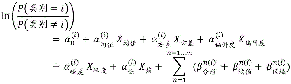
其中,方程式的左侧是属于和不属于类别i的一组特征的概率之间的比值比的自然对数。在右侧,α表示直接从密度图ROI提取的常见纹理特征。三个β使用一个二值图像从Otsu算法的总共m(例如,m=5)个不同掩码中提取的“高级”纹理特征。where the left side of the equation is the natural logarithm of the odds ratio between the probabilities of a set of features belonging to class i and not belonging to class i. On the right, α represents common texture features extracted directly from density map ROIs. Three β "high-level" texture features extracted from a total of m (eg, m=5) different masks of the Otsu algorithm using one binary image.
因此,在总共三个逻辑模型中,一个逻辑模型有21个系数,对应于四个分类类别。例如,对于一组已知特征,我们可以使用方程式(1)来计算纹理特征落入类别1、2或3的概率。Thus, out of a total of three logistic models, one logistic model has 21 coefficients, corresponding to the four categorical categories. For example, for a set of known features, we can use Equation (1) to calculate the probability that a texture feature falls into
纹理特征落入类别4的可能性如下:The likelihood of a texture feature falling into category 4 is as follows:
(2)P(类别=4)=1-P(类别=1)-P(类别=2)-P(类别=3)(2) P(class=4)=1-P(class=1)-P(class=2)-P(class=3)
回归模型的导出系数值总结在表1中(下一页)。小于0.05的p值用下划线标出;具有显著(p<0.05)值的模型系数被框出。The derived coefficient values for the regression models are summarized in Table 1 (next page). p-values less than 0.05 are underlined; model coefficients with significant (p<0.05) values are boxed.
当使用平行回归(比例比值模型)时,这些模型具有不同的截距,但是在分类之间具有共同的斜率。斜率系数的值表示特定特征对比值比的影响程度。例如,-27.31的系数α峰度估计值表明峰度的单位变化将影响图像在分类中与不在分类中的比值,其因子为exp(-27.31),其他都相等。When using parallel regression (proportional ratio models), these models have different intercepts, but a common slope between classes. The value of the slope coefficient indicates the degree of influence of a particular feature to the ratio of values. For example, the coefficient alpha kurtosis estimate of -27.31 indicates that a unit change in kurtosis will affect the ratio of an image in classification to not in classification, by a factor of exp(-27.31), all else being equal.
举例来说,使用方程式(1)和表1中的系数,对37个乳腺X线摄片评估BPE分类。结果总结在如图5所示的混淆矩阵中。For example, using equation (1) and the coefficients in Table 1, 37 mammograms were evaluated for BPE classification. The results are summarized in the confusion matrix shown in Figure 5.
表1 多项式回归模型系数Table 1 Polynomial regression model coefficients
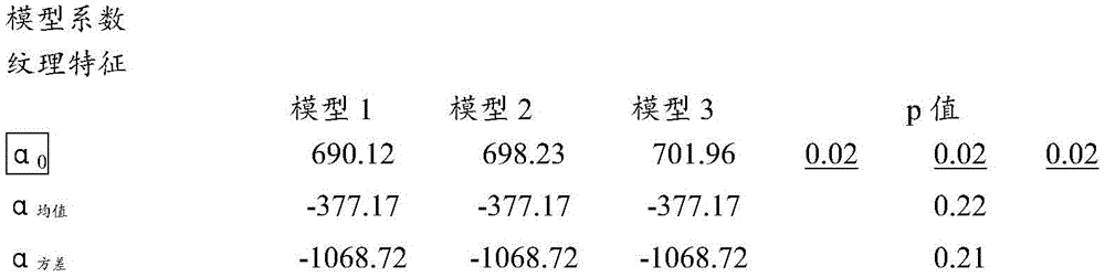
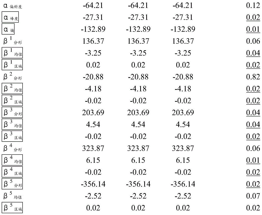
在混淆矩阵(图5)上,行对应于BPE的预测的类别(输出类别),以及列显示BPE的真实类别(目标类别)。对角线单元显示了回归模型正确估计观察类别的示例的数量(和百分比)。也就是说,它显示了真实类别和预测类别匹配的百分比。非对角线单元显示不正确的类别。图表最右侧的一列显示了每个预测类别的准确度,而图表底部的一行显示了每个真实类别的准确度。图表右下方的单元显示了整体准确度。On the confusion matrix (Fig. 5), the rows correspond to the predicted classes (output classes) of the BPE, and the columns show the true classes (target classes) of the BPE. The diagonal cells show the number (and percentage) of examples where the regression model correctly estimated the observed class. That is, it shows the percentage of matches between the true and predicted classes. Off-diagonal cells display incorrect categories. The far right column of the graph shows the accuracy of each predicted class, while the bottom row of the graph shows the accuracy of each true class. The cell at the bottom right of the graph shows the overall accuracy.
在图5中,对角线单元显示了回归模型正确类别的数量和百分比。例如,七个图像被正确分类为类别1,例如“最小”。这相当于全部37个图像的18.9%。类似地,14个案例被正确分类为类别2,“轻度”。这相当于所有图像的37.8%。In Figure 5, the diagonal cells show the number and percentage of correct classes for the regression model. For example, seven images were correctly classified as
其中一个“显著”图像被错误地分类为“中度”,而这相当于数据中全部37个图像的2.7%。类似地,一个“最小”被错误地分类为“轻度”,而这相当于所有数据的2.7%。One of the "significant" images was incorrectly classified as "moderate," which is equivalent to 2.7% of all 37 images in the data. Similarly, a "minimal" was incorrectly classified as "mild," which is equivalent to 2.7% of all data.
在七个“最小”预测中,100%是正确的。在15个“轻度”的预测中,93.3%是正确的,而6.7%是错误的。在八个“中度”案例中,87.5%被正确预测,而12.5%被预测为“显著”。在七个‘显著”案例中,100%被正确分类。Of the seven "minimal" predictions, 100% were correct. Of the 15 "mild" predictions, 93.3% were correct and 6.7% were wrong. Of the eight "moderate" cases, 87.5% were predicted correctly, while 12.5% were predicted as "significant". In seven 'significant' cases, 100% were correctly classified.
总的来说,94.6%的预测是正确的,而5.4%是不正确的分类。Overall, 94.6% of the predictions were correct, while 5.4% were incorrect classifications.
此外,该实施例涉及组织模式复杂性。因此,在一个实施例中,分形维数优选地被用于确定BPE分类。Furthermore, this embodiment deals with org-mode complexity. Therefore, in one embodiment, the fractal dimension is preferably used to determine the BPE classification.
图6中示出了推导分形维数的过程。从灰度级的ROI图像中,确定三个上升阈值:I1<I2<I3,因此产生三个单阈值二值掩码。使用多重Otsu算法,使用阈值间隔(I1,I2)和(I2,I3)获得两个掩码。从这些二值掩码中,提取它们相应的边界图,并通过简单的盒计数算法来计算Hausdorff分形维数。回顾分形维数的定义,它的值描述了对象边界的粗糙度或复杂性。因此,BPE分类可能与组织结构的复杂性相关。也就是说,组织呈现的不规则模式越多,乳房可能属于的BPE分类就越高。仅使用分形维数的70.3%的中度预测准确度支持了这一点(如图7所示)。The process of deriving the fractal dimension is shown in FIG. 6 . From the grayscale ROI image, three ascending thresholds are determined: I 1 <I 2 <I 3 , thus producing three single-threshold binary masks. Using the multiple Otsu algorithm, two masks were obtained using threshold intervals (I 1 , I 2 ) and (I 2 , I 3 ). From these binary masks, their corresponding boundary maps are extracted and the Hausdorff fractal dimension is calculated by a simple box counting algorithm. Looking back at the definition of fractal dimension, its value describes the roughness or complexity of an object's boundaries. Therefore, BPE classification may be related to the complexity of the tissue structure. That is, the more irregular patterns the tissue presents, the higher the BPE classification the breast is likely to belong to. This is supported by only a moderate prediction accuracy of 70.3% using fractal dimension (shown in Figure 7).
图10示出了两个乳房的示例:一个在左边的图像中示出了致密的低BPE读数,而另一个在右边的图像中示出了脂肪质的高BPE读数。右乳房的组织结构比左乳房的组织结构更复杂,这进一步支持了我们对组织模式复杂性和BPE分类之间可能的正相关的理解。Figure 10 shows an example of two breasts: one showing dense, low BPE readings in the left image, and the other showing fatty, high BPE readings in the right image. The histology of the right breast was more complex than that of the left breast, further supporting our understanding of the possible positive correlation between tissue pattern complexity and BPE classification.
在另一实施例中,为了准确且可靠地确定BPE,执行特征选择以降低每个预测器有十个以上观测值的建模的维数和复杂性。例如,特征选择方法包括:In another embodiment, to accurately and reliably determine BPE, feature selection is performed to reduce the dimensionality and complexity of modeling with more than ten observations per predictor. For example, feature selection methods include:
后向选择——使用所有特征拟合模型。然后,丢弃最不重要的特征。简化的模型被连续地重新拟合,直到所有剩余的变量都是统计显著的。最终保留了四个特征:Backward Selection - Fits the model using all features. Then, the least important features are discarded. The simplified model is continuously refitted until all remaining variables are statistically significant. In the end, four characteristics remain:
(3)(3)

他们建立了准确度只有35.1%的模型。They built a model with only 35.1% accuracy.
学习矢量量化模型——由完整的特征矩阵构建,报告重要性指数作为特征选择的指标,其中一些特征在一个分类中是重要的,但在其他分类中不是重要的。参考图9,从ROI提取的纹理特征的重要性显示用于BPE类别。方差是“最小”和“中度”类别中最不重要的特征,但是在“轻度”和“显著”中是重要的特征。Learn a vector quantization model - constructed from the full feature matrix, reporting importance indices as an indicator of feature selection, where some features are important in one classification but not in others. Referring to Figure 9, the importance of texture features extracted from ROIs is shown for the BPE category. Variance is the least important feature in the "minimal" and "moderate" categories, but is an important feature in the "mild" and "significant" categories.
递归特征消除——基于随机森林选择函数,如图8所示,通过其十一个属性给出最具可比性的结果,而三个属性给出最差的结果。Recursive Feature Elimination - Based on the random forest selection function, as shown in Figure 8, it gives the most comparable results through its eleven attributes, while three attributes give the worst results.
用于特征选择的其他算法包括Boruta算法和最小绝对收缩和选择算子(LASSO)。Other algorithms used for feature selection include Boruta's algorithm and Least Absolute Shrinkage and Selection Operator (LASSO).
在另一个实施例中,方法应用于CAD,由此通过使用CAD标记验证图像参数来确定CAD标记的有效性。图像参数一旦得到验证,将指导对对象以及标记的相关性和可靠性的确定。在其他优点中,减少了被错误识别的特征(“假阳性”)的数量,且不依赖于视觉感知。In another embodiment, the method is applied to CAD whereby the validity of the CAD mark is determined by validating image parameters using the CAD mark. Image parameters, once validated, guide the determination of object and marker relevance and reliability. Among other advantages, the number of misidentified features ("false positives") is reduced and does not rely on visual perception.
在单个研究中识别CAD标记(单个研究可能包括一个以上的图像),并将标记的图像与类似分类的存储图像(患者特定的存储的图像和/或累积的参考图像)进行比较,以便借助于图像质量标记和差异识别“高风险”研究,并依次存储单个研究。说明性的示例包括以下步骤:Identify CAD markers in a single study (single study may include more than one image) and compare the marked images with similarly classified stored images (patient-specific stored images and/or accumulated reference images) for Image quality flags and discrepancies identify "high risk" studies and store individual studies in turn. An illustrative example includes the following steps:
1/处理乳房的X射线图像(现场或发送到云)1/ Process an X-ray image of the breast (onsite or send to the cloud)
2/定性参数(诸如,密度、压缩、剂量、定位、对比度噪声比和模糊度被计算出并且图像“质量分数”被计算出)2/ Qualitative parameters (such as density, compression, dose, localization, contrast-to-noise ratio and blurriness are calculated and an image "quality score" is calculated)
3/在图像上运行CAD3/ Run CAD on the image
4/将CAD标记与验证的参数结合,以确定“总体风险评分”4/ Combine CAD markers with validated parameters to determine an "overall risk score"
5/“总体风险评分”识别“有风险”和/或“高风险”的患者,并警告读取者5/ "Overall Risk Score" identifies "at risk" and/or "high risk" patients and warns readers
例如,“总体风险评分”包括将CAD标记的数量和类型之间的差异进行比较;以及在同期图像之间,例如在左侧和右侧之间进行比较。For example, an "overall risk score" includes comparing differences between the number and type of CAD markers; and between contemporaneous images, eg, between left and right.
另外的优点包括:Additional benefits include:
标记的发生率和重要性的概览,以产生“缺失可能性”分数An overview of the frequency and importance of markers to produce a "missing likelihood" score
运动模糊特征,尤其是运动模糊的位置:例如,运动模糊最初被识别,然后外围上的运动模糊和相关标记被打折,以优先考虑与风险相关的位置(例如,中心)中的运动模糊标记。模糊Motion blur characteristics, especially the location of motion blur: For example, motion blur is initially identified, and then motion blur and associated markers on the periphery are discounted to prioritize motion blur markers in risk-relevant locations (eg, center). Vague
对在特征(例如乳房中的小叶,特别是II型和III型小叶)的预定附近或其内的标记被优先考虑。Preference is given to markers that are in the predetermined vicinity of or within features such as lobules in the breast, especially type II and type III lobules.
因此,对CAD标记的定性验证提高了工作流程的效率:告知与最高置信度CAD标记、具有“最大缺失可能性”的患者和/或基于密度和/或CAD标记的患者排名相关的风险模型。此外,除了减少“假阳性”之外,本发明还有助于避免其他错误:例如,在基于自动客观测量的情况下,读取者可能以“临床上不可接受”为由拒绝图像;由于图像模糊而没有CAD标记;并且图像被最佳压缩,并且图像清晰度导致更多和“更高置信度”的CAD标记;CAD标记出现在图像的一个部分而不出现在另一个部分中,表示图像的该部分模糊或压缩不良,而不是ROI的位置。Thus, qualitative validation of CAD markers increases the efficiency of the workflow: informing risk models associated with the highest confidence CAD markers, patients with "most likely missing" and/or patient ranking based on density and/or CAD markers. Furthermore, in addition to reducing "false positives", the present invention also helps to avoid other errors: for example, in cases based on automated objective measurements, the reader may reject images on the grounds that they are "clinically unacceptable"; Blurred without CAD marks; and the image is optimally compressed, and image clarity results in more and "higher confidence" CAD marks; CAD marks appear in one part of the image and not in another, indicating the image The part of the ROI is blurry or poorly compressed, not the location of the ROI.
本发明已经通过若干实施例进行了描述,并且进行了修改和替换,但是在阅读和理解了该描述之后,进一步的实施例和修改对于本领域技术人员来说将是明显的。所有这样的实施例和修改都旨在落在本发明的范围内。The invention has been described in terms of several embodiments, with modifications and substitutions, but further embodiments and modifications will be apparent to those skilled in the art upon reading and understanding this description. All such embodiments and modifications are intended to fall within the scope of the present invention.
Claims (29)
Applications Claiming Priority (7)
| Application Number | Priority Date | Filing Date | Title |
|---|---|---|---|
| GBGB1718378.1A GB201718378D0 (en) | 2017-11-07 | 2017-11-07 | Method and system for identifying 'at risk' patients |
| GB1718378.1 | 2017-11-07 | ||
| GBGB1719206.3A GB201719206D0 (en) | 2017-11-20 | 2017-11-20 | A method for background parenchymal enhancement categorisation |
| GB1719206.3 | 2017-11-20 | ||
| GB1806299.2 | 2018-04-18 | ||
| GBGB1806299.2A GB201806299D0 (en) | 2018-04-18 | 2018-04-18 | A system and method for quantification of tissue over time |
| PCT/IB2018/058663 WO2019092575A1 (en) | 2017-11-07 | 2018-11-05 | System and method for quantification of tissue over time |
Publications (2)
| Publication Number | Publication Date |
|---|---|
| CN111247592A true CN111247592A (en) | 2020-06-05 |
| CN111247592B CN111247592B (en) | 2024-04-16 |
Family
ID=64572399
Family Applications (1)
| Application Number | Title | Priority Date | Filing Date |
|---|---|---|---|
| CN201880068341.9A Active CN111247592B (en) | 2017-11-07 | 2018-11-05 | Systems and methods for quantifying tissue over time |
Country Status (5)
| Country | Link |
|---|---|
| US (1) | US20210097677A1 (en) |
| EP (1) | EP3707727B1 (en) |
| CN (1) | CN111247592B (en) |
| ES (1) | ES2992046T3 (en) |
| WO (1) | WO2019092575A1 (en) |
Families Citing this family (7)
| Publication number | Priority date | Publication date | Assignee | Title |
|---|---|---|---|---|
| JP7542059B2 (en) * | 2019-09-27 | 2024-08-29 | ホロジック, インコーポレイテッド | Motion detection of internal breast tissue in tomosynthesis |
| DE102020209711A1 (en) | 2020-07-31 | 2022-02-03 | Siemens Healthcare Gmbh | Method for evaluating calcifications in thoracic arteries |
| EP4578396A4 (en) * | 2022-08-22 | 2025-11-26 | Fujifilm Corp | INFORMATION PROCESSING DEVICE, INFORMATION PROCESSING METHOD AND PROGRAM |
| JPWO2024042889A1 (en) * | 2022-08-22 | 2024-02-29 | ||
| EP4338674B1 (en) * | 2022-09-14 | 2026-02-11 | Siemens Healthineers AG | Method and system for determining bpe in a contrast agent-enhanced breast x-ray examination |
| US20250078257A1 (en) * | 2023-09-01 | 2025-03-06 | Siemens Medical Solutions Usa, Inc. | Calibration of activity concentration uptake |
| CN120549537A (en) * | 2024-02-29 | 2025-08-29 | 通用电气精准医疗有限责任公司 | Quantification method and ultrasound imaging system for breast density |
Citations (11)
| Publication number | Priority date | Publication date | Assignee | Title |
|---|---|---|---|---|
| US20070014448A1 (en) * | 2005-06-30 | 2007-01-18 | Wheeler Frederick W | Method and system for lateral comparative image analysis and diagnosis |
| US20080187095A1 (en) * | 2005-05-03 | 2008-08-07 | The Regents Of The University Of California | Biopsy Systems For Breast Computed Tomography |
| US20090003670A1 (en) * | 2007-06-27 | 2009-01-01 | Fujifilm Corporation | Method and apparatus for thickness compensation in mammographic images |
| CN101853336A (en) * | 2010-06-13 | 2010-10-06 | 中国人民解放军总医院 | Bone surgery simulation human-computer interaction input method and device and image recognition method |
| CN102103132A (en) * | 2009-12-18 | 2011-06-22 | 中国科学院大连化学物理研究所 | Method for screening diabetes markers from body fluid metabonome profile |
| CN102362278A (en) * | 2009-03-26 | 2012-02-22 | 皇家飞利浦电子股份有限公司 | perfusion imaging |
| US20140010429A1 (en) * | 2010-11-30 | 2014-01-09 | Ralph Highnam | Imaging Technique and Imaging System |
| JP2014030556A (en) * | 2012-08-02 | 2014-02-20 | Hitachi Ltd | Image processing device |
| US20160133033A1 (en) * | 2013-06-06 | 2016-05-12 | John Michael Brady | A Method of Reconstruction of an Object from Projection Views |
| US20160256126A1 (en) * | 2013-11-19 | 2016-09-08 | Jeffery C. Wehnes | Obtaining breast density measurements and classifications |
| CN106295110A (en) * | 2015-06-24 | 2017-01-04 | 西门子保健有限责任公司 | Medical image processing method and image processing system |
Family Cites Families (1)
| Publication number | Priority date | Publication date | Assignee | Title |
|---|---|---|---|---|
| US8270695B2 (en) * | 2008-10-07 | 2012-09-18 | Carestream Health, Inc. | Diagnostic image processing with automatic self image quality validation |
-
2018
- 2018-11-05 CN CN201880068341.9A patent/CN111247592B/en active Active
- 2018-11-05 US US15/733,058 patent/US20210097677A1/en not_active Abandoned
- 2018-11-05 WO PCT/IB2018/058663 patent/WO2019092575A1/en not_active Ceased
- 2018-11-05 EP EP18812262.6A patent/EP3707727B1/en active Active
- 2018-11-05 ES ES18812262T patent/ES2992046T3/en active Active
Patent Citations (11)
| Publication number | Priority date | Publication date | Assignee | Title |
|---|---|---|---|---|
| US20080187095A1 (en) * | 2005-05-03 | 2008-08-07 | The Regents Of The University Of California | Biopsy Systems For Breast Computed Tomography |
| US20070014448A1 (en) * | 2005-06-30 | 2007-01-18 | Wheeler Frederick W | Method and system for lateral comparative image analysis and diagnosis |
| US20090003670A1 (en) * | 2007-06-27 | 2009-01-01 | Fujifilm Corporation | Method and apparatus for thickness compensation in mammographic images |
| CN102362278A (en) * | 2009-03-26 | 2012-02-22 | 皇家飞利浦电子股份有限公司 | perfusion imaging |
| CN102103132A (en) * | 2009-12-18 | 2011-06-22 | 中国科学院大连化学物理研究所 | Method for screening diabetes markers from body fluid metabonome profile |
| CN101853336A (en) * | 2010-06-13 | 2010-10-06 | 中国人民解放军总医院 | Bone surgery simulation human-computer interaction input method and device and image recognition method |
| US20140010429A1 (en) * | 2010-11-30 | 2014-01-09 | Ralph Highnam | Imaging Technique and Imaging System |
| JP2014030556A (en) * | 2012-08-02 | 2014-02-20 | Hitachi Ltd | Image processing device |
| US20160133033A1 (en) * | 2013-06-06 | 2016-05-12 | John Michael Brady | A Method of Reconstruction of an Object from Projection Views |
| US20160256126A1 (en) * | 2013-11-19 | 2016-09-08 | Jeffery C. Wehnes | Obtaining breast density measurements and classifications |
| CN106295110A (en) * | 2015-06-24 | 2017-01-04 | 西门子保健有限责任公司 | Medical image processing method and image processing system |
Also Published As
| Publication number | Publication date |
|---|---|
| US20210097677A1 (en) | 2021-04-01 |
| EP3707727B1 (en) | 2024-09-04 |
| CN111247592B (en) | 2024-04-16 |
| WO2019092575A1 (en) | 2019-05-16 |
| EP3707727A1 (en) | 2020-09-16 |
| EP3707727C0 (en) | 2024-09-04 |
| ES2992046T3 (en) | 2024-12-05 |
Similar Documents
| Publication | Publication Date | Title |
|---|---|---|
| CN111247592B (en) | Systems and methods for quantifying tissue over time | |
| US10984528B2 (en) | System and apparatus for clinical decision optimisation | |
| US8634610B2 (en) | System and method for assessing cancer risk | |
| US8238630B2 (en) | Image processing apparatus and program for the same | |
| Gweon et al. | Radiologist assessment of breast density by BI-RADS categories versus fully automated volumetric assessment | |
| Giger et al. | Anniversary paper: history and status of CAD and quantitative image analysis: the role of medical physics and AAPM | |
| US9865067B2 (en) | Method of reconstruction of an object from projection views | |
| US20030174873A1 (en) | Method and system for risk-modulated diagnosis of disease | |
| JP7612693B2 (en) | Systems and methods for analyzing two-dimensional and three-dimensional image data - Patents.com | |
| US12507978B2 (en) | Methods and systems for cancer risk assessment using tissue sound speed and stiffness | |
| US20170332992A1 (en) | Miniaturized Phantoms for Quantitative Image Analysis and Quality Control | |
| US20110052025A1 (en) | System for imaging pathway verification | |
| Jamal et al. | Quantitative assessment of breast density from digitized mammograms into Tabar's patterns | |
| Lu et al. | Computing mammographic density from a multiple regression model constructed with image-acquisition parameters from a full-field digital mammographic unit | |
| US20250037281A1 (en) | System and method for evaluation of image quality | |
| Salh et al. | Review of Techniques Used in Computer-Aided Detection and Diagnosis for Breast Cancer Using Mammograms | |
| WO2024252316A1 (en) | System and method for interpretation of medical images | |
| Khan et al. | Automatic BI-RADS classification of mammograms | |
| WO2024258873A2 (en) | Ai driven analysis and explanation of medical images | |
| Diffey | Development and evaluation of a method for measuring breast density | |
| Kallenberg | Quantitative analysis of breast images | |
| List | Breast compression parameters among women imaged with full field digital mammography and breast tomosynthesis in BreastScreen Norway |
Legal Events
| Date | Code | Title | Description |
|---|---|---|---|
| PB01 | Publication | ||
| PB01 | Publication | ||
| SE01 | Entry into force of request for substantive examination | ||
| SE01 | Entry into force of request for substantive examination | ||
| GR01 | Patent grant | ||
| GR01 | Patent grant |