CN111166316A - Method for calculating instantaneous contrast wave-free ratio and contrast diastolic pressure ratio based on contrast images - Google Patents
Method for calculating instantaneous contrast wave-free ratio and contrast diastolic pressure ratio based on contrast images Download PDFInfo
- Publication number
- CN111166316A CN111166316A CN201811344074.1A CN201811344074A CN111166316A CN 111166316 A CN111166316 A CN 111166316A CN 201811344074 A CN201811344074 A CN 201811344074A CN 111166316 A CN111166316 A CN 111166316A
- Authority
- CN
- China
- Prior art keywords
- contrast
- pressure
- blood
- dimensional
- blood vessel
- Prior art date
- Legal status (The legal status is an assumption and is not a legal conclusion. Google has not performed a legal analysis and makes no representation as to the accuracy of the status listed.)
- Granted
Links
Images


Classifications
-
- A—HUMAN NECESSITIES
- A61—MEDICAL OR VETERINARY SCIENCE; HYGIENE
- A61B—DIAGNOSIS; SURGERY; IDENTIFICATION
- A61B5/00—Measuring for diagnostic purposes; Identification of persons
- A61B5/02—Detecting, measuring or recording for evaluating the cardiovascular system, e.g. pulse, heart rate, blood pressure or blood flow
- A61B5/026—Measuring blood flow
-
- A—HUMAN NECESSITIES
- A61—MEDICAL OR VETERINARY SCIENCE; HYGIENE
- A61B—DIAGNOSIS; SURGERY; IDENTIFICATION
- A61B5/00—Measuring for diagnostic purposes; Identification of persons
- A61B5/02—Detecting, measuring or recording for evaluating the cardiovascular system, e.g. pulse, heart rate, blood pressure or blood flow
- A61B5/02007—Evaluating blood vessel condition, e.g. elasticity, compliance
-
- A—HUMAN NECESSITIES
- A61—MEDICAL OR VETERINARY SCIENCE; HYGIENE
- A61B—DIAGNOSIS; SURGERY; IDENTIFICATION
- A61B5/00—Measuring for diagnostic purposes; Identification of persons
- A61B5/02—Detecting, measuring or recording for evaluating the cardiovascular system, e.g. pulse, heart rate, blood pressure or blood flow
- A61B5/021—Measuring pressure in heart or blood vessels
-
- G—PHYSICS
- G06—COMPUTING OR CALCULATING; COUNTING
- G06T—IMAGE DATA PROCESSING OR GENERATION, IN GENERAL
- G06T17/00—Three dimensional [3D] modelling, e.g. data description of 3D objects
- G06T17/20—Finite element generation, e.g. wire-frame surface description, tesselation
Landscapes
- Health & Medical Sciences (AREA)
- Life Sciences & Earth Sciences (AREA)
- Engineering & Computer Science (AREA)
- Physics & Mathematics (AREA)
- Cardiology (AREA)
- Molecular Biology (AREA)
- Public Health (AREA)
- Pathology (AREA)
- Physiology (AREA)
- Biomedical Technology (AREA)
- Heart & Thoracic Surgery (AREA)
- Medical Informatics (AREA)
- Veterinary Medicine (AREA)
- Surgery (AREA)
- Animal Behavior & Ethology (AREA)
- General Health & Medical Sciences (AREA)
- Biophysics (AREA)
- Vascular Medicine (AREA)
- Hematology (AREA)
- Computer Graphics (AREA)
- Geometry (AREA)
- Software Systems (AREA)
- General Physics & Mathematics (AREA)
- Theoretical Computer Science (AREA)
- Apparatus For Radiation Diagnosis (AREA)
- Measuring Pulse, Heart Rate, Blood Pressure Or Blood Flow (AREA)
Abstract
The invention discloses a method for calculating an instantaneous contrast wave-free ratio and a contrast diastolic pressure ratio based on a contrast image, which comprises the following steps of: measuring the pressure P at the coronary ostia of the heart in diastolea(ii) a Acquiring the two-dimensional caliber and the length of a blood vessel through a radiography image, generating a three-dimensional blood vessel grid model through two radiography images and acquiring the three-dimensional caliber and the length of the blood vessel; during diastole, the time taken by blood containing a contrast agent from a starting point to an ending point of a specified blood vessel is measured, and the blood flow velocity V is calculated from the time and the three-dimensional length of the blood vessel1(ii) a Will V1Calculating the pressure from the coronary inlet to the distal end of the coronary stenosis as the coronary inlet flow velocityForce drop Δ P, mean pressure in the stenotic distal coronary artery Pd=Pa- Δ P, calculating the contrast instantaneous wave-free ratio and the contrast diastolic pressure ratio. caiFR, cadFR and cadFR can be obtained by conventional imaging without the use of vasodilators.
Description
Technical Field
The invention relates to the field of coronary angiography evaluation, in particular to a method for determining a contrast instantaneous mode-free ratio (caiFR) and a contrast diastolic pressure ratio (cadFR and cadFR) only through a contrast image and an aorta pressure.
Background
The Fractional Flow Reserve (FFR) can indicate the influence of coronary stenosis lesion on distal blood flow, diagnose whether the myocardium is ischemic, and become a recognized index for functional evaluation of coronary stenosis. FFR is defined as the ratio of the maximum blood flow provided by a stenotic coronary artery to the myocardium in the innervation area to the maximum blood flow provided to the myocardium in the normal state of the same coronary artery. Can be simplified into the narrow distal coronary artery internal pressure equalizing (P) under the maximal hyperemia state of the cardiac muscled) And mean pressure (P) of coronary artery and oral aortaa) I.e. FFR ═ Pd/Pa。
When the FFR is determined, the FFR is calculated by obtaining the mean pressure in the coronary artery at the distal end of the stenosis by different means based on the blood flow velocity in the maximal hyperemia state of the myocardium and the mean pressure in the aorta at the mouth of the coronary artery. However, maximal myocardial hyperemia requires coronary or intravenous injection of adenosine or ATP, which causes a decrease in aortic pressure and has certain side effects such as atrioventricular block, sinus bradycardia, sinus arrest, etc., contraindications including 2 or 3 degree atrioventricular block, sinoatrial node disease, tracheal or bronchial asthma, and adenosine hypersensitivity.
The instantaneous waveform-free ratio (iFR) can provide a similar method of measuring pressure in the coronary arteries as Fractional Flow Reserve (FFR). The iFR does not need a vasodilator, is simple to operate and can be more applied to coronary intervention treatment. The ADVISE study found that, during a certain period of diastole (called the no-waveform period), the intracoronary microvascular resistance was relatively the most stable and the lowest, and that the average achieved during coronary hyperemia with vasodilators such as adenosine was the averageThe resistance is similar. I.e. iFR ═ PdWave-free period/PaWave-free period(PdWave-free period: the coronary artery at the distal end of the stenotic lesion is evenly compressed during the absence of the waveform. PaWave-free period: the mean aortic pressure during the absence of the waveform. Operation time of the instantaneous waveform-free period: 25% of the time after the start of the waveform-free period in diastole, and the calculation is stopped at the time 5ms before the start of systole). A study article was published in top-grade medical journal NEJM that the IFR-directed revascularization strategy was not inferior to the FFR-directed reconstruction strategy in patients with stable angina or acute coronary syndrome, and was similar in the incidence of major adverse cardiac events over 12 months.
The resting diastolic pressure ratio (dFR and DFR) can be expressed as: dFR ═ PdDiastolic period/PaDiastolic period(PdDiastolic period: the mean coronary pressure distal to the stenotic lesion during the diastolic phase. PaDiastolic period: aortic mean pressure during diastolic state); DFR ═ PdDiastolic hyperemia free period/PaDiastolic hyperemia free period(PdDiastolic hyperemia free period: in the interval from the aorta pressure being less than the average aorta pressure to the minimum aorta pressure, the coronary artery pressure at the distal end of the stenosis is the average coronary pressure. PaDiastolic hyperemia free period: the mean aortic pressure in the interval from the aortic pressure being less than the mean aortic pressure to the aortic pressure being the lowest). Further studies have shown that the resting diastolic pressure ratio (dFR and DFR) is substantially fully equivalent to the instantaneous non-waveform ratio (iFR). Therefore, we can obtain iFR ≡ DFR ≡ dFR ═ PdDiastolic period/PaDiastolic period。
Currently, the existing methods for measuring instantaneous mode-free ratio (iFR) and resting diastolic pressure ratio (dFR and DFR) are mainly: and measuring corresponding diastolic intervals in the resting state of the pressure guide wire to determine iFR, dFR and DFR. The pressure guide wire is needed to be relied on for measurement, the tail end of the blood vessel needs to be intervened when the pressure guide wire is used for measurement, the operation difficulty and risk are increased, and meanwhile, the large-scale application of the pressure guide wire is limited due to the expensive price of the pressure guide wire. A degree of myocardial hyperemia may result in contrast states relative to resting states, and thus contrast transient wave-free ratios (caiFR), contrast diastolic pressure ratios (cadFR and cadFR) should be more indicative of myocardial ischemia.
Disclosure of Invention
In order to solve the technical problems, the invention aims to: a method for calculating a contrast instantaneous wave-free ratio and a contrast diastolic pressure ratio based on a contrast image is provided for detecting myocardial ischemia by conventional coronary angiography procedures in patients with coronary heart disease, i.e., without the use of vasodilators (i.e., without the need for maximal myocardial hyperemia and without the use of adenosine or ATP). The instantaneous wave-free ratio (caiFR), diastolic pressure ratio (cadFR and cadFR) in the contrast state are calculated from the conventional contrast image, aortic pressure and blood flow.
The technical scheme of the invention is as follows:
a method of calculating a contrast instantaneous wave-free ratio and a contrast diastolic pressure ratio based on a contrast image, comprising the steps of:
s01: measuring pressure Pa of a coronary ostium of a heart in diastole by a blood pressure sensor;
s02: acquiring the two-dimensional diameter and length of a blood vessel through a radiography image, generating a three-dimensional blood vessel grid model through two radiography images with an included angle of more than 30 degrees, and acquiring the three-dimensional diameter and length of the blood vessel;
s03: measuring the time taken by blood containing a contrast agent from a starting point to an ending point of a specified blood vessel in diastole, and calculating a blood flow velocity V1 according to the time and the three-dimensional length of the blood vessel;
s04: the blood flow velocity V1 in the contrast state obtained by the calculation is used as a coronary artery inlet flow velocity, a pressure drop Δ P from the coronary artery inlet to the distal end of the coronary artery stenosis is calculated, the mean pressure Pd in the distal coronary artery of the diastolic stenosis is Pa- Δ P, and a contrast instantaneous no-mode ratio (caiFR) and a contrast resting state diastolic pressure ratio (caDFR and caDFR) are calculated by the formula caiFR ≡ caDFR ≈ caDFR ═ Pd/Pa.
In a preferred embodiment, the step S01 includes connecting a pressure tube using a blood pressure sensor to a multi-connection tee, and connecting the pressure tube to the multi-connection tee through a contrast catheterThe heart coronary artery is connected with the mouth part, the pressure tube of the blood pressure sensor is filled with saline, the blood pressure sensor and the heart are kept at the same horizontal position, the pressure wave measured by the blood pressure sensor is the pressure wave of the heart coronary artery, and the average value of the instantaneous pressure is P in diastolea。
In a preferred embodiment, the method for generating a three-dimensional blood vessel mesh model in step S02 includes the following steps:
s21: performing three-dimensional reconstruction on 2D structural data of two segmented blood vessels with a mapping relation on two X-ray coronary angiography images with an included angle of more than 30 degrees to obtain 3D structural data of the segmented blood vessels;
s22: and repeating the step S21 until the three-dimensional reconstruction of all the segmented blood vessels is completed, and combining the reconstructed segmented blood vessels to obtain a complete three-dimensional blood vessel mesh model.
In a preferred embodiment, the blood flow velocity V is calculated in step S031The specific method comprises the following steps:
s31: the heart rate of a specified patient is acquired as H times/minute, the image frequency is acquired from contrast image information as S frames/second, and the calculation formula of the frame number X is as follows: x ═ 1 ÷ (H ÷ 60)) × S;
s32: respectively obtaining a starting point and an ending point of a diastolic period of a heartbeat cycle on images corresponding to a two-dimensional starting frame and an ending frame according to the number of frames of images in the diastolic period of the heartbeat cycle, and then intercepting the length of a blood vessel in the diastolic period of the heartbeat cycle in a three-dimensional blood vessel mesh model according to the starting point and the ending point;
s33: by the formula V1Calculating the blood flow velocity V as L/P1L is the length of the blood vessel, P is the time taken for one heart cycle to diastole, P ═ X ÷ S.
In a preferred embodiment, the specific method for calculating the pressure drop Δ P from the coronary artery entrance to the distal end of the coronary stenosis in step S04 is as follows:
s41: solving a basic formula of the incompressible flow based on the blood flow velocity and the three-dimensional blood vessel mesh model, solving the three-dimensional blood vessel mesh model, and solving continuity and a Navier-Stokes equation by using a numerical method:


wherein P, rho and mu are respectively flow velocity, pressure, blood flow density and blood flow viscosity;
the inlet boundary condition is the blood flow velocity, and the outlet boundary condition is the out-flow boundary condition;
s42: the pressure drop ap from the entrance to various points downstream along the centerline of the vessel is calculated.
Compared with the prior art, the invention has the advantages that:
myocardial ischemia is detected by conventional coronary angiography procedures in patients with coronary heart disease, i.e., without the use of vasodilators (i.e., without the need for maximal hyperemia of the myocardium and without the use of adenosine or ATP). The instantaneous mode-free ratio (caiFR), contrast diastolic pressure ratio (cadFR and cadFR) were calculated from the conventional contrast image, aortic pressure and blood flow. The pressure guide wire does not need to be additionally inserted for measurement, the operation is simple and convenient, the operation difficulty and risk are greatly reduced, and the method can be clinically popularized and applied in a large scale.
Drawings
The invention is further described with reference to the following figures and examples:
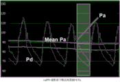
FIG. 1 is a schematic of the instantaneous mode-free ratio (caiFR) of contrast;
FIG. 2 is a schematic representation of contrast diastolic pressure ratio (cadFR);
FIG. 3 is a graphical representation of contrast diastolic pressure ratio (cadFR);
FIG. 4 is a flow chart of a method of the present invention;
FIG. 5 is a two-dimensional blood vessel image;
FIG. 6.1 is an image of the body position-one contrast agent flow to the catheter port;
FIG. 6.2 is an image of body position-contrast agent flow to the distal end of the vessel;
FIG. 6.3 is an image of the second contrast agent flow to the catheter port;
FIG. 6.4 is an image of the second contrast agent flow to the distal end of the vessel;
FIG. 7 is a cross-sectional screen shot of a grid;
fig. 8 is a cross-sectional view of a grid.
Detailed Description
In order to make the objects, technical solutions and advantages of the present invention more apparent, the present invention will be described in further detail with reference to the accompanying drawings in conjunction with the following detailed description. It should be understood that the description is intended to be exemplary only, and is not intended to limit the scope of the present invention. Moreover, in the following description, descriptions of well-known structures and techniques are omitted so as to not unnecessarily obscure the concepts of the present invention.
The present invention determines a contrast instantaneous mode-free ratio (caiFR) and a contrast diastolic pressure ratio (cadFR and cadFR) only from a contrast image and an aortic pressure, as shown in fig. 1, the contrast instantaneous mode-free ratio (caiFR), caiFR being Pd/Pa within a WFP interval in a contrast state, as shown in fig. 2, the contrast diastolic pressure ratio cadFR being Pd/Pa within a Diastole interval in the contrast state, as shown in fig. 3, the contrast diastolic pressure ratio cadFR being Pd/Pa within a contrast state lower frame interval, the instantaneous mode-free ratio (caiFR) and the contrast diastolic pressure ratios (cadFR and cadFR) being substantially completely equivalent, and the caiFR cadFR being cadFR.
As shown in fig. 4, the method of the present invention for determining the instantaneous contrast mode-free ratio (caiFR) and the contrast diastolic pressure ratio (cadFR and cadFR) only from the contrast image and the aortic pressure includes the following steps.
Step S1: measuring the pressure P of the coronary ostia of the heart in diastole by means of a blood pressure sensoraThe specific method comprises the following steps:
connecting pressure tube of blood pressure sensor to multi-connected tee, connecting with coronary artery of heart via contrast catheter, filling pressure tube of blood pressure sensor with saline, and keeping blood pressure sensor and heartThe heart is at the same horizontal position, the pressure wave measured by the blood pressure sensor is the pressure wave of the coronary ostia of the heart, and the average value of the instantaneous pressure is P in the diastolea。
Step S2: acquiring the two-dimensional diameter and length of a blood vessel through a radiography image, as shown in fig. 5, generating a three-dimensional blood vessel mesh model through two radiography images with an included angle of more than 30 degrees, and acquiring the three-dimensional diameter and length of the blood vessel;
the specific method of the three-dimensional blood vessel mesh model is as follows:
performing three-dimensional reconstruction on the 2D structural data of two segmented blood vessels which are in a mapping relation on the X-ray coronary angiography images at two different angles to obtain the 3D structural data of the segmented blood vessels;
repeating the above steps until the three-dimensional reconstruction of all the segmented blood vessels is completed, and then combining the reconstructed segmented blood vessels to obtain the complete three-dimensional blood vessel, as shown in fig. 7 and 8.
Step S3: as shown in FIGS. 6.1-6.4, during diastole, the time taken for blood (containing contrast agent) to pass from the starting point (6.1, 6.3) to the ending point (6.2, 6.4) of a specified segment of blood vessel (including a possible crime vessel) is measured, and the blood flow velocity V is calculated from the time and the three-dimensional length of the blood vessel1The specific method comprises the following steps:
the heart rate of a specified patient is acquired as H times/minute, the image frequency is acquired from contrast image information as S frames/second, and the calculation formula of the frame number X is as follows: x ═ 1 ÷ (H ÷ 60)) × S;
respectively obtaining a starting point and an ending point of a diastolic period of a heartbeat cycle on images corresponding to a two-dimensional starting frame and an ending frame, such as fig. 6.1 and 6.2 or fig. 6.3 and 6.4, according to the number of frames of the images in the diastolic period of the heartbeat cycle, and then intercepting the length of a blood vessel of the diastolic period of the heartbeat cycle from three-dimensional synthetic data according to the starting point and the ending point;
assuming that the length of the intercepted blood vessel is L and the time taken for the diastole of one heart cycle is P, the method is represented by formula 1: p ═ X ÷ S; equation 2: v1Obtain the blood flow velocity V as L/P1。
Step S4: will step withThe blood flow velocity V in the contrast state calculated in step S31Calculating the pressure drop Δ P from the coronary inlet to the distal end of the coronary stenosis as the coronary inlet flow velocity, averaging the pressure P within the coronary artery distal to the diastolic stenosisd=Pa- Δ P, and then via the formula caiFR ≡ caDFR ═ Pd/PaThe instantaneous contrast mode-free ratio (caiFR) and the resting-state contrast diastolic pressure ratio (cadFR and cadFR) were calculated.
The specific method for calculating the pressure drop Δ P from the coronary artery entrance to the distal end of the coronary stenosis in step S4 is as follows:
solving a basic formula of the incompressible flow based on the blood flow velocity and the three-dimensional blood vessel mesh model, solving the three-dimensional blood vessel mesh model, and solving continuity and a Navier-Stokes equation by using a numerical method:


wherein P, rho and mu are respectively flow velocity, pressure, blood flow density and blood flow viscosity;
the inlet boundary condition is the blood flow velocity, and the outlet boundary condition is the out-flow boundary condition;
the pressure drop ap from the entrance to various points downstream along the centerline of the vessel is calculated.
It is to be understood that the above-described embodiments of the present invention are merely illustrative of or explaining the principles of the invention and are not to be construed as limiting the invention. Therefore, any modification, equivalent replacement, improvement and the like made without departing from the spirit and scope of the present invention should be included in the protection scope of the present invention. Further, it is intended that the appended claims cover all such variations and modifications as fall within the scope and boundaries of the appended claims or the equivalents of such scope and boundaries.
Claims (5)
1. A method of calculating a contrast instantaneous wave-free ratio and a contrast diastolic pressure ratio based on a contrast image, comprising the steps of:
s01: measuring pressure Pa of a coronary ostium of a heart in diastole by a blood pressure sensor;
s02: acquiring the two-dimensional diameter and length of a blood vessel through a radiography image, generating a three-dimensional blood vessel grid model through two radiography images with an included angle of more than 30 degrees, and acquiring the three-dimensional diameter and length of the blood vessel;
s03: measuring the time taken by blood containing a contrast agent from a starting point to an ending point of a specified blood vessel in diastole, and calculating a blood flow velocity V1 according to the time and the three-dimensional length of the blood vessel;
s04: the blood flow velocity V1 in the contrast state obtained by the calculation is used as a coronary artery inlet flow velocity, a pressure drop Δ P from the coronary artery inlet to the distal end of the coronary artery stenosis is calculated, the mean pressure Pd in the distal coronary artery of the diastolic stenosis is Pa- Δ P, and a contrast instantaneous no-mode ratio (caiFR) and a contrast resting state diastolic pressure ratio (caDFR and caDFR) are calculated by the formula caiFR ≡ caDFR ≈ caDFR ═ Pd/Pa.
2. The method for calculating the contrast instantaneous wave-free ratio and the contrast diastolic pressure ratio based on the contrast image as claimed in claim 1, wherein the step S01 includes connecting the pressure tube of the blood pressure sensor to the multi-connected tee, connecting the pressure tube to the coronary ostium of the heart through the contrast catheter, filling the pressure tube of the blood pressure sensor with saline, and maintaining the blood pressure sensor at the same horizontal position with the heart, the pressure wave measured by the blood pressure sensor being the pressure wave of the coronary ostium of the heart, and the average value of the instantaneous pressure during the diastolic period being Pa。
3. The method for calculating the instantaneous wave-free ratio and the diastolic pressure ratio of contrast based on contrast images as claimed in claim 1, wherein the method for generating the three-dimensional blood vessel mesh model in step S02 comprises the steps of:
s21: performing three-dimensional reconstruction on 2D structural data of two segmented blood vessels with a mapping relation on two X-ray coronary angiography images with an included angle of more than 30 degrees to obtain 3D structural data of the segmented blood vessels;
s22: and repeating the step S21 until the three-dimensional reconstruction of all the segmented blood vessels is completed, and combining the reconstructed segmented blood vessels to obtain a complete three-dimensional blood vessel mesh model.
4. The method for calculating the instantaneous wave-free ratio and diastolic pressure ratio of contrast based on contrast images as claimed in claim 1, wherein the blood flow velocity V is calculated in step S031The specific method comprises the following steps:
s31: the heart rate of a specified patient is acquired as H times/minute, the image frequency is acquired from contrast image information as S frames/second, and the calculation formula of the frame number X is as follows: x ═ 1 ÷ (H ÷ 60)) × S;
s32: respectively obtaining a starting point and an ending point of a diastolic period of a heartbeat cycle on images corresponding to a two-dimensional starting frame and an ending frame according to the number of frames of images in the diastolic period of the heartbeat cycle, and then intercepting the length of a blood vessel in the diastolic period of the heartbeat cycle in a three-dimensional blood vessel mesh model according to the starting point and the ending point;
s33: by the formula V1Calculating the blood flow velocity V as L/P1L is the length of the blood vessel, P is the time taken for one heart cycle to diastole, P ═ X ÷ S.
5. The method for calculating the instantaneous contrast ratio and the diastolic contrast ratio based on contrast images as claimed in claim 1, wherein the specific method for calculating the pressure drop Δ P from the coronary artery entrance to the distal end of the coronary stenosis in step S04 is as follows:
s41: solving a basic formula of the incompressible flow based on the blood flow velocity and the three-dimensional blood vessel mesh model, solving the three-dimensional blood vessel mesh model, and solving continuity and a Navier-Stokes equation by using a numerical method:


wherein P, rho and mu are respectively flow velocity, pressure, blood flow density and blood flow viscosity;
the inlet boundary condition is the blood flow velocity, and the outlet boundary condition is the out-flow boundary condition;
s42: the pressure drop ap from the entrance to various points downstream along the centerline of the vessel is calculated.
Priority Applications (9)
| Application Number | Priority Date | Filing Date | Title |
|---|---|---|---|
| CN201811344074.1A CN111166316B (en) | 2018-11-13 | 2018-11-13 | Method for calculating instantaneous contrast wave-free ratio and contrast diastolic pressure ratio based on contrast images |
| PCT/CN2019/071206 WO2020098140A1 (en) | 2018-11-13 | 2019-01-10 | Method for calculating instantaneous wave-free ratio and diastolic pressure ratio during angiography on basis of angiography images |
| EP19885173.5A EP3881758B1 (en) | 2018-11-13 | 2019-11-13 | Method, apparatus and system for acquiring vascular assessment parameter on basis of angiographic image |
| CN201980040566.8A CN112384137B (en) | 2018-11-13 | 2019-11-13 | Method and device for obtaining resting vascular assessment parameters based on contrast images |
| CN201980040404.4A CN112384136A (en) | 2018-11-13 | 2019-11-13 | Method, device and system for obtaining blood vessel evaluation parameters based on radiography images |
| PCT/CN2019/118053 WO2020098704A1 (en) | 2018-11-13 | 2019-11-13 | Method, apparatus and system for acquiring vascular assessment parameter on basis of angiographic image |
| PCT/CN2019/118058 WO2020098705A1 (en) | 2018-11-13 | 2019-11-13 | Method and device for obtaining vascular assessment parameters in resting state on basis of angiographic image |
| JP2021523637A JP7162934B2 (en) | 2018-11-13 | 2019-11-13 | Method, apparatus and system for obtaining vascular assessment parameters based on contrast-enhanced images |
| US17/237,662 US20210236000A1 (en) | 2018-11-13 | 2021-04-22 | Method, device and system for acquiring blood vessel evaluation parameters based on angiographic image |
Applications Claiming Priority (1)
| Application Number | Priority Date | Filing Date | Title |
|---|---|---|---|
| CN201811344074.1A CN111166316B (en) | 2018-11-13 | 2018-11-13 | Method for calculating instantaneous contrast wave-free ratio and contrast diastolic pressure ratio based on contrast images |
Publications (2)
| Publication Number | Publication Date |
|---|---|
| CN111166316A true CN111166316A (en) | 2020-05-19 |
| CN111166316B CN111166316B (en) | 2023-03-21 |
Family
ID=70646071
Family Applications (1)
| Application Number | Title | Priority Date | Filing Date |
|---|---|---|---|
| CN201811344074.1A Active CN111166316B (en) | 2018-11-13 | 2018-11-13 | Method for calculating instantaneous contrast wave-free ratio and contrast diastolic pressure ratio based on contrast images |
Country Status (2)
| Country | Link |
|---|---|
| CN (1) | CN111166316B (en) |
| WO (1) | WO2020098140A1 (en) |
Cited By (2)
| Publication number | Priority date | Publication date | Assignee | Title |
|---|---|---|---|---|
| CN112690814A (en) * | 2020-11-06 | 2021-04-23 | 杭州阿特瑞科技有限公司 | Low-error coronary artery blood flow reserve fraction measuring method |
| CN113018553A (en) * | 2021-02-05 | 2021-06-25 | 深圳圣诺医疗设备股份有限公司 | Flushing pressure detection method and device based on peristaltic pump and flushing suction system |
Families Citing this family (1)
| Publication number | Priority date | Publication date | Assignee | Title |
|---|---|---|---|---|
| CN114549539A (en) * | 2022-02-25 | 2022-05-27 | 中国医学科学院阜外医院 | Method and system for calculating blood flow SYNTAX scoring weight distribution |
Citations (9)
| Publication number | Priority date | Publication date | Assignee | Title |
|---|---|---|---|---|
| US8548778B1 (en) * | 2012-05-14 | 2013-10-01 | Heartflow, Inc. | Method and system for providing information from a patient-specific model of blood flow |
| US20140249784A1 (en) * | 2013-03-04 | 2014-09-04 | Heartflow, Inc. | Method and system for sensitivity analysis in modeling blood flow characteristics |
| US20150324962A1 (en) * | 2014-05-09 | 2015-11-12 | Siemens Aktiengesellschaft | Method and System for Non-Invasive Computation of Hemodynamic Indices for Coronary Artery Stenosis |
| CN106327487A (en) * | 2016-08-18 | 2017-01-11 | 苏州润心医疗科技有限公司 | Coronary artery blood flow reserve fraction calculation method based on X ray coronary artery angiographic image |
| US20170046834A1 (en) * | 2014-04-22 | 2017-02-16 | Siemens Healthcare Gmbh | Method and system for hemodynamic computation in coronary arteries |
| CN107580470A (en) * | 2015-01-15 | 2018-01-12 | 皇家飞利浦有限公司 | Instantaneous delivery lays in computer tomography |
| CN108242075A (en) * | 2018-01-05 | 2018-07-03 | 苏州润迈德医疗科技有限公司 | A kind of multi-angle reconstructing blood vessel method based on X ray coronary angiography image |
| CN108245178A (en) * | 2018-01-11 | 2018-07-06 | 苏州润迈德医疗科技有限公司 | A kind of blood flowing speed computational methods based on X ray coronary angiography image |
| CN108550189A (en) * | 2018-05-03 | 2018-09-18 | 苏州润迈德医疗科技有限公司 | Microcirculation drag index computational methods based on contrastographic picture and fluid mechanic model |
Family Cites Families (1)
| Publication number | Priority date | Publication date | Assignee | Title |
|---|---|---|---|---|
| US9775523B2 (en) * | 2013-10-14 | 2017-10-03 | Boston Scientific Scimed, Inc. | Pressure sensing guidewire and methods for calculating fractional flow reserve |
-
2018
- 2018-11-13 CN CN201811344074.1A patent/CN111166316B/en active Active
-
2019
- 2019-01-10 WO PCT/CN2019/071206 patent/WO2020098140A1/en not_active Ceased
Patent Citations (10)
| Publication number | Priority date | Publication date | Assignee | Title |
|---|---|---|---|---|
| US8548778B1 (en) * | 2012-05-14 | 2013-10-01 | Heartflow, Inc. | Method and system for providing information from a patient-specific model of blood flow |
| US20140249784A1 (en) * | 2013-03-04 | 2014-09-04 | Heartflow, Inc. | Method and system for sensitivity analysis in modeling blood flow characteristics |
| US20170046834A1 (en) * | 2014-04-22 | 2017-02-16 | Siemens Healthcare Gmbh | Method and system for hemodynamic computation in coronary arteries |
| US20150324962A1 (en) * | 2014-05-09 | 2015-11-12 | Siemens Aktiengesellschaft | Method and System for Non-Invasive Computation of Hemodynamic Indices for Coronary Artery Stenosis |
| CN105078440A (en) * | 2014-05-09 | 2015-11-25 | 西门子公司 | Method and system for non-invasive computation of hemodynamic indices for coronary artery stenosis |
| CN107580470A (en) * | 2015-01-15 | 2018-01-12 | 皇家飞利浦有限公司 | Instantaneous delivery lays in computer tomography |
| CN106327487A (en) * | 2016-08-18 | 2017-01-11 | 苏州润心医疗科技有限公司 | Coronary artery blood flow reserve fraction calculation method based on X ray coronary artery angiographic image |
| CN108242075A (en) * | 2018-01-05 | 2018-07-03 | 苏州润迈德医疗科技有限公司 | A kind of multi-angle reconstructing blood vessel method based on X ray coronary angiography image |
| CN108245178A (en) * | 2018-01-11 | 2018-07-06 | 苏州润迈德医疗科技有限公司 | A kind of blood flowing speed computational methods based on X ray coronary angiography image |
| CN108550189A (en) * | 2018-05-03 | 2018-09-18 | 苏州润迈德医疗科技有限公司 | Microcirculation drag index computational methods based on contrastographic picture and fluid mechanic model |
Cited By (2)
| Publication number | Priority date | Publication date | Assignee | Title |
|---|---|---|---|---|
| CN112690814A (en) * | 2020-11-06 | 2021-04-23 | 杭州阿特瑞科技有限公司 | Low-error coronary artery blood flow reserve fraction measuring method |
| CN113018553A (en) * | 2021-02-05 | 2021-06-25 | 深圳圣诺医疗设备股份有限公司 | Flushing pressure detection method and device based on peristaltic pump and flushing suction system |
Also Published As
| Publication number | Publication date |
|---|---|
| CN111166316B (en) | 2023-03-21 |
| WO2020098140A1 (en) | 2020-05-22 |
Similar Documents
| Publication | Publication Date | Title |
|---|---|---|
| CN111166317B (en) | Method for calculating contrast fractional flow reserve and resting state pressure ratio based on contrast image | |
| CN110226923B (en) | Method for measuring fractional flow reserve without vasodilator | |
| CN109805949B (en) | Method for calculating fractional flow reserve based on pressure sensor and contrast image | |
| US20210361176A1 (en) | Method for calculating instantaneous wave-free ratio based on pressure sensor and angiogram images | |
| CN108186038B (en) | System for calculating coronary blood flow reserve fraction based on arteriography image | |
| CN112384136A (en) | Method, device and system for obtaining blood vessel evaluation parameters based on radiography images | |
| CN106327487B (en) | Coronary flow reserve fraction computational methods based on X ray coronary angiography image | |
| Boettler et al. | New aspects of the ventricular septum and its function: an echocardiographic study | |
| CN112089433B (en) | Coronary artery blood flow reserve fraction measuring method based on CTA and DSA | |
| WO2019210553A1 (en) | Microcirculation resistance index calculation method based on angiogram image and hydrodynamics model | |
| CN111166316B (en) | Method for calculating instantaneous contrast wave-free ratio and contrast diastolic pressure ratio based on contrast images | |
| CN101686822A (en) | Method for acquiring 3-dimensional images of coronary vessels, particularly of coronary veins | |
| CN108742587A (en) | The method and device of flow characteristic value is obtained based on history information | |
| CN118102980A (en) | Determination of vascular parameters | |
| WO2020057323A2 (en) | Method for measuring index of microcirculatory resistance | |
| CN109009061B (en) | Calculation method and device for obtaining blood flow characteristic value based on blood pressure correction | |
| CN110522439A (en) | Measure the simplification method, apparatus and system of coronary artery assessment parameters | |
| CN111166315B (en) | Method for calculating instantaneous mode-free ratio and resting state diastolic pressure ratio based on contrast image | |
| CN108742570B (en) | Device for acquiring blood vessel pressure difference based on coronary artery advantage type | |
| CN109044324A (en) | Method and device based on plaque location amendment flow characteristic value | |
| WO2024182878A1 (en) | Method and system for determining hemodynamic parameters | |
| CN108742667B (en) | Method and device for obtaining blood flow characteristic value based on body quality index | |
| CN116807514B (en) | Vascular imaging systems, methods, equipment, electronic devices and storage media | |
| CN108784676A (en) | The method and device of pressure difference is obtained based on age information |
Legal Events
| Date | Code | Title | Description |
|---|---|---|---|
| PB01 | Publication | ||
| PB01 | Publication | ||
| SE01 | Entry into force of request for substantive examination | ||
| SE01 | Entry into force of request for substantive examination | ||
| GR01 | Patent grant | ||
| GR01 | Patent grant |