CN111093471A - Method for identifying pathological brain activity from scalp EEG - Google Patents
Method for identifying pathological brain activity from scalp EEG Download PDFInfo
- Publication number
- CN111093471A CN111093471A CN201880035130.5A CN201880035130A CN111093471A CN 111093471 A CN111093471 A CN 111093471A CN 201880035130 A CN201880035130 A CN 201880035130A CN 111093471 A CN111093471 A CN 111093471A
- Authority
- CN
- China
- Prior art keywords
- family
- zero
- functions
- signal
- computer
- Prior art date
- Legal status (The legal status is an assumption and is not a legal conclusion. Google has not performed a legal analysis and makes no representation as to the accuracy of the status listed.)
- Granted
Links
Images


Classifications
-
- A—HUMAN NECESSITIES
- A61—MEDICAL OR VETERINARY SCIENCE; HYGIENE
- A61B—DIAGNOSIS; SURGERY; IDENTIFICATION
- A61B5/00—Measuring for diagnostic purposes; Identification of persons
- A61B5/24—Detecting, measuring or recording bioelectric or biomagnetic signals of the body or parts thereof
- A61B5/316—Modalities, i.e. specific diagnostic methods
- A61B5/369—Electroencephalography [EEG]
- A61B5/372—Analysis of electroencephalograms
-
- A—HUMAN NECESSITIES
- A61—MEDICAL OR VETERINARY SCIENCE; HYGIENE
- A61B—DIAGNOSIS; SURGERY; IDENTIFICATION
- A61B5/00—Measuring for diagnostic purposes; Identification of persons
- A61B5/40—Detecting, measuring or recording for evaluating the nervous system
- A61B5/4076—Diagnosing or monitoring particular conditions of the nervous system
- A61B5/4094—Diagnosing or monitoring seizure diseases, e.g. epilepsy
-
- A—HUMAN NECESSITIES
- A61—MEDICAL OR VETERINARY SCIENCE; HYGIENE
- A61B—DIAGNOSIS; SURGERY; IDENTIFICATION
- A61B5/00—Measuring for diagnostic purposes; Identification of persons
- A61B5/24—Detecting, measuring or recording bioelectric or biomagnetic signals of the body or parts thereof
- A61B5/30—Input circuits therefor
- A61B5/307—Input circuits therefor specially adapted for particular uses
- A61B5/31—Input circuits therefor specially adapted for particular uses for electroencephalography [EEG]
-
- A—HUMAN NECESSITIES
- A61—MEDICAL OR VETERINARY SCIENCE; HYGIENE
- A61B—DIAGNOSIS; SURGERY; IDENTIFICATION
- A61B5/00—Measuring for diagnostic purposes; Identification of persons
- A61B5/24—Detecting, measuring or recording bioelectric or biomagnetic signals of the body or parts thereof
- A61B5/316—Modalities, i.e. specific diagnostic methods
- A61B5/369—Electroencephalography [EEG]
- A61B5/37—Intracranial electroencephalography [IC-EEG], e.g. electrocorticography [ECoG]
-
- A—HUMAN NECESSITIES
- A61—MEDICAL OR VETERINARY SCIENCE; HYGIENE
- A61B—DIAGNOSIS; SURGERY; IDENTIFICATION
- A61B5/00—Measuring for diagnostic purposes; Identification of persons
- A61B5/72—Signal processing specially adapted for physiological signals or for diagnostic purposes
- A61B5/7235—Details of waveform analysis
- A61B5/7246—Details of waveform analysis using correlation, e.g. template matching or determination of similarity
-
- A—HUMAN NECESSITIES
- A61—MEDICAL OR VETERINARY SCIENCE; HYGIENE
- A61B—DIAGNOSIS; SURGERY; IDENTIFICATION
- A61B5/00—Measuring for diagnostic purposes; Identification of persons
- A61B5/72—Signal processing specially adapted for physiological signals or for diagnostic purposes
- A61B5/7235—Details of waveform analysis
- A61B5/7264—Classification of physiological signals or data, e.g. using neural networks, statistical classifiers, expert systems or fuzzy systems
-
- A—HUMAN NECESSITIES
- A61—MEDICAL OR VETERINARY SCIENCE; HYGIENE
- A61B—DIAGNOSIS; SURGERY; IDENTIFICATION
- A61B5/00—Measuring for diagnostic purposes; Identification of persons
- A61B5/24—Detecting, measuring or recording bioelectric or biomagnetic signals of the body or parts thereof
- A61B5/316—Modalities, i.e. specific diagnostic methods
- A61B5/369—Electroencephalography [EEG]
-
- A—HUMAN NECESSITIES
- A61—MEDICAL OR VETERINARY SCIENCE; HYGIENE
- A61B—DIAGNOSIS; SURGERY; IDENTIFICATION
- A61B5/00—Measuring for diagnostic purposes; Identification of persons
- A61B5/72—Signal processing specially adapted for physiological signals or for diagnostic purposes
- A61B5/7225—Details of analogue processing, e.g. isolation amplifier, gain or sensitivity adjustment, filtering, baseline or drift compensation
-
- G—PHYSICS
- G16—INFORMATION AND COMMUNICATION TECHNOLOGY [ICT] SPECIALLY ADAPTED FOR SPECIFIC APPLICATION FIELDS
- G16H—HEALTHCARE INFORMATICS, i.e. INFORMATION AND COMMUNICATION TECHNOLOGY [ICT] SPECIALLY ADAPTED FOR THE HANDLING OR PROCESSING OF MEDICAL OR HEALTHCARE DATA
- G16H50/00—ICT specially adapted for medical diagnosis, medical simulation or medical data mining; ICT specially adapted for detecting, monitoring or modelling epidemics or pandemics
- G16H50/20—ICT specially adapted for medical diagnosis, medical simulation or medical data mining; ICT specially adapted for detecting, monitoring or modelling epidemics or pandemics for computer-aided diagnosis, e.g. based on medical expert systems
Landscapes
- Health & Medical Sciences (AREA)
- Life Sciences & Earth Sciences (AREA)
- Engineering & Computer Science (AREA)
- Physics & Mathematics (AREA)
- Public Health (AREA)
- Surgery (AREA)
- Veterinary Medicine (AREA)
- General Health & Medical Sciences (AREA)
- Animal Behavior & Ethology (AREA)
- Biophysics (AREA)
- Pathology (AREA)
- Biomedical Technology (AREA)
- Heart & Thoracic Surgery (AREA)
- Medical Informatics (AREA)
- Molecular Biology (AREA)
- Neurology (AREA)
- Psychiatry (AREA)
- Physiology (AREA)
- Neurosurgery (AREA)
- Artificial Intelligence (AREA)
- Computer Vision & Pattern Recognition (AREA)
- Signal Processing (AREA)
- Psychology (AREA)
- Evolutionary Computation (AREA)
- Fuzzy Systems (AREA)
- Mathematical Physics (AREA)
- Measurement And Recording Of Electrical Phenomena And Electrical Characteristics Of The Living Body (AREA)
Abstract
The present invention relates to a computer-implemented method for detecting a pathological brain activity pattern from a scalp electroencephalographic signal, the method comprising the steps of: acquiring (a) electroencephalographic signals varying with a plurality of channels and time; identifying (C) zero crossings of the electroencephalographic signal at a fixed threshold for each channel; generating a zero-crossing representation of at least a segment of the acquired electroencephalographic signal using the identified zero-crossings; obtaining (D) a reference family of real functions of time and channel by performing a zero-crossing statistical analysis on a zero-crossing representation of the pre-recorded electroencephalogram signal; calculating (E) a matching score by comparing the zero-crossing representation of a segment of an electroencephalogram signal with at least one reference function from a family of reference functions; and calculating a time-varying matching score by sliding the at least one reference function from the family of reference functions over the electroencephalogram signal.
Description
Technical Field
The present invention relates to a system and method for analyzing scalp electroencephalography. In particular, the present invention relates to a method of identifying a pathological brain activity pattern in a subject using a non-linear classification method.
Background
Scalp electroencephalography, which records brain electrical activity using electrodes placed on the surface of the scalp, is a fundamental tool for diagnosing and studying neurological diseases, including epilepsy.
Traditionally, clinical examination of scalp electroencephalograms relies on visual assessment of the morphology and spatial distribution of waveform patterns recorded in multiple channels, a process that is often time consuming and inefficient. The method of the invention enables automatic detection of pathological brain activity patterns, such as low-amplitude scalp manifestations of inter-seizure epileptiform discharges and other neurophysiologic phenomena.
As a neurological disorder involving about 1% of the population, epilepsy is characterized by the spontaneous occurrence of seizures. In the period between two seizures (the rest period), the identification of scalp and intracranial epileptic discharges plays an important role in the diagnosis of epilepsy and in monitoring the response to drug therapy. Inter-seizure epileptiform discharges are associated not only with epilepsy, but also with disorders that are secondary manifestations of seizures that may represent, such as neuritis and neurovascular diseases, and with certain non-epileptic neurological disorders, such as early stage alzheimer's disease. Indeed, in some conditions, electroencephalographic patterns are highly disease-specific.
Simultaneous recording display of scalp and intracranial electroencephalogram signals: only a small fraction of the inter-seizure epileptiform discharges detected by intracranial electrodes can be identified by visual inspection of the scalp electroencephalogram. In fact, scalp electroencephalograms are affected by signal attenuation, poor spatial resolution, noise and artifacts, which impair the detection of the signal of interest. In contrast, intracranial electroencephalographic signals are acquired by electrodes placed in close proximity to the relevant brain structure by invasive surgery.
In addition to inter-seizure epileptiform discharges, clinically significant patterns of electroencephalography are of interest and have been reported in the literature. For example, the detection of pathological electroencephalographic patterns in a subject just prior to a seizure (so-called pre-seizure) allows the generation of an appropriate warning signal to allow safe regulation thereof. The ability to distinguish epileptic from non-epileptic subjects, to distinguish different epileptic subtypes, or to diagnose non-epileptic neurological conditions, based on electroencephalographic patterns other than inter-seizure epileptic discharges and electrographic seizures, would expand the utility of electroencephalography in general neurology.
Therefore, one of the main problems in this context is to develop a method that allows detecting pathological brain activity patterns by using signals recorded by non-invasive scalp electroencephalography.
A number of scientific publications and patents have been made in an effort to overcome the technical problems.
US 6,442,421 (differentiating epileptic discharges in scale EEGbase on a variable Bayesian Gaussian mixture model of zero-cross correlation, IEEE Transactions on biological Engineering, 2013) to Zandi et al and more recently Spiro et al (Detection of Intra Signal of Interactive Epilemental Performance from Current statistical EEG. Internal cross correlation, 2016) and Pyrzowski et al (Interval analysis of Interial EEG: Pathology of pathological in epileptic correlation. scientific, Rep.) disclose automated methods of detecting seizure epileptic discharges and other pathological brain activity patterns.
Spyrou et al disclose an algorithm for automatic detection of intracranial inter-seizure epileptiform discharges based on a linear classifier. The algorithm employs multi-channel pattern classification to classify events in a subject-independent manner. And acquiring the time-frequency characteristics analyzed by the two classifiers by a spectrogram method. The second classifier distinguishes segments in the scalp electroencephalographic signal that are associated with intracranial inter-episode discharges from segments that are not associated with intracranial inter-episode discharges. However, the linear signal analysis method is not suitable for electroencephalogram signal analysis, compared to the non-linear method. In addition, the linear classifier uses time-frequency features of the signal that may be sensitive to electroencephalographic signal artifacts such as those produced by eye movement.
US 6,442,421 discloses a method capable of detecting changes in the electrical activity of the brain of a subject, allowing for the prediction of seizures and thus allowing for the discrimination between physiological and pathological brain activity patterns. The method disclosed in US 6,442,421 detects pathological pre-seizure states by comparing an online recorded electroencephalographic signal with a reference state of absence of seizures. However, since the non-epileptic seizure reference must be calculated individually for each subject, this method is subject dependent and limited to single channel analysis, which results in a loss of information about the phase relationship that exists between the different channels that have been recorded.
Pyrzowski et al disclose a computational method for distinguishing non-epileptic subjects from subjects with focal epilepsy, the method comprising the steps of:
a) acquiring a plurality of electroencephalographic signals for a determined period of time using a plurality of electrodes;
b) performing band-pass filtering on each channel signal by using a zero phase shift finite impulse response filter;
c) identifying zero (e.g., equipotential line) crossings in the previously processed electroencephalogram signal by linear interpolation;
d) histogram the time interval between subsequent zero crossings for each channel;
e) summing histograms obtained from different channels;
f) estimating standard statistical parameters and entropy measures of the time interval histogram and relative counts of fixed length intervals; and
g) classifying the electroencephalographic signal using the parameters monitored in f).
Pyrzowski et al disclose a binary classification method. In particular, Pyrzowski et al disclose a method for classifying inter-episode electroencephalographic recordings based on interval analysis. The principle is to define a histogram of the time intervals between subsequent zero crossings independently for a number of channels. The feature of interest is calculated from a histogram representing the segment of the electroencephalographic recording to be classified. Classification allows distinguishing between subjects with epilepsy and non-epileptic control groups.
However, as with the method disclosed in US 6,442,421, Pyrzowski et al, in particular steps d) and e), results in a loss of information about the phase relationship existing between the different channels that has been recorded.
Zandi et al discloses a method for detecting pathological brain activity patterns, allowing to predict epileptic seizures in a subject. The method is also based on analysis of a zero-crossing interval histogram acquired from a scalp electroencephalogram. However, as in US 6,442,421, the method is subject dependent, as epileptic seizures are predicted by analyzing the similarity between the current electroencephalographic signal and the appropriate pre-seizure and inter-seizure references taken from the same subject.
In the present invention, the detection of pathological brain activity patterns (e.g., inter-seizure epileptiform discharges) relies on the calculation of a match score that reflects the similarity between the analyzed segments of the electroencephalographic signal and a typical reference state for the pathological condition.
An improved computer-implemented method for detecting a pathological brain activity pattern in a subject is disclosed. In the present invention, a non-linear classification method is used to evaluate electroencephalographic recordings of a subject relative to a reference pathological state. Compared to the method disclosed by Spyrou et al, the method of the present invention uses only the phase information of the electroencephalogram signal, reducing the effects of signal artifacts and amplitude noise, providing improved performance.
Furthermore, in the present invention, the reference state is generalized for a specific pathological condition, and can be used for different subjects affected by said specific pathological condition. Thus, the present invention allows for a broader range of applications than the application only to epilepsy, and overcomes the limitations of calculating patient specific references as described in US 6,442,421 and Zandi et al.
Disclosure of Invention
The present invention relates to a computer-implemented method for detecting a pathological brain activity pattern from scalp electroencephalographic signals, the method comprising the steps of:
a) acquiring electroencephalogram signals which change along with a plurality of channels and time, and identifying each channel;
b) identifying, for each channel, a zero crossing of the electroencephalographic signal at a fixed threshold;
c) generating a zero-crossing representation of at least a segment of the acquired electroencephalographic signal using the identified zero-crossings;
d) obtaining a reference family of real functions of time and channel by performing a zero crossing statistical analysis on a zero crossing representation of a pre-recorded electroencephalogram signal;
e) calculating a match score by comparing the zero-crossing representation of a segment of an electroencephalogram signal to at least one reference function from a family of reference functions; and
f) a time-varying matching score is calculated by sliding the at least one reference function from the family of reference functions over the electroencephalogram signal.
The present invention relates to a computer-implemented method for detecting a pathological brain activity pattern from a scalp electroencephalographic signal, the method comprising the steps of:
a) acquiring electroencephalogram signals varying with a plurality of channels and time;
b) identifying, for each channel, a zero crossing of the electroencephalographic signal at a fixed threshold;
c) generating a zero-crossing representation of at least a segment of the acquired electroencephalographic signal using the identified zero-crossings;
d) obtaining a reference family of real functions of time and channel by performing a zero crossing statistical analysis on a zero crossing representation of a pre-recorded electroencephalogram signal, wherein the step of obtaining the reference family comprises the steps of:
i. acquiring simultaneous recordings of at least one scalp electroencephalographic signal and/or at least one intracranial electroencephalographic signal of a predetermined length;
performing a statistical analysis of zero crossings of at least one scalp electroencephalographic signal relative to at least one characteristic of the acquired record; and
calculating a family of reference functions from the statistical analysis;
e) calculating a match score by comparing the zero-crossing representation of a segment of an electroencephalogram signal to at least one reference function from a family of reference functions; and
a time-varying matching score is calculated by sliding the at least one reference function from the family of reference functions over the electroencephalogram signal.
According to one embodiment, the computer-implemented method further comprises the step of evaluating the match score.
According to one embodiment, step b) is preceded by a filtering step comprising filtering the electroencephalographic signal with a zero-phase-shift band-pass filter.
According to one embodiment, the filtering step is followed by a step of dividing the electroencephalographic signal into non-overlapping continuous segments of fixed length.
According to one embodiment, for0The matching score of a centered reference function T with respect to an electroencephalogram fragment is defined by the following equation:

wherein the first summation is over a set of channels analyzed K e K, and the second summation is around t0Of each channel k of an electroencephalogram segment of (c), a subset Φ (k, t) of defined zero crossings0) The above process was carried out.
According to one embodiment, matching scores are computed for a set of functions belonging to a family of reference functions, resulting in any given t0Respectively, are indexed by the matching scores of (a).
According to one embodiment, matching scores are computed for a family of consecutively indexed reference functions, resulting in a matching score for any given t0A continuous matching score function.
According to one embodiment, electroencephalographic signals are analyzed by computing a matching score for two reference functions from two different families of reference functions.
According to one embodiment, step f) is performed by setting a binary criterion on the match score, e.g. comparing the match score with a fixed threshold.
According to one embodiment, the method distinguishes between physiological electroencephalographic signals and electroencephalographic signals including patterns of pathological brain activity.
According to one embodiment, physiological electroencephalographic signals are distinguished from electroencephalographic signals including pathological brain activity patterns in real time.
The invention also relates to a method for constructing a family of reference functions, said method comprising the steps of: obtaining simultaneous recordings of at least one scalp electroencephalographic signal and/or at least one intracranial electroencephalographic signal of a predetermined length, statistically analyzing zero crossings of the at least one scalp electroencephalographic signal relative to at least one characteristic of the obtained recordings; and computing a family of reference functions from the statistical analysis.
The invention also relates to a method for constructing a family of reference functions, said method comprising the steps of: a recording of at least one scalp electroencephalographic signal of a predetermined length is acquired and a family of reference functions is derived by optimizing parameters of a mathematical function according to characteristics of the acquired electroencephalographic signal recording.
The invention further relates to a system for detecting a pathological brain activity pattern, the system comprising a data processing system comprising means for performing the steps of the computer-implemented method according to any of the above embodiments.
The invention also relates to a computer program product for detecting a pathological brain activity pattern, comprising instructions which, when the program is executed by a computer, cause the computer to carry out the steps of the method.
The invention also relates to a computer-readable storage medium comprising instructions which, when the program is executed by a computer, cause the computer to carry out the steps of the computer-implemented method according to any one of the above embodiments.
According to one embodiment, the computer-readable storage medium further comprises at least one reference function family.
Definition of
In the present invention, the following terms have the following meanings:
by "about" is meant a value within 10%, preferably within 5% of the reference.
"two classifiers" refers to systems that classify a given set of objects into two classes based on classification rules.
"time period" refers to a determined period of time of the electroencephalogram signal that is analyzed independently. The time periods do not overlap.
"physiological activity pattern" refers to the normal electrical activity of the brain, such as occipital alpha rhythm, sleep spindles, K-complexes and slow wave sleep.
"pathological activity pattern" refers to abnormalities that may be present in the electrical activity of the brain. Abnormalities in brain electrical activity may refer to spectral features such as amplitude, phase, and frequency or any pattern that can be detected in the brain electrical signal. The anomaly may also result from any operation on the brain electrical signal (e.g., comparison, correlation, etc.), such as operation over a predetermined time window or a predetermined frequency band. In some cases, the anomalies are defined by comparing attributes of the signals on the acquired sequence against a relative reference or against an absolute reference (e.g., a database reference).
In some specific cases, a "pathological activity pattern" refers to the electrical brain activity that occurs in brain diseases and is distinct from physiological activity, such as inter-seizure epileptiform discharges and electrographic seizures known as features of epilepsy.
"electroencephalography" refers to the recording of electrical activity of a subject's brain.
"family of functions" refers to an indexed set of functions { fi } I ∈ I, where the set of metrics I is discrete or continuous.
"Intermal-seizure-like discharges" refers to discharges that occur between two epileptic seizures, and inter-seizure-like discharges are characteristic of epileptic brains.
- "integrable function" means a function There is a number L for the function such that for any ε>0, there is an interval Δ such that L- ε<S(f,Δ)<L+ε。
"intracranial electroencephalography" refers to the recording of the electrical activity of a subject's brain, obtained by means of intracranial implanted electrodes.
By "subject" is meant a mammal, preferably a human.
"Pre-seizure state" means a state that precedes (and predicts) a seizure by a number of minutes to a number of hours.
By "real function" it is meant that the domain and the value domain are a set of real numbers A mapping or function of the subset of (a).
"recipient operational characteristics" refers to a graph showing the performance of the classifier system as a function of its discrimination threshold. A curve is created by plotting the true positive rate against the false positive rate.
"scalp electroencephalography" refers to the recording of the electrical activity of the brain of a subject taken by means of an electric motor applied to the scalp.
"function set" means a family of functions whose set of indices is discrete.
"epileptic seizure" refers to signs and/or symptoms that arise transiently due to abnormal, excessive, and simultaneous neuronal activity in the brain.
"spikes" are distinct sharp waveforms that are distinguished from background activity and resemble a scaled record of waveforms of a human subject suffering from epilepsy.
"waveform" refers to the shape and form of a signal segment.
"zero-phase-shift bandpass filter" means a filter which: the filter passes frequencies within a certain range and rejects frequencies outside this range in the special case where its phase slope is equal to zero.
- "get": refers to computer implemented operations such as computing or generating or receiving or any other equivalent operations.
Drawings
Fig. 1 is a flow chart illustrating different steps of the method of the invention for detecting pathological brain activity patterns in real time from electroencephalogram signals.
Fig. 2 is a schematic diagram of scalp channels showing the simultaneous recording of zero crossings in intracranial channels and their relationship to the timing of inter-seizure epileptiform discharges.
FIG. 3 is a graph correlating the results obtained in example 1, showing a reference function T as a function of scalp channel and time including standard bipolar leads for selected right temporal lobe epileptic subjectsk(t) graphical representation.
FIG. 4, which correlates with the results obtained in example 1, shows a matrix AUC (T) of values of the area under the receiver operating characteristic curve for each pair of intracranial channels l (from which the reference function was derived) and l' (for testing)(l)And l') is shown. The optimal combination between the reference function and the estimated intracranial channel is marked with an asterisk on the matrix of correlation values.
Fig. 5 is a graph relating to the results obtained in example 1, showing, for a selected right temporal lobe epileptic and an optimal combination between a reference function and the evaluated intracranial channels, a superposition of intracranial voltage signals corresponding to the detection performed by applying the optimal reference function to simultaneous scalp electroencephalography signals.
Fig. 6 is a graph relating to the results obtained in example 1, showing receiver operating characteristics curves for 16 subjects obtained by applying the validation reference function family { T }.
FIG. 7 is a graph correlating the results obtained in example 1, showing the area under the receiver operating characteristic curve (AUC) of the optimal intra-subject combination of the reference function and the assessed intra-road channel for each of the 16 subjects studiedopt) And the area under the operating characteristic curve of the recipient (AUC) obtained by applying the family of verification reference functions { T }valid) The relationship between them.
Fig. 8 is a graph correlating the results obtained in example 2, showing a reference function ranked along the vertical axis according to performance estimated from the area under the receiver operating characteristic curve as a function of full length recordings.
Fig. 9 is a graph relating to the results obtained in example 2, showing the average of the area under the receiver operating characteristic as a function of the length of the recording, taking into account all the reference functions or a subset of 5 reference functions having an area under the receiver operating characteristic greater than 0.65 for a full length recording.
Detailed Description
The present invention relates to a computer-implemented method that allows for the detection of pathological brain activity patterns by analysis of a subject's scalp electroencephalographic signal. The pathological brain activity pattern is characterized by the presence of specific waveforms for the brain pathology examined. According to one embodiment, the computer-implemented method of the invention identifies segments of scalp electroencephalographic signals exhibiting the pathological brain activity pattern by computing a matching score obtained by comparing each segment of the signal under study with a family of reference functions associated with the pathological state. The match score is then evaluated based on the analyzed pathology.
According to one embodiment, the present invention includes a preliminary step of receiving time-varying electroencephalographic signals from a subject from a plurality of channels.
According to one embodiment, the received electroencephalographic signals are recorded by a plurality of electrodes positioned on a predetermined area of the scalp of the subject to obtain a multichannel electroencephalographic signal. According to one embodiment, the electrode arrangement is to be placed on the scalp according to a 10-10 or 10-20 system or dense array positioning (fig. 1, a).
According to one embodiment, the received electroencephalographic signals are acquired using a standard recording module having a sampling frequency of at least 200 Hz.
According to one embodiment, the electroencephalographic signal is received in real-time. According to another embodiment, electroencephalographic signals are recorded and stored in a storage medium for a predetermined period of time. According to another embodiment, the electroencephalographic signal is obtained from a database (e.g., a medical database).
According to one embodiment, the electroencephalographic signals from the respective scalp electrodes are digitally filtered by using a band-pass filter that selects a particular frequency range (fig. 1, B); the skilled person will be able to select a suitable frequency range. According to one embodiment, the band pass filter is a zero phase shift band pass filter.
According to one embodiment, the computer-implemented method includes the step of dividing the pre-recorded electroencephalographic signal into non-overlapping continuous segments of fixed length (also referred to as a time period). According to one embodiment, said fixed length of a segment is in the order of tens of seconds. Tens of seconds is understood to mean time intervals of, for example, 10s, 20s, 30s, 40s, 50s or 60 s.
According to one embodiment, the method of the invention identifies the so-called zero crossings of the electroencephalographic signal at a fixed threshold value for each channel (fig. 1, C). The fixed threshold may be an equipotential line corresponding to zero amplitude. According to one embodiment, the zero crossing is identified as the point at which the voltage value passes from below a fixed threshold to above the fixed threshold. According to a further embodiment, the zero crossing is identified as the point at which the voltage value passes from above a fixed threshold to below the fixed threshold. According to another embodiment, the zero crossing is identified as the point where the voltage value passes from below to above a fixed threshold or from above to below a fixed value.
According to one embodiment, the computer-implemented method for detecting pathological brain activity patterns used in the present invention relies on a family of reference functions { T } ≡ { T } obtained from predetermined electroencephalographic signalsl}l∈L. The reference function T e { T } comprising the family indexed by the variable L e L is a real function of time and channel:

where t represents time and k represents a channel identifier (e.g., the name or number of the channel). In one embodiment, the reference function is time integrable. The predetermined electroencephalographic signals from which the family of reference functions is obtained are selected to contain patterns of brain waveforms specific to a single pathology.
According to one embodiment, the computer-implemented method includes at least one family of reference functions (fig. 1, D). According to one embodiment, at least one family of reference functions is stored in a storage medium. According to one embodiment, a family of reference functions referencing a pathology is calculated in advance from electroencephalographic signals containing patterns of brain waveforms associated with the pathology.
According to one embodiment, a computer-implemented method for detecting a pathological brain activity pattern comprises the steps of: a match score is calculated by comparing the segment of the electroencephalographic signal to at least one of the functions of the family of reference functions.
According to one embodiment, t is relative to one0Matching score S for electroencephalographic fragment of centered reference function T (individual member of family of reference functions)T(t0) Defined by the following equation:

wherein the first summation is over the set of all analyzed channels K ∈ K and the second summation is over the set of channels for surrounding t0Of each channel k of an electroencephalogram segment of (c), a subset Φ (k, t) of defined zero crossings0) The above process was carried out.
According to one embodiment, the defined subset Φ (k, t) of zero crossings0) Consists of all zero crossings identified in channel k in the segment under study and related to t0Is irrelevant. According to another embodiment, the defined subset of zero crossings Φ (k, t)0) Consisting of all zero crossings in channel k, where for a fixed value of τ, | t-t0I < τ is always true. According to another embodiment, the defined subset of zero crossings Φ (k, t)0) By t in channel k0The previous n zero crossings and t0The following m zero crossings, where n and m are fixed.
According to one embodiment, by at t0Sliding the at least one reference function along the spatio-temporal set of zero crossings within a small interval of (a), calculating a time t over the recorded electroencephalogram signal0Varying match scores (fig. 1, E). For small intervals it is to be understood as time intervals of the order of milliseconds, for example 1, 2, 3, 4, 5, 6, 7, 8, 9 or 10 milliseconds.
According to the use of a single reference functionWhen the reference function is slid within the small interval, the matching score is equal to each time t0Associated real numbers, wherein the reference function is in time t0As the center.
According to one embodiment, by giving t0Calculating two reference functions T(1)And T(2)The matching score of (2): the entire recording of the electroencephalographic signal is analyzed.
According to one embodiment, the matching score is computed for a discrete set of reference functions belonging to a family of reference functions. The use of a discrete set of the functions produces a separately indexed set of match scores, each of which is a time t0The real function of (c):
according to another embodiment, matching scores are computed for successively indexed families of reference functions, which result in successive matching scores Time t0And a real function of the index variable l.
In one of many possible applications of the method of the invention, the interpretation of the matching scores associated with a family of reference functions may depend on the clinical context and the design of the family of reference functions. According to alternative applications, the interpretation of the matching scores may depend only on the design of the family of reference functions.
According to one embodiment, the present invention includes an operation for detecting a pathological brain activity pattern by setting a binary criterion on a matching score. The binary criterion depends on the family of reference functions used and the clinical context.
According to one embodiment, the binary criterion is that each time t will be for0The obtained match score is compared to a threshold value, wherein the threshold value depends on the used family of reference functions and clinical context。
According to one embodiment, the binary criterion is that it will be on a family of reference functions for each time t0The supremum of the obtained match score is compared to a threshold value, wherein the threshold value depends on the used family of reference functions and clinical context. In this case, we refer to the supremum as the composite match score.
According to another embodiment, the operation of detecting pathological brain activity further comprises the steps of: will be recorded over one or more predetermined periods of electroencephalogram recording for each time t0The obtained matching scores are summed to calculate a cumulative matching score. According to one embodiment, the binary criterion comprises comparing the cumulative match score with a threshold value, wherein the threshold value depends on the family of reference functions used, the duration of the time period and the clinical context.
According to one embodiment, the method of the present invention allows for the discrimination between physiological electroencephalographic signals and electroencephalographic signals containing patterns of pathological brain activity. According to one embodiment, the distinguishing is performed in real time. According to one embodiment, the match score and/or the identified detected pathological brain waveform are displayed graphically on a display. The identified pathological brain waveforms may be highlighted to facilitate examination of the electroencephalogram signals.
According to one embodiment, real-time monitoring of scalp electroencephalographic signals allows for determining whether a subject is in a pre-seizure state. The determination of the pre-seizure state may alert the subject and allow it to be safely adjusted.
The invention also includes a method of constructing a family of reference functions to allow detection of a particular pathological brain activity pattern in a subject.
According to one embodiment, the method of constructing a family of reference functions comprises a preliminary step of acquiring a recording of at least one scalp electroencephalographic signal of predetermined length.
According to an alternative embodiment, the preliminary step comprises simultaneously acquiring at least one intracranial electroencephalogram signal and at least one scalp electroencephalogram signal of predetermined lengths.
According to one embodiment, a statistical analysis is performed on at least one characteristic of the electroencephalographic signal.
According to one embodiment, the at least one characteristic of the electroencephalographic signal is the timing of a particular brain waveform. According to another embodiment, the at least one characteristic of the electroencephalographic signal is a clinical diagnosis or other clinically relevant data associated with a subject from whom the electroencephalographic signal was obtained.
According to one embodiment, the method of constructing a family of reference functions computes the family of reference functions directly from statistical analysis.
According to one embodiment, the family of reference functions is calculated using a histogram of zero-crossing delays with respect to the timing of a particular brain waveform.
According to another embodiment, the method of constructing a family of reference functions derives a family of reference functions by optimizing parameters of mathematical functions according to the characteristics of the obtained electroencephalography recordings.
According to one embodiment, the optimization of the parameters of the mathematical function is based on a clinical diagnosis associated with the subject from which the electroencephalographic signal was obtained, for example, to maximize the ability to discriminate between subjects detecting epilepsy and non-epileptic control subjects. According to another embodiment, the optimization is performed so as to maximize the discriminative power of distinguishing electroencephalographic signals obtained from subjects with a specified epileptic subtype. According to another embodiment, the optimization is performed so as to maximize the discriminative power of detecting electroencephalographic signals associated with a particular non-epileptic condition in the subject.
According to another embodiment, the optimization of the parameters of the mathematical function is based on the known timing of the occurrence of a specific pathological brain activity pattern (e.g. electrographic seizure). According to one embodiment, the optimization is performed so as to maximize the ability to detect discrimination of the period immediately preceding the seizure, such as the pre-seizure state.
According to another embodiment, the optimization is performed so as to maximize the discrimination of detecting a particular wake state (e.g. of a sleep stage).
According to one embodiment, the optimization is performed using a mathematical method derived from machine learning rules.
According to one embodiment, the family of reference functions may be further transformed mathematically. Given a given clinical context, one skilled in the art will be able to select an appropriate transformation on a heuristic basis.
Examples of neurological diseases associated with the presence of pathological brain activity patterns in electroencephalographic signals that can be detected by the computer-implemented method of the present invention include, but are not limited to: all epileptic disorders, neurodegenerative diseases (e.g. alzheimer's disease, vascular dementia, dementia with lewy bodies, frontotemporal dementia), toxic or metabolic diseases (e.g. hepatic encephalopathy), hypoxic encephalopathy, neuroinflammatory diseases (e.g. systemic lupus erythematosus with brain involvement), creutzfeldt-jakob disease and other previous diseases, neuroinfectious diseases (e.g. herpes simplex encephalitis, subacute sclerosing panencephalitis, HIV encephalitis), neurovascular diseases (e.g. hemorrhagic and ischemic strokes including subarachnoid hemorrhage), headaches (e.g. migraine, tension-type headaches), amnesic states (e.g. transient total amnesia), psychiatric diseases (e.g. schizophrenia, depression) and other specific pathological states such as coma, vegetative state and state of least consciousness.
According to one embodiment of the invention, the pathological brain activity results from an epileptic condition.
ILAE (International anti-epileptic alliance) released a revised classification of epileptic disorders in 2010 (Berg et al, Epilepsia, 51 (4): 676-. According to the classification, the epileptic condition may be classified according to: seizure type (generalized seizures, focal seizures or spasms), etiology (hereditary [ including idiopathic ], structural/metabolic [ or symptomatic ] or unknown cause [ or cryptogenic ]), age of onset, pre-and consequences of cognition and development, motor and sensory examination, electroencephalographic features, triggers or triggers, and/or sleep-related seizure patterns.
Examples of epileptic disorders include, but are not limited to: epileptic encephalopathy; early Infant Epileptic Encephalopathy (EIEE); dravet syndrome; benign Familial Neonatal Epilepsy (BFNE); early Myoclonic Encephalopathy (EME); a landiological syndrome; infantile epilepsy with wandering focal seizures; infantile spasms; myoclonic epilepsy in infancy (MEI); benign infantile epilepsy; benign familial infantile epilepsy; myoclonic encephalopathy in non-progressive disease; febrile convulsion adjunctive disease (FS +); benign early onset childhood occipital epilepsy; myoclonic dystonic seizure epilepsy; benign epilepsy with central temporal spike (BECTS); autosomal dominant hereditary nocturnal seizure frontal lobe epilepsy (ADNFLE); delayed type 5 children occipital lobe epilepsy; myoclonus absence epilepsy; Lennox-Gastaut syndrome; epileptic associated slow wave sleep phase sustained spike-slow wave (CSWS); acquired epileptic aphasia (LKS); childhood Absence Epilepsy (CAE); juvenile Absence Epilepsy (JAE); juvenile Myoclonic Epilepsy (JME); epilepsy with simple generalized tonic clonic seizures; progressive Myoclonic Epilepsy (PME); autosomal dominant hereditary epilepsy with auditory symptoms (ADEAF); focal epilepsy; familial and sporadic epilepsy disorders; focal and non-focal epilepsy disorders; other Familial Temporal Lobe Epilepsy (FTLE) (e.g., medial FTLE, Familial Medial Temporal Lobe Epilepsy (FMTLE), or Familial Lateral Temporal Lobe Epilepsy (FLTLE)); familial focal epilepsy with variable foci (FFEVF, children to adults); familial partial epilepsy with variable foci (FPEVF); benign familial partial epilepsy in children; reflex epilepsy; medial temporal lobe epilepsy with hippocampal sclerosis (MTLE with HS); temporal lobe epilepsy; idiopathic Generalized Epilepsy (IGE); rasmussen syndrome; hypothalamic hamartoma dementia-smiling seizures; hemilateral convulsion-hemiplegia-epilepsy; nervous skin 20 syndrome (tuberous sclerosis, cerebral-facial angiomatosis, etc.); epilepsy due to cortical development malformation, tumors, infection, or trauma; benign convulsions of neonates (BNS); febrile convulsions (FS), generalized epilepsy with febrile convulsions adjunctive symptoms (GEFS +); and epileptic disorders including specific syndromes: such as ADNFLE, FTLE, FFEVF, roland epilepsy and partial epilepsy migratory in malignant infants.
In one embodiment of the invention, the epilepsy disorder is focal epilepsy.
In an alternative embodiment of the invention, the epilepsy disorder is generalized epilepsy.
In one embodiment of the invention, the epilepsy disorder is temporal lobe epilepsy.
In an alternative embodiment of the invention, the epilepsy disorder is frontal lobe epilepsy.
In one embodiment of the invention, the epilepsy disorder is medial temporal lobe epilepsy with hippocampal sclerosis.
In one embodiment of the invention, the epilepsy disorder is focal epilepsy due to cortical developmental malformation.
The invention also relates to a system for detecting a pathological brain activity pattern, the system comprising a data processing system comprising means for performing the steps of the computer-implemented method according to any of the embodiments described above.
The invention also relates to a computer program product for detecting a pathological brain activity pattern, comprising instructions which, when the program is executed by a computer, cause the computer to carry out the steps of the computer-implemented method according to any of the above embodiments.
The invention also relates to a computer-readable storage medium comprising instructions which, when the program is executed by a computer, cause the computer to carry out the steps of the computer-implemented method according to any of the above embodiments.
According to one embodiment, the invention relates to an apparatus comprising a plurality of electrodes positioned on a predetermined area of a scalp of a subject and a system for detecting a pathological brain activity pattern, the apparatus recording multichannel electroencephalographic signals through the plurality of electrodes.
According to an alternative aspect, the invention relates to a computer-implemented method for detecting brain activity patterns from scalp electroencephalographic signals, the method comprising the steps of:
a) acquiring electroencephalogram signals varying with a plurality of channels and time;
b) identifying, for each channel, a zero crossing of the electroencephalographic signal at a fixed threshold;
c) obtaining a reference family of real functions of time and channel by performing a zero crossing statistical analysis on zero crossings of the electroencephalogram signal;
d) calculating a match score by comparing a zero-crossing representation of a segment of the electroencephalographic signal with at least one reference function from a family of reference functions; and
e) calculating a time-varying matching score by sliding the at least one reference function from the family of reference functions over the electroencephalogram signal.
According to one embodiment, a matching score is evaluated.
According to another embodiment, the matching scores are calculated for a plurality of reference functions.
According to an alternative embodiment or an embodiment combined with the previous embodiment, the matching score is the output of the method. In one embodiment, the matching score is displayed graphically on a display screen or printed paper. The graphical representation may be a reference mark that overlaps with the graphical representation of the electroencephalogram signal. The position, size and/or color of such reference markers may vary with the match scores calculated for a given electroencephalogram session. According to one embodiment, where the match score is calculated in real time, the match score is output as a sound. The modulation of the intensity and/or frequency of the sound may vary according to the matching score. In such embodiments, the sound is output for hearing by the subject and/or medical personnel. In one illustrative example, the modulated sound reaching a predetermined intensity and/or frequency may be used as an indicator signal to instruct a user and/or medical personnel to begin a procedure (e.g., a safety procedure). The safety procedure may include, for example, having the subject sit in a chair or lie in a bed.
In one embodiment, the match score is stored on a transitory or non-transitory computer readable storage medium.
While various embodiments have been described and illustrated, the specific embodiments should not be construed as limited thereto. Various modifications to the described embodiments will be apparent to those skilled in the art without departing from the true spirit and scope of the disclosure as defined by the appended claims.
Reference numerals
A-a device;
b-signal filtering for each channel;
detection of C-zero crossings;
d-a family of reference functions;
e-calculating a matching score over time;
1-scalp channel;
2-intracranial channels;
3-time interval between intracranial spike and previous scalp zero crossing;
4-time interval between intracranial spike and the subsequent scalp zero crossing;
5-significance;
6-duration of the waveform;
7-sharpness;
8-intracranial spikes;
100-a graphical representation of the selected reference function;
200-matrix of values AUC (T)(l),l')。
Examples of the invention
The invention is further illustrated by the following examples.
Example 1:
materials and methods
Material
The computer-implemented method according to the invention was used to detect pathological brain activity patterns in 16 subjects. The 16 subjects presented with epilepsy, with intracranial interphase epileptiform discharges characteristic of pathological brain activity patterns.
Thus, the family of reference functions is configured to allow detection of intracranial inter-seizure epileptiform discharges from scalp electroencephalographic signals.
Pre-recorded simultaneous intracranial and scalp electroencephalographic signals are selected from the medical database epilamesiae. This prerecorded signal was selected from 16 temporal lobe epileptic patients for whom simultaneous scalp electroencephalographic signals (1) and intracranial electroencephalographic signals (2) were acquired as shown in fig. 2. This example includes only patients with full 10-20 scalp coverage supplemented with true anterior temporal electrodes (T1 and T2). The pre-recorded signal length is 51 to 346 hours.
Clinical and demographic details of the subjects are listed in table 1 below:

TABLE 1
Method of producing a composite material
For each subject, the recordings of the intracranial channels were segmented into non-overlapping 60-second periods. The period recorded within 30 minutes from any annotated seizure was not further analyzed. For each subject, approximately 10 was selected using stratified sampling3Two verification subsets V of a time period1And V2The remaining period is used for reference function design (NV, non-verification period). The two verification subsets V1And V2For evaluating the performance of the calculated family of reference functions.
For each non-verification period, simultaneous intracranial signals (2) are first converted into local bipolar leads. For a bipolar lead, a lead is understood that includes two electrodes per channel, such that generally each channel has a different reference electrode. According to one embodiment, the bipolar lead is obtained by reference to a sub-dural grid electrode, a sub-dural strip electrode, or a pair of adjacent contacts of a deep electrode starting from the deepest electrode.
Then for each channel, the signal is first set at a pass frequency of [3-45 ]]A digital bandpass filter in Hz. All maxima and minima of the filtered signal are identified in digital form. For each of said maxima and minima, the significance p is calculated according to the following definitionk(5) And sharpness sk(7) The value of (c):


wherein a iskAnd tkThe amplitude and timing of the kth maximum or minimum, respectively.
If the extrapolated duration (6) of the waveform does not exceed 200 milliseconds, the maximum and minimum values associated with significance and sharpness values above a predetermined threshold (significance better than 3 standard deviations over the current study period and sharpness better than 5 standard deviations over the study period) are identified as spikes (8). The significance and sharpness thresholds are chosen to allow highly specific detection.
The zero crossing of each scalp channel/is calculated. For each subject, a reference function T is derived for each intracranial channel l using a non-verification period selected as described above(l). Each intracranial channel l is studied separately and generates its own reference function T(l)。
For each intracranial spike (8) detected in an intracranial channel l within a given time period j and for a given scalp channel k, a time interval (3) between the intracranial spike (8) and a zero crossing in the scalp signal l preceding it is calculated. These values are then histogrammed and normalized in a 10 millisecond bin to estimate the distribution of latency for each channel
For each intracranial spike (8) in an intracranial channel l and a given scalp channel k within a given time period j, a time interval (4) between each intracranial spike (8) and its subsequent scalp zero crossing is calculated. These values are then histogrammed and normalized in a 10 millisecond bin to estimate the distribution of latency for each channel
By histogram-forming the interval with a 10 ms histogram, the normalized distribution of the time interval i between two subsequent zero crossings (the so-called interval distribution) is also estimated for each time segment j and channel k ). The interval distribution is then used to estimate the empty latency distribution of each channel k, which consists only of the intervals j and k for each period j and k, assuming that the sequence of intervals in the scalp channel corresponds to an independent fixed update process for each 60 second period
 And (5) characterizing. Thus, for any uniformly sampled random point in time, the latency to the next zero crossing is based on the distribution of empty latencies
 To distribute:

wherein t is a time period in which, is the probability of an interval of length i containing uniformly sampled time points. The average empty latency distribution is then obtained by weighted averaging the periods j of the non-verification periods (NV):

wherein Is a weighting factor that is calculated as the total number of spikes detected in all channels of period j.
The average latency distribution is obtained by:


indexed by channel l of interest and including t0Family of reference functions of real functions of a central scalp channel k and time t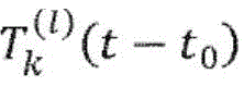 The definition is as follows:


the computer-implemented method according to the invention then uses the family of reference functions as defined above The family of reference functions { T } as defined above is then applied at a time T of 10 milliseconds0Increments are applied sequentially to the subject's electroencephalogram recordings. Fig. 3 shows a representative reference function derived from an electroencephalographic recording of a patient with temporal lobe epilepsy.
The matching score is selected as a specific formula for equation (1):

wherein, and
and respectively, at time t0The first zero crossings that occur in scalp channel k before and after.
To validate a family of reference functions T derived for each intracranial channel l of a single subject (non-validation period)lUsing a computer-implemented method to detect the same based only on electroencephalographic signals of the scalp of the subjectAn intracranial inter-seizure epileptiform discharge in a subject.
The computer-implemented method using the family of reference functions { T } thus designed is applied to the verification subset V of the same subject1The scalp channel of time period (b), wherein the reference function family { T } is derived from the scalp channel. In the framework of receiver operation characteristic analysis, reference function T is selected(l)E { T }, produces a matching score S(l)(t) assessing the efficiency in detecting intracranial spikes. More specifically, a matching score S is calculated for each successive 10 millisecond time window on the scalp channel(l)(t) is used to determine whether the time window contains a binary classification of an intracranial spike (8) in an intracranial channel l'. For each pair of intracranial channels l (for which a reference function T is derived)(l)) And l' (for evaluation), the corresponding values of the area under the receiver operating characteristic curve are obtained, thereby producing a matrix of values. An example of such a matrix is presented in fig. 4. FIG. 5 is a graph of selected right temporal lobe epilepsy subjects and a reference function TlOptimal combination with the evaluated intracranial channels shows a superposition of the intracranial voltage signals corresponding to the detections performed by applying the optimal reference function to the simultaneous scalp electroencephalographic signals. The optimal combination between the reference function and the estimated intracranial channel is marked with an asterisk on the matrix of correlation values in fig. 4.
For each subject studied, the optimal area under the receiver operating characteristic curve value was estimated according to the following equation:
AUCopt=maxl,l′AUC(l,l′), (9)
furthermore, to estimate the robustness of the computer-implemented method of the invention, cross-validation is performed by implementing a family of reference functions derived from one subject using the computer-implemented method for the other subject.
To this end, a family of 22 validation reference functions { T } is selected from a set comprising all 964 reference functions (one for each intracranial channel l under study). Reference function T comprising said family { T }lThe following two conditions are satisfied:
maxl′AUC(Tl,l′)>0.8, (10a)

where | NV | is the number of non-verification periods and the summation is over the non-verification periods.
The first condition (10a) requires the presence of at least one intracranial channel/, wherein, when applied to the same subject (reference function T)(l)Derived from the same subject) obtained (V)1Subset of time periods), the reference function T(l)E { T } detects a spike for the intracranial channel having an area under the receiver operating characteristic curve with a value greater than 0.8. The second condition (10b) is applied such that on average (a subset of NV periods) the minimum inter-seizure epileptiform discharge in the intracranial channel l is detected at a rate greater than one inter-seizure epileptiform discharge per period, wherein the reference function T is applied(l)Is deduced from the intracranial channel i.
For each subject, validating the family of reference functions T(l)E { T } is applied to the verification subset V2And the resulting matching score S(l)(t) the set is used to calculate a composite match score S*(t), defined as follows:

the composite match score, S, is given according to equation (11)*(t) is: for each time point, the maximum of the matching scores produced by all functions of the family of reference functions { T } except the reference function derived from the channel of the currently evaluated subject.
Evaluating a composite matching function S*(t) for the following two classifications: within a continuous 10-millisecond wide window centered at time t, the subject is evaluated for the presence or absence of an intracranial detected spike in a given channel l'. For each subject, the highest area of the antennal-wise channel (channel-wise) under the receiver's operating characteristic curveIs identified as:
AUCwal=maxl′AUC({T*},l′). (12)
results
Optimal area under the receiver operating characteristic curve AUC in the current group of 16 subjectsoptValues from 0.63 to 0.99 with an average value of 0.83.
Validated antegrade maximum area AUC under the receiver operating characteristic Curve in the current group of 16 subjectsvalValues from 0.53 to 0.81, with an average value of 0.70.
Specific values for the optimal area under the down-channel maximum recipient operational characteristic curve and recipient operational characteristic curve are presented in table 2. The associated recipient operational characteristics are shown in fig. 6. The relationship between the antecedent maximum recipient operational characteristic curve and the optimal area under the recipient operational characteristic curve is shown in fig. 7.


TABLE 2
Example 2:
materials and methods
Material
The computer-implemented method according to the invention was used to detect pathological brain activity patterns in 37 other subjects. The 37 subjects presented epilepsy with strictly unilateral epileptic focal location, either of the temporal lobe (n-22) or extratemporal (n-15, e.g. focal frontal, parietal or occipital lobe). In this example, the family of reference functions constructed in example 1, suitably modified, is used to allow discrimination between temporal lobe epileptic patients and extratemporal epileptic patients. 37 pre-recorded scalp electroencephalographic signals were selected from the "Epilepsiae" medical database that were 60 minutes long and which were not used in example 1. Intracranial signals are not used in this example. As in example 1, only patients with full 10-20 scalp electrode coverage supplemented with true anterior temporal electrodes (T1 and T2) were included in this example.
The clinical and demographic details of the subjects are listed in table 3 below:

TABLE 3
Method of producing a composite material
As a starting point for the analysis, the family of 22 verification reference functions { T } derived in example 1 was selected. Another family of reference functions { T } is then derived by applying the following transformation to each of the reference functions T e { T }:

where t is time, k is the channel name and f (k) is the transformation of the channel name specified below:

and for a bipolar channel pair [ k, k' ]:
f([k,k'])=[f(k),f(k')]. (13c)
the transformation generates an initial reference function T by substituting a symmetric channel f (k) into each channel k, as shown in equation (13a)kTransposed form of (1). Applying the transformation F to the functions in T is one example of heuristically operating a family of reference functions during the design process.
Notably, no record of the subject being evaluated is used to derive these families of reference functions.
The matching score is again selected as a specific formula for equation (1):

where T is for calculating the score STIs determined by the reference function of (a), and
and respectively, at time t0The first zero crossings that occur in scalp channel k before and after. The composite cumulative match score S x (t) is defined as

This is an example of a cumulative match score and how the match score can be defined for two reference functions, T ∈ { T } and f (T) ∈ { T } belonging to two different families of reference functions.
A cumulative match score of a fixed value t is then evaluated in the framework of receiver operational feature analysis to assess the ability to distinguish electroencephalographic recordings of subjects superotemporal and superotemporal.
Results
The cumulative matching scores are performed differently for different reference functions T e { T } and their corresponding transformation forms f (T) e { T }. For the 60 minute recording, 5 of the 22 analyzed reference functions resulted in an area value under the receiver operating characteristic curve of greater than 0.65, and the maximum area value under the receiver operating characteristic curve obtained was 0.71. These results demonstrate the potential applicability of the present method to the classification problem.
Fig. 8 and 9 show the dependence of the area values of different reference functions under the receiver operating characteristic curve on the length of the record under investigation. In fig. 8, the reference functions are ordered along the vertical axis according to their performance estimated from the area under the receiver operating characteristic curve as a function of the full length recording. It can be seen that there is a significant fluctuation when the recording length is less than 20 minutes, and stable performance is obtained when the recording length exceeds 30 to 45 minutes. Fig. 9 shows the average area under the receiver operating characteristic values as a function of the length of the recording, either over all reference functions or with a subset of 5 reference functions having an area under the receiver operating characteristic values greater than 0.65 for a full length recording. A significant improvement in the sorting performance can be seen for recording lengths exceeding 20-30 minutes.
Claims (18)

Applications Claiming Priority (3)
| Application Number | Priority Date | Filing Date | Title |
|---|---|---|---|
| EP17164291 | 2017-03-31 | ||
| EP17164291.1 | 2017-03-31 | ||
| PCT/EP2018/058292 WO2018178333A1 (en) | 2017-03-31 | 2018-03-30 | Method for identification of pathological brain activity from scalp electroencephalogram |
Publications (2)
| Publication Number | Publication Date |
|---|---|
| CN111093471A true CN111093471A (en) | 2020-05-01 |
| CN111093471B CN111093471B (en) | 2022-11-18 |
Family
ID=58488839
Family Applications (1)
| Application Number | Title | Priority Date | Filing Date |
|---|---|---|---|
| CN201880035130.5A Expired - Fee Related CN111093471B (en) | 2017-03-31 | 2018-03-30 | Method for detecting pathological brain activity from scalp electroencephalograms |
Country Status (7)
| Country | Link |
|---|---|
| US (1) | US11684303B2 (en) |
| EP (1) | EP3599999A1 (en) |
| JP (1) | JP2020512860A (en) |
| CN (1) | CN111093471B (en) |
| CA (1) | CA3058261A1 (en) |
| TW (1) | TW201842458A (en) |
| WO (1) | WO2018178333A1 (en) |
Cited By (3)
| Publication number | Priority date | Publication date | Assignee | Title |
|---|---|---|---|---|
| CN113499083A (en) * | 2021-05-18 | 2021-10-15 | 生物岛实验室 | Scalp electroencephalogram high-frequency oscillation automatic detection method |
| CN114288520A (en) * | 2021-12-31 | 2022-04-08 | 广州酷狗计算机科技有限公司 | Sleep assisting method, device, equipment and storage medium based on brain waves |
| CN114983441A (en) * | 2022-05-10 | 2022-09-02 | 浙江新悦翔宇智能科技有限公司 | Brain wave forward induction feedback regulation system |
Families Citing this family (5)
| Publication number | Priority date | Publication date | Assignee | Title |
|---|---|---|---|---|
| US20200077906A1 (en) * | 2018-09-07 | 2020-03-12 | Augusta University Research Institute, Inc. | Method and System for Monitoring Brain Function and Intracranial Pressure |
| WO2021183048A1 (en) * | 2020-03-09 | 2021-09-16 | Nanyang Technological University | Detection of slowing patterns in eeg data |
| RU2747712C1 (en) * | 2020-06-01 | 2021-05-13 | ОБЩЕСТВО С ОГРАНИЧЕННОЙ ОТВЕТСТВЕННОСТЬЮ "СберМедИИ" | Method for detecting epileptiform discharges in long-term eeg recording |
| CN113288150B (en) * | 2021-06-25 | 2022-09-27 | 杭州电子科技大学 | Channel selection method based on fatigue electroencephalogram combination characteristics |
| CN115530845A (en) * | 2022-10-17 | 2022-12-30 | 常州瑞神安医疗器械有限公司 | Method for detecting abnormal discharge in epilepsia electroencephalogram signal |
Citations (5)
| Publication number | Priority date | Publication date | Assignee | Title |
|---|---|---|---|---|
| US20020095099A1 (en) * | 2000-04-27 | 2002-07-18 | Centre National De La Recherche Scientificque | Method and device for the medical monitoring in real time of a patient from the analysis of electro-encephalograms, application of this method to characterize and to differentiate between physiological or pathologial conditions, and a method for anticipating epileptic seizures in real time |
| US20020193670A1 (en) * | 2001-05-29 | 2002-12-19 | Reproductive Health Technologies, Inc. | Device and system for remote for in-clinic trans-abdominal/vaginal/cervical acquisition, and detection, analysis, and communication of maternal uterine and maternal and fetal cardiac and fetal brain activity from electrical signals |
| US20060015034A1 (en) * | 2002-10-18 | 2006-01-19 | Jacques Martinerie | Analysis method and real time medical or cognitive monitoring device based on the analysis of a subject's cerebral electromagnetic use of said method for characterizing and differenting physiological and pathological states |
| US20090124923A1 (en) * | 2007-10-23 | 2009-05-14 | Optima Neuroscience, Inc. | System for seizure monitoring and detection |
| US20160228705A1 (en) * | 2015-02-10 | 2016-08-11 | Neuropace, Inc. | Seizure onset classification and stimulation parameter selection |
Family Cites Families (3)
| Publication number | Priority date | Publication date | Assignee | Title |
|---|---|---|---|---|
| US6594524B2 (en) * | 2000-12-12 | 2003-07-15 | The Trustees Of The University Of Pennsylvania | Adaptive method and apparatus for forecasting and controlling neurological disturbances under a multi-level control |
| US7761145B2 (en) * | 2006-04-21 | 2010-07-20 | Medtronic, Inc. | Method and apparatus for detection of nervous system disorders |
| US20170231519A1 (en) | 2014-08-19 | 2017-08-17 | The General Hospital Corporation | System and method for annotating and analyzing eeg waveforms |
-
2018
- 2018-03-30 US US16/499,445 patent/US11684303B2/en active Active
- 2018-03-30 WO PCT/EP2018/058292 patent/WO2018178333A1/en not_active Ceased
- 2018-03-30 TW TW107111277A patent/TW201842458A/en unknown
- 2018-03-30 JP JP2019553868A patent/JP2020512860A/en active Pending
- 2018-03-30 CA CA3058261A patent/CA3058261A1/en active Pending
- 2018-03-30 EP EP18713963.9A patent/EP3599999A1/en not_active Withdrawn
- 2018-03-30 CN CN201880035130.5A patent/CN111093471B/en not_active Expired - Fee Related
Patent Citations (5)
| Publication number | Priority date | Publication date | Assignee | Title |
|---|---|---|---|---|
| US20020095099A1 (en) * | 2000-04-27 | 2002-07-18 | Centre National De La Recherche Scientificque | Method and device for the medical monitoring in real time of a patient from the analysis of electro-encephalograms, application of this method to characterize and to differentiate between physiological or pathologial conditions, and a method for anticipating epileptic seizures in real time |
| US20020193670A1 (en) * | 2001-05-29 | 2002-12-19 | Reproductive Health Technologies, Inc. | Device and system for remote for in-clinic trans-abdominal/vaginal/cervical acquisition, and detection, analysis, and communication of maternal uterine and maternal and fetal cardiac and fetal brain activity from electrical signals |
| US20060015034A1 (en) * | 2002-10-18 | 2006-01-19 | Jacques Martinerie | Analysis method and real time medical or cognitive monitoring device based on the analysis of a subject's cerebral electromagnetic use of said method for characterizing and differenting physiological and pathological states |
| US20090124923A1 (en) * | 2007-10-23 | 2009-05-14 | Optima Neuroscience, Inc. | System for seizure monitoring and detection |
| US20160228705A1 (en) * | 2015-02-10 | 2016-08-11 | Neuropace, Inc. | Seizure onset classification and stimulation parameter selection |
Cited By (5)
| Publication number | Priority date | Publication date | Assignee | Title |
|---|---|---|---|---|
| CN113499083A (en) * | 2021-05-18 | 2021-10-15 | 生物岛实验室 | Scalp electroencephalogram high-frequency oscillation automatic detection method |
| CN113499083B (en) * | 2021-05-18 | 2023-10-20 | 生物岛实验室 | Scalp electroencephalogram high-frequency oscillation automatic detection method |
| CN114288520A (en) * | 2021-12-31 | 2022-04-08 | 广州酷狗计算机科技有限公司 | Sleep assisting method, device, equipment and storage medium based on brain waves |
| CN114983441A (en) * | 2022-05-10 | 2022-09-02 | 浙江新悦翔宇智能科技有限公司 | Brain wave forward induction feedback regulation system |
| CN114983441B (en) * | 2022-05-10 | 2024-09-03 | 杭州星脑康科技有限公司 | Brain wave forward induction feedback regulation system |
Also Published As
| Publication number | Publication date |
|---|---|
| WO2018178333A1 (en) | 2018-10-04 |
| TW201842458A (en) | 2018-12-01 |
| CA3058261A1 (en) | 2018-10-04 |
| JP2020512860A (en) | 2020-04-30 |
| EP3599999A1 (en) | 2020-02-05 |
| US11684303B2 (en) | 2023-06-27 |
| US20210093247A1 (en) | 2021-04-01 |
| CN111093471B (en) | 2022-11-18 |
Similar Documents
| Publication | Publication Date | Title |
|---|---|---|
| CN111093471B (en) | Method for detecting pathological brain activity from scalp electroencephalograms | |
| Nakamura et al. | Automatic sleep monitoring using ear-EEG | |
| US10575751B2 (en) | Method and apparatus for determining sleep states | |
| CN106413541B (en) | System and method for diagnosing sleep | |
| Boubchir et al. | A review of feature extraction for EEG epileptic seizure detection and classification | |
| JP2022536552A (en) | Seizure detection system and method based on changes in electroencephalogram (EEG) nonlinearity | |
| US20150245800A1 (en) | Method for Detection Of An Abnormal Sleep Pattern In A Person | |
| JP7562745B2 (en) | Seizure detection system and method based on changes in electroencephalography (EEG) nonlinearity | |
| EP0898460A1 (en) | Systems for prediction, rapid detection, warning, prevention or control of changes in activity states in the brain | |
| BR112019011229A2 (en) | seizure semiology including muscle signals collected from electroencephalography electrodes | |
| Gallego-Jutgla et al. | A theta-band EEG based index for early diagnosis of Alzheimer's disease | |
| Greene et al. | Electrocardiogram based neonatal seizure detection | |
| CN101529429A (en) | System and method for prediction of adverse events during treatment of psychological and neurological disorders | |
| KR102376984B1 (en) | Measurement apparatus of altered cortical region using functional network and method thereof | |
| Tiwari et al. | Supervised approach based sleep disorder detection using non-linear dynamic features (NLDF) of EEG | |
| Zhou et al. | Real-time epileptic seizure detection based on deep learning | |
| Wang et al. | Exploration of instantaneous amplitude and frequency features for epileptic seizure prediction | |
| Waser et al. | A blind source-based method for automated artifact-correction in standard sleep EEG | |
| Bhatt et al. | Automatic detection of Parkinson’s disease using EEG signals: A machine learning approach | |
| Zeljković et al. | Pre-Ictal phase detection algorithm based on one dimensional EEG signals and two dimensional formed images analysis | |
| Nancy et al. | A brain EEG classification system for the mild cognitive impairment analysis | |
| Preoteasa et al. | Research on Best Practices for EEG Analysis in Sleep Stage Scoring | |
| TH et al. | Improved feature exctraction process to detect seizure using CHBMIT-dataset | |
| Zeljković et al. | Automated pre-ictal phase detection algorithm from eeg signals | |
| Zerrouk et al. | Prediction of Epileptic Seizures Based on Analysis of Electrical Activity of the Brain and Parameters of Heart Rate Variability |
Legal Events
| Date | Code | Title | Description |
|---|---|---|---|
| PB01 | Publication | ||
| PB01 | Publication | ||
| SE01 | Entry into force of request for substantive examination | ||
| SE01 | Entry into force of request for substantive examination | ||
| GR01 | Patent grant | ||
| GR01 | Patent grant | ||
| CF01 | Termination of patent right due to non-payment of annual fee |
Granted publication date: 20221118 |
|
| CF01 | Termination of patent right due to non-payment of annual fee |