CN111024863A - Test method of peptide map of ramucirumab - Google Patents
Test method of peptide map of ramucirumab Download PDFInfo
- Publication number
- CN111024863A CN111024863A CN201911423878.5A CN201911423878A CN111024863A CN 111024863 A CN111024863 A CN 111024863A CN 201911423878 A CN201911423878 A CN 201911423878A CN 111024863 A CN111024863 A CN 111024863A
- Authority
- CN
- China
- Prior art keywords
- mobile phase
- vol
- test method
- ramucirumab
- desalting
- Prior art date
- Legal status (The legal status is an assumption and is not a legal conclusion. Google has not performed a legal analysis and makes no representation as to the accuracy of the status listed.)
- Pending
Links
- 108090000765 processed proteins & peptides Proteins 0.000 title claims abstract description 37
- 229960002633 ramucirumab Drugs 0.000 title claims abstract description 32
- 238000010998 test method Methods 0.000 title claims abstract description 18
- 238000011033 desalting Methods 0.000 claims abstract description 24
- 102000004169 proteins and genes Human genes 0.000 claims abstract description 19
- 108090000623 proteins and genes Proteins 0.000 claims abstract description 19
- 238000001195 ultra high performance liquid chromatography Methods 0.000 claims abstract description 15
- 238000005804 alkylation reaction Methods 0.000 claims abstract description 12
- 238000004925 denaturation Methods 0.000 claims abstract description 11
- 230000036425 denaturation Effects 0.000 claims abstract description 11
- 239000007788 liquid Substances 0.000 claims abstract description 11
- 239000003153 chemical reaction reagent Substances 0.000 claims abstract description 10
- LZKWZMPRGOBGFV-IMJSIDKUSA-N (2S,3S)-2,3-bis(sulfanyl)butane-1,2,3,4-tetrol Chemical compound S[C@@]([C@@](CO)(O)S)(O)CO LZKWZMPRGOBGFV-IMJSIDKUSA-N 0.000 claims abstract description 9
- 238000011282 treatment Methods 0.000 claims abstract description 9
- 102000004142 Trypsin Human genes 0.000 claims abstract description 8
- 108090000631 Trypsin Proteins 0.000 claims abstract description 8
- 238000006243 chemical reaction Methods 0.000 claims abstract description 8
- 239000012588 trypsin Substances 0.000 claims abstract description 8
- 239000007791 liquid phase Substances 0.000 claims abstract description 7
- 150000007524 organic acids Chemical class 0.000 claims abstract description 7
- 230000002152 alkylating effect Effects 0.000 claims abstract description 6
- 239000003638 chemical reducing agent Substances 0.000 claims abstract description 5
- 239000012071 phase Substances 0.000 claims description 76
- 239000000243 solution Substances 0.000 claims description 35
- 238000010828 elution Methods 0.000 claims description 25
- BDAGIHXWWSANSR-UHFFFAOYSA-N methanoic acid Natural products OC=O BDAGIHXWWSANSR-UHFFFAOYSA-N 0.000 claims description 24
- DTQVDTLACAAQTR-UHFFFAOYSA-N Trifluoroacetic acid Chemical compound OC(=O)C(F)(F)F DTQVDTLACAAQTR-UHFFFAOYSA-N 0.000 claims description 20
- WEVYAHXRMPXWCK-UHFFFAOYSA-N Acetonitrile Chemical compound CC#N WEVYAHXRMPXWCK-UHFFFAOYSA-N 0.000 claims description 18
- OSWFIVFLDKOXQC-UHFFFAOYSA-N 4-(3-methoxyphenyl)aniline Chemical compound COC1=CC=CC(C=2C=CC(N)=CC=2)=C1 OSWFIVFLDKOXQC-UHFFFAOYSA-N 0.000 claims description 12
- 235000019253 formic acid Nutrition 0.000 claims description 12
- 229960000789 guanidine hydrochloride Drugs 0.000 claims description 5
- PJJJBBJSCAKJQF-UHFFFAOYSA-N guanidinium chloride Chemical compound [Cl-].NC(N)=[NH2+] PJJJBBJSCAKJQF-UHFFFAOYSA-N 0.000 claims description 5
- JDNTWHVOXJZDSN-UHFFFAOYSA-N iodoacetic acid Chemical compound OC(=O)CI JDNTWHVOXJZDSN-UHFFFAOYSA-N 0.000 claims description 5
- 239000002168 alkylating agent Substances 0.000 claims description 4
- 229940100198 alkylating agent Drugs 0.000 claims description 4
- 239000007864 aqueous solution Substances 0.000 claims description 4
- 238000000108 ultra-filtration Methods 0.000 claims description 4
- 238000001514 detection method Methods 0.000 claims description 3
- 230000005526 G1 to G0 transition Effects 0.000 claims description 2
- PBCJIPOGFJYBJE-UHFFFAOYSA-N acetonitrile;hydrate Chemical compound O.CC#N PBCJIPOGFJYBJE-UHFFFAOYSA-N 0.000 claims description 2
- 230000007423 decrease Effects 0.000 claims description 2
- 230000007071 enzymatic hydrolysis Effects 0.000 claims description 2
- 238000006047 enzymatic hydrolysis reaction Methods 0.000 claims description 2
- 239000011259 mixed solution Substances 0.000 claims description 2
- 230000035484 reaction time Effects 0.000 claims description 2
- 238000004704 ultra performance liquid chromatography Methods 0.000 claims description 2
- 238000012360 testing method Methods 0.000 abstract description 14
- 238000000034 method Methods 0.000 abstract description 11
- 238000000926 separation method Methods 0.000 abstract description 5
- 238000010586 diagram Methods 0.000 abstract description 4
- 239000000523 sample Substances 0.000 description 19
- 150000001768 cations Chemical class 0.000 description 16
- 239000003480 eluent Substances 0.000 description 9
- QKNYBSVHEMOAJP-UHFFFAOYSA-N 2-amino-2-(hydroxymethyl)propane-1,3-diol;hydron;chloride Chemical compound Cl.OCC(N)(CO)CO QKNYBSVHEMOAJP-UHFFFAOYSA-N 0.000 description 8
- 239000003814 drug Substances 0.000 description 8
- 102000004190 Enzymes Human genes 0.000 description 6
- 108090000790 Enzymes Proteins 0.000 description 6
- 102000016549 Vascular Endothelial Growth Factor Receptor-2 Human genes 0.000 description 5
- 108010053099 Vascular Endothelial Growth Factor Receptor-2 Proteins 0.000 description 5
- BJHCYTJNPVGSBZ-YXSASFKJSA-N 1-[4-[6-amino-5-[(Z)-methoxyiminomethyl]pyrimidin-4-yl]oxy-2-chlorophenyl]-3-ethylurea Chemical compound CCNC(=O)Nc1ccc(Oc2ncnc(N)c2\C=N/OC)cc1Cl BJHCYTJNPVGSBZ-YXSASFKJSA-N 0.000 description 4
- 238000010612 desalination reaction Methods 0.000 description 4
- 229940079593 drug Drugs 0.000 description 4
- 238000005406 washing Methods 0.000 description 4
- 150000001413 amino acids Chemical class 0.000 description 3
- 238000003113 dilution method Methods 0.000 description 3
- 239000000203 mixture Substances 0.000 description 3
- 102000009524 Vascular Endothelial Growth Factor A Human genes 0.000 description 2
- 108010073929 Vascular Endothelial Growth Factor A Proteins 0.000 description 2
- 230000029936 alkylation Effects 0.000 description 2
- 239000000872 buffer Substances 0.000 description 2
- 238000000227 grinding Methods 0.000 description 2
- 239000003446 ligand Substances 0.000 description 2
- 238000002156 mixing Methods 0.000 description 2
- 229920001184 polypeptide Polymers 0.000 description 2
- 102000004196 processed proteins & peptides Human genes 0.000 description 2
- 206010009944 Colon cancer Diseases 0.000 description 1
- 206010028980 Neoplasm Diseases 0.000 description 1
- 102000035195 Peptidases Human genes 0.000 description 1
- 108091005804 Peptidases Proteins 0.000 description 1
- 102000007079 Peptide Fragments Human genes 0.000 description 1
- 108010033276 Peptide Fragments Proteins 0.000 description 1
- 208000005718 Stomach Neoplasms Diseases 0.000 description 1
- 108010073923 Vascular Endothelial Growth Factor C Proteins 0.000 description 1
- 102000009520 Vascular Endothelial Growth Factor C Human genes 0.000 description 1
- 108010073919 Vascular Endothelial Growth Factor D Proteins 0.000 description 1
- 102000009519 Vascular Endothelial Growth Factor D Human genes 0.000 description 1
- 102000005789 Vascular Endothelial Growth Factors Human genes 0.000 description 1
- 108010019530 Vascular Endothelial Growth Factors Proteins 0.000 description 1
- 239000002318 adhesion promoter Substances 0.000 description 1
- 238000004458 analytical method Methods 0.000 description 1
- 230000009286 beneficial effect Effects 0.000 description 1
- 239000007853 buffer solution Substances 0.000 description 1
- 125000002091 cationic group Chemical group 0.000 description 1
- 208000029742 colonic neoplasm Diseases 0.000 description 1
- 230000007547 defect Effects 0.000 description 1
- 238000011161 development Methods 0.000 description 1
- 239000012470 diluted sample Substances 0.000 description 1
- 238000007865 diluting Methods 0.000 description 1
- 210000002889 endothelial cell Anatomy 0.000 description 1
- 238000002474 experimental method Methods 0.000 description 1
- 239000012530 fluid Substances 0.000 description 1
- 239000012634 fragment Substances 0.000 description 1
- 206010017758 gastric cancer Diseases 0.000 description 1
- 201000007492 gastroesophageal junction adenocarcinoma Diseases 0.000 description 1
- 238000011534 incubation Methods 0.000 description 1
- 238000002347 injection Methods 0.000 description 1
- 239000007924 injection Substances 0.000 description 1
- 230000003834 intracellular effect Effects 0.000 description 1
- 238000013508 migration Methods 0.000 description 1
- 230000005012 migration Effects 0.000 description 1
- 208000002154 non-small cell lung carcinoma Diseases 0.000 description 1
- 238000012510 peptide mapping method Methods 0.000 description 1
- 210000004896 polypeptide structure Anatomy 0.000 description 1
- 230000035755 proliferation Effects 0.000 description 1
- 238000011519 second-line treatment Methods 0.000 description 1
- 230000004936 stimulating effect Effects 0.000 description 1
- 201000011549 stomach cancer Diseases 0.000 description 1
- 239000000126 substance Substances 0.000 description 1
- 238000006467 substitution reaction Methods 0.000 description 1
- 230000001502 supplementing effect Effects 0.000 description 1
- 208000029729 tumor suppressor gene on chromosome 11 Diseases 0.000 description 1
Images


Classifications
-
- G—PHYSICS
- G01—MEASURING; TESTING
- G01N—INVESTIGATING OR ANALYSING MATERIALS BY DETERMINING THEIR CHEMICAL OR PHYSICAL PROPERTIES
- G01N30/00—Investigating or analysing materials by separation into components using adsorption, absorption or similar phenomena or using ion-exchange, e.g. chromatography or field flow fractionation
- G01N30/02—Column chromatography
- G01N30/04—Preparation or injection of sample to be analysed
- G01N30/06—Preparation
- G01N30/14—Preparation by elimination of some components
-
- G—PHYSICS
- G01—MEASURING; TESTING
- G01N—INVESTIGATING OR ANALYSING MATERIALS BY DETERMINING THEIR CHEMICAL OR PHYSICAL PROPERTIES
- G01N30/00—Investigating or analysing materials by separation into components using adsorption, absorption or similar phenomena or using ion-exchange, e.g. chromatography or field flow fractionation
- G01N30/02—Column chromatography
- G01N30/26—Conditioning of the fluid carrier; Flow patterns
- G01N30/28—Control of physical parameters of the fluid carrier
- G01N30/34—Control of physical parameters of the fluid carrier of fluid composition, e.g. gradient
-
- G—PHYSICS
- G01—MEASURING; TESTING
- G01N—INVESTIGATING OR ANALYSING MATERIALS BY DETERMINING THEIR CHEMICAL OR PHYSICAL PROPERTIES
- G01N30/00—Investigating or analysing materials by separation into components using adsorption, absorption or similar phenomena or using ion-exchange, e.g. chromatography or field flow fractionation
- G01N30/02—Column chromatography
- G01N30/62—Detectors specially adapted therefor
- G01N30/74—Optical detectors
-
- G—PHYSICS
- G01—MEASURING; TESTING
- G01N—INVESTIGATING OR ANALYSING MATERIALS BY DETERMINING THEIR CHEMICAL OR PHYSICAL PROPERTIES
- G01N30/00—Investigating or analysing materials by separation into components using adsorption, absorption or similar phenomena or using ion-exchange, e.g. chromatography or field flow fractionation
- G01N30/02—Column chromatography
- G01N30/86—Signal analysis
- G01N30/8675—Evaluation, i.e. decoding of the signal into analytical information
- G01N30/8686—Fingerprinting, e.g. without prior knowledge of the sample components
-
- G—PHYSICS
- G01—MEASURING; TESTING
- G01N—INVESTIGATING OR ANALYSING MATERIALS BY DETERMINING THEIR CHEMICAL OR PHYSICAL PROPERTIES
- G01N30/00—Investigating or analysing materials by separation into components using adsorption, absorption or similar phenomena or using ion-exchange, e.g. chromatography or field flow fractionation
- G01N30/02—Column chromatography
- G01N30/88—Integrated analysis systems specially adapted therefor, not covered by a single one of the groups G01N30/04 - G01N30/86
-
- G—PHYSICS
- G01—MEASURING; TESTING
- G01N—INVESTIGATING OR ANALYSING MATERIALS BY DETERMINING THEIR CHEMICAL OR PHYSICAL PROPERTIES
- G01N30/00—Investigating or analysing materials by separation into components using adsorption, absorption or similar phenomena or using ion-exchange, e.g. chromatography or field flow fractionation
- G01N30/02—Column chromatography
- G01N30/04—Preparation or injection of sample to be analysed
- G01N30/06—Preparation
- G01N2030/067—Preparation by reaction, e.g. derivatising the sample
-
- G—PHYSICS
- G01—MEASURING; TESTING
- G01N—INVESTIGATING OR ANALYSING MATERIALS BY DETERMINING THEIR CHEMICAL OR PHYSICAL PROPERTIES
- G01N30/00—Investigating or analysing materials by separation into components using adsorption, absorption or similar phenomena or using ion-exchange, e.g. chromatography or field flow fractionation
- G01N30/02—Column chromatography
- G01N30/88—Integrated analysis systems specially adapted therefor, not covered by a single one of the groups G01N30/04 - G01N30/86
- G01N2030/8809—Integrated analysis systems specially adapted therefor, not covered by a single one of the groups G01N30/04 - G01N30/86 analysis specially adapted for the sample
- G01N2030/8813—Integrated analysis systems specially adapted therefor, not covered by a single one of the groups G01N30/04 - G01N30/86 analysis specially adapted for the sample biological materials
- G01N2030/8831—Integrated analysis systems specially adapted therefor, not covered by a single one of the groups G01N30/04 - G01N30/86 analysis specially adapted for the sample biological materials involving peptides or proteins
Landscapes
- Physics & Mathematics (AREA)
- Biochemistry (AREA)
- Health & Medical Sciences (AREA)
- Life Sciences & Earth Sciences (AREA)
- Chemical & Material Sciences (AREA)
- Analytical Chemistry (AREA)
- General Health & Medical Sciences (AREA)
- General Physics & Mathematics (AREA)
- Immunology (AREA)
- Pathology (AREA)
- Spectroscopy & Molecular Physics (AREA)
- Engineering & Computer Science (AREA)
- Library & Information Science (AREA)
- Measuring Or Testing Involving Enzymes Or Micro-Organisms (AREA)
Abstract
The invention provides a test method of a peptide map of ramucirumab. The test method comprises the following steps: performing denaturation reduction treatment on the ramucirumab sample by using protein denaturation liquid and a disulfide bond reducing agent in a liquid phase environment; adding an alkylating reagent, carrying out an alkylation reaction, and terminating the reaction by using dimercaptothreitol; desalting, adding trypsin for enzymolysis, stopping enzymolysis reaction by using organic acid and acidifying the peptide segment; and detecting by using ultra-high performance liquid chromatography to obtain a peptide map of the ramucirumab. The testing method provided by the invention is simple and feasible, the testing can be completed within 90min, the separation degree of each peak of the obtained peptide diagram is high, the accuracy is high, the repeatability of the method is good, and the method is stable and feasible.
Description
Technical Field
The invention belongs to the technical field of chemical analysis, and particularly relates to a method for testing a peptide map of ramucirumab.
Background
The ramucirumab is a fully humanized IgG1 monoclonal antibody with molecular formula of C6376H9886N1702O1996S46And a molecular weight of about 147kDa, which is a polypeptide consisting of two heavy chains and two light chains. The two heavy chains consist of 446 amino acids each and the two light chains consist of 214 amino acids each.
The ramucirumab can specifically bind to vascular endothelial growth factor receptor 2(VEGFR-2) and block the binding of VEGFR-2 to its ligands VEGF-A, VEGF-C and VEGF-D. The binding force of the ramucirumab and VEGFR-2 is about 8 times that of VEGFR-2 and a ligand VEGF-A thereof, so that the adhesion promoter can inhibit VEGF from stimulating the proliferation and migration of endothelial cells and finally inhibit the generation of tumor vessels. Ramucirumab is currently approved for second-line treatment of advanced gastric cancer, esophagogastric junction adenocarcinoma, colon cancer, and non-small cell lung cancer.
Peptide Mapping is a characteristic fingerprint formed by using intracellular proteolytic enzyme with strong specificity to act on a special Peptide chain site to crack protein into small fragments and using a certain separation and detection means according to the molecular weight of the protein and the composition characteristics of primary structure amino acid.
The peptide structure of protein or polypeptide medicine can be researched through a peptide diagram, and the polypeptide structure is closely related to the biological medicine property and efficacy of the medicine. Therefore, the development of a suitable method has great significance for testing the peptide map of the ramucirumab.
Disclosure of Invention
Aiming at the defects in the prior art, the invention aims to provide a method for testing a peptide map of ramucirumab. The testing method is simple and feasible, and the obtained peptide diagram has high separation degree of each peak, high accuracy, good repeatability, stability and feasibility.
In order to achieve the purpose, the invention adopts the following technical scheme:
the invention provides a test method of a peptide map of ramucirumab, which comprises the following steps:
(1) performing denaturation reduction treatment on the ramucirumab sample by using protein denaturation liquid and a disulfide bond reducing agent in a liquid phase environment;
(2) adding an alkylating reagent into the solution obtained in the step (1) to carry out an alkylation reaction, and terminating the reaction by using dimercaptothreitol;
(3) desalting the solution obtained in the step (2), adding trypsin for enzymolysis, stopping the enzymolysis reaction by using organic acid and acidifying the peptide segment;
(4) and (4) detecting the sample treated in the step (3) by using ultra-high performance liquid chromatography to obtain a peptide map of the ramucirumab.
The invention adopts trypsin to carry out enzymolysis treatment on the ramucirumab and combines with ultra-high performance liquid chromatography to realize the test of the peptide graph of the ramucirumab. The testing method is simple and easy to implement, and the obtained peptide graph has high separation degree of each peak, high accuracy and good repeatability.
In a preferred embodiment of the present invention, the protein denaturation solution in the step (1) contains guanidine hydrochloride;
preferably, the concentration of guanidine hydrochloride in the liquid phase environment in step (1) is 5.5-6.5M, such as 5.5M, 5.6M, 5.7M, 5.8M, 5.9M, 6M, 6.1M, 6.2M, 6.3M, 6.4M or 6.5M; more preferably 6M.
Unless otherwise specified, the unit "M" is mol/L and the unit "mM" is mmol/L in the present invention.
Preferably, the disulfide bond reducing agent in step (1) is dimercaptothreitol.
Preferably, the concentration of dimercaptothreitol in the liquid phase environment of step (1) is 40-60mM, and may be, for example, 40mM, 42mM, 43mM, 45mM, 46mM, 48mM, 50mM, 52mM, 53mM, 55mM, 56mM, 58mM, or 60mM, etc.; more preferably 50 mM.
As a preferred embodiment of the present invention, the temperature of the denaturation reduction treatment in step (1) is 35 to 40 ℃, and may be, for example, 35 ℃, 35.5 ℃, 36 ℃, 36.5 ℃, 37 ℃, 37.5 ℃, 38 ℃, 38.5 ℃, 39 ℃, 39.5 ℃ or 40 ℃; further preferably 37 ℃.
Preferably, the time of the denaturation reduction treatment in the step (1) is 1-3h, such as 1h, 1.2h, 1.5h, 1.8h, 2h, 2.2h, 2.5h, 2.8h or 3 h; further preferably 1 hour.
In a preferred embodiment of the present invention, the alkylating reagent in step (2) is iodoacetic acid.
Preferably, the amount of the alkylating reagent added in step (2) is 100-140mM, and may be, for example, 100mM, 102mM, 105mM, 108mM, 110mM, 112mM, 115mM, 118mM, 120mM, 122mM, 125mM, 128mM, 130mM, 132mM, 135mM, 138mM or 140mM, etc.; further preferably 120 mM.
In the present invention, the term "amount added" refers to the final concentration of the reagent after the reagent is added to the solution. If "the amount of alkylating agent added is 100-140 mM" means that the final concentration of alkylating agent in the solution is 100-140mM after the addition of alkylating agent.
Preferably, the alkylation reaction in step (2) is carried out under protection from light.
Preferably, the alkylation reaction in step (2) is carried out at a temperature of 18 to 25 ℃, for example, 18 ℃, 19 ℃, 20 ℃, 21 ℃, 22 ℃, 23 ℃, 24 ℃ or 25 ℃ and the like; further preferably 22 ℃.
Preferably, the alkylation reaction time in the step (2) is 40-60min, such as 40min, 42min, 43min, 45min, 46min, 48min, 50min, 52min, 53min, 55min, 56min, 58min or 60 min; further preferably 45 min.
Preferably, the amount of dimercaptothreitol added in step (2) is 30-50mM, and may be, for example, 30mM, 32mM, 33mM, 35mM, 36mM, 38mM, 40mM, 42mM, 43mM, 45mM, 46mM, 48mM, or 50mM, etc.; more preferably 40 mM.
As a preferred technical scheme of the invention, the desalting method in the step (3) is ultrafiltration tube centrifugal desalting or desalting by a desalting column.
Preferably, after the desalting in step (3), the following operation is further performed: and detecting the concentration of the ramucirumab in the desalted solution by using a trace ultraviolet spectrophotometer.
Preferably, the mass ratio of the trypsin to the ramucirumab in the desalted solution in the step (3) is 1 (12.5-50), and may be, for example, 1:12.5, 1:14, 1:15, 1:18, 1:20, 1:22, 1:25, 1:28, 1:30, 1:32, 1:35, 1:38, 1:40, 1:42, 1:45, 1:48, or 1: 50; further preferably 1: 25.
As the preferred embodiment of the present invention, the temperature of the enzymatic hydrolysis in step (3) is 35 to 40 ℃, and may be, for example, 35 ℃, 35.5 ℃, 36 ℃, 36.5 ℃, 37 ℃, 37.5 ℃, 38 ℃, 38.5 ℃, 39 ℃, 39.5 ℃ or 40 ℃; further preferably 37 ℃.
Preferably, the enzymolysis time in step (3) is 1-4h, for example, 1h, 1.2h, 1.5h, 1.8h, 2h, 2.2h, 2.5h, 2.8h, 3h, 3.2h, 3.5h, 3.8h or 4h, etc.; further preferably 2 hours.
Preferably, the organic acid in step (3) is formic acid.
Preferably, the organic acid is added in the step (3) in an amount of 0.1-0.2 Vol%, such as 0.1 Vol%, 0.11 Vol%, 0.12 Vol%, 0.13 Vol%, 0.14 Vol%, 0.15 Vol%, 0.16 Vol%, 0.17 Vol%, 0.18 Vol%, 0.19 Vol%, or 0.2 Vol%; further preferably 0.1 Vol%.
As a preferred technical scheme of the invention, the stationary phase of the chromatographic column adopted by the ultra-high performance liquid chromatography in the step (4) is C18
Preferably, the temperature of the chromatographic column used for the ultra-high performance liquid chromatography is 47-53 ℃, and can be 47 ℃, 48 ℃, 49 ℃, 50 ℃, 51 ℃, 52 ℃ or 53 ℃ and the like; further preferably 50 ℃.
Preferably, the detection wavelength of the ultra-high performance liquid chromatography is 210-220nm, such as 210nm, 211nm, 212nm, 213nm, 214nm, 215nm, 216nm, 217nm, 218nm, 219nm or 220nm, etc.; further preferably 214 nm.
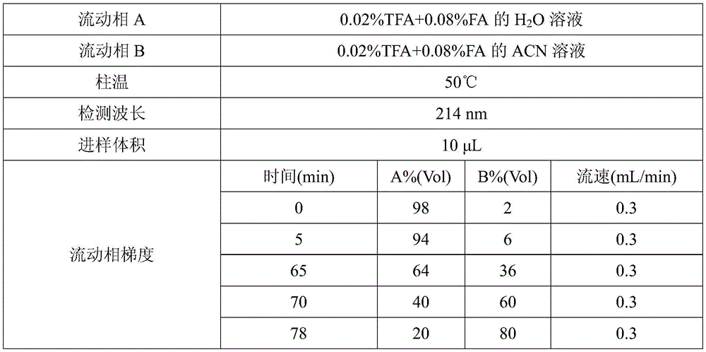
As a preferred technical scheme of the invention, the mobile phase adopted by the ultra-high performance liquid chromatography is a water-acetonitrile mixed solution containing trifluoroacetic acid and formic acid.
Preferably, the flow rate of the mobile phase is 0.25-0.35mL/min, and may be, for example, 0.25mL/min, 0.26mL/min, 0.27mL/min, 0.28mL/min, 0.29mL/min, 0.3mL/min, 0.31mL/min, 0.32mL/min, 0.33mL/min, 0.34mL/min, or 0.35mL/min, etc.; further preferably 0.3 mL/min.
As a preferred technical scheme of the invention, the ultra-high performance liquid chromatography adopts a mobile phase to carry out gradient elution.
Preferably, the gradient elution uses a mobile phase in which the acetonitrile concentration increases and then decreases with elution time.
Preferably, the mobile phase used for the gradient elution consists of mobile phase a and mobile phase B in the following volume percentages: 0-5 min: 97-99% mobile phase A and 1-3% mobile phase B; 5-65 min: 93-95% mobile phase A and 5-7% mobile phase B; 65-70 min: 60-68% mobile phase A and 32-40% mobile phase B; 70-78 min: 35-45% mobile phase a and 55-65% mobile phase B; 78-80 min: 17-23% mobile phase a and 77-83% mobile phase B; 80-90 min: 97-99% mobile phase A and 1-3% mobile phase B; the mobile phase A is an aqueous solution comprising 0.02 Vol% trifluoroacetic acid and 0.08 Vol% formic acid; the mobile phase B was an acetonitrile solution comprising 0.02 Vol% trifluoroacetic acid and 0.08 Vol% formic acid.
Preferably, the mobile phase used for the gradient elution consists of mobile phase a and mobile phase B in the following volume percentages: 0-5 min: 98% mobile phase a and 2% mobile phase B; 5-65 min: 94% mobile phase a and 6% mobile phase B; 65-70 min: 64% mobile phase a and 36% mobile phase B; 70-78 min: 40% mobile phase a and 60% mobile phase B; 78-80 min: 20% mobile phase a and 80% mobile phase B; 80-90 min: 98% mobile phase a and 2% mobile phase B; the mobile phase A is an aqueous solution comprising 0.02 Vol% trifluoroacetic acid and 0.08 Vol% formic acid; the mobile phase B was an acetonitrile solution comprising 0.02 Vol% trifluoroacetic acid and 0.08 Vol% formic acid.
In a preferred embodiment of the present invention, the sample injection volume of the ultra high performance liquid chromatography is 8 to 15. mu.L, for example, 8. mu.L, 9. mu.L, 10. mu.L, 11. mu.L, 12. mu.L, 13. mu.L, 14. mu.L, or 15. mu.L; more preferably 10. mu.L.
Compared with the prior art, the invention has the following beneficial effects:
the testing method provided by the invention is simple and feasible, the testing can be completed within 90min, the separation degree of each peak of the obtained peptide diagram is high, the accuracy is high, the repeatability of the method is good, and the method is stable and feasible.
Drawings
FIG. 1 is a peptide map of each sample in example 1.
FIG. 2 is a peptide map of each sample in example 2.
FIG. 3 is a peptide map of each sample in example 3.
Detailed Description
The technical scheme of the invention is further explained by the specific implementation mode in combination with the attached drawings. It should be understood by those skilled in the art that the specific embodiments are only for the understanding of the present invention and should not be construed as the specific limitations of the present invention.
The sample, reagent and instrument information used in the examples of the present invention are shown in tables 1 to 3 below:
TABLE 1 sample information


TABLE 2 reagent information

TABLE 3 Instrument Equipment information


Example 1
The embodiment provides a method for testing a peptide map of ramucirumab, which comprises the following steps:
(1) a500. mu.g sample was taken (50. mu.L of original drug Cyramza; 3 parts of cation eluate collection 1, 36.4. mu.L each), 225. mu.L of protein denaturation solution and 15. mu.L of 1M DTT solution were added, and the total volume was made up to 300. mu.L with 0.05M Tris-HCl solution (pH 8.0) to give 6M guanidine hydrochloride and 50mM DTT, mixed and incubated at 37 ℃ for 1 hour.
(2) Adding 12.5 μ L of 3M iodoacetic acid (IAA) solution to make the concentration of iodoacetic acid 120mM, mixing, and incubating at room temperature in dark place for 45 min; after the incubation of the sample, 13. mu.L of 1M DTT solution was added and mixed well so that the concentration of DTT added in this step was 40mM, and the alkylation reaction was terminated.
(3) Transferring the sample with the termination of the alkylation into a Millipore ultrafiltration tube (10KD), supplementing the sample to 0.5mL by using 0.05M Tris-HCl solution (pH 8.0), mixing the mixture uniformly, centrifuging the mixture at 12000G at 8 ℃ for 12min for desalting, repeating the desalting for 3 times, collecting the trapped fluid, and detecting the concentration of the desalted monoclonal antibody protein by using a micro ultraviolet spectrophotometer.
(4) Taking a desalted sample, diluting the desalted sample with 0.05M Tris-HCl solution (pH 8.0), taking 1 part of each diluted sample, adding trypsin to perform enzymolysis on antibody protein according to the ratio of enzyme to the sample being 1:25(w/w), incubating for 2h at 37 ℃, adding 10% FA solution to make the final concentration of the solution be 0.1% after the enzymolysis is finished, stopping the enzymolysis reaction, and simultaneously acidifying the peptide segment;
then, 40 μ L of each of 2 parts of desalted samples of the cation elution pool 1 was taken, 6 μ L of trypsin and 1.5 μ L of zymolytic antibody protein were added to the samples according to the ratio of 1:12.5(w/w) of the enzyme to the monoclonal antibody protein and 1:50(w/w) of the enzyme to the monoclonal antibody protein, and incubated at 37 ℃ for 2 hours, and 0.4 μ L of 10% FA was added to the samples after the zymolysis to make the final concentration 0.1%, to terminate the zymolysis reaction and acidify the peptide fragment.
The concentration and dilution process of the desalted monoclonal antibody protein are shown in the following table 4:
TABLE 4

(5) Detecting the sample treated in the step (4) by using ultra performance liquid chromatography, wherein the chromatographic conditions are shown in the following table 5:
TABLE 5

The peptide map obtained in this example is shown in fig. 1, in which the peptide maps of Cyramza (original drug), 3 parallel samples of the cation elution pool 1 (enzyme: mab protein ═ 1:25), the cation elution pool 1 (enzyme: mab protein ═ 1:12.5), and the cation elution pool 1 (enzyme: mab protein ═ 1:50) are shown from bottom to top. As can be seen from FIG. 1, the peak patterns of the samples are basically consistent, and only a few circled points are slightly different, which shows that the test method provided by the invention is accurate and reliable and has good repeatability.
Example 2
The present example provides a method for testing a peptide map of ramucirumab, which is different from example 1 in that the samples used are MV buffer and cation elution collecting solution 1, and the enzymolysis time of the cation elution collecting solution 1 is 1h, 2h (3 parallel samples), 4h, and 21 h. Wherein, the concentration and the dilution process of the desalted monoclonal antibody protein are shown in the following table 6:
TABLE 6

The peptide pattern obtained in this example is shown in FIG. 2. Wherein, the peptide graphs of MV buffer solution, 3 parallel samples of cation elution collecting solution 1 (enzymolysis for 2h), cation elution collecting solution 1 (enzymolysis for 1h), cation elution collecting solution 1 (enzymolysis for 4h) and cation elution collecting solution 1 (enzymolysis for 21h) are sequentially arranged from bottom to top. As can be seen from FIG. 2, the peak patterns of the cation-eluting pool 1 (except for the 21h digested sample) were substantially identical, with only slight differences in the circled positions. The buffer did not appear in response to the peak appearance at the time of the main peak of the sample. The test method provided by the invention has good repeatability, and the time suitable for enzymolysis is 1-4 h.
Example 3
This example provides a method for testing a peptide map of ramucirumab, which is different from example 1 in that the samples used are original grinding drug Cyramza and cation elution collection liquid 2, and 5 sets of experiments are set up:
wherein 1 part of original drug Cyramza and 1 part of cation elution pool 2 were tested in the same manner as in example 1;
the difference between the treatment of 3 parallel cationic eluate collections 2 and example 1 is that in step (3) desalting was carried out using a G-25 desalting column, specifically: balancing a G-25 desalting column with 5mL of 0.05M Tris-HCl solution, repeatedly balancing for 2 times after liquid does not drip down, adding 300 mu L of sample for terminating alkylation, adding 900 mu L of 0.05M Tris-HCl solution for washing the column after liquid does not drip down, adding 150 mu L of 0.05M Tris-HCl solution for washing the column after liquid does not drip down, collecting eluent (3 parallel samples are respectively marked as 1-1, 2-1 and 3-1), adding 150 mu L of 0.05M Tris-HCl solution for washing the column, collecting eluent (3 parallel samples are respectively marked as 1-2, 2-2 and 3-2), adding 150 mu L of 0.05M Tris-HCl solution for washing the column, and collecting eluent (3 parallel samples are respectively marked as 1-3, 2-3 and 3-3);
and (3) carrying out subsequent enzymolysis and testing on the column desalting eluent 1-2, 2-2 and 3-2, wherein the enzymolysis time of the column desalting eluent 1-2 and 2-2 is 2 hours, and the enzymolysis time of the column desalting eluent 3-2 is 4 hours.
In this example, the concentration and dilution process of the desalted monoclonal antibody protein are shown in table 7 below:
TABLE 7


The peptide pattern obtained in this example is shown in FIG. 3. Wherein, the raw grinding medicine Cyramza (Millipore ultrafiltration tube desalination, enzymolysis for 2h), the cation elution collecting liquid 2(G-25 desalting column desalination eluent 1-2, enzymolysis for 2h), the cation elution collecting liquid 2(G-25 desalting column desalination eluent 2-2, enzymolysis for 2h), and the cation elution collecting liquid 2(G-25 desalting column desalination eluent 3-2, enzymolysis for 4h) are arranged from bottom to top in sequence.
As can be seen from FIG. 3, the peptide patterns of the samples are basically consistent in peak pattern, and only a few circled points are slightly different, which shows that the test method provided by the invention is accurate and reliable and has good repeatability.
The applicant declares that the above description is only a specific embodiment of the present invention, but the scope of the present invention is not limited thereto, and it should be understood by those skilled in the art that any changes or substitutions that can be easily conceived by those skilled in the art within the technical scope of the present invention are within the scope and disclosure of the present invention.
Claims (10)
1. A test method for a peptide map of ramucirumab, which is characterized by comprising the following steps:
(1) performing denaturation reduction treatment on the ramucirumab sample by using protein denaturation liquid and a disulfide bond reducing agent in a liquid phase environment;
(2) adding an alkylating reagent into the solution obtained in the step (1) to carry out an alkylation reaction, and terminating the reaction by using dimercaptothreitol;
(3) desalting the solution obtained in the step (2), adding trypsin for enzymolysis, stopping the enzymolysis reaction by using organic acid and acidifying the peptide segment;
(4) and (4) detecting the sample treated in the step (3) by using ultra-high performance liquid chromatography to obtain a peptide map of the ramucirumab.
2. The test method according to claim 1, wherein the protein-denatured liquid in step (1) contains guanidine hydrochloride;
preferably, the concentration of guanidine hydrochloride in the liquid phase environment in the step (1) is 5.5-6.5M, and more preferably 6M;
preferably, the disulfide bond reducing agent in step (1) is dimercaptothreitol;
preferably, the concentration of dimercaptothreitol in the liquid phase environment of step (1) is 40-60mM, and more preferably 50 mM.
3. The test method according to claim 1 or 2, wherein the temperature of the denaturing reduction treatment in step (1) is 35 to 40 ℃, further preferably 37 ℃;
preferably, the time of the denaturation reduction treatment in the step (1) is 1 to 3 hours, and more preferably 1 hour.
4. The test method according to any one of claims 1 to 3, wherein the alkylating agent in the step (2) is iodoacetic acid;
preferably, the amount of the alkylating reagent added in the step (2) is 100-140mM, and more preferably 120 mM;
preferably, the alkylation reaction in step (2) is carried out under the condition of keeping out light;
preferably, the temperature of the alkylation reaction in the step (2) is 18 to 25 ℃, and further preferably 22 ℃;
preferably, the alkylation reaction time in the step (2) is 40-60min, and more preferably 45 min;
preferably, the amount of dimercaptothreitol added in step (2) is 30-50mM, more preferably 40 mM.
5. The test method according to any one of claims 1 to 4, wherein the desalting in step (3) is ultrafiltration tube centrifugal desalting or desalting with a desalting column;
preferably, after the desalting in step (3), the following operation is further performed: detecting the concentration of the ramucirumab in the desalted solution by using a trace ultraviolet spectrophotometer;
preferably, the mass ratio of the trypsin to the ramucirumab in the desalted solution in the step (3) is 1 (12.5-50), and more preferably 1: 25.
6. The test method according to any one of claims 1 to 5, wherein the temperature of the enzymatic hydrolysis in step (3) is 35 to 40 ℃, further preferably 37 ℃;
preferably, the enzymolysis time in the step (3) is 1-4h, and further preferably 2 h;
preferably, the organic acid in step (3) is formic acid;
preferably, the amount of the organic acid added in step (3) is 0.1 to 0.2 Vol%, more preferably 0.1 Vol%.
7. The test method according to any one of claims 1 to 6, wherein the stationary phase of the column used in the ultra high performance liquid chromatography in the step (4) is C18;
preferably, the temperature of a chromatographic column adopted by the ultra-high performance liquid chromatography is 47-53 ℃, and further preferably 50 ℃;
preferably, the detection wavelength of the ultra-high performance liquid chromatography is 210-220nm, and further preferably 214 nm.
8. The test method according to any one of claims 1 to 7, wherein the mobile phase used in the ultra-high performance liquid chromatography is a water-acetonitrile mixed solution containing trifluoroacetic acid and formic acid;
preferably, the flow rate of the mobile phase is 0.25 to 0.35mL/min, more preferably 0.3 mL/min.
9. The test method according to any one of claims 1 to 8, wherein the ultra performance liquid chromatography is performed with gradient elution using a mobile phase;
preferably, the gradient elution uses a mobile phase in which the concentration of acetonitrile increases and then decreases with elution time;
preferably, the mobile phase used for the gradient elution consists of mobile phase a and mobile phase B in the following volume percentages: 0-5 min: 97-99% mobile phase A and 1-3% mobile phase B; 5-65 min: 93-95% mobile phase A and 5-7% mobile phase B; 65-70 min: 60-68% mobile phase A and 32-40% mobile phase B; 70-78 min: 35-45% mobile phase a and 55-65% mobile phase B; 78-80 min: 17-23% mobile phase a and 77-83% mobile phase B; 80-90 min: 97-99% mobile phase A and 1-3% mobile phase B; the mobile phase A is an aqueous solution comprising 0.02 Vol% trifluoroacetic acid and 0.08 Vol% formic acid; the mobile phase B is an acetonitrile solution comprising 0.02 Vol% trifluoroacetic acid and 0.08 Vol% formic acid;
preferably, the mobile phase used for the gradient elution consists of mobile phase a and mobile phase B in the following volume percentages: 0-5 min: 98% mobile phase a and 2% mobile phase B; 5-65 min: 94% mobile phase a and 6% mobile phase B; 65-70 min: 64% mobile phase a and 36% mobile phase B; 70-78 min: 40% mobile phase a and 60% mobile phase B; 78-80 min: 20% mobile phase a and 80% mobile phase B; 80-90 min: 98% mobile phase a and 2% mobile phase B; the mobile phase A is an aqueous solution comprising 0.02 Vol% trifluoroacetic acid and 0.08 Vol% formic acid; the mobile phase B was an acetonitrile solution comprising 0.02 Vol% trifluoroacetic acid and 0.08 Vol% formic acid.
10. The test method according to any one of claims 1 to 9, wherein the sample volume for ultra high performance liquid chromatography is 8 to 15 μ L, more preferably 10 μ L.
Priority Applications (1)
| Application Number | Priority Date | Filing Date | Title |
|---|---|---|---|
| CN201911423878.5A CN111024863A (en) | 2019-12-31 | 2019-12-31 | Test method of peptide map of ramucirumab |
Applications Claiming Priority (1)
| Application Number | Priority Date | Filing Date | Title |
|---|---|---|---|
| CN201911423878.5A CN111024863A (en) | 2019-12-31 | 2019-12-31 | Test method of peptide map of ramucirumab |
Publications (1)
| Publication Number | Publication Date |
|---|---|
| CN111024863A true CN111024863A (en) | 2020-04-17 |
Family
ID=70201807
Family Applications (1)
| Application Number | Title | Priority Date | Filing Date |
|---|---|---|---|
| CN201911423878.5A Pending CN111024863A (en) | 2019-12-31 | 2019-12-31 | Test method of peptide map of ramucirumab |
Country Status (1)
| Country | Link |
|---|---|
| CN (1) | CN111024863A (en) |
Cited By (1)
| Publication number | Priority date | Publication date | Assignee | Title |
|---|---|---|---|---|
| CN114965839A (en) * | 2022-05-11 | 2022-08-30 | 朗肽生物制药股份有限公司 | Peptide map analysis method of human basic fibroblast growth factor |
Citations (5)
| Publication number | Priority date | Publication date | Assignee | Title |
|---|---|---|---|---|
| WO2018067987A1 (en) * | 2016-10-06 | 2018-04-12 | Amgen Inc. | Reduced viscosity protein pharmaceutical formulations |
| WO2018167847A1 (en) * | 2017-03-14 | 2018-09-20 | 株式会社島津製作所 | Method for simultaneous quantification of monoclonal antibody |
| US20190004060A1 (en) * | 2017-06-30 | 2019-01-03 | Amgen Inc. | Methods of protein clips recovery |
| WO2019178151A1 (en) * | 2018-03-13 | 2019-09-19 | Amgen Inc. | Methods for the preparation of trypsin-resistant polypeptides for mass spectrometric analysis |
| WO2019178280A1 (en) * | 2018-03-13 | 2019-09-19 | Amgen Inc. | Sequential digestion of polypeptides for mass spectrometric analysis |
-
2019
- 2019-12-31 CN CN201911423878.5A patent/CN111024863A/en active Pending
Patent Citations (5)
| Publication number | Priority date | Publication date | Assignee | Title |
|---|---|---|---|---|
| WO2018067987A1 (en) * | 2016-10-06 | 2018-04-12 | Amgen Inc. | Reduced viscosity protein pharmaceutical formulations |
| WO2018167847A1 (en) * | 2017-03-14 | 2018-09-20 | 株式会社島津製作所 | Method for simultaneous quantification of monoclonal antibody |
| US20190004060A1 (en) * | 2017-06-30 | 2019-01-03 | Amgen Inc. | Methods of protein clips recovery |
| WO2019178151A1 (en) * | 2018-03-13 | 2019-09-19 | Amgen Inc. | Methods for the preparation of trypsin-resistant polypeptides for mass spectrometric analysis |
| WO2019178280A1 (en) * | 2018-03-13 | 2019-09-19 | Amgen Inc. | Sequential digestion of polypeptides for mass spectrometric analysis |
Non-Patent Citations (4)
| Title |
|---|
| JANOUSEK, J 等: "Antiangiogenic Human Monoclonal Antibody Ramucirumab Radiolabelling: In Vitro Evaluation on VEGFR2-positive Cell Lines", 《ANTICANCER RESEARCH》 * |
| 乔玉玲 等: "抗CD52单克隆抗体HPLC-肽图分析方法的建立", 《生物技术通报》 * |
| 吴霖萍: "治疗性单克隆抗体的理化特性分析平台的建立与应用", 《中国优秀博硕士学位论文全文数据库(硕士)医药卫生科技辑》 * |
| 桂芳 等: "抗人TNF-α单克隆抗体液质联用肽图分析方法的建立及验证", 《药物分析杂志》 * |
Cited By (1)
| Publication number | Priority date | Publication date | Assignee | Title |
|---|---|---|---|---|
| CN114965839A (en) * | 2022-05-11 | 2022-08-30 | 朗肽生物制药股份有限公司 | Peptide map analysis method of human basic fibroblast growth factor |
Similar Documents
| Publication | Publication Date | Title |
|---|---|---|
| CN115724973B (en) | Anti-human ROR1 high-affinity rabbit monoclonal antibody and application thereof | |
| CN111735948B (en) | Application of PADI4 in the preparation of tumor diagnostic kits | |
| CN111024863A (en) | Test method of peptide map of ramucirumab | |
| US8304236B2 (en) | Modified biotin-binding protein | |
| CN110618270B (en) | Preparation method of reagent for quantitatively determining helicobacter pylori antigen in feces | |
| CN109293739B (en) | A kind of A3 superfamily universal tumor antigen polypeptide and its application | |
| CN113929776B (en) | Antifungal (1, 3) -beta-D glucan monoclonal antibody, encoding gene and expression and application thereof | |
| CN113621079B (en) | Fusion protein of Fab antibody and calf intestinal alkaline phosphatase and preparation method thereof | |
| CN119192368B (en) | Monoclonal antibody or antigen-binding fragment thereof for detecting folic acid and its binding protein complex, preparation method and application thereof | |
| CN108059676B (en) | Anti-human nerve growth factor scFv antibody and preparation method thereof | |
| CN112094355B (en) | Composite quality control product for clinical diagnosis and preparation method thereof | |
| CN118344485B (en) | Anti-folic acid and monoclonal antibody of binding protein complex thereof or antigen binding fragment thereof, preparation method and application thereof | |
| CN111733151B (en) | Antigen and antibody prepared based on PADI4 as tumor marker and application | |
| CN116925219B (en) | Antibody of small heat shock protein HSPB1, hybridoma cell strain and application thereof | |
| CN116925218B (en) | Antibody of small heat shock protein HSPB1, antibody composition, hybridoma cell strain and application thereof | |
| CN116773813B (en) | Preparation and application of VEGFR1 detection kit | |
| CN114989299B (en) | Composition of monoclonal antibody, application of composition, reagent, kit and method for detecting human interleukin 1 beta | |
| CN115109159B (en) | CD19 mutant protein and its application | |
| CN113754749B (en) | Cryptosporidium parvum Gp40/15 protein epitope polypeptide and adenovirus vector vaccine thereof | |
| CN107746430B (en) | Preparation and application of GP 73C-terminal antigen | |
| CN112442496B (en) | Arginine deiminase mutant and application thereof | |
| US9914757B2 (en) | Methionyl tRNA synthetase for biosynthesis of photomethionine-labeled protein and method for preparing photoactive protein G variant using same | |
| CN110468108B (en) | Hybridoma cell strain secreting human ferritin light chain monoclonal antibody and application thereof | |
| CN114276449A (en) | Monoclonal antibody for resisting human inhibin alpha, preparation method thereof, immunoassay reagent and application | |
| CN106729769A (en) | TEM1 specificity fluorescents probe and its application |
Legal Events
| Date | Code | Title | Description |
|---|---|---|---|
| PB01 | Publication | ||
| PB01 | Publication | ||
| SE01 | Entry into force of request for substantive examination | ||
| SE01 | Entry into force of request for substantive examination | ||
| RJ01 | Rejection of invention patent application after publication |
Application publication date: 20200417 |
|
| RJ01 | Rejection of invention patent application after publication |