CN110956628B - Picture grade classification method, device, computer equipment and storage medium - Google Patents
Picture grade classification method, device, computer equipment and storage medium Download PDFInfo
- Publication number
- CN110956628B CN110956628B CN201911283146.0A CN201911283146A CN110956628B CN 110956628 B CN110956628 B CN 110956628B CN 201911283146 A CN201911283146 A CN 201911283146A CN 110956628 B CN110956628 B CN 110956628B
- Authority
- CN
- China
- Prior art keywords
- quality
- model
- picture
- screening
- training
- Prior art date
- Legal status (The legal status is an assumption and is not a legal conclusion. Google has not performed a legal analysis and makes no representation as to the accuracy of the status listed.)
- Active
Links
Images


Classifications
-
- G—PHYSICS
- G06—COMPUTING OR CALCULATING; COUNTING
- G06T—IMAGE DATA PROCESSING OR GENERATION, IN GENERAL
- G06T7/00—Image analysis
- G06T7/0002—Inspection of images, e.g. flaw detection
- G06T7/0012—Biomedical image inspection
-
- G—PHYSICS
- G06—COMPUTING OR CALCULATING; COUNTING
- G06T—IMAGE DATA PROCESSING OR GENERATION, IN GENERAL
- G06T2207/00—Indexing scheme for image analysis or image enhancement
- G06T2207/20—Special algorithmic details
- G06T2207/20081—Training; Learning
-
- G—PHYSICS
- G06—COMPUTING OR CALCULATING; COUNTING
- G06T—IMAGE DATA PROCESSING OR GENERATION, IN GENERAL
- G06T2207/00—Indexing scheme for image analysis or image enhancement
- G06T2207/20—Special algorithmic details
- G06T2207/20084—Artificial neural networks [ANN]
-
- G—PHYSICS
- G06—COMPUTING OR CALCULATING; COUNTING
- G06T—IMAGE DATA PROCESSING OR GENERATION, IN GENERAL
- G06T2207/00—Indexing scheme for image analysis or image enhancement
- G06T2207/30—Subject of image; Context of image processing
- G06T2207/30004—Biomedical image processing
- G06T2207/30041—Eye; Retina; Ophthalmic
-
- G—PHYSICS
- G06—COMPUTING OR CALCULATING; COUNTING
- G06T—IMAGE DATA PROCESSING OR GENERATION, IN GENERAL
- G06T2207/00—Indexing scheme for image analysis or image enhancement
- G06T2207/30—Subject of image; Context of image processing
- G06T2207/30004—Biomedical image processing
- G06T2207/30096—Tumor; Lesion
Landscapes
- Engineering & Computer Science (AREA)
- Health & Medical Sciences (AREA)
- General Health & Medical Sciences (AREA)
- Medical Informatics (AREA)
- Nuclear Medicine, Radiotherapy & Molecular Imaging (AREA)
- Radiology & Medical Imaging (AREA)
- Quality & Reliability (AREA)
- Computer Vision & Pattern Recognition (AREA)
- Physics & Mathematics (AREA)
- General Physics & Mathematics (AREA)
- Theoretical Computer Science (AREA)
- Image Analysis (AREA)
Abstract
The application relates to a picture level classification method, a picture level classification device, computer equipment and a storage medium. The method comprises the following steps: inputting a picture to be detected into a quality judging model to obtain the quality grade of the picture to be detected; the quality grade is used for representing the definition degree of the picture to be detected; inputting the picture to be detected into a screening model to obtain the lesion grade of a region to be detected in the picture to be detected; and determining the credibility of the lesion level of the picture to be detected according to the quality level of the picture to be detected. By adopting the method, the reduction of the reliability and the accuracy of the lesion level of the to-be-detected region caused by the uneven quality of the to-be-detected picture can be avoided, and the credibility of the final picture level classification result is improved.
Description
Technical Field
The present disclosure relates to the field of image processing technologies, and in particular, to a method and apparatus for classifying picture levels, a computer device, and a storage medium.
Background
Diabetic retinopathy (Diabetic Retinopathy, DR) is a common blinding eye disease and is one of the most common chronic complications for diabetics. DR has no obvious clinical symptoms in early stages, and can significantly reduce the risk of blindness if fundus examination can be performed periodically at the beginning of the onset.
In the traditional technology, the manual DR screening method depends on clinical experience of doctors to-be-detected pictures, and because the fundus images do not display clear retina structures due to different experiences of image acquisition personnel, the basic-level ophthalmology does not have good fundus photographing professional equipment and the retina images acquired by different model fundus cameras are different in type, so that the quality of the shot to-be-detected pictures is uneven, and the reliability and the accuracy of DR screening are greatly reduced.
Disclosure of Invention
Based on this, it is necessary to provide a picture level classification method, apparatus, computer device and storage medium in order to address the above technical problems.
In one aspect, a method for classifying picture levels is provided, the method comprising:
inputting a picture to be detected into a quality judging model to obtain the quality grade of the picture to be detected; the quality grade is used for representing the definition degree of the picture to be detected;
inputting the picture to be detected into a screening model to obtain the lesion grade of a region to be detected in the picture to be detected;
and determining the credibility of the lesion level of the picture to be detected according to the quality level of the picture to be detected.
In another aspect, there is provided an image grade classification apparatus, the apparatus comprising:
The quality judging module is used for inputting the picture to be detected into the quality judging model to obtain the quality grade of the picture to be detected; the quality grade value is used for representing the definition degree of the picture to be detected;
the screening module is used for inputting the picture to be detected into a screening model to obtain the lesion grade of the region to be detected in the picture to be detected;
and the credibility module is used for determining the credibility of the pathological change grade of the picture to be detected according to the quality grade of the picture to be detected.
In another aspect, a computer device is provided, comprising a memory storing a computer program and a processor implementing the steps of any of the methods described above when the processor executes the computer program.
In another aspect, a computer readable storage medium is provided, having stored thereon a computer program which, when executed by a processor, implements the steps of any of the methods described above.
The picture level classification method, the device, the computer equipment and the storage medium comprise the following steps: and classifying the quality grade of the picture to be detected by adopting the quality judging model to obtain the quality grade of the picture to be detected. Inputting the picture to be detected into the screening model to obtain the lesion level of the region to be detected in the picture to be detected, and determining the credibility of the lesion level to be detected according to the quality level. The reliability and the accuracy of the lesion level of the to-be-detected area, which are caused by uneven quality of the to-be-detected picture, are prevented from being reduced, the reliability of the lesion level is represented by the quality level, the reliability and the accuracy of the obtained lesion level of the to-be-detected area are improved, and the reliability of the classification result of the final picture level is improved.
Drawings
FIG. 1 is a flow chart of a method for classifying picture levels in one embodiment;
FIG. 2 is a flow chart of training an initial quality discriminant model in one embodiment;
FIG. 3 is a schematic diagram of an initial quality discrimination model in one embodiment;
FIG. 4 is a flow diagram of training an initial screening model in one embodiment;
FIG. 5 is a schematic diagram of the structure of an initial screening model in one embodiment;
FIG. 6 is a block diagram of a picture level classification apparatus in one embodiment;
fig. 7 is an internal structural diagram of a computer device in one embodiment.
Detailed Description
In order to make the objects, technical solutions and advantages of the present application more apparent, the present application will be further described in detail with reference to the accompanying drawings and examples. It should be understood that the specific embodiments described herein are for purposes of illustration only and are not intended to limit the present application.
In one embodiment, as shown in fig. 1, there is provided a picture level classification method, the method including:
s110, inputting the picture to be detected into a quality judging model to obtain the quality grade of the picture to be detected.
The quality grade is used for representing the definition degree of the picture to be detected.
Further, the to-be-inspected picture is a picture including the fundus of the person to be inspected, which is shot by the user. The quality judging model is a convolutional neural network model taking MobileNetV1 as a basic network and operates in terminal equipment in the form of a computer executing program, and is used for classifying the quality grades of the input pictures to obtain the quality grades. For example, a value 1 is adopted to represent that the quality grade of the picture to be detected is excellent, namely, the fundus area in the picture to be detected is clear, a value 0 is adopted to represent that the quality grade of the picture to be detected is poor, namely, when the fundus area in the picture to be detected is not clear or has shielding, the picture to be detected is input into the quality judging model, and when the output value is 0-0.2, the quality grade of the picture to be detected is poor; when the output value is 0.2-0.8, the quality grade of the picture to be detected is ambiguous; and when the output value is 0.8-1, the quality grade of the picture to be detected is excellent.
Specifically, the computer device may directly obtain the to-be-inspected picture including the fundus of the person to be inspected through an image pickup device such as a camera, or may obtain the already-photographed to-be-inspected picture from a storage device such as a mobile phone. After the computer equipment obtains the picture to be detected, inputting the picture to be detected into the quality judging model, and classifying the quality grade of the picture to be detected by the quality judging model to obtain the quality grade of the picture to be detected. The quality judging model is a convolutional neural network model which is obtained through a large number of clear and unclear fundus pictures and is used for classifying the fundus pictures of the person to be inspected in quality grade.
Further, when the quality level of the picture to be detected is poor, prompting the user that the quality of the picture is poor, resulting in unreliable results, and stopping the current quality level classification or continuing to proceed to the next step S120 according to the user selection.
S120, inputting the picture to be detected into a screening model to obtain the lesion level of the region to be detected in the picture to be detected.
The lesion level is used for representing the lesion severity of a region to be detected in the picture to be detected.
Further, the region to be detected is the fundus of the person to be detected. The screening model is a convolutional neural network model taking EfficientNetB5 as a basic network, and runs in a terminal device in a form of a computer execution program, and is used for classifying lesion grades of eyeground in an input picture to obtain the lesion grade. For example, the value 0 is used for representing that the lesion level of the fundus in the to-be-detected picture is no lesion on the fundus, the value 1 is used for representing that the lesion level of the fundus in the to-be-detected picture is slight lesion on the fundus, the value 2 is used for representing that the lesion level of the fundus in the to-be-detected picture is moderate lesion on the fundus, the value 3 is used for representing that the lesion level of the fundus in the to-be-detected picture is severe non-proliferation lesion on the fundus, and the value 4 is used for representing that the lesion level of the fundus in the to-be-detected picture is severe proliferation lesion on the fundus.
Specifically, the computer equipment inputs the picture to be detected into the screening model, and the screening model classifies the lesion level of the picture to be detected to obtain the lesion level of the fundus in the picture to be detected. The screening model is obtained through training a large number of normal and pathological fundus pictures and is used for classifying the fundus pathological changes of the fundus pictures of the person to be detected in a grade mode.
S130, determining the credibility of the lesion level of the picture to be detected according to the quality level of the picture to be detected.
Specifically, the computer device may directly use the quality level of the to-be-detected picture to represent the credibility of the lesion level of the to-be-detected region in the to-be-detected picture.
In this embodiment, the computer device classifies the quality level of the to-be-detected picture by using the quality screening model, so as to obtain the quality level of the to-be-detected picture. Inputting the picture to be detected into the screening model to obtain the lesion grade of the region to be detected in the picture to be detected, and obtaining the credibility of the lesion grade according to the quality grade. The reliability and the accuracy of the lesion level of the to-be-detected area, which are caused by uneven quality of the to-be-detected picture, are prevented from being reduced, the reliability of the lesion level of the to-be-detected area is characterized by the quality level, the reliability and the accuracy of the obtained lesion level of the to-be-detected area are improved, the reliability of the classification result of the picture level is improved, and the follow-up problem caused by low reliability and accuracy of the classification result is avoided.
In another embodiment, the step S130 of determining the credibility of the lesion level of the to-be-detected picture according to the quality level of the to-be-detected picture includes:
if the quality level is excellent, determining that the reliability is high;
if the quality level is uncertain, determining the credibility as uncertain;
if the quality level is poor, the reliability is determined to be low.
In this embodiment, the computer device directly uses the quality level of the to-be-detected picture to forward characterize the credibility of the lesion level of the to-be-detected picture, so as to clearly determine the credibility of the lesion level of the to-be-detected region in the picture obtained according to the input picture, and facilitate the user to take measures in time to improve the credibility of the lesion level.
In another embodiment, before the step S110 of inputting the picture to be detected into the quality discrimination model to obtain the quality level of the picture to be detected, the method further includes:
and carrying out quality picture pretreatment on the picture to be detected so as to unify the picture size and the display effect.
Further, the to-be-inspected picture may be a fundus picture of the to-be-inspected person acquired by a user through different image acquisition devices, or a fundus picture of the to-be-inspected person acquired by different users. Due to the fact that the image acquisition equipment is different, the positions of eyeground of the person to be detected are different by the person to be detected, the environment factors of the acquisition place are different, the initial picture to be detected has environment interference caused by the different environment factors, and accuracy of quality grade classification and lesion grade classification of the eyeground picture is affected.
Specifically, the computer device preprocesses the picture to be detected. The preprocessing may include background denoising, for example, removing a black frame of the to-be-detected picture, reserving an area of an effective fundus image, adjusting the resolution of the to-be-detected picture to 224 x 224, and the preprocessing may further include display normalization, for example, whitening the to-be-detected picture and brightness adjustment normalization to the same range, and the preprocessing may further include random rotation and other operations with the center of the to-be-detected picture as a rotation center, so as to highlight fundus focus information and eliminate environmental interference. And after the computer equipment performs the preprocessing on the picture to be detected, the S110 is executed again according to the unified picture size and the display effect.
In this embodiment, the computer device performs preprocessing on the to-be-detected picture, and after the preprocessing, the to-be-detected picture with uniform picture size and display effect is obtained, so that quality grade classification and lesion grade classification inaccuracy caused by different image acquisition devices, different fundus positions of the to-be-detected person, different environmental factors of acquisition places and the like are eliminated, environmental interference is eliminated by highlighting fundus focus information, and accuracy of quality grade classification and lesion grade classification of the picture is improved.
In another embodiment, before the step S110 of inputting the picture to be detected into the quality discrimination model to obtain the quality level of the picture to be detected, the method further includes: training the quality discrimination model, specifically training an initial quality discrimination model to obtain the quality discrimination model.
As shown in fig. 2, the training process of the quality discrimination model includes the following steps:
s210, acquiring a quality screening sample set.
Wherein the quality screening sample set includes a plurality of screening pictures, and each of the screening pictures carries a label for characterizing image sharpness.
Further, the label carried by the screening picture is a numerical value label which is obtained manually and used for representing the picture definition degree according to the exposure degree, the light spot size, the focusing deficiency and excess of the picture and whether the area to be detected is shielded or not, and the experience cognition of the detection personnel on whether the picture is clear or not. For example, the label includes 0 and 1, the label 1 is used to represent that the fundus area in the to-be-detected picture is clear, and the label 0 is used to represent that the fundus area in the to-be-detected picture is not clear or has shielding.
S220, preprocessing and dividing the screening pictures in the quality screening sample set to obtain a normalized quality training subset, a normalized quality verification subset and a normalized quality testing subset.
The quality training subset is used for training the initial quality judging model, the quality verification subset is used for verifying the trained initial quality judging model, and the quality testing subset is used for testing the verified initial quality judging model.
Specifically, because the quality screening pictures in the quality screening sample set have environmental interference caused by different image acquisition devices and different acquisition personnel, the computer equipment needs to preprocess the quality screening pictures in the quality screening sample set so as to eliminate the environmental interference. And preprocessing the picture to be detected in the previous embodiment to unify the picture size and the display effect, so as to obtain the normalized quality screening sample set. Meanwhile, the quality screening sample set is divided into a quality training subset, a quality verification subset and a quality testing subset according to a certain proportion. For example, the quality screening sample set is divided into the quality training subset, the quality verification subset, and the quality testing subset in a ratio of 6:3:1.
And S230, training the initial quality judgment model by adopting the quality training subset, verifying the trained initial quality judgment model by adopting the quality verification subset, and testing the verified initial quality judgment model by adopting the quality test subset to obtain the quality judgment accuracy.
When the quality judgment accuracy is greater than or equal to a preset accuracy threshold, the quality judgment model is obtained;
and when the quality judgment accuracy is smaller than the accuracy threshold, continuing to train the quality judgment model again by adopting the quality training subset until the new quality judgment accuracy is larger than or equal to the accuracy threshold, and obtaining the quality judgment model.
The initial quality discriminant model 300 is shown in fig. 3, and includes a MobileNetV1 base network and two full-connection layers. The mobilenet v1 base network includes a standard convolution layer of 1 layer, a separable convolution layer of 13 layers, and a global average pooling layer of 1 layer.
The above-described structure of the initial quality judgment model is merely an example, and the initial quality judgment model described in the present application is not limited to the above-described specific embodiment.
Specifically, the computer equipment inputs all the quality training subsets into the initial quality judging model for a plurality of times, adjusts model parameters in the initial quality judging model according to a loss function in the initial quality judging model each time, adjusts the model parameters once for each training time to determine the model parameters in the initial quality judging model, and realizes a plurality of times of training on the initial quality judging model. And the computer equipment inputs all the quality verification subsets into the initial quality judgment model after each training, and determines whether the training in the initial quality judgment model is stopped according to the change condition of a loss function in the initial quality judgment model obtained once and for realizing the verification of the initial quality judgment model. And the computer equipment inputs all the quality test subsets into the verified initial quality judgment model, and obtains the quality judgment accuracy of the initial quality judgment model according to the quality grade obtained after the quality test subsets are input into the quality judgment model and the labels of the quality test subsets. Determining whether the initial quality decision model is used as the quality decision model or retrained based on the quality decision accuracy.
When the quality judgment accuracy is greater than or equal to a preset accuracy threshold, the initial quality judgment model after training, verification and testing can be used as the quality judgment model.
When the quality judgment accuracy is smaller than the accuracy threshold, the initial quality judgment model after training, verification and testing is unstable, the quality judgment model is required to be retrained by continuing to adopt the quality training subset until the new quality judgment accuracy is larger than or equal to the accuracy threshold, and the quality judgment model is obtained.
Further, in combination with the structure of the initial quality judgment model 300, after the picture is input into the initial quality judgment model 300, the specific process of training/verifying/testing the picture by the initial quality judgment model 300 is as follows:
and the standard convolution layer carries out edge compensation processing on the input picture, carries out convolution operation on the picture subjected to the edge compensation processing according to the convolution kernel size and the sliding step length of the standard convolution layer to obtain a standard feature map, carries out batch normalization processing on the standard feature map, and outputs a standard feature map after processing by adopting an activation function such as ReLU, wherein the standard feature map is used as the input of a next layer structure.
Further, the depth separable convolution layer receives the standard feature map of the upper layer structure output. Wherein the depth separable convolution layers include a depth convolution layer and a point-by-point convolution layer. The depth convolution layer receives the standard feature map, performs edge compensation processing on the standard feature map, performs convolution operation on the picture subjected to the edge compensation processing according to the convolution kernel size and the sliding step length of the depth convolution layer to obtain a depth feature map, performs batch normalization processing on the depth feature map, outputs the depth feature map after processing by adopting an activation function such as ReLU, and takes the depth feature map as input of the point-by-point convolution layer. And the point-by-point convolution layer carries out point-by-point convolution operation on the depth feature map according to the convolution kernel size and the sliding step length of the point-by-point convolution layer to obtain a point-by-point feature map, then carries out batch normalization processing on the point-by-point feature map, adopts an activation function such as ReLU processing to output the point-by-point feature map, and takes the point-by-point feature map as the input of a next layer structure.
Further, the global average pooling layer receives the point-by-point feature map, performs average pooling on the point-by-point feature map according to the convolution kernel size of the global average pooling layer, outputs a pooled feature map, and takes the pooled feature map as an input of a next layer structure.
Further, the full-connection layer receives the pooling feature map, the number of neurons of the last layer of the full-connection layer is 1, and the full-connection layer classifies the pooling feature map by adopting a sigmoid classifier to obtain the quality grade of the input picture.
In another embodiment, the step S230 of training the initial quality discrimination model by using the quality training subset, verifying the trained initial quality discrimination model by using the quality verification subset, and testing the verified initial quality discrimination model by using the quality testing subset to obtain a quality determination accuracy rate includes:
and training common parameters in the initial quality judgment model by adopting the quality training subset, and testing the initial quality judgment model after verification by adopting the quality testing subset when the cross entropy loss value corresponding to the quality verification subset meets the preset verification requirement to obtain the quality judgment accuracy.
The common parameters are variables which can be automatically learned according to the input data in the convolutional neural network model.
Further, the sigmoid classifier in the quality judging model adopts cross entropy as a loss function, and the cross entropy is used for representing the inconsistency degree of the quality grade obtained by inputting the picture into the quality judging model and the label of the picture.
Further, the cross entropy satisfies the following formula:

wherein H (p, q) is cross entropy, p (x) is label, q (x) is quality grade output by the quality discrimination model, x is serial number of input picture.
Specifically, the super-parameters in the initial quality judgment model are preset, for example, the minimum batch is set to 128, the learning rate is 0.001, the momentum parameter is 0.9, and the learning round number is 26. The computer equipment inputs all the quality training subsets to the initial quality judging model, the obtained cross entropy adjusts common parameters in the initial quality judging model according to the cross entropy, one-time training of the initial quality judging model is achieved, then all the quality training subsets are input to the initial quality judging model after the common parameters are adjusted, the cross entropy is obtained, and then the common parameters in the initial quality judging model are adjusted according to the cross entropy, so that multiple times of training of the initial quality judging model are achieved. And inputting the quality verification subset into the initial quality judgment model obtained through each training, comparing the change condition of the cross entropy, and stopping training the initial quality judgment model until the cross entropy is not reduced. And inputting the quality test subset into the initial quality judgment model obtained by the last training to obtain the quality judgment accuracy.
In this embodiment, the quality training subset is used to train the common parameters in the initial quality discriminating model, the quality verification subset is used to verify the trained initial quality discriminating model, and the quality test subset is used to test the verified initial quality discriminating model, so as to obtain the quality discriminating model with the highest and most stable accuracy of the output result, thereby realizing the omnibearing processing of training, verifying and testing the initial quality discriminating model, and improving the reliability and accuracy of the quality grade classification of the final quality discriminating model.
In another embodiment, before the step S120 of inputting the to-be-detected picture into the screening model to obtain the lesion level of the to-be-detected region in the to-be-detected picture, the method further includes: training the screening model specifically comprises training an initial screening model to obtain the screening model.
As shown in fig. 4, the training process of the screening model includes the following steps:
s410, acquiring a public sample set.
Wherein the common sample set comprises a plurality of common pictures, and each common picture carries a label for representing the lesion degree.
Further, the common sample set generally refers to a lesion picture of the region to be detected obtained from the public data platform. The fundus-specific lesion image may be a common fundus color taken from a diabetic retinopathy detection (Diabetic Retinopathy Detection) dataset in a karaoke network (kagle). The label carried by the public picture is based on a high-recognition-degree judgment standard, and the fundus pathological change degree is artificially obtained by combining whether symptoms such as microangioma, hard exudation, macular edema, cotton velvet spot and hemorrhage exist in the fundus in the picture. For example, the label includes 0,1,2,3,4, the label 1 is used to represent that the fundus area in the initial to-be-detected picture is clear, the label 0 is used to represent that the fundus in the to-be-detected picture has no lesion, the label 1 is used to represent that the fundus in the to-be-detected picture has slight lesions, the label 2 is used to represent that the fundus in the to-be-detected picture has moderate lesions, the label 3 is used to represent that the fundus in the to-be-detected picture has severe non-proliferative lesions, and the label 4 is used to represent that the fundus in the to-be-detected picture has severe proliferative lesions.
S420, preprocessing and dividing the public pictures in the public sample set to obtain a normalized public training subset and a normalized public verification subset.
The public training subset is used for training the initial screening model, and the public verification subset is used for verifying the trained initial screening model.
Specifically, because the common sample set has environmental interference caused by different image acquisition devices and different acquisition personnel, the computer device needs to preprocess the common pictures in the common sample set so as to eliminate the environmental interference. The preprocessing comprises background denoising, namely removing a black frame of a public picture in the public sample set, reserving an area of an effective fundus image, adjusting the resolution of the public picture in the public sample set to 456 x 456, and display normalization, namely whitening and brightness adjustment normalization of the public picture in the public sample set to be in the same range. The preprocessing also includes Gaussian blur denoising of the common pictures in the common sample set. The preprocessing also comprises the operations of random rotation, mirroring, translation, color space conversion, scaling and the like by taking the center of the public picture in the public sample set as a rotation center so as to highlight fundus focus information and eliminate environmental interference. And after the computer equipment performs the pretreatment on the public pictures in the public sample set, fundus pictures with uniform picture sizes and display effects are obtained. Meanwhile, the common sample set is divided into a common training subset and a common verification subset according to a certain proportion. For example, the common sample set is divided into the common training subset and the common verification subset in a ratio of 7:3.
And S430, training the initial screening model by adopting the public training subset, verifying the trained initial screening model by adopting the public verification subset, and obtaining a temporary screening model according to a verification result.
The initial lesion convolutional neural network model 500 is shown in fig. 5, and includes an afflicientnetb 5 base network, a global averaging pooling layer, a discard layer, and a 2-layer fully connected layer. The EfficientNetB5 base network is a convolutional neural network with a mobile inversion bottleneck convolutional (MBConv: mobile inverted bottleneck convolution) as a basic structure. The MBConv structure is derived from MobileNetV2, and unlike classical residual blocks, replaces the bottleneck (bottleneck) in classical residual blocks with a deep convolution (Depthwise convolution), applies a 1 x 1 liter dimension to the input picture, a 3 x 3 deep convolution and then applies an activation function such as ReLU processing, and a 1 x 1 dimension-reducing bilinear transformation yields a base feature map.
The above-described structure of the initial screening model is merely an example, and the initial screening model described in the present application is not limited to the above-described specific embodiment.
Specifically, the computer device inputs all the common training subsets into the initial screening model for multiple times, adjusts model parameters in the initial screening model according to a loss function in the initial screening model each time, adjusts the model parameters once for each training to determine the model parameters in the initial screening model, and realizes multiple training of the initial screening model. And the computer equipment inputs all the public verification subsets into the initial screening model after each training, determines whether training in the initial screening model is stopped according to the change condition of a loss function in the initial screening model obtained once, and verifies the initial screening model to obtain the temporary screening model.
Further, with reference to the structure of the initial screening model 500, after the picture is input into the initial screening model 500, the specific process of training/verifying the picture by the initial screening model 500 is as follows:
and the EfficientNetB5 basic network processes the input picture and outputs a basic feature map, and the basic feature map is used as the input of the next layer structure.
Further, the global average pooling layer receives the basic feature map, performs average pooling on the basic feature map according to the convolution kernel size of the global average pooling layer, outputs a pooled feature map, and takes the pooled feature map as an input of a next layer structure.
Further, the discarding layer receives the pooled feature map output by the previous layer structure. The discarding layer is used for temporarily discarding the neural network unit in the initial screening model from the network model according to a certain probability. In this embodiment, the discarding ratio of the neural network unit is 0.5. And after the discarding layer outputs the discarding feature map, taking the discarding feature map as the input of the next layer structure.
Further, the number of neurons of the full-connection layer is 5, an activation function such as an ELU activation function is used for processing, then the activation feature map is output, the activation feature map is used as input of the next full-connection layer, the number of neurons of the next full-connection layer is 1, and after being processed by adopting the activation function such as a linear activation function, the lesion level of the picture is output. Wherein the next full connection layer adopts a RAdam optimizer.
S440, inputting the lesion screening sample set into the temporary screening model to obtain the lesion screening sample set carrying the pseudo tag.
The lesion screening sample set is fundus lesion images which are collected by a user, and a label used for representing the lesion degree is not provided. And after the pseudo tag is input into the temporary screening model for the lesion screening sample, the lesion grade of the region to be detected of the screening picture in the lesion screening sample set output by the temporary screening model.
Specifically, the computer equipment inputs the lesion screening sample set into the trained temporary screening model, the temporary screening model correspondingly outputs the lesion grade of the fundus in the screening picture in the lesion screening sample set, and the outputted lesion grade is marked on the corresponding screening picture in the lesion screening sample set as the pseudo tag, so that the lesion screening sample set carrying the pseudo tag is obtained.
Further, at S440, inputting the lesion screening sample set into the temporary screening model to obtain a lesion screening sample set carrying a pseudo tag, which further includes: and preprocessing the screened pictures in the lesion screening sample set to unify the picture size and the display effect, wherein the specific content of the preprocessing is as in the preprocessing in the step S420, and the preprocessing is not repeated.
S450, inputting the lesion screening sample set with the pseudo tag and the public training subset into the temporary screening model for training, and adopting the public verification subset to verify the trained temporary screening model to obtain the screening model.
Specifically, the computer equipment inputs all the lesion screening sample sets carrying the pseudo tags and the public training subsets into the temporary screening model for a plurality of times, adjusts model parameters in the temporary screening model according to a loss function in the temporary screening model each time, adjusts the model parameters once for each training time to determine the model parameters in the temporary screening model, and realizes a plurality of training on the temporary screening model. And the computer equipment inputs all the public verification subsets into the temporary screening model after each training, determines whether the training of the temporary screening model is stopped according to the change condition of the loss function in the temporary screening model obtained once, and verifies the temporary screening model to obtain the screening model.
The temporary screening model has the same structure as the initial screening model, and the specific training/verification process is similar to that described in S430, and is not described herein again, so as to finally obtain the screening model.
Since most of the disease screening sample sets are normal or slight disease pictures of the fundus, the number of the severe disease picture samples of the fundus is very small, so that the fundus disease grade is not classified comprehensively by the trained screening model, the sample sets are required to be additionally expanded, the disclosed sample sets comprise a large number of fundus disease picture samples with different degrees, but the sample race difference and grade value marking standard are not used with the disease screening pictures, and cannot be directly expanded. In this embodiment, the computer device trains and verifies the initial screening model by using the public sample set to obtain the temporary screening model, inputs the lesion screening sample set into the temporary screening model to obtain the lesion screening sample set with the pseudo tag, unifies the public sample set and the lesion screening sample set with the lesion grade value marking standard, inputs the public training subset and the lesion screening sample set into the temporary screening model to train the temporary screening model, thereby expanding the lesion screening sample set and effectively improving the accuracy of the trained screening model.
In another embodiment, the step S430 of training the initial screening model by using the common training subset, and verifying the trained initial screening model by using the common verification subset, and obtaining the temporary screening model according to the verification result includes:
and training the common parameters in the initial screening model by adopting the common training subset, training the stage coefficients in the initial screening model after training by adopting the common verification subset, and obtaining a temporary screening model when the mean square error corresponding to the common verification subset meets the preset verification requirement.
Further, the RAdam optimizer in the screening model employs a mean square error as a loss function. The mean square error is used for representing the deviation degree of the lesion level obtained by inputting the picture into the screening model and the label of the picture.
Further, the mean square error satisfies the following formula:

wherein MSE (y, y') is the mean square error, y i Is a label, y' i And (3) for screening the lesion level output by the model, i is the serial number of the input pictures, and n is the number of the input pictures.
Specifically, the EfficientNetB5 network parameters obtained by image Net training are used as initial parameters of the initial screening model, the computer equipment inputs all public training subsets to the initial screening model, the obtained mean square error is adjusted according to the common parameters in the initial screening model, one-time training of the initial screening model is achieved, all public training subsets are input to the initial screening model after the common parameters are adjusted, the mean square error is obtained, and common parameters in the initial screening model are adjusted according to the mean square error, so that multiple training of the initial screening model is achieved. And inputting the public verification subset into the initial screening model obtained through each training, training the stage coefficients in the initial screening model, comparing the change condition of the mean square error, and stopping training the common parameters and the stage coefficients in the initial screening model until the mean square error is not reduced.
Further, training the stage coefficients in the verified initial screening model by the public verification subset to obtain the temporary screening model.
Wherein, the rules of lesion class classification are: the stage coefficient in the screening model is C1-C5, the value y output by the model is less than C1, the corresponding output lesion level is C1, and the fundus can be represented as no lesion; the numerical value C1 which is output by the model is less than or equal to y and less than C2, and the corresponding output lesion level is C2, so that the slight lesion of the fundus can be represented; the numerical value C2 which is output by the model is less than or equal to y and less than C3, and the corresponding output lesion grade is C3, so that the fundus moderate lesion can be represented; the numerical value C3 which is output by the model is less than or equal to y and less than C4, and the corresponding output lesion grade is C4, so that the severe non-proliferative lesion of the fundus can be represented; the numerical value y output by the model is more than or equal to C5, and the corresponding output lesion grade is C5, so that the severe proliferation lesion of the fundus can be represented. Wherein, the optimal lesion stage coefficient can be obtained according to the kappa coefficient.
Further, the kappa coefficient satisfies the following formula:
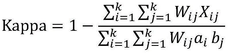
wherein Kappa represents Kappa coefficient, W ij Penalty coefficients representing classification errors, determined by the model itself; a, a i Representing the number of the class i of the label lesion class corresponding to the label lesion class value, b j Representing the number of j classes of test lesion levels corresponding to the test lesion level values; x is X ij Representing the number of i classes divided into j classes.
Specifically, the computer equipment inputs all public verification subsets to the verified initial screening model, obtains the kappa coefficient according to an output result, adjusts initial stage coefficients in the initial screening model according to the kappa coefficient, realizes one-time training of the initial screening model, inputs all public verification subsets to the initial screening model after the initial stage coefficients are adjusted, obtains the kappa coefficient according to the output result, adjusts the initial stage coefficients in the initial screening model according to the kappa coefficient, compares the change condition of the kappa coefficient obtained after the initial stage coefficients are adjusted each time until the kappa coefficient is not lowered any more, stops training of the initial stage parameters in the initial screening model, and obtains the initial screening model as the temporary screening model after the last training.
In this embodiment, the common training subset is used to train the common parameters in the initial screening model, and the common screening verification subset is used to verify the trained initial screening model, so as to obtain the initial screening model with the highest accuracy of the output result, as the temporary screening model. And training and verifying the initial screening model improves the reliability and accuracy of lesion grade classification of the temporary screening model.
In another embodiment, at S450, inputting the lesion screening sample set with the pseudo tag and the common training subset into the temporary screening model for training, and verifying the trained temporary screening model by using the common verification subset, to obtain the screening model, including:
and training the common parameters in the temporary screening model by adopting the lesion screening sample set carrying the pseudo tag and the public training subset, training the stage coefficients in the trained temporary screening model by adopting the public verification subset, and obtaining the screening model when the mean square error corresponding to the public verification subset meets the preset verification requirement.
Specifically, the specific process of training the common parameters in the temporary screening model by using the lesion screening sample set with the pseudo tag and the common training subset is similar to the specific process of training the common parameters in the initial screening model by using the common training subset in S430, except that the training of the common parameters in the temporary model is to input the lesion screening sample set with the pseudo tag and the common training subset into the temporary model. The specific process of verifying the trained temporary screening model by using the common verification subset is similar to the specific process of training the stage coefficients in the initial screening model after verification by using the common verification subset in S430, except that the stage coefficients in the temporary model are trained by inputting the common verification subset into the temporary model, and the screening model is finally obtained.
Further, because the serious fundus lesion pictures in the lesion screening sample set are few, the picture samples with the pseudo labels of 2-4 obtained by inputting the temporary screening model are reserved, the training of the temporary screening model is participated, and the picture samples with the other pseudo labels are reserved in a sampling mode. Because the disclosed sample set comprises a large number of fundus lesion pictures with different degrees, the training of the temporary screening model is facilitated, and therefore the number of the lesion screening sample set carrying the pseudo tag is not more than 10% of the disclosed training subset, so that the diversity and the accuracy of the finally obtained lesion grade classification result of the screening model are ensured.
In this embodiment, the common parameters in the temporary screening model are trained by using the lesion screening sample set, and the trained temporary screening model is verified by using the public screening verification subset, so that the temporary screening model with the highest accuracy of the output result is obtained and is used as the screening model. And training and verifying the temporary screening model improves the reliability and accuracy of the finally obtained classification of the lesion level of the screening model.
Further, the step S120 of inputting the to-be-detected picture into a screening model to obtain a lesion level of the to-be-detected region in the to-be-detected picture, further includes:
and performing sensitivity and/or specificity detection on the screening model.
Wherein the sensitivity is used to characterize the ability of the screening model to correctly determine fundus lesions. The specificity is used to characterize the ability of the screening model to correctly determine that the fundus is free of lesions.
Specifically, the sensitivity formula satisfies the following formula:
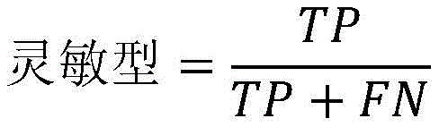
wherein TP is true positive (true label positive, predicted label positive), FN is false negative (true label positive, predicted label negative)
The specific formula satisfies the following formula:
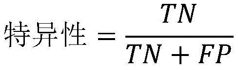
wherein TN is true negative (true label is negative, predicted label is negative), FP is false positive (true label is negative, predicted label is positive).
Specifically, in fundus lesion level classification of fundus pictures, the real label is a label of the fundus pictures, the prediction label is a lesion level obtained by inputting a screening model into the fundus pictures, the positive indicates that the fundus lesion level is a fundus moderate lesion, a fundus severe non-proliferation lesion or a fundus proliferation lesion, and the negative indicates that the fundus lesion level is a fundus no lesion.
In the above embodiment, the kappa coefficient corresponding to the initial stage coefficient in the temporary screening model is compared with the change condition of the kappa coefficient corresponding to the temporary screening model after the initial stage coefficient is adjusted, and as the kappa coefficient can be used for representing the consistency of the lesion grade output by the model and the label, the optimal lesion stage coefficient can be obtained according to the kappa coefficient, and the screening model is correspondingly obtained. The higher the consistency is, the higher the accuracy of the screening model is, so that the final classification of the lesion level by adopting the screening model with high accuracy is realized, and the reliability and the accuracy of the classification result of the lesion level are improved. The accuracy of the screening can be further characterized by sensitivity and specificity.
It should be understood that, although the steps in the flowcharts of fig. 1-5 are shown in order as indicated by the arrows, these steps are not necessarily performed in order as indicated by the arrows. The steps are not strictly limited to the order of execution unless explicitly recited herein, and the steps may be executed in other orders. Moreover, at least some of the steps in fig. 1-5 may include multiple sub-steps or stages that are not necessarily performed at the same time, but may be performed at different times, nor do the order in which the sub-steps or stages are performed necessarily occur sequentially, but may be performed alternately or alternately with at least a portion of the sub-steps or stages of other steps or steps.
In one embodiment, as shown in fig. 6, there is provided an image grade classification apparatus 600 comprising: a quality discrimination module 610, a screening module 620, and a confidence module 630, wherein:
the quality discriminating module 610 is configured to input a picture to be detected into a quality discriminating model to obtain a quality grade of the picture to be detected; the quality grade value is used for representing the definition degree of the picture to be detected;
the screening module 620 is configured to input the to-be-detected picture into a screening model to obtain a lesion level of a to-be-detected region in the to-be-detected picture;
the credibility module 630 is configured to determine, according to the quality level of the to-be-detected picture, credibility of a lesion level of the to-be-detected picture.
For specific limitations of the image level classification device, reference may be made to the above limitations of the image level classification method, and no further description is given here. The respective modules in the above-described image level classification apparatus may be implemented in whole or in part by software, hardware, and a combination thereof. The above modules may be embedded in hardware or may be independent of a processor in the computer device, or may be stored in software in a memory in the computer device, so that the processor may call and execute operations corresponding to the above modules.
It will be appreciated by those skilled in the art that the structure shown in fig. 6 is merely a block diagram of some of the structures associated with the present application and is not limiting of the computer device to which the present application may be applied, and that a particular computer device may include more or fewer components than shown, or may combine certain components, or have a different arrangement of components.
In one embodiment, a computer device is provided, which may be a server or a terminal, and the internal structure of the computer device may be as shown in fig. 7. The computer device includes a processor, a memory, a network interface, a database, a display screen, and an input device connected by a system bus. Wherein the processor of the computer device is configured to provide computing and control capabilities. The memory of the computer device includes a non-volatile storage medium and an internal memory. The non-volatile storage medium stores an operating system, computer programs, and a database. The internal memory provides an environment for the operation of the operating system and computer programs in the non-volatile storage media. The database of the computer device is used for storing the picture data to be checked. The network interface of the computer device is used for communicating with an external terminal through a network connection. The computer program is executed by a processor to implement a method of picture level classification. The display screen of the computer equipment can be a liquid crystal display screen or an electronic ink display screen, and the input device of the computer equipment can be a touch layer covered on the display screen, can also be keys, a track ball or a touch pad arranged on the shell of the computer equipment, and can also be an external keyboard, a touch pad or a mouse and the like.
It will be appreciated by those skilled in the art that the structure shown in fig. 7 is merely a block diagram of some of the structures associated with the present application and is not limiting of the computer device to which the present application may be applied, and that a particular computer device may include more or fewer components than shown, or may combine certain components, or have a different arrangement of components.
In an embodiment, a computer device is provided, comprising a memory and a processor, the memory having stored therein a computer program, the processor implementing the steps of any of the above described picture level classification method embodiments when the computer program is executed.
In one embodiment, a computer readable storage medium is provided, having stored thereon a computer program which, when executed by a processor, implements the steps of any of the above-described picture level classification method embodiments.
Those skilled in the art will appreciate that implementing all or part of the above described methods may be accomplished by way of a computer program stored on a non-transitory computer readable storage medium, which when executed, may comprise the steps of the embodiments of the methods described above. Any reference to memory, storage, database, or other medium used in the various embodiments provided herein may include non-volatile and/or volatile memory. The nonvolatile memory can include Read Only Memory (ROM), programmable ROM (PROM), electrically Programmable ROM (EPROM), electrically Erasable Programmable ROM (EEPROM), or flash memory. Volatile memory can include Random Access Memory (RAM) or external cache memory. By way of illustration and not limitation, RAM is available in a variety of forms such as Static RAM (SRAM), dynamic RAM (DRAM), synchronous DRAM (SDRAM), double Data Rate SDRAM (DDRSDRAM), enhanced SDRAM (ESDRAM), synchronous Link DRAM (SLDRAM), memory bus direct RAM (RDRAM), direct memory bus dynamic RAM (DRDRAM), and memory bus dynamic RAM (RDRAM), among others.
The technical features of the above embodiments may be arbitrarily combined, and all possible combinations of the technical features in the above embodiments are not described for brevity of description, however, as long as there is no contradiction between the combinations of the technical features, they should be considered as the scope of the description.
The above examples merely represent a few embodiments of the present application, which are described in more detail and are not to be construed as limiting the scope of the invention. It should be noted that it would be apparent to those skilled in the art that various modifications and improvements could be made without departing from the spirit of the present application, which would be within the scope of the present application. Accordingly, the scope of protection of the present application is to be determined by the claims appended hereto.
Claims (10)
1. A picture level classification method, the method comprising:
inputting a picture to be detected into a quality judging model to obtain the quality grade of the picture to be detected; the quality grade is used for representing the definition degree of the picture to be detected;
inputting the picture to be detected into a screening model to obtain the lesion grade of a region to be detected in the picture to be detected; the training process of the screening model comprises the following steps: acquiring a public sample set; preprocessing and dividing the public pictures in the public sample set to obtain a normalized public training subset and a public verification subset; training an initial screening model by adopting the public training subset, verifying the trained initial screening model by adopting the public verification subset, and obtaining a temporary screening model according to a verification result; inputting the lesion screening sample set into the temporary screening model to obtain a lesion screening sample set carrying a pseudo tag; inputting the lesion screening sample set carrying the pseudo tag and the public training subset into the temporary screening model for training, and adopting the public verification subset to verify the trained temporary screening model to obtain the screening model;
And determining the credibility of the lesion level of the picture to be detected according to the quality level of the picture to be detected.
2. The method according to claim 1, wherein said determining the credibility of the lesion level of the picture to be examined according to the quality level of the picture to be examined comprises:
if the quality level is excellent, determining that the reliability is high;
if the quality level is uncertain, determining the credibility as uncertain;
if the quality level is poor, the reliability is determined to be low.
3. The method of claim 1, wherein the training process of the quality discriminant model comprises:
acquiring a quality screening sample set; the quality screening sample set comprises a plurality of screening pictures, and each screening picture carries a label for representing the definition of an image;
preprocessing and dividing the screening pictures in the quality screening sample set to obtain a normalized quality training subset, a quality verification subset and a quality test subset;
training an initial quality judgment model by adopting the quality training subset, verifying the trained initial quality judgment model by adopting the quality verification subset, and testing the verified initial quality judgment model by adopting the quality test subset to obtain quality judgment accuracy;
When the quality judgment accuracy is greater than or equal to a preset accuracy threshold, the quality judgment model is obtained;
and when the quality judgment accuracy is smaller than the accuracy threshold, continuing to train the quality judgment model again by adopting the quality training subset until the new quality judgment accuracy is larger than or equal to the accuracy threshold, and obtaining the quality judgment model.
4. A method according to claim 3, wherein training the initial quality discriminant model using the quality training subset and verifying the trained initial quality discriminant model using the quality verification subset and testing the verified initial quality discriminant model using the quality testing subset to obtain quality determination accuracy comprises:
and training common parameters in the initial quality judgment model by adopting the quality training subset, and testing the initial quality judgment model after verification by adopting the quality testing subset when the cross entropy loss value corresponding to the quality verification subset meets the preset verification requirement to obtain the quality judgment accuracy.
5. The method of claim 1, wherein the common sample set comprises a plurality of common pictures, and each of the common pictures carries a label for characterizing a degree of pathology.
6. The method of claim 1, wherein training the initial screening model using the common training subset and verifying the trained initial screening model using the common verification subset, and obtaining the temporary screening model based on the verification result, comprises:
and training the common parameters in the initial screening model by adopting the common training subset, training the stage coefficients in the initial screening model after training by adopting the common verification subset, and obtaining a temporary screening model when the mean square error corresponding to the common verification subset meets the preset verification requirement.
7. The method of claim 1, wherein inputting the set of pseudo-tagged lesions screening samples and the common training subset into the temporary screening model for training and validating the trained temporary screening model using the common validation subset to obtain the screening model, comprising:
and training the common parameters in the temporary screening model by adopting the lesion screening sample set carrying the pseudo tag and the public training subset, training the stage coefficients in the trained temporary screening model by adopting the public verification subset, and obtaining the screening model when the mean square error corresponding to the public training subset meets the preset verification requirement.
8. An image grade classification apparatus, the apparatus comprising:
the quality judging module is used for inputting the picture to be detected into the quality judging model to obtain the quality grade of the picture to be detected; the quality grade value is used for representing the definition degree of the picture to be detected;
the screening module is used for inputting the picture to be detected into a screening model to obtain the lesion grade of the region to be detected in the picture to be detected; the training process of the screening model comprises the following steps: acquiring a public sample set; preprocessing and dividing the public pictures in the public sample set to obtain a normalized public training subset and a public verification subset; training an initial screening model by adopting the public training subset, verifying the trained initial screening model by adopting the public verification subset, and obtaining a temporary screening model according to a verification result; inputting the lesion screening sample set into the temporary screening model to obtain a lesion screening sample set carrying a pseudo tag; inputting the lesion screening sample set carrying the pseudo tag and the public training subset into the temporary screening model for training, and adopting the public verification subset to verify the trained temporary screening model to obtain the screening model;
And the credibility module is used for determining the credibility of the pathological change grade of the picture to be detected according to the quality grade of the picture to be detected.
9. A computer device comprising a memory and a processor, the memory storing a computer program, characterized in that the processor implements the steps of the method of any of claims 1 to 7 when the computer program is executed.
10. A computer readable storage medium, on which a computer program is stored, characterized in that the computer program, when being executed by a processor, implements the steps of the method of any of claims 1 to 7.
Priority Applications (1)
| Application Number | Priority Date | Filing Date | Title |
|---|---|---|---|
| CN201911283146.0A CN110956628B (en) | 2019-12-13 | 2019-12-13 | Picture grade classification method, device, computer equipment and storage medium |
Applications Claiming Priority (1)
| Application Number | Priority Date | Filing Date | Title |
|---|---|---|---|
| CN201911283146.0A CN110956628B (en) | 2019-12-13 | 2019-12-13 | Picture grade classification method, device, computer equipment and storage medium |
Publications (2)
| Publication Number | Publication Date |
|---|---|
| CN110956628A CN110956628A (en) | 2020-04-03 |
| CN110956628B true CN110956628B (en) | 2023-05-09 |
Family
ID=69981510
Family Applications (1)
| Application Number | Title | Priority Date | Filing Date |
|---|---|---|---|
| CN201911283146.0A Active CN110956628B (en) | 2019-12-13 | 2019-12-13 | Picture grade classification method, device, computer equipment and storage medium |
Country Status (1)
| Country | Link |
|---|---|
| CN (1) | CN110956628B (en) |
Families Citing this family (6)
| Publication number | Priority date | Publication date | Assignee | Title |
|---|---|---|---|---|
| CN111754486B (en) | 2020-06-24 | 2023-08-15 | 北京百度网讯科技有限公司 | Image processing method, device, electronic equipment and storage medium |
| CN114005097A (en) * | 2020-07-28 | 2022-02-01 | 株洲中车时代电气股份有限公司 | Train operation environment real-time detection method and system based on image semantic segmentation |
| CN115205572B (en) * | 2021-03-24 | 2025-09-12 | 深圳硅基智能科技有限公司 | Quality control system and quality control method for fundus image reading system |
| CN113743512A (en) * | 2021-09-07 | 2021-12-03 | 上海观安信息技术股份有限公司 | Autonomous learning judgment method and system for safety alarm event |
| CN114489897B (en) | 2022-01-21 | 2023-08-08 | 北京字跳网络技术有限公司 | Object processing method, device, terminal equipment and medium |
| CN116563735A (en) * | 2023-05-15 | 2023-08-08 | 国网电力空间技术有限公司 | A focus judgment method for power transmission tower inspection images based on deep artificial intelligence |
Citations (1)
| Publication number | Priority date | Publication date | Assignee | Title |
|---|---|---|---|---|
| CN108831552A (en) * | 2018-04-09 | 2018-11-16 | 平安科技(深圳)有限公司 | electronic device, nasopharyngeal carcinoma screening analysis method and computer readable storage medium |
Family Cites Families (5)
| Publication number | Priority date | Publication date | Assignee | Title |
|---|---|---|---|---|
| CA2420530A1 (en) * | 2000-08-23 | 2002-02-28 | Philadelphia Ophthalmologic Imaging Systems, Inc. | Systems and methods for tele-ophthalmology |
| CN107203778A (en) * | 2017-05-05 | 2017-09-26 | 平安科技(深圳)有限公司 | PVR intensity grade detecting system and method |
| CN108171256A (en) * | 2017-11-27 | 2018-06-15 | 深圳市深网视界科技有限公司 | Facial image matter comments model construction, screening, recognition methods and equipment and medium |
| CN108596882B (en) * | 2018-04-10 | 2019-04-02 | 中山大学肿瘤防治中心 | The recognition methods of pathological picture and device |
| CN109464120A (en) * | 2018-10-31 | 2019-03-15 | 深圳市第二人民医院 | Diabetic retinopathy screening method, device and storage medium |
-
2019
- 2019-12-13 CN CN201911283146.0A patent/CN110956628B/en active Active
Patent Citations (1)
| Publication number | Priority date | Publication date | Assignee | Title |
|---|---|---|---|---|
| CN108831552A (en) * | 2018-04-09 | 2018-11-16 | 平安科技(深圳)有限公司 | electronic device, nasopharyngeal carcinoma screening analysis method and computer readable storage medium |
Also Published As
| Publication number | Publication date |
|---|---|
| CN110956628A (en) | 2020-04-03 |
Similar Documents
| Publication | Publication Date | Title |
|---|---|---|
| CN110956628B (en) | Picture grade classification method, device, computer equipment and storage medium | |
| Elangovan et al. | Glaucoma assessment from color fundus images using convolutional neural network | |
| US11295178B2 (en) | Image classification method, server, and computer-readable storage medium | |
| CN110599451B (en) | Medical image focus detection and positioning method, device, equipment and storage medium | |
| CN111862044B (en) | Ultrasonic image processing method, ultrasonic image processing device, computer equipment and storage medium | |
| CN110197493B (en) | Fundus image blood vessel segmentation method | |
| WO2021139258A1 (en) | Image recognition based cell recognition and counting method and apparatus, and computer device | |
| CN117015796A (en) | Methods of processing tissue images and systems for processing tissue images | |
| Pham et al. | Generating future fundus images for early age-related macular degeneration based on generative adversarial networks | |
| CN116091963B (en) | Quality evaluation method and device for clinical test institution, electronic equipment and storage medium | |
| CN113011340B (en) | A risk classification method and system for cardiovascular surgery indicators based on retinal images | |
| Wang et al. | SAC-Net: Enhancing spatiotemporal aggregation in cervical histological image classification via label-efficient weakly supervised learning | |
| CN111275686A (en) | Method and device for generating medical image data for artificial neural network training | |
| CN113436735A (en) | Body weight index prediction method, device and storage medium based on face structure measurement | |
| KR20250047756A (en) | Methods and systems for automated HER2 scoring | |
| CN119810394A (en) | A data processing method and system based on image acquisition device | |
| CN119399134A (en) | Retinal image quality assessment method based on dual-path frequency domain cross-attention fusion | |
| WO2022212978A1 (en) | Machine learning model for detecting out-of-distribution inputs | |
| CN112633348B (en) | Method and device for detecting cerebral arteriovenous malformation and judging dispersion property of cerebral arteriovenous malformation | |
| CN110458024A (en) | Biopsy method and device and electronic equipment | |
| JP7229445B2 (en) | Recognition device and recognition method | |
| Kenney et al. | How Advancements in AI Can Help Improve Neuro-Ophthalmologic Diagnostic Clarity | |
| Sam et al. | Preliminary Study of Diabetic Retinopathy Classification from Fundus Images Using Deep Learning Model | |
| CN119006835B (en) | Skin image segmentation method for reducing influence of confounding factors | |
| Alrashdi et al. | Enhancing medical imaging with Ghost-ResNeXt and locust-inspired optimization: A case study on diabetic retinopathy |
Legal Events
| Date | Code | Title | Description |
|---|---|---|---|
| PB01 | Publication | ||
| PB01 | Publication | ||
| SE01 | Entry into force of request for substantive examination | ||
| SE01 | Entry into force of request for substantive examination | ||
| GR01 | Patent grant | ||
| GR01 | Patent grant |