CN110880356A - Method and apparatus for screening, diagnosing or risk stratification for ovarian cancer - Google Patents
Method and apparatus for screening, diagnosing or risk stratification for ovarian cancer Download PDFInfo
- Publication number
- CN110880356A CN110880356A CN201811030743.8A CN201811030743A CN110880356A CN 110880356 A CN110880356 A CN 110880356A CN 201811030743 A CN201811030743 A CN 201811030743A CN 110880356 A CN110880356 A CN 110880356A
- Authority
- CN
- China
- Prior art keywords
- ovarian cancer
- chromosome
- cscore
- sample
- chromosomes
- Prior art date
- Legal status (The legal status is an assumption and is not a legal conclusion. Google has not performed a legal analysis and makes no representation as to the accuracy of the status listed.)
- Pending
Links
- 206010033128 Ovarian cancer Diseases 0.000 title claims abstract description 137
- 206010061535 Ovarian neoplasm Diseases 0.000 title claims abstract description 137
- 238000000034 method Methods 0.000 title claims abstract description 60
- 238000012216 screening Methods 0.000 title claims abstract description 32
- 238000013517 stratification Methods 0.000 title claims abstract description 12
- 101000623901 Homo sapiens Mucin-16 Proteins 0.000 claims abstract description 74
- 102100023123 Mucin-16 Human genes 0.000 claims abstract description 74
- 238000003066 decision tree Methods 0.000 claims abstract description 56
- 206010028980 Neoplasm Diseases 0.000 claims abstract description 46
- 238000012360 testing method Methods 0.000 claims abstract description 33
- 238000012165 high-throughput sequencing Methods 0.000 claims abstract description 26
- 210000000349 chromosome Anatomy 0.000 claims description 171
- 238000012549 training Methods 0.000 claims description 32
- 238000012163 sequencing technique Methods 0.000 claims description 31
- 108020004414 DNA Proteins 0.000 claims description 26
- 230000011218 segmentation Effects 0.000 claims description 20
- 210000004369 blood Anatomy 0.000 claims description 16
- 239000008280 blood Substances 0.000 claims description 16
- 230000002759 chromosomal effect Effects 0.000 claims description 15
- 230000002611 ovarian Effects 0.000 claims description 13
- 239000003153 chemical reaction reagent Substances 0.000 claims description 11
- 210000005259 peripheral blood Anatomy 0.000 claims description 9
- 239000011886 peripheral blood Substances 0.000 claims description 9
- 238000005070 sampling Methods 0.000 claims description 8
- 230000002093 peripheral effect Effects 0.000 claims description 6
- 230000006870 function Effects 0.000 claims description 5
- 150000007523 nucleic acids Chemical class 0.000 claims description 5
- 108091028043 Nucleic acid sequence Proteins 0.000 claims description 4
- 210000003917 human chromosome Anatomy 0.000 claims description 3
- 108020004707 nucleic acids Proteins 0.000 claims description 3
- 102000039446 nucleic acids Human genes 0.000 claims description 3
- 238000002360 preparation method Methods 0.000 claims description 3
- 238000004519 manufacturing process Methods 0.000 claims description 2
- 238000005259 measurement Methods 0.000 claims description 2
- 201000008016 ovarian benign neoplasm Diseases 0.000 abstract description 12
- 230000036210 malignancy Effects 0.000 abstract description 6
- 206010004433 Benign ovarian tumour Diseases 0.000 abstract description 5
- 201000011510 cancer Diseases 0.000 abstract description 5
- 208000024891 symptom Diseases 0.000 abstract description 2
- 206010058823 Ovarian mass Diseases 0.000 abstract 1
- 206010034260 pelvic mass Diseases 0.000 abstract 1
- 239000000439 tumor marker Substances 0.000 abstract 1
- 238000003745 diagnosis Methods 0.000 description 21
- 239000011324 bead Substances 0.000 description 18
- 210000002966 serum Anatomy 0.000 description 18
- LFQSCWFLJHTTHZ-UHFFFAOYSA-N Ethanol Chemical compound CCO LFQSCWFLJHTTHZ-UHFFFAOYSA-N 0.000 description 16
- 230000035945 sensitivity Effects 0.000 description 15
- 210000004027 cell Anatomy 0.000 description 11
- 238000001514 detection method Methods 0.000 description 11
- 239000007788 liquid Substances 0.000 description 11
- 230000000694 effects Effects 0.000 description 10
- 210000001672 ovary Anatomy 0.000 description 10
- 108090000623 proteins and genes Proteins 0.000 description 10
- 239000006228 supernatant Substances 0.000 description 10
- 238000004458 analytical method Methods 0.000 description 9
- 201000010099 disease Diseases 0.000 description 9
- 208000037265 diseases, disorders, signs and symptoms Diseases 0.000 description 9
- 238000005516 engineering process Methods 0.000 description 9
- 239000000243 solution Substances 0.000 description 9
- XLYOFNOQVPJJNP-UHFFFAOYSA-N water Chemical compound O XLYOFNOQVPJJNP-UHFFFAOYSA-N 0.000 description 8
- 238000006243 chemical reaction Methods 0.000 description 7
- 230000003902 lesion Effects 0.000 description 7
- 230000008569 process Effects 0.000 description 7
- 208000036878 aneuploidy Diseases 0.000 description 6
- 238000004140 cleaning Methods 0.000 description 6
- 239000003550 marker Substances 0.000 description 6
- 239000000203 mixture Substances 0.000 description 6
- 238000010827 pathological analysis Methods 0.000 description 6
- 210000001519 tissue Anatomy 0.000 description 6
- 206010003445 Ascites Diseases 0.000 description 5
- 206010027476 Metastases Diseases 0.000 description 5
- 231100001075 aneuploidy Toxicity 0.000 description 5
- 230000008901 benefit Effects 0.000 description 5
- 230000014509 gene expression Effects 0.000 description 5
- 230000003211 malignant effect Effects 0.000 description 5
- 230000009401 metastasis Effects 0.000 description 5
- 238000011160 research Methods 0.000 description 5
- 241000894007 species Species 0.000 description 5
- 238000007664 blowing Methods 0.000 description 4
- 239000000872 buffer Substances 0.000 description 4
- 239000003814 drug Substances 0.000 description 4
- 230000002068 genetic effect Effects 0.000 description 4
- 238000011534 incubation Methods 0.000 description 4
- 238000007481 next generation sequencing Methods 0.000 description 4
- 102000053602 DNA Human genes 0.000 description 3
- 208000007433 Lymphatic Metastasis Diseases 0.000 description 3
- 238000002835 absorbance Methods 0.000 description 3
- 238000003556 assay Methods 0.000 description 3
- 238000005119 centrifugation Methods 0.000 description 3
- 238000010276 construction Methods 0.000 description 3
- 230000034994 death Effects 0.000 description 3
- 231100000517 death Toxicity 0.000 description 3
- 238000011161 development Methods 0.000 description 3
- 230000018109 developmental process Effects 0.000 description 3
- 238000011049 filling Methods 0.000 description 3
- 239000012634 fragment Substances 0.000 description 3
- 230000008014 freezing Effects 0.000 description 3
- 238000007710 freezing Methods 0.000 description 3
- 239000000463 material Substances 0.000 description 3
- 239000012466 permeate Substances 0.000 description 3
- 239000000126 substance Substances 0.000 description 3
- 238000010257 thawing Methods 0.000 description 3
- YBJHBAHKTGYVGT-ZKWXMUAHSA-N (+)-Biotin Chemical compound N1C(=O)N[C@@H]2[C@H](CCCCC(=O)O)SC[C@@H]21 YBJHBAHKTGYVGT-ZKWXMUAHSA-N 0.000 description 2
- 108091032973 (ribonucleotides)n+m Proteins 0.000 description 2
- KCXVZYZYPLLWCC-UHFFFAOYSA-N EDTA Chemical compound OC(=O)CN(CC(O)=O)CCN(CC(O)=O)CC(O)=O KCXVZYZYPLLWCC-UHFFFAOYSA-N 0.000 description 2
- 238000011891 EIA kit Methods 0.000 description 2
- 108010067770 Endopeptidase K Proteins 0.000 description 2
- 108090000790 Enzymes Proteins 0.000 description 2
- 102000004190 Enzymes Human genes 0.000 description 2
- 208000007571 Ovarian Epithelial Carcinoma Diseases 0.000 description 2
- 208000020584 Polyploidy Diseases 0.000 description 2
- 230000010100 anticoagulation Effects 0.000 description 2
- 239000000427 antigen Substances 0.000 description 2
- 102000036639 antigens Human genes 0.000 description 2
- 108091007433 antigens Proteins 0.000 description 2
- 230000001640 apoptogenic effect Effects 0.000 description 2
- 238000010009 beating Methods 0.000 description 2
- 238000004364 calculation method Methods 0.000 description 2
- 210000002230 centromere Anatomy 0.000 description 2
- 230000008859 change Effects 0.000 description 2
- 238000012512 characterization method Methods 0.000 description 2
- 238000000749 co-immunoprecipitation Methods 0.000 description 2
- 230000009089 cytolysis Effects 0.000 description 2
- 238000012350 deep sequencing Methods 0.000 description 2
- 230000007812 deficiency Effects 0.000 description 2
- 238000012217 deletion Methods 0.000 description 2
- 230000037430 deletion Effects 0.000 description 2
- 239000012153 distilled water Substances 0.000 description 2
- 229940079593 drug Drugs 0.000 description 2
- 238000013399 early diagnosis Methods 0.000 description 2
- 230000013020 embryo development Effects 0.000 description 2
- 210000000981 epithelium Anatomy 0.000 description 2
- 238000000605 extraction Methods 0.000 description 2
- 210000001752 female genitalia Anatomy 0.000 description 2
- 238000003384 imaging method Methods 0.000 description 2
- 238000000338 in vitro Methods 0.000 description 2
- 238000001727 in vivo Methods 0.000 description 2
- 208000015181 infectious disease Diseases 0.000 description 2
- 210000004185 liver Anatomy 0.000 description 2
- 210000001165 lymph node Anatomy 0.000 description 2
- 206010061289 metastatic neoplasm Diseases 0.000 description 2
- 230000035772 mutation Effects 0.000 description 2
- 230000001338 necrotic effect Effects 0.000 description 2
- 208000012988 ovarian serous adenocarcinoma Diseases 0.000 description 2
- 201000003709 ovarian serous carcinoma Diseases 0.000 description 2
- 231100000915 pathological change Toxicity 0.000 description 2
- 230000036285 pathological change Effects 0.000 description 2
- 239000000047 product Substances 0.000 description 2
- 102000004169 proteins and genes Human genes 0.000 description 2
- 238000000746 purification Methods 0.000 description 2
- 238000002864 sequence alignment Methods 0.000 description 2
- 238000003756 stirring Methods 0.000 description 2
- 230000004083 survival effect Effects 0.000 description 2
- 210000004881 tumor cell Anatomy 0.000 description 2
- VBUWJOHKCBQXNU-IUYQGCFVSA-N 2-deoxy-D-ribonic acid Chemical compound OC[C@@H](O)[C@@H](O)CC(O)=O VBUWJOHKCBQXNU-IUYQGCFVSA-N 0.000 description 1
- 201000000736 Amenorrhea Diseases 0.000 description 1
- 206010001928 Amenorrhoea Diseases 0.000 description 1
- 102000036365 BRCA1 Human genes 0.000 description 1
- 108700020463 BRCA1 Proteins 0.000 description 1
- 101150072950 BRCA1 gene Proteins 0.000 description 1
- 206010006187 Breast cancer Diseases 0.000 description 1
- 208000026310 Breast neoplasm Diseases 0.000 description 1
- CURLTUGMZLYLDI-UHFFFAOYSA-N Carbon dioxide Chemical compound O=C=O CURLTUGMZLYLDI-UHFFFAOYSA-N 0.000 description 1
- 108010077544 Chromatin Proteins 0.000 description 1
- 108091026890 Coding region Proteins 0.000 description 1
- 238000007400 DNA extraction Methods 0.000 description 1
- 208000005431 Endometrioid Carcinoma Diseases 0.000 description 1
- 201000009273 Endometriosis Diseases 0.000 description 1
- 239000004606 Fillers/Extenders Substances 0.000 description 1
- 108090000288 Glycoproteins Proteins 0.000 description 1
- 102000003886 Glycoproteins Human genes 0.000 description 1
- 206010061218 Inflammation Diseases 0.000 description 1
- 241001465754 Metazoa Species 0.000 description 1
- 108700011259 MicroRNAs Proteins 0.000 description 1
- 238000003559 RNA-seq method Methods 0.000 description 1
- 238000012167 Small RNA sequencing Methods 0.000 description 1
- 108091023040 Transcription factor Proteins 0.000 description 1
- 102000040945 Transcription factor Human genes 0.000 description 1
- 102100023935 Transmembrane glycoprotein NMB Human genes 0.000 description 1
- 208000037280 Trisomy Diseases 0.000 description 1
- 206010054094 Tumour necrosis Diseases 0.000 description 1
- 210000001015 abdomen Anatomy 0.000 description 1
- 210000000683 abdominal cavity Anatomy 0.000 description 1
- 230000003187 abdominal effect Effects 0.000 description 1
- 230000002159 abnormal effect Effects 0.000 description 1
- 239000002250 absorbent Substances 0.000 description 1
- 230000002745 absorbent Effects 0.000 description 1
- 208000009956 adenocarcinoma Diseases 0.000 description 1
- 231100000540 amenorrhea Toxicity 0.000 description 1
- 230000003322 aneuploid effect Effects 0.000 description 1
- 230000002146 bilateral effect Effects 0.000 description 1
- 230000015572 biosynthetic process Effects 0.000 description 1
- 229960002685 biotin Drugs 0.000 description 1
- 235000020958 biotin Nutrition 0.000 description 1
- 239000011616 biotin Substances 0.000 description 1
- 230000000740 bleeding effect Effects 0.000 description 1
- 238000009395 breeding Methods 0.000 description 1
- 230000001488 breeding effect Effects 0.000 description 1
- 150000001720 carbohydrates Chemical class 0.000 description 1
- 235000011089 carbon dioxide Nutrition 0.000 description 1
- 238000004113 cell culture Methods 0.000 description 1
- 239000003795 chemical substances by application Substances 0.000 description 1
- 210000003483 chromatin Anatomy 0.000 description 1
- 210000001726 chromosome structure Anatomy 0.000 description 1
- 208000009060 clear cell adenocarcinoma Diseases 0.000 description 1
- 239000000701 coagulant Substances 0.000 description 1
- 239000002131 composite material Substances 0.000 description 1
- 238000005094 computer simulation Methods 0.000 description 1
- 239000000470 constituent Substances 0.000 description 1
- 238000005520 cutting process Methods 0.000 description 1
- 238000007405 data analysis Methods 0.000 description 1
- 239000008367 deionised water Substances 0.000 description 1
- 229910021641 deionized water Inorganic materials 0.000 description 1
- 230000006866 deterioration Effects 0.000 description 1
- 239000000032 diagnostic agent Substances 0.000 description 1
- 229940039227 diagnostic agent Drugs 0.000 description 1
- 238000010790 dilution Methods 0.000 description 1
- 239000012895 dilution Substances 0.000 description 1
- 238000010828 elution Methods 0.000 description 1
- 201000003914 endometrial carcinoma Diseases 0.000 description 1
- 208000028730 endometrioid adenocarcinoma Diseases 0.000 description 1
- 229940011871 estrogen Drugs 0.000 description 1
- 239000000262 estrogen Substances 0.000 description 1
- 238000010195 expression analysis Methods 0.000 description 1
- 230000035558 fertility Effects 0.000 description 1
- 238000013467 fragmentation Methods 0.000 description 1
- 238000006062 fragmentation reaction Methods 0.000 description 1
- 239000003349 gelling agent Substances 0.000 description 1
- 230000012010 growth Effects 0.000 description 1
- 238000002513 implantation Methods 0.000 description 1
- 238000007901 in situ hybridization Methods 0.000 description 1
- 208000000509 infertility Diseases 0.000 description 1
- 230000036512 infertility Effects 0.000 description 1
- 231100000535 infertility Toxicity 0.000 description 1
- 230000004054 inflammatory process Effects 0.000 description 1
- 230000010354 integration Effects 0.000 description 1
- 230000009545 invasion Effects 0.000 description 1
- 238000011068 loading method Methods 0.000 description 1
- 238000010339 medical test Methods 0.000 description 1
- 239000012528 membrane Substances 0.000 description 1
- 230000003340 mental effect Effects 0.000 description 1
- 239000002679 microRNA Substances 0.000 description 1
- 238000002493 microarray Methods 0.000 description 1
- 239000011259 mixed solution Substances 0.000 description 1
- 238000012986 modification Methods 0.000 description 1
- 230000004048 modification Effects 0.000 description 1
- 238000012544 monitoring process Methods 0.000 description 1
- 208000030454 monosomy Diseases 0.000 description 1
- 201000010879 mucinous adenocarcinoma Diseases 0.000 description 1
- 239000002773 nucleotide Substances 0.000 description 1
- 125000003729 nucleotide group Chemical group 0.000 description 1
- 210000004940 nucleus Anatomy 0.000 description 1
- 210000000056 organ Anatomy 0.000 description 1
- 210000003101 oviduct Anatomy 0.000 description 1
- 230000007170 pathology Effects 0.000 description 1
- 238000003909 pattern recognition Methods 0.000 description 1
- 230000002265 prevention Effects 0.000 description 1
- 238000003908 quality control method Methods 0.000 description 1
- 230000005855 radiation Effects 0.000 description 1
- 239000000700 radioactive tracer Substances 0.000 description 1
- 230000006798 recombination Effects 0.000 description 1
- 238000005215 recombination Methods 0.000 description 1
- 238000007480 sanger sequencing Methods 0.000 description 1
- 238000000926 separation method Methods 0.000 description 1
- 208000004548 serous cystadenocarcinoma Diseases 0.000 description 1
- 150000003384 small molecules Chemical class 0.000 description 1
- 238000003860 storage Methods 0.000 description 1
- 239000000758 substrate Substances 0.000 description 1
- 238000001356 surgical procedure Methods 0.000 description 1
- 238000003786 synthesis reaction Methods 0.000 description 1
- 238000002560 therapeutic procedure Methods 0.000 description 1
- 108091007466 transmembrane glycoproteins Proteins 0.000 description 1
- 239000000107 tumor biomarker Substances 0.000 description 1
- 238000002604 ultrasonography Methods 0.000 description 1
- 210000004291 uterus Anatomy 0.000 description 1
- 238000005406 washing Methods 0.000 description 1
- 238000012049 whole transcriptome sequencing Methods 0.000 description 1
- 239000012224 working solution Substances 0.000 description 1
Classifications
-
- C—CHEMISTRY; METALLURGY
- C12—BIOCHEMISTRY; BEER; SPIRITS; WINE; VINEGAR; MICROBIOLOGY; ENZYMOLOGY; MUTATION OR GENETIC ENGINEERING
- C12Q—MEASURING OR TESTING PROCESSES INVOLVING ENZYMES, NUCLEIC ACIDS OR MICROORGANISMS; COMPOSITIONS OR TEST PAPERS THEREFOR; PROCESSES OF PREPARING SUCH COMPOSITIONS; CONDITION-RESPONSIVE CONTROL IN MICROBIOLOGICAL OR ENZYMOLOGICAL PROCESSES
- C12Q1/00—Measuring or testing processes involving enzymes, nucleic acids or microorganisms; Compositions therefor; Processes of preparing such compositions
- C12Q1/68—Measuring or testing processes involving enzymes, nucleic acids or microorganisms; Compositions therefor; Processes of preparing such compositions involving nucleic acids
- C12Q1/6876—Nucleic acid products used in the analysis of nucleic acids, e.g. primers or probes
- C12Q1/6883—Nucleic acid products used in the analysis of nucleic acids, e.g. primers or probes for diseases caused by alterations of genetic material
- C12Q1/6886—Nucleic acid products used in the analysis of nucleic acids, e.g. primers or probes for diseases caused by alterations of genetic material for cancer
Landscapes
- Chemical & Material Sciences (AREA)
- Life Sciences & Earth Sciences (AREA)
- Health & Medical Sciences (AREA)
- Organic Chemistry (AREA)
- Proteomics, Peptides & Aminoacids (AREA)
- Engineering & Computer Science (AREA)
- Immunology (AREA)
- Pathology (AREA)
- Analytical Chemistry (AREA)
- Zoology (AREA)
- Genetics & Genomics (AREA)
- Wood Science & Technology (AREA)
- Physics & Mathematics (AREA)
- Biotechnology (AREA)
- Microbiology (AREA)
- Molecular Biology (AREA)
- Hospice & Palliative Care (AREA)
- Biophysics (AREA)
- Oncology (AREA)
- Biochemistry (AREA)
- Bioinformatics & Cheminformatics (AREA)
- General Engineering & Computer Science (AREA)
- General Health & Medical Sciences (AREA)
- Measuring Or Testing Involving Enzymes Or Micro-Organisms (AREA)
Abstract
The present invention relates to methods and devices for screening, diagnosing or risk stratification for ovarian cancer. In particular, the present invention relates to methods and apparatus for screening, diagnosing or risk staging of ovarian cancer using high throughput sequencing and a CA-125 level building decision tree model. More particularly, the invention relates to a decision tree model constructed by using high-throughput sequencing and CA-125 level, and the model is trained to screen and detect ovarian cancer, so that the problem that early ovarian cancer symptom is hidden and difficult to find is solved on the aspect of molecular biology; the property of the ovarian mass is undetermined, and the ovarian cancer or benign ovarian tumor is difficult to distinguish; and the clinical problem of negative tumor marker with the possibility of malignancy of pelvic mass. The method and system of the present invention enable the discovery of possible ovarian cancer with a single test.
Description
Technical Field
The present invention relates to the fields of medicine, biology, pattern recognition, and the like. In particular, the present invention relates to medical testing. More specifically, the invention constructs a decision tree model through genome detection and CA-125 results, trains the model to screen, detect and grade the risk of ovarian cancer.
Background
Ovarian cancer is the most mortality tumor of gynecological malignancies. Ovarian cancer is the leading cause of death among all gynecological cancers in western countries. The estimated number of new and deaths of ovarian cancer in 2015 in china was 52100 and 22500, respectively. Because the ovarian cancer is in a deep pelvic cavity, the part is hidden, the detection is difficult, the symptoms are not typical, an early diagnosis marker and an effective tool are lacked, and when the ovarian cancer can be detected and clearly diagnosed, the ovarian cancer usually reaches the middle or late stage, which is also the main reason for high mortality rate of the ovarian cancer.
Currently, the etiology of ovarian cancer is unknown, and may be related to factors such as age, fertility status, blood type, mental factors, and environment. In recent years, the incidence of ovarian cancer has been on the rise year by year. The five high risk groups of ovarian cancer previously released by the united states centers for disease control and prevention (CDC) are: women over the age of 50 years old; infertility, with a history of endometriosis; the estrogen drug is taken alone for more than 10 years; people with mutation of breast cancer susceptibility gene (BRCA1 or BRCA 2); and those with a family genetic history of ovarian or malignant tumors.
CA-125 (also known as glycoantigen 125, carbohydrate antigen 125, glycoantigen 125 or cancer antigen 125) is a large transmembrane glycoprotein derived from the coelomic epithelium during embryonic development, absent in normal ovarian tissue, most commonly found in the serum of ovarian cancer patients, where CA-125 levels above 35U/mL are considered abnormal. The use of CA-125 as a serum marker for the detection of ovarian cancer has been in history for decades, but CA-125 is less specific, is not an ovarian specific marker, and is easily confused with changes caused by other cancers, benign tumors of the pelvic cavity, gynecological inflammation and the like to cause misdiagnosis and false positive. And not every patient with ovarian cancer has CA-125 levels above 35U/mL; in particular, in early ovarian cancer cases, the sensitivity is not high, the false negative is very high, and more than 50 percent of cases can not detect the increase of CA-125. Thus, there is a need for a highly sensitive and specific method and tool for detecting and screening ovarian cancer, particularly for distinguishing ovarian cancer from benign ovarian tumors.
NGS (next generation sequencing technology) can sequence hundreds of thousands to millions of DNA molecules at a time, while high-throughput sequencing enables detailed and comprehensive analysis of transcriptomes and genomes of one species, and is therefore also referred to as deep sequencing.
cfDNA (cell-free DNA) represents free DNA in plasma, while ctDNA (circulating tumor DNA) represents a small fraction of cfDNA. ctDNA is a small piece of DNA free in plasma, produced by genomic invasion of apoptotic or necrotic tumor cells, and thus carries specific genetic characteristics of the primary or metastatic tumor. ctDNA obtains conveniently and comparatively stably.
Chromosomal imbalances are a feature of malignancies and refer to structural variations of the genome, including changes in the number of chromosomes, such as polyploids or haploids, relative to a common diploid genome; also included are changes in chromosome locality, such as copy number increases or copy number deletions, and the like. The chromosomal imbalance can directly change the expression level of genes or regulate the expression of other genes through gene dosage effect, so the chromosomal imbalance has important significance in the occurrence and development of tumors. The reflection of the equilibrium state of the chromosome by the study of ctDNA may have certain feasibility and significance for the characterization of tumors.
Decision Tree (Decision Tree) is a Decision analysis method for evaluating the risk of a project and judging the feasibility of the project by constructing a Decision Tree to obtain the probability that the expected value of the net present value is equal to or greater than zero on the basis of the known occurrence probability of various conditions. The decision tree is a graphical method which intuitively uses probability analysis, is simple to use and wide in application, and can efficiently classify and predict unknown data by constructing the decision tree through training data.
There is an urgent need in the art to find methods for screening and diagnosing ovarian cancer. However, it is understood that to date, there is no effective method for rapid diagnosis, especially early diagnosis, of ovarian cancer through NGS and decision trees. The inventor firstly constructs a method for researching chromosome imbalance and CA-125 level after high-throughput sequencing ctDNA recombination based on a decision tree system, thereby screening, diagnosing and risk grading ovarian cancer.
Disclosure of Invention
Systems and methods for screening, diagnosing, and risk stratification for ovarian cancer are provided herein. Specifically, the invention overcomes the deficiencies of the prior art in CA-125 detection and provides a method, a device and a system for screening, diagnosing and risk grading ovarian cancer by using high-throughput sequencing and CA-125 detection based on a decision tree system.
The invention utilizes a high-throughput sequencing technology, a CA-125 level and a decision tree system to screen ovarian cancer, in particular to diagnose the possibility of the tumor serum marker CA-125 negative suspected ovarian cancer from the aspect of molecular biology. The method has the advantages of small error rate, accurate screening result, high automation degree, high calculation speed, high integration degree and the like, is suitable for screening large-scale samples, and can be widely applied to the fields of chemistry, medicine and the like. The methods, devices and systems of the present invention enable the detection of possible ovarian cancer by a single rapid non-invasive test in combination with CA-125 levels.
In particular, the present invention relates to the following aspects, and various technical solutions between the aspects can be combined as necessary.
In a first aspect the invention relates to a set of chromosomes for use in screening, diagnosing or risk stratification for ovarian cancer.
In some embodiments, the set of chromosomes is a human chromosome.
In some embodiments, the set of chromosomes comprises at least 1 of chromosomes 1 through 22. In some embodiments, the set of chromosomes consists of chromosomes 1 through 22, the chromosomes being human chromosomes.
In some embodiments, the set of chromosomes is isolated.
In a second aspect the invention relates to a computer readable medium having instructions stored thereon, wherein the instructions, when executed by a processor, cause a computer to:
calculating an imbalance measure CScore value for a subject as a whole based on chromosomal imbalance or chromosomal arm imbalance of chromosomes 1 to 22 of a sample from the subject (e.g., a human);
combining the CScore value with the level of CA-125 in said sample to determine whether said subject has ovarian cancer or is at high risk of having ovarian cancer.
In some embodiments, the computer readable medium calculates the CScore value by:
aligning a subject's (e.g., human) genome-wide data sequence (e.g., a genome-wide data sequence obtained by high-throughput sequencing techniques) to a reference genome (e.g., human reference genome Hg19) and dividing, e.g., by 10-1000k/bin (e.g., 50k/bin), into a plurality of segments (e.g., bins);
the average number of reads (cov) of segments (e.g., bins) covered by the long arm of chromosome i is calculated respectivelyChriq) And the average number of reads (cov) of segments (e.g., bins) covered by the short arm of the chromosomeChrip);
Calculating the R value of chromosome i according to the following formula ( ):
):


Wherein q represents the long arm, p represents the short arm, Chr is an abbreviation for chromosome (chromosome), and i is selected from chromosomes 1 to 22;
calculating Z-score (Z) of chromosome i according to formula (2) based on the R valueChri):

Wherein Is the average of the R values corresponding to healthy people,
 is the standard deviation of the R values corresponding to healthy people;
alternatively, the Z-score of the corresponding long arm q of chromosome i is calculated according to equations (3a) and (3b) (( ) And Z-score (of short arm p)
 ):
):


Wherein Is the average of the read lengths of the long arm of chromosome i corresponding to healthy people,
 is the average number of reads of the short arm of chromosome i corresponding to healthy people;
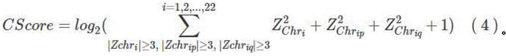
a chromosome or chromosome arm is considered to be unbalanced if the absolute value of Z-score for chromosome i or chromosome arm is greater than or equal to 3; selecting chromosomes with the absolute value of the chromosome or chromosome arm Z-score being more than or equal to 3, and calculating the integral unbalance measurement CScore value according to the formula (4):

in some embodiments, the computer readable medium uses the levels of CScore and CA-125 to build a decision tree model to predict ovarian cancer risk.
In some embodiments, the decision tree model is built by the R language. Specifically, a decision tree model is built by:
(1) constructing an overall sample data set, wherein about 50 percent of the overall sample data set are ovarian cancer samples and about 50 percent of the overall sample data set are ovarian benign tumor samples;
(2) splitting data, namely segmenting a whole sample data set by using a random sampling function 'sample' in R, and respectively constructing a training set and a test set, wherein the training set accounts for about 60% of the whole sample, and the test set accounts for about 40% of the whole sample;
(3) classification tree training data;
(4) determining optimal parameters of the decision tree model;
(5) optionally, the test set data is examined using the decision tree model and corresponding optimal parameters to determine how effective the optimal parameters of the model are.
In a specific embodiment, the entire sample data set is constructed by: selecting samples, and constructing an integral sample data set for each sample according to the CScore, the CA-125 level, the Type attribute and the sample number.
In one embodiment, the classification tree training data and the determination of the optimal parameters of the decision tree model are performed by: and modeling the training set by using a conditional inference decision tree ctree of a party package in the R, wherein the conditions are CScore and CA-125, the label is Type, the stream of the ctre package is repeatedly checked, the values of the condition attributes in the sample data are used as segmentation points and threshold values for classification, and finally the optimal parameter values of the model are generated.
In one specific embodiment, the model determines the optimal segmentation points and corresponding thresholds as follows:
① selecting CScore result as first segmentation independent variable, determining the subject has ovarian cancer or has high risk of having ovarian cancer under the condition that the threshold is 5.18 and CScore is more than 5.18;
② if the CScore of the subject is less than or equal to 5.18 and the CA-125 level is more than 103.1U/mL, judging that the subject still has ovarian cancer or has high risk of having ovarian cancer according to the result of the decision tree model;
③ when the subject has a CScore < 5.18 and a CA-125 level < 103.1U/mL, the subject is considered to be not suffering from ovarian cancer or to be at a lower risk of suffering from ovarian cancer.
In some embodiments, the sample is free DNA in peripheral blood, preferably peripheral venous blood, more preferably peripheral venous blood.
In a third aspect of the invention, a computing device is involved that includes the computer-readable medium of the invention and a processor.
In a fourth aspect of the invention, a system is directed to comprising:
a sequencing device for receiving nucleic acids from a test sample to provide nucleic acid sequence information from the sample (e.g., a full genomic data sequence obtained by high throughput sequencing techniques);
-a computing device of the invention.
In a fifth aspect the invention relates to the use of an agent for determining chromosome or chromosome arm imbalance (preferably difference in chromosome and chromosome arm copy number, more preferably difference in chromosome and chromosome arm coverage) and CA-125 level of at least 1 of chromosomes 1 to 22 in the preparation of a diagnostic agent for screening, diagnosis or risk stratification of ovarian cancer.
In a sixth aspect the invention relates to the use of a device for determining chromosome or chromosome arm imbalance (preferably difference in chromosome and chromosome arm copy number, more preferably difference in chromosome and chromosome arm coverage) and CA-125 level of at least 1 of chromosomes 1 to 22 in the manufacture of a device for screening, diagnosis or risk stratification of ovarian cancer.
In a seventh aspect the present invention relates to an apparatus for screening, diagnosing or risk stratification for ovarian cancer, the apparatus comprising:
a determination device for determining whether there is a chromosome or chromosome arm imbalance (preferably a difference in chromosome and chromosome arm copy number, more preferably a difference in chromosome and chromosome arm coverage) of chromosomes 1 to 22 or chromosome arms of a sample from a subject (e.g., a human);
a calculating means for calculating CScore of the subject according to the formulae (1) to (4) based on the case where the chromosome or the chromosome arm obtained by the judging means is unbalanced;
a screening, diagnosis and classification device for screening, diagnosing or risk-staging ovarian cancer based on the subject CScore and CA-125 levels according to thresholds determined by a decision tree model.
In an eighth aspect the invention relates to a method of determining whether a classification has ovarian cancer or is at high risk for ovarian cancer using a high throughput sequencing and decision tree system.
In some embodiments, the method comprises:
-collecting blood (e.g. peripheral blood) from a subject and determining the level of CA-125 in the serum;
determining by high throughput sequencing technology whether there is a chromosome or chromosome arm imbalance (preferably a difference in chromosome and chromosome arm copy number, more preferably a difference in chromosome and chromosome arm coverage) of chromosomes 1 to 22 or chromosome arms from blood of a subject (e.g. a human);
-calculating the subject's CScore based on the chromosome or chromosome arm imbalance according to formulae (1) to (4);
screening, diagnosing or risk-staging ovarian cancer based on the subject CScore and CA-125 levels, a threshold determined according to the decision tree model trained according to the second aspect.
In some embodiments, the method is performed by a computer-readable medium, device, or system of the present invention.
In some embodiments, the methods are combined with other methods for diagnosing ovarian cancer. Such other methods include pelvic ultrasound, imaging diagnosis (including dynamic enhanced CT, magnetic resonance MRI).
The invention has the advantages of
The present invention achieves unexpected benefits in at least the following respects:
1. the invention further improves the screening rate of ovarian cancer from the molecular biological level, particularly the screening rate of CA-125 negative suspected ovarian cancer or ovarian benign tumor, and reduces the false positive and false negative of the original CA-125 ovarian cancer detection method.
2. The method has the advantages of high sensitivity, specificity and accuracy.
3. The invention provides a method for constructing, screening and detecting ovarian cancer by using a decision tree system for ovarian cancer high-throughput sequencing data and CA-125 data for the first time, which can effectively detect whether the ovarian cancer occurs or not and the risk of a subject suffering from ovarian cancer.
4. The invention provides a one-time detection method, which avoids the problems of high false positive and false negative of CA-125 detection ovarian cancer, the invasiveness of tumor tissue detection, radiation of CT and other imaging examinations and the like.
5. The method of the invention is applicable to all sequencing depths and sequencing quantities with a sequencing depth of above 0.01.
6. The method can simply, conveniently and quickly distinguish the ovarian cancer from the benign ovarian tumor.
Detailed Description
Embodiments of the present invention will be described in detail below with reference to specific embodiments, but those skilled in the art will understand that the following examples are only for illustrating the present invention and do not limit the scope of the present invention. Various objects and advantageous aspects of the present invention will become apparent to those skilled in the art from the following detailed description of the preferred embodiments.
Definition of
In the present invention, unless otherwise specified, scientific and technical terms used herein have the meanings that are commonly understood by those skilled in the art. Also, the laboratory procedures referred to herein are all conventional procedures widely used in the corresponding field. Meanwhile, in order to better understand the present invention, the definitions and explanations of related terms are provided below.
As used herein, the term "chromosome" refers to a substance that carries genetic information in the nucleus of a cell, and is cylindrical or rod-shaped under a microscope, and is composed mainly of DNA and proteins. The part from the centromere to the ends of the chromosome is called the chromosome arm, and if the centromere is not in the center of the chromosome, it can be distinguished into the long arm (q) and the short arm (p). The length of the two arms is important for identifying the chromosome.
As used herein, the term "DNA", i.e., Deoxyribonucleic acid (deoxyribonic acid), is a major constituent of chromosomes and is also the major genetic material.
As used herein, the term "ctDNA" is a small piece of DNA free in plasma, produced by genomic bleeding from apoptotic or necrotic tumor cells, and thus carrying specific genetic characteristics of the primary or metastatic tumor.
As used herein, the term "High-throughput sequencing" (also known as Next-generation sequencing) refers to a sequencing technique that is capable of sequencing hundreds of thousands to millions of DNA molecules in parallel at a time.
As used herein, the term "Sequencing Depth" (Sequencing Depth) refers to the ratio of the total amount of bases (bp) obtained by Sequencing to the size of the genome, and is one of the indicators for evaluating the amount of Sequencing.
As used herein, the term "reads," also referred to as read sequences, refers to the length of a sequence that can be determined by a sequencing reaction. If the DNA sequence is longer than the read length, the DNA sequence must be fragmented into short sequences within the read length for sequencing.
As used herein, the term "sequence alignment" refers to the alignment of reads (reads) to a standard reference genome (e.g., a standard human reference genome) by the principle of sequence identity.
As used herein, the term "throughput" refers to the amount of data that can be generated per unit time, and is a composite representation of sequencing speed and sequencing quantity.
As used herein, the term "CA-125" is an ovarian cancer biomarker. In 1981, a glycoprotein which was detected by Bast et al from epithelial ovarian cancer antigen and could be bound by monoclonal antibody OC125 was derived from the epithelium of the body cavity during embryonic development. The normal reference range of CA125 in serum is less than 35U/mL, and the serum is mainly used as a serum marker of middle and late stage ovarian cancer at present clinically and is used for diagnosis and curative effect monitoring of the middle and late stage ovarian cancer. Methods for determining CA-125 are known in the art, e.g., the level of CA-125 in serum (also referred to as the "CA-125 value"), expressed as "U/mL" (i.e., units per milliliter of serum), can be determined by collecting peripheral blood from a subject and using the EIA kit from diagnostic reagents, Inc. of Canaglobum, Sweden (Fujirebio).
As used herein, the term "risk stratification" refers to the high or low risk of a subject for a disease (e.g., ovarian cancer). For example, "there is a high risk of ovarian cancer" or "a high risk of ovarian cancer" refers to a subject classified as ovarian cancer using a decision tree model, and in particular to the models constructed herein, the optimal segmentation point and threshold determined by the constitutive modeling classification is:
① selecting CScore result as first segmentation independent variable, determining the subject has ovarian cancer or has high risk of having ovarian cancer under the condition that the threshold is 5.18 and CScore is more than 5.18;
② if the CScore of the subject is less than or equal to 5.18 and the CA-125 level is more than 103.1U/mL, judging that the subject still has ovarian cancer or has high risk of having ovarian cancer according to the result of the decision tree model;
by "low risk of ovarian cancer in the subject" is meant a subject that is benign using the model classification results, and in particular to the models constructed herein, the optimal segmentation points and thresholds determined by the constitutive modeling classification are:
① when the subject has a CScore < 5.18 and a CA-125 level < 103.1U/mL, the subject is considered to be not suffering from ovarian cancer or to be at a lower risk of suffering from ovarian cancer.
As used herein, the term "in vitro" refers to an artificial environment and processes or reactions occurring within an artificial environment. The in vitro environment may consist of, but is not limited to, test tubes and cell cultures. The term "in vivo" refers to the natural environment (e.g., an animal or cell) and to processes or reactions that occur in the natural environment.
As used herein, the term "sensitivity" refers to the percentage of the total number of patients in which a positive test is derived from a sample. In medical diagnosis, sensitivity can be expressed by the following formula, reflecting the rate of correctly judged patients:
the sensitivity is 100% of the number of true positives/(number of true positives + number of false negatives).
As used herein, the term "specificity" refers to the percentage of healthy people in which a negative test is derived from the sample to the total number of healthy people. In medical diagnostics, specificity can be expressed by the following formula, reflecting the rate of correct judgment of non-patients:
the specificity is true negative number/(true negative number + false positive number) × 100%.
As used herein, the term "rate of missed diagnosis," also known as false negative rate, refers to the percentage of non-patients, on a diagnostic basis, who are actually ill when screening or diagnosing a disease in a population. In medical diagnosis, the leak rate can be expressed by the following formula:
the rate of missed diagnosis is 100% of the number of false negative people/(number of true positive people + number of false negative people).
As used herein, the term "misdiagnosis rate," also known as false positive rate, refers to the percentage of patients in a population who are actually not ill when screening or diagnosing a disease, as defined by the diagnostic criteria. In medical diagnosis, the misdiagnosis rate can be expressed by the following formula:
the misdiagnosis rate is 100% of the number of false positives/(the number of true negatives + the number of false positives).
As used herein, the term "healthy population" refers to individuals who are not at risk of, and are not at risk of, ovarian cancer.
As used herein, the term "Z-score," also known as Z-score or standard score, is the process of the difference of a number from a mean divided by the standard deviation. In statistics, a standard score is the number of symbols for which the value of an observation or data point is higher than the standard deviation of the average of the observed or measured values. In statistics, Z-score is represented by the following formula:

where μ is the overall mean, X- μ is the mean deviation, and σ represents the overall standard deviation.
As used herein, the term "isolated" refers to the removal of a detected object from the in vivo environment of a subject (e.g., a human).
As used herein, the term "about" should be understood by those skilled in the art and will vary to some extent depending on the context in which it is used. If the meaning is not clear to one of skill in the art based on the context in which the term is used, then "about" means a deviation of no more than plus or minus 15% (e.g., 10%) of the particular value or range.
The singular forms "a", "an" and "the" include plural referents unless the context clearly dictates otherwise. Similarly, the word "or" is intended to include "and" unless the context clearly indicates otherwise.
As used herein, the term "decision tree" generally consists of a decision graph and possible outcomes (including resource costs and risks) used to create a plan to reach a goal. The decision tree is built and used for assisting decision making, and is a special tree structure. Decision trees are decision support tools that utilize tree-like graphs or decision models, including random event results, resource costs and utility. It is an algorithmic display method. Decision trees are often used in operations research, particularly in decision analysis, to help determine a strategy that is most likely to achieve a goal.
I. High throughput sequencing technology
High throughput sequencing, also known as "next generation" sequencing, is characterized by the ability to sequence hundreds of thousands to millions of DNA molecules in parallel at a time, and by a shorter read length, as compared to conventional sanger sequencing. High throughput sequencing also makes it possible to perform detailed and comprehensive analysis of transcriptomes and genomes of a species, and is therefore also referred to as "deep sequencing".
With the rapid development of high-throughput sequencing technologies, the scientific community is beginning to apply high-throughput sequencing technologies more and more to solve biological and medical problems. For example, de novo sequencing is performed on a species without a reference sequence on the genome level to obtain the reference sequence of the species, so that a foundation is laid for subsequent research and molecular breeding; and (3) carrying out whole genome re-sequencing on the species with the reference sequence, scanning and detecting mutation sites on the whole genome level, and finding out the molecular basis of individual difference. Carrying out whole transcriptome sequencing on the transcriptome level so as to carry out researches on alternative splicing, coding sequence single nucleotide polymorphism (cSNP) and the like; or small-molecule RNA sequencing (small RNA sequencing), and RNA molecules with specific sizes are separated for sequencing, so that novel microRNA molecules are discovered. At the transcriptome level, combined with chromatin co-immunoprecipitation (ChIP) and methylated DNA co-immunoprecipitation (MeDIP) techniques, DNA regions binding to specific transcription factors and methylated sites on the genome are detected. The birth of the high-throughput sequencing technology is an event with milestone significance in the field of genomics research.
High throughput sequencing is generally performed by the following steps:
1. sample preparation
2. Library construction
3. Sequencing reactions
4. Data analysis
In the method of the invention, the following steps are generally involved:
1. collecting plasma
(1) 12ml (6 ml. times.2) of peripheral blood of the subject was collected and placed in an EDTA anticoagulation tube, and the tube was immediately and gently inverted and mixed 10 times to obtain fresh blood.
(2) Within 4 hours of collecting fresh blood, it was centrifuged at 1600g for 10 minutes at 4 ℃.
(3) After centrifugation, the supernatant (plasma) was dispensed into multiple 1.5ml centrifuge tubes.
(4) Centrifuging the supernatant collected in step (3) at 16000g for 10 min at 4 ℃ to remove residual cells; the supernatant (i.e., plasma) was dispensed into new 1.5ml centrifuge tubes.
2. Extraction of cfDNA
cfDNA can be extracted by means known in the art.
The Kapa DNA breaking enzyme (proteinase K as described in the examples) can effectively fragment double-stranded DNA, and the fragmentation degree is controlled by the enzyme cutting time and temperature without limitation on the DNA type and the initial amount (1ng-1 ug). The broken DNA can be directly used for library construction of second-generation sequencing, and the effect is equivalent to that of the interruption of a Covaris machine; storing at below-15 deg.C for 6 months. Repeated freezing and thawing is avoided as much as possible, and the freezing and thawing times cannot exceed 5. And in the transportation process, the reagent box is packaged by an ice bag and dry ice for transportation.
3. Library construction sequencing
The establishment of DNA libraries and chromosome sequencing can be performed by means known in the art.
In a particular embodiment of the invention, this is carried out in the following manner:
(1) the magnetic beads for purification are placed for 30 minutes at room temperature for later use;
(2) the beads were vortexed and 0.6X beads were added to each sample (the beads were vortexed again before each sample was added). Pipette 10 times with a 200. mu.l pipette and mix the samples. The samples were mixed for 5 minutes at room temperature;
(3) placing the sample on a magnetic frame, and standing for 5 minutes at room temperature until the liquid becomes clear;
(4) transferring the supernatant into a new 1.5ml centrifuge tube by using a 200-microliter pipettor, and marking the corresponding number;
(5) the beads were vortexed and 0.3X beads (beads were again vortexed before each sample was added) was added to each new off-center tube. Pipette 10 times with a 200. mu.l pipette and mix the samples. The samples were mixed for 5 minutes at room temperature;
(6) remove the supernatant with a 200. mu.l pipette (care: do not stir the beads), add 200. mu.l 80% ethanol immediately, blow twice, and stand on a magnetic stand for 1 minute;
(7) sucking out ethanol, adding 200 μ l 80% ethanol again, blowing and beating twice, and standing on magnetic frame for 30 s;
(8) sucking the liquid in the sample, and airing the sample on a magnetic rack for 10 minutes;
(9) adding 32 mu l of nuclease-free water, taking down the sample tube, and blowing the gun head until the magnetic beads are completely and uniformly mixed;
(10) after being placed at room temperature for 2 minutes, the mixture is placed on a magnetic frame again for 5 minutes until the liquid becomes clear;
(11) aspirate 30 μ l of liquid into a 1.5mlL centrifuge tube;
(12) sequencing data was generated using Illumina MiSeq, NextSeq, HiSeq, X10, NovaSeq and any sequencing platform with read lengths exceeding 30 bp.
In the embodiments of the present application, specific operation steps will be described in further detail.
Representative of the high throughput sequencing platforms currently on the market and their principles are shown in table 1 below:
TABLE 1

Any suitable high throughput sequencing platform can be used in the present invention. Preferably, the sequencing technology used in the present invention is the sequencing platform provided by Illumina, including but not limited to MiSeq, NextSeq, HiSeq, X10, NovaSeq. Illumina Sequencing employs Sequencing By Synthesis (SBS) technology.
Chromosome imbalance and chromosome arm imbalance
Chromosome imbalance
Chromosomal imbalances, also known as chromosomal imbalances. Chromosomal imbalances are one of the characteristics of malignancies and refer to structural variations of the genome relative to the common diploid genome. Chromosome imbalances in the broad sense include changes in chromosome number, such as polyploids or haploids; variations in chromosomal locality, such as copy number increases or copy number deletions, are also included.
A chromosome imbalance in a narrow sense is referred to as aneuploidy.
In diploids, aneuploid variation has four major types.
1. Aneuploidy deficiency
A pair of homologous chromosomes is lost, i.e., the number of chromosomes in the cell is 2 n-2.
2. Aneuploidy monosomy
The number of chromosomes of a lost single chromosome, i.e., a cell, is 2 n-1.
3. Aneuploidy trisomy
One extra chromosome is added, i.e., one chromosome in the genome has three copies. I.e. the number of chromosomes in the cell is 2n + 1.
4. Aneuploidy tetrad
A pair of extra chromosomes is added so that one chromosome in the genome has four copies. I.e. the number of chromosomes in the cell is 2n + 2.
Chromosome arm imbalance
Similar to chromosomal imbalances, changes in the number of occurrences on a chromosomal arm, increased or lost chromosome arm-wide copy number.
The imbalance of chromosome or chromosome arm can directly change the expression level of gene or regulate the expression of other genes through gene dosage effect, so the imbalance of chromosome or chromosome arm has important significance in the occurrence and development of tumor. The reflection of the equilibrium state of chromosomes by studying ctDNA may have some significance for the characterization of tumors.
In the prior art, chromosome structure information is obtained by non-invasive DNA, chromosome in situ hybridization (FISH), microarray, gene chip, chromosome karyotype and other methods, and chromosome arm imbalance is analyzed.
In the present application, the present inventors have surprisingly found that the use of a specific decision tree system method to determine the corresponding threshold and segmentation point according to the chromosome imbalance state CScore value and the serum marker CA-125 value can be used to diagnose, screen or risk grade ovarian cancer well. Specifically, first, the whole genome data sequence of the human subject obtained by the high-throughput sequencing technique was aligned to the human reference genome Hg19 and equally divided into a plurality of bins at 50 k/bin;
the average number of reads (cov) of segments (e.g., bins) covered by the long arm of chromosome i is calculated respectivelyChriq) And the average number of reads (cov) of segments (e.g., bins) covered by the short arm of the chromosomeChrip);
The R value was calculated according to the following formula:


Wherein q represents the long arm, p represents the short arm, Chr is an abbreviation for chromosome (chromosome), and i is selected from chromosomes 1 to 22;
in a further specific embodiment, the Z-score of chromosome i is calculated according to formula (2) based on the R value:

wherein Is the average of the R values corresponding to healthy people,
 is the standard deviation of the R values corresponding to healthy people;
and, based on the average number of reads (cov) on the short arm of each chromosomeChrip) And the average number of reads (cov) on the long arm of each chromosomeChriq) (ii) a The Z-score of the corresponding short arm p and long arm q of chromosome i is also calculated according to equations (3a) and (3 b):


optionally, chromosomes with an absolute value of 3 or more of chromosome and chromosome arm Z-score are selected, and the final global imbalance measure, CScore, is calculated according to equation (4):
establishing a decision tree model
As previously mentioned, a decision tree is generally composed of a decision graph and possible outcomes (including resource costs and risks) that are used to create plans to reach a goal. The decision tree is built and used for assisting decision making, and is a special tree structure. Decision trees are decision support tools that utilize tree-like graphs or decision models, including random event results, resource costs and utility. It is an algorithmic display method. Decision trees are often used in operations research, particularly in decision analysis, to help determine a strategy that is most likely to achieve a goal.
The process of building the decision tree model may include:
-constructing an overall sample data set;
-splitting the data, building a training set and a test set;
-classification tree training data;
-determining optimal parameters of the decision tree model;
and (4) checking the test set data by using the decision tree model and the corresponding optimal parameters to determine the effect of the optimal parameters of the model. "optimal parameters of the model" means the segmentation points and corresponding thresholds at which the classification of the model works best.
In some embodiments, the ovarian cancer risk is predicted using the levels of CScore and CA-125 trained and developed into a decision tree model. In one embodiment, the method comprises the following steps:
(1) constructing an overall sample data set, collecting the CA-125 level and CScore data, wherein about 50 percent of the data are ovarian cancer samples and about 50 percent of the data are ovarian benign tumor samples, and constructing the overall sample data set by the data according to the CScore, the CA-125, the Type attribute and the sample number;
(2) splitting data, randomly segmenting a sample data set by using a random sampling function 'sample' in R, and respectively constructing a training set and a test set, wherein about 60% of samples are used for constructing the training set, and about 40% of samples are used for constructing the test set;
(3) classifying tree training data, modeling a training set by using a conditional inference decision tree ctre of a party package in R, wherein the conditions are CScore and CA-125, the label is Type, the value of the condition attribute in the ctre package flow repeated sampling data is used as a segmentation point, classifying is carried out by using a threshold value, and finally, the optimal parameter of the model is generated, namely the segmentation point with the best classification effect of the model and the corresponding threshold value;
(4) and (4) carrying out prediction classification on the test set data by using the trained model, and judging whether the sample is ovarian cancer.
Ovarian cancer and benign ovarian tumors
Ovarian cancer
Ovarian cancer is the most mortality tumor of gynecological malignancies. Ovarian cancer is the leading cause of death among all gynecological cancers in western countries. This high mortality rate is the result of most patients being diagnosed at an advanced stage. Because ovarian cancer is occult, nonspecific, and rapidly progresses, 70% of ovarian cancer patients are found in the middle-advanced stage, the overall 5-year survival rate is only about 30%, and the 5-year survival period of early ovarian cancer patients can reach 90%.
Ovarian cancer includes ovarian serous carcinoma, mucinous carcinoma, clear cell carcinoma, endometrioid carcinoma. Ovarian high-grade serous carcinoma is one type of ovarian serous carcinoma, and the most common ovarian carcinoma type accounts for 70% -80% of epithelial ovarian carcinoma.
The stage of ovarian cancer mainly refers to the size of a tumor body, whether the ovarian cancer invades other organs, whether lymphatic metastasis exists and whether distant metastasis exists. The ovarian cancer stage can be mainly divided into four stages, i.e., a first stage, a second stage, a third stage and a fourth stage.
And (3) stage I: the lesions are localized in the ovary
Stage a: the pathological changes are limited to one side of ovary, the envelope is complete, and the surface has no tumor and no ascites;
and b stage: lesions are limited in bilateral ovaries, complete in envelope, free of tumor on the surface and free of ascites;
and c, stage: the ia or ib stage lesions have passed out of the ovarian surface, or the envelope has ruptured, or malignant cells are found in the ascites or peritoneal washes.
And (2) in a stage II: one or two sides of the ovary involved in the pathological changes, with pelvic cavity metastasis
Stage a: the lesion expands or metastasizes to the uterus or fallopian tubes;
and b stage: lesions spread to other pelvic tissues;
and c, stage: IIa or IIb stage lesions, with tumors protruding from the ovarian surface; or rupture of the envelope; or malignant cells found in ascites or peritoneal washes.
Stage III: affected part affecting one or two ovaries with implantation outside pelvic cavity or retroperitoneal lymph node metastasis
Stage a: the lesions are generally seen in the pelvic cavity and are negative to lymph nodes, but the abdominal surface of the abdominal cavity under the endoscope has the implanted tumor;
and b stage: the diameter of the peritoneal cultivated tumor is less than 2cm, and the lymph node is negative;
and c, stage: the diameter of the peritoneal planting tumor is more than or equal to 2cm, or with retroperitoneal or inguinal lymph node metastasis.
IV: distant metastasis
Malignant cells need to be found in the presence of ascites;
liver metastasis (involvement of the liver parenchyma).
Benign tumor of ovary
The ovary is a female genital organ with good tumor, and the benign tumor of the ovary accounts for 1/4-1/3 of the benign tumor of the female genital organ, can occur at any age, but is frequently found in women of childbearing age. The benign tumor of the ovary is different from cancer and is one of common gynecological diseases, the benign tumor of the ovary can be diagnosed without worry, the benign tumor of the ovary can be removed by operation under the condition of no deterioration, the metastasis and the infection of other tissues can not occur, and the cure rate is extremely high. If a young female in the growth period detects small lumps in the ovary without amenorrhea and family history, the small lumps can be physiological benign tumors of the ovary, and some of the small lumps can even disappear by themselves or disappear slowly after taking medicines. However, the histological type of ovarian tumor is also very complex, and some benign tumors can be malignant and transformed into ovarian cancer or other tumors with higher malignancy. Benign tumor of ovary can also have pedicle torsion, which causes sudden severe pain of lower abdomen of patient, often needs urgent surgical treatment, and untimely operation can cause serious complications such as tumor necrosis, infection, rupture and the like.
V. computer readable medium
Computer-readable media, also known as computer-readable storage media, refers to media that can be read by a computer and in which instructions or information are stored.
In some embodiments of the invention, the computer-readable medium stores instructions that, when executed by the processor, cause the computer to perform the following operations.
The first step is as follows: subject CA-125 levels were recorded and entered into the computer.
The second step is that: determining whether a chromosomal imbalance exists in at least 1 of chromosomes 1 through 22 (e.g., chromosomes 1 through 22) of a sample from a subject (e.g., a human) (e.g., whether the difference in chromosome long-arm copy number and chromosome short-arm copy number is greater than or equal to a threshold, and further, whether the difference in chromosome long-arm coverage and chromosome short-arm coverage is greater than or equal to a threshold);
for example, the chromosome structural information (e.g., structural information required to determine chromosome imbalance, a difference in chromosome long arm copy number and short arm copy number, or a difference in chromosome long arm coverage and short arm coverage) of at least 1 of chromosomes 1 through 22 in a sample from a subject is compared to the chromosome structural information of the corresponding chromosome from a healthy individual to determine whether there is a chromosome imbalance in the above chromosome in the sample from the individual.
In a specific embodiment, determining chromosomal imbalance is performed by:
aligning a subject's (e.g., human) genome-wide data sequence (e.g., a genome-wide data sequence obtained by high-throughput sequencing techniques) to a reference genome (e.g., human reference genome Hg19) and dividing, e.g., by 10-1000k/bin (e.g., 50k/bin), into a plurality of segments (e.g., bins);
the average number of reads (cov) of segments (e.g., bins) covered by the long arm of chromosome i is calculated respectivelyChriq) And the average number of reads (cov) of segments (e.g., bins) covered by the short arm of the chromosomeChrip);
Calculating the R value of chromosome i according to the following formula ( ):
):


Wherein q represents the long arm, p represents the short arm, Chr is an abbreviation for chromosome (chromosome), and i is selected from chromosomes 1 to 22.
In a further specific embodiment, the Z-score of chromosome i is calculated according to formula (2) based on the R value (i.e., ):
):

wherein Is the average of the R values corresponding to healthy people,
 is the standard deviation of the R values corresponding to healthy people;
in addition, in a further preferred embodiment, imbalances in each chromosome arm are also added to the calculation process based on the average of reads (cov) on each chromosome short armChrip) And the average number of reads (cov) on the long arm of each chromosomeChriq) (ii) a Calculating Z-score (of) corresponding short arm p of chromosome i according to formulas (3a) and (3b) ) And Z-score of long arm q ((C))
 ):
):


Wherein Is the average of the read lengths of the short arms of chromosome i corresponding to healthy people,
 is the average number of reads of the long arm of chromosome i corresponding to healthy people;
optionally, chromosomes with an absolute value of 3 or more of the chromosome or chromosome arm Z-score are selected and the final global imbalance measure CScore value is determined according to equation (4):

in a further specific embodiment, wherein said sample is from peripheral blood of the subject, preferably peripheral venous blood. More specifically, the sample is free DNA in the plasma of peripheral venous blood.
The third step: according to the method and the result of the first step and the second step, the risk of ovarian cancer is predicted by using the CScore and CA-125 level training and establishing a decision tree model.
(1) Constructing an overall sample data set, collecting CA-125 and CScore data, wherein about 50 percent of the data are ovarian cancer samples and about 50 percent of the data are ovarian benign tumor samples, and constructing the overall sample data set according to CScore, CA-125, Type attribute and sample number.
(2) Splitting data, and randomly segmenting a sample data set by using a random sampling function 'sample' in R to respectively construct a training set and a test set, wherein about 60% of samples are used for constructing the training set, and about 40% of samples are used for constructing the test set.
(3) Classifying tree training data, modeling a training set by using a conditional inference decision tree ctre of a party package in R, wherein the conditions are CScore and CA-125, the label is Type, the value of a condition attribute in the ctre package flow repeated check sampling data is used as a segmentation point, classifying is carried out by using a threshold value, and finally, the optimal parameter of the model is generated, namely the segmentation point with the best classification effect of the model and the corresponding threshold value.
(4) And (4) carrying out prediction classification on the test set data by using the trained model, and judging whether the sample is ovarian cancer.
VI. kit
Reagents, tools, and/or instructions for performing the methods described herein can be provided in a kit. For example, a kit may comprise reagents, tools, and instructions for determining an appropriate therapy for a cancer patient. Such kits may include reagents for collecting tissue (e.g., blood) from a patient, and reagents for treating the tissue. The kit may also include appropriate buffers for the assay. Detection reagents required for any of these assays may also be included.
The kits characterized herein may also include an instruction sheet describing how to perform these assays. The informational material included in the kit may be descriptive, instructive, marketing, or other material related to the use of the methods described herein and/or reagents for the methods described herein. For example, the informational material of the kit may contain contact information, such as a physical address, an email address, a website, or a telephone number, where the user of the kit may obtain a wealth of information regarding performing gene expression analysis and interpreting the results.
Criteria for pathological diagnosis and screening
In pathological diagnosis and screening, sensitivity, specificity, missed diagnosis rate, misdiagnosis rate and accuracy are generally adopted as diagnosis criteria.
"sensitivity" refers to the percentage of patients in which a positive test is obtained, based on the total number of patients. In medical diagnosis, sensitivity can be expressed by the following formula, reflecting the rate of correctly judged patients:
the sensitivity is 100% of the number of true positives/(number of true positives + number of false negatives).
"specificity" refers to the percentage of healthy individuals in which a negative test is obtained. In medical diagnostics, specificity can be expressed by the following formula, reflecting the rate of correct judgment of non-patients:
the specificity is true negative number/(true negative number + false positive number) × 100%.
The "rate of missed diagnosis", also known as the false negative rate, refers to the percentage of non-patients that are actually ill when screening or diagnosing a disease in a population, as determined by diagnostic criteria. In medical diagnosis, the leak rate can be expressed by the following formula:
the rate of missed diagnosis is 100% of the number of false negative people/(number of true positive people + number of false negative people).
The "misdiagnosis rate" also called false positive rate refers to the percentage of patients in a population who are actually not ill when screening or diagnosing a disease. In medical diagnosis, the misdiagnosis rate can be expressed by the following formula:
the misdiagnosis rate is 100% of the number of false positives/(the number of true negatives + the number of false positives).
In short, if true positive, false positive, true negative and false negative are represented as a, b, c, d, respectively, the relationship of sensitivity, specificity, missed diagnosis rate, misdiagnosis rate and accuracy can be shown as follows.
TABLE 2

In the case number of positive screening results by adopting the method, the true positive (a) represents that the pathological diagnosis is diseased, and meanwhile, the result of the method also represents the positive case number; false positive (b) indicates the number of cases in which the pathological diagnosis is disease-free and the result of the method is positive; false negative (c) indicates the number of cases in which the pathological diagnosis is diseased and the result of the method is negative; true negatives (d) indicate the number of cases in which the pathological diagnosis was disease-free and the result of the method was negative.
Sensitivity (sen) ═ a/(a + c);
specificity (sep) ═ d/(b + d);
the rate of missed diagnosis is c/(a + c);
misdiagnosis rate b/(b + d);
accuracy ═ a + d)/(a + b + c + d)
As known to those skilled in the art, the higher the value of sensitivity and specificity, the better; the lower the missed diagnosis rate and the misdiagnosis rate, the better.
Detailed Description
Embodiments of the present invention will be described in detail below with reference to examples, but those skilled in the art will appreciate that the following examples are only illustrative of the present invention and should not be construed as limiting the scope of the present invention. The examples, in which specific conditions are not specified, were conducted under conventional conditions or conditions recommended by the manufacturer. The reagents or instruments used are not indicated by the manufacturer, and are all conventional products commercially available.
Examples
Example 1 plasma and serum Collection
Plasma and serum were collected by:
(1) 12ml (6 ml. times.2) of peripheral blood of the subject was collected and placed in an EDTA anticoagulation tube, and the tube was immediately and gently inverted and mixed 10 times to obtain fresh blood.
(2) Within 4 hours of collecting fresh blood, it was centrifuged at 1600g for 10 minutes at 4 ℃.
(3) After centrifugation, the supernatant (plasma) was dispensed into multiple 1.5ml centrifuge tubes.
(4) Centrifuging the supernatant collected in step (3) at 16000g for 10 min at 4 ℃ to remove residual cells; the supernatant (i.e., plasma) was dispensed into new 1.5ml centrifuge tubes.
(5) Collecting peripheral blood 6ml (6ml x 1) of the subject, placing in a vacuum blood collection tube containing separation gel and coagulant, standing at room temperature for 30 min to obtain serum, and collecting the serum sample at +2 deg.C to +8 deg.C
Can be stored for 24 hours and can be stored for a long time at-70 ℃. Freezing is limited to one time. Frozen specimens can be thawed overnight at +2 ℃ to +8 ℃. Serum samples were returned to room temperature prior to testing.
EXAMPLE 2 determination of the CA-125 value of serum
An EIA kit (CanAg CA125EIA, 40010) of diagnostic reagent company Cannage (Fujirebio) is adopted, and the operation is carried out according to the instruction, and the specific operation steps are as follows:
(1) cleaning solution is prepared according to the product instruction, the container is washed with distilled water for 3 times before use, concentrated cleaning solution carried by 50ml of the kit is poured into a clean container, 1200ml of distilled water or deionized water is added, and the cleaning solution is the required cleaning solution after 25 times of dilution.
(2) The microplate strips were transferred to a rack (the remaining unused strips were placed back in the plastic bag and sealed). The microplate strips were washed once with the washing solution within 30 minutes.
(3) 25ul of CA-125 standard (CAL 0,10,40,200,500), CA-125 quality control (C1, C2) and subject serum samples (unknown concentration-Unk) were each added dropwise to the wells according to the loading protocol shown in Table 3 below. Note: ".." indicates that more specimens will be sequentially incremented.
TABLE 3
| 1 | 2 | 3 | 4 | 5 | 6 | 7... | |
| A | Cal 0 | Cal 500 | Second Unk | ||||
| B | Cal 0 | Cal 500 | Second Unk | ||||
| C | Cal 10 | C1 | ... | ||||
| D | Cal 10 | C1 | |||||
| E | Cal 40 | C2 | |||||
| F | Cal 40 | C2 | |||||
| G | Cal 200 | First Unk | |||||
| H | Cal 200 | First Unk |
(4) 100ul of biotin-labeled anti-CA-125 antibody was pipetted into each well using a 100ul precision pipette. The tip of the precision pipettor is slightly higher than the bottom end of the hole to avoid touching the plastic micropores or the liquid level.
(5) The reaction was shaken on a microplate shaker at room temperature (+20 ℃ to +25 ℃) for 2 hours.
(6) After incubation, the wells were blotted dry and washed 6 times. To ensure that each well is completely filled with cleaning solution, the suction is thorough and the bottom of the well is allowed to dry after the suction. If there is residual liquid in the well, the plate should be flipped over and gently patted on absorbent paper.
(7) In the same step (4), 100ul of tracer working solution is added into each hole.
(8) The reaction was shaken on a microplate shaker at room temperature (+20 ℃ to +25 ℃) for 1 hour.
(9) After incubation, the wells were blotted dry and washed 6 times. The cleaning method is the same as the step (6).
(10) In the same step (4), 100ul of TMB HRP-substrate solution was added to each well. The higher the filling speed, the better the filling time from the first hole to the last hole is, the longer the filling time is.
(11) The reaction was stopped by shaking with light on a microplate shaker at room temperature (+20 ℃ to +25 ℃) for 30 minutes.
(12) The absorbance was immediately measured on a 620nm microplate reader.
(13) And establishing a standard curve according to the concentration and the absorbance of the CA-125 standard substance, wherein the standard curve comprises the standard substance 0(0U/mL), and calculating the CA-125 concentration of the serum sample of the subject according to the absorbance.
Example 3 extraction of cfDNA in plasma
1-50ng of DNA was extracted per 4mL of peripheral Blood using a standard Qiagen free DNA extraction Kit (QIAGEN, QiaAmp DNA Blood Mini Kit, 55114) according to the instructions.
The specific operation steps are as follows:
(1) after thawing 1 tube of plasma on ice, 100mL of QIAGEN proteinase K was added.
(2) Add 0.8mL Buffer ACL (1.0 yg of carrier RNA added beforehand) to cover the tube and vortex for 30s until the liquid in the tube is homogeneous.
(3) Incubation at 60 ℃ 15 For 20 minutes.
(4) 1.8mL of Buffer ACB was added, vortexed and mixed 15 times 30 s; and standing for 5 minutes on ice.
(5) The QIAamp micropillars were inserted into the Vac connector placed in the QIAvac 24Plus, and 20mL tube extenders were inserted into the QIAamp micropillars.
(6) Carefully adding the lysis mixed solution obtained in the step (4) into a tube expander of a QIAamp microcolumn, turning on a vacuum pump, turning off the vacuum pump until all the lysis solution is completely infiltrated from the tube, releasing the pressure to Ombar, carefully taking out the tube expander and discarding.
(7) 600yL Buffer ACW1 was added to the tube, the tube lid was kept open, the vacuum pump was turned on to allow Buffer ACW1 to permeate completely through the QIAamp microcolumn, the vacuum pump was turned off and the pressure was released to Omba.
(8) 750mL Buffer ACW2 was added to the QIAamp mini-column; keeping the tube cover open, starting the vacuum pump to allow the ACW2buffer to completely permeate through the QIAamp microcolumn, closing the vacuum pump, and releasing the pressure to Ombars.
(9) Adding 750% ethanol (96-100%) to QIAamp microcolumn, keeping the tube cover open, opening the vacuum pump to make all ethanol completely permeate, closing the vacuum pump, and releasing pressure to Ombars.
(10) Closing the tube cover; the QIAamp micropillars were removed from the vacuum manifold and the Vac connectors discarded; the QIAamp mini-column was placed on a new 2mL adapter tube and centrifuged at full speed (20,000x g; 14,000rpm) for 3 minutes.
(11) The QIAamp mini-column was placed in a new 2mL collection tube, the tube cap was opened, and incubation was performed at 56 ℃ for 10 minutes.
(12) Placing the QIAamp microcolumn on a new 1.5mL elution tube, and discarding the collection tube in the previous step; carefully adding 20 into the middle of the membrane 150 μ L of Buffer AVE. The lid was closed and incubated at room temperature for 3 minutes.
(13) The nucleic acid was eluted by centrifugation at full speed (20,000x g; 14,000rpm) for 1 minute, and the plasma free double-stranded DNA was collected.
Example 4 establishment of sequencing library
(1) The magnetic beads for purification are placed for 30 minutes at room temperature for later use;
(2) the beads were vortexed and 0.6X beads were added to each sample (the beads were vortexed again before each sample was added). Pipette 10 times with a 200. mu.l pipette and mix the samples. The samples were mixed for 5 minutes at room temperature;
(3) placing the sample on a magnetic frame, and standing for 5 minutes at room temperature until the liquid becomes clear;
(4) transferring the supernatant into a new 1.5ml centrifuge tube by using a 200-microliter pipettor, and marking the corresponding number;
(5) the beads were vortexed and 0.3X beads (beads were again vortexed before each sample was added) was added to each new off-center tube. Pipette 10 times with a 200. mu.l pipette and mix the samples. The samples were mixed for 5 minutes at room temperature;
(6) remove the supernatant with a 200. mu.l pipette (care: do not stir the beads), add 200. mu.l 80% ethanol immediately, blow twice, and stand on a magnetic stand for 1 minute;
(7) sucking out ethanol, adding 200 μ l 80% ethanol again, blowing and beating twice, and standing on magnetic frame for 30 s;
(8) sucking the liquid in the sample, and airing the sample on a magnetic rack for 10 minutes;
(9) adding 32 mu l of nuclease-free water, taking down the sample tube, and blowing the gun head until the magnetic beads are completely and uniformly mixed;
(10) after being placed at room temperature for 2 minutes, the mixture is placed on a magnetic frame again for 5 minutes until the liquid becomes clear;
(11) aspirate 30. mu.l of liquid into a 1.5mlL centrifuge tube.
Example 5 high throughput sequencing
The amplified DNA fragment library obtained in example 2 was sequenced from one or both ends thereof using Illumina X10 sequencer, the adaptor (Adapter) and the sample tag (barcode) were subtracted from the measured sequence, and noise (e.g., low quality region) was removed to obtain the sequence of the sample DNA fragment, i.e., effective read length (reads).
Example 6 sequence alignment
(1) Alignment of effective reads (reads) to the Standard reference genome
Using BWA-MEM software (http://bio-bwa.sourceforge.net) The effective reads (reads) obtained in example 3 were aligned to the standard human reference genome, and the alignment results were written into multiple copies of 50kb eachin-formatted files.
(2) Statistics of the number of reads (reads) aligned to the standard human reference genome
Selecting multiple x-bin files covered by the long arm and the short arm of the human i-chromosome (Chri) from the multiple x-bin files obtained in the step (1), and calculating the average number (cov) of the numbers of effective reading lengths (reads) in the selected x-bin files compared to the standard human reference genomeChripAnd covChriqWhere q represents the long arm and p represents the short arm).
Example 7 determination of Presence or absence of chromosome or chromosome arm imbalance
Chromosome equilibrium scores (Z-score and CScore for chromosome integrity and chromosome arms) were calculated as follows using a computational model.
(1) Aligning the complete genome data sequence of the subject to a human reference genome, and dividing the human reference genome into a plurality of bins according to 50 k/bin;
(2) calculating the average of the bin read lengths (reads) covered by the long and short arms of the chromosome (cov), respectively;
(3) calculating the R value of chromosome i by dividing the average number of reads for the long arm by the average number of reads for the short arm according to equation (1) (( ):
):


Wherein q represents a long arm, p represents a short arm, chr is an abbreviation for chromosome (chromosome), and i represents a certain chromosome;
(4) calculating Z-score of chromosome i using the R value obtained in the previous step (c) ((m)) ):
):

Wherein mu is the mean of R calculated from healthy population, and sigma is the standard deviation calculated from healthy population;
and, based on the average number of reads (cov) on the short arm of each chromosomeChrip) And the average number of reads (cov) on the long arm of each chromosomeChriq) (ii) a The Z-score of the corresponding short arm p and long arm q of chromosome i is also calculated according to equations (3a) and (3 b):


(5) using the chromosome with the absolute value of Z-score calculated by formula (2), formula (3a) and formula (3b) being greater than or equal to 3, the CScore value is obtained by formula (4):

example 8 the CScore and CA-125 were used to build a decision tree model to determine the optimal parameters for the model to predict ovarian cancer risk.
The model establishment process in this embodiment is as follows:
(1) constructing a global data set, collecting CA-125 levels and CScore data, wherein about 50% are ovarian cancer samples and about 50% are ovarian benign tumor samples, and constructing the global data set according to CScore, CA-125 levels, Type attributes and sample number. In this example, the CA-125 levels, CScore and classification data were used for 120 samples (i.e., 120 human subjects), with 52 samples diagnosed as ovarian cancer and 68 samples diagnosed as benign tumors of the ovary.
(2) Splitting data, and randomly segmenting a sample data set by using a random sampling function 'sample' in R to respectively construct a training set and a test set, wherein about 60% of samples are used for constructing the training set, and about 40% of samples are used for constructing the test set. In this example, 74 samples were randomly selected to construct a training set (47 benign tumor samples and 27 ovarian cancer samples), and the remaining 46 samples were used as a test set (21 benign tumor samples and 25 ovarian cancer samples).
(3) Classifying tree training data, modeling a training set by using a conditional inference decision tree ctre of a party package in R, wherein the conditions are CScore and CA-125, the label is Type, the value of a condition attribute in the ctre package flow repeated check sampling data is used as a segmentation point, classifying is carried out by using a threshold value, and finally, the optimal parameter of the model is generated, namely the segmentation point with the best classification effect of the model and the corresponding threshold value. The model in this example determines the optimal parameters as follows:
① the CScore result is selected as the independent variable of the first segmentation, and if the threshold value is determined to be 5.18 and the CScore is more than 5.18, the subject is judged to have ovarian cancer or be at risk of having ovarian cancer.
② if the CScore ≦ 5.18 of the subject, but the CA-125>103.1 condition is satisfied, the subject is judged to have ovarian cancer or to be at risk of having ovarian cancer according to the results of the decision tree model.
③ when the subject has a CScore ≦ 5.18 and a CA-125 ≦ 103.1, the subject is considered not ovarian cancer or is at lower risk of ovarian cancer.
(4) And (4) carrying out prediction classification on the test set data by using the trained model, and judging whether the sample is ovarian cancer. The results are shown in example 8 below for data statistics and diagnostic results analysis.
Example 9 data statistics and diagnostic result analysis
A total of 120 samples (i.e., 120 human subjects) were included in the study for modeling and analysis, 58 of which were ovarian cancer samples and 64 were ovarian benign tumor samples. Of these 74 samples (about 60%) were randomly selected to construct a training set (41 benign tumor samples, 33 ovarian cancer samples), and the remaining 46 samples (about 40%) were used as a test set (21 benign tumor samples, 25 ovarian cancer samples).
CScore is calculated for these samples according to equations (1a), (1b), (2), (3a), (3b), and (4), and then a decision tree model is constructed together with CA-125 values, where CScore, CA-125 values, actual classes, model classes in the training set are listed in Table 4 below, CScore, CA-125 values, actual classes, and model prediction classes in the test set are listed in Table 5 below. "Benign" is Benign tumor of ovary, and "Malignant" is ovarian cancer.
TABLE 4 training set data and model classification results


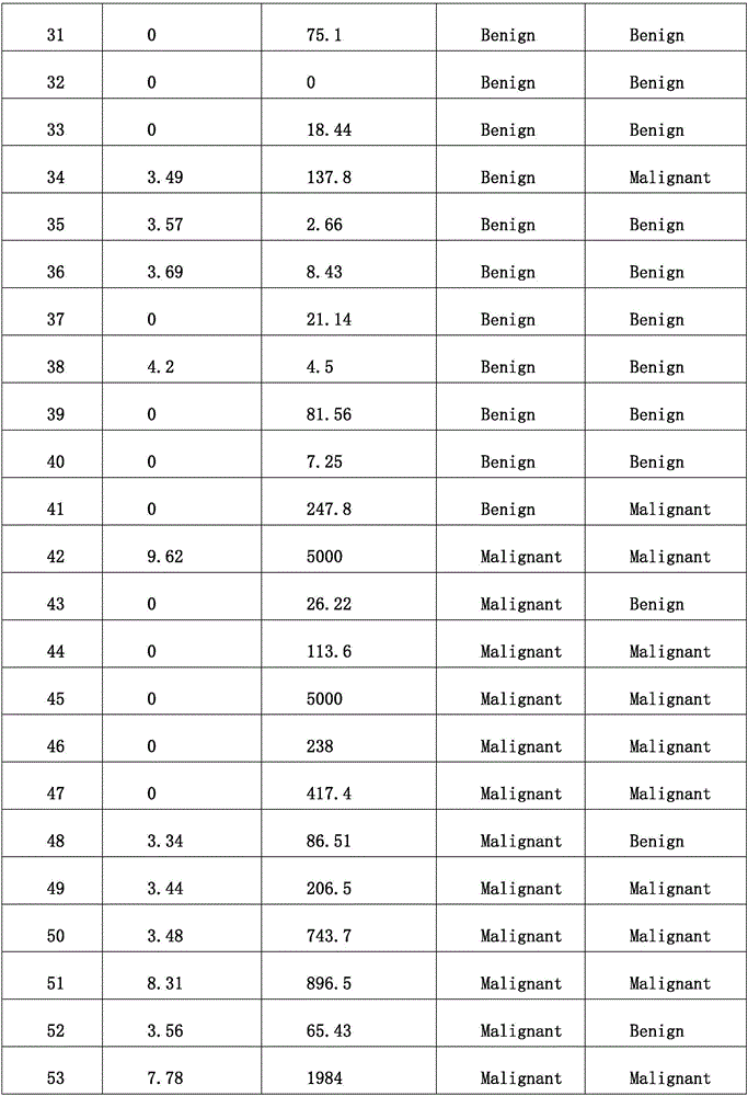

The model trained using the above training set data, wherein the first segmentation point is CScore, the branch threshold is 5.18, when CScore of the subject is greater than 5.18, the subject is predicted to have ovarian cancer or have a high risk of having ovarian cancer, when CScore is less than or equal to 5.18, the second segmentation point CA-125 is constructed, the branch threshold is 101.3, when CA-125 is greater than 103.1, the prediction result is ovarian cancer or have a high risk of having ovarian cancer, according to the model, when CScore is less than or equal to 5.18 and CA-125 is less than or equal to 103.1, the subject is determined not to have ovarian cancer or have a low risk of having ovarian cancer. Pathologically, 1-41 (41) subjects were benign tumors of the ovary, and 42-74 (33) subjects were ovarian cancer patients. In the training set, the sensitivity of the model constructed by the method is 81.82% (27/33), the specificity is 87.8% (36/41), the accuracy is 85.14% (63/74), the omission factor is 18.18% (6/33) and the misdiagnosis rate is 12.2% (5/41) when the model is screened for ovarian cancer.
TABLE 5 test set data and model Classification results


The results show that: the decision tree model has good classification effect on test set samples, and the number 75-95 (21) subjects are all benign ovarian tumors, and the number 96-120 (25) subjects are ovarian cancer patients, which are confirmed by pathology. In the test set, for screening ovarian cancer, the sensitivity of the classification result of the model constructed by the method is 88% (22/25), the specificity is 100% (21/21), the accuracy is 93.48% (43/46), the omission factor is 12% (3/25) and the misdiagnosis rate is 0% (0/21).
The results show that the method and the trained model can simply, conveniently and efficiently diagnose and screen ovarian cancer, have the advantages of very high sensitivity, specificity and accuracy and low omission factor and misdiagnosis rate, and have an unexpected technical effect compared with the prior art.
While specific embodiments of the invention have been described in detail, those skilled in the art will understand that: various modifications and changes in detail can be made in light of the overall teachings of the disclosure, and such changes are intended to be within the scope of the present invention. The full scope of the invention is given by the appended claims and any equivalents thereof.
Claims (16)
1. A set of chromosomes for screening, diagnosing or risk stratification for ovarian cancer, the set of chromosomes comprising at least 1 of chromosomes 1 through 22.
2. The set of chromosomes according to claim 1, consisting of human chromosomes 1 to 22.
3. A computer-readable medium having instructions stored thereon, wherein the instructions, when executed by a processor, cause a computer to:
calculating an imbalance measure CScore value for a subject as a whole based on chromosomal imbalance or chromosomal arm imbalance of chromosomes 1 to 22 of a sample from the subject (e.g., a human);
combining the CScore value with the level of CA-125 in said sample to determine whether said subject has ovarian cancer or is at high risk of having ovarian cancer.
4. The computer-readable medium of claim 3, wherein the CScore value is calculated by:
aligning a subject's (e.g., human) genome-wide data sequence (e.g., a genome-wide data sequence obtained by high-throughput sequencing techniques) to a reference genome (e.g., human reference genome Hg19) and dividing, e.g., by 10-1000k/bin (e.g., 50k/bin), into a plurality of segments (e.g., bins);
the average number of reads (cov) of segments (e.g., bins) covered by the long arm of chromosome i is calculated respectivelyChriq) And the average number of reads (cov) of segments (e.g., bins) covered by the short arm of the chromosomeChrip);
Calculating the R value of chromosome i according to the following formula


Wherein q represents the long arm, p represents the short arm, Chr is an abbreviation for chromosome (chromosome), and i is selected from chromosomes 1 to 22;
calculating Z-score (Z) of chromosome i according to formula (2) based on the R valueChri):

Wherein Is the average of the R values corresponding to healthy people,
 is the standard deviation of the R values corresponding to healthy people;
alternatively, the corresponding long arm q of chromosome i is calculated according to equations (3a) and (3b) And short arm p


Wherein Is the average of the read lengths of the long arm of chromosome i corresponding to healthy people,
 is the average number of reads of the short arm of chromosome i corresponding to healthy people;
a chromosome or chromosome arm is considered to be unbalanced if the absolute value of Z-score for chromosome i or chromosome arm is greater than or equal to 3; selecting chromosomes with the absolute value of the chromosome or chromosome arm Z-score being more than or equal to 3, and calculating the integral unbalance measurement CScore value according to the formula (4):

5. the computer readable medium of claim 3 or 4, wherein the ovarian cancer risk is predicted using a decision tree model established using the levels of CScore and CA-125.
6. The computer-readable medium of claim 3 or 4, wherein the decision tree model is built by the R language.
7. The computer-readable medium of claim 6, wherein the steps of building a decision tree model:
(1) constructing an overall sample data set, wherein about 50 percent of the overall sample data set are ovarian cancer samples and about 50 percent of the overall sample data set are ovarian benign tumor samples;
(2) splitting data, namely segmenting a whole sample data set by using a random sampling function 'sample' in R, and respectively constructing a training set and a test set, wherein the training set accounts for about 60% of the whole sample, and the test set accounts for about 40% of the whole sample;
(3) classification tree training data;
(4) determining optimal parameters of the decision tree model;
(5) optionally, the test set data is examined using the decision tree model and corresponding optimal parameters to determine how effective the optimal parameters of the model are.
8. The computer-readable medium of claim 7, wherein an entire sample data set is constructed by: selecting samples, and constructing an integral sample data set for each sample according to the CScore, the CA-125 level, the Type attribute and the sample number.
9. The computer-readable medium of claim 7, wherein classifying tree training data and determining optimal parameters for a decision tree model are performed by: and modeling the training set by using a conditional inference decision tree ctree of a party package in the R, wherein the conditions are CScore and CA-125, the label is Type, the stream of the ctre package is repeatedly checked, the values of the condition attributes in the sample data are used as segmentation points and threshold values for classification, and finally the optimal parameters of the model are generated.
10. The computer-readable medium of claim 9, wherein the model determines optimal segmentation points and corresponding thresholds as follows:
① selecting CScore result as first segmentation independent variable, determining the subject has ovarian cancer or has high risk of having ovarian cancer under the condition that the threshold is 5.18 and CScore is more than 5.18;
② if the CScore of the subject is less than or equal to 5.18 and the CA-125 level is more than 103.1U/mL, judging that the subject still has ovarian cancer or has high risk of having ovarian cancer according to the result of the decision tree model;
③ when the subject has a CScore < 5.18 and a CA-125 level < 103.1U/mL, the subject is considered to be not suffering from ovarian cancer or to be at a lower risk of suffering from ovarian cancer.
11. The computer readable medium according to any one of claims 3-10, wherein the sample is free DNA in peripheral blood, preferably peripheral venous blood, more preferably peripheral venous blood.
12. A computing apparatus, comprising:
the computer-readable medium and processor of any one of claims 3-10.
13. A system, comprising:
a sequencing device for receiving nucleic acids from a test sample to provide nucleic acid sequence information from the sample (e.g., a full genomic data sequence obtained by a high throughput sequencing technique); and a computing device according to claim 12.
14. The system of claim 13, wherein the sequencing device is a high-throughput sequencer.
15. Use of a reagent for determining the levels of CScore and CA-125 in chromosomes 1 through 22 in the preparation of a diagnostic for screening, diagnosing or risk stratification of ovarian cancer.
16. Use of a device for determining the levels of CScore and CA-125 of chromosomes 1 through 22 in the manufacture of an apparatus for screening, diagnosing or risk stratification of ovarian cancer.
Priority Applications (1)
| Application Number | Priority Date | Filing Date | Title |
|---|---|---|---|
| CN201811030743.8A CN110880356A (en) | 2018-09-05 | 2018-09-05 | Method and apparatus for screening, diagnosing or risk stratification for ovarian cancer |
Applications Claiming Priority (1)
| Application Number | Priority Date | Filing Date | Title |
|---|---|---|---|
| CN201811030743.8A CN110880356A (en) | 2018-09-05 | 2018-09-05 | Method and apparatus for screening, diagnosing or risk stratification for ovarian cancer |
Publications (1)
| Publication Number | Publication Date |
|---|---|
| CN110880356A true CN110880356A (en) | 2020-03-13 |
Family
ID=69727864
Family Applications (1)
| Application Number | Title | Priority Date | Filing Date |
|---|---|---|---|
| CN201811030743.8A Pending CN110880356A (en) | 2018-09-05 | 2018-09-05 | Method and apparatus for screening, diagnosing or risk stratification for ovarian cancer |
Country Status (1)
| Country | Link |
|---|---|
| CN (1) | CN110880356A (en) |
Cited By (5)
| Publication number | Priority date | Publication date | Assignee | Title |
|---|---|---|---|---|
| CN112472114A (en) * | 2020-12-10 | 2021-03-12 | 三峡大学 | Ovarian cancer and tuberculous peritonitis classification system based on imaging characteristics |
| CN112599197A (en) * | 2020-12-23 | 2021-04-02 | 北京吉因加医学检验实验室有限公司 | Method and device for evaluating cancer risk based on plasma DNA fragment analysis |
| CN116013410A (en) * | 2023-01-31 | 2023-04-25 | 南京世和基因生物技术股份有限公司 | Detection method for large fragment rearrangement of germ line gene |
| CN116230215A (en) * | 2023-05-08 | 2023-06-06 | 中国医学科学院北京协和医院 | A device, system and storage medium for predicting ovarian clear cell carcinoma |
| CN116855605A (en) * | 2023-06-13 | 2023-10-10 | 中国医学科学院北京协和医院 | Application of AOC1 as a marker to distinguish ovarian clear cell carcinoma from high-grade serous carcinoma |
Citations (12)
| Publication number | Priority date | Publication date | Assignee | Title |
|---|---|---|---|---|
| US20110053281A1 (en) * | 2007-11-30 | 2011-03-03 | Steinunn Thorlacius | Genetic variants on CHR 11Q and 6Q as markers for prostate and colorectal cancer predisposition |
| CN102203283A (en) * | 2008-07-21 | 2011-09-28 | 新诊断学股份有限公司 | Methods for the cytological analysis of cervical cells |
| CN105653898A (en) * | 2016-01-12 | 2016-06-08 | 江苏格致生命科技有限公司 | Cancer detection kit based on large-scale data mining and detection method |
| WO2016090584A1 (en) * | 2014-12-10 | 2016-06-16 | 深圳华大基因研究院 | Method and device for determining concentration of tumor nucleic acid |
| CN105699473A (en) * | 2016-03-23 | 2016-06-22 | 深圳市老年医学研究所 | Gastric cancer sialoprotein fingerprint spectrum molecular diagnosis module building method |
| CN106164296A (en) * | 2014-02-07 | 2016-11-23 | 阿尔玛克诊断有限公司 | For prediction, the response of anti-angiogenic drugs and the molecular diagnosis of cancer prognosis are tested |
| EP3118324A1 (en) * | 2015-07-13 | 2017-01-18 | Cartagenia N.V. | Method for analyzing copy number variation in the detection of cancer |
| CN106897579A (en) * | 2015-12-19 | 2017-06-27 | 中国人民解放军北京军区总医院 | New early tumor markers based on chromosomal variation index and its application |
| CN107201401A (en) * | 2017-05-23 | 2017-09-26 | 深圳市第二人民医院 | A kind of Multiple-Factor Model and its method for building up for pathogenesis of breast carcinoma risk profile |
| CN107435070A (en) * | 2012-04-12 | 2017-12-05 | 维里纳塔健康公司 | Copy the detection and classification of number variation |
| US20180068083A1 (en) * | 2014-12-08 | 2018-03-08 | 20/20 Gene Systems, Inc. | Methods and machine learning systems for predicting the likelihood or risk of having cancer |
| CN107992719A (en) * | 2017-11-23 | 2018-05-04 | 南方医科大学 | A kind of carcinoma of urinary bladder detection kit based on high-flux sequence |
-
2018
- 2018-09-05 CN CN201811030743.8A patent/CN110880356A/en active Pending
Patent Citations (12)
| Publication number | Priority date | Publication date | Assignee | Title |
|---|---|---|---|---|
| US20110053281A1 (en) * | 2007-11-30 | 2011-03-03 | Steinunn Thorlacius | Genetic variants on CHR 11Q and 6Q as markers for prostate and colorectal cancer predisposition |
| CN102203283A (en) * | 2008-07-21 | 2011-09-28 | 新诊断学股份有限公司 | Methods for the cytological analysis of cervical cells |
| CN107435070A (en) * | 2012-04-12 | 2017-12-05 | 维里纳塔健康公司 | Copy the detection and classification of number variation |
| CN106164296A (en) * | 2014-02-07 | 2016-11-23 | 阿尔玛克诊断有限公司 | For prediction, the response of anti-angiogenic drugs and the molecular diagnosis of cancer prognosis are tested |
| US20180068083A1 (en) * | 2014-12-08 | 2018-03-08 | 20/20 Gene Systems, Inc. | Methods and machine learning systems for predicting the likelihood or risk of having cancer |
| WO2016090584A1 (en) * | 2014-12-10 | 2016-06-16 | 深圳华大基因研究院 | Method and device for determining concentration of tumor nucleic acid |
| EP3118324A1 (en) * | 2015-07-13 | 2017-01-18 | Cartagenia N.V. | Method for analyzing copy number variation in the detection of cancer |
| CN106897579A (en) * | 2015-12-19 | 2017-06-27 | 中国人民解放军北京军区总医院 | New early tumor markers based on chromosomal variation index and its application |
| CN105653898A (en) * | 2016-01-12 | 2016-06-08 | 江苏格致生命科技有限公司 | Cancer detection kit based on large-scale data mining and detection method |
| CN105699473A (en) * | 2016-03-23 | 2016-06-22 | 深圳市老年医学研究所 | Gastric cancer sialoprotein fingerprint spectrum molecular diagnosis module building method |
| CN107201401A (en) * | 2017-05-23 | 2017-09-26 | 深圳市第二人民医院 | A kind of Multiple-Factor Model and its method for building up for pathogenesis of breast carcinoma risk profile |
| CN107992719A (en) * | 2017-11-23 | 2018-05-04 | 南方医科大学 | A kind of carcinoma of urinary bladder detection kit based on high-flux sequence |
Non-Patent Citations (2)
| Title |
|---|
| TONG REN等: "Using low-coverage whole genome sequencing technique to analyze the chromosomal copy number alterations in the exfoliative cells of cervical cancer", 《JOURNAL OF GYNECOLOGIC ONCOLOGY》 * |
| 邬杨: "基于机器学习的卵巢肿瘤预测与分析研究", 《中国优秀硕士学位论文全文数据库(电子期刊)信息科技辑》 * |
Cited By (8)
| Publication number | Priority date | Publication date | Assignee | Title |
|---|---|---|---|---|
| CN112472114A (en) * | 2020-12-10 | 2021-03-12 | 三峡大学 | Ovarian cancer and tuberculous peritonitis classification system based on imaging characteristics |
| CN112472114B (en) * | 2020-12-10 | 2021-07-30 | 三峡大学 | A classification system for ovarian cancer and tuberculous peritonitis based on imaging features |
| CN112599197A (en) * | 2020-12-23 | 2021-04-02 | 北京吉因加医学检验实验室有限公司 | Method and device for evaluating cancer risk based on plasma DNA fragment analysis |
| CN116013410A (en) * | 2023-01-31 | 2023-04-25 | 南京世和基因生物技术股份有限公司 | Detection method for large fragment rearrangement of germ line gene |
| CN116013410B (en) * | 2023-01-31 | 2025-09-26 | 南京世和基因生物技术股份有限公司 | A method for detecting large germline gene rearrangements |
| CN116230215A (en) * | 2023-05-08 | 2023-06-06 | 中国医学科学院北京协和医院 | A device, system and storage medium for predicting ovarian clear cell carcinoma |
| CN116230215B (en) * | 2023-05-08 | 2023-08-18 | 中国医学科学院北京协和医院 | A device, system and storage medium for predicting ovarian clear cell carcinoma |
| CN116855605A (en) * | 2023-06-13 | 2023-10-10 | 中国医学科学院北京协和医院 | Application of AOC1 as a marker to distinguish ovarian clear cell carcinoma from high-grade serous carcinoma |
Similar Documents
| Publication | Publication Date | Title |
|---|---|---|
| Pinzani et al. | Isolation by size of epithelial tumor cells in peripheral blood of patients with breast cancer: correlation with real-time reverse transcriptase–polymerase chain reaction results and feasibility of molecular analysis by laser microdissection | |
| ES2564656T3 (en) | Means and methods for the non-invasive diagnosis of chromosomal aneuploidy | |
| CN110880356A (en) | Method and apparatus for screening, diagnosing or risk stratification for ovarian cancer | |
| DK2809801T3 (en) | NON-INVASIVE CANCER DIAGNOSIS | |
| US20180068058A1 (en) | Methods and compositions for sample identification | |
| TW201718874A (en) | Single molecule sequencing of plasma DNA | |
| US20230250483A1 (en) | Papanicolaou test for ovarian and endometrial cancers | |
| CN119307611A (en) | Blood extracellular vesicle miRNA combination and its application in early diagnosis of lung cancer | |
| CN110736834A (en) | Method, device and system for screening and diagnosing liver cancer based on high-throughput sequencing method | |
| US20230203593A1 (en) | Method for carrying out in vitro molecular diagnosis of ovarian tumor and kit | |
| CN119741967B (en) | Construction method and device of early cancer screening model based on cfDNA difference statistics | |
| CN117316280B (en) | A non-invasive early screening method and system for cancer based on cfDNA terminal sequence characteristics | |
| CN109971857A (en) | Breast cancer diagnosis and treatment biomarker | |
| CN111979326B (en) | Molecular marker combination for representing lung adenocarcinoma qi deficiency and phlegm dampness syndrome, screening and model building method | |
| CN113637747B (en) | Methods and applications for determining SNVs and tumor mutational burden in nucleic acid samples | |
| CN109988835A (en) | Method and apparatus based on the screening of high-flux sequence method and the high-level serous carcinoma of diagnosis of ovarian | |
| CN116656830B (en) | Methylation markers, devices, apparatuses and storage media for gastric cancer assisted diagnosis | |
| CN118207322A (en) | Application of HDR signaling pathway mutations in bile cfDNA in the prognostic evaluation of cholangiocarcinoma | |
| CN108342483A (en) | One group of gene and its application for non-super saltant type colorectal cancer molecule parting | |
| CN120220803A (en) | A screening method for a gene region set for detecting minimal residual lesions of ovarian cancer, a gene region set and a detection system thereof | |
| CN120425050A (en) | Blood sEVs miRNA marker composition, kit and its application in early risk assessment of lung cancer | |
| CN120758624A (en) | A plasma exosome mRNA biomarker and its application in establishing a gastric cancer diagnostic model | |
| CN119307616A (en) | An early prostate cancer diagnostic marker based on circulating free DNA, an evaluation system and its application | |
| CN121087146A (en) | A method and reagent kit for non-invasive prenatal detection of deafness | |
| CN118942546A (en) | A gene set for detecting "blocked" spermatogenesis disorders, a method for preparing a gene detection panel, and its application |
Legal Events
| Date | Code | Title | Description |
|---|---|---|---|
| PB01 | Publication | ||
| PB01 | Publication | ||
| SE01 | Entry into force of request for substantive examination | ||
| SE01 | Entry into force of request for substantive examination | ||
| WD01 | Invention patent application deemed withdrawn after publication |
Application publication date: 20200313 |
|
| WD01 | Invention patent application deemed withdrawn after publication |